Harris-Year2 Final Report Revised - Defense Technical ...
47
AWARD NUMBER: W81XWH-13-1-0247 TITLE: ENZYME-CATALYZED MUTATION IN BREAST CANCER PRINCIPAL INVESTIGATOR: REUBEN S. HARRIS CONTRACTING ORGANIZATION: UNIVERSITY OF MINNESOTA – TWIN CITIES Minneapolis, MN 55455-2070 REPORT DATE: October 2015 TYPE OF REPORT: FINAL PREPARED FOR: U.S. Army Medical Research and Materiel Command Fort Detrick, Maryland 21702-5012 DISTRIBUTION STATEMENT: Approved for Public Release; Distribution Unlimited The views, opinions and/or findings contained in this report are those of the author(s) and should not be construed as an official Department of the Army position, policy or decision unless so designated by other documentation.
-
Upload
khangminh22 -
Category
Documents
-
view
0 -
download
0
Transcript of Harris-Year2 Final Report Revised - Defense Technical ...

AWARD NUMBER: W81XWH-13-1-0247
TITLE: ENZYME-CATALYZED MUTATION IN BREAST CANCER
PRINCIPAL INVESTIGATOR: REUBEN S. HARRIS
CONTRACTING ORGANIZATION: UNIVERSITY OF MINNESOTA – TWIN CITIESMinneapolis, MN 55455-2070
REPORT DATE: October 2015
TYPE OF REPORT: FINAL
PREPARED FOR: U.S. Army Medical Research and Materiel Command Fort Detrick, Maryland 21702-5012
DISTRIBUTION STATEMENT: Approved for Public Release; Distribution Unlimited
The views, opinions and/or findings contained in this report are those of the author(s) and should not be construed as an official Department of the Army position, policy or decision unless so designated by other documentation.

REPORT DOCUMENTATION PAGE Form Approved
OMB No. 0704-0188 Public reporting burden for this collection of information is estimated to average 1 hour per response, including the time for reviewing instructions, searching existing data sources, gathering and maintaining the data needed, and completing and reviewing this collection of information. Send comments regarding this burden estimate or any other aspect of this collection of information, including suggestions for reducing this burden to Department of Defense, Washington Headquarters Services, Directorate for Information Operations and Reports (0704-0188), 1215 Jefferson Davis Highway, Suite 1204, Arlington, VA 22202-4302. Respondents should be aware that notwithstanding any other provision of law, no person shall be subject to any penalty for failing to comply with a collection of information if it does not display a currently valid OMB control number. PLEASE DO NOT RETURN YOUR FORM TO THE ABOVE ADDRESS.
1. REPORT DATE 2. REPORT TYPE 3. DATES COVERED
4. TITLE AND SUBTITLE 5a. CONTRACT NUMBER
5b. GRANT NUMBER
5c. PROGRAM ELEMENT NUMBER
6. AUTHOR(S) 5d. PROJECT NUMBER
5e. TASK NUMBER
E-Mail:
5f. WORK UNIT NUMBER
7. PERFORMING ORGANIZATION NAME(S) AND ADDRESS(ES)
AND ADDRESS(ES)
8. PERFORMING ORGANIZATION REPORTNUMBER
UNIVERSITY OF MINNESOTA – TWIN CITIESMinneapolis, MN 55455-2070
9. SPONSORING / MONITORING AGENCY NAME(S) AND ADDRESS(ES) 10. SPONSOR/MONITOR’S ACRONYM(S)
U.S. Army Medical Research and Materiel Command
Fort Detrick, Maryland 21702-5012 11. SPONSOR/MONITOR’S REPORT
NUMBER(S)
12. DISTRIBUTION / AVAILABILITY STATEMENT
Approved for Public Release; Distribution Unlimited
13. SUPPLEMENTARY NOTES
14. ABSTRACT
The development of breast cancer, including late stage events such as metastasis and drug resistance,requires mutations. The origins of most of these mutations are unknown. We recently implicated the DNAcytosine deaminase APOBEC3B. This Idea Award studies tests the hypothesis that APOBEC3B causes agenome wide hypermutable state and the hypothesis that APOBEC3B alters the epigenome by cytosinedeamination and methyl-cytosine deamination mechanisms, respectively. Positive results will besignificant because they will delineate a major source of mutations and epigenetic changes in breastcancer, and thereby pave the way for new diagnostic/prognostic tests and methods to treat breast cancerby preventing the activity of this enzyme.
15. SUBJECT TERMS
16. SECURITY CLASSIFICATION OF: 17. LIMITATIONOF ABSTRACT
18. NUMBEROF PAGES
19a. NAME OF RESPONSIBLE PERSON
USAMRMC
a. REPORT
Unclassified
b. ABSTRACT
Unclassified
c. THIS PAGE
Unclassified Unclassified
19b. TELEPHONE NUMBER (include area
code)
Standard Form 298 (Rev. 8-98) Prescribed by ANSI Std. Z39.18
W81XWH-
October 2015 Final 1Aug2013 - 31Jul2015
13-1-0247ENZYME-CATALYZED MUTATION IN BREAST CANCER
REUBEN S. HARRIS
Nothing listed
47

Table of Contents Page 2
Introduction Page 3
Keywords Page 4
Overall Project Summary Page 5
Key Research Accomplishments Page 10
Conclusion Page 11
Publications, Abstracts and Presentations Page 12
Inventions, Patents and Licenses Page 14
Reportable Outcomes Page 15
Other Achievements Page 16
References Page 17
Appendices Page 18

Harris, DoD Idea Award BC121347
Page 3
Introduction
The development of breast cancer, including late stage events such as metastasis and drug resistance, requires mutations. The origins of most of these mutations are unknown. We recently implicated the DNA cytosine deaminase APOBEC3B. This Idea Award studies tests the hypothesis that APOBEC3B causes a genome wide hypermutable state and the hypothesis that APOBEC3B alters the epigenome by cytosine deamination and methyl-cytosine deamination mechanisms, respectively. Positive results will be significant because they will delineate a major source of mutations and epigenetic changes in breast cancer, and thereby pave the way for new diagnostic/prognostic tests and methods to treat breast cancer by preventing the activity of this enzyme.

Harris, DoD Idea Award BC121347
Page 4
Keywords
APOBEC3B; Apolipoprotein B mRNA editing enzyme, catalytic polypeptide-like-3 B; sometimes abbreviated A3B; one of 7 human A3 family members
C; Cytosine (a DNA and RNA base)
DNA; Deoxyribonucleic acid
ER; estrogen receptor (molecular target of the breast cancer therapeutic tamoxifen)
G; Guanine (a DNA and RNA base)
MeC; 5-methyl-cytosine (a common epigenetic modification in human DNA)
qPCR; Quantitative polymerase chain reaction
shRNA; short hairpin RNA (a molecular tool used to decrease gene expression)
SOW; Statement of Work
T; Thymine (a base typically found in DNA, but also the product of APOBEC3B-catalyzed MeC deamination)
U; Uracil (a base typically found in RNA but also the product of APOBEC3B-catalyzed C deamination)

Harris, DoD Idea Award BC121347
Page 5
Overall Project Summary (significant revisions and/or additions to the original final report are highlighted in yellow)
This section provides a final report and a narrative of progress over the 2 year duration of this Idea award. Please see Table 1 below for an updated SOW including final reports of the status of each task. A summary and discussion (as requested) of the progress on each aim follows.
Aim 1 – Does A3B cause a genome-wide hypermutable state? Aim 1 rationale: Although we have demonstrated APOBEC3B up-regulation in tumors and APOBEC3B activity in the nuclear extracts of several breast cancer cell lines[1], we still need to overcome the highest hurdle and demonstrate that APOBEC3B actually alters the genetic landscape of a breast cancer cell. This will be done by deep-sequencing to document the APOBEC3B-dependent contribution to the overall mutation distribution in cell lines and by performing a series of experiments with a well-established xenograft tumor model.
Aim 1 - Summary of Results, Progress and Accomplishments with Discussion. Aim 1A – deep-sequencing cell lines: We have now deep sequenced several
different cancer cell lines, and have encountered significant genetic heterogeneity in most instances that precluded analyses of APOBEC3B mutations. However, we have succeeded in one system in which APOBEC3B can be expressed inducibly. These results are detailed in Appendix A, an open access publication by Akre et al., 2016, PLoS One (PMID: 27163364 PMCID: PMC4862684) and discussed here.
Figure 1 shows doxycycline-induced expression of APOBEC3B. Figure 2 shows a titration of doxycycline levels that induce APOBEC3B expression and result in approximately 90% cell death. This level of doxycycline was used to induce 10-rounds of APOBEC3B expression and mutagenesis in daughter pools. Representative cells were then outgrown from each pool (single cell cloned) and subjected to microarray analysis for single nucleotide polymorphisms (SNPs) and full genome DNA sequencing. Figure 3 shows the results of the microarray analysis with increased numbers of SNPs and increased levels of copy number variations (CNVs). Figure 4 shows the results of the full genome sequence analysis. As anticipated, APOBEC3B mutations were detected throughout the genome at elevated frequencies. However, unexpectedly, we discovered that this cell line is defective in mismatch repair and had very high background levels of mutation, which precluded more extensive analyses of the APOBEC3B mutational landscape. Nevertheless, this series of experiments demonstrated the genome-wide impact of APOBEC3B and provided several valuable lessons to apply in future studies.
Aim 1B – xenograft experiments in mice: The proposed xenograft studies took longer than expected in part due to repeating key experiments and due to adding an over-expression study. However, we are delighted to report that the results are positive, and that therapy (tamoxifen) resistance in the ER+ breast cancer cell line MCF-7L is dependent upon APOBEC3B. Specifically, APOBEC3B knockdown slows down the rate of tumor evolution and drug resistance, and APOBEC3B over-expression speeds-up tumor

Harris, DoD Idea Award BC121347
Page 6
evolution and drug resistance. These analyses, included extensive methodologies, are detailed in Appendix B, an open access publication by Law et al., 2016, Science Advances (PMID: 27730215 PMCID: PMC5055383) and discussed here.
Figure 1 reports clinical data from our Dutch collaborators. A significant correlation is evident between APOBEC3B mRNA levels in primary tumors and progression free survival upon disease recurrence. Essentially, the higher the APOBEC3B levels in the original tumor, the poorer the outcomes in the recurrent setting for ER+ disease subjected to tamoxifen monotherapy. Figure 2 shows that shRNA mediated knockdown of APOBEC3B in the ER+ breast cancer cell line MCF-7L is robust and, importantly, that it does not alter cellular growth rates in culture. Figure 3 is a representative xenograft experiment in which APOBEC3B knockdown improves the durability of tamoxifen treatment by reducing the rate of developing drug resistance. Figures 4 and 5 show the results of APOBEC3B overexpression using a novel lentivirus-based construct (schematic in Figure 4A). Importantly, overexpression of the wildtype APOBEC3B enzyme, but not a catalytically dead form, reduces the durability of tamoxifen treatment by accelerating the rate of developing drug resistance. Taken together, these results are the first to demonstrate that altering the cellular levels of a single enzyme, APOBEC3B, can systematically influence the rate of acquired resistance to tamoxifen therapy.
Although this study was successful, it also faced some technical challenges. For instance, the MCF-7L cell line is genetically heterogeneous, which precluded the identification of the resistance mutations by exome sequencing. However, we have learned from these challenges and have taken a number of precautions, including the utilization of pre-defined clonogenic breast cancer cell lines, that we are confident will enable future successes.
Aim 2 – Does A3B impact genomic MeC levels?
Aim 2 rationale: The impetus for this aim stems from observations that the related DNA deaminases AID and APOBEC3A elicit MeC-to-T editing activity in vitro[2-4], and AID has been implicated in altering the MeC status of mouse germ and stem cells[5, 6]. Since AID is not expressed in normal breast epithelium or breast tumor cells and only A3B is up-regulated in breast tumors[1], we hypothesize that A3B alone has the capacity to remodel the breast cancer MeC landscape. This hypothesis will be tested here in experiments that are complementary to those described above.
Aim 2 - Summary of Results, Progress and Accomplishments with Discussion. Aims 2A-C: We have completed the original studies as proposed and have found that APOBEC3B is not likely to have a role in genomic DNA demethylation (although it can do so biochemically). In essence, bisulfite sequencing has not identified any sites in the genome that become hypomethylated in APOBEC3B over-expressing cell lines in comparison to non-APOBEC3B expressing controls. We are concerned that the developmental fate of cell lines is difficult to alter, and that future studies in mice in vivo may be more informative.

Harris, DoD Idea Award BC121347
Page 7
Table 1. Progress on original SOW with current status/progress highlighted in blue.
Aim 1: Does APOBEC3B cause a genome-‐wide hypermutable state?
Task Methods employed Timeline and Status
Engineering breast cancer cell lines MDA-‐MB-‐231, MDA-‐MB-‐453, MDA-‐MB-‐468, and HCC1569 to knock-‐down endogenous A3B and generate control lines; generate multiple sub-‐clones for each line.
Molecular biology, cell culture, qRT-‐PCR
Months 1-‐6; completed as proposed
Preparation of genomic DNA from selected cell lines (likely HCC1569) prepared in the above tasks to express high or low levels of A3B. Delivery of DNA to sequencing facility for whole exome capture, deep sequencing, and data/sequence analysis.
General molecular biology techniques, data/sequence analysis, bioinformatics
Months 6-‐18; sequencing done but results ambiguous because most cell lines were heterogenous; we have had success with one cell line and the results were published in PLoS One (Appendix A – Akre et al., 2016).
Completion of IACUC forms for approval of animal experiments (80 NCr nude mice are proposed for the full xenograft experiment with numbers determined by power analysis – details can be found in the main text of the proposal). Once approved, the engineered cell lines described above (and in the narrative) will begin being xenografted into mice and therapies administered.
Cell culture, mouse model techniques
Months 1-‐5 for IACUC review, months 6-‐18 for animal procurement and xenograft experiments; IACUC approval was received, the cell lines were engineered, and the xenograft experiments were done
Tumor collection and analysis from xenografts. Mouse model techniques, cancer-‐molecular biology techniques, qRT-‐PCR, sequence analysis
Months 16-‐20; done but DNA sequencing results were ambiguous because the cell line was

Harris, DoD Idea Award BC121347
Page 8
heterogenous
Prepare data for publication. Publish manuscript. Data analysis and writing
Months 20-‐24; a manuscript has been published in Science Advances (Appendix B – Law et al., 2016).
Aim 2: Does APOBEC3B impact the genomic methyl-‐cytosine landscape?
Task Methods Timeframe
Engineering of cell lines MDA-‐MB-‐231, MDA-‐MB-‐453, MDA-‐MB-‐468, and HCC1569 to knock-‐down endogenous A3B. Passage of lines from generations 2-‐32, with collection of DNA at generations 2, 4, 8, 16, and 32. Assessment of MeC levels using MeC ELISA kit.
Cell culture, molecular biology techniques, western blotting, qRT-‐PCR, ELISA
Months 1-‐6; completed as proposed.
In parallel with the task immediately above, the same DNA samples will be assessed for MeC content using HPLC-‐MS/MS, rather than ELISA.
Cell culture, molecular biology techniques, western blotting, qRT-‐PCR, HPLC-‐MS/MS
Months 2-‐7; completed as proposed.
Again, the same DNA samples as in the previous 2 tasks will be subjected to bisulfite sequencing to assess DNA methylation status in regions of the genome that are known to be effected by hypomethylation (see narrative for further details).
Cell culture, molecular biology techniques, deep-‐sequencing western blotting, qRT-‐PCR, bisulfite sequencing
Months 3-‐12; completed but the bisulfite DNA sequencing results were ambiguous because the cell line was heterogenous
We will engineer the non-‐tumorigenic cell lines MCF-‐10A (previously acquired from ATCC) and hTERT-‐HMEC (a gift from the lab of Dr. Vitaly Polunovsky) to over-‐express A3B by transfection with a linearized, tagged A3B-‐espression cassette followed by selection of stable clones. Control lines will be generated using the catalytically dead, tagged A3B-‐E255Q.
Cell culture, molecular biology techniques, western blotting, qRT-‐PCR
Months 3-‐12; completed as proposed.
Assessment of A3B over-‐expressing engineered cell lines’ ability to alter the levels of MeC in the cell genome (determined by ELISA, HPLC-‐MS/MS, and bisulfite-‐sequencing).
Cell culture, cancer-‐molecular biology techniques, ELISA, HPLC-‐MS/MS, bisulfite sequencing
Months 8-‐16; completed as proposed.
Bisulfite-‐coupled deep sequencing will be performed to Bisulfite-‐coupled Months 15-‐22;

Harris, DoD Idea Award BC121347
Page 9
quantify the levels of demethylation and identify any demethylation hot-‐spots and mutational spectra as a function of A3B expression. Samples sent for sequencing will be pairs of A3B high/A3B knock-‐down and A3B over-‐expressed/A3B-‐E255Q over-‐expressed DNA determined empirically from the previous aims to have positive results by ELISA, HPLC-‐MS/MS, and local bisulfite sequencing.
deep sequencing completed but the bisulfite DNA sequencing results were ambiguous because the cell line was heterogenous
Analysis and compilation of data. Assembly of manuscript. Data analysis and writing
Months 20-‐24; we have invested almost all effort in the success of Aim 1 once we learned Aim 2 would test negative.

Harris, DoD Idea Award BC121347
Page 10
Key Research Accomplishments 1) Cell lines have been constructed that inducibly express APOBEC3B, and demonstrate
the genome-wide nature of this breast cancer mutagenesis mechanism [Appendix A: Akre et al., 2016, PLoS One (PMID: 27163364 PMCID: PMC4862684)].
2) Xenograft experiments with the ER+ breast cancer cell line MCF7L have demonstrated
that APOBEC3B is a significant driver of tumor evolution and resistance to the SERM tamoxifen [Appendix B: Law et al., 2016, Science Advances (PMID: 27730215 PMCID: PMC5055383)].

Harris, DoD Idea Award BC121347
Page 11
Conclusion We are thrilled to report that our xenograft studies have been successful, and allowed us to demonstrate that APOBEC3B drives tamoxifen resistance in an ER+ breast cancer cell line (Law et al., 2016, Science Advances). Due to the fundamental nature of the underlying mutational process and the breadth of APOBEC3B over-expression in breast and other cancer types, this result is likely to be broadly applicable. The next step will be developing strategies to stop APOBEC3B driven breast tumor evolution in the hope of improving the efficacy of existing therapies such as tamoxifen, which can be undermined by tumor evolution and the acquisition of resistance mutations.

Harris, DoD Idea Award BC121347
Page 12
Publications, Abstracts and Presentations
Publications Acknowledging DoD Idea Award Support:
Harris, R.S. (2013) Cancer mutation signatures, DNA damage mechanisms, and potential clinical implications. Genome Medicine 5:87 (3 pages). PMID: 24073723; PMCID: PMC3978439
Swanton, C., N. McGranahan, G.J. Starrett & R.S. Harris (2015) APOBEC enzymes: mutagenic fuel for cancer evolution and heterogeneity. Cancer Discovery 5:704-12. PMID: 26091828; PMCID: PMC4497973
Harris, R.S. (2015) Molecular mechanism and clinical impact of APOBEC3B-catalyzed mutagenesis in breast cancer. Breast Cancer Research 17:8 (10 pages). PMID: 25778352; PMCID: PMC4303225
Appendix A: Akre*, M.K, G.J. Starrett*, J.S. Quist, N.A. Temiz, M.A. Carpenter, A.N.J. Tutt, A. Grigoriadis & R.S. Harris (2016) Mutation processes in 293-based clones overexpressing the DNA cytosine deaminase APOBEC3B. PLoS One 11(5):e0155391. doi: 10.1371/journal.pone.0155391 (*equal contributions). PMID: 27163364; PMCID: PMC4862684
Appendix B: Law*, E.K., A.M. Sieuwerts*, K. LaPara, B. Leonard, G.J. Starrett, A.M. Molan, N.A. Temiz, R. Isaksson Vogel, M.E. Meijer-van Gelder, F.C.G.J. Sweep, P.N. Span, J.A. Foekens, J.W.M. Martens, D. Yee & R.S. Harris (2016) The DNA cytosine deaminase APOBEC3B promotes tamoxifen resistance in ER+ breast cancer. Science Advances 2, e1601737 (9 pages; *equal contributions). PMID: 27730215; PMCID: PMC5055383
Oral Presentations Acknowledging DoD Idea Award Support:
R.S. Harris; 8/13; “Evidence for APOBEC3B mutagenesis in multiple human cancers”, Center for Molecular Medicine Norway, Oslo (Hans Prydz Distinguished Guest Lecture invited by Dr. H. Nilsen)
R.S. Harris; 10/13; “Integral roles for enzymatic DNA cytosine deamination in both antiviral innate immunity and cancer mutagenesis” University of Alberta, Edmonton, Canada (seminar invited by Dr. A. Mason)
R.S. Harris; 10/13; “Molecular and clinical impact of APOBEC3B mutagenesis in breast cancer”, AACR special conference on Advances in Breast Cancer Research, San Diego, CA (talk invited by organizers)
R.S. Harris; 11/13; "Carcinogenesis fueled by enzyme-catalyzed DNA cytosine deamination”, University of Virginia (talk invited by Drs. L. Hammarskjöld and D. Rekosh)
R.S. Harris; 11/13; “APOBEC3B mutagenesis in cancer”, 29th Radiation Biology Center Symposium entitled: “Next generation radiation biology and beyond: New

Harris, DoD Idea Award BC121347
Page 13
perspectives on DNA damage and repair”, Kyoto, Japan (talk invited by organizers)
R.S. Harris; 11/13; “APOBEC3 proteins in antiviral immunity and carcinogenesis”, Kyoto University, Kyoto, Japan (talk invited by Dr. A. Takori-Kondo)
R.S. Harris; 2/14; "APOBEC3B-catalyzed mutagenesis in human cancer”, NCI Frederick (talk invited by Dr. S. LeGrice)
R.S. Harris; 2/14; "APOBEC3 DNA deaminases in retrovirus restriction and cancer mutagenesis”, University of Wisconsin at Madison (talk invited by Dr. D. Evans)
R.S. Harris; 4/14; "Mechanism and impact of enzymatic DNA cytosine deamination in human cancers”, Wake Forest School of Medicine, Department of Biochemistry (talk invited by Dr. F. Perrino)
R.S. Harris; 7/14; “Can mutagenesis be a biomarker and a therapeutic target?”, Stratified Medicine Symposium, UK NHS, Guy’s Hospital, London, England (talk invited by Dr. Andrew Tutt and the organizing committee)
R.S. Harris; 9/14; “APOBEC3B mutagenesis in human cancer: basic mechanisms and clinical implications” AbCam Conference on Chromothripsis, Clustered Mutation and Complex Chromosome Rearrangements, Boston, MA (talk invited by Drs. Ralph Scully and James Haber)
R.S. Harris; 9/14; “APOBEC-mediated mutagenesis of viral and cancer genomes”, IRCM, Montreal, Canada (talk invited by Dr. Javier DiNoia)
R.S. Harris; 9/14; “DNA editing in cancer” NCI workshop on RNA Editing, Epitranscriptomics, and Processing in Cancer Progression, Bethesda, MD (keynote talk invited by Drs. John Coffin and Betsy Read-Cannole)
R.S. Harris; 9/14; “The biological and pathological importance of enzyme-catalyzed DNA cytosine deamination”, Innovative Approaches for Identification of Antiviral Agents Summer School (IAAASS), Sardinia, Italy (lecture invited by the organizing committee)
R.S. Harris; 10/14; “Cancer mutagenesis by the antiviral enzyme APOBEC3B” Genentech, San Francisco, CA (talk invited by Dr. Tim Behrens)
R.S. Harris; 10/14; “APOBEC3B mutagenesis in human cancer: basic mechanisms and clinical implications”, Erasmus Medical Center, Holland (talk invited by Dr. John Martens)
R.S. Harris; 11/14; “APOBEC3 proteins, DNA uracil, and cancer mutagenesis” US-EU conference on repair of endogenous DNA damage, Santa Fe, NM (talk invited by the organizers)
R.S. Harris; 11/14; “APOBEC3B mutagenesis in human cancer: basic mechanisms and clinical implications”, 10th National Cancer Research Institute Conference, BT Convention Center, Liverpool, UK (plenary talk invited by the organizing committee)

Harris, DoD Idea Award BC121347
Page 14
R.S. Harris; 12/14; “APOBEC3B mutagenesis in cancer: basic mechanisms and clinical implications” San Antonio Breast Cancer Symposium, San Antonio, TX (talk invited by the organizers)
R.S. Harris; 2/15; “Cancer mutagenesis by the antiviral DNA cytosine deaminase APOBEC3B”, Mayo Clinic, Rochester, MN (talk invited by Dr. Yasuhiro Ikeda)
R.S. Harris; 2/15; “Virus restriction and cancer mutation by the APOBEC family of DNA cytosine deaminases ", Massey University, New Zealand (talk invited Dr. Elena Harjes)
R.S. Harris; 3/15; “DNA deamination in cancer mutagenesis", Gordon Research Conference on RNA Editing, Lucca (Barga), Italy (talk invited by the organizing committee)
R.S. Harris; 4/15; “Mechanism and clinical impact of APOBEC3B mutagenesis in cancer”, Ohio State University, Columbus, OH (seminar invited by Dr. Kay Huebner)
R.S. Harris; 5/15; “APOBEC mutation signatures and the origins of mutation in breast cancer", IMPAKT, Brussels, Belgium (talk invited by the organizing committee)
R.S. Harris; 6/15; “Molecular mechanism and clinical impact of APOBEC mutagenesis in cancer”, Istituto Superiore di Sanità (ISS), Rome, Italy (talk invited by Drs. Margherita Bignami and Eugenia Dogliotti)
R.S. Harris; 6/15; “Cancer mutagenesis by enzymatic DNA cytosine deamination”, Ospedale Pediatrico Bambino Gesù, Rome, Italy (talk invited by Dr. Angela Gallo)
R.S. Harris; 7/15; “Antiviral enzymes in cancer – molecular mechanism and clinical implications”, University of Cagliari, Italy (talk invited by Drs. Elias Maccioni and Enzo Tramontano)
R.S. Harris; 10/15; “APOBEC-catalyzed mutagenesis in cancer”, Wistar Institute, Philadelphia, PA (seminar invited by Drs. Kazuko Nishikura and Ashani Weeraratna)
R.S. Harris; 10/15; “Genomic DNA deamination in cancer”, 17th Annual John Goldman Conference on Chronic Myeloid Leukemia: Biology and Therapy, Lisbon, Portugal (talk invited by meeting organizers)
R.S. Harris; 11/14; “Retrovirus restriction and cancer mutagenesis through enzymatic DNA cytosine deamination", University of Toronto, Canada (talk invited by Dr. Jeffrey Lee)
R.S. Harris; 12/15; “Tamoxifen resistance driven by the DNA cytosine deaminase APOBEC3B in recurrent estrogen receptor positive breast cancer” San Antonio Breast Cancer Symposium, San Antonio, TX (talk invited by the organizers)

Harris, DoD Idea Award BC121347
Page 15
Inventions, Patents and Licenses Nothing to report.

Harris, DoD Idea Award BC121347
Page 16
Reportable Outcomes Nothing to report.

Harris, DoD Idea Award BC121347
Page 17
Other Achievements One Ph.D. student, Ms. Monica Akre, is being supported by this award. She passed her written and oral preliminary exams and she helped to complete the proposed studies in Aim 1A (Appendix A).

Harris, DoD Idea Award BC121347
Page 18
References [1] Burns MB, Lackey L, Carpenter MA, Rathore A, Land AM, Leonard B, et al. APOBEC3B
is an enzymatic source of mutation in breast cancer. Nature. 2013;494:366-70. [2] Morgan HD, Dean W, Coker HA, Reik W, Petersen-Mahrt SK. Activation-induced
cytidine deaminase deaminates 5-methylcytosine in DNA and is expressed in pluripotent tissues: implications for epigenetic reprogramming. J Biol Chem. 2004;279:52353-60.
[3] Bransteitter R, Pham P, Scharff MD, Goodman MF. Activation-induced cytidine deaminase deaminates deoxycytidine on single-stranded DNA but requires the action of RNase. Proc Natl Acad Sci U S A. 2003;100:4102-7.
[4] Wijesinghe P, Bhagwat AS. Efficient deamination of 5-methylcytosines in DNA by human APOBEC3A, but not by AID or APOBEC3G. Nucleic Acids Res. 2012;40:9206-17.
[5] Popp C, Dean W, Feng S, Cokus SJ, Andrews S, Pellegrini M, et al. Genome-wide erasure of DNA methylation in mouse primordial germ cells is affected by AID deficiency. Nature. 2010;463:1101-5.
[6] Bhutani N, Brady JJ, Damian M, Sacco A, Corbel SY, Blau HM. Reprogramming towards pluripotency requires AID-dependent DNA demethylation. Nature. 2010;463:1042-7.

Harris, DoD Idea Award BC121347
Page 19
Appendices Appendix A: Akre*, M.K, G.J. Starrett*, J.S. Quist, N.A. Temiz, M.A. Carpenter, A.N.J.
Tutt, A. Grigoriadis & R.S. Harris (2016) Mutation processes in 293-based clones overexpressing the DNA cytosine deaminase APOBEC3B. PLoS One 11(5):e0155391. doi: 10.1371/journal.pone.0155391 (*equal contributions). PMID: 27163364; PMCID: PMC4862684
Appendix B: Law*, E.K., A.M. Sieuwerts*, K. LaPara, B. Leonard, G.J. Starrett, A.M. Molan, N.A. Temiz, R. Isaksson Vogel, M.E. Meijer-van Gelder, F.C.G.J. Sweep, P.N. Span, J.A. Foekens, J.W.M. Martens, D. Yee & R.S. Harris (2016) The DNA cytosine deaminase APOBEC3B promotes tamoxifen resistance in ER+ breast cancer. Science Advances 2, e1601737 (9 pages; *equal contributions). PMID: 27730215; PMCID: PMC5055383

RESEARCH ARTICLE
Mutation Processes in 293-Based ClonesOverexpressing the DNA Cytosine DeaminaseAPOBEC3BMonica K. Akre1☯, Gabriel J. Starrett1☯, Jelmar S. Quist2, Nuri A. Temiz1, MichaelA. Carpenter1, Andrew N. J. Tutt2, Anita Grigoriadis2, Reuben S. Harris1,3*
1 Department of Biochemistry, Molecular Biology, and Biophysics, Institute for Molecular Virology, MasonicCancer Center, University of Minnesota, Minneapolis, MN, United States of America, 2 Breast Cancer NowResearch Unit, Research Oncology, Guy’s Hospital, King’s College London, London, United Kingdom,3 Howard Hughes Medical Institute, University of Minnesota, Minneapolis, MN, United States of America
☯ These authors contributed equally to this work.* [email protected]
AbstractMolecular, cellular, and clinical studies have combined to demonstrate a contribution from
the DNA cytosine deaminase APOBEC3B (A3B) to the overall mutation load in breast,
head/neck, lung, bladder, cervical, ovarian, and other cancer types. However, the complete
landscape of mutations attributable to this enzyme has yet to be determined in a controlled
human cell system. We report a conditional and isogenic system for A3B induction, genomic
DNA deamination, and mutagenesis. Human 293-derived cells were engineered to express
doxycycline-inducible A3B-eGFP or eGFP constructs. Cells were subjected to 10 rounds of
A3B-eGFP exposure that each caused 80–90% cell death. Control pools were subjected to
parallel rounds of non-toxic eGFP exposure, and dilutions were done each round to mimic
A3B-eGFP induced population fluctuations. Targeted sequencing of portions of TP53 and
MYC demonstrated greater mutation accumulation in the A3B-eGFP exposed pools.
Clones were generated and microarray analyses were used to identify those with the great-
est number of SNP alterations for whole genome sequencing. A3B-eGFP exposed clones
showed global increases in C-to-T transition mutations, enrichments for cytosine mutations
within A3B-preferred trinucleotide motifs, and more copy number aberrations. Surprisingly,
both control and A3B-eGFP clones also elicited strong mutator phenotypes characteristic of
defective mismatch repair. Despite this additional mutational process, the 293-based sys-
tem characterized here still yielded a genome-wide view of A3B-catalyzed mutagenesis in
human cells and a system for additional studies on the compounded effects of simultaneous
mutation mechanisms in cancer cells.
IntroductionCancer genome sequencing studies have defined approximately 30 distinct mutation signatures(reviewed by [1–4]). Some signatures are large-scale confirmations of established sources of
PLOSONE | DOI:10.1371/journal.pone.0155391 May 10, 2016 1 / 17
a11111
OPEN ACCESS
Citation: Akre MK, Starrett GJ, Quist JS, Temiz NA,Carpenter MA, Tutt ANJ, et al. (2016) MutationProcesses in 293-Based Clones Overexpressing theDNA Cytosine Deaminase APOBEC3B. PLoS ONE11(5): e0155391. doi:10.1371/journal.pone.0155391
Editor: Javier Marcelo Di Noia, Institut deRecherches Cliniques de Montréal (IRCM), CANADA
Received: February 26, 2016
Accepted: April 5, 2016
Published: May 10, 2016
Copyright: © 2016 Akre et al. This is an openaccess article distributed under the terms of theCreative Commons Attribution License, which permitsunrestricted use, distribution, and reproduction in anymedium, provided the original author and source arecredited.
Data Availability Statement: All raw sequences areavailable from NCBI SRA under project number,PRJNA312357. All SNP data sets have beendeposited in the NCBI GEO database underaccession code GSE78710.
Funding:Work in the Grigoriadis and Tuttlaboratories is supported by Breakthrough BreastCancer (recently merged with Breast CancerCampaign forming Breast Cancer Now). JQ is on aPhD studentship in Translational Medicine from theNIHR Biomedical Research Centre at Guy’s and StThomas. Cancer studies in the Harris laboratory aresupported by grants from the Department of Defense

DNA damage that escaped repair or were repaired incorrectly. The largest is water-mediateddeamination of methyl-cytosine bases, which manifest as C-to-T transitions in genomic 5’-CGmotifs [5]. This process impacts almost all cancer types and accumulates as a function of age.Other well known examples include ultraviolet radiation, UV-A and UV-B, which crosslinkadjacent pyrimidine bases and result in signature C-to-T transitions [6], and tobacco mutagenssuch as nitrosamine ketone (NNK), which metabolize into reactive forms that covalently bindguanine bases and result in signature G-to-T transversions [7]. These latter mutagenic pro-cesses are well known drivers of skin cancer and lung cancer, respectively, but also contributeto other tumor types. A lesser-known but still significant example of a mutagen is the dietarysupplement aristolochic acid, which is derived from wild ginger and related plants and metabo-lized into reactive species that covalently bind adenine bases and cause A-to-T transversions[8, 9]. Aristolochic acid mutation signatures are evident in urothelial cell, hepatocellular, andbladder carcinomas. Other confirmed mutation sources include genetic defects in recombina-tion repair (BRCA1, BRCA2, etc.), post-replication mismatch repair (MSH2,MLH1, etc.), andDNA replication proofreading function, which manifest as microhomology-mediated inser-tion/deletion mutations, repeat/microsatellite slippage mutations, and transversion mutationsignatures, respectively [4, 5, 10].
The largest previously undefined mutation signature in cancer is C-to-T transitions and C-to-G transversions within 5’-TC dinucleotide motifs [5, 11, 12]. This mutation signature occursthroughout the genome, as well as less frequently in dense clusters called kataegis. This signa-ture is ascribable to the enzymatic activity of members of the APOBEC family of DNA cytosineto uracil deaminases [5, 11–15]. Human cells encode up to 9 distinct APOBEC family memberswith demonstrated C-to-U editing activity, and 7/9 have been shown to prefer 5’-TC dinucleo-tide motifs in single-stranded DNA substrates: APOBEC1, APOBEC3A, APOBEC3B (A3B),APOBEC3C, APOBEC3D, APOBEC3F, and APOBEC3H. In contrast, AID and APOBEC3Gprefer 5’RC and 5’CC, respectively (R = purine; reviewed by [16, 17]). The size and similarityof this protein family, as well as the formal possibility that another DNA damage source maybe responsible for the same mutation signature [18], have made DNA sequencing data andinformatics analyses open to multiple interpretations.
However, independent [13, 19] and subsequent [14, 15, 20–26] studies indicate that at leastone DNA deaminase family member, A3B, has a significant role in causing these types of muta-tions in cancer. A3B localizes to the nucleus throughout the cell cycle except during mitosiswhen it appears excluded from chromatin [19]. A3B is upregulated in breast cancer cell linesand primary tumors at the mRNA, protein, and activity levels [13, 20, 27]. Endogenous A3B isthe only detectable deaminase activity in nuclear extracts of many cancer cell lines representinga broad spectrum of cancer types (breast, head/neck, lung, ovarian, cervix, and bladder [13, 20,27]). Endogenous A3B is required for elevated levels of steady state uracil and mutation fre-quencies in breast cancer cell lines [13]. Overexpressed A3B induces a potent DNA damageresponse characterized by gamma-H2AX and 53BP1 accumulation, multinuclear cell forma-tion, and cell cycle deregulation [13, 21, 22]. A3B levels correlate with overall mutation loads inbreast and head/neck tumors [13, 23]. The biochemical deamination preference of recombi-nant A3B, 5’TCR, is similar to the actual cytosine mutation pattern observed in breast, head/neck, lung, cervical, and bladder cancers [13, 14, 20]. Human papillomavirus (HPV) infectioninduces A3B expression in several human cell types, providing a link between viral infectionand the observed strong APOBEC mutation signatures in cervical and some head/neck andbladder cancers [28–30]. The spectrum of oncogenic mutations in PIK3CA is biased towardsignature A3B mutation targets in HPV-positive head/neck cancers [23]. Last but not least,high A3B levels correlate with poor outcomes for estrogen receptor-positive breast cancerpatients [25, 26, 31].
APOBEC3BMutation Signature in Human Cells
PLOS ONE | DOI:10.1371/journal.pone.0155391 May 10, 2016 2 / 17
Breast Cancer Research Program (BC121347), theJimmy V Foundation for Cancer Research, theNorwegian Centennial Chair Program, the MinnesotaPartnership for Biotechnology and MedicalGenomics, and the Randy Shaver Cancer Researchand Community Fund. Salary support for GJS wasprovided by a National Science Foundation GraduateResearch Fellowship (DGE 13488264). RSH is anInvestigator of the Howard Hughes Medical Institute.The funders had no role in study design, datacollection and analysis, decision to publish, orpreparation of the manuscript.
Competing Interests: RSH is a co-founder ofApoGen Biotechnologies Inc. The other authorsdeclare no competing financial interests. This doesnot alter the authors' adherence to PLOS ONEpolicies on sharing data and materials.

Despite this extensive and rapidly growing volume of genomic, molecular, and clinicalinformation on A3B in cancer, the association between A3B and APOBEC mutational signa-tures has so far only been correlative, and a mechanistic demonstration of this enzyme’s activ-ity on the human genome has yet to be determined. Here we report further development of ahuman 293 cell-based system for conditional expression of human A3B. The results reveal, forthe first time in a human cell line, the genomic landscape of A3B induced mutagenesis.
Materials and Methods
Cell LinesWe previously reported T-REx-293 cells that conditionally express A3B [13]. However, themother, daughter, and granddaughter lines described here are new in order to ensure a singlecell origin and have all of the controls derived in parallel. T-REx-293 cells were cultured inhigh glucose DMEM (Hyclone) supplemented with 10% FBS and 0.5% Pen/Strep. Single cellderived mother lines, A and C, were obtained by limiting dilution in normal growth medium.These mother clones were transfected with linearized pcDNA5/TO-A3Bintron-eGFP (A3Bi-eGFP) or pcDNA5/TO-eGFP vectors [13, 32], selected with 200 μg/mL hygromycin, andscreened as described in the main text to identify drug-resistant daughter clones capable ofDox-mediated induction of A3Bi-eGFP or eGFP, respectively. The encoded A3B enzyme isidentical to “isoform a” in GenBank (NP_004891.4). GFP flow cytometry was done using aFACSCanto II instrument (BD Biosciences).
ImmunoblotsWhole cell lysates were prepared by suspending 1x10⁶ cells in 300μL 10x reducing sample buffer(125mMTris pH 6.8, 40% Glycerol, 4%SDS, 5% 2-mercaptoethanol and 0.05% bromophenolblue). Soluble proteins were fractionated by 4% stacking and 12% resolving SDS PAGE, and trans-ferred to PVDFmembranes using a wet transfer BioRad apparatus. Membranes were blocked for1 hr in 4%milk in PBS with 0.05% sodium azide. Primary antibody incubations, anti-GFP(JL8-BD Clontech) and anti-β-actin (Cell Signaling) were done in at a 1:1000 dilution in 4%milkdiluted in PBST, and incubation conditions ranged from 4–8 degrees C for 2–16 hrs. Membraneswere then washed 3 times for 5 minutes in PBST. Secondary antibody incubations, anti-mouse 680(1:20000) and anti-rabbit 800 (1:20000), were done in 4%milk diluted in PBST with 0.01% SDS,and incubation conditions ranged from 4–8 degrees C for 2–16 hrs. The resulting membraneswere washed 3 times for 5 minutes in PBST and imaged using Licor instrumentation (Odyssey).
DNA Deaminase Activity AssaysThis assay was adapted from published procedures [27, 33]. Whole-cell extracts were preparedfrom 1x10⁶ cells by sonication in 200μL HED buffer (25mMHEPES, 5mM EDTA, 10% glyc-erol, 1mM DTT, and one tablet protease inhibitor-Roche per 50mL HED buffer). Debris wasremoved by a 30 min maximum speed spin in a tabletop micro-centrifuge at 4 degrees C. Thesupernatant was then used in 20μL deamination reactions that contained the following: 1μL of4pM fluorescently-labeled 43-mer oligo (5’-ATTATTATTATTCGAATGGATTTATTTATTTATTTATTTATTT-fluorescein) containing a single interior 5’-TC substrate, 9.25μL UDG(NEB), 0.25μL RNase, 2μL 10x UDG buffer (NEB), 16.5μL lysate. Reactions were incubated at37 degrees C for 1h. 2μL 1M NaOH was added and reaction was heated to 95 degrees C in athermocycler for 10 min. 22μL of 2x formamide loading buffer was added to each sample. 5μLof each reaction was fractionated on a 15% TBE Urea Gel and imaged using a SynergyMx platereader (BioTek).
APOBEC3BMutation Signature in Human Cells
PLOS ONE | DOI:10.1371/journal.pone.0155391 May 10, 2016 3 / 17

Differential DNA Denaturation (3D) PCR ExperimentsThis assay was adapted from published procedures [13, 34]. Genomic DNA was extracted fromsamples using a PureGene protocol (Gentra) and quantified using Nanodrop instrumentation(ThermoFisher Scientific). 20 ng of genomic DNA was subjected to one round of normal highdenaturation temperature PCR using Taq Polymerase (Denville) and primers forMYC (5’-ACGTTAGCTTCACCAACAGG and 3’TTCATCAAAAACATCATCATCCAG) or TP53 (5’GAGCTGGAGCTTAGGCTCCAGAAAGGACAA and 3’TTCCTAGCACTGCCCAACAACACCAGC). 383 bp and 376 bp PCR products were purified and quantified using qPCR withnested primer sets and SYBR Green detection (Roche 480 LightCycler; 5’ACGAGGAGGAGAACTTCTACCAGCA and 3’TTCATCTGCGACCCGGACGACGAGA forMYC and 5’TTCTCTTTTCCTATCCTGAGTAGTGGTAA and 3’TTATGCCTCAGATTCACTTTTATCACCTTT for TP53). Equivalent amounts of each PCR product were then used for 3D-PCR usingthe same nested PCR primer sets. The resulting 291 and 235 bp products were fractionated byagarose gel electrophoresis, purified using QIAEX II (Qiagen), cloned into a pJet vector (Fer-mentas), and subjected to sequencing (GENEWIZ). Alignments and mutation calls were donewith Sequencher (Gene Codes Corporation).
SNP Array Based Mutational AnalysisGranddaughter clones were established by limiting dilution after the final pulse round. Geno-mic DNA was prepared from daughter and granddaughter clones using the Gentra PureGenekit (Qiagen, Valencia, CA), quantified by agarose gel staining with ethidium bromide and byNanoDrop measurements (Thermo Scientific, Wilmington, DE), and subjected to SNP arrayanalyses by Source BioScience (Cambridge, UK) using the Human OmniExpress-24v1-0 Bead-Chip (Illumina, San Diego, CA). Raw data were pre-processed in GenomeStudio using theGenotyping Module (Illumina, San Siego, CA). Genotype clustering was performed using thehumanomniexpress_24v1-0_a cluster file, whereby probes with a GenCall score below 0.15,indicating low genotyping reliability, were discarded. All samples passed quality control asassessed by call rates and frequencies. Genotypes for a total of 716,503 probes were used forfurther analyses.
By comparing the genotypes of the granddaughter clones to the pre-pulsed daughter clones,six classes of base substitutions could be determined (C-to-T, C-to-G, C-to-A, T-to-G, T-to-C,and T-to-A). For example, a C-to-T transition occurred if the C/C genotype of the motherclone changed to a C/T genotype in the granddaughter clone. Given the design of some micro-array probes (i.e., some probes detect the Watson-strand rather than the Crick-strand), achange from a G/G in the mother clone to a G/A genotype in the granddaughter clone was alsoscored as a C-to-T transition.
Chromosomal abnormalities in the genomes of granddaughter clones were identified withNexus Copy Number 7.5 software (BioDiscovery, Hawthorne, CA), using the matched motherclone as a reference. SNPRank segmentation was applied and the segmented copy number datawere further processed with the Tumor Aberrations Prediction Suite (TAPS) to obtain allele-specific copy number profiles [35]. All analyses were performed using the R statistical environ-ment (http://www.R-project.org). The number of copy number alterations in the A3B-eGFPpulsed clones were determined based on the difference between the segment copy numbercounts of the A3B-eGFP pulsed clones and the eGFP pulsed clones. Segments which the eGFPpulsed granddaughter clones were not identical or had CN of 0 were excluded. These were sub-sequently binned by copy number loss or gain. All SNP data sets have been deposited in theNCBI GEO database under accession code GSE78710.
APOBEC3BMutation Signature in Human Cells
PLOS ONE | DOI:10.1371/journal.pone.0155391 May 10, 2016 4 / 17

Whole Genome Sequencing (WGS)Library preparation and sequencing was performed by the Beijing Genome Institute (BGI) onthe Illumina X Ten platform to an average of 34.5 ± 2.8 fold coverage using purified DNA fromPulse 10 subclone extractions described in the SNP array based methods. Sequences werealigned to the hg19 reference genome using BWA. PCR duplicates were marked and removedwith Picard-tools (Broad). Somatic mutation calling was conducted using mpileup (SamTools),VarScan2 (Washington University, MO)), and MuTect (Broad Institute, MA). Mutationsdetected by both VarScan2 and MuTect were kept as true somatic mutations. VarScan2 wasrun using procedures describe by de Bruin and coworkers [24]. MuTect was run using defaultparameters. Alignments from CG1 and CG2 were used as “normal” controls for CA1 and CA3,respectively. Alignment from AG3 was used as the as “normal” control for AA3. CG1 and CG2were used as normals for each other in order to determine their somatic mutations. Somaticmutations that were called against multiple “normal” genomes were merged to increase detec-tion rates by overcoming regions of poor sequence coverage unique to either “normal” genome.Variants occurring at an allele frequency greater than 0.5 or falling into repetitive regions orthose with consistent mapping errors were removed as described [24]. Somatic indels werecalled by VarScan2 and filtered using the same methods described above. Separation of muta-tion signatures present in our WGS data was performed by the Somatic Signatures R packageusing nsNMF decomposition instead of Brunet NMF decomposition as described by Coving-ton and colleagues [36]. Mutation strand asymmetries were analyzed using somatic mutationsfrom all samples and the AsymTools MatLab software [37]. All raw sequences are availablefrom NCBI SRA under project number, PRJNA312357.
Results
System for Conditional A3B ExpressionPrevious studies have demonstrated that A3B over-expression induces a strong DNA damageresponse resulting in cell cycle aberrations and eventual cell death [13, 19, 21, 22, 32]. To be able tocontrol the degree of A3B-induced genotoxicity, we built upon our prior studies [13] by establish-ing a single cell-derived isogenic system for conditional and titratable expression of this enzyme.T-REx-293 cells were subcloned to establish an isogenic “mother” line, which was then transfectedstably with a doxycycline (Dox) inducible A3B-eGFP construct or with an eGFP vector as a nega-tive control. The resulting “daughter” clones were screened by flow cytometry to identify thosethat were non-fluorescent without Dox (i.e., non-leaky) and uniformly fluorescent with Dox treat-ment (Fig 1A). Daughter clones were also screened for Dox-inducible overexpression of A3B-eGFP or eGFP by anti-GFP immunoblotting (Fig 1B). A3B-eGFP clones were uniformly GFP-negative without Dox treatment, but eGFP only clones showed a low level of leaky expression pos-sibly related to greater protein stability. As additional confirmation, the functionality of theinduced A3B-eGFP protein was tested using an in vitro ssDNA deamination assay using whole cellextracts [33]. As expected, only extracts from Dox-treated A3B-eGFP cells elicited strong ssDNAC-to-U editing activity as evidenced by the accumulation of the deaminated and hydrolyticallycleaved reaction products (labeled P in Fig 1C; see Methods for details). Nearly identical resultswere obtained with a parallel set of independently derived daughter clones (Fig 1D and 1E).
Iterative Rounds of A3B ExposureTo establish reproducible A3B induction conditions, a series of cytotoxicity experiments wasdone using a range of Dox concentrations. 10,000 T-REx-293 A3B-eGFP cells were plated in10 cm plates in triplicate, treated with 0, 1, 4, or 16 ng/mL Dox, incubated 14 days to allow
APOBEC3BMutation Signature in Human Cells
PLOS ONE | DOI:10.1371/journal.pone.0155391 May 10, 2016 5 / 17

time for colony formation, and quantified by crystal violet staining. As expected, higher Doxconcentrations led to greater levels of toxicity (Fig 2A and 2B). Interpolation from a best-fitlogarithmic curve indicated that 2 ng/mL Dox (C-series daughter clone) or 1 ng/mL Dox (A-series daughter clone) would cause 80–90% cytotoxicity, and this concentration was selectedfor subsequent experiments. Taken together with the measured doubling times of daughterclones, each A3B-eGFP induction series was estimated to span 7 days (represented in the work-flow schematic in Fig 2C).
Fig 1. A conditional system for A3B expression. (A) Flow cytometry data for T-REx-293 A3B-eGFP andeGFP daughter cultures 24 hrs after Dox treatment (n = 3; mean +/- SD of technical replicates). (B) Anti-GFPimmunoblot of T-REx-293 A3B-eGFP and eGFP daughter cultures 24 hrs after Dox treatment. (C) DNAcytosine deaminase activity data of whole cell extracts from T-REx-293 A3B-eGFP and eGFP daughtercultures 24 hrs after Dox treatment. (D, E, F) Biological replicate data using A-series daughter clones of theexperiments described in panels A, B, and C, which used C-series daughter clones.
doi:10.1371/journal.pone.0155391.g001
APOBEC3BMutation Signature in Human Cells
PLOS ONE | DOI:10.1371/journal.pone.0155391 May 10, 2016 6 / 17

APOBEC3BMutation Signature in Human Cells
PLOS ONE | DOI:10.1371/journal.pone.0155391 May 10, 2016 7 / 17

Each T-REx-293 A3B-eGFP daughter clone was then subjected to 10 rounds of A3B-eGFPinduction and recovery (Fig 2C). Iterative exposures to A3B-eGFP were expected to generatedispersed mutations throughout the genome. Ten rounds of A3B-eGFP induction were chosenas a sufficient regimen for the cells to accumulate readily detectable levels of somatic mutationas a proof-of-concept for this inducible system. This approach also left open the option to goback and characterize an intermediate round, or pursue additional rounds should analysesrequire less or more mutations, respectively.
A potential pitfall of this experimental approach is the possibility of selecting cells that haveinactivated the A3B expression construct or the capacity for induction to avoid the cytotoxiceffects of overexpressing this DNA deaminase. Aliquots of cells from each pulse series weretherefore periodically tested by flow cytometry for A3B-eGFP inducibility, western blot forprotein expression, and ssDNA deamination assays for enzymatic activity (e.g., Fig 1). Evenafter the tenth induction series, the A3B-eGFP daughter clones performed similar to originaldaughter cultures as well as to daughter cultures that had been grown continuously in parallelto the Dox-exposed experimental cultures and diluted to mimic the population dynamicscaused by each A3B-eGFP exposure (e.g., Fig 1). These observations indicate that, despite neg-ative selection pressure imposed by A3B-eGFP mediated DNA damage, resistance or escapemechanisms did not become overt.
Targeted DNA Sequencing Provides Evidence for A3B MutagenesisNext, target gene 3D-PCR and sequencing were used to determine if the cells within eachdaughter culture had accumulated detectable levels of mutation after 10 rounds of A3B-eGFPexposure. 3D-PCR is a technique that enables the preferential recovery of DNA templates withC-to-T transitions and/or C-to-A transversions, because these mutations cause reduced hydro-gen bonding potential and yield DNA molecules that can be amplified at PCR denaturationtemperatures lower than those required to amplify the original non-mutated sequences [13, 38,39].MYC and TP53 were selected as target genes for this analysis because our prior work withtransiently over-expressed A3B and by others with related A3 family members has demon-strated that these genomic regions are susceptible to enzyme-catalyzed deamination [13, 34,40–46].
The 3D-PCR and DNA sequencing analyses revealed substantially more mutations inMYCand TP53 in A3B-eGFP exposed daughter cultures in comparison to controls (Fig 2D–2G).For instance, in the C-series daughter clone 43 mutations, mostly C-to-T transitions, were evi-dent inMYC amplicons from A3B-eGFP exposed cultures, whereas only 9 mutations werefound in a similar number of control amplicons (mutation plot on left side of Fig 2D;p = 0.00036, Student's two-tailed t-test). The mutation load per amplicon was also higher (piegraphs on right side of Fig 2D). Similar results were obtained for TP53 (Fig 2E; p = 0.11, Stu-dent's two-tailed t-test), as well as for bothMYC and TP53 in a parallel set of independently
Fig 2. A3B induction optimization and targeted sequencing results. (A, B) Dose response curves indicating therelative colony forming efficiency (viability index) of T-REx-293 A3B-eGFP daughter clones treated with the indicatedDox concentrations (n = 3; mean viability +/- SD of biological replicates). The dotted lines show the Dox concentrationrequired to induce 80% cell death (2 or 1 ng/mL for C- and A-series daughter clones, respectively). (C) A schematicrepresentation of the experimental workflow depicting the viability index of a population of cells induced to express A3B-eGFP and recover over time. Dox treatment occurs on day 1, maximal death is observed on days 3 or 4, and eachpopulation typically rebounds to normal viability levels by days 6 or 7. (D-G) A summary of the base substitutionmutations observed inMYC (241 bp) and TP53 (176 bp) by 3D-PCR analysis of genomic DNA after 10 rounds of A3B-eGFP or eGFP exposure. Red, blue, and black columns represent the absolute numbers of C-to-T, C-to-A, and otherbase substitution types in sequenced 3D-PCR products, respectively. Asterisks indicate cytosine mutations occurring in5’-TC dinucleotide motifs. The adjacent pie graphs summarize the base substitution mutation load for each 3D-PCRamplicon. The number of sequences analyzed is indicated in the center of each pie graph.
doi:10.1371/journal.pone.0155391.g002
APOBEC3BMutation Signature in Human Cells
PLOS ONE | DOI:10.1371/journal.pone.0155391 May 10, 2016 8 / 17

derived A-series daughter clones (Fig 2F and 2G; p<0.0001 and p<0.0001, respectively, Stu-dent's two-tailed t-test). The differences between A3B-eGFP exposed and control conditionswere statistically significant for three of four conditions and, taken together, these results pro-vided strong confirmation that 10 rounds of A3B-eGFP exposure caused increased levels ofgenomic DNAmutagenesis.
GenomeWide Mutation AnalysesThe experiments described used pools of cells and, due to the largely stochastic nature of theA3B mutational process and the duration of the pulse series, each pool would be expected tomanifest extreme genetic heterogeneity. This complexity would constrain a standard deepsequencing approach by enabling only the earliest arising mutations to be detected in the poolbecause most subsequent mutations would persist at frequencies too low for reliable detection.To reduce this complexity to a manageable level and be able to investigate the mutational his-tory of a single cell exposed to iterative rounds of either A3B-eGFP or eGFP, we used limitingdilution to generate “granddaughter” subclones from the tenth generation daughter pools. Thestrength of this strategy is that any new base substitution in a single daughter cell, whichoccurred between the time the daughter clone was originally generated until the recoveryperiod following the tenth Dox treatment, would be fixed in the granddaughter clonal popula-tion at a predictable allele frequency depending on local chromosome ploidy (i.e., new muta-tions would be expected at 50% in diploid regions, 33% in triploid regions, 25% in tetraploidregions, etc., of the 293 cell genome).
The dynastic relationship between mother, daughter, and granddaughter clones in thisstudy is shown in Fig 3A. To provide initial estimates of the overall level of new base substitu-tion mutations, genomic DNA was extracted from each granddaughter clone and subjected tosingle nucleotide polymorphism (SNP) analysis using the Illumina OmniExpress Bead Chip. Abase substitution mutation was defined as a clear SNP difference between each daughter cloneand her respective granddaughter clone. These analyses revealed a wide range of SNP alterationsamong granddaughter clones, ranging from a low of<500 in the C-series eGFP expressinggranddaughter subclone CG1 to a high of over 8,000 in the A-series A3B-eGFP expressing grand-daughter subclone AA3 (Fig 3B). This extensive variability was expected based on the sublethalDox concentration used in each exposure round, the randomness of granddaughter clone selec-tion, and the stochastic nature of the mutation processes. Nevertheless, A3B-eGFP exposedgranddaughter clones had an average of 3.4-fold more new cytosine mutations than the eGFPcontrols (averages shown by dashed vertical lines in Fig 3B). Sanger sequencing of cloned PCRproducts was used to confirm several distinct SNP alterations and provided an orthologous vali-dation of this array-based approach (e.g., representative chromatograms of mutations in grand-daughter CA1 versus corresponding non-mutated sequences from CG2 in Fig 3C). In addition,hundreds more genomic copy number alterations were evident in A3B-eGFP exposed grand-daughters in comparison eGFP controls (Fig 3D). Interestingly, the overall number of copy num-ber alterations appeared to correlate positively with the overall number of cytosine mutations,suggesting that many A3B-catalyzed genomic DNA deamination events are likely processed intoDNA breaks and result in larger-scale copy number aberrations (Fig 3E).
A3BMutational Landscape byWhole Genome SequencingNext, whole genome sequencing (WGS) was done to assess the mutation landscape for 3 A3B-eGFP exposed and 3 eGFP control granddaughter clones from two distinct biological replicaexperiments (granddaughters depicted in Fig 3A). Samples were sequenced using the IlluminaX Ten platform at the Beijing Genome Institute. Approximately 700 million 150 bp paired-end
APOBEC3BMutation Signature in Human Cells
PLOS ONE | DOI:10.1371/journal.pone.0155391 May 10, 2016 9 / 17

reads were generated for each genome, with an average read depth of 34.5 ± 2.8 (SD) per locus.Reads were aligned against the hg19 genome with BWA and somatic mutations were calledusing both VarScan2 (Washington University, MO) and MuTect (Broad Institute, MA), withthe intersection of the results these two methods identifying unambiguous mutations for fur-ther analysis [47, 48].
Fig 3. SNP analyses to estimate newmutation accumulation. (A) A dynastic tree illustrating the relationship between mother, daughter, andgranddaughter clones used for SNP andWGS experiments. The red, dashed box around the daughter clones denotes 10 cycles of Dox-treatment.(B) A histogram summarizing the SNP alterations observed in granddaughter clones by microarray hybridization. Red, blue, and black colorsrepresent C-to-T, C-to-A, and C-to-G mutations, respectively. (C) Sanger sequencing chromatograms confirming representative cytosine mutationspredicted by SNP analysis. The left chromatogram shows a G-to-A transition (C-to-T on the opposite strand) and the right chromatogram a C-to-Gtransversion. (D) A histogram plot of the total number of copy number (CN) alterations in the indicated categories in A3B-eGFP exposedgranddaughter clones in comparison to eGFP exposed controls, which were normalized to zero in order to make this comparison. (E) A dot plot andbest-fit line of data in panel B versus data in panel D.
doi:10.1371/journal.pone.0155391.g003
APOBEC3BMutation Signature in Human Cells
PLOS ONE | DOI:10.1371/journal.pone.0155391 May 10, 2016 10 / 17

Using this conservative approach for mutation identification, a total of 6741, 3496, and3530 somatic mutations occurred at cytosines in granddaughter clones that had been subjectedto 10 rounds of A3B-eGFP pulses in comparison to only 910 and 1531 cytosine mutations inthe eGFP controls, consistent with the results of the SNP analyses described above (p = 0.018,Student’s t-test; Fig 4A; S1 Table). In particular, the A3B-eGFP pulsed granddaughter cloneshad higher proportions of C-to-T mutations than the eGFP controls, 59%, 54%, and 52% ver-sus 36% and 47%, respectively (red slices in pie graphs in Fig 4B). The A3B-eGFP pulsedgranddaughter clones also had higher proportions of mutations at A/T base pairs suggestingthat genomic uracil lesions introduced by A3B may be processed by downstream error-pronerepair processes analogous to those involved in AID-dependent somatic hypermutation ofimmunoglobulin genes [49] (Fig 4A).
However, despite finding significantly higher base substitution mutation loads in A3B-eGFP pulsed granddaughter clones, the overall distributions of cytosine mutations within the16 possible trinucleotide contexts appeared visually similar for the A3B-eGFP and eGFP con-trols (histograms comparing the absolute frequencies of cytosine mutations with the 16 possi-ble trinucleotide contexts are shown in Fig 4C). This result was initially surprising because wehad expected obvious differences between the A3B-induced mutation spectrum and that attrib-utable to other mechanisms, particularly within 5’TC contexts. However, a closer inspection ofthe eGFP control data sets strongly indicated that this 293-based system has a mutator pheno-type possibly due to a defective replicative DNA polymerase proofreading domain and/or com-promised post-replication mismatch repair [50, 51]. For instance, the eGFP controls had largenumbers base substitution mutations (predominantly C-to-A, C-to-T, and T-to-C) as well ashallmark mutation asymmetries consistent with reported mutation spectra in mismatch repairdefective tumors with microsatellite instabilities (S1 Fig) [5, 37, 51]. Moreover, each eGFP con-trol had over 10,000 insertion/deletion mutations ranging in size from 1 to 46 base pairs (con-strained by the length of the Illumina sequencing reads).
Therefore, to distinguish the A3B-eGFP induced mutation contribution from those causedby intrinsic sources, we used nsNMF decomposition via the Somatic Signatures R package toextract mutational signatures from granddaughter clones (Methods). This method extractedthree signatures that explain 99.6% of the total variance in the observed mutation spectra.Extracted signature 1 (ES1) had large proportions of C-to-T mutations compared to the rawprofiles observed for each sample. ES1 also contained low proportions of C-to-A mutations.The contribution of this signature to the overall mutation profile was specifically enriched inthe A3B-eGFP pulsed granddaughter clones, contributing about 75% of all mutations (Fig 4E).Notably, this signature shows significant enrichments for C-to-T mutations within 5’TCGmotifs, which are biochemically preferred by recombinant A3B enzyme [13, 14, 20] (Fisher’sexact test for ES1 using the average of the total observed mutations across A3B-eGFP pulsedclones: TCA, p = 0.17; TCC, p = 1.00; TCG, p< 0.0001; TCT, p = 0.017). Moreover, strongenrichments for C-to-G transversion mutations were evident for cytosine mutations withinTCW contexts (W = A or T) in ES1 in comparison to other trinucleotide combinations(p = 0.0001, Student’s t-test). C-to-G transversions are hallmark A3B-mediated mutationsbecause other known cytosine-biased mutational processes such as aging (spontaneous deami-nation of methyl-cytosines in 5’CGmotifs) and UV-light (polymerase-mediated bypass ofcross-linked pyrimidine bases) primarily result in C-to-T transitions [2, 52]. Extracted signa-tures 2 (ES2) and 3 (ES3) were characterized by large proportions of C-to-A mutations occur-ring independently of trinucleotide motif, in contrast to ES1. These WGS studies demonstratedincreased genome-wide mutagenesis attributable to A3B, even over top of significant pre-exist-ing mutation processes in this human 293 cell-based system.
APOBEC3BMutation Signature in Human Cells
PLOS ONE | DOI:10.1371/journal.pone.0155391 May 10, 2016 11 / 17

APOBEC3BMutation Signature in Human Cells
PLOS ONE | DOI:10.1371/journal.pone.0155391 May 10, 2016 12 / 17

DiscussionA3B is emerging as a significant source of somatic mutation in many different cancer types(reviewed by [1–4] and see Introduction for references to primary literature). Here, we furtherdevelop a 293-based cellular system for conditional, Dox-mediated expression of A3B. The sys-tem was validated using flow cytometry, immunoblotting, enzyme activity assays, and, mostimportantly, three complementary mutation detection methods (3D-PCR, SNP array, andWGS). Our results demonstrated higher levels of cytosine-focused mutations in A3B-eGFPexpressing cells, in comparison to eGFP controls. In particular, C-to-T transition mutationsand C-to-G transversion mutations in A3B preferred trinucleotide motifs predominated afterthe composite mutation spectra were extracted into 3 separate signatures. These studies fortifythe conclusion that A3B is a potent human genomic DNAmutagen.
An even more complex picture emerged by comparing the A3B-induced mutation signaturewith previously defined signatures [5]. ES1, which is attributable to A3B induction in this293-based experimental system, clustered most closely to signature 1B, which is characterizedby a dominant proportion of C-to-T transitions at NCG motifs attributed to spontaneousdeamination of methyl-cytosine bases, rather than signatures 2 or 13, which are normallyattributed to APOBEC. A previous study overexpressed A3B in a different 293-based system,and observed a similarly complex cytosine mutation distribution [22]. It is therefore possiblethat the intrinsic preference of A3B for deaminating TCA and TCG motifs may be skewed inliving cells by downstream repair pathways or other mutation generating processes. In addi-tion, although the 293-based system used here showed evidence for some sort of repair defi-ciency (below), ES2 and ES3 appeared most similar to signatures 5 and 16, which currentlyhave no known etiology. Thus, the WGS data from this 293-based system indicated that theoverall “APOBEC” signature is likely to be more complex than inferred by prior studies.
An unexpected outcome of our studies was the discovery of a significant preexisting muta-tion process operating in this 293-based system. It is likely attributable to a defect in replicativeDNA polymerase proofreading function and/or in mismatch repair evident by microsatelliteinstability and pronounced base substitution mutation biases. However, the molecular natureof this defect is not obvious and may be genetic and/or epigenetic. For instance, the WGS datashow 6 exonic and over 100 intronic alterations to mismatch repair and related genes thatcould induce such a mutator phenotype. These results are consistent with a prior WGS studythat found 1000’s of mutation differences between 6 different 293-derived cell lines, as well assignificant down-regulation of MLH1 and MLH3 in a subset of lines [53]. Our studies are alsoconsistent with at least two additional prior reports characterizing the related 293T cell line asmismatch repair defective [54, 55]. Regardless of the precise molecular explanation, given thelarge number of labs worldwide that rely upon 293 or 293-derived cell lines, knowledge of thismutator phenotype is likely to be helpful for informing future experimental designs using thissystem.
Despite a compelling case for A3B in cancer mutagenesis (key results cited in Introduc-tion), the overall APOBEC mutation signature in cancer cannot be explained by A3B alone,because it is still evident in breast cancers lacking the entirety of the A3B gene due to a common
Fig 4. Summary of somatic mutations detected byWGS. (A) Stacked bar graphs representing total number of C/G and T/A context somatic mutationsin the indicated granddaughter subclones (black and white bars, respectively). Sequences from granddaughter clone AG3 were used as a baseline to callmutations in AA3 (i.e., mutations for AG3 are not shown in bar format becauseWGS data from another control granddaughter clone were not available forcomparison). (B) Pie charts representing the proportion of each type of cytosine mutation across the genome in the indicated granddaughter clones. Red,blue, and black wedges represent C-to-T, C-to-A, and C-to-G mutations, respectively. (C) Stacked bar graphs representing the observed percentage of C-context somatic trinucleotide mutations detected in each granddaughter clone from the B panel. (D) Stacked bar graphs representing the extractedmutation signatures fromWGS data. (E) The relative proportion that each extracted mutation signature contributes to the overall base substitutionspectrum in the indicated granddaughter clones.
doi:10.1371/journal.pone.0155391.g004
APOBEC3BMutation Signature in Human Cells
PLOS ONE | DOI:10.1371/journal.pone.0155391 May 10, 2016 13 / 17

deletion polymorphism [56]. One or more of the other APOBEC family members with anintrinsic preference for 5’TC dinucleotide substrates may be responsible. A leading candidate isA3A due to high catalytic activity in biochemical assays, nuclear/cell-wide localization in somecell types, propensity to induce a DNA damage response and cell death upon overexpression,and the resemblance of its mutation signature in model systems to the observed APOBEC sig-nature in many cancers [13, 19, 21, 33, 39, 42, 57–63]. A3A gene expression may also be dere-pressed as a side-affect of the A3B gene deletion [64]. Additional studies will be needed tounambiguously delineate the identities of the full repertoire of cancer-relevant APOBEC3enzymes, quantify their relative contributions to mutation in each cancer type, and build uponthis fundamental knowledge to improve cancer diagnostics and therapeutics.
Supporting InformationS1 Fig. T-to-C mutations in all samples exhibit a DNA replication strand bias similar tothat observed in MSI cancers.(PDF)
S1 Table. Somatic mutations fromWGS of 293-based clones.(XLSX)
AcknowledgmentsWe thank Emily Law for providing A3B-eGFP and eGFP constructs and several Harris andGrigoriadis lab members for helpful comments.
Author ContributionsConceived and designed the experiments: RSH AGMKA GJS. Performed the experiments:MKA GJS JSQ. Analyzed the data: MKA GJS JSQ NATMAC ANJT AG RSH. Contributedreagents/materials/analysis tools: MAC. Wrote the paper: MKA GJS JSQ AG RSH.
References1. Roberts SA, Gordenin DA. Hypermutation in human cancer genomes: footprints and mechanisms.
Nature reviews. 2014; 14(12):786–800. doi: 10.1038/nrc3816 PMID: 25417590.
2. Swanton C, McGranahan N, Starrett GJ, Harris RS. APOBEC enzymes: mutagenic fuel for cancer evo-lution and heterogeneity. Cancer Discov. 2015; 5(7):704–12. doi: 10.1158/2159-8290.CD-15-0344PMID: 26091828; PubMed Central PMCID: PMC4497973.
3. Henderson S, Fenton T. APOBEC3 genes: retroviral restriction factors to cancer drivers. Trends MolMed. 2015; 21(5):274–84. doi: 10.1016/j.molmed.2015.02.007 PMID: 25820175.
4. Helleday T, Eshtad S, Nik-Zainal S. Mechanisms underlying mutational signatures in human cancers.Nat Rev Genet. 2014; 15(9):585–98. doi: 10.1038/nrg3729 PMID: 24981601.
5. Alexandrov LB, Nik-Zainal S, Wedge DC, Aparicio SA, Behjati S, Biankin AV, et al. Signatures of muta-tional processes in human cancer. Nature. 2013; 500(7463):415–21. Epub 2013/08/16. doi: 10.1038/nature12477 nature12477 [pii]. PMID: 23945592.
6. Cleaver JE, Crowley E. UV damage, DNA repair and skin carcinogenesis. Front Biosci. 2002; 7:d1024–43. Epub 2002/03/19. PMID: 11897551.
7. Hecht SS. Lung carcinogenesis by tobacco smoke. Int J Cancer. 2012; 131(12):2724–32. Epub 2012/09/05. doi: 10.1002/ijc.27816 PMID: 22945513; PubMed Central PMCID: PMC3479369.
8. Poon SL, Pang ST, McPherson JR, YuW, Huang KK, Guan P, et al. Genome-wide mutational signa-tures of aristolochic acid and its application as a screening tool. Sci Transl Med. 2013; 5(197):197ra01.doi: 10.1126/scitranslmed.3006086 PMID: 23926199.
9. Poon SL, Huang MN, Choo Y, McPherson JR, YuW, Heng HL, et al. Mutation signatures implicate aris-tolochic acid in bladder cancer development. Genomemedicine. 2015; 7(1):38. doi: 10.1186/s13073-015-0161-3 PMID: 26015808; PubMed Central PMCID: PMC4443665.
APOBEC3BMutation Signature in Human Cells
PLOS ONE | DOI:10.1371/journal.pone.0155391 May 10, 2016 14 / 17

10. Lord CJ, Tutt AN, Ashworth A. Synthetic lethality and cancer therapy: lessons learned from the devel-opment of PARP inhibitors. Annu Rev Med. 2015; 66:455–70. doi: 10.1146/annurev-med-050913-022545 PMID: 25341009.
11. Nik-Zainal S, Alexandrov LB, Wedge DC, Van Loo P, Greenman CD, Raine K, et al. Mutational pro-cesses molding the genomes of 21 breast cancers. Cell. 2012; 149(5):979–93. Epub 2012/05/23.S0092-8674(12)00528-4 [pii] doi: 10.1016/j.cell.2012.04.024 PMID: 22608084.
12. Roberts SA, Sterling J, Thompson C, Harris S, Mav D, Shah R, et al. Clustered mutations in yeast andin human cancers can arise from damaged long single-strand DNA regions. Mol Cell. 2012; 46(4):424–35. doi: 10.1016/j.molcel.2012.03.030 PMID: 22607975; PubMed Central PMCID: PMC3361558.
13. Burns MB, Lackey L, Carpenter MA, Rathore A, Land AM, Leonard B, et al. APOBEC3B is an enzy-matic source of mutation in breast cancer. Nature. 2013; 494(7437):366–70. Epub 2013/02/08. doi: 10.1038/nature11881 nature11881 [pii]. PMID: 23389445.
14. Burns MB, Temiz NA, Harris RS. Evidence for APOBEC3Bmutagenesis in multiple human cancers.Nat Genet. 2013; 45(9):977–83. Epub 2013/07/16. doi: 10.1038/ng.2701 ng.2701 [pii]. PMID:23852168.
15. Roberts SA, Lawrence MS, Klimczak LJ, Grimm SA, Fargo D, Stojanov P, et al. An APOBEC cytidinedeaminase mutagenesis pattern is widespread in human cancers. Nat Genet. 2013; 45(9):970–6. doi:10.1038/ng.2702 PMID: 23852170; PubMed Central PMCID: PMC3789062.
16. Albin JS, Harris RS. Interactions of host APOBEC3 restriction factors with HIV-1 in vivo: implications fortherapeutics. Expert Rev Mol Med. 2010; 12:e4. Epub 2010/01/26. S1462399409001343 [pii] doi: 10.1017/S1462399409001343 PMID: 20096141; PubMed Central PMCID: PMC2860793.
17. Harris RS, Dudley JP. APOBECs and virus restriction. Virology. 2015;479–480C:131–45. doi: 10.1016/j.virol.2015.03.012 PMID: 25818029; PubMed Central PMCID: PMC4424171.
18. Bacolla A, Cooper DN, Vasquez KM. Mechanisms of base substitution mutagenesis in cancergenomes. Genes. 2014; 5(1):108–46. doi: 10.3390/genes5010108 PMID: 24705290; PubMed CentralPMCID: PMC3978516.
19. Lackey L, Law EK, BrownWL, Harris RS. Subcellular localization of the APOBEC3 proteins duringmitosis and implications for genomic DNA deamination. Cell Cycle. 2013; 12(5):762–72. Epub 2013/02/08. doi: 10.4161/cc.23713 23713 [pii]. PMID: 23388464; PubMed Central PMCID: PMC3610724.
20. Leonard B, Hart SN, Burns MB, Carpenter MA, Temiz NA, Rathore A, et al. APOBEC3B upregulationand genomic mutation patterns in serous ovarian carcinoma. Cancer Res. 2013; 73(24):7222–31.Epub 2013/10/25. doi: 10.1158/0008-5472.CAN-13-1753 PMID: 24154874; PubMed Central PMCID:PMC3867573.
21. Taylor BJ, Nik-Zainal S, Wu YL, Stebbings LA, Raine K, Campbell PJ, et al. DNA deaminases inducebreak-associated mutation showers with implication of APOBEC3B and 3A in breast cancer kataegis.Elife. 2013; 2:e00534. Epub 2013/04/20. doi: 10.7554/eLife.00534 00534 [pii]. PMID: 23599896;PubMed Central PMCID: PMC3628087.
22. Shinohara M, Io K, Shindo K, Matsui M, Sakamoto T, Tada K, et al. APOBEC3B can impair genomicstability by inducing base substitutions in genomic DNA in human cells. Sci Rep. 2012; 2:806. Epub2012/11/15. doi: 10.1038/srep00806 PMID: 23150777; PubMed Central PMCID: PMC3496164.
23. Henderson S, Chakravarthy A, Su X, Boshoff C, Fenton TR. APOBEC-mediated cytosine deaminationlinks PIK3CA helical domain mutations to human papillomavirus-driven tumor development. Cell Rep.2014; 7(6):1833–41. doi: 10.1016/j.celrep.2014.05.012 PMID: 24910434.
24. de Bruin EC, McGranahan N, Mitter R, Salm M, Wedge DC, Yates L, et al. Spatial and temporal diver-sity in genomic instability processes defines lung cancer evolution. Science. 2014; 346(6206):251–6.doi: 10.1126/science.1253462 PMID: 25301630.
25. Sieuwerts AM,Willis S, Burns MB, Look MP, Meijer-Van Gelder ME, Schlicker A, et al. Elevated APO-BEC3B correlates with poor outcomes for estrogen-receptor-positive breast cancers. Hormones & can-cer. 2014; 5(6):405–13. doi: 10.1007/s12672-014-0196-8 PMID: 25123150; PubMed Central PMCID:PMC4228172.
26. Cescon DW, Haibe-Kains B, Mak TW. APOBEC3B expression in breast cancer reflects cellular prolifer-ation, while a deletion polymorphism is associated with immune activation. Proc Natl Acad Sci U S A.2015; 112(9):2841–6. doi: 10.1073/pnas.1424869112 PMID: 25730878.
27. Leonard B, McCann JL, Starrett GJ, Kosyakovsky L, Luengas EM, Molan AM, et al. The PKC/NF-kap-paB signaling pathway induces APOBEC3B expression in multiple human cancers. Cancer Res. 2015;75(21):4538–47. doi: 10.1158/0008-5472.CAN-15-2171-T PMID: 26420215; PubMed Central PMCID:PMC4631676.
28. Vieira VC, Leonard B, White EA, Starrett GJ, Temiz NA, Lorenz LD, et al. Human papillomavirus E6 trig-gers upregulation of the antiviral and cancer genomic DNA deaminase APOBEC3B. MBio. 2014; 5(6).doi: 10.1128/mBio.02234-14 PMID: 25538195; PubMed Central PMCID: PMC4278539.
APOBEC3BMutation Signature in Human Cells
PLOS ONE | DOI:10.1371/journal.pone.0155391 May 10, 2016 15 / 17
29. Warren CJ, Xu T, Guo K, Griffin LM, Westrich JA, Lee D, et al. APOBEC3A functions as a restrictionfactor of human papillomavirus. J Virol. 2015; 89(1):688–702. doi: 10.1128/JVI.02383-14 PMID:25355878; PubMed Central PMCID: PMC4301161.
30. Mori S, Takeuchi T, Ishii Y, Kukimoto I. Identification of APOBEC3B promoter elements responsible foractivation by human papillomavirus type 16 E6. Biochem Biophys Res Commun. 2015; 460(3):555–60.doi: 10.1016/j.bbrc.2015.03.068 PMID: 25800874.
31. PeriyasamyM, Patel H, Lai CF, Nguyen VT, Nevedomskaya E, Harrod A, et al. APOBEC3B-mediatedcytidine deamination is required for estrogen receptor action in breast cancer. Cell Rep. 2015; 13(1):108–21. doi: 10.1016/j.celrep.2015.08.066 PMID: 26411678; PubMed Central PMCID:PMC4597099.
32. Land AM, Wang J, Law EK, Aberle R, Kirmaier A, Krupp A, et al. Degradation of the cancer genomicDNA deaminase APOBEC3B by SIV Vif. Oncotarget. 2015; 6(37):39969–79. doi: 10.18632/oncotarget.5483 PMID: 26544511.
33. Carpenter MA, Li M, Rathore A, Lackey L, Law EK, Land AM, et al. Methylcytosine and normal cytosinedeamination by the foreign DNA restriction enzyme APOBEC3A. J Biol Chem. 2012; 287(41):34801–8.Epub 2012/08/17. doi: 10.1074/jbc.M112.385161 M112.385161 [pii]. PMID: 22896697; PubMed Cen-tral PMCID: PMC3464582.
34. Suspène R, Aynaud MM, Guetard D, Henry M, Eckhoff G, Marchio A, et al. Somatic hypermutation ofhuman mitochondrial and nuclear DNA by APOBEC3 cytidine deaminases, a pathway for DNA catabo-lism. Proc Natl Acad Sci U S A. 2011; 108(12):4858–63. Epub 2011/03/04. 1009687108 [pii] doi: 10.1073/pnas.1009687108 PMID: 21368204; PubMed Central PMCID: PMC3064337.
35. Rasmussen M, Sundstrom M, Goransson Kultima H, Botling J, Micke P, Birgisson H, et al. Allele-spe-cific copy number analysis of tumor samples with aneuploidy and tumor heterogeneity. Genome Biol.2011; 12(10):R108. doi: 10.1186/gb-2011-12-10-r108 PMID: 22023820; PubMed Central PMCID:PMC3333778.
36. Gehring JS, Fischer B, Lawrence M, Huber W. SomaticSignatures: inferring mutational signatures fromsingle-nucleotide variants. Bioinformatics. 2015; 31(22):3673–5. doi: 10.1093/bioinformatics/btv408PMID: 26163694.
37. Haradhvala NJ, Polak P, Stojanov P, Covington KR, Shinbrot E, Hess JM, et al. Mutational strandasymmetries in cancer genomes reveal mechanisms of DNA damage and repair. Cell. 2016; 164(3):538–49. doi: 10.1016/j.cell.2015.12.050 PMID: 26806129; PubMed Central PMCID: PMC4753048.
38. Suspène R, Henry M, Guillot S, Wain-Hobson S, Vartanian JP. Recovery of APOBEC3-edited humanimmunodeficiency virus G->A hypermutants by differential DNA denaturation PCR. J Gen Virol. 2005;86(Pt 1):125–9. PMID: 15604439.
39. Stenglein MD, Burns MB, Li M, Lengyel J, Harris RS. APOBEC3 proteins mediate the clearance of for-eign DNA from human cells. Nat Struct Mol Biol. 2010; 17(2):222–9. PMID: 20062055. doi: 10.1038/nsmb.1744
40. Liu M, Duke JL, Richter DJ, Vinuesa CG, Goodnow CC, Kleinstein SH, et al. Two levels of protectionfor the B cell genome during somatic hypermutation. Nature. 2008; 451(7180):841–5. PMID:18273020. doi: 10.1038/nature06547
41. Nilsen H, An Q, Lindahl T. Mutation frequencies and AID activation state in B-cell lymphomas fromUng-deficient mice. Oncogene. 2005; 24(18):3063–6. doi: 10.1038/sj.onc.1208480 PMID: 15735713.
42. Mussil B, Suspene R, Aynaud MM, Gauvrit A, Vartanian JP, Wain-Hobson S. Human APOBEC3A iso-forms translocate to the nucleus and induce DNA double strand breaks leading to cell stress and death.PloS one. 2013; 8(8):e73641. doi: 10.1371/journal.pone.0073641 PMID: 23977391; PubMed CentralPMCID: PMC3748023.
43. Ramiro AR, Jankovic M, Eisenreich T, Difilippantonio S, Chen-Kiang S, Muramatsu M, et al. AID isrequired for c-myc/IgH chromosome translocations in vivo. Cell. 2004; 118(4):431–8. PMID: 15315756.
44. Rucci F, Cattaneo L, Marrella V, Sacco MG, Sobacchi C, Lucchini F, et al. Tissue-specific sensitivity toAID expression in transgenic mouse models. Gene. 2006; 377:150–8. doi: 10.1016/j.gene.2006.03.024 PMID: 16787714.
45. Komori J, Marusawa H, Machimoto T, Endo Y, Kinoshita K, Kou T, et al. Activation-induced cytidinedeaminase links bile duct inflammation to human cholangiocarcinoma. Hepatology. 2008; 47(3):888–96. doi: 10.1002/hep.22125 PMID: 18306229.
46. Matsumoto Y, Marusawa H, Kinoshita K, Endo Y, Kou T, Morisawa T, et al. Helicobacter pylori infectiontriggers aberrant expression of activation-induced cytidine deaminase in gastric epithelium. Nat Med.2007; 13(4):470–6. doi: 10.1038/nm1566 PMID: 17401375.
47. Koboldt DC, Zhang Q, Larson DE, Shen D, McLellan MD, Lin L, et al. VarScan 2: somatic mutation andcopy number alteration discovery in cancer by exome sequencing. Genome Res. 2012; 22(3):568–76.doi: 10.1101/gr.129684.111 PMID: 22300766; PubMed Central PMCID: PMC3290792.
APOBEC3BMutation Signature in Human Cells
PLOS ONE | DOI:10.1371/journal.pone.0155391 May 10, 2016 16 / 17

48. Cibulskis K, Lawrence MS, Carter SL, Sivachenko A, Jaffe D, Sougnez C, et al. Sensitive detection ofsomatic point mutations in impure and heterogeneous cancer samples. Nat Biotechnol. 2013; 31(3):213–9. doi: 10.1038/nbt.2514 PMID: 23396013; PubMed Central PMCID: PMC3833702.
49. Di Noia JM, Neuberger MS. Molecular mechanisms of antibody somatic hypermutation. Annu Rev Bio-chem. 2007; 76:1–22. PMID: 17328676.
50. Rayner E, van Gool IC, Palles C, Kearsey SE, Bosse T, Tomlinson I, et al. A panoply of errors: polymer-ase proofreading domain mutations in cancer. Nature reviews. 2016; 16(2):71–81. doi: 10.1038/nrc.2015.12 PMID: 26822575.
51. Boland CR, Goel A. Microsatellite instability in colorectal cancer. Gastroenterology. 2010; 138(6):2073–87 e3. doi: 10.1053/j.gastro.2009.12.064 PMID: 20420947; PubMed Central PMCID:PMC3037515.
52. Harris RS. Molecular mechanism and clinical impact of APOBEC3B-catalyzed mutagenesis in breastcancer. Breast Cancer Res. 2015; 17:8. doi: 10.1186/s13058-014-0498-3 PMID: 25848704; PubMedCentral PMCID: PMC4303225.
53. Lin YC, Boone M, Meuris L, Lemmens I, Van Roy N, Soete A, et al. Genome dynamics of the humanembryonic kidney 293 lineage in response to cell biology manipulations. Nat Commun. 2014; 5:4767.doi: 10.1038/ncomms5767 PMID: 25182477; PubMed Central PMCID: PMC4166678.
54. Roesner LM, Mielke C, Fahnrich S, Merkhoffer Y, Dittmar KE, Drexler HG, et al. Stable expression ofMutLgamma in human cells reveals no specific response to mismatched DNA, but distinct recruitmentto damage sites. J Cell Biochem. 2013; 114(10):2405–14. doi: 10.1002/jcb.24591 PMID: 23696135.
55. Trojan J, Zeuzem S, Randolph A, Hemmerle C, Brieger A, Raedle J, et al. Functional analysis ofhMLH1 variants and HNPCC-related mutations using a human expression system. Gastroenterology.2002; 122(1):211–9. PMID: 11781295.
56. Nik-Zainal S, Wedge DC, Alexandrov LB, Petljak M, Butler AP, Bolli N, et al. Association of a germlinecopy number polymorphism of APOBEC3A and APOBEC3B with burden of putative APOBEC-depen-dent mutations in breast cancer. Nat Genet. 2014; 46(5):487–91. doi: 10.1038/ng.2955 PMID:24728294; PubMed Central PMCID: PMC4137149.
57. Chen H, Lilley CE, Yu Q, Lee DV, Chou J, Narvaiza I, et al. APOBEC3A is a potent inhibitor of adeno-associated virus and retrotransposons. Curr Biol. 2006; 16(5):480–5. PMID: 16527742.
58. Thielen BK, McNevin JP, McElrath MJ, Hunt BV, Klein KC, Lingappa JR. Innate immune signalinginduces high levels of TC-specific deaminase activity in primary monocyte-derived cells throughexpression of APOBEC3A isoforms. J Biol Chem. 2010; 285(36):27753–66. Epub 2010/07/10.M110.102822 [pii] doi: 10.1074/jbc.M110.102822 PMID: 20615867; PubMed Central PMCID:PMC2934643.
59. Hultquist JF, Lengyel JA, Refsland EW, LaRue RS, Lackey L, BrownWL, et al. Human and rhesusAPOBEC3D, APOBEC3F, APOBEC3G, and APOBEC3H demonstrate a conserved capacity to restrictVif-deficient HIV-1. J Virol. 2011; 85(21):11220–34. Epub 2011/08/13. JVI.05238-11 [pii] doi: 10.1128/JVI.05238-11 PMID: 21835787; PubMed Central PMCID: PMC3194973.
60. Aynaud MM, Suspene R, Vidalain PO, Mussil B, Guetard D, Tangy F, et al. Human Tribbles 3 protectsnuclear DNA from cytidine deamination by APOBEC3A. J Biol Chem. 2012; 287(46):39182–92. Epub2012/09/15. doi: 10.1074/jbc.M112.372722 M112.372722 [pii]. PMID: 22977230; PubMed CentralPMCID: PMC3493958.
61. Land AM, Law EK, Carpenter MA, Lackey L, BrownWL, Harris RS. Endogenous APOBEC3A DNAcytosine deaminase is cytoplasmic and nongenotoxic. J Biol Chem. 2013; 288(24):17253–60. Epub2013/05/04. doi: 10.1074/jbc.M113.458661 M113.458661 [pii]. PMID: 23640892; PubMed CentralPMCID: PMC3682529.
62. Shee C, Cox BD, Gu F, Luengas EM, Joshi MC, Chiu LY, et al. Engineered proteins detect spontane-ous DNA breakage in human and bacterial cells. Elife. 2013; 2:e01222. doi: 10.7554/eLife.01222PMID: 24171103; PubMed Central PMCID: PMC3809393.
63. Chan K, Roberts SA, Klimczak LJ, Sterling JF, Saini N, Malc EP, et al. An APOBEC3A hypermutationsignature is distinguishable from the signature of background mutagenesis by APOBEC3B in humancancers. Nat Genet. 2015; 47(9):1067–72. doi: 10.1038/ng.3378 PMID: 26258849.
64. Caval V, Suspene R, Shapira M, Vartanian JP, Wain-Hobson S. A prevalent cancer susceptibility APO-BEC3A hybrid allele bearing APOBEC3B 3'UTR enhances chromosomal DNA damage. Nat Commun.2014; 5:5129. doi: 10.1038/ncomms6129 PMID: 25298230.
APOBEC3BMutation Signature in Human Cells
PLOS ONE | DOI:10.1371/journal.pone.0155391 May 10, 2016 17 / 17

SC I ENCE ADVANCES | R E S EARCH ART I C L E
HEALTH AND MED IC INE
1Howard Hughes Medical Institute, University of Minnesota, Minneapolis, MN55455, USA. 2Masonic Cancer Center, University of Minnesota, Minneapolis, MN55455, USA. 3Institute for Molecular Virology, University of Minnesota, Minneapolis,MN 55455, USA. 4Department of Biochemistry, Molecular Biology and Biophysics,University of Minnesota, Minneapolis, MN 55455, USA. 5Department of Medical On-cology and Cancer Genomics Netherlands, Erasmus MC Cancer Institute, 3015 GERotterdam, Netherlands. 6Division of Gynecologic Oncology, Department of Obstet-rics, Gynecology and Women’s Health, University of Minnesota, Minneapolis, MN55455, USA. 7Department of Laboratory Medicine, Radboud University MedicalCenter, Nijmegen, Netherlands. 8Department of Radiation Oncology, Radboud Uni-versity Medical Center, Nijmegen, Netherlands.*These authors contributed equally to this work.†Corresponding author. Email: [email protected]
Law et al., Sci. Adv. 2016;2 : e1601737 7 October 2016
2016 © The Authors,
some rights reserved;
exclusive licensee
American Association
for the Advancement
of Science. Distributed
under a Creative
Commons Attribution
NonCommercial
License 4.0 (CC BY-NC).
http://advancesD
ownloaded from
The DNA cytosine deaminase APOBEC3B promotestamoxifen resistance in ER-positive breast cancerEmily K. Law,1,2,3,4* Anieta M. Sieuwerts,5* Kelly LaPara,2 Brandon Leonard,2,3,4
Gabriel J. Starrett,2,3,4 Amy M. Molan,2,3,4 Nuri A. Temiz,2,3,4 Rachel Isaksson Vogel,2,6
Marion E. Meijer-van Gelder,5 Fred C. G. J. Sweep,7 Paul N. Span,8 John A. Foekens,5
John W. M. Martens,5 Douglas Yee,2 Reuben S. Harris1,2,3,4†
Breast tumors often display extreme genetic heterogeneity characterized by hundreds of gross chromosomal aber-rations and tens of thousands of somatic mutations. Tumor evolution is thought to be ongoing and driven bymultiplemutagenic processes. Amajor outstanding question iswhether primary tumors havepreexistingmutationsfor therapy resistance or whether additional DNA damage and mutagenesis are necessary. Drug resistance is a keymeasure of tumor evolvability. If a resistancemutation preexists at the time of primary tumor presentation, then theintended therapy is likely to fail. However, if resistance does not preexist, then ongoing mutational processes stillhave the potential to undermine therapeutic efficacy. The antiviral enzyme APOBEC3B (apolipoprotein B mRNA-editing enzyme, catalytic polypeptide-like 3B) preferentially deaminates DNA C-to-U, which results in signatureC-to-T and C-to-Gmutations commonly observed in breast tumors. We use clinical data and xenograft experimentsto ask whether APOBEC3B contributes to ongoing breast tumor evolution and resistance to the selective estrogenreceptor modulator, tamoxifen. First, APOBEC3B levels in primary estrogen receptor–positive (ER+) breast tumors in-versely correlate with the clinical benefit of tamoxifen in the treatment of metastatic ER+ disease. Second, APOBEC3Bdepletion in an ER+ breast cancer cell line results in prolonged tamoxifen responses inmurine xenograft experiments.Third, APOBEC3B overexpression accelerates the development of tamoxifen resistance in murine xenograftexperiments by a mechanism that requires the enzyme’s catalytic activity. These studies combine to indicate thatAPOBEC3B promotes drug resistance in breast cancer and that inhibiting APOBEC3B-dependent tumor evolvabilitymay be an effective strategy to improve efficacies of targeted cancer therapies.
.s
on December 3, 2016
ciencemag.org/
INTRODUCTIONImprovements in the detection and therapy of operable breast tumorshave contributed to a steady decline in mortality (1, 2). Essentially allbreast cancer deaths are caused by metastatic outgrowths thatcompromise vital organs, such as the brain, liver, or lungs. Adjuvantsystemic therapies effectively reduce the risk of recurrence at these dis-tant metastatic sites by treating preexisting, clinically undetectable,micrometastatic deposits. In estrogen receptor–positive (ER+) breastcancer, a propensity for late recurrencemore than 5 years after surgeryis well documented and has resulted in recommendations to extendadjuvant endocrine therapy for a total of 10 years (3, 4). Although en-docrine therapy may be extended, it is evident that late recurrencesoccur even while the patient is taking appropriate therapy (5). The laterecurrence of these apparently dormant metastatic breast cancer cellsmay be due to ongoing tumor evolution and acquisition of additionalgenetic aberrations.
Mutations are thought to be the major drivers of recurrence,metastasis, and therapeutic resistance. Recent studies on the molec-
ular origins of mutations in breast cancer have implicated severalmolecular mechanisms, including both spontaneous and enzyme-catalyzed deamination of DNA cytosine bases (6–10) [reviewed bySwanton et al. (11), Roberts and Gordenin (12), and Helleday et al.(13)]. The former process correlates with aging and is mostly due tohydrolytic conversion of 5-methyl cytosine (mC) bases within 5′NmCG motifs into thymines, which escape base excision repair andare converted into C-to-T transition mutations by DNA replication(N = A, C, G, or T). The latter process is attributable to single-strandedDNA cytosine-to-uracil (C-to-U) deamination catalyzed by one ormore members of the APOBEC3 (apolipoprotein B mRNA-editingenzyme, catalytic polypeptide-like 3) family of enzymes, characterizedby C-to-T transitions and C-to-G transversions in 5′TCW motifs(W = A or T).
Human cells have the capacity to express up to seven distinctAPOBEC3 enzymes, which function normally as overlapping innateimmune defenses against a wide variety of DNA-based viruses andtransposons [reviewed by Malim and Bieniasz (14), Stavrou and Ross(15), and Simon et al. (16)]. APOBEC3A (A3A) and APOBEC3B(A3B) are leading candidates for explaining APOBEC signature mu-tations in breast tumors because overexpression of these enzymestriggers DNA damage responses and inflicts chromosomal muta-tions in hallmark trinucleotide contexts (7, 17–21). However, endog-enous A3A is not expressed significantly, nor is its activity detectablein breast cancer cell lines (7, 22) (see Results). The molecular relevanceof A3A is therefore difficult to assess because the impact of the endog-enous protein cannot be quantified. In comparison, endogenous A3Bis predominantly nuclear and has been shown to be responsible forelevated levels of genomic uracil and mutation in multiple breastcancer cell lines (7, 22). A3B is overexpressed in approximately 50%
1 of 9

SC I ENCE ADVANCES | R E S EARCH ART I C L E
http://advances.scienceD
ownloaded from
of primary breast tumors (7, 8), and retrospective studies have asso-ciated elevated A3B mRNA levels with poor outcomes for adjuvanttreatment–naïve ER+ breast cancer cohorts (23, 24). Our original studiesrelied on a retrospective prognostic analysis of a treatment-naïve ER+
breast cancer cohort (23); therefore, the observed correlation betweenelevated A3B mRNA levels and poor clinical outcomes is consistentwith a variety of therapy-independent intrinsic molecular mechanismsranging from indirect models (such as A3B promoting tumor cellgrowth) to direct models (such as A3B causing the genomic DNAdamage that results in mutations that fuel ongoing tumor evolution).
A current debate in the cancer field is whether the mutationsthat cause therapy resistance preexist in primary tumors (that is,exist even before diagnosis) or continually accumulate (even aftertreatment initiation). In support of the former view, primary tumorsare often composed of billions of cells that are highly heterogeneous,and deep-sequencing studies have found known drug resistance mu-tations before therapy initiation [for example, (25–27)]. However,many studies also support the latter view of ongoing tumor evolu-tion. For instance, primary tumor deep-sequencing studies often failto find evidence for preexisting resistance mutations [for example,(26, 28)]. Recurrent breast tumors also often have many more so-matic mutations compared to corresponding primary tumors, sug-gesting ongoing and cumulative mutational processes (29, 30). Inaddition, the subclonal nature of most mutations in breast cancer,as well as many other cancer types, provides strong evidence for on-going tumor evolution, including significant proportions of APOBECsignature mutations (28, 31, 32). Moreover, at the clinical level, thefact that remission periods in breast cancer can last for many yearsstrongly suggests that additional genetic changes are required for atleast one remaining tumor cell to manifest as recurrent disease (3, 4).Here, we test the hypothesis that A3B contributes to ongoing tumor
Law et al., Sci. Adv. 2016;2 : e1601737 7 October 2016
evolution and to the development of drug resistance mutations in ER+
breast cancer.
RESULTSPrimary breast tumor A3B mRNA levels predict therapeuticfailure upon tumor recurrenceTo determine whether A3B contributes to endocrine therapyresistance, we evaluated the predictive potential of A3B expressionin primary breast tumors from a total of 285 hormone therapy–naïve breast cancer patients who received tamoxifen as a first-linetherapy for recurrent disease (33). A schematic of the study timelineis shown in Fig. 1A, and detailed patient characteristics are shown intable S1. Archived fresh-frozen primary tumor specimens were usedto prepare total RNA, and reverse transcription quantitative poly-merase chain reaction (RT-qPCR) was used to quantify A3B mRNAlevels. These gene expression results were divided into four quartilesfor subsequent clinical data analysis, with primary tumors of theupper quartile expressing an average of fourfold to sixfold more A3BmRNA than those in the lower quartile (dark blue versus red histo-gram bars, respectively, in Fig. 1B).
The progression-free survival (PFS) durations following recur-rence and subsequent first-line tamoxifen therapy were comparedfor each of the four A3B expression groups. This analysis revealed adose-response relationship, with the highest A3B-expressing groupassociating with the shortest PFS and with the lowest A3B-expressinggroup associating with the longest PFS (Fig. 1C; log-rank, P <0.0001). The median PFS was 6.2 months for the highest A3B-expressing group and 14.5 months for the lowest A3B-expressinggroup [hazard ratio (HR) 2.40 (1.69 to 3.41); log-rank, P < 0.0001].This result remained significant for high versus low A3B levels in
on Decem
ber 3, 2016m
ag.org/
Low
Mod
erat
e
Elevate
dHigh
A3B lowA3B moderateA3B elevatedA3B high
0 10 20 30 400
20
100
Months after recurrence with tamoxifen
PF
S (
%)
B C
ASurgery and
RT-qPCR analysis
40
60
80
0
0.2
0.4
0.6
0.8
1.0
Rel
ativ
e A
3B le
vels
ER+
primarytumor
Analysis of clinical outcome (PFS)
Recurrence andtamoxifen therapy
No endocrine adjuvant therapy
Fig. 1. High A3B levels in primary ER+ breast tumors predict poor response to tamoxifen therapy after tumor recurrence. (A) Schematic of the clinical time course.Timeline breaks depict variable intervals between clinical milestones. (B) Relative A3B expression levels in each observation group [mean ± SD of n = 72 (quartiles 1 and3), n = 70 (quartile 2), and n = 71 (quartile 4)]. (C) Kaplan-Meier curves showing the periods of PFS after initiating tamoxifen therapy for patients whose primary tumorsexpressed A3B at low (dark blue line), intermediate (light blue and orange lines), or high levels [red line; patient groups and color scheme match those in (B)].
2 of 9
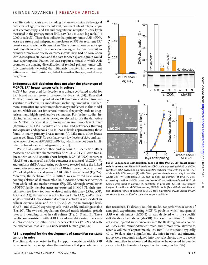
SC I ENCE ADVANCES | R E S EARCH ART I C L E
on Decem
ber 3, 2016http://advances.sciencem
ag.org/D
ownloaded from
a multivariate analysis after including the known clinical pathologicalpredictors of age, disease-free interval, dominant site of relapse, adju-vant chemotherapy, and ER and progesterone receptor mRNA levelsmeasured in the primary tumor [HR 2.19 (1.51 to 3.20); log-rank, P <0.0001; table S2]. These data indicate that primary tumor A3B mRNAlevels are strong and independent predictors of PFS for recurrent ER+
breast cancer treated with tamoxifen. These observations do not sup-port models in which resistance-conferring mutations preexist inprimary tumors—or disease outcomes would have had no correlationwith A3B expression levels and the data for each quartile group wouldhave superimposed. Rather, the data support a model in which A3Bpromotes the ongoing diversification of residual primary tumor cells(micrometastatic deposits) that ultimately manifest in the recurrentsetting as acquired resistance, failed tamoxifen therapy, and diseaseprogression.
Endogenous A3B depletion does not alter the phenotype ofMCF-7L ER+ breast cancer cells in cultureMCF-7 has been used for decades as a unique cell-based model forER+ breast cancer research [reviewed by Lee et al. (34)]. EngraftedMCF-7 tumors are dependent on ER function and therefore aresensitive to selective ER modulators, including tamoxifen. Further-more, tamoxifen-induced tumor dormancy (indolence) in this modelsystem, which can last for several months, frequently leads to drug-resistant and highly proliferative cell masses. For further studies, in-cluding animal experiments below, we elected to use the derivativeline MCF-7L because it is tumorigenic in immunodeficient mice[Ibrahim et al. (35), Sachdev et al. (36), and references therein]and expresses endogenous A3BmRNA at levels approximating thosefound in many primary breast tumors (7). Like most other breastcancer cell lines, MCF-7L cells have very low levels of A3A and var-iable levels of other APOBEC3 mRNAs, which have not been impli-cated in breast cancer mutagenesis (fig. S1).
We initially asked whether endogenous A3B depletion altersmolecular or cellular characteristics of MCF-7L. Cells were trans-duced with an A3B-specific short hairpin RNA (shRNA) construct(shA3B) or a nonspecific shRNA construct as a control (shCON) (7),and uniform shRNA-expressing pools were selected using the linkedpuromycin resistance gene. In all shA3B-transduced pools, a robust>25-fold depletion of endogenous A3BmRNA was achieved (Fig. 2A).Moreover, the depletion of A3B mRNA was mirrored by a corres-ponding ablation of all measurable DNA cytosine deaminase activitiesfrom whole-cell and nuclear extracts (Fig. 2B). Although several otherAPOBEC family member genes are expressed in MCF-7L, their pro-tein levels are likely too low to detect using this assay (A3A, A3D,A3G, and A1), the enzyme is not active on DNA (A2), and/or theirsingle-stranded DNA cytosine deaminase activity is not evident incellular extracts (A3C and A3F) (7, 22). At the microscopic level,shA3B- and shCON-expressing cells were visibly indistinguishable(Fig. 2C). The two cell populations showed nearly identical growthrates and doubling times in cell culture (Fig. 2, D and E). Theseresults are consistent with A3B knockdown data using the sameshRNA construct in other breast cancer cell lines (7, 22) and withthe observation that A3B is a nonessential human gene (37).
A3B is required for the development of tamoxifen-resistanttumors in miceThe clinical data reported in Fig. 1 support a model in which A3Bis responsible for precipitating the mutations that promote tamox-
Law et al., Sci. Adv. 2016;2 : e1601737 7 October 2016
ifen resistance. To directly test this model, we performed a series ofxenograft experiments using MCF-7L pools in which endogenousA3B was left intact (shCON) or was depleted with the specificshRNA described above (shA3B). For each condition, 5 millioncells were injected subcutaneously into the flank regions of a cohortof 5-week-old immunodeficient mice, and tumors were allowed toreach a volume of approximately 150 mm3. At this point, typically40 to 50 days after engraftment, the mice in each experimentalgroup were randomly assigned into two subcohorts, one to receivedaily tamoxifen injections and the other to be observed in parallelas a control (schematic of experimental design in Fig. 3A).
shCON shA3B0
0.05
0.10
0.15
MCF-7L
A3B
rel
ativ
e to
TB
P
A
D
shCON shA3B0
10
20
30
MCF-7L
Dou
blin
g tim
e (h
ours
)
E
5 1000
1
2
3
4
5
Days after plating
Cel
l num
ber
(mill
ions
)
V A3B W C N W C N
0
50
100
DN
A d
eam
inat
ion
(%)
7.7
24
S-
P-
shCON shA3B
25
75
Not
det
ecte
d
Not detectedNot
det
ecte
d
shCON
shA3B
C
B
400 µm
400 µm
Fig. 2. Endogenous A3B depletion does not alter MCF-7L ER+ breast cancercells in culture. (A) A3B mRNA levels in MCF-7L cells expressing shA3B or shCONconstructs (TBP, TATA-binding protein mRNA; each bar represents the mean ± SDof three RT-qPCR assays). (B) A3B DNA cytosine deaminase activity in solublewhole-cell (W), cytoplasmic (C), and nuclear (N) extracts of MCF-7L cellsexpressing shA3B or shCON constructs. Vector (V) and A3B-transfected 293T celllysates were used as controls (S, substrate; P, product). (C) Light microscopyimages of shA3B and shCON expressing MCF-7L pools. (D and E) Growth kineticsand doubling times of cultured MCF-7L cells expressing shA3B versus shCONconstructs (mean ± SD of n = 6 cultures per condition).
3 of 9

SC I ENCE ADVANCES | R E S EARCH ART I C L E
on Decem
ber 3, 2016http://advances.sciencem
ag.org/D
ownloaded from
Control-transduced MCF-7L cells formed large 1000-mm3 tu-mors within 100 days after engraftment and, interestingly, A3Bknockdown caused a modest delay in tumor growth (open blueversus open orange symbols in Fig. 3B; linear mixed model, F test,
Law et al., Sci. Adv. 2016;2 : e1601737 7 October 2016
P = 0.002). This result differed from the near-identical growth ratesin cell culture (Fig. 2, D and E) and may be due to the likelihoodthat additional adaptations/mutations are required for monolayer/plastic-conditioned cells to be able to grow optimally as tumors inmice. As expected, tamoxifen treatment attenuated the growth ofboth engineered pools (filled orange and blue symbols in Fig. 3B).However, control-transduced cells rapidly developed resistance to ta-moxifen and grew into large tumors, whereas the growth of the A3B-depleted cell masses was mostly suppressed by tamoxifen over theyear-long duration of this representative experiment (filled orangeversus blue symbols in Fig. 3B; linear mixed model, F test, P <0.0001). Similar outcomes were observed in additional experiments(for example, fig. S2).
Xenograft tumor A3B mRNA levels were analyzed by RT-qPCR,and, in all instances, the intended knockdown or control mRNAlevel was found to be durable and maintained through the entire du-ration of the experiment (Fig. 3C). This series of control experimentsalso revealed that endogenous A3B mRNA levels increase in controlshRNA–transduced tumor masses in comparison to the same cellsbefore engraftment (Fig. 3C). The mechanism for A3B inductionin immunodeficient mice is not known but is unlikely to be due toestrogen (figs. S3 and S4), as suggested by a recent report (38). Rep-resentative xenografts were recovered in culture, and the tamoxifen-resistant phenotype was reconfirmed (for example, Fig. 3D). Theseresults are fully supportive of a mechanism in which endogenousA3B causes an inheritable drug resistance phenotype (addressed fur-ther below). It is notable that endogenous A3B mRNA levels in thissystem are comparable to those observed in a large proportion ofprimary tumors [approximately 0.1 to 0.2 relative to TBP mRNAlevels in cultured MCF-7L cells (Fig. 2B), 0.4 relative to TBP in ani-mal tumors described here (Fig. 3C and fig. S3), and a range of 0 to1.25 and a median of 0.25 relative to TBP in primary breast tumorspreviously documented using the same RT-qPCR assay (7)].
A novel lentivirus-based system enables A3B overexpressionin any cell typeWe next developed a conditional A3B overexpression system to fur-ther test the A3B mutagenesis model. A conditional approach is re-quired because A3B expression in virus-producing cells causes lethalmutagenesis of retroviral complementary DNA intermediates duringreverse transcription (39–42), and excessive levels of cellular A3Bhave the potential to inflict genomic DNA damage that ultimatelyleads to cytotoxicity (7, 18, 19). We therefore developed a novel lenti-viral construct that will only express A3B upon transduction into sus-ceptible target cells (Fig. 4A). This construct mitigates viral toxicityissues because it is inactive in virus-producing cells as a result of dis-ruption of the antisense A3B open reading frame with a sense strandintron, and it is only expressed after intron removal by splicing in thevirus-producing cells and reverse transcription and integration of thefull proviral DNA in susceptible target cells. It also mitigates toxicityissues for target cell populations because expression levels are notexcessive (see below). In parallel, an A3B catalytic mutant derivative(E255Q) was created by site-directed mutagenesis to serve as a neg-ative control.
Transducing viruses were made by plasmid transfection into 293Tcells with appropriate retroviral helper plasmids encoding Gag, Pol,and Env (vesicular stomatitis virus glycoprotein). As anticipated, noproducer cell toxicity was observed, and A3B and A3B-E255Q viraltiters were equivalent by RT-qPCR. MCF-7L cells were transduced
Age
shA3B
shCONBiweekly
observationDailyTAM
ControlBiweekly
observation
Biweeklyobservation
DailyTAM
ControlBiweekly
observation
A
(weeks)0 403010 20
B
0 100 200 3000
250
500
750
1000
1250
Days after injection
Tum
or v
olum
e (m
m3 )
shCON + TAMshCON
shA3B + TAMshA3B
C
Inpu
t
Inpu
t
0
0.2
0.4
0.6
0.8
A3B
rel
ativ
e to
TB
P
shA3BCTRL +TAM
shCONCTRL +TAM
D
0
1
2
MT
T a
bs (
570
nM)
TAM (nM)
MCF-7Linput
TAMR
tumor
Fig. 3. A3B is required for the development of tamoxifen-resistant tumors inmice. (A) Schematic of the A3B knockdown xenograft study design and timecourse (see text for details). (B) Growth kinetics of engrafted MCF-7L cellsexpressing shA3B or shCON in the absence or presence of tamoxifen (TAM) treatment.Tumor volumes were measured weekly (mean + SEM shown for clarity of data presen-tation). (C) A3B mRNA levels in xenografted tumors recovered from the experimentshown in (B) (TBPmRNA; each bar represents the mean ± SD of three RT-qPCR assays).(D) MTT [3-(4,5-dimethylthiazol-2-yl)-2,5-diphenyltetrazolium bromide] data compar-ing tamoxifen susceptibility of input MCF-7L cells versus tamoxifen resistance of arepresentative MCF-7L shCON tumor [tamoxifen (10, 100, and 1000 ng/ml)].
4 of 9

SC I ENCE ADVANCES | R E S EARCH ART I C L E
on Decem
ber 3, 2016http://advances.sciencem
ag.org/D
ownloaded from
with each virus stock, and puromycin selection was used to eliminatenontransduced cells and to ensure 100% transduction efficiencies.A3B quantification by RT-qPCR showed that each construct elevatesmRNA expression to levels approximately 10-fold higher than thoseof the reference gene TBP (Fig. 4B), which equate to levels approxi-mately 50-fold higher than those of the endogenous A3B expressedin this system. These A3B mRNA levels are similar to those foundin the top fraction of breast tumors and cancer cell lines [Burnset al. (7), Leonard et al. (22), Sieuwerts et al. (23), and this study].As for the A3B knockdown experiments above, A3B- and A3B-E255Q–overexpressing MCF-7L populations showed no overt signsof toxicity and indistinguishable growth rates (Fig. 4C).
Overexpression of catalytically active A3B accelerates thedevelopment of tamoxifen-resistant tumorsTo further test the model in which A3B provides mutagenic fuel fortumor evolution and drug resistance, we performed a series ofxenograft experiments using MCF-7L cells transduced with theaforementioned constructs and thereby overexpressing wild-typeA3B or the catalytic mutant derivative A3B-E255Q (Fig. 5A). Im-munodeficient animals were injected subcutaneously with 5 millioncells and, upon palpable tumor growth (150 mm3), randomlydivided into groups for tamoxifen injections or control observation.Remarkably, most of the cell masses overexpressing A3B developedrapid resistance to tamoxifen (filled red symbols in Fig. 5B). Incomparison, MCF-7L cells expressing equivalent levels of A3B-E255Q mutant mRNA showed resistance kinetics similar to thoseof the shCON engraftments described above (filled orange symbolsin Fig. 5B; linear mixed model, F test, P = 0.015). An independentexperiment yielded similar results (fig. S5). These data demonstratethat A3B overexpression accelerates the kinetics of the developmentof tamoxifen resistance and, notably, that this phenotype requirescatalytic activity.
Reverse transcription/integration
Producer cellTarget cell
Transcription/splicing/packaging
PRSV
5'LTR 3'LTR
PSV40
PCMV/TO
A3BNTDA3BCTD
Intron PuroR
A3BNTDA3BCTD
polyA5'LTR 3'LTR
PSV40
PCMV/TO
A3B
A3BNTDA3BCTD
5'LTR 3'LTR
PSV40
PCMV/TO
A
PuroR
PuroR
PuroR
B
+A3B +E255Q0.01
0.1
1
10
100
A3B
rel
ativ
e to
TB
P
C
+A3B +E255Q0
10
20
30
Dou
blin
g tim
e (h
ours
)
Fig. 4. Novel lentivirus-based system for conditional A3B overexpression. (A) Schematic of the lentiviral construct for conditional A3B overexpression (see text fordetails). LTR, long terminal repeat; RSV, Rous sarcoma virus; CTD, C-terminal domain; NTD, N-terminal domain; CMV, cytomegalovirus; SV40, simian virus 40. (B) A3BmRNA levels relative to TBP in MCF-7L cells expressing lentivirus-delivered A3B or a catalytic mutant derivative (E255Q) as well as endogenous A3B (mean ± SD of threeRT-qPCR assays). (C) Doubling times of cultured MCF-7L cells overexpressing A3B or A3B-E255Q (mean ± SD of four replicates).
Law et al., Sci. Adv. 2016;2 : e1601737 7 October 2016
B
25 50 75 100 1250
500
1000
Days after injection
Tum
or v
olum
e (m
m3 )
+A3B+A3B +TAM+E255Q+E255Q +TAM
1250
750
250
Age
+E255Q
+A3BBiweekly
observationDailyTAM
ControlBiweekly
observation
Biweeklyobservation
DailyTAM
ControlBiweekly
observation
A
(weeks)0 403010 20
150
Fig. 5. Overexpression of catalytically active A3B accelerates the develop-ment of tamoxifen-resistant tumors in mice. (A) Schematic of the A3B overex-pression xenograft study design and time course (see text for details). (B) Growthkinetics of engrafted MCF-7L cells overexpressing A3B or A3B-E255Q in the ab-sence or presence of tamoxifen treatment. The graph reports tumor volumesmeasured weekly (mean + SEM shown for clarity of data presentation). Averagetumor volumes from the untreated control arms are shown by gray symbols, andoverlapping error bars are omitted for clarity of presentation.
5 of 9

SC I ENCE ADVANCES | R E S EARCH ART I C L E
Dow
nloaded from
ESR1 mutations are not responsible for tamoxifen resistancein the MCF-7L model for ER+ breast cancerAlthough the development of tamoxifen-resistant breast tumors isa major clinical problem, in most cases the molecular basis forresistance is unknown. A small fraction of treated patients developtumors with ESR1 exonic mutations that cause amino acid changesin the hormone-binding domain of the ER. These mutations havebeen seen mostly in tumors resistant to aromatase inhibitors andnot as frequently in tumors resistant to tamoxifen [reviewed byClarke et al. (43) and Jeselsohn et al. (44)]. To determine whetherESR1 mutations are also part of the tamoxifen resistance mecha-nism in MCF-7L cells, we performed DNA exome sequencing on9 independent tamoxifen-resistant xenografts and 10 independentcontrol tumor masses. The ESR1 gene contained no mutations un-der either condition (see table S3 for a full list of base substitutionmutations). Resistant tumor ESR1 mRNA levels were somewhatvariable but still similar to those present in the original MCF-7Lcell populations (fig. S6). Together with the data presented aboveindicating heritable resistance to tamoxifen (Fig. 3D), these resultssuggest that at least one other resistance mechanism occurs in theMCF-7 model system for ER+ breast cancer.
on Decem
ber 3, 2016http://advances.sciencem
ag.org/
DISCUSSIONThe clinical and xenograft results presented here strongly support amodel in which A3B drives tamoxifen resistance in ER+ breastcancer. Clinically, resistance to endocrine therapies has been definedas primary or secondary, depending on the length of time a patientbenefits from ER-targeted therapy. Our data suggest that A3B mayhave a role in both kinds of resistance and particularly in the develop-ment of secondary, acquired resistance. Suppression of endogenouslevels of A3B enhances tamoxifen benefit (Fig. 3), whereas overex-pression of A3B eliminates almost all benefits from tamoxifen ther-apy (Fig. 5). Because the only known biochemical activity of A3B issingle-stranded DNA cytosine deamination [for example, (7, 42, 45)]and the tamoxifen resistance phenotype is heritable (Fig. 3D), themost likely mechanism is A3B-catalyzed DNA C-to-U editing coupledto the processing of these uracil lesions into somatic mutations bynormal DNA repair processes [reviewed by Swanton et al. (11), Robertsand Gordenin (12), and Helleday et al. (13)]. In further support of thismechanism, the catalytic glutamate of A3B (E255) is required foraccelerated tamoxifen resistance kinetics upon enzyme overexpression.
Because ESR1mutations were not observed in MCF-7L tamoxifen-resistant tumors, the identity of the resistance-conferring mutationsin this system will require significant future studies and possibly evenwhole-genome sequencing if the predominant causal lesions lie out-side the exomic fraction of the genome. The intrinsic signature ofA3B may help to identify candidate (frequently mutated) sites formechanistic follow-up. Then, for instance, genetic knock-in experi-ments could be used to unambiguously establish a cause-effect re-lationship. However, the resistance-conferring mutations (such asgene translocations, amplifications, or deletions) could also be complexand difficult to recapitulate precisely because DNA repair enzymescan readily process genomic uracil lesions into single- and double-stranded breaks (46, 47).
A3Bhas been implicated as a dominant source ofmutation in breast,head/neck, lung, bladder, and cervical cancers and—to a lesser but stillsignificant extent—in many other tumor types (7–10, 28, 32, 48, 49).The fundamental nature of theDNAdeaminationmechanism, together
Law et al., Sci. Adv. 2016;2 : e1601737 7 October 2016
with the data presented here, strongly suggests that A3B may be a gen-eral mechanism of therapeutic resistance to cancer therapy. At thispoint, potential mutagenic contributions from other APOBEC3 familymembers, such as A3A, cannot be excluded fully, but they do not ap-pear to manifest in the MCF-7L system, nor are these potential contri-butions large enough to prevent the significant association betweenA3B expression levels and clinical outcomes for ER+ breast cancerpatients [treatment-naïve data in the studies by Sieuwerts et al. (23)and Cescon et al. (24) and post-recurrence tamoxifen resistance datain Fig. 1]. Thus, strategies to down-regulate A3B activity or expression,as reported here using a specific shRNA knockdown construct in amodel system for ER+ breast cancer, may be beneficial as chemo-therapeutic adjuvants to “turn down” the mutation rate, decrease thelikelihood of evolving drug resistance, and prolong the clinical benefitof therapy for the many cancers that are likely to be driven by thisongoing mutational process.
MATERIALS AND METHODSClinical studiesThe clinical characteristics of the 285 patients [225 from Rotterdam(Erasmus University Medical Center) and 60 from Nijmegen (RadboudUniversity Medical Center)] whose primary tumor specimens and datawere used here have been described previously by Sieuwerts et al. (33).The protocol to study biological markers associated with disease out-come was approved by the medical ethics committee of the ErasmusUniversity Medical Center (Rotterdam, Netherlands) (MEC 02.953);for Nijmegen, coded primary tumor tissues were used in accordancewith the Codes of Conduct of the Federation of Medical Scientific So-cieties in the Netherlands (www.federa.org/codes-conduct). Thirty-two patients presented with distant metastasis at diagnosis or developeddistant metastasis (including supraclavicular lymph node metastasis)within 1 month following primary surgery (M1 patients). These 32patients and the 253 patients who developed a first recurrence duringfollow-up (25 patients with local-regional relapse and 228 patientswith distant metastasis) were treated with first-line tamoxifen. All pa-tients were ER+ and anti–hormonal therapy–naïve, but 38 patientsreceived adjuvant chemotherapy. The median time between theprimary surgery and the start of therapy was 24 months (range, 0to 120 months). The median follow-up of patients alive at the end offollow-up was 98 months (range, 9 to 240 months) after the primarysurgery and 45 months (range, 3 to 178 months) after the start offirst-line tamoxifen therapy. For 182 patients (64%), disease progres-sion occurred within 6 months of the start of the first-line therapybeing controlled by tamoxifen. At the end of the follow-up period,268 (94%) patients had developed tumor progression, and 222(78%) patients had died.
Total RNA was extracted with RNA Bee (Tel Test, Thermo FisherScientific Inc.) from 30-mm fresh-frozen primary tumor tissue sectionscontaining at least 30% invasive tumor cell nuclei, and mRNA tran-scripts were quantified by RT-qPCR as described previously by Sieuwertset al. (23). The median A3B expression level in the group of 285 breastcancers was 0.22 relative to the normalized average of three referencegenes [HPRT1, HMBS, and TBP (23)].
DNA constructsA3B knockdown and control shRNA constructs were described andvalidated previously by Burns et al. (7) and Leonard et al. (50). TheA3B and A3B-E255Q lentiviral expression constructs were based
6 of 9

SC I ENCE ADVANCES | R E S EARCH ART I C L E
on Decem
ber 3, 2016http://advances.sciencem
ag.org/D
ownloaded from
on the pLenti4TO backbone (Life Technologies). Overlapping PCRwas used to place a sense-encoded intron between an antisense-encoded A3B open reading frame (primers available on request). Acytomegalovirus promoter drove A3B expression, and a simian virus40 early promoter drove puromycin resistance. Constructs were ver-ified by DNA sequencing.
Cell culture studiesMCF-7L cells were cultured at 37°C under 5% CO2 and maintainedin improved minimum essential medium (Richter’s modificationmedium) containing 5% fetal bovine serum, penicillin (100 U/ml),streptomycin (100 mg/ml), and 11.25 nM recombinant human in-sulin. These cells were originally obtained from C. Kent Osborne(Baylor College of Medicine, Houston, TX) and are subject to shorttandem repeat analysis yearly to confirm their identity with theoriginal MCF-7 cell line. Cells were transduced with the lentivirus-based shRNA or conditional expression constructs described aboveand selected with puromycin (1 mg/ml; United States Biological) for72 hours to generate uniformly transduced pools. Cell growth ex-periments were performed by plating 100,000 cells per six-well plateand incubating them at 37°C for the indicated days. Cells were tryp-sinized, diluted 1:2 in trypan blue (Invitrogen), and counted via a he-mocytometer (six biological replicates per day per condition). Cellproliferation rates were determined using the xCELLigence real-timecell analyzer dual-plate instrument according to the manufacturer’sinstructions (ACEA Biosciences).
The mRNA level of eachAPOBEC family member gene was quan-tified using previously described RT-qPCR protocols and primer/probe combinations and presented relative to the housekeeping geneTBP (7, 51, 52). ESR1 andC-MYCRNAwere quantified by RT-qPCRusing intron-spanning primers 5′-ATGACCATGACCCTCCA-CACC and 5′-TCAGACCGTGGCAGGGAAACC (UPL24) and5′-GCTGCTTAGACGCTGGATTT and 5′-TAACGTTGAGGGG-CATCG (UPL66), respectively, and manufacturer-recommendedprotocols (LightCycler 480, Roche). C-MYC is an established estrogen-responsive gene (53).
DNA deaminase activity was measured in soluble whole-cell,nuclear, and cytoplasmic fractions of MCF-7L cultures usingestablished protocols (7, 54). The single-stranded DNA substratecontained a single target cytosine (5′-ATTATTATTATTC-GAATGGATTTATTTATTTATTTATTTATTT-fluorescein); de-amination, uracil excision, and backbone cleavage resulted in asingle faster-migrating product on SDS–polyacrylamide gel electro-phoresis and image analysis (Typhoon FLA 7000 and ImageQuantsoftware, GE Healthcare Life Sciences).
Xenograft studiesThe University of Minnesota Institutional Animal Care and UseCommittee approved the animal protocols used here (1305-30638A). MCF-7L cells were harvested at 70% confluence, counted,and resuspended in serum-free medium (without phenol red) at aconcentration of 5 million cells per 50 ml of final volume. Ovariec-tomized, athymic mice (Harlan) were injected subcutaneously inthe left flank with 50 ml of cell suspension at approximately 5 weeksof age. Each experiment was initiated with 5 or 10 mice per exper-imental condition. One week before injection and at all times fol-lowing, the mice were provided with drinking water supplementedwith 1 mM b-estradiol (Sigma-Aldrich) (except for the subset ofmice used in the experiment shown in fig. S3). Tumors were measured
Law et al., Sci. Adv. 2016;2 : e1601737 7 October 2016
bidirectionally twice weekly, and tamoxifen treatment began when theaverage tumor volume reached 150 mm3. Tamoxifen citrate (500 mg;Sigma-Aldrich) emulsified in 50 ml of peanut oil was administeredsubcutaneously 5 of 7 days each week. Tumor volumes were calculatedusing the following formula: length × breadth2/2.
MCF-7L exome sequencingGenomicDNAwas prepared from tumor cellmasses (~20mg per sam-ple) via the Gentra Puregene Tissue DNA isolation protocols (Qiagen).Samples were diluted to 100 ng/ml and assessed further for quality andpurity by SYBRGreenPCRon a 197-bp fragment ofA3H using primers5′-CATGGGACTGGACGAAGCGCA and 5′-TGGGATCCACACA-GAAGCCGCA. Samples with no amplification were excluded fromthe analysis. One microgram of total genomic DNA per sample wassubjected to whole-exome sequencing on the Complete Genomicsplatform to an average target depth of 100× (BGI). Reads werealigned by BGI using its in-house pipeline, and the alignments in bamformat were used for variant calling. Somatic variants were called foreach tumor alignment by VarScan 2 (55) using an estrogen-treatedshA3B sample as the normal control. The variants were filtered with aminimum overall coverage depth of 20 reads and a minimum coveragedepth of 4 reads for the alternate allele. Any variant occurring at anyfrequency above 0 at the same position in more than one sample wasconsidered a common mutation in the input pool and was removed.A full list of base substitution mutations is provided in table S3.
StatisticsComparisons of the PFS of hormone-naïve breast cancer patientsfollowing treatment for first recurrence with tamoxifen, by A3B ex-pression level (divided into quartiles), were conducted using log-rank tests; HRs and 95% confidence intervals are presented forpairwise comparisons. Clinical data were analyzed using SPSS Sta-tistics version 23.0 (IBM). In the xenograft studies, repeated mea-sures of tumor volume over time were compared by treatmentgroup using linear mixed models with fixed effects for treatment,days, and interaction between treatment and days and with randomintercept and slope effects for each mouse. P values <0.05 wereconsidered statistically significant. Xenograft data were analyzedusing Prism 6 and SAS 9.3.
SUPPLEMENTARY MATERIALSSupplementary material for this article is available at http://advances.sciencemag.org/cgi/content/full/2/10/e1601737/DC1fig. S1. APOBEC family member expression in MCF-7L cells.fig. S2. Replica A3B knockdown xenograft experiment.fig. S3. Estrogen does not affect A3B mRNA levels in engrafted MCF-7L cells.fig. S4. A3B is not estrogen-inducible.fig. S5. Replica A3B overexpression xenograft experiment.fig. S6. ESR1 mRNA levels in tamoxifen-resistant MCF-7L cells.table S1. Patient characteristics and median and interquartile range of APOBEC3B mRNA levels.table S2. Cox univariate and multivariate analyses for PFS after initiating first-line tamoxifen.table S3. Single-base substitution mutations in tamoxifen-resistant tumors (separate MicrosoftExcel file).
REFERENCES AND NOTES1. E. B. Elkin, C. A. Hudis, Parsing progress in breast cancer. J. Clin. Oncol. 33, 2837–2838
(2015).2. J. H. Park, W. F. Anderson, M. H. Gail, Improvements in US breast cancer survival and
proportion explained by tumor size and estrogen-receptor status. J. Clin. Oncol. 33,2870–2876 (2015).
7 of 9

SC I ENCE ADVANCES | R E S EARCH ART I C L E
on Decem
ber 3, 2016http://advances.sciencem
ag.org/D
ownloaded from
3. T. Saphner, D. C. Tormey, R. Gray, Annual hazard rates of recurrence for breast cancerafter primary therapy. J. Clin. Oncol. 14, 2738–2746 (1996).
4. H. J. Burstein, S. Temin, H. Anderson, T. A. Buchholz, N. E. Davidson, K. E. Gelmon,S. H. Giordano, C. A. Hudis, D. Rowden, A. J. Solky, V. Stearns, E. P. Winer, J. J. Griggs,Adjuvant endocrine therapy for women with hormone receptor–positive breast cancer:American Society of Clinical Oncology clinical practice guideline focused update.J. Clin. Oncol. 32, 2255–2269 (2014).
5. C. Davies, H. Pan, J. Godwin, R. Gray, R. Arriagada, V. Raina, M. Abraham,V. H. Medeiros Alencar, A. Badran, X. Bonfill, J. Bradbury, M. Clarke, R. Collins, S. R. Davis,A. Delmestri, J. F. Forbes, P. Haddad, M.-F. Hou, M. Inbar, H. Khaled, J. Kielanowska,W. H. Kwan, B. S. Mathew, I. Mittra, B. Müller, A. Nicolucci, O. Peralta, F. Pernas,L. Petruzelka, T. Pienkowski, R. Radhika, B. Rajan, M. T. Rubach, S. Tort, G. Urrútia,M. Valentini, Y. Wang, R. Peto; Adjuvant Tamoxifen: Longer Against Shorter Collaborative,Long-term effects of continuing adjuvant tamoxifen to 10 years versus stopping at5 years after diagnosis of oestrogen receptor-positive breast cancer: ATLAS, arandomised trial. Lancet 381, 805–816 (2013).
6. S. Nik-Zainal, L. B. Alexandrov, D. C. Wedge, P. Van Loo, C. D. Greenman, K. Raine, D. Jones,J. Hinton, J. Marshall, L. A. Stebbings, A. Menzies, S. Martin, K. Leung, L. Chen, C. Leroy,M. Ramakrishna, R. Rance, K. W. Lau, L. J. Mudie, I. Varela, D. J. McBride, G. R. Bignell,S. L. Cooke, A. Shlien, J. Gamble, I. Whitmore, M. Maddison, P. S. Tarpey, H. R. Davies,E. Papaemmanuil, P. J. Stephens, S. McLaren, A. P. Butler, J. W. Teague, G. Jönsson,J. E. Garber, D. Silver, P. Miron, A. Fatima, S. Boyault, A. Langerød, A. Tutt, J. W. M. Martens,S. A. J. R. Aparicio, Å. Borg, A. V. Salomon, G. Thomas, A.-L. Børresen-Dale, A. L. Richardson,M. S. Neuberger, P. A. Futreal, P. J. Campbell, M. R. Stratton; Breast Cancer WorkingGroup of the International Cancer Genome Consortium, Mutational processes molding thegenomes of 21 breast cancers. Cell 149, 979–993 (2012).
7. M. B. Burns, L. Lackey, M. A. Carpenter, A. Rathore, A. M. Land, B. Leonard, E. W. Refsland,D. Kotandeniya, N. Tretyakova, J. B. Nikas, D. Yee, N. A. Temiz, D. E. Donohue,R. M. McDougle, W. L. Brown, E. K. Law, R. S. Harris, APOBEC3B is an enzymatic source ofmutation in breast cancer. Nature 494, 366–370 (2013).
8. M. B. Burns, N. A. Temiz, R. S. Harris, Evidence for APOBEC3B mutagenesis in multiplehuman cancers. Nat. Genet. 45, 977–983 (2013).
9. S. A. Roberts, M. S. Lawrence, L. J. Klimczak, S. A. Grimm, D. Fargo, P. Stojanov, A. Kiezun,G. V. Kryukov, S. L. Carter, G. Saksena, S. Harris, R. R. Shah, M. A. Resnick, G. Getz,D. A. Gordenin, An APOBEC cytidine deaminase mutagenesis pattern is widespread inhuman cancers. Nat. Genet. 45, 970–976 (2013).
10. L. B. Alexandrov, S. Nik-Zainal, D. C. Wedge, S. A. J. R. Aparicio, S. Behjati, A. V. Biankin,G. R. Bignell, N. Bolli, A. Borg, A.-L. Børresen-Dale, S. Boyault, B. Burkhardt, A. P. Butler,C. Caldas, H. R. Davies, C. Desmedt, R. Eils, J. E. Eyfjörd, J. A. Foekens, M. Greaves,F. Hosoda, B. Hutter, T. Ilicic, S. Imbeaud, M. Imielinsk, N. Jäger, D. T. W. Jones,D. Jones, S. Knappskog, M. Kool, S. R. Lakhani, C. López-Otín, S. Martin, N. C. Munshi,H. Nakamura, P. A. Northcott, M. Pajic, E. Papaemmanuil, A. Paradiso, J. V. Pearson,X. S. Puente, K. Raine, M. Ramakrishna, A. L. Richardson, J. Richter, P. Rosenstiel,M. Schlesner, T. N. Schumacher, P. N. Span, J. W. Teague, Y. Totoki, A. N. J. Tutt,R. Valdés-Mas, M. M. van Buuren, L. van‘t Veer, A. Vincent-Salomon, N. Waddell,L. R. Yates; Australian Pancreatic Cancer Genome Initiative, ICGC Breast CancerConsortium, ICGC MMML-Seq Consortium, ICGC PedBrain, J. Zucman-Rossi,P. A. Futreal, U. McDermott, P. Lichter, M. Meyerson, S. M. Grimmond, R. Siebert,E. Campo, T. Shibata, S. M. Pfister, P. J. Campbell, M. R. Stratton, Signatures ofmutational processes in human cancer. Nature 500, 415–421 (2013).
11. C. Swanton, N. McGranahan, G. J. Starrett, R. S. Harris, APOBEC enzymes: Mutagenic fuelfor cancer evolution and heterogeneity. Cancer Discov. 5, 704–712 (2015).
12. S. A. Roberts, D. A. Gordenin, Hypermutation in human cancer genomes: Footprintsand mechanisms. Nat. Rev. Cancer 14, 786–800 (2014).
13. T. Helleday, S. Eshtad, S. Nik-Zainal, Mechanisms underlying mutational signatures inhuman cancers. Nat. Rev. Genet. 15, 585–598 (2014).
14. M. H. Malim, P. D. Bieniasz, HIV restriction factors and mechanisms of evasion. Cold SpringHarb. Perspect. Med. 2, a006940 (2012).
15. S. Stavrou, S. R. Ross, APOBEC3 proteins in viral immunity. J. Immunol. 195, 4565–4570 (2015).16. V. Simon, N. Bloch, N. R. Landau, Intrinsic host restrictions to HIV-1 and mechanisms
of viral escape. Nat. Immunol. 16, 546–553 (2015).17. S. Landry, I. Narvaiza, D. C. Linfesty, M. D. Weitzman, APOBEC3A can activate the DNA
damage response and cause cell-cycle arrest. EMBO Rep. 12, 444–450 (2011).18. M. Shinohara, K. Io, K. Shindo, M. Matsui, T. Sakamoto, K. Tada, M. Kobayashi, N. Kadowaki,
A. Takaori-Kondo, APOBEC3B can impair genomic stability by inducing basesubstitutions in genomic DNA in human cells. Sci. Rep. 2, 806 (2012).
19. B. J. M. Taylor, S. Nik-Zainal, Y. L. Wu, L. A. Stebbings, K. Raine, P. J. Campbell, C. Rada,M. R. Stratton, M. S. Neuberger, DNA deaminases induce break-associated mutation showerswith implication of APOBEC3B and 3A in breast cancer kataegis. ELife 2, e00534 (2013).
20. B. Mussil, R. Suspène, M.-M. Aynaud, A. Gauvrit, J.-P. Vartanian, S. Wain-Hobson, HumanAPOBEC3A isoforms translocate to the nucleus and induce DNA double strand breaksleading to cell stress and death. PLOS One 8, e73641 (2013).
Law et al., Sci. Adv. 2016;2 : e1601737 7 October 2016
21. V. Caval, R. Suspène, M. Shapira, J.-P. Vartanian, S. Wain-Hobson, A prevalent cancersusceptibility APOBEC3A hybrid allele bearing APOBEC3B 3′UTR enhances chromosomalDNA damage. Nat. Commun. 5, 5129 (2014).
22. B. Leonard, J. L. McCann, G. J. Starrett, L. Kosyakovsky, E. M. Luengas, A. M. Molan,M. B. Burns, R. M. McDougle, P. J. Parker, W. L. Brown, R. S. Harris, The PKC/NF-kB signalingpathway induces APOBEC3B expression in multiple human cancers. Cancer Res. 75,4538–4547 (2015).
23. A. M. Sieuwerts, S. Willis, M. B. Burns, M. P. Look, M. E. Meijer-Van Gelder, A. Schlicker,M. R. Heideman, H. Jacobs, L. Wessels, B. Leyland-Jones, K. P. Gray, J. A. Foekens,R. S. Harris, J. W. M. Martens, Elevated APOBEC3B correlates with poor outcomes forestrogen-receptor-positive breast cancers. Horm. Cancer 5, 405–413 (2014).
24. D. W. Cescon, B. Haibe-Kains, T. W. Mak, APOBEC3B expression in breast cancer reflectscellular proliferation, while a deletion polymorphism is associated with immuneactivation. Proc. Natl. Acad. Sci. U.S.A. 112, 2841–2846 (2015).
25. L. R. Yates, M. Gerstung, S. Knappskog, C. Desmedt, G. Gundem, P. Van Loo, T. Aas,L. B. Alexandrov, D. Larsimont, H. Davies, Y. Li, Y. S. Ju, M. Ramakrishna, H. K. Haugland,P. K. Lilleng, S. Nik-Zainal, S. McLaren, A. Butler, S. Martin, D. Glodzik, A. Menzies,K. Raine, J. Hinton, D. Jones, L. J. Mudie, B. Jiang, D. Vincent, A. Greene-Colozzi, P.-Y. Adnet,A. Fatima, M. Maetens, M. Ignatiadis, M. R. Stratton, C. Sotiriou, A. L. Richardson,P. E. Lønning, D. C. Wedge, P. J. Campbell, Subclonal diversification of primary breastcancer revealed by multiregion sequencing. Nat. Med. 21, 751–759 (2015).
26. A. N. Hata, M. J. Niederst, H. L. Archibald, M. Gomez-Caraballo, F. M. Siddiqui, H. E. Mulvey,Y. E. Maruvka, F. Ji, H. E. Bhang, V. Krishnamurthy Radhakrishna, G. Siravegna, H. Hu,S. Raoof, E. Lockerman, A. Kalsy, D. Lee, C. L. Keating, D. A. Ruddy, L. J. Damon, A. S. Crystal,C. Costa, Z. Piotrowska, A. Bardelli, A. J. Iafrate, R. I. Sadreyev, F. Stegmeier, G. Getz,L. V. Sequist, A. C. Faber, J. A. Engelman, Tumor cells can follow distinct evolutionary pathsto become resistant to epidermal growth factor receptor inhibition. Nat. Med. 22, 262–269(2016).
27. K. Kemper, O. Krijgsman, P. Cornelissen-Steijger, A. Shahrabi, F. Weeber, J.-Y. Song,T. Kuilman, D. J. Vis, L. F. Wessels, E. E. Voest, T. N. M. Schumacher, C. U. Blank, D. J. Adams,J. B. Haanen, D. S. Peeper, Intra- and inter-tumor heterogeneity in a vemurafenib-resistantmelanoma patient and derived xenografts. EMBO Mol. Med. 7, 1104–1118 (2015).
28. S. Nik-Zainal, H. Davies, J. Staaf, M. Ramakrishna, D. Glodzik, X. Zou, I. Martincorena,L. B. Alexandrov, S. Martin, D. C. Wedge, P. Van Loo, Y. S. Ju, M. Smid, A. B. Brinkman,S. Morganella, M. R. Aure, O. C. Lingjærde, A. Langerød, M. Ringnér, S.-M. Ahn, S. Boyault,J. E. Brock, A. Broeks, A. Butler, C. Desmedt, L. Dirix, S. Dronov, A. Fatima, J. A. Foekens,M. Gerstung, G. K. J. Hooijer, S. J. Jang, D. R. Jones, H.-Y. Kim, T. A. King, S. Krishnamurthy,H.-J. Lee, J. Y. Lee, Y. Li, S. McLaren, A. Menzies, V. Mustonen, S. O’Meara, I. Pauporté,X. Pivot, C. A. Purdie, K. Raine, K. Ramakrishnan, F. G. Rodríguez-González, G. Romieu,A. M. Sieuwerts, P. T. Simpson, R. Shepherd, L. Stebbings, O. A. Stefansson, J. Teague,S. Tommasi, I. Treilleux, G. G. Van den Eynden, P. Vermeulen, A. Vincent-Salomon, L. Yates,C. Caldas, L. van’t Veer, A. Tutt, S. Knappskog, B. K. T. Tan, J. Jonkers, Å. Borg, N. T. Ueno,C. Sotiriou, A. Viari, P. A. Futreal, P. J. Campbell, P. N. Span, S. Van Laere, S. R. Lakhani,J. E. Eyfjord, A. M. Thompson, E. Birney, H. G. Stunnenberg, M. J. van de Vijver,J. W. M. Martens, A.-L. Børresen-Dale, A. L. Richardson, G. Kong, G. Thomas, M. R. Stratton,Landscape of somatic mutations in 560 breast cancer whole-genome sequences.Nature 534, 47–54 (2016).
29. D. Juric, P. Castel, M. Griffith, O. L. Griffith, H. H. Won, H. Ellis, S. H. Ebbesen, B. J. Ainscough,A. Ramu, G. Iyer, R. H. Shah, T. Huynh, M. Mino-Kenudson, D. Sgroi, S. Isakoff, A. Thabet,L. Elamine, D. B. Solit, S. W. Lowe, C. Quadt, M. Peters, A. Derti, R. Schegel, A. Huang,E. R. Mardis, M. F. Berger, J. Baselga, M. Scaltriti, Convergent loss of PTEN leads to clinicalresistance to a PI(3)Ka inhibitor. Nature 518, 240–244 (2015).
30. S. P. Shah, A. Roth, R. Goya, A. Oloumi, G. Ha, Y. Zhao, G. Turashvili, J. Ding, K. Tse,G. Haffari, A. Bashashati, L. M. Prentice, J. Khattra, A. Burleigh, D. Yap, V. Bernard,A. McPherson, K. Shumansky, A. Crisan, R. Giuliany, A. Heravi-Moussavi, J. Rosner, D. Lai,I. Birol, R. Varhol, A. Tam, N. Dhalla, T. Zeng, K. Ma, S. K. Chan, M. Griffith, A. Moradian,S.-W. G. Cheng, G. B. Morin, P. Watson, K. Gelmon, S. Chia, S.-F. Chin, C. Curtis, O. M. Rueda,P. D. Pharoah, S. Damaraju, J. Mackey, K. Hoon, T. Harkins, V. Tadigotla, M. Sigaroudinia,P. Gascard, T. Tlsty, J. F. Costello, I. M. Meyer, C. J. Eaves, W. W. Wasserman, S. Jones,D. Huntsman, M. Hirst, C. Caldas, M. A. Marra, S. Aparicio, The clonal and mutationalevolution spectrum of primary triple-negative breast cancers. Nature 486, 395–399(2012).
31. S. Nik-Zainal, P. Van Loo, D. C. Wedge, L. B. Alexandrov, C. D. Greenman, K. W. Lau,K. Raine, D. Jones, J. Marshall, M. Ramakrishna, A. Shlien, S. L. Cooke, J. Hinton, A. Menzies,L. A. Stebbings, C. Leroy, M. Jia, R. Rance, L. J. Mudie, S. J. Gamble, P. J. Stephens,S. McLaren, P. S. Tarpey, E. Papaemmanuil, H. R. Davies, I. Varela, D. J. McBride, G. R. Bignell,K. Leung, A. P. Butler, J. W. Teague, S. Martin, G. Jönsson, O. Mariani, S. Boyault,P. Miron, A. Fatima, A. Langerød, S. A. J. R. Aparicio, A. Tutt, A. M. Sieuwerts, Å. Borg,G. Thomas, A. V. Salomon, A. L. Richardson, A.-L. Børresen-Dale, P. A. Futreal, M. R. Stratton,P. J. Campbell, The life history of 21 breast cancers. Cell 149, 994–1007 (2012).
32. E. C. de Bruin, N. McGranahan, R. Mitter, M. Salm, D. C. Wedge, L. Yates, M. Jamal-Hanjani,S. Shafi, N. Murugaesu, A. J. Rowan, E. Grönroos, M. A. Muhammad, S. Horswell,
8 of 9

SC I ENCE ADVANCES | R E S EARCH ART I C L E
on Decem
ber 3, 2016http://advances.sciencem
ag.org/D
ownloaded from
M. Gerlinger, I. Varela, D. Jones, J. Marshall, T. Voet, P. Van Loo, D. M. Rassl, R. C. Rintoul,S. M. Janes, S.-M. Lee, M. Forster, T. Ahmad, D. Lawrence, M. Falzon, A. Capitanio,T. T. Harkins, C. C. Lee, W. Tom, E. Teefe, S.-C. Chen, S. Begum, A. Rabinowitz, B. Phillimore,B. Spencer-Dene, G. Stamp, Z. Szallasi, N. Matthews, A. Stewart, P. Campbell, C. Swanton,Spatial and temporal diversity in genomic instability processes defines lung cancerevolution. Science 346, 251–256 (2014).
33. A. M. Sieuwerts, M. B. Lyng, M. E. Meijer-van Gelder, V. de Weerd, F. C. Sweep,J. A. Foekens, P. N. Span, J. W. Martens, H. J. Ditzel, Evaluation of the ability of adjuvanttamoxifen-benefit gene signatures to predict outcome of hormone-naive estrogenreceptor-positive breast cancer patients treated with tamoxifen in the advanced setting.Mol. Oncol. 8, 1679–1689 (2014).
34. A. V. Lee, S. Oesterreich, N. E. Davidson, MCF-7 cells—Changing the course of breastcancer research and care for 45 years. J. Natl. Cancer Inst. 107, djv073 (2015).
35. Y. H. Ibrahim, S. A. Byron, X. Cui, A. V. Lee, D. Yee, Progesterone receptor-B regulation ofinsulin-like growth factor—Stimulated cell migration in breast cancer cells via insulinreceptor substrate-2. Mol. Cancer Res. 6, 1491–1498 (2008).
36. D. Sachdev, X. Zhang, I. Matise, M. Gaillard-Kelly, D. Yee, The type I insulin-like growthfactor receptor regulates cancer metastasis independently of primary tumor growthby promoting invasion and survival. Oncogene 29, 251–262 (2010).
37. J. M. Kidd, T. L. Newman, E. Tuzun, R. Kaul, E. E. Eichler, Population stratification of acommon APOBEC gene deletion polymorphism. PLOS Genet. 3, e63 (2007).
38. M. Periyasamy, H. Patel, C.-F. Lai, V. T. M. Nguyen, E. Nevedomskaya, A. Harrod, R. Russell,J. Remenyi, A. M. Ochocka, R. S. Thomas, F. Fuller-Pace, B. Győrffy, C. Caldas,N. Navaratnam, J. S. Carroll, W. Zwart, R. C. Coombes, L. Magnani, L. Buluwela, S. Ali,APOBEC3B-mediated cytidine deamination is required for estrogen receptor action inbreast cancer. Cell Rep. 13, 108–121 (2015).
39. K. N. Bishop, R. K. Holmes, A. M. Sheehy, N. O. Davidson, S.-J. Cho, M. H. Malim, Cytidinedeamination of retroviral DNA by diverse APOBEC proteins. Curr. Biol. 14, 1392–1396 (2004).
40. Q. Yu, D. Chen, R. König, R. Mariani, D. Unutmaz, N. R. Landau, APOBEC3B and APOBEC3Care potent inhibitors of simian immunodeficiency virus replication. J. Biol. Chem. 279,53379–53386 (2004).
41. B. P. Doehle, A. Schäfer, B. R. Cullen, Human APOBEC3B is a potent inhibitor ofHIV-1 infectivity and is resistant to HIV-1 Vif. Virology 339, 281–288 (2005).
42. J. F. Hultquist, J. A. Lengyel, E. W. Refsland, R. S. LaRue, L. Lackey, W. L. Brown, R. S. Harris,Human and rhesus APOBEC3D, APOBEC3F, APOBEC3G, and APOBEC3H demonstrate aconserved capacity to restrict Vif-deficient HIV-1. J. Virol. 85, 11220–11234 (2011).
43. R. Clarke, J. J. Tyson, J. M. Dixon, Endocrine resistance in breast cancer—An overviewand update. Mol. Cell. Endocrinol. 418, 220–234 (2015).
44. R. Jeselsohn, G. Buchwalter, C. De Angelis, M. Brown, R. Schiff, ESR1 mutations—Amechanism for acquired endocrine resistance in breast cancer. Nat. Rev. Clin. Oncol. 12,573–583 (2015).
45. K. Shi, M. A. Carpenter, K. Kurahashi, R. S. Harris, H. Aihara, Crystal structure of the DNAdeaminase APOBEC3B catalytic domain. J. Biol. Chem. 290, 28120–28130 (2015).
46. H. E. Krokan, F. Drabløs, G. Slupphaug, Uracil in DNA—Occurrence, consequences andrepair. Oncogene 21, 8935–8948 (2002).
47. J. M. Di Noia, M. S. Neuberger, Molecular mechanisms of antibody somatichypermutation. Annu. Rev. Biochem. 76, 1–22 (2007).
48. S. Henderson, A. Chakravarthy, X. Su, C. Boshoff, T. R. Fenton, APOBEC-mediated cytosinedeamination links PIK3CA helical domain mutations to human papillomavirus-driventumor development. Cell Rep. 7, 1833–1841 (2014).
49. N. McGranahan, C. Swanton, Biological and therapeutic impact of intratumorheterogeneity in cancer evolution. Cancer Cell 27, 15–26 (2015).
50. B. Leonard, S. N. Hart, M. B. Burns, M. A. Carpenter, N. A. Temiz, A. Rathore, R. I. Vogel,J. B. Nikas, E. K. Law, W. L. Brown, Y. Li, Y. Zhang, M. J. Maurer, A. L. Oberg,J. M. Cunningham, V. Shridhar, D. A. Bell, C. April, D. Bentley, M. Bibikova, R. K. Cheetham,J.-B. Fan, R. Grocock, S. Humphray, Z. Kingsbury, J. Peden, J. Chien, E. M. Swisher,
Law et al., Sci. Adv. 2016;2 : e1601737 7 October 2016
L. C. Hartmann, K. R. Kalli, E. L. Goode, H. Sicotte, S. H. Kaufmann, R. S. Harris, APOBEC3Bupregulation and genomic mutation patterns in serous ovarian carcinoma. Cancer Res.73, 7222–7231 (2013).
51. E. W. Refsland, M. D. Stenglein, K. Shindo, J. S. Albin, W. L. Brown, R. S. Harris, Quantitativeprofiling of the full APOBEC3 mRNA repertoire in lymphocytes and tissues: Implicationsfor HIV-1 restriction. Nucleic Acids Res. 38, 4274–4284 (2010).
52. M. D. Stenglein, M. B. Burns, M. Li, J. Lengyel, R. S. Harris, APOBEC3 proteins mediate theclearance of foreign DNA from human cells. Nat. Struct. Mol. Biol. 17, 222–229 (2010).
53. C. S. Ross-Innes, R. Stark, A. E. Teschendorff, K. A. Holmes, H. R. Ali, M. J. Dunning,G. D. Brown, O. Gojis, I. O. Ellis, A. R. Green, S. Ali, S.-F. Chin, C. Palmieri, C. Caldas,J. S. Carroll, Differential oestrogen receptor binding is associated with clinicaloutcome in breast cancer. Nature 481, 389–393 (2012).
54. L. Lackey, E. K. Law, W. L. Brown, R. S. Harris, Subcellular localization of the APOBEC3proteins during mitosis and implications for genomic DNA deamination. Cell Cycle 12,762–772 (2013).
55. D. C. Koboldt, Q. Zhang, D. E. Larson, D. Shen, M. D. McLellan, L. Lin, C. A. Miller,E. R. Mardis, L. Ding, R. K. Wilson, VarScan 2: Somatic mutation and copy numberalteration discovery in cancer by exome sequencing. Genome Res. 22, 568–576(2012).
Acknowledgments: We thank several laboratory members for thoughtful comments.Funding: Animal studies were supported by the Department of Defense Breast CancerResearch Program (BC121347), the Jimmy V Foundation for Cancer Research, and the ProspectCreek Foundation. Additional support for cancer research in the Harris laboratory wasprovided by the Howard Hughes Medical Institute, Norwegian Centennial Chair Program, andMinnesota Ovarian Cancer Alliance. An NSF Graduate Research Fellowship provided salarysupport for G.J.S. NIH grant T32 CA009138 provided partial salary support for B.L. Clinicalstudies were supported by Cancer Genomics Netherlands (A.M.S. and J.W.M.M.), NetherlandsOrganisation for Scientific Research (NWO) (J.W.M.M.), and European Research CouncilAdvanced Grant no. 322737 (J.A.F.). Statistical analyses were supported by NIH grant P30CA77598 using the Biostatistics and Bioinformatics Core shared resource of the MasonicCancer Center, University of Minnesota (R.I.V.). R.S.H. is an investigator of the Howard HughesMedical Institute. Author contributions: R.S.H. and D.Y. designed the xenograft studies.A.M.S., J.A.F., and J.W.M.M. designed the clinical experiments. E.K.L. performed the xenograftexperiments with assistance from B.L., whereas G.J.S., N.A.T., and R.I.V. contributed tobioinformatics and statistical analyses. K.L., E.K.L., and A.M.M. performed cell culture studies.M.E.M.-v.G. supplied the clinical data. F.C.G.J.S., P.N.S., J.W.M.M., and J.A.F. provided the clinicalsamples. A.M.S. quantified gene expression in primary tumors and performed statisticalanalyses. R.S.H. drafted the manuscript. All authors contributed revisions and approved thesubmitted version. Competing interests: R.S.H. is a cofounder of ApoGen Biotechnologies Inc.D.Y. is a scientific advisory board member of ApoGen Biotechnologies Inc. All other authorsdeclare that they have no competing interests. Data and materials availability: All dataneeded to evaluate the conclusions in the paper are present in the paper and/or theSupplementary Materials. All the reagents and primary data sets reported here are availableupon written request to R.S.H.
Submitted 26 July 2016Accepted 31 August 2016Published 7 October 201610.1126/sciadv.1601737
Citation: E. K. Law, A. M. Sieuwerts, K. LaPara, B. Leonard, G. J. Starrett, A. M. Molan, N. A. Temiz,R. I. Vogel, M. E. Meijer-van Gelder, F. C. G. J. Sweep, P. N. Span, J. A. Foekens, J. W. M. Martens,D. Yee, R. S. Harris, The DNA cytosine deaminase APOBEC3B promotes tamoxifen resistance inER-positive breast cancer. Sci. Adv. 2, e1601737 (2016).
9 of 9

doi: 10.1126/sciadv.16017372016, 2:.Sci Adv
Douglas Yee and Reuben S. Harris (October 7, 2016)Sweep, Paul N. Span, John A. Foekens, John W. M. Martens, Isaksson Vogel, Marion E. Meijer-van Gelder, Fred C. G. J.Leonard, Gabriel J. Starrett, Amy M. Molan, Nuri A. Temiz, Rachel Emily K. Law, Anieta M. Sieuwerts, Kelly LaPara, Brandontamoxifen resistance in ER-positive breast cancerThe DNA cytosine deaminase APOBEC3B promotes
this article is published is noted on the first page. This article is publisher under a Creative Commons license. The specific license under which
article, including for commercial purposes, provided you give proper attribution.licenses, you may freely distribute, adapt, or reuse theCC BY For articles published under
. hereAssociation for the Advancement of Science (AAAS). You may request permission by clicking for non-commerical purposes. Commercial use requires prior permission from the American
licenses, you may distribute, adapt, or reuse the articleCC BY-NC For articles published under
http://advances.sciencemag.org. (This information is current as of December 3, 2016):The following resources related to this article are available online at
http://advances.sciencemag.org/content/2/10/e1601737.fullonline version of this article at:
including high-resolution figures, can be found in theUpdated information and services,
http://advances.sciencemag.org/content/suppl/2016/10/03/2.10.e1601737.DC1 can be found at: Supporting Online Material
http://advances.sciencemag.org/content/2/10/e1601737#BIBL 21 of which you can access for free at: cites 55 articles,This article
trademark of AAAS otherwise. AAAS is the exclusive licensee. The title Science Advances is a registered York Avenue NW, Washington, DC 20005. Copyright is held by the Authors unless statedpublished by the American Association for the Advancement of Science (AAAS), 1200 New
(ISSN 2375-2548) publishes new articles weekly. The journal isScience Advances
on Decem
ber 3, 2016http://advances.sciencem
ag.org/D
ownloaded from