GVG Writing Group for the Joint Guidelines of the Society for ...
303
GVG Writing Group for the Joint Guidelines of the Society for Vascular Surgery (SVS), European Society for Vascular Surgery (ESVS), and World Federation of Vascular Societies (WFVS) (2019). Global Vascular Guidelines on the Management of Chronic Limb-Threatening Ischemia. European Journal of Vascular and Endovascular Surgery, 69(6), 3S-125S.e40. https://doi.org/10.1016/j.jvs.2019.02.016, https://doi.org/10.1016/j.ejvs.2019.05.006 Peer reviewed version License (if available): CC BY-NC-ND Link to published version (if available): 10.1016/j.jvs.2019.02.016 10.1016/j.ejvs.2019.05.006 Link to publication record in Explore Bristol Research PDF-document This is the author accepted manuscript (AAM). The final published version (version of record) is available online via Elsevier at https://www.sciencedirect.com/science/article/abs/pii/S0741521419303210?via%3Dihub. Please refer to any applicable terms of use of the publisher. University of Bristol - Explore Bristol Research General rights This document is made available in accordance with publisher policies. Please cite only the published version using the reference above. Full terms of use are available: http://www.bristol.ac.uk/red/research-policy/pure/user-guides/ebr-terms/
-
Upload
khangminh22 -
Category
Documents
-
view
3 -
download
0
Transcript of GVG Writing Group for the Joint Guidelines of the Society for ...
GVG Writing Group for the Joint Guidelines of the Society for VascularSurgery (SVS), European Society for Vascular Surgery (ESVS), andWorld Federation of Vascular Societies (WFVS) (2019). GlobalVascular Guidelines on the Management of Chronic Limb-ThreateningIschemia. European Journal of Vascular and Endovascular Surgery,69(6), 3S-125S.e40. https://doi.org/10.1016/j.jvs.2019.02.016,https://doi.org/10.1016/j.ejvs.2019.05.006
Peer reviewed versionLicense (if available):CC BY-NC-NDLink to published version (if available):10.1016/j.jvs.2019.02.01610.1016/j.ejvs.2019.05.006
Link to publication record in Explore Bristol ResearchPDF-document
This is the author accepted manuscript (AAM). The final published version (version of record) is available onlinevia Elsevier at https://www.sciencedirect.com/science/article/abs/pii/S0741521419303210?via%3Dihub. Pleaserefer to any applicable terms of use of the publisher.
University of Bristol - Explore Bristol ResearchGeneral rights
This document is made available in accordance with publisher policies. Please cite only thepublished version using the reference above. Full terms of use are available:http://www.bristol.ac.uk/red/research-policy/pure/user-guides/ebr-terms/

1
Global Vascular Guideline on the Management of Chronic Limb Threatening Ischemia
Michael S. Conte MD1 (Co-Editor), Andrew W. Bradbury MD2 (Co-Editor), Philippe Kolh MD3
(Co-Editor)
John V. White MD4(Steering Committee), Florian Dick MD5(Steering Committee), Robert
Fitridge MBBS6(Steering Committee), Joseph L. Mills MD7(Steering Committee), Jean-Baptiste
Ricco MD8(Steering Committee), Kalkunte R. Suresh MD9 (Steering Committee), M. Hassan
Murad MD, MPH10; and the GVG Writing Group (in PubMed citation- list all in alphabetical
order)
Joint guidelines of the Society for Vascular Surgery, European Society for Vascular Surgery,
World Federation of Vascular Societies
Invited endorsing societies:
1. University of California, San Francisco
2. University of Birmingham, UK
3. University of Liege, Belgium
4. Advocate Lutheran General Hospital, Niles, Illinois
5. Vascular Surgery, Kantonsspital St. Gallen
6. The University of Adelaide
7. Baylor College of Medicine, Houston, Texas
8. University of Poitiers, Medical School
9. Jain Institute of Vascular Sciences, Bangalore, India
10. Mayo Clinic Evidence-Based Practic Center, Rochester, Minnesota
Length (words): 48,259
Keywords: practice guideline, peripheral artery disease, limb threatening ischemia, critical limb
ischemia, diabetic foot, revascularization, amputation, global vascular guidelines
2
TABLE OF CONTENTS
Conflict of Interest Policy
Contributing Authors
Table of Abbreviations and Acronyms
Introduction
Summary of Recommendations
Guideline Sections:
1: Definitions and Nomenclature
2: Global epidemiology and risk factors
3: Diagnosis and limb staging
4: Medical management
5: The Global Limb Anatomical Staging System for CLTI
6: Strategies for Revascularization in CLTI
7: Non-revascularization treatments
8: Biologic and regenerative medicine approaches
9: The role of minor and major amputations
10: Post-procedural care and surveillance following infrainguinal revascularization
11: Study designs and trial endpoints
12: Creating a Center of Excellence for Amputation Prevention
13: Global Perspectives

3
GVG GUIDELINE WRITING GROUP CONFLICT OF INTEREST POLICY:
INDUSTRY RELATIONSHIPS
I. Introduction
The organizations participating in the Global Vascular Guidelines are committed to the precept
of developing trustworthy clinical practice guidelines through transparency and full disclosure by
those participating in the process of guideline development.
The tenets of the policy as set forth below are reflective of the desire to maintain a balanced
approach in the guidelines development process. Ensuring that industry will have no influence on
the clinical content and recommendations of the clinical guideline is fundamental to a
trustworthy and independent document. Conversely, it is acknowledged that a healthy
relationship between content experts and industry when properly managed and transparent may
bring value to the process and the final document.
II. Scope
All co-editors, steering committee members, and authors are required to disclose relationships
with industry and other relevant entities as defined in Section IV.
III. Disclosure Categories
The required categories for disclosure and their respective examples are as follows:
4
• Industry income – Monies received from biomedical companies, device
manufacturers, pharmaceutical companies or other companies producing products
related to the field.
• Industry relationships –
- Serve as an officer, board member, trustee, owner or employee of a
company;
- Direct owner of stock, stock options, or bonds of a company (excludes
diversified mutual funds);
- Consultancy, scientific advisory committee membership, or lecturer for a
company (required to disclose regardless of income; if income must
disclose amount; please note that disclosure is not required for an
honorarium paid by a university, hospital, or medical society for a lecture
that has received an unrestricted funding);
- Investigator for a company, including holding research grants from the
company (disclosure of research funding paid directly to your institution
is not required as it does not constitute industry income);
- Personal income from patents (intellectual property).
IV. Reporting Timeframe and Disclosure Timing
Disclosure is required from all members of the writing group for the past 12 months. Authors are
discouraged from adding new relationships during the guideline development process; if relevant
relationships are added, they must be disclosed immediately to the co-chairs and verbally
disclosed during any conference calls or meeting, and added to the author disclosure grid. In the
5
event that the required balance is not met, additional members may be added or removed to
achieve balance.
Disclosures are made in writing or online prior to the writing initiative to determine eligibility of
members to serve and throughout the guideline development process to ensure transparency.
V. COI Requirements by Role
The co-editors of the Global Vascular Guidelines should have less than $10,000 USD in industry
income in aggregate during their work on the guidelines or subsequent revisions.
The majority (>50%) of the steering committee members and guideline authors should have less
than $10,000 USD in industry income in aggregate during their work on the guidelines or
subsequent revisions.
The minority of steering committee members and authors allowed additional industry income
may have no more than $50,000 per annum (USD) in aggregate during their work on the
guidelines or subsequent revisions.
Guideline reviewers are required to adhere to the same criteria for conflict of interest as the
steering committee members and guideline authors.
VI. Review of Disclosures

6
The Conflict of Interest Committee for each sponsoring organization will review disclosures for
relevant conflicts of interest. A member of the steering committee will be appointed to ensure
ongoing compliance by committee members and authors.
VII. Industry Involvement
Industry involvement in the development and review process is not permitted.
• Direct industry funding will not be accepted by participating societies to support
the Global Vascular Guidelines initiative.
• Part-time, full-time, and paid industry consultants (i.e. advocacy, government
affairs, and lobbyists) are prohibited from serving as members of the guidelines
writing group and as document reviewers.

7
CONTRIBUTING AUTHORS

8
Author Affiliation Section COI Disclosures
Andrew
Bradbury (co-
Editor)
University of
Birmingham, UK
1,6,11 (Co-
lead)
Daiichi Sankyo (Honoraria)
Michael Conte
(co-Editor)
University of California,
San Francisco
5,6 (Co-
lead), 11
SymicBio (Advisor); Abbott Vascular (Advisor)
Philippe Kolh
(Co-Editor)
University of Liege,
Belgium
4,6 (Co-
lead)
AstraZeneca, lecturer at symposium
Florian Dick
(Steering
Committee)
Vascular Surgery,
Kantonsspital St.
Gallen, Switzerland
5,6 None
Robert Fitridge
(Steering
Committee)
The University of
Adelaide, Australia
1,7 (Co-
lead), 6,8
(Co-lead)
None
Joseph L. Mills,
Sr. (Steering
Committee)
Baylor College of
Medicine
1 (Co-
lead), 6
Nangio TX (stock); Innomed (consultant)
Jean-Baptiste
Ricco (Steering
Committee)
University of Poitiers
Medical School, France
1,2,3 (Co-
lead), 6
Scientific Advisory Committee for BAYER
Kalkunte R.
Suresh
(Steering
Committee)
Jain Institute of
Vascular Sciences,
Bangalore, India
6,10 (Co-
lead), 13
(lead)
None
John White
(Steering
Committee)
Advocate Lutheran
General Hospital
6,9 (Co-
lead)
None
Victor Aboyans Department of
Cardiology, Dupuytren
University Hospital,
France
1,2 (Co-
lead), 7
Bayer (Honoraria, Scientific Advisory Committee);
AMGEN; Novartis (Honoraria); Pfizer/BMS
Alliance; Sanofi
Murat Aksoy Dep. Vascular Surgery
American Hospital,
Turkey
1,3,5 None
Vlad-Adrian
Alexandrescu
University of Liège
CHU Sart-Tilman
Hospital, Belgium
5 None
David
Armstrong
University of South
California
1,8,10,12
(Co-lead)
None

9
Nobuyoshi
Azuma
Asahikawa Medical
University
6 Astellas, Sanofi, Daiichi Sankyo, Otsuka, Terumo
(Honoraria)
Jill Belch Ninewells Hospital
University of Dundee,
UK
3,4 (Co-
lead)
Bayer Advisory Board member, and chair of
scientific
meeting (nonpromotional) Amgen, Advisory Board
member and speaker at events Astra Zeneca,
advisory
board member Sanofi, consultancy Rexgenero, End
points adjudicator and advisor
Michel
Bergoeing
Escuela de Medicina
Pontificia Universidad
Catolica de Chile, Chile
4,5 Altura Medical Inc, Novate Medical Inc, PQ Bypass
Inc. (Honoraria)
Martin Bjorck Department of Surgical
Sciences, Vascular
Surgery, Uppsala
University, Sweden
4,11 None
Nabil Chakfe University Hospital of
Strasbourg, France
6 I own stocks in a start-up developed on a patent for a
venous stent. Still in development
Stephen Cheng The University of Hong
Kong
2,5 None
Joseph Dawson Royal Adelaide Hospital
& University of
Adelaide, Australia
7 (Co-lead) None
Eike Sebastian
Debus
University Heart Center
Hamburg, University
Hospital Hamburg-
Eppendorf, Germany
10 Executive Committee Member Voyager PAD study
(Bayer)
Andrew Dueck Schulich Heart Centre,
Sunnybrook Health
Sciences Centre,
University of Toronto
5 Employed by Medtronic (Chaired an aortic surgery
update meeting)
Susan Duval Cardiovascular
Division, University of
Minnesota Medical
School
2 Scientific advisory board; Merck Statistical
consultant;
AstraZeneca Statistical consultant; Merck Research
Grant, Merck
Hans Henning
Eckstein
Technical University of
Munich, Germany
9,10 None
10
Roberto
Farraresi
Interventional
Cardiovascular Unit,
Cardiology Department,
Istituto Clinico Città
Studi, Milan, Italy
5 Boston Scientific, Abbott, Medtronic, Biotronik,
Cook (Speakers bureau); Boston Scientific
(Advisor); Medtronic (consultant)
Raghvinder
Gambhir
King’s College Hospital,
London, UK
8,9 (Co-
lead)
None
Mauro Garguilo Diagnostica e
Sperimentale,
University of Bologna,
Italy
6,10 Medtronic Vascular Inc: Consultancy, lecturer
William Cook Europe ApS: (Consultancy, lecturer)
Patrick
Geraghty
Washington University
School of Medicine
5,11 Bard/Lutonix- trial PI (payment goes to Washington
University); Bard/Lutonix- consultant; Boston
Scientific-advisory
board member; Cook Medical- trial PI (payment
goes to Washington University); Intact Vascular-
previously
advisory board member, now trial PI
(payment goes to Washington University); Pulse
Therapeutics- stock holder (startup; no current
products
in market)
Steve Goode Sheffield Vascular
Institute
3 (Co-
lead), 5,11
Boston Scientific (Scientific Advisory Committee);
Vascular Solutions (Royalties)
Bruce Gray Greenville Health
System
5,10 National Cardiovascular Data Registry (Peripheral
Vascular Interventions registry committee)
Wei Guo 1,6,10
Prem Chand
Gupta
Care Hospital, Banjara
Hills, Hyderabad, India
2,10 None
Robert
Hinchliffe
University of Bristol,
UK
2 (Co-
lead),8,11
Non-funded member of the trial steering committee
for a
randomized clinical trial of stem cells for the
treatment
of critical limb ischemia (PACE Study, Pluristem
Therapeutics, Inc.)
Prasad Jetty Division of Vascular
and Endovascular
Surgery, The Ottowa
Hospital and the
10 None

11
University of Ottowa,
Ottowa, Canada
Kimihiro
Komori
Nagoya University
Graduates School of
Medicine, Japan
2,6 None
Lawrence
Lavery
UT Southwestern
Medical Center
3,8 Research Grant - Cardinal Consultant / Advisor -
Aplion
Medical Users, Harbor Med Tech, Boehringer
Ingelheim,
Medline Industries. Grant-Cardinal. Speakers
Bureau - Osiris, Integr, Smith-Nephew
Wei Liang Renji Hospital, School
of Medicine, Shanghai
Jiaotong University,
China
1,9 None
Robert
Lookstein
Division of Vascular
and Interventional
Radiology, Icahn School
of Medicine at Mount
Sinai
6 Boston Scientific, Consultant & Advisory Board;
Medtronic, Consultant & Advisory Board; BTG,
consultant; Gore, Consultant
Matthew
Menard
Brigham and Women’s
Hospital
11 (Co-
lead)
Janssen (Scientific Advisory Committee) Aralez
Pharmaceuticals, Inc. (Scientific Advisory Board)
Sanjay Misra Mayo Clinic 3 FLEXSTENT-DSMB Board member COVR
Medical,
Advisory Committee; patent from Bergheim
Ingersoll
Tetsuro Miyata Sanno Hospital and
Sanno Medical Center,
Japan
1,8 Kaken Pharmaceutical Co., Ltd., Astellas Pharma
Inc., Taisho Toyama Pharmaceutical Co., Ltd.,
Mitsubishi Tanabe Pharma Co., Amgen Astellas
BioPharma K.K.., Daiichi Sankyo Co., Ltd., Otsuka
Pharmaceutical Co., Ltd., Pfizer Inc., Nippon
Shinyaku Co., Ltd., LeMaitre Vascular GK.,
Cardinal Health Japan, Bristol-Myers Squibb, W.L.
Gore & Associates, Toray Industries, Inc., Bayer
Yakuhin, Ltd., Mochida Pharmaceutical Co., Ltd.
(Speakers Bureau)
Greg Moneta Oregon Health &
Science University
1,2,10 (Co-
lead)
None
Jose Antonio
Munoa Prado
Clinic Venart, Mexico
6 None
Alberto Munoz Colombia National
University, Colombia
3,6 Pint Pharma (Speaker Bureau)

12
Juan Esteban
Paolini
Sanatorio Dr. Julio
Mendez, University of
Buenos Aires, Argentina
6,7 None
Manesh Patel 5,11
Frank
Pomposelli
St. Elizabeth’s Medical
Center
5,6 Cruzar Medical (Consultant)
Richard Powell Dartmouth-Hitchcock 8 (Co-
lead), 11
Anges (Consultant)
Peter Robless Mt. Elizabeth Hospital,
Singapore
4,7 None
Lee Rogers Amputation Prevention
Centers of America
12 RestorixHealth, Inc., Consultant Advanced Tissue,
LLC,
Consultant
Andres
Schanzer
University of
Massachusetts
3,4 Cook Medical (Proctor)
Peter Schneider Kaiser Foundation
Hospital Honolulu and
Hawaii Permanente
Medical Group
5 (Co-
lead),6,10
Chief Medical Officer (Intact Vascular and Cagent);
Cook Medical-modest royalty. Participate in
research sponsored by Gore, Silk Road Medical,
Medtronic, Boston Scientific (no personal financial
relationship)
Spence Taylor Greenville Health
Center/USC School of
Medicine Greenville
9 None
Melina Vega
De Ceniga
Hospital de Galdakao-
Usansolo, Bizkaia,
Spain
7 (Co-
lead),11
None
Martin Veller University of the
Witwatersrand,
Johannesburg, South
Africa
2,9 Bayer (Honoraria)
Frank
Vermassen
Ghent University
Hospital, Belgium
6,10 Lecturer for Medtronic, Abbott Vascular, Bard,
Terumo, Boston Scientific, Philips Consultancy for
Medtronic, Terumo, Boston Scientific, Philips.
Jinsong Wang The First Affiliated
Hospital, Sun Yat-sen
University, Guangzhou,
China
2,3,8 None
Shenming
Wang
The First Affiliated
Hospital, Sun Yat Sen
2,3,8 Gore, Bayer (consultant)

13
University, Guangzhou,
China

14
TABLE OF ABBREVIATIONS AND ACRONYMNS
ABI Ankle-brachial index
AFS Amputation-free survival
AKA Above-knee amputation
ASA Acetylsalicylic acid
AT Anterior tibial
BKA Below-knee amputation
BMI Body mass index
BMMNC Bone marrow mononuclear cells
CE-MRA Contrast-enhanced MRA
CFA Common femoral artery
CKD Chronic Kidney Disease
CLI Critical limb ischemia
CLTI Chronic limb-threatening ischemia
CPG Clinical practice guidelines
CT Computed tomography
CTA Computed tomography angiography
CTO Chronic total occlusions
CVD Cardiovascular disease
DALYs Disability life-adjusted years
DAPT Dual antiplatelet therapy
DCB Drug-coated balloons
DES Drug-eluting stents
DFU Diabetic foot ulcer
DSA Digital subtraction angiography
DUS Duplex ultrasound
EBR Evidence-based revascularization
EQ-5D EuroQuol five dimensions
questionnaire
ESRD End-stage renal disease
ESVS European Society for Vascular
Surgery
FGF Fibroblast growth factor
FP Femoropopliteal
GFR Glomerular filtration rate
GLASS Global Limb Anatomical Staging
System
GRADE Grading of recommendations
assessment, development, and
evaluation
GSV Great saphenous vein
GVG Global Vascular Guidelines
HBOT Hyperbaric oxygen therapy
HGF Hepatocyte growth factor
HIC High-income countries
HRQL Health-related quality of life
IC Intermittent claudication
IM Inframalleolar
IP Infrapopliteal
IPC Intermittent pneumatic compression
LBP Limb-based patency
LDL-C Low-density lipoprotein cholesterol
LEA Lower extremity amputation
LMIC Low- or middle-income countries
LS Lumbar sympathectomy
MACE Major adverse cardiovascular events
MALE Major adverse limb event
MRA Magnetic resonance angiography
OPG Objective performance goals
PAD Peripheral arterial disease
PCS Prospective cohort studies
PFA Profunda femoris artery
PLAN Patient risk estimation, limb
staging, anatomic pattern of disease
POBA Plain balloon angioplasty
PROM Patient-reported outcomes measures
PSV Peak systolic velocity
PT Posterior tibial
QOL Quality of life
RCT Randomized controlled trials
SCS Spinal cord stimulation
SF-12 Short-form health survey
SFA Superficial femoral artery
SLI Subcritical limb ischemia
SSV short saphenous vein
SVS Society for Vascular Surgery
SYNTAX (System for coronary disease)
TAP Target arterial path
TBI Toe brachial index
TcPO2 Transcutaneous oximetry
TKA Through-knee amputation
TP Toe pressure
VascuQoL Vascular quality of life tool
VKA Vitamin K antagonist
VR Velocity ratio
WFVS World Federation of Vascular
Societies
WIfI Wound, ischemia, foot infection
15
INTRODUCTION
Rationale and goals
Chronic limb-threatening ischemia (CLTI) represents the end-stage of peripheral arterial
disease (PAD), a problem of growing prevalence and increased health care costs around the
globe.1 CLTI is a highly morbid disease, incurring significant mortality, limb loss, pain, and
diminished health-related quality of life (HRQL) among those afflicted. Multiple health care
specialists are involved in the management of CLTI, yet lack of public awareness and the
frequent failure to make an early diagnosis continue to be major obstacles to effective treatment.
Variability in practice patterns is high, contributing to a broad disparity in the utilization of
treatments and clinical outcomes. For example, a recent study from the United States suggests
that many patients do not even receive an angiogram in the year prior to major limb amputation.2
These data also demonstrate a broad variation in the use of open or endovascular interventions
by region of the country and hospital referral center.2 More expensive (and more invasive) care is
not associated with better outcomes.3 Instead, what is lacking is a uniform definition of clinical
stages of disease and key patient-focused outcomes, contributing to an incomplete picture of the
epidemiology of CLTI and a limited evidence base to guide daily practice.
At the same time, rapidly evolving technologies in diagnostics, devices, drugs, and
biologics offer new opportunities to improve treatment and address unmet needs in this
vulnerable population. A recent PubMed search of the term “critical limb ischemia” revealed
more than 5000 citations, with a clear inflection point at the turn of the millennium,
demonstrating an explosion of interest. A new framework is urgently needed to establish
evidence-based medical practices in this changing field. The rationale for this global guideline on
the management of CLTI was based on this nexus of factors, and the recognition of its growing

16
impact on public health across all nations and socio-economic strata. Vascular specialists play a
dominant role in the treatment of CLTI. Accordingly, in 2013, when several leading vascular
societies determined to launch the Global Vascular Guidelines (GVG™) initiative, CLTI was
considered the first priority disease area of focus. The primary goal of this practice guideline on
CLTI is to improve the quality of care for all patients with CLTI, as well as for those at risk for
CLTI. An important secondary goal is to identify key research priorities in need of further basic,
translational, clinical and/or health services investigation to advance those aims.
Global Vascular Guideline Structure
The three major global vascular surgical societies, the European Society for Vascular
Surgery (ESVS), the Society for Vascular Surgery (SVS), and the World Federation of Vascular
Societies (WFVS), joined efforts to launch the GVG initiative. In this process, the ESVS
represents national vascular societies from Europe and the SVS represents national, regional, and
local vascular societies in North America. The WFVS represents a large number of non-
European, non-North American vascular surgical societies from across the world. These include
the Australian and New Zealand Society for Vascular Surgery, the Japanese Society for Vascular
Surgery, the Vascular Society of India, the Vascular Society of Southern Africa, the Asian
Society for Vascular Surgery, and the Latin American Society of Vascular Surgery and
Angiology (this list is not exhaustive). As the primary sponsors, the ESVS, SVS, and WFVS
developed the organizational structure, policies on conflict of interest, and committed financial
support for the GVG program. All financial support for the GVG was derived directly from the
sponsoring societies and without the direct involvement of industry or other external
stakeholders. Representatives from the three leading societies were asked to serve as Co-Editors

17
as well as members of the Steering Committee to oversee all aspects of the project and its
subsequent communications. Oversight from the societies was limited to budgetary and
administrative aspects, including their respective document review policies prior to public
dissemination of the final guideline. The Steering Committee recruited a large and diversified
writing group, developed the scope and section briefs for the guideline, identified priority
questions for commissioned evidence reviews, and participated in all stages of writing,
consensus debate, and editing of the manuscript.
Conflict of Interest Policy
A primary consideration upon inception of the GVG was to create a robust yet practical
approach to conflict of interest in order to enable an unbiased effort at guideline development by
experts in the field. A central element to this, in concert with the exclusion of direct commercial
funding sources, was full disclosure and specific limits on relevant financial relationships for
members of the writing group, Steering Committee, and co-editors. A full description of the
GVG Conflict of Interest policy is provided at the beginning of this supplement. Financial
disclosures for all contributing authors were collected and updated by the Steering Committee.
They are detailed in the Table of Contributing Authors listed at the beginning of the guideline.
Leadership and Writing Group
The Co-editors and Steering Committee were selected by the three major sponsoring
societies and were tasked with the recruitment of a multi-disciplinary, international writing group
of recognized experts. In total, the final writing group comprised 58 individuals from 24

18
countries across 6 continents. This group represents specialists in vascular surgery, vascular
medicine, interventional cardiology and radiology, angiology, epidemiology, podiatry, and
orthopedics, as well as a methodologist with expertise in guideline development. Authors were
assigned to individual sections of the guideline, and all authors reviewed the complete final
document prior to societal review.
Methodology
The Steering Committee drafted a Table of Contents that was divided into distinct
sections. Briefs were created to outline the scope and content of each section. Potential authors
were then solicited and vetted and two authors were chosen to co-lead the writing effort for each
section. The co-lead authors communicated directly with the Steering Committee on their
progress and on iterative cycles of revision as needed. All of the authors of each section
reviewed and approved their final versions prior to compilation of the full document.
The Steering Committee examined the state of recent evidence reviews in the field,
including those recently commissioned by the participating societies, and determined the need
for additional evidence reviews and updating. These were commissioned to an external group
(Mayo Clinic Evidence Based Practice Research Program) who performed four systematic
reviews that summarized evidence from randomized and nonrandomized studies.4-7 These
systematic reviews underwent peer review and were published in the Journal of Vascular
Surgery, one of which is published as an accompaniment to the guideline document in this
supplement.7

19
Consensus development during the process occurred via confidential electronic
communications, teleconferences, and multiple in-person meetings of the Steering Committee
and members of the writing group. The Grading of Recommendations Assessment,
Development, and Evaluation (GRADE) approach was used to determine the quality of evidence
and strength of recommendations.8 A strong (Grade 1) recommendation implies that the
guideline developers are confident as to the balance of benefits and harm, and that this
recommendation should apply to the majority of patients. A conditional recommendation (Grade
2) implies less certainty and indicates that a different course of action is reasonable. The
guideline developers used an imperative verb to denote strong recommendations and used the
term “consider” to denote a conditional recommendation. The level of evidence for each
recommendation is considered as high quality (A), moderate quality (B), or low quality (C). The
guideline also includes good practice recommendations (GPR). These ungraded GPRs are
supported by a wealth of indirect evidence but no direct evidence, and the benefit of pursuing the
recommended actions is considered to outweigh any plausible harm. The intention of these GPR
was to draw attention to and remind providers of known and non-controversial surgical
principles or principles about general medical care. For example, there are good practice
statements about performing a comprehensive history and physical examination in patients with
CLTI.9
The final grading of all guideline recommendations was determined by the guideline
developers and the methodologist. Following approval by the full writing group, the sections
were compiled into one document and reviewed concurrently by the document oversight bodies
of each of the three sponsoring societies. An open comment period was subsequently enabled on
a secure website (http://vsweb.org/GlobalVascularGuidelines) in order to provide an opportunity
20
for external stakeholders to review the document. The Co-editors collated all reviews and made
final revisions to the document, which was then approved by the sponsoring societies prior to
publication and dissemination.
Target Population
The target patient population includes adults with CLTI defined as a patient with
objectively documented PAD and any of the following clinical symptoms or signs:
• Ischemic rest pain with confirmatory hemodynamic studies
• Diabetic foot ulcer or any lower limb ulceration present for at least 2 weeks
• Gangrene involving any portion of the lower limb or foot
Specifically excluded are patients with pure venous ulcers, pure traumatic wounds, acute
limb ischemia (symptoms present for 2 weeks or less), embolic disease, and non-atherosclerotic
chronic vascular conditions of the lower extremity (eg, vasculitis, Buerger’s disease, radiation
arteritis, etc).
Target Audience
The primary target audience for this guideline includes all clinicians who are directly
involved in the management of patients with CLTI, to include surgeons (vascular, general,
plastic and orthopedic), interventionalists (radiologists, cardiologists), podiatrists, wound care
providers, rehabilitation medicine specialists, orthotists and physical therapists, as well as
trainees in these disciplines.

21
Secondary audiences include referring providers such as primary care physicians,
medical specialists, nurses, and other allied health providers who may care for the at-risk
population and who are critical for awareness and timely specialist referral of patients with
suspected CLTI. Other key targets for this guideline are third parties with influence over the
current and future treatment of CLTI including government agencies, payers (funders), industry
stakeholders, investigators, and research organizations.
CLTI: A New Paradigm for Treatment and Research
This clinical practice guideline intentionally seeks to create a new conceptual framework
for the treatment of CLTI. It encompasses nomenclature, disease staging, and a platform for
evidence-based revascularization (EBR) that will allow for future evolution and quality
improvement in the field. A brief introduction to the key elements introduced in this document is
provided below.
Nomenclature. Consistent and meaningful nomenclature is of fundamental importance
for assessing the state of evidence and guiding future research efforts. To this end, the GVG
promotes the use of the term CLTI, defined by the target population above, to denote the
universe of patients with advanced lower limb ischemia, wounds, neuropathy, and infection who
are commonly referred to vascular specialists for evaluation and management. Prior
terminologies such as “critical” or “severe” limb ischemia connote specific hemodynamic
thresholds and fail to recognize the full spectrum and inter-relatedness of components beyond
ischemia that contribute to major limb amputation and/or long-term disability. This is addressed
fully in Section 1 of the guideline.

22
Disease staging in CLTI. Improved disease staging is mandatory for designing clinical
trials, conducting comparative effectiveness research, identifying critical gaps in knowledge, and
developing effective algorithms for treatment. CLTI represents a broad range of clinical severity
(limb threat) and anatomic complexity of disease. The GVG incorporates the recently described
SVS Lower Extremity Threatened Limb Classification System10 as a preferred staging system for
CLTI, which is discussed more fully in Section 1 and other related areas of the document.
EBR and the PLAN Concept. The GVG espouses a goal of evidence-based
revascularization (EBR) for CLTI in order to improve the quality of vascular care and reduce
disparities in the treatment and outcomes. However, the existing database to support EBR is
found to be lacking in many domains. There have been few high-quality randomized controlled
trials (RCT) or comparative effectiveness studies in the field. This remains a major unmet need
requiring broad support from national health agencies, payers, industry, professional
organizations, and research foundations. The writing group sought the best available evidence to
generate consensus recommendations, while also providing a foundation for future iterations
based on a patient and limb-centric approach to treatment, rather than the prevailing lesion-
focused lexicon in the field.
The PLAN concept of EBR (Section 6) stresses a structured management approach based
on Patient risk, Limb severity, and ANatomic pattern of disease, in that order of priority. The
authors believe that adequate stratification along these three independent axes is clinically
relevant and of fundamental importance to improve evidence quality and achieve EBR for
patients with CLTI. Further development of this approach requires prospective validation and
refinement of tools to accurately stage patient risk, limb threat, and anatomic patterns of disease,
as discussed in detail in the document.
23
GLASS: a new anatomic scheme for the threatened limb. Commonly used anatomic
classification schemes for PAD are either lesion/segment-focused11 or aim to quantify the overall
burden of disease,12 rather than integrating the complex patterns of disease found in the great
majority of patients with CLTI. Successful revascularization in CLTI, particularly in patients
with tissue loss, nearly always requires restoration of in-line (pulsatile) flow to the foot.
Moreover, there is a general lack of understanding of the relationships between patterns of
disease, hemodynamic improvement post-treatment, anatomic durability, clinical stage, and
outcomes which continues to plague the field. With this in mind, a new approach was developed
to facilitate clinical decision-making in CLTI – the Global Limb Anatomical Staging System
(GLASS) (Section 5). To be most useful, GLASS incorporates a set of baseline assumptions to
avoid over-complexity and allow for its ready utility in everyday clinical practice and in future
research.
GLASS incorporates two novel and important concepts, the target arterial path (TAP) and
estimated limb-based patency (LBP). Based on appropriate angiographic imaging, the TAP is
defined by the treating surgeon/interventionalist as the optimal arterial pathway to restore in-line
(pulsatile) flow to the ankle and foot. It may incorporate either the least diseased, or an
angiosome-preferred path, as chosen by the treating clinician. LBP is defined as maintenance of
in-line flow throughout the TAP, from groin to ankle. LBP allows for more direct comparison of
anatomic outcomes across revascularization strategies in CLTI. The complexity of disease
traversed by the TAP is integrated in the GLASS system. Femoropopliteal (FP) and
infrapopliteal (IP) arterial segments are individually graded on a scale from 0-4. Using a
consensus-based matrix, these segmental grades are combined into three overall GLASS (I-III)
stages for the limb.

24
GLASS includes a simplified approach to inflow (aorto-iliac) disease, a dichotomous
stratification for severe calcification within segment, and a simple modifier for pedal
(inframalleolar) disease. GLASS stages (I-III) were defined on the basis of expected technical
success and anatomic durability for infra-inguinal endovascular intervention, and reflect the
overall complexity of disease within the TAP. The consensus process for developing and
assigning GLASS stages was informed by an updated systematic review of revascularization
outcomes in CLTI.7 Thus, GLASS stages I-III correlate with low, intermediate, or high-
complexity infrainguinal disease patterns, with expected correlation to immediate technical
success and one-year LBP for endovascular intervention. The relevance of these GLASS
anatomic stages in different clinical scenarios is integrated within the PLAN framework for
decision-making. GLASS is designed for subsequent refinement, re-classification, and validation
based on data from prospective studies that employ the scheme and report appropriate outcome
measures. A mobile ‘app’ to quickly derive GLASS stage from angiographic imaging in real
time will be released in proximity to the guideline publication.
Endpoints and trial designs. Existing limitations of the evidence base in CLTI were
obvious and broadly acknowledged during the GVG development process. The importance of
developing consensus around key outcome measures, with a focus on patient-oriented endpoints,
is critical to advancing the field. It is anticipated that currently enrolling RCTs, including
BASIL-2, BASIL-3, and BEST-CLI ,will allow important advances in the management of CLTI,
with significant overlap among these efforts.13-15 In Section 11 of the guideline, a full
consideration of this important topic is provided as a framework, with specific recommendations
for study/RCT designs going forward.

25
Interdisciplinary Team in CLTI. There has been growing recognition of the value of
multi- and inter-disciplinary, team-based care to optimize the outcomes for patients with CLTI.
The components of such teams vary considerably across centers and regions of practice, but
certain critical skill sets, expertise, facilities, and resources are required to create a center of
excellence for CLTI management. Consideration of this important topic is addressed in Section
12 of the guideline.
Dissemination, Translation to Practice, and Future revisions of the guideline
Translation of expert guidelines into clinical practice is known to be a major obstacle to
evidence-based medicine. Reasons are multi-factorial and include limited provider and patient
engagement, lack of consensus, economic conflicts, and resource constraints. The international
scope of the GVG mandated an attempt to survey differences in practice patterns, resources, and
potential hurdles to implementation around the globe (Section 13). Dissemination of the
guideline by the sponsoring societies is planned to include an array of print, web and social
media, mobile apps, and communications at multiple national and regional meetings in order to
facilitate discussion. The incorporation of suggested staging systems and endpoints into national
and multi-national registries will greatly facilitate utilization and future refinement of this effort.
It is anticipated that the GVG will be translated into the other major world languages.
In order to remain current and evidence-based, practice guidelines must be periodically
reviewed and updated. Ongoing RCTs and prospective cohort studies (PCS) will provide critical
new evidence in the management of CLTI over the next several years. The sponsoring societies
of the GVG recognize the importance of stewardship of this practice guideline, both as new key
evidence arises and as a planned interval exercise.

26
27
Supporting materials
Evidence-based recommendations made in this guideline are supported by key references
listed in the text. An Evidence Table summarizing the relevant findings from the studies used to
support each recommendation is provided as a supplement to the guideline.
A scientific manuscript summarizing a commissioned evidence review on the outcomes
of revascularization in CLTI is also published within the guidelines supplement.7 This
manuscript underwent independent peer review by the Journal of Vascular Surgery. The
appendix to that document, consisting of multiple tables summarizing the individual source
studies and the various outcomes analyzed by time interval, is also available as an on-line
supplement (https://www.jvascsurg.org/article/S0741-5214(18)30854-1/fulltext).

28
SUMMARY OF RECOMMENDATIONS
Recommendation Grade
Level of
Evidence Key References
1. Definitions and Nomenclature
1.1 Use objective hemodynamic tests to determine the
presence and quantify the severity of ischemia in all
patients with suspected CLTI.
1 (Strong) C (Low)
De Graaf, 2003 16
Brownrigg, 2016 17
Wang, 2016 18
1.2
Use a lower extremity threatened limb classification
staging system (eg, Society for Vascular Surgery’s
Wound, Ischemia, and foot Infection Classification
System) that grades wound extent, degree of ischemia,
and severity of infection to guide clinical management
in all patients with suspected CLTI.
1 (Strong) C (Low) See Table 1.2 in full guideline
2. Global Epidemiology and Risk Factors
No recommendations
3. Diagnosis and Limb Staging
3.1 Perform a detailed history to determine symptoms,
past medical history, and cardiovascular risk factors in
all patients with suspected CLTI.
Good practice statement
3.2 Perform a complete cardiovascular physical
examination on all patients with suspected CLTI. Good practice statement
3.3
Perform a complete examination of the foot, including
an assessment of neuropathy and a probe-to-bone test
of any open ulcers, in all patients with pedal tissue loss
and suspected CLTI.
Good practice statement
3.4
Measure ankle pressures (AP) and ankle brachial index
(ABI) as the first-line non-invasive test in all patients
with suspected CLTI.
1 (Strong) B (Moderate)
Lijmer, 1996 19
Dachun, 2010 20
3.5
Measure toe pressure (TP) and toe brachial index (TBI)
in all patients with suspected CLTI and tissue loss
(Figure 3.1 in full guideline)
1 (Strong) B (Moderate) Aboyans, 2008 21
Salaun, 2018 (COPART) 22
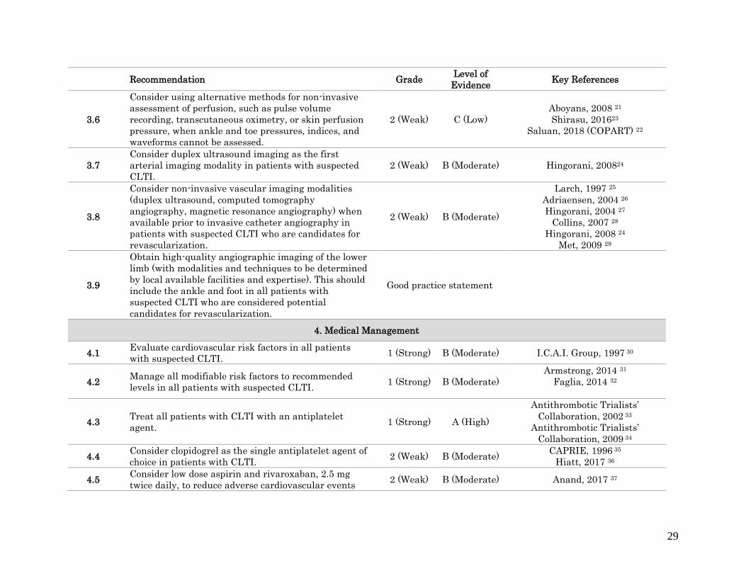
29
Recommendation Grade
Level of
Evidence Key References
3.6
Consider using alternative methods for non-invasive
assessment of perfusion, such as pulse volume
recording, transcutaneous oximetry, or skin perfusion
pressure, when ankle and toe pressures, indices, and
waveforms cannot be assessed.
2 (Weak) C (Low)
Aboyans, 2008 21
Shirasu, 201623
Saluan, 2018 (COPART) 22
3.7
Consider duplex ultrasound imaging as the first
arterial imaging modality in patients with suspected
CLTI.
2 (Weak) B (Moderate) Hingorani, 200824
3.8
Consider non-invasive vascular imaging modalities
(duplex ultrasound, computed tomography
angiography, magnetic resonance angiography) when
available prior to invasive catheter angiography in
patients with suspected CLTI who are candidates for
revascularization.
2 (Weak) B (Moderate)
Larch, 1997 25
Adriaensen, 2004 26
Hingorani, 2004 27
Collins, 2007 28
Hingorani, 2008 24
Met, 2009 29
3.9
Obtain high-quality angiographic imaging of the lower
limb (with modalities and techniques to be determined
by local available facilities and expertise). This should
include the ankle and foot in all patients with
suspected CLTI who are considered potential
candidates for revascularization.
Good practice statement
4. Medical Management
4.1 Evaluate cardiovascular risk factors in all patients
with suspected CLTI. 1 (Strong) B (Moderate) I.C.A.I. Group, 1997 30
4.2 Manage all modifiable risk factors to recommended
levels in all patients with suspected CLTI. 1 (Strong) B (Moderate)
Armstrong, 2014 31
Faglia, 2014 32
4.3 Treat all patients with CLTI with an antiplatelet
agent. 1 (Strong) A (High)
Antithrombotic Trialists’
Collaboration, 2002 33
Antithrombotic Trialists’
Collaboration, 2009 34
4.4 Consider clopidogrel as the single antiplatelet agent of
choice in patients with CLTI. 2 (Weak) B (Moderate)
CAPRIE, 1996 35
Hiatt, 2017 36
4.5 Consider low dose aspirin and rivaroxaban, 2.5 mg
twice daily, to reduce adverse cardiovascular events 2 (Weak) B (Moderate) Anand, 2017 37
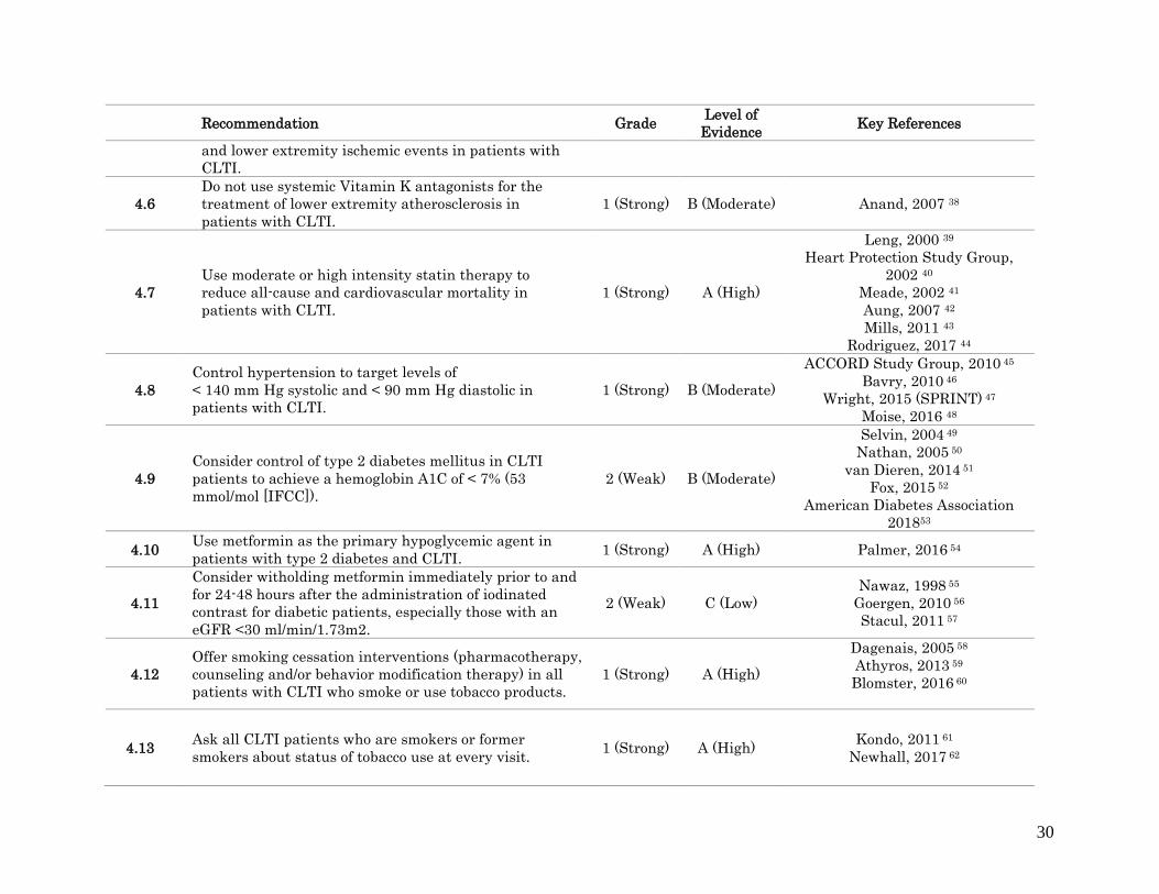
30
Recommendation Grade
Level of
Evidence Key References
and lower extremity ischemic events in patients with
CLTI.
4.6
Do not use systemic Vitamin K antagonists for the
treatment of lower extremity atherosclerosis in
patients with CLTI.
1 (Strong) B (Moderate) Anand, 2007 38
4.7
Use moderate or high intensity statin therapy to
reduce all-cause and cardiovascular mortality in
patients with CLTI.
1 (Strong) A (High)
Leng, 2000 39
Heart Protection Study Group,
2002 40
Meade, 2002 41
Aung, 2007 42
Mills, 2011 43
Rodriguez, 2017 44
4.8
Control hypertension to target levels of
< 140 mm Hg systolic and < 90 mm Hg diastolic in
patients with CLTI.
1 (Strong) B (Moderate)
ACCORD Study Group, 2010 45
Bavry, 2010 46
Wright, 2015 (SPRINT) 47
Moise, 2016 48
4.9
Consider control of type 2 diabetes mellitus in CLTI
patients to achieve a hemoglobin A1C of < 7% (53
mmol/mol [IFCC]).
2 (Weak) B (Moderate)
Selvin, 2004 49
Nathan, 2005 50
van Dieren, 2014 51
Fox, 2015 52
American Diabetes Association
201853
4.10 Use metformin as the primary hypoglycemic agent in
patients with type 2 diabetes and CLTI. 1 (Strong) A (High) Palmer, 2016 54
4.11
Consider witholding metformin immediately prior to and
for 24-48 hours after the administration of iodinated
contrast for diabetic patients, especially those with an
eGFR <30 ml/min/1.73m2.
2 (Weak) C (Low)
Nawaz, 1998 55
Goergen, 2010 56
Stacul, 2011 57
4.12
Offer smoking cessation interventions (pharmacotherapy,
counseling and/or behavior modification therapy) in all
patients with CLTI who smoke or use tobacco products.
1 (Strong) A (High)
Dagenais, 2005 58
Athyros, 2013 59
Blomster, 2016 60
4.13 Ask all CLTI patients who are smokers or former
smokers about status of tobacco use at every visit. 1 (Strong) A (High)
Kondo, 2011 61
Newhall, 2017 62

31
Recommendation Grade
Level of
Evidence Key References
4.14
Prescribe analgesics of appropriate strength for CLTI
patients who have ischemic rest pain of the lower
extremity and foot until pain resolves after
revascularization.
Good practice statement
4.15
In CLTI patients with chronic severe pain, use paracetamol
(acetaminophen) in combination with opioids for pain
control.
Good practice statement
5: The Global Limb Anatomical Staging System for CLTI
5.1
Use an integrated, limb-based anatomical staging
system (such as the Global Limb Anatomical Staging
System) to define complexity of a preferred target
arterial pathway, and facilitate evidence-based
revascularization in patients with CLTI.
Good practice statement
6: Strategies for Evidence-based Revascularization
6.1
Refer all patients with suspected CLTI to a vascular
specialist for consideration of limb salvage, unless major
amputation is considered medically urgent.
Good practice statement
6.2
Offer primary amputation or palliation to patients with
very limited life expectancy, poor functional status (eg,
non-ambulatory), or those with an unsalvageable limb
following shared decision-making.
Good practice statement
6.3
Estimate peri-procedural risk and life expectancy in
patients with CLTI who are candidates for
revascularization.
1
(Strong) C (Low) Biancari, 2007 63
Schanzer, 2008 64
Bradbury, 2010 65
Meltzer, 2013 66
Simons, 2016 67 6.4
Define a CLTI patient as average surgical risk when
anticipated peri-procedural mortality is < 5%, and
estimated 2-year survival is > 50%.
2 (Weak) C (Low)

32
Recommendation Grade
Level of
Evidence Key References
6.5
Define a CLTI patient as high surgical risk when
anticipated peri-procedural mortality is ≥ 5%, or
estimated 2-year survival is ≤ 50%.
2
(Weak) C (Low)
6.6
Use an integrated limb-threat classification system
(such as Wound Ischemia foot Infection (WIfI)) to stage
all CLTI patients who are candidates for limb salvage.
1
(Strong)
C (Low)
Cull. 2014 68
Zhan, 2015 69
Causey, 2016 70
Darling, 2016 71
Robinson, 2017 72
6.7
Perform urgent surgical drainage and debridement
(including minor amputation if needed), and commence
antibiotic treatment, in all patients with suspected CLTI
who present with deep space foot infection or wet
gangrene.
Good practice statement
6.8
Repeat limb staging after surgical drainage,
debridement, minor amputations, or correction of inflow
disease (aortoiliac, common and deep femoral artery
disease) and prior to the next major treatment decision.
Good practice statement
6.9
Do not perform revascularization in the absence of
significant ischemia (WIfI Ischemia Grade 0), unless an
isolated region of poor perfusion in conjunction with
major tissue loss (e.g. WIfI wound grade 2 or 3) can be
effectively targeted, and the wound progresses or fails to
reduce in size by ≥ 50% within 4 weeks, despite
appropriate infection control, wound care, and
offloading.
Good practice statement
6.10
Do not perform revascularization in very low-risk limbs
(eg, WIfI Stage 1) unless the wound progresses or fails to
reduce in size by ≥ 50% within 4 weeks despite
appropriate infection control, wound care and offloading.
2
(Weak)
C (Low)
Sheehan, 2003 73
Cardinal, 2008 74
Lavery, 2008 75
Snyder, 2010 76
6.11
Offer revascularization to all standard risk patients with
advanced limb-threatening conditions (eg, WIfI Stage 4)
and significant perfusion deficits (eg, WIfI ischemia
grades 2 and 3).
1
(Strong)
C (Low) Abu Dabrh, 2015 5

33
Recommendation Grade
Level of
Evidence Key References
6.12
Consider revascularization for standard risk patients
with intermediate limb threat (eg, WIfI stages 2 and 3)
and significant perfusion deficits (eg, WIfI ischemia
grades 2 and 3).
2 (Weak)
C (Low)
Zhan, 2015 69
Causey, 2016 70
Darling, 2016 71
Robinson, 2017 72
6.13
Consider revascularization in standard-risk patients with
advanced limb threat (eg, WIfI stage 4) and moderate
ischemia (eg, WIfI ischemia grade 1).
2 (Weak)
C (Low)
6.14
Consider revascularization in standard-risk patients with
intermediate limb threat (eg, WIfI stages 2 and 3) and
moderate ischemia (eg, WIfI ischemia grade 1), if the
wound progresses or fails to reduce in size by ≥ 50%
within 4 weeks despite appropriate infection control,
wound care and offloading.
2 (Weak)
C (Low)
6.15
Obtain high-quality angiographic imaging with dedicated
views of ankle and foot arteries to permit anatomic
staging and procedural planning in all CLTI patients who
are candidates for revascularization.
Good practice statement
6.16
Use an integrated limb-based staging system (eg, Global
Limb Anatomical Staging System) to define the anatomic
pattern of disease and preferred target artery path in all
CLTI patients who are candidates for revascularization.
Good practice statement
6.17 Perform ultrasound vein mapping when available in all
CLTI patients who are candidates for surgical bypass.
1
(Strong) C (Low)
Seeger, 1987 77
Wengerter, 1990 78
Schanzer, 2007 79
6.18
Map the ipsilateral great saphenous vein and short
saphenous vein for planning surgical bypass.
Map veins in the contralateral leg and both arms if
ipsilateral vein is insufficient / inadequate.
Good practice statement
6.19
Do not classify a CLTI patient as being unsuitable for
revascularization without review of adequate quality
imaging studies and clinical evaluation by a qualified
vascular specialist.
Good practice statement
34
Recommendation Grade
Level of
Evidence Key References
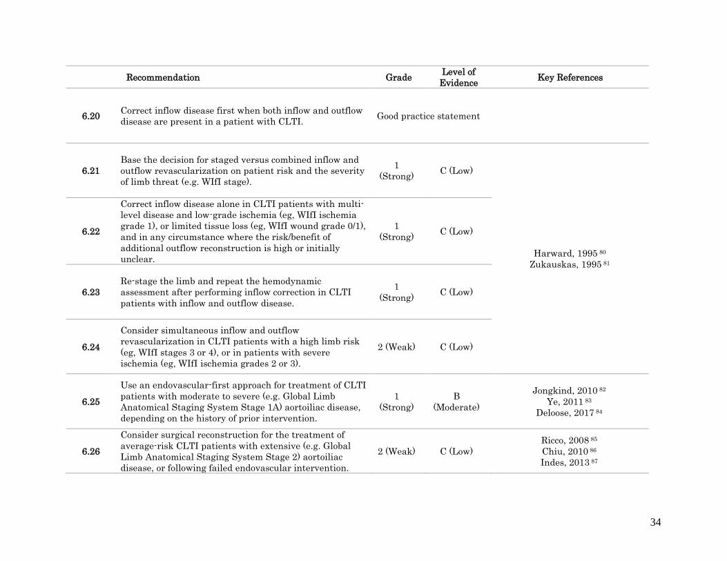
6.20 Correct inflow disease first when both inflow and outflow
disease are present in a patient with CLTI. Good practice statement
6.21
Base the decision for staged versus combined inflow and
outflow revascularization on patient risk and the severity
of limb threat (e.g. WIfI stage).
1
(Strong) C (Low)
Harward, 1995 80
Zukauskas, 1995 81
6.22
Correct inflow disease alone in CLTI patients with multi-
level disease and low-grade ischemia (eg, WIfI ischemia
grade 1), or limited tissue loss (eg, WIfI wound grade 0/1),
and in any circumstance where the risk/benefit of
additional outflow reconstruction is high or initially
unclear.
1
(Strong) C (Low)
6.23
Re-stage the limb and repeat the hemodynamic
assessment after performing inflow correction in CLTI
patients with inflow and outflow disease.
1
(Strong) C (Low)
6.24
Consider simultaneous inflow and outflow
revascularization in CLTI patients with a high limb risk
(eg, WIfI stages 3 or 4), or in patients with severe
ischemia (eg, WIfI ischemia grades 2 or 3).
2 (Weak) C (Low)
6.25
Use an endovascular-first approach for treatment of CLTI
patients with moderate to severe (e.g. Global Limb
Anatomical Staging System Stage 1A) aortoiliac disease,
depending on the history of prior intervention.
1
(Strong)
B
(Moderate)
Jongkind, 2010 82
Ye, 2011 83
Deloose, 2017 84
6.26
Consider surgical reconstruction for the treatment of
average-risk CLTI patients with extensive (e.g. Global
Limb Anatomical Staging System Stage 2) aortoiliac
disease, or following failed endovascular intervention.
2 (Weak) C (Low)
Ricco, 2008 85
Chiu, 2010 86
Indes, 2013 87

35
Recommendation Grade
Level of
Evidence Key References
6.27
Perform open common femoral artery endarterectomy
with patch angioplasty, with or without extension into
the profunda femoris, in CLTI patients with
hemodynamically significant (> 50% stenosis) disease of
the common +/- deep femoral arteries.
1
(Strong) C (Low)
Kang, 2008 88
Ballotta, 2010 89
6.28
Consider a hybrid procedure combining open common
femoral artery endarterectomy and endovascular
treatment for aortoiliac disease with concomitant
common femoral artery involvement (Global Limb
Anatomical Staging System Stage 1B).
2 (Weak) C (Low) Chang, 2008 90
6.29
Consider endovascular treatment for significant common
femoral artery disease in selected patients who are
deemed to be at very high surgical risk or have a hostile
groin.
2 (Weak) C (Low)
Bauman, 2011 91
Bonvini, 2011 92
Gouëffic, 2017 93
Siracuse, 2017 94
6.30
Avoid stents in the common femoral artery, and do not
place stents across the origin of a patent deep femoral
artery.
Good practice statement
6.31
Correct hemodynamically significant (≥ 50% stenosis)
disease of the proximal deep femoral artery whenever
technically feasible.
Good practice statement
6.32
In average-risk CLTI patients with infrainguinal disease,
base decisions on endovascular intervention versus open
surgical bypass on the severity of limb threat (eg, WIfI),
the anatomic pattern of disease (eg, Global Limb
Anatomical Staging System), and the availability of
autologous vein.
1
(Strong)
C (Low) Almasri, 2017 7
6.33
Offer endovascular revascularization when technically
feasible for high-risk patients with advanced limb threat
(eg, Wound Ischemia foot Infection (WIfI) stage 4) and
significant perfusion deficits (eg, WIfI ischemia grades 2
and 3).
2 (Weak) C (Low)
Abu Dabrh, 2015 5
Zhan, 2015 69
Causey, 2016 70
Darling, 2016 71
Robinson, 2017 72

36
Recommendation Grade
Level of
Evidence Key References
6.34
Consider endovascular revascularization for high-risk
patients with intermediate limb threat (eg, WIfI stages 2
and 3) and significant perfusion deficits (eg, WIfI
ischemia grades 2 and 3).
2 (Weak) C (Low)
6.35
Consider endovascular revascularization for high-risk
patients with advanced limb threat (eg, WIfI stage 4) and
moderate ischemia (eg, WIfI ischemia grade 1), if the
wound progresses or fails to reduce in size by ≥ 50%
within 4 weeks despite appropriate infection control,
wound care and offloading, when technically feasible.
2 (Weak) C (Low)
6.36
Consider endovascular revascularization for high-risk
patients with intermediate limb threat (eg, WIfI stages 2
and 3) and moderate ischemia (eg, WIfI ischemia grade
1), if the wound progresses or fails to reduce in size by ≥
50% within 4 weeks despite appropriate infection control,
wound care and offloading, when technically feasible.
2 (Weak) C (Low)
6.37
Consider open surgery in selected high-risk patients with
advanced limb threat (eg, WIfI stage 3 or 4), significant
perfusion deficits (ischemia grade 2-3), and advanced
complexity of disease (eg, Global Limb Anatomical
Staging System stage III), or after prior failed
endovascular attempts and unresolved symptoms of
CLTI.
2 (Weak) C (Low)
6.38
Consider angiosome-guided revascularization in patients
with significant wounds (eg, WIfI wound grade 3 and 4),
particularly those involving the mid- or hindfoot, and
when the appropriate target arterial pathway is
available.
2 (Weak) C (Low)
Azuma, 2012 95
Sumpio, 2013 96
Biancari, 2014 97
Chae, 2016 98
Jongsma, 2017 99
6.39
When treating femoropopliteal disease in CLTI patients
by endovascular means, consider adjuncts to balloon
angioplasty (e.g. stents, covered stents, or drug eluting
technologies) when there is a technically inadequate
result (residual stenosis or flow limiting dissection) or in
the setting of advanced lesion complexity (eg, Global
2 (Weak)
B
(Moderate)
Schillinger, 2006 100
Saxon, 2008 101
Dake, 2011 102
Rosenfield, 2015 103
Almasri, 2018 7

37
Recommendation Grade
Level of
Evidence Key References
Limb Anatomical Staging System femoropopliteal grade
2-4).
6.40 Use autologous vein as the preferred conduit for
infrainguinal bypass surgery in CLTI.
1
(Strong)
B
(Moderate) Almasri, 2018 7
6.41
Avoid using a non-autologous conduit for infrainguinal
bypass unless there is no endovascular option and no
adequate autologous vein.
2
(Weak) C (Low) Almasri, 2018 7
6.42
Perform intraoperative imaging (angiography, duplex
ultrasound, or both) upon completion of open bypass
surgery for CLTI, and correct significant technical defects
if feasible during the index operation.
1
(Strong) C (Low)
Mills, 1992 104
Bandyk, 1994 105
7: Non-revascularization treatments of the limb
7.1
Consider spinal cord stimulation to reduce the risk of
amputation and decrease pain in carefully selected
patients (eg, rest pain, minor tissue loss) in whom
revascularization is not possible.
2
(Weak)
B
(Moderate) Ubbink, 2013 106
7.2 Do not use lumbar sympathectomy for limb salvage in
CLTI patients in whom revascularization is not possible.
2
(Weak) C (Low) Karanth, 2016 107
7.3
Consider intermittent pneumatic compression therapy in
carefully selected patients (eg, rest pain, minor tissue
loss) in whom revascularization is not possible.
2
(Weak)
B
(Moderate) Abu Dabrh, 2015 4
38
Recommendation Grade
Level of
Evidence Key References
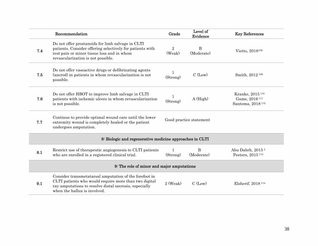
7.4
Do not offer prostanoids for limb salvage in CLTI
patients. Consider offering selectively for patients with
rest pain or minor tissue loss and in whom
revascularization is not possible.
2
(Weak)
B
(Moderate) Vietto, 2018108
7.5
Do not offer vasoactive drugs or defibrinating agents
(ancrod) in patients in whom revascularization is not
possible.
1
(Strong) C (Low) Smith, 2012 109
7.6
Do not offer HBOT to improve limb salvage in CLTI
patients with ischemic ulcers in whom revascularization
is not possible.
1
(Strong) A (High)
Kranke, 2015 110
Game, 2016 111
Santema, 2018 112
7.7
Continue to provide optimal wound care until the lower
extremity wound is completely healed or the patient
undergoes amputation.
Good practice statement
8: Biologic and regenerative medicine approaches in CLTI
8.1 Restrict use of therapeutic angiogenesis to CLTI patients
who are enrolled in a registered clinical trial.
1
(Strong)
B
(Moderate)
Abu Dabrh, 2015 4
Peeters, 2015 113
9: The role of minor and major amputations
9.1
Consider transmetatarsal amputation of the forefoot in
CLTI patients who would require more than two digital
ray amputations to resolve distal necrosis, especially
when the hallux is involved.
2 (Weak) C (Low) Elsherif, 2018 114
39
Recommendation Grade
Level of
Evidence Key References
9.2
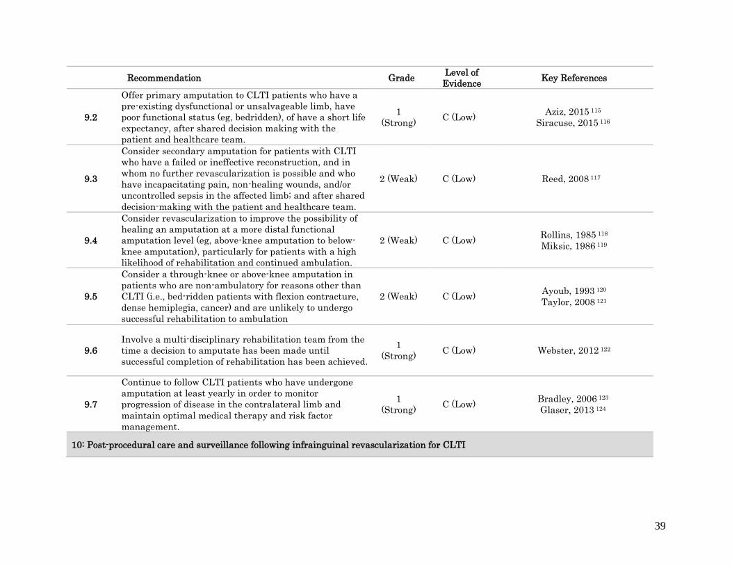
Offer primary amputation to CLTI patients who have a
pre-existing dysfunctional or unsalvageable limb, have
poor functional status (eg, bedridden), of have a short life
expectancy, after shared decision making with the
patient and healthcare team.
1
(Strong) C (Low)
Aziz, 2015 115
Siracuse, 2015 116
9.3
Consider secondary amputation for patients with CLTI
who have a failed or ineffective reconstruction, and in
whom no further revascularization is possible and who
have incapacitating pain, non-healing wounds, and/or
uncontrolled sepsis in the affected limb; and after shared
decision-making with the patient and healthcare team.
2 (Weak) C (Low) Reed, 2008 117
9.4
Consider revascularization to improve the possibility of
healing an amputation at a more distal functional
amputation level (eg, above-knee amputation to below-
knee amputation), particularly for patients with a high
likelihood of rehabilitation and continued ambulation.
2 (Weak) C (Low) Rollins, 1985 118
Miksic, 1986 119
9.5
Consider a through-knee or above-knee amputation in
patients who are non-ambulatory for reasons other than
CLTI (i.e., bed-ridden patients with flexion contracture,
dense hemiplegia, cancer) and are unlikely to undergo
successful rehabilitation to ambulation
2 (Weak) C (Low) Ayoub, 1993 120
Taylor, 2008 121
9.6
Involve a multi-disciplinary rehabilitation team from the
time a decision to amputate has been made until
successful completion of rehabilitation has been achieved.
1
(Strong) C (Low) Webster, 2012 122
9.7
Continue to follow CLTI patients who have undergone
amputation at least yearly in order to monitor
progression of disease in the contralateral limb and
maintain optimal medical therapy and risk factor
management.
1
(Strong) C (Low)
Bradley, 2006 123
Glaser, 2013 124
10: Post-procedural care and surveillance following infrainguinal revascularization for CLTI

40
Recommendation Grade
Level of
Evidence Key References
10.1
Continue best medical therapy for peripheral arterial
disease, including the long-term use of antiplatelet and
statin therapies, in all patients who have undergone
lower extremity revascularization.
1
(Strong) A (High)
Abbruzzese, 2004 125
Henke, 2004 126
Brown, 2008 127
Bedenis, 2015 128
Suckow, 2015 129
10.2 Promote smoking cessation in all CLTI patients who have
undergone lower extremity revascularization.
1
(Strong) A (High)
Hobbs, 2003 130
Willigendael, 2005 131
10.3
Consider dual antiplatelet therapy (aspirin plus
clopidogrel) in patients who have undergone
infrainguinal prosthetic bypass for CLTI, for a period of 6
to 24 months, to maintain graft patency.
2
(Weak)
B
(Moderate)
Brown, 2008 127
Belch, 2010 132
Gassman, 2014 133
Bedenis, 2015 128
10.4
Consider dual antiplatelet therapy (aspirin plus
clopidogrel) in patients who have undergone
infrainguinal endovascular interventions for CLTI, for at
least 1 month.
2
(Weak) C (Low)
Cassar, 2005 134
Bhatt, 2006 135
Tepe, 2012 136
Strobl, 2013 137
10.5
Consider dual antiplatelet therapy for 1-6 months in
patients undergoing repeat catheter-based interventions,
if they are at low risk for bleeding.
2
(Weak) C (Low)
Cassar, 2005 134
Tepe, 2012 136
Strobl, 2013 137
10.6
Follow patients who have undergone lower extremity vein
bypass for CLTI on a regular basis for at least 2 years,
with a clinical surveillance program consisting of interval
history, pulse examination, and measurement of resting
ankle and toe pressures. Consider duplex ultrasound
scanning where available.
Good practice statement
10.7
Follow patients who have undergone lower extremity
prosthetic bypass for CLTI on a regular basis for at least
2 years, with interval history, pulse examination, and
measurement of resting ankle and toe pressures.
Good practice statement

41
Recommendation Grade
Level of
Evidence Key References
10.8
Follow patients who have undergone infrainguinal
endovascular interventions for CLTI in a surveillance
program that includes clinical visits, pulse examination,
and noninvasive testing (resting ankle and toe pressures).
Good practice statement
10.9
Consider performing additional imaging in patients with
lower extremity vein grafts who have a decrease in ankle
brachial index ≥ 0.15 and/or recurrence of
symptoms/change in pulse status, to detect vein graft
stenosis.
Good practice statement
10.10
Offer intervention for duplex ultrasound detected vein
graft lesions with an associated peak systolic velocity of >
300 cm/sec, a peak systolic velocity ratio > 3.5, or grafts
with low velocity (midgraft peak systolic velocity < 45
cm/sec) to maintain patency.
1
(Strong) B (Moderate) Mills, 2001 138
10.11
Maintain long term surveillance following surgical or
catheter-based revision of a vein graft, including duplex
ultrasound graft scanning where available, to detect
recurrent graft-threatening lesions.
1
(Strong)
B (Moderate)
Landry, 2002 139
Nguyen, 2004 140
10.12
Consider arterial imaging following endovascular
intervention for failure to improve (wound healing, rest
pain) or a recurrence of symptoms, to detect restenosis or
progression of pre-existing disease.
2
(Weak) C (Low) Bui, 2012 141
10.13
Consider reintervention for patients with duplex detected
restenosis lesions > 70% (peak systolic velocity ratio >
3.5, peak systolic velocity > 300 cm/s), if symptoms of
CLTI are unresolved, or on a selective basis in
asymptomatic patients following catheter-based
interventions.
2
(Weak) C (Low) Humphries, 2011 142
10.14 Provide mechanical offloading as a primary component
for care of all CLTI patients with pedal wounds.
1
(Strong) A (High) Elraiyah, 2016 143
10.15
Provide counseling on continued protection of the healed
wound and foot, to include appropriate shoes, insoles, and
monitoring of inflammation.
1
(Strong) A (High) Elraiyah, 2016 143

42
Recommendation Grade
Level of
Evidence Key References
11: Study designs and trial endpoints in CLTI
11.1
Use a research framework such as the Idea,
Development, Exploration, Assessment, and Long-term
study, for gathering new data and evidence on the
surgical and endovascular management of CLTI.
Good practice statement
11.2
Encourage funders, journal reviewers and editors to
prioritize prospective, multicenter, controlled, and
preferably randomized studies over retrospective cases
series, studies using historical controls, or other, less
rigorous research methodologies.
Good practice statement
11.3
When randomized controlled trials are not feasible, use
the objective performance goal benchmarks from the
Society for Vascular Surgery’s Critical Limb Ischemia
Working Group to evaluate the efficacy of novel
endovascular CLTI techniques and devices.
Good practice statement
11.4
In order to facilitate sufficient enrollment, limit
randomized controlled trial exclusion criteria to those
that are deemed essential to trial integrity.
Good practice statement
11.5 Design randomized controlled trials, prospective cohort
studies, and registries that are specific to CLTI. Good practice statement
11.6
Use an integrated, limb-based limb threat system
(eg, Wound Ischemia foot Infection) and a whole limb
anatomical classification scheme (eg, Global Limb
Anatomical Staging System) to describe the
characteristics and outcomes of CLTI patients who are
enrolled.
Good practice statement
11.7
Describe outcomes in CLTI trials using a combination of
objective and clinically relevant events, subjective
patient-reported outcomes measures/health-related
quality of life assessments, and anatomic and
hemodynamic endpoints.
Good practice statement
11.8
Require regulatory trials aimed at obtaining pre-market
approval for devices for use in CLTI to study CLTI
patients and to present data on objective, clinically
Good practice statement

43
Recommendation Grade
Level of
Evidence Key References
relevant endpoints, patient-reported outcomes
measures/health-related quality of life assessments, and
anatomic and hemodynamic endpoints.
11.9
Follow-up patients in trials for a time sufficient (this will
usually be more than 2 years) to allow appropriate
comparison of the impact of the different interventions on
the natural history of CLTI. Measure and declare
completeness of follow-up coverage to quantify risk of
attrition bias.
Good practice statement
11.10
Include a time-integrated measure of clinical disease
severity (such as freedom from CLTI) in the CLTI trial
design, in order to describe the total impact of comparator
CLTI interventions.
Good practice statement
11.11
Publish all CLTI trial protocols, together with the full
statistical analysis plans, in peer-reviewed journals to
allow for independent, public, and transparent scrutiny
and to prevent non-reporting of negative trials.
Good practice statement
11.12
Conduct post-marketing surveillance data collection
using well-designed, large observational studies and
registries.
Good practice statement
11.13
Share clinical trial data to allow subsequent individual
patient data, meta-, and subgroup analyses; updating of
objective performance goals; and validation of decision-
making tools such as the Wound, Ischemia, and foot
Infection system and Global Limb Anatomical Staging
System.
Good practice statement
11.14
Assess the quality of evidence in CLTI research using
frameworks such as the Grading of Recommendations
Assessment, Development and Evaluation, that consider
multiple certainty domains and are not based solely on
study design.
Good practice statement
12: Creating a Center of Excellence for Amputation Prevention
No recommendations

44
45
1: DEFINITIONS AND NOMENCLATURE
Defining and describing the severity of peripheral artery disease
The term ‘critical limb ischemia’ (CLI) is outdated and fails to encompass the full
spectrum of patients who are evaluated and treated for limb-threatening ischemia in modern
practice. Instead, the new term CLTI is proposed, in order to include a broader and more
heterogeneous group of patients with varying degrees of ischemia that can often delay wound
healing and increase amputation risk.
In order to develop a clearer concept of CLTI, the following are excluded from the
population as defined in this Guidelines document: patients with purely venous ulcers, acute limb
ischemia, acute trash foot, ischemia due to emboli, acute trauma, or mangled extremity; or those
with wounds related to nonatherosclerotic conditions. These include vasculitides, collagen
vascular disease, Buerger’s disease, neoplastic disease, dermatoses, and radiation arteritis.
Previous leg ischemia definition and classification systems
Critical limb ischemia. In 1982, a working group of vascular surgeons defined CLI as
ischemic rest pain with an ankle pressure < 40 mm Hg, or tissue necrosis with an ankle pressure
< 60 mm Hg, in patients without diabetes.144 Patients with diabetes were specifically excluded
due to the confounding effects of neuropathy and susceptibility to infection. This definition has
long been debated because it failed to capture a large group of patients who were at risk for
amputation from a broader range of ischemia.145, 146 To address this limitation, multiple and
disparate lower-limb ischemia and wound/ DFU classification systems have been developed and
46
promulgated over the past 5 decades, many of which remain in use today. These and other
commonly used classifications and their associated components and grades of severity, are
summarized in Table 1.1.10, 147-158 Among vascular surgeons, the Fontaine and Rutherford
classifications have been the most widely adopted, while orthopedists, podiatric surgeons, and
diabetic foot specialists traditionally applied the Wagner and University of Texas classifications.
The strengths and limitations of each have been widely discussed in previous key publications.10,
150, 159-161 Although each of these systems has advantages, the use of multiple classification
systems has hindered the development of optimal treatment algorithms. It has also contributed to
the fragmentation and variability of care provided for patients with DFUs, as well as for non-
diabetic patients across the spectrum of CLTI.
Lower extremity threatened-limb classification system
The definitions summarized in Table 1.1 were developed primarily to describe patients
suffering from pure ischemia due to atherosclerosis. This was when the predominant risk factor
was tobacco smoking and prior to the global epidemic of diabetes mellitus (DM). As such, these
definitions were ischemia-dominant models of limb threat. However, because patients with DM
now make up the majority of patients with CLTI, absolute perfusion now needs to be considered
in the context of neuropathy, wound characteristics, and infection. In order to address this unmet
need, the SVS Lower Extremity Guidelines Committee created the SVS Lower Extremity
Threatened Limb Classification System. This system stratifies amputation risk according to
wound extent, the degree of ischemia, and the presence and severity of foot infection (WIfI).10
While it may require some adjustments, WIfI appears to correlate strongly with important
clinical outcomes. This includes those set forth in the SVS objective performance goals (OPG)

47
that focus on limb amputation, 1-year amputation-free survival, and wound healing time (Table
1.2).10, 68-72, 162-167
The WIfI classification system is currently being evaluated in multi-center trials
including the US NIH-funded BEST-CLI trial13 and the UK NIHR HTA-funded BASIL-2 and
BASIL-3 trials. 14, 15 WIfI is also being incorporated into the US SVS Vascular Quality Initiative
(VQI) Registry of lower extremity interventions.
Hemodynamic criteria
Although previous guidelines have suggested a range of ankle pressure and toe pressure
thresholds for defining limb-threatening ischaemia, such thresholds must be used with great
caution, and considered in the clinical context, due to multiple confounding factors and the lack
of a clear and reliable relationship to outcomes. Patients with limb-threatening ischemia should
be defined primarily in terms of their clinical presentation, supplemented by physiologic studies
that demonstrate a degree of ischemia sufficient to cause pain, impair wound healing and
increase amputation risk.
In addition to patients who meet the proposed new definition of CLTI, there are a
significant number of patients whose PAD is so severe that they are likely to be at increased risk
of developing CLTI in the foreseeable future.168 Although data are lacking, it is logical to
suggest that such individuals should be monitored closely for clinical disease progression.

48
Chronic limb threatening ischemia
We propose that CLTI be defined to include a broader and more heterogeneous group of
patients with varying degrees of ischemia that may delay wound healing and increase amputation
risk. A diagnosis of CLTI requires objectively documented atherosclerotic PAD, in association
with ischemic rest pain and/or tissue loss (ulceration and/or gangrene).
Ischemic rest pain is typically described as affecting the forefoot, and is often made
worse with recumbency while being relieved by dependency. It should be present for more than
2 weeks, and be associated with one or more abnormal hemodynamic parameters. These
parameters include an ankle brachial index (ABI) < 0.4 (using higher of the dorsalis pedis,
DP,and posterior tibial PT, arteries); absolute highest ankle pressure (AP) < 50 mm Hg; absolute
toe pressure (TP) < 30 mm Hg; transcutaneous partial pressure of oxygen (TcP02)< 30 mm Hg;
and/or flat or minimally pulsatile pulse volume recording (PVR) waveforms (equivalent to WIfI
ischemia grade 3). Pressure measurements should be correlated with Doppler arterial waveforms,
keeping in mind that AP and ABI are frequently falsely elevated due to medial calcinosis,
especially in people with DM and/or end-stage renal disease (ESRD). For this reason, a
combination of tests may be needed. In patients with DM and/or ESRD, toe waveforms and
systolic pressures are preferred. A recent study demonstrated that AP alone failed to identify
42% of patients with CLTI. TP and TcPO2measurements were more accurate than AP and also
were more predictive of 1-year amputation risk (TP < 30 mm Hg or TcPO2 < 10 mm Hg).169
Tissue loss related to CLTI includes gangrene of any part of the foot or non-healing
ulceration present for at least 2 weeks. It should be accompanied by objective evidence of
significant PAD (eg, WIfI ischemia grade ≥1). This definition excludes purely neuropathic,
traumatic or venous ulcers lacking any ischemic component. However, the WIfI scheme

49
recognizes that a wide range of ischemic deficit may be limb threatening when it co-exists with
varying degrees of wound complexity and superimposed infection. CLTI is present if either
ischemic rest pain or tissue loss with appropriate hemodynamics are present.
Some patients may have relatively normal hemodynamics when considering the limb or
foot as a whole, but neverthess suffer ulceration due to diminished local perfusion (ie,
angiosomal or regional ischemia without adequate collateral flow). It is recognized that such
ulcers may contribute to limb threat, and current tools to assess regional ischemia require further
development to better define such circumstances and their treatment. The relationship between
regional ischemia and patterns of infra-popliteal and pedal disease also requires more in-depth
study 12, 170
GVG recommends use of the SVS WIfI classification (Section 3) in a manner analogous
to the TNM (Tumor, Nodes, Metastasis) system of cancer staging, to stage the limb in patients
with CLTI. The WIfI classification is intuitive and has been made user friendly by the
availability of free online application software provided by the SVS (SVS iPG – interactive
Practice Guidelines https://itunes.apple.com/app/id1014644425).
Data accrued in nearly 3000 patients to date, and summarized in Table 1.2, suggest that
the four WIfI clinical stages of limb threat correlate with the risk of major limb amputation and
time to wound healing. It has also been suggested that novel WIfI composite and mean scores
may predict other clinically significant events as well.164 The WIfI system appears to contain the
key limb-status elements needed to gauge the severity of limb threat at presentation.
In addition, recent data suggest that WIfI can assist in predicting which patients might
50
fare better with open surgical bypass compared to endovascular therapy.171, 172 One study
reported that when endovascular therapy alone was applied to WIfI stage 4 patients, results were
worse than in lower clinical stage patients.172 Specifically, the wound healing rate was only 44%,
the major limb amputation rate was 20% and 46% of patients required multiple, repetitive
endovascular procedures. In a non-randomized, single-center comparison of WIfI stage 4
patients, researchers found that freedom from major limb amputation was superior in patients
who underwent bypass compared to those who underwent endovascular therapy.171 If these
results can be confirmed, WIfI may prove to be a useful tool when deciding whether to offer
endovascular therapy or bypass.
Another recent study utilized WIfI in a fashion analogous to TNM staging for cancer and
restaged patients after 1 month of therapy. The investigators found that at 1 and 6 months,
wound, ischemia, and infection grades correlated with amputation-free survival (AFS), while
baseline ischemia grade did not.173 These data suggest that restaging with WIfI at 1 and 6 months
after intervention may help identify a cohort of patients undergoing therapy for CLTI that
remains at higher risk for major limb amputation and may merit targeted reintervention.
Ultimately, the optimal staging system for CLTI is expected to evolve with additional
clinical application and larger scale, multi-center and multi-national data analysis.

51
Recommendation Grade Level of
Evidence
Key References
1.1 Use objective hemodynamic tests to determine the
presence and quantify the severity of ischemia in all
patients with suspected CLTI.
1
(Strong)
C (Low) De Graaf et al
2003 16
Brownrigg et al
2016 17
Wang et al 2016 18
1.2 Use a lower extremity threatened limb classification
staging system (eg, Society for Vascular Surgery’s
Wound, Ischemia, and foot Infection Classification
System) that grades wound extent, degree of
ischemia, and severity of infection to guide clinical
management in all patients with suspected CLTI.
1
(Strong)
C (Low)
See Table 1.2

52
2: GLOBAL EPIDEMIOLOGY AND RISK FACTORS FOR CLTI
In 2010, estimates suggested that over 200 million people worldwide were living with
PAD. This represented a 23.5% increase since 2000; an increase that is believed to be largely
attributable to aging populations and the growing prevalence of risk factors, in particular DM.1
These figures are thought to almost certainly underestimate the true burden of disease as they are
largely based on community-based studies that define PAD on the basis of reduced ABI.
Although CLTI is widely believed to be a growing global healthcare problem, reliable
epidemiological data are extremely limited.
Men have been reported to have a higher prevalence of PAD in high-income countries
(HIC) (Fig 2.1), while women seem to have a higher prevalence of PAD in low- and middle-
income countries (LMIC).1 As life expectancy increases, the burden of PAD seems likely to rise
in LMIC. However, in certain geographic regions, notably in the western Pacific and Southeast
Asia, most PAD cases are reported in people younger than 55.1
In a recent meta-analysis from the US, the prevalence of PAD in men ranged from 6.5%
(aged 60 to 69 years) to 11.6% (aged 70 to 79) to 29.4% (over 80 years).174 There were similar
age-related increases in PAD prevalence in women (5.3%, 11.5%, and 24.7% in the above age
categories respectively).174 Given that the life expectancy of women still exceeds that of men, the
overall burden of PAD (total number of individuals affected) is likely greater in women than in
men. The epidemiology of PAD is likely to be similar in other developed countries, such as the
UK, and regions, such as the EU.175, 176 However, as these populations become more multi-
cultural, differences in disease burden between different communities within these nations seem
likely to become apparent so further complicating the epidemiology of the condition.177

53
Data on the epidemiology of PAD, and in particular CLTI, in other parts of the world are
even more limited. In one Japanese community study of people over 40, the prevalence of ABI <
0.90 was very low (1.4%).178 In a recent population-based cohort of 4055 Chinese men and
women over 60, the prevalence of PAD (ABI < 0.90) was 2.9% and 2.8%, respectively.179
Another population-based cohort of 1871 individuals aged less than 65 in two countries from
Central Africa, showed that the overall prevalence of PAD was 14.8%.180
There is a considerable body of evidence showing that PAD is more common among
black individuals than it is in whites.181-184 There is also evidence that Asians and Hispanics have
a lower prevalence of PAD than whites.184 It is not clear if these differences have a genetic basis
or whether they simply reflect differential exposure to traditional risk factors. However, disease
risk profiles appear to change as populations migrate, suggesting that environment is more
important than genetic make-up. Another explanation may be that ABI is intrinsically lower in
blacks, resulting in a falsely high prevalence of PAD.185
There are far more international data on the epidemiology of intermittent claudication
(IC) than there is on CLTI. The annual incidence of IC in 60-year-old men has been shown to
range from 0.2% in Iceland to 1.0% in Israel.186 A recent study using data from a large, insured
US population estimated the annual incidence of PAD, defined by the presence of a diagnosis or
procedure insurance claim, to be 2.4% in a cohort of adults over 40.187 Studies reporting on the
epidemiology of PAD based on ABI rather than on the presence of symptomatic disease suggest
that the prevalence of asymptomatic PAD may be similar between in men and women, although
IC appears to be more prevalent in men.188, 189 Differences in presentation between men and
women with IC may influence the accuracy of prevalence estimates.190

54
Risk factors for PAD
Modifiable risk factors for PAD have been comprehensively studied in HIC and include
smoking, DM, hypertension, hypercholesterolemia, and air pollution. A recent global study
suggests that, although these risk factors may equally applicable to LMIC, for most, the strength
of the association was greater in HIC. This may be because HIC studies often include a larger
number of older patients and because the exposure time tends to be shorter in LMIC.1
Smoking is unarguably a significant risk factor in the development and progression of
PAD. Nevertheless, while smoking rates are falling in most HIC, this is not the case in LMIC
(Fig 2.2). DM is also strongly associated with the development of PAD, and risk increases with
the duration of DM in affected individuals. Patients with DM are widely recognized to be at
markedly higher risk of amputation.191, 192 The rapidly increasing worldwide prevalence of type 2
DM is concerning, and likely to have a significant impact on the future incidence and prevalence
of PAD and CLTI, as well as their morbid endpoints.
The link between obesity and PAD is inconsistent. Many studies have suggested the
existence of an “obesity paradox” with lower rates of PAD being observed in patients with a
higher BMI.186 By contrast, other studies that have adjusted for smoking, which is associated
with a generally lower BMI,193 report a positive correlation between BMI and PAD.
Hypertension is associated with the development of PAD and is another very common risk factor
in the adult population.
The association between dyslipidemia and the development and progression of
atherosclerosis has been extensively studied. While elevated total cholesterol (TC) and low
density lipoprotein (LDL)-C are widely accepted as a risk factors for PAD, reduced high density
lipoprotein (HDL)-C levels also appear to be associated with increased mortality in PAD

55
patients.194 A ratio of the two may also be a useful predictor of PAD.195 While
hypertriglyceridemia appears to be atherogenic,196 its role in the development and progression of
PAD remains incompletely defined.
Chronic kidney disease (CKD) and in particular end-stage renal disease (ESRD), is a
strong risk factor for PAD and limb loss, especially when associated with DM. Affected patients
frequently have heavily calcified arteries and a distal pattern of arterial disease.186
The association between alcohol consumption and PAD is inconsistent making it difficult
to draw any firm conclusions.197 However, heavy alcohol consumption is often associated with
other risk factors for PAD such as smoking and, as with DM, the presence of alcoholic
neuropathy increases the risk of tissue loss for any given perfusion deficit.
Recent data suggest that air pollution from sources such as motor vehicles, power plants,
wood burning, and some industrial processes, may be associated with increased cardiovascular
morbidity and mortality.198 Likewise, chronic inflammation, characterized by elevated levels of
C-reactive protein (CRP) and other biomarkers, has been shown to be associated with PAD.186
Homocysteine levels are higher in several case-control PAD cohort studies, although the benefits
of folate supplementation appear to be negligible.186, 199
The significance of family history and genetic make-up are uncertain.200, 201 Studies have
yielded varying results, with some identifying a small number of candidate genes or even single
nucleotide polymorphisms, and others failing to identify any association at all.
Finally, people of lower socio-economic status and educational attainment tend to have a
higher prevalence of IC, and probably also CLTI, although the association is not always strong
and can often be explained in part by their increased exposure to other risk factors such as

56
smoking.180, 183, 202 However there is increasing evidence that chronic mental and psycho-social
stress may have direct effects on cardiovascular health.203
Incidence and prevalence of CLTI
As noted above, high-quality data on the epidemiology of CLTI are lacking, especially
from LMIC, with many estimates being extrapolated from the incidence and prevalence of IC,
amputation, and DM. Unfortunately, such estimates can be highly misleading for a number of
reasons. First, IC does not progress to CLTI in a predictable manner. Second, CLTI likely
represents less than 10% of all PAD patients; and those undergoing amputation for CLTI are at
very high risk of premature mortality (and so more likely to be absent from population-based
studies). Third, the clinical and hemodynamic data required to reliably diagnose CLTI are
difficult to obtain in large populations. This is particularly true in patients with DM who often
have incompressible vessels. Thus, although it is estimated that approximately half of all patients
with a DFU in Western Europe and North America also have significant PAD, the disease may
often appear relatively mild (not fulfilling the criteria for CLTI) on hemodynamic assessment.204
For many years, the annual incidence of what has typically been termed ‘critical limb
ischemia’ was estimated at 500 to 1000 new cases per million individuals in Western
countries.205 Unfortunately, there are no reliable contemporary epidemiological data that take
into account recent changes in lifestyle (such as reduced smoking rates), the identification and
medical management of cardiovascular risk factors, the prevalence of obesity and diabetes, and
overall increasing life expectancy around the world.
In 2013, a meta-analysis involving 6 studies and close to 83,000 patients showed the
overall prevalence of severe chronic limb ischemia (defined by Fontaine stage, AP < 70 mm Hg,

57
and/or ABI < 0.60) was 0.74% (95% CI, 0.26-1.46) with marked heterogeneity between studies
(prevalence 0.11% to 1.59%).206
In an analysis of the US MarketScan database, comprised of approximately 12 million
Americans aged 40 and over receiving care from Medicare and Medicaid between 2003 and
2008, the prevalence and annual incidence of CLTI was estimated at 1.33% and 0.35%,
respectively. This equates to around 3,500 new cases per million individuals per year.187 The
study defined primary CLTI as patients with no prior PAD or subsequent PAD diagnostic code
more than 30 days following CLTI diagnostic code. Secondary CLTI included patients with prior
PAD (or subsequent PAD diagnostic codes within 30 days of a CLTI diagnostic code). The
annual incidence rate of primary and secondary CLTI was 0.19% and 0.16%. CLTI patients
represented 11.08% (95% CI, 11.03-11.13%) of total PAD patients annually. As noted above,
while one might expect similar rates of CLTI in other developed nations and regions, data from
LMIC are lacking. Even within HIC, the epidemiology of CLTI is likely to be complex and
evolving.
Amputation and CLTI
A number of studies have used major lower limb amputation as a surrogate for CLTI on
the basis that most (> 80%) are due to CLTI. However, it can be difficult to distinguish reliably
between minor (below the ankle) and major (above the ankle) amputations in some
administrative data. Furthermore, the number of amputations that are performed for trauma,
tumor or infection, including patients with DM and neuropathy (but without PAD), are likely to
vary considerably from country to country; particularly, when comparing HIC and LMIC.
58
In the US in 2015, an estimated 504,000 individuals (of a total estimated population of
295.5 million) were living with a major amputation due to PAD; a number that was projected to
more than double by 2050.207 In Minnesota, a state with low overall rates of cardiovascular
disease, one study showed that between 2005 and 2008, the age-adjusted annual incidence of
ischemic lower limb amputation (amputations not due to trauma or cancer) remained unchanged
at 20 per 100,000.208
A recent systematic review found that the rate of major amputation varied considerably
(3.6 to 68.4 per 100,000 per year) across the world, probably due to differences in ethnicity,
social deprivation and, in particular, the prevalence of DM.209 In some countries, including
England, the incidence of amputations unrelated to DM appears to be decreasing.210 However, in
most parts of the world, the incidence of DM-related limb amputations is increasing.211
Natural history of untreated CLTI
A recently published meta-analysis (13 studies and 1527 patients) of the natural history
of untreated CLTI found that during a median follow up of 12 months, both the mortality rate
and the per-patient amputation rate were 22%, although there was marked heterogeneity between
studies.5 With regard to disease progression, one study estimated that only 5-10% of patients
with either asymptomatic PAD or IC went on the develop CLTI over a 5-year period.212
However, another recent meta-analysis suggested that this progression rate may be significantly
higher at 21% (range 12-29%) over 5 years.213 It is also important to note that approximately
50% of patients presenting with CLTI have no prior history of PAD.214, 215
Patients with CLTI present with a wide spectrum of clinical, hemodynamic, and anatomic
disease. Outcomes depend on the availability and quality of primary and secondary care and may

59
be further influenced by factors such as social stigmatization and cultural and religious beliefs.
Those living in regions with poor access to healthcare often present late with advanced disease
and unsalvageable limbs. Indeed, it has been estimated that approximately half of all patients
with CLTI do not undergo revascularization.216 Even in HIC with advanced healthcare systems,
such as Germany and the US, many patients with suspected CLTI do not receive angiography or
any attempt at revascularization.217 This may be because patients are either too sick or frail, are
thought to have no revascularization option, or present too late. Unfortunately, while reasonable
data are available on amputation rates, data on processes of care that can help explain the
shortfall and differences in revascularization and amputation are lacking.
The recently published VASCUNET report showed large (almost six-fold) differences,
but an overall decline, in major amputation rates in 12 European and Australasian countries
between 2010 and 2014.218 DM prevalence, age distribution, and mortality rates were also found
to vary between countries. Despite limitations inherent to the use registry data, these findings are
important and may indicate disparities in access to vascular surgical intervention across the
countries studied. Further research is clearly required in order to improve limb salvage in
different demographic and geographic settings.218
In patients with known PAD, the risk of developing CLTI appears to be greater in men, in
patients who have had a stroke or are in heart failure, and in patients with DM.187 Patients who
present de novo with CLTI (no prior diagnosis of PAD) seem more likely to be older, male, have
pre-existing cardio-vascular disease (CVD) (including hypertension, myocardial infarction, heart
failure or stroke), and renal failure.187 Not surprisingly, due to the associated high prevalence of
neuropathy, DM had the strongest association with a new presentation of CLTI (odds ratio [OR],
7.45; 95% CI, 7.19-7.72). The medical management of patients who have or are at risk of having

60
CLTI is covered elsewhere in the guideline (Section 4). Still, there is growing evidence that
aggressive medical management of risk factors can significantly improve the overall prognosis
for patients with PAD. This may in part explain the decline in mortality observed in patients with
IC and CLTI in the Netherlands between 1998 and 2010.219
The risk of amputation is very high in CLTI patients, even in those undergoing a
successful revascularization.220 Unsurprisingly, patients who present late, and with the greatest
degree of tissue loss, are at highest risk. In a recent analysis, the rates of amputation at 4 years
were 12.1%, 35.3%, and 67.3% for Rutherford classes 4, 5 and 6, respectively.217
Anatomic patterns of disease
CLTI is usually the result of multi-level arterial occlusive disease. Involvement of
parallel vascular beds, such as the superficial femoral and profunda femoris arteries, is also
common. Below-knee arteries typically become increasingly involved as the overall severity of
disease worsens. However, FP and IP disease does not always progress in parallel. The general
requirement is that there needs to be two levels of arterial occlusive disease to cause CLTI.
However, an increasingly observed exception is diffuse disease involving the infrapopliteal
and/or pedal arteries in patients with DM and/or CKD. In patients with CLTI and IP disease, the
PT artery tends to be the most diseased, often with relative sparing of the peroneal artery. In
patients with DM, there may also be sparing of the DP artery. A number of specific factors
appear to drive the distribution of lower limb PAD (Fig 2.3). Thus, women may be more prone
to develop FP disease, while elderly, male patients and those with diabetes are more likely to
develop IP disease.221 There is also some evidence that black people and Asians are more likely
to develop distal disease.222, 223
61
Cardiovascular disease and mortality risk
Despite some evidence of recent improvements in HIC, patients who develop PAD and
CLTI remain at high risk of premature mortality. Thus, in a German study, 4-year mortality was
18.9% in Rutherford classes 1 to 3, 37.7% in class 4, 52.2% in class 5, and 63.5% in class 6.217
However, interestingly, up to 40% of the deaths were not cardiovascular, perhaps because better
medical therapy and management of risk factors has improved overall survival from CVD.224, 225
In 2014, the Global Burden of Diseases (2010) database was used to estimate PAD
deaths, disability-adjusted life years (DALYs), and years of life lost in 21 regions worldwide
between 1990 and 2010. In 1990, the age-specific PAD death rate per 100,000 population ranged
from 0.05 among those aged 40 to 44 to 16.63 among those aged 80 or over. In 2010, the
corresponding estimates were 0.07 and 28.71. Death rates increased consistently with age in
1990 and 2010, and the rates in 2010 were higher than they were in 1990 in all age categories.
The overall relative change in median DALYs was greater for men and women in developing
than in developed nations. The overall relative change in the median years of life lost rate in
developed countries was larger in women than in men. Researchers concluded that disability and
mortality associated with PAD increased over the 20 years of the study, and that this increase in
burden was greater among women than men. In addition, the burden of PAD is no longer
confined to the elderly population and now includes young adults. Finally, the relative increase
in PAD burden in developing regions of the world is striking and exceeds the increases in
developed nations.226

62
Management strategies in CLTI
A study based in South Carolina identified patients who underwent revascularization for
CLTI in 1996 and 2005 and examined the requirement for subsequent amputations and further
revascularizations. Although revascularization procedures increased by 33%, the 1- and 3-year
amputation rates did not change significantly between 1996 (34% and 43%) and 2005 (34% and
40%). However, the percentage of patients who required further revascularization in the same
calendar year increased from 8% to 19%. Investigators concluded that the shift to endovascular
interventions increased the number of secondary procedures required to maintain limb-salvage
rates. Although the absolute number of amputations appeared to decrease, despite the increasing
population at risk, they concluded that it could be misleading to suggest a direct relationship to
the increase in revascularization rates. Thus, while the number of amputations fell by
approximately 500, the number of revascularization procedures rose by only 187.227 As noted
above, improved risk factor management and utilization of best medical therapy (BMT) are
likely to have been important factors. Theincreased number of revascularization procedures may
also be due to the increasing availability of endovascular technology and techniques. Indeed,
there is some suggestion that practitioners have become more liberal with the use of all
revascularization techniques, including bypass and angioplasty.228 Data from the UK suggest that
an increasing number of patients are undergoing attempts at revascularization.228
Undoubtedly, there is an increase in the number and proportion of revascularization
procedures performed using an endovascular approach. In the South Carolina study, the
endovascular approach was used in 26% of CLTI revascularization procedures performed in
1996, compared with 51% in 2005.227 It is difficult to establish whether this change in
management strategy has resulted in the salvage of more limbs and prevention of premature

63
deaths. Such questions can only be answered by RCTs. There are, however, consistent data to
suggest that more modern vascular strategies (including a more widespread adoption of
endovascular techniques as first or second line therapies) are associated with an increased
number of patients requiring a repeat revascularization (increasing from 8% to 19% in the South
Carolina study).227 Alternative explanations may be that vascular surgeons are becoming more
aggressive at retreating patients or that patients are living longer.
Summary
PAD is an increasingly common condition worldwide. Most patients remain
asymptomatic but it is estimated that up to 10% will progress to, or present de novo, with CLTI
(although that figure appears to vary widely). The number of women with PAD continues to
increase and women may be more likely to develop symptomatic disease. Modifiable risk factors
include DM, smoking, hypertension, dyslipidemia, CKD, obesity, and a sedentary lifestyle.
Despite advances in risk factor management and BMT, PAD and especially CLTI, is
associated with markedly increased cardiovascular morbidity and mortality, especially in LMIC.
Left untreated, the overall risk of limb loss in CLTI is estimated at approximately 25% at 1 year.5
However, it will probably be much higher than that for some groups, such as those with
extensive tissue loss at presentation. Aggressive risk factor and BMT, together with timely EBR,
is the key to preventing limb loss. There are major differences in amputation rates between and
within countries. An increasing number of patients appear to be undergoing revascularization
(both endovascular and bypass surgery) in HIC and, at least in part, this may account for a
reduction in amputation. However, improvements in cardiovascular risk management, processes
of care, and vascular and endovascular technology may be equally important.

64
Research Priorities
2.1 Quantify and track the incidence, prevalence, demographics and risk factors
associated with CLTI in different global regions.
2.2 Describe the contemporary natural history of CLTI (including risk to the limb,
cardiovascular events and all-cause mortality in that population) in different global
regions.
2.3 Describe the contemporary management strategies used in the treatment of CLTI
around the world and the associated outcomes.
2.4 Describe and monitor the incidence and prevalence of non-traumatic lower limb
amputation around the globe (for example, the Global Amputation Study,
https://GAS.vascunet.org).
2.5 Establish a reliable system to monitor the number of major amputations in as many
countries and regions as possible. Time trends and differences around the globe
could then be studied.
65
3: DIAGNOSIS AND LIMB STAGING IN CLTI
Diagnosis and evaluation
The diagnostic evaluation, staging and imaging of patients with suspected CLTI, leading
to EBR, is an integral part of successful treatment. Beyond history and examination, an
important new tool is the SVS Threatened Limb Classification (WIfI) system, which correlates
with the probability of limb salvage and wound healing following revascularization. Figure 3.1
summarizes the recommended evaluation pathway for patients presenting with CLTI that should
be followed whenever possible. In patients who are appropriate candidates for revascularization
(Section 6), the GLASS (Section 5) anatomic scheme can be used to help define the optimal
revascularization strategy.
Recent technological advances have made the diagnosis and imaging of CLTI more
accurate, which in turn allows for better patient selection and planning of revascularization.
However, the authors are well aware that access to sophisticated diagnostic modalities and
vascular imaging varies considerably around the globe and, as expected, this leads to a wide
range of different approaches being employed in different healthcare settings.229 As such, it
would not be possible or indeed desirable to make firm, proscriptive recommendations in this
section. Rather, the aim is to set out broad principles and considerations that can reasonably be
used to guide patient evaluation, diagnosis, limb staging, and imaging in most healthcare
environments.
History
Ischemic rest pain usually affects the forefoot, is frequently worse at night , and often

66
requires opiate analgesia for management. If present for more than 2 weeks, and combined with
hemodynamic evidence of severely impaired perfusion (e.g. absolute AP < 50 mm Hg, absolute
TP < 30 mm Hg), it is diagnostic of CLTI.230
Ischemic ulceration is frequently located on the toes and forefoot, but other areas may be
affected in patients with diabetic neuropathy, altered biomechanics, or foot deformity. Gangrene
usually occurs on the forefoot. A range of perfusion deficits may be limb-threatening in different
scenarios of tissue loss and concomitant infection (Section 1). Thus, all patients presenting with
signs and/or symptoms of suspected CLTI should undergo a complete vascular assessment.
In addition to a carefully documented history of presenting limb complaints, it is
important to record details of cardiovascular risk factors, drug history, and previous vascular and
endovascular revascularization procedures and amputations.230, 231 Asseesment of frailty,
functional staus and HRQL are also important.232, 233
Physical examination
All patients with suspected CLTI should undergo a complete physical examination.234, 235
Palpation of lower limb pulses can help determine the likely presence and distribution of arterial
disease.236-240 Although they can be non-specific, features such as coolness, dry skin, muscle
atrophy, hair loss, and dystrophic toenails are frequently observed in patients with PAD.
Buerger’s sign, comprising pallor of the foot upon elevation and rubor (so-called “sunset foot”)
upon dependency is usually positive in CLTI. The capillary refill time will usually exceed 5
seconds, especially when the patient is lying supine or the leg is elevated.239 It is important not to
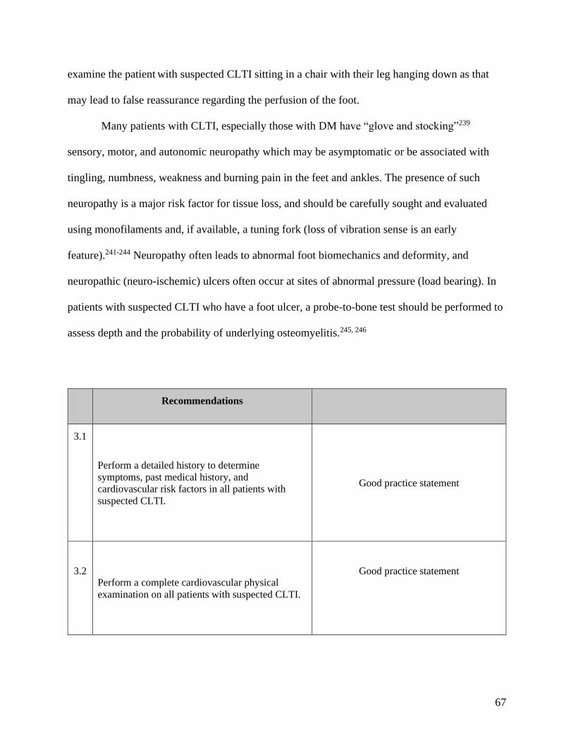
67
examine the patient with suspected CLTI sitting in a chair with their leg hanging down as that
may lead to false reassurance regarding the perfusion of the foot.
Many patients with CLTI, especially those with DM have “glove and stocking”239
sensory, motor, and autonomic neuropathy which may be asymptomatic or be associated with
tingling, numbness, weakness and burning pain in the feet and ankles. The presence of such
neuropathy is a major risk factor for tissue loss, and should be carefully sought and evaluated
using monofilaments and, if available, a tuning fork (loss of vibration sense is an early
feature).241-244 Neuropathy often leads to abnormal foot biomechanics and deformity, and
neuropathic (neuro-ischemic) ulcers often occur at sites of abnormal pressure (load bearing). In
patients with suspected CLTI who have a foot ulcer, a probe-to-bone test should be performed to
assess depth and the probability of underlying osteomyelitis.245, 246
Recommendations
3.1
Perform a detailed history to determine
symptoms, past medical history, and
cardiovascular risk factors in all patients with
suspected CLTI.
Good practice statement
3.2 Perform a complete cardiovascular physical
examination on all patients with suspected CLTI.
Good practice statement

68
3.3
Perform a complete examination of the foot,
including an assessment of neuropathy and a
probe-to-bone test of any open ulcers, in all
patients with pedal tissue loss and suspected
CLTI.
Good practice statement
Non-invasive hemodynamic tests
AP and ABI. Measurement of AP and calculation of ABI (highest ankle pressure divided
by highest brachial systolic pressure) is recommended as the first-line non-invasive
hemodynamic test in all patients with suspected CLTI (Figure 3.1).19 Although many patients
with CLTI will have an AP < 50 mm Hg and/or a markedly reduced ABI (typically less than
0.40), an increasing proportion will not, especially those with DM and CKD who may have
incompressible crural arteries. ABI results should be reported as non-compressible if the value is
greater than 1.4. However it is important to be aware that incompressibility can lead to
artifactually elevated readings between 0.4 and 1.4.247-249 This should be suspected when the ABI
falls in or near the normal range but is associated with dampened, monophasic waveforms
(recognized acoustically and/or visually on a screen).23 These falsely normal ankle pressures/ABI
values have been reported to be an independent predictor of major amputation.250 In such
patients, TP and TBIs, or other hemodynamic measurements as described below, should always
be obtained.251
TP and TBI. TP is measured using an appropriately sized mini-cuff placed around the
base of the (typically) great toe and attached to a standard manometer. A photoplethysmographic
or continuous-wave Doppler flow detector is then used to determine when flow returns while the

69
inflated cuff is slowly deflated. Various automated systems can be purchased. TP are less often
affected by incompressibility and, if possible, should be measured whenever falsely elevated
AP/ABI’s are detected or suspected, particularly when such values are non-concordant with
acoustic or visual waveform analysis. Recent studies suggest that TP is more sensitive than AP in
the diagnosis of CLTI, and more predictive of amputation risk.21, 22 Systolic TP are generally 20
to 40 mmHg lower than AP. TBI < 0.7 are considered abnormal and TP < 30 mmHg are
typically associated with advanced ischemia.22, 230, 252
Other methods for non-invasive diagnosis of CLTI
Alternative non-invasive testing methods can also be used to assist in the diagnosis of
CLTI (Table 3.1). While each method has its own advantages and limitations, depending on
local availability and expertise, they can be used to augment ankle and toe pressures and indices.
Segmental pressures can provide information on anatomical localization of lower limb vascular
disease in patients with CLTI, but nowadays are used infrequently, at least in HIC. Several other
non-invasive tests, including laser Doppler flowmetry, TcPO2, skin perfusion pressure (SPP), and
plethysmography have been used to evaluate limb perfusion.16, 253 However, these tests can be
influenced by a variety confounding factors and are not used routinely in most vascular
laboratories around the world.

70
Recommendation Grade Level of
Evidence
Key References
3.4 Measure ankle pressures (AP) and ankle brachial index
(ABI) as the first-line non-invasive test in all patients with
suspected CLTI.
1
(Strong)
B
(Moderate)
Ligmer et al
1996 19
Dachun 2010 20
3.5 Measure toe pressure (TP) and toe brachial index (TBI) in all patients with suspected CLTI and tissue loss (Figure
3.1)
1
(Strong)
B
(Moderate)
Aboyans et al
2008 21
Salaun
2018 169
3.6 Consider using alternative methods for non-invasive
assessment of perfusion, such as pulse volume recording,
transcutaneous oximetry, or skin perfusion pressure, when
ankle and toe pressures, indices, and waveforms cannot be
assessed.
2 (Weak) C (Low) Aboyans et al
200821
Shirasu 2016 23
Saluan 2018169
Wound and tissue loss classification systems
A number of limb and wound classification systems have been developed to try to
improve clinical decision-making and clinical outcomes.254-256 The WIfI system10 is based on
three key factors: wound, ischemia, and foot infection (Tables 3.2 – 3.5). WIfI correlates with
limb salvage, amputation risk, and wound healing; and can identify patients who are likely to
benefit from revascularization.68, 69
A limb-staging classification system, such as WIfI, should be used in all patients
presenting with suspected CLTI (Tables 3.2 – 3.5). Limb staging should be repeated following
vascular intervention, foot surgery, and/or treatment of infection; or whenever there is suspected
clinical deterioration.
Imaging of vascular anatomy

71
Vascular imaging should be performed in all patients with suspected CLTI (Table 3.6) to
determine the presence, extent, and severity of arterial disease and help inform decisions
regarding revascularization. Although, in recent years, there have been huge advances in imaging
techniques, access to these latest modalities, and so practice, varies considerably between and
even within countries.
In patients with CLTI who are candidates for revascularization (Section 6), imaging
should allow for complete anatomic staging using, for example, GLASS (Section 5). Adequate
imaging of the tibial and pedal vessels is of critical importance, particularly in planning
intervention in patients with tissue loss. History and physical examination often help to guide the
optimal imaging approach. For those with tibial disease, particularly in the setting of tissue loss,
computed tomography angiography (CTA) and magnetic resonance angiography (MRA) may
offer useful information but may fail to completely image the ankle and foot vessels with
sufficient resolution for procedural planning. Many vascular specialists believe that digital
subtraction angiography (DSA) remains the gold standard. CTA offers more precise
quantification of arterial calcification when compared with MRA and DSA. Intra-arterial dual-
energy CTA (s-CTA) combines low contrast dose conventional angiography with computed
tomography (CT) and, if available, may allow crural artery visualization in patients with renal
insufficiency.257 This technology is in evolution and not routinely available.
Duplex ultrasound imaging (DUS). DUS imaging is usually the first imaging modality
of choice, and in some healthcare settings may be the only modality available. DUS provides
information on the anatomic location and extent of disease as well as information about flow
volume and velocity.258, 259 There may be difficulty in directly imaging the aortoiliac (AI)

72
segments due to body habitus, bowel gas, and movement. However, the presence of ‘inflow’
disease can often be inferred from common femoral artery (CFA) waveforms. In the IP arterial
segments, assessment can be technically challenging, particularly when vessel calcification and
overlying tissue loss are present. Some vascular specialists advocate the use of US contrast
agents to improve visualization, however clinical studies to date are limited.260 Although
multiple studies have shown DUS to be inferior to other imaging techniques such as DSA, it
offers many advantages as a first-line imaging modality, including its non-invasive nature, low
cost, no iodinated contrast media, no ionizing radiation, and no fixed installation (mobility).25, 261,
262 The main disadvantages of DUS are that it is time consuming, highly operator dependent, and
does not produce a continuous lesion map. DUS is also poor at estimating collateral blood supply
and reserve. Furthermore, the stored images can be difficult to interpret at a later point in time.
Recommendation
Grade
Level of
Evidence
Key
References
3.7 Consider duplex ultrasound imaging as the first arterial
imaging modality in patients with suspected CLTI.
2 (Weak) B
(Moderate)
Hingorani et al
2008 24
3.8 Consider non-invasive vascular imaging modalities
(duplex ultrasound, computed tomography angiography,
magnetic resonance angiography) when available prior to
invasive catheter angiography in patients with suspected
CLTI who are candidates for revascularization.
2 (Weak) B
(Moderate)
Larch et al
1997 25
Adriaensen et
al 2004 26
Hingorani et al
2004 27
Collins et al
2007 28
Hingorani et al
2008 24
Met et al 2009 29
Computed tomography angiography (CTA). In recent years, CTA has advanced
73
considerably in terms of accuracy and acquisition times. Modern CTA quickly generates high
resolution, contrast-enhanced images that can be viewed in multiple planes or as three-
dimensional reconstructions.26, 263-265 In a meta-analysis comparing CTA to DSA that
predominantly included patients with IC, CTA was found to have high sensitivity and specificity
in the AI (95% and 96% respectively) and FP segments (97% and 94%), but was somewhat
inferior in the IP segment (95% and 91%).29 The researchers highlighted the difficulties
encountered with blooming artifact in calcified arteries (where motion-related artefact causes
calcium deposits to appear larger than they truly are), which would probably result in lowered
accuracy of this modality in the CLTI population, particularly in the IP segment. As such, in
many centers, CTA is primarily used to image and plan intervention in AI and FP segments.266
Contrast-induced nephropathy can be a significant problem57, 267, 268 and patients with pre-
existing renal insufficiency are at particular risk.269 Various guidelines have been written 270, 271
and many hospitals have local operating policies to try to mitigate the risks. Unfortunately,
practices vary considerably, making it impossible to identify firm recommendations, outside of
recognizing the risk. Finally, CTA is associated with significant doses of ionizing radiation.26, 272
Magnetic resonance angiography (MRA). MRA has the potential to produce images
that are comparable in quality to DSA but without exposure to ionizing radiation or iodinated
contrast, making contrast nephropathy extremely rare.27-29, 57, 263-269, 272-276 Time-resolved
techniques can accurately image flow patterns, which can be helpful in assessing IP run-off. In a
meta-analysis, MRA also showed improved specificity and sensitivity over CTA and DUS.276
While conventional time-of-flight MRA sequences may overestimate the degree of arterial
stenosis, newer techniques suggest that non-contrast MRA remains an excellend imaging
modality for patients with CLTI, accurately assessing distal lower extremity vessels.277 However,
74
failure of MRA to visualize vessel wall calcification may under-estimate the difficulty of
surgical and endovascular revascularization. Contrast-enhanced (CE) MRA using gadolinium-
based contrast agents, is generally preferred due to the high contrast-to-noise ratio, better spatial
resolution, more rapid acquisition, and less artifact. Time-resolved MRA is particularly useful in
imaging IP disease.274 Finally, MRA produces a three-dimensional map of the overall arterial
tree, with the possibility of additional accurate mapping of the IP and foot vessels in more
specialized centers. Other challenges of MRA include the potential overestimation of stenoses,
problems visualizing in-stent restenosis, compatibility with implanted devices such as
pacemakers and defibrillators, longer image acquisition times, and image artifact. Patients often
have a lower tolerance for MRA than CTA due to claustrophobia. Accurate interpretation of the
images by a dedicated subspecialist, such as a vascular radiologist, is essential in aiding
revascularization strategies. MRA equipment is expensive, although it can be used for other non-
vascular MR-based investigations. Thus, in some developing and developed countries, access
MRA and to dedicated subspecialists who are available to interpret the images is scarce.229
Finally, gadolinium contrast enhancement has been associated with cases of nephrogenic
systemic fibrosis, primarily in individuals with an estimated glomerular filtration rate (GFR) of <
30 mls/ minute.278
Foot MRA. CLTI patients have a high incidence of IP and pedal artery disease. The
precise location, length, and severity of disease, as well as the patency of runoff vessels, should
ideally be delineated prior to revascularization planning. In highly specialized centers, when
compared with DSA, foot CE-MRA yielded a sensitivity of 92% for the detection of significant
disease in IP and pedal vessels.279 MR perfusion imaging may have a role in assessing overall
foot perfusion before and after intervention.280, 281 As for limitations of foot CE-MRA, in slow
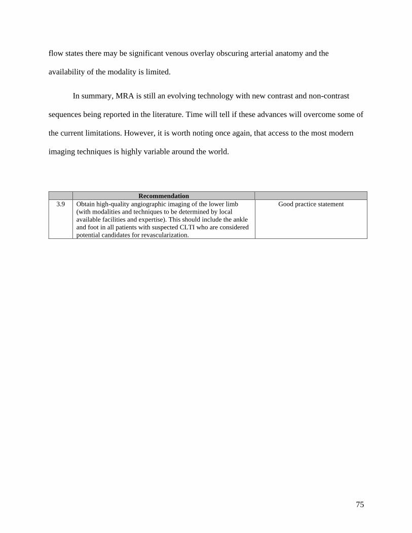
75
flow states there may be significant venous overlay obscuring arterial anatomy and the
availability of the modality is limited.
In summary, MRA is still an evolving technology with new contrast and non-contrast
sequences being reported in the literature. Time will tell if these advances will overcome some of
the current limitations. However, it is worth noting once again, that access to the most modern
imaging techniques is highly variable around the world.
Recommendation
3.9 Obtain high-quality angiographic imaging of the lower limb
(with modalities and techniques to be determined by local
available facilities and expertise). This should include the ankle
and foot in all patients with suspected CLTI who are considered
potential candidates for revascularization.
Good practice statement

76
Catheter digital subtraction angiography (DSA). With the advent of duplex, CTA, and MRA,
diagnostic DSA is probably performed less commonly nowadays, however many vascular
specialists still consider it the gold standard imaging modality in patients with suspected CLTI,
particularly when IP is likely to be present.282 Enthusiasts for DSA will also point out that it
allows for intervention at the same setting. Other vascular specialists, however, argue that
diagnostic DSA is outdated. The DSA technique should minimize the amount of iodinated
contrast and the dose of ionizing radiation used while maximizing imaging of the distal
vasculature.268, 283-285 In general, diagnostic DSA is widely available and the complication rate is
low.283, 286
CO2 angiography. CO2 angiography can be used in patients with a contrast allergy or in
individuals with severe CKD; unfortunately, it frequently causes significant patient discomfort.
CO2 angiography is generally considered inferior to iodinated angiography but can still provide
useful diagnostic images. There is a general trend of imaging performance progressively
degrading down the leg.287 Power injectors may improve safety and quality.
Perfusion angiography. This is a new technique performed using a dedicated imaging
suite and workstation to provide time-resolved perfusion imaging of the foot to aid in the
diagnosis and impact of revascularization techniques. Perfusion angiography provides
quantifiable information of functional status of the foot perfusion and is a positive step toward
functional imaging of the foot.288

77
Summary
All patients presenting with CLTI should have a full history and physical examination
followed by non-invasive hemodynamic testing. These studies can be easily performed in most
centers around the world. The authors recommend that all patients undergo limb staging using a
classification system, such as WIfI, that integrates multiple key elements (e.g. wound, ischemia,
infection) and correlates with the risk of amputation and the likelihood of wound healing. The
next step in appropriate candidates (Section 6) is to obtain high-quality diagnostic images to
guide revascularization. This will depend heavily upon the availability of equipment and local
expertise (Fig 3.2). Where available, DUS is the preferred first non-invasive imaging modality.
However, for more complete non-invasive anatomic imaging, either MRA or CTA can be
considered.
Catheter DSA represents the gold standard imaging technique, especially below the knee.
In many centers, however, DSA is typically only used when MRA or CTA is not available, when
MRA or CTA imaging is suboptimal and fails to adequately define the arterial anatomy, or for
those patients expected to proceed to endovascular intervention. It is important to emphasize that
no patient with suspected CLTI who is a suitable candidate for limb salvage should be denied
revascularization without first undergoing a complete diagnostic angiogram that includes the
ankle and foot.

78
Research Priorities
3.1 Define optimal methods for measuring foot perfusion and its correlation with stages of disease and
response to treatment.
3.2 Validate contrast-enhanced ultrasound in patients with CLTI.
3.3 Define optimal strategies to reduce the incidence of contrast-induced nephropathy in patients with
CLTI.
3.4 Improve non-invasive imaging of the ankle and foot vascular tree using magnetic resonance
angiography.

79
4: MEDICAL MANAGEMENT
CLTI is an end-stage manifestation of systemic atherosclerosis. It is frequently
accompanied by clinically significant CVD, resulting in exceedingly high mortality from stroke
and myocardial infarction (MI). In the absence of aggressive identification and treatment of risk
factors and associated co-morbid conditions, the prognosis of CLTI is usually poor with a
mortality rate of 20% to 26% within 1 year of diagnosis.5, 30, 154, 213, 219, 220, 230, 289
In a study of 574 patients with CLTI who did not undergo revascularization after 2 years,
31.6% had died, primarily from CVD, and 23% required major amputation.290
The goal of treatment of patients with CLTI is not only to salvage a functional limb but to
reduce cardiovascular morbidity and mortality, through aggressive risk factor modification and
best medical therapy (BMT).31, 32, 224 While certain risk factors, such as age and gender, cannot
be modified, others can, including hyperlipidemia, hypertension, diabetes, smoking, and
sedentary lifestyle.
Recommendations Grade Level of
Evidence
Key
References
4.1 Evaluate cardiovascular risk factors in all
patients with suspected CLTI.
1 (Strong) B (Moderate) I.C.A.I.
Group et al
1997 30
4.2 Manage all modifiable risk factors to
recommended levels in all patients with
suspected CLTI
1 (Strong) B (Moderate) Armstrong
2014 224
Faglia et al
2014 32
Antithrombotic therapy
Antiplatelet agents (APA) are strongly recommended for all patients with symptomatic
PAD in order to reduce the risk of major adverse cardiovascular events (MACE).33, 34, 291 The
Antithrombotic Trialists’ Collaboration performed a meta-analysis of APA trials prior to 1997.33

80
It included 135,000 patients with cerebrovascular, coronary, or PAD (IC) who were treated with
APA, and 77,000 control patients. The APA group had a 22% reduction in MACE and 75-150
mg aspirin per day were as effective as higher doses, but with a lower risk of bleeding.33 A more
recent meta-analysis studied the specific benefit of aspirin in 16 secondary prevention trials
comprising 17,000 patients.34 This study confirmed the benefit of APA with an 18.2% reduction
in MACE in both men and women.. The Critical Leg Ischaemia Prevention Study (CLIPS) group
compared the benefit of 100 mg of aspirin per day in 185 patients with symptoms of PAD and an
ABI < 0.85 or a TBI < 0.6 to placebo, and reported a 64% risk reduction in vascular events
compared to a 24% reduction in the placebo group.291
However, there is a growing body of literature indicating that alternatives to aspirin such
ticlopidine, dipyridamole, and clopidogrel may be more effective.35, 292-294 The CAPRIE
(Clopidogrel versus Aspirin in Patients at Risk for Ischemic Events) Trial, though not
specifically designed to address CLTI, compared 75 mg per of clopidogrel per day to 325 mg per
day of aspirin in patients with PAD. Researchers noted an 8.7% decrease in MACE with
clopidogrel compared with aspirin. There was no significant difference in bleeding risks between
the two agents.35
Other antiplatelet agents, such as ticagrelor and vorapaxar, have also been shown to
reduce MACE in patients with PAD.292-294 However, benefit over clopidogrel has not been
demonstrated.36, 294-298 The EUCLID trial compared ticagrelor to clopidogrel in 13,885 patients
with symptomatic PAD and an ABI < 0.8.36 Although both drugs had a similar safety profile,
ticagrelor was not superior to clopidogrel. The TRA2°P-TIMI 50 trial examined the effects of the
protease-activated receptor-1 antagonist vorapaxar on secondary prevention of ischemia events
in patients with stable atherosclerosis, including symptomatic PAD.295 Acute limb ischemia, a

81
pre-specified study endpoint, was reduced by 41% among the PAD cohort.298 However
vorapaxar has been associated with an increase in intracranial hemorrhage in patients who have
had a prior stroke or transient ischemic attack.296 In a recent meta-analysis, vorapaxar added to
aspirin yielded little improvement in the reduction of MACE in patients with atherosclerosis, and
was associated with a slightly higher incidence of intracranial hemorrhage.294 Finally, a meta-
analysis that reviewed the use of ticagrelor, ticlopidine, aspirin, cilostazol, picotamide,
vorapaxar, and clopidogrel as mono or dual antiplatelet therapy in patients with PAD found that
clopidogrel monotherapy resulted in the best overall safety and efficacy (reduction of MACE).297
The long-term use of dual APA therapy (DAPT) or systemic anticoagulation with vitamin
K antagonists is not indicated for PAD.299, 300 The role of direct oral anticoagulants (DOAC) is
currently the subject of intense investigation. COMPASS, a recent multicenter randomized trial
of 7,470 individuals with stable, mild to moderate PAD found that low-dose rivaroxaban (an oral
Factor Xa inhibitor), in combination with aspirin, reduced major adverse cardiovascular events
(death, myocardial infarction, or stroke) and major adverse limb events when compared with
aspirin alone. 37 Patients who had previous lower extremity revascularization, amputation, or a
history of intermittent claudication and an ABI of <0.90, a documented peripheral stenosis of
>50%, or a carotid stenosis of >50% were included in the study. Overall, 8.5% of study patients
had an ABI of <0.70. In this population there was a significant reduction in major adverse limb
events, major amputation, and acute limb ischemia compared to aspirin alone.301 This drug
combination was associated with a small but statistically significant increase in clinically
relevant bleeding. While the study results are promising, the benefits and risks of the low dose
rivaroxaban and low dose aspirin combination in patients with CLTI has not yet been adequately
defined. Additionally, this drug combination is not globally available at this time.
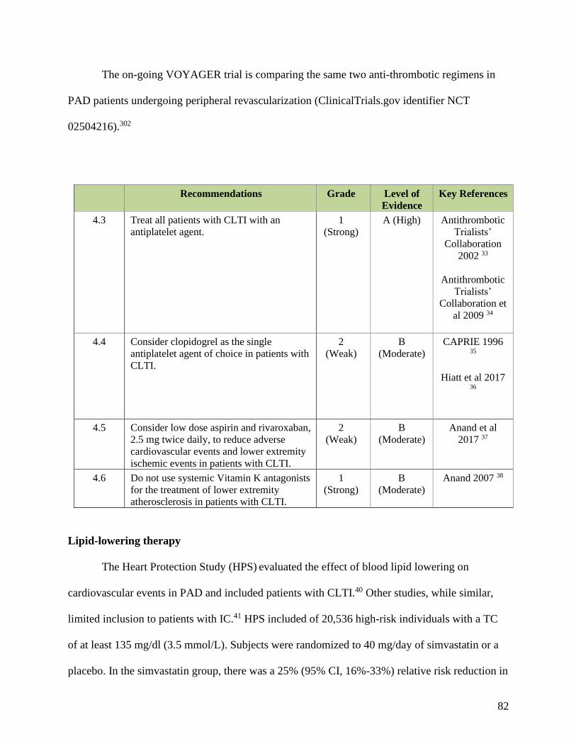
82
The on-going VOYAGER trial is comparing the same two anti-thrombotic regimens in
PAD patients undergoing peripheral revascularization (ClinicalTrials.gov identifier NCT
02504216).302
Recommendations Grade Level of
Evidence
Key References
4.3 Treat all patients with CLTI with an
antiplatelet agent.
1
(Strong)
A (High) Antithrombotic
Trialists’
Collaboration
2002 33
Antithrombotic
Trialists’
Collaboration et
al 2009 34
4.4 Consider clopidogrel as the single
antiplatelet agent of choice in patients with
CLTI.
2
(Weak)
B
(Moderate)
CAPRIE 1996 35
Hiatt et al 2017 36
4.5 Consider low dose aspirin and rivaroxaban,
2.5 mg twice daily, to reduce adverse
cardiovascular events and lower extremity
ischemic events in patients with CLTI.
2
(Weak)
B
(Moderate)
Anand et al
2017 37
4.6 Do not use systemic Vitamin K antagonists
for the treatment of lower extremity
atherosclerosis in patients with CLTI.
1
(Strong)
B
(Moderate)
Anand 2007 38
Lipid-lowering therapy
The Heart Protection Study (HPS) evaluated the effect of blood lipid lowering on
cardiovascular events in PAD and included patients with CLTI.40 Other studies, while similar,
limited inclusion to patients with IC.41 HPS included of 20,536 high-risk individuals with a TC
of at least 135 mg/dl (3.5 mmol/L). Subjects were randomized to 40 mg/day of simvastatin or a
placebo. In the simvastatin group, there was a 25% (95% CI, 16%-33%) relative risk reduction in

83
the first major vascular event among subjects who had no history of a coronary event at
baseline.40 In addition, lipid lowering was shown to be most effective in patients with a blood
cholesterol > 135 mg/dl (> 3.5 mmol/L). There was also a significant reduction in cardiovascular
events (P < 0.0001) among a subgroup of individuals with PAD.
A Cochrane Review evaluated 18 lipid-lowering trials comprising 10,049 PAD
patients.39, 42 While the majority had IC, and only some trials included CLTI, the results appear
relevant to the CLTI population. Only one study showed a negative effect of lipid lowering.
When this study was excluded, analysis showed that lipid-lowering therapy significantly reduced
the risk of total cardiovascular events in PAD (odds ratio [OR], 0.74; CI, 0.55-0.98) in PAD.42
This was primarily due to a positive effect on total coronary events (OR, 0.76; CI, 0.67-0.87).
The impact of statin agents may extend beyond their lipid-lowering effect. by reducing
inflammation in patients with PAD.303, 304 An individual-patient data (IPD) meta-analysis of 54
prospective cohort studies demonstrated that inflammatory biomarkers independently predict
vascular risk with a magnitude of effect at least as large as that of blood pressure or
cholesterol.305 Even after adjusting for age, sex, and traditional risk factors, patients with PAD
are known to have increased levels of inflammatory cytokines, acute-phase reactants, and soluble
adhesion molecules.306 However, while the attributable vascular risk associated with
inflammation is large, and while animal models using targeted anti-inflammatory therapies have
shown promise, it remains unknown whether inhibiting inflammation alone will lower vascular
event rates.
The landmark JUPITER trial examined the use of intensive statin therapy (rosuvastatin
20 mg daily vs placebo) in a primary prevention trial.307, 308 In total, there were 17,802
individuals who had low levels of LDL-C but an elevated vascular risk, based on a pro-

84
inflammatory biomarker (high levels of high-sensitivity CRP; hsCRP). Investigators
demonstrated a 44% reduction in major vascular events, including a 54% reduction in MI, a 48%
reduction in stroke, a 46% reduction in arterial revascularization, a 43% reduction in deep
venous thrombosis (DVT) / pulmonary embolism (PE), and a 20% reduction in mortality. The
greatest absolute risk and the greatest absolute risk reduction were observed among those with
the highest levels of hsCRP. There are now multiple studies showing a decrease in
cardiovascular events in patients with established atherosclerosis treated with intensive statin
therapy.43, 224, 309, 310 A recent large retrospective cohort study from the US Veterans Health
Administration population demonstrated reduced mortality and major amputation rates among
patients with established PAD receiving intensive dose statins.311 Statin therapy can be
associated with muscle aching, the most common adverse effect limiting its use. In the setting of
this complication, statin dose can be lowered to the maximum tolerated dose and a second non-
statin cholesterol-lowering drug can be added to reduce cholesterol levels even further.
Recent (2013, 2018) AHA/ACC guidelines on treatment of blood cholesterol recommend
the use of moderate- to high-intensity statins for all individuals with established atherosclerotic
cardiovascular disease including PAD.312, 313 Both rosuvastatin (20-40 mg) and atorvastatin (40-
80 mg) have been shown to be effective.310 The 2018 guideline describes “very high risk”
individuals to include those with symptomatic PAD and at least one other high-risk condition
(age ≥65, familial hypercholesterolemia, history of coronary revascularization, diabetes mellitus,
hypertension, CKD, current smoking, CHF)—a categorization that applies to the overwhelming
majority of patients with CLTI. For this population high-intensity/maximal tolerated statin
dosing is recommended, and if on-treatment LDL-C levels remain ≥ 70 mg/dl (1.8 mmol/L) then
the addition of ezetimibe is considered reasonable.313

85
New lipid lowering agents have entered the armamentarium. Proprotein convertase
subtilisin/kexin type 9 [PCSK9] directs the degradation of LDL receptors in the liver, and has
become a drug target. A recent RCT (FOURIER) demonstrated an additional benefit of
evolocumab (a proprotein convertase subtilisin/kexin type 9 [PCSK9] inhibitor) in reducing
MACE in PAD patients already on statin therapy.314 The composite endpoint of cardiovascular
death, myocardial infarction, stroke, hospital admission for unstable angiona, or coronary
revascularization was statistically reduced in PAD patients treated with the PCSK-9 inhibitor,
evolocumab (HR 0.79, p=0.0040). There was also a reduction in the risk of major adverse limb
events, including acute limb ischemia and major amputation. Further studies will be needed in
PAD subpopulations including CLTI.
Further studies of these agents are desirable in high-risk PAD subpopulations including
CLTI.
Recommendation Grade Level of
Evidence
Key References
4.7 Use moderate or high intensity statin therapy to
reduce all-cause and cardiovascular mortality in
patients with CLTI.
1 (Strong) A (High) Leng et al 2000 39
Heart Protection
Study Group 2002 40
Meade et al 2002 41
Aung et al 2007 42
Mills 2011 43
Rodriguez et al 2017 44

86
Management of hypertension
It is universally accepted that control of hypertension reduces MACE in patients with
PAD. The INVEST trial studied the impact of control of hypertension on all-cause death, non-
fatal MI, and non-fatal stroke in 22,576 hypertensive patients with stable coronary artery disease
(CAD) of whom 2699 also had PAD.46 PAD patients had a significantly higher incidence of
sustaining a primary endpoint MACE compared with those without PAD (16.3% vs 9.2%).
Additionally, among those with PAD, a MACE was less likely to occur in patients with systolic
BP less than 145 mmHg and diastolic pressures less than 90 mmHg. Further reduction of BP to
below130 mmHg systolic and 80 mmHg diastolic provides even greater protection from
cardiovascular events.48 The SPRINT trial compared blood pressure control to a systolic pressure
of 120 mmHg (intensive control) or 140 mmHg (standard control) in 2510 patients with a mean
age of 79.9 years followed for a mean of 3.14 years.315 The study documented a significantly
lower incidence of composite cardiovascular events of death with intensive control. However,
intensive BP control may result in greater morbidity associated with periods of clinically
significant hypotension.45, 47 Optimal blood pressure control for patients with CLTI has not been
established and while maintaining systolic pressure < 140 mmHg and diastolic pressure < 90
mmHg is important, lower pressures may be beneficial to further reduce MACE.
The first line category of oral antihypertensive does not appear to be of significance.
Angiotensin-converting enzyme inhibitors (ACEI), calcium channel blockers, and diuretics,
when successful in lowering BP to target, reduce cardiovascular events to a similar extent.316, 317
Although the ONTARGET and HOPE trials suggest that, in the absence of heart failure,
monotherapy with an ACEI (Ramipril) reduces the rate of major adverse cardiovascular events
in high-risk patients, there is recent evidence that suggests this class of drug may result in a

87
higher amputation rate for patients with CLTI.318 In an analysis of the Medicare database for
2007-2008, 22,954 patients underwent lower extremity revascularization. Of these, 64.6% were
treated for CLTI. Compared with those not taking an ACEI, patients who presented with rest
pain and were taking an ACEI after the index procedure had a higher risk of amputation. Other
studies have not noted an increased risk of amputation associated with ACEI but have suggested
an increased rate of reintervention. A propensity score matched cohort study of 17,495 Danish
patients compared those receiving ACEI to those who were not after vascular reconstruction.
Followed for a mean of 1.6 years, the patients treated with ACEI had a lower all-cause mortality
(20.4% vs 24.9%) but underwent more reintervention (24% vs 23.1%).319 Using the same general
methodology, these investigators found that the use of beta blockers after primary vascular
reconstruction was associated with a decrease in the incidence of major amputation but a higher
rate of MI and stroke without an increase in all-cause mortality. 320
Globally, adequate control of hypertension remains a significant challenge. In LMIC
countires the availability of oral antihypertensives is limited and costs are high resulting in poor
overall BP control. Strategies are urgently required to improve availability and affordability of
drugs so that vascular specialists can treat their patients to target.321
There have been concerns that drugs which reduce heart rate and blood pressure will
worsen ischaemia in patients with PAD. Although beta blockade has not been directly evaluated
in CLTI, it has been the subject of several clinical trials in IC and has been shown to be effective
in lowering BP without worsening symptoms.322, 323
Recommendation Grade Level of
Evidence
Key References
4.8 Control hypertension to target levels of
< 140 mm Hg systolic and < 90 mm Hg
diastolic in patients with CLTI.
1 (Strong) B (Moderate) Bavry et al 2010 46
ACCORD Study
Group et al 2010 45
Wright et al 2015 47
Moise et al 2016 48

88
Management of diabetes
Type 2 diabetes (T2DM), is a significant risk factor for PAD.324, 325 and the extent of
vascular disease appears related to the duration and severity of hyperglycemia. Glycemic control
is therefore essential in all diabetic patients with PAD. Metformin monotherapy is generally
recognized as the best initial oral hypoglycemic agent (OHA). When additional therapy is
needed, any other class of OHA, including sulfonylurea, thiazolidinedione, DPP-4 inhibitor, or
alpha glucosidase can be added with equal effectiveness.54 While there is some data to suggest
that the DPP-4 inhibitors may reduce the risks of myocardial infarction and stroke, the impact on
peripheral arterial disease in patients with CLTI has not yet been defined.326 The goal for most
adults with DM is to maintain a glycosylated hemoglobin (Hb) A1c level of < 7% (equivalent to
IFCC 53 mmol/mol).49-52 However less stringent goals (e.g. A1c < 8%) may be appropriate for
individuals with advanced vascular complications or limited life expectancy.53
T2DM patients with abnormal renal function treated with metformin may be at higher
risk for contrast-induced nephropathy and lactic acidosis. While the matter is the subject of
continued debate, it is reasonable to withhold metformin for 24 to 48 hours after the
administration of iodinated contrast.55-57, 270, 271
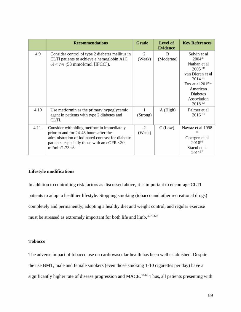
89
Recommendations Grade Level of
Evidence
Key References
4.9 Consider control of type 2 diabetes mellitus in
CLTI patients to achieve a hemoglobin A1C
of < 7% (53 mmol/mol [IFCC]).
2
(Weak)
B
(Moderate)
Selvin et al
200449
Nathan et al
2005 50
van Dieren et al
2014 51
Fox et al 201552
American
Diabetes
Association
2018 53
4.10 Use metformin as the primary hypoglycemic
agent in patients with type 2 diabetes and
CLTI.
1
(Strong)
A (High) Palmer et al
2016 54
4.11 Consider witholding metformin immediately
prior to and for 24-48 hours after the
administration of iodinated contrast for diabetic
patients, especially those with an eGFR <30
ml/min/1.73m2.
2
(Weak) C (Low) Nawaz et al 1998
55
Goergen et al
201056
Stacul et al
201157
Lifestyle modifications
In addition to controlling risk factors as discussed above, it is important to encourage CLTI
patients to adopt a healthier lifestyle. Stopping smoking (tobacco and other recreational drugs)
completely and permanently, adopting a healthy diet and weight control, and regular exercise
must be stressed as extremely important for both life and limb.327, 328
Tobacco
The adverse impact of tobacco use on cardiovascular health has been well established. Despite
the use BMT, male and female smokers (even those smoking 1-10 cigarettes per day) have a
significantly higher rate of disease progression and MACE.58-60 Thus, all patients presenting with
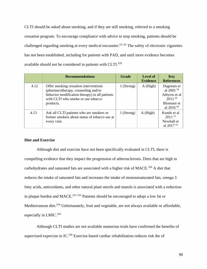
90
CLTI should be asked about smoking, and if they are still smoking, referred to a smoking
cessation program. To encourage compliance with advice to stop smoking, patients should be
challenged regarding smoking at every medical encounter.61, 62 The safety of electronic cigarettes
has not been established, including for patients with PAD, and until more evidence becomes
available should not be considered in patients with CLTI.329
Recommendations Grade Level of
Evidence
Key
References
4.12 Offer smoking cessation interventions
(pharmacotherapy, counseling and/or
behavior modification therapy) in all patients
with CLTI who smoke or use tobacco
products.
1 (Strong) A (High) Dagenais et
al 2005 58
Athyros et al
2013 59
Blomster et
al 2016 60
4.13 Ask all CLTI patients who are smokers or
former smokers about status of tobacco use at
every visit.
1 (Strong) A (High) Kondo et al
2011 61
Newhall et
al 2017 62
Diet and Exercise
Although diet and exercise have not been specifically evaluated in CLTI, there is
compelling evidence that they impact the progression of atherosclerosis. Diets that are high in
carbohydrates and saturated fats are associated with a higher risk of MACE.330 A diet that
reduces the intake of saturated fats and increases the intake of monounsaturated fats, omega 3
fatty acids, antioxidants, and other natural plant sterols and stanols is associated with a reduction
in plaque burden and MACE.331-333 Patients should be encouraged to adopt a low fat or
Mediterranean diet.334 Unfortunately, fruit and vegetable, are not always available or affordable,
especially in LMIC.335
Although CLTI studies are not available numerous trials have confirmed the benefits of
supervised expercise in IC.336 Exercise-based cardiac rehabilitation reduces risk the of

91
subsequent MI and cardiovascular mortality.337 It therefore seems reasonable to suggest that a
post-revascualrisation walking-based exercise program would also benefit CLTI patients who are
cleared for full weight-bearing.
Management of pain
Although pain is a very important issue for most CLTI patients it is often poorly
managed. Poor pain control can reduce HRQL levels to those seen in patients with terminal
cancer and has a major adverse impact on functional capacity.
As no RCTs have been conducted in CLTI, good practice recommendations have to be
extrapolated from other conditions where severe pain is a major factor. The management of
ischaemic pain in CLTI is often complicated by the co-existing neuropathic pain, particularly in
patients with DM. However the management of neuropathic pain is not covered here.
Guidelines usually recommend a tiered approach to pain management, with a ‘trade off’
between benefits and harms (eg, constipation, drowsiness, etc).338, 339 Patients should be offered
paracetamol (acetaminophen) in combination with opioids, and in proportion to the severity of
pain. All patients receiving opioids should also be offered laxatives and anti-nausea medication.
If the maximum tolerated analgesic dose does not produce adequate pain relief, alternative
approaches should be considered. These include tricyclic antidepressants, gabapentin, and
pregabalin, all of which are used effectively for neuropathic pain. It is important to note,
however, that if the clinician is unfamiliar with the use of these compounds, early referral to a
Pain Management Service for patients with pain not controlled by opioids is required.
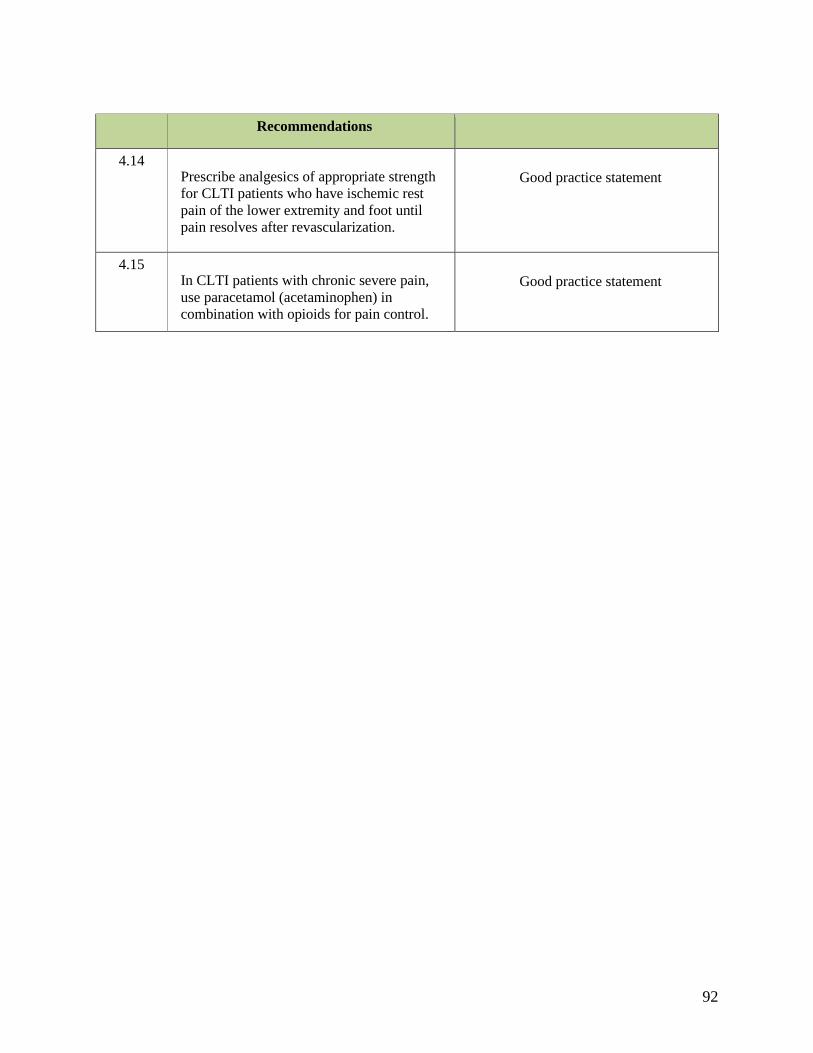
92
Recommendations
4.14 Prescribe analgesics of appropriate strength
for CLTI patients who have ischemic rest
pain of the lower extremity and foot until
pain resolves after revascularization.
Good practice statement
4.15 In CLTI patients with chronic severe pain,
use paracetamol (acetaminophen) in
combination with opioids for pain control.
Good practice statement

93
Research Priorities
4.1 Define the optimal antithrombotic regimen (safety and efficacy) in patients with CLTI to reduce
cardiovascular and limb-specific events
4.2 Define treatment targets and optimal dosing for lipid lowering agents in the CLTI population
4.3 Identify biomarkers that are predictive of clinical events in the CLTI population and may serve as
targets for therapy
4.4 Identify effective smoking cessation strategies for patients with advanced peripheral arterial
disease and CLTI
4.5 Identify the type of analgesia that is most effective in patients with chronic pain secondary to
CLTI
94
5: THE GLOBAL LIMB ANATOMICAL STAGING SYSTEM FOR CLTI
Rationale
An accurate assessment of limb threat and stratification of the anatomical pattern of
disease are the foundations of EBR. This is true not only in everyday practice but also in
improving outcomes assessment and research. The authors propose a new, clinically oriented
framework for classifying the pattern of arterial disease in CLTI. GLASS is a fundamental
departure from current approaches used in PAD and more analogous to the SYNTAX system for
CAD.340, 341
Current PAD anatomic classification schemes either describe the location and severity of
individual arterial lesions11, 156 or quantify the overall burden and morphology of disease.12, 151, 170
Lesion- or segment-based grading systems are useful for comparing endovascular device
performance in well-defined clinical situations. They are not, however, useful for defining EBR
strategies in CLTI, especially given the complex, multi-level, and increasingly distal disease
patterns typically seen in current clinical practice.
Successful revascularization in CLTI, particularly in patients with tissue loss, nearly
always requires restoration of (pulsatile) in-line flow to the foot. Because individual lesion-based
schemes correlate poorly with effective revascularization in CLTI, vascular specialists must
integrate approaches for arterial segments into a management strategy for the whole limb.
Factors that determine a successful anatomic outcome are intrinsically different for bypass
grafting and endovascular intervention. Bypass surgery requires adequate inflow and outflow
and, perhaps most importantly, a suitable autologous conduit. By contrast, the success of

95
endovascular intervention is largely defined by the complexity of atherosclerosis within the
anticipated target artery path (TAP) that provides in-line flow to the foot. When the TAP
includes multiple lesions in series, technical success and sustained patency for the limb as a
whole must be estimated as a product function of each lesion traversed.
GLASS is based on defining the TAP in each individual patient based on high-quality
imaging, and requires selection of a preferred infra-popliteal (IP) artery. The TAP is generally
selected based on the least diseased crural artery providing runoff to the foot. It can also be
selected based on other relevant factors, such as angiosome preference, or avoidance of a
previously instrumented vessel. While the relationship between the pattern of occlusive disease,
patency of the chosen intervention, and clinical success in CLTI is a complex one, an integrated
limb-based anatomic staging system like GLASS is critical to define it. The preferred TAP for
endovascular intervention and preferred target artery for open bypass surgery may not always be
the same; clinical decision-making thus hinges on a comparative estimate of risk and success for
each. Like SYNTAX, GLASS stage is designed to correlate primarily with endovascular
outcomes. As such, it does not incorporate factors such as venous conduit quality or distal runoff
that are more directly relevant for bypass grafting.
GLASS provides a basis for clinical practice and supports future research in CLTI. When
combined with tools for stratification of patient-risk and severity of limb threat (Sections 1 & 3),
GLASS facilitates the development of specific EBR guidelines in CLTI (Section 6). In
developing GLASS, the writing group was informed by a commissioned systematic review of
revascularization outcomes in CLTI7 and expert opinion. Still, the authors acknowledge that the
new grading system requires prospective validation in a variety of patient populations and
healthcare environments. The system is expected to undergo revisions as outcomes are reported.

96
Important factors for refinement include the current state of limited high-quality evidence in the
field, ongoing changes in both epidemiology and technology, and differences in disease patterns
and practice around the world.
Assumptions and approach
As CLTI is usually the result of complex multi-level occlusive disease, certain
simplifying assumptions are required to develop a usable anatomic staging system (Table 5.1).
First, because existing schemes for AI disease appear adequate, the focus of GLASS is on
infrainguinal disease (see Table 5.2 for a simplified inflow disease scheme). In GLASS, the
CFA and profunda femoris artery (PFA) are seen as inflow arteries, and the infrainguinal system
begins at the origin of the superficial femoral artery (SFA). This is justified by the distinct
approaches utilized in the treatment of CFA/PFA disease (Section 6), and long-term results that
are similar to those for AI interventions.
In order for GLASS to be useful in everyday clinical practice, and to form the basis of
practice-changing research, it is important that it does not rely on complex methods of lesion
characterization. With regard to vessel calcification, GLASS adopts a dichotomous subjective
scale in which severe calcification (eg, > 50% of circumference, diffuse, bulky, or “coral reef”
plaques) increases the within-segment grade by one numeric level. This is a subjective
determination made by the treating physician that the severity of calcification in the TAP
significantly increases technical complexity (and expected failure rates) for endovascular
intervention. Alternative approaches for quantifying arterial calcification in PAD have been
suggested but are more complex, and none of these has been validated for discriminating clinical
outcomes.342, 343 With regard to inframalleolar (IM) disease, GLASS employs a three-level
97
modifier (Figure 5.1) to describe the status of arteries crossing the ankle (including the terminal
divisions of the peroneal artery) and the pedal arch. Currently, the IM disease modifier is not
considered within the primary assignment of limb stages in GLASS, given the absence of strong
evidence on how it impacts treatment outcomes. It should, however, be captured in future studies
to better define how to incorporate pedal outflow disease into anatomic staging in CLTI.
GLASS also makes the following assumptions:
• Restoring durable (pulsatile) in-line flow to the affected part, particularly in patients with
tissue loss, is a primary goal of revascularization in CLTI.
• Using high-quality imaging (Section 3), the vascular specialist chooses and defines a
TAP that is most likely to achieve that in-line flow.
• The TAP will usually involve the least diseased IP artery.
• Other IP arteries (not selected for the TAP) are equally or more diseased.
Additionally, although it is an important research question, the current version of GLASS
does not consider multi-vessel IP revascularization because evidence of its role is still lacking.
Where the clinician is considering such revascularisation, GLASS staging is based on the
primary IP target, as defined by the clinician prior to the intervention.
In defining infrainguinal anatomic stages (I-III), GLASS combines grades (0-4) for the
FP (origin of the SFA to the origin of the anterior tibial (AT) artery; Figure 5.2) and IP (origin of
the tibioperoneal trunk and the AT artery to the malleoli; Figure 5.3) segments in series. Stages
were developed to correlate with estimated limb-based patency (LBP), defined as maintenance of
in-line flow through the entire length of the TAP, from the SFA origin to the malleoli. LBP is
considered to be lost when any one of the following occurs:
98
1) Anatomical failure: occlusion, critical stenosis, or reintervention affecting any portion of
the defined TAP
2) Haemodynamic failure: a significant drop in ABI (≥ 0.15) or TBI (≥ 0.10), or
identification of ≥ 50% stenosis in the TAP, in the presence of recurrent or unresolved
clinical symptoms, eg, rest pain, worsening or persistent tissue loss.
LBP is an important new concept allowing more direct comparison between
revascularization approaches in CLTI. Estimating LBP following surgical or endovascular
intervention is central to the development of EBR (Section 6). The writing group defined three
GLASS stages based on the likelihood of immediate technical failure (ITF) 343 and one year LBP
following endovascular intervention of the selected TAP. GLASS stages for the limb thus reflect
a gradient of infrainguinal disease complexity:
• Stage I: Low Complexity Disease: expected ITF < 10% and > one year LBP > 70%
• Stage II: Intermediate Complexity Disease: expected ITF < 20% and one year LBP 50-
70%
• Stage III: High Complexity Disease: expected ITF > 20%; or one year LBP < 50%
Consensus process and assignment of limb stages
In order to assign GLASS stages (I-III) in the two-dimensional matrix shown in Table
5.3, a multi-national, multi-specialty group of vascular specialists (GVG writing group and
invited external experts), as well as evidence summaries7 and other published material79, 160, 344-
400 were surveyed. Representative examples of GLASS stage I-III disease are illustrated in the
angiograms depicted in Figures 5.4-5.6. Table 5.4 provides a descriptive summary of the three
GLASS stages.

99
Managing CLTI with GLASS
Use of the GLASS system involves the following steps (Figure 5.7)
1. Obtain high-quality angiographic imaging, to include the ankle and foot (Section 3).
2. Identify the TAP.
3. Determine the FP GLASS grade (0-4) (Figure 5.2).
4. Determine the IP GLASS grade (0-4) (Figure 5.3).
5. Decide whether there is severe calcification (eg, > 50% of circumference, diffuse, bulky,
or “coral reef” plaques likely to compromise endovascular outcomes) within the FP
and/or IP segments of the TAP. If present, increase the segment grade by one.
6. Combine FP and IP grades to determine the overall GLASS stage (Table 5.3).
7. Use the pedal modifier (P0, P1, or P2) to describe the status of IM arteries.
For the individual patient with CLTI, an EBR strategy (Section 6) is based on the full
integration of
1. estimated patient risk and long-term survival
2. the severity of limb threat (e.g. using WIfI) (Sections 1 & 3)
3. anatomical pattern and severity of disease in the affected limb (e.g. GLASS).
Recommendation
5.1 Use an integrated, limb-based anatomical staging
system (such as the Global Limb Anatomical Staging
System) to define complexity of a preferred target
arterial pathway, and facilitate evidence-based
revascularization in patients with CLTI.
Good practice statement

100
Limitations and future direction
The authors acknowledge the limitations of the available data in developing this initial
version of GLASS. Severe calcification, particularly in the tibial arteries, is a negative predictor
of technical success for intervention, and signifies a higher risk for amputation.401, 402 However, a
simplified and validated scoring system for calcification that is associated with procedural
outcomes is still lacking.342 At the same time, pedal artery disease appears to be increasing in
both prevalence and importance, particularly in CLTI patients experiencing major tissue loss
and/or infection (WIfI Stage 4).403, 404
Pedal interventions remain relatively uncommon and data on outcomes are extremely
limited. Patients with no IM revascularization target are placed in a high-risk subgroup, though
they are assigned a simplified modifier (P2) in the current version of GLASS. In the future, it is
anticipated that better data will allow for a more sophisticated incorporation of calcification and
pedal disease. Other important issues, including the benefits of revascularizing multiple IP
arteries, the relative quality of runoff distal to the revascularization and extending to the wound-
related artery or angiosome, and the complex relationship between hemodynamic and clinical
success, also require further study.
In assigning GLASS stages, the authors assume that pre-procedural decision-making is
frequently driven by the estimation of the anticipated technical and clinical success following
endovascular intervention. As a result, the preferred TAP for endovascular intervention and
bypass surgery may not always be the same. Thus, treatment outcomes for surgical bypass
should also be reported and analyzed based on the actual procedure performed, including inflow
artery, outflow artery, and the conduit utilized.

101
Research Priorities
5.1 What are the expected procedural, hemodynamic, and clinical outcomes of revascularization
across the spectrum of infrainguinal disease severity? Better evidence is needed to validate the
Global Limb Anatomical Staging System, particularly for endovascular strategies in
intermediate (II) and severe (III) stages of infrainguinal disease.
5.2 What is the effect of severe infra-malleolar and pedal arch disease on revascularization
outcomes in CLTI? Is there a clinically useful way to grade this level of disease?
5.3 Is there evidence that other measures such as outflow bed resistance or below the knee runoff
scores are predictive of procedural or clinical outcomes? How do these compare to target path
lesion complexity assessed by angiography?
5.4 Is there a simple, reproducible method for quantification of calcification that has predictive
value for infrainguinal interventions?
5.5 Are there specific patient factors (eg, demographic or comorbidity) associated with anatomic
patterns of disease in CLTI?
5.6 Are there anatomic patterns of disease in which an endovascular approach is futile?
5.7 How does lesion morphology (eg, concentric vs eccentric) influence treatment success for
different endovascular interventions?
5.8 Is there a correlation between Global Limb Anatomical Staging System stage and clinical
presentation (Wound Ischemia foot Infection)?
5.9 What is the comparative value of direct (angiosome based) versus indirect revascularization in
the setting of tissue loss, and how should it drive selection of the preferred target arterial path?
Is this specific to wound location or Wound Ischemia foot Infection stage?

102
6: STRATEGIES FOR EVIDENCE-BASED REVASCULARIZATION
Effective revascularization is the cornerstone of limb salvage in CLTI. Although multiple
techniques are available, there are limited high-quality data to support EBR. A new, systematic
paradigm is required to improve decision-making, clinical outcomes and cost- effectiveness.
In order to aid clinical decision-making in everyday practice, and to facilitate future EBR
research in CLTI, the authors propose a three-step integrated approach (PLAN) (Figs 6.1 & 6.2)
based on:
• Patient risk estimation
• Limb staging
• ANatomic pattern of disease
PLAN: Patient Risk Estimation
The first step involves assessing the patient for the following:
• candidacy for limb salvage
• peri-procedural risk
• life expectancy
CLTI is associated with advanced age, multiple co-morbidities, and frailty. The goals of
treatment include relief of pain, healing of wounds, and preservation of a functional limb.
However, revascularization may incur significant morbidity and mortality, requiring multiple
hospitalizations, prolonged outpatient care, and thus considerable health and social care costs.
While the great majority of patients with CLTI should be considered as candidates for limb
salvage, some may be appropriately treated with primary amputation or palliation, following

103
shared decision-making. Patients, families, and caregivers should have access to appropriate
expertise in making these challenging decisions. Although maintenance of independent
ambulatory status is an important goal, predicting functional outcomes following
revascularization may be challenging, particularly in patients who are severely deconditioned.
Palliative care consultants, where available, may be a valuable resource to optimize symptom
management in patients with limited goals of care.
Recommendations
6.1 Refer all patients with suspected CLTI to a vascular specialist
for consideration of limb salvage, unless major amputation is
considered medically urgent.
Good practice statement
6.2 Offer primary amputation or palliation to patients with very
limited life expectancy, poor functional status (eg, non-
ambulatory), or those with an unsalvageable limb following
shared decision-making.
Good practice statement
Palliative therapy should rarely include revascularization except in special circumstances
such as:
• Treatment of hemodynamically significant inflow disease, if needed to improve the
likelihood of a successful amputation at the most distal possible level.
• Relief of intractable pain, or to improve wound healing, after shared decision-making
with the patient, family, and vascular treatment team.
Estimation of operative risk and life expectancy plays a critical role in EBR. Trade-offs
between risk, invasiveness, hemodynamic gain, and anatomic durability of the vascular
intervention are commonly made in everyday practice. Risk stratification tools can assist by
providing objective criteria for such decisions. Multiple tools have been developed and applied
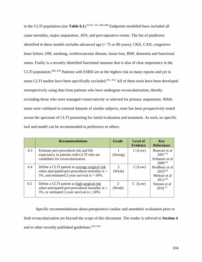
104
to the CLTI population (see Table 6.1).63-67, 225, 405-408 Endpoints modelled have included all
cause mortality, major amputation, AFS, and peri-operative events. The list of predictors
identified in these models includes advanced age (> 75 or 80 years), CKD, CAD, congestive
heart failure, DM, smoking, cerebrovascular disease, tissue loss, BMI, dementia and functional
status. Frailty is a recently identified functional measure that is also of clear importance in the
CLTI population.409, 410 Patients with ESRD are at the highest risk in many reports and yet in
some CLTI studies have been specifically excluded.411, 412 All of these tools have been developed
retrospectively using data from patients who have undergone revascularization, thereby
excluding those who were managed conservatively or selected for primary amputation. While
some were validated in external datasets of similar subjects, none has been prospectively tested
across the spectrum of CLTI presenting for initial evaluation and treatment. As such, no specific
tool and model can be recommended in preference to others.
Recommendations Grade Level of
Evidence
Key
References
6.3 Estimate peri-procedural risk and life
expectancy in patients with CLTI who are
candidates for revascularization.
1
(Strong)
C (Low) Biancari et al
2007 63
Schanzer et al
2008 64
Bradbury et al
2010 65
Meltzer et al
2013 66
Simons et al
2016 67
6.4 Define a CLTI patient as average surgical risk
when anticipated peri-procedural mortality is <
5%, and estimated 2-year survival is > 50%.
2
(Weak)
C (Low)
6.5 Define a CLTI patient as high surgical risk
when anticipated peri-procedural mortality is ≥
5%, or estimated 2-year survival is ≤ 50%.
2
(Weak)
C (Low)
Specific recommendations about preoperative cardiac and anesthetic evaluation prior to
limb revascularization are beyond the scope of this document. The reader is referred to Section 4
and to other recently published guidelines.413, 414

105
PLAN: Limb Staging
CLTI patients present with a broad spectrum of disease severity. Staging of the limb is central to
EBR (see Section 3) and use of the SVS Threatened Limb Classification System (“WIfI”) is
recommended (see Section 1) .10, 68-72, 171. This is the only system that fully integrates wound
severity, ischemia, and infection to stage CLTI.
The severity of ischemia and the benefits of revascularization do not map in an
exclusively concordant fashion with amputation risk across the spectrum of CLTI, as expressed
in the original WIfI consensus document.10 Expert opinion, now supported by reports from
institutional series69, 70, 72 suggest that the presumed benefit of revascularization in CLTI is linked
to both the severity of ischemia and to the degree of limb threat (Figure 6.3). All symptomatic
patients who have severe (e.g. WIfI grade 3) ischemia should undergo attempted
revascularization presuming they are appropriate candidates for limb salvage.5 In settings of
advanced tissue loss/infection (e.g. WIfI Stage 4 limbs), revascularization may also be of benefit
in the presence of moderate ischemia (e.g. WIfI Ischemia grades 1/2). Conversely, patients with
lesser degrees of tissue loss/infection (e.g. WIfI Stages 1-3) and mild to moderate ischemia are
often successfully treated with infection control, wound and podiatric care. Revascularization
may be considered selectively in these patients if their wounds fail to progress (or regress)
despite appropriate limb care after 4 to 6 weeks, or if they manifest signs or symptoms of clinical
deterioration. In such cases, all elements of the initial staging and treatment plan, including
treatment of underlying moderate ischemia, should be re-evaluated. Whenever possible, the limb
should be re-staged following surgical drainage or debridement, and after the infective
component is stabilized. During the course of treatment, periodic restaging of the limb is

106
important in guiding subsequent decisions, particularly when there is lack of progress in healing
or any deterioration of symptoms.
WIfI also provides a useful and necessary tool through which one can compare and
contrast the quality of different revascularization strategies in CLTI. This has become an issue of
critical importance as an ever-increasing array of technologies and treatment strategies are being
utilized. The magnitude and durability of increased perfusion required to resolve the clinical
situation, and to maintain satisfactory limb health (eg, preservation of a functional foot, freedom
from recurrent CLTI), will vary considerably across the spectrum. The extent of benefit for
revascularization (Fig 6.3) is also linked to anatomic durability of the selected intervention.
These concepts are central to PLAN and to the development of EBR strategies in CLTI.
Recommendations Grade Level of
Evidence
Key
References
6.6 Use an integrated limb-threat classification
system (such as Wound Ischemia foot Infection
(WIfI)) to stage all CLTI patients who are
candidates for limb salvage.
1 (Strong)
C (Low)
Cull et al 2014 68
Zhan et al
2015 69
Causey et al
2016 70
Darling et al
2016 71
Robinson et al
2017 72
6.7 Perform urgent surgical drainage and
debridement (including minor amputation if
needed), and commence antibiotic treatment,
in all patients with suspected CLTI who
present with deep space foot infection or wet
gangrene.
Good practice statement
6.8 Repeat limb staging after surgical drainage,
debridement, minor amputations, or correction
of inflow disease (aortoiliac, common and
deep femoral artery disease) and prior to the
next major treatment decision.
Good practice statement

107
6.9 Do not perform revascularization in the
absence of significant ischemia (WIfI
Ischemia Grade 0), unless an isolated region of
poor perfusion in conjunction with major
tissue loss (e.g. WIfI wound grade 2 or 3) can
be effectively targeted, and the wound
progresses or fails to reduce in size by ≥ 50%
within 4 weeks, despite appropriate infection
control, wound care, and offloading.
Good practice statement
6.10 Do not perform revascularization in very low-
risk limbs (eg, WIfI Stage 1) unless the wound
progresses or fails to reduce in size by ≥ 50%
within 4 weeks despite appropriate infection
control, wound care and offloading.
2 (Weak)
C (Low)
Sheehan et al
2003 73
Cardinal et al
2008 74
Lavery et al
2008 75
Snyder et al
2010 76
6.11 Offer revascularization to all average risk
patients with advanced limb-threatening
conditions (eg, WIfI Stage 4) and significant
perfusion deficits (eg, WIfI ischemia grades 2
and 3).
1
(Strong)
C (Low)
Abu Dabrh et
al 2015 5
6.12 Consider revascularization for average risk
patients with intermediate limb threat (eg, WIfI
stages 2 and 3) and significant perfusion
deficits (eg, WIfI ischemia grades 2 and 3).
2 (Weak)
C (Low)
Zhan et al
2015 69
Causey et al
2016 70
Darling et al
2016 71
Robinson et
al 2017 72
6.13 Consider revascularization in average-risk
patients with advanced limb threat (eg, WIfI
stage 4) and moderate ischemia (eg, WIfI
ischemia grade 1).
2 (Weak)
C (Low)
6.14 Consider revascularization in average-risk
patients with intermediate limb threat (eg, WIfI
stages 2 and 3) and moderate ischemia (eg,
WIfI ischemia grade 1), if the wound
progresses or fails to reduce in size by ≥ 50%
within 4 weeks despite appropriate infection
control, wound care and offloading.
2 (Weak)
C (Low)

108
PLAN: Anatomic Pattern of Disease (and conduit availability)
Although secondary to the broader context of patient risk and limb threat severity as
described above, the anatomic pattern of arterial occlusive disease is a dominant consideration in
EBR. The overall pattern and severity of disease in the limb, e.g. as described by GLASS
(Section 4), helps to define the optimal strategy for vascular intervention. Furthermore, the
availability and quality of autologous vein conduit (especially the great saphenous vein [GSV])
is a key consideration for bypass surgery, and should be defined before revascularization
decisions are taken in average risk patients.13, 77, 79
“No option” anatomy
The majority of CLTI patients are anatomically suitable for revascularization and.
establishing direct in-line flow to the foot is the primary technical goal. One important exception
is ischemic rest pain, where correction of inflow disease alone, or treatment of femoropopliteal
disease even without continuous tibial runoff to the foot, may provide relief of symptoms. This
may also be the case in patients presenting with minor degrees of tissue loss (eg, WIfI Stage 2).
Thus, the definition of a “no option” anatomic pattern of disease is dependent on clinical context.
Lack of a target artery crossing the ankle and/or absence of a suitable pedal or plantar artery
target (eg, GLASS P2 modifier), may be considered “no option” disease patterns in patients with
advanced CLTI (eg, WIfI Stages 3, 4). It should be noted that angiography may occasionally fail
to detect a patent distal artery target, and there are reports of successful tibial and pedal bypass
grafting based on exploration of an artery based on Doppler ultrasound that was not identified on
contrast arteriography.415, 416 Careful selection and experienced surgical judgment is required
before proceeding to surgery in such instances.

109
EBR Strategies in CLTI
The technical options for treating complex patterns of disease, in a minimally invasive
fashion, have increased markedly in recent years and led some to advocate an “endovascular
first” approach for most or all patients with CLTI, reserving bypass surgery as a secondary
option. However, existing evidence argues strongly for a selective revascularization algorithm
based on specific clinical and anatomic scenarios, as described below. Currently enrolling RCTs
are eagerly awaited to provide higher quality data in support of EBR in patients with CLTI.13-15
The Bypass versus Angioplasty in Severe Ischemia of the Limb trial (BASIL) (now
called BASIL-1) remains the only multi-center RCT to have directly compared an endovascular-
first with a bypass surgery-first strategy in limb-threatening ischemia due to infrainguinal
disease.159, 417 BASIL was conducted across 27 hospitals in the UK and enrolled 452 subjects
between 1999 and 2004. All but six patients in the endovascular arm received (plain balloon)
angioplasty (PBA) alone; approximately 25% of the bypasses were prosthetic; around one third
of the procedures were IP; and just over 50% of patients were followed for more than 5 years.
Considering the follow-up period as a whole, an intention to treat analysis showed no significant
difference between the two arms in terms of AFS and OS. However, for the approximately 70%
of patients who lived for more than 2 years, hazard ratios for overall survival (0.65; P=.009) and
AFS (0.85; P=.108) were better for those treated initially with bypass surgery. A by-treatment
received analysis showed that prosthetic bypasses performed very poorly (worse than PBA); and
that patients having bypass after failed PBA had a highly significantly worse AFS and OS when
compared to those patients who received bypass as their first allocated treatment.160

110
A recent systematic review comparing open and endovascular treatments for CLTI found
only nine studies meeting standard criteria, three of which were RCTs (among which only
BASIL met all of the study quality benchmarks).6 Researchers concluded that low-quality (due to
heterogeneity and imprecision) evidence suggested similar mortality and amputation outcomes,
but better expected patency for bypass surgery. Other recent comparative reviews have yielded
broadly similar conclusions.227, 418-421 OPG for endovascular interventions in CLTI have been
suggested based on open surgical data from high-quality sources, and provide minimum
standards of safety and efficacy until direct comparative data become available.162
In order to obtain updated data on outcomes following endovascular and open bypass
surgery in CLTI, a review was conducted of comparative studies, and non-comparative studies
that met more inclusive criteria.7 These criteria included: prospective study design, 50 or more
patients with critical or severe limb ischemia (Rutherford 4-6 definition), infrainguinal
procedure, minimum follow up of 1 year, at least 50 procedures of each subtype (endovascular or
open), and with adequate anatomic description of lesion location and types of sub-interventions
(eg, PTA, stent, atherectomy) employed. In total, 44 studies enrolling 8,602 patients were
reviewed in detail and results tabulated to display outcomes across anatomic subsets, and from
30 days to 5-year follow-up intervals. Most of the studies were assessed as having moderate to
high risk of bias, and the study quality was variable.
Review of the attributes of these studies revealed several notable limitations, including:
1) very few studies of SFA intervention included due to inadequate numbers of CLTI patients
(versus those with IC); 2) the majority of FP bypass studies included prosthetic grafts; and 3)
although a good number of studies (20) addressed endovascular intervention for IP disease, the
severity of disease was generally mild to moderate (GLASS IP grades 1 and 2) with no studies

111
including GLASS IP grade 4 disease. Thus, the current state of evidence in CLTI remains
severely limited, particularly for assessing endovascular outcomes in commonly encountered,
complex (especially distal) disease patterns. Caveats aside, the compendium of data suggests
similar mortality, amputation, and AFS rates for endovascular and bypass surgery at 1 year, with
improved patency for bypass using vein as compared to endovascular interventions or to
prosthetic bypass grafts at 1 year and beyond.
Additional evidence, including a larger body of retrospective studies and recent registries
provides further insights into specific factors associated with inferior outcomes for individual
techniques, and informs current vascular practice.79, 361, 362, 365, 368, 369, 372, 381, 387, 389, 391, 398, 403, 422-
434 Surgical bypass with non-autologous conduits to IP targets in CLTI performs poorly.
Similarly, patency rates for endovascular intervention are poor in settings of diffuse tibial
disease, popliteal and trifurcation occlusions, and are diminished in small, diffusely diseased and
heavily calcified FP arteries. Several studies suggest that endovascular outcomes for advanced
tissue loss (eg, gangrene, WIfI Stage 4, WIfI Ischemia grade 3 or foot infection grades 2/3) are
inferior, with high early rates of major amputation.171, 435 Patients with ESRD experience higher
rates of limb loss across all interventions. These factors must be carefully considered in each
individual case, evaluating the available treatment options against patient risk, limb stage,
functional status, and the presumptive importance of a hemodynamically durable intervention for
resolving the clinical scenario at hand.
Finally, it must be noted that a non-selective “endovascular-first” approach carries some
risk of both clinically and cost-ineffective treatment, and potential for harm. While a significant
percentage of CLTI patients are appropriate candidates for endovascular intervention, those with
severe anatomic patterns and higher stages of limb threat may not be well served by a non-

112
selective approach for several reasons. First, ineffective revascularization can lead to poor
symptom relief, limited durability of benefit, delayed wound healing, inadequate clearance of
infection, or progression of tissue loss in the foot. There are both patient and system costs to
inadequately treated CLTI. Another important consideration is the potential effect of
endovascular failures on the outcomes of secondary bypass surgery in CLTI. Although data in
this regard are limited, several multi-center datasets including BASIL160 and large regional
registries436, 437 suggest that the outcomes of bypass surgery in patients who have undergone
failed endovascular interventions are significantly inferior to those who underwent primary
bypass surgery. The inferior outcomes associated with “secondary bypass” are similar whether
the initial failure was percutaneous or a prior bypass graft. This may be a particularly high
penalty to pay if clinical success of the initial procedure was short-lived. These studies cannot
establish causality versus association, but they strongly suggest that the success of the initial
vascular intervention is of importance in CLTI and that endovascular failure, like open bypass
failure, carries consequences. Thus, an important consideration is to avoid risking potential loss
of bypass targets when performing endovascular interventions. Conversely, surgical bypass may
incur significant morbidity and mortality despite the potential attractiveness of greater durability.
Factors that may increase the risk of wound complications, graft failure, or other major post-
operative complications must be carefully weighed. The above considerations informed the
consensus recommendations on specific EBR strategies that are outlined below.
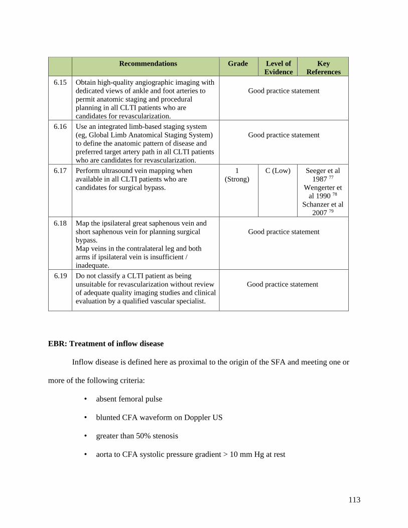
113
Recommendations Grade Level of
Evidence
Key
References
6.15 Obtain high-quality angiographic imaging with
dedicated views of ankle and foot arteries to
permit anatomic staging and procedural
planning in all CLTI patients who are
candidates for revascularization.
Good practice statement
6.16 Use an integrated limb-based staging system
(eg, Global Limb Anatomical Staging System)
to define the anatomic pattern of disease and
preferred target artery path in all CLTI patients
who are candidates for revascularization.
Good practice statement
6.17 Perform ultrasound vein mapping when
available in all CLTI patients who are
candidates for surgical bypass.
1
(Strong)
C (Low) Seeger et al
1987 77
Wengerter et
al 1990 78
Schanzer et al
2007 79
6.18 Map the ipsilateral great saphenous vein and
short saphenous vein for planning surgical
bypass.
Map veins in the contralateral leg and both
arms if ipsilateral vein is insufficient /
inadequate.
Good practice statement
6.19 Do not classify a CLTI patient as being
unsuitable for revascularization without review
of adequate quality imaging studies and clinical
evaluation by a qualified vascular specialist.
Good practice statement
EBR: Treatment of inflow disease
Inflow disease is defined here as proximal to the origin of the SFA and meeting one or
more of the following criteria:
• absent femoral pulse
• blunted CFA waveform on Doppler US
• greater than 50% stenosis
• aorta to CFA systolic pressure gradient > 10 mm Hg at rest

114
The decision to perform staged versus multi-level revascularization for patients with
combined inflow and outflow disease is individualized based on severity of limb threat
(especially presence of tissue loss), anatomic complexity, and patient risk. In settings of rest pain
and minor tissue loss, inflow correction alone may suffice to achieve the desired clinical
outcome. As procedural complexity increases, peri-operative morbidity and mortality rise as
well. Most patterns of AI disease may be successfully treated using an endovascular approach,
frequently employing bare metal or covered stents.82-84 Surgery is often reserved for extensive
occlusions or after failed prior endovascular procedures. The choice of an open surgical inflow
procedure should be based on patient risk, anatomic pattern of disease, and other clinical factors.
Direct anatomic bypass (eg, aortofemoral) grafting may be preferred over extra-anatomic
reconstruction in average risk patients with severe ischemia (WIfI ischemia grades 2, 3) due to
greater anatomic and hemodynamic durability.85-87
CFA endarterectomy can be performed with low morbidity and excellent long-term
durability.88, 89 It remains the optimal approach to treat hemodynamically significant CFA
disease, which often includes bulky calcific plaque. In some cases, femoral interposition grafting
may be preferred. In all cases, durable in-line PFA flow should be maximized. CFA
endarterectomy may be combined with proximal intervention to treat combined disease in a
“hybrid” fashion.90 Although long terms outcome data are sparse, recent reports suggest that
endovascular treatment of CFA disease may be a safe alternative in selected patients (eg, high
surgical risk, hostile groin anatomy).91-94
Surgical treatment (eg, profundaplasty or bypass grafting) of PFA disease is an important
component of CLTI revascularization with a major impact on the long-term prognosis for the

115
limb. The indications and optimal approaches for treating non-orificial (ie, not in continuity with
the CFA) or long segment PFA disease are not established. There is limited evidence regarding
the use of endovascular interventions for PFA disease. However it may be considered as a
secondary approach in settings of hostile groin anatomy or in other high-risk circumstances.
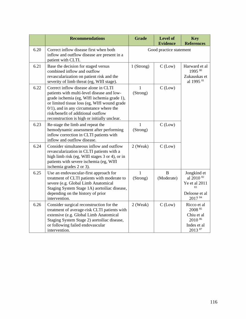
116
Recommendations Grade
Level of
Evidence
Key
References
6.20 Correct inflow disease first when both
inflow and outflow disease are present in a
patient with CLTI.
Good practice statement
6.21 Base the decision for staged versus
combined inflow and outflow
revascularization on patient risk and the
severity of limb threat (eg, WIfI stage).
1 (Strong) C (Low) Harward et al
1995 80
Zukauskas et
al 1995 81
6.22 Correct inflow disease alone in CLTI
patients with multi-level disease and low-
grade ischemia (eg, WIfI ischemia grade 1),
or limited tissue loss (eg, WIfI wound grade
0/1), and in any circumstance where the
risk/benefit of additional outflow
reconstruction is high or initially unclear.
1
(Strong)
C (Low)
6.23 Re-stage the limb and repeat the
hemodynamic assessment after performing
inflow correction in CLTI patients with
inflow and outflow disease.
1
(Strong)
C (Low)
6.24 Consider simultaneous inflow and outflow
revascularization in CLTI patients with a
high limb risk (eg, WIfI stages 3 or 4), or in
patients with severe ischemia (eg, WIfI
ischemia grades 2 or 3).
2 (Weak) C (Low)
6.25 Use an endovascular-first approach for
treatment of CLTI patients with moderate to
severe (e.g. Global Limb Anatomical
Staging System Stage 1A) aortoiliac disease,
depending on the history of prior
intervention.
1
(Strong)
B
(Moderate)
Jongkind et
al 2010 82
Ye et al 2011 83
Deloose et al
2017 84
6.26 Consider surgical reconstruction for the
treatment of average-risk CLTI patients with
extensive (e.g. Global Limb Anatomical
Staging System Stage 2) aortoiliac disease,
or following failed endovascular
intervention.
2 (Weak) C (Low) Ricco et al
2008 85
Chiu et al
2010 86
Indes et al
2013 87
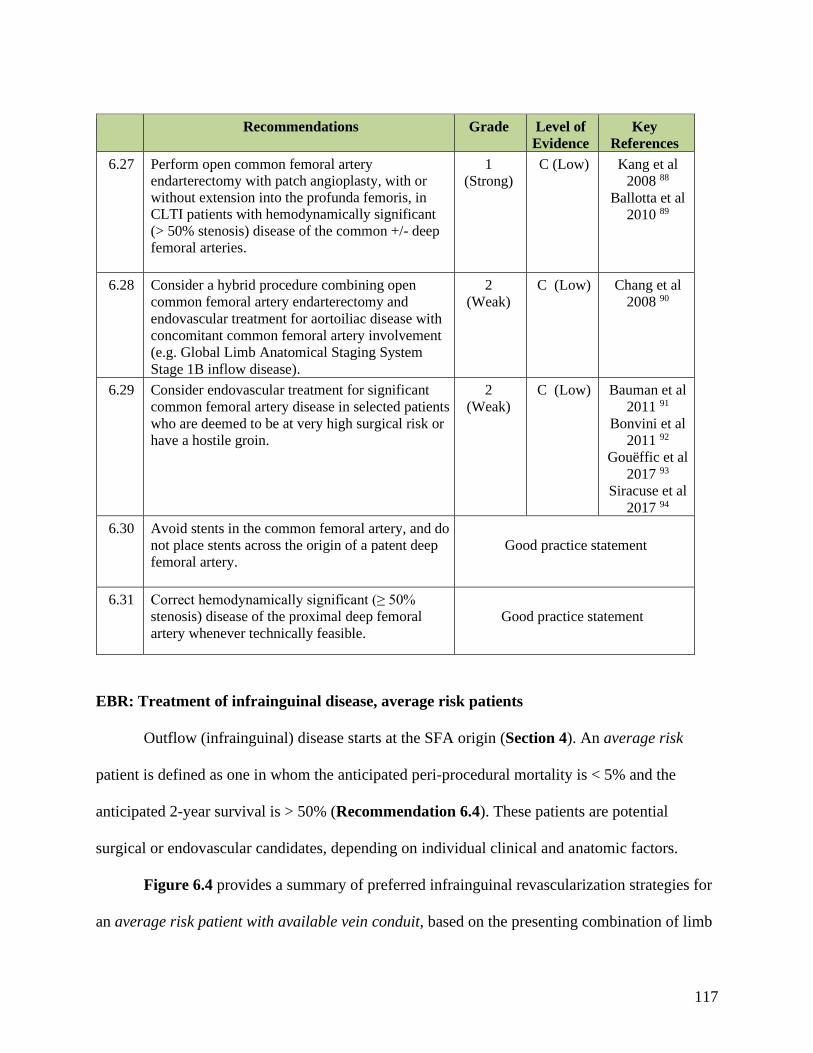
117
Recommendations Grade
Level of
Evidence
Key
References
6.27 Perform open common femoral artery endarterectomy with patch angioplasty, with or
without extension into the profunda femoris, in CLTI patients with hemodynamically significant
(> 50% stenosis) disease of the common +/- deep
femoral arteries.
1
(Strong)
C (Low) Kang et al
2008 88
Ballotta et al
2010 89
6.28 Consider a hybrid procedure combining open
common femoral artery endarterectomy and
endovascular treatment for aortoiliac disease with
concomitant common femoral artery involvement
(e.g. Global Limb Anatomical Staging System
Stage 1B inflow disease).
2
(Weak)
C (Low) Chang et al
2008 90
6.29 Consider endovascular treatment for significant
common femoral artery disease in selected patients
who are deemed to be at very high surgical risk or
have a hostile groin.
2
(Weak)
C (Low) Bauman et al
2011 91
Bonvini et al
2011 92
Gouëffic et al
2017 93
Siracuse et al
2017 94
6.30 Avoid stents in the common femoral artery, and do
not place stents across the origin of a patent deep
femoral artery.
Good practice statement
6.31 Correct hemodynamically significant (≥ 50%
stenosis) disease of the proximal deep femoral
artery whenever technically feasible.
Good practice statement
EBR: Treatment of infrainguinal disease, average risk patients
Outflow (infrainguinal) disease starts at the SFA origin (Section 4). An average risk
patient is defined as one in whom the anticipated peri-procedural mortality is < 5% and the
anticipated 2-year survival is > 50% (Recommendation 6.4). These patients are potential
surgical or endovascular candidates, depending on individual clinical and anatomic factors.
Figure 6.4 provides a summary of preferred infrainguinal revascularization strategies for
an average risk patient with available vein conduit, based on the presenting combination of limb
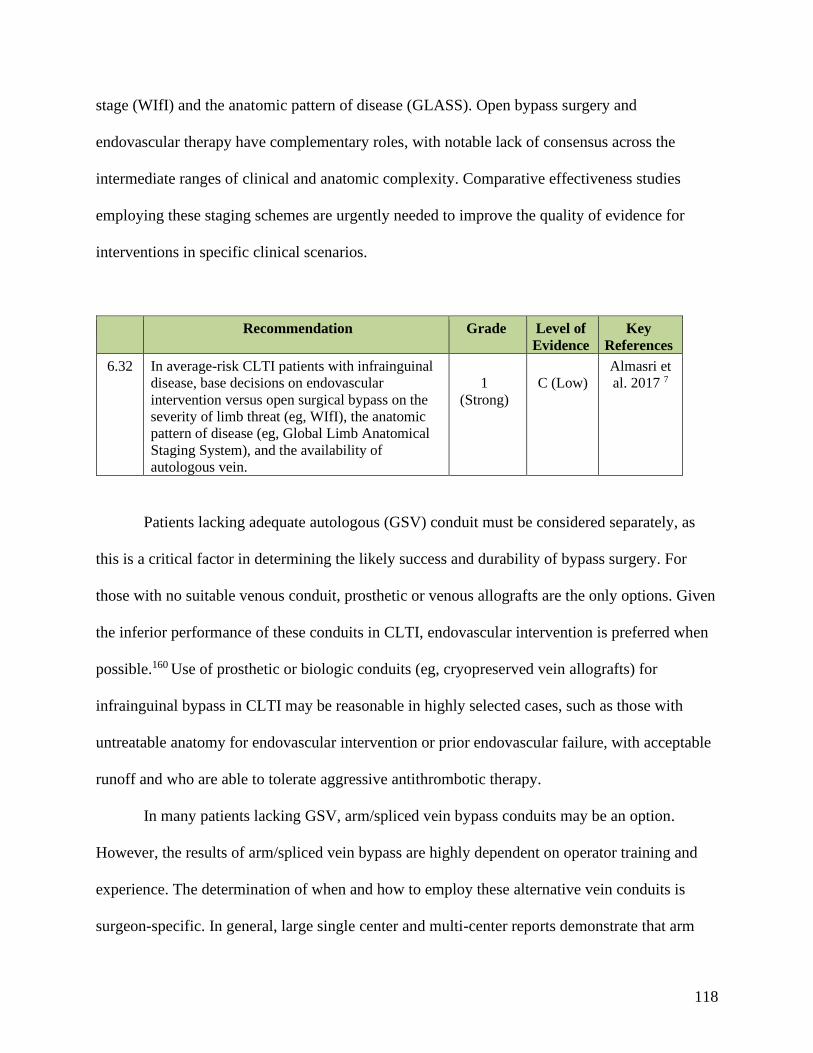
118
stage (WIfI) and the anatomic pattern of disease (GLASS). Open bypass surgery and
endovascular therapy have complementary roles, with notable lack of consensus across the
intermediate ranges of clinical and anatomic complexity. Comparative effectiveness studies
employing these staging schemes are urgently needed to improve the quality of evidence for
interventions in specific clinical scenarios.
Recommendation Grade
Level of
Evidence
Key
References
6.32 In average-risk CLTI patients with infrainguinal
disease, base decisions on endovascular
intervention versus open surgical bypass on the
severity of limb threat (eg, WIfI), the anatomic
pattern of disease (eg, Global Limb Anatomical
Staging System), and the availability of
autologous vein.
1
(Strong)
C (Low)
Almasri et
al. 2017 7
Patients lacking adequate autologous (GSV) conduit must be considered separately, as
this is a critical factor in determining the likely success and durability of bypass surgery. For
those with no suitable venous conduit, prosthetic or venous allografts are the only options. Given
the inferior performance of these conduits in CLTI, endovascular intervention is preferred when
possible.160 Use of prosthetic or biologic conduits (eg, cryopreserved vein allografts) for
infrainguinal bypass in CLTI may be reasonable in highly selected cases, such as those with
untreatable anatomy for endovascular intervention or prior endovascular failure, with acceptable
runoff and who are able to tolerate aggressive antithrombotic therapy.
In many patients lacking GSV, arm/spliced vein bypass conduits may be an option.
However, the results of arm/spliced vein bypass are highly dependent on operator training and
experience. The determination of when and how to employ these alternative vein conduits is
surgeon-specific. In general, large single center and multi-center reports demonstrate that arm

119
and spliced vein bypasses perform better than non-autologous grafts to distal targets and are
inferior to autologous GSV conduits.7, 79, 438, 439 However, these higher risk vein grafts require
closer surveillance and more reinterventions to maintain primary-assisted patency.440
EBR: Treatment of infrainguinal disease in high-risk patients
A high-risk patient is defined as one in whom the anticipated perioperative mortality is
greater than 5% or the anticipated 2-year survival is less than 50%. Because endovascular
intervention can be performed with reduced morbidity, it may often be preferred in high-risk
patients who are otherwise candidates for functional limb salvage. Shared decision-making is of
great importance in high-risk patients to allow the patient, family and other stakeholders to
express value judgments on the trade-offs between risk and effectiveness in relation to the
desired goals.

120
Recommendations Grade
Level of
Evidence
Key
References
6.33 Offer endovascular revascularization when
technically feasible for high-risk patients with
advanced limb threat (eg, Wound Ischemia foot
Infection (WIfI) stage 4) and significant perfusion
deficits (eg, WIfI ischemia grades 2 and 3).
2
(Weak)
C (Low) Abu Dabrh et
al 2015 5
Zhan et al
2015 69
Causey et al
2016 70
Darling et al
2016 71
Robinson et
al 2017 72
6.34 Consider endovascular revascularization for high-
risk patients with intermediate limb threat (eg,
WIfI stages 2 and 3) and significant perfusion
deficits (eg, WIfI ischemia grades 2 and 3).
2
(Weak)
C (Low)
6.35 Consider endovascular revascularization for high-
risk patients with advanced limb threat (eg, WIfI
stage 4) and moderate ischemia (eg, WIfI
ischemia grade 1), if the wound progresses or fails
to reduce in size by ≥ 50% within 4 weeks despite
appropriate infection control, wound care and
offloading, when technically feasible.
2
(Weak)
C (Low)
6.36 Consider endovascular revascularization for high-
risk patients with intermediate limb threat (eg,
WIfI stages 2 and 3) and moderate ischemia (eg,
WIfI ischemia grade 1), if the wound progresses
or fails to reduce in size by ≥ 50% within 4 weeks
despite appropriate infection control, wound care
and offloading, when technically feasible.
2
(Weak)
C (Low)
6.37 Consider open surgery in selected high-risk
patients with advanced limb threat (eg, WIfI stage
3 or 4), significant perfusion deficits (ischemia
grade 2-3), and advanced complexity of disease
(eg, Global Limb Anatomical Staging System
stage III), or after prior failed endovascular
attempts and unresolved symptoms of CLTI.
2
(Weak)
C (Low)
121
EBR: Severe infra-malleolar disease
Severe IM disease creates a major challenge to effective revascularization.403 The P2
modifier in GLASS describes the circumstance where no named artery crosses the ankle into the
foot and there is no suitable target for bypass surgery. Although technically successful
endovascular interventions in the pedal arch have been reported, their durability, hemodynamic
and clinical effectiveness remain unknown.434 Diabetic patients often have a segment of
preserved pedal artery that may be a target for bypass. Open bypass surgery has also been
successfully employed to tarsal and plantar arteries, but again techniques and outcomes are not
established. Given the technical difficulty, likely reduced hemodynamic impact and durability,
the appropriate role for interventions at this level is not determined. The impact of IM disease on
the success of proximal revascularization, whether open or endovascular, is likewise unknown.
Although the presence of an intact pedal arch appears important for both, clinical success may
still be attained in the presence of significant IM disease. The severity of limb threat (tissue
loss/infection) is likely a critical modifier of the relationship between IM disease severity and
post-procedural clinical outcomes.
EBR: Role of angiosome-guided revascularization
While few would argue about the desirability of maximizing perfusion at the site of
tissue loss, there is considerable debate about the utility of “angiosome-guided”
revascularisation.441, 442 First, unambiguous assignment of foot wounds to an individual
angiosome is possible in only a minority of cases.443 Toe lesions, which typically comprise more
than half of the lesions encountered have a dual blood supply (AT and PT), although for more
proximal foot lesions, unique angiosome assignment may be achieved in up to 75-80% of

122
patients. Then there is the practical question of whether the desired target artery for the
angiosome is available, and the comparative hemodynamic and clinical effectiveness of “direct”
versus “indirect” revascularization. Tibial and peroneal bypasses perform equally well for limb
salvage, and DP bypass can be effective for some hindfoot lesions.444 Recent systematic reviews
have yielded conflicting results.96-99 and data are inextricably confounded by the quality of the
pedal arch and the nature of the revascularization performed.95, 445 While wound healing may be
improved when direct revascularization is achievable, major amputation rates and patency are
not consistently different. To date, none of the analyses take into account the confounding effect
of limb staging, for example using WIfI. In summary, angiosome-guided revascularization may
be of importance in the setting of endovascular intervention for mid- and hindfoot lesions but is
likely irrelevant for ischemic rest pain, and of marginal value for most forefoot lesions and minor
ulcers. The role of multi-vessel (tibial) revascularization is also currently unknown. However, it
may be reasonable in selected patients with advanced limb threat (eg, WIfI Stages 3, 4)
undergoing endovascular therapy, if it can be safely accomplished without risking loss of a
bypass target or compromising runoff to the foot.
Recommendations Grade
Level of
Evidence
Key
References
6.38 Consider angiosome-guided revascularization in
patients with significant wounds (eg, WIfI wound
grade 3 and 4), particularly those involving the
mid- or hindfoot, and when the appropriate target
arterial pathway is available.
2
(Weak)
C (Low) Azuma et al
2012 95
Sumpio et al
2013 96
Biancari et al
2014 97
Chae et al
2016 98
Jongsma et al
2017 99

123
EBR: Preferred endovascular techniques for infrainguinal disease
PBA, drug-coated balloon (DCB) angioplasty, bare metal (BMS) drug-eluting (DES) or
covered stent placement, and atherectomy may all be reasonable options in specific
circumstances and lesion anatomies. However, unfortunately, there are very few high-quality
comparative data to guide the choice of a specific endovascular approach in CLTI.7, 376, 383-385, 392,
446-451
PBA may be inferior to DCB angioplasty and stents for the treatment of intermediate
length SFA disease (FP grades 2-4) in patients with IC and possibly rest pain.100-103 However,
there are inadequate data to support a preferred endovascular approach for FP disease in CLTI.
PBA remains a reasonable primary endovascular approach for anatomically suitable IP
disease as current evidence is inadequate to support other, more expensive techniques.
Atherectomy is not superior to PBA and is associated with greatly increased costs.449.
Combination approaches such as atherectomy followed by DCB add significant cost and lack
high quality comparative data. Several modest sized trials suggest potential short-term benefit for
DES in short (ie, < 3 cm) tibial lesions, but one cannot generalise these data to the CLTI
population as a whole who typically present with much more extensive disease.7, 452 DES may be
a preferred endovascular “bail-out” following technical complications (eg, dissection) or failed
PBA for short, proximal IP lesions. Although early studies suggested a potential advantage for
DCB in tibial arteries a recent RCT showed no benefit of DCB over PBA, with a non-significant
higher rate of amputations in the DCB group.392 The results of further, on-going studes are
awaited. In summary, PBA currently remains the standard of care for the endovascular treatment
of IP disease in CLTI.
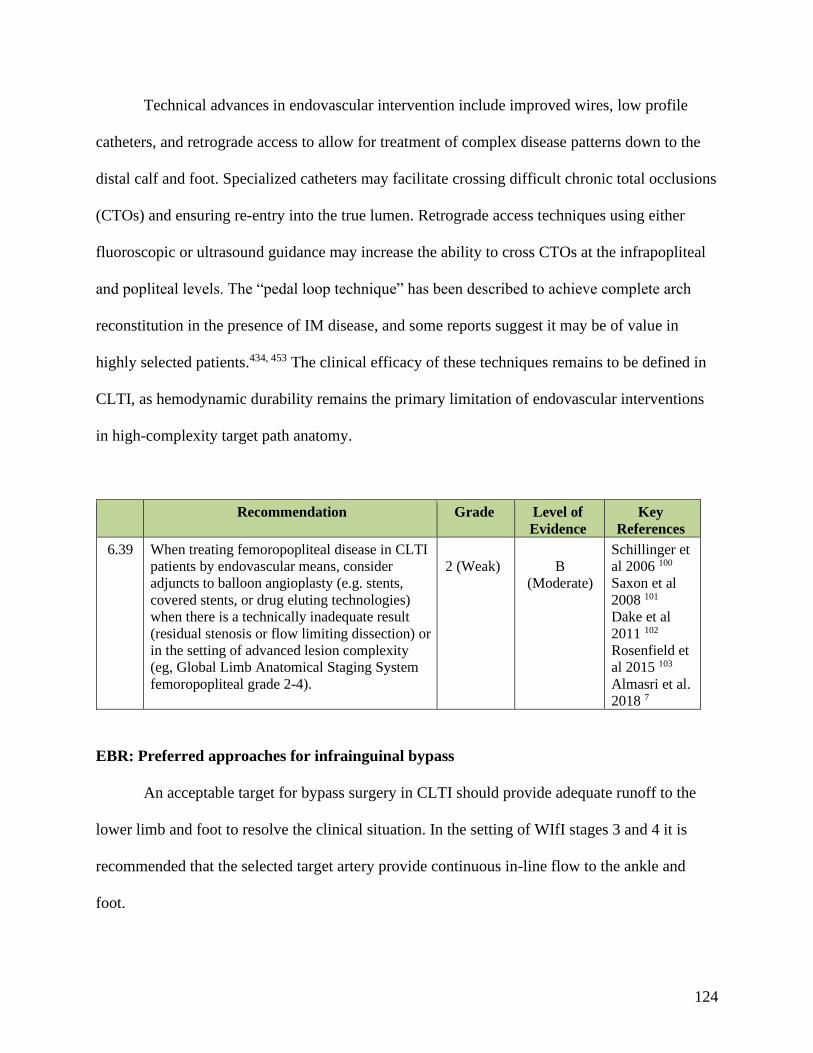
124
Technical advances in endovascular intervention include improved wires, low profile
catheters, and retrograde access to allow for treatment of complex disease patterns down to the
distal calf and foot. Specialized catheters may facilitate crossing difficult chronic total occlusions
(CTOs) and ensuring re-entry into the true lumen. Retrograde access techniques using either
fluoroscopic or ultrasound guidance may increase the ability to cross CTOs at the infrapopliteal
and popliteal levels. The “pedal loop technique” has been described to achieve complete arch
reconstitution in the presence of IM disease, and some reports suggest it may be of value in
highly selected patients.434, 453 The clinical efficacy of these techniques remains to be defined in
CLTI, as hemodynamic durability remains the primary limitation of endovascular interventions
in high-complexity target path anatomy.
Recommendation Grade
Level of
Evidence
Key
References
6.39 When treating femoropopliteal disease in CLTI
patients by endovascular means, consider
adjuncts to balloon angioplasty (e.g. stents,
covered stents, or drug eluting technologies)
when there is a technically inadequate result
(residual stenosis or flow limiting dissection) or
in the setting of advanced lesion complexity
(eg, Global Limb Anatomical Staging System
femoropopliteal grade 2-4).
2 (Weak)
B
(Moderate)
Schillinger et
al 2006 100
Saxon et al
2008 101
Dake et al
2011 102
Rosenfield et
al 2015 103
Almasri et al.
2018 7
EBR: Preferred approaches for infrainguinal bypass
An acceptable target for bypass surgery in CLTI should provide adequate runoff to the
lower limb and foot to resolve the clinical situation. In the setting of WIfI stages 3 and 4 it is
recommended that the selected target artery provide continuous in-line flow to the ankle and
foot.
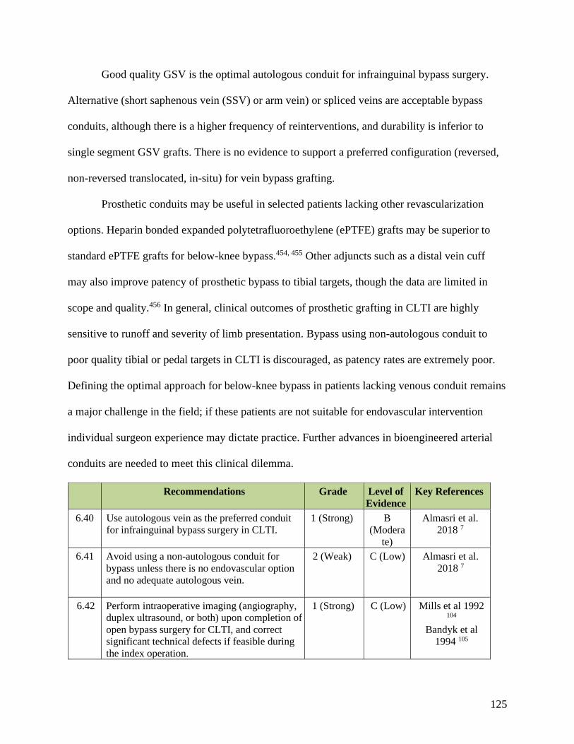
125
Good quality GSV is the optimal autologous conduit for infrainguinal bypass surgery.
Alternative (short saphenous vein (SSV) or arm vein) or spliced veins are acceptable bypass
conduits, although there is a higher frequency of reinterventions, and durability is inferior to
single segment GSV grafts. There is no evidence to support a preferred configuration (reversed,
non-reversed translocated, in-situ) for vein bypass grafting.
Prosthetic conduits may be useful in selected patients lacking other revascularization
options. Heparin bonded expanded polytetrafluoroethylene (ePTFE) grafts may be superior to
standard ePTFE grafts for below-knee bypass.454, 455 Other adjuncts such as a distal vein cuff
may also improve patency of prosthetic bypass to tibial targets, though the data are limited in
scope and quality.456 In general, clinical outcomes of prosthetic grafting in CLTI are highly
sensitive to runoff and severity of limb presentation. Bypass using non-autologous conduit to
poor quality tibial or pedal targets in CLTI is discouraged, as patency rates are extremely poor.
Defining the optimal approach for below-knee bypass in patients lacking venous conduit remains
a major challenge in the field; if these patients are not suitable for endovascular intervention
individual surgeon experience may dictate practice. Further advances in bioengineered arterial
conduits are needed to meet this clinical dilemma.
Recommendations Grade
Level of
Evidence
Key References
6.40 Use autologous vein as the preferred conduit
for infrainguinal bypass surgery in CLTI.
1 (Strong) B
(Modera
te)
Almasri et al.
2018 7
6.41 Avoid using a non-autologous conduit for
bypass unless there is no endovascular option
and no adequate autologous vein.
2 (Weak) C (Low) Almasri et al.
2018 7
6.42 Perform intraoperative imaging (angiography,
duplex ultrasound, or both) upon completion of
open bypass surgery for CLTI, and correct
significant technical defects if feasible during
the index operation.
1 (Strong) C (Low) Mills et al 1992 104
Bandyk et al
1994 105

126
RESEARCH PRIORITIES
6.1
6.1
In patients presenting with the full spectrum of CLTI, prospectively validate and refine
patient risk stratification models.
6.2
6.2
Conduct comparative effectiveness studies directly comparing strategies of
revascularization, and specific techniques and technologies, in well-defined subgroups of
patients (eg, WIfI and GLASS stages) with CLTI.
6.3
6.3
Define the circumstances in which angiosome-targeted and/or multi-vessel
revascularization provides clinical benefit in CLTI.
6.4 Develop and test strategies for the management of “no option” CLTI patients.
127
7: NON-REVASCULARIZATION TREATMENTS OF THE LIMB
Introduction
Although the optimal treatment for CLTI is undoubtedly revascularization, unfortunately,
a significant proportion of patients are not suitable for revascularization for anatomical and/or
physiological reasons. While major amputation may be suitable for some of these patients, there
is clearly a significant number who might benefit from non-revascularization-based treatments.
There is, however, a paucity of strong evidence regarding these treatment options. The
majority of studies are low quality and uncontrolled, combined with considerable study
heterogeneity, and making systematic review and meta-analysis difficult or even impossible.
This heterogeneity is reflected by large variations in patient factors, lesions of interest,
intervention protocols, study designs, and end-points (limb salvage, AFS, target-lesion patency,
pain-relief, quality of life determinants, ulcer healing and evolution of tissue lesions).4
This section reviews the following non-revascularization-based treatments:
A. Interventional
1. Spinal cord stimulation
2. Lumbar sympathectomy
3. Intermittent pneumatic compression
B. Pharmacotherapy
1. Prostanoids
2. Vasoactive drugs
3. Vasodilators
4. Defibrinating agents

128
5. Hyperbaric oxygen therapy
C. Conservative management
1. Wound care
Interventional non-revascularization treatments
Spinal cord stimulation
Mechanism of action. Spinal cord stimulation (SCS) was originally used to treat chronic
pain, and first described by Cook et al in the treatment of PAD.457 In SCS, electrodes are
implanted in the lumbar epidural space and connected to a generator to stimulate sensory
fibers.458 SCS promotes activation of cell-signaling pathways which cause the release of
vasodilatory molecules, leading to a decrease in vascular resistance and relaxation of smooth
muscle cells.458 This improved peripheral microcirculatory status has been shown to result in
increased capillary flow and density of perfusing capillaries, skin temperature, local TcPO2,
normalization of pulse wave morphology, and improved skin nutrition.106 In addition, SCS
suppresses sympathetic vasoconstriction and pain transmission.458
Evidence. A 2013 Cochrane review analyzed data from 444 patients in six controlled
studies investigating the use of SCS in CLTI.106, 459-464 The general quality of studies was good
and all studies used limb salvage as the primary endpoint (major AFS at 12 months). When the
results were pooled, limb salvage rates were found to be significantly higher in the SCS group
(relative risk [RR] for major amputation, 0.71; 95% CI, 0.56-0.90).106 Results were better when
patients were selected based on their initial TcPO2. There was also significant pain relief found
in both treatment groups, although the SCS groups required less analgesia. In addition, there was
no significant effect on ulcer healing. Overall mortality was not evaluated but the overall
129
complication rate was 17% (95% CI, 12-22%). Implantation problems occurred in 9% (95% CI,
4-15%), reintervention for changes in stimulation in 15% (95% CI, 10-20%), and infection of a
lead or pulse generator pocket accounted for 3% (95% CI, 0-6%).106
Researchers concluded that SCS offered a modest positive effect on pain relief and an
11% reduction in the amputation rate when compared with conservative management at 1
year.106 They stress, however, that the positive benefits should be weighed against the high cost
and possible complications. In fact, the Cochrane review found the cost was significantly higher
in the SCS group by $8824. Klomp and colleagues calculated the number needed to treat to save
one limb as 13, at $111,705 per limb saved and $312,754 per quality-adjusted life year gained.465
They concluded that SCS was not a cost-effective treatment for CLTI.
Lumbar sympathectomy
Mechanism of action. Sympathetic denervation of the lumbar sympathetic ganglia is
performed either through open or laparoscopic retroperitoneal access, or through percutaneous
chemical blockade. Lumbar sympathectomy (LS) increases blood flow to the lower limb by
inducing vasodilation of the collateral circulation and shunting of blood through cutaneous
arteriovenous anastomoses via its reduction of sympathetic tone. This, in turn, improves tissue
oxygenation and decreases tissue damage and pain. Pain is also decreased by interrupting
sympathetic nociceptive coupling and by a direct neurolytic action on nociceptive fibers.466
Evidence. In their systematic review, Sanni et al reported that RCTs failed to identify any
objective benefits for LS in patients with CLTI.466 They concluded, however, that LS may be
considered as an alternative to amputation in patients with otherwise viable limbs, because it is
minimally invasive and cost-effective with a low complication rate.466 Chemical and surgical
130
sympathectomy also appear to perform equally as well, with some suggestion that LS can benefit
diabetic patients.
Of the three RCTs that focus on LS in PAD, only two report on its use in CLTI,467, 468
with the third reporting on its use in IC.469 Cross et al found that chemical sympathectomy
provided relief of rest pain in 67% of patients undergoing LS, compared to 24% of controls at 6
months.468 However, in a contrasting study, Barnes et al found that LS combined with aortoiliac
revascularization did not provide any additional benefits when compared with revascularization
alone.467 In fact, the majority of cohort studies reporting LS in CLTI470-479 consistently
demonstrate subjective improvements in approximately 60% of patients with regard to pain relief
and ulcer healing.466 Moreover, a recent Cochrane systematic review was unable to find any
RCTs that evaluated the effect of LS (open, laparoscopic, or chemical) compared to no
intervention in CLTI due to non-reconstructable PAD.107 Overall, data are limited but there is no
evidence to suggest that LS reduces the risk of major amputation in patients with CLTI. It
remains unclear as to whether any subgroup of CLTI patients may have improved pain control or
ulcer healing with LS.
Intermittent pneumatic compression
Mechanism of action. In patients treated with intermittent pneumatic compression (IPC),
arterial blood flow is increased in the distal limbs by an increase in the arteriovenous pressure
gradient, which stimulates the endothelial vasodilators, thus suspending the veno-arteriolar reflex
and stimulating collateral artery growth.480 As a result, the arterial flow, peak systolic velocity
(PSV), end diastolic velocity, and pulse volume are all increased.481
131
Several methods of lower limb IPC use various protocols. These include the ArtAssist®
(ACI Medical, Marcos CA) device that provides sequential compression to the foot and calf; the
Aircast© ArterialFlow (DJO Global, Vista CA) device that compresses the calf; and devices that
deliver leg compression that is synchronized with ventricular contraction of the heart
(Syncarbon®, Contilabo, Saint Gobain France; and Vascular Pump, Rheomedix®, Philadelphia
PA).480
Evidence. Two controlled studies482, 483 and several case series484-491 have been published
regarding IPC, but there is no robust evidence from high-quality trials. In one, investigators
entered 171 patients with CLTI into a 3-month IPC program.490 They reported improved pain
relief, increased toe pressures by a mean of 15 mm Hg, and increased popliteal artery flow by a
mean of 20 cm/s. The median AFS was 18 months with 94% limb salvage at 3.5 years. They
determined that IPC was a cost-effective intervention at a cost of $4,454 per patient.490 In a
retrospective observational study involving 107 patients, researchers from the Mayo Clinic found
40% wound healing at 6 months.489
In another study, a non-RCT involving 48 patients, investigators found that 58% of
patients who underwent IPC benefited from complete healing and limb salvage, as compared to
17% in the control group (odds ratio [OR], 7.00; 95% CI, 1.82-26.89).482 In a prospective trial,
changes in quality of life were reviewed before and after IPC treatment.491 Researchers reported
a significant improvement in pain, physical functioning, and general health perception. Another
systematic review found that IPC might be associated with improved limb salvage, wound
healing, and pain management; as well as a low risk of complications.480 However, this review
also noted a high risk of bias in these studies, with large variations in the type of compression
and optimum parameters used.480
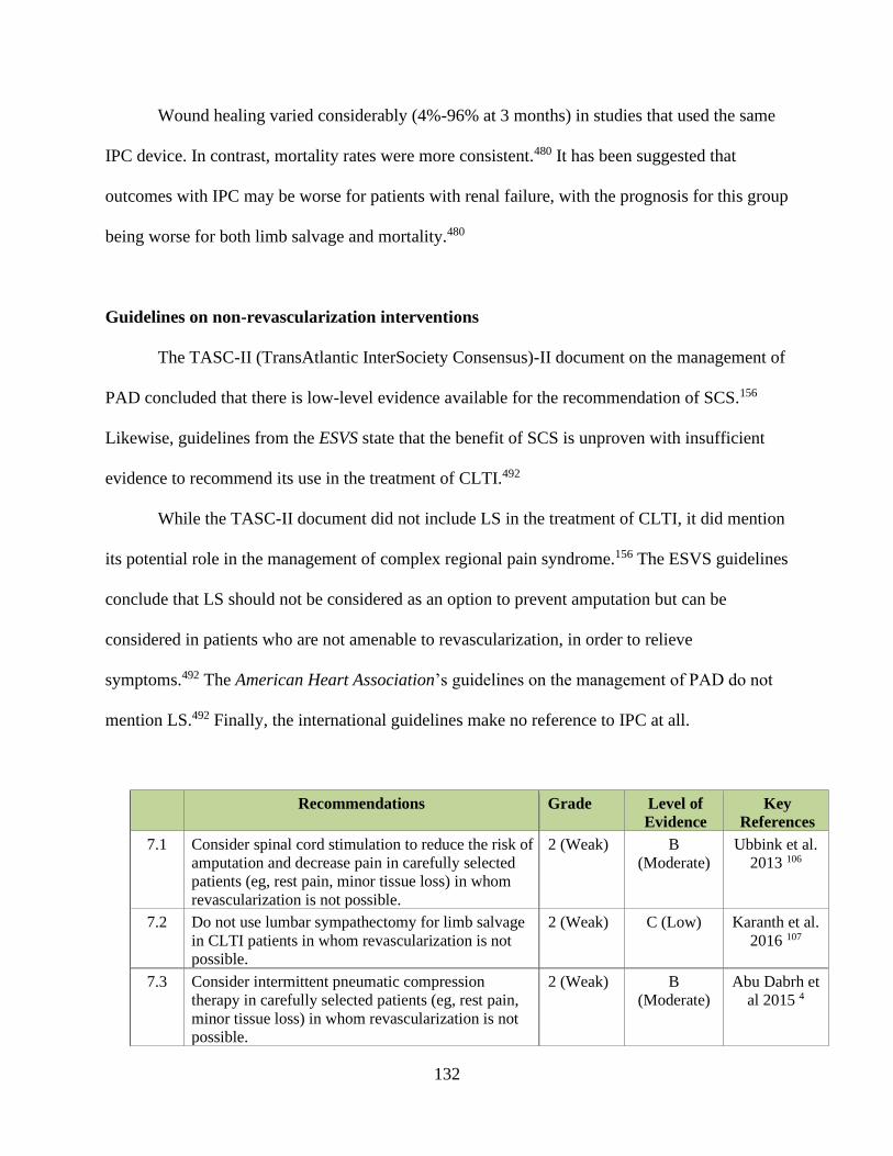
132
Wound healing varied considerably (4%-96% at 3 months) in studies that used the same
IPC device. In contrast, mortality rates were more consistent.480 It has been suggested that
outcomes with IPC may be worse for patients with renal failure, with the prognosis for this group
being worse for both limb salvage and mortality.480
Guidelines on non-revascularization interventions
The TASC-II (TransAtlantic InterSociety Consensus)-II document on the management of
PAD concluded that there is low-level evidence available for the recommendation of SCS.156
Likewise, guidelines from the ESVS state that the benefit of SCS is unproven with insufficient
evidence to recommend its use in the treatment of CLTI.492
While the TASC-II document did not include LS in the treatment of CLTI, it did mention
its potential role in the management of complex regional pain syndrome.156 The ESVS guidelines
conclude that LS should not be considered as an option to prevent amputation but can be
considered in patients who are not amenable to revascularization, in order to relieve
symptoms.492 The American Heart Association’s guidelines on the management of PAD do not
mention LS.492 Finally, the international guidelines make no reference to IPC at all.
Recommendations Grade Level of
Evidence
Key
References
7.1 Consider spinal cord stimulation to reduce the risk of
amputation and decrease pain in carefully selected
patients (eg, rest pain, minor tissue loss) in whom
revascularization is not possible.
2 (Weak) B
(Moderate)
Ubbink et al.
2013 106
7.2 Do not use lumbar sympathectomy for limb salvage
in CLTI patients in whom revascularization is not
possible.
2 (Weak) C (Low) Karanth et al.
2016 107
7.3 Consider intermittent pneumatic compression
therapy in carefully selected patients (eg, rest pain,
minor tissue loss) in whom revascularization is not
possible.
2 (Weak) B
(Moderate)
Abu Dabrh et
al 2015 4

133
Pharmacotherapy
Prostanoids
Mechanism of action. Prostanoids include a family of inflammatory mediators, mainly
prostaglandin (E1), prostacyclin, and iloprost. Prostanoids act by inhibiting the activation of
platelets and leucocytes, adhesion and aggregation of platelets, and by promoting vasodilatation
and vascular endothelial cytoprotection through antithrombotic and profibrinolytic activities.108,
493, 494
Evidence. A meta-analysis evaluating the use of prostaglandin E1 versus placebo in the
treatment of 254 patients with CLTI demonstrated very favorable results at 6 months, with ulcer
healing and/or pain reduction (47.8% vs 25.2% placebo), and reduction in major amputation or
death (22.6% vs 36.2% placebo) associated with prostaglandin E1 use.495 Subsequently, a 2018
Cochrane paper reviewed 33 prostanoid studies with various formulations, doses, and
administration routes.108 These included IV administration of prostaglandin E1 (synthetic form,
alprostadil) for 21 days and an intra-arterial administration; IV of prostacyclin (PGI2) for 4 to 7
days; IV of iloprost (synthetic analogue of PGI2) for 14 to 28 days, oral 28 days to 1 year, with
low dose infusion; IV of lipoecaprost for 50 days; and IV of ciprostene (a PGI2 analogue) for 7
days.108, 493 When compared to placebo, prostanoids appeared to have some efficacy for treating
rest pain (RR 1.30, 95% CI 1.06 to 1.59) and ulcer healing (RR 1.24, 95% CI 1.04 to 1.48). As a
group, however, prostanoids did not have a significant impact on amputations or mortality,
although not all studies defined major versus minor amputations.494 Prostanoids were associated
with a statistically significant increase in side effects (RR, 2.35; 95% CI, 1.99-2.78).494 The side
effects were mostly minor, including headache, facial flushing, nausea, vomiting and diarrhea.
134
The authors of the Cochrane systematic review concluded that there is no strong evidence
on the efficacy and safety of prostanoids in patients with CLTI, based on a high-quality meta-
analysis of homogeneous, long-term RCTs.493 They also called on the need for further high-
quality trials.494 A subgroup analysis of the Cochrane meta-analysis, however, suggested that
iloprost appeared to reduce major amputation (RR, 0.69; 95% CI, 0.52–0.93), and fared better
with rest pain (RR, 1.54; 95% CI 1.19-1.99) and ulcer healing (RR 1.80, 95% CI 1.29-2.50). The
authors stated that while previous meta-analyses of iloprost had been more positive,496 only a
few of the studies used in those previous meta-analyses could be included in the Cochrane
review due to study methodology issues. In fact, in clinical practice, iloprost appears to benefit
approximately 40% of patients in whom revascularization is not possible.156, 496
Since the Cochrane review was published, a newer RCT comparing a placebo with the
use of prostacyclin analogue taprostene IV for 2 weeks, failed to demonstrate any difference in
pain relief, ulcer size improvement, or prevention of amputation.497 There is no data to support
the use of prostanoids to reduce the risk of major amputation in CLTI patients in whom
revasclarization is not possible.
Vasoactive drugs
Naftidrofuryl. A Cochrane review of eight RCTs examined the IV administration of
naftidrofuryl in 269 patients.109 The treatment tended to reduce rest pain and improve skin
necrosis, but this was not statistically significant. The studies were found to be of low
methodological quality, with varying levels of severity of CLTI, varying lengths of duration of
treatment (from 3 to 42 days), and different measures of effect. This resulted in varying

135
endpoints that precluded a meaningful pooling of results.109 Thus, there is currently insufficient
evidence to support the use of naftidrofuryl in the treatment of CLTI.494
Pentoxifylline. This drug improves blood flow by increasing red blood cell deformity and
decreasing viscosity. A European RCT involving 314 patients found a significant reduction in
rest pain, sleep disturbance, and analgesia requirements.498 In a separate Norwegian study using
the same dosing regimen, there was no statistically significant difference either in pain-free
levels or in absolute walking distance between the two groups.499 Researchers concluded that
further investigation was necessary in order to evaluate the role of pentoxifylline in the treatment
of patients with CLTI. Thus, there is currently a lack of consistent evidence to recommend the
use of pentoxifylline in the treatment of CLTI.494
Cilostazol. This drug has been well studied in claudicants, but not as much in CLTI. One
small study demonstrated that cilostazol improves microvascular circulation and skin perfusion
pressure in ischemic limbs.500 Another uncontrolled study that used cilostazol in conjunction
with endovascular revascularization, reported higher rates of AFS and limb salvage but not
higher rates of survival or freedom from further revascularization.501 In the absence of RCTs in
patients with CLTI, there is insufficient evidence that cilostazol improves clinical outcomes in
patients with CLTI.500, 501
Vasodilators
Because vasodilators can cause shunting of blood away from ischemic areas to non-
ischemic areas, they are of no value to patients with CLTI.156 Because vasodilators can cause
shunting of blood away from ischemic areas to non-ischemic areas, they are of no value to
patients with CLTI.156

136
Defibrinating agents
Two small RCTs compared ancrod, a defibrinating agent, with placebo in CLTI.502, 503
Although one study showed positive changes in ankle and toe pressures, both studies failed to
demonstrate any improvements in clinical outcome. Two small RCTs compared ancrod, a
defibrinating agent, with placebo in CLTI.502, 503 Although one study showed positive changes in
ankle and toe pressures, both studies failed to demonstrate any improvements in clinical
outcome.
Hyperbaric oxygen therapy
There are numerous plausible mechanisms for hyperbaric oxygen therapy (HBOT) to
have a therapeutic role in CLTI. These include increased oxygen transport capacity of plasma
(independent of red blood corpuscle number and function), improved function of the leukocyte
oxygen-dependent peroxidase system, reduced tissue edema due to the osmotic effect of oxygen,
stimulation of progenitor stem-cell mobilization and angiogenesis, and improved fibroblast
function.504 If there is superimposed infection, HBOT also inhibits bacterial growth (particularly
anaerobes), generates free radicals that destroy bacterial cellular structures, and improves the
oxygen-dependent transport of antibiotics.505
In 2015, a Cochrane review of the role of HBOT in healing chronic wounds was
published,110 involving 12 trials and 577 patients. Ten of the twelve trials studied the effect of
HBOT on ulcer healing in patients with diabetes. The 2015 review concluded that HBOT
increased the rate of ulcer healing in DFUs at 6 weeks, but not at longer-term follow up, and with
no significant difference in the risk of major amputation.110 In 2015, a Cochrane review of the

137
role of HBOT in healing chronic wounds was published,110 involving 12 trials and 577 patients.
Ten of the twelve trials studied the effect of HBOT on ulcer healing in patients with diabetes.
The 2015 review concluded that HBOT increased the rate of ulcer healing in DFUs at 6 weeks,
but not at longer-term follow up, and with no significant difference in the risk of major
amputation.110
Three other studies involved patients with ischemic ulcers but each study used varying
definitions of ischemia.506-508 Abidia and colleagues randomized 18 patients with an ABI of < 0.8
or toe-brachial index of < 0.7 and found improvement in wound healing in the treatment
group.507 Londahl randomized 94 patients with adequate distal perfusion or non-reconstructable
arterial disease.508 They found that 57% of patients had a toe pressure of < 60 mm Hg (median
toe pressure 52 mm Hg). Complete ulcer healing occurred in 52% of the patients treated with
HBOT, as compared to 29% of controls at 12 months (P < 0.02). Stratification based on toe
pressures did not appear to affect healing rates.
A subsequent publication by this group demonstrated that preintervention TcPO2
correlated with ulcer healing and that individuals with a TcPO2 of < 25 mm Hg did not heal.509
There was no significant difference in major amputations between the two groups, with three
amputations in the HBOT cohort and one in the control cohort.
One study randomized 70 patients with DFUs to either HBOT or standard care.506 The
mean ABI and TcPO2 were 0.65 and 23 mm Hg in the HBOT cohort, and 0.64 and 21 mm Hg in
the non-HBOT group. All patients with an ABI < 0.9 or TcPO2 < 50 mm Hg were considered
ischemic, underwent an iloprost infusion, and were examined for possible revascularization.
Thirteen patients in each group underwent a revascularization procedure. At the completion of
the therapy, resting TcPO2 increased by a mean of 12.1 in the HBOT group and 5.0 in the control

138
group (P < 0.0002). There was a significant reduction in major amputations in the HBOT group
(P < 0.016).506
A large longitudinal cohort study using data from a wound-healing group in the United
States61 included patients with DFUs and adequate foot perfusion, as determined by clinicians. A
total of 793 patients underwent HBOT therapy. Propensity scoring was used to compensate for
the lack of randomization. The study found that individuals treated with HBOT were less likely
to have healing of ulcers (hazard ratio [HR], 0.68; 0.63 -0.73) and more likely to undergo lower
limb amputation (HR, 2.37; 1.84-3.04).510
A subsequent multi-center RCT (DAMO2CLES) undertaken in 25 hospitals in the
Netherlands and Belgium randomized 120 patients with an ischemic foot wound and diabetes to
standard care (SC) with or without a course of HBOT (SC + HBOT). Ischemia was defined as
ankle pressure < 70 mm Hg, toe pressure < 50 mm Hg or TcPO2 < 40 mm Hg. All patients were
assessed for revascularization and when applicable, this was generally performed prior to HBOT.
Primary outcomes were limb salvage, wound healing at 12 months, and time to wound healing.
Mortality and AFS were also analyzed. Limb salvage (47/60 in SC cohort and 53/60 in SC +
HBOT cohort), index wound healing at 12 months (28/60 in SC cohort versus 30 in SC + HBOT
cohort), and AFS (41/60 in SC cohort versus 49 in SC + HBT cohort) were not significantly
different between the two groups. A high proportion (35%) of those allocated to HBOT were
unable to undergo HBOT or did not complete at least 30 treatments, mostly for medical co-
morbidities or logistical reasons, reinforcing the significant medical co-morbidities present in
these patients.112
Overall, while controversy remains, there may be a role for the use of hyperbaric oxygen therapy
to accelerate ulcer healing in diabetic patients with non-healing neuropathic ulcers and low grade
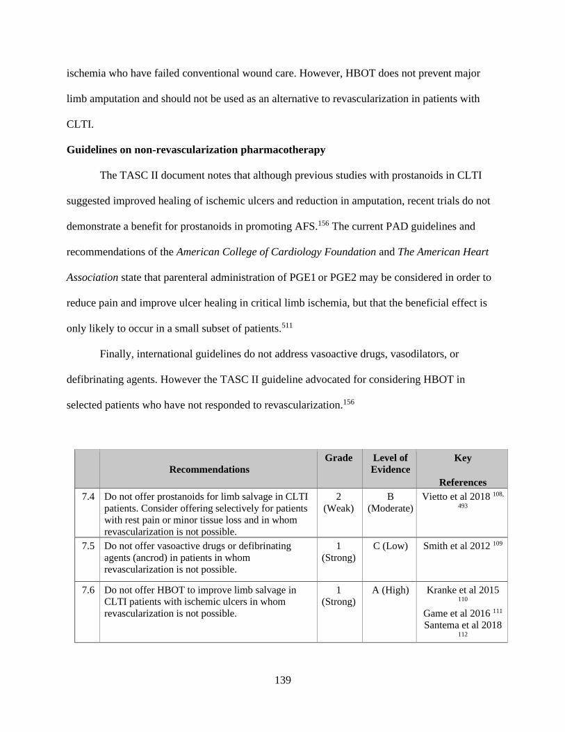
139
ischemia who have failed conventional wound care. However, HBOT does not prevent major
limb amputation and should not be used as an alternative to revascularization in patients with
CLTI.
Guidelines on non-revascularization pharmacotherapy
The TASC II document notes that although previous studies with prostanoids in CLTI
suggested improved healing of ischemic ulcers and reduction in amputation, recent trials do not
demonstrate a benefit for prostanoids in promoting AFS.156 The current PAD guidelines and
recommendations of the American College of Cardiology Foundation and The American Heart
Association state that parenteral administration of PGE1 or PGE2 may be considered in order to
reduce pain and improve ulcer healing in critical limb ischemia, but that the beneficial effect is
only likely to occur in a small subset of patients.511
Finally, international guidelines do not address vasoactive drugs, vasodilators, or
defibrinating agents. However the TASC II guideline advocated for considering HBOT in
selected patients who have not responded to revascularization.156
Recommendations
Grade Level of
Evidence
Key
References
7.4 Do not offer prostanoids for limb salvage in CLTI
patients. Consider offering selectively for patients
with rest pain or minor tissue loss and in whom
revascularization is not possible.
2
(Weak)
B
(Moderate)
Vietto et al 2018 108,
493
7.5 Do not offer vasoactive drugs or defibrinating
agents (ancrod) in patients in whom
revascularization is not possible.
1
(Strong)
C (Low) Smith et al 2012 109
7.6 Do not offer HBOT to improve limb salvage in
CLTI patients with ischemic ulcers in whom
revascularization is not possible.
1
(Strong)
A (High) Kranke et al 2015 110
Game et al 2016 111
Santema et al 2018 112
140
Conservative management
Wound care. It is important to remember that CLTI is associated with a markedly
shortened life expectancy and, not surprisingly, patients with unreconstructed CLTI experience
poorer outcomes in terms of survival and limb salvage. In a retrospective study involving 105
patients with unreconstructed CLTI, 46% of patients lost their limb and 54% died within 1
year.512 Of the patients with a non-amputated leg, 72% were dead within 1 year. Thus, despite
advances in revascularization techniques and anesthetics, endovascular or surgical
revascularization may not be appropriate in some patients, even if it is technically possible, due
to significant comorbidities and reduced life expectancy.
A group of 169 patients with stable tissue loss who were unsuitable for revascularization
based on medical and anatomical reasons was entered into a dedicated wound management
program.290 At 1 year, 77% of patients remained amputation free, 52% had ulcer healing, and
only 28% required minor amputation. Investigators concluded that conservative management
might serve a subset of CLTI patients. In fact, circumstances other than revascularization have
been identified as important for conservative management, including adequate nutrition, absence
of infection, removal of mechanical features interfering with wound healing (by surgical
debridement, hydrotherapy, or larvae therapy), negative dressing therapy, and non-contact low-
frequency ultrasound.513
More recently, a group of 602 diabetic patients with foot ulcers and low toe or ankle
pressures were followed.514 Over the variable follow-up period of 1 to 276 weeks, 38% of
patients had healed primarily, 12% had minor amputation, 17% healed following major
amputation, and 33% died unhealed.

141
Recommendation
7.7 Continue to provide optimal wound care until the
lower extremity wound is completely healed or the
patient undergoes amputation.
Good practice statement
Conclusions
Despite the lack of evidence to support non-revascularization methods in CLTI, they are
still widely used in real-world practice. In a mail-in questionnaire of vascular surgeons in the
United Kingdom, 75% believed that LS had a role in clinical practice for inoperable PAD.515
Similarly, in a report on outcomes in patients with non-reconstructable CLTI, 88% received
prostanoid infusions, 14% low molecular weight heparin or oral anticoagulants, 3% SCS, 17%
HBO, and 69% wound treatment. In addition, 13% of patients underwent toe or other foot-
sparing amputations and, at 24 months, the major amputation rate was 9.3% with a mortality rate
of 23.2%.516 It is possible that these examples of real-world non-evidence-based practice
represent the desire to help this very challenging population of patients when traditional methods
are either unsuitable or have failed. Still, it is important to remember that these treatments are
mostly unsupported by evidence and should only be considered as alternatives on an individual
basis, and after carefully considering benefit and risks.
Research Priorities
7.1 To assess whether pneumatic compression is effective in improving amputation-free survival
and resolution of rest pain in patients with CLTI.
7.2 To better define individuals with CLTI who are likely to benefit from non-revascularization
therapies.
7.3 Define the role of exercise therapy for the non-revascularization treatment of patients with
CLTI.

142
8: BIOLOGIC AND REGENERATIVE MEDICINE APPROACHES IN CLTI
Introduction
Biological or regenerative medicine therapies include gene therapy and cellular therapy.
These treatments offer the potential to promote wound healing and prevent amputation in
patients who otherwise have no options for revascularization.
Therapeutic angiogenesis is defined as the growth of new blood vessels from pre-existing
blood vessels in response to growth factor stimulation. This has been shown to occur in animal
models of hind limb ischemia and can be induced either by angiogenic proteins such as vascular
endothelial growth factor or by cellular therapy using stem cells or bone marrow aspirate. The
concept of angiogenesis was introduced into the clinical realm by Dr. Jeffrey Isner in the early
1990s.517 Various growth factors including vascular endothelial growth factor, hepatocyte growth
factor (HGF), and fibroblast growth factor (FGF) have been shown to promote angiogenesis in
animal models. The short half-life of these proteins has led to the use of gene therapy to maintain
sustained expression in the ischemic limb. Most clinical trials to date have utilized intramuscular
injection of either a gene or cellular therapy. In the case of gene therapy, expression of the
protein is maintained for 2 to 6 weeks. Ongoing research in this arena includes alternative
vectors to safely enhance long-term gene expression.
The putative mechanism of cellular therapy involves either the differentiation of stem
cells into vascular cells, following injection into the hypoxic extremity, or induction of
angiogenic growth factor expression, again due to relative tissue hypoxia in the ischemic
extremity. General concerns regarding the safety of angiogenic therapy have been related to the
potential for “off-target” angiogenesis, which can result in promotion of occult tumor growth or

143
accelerated progression of diabetic proliferative retinopathy. To date, these concerns have not
occurred in angiogenic clinical therapy trials that have been completed.
Trials of gene and stem cell therapy in CLTI
Gene therapy
Fibroblast growth factor. This has been extensively studied in the context of severe limb
ischemia. The TALISMAN Phase 2 trial (NCT00798005) enrolled 125 patients and reported a
significant improvement in AFS at 12 months of 73% in patients treated with FGF plasmid,
compared to 48% in placebo-treated patients with no options for revascularization (P = 0.009).518
Complete ulcer healing at 6 months occurred in 14% of the placebo group and 20% of the
treatment group (not significant).518 In a separate study, the investigators demonstrated proof of
concept of gene therapy when they identified the FGF plasmid, mRNA, and protein in the
amputation specimens in patients with CLTI who received FGF plasmid injections prior to
amputation.519
These findings led to a phase 3 trial, the TAMARIS trial (NCT 00566657).520 This trial
enrolled 525 patients from 30 countries who had either an ischemic ulcer or minor gangrene.
However, the TAMARIS trial failed to show a difference in AFS when compared to placebo in
patients with CLTI (63% in the treatment group vs 67% in the placebo group).520 The AFS for
both groups was similar to the FGF-treated patients in the phase 2 TALISMAN trial (Table 8.1).
The likely explanation for the different results observed in the phase 2 TALISMAN and phase 3
TAMARIS trials is a type II error in the earlier study.
Hepatocyte growth factor. Several clinical trials have evaluated HGF plasmid in the
treatment of patients with CLTI and no option for revascularization. Early phase 2 trials
(NCT00189540, NCT00060892) have shown that HGF plasmid gene therapy can improve

144
TcPO2 and pain scores in patients with CLTI, as compared to placebo, but this did not result in
improved AFS.521, 522 A Japanese trial of 40 patients demonstrated a significant improvement in a
composite endpoint of improvement of rest pain in patients without ulcers or reduction in ulcer
size in those with ulcers at 12 weeks (70.4% vs 30.8%, P = 0.014).523 The AFS at 12 months was
not reported. There are currently no US Food and Drug Administration (FDA)-approved gene
therapies for treating patients with CLTI.
Stem cell therapy. Preclinical studies using animal hind limb ischemia models have
shown that stem cells injected intramuscularly into the hind limb can promote improved blood
flow via an angiogenic mechanism. Early studies in humans have similarly shown improved
vascularity in the treated extremity, as measured by ABI, although the mechanism by which this
occurs in humans is unknown. Cellular therapies can be divided into autologous and allogeneic.
Several phase 1 and 2 trials have recently been completed including ones from Harvest
Technologies (NCT00498069) and Biomet (NCT01049919).524, 525 Both of these report
promising early results of phase 1 trials using autologous bone marrow mononuclear cells
(BMMNC) in the treatment of CLTI.524, 525 Additionally, both companies have developed point
of care cell preparation systems. Following bone marrow harvest, the BMMNC are extracted for
direct intramuscular injection into the ischemic limb.
Recently, Iafrati and colleagues reported the results on 97 patients.524 In patients treated
with intramuscular bone marrow concentrate, there was a 64% AFS at 6 months, compared to
65% in the control group. The treated patients had a significant improvement in pain relief and
toe-brachial index.524, 526 Another trial of 152 patients found little difference in AFS between the
treatment group and control group at 6 months (80% vs 69%, P = 0.224).525, 527 Both of these
145
phase III trials are being conducted through Investigator Device Exemptions (IDE) from the
Center for Device and Radiologic Health of the FDA.
Another trial, the RESTORE-CLI (phase 2) trial, utilized expanded use of autologous
stem cell therapy, ixmyelocel-T, in the treatment of CLTI patients for whom revascularization
was not an option.528 Bone marrow aspirate (50 ml) was taken from study patients and sent to the
sponsor, where the cells were cultured in a bioreactor and expanded over a 2-week period. When
expanded, the cell population is enriched with mesenchymal precursors and alternatively
activated macrophages. It was then returned to the trial site for intramuscular injection into the
ischemic limb of the patient. The trial enrolled 72 patients with either ischemic rest pain or tissue
loss. At 12 months, patients who were treated with ixmyelocel-T experienced one or more
treatment failure events (defined as death, major amputation, doubling of wound size from
baseline, or new onset gangrene) in 40% of patients, compared to 67% of placebo-treated
patients (P = 0.045, Fisher’s exact test). There was no difference in AFS.528 Treatment failure
events were particularly pronounced in patients who presented with tissue loss at baseline. In the
subgroup of patients presenting with wounds, 45% of patients treated with ixmyelocel-T
experienced a treatment failure event, as compared to 88% of control patients (P = 0.01).528
In a small study of 28 patients with CLTI, Losordo and coworkers completed a placebo-
controlled trial to compare CD34 positive cells selected by leukapheresis following mobilization
with granulocyte colony-stimulating factor.529 The investigators showed a trend toward reduction
in all amputations (both major and minor). At 12 months, 31% of treated patients underwent
amputation, compared to 75% of placebo-treated patients (P = 0.058). There was no difference
between the two groups when only major amputation was evaluated, although the number of
patients in the trial was small.529
146
In another small trial, the Bone Marrow Autograft in Limb Ischemia (BALI) study
randomized 38 patients with CLI to treatment with bone marrow derived mononuclear cells
versus placebo, at 7 centers in France.530 A single treatment employing 30 separate intramuscular
injections in the ischemic limb was performed. There was no statistical difference in major
amputation at 6 or 12 months, or in ulcers or pain relief at 6 months. Interestingly, TcP02 values
increased in both treated and placebo patients. Using a “jackknife” method of logistic regression,
the authors suggest some benefit in major amputation for the treated group. However, total
number of patients and events in this trial was small, and the results can only be considered as
exploratory at best.
The JUVENTUS Trial randomized 160 patients with severe limb ischemia to three intra-
arterial infusions of either BMMNC or placebo, three weeks apart.531 No major differences were
found in major amputations at 6 months (19% BMMNC vs 13% in the placebo cohort) or in AFS
at 6 months (77% in BMMNC vs 84% in the placebo group). No differences were found in the
safety outcomes or secondary outcomes between the two groups.531
The recently completed phase 1 allogeneic cell therapy trial sponsored by Pluristem
(NCT00951210) has shown promising safety and potential efficacy (personal communication).
This open label trial of allogeneic placental stem cells (PLX-PAD cells) will be entering phase 2
placebo-controlled trials. The PLX-PAD cells are mesenchymal-like stromal cells derived from
the full-term placenta and are expanded using the sponsor’s proprietary bioreactor. The cells are
believed to be immune privileged and would potentially offer an “off-the-shelf” treatment
option.

147
Finally, a recent meta-analysis of randomized placebo-controlled trials of stem cell
therapy involved 499 patients in 10 trials.113 Follow up in all of the included trials was less than
12 months and only 3 studies followed patients for at least 6 months. This meta-analysis
demonstrated no improvement in major amputation rates or AFS associated with stem cell
therapy. Secondary outcomes (ABI, TcPO2, and pain scores) were significantly better in the
treatment group.113
Safety of therapeutic angiogenesis
Early concerns regarding off-target angiogenesis and potential for the progression of
diabetic proliferative retinopathy or occult tumor growth previously resulted in significant
restrictions in the inclusion and exclusion criteria for entry into these studies. As early studies
demonstrated an acceptable safety record for this therapy, and potential concerns over off-target
angiogenic complications lessened, these restrictions have since decreased.
Unanswered questions in the field
Trial design and completion hurdles. Trials involving CLTI patients face multiple
hurdles that have resulted in delays in completion. The overall comorbid burden of the CLTI
patient population results in a high incidence of adverse events throughout the length of the
study. Likewise, the heterogeneous nature of CLTI results in a highly variable natural history.
Patients with ischemic tissue loss have a major amputation rate at 1 year of up to 35%, compared
to less than 10% in patients with rest pain. In addition, the FDA recommends that AFS should be
the primary efficacy endpoint in a phase 3 CLTI trial. This has resulted in studies with an
expected enrollment requirement of at least 500 patients. The reason for these large numbers in a

148
phase 3 trial is that biologic treatment of CLTI is a limb-sparing procedure. As such, it is not
expected to significantly influence mortality, although mortality is a component of the primary
endpoint. Consequently, due to the heterogeneous and frail nature of the CLTI patient
population, larger numbers of patients are needed to complete a clinical trial that can detect any
potential efficacy on amputation at 1 year.
Patient selection. Many trials have recruited individuals who are considered to have “no
option” for revascularization. Unfortunately, there is no consistent definition of “no-option”
critical limb ischemia. Published studies referred to in this section have broadly included
individuals who were considered poor candidates for surgical or endovascular revascularization.
This was due to either technical factors (inadequate venous conduit, unfavorable anatomy such
as absence of a patent artery in the calf that is in continuity to the foot) or patient-related factors
(poor operative risk, but whose pain or tissue loss was unlikely to require amputation within 4
weeks). In several studies, imaging was assessed by an independent vascular specialist.
The development of advanced endovascular techniques gives many patients who were
previously considered “no option” for revascularization a new opportunity to be considered
potentially suitable for endovascular intervention. Nonetheless, there is little data supporting
many of these techniques. The possibility of utilizing novel methods to measure circulating
stem/progenitor cells prior to therapy may prove helpful in serving as companion diagnostics in
identifying those individuals who may or may not respond to angiogenic therapy.532
Conclusion
There have been promising early safety and efficacy trial data for both gene and cellular
therapies in patients with CLTI. Despite these early promising results, no phase 3 trials have

149
shown this therapy to be effective. Still, current trial design has improved and there are multiple
phase 3 clinical trials that are either actively enrolling or are in early stages of development.
These involve potentially disruptive technologies that, if proven effective, could dramatically
alter how patients with CLTI are cared for in the future. Until further evidence is available, these
therapies should be considered as investigational.
Recommendation Grade Level of
Evidence
Key
References
8.1 Restrict use of therapeutic angiogenesis to CLTI patients
who are enrolled in a registered clinical trial.
1
(Strong)
B
(Moderate)
Abu Dabrh
et al 2015 4
Peeters et
al 2015 113
Research Priorities
8.1
Identify surrogate markers (biomarkers, imaging) that would assist in understanding the
possible mechanisms of action of gene and cell based therapies in CLTI.
8.2
Determine if gene or cell based therapies can serve as an adjunct to revascularization in
order to improve clinical outcomes in subsets of CLTI patients.

150
9: THE ROLE OF MINOR AND MAJOR AMPUTATIONS
CLTI is associated with a reduced life expectancy, a significant curtailment in
ambulation, and a high likelihood of limb loss. Preservation of a patient’s ability to walk is an
important aspect of care in CLTI and vascular reconstruction is the most direct method for
achieving functional limb salvage in these often critically ill patients. When properly applied,
open surgical and endovascular techniques have proven useful and successful for the
preservation of limb function. A successful limb salvage intervention is associated with low post-
procedural morbidity and mortality, preservation or restoration of independent ambulation,
improved patient QOL, and lower cost to the healthcare system. Although most patients require a
single procedure to accomplish this, many will need minor amputations to remove distal necrotic
or infected tissue to achieve a completely healed and functional extremity. This is especially true
of diabetics, who have a lifetime risk of foot ulceration of 25% with 50% of ulcers becoming
infected.154 Treatment of these patients requires both in-line pulsatile flow to the foot and wound
debridement or minor amputation.533
Minor Amputations
Minor amputations of the foot include digital and ray amputation of the toe,
transmetatarsal amputation of the forefoot, and Lisfranc and Chopart amputations of the midfoot.
Each of these can be useful to preserve foot function in appropriately selected patients. While in
diabetics there is a significant risk of need for reamputation at a higher level, the use of minor
amputations, including single digit and ray amputations, can preserve foot function in the
majority of patients.534-536 There are some instances where transmetatarsal amputation may be a
better first procedure, including necrosis of the great toe requiring long ray amputation or ray

151
amputation of the first and fifth toes, but assuring adequate distal perfusion and appropriate
offloading of the forefoot are the major principles for preservation of foot function.114, 537
There are, however, situations where an aggressive attempt at limb salvage would be
unlikely to succeed, would pose too great a physiologic stress on the patient, or would be of
limited value due to other causes of limb dysfunction. For these patients, major amputation may
be considered a reasonable option. Because a well-planned primary amputation can often result
in a high likelihood of independent ambulation for many patients, this procedure should not be
considered a failure of vascular surgery. Rather, it should be viewed as another path to the goal
of preserving the walking ability in carefully selected patients or for resolution of ischemic pain,
ulceration, and infection.
Primary amputation
Primary amputation in patients with CLTI is defined as lower extremity amputation
without an antecedent open or endovascular attempt at limb salvage. There are four major goals
of primary amputation for patients with CLTI and they are: (1) relief of ischemic pain, (2)
removal of all lower extremity diseased, necrotic, or grossly infected tissues, (3) achievement of
primary healing, and (4) the preservation of independent ambulatory ability for patients who are
capable. Additionally, there are five major indications for primary amputation, described below.
1. Non-reconstructable arterial disease, as confirmed by clear distal imaging studies
that fail to identify patent distal vessels needed for a successful intervention. In the setting of
severe distal ischemia, in particular when associated with ischemic ulceration, gangrene, or
infection, the inability to improve straight line distal perfusion often results in major amputation
even with a patent bypass graft. Bypasses to arteries that do not have at least large,
152
angiographically apparent collateral vessel outflow provide little additional flow to the foot for
distal limb salvage.538 Patients without any appropriate targets for successful distal
revascularization are frequently better served with a primary major amputation.
2. Destruction of the major weight-bearing portions of the foot, rendering it
incompatible with ambulation. The weight-bearing portions of the foot consist of the
calcaneus, the first and fifth metatarsal heads, and a functional arch. Patients with gross
destruction of the calcaneus and overlying skin should be considered for primary amputation
since a functional foot can infrequently be salvaged. After aggressive heel ulcer excision and
extensive calcanectomy, complete wound healing is infrequent and chronic pain is common.539,
540
3. Nonfunctional lower extremity due to paralysis or unremediable flexion
contractures. These patients are unlikely to benefit from attempts at revascularization and there
will be little change in quality of life despite a successful intervention.
4. Severe co-morbid conditions or very limited life expectancy due to a terminal
illness. The goal of treatment for these patients is relief from ischemic pain, if present, and an
improvement in the remaining quality of life. Extensive distal revascularization, prolonged
hospitalization, and a protracted recovery should be avoided. Assessment of patient frailty may
be of value to determine whether primary major amputation would be more appropriate than
distal revascularization.541, 542
5. Multiple surgical procedures needed to restore a viable lower extremity. As the
technology and techniques of vascular surgery have improved, surgeons have advanced beyond
revascularization to complex vascular and soft tissue reconstruction. This latter approach usually
involves multiple surgical procedures to increase distal flow, removal of all necrotic tissue, and
153
reconstruction of these areas with free flaps. The course of treatment is prolonged, involving
multiple returns to the operating room, long periods of inactivity, and a difficult recovery. For
these patients, if multiple procedures with high morbidity are required, primary amputation
should be strongly considered to permit early ambulation. A detailed discussion with the patient
to develop a comprehensive treatment plan with shared decision making is important for such
advanced vascular disease.
For all patients considered for primary amputation, also consider revascularization to
improve inflow in an attempt to reduce the level of the amputation.118, 119 For example, those
patients with extensive infrainguinal arterial occlusion, including the common and proximal
PFA, might benefit from restoration of flow into the deep femoral system in order to reduce the
amputation level from the upper thigh to the level of the knee. In such cases, despite some
additional risk, proximal revascularization has the potential to offer a tangible and significant
benefit to the patient.
Secondary Amputation
For those who have failed one or more attempts at revascularization, and the likelihood of
a successful and durable redo procedure is limited, major amputation with a goal of rehabilitation
to independent ambulation should be considered.
Level of amputation
Selecting the level of amputation that will heal primarily is critical to successful
prosthetic rehabilitation and maximal functional mobility. Thus, a great deal of consideration
154
must go into selecting the initial level of amputation. Preoperative tissue perfusion assessment
can make it possible to lower the level of amputation, although there is no accurate method to
predict the optimal level of amputation.543 In addition, while assessment of the preoperative
tissue perfusion can aid in the decision making, it still remains largely a clinical decision. Many
techniques to evaluate tissue perfusion have been tried, including laser Doppler flowmetry,
thermography, skin perfusion pressure, fluorometric quantification of a fluorescein dye, TcPO2,
and indocyanine green fluorescence angiography. In particular, TcPO2 has been extensively
evaluated and it has been shown that wound complications increase as TcPO2 levels fall below
40 mm Hg.543 Currently, there is still no single definitive method of evaluating tissue perfusion
that can accurately predict the wound healing potential or failure at the site of amputation.
Healing rates of amputations and reamputations
Achieving primary healing is challenging in ischemic lower limbs and it is difficult to
predict early failure (Table 9.1). Multiple debridements and reamputations are required in 4% to
40% of patients, depending upon the level of amputation.544-546 Likewise, readmission rates of
20% have been reported even after minor amputations (toe and distal forefoot), and with the
majority of reamputations occurring within 1 month.544-546 Reported long-term healing rates
following transmetatarsal amputations are approximately 53%.547 These amputations should not
be offered to patients who have poor rehabilitation potential.
The role of partial foot or midfoot (e.g. Lisfranc, Chopart) amputations remains
controversial. Prosthetic specialists discourage the use of these procedures as they have higher
rates of delayed healing, require more revisions, develop deformities and ulcers, and patients
often struggle to achieve their full rehabilitation potential. Conversely, these amputations
155
preserve a weight-bearing heel and allow amputees the ability to mobilize for short distances
without prostheses.548
Trans-tibial amputations (below-knee amputations [BKA]) and trans-femoral
amputations (above-knee amputations [AKA]) are performed with an almost equal frequency in
patients with CLTI. Reports have shown primary healing rates for BKA of approximately 60%,
with 15% leading to a trans-femoral amputation.121, 544 The trans-femoral amputation has the
highest probability of successful primary healing and, therefore, has been the amputation of
choice in individuals who are less likely to ambulate with a prosthesis.
Recent data from the American College of Surgeons National Surgical Quality
Improvement Program (ACS-NSQIP) shows improved results with a 12.6% early failure rate for
BKA compared to 8.1% for AKA.549 A similar trend is found in data from the National Vascular
Registry out of the United Kingdom which show one in eight AKA and one in six BKA remain
unhealed at 30 days.550
Knee disarticulation
The biomechanical advantages of a knee disarticulation, or through-knee amputation
(TKA), compared to an AKA are well recognized, though it remains an infrequently performed
amputation. A well performed TKA offers healing rates that are comparable to AKA and
provides bedridden and wheelchair-bound patients with a higher level of mobilization and
transfer, counterbalance, and reduced potential for contractures. Ayoub, 1993 #1034;Morse,
2008 #802;Albino, 2014 #803} Even in patients who have rehabilitation potential, the current
prosthetic technology permits excellent functional mobility making TKA a good amputation

156
choice when a BKA is unlikely to heal. The aesthetic disadvantage of a TKA is that the
prosthetic knee will be marginally distal to the normal contra-lateral knee in a sitting position.
Mortality
Survival following major lower limb amputation is poor, as seen in a recent systematic
review that reported 30-day postoperative mortality rates of 4% to 22%.551 Even following minor
amputations, the 1-year and 5-year mortality rates are reported to be 16% and 25% respectively
for those with limb ischemia.552 Mortality rates for minor amputations are higher in diabetics,
with Type 2 diabetics having a 5-year mortality of more than 50%.553 The 5-year mortality
following major amputations varies from 30% to 70% and is significantly worse for AKA than
BKA.554, 555 The mortality is even higher in bilateral lower limb amputees, with a 5-year survival
rate of less than 40%.556 These mortality rates demonstrate the high rate of co-morbidities and
the frailty of this group of patients.
In patients with diabetes who have had major amputations, survival is often worse than in
some malignancies. Survival rates have been reported as 78% at 1 year, 61% at 3 years, 44% at 5
years, and 19% at 10 years.557
In 2010, recognizing the need to do more to reduce perioperative mortality, the Vascular
Society of Great Britain and Ireland introduced a quality improvement framework to reduce
mortality from amputation surgery to less than 5% by 2015, which was later revised to less than
10% in 2016.558 Recent data from the United Kingdom’s National Vascular Registry showed
mortality rates of 11.6% for AKA and 6.1% for BKA by establishing dedicated multidisciplinary

157
amputation services that provided expeditious and comprehensive pre and post-operative care.546
These rates are similar to results from the ACS-NSQIP of 12.7% for AKA and 6.5% for BKA,
with an overall 9.1% mortality from 6389 patients studied.559
Fate of contralateral limb after lower extremity amputation
Published reports of the risk of contralateral amputation vary from 2.2% to 44%, with a
lower risk if the index amputation is a minor amputation.124 In most patients, the reason for
contralateral amputation is disease progression, although the medical management of unilateral
amputees can also be suboptimal, with one third of patients not prescribed a statin and an
antiplatelet agent.123 Continued follow up of these patients at least yearly after amputation with
attention to the contralateral limb is important.124
Prosthetic rehabilitation, mobility, and quality of life
When an amputation is inevitable, and whenever possible, a prosthetic specialist should
be involved in the decision making with the surgical team regarding the optimal level of
amputation that will ensure the best opportunity for healing, survival, and maximum functional
mobility. Advances in prosthetics have resulted in a prosthesis for every stump. However, to use
the prosthesis effectively, the stump must be created to truly function as a dynamic sensory
motor end organ and not simply as an inert filler in the socket.
Muscle stabilizing procedures can help create a stump with its proprioception intact and
any of the procedures can be used, including myoplasty, myodesis, and osteomyoplasty. The
stump evolves with time and the prosthetic requirements continue to change. The patient requires

158
regular adjustments in the prosthesis, and often, complete revisions. A poorly fitting prosthesis
can be as disabling as the actual amputation.
The QOL following amputation is significantly influenced by pain, social isolation,
depression, and the patient’s lifestyle prior to amputation. Mobility has a direct effect on QOL. It
is a key determinant to the social reintegration of the amputee and has a beneficial effect on late
mortality.
Energy expenditures of ambulation increase with ascending levels of amputation. Energy
consumption during ambulation is increased by 10% to 40% following BKA and by 50% to 70%
following AKA.560 The potential for rehabilitation is better with BKA than with AKA.
Therefore, it is worthwhile to try to salvage a BKA in a patient who has the potential to ambulate
fully. In studies involving more than 100 patients, ambulatory status at 6 to 12 months following
amputation varies from 16% to 74%.547 At 2 years, only 40% of BKA achieve full mobility.121
Maintaining ambulation is one of the most important factors in preserving independence.
There is a significant amount of evidence available to suggest that early post-surgical prosthetic
fitting leads to early mobility.561 However, in order to achieve and maintain daily functional
ambulation, multidisciplinary inputs are needed from physiotherapists, occupational therapists,
prosthetists, social workers, recreational therapist nurses, psychologist, and the surgeon. It is also
important to note that despite initial successful prosthetic rehabilitation, prosthetic use
deteriorates over time and most patients eventually become household walkers only.562
Delivery of amputation service
Based on current international practice,558, 562 the following best practice
recommendations will help decrease mortality and improve functional outcomes:
159
1. Indication for any non-urgent amputation should be discussed at a multi-disciplinary
team meeting following a full functional and vascular assessment.
2. Patients should be informed as to the rationale of any amputation, as well as the post-
amputation care pathway.
3. Patients should have access to a second-opinion (by a vascular specialist from another
institution).
4. A pre-operative assessment by a rehabilitation and occupational physiotherapist, as well
as a prosthetic specialist, should be organized.
5. Procedures should be performed on an elective list (within 48 hours of the decision)
6. Amputations should be performed by, or in the presence of a board-certified consultant
surgeon.
7. A named discharge coordinator should ensure that there is a defined post-amputation care
pathway.

160
Recommendations Grade Level of
Evidence
Key References
9.1 Consider transmetatarsal amputation of the
forefoot in CLTI patients who would require
more than two digital ray amputations to
resolve distal necrosis, especially when the
hallux is involved.
2 (Weak) C (Low) Elsherif et al
2017 114
9.2 Offer primary amputation to CLTI patients who
have a pre-existing dysfunctional or
unsalvageable limb, have poor functional status
(eg, bedridden), of have a short life expectancy,
after shared decision making with the patient
and healthcare team.
1
(Strong)
C (Low) Siracuse et al
2015 116
Aziz et al
2015 115
9.3 Consider secondary amputation for patients
with CLTI who have a failed or ineffective
reconstruction, and in whom no further
revascularization is possible and who have
incapacitating pain, non-healing wounds, and/or
uncontrolled sepsis in the affected limb; and
after shared decision-making with the patient
and healthcare team.
2 (Weak) C (Low) Reed et al 2008 117
9.4 Consider revascularization to improve the
possibility of healing an amputation at a more
distal functional amputation level (eg, above-
knee amputation to below-knee amputation),
particularly for patients with a high likelihood
of rehabilitation and continued ambulation.
2 (Weak) C (Low) Rollins et al
1985 118
Miksic et al 1986 119
9.5 Consider a through-knee or above-knee amputation in patients who are non-ambulatory for reasons other than CLTI (i.e., bed-ridden patients with flexion contracture, dense hemiplegia, cancer) and are unlikely to undergo successful rehabilitation to ambulation
2 (Weak) C (Low) Ayoub 1993 120
Taylor 2008 121
9.6 Involve a multi-disciplinary rehabilitation team
from the time a decision to amputate has been
made until successful completion of
rehabilitation has been achieved.
1
(Strong)
C (Low) Webster et al
2012 122
9.7 Continue to follow CLTI patients who have
undergone amputation at least yearly in order to
monitor progression of disease in the
contralateral limb and maintain optimal medical
therapy and risk factor management.
1
(Strong)
C (Low) Glaser et al
2013 124
Bradley et al
2006 123

161
Research Priorities
9.1 Identify the best noninvasive test to predict the optimal level of amputation with respect
to primary healing.
9.2 Determine if the primary healing rates, post-procedure mobility with prosthesis, and
quality of life data justify a through-knee amputation over an above-knee amputation.
9.3 Investigate whether there is a difference in stump healing between the skew flap, long
posterior flap, and equal anterior and posterior flap techniques of below-knee
amputation.
9.4 Investigate whether the quality of life following partial foot amputations is inferior or
even better than after below-knee amputation or above-knee amputation.
9.5 Determine the optimal early prosthesis fitting and rehabilitation strategies for
independent ambulation.

162
10: POST-PROCEDURAL CARE AND SURVEILLANCE FOLLOWING
INFRAINGUINAL REVASCULARIZATION FOR CLTI
This section reviews evidence for adjunctive medical therapies, surveillance,
reintervention and post-procedural care following infrainguinal revascularization for CLTI.
Medical therapies
All patients who have undergone revascularization for CLTI should continue with best
medical therapies to treat their underlying atherosclerosis and risk factors as recommended in
Section 4. In addition, the role of specific pharmacotherapy for maintaining the benefits of
revascularization has been the subject of a number of studies.
Endovascular interventions
Long-term antiplatelet therapy remains a cornerstone to reduce atherothrombotic events,
improve patency and limb salvage rates after peripheral interventions.35, 135 Contemporary
management involves the choice between single or dual antiplatelet therapy (DAPT). Aspirin has
been a mainstay of treatment because it is efficacious and cost effective. Clopidogrel is also
efficacious as a single agent.35, 563 Use of DAPT post-intervention has become standard in the
treatment of coronary artery disease,134, 564 and has migrated to other arenas of vascular
intervention. Clopidogrel is a prodrug requiring conversion by cytochrome P450 (CYP)
enzymes, the activity of which may be affected by genetic polymorphisms or drug-drug
interactions. It has been estimated that between 4-30% of individuals treated with conventional
doses of clopidogrel do not attain the full antiplatelet response.565 Of note, it has been reported

163
that patients with peripheral arterial disease may have a higher prevalence of resistance to
clopidogrel than coronary intervention patients.136
Despite an absence of level 1 evidence, DAPT is frequently employed for 1-6 months
following peripheral interventions.134, 136 The Clopidogrel and Aspirin in the Management of
Peripheral Endovascular Revascularization (CAMPER) study was designed to compare
acetylsalicylic acid (ASA or aspirin) to DAPT, but was stopped due to poor enrollment.137, 566
The MIRROR trial was a double-blind, randomized controlled trial comparing clinical outcomes
of ASA and placebo vs ASA and clopidogrel for 6 months after FP intervention. Of the 80
patients who were randomized, 42% had CLTI.136 Decreased target lesion revascularization was
observed in patients randomized to the DAPT arm, though there was no significant difference in
patency rate. A recent meta-analysis suggested that DAPT might be associated with a reduced
risk of major amputations following revascularization, with increased bleeding risk versus
monotherapy.297 A recent propensity adjusted analysis from the Vascular Quality Initiative
associated DAPT use with improved survival following revascularization for CLTI.567 The
efficacy of DAPT may depend on multiple factors, including procedural-related, anatomical, and
patient factors. Subgroups of patients who are likely to derive more benefit from DAPT include
those with complex disease patterns, prior failed interventions, and those at lower risk of
bleeding complications (eg, younger patients). Adequately powered RCTs are needed to better
define the risks and benefits of DAPT after peripheral intervention, as well as optimal dosing.
The phosphodiesterase inhibitor cilostazol has antiplatelet and anti-proliferative
properties, and several studies have suggested it may reduce the incidence of restenosis
following catheter interventions. Iida reported that cilostazol treatment reduced angiographic
restenosis following FP intervention (angioplasty with provisional stenting) in an open label

164
randomized trial of 200 patients, of whom 90% were claudicants.568 A recent meta-analysis
suggested an association between cilostazol use and reduced rates of in-stent restenosis following
FP stenting in “high risk” subjects, pooling studies that included 75% claudicants.569 Conversely,
a recent open label RCT found no effect of cilostazol treatment in reducing restenosis following
IP interventions for advanced limb ischemia.570 No clear recommendation can be made at the
present time regarding the potential benefit of cilostazol following endovascular interventions for
CLTI.
Vein and prosthetic bypass grafts
Following vein graft implantation, patency of the graft is likely enhanced by lifestyle
modifications and medical therapy. Most studies of vein graft patency include patients with both
CLTI and claudication. Meta-analyses from prospective studies,130, 131 along with multiple case
series, demonstrate a consistent association between the avoidance of smoking and enhanced
vein graft patency. Statin medications have not been evaluated in randomized trials for
enhancement of vein graft patency, though some retrospective studies suggest that they may be
of benefit.125, 126 In a recent cohort study statin use was not associated with better limb outcomes,
though overall survival was improved.129
Although antiplatelet agents are commonly used, there is inconclusive evidence that they
specifically enhance lower extremity vein graft patency. A Dutch trial of 2690 patients
randomized to oral anticoagulants (target INR 3-4.5) or 80 mg aspirin per day after lower
extremity bypass found better vein graft patency at 12 and 24 months for the oral anticoagulants,
on subgroup analysis.571 However there were twice as many bleeding complications in the
anticoagulant-treated patients. In contrast, a multi-center US trial comparing warfarin plus

165
aspirin (ASA) to aspirin alone found no improvement in vein graft patency and a higher rate of
bleeding in the combined treatment arm.572 A study of 56 patients with poor quality venous
conduits compared aspirin alone to a combination of aspirin and warfarin, and found improved
patency in the aspirin plus warfarin group.573 Finally, a systematic review found no effect of
ASA or dipyridamole compared to placebo on vein graft patency at 1 year.127, 128 Vein graft
patients receiving ASA or ASA plus clopidogrel have similar patency and there is a higher rate
of mild to moderate bleeding with DAPT.132 A more recent systematic review concluded that
antiplatelet therapy has a beneficial effect on primary patency of peripheral bypass grafts
compared to placebo or no treatment.128 It appears then, that there is limited evidence to support
a specific antithrombotic regimen in patients following vein bypass grafting for CLTI. Single
antiplatelet therapy, recommended as standard for long-term PAD management, should be
continued in these patients. Treatment with warfarin may be considered in patients with high-risk
vein grafts (eg, spliced vein conduit, poor runoff) who are not at increased risk for bleeding.
In contrast, there is consistent evidence supporting the use of antiplatelet therapy in
patients who have undergone prosthetic bypass grafting. Two Cochrane Database Systemic
Reviews have supported the use of ASA and other anti-platelets in maintaining lower extremity
bypass graft patency; and greater benefits have been seen with prosthetic grafts.127, 128 Other
studies have demonstrated similar findings.133 In particular, one randomized trial (CASPAR)
showed that DAPT with clopidogrel and ASA led to significantly improved patency in prosthetic
grafts, but not in venous grafts.132 However, this was accompanied by an increased risk of mild
to moderate bleeding. Another study demonstrated that the use of anticoagulants such as Vitamin
K antagonists (VKAs) did not improve the prosthetic graft patency, though they were beneficial

166
in venous conduits.571, 574 In a single center study, investigators suggested the use of therapeutic
VKAs to prolong the patency of prosthetic grafts with low velocities.575
Surveillance and reintervention
Post-endovascular treatment
Despite the high initial technical success rates of endovascular interventions, early failure
of these minimally invasive procedures is common.100, 361, 576-579 This has led to high rates of
secondary interventions and questions of clinical efficacy to support them.
Currently, guidelines support DUS surveillance and prophylactic reintervention for
asymptomatic vein graft stenosis to promote long-term patency.138, 580-585 Conversely, strategies
for surveillance and guidelines for reintervention following angioplasty have primarily been left
up to the individual practitioner. There are many determinants of failure post-angioplasty,
including indication (claudication vs CLTI), lesion length, lesion severity (occlusion vs stenosis),
calcification, location, concomitant inflow and outflow vessel disease, use of stents and residual
stenosis or recoil at the time of the initial procedure. As a result, predicting which interventions
are more prone to failure has proven challenging, and there is scarce evidence to support
indications for repeat interventions in CLTI.
Modalities for surveillance include: (1) clinical follow-up visits (assessment of
symptoms, inspection of the extremity, pulse examination); (2) ABI measurements; and (3)
duplex scan (PSV measurement and velocity ratio [VR]). Other imaging modalities such as
DSA, CTA, and MRA are not reasonable for surveillance due to invasiveness, cost and limited
access; as well as exposure to ionizing radiation, contrast, and potential risks from the procedure
itself.

167
Surveillance using clinical follow-up alone may be insufficient to detect restenosis, as
patients may remain asymptomatic until the target artery has occluded, akin to bypass grafts.
Likewise, ABI measurement alone has limited value given the difficulty in determining the level
of restenosis, the limitation in diabetics with calcified vessels, and variability of correlation when
there is a drop in ABI (> 0.15) with lesion severity.586, 587 The addition of DUS provides
anatomic information using direct visualization of the vessel, as well as physiologic information
based on spectral waveforms, pressure, and velocity measurements. The combination of PSV and
VR measurements offers high positive predictive value for identifying moderate and severe
restenosis when correlated with angiography.588, 589
The value of DUS in a post-procedural surveillance program needs to be balanced by the
potential harm associated with performing unnecessary procedures on asymptomatic restenotic
lesions that may have an otherwise benign natural history. The cost associated with maintaining
such a program should also be considered. One strategy is to pursue duplex surveillance at
regular intervals (3-6 months) and consider reintervention for severe recurrent asymptomatic
lesions (> 70%) before they progress to complete occlusion. This approach is supported by data
suggesting that restenotic lesions are markers of subsequent failure.142, 590, 591
Several studies have shown that reintervention on occluded lesions brings higher rates of
distal embolization and subsequent reocclusion, in comparison to intervening on restenotic but
patent vessels.592, 593 Although these seem to be reasonable incentives for surveillance, DUS may
not identify all of these lesions prior to failure; for example, not all angioplasty site reocclusions
are preceded by severe restenotic lesions.141, 594, 595 To date, there is inadequate data
demonstrating clinical benefit of a DUS surveillance program following endovascular
intervention for CLTI. Still, there are likely subgroups of patients who may benefit more than

168
others may from close surveillance and early reintervention. These may include patients who
have experienced multiple failed angioplasties; patients who have previously undergone failed
bypasses or where conduits are unavailable; patients who had presented with severe ischemia
(eg, WIfI grade 3), unresolved tissue loss, or appearance of new inflow lesions; or patients with
known poor runoff or long target vessel occlusions that are prone to failure.
Vein and prosthetic bypass
Vein grafts primarily fail when stenotic lesions develop within the venous conduit or at
anastomotic sites of the conduit to the inflow and outflow arteries. They can also develop in the
outflow artery remote to the distal anastomotic site. Approximately one third of lower extremity
vein grafts develop lesions that threaten graft patency, and most occur within 2 years of graft
placement. Vein grafts are never entirely free of the risk of developing intragraft or anastomotic
stenosis. The risk of vein graft stenosis is greater with smaller caliber conduits, non-saphenous or
spliced venous conduits, and in grafts with anastomosis to more distal (tibial or pedal) arteries.
Surveillance of lower extremity autologous vein grafts is based on this natural history and
assumes that a patent, hemodynamically uncompromised reconstruction is optimal for wound
healing and limb viability. Secondary reconstructions for thrombosed lower extremity vein grafts
are technically more complex and less durable than revision of a failing, but patent bypass.
Vein graft surveillance programs may be solely clinical or clinical and vascular
laboratory-based. The TASC-II working group recommended that patients treated with lower
extremity vein grafts be followed for at least 2 years with a surveillance program consisting of an
interval history to detect new symptoms, pulse examination, and measurement of resting and
post-exercise ABI, when possible.156 Most vascular laboratory-based surveillance programs
169
focus on DUS detection of stenotic lesions within the graft or at the anastomotic sites. While
there is considerable information on DUS surveillance of lower extremity vein grafts for CLTI,
there is little prospective data.
The Vein Graft Surveillance Randomized Trial (VGST) was a prospective trial from the
United Kingdom that randomized 594 patients with patent vein grafts 30 days following surgery
to either clinical surveillance or combined duplex surveillance and clinical surveillance. The
majority of operations (2/3) were femoral popliteal bypasses for CLTI. Conduits were ipsilateral
reversed saphenous vein in more than 90%. Thus, technical complexity of surgery in VGST may
not reflect that of open reconstructions performed for CLTI in the modern endovascular era. At
18 months, the investigators found no differences in primary, primary assisted, or secondary
patency between the two surveillance strategies.585 A smaller study from Sweden randomized
156 patients with lower extremity arterial reconstructions to either intensive surveillance,
including duplex scanning (n=79), or routine clinical surveillance (n=77). There were 40
polytetrafluoroethylene grafts, equally distributed between the two groups. Only two grafts in
each group were performed for claudication and two thirds were to the popliteal artery. Among
the vein grafts in the study, there was improved assisted primary and secondary patency in the
intensive surveillance group that had duplex scanning.581
The benefit of a vein graft surveillance program with duplex scanning is suggested in
large single institution case series, as well as one large multi-institution prospective study. 79, 138,
140, 596, 597 These studies, and others, have demonstrated large differences between primary and
assisted primary patency of vein grafts monitored with a duplex based surveillance program.139
They also demonstrate that electively revised vein grafts have excellent long-term patency, even
comparable to grafts that have never undergone revision. In contrast, salvage of vein grafts that
170
have already thrombosed is associated with markedly reduced secondary patency. Improved
quality of life has been associated with maintained patency of vein grafts performed for CLTI.233
Despite these observations it must, however, be acknowledged that the clinical benefit of duplex
based surveillance following vein bypass for CLTI is still unclear. A recent systematic review
found low quality evidence for duplex surveillance of infrainguinal vein grafts.598
The underlying principle of clinical surveillance of vein grafts is that recurrence of
symptoms, change in pulse status, or a decrease in ABI > 0.15 indicates an at-risk graft that
should be considered for revision. It is also suggested that vein grafts with > 70% stenosis that
are identified by duplex scanning be considered for revision, as such lesions are unlikely to
improve and associated grafts have an adverse natural history.138, 596 These lesions are defined by
an associated peak systolic velocity (PSV) of > 300 cm/sec, a PSVR (peak systolic velocity ratio,
defined as PSV at the lesion divided by PSV in a proximal segment) of, > 3.5, or those with a
midgraft PSV < 45 cm/sec. Vein graft stenoses treated with open surgical techniques (patch
angioplasty or interposition grafting) have excellent long-term patency and associated limb
salvage.139 The technical success and short-term patency of surveillance-detected lesions treated
with catheter-based techniques are high, although long-term data are lacking. In general, longer
lesions, and lesions detected within 3 months of graft implantation, are best treated surgically.
Short lesions, and those treated after 3 months of graft implantation, may be treated either
surgically or with catheter-based techniques, primarily balloon angioplasty, and possibly with
drug-coated balloons.599, 600 With either mode of treatment, recurrence of stenosis within the vein
graft or its anastomoses is possible. Thus, continued surveillance following reintervention is
indicated to detect recurrent and new stenotic lesions. Following treatment of a vein graft
stenosis, the treated graft should undergo surveillance at intervals similar to the primarily placed

171
grafts.139 Treatment of recurrent lesions in previously revised vein grafts can also provide
continued long-term patency and limb salvage.139
Long-term patency of infrainguinal prosthetic bypass grafts is inferior to venous bypass
grafts. Evidence as to the efficacy of prosthetic graft surveillance programs is more inconclusive.
In one study, 69 patients with infrainguinal prosthetic bypasses were assessed by ultrasound after
4 weeks, and every 3 months thereafter (total follow up was 3 years).601 The ultrasound appeared
to be of limited value, with 12 of 14 failing grafts not correctly predicted. In a retrospective
analysis of 118 above-knee prosthetic grafts, most bypass occlusions again occurred without
prior detected lesions.602 A quarter of patients developed a graft-related stenosis detected by
ultrasound. Successful intervention of the stenotic lesions was associated with a lower bypass
occlusion rate of 21% at 2 years (versus 41% for the entire series). Hence, in the authors’
opinion, ultrasound surveillance was justified. In another study, of 89 grafts in 66 patients
(femoropopliteal and femorotibial), specific criteria for DUS proved predictive for patency of
prosthetic tibial bypasses but not for popliteal bypasses.603 These criteria included PSV > 300
cm/sec at graft anastomoses, adjacent PSVR > 3.0, uniform PSVs < 45 cm/sec, or monophasic
flow throughout the graft.
One study sought to describe modes of failure and associated limb loss following
infrainguinal polytetrafluoroethylene bypass grafting, as well as benefits of warfarin on graft
patency.575 The study involved 121 patients (86% with CLTI) with 131 infrainguinal (above-
knee and below-knee) bypasses. Of these, 77% of the below-knee bypasses had anastomotic
adjuncts (vein cuff or patch). Postoperative DUS was performed at 1, 4, and 7 months, and then
twice yearly. Multivariate analysis showed low graft flow (mid-graft velocity < 45 cm/sec) was
more commonly associated with graft failure than stenosis detected by DUS. Therapeutic
172
anticoagulation with warfarin increased patency in patients with low-flow grafts but not in
patients with high-flow grafts.575
A consensus document from Mohler et al,604
supports surveillance of prosthetic
reconstructions at baseline and at 6-month intervals, similar to vein reconstructions. Duplex
imaging criteria were recommended for patients following femoral-femoral bypass grafting,
particularly for those with a PSV higher than 300 cm/s in the inflow iliac artery, and a midgraft
velocity lower than 60 cm/s predictive of graft failure.605 When duplex-directed intervention was
performed, patency at 5 years (assisted patency) was 88%. Patency appeared to be improved in
comparison to most reports in the literature of patency without surveillance.605 Duplex
surveillance of prosthetic grafts does not reliably detect correctable lesions that precede failure,
as it does in vein bypass grafts. Instead, surveillance may serve as a predictor of graft thrombosis
by the detection of midgraft velocities below 45 cm/sec. Prosthetic grafts with low velocity may
benefit from warfarin to improve patency, which may justify surveillance. The use of warfarin
was recommended if the mean graft velocity was below 60 cm/sec to reduce the incidence of
expanded polytetrafluoroethylene bypass graft thrombosis.575 No specific recommendations can
be made, however, regarding surveillance and reintervention for prosthetic grafts, and the above
information can only serve as a guideline. The post-procedural surveillance modalities following
revascularization for CLTI are summarized in Table 10.1.

173
Recommendations Grade Level of
Evidence
Key
References
10.1 Continue best medical therapy for peripheral
arterial disease, including the long-term use of
antiplatelet and statin therapies, in all patients who
have undergone lower extremity revascularization.
1
(Strong)
A (High) Abbruzzese
et al 2004125
Henke et al
2004126
Brown et al
2008127
Bedenis et al
2015128
Suckow et al
2015129
10.2 Promote smoking cessation in all CLTI patients
who have undergone lower extremity
revascularization.
1
(Strong)
A (High)
Hobbs et al
2003130
Willigendael
et al 2005131
10.3 Consider dual antiplatelet therapy (aspirin plus
clopidogrel) in patients who have undergone
infrainguinal prosthetic bypass for CLTI, for a
period of 6 to 24 months, to maintain graft
patency.
2
(Weak)
B
(Moderate)
Brown et al
2008127
Belch et al
2010606
Gassman et al
2014133
Bedenis et al
2015128
10.4 Consider dual antiplatelet therapy (aspirin plus
clopidogrel) in patients who have undergone
infrainguinal endovascular interventions for CLTI,
for at least 1 month.
2
(Weak)
C (Low)
Cassar et al
2005134
Bhatt et al
2006135
Tepe et al
2012136
Strobl et al
2013137
10.5 Consider dual antiplatelet therapy for 1-6 months
in patients undergoing repeat catheter-based
interventions, if they are at low risk for bleeding.
2
(Weak)
C (Low) Cassar et al
2005134
Tepe et al
2012136
Strobl et al
2013137
10.6 Follow patients who have undergone lower
extremity vein bypass for CLTI on a regular basis
for at least 2 years, with a clinical surveillance
program consisting of interval history, pulse
examination, and measurement of resting ankle
and toe pressures. Consider duplex ultrasound
scanning where available.
Good practice statement

174
Recommendations Grade Level of
Evidence
Key
References
10.7 Follow patients who have undergone lower
extremity prosthetic bypass for CLTI on a regular
basis for at least 2 years, with interval history,
pulse examination, and measurement of resting
ankle and toe pressures.
Good practice statement
10.8 Follow patients who have undergone
infrainguinal endovascular interventions for CLTI
in a surveillance program that includes clinical
visits, pulse examination, and noninvasive testing
(resting ankle and toe pressures).
Good practice statement
10.9 Consider performing additional imaging in
patients with lower extremity vein grafts who
have a decrease in ankle brachial index ≥ 0.15
and/or recurrence of symptoms/change in pulse
status, to detect vein graft stenosis.
Good practice statement
10.10 Offer intervention for duplex ultrasound
detected vein graft lesions with an associated
peak systolic velocity of > 300 cm/sec, a peak
systolic velocity ratio > 3.5, or grafts with low
velocity (midgraft peak systolic velocity < 45
cm/sec) to maintain patency.
1
(Strong)
B
(Moderate)
Mills et al
2001 138
10.11 Maintain long term surveillance following
surgical or catheter-based revision of a vein
graft, including duplex ultrasound graft
scanning where available, to detect recurrent
graft-threatening lesions.
1
(Strong)
B
(Moderate)
Landry et al
2002139
Nguyen et al
2004140
10.12 Consider arterial imaging following
endovascular intervention for failure to
improve (wound healing, rest pain) or a
recurrence of symptoms, to detect restenosis or
progression of pre-existing disease
2 (Weak) C (Low)
Bui et al
2012141
10.13 Consider reintervention for patients with
duplex detected restenosis lesions > 70% (peak
systolic velocity ratio > 3.5, peak systolic
velocity > 300 cm/s), if symptoms of CLTI are
unresolved, or on a selective basis in
asymptomatic patients following catheter-
based interventions.
2 (Weak) C (Low)
Humphries et
al 2011142

175
Management of the limb post-revascularization
Treatment of lower extremity tissue loss both acutely and longer term is complex and
mandates a team approach. Physicians, surgeons, and nurses must work concurrently rather than
in individual silos of care.607-609 In these cases, the wound healing is protracted with the median
time to healing ranging from 147 days for forefoot wounds, to 188 days for midfoot wounds, and
237 for hindfoot wounds.610 The likelihood and duration of healing are also determined by the
presence of concomitant infection and ischemia.192
The Threatened Limb Classification system from the SVS has been validated in several
studies.68-70, 164, 166 It is a promising, pragmatic means to assess the likelihood of morbidity for at-
risk legs and to communicate severity. The structure of the WIfI system is designed using a scale
of none (0), mild (1), moderate (2), or severe (3), similar to tumor, node, metastasis system in
cancer assessment.10, 68, 69, 164 The system can be visualized as three intersecting rings of risk,
enabling the team to collectively identify which risk is more dominant at any given time.
Tissue loss dominant conditions. The primary issue following revascularization in CLTI
is often management of tissue loss (wound healing). Therapy is based primarily upon appropriate
debridement, offloading, and a simple moisture-retentive dressing strategy.233 Pressure
offloading is one of the single most important, and yet neglected, aspects of therapy. While the
total contact cast remains the gold standard for offloading non-infected, non-ischemic wounds,
other techniques may also be considered depending on available resources.611, 612
More significant degrees of tissue loss may require a strategy of filling the defect
followed by skin grafting.613, 614 Once the wound heals, and the patient is no longer “tissue loss
dominant,” care then shifts to maximizing ulcer-free and activity-rich days in diabetic foot
remission.615 This may include protecting the tissue via external (shoes, insoles, and

176
inflammation monitoring) and internal (reconstructive surgical, physical therapy and
rehabilitation) means.616-619 The role and timing of foot amputations (eg, digital, forefoot or mid-
foot) is discussed in Section 9.
Ischemia dominant conditions. The management and monitoring of ischemia plays a
central role in healing, as well as in recurrence, and involves regular vascular assessment and
monitoring for potential intervention.
Infection dominant conditions. Infection is often the primary factor leading to
amputation, accentuated by tissue loss and ischemia. Addressing this triad involves surgical and
medical therapy based on established criteria. Each member of the wound care team must work
to categorize, stage, and grade the severity of each component of the “wound triad,” initially and
at all follow-up encounters. Appropriate and regular documentation of the wound status is
crucial, including pictures to document progress. Often, one or more of these conditions can be
found to be more “dominant” and can then be targeted for care. These conditions are dynamic
and will change over time. During follow up, recurrence may be related to tissue loss (deformity,
inappropriate shoes, or change in activity). As a result, non-healing may be due to ongoing or
recurrent ischemia and intervening in the development of an infection may require additional
surgical or medical intervention.
Recommendations Grade Level of
Evidence
Key
References
10.14 Provide mechanical offloading as a
primary component for care of all
CLTI patients with pedal wounds.
1
(Strong)
A (High)
Elraiyah et al
2016143
10.15 Provide counseling on continued
protection of the healed wound and
foot, to include appropriate shoes,
insoles, and monitoring of
inflammation.
1
(Strong)
A (High)
Elraiyah et al
2016143
177
11: STUDY DESIGNS AND TRIAL ENDPOINTS IN CLTI
IDEAL: a framework for research
The evidence base underpinning the surgical and endovascular management of CLTI is
weak when compared with that available for coronary interventions and pharmacological
cardiovascular risk reduction. In addition, methodologies (Phase I, II, III and IV trials) that have
been successfully used by the pharmaceutical industry to generate Level 1 evidence cannot be
easily transferred to the evaluation of revascularization strategies for CLTI; and so, different
approaches are required. The Idea, Development, Exploration, Assessment, and Long-term study
(IDEAL) framework provides a system for evaluating new surgical and interventional therapies
that can be adapted for use in CLTI (Table 11.1).620-624
Recommendation
11.1 Use a research framework such as the
Idea, Development, Exploration,
Assessment, and Long-term study, for
gathering new data and evidence on the
surgical and endovascular management of
CLTI.
Good practice statement
Depending on the stage of surgical innovation, the IDEAL framework describes a wide
range of different methodologies that can be used to provide varying level of evidence that serve
different purposes. However, once the assessment stage has been reached, RCTs remain by far
the most reliable means of comparing the clinical and cost-effectiveness of alternative treatment
strategies and so should be the method of choice whenever practically and financially feasible.
Funding of such trials by governmental or professional organizations to assess existing or new
technologies further enhances the value of the resulting data by avoiding actual or perceived
commercial sponsor bias. Still, RCTs have limitations, including cost, long completion times,

178
potentially incomplete applicability to patient populations outside the defined inclusion criteria,
and restricted ability to address epidemiologic study questions.
As a result, a number of alternative methodologic approaches are available and can be
employed in certain circumstances.625 For example, large administrative databases and
prospective registries (particularly population-based ones) have the benefit of relative low cost,
simplicity, and improved external validity, although they can carry a substantial risk of treatment
bias and confounding. Given that the observed treatments are typically not randomly assigned
but rather chosen based on a mix of patient characteristics and provider inclination, reliable
comparisons between dissimilar groups can be problematic. Additional risks include important
sampling errors and improperly or imprecisely assigning causality to a particular observed
endpoint, although some of these limitations can be mitigated by employing multivariate
analysis. Still, the increasing use of registries designed to capture the outcomes of patients with
vascular disease reflects their value in identifying trends in practice patterns. Added value can be
found in capturing the experience of particular subsets in patients undergoing defined treatments
or techniques. However, because registries are highly dependent on robust follow up and detailed
patient information being captured on a consistent basis, they are also susceptible to reporting
and attrition bias that can paint an unreliable picture with regard to the clinical effectiveness and
cost effectiveness of a particular treatment strategy.

179
Recommendation
11.2 Encourage funders, journal reviewers and editors to
prioritize prospective, multicenter, controlled, and
preferably randomized studies over retrospective cases
series, studies using historical controls, or other, less
rigorous research methodologies.
Good practice statement
Objective performance goals
The SVS Critical Limb Ischemia (SVS-CLI) Working Group developed a standardized
set of outcome measurements, OPGs, derived from CLTI patients undergoing open bypass in
several RCTs.162 The OPGs include major adverse limb event (MALE) and postoperative death
as a measure of early safety, and AFS defining longer-term clinical effectiveness. Additional
safety and efficacy OPGs were created for specific outcome variables of interest and risk-
stratified guidelines were identified for defined subgroups based on clinical, anatomical, and
conduit criteria. The main aim of the OPG initiative was to establish benchmark values against
which novel endovascular therapies could be initially evaluated without undertaking full RCTs.
However, without good quality RCTs, OPGs cannot be refreshed and, over time, will
increasingly come to rely on historical controls. As such, RCTs are still required in order to
determine both the clinical effectiveness and cost effectiveness once safety and efficacy OPGs
have been met.
180
Recommendation
11.3 When randomized controlled trials are not feasible, use
the objective performance goal benchmarks from the
Society for Vascular Surgery’s Critical Limb Ischemia
Working Group to evaluate the efficacy of novel
endovascular CLTI techniques and devices.
Good practice statement
Randomized controlled trials
An appropriately designed RCT remains the optimal means for providing critical
confirmatory evidence prior to the widespread adoption of novel interventions.626-628 The paucity
of such studies in CLTI,13-15, 629 however, underscores the many challenges that aspiring
investigators face, particularly when trying to complete trials on time and on budget.
Trial design. The adaptive features of a pragmatic trial design allow investigators greater
flexibility with regard to specific treatment decisions. They will also generally lead to results that
are more universally applicable, particularly in time-intensive and laborious studies that unfold
over a period of potentially changing treatment paradigms. Conversely, a non-pragmatic design
can more definitively generate supportive evidence for a particular technology or treatment
scheme. It can also facilitate direct comparisons within a given revascularization strategy. One
should determine to what degree a particular study is targeting ‘real-world’ applicability and
balance the theoretical, statistical, and practical impact of choosing one design over another.
Inclusion and exclusion criteria. Therapeutic goals can differ depending on whether the
CLTI patient presents with ischemic rest pain only or with minor or major tissue loss. More
importantly, the goals in all CLTI patients differ significantly from those presenting with IC.
Therefore, it should be clear that it is rarely, if ever, appropriate to combine IC patients and CLTI

181
patients in the same study. Similarly, it is clearly inappropriate to extrapolate data gathered in
patients with IC to those with CLTI, or vice versa.
Because CLTI represents a wide spectrum of disease, it is important that trials describe
patients who are enrolled in terms of limb threat (Sections 1, 3) and anatomic burden of disease
(Section 5). Amputation rates are significantly higher in patients with tissue loss than in those
with rest pain. This makes the former group a potentially more attractive one for a study in terms
of being able to demonstrate the clinical effectiveness and cost effectiveness of a new
intervention with an achievable sample size and within a realistic period of time. However, as the
severity of tissue loss progresses, opportunities to detect therapeutic benefit may begin to
decrease, as some patients with advanced disease will inevitably progress to amputation and/or
death, regardless of the intervention provided. As such, the CLTI patient group, in which there is
a real prospect of showing true benefit for a new intervention, may be more limited than is often
initially appreciated.
Recommendations
11.4 In order to facilitate sufficient enrollment, limit
randomized controlled trial exclusion criteria to those that
are deemed essential to trial integrity.
Good practice statement
11.5 Design randomized controlled trials, prospective cohort
studies, and registries that are specific to CLTI.
Good practice statement
11.6 Use an integrated, limb-based limb threat system
(eg, Wound Ischemia foot Infection) and a whole limb
anatomical classification scheme (eg, Global Limb
Anatomical Staging System) to describe the
characteristics and outcomes of CLTI patients who are
enrolled.
Good practice statement
182
Outcomes
Efficacy vs effectiveness. It is very important to distinguish between clinical efficacy and
clinical effectiveness. Clinical efficacy is the patient benefit observed under ideal circumstances.
Does the procedure work in a selected group of homogeneous patients when performed by a
selected group of clinicians? This is best demonstrated by an explanatory trial. Clinical
effectiveness is the patient benefit observed from a procedure in the real world. It is best
demonstrated by a pragmatic trial. With regard to CLTI, although the majority of published
(usually industry-funded) trials fall into the clinical efficacy category, the results are often
presented and over-interpreted as if they represent clinical effectiveness. This has incorrectly led
to new treatments being adopted as the standard of care based solely on limited evidence
gathered in highly selected patients and centers.
Types of endpoints. Most CLTI trial endpoints can be broadly divided into the following
categories:
1. Objective clinical: AFS, MALE
2. Subjective clinical: patient-reported outcomes measures (PROMs), including generic and
disease-specific, HRQL instruments.630
3. Hemodynamic: ankle and toe pressures and indices
4. Anatomic: patency, target lesion/vessel/limb revascularization
In order to describe the overall quality of revascularization for CLTI, RCTs should use a
menu of outcomes derived from all four of the categories (Table 11.2).
183
It is also important for RCTs to include a full health economic analysis in order for the
cost effectiveness of the comparator interventions to be determined. This is preferably based on
quality adjusted life years. It is then up to each healthcare system to determine whether and how
such data should be used in relation to individual ‘willingness to pay’ thresholds, which are
typically based on economic, societal, and political considerations. For example, in the UK,
bearing in mind the proportion of GDP that the country has decided to spend on healthcare, and
the Department of Health’s agreed social value judgments, the Natinoal Health Service will not
usually fund interventions that are associated with an incremental cost-effectiveness ratio (ICER)
in excess of £20,000 per quality adjusted life year (QALY). This figure represents the UK’s
willingness to pay (WTP) threshold.
Objective clinical outcomes. AFS has been recommended as a suitable primary CLTI
efficacy endpoint by TASC II, the US FDA, the UK National Institute of Health and Care
Excellence (NICE), and the SVS CLI Working Group. It has been used in a number of CLTI
RCTs, including PREVENT III,631 all three BASIL trials, and BEST-CLI. As with most
endpoints, however, AFS has its limitations. For example, AFS does not distinguish between
transfemoral and transtibial amputation; and because the performance and timing of amputation
can be discretionary, and not easily blinded, AFS does not necessarily capture the full clinical
impact of particular revascularization strategies. Thus, pain severity and use of analgesia, success
of healing of minor amputations and tissue loss, and the requirement for reintervention are all
important clinical parameters not characterized by AFS. In addition, its appropriateness in
patients with rest pain only has been questioned and, as a composite, AFS life tables do not
distinguish between effect of the intervention on limb salvage and overall mortality. Therefore,
while it is reasonable to use AFS and other related composite endpoints, such as MALE, as the

184
determinants of sample size calculations, they should be accompanied by a range of single,
composite, objective, and subjective clinical endpoints.
Subjective outcomes. Given the growing appreciation for the importance of the patients’
perception of their treatment experience, incorporating HRQL and PROM into trial designs is
strongly recommended. A number of well-validated generic HRQL instruments are now
available in a range of languages. These include the short-form health survey, SF-12, and the
EuroQol five dimensions questionnaire (EQ-5D), as well as more disease-specific instruments
such as the vascular quality of life tool, VascuQoL. Some researchers have advocated that future
RCTs be based on anticipated PROM and HRQL benefits.
Hemodynamic outcomes. Measuring hemodynamic parameters in CLTI patients can be
challenging, since CLTI is defined in part by the hemodynamic consequences of the disease
(Section 1). Thus, it is important to attempt to describe the outcome of various interventions for
CLTI in terms of their impact on hemodynamic measures, including ankle and toe pressures and
indices.
Anatomic outcomes. Anatomic outcomes such as patency have been widely used in
regulatory trials designed to obtain pre-marketing authorizations, despite the well-recognized
problematic relationship between these outcomes and clinical success. The related outcome
measures of clinically driven target-lesion and target-vessel revascularization are inappropriate in
the context of CLTI, given the frequency of complex multilevel disease and the high degree of
subjectivity surrounding decisions to re-intervene. Patency as an outcome metric is further
limited by the lack of consensus with regard to definitions following endovascular interventions.
The role of patency and other anatomic endpoints within CLTI trial methodology needs to be
better defined.

185
Recommendations
11.7 Describe outcomes in CLTI trials using a combination
of objective and clinically relevant events, subjective
patient-reported outcomes measures/health-related
quality of life assessments, and anatomic and
hemodynamic endpoints.
Good practice statement
11.8 Require regulatory trials aimed at obtaining pre-market approval for devices for use in CLTI to study CLTI
patients and to present data on objective, clinically relevant endpoints, patient-reported outcomes
measures/health-related quality of life assessments, and anatomic and hemodynamic endpoints.
Good practice statement
Follow up. Determining the endpoints, as well as the frequency and time during which
they will be collected, will depend upon the study aims, design, and budget. Given the
importance of evaluating the impact of comparator interventions on the natural history of CLTI,
a follow-up period of at least 2-3 years is strongly recommended, as it is unlikely that 6-month or
12-month follow-up periods will provide adequate assessment of clinical durability.
Clinical outcomes can be measured either in absolute proportions or by cumulative
outcome estimates using the Kaplan Meier analysis. Absolute proportions provide the most
transparent and reliable outcome measure. Unfortunately, because they evaluate identical follow-
up periods in all participants, they also limit follow up to the observation period of the last
included patient. In contrast, cumulative estimates can integrate variable follow-up periods,
thereby avoiding loss of available information. These estimates, however, are based on specific
assumptions and are therefore vulnerable to attrition bias.632, 633 Consequently, incomplete follow
up might lead to relevant but easily missed false outcome estimates that can affect study groups

186
differently.634 In order to evaluate the risk of attrition bias, completion of follow up should be
measured independently of the study design, and systematically declared against a predefined
study end date using the follow-up index or the C-index.
Recommendation
11.9 Follow-up patients in trials for a time sufficient (this will
usually be more than 2 years) to allow appropriate
comparison of the impact of the different interventions on
the natural history of CLTI. Measure and declare
completeness of follow-up coverage to quantify risk of
attrition bias.
Good practice statement
Time to event analysis. Given the chronic and recurrent nature of CLTI, there is a
compelling need to develop endpoints that move beyond the historical paradigm of a simple
‘time-to-first-event’ analysis. Endpoints such as AFS can reliably capture the centrally important
end-stage events of limb amputation and death. Likewise, MALE, and other endpoints focused
on reintervention or other patient-related outcomes, can capture the early clinical impact of
treatment failure. Unfortunately, these, and other ‘time-to-first-event’ endpoints, collectively do
a poor job of measuring the total impact of various CLTI treatment strategies over time.
The primary goal of a ‘time-integrated measure’ for CLTI disease severity should be to
more accurately assess long-term relief from commonly occurring multiple events in a manner
that is analogous to disease-free survival following cancer treatment. Without such a ‘time-
integrated’ approach, even an otherwise well-designed CLTI trial may prove to be an incomplete
and potentially misleading assessment of overall clinical effectiveness and cost effectiveness. As
an example, consider two CLTI patients with ulceration.

187
• Patient 1 has an endovascular intervention that heals his wound but after 2 months has
recurrent symptoms and restenosis with a second intervention at 4 months. He develops
another recurrence with pain and two gangrenous digits at 6 months. The patient
subsequently requires a bypass graft at 7 months and a transmetatarsal amputation of the
foot, resulting in clinical stabilization for 2 years. Outcomes: no death, no amputation,
time to first reintervention 4 months, time to initial healing 2 months, time to MALE 7
months.
• Patient 2 gets a bypass graft that heals his wound by 3 months. At 7 months, he presents
with an asymptomatic graft stenosis and gets a surgical revision (3 cm interposition
graft). He remains clinically stable for 2 years. Outcomes: no death, no amputation, time
to first reintervention/MALE 7 months, time to initial healing 3 months.
Patient 1 had clinical recurrences, two reinterventions, and spent most of the year with
symptoms. Patient 2 had a prophylactic reintervention and spent most of the year symptom-free.
A CLTI trial using only AFS and MALE as endpoints would have failed to differentiate these
two notably different clinical experiences.
Recommendation
11.10 Include a time-integrated measure of clinical
disease severity (such as freedom from CLTI) in
the CLTI trial design, in order to describe the total
impact of comparator CLTI interventions.
Good practice statement

188
Sample and effect size
CLTI patients who are entered into the “non-active treatment” (placebo) group in RCTs
often have outcomes that are better than expected, when compared with similar patients who are
treated outside of research conditions. This makes it more difficult to demonstrate differences in
clinical effectiveness and cost effectiveness among the comparator interventions for CLTI. As a
result, researchers must avoid the potential pitfall of basing the power calculation for their trial
on an unrealistically large effect size. It is widely agreed that it is poor science and unethical to
embark on a trial when there is no realistic prospect of answering the question being posed. An
overpowered trial is equally undesirable, as it is a misuse of resources, and patients may be
disadvantaged by continuing to receive a treatment that is likely of little to no value, or even
potentially harmful to them. Despite this understanding, the CLTI literature is filled with trials
that are highly questionable, post-hoc, sub-group analyses. To guard against this, all CLTI
protocols, along with full statistical analysis plans, should be published in peer-reviewed journals
to allow for independent, public, and transparent scrutiny.
Recommendation
11.11 Publish all CLTI trial protocols, together with the full
statistical analysis plans, in peer-reviewed journals to
allow for independent, public, and transparent scrutiny
and to prevent non-reporting of negative trials.
Good practice statement

189
Beyond the pivotal RCT
Given the inherent challenges to evaluating the wide array of novel endovascular
modalities for CLTI, comparative trials of varying size and scope can be effective in establishing
the utility of a particular technique, device, or overall revascularization strategy. As described
within the OPGs, focused superiority or non-inferiority RCTs can also be used to test a novel
intervention against more established alternatives, and the safety and efficacy of new
technologies can be effectively studied in a timely fashion. It is important, however, that once the
pivotal RCTs have been successfully completed, ongoing surveillance be rigorously undertaken
with the use of well-designed, large, prospective, observational studies, including disease or
procedure-based national registries. Of note, some countries require manufacturers and importers
to submit reports of device-related deaths, serious injuries, or malfunctions to the appropriate
regulatory bodies.
Also important is cooperation among investigators when designing and performing
RCTs. Currently, this is happening with the BASIL and BEST-CLI trials, which will serve to
facilitate subsequent individual patient data, meta-analyses, and sub-group analyses. Ultimately,
this type of data sharing will provide a powerful framework for refining OPGs and validating the
use of tools to better define patient, limb, lesion, and anatomic risk in CLTI patients, such as
WIfI and GLASS.
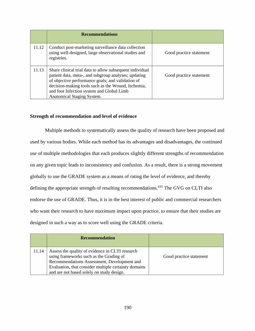
190
Recommendations
11.12 Conduct post-marketing surveillance data collection
using well-designed, large observational studies and
registries.
Good practice statement
11.13 Share clinical trial data to allow subsequent individual
patient data, meta-, and subgroup analyses; updating
of objective performance goals; and validation of
decision-making tools such as the Wound, Ischemia,
and foot Infection system and Global Limb
Anatomical Staging System.
Good practice statement
Strength of recommendation and level of evidence
Multiple methods to systematically assess the quality of research have been proposed and
used by various bodies. While each method has its advantages and disadvantages, the continued
use of multiple methodologies that each produces slightly different strengths of recommendation
on any given topic leads to inconsistency and confusion. As a result, there is a strong movement
globally to use the GRADE system as a means of rating the level of evidence, and thereby
defining the appropriate strength of resulting recommendations.635 The GVG on CLTI also
endorse the use of GRADE. Thus, it is in the best interest of public and commercial researchers
who want their research to have maximum impact upon practice, to ensure that their studies are
designed in such a way as to score well using the GRADE criteria.
Recommendation
11.14 Assess the quality of evidence in CLTI research
using frameworks such as the Grading of
Recommendations Assessment, Development and
Evaluation, that consider multiple certainty domains
and are not based solely on study design.
Good practice statement
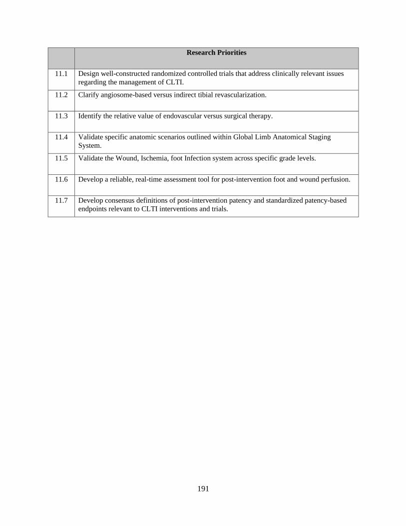
191
Research Priorities
11.1 Design well-constructed randomized controlled trials that address clinically relevant issues
regarding the management of CLTI.
11.2 Clarify angiosome-based versus indirect tibial revascularization.
11.3 Identify the relative value of endovascular versus surgical therapy.
11.4 Validate specific anatomic scenarios outlined within Global Limb Anatomical Staging
System.
11.5 Validate the Wound, Ischemia, foot Infection system across specific grade levels.
11.6 Develop a reliable, real-time assessment tool for post-intervention foot and wound perfusion.
11.7 Develop consensus definitions of post-intervention patency and standardized patency-based
endpoints relevant to CLTI interventions and trials.

192
12: CREATING A CENTER OF EXCELLENCE FOR AMPUTATION PREVENTION
Introduction
The major causes of amputation are related to diabetes and CLTI. Of the 200 million
people worldwide with PAD, CLTI affects at least 2%-3% of this population.1 While
revascularization is the treatment of choice in preventing limb loss, procedure bias, lack of
specialty training, market forces, and lack of consensus definitions remain major obstacles in
achieving the best possible outcomes for CLTI care.636
The CLTI patient is particularly complex. Patients with PAD have an increased risk of
coronary artery disease, cerebrovascular disease, and an elevated risk of 5-year mortality.637
Historically, CLTI was primarily a sequel of smoking and a diet high in saturated fats. However,
over the last few decades, the rise in CLTI has followed the global epidemic of diabetes. Because
of this changing epidemiology, this section focuses mainly on establishing and monitoring teams
for the patient with diabetes-related CLTI, but the concepts presented herein can be applied to all
CLTI teams.
Diabetes-related CLTI is only one part of diabetic foot syndrome, which is a common,
though complex group of complications from diabetes. These include neuropathy, ulceration, the
Charcot foot, soft tissue and bone infection, and peripheral artery disease, including critical limb
ischemia and gangrene. It is well known that diabetes increases the risk of myocardial infarction
by 50% and stroke by 25%, however, the greatest increased risk is for a foot or leg
amputation.615 Diabetic foot syndrome is also a costly comorbidity representing approximately
one third of the total cost of diabetes.638 A recent study found that the mean 1-year cost from a
public payer perspective in the United States was $44,200.639 Roughly 75% of the cost was due
193
to inpatient hospitalizations, where the average length of stay for diabetic foot ulcer and lower
extremity amputation (LEA) exceeded that of myocardial infarction, stroke, and diabetic
ketoacidosis.640-642
The patient with diabetic foot syndrome has a poor prognosis. It is frequently associated
with loss of quality of life, work, independence, and income for both the patient and the primary
caregiver. The relative 5-year mortality rate following a LEA is a staggering 70%.643 For patients
with diabetic foot ulcer it is 55%, and for patients with PAD alone, the 5-year relative mortality
rate is 32%.644 Thus, while diabetes is an endocrine disease, common complications of diabetes
are related to micro- or macrovascular disease. For this reason, diabetic foot syndrome should be
more appropriately thought of as part of the cardiovascular complications of diabetes.
Many institutions and government agencies have responded to the growing complexity,
options, and sub-specialization of treating medical conditions by creating disease-specific
Centers of Excellence. A Center of Excellence is a virtual or physical location with a team of
highly skilled experts who are often involved in research and innovation in order to advance their
field.645 While there have been experts in the field of PAD who have opined on what a Center of
Excellence for CLTI, diabetic foot care, or amputation prevention might encompass, there are
currently no governmental agencies or professional societies that have established such
guidelines.
Center of Excellence
In 2010, building on the work of the International Working Group on the diabetic foot,
three tiers of care were proposed for an amputation prevention team – basic, intermediate, and
Center of Excellence (Table 12.1).646 The basic model of care is performed in an office setting

194
with a general practitioner, internist, or endocrinologist, and/or a specialist nurse. An
intermediate model of care is set in a hospital or multidisciplinary clinic and consists of various
specialists to heal wounds and prevent limb loss. This model is similar to a wound care center in
the United States or a diabetic foot clinic in Europe. A Center of Excellence model is typically
found in a tertiary care hospital with a predetermined team of specialists operating under clinical
practice pathways and policies and procedures. The Center of Excellence has advanced
diagnostics and can intervene rapidly to prevent limb loss.
Currently, in many countries, there are no criteria required to designate oneself a Center
of Excellence for healthcare. Anyone or any institution can use the terminology and doing so
does not guarantee that excellent care is being delivered. Based on experience in creating Centers
of Excellence, a set of criteria is proposed to determine Center of Excellence designation in
CLTI and amputation prevention, as outlined in Table 12.2.
Team setting, components, and function
No single specialist possesses all the necessary skills to manage diabetic foot syndrome.
Therefore, it is important to create a team of specialists with the required skills. While some of
the services required to treat CLTI and prevent amputation can be performed in the outpatient
setting, many needed services are intensive and require access to an acute care hospital.
An understanding of the natural history of amputation in diabetes can assist in
determining how to build an effective team (Fig 12.1).647 Diabetes leads to peripheral
neuropathy, though the timing of its onset is related to long-term control of blood glucose.
Peripheral neuropathy leads to unfelt repetitive trauma and, in combination with foot deformity,
causes diabetic foot ulcer.648 Approximately half of these patients have significant PAD with
195
their diabetic foot ulcer. Still, more often than not, infection serves as the final event leading up
to the amputation.649
Fitzgerald et al described the seven essential skills for limb salvage teams.607 These were
modified to identify nine skills needed for the comprehensive management of diabetic foot.
Table 12.3 lists the essentials skills, as well as the type of specialist who should be added to the
team to complete a given task. The simplest method to construct a team for a Center of
Excellence is to ensure that each of these skills is covered by an expert on the team.
Additionally, several authors have described an irreducible minimum to the team that includes
vascular surgery and surgical podiatry. These two specialties have been nicknamed the “Toe and
Flow” team.607, 646
Team-driven protocols
It is simply not enough to have a designated team. The team must be used in an effective
manner and outcomes should be monitored in a structured fashion. Figure 12.2 illustrates a
useful pathway in setting up the structure of the team, establishing goals, and ensuring that the
goals are met. Published clinical practice guidelines (CPG) from medical and surgical societies
establish best practices, but they are not always feasible for practice in all settings. Current CPGs
exist for PAD in diabetes, diabetic foot infections, DFUs, offloading DFUs, inpatient
management of the diabetic foot and the Charcot foot, and the prevention of diabetic foot
problems.158, 650-655 While these CPGs can serve as a template, localities are encouraged to create
their own clinical practice pathways specific to the facility or system in which they practice.
The clinical practice pathways are used to identify the team structure, patient flow, when
to engage various members, and what to do if the patient is not improving as expected. Policies
and procedures are then created to assist providers and staff in complying with the pathway.

196
Quality assurance goals are also created for measurable policies and procedures. Certain
outcomes are self-explanatory, such as limb salvage rate, while others should be followed to
ensure the quality of care delivered by the Center of Excellence. These can include the high-low
amputation ratio,656 median days-to-heal for foot wounds, healing percentage, and quality of life
measures. Table 12.4 lists the most important measurable outcomes for limb salvage and their
calculation. These data may not always be easy to track. Existing electronic health record
systems are lacking in their ability to track and report most of these, or other custom measures.
Oftentimes, Centers of Excellence resort to developing their own software or keeping track of
data manually in spreadsheets.
Finally, performance improvement plans must be drafted and initiated when the quality
assurance goals are not met. Figure 12.3 shows an example of how this system would be applied
to vascular disease screening in DFUs.
Team Impact
In 2005, the World Health Organization and the International Diabetes Federation
declared that up to 80% of diabetes-related amputations were preventable.657, 658 Currently, the
only intervention to address this has been the formation of multidisciplinary teams to prevent
unnecessary amputations. In fact, the multidisciplinary team to prevent diabetes-related
amputations dates back to at least 1934, when Elliott P Joslin MD, an endocrinologist in Boston,
established his team to treat diabetic gangrene.659
In the United States, an organized team in a public hospital reduced LEAs 72% over 2
years. In the Veterans Affairs medical centers, several factors were significant in the reduction of
LEAs, including use of a specialized team and establishment of a high-risk foot clinic.660, 661 In a
197
military medical center, amputations were reduced by 82% as a result of a specialized limb-
preservation service.662 Another report showed a reduction improvement in diabetes-related foot
outcomes with an integrated interdisciplinary team in a large academic medical center.663 In
several other studies, adding podiatry to the team was found to be helpful in reducing
amputations and significantly reducing the cost associated with diabetic foot.638, 660, 664, 665
The impact of a limb salvage team is not limited to any geographical area. In the
Netherlands, investigators reported a 34% nationwide reduction in amputations after setting up
multidisciplinary teams.666 In Brazil, the establishment of more than 20 interdisciplinary foot
clinics nationwide is leading to improved care.667 In Italy, investigators reported a reduction in
hospitalizations and amputations in the diabetic foot after implementing a multidisciplinary
referral team.667, 668 In Spain, a multidisciplinary foot team reduced amputations over 3 years
when compared with the previous 6 years.669 The United Kingdom has also seen reduced
amputations secondary to better-organized diabetic foot care with specialized clinics that follow
multidisciplinary care pathways and protocols.670, 671 Lastly, in Finland, a decrease in major
amputations was correlated with rising interest in limb salvage and an increase in distal vascular
procedures.672 In a subsequent study, researchers reported a reduction in amputations and length
of stay when inpatient care was reorganized.673
Summary
Centers of Excellence can be implemented with a well-organized team approach to
diabetic foot syndrome and, in particular, the foot with critical limb ischemia. Creating an
integrated team whose primary focus is limb salvage and receives all referrals for suspected
CLTI is key. Teams can improve processes, time to intervention, and outcomes. The setting and
198
structure of the team will ultimately depend on the availability and local need. However, in order
to be most successful, Centers of Excellence should have team members who are capable of
performing the nine essential skills as outlined in Table 12.3.
Centers of Excellence have published pathways and policies and procedures to determine
the function and involvement of various members. Equally important to setting up the team is
measuring the Center’s performance. This is best accomplished with concrete quality assurance
goals and the implementation of a performance improvement plan to be used when these goals
are not met.
199
13. GLOBAL PERSPECTIVES IN CLTI
Introduction
The preceding sections of this guideline make recommendations regarding the diagnosis
and treatment of CLTI based upon data published in peer-reviewed journals and, where such data
are lacking, consensus expert opinion. Vascular specialists managing CLTI across the globe
serve the needs of diverse communities and cultures, working within a very wide range of
healthcare environments. Most vascular specialists will strive to keep up-to-date with the
published evidence base and are greatly facilitated in so doing through the use of modern
information technology systems. However, the reality is that most publications on CLTI are
written in English and the data contained therein overwhelmingly derive from relatively few,
mainly high-income countries (Western Europe, North America, Japan) that have mature, well-
resourced health and social care systems, as well as clinical research infra-structure. Most
vascular specialists treating patients with CLTI do not, of course, work in such favorable
environments. As such, they often have to adapt foreign “evidence based recommendations” to
their own particular situation in order to provide the best possible care to their patients with the
resources available. The GVG authors recognise this and, specifically, that some of the
recommendations contained within this guideline are likely to remain aspirational for many
vascular specialists working in diverse healthcare settings across the globe. The authors therefore
thought it important to examine the state of CLTI care from a broader perspective. To that end, a
questionnaire enquiring about the presentation, diagnosis and management of CLTI was sent to
vascular specialists (N=50) working in a range of lower, middle, and higher-income countries.
This section primarily comprises a description of the responses received (N=22), supported by

200
published loco-regional data where available. While the information provided may not be
considered high in quality from an epidemiologic perspective, a number of important global
issues emerged from the responses. This brief overview, highlights the urgent need for better
data on the impact of CLTI and how it is managed around the world. It should be noted that the
majority of responses derive from a few key opinion leaders from Latin America, Asia, and
Africa; thus the following discussion may not reflect concerns of other populations, providers,
and nations.
Definition and Classification
Clinical criteria, history and examination, are the mainstay of CLTI diagnosis across the
world with the use of adjunctive haemodynamic and perfusion measurements appearing to be
highly variable. ABI testing was used by all except one respondent. However, although all
respondents regularly dealt with diabetic vascular disease and the acknowledged limitations of
ankle pressures in that setting, only two used toe pressures; and none used TcPO2 routinely. All
(except one who exclusively used WIfI) used either Fontaine and/or Rutherford for staging,
approximately in equal numbers. About one third of respondents described employing WIfI in
addition to another clinical classification systems. In summary, therefore, across most of the
world there appears to be limited adherence to any one published definition or staging system for
CLTI.
Epidemiology and Risk factors
Although accurate country-specific epidemiological data are sparse, there seems little doubt that
the increasing prevalence of diabetes mellitus (Figure 1) , together with the growing use of

201
tobacco and population aging, is resulting in a significant increase in CLTI and amputations
across much of the world, and especially in Low-Middle Income Countries (LMIC).674
In 2013, Fowkes et al undertook a meta-analysis of 34 studies to compare the prevalence
and risk factors between High Income Countries (HIC) and Low-Middle Income Countries
(LMIC).1 This is well outlined in Chapter 2 of this document, but worth recalling some of the
key presented data. They concluded “Globally, 202 million people were living with peripheral
artery disease in 2010, 69.7% of them in LMIC, including 54.8 million in Southeast Asia and
45.9 million in the western Pacific Region. During the preceding decade, the number of
individuals with peripheral artery disease increased by 28.7% in LMIC and 13.1% in HIC. Also
of note is the percentage of increase of PAD is higher in women than men in LMIC which is
opposite of HIC”. The increase in PAD burden observed in women and in the younger,
economically productive, age groups is especially worrisome. (Table 1)
The data on country-specific incidence of PAD/CLTI is sparse in these LMIC unlike HIC. There
are no relevant epidemiological data from large regions, but the updated data from Abbas Z G
(4) is tabulated below for perspective, reflecting PAD in diabetics in sub-Saharan Africa (Table
2).
Lacking firm epidemiologic data, recent estimates of CLTI prevalence have used
extrapolations from demographic and other available disease prevalence data, yielding global
estimates of between 20-40 million individuals afflicted. About two thirds of these are projected
to be in LMICs. Unfortunately, documented data to support this is hard to find in any indexed,
peer-reviewed journals.
According to the survey respondents, the risk factors for CLTI in their regions are largely
as expected, but DM is a predominant cause, more than HICs. The prevalence reported by
202
respondents varied from 40 to 90%.. Interestingly, a cultural preference for walking barefoot
and/or a lack of appropriate footwear is a significant problem in some countries. Approximately
60-80% of all the PAD patients seen by the respondents present with CLTI. The average age was
around 65 years and about 70% were men. Most respondent reported that 70-100% of CLTI
patients presented with tissue loss; in three countries it was less than 50%. Primary amputation
was performed in 10-40% of CLTI patients, this being mainly (25-90%) because of delayed
presentation/referral. Only two countries reported a primary amputation rate of less than 10%.
Post-procedural amputation rates were reported at around 5-10%, although two countries
reported much higher rates (60-70%) due to late presentation and/or aggressive disease patterns
encountered.
Diagnostic Evaluation
Duplex ultrasound appears to be used almost universally, although three respondents
preferred to proceed directly to other imaging modalities. Only five respondents performed
digital subtraction arteriography (DSA) as their primary imaging modality. The remainder opted
for MRA and CTA in about equal numbers. In patients with renal impairment, DSA was
preferred by most with half opting for iodinated contrast with appropriate renal protection
measures and the other half favoring CO2 angiography. Two respondents performed only
noninvasive testing in such patients prior to intervention.
Medical and non-interventional management (+/- revascularization)
Respondents reported widespread routine use of antiplatelet and lipid lowering agents.
ACE inhibitors, vasoactive drugs (such cilostazol and pentoxifylline) and anticoagulants were

203
used selectively. Intravenous prostanoids and vasodilators were used by some as adjuncts to
revascularization and in those with non-reconstructible disease. Use of arterial assist devices,
hyperbaric oxygen and spinal cord stimulation was uncommon. Lumbar sympathectomy was
performed by a third of respondents, perhaps in patients with Buerger’s disease (not specified)
Anatomical classification, risk stratification, and predictors of limb salvage
The almost uniform answer to the question “how satisfied are you with present systems?”
was “somewhat satisfied”. Interestingly, only six respondents used TASC to inform decisions
regarding revascularization strategies and procedures in patients with CLTI. There was strong
support for a new approach to patient and limb risk stratification and for a new anatomical
classification system.
Revascularization
Although, overall, there has been a shift towards endovascular intervention, there is
considerable variation in practice across the respondents – varying from 5 to 80% for both
endovascular and “open” procedures!. All stated that the preferred conduit for both above and
below knee bypass continues to be autogenous vein. Prosthetic grafts are used selectively above
knee but none advocate their use for distal bypass. None of the respondents endorsed “routine
stenting” in the femoral popliteal region and all endovascular options (BA, DCB, stenting) are
used selectively. BA is preferred for endovascular intervention in infra popliteal vessels; four
responders selectively use DCB but none were in favor of stents below the knee.
Post procedural surveillance and follow up

204
All the respondents said they had defined follow-up protocols for patients undergoing
infra-inguinal revascularization. All patients (surgical and endovascular) are followed at least 3-
monthly for a year and then at variable intervals thereafter. Clinical evaluation and ABI is the
mainstay of surveillance. Use of other non-invasive methods (PVR, duplex) is variable. Specific
protocols for vein and prosthetic bypass grafts seem to be standardized as per available data in
minority of centers. Approach to surveillance-detected lesions is similarly quite variable, but
mostly dictated by the patient’s symptoms rather than the result of physiological testing.
Arteriography is reserved for clinically significant lesions. Post-procedural drug therapy, for
example with antiplatelet and lipid lowering agents appears concordant with current published
recommendations. Since most CLTI patients had tissue loss, almost all the centers provided
intensive wound services with in their department as part of a multi-disciplinary team approach.
Nearly all agreed that wound infection was a significant determinant of outcome following
revascularization and possible cause of amputation even after successful revascularization.
Health Economics
CLTI has a serious adverse economic impact on patients, their families and wider
communities right across the world but especially so in LMIC. Though these countries are often
grouped together, the division between “middle” and “lower income” is variable and imprecise.
Furthermore, there is often considerable inequality within each LMIC country, and respondents
reported that most patients with CLTI (30 to 90%) appear to come from the poor socio-economic
backgrounds. The following data from the Indian National Sample Survey Office could represent
the situation in many LMIC:675

205
1. Only18% of urban and 14% of rural population is covered by some form of health
insurance
2. Governmental health expenditure is less than 2% of GDP overall
3. People in villages mainly depend on “household income or savings” (68%) and
“borrowings” (25%) to fund hospitalization expenses
4. Around 1% of the poor in rural areas have to sell their physical assets to meet health
expenditure and more than 5% seek help of friends and relatives. This is also in line with
earlier studies showing that millions are pushed into poverty each year by medical
expenditure and that such expenses are among the leading causes of indebtedness among
the poor
5. In cities, people rely much more on their “income or savings” (75%) than on
“borrowings” (18%) to fund their treatment. Previous studies have repeatedly shown that
India has one of the most privatized healthcare systems in the world with “out of pocket”
expenses accounting for the bulk of medical spending.
In India, the cost of infra-popliteal bypass is US$ 1500-3000 and costs of BA are similar.
The use of stent or DCB would add another US$500-1000, and wound care adds at least
US$500. Such “out of pockets” expenses are probably unaffordable for most CLTI patients.
Importantly, these costs depend on recycling SUDs (Single Use Devices) like sheaths,
angioplasty balloons and guide wires (see below). Without such practice, the cost would increase
by at least 50% and far fewer, especially poorer, patients would have access to treatment
resulting in much greater loss of life and limb. Recycling of (not just vascular) SUDs is common
in Asia, Africa, Latin America, and Eastern Europe and proper regulation of the practice,
206
including appropriate consent procedures, is important in order to mitigate patient harm (Aditya
Kapoor et al 2017).676
Summary of Global Perspectives
Based on the responses to the questionnaire and the limited published and unpublished
data at times, we can draw the following conclusions:
1. CLTI is a significant and increasing global problem, especially in LMIC, where the
incidence in women appears to be rising more quickly than in men.
2. Diabetes and unabated smoking are the major causes of CLTI globally
3. Although vascular specialists try to follow the published “evidence base”, economic and
social constraints mean that the approach to CLTI must to tailored to the working
environment.
4. CLTI and “diabetic foot” problems are associated with high amputation rates in LMIC
due to delayed presentation and referral, and limited access to affordable care.
5. Economic constraints are an important limitation in the adoption of advanced vascular
technologies, and practical issues such as recycling of SUDs require oversight from a
public health perspective.
6. Few countries maintain national registries or other CLTI datasets
7. Most countries do not have a standardised approach to CLTI, with considerable
locoregional variation in practice
8. Most countries do not have well-organised and supported vascular societies where best
practice and research can be shared and disseminated

207
Dissemination and Implementation
A large number of vascular specialists from around the world have contributed to the
GVG and that global involvement sets the present guideline document apart from all previous
consensus statements. The paradigms and tools, such as WIfI, PLAN, and GLASS, set out in the
GVG will hopefully meet the needs of the global vascular community as expressed by our
questionnaire respondents. However, it is important to emphasize that some guideline
recommendations will not be achievable by vascular specialists working in LMIC. GVG
recommendations should not, therefore, be viewed as an inflexible global “standard of care”.
Following publication, it will be important to disseminate the GVG as quickly and widely as
possible, simultaneously through a range of different channels, and to obtain validation and
feedback from the global community. Dissemination wil be assisted by:
1. Publication of the full GVG as a supplement JVS and EJEVS,
2. Publication of an executive summary with the recommendations in a range of other,
journals in a number of different languages,
3. Presentations at conferences, and
4. Free online access to the documents, linked from societies’ webpages.

208
REFERENCES
1. Fowkes FGR, Rudan D, Rudan I, Aboyans V, Denenberg JO, McDermott MM, et al.
Comparison of global estimates of prevalence and risk factors for peripheral artery
disease in 2000 and 2010: a systematic review and analysis. The Lancet.
2013;382(9901):1329-40.
2. Goodney PP, Holman K, Henke PK, Travis LL, Dimick JB, Stukel TA, et al. Regional
intensity of vascular care and lower extremity amputation rates. J Vasc Surg.
2013;57(6):1471-79, 80 e1-3; discussion 9-80.
3. Goodney PP, Travis LL, Brooke BS, DeMartino RR, Goodman DC, Fisher ES, et al.
Relationship between regional spending on vascular care and amputation rate. JAMA
Surg. 2014;149(1):34-42.
4. Abu Dabrh AM, Steffen MW, Asi N, Undavalli C, Wang Z, Elamin MB, et al.
Nonrevascularization-based treatments in patients with severe or critical limb ischemia. J
Vasc Surg. 2015;62(5):1330-9.e13.
5. Abu Dabrh AM, Steffen MW, Undavalli C, Asi N, Wang Z, Elamin MB, et al. The
natural history of untreated severe or critical limb ischemia. J Vasc Surg.
2015;62(6):1642-51.e3.
6. Abu Dabrh AM, Steffen MW, Asi N, Undavalli C, Wang Z, Elamin MB, et al. Bypass
surgery versus endovascular interventions in severe or critical limb ischemia. J Vasc
Surg. 2016;63(1):244-53.e11.
209
7. Almasri J, Adusumalli J, Asi N, Lakis S, Alsawas M, Prokop LJ, et al. A systematic
review and meta-analysis of revascularization outcomes of infrainguinal chronic limb-
threatening ischemia. J Vasc Surg. 2018;68(2):624-33.
8. Murad MH. Clinical Practice Guidelines: A Primer on Development and Dissemination.
Mayo Clin Proc. 2017;92(3):423-33.
9. Guyatt GH, Alonso-Coello P, Schunemann HJ, Djulbegovic B, Nothacker M, Lange S, et
al. Guideline panels should seldom make good practice statements: guidance from the
GRADE Working Group. J Clin Epidemiol. 2016;80:3-7.
10. Mills JL, Sr., Conte MS, Armstrong DG, Pomposelli FB, Schanzer A, Sidawy AN, et al.
The Society for Vascular Surgery Lower Extremity Threatened Limb Classification
System: risk stratification based on wound, ischemia, and foot infection (WIfI). J Vasc
Surg. 2014;59(1):220-34 e1-2.
11. Jaff MR, White CJ, Hiatt WR, Fowkes GR, Dormandy J, Razavi M, et al. An update on
methods for revascularization and expansion of the TASC lesion classification to include
below‐the‐knee arteries: a supplement to the inter‐society consensus for the
management of peripheral arterial disease (TASC II): the TASC steering committee.
Catheter Cardiovasc Interv. 2015;86(4):611-25.
12. Bollinger A, Breddin K, Hess H, Heystraten FM, Kollath J, Konttila A, et al.
Semiquantitative assessment of lower limb atherosclerosis from routine angiographic
images. Atherosclerosis. 1981;38(3-4):339-46.
13. Menard MT, Farber A, Assmann SF, Choudhry NK, Conte MS, Creager MA, et al.
Design and Rationale of the Best Endovascular Versus Best Surgical Therapy for Patients
With Critical Limb Ischemia (BEST-CLI) Trial. J Am Heart Assoc. 2016;5(7).
210
14. Popplewell MA, Davies H, Jarrett H, Bate G, Grant M, Patel S, et al. Bypass versus angio
plasty in severe ischaemia of the leg - 2 (BASIL-2) trial: study protocol for a randomised
controlled trial. Trials. 2016;17(1):11.
15. Hunt BD, Popplewell MA, Davies H, Meecham L, Jarrett H, Bate G, et al. BAlloon
versus Stenting in severe Ischaemia of the Leg-3 (BASIL-3): study protocol for a
randomised controlled trial. Trials. 2017;18(1):224.
16. de Graaff JC, Ubbink DT, Legemate DA, Tijssen JGP, Jacobs MJHM. Evaluation of toe
pressure and transcutaneous oxygen measurements in management of chronic critical leg
ischemia: a diagnostic randomized clinical trial. J Vasc Surg. 2003;38(3):528-34.
17. Brownrigg JRW, Hinchliffe RJ, Apelqvist J, Boyko EJ, Fitridge R, Mills JL, et al.
Performance of prognostic markers in the prediction of wound healing or amputation
among patients with foot ulcers in diabetes: a systematic review. Diabetes Metab Res
Rev. 2016;32(S1):128-35.
18. Wang Z, Hasan R, Firwana B, Elraiyah T, Tsapas A, Prokop L, et al. A systematic review
and meta-analysis of tests to predict wound healing in diabetic foot. J Vasc Surg.
2016;63(2):29S-36S. e2.
19. Lijmer JG, Hunink MG, van den Dungen JJ, Loonstra J, Smit AJ. ROC analysis of
noninvasive tests for peripheral arterial disease. Ultrasound Med Biol. 1996;22(4):391-8.
20. Dachun X, Jue L, Liling Z, Yawei X, Dayi H, Pagoto SL, et al. Sensitivity and specificity
of the ankle--brachial index to diagnose peripheral artery disease: a structured review.
Vasc Med. 2010;15(5):361-9.
211
21. Aboyans V, Ho E, Denenberg JO, Ho LA, Natarajan L, Criqui MH. The association
between elevated ankle systolic pressures and peripheral occlusive arterial disease in
diabetic and nondiabetic subjects. J Vasc Surg. 2008;48(5):1197-203.
22. Salaun P, Desormais I, Lapebie FX, Riviere AB, Aboyans V, Lacroix P, et al.
Comparison of Ankle Pressure, Systolic Toe Pressure, and Transcutaneous Oxygen
Pressure to Predict Major Amputation After 1 Year in the COPART Cohort. Angiology.
2018:3319718793566.
23. Shirasu T, Hoshina K, Akagi D, Miyahara T, Yamamoto K, Watanabe T. Pulse volume
recordings to identify falsely elevated ankle brachial index. Asian Cardiovasc Thorac
Ann. 2016;24(6):517-22.
24. Hingorani AP, Ascher E, Marks N, Puggioni A, Shiferson A, Tran V, et al. Limitations of
and lessons learned from clinical experience of 1,020 duplex arteriography. Vascular.
2008;16(3):147-53.
25. Larch E, Minar E, Ahmadi R, Schnurer G, Schneider B, Stumpflen A, et al. Value of
color duplex sonography for evaluation of tibioperoneal arteries in patients with
femoropopliteal obstruction: a prospective comparison with anterograde intraarterial
digital subtraction angiography. J Vasc Surg. 1997;25(4):629-36.
26. Adriaensen ME, Kock MC, Stijnen T, van Sambeek MR, van Urk H, Pattynama PM, et
al. Peripheral arterial disease: therapeutic confidence of CT versus digital subtraction
angiography and effects on additional imaging recommendations. Radiology.
2004;233(2):385-91.
27. Hingorani A, Ascher E, Markevich N, Kallakuri S, Schutzer R, Yorkovich W, et al. A
comparison of magnetic resonance angiography, contrast arteriography, and duplex
212
arteriography for patients undergoing lower extremity revascularization. Ann Vasc Surg.
2004;18(3):294-301.
28. Collins R, Cranny G, Burch J, Aguiar-Ibanez R, Craig D, Wright K, et al. A systematic
review of duplex ultrasound, magnetic resonance angiography and computed tomography
angiography for the diagnosis and assessment of symptomatic, lower limb peripheral
arterial disease. Health Technol Assess. 2007;11(20):iii-iv, xi-xiii, 1-184.
29. Met R, Bipat S, Legemate DA, Reekers JA, Koelemay MJ. Diagnostic performance of
computed tomography angiography in peripheral arterial disease: a systematic review and
meta-analysis. JAMA. 2009;301(4):415-24.
30. Long-term mortality and its predictors in patients with critical leg ischaemia. The I.C.A.I.
Group (Gruppo di Studio dell'Ischemia Cronica Critica degli Arti Inferiori). The Study
Group of Criticial Chronic Ischemia of the Lower Exremities. Eur J Vasc Endovasc Surg.
1997;14(2):91-5.
31. Armstrong EJ, Wu J, Singh GD, Dawson DL, Pevec WC, Amsterdam EA, et al. Smoking
cessation is associated with decreased mortality and improved amputation-free survival
among patients with symptomatic peripheral artery disease. J Vasc Surg.
2014;60(6):1565-71.
32. Faglia E, Clerici G, Scatena A, Caminiti M, Curci V, Morabito A, et al. Effectiveness of
combined therapy with angiotensin-converting enzyme inhibitors and statins in reducing
mortality in diabetic patients with critical limb ischemia: An observational study.
Diabetes Res Clin Pract. 2014;103(2):292-7.

213
33. Antithrombotic Trialists C. Collaborative meta-analysis of randomised trials of
antiplatelet therapy for prevention of death, myocardial infarction, and stroke in high risk
patients. BMJ. 2002;324(7329):71-86.
34. Antithrombotic Trialists C, Baigent C, Blackwell L, Collins R, Emberson J, Godwin J, et
al. Aspirin in the primary and secondary prevention of vascular disease: collaborative
meta-analysis of individual participant data from randomised trials. Lancet.
2009;373(9678):1849-60.
35. CAPRIE Steering Committee. A randomised, blinded, trial of clopidogrel versus aspirin
in patients at risk of ischaemic events (CAPRIE). The Lancet. 1996;348(9038):1329-39.
36. Hiatt WR, Fowkes FG, Heizer G, Berger JS, Baumgartner I, Held P, et al. Ticagrelor
versus Clopidogrel in Symptomatic Peripheral Artery Disease. N Engl J Med.
2017;376(1):32-40.
37. Anand SS, Bosch J, Eikelboom JW, Connolly SJ, Diaz R, Widimsky P, et al.
Rivaroxaban with or without aspirin in patients with stable peripheral or carotid artery
disease: an international, randomised, double-blind, placebo-controlled trial. Lancet.
2018;391(10117):219-29.
38. Anand S, Yusuf S, Xie C, Pogue J, Eikelboom J, Budaj A, et al. Oral anticoagulant and
antiplatelet therapy and peripheral arterial disease. N Engl J Med. 2007;357(3):217-27.
39. Leng GC, Price JF, Jepson RG. Lipid-lowering for lower limb atherosclerosis. Cochrane
Database Syst Rev. 2000(2):Cd000123.
40. Heart Protection Study Collaborative G. MRC/BHF Heart Protection Study of cholesterol
lowering with simvastatin in 20,536 high-risk individuals: a randomised placebo-
controlled trial. Lancet. 2002;360(9326):7-22.

214
41. Meade T, Zuhrie R, Cook C, Cooper J. Bezafibrate in men with lower extremity arterial
disease: randomised controlled trial. BMJ. 2002;325(7373):1139.
42. Aung PP, Maxwell HG, Jepson RG, Price JF, Leng GC. Lipid-lowering for peripheral
arterial disease of the lower limb. Cochrane Database Syst Rev. 2007(4):Cd000123.
43. Mills EJ, O'Regan C, Eyawo O, Wu P, Mills F, Berwanger O, et al. Intensive statin
therapy compared with moderate dosing for prevention of cardiovascular events: a meta-
analysis of >40 000 patients. Eur Heart J. 2011;32(11):1409-15.
44. Rodriguez F, Maron DJ, Knowles JW, Virani SS, Lin S, Heidenreich PA. Association
Between Intensity of Statin Therapy and Mortality in Patients With Atherosclerotic
Cardiovascular Disease. JAMA Cardiol. 2017;2(1):47-54.
45. ACCORD Study Group, Cushman WC, Evans GW, Byington RP, Goff DC, Jr., Grimm
RH, Jr., et al. Effects of Intensive Blood-Pressure Control in Type 2 Diabetes Mellitus. N
Engl J Med. 2010;362(17):1575-85.
46. Bavry AA, Anderson RD, Gong Y, Denardo SJ, Cooper-Dehoff RM, Handberg EM, et
al. Outcomes Among hypertensive patients with concomitant peripheral and coronary
artery disease: findings from the INternational VErapamil-SR/Trandolapril STudy.
Hypertension. 2010;55(1):48-53.
47. Wright JT, Jr., Williamson JD, Whelton PK, Snyder JK, Sink KM, Rocco MV, et al. A
Randomized Trial of Intensive versus Standard Blood-Pressure Control. N Engl J Med.
2015;373(22):2103-16.
48. Moise N, Huang C, Rodgers A, Kohli-Lynch CN, Tzong KY, Coxson PG, et al.
Comparative Cost-Effectiveness of Conservative or Intensive Blood Pressure Treatment

215
Guidelines in Adults Aged 35-74 Years: The Cardiovascular Disease Policy Model.
Hypertension. 2016;68(1):88-96.
49. Selvin E, Marinopoulos S, Berkenblit G, Rami T, Brancati FL, Powe NR, et al. Meta-
analysis: glycosylated hemoglobin and cardiovascular disease in diabetes mellitus. Ann
Intern Med. 2004;141(6):421-31.
50. Nathan DM, Cleary PA, Backlund JY, Genuth SM, Lachin JM, Orchard TJ, et al.
Intensive diabetes treatment and cardiovascular disease in patients with type 1 diabetes.
N Engl J Med. 2005;353(25):2643-53.
51. van Dieren S, Kengne AP, Chalmers J, Beulens JW, Davis TM, Fulcher G, et al.
Intensification of medication and glycaemic control among patients with type 2 diabetes -
the ADVANCE trial. Diabetes Obes Metab. 2014;16(5):426-32.
52. Fox CS, Golden SH, Anderson C, Bray GA, Burke LE, de Boer IH, et al. Update on
Prevention of Cardiovascular Disease in Adults With Type 2 Diabetes Mellitus in Light
of Recent Evidence: A Scientific Statement From the American Heart Association and
the American Diabetes Association. Diabetes Care. 2015;38(9):1777-803.
53. American Diabetes Association. 6. Glycemic Targets: Standards of Medical Care in
Diabetes-2018. Diabetes Care. 2018;41(Suppl 1):S55-s64.
54. Palmer SC, Mavridis D, Nicolucci A, et al. Comparison of clinical outcomes and adverse
events associated with glucose-lowering drugs in patients with type 2 diabetes: A meta-
analysis. JAMA. 2016;316(3):313-24.
55. Nawaz S, Cleveland T, Gaines PA, Chan P. Clinical risk associated with contrast
angiography in metformin treated patients: a clinical review. Clin Radiol.
1998;53(5):342-4.

216
56. Goergen SK, Rumbold G, Compton G, Harris C. Systematic review of current guidelines,
and their evidence base, on risk of lactic acidosis after administration of contrast medium
for patients receiving metformin. Radiology. 2010;254(1):261-9.
57. Stacul F, van der Molen AJ, Reimer P, Webb JAW, Thomsen HS, Morcos SK, et al.
Contrast induced nephropathy: updated ESUR Contrast Media Safety Committee
guidelines. Eur Radiol. 2011;21(12):2527-41.
58. Dagenais GR, Yi Q, Lonn E, Sleight P, Ostergren J, Yusuf S. Impact of cigarette
smoking in high-risk patients participating in a clinical trial. A substudy from the Heart
Outcomes Prevention Evaluation (HOPE) trial. Eur J Cardiovasc Prev Rehabil.
2005;12(1):75-81.
59. Athyros VG, Tziomalos K, Katsiki N, Gossios TD, Giouleme O, Anagnostis P, et al. The
impact of smoking on cardiovascular outcomes and comorbidities in statin-treated
patients with coronary artery disease: a post hoc analysis of the GREACE study. Curr
Vasc Pharmacol. 2013;11(5):779-84.
60. Blomster JI, Woodward M, Zoungas S, Hillis GS, Harrap S, Neal B, et al. The harms of
smoking and benefits of smoking cessation in women compared with men with type 2
diabetes: an observational analysis of the ADVANCE (Action in Diabetes and Vascular
Disease: Preterax and Diamicron modified release Controlled Evaluation) trial. BMJ
Open. 2016;6(1):e009668.
61. Kondo T, Osugi S, Shimokata K, Honjo H, Morita Y, Maeda K, et al. Smoking and
smoking cessation in relation to all-cause mortality and cardiovascular events in 25,464
healthy male Japanese workers. Circ J. 2011;75(12):2885-92.
217
62. Newhall K, Suckow B, Spangler E, Brooke BS, Schanzer A, Tan TW, et al. Impact and
Duration of Brief Surgeon-Delivered Smoking Cessation Advice on Attitudes Regarding
Nicotine Dependence and Tobacco Harms for Patients with Peripheral Arterial Disease.
Ann Vasc Surg. 2017;38:113-21.
63. Biancari F, Salenius JP, Heikkinen M, Luther M, Ylonen K, Lepantalo M. Risk-scoring
method for prediction of 30-day postoperative outcome after infrainguinal surgical
revascularization for critical lower-limb ischemia: a Finnvasc registry study. World J
Surg. 2007;31(1):217-25; discussion 26-7.
64. Schanzer A, Mega J, Meadows J, Samson RH, Bandyk DF, Conte MS. Risk stratification
in critical limb ischemia: derivation and validation of a model to predict amputation-free
survival using multicenter surgical outcomes data. J Vasc Surg. 2008;48(6):1464-71.
65. Bradbury AW, Adam DJ, Bell J, Forbes JF, Fowkes FG, Gillespie I, et al. Bypass versus
Angioplasty in Severe Ischaemia of the Leg (BASIL) trial: A survival prediction model
to facilitate clinical decision making. J Vasc Surg. 2010;51(5 Suppl):52S-68S.
66. Meltzer AJ, Graham A, Connolly PH, Meltzer EC, Karwowski JK, Bush HL, et al. The
Comprehensive Risk Assessment for Bypass (CRAB) facilitates efficient perioperative
risk assessment for patients with critical limb ischemia. J Vasc Surg. 2013;57(5):1186-95.
67. Simons JP, Goodney PP, Flahive J, Hoel AW, Hallett JW, Kraiss LW, et al. A
comparative evaluation of risk-adjustment models for benchmarking amputation-free
survival after lower extremity bypass. J Vasc Surg. 2016;63(4):990-7.
68. Cull DL, Manos G, Hartley MC, Taylor SM, Langan EM, Eidt JF, et al. An early
validation of the Society for Vascular Surgery lower extremity threatened limb
classification system. J Vasc Surg. 2014;60(6):1535-41.

218
69. Zhan LX, Branco BC, Armstrong DG, Mills JL, Sr. The Society for Vascular Surgery
lower extremity threatened limb classification system based on Wound, Ischemia, and
foot Infection (WIfI) correlates with risk of major amputation and time to wound healing.
J Vasc Surg. 2015;61(4):939-44.
70. Causey MW, Ahmed A, Wu B, Gasper WJ, Reyzelman A, Vartanian SM, et al. Society
for Vascular Surgery limb stage and patient risk correlate with outcomes in an
amputation prevention program. J Vasc Surg. 2016;63(6):1563-73.e2.
71. Darling JD, McCallum JC, Soden PA, Meng Y, Wyers MC, Hamdan AD, et al.
Predictive ability of the Society for Vascular Surgery Wound, Ischemia, and foot
Infection (WIfI) classification system following infrapopliteal endovascular interventions
for critical limb ischemia. J Vasc Surg. 2016;64(3):616-22.
72. Robinson WP, Loretz L, Hanesian C, Flahive J, Bostrom J, Lunig N, et al. Society for
Vascular Surgery Wound, Ischemia, foot Infection (WIfI) score correlates with the
intensity of multimodal limb treatment and patient-centered outcomes in patients with
threatened limbs managed in a limb preservation center. J Vasc Surg. 2017;66(2):488-
98.e2.
73. Sheehan P, Jones P, Caselli A, Giurini JM, Veves A. Percent change in wound area of
diabetic foot ulcers over a 4-week period is a robust predictor of complete healing in a
12-week prospective trial. Diabetes Care. 2003;26(6):1879-82.
74. Cardinal M, Eisenbud DE, Phillips T, Harding K. Early healing rates and wound area
measurements are reliable predictors of later complete wound closure. Wound Repair
Regen. 2008;16(1):19-22.

219
75. Lavery LA, Barnes SA, Keith MS, Seaman JW, Jr., Armstrong DG. Prediction of healing
for postoperative diabetic foot wounds based on early wound area progression. Diabetes
Care. 2008;31(1):26-9.
76. Snyder RJ, Cardinal M, Dauphinee DM, Stavosky J. A post-hoc analysis of reduction in
diabetic foot ulcer size at 4 weeks as a predictor of healing by 12 weeks. Ostomy Wound
Manage. 2010;56(3):44-50.
77. Seeger JM, Schmidt JH, Flynn TC. Preoperative saphenous and cephalic vein mapping as
an adjunct to reconstructive arterial surgery. Ann Surg. 1987;205(6):733-9.
78. Wengerter KR, Veith FJ, Gupta SK, Ascer E, Rivers SP. Influence of vein size (diameter)
on infrapopliteal reversed vein graft patency. J Vasc Surg. 1990;11(4):525-31.
79. Schanzer A, Hevelone N, Owens CD, Belkin M, Bandyk DF, Clowes AW, et al.
Technical factors affecting autogenous vein graft failure: observations from a large
multicenter trial. J Vasc Surg. 2007;46(6):1180-90; discussion 90.
80. Harward TR, Ingegno MD, Carlton L, Flynn TC, Seeger JM. Limb-threatening ischemia
due to multilevel arterial occlusive disease. Simultaneous or staged inflow/outflow
revascularization. Ann Surg. 1995;221(5):498-503; discussion -6.
81. Zukauskas G, Ulevicius H, Triponis V. Sequential aortofemoropopliteal/distal bypass for
treatment of critical lower-limb ischaemia. Cardiovasc Surg. 1995;3(6):671-8.
82. Jongkind V, Akkersdijk GJ, Yeung KK, Wisselink W. A systematic review of
endovascular treatment of extensive aortoiliac occlusive disease. J Vasc Surg.
2010;52(5):1376-83.

220
83. Ye W, Liu CW, Ricco JB, Mani K, Zeng R, Jiang J. Early and late outcomes of
percutaneous treatment of TransAtlantic Inter-Society Consensus class C and D aorto-
iliac lesions. J Vasc Surg. 2011;53(6):1728-37.
84. Deloose K, Bosiers M, Callaert J, Verbist J, Vermassen F, Scheinert D, et al. Primary
stenting is nowadays the gold standard treatment for TASC II A & B iliac lesions: the
definitive MISAGO 1-year results. J Cardiovasc Surg (Torino). 2017;58(3):416-21.
85. Ricco JB, Probst H. Long-term results of a multicenter randomized study on direct versus
crossover bypass for unilateral iliac artery occlusive disease. J Vasc Surg. 2008;47(1):45-
53; discussion -4.
86. Chiu KW, Davies RS, Nightingale PG, Bradbury AW, Adam DJ. Review of direct
anatomical open surgical management of atherosclerotic aorto-iliac occlusive disease.
Eur J Vasc Endovasc Surg. 2010;39(4):460-71.
87. Indes JE, Pfaff MJ, Farrokhyar F, Brown H, Hashim P, Cheung K, et al. Clinical
outcomes of 5358 patients undergoing direct open bypass or endovascular treatment for
aortoiliac occlusive disease: a systematic review and meta-analysis. J Endovasc Ther.
2013;20(4):443-55.
88. Kang JL, Patel VI, Conrad MF, Lamuraglia GM, Chung TK, Cambria RP. Common
femoral artery occlusive disease: contemporary results following surgical
endarterectomy. J Vasc Surg. 2008;48(4):872-7.
89. Ballotta E, Gruppo M, Mazzalai F, Da Giau G. Common femoral artery endarterectomy
for occlusive disease: An 8-year single-center prospective study. Surgery.
2010;147(2):268-74.
221
90. Chang RW, Goodney PP, Baek JH, Nolan BW, Rzucidlo EM, Powell RJ. Long-term
results of combined common femoral endarterectomy and iliac stenting/stent grafting for
occlusive disease. J Vasc Surg. 2008;48(2):362-7.
91. Baumann F, Ruch M, Willenberg T, Dick F, Do DD, Keo HH, et al. Endovascular
treatment of common femoral artery obstructions. J Vasc Surg. 2011;53(4):1000-6.
92. Bonvini RF, Rastan A, Sixt S, Noory E, Schwarz T, Frank U, et al. Endovascular
treatment of common femoral artery disease: medium-term outcomes of 360 consecutive
procedures. J Am Coll Cardiol. 2011;58(8):792-8.
93. Goueffic Y, Della Schiava N, Thaveau F, Rosset E, Favre JP, Salomon du Mont L, et al.
Stenting or Surgery for De Novo Common Femoral Artery Stenosis. JACC Cardiovasc
Interv. 2017;10(13):1344-54.
94. Siracuse JJ, Van Orden K, Kalish JA, Eslami MH, Schermerhorn ML, Patel VI, et al.
Endovascular treatment of the common femoral artery in the Vascular Quality Initiative.
J Vasc Surg. 2017;65(4):1039-46.
95. Azuma N, Uchida H, Kokubo T, Koya A, Akasaka N, Sasajima T. Factors influencing
wound healing of critical ischaemic foot after bypass surgery: is the angiosome important
in selecting bypass target artery? Eur J Vasc Endovasc Surg. 2012;43(3):322-8.
96. Sumpio BE, Forsythe RO, Ziegler KR, van Baal JG, Lepantalo MJ, Hinchliffe RJ.
Clinical implications of the angiosome model in peripheral vascular disease. J Vasc Surg.
2013;58(3):814-26.
97. Biancari F, Juvonen T. Angiosome-targeted lower limb revascularization for ischemic
foot wounds: systematic review and meta-analysis. Eur J Vasc Endovasc Surg.
2014;47(5):517-22.

222
98. Chae KJ, Shin JY. Is Angiosome-Targeted Angioplasty Effective for Limb Salvage and
Wound Healing in Diabetic Foot? : A Meta-Analysis. PLoS One. 2016;11(7):e0159523.
99. Jongsma H, Bekken JA, Akkersdijk GP, Hoeks SE, Verhagen HJ, Fioole B. Angiosome-
directed revascularization in patients with critical limb ischemia. J Vasc Surg.
2017;65(4):1208-19 e1.
100. Schillinger M, Sabeti S, Loewe C, Dick P, Amighi J, Mlekusch W, et al. Balloon
angioplasty versus implantation of nitinol stents in the superficial femoral artery. N Engl
J Med. 2006;354(18):1879-88.
101. Saxon RR, Dake MD, Volgelzang RL, Katzen BT, Becker GJ. Randomized, multicenter
study comparing expanded polytetrafluoroethylene-covered endoprosthesis placement
with percutaneous transluminal angioplasty in the treatment of superficial femoral artery
occlusive disease. J Vasc Interv Radiol. 2008;19(6):823-32.
102. Dake MD, Ansel GM, Jaff MR, Ohki T, Saxon RR, Smouse HB, et al. Paclitaxel-eluting
stents show superiority to balloon angioplasty and bare metal stents in femoropopliteal
disease: twelve-month Zilver PTX randomized study results. Circ Cardiovasc Interv.
2011;4(5):495-504.
103. Rosenfield K, Jaff MR, White CJ, Rocha-Singh K, Mena-Hurtado C, Metzger DC, et al.
Trial of a Paclitaxel-Coated Balloon for Femoropopliteal Artery Disease. N Engl J Med.
2015;373(2):145-53.
104. Mills JL, Fujitani RM, Taylor SM. Contribution of routine intraoperative completion
arteriography to early infrainguinal bypass patency. Am J Surg. 1992;164(5):506-10;
discussion 10-1.

223
105. Bandyk DF, Mills JL, Gahtan V, Esses GE. Intraoperative duplex scanning of arterial
reconstructions: fate of repaired and unrepaired defects. J Vasc Surg. 1994;20(3):426-32;
discussion 32-3.
106. Ubbink DT, Vermeulen H. Spinal cord stimulation for non-reconstructable chronic
critical leg ischaemia. Cochrane Database Syst Rev. 2013;2:Cd004001.
107. Karanth VKL, Karanth TK, Karanth L. Lumbar sympathectomy techniques for critical
lower limb ischaemia due to non‐reconstructable peripheral arterial disease. Cochrane
Database Syst Rev. 2016(12).
108. Vietto V, Franco JV, Saenz V, Cytryn D, Chas J, Ciapponi A. Prostanoids for critical
limb ischaemia. Cochrane Database Syst Rev. 2018;1:Cd006544.
109. Smith FB BA, Fowkes G. . Intravenous naftidrofuryl for critical limb ischaemia.
Cochrane Database Syst Rev. 2012(Issue 7):Art. No.: CD002070.
110. Kranke P, Bennett MH, Martyn-St James M, Schnabel A, Debus SE, Weibel S.
Hyperbaric oxygen therapy for chronic wounds. Cochrane Database Syst Rev.
2015;6:Cd004123.
111. Game FL, Apelqvist J, Attinger C, Hartemann A, Hinchliffe RJ, Londahl M, et al.
Effectiveness of interventions to enhance healing of chronic ulcers of the foot in diabetes:
a systematic review. Diabetes Metab Res Rev. 2016;32 Suppl 1:154-68.
112. Santema KTB, Stoekenbroek RM, Koelemay MJW, Reekers JA, van Dortmont LMC,
Oomen A, et al. Hyperbaric Oxygen Therapy in the Treatment of Ischemic Lower-
Extremity Ulcers in Patients With Diabetes: Results of the DAMO2CLES Multicenter
Randomized Clinical Trial. Diabetes Care. 2018;41(1):112-9.

224
113. Peeters Weem SM, Teraa M, de Borst GJ, Verhaar MC, Moll FL. Bone Marrow derived
Cell Therapy in Critical Limb Ischemia: A Meta-analysis of Randomized Placebo
Controlled Trials. Eur J Vasc Endovasc Surg. 2015;50(6):775-83.
114. Elsherif M, Tawfick W, Canning P, Hynes N, Sultan S. Quality of time spent without
symptoms of disease or toxicity of treatment for transmetatarsal amputation versus digital
amputation in diabetic patients with digital gangrene. Vascular. 2018;26(2):142-50.
115. Aziz H, Branco BC, Braun J, Hughes JD, Goshima KR, Trinidad-Hernandez M, et al.
The influence of do-not-resuscitate status on the outcomes of patients undergoing
emergency vascular operations. J Vasc Surg. 2015;61(6):1538-42.
116. Siracuse JJ, Jones DW, Meltzer EC, Graham AR, Salzler GG, Connolly PH, et al. Impact
of "Do Not Resuscitate" Status on the Outcome of Major Vascular Surgical Procedures.
Ann Vasc Surg. 2015;29(7):1339-45.
117. Reed AB, Delvecchio C, Giglia JS. Major lower extremity amputation after multiple
revascularizations: was it worth it? Ann Vasc Surg. 2008;22(3):335-40.
118. Rollins DL, Towne JB, Bernhard VM, Baum PL. Isolated profundaplasty for limb
salvage. J Vasc Surg. 1985;2(4):585-90.
119. Miksic K, Novak B. Profunda femoris revascularization in limb salvage. J Cardiovasc
Surg (Torino). 1986;27(5):544-52.
120. Ayoub MM, Solis MM, Rogers JJ, Dalton ML. Thru-knee amputation: the operation of
choice for non-ambulatory patients. Am Surg. 1993;59(9):619-23.
121. Taylor SM, Kalbaugh CA, Cass AL, Buzzell NM, Daly CA, Cull DL, et al. "Successful
outcome" after below-knee amputation: an objective definition and influence of clinical
variables. Am Surg. 2008;74(7):607-12; discussion 12-3.
225
122. Webster JB, Hakimi KN, Czerniecki JM. Prosthetic fitting, use, and satisfaction
following lower-limb amputation: A prospective study. J Rehabil Res Dev.
2012;49(10):1493.
123. Bradley L, Kirker S. Secondary prevention of arteriosclerosis in lower limb vascular
amputees: a missed opportunity. Eur J Vasc Endovasc Surg. 2006;32(5):491-3.
124. Glaser JD, Bensley RP, Hurks R, Dahlberg S, Hamdan AD, Wyers MC, et al. Fate of the
contralateral limb after lower extremity amputation. J Vasc Surg. 2013;58(6):1571-7 e1.
125. Abbruzzese TA, Havens J, Belkin M, Donaldson MC, Whittemore AD, Liao JK, et al.
Statin therapy is associated with improved patency of autogenous infrainguinal bypass
grafts. J Vasc Surg. 2004;39(6):1178-85.
126. Henke PK, Blackburn S, Proctor MC, Stevens J, Mukherjee D, Rajagopalin S, et al.
Patients undergoing infrainguinal bypass to treat atherosclerotic vascular disease are
underprescribed cardioprotective medications: effect on graft patency, limb salvage, and
mortality. J Vasc Surg. 2004;39(2):357-65.
127. Brown J, Lethaby A, Maxwell H, Wawrzyniak AJ, Prins MH. Antiplatelet agents for
preventing thrombosis after peripheral arterial bypass surgery. Cochrane Database Syst
Rev. 2008(4):Cd000535.
128. Bedenis R, Lethaby A, Maxwell H, Acosta S, Prins MH. Antiplatelet agents for
preventing thrombosis after peripheral arterial bypass surgery. Cochrane Database Syst
Rev. 2015(2).
129. Suckow BD, Kraiss LW, Schanzer A, Stone DH, Kalish J, DeMartino RR, et al. Statin
therapy after infrainguinal bypass surgery for critical limb ischemia is associated with
improved 5-year survival. J Vasc Surg. 2015;61(1):126-33.
226
130. Hobbs SD, Bradbury AW. Smoking cessation strategies in patients with peripheral
arterial disease: an evidence-based approach. Eur J Vasc Endovasc Surg. 2003;26(4):341-
7.
131. Willigendael EM, Teijink JA, Bartelink ML, Peters RJ, Buller HR, Prins MH. Smoking
and the patency of lower extremity bypass grafts: a meta-analysis. J Vasc Surg.
2005;42(1):67-74.
132. Belch JJ, Dormandy J, Biasi GM, Cairols M, Diehm C, Eikelboom B, et al. Results of the
randomized, placebo-controlled clopidogrel and acetylsalicylic acid in bypass surgery for
peripheral arterial disease (CASPAR) trial. J Vasc Surg. 2010;52(4):825-33, 33.e1-2.
133. Gassman AA, Degner BC, Al-Nouri O, Philippi L, Hershberger R, Halandras P, et al.
Aspirin usage is associated with improved prosthetic infrainguinal bypass graft patency.
Vascular. 2014;22(2):105-11.
134. Cassar K, Ford I, Greaves M, Bachoo P, Brittenden J. Randomized clinical trial of the
antiplatelet effects of aspirin–clopidogrel combination versus aspirin alone after lower
limb angioplasty. Br J Surg. 2005;92(2):159-65.
135. Bhatt DL, Fox KA, Hacke W, Berger PB, Black HR, Boden WE, et al. Clopidogrel and
aspirin versus aspirin alone for the prevention of atherothrombotic events. N Engl J Med.
2006;354(16):1706-17.
136. Tepe G, Bantleon R, Brechtel K, Schmehl J, Zeller T, Claussen CD, et al. Management of
peripheral arterial interventions with mono or dual antiplatelet therapy—the MIRROR
study: a randomised and double-blinded clinical trial. Eur Radiol. 2012;22(9):1998-2006.
137. Strobl FF, Brechtel K, Schmehl J, Zeller T, Reiser MF, Claussen CD, et al. Twelve-
month results of a randomized trial comparing mono with dual antiplatelet therapy in

227
endovascularly treated patients with peripheral artery disease. J Endovasc Ther.
2013;20(5):699-706.
138. Mills JL, Sr., Wixon CL, James DC, Devine J, Westerband A, Hughes JD. The natural
history of intermediate and critical vein graft stenosis: recommendations for continued
surveillance or repair. J Vasc Surg. 2001;33(2):273-8; discussion 8-80.
139. Landry GJ, Moneta GL, Taylor LM, Edwards JM, Yeager RA, Porter JM. Long-term
outcome of revised lower-extremity bypass grafts. J Vasc Surg. 2002;35(1):56-63.
140. Nguyen LL, Conte MS, Menard MT, Gravereaux EC, Chew DK, Donaldson MC, et al.
Infrainguinal vein bypass graft revision: factors affecting long-term outcome. J Vasc
Surg. 2004;40(5):916-23.
141. Bui TD, Mills JL, Sr., Ihnat DM, Gruessner AC, Goshima KR, Hughes JD. The natural
history of duplex-detected stenosis after femoropopliteal endovascular therapy suggests
questionable clinical utility of routine duplex surveillance. J Vasc Surg. 2012;55(2):346-
52.
142. Humphries MD, Pevec WC, Laird JR, Yeo KK, Hedayati N, Dawson DL. Early duplex
scanning after infrainguinal endovascular therapy. J Vasc Surg. 2011;53(2):353-8.
143. Elraiyah T, Prutsky G, Domecq JP, Tsapas A, Nabhan M, Frykberg RG, et al. A
systematic review and meta-analysis of off-loading methods for diabetic foot ulcers. J
Vasc Surg. 2016;63(2 Suppl):59S-68S.e1-2.
144. Bell P. The definition of critical ischaemia of a limb. Br J Surg. 1982;69:S2.
145. Thompson MM, Sayers RD, Varty K, Reid A, London NJ, Bell PR. Chronic critical leg
ischaemia must be redefined. Eur J Vasc Surg. 1993;7(4):420-6.

228
146. Hafner J, Schaad I, Schneider E, Seifert B, Burg G, Cassina PC. Leg ulcers in peripheral
arterial disease (arterial leg ulcers): impaired wound healing above the threshold of
chronic critical limb ischemia. J Am Acad Dermatol. 2000;43(6):1001-8.
147. Fontaine R, Kim M, Kieny R. [Surgical treatment of peripheral circulation disorders].
Helv Chir Acta. 1954;21(5-6):499-533.
148. Meggitt B. Surgical management of the diabetic foot. Br J Hosp Med. 1976;16:227-32.
149. Wagner FW, Jr. The dysvascular foot: a system for diagnosis and treatment. Foot Ankle.
1981;2(2):64-122.
150. Second European Consensus Document on chronic critical leg ischemia. Circulation.
1991;84(4 Suppl):Iv1-26.
151. Rutherford RB, Baker JD, Ernst C, Johnston KW, Porter JM, Ahn S, et al. Recommended
standards for reports dealing with lower extremity ischemia: revised version. J Vasc Surg.
1997;26(3):517-38.
152. Armstrong DG, Lavery LA, Harkless LB. Validation of a diabetic wound classification
system. The contribution of depth, infection, and ischemia to risk of amputation. Diabetes
Care. 1998;21(5):855-9.
153. Macfarlane RM, Jeffcoate WJ. Classification of diabetic foot ulcers: the S (AD) SAD
system. Diabetic Foot. 1999;2:123-6.
154. Dormandy JA, Rutherford RB. Management of peripheral arterial disease (PAD). TASC
Working Group. TransAtlantic Inter-Society Consensus (TASC). J Vasc Surg. 2000;31(1
Pt 2):S1-s296.

229
155. Schaper NC. Diabetic foot ulcer classification system for research purposes: a progress
report on criteria for including patients in research studies. Diabetes Metab Res Rev.
2004;20 Suppl 1:S90-5.
156. Norgren L, Hiatt WR, Dormandy JA, Nehler MR, Harris KA, Fowkes FG. Inter-Society
Consensus for the Management of Peripheral Arterial Disease (TASC II). J Vasc Surg.
2007;45 Suppl S:S5-67.
157. Martinez-De Jesus FR. A checklist system to score healing progress of diabetic foot
ulcers. Int J Low Extrem Wounds. 2010;9(2):74-83.
158. Lipsky BA, Berendt AR, Cornia PB, Pile JC, Peters EJ, Armstrong DG, et al. 2012
Infectious Diseases Society of America clinical practice guideline for the diagnosis and
treatment of diabetic foot infections. Clin Infect Dis. 2012;54(12):e132-73.
159. Adam DJ, Beard JD, Cleveland T, Bell J, Bradbury AW, Forbes JF, et al. Bypass versus
angioplasty in severe ischaemia of the leg (BASIL): multicentre, randomised controlled
trial. Lancet. 2005;366(9501):1925-34.
160. Bradbury AW, Adam DJ, Bell J, Forbes JF, Fowkes FG, Gillespie I, et al. Bypass versus
Angioplasty in Severe Ischaemia of the Leg (BASIL) trial: Analysis of amputation free
and overall survival by treatment received. J Vasc Surg. 2010;51(5 Suppl):18s-31s.
161. Shishehbor MH, Hammad TA, Zeller T, Baumgartner I, Scheinert D, Rocha-Singh KJ.
An analysis of IN.PACT DEEP randomized trial on the limitations of the societal
guidelines-recommended hemodynamic parameters to diagnose critical limb ischemia. J
Vasc Surg. 2016;63(5):1311-7.
230
162. Conte MS, Geraghty PJ, Bradbury AW, Hevelone ND, Lipsitz SR, Moneta GL, et al.
Suggested objective performance goals and clinical trial design for evaluating catheter-
based treatment of critical limb ischemia. J Vasc Surg. 2009;50(6):1462-73 e1-3.
163. Beropoulis E, Stavroulakis K, Schwindt A, Stachmann A, Torsello G, Bisdas T.
Validation of the Wound, Ischemia, foot Infection (WIfI) classification system in
nondiabetic patients treated by endovascular means for critical limb ischemia. J Vasc
Surg. 2016;64(1):95-103.
164. Darling JD, McCallum JC, Soden PA, Guzman RJ, Wyers MC, Hamdan AD, et al.
Predictive ability of the Society for Vascular Surgery Wound, Ischemia, and foot
Infection (WIfI) classification system after first-time lower extremity revascularizations.
J Vasc Surg. 2017;65(3):695-704.
165. Mathioudakis N, Hicks CW, Canner JK, Sherman RL, Hines KF, Lum YW, et al. The
Society for Vascular Surgery Wound, Ischemia, and foot Infection (WIfI) classification
system predicts wound healing but not major amputation in patients with diabetic foot
ulcers treated in a multidisciplinary setting. J Vasc Surg. 2017;65(6):1698-705.e1.
166. Ward R, Dunn J, Clavijo L, Shavelle D, Rowe V, Woo K. Outcomes of Critical Limb
Ischemia in an Urban, Safety Net Hospital Population with High WIfI Amputation
Scores. Ann Vasc Surg. 2017;38:84-9.
167. Tokuda T, Hirano K, Sakamoto Y, Mori S, Kobayashi N, Araki M, et al. Use of the
Wound, Ischemia, foot Infection classification system in hemodialysis patients after
endovascular treatment for critical limb ischemia. J Vasc Surg. 2018;67(6):1762-8.
168. White JV, Rutherford RB, Ryjewski C, editors. Chronic subcritical limb ischemia: a
poorly recognized stage of critical limb ischemia. Semin Vasc Surg; 2007: Elsevier.
231
169. Salaun P, Desormais I, Lapébie F-X, Rivière AB, Aboyans V, Lacroix P, et al.
Comparison of Ankle Pressure, Systolic Toe Pressure, and Transcutaneous Oxygen
Pressure to Predict Major Amputation After 1 Year in the COPART Cohort. Angiology.
[2018 ePub ahead of print]:DOI:10.1177/0003319718793566.
170. Graziani L, Silvestro A, Bertone V, Manara E, Andreini R, Sigala A, et al. Vascular
Involvement in Diabetic Subjects with Ischemic Foot Ulcer: A New Morphologic
Categorization of Disease Severity. Eur J Vasc Endovasc Surg. 2007;33(4):453-60.
171. Ramanan B, Ahmed A, Wu B, Causey MW, Gasper WJ, Vartanian SM, et al.
Determinants of midterm functional outcomes, wound healing, and resources used in a
hospital-based limb preservation program. J Vasc Surg. 2017;66(6):1765-74.
172. Kobayashi N, Hirano K, Yamawaki M, Araki M, Sakai T, Sakamoto Y, et al.
Characteristics and clinical outcomes of repeat endovascular therapy after infrapopliteal
balloon angioplasty in patients with critical limb ischemia. Catheter Cardiovasc Interv.
2018;91(3):505-14.
173. Leithead C, Novak Z, Spangler E, Passman MA, Witcher A, Patterson MA, et al.
Importance of postprocedural Wound, Ischemia, and foot Infection (WIfI) restaging in
predicting limb salvage. J Vasc Surg. 2018;67(2):498-505.
174. Hirsch AT, Allison MA, Gomes AS, Corriere MA, Duval S, Ershow AG, et al. A call to
action: women and peripheral artery disease: a scientific statement from the American
Heart Association. Circulation. 2012;125(11):1449-72.
175. Marrett E, DiBonaventura M, Zhang Q. Burden of peripheral arterial disease in Europe
and the United States: a patient survey. Health Qual Life Outcomes. 2013;11:175.
232
176. Howard DP, Banerjee A, Fairhead JF, Hands L, Silver LE, Rothwell PM. Population-
Based Study of Incidence, Risk Factors, Outcome, and Prognosis of Ischemic Peripheral
Arterial Events: Implications for Prevention. Circulation. 2015;132(19):1805-15.
177. Olinic DM, Spinu M, Olinic M, Homorodean C, Tataru DA, Liew A, et al. Epidemiology
of peripheral artery disease in Europe: VAS Educational Paper. Int Angiol.
2018;37(4):327-34.
178. Kojima I, Ninomiya T, Hata J, Fukuhara M, Hirakawa Y, Mukai N, et al. A Low Ankle
Brachial Index is Associated with an Increased Risk of Cardiovascular Disease: The
Hisayama Study. Journal of Atherosclerosis and Thrombosis. 2014;21(9):966-73.
179. Sheng CS, Li Y, Huang QF, Kang YY, Li FK, Wang JG. Pulse Waves in the Lower
Extremities as a Diagnostic Tool of Peripheral Arterial Disease and Predictor of Mortality
in Elderly Chinese. Hypertension. 2016;67(3):527-34.
180. Desormais I, Aboyans V, Guerchet M, Ndamba-Bandzouzi B, Mbelesso P, Dantoine T, et
al. Prevalence of peripheral artery disease in the elderly population in urban and rural
areas of Central Africa: the EPIDEMCA study. European Journal of Preventive
Cardiology. 2014;22(11):1462-72.
181. Newman AB, Siscovick DS, Manolio TA, Polak J, Fried LP, Borhani NO, et al. Ankle-
arm index as a marker of atherosclerosis in the Cardiovascular Health Study.
Cardiovascular Heart Study (CHS) Collaborative Research Group. Circulation.
1993;88(3):837-45.
182. Criqui MH, McClelland RL, McDermott MM, Allison MA, Blumenthal RS, Aboyans V,
et al. The Ankle-Brachial Index and Incident Cardiovascular Events in the MESA (Multi-
Ethnic Study of Atherosclerosis). J Am Coll Cardiol. 2010;56(18):1506-12.
233
183. Guerchet M, Aboyans V, Mbelesso P, Mouanga AM, Salazar J, Bandzouzi B, et al.
Epidemiology of Peripheral Artery Disease in Elder General Population of Two Cities of
Central Africa: Bangui and Brazzaville. Eur J Vasc Endovasc Surg. 2012;44(2):164-9.
184. Forbang NI, Hughes-Austin JM, Allison MA, Criqui MH. Peripheral artery disease and
non-coronary atherosclerosis in Hispanics: another paradox? Prog Cardiovasc Dis.
2014;57(3):237-43.
185. Aboyans V, Criqui MH, McClelland RL, Allison MA, McDermott MM, Goff DC, Jr., et
al. Intrinsic contribution of gender and ethnicity to normal ankle-brachial index values:
the Multi-Ethnic Study of Atherosclerosis (MESA). J Vasc Surg. 2007;45(2):319-27.
186. Criqui MH, Aboyans V. Epidemiology of peripheral artery disease. Circ Res.
2015;116(9):1509-26.
187. Nehler MR, Duval S, Diao L, Annex BH, Hiatt WR, Rogers K, et al. Epidemiology of
peripheral arterial disease and critical limb ischemia in an insured national population. J
Vasc Surg. 2014;60(3):686-95.e2.
188. Criqui MH, Fronek A, Barrett-Connor E, Klauber MR, Gabriel S, Goodman D. The
prevalence of peripheral arterial disease in a defined population. Circulation.
1985;71(3):510-5.
189. Gallotta G, Iazzetta N, Milan G, Ruocco A, Napoli C, Postiglione A. Prevalence of
Peripheral Arterial Disease in an Elderly Rural Population of Southern Italy.
Gerontology. 1997;43(5):289-95.
190. Sigvant B, Lundin F, Nilsson B, Bergqvist D, Wahlberg E. Differences in presentation of
symptoms between women and men with intermittent claudication. BMC Cardiovasc
Disord. 2011;11:39.
234
191. Moss SE, Klein R, Klein BE. The 14-year incidence of lower-extremity amputations in a
diabetic population. The Wisconsin Epidemiologic Study of Diabetic Retinopathy.
Diabetes Care. 1999;22(6):951-9.
192. Prompers L, Schaper N, Apelqvist J, Edmonds M, Jude E, Mauricio D, et al. Prediction
of outcome in individuals with diabetic foot ulcers: focus on the differences between
individuals with and without peripheral arterial disease. The EURODIALE Study.
Diabetologia. 2008;51(5):747-55.
193. Ix JH, Biggs ML, Kizer JR, Mukamal KJ, Djousse L, Zieman SJ, et al. Association of
Body Mass Index With Peripheral Arterial Disease in Older AdultsThe Cardiovascular
Health Study. Am J Epidemiol. 2011;174(9):1036-43.
194. Martinez-Aguilar E, Orbe J, Fernandez-Montero A, Fernandez-Alonso S, Rodriguez JA,
Fernandez-Alonso L, et al. Reduced high-density lipoprotein cholesterol: A valuable,
independent prognostic marker in peripheral arterial disease. J Vasc Surg.
2017;66(5):1527-33.e1.
195. Ridker PM, Stampfer MJ, Rifai N. Novel risk factors for systemic atherosclerosis: A
comparison of c-reactive protein, fibrinogen, homocysteine, lipoprotein(a), and standard
cholesterol screening as predictors of peripheral arterial disease. JAMA.
2001;285(19):2481-5.
196. Peng J, Luo F, Ruan G, Peng R, Li X. Hypertriglyceridemia and atherosclerosis. Lipids
Health Dis. 2017;16(1):233.
197. Vliegenthart R, Geleijnse JM, Hofman A, Meijer WT, van Rooij FJ, Grobbee DE, et al.
Alcohol consumption and risk of peripheral arterial disease: the Rotterdam study. Am J
Epidemiol. 2002;155(4):332-8.

235
198. Kaufman JD, Adar SD, Barr RG, Budoff M, Burke GL, Curl CL, et al. Association
between air pollution and coronary artery calcification within six metropolitan areas in
the USA (the Multi-Ethnic Study of Atherosclerosis and Air Pollution): a longitudinal
cohort study. The Lancet. 2016;388(10045):696-704.
199. Khandanpour N, Loke YK, Meyer FJ, Jennings B, Armon MP. Homocysteine and
Peripheral Arterial Disease: Systematic Review and Meta-analysis. Eur J Vasc Endovasc
Surg. 2009;38(3):316-22.
200. Prushik SG, Farber A, Gona P, Shrader P, Pencina MJ, D'Agostino RB, Sr., et al.
Parental Intermittent Claudication as Risk Factor for Claudication in Adults. Am J
Cardiol. 2012;109(5):736-41.
201. Wassel CL, Lamina C, Nambi V, Coassin S, Mukamal KJ, Ganesh SK, et al. Genetic
determinants of the ankle-brachial index: A meta-analysis of a cardiovascular candidate
gene 50K SNP panel in the candidate gene association resource (CARe) consortium.
Atherosclerosis. 2012;222(1):138-47.
202. Fowkes FGR, Housley E, Cawood EHH, Macintyre CCA, Ruckley CV, Prescott RJ.
Edinburgh Artery Study: Prevalence of Asymptomatic and Symptomatic Peripheral
Arterial Disease in the General Population. Int J Epidemiol. 1991;20(2):384-92.
203. Batty GD, Russ TC, Stamatakis E, Kivimaki M. Psychological distress and risk of
peripheral vascular disease, abdominal aortic aneurysm, and heart failure: pooling of
sixteen cohort studies. Atherosclerosis. 2014;236(2):385-8.
204. Prompers L, Huijberts M, Apelqvist J, Jude E, Piaggesi A, Bakker K, et al. High
prevalence of ischaemia, infection and serious comorbidity in patients with diabetic foot
236
disease in Europe. Baseline results from the Eurodiale study. Diabetologia.
2007;50(1):18-25.
205. Norgren L, Hiatt WR, Dormandy JA, Nehler MR, Harris KA, Fowkes FG, et al. Inter-
society consensus for the management of peripheral arterial disease. Int Angiol.
2007;26(2):81-157.
206. Biancari F. Meta-analysis of the prevalence, incidence and natural history of critical limb
ischemia. J Cardiovasc Surg (Torino). 2013;54(6):663-9.
207. Ziegler-Graham K, MacKenzie EJ, Ephraim PL, Travison TG, Brookmeyer R.
Estimating the prevalence of limb loss in the United States: 2005 to 2050. Arch Phys
Med Rehabil. 2008;89(3):422-9.
208. Peacock JM, Keo HH, Duval S, Baumgartner I, Oldenburg NC, Jaff MR, et al. The
incidence and health economic burden of ischemic amputation in Minnesota, 2005-2008.
Prev Chronic Dis. 2011;8(6):A141.
209. Moxey PW, Gogalniceanu P, Hinchliffe RJ, Loftus IM, Jones KJ, Thompson MM, et al.
Lower extremity amputations — a review of global variability in incidence. Diabet Med.
2011;28(10):1144-53.
210. Vamos EP, Bottle A, Edmonds ME, Valabhji J, Majeed A, Millett C. Changes in the
incidence of lower extremity amputations in individuals with and without diabetes in
England between 2004 and 2008. Diabetes Care. 2010;33(12):2592-7.
211. Lombardo FL, Maggini M, De Bellis A, Seghieri G, Anichini R. Lower Extremity
Amputations in Persons with and without Diabetes in Italy: 2001–2010. PLoS One.
2014;9(1):e86405.
237
212. Hirsch AT, Haskal ZJ, Hertzer NR, Bakal CW, Creager MA, Halperin JL, et al.
ACC/AHA 2005 guidelines for the management of patients with peripheral arterial
disease (lower extremity, renal, mesenteric, and abdominal aortic): executive summary a
collaborative report from the American Association for Vascular Surgery/Society for
Vascular Surgery, Society for Cardiovascular Angiography and Interventions, Society for
Vascular Medicine and Biology, Society of Interventional Radiology, and the ACC/AHA
Task Force on Practice Guidelines (Writing Committee to Develop Guidelines for the
Management of Patients With Peripheral Arterial Disease) endorsed by the American
Association of Cardiovascular and Pulmonary Rehabilitation; National Heart, Lung, and
Blood Institute; Society for Vascular Nursing; TransAtlantic Inter-Society Consensus;
and Vascular Disease Foundation. J Am Coll Cardiol. 2006;47(6):1239-312.
213. Sigvant B, Lundin F, Wahlberg E. The Risk of Disease Progression in Peripheral Arterial
Disease is Higher than Expected: A Meta-Analysis of Mortality and Disease Progression
in Peripheral Arterial Disease. Eur J Vasc Endovasc Surg. 2016;51(3):395-403.
214. Nehler MR, Hiatt WR, Taylor LM, Jr. Is revascularization and limb salvage always the
best treatment for critical limb ischemia? J Vasc Surg. 2003;37(3):704-8.
215. Nehler MR, McDermott MM, Treat-Jacobson D, Chetter I, Regensteiner JG. Functional
outcomes and quality of life in peripheral arterial disease: current status. Vasc Med.
2003;8(2):115-26.
216. National Institute for Health and Care Excellence. Peripheral arterial disease: diagnosis
and management. United Kingdom. : 2018 February. Report No.: Contract No.: CG 147.
https://www.nice.org.uk/guidance/cg147/chapter/Recommendations. Accessed October
30, 2018.

238
217. Reinecke H, Unrath M, Freisinger E, Bunzemeier H, Meyborg M, Lüders F, et al.
Peripheral arterial disease and critical limb ischaemia: still poor outcomes and lack of
guideline adherence. Eur Heart J. 2015;36(15):932-8.
218. Behrendt CA, Sigvant B, Szeberin Z, Beiles B, Eldrup N, Thomson IA, et al.
International Variations in Amputation Practice: A VASCUNET Report. Eur J Vasc
Endovasc Surg. 2018;56(3):391-9.
219. van Haelst STW, Koopman C, den Ruijter HM, Moll FL, Visseren FL, Vaartjes I, et al.
Cardiovascular and all-cause mortality in patients with intermittent claudication and
critical limb ischaemia. Br J Surg. 2018;105(3):252-61.
220. Baubeta Fridh E, Andersson M, Thuresson M, Sigvant B, Kragsterman B, Johansson S, et
al. Amputation Rates, Mortality, and Pre-operative Comorbidities in Patients
Revascularised for Intermittent Claudication or Critical Limb Ischaemia: A Population
Based Study. Eur J Vasc Endovasc Surg. 2017;54(4):480-6.
221. Ortmann J, Nüesch E, Traupe T, Diehm N, Baumgartner I. Gender is an independent risk
factor for distribution pattern and lesion morphology in chronic critical limb ischemia. J
Vasc Surg. 2012;55(1):98-104.
222. Hobbs SD, Wilmink ABM, Bradbury AW. Ethnicity and peripheral arterial disease. Eur J
Vasc Endovasc Surg. 2003;25(6):505-12.
223. Diehm N, Shang A, Silvestro A, Do DD, Dick F, Schmidli J, et al. Association of
Cardiovascular Risk Factors with Pattern of Lower Limb Atherosclerosis in 2659 Patients
Undergoing Angioplasty. Eur J Vasc Endovasc Surg. 2006;31(1):59-63.
224. Armstrong EJ, Chen DC, Westin GG, Singh S, McCoach CE, Bang H, et al. Adherence
to guideline-recommended therapy is associated with decreased major adverse
239
cardiovascular events and major adverse limb events among patients with peripheral
arterial disease. J Am Heart Assoc. 2014;3(2):e000697.
225. Soga Y, Iida O, Takahaera M, Hirano K, Suzuki K, Kawasaki D, et al. Two-year life
expectancy in patients with critical limb ischemia. JACC Cardiovasc Interv.
2014;7(12):1444-9.
226. Sampson UK, Fowkes FG, McDermott MM, Criqui MH, Aboyans V, Norman PE, et al.
Global and regional burden of death and disability from peripheral artery disease: 21
world regions, 1990 to 2010. Global Heart. 2014;9(1):145-58.e21.
227. Cull DL, Langan EM, Gray BH, Johnson B, Taylor SM. Open versus Endovascular
Intervention for Critical Limb Ischemia: A Population-Based Study. J Am Coll Surg.
2010;210(5):555-61.
228. Moxey PW, Hofman D, Hinchliffe RJ, Jones K, Thompson MM, Holt PJE.
Epidemiological study of lower limb amputation in England between 2003 and 2008.
BJS. 2010;97(9):1348-53.
229. European Society of Radiology. Summary of the proceedings of the International Summit
2015: General and subspecialty radiology. Insights Imaging. 2015;7(1):1-5.
230. Wolfe JH, Wyatt MG. Critical and subcritical ischaemia. Eur J Vasc Endovasc Surg.
1997;13(6):578-82.
231. Conde ID, Erwin PA. Evaluation of the Patient Who Presents With Critical Limb
Ischemia: Diagnosis, Prognosis, and Medical Management. Techniques in Vascular &
Interventional Radiology. 2014;17(3):140-6.
240
232. Morgan MB, Crayford T, Murrin B, Fraser SC. Developing the Vascular Quality of Life
Questionnaire: a new disease-specific quality of life measure for use in lower limb
ischemia. J Vasc Surg. 2001;33(4):679-87.
233. Nguyen LL, Moneta GL, Conte MS, Bandyk DF, Clowes AW, Seely BL. Prospective
multicenter study of quality of life before and after lower extremity vein bypass in 1404
patients with critical limb ischemia. J Vasc Surg. 2006;44(5):977-83; discussion 83-4.
234. Gerhard-Herman MD, Gornik HL, Barrett C, Barshes NR, Corriere MA, Drachman DE,
et al. 2016 AHA/ACC Guideline on the Management of Patients With Lower Extremity
Peripheral Artery Disease: A Report of the American College of Cardiology/American
Heart Association Task Force on Clinical Practice Guidelines. Circulation.
2017;135(12):e726-e79.
235. Aboyans V, Ricco J-B, Bartelink M-LEL, Björck M, Brodmann M, Cohnert T, et al.
Editor's Choice – 2017 ESC Guidelines on the Diagnosis and Treatment of
Peripheral Arterial Diseases, in collaboration with the European Society for Vascular
Surgery (ESVS). Eur J Vasc Endovasc Surg. 2018;55(3):305-68.
236. Criqui MH, Fronek A, Klauber MR, Barrett-Connor E, Gabriel S. The sensitivity,
specificity, and predictive value of traditional clinical evaluation of peripheral arterial
disease: results from noninvasive testing in a defined population. Circulation.
1985;71(3):516-22.
237. Feigelson HS, Criqui MH, Fronek A, Langer RD, Molgaard CA. Screening for Peripheral
Arterial Disease: The Sensitivity, Specificity, and Predictive Value of Noninvasive Tests
in a Defined Population. Am J Epidemiol. 1994;140(6):526-34.

241
238. McGee SR, Boyko EJ. Physical examination and chronic lower-extremity ischemia: a
critical review. Arch Intern Med. 1998;158(12):1357-64.
239. Khan NA, Rahim SA, Anand SS, Simel DL, Panju A. Does the clinical examination
predict lower extremity peripheral arterial disease? JAMA. 2006;295(5):536-46.
240. Cournot M, Boccalon H, Cambou JP, Guilloux J, Taraszkiewicz D, Hanaire-Broutin H, et
al. Accuracy of the screening physical examination to identify subclinical atherosclerosis
and peripheral arterial disease in asymptomatic subjects. J Vasc Surg. 2007;46(6):1215-
21.
241. Armstrong DG, Lavery LA, Vela SA, Quebedeaux TL, Fleischli JG. Choosing a practical
screening instrument to identify patients at risk for diabetic foot ulceration. Arch Intern
Med. 1998;158(3):289-92.
242. Kamei N, Yamane K, Nakanishi S, Yamashita Y, Tamura T, Ohshita K, et al.
Effectiveness of Semmes-Weinstein monofilament examination for diabetic peripheral
neuropathy screening. J Diabetes Complications. 2005;19(1):47-53.
243. Feng Y, Schlosser FJ, Sumpio BE. The Semmes Weinstein monofilament examination as
a screening tool for diabetic peripheral neuropathy. J Vasc Surg. 2009;50(3):675-82, 82
e1.
244. O'Brien T, Karem J. An initial evaluation of a proof-of-concept 128-Hz electronic tuning
fork in the detection of peripheral neuropathy. J Am Podiatr Med Assoc.
2014;104(2):134-40.
245. Lam K, van Asten SA, Nguyen T, La Fontaine J, Lavery LA. Diagnostic Accuracy of
Probe to Bone to Detect Osteomyelitis in the Diabetic Foot: A Systematic Review. Clin
Infect Dis. 2016;63(7):944-8.
242
246. Schaper NC, Van Netten JJ, Apelqvist J, Lipsky BA, Bakker K. Prevention and
management of foot problems in diabetes: A Summary Guidance for Daily Practice 2015,
based on the IWGDF guidance documents. Diabetes Res Clin Pract. 2017;124:84-92.
247. Hirsch AT, Haskal ZJ, Hertzer NR, Bakal CW, Creager MA, Halperin JL, et al.
ACC/AHA 2005 Practice Guidelines for the management of patients with peripheral
arterial disease (lower extremity, renal, mesenteric, and abdominal aortic): a collaborative
report from the American Association for Vascular Surgery/Society for Vascular
Surgery, Society for Cardiovascular Angiography and Interventions, Society for Vascular
Medicine and Biology, Society of Interventional Radiology, and the ACC/AHA Task
Force on Practice Guidelines (Writing Committee to Develop Guidelines for the
Management of Patients With Peripheral Arterial Disease): endorsed by the American
Association of Cardiovascular and Pulmonary Rehabilitation; National Heart, Lung, and
Blood Institute; Society for Vascular Nursing; TransAtlantic Inter-Society Consensus;
and Vascular Disease Foundation. Circulation. 2006;113(11):e463-654.
248. Al-Qaisi M, Nott DM, King DH, Kaddoura S. Ankle brachial pressure index (ABPI): An
update for practitioners. Vasc Health Risk Manag. 2009;5:833-41.
249. Gornik HL. Rethinking the morbidity of peripheral arterial disease and the "normal"
ankle-brachial index. J Am Coll Cardiol. 2009;53(12):1063-4.
250. Silvestro A, Diehm N, Savolainen H, Do DD, Vogelea J, Mahler F, et al. Falsely high
ankle-brachial index predicts major amputation in critical limb ischemia. Vasc Med.
2006;11(2):69-74.
243
251. Aboyans V, Criqui MH, Abraham P, Allison MA, Creager MA, Diehm C, et al.
Measurement and interpretation of the ankle-brachial index: a scientific statement from
the American Heart Association. Circulation. 2012;126(24):2890-909.
252. Tyrrell MR, Wolfe JH. Critical leg ischaemia: an appraisal of clinical definitions. Joint
Vascular Research Group. Br J Surg. 1993;80(2):177-80.
253. Castronuovo JJ, Jr., Adera HM, Smiell JM, Price RM. Skin perfusion pressure
measurement is valuable in the diagnosis of critical limb ischemia. J Vasc Surg.
1997;26(4):629-37.
254. Abbas ZG, Lutale JK, Game FL, Jeffcoate WJ. Comparison of four systems of
classification of diabetic foot ulcers in Tanzania. Diabet Med. 2008;25(2):134-7.
255. Parisi MC, Zantut-Wittmann DE, Pavin EJ, Machado H, Nery M, Jeffcoate WJ.
Comparison of three systems of classification in predicting the outcome of diabetic foot
ulcers in a Brazilian population. Eur J Endocrinol. 2008;159(4):417-22.
256. Santema TB, Lenselink EA, Balm R, Ubbink DT. Comparing the Meggitt-Wagner and
the University of Texas wound classification systems for diabetic foot ulcers: inter-
observer analyses. Int Wound J. 2016;13(6):1137-41.
257. Swanberg J, Nyman R, Magnusson A, Wanhainen A. Selective Intra-arterial Dual-energy
CT Angiography (s-CTA) in Lower Extremity Arterial Occlusive Disease. Eur J Vasc
Endovasc Surg. 2014;48(3):325-9.
258. Moneta GL, Yeager RA, Lee RW, Porter JM. Noninvasive localization of arterial
occlusive disease: a comparison of segmental Doppler pressures and arterial duplex
mapping. J Vasc Surg. 1993;17(3):578-82.

244
259. Pinto F, Lencioni R, Napoli V, Petrucci R, Vignali C, Armillotta N, et al. Peripheral
ischemic occlusive arterial disease: comparison of color Doppler sonography and
angiography. J Ultrasound Med. 1996;15(10):697-704; quiz 5-6.
260. Hou XX, Chu GH, Yu Y. Prospects of Contrast-Enhanced Ultrasonography for the
Diagnosis of Peripheral Arterial Disease: A Meta-analysis. J Ultrasound Med.
2018;37(5):1081-90.
261. Ligush J, Jr., Reavis SW, Preisser JS, Hansen KJ. Duplex ultrasound scanning defines
operative strategies for patients with limb-threatening ischemia. J Vasc Surg.
1998;28(3):482-90; discussion 90-1.
262. Proia RR, Walsh DB, Nelson PR, Connors JP, Powell RJ, Zwolak RM, et al. Early results
of infragenicular revascularization based solely on duplex arteriography. J Vasc Surg.
2001;33(6):1165-70.
263. Ota H, Takase K, Igarashi K, Chiba Y, Haga K, Saito H, et al. MDCT compared with
digital subtraction angiography for assessment of lower extremity arterial occlusive
disease: importance of reviewing cross-sectional images. AJR Am J Roentgenol.
2004;182(1):201-9.
264. Romano M, Mainenti PP, Imbriaco M, Amato B, Markabaoui K, Tamburrini O, et al.
Multidetector row CT angiography of the abdominal aorta and lower extremities in
patients with peripheral arterial occlusive disease: diagnostic accuracy and interobserver
agreement. Eur J Radiol. 2004;50(3):303-8.
265. Edwards AJ, Wells IP, Roobottom CA. Multidetector row CT angiography of the lower
limb arteries: a prospective comparison of volume-rendered techniques and intra-arterial
digital subtraction angiography. Clin Radiol. 2005;60(1):85-95.

245
266. Heijenbrok-Kal MH, Kock MC, Hunink MG. Lower extremity arterial disease:
multidetector CT angiography meta-analysis. Radiology. 2007;245(2):433-9.
267. Thomsen HS, Morcos SK. ESUR guidelines on contrast media. Abdom Imaging.
2006;31(2):131-40.
268. Smith-Bindman R, Lipson J, Marcus R, et al. Radiation dose associated with common
computed tomography examinations and the associated lifetime attributable risk of
cancer. Arch Intern Med. 2009;169(22):2078-86.
269. Solomon R. The role of osmolality in the incidence of contrast-induced nephropathy: a
systematic review of angiographic contrast media in high risk patients. Kidney Int.
2005;68(5):2256-63.
270. ACR Committee on Drugs and Contrast Media. ACR manual on contrast media, v.10.3.
American College of Radiology; 2017; Available from: https://www.acr.org/-
/media/ACR/Files/Clinical-Resources/Contrast_Media.pdf.
271. van der Molen AJ, Reimer P, Dekkers IA, Bongartz G, Bellin MF, Bertolotto M, et al.
Post-contrast acute kidney injury. Part 2: risk stratification, role of hydration and other
prophylactic measures, patients taking metformin and chronic dialysis patients :
Recommendations for updated ESUR Contrast Medium Safety Committee guidelines.
Eur Radiol. 2018;28(7):2856-69.
272. Spinazzi A, Pozzi Mucelli R. [Administration of iodinated contrast in patients with pre-
existing renal failure: a review]. Radiol Med. 2004;107(1-2):88-97.
273. Carpenter JP, Baum RA, Holland GA, Barker CF. Peripheral vascular surgery with
magnetic resonance angiography as the sole preoperative imaging modality. J Vasc Surg.
1994;20(6):861-71.

246
274. Mell M, Tefera G, Thornton F, Siepman D, Turnipseed W. Clinical utility of time-
resolved imaging of contrast kinetics (TRICKS) magnetic resonance angiography for
infrageniculate arterial occlusive disease. J Vasc Surg. 2007;45(3):543-8; discussion 8.
275. Kreitner KF, Kunz RP, Herber S, Martenstein S, Dorweiler B, Dueber C. MR
angiography of the pedal arteries with gadobenate dimeglumine, a contrast agent with
increased relaxivity, and comparison with selective intraarterial DSA. J Magn Reson
Imaging. 2008;27(1):78-85.
276. Menke J, Larsen J. Meta-analysis: Accuracy of contrast-enhanced magnetic resonance
angiography for assessing steno-occlusions in peripheral arterial disease. Ann Intern
Med. 2010;153(5):325-34.
277. Altaha MA, Jaskolka JD, Tan K, Rick M, Schmitt P, Menezes RJ, et al. Non-contrast-
enhanced MR angiography in critical limb ischemia: performance of quiescent-interval
single-shot (QISS) and TSE-based subtraction techniques. Eur Radiol. 2017;27(3):1218-
26.
278. Kribben A, Witzke O, Hillen U, Barkhausen J, Daul AE, Erbel R. Nephrogenic systemic
fibrosis: pathogenesis, diagnosis, and therapy. J Am Coll Cardiol. 2009;53(18):1621-8.
279. Turowski B, Schramm P. An Appeal to Standardize CT- and MR-Perfusion. Clin
Neuroradiol. 2015;25(2):205-10.
280. Grözinger G, Pohmann R, Schick F, Grosse U, Syha R, Brechtel K, et al. Perfusion
measurements of the calf in patients with peripheral arterial occlusive disease before and
after percutaneous transluminal angioplasty using Mr arterial spin labeling. J Magn
Reson Imaging. 2014;40(4):980-7.

247
281. Hingorani A, Ascher E, Markevich N, Kallakuri S, Hou A, Schutzer R, et al. Magnetic
resonance angiography versus duplex arteriography in patients undergoing lower
extremity revascularization: which is the best replacement for contrast arteriography? J
Vasc Surg. 2004;39(4):717-22.
282. Lapeyre M, Kobeiter H, Desgranges P, Rahmouni A, Becquemin J-P, Luciani A.
Assessment of Critical Limb Ischemia in Patients with Diabetes: Comparison of MR
Angiography and Digital Subtraction Angiography. American Journal of Roentgenology.
2005;185(6):1641-50.
283. Hessel SJ, Adams DF, Abrams HL. Complications of angiography. Radiology.
1981;138(2):273-81.
284. Aspelin P, Aubry P, Fransson S-G, Strasser R, Willenbrock R, Berg KJ. Nephrotoxic
Effects in High-Risk Patients Undergoing Angiography. N Engl J Med. 2003;348(6):491-
9.
285. Baker CSR, Wragg A, Kumar S, De Palma R, Baker LRI, Knight CJ. A rapid protocol for
the prevention of contrast-induced renal dysfunction: the RAPPID study. J Am Coll
Cardiol. 2003;41(12):2114-8.
286. Waugh JR, Sacharias N. Arteriographic complications in the DSA era. Radiology.
1992;182(1):243-6.
287. Palena LM, Diaz-Sandoval LJ, Candeo A, Brigato C, Sultato E, Manzi M. Automated
Carbon Dioxide Angiography for the Evaluation and Endovascular Treatment of Diabetic
Patients With Critical Limb Ischemia. J Endovasc Ther. 2016;23(1):40-8.

248
288. Jens S, Marquering HA, Koelemay MJ, Reekers JA. Perfusion angiography of the foot in
patients with critical limb ischemia: description of the technique. Cardiovasc Intervent
Radiol. 2015;38(1):201-5.
289. Vierthaler L, Callas PW, Goodney PP, Schanzer A, Patel VI, Cronenwett J, et al.
Determinants of survival and major amputation after peripheral endovascular intervention
for critical limb ischemia. J Vasc Surg. 2015;62(3):655-64.e8.
290. Marston WA, Davies SW, Armstrong B, Farber MA, Mendes RC, Fulton JJ, et al.
Natural history of limbs with arterial insufficiency and chronic ulceration treated without
revascularization. J Vasc Surg. 2006;44(1):108-14.
291. Critical Leg Ischaemia Prevention Study (CLIPS) Group, Catalano M, Born G, Peto R.
Prevention of serious vascular events by aspirin amongst patients with peripheral arterial
disease: randomized, double-blind trial. J Intern Med. 2007;261(3):276-84.
292. Bonaca MP, Bhatt DL, Storey RF, Steg PG, Cohen M, Kuder J, et al. Ticagrelor for
Prevention of Ischemic Events After Myocardial Infarction in Patients With Peripheral
Artery Disease. J Am Coll Cardiol. 2016;67(23):2719-28.
293. Bonaca MP, Creager MA, Olin J, Scirica BM, Gilchrist IC, Murphy SA, et al. Peripheral
Revascularization in Patients With Peripheral Artery Disease With Vorapaxar: Insights
From the TRA 2°P–TIMI 50 Trial. JACC Cardiovasc Interv. 2016;9(20):2157-64.
294. Sharma A, Helft G, Garg A, Agrawal S, Chatterjee S, Lavie CJ, et al. Safety and efficacy
of vorapaxar in secondary prevention of atherosclerotic disease: A meta-analysis of
randomized control trials. Int J Cardiol. 2017;227:617-24.

249
295. Morrow DA, Braunwald E, Bonaca MP, Ameriso SF, Dalby AJ, Fish MP, et al.
Vorapaxar in the secondary prevention of atherothrombotic events. N Engl J Med.
2012;366(15):1404-13.
296. Bonaca MP, Scirica BM, Braunwald E, Wiviott SD, Goto S, Nilsen DW, et al. New
Ischemic Stroke and Outcomes With Vorapaxar Versus Placebo: Results From the TRA
2°P-TIMI 50 Trial. J Am Coll Cardiol. 2014;64(22):2318-26.
297. Katsanos K, Spiliopoulos S, Saha P, Diamantopoulos A, Karunanithy N, Krokidis M, et
al. Comparative Efficacy and Safety of Different Antiplatelet Agents for Prevention of
Major Cardiovascular Events and Leg Amputations in Patients with Peripheral Arterial
Disease: A Systematic Review and Network Meta-Analysis. PLoS One.
2015;10(8):e0135692.
298. Bonaca MP, Gutierrez JA, Creager MA, Scirica BM, Olin J, Murphy SA, et al. Acute
Limb Ischemia and Outcomes With Vorapaxar in Patients With Peripheral Artery
Disease: Results From the Trial to Assess the Effects of Vorapaxar in Preventing Heart
Attack and Stroke in Patients With Atherosclerosis-Thrombolysis in Myocardial
Infarction 50 (TRA2 degrees P-TIMI 50). Circulation. 2016;133(10):997-1005.
299. Whayne TF. A review of the role of anticoagulation in the treatment of peripheral arterial
disease. Int J Angiol. 2012;21(4):187-94.
300. Fanari Z, Malodiya A, Weiss SA, Hammami S, Kolm P, Weintraub WS. Long-term use
of dual antiplatelet therapy for the secondary prevention of atherothrombotic events:
Meta-analysis of randomized controlled trials. Cardiovasc Revasc Med. 2017;18(1):10-5.

250
301. Anand SS, Caron F, Eikelboom JW, Bosch J, Dyal L, Aboyans V, et al. Major Adverse
Limb Events and Mortality in Patients With Peripheral Artery Disease: The COMPASS
Trial. J Am Coll Cardiol. 2018;71(20):2306-15.
302. Capell WH, Bonaca MP, Nehler MR, Chen E, Kittelson JM, Anand SS, et al. Rationale
and design for the Vascular Outcomes study of ASA along with rivaroxaban in
endovascular or surgical limb revascularization for peripheral artery disease (VOYAGER
PAD). Am Heart J. 2018;199:83-91.
303. Ross R. Atherosclerosis — An Inflammatory Disease. N Engl J Med. 1999;340(2):115-
26.
304. Libby P, Ridker PM, Hansson GK. Inflammation in Atherosclerosis: From
Pathophysiology to Practice. J Am Coll Cardiol. 2009;54(23):2129-38.
305. Kaptoge S, Di Angelantonio E, Lowe G, Pepys MB, Thompson SG, Collins R, et al. C-
reactive protein concentration and risk of coronary heart disease, stroke, and mortality: an
individual participant meta-analysis. Lancet. 2010;375(9709):132-40.
306. McDermott MM, Guralnik JM, Corsi A, Albay M, Macchi C, Bandinelli S, et al. Patterns
of inflammation associated with peripheral arterial disease: The InCHIANTI study. Am
Heart J. 2005;150(2):276-81.
307. Ridker PM, Danielson E, Fonseca FAH, Genest J, Gotto AM, Kastelein JJP, et al.
Rosuvastatin to Prevent Vascular Events in Men and Women with Elevated C-Reactive
Protein. N Engl J Med. 2008;359(21):2195-207.
308. Ridker PM, MacFadyen J, Libby P, Glynn RJ. Relation of Baseline High-Sensitivity C-
Reactive Protein Level to Cardiovascular Outcomes With Rosuvastatin in the

251
Justification for Use of Statins in Prevention: An Intervention Trial Evaluating
Rosuvastatin (JUPITER). Am J Cardiol. 2010;106(2):204-9.
309. Rogers SL, Magliano DJ, Levison DB, Webb K, Clarke PJ, Grobler MP, et al. A dose-
specific meta-analysis of lipid changes in randomized controlled trials of atorvastatin and
simvastatin. Clin Ther. 2007;29(2):242-52.
310. Foley TR, Singh GD, Kokkinidis DG, Choy HK, Pham T, Amsterdam EA, et al. High-
Intensity Statin Therapy Is Associated With Improved Survival in Patients With
Peripheral Artery Disease. J Am Heart Assoc. 2017;6(7).
311. Arya S, Khakharia A, Binney ZO, DeMartino RR, Brewster LP, Goodney PP, et al.
Association of Statin Dose With Amputation and Survival in Patients With Peripheral
Artery Disease. Circulation. 2018;137(14):1435-46.
312. Stone NJ, Robinson JG, Lichtenstein AH, Bairey Merz CN, Blum CB, Eckel RH, et al.
2013 ACC/AHA guideline on the treatment of blood cholesterol to reduce atherosclerotic
cardiovascular risk in adults: a report of the American College of Cardiology/American
Heart Association Task Force on Practice Guidelines. J Am Coll Cardiol. 2014;63(25 Pt
B):2889-934.
313. Grundy SM, Stone NJ, Bailey AL, Beam C, Birtcher KK, Blumenthal RS, et al. 2018
AHA/ACC/AACVPR/AAPA/ABC/ACPM/ADA/AGS/APhA/ASPC/NLA/PCNA
Guideline on the Management of Blood Cholesterol: A Report of the American College
of Cardiology/American Heart Association Task Force on Clinical Practice Guidelines. J
Am Coll Cardiol. 2018.
314. Bonaca MP, Nault P, Giugliano RP, Keech AC, Pineda AL, Kanevsky E, et al. Low-
Density Lipoprotein Cholesterol Lowering With Evolocumab and Outcomes in Patients
252
With Peripheral Artery Disease: Insights From the FOURIER Trial (Further
Cardiovascular Outcomes Research With PCSK9 Inhibition in Subjects With Elevated
Risk). Circulation. 2018;137(4):338-50.
315. Williamson JD, Supiano MA, Applegate WB, Berlowitz DR, Campbell RC, Chertow
GM, et al. Intensive vs Standard Blood Pressure Control and Cardiovascular Disease
Outcomes in Adults Aged >/=75 Years: A Randomized Clinical Trial. JAMA.
2016;315(24):2673-82.
316. Allhat Officers and Coordinators for the Allhat Collaborative Research Group. Major
outcomes in high-risk hypertensive patients randomized to angiotensin-converting
enzyme inhibitor or calcium channel blocker vs diuretic: The antihypertensive and lipid-
lowering treatment to prevent heart attack trial (allhat). JAMA. 2002;288(23):2981-97.
317. Piller LB, Simpson LM, Baraniuk S, Habib GB, Rahman M, Basile JN, et al.
Characteristics and Long-Term Follow-Up of Participants with Peripheral Arterial
Disease During ALLHAT. J Gen Intern Med. 2014;29(11):1475-83.
318. Kray JE, Dombrovskiy VY, Vogel TR. Use of angiotensin-converting enzyme inhibitors
and freedom from amputation after lower extremity revascularization. Vasc Health Risk
Manag. 2017;13:269-74.
319. Høgh A, Lindholt JS, Nielsen H, Jensen LP, Johnsen SP. Use of Angiotensin-Converting
Enzyme Inhibitors and Cardiovascular Outcomes Following Primary Vascular Surgery:
A Nationwide Propensity Score Matched Follow-up Study. Vasc Endovascular Surg.
2012;46(7):515-23.

253
320. Hogh A, Lindholt JS, Nielsen H, Jensen LP, Johnsen SP. Beta-blocker use and clinical
outcomes after primary vascular surgery: a nationwide propensity score-matched study.
Eur J Vasc Endovasc Surg. 2013;46(1):93-102.
321. Attaei MW, Khatib R, McKee M, Lear S, Dagenais G, Igumbor EU, et al. Availability
and affordability of blood pressure-lowering medicines and the effect on blood pressure
control in high-income, middle-income, and low-income countries: an analysis of the
PURE study data. The Lancet Public Health. 2017;2(9):e411-e9.
322. Paravastu SCV, Mendonca DA, Da Silva A. Beta blockers for peripheral arterial disease.
Cochrane Database Syst Rev. 2013(9).
323. Thomas Manapurathe D, Krishna SM, Dewdney B, Moxon JV, Biros E, Golledge J.
Effect of blood pressure lowering medications on leg ischemia in peripheral artery
disease patients: A meta-analysis of randomised controlled trials. PLoS One.
2017;12(6):e0178713.
324. Britton KA, Mukamal KJ, Ix JH, Siscovick DS, Newman AB, de Boer IH, et al. Insulin
resistance and incident peripheral artery disease in the Cardiovascular Health Study. Vasc
Med. 2012;17(2):85-93.
325. Joosten MM, Pai JK, Bertoia ML, et al. Associations between conventional
cardiovascular risk factors and risk of peripheral artery disease in men. JAMA.
2012;308(16):1660-7.
326. Home P. Cardiovascular outcome trials of glucose-lowering medications: an update.
Diabetologia. 2019.
254
327. Baggish AL, Weiner RB, Kanayama G, Hudson JI, Lu MT, Hoffmann U, et al.
Cardiovascular Toxicity of Illicit Anabolic-Androgenic Steroid Use. Circulation.
2017;135(21):1991-2002.
328. Sandfort V, Bluemke DA, Vargas J, Brinker JA, Gerstenblith G, Kickler T, et al.
Coronary Plaque Progression and Regression in Asymptomatic African American
Chronic Cocaine Users With Obstructive Coronary Stenoses: A Preliminary Study. J
Addict Med. 2017;11(2):126-37.
329. Orr KK, Asal NJ. Efficacy of Electronic Cigarettes for Smoking Cessation. Ann
Pharmacother. 2014;48(11):1502-6.
330. Dehghan M, Mente A, Zhang X, Swaminathan S, Li W, Mohan V, et al. Associations of
fats and carbohydrate intake with cardiovascular disease and mortality in 18 countries
from five continents (PURE): a prospective cohort study. The Lancet.
2017;390(10107):2050-62.
331. Kok FJ, Kromhout D. Atherosclerosis--epidemiological studies on the health effects of a
Mediterranean diet. Eur J Nutr. 2004;43 Suppl 1:I/2-5.
332. Bhupathiraju SN, Tucker KL. Coronary heart disease prevention: Nutrients, foods, and
dietary patterns. Clin Chim Acta. 2011;412(17):1493-514.
333. Nosova EV, Conte MS, Grenon SM. Advancing beyond the "heart-healthy diet" for
peripheral arterial disease. J Vasc Surg. 2015;61(1):265-74.
334. Vincent-Baudry S, Defoort C, Gerber M, Bernard M-C, Verger P, Helal O, et al. The
Medi-RIVAGE study: reduction of cardiovascular disease risk factors after a 3-mo
intervention with a Mediterranean-type diet or a low-fat diet. The American Journal of
Clinical Nutrition. 2005;82(5):964-71.

255
335. Miller V, Yusuf S, Chow CK, Dehghan M, Corsi DJ, Lock K, et al. Availability,
affordability, and consumption of fruits and vegetables in 18 countries across income
levels: findings from the Prospective Urban Rural Epidemiology (PURE) study. The
Lancet Global Health. 2016;4(10):e695-e703.
336. Lane R, Ellis B, Watson L, Leng GC. Exercise for intermittent claudication. Cochrane
Database Syst Rev. 2014(7).
337. Anderson L, Thompson DR, Oldridge N, Zwisler AD, Rees K, Martin N, et al. Exercise-
based cardiac rehabilitation for coronary heart disease. Cochrane Database Syst Rev.
2016(1):Cd001800.
338. Shaffer EE, Pham A, Woldman RL, Spiegelman A, Strassels SA, Wan GJ, et al.
Estimating the Effect of Intravenous Acetaminophen for Postoperative Pain Management
on Length of Stay and Inpatient Hospital Costs. Adv Ther. 2016;33(12):2211-28.
339. Helander EM, Menard BL, Harmon CM, Homra BK, Allain AV, Bordelon GJ, et al.
Multimodal Analgesia, Current Concepts, and Acute Pain Considerations. Current Pain
and Headache Reports. 2017;21(1):3.
340. Sianos G, Morel MA, Kappetein AP, Morice MC, Colombo A, Dawkins K, et al. The
SYNTAX Score: an angiographic tool grading the complexity of coronary artery disease.
EuroIntervention. 2005;1(2):219-27.
341. Kolh P, Windecker S, Alfonso F, Collet JP, Cremer J, Falk V, et al. 2014 ESC/EACTS
Guidelines on myocardial revascularization: the Task Force on Myocardial
Revascularization of the European Society of Cardiology (ESC) and the European
Association for Cardio-Thoracic Surgery (EACTS). Developed with the special

256
contribution of the European Association of Percutaneous Cardiovascular Interventions
(EAPCI). Eur J Cardiothorac Surg. 2014;46(4):517-92.
342. Rocha-Singh KJ, Zeller T, Jaff MR. Peripheral arterial calcification: prevalence,
mechanism, detection, and clinical implications. Catheter Cardiovasc Interv.
2014;83(6):E212-20.
343. Patel MR, Conte MS, Cutlip DE, Dib N, Geraghty P, Gray W, et al. Evaluation and
treatment of patients with lower extremity peripheral artery disease: consensus definitions
from Peripheral Academic Research Consortium (PARC). J Am Coll Cardiol.
2015;65(9):931-41.
344. Davies MG, Feeley TM, O'Malley MK, Colgan MP, Moore DJ, Shanik GD. Infrainguinal
polytetrafluoroethylene grafts: saved limbs or wasted effort? A report on ten years'
experience. Ann Vasc Surg. 1991;5(6):519-24.
345. Donaldson MC, Mannick JA, Whittemore AD. Femoral-distal bypass with in situ greater
saphenous vein. Long-term results using the Mills valvulotome. Ann Surg.
1991;213(5):457-64; discussion 64-5.
346. O'Riordain DS, Buckley DJ, O'Donnell JA. Polytetrafluoroethylene in above-knee
arterial bypass surgery for critical ischemia. Am J Surg. 1992;164(2):129-31.
347. Smith FC, Thomson IA, Hickey NC, Paterson IS, Tsang GM, Simms MH, et al. Adjuvant
prostanoid treatment during femorodistal reconstruction. Ann Vasc Surg. 1993;7(1):88-
94.
348. Sayers RD, Thompson MM, Dunlop P, London NJ, Bell PR. The fate of infrainguinal
PTFE grafts and an analysis of factors affecting outcome. Eur J Vasc Surg.
1994;8(5):607-10.

257
349. Hearn AT, Smith JM, Welling RE, Lohr JM, Thibodeaux LC. Analysis of autogenous
vein femoral-infrapopliteal bypass for limb salvage in the elderly. Cardiovasc Surg.
1996;4(1):105-9.
350. Panayiotopoulos YP, Reidy JF, Taylor PR. The concept of knee salvage: why does a
failed femorocrural/pedal arterial bypass not affect the amputation level? Eur J Vasc
Endovasc Surg. 1997;13(5):477-85.
351. Wijesinghe LD, Beardsmore DM, Scott DJ. Polytetrafluoroethylene (PTFE) femorodistal
grafts with a distal vein cuff for critical ischaemia. Eur J Vasc Endovasc Surg.
1998;15(5):449-53.
352. Hamsho A, Nott D, Harris PL. Prospective randomised trial of distal arteriovenous fistula
as an adjunct to femoro-infrapopliteal PTFE bypass. Eur J Vasc Endovasc Surg.
1999;17(3):197-201.
353. Johnson WC, Lee KK. A comparative evaluation of polytetrafluoroethylene, umbilical
vein, and saphenous vein bypass grafts for femoral-popliteal above-knee
revascularization: a prospective randomized Department of Veterans Affairs cooperative
study. J Vasc Surg. 2000;32(2):268-77.
354. Ruckert RI, Settmacher U, Kruger U, Scholz H. Femorodistal PTFE bypass grafting for
severe limb ischaemia: results of a prospective clinical study using a new distal
anastomotic technique. Eur J Vasc Endovasc Surg. 2000;20(1):51-6.
355. Soder HK, Manninen HI, Jaakkola P, Matsi PJ, Rasanen HT, Kaukanen E, et al.
Prospective trial of infrapopliteal artery balloon angioplasty for critical limb ischemia:
angiographic and clinical results. J Vasc Interv Radiol. 2000;11(8):1021-31.

258
356. Dorros G, Jaff MR, Dorros AM, Mathiak LM, He T. Tibioperoneal (outflow lesion)
angioplasty can be used as primary treatment in 235 patients with critical limb ischemia:
five-year follow-up. Circulation. 2001;104(17):2057-62.
357. Schneider PA, Caps MT, Ogawa DY, Hayman ES. Intraoperative superficial femoral
artery balloon angioplasty and popliteal to distal bypass graft: an option for combined
open and endovascular treatment of diabetic gangrene. J Vasc Surg. 2001;33(5):955-62.
358. Klinkert P, Van Dijk PJE, Breslau PJ. Polytetrafluoroethylene Femorotibial Bypass
Grafting: 5-Year Patency and Limb Salvage. Ann Vasc Surg. 2003;17 (5):486-91.
359. Hynes N, Akhtar Y, Manning B, Aremu M, Oiakhinan K, Courtney D, et al. Subintimal
angioplasty as a primary modality in the management of critical limb ischemia:
comparison to bypass grafting for aortoiliac and femoropopliteal occlusive disease. J
Endovasc Ther. 2004;11(4):460-71.
360. Pedersen G, Laxdal E, Hagala M, Amundsen SR, Dregelid E, Aune S. The impact of
patient characteristics on long-term results of above-knee prosthetic femoropopliteal
bypass for critical ischemia. Int Angiol. 2005;24(4):349-54.
361. Surowiec SM, Davies MG, Eberly SW, Rhodes JM, Illig KA, Shortell CK, et al.
Percutaneous angioplasty and stenting of the superficial femoral artery. J Vasc Surg.
2005;41(2):269-78.
362. Ascher E, Marks NA, Hingorani AP, Schutzer RW, Mutyala M. Duplex-guided
endovascular treatment for occlusive and stenotic lesions of the femoral-popliteal arterial
segment: A comparative study in the first 253 cases. J Vasc Surg. 2006;44(6):1230-7.
259
363. Bosiers M, Hart JP, Deloose K, Verbist J, Peeters P. Endovascular therapy as the primary
approach for limb salvage in patients with critical limb ischemia: experience with 443
infrapopliteal procedures. Vascular. 2006;14(2):63-9.
364. Peeters P, Verbist J, Deloose K, Bosiers M. Results with heparin bonded
polytetrafluoroethylene grafts for femorodistal bypasses. J Cardiovasc Surg (Torino).
2006;47(4):407-13.
365. DeRubertis BG, Pierce M, Chaer RA, Rhee SJ, Benjeloun R, Ryer EJ, et al. Lesion
severity and treatment complexity are associated with outcome after percutaneous infra-
inguinal intervention. J Vasc Surg. 2007;46(4):709-16.
366. Jensen LP, Lepantalo M, Fossdal JE, Roder OC, Jensen BS, Madsen MS, et al. Dacron or
PTFE for above-knee femoropopliteal bypass. a multicenter randomised study. Eur J
Vasc Endovasc Surg. 2007;34(1):44-9.
367. Bosiers M, Kallakuri S, Deloose K, Verbist J, Peeters P. Infragenicular angioplasty and
stenting in the management of critical limb ischaemia: one year outcome following the
use of the MULTI-LINK VISION stent. EuroIntervention : journal of EuroPCR in
collaboration with the Working Group on Interventional Cardiology of the European
Society of Cardiology. 2008;3(4):470-4.
368. Dosluoglu HH, Cherr GS, Lall P, Harris LM, Dryjski ML. Stenting vs above knee
polytetrafluoroethylene bypass for TransAtlantic Inter-Society Consensus-II C and D
superficial femoral artery disease. J Vasc Surg. 2008;48(5):1166-74.
369. Giles KA, Pomposelli FB, Hamdan AD, Blattman SB, Panossian H, Schermerhorn ML.
Infrapopliteal angioplasty for critical limb ischemia: Relation of TransAtlantic
InterSociety Consensus class to outcome in 176 limbs. J Vasc Surg. 2008;48(1):128-36.

260
370. Peeters P, Verbist J, Deloose K, Bosiers M. Will heparin-bonded PTFE replace
autologous venous conduits in infrapopliteal bypass? Italian Journal of Vascular and
Endovascular Surgery. 2008;15 (3):143-8.
371. Peregrin JH, Smirova S, Koznar B, Novotny J, Kovac J, Lastovickova J, et al. Self-
expandable stent placement in infrapopliteal arteries after unsuccessful angioplasty
failure: one-year follow-up. Cardiovasc Intervent Radiol. 2008;31(5):860-4.
372. Ah Chong AK, Tan CB, Wong MW, Cheng FS. Bypass surgery or percutaneous
transluminal angioplasty to treat critical lower limb ischaemia due to infrainguinal
arterial occlusive disease? Hong Kong Med J. 2009;15(4):249-54.
373. Deloose K, Bosiers M, Peeters P. One year outcome after primary stenting of
infrapopliteal lesions with the Chromis Deep stent in the management of critical limb
ischaemia. EuroIntervention. 2009;5(3):318-24.
374. Randon C, Vermassen F, Jacobs B, Deryck F. Long-term results of femorodistal
reconstructions with cryopreserved saphenous vein allografts. Interact Cardiovasc Thorac
Surg. 2009;Conference: European Society for CardioVascular Surgery 58th International
Congress Warsaw Poland. Conference Start: 20090430 Conference End: 20090502.
Conference Publication: (var.pagings). 8:S27-S8.
375. Setacci C, Chisci E, de Donato G, Setacci F, Iacoponi F, Galzerano G. Subintimal
angioplasty with the aid of a re-entry device for TASC C and D lesions of the SFA. Eur J
Vasc Endovasc Surg. 2009;38(1):76-87.
376. Siablis D, Karnabatidis D, Katsanos K, Diamantopoulos A, Spiliopoulos S, Kagadis GC,
et al. Infrapopliteal application of sirolimus-eluting versus bare metal stents for critical

261
limb ischemia: analysis of long-term angiographic and clinical outcome. J Vasc Interv
Radiol. 2009;20(9):1141-50.
377. Rastan A, Schwarzwalder U, Noory E, Taieb FH, Beschorner U, Sixt S, et al. Primary use
of sirolimus-eluting stents in the infrapopliteal arteries. J Endovasc Ther. 2010;17
(4):480-7.
378. Bradbury AW, Adam DJ, Bell J, Forbes JF, Fowkes FG, Gillespie I, et al. Bypass versus
Angioplasty in Severe Ischaemia of the Leg (BASIL) trial: An intention-to-treat analysis
of amputation-free and overall survival in patients randomized to a bypass surgery-first or
a balloon angioplasty-first revascularization strategy. J Vasc Surg. 2010;51(5 Suppl):5S-
17S.
379. Feiring AJ, Krahn M, Nelson L, Wesolowski A, Eastwood D, Szabo A. Preventing leg
amputations in critical limb ischemia with below-the-knee drug-eluting stents: the
PaRADISE (PReventing Amputations using Drug eluting StEnts) trial. J Am Coll
Cardiol. 2010;55(15):1580-9.
380. Fransson T, Thorne J. In situ saphenous vein bypass grafting - still first line treatment? A
prospective study comparing surgical results between diabetic and non-diabetic
populations. Vasa. 2010;39(1):59-65.
381. Schmidt A, Ulrich M, Winkler B, Klaeffling C, Bausback Y, Bräunlich S, et al.
Angiographic patency and clinical outcome after balloon‐angioplasty for extensive
infrapopliteal arterial disease. Catheter Cardiovasc Interv. 2010;76(7):1047-54.
382. Lindholt JS, Gottschalksen B, Johannesen N, Dueholm D, Ravn H, Christensen ED, et al.
The Scandinavian Propaten Trial - 1-Year patency of PTFE vascular prostheses with
heparin-bonded luminal surfaces compared to ordinary pure PTFE vascular prostheses -

262
A randomised clinical controlled multi-centre trial. Eur J Vasc Endovasc Surg. 2011;41
(5):668-73.
383. Rastan A, Tepe G, Krankenberg H, Zahorsky R, Beschorner U, Noory E, et al. Sirolimus-
eluting stents vs. bare-metal stents for treatment of focal lesions in infrapopliteal arteries:
a double-blind, multi-centre, randomized clinical trial. Eur Heart J. 2011;32(18):2274-81.
384. Bosiers M, Scheinert D, Peeters P, Torsello G, Zeller T, Deloose K, et al. Randomized
comparison of everolimus-eluting versus bare-metal stents in patients with critical limb
ischemia and infrapopliteal arterial occlusive disease. J Vasc Surg. 2012;55(2):390-8.
385. Shammas NW, Lam R, Mustapha J, Ellichman J, Aggarwala G, Rivera E, et al.
Comparison of orbital atherectomy plus balloon angioplasty vs. balloon angioplasty alone
in patients with critical limb ischemia: results of the CALCIUM 360 randomized pilot
trial. Journal of endovascular therapy : an official journal of the International Society of
Endovascular Specialists. 2012;19(4):480-8.
386. Baumann F, Bloesch S, Engelberger RP, Makaloski V, Fink H, Do DD, et al. Clinically-
driven need for secondary interventions after endovascular revascularization of tibial
arteries in patients with critical limb ischemia. J Endovasc Ther. 2013;20(5):707-13.
387. Baumann F, Bloesch S, Engelberger RP, Makaloski V, Fink HP, Do DD, et al. Clinically
driven need for secondary interventions after endovascular revascularization of tibial
arteries in patients with critical limb ischemia. Vasa - Journal of Vascular Diseases.
2013;Conference: 16th Tri-country Congress of the Austrian, German and Swiss Society
of Angiology (16th)Austria. Conference Start: 20130915 Conference End: 20130918
Sponsor: Abbott Vascular, Actelion, BARD, Bayer HealthCare, Biotronik, Boehringer

263
Ingelheim, Bristol-Myers Squibb, Pfizer, Daiichi-Sankyo, LEO, Medtronic, UCB.
Conference Publication: (103 pages). 42:57.
388. Liistro F, Porto I, Angioli P, Grotti S, Ricci L, Ducci K, et al. Drug-eluting balloon in
peripheral intervention for below the knee angioplasty evaluation (DEBATE-BTK): a
randomized trial in diabetic patients with critical limb ischemia. Circulation.
2013;128(6):615-21.
389. Lo RC, Darling J, Bensley RP, Giles KA, Dahlberg SE, Hamdan AD, et al. Outcomes
following infrapopliteal angioplasty for critical limb ischemia. J Vasc Surg.
2013;57(6):1455-64.
390. McKinsey JF, Zeller T, Rocha-Singh KJ, Jaff MR, Garcia LA, Investigators DL. Lower
extremity revascularization using directional atherectomy: 12-month prospective results
of the DEFINITIVE LE study. JACC Cardiovasc Interv. 2014;7(8):923-33.
391. Singh GD, Armstrong EJ, Yeo K-K, Singh S, Westin GG, Pevec WC, et al. Endovascular
recanalization of infrapopliteal occlusions in patients with critical limb ischemia. J Vasc
Surg. 2014;59(5):1300-7.
392. Zeller T, Baumgartner I, Scheinert D, Brodmann M, Bosiers M, Micari A, et al. Drug-
eluting balloon versus standard balloon angioplasty for infrapopliteal arterial
revascularization in critical limb ischemia: 12-month results from the IN.PACT DEEP
randomized trial. J Am Coll Cardiol. 2014;64(15):1568-76.
393. Bosiers M, Keirse K, Hendriks J, Peeters P, Maene L, Bouchez D, et al. The PES-BTK-
70 study: Final results of the assessment of the first self-expanding nitinol paclitaxel
eluting stent in below-the-knee lesions. EuroIntervention Conference: EuroPCR.
2015;20150519(20150522).

264
394. Lee JH, Oh J, Ko YG, Rha SW, Kim KC, Park SH, et al. Impact of nitinol self-expanding
stent on the patency of subintimal angioplasty for below-the-knee occlusions with critical
limb ischemia (NEXSIA-BTK): Multicenter, prospective registry. J Am Coll Cardiol.
2015;Conference: 27th Annual Symposium Transcatheter Cardiovascular Therapeutics,
TCT 2015 San Francisco, CA United States. Conference Start: 20151011 Conference
End: 20151015. Conference Publication: (var.pagings). 66 (15 SUPPL. 1):B323-B4.
395. Rastan A, McKinsey JF, Garcia LA, Rocha-Singh KJ, Jaff MR, Noory E, et al. One-year
outcomes following directional atherectomy of infrapopliteal artery lesions: Subgroup
results of the prospective, multicenter DEFINITIVE LE Trial. J Endovasc Ther. 2015;22
(6):839-46.
396. Yiu WK, Conte MS. Primary stenting in femoropopliteal occlusive disease - what is the
appropriate role? Circ J. 2015;79(4):704-11.
397. Yiu WK, Conte, M.S. The roles of drug-eluting technology and atherectomy in
infrapopliteal occlusive disease. Italian Journal of Vascular and Endovascular Surgery.
2015;22:1-2.
398. Mustapha JA, Finton SM, Diaz-Sandoval LJ, Saab FA, Miller LE. Percutaneous
Transluminal Angioplasty in Patients With Infrapopliteal Arterial Disease: Systematic
Review and Meta-Analysis. Circ Cardiovasc Interv. 2016;9(5):e003468.
399. Spreen MI, Martens JM, Hansen BE, Knippenberg B, Verhey E, van Dijk LC, et al.
Percutaneous Transluminal Angioplasty and Drug-Eluting Stents for Infrapopliteal
Lesions in Critical Limb Ischemia (PADI) Trial. Circ Cardiovasc Interv.
2016;9(2):e002376.

265
400. Spreen MI, Martens JM, Knippenberg B, van Dijk LC, de Vries JPM, Vos JA, et al.
Long-Term Follow-up of the PADI Trial: Percutaneous Transluminal Angioplasty Versus
Drug-Eluting Stents for Infrapopliteal Lesions in Critical Limb Ischemia. J Am Heart
Assoc. 2017;6(4).
401. Guzman RJ, Brinkley DM, Schumacher PM, Donahue RM, Beavers H, Qin X. Tibial
artery calcification as a marker of amputation risk in patients with peripheral arterial
disease. J Am Coll Cardiol. 2008;51(20):1967-74.
402. Kang IS, Lee W, Choi BW, Choi D, Hong M-K, Jang Y, et al. Semiquantitative
assessment of tibial artery calcification by computed tomography angiography and its
ability to predict infrapopliteal angioplasty outcomes. J Vasc Surg. 2016;64(5):1335-43.
403. Kawarada O, Fujihara M, Higashimori A, Yokoi Y, Honda Y, Fitzgerald PJ. Predictors of
adverse clinical outcomes after successful infrapopliteal intervention. Catheter
Cardiovasc Interv. 2012;80(5):861-71.
404. Haine A, Haynes AG, Limacher A, Sebastian T, Saengprakai W, Fuss T, et al. Patency of
the arterial pedal-plantar arch in patients with chronic kidney disease or diabetes mellitus.
Ther Adv Cardiovasc Dis. 2018;12(5):145-53.
405. Taylor SM, Kalbaugh CA, Blackhurst DW, Cass AL, Trent EA, Langan EM, 3rd, et al.
Determinants of functional outcome after revascularization for critical limb ischemia: an
analysis of 1000 consecutive vascular interventions. J Vasc Surg. 2006;44(4):747-55;
discussion 55-6.
406. Arvela E, Söderström M, Korhonen M, Halmesmäki K, Albäck A, Lepäntalo M, et al.
Finnvasc score and modified Prevent III score predict long-term outcome after

266
infrainguinal surgical and endovascular revascularization for critical limb ischemia. J
Vasc Surg. 2010;52(5):1218-25.
407. Moxey PW, Brownrigg J, Kumar SS, Crate G, Holt PJ, Thompson MM, et al. The BASIL
survival prediction model in patients with peripheral arterial disease undergoing
revascularization in a university hospital setting and comparison with the FINNVASC
and modified PREVENT scores. J Vasc Surg. 2013;57(1):1-7.
408. Simons JP, Schanzer A, Flahive JM, Osborne NH, Mills Sr JL, Bradbury AW, et al.
Survival prediction in patients with chronic limb-threatening ischemia who undergo
infrainguinal revascularization. J Vasc Surg. 2018:2018 Nov 26. pii: S0741-
5214(18)32236-5. doi: 10.1016/j.jvs.2018.08.169.
409. Kraiss LW, Beckstrom JL, Brooke BS. Frailty assessment in vascular surgery and its
utility in preoperative decision making. Semin Vasc Surg. 2015;28(2):141-7.
410. Kraiss LW, Al-Dulaimi R, Presson AP, Arya S, Lee GK, Goodney PP, et al. A Vascular
Quality Initiative-Based Frailty Instrument Predicts 9-Month Postoperative Mortality. J
Vasc Surg. 2016;64(2):551-2.
411. Kodama A, Sugimoto M, Kuma S, Okazaki J, Mii S, Komori K. Clinical outcomes after
infrainguinal bypass grafting for critical limb ischaemia in patients with dialysis-
dependent end-stage renal failure. Eur J Vasc Endovasc Surg. 2014;48(6):695-702.
412. Fallon JM, Goodney PP, Stone DH, Patel VI, Nolan BW, Kalish JA, et al. Outcomes of
lower extremity revascularization among the hemodialysis-dependent. J Vasc Surg.
2015;62(5):1183-91 e1.
413. Fleisher LA, Fleischmann KE, Auerbach AD, Barnason SA, Beckman JA, Bozkurt B, et
al. 2014 ACC/AHA guideline on perioperative cardiovascular evaluation and
267
management of patients undergoing noncardiac surgery: a report of the American College
of Cardiology/American Heart Association Task Force on practice guidelines. J Am Coll
Cardiol. 2014;64(22):e77-137.
414. Kristensen SD, Knuuti J, Saraste A, Anker S, Bøtker HE, Hert SD, et al. 2014 ESC/ESA
Guidelines on non-cardiac surgery: cardiovascular assessment and managementThe Joint
Task Force on non-cardiac surgery: cardiovascular assessment and management of the
European Society of Cardiology (ESC) and the European Society of Anaesthesiology
(ESA). Eur Heart J. 2014;35(35):2383-431.
415. Pomposelli FB, Jr., Jepsen SJ, Gibbons GW, Campbell DR, Freeman DV, Miller A, et al.
Efficacy of the dorsal pedal bypass for limb salvage in diabetic patients: short-term
observations. J Vasc Surg. 1990;11(6):745-51; discussion 51-2.
416. Eiberg JP, Hansen MA, Jorgensen LG, Rasmussen JB, Jensen F, Schroeder TV. In-situ
bypass surgery on arteriographically invisible vessels detected by Doppler-ultrasound for
limb salvage. J Cardiovasc Surg (Torino). 2004;45(4):375-9.
417. Bradbury AW, Adam DJ, Bell J, Forbes JF, Fowkes FGR, Gillespie I, et al. Bypass
versus Angioplasty in Severe Ischaemia of the Leg (BASIL) trial: An intention-to-treat
analysis of amputation-free and overall survival in patients randomized to a bypass
surgery-first or a balloon angioplasty-first revascularization strategy. J Vasc Surg.
2010;51(5):5S-17S.
418. Korhonen M, Biancari F, Söderström M, Arvela E, Halmesmäki K, Albäck A, et al.
Femoropopliteal Balloon Angioplasty vs. Bypass Surgery for CLI: A Propensity Score
Analysis. Eur J Vasc Endovasc Surg. 2011;41(3):378-84.

268
419. Jones WS, Dolor RJ, Hasselblad V, Vemulapalli S, Subherwal S, Schmit K, et al.
Comparative effectiveness of endovascular and surgical revascularization for patients
with peripheral artery disease and critical limb ischemia: systematic review of
revascularization in critical limb ischemia. Am Heart J. 2014;167(4):489-98.e7.
420. Meltzer AJ, Sedrakyan A, Isaacs A, Connolly PH, Schneider DB. Comparative
effectiveness of peripheral vascular intervention versus surgical bypass for critical limb
ischemia in the Vascular Study Group of Greater New York. J Vasc Surg.
2016;64(5):1320-6.e2.
421. Siracuse JJ, Menard MT, Eslami MH, Kalish JA, Robinson WP, Eberhardt RT, et al.
Comparison of open and endovascular treatment of patients with critical limb ischemia in
the Vascular Quality Initiative. J Vasc Surg. 2016;63(4):958-65 e1.
422. Pomposelli FB, Kansal N, Hamdan AD, Belfield A, Sheahan M, Campbell DR, et al. A
decade of experience with dorsalis pedis artery bypass: analysis of outcome in more than
1000 cases. J Vasc Surg. 2003;37(2):307-15.
423. Dick F, Diehm N, Galimanis A, Husmann M, Schmidli J, Baumgartner I. Surgical or
endovascular revascularization in patients with critical limb ischemia: Influence of
diabetes mellitus on clinical outcome. J Vasc Surg. 2007;45(4):751-61.
424. Fernandez N, McEnaney R, Marone LK, Rhee RY, Leers S, Makaroun M, et al.
Multilevel versus isolated endovascular tibial interventions for critical limb ischemia. J
Vasc Surg. 2011;54(3):722-9.
425. Antoniou GA, Chalmers N, Georgiadis GS, Lazarides MK, Antoniou SA, Serracino-
Inglott F, et al. A meta-analysis of endovascular versus surgical reconstruction of
femoropopliteal arterial disease. J Vasc Surg. 2013;57(1):242-53.

269
426. Baumann F, Engelberger RP, Willenberg T, Do D-D, Kalka C, Baumgartner I, et al.
Infrapopliteal Lesion Morphology in Patients With Critical Limb Ischemia: Implications
for the Development of Anti-Restenosis Technologies. J Endovasc Ther. 2013;20(2):149-
56.
427. Iida O, Nakamura M, Yamauchi Y, Kawasaki D, Yokoi Y, Yokoi H, et al. Endovascular
treatment for infrainguinal vessels in patients with critical limb ischemia: OLIVE
registry, a prospective, multicenter study in Japan with 12-month follow-up. Circ
Cardiovasc Interv. 2013;6(1):68-76.
428. Saqib NU, Domenick N, Cho JS, Marone L, Leers S, Makaroun MS, et al. Predictors and
outcomes of restenosis following tibial artery endovascular interventions for critical limb
ischemia. J Vasc Surg. 2013;57(3):692-9.
429. Ballotta E, Toniato A, Piatto G, Mazzalai F, Da Giau G. Lower extremity arterial
reconstruction for critical limb ischemia in diabetes. J Vasc Surg. 2014;59(3):708-19.
430. Iida O, Nakamura M, Yamauchi Y, Fukunaga M, Yokoi Y, Yokoi H, et al. 3-Year
Outcomes of the OLIVE Registry, a Prospective Multicenter Study of Patients With
Critical Limb Ischemia: A Prospective, Multi-Center, Three-Year Follow-Up Study
on Endovascular Treatment for Infra-Inguinal Vessel in Patients With Critical Limb
Ischemia. JACC Cardiovasc Interv. 2015;8(11):1493-502.
431. Brothers TE, Zhang J, Mauldin PD, Tonnessen BH, Robison JG, Vallabhaneni R, et al.
Predicting outcomes for infrapopliteal limb-threatening ischemia using the Society for
Vascular Surgery Vascular Quality Initiative. J Vasc Surg. 2016;63(1):114-24.e5.
432. Okamoto S, Iida O, Takahara M, Yamauchi Y, Hirano K, Soga Y, et al. Impact of
Perioperative Complications After Endovascular Therapy in Diabetic Patients With
270
Critical Limb Ischemia due to Isolated Infrapopliteal Lesions. J Endovasc Ther.
2016;23(2):371-7.
433. Iida O, Takahara M, Soga Y, Kodama A, Terashi H, Azuma N. Three-Year Outcomes of
Surgical Versus Endovascular Revascularization for Critical Limb Ischemia: The
SPINACH Study (Surgical Reconstruction Versus Peripheral Intervention in Patients
With Critical Limb Ischemia). Circ Cardiovasc Interv. 2017;10(12).
434. Nakama T, Watanabe N, Haraguchi T, Sakamoto H, Kamoi D, Tsubakimoto Y, et al.
Clinical outcomes of pedal artery angioplasty for patients with ischemic wounds: results
from the multicenter RENDEZVOUS registry. JACC Cardiovasc Interv. 2017;10(1):79-
90.
435. Vogel TR, Dombrovskiy VY, Carson JL, Graham AM. In-hospital and 30-day outcomes
after tibioperoneal interventions in the US Medicare population with critical limb
ischemia. J Vasc Surg. 2011;54(1):109-15.
436. Nolan BW, De Martino RR, Stone DH, Schanzer A, Goodney PP, Walsh DW, et al. Prior
failed ipsilateral percutaneous endovascular intervention in patients with critical limb
ischemia predicts poor outcome after lower extremity bypass. J Vasc Surg.
2011;54(3):730-5; discussion 5-6.
437. Jones DW, Schanzer A, Zhao Y, MacKenzie TA, Nolan BW, Conte MS, et al. Growing
impact of restenosis on the surgical treatment of peripheral arterial disease. J Am Heart
Assoc. 2013;2(6):e000345.
438. Faries PL, LoGerfo FW, Arora S, Hook S, Pulling MC, Akbari CM, et al. A comparative
study of alternative conduits for lower extremity revascularization: All-autogenous
conduit versus prosthetic grafts. J Vasc Surg. 2000;32(6):1080-90.

271
439. Brochado Neto F, Sandri GA, Kalaf MJ, Matielo MF, Casella IB, Godoy MR, et al. Arm
Vein as an Alternative Autogenous Conduit for Infragenicular Bypass in the Treatment of
Critical Limb Ischaemia: A 15 Year Experience. Eur J Vasc Endovasc Surg.
2014;47(6):609-14.
440. Armstrong PA, Bandyk DF, Wilson JS, Shames ML, Johnson BL, Back MR. Optimizing
infrainguinal arm vein bypass patency with duplex ultrasound surveillance and
endovascular therapy. J Vasc Surg. 2004;40(4):724-30; discussion 30-1.
441. Taylor GI, Palmer JH. The vascular territories (angiosomes) of the body: experimental
study and clinical applications. Br J Plast Surg. 1987;40(2):113-41.
442. Attinger CE, Evans KK, Bulan E, Blume P, Cooper P. Angiosomes of the Foot and Ankle
and Clinical Implications for Limb Salvage: Reconstruction, Incisions, and
Revascularization. Plast Reconstr Surg. 2006;117(7S):261S-93S.
443. Aerden D, Denecker N, Gallala S, Debing E, Van den Brande P. Wound Morphology and
Topography in the Diabetic Foot: Hurdles in Implementing Angiosome-Guided
Revascularization. Int J Vasc Med. 2014;2014:6.
444. Berceli SA, Chan AK, Pomposelli FB, Jr., Gibbons GW, Campbell DR, Akbari CM, et
al. Efficacy of dorsal pedal artery bypass in limb salvage for ischemic heel ulcers. J Vasc
Surg. 1999;30(3):499-508.
445. Rashid H, Slim H, Zayed H, Huang DY, Wilkins CJ, Evans DR, et al. The impact of
arterial pedal arch quality and angiosome revascularization on foot tissue loss healing and
infrapopliteal bypass outcome. J Vasc Surg. 2013;57(5):1219-26.
272
446. Fusaro M, Cassese S, Ndrepepa G, Tepe G, King L, Ott I, et al. Drug-Eluting Stents for
Revascularization of Infrapopliteal Arteries: Updated Meta-Analysis of Randomized
Trials. JACC Cardiovasc Interv. 2013;6(12):1284-93.
447. Geraghty PJ, Mewissen MW, Jaff MR, Ansel GM, Investigators V. Three-year results of
the VIBRANT trial of VIABAHN endoprosthesis versus bare nitinol stent implantation
for complex superficial femoral artery occlusive disease. J Vasc Surg. 2013;58(2):386-
95. e4.
448. Todd KE, Ahanchi SS, Maurer CA, Kim JH, Chipman CR, Panneton JM. Atherectomy
offers no benefits over balloon angioplasty in tibial interventions for critical limb
ischemia. J Vasc Surg. 2013;58(4):941-8.
449. Ambler GK, Radwan R, Hayes PD, Twine CP. Atherectomy for peripheral arterial
disease. Cochrane Database Syst Rev. 2014(3).
450. Antoniou GA, Georgakarakos EI, Antoniou SA, Georgiadis GS. Does endovascular
treatment of infra-inguinal arterial disease with drug-eluting stents offer better results
than angioplasty with or without bare metal stents? Interact Cardiovasc Thorac Surg.
2014;19(2):282-5.
451. Fanelli F, Cannavale A. Endovascular treatment of infrapopliteal arteries: angioplasty vs
stent in the drug-eluting era. Eur Radiol. 2014;24(4):793-8.
452. Liu X, Zheng G, Wen S. Drug-eluting stents versus control therapy in the infrapopliteal
disease: A meta-analysis of eight randomized controlled trials and two cohort studies.
International Journal of Surgery. 2017;44:166-75.
453. Gandini R, Del Giudice C, Simonetti G. Pedal and plantar loop angioplasty: technique
and results. J Cardiovasc Surg (Torino). 2014;55(5):665-70.

273
454. Dorigo W, Pulli R, Castelli P, Dorrucci V, Ferilli F, De Blasis G, et al. A multicenter
comparison between autologous saphenous vein and heparin-bonded expanded
polytetrafluoroethylene (ePTFE) graft in the treatment of critical limb ischemia in
diabetics. J Vasc Surg. 2011;54(5):1332-8.
455. Uhl C, Grosch C, Hock C, Topel I, Steinbauer M. Comparison of Long-term Outcomes
of Heparin Bonded Polytetrafluoroethylene and Autologous Vein Below Knee
Femoropopliteal Bypasses in Patients with Critical Limb Ischaemia. Eur J Vasc Endovasc
Surg. 2017;54(2):203-11.
456. Neville RF, Capone A, Amdur R, Lidsky M, Babrowicz J, Sidawy AN. A comparison of
tibial artery bypass performed with heparin-bonded expanded polytetrafluoroethylene and
great saphenous vein to treat critical limb ischemia. J Vasc Surg. 2012;56(4):1008-14.
457. Cook AW, Oygar A, Baggenstos P, Pacheco S, Kleriga E. Vascular disease of
extremities. Electric stimulation of spinal cord and posterior roots. N Y State J Med.
1976;76(3):366-8.
458. Naoum JJ, Arbid EJ. Spinal cord stimulation for chronic limb ischemia. Methodist
Debakey Cardiovasc J. 2013;9(2):99-102.
459. Suy R, Gybels J, Van Damme H, Martin D, van Maele R, Delaporte C, editors. Spinal
cord Stimulation for ischemic rest pain. The Belgian randomized study1994; Heidelberg:
Steinkopff.
460. Jivegard LE, Augustinsson LE, Holm J, Risberg B, Ortenwall P. Effects of spinal cord
stimulation (SCS) in patients with inoperable severe lower limb ischaemia: a prospective
randomised controlled study. Eur J Vasc Endovasc Surg. 1995;9(4):421-5.

274
461. Claeys LG, Horsch S. Transcutaneous oxygen pressure as predictive parameter for ulcer
healing in endstage vascular patients treated with spinal cord stimulation. Int Angiol.
1996;15(4):344-9.
462. Ubbink DT, Spincemaille GHJJ, Prins MH, Reneman RS, Jacobs MJHM, Dutch Spinal
Cord Stimulation Study G. Microcirculatory investigations to determine the effect of
spinal cord stimulation for critical leg ischemia: The Dutch Multicenter Randomized
Controlled Trial. J Vasc Surg. 1999;30(2):236-44.
463. Spincemaille GH, Klomp HM, Steyerberg EW, Habbema JDF. Spinal Cord Stimulation
in Patients with Critical Limb Ischernia: A Preliminary Evaluation of a Multicentre Trial.
Acta Chirurgica Austriaca. 2000;32(2):49-51.
464. Amann W, Berg P, Gersbach P, Gamain J, Raphael JH, Ubbink DT. Spinal cord
stimulation in the treatment of non-reconstructable stable critical leg ischaemia: Results
of the European Peripheral Vascular Disease Outcome Study (SCS-EPOS). Eur J Vasc
Endovasc Surg. 2003;26(3):280-6.
465. Klomp HM, Steyerberg EW, van Urk H, Habbema JDF, for the ESG. Spinal Cord
Stimulation is not Cost-effective for Non-surgical Management of Critical Limb
Ischaemia. Eur J Vasc Endovasc Surg. 2006;31(5):500-8.
466. Sanni A, Hamid A, Dunning J. Is sympathectomy of benefit in critical leg ischaemia not
amenable to revascularisation? Interact Cardiovasc Thorac Surg. 2005;4(5):478-83.
467. Barnes RW, Baker WH, Shanik G, Maixner W, Hayes AC, Lin R, et al. Value of
concomitant sympathectomy in aortoiliac reconstruction. Results of a prospective,
randomized study. Arch Surg. 1977;112(11):1325-30.

275
468. Cross FW, Cotton LT. Chemical lumbar sympathectomy for ischemic rest pain. A
randomized, prospective controlled clinical trial. Am J Surg. 1985;150(3):341-5.
469. Fyfe T, Quin RO. Phenol sympathectomy in the treatment of intermittent claudication: a
controlled clinical trail. Br J Surg. 1975;62(1):68-71.
470. Kim GE, Ibrahim IM, Imparato AM. Lumbar sympathectomy in end stage arterial
occlusive disease. Ann Surg. 1976;183(2):157-60.
471. Collins GJ, Jr., Rich NM, Clagett GP, Salander JM, Spebar MJ. Clinical results of lumbar
sympathectomy. Am Surg. 1981;47(1):31-5.
472. Norman PE, House AK. The early use of operative lumbar sympathectomy in peripheral
vascular disease. J Cardiovasc Surg (Torino). 1988;29(6):717-22.
473. Baker DM, Lamerton AJ. Operative lumbar sympathectomy for severe lower limb
ischaemia: still a valuable treatment option. Ann R Coll Surg Engl. 1994;76(1):50-3.
474. Alexander JP. Chemical lumbar sympathectomy in patients with severe lower limb
ischaemia. Ulster Med J. 1994;63(2):137-43.
475. Mashiah A, Soroker D, Pasik S, Mashiah T. Phenol lumbar sympathetic block in diabetic
lower limb ischemia. J Cardiovasc Risk. 1995;2(5):467-9.
476. Perez-Burkhardt JL, Gonzalez-Fajardo JA, Martin JF, Carpintero Mediavilla LA, Mateo
Gutierrez AM. Lumbar sympathectomy as isolated technique for the treatment of lower
limbs chronic ischemia. J Cardiovasc Surg (Torino). 1999;40(1):7-13.
477. Holiday FA, Barendregt WB, Slappendel R, Crul BJ, Buskens FG, van der Vliet JA.
Lumbar sympathectomy in critical limb ischaemia: surgical, chemical or not at all?
Cardiovasc Surg. 1999;7(2):200-2.

276
478. Matarazzo A, Rosati Tarulli V, Sassi O, Florio A, Tatafiore M, Molino C. Possibilities at
present for the application of lumbar sympathectomy in chronic occlusive arterial disease
of the lower limbs. Minerva Cardioangiol. 2002;50(4):363-9.
479. Pieri S, Agresti P, Ialongo P, Fedeli S, Di Cesare F, Ricci G. Lumbar sympathectomy
under CT guidance: therapeutic option in critical limb ischaemia. Radiol Med.
2005;109(4):430-7.
480. Moran PS, Teljeur C, Harrington P, Ryan M. A systematic review of intermittent
pneumatic compression for critical limb ischaemia. Vasc Med. 2015;20(1):41-50.
481. Labropoulos N, Wierks C, Suffoletto B. Intermittent pneumatic compression for the
treatment of lower extremity arterial disease: a systematic review. Vasc Med.
2002;7(2):141-8.
482. Kavros SJ, Delis KT, Turner NS, Voll AE, Liedl DA, Gloviczki P, et al. Improving limb
salvage in critical ischemia with intermittent pneumatic compression: a controlled study
with 18-month follow-up. J Vasc Surg. 2008;47(3):543-9.
483. Tawfick WA, Hamada N, Soylu E, Fahy A, Hynes N, Sultan S. Sequential compression
biomechanical device versus primary amputation in patients with critical limb ischemia.
Vasc Endovascular Surg. 2013;47(7):532-9.
484. Dillon RS. Treatment of Resistant Venous Stasis Ulcers and Dermatitis with the End-
Diastolic Pneumatic Compression Boot. Angiology. 1986;37(1):47-56.
485. DILLON RS. Successful Treatment of Osteomyelitis and Soft Tissue Infections in
Ischemic Diabetic Legs by Local Antibiotic Injections and the End-diastolic Pneumatic
Compression Boot. Ann Surg. 1986;204(6):643-9.

277
486. Steinberg J. Cardiosynchronous Limb Compression: Effects on Noninvasive Vascular
Tests and Clinical Course of the Ischemic Limb. Angiology. 1992;43(6):453-61.
487. van Bemmelen PS, Gitlitz DB, Faruqi RM, et al. Limb salvage using high-pressure
intermittent compression arterial assist device in cases unsuitable for surgical
revascularization. Arch Surg. 2001;136(11):1280-5.
488. Louridas G, Saadia R, Spelay J, Abdoh A, Weighell W, Arneja AS, et al. The ArtAssist
Device in chronic lower limb ischemia. A pilot study. Int Angiol. 2002;21(1):28-35.
489. Montori VM, Kavros SJ, Walsh EE, Rooke TW. Intermittent compression pump for
nonhealing wounds in patients with limb ischemia. The Mayo Clinic experience (1998-
2000). Int Angiol. 2002;21(4):360-6.
490. Sultan S, Hamada N, Soylu E, Fahy A, Hynes N, Tawfick W. Sequential compression
biomechanical device in patients with critical limb ischemia and nonreconstructible
peripheral vascular disease. J Vasc Surg. 2011;54(2):440-6; discussion 6-7.
491. Chang ST, Hsu JT, Chu CM, Pan KL, Jang SJ, Lin PC, et al. Using intermittent
pneumatic compression therapy to improve quality of life for symptomatic patients with
infrapopliteal diffuse peripheral obstructive disease. Circ J. 2012;76(4):971-6.
492. Setacci C, de Donato G, Teraa M, Moll FL, Ricco JB, Becker F, et al. Chapter IV:
Treatment of critical limb ischaemia. Eur J Vasc Endovasc Surg. 2011;42 Suppl 2:S43-
59.
493. Ruffolo AJ, Romano M, Ciapponi A. Prostanoids for critical limb ischaemia. The
Cochrane database of systematic reviews. 2010(1):CD006544.
494. Lambert MA, Belch JJ. Medical management of critical limb ischaemia: where do we
stand today? J Intern Med. 2013;274(4):295-307.
278
495. Creutzig A, Lehmacher W, Elze M. Meta-analysis of randomised controlled
prostaglandin E1 studies in peripheral arterial occlusive disease stages III and IV. Vasa.
2004;33(3):137-44.
496. Loosemore TM, Chalmers TC, Dormandy JA. A meta-analysis of randomized placebo
control trials in Fontaine stages III and IV peripheral occlusive arterial disease. Int
Angiol. 1994;13(2):133-42.
497. Belch JJ, Ray S, Rajput-Ray M, Engeset J, Fagrell B, Lepantalo M, et al. The Scottish-
Finnish-Swedish PARTNER study of taprostene versus placebo treatment in patients with
critical limb ischemia. Int Angiol. 2011;30(2):150-5.
498. Intravenous pentoxifylline for the treatment of chronic critical limb ischaemia. The
European Study Group. Eur J Vasc Endovasc Surg. 1995;9(4):426-36.
499. Efficacy and clinical tolerance of parenteral pentoxifylline in the treatment of critical
lower limb ischemia. A placebo controlled multicenter study. Norwegian Pentoxifylline
Multicenter Trial Group. Int Angiol. 1996;15(1):75-80.
500. Miyashita Y, Saito S, Miyamoto A, Iida O, Nanto S. Cilostazol increases skin perfusion
pressure in severely ischemic limbs. Angiology. 2011;62(1):15-7.
501. Soga Y, Iida O, Hirano K, Suzuki K, Kawasaki D, Miyashita Y, et al. Impact of
cilostazol after endovascular treatment for infrainguinal disease in patients with critical
limb ischemia. J Vasc Surg. 2011;54(6):1659-67.
502. Tonnesen KH, Sager P, Gormsen J. Treatment of severe foot ischaemia by defibrination
with ancrod: a randomized blind study. Scand J Clin Lab Invest. 1978;38(5):431-5.

279
503. Lowe GD, Dunlop DJ, Lawson DH, Pollock JG, Watt JK, Forbes CD, et al. Double-blind
controlled clinical trial of ancrod for ischemic rest pain of the leg. Angiology.
1982;33(1):46-50.
504. Stoekenbroek RM, Santema TB, Legemate DA, Ubbink DT, van den Brink A, Koelemay
MJ. Hyperbaric oxygen for the treatment of diabetic foot ulcers: a systematic review. Eur
J Vasc Endovasc Surg. 2014;47(6):647-55.
505. Murad MH, Altayar O, Bennett M, Wei JC, Claus PL, Asi N, et al. Using GRADE for
evaluating the quality of evidence in hyperbaric oxygen therapy clarifies evidence
limitations. J Clin Epidemiol. 2014;67(1):65-72.
506. Faglia E, Favales F, Aldeghi A, Calia P, Quarantiello A, Oriani G, et al. Adjunctive
Systemic Hyperbaric Oxygen Therapy in Treatment of Severe Prevalently Ischemic
Diabetic Foot Ulcer: A randomized study. Diabetes Care. 1996;19(12):1338-43.
507. Abidia A, Laden G, Kuhan G, Johnson BF, Wilkinson AR, Renwick PM, et al. The role
of hyperbaric oxygen therapy in ischaemic diabetic lower extremity ulcers: a double-
blind randomised-controlled trial. Eur J Vasc Endovasc Surg. 2003;25(6):513-8.
508. Londahl M, Katzman P, Nilsson A, Hammarlund C. Hyperbaric oxygen therapy
facilitates healing of chronic foot ulcers in patients with diabetes. Diabetes Care.
2010;33(5):998-1003.
509. Löndahl M, Katzman P, Hammarlund C, Nilsson A, Landin-Olsson M. Relationship
between ulcer healing after hyperbaric oxygen therapy and transcutaneous oximetry, toe
blood pressure and ankle–brachial index in patients with diabetes and chronic foot ulcers.
Diabetologia. 2011;54(1):65-8.
280
510. Margolis DJ, Gupta J, Hoffstad O, Papdopoulos M, Glick HA, Thom SR, et al. Lack of
effectiveness of hyperbaric oxygen therapy for the treatment of diabetic foot ulcer and the
prevention of amputation: a cohort study. Diabetes Care. 2013;36(7):1961-6.
511. Anderson JL, Halperin JL, Albert NM, Bozkurt B, Brindis RG, Curtis LH, et al.
Management of patients with peripheral artery disease (compilation of 2005 and 2011
ACCF/AHA guideline recommendations): a report of the American College of
Cardiology Foundation/American Heart Association Task Force on Practice Guidelines.
Circulation. 2013;127(13):1425-43.
512. Lepantalo M, Matzke S. Outcome of unreconstructed chronic critical leg ischaemia. Eur J
Vasc Endovasc Surg. 1996;11(2):153-7.
513. Slovut DP, Sullivan TM. Critical limb ischemia: medical and surgical management. Vasc
Med. 2008;13(3):281-91.
514. Elgzyri T, Larsson J, Thorne J, Eriksson KF, Apelqvist J. Outcome of ischemic foot ulcer
in diabetic patients who had no invasive vascular intervention. Eur J Vasc Endovasc
Surg. 2013;46(1):110-7.
515. Nesargikar PN, Ajit MK, Eyers PS, Nichols BJ, Chester JF. Lumbar chemical
sympathectomy in peripheral vascular disease: does it still have a role? Int J Surg.
2009;7(2):145-9.
516. Martini R, Andreozzi GM, Deri A, Cordova R, Zulian P, Scarpazza O, et al. Amputation
rate and mortality in elderly patients with critical limb ischemia not suitable for
revascularization. Aging Clin Exp Res. 2012;24(3 Suppl):24-7.
281
517. Isner JM, Pieczek A, Schainfeld R, Blair R, Haley L, Asahara T, et al. Clinical evidence
of angiogenesis after arterial gene transfer of phVEGF165 in patient with ischaemic limb.
Lancet. 1996;348(9024):370-4.
518. Nikol S, Baumgartner I, Van Belle E, Diehm C, Visona A, Capogrossi MC, et al.
Therapeutic angiogenesis with intramuscular NV1FGF improves amputation-free
survival in patients with critical limb ischemia. Mol Ther. 2008;16(5):972-8.
519. Baumgartner I, Chronos N, Comerota A, Henry T, Pasquet JP, Finiels F, et al. Local gene
transfer and expression following intramuscular administration of FGF-1 plasmid DNA
in patients with critical limb ischemia. Mol Ther. 2009;17(5):914-21.
520. Belch J, Hiatt WR, Baumgartner I, Driver IV, Nikol S, Norgren L, et al. Effect of
fibroblast growth factor NV1FGF on amputation and death: a randomised placebo-
controlled trial of gene therapy in critical limb ischaemia. Lancet. 2011;377(9781):1929-
37.
521. Powell RJ, Goodney P, Mendelsohn FO, Moen EK, Annex BH. Safety and efficacy of
patient specific intramuscular injection of HGF plasmid gene therapy on limb perfusion
and wound healing in patients with ischemic lower extremity ulceration: results of the
HGF-0205 trial. J Vasc Surg. 2010;52(6):1525-30.
522. Powell RJ, Simons M, Mendelsohn FO, Daniel G, Henry TD, Koga M, et al. Results of a
double-blind, placebo-controlled study to assess the safety of intramuscular injection of
hepatocyte growth factor plasmid to improve limb perfusion in patients with critical limb
ischemia. Circulation. 2008;118(1):58-65.

282
523. Shigematsu H, Yasuda K, Iwai T, Sasajima T, Ishimaru S, Ohashi Y, et al. Randomized,
double-blind, placebo-controlled clinical trial of hepatocyte growth factor plasmid for
critical limb ischemia. Gene Ther. 2010;17(9):1152-61.
524. Iafrati MD, Hallett JW, Geils G, Pearl G, Lumsden A, Peden E, et al. Early results and
lessons learned from a multicenter, randomized, double-blind trial of bone marrow
aspirate concentrate in critical limb ischemia. J Vasc Surg. 2011;54(6):1650-8.
525. Murphy MP, Lawson JH, Rapp BM, Dalsing MC, Klein J, Wilson MG, et al. Autologous
bone marrow mononuclear cell therapy is safe and promotes amputation-free survival in
patients with critical limb ischemia. J Vasc Surg. 2011;53(6):1565-74.e1.
526. Iafrati MD, O'Donnell TF, Jr., Perler B, Illig KA, Hallett J, Woo K, et al. SS03. Bone
Marrow Aspirate Concentrate in Critical Limb Ischemia: Results of an Abridged
Prospective Randomized Pivotal Trial in No Option CLI. J Vasc Surg. 2016;63(6):47S.
527. Murphy M, Ross C, Kibbe M, Kelso R, Sharafuddin M, Tzeng E, et al. Administration of
Autologous Bone Marrow Cells for Limb Salvage in Patients With Critical Limb
Ischemia: Results of the Multicenter Phase III MOBILE Trial. American Heart
Association; Nov 12 -16, 2016; New Orleans, Lousiana2016.
528. Powell RJ, Marston WA, Berceli SA, Guzman R, Henry TD, Longcore AT, et al. Cellular
therapy with Ixmyelocel-T to treat critical limb ischemia: the randomized, double-blind,
placebo-controlled RESTORE-CLI trial. Mol Ther. 2012;20(6):1280-6.
529. Losordo DW, Kibbe MR, Mendelsohn F, Marston W, Driver VR, Sharafuddin M, et al. A
randomized, controlled pilot study of autologous CD34+ cell therapy for critical limb
ischemia. Circ Cardiovasc Interv. 2012;5(6):821-30.

283
530. Pignon B, Sevestre MA, Kanagaratnam L, Pernod G, Stephan D, Emmerich J, et al.
Autologous Bone Marrow Mononuclear Cell Implantation and Its Impact on the Outcome
of Patients With Critical Limb Ischemia- Results of a Randomized, Double-Blind,
Placebo-Controlled Trial. Circ J. 2017;81(11):1713-20.
531. Teraa M, Sprengers RW, Schutgens RE, Slaper-Cortenbach IC, van der Graaf Y, Algra
A, et al. Effect of repetitive intra-arterial infusion of bone marrow mononuclear cells in
patients with no-option limb ischemia: the randomized, double-blind, placebo-controlled
Rejuvenating Endothelial Progenitor Cells via Transcutaneous Intra-arterial
Supplementation (JUVENTAS) trial. Circulation. 2015;131(10):851-60.
532. Thom SR, Hampton M, Troiano MA, Mirza Z, Malay DS, Shannon S, et al.
Measurements of CD34+/CD45-dim stem cells predict healing of diabetic neuropathic
wounds. Diabetes. 2016;65(Feb):86-97.
533. Sheahan MG, Hamdan AD, Veraldi JR, McArthur CS, Skillman JJ, Campbell DR, et al.
Lower extremity minor amputations: The roles of diabetes mellitus and timing of
revascularization. J Vasc Surg. 2005;42(3):476-80.
534. Kadukammakal J, Yau S, Urbas W. Assessment of Partial First-Ray Resections and Their
Tendency to Progress to Transmetatarsal Amputations. J Am Podiatr Med Assoc.
2012;102(5):412-6.
535. Borkosky SL, Roukis TS. Incidence of Repeat Amputation after Partial First Ray
Amputation Associated with Diabetes Mellitus and Peripheral Neuropathy: An 11-Year
Review. The Journal of Foot and Ankle Surgery. 2013;52(3):335-8.

284
536. Thorud JC, Jupiter DC, Lorenzana J, Nguyen TT, Shibuya N. Reoperation and
Reamputation After Transmetatarsal Amputation: A Systematic Review and Meta-
Analysis. The Journal of Foot and Ankle Surgery. 2016;55(5):1007-12.
537. Lavery LA, Lavery DC, Quebedeax-Farnham TL. Increased foot pressures after great toe
amputation in diabetes. Diabetes Care. 1995;18(11):1460-2.
538. Ballotta E, Da Giau G, Gruppo M, Mazzalai F, Martella B, Militello C, et al.
Revascularization to an isolated (blind) popliteal artery segment: A viable procedure for
critical limb ischemia. Surgery. 2009;145(4):426-34.
539. Mohapatra A, Henry JC, Avgerinos ED, Chaer RA, Leers SA, Boitet A, et al. Heel
Wounds Predict Mortality but Not Amputation after Infrapopliteal Revascularization.
Ann Vasc Surg. 2018;51:78-85.
540. Oliver NG, Steinberg JS, Powers K, Evans KK, Kim PJ, Attinger CE. Lower Extremity
Function following Partial Calcanectomy in High-Risk Limb Salvage Patients. Journal of
Diabetes Research. 2015;2015:7.
541. Kodama A, Koyama A, Sugimoto M, Niimi K, Banno H, Komori K. Association
Between Preoperative Frailty and Mortality in Patients With Critical Limb Ischemia
Following Infrainguinal Bypass Surgery- Usefulness of the Barthel Index. Circ J.
2017;82(1):267-74.
542. Chopra A, Azarbal AF, Jung E, Abraham CZ, Liem TK, Landry GJ, et al. Ambulation
and functional outcome after major lower extremity amputation. J Vasc Surg.
2018;67(5):1521-9.
543. Arsenault KA, Al-Otaibi A, Devereaux PJ, Thorlund K, Tittley JG, Whitlock RP. The
Use of Transcutaneous Oximetry to Predict Healing Complications of Lower Limb
285
Amputations: A Systematic Review and Meta-analysis. Eur J Vasc Endovasc Surg.
2012;43(3):329-36.
544. Dillingham TR, Pezzin LE, Shore AD. Reamputation, mortality, and health care costs
among persons with dysvascular lower-limb amputations. Arch Phys Med Rehabil.
2005;86(3):480-6.
545. Kono Y, Muder RR. Identifying the Incidence of and Risk Factors for Reamputation
Among Patients Who Underwent Foot Amputation. Ann Vasc Surg. 2012;26(8):1120-6.
546. Beaulieu RJ, Grimm JC, Lyu H, Abularrage CJ, Perler BA. Rates and predictors of
readmission after minor lower extremity amputations. J Vasc Surg. 2015;62(1):101-5.
547. Landry GJ, Silverman DA, Liem TK, Mitchell EL, Moneta GL. Predictors of healing and
functional outcome following transmetatarsal amputations. Arch Surg.
2011;146(9):1005-9.
548. Quigley M, Dillon MP. Quality of life in persons with partial foot or transtibial
amputation: A systematic review. Prosthet Orthot Int. 2016;40(1):18-30.
549. O'Brien PJ, Cox MW, Shortell CK, Scarborough JE. Risk Factors for Early Failure of
Surgical Amputations: An Analysis of 8,878 Isolated Lower Extremity Amputation
Procedures. J Am Coll Surg. 2013;216(4):836-42.
550. Waton S, Johal A, Heikkila K, Cromwell D, Loftus I. National Vascular Registry: 2015
Annual report. London: 2015 November. Report No.
551. van Netten JJ, Fortington LV, Hinchliffe RJ, Hijmans JM. Early Post-operative Mortality
After Major Lower Limb Amputation: A Systematic Review of Population and Regional
Based Studies. Eur J Vasc Endovasc Surg. 2016;51(2):248-57.

286
552. Uzzaman MM, Jukaku S, Kambal A, Hussain ST. Assessing the Long-Term Outcomes of
Minor Lower Limb Amputations: A 5-Year Study. Angiology. 2011;62(5):365-71.
553. Wilbek TE, Jansen RB, Jørgensen B, Svendsen OL. The Diabetic Foot in a
Multidisciplinary Team Setting. Number of Amputations below Ankle Level and
Mortality. Exp Clin Endocrinol Diabetes. 2016;124(09):535-40.
554. Subramaniam B, Pomposelli F, Talmor D, Park KW. Perioperative and Long-Term
Morbidity and Mortality After Above-Knee and Below-Knee Amputations in Diabetics
and Nondiabetics. Anesth Analg. 2005;100(5):1241-7.
555. Fortington LV, Geertzen JHB, van Netten JJ, Postema K, Rommers GM, Dijkstra PU.
Short and Long Term Mortality Rates after a Lower Limb Amputation. Eur J Vasc
Endovasc Surg. 2013;46(1):124-31.
556. Inderbitzi R, Buettiker M, Enzler M. The long-term mobility and mortality of patients
with peripheral arterial disease following bilateral amputation. Eur J Vasc Endovasc
Surg. 2003;26(1):59-64.
557. Hoffmann M, Kujath P, Flemming A, Proß M, Begum N, Zimmermann M, et al. Survival
of diabetes patients with major amputation is comparable to malignant disease. Diabetes
and Vascular Disease Research. 2015;12(4):265-71.
558. The Vascular Society of Great Britain and Ireland quality improvement framework for
major amputation surgery. A best practice clinical care pathway for major amputation
surgery. 2016 April.
https://www.vascularsociety.org.uk/_userfiles/pages/files/Resources/Vasc_Soc_Amputati
on_Paper_V2.pdf. Accessed March 2017. Report No.

287
559. Karam J, Shepard A, Rubinfeld I. Predictors of operative mortality following major lower
extremity amputations using the National Surgical Quality Improvement Program public
use data. J Vasc Surg. 2013;58(5):1276-82.
560. Fisher SV, Gullickson G, Jr. Energy cost of ambulation in health and disability: a
literature review. Arch Phys Med Rehabil. 1978;59(3):124-33.
561. Ali MM, Loretz L, Shea A, Poorvu E, Robinson WP, Schanzer A, et al. A Contemporary
Comparative Analysis of Immediate Postoperative Prosthesis Placement Following
Below-Knee Amputation. Ann Vasc Surg. 2013;27(8):1146-53.
562. National confidential enquiry into patient outcome and death. Lower limb amputation:
working together. 2014
http://www.ncepod.org.uk/2014report2/downloads/WorkingTogetherFullReport.pdf.
Accessed March 2017. Report No.
563. Mehta SR, Yusuf S, Peters RJG, Bertrand ME, Lewis BS, Natarajan MK, et al. Effects of
pretreatment with clopidogrel and aspirin followed by long-term therapy in patients
undergoing percutaneous coronary intervention: the PCI-CURE study. The Lancet.
2001;358(9281):527-33.
564. Steinhubl SR, Berger PB, Mann IJ, et al. Early and sustained dual oral antiplatelet
therapy following percutaneous coronary intervention: A randomized controlled trial.
JAMA. 2002;288(19):2411-20.
565. Nguyen TA, Diodati JG, Pharand C. Resistance to clopidogrel: A review of the evidence.
J Am Coll Cardiol. 2005;45(8):1157-64.

288
566. Dörffler‐Melly J, Koopman MM, Prins MH, Büller HR. Antiplatelet and anticoagulant
drugs for prevention of restenosis/reocclusion following peripheral endovascular
treatment. Cochrane Database Syst Rev. 2005(1).
567. Soden PA, Zettervall SL, Ultee KH, Landon BE, O'Malley AJ, Goodney PP, et al. Dual
antiplatelet therapy is associated with prolonged survival after lower extremity
revascularization. J Vasc Surg. 2016;64(6):1633-44.e1.
568. Iida O, Yokoi H, Soga Y, Inoue N, Suzuki K, Yokoi Y, et al. Cilostazol reduces
angiographic restenosis after endovascular therapy for femoropopliteal lesions in the
Sufficient Treatment of Peripheral Intervention by Cilostazol study. Circulation.
2013;127(23):2307-15.
569. Iftikhar O, Oliveros K, Tafur AJ, Casanegra AI. Prevention of Femoropopliteal In-Stent
Restenosis With Cilostazol: A Meta-Analysis. Angiology. 2016;67(6):549-55.
570. Soga Y, Takahara M, Iida O, Yamauchi Y, Hirano K, Fukunaga M, et al. Efficacy of
CilostAzol for Below-the-Knee Artery Disease after Balloon AnGioplasty in PatiEnts
with Severe Limb Ischemia (CABBAGE Trial). Ann Vasc Surg. 2017;45:22-8.
571. Efficacy of oral anticoagulants compared with aspirin after infrainguinal bypass surgery
(The Dutch Bypass Oral anticoagulants or Aspirin study): a randomised trial. The Lancet.
2000;355(9201):346-51.
572. Johnson WC, Williford WO. Benefits, morbidity, and mortality associated with long-term
administration of oral anticoagulant therapy to patients with peripheral arterial bypass
procedures: A prospective randomized study. J Vasc Surg. 2002;35(3):413-21.

289
573. Sarac TP, Huber TS, Back MR, Ozaki CK, Carlton LM, Flynn TC, et al. Warfarin
improves the outcome of infrainguinal vein bypass grafting at high risk for failure. J Vasc
Surg. 1998;28(3):446-57.
574. Geraghty AJ, Welch K. Antithrombotic agents for preventing thrombosis after
infrainguinal arterial bypass surgery. Cochrane Database Syst Rev. 2011(6).
575. Brumberg RS, Back MR, Armstrong PA, Cuthbertson D, Shames ML, Johnson BL, et al.
The relative importance of graft surveillance and warfarin therapy in infrainguinal
prosthetic bypass failure. J Vasc Surg. 2007;46(6):1160-6.
576. Mewissen MW. Self-expanding nitinol stents in the femoropopliteal segment: technique
and mid-term results. Techniques in Vascular & Interventional Radiology. 2004;7(1):2-5.
577. Duda SH, Bosiers M, Lammer J, Scheinert D, Zeller T, Oliva V, et al. Drug-Eluting and
Bare Nitinol Stents for the Treatment of Atherosclerotic Lesions in the Superficial
Femoral Artery: Long-Term Results from the SIROCCO Trial. J Endovasc Ther.
2006;13(6):701-10.
578. Schillinger M, Sabeti S, Dick P, Amighi J, Mlekusch W, Schlager O, et al. Sustained
benefit at 2 years of primary femoropopliteal stenting compared with balloon angioplasty
with optional stenting. Circulation. 2007;115(21):2745-9.
579. Laird JR, Jain A, Zeller T, Feldman R, Scheinert D, Popma JJ, et al. Nitinol Stent
Implantation in the Superficial Femoral Artery and Proximal Popliteal Artery: Twelve-
Month Results From the Complete SE Multicenter Trial. J Endovasc Ther.
2014;21(2):202-12.
290
580. Idu MM, Blankstein JD, de Gier P, Truyen E, Buth J. Impact of a color-flow duplex
surveillance program on infrainguinal vein graft patency: A five-year experience. J Vasc
Surg. 1993;17(1):42-53.
581. Lundell A, Lindblad B, Bergqvist D, Hansen F. Femoropopliteal-crural graft patency is
improved by an intensive surveillance program: A prospective randomized study. J Vasc
Surg. 1995;21(1):26-34.
582. Ihlberg L, Luther M, Tierala E, Lepantalo M. The utility of duplex scanning in
infrainguinal vein graft surveillance: results from a randomised controlled study. Eur J
Vasc Endovasc Surg. 1998;16(1):19-27.
583. Ihlberg L, Luther M, Alback A, Kantonen I, Lepantalo M. Does a completely
accomplished duplex-based surveillance prevent vein-graft failure? Eur J Vasc Endovasc
Surg. 1999;18(5):395-400.
584. Bandyk DF. Infrainguinal vein bypass graft surveillance: how to do it, when to intervene,
and is it cost-effective?<sup>1</sup>. J Am Coll Surg. 2002;194(1):S40-S52.
585. Davies AH, Hawdon AJ, Sydes MR, Thompson SG. Is duplex surveillance of value after
leg vein bypass grafting? Principal results of the Vein Graft Surveillance Randomised
Trial (VGST). Circulation. 2005;112(13):1985-91.
586. Baril DT, Rhee RY, Kim J, Makaroun MS, Chaer RA, Marone LK. Duplex criteria for
determination of in-stent stenosis after angioplasty and stenting of the superficial femoral
artery. J Vasc Surg. 2009;49(1):133-9.
587. Saarinen E, Laukontaus SJ, Albäck A, Venermo M. Duplex Surveillance After
Endovascular Revascularisation for Critical Limb Ischaemia. Eur J Vasc Endovasc Surg.
2014;47(4):418-21.

291
588. Armstrong PA, Bandyk DF. Surveillance after peripheral artery angioplasty and stenting.
In: Zierler E, editor. Strandness's Duplex Scanning in Vascular Disorders. 4th ed.
Philadelphia: Lippincott Williams and Wilkins; 2010. p. 169-76.
589. Baril DT, Marone LK. Rationale and Benefits of Surveillance After Percutaneous
Transluminal Angioplasty and Stenting of Iliac and Femoral Arteries. In: AbuRahma AF,
Bandyk DF, editors. Noninvasive Vascular Diagnosis: A Practical Guide to Therapy.
London: Springer London; 2013. p. 339-46.
590. Mewissen MW, Kinney EV, Bandyk DF, Reifsnyder T, Seabrook GR, Lipchik EO, et al.
The role of duplex scanning versus angiography in predicting outcome after balloon
angioplasty in the femoropopliteal artery. J Vasc Surg. 1992;15(5):860-5.
591. Spijkerboer AM, Nass PC, de Valois JC, van der Graaf Y, Eikelboom BC, Mali WP.
Evaluation of femoropopliteal arteries with duplex ultrasound after angioplasty. Can we
predict results at one year? Eur J Vasc Endovasc Surg. 1996;12(4):418-23.
592. Kasirajan K, Gray B, Beavers FP, Clair DG, Greenberg R, Mascha E, et al. Rheolytic
Thrombectomy in the Management of Acute and Subacute Limb-threatening Ischemia. J
Vasc Interv Radiol. 2001;12(4):413-21.
593. Sarac TP, Hilleman D, Arko FR, Zarins CK, Ouriel K. Clinical and economic evaluation
of the trellis thrombectomy device for arterial occlusions: preliminary analysis. J Vasc
Surg. 2004;39(3):556-9.
594. Sacks D, Robinson ML, Summers TA, Marinelli DL. The value of duplex sonography
after peripheral artery angioplasty in predicting subacute restenosis. American Journal of
Roentgenology. 1994;162(1):179-83.

292
595. Tielbeek AV, Rietjens E, Buth J, Vroegindeweij D, Schol FP. The value of duplex
surveillance after endovascular intervention for femoropopliteal obstructive disease. Eur J
Vasc Endovasc Surg. 1996;12(2):145-50.
596. Mills JL, Bandyk DF, Gahtan V, Esses GE. The origin of infrainguinal vein graft
stenosis: A prospective study based on duplex surveillance. J Vasc Surg. 1995;21(1):16-
25.
597. Avino AJ, Bandyk DF, Gonsalves AJ, Johnson BL, Black TJ, Zwiebel BR, et al. Surgical
and endovascular intervention for infrainguinal vein graft stenosis. J Vasc Surg.
1999;29(1):60-71.
598. Abu Dabrh AM, Mohammed K, Farah W, Haydour Q, Zierler RE, Wang Z, et al.
Systematic review and meta-analysis of duplex ultrasound surveillance for infrainguinal
vein bypass grafts. J Vasc Surg. 2017;66(6):1885-91.e8.
599. Björkman P, Peltola E, Albäck A, Venermo M. Peripheral Vascular Restenosis: A
Retrospective Study on the Use of Drug-Eluting Balloons in Native Arteries, Vein Grafts
and Dialysis Accesses. Scand J Surg. 2017;106(2):158-64.
600. Jongsma H, Akkersdijk GP, de Smet A, Vroegindeweij D, de Vries JPM, Fioole B. Drug-
eluting balloons and uncoated balloons perform equally to rescue infrainguinal
autologous bypasses at risk. J Vasc Surg. 2017;66(2):454-60.
601. Dunlop P, Sayers RD, Naylor AR, Bell PR, London NJ. The effect of a surveillance
programme on the patency of synthetic infrainguinal bypass grafts. Eur J Vasc Endovasc
Surg. 1996;11(4):441-5.
602. Aune S, Pedersen OM, Trippestad A. Surveillance of above-knee prosthetic
femoropopliteal bypass. Eur J Vasc Endovasc Surg. 1998;16(6):509-12.

293
603. Calligaro KD, Doerr K, McAffee-Bennett S, Krug R, Raviola CA, Dougherty MJ. Should
Duplex Ultrasonography Be Performed for Surveillance of Femoropopliteal and
Femorotibial Arterial Prosthetic Bypasses? Ann Vasc Surg. 2001;15(5):520-4.
604. Mohler ER, Gornik HL, Gerhard-Herman M, Misra S, Olin JW, Zierler RE.
ACCF/ACR/AIUM/ASE/ASN/ICAVL/SCAI/SCCT/SIR/SVM/SVS 2012 Appropriate
Use Criteria for Peripheral Vascular Ultrasound and Physiological Testing Part I: Arterial
Ultrasound and Physiological Testing: A Report of the American College of Cardiology
Foundation Appropriate Use Criteria Task Force, American College of Radiology,
American Institute of Ultrasound in Medicine, American Society of Echocardiography,
American Society of Nephrology, Intersocietal Commission for the Accreditation of
Vascular Laboratories, Society for Cardiovascular Angiography and Interventions,
Society of Cardiovascular Computed Tomography, Society for Interventional Radiology,
Society for Vascular Medicine, and Society for Vascular Surgery. J Am Coll Cardiol.
2012;60(3):242-76.
605. Stone PA, Armstrong PA, Bandyk DF, Keeling WB, Flaherty SK, Shames ML, et al.
Duplex ultrasound criteria for femorofemoral bypass revision. J Vasc Surg.
2006;44(3):496-502.
606. Belch JJF, Dormandy J. Results of the randomized, placebo-controlled clopidogrel and
acetylsalicylic acid in bypass surgery for peripheral arterial disease (CASPAR) trial. J
Vasc Surg. 2010;52(4):825-33.e2.
607. Fitzgerald RH, Mills JL, Joseph W, Armstrong DG. The diabetic rapid response acute
foot team: 7 essential skills for targeted limb salvage. Eplasty. 2009;9:e15.

294
608. Rogers LC, Andros G, Caporusso J, Harkless LB, Mills JL, Sr., Armstrong DG. Toe and
flow: Essential components and structure of the amputation prevention team. J Vasc
Surg. 2010;52(3):23S-7S.
609. Armstrong DG, Mills JL. Juggling risk to reduce amputations: The three-ring circus of
infection, ischemia and tissue loss-dominant conditions. Wound Medicine. 2013;1:13-4.
610. Pickwell KM, Siersma VD, Kars M, Holstein PE, Schaper NC. Diabetic foot disease:
impact of ulcer location on ulcer healing. Diabetes Metab Res Rev. 2013;29(5):377-83.
611. Armstrong DG, Lavery LA, Nixon BP, Boulton AJM. It's Not What You Put On, but
What You Take Off: Techniques for Debriding and Off-Loading the Diabetic Foot
Wound. Clin Infect Dis. 2004;39(Supplement_2):S92-S9.
612. Bus SA, Armstrong DG, van Deursen RW, Lewis J, Caravaggi CF, Cavanagh PR.
IWGDF Guidance on footwear and offloading interventions to prevent and heal foot
ulcers in patients with diabetes. [October 30, 2018]; Available from:
www.iwgdf.org/files/2015/website_footwearoffloading.pdf.
613. Pappalardo J, Plemmons B, Armstrong D. Wound healing simplification: a vertical and
horizontal philosophy illustrated. J Wound Technol. 2013;19:38-9.
614. Rose JF, Giovinco N, Mills JL, Najafi B, Pappalardo J, Armstrong DG. Split-thickness
skin grafting the high-risk diabetic foot. J Vasc Surg. 2014;59(6):1657-63.
615. Armstrong DG, Boulton AJM, Bus SA. Diabetic Foot Ulcers and Their Recurrence. N
Engl J Med. 2017;376(24):2367-75.
616. Armstrong DG, Lavery LA, Frykberg RG, Wu SC, Boulton AJ. Validation of a diabetic
foot surgery classification. Int Wound J. 2006;3(3):240-6.

295
617. Armstrong DG, Holtz-Neiderer K, Wendel C, Mohler MJ, Kimbriel HR, Lavery LA.
Skin Temperature Monitoring Reduces the Risk for Diabetic Foot Ulceration in High-risk
Patients. The American Journal of Medicine. 2007;120(12):1042-6.
618. Arad Y, Fonseca V, Peters A, Vinik A. Beyond the Monofilament for the Insensate
Diabetic Foot. A systematic review of randomized trials to prevent the occurrence of
plantar foot ulcers in patients with diabetes. 2011;34(4):1041-6.
619. Ulbrecht JS, Hurley T, Mauger DT, Cavanagh PR. Prevention of Recurrent Foot Ulcers
With Plantar Pressure–Based In-Shoe Orthoses: The CareFUL Prevention Multicenter
Randomized Controlled Trial. Diabetes Care. 2014;37(7):1982-9.
620. Cook JA, McCulloch P, Blazeby JM, Beard DJ, Marinac-Dabic D, Sedrakyan A. IDEAL
framework for surgical innovation 3: randomised controlled trials in the assessment stage
and evaluations in the long term study stage. BMJ : British Medical Journal. 2013;346.
621. Ergina PL, Barkun JS, McCulloch P, Cook JA, Altman DG. IDEAL framework for
surgical innovation 2: observational studies in the exploration and assessment stages.
BMJ : British Medical Journal. 2013;346.
622. Hirst A, Philippou Y, Blazeby J, Campbell B, Campbell M, Feinberg J, et al. No Surgical
Innovation Without Evaluation: Evolution and Further Development of the IDEAL
Framework and Recommendations. Ann Surg. 2018.
623. McCulloch P, Cook JA, Altman DG, Heneghan C, Diener MK. IDEAL framework for
surgical innovation 1: the idea and development stages. BMJ : British Medical Journal.
2013;346.
296
624. Sedrakyan A, Campbell B, Merino JG, Kuntz R, Hirst A, McCulloch P. IDEAL-D: a
rational framework for evaluating and regulating the use of medical devices. BMJ.
2016;353.
625. Behrendt CA, Bertges D, Eldrup N, Beck AW, Mani K, Venermo M, et al. International
Consortium of Vascular Registries Consensus Recommendations for Peripheral
Revascularisation Registry Data Collection. Eur J Vasc Endovasc Surg. 2018;56(2):217-
37.
626. Bhatt DL, Mehta C. Adaptive Designs for Clinical Trials. N Engl J Med. 2016;375(1):65-
74.
627. Bothwell LE, Podolsky SH. The Emergence of the Randomized, Controlled Trial. N Engl
J Med. 2016;375(6):501-4.
628. Avery KNL, Williamson PR, Gamble C, O'Connell Francischetto E, Metcalfe C,
Davidson P, et al. Informing efficient randomised controlled trials: exploration of
challenges in developing progression criteria for internal pilot studies. BMJ Open.
2017;7(2).
629. Bradbury AW, Adam DJ, Bell J, Forbes JF, Fowkes FG, Gillespie I, et al. Multicentre
randomised controlled trial of the clinical and cost-effectiveness of a bypass-surgery-first
versus a balloon-angioplasty-first revascularisation strategy for severe limb ischaemia
due to infrainguinal disease. The Bypass versus Angioplasty in Severe Ischaemia of the
Leg (BASIL) trial. Health Technol Assess. 2010;14(14):1-210, iii-iv.
630. Nordanstig J, Wann-Hansson C, Karlsson J, Lundström M, Pettersson M, Morgan MBF.
Vascular Quality of Life Questionnaire-6 facilitates health-related quality of life
assessment in peripheral arterial disease. J Vasc Surg. 2014;59(3):700-7.e1.

297
631. Conte MS, Bandyk DF, Clowes AW, Moneta GL, Seely L, Lorenz TJ, et al. Results of
PREVENT III: A multicenter, randomized trial of edifoligide for the prevention of vein
graft failure in lower extremity bypass surgery. J Vasc Surg. 2006;43(4):742-51.e1.
632. Clark TG, Altman DG, Stavola BLD. Quantification of the completeness of follow-up.
The Lancet. 2002;359(9314):1309-10.
633. Clark TG, Bradburn MJ, Love SB, Altman DG. Survival Analysis Part I: Basic concepts
and first analyses. Br J Cancer. 2003;89:232.
634. von Allmen RS, Weiss S, Tevaearai HT, Kuemmerli C, Tinner C, Carrel TP, et al.
Completeness of Follow-Up Determines Validity of Study Findings: Results of a
Prospective Repeated Measures Cohort Study. PLoS One. 2015;10(10):e0140817.
635. Grading of Recommendations Assessment Development and Evaluation Working Group.
GRADE. [cited 2018 October 30]; Available from: www.gradeworkinggroup.org.
636. Conte MS, Farber A. Revascularization for chronic limb-threatening ischaemia. BJS.
2015;102(9):1007-9.
637. Caro J, Migliaccio-Walle K, Ishak KJ, Proskorovsky I. The morbidity and mortality
following a diagnosis of peripheral arterial disease: Long-term follow-up of a large
database. BMC Cardiovasc Disord. 2005;5(1):14.
638. Carls GS, Gibson TB, Driver VR, Wrobel JS, Garoufalis MG, DeFrancis RR, et al. The
Economic Value of Specialized Lower-Extremity Medical Care by Podiatric Physicians
in the Treatment of Diabetic Foot Ulcers. J Am Podiatr Med Assoc. 2011;101(2):93-115.
639. Chan B, Cadarette S, Wodchis W, Wong J, Mittmann N, Krahn M. Cost-of-illness studies
in chronic ulcers: a systematic review. J Wound Care. 2017;26(sup4):S4-S14.
298
640. Stockl K, Vanderplas A, Tafesse E, Chang E. Costs of Lower-Extremity Ulcers Among
Patients With Diabetes. Diabetes Care. 2004;27(9):2129-34.
641. Centers for Disease Control and Prevention. United States Hospital Discharge Data,
2012.; Available from: https://www.cdc.gov/nchs/nhds/.
642. Skrepnek GH, Mills JL, Lavery LA, Armstrong DG. Health Care Service and Outcomes
Among an Estimated 6.7 Million Ambulatory Care Diabetic Foot Cases in the U.S.
Diabetes Care. 2017;40(7):936-42.
643. Gök Ü, Selek Ö, Selek A, Güdük A, Güner MÇ. Survival evaluation of the patients with
diabetic major lower-extremity amputations. Musculoskelet Surg. 2016;100(2):145-8.
644. Robbins JM, Strauss G, Aron D, Long J, Kuba J, Kaplan Y. Mortality Rates and Diabetic
Foot Ulcers. J Am Podiatr Med Assoc. 2008;98(6):489-93.
645. Sugerman D. Centers of excellence. JAMA. 2013;310(9):994-.
646. Rogers LC, Andros G, Caporusso J, Harkless LB, Mills Sr JL, Armstrong DG. Toe and
Flow. J Am Podiatr Med Assoc. 2010;100(5):342-8.
647. Rogers LC, Armstrong DG. Podiatry Care. In: Cronenwett KW, Johnston JL, editors.
Rutherford's Vascular Surgery. Amsterdam: Elsevier; 2010. p. 1747-60.
648. Reiber GE, Vileikyte L, Boyko EJ, del Aguila M, Smith DG, Lavery LA, et al. Causal
pathways for incident lower-extremity ulcers in patients with diabetes from two settings.
Diabetes Care. 1999;22(1):157-62.
649. Pecoraro RE, Reiber GE, Burgess EM. Pathways to diabetic limb amputation. Basis for
prevention. Diabetes Care. 1990;13(5):513-21.
650. Rogers LC, Frykberg RG, Armstrong DG, Boulton AJM, Edmonds M, Ha Van G, et al.
The Charcot Foot in Diabetes. J Am Podiatr Med Assoc. 2011;101(5):437-46.

299
651. Wukich DK, Armstrong DG, Attinger CE, Boulton AJM, Burns PR, Frykberg RG, et al.
Inpatient Management of Diabetic Foot Disorders: A Clinical Guide. Diabetes Care.
2013;36(9):2862-71.
652. Snyder RJ, Frykberg RG, Rogers LC, Applewhite AJ, Bell D, Bohn G, et al. The
Management of Diabetic Foot Ulcers Through Optimal Off-Loading. J Am Podiatr Med
Assoc. 2014;104(6):555-67.
653. Hingorani A, LaMuraglia GM, Henke P, Meissner MH, Loretz L, Zinszer KM, et al. The
management of diabetic foot: A clinical practice guideline by the Society for Vascular
Surgery in collaboration with the American Podiatric Medical Association and the
Society for Vascular Medicine. J Vasc Surg. 2016;63(2 Suppl):3s-21s.
654. National Institute for Health and Care Excellence. Diabetic Foot Problems: Prevention
and Management. 2016 [cited 2018 October 30]; Available from:
https://www.nice.org.uk/guidance/ng19.
655. World Union of Wound Healing Societies. Local Management of Diabetic Foot Ulcers;
A Position Document. 2016 [cited 2018 October 30]; Available from:
http://www.wuwhs2016.com/files/WUWHS_DFUs_web.pdf.
656. Wrobel JS, Robbins J, Armstrong DG. The High-Low Amputation Ratio: A Deeper
Insight into Diabetic Foot Care? The Journal of Foot and Ankle Surgery. 2006;45(6):375-
9.
657. International Diabetes Federation. The diabetic foot: Amputations are preventable. [cited
2008 July 30]; Available from: http://www.idf.org/Position_statementsdiabetic_foot.

300
658. World Health Organization. World Diabetes Day: Too many people are losing lower
limbs unnecessarily to diabetes [press release]. 2005 [cited 2008 July 30]; Available
from: https://www.who.int/mediacentre/news/releases/2005/pr61/en/.
659. Sanders LJ, Robbins JM, Edmonds ME. History of the team approach to amputation
prevention: Pioneers and milestones. J Vasc Surg. 2010;52(3):3S-16S.
660. Van Gils CC, Wheeler LA, Mellstrom M, Brinton EA, Mason S, Wheeler CG.
Amputation prevention by vascular surgery and podiatry collaboration in high-risk
diabetic and nondiabetic patients. The Operation Desert Foot experience. Diabetes Care.
1999;22(5):678-83.
661. Wrobel JS, Robbins JM, Charns MP, Bonacker KM, Reiber GE, Pogach L. Diabetes-
Related Foot Care at 10 Veterans Affairs Medical Centers: Must Do’s
Associated with Successful Microsystems. Jt Comm J Qual Patient Saf. 2006;32(4):206-
13.
662. Driver VR, Madsen J, Goodman RA. Reducing Amputation Rates in Patients With
Diabetes at a Military Medical Center. The Limb Preservation Service model.
2005;28(2):248-53.
663. Armstrong DG, Bharara M, White M, Lepow B, Bhatnagar S, Fisher T, et al. The impact
and outcomes of establishing an integrated interdisciplinary surgical team to care for the
diabetic foot. Diabetes Metab Res Rev. 2012;28(6):514-8.
664. Sloan FA, Feinglos MN, Grossman DS. Receipt of Care and Reduction of Lower
Extremity Amputations in a Nationally Representative Sample of U.S. Elderly. Health
Serv Res. 2010;45(6p1):1740-62.

301
665. Schmidt BM, Wrobel JS, Munson M, Rothenberg G, Holmes CM. Podiatry impact on
high-low amputation ratio characteristics: A 16-year retrospective study. Diabetes Res
Clin Pract. 2017;126:272-7.
666. van Houtum WH, Rauwerda JA, Ruwaard D, Schaper NC, Bakker K. Reduction in
Diabetes-Related Lower-Extremity Amputations in the Netherlands: 1991–2000.
Diabetes Care. 2004;27(5):1042-6.
667. Pedrosa H, Boulton AJM, Oliveira Dias MS. The Diabetic Foot in Brazil. In: Boulton
AJM, Cavanagh PR, Rayman G, editors. The Foot in Diabetes. Hoboken: John Wiley &
Sons; 2006. p. 364-74.
668. Anichini R, Zecchini F, Cerretini I, Meucci G, Fusilli D, Alviggi L, et al. Improvement of
diabetic foot care after the Implementation of the International Consensus on the Diabetic
Foot (ICDF): Results of a 5-year prospective study. Diabetes Res Clin Pract.
2007;75(2):153-8.
669. Rubio JA, Aragón-Sánchez J, Jiménez S, Guadalix G, Albarracín A, Salido C, et al.
Reducing Major Lower Extremity Amputations After the Introduction of a
Multidisciplinary Team for the Diabetic Foot. The International Journal of Lower
Extremity Wounds. 2014;13(1):22-6.
670. Canavan RJ, Unwin NC, Kelly WF, Connolly VM. Diabetes- and Nondiabetes-Related
Lower Extremity Amputation Incidence Before and After the Introduction of Better
Organized Diabetes Foot Care. Continuous longitudinal monitoring using a standard
method. 2008;31(3):459-63.

302
671. Williams DT, Powell-Chandler A, Qureshi Q, Zaidi A, Whitaker CJ. Improved limb
salvage for patients with vascular disease and tissue loss associated with new model of
provision targeted at the diabetic foot. Diabetes Res Clin Pract. 2018;135:50-7.
672. Eskelinen E, Eskelinen A, Albäck A, Lepäntalo M. Major Amputation Incidence
Decreases Both in Non-Diabetic and in Diabetic Patients in Helsinki. Scand J Surg.
2006;95(3):185-9.
673. Laakso M, Honkasalo M, Kiiski J, Ala-Houhala M, Haapasalo H, Laine H-J, et al. Re-
organizing inpatient care saves legs in patients with diabetic foot infections. Diabetes Res
Clin Pract. 2017;125:39-46.
674. Hirsch AT, Duval S. The global pandemic of peripheral artery disease. Lancet.
2013;382(9901):1312-4.
675. Office. NSS. Health in India. Government of India; 2014; Available from:
http://mospi.nic.in/sites/default/files/publication_reports/nss_rep574.pdf.
676. Kapoor A, Vora A, Nataraj G, Mishra S, Kerkar P, Manjunath CN. Guidance on reuse of
cardio-vascular catheters and devices in India: A consensus document. Indian Heart J.
2017;69(3):357-63.