Naturalism and Philosophical Anthropology _ Editor's Introduction
Upload
khangminh22Category
view
1download
0

ASIA PACIFIC DENTAL JOURNAL, Vol. 4, issue 2, April to June 2017
From the Editor’s Desk
Dear Colleagues,
Dental science thrives on innovation, research and exciting exchange of knowledge and sharing of enhanced skills. Research opens up new avenues of immense possibilities of rendering better service delivery and professional advancement. Dental research will have value only if it helps manage specific and significant oral health problems in a comfortable manner acceptable to the patients. Scientific knowledge gets translated into improved healthcare only when such knowledge and innovative ideas are translated into practical clinical use.
New technologies have significantly improved diagnostic quality, patient comfort and efficiency in dental care. The consistent breakthroughs in the field of dental science pave the way to the development of powerful and sophisticated tools that offer an enhanced role to dentistry in healthcare management.
APDJ attempts to bring to its readers these developments in the field of dental science that would help dentists to recognize and address the needs and problems of their patients in an environment-friendly manner by drawing upon these research findings.
We would welcome constructive suggestions for improving the quality of the publication.
Best Regards
Dr. Bhagwant Singh
Editor

ASIA PACIFIC DENTAL JOURNAL, Vol. 4, issue 2, April to June 2017
Council Members APDF/APRO
Executive Officers (2016-2017)President Dr Cristina Antonio (Macau) President Elect Dr Fernando Fernandez (Philippines)Secretary General Dr Oliver Hennedige (Singapore) Chairman College Dr Jeffrey Y S Tsang (Hong Kong) Treasurer Dr Yang Chun-Chieh (Chinese Taipei)
Council Officers (2016-2017)Vice Presidents Dr Asif Niaz Arain (Pakistan)Vice Presidents Prof Chia Tze Kao (Chinese Taipel)Vice Presidents Dr Kelvin Chuan Hee Chye (Singapore) Vice Presidents Dr Parlos VuendiaVice Presidents Dr Kenny Lau (Hong Kong)
Imm Past President Dr Sigmund Leung (Hong Kong )Editor Dr Bhagwant Singh (India) Chairman, Oral Diseases Commission Dr Mahmood Shah (Pakistan)Chairman, Dental Education Commission Dean Arturo P De Leon (Philippines)Chairman, General Dental Practice Commission Dr Sudin Shakya (Nepal)Chairman, Dental Public Health Commission Dr Anwar Saeed (Pakistan) Chairman, Defence Forces Dentistry Commission Air Vice Marshall (Retired) Dr A M B Amunugama (Sri Lanka)
ICCDE Board Members
President Dr Jeffrey Y S TsangPresident Emeritus Dr Jhee Heun TaikExecutive Director Dr Oliver HennedigeVice-President Dr Cristina Antonio Prof Dr S M Balaji Prof Dr Amish Mehta Dr Sudin Shakya Dean Arturo P. De LeonFinance Director Dr Yang Chun ChiehEditor Dr Bhagwant SinghBoard of Directors Dr Asif Niaz Arain Dr Hermogenes P Villareal Dr Mahmood Shah Dr Keki Mistry Dr Roberto M Tajonera Dr Fernando “Andy” Fernandez Dr S P Aggarwal Dr Dhruv Arora Dr Saurabh Arora Dr Ritika AroraRegent For South Asia & Chief Regent Dr R K BaliRegent For East Asia Dr James Chih Chien LeeRegent For Middle East Dr Munir AmroRegent For South East Asia Dr Mirza Zamzami Djasri M A

ASIA PACIFIC DENTAL JOURNAL, Vol. 4, issue 2, April to June 2017
ASIA PACIFIC DENTAL JOURNALEditor
Dr. Bhagwant SinghA-6, Gurudwara Shaheedan Road, Model Town Ludhiana (Pb.), India – 141002
M. +91-98142-45608 Email : [email protected]
Associate Editors Dr. Saurabh Arora Dr. Amish Mehta Dr. Vikas Jindal
Assistant Editors Dr. Ravneet Arora Dr. Pallvi Goomer Dr. Arpit Sikri
Community DentistryDr. R.K. Bali Dr. Ajith Krishnan Dr. Ankur Singh (Australia)
PedodonticsDr. S.G. DamleDr. I.K.PanditDr. Neru SinghDr. Nikhil SivastavaDr. Vivek Gaurav
Conservative Dentistry Dr. Vimal SikriDr. Rajiv Bali Dr. Parvin KumarDr. Jaidev Singh DhillonDr. Vijita MehtaDr. Nikhil BahugunaDr. R. VemareddyDr. Sachin Dev MehtaDr. Sukhpash Sandhu
ImplantsDr. Sanjay KalraDr. S.P. AggarwalDr. Minas Leventis (Greece)Dr. Rohan SikkaDr. Andrea Mastrorosa (New York)
Esthetic DentistryDr. Sandesh Mayekar Dr. Rumpa WigDr. Sushant Umre
PeriodonticsDr. ArunachalamDr. Ritika AroraDr. Pradeep ShuklaDr. Mayur Kaushik
Gen. DentistryDr. Asif Niaz ArianDr. Christina AntonioDr. Anwar SaeedDr. Sudin ShakyaDr. Amar SinghDr. K.S. GhaiDr. Vivek Vij (New York)Dr. Shikha Kanotra (Boston)Dr. Anureet Dhillon
ProsthodonticsDr. Mahesh VermaDr. PadmanabhanDr. Himanshu AeranDr. Ramanpreet RanaudaDr. Rajesh BhanotDr. Manu RatheeDr. Sunil Arora
Advisory Editorial Board Dr. Aisha Sultan (U.A.E.) Dr. Oliver Hennedige (Singapore) Lt. Gen. Vimal Arora (India) Jeffre Y.S. Tasang (Hong Kong) Dr. Anil Kohli (India) Dr. Keki Mistry (India) Dr. Boy Vallareal (Philippines) Dr. James Lee (Chinese Taipei) Dr. Arturo De Leon (Philippines)
Oral MedicineDr. S.Y. RajanDr. Soheyl SheikhDr. Ankur AggarwalDr. Sanjeet S. Risam
Orthodontics Dr. D.N. KapoorDr. Krishna NayakDr. O.P. KharbandaDr. Chandresh ShuklaDr. Diki Tsering Lasquite (Philippines)Dr. Mauricio Gonzalez Balut (Mexico)Dr. Anand Marya (Philippines)
Oral Pathology Dr. R.M. MathurDr. Ish Paul Singh
Oral SurgeryDr. S.P.S. SodhiDr. Vimal KaliaDr. Rahul Thakkur Dr. Puneet GirdharDr. Amreen Kaur
Allied Medical SciencesDr. L.S. Chawla (Medicine)Dr. Robert Patricia (Dermatologist, Sweden)Dr. Rohan Arora (Neurologist, USA)Dr. Carl Brown (Cardiologist, Canada)Dr. R.S. Bhatia (Pulmonologist)

ASIA PACIFIC DENTAL JOURNAL, Vol. 4, issue 2, April to June 2017
CONTENTS
100 Years of Dentistry in Sri Lanka 1Dr. Hilary Cooray
Digitizing the Art of Metal Casting through Direct Metal Laser Sintering (DMLS) 5Dr. Atulana Roy, Dr. Arpit Sikri
Radiosurgery! the Spectrum is Wide Open in Periodontics 9Nitin Saroch, Sanjeela Guru
Mobile Clinics -Boon for Underserved Population 16Dr. Pallvi Goomer and Dr. Sumit Singla
Maxillary intercanine width and its relationship with facio-maxillary reference points: Useful tool in forensic dentistry for 2-D reconstruction of face 18Dr. Esha Garg, Dr. Satya Arya, Dr. Arpit Sikri, Dr. Chetan Pathak, Dr. Salil Pawah, Dr. Neha Jain
Prevalence of Class II Division 2 Malocclusion in Gujarati Community 22Prof. Dr. Amish Mehta, Dr. Tanu Srivastava
Difference in the Perception of Bidentoalveolar Protrusion Lip Profile and Pout amongst Laymen, Dental Surgeons and Orthodontists 27Prof. Dr. Amish Mehta, Dr. Lavina Jagtiani

ASIA PACIFIC DENTAL JOURNAL, Vol. 4, issue 2, April to June 2017 1
Dentistry in the western (British) system of medicine which exists now has reached a landmark occasion this year. The first dentist was formally registered under the British Dental Registration Ordinance in 1915. The dental register is presently maintained by the Sri Lanka Medical Council. The first dentist to be registered in the dentist register which was then kept and maintained by the Ceylon Medical College Council was Sperling Christolfesz who had the British qualification LRCP, MRCS and DLDS (Edin).
There were two principal ways in which a dentist in Ceylon (later Sri Lanka) could get his name registered in the dental register:- by way of qualification obtained in the United Kingdom or by undergoing an apprenticeship with a registered dentist followed by an examination conducted by the Ceylon Medical Council.
Prior to this, Dentistry was practiced in the Ayurvedic System among the Ceylonese. The British Doctors who moved with the British troops in 1815 were practicing dentistry under the Western System amongst the British population who came here to Ceylon in search of greener pastures for trading as well as professional services like Medicine, Accounting, Legal, Civil Services and Agriculture. The practice of Dentistry by the British Doctors was restricted mainly to the extraction of teeth.
General Dental Practice as the earliest Discipline of DentistrySri Lanka was known to be a prosperous colony and many British Nationals came searching for better propsects in terms of work and investment in plantations. Land was distributed for British Citizens at very nominal rates for opening up coffee plantations.
It was in this era that the professionals like doctors, Dentists, Accountants, lawyers and Civil Servants came here looking for comfortable lifestyles. Sperling Christolffez established himself in a lucrative private practice in a prestigious Hotel (Bristol Hotel) in the heart of Colombo Fort. Two other Dentists trained and with Medical and Dental qualifications fromt he United Kingdom namely J.S.R. Goonawardena and Eric Swan were registered in 1915. They too commenced their own private practices in Colombo and Kandy respectively. The first dentist to be registered without Medical Qualifications was Sydney William Garne L.D.S. (Eng). He joined in the partnership with Sperling Christolffelz at the Bristol Hotel. Further many other dentists who arrived here from the United Kingdom, Germany, and Singapore established themselves in General Dental Practice in Colombo, Kandy and Galle too.
100 YEARS OF DENTISTRY IN SRI LANKADr. Hilary Cooray
The complete faculty consisted of Dr. F.A.L. Fernando, Dr. S.S. P De Jong De silva, Dr. John E.S. Kitto, Dr. M. Cruz Rodrigo, Dr. C.L. Batholomeusz and Dr. G.P.D. Rajarooriya. It was intended to develop a course to provide a full dental curriculum for students not having a medical qualification.
The principal and the entire staff of the first Dental School had qualifications obtained in the United Kingdom. The first batch of six medical graduates completed the course in 1940. Three of them, namely Dr. F.A.L. Fernado, Dr. S.S. P De Jong De Silva and Dr. John G.S. Kitto. joined the government services and were appointed to the dental clinics at Batticaloa, Jaffna and Kurunegala respectively. The other three, namely Dr. M. Cruz Rodrigo, Dr. C.L. Bartholomeusz and Dr. E.P.D. Rajasooriya, established themselves in General Dental practices in Colombo. The practice of Dr. Rodrigo was continued by his daughter Philomena.
The second government dental clinic was started at Galle Hospital in 1937 with Dr. A.F. Davit L.M.S. (Cey), L. D. S. (Eden) as its first dental surgeon (Administrative Report 1938). This was followed by the opening of a dental clinic at Kandy hospital with Dr. Sam Goonawardena L.M.S. (Cey), L.D.S. (Manchester) appointed as the dental surgeon in charge.
By 1940 there were forty one dentists registered in the Dentists Register. They included two dentists from Germany namely Rudolf Weiner D.M.D., (Munich) Paul Albesheim D.M.D. (Munich), Shinya Shinzo a Japanese dentist with a certificate from the Dental Board of Singapore and six qualified from Ceylon Dental Hospital and School. In addition four people who had passed the Ceylon Medical Council examination conducted for those who had completed an apprenticeship in dentistry. The others had qualified in the United Kingdom.
The training programme which was started as a postgraduate course for medical graduates was obandoned after the first course. The Ceylon Dental Association protested against the closure and appealed to the Governor, Sir Andrew Caldecot to establish a proper dental School (Minutes book of Ceylon Dental Association 1940)
The Dental School of Faculty of Medcine, University of CeylonTraining schemes for dental surgeons with a full dental curriculum following the University entrance examination was by now well established in the United Kingdom. The Ceylon Dental Association and the Health Department realized the need for an independent undergraduate dental curriculum leading to a Licentiate and Bachelors degree in
ASIA PACIFIC DENTAL JOURNAL, Vol. 4, issue 2, April to June 20172
dentistry, as was established in the United Kingdom. Reforms in the tertiary education system itself were taking pace in the early forties, in 1942 Ceylon University College (established in 1921) was amalgamated with the Ceylon Medical College (established in 1870) to from the University of Ceylon. There were faculties for various academic and professional disciplines. Dr. (later Sir) Ivor Jennings was appointed the first vice Chancellor of the University of Ceylon.
In 1943 a Dental School was started as part of the Faculty of Medicine of the University of Ceylon. The Dental Students Handbook 1943 states: “The University hopes to offer as from 1st October 1943 a full course of study leading to the degree of Bachelor of Dental Surgery or the Diploma, Licentiate in Dental Surgery. In both cases the course will be one of four years after the first examination in Dental Surgery, but a higher standard will be expected throughout the course from candidates for the degree”. The Vice Chancellor, Dr, Ivor jennings, appointed a Board of Studies in Dental Surgery to organize the course of studies, examination, lectures, practical classes and hospital practice; and to procure the necessary equipment.
The members of this board were Dr. W. Belinda and Dr. A.A. Gomes, Dr. Sam Goonewardena. Five students were enrolled in 1943. (Tillakaratne 1992). In the first year, anatomy and physiology were taught by lectures of the Faculty of Medicine. Dental Metallurgy lectures were given by the staff of the Department of Chemistry of the Faculty of Science.
Dr. M.A. Brito Muthunayagam and Dr. G.P.D. Rajasooriya, both General Dental Practitioners, taught prosthetic and Mechanics. The 3rd year subjects, General pathology, Bacteriology, General pharmacology, General Medicine by the staff of the Medical Faculty.
THe first batch of dental graduates qualified from the new Dental School in October 1947. They were appointed as temporary acting dental surgeons on a daily paid rate of Rupees Eleven and twenty-five cents. Later, they were appointed on a permanent basis as Grade II Dental officers.
The Government Dental Clinic at Ward PlaceIn 1925 the first government dental clinic was established in the Colombo Hospital. Dr. W. Balendra LMS (Cey) MRCS (Eng) LRCP (Lond) LDS (Eng) was appointed as the first dental surgeon in charge of this clinic. He trained three apothecaries to do extractions and scaling. They were registered as dentists under Act 26 of 1927 (Dentists Register). The clinic continued for nearly ten years with their assistance. They were officially employed by the Department of Health at the same salary as qualified Dental Surgeons (Saunders Report 1954). Apothecaries had been trained and certified at the medical school following a two year training program since 1899. They played a very useful role in the health care of the people in rural areas. Later they were designated as assistant medical practitioners. Due to the absence of a training program for
Dentists, Dr Balendra had no option but to train apothecaries in some aspects of dentistry and employ them in the clinic to do the work of dentists. They were employed at the Dental Institute until 1948.
Formation of the Ceylon Dental AssociationBy 1932, there were nearly 25 registered Dentists practicing in Ceylon at government hospitals and in the private sector.
An association is born when there is a need for it by that particular group and the public who are served by this group, and it is the availability of good leadership that will decide whether this association will survive. On 6th December 1932, a group of 12 dentists working in the private and public sector met at the Dental Institute and formed the ‘Ceylon Dental Association’. It was decided that the Association be conducted as nearly as possible on the lines of the British Dental Association (B.D.A.)
All twelve founder members of the CDA had British qualifications. The constitution provided a strong foundation on which this association grew steadily.
The CDA brought about far reaching changes to the profession in many areas. On the resolution made by the association in 1933, the Dental Registration Ordinance was amended to ensure the title “Dental Surgeon” could be used only by those having a qualification from a University. In 1938, the association again made representations together with Ceylon Branch of the British Medical Association (BMA) to the government, when the dentists from some European countries started to register and work in Sri Lanka.
The Dental Ordinance was amended, by amendment No 55 of 1938 which prevented foreigners from practicing in the island. Medical colleagues pointed out that those nationals from other ocuntries which did not recognize the qualifications from Ceylon, should not be registered. The Amendment states “No alien shall be registered except with the approval of the Governor on the recommendation of the Executive Committee of Health” In this section, “alien” had the same meaning as the Aliens Registration Ordinance No 30 of 1935.
On the initiative of Dr. Annesly Gomes, in 1933 the Ceylon Dental Association proposed to the government, the establishment of a Dental School. In February 1938 the first Dental School in Sri Lanka was started as the Ceylon Dental Hospital and School under the Auspices of Ceylon Medical College. It opened at the Government Dental Clinic where facilities for clinical training and dental hospital practice were available . The first principal of the School was the Director of Medical and Sanitary Service Dr. S.T. Gunasekara L.M.S. (Cey) M.R.C.P., (Lond), D.T.M. & H (Lond). The Dental School commenced with six medical graduates who were to follow a two years training with a view to granting them a licence to practice dental surgery.

ASIA PACIFIC DENTAL JOURNAL, Vol. 4, issue 2, April to June 2017 3
The govenrment arbitrarily decided on a salary scale for the dental officers which brought about discontentment from the start. In the absence of a separate association to look after the interests of the government employed Dental Surgeons, the Ceylon Dental Association first made representations to the Ministry of Health in May 1947. It sought a similar salary scale for Dental and Medical Officers. On 1st October 1949, the C.D.A. submitted a memorandum regarding salaries of Dental Surgeons as there was much discontentment in the Dental Service because of the dispartiy in salaries paid to them and the Medical colleagues.
Formation of Govenrment Dental Surgeons Association (GDSA)By 1951 there were approximately 30 Dental officers who had graduated from local Dental School and other from U.K. employed by the Department of Health. They were faced with many problems including anomalies in salaries; disparities in service, issue of medical certificates, railway warrants and many others.
On the 4th of October 1951 a meeting was called at the Dental Institute for the formation of a trade union. At this meeting, Mr Dickson Silva was elected as the first president and Mr. V. Cumaraswamy as the secretary. Since then up to now, the trade union has been continuously demanding improvements to conditions of employment by negotiation and at times by trade union action, to bring the services to the present day levels.
The Sri Lanka Dental Association was formed in 1932 which provides leadership in professional matters. Until the GDSA was formed, all negotiations with the government on all matters of Dental Surgeons employed in the Department of Health were done by them.
Establishment of the Dental Nurses Training SchoolThe Dental Nurses Training School was established in 1951, with the assistance of the government of New Zealand in order to train Dental Nurses who were to treat the Children’s needs in dental care. The Medical ordinance was amended to enable Dental Nurses (later designated as therapists) to attend on children who were under the age of twelve years. This clause reads as follows. “the performance by a qualified Dental nurse in the employment of the government of Ceylon, of minor dental work in any public dental service School be done under the supervision of a Dental or Medical practitioner”. 414 School Dental clinics are managed by dental therapists who perform a yeoman service to the children of this country by treating approximately a half a million of them every year.
Formation of the Independent Dental Surgeons AssociationIn 1971, Kalasuri Dr Ranjan Abeyasinghe, a senior General Dental Practitioner at the time, convened a meeting for the
formation of an Association for Independent Dental Surgeons in private practice. It was formed with 28 members.
In 1982 when there was a need for the General Dental Practitioners to obtain higher qualifications in the discipline of General Dental Practice Dr Abeysinghe again called a meeting of General Dental Practitioners and the College of General Dental Practitioners was formed. The Founder members were those who had Post graduate Dental Qualifications or those with 10 years of General Dental Practice Experience. Since then it has been collaborating with the Post Graduate Institute of Medicine of the Univeristy of Colombo to award the Diploma in General Dental Practice Examination. The College has since then taken the task of providing Post Graduate and Continuing Dental Education to the General Dental Practitioners.
Period of Rapid DevelopmentWith the dedication, efforts, and the hard work of the members of the S.L.D.A. It had managed to purchase its own office in 1982 at the Organization of the Professional Association Building. This has enabled the SLDA to have a fully fledged functional office with permanent staff.
Many activates were orginated. Amongst them were the Annual Scientific Sessions. The S.L.D.A. orations and regular continuing professional Development programs. The branches of the association were established in Kandy, Galle, North Western, North Central and North province. Regular out-reach programs were carried out at various parts of the country. A research fund was also established and the funds were dispersed to the research projects of the members. “Treatment of Fluorosis” project was established in conjunction with the Japanese Dental Association. “Live, Laugh & Learn” project to impart oral health education to the children and trainers were being done with the FDI Unilever collaboration.
Looking into the future will be very important for an active association like the SLDA to know its future. It has its own vision & mission statements.
VISION - To be the recognized leader in promoting excellent oral health care.
MISSION - Committed to maintaining the honour and interest of the dental profession whilst keeping the excellence in oral health care nationally by public education and interaction with other health care stakeholders.
One of the important tasks in the present day scenario is to develop a good leaderhip amongst the profession. The association also needs to initiate action in comunity service by educating the public, providing access to dental care and ensuring an equitable distribution also has the responsibility of promoting as well as maintaining professional ethics and a good set of values for its membership.

ASIA PACIFIC DENTAL JOURNAL, Vol. 4, issue 2, April to June 20174
The General Dental Practitioners Association representing the private sector dentists, the Government Dental Surgeons Association looking after the interests of the Dentists in the public sector, Sri Lanka Dental Association and the Faculty of Dental Science is charged with the proper training of future dentists are the custodians of the well being and further development of the Dental Profession for the next few decades.
Dr. Hilary CoorayBDS (Cey), MSc (Lond), MFGDP, RCS (UK), FICCDE (Ortho), Singapore

ASIA PACIFIC DENTAL JOURNAL, Vol. 4, issue 2, April to June 2017 5
ABSTRACTA diversified progress has been made in the field of prosthodontic procedures by the introduction of newer techniques such as 3D CAD, rapid prototyping, laser welding etc. One of such techniques is “DIRECT METAL LASER SINTERING” for the casting of fixed prosthesis. DMLS is a manufacturing process used for producing complex 3D components directly from 3D CAD data without using any machining. This technique has simplified the conventional casting procedures, thus producing more accurate restorations with less porosity hence increasing the strength and durability of the final prosthesis. This article will give an insight of the manufacturing events occurring with the help of the DMLS machine including a historical perspective to aid in the understanding of how this technology has developed from its roots to its present state of art.
DIGITIZING THE ART OF METAL CASTING THROUGH DIRECT METAL LASER SINTERING (DMLS)
Dr. Atulana Roy, Dr. Arpit Sikri
INTRODUCTION AND ITS HISTORICAL REVIEWHistorically, all the metallic restorations used in dentistry have been manufactured through the traditional lost wax technique. The recent proliferation of dental CAD/CAM technologies have shown to produce a magnificent change in the manufacturing of metal parts by the introduction of a new technology named DMLS in 1970.
THE INITIATIONIn 1971, Frenchman Pierre Ciraud filed a patent application describing a method for manufacturing articles of any geometry by applying powdered material e.g. metal powder onto a substrate and solidifying it by means of a beam of energy e.g. a laser beam. However, this idea was not yet ready for commercialization since both lasers and computers were in their infancy.
In 1977, a private inventor named Ross F. Housholder filed a patent application which included a description of a system that provided a new and unique method of molding that formed three dimensional objects in layers and this process could be controlled by modern technology such as computers. Due to the extremely high cost of lasers at that time, Housholder was only able to fully test a variation method which did not require a laser. Hence, his invention was not commercialized until it was discovered by the DTM Corporation who patented his idea and guarded it for years to defend their business.
GATEWAY TOWARDS THE COMMERCIALISATION OF THE POWDER BASED PROCESSESThe mid 1980’s saw the first steps towards the commercialization of the powder based additive processes. In August 1984, Chuck Hull the founder of the company 3D Systems was the first to commercialize the process of rapid prototyping. 3D systems did not develop any powder based technology for many years, it was still considered to have played a significant role in the development of DMLS. The
technology which Hull described in detail and developed as a product used a vat of liquid resin, a concept which had been previously published by Hideo Kodama in Japan in 1980 and Jean-Claude Andre in France earlier in 1984 but was not commercialized by them. However, Hull realized later that the concept was not limited to liquids and therefore gave it the generic name ‘Stereolithography’ (three dimensional printing).
In 1997, EOS acquired the exclusive rights to the entire patent portfolio of 3D Systems for the field of laser sintering.
THE ADVENT OF LASER SINTERINGIn October 1986, a Masters student at the University of Texas named Carl Deckard patented an application by the name of Part Generation by Layerwise Selective Sintering (PGLSS), later changing the name to Selective Laser Sintering (SLS). His idea was similar to that of Housholder’s but in this case a real experimentation was done using a 100 Watt Nd:Yag laser in continuous mode with ABS polymer powder. This technology was later licensed from the University of Texas to a company set up specifically to commercialize it, which soon came to be known as DTM (Desk Top Manufacturing).
In April 1987, an independent inventor named Michael Feygin described his method in which a layer (0.002 inch to 0.020 inch) of powdered metal was spread on a base and was bonded partially by running a heated roller over the powder at a controlled pressure. This layer was then scanned by the laser in the pattern of the desired cross-sectional slice. This completed the sintering of the metal. He later set up his own company to commercialize his ideas which came to be known as Helisys.
In March 1988, a parallel development was taking place at the Westinghouse Electric Corp. led by Frank Arcella. According to his methodology, shapes could be casted without using a mold or a die in a fluidized bed using laser or electron gun

ASIA PACIFIC DENTAL JOURNAL, Vol. 4, issue 2, April to June 20176
using the most preferable metal named titanium. The powdered layer of metal is built up layer by layer in a protective gas atmosphere. The parts were built on a fluidized bed which was switched off during the process of manufacturing to create a supportive powder bed which otherwise remained fluidized.
In 1997, Arcella built up the company Aeromet to commercialize his technology which specialized in the production of complex titanium structures for the aerospace industry.
BREAKTHROUGH IN THE 1990’sIn December 1992, the shipment of the first proper commercial system for laser sintering was ‘The Sinterstation 2000’ from DTM(now part of 3D Systems) Corp. of Austin, Texas.
Later in April 1994, the second commercial for laser sintering was introduced by Electro Optical Systems (EOS) GmbH of Munich, Germany by the name of ‘EOSINT (P) 350’. Table I gives a comparison of some of the key features of these systems.
DEVELOPMENT AND PROLIFERATION OF DMLSThe early attempts in the establishment of DMLS had seen the failures in the production of metal parts using single phase metals such as lead, zinc or tin.
In 1995, the first commercial system for DMLS was made which was named ‘EOSINT M 250’ which was the result of combination of EOS plastic laser-sintering technology and a powder metallurgy development from Electrolux Rapid Development(ERD) of Rusko, Finland. In 1989, Nyrhila had invented a novel powder concept for pressureless sintering with very low shrinkage. The modified version of this Nyrhila’s bronze-nickel based powder was laser sintered in 100micrometer layers using 100 watt CO2 laser which resulted in the building up of massive parts with high accuracy and good surface quality which had not been possible until then with any other direct metal processes.
In 1997, an improved version of the metal powder was introduced reduced the layer thickness from 100 micrometers to 50 micrometers thereby improving the surface quality significantly.
Table 1: Key features of the Sinterstation 2000 and EOSINT (P) 350 laser sintering systems
FEATURE SINTERSTATION 2000 EOSINT (P) 350Laser CO2 , 50 Watt CO2 , 50 WattBuild volume 30 litres 73.5 litresPowder dispensing From below From aboveLayer application method Counter rotating roller Vibrating channelScanning method Raster VectorPart removal From above From belowEarly materials Wax, polycarbonate Polystyrene, nylon mixture
In 2001, the introduction of Direct Steel 20 lead to the use of steel powder built with a layer thickness of just 20 micrometers.
In 2004, a new system was introduced the ‘EOSINT M 270’ which uses a solid-state fibre laser such as the 200 Watt Ytterbium fibre laser.
In the early 1980’s CAD/CAM technology was used to produce clinical dental restorations when Andersson first envisioned the use of titanium for the fabrication of crowns. Since then CAD/CAM became a familiar field for the dentists. This innovation was followed by scanning that emerged as a consequence of technology and equipment adopted from the other industries to be used in dentistry. DMLS is yet another type of 3D printing technology which is being used in the dental industry along with the other 3D printing technologies such as the stereolithography apparatus and digital light projection. Each of these systems vary in the types of materials being used and in the mechanism by which these materials can be solidified.
WORKING OF THE DIRECT METAL LASER SINTERING APPARATUSA digital or a conventional impression of a tooth preparation is made which is then casted in an intermediate dental laboratory and a model is prepared. The model is then scanned and a design of crown and bridge is made using CAD software after which the desired design is sent to the central processing unit. A special CAM software is used to import a CAD file which is usually supplied in the STL format. The CAM software further slices the design into discrete horizontal layers.
The DMLS machine is composed of two platforms and two pistons, one being the powder delivery piston which is held on the material dispensing platform and the fabrication piston which is attached to the build platform. Once there are a sufficient number of crown copings and bridge framework (usually 90-120 units) then the machine can be instructed to start the manufacturing of the crowns and bridges. The material dispensing platform along with the roller is used to move the new powder over the build platform. The metal powder is then fused into a solid part by melting it using a focused laser beam. The parts are built up layer by layer

ASIA PACIFIC DENTAL JOURNAL, Vol. 4, issue 2, April to June 2017 7
which are usually 20 micrometer in thickness. After a layer is built, the fabrication piston lowers the build platform and the next layer of powder is then applied. This process allows for the highly complex geometries to be created directly from the 3D CAD data. Fig.1 shows the parts of a DMLS machine while describing its manufacturing procedure.
THE TECHNOLOGICAL HEART OF DIRECT DENTAL MANUFACTURING – EOSINT M 270EOSINT M 270 is the only system that provides dental restorations made with the process of DMLS. Its solid state fibre laser offers high performance and reliability over the years of use. The fine focusing optics enable an excellent detail of resolution while a variable focus diameter allows increased productivity and broad process control. The inert gas environment in the process chamber results in fully dense restorations without porosity.
DISCUSSIONCobalt-Chromium-Molybdenum (Co-Cr-Mo) alloy is the most widely used alloy for the fabrication of removable partial dentures (RPDs) and porcelain fused to metal crowns in dentistry today. Certain inaccuracies may occur in casting of Co-Cr-Mo alloys due to their higher melting range, limited ductility and potential for oxidation [16]. A study that was conducted to compare the difference in the physical characteristics of Co-Cr-Mo alloy after casting using the conventional procedure and DMLS has proven that, the DMLS sample exhibited a more homogenous structure than the casting metal sample and the metal elution in artificial saliva from the DMLS alloy was lower than the elution from the cast alloy thus reducing the chances of corrosion of the alloy [6]. Hence, this may suggest that direct metal laser sintering (DMLS) is a promising technology that may enable
the fabrication of dental devices, overcoming some of the imperfections of casting [13].
With DMLS it is possible to control the porosity of each layer but also pore interconnectivity, size, shape and distribution consequently the 3D architecture of the implant by changing the processing parameters such as laser power and peak power, laser spot diameter, layer thickness, hatching pitch, scan speed and scanning strategy or by modifying the size of the original implant particles. This is an important advantage of this technique: a high level of interconnectivity resulting in a predominantly open pored morphology may allow bone ingrowth and vascularization thus enhancing osseointegration, the essential factor for the long term reliability of an implant [5].
In fact, a DMLS builds up parts directly from CAD data with no further tooling costs or inventories are necessary. Finally, in contrast to cutting or milling processes, DMLS technology produces less waste and thus there is almost no loss of material [1].
A study has shown that the DMLS metal-ceramic fixed partial denture prosthesis had a survival rate of 95.5% and yielded promising results during the 5-year clinical study [2]. When the process of DMLS was combined with the modern CBCT acquisition, allowed us to fabricate a customized Root Analogue Implant (RAI). In a study, custom made DMLS titanium RAI was inserted into a fresh extraction socket in the esthetic area of the anterior maxilla which after a follow up of 1 year revealed the integration of the DMLS implant satisfactory both functionally as well as esthetically with no bone resorption or soft tissue recessions [1]. Many materials are being used extensively in the modern manufacturing of the light weight metal matrix composites such as an aluminium
Fig. 1. Diagrammatic Representation of the Parts of a DMLS machine.

ASIA PACIFIC DENTAL JOURNAL, Vol. 4, issue 2, April to June 20178
alloy with enhanced wear resistance which are now under research for the DMLS process due to the freeform fabrication of intricate parts in a reduced production cycle. A study was conducted to produce lightweight structural parts made up of aluminium for robotic application using DMLS, is a proof of the upcoming advancements in the field of prosthodontic maxillofacial rehabilitation [8].
In dentistry, 3D printing holds a great deal of promise to make possible many new and exciting treatments and approaches in the manufacturing of dental restorations. DMLS being one such procedure that brings with itself a new opportunity to produce more accurate restorations which are free of porosity unlike the conventional castings and are better in electromechanical characteristics. According to the studies lead by, McLean and Von Fraunhofer have suggested that the marginal gap in copings of upto 120 micrometers, as the range of clinical acceptance [4]. Long term studies have shown that the laser sintered crowns have got marginal gaps of less than 65 micrometers which was comparatively lesser than the marginal gaps of 81- 136 micrometers found in all ceramic restorations [14]. The DMLS technique permits the fabrication of functionally graded titanium implants with highly porous surface and a dense core which may help to avoid any stress-shielding effect further reducing pressure that may induce bone loss. The porous surface obtained by the DMLS process is capable of accelerating the healing process that may finally promote Osseo integration.
CONCLUSIONThe slowly evolving use of digital technologies in dentistry has gathered momentum to a certain point. Now the challenge is not only to look at direct metal laser sintering as a new tool to do what we have always done but to look at it as a new technology that will allow us to be more creative and will help us to develop newer materials that will be less invasive and cheaper for our patients. A great amount of research is needed to define the standards of direct metal laser sintering and to make sure that the equipment is rapidly finding its way into our laboratories performs as well as the current conventional ‘analogue’ processes.
REFERENCESFigliuzzi M, Giudice A, Mangano FG, Fortunato L. A direct metal
laser sintering (DMLS) root analogue implant placed in the anterior maxilla: case report. iMedPub Journals 2016; vol.2.
Prabhu R, Prabhu G, Baskaran E, Arumugam EM Clinical acceptability of metal ceramic fixed partial prosthesis fabricated with direct metal laser sintering technique-5 year follow-up. The Journal Indian Prosthodontic Society 2016; vol.16.
Dawood A, Marti B. 3D printing in dentistry. British Dental Journal 2015; vol.219.
Park JK, Lee WS, Kim HY, Kim WC, Kim JH. Accuracy evaluation of metal copings fabricated by computer-aided milling and direct metal laser sintering systems. J Adv Prosthodont 2015; 7:122-8.
Mangano F, Chambrone L, Noort RV, Miller C, Hatton P, Mangano C. Direct metal laser sintering titanium dental implants: A review of the current literature. International Journal of Biomaterials 2014.
Puskar T, Jevremovic D, William RJ, Eggbeer D, Vukelic D, Budak I. A comparative analysis of the corrosive effect of artificial saliva of variable pH on DMLS and cast Co-Cr-Mo dental alloy. The Open Materials Science Journal. 2014; 6487-6501.
Venkatesh KV, Nandini VV. Direct metal laser sintering: A digitized metal casting technology. J Indian Prosthodont Soc. 2013; 13(4):389-392.
Manfredi D, Calignano F, Ambrosio EP, Krishnan M, Canali R,Biamino S. Direct metal laser sintering :an additive manufacturing technology ready to produce lightweight structural parts for robotic applications. La Metallurgia Italiana- n.10. 2013.
Calignano F, Manfredi D, Ambrosio EP, Iuliano L. Influence of process parameters on surface roughness of aluminium parts produced by DMLS. Int J Adv Manuf Technol. 2013; 67:2743-2751.
Yan C, Liu J. Preparation and selective laser sintering of nylon-12 coated metal powders and post processing. Journal of Materials Processing Technology209. 2009;5785-5792.
Ucar Y, Akova T, Akyil MS, Brantley WA. Internal fit evaluation of crowns prepared using a new dental crown fabrication technique: laser-sintered Co-Cr crowns. J Prosthodontic Dentistry. 2009; vol.102.
Christensen GJ. Will digital impressions eliminate the current problems with conventional impressions. J Am Dent Assoc. 2008; vol.139, 761-763.
Akova T.Comparison of the bond strength of laser-sintered and cast base metal dental alloys to porcelain. Dent Mater. 2008; 24, 1400-1404.
Bindl A, Morman WH. Marginal and internal fit of all ceramic CAD/CAM crown copings on chamfer preparations. J Oral Rehabil. 2005 32:441-447.
Shellabear M, Nyrhila O. DMLS- Development history and state of the art. 2004.
Anusavice K. Phillips science of dental materials, 11th edition; Elsevier Health Sciences: St. Louis, MO, USA. 2003.
Dr. Atulana RoyPG Student (2nd year)Department of Prosthodontics, Dasmesh Institute of Research & Dental Sciences and Research, Faridkot, Punjab.
Dr. Arpit Sikri, M.D.S, Senior Lecturer, Department of Prosthodontics, Sudha Rustagi College of Dental Sciences and Research, Faridabad, Haryana.

ASIA PACIFIC DENTAL JOURNAL, Vol. 4, issue 2, April to June 2017 9
BACKGROUNDThe control of bleeding by the application of heat was probably first used by the Ancient Egyptians and there are numerous accounts of heated metal instruments being used to destroy tissues and control hemorrhage.
The use of electricity in dentistry and in medicine has come a very long way from spark gap generators (Hyfrecators) (in 1907)1, cautery units (in 1909)2 and electrosurgery (in 1928)3 units. Electric current has been used in medical equipment for over a century. The traditional electrocautery consists of a platinum wire which could be heated to red heat by the application of an electric current - this had the ability to both cut and coagulate tissues. Later, with the advent of alternating current and transformer, high- electric currents were used in medicine to coagulate, cut and destroy tissues, and today diathermy equipment is found in every operating theatre in the world.
The invention of the vacuum tube, as used in radios and televisions, allowed the production of amplified electric currents which would cut tissues, and in 1978 Maness showed that 3.8 million cycles per second (mega-hertz) was the optimal frequency for cutting soft tissues. This frequency is still used in modern radio- units. The advantage of radiosurgery4-6 was that; it was now possible, using a fully rectified and filtered current, to mimic a cold scalpel blade with no pathologically significant histological7-8or clinical significant difference.9 Furthermore, in modern radio-surgical instruments several different waveforms can be generated, each having different characteristic effects on tissues viz. incision, excision, coagulation.
Today, dental radiosurgical units are being used with excellent and very predictable results in all medical surgical situations.10-17
ABSTRACTAchieving a blood-less operating field is one of the primary goals of surgery. Various advancements have been done in the field of surgical therapy to enhance the field of surgical area by minimizing the loss of blood. These include electrosurgery and application of lasers. The introduction of Radiosurgery into dentistry has given a whole new definition of a bloodless operating field. Radiosurgery is the introduction of a high frequency radio wave of 3.0-4.0 Megahertz (MHz) above AM and below FM frequencies. The high frequency radio signal produces a pressureless, micro smooth incision with hemostasis and minimum tissue alteration. This article reviews the current and potential applications of radiosurgery in the dentistry. Based on its various characteristics, such as waveform, minimum lateral heat production, and sterilization effect, treatment with Radiosurgery may serve as important tool for surgical therapy. A comprehensive computer-based search was done using google search and relevant information was obtained from the published data.Key-words- electrosurgery, radiosurgery, electrocautery, waveform, electrodes, rectified waveform, AM (amplitude modulated), FM (frequency modulated).
RADIOSURGERY! THE SPECTRUM IS WIDE OPEN IN PERIODONTICSNitin Saroch, Sanjeela Guru
WHAT IS THE PHYSICS OF RADIOSURGERY? Radiosurgery is the introduction of a high frequency radio wave of 3.0-4.0 Megahertz (MHz) above AM (amplitude modulated) and below FM (frequency modulated) frequencies. The high frequency radio signal produces a pressureless, micro smooth incision with hemostasis and minimum tissue alteration. The radiosurgical instrument produces the radio wave which is transmitted to two metallic plates, one being active and the other passive. A small metallic wire electrode acts as the active plate and a large metallic antenna plate acts as the passive one.
The soft tissue is placed between the two electrodes and the radio signal is allowed to flow from the active to the passive electrode. The passage of these high frequency radio waves through the tissue causes the tissue to heat as a result of the tissue’s natural resistance to the radio signal. Cell destruction or volatilization is created at the tip of the electrode as a result of this resistance and the heat generated. The radio signal is guided through the tissue by the active electrode leaving a path of cell destruction and, in turn, an incision is produced. Figure 1 presents the basic structure of a radiosurgery unit.
Electrodes 18
Radiosurgery unit is composed of two electrodes, passive electrode which acts as an antenna to draw the radio- signals back to the radiosurgial unit and an active electrode which is the cutting tip of the radiosurgery unit.
Passive electrodeIt has been found that closer the passive electrode to the site of surgery, less power is required. Radio-surgical unit functions as a mono-terminal or bi- terminal path for the radio signal. The mono- terminal unit emits the radio signal from the active electrode through the tissue. At this point radio-signals escape

ASIA PACIFIC DENTAL JOURNAL, Vol. 4, issue 2, April to June 201710
from the tissue and return to the unit via earth grounding of the electrical wiring within the office. It results in an uncontrolled radiosignal that requires more power and often gives a less consistent cut. The bi- terminal path is more controlled and is preferred therefore. The radio- signal is transmitted from the active electrode through the tissue, being received by the passive electrode and returned directly to the radio- surgery unit. This path for radio- signal is most efficient and produces a more stable and consistent cutting current.
There are several types of passive electrodes available-
Metallic plate -Metallic plate is supplied with most of the units and is placed behind the patient’s back; it is placed in the shoulder region which is in close proximity to the site of surgery. If this type of passive electrode is used, placing it behind the patient head should be avoided. Roundness of the head permits only a small area of contact with the passive electrode. In addition, any contact with the metallic surfaces should be prevented to avoid small burns.
Coated/ insulated passive electrodes -The enhancement of the metallic plate is provided by using a coated or insulated passive electrode. Both the metal plate and all surfaces are coated to eliminate the possibility of burns or shock and this provides a more desirable type of electrode due to safety features. It is used in the same way by placing it behind the patient’s back in the shoulder region. Closer is the electrode to the site of surgery, less power is needed to make an incision.
Matellic wrist band –Another type of passive electrode is the metallic wrist band which is worn on the doctor’s wrist. The closeness of the doctor’s wrist to the site of surgery means this type requires less amount of power to create an incision. This type of passive electrode is however cumbersome for the doctor.
Matellic hand held rod –It is a metallic rod held by the patient in the close proximity of the surgical site. A wire runs from the passive electrode to the passive terminal of the radiosurgery unit, and due to close proximity to the surgical site, the metallic rod requires a less power for making an incision. However, the patient is most likely to be apprehensive with the use of this type of electrode.
Perma- ground –It is a passive electrode designed to eliminate the use of visible passive electrode. Perma- ground is a self- adhesive mylar strip which is adhered permanently to the back of the dental chair. It can be wired to the metal base of the dental chair which is which is usually connected to the grounding system of the office. Radio surgery unit is also connected to this system. This type of antenna is in fact the most desirable system because it is permanently connected and creates no patients awareness or apprehension.
Active electrodes –Active electrodes are used for doing cutting of the tissue or coagulation. They are designed in various forms according to the function for which they are going to be utilized. Needle like active electrodes are used for the cutting of the tissue whereas electrodes with a greater surface area at tip are used for coagulation. (Figure-2)
WAVEFORM TYPES AND PROPERTIES 18
There are four radiosurgery waveforms available in dentistry. These waveforms are fully rectified filtered, fully rectified, partially rectified and fulguration. The variety of waveform allows control of the amount of hemostasis and choice of the type of cut. It is important to be aware or which waveforms machine is equipped with in order to establish the procedures that can be safely performed,
FIGURE 1: Basic Structure of a Radiosurgery Unit.

ASIA PACIFIC DENTAL JOURNAL, Vol. 4, issue 2, April to June 2017 11
Fully rectified filtered waveform-Fully rectified filtered waveform is a pure continuous flow of high frequency energy. The filtration results in a continuous non-pulsating flow of current which provides micro-smooth cutting. The fully rectified filtered waveform produces the least amount of lateral heat and tissue shrinkage, The Fully rectified filtered waveform resembles the scalloped incision most and it as the only waveform that allows cutting in close proximity to bone due to minimum amount of lateral heat production.
Applications – The applications for the filtered waveform include the following
• Biopsy procedure - due to the nature of the waveform no coagulation is produced, thus providing a clean cut of specimen for the pathologist’s diagnosis.
• Troughing procedure - widening of the sulcus for a crown impression around anterior teeth where the tissue is thin and frail.
• Frenectomies.• Incision and drainage.• Grafting procedures.• Mucogingival or osseous surgery.• Implant flaps.
The filtered waveform produces an incision similar to the scalpel blade and is recommended in any application where trauma must be minimized as much as possible. An oscilloscope is used to measure the waveform being produced by a radio signal. A fully rectified filtered waveform is remunerated on an oscilloscope as a smooth un-modulated or uninterrupted radiowave as shown in figure 3.
FULLY RECTIFIED WAVEFORM -The fully rectified waveform is a full wave current that has been modified by electronic titration; it produces cutting with simultaneous hemostasis. When an incision is made, cauterization occurs on either side of the electrode tip. The fully rectified waveform does create shrinkage and additional lateral heat and therefore should not be used in close proximity to bone.
Applications -The applications of the fully rectified waveform are as follows-
• Gingivectomy/ gingivoplasty like procedures.• Palatal stripping of hyperplastic palate.• Epulis removal and ridge re-contour.• Pulpotomies.• Periocoronal flap removal.• Removal of tissue exposing gum line decay.
• Removal of tissue around anterior composite for visibility and elimination of ‘pink- composite’.
• Removing inter-proximal tissue for ease of matrix placement and elimination of overhanging margins.
• Removing tissue around fractured facings and crowns for ease of facing reconstruction.
• Removing tissue to facilitate placement of bonded bridge.• Trough procedures for crown impression on the posterior
teeth. Fully rectified waveform has advantage of cutting with hemostasis. The fully rectified waveform is demonstrated as a full wave modulated signal when viewed on an oscilloscope as shown in figure 4.
PARTIALLY RECTIFIED WAVEFORM -The partially rectified waveform is an intermittent flow of the high-frequency current which is excellent in producing hemostasis of the soft tissue. The partially rectified waveform produces a great amount of lateral heat and tissue shrinkage: therefore it is not used for coagulation in close proximity to the bone or when performing osseous surgery. When coagulating soft tissue, the area should first be freed of blood using gauze or air from a syringe. The electrode, which is usually a ball or bar in shape, is placed on the bleeding site or bleeding vessel.
Applications -The applications for the partially rectified waveform include the following:
• Coagulation of soft tissue.• Desensitizing dentin and cementum from cervical
erosion.• Leaching endodontically treated teeth.• Drying out and sterilization of endodontic instruments.The oscilloscope depicts a ‘half-wave’ modulated waveform of the partially rectified waveform as shown in figure 5. The partially rectified waveform provides excellent coagulation of soft tissue.
Fulguration waveform The fulguration waveform is half wave current that has dehydrating effect on the tissue. It produces the greatest amount of lateral heat. The fulguration waveform is for coagulation and destruction of cyst remnants only and can be used near the bone as electrodes do not touch tissue. The electrode usually spear or pencil shaped is placed 0.5 mm above the soft tissue surface, when activated, spark is produced by the initial surge of current, this spark jumps from the electrode causing coagulation to the point of carbonization.
Applications -The applications of the fulguration waveform are as follows-
• Hemostasis involving the osseous surgery.

ASIA PACIFIC DENTAL JOURNAL, Vol. 4, issue 2, April to June 201712
FIGURE 2: Various forms of active electrodes.
FIGURE 5: Partially rectified waveform as seen on an oscilloscope.
FIGURE 3: Fully rectified filtered waveform as seen on an oscilloscope.
FIGURE 4: Fully rectified waveform as seen on an oscilloscope.

ASIA PACIFIC DENTAL JOURNAL, Vol. 4, issue 2, April to June 2017 13
• Removal and destruction of any cyst remanants from biopsy and epicoectomy.
• Destruction or enucleation of any fistulous tracks.• Coagulation of any pin point pulpal exposure.Fulguration provides excellent coagulation and desiccation for many clinical applications in dentistry. The ossilloscope depicts the erratic flow of current due to highly damped waveform of radio signal.
Lateral heat Lateral heat is the heat production to the surrounding tissue by the radiosurgical waveform passing through the tissue. The resistance of the tissue to radiowave will, in turn, produce a certain amount of heat. Although small amount of heat is tolerated by the tissue, it is important to prevent extensive heat which could lead to the necrosis of the tissue. The following formula is applicable to the amount of lateral heat production,
T. + A.C. + E.F. + C.S. L.H. = ————————— T.I.
L.H. = Lateral Heat.T. = Time.A.C. = Amplitude of Current.E.F. = Electrode Form.C.S. = Current Selection.T.I. = Tissue Impedance.
Time – Time will affect the amount of lateral heat produced. The slower the guidance of electrode across the tissue, the greater the degree of lateral heat produced. The faster the electrode moves, the smaller the degree of lateral heat produced. Different waveforms in radiosurgery and their properties are presented in table 1.
The ability to vary the waveform of the radio signal, together with the combination of the different active and passive electrodes, offers the doctor numerous advantages: 19
• Precision and control of desired effect on tissue.• Ability to make an incision of any configuration without
applying pressure on tissue.
• Simultaneous cut/ coagulation of small blood vessels.• Absence of thermal damage to tissue.• Absence of tissue “sticking” to electrode during bipolar
electrode coagulation.Along with above advantages, it also provides following benefits,
• The active electrodes are flexible fine wires which can be bent or shaped easily to fit any requirement.
• It prevents seeding of bacteria into the incision site.
• The electrodes never need resharpening and are self-sterilizing.
• It provides a clear and improved view of the operative site.
• It eliminates scar tissue formation.
• It increases operative efficiency.
• It minimizes postoperative discomfort and treatments.
COMPARISION OF LASER, SCALPEL AND RADIOSURGERY -Incisions produced by the radiosurgery unit are similar histologically to those produced by a scalpel. These incisions lack thermal and mechanical artifact due to the low level of lateral heat produced. The scalpel requires pressure on incision with immediate bleeding and compromised surgical visibility. Electrosurgery produces more tissue alteration and histologic thermal artifact as a result of the increased lateral heat produced by the low frequency radio wave of 0.5-2.9 MHz. The laser has been shown to histologically produce char and thermal artifacts due to the increased lateral heat and thereby increases tissue alteration. Various characteristic features of lasersurgery, conventional scalpel surgery and radiosurgery are given in Table 2.
RADIOSUGERY INSTRUMENTS –A variety of radiosurgery units are available in dentistry. The various units may vary in power from 25 to 100 watts wit 75 to 100 watts being most desirable. The units vary in frequency from 1.3 to 4 MHz. With less tissue destruction found wit the use of high frequency of 4 MHz. Description about ELLMAN radiosurgery unit is given here,
TABLE 1: VARIOUS WAVEFORMS AND THERE PROPERTIES
WAVE FORM USAGE TISSUE SECTIONING
COAGULATION LATERAL HEAT
Fully rectified filter Pure cutting Excellent Minimal leastFully rectified Cutting with hemostasis Very good Very good morePartially rectified Coagulation on soft tissue Very poor Excellent Slightly greaterfulguration Superficial destruction and
coagulation near boneNone Excellent for osseous
surgerygreatest

ASIA PACIFIC DENTAL JOURNAL, Vol. 4, issue 2, April to June 201714
ELLMAN18 -The ELLMAN Dento-Surg 90 FFP radiosurgical unit is a 90 watts unit operating on a vacuum tube and has a frequency of 3.8 MHz. this unit features a fully filtered, fully rectified, partially rectified and fulguration waveform. It also features a linear progression power dial and weighs 7.5 pounds. Included with this unit are autoclavable hand-piece, autoclavable bendable electrodes, a Vari-Tip electrode, and an insulated, coated antenna plate. Optional accessories include: a fingerswitch handpiece, universal blade handpiece and bipolar forceps. This unit has UL and CSA approval and is ADA accepted. (figure-6)
designed to comply with all FDA and all international safety standards and is rated as IEC 601-1 as well as IEC 602-2.
CONCLUSION –Radiosurgery is one of the recent advancement in the surgical field. This review presents the various waveforms and their properties, along with their applications in dentistry. Radiosurgey has advantages over the traditional methods of surgery, which makes it a very useful tool in surgical field. More research is required in this direction to control and utilize the energy within radiowaves.
REFERENCESPollack SV, Carruthers A, Grekin RC. The history of electrosurgery.
Dermatol Surg. 2000; 26:903–908.Doyen D. Sur les destruction des tumeurs cancereuses accessibles
par la methode de la volitization bipolaire et de l’electro-coagulation thermique. Arch. Elec. Med. 1909. 17:1791–1795.
Bovie WT. New electro-surgical unit with preliminary note on new surgical current generator. Surg. Gynecol. Obstet. 1928. 47:751– 552.
Bridenstine JB. Use of ultra high frequency electrosurgery (radiosurgery) for cosmetic surgical procedures. Dermatol. Surg. 1998. 24:397–400.
Saidi MH, Setzier FD, et al. Comparison of office loop electrosurgical conization and cold kinife conization. J. Am. Assoc. Gynecol. Laparo. 1994. 1:135–139.
Olivar AC, Parouhar FA, Gillies CA, et al. Transmission electron microscopy: evaluation of damage in human oviducts caused by different surgical instruments. Ann. Clin. Lab. Si. 1999. 29:281– 285.
Sozio RB, Riley EJ, Shklar G. A histologic and electonic evaluation of electosurgical currents: nonfiltered full wave modulated vs. filtered current. J. Prosth. Dent., 1975. 33:300–310.
Maness WL, Robert F, Clark RE, et al. A histological evaluation of electrosurgical incisions varying frequency and wave form. J. Prosth. Dent 1978; 40:304.
TABLE 2: COMPARISION OF LASER, SCALPEL AND RADIOSURGERY
CHARACTERISTIC LASER SCALPEL RADIOSURGERYVariety of incisions Yes Yes YesExcisions Yes Yes YesCutting tip Yes* No Yes†Ability to obtain biopsies Yes Yes YesSelf-sterilizing Yes No YesProduction of a sterilized incision Yes No YesElimination of bleeding Yes No YesHealing time Same Same SameProduction of scar tissue Little Yes NoAbility to plane soft tissue Yes No Yes
* Fiberoptic wand is flexible.† Electrode tips are bendable to desired shape.
FIGURE 6: Ellman Dento-Surg Unit
The ELLMAN Surgitron Radiolase is a high frequency, low power radiosurgical device. The unit has maximum 50 watts output and weighs 8 pounds. The instrument has a high frequency of 4 MHz and provides a capability of precision cutting and coagulation, as well as hemostasis with three different waveforms provided. The radiolase has the ability to use both autoclavable and disposable electrodes and is available in 100 V, 120 V, 220 V and 240 V. this instrument is

ASIA PACIFIC DENTAL JOURNAL, Vol. 4, issue 2, April to June 2017 15
Rathofer SA, Gardner FM, et al. Comparison of healing and pain following excision of inflammatory papillary hyperplasia with electrosurgery and blade-loop knives in human patients. Oral Surgery, Oral Medicine and Oral Pathology. 1985, 59:130–135.
B. Guillaume, Contribution of Radiosurgery to Implantology. J. Implant. 2005. Elsevier.
Davidoff RS, Developing soft tissue contours for implant-supported restorations; a simplified method for enhanced aesthetics. Pract. Perio. and Anesth. 1996. 8 (5):507– 513.
Ferris R. T., Periodontal flap management is improved with Radiosurgery. Dent. Econ. 1993. 83 (6):96–7. 15) Brown J. S., Radio surgery for minor operations in general practice. Cosmetic Dermatology. 07/2000.
Hurwitz JJ, Johnson D. High frequency radiowave electrosection of full thickness eyelid tissue. Can. J. Opth. 1992. 28:28–31.
Older JJ. The value of radiosurgery in oculoplastics, dual freq. Surgitron: Opthal Plastic and Recontructive Surgery. Vol. 18 (3) 05/2002: 214–218.
Nikolayev MP, Ulyanov YP, Kutin GA, et al. Role of Radiosurgery in Otorhinolaryngology. Int. Med. 1998. 11/12:933–935.
Sameh M. Ragab. The effect of radiosurgery on the closure rate of human tympanostomy. Otology and Neurotology. 2005. Vol. 26:355–360.
Anders JC. High frequency Radiosurgery: Novel energy source for intracranial neurosurgery with monopolar indications. Abstract. Japan Neurosurgery Association. 12/2006.
Jeffrey A. Sherman. Oral radiosurgery: An illustrated clinical guide. Third edition; Taylor and francis.
Bersnev VP, Solovyev AN, Gulyaev DA. Brain tumor removal using radiowave surgical device “surgitron”. Russian Neurosurgical Research Institute, St. Petersburg. Materials of Polenov Readings. January 16, 2006.
Nitin Saroch Reader, Department of periodontics, MN DAV Dental college Solan.Email: [email protected]
Sanjeela GuruReader, Department of Periodontics, Vydehi Dental College, Bangluru.

ASIA PACIFIC DENTAL JOURNAL, Vol. 4, issue 2, April to June 201716
INTRODUCTIONAccording to recent UN report, India will be first most populous country in the world by year 2022. The technological and economic growth over the past few decades in India has been remarkable1. However, India ranks low in the Human Development Index (134th among 182 countries in the year 2009) due to insufficient investment in health and education and poor living standards2. According to the government estimates, 29 percent people in India are below poverty line; moreover, a more sensitive index such as the Multidimensional Poverty Index (MPI) measures more than 55% of the Indians as poor. India has an extensive rural population (68.8%) and approximately, 23.5% of the urban population resides in urban slum areas3.
Availability of dental care services are few in rural parts of India where the majority of the Indian population resides. Discrepancy exist between the oral health status in rural areas and urban areas 4. The dentist-to-population ratio is 1:12,000 and is 1:30,000 in rural areas. This amounts to only 10% dentists for 72% of the nation’s population. Poverty, non-availability of dental services, a low literacy rate and a poor awareness about oral health in rural areas augment the burden of dental disease 5. For majority of Indians who suffer from dental disease, consulting a dentist remains an elusive dream, especially in rural areas6. Mobile and portable dental services are a suitable option to take the sophisticated dental services to the doorsteps of the disadvantaged population3.
HISTORICAL BACKGROUNDThe concept of mobile dental clinics was introduced in 1617. John Woodall, Surgeon General to East India Company produced details of the contents of surgical chests that included instruments for scaling, gum treatment and extractions6. In 1917, the Cleveland Chapter of the Preparedness League of American Dentists presented a “dental ambulance” to the
ABSTRACTLack of proper dental care can worsen effects of dental caries and gum disease, cause nutritional deficiencies, and alter self-image. Poor oral health has also been linked to various systemic conditions, such as poor outcomes of pregnancy among expectant mothers, exasperation of diabetes, and heart disease in untreated patients. The mobile dental clinic offer advantages such as operating in a broad geographic sphere, which wouldn’t otherwise be possible with fixed facilities, providing community outreach, increasing personal satisfaction for geriatric patients and dental professionals, and providing dental care to those who would otherwise not receive treatment due to some or the other barriers. Mobile dental clinics is a good option although it requires more care as dismantling the entire set up can be unwieldy and time consuming. This review discusses the mobile dental services, their applicability and feasibility in the oral health-care delivery for developing country like India having a large rural population.Key words: Mobile, Dental Care, Geriatric
MOBILE CLINICS -BOON FOR UNDERSERVED POPULATIONDr. Pallvi Goomer and Dr. Sumit Singla
army in the name of Red Cross. The ambulance was operated by four dentists and one or two assistants7. The earliest records suggesting the utilization of “Mobile Dental Van” other than military setting are credited to Dr. Talley Ballou, dental director of the Bureau of Mouth Hygiene, Virginia8. Portable dentistry began during the World War II. The dental officer of each tactical unit was supplied one large shoulder pouch and his assistant carried two smaller pouches, containing instruments for emergency use in combat when M. D. Chest No. 60 was not available. Items required for the relief of pain, simple extractions, emergency treatment of maxillofacial injuries and temporary fillings were included in the pouches7.
In February 1921, Dr. N. Talley Ballou was appointed as dental director of the Bureau of Mouth Hygiene. He emphasized on oral health education and proper brushing, reduction of sweets and regular visits to the dentist. He propagated the awareness programme in his mobile dental van3 .
GOALS AND APPLICATION OF MOBILE DENTAL SERVICEThe main goal of the mobile dental clinics is to provide basic oral health care services to all the sections of the society. Due to shortage of dental health facilities in rural areas such portable units can solve the basic dental problem to a great extent. It is a boon to geriatric and people with disabilities for whom travelling is a major issue. Mobile Dental Van (MDV) removes the barrier of accessibility and improves the care of underprivileged populations9. Lower cost of dental treatments is a big advantage to the lower socioeconomic strata of the society. Thus, mobile clinics are now an integral part of all the dental health schools10.
Mobile dental clinics are an alternative strategy to provide oral health care. Unlike stationary dental clinics, mobile clinics provide greater physical access to dental care for medically underserved populations in poor urban and remote

ASIA PACIFIC DENTAL JOURNAL, Vol. 4, issue 2, April to June 2017 17
rural communities, and many existing mobile dental clinics offer basic services at lower or no cost to the user11.
Practical application of mobile, portable or hybrid systems may be performed in various situations, such as providing oral health education to children, screening of the population for various dental diseases, school and community dental health program, providing dental services to people who are underprivileged and staying in inaccessible area, and supplementing the medical services in case of any emergency relief situation3. Mobile dental services eliminate the transportation barrier by bringing the service to the client. The portable dental chair provides a greater assistance to handicapped patients staying in those out of reach places. They make it possible for the geriatric patients to receive the basic dental care they deserve. The mobile dental services also enable care for the elderly in their homes or care facilities6. The success of mobile dental clinics is based on the trust between the oral health service providers, community-based organizations and the community5.
DENTAL CARE FOR UNDERPREVILEGED CHILDRENProvision of basic dental care for young children can be practiced in a mobile van if treatment is not very extensive and if the child is cooperative. This is particularly helpful in areas where there are limited numbers of pediatric dentists or in rural areas where dental clinics or practices are not easily geographically accessible. Oral health care services can be provided to children using portable equipment in their school or on mobile vans. Each type of setting has a unique set of challenges. Services may be targeted to schools with a high proportion of low-income/ underprivileged students in rural areas. Services may include dental screenings, preventive care such as dental sealants or topical fluoride, or comprehensive care such as restorations or extractions. Some programs may be part of multidisciplinary health clinics that provide immunizations, vision and hearing screenings, or general health care. Service schedules vary from a few times each week to one day every four to eight weeks, one day every six months, one week during a year, or other intervals12.
CONCLUSIONThe mobile dental clinic is an underutilized resource for helping the nation reduce disparities and achieve the aim of improving care, improving health and saving health care costs. People residing in rural areas have been found to have more unmet dental needs and lower dental service utilisation rates than those in urban sites. In a developing country like India where there is still a large number of rural population that cannot access the basic dental care services, mobile dental clinic is a boon for underprivileged population. Mobile dental service is an effective adjunct to the dental colleges and hospitals to impart basic oral health services to geriatric patients and those living in rural areas.
REFERENCES1. R. Horton and P. Das. Indian health: the path from crisis to
progress. The Lancet 2011;377 (9761):181–183 2. John TJ, Dandona L, Sharma VP, and Kakkar M. Continuing
challenge of infectious diseases in India. The Lancet 2011;377(9761):252–269
3. Ganavadiya R, Chandrashekar B, Goel P, Hongal S, Jain M. Mobile and portable dental services catering to the basic oral health needs of the underserved population in developing countries: a proposed model. Ann Med Health Sci Res.2014;4(3):293-304
4. Singh A, Purohit BM Addressing oral health disparities, inequity in access and workforce issues in a developing country. Int Dent J. 2013;63(5):225-9
5. Thomas BA, Ranganathan LK, Jacob ME, Mann NC, Mathew GC, Samuel CJ. Community participatory model of mobile dental service-survey among stakeholders. CHRISMED J Health Res 2015;2(1): 32-37
6. Goel P, Goel A, Torwane NA. Cost-Efficiency of Indigenously Fabricated Mobile-Portable Dental Unit in Delivery of Primary Healthcare in Rural India. JCDR. 2014; 8 (7 ): ZC06 - ZC09
7. Office of Medical History, US Army Medical Department; [Updated on 2008 Sep 12; Cited on 2012 May 10]. Dental equipment and supply [Internet Source] Available from:http://history.amedd.army.mil/corps/dental/wwii/ chapterv_wwii.html
8. Day KC, Doherty JM. Celebrating 75 years of dental public health in Virginia. Va Dent J 1996;73(3):8–11.
9. Sandesh N, Nagarajappa R, Hussain SA, Ramesh G, Singla A, Prabhusankar K. Utilization of Mobile Dental Vans at Post Graduate Dental Institutions in India.Oral Health Dent Manag. 2014 Mar;13(1):20-6.
10. Perwez E. Dental Treatment On Wheels. Ann Dent Spec 2015; 3(1):12-15
11. Gupta N and Gupta V. Efficiency of mobile dental unit in public health programs. IJMRHS 2016;5(7):14-18
12. Mobile and Portable Dental Services in Preschool and School Settings: Complex Issues. The Association of State and Territorial Dental Directors (astdd), February 2011, page 1-16
Dr. Pallvi GoomerBDS, MDS (Paediatric and Preventive Dentistry)Reader, Department of Paedodontics and Preventive Dentistry, BRS Dental College and Hospital, Panchkula, Haryana, IndiaEmail id: [email protected]
Dr. Sumit SinglaMDS (Paedodontics and Preventive Dentistry)Senior Lecturer, Desh Bhagat Dental College, Gobindgarh, Punjab, IndiaEmail id: [email protected]

ASIA PACIFIC DENTAL JOURNAL, Vol. 4, issue 2, April to June 201718
IntroductionProper knowledge about relationships between the various soft tissues of the face and the skull is important for craniofacial identification techniques, for many medical surgeries and for medicolegal cases.[1]Personal identification of skeletal remains is an integral part of archaeological and many medico-legal examinations in forensic medicine. The identification of remains by various techniques gains utmost importance in cases of mass fatality like in earthquakes, tsunami, cyclones and flood etc., where the large number of bodies are damaged beyond recognition.[1-3] In such situations, where only fragments of skeletal remains are found for e.g. fragment of the jaws, in that kind of situations only the odontometric features will help in recognition of the individual from the remains. Identification of an Individual is classified as certain, probable, possible or excluded. [2, 4] Facial reconstruction is the building of the face of an individual either in 2 dimensional or 3-D on to the skull and has been used primarily in the forensic field for identification of skeletal remains. Krogman and Iscan stated that “The skull is the matrix of the living head; it is the bony core of the fleshy head and face in life”. [2]
Teeth are the hardest and chemically inert tissues in the body. They are known to resist various post-mortem, mechanical, chemical, physical and thermal types of damage. Besides, they are also readily identified by their specific morphological features and do not need special dissection. Therefore, teeth are commonly used elements in identifying anthropological, genetic, odontologic, evolutionary and forensic investigations
ABSTRACTObjective: - This research evaluates the role of maxillary intercanine width in the 2D reconstruction of the face and to determine the relationship of maxillary intercanine width with other facio-maxillary parameters.Materials and Methods: The study comprised of 240 North Indian subjects-120 males and 120 females with normal dentition whose ages ranged from 18 to 32 years. Five parameters were measured– intercanine width, inter pupillary distance, inner & outer intercanthal distance and interalar distance. All the measurements were carried out with a digital Vernier caliper having resolution of .02 mm. The bizygomatic width was measured from posterior-anterior view. Each measurement was carried out thrice to reduce the possibility of errors. The results obtained were subjected to statistical analysis to find the relationship of maxillary intercanine width with other facio-maxillary parameters that helped in 2-D reconstruction of face.Results: The results of our study showed Intercanine width has a significant relationship with facio-maxillary parameters. The width varied with age and gender. Conclusion: Our study concluded that Intercanine width can be a useful parameter in the 2-D reconstruction of face as it shows significant relationship with faciomaxillary reference point such as interpupillary distance, inner and outer intercanthal distance, interalar distance and bizygomatic width.Key words: 2-DFacial reconstruction, forensic dentistry, maxillary intercanine width.
MAxILLARY INTERCANINE WIDTH AND ITS RELATIONSHIP WITH FACIO-MAxILLARY REFERENCE POINTS: USEFUL TOOL IN FORENSIC
DENTISTRY FOR 2-D RECONSTRUCTION OF FACEDr. Esha Garg, Dr. Satya Arya, Dr. Arpit Sikri, Dr. Chetan Pathak, Dr. Salil Pawah, Dr. Neha Jain
among living and non-living population. [5, 6] The study of permanent Maxillary Canine teeth offers certain advantages. These teeth are less affected by periodontal diseases and are also less exposed to plaque and calculus. Over and above, maxillary canines are the last to be extracted with respect to age. The construction of complete dentures is based on valuable clinical parameters such as the combined width of maxillary anterior teeth, especially when pre-extraction records are not available.[2,7] According to previous studies intercanine width is known to correlate well with several facial measurements.[8] However, there seem to be conflicting views on the value of such estimation. The aim of this present study was to estimate whether intercanine width can be used to determine interpupillary distance, intercanthal distance, interalar distance, bizygomatic distance and then to evaluate the possible application of these findings for use in forensic dentistry and thereby 2Dreconstruction of the face.
Materials and MethodsThe study comprised of 240 North Indian Subjects who reported to the Department of Oral Medicine and Radiology for routine dental checkup or wanted Oral prophylaxis. Out of 240 North Indian subjects, 120 were males and 120 were females. We divided both the genders into three age groups, i.e. 18-22, 23-27, 28-32 respectively. We selected all the subjects between ages ranged from 18 to 32 years because facial growth is complete at the end of this period. Informed consent was taken from all the subjects. All the subjects included in

ASIA PACIFIC DENTAL JOURNAL, Vol. 4, issue 2, April to June 2017 19
this study had complete permanent dentition with ideal arch form and alignment of teeth. Subjects having developmental anomalies like peg lateral tooth, presence of supernumerary teeth, mesiodens, retained deciduous teeth were excluded. Patients having history of previous orthodontic treatment or extraction of any tooth except third molars, congenital facial defects like cleft lip or cleft palate, presence of restoration, presence of crowding, diastema, rotation, tooth fracture, proclination and retroclination and gingival inflammation or hypertrophy were also excluded from this study. After proper both extraoral and intraoral clinical examination of all the subjects were measured four facial parameters: - inner intercanthal distance, outer intercanthal distance, inter alar width and interpupillary distance with digital Vernier caliper. After measuring four facial parameters, we did posterio-anterior cephalogram of all the patients and measured bizygomatic width. Inter canine width of all the subjects were measured on the casts made by a high quality alginate impression using a digital Vernier caliper. Three investigators measured all the parameters independently.
Measurement of inner inter canthal distanceWe allowed all the subjects to seat comfortably and relaxed on the dental chair in an upright position with the head resting firmly against the head rest. The inner inter canthal distance was measured with the help of digital vernier caliper from the medial angle of left to the medial angle of right palpebral fissure. The determination of inner intercanthal distance was done by bringing the recording parts of the caliper just in contact with the medial angle of the right and left inner palpebral fissure, without applying pressure [Figure 1].
Measurement of outer inter canthal distanceThe outer canthal distance was measured from the lateral angle of left to the lateral angle of right palpebral fissure. The distance between these two points was measured by bringing the recording parts of the caliper just in contact with the lateral angle of the right and left palpebral fissure, without applying pressure [Figure 2].
Measurement of interpupillary distanceWe advised all the subjects to look straight without laying much pressure on the eyes. The distance between the center of the pupils was measured using a digital Vernier caliper, by placing two marks with the help of scales vertically just at the position of the center of the pupil [Figure 3].
Measurement of inter-alar widthThe inter-alar width was measured by using the external width of the nose at the widest point. The distance between these two points was measured with the help of vernier caliper without applying pressure on the nose. While doing measurements, all the subjects were asked to stop breathing momentarily, in order to avoid any changes in the shape of the nose. [Figure 4].
Measurement of inter-canine widthInter-canine width was measured from the casts made by a high quality alginate impression using a digital Vernier caliper having a resolution of 0.01 mm, between incisal edges of canines [Figure 5].
Measurment of bizygomatic widthBizygomatic width was measured from postero-anterior view by measuring the distance between most lateral positions in zygomatic arch (Prima console software in Allengers-Alldent HF machine) [Figures 6].
All the measurements were subjected to statistical analysis. Analysis of variance and Pearson correlation of study variables was established. Regression analysis was also performed to predict the study variables by intercanine width. The Statistical software namely SAS 9.2,SPSS 15.0, Stata 10.1, MedCalc 9.0.1, Systat 12.0 and R environment version 2.11.1 were used for the analysis of the data.
ResultsWe calculated the mean of different variables i.e. inner inter-canthal distance, outer inter-canthal distance, interpupillary distance, interalar distance, bizygomatic distance and intercanine width in different age groups as shown in Tables 1 and 2. In our study most of the reference points were comparatively more in males than in females except inner inter-canthal distance, outer inter cantal distance and bizygomatic width in the age group 23-27 and28-32 years [Tables 1 and 2].
In males, Pearson correlation showed• There was a highly significant association of intercanine
width with inter pupillary distance, outer and inner intercanthal distance and interalar width and moderately significant with bizygomatic width in the age group 18-22 [Table 3]
• There was a moderately significant association of intercanine width with inter pupillary distance, inter alar width and non-significant with outer inter-canthal distance, inner intercanthal distance and bizygomatic width in the age group 23-27 [Table 3]
• There was a highly significant association of intercanine width with inter pupillary distance, suggestive significant with inter alar width and non-significant with outer inter-canthal distance, inner inter-canthal distance and bizygomatic width in the age group 28-32 [Table 3].
In females, Pearson correlation showed• There was a moderately significant association of
intercanine width with inner inter-canthal distance, interpupillary distance, suggestive of significant association with bizygomatic width and non-significant with outer inter-canthal distance and interalar width in the age group 18-22 [Table 4]

ASIA PACIFIC DENTAL JOURNAL, Vol. 4, issue 2, April to June 201720
• There was a highly significant association of intercanine width with inter pupillary distance, moderately significant with outer inter-canthal distance, inter-alar distance and non-significant with inner inter-canthal distance and bizygomatic distance in the age group 23-27 [Table 4]
• There was a highly significant association of intercanine width with outer inter-canthal distance and bizygomatic width, moderately significant with inter pupillary distance, inner inter-canthal distance and non-significant with inter-alar distance in the age group of 28-32 [Table 4].
Prediction analysis showed the regression equation to predict the various reference points with intercanine widthReference point to be detected = Constant + Beta-coefficient ×intercanine width
Where constant and Beta-coefficient are fixed values [as by Tables 5 and 6] and intercanine width varies from subject to subject. For example, for females in 23-27 age group-Inter pupillary distance = 10.07 + 1.54 × inter canine width [Tables 5 and 6].
DiscussionWith an increase in the number of natural, as well as man-made disasters like earthquakes, floods, wars, riots etc. the need to correctly identify the remains of dead individuals have increased and played great importance in forensic dentistry.[1-3]
Although in forensic dentistry, both maxillary and mandibular canines play an very important role in personnel identification and determination of sex. Numerous studies had been done in past on maxillary canines for determination of sex. In Forensic cases, it is common to recover partial remains like fragmented skull, jaws and other bones of the body. The teeth being one of the strongest human tissues are known to resist a variety of ante-mortem and post-mortem insults and are one of the most commonly recovered remains. [2-6] But the aim of our study to use intercanine width for 2-D reconstruction of face. We aimed to see whether intercanine width measured on patient’s cast can be used to determine various facial reference points. This is first study done in North India and although similar study was done in south India by Peeyush Shivhare et al, but the results of our study was different from their study especially in males. We had divided subjects into three age groups in both males and females because literatures showed variability in intercanine width with age. In males, Pearson correlation of maxillary intercanine width with interpupillary distance, outer inter-canthal distance, inner inter-canthal distance, inter-alar width of age group 18-22, interpupillary distance in age group 28-32 showed a high degree of significance with P < 0.01. Pearson correlation of maxillary intercanine width with bizygomatic width for age group 18-22, interpupillary distance, interalar width in age group 23-27 and interalar width in age group 28-32 showed a moderate significance and non-significance with outer inter-canthal distance, inner inter-canthal distance, bizygomatic width in age group of 23-27 and 28-32 years. In
Figure 1
Figure 4
Figure 2
Figure 5
Figure 3
Figure 6

ASIA PACIFIC DENTAL JOURNAL, Vol. 4, issue 2, April to June 2017 21
females, Pearson correlation of maxillary intercanine width with Interpupillary distance in age group of 23-27 years, outer inter-canthal distance, bizygomatic width in the age group of 28-32 showed a high degree of significance with P < 0.01. Pearson correlation of maxillary intercanine width with inner inter-canthal distance, interpupillary distance, bizygomatic width for age group 18-22, interalar width, outer intercanthal distance for age group 23-27, inner inter-canthal distance, interpupillary distance for age group 28-32 showed a moderate significance or suggestive significance and non-significant with outer intercanthal distance, interalar width in the age group of 18-22 years, inner intercanthal distance, bizygomatic width in the age group of 23-27 years and interalar width in the age group of 28-32 years. These findings reveal that these parameters are influenced by the differences in the size of the jaws, the teeth and the overall facial form of both males and females. The limitation of this study was resiliency of the soft-tissues. Hence, additional studies are required where bony landmarks can also be taken as reference points, in which case, it will be perhaps more reliable. The correlation between intercanine distance with other facio-maxillary parameters can be a future prospectus for the basis of personnel identification or two dimensional reconstruction of face that may be used as important tool in forensic dentistry apart from prosthodontic field where certain facial parameters are used to measure the width of maxillary anterior teeth. [8-11]
ConclusionResults of our study concluded that inter canine width can be useful tool for prediction of various facial reference points but not in all geographical areas, as this study was purely limited to North Indian population. This is the first study of its kind in North India.
References:- Quatrehomme G, Balaguer T, Staccini P, Alunni-Perret V. Assessment
of the accuracy of three-dimensional manual craniofacial reconstruction: A series of 25 controlled cases. Int J Legal Med 2007; 121:469-75.
Peeyush Shivhare, Lata Shankarnarayan, Sowbhagya Malligere, Basavaraju, Ashish Gupta, Vinitra Vasan, Usha Jambunath: Intercanine width as a tool in two dimensional reconstruction of face: An aid in forensic dentistry. Journal of Forensic Dental Sciences. January- April 2015: Vol 7; Issue 1.
Sherfudhin H, Abdullah MA, Khan N. A cross-sectional study of canine dimorphism in establishing sex identity: Comparison of two statistical methods. J. Oral Rehabil. 1996; 23(9): 627-31.
Gorea RK, Sharma M. Odontometric study of canines in Indian population for sex determination. JINPAFO. 2010; 1: 34-37
Bishara SE, Treder JE, Damon P, Olsen M. Changes in the dental arches and dentition between 25 and 45 years of age. Angle Orthod. 1996; 66(6): 417-22.
Ling JY, Wong RW. Dental arch widths of Southern Chinese. Angle Orthod. 2009; 79(1): 54-63.
Kiesu, J.A., 1990 Human adult odontometrics. In: The study of variation in adult tooth size. Cambridge University Press.
Dr. Esha GargPrivate PractitionerMDS – Oral Medicine & Radiology
Dr. Satya AryaPrivate PractitionerMDS – Conservative Dentistry & Endodontics
Dr. Arpit SikriM.D.S, Senior lecturer, Department of Prosthodontics, Sudha Rustagi College of Dental Sciences and Research, Faridabad, Haryana.
Dr. Chetan PathakM.D.S, Senior lecturer, Department of Prosthodontics, Sudha Rustagi College of Dental Sciences and Research, Faridabad, Haryana
Dr. Salil PawahM.D.S, Professor and Head, Department of Prosthodontics, Sudha Rustagi College of Dental Sciences and Research, Faridabad, Haryana
Dr. Neha JainM.D.S, Reader, Department of Prosthodontics, Sudha Rustagi College of Dental Sciences and Research, Faridabad, Haryana.
Quatrehomme G, Subsol G. Classical non-computer-assisted craniofacial reconstruction. In: Clement JG, Marks MK, editors. Computer Graphic Facial Reconstruction. Amsterdam: Elsevier, Academic; 2005. p. 15-32.
Abdullah MA, Stipho HD, Talic YF, Khan N. The significance of inner-canthal distance in prosthodontics. Saudi Dent J 1997; 9:36-9.
Cesario VA Jr, Latta GH Jr. Relationship between the mesiodistal width of the maxillary central incisor and interpupillary distance. J Prosthet Dent 1984; 52: 641-3.
Al Wazzan KA. The relationship between intercanthal dimension and the widths of maxillary anterior teeth. J Prosthet Dent 2001; 86:608-12.

ASIA PACIFIC DENTAL JOURNAL, Vol. 4, issue 2, April to June 201722
INTRODUCTIONAn aligned detention contributes to the health of the oral cavity and Stomatognathic system, influencing the personality of the individual. Malocclusion compromises the health of oral tissues and also can lead to psychosocial problems.
The Health care system gets burdened by increase in prevalence of the Diseases and Disorders. To estimate the quantum of resources required to address Health problems, to plan and execute the activities thus, demands that the prevalence and the causative factors are known first hand. The demand for orthodontic treatment is ever increasing, firstly due to higher incidences and awareness as well as Aesthetics being on the front ranks of hierarchy of Societal and Professional interactions of one’s life. Therefore, the epidemiological data on the prevalence of malocclusion is essential.
The prevalence of malocclusion has been reported to vary from 11% to 93%. These significant variations may depend on differences in registration methods, ethnic origin, social class, or age of the examined subjects. However, diagnostic criteria are the key factor determining the prevalence of malocclusion.
Normalcy is a range. Occlusion can deviate from ideal within that range and still ne normal. Hence, Malocclusion is a deviation/s from normal range of occlusion. It can, not be ideal, but still be normal. The teeth are in abnormal position in relationship to the basal bone of the alveolar process, to the adjacent teeth and/or to the opposing teeth. According to Angle, “Occlusion is the normal relation of the occlusal inclined planes of the teeth when the jaws are closed.” ANGLE
ABSTRACTAims and Objectives: To find the Prevalence of class II division 2 Malocclusion in Gujarati Community. Materials and Methods: Subjects for the study were taken from faculty of dental sciences Dharamsinh Desai University. The sample consisted of 500 students studying in Dharamsinh Desai University with the mean age of 18-22 years. DDU has students from length and breadth of the State of Gujarat with varying frequency and hence, they were presumed to represent the cross section of the general populace.Results: The results showed 141 subjects having class II division 2 malocclusion among 500 subjects. Most common features found in our study were retroclined central incisors, deep mentolabial sulcus, decreased lower anterior facial height, deep overbite. Other features such as poorly developed cingulum on upper incisors, thin incisor sin labiolingual dimension, high lip line and hyperactive mentalis were also found which were otherwise not found in other studies. The results thus obtained can elicit questions and queries culminating into further research.Conclusion: In our study our of 500 samples 141 had class II division 2 malocclusion with frequency of 28.2%KEYWORDS: RETROCLINED INCISORS, CLASS II
PREVALENCE OF CLASS II DIVISION 2 MALOCCLUSION IN GUJARATI COMMUNITY
Prof. Dr. Amish Mehta, Dr. Tanu Srivastava
(1899) devised a classification based on permanent first molar relationship. His method disregarded the relationship of the teeth to the face and although malocclusion was a three dimensional problem, Angle’s system had only taken into account antero-posterior deviations in the sagittal plane. When reliability was tested, between examiner errors as well as within examiner errors in categorizing Angle Class II, Division 2 malocclusion was relatively high. Also there was difficulty associated with asymmetry between left and right sides, or those where tooth movements had occurred because of factors such as crowding and premature loss of deciduous teeth.
The survey has been conducted at faculty of dental sciences Dharamsinh Desai University.
The purpose of conducting this survey is to find the prevalence of class II Division 2 malocclusion in Gujarati community. The results thus obtained can elicit questions and queries culminating in to further research.
The aim of this study was to determine the prevalence of class II division 2 malocclusion in the local population of Gujarat.
MATERIALS AND METHODSSubjects for the study were taken from faculty of dental sciences Dharamsinh Desai University. The sample consisted of 500 students studying in Dharamsinh Desai University with the mean age of 18-22 years. DDU has students from length and breadth of the State of Gujarat with varying frequency and hence, they were presumed to represent the cross section of the general populace.
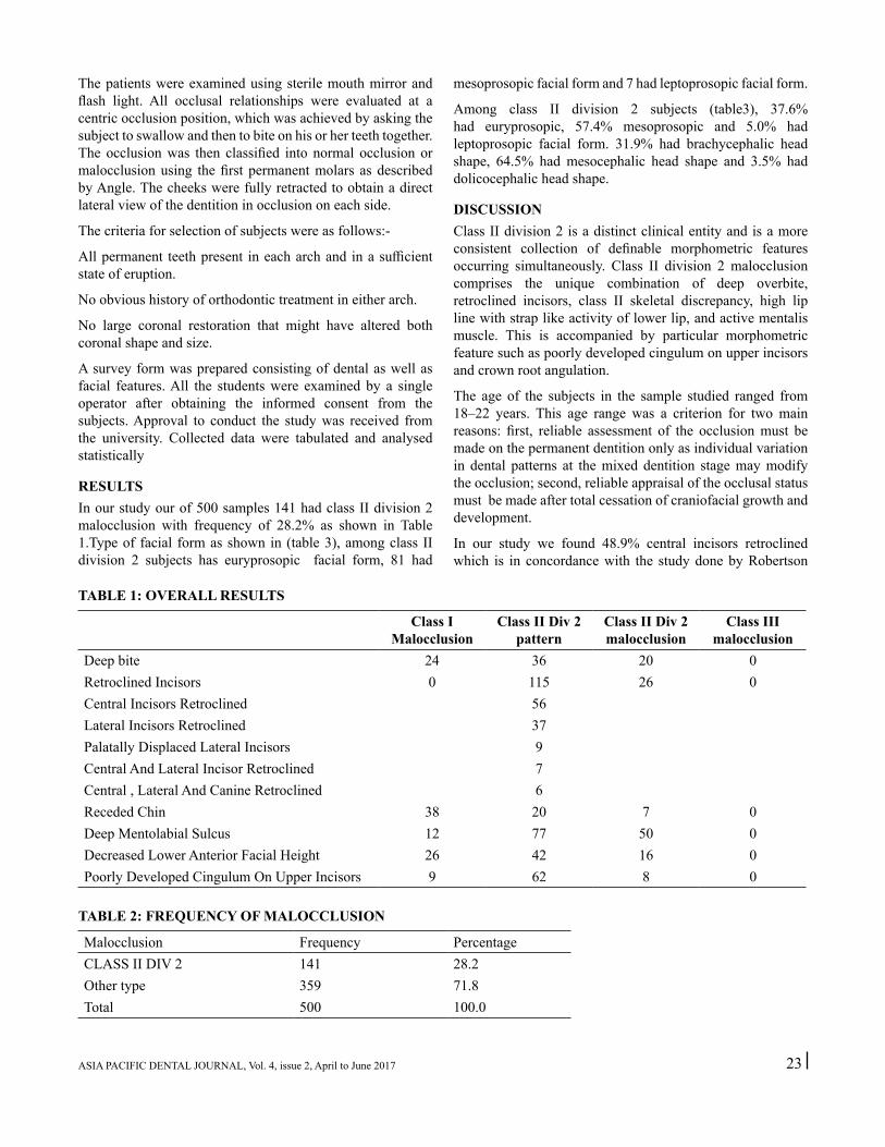
ASIA PACIFIC DENTAL JOURNAL, Vol. 4, issue 2, April to June 2017 23
The patients were examined using sterile mouth mirror and flash light. All occlusal relationships were evaluated at a centric occlusion position, which was achieved by asking the subject to swallow and then to bite on his or her teeth together. The occlusion was then classified into normal occlusion or malocclusion using the first permanent molars as described by Angle. The cheeks were fully retracted to obtain a direct lateral view of the dentition in occlusion on each side.
The criteria for selection of subjects were as follows:-
All permanent teeth present in each arch and in a sufficient state of eruption.
No obvious history of orthodontic treatment in either arch.
No large coronal restoration that might have altered both coronal shape and size.
A survey form was prepared consisting of dental as well as facial features. All the students were examined by a single operator after obtaining the informed consent from the subjects. Approval to conduct the study was received from the university. Collected data were tabulated and analysed statistically
RESULTSIn our study our of 500 samples 141 had class II division 2 malocclusion with frequency of 28.2% as shown in Table 1.Type of facial form as shown in (table 3), among class II division 2 subjects has euryprosopic facial form, 81 had
TABLE 1: OVERALL RESULTS
Class I Malocclusion
Class II Div 2 pattern
Class II Div 2 malocclusion
Class III malocclusion
Deep bite 24 36 20 0Retroclined Incisors 0 115 26 0Central Incisors Retroclined 56Lateral Incisors Retroclined 37Palatally Displaced Lateral Incisors 9Central And Lateral Incisor Retroclined 7Central , Lateral And Canine Retroclined 6Receded Chin 38 20 7 0Deep Mentolabial Sulcus 12 77 50 0Decreased Lower Anterior Facial Height 26 42 16 0Poorly Developed Cingulum On Upper Incisors 9 62 8 0
TABLE 2: FREQUENCY OF MALOCCLUSION
Malocclusion Frequency PercentageCLASS II DIV 2 141 28.2Other type 359 71.8Total 500 100.0
mesoprosopic facial form and 7 had leptoprosopic facial form.
Among class II division 2 subjects (table3), 37.6% had euryprosopic, 57.4% mesoprosopic and 5.0% had leptoprosopic facial form. 31.9% had brachycephalic head shape, 64.5% had mesocephalic head shape and 3.5% had dolicocephalic head shape.
DISCUSSIONClass II division 2 is a distinct clinical entity and is a more consistent collection of definable morphometric features occurring simultaneously. Class II division 2 malocclusion comprises the unique combination of deep overbite, retroclined incisors, class II skeletal discrepancy, high lip line with strap like activity of lower lip, and active mentalis muscle. This is accompanied by particular morphometric feature such as poorly developed cingulum on upper incisors and crown root angulation.
The age of the subjects in the sample studied ranged from 18–22 years. This age range was a criterion for two main reasons: first, reliable assessment of the occlusion must be made on the permanent dentition only as individual variation in dental patterns at the mixed dentition stage may modify the occlusion; second, reliable appraisal of the occlusal status must be made after total cessation of craniofacial growth and development.
In our study we found 48.9% central incisors retroclined which is in concordance with the study done by Robertson

ASIA PACIFIC DENTAL JOURNAL, Vol. 4, issue 2, April to June 201724
and Shwarz et al. Deep overbite was found to be present in maximum subjects which is in concordance with the study done by peck et al, Mills et al and shwarz et al.
Peck et al (1998) described characteristic smaller than average teeth when measured mesiodistally emphasizing similar observation made by Beresford(1969) and by Robertson and Hilton(1965) which found these teeth to be significantly ‘thinner’ in labiolingual dimension. Similarly results were found in our study.
In our study, Class II division 2 has a forwardly rotating mandibular development which contributes to deep bite, chin prominence and reduced lower facial height which is in concordance with the study done by Creekmore TD and Profitt WR.
Deep mentolabial sulcus was found to be maximum among class II division 2 subjects which is supported by Hartsfield JK Jr study in 2011.
Reduced lower facial height has an influence on the position of lower lip relative to upper incisors and an increase in masticatory muscle forces reported by Quinn and Yoshikawa(1985). Ballard(1963), Houston(1975), Mills(1982) and others considered that a high lip line and lip morphology and behaviour were the main aetiological factors in class II division 2 malocclusion which we also found in our study as 110 subjects had high lip line.
We also found other significant features such as poorly developed cingulum on upper incisors, thin incisors in labiolingual dimension and hyperactive mentalis muscle which were not found in any other study.
CONCLUSIONClass II division 2 phenotype varies in the occurrence of its components. Among Class II division 2 traits common in caucasians, in our study we found significant collection of definable morphometric features. Among the Retroclined Incisors, Central Incisors were the most commonly retroclined incisors due to the palatal position of the Crown or forward position of the apices of these teeth. It includes the contribution of active Lip muscular pressure to bimaxillary incisor retroclination and the resting pressure from the lower lip on the maxillary incisors, higher than the pressure exerted by the upper lip, leading to retroclination of the incisors. Increased overbite was reported as the cause was the reduced posterior dentoalveolar height, deep mentolabial sulcus, decreased lower anterior facial height, hyperactive, mentalis muscle and poorly developed cingulum on upper incisors. However traits usually found prevalent in Class II division 2 of Caucasian population were found to have zero incidence in our sample. The prevalence of Class II division 2 however has been reported to be low in Indian Population. Statistically speaking in a low incidence, the dispersion of data over small sample size can be misleading giving true negative and
false positive results like high incidence and low prevalence. Hence for uniform dispersion of such traits with Class II division 2 subjects, a large sample size would elicit more reliable interpretation. We recommend same study should be conducted by expanding sample size.
REFERENCES1. Angle EH. Classificaiton of malocclusion. Dent Cosmos
1988;41:248-64.2. Tang ELK, Stephen HY Wei. Recording and measuring
malocclusion. A review of the literature. Am J Orthod Dentofac Orthop 1993;103:344-51.
3. McCall JO. A study of malocclusion in preschool and school children. Dent Items Interest 1944;131-33.
4. Björk A, Krebs AA, Solow B. A method for epidemiological registration of malocclusion. Acta Odontol Scan 1964;22:27-41.
5. Gravely LD, Johnson DB. Angles’s classification of malocclusion: an assessment of reliability. Br J Orthod 1974;1(3):79-86.
6. Brzroukov V, Freer TJ, Helm S, Kalamkarov H, Sardoinfirri J, Solow B. Basic methods for recording malocclusion traits. Bull World Health Organ 1979;57(6):955-61.
7. Chukwudi ochi onyeaso. Prevalence of malocclusion among adolescents in Ibadan, Nigeria. Am J Orthod Dentofac Orthop 2004, 126;604-7.
8. P.A.Mossey. The Heritability of Malocclusion: part 2. The influence of Genetics in Malocclusion.British Journal of Orthodontics, vol 26;1999-195-203.
9. Joseph Ghafari, Ramzi V. Haddad. Cephalometric and dental analysis of class II division 2 reveals various subtypes of the malocclusion and the primacy of dentoalveolar components. Semin Orthod 2014;20:272-286.
10. RobertsonNRE,HiltonR. Feature of the upper central incisors in ClassII, Division2. AngleOrthod. 1965;35:51–53.
11. Peck S,PeckL,KatajaM. ClassII Division2 malocclusion: a heritable pattern of small teeth in well-developed jaws. Angle Orthod. 1998;68:9–20.
12. Strang RHW.ClassII, Division2 malocclusion. Angle Orthod. 1958;28:210–214.
13. Renfroe EW. A study of the facial patterns associated with Class II , Division 1 and ClassII, Division 2 malocclusions. Angle Orthod. 1948;111:12.
14. CreekmoreTD. Inhibition or stimulation of the vertical growth of the facial complex: its significance to treatment. Angle Orthod. 1967;37:285–297.
15. Proffit WR.On the aetiology of malocclusion. Br JOrthod. 1986;13:1–11.
16. Lapatki BG,KlattA,Schulte-MontingJ,JonasIE. Dentofacial parameters explaining variability in retroclination of the maxillary central incisors. J OrofacOrthop. 2007;68:109–123.
17. Lapatki BG, Mager AS, Schulte-Moenting J, Jonas IE. The importance of the level of the lip line and resting lip pressure in ClassII, Division2 malocclusion. J DentRes. 2002;81:323–328. 36.

ASIA PACIFIC DENTAL JOURNAL, Vol. 4, issue 2, April to June 2017 25
18. Hitchcock HP.The cephalometric distinction of ClassII, Division 2 malocclusion. Am JOrthod. 1976;69:447–454.
19. Fletcher GGT, Ortho D. The retroclined upper incisor. Br J Orthod. 1975;2:207–216.
20. Maj G, Lucchese P. The mandible in ClassII, Division 2. Angle Orthod. 1982;52:288–292.
Prof. Dr. Amish Mehta
Dr. Tanu Srivastava B.D.S. (3nd year Resident)*Post Graduate Student,Department of Orthodontics and Dentofacial Orthopaedics,Faculty of Dental Sciences, Dharamsinh Desai University, Nadiad. E-mail: [email protected]

ASIA PACIFIC DENTAL JOURNAL, Vol. 4, issue 2, April to June 201726
ABSTRACTAims and Objectives:To analyze and evaluate the aesthetic appeal of the given bimaxillary silhouettes on a visual analogue scale among three groups of raters.Materials and Methods:This photographic study consisted of a right profile photograph of a patient with acceptable bidental protrusion which was then manipulated for varying lip pouts using the Dolphin Imaging Software. Digital cephalometric tracing was done which was later superimposed onto the right profile photograph of the selected subject and saved. In totality, 14 such digitally altered computer generated images were generated and converted into silhouettes. 401 participants with different educational backgrounds were asked to rate the silhouettes based on their aesthetic appeal on a 7-point VAS (visual analogue scale). Correlations between the perceptions of the above mentioned three groups were evaluated.Results:As the protrusiveness of the profiles was enhanced in terms of lip pout, their appeal diminished.The upper lip is considered to be the most contributing feature to one’s facial attractiveness, leaving ‘nose’ on the far end of the spectrum. As far as the profile perception was concerned, a flat or straight or otherwise orthognathic profile is most sought after amongst the Gujarati population at present.As the category changes, perceptions also change significantly (p<0.001).Laymen have proved to be less discerning of minor variations in the silhouettes compared to orthodontists and general dentists.Conclusion:The public’s evaluation of a person’s profile is very similar to, but less discriminating than, the perceptions of the two specialty groups.KEYWORDS: Bimaxillary protrusion; Facial Aesthetics; Silhouettes
DIFFERENCE IN THE PERCEPTION OF BIDENTOALVEOLAR PROTRUSION LIP PROFILE AND POUT AMONGST LAYMEN, DENTAL
SURGEONS AND ORTHODONTISTSProf. Dr. Amish Mehta, Dr. Lavina Jagtiani
INTRODUCTIONPerception of beauty, per say, is a subjective feeling.1 In 1878, Margaret Wolfe Hungerford published a simple but insightful phrase in her novel ‘Molly Bawn’ that was to be quoted so often that it has almost become cliché: “beauty is in the eyes of the beholder”. 2The 18th century philosopher Alexander Baumgarten, who established esthetics as a distinct field of philosophy, coined the term, which is derived from the Greek word for sensory perception (aisthesis).3 The philosopher Pascal commented, “Had Cleopatra’s nose been shorter, the whole face of the world would have changed!”4
of beauty by another. Aesthetic criteria appear to have been defined in almost all cultures, but several findings suggest that the perception of beauty may be innate and, additionally, universal or crosscultural.6-7
Donald H.Enlow and Mark G.Hans reported that no two faces are the same, not even those of identical twins. Every individual face is unique, exquisite and custom made original.
Our minds, right from early infancy, have been programmed so as to seek out specific prototypes, which trigger strong perceptual responses and are thus appealing.Prototypes, such as averages, are recognized faster and therefore create higher
nervous excitation. Accordingly, Langlois and Roggman (1990) and Langlois et al. (1994) proposed that averageness is attractive (a face is perceived as attractive when its facial gestalt (form) is close to the average or mean of a population of faces).8-9
Within each race and sex there appears to be a balance of facial features that are viewed by the majority as being ‘pleasing to the eye’.7Robert Ricketts, in one of his studies in 1982,attributed the perception of beauty to the ‘Golden Proportion’, that gives each entity its exquisiteness. As per this, the ratio of the smaller section to the larger section is the same as that of the larger section to the whole. Mathematically, it is expressed as 1.618:1 or 1:0.618. This study strongly suggested that the perception of beauty can be made scientific rather than the need to resort to subjective interpretations.
People seem to share a common basis for aesthetic judgment regardless of nationality, age, sex, or occupation .10-11 Audio-visual, digital and print media, all influence our idea of facial stereotypes on a daily basis.12-13
With the above background, this study was undertaken as a pilot study to evaluate the perception of beauty of the people of Central Gujarat. Differences have been found in judging facial

ASIA PACIFIC DENTAL JOURNAL, Vol. 4, issue 2, April to June 2017 27
attractiveness between orthodontists and non orthodontists in earlier studies(Knight and Keith 2005; Maple et al.,2005).14-
17In order to bridge this gap and help orthodontists and other dental health care professionals provide a successful, satisfactory aesthetic treatment outcome, we have attempted this study,putting the following null hypothesis to test:
There is no difference between the perception of beauty of the following group of people: - (a) orthodontists, (b) laymen and (c) general dentists.
The aim of this study is to analyze and evaluate the aesthetic appeal of the given bimaxillary silhouettes on a visual analogue scale among the above mentioned three groups.
Enhancement of one’s aesthetic appeal is one of the key reasons for patients seeking orthodontic treatment.18-22 Hence it is of utmost importance that there be a sense of congruency between the aesthetic sensibilities of the general public, the referring dental surgeon and the orthodontist.
Bimaxillary protrusion is a condition characterized by proclined upper and lower incisors and an increased procumbency of the lips.23 The position of the lips, as determined by maxillomandibular protrusion or retrusion, by dental protrusion or retrusion and/or by lip thickness have a significant influence on facial attractiveness perception.
Judging facial esthetics via photographs on visual analog scale (VAS) has been demonstrated to be a valid and reliable method in the past.24-
26 However, in this study, in order to eliminate the bias arising from factors like shape, colour, distance from other structures etc, silhouettes
have been used. The term “silhouette” has been extended to describe the sight or representation of a person, object or scene that is backlit, and appears dark against a lighter background.
This study intends to evaluate and correlate the perceptions of the above mentioned three groups (i.e., orthodontists, dental surgeons, laymen) which will in the long run enable us to plan
and render better oral health care and oral health care policies related to enhancement of quality of life.
MATERIALS AND METHODSThis study employed the photograph of a patient with bimaxillary protrusion who reported to the department of Orthodontics and Dentofacial Orthopaedics, Faculty of Dental Science, D.D.U, Nadiad, with the chief complain of forwardly placed upper and lower front teeth.
Four such patients with bidental protrusion were selected, out of which the most appropriate and acceptable bidental protrusion was sought out by Peer Assessment Rating method, by the post graduates and intern students posted at that time in the dept. of Orthodontics and Dentofacial Orthopaedics. Right profile photograph of the above selected patient was then manipulated for varying lip pouts using the Dolphin Imaging Software. Digital cephalometric tracing was done which was later superimposed onto the right profile photograph of the selected subject and saved. 14 such digitally altered computer generated images were generated of which, 13 were manipulated with 1 mm of protrusion or retrusion at a time, and 1 was the original bidentoalveolar protrusive image of the selected subject.
These 14 images were then converted into silhouettes to eliminate any degree of bias, using the Picasa software. Due consent was taken from the patient for the same.
The following bimaxillary image of the patient, selected for the study, was manipulated using the Dolphin Software, which was later transformed into silhouettes via Picassa.
RESULTSIn toto, 402 raters had participated in the study and were asked to fill up the questionnaires.
Of the 402 responses, one answer sheet was incomplete leaving us with 401 total feedbacks.45.4% of the participants were dental surgeons, 42.4% laymen and 12.2 % were
Table 2. Feature contributing Most to facial attractiveness
Feature Contributing Most to facial attractiveness TotalNose Upper Lip Lower Lip Chin
Category Dental Surgeon 24 122 9 27 18213.2% 67.0% 4.9% 14.8% 100.0%
Laymen 39 102 0 29 17022.9% 60.0% .0% 17.1% 100.0%
Orthodontists 1 42 0 6 492.0% 85.7% .0% 12.2% 100.0%
Total 64 266 9 62 40116.0% 66.3% 2.2% 15.5% 100.0%
Value Df Exact p-valuePearson Chi-Square 27.317 6 <0.001

ASIA PACIFIC DENTAL JOURNAL, Vol. 4, issue 2, April to June 201728
orthodontists.
When asked which feature contributed most to facial attractiveness, 67% dental surgeons opted for the upper lip. 60% laymen also voted for the same feature which was again in agreement with 85.7% of orthodontists.We used the Pearson Chi-Square test to assess the significance of this result which turned out to be significant. i.e., p <0.001.
Next the raters were asked to point out which feature contributed least to the overall facial attractiveness. Here there was agreement between the views of the dental surgeons and the orthodontists. 36.8% dental surgeons and 73.5% orthodontists rated nose to be the least appealing feature while 45.9% laymen opted for the chin as the least contributing feature.
However, overall, 148 out of 401 raters (36.9%), voted for nose to be the least contributing feature to facial attractiveness. The Pearson Chi-Square test showed p value <0.001
Among the factors contributing to facial attractiveness, UPPER LIP was rated as the most contributing feature (66.3%), whereas NOSE was rated as the feature contributing least to facial attractiveness (36.9%). However, when the perception of each individual group was assessed, laymen rated chin as the least contributing feature to facial attractiveness (45.9%).
There was unanimity in the perception of all these groups as far as the most contributing feature to facial attractiveness was concerned.While tabulating the results of each individual silhouette, the following conclusion was achieved:-
As the protrusiveness of the profiles was enhanced in terms of lip pout, their appeal diminished.
This fact was confirmed by comparing the means for each silhouette and also the frequency table charted out for the same.
Table 3. Feature contributing Least to facial attractiveness
Feature contributing Least to facial attractiveness TotalNose Upper Lip Lower Lip Chin
Category Dental Surgeon 67 40 50 25 18236.8% 22.0% 27.5% 13.7% 100.0%
Laymen 45 6 41 78 17026.5% 3.5% 24.1% 45.9% 100.0%
Orthodontists 36 0 13 0 4973.5% .0% 26.5% .0% 100.0%
Total36.9%
148 46 104 103 40111.5% 25.9% 25.7% 100.0%
Value df p-valuePearson Chi-Square 1.053E2 6 <0.001
The visual analogue scale ratings for all the 14 images used in the study.
Table 23. Factor Analysis for Laymen
KMO and Bartlett’s TestKaiser-Meyer-Olkin Measure of Sampling Adequacy.
.647
Bartlett’s Test of Sphericity
Approx. Chi-Square 2.322E3Df 91Sig. .000
Rotated Component MatrixComponent
1 2 3Q.10(a) .638 .407 -.170Q.10(b) .897 -.161Q.10(c) .872 .164Q.10(d) .807 .259Q.10(e) .825 .433 .118Q.10(f) .570 .713Q.10(g) .171 .901 -.161Q.10(h) .157 .914Q.10(i) .275 .726 .173Q.10(j) .627 .435Q.10(k) .153 .366 .824Q.10(l) .900Q.10(m) -.224 .752Q.10(n) -.270 .124 .772Extraction Method: Principal Component Analysis. Rotation Method: Varimax with Kaiser Normalization.

ASIA PACIFIC DENTAL JOURNAL, Vol. 4, issue 2, April to June 2017 29
As per Laymen, given fourteen images can be divided in three groups as follows :
Group-1 :a,b,c,d,eGroup-2 :f,g,h,i,jGroup-3 :k,l,m,n
This pattern of grouping exhibited by the laymen shows their inability to detect minor alterations made in the profiles. Variations more than 3mm were required for the laymen to detect a change in the silhouettes.
Table 24. Factor Analysis for Orthodontists
Rotated Component MatrixComponent
1 2 3Q.10(a) -.555 .745 .334Q.10(b) .689 .388 .593Q.10(c) .979Q.10(d) -.227Q.10(e) .908 .377 .154Q.10(f) .637 .711 .293Q.10(g) .372 .914 .135Q.10(h) .372 .914 .135Q.10(i) .973Q.10(j) .942 -.148 -.261Q.10(k) .863 .375 .275Q.10(l) -.787 -.605Q.10(m) -.928 -.322 .142Q.10(n) -.114 -.966 -.209Extraction Method: Principal Component Analysis. Rotation Method: Varimax with Kaiser Normalization.
As per Orthodontists, given fourteen images can be divided in three groups as follows :Group-1 :b,e,i,j,k,mGroup-2 :a,f,g,h,l,nGroup-3 : c
The images grouped together in group-1 were slightly retrusive in nature. Group-2 images depicted a straight or acceptable orthognathic profile whereas group-3 image depicted a convex or highly protrusive profile. The nature of this grouping pattern into straight, protrusive and retrusive types clearly is in concurrence with the educational background, skill and training of this group of raters. Given the fact that orthodontics is a specialty dealing with the ability to alter and enhance the facial profiles, it was only natural that this group of highly skilled dentists could easily demarcate between the different profiles and group them together.
DISCUSSIONOne of the chief reasons for patients seeking orthodontic treatment today is the enhancement of their facial and smile
aesthetics. This makes it all the more important for there to be a consensus in the perception of beauty and aesthetics between the patient and the oral health care professional. The following study compares the perception of three different groups of raters namely: laymen, dental surgeons and orthodontists.
Patient’s hope for results as determined by social and cultural rules of beauty in their reference groups and in the society in general. On the other hand, orthodontists prefer to use parameters and norms set on the basis of averageness, to determine diagnosis and subsequent treatment planning. Our patients, however, influenced by images portrayed in the media, may request treatment, which does not comply with the average. Cephalometric norms/averages are not always available to define beauty. An example of this is seen in the Sculpture of Queen Nefertiti (1370-1330 BC), which dates back to the dawn of Egyptian civilization, a time when the Nile valley swamps were settled by people from Africa and Asia. Nefertiti has been extolled as a standard of beauty throughout the history of man,not surprising, as her features represented the average of the population in which she lived, of Asian and African origin. However, we would struggle to find a set of cephalometric norms to accurately represent her features.27
This study employs 14 images which have been manipulated from a single bidentoalveolar protrusion image to correlate the perception of the above mentioned three groups. Manipulating the same image allowed us to pair truly comparable images rather than searching for naturally occurring variations and having to accept compromises and inviting selection bias.
All 14 images were converted into silhouettes using the Picassa Software, which eliminated all extrinsic and intrinsic distracting variables (such as hairstyle, make-up, skin complexion) that could influence the evaluators aesthetic score rating.27-28 Apart from rating the 14 images, the raters were also asked two additional questions regarding the most and least attractive facial feature according to them. This exercise allowed us to gain an additional insight into the aesthetic psyche of the raters.
Upper lip was rated as the most attractive feature by all three groups unanimously which was in concordance with previous studies. In the past several similar attempts have been made to evaluate the perception of beauty wherein the Caucasian sensibilities were analyzed. In this study, we have employed an Indian subject being rated by Indian raters (Gujarati population in particular).
In recent times, a preference for a fuller, more anteriorly positioned lips and an acute nasolabial angle has been demonstrated, i.e., greater profile convexity and lip prominence. However, the results of this study are more in favor of a straight or orthognathic profile which is in concordance with earlier studies.29-37 As the protrusiveness of the profiles was enhanced in terms of lip pout, their appeal

ASIA PACIFIC DENTAL JOURNAL, Vol. 4, issue 2, April to June 201730
diminished. Andrew Hockley and co-workers, in one of their studies, concluded that using silhouettes for evaluation of patient aesthetics could influence clinicians or researchers to select profiles that are flatter than the established esthetic norm. (Am J Orthod Dentofacial Orthop 2012; 141:161-8)38
The original image selected for the study was quite protrusive in terms of lip pout. Even after 10mm of computerized retrusion, the silhouette which was generated was more or less orthognathic. So, none of the fourteen images which were used in the study could actually be termed retrusive. This was the drawback of this study which could have had a bearing on the results we achieved.
A positive correlation is found between each category of raters and their respective perception. As the category changes, perceptions also change significantly, which is supported and demonstrated by the significant Pearson Chi-Square results. (p<0.001) Atleast two out of three groups have significant variations in their perception of an aesthetic profile, which renders the null hypothesis void and rejected.
From the results achieved, laymen have proved to be less discerning of minor variations in the silhouettes which is in agreement with earlier studies.39 On the other hand orthodontists and general dentists have proved to be more perceptive in detecting minor degrees of variation from ideal. Owing to their educational qualification, dentists and orthodontists are in a much better position to pinpoint minor variations made in the silhouettes. Whereas, for the laymen to detect any change atleast 3mm of variation was needed in the profiles. These results are consistent with the lower correlation between lay person’s and dental specialist’s ratings than between the two specialist’s ratings. This suggests that the public’s evaluation of a person’s profile is very similar to, but less discriminating than, the perceptions of the two specialty groups. (Bell et al. Am J Orthod.1985. 88: 323-332.)40
This study, thus, highlights the threshold at which an aberration becomes noticeable by the laymen at large, in terms of lip pout. It renders the orthodontists an insight into the perception of beauty of the local Gujarati population, making them more efficient in planning and rendering a treatment plan which is in tandem with the aesthetic sensibilities of the so called laymen group.
CONCLUSIONThe concept of beauty is highly non-static, influenced by the most socially dominant group at that time. Hence, we as dental health care professionals and specialists, constantly and dynamically need to keep ourselves updated with this ever changing definition of facial beauty to best achieve the most aesthetic and satisfactory treatment results.
Orthodontists consider improvement of facial aesthetics as an important treatment goal, and therefore, it is an important issue in their decision making process and their treatment plans (Bowman and Johnston,2001; Ackerman, 2004).The opinion
of the general public, the end-users of orthodontic services, may have the most value in determining the appropriateness of aesthetic results (Bowman and Johnston,2001).45
This study concludes that the upper lip is considered to be the most contributing feature to one’s facial attractiveness, leaving ‘nose’ on the far end of the spectrum. As far as the profile perception was concerned, a flat or straight or otherwise orthognathic profile is most sought after amongst the Gujarati population at present.
Having concluded the above, awareness that previously held concepts of facial beauty and aesthetics may no longer apply, must constantly be forebearing on our minds. Only then will it enable us to render treatment outcomes which are in sync with the psyche of the general population.
This study provides significant insights into the aesthetic results sought after by the Gujarati population. However, further studies should periodically be conducted to keep up with the changing trends in aesthetics.
REFERENCES1. Lucker GW. Esthetics and a quantitative analysis of facial
appearance. In: Lucker GW, Ribbe KA, McNamara JA Jr, Eds.Psychological Aspects of Facial Form. Monograph 11,Craniofacial Growth Series. Ann Arbor, Mich: The Center for Human Growth and Development, The University of Michigan; 1980:49–79.
2. NainiF,MossJ,GillD.The enigma of facial beauty: Esthetics,proportions, deformity, and controversy.Am J Orthod Dentofacial Orthop. 2006;130:277-82.
3.Baumgarten AG. Aesthetica. Paris: L’herne; 1989.4. Pascal B. Pensees. London: Penguin Classics; 1966.5. Marlowe C. Doctor Faustus. New York: Dover Publications;1995.6. Langlois J L, Roggman L A, Musselman L. What is average
and what is not average about attractive faces? Psychological Science. 1994; 5: 214–220.
7. Bravo L A. Soft tissue facial profile changes after orthodontic treatment with four premolars extracted. Angle Orthodontist. 1994; 64: 31-42.
8. Cons N C, Jenny J. Comparing perceptions of dental aesthetics in the USA with those in eleven ethnic groups.International Dental Journal. 1994; 44: 489-494.
9. De Smit A, Dermaut L. Soft tissue profile preference. Am J Orthod. 1984; 86: 67-73.
10. Child I L. Iwao S. Personality and aesthetic sensitivity: extension of findings to younger ages and to different cultures. Journal of Personality and Social Psychology. 1968; 8:308-312.
11. Ford C S, Prothro E T, Child I L. Some trans-cultural comparisons of aesthetic judgment. Journal of Personality and Social Psychology. 1966; 68: 19-26.
12. Leonardi B, Annunziata A, Licciardello V, Barbato E. Soft tissue changes following the extraction of premolars in non growing patients with bimaxillary protrusion. A systematic review. Angle Orthod. 2010;80:211–216.
13. Kiekens RM, Kuijpers-Jagtman AM, van’t Hof MA, van’t Hof

ASIA PACIFIC DENTAL JOURNAL, Vol. 4, issue 2, April to June 2017 31
BE, Straatman H, Maltha JC. Facial esthetics in adolescents and its relationship to ‘‘ideal’’ ratios and angles. Am J Orthod Dentofacial Orthop. 2008;133:188–198.
14. Prahl-Anderson B, Boersma H. Perception of dentofacial morphology by layperson, general dentist, and orthodontist. J Am Dent Assoc. 1979;98:209-12.
15. Kerr WJS, O’Donnell JM. A panel perception of facial attractiveness.Br J Orthod 1990;17:299-304.
16. Bell R, Kiyak HA, Joondeph DR, McNeil WR, Wallen TR. Perceptions of facial profile and their influence on the decision to undergo orthognathic surgery. Am J Orthod. 1985;88:323-32.
17. Foster EJ. Profile preference among diversified groups. Am J Orthod. 1973;63:34-40.
18. Cox NH, van der Linden FP. Facial harmony. Am J Orthod. 1971;60:175-83.
19. Foster EJ. Profile preferences among diversified groups. Angle Orthod. 1973;43:34-40.
20. Romani KL, Agahi F, Nanda R, Zernik JH. Evaluation of horizontal and vertical differences in facial profiles by orthodontists and lay people. Angle Orthod. 1993;63:175-82.
21. Baldwin DC. Appearance and aesthetics in oral health. Community Dent Oral Epidemiol. 1980;9:244-56.
22. Kilpelanien P, Phillips C, Tulloch JFC. Anterior tooth position and motivation for early treatment. Angle Orthod. 1993;63:1714.
23. Nguyen D D, Turley P K.Changes in the Caucasian male facial profile as depicted in fashion magazines during the twentieth century. Am J Orthod and Dentofacial Orthop.1998;114:208–217.
24. Kiekens R M A, Maltha J C, van ́ t Hof M A, Kuijpers-Jagtman A M. Objective measures as indicators for facial esthetics in white adolescents. Angle Orthodontist. 2006; 76: 551–556.
25. Scott C R, Goonewardene M S, Murray K.Influence of lips on the perception of malocclusion. Am J Orthod and Dentofacial Orthop.2006; 130: 152–162.
26. McNamara L, McNamara J A Jr, Ackerman M B, BaccettiT.Hard- and soft-tissue contributions to the esthetics of the posed smile in growing patients seeking orthodontic treatment. Am J Orthod and Dentofacial Orthop.2008;133: 491–499.
27. Margaret Collins. The Attractiveness of the Average Face. SeminOrthod. 2012;18:217-228.
28. Foster FJ. Profile preferences among diversified groups. Angle Orthod. 1973;43:34-40.
29. Cox NH, Van der Linden FPGM. Facial harmony. Am J Orthod and Dentofacial Orthop.1971;60:175.
30. Foster El. Profile preferences among diversified groups. Angle Orthod 1973;43:34.
31. Riedel RA. An analysis of dentofacial relationships. Am J Orthod 1957;43:103-19.
32. Kerr WJS, O’Donnell JM. Panel perception of facial attractiveness.Br J Orthod 1990;17:299-304.
33. DeSmit A, Dermaut L. Soft-tissue profile preference. Am J Orthod. 1984;86:67-73.
34. Raj M. The perception of facial profile attractiveness by providers and consumers [thesis]. Columbus: Ohio State University; 2002.
35. Phillips C, Griffin T, Bennett E. Perception of facial attractiveness by patients, peers, and professionals. Int J Adult OrthodOrthognath Surg. 1995;10:127-35.
36. Burstone CJ. The integumental contour and extension patterns.AngleOrthod 1958;29:93-104.
37. Kast GR. Perception of facial profile esthetics by providers and consumers with incremental alterations to the mandible [thesis].Columbus: Ohio State University; 1997.
38. Hockley A,Weinstein M, Borislow A.J, BraitmanL.E.Photos vs silhouettes for evaluation of African American profile esthetics. Am J Orthod Dentofacial Orthop.2012;141:161-8.
39. Lines PA. Lines RR, Lines CA: Profilemetrics and facial esthetics. Am J Orthod and Dentofacial Orthop.1978;73:648-657.
40. Bell et al. Perceptions of facial profile and their influence on the decision to undergo orthognathic surgery .Am J Orthod.1985;88: 323-332.
41. Margaret Collins. The Eye of the Beholder: Face Recognition and Perception. SeminOrthod. 2012;18:229-234.
42. Peck S, Peck L. Selected aspects of the art and science of facial esthetics. SeminOrthod. 1995; 1: 105-126.
43. Sutter RE, Turley PK: Soft tissue evaluation of contemporary Caucasian and African American female facial profiles. Angle Orthod. 1998; 68:487-496.
44. Farkas LG, Hreczko TA, Kolar JC, et al. Vertical and horizontal proportions of the face in young adult North American Caucasians: Revision of neoclassical canons. PlastReconstr Surg. 1985; 75:328-337.
45. Gochman DS: The measurement and development of dentally relevant motives. J Public Health Dent. 1975; 35:160-164.
Prof. Dr. Amish Mehta
Dr. Lavina Jagtiani, B.D.S. (3nd year resident)*Post Graduate Student,Department of Orthodontics and Dentofacial Orthopaedics,Faculty of Dental Sciences, Dharamsinh Desai University, Nadiad. E-mail: [email protected]

ASIA PACIFIC DENTAL JOURNAL, Vol. 4, issue 2, April to June 2017
About the JournalIt is the official Publication of Asia Pacific Dental Federation (heretofore referred to as APDF): Asia Pacific Regional Organization of the FDI World Dental Federation. In each Issue, its readers have access to original Peer-reviewed Articles that examine all phases
of Dental Health and Treatment, well Illustrated, including Tables, Photos and statistical data. Coverage also includes successful diagnostic procedures, Imaging Techniques, Dental materials, Endodontics, Periodontics, Conservative and Operative dentistry, Paedodontics and Child Dental Care, Oral Medicine and Radiology, Orthodontics, Prosthodontics and Crown and bridge, Extraction and Impaction concerns, Maxillo-Facial Surgery, TMJ disorders, and Geriatric Dentistry, and any other related Dental Subjects.OBJECTIVESCollect, Compile and edit, Articles for Publication, of Dental and related fields in the form of Original Articles, Case Reports, Literature Review, Viewpoints, Essays and any other established Form in Print as well Online (Soft Copy) format for the Education of the Medical and Dental personnel and Specialists including general Medical and Dental Practitioners and Paramedical & technical Staff of hospital, clinic, or a diagnostic lab as well as for generating and spreading Awareness of Dental Field in the Asia Pacific Region and Worldwide.
EDITOR’S OFFICEDr. Bhagwant Singh
Gurudwara Shaheedan Road, Model Town, Ludhiana, Punjab-141002
Phone: +91 981 424 5608, +91 161 501 5588Email: [email protected] ;
GUIDELINES FOR AUTHORSManuscripts must be prepared in accordance with “Uniform requirements for Manuscripts submitted to Biomedical Journal” developed by International Committee of Medical Journal Editors.Original articles: Up to 3000 words excluding references and abstract.Case reports: Up to 1000 words excluding references and abstract and up to 10 references. PRESENTATION AND FORMAT Double Spacing Margins 1.5 cm from all four sides Uniformly American or U K English (But not mixture
of both) Abbreviations spelt out in full for the first time.
TITLE PAGE:1. Type of manuscript (e.g. Original article, Research,
Systemized Review, Case report)
2. The title of the article, in full, (Concise and Informative); 3. Running title or short title not more than 50 characters; 4. The name by which each contributor is known with
his or her highest academic degree(s) and institutional affiliation;
5. The name of the department(s) and institution(s) to which the work should be attributed;
6. The name, address, phone numbers, facsimile numbers and e-mail address of the contributor responsible for correspondence about the manuscript;
7. The total number of pages, total number of photographs and word counts separately for abstract and for the text (excluding the references and abstract);
8. Source(s) of support in the form grants, equipment, drugs, or all of these;
9. Acknowledgment, if any; and 10. If the manuscript was presented as part at a meeting, the
organization place and exact date on which it was read.
ABSTRACT PAGE:The second page should carry the full title of the manuscript and an abstract (of no more than 150 words for case reports, brief reports and 250 words for original articles). The abstract should be structured and state the Context, Aims, Methods and Materials, Statistical Analysis used, results and Conclusions. Below the abstract should provide 3 to10 keywords.
MAIN MANUSCRIPT:INTRODUCTION:Brief write the purpose of the article, summarize the rationale, the study or observation.
METHODS:The methods section should include only information that was available at the time the plan or protocol for the study was written; all information obtained during the conduct of the study belongs in the Result section. When reporting experiments on human subjects, state whether the procedures followed were in accordance with the ethical standards of the responsible committee on human experimentation (institutional or regional), and with the Helsinki Declaration of 1975, as revised in 2000. State clearly whether Ethical Clearance has been obtained or not.
RESULTS:Describe and present the results in sequence in the Text, Tables, and Illustrations, with the main and important findings preceding the rest.
DISCUSSION:Summarize the key findings (Primary Outcome Measures, Secondary Outcome Measures, results as they relate to a prior hypothesis); Discuss the Strengths and limitations (design, data Assimilation, analytic methods and the analysis);

ASIA PACIFIC DENTAL JOURNAL, Vol. 4, issue 2, April to June 2017
SUBSCRIPTION INFORMATIONAnnual Subscription (for 2014-15)
Individual ` 800 (National) $ 50 (International)
Institutional ` 1000 (National) $ 100 (International)
Subscription can be sent to EDITOR’S OFFICEDr. Bhagwant SinghGurudwara Shaheedan Road, Model Town, Ludhiana, Punjab-141002Phone: +91 981 424 5608, +91 161 501 5588Email: [email protected] : [email protected]
Interpretation and implications in the context of the totality of the evidence (Is there a systemic review to refer to, if not, could one be reasonably done here and now?, How does your findings add to the available evidence, effects on patient care and health policy, possible new mechanisms); Controversies or Conflict of opinion, if any raised by this study; and Future research direction (for this particular research collaboration, underlying mechanisms, clinical research). Avoid repetitions. REFERENCES:Write References ONLY in Vancouver Style. References are to be numbered consequently in the order in which they are first mentioned in the text (not in alphabetic order). Identify references in text, tables, and legends by Arabic numerals in square brackets (e.g. [10]). TABLES, FIGURES, PHOTOS, ETC:Tables: self-explanatory and not duplicate textual material.Number Tables, Figures, and Photos in Arabic numerals, consequently in the order of their first citation in text and supply a brief title/ legend for each.
THE APDF RECOMMENDS THAT ALL RESEARCH, REVIEWS, CASE REPORTS SHOULD FOLLOW THE CONSORT and PRISMA Guidelines. All Consent s wherever required (including those for the Photographs of Patients or reproduction of Clinical Data of other Individuals) should be provided.
COPYRIGHT FORM (Sample) (Can be modified to suit Individual needs)
TITLE OF THE ARTICLEWe certify that we have participated sufficiently in the intellectual content, conception and design of this work or the analysis and interpretation of the data (when applicable), as well as writing of the manuscript, to take public responsibility for it and have agreed to have our name as contributor. We believe the manuscript represents valid work. Neither this manuscript nor one with substantially similar content under our authorship has been published or is being considered for publication elsewhere, except as described in the covering letter. We certify that all the data collected during the study is presented in this manuscript and no data from the study has been or will be published separately. We attest that, if requested by the Editors, we will provide data/information or will cooperate fully in obtaining and providing the data/information on which the manuscript is based, for examination by the Editors and their assignees. Financial interests, direct or indirect, that exist or may be perceived to exit for individual contributors in connection with the content of this paper have been disclosed in the cover letter. Sources of outside support of the project are named in the cover letter.
We hereby transfer(s), assign(s), or otherwise convey(s) all copyright ownership, including any and all rights incidental thereto, exclusively to the Editor and the APDF in the event that such work is published by APDF. The Editor and the APDF shall own the work, including 1) copyright; 2) the right to grant permission to republish the article in whole or in part, with or without fee; 3) the right to produce preprints or reprints and translate into languages other than English for sale or free distribution; and 4) the right to republish the work in a collection of articles in any other mechanical or electronic format.
We give the rights to the corresponding author to make necessary changes as per the request of the journal, do the rest of the correspondence on our behalf and he/she will act as the guarantor for the manuscript on our behalf.
All persons who have made substantial contributions to the work reported in the manuscript, but who are not contributors, are named in Acknowledgment and have given me/us their written permission to be named. If we do not include an Acknowledgment that means we have not received substantial contributions from non-contributors and no contributor has been omitted.
Name Signature Date
1. ________________ _____________ __________
2. ________________ _____________ __________
3. ________________ _____________ __________ Editor and APDF reserve the Right to Alter/Change the Guidelines as per need and time.
Copyright © 2022 FDOKUMEN