Quaternary strata in CRP-1, Cape Roberts Project, Antarctica
ACTA PALAEONTOLOGICA ROMANIAE (2014) V. 10 (1-2), P. 47-60
________________________________
1 Université de Bretagne Occidentale (UBO), UMR 6538 Domaine Océanique, 6, avenue Le Gorgeu - CS 93837, 29238 Brest Cedex 3 (France);
[email protected] 2 Department of Ecology and Evolutionary Biology, The University of Kansas, 1200 Sunnyside Avenue, Lawrence, Kansas 66045 (USA); 47
[email protected] 3 Lebanese University, Faculty of Science II, Fanar, Natural Sciences Department, Fanar - El-Matn, PO box 26110217 (Lebanon); [email protected]
FIRST RECORD OF HARLANJOHNSONELLA ANNULATA ELLIOTT IN GRANIER &
DELOFFRE, 1993, NON 1968, A TRIPLOPORELLACEAN ALGA IN UPPER
BARREMIAN-LOWERMOST BEDOULIAN STRATA OF LEBANON
Sibelle Maksoud1, Bruno Granier2 & Dany Azar3
Received: 04 April 2014 / Accepted: 13 September 2014 / Published online: 13 October 2014
Abstract Harlanjohnsonella annulata Elliott in Granier & Deloffre, 1993, non 1968, was originally described from
Cenomanian strata in Serbia. Herein, we report its first occurrence outside the type area, i.e., in Lebanon, and in older
strata, i.e., earliest Bedoulian or Late Barremian in age. Our specimens are compared with topotypic material. The
species was recently revised by Radoičić & Schlagintweit who transferred it to the genus Dissocladella. However, we
never observed any secondary branch and therefore we cannot agree with their interpretation. In addition on the basis
of calculations and comparisons of the volume ratio of several species, either fossil (Montiella elitzae and
Triploporella steinmannii) and living (Dasycladus vermicularis and Neomeris dumetosa) Dasycladalean algae, we
assume that Harlanjohnsonella annulata was more probably an endosporate form rather than a cladosporate form.
Keywords: Anti-Lebanon, Cretaceous, Barremian, Bedoulian, Jezzinian regional stage, Falaise de Blanche,
Dasycladales, Triploporellacean alga.
INTRODUCTION
It has been almost half a century since Elliott (1968)
described his Harlanjohnsonella annulata from Upper
Cretaceous (“possibly Cenomanian”) strata of Zlatibor
(Serbia, former Yugoslavia). There has been no further
record since then outside the type region, except for
Jaffrezo et al. (1980) who mention a “Harlanjohnsonella
cf. annulata” from Aptian strata of Bey Daglari (Turkey).
Recently, Radoičić (1995) and Radoičić and
Schlagintweit (2010) reexamined some topotypic
material, i.e, material originating from Cenomanian strata
of the type region in western Serbia. In the later
publication (Radoičić & Schlagintweit, 2010), they
proposed to transfer the species to the genus
Dissocladella, a proposal that we do not support.
Our paper documents the first occurrence of
Harlanjohnsonella annulata in older strata, i.e., at the
transition of the Barremian to the Bedoulian stage in
Lebanon. We describe this Lebanese material, compare it
to topotypic material (which consists on nine thin
sections of the Collection Deloffre, Leg. Radoičić) and
with the Turkish material of the Collection Jaffrezo (to
review this determination), and discuss its reproductive
strategy together with those of some other Dasycladalean
algae.
GEOLOGICAL SETTING AND MATERIAL
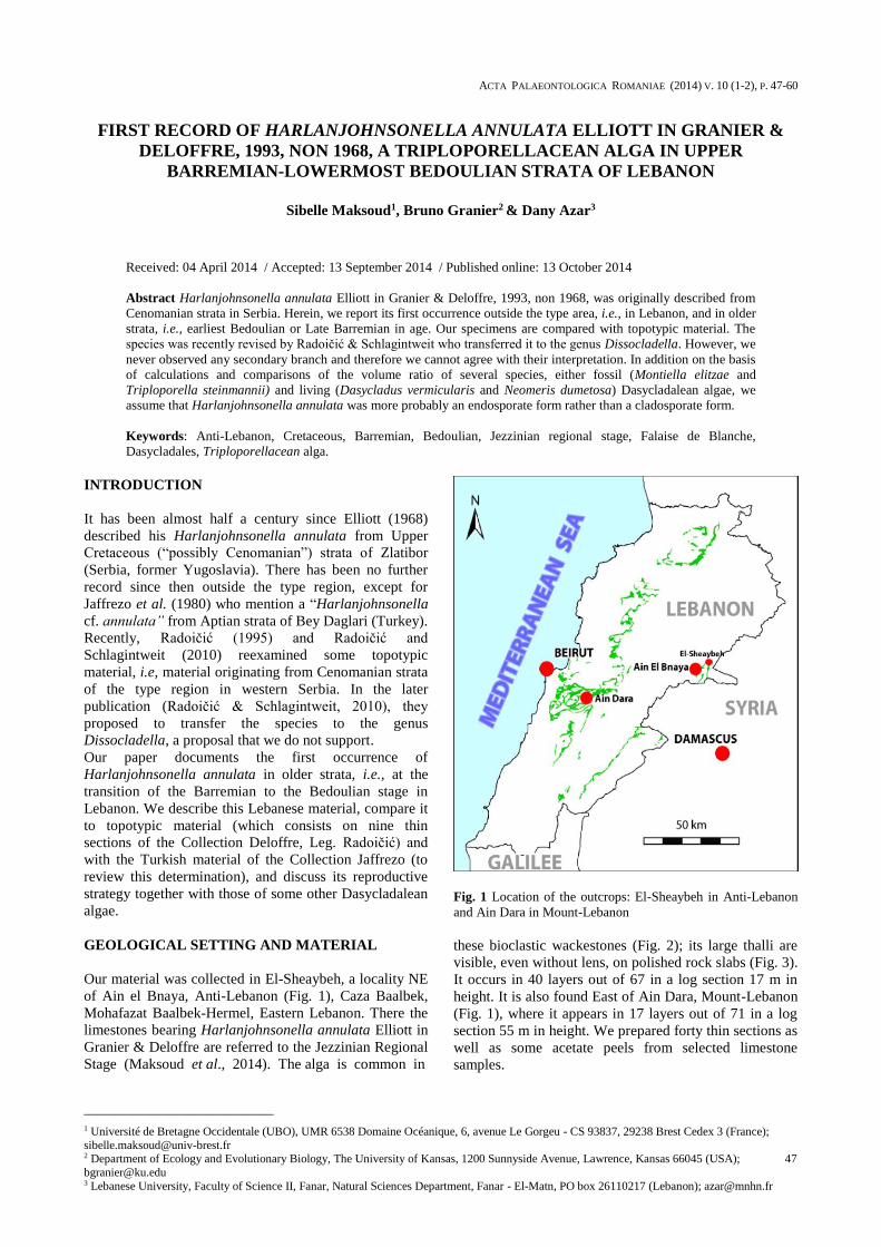
Our material was collected in El-Sheaybeh, a locality NE
of Ain el Bnaya, Anti-Lebanon (Fig. 1), Caza Baalbek,
Mohafazat Baalbek-Hermel, Eastern Lebanon. There the
limestones bearing Harlanjohnsonella annulata Elliott in
Granier & Deloffre are referred to the Jezzinian Regional
Stage (Maksoud et al., 2014). The alga is common in
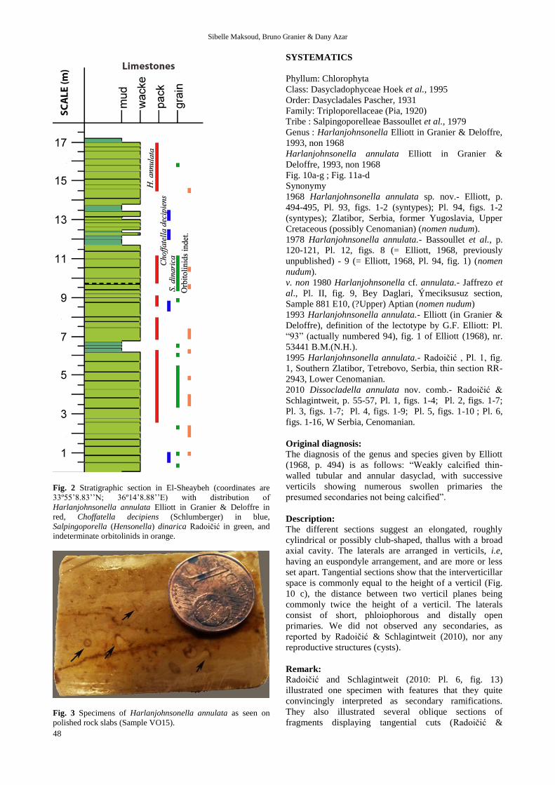
Fig. 1 Location of the outcrops: El-Sheaybeh in Anti-Lebanon
and Ain Dara in Mount-Lebanon
these bioclastic wackestones (Fig. 2); its large thalli are
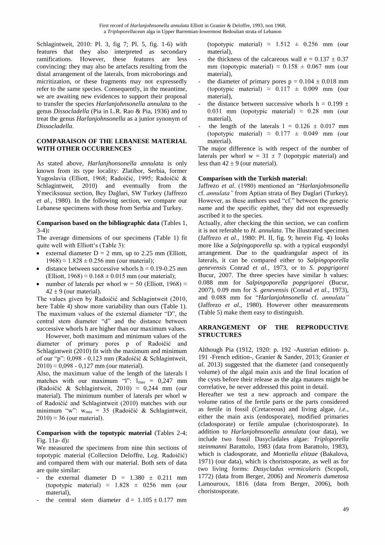
visible, even without lens, on polished rock slabs (Fig. 3).
It occurs in 40 layers out of 67 in a log section 17 m in
height. It is also found East of Ain Dara, Mount-Lebanon
(Fig. 1), where it appears in 17 layers out of 71 in a log
section 55 m in height. We prepared forty thin sections as
well as some acetate peels from selected limestone
samples.
Sibelle Maksoud, Bruno Granier & Dany Azar
48
Fig. 2 Stratigraphic section in El-Sheaybeh (coordinates are
33º55’8.83’’N; 36º14’8.88’’E) with distribution of
Harlanjohnsonella annulata Elliott in Granier & Deloffre in
red, Choffatella decipiens (Schlumberger) in blue,
Salpingoporella (Hensonella) dinarica Radoičić in green, and
indeterminate orbitolinids in orange.
Fig. 3 Specimens of Harlanjohnsonella annulata as seen on
polished rock slabs (Sample VO15).
SYSTEMATICS
Phyllum: Chlorophyta
Class: Dasycladophyceae Hoek et al., 1995
Order: Dasycladales Pascher, 1931
Family: Triploporellaceae (Pia, 1920)
Tribe : Salpingoporelleae Bassoullet et al., 1979
Genus : Harlanjohnsonella Elliott in Granier & Deloffre,
1993, non 1968
Harlanjohnsonella annulata Elliott in Granier &
Deloffre, 1993, non 1968
Fig. 10a-g ; Fig. 11a-d
Synonymy
1968 Harlanjohnsonella annulata sp. nov.- Elliott, p.
494-495, Pl. 93, figs. 1-2 (syntypes); Pl. 94, figs. 1-2
(syntypes); Zlatibor, Serbia, former Yugoslavia, Upper
Cretaceous (possibly Cenomanian) (nomen nudum).
1978 Harlanjohnsonella annulata.- Bassoullet et al., p.
120-121, Pl. 12, figs. 8 (= Elliott, 1968, previously
unpublished) - 9 (= Elliott, 1968, Pl. 94, fig. 1) (nomen
nudum).
v. non 1980 Harlanjohnsonella cf. annulata.- Jaffrezo et
al., Pl. II, fig. 9, Bey Daglari, Ýmeciksusuz section,
Sample 881 E10, (?Upper) Aptian (nomen nudum)
1993 Harlanjohnsonella annulata.- Elliott (in Granier &
Deloffre), definition of the lectotype by G.F. Elliott: Pl.
“93” (actually numbered 94), fig. 1 of Elliott (1968), nr.
53441 B.M.(N.H.).
1995 Harlanjohnsonella annulata.- Radoičić , Pl. 1, fig.
1, Southern Zlatibor, Tetrebovo, Serbia, thin section RR-
2943, Lower Cenomanian.
2010 Dissocladella annulata nov. comb.- Radoičić &
Schlagintweit, p. 55-57, Pl. 1, figs. 1-4; Pl. 2, figs. 1-7;
Pl. 3, figs. 1-7; Pl. 4, figs. 1-9; Pl. 5, figs. 1-10 ; Pl. 6,
figs. 1-16, W Serbia, Cenomanian.
Original diagnosis:
The diagnosis of the genus and species given by Elliott
(1968, p. 494) is as follows: “Weakly calcified thin-
walled tubular and annular dasyclad, with successive
verticils showing numerous swollen primaries the
presumed secondaries not being calcified”.
Description:
The different sections suggest an elongated, roughly
cylindrical or possibly club-shaped, thallus with a broad
axial cavity. The laterals are arranged in verticils, i.e,
having an euspondyle arrangement, and are more or less
set apart. Tangential sections show that the interverticillar
space is commonly equal to the height of a verticil (Fig.
10 c), the distance between two verticil planes being
commonly twice the height of a verticil. The laterals
consist of short, phloiophorous and distally open
primaries. We did not observed any secondaries, as
reported by Radoičić & Schlagintweit (2010), nor any
reproductive structures (cysts).
Remark:
Radoičić and Schlagintweit (2010: Pl. 6, fig. 13)
illustrated one specimen with features that they quite
convincingly interpreted as secondary ramifications.
They also illustrated several oblique sections of
fragments displaying tangential cuts (Radoičić &
First record of Harlanjohnsonella annulata Elliott in Granier & Deloffre, 1993, non 1968,
a Triploporellacean alga in Upper Barremian-lowermost Bedoulian strata of Lebanon
49
Schlagintweit, 2010: Pl. 3, fig 7; Pl. 5, fig. 1-6) with
features that they also interpreted as secondary
ramifications. However, these features are less
convincing: they may also be artefacts resulting from the
distal arrangement of the laterals, from microborings and
micritization, or these fragments may not expressedly
refer to the same species. Consequently, in the meantime,
we are awaiting new evidences to support their proposal
to transfer the species Harlanjohnsonella annulata to the
genus Dissocladella (Pia in L.R. Rao & Pia, 1936) and to
treat the genus Harlanjohnsonella as a junior synonym of
Dissocladella.
COMPARAISON OF THE LEBANESE MATERIAL
WITH OTHER OCCURRENCES
As stated above, Harlanjhonsonella annulata is only
known from its type locality: Zlatibor, Serbia, former
Yugoslavia (Elliott, 1968; Radoičić, 1995; Radoičić &
Schlagintweit, 2010) and eventually from the
Ýmeciksusuz section, Bey Dağlari, SW Turkey (Jaffrezo
et al., 1980). In the following section, we compare our
Lebanese specimens with those from Serbia and Turkey.
Comparison based on the bibliographic data (Tables 1,
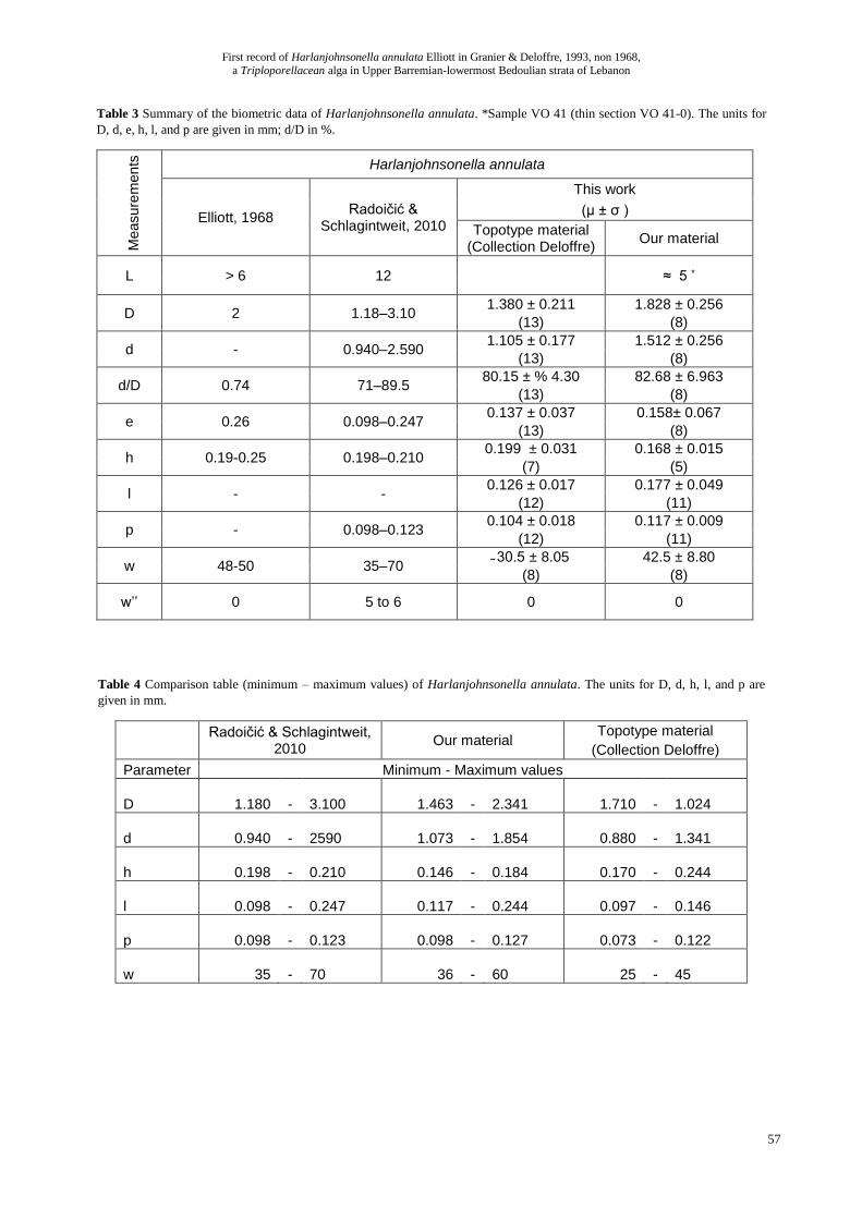
3-4):
The average dimensions of our specimens (Table 1) fit
quite well with Elliott‘s (Table 3):
external diameter D = 2 mm, up to 2.25 mm (Elliott,
1968) ≈ 1.828 ± 0.256 mm (our material);
distance between successive whorls h = 0.19-0.25 mm
(Elliott, 1968) ≈ 0.168 ± 0.015 mm (our material);
number of laterals per whorl w = 50 (Elliott, 1968) ≈
42 ± 9 (our material).
The values given by Radoičić and Schlagintweit (2010,
here Table 4) show more variability than ours (Table 1).
The maximum values of the external diameter “D”, the
central stem diameter “d” and the distance between
successive whorls h are higher than our maximum values.
However, both maximum and minimum values of the
diameter of primary pores p of Radoičić and
Schlagintweit (2010) fit with the maximum and minimum
of our “p”: 0,098 - 0,123 mm (Radoičić & Schlagintweit,
2010) ≈ 0,098 - 0,127 mm (our material).
Also, the maximum value of the length of the laterals l
matches with our maximum “l”: lmax = 0,247 mm
(Radoičić & Schlagintweit, 2010) ≈ 0,244 mm (our
material). The minimum number of laterals per whorl w
of Radoičić and Schlagintweit (2010) matches with our
minimum “w”: wmin = 35 (Radoičić & Schlagintweit,
2010) ≈ 36 (our material).
Comparison with the topotypic material (Tables 2-4;
Fig. 11a- d):
We measured the specimens from nine thin sections of
topotypic material (Collection Deloffre, Leg. Radoičić)
and compared them with our material. Both sets of data
are quite similar:
- the external diameter D = 1.380 ± 0.211 mm
(topotypic material) ≈ 1.828 ± 0256 mm (our
material),
- the central stem diameter d = 1.105 ± 0.177 mm
(topotypic material) ≈ 1.512 ± 0.256 mm (our
material),
- the thickness of the calcareous wall e = 0.137 ± 0.37
mm (topotypic material) ≈ 0.158 ± 0.067 mm (our
material),
- the diameter of primary pores p = 0.104 ± 0.018 mm
(topotypic material) ≈ 0.117 ± 0.009 mm (our
material),
- the distance between successive whorls h = 0.199 ±
0.031 mm (topotypic material) ≈ 0.28 mm (our
material),
- the length of the laterals l = 0.126 ± 0.017 mm
(topotypic material) ≈ 0.177 ± 0.049 mm (our
material).
The major difference is with respect of the number of
laterals per whorl w = 31 ± 7 (topotypic material) and
less than 42 ± 9 (our material).
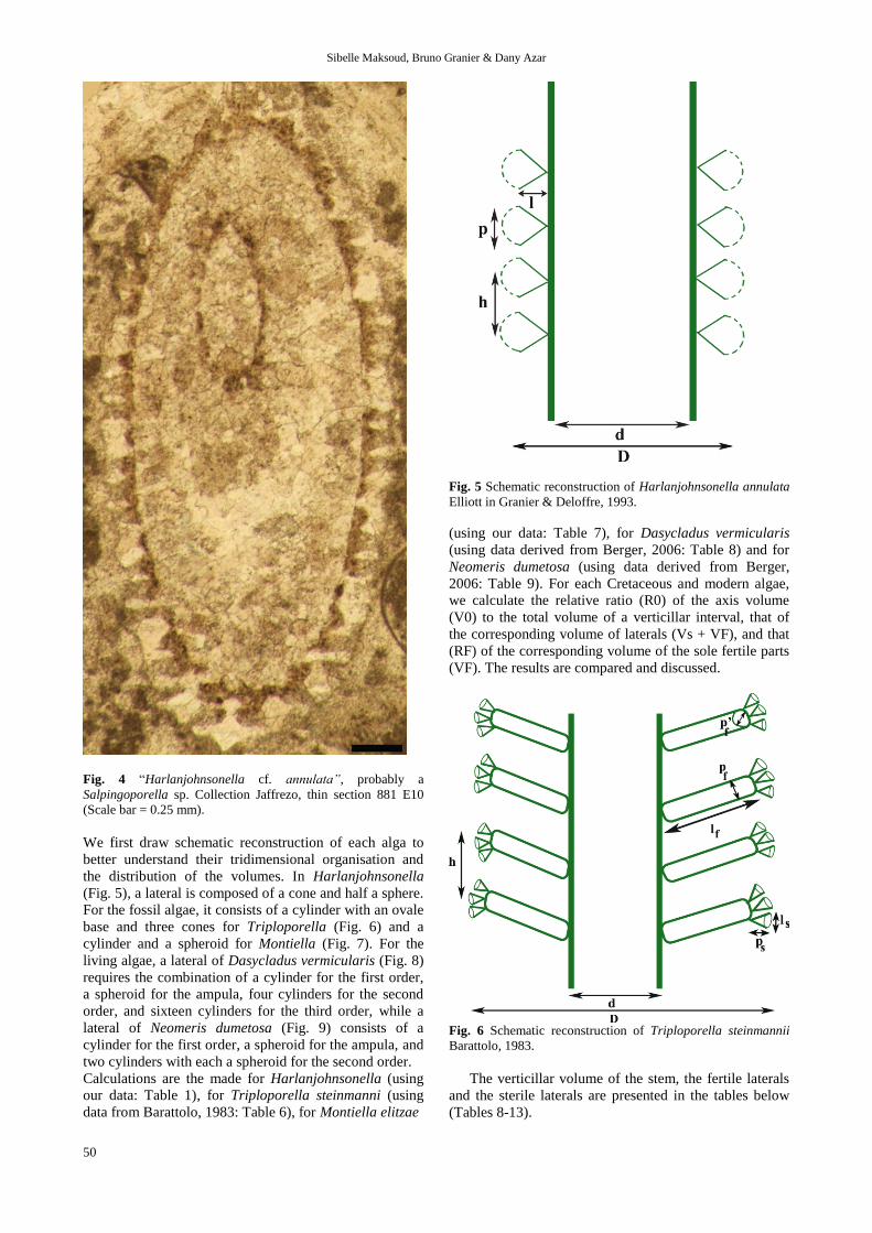
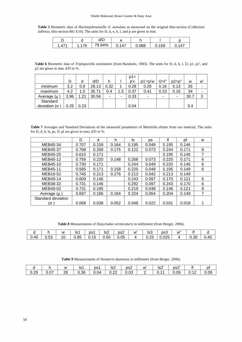
Comparison with the Turkish material: Jaffrezo et al. (1980) mentioned an “Harlanjohnsonella
cf. annulata” from Aptian strata of Bey Daglari (Turkey).
However, as these authors used “cf.” between the generic
name and the specific epithet, they did not expressedly
ascribed it to the species.
Actually, after checking the thin section, we can confirm
it is not referable to H. annulata. The illustrated specimen
(Jaffrezo et al., 1980: Pl. II, fig. 9; herein Fig. 4) looks
more like a Salpingoporella sp. with a typical euspondyl
arrangement. Due to the quadrangular aspect of its
laterals, it can be compared either to Salpingoporella
genevensis Conrad et al., 1973, or to S. popgrigorei
Bucur, 2007. The three species have similar h values:
0.088 mm for Salpingoporella popgrigorei (Bucur,
2007), 0.09 mm for S. genevensis (Conrad et al., 1973),
and 0.088 mm for “Harlanjohnsonella cf. annulata”
(Jaffrezo et al., 1980). However other measurements
(Table 5) make them easy to distinguish.
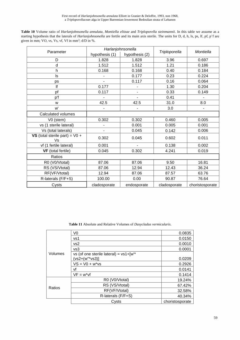
ARRANGEMENT OF THE REPRODUCTIVE
STRUCTURES
Although Pia (1912, 1920: p. 192 -Austrian edition- p.
191 -French edition-, Granier & Sander, 2013; Granier et
al. 2013) suggested that the diameter (and consequently
volume) of the algal main axis and the final location of
the cysts before their release as the alga matures might be
correlative, he never addressed this point in detail.
Hereafter we test a new approach and compare the
volume ratios of the fertile parts or the parts considered
as fertile in fossil (Cretaceous) and living algae, i.e.,
either the main axis (endosporate), modified primaries
(cladosporate) or fertile ampulae (choristosporate). In
addition to Harlanjohnsonella annulata (our data), we
include two fossil Dasycladales algae: Triploporella
steinmanni Barattolo, 1983 (data from Barattolo, 1983),
which is cladosporate, and Montiella elitzae (Bakalova,
1971) (our data), which is choristosporate, as well as for
two living forms: Dasycladus vermicularis (Scopoli,
1772) (data from Berger, 2006) and Neomeris dumetosa
Lamouroux, 1816 (data from Berger, 2006), both
choristosporate.
Sibelle Maksoud, Bruno Granier & Dany Azar
50
Fig. 4 “Harlanjohnsonella cf. annulata”, probably a
Salpingoporella sp. Collection Jaffrezo, thin section 881 E10
(Scale bar = 0.25 mm).
We first draw schematic reconstruction of each alga to
better understand their tridimensional organisation and
the distribution of the volumes. In Harlanjohnsonella
(Fig. 5), a lateral is composed of a cone and half a sphere.
For the fossil algae, it consists of a cylinder with an ovale
base and three cones for Triploporella (Fig. 6) and a
cylinder and a spheroid for Montiella (Fig. 7). For the
living algae, a lateral of Dasycladus vermicularis (Fig. 8)
requires the combination of a cylinder for the first order,
a spheroid for the ampula, four cylinders for the second
order, and sixteen cylinders for the third order, while a
lateral of Neomeris dumetosa (Fig. 9) consists of a
cylinder for the first order, a spheroid for the ampula, and
two cylinders with each a spheroid for the second order.
Calculations are the made for Harlanjohnsonella (using
our data: Table 1), for Triploporella steinmanni (using
data from Barattolo, 1983: Table 6), for Montiella elitzae
Fig. 5 Schematic reconstruction of Harlanjohnsonella annulata
Elliott in Granier & Deloffre, 1993.
(using our data: Table 7), for Dasycladus vermicularis
(using data derived from Berger, 2006: Table 8) and for
Neomeris dumetosa (using data derived from Berger,
2006: Table 9). For each Cretaceous and modern algae,
we calculate the relative ratio (R0) of the axis volume
(V0) to the total volume of a verticillar interval, that of
the corresponding volume of laterals (Vs + VF), and that
(RF) of the corresponding volume of the sole fertile parts
(VF). The results are compared and discussed.
Fig. 6 Schematic reconstruction of Triploporella steinmannii
Barattolo, 1983.
The verticillar volume of the stem, the fertile laterals
and the sterile laterals are presented in the tables below
(Tables 8-13).
First record of Harlanjohnsonella annulata Elliott in Granier & Deloffre, 1993, non 1968,
a Triploporellacean alga in Upper Barremian-lowermost Bedoulian strata of Lebanon
51
Fig. 7 Schematic reconstruction of Montiella elitzae
(Bakalova, 1971).
The executed calculations for each alga are the
following:
For Harlanjohnsonella annulata (Tables 1, 10):
As a starting hypothesis (1) we consider that the laterals
of Harlanjohnsonella are fertile and its main axis sterile
(cladosporate).
For each verticil, the volume of the stem is equal to a
cylinder volume:
V0 = π*h*d2/4 = 0.302 mm3
The volume of a lateral is equal to the sum of a cone and
half a sphere volumes:
vf = [(π*l*p2 /12)] + [π*p3/12] = [π*(p + l)*p2]/12 =
0.001 mm3
The total volume of the laterals is equal to:
VF = w*vf = 0.045 mm3
The resulting rates are:
R0 = RS = [V0/(VF+V0)]*100 = [0.288 / (0.045 +
0.288)]*100 = 87.06 % for the stem only (the “sterile”
part);
RF = [VF/(VF+V0)]*100 = [0.045 / (0.045 + 0.288)]*100
= 12.94 % for the laterals (supposed “fertile” in this
starting hypothesis).
In the alternative hypothesis (2) we consider that the
laterals of Harlanjohnsonella are sterile and its main axis
fertile (endosporate).
The resulting rates are:
R0 = RF = [V0/(VS+V0)]*100 = [0.288 / (0.045 +
0.288)]*100 = 87.06 % for the stem only (the “fertile”
part);
RS = [VS/(VS+V0)]*100 = [0.045 / (0.045 + 0.288)]*100
= 12.94 % for the laterals (supposed “sterile” in this
alternative hypothesis).
For Triploporella steinmannii (Tables 6, 10) :
The biometric data are from Barattolo (1983). When an
average value is not available, we use the maximal value.
Fig. 8 Schematic reconstruction of Dasycladus vermicularis
(Scopoli, 1772).
For each verticil, the volume of the stem is equal to a
cylinder volume:
V0 = 0.46 mm3
The primaries are fertile (with visible reproductive
structures). The volume of a primary lateral is equal to a
cylinder with elliptic base volume:
vf = π*l*p1*p1’/4 = 0.1381 mm3
The volume of a secondary is equal to a cone volume:
v2 = π*l2*p2*p2’/12 = 0.0015 mm3
The total volume of the sterile part in a verticil is equal
to: VS = V0 + w*w’*v2 = 0.6019 mm3
The total volume of the fertile part in a verticil is equal to
the volume of the primary laterals:
VF = w*vf = 4.2410 mm3
The resulting rates are:
R0 = 9.50 % for the stem only.
RS = 12.43 % for the whole sterile part (including the
stem).
RF = 87.57 % for the fertile part.
The volume ratio of the fertile part in the laterals is: R
(F/F+S) = 90.87 %
For Montiella elitzae (Tables 7, 10, Fig. 10h-j):
For each verticil, the volume of the stem is equal to a
cylinder volume:
V0 = 0.005 mm3
The volume of a sterile lateral is equal to a cylinder
volume:
vs = π*ls*ps2/4 = 0.001 mm3
The total volume of the sterile laterals is:
Vs = w*vs = 0.006 mm3
The total volume of the sterile part is:
VS = V0 + w*vs = 0.011 mm3
The volume of a fertile lateral is equal to a spheroid
volume:
vf = (4/3)*π*(lf/2)*(pf/2)2 = π*lf*pf2/6 = 0.002 mm3
Sibelle Maksoud, Bruno Granier & Dany Azar
52
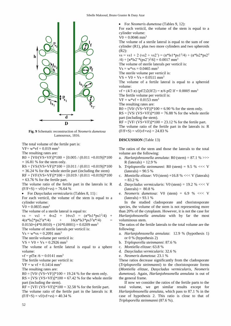
Fig. 9 Schematic reconstruction of Neomeris dumetosa
Lamouroux, 1816.
The total volume of the fertile part is:
VF= w*vf = 0.019 mm3
The resulting rates are:
R0 = [V0/(VS+VF)]*100 = [0.005 / (0.011 +0.019)]*100
= 16.81 % for the stem only.
RS = [VS/(VS+VF)]*100 = [0.011 / (0.011 +0.019)]*100
= 36.24 % for the whole sterile part (including the stem)
RF = [VF/(VS+VF)]*100 = [0.019 / (0.011 +0.019)]*100
= 63.76 % for the fertile part.
The volume ratio of the fertile part in the laterals is: R
(F/F+S) = vf/(vf+vs) = 76.64 %
For Dasycladus vermicularis (Tables 8, 11) :
For each verticil, the volume of the stem is equal to a
cylinder volume:
V0 = 0.0835 mm3
The volume of a sterile lateral is equal to:
vs = vs1 + 4vs2 + 16vs3 = (π*ls1*ps12/4) +
4(π*ls2*ps22π*/4) + 16(π*ls3*ps32π*/4) =
0.0150+(4*0.0010) + (16*0.0001) = 0.0209 mm3
The volume of sterile laterals per verticil is:
Vs = w*vs = 0.2091 mm3
The sterile volume per verticil is:
VS = V0 + Vs = 0.2926 mm3
The volume of a fertile lateral is equal to a sphere
volume:
vf = pf3π /6 = 0.0141 mm3
The fertile volume per verticil is:
VF = w vf = 0.1414 mm3
The resulting rates are:
R0 = [V0/ (VS+VF)]*100 = 19.24 % for the stem only.
RS = [VS/ (VS+VF)]*100 = 67.42 % for the whole sterile
part (including the stem).
RF = [VF/ (VS+VF)]*100 = 32.58 % for the fertile part.
The volume ratio of the fertile part in the laterals is: R
(F/F+S) = vf/(vf+vs) = 40.34 %
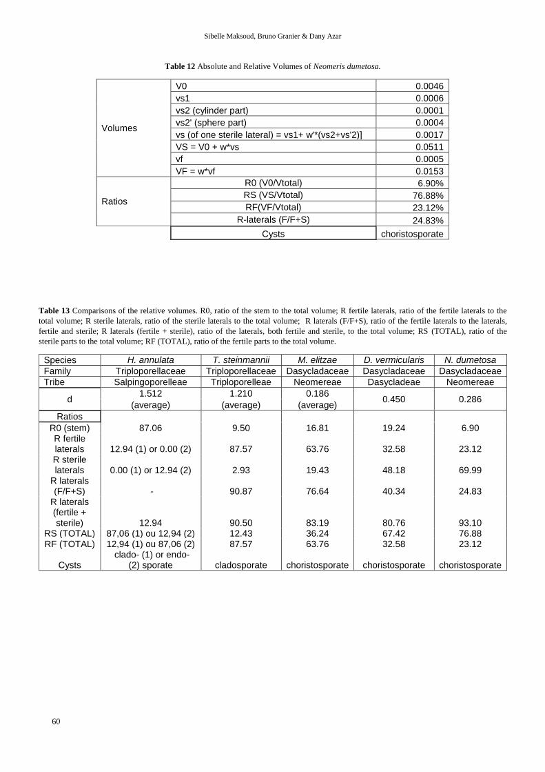
For Neomeris dumetosa (Tables 9, 12):
For each verticil, the volume of the stem is equal to a
cylinder volume:
V0 = 0.0046 mm3
The volume of a sterile lateral is equal to the sum of one
cylinder (R1), plus two more cylinders and two spheroids
(R2):
vs = vs1 + 2 (vs2 + vs2’) = (π*ls1*ps12/4) + (π*ls2*ps22
/4) + [π*ls2’*(ps2’)2/6] = 0.0017 mm3
The volume of sterile laterals per verticil is:
Vs = w*vs = 0.0465 mm3
The sterile volume per verticil is:
VS = V0 + Vs = 0.0511 mm3
The volume of a fertile lateral is equal to a spheroid
volume:
vf = (4/3 π) (pf/2)2(lf/2) = π/6 pf2 lf = 0.0005 mm3
The fertile volume per verticil is:
VF = w*vf = 0.0153 mm3
The resulting rates are:
R0 = [V0/ (VS+VF)]*100 = 6.90 % for the stem only.
RS = [VS/ (VS+VF)]*100 = 76.88 % for the whole sterile
part (including the stem).
RF = [VF/ (VS+VF)]*100 = 23.12 % for the fertile part.
The volume ratio of the fertile part in the laterals is: R
(F/F+S) = vf/(vf+vs) = 24.83 %
DISCUSSION (Table 13)
The ratios of the stem and those the laterals to the total
volume are the following:
a. Harlanjohnsonella annulata: R0 (stem) = 87.1 % >>>
R (laterals) = 12.9 %
b. Triploporella steinmanni: R0 (stem) = 9.5 % <<< V
(laterals) = 90.5 %
c. Montiella elitzae: V0 (stem) =16.8 % <<< V (laterals)
= 83.2 %
d. Dasycladus vermicularis: V0 (stem) = 19.2 % <<< V
(laterals) = 80.8 %
e. Neomeris dumetosa: V0 (stem) = 6.9 % <<< V
(laterals) = 93.1 %
In the studied cladosporate and choristosporate
species, the volume of the stem is not representing more
than 20% of the cytoplasm. However, it is not the case for
Harlanjohnsonella annulata with by far the most
voluminous stem.
The ratios of the fertile laterals to the total volume are the
following:
a. Harlanjohnsonella annulata: 12.9 % (hypothesis 1)
or 0 % (hypothesis 2)
b. Triploporella steinmanni: 87.6 %
c. Montiella elitzae: 63.8 %
d. Dasycladus vermicularis: 32.6 %
e. Neomeris dumetosa: 23.1 %
These ratios decrease significantly from the cladosporate
(Triploporella steinmanni) to the choristosporate forms
(Montiella elitzae, Dasycladus vermicularis, Neomeris
dumetosa). Again, Harlanjohnsonella annulata is out of
the general frame.
If now we consider the ratios of the fertile parts to the
total volume, we get similar results except for
Harlanjohnsonella annulata, which pass to 87.1 % in the
case of hypothesis 2. This ratio is close to that of
Triploporella steinmanni (87.6 %).
First record of Harlanjohnsonella annulata Elliott in Granier & Deloffre, 1993, non 1968,
a Triploporellacean alga in Upper Barremian-lowermost Bedoulian strata of Lebanon
53
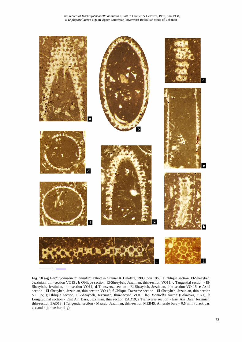
Fig. 10 a-g Harlanjohnsonella annulata Elliott in Granier & Deloffre, 1993, non 1968; a Oblique section, El-Sheaybeh,
Jezzinian, thin-section VO15 ; b Oblique section, El-Sheaybeh, Jezzinian, thin-section VO11; c Tangential section - El-
Sheaybeh, Jezzinian, thin-section VO11; d Transverse section - El-Sheaybeh, Jezzinian, thin-section VO 15. e Axial
section - El-Sheaybeh, Jezzinian, thin-section VO 15; f Oblique-Trasverse section - El-Sheaybeh, Jezzinian, thin-section
VO 15; g Oblique section, El-Sheaybeh, Jezzinian, thin-section VO15. h-j Montiella elitzae (Bakalova, 1971); h
Longitudinal section - East Ain Dara, Jezzinian, thin section EAD19; i Transverse section - East Ain Dara, Jezzinian,
thin-section EAD18; j Tangential section - Maarab, Jezzinian, thin-section MEB45. All scale bars = 0.5 mm, (black bar:
a-c and h-j; blue bar: d-g)

Sibelle Maksoud, Bruno Granier & Dany Azar
54
It is suggested that the fertile part of these algae is
decreasing gradually from the ancestral endosporate and
cladosporate forms to the choristoporate form with
specialized ampulae. As in genuine cladosporate forms
(such as Triploporella steinmanni) the fertile part is
mostly concentrated in the primary laterals, we would
expect similar ratio for most cladosporate species. With
primary laterals representing only 12.9% of the total
volume of the cytoplasm, Harlanjohnsonella annulata
does not fit with this model. Hypothesis 2 should
therefore be favoured; as a result, Harlanjohnsonella
annulata is probably an endosporate species.
CONCLUSION
The Cenomanian Harlanjohnsonella annulata Elliott in
Granier & Deloffre, 1993, is reported for the first time
outside its type area, i.e., in Lebanon, and in older strata,
i.e., Late Barremian or Bedoulian in age. In these
specimens, as well as in topotypic material of
Harlanjohnsonella annulata that we examined
(Collection Deloffre, Leg. Radoičić), we did not observed
any evidence of secondary laterals. On the basis of an
investigation on the distribution of the cytoplasmic
volume, it is suggested that the reproductive strategy of
Harlanjohnsonella annulata was most likely endosporate,
not cladosporate.
ACKNOWLEDGEMENTS
We would like to thank the CNRSL (National Council for
Scientific Research-Lebanon) and the AUF (University
Agency of the Francophony) for their financial support.
This research is a contribution to the project N° 30959NJ
- PHC CEDRE 2014; supported by of the research project
of Hubert-Curien Partnership program (PHC) CEDRE,
implemented in Lebanon and France by the Ministry of
Foreign Affairs (Ministère des Affaires étrangères, MAE)
and the Ministry of Higher Education and Research
(Ministère de l'Enseignement Supérieur et de la
Recherche, MESR). This paper is a contribution of the
team project "Biodiversity: Origin, Structure, Evolution
and Geology" led by Dany Azar at the Lebanese
University. The thin sections are deposited with LPB
numbers in the collections the Département des Sciences
de la Terre et de l'Univers, Université de Bretagne
Occidentale, Brest (France). Finally, we would like to
thank Filippo Barattolo, Ioan I. Bucur and Mariano
Parente for their constructive comments.
REFERENCES
Barattolo, F., 1983. Observazioni su Triploporella
steinmannii n. sp. (alghe Verdi, Dasicladali) del
Cretacico del Messico. Bollettino della Società dei
Naturalisti in Napoli, 91 (1982): 1–53.
Bassoullet, J.-P., Bernier, P., Conrad, M.A., Deloffre R.
& Jaffrezo, M., 1978. Les algues Dasycladales du
Jurassique et du Cretacé. Géobios, Mémoire spécial 2,
330 pp.
Berger, S., 2006. Photo-Atlas of living Dasycladales.
Carnets de Géologie [Notebooks on Geology],
CG2006_B02, 348 pp.
Bucur, I.I., 2007. Salpingoporella? popgrigorei, a new
species of Dasycladales (calcareous algae) from the
Lower Aptian deposits of Pădurea Craiului (northern
Apuseni Mountains, Romania. Facies, 53: 377-388.
Elliott, G., 1968. Three new Tethyan Dasycladaceae
(calcareous algae). Paleontology, 11(4): 491-497.
Fig. 11 a-d Harlanjohnsonella annulata Elliott in Granier & Deloffre, 1993, non 1968; topotypic material (Collection
Deloffre, Leg. Radoičić) ; a Oblique section; b Oblique section; c Longitudinal section ; d Fragment in a transverse
section. Scale bar = 0.5 mm.
First record of Harlanjohnsonella annulata Elliott in Granier & Deloffre, 1993, non 1968,
a Triploporellacean alga in Upper Barremian-lowermost Bedoulian strata of Lebanon
55
Granier, B. & Deloffre, R., 1993. Inventaire critique des
algues dasycladales fossiles II. Partie - les algues
dasycladales du Jurassique et du Crétacé. Revue de
Paléobiologie, 12 (1): 19-65.
Granier, B., Lethiers, A. & Sander, N.J., 2013. The XXIst
Century Edition of the "New studies on Triassic
Siphoneae verticillatae, by Julius von Pia. Jahrbuch
der Geologischen Bundesanstalt, 153(1-4): 239-300.
Granier, B. & Sander, N.J., 2013. The XXIst Century (the
100th Anniversary) Edition of the "New studies on
Triassic Siphoneae verticillatae" by Julius von Pia.
Carnets de Géologie [Notebooks on Geology],
CG2013_B01, 108 pp. [56 videos].
Jaffrezo, M., Poisson, A. & Akbulut, A., 1980. Les
Algues du Crétacé Inférieur des séries de type Bey
Daglari (Taurides Occidentales, Turquie. Bulletin of
Mineral Research and Exploration Institute of Turkey,
91: 76-88.
Maksoud, S., Granier, B., Azar, D., Gèze, R., Paicheler,
J.-C., Clavel, B. & Moreno-Bedmar, J.A., 2014, in
press. Revision of "Falaise de Blanche" (Lower
Cretaceous) in Lebanon, with the definition of a
Jezzinian Regional Stage, and its micro-
paleontological characterization. Carnets de Géologie
[Notebooks on Geology], 14(18).
Pia, J. von, 1912. Neue Studien über die triadischen
Siphoneae verticillatae. Beiträge zur Paläontologie
und Geologie Österreich-Ungarns und des Orients,
25: 25-81.
Pia, J. von, 1920. Die Siphoneae verticillatae vom
Karbon bis zur Kreide. Abh. zool.-bot. Gesellschaft,
11 (2): 263 pp. [Les Siphonées verticillées du
Carbonifère au Crétacé. Éditions Technip, 1961].
Radoičić, R., 1995. Contribution to the study of
Cretaceous biostratigraphy of Zlatibor Mountain.
Radovi Geoinstituta, 31: 17–30 [in Serbo-Croatian
with English abstract].
Radoičić, R. & Schlagintweit, F., 2010. Observations on
Dissocladella annulata (Elliott, 1993) nov. comb.
(Calcareous algae, Dasycladales) from the
Cenomanian of west Serbia. Annales Géologiques de
la Péninsule Balkanique, 71: 53-71.
Sibelle Maksoud, Bruno Granier & Dany Azar
56
TABLES
Table 1 Averages and Standard Deviations of the measured parameters of the Harlanjohnsonella annulata specimens found in
Lebanon. The units for D, d, e, h, l, and p are given in mm; d/D in %. L: maximum length; D: external diameter; d: central stem
diameter; h: interverticillar distance of two successive whorls = from a reference plane in one whorl to the same plane in the next; l:
length of the laterals; p: width of primary laterals; e: thickness of the calcareous wall; w: number of laterals per verticil.
Specimen D d d/D e h l p w
VO15-12 0.162 0.213 0.106 36
VO11-3 1.702 1.425 83.73 0.139 0.184 0.148 0.127 36
VO15-44 1.734 1.617 93.25 0.059 0.117 0.117 36
VO41-4 1.814 1.592 87.76 0.111 0.179 0.148 0.111
VO14-23 1.463 1.073 73.34 0.195 0.219 0.122 40
VO59-10 1.915 1.414 73.84 0.251 0.243 0.122 60
VO19-21 1.707 1.414 82.84 0.147 0.122 0.121 40
VO16-26 0.171 0.195 0.122
VO11-27 0.146 0.171 0.122
VO18-05 1.951 1.707 87.49 0.122 0.122 0.098 40
VO38-10 2.341 1.854 79.20 0.244 0.244 0.122 52
Average (µ ) 1.828 1.512 82.681 0.158 0.168 0.177 0.117 42.50
Standard deviation (σ ) 0.256 0.237 6.963 0.067 0.015 0.049 0.009 8.80
Table 2 Averages and Standard Deviations of the measured parameters from a topotypic material (Collection Deloffre, Leg.
Radoičić). The units for D, d, e, h, l, and p are given in mm; d/D in %.
Specimen D d d/D e h l p w
a 1.268 0.976 76.97 0.146 0.244 0.122 0.122 b 1.024 0.880 85.94 0.072 0.195 0.097 0.073 c 1.585 1.341 84.61 0.122 0.231 0.122 0.122 25 d 1.610 1.366 84.84 0.122 0.146 0.122 30 e 1.366 1.171 85.72 0.098 0.210 0.098 0.122 27 f 1.226 0.980 79.93 0.123 0.170 0.122 0.098 g 1.220 0.927 75.98 0.147 0.122 0.098 27 h 1.317 1.024 77.75 0.147 0.121 0.098 40 i 1.220 0.902 73.93 0.159 0.170 0.146 0.073 j 1.710 1.341 78.42 0.185 0.170 0.146 0.122 k 1.707 1.268 74.28 0.220 0.146 0.098 45 l 1.317 1.073 81.47 0.122 0.122 0.098 27
m 1.366 1.122 82.14 0.122 28
Average (µ ) 1.380 1.105 80.15 0.137 0.199 0.126 0.104 31.13
Standard deviation (σ ) 0.211 0.177 4.30 0.037 0.031 0.017 0.018 7.28
First record of Harlanjohnsonella annulata Elliott in Granier & Deloffre, 1993, non 1968,
a Triploporellacean alga in Upper Barremian-lowermost Bedoulian strata of Lebanon
57
Table 3 Summary of the biometric data of Harlanjohnsonella annulata. *Sample VO 41 (thin section VO 41-0). The units for
D, d, e, h, l, and p are given in mm; d/D in %. M
easure
men
ts
Harlanjohnsonella annulata
Elliott, 1968 Radoičić &
Schlagintweit, 2010
This work
(µ ± σ )
Topotype material (Collection Deloffre)
Our material
L > 6 12 ≈ 5 *
D 2 1.18–3.10 1.380 ± 0.211 1.828 ± 0.256
(13) (8)
d - 0.940–2.590 1.105 ± 0.177 1.512 ± 0.256
(13) (8)
d/D 0.74 71–89.5 80.15 ± % 4.30 82.68 ± 6.963
(13) (8)
e 0.26 0.098–0.247 0.137 ± 0.037 0.158± 0.067
(13) (8)
h 0.19-0.25 0.198–0.210 0.199 ± 0.031 0.168 ± 0.015
(7) (5)
l - - 0.126 ± 0.017 0.177 ± 0.049
(12) (11)
p - 0.098–0.123 0.104 ± 0.018 0.117 ± 0.009
(12) (11)
w 48-50 35–70 ̴ 30.5 ± 8.05 42.5 ± 8.80
(8) (8)
w’’ 0 5 to 6 0 0
Table 4 Comparison table (minimum – maximum values) of Harlanjohnsonella annulata. The units for D, d, h, l, and p are
given in mm.
Radoičić & Schlagintweit, 2010
Our material Topotype material
(Collection Deloffre)
Parameter Minimum - Maximum values
D 1.180 - 3.100 1.463
- 2.341 1.710
- 1.024
d 0.940 - 2590 1.073
- 1.854 0.880
- 1.341
h 0.198 - 0.210 0.146
- 0.184 0.170
- 0.244
l 0.098 - 0.247 0.117
- 0.244 0.097
- 0.146
p 0.098 - 0.123 0.098
- 0.127 0.073
- 0.122
w 35 - 70 36
- 60 25
- 45
Sibelle Maksoud, Bruno Granier & Dany Azar
58
Table 5 Biometric data of Harlanjohnsonella cf. annulata as measrued on the original thin-section (Collection
Jaffrezo, thin section 881 E10). The units for D, d, e, h, l, and p are given in mm.
D d d/D e h l p
1.471 1.176 79.94% 0.147 0.088 0.159 0.147
Table 6 Biometric data of Triploporella steinmanni (from Barattolo, 1983). The units for D, d, h, l, l2, p1, p1', and
p2 are given in mm; d/D in %.
D d d/D h l p1= p'v p1'=p'w l2=l'' p2=p'' w w'
minimum 3.2 0.9 28.13 0.32 1 0.28 0.28 0.16 0.13 26 -
maximum 4.2 1.5 35.71 0.4 1.3 0.37 0.41 0.23 0.16 34 -
Average (µ ) 3.96 1.21 30.56 - - 0.33 - - - 30.7 3
Standard deviation (σ ) 0.25 0.23 0.04 3.4
Table 7 Averages and Standard Deviations of the measured parameters of Montiella elitzae from our material. The units
for D, d, h, ls, ps, lf, pf are given in mm; d/D in %.
D d h ls ps lf pf w
MEB45-34 0.707 0.159 0.164 0.195 0.049 0.195 0.146
MEB45-37 0.768 0.268 0.175 0.122 0.073 0.244 0.171 8
MEB45-25 0.610 0.171 0.195 0.146 7
MEB45-12 0.756 0.220 0.148 0.268 0.073 0.220 0.171 6
MEB45-10 0.730 0.171 0.244 0.049 0.220 0.146 6
MEB45-11 0.585 0.171 0.158 0.220 0.049 0.195 0.149 6
MEB19-52 0.745 0.213 0.276 0.213 0.042 0.213 0.149
MEB49-14 0.609 0.146 0.243 0.097 0.170 0.121 6
MEB38-32 0.731 0.146 0.292 0.097 0.243 0.170 6
MEB49-02 0.731 0.195 0.219 0.048 0.146 0.121 8
Average (µ ) 0.697 0.186 0.184 0.224 0.064 0.204 0.149 7
Standard deviation (σ ) 0.068 0.038 0.052 0.048 0.022 0.031 0.018 1
Table 8 Measurements of Dasycladus vermicularis in millimeter (from Berger, 2006).
d h w ls1 ps1 ls2 ps2 w' ls3 ps3 w'' lf d
0.45 0.53 10 0.85 0.15 0.50 0.05 4 0.25 0.025 4 0.30 0.45
Table 9 Measurements of Neomeris dumetosa in millimeter (from Berger, 2006).
d h w ls1 ps1 ls2 ps2 w' ls2' ps2' lf pf
0.29 0.07 28 0.36 0.04 0.22 0.03 2 0.11 0.09 0.12 0.09
First record of Harlanjohnsonella annulata Elliott in Granier & Deloffre, 1993, non 1968,
a Triploporellacean alga in Upper Barremian-lowermost Bedoulian strata of Lebanon
59
Table 10 Volume ratio of Harlanjohnsonella annulata, Montiella elitzae and Triploporella steinmannii. In this table we assume as a
starting hypothesis that the laterals of Harlanjohnsonella are fertile and its main axis sterile. The units for D, d, h, ls, ps, lf, pf, p’f are
given in mm; VO, vs, Vs, vf, Vf in mm3; d/D in %.
Parameter Harlanjohnsonella
Triploporella Montiella hypothesis (1) hypothesis (2)
D 1.828 1.828 3.96 0.697
d 1.512 1.512 1.21 0.186
h 0.168 0.168 0.40 0.184
ls - 0.177 0.23 0.224
ps - 0.117 0.16 0.064
lf 0.177 - 1.30 0.204
pf 0.117 - 0.33 0.149
p'f - - 0.41 -
w 42.5 42.5 31.0 8.0
w' - - 3.0 -
Calculated volumes
V0 (stem) 0.302 0.302 0.460 0.005
vs (1 sterile lateral) - 0.001 0.005 0.001
Vs (total laterals) - 0.045 0.142 0.006
VS (total sterile part) = V0 + Vs
0.302 0.045 0.602 0.011
vf (1 fertile lateral) 0.001 - 0.138 0.002
VF (total fertile) 0.045 0.302 4.241 0.019
Ratios 0.347 0.347 4.843 0.030
R0 (V0/Vtotal) 87.06 87.06 9.50 16.81
RS (VS/Vtotal) 87.06 12.94 12.43 36.24
RF(VF/Vtotal) 12.94 87.06 87.57 63.76
R-laterals (F/F+S) 100.00 0.00 90.87 76.64
Cysts cladosporate endosporate cladosporate choristosporate
Table 11 Absolute and Relative Volumes of Dasycladus vermicularis.
Volumes
V0 0.0835
vs1 0.0150
vs2 0.0010
vs3 0.0001
vs (of one sterile lateral) = vs1+[w'* (vs2+(w"*vs3)] 0.0209
VS = V0 + w*vs 0.2926
vf 0.0141
VF = w*vf 0.1414
Ratios
R0 (V0/Vtotal) 19.24%
RS (VS/Vtotal) 67.42%
RF(VF/Vtotal) 32.58%
R-laterals (F/F+S) 40.34%
Cysts choristosporate
Sibelle Maksoud, Bruno Granier & Dany Azar
60
Table 12 Absolute and Relative Volumes of Neomeris dumetosa.
Volumes
V0 0.0046
vs1 0.0006
vs2 (cylinder part) 0.0001
vs2' (sphere part) 0.0004
vs (of one sterile lateral) = vs1+ w'*(vs2+vs'2)] 0.0017
VS = V0 + w*vs 0.0511
vf 0.0005
VF = w*vf 0.0153
Ratios
R0 (V0/Vtotal) 6.90%
RS (VS/Vtotal) 76.88%
RF(VF/Vtotal) 23.12%
R-laterals (F/F+S) 24.83%
Cysts choristosporate
Table 13 Comparisons of the relative volumes. R0, ratio of the stem to the total volume; R fertile laterals, ratio of the fertile laterals to the
total volume; R sterile laterals, ratio of the sterile laterals to the total volume; R laterals (F/F+S), ratio of the fertile laterals to the laterals,
fertile and sterile; R laterals (fertile + sterile), ratio of the laterals, both fertile and sterile, to the total volume; RS (TOTAL), ratio of the
sterile parts to the total volume; RF (TOTAL), ratio of the fertile parts to the total volume.
Species H. annulata T. steinmannii M. elitzae D. vermicularis N. dumetosa
Family Triploporellaceae Triploporellaceae Dasycladaceae Dasycladaceae Dasycladaceae
Tribe Salpingoporelleae Triploporelleae Neomereae Dasycladeae Neomereae
d 1.512 1.210 0.186
0.450 0.286 (average) (average) (average)
Ratios
R0 (stem) 87.06 9.50 16.81 19.24 6.90 R fertile laterals 12.94 (1) or 0.00 (2) 87.57 63.76 32.58 23.12 R sterile laterals 0.00 (1) or 12.94 (2) 2.93 19.43 48.18 69.99
R laterals (F/F+S) - 90.87 76.64 40.34 24.83
R laterals (fertile + sterile) 12.94 90.50 83.19 80.76 93.10
RS (TOTAL) 87,06 (1) ou 12,94 (2) 12.43 36.24 67.42 76.88 RF (TOTAL) 12,94 (1) ou 87,06 (2) 87.57 63.76 32.58 23.12
Cysts clado- (1) or endo-
(2) sporate cladosporate choristosporate choristosporate choristosporate
Copyright © 2022 FDOKUMEN