Factores Genéticos asociados a la Degeneración Lobar ...
226
Factores Genéticos asociados a la Degeneración Lobar Frontotemporal Análisis de susceptibilidad genética y correlación fenotipo-genotipo Doctoranda Mª Isabel Hernández Ruiz Directores Agustín Ruiz Laza Mercé Boada Rovira Rafael Blesa Gonzalez Tutor José Álvarez Sabin PROGRAMA DE DOCTORADO EN MEDICINA DEPARTAMENTO DE MEDICINA UNIVERSITAT AUTÒNOMA DE BARCELONA 2014
-
Upload
khangminh22 -
Category
Documents
-
view
0 -
download
0
Transcript of Factores Genéticos asociados a la Degeneración Lobar ...
Factores Genéticos asociados a la Degeneración Lobar Frontotemporal
Análisis de susceptibilidad genética y correlación fenotipo-genotipo
Doctoranda Mª Isabel Hernández Ruiz
Directores Agustín Ruiz Laza
Mercé Boada Rovira Rafael Blesa Gonzalez
Tutor José Álvarez Sabin
PROGRAMA DE DOCTORADO EN MEDICINA DEPARTAMENTO DE MEDICINA
UNIVERSITAT AUTÒNOMA DE BARCELONA2014


DIRECTORES DE TESIS
Dr. AGUSTÍN RUIZ LAZA, Doctor en Medicina por la Universidad de Sevilla. Dra. MERCÉ BOADA ROVIRA, Doctora en Medicina por la Universidad de Barcelona. Dr. RAFAEL BLESA GONZALEZ, Doctor en Medicina por la Universidad de Barcelona.
TUTOR DE TESIS
Dr. JOSÉ ÁLVAREZ SABIN, Doctor en Medicina por la Universidad de Barcelona. Profe-sor Titular de Neurología de la Universitat Autònoma de Barcelona.
CERTIFICAN
Que la tesis titulada “Factores Genéticos asociados a la Degeneración Lobar Fron-totemporal. Análisis de susceptibilidad genética y correlación fenotipo-genotipo”, presentada por Mª Isabel Hernández Ruiz, se ha realizado bajo nuestra supervisión y consideramos que reúne los requisitos necesarios para ser defendida ante el Tribunal correspondiente para optar al grado de Doctor en Medicina por la Universitat Autò-noma de Barcelona.
Agustín Ruiz Laza Mercè Boada Rovira
Cap de Recerca Directora Mèdica Fundació ACE (ICNA) Fundació ACE (ICNA)Barcelona Barcelona
Rafael Blesa González José Álvarez Sabin Cap de Servei Neurología Cap de Servei de Neurología Hospital de la Santa Creu i Sant Pau Hospital de la Vall d’HebrónBarcelona Barcelona Professor Titular de Neurología Universitat Autònoma de Barcelona
Barcelona a 15 de Septiembre de 2014

“Enseñarás a volar, pero no volarán tu vuelo.
Enseñarás a soñar, pero no soñarán tu sueño.
Enseñarás a vivir,pero no vivirán tu vida.
Sin embargo... en cada vuelo, en cada vida,
en cada sueño, perdurará siempre la huella
del camino enseñado”
Madre Teresa De Calcuta
A mi padre, que supo transmitirme el valor del respeto y la generosidad


Hace 20 años aposté, como neuróloga, que iba a dedicar el resto de mis años de actividad profesional al mundo de las demencias. No me he equivocado. La demen-cia es para mí una de las enfermedades más dura y compleja que existe. El tiempo dedicado a los pacientes, la confianza que han depositado en mí, las frustraciones sufridas por no poder ofrecerles más y su pérdida final me han demostrado que lo más importante en la vida son las personas, sus emociones y sus sentimientos. Haber-me dejado compartir con ellos esta experiencia ha sido todo un honor.
Es por eso que mi primer agradecimiento va dedicado a ellos, por los años que me han dedicado y por permitirme acompañarlos en su largo “viaje a ninguna parte”. Sin ellos este trabajo no hubiera sido posible. A sus familiares y cuidadores, por trans-mitirme todo aquello que “ellos” no podían apreciar, sentir o expresar. Por su constan-te labor de cuidado y amor hacia alguien que algún día fue y que ya no es.
Reconocer que fueron Lluis y Mercè los primeros en creer y apostar por mí, dejándo-me acompañarles en su sueño desde el principio y manteniéndome a su lado, como profesional y amiga, hasta la realidad de hoy. También por su insistencia y ayuda en la elaboración de este trabajo. Gracias a los dos.
A Pilar, Montse, Anna y Ana, Maiteé, América y Marina, a Charo y a todos los que han compartido conmigo la realidad de Fundació ACE todo este tiempo. Su compañía, risas y alegrías, dedicación, apoyo y trabajo han contribuido a que el mío haya sido más agradable todos estos años. Un especial recuerdo para “nuestra Rosa”. Donde quiera que estés siempre te llevaré conmigo.
A “Agus” por su entrega y dedicación como “Director Becario” y por introducirme en el mundo de la genética donde, a pesar de sus enseñanzas, sigo siendo una apren-diz. Sin él este trabajo no hubiera visto la luz.
A mis colegas de profesión, porque todos me han enseñado algo.
A José Mª por haber estado siempre a mi lado acompañándome en mi vida profe-sional, siempre en segundo plano, pero apoyándome y haciéndome avanzar en los momentos difíciles.
A Victoria y Ana que siempre han sido mi prioridad, mi razón de ser y mi continua preocupación como madre.
Finalmente a Nieves, mi madre, porque su pérdida precoz me motiva en la lucha contra estas enfermedades que hoy son aún incurables.
AGRADECIMIENTOS
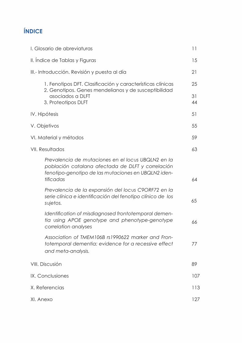
ÍNDICE
I. Glosario de abreviaturas 11
II. Índice de Tablas y Figuras 15
III.- Introducción. Revisión y puesta al día 21
1. Fenotipos DFT. Clasificación y características clínicas 252. Genotipos. Genes mendelianos y de susceptibilidad asociados a DLFT 313. Proteotipos DLFT 44
IV. Hipótesis 51
V. Objetivos 55
VI. Material y métodos 59
VII. Resultados 63
VIII. Discusión 89
IX. Conclusiones 107
X. Referencias 113
XI. Anexo 127
Prevalencia de mutaciones en el locus UBQLN2 en la población catalana afectada de DLFT y correlación fenotipo-genotipo de las mutaciones en UBQLN2 iden-tificadas
Prevalencia de la expansión del locus C9ORF72 en la serie clínica e identificación del fenotipo clínico de los sujetos.
Identification of misdiagnosed frontotemporal demen-tia using APOE genotype and phenotype-genotype correlation analyses
Association of TMEM106B rs1990622 marker and Fron-totemporal dementia: evidence for a recessive effect and meta-analysis.
64
65
66
77

1111
I. Glosario de abreviaturas


1313
I. Glasario de abreviaturas
APP: Afasia Progresiva Primaria
APPnf: Afasia Progresiva Primaria no fluente
APPvs: Afasia Progresiva Primaria variante semántica
APPvl: Afasia Progresiva primaria variante logopénica
aDLFT-U: DLFT atípica con inclusiones de ubiquitina,
BIBD: Enfermedad por cuerpos de inclusión basofílicos
CHMP2B: Charged multivesicular body protein
C9orf72: Chromosome 9 open reading frame 72
DLFT: Degeneración Lobar Frontotemporal
DFT: Demencia Frontotemporal
DFTvc: Demencia Frontotemporal variante de conducta
DFTP-17: Demencia Frontotemporal con Parkinsonismo asociada al cromosoma 17
DFT-ELA: Demencia Frontotemporal con Esclerosis Lateral Amiotrófica
DFLT-U: Degeneración Lobar Frontotemporal por depósitos que se tiñen con Ubiquitina
DLFT-TAU: Degeneración Lobar Frontotemporal por depósitos de proteína Tau
DFLT-TDP: Degeneración Lobar Frontotemporal por depósitos de proteína TDP-43
DLFT-FUS: Degeneración Lobar Frontotemporal por depósitos de proteína FUS
DLFT-UPS: Degeneración Lobar Frontotemporal con inmuno-histoquímica contra proteínas del sistema
ubiquitina-proteasoma
DLFT-ni: Degeneración Lobar Frontotemporal sin inclusiones
DGA: Demencia por Granos Argirófilos
EA: Enfermedad de Alzheimer
EMN: Enfermedad de Motoneurona
ELA: Esclerosis Lateral Amiotrófica
FUS: (gen) Fused in sarcoma
FUS: Tumor associated protein fused in sarcoma
GRN: (gen) Progranulina
MAPT: Microtubule associated protein Tau gen
NIFID: Enfermedad por inclusión de filamentos neuronales intermedios
PSP: Parálisis Supranuclear Progresiva
P62: Proteína de unión a ubiquitina
SCB: Síndrome Cortico Basal
SQSTM1: (gen) sequestrosoma
TARDBP: (gen) TAR DNA Binding Protein
Tau: Proteína Tau asociada a microtúbulos
TDP-43: Transactive response DNA binding protein of 43 kD
TMEM106B: Transmembrane protein 106B
TREM2: (gen) Triggering receptor expressed on myeloid cells 2
UBQLN2: (gen) Ubicuitin 2
VCP: (gen) Valosin-containing protein


1515
II. Índice de Tablas y Figuras


1717
II. Índice de Tablas y Figuras
TABLAS
Tabla 1. Clasificación molecular Fenotipos y genes asociados en la DLFT. 49
Tabla 2. Demografía de la serie clínica DFT en Fundació ACE. 62
Tabla 3: Fenotipo evolutivo de la serie DFT clínica 95
Tabla 4: Patrones de Neuroimagen 96
Tabla 5: Neuropatología disponible de la serie clínica 97
Tabla 6: Variantes TREM2 raras identificadas en la serie y fenotipo asociado 104
Tabla 7: Estudios en colaboración 110
FIGURAS
Figura 1. Diferentes subtipos de TDP-43 46
Figura 2: Fenotipos Clínicos y correlación patológica 92
Figura 3: Genotipos DLFT y correlación patológica 93
Figura 4: Neuropathology of pacient M0010098 100
Figura 5: Neuropatología del paciente con mutación TAU 103


19
ABSTRACT
La Degeneración Lobar Frontotemporal es un grupo heterogéneo de enfer-medades, la segunda causa más frecuente de demencia en edad presenil y la que presenta el mayor número de casos hereditarios. Se caracteriza por una gran varia-bilidad clínica, genética e histopatológica. Las personas afectas pueden presentar síntomas que abarcan desde los trastornos de conducta hasta las diferentes altera-ciones del lenguaje, con o sin enfermedad de motoneurona o parkinsonismo aso-ciado. La atrofia en los lóbulos frontales y temporales es el hallazgo radiológico más relevante. En los últimos 10 años el conocimiento de esta entidad clínica ha presen-tado remarcables cambios a nivel genético e histopatológico, que han servido para establecer criterios clínicos más consistentes. Hasta el momento han sido descritos diez genes asociados a DLFT y cuatro diferentes proteínas de agregación han sido detectadas en los cerebros afectados. Este trabajo aporta la experiencia clínica de más de 15 años en pacientes con DLFT y el trabajo de colaboración con diferentes grupos de investigación en genética de enfermedades neurodegenerativas.
Frontotemporal Lobar Degeneration is a heterogeneous group of disorders, the second most frequent cause of early dementia and the one with the highest number of inherited cases. It is characterized by considerable variability in clinical, genetic and histopathology features. Patients may present symptoms ranging from behavioral disturbances to different language disorders, with or without motor neuron disorders or associated Parkinsonism. Atrophy in frontal and temporal lobes is the most relevant radiological finding. In the last 10 years, the knowledge of this clinical entity has un-dergone remarkable changes both genetically and histopathologically, which have served to establish more consistent clinical criteria. Until now 10 genes causative of dementia have been described and up to four different proteins causative of atro-phy have been detected in aggregates. This work provides the clinical experience of more than 15 years with DLFT patients and the collaborative work with different Gene-tic Research Groups in Neurogenerative Disorders.


2121
III.- Introducción Revisión y puesta al día


2323
III. Introducción. Revisión y puesta al día
La Degeneración Lobar Frontotemporal (DLFT) se caracteriza por una pérdida selectiva, específica y progresiva de neuronas localizadas preferentemente en las regiones frontales y temporales del cerebro humano. Una misma población neuronal puede ser objeto diana de diferentes patologías (proteotipo), por lo que la sintoma-tología de presentación clínica (fenotipo) va a variar dependiendo del área afecta-da.
Representan un grupo de enfermedades con unas bases clínicas, moleculares y ge-néticas heterogéneas, donde convergen a menudo los mecanismos neurodege-nerativos con el fenotipo clínico de presentación. El espectro clínico varía desde la sintomatología de conducta hasta los síndromes afásicos progresivos, parkinsonismo plus y/o enfermedad de motoneurona, asociándose a menudo varios síndromes en un mismo sujeto a lo largo de la evolución clínica (Thelen et al. 2014). Se caracterizan fundamentalmente por cambios progresivos de conducta, disfunción ejecutiva y di-ficultades de lenguaje.
La historia natural de la DLFT va a depender de múltiples factores, tanto intrínsecos (biológicos, genéticos) como extrínsecos (ambientales y sociales). Los factores intrín-secos, implícitamente impuestos, no son controlables por el clínico y van a definir el fenotipo de presentación. No es así con los extrínsecos, que son los que marcarán la variabilidad en la evolución de un fenotipo determinado. Es por ello que estas enfer-medades neurodegenerativas han de ser valoradas en su contexto global, clínico y social, para poder ofrecer, tanto al paciente como a la familia, una orientación adecuada.
Hasta 1994, en que los grupos de investigación de Lund y Manchester proponen los criterios clínicos para el diagnóstico de DLFT (Statement 1994), estos pacientes eran comúnmente diagnosticados de Enfermedad de Alzheimer (EA). Es en el año 1998 (Neary et al. 1998) que son publicados los criterios diagnósticos para este tipo de en-fermedad neurodegenerativa, que la diferencia clínicamente y de forma clara de la EA. Por sus peculiares características clínicas, es una patología que en ocasiones ge-nera dudas y orientaciones diagnósticas equivocadas, dando lugar a tratamientos y recursos sociales alejados de las necesidades del caso.
La DLFT es la segunda causa más común de demencia en individuos menores de 65 años, representando el 5-15% de todas las demencias en este grupo de edad (Bird et al. 2003) (después de EA), y la tercera causa en mayores de 65 después de la de-mencia por cuerpos de Lewy (Arvanitakis 2010).
Factores Genéticos asociados a la Degeneración Lobar Frontotemporal
2424
La prevalencia de la DLFT varía según las series analizadas: desde 2.7/100.000 y 9.4/100.000 en el grupo de 60-69 en la serie de Netherlands (Rosso et al. 2003) hasta 15.1/100.000 en la serie de Cambridge (Ratnavalli et al. 2002). La prevalencia más baja se informa en la serie de Japón, con un 2.0/100.000 (Ikejima et al. 2009) y la más alta en Italia, con un 31/100.000 (Gilberti et al. 2012). Está considerada una demencia presenil y supone el 20-25 % de los todos los casos de demencias que se presentan alrededor de los 65 años, aunque en la serie de Sweden (Gislason et al. 2003) se en-contró una prevalencia del 3% en el grupo de edad de 85 años. En una serie ana-tomopatológica de Newcastle General Hospital (Baborie et al. 2012), el 3.4 % de los pacientes seniles autopsiados presentaban criterios de DLFT, con una edad media de 73.5 [65-92] circunstancia que hace pensar que posiblemente esta infradiagnostica-da en este grupo de edad.
En los últimos 15 años, el espectro de la DLFT ha cambiado notablemente, tanto a nivel clínico como en el conocimiento de la genética asociada y las proteínas de depósito implicadas. No es así en el área de tratamiento, huérfanas aún de posibles terapias.
Para mejor comprensión de los términos, DLFT hace referencia a la enfermedad y su diagnóstico anatomopatológico en todas sus variantes y DFT al síndrome clínico de cualquier presentación fenotípica.

2525
III. Introducción. Revisión y puesta al día
1. FENOTIPOS DFT. CLASIFICACIÓN Y CARACTERÍSTICAS CLÍNICAS.
Las DFT se clasifican, de acuerdo a la característica clínica principal observada en el paciente, como: demencia frontal variante de conducta (DFTvc), afasia progresiva primaria (APP): variante semántica o demencia semántica (DS), afasia progresiva no fluente (APNF), síndrome cortico basal (SCB), síndrome de parálisis supranuclear pro-gresiva (SPSP) y DFT con enfermedad de motoneurona (DFT-EMN).
a. Demencia Frontotemporal variante de conducta (DFTvc)
La DFTvc comprende más de la mitad de los casos de DLFT y es su fenotipo heredi-tario más común. Su debut suele presentarse antes de los 65 años, con una media a los 58 años (K. A. Josephs et al. 2011). Se caracteriza por cambios precoces en la personalidad y la conducta, tales como la desinhibición, a menudo coexistiendo a lo largo de la evolución con apatía, impulsividad, falta de empatía, conductas es-tereotipadas y pérdida de competencia y conducta social. Las funciones ejecutivas se encuentran alteradas precozmente estando la memoria y las funciones visuo-per-ceptivas bien preservadas, tal como revelan los test cognitivos (Sieben et al. 2012). Un grado variable de alteración de lenguaje está también presente y la hiperoralidad y los cambios en los hábitos alimentarios son a menudo comunes, dando lugar a un notable aumento de peso.
De acuerdo con los criterios de “International Behavioral Variant FTD Consortium” (Rascovsky et al. 2011) se clasifica en: DFTvc posible, sólo por criterios clínicos y re-quiriendo la presencia de tres de seis signos de trastornos de conducta y cognición: desinhibición , apatía/inercia, pérdida de empatía, conducta perseverativa e impul-siva, hiperoralidad y perfil neuropsicológico disejecutivo; DFTvc probable cuando se observa declive funcional y la neuroimagen soporta los criterios de posible y DFTvc “definitiva” cuando se dispone de confirmación neuropatológica o evidencia de mutación genética conocida.
Teniendo en cuenta sólo la neuroimagen, cuatro subtipos han sido identificados de-pendiendo de la pérdida de sustancia gris relativa observada: frontal dominante, frontotemporal, fronto-temporo-parietal y temporal dominante (Whitwell et al. 2009). El subtipo frontal dominante engloba los lóbulos frontales y la ínsula anterior. El sub-tipo frontotemporal muestra afectación de los lóbulos frontales, la ínsula anterior, el caudado y el putamen y lóbulo temporal anterior derechos. El subtipo fronto-tempo-

Factores Genéticos asociados a la Degeneración Lobar Frontotemporal
2626
ro-parietal muestra mayor pérdida de materia gris en comparación con el subtipo dominante temporal, caracterizado por la implicación del lóbulo temporal, particu-larmente derecho, medial e inferior.
b. Afasia Progresiva Primaria (APP)
Se debe aplicar el término Afasia Progresiva Primaria a aquella alteración del habla o el lenguaje que se presenta, durante un periodo de al menos dos años, como única queja cognitiva (Mesulam 1982). Pacientes con DFT y dificultades lingüísticas habían sido diagnosticados, a lo largo de los años, en dos categorías: afasia progresiva no fluente (APNF) y demencia semántica (DS). Sin embargo hay pacientes con APP cu-yas características clínicas no cumplen ninguno de esos criterios. Es en 2011 cuando se publican las nuevas recomendaciones para la sub-clasificación de las APP, descri-biendo tres subtipos diferenciados: (ML Gorno-Tempini et al. 2011) afasia progresiva primaria no fluente (APPnf), afasia progresiva primaria variante semántica (APPvs) y afasia progresiva primaria variante logopénica (APPvl).
► APPnf es la segunda presentación clínica más prevalente dentro de las DFT, representando alrededor del 25% (Johnson et al. 2005a) y caracterizada por simplificación gramatical, esfuerzo y vacilación en el habla con errores en la emisión de sonidos y la producción del lenguaje. La comprensión lingüística se encuentra relativamente preservada así como el resto de las funciones cogni-tivas (Grossman 2012). Es frecuente que la afasia se acompañe de apraxia del habla y orolingual (K. A. Josephs et al. 2011). Durante la evolución el lenguaje se vuelve telegráfico, tanto en la expresión oral como en la escritura, seguido de un gradual deterioro de la comprensión de frases y finalmente mutismo y demencia (Turner et al. 1996). La apatía es el cambio de conducta asociado más común. La neuroimagen muestra anormalidades en la región fronto-insu-lar posterior izquierda, giro frontal inferior, ínsula, áreas premotoras y motoras suplementarias, siendo necesarios estos hallazgos para realizar el diagnóstico de APPnf probable (M L Gorno-Tempini et al. 2011). A lo largo de la progresión clínica pueden aparecer signos extrapiramidales tipo SPS o SCB, dando lugar a cambios en el fenotipo y por lo tanto en el diagnóstico clínico.
► La APPvs se caracteriza por la pérdida del significado de las palabras (Hod-ges and Patterson 2007). Basándose en los criterios establecidos, los déficits de comprensión para palabras simples y la nominación por confrontación visual
2727
III. Introducción. Revisión y puesta al día
son los signos principales y esenciales para el diagnóstico (M L Gorno-Tempini et al. 2011), aunque el habla es fluida y la sintaxis correcta. Las alteraciones de conducta también están presentes, como la falta de empatía y la inflexibi-lidad mental. Los déficits de reconocimiento de personas son especialmente presentes cuando el lóbulo temporal derecho es el afectado. La neuroimagen muestra una atrofia significativa de los lóbulos temporales mediales y laterales, aunque más llamativa en el lado izquierdo (Mummery et al. 2000).
► La APPvl se asocia, sobre todo, al diagnóstico neuropatológico de enferme-dad de EA (Rabinovici et al. 2008) y no se considera parte del grupo de las DFT. La importante dificultad en encontrar las palabras (en el lenguaje espontáneo y en las tareas de confrontación verbal) y la alteración en la repetición de fra-ses y oraciones, son los signos claves de esta variante afásica. Son necesarias y apoyan el diagnóstico de APPvl alteraciones radiológicas en las áreas tem-poro-parietal izquierda, temporal posterior y giros angular y supramarginal (ML Gorno-Tempini et al. 2004).
La APP se clasifica como “posible” basándose en las características clínicas. Se cla-sifica como “probable” cuando los hallazgos clínicos están apoyados por las técni-cas de neuroimagen y sólo después del análisis posmortem o teniendo evidencia de la existencia mutación genética, la APP es clasificada como “definitiva” (ML Gor-no-Tempini et al. 2011).
c. Síndromes asociados a la DFT. Síndrome Córticobasal (CBS) y Síndrome de Parálisis Supranuclear Progresiva (SPSP)
Ambos síndromes han sido descritos originalmente como trastornos del movimiento, parkinsonismos atípicos o “Parkinson Plus”, pero muestran una asociación significa-tiva con la DLFT desde el punto de vista clínico, genético y anatomopatológico (A Kertesz et al. 2000).
► El SCB presenta una prevalencia menor de 1/100.000 habitantes y se carac-teriza por síntomas extrapiramidales de evolución progresiva, de tipo rígido y asimétrico y con distonía asociada. La pérdida sensorial cortical, síndrome del miembro ajeno, heminegligencia y mioclonias están presentes. Frecuentemente se presenta combinado con APPnf y en las etapas avanzadas de la enferme-dad, con alteraciones de conducta (A Kertesz et al. 1994); y por el contrario, los pacientes que inicialmente presentaron DFTvc o APPnf, pueden con el tiempo
Factores Genéticos asociados a la Degeneración Lobar Frontotemporal
2828
desarrollar los trastornos del movimiento característicos del SCB (I Ferrer et al. 2003) o SPSP.
► La PSP presenta una prevalencia de 3.1/100.000 habitantes y es un trastorno neurológico primario que se presenta con inestabilidad postural, parkinsonismo de predominio axial con retropulsión, enlentecimiento motor y parálisis de la mirada vertical. Disartria, disfagia y signos pseudobulbares son signos frecuen-temente asociados (Litvan et al. 1996). La disfunción cognitiva se presenta por alteración de los circuitos frontosubcorticales, provocando disfunción ejecutiva, enlentecimiento psicomotor y alteración de la memoria de trabajo (Grafman, Litvan, and Stark 1995). El espectro de SPSP no sólo incluye la clásica PSP tipo Richardson sino también la PSP-parkinsonismo, que se presenta de forma más asimétrica, asemejándose a la enfermedad de Parkinson y al síndrome de aci-nesia pura, caracterizado por bloqueo de la marcha y falta de fluidez del habla como características más prominentes.
Cuatro formas de presentación fenotípica han sido aprobados recientemente por consenso para hablar de criterios clínicos de degeneración cortico basal (DCB) (Arm-strong et al. 2013): síndrome cortico basal clásico (SCB), síndrome frontal con trastor-no conductual y alteración espacial (SFC), variante no fluente agramática de afasia progresiva primaria (vnfaAPP) y síndrome de parálisis supranuclear progresiva (SPSP). Sin embargo, estos criterios precisan de futuras validaciones.
En el caso del SPSP y de cara a mejorar la precisión diagnóstica, el grupo de expertos del National Institute for Neurological Disorders and Stroke (NINDS) han publicado los criterios de NINDS-SPSP (Respondek et al. 2013), donde la combinación de criterios posibles y probables proporciona una sensibilidad más alta en la atención clínica ru-tinaria. Sin embargo los autores sugieren que los criterios de probable son preferibles para el reclutamiento de pacientes en los ensayos clínicos, donde el diagnostico específico y precoz es importante.
d. Síndromes asociados: Enfermedad de Motoneurona (EMN)
La EMN comprende un grupo de enfermedades con pérdida progresiva de neuronas motoras: Esclerosis Lateral Amiotrófica (ELA), Parálisis Bulbar Progresiva (PBP) y Esclero-sis Lateral Primaria (ELP). Clínicamente, la EMN se manifiesta con debilidad progresiva, pérdida de masa muscular y espasticidad, produciendo la muerte por insuficiencia respiratoria a los tres años de media, después de su inicio, en el 50% de los pacientes.

2929
III. Introducción. Revisión y puesta al día
La Esclerosis Lateral Amiotrófica (ELA) es la forma más común de presentación y se caracteriza, patológicamente, por la pérdida progresiva de neuronas motoras supe-riores en la capa 5 de la corteza y neuronas motoras inferiores en los núcleos motores del tronco cerebral y del asta anterior de la médula espinal. La asociación entre la demencia y la ELA se observó hacia finales del siglo XIX y sucesivamente ha sido pu-blicada por muchos investigadores.
Los casos familiares y esporádicos de ELA pueden tener disfunción frontal, cambios de personalidad, conducta, planificación, organización y disfunción de lenguaje. Los síntomas de ELA pueden preceder, presentarse simultáneamente o seguir a los signos y síntomas de DFT, aunque los hallazgos más comunes son encontrar cambios cog-nitivos seguidos de la debilidad muscular (Achi and Rudnicki 2012). En el 25% de los pacientes de ELA, el fenotipo clínico de DFT más comúnmente asociado es la DFTvc y ocasionalmente la APP.
Un consenso clínico de diagnóstico para DFT y EMN, consistente en cuatro ejes, fue propuesto por un grupo de trabajo internacional en 2007 (Strong et al. 2009).
Eje I: diagnóstico clínico de EMN; Eje II: disfunción cognitiva y conductual; Eje III: ma-nifestaciones no motoras adicionales; Eje IV: identificación de la presencia de modi-ficadores de la enfermedad. Tres formas clínicas diferentes pueden ser identificadas de acuerdo con el consenso: ELA con alteración de conducta, ELA con alteración cognitiva y ELA con demencia comórbida (otras no DFT).
e. Demencia Frontotemporal con Parkinsonismo (DFTP-17)
La DFTP-17 fue definida en una conferencia de consenso en 1997 (Foster et al. 1997). La enfermedad fue descrita en 13 familias que presentaban una enfermedad he-reditaria autosómica dominante. Esta enfermedad es un síndrome clínico poco fre-cuente que afecta aproximadamente a 200 familias y en las que unos 639 sujetos son portadores de mutaciones de MAPT (Spillantini, Bird, and Ghetti 2006). Los síntomas de presentación son demencia, desinhibición, parkinsonismo y amiotrofia. En esta-dios tempranos la disfunción de la memoria anterógrada no está presente y pro-gresivamente aparecen disfunción global de memoria, alteración visuoespacial y desorientación. Los signos motores incluyen bradicinesia progresiva, rigidez axial e inestabilidad postural. Su inicio se sitúa hacia los 50 años de edad con un rango de presentación que oscila entre los 20 y los 70 años.
Factores Genéticos asociados a la Degeneración Lobar Frontotemporal
3030
f.EnfermedadporGranosArgirófilos(DGA)
La DGA es una enfermedad neurodegenerativa esporádica, común de la edad senil y caracterizada por la presencia de granos argirófilos en la corteza entorrinal, hipo-campo, amígdala y corteza temporal vecina. Es responsable de aproximadamente el 5% de todos los casos de demencia y puede estar asociada con otras enferme-dades neurodegenerativas (EA, PSP, DCB y sinucleopatías como Cuerpos de Lewy (DCL), Enfermedad de Parkinson (EP) y Atrofia Multisistémica (AMS). La presentación clínica inicial es similar a la EA, pero la progresión de la enfermedad es menos agresi-va, pudiendo manifestarse como deterioro cognitivo leve amnésico durante muchos años. La enfermedad también puede manifestarse como deterioro cognitivo y de-mencia con anomalías de comportamiento, personalidad y cambios emocionales. Estos casos apoyan la propuesta de considerar la DGA como una de las causas de DLFT. (Isidro Ferrer, Santpere, and van Leeuwen 2008).

3131
III. Introducción. Revisión y puesta al día
2. GENOTIPOS. GENES MENDELIANOS Y DE SUSCEPTIBILIDAD ASOCIADOS A DLFT
La DLFT presenta un marcado componente familiar. Entre el 30-50% de los pacientes informan algún familiar con similar sintomatología y al menos un 10-30% se asocia a un patrón de herencia autosómica dominante. Sobreestimar o subestimar la tenden-cia genética dentro de los diferentes subtipos de DLFT y pensar en ella como una única entidad genética puede llevar a engaño. La DFTvc muestra una importante asociación familiar (45%), sobre todo cuando se asocia a la EMN (60%), mientras que la DS presenta una frecuencia muy baja (17%) de los casos familiares. (Goldman et al. 2005).
Al menos el 18% de las familias de DLFT con patrón de herencia autosómica domi-nante tienen una mutación del gen Tau en el cromosoma 17 (Rosso and van Swieten 2002). Otras familias se han vinculado a los cromosomas 3, 9, y a regiones no Tau del cromosoma 17. Otros síndromes con síntomas de DFT, incluyen la miopatía por cuerpos de inclusión asociada con la enfermedad de Paget y de DLFT causada por mutaciones en el gen de la Valosina (VCP).
Hasta ahora diez mutaciones han sido identificadas como causantes de DLFT, siendo las más frecuentes MAPT, GRN and C9orf72, en orden de descripción y publicación. En un grupo de 306 pacientes, se encuentra la expansión de C9orf72 en un 8.2% de la muestra analizada y las mutaciones de GRN y MAPT en un 3.9% y 3.3% respectiva-mente (Wood et al. 2013).
a. Genes más comunes
MAPT: Microtubule associated protein Tau gen
La primera evidencia de una causa genética para la DLFT fue el hallazgo de la aso-ciación del cromosoma 17q21.2 con la forma clínica autosómica dominante de DLFT familiar con parkinsonismo (Lynch et al. 1994). El gen responsable de la mutación, llamado MAPT fue descubierto en 1998 (Hutton et al. 1998). El gen MAPT codifica la proteína Tau asociada a microtúbulos, implicada en el ensamblaje y estabilización de los microtúbulos neuronales.

Factores Genéticos asociados a la Degeneración Lobar Frontotemporal
3232
Hasta la fecha se han descrito 44 mutaciones MAPT patógenas en 134 familias. La frecuencia, según las series analizadas, varía entre un 1.9% y un 8.9% (J D Rohrer et al. 2009). No se ha identificado predilección de género y la edad de inicio se sitúa entre los 25 y 65 años (53±6) con un 100% de penetrancia. La evolución hasta el éxitus es de 3-10 años (Boeve and Hutton 2008).
Las mutaciones se agrupan principalmente en cinco exones (del 9 al 13), donde co-difica los cuatro dominios de unión a microtúbulos de la proteína Tau. En el cerebro normal, la proteína Tau produce seis isoformas de las cuales tres contienen tres domi-nios (Tau 3R) y otras tres contienen cuatro dominios de unión a microtúbulos (Tau 4R). Un número considerable de mutaciones han sido localizadas en la región reguladora de corte y empalme del exón 10, dando lugar a proporciones aberrantes de Tau de 3R y 4R (Sieben et al. 2012)
La presentación clínica de los portadores de mutaciones MAPT es fundamentalmen-te DFTvc, aunque se han publicado casos de APP (Villa et al. 2011). Los síntomas in-cluyen disfunción ejecutiva y alteración de la personalidad y la conducta, evolucio-nando con afasia y parkinsonismo, en muchos casos, y mostrando una severa atrofia del lóbulo temporal. La asociación con ELA es rara. Fenotipos clínicos como EA, DS o SCB raramente se manifiestan.
La neuroimagen estructural muestra pérdida simétrica de volumen cerebral que eng-loba los lóbulos temporales anteromediales, cortex orbitofrontal y tractos de sustan-cia blanca, incluyendo el cuerpo calloso (Jonathan D Rohrer et al. 2010). Similar topo-grafía puede observarse en el SPECT o PET-FDG. Los patrones de atrofia en pacientes portadores pueden ser heterogéneos pero afectan más comúnmente los lóbulos frontales y temporales, aunque la mayor atrofia se produce en el lóbulo temporal, en su mayoría derecho (K. a Josephs et al. 2009) (Whitwell and Josephs 2012). No se ha observado atrofia de cerebelo en estos pacientes (Whitwell et al. 2012).
La DLFT debida a mutaciones de MAPT se caracteriza patológicamente como Dege-neración Lobar Frontotemporal con depósitos de proteína Tau (DLFT-TAU). Filamentos anormales de depósitos de Tau también se han descrito en otras enfermedades neu-rodegenerativas, incluyendo EA, DGA, PSP y DCB.

3333
III. Introducción. Revisión y puesta al día
GRN: Progranulina
Otras mutaciones causantes de DLFT autosómica dominante, en un segundo gen del cromosoma 17q21, llamado Progranulina (GRN) fueron halladas en un serie de familias que previamente habían mostrado estar libres de mutaciones de MAPT. 63 mutaciones heterocigóticas han sido identificadas a nivel mundial en 163 familias, que explican alrededor del 5 al 10% de las DLFT (I Gijselinck, Van Broeckhoven, and Cruts 2008).
En los casos de mutaciones de GRN, el depósito de proteína predominante es una proteína ubiquitinada llamada “TAR DNA binding” (TARDBP or TDP-43) y los depósitos de Tau son raramente observados. La mayoría de mutaciones de GRN conocidas son mutaciones sin sentido que dan lugar a un codón de parada o empalme prematuro que altera la lectura del ARN mensajero (ARNm). El resultado es que el ARNm mutado se degrada por la descomposición mediada por la mutación y no se produce ningu-na proteína a partir del gen mutado, produciendo la DLFT por una haploinsuficiencia de la GRN (Yu et al. 2010). El gen de la GRN está situado centromérico a 1.7Mb del
Paciente de la serie clínica de Fundació ACE, con mutación de MAPT y fenotipo DFTP-17, donde se evidencia la atrofia temporal derecha (Isidro Ferrer et al. 2003)

Factores Genéticos asociados a la Degeneración Lobar Frontotemporal
3434
gen MAPT, en el cromosoma 17q21.31 y codifica un factor de crecimiento implicado en la regulación de varios procesos, incluyendo el desarrollo, la reparación de heridas y la inflamación. El gen también ha sido fuertemente asociado a la tumorogénesis. Además, la expresión de GRN se incrementa en la microglía activada de muchas en-fermedades neurodegenerativas, incluyendo la enfermedad de Creutzfeldt-Jakob, ELA y EA (Baker et al. 2006).
Los fenotipos asociados a mutaciones de GRN varían ampliamente. Un 63% de los portadores desarrollan DFTvc y los demás pueden presentarse fenotípicamente como APP, SCB, DCL o EA. El 41% de los pacientes desarrolla parkinsonismo, el 25% alucina-ciones visuales y el 24% apraxia motora. Los trastornos en la memoria episódica son frecuentes. Según los datos de un estudio francés (Le Ber et al. 2008) sobre una mues-tra de 502 sujetos, la frecuencia de mutaciones de GRN fue del 5.7% en el fenotipo DFTvc (17.9% de ellos con herencia autosómica dominante) 4.4% en fenotipo APP y el 3.3% en el fenotipo DCB. No se encontraron mutaciones en el fenotipo DFT-EMN. La neuroimagen muestra pérdida de volumen asimétrica, involucrando principalmente los lóbulos frontal inferior, temporal superior y parietal inferior, precuneo y cortex cin-gulado, así como tractos de sustancia blanca (Jonathan D Rohrer et al. 2010).
La frecuencia de mutaciones MAPT vs GRN varía según series y países. En Reino Uni-do 8.9% vs 8.4% (J D Rohrer et al. 2009) y 2.9% vs 4.8% (Pickering-Brown et al. 2008). En Francia 2.9% vs 4.8% (Le Ber et al. 2007), en USA 4.4% vs 4.8% (Gass et al. 2006) y en Bélgica 1.9% vs 10.7% (Cruts, Kumar-Singh, and Van Broeckhoven 2006).
Los pacientes con mutaciones de MAPT y GRN no difieren significativamente de otros casos de DLFT en términos de distribución por género. La historia familiar de demen-cia en primer grado está presente en el 100% de los casos de MAPT, 71% de los casos de GRN y en el 39% de otros casos de DLFT (Pickering-Brown et al. 2008) y la edad de inicio en los casos portadores de mutaciones de GRN es amplia (47-79 años), inclu-yendo los miembros de una misma familia (Pietroboni et al. 2011).
C9orf72: chromosome 9 open reading frame 72
Las mutaciones de C9orf72 son la causa genética familiar y esporádica más común de DFTvc (11.7%) y ELA (23.5%). Es un gen de función desconocida hasta el momento, publicado al mismo tiempo en dos cohortes familiares de DFT y ELA en EEUU y Finlan-dia. (DeJesus-Hernandez et al. 2011), (Renton et al. 2011). La mutación fue particu-larmente frecuente en pacientes y familias con DFT-EMN. Se trata de una expansión
3535
III. Introducción. Revisión y puesta al día
del hexanucleotido G4 C2 no codificante en el gen C9orf72. En la población normal, el tamaño de la repetición G4 C2 varía de 3 a 25 unidades, pero ésta se expande al menos 60 unidades en los pacientes afectos, pudiendo llegar a más de 1000. Sin embargo, no se ha encontrado ninguna asociación significativa entre el número de repeticiones, la forma de presentación fenotípica y la edad de inicio de la enferme-dad (Rutherford et al. 2012).
En un extenso estudio realizado posteriormente en 17 regiones de todo el mundo, donde fueron incluidos 4448 pacientes con ELA y 1425 pacientes con DLFT, se han en-contrado diferencias en la frecuencia de la expansión del C9orf72 entre las regiones estudiadas (Majounie et al. 2012). Así, dentro de Europa la frecuencia más alta de mutaciones está presente en la población Finlandesa, con un 1.8% de DFT esporádi-cas (DLFTe), 21.1% de ELA esporádica (ELAe) y 46.5% de ELA familiar (ELAf), estando también presente la mutación en un tercio de los casos de ELA de descendientes consanguíneos europeos (Renton et al. 2011). La expansión de C9orf72 está presente en alrededor del 6% de pacientes con ELAe en Alemania e Inglaterra, en 4.1% de los pacientes italianos con ELAe y en el 2.2% de los casos holandeses de DFTe. En los ca-sos de población blanca de Australia y USA, un 5.0% de pacientes con ELAe también presentan la expansión.
Resumiendo, esta mutación explica una sustancial proporción de casos de ELAe (7.0%) y DFTe (6.0%) en la población blanca. La penetrancia relacionada con la edad muestra que el 50% de los portadores manifiestan la enfermedad hacia los 59 años de edad y que la mutación es totalmente penetrante a los 80 años (Majounie et al. 2012).
El consorcio “European Early Onset Dementia” (EOD) liderado por Chistine van Bro-eckhoven, también ha evaluado, en una muestra de 1205 pacientes, la distribución geográfica de la expansión C9orf72 G4C2 en DLFT en Europa. La serie estaba formada por la cohorte de Flandes (Bélgica) y una cohorte europea de 15 países de Europa occidental (van der Zee et al. 2013). La frecuencia de la expansión C9orf72 fue del 9,98%: 18.52% en la forma familiar y 6,26% en pacientes esporádicos. Finlandia y Sue-cia mostraron la frecuencia más alta de la serie, con un 29,33% y 20,73% respectiva-mente, pero también España con 25,49%. La prevalencia en Alemania se limitó al 4,82%.
En la cohorte de Flandes-Bélgica (305 DLFT, 137 ELA, 23 DLFT-ELA y 856 controles) (Ilse Gijselinck et al. 2012) se ha observado que la expansión del C9orf72 es altamente penetrante, con un 86% de frecuencia en DLFT-ELA familiar y 47% de ELA, y donde la

Factores Genéticos asociados a la Degeneración Lobar Frontotemporal
3636
expansión es la causa genética más común y la única mutación identificada en el grupo de pacientes con DLFT-ELA. En este estudio el grupo DLFT con la expansión de la repetición G5C2 fue la segunda causa más común de enfermedad después de las mutaciones de GRN.
La frecuencia alélica de C9orf72 también ha sido investigada en cuatro enferme-dades neurodegenerativas (520 FTD, 289 ELA, 424 EA y 29 EP con la mutación LRRK2 G2019S) por el grupo de la Universidad de Toronto. La mutación fue detectada en el 9.3% de los pacientes con ELA, 7.2% de los pacientes con DFT y el 0.7% de los pacien-tes con EP, pero no en los controles ni en los pacientes con EA (Xi et al. 2012).
Aunque la mutación C9orf72 parece ser una de las mutaciones más frecuentes aso-ciada con DLFT, es difícil sospechar la presencia de esta mutación en parte debido a los numerosos casos esporádicos. El análisis de haplotipos de todos los pacientes (esporádicos y familiares) portadores de la mutación comparte el haplotipo de riesgo fundador finlandés. Estos hallazgos sugieren que esta mutación podría haber ocu-rrido hace unos 1500 años (una media de 100-105 generaciones) como un evento único y posteriormente diseminada al resto del mundo (Majounie et al. 2012).
Los fenotipos clínicos de presentación más frecuente incluyen DFTvc, ELA o DFT-ELA. Muchos casos con el fenotipo predominante DFTvc pueden tener implicación de neurona motora superior y/o inferior y algunos con fenotipo ELA pueden presentar signos característicos de DFTvc. Los fenotipos APP y SCB no aparecen asociados a esta mutación. La psicosis y los cambios en la conducta alimentaria son comunes. Ansiedad y agitación pueden ser precoces, prominentes y de suficiente significación, en algunos casos, como para contactar tempranamente con los servicios de psiquia-tría (Boeve et al. 2012) (Mahoney et al. 2012).
Los signos neuropsicológicos revelan alteración en la atención, funciones ejecutivas y fluencia verbal. El rendimiento en otros dominios es muy variable (Boeve et al. 2012). El trastorno de memoria domina la presentación clínica en algunos casos, dando lu-gar al diagnóstico inicial de EA (Mahoney et al. 2012).
La neuroimagen muestra perdida cortical extensa, relativamente simétrica en los dos hemisferios e involucrando a lóbulos frontales, temporales y parietales. El análisis mor-fométrico basado en voxels muestra pérdida de sustancia gris en tálamo y cerebelo, lo que podría explicar las características neuropsiquiátricas prominentes en estos ca-sos, como el déficit de memoria episódica, quejas somáticas, alucinaciones y delirios (Tedesco et al. 2011).
3737
III. Introducción. Revisión y puesta al día
b. Otros genes menos comunes asociados con DFT hereditaria
VCP: valosin-containing protein
Las mutaciones del VCP fueron identificadas en el cromosoma 9p21.1 a través de estudios de análisis de ligamiento en familias con herencia autosómico dominante. Los sujetos afectos de dichas familias presentaban debilidad muscular incapacitante debida a miopatía por cuerpos de inclusión, lesiones óseas osteolíticas compatibles con enfermedad de Paget y DFTvc (Inclusion Body Miopathy, Paget and Frontal De-mentia - IBMPFD) (Watts et al. 2004). Los investigadores encontraron 6 mutaciones sin sentido en el gen codificante de la proteína “valosin-containing” (VCP), exclu-sivamente en los 61 individuos afectados. El análisis de haplotipos indicaba que los descendientes de los fundadores, en 2 familias no relacionadas de Norteamérica, suponían más o menos el 50% de las familias afectadas. El gen de la VCP codifica una proteína de la superfamilia de las AAA-ATPasas que facilita la degradación de las proteínas por las vías de la ubiquitina-proteasoma y la autofagia. Los tejidos de los cerebros y músculos afectados en IBMPDF presentan depósitos ubiquitinados e inclu-siones de TAR-DNA binding protein-43 (TDP-43) (Weihl 2011).
Clínicamente, el 90% de los pacientes desarrollan debilidad muscular incapacitante a una edad media de 45 años. Hacia la misma edad, el 51% de los pacientes desa-rrollan enfermedad de Paget y el 32% desarrollan trastornos de conducta y lenguaje con una media de edad de 54 años (Kimonis & Watts , 2005). Sólo el 3% presentan DFTvc como fenotipo aislado (Kimonis et al. 2008). Han sido comunicadas otros sín-tomas, como cardiomiopatía dilatada, esteatosis hepática, cataratas y neuropatía axonal sensitivo-motora. Lo pacientes no siempre expresan los tres componentes del fenotipo, pudiendo expresar uno o dos aisladamente (Guyant-Maréchal et al. 2006).
CHMP2B: Charged multivesicular body protein
La mutación CHMP2B en el cromosoma 3p11.2 fue identificada por análisis de liga-miento en una familia Danesa con miembros afectos de DLFT (Skibinski et al. 2005). CHMP2B es un componente del complejo ESCRT-III, que se requiere para la función de los cuerpos multivesiculares (MVB), una estructura endosomal que se fusiona con el lisosoma para degradar las proteínas por endocitosis. Los estudios funcionales de-muestran una alteración específica de la fusión endosoma-lisosoma, necesaria para la correcta función neuronal (Urwin et al. 2010).

Factores Genéticos asociados a la Degeneración Lobar Frontotemporal
3838
El fenotipo característico es DFTvc, con cambios precoces de personalidad como síntoma principal, acompañado de hiperoralidad e ingestión de objetos no comes-tibles. Los pacientes afectos presentan desinhibición marcada y respuestas emocio-nales inadecuadas. La edad de inicio varía entre 46 y 65 años (media de 57 años) en el pedigrí comunicado por Gydesen (Gydesen et al. 2002) y seguidos durante los 17 años del estudio. El patrón de herencia es autosómico-dominante con alta pene-trancia y observándose anticipación genética en los casos de transmisión paterna. Otras mutaciones en CHMP2B (Q206H y 129V) han sido identificadas en 2 pacientes con ELA y negativas para otras formas conocidas de mutaciones de ELA. (Parkinson et al. 2006). La neuropatología muestra pérdida neuronal y gliosis sin características histopatológicas específicas.
TARDBP: TAR DNA Binding Protein
El gen TARDBP proporciona instrucciones para la fabricación de la proteína “transac-tive response DNA binding protein 43 kDa” (TDP-43). Esta proteína se encuentra den-tro del núcleo de la célula en la mayoría de los tejidos y está implicada en muchos de los pasos de la producción de proteínas.
Las mutaciones de TARDBP fueron identificadas inicialmente como consecuencia di-recta de la identificación de la proteína TDP-43 como la mayor constituyente de los agregados observados en las DLFT ubiquitin positivas (DLFT-U) y en las neuronas moto-ras superiores e inferiores de los pacientes con ELA sin mutaciones SOD1. (Sreedharan et al. 2008). Alrededor del 5% de los pacientes con ELA familiar tienen la mutación TARDBP y es raramente hallada en la DFT-ELA (Benajiba et al. 2009).
FUS: Fused in sarcoma
Las mutaciones de FUS han sido comunicadas como causantes del 4% de los casos de ELA familiar (Vance et al. 2009) (Kwiatkowski et al. 2009). Hasta hoy 15 diferentes mutaciones de FUS han sido descritas en 26 ELA familiares no relacionadas.
El gen FUS está localizado en el cromosoma 16p11.2 y codifica la proteína FUS, miem-bro de un heterogéneo grupo de ribonucleoproteinas nucleares (hnRNP family). Es-tas proteínas comunican información crucial para la maquinaria de traducción y localización del RNAm y la vigilancia de las “nonsense mutations” (Dreyfuss, Kim, and Kataoka 2002).

3939
III. Introducción. Revisión y puesta al día
Mutaciones de sentido erróneo de FUS han sido también identificadas en paciente con DFTvc pura, lo que ha llevado a sugerir que ELA y DLFT son parte del mismo es-pectro clínico, genético y patológico (Van Langenhove et al. 2010). La patología subyacente está caracterizada por depósitos de proteína FUS positivos e inclusiones TDP-43 negativas.
TARDBP y FUS tienen una estructura y funcionalidad similar y muchas de las mutacio-nes en ambos genes también se agrupan en el extremo C-terminal de las proteínas. Los mecanismos moleculares a través de los cuales las TDP-43 y FUS mutadas pueden causar la degeneración de las neuronas motoras no están bien aclarados. Ambas proteínas juegan un importante papel en el transporte de RNAm, el mantenimiento axonal y el desarrollo de las neuronas motoras.
UBQLN2: Ubicuilin 2
Mutaciones en el gen de la UBQLN2, que codifica una proteína ubiquitinada llamada ubiquilin 2 causan ELA y ELA-demencia de causa autosómica dominante ligada al CrX. Cinco mutaciones diferentes en el gen UBQLN2 han sido identificadas en DFT-ELA, ligadas al cromosoma X pero no totalmente penetrantes (Deng et al. 2011), en-contrando también estos autores, correlación de la patología UBQLN2 hipocampal con demencia en los casos de ELA con o sin mutaciones UBQLN2, lo que sugiere que este gen podría estar implicado en la demencia relacionada con la ELA, incluso sin mutaciones UBQLN2.
La proteína ubiquitina 2 es un miembro de la familia de las ubicuilinas, que regulan la degradación de las proteínas ubiquitinadas. El papel de la ubiquitinización en neuro-degeneración ha sido bien establecido en diferentes enfermedades neurodegene-rativas humanas como la enfermedad de Parkinson (Leroy et al. 1998) o el Síndrome de Marinesco-Sjögren (Zhao et al. 2010). Inspecciones preliminares de secuencias no-PXX en 130 casos de ELA familiar en una serie francesa (Millecamps et al. 2012) y en 77 casos de DFT con historia familiar positiva en Cataluña (Hernández et al. 2012), sugieren que las mutaciones en la línea germinal de UBQLN2 son escasas, tanto en DFT como en ELA familiares.

Factores Genéticos asociados a la Degeneración Lobar Frontotemporal
4040
TREM2: Triggering receptor expressed on myeloid cells 2
El gen TREM2 proporciona instrucciones para fabricar una proteína llamada “trigge-ring receptor expressed on myeloid cells 2”, proteína transmembrana tipo I que inte-racciona con la proteína formada por el gen TYROBP llamada “tyrosine kinase- bin-ding protein”. Ambas proteínas, TREM2 y TYROBP forman un complejo que transmite señales químicas para la activación de la respuesta inmune en macrófagos y células dendríticas. TREM2 se expresa en macrófagos y células dendríticas pero no en granu-locitos o monocitos, sugiriendo el rol de TREM2 más bien en los estados crónicos que en los inflamatorios (http://omim.org/entry/605086).
Mutaciones en ambos genes han sido asociadas con la “osteodisplasia poliquística lipomenbranosa con leucoencefalopatia esclerosante” (PLOST) o enfermedad de Nasu-Hakola. Esta enfermedad está asociada con fracturas patológicas por lesiones óseas poliquísticas (de inicio en la tercera década de la vida) seguido de cambios progresivos de conducta y personalidad (consistentes con DFTvc) en la siguiente dé-cada (Paloneva BM et al. 2001). En tres de 44 pacientes turcos con diagnóstico clíni-co de DFT-like, fueron identificadas en TREM2 diferentes mutaciones homocigóticas, que habían sido previamente asociadas a cambios de conducta y deterioro cogni-tivo con signos motores pero sin los fenotipos ni quistes óseos asociados con PLOST.
Un estudio reciente llevado a cabo por “The Dementia Genetic Spanish Consortium” (DEGESCO) (Ruiz et al. 2014) evaluando el papel del polimorfismo p.R47H del gen TREM2 en 3172 EA, 682 DLFT y 2169 controles sanos, como factor de riesgo para la EA y la DLFT, concluye que el 0.6% de los pacientes EA son portadores de esta variante, comparado con el 0.1 % de los controles [OR]= 4.12 sugiriendo que esta rara variante no está relacionada con la DLFT pues no apareció en ningún caso estudiado.
Otro estudio reciente llevado a cabo en la Universidad de Bonn y en colaboración con DEGESCO (Thelen et al. 2014) para detectar otras variantes raras de TREM 2 en los diferentes subtipos de DFT-S (DFT síndromes), ha identificado 7 variantes raras (p.A28V, p.W44X, p.R47H, p.R62H, p.T66M, p.T96K, y p.L211P) y una nueva mutación de cam-bio de aminoácido (p.A105T). La variante p.R47H fue hallada en 4 pacientes con diagnóstico clínico de DFT-S pero dos de ellos mostraban bioquímica el LCR típica de EA, lo que sugiere que estos pacientes presentan fenotipo DFT-S pero una patología EA subyacente. No se encontró asociación con esta variante en DFT. Si se asocia-ron a DFT las variantes p.T96K y p.L211P. No se encontró alteración en ninguno de los controles.

4141
III. Introducción. Revisión y puesta al día
SQSTM1: Sequestrosoma 1
SQSTM1 codifica la proteína P62 (también llamada sequestrosoma 1) una nueva pro-teína de unión a ubiquitina que tiene un importante rol en la degradación de pro-teínas vía proteasoma y autofagia, siendo importante esta última vía en la digestión lisosomal de los constituyentes celulares. La acumulación de la P62 no es específica para la ELA y la DLFT, habiéndose hallado también en la Enfermedad de Alzheimer, Parkinson, Atrofia Multisistémica y enfermedad de Pick. También ha sido observada en inclusiones citoplasmáticas TDP-43, ubiquitina y UBQLN2 en pacientes DLFT con ELA (Appel and Rowland 2012). Recientemente se han identificado mutaciones en los casos familiares y esporádicos de ELA (Teyssou et al. 2013).
En un estudio reciente se ha analizado la secuencia de codificación para las mu-taciones de SQSTM1 en una cohorte de 1.808 pacientes con DLFT procedentes del consorcio EOD. (van der Zee et al. 2014). Como controles se han utilizado 1.625 indi-viduos europeos y se han analizado los datos de todo el exoma de 2274 individuos alemanes (total n = 3899). Se ha realizado también en un meta-análisis de 4332 FTLD y 10.240 alelos de control la asociación con SQSTM1. El trabajo ha identificado 25 variantes en la región codificante de SQSTM1 en pacientes con DLFT, de las cuales 10 eran nuevas. Quince mutaciones estaban ausentes en los controles (frecuencia por-tadores <0,00026), mientras que las otras eran poco frecuentes en ambos (pacientes y controles). El estudio ha demostrado que las mutaciones SQSTM1 están asociadas a la patología TDP-43.
Factores Genéticos asociados a la Degeneración Lobar Frontotemporal
4242
c. Genes de susceptibilidad
TMEM106B: transmembrane protein 106B y otros
Para buscar nuevos genes asociados a DLFT, un equipo internacional liderado por la Facultad de Medicina de la Universidad de Pennsylvania (USA), realizó en 2010 un estudio amplio de asociación del genoma (GWAS) en 515 pacientes DLFT con inclu-siones de TDP-43 confirmadas en autopsia y 2509 controles poblacionales (Deerlin et al. 2010). Todos los casos cumplían criterios genéticos o patológicos para DFT-TDP, lo que fue confirmado por la detección de mutaciones o de inclusiones TDP-43 me-diante inmuno-histoquímica. Los autores encontraron, tras el mapeo en el Cr.7p21, múltiples SNPs asociados a DFT-TDP. Esta región contiene el gen TMEM106B. Tres SNPs mostraban asociación genética después de múltiples pruebas de corrección; el ale-lo menor (C) del SNP rs1990622 se mostró como factor genético de protección para DLFT-TDP. La asociación se replicó en 89 casos autopsiados de DFT-TDP y 553 contro-les, pero no en una serie clínica de 192 DFT.
Estudios independientes intentando replicar estos hallazgos en series clínicas han mos-trado resultados controvertidos. Así, en la cohorte clínica de Londres y Manchester (520 casos de DFT y 247 controles) no se encontró ninguna asociación en TMEM106B (Rollinson et al. 2011). Sin embargo, usando la cohorte de Flandes (Bélgica), com-puesta principalmente por pacientes clínicos (288 DFT y 595 controles) se confirmó la asociación con TMEM106B rs1990622 [OR=0.75 (0.61–0.93)] (van der Zee et al. 2011).
Los autores identifican TMEM106B como un factor de riesgo importante para la DLFT con patología TDP-43. La asociación más significativa ha sido observada en aquellos casos con mutaciones de GRN (Lang et al. 2012).
Se ha realizado otro GWAS analizando un total de 3.526 casos y 9402 controles de descendientes europeos (Ferrari et al. 2014).
Los distintos fenotipos clínicos de DFT se analizaron por separado, con los siguientes resultados:
► El locus TOMM40/APOE superó la significación estadística en la DFTvc, pero no en DS, APNF y DFT-ELA. Esta asociación puede venir a reflejar una contami-nación (~15%) de EA en casos clínicamente diagnosticados de DFT.
► En el locus TMEM106B el estudio sólo mostró una discreta asociación en DFTvc (P = 0.00585), pero no se encontró asociación con los otros subtipos de DFT. En este estudio los portadores de mutaciones de GRN fueron excluidos.

4343
III. Introducción. Revisión y puesta al día
Vale la pena señalar que la publicación original incluía un número de porta-dores de mutaciones GRN (Deerlin et al. 2010) y que la evidencia bioquímica ha sugerido que TMEM106B está directamente relacionado con el metabolis-mo de GRN.

Factores Genéticos asociados a la Degeneración Lobar Frontotemporal
4444
3. PROTEOTIPOS DLFT
Múltiples son las anormalidades neuropatológicas asociadas a la DLFT. Las caracte-rísticas clínicas y neuropsicológicas ayudan a orientar el posible espectro patológico en el paciente diagnosticado de DFT, pero son los biomarcadores, junto con el fe-notipo clínico los que van a predecir la neuropatología subyacente. Todas la DLFT están asociadas a grados variables de atrofia, pérdida neuronal y gliosis en los lóbulos frontales y temporales. Sin embargo, cada enfermedad difiere una de otra por las diferencias en el depósito de proteínas, la firma bioquímica y la morfología y distribu-ción de las inclusiones.
Tres proteínas principales han sido identificadas en el mecanismo de la neurodege-neración de las DLFT. Éstas son la “microtubule-associated protein” (Tau) (Hutton et al. 1998), la “transactive response DNA binding protein of 43 kD” (TDP-43) (Neumann et al. 2006)(Arai et al. 2006), y la “tumor associated protein fused in sarcoma” (FUS) (Kwiatkowski et al. 2009). Algunos de los más renombrados neuropatólogos (I R A Mackenzie et al. 2009) recomiendan que la terminología fenotípica de las DLFT debe conservar las nomenclatura asociada a las entidades clínicas, como DFT, APNF y/o DS, mientras que las subdivisiones neuropatológicas deben designarse por la proteí-na anómala patógena más característica que ha dado lugar al proceso clínico (es decir DLFT-proteína). Por lo tanto, en el estrato más alto, la mayoría de las DLFT deben ser sub-clasificadas en DFLT-Tau, DLFT-TDP y DLFT-FUS.
(Tabla 1, pag 49)
a. DLFT-TAU
La proteína Tau (proteína asociada a microtúbulos) se acumula tanto en neuronas como en células gliales. En casos esporádicos los acúmulos pueden formar cuerpos de Pick en neuronas (DLFT-Tau [enfermedad de Pick]), astrocitos estrellados (DLFT-Tau [Parálisis Supranuclear Progresiva]) o placas astrocíticas (DLFT-Tau [Degenera-ción Cortico-Basal]), mientras que en los casos hereditarios, estos pueden presentarse como inclusiones similares a una de ellas o como patología Tau única (Halliday et al. 2012).
4545
III. Introducción. Revisión y puesta al día
Alrededor del 40% de los pacientes con DLFT muestran inclusiones Tau. Estos incluyen la mayoría de los casos de APNF, ~45% de los casos de DFTvc y algunos casos de DS (Piguet et al. 2011). Crecientes evidencias sugieren que la presencia de estas lesiones neurofibrilares en la fase final de la enfermedad no son la causa de la pérdida neuro-nal, sino que la neurodegeneración está provocada por la alteración de la proteína Tau soluble. En particular, la fosforilación Tau aberrante es reconocida como clave en el proceso de la enfermedad, influyendo en la estructura de Tau, su distribución y su función en las neuronas (Noble et al. 2013).
La proteína Tau es el componente principal de los ovillos neurofibrilares observados en los cerebros de pacientes con enfermedad de Alzheimer y otras enfermedades neurodegenerativas. Es una proteína altamente soluble que se encuentra predomi-nantemente en las neuronas. Seis isoformas diferentes de Tau son expresadas en el sistema nervioso adulto gracias a los procesos de maduración alternativa del gen MAPT, que comprende 16 exones y se encuentra en el cromosoma 17q21.3. La inclu-sión regulada de los exones 2 y 3 dan lugar a isoformas de Tau con 0, 1 ó 2 inserciones N-terminal, mientras que la exclusión o inclusión del exón 10 conduce a la expresión de isoformas de Tau con tres (3R) o cuatro (4R) repeticiones de unión a microtúbulos. En el cerebro humano normal la proporción de 4R-3R es aproxima-damente de uno, mientras que en muchas taupatías la proporción está alterada. Así, la PSP, la DCB y la enfermedad por granos argirófilos (EGA) presentan una sobre-expresión de isoformas Tau de 4R, mientras que la enfermedad de Pick esta principalmente caracterizada por inclusión de isoformas Tau de 3R (Noble et al. 2013).
b. DLFT-TDP
Más del 50% de los pacientes con DLFT que presentan una tinción negativa para in-clusiones Tau, pero son positivas para tinciones de Ubiquitina y el 80-90% de este gru-po está compuesto por inclusiones TDP-43 (“transactive response (TAR) DNA-binding protein 43”) (Roeber et al. 2008). TAR DNA-binding protein 43 (TDP-43) es una de las principales proteínas de la DLFT con inclusiones ubiquitín-positivas (DLFT-U), Tau-nega-tivas con o sin enfermedad de motoneurona.
TDP-43 es una proteína de unión al ADN nuclear implicada en la regulación transcrip-cional y que se deposita de forma aberrante en inclusiones citoplásmicas filamento-sas después de una serie de modificaciones post-traducción, incluyendo proteólisis, fosforilación y ubiquitinación (Neumann et al. 2006).
Factores Genéticos asociados a la Degeneración Lobar Frontotemporal
4646
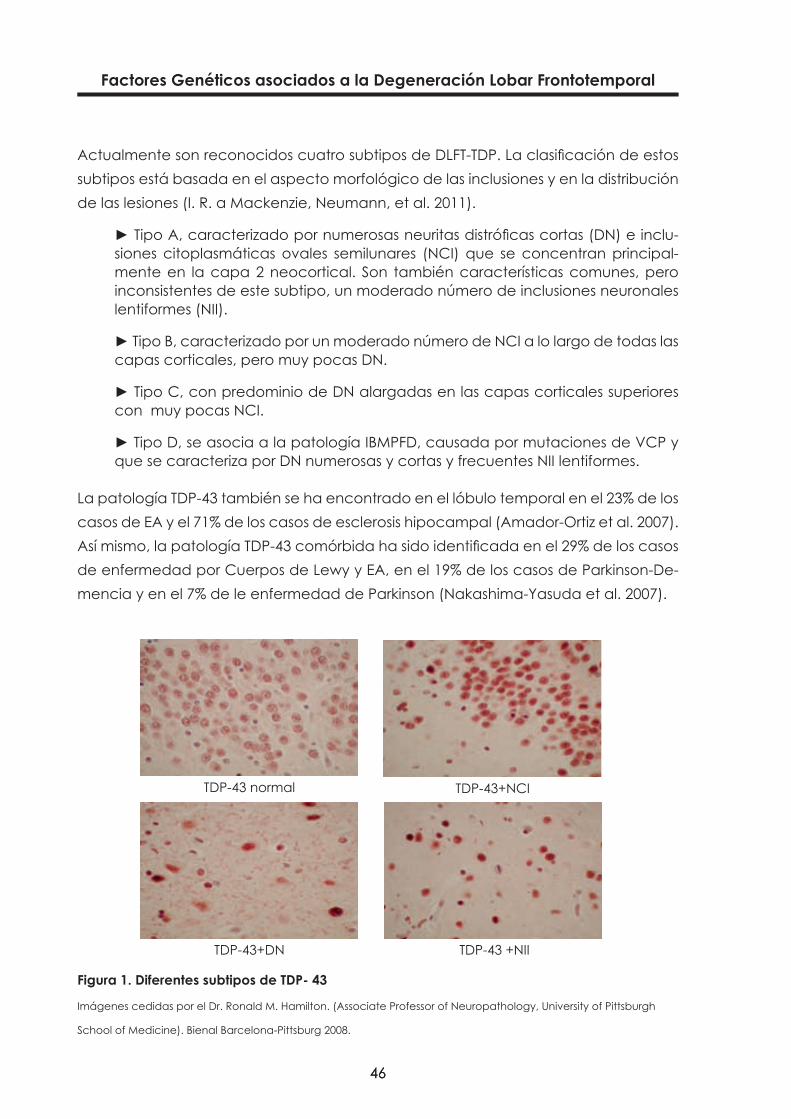
Actualmente son reconocidos cuatro subtipos de DLFT-TDP. La clasificación de estos subtipos está basada en el aspecto morfológico de las inclusiones y en la distribución de las lesiones (I. R. a Mackenzie, Neumann, et al. 2011).
► Tipo A, caracterizado por numerosas neuritas distróficas cortas (DN) e inclu-siones citoplasmáticas ovales semilunares (NCI) que se concentran principal-mente en la capa 2 neocortical. Son también características comunes, pero inconsistentes de este subtipo, un moderado número de inclusiones neuronales lentiformes (NII).
► Tipo B, caracterizado por un moderado número de NCI a lo largo de todas las capas corticales, pero muy pocas DN.
► Tipo C, con predominio de DN alargadas en las capas corticales superiores con muy pocas NCI.
► Tipo D, se asocia a la patología IBMPFD, causada por mutaciones de VCP y que se caracteriza por DN numerosas y cortas y frecuentes NII lentiformes.
La patología TDP-43 también se ha encontrado en el lóbulo temporal en el 23% de los casos de EA y el 71% de los casos de esclerosis hipocampal (Amador-Ortiz et al. 2007). Así mismo, la patología TDP-43 comórbida ha sido identificada en el 29% de los casos de enfermedad por Cuerpos de Lewy y EA, en el 19% de los casos de Parkinson-De-mencia y en el 7% de le enfermedad de Parkinson (Nakashima-Yasuda et al. 2007).
Figura 1. Diferentes subtipos de TDP- 43
Imágenes cedidas por el Dr. Ronald M. Hamilton. (Associate Professor of Neuropathology, University of Pittsburgh
School of Medicine). Bienal Barcelona-Pittsburg 2008.
TDP-43 normal
TDP-43+DN TDP-43 +NII
TDP-43+NCI

4747
III. Introducción. Revisión y puesta al día
c. DLFT-FUS
Como se ha visto hasta ahora, muchos casos de DLFT están caracterizados por una acumulación intracelular anormal de proteínas Tau o TDP-43, pero más del 10% de los casos están compuestos por una acumulación heterogénea de depósitos poco comunes. El 10-20% de DLFT no muestran evidencia de TDP-43 anormal y algunos sub-tipo de DLFT Tau/TDP-43 negativos son inmunoreactivos (ir) para la proteína “fused in sarcoma” (FUS) (I. R. a Mackenzie, Muñoz et al. 2011).
La categoría DLFT-FUS hace mención a tres enfermedades relativamente raras: la enfermedad por inclusión de filamentos intermedios (NIFID), la enfermedad por in-clusión de cuerpos basofílicos (BIBD) y la DLFT atípica con cambios inmunoreactivos sólo para ubiquitina (aDLFT-U). Estas tres entidades comparten el hecho de que todas muestran inmunoreactividad para FUS, pero las características encontradas entre cada una de ellas permite que sean consideradas entidades patológicas diferentes (K. A. Josephs et al. 2011).
► NIFID: DFT esporádica de presentación típica en edad temprana, asociada a signos piramidales y/o extrapiramidales. Los hallazgos inmunohistoquímicos co-munes en todos los casos son inclusiones de alfa-internexina y de neurofilamen-tos positivos citoplasmáticos, sin las densidades comparables a otras enferme-dades neurodegenerativas (Roeber et al. 2006).
► BIBD: término usado para un pequeño número de casos clínicos y patológica-mente heterogéneos que tienen en común el hallazgo de NCI que son basofíli-cos para las tinciones de hematoxilina y eosina (inclusiones basofílicas [BI]). Los fenotipos clínicos incluyen ELA esporádica, ELA familiar, ELA con demencia y DFT pura (I. R. a Mackenzie, Munoz, et al. 2011).
En ambas patologías los signos iniciales pueden incluir: debilidad y alteración de me-moria en BIBD y disartria en ambas. En alguno de los casos, la demencia de desarrolla más de un año después del inicio de los síntomas. Se han observado signos de neuro-na motora superior e inferior, parkinsonismo y signos parietales en ambas enfermeda-des. También aparecen movimientos involuntarios en BIBD. Un hallazgo consistente en las dos patologías es la severa atrofia de caudado (Yokota et al. 2008).
► aDLFT-U: Es un fenotipo clínico esporádico e inusual de inicio temprano (me-dia de 35 años). Se caracteriza por una rápida y severa alteración conductual en ausencia de déficits de lenguaje o motores significativos (Ian R A Mackenzie et al. 2008). La neuropatología es también atípica, mostrando NCI y NII que

Factores Genéticos asociados a la Degeneración Lobar Frontotemporal
4848
aparecen como filamentos largos y gruesos, que pueden ser rectos, curvos o sometidos a torsión (vermiformes). Los NCI y los NII en la aDLFT-U son sistemáti-camente inmunoreactivos con anticuerpos contra FUS (Neumann et al. 2009).
En aproximadamente el 50% de los casos de NIFID y BIBD se encuentra una dege-neración de motoneurona, pero ésta no ha sido comunicada en la aDLFT-U (K. A. Josephs et al. 2011).
d. DLFT-UPS
DLFT-UPS se designan a aquellos casos con inclusiones que sólo se pueden demos-trar inmuno-histoquímicamente contra el sistema ubiquitina proteasoma (UPS) (I R A Mackenzie et al. 2009).
La DFT-3 (mutaciones en el gen CHMP2B) es la principal entidad asociada con la patología DLFT-UPS. Esta designación reconoce que las inclusiones TDP-43 negativas pueden presentar inmunotinción para proteínas UPS diferentes a la ubiquitina, como la p62 (SQSTM1).
El examen macroscópico muestra una atrofia cortical generalizada, más acusada en las cortezas frontal y temporal. La microscopía muestra pérdida de neuronas cor-ticales, microvacuolación de la capa II, gliosis leve y desmielinización de la sustancia blanca profunda. Inclusiones raras ubiquitina-positiva también se encuentran en neu-ronas frontales y temporales corticales, positivas para p62, pero no para TDP-43 (Holm et al. 2007).
La proteína p62 es codificada por SQSTM1 (también llamado sequestrosoma 1), La acumulación de p62 no es específica de ELA y DLFT. Inclusiones ubiquitina-positivas conteniendo p62 ha sido comunicada en EA, EP, Atrofia Multisistémica y enfermedad de Pick. La p62 también se puede encontrar asociada a inclusiones citoplasmáticas TDP-43 (+), ubiquitina y en pacientes con DLFT con EMN por mutaciones de UBQLN2. La presencia de p62 en las inclusiones ubiquitina (+) en diferentes enfermedades neu-rodegenerativas, centra la atención en un tema común de importancia potencial en estos trastornos. Es decir, el papel en los procesos neurodegenerativos de las proteí-nas mal plegadas, la deficiente digestión proteosómica y la autofagia (Appel and Rowland 2012).

4949
III. Introducción. Revisión y puesta al día
Tabla1.Clasificaciónmolecular,fenotiposygenesasociadosenlaDLFT.
ClasificaciónMolecular
Inclusiones patológicas
Subtipo patológico
Fenotipo Clínico asociado
Genes asociados y localización
DLFT-Tau Tau (+)
PiD (3R) DFTvc
MAPT (17q21.1)
DCB (4R) APNF
PSP (4R) APNF
EGA (4R) DFTvc
NFT-demencia (3+4R) DFTvc
MSTD (4R) DFTvc
DLFT-TDP
TDP-43(+), NCI, DN Tipo A DFTvc
APNFGRN (17q21.23)
TDP-43(+), p62(+), NCI, DN Tipo B
DFTvc
EMN con DFTC9orf72 (9p21.2)
TDP-43(+), NCI, DN Tipo CDS
DFTvc
TDP-43 (+), NCI, NII, DN Tipo D IBMPFD Familiar VCP (9p13.3)
TDP-43 (+) ELA TARDBP (1p36.22)
DLFT-FUS
Ubicuitin(+)
TDP-43 (-), NCI, NII
NIFID
BIBD
aDLFT-U
DFTvc FUS (16p11.2)
DLFT-otrasUbicuitin (+), TDP-
43 (-), NCI DLFT-ni
DLFT-UPSDFTvc CHMP2B (3p11.2)
DLFT-Tau: DLFT caracterizada por inclusiones Tau inmunoreactivas, DLFT-TDP: DLT caracterizada por inclusiones TDP-43 inmunoreactivas. DLFT-FUS: DLFT caracterizada por inclusiones FUS inmunoreactivas, DFLT-otras: DLFT in-clasificables (inclusions inmunoreactivas no Tau, no TDP-43, no FUS), Tau(+): Inclusiones Tau positivas, TDP-43(+): Inclusiones TDP-43 positivas, NCI: Inclusiones neuronales citoplasmáticas, DN: Neuritas distróficas, NII: Inclusiones intraneuronales, p62(+): Proteína de unión a ubiquitina, Ubicuitin (+) : Ubiquitina, 3R: Tres repeticiones , 4R: Cuatro repeticiones, PiD: enfermedad de Pick, DCB: Degeneración Cortico-Basal, PSP: Parálisis Supranuclear Progresiva, EGA: Enfermedad por granos argirófilos, NFT-demencia: Demencia por Ovillos Neurofibrilares, MSTD: Taupatía multisistémica esporádica, DFTvc: Demencia Frontotemporal variante de conducta, APNF: Afasia Progresiva no fluente, EMN: Enfermedad de Motoneurona, DS: Demencia Semántica, SCB: Síndrome Corticobasal, IBMPFD: Mio-patía por cuerpos de inclusión con enfermedad de Paget y DFT, NIFID: Enfermedad por inclusión de filamentos neuronales intermedios, BIBD: Enfermedad por cuerpos de inclusión basofílicos , aDLFT-U: DLFT atípica con inclu-siones de ubiquitina, FTLD-ni: DLFT sin inclusiones, DLFT-UPS: DLFT con inmuno-histoquímica contra proteínas del sistema proteosomal de la ubiquitina, MAPT: Gen de la proteína Tau asociada a microtúbulos, GNR: Progranulina, C9orf72: Cr9 open reading frame 72, VCP: valosin-containing protein, TARDBP: TAR DNA-binding protein, FUS: Fu-sed in sarcoma, CHMP2B: Charged multivesicular body protein 2B.


5151
IV. Hipótesis


5353
IV. Hipótesis
Las enfermedades neurodegenerativas en general y la demencia Frontotemporal en particular tienen una importante base genética. Desde un punto de vista estric-tamente genético, la heterogeneidad alélica y no alélica son la norma de estas en-fermedades. Ello implica que existe cierta convergencia anátomo-patológica y clí-nica de alteraciones genéticas muy diversas y que afectan a funciones neuronales radicalmente distintas. La identificación de patrones clínicos o fenotípicos con las diferentes mutaciones de base es una tarea compleja que requiere grandes series epidemiológicas bien fenotipadas y un análisis molecular muy preciso.
Durante este trabajo de investigación se profundiza en el análisis de los perfiles clíni-cos y evolutivos de las demencias frontotemporales y su correlación con el genotipo identificado en los pacientes.


5555
V. Objetivos

5757
V. Objetivos
Al diseñar este trabajo se ha tenido en cuenta la gran variabilidad genética de la DLFT. Por tanto, su abordaje siempre se ha de enmarcar bajo un prisma multicéntrico y multidisciplinar. Nuestro objetivo siempre ha sido abordar los estudios concretos que se han planteado sin que éstos se solapen con los trabajos de los diferentes grupos de investigación con los que ya se colaboraba. A estos centros se les ha cedido parte de las muestras disponibles de la serie clínica para que realicen estudios en otros loci. Estas colaboraciones han generado diversas publicaciones (ver Anexo) y comple-mentan el estudio genético de los pacientes disponibles en la institución. Teniendo en marcha numerosas colaboraciones, se optó por abordar los genes más recientes y menos conocidos al diseñar los objetivos de este trabajo de tesis (UBQLN2, C9orf72 y TMEM106B).
Así, los objetivos planteados han sido los siguientes:
1. Analizar la prevalencia de mutaciones en el locus UBQLN2 en pacientes con historia familiar de demencia frontotemporal dentro de la serie clínica aporta-da y realizar la correlación fenotipo-genotipo de las mutaciones identificadas.
2. Estudiar la prevalencia de la expansión del locus C9ORF72 en la serie clínica aportada para posteriormente realizar la correlación fenotípica en los porta-dores de la mutación.
3. Realizar una correlación genotipo-fenotipo de la serie clínica de DFT, usan-do el polimorfismo ApoE como elemento discriminante para identificar sujetos con posible Enfermedad de Alzheimer (EA) de predominio frontal, que pue-dan estar contaminando la serie clínica.
4. Estudiar la presencia del polimorfismo de riesgo del SNP rs1990622 del locus TMEM106B en pacientes con fenotipo DFT, excluyendo de la serie clínica aque-llos sujetos con confirmación anatomopatológica de proteotipo Tau, para posteriormente realizar la correlación fenotípica.


5959
VI. Material y métodos


6161
VI. Material y métodos
La metodología empleada en la presente tesis doctoral se explica con detalle en el correspondiente apartado de los trabajos de investigación presentados. En este apartado tan sólo se describen las características de la serie utilizada, recogida en su totalidad en Fundació ACE.
De un total de 17.042 sujetos valorados en la Unidad de Memoria de Fundació ACE, en primera visita (periodo 1996-2012), 9102 (53%) cumplían criterios de demencia se-gún los diferentes criterios internacionales y 5218 (31%) criterios de Deterioro Cognitivo Leve. De los sujetos diagnosticados de demencia, 495 (5,4%) cumplían criterios de DFT posible o probable en sus diferentes variantes fenotípicas.
Para este trabajo de tesis se han seleccionado 224 (45.2%) de los 495 sujetos diag-nosticados de DFT, ya fuera en primera visita o que durante el seguimiento clínico modificaran su fenotipo hacia DFT y que dispusieran de suficientes variables clínicas evolutivas y material biológico. Todos ellos disponían de valoración neurológica y neuropsicológica, y en 180 (80.3%) se ha podido rescatar la neuroimagen histórica para su posterior valoración.
La valoración de DFT por neuroimagen la llevaron a cabo dos neurólogos clínicos de la Unidad de Memoria ciegos para el fenotipo inicial y evolutivo de los sujetos y clasi-ficándolos en 6 patrones de atrofia cerebral.
La serie anatomopatológica disponible ha sido posible gracias a las donaciones de los familiares de los pacientes y en colaboración con el Banco de Tejidos Neurológi-cos de la Universidad de Barcelona (IDIBAPS).
La muestra de sangre para los estudios genéticos, su obtuvo directamente de los pa-cientes. Se registró la firma de un consentimiento informado por parte del paciente o de sus representantes legales, de acuerdo con el protocolo GIPSY aprobado por el comité ético del Hospital Clínico y Provincial de Barcelona. El consentimiento informa-do está en consonancia con las leyes biomédicas españolas (Ley 14/2007, 3 de julio, de investigación biomédica y Real decreto 1716/2011, de18 noviembre).

Factores Genéticos asociados a la Degeneración Lobar Frontotemporal
6262
Diag
nóst
ico
Basa
l
Dem
ogra
fía N
(%)
N (%
)V
arón
(%)
Educ
ació
n>6
año
s (%
)
Año
s de
evol
ució
n al
dia
gnós
-tic
o (
)
Año
s al
dia
gnós
tico
()
Hist
oria
fa-
milia
r N (%
)M
MSE
()
Psiq
uiát
rico
3(1.
3)33
.333
.36±
7.1
70.3
±4.9
2 (6
6.7)
19.3
±2.
8
SCB
14 (6
.3)
64.3
57.1
3±2.
171
.7±6
.84
(28.
6)21
.7±7
.4
PSP
16 (7
.1)
56.3
68.8
2.5±
2.2
72.3
±74
(25.
0)23
.1±3
.7
EA8
(3.6
)50
.050
.02.
0±1.
767
.5±1
2.5
2 (2
5.0)
22.7
±4.3
DCL
17 (7
.6)
58.8
29.4
2.4±
2.1
72.8
±3.8
9 (5
2.9)
26.8
±3.3
APN
F32
(14.
3)31
.362
.52.
4±1.
571
.4±7
.814
(43.
8)20
.7±7
.7
DFTv
c99
(44.
2)54
.570
.73.
4±2.
469
.6±1
0.9
51 (5
1.5)
22.2
±6.4
DS33
(14.
7)60
.666
.73.
0±1.
869
.4±1
1.8
11 (3
3.3)
21.2
±5.8
DFT-
EMN
2 (0
.9)
50.0
100
1.5±
0.7
64.5
±0.7
1 (5
0.0)
26.0
±1.4
TOTA
L22
452
.763
.83,
0±2.
370
.3±9
.398
(43.
8)22
.2±6
.2
Tabla 2. Demografía de la serie clínica DFT en Fundació ACE.

6363
VII. Resultados
Factores Genéticos asociados a la Degeneración Lobar Frontotemporal
6464
TRABAJO I
Prevalencia de mutaciones en el locus UBQLN2 en la población catalana afectada de DLFT y correlación fenotipo-genotipo de las mutaciones en UBQLN2 identificadas.
Ver Anexo: 1er artículo
6565
VII. Resultados
TRABAJO II
Prevalencia de la expansión del locus C9ORF72 en la serie clínica e identificación del fenotipo clínico de los sujetos.
Ver Anexo: 2º artículo
Factores Genéticos asociados a la Degeneración Lobar Frontotemporal
6666
Trabajo III
Identification of misdiagnosed frontotemporal dementia using APOE genotype and phenotype-genotype correlation analyses
Hernández I, Mauleón A, Rosense-Roca M, Alegret M, Vinyes G, Espinosa A, Sotolon-go-Grau O, Becker JT, Valero S, Tarraga L, López OL, Ruiz A, Boada M.
Curr Alzheimer Res. 2014 Feb; 11(2):182-91.
IF: 3.676 (2014) Q1
67
182 Current A/zheimer Research, 2014, 11, 182-191
ldentification of Misdiagnosed Fronto-Temporal Dementia Using APOEGenotype and Phenotype-Genotype Correlation Analyses
Isabel Hernández1, Ana Mauleón1, Maiteé Rosen se-Roca1, Montserrat Alegret1, Georgina Vinyes1, Anna Espi-nosa1, Osear Sotolongo-Grau1, James T. Becker2, Sergi Valero3, Lluís Tárraga1, Osear L. López2, Agustín Ruiz1’* and Merce Boada1,4
1Fundació ACE. Barcelona Alzheimer’s Treatment and Research Center, Barcelona (Spain); 2Alzheimer’s Disease Research Center. Departments of Neurology, Psychiatry and Psychology. University of Pittsburgh, Pittsburgh PA (USA); 3Deparment of Psychiatry. Hospital Universitari Vall d’Hebron. CIBERSAM. Universitat Autónoma de Barcelona, Barcelona. (Spain); 4Hos-pital Universitari Vall d’Hebron - Institut de Recerca, Universitat Autónoma de Barcelona. (VHIR-UAB), Barcelona (Spain)
Abstract: Objective: Postmortem and genetic studies of clinically diagnosed Frontotemporal dementia (FTD) patients suggest that a number of clinically diagnosed FTD patients are actually “frontal variants” of Alzheimer’s disease (fvAD). The purpose of this study was to evaluare this hypothesis by combining neuropathological data, genetic association studies of APOE, phenotype-APOE genotype correlations and discriminant analysis techniques. Methods: Neuropathological information on 24 FTD cases, genetic association studies of APOE (168 FTD, 3083 controls and 2528 AD), phenotype-genotype correlations and discriminant techniques (LDA, logistic regression and decision trees) were combined to identify fvAD patients within a clinical FTD series. Results: Four of 24 FTLD patients (16.6%) met critetia for definite AD. By comparing allele and genotype frequencies of APOE in controls, FTD and AD groups and by applying the Hardy-Weinberg equilibrium law (HWE), we inferred a consistent (17.2%) degree of AD contamina-tion in clinical FTD. A penetrance analysis for APOE ɛ4 genotype in the FTD series identified 14 features for discrimination analysis. These features were compared between clínical AD (n=332) and clinical FTD series (n=l68) and classifiers were constructed using linear discriminant analysis logistic regression or decision tree techniques. The classifier had 92.8% sensitivity to FTD and 93.4% sensitivity to AD relative to neuropathology (global AUC=0.939, p<<0.001 ). We identified 30 potential fvAD cases (17.85%) in the clinical FTD sample. Conclusion: The APOE locus association in clinical FTD might be entirely explained by the existence of “hidden” fvAD cases within an FTD sample. The degree of fvAD contamination can be inferred from APOE genotypes.
Keywords: Alzheimer’s disease, apolipoprotein E4, diagnostic classification, frontotemporal lobe dementia, genetics.
INTRODUCTION
Clínical discrimination between the frontal variant of Al-zheimer’s disease (fvAD) and true non-AD Frontotemporal dementia (FTD) is challenging. Several studies have tried to find a useful biomarker that discriminares AD, FTD or other dementias [1 , 2]. lmpaired language and executive functions are common in both FTD and AD [3] and “frontal” behavio-ral symptoms may also appear in AD [4]. The clinical ma-nifestations of frontal system dysfunction observed in some AD patients are usually correlated with the presence of neu-rofibrillary degeneration in the frontal lobes [5].
Patients presenting with behavioral, language and execu-tive system abnormalities in the context of a neurodegene-rative disorder are usually diagnosed with FTD. FTD com-prises a group of progressive neurodegenerative disorders sub-classified according to the clínical phenotype: behavio-ral variant (bvFTD), semantic dementia (SD), progressive
* Address correspondeoce by this authors at the Fundació ACE, Marques deSentmenat, 57, 08029 Barcelona (Spain); Tel: 34-93-444-731 8;Fax: 34-93-410- 1701 ; E-mail: [email protected]
non fluent aphasia (PNF A), corticobasal syndrome (CBS), progressive supranuclear palsy (PSP), and FTD with motor neuron disease (FTD-MND).The clínical spectrum of FTD ranges from behavioral symptoms until progressive aphasic syndromes, parkinsonism plus and/or motor neuron disease.They are often associated various syndromes in the sarne subject over the clinical [6]. Although FTD and fvAD share clinical signs and symptoms, FTD is usually associated withseveral different histopathological features, different abnor-mally aggregated proteins in target neurons and the presenceof a range of germline mutations in several multiple genes completely different from those found in AD [7].
Although the APOE locus is the strongest genetic risk marker for sporadic AD [8 -1 O] it is less clear whether thereis a link between APOE and FTD [11 -15]. lndeed, any obser-ved associations could be explained partially (or even tota-lly) if the FTD clinical series was contaminated by “hidden”AD cases [16]. That is, AD cases within an FTD series would tend to inflate the E4 allele frequency, yielding an interme-diare allele frequency between population-based controls and AD. The purpose of the present study was to evalua-re this hypothesis [17] by combining neuropathological
1875-5828/14 $58.00+.00 © 2014 Bentham Science Publishers

68
Identification of Misdiagnosed Fronto-Temporal Dementia Using APOE Genotype Current Alzheimer Research, 2014, Vol. 11, No. 2 183
data, genetic association studies of APOE, phenotype-APOEgenotype correlations and discriminant analysis teclmiques .We estimated the proportion of AD pathology in our clinicalFTD series and identified potential candidate fvAD patients.
METHODSPatients All of the patients agreed to participate in the study and blood samples were obtained after they and/or their legal representatives provided written consent according to the GIPSY protocol approved by the ethics committee in the Hospital Clínic i Provincial (Barcelona, Spain). lnformed consent was in accordance with Spanish biomedical laws (Law 14/2007, July 3rd, about biomedical research; Royal Decree 1716/2011 , November 18th).
All patients were south-Europeans (Spanish lineage, two generation or more) recruited from the fundació ACE Bar celona Alzheimer Treatment & Research Center for APOE analysis and Hardy-Weinberg estimations data from 5779 unrelated individuals were used: 168 FTD, 2528 AD and 3083 population-based controls. The AD patients and popu-lation-based controls series have been previously described[ 18]. The 168 clinical FTD patients underwent a full neuro-logical evaluation and were diagnosed either at baseline or during follow-up as clinical FTD cases by using internationalresearch criteria: bvfTD [19, 20], PNFA [21, 22], SD [23], PSP [24] and CBS [25]. Some FTD patients developed MNDsigns during follow-up. (Table 1-a and 1-b). Mutation scree-ning of MAPT in FTD patients has been conducted in all FTD patients. We found l out of 168 canying a MAPT mutations (0.5% of cases). We also identified a C90RF72 expansion in 3 out of 168 FTD ( 1.7%) [26]. Of note, patients canying either MAPT or C90RF72 mutations were not excluded from this work. Other FTD genes have been not studied.
The Fundació ACE Memory Clinic diagnoses patients at a daily consensus conference among neurologists, neuropsy-chologists and social workers. Baseline signs and symptoms are acquired directly from the patient or from the primary caregiver. The patients were administered a neuropsycholo-gical battery [27] that included measures sensitive to orien-tation, attention, verbal learning and long-term memory, language, visuoperception, gnosis, praxis and executive functions. Neuroimaging studies were either CT and/or MRl which is required for an FTD diagnosis [20]; in sorne cases SPECT scans were also available. The locus and extent of hemispheric atrophy was rated by visual inspection by an experienced neurologist, and independently confirmed by a neuroradiologist.
Neuropathology Postmortem neuropathology examination was performed at the Neurological Tissue Bank (NTB) of the Biobanc Hos-pital Clinic -IDIBAPS (Barcelona, Spain), after obtaining written informed consent from patients and/or next of kin. Brain was processed according to a standardized protocol. Histopathological evaluation was performed on multiple, formalin-fixed and parafine embedded tissue blocks. AD-related neurofibrillary pathology was staged according to the Braak & Braak [28, 29] classification and a diagnosis
of definite AD was assigned applying the CERAD criteria [30, 31] based on the semiquantitative assessment of neuritic plaque density. Both were combined using the current con-sensus guidelines [32].
Germline DNA Extraction and APOE Analysis
We extracted DNA from frozen blood using the Nucleo Spin Blood kit (Macherey-Nagel, Düren, Germany). The APOEɛ4 allele was identified with commercial kits for APOE rs429358 (SNP112) and rs7412 (SNP158) from Ro-che Diagnostics (Germany). The APOE alleles were ampli-fied using LightCyclerApoE Mutation Detection Kit (Roche diagnostics, Germany) and detected using real-time PCR technology (LightcyclerR 480 System, Roche Diagnostics, Germany) following the manufacturer’s instructions. To check the quality of the results, different compound hete-rozygotes for APOE SNPs were verified in an independent research laboratory. FTD-control, AD-control and HWE sta-tistical analyses were performed manually or using online tools (http ://ihg.gsf.de/cgi-binlhw/hwal.pl).
Penetrance Analysis of APOEɛ4 Genotype in FTD Series
We divided the FTD patients into two groups based on the presence or absence of the ɛ4 allele, irrespective of the allele dosage (dominant model, i.e. APOEɛ4 + and APOEɛ4 - sub-groups) in order to identify the clinical, neuropsychological and neuroimaging variables (features) potentially associatedwith APOEɛ4 carrier status in FTD patients. All baseline variables were analyzed as a function of APOE genoty-pe categories using SPSS for Windows, vl8.0 (SPSS Inc, Chicago, IL). To compare qualitative variables a standard Pearson’s chi-square test was performed. To compare quan-titative variables we used student’s T-test or the Mann-Whit-ney U test (as appropriate). All of the clinical variables that showed a trend towards association (p<0.1 ; when comparing APOEɛ4+ and ɛ4- subgroups), were retained for the discri-minant analyses (Supplementary Table 1).
Discriminant Analyses
To identify the linear combination of features that best discriminated between the two groups of patients with pa-thologically confirmed FTD or AD, an discriminant analy-sis was carried out [33]. Fourteen pre-selected features from the clínical FTD database were used for classifier generation (direct approach) including two qualitative neuroimaging variables (hemispheric pattern atrophy and predominant brain atrophy), presence of six clinical variables at baseline (memory deficit; behavioral symptoms; language alteration; dyslipidemia, sucking retlex and gait alteration) and six neu-ropsychological variables at baseline (Boston naming; verbalcomprehension; semantic verbal fluency; abstract reasoning;digits span forward and Poppelreuter test). The coding of each feature, the coefficients applied to calculate the classi fier and the final discriminant function are detailed in Su-pplementary (Table 2).
The classifier was not biased based by the proportion of patients in each class (i.e., prior probability set to 0.5 for each class). To calculate the classifier coefficient for each va-riable and the constant for the canonical discriminant func-tion, we used 332 randomly selected clinically diagnosed

69
184 Current Alzheimer Research, 2014, Vol. 11, No. 2 Hernández et al.
Table la. Demographic characteristics of study subjects.
Diagnostic Groups at baseline Statisitic(p.value)1 D.f
bvFTD PNFA SD CBS PSP CI-ND* Other*
N(%) 74(44.0) 22(13.1) 24(14.3) 11(6.5) 11(6.5) 13(7.7) 13(7.7)
Age at onset 66.5± 10.2 68.6±8.7 68.4± 11.6 67.6±7.5 68.3±8 70.9±4.9 67±9.2 0.513 6
Gender (%males) 43(58.1) 8(36.4) 18(75) 8(72.7) 7(58.3) 7(53.8) 5(41.7) 9.36 6
Years evolution before diagnostic (average) 3.76±2.6 2.39±1.6 2.96± 1.8 3.27±2.4 3.2±2.5 2±1 2.42±2.5 2.025 6
Family History N(%) Yes) 41(56.9) 9(40.9) 6(25) 5(45.5) 2(16.7) 9(69.2) 3(25) 17.12 6
Education N(%) 6+ years 48(64.9) 15(68.2) 16(66.7) 6(54.5) 7(58.3) 4(30.8) 5(41.7) 8.25 6
ApoE4 carriers frequency N(%) 22(29.7) 3(13.6) 5(20.8) 3(27.3) 2(18.2) 3(23.1) 6(46.2) 5.746 6
vbFTD: behavioral variant Frontotemporal; PNFA: Progressive non Fluent Aphasia; SD: Semantic Dementia; CBS: Corticobasal Syndrome; PSP: Progressive Suplanuclear Palsy: CI-ND: Cognitive lmpairment non dementia; Other: Unspecific Dementia at baseline.1 - X2 for categorical data, F for continuous data (one-way ANOVA) D.f= degree of freedom 2 - p< .05*: Patient initially non classified as FTD who envolved clinical FTD during follow up.
Table 1b. Evolutive phenotype of the groups.
Diagnostic Groups at baseline N(%)
Evolutive phenotypeN(%) bvFTD PNFA SD CBS PSP CI-ND Others Total
ApoE4 carriersfrequency
N(%)
bvFTD 65(87.8) 1(4.5) 2(8.3) 0 0 8(61.5) 6(46.2) 82(48.8) 25(30.5)
PNFA 1(1.4) 13(59.1) 0 1 (9.1) 0 1(7.7) 1(1.4) 17(10.1) 3(17.6)
SD 2(2.7) 1(4.5) 21(87.5) 0 0 2(15.4) 0 26(1 5.5) 5(19.2)
FTD-MND 1(1.4) 1(4.5) 0 0 0 1(7.7) 0 3(1.8) 1(33.3)
CBS 1(1.4) 2(9.1 ) 1(4.2) 9(81.8) 0 1 (7.7) 4(30.8) 18(10.7) 5(27.8)
PSP 1(1.4) 2(9.1) 0 1(9. 1) 11(100) 0 2(15.4) 17(10.1) 3(17.6)
AD 3(4.1) 2(9.1) 0 0 0 0 0 5(3.0) 2(40.0)
Total 74(44.0) 22(13.1) 24(14.3) 11(6.5) 11(6.5) 13(7.7) 13(7.7) 168 44(26.2)
bvFTD: behavioral variant Frontotemporal; PNFA: Progressive non Fluent Aphasia; SD: Semantic Dementia; FTD-MND: frontotemporal dementia with motor neuron disease: CBS: Corticobasal Syndrome;PSP: Progressive Suplanuclear Palsy; CI-ND: Cognitive lmpairment non dementia Other: Unspecific Dementia at baseline.
Table 2. APOE locus genetic data available for this study.
FTD AD Controls
APOE (Haplogenotypes)
ɛ2ɛ2 1 5 17
ɛ2ɛ3 9 117 317
ɛ2ɛ4 4 41 34
ɛ3ɛ3 114 1273 2178
ɛ3ɛ4 37 947 508
ɛ4ɛ4 3 145 29
Total 168 2528 3083
APOE ɛ4 Allelic Frequency (%) 14% 25.3% 9.7%
APOE ɛ4 carriers (%) 26.2% 44.8% 18.5%
Hardy Weinberg Equilibrium (HWE) (p-value; Pearson) 0.85 0.08 0.87

70
Identification of Misdiagnosed Fronto-Temporal Dementia Using APOE Genotype Current Alzheimer Research, 2014, Vol. 11, No. 2 185
AD patients, 113 APOE ɛ4 negative FTD patients and 8 FTDpatients with unknown genotype (discovery set). We used only the data from APOE ɛ4 negative patients during this ini-tial step because, we inferred a lower frequency of fvAD ca-ses in the FTD APOE ɛ4 negative subgroup (13%) corpored to APOE ɛ4 positive subgroup (29%). Any missing values from the features were replaced by the mean value observed in the whole series. 41 FTD APOE E4 positive patients with available clinical data were used to conduct a replication analysis to check the reliability of the results. Classifier sco-res from 332 clinical AD and 168 clinical FTD (n=500) were used to calculate the area under curve (AUC) of the propo-sed classifier. AUC was generated using SPSS. This exercise was conducted only to provide information about classifier performance (not for true diagnostics purposes yet). Of note, SPSS automatically calculated the best cut-off for the pro-posed classifier in an unsupervised manner. lndividuals with classifier values ≤-1 were classified as FTD automatically by the system. lndividuals with a classifier value >-1 were clas-sified as AD. Fourteen FTD cases with neuropathologically confirmed disease and clinical data were used to estimate the sensitivity of the classifier to detect true FTD individuals.
Sensitivity Analysis
Other discriminant techniques were used to demonstrate the existence of “AD information” within FTD series. Spe-cifically, to further reassure that results were not technical artifacts inherent to LDA method limitations, we conducted a sensitivity analysis by using two additional statistical approa-ches. Briefly, the prognostic utility of selected variables was investigated by an unsupervised decision tree approach ente-ring all selected variables. Two variables were automaticallyselected by the system: node 0: “predominant brain atrophy” divided in three sub-nodes. Node 1: temporal pole or fron-to-temporal and parietal atrophy or unknown. Node 2: parie-tal atrophy (with or without language alterations; node 4 or 5) and node 3: temporal-parietal or hippocampal atrophy. Of note, training and validation samples were identical to tho-se used in LDA. Sensitivity (fraction FTD) and specificity (fraction EA) values were produced. Data were considered significant when the P-value was below 0.05. Concordance between LDA and decision trees was calculated by using a simple 2x2 chi-square test among classified subjects. Con-cordant classification was obtained for 86.8% of individuals (p=3.47x10-55) (Supplementary Table 5).
Finally, a binary logistic regression approach was conductedby using identical 14 features entered during discriminant analysis or decision tree experiments. By comparing predic-ted phenotypes by LDA or logistic regression, an identical classification of individuals was obtained for 92.4% of in-dividuals (p=7.87x10-48). Concordance between decision tree classification and logistic regression was also measured (97.1%; p=7.83x10-67)(Supplementary Table 5).
Phenotype Genotype Correlations
The classifier algorithm identified 30 FTD patients (J 7 .85%) that had classifier scores in the range of AD (i.e.,score >-1). So, we conducted a new exhaustive phenotype analy-sis including demographic, clinical, neuropsychological and follow-up data of the FTD series by dividing it in two
groups (genuine “FTD” and fvAD) .
RESULTS
Histopathological examination of the 24 FTD patients revealed that 4 (16.6%) met criteria for Definite AD without any other pathological evidence or protein deposit related to an FTD phenotype. A summary of each FTD patient with inconsistent clinical and pathological findings is provided in Supplementary Table 3. The remaining cases were classifiedpathologically as FTLD-TDP (N=ll(45.8%)], FTLD-TAU (N= 8 (33.3%)] and FTLD-FUS (N= 1 (4.1 %)].
We observed the expected E4 allele differences when comparing AD and controls (44.8% vs. 18.5% respectively;APOE E4 allele Odds Ratio= 3.2; 95% Confidence Interval (C.L) = [2.839-3.511); p<<.001). To calculate potential AD contamination in FTD series, we compared the E4 allele fre-quencies between FTD, AD and the population-based con-trols (Table 2). Two assumptions were made: a) the E4 allele frequency does not deviate from the HWE law expectation and b) any increase of E4 allele frequency in FTD would be due to contamination by AD cases. There was a significant “excess” of E4 carriers in the FTD series when compared with population based controls (26.2% vs. 18.5% carriers; Allele Odds Ratio= l.5 (95% Cl = (1.104-2.093]), p=.009). The observed allele frequency inflation in the clinical FTD cases (i .e., 26.2 - 18.5 = 7.7%) was used to deduce the frac-tion of fvAD individuals not carrying this allele. Taking into account that 44.8% of AD individuals were E4+ (Table 2), we inferred the fraction of fvAD non-carriers contaminating the FTD series by using a simple model of three (assuming HWE) which results in 9.5% of the FTD series. The global estimation of “hidden” AD cases within our clinical FTD series was obtained by summing the excess of E4 carriers observed and the corresponding estimate of noncarriers as-suming HWE (i.e., 7.7+9.5= 17.2%). The genetics based estimation suggests that about 29 FTD cases may, in fact, be fvAD patients. This result is not statistically different from that observed in the neuropathological series (16.6%) (p=.69; Fisher’s exact test).
A penetrance analysis was conducted to identify phenoty-pe differences between APOEɛ4+/- FTD subgroups (Supple-mentary Table 1). Fourteen variables (features) had moderate association (p< .1) with genotype (Table 3). A numeric clas-sifier using selected features was constructed using discrimi-nant analysis techniques and was applied to 500 individuals (332 clinical AD vs 168 clinical FTD; 14 of these with patho-logical confirmation). The classifier resulted in 91.9% sen-sitivity (90.7% cross-validation) to AD (global AUC= .939, p<<.001), and correctly classified 92.8% of the postmortem FTD cases (13/14 pathological FTLD with whole phenoty-pe available for discriminant analysis). Similar results were obtained by using decision tree or binary Jogistic regression approaches (supplementary Table 5)
Of note, the classifier displayed a consistent, lower sensi-tivity for FTD c.4 carriers (82.7%) and non-carriers (80.5%) compared to AD (both groups were separately analyzed) (su-pplementary Table 5). Thirty FTD patients (17.85%) were assigned as fvAD, which is consistent with our previous

71
186 Current Alzheimer Research, 2014, Vol. 11, No. 2 Hernández et al.
Table 3. Candidates discriminant variables derived from penetrance analysis.
VariableAPOE Status Statistics
E4- E4+ ᵪ2 t
Hemisfcric Pattern atrophy (% (N))
Lelf 34.7 (35) 8.8 (3)
0.013Right 10.9 (11) 11.8 (1)
Symmetric 54.5 (55) 79.4 (27)
Predominant Brain atrophy (%(N))
Frontal 17.8 (18) 32.4 (11)
0.082
Temporal 31.7 (32) 20.6 (7)
Parietal 3.0 (3) 11.8 (4)
Fronto-temporal 30.7 (31 ) 23.5 (8)
Fronto-parietal 3.0 (3) 5.9 (2)
Global 13.9 (14) 5.9 (2)
lnitial symptom reported by caregivcr (%(N))
Memory 35 (41) 50 (20) 0.094
Behavior 41.4(46) 64.1 (25) 0.015
Language 53.2 (59) 33.3 (13) 0.033
Pathological background (% (N))
Dyslipemia 32.8 (38) 47.5 (19) 0.095
Neurological exploration (%(N))
Sucking 1 (1) 5.7 (2) 0.092
Normal gait 66.4 (75) 81.6 (31 ) 0.076
Altered gait 33.6 (38) 18.4 (7)
Ncuropsychological exploration. Quantitative evaluation (average)
Visual Naming (15-BNT) 10.42±3.96 12.09±3.34 0.036
Verbal comprehemsion 4.91 ± 1.3 5.53±0.96 0.006
Semantic verbal fluency 8.48±4.55 11.03±3.94 0.005
Similarities WAlS-III 5.49±3.50 7.34±3.60 0.016
Neuropsychological exploration. Qualitative evaluation impaired (% (N))
Poppelreuter test 48.2 (41) 2!.2 (7) 0.007
Digit Span Forward WAlS-III 72.2 (65) 54.5 (18) 0.064
ᵪ2 = Pearson Chi-square
t= Levene for equality of variances
estimation from the pathological series (16.6%; p= .64) and HWE methods (17.2%; p= .88) which also predicted a simi-lar number offvAD cases (n=29).
In order to identify clinical differences, the series was split based on the results of the classifier (fvAD versus ge-nuine FTD) (Table 4). The FTD patients who were classified as fvAD had a higher frequency of change of diagnosis du-ring the course of the disease (31% versus 16.9%; p=.Ol). In addition, there were statistically significant differences between groups in terms of the presence of memory comp-
laints or absence of behavioral symptoms at baseline (Table 4). The MMSE score did not vary between the two groups (ANOYA, eta2= .001. p>.05), nor did multiple neurological and neuropsychological variables differ. Neuroimaging data suggested that differences in predominance of hemispheric atrophy for each of the groups might be useful for selecting fv AD candidates.
Given that FTD is more common in presenile age (es-pecially the behavioral variant phenotype), we also separa-ted the clinical series (N = 168) into older and younger than

72
Identification of Misdiagnosed Fronto-Temporal Dementia Using APOE Genotype Current Alzheimer Research, 2014, Vol. 11, No. 2 187
Table 4. Characteristics of patients as a function of diagnosis and classifier algorithm.
Diagnosed FTD
Diagnosed AD(AD)Classified FTD
(FTD)Classified AD
(fvAD)Effect Size
(FTD vsfvAD)
Effect Size (p-value)
(AD versus fvAD)
Effect Size (p-value) (AD versus FfD)
Demographics
N 138 30 Na 332 Na Na
Age 70.6±9.8 72.8±7.3 Eta2=0.005(0.3) 81.4±7.6 Eta2=0.11(p<0.001) Eta2=0.29(p<0.001)
Education (% <6years) 37% 46% OR= l .4(0.3) 48% OR=0.94(0.8) OR=0.63(0.02)
Sex (male %) 55.8% 60% OR= 1.18(0.68) 25% OR=4.3 (<0.001 ) OR=3.6 (<0.001 )
Mini-Mental State Score 22.2±6.8 22.6±6.2 Eta2=0.001(0.7) 19.2±3.9 Eta2=0.04(<0.001) Eta2=0.067(<0.001)
Family History (% Yes) 37.2% 57.1% OR=2.25(0.06) 46.6% OR= l.5(0.28) OR=0.68(0.08)
ApoE4 carriers frequencyN(%) 35(25.4%) 9(30%) Na
Phenotype
Chief complaints at baseline
Focal Signs 10% 3%
P(6dt)= l.7E-5
0%
P(4dt)=0.01 P(6d t)= 3E-34
Behavior 19.6% 0% 1.5%
Gait disturbance 1.4% 0 0%
Memory 34.1% 90% 89.2%
Language 1.4% 0 0
Other 33.5% 7% 9.3%
Symptoms reported by the caregiver at the beginning of the process
Depression 8.7% 0
P(8dt)=6.8E-12
1.5%
P(6dt)=0.8 P(8dt)=9.5E-47
Job Loss 0.7% 0 0
Focal Signs 15.2% 0 0.6%
Behavior 29.8% 0 3.3%
Gait disturbance 2.2% 0 0.6%
Memory 20% 100% 90.7%
Language 1.4% 0 0
Others 22% 0 3.3%
Predominant signs at baseline
Agitation 5.0% 0% OR=0.80(0.59) 2.1% OR=0.92(1) OR=2.4(0.12)
Bulimia 7.5% 0% OR=0.80(0.21) 0% Na OR=0.25(<0.001)
Depression 24.2% 0% OR=0.77(0.02) 24.5% OR=0.91 (0.001) OR=0.984(1.0)
Disorientation 26.7% 29.6% OR= 1.15(0.81) 56% OR=0.3 3(0.009) OR=0.28(<0.001)
Hypersexuality 4.2% 0% OR=0.81(0.58) 0% Na OR=0.26(0.001)
Delusions 10% 11.1% OR= l. l ( l) 10.1% OR= l.l (0.74) OR=0.99(1)
lnhibition 6.7% 0% OR=0.8(0.35) 1.5% OR=0.92(1) OR=4.6(0.008)
Memory loss 71.7% 96.3% OR=10.2(0.005) 98.5% OR=0.40(0.381) OR=0.39(<0.001)
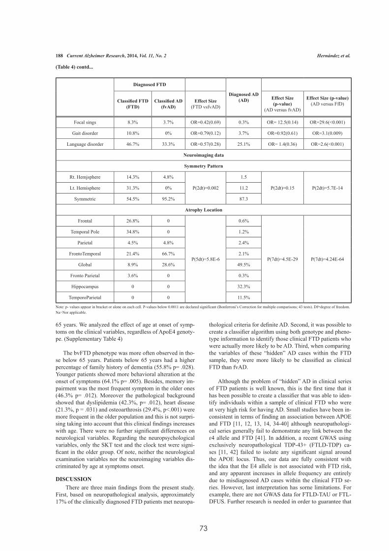
73
188 Current Alzheimer Research, 2014, Vol. 11, No. 2 Hernández et al.
(Table 4) contd...
Diagnosed FTD
Diagnosed AD(AD)Classified FTD
(FTD)Classified AD
(fvAD)Effect Size
(FTD vsfvAD)
Effect Size (p-value)
(AD versus fvAD)
Effect Size (p-value) (AD versus FfD)
Focal sings 8.3% 3.7% OR=0.42(0.69) 0.3% OR= 12.5(0.14) OR=29.6(<0.001)
Gait disorder 10.8% 0% OR=0.79(0.12) 3.7% OR=0.92(0.61) OR=3.1(0.009)
Language disorder 46.7% 33.3% OR=0.57(0.28) 25.1% OR= 1.4(0.36) OR=2.6(<0.001)
Neuroimaging data
Symmetry Pattern
Rt. Hemjsphere 14.3% 4.8%
P(2dt)=0.002
1.5
P(2dt)=0.15 P(2dt)=5.7E-14Lt. Hemisphere 31.3% 0% 11.2
Symmetric 54.5% 95.2% 87.3
Atrophy Location
Frontal 26.8% 0
P(5dt)=5.8E-6
0.6%
P(7dt)=4.5E-29 P(7dt)=4.24E-64
Temporal Pole 34.8% 0 1.2%
Parietal 4.5% 4.8% 2.4%
FrontoTemporal 21.4% 66.7% 2.1%
Global 8.9% 28.6% 49.5%
Fronto Parietal 3.6% 0 0.3%
Hippocampus 0 0 32.3%
TemporoParietal 0 0 11.5%
Note: p- values appear in bracket or alone on each cell. P-values below 0.0011 are declared signifícant (Bonferroni’s Correction for multiple comparisons; 43 tests). Df=degree of freedom. Na=Nor applicable.
65 years. We analyzed the effect of age at onset of symp-toms on the clinical variables, regardless of ApoE4 genoty-pe. (Supplementary Table 4)
The bvFTD phenotype was more often observed in tho-se below 65 years. Patients below 65 years had a higher percentage of family history of dementia (55.8% p= .028). Younger patients showed more behavioral alteration at the onset of symptoms (64.1% p= .005). Besides, memory im-pairment was the most frequent symptom in the older ones (46.3% p= .012). Moreover the pathological background showed that dyslipidemia (42.3%, p= .012), heart disease (21.3%, p = .031) and osteoarthrosis (29.4%, p<.001) were more frequent in the older population and this is not surpri-sing taking into account that this clinical findings increases with age. There were no further significant differences on neurological variables. Regarding the neuropsychological variables, only the SKT test and the clock test were signi-ficant in the older group. Of note, neither the neurological examination variables nor the neuroimaging variables dis-criminated by age at symptoms onset.
DISCUSSION There are three main findings from the present study. First, based on neuropathological analysis, approximately 17% of the clinically diagnosed FTD patients met neuropa-
thological criteria for definite AD. Second, it was possible tocreate a classifier algorithm using both genotype and pheno-type information to identify those clinical FTD patients whowere actually more likely to be AD. Third, when comparingthe variables of these “hidden” AD cases within the FTD sample, they were more likely to be classified as clinical FTD than fvAD.
Although the problem of “hidden” AD in clinical series of FTD patients is well known, this is the first time that it has been possible to create a classifier that was able to iden-tify individuals within a sample of clinical FTD who were at very high risk for having AD. Small studies have been in-consistent in terms of finding an association between APOE and FTD [11, 12, 13, 14, 34-40] although neuropathologi-cal series generally fail to demonstrate any link between the ɛ4 allele and FTD [41]. In addition, a recent GWAS using exclusively neuropathological TDP-43+ (FTLD-TDP) ca-ses [11, 42] failed to isolate any significant signal around the APOE locus. Thus, our data are fully consistent with the idea that the E4 allele is not associated with FTD risk, and any apparent increases in allele frequency are entirely due to misdiagnosed AD cases within the clinical FTD se-ries. However, last interpretation has sorne limitations. For example, there are not GWAS data for FTLD-TAU or FTL-DFUS. Further research is needed in order to guarantee that
74
Identification of Misdiagnosed Fronto-Temporal Dementia Using APOE Genotype Current Alzheimer Research, 2014, Vol. 11, No. 2 189
any apparent increases in allele frequency are entirely due to misdiagnosed AD cases within the clinical FTD series.
Clinicians and researchers must be cautioned that despite the “hidden” AD cases within the group with FTD, it would not be appropriate to eliminate patients with a ɛ4 allele from either clinical series or research studies of FTD. To do so would remove approximately two-thirds of the genuine FTD cases. Therefore, the most fruitful approach would be to use a statistical classifier to identify those individuals within the FTD sample who were most likely to be “hidden” AD cases, and to treat them as a separate group for analysis, as shown here. While this does not completely eliminate the problem of the contamination of the FTD series, it may help to mini-mize the impact of that contamination on the outcomes of interest.
The identification of individuals who were diagnosed with FTD but had AD is important since the misdiagnosis can re-sult in the failure to provide appropriate medication for the patient, or even potentially the prescription of inappropriate medications [43]. The behavioral phenotype, alone, is not sufficient to disentangle these two clinical syndromes, but using both genetic and neuro-imaging markers along with the clinical signs/symptoms, has the potential to improve patient classification and thus case management. Indeed, it may be that among patients diagnosed with FTD who are ɛ4 carriers, it would be important for them to be able to have more specific neuroimaging assessment, using either FDG metabolic scans or in vivo amyloid imaging to help to clarify the diagnosis.
It is vital to emphasize the need for a reliable estimate of ɛ4 allele frequency; a large series of AD and controls (e.g., n>1000) is an essential component of this kind of research. We used estimates based on more than eleven thousand chro-mosomes (5611 individuals) to calculate fvAD contamina-tion. The precision of the e4 frequencies facilitated the infe-rence of fvAD (17.2%) that resulted in almost identical rates compared to the postmortem data (16.6%). The penetrance and discriminant analyses were used to indirectly demonstrate that the information contained in cli-nical variables differentiating APOEɛ4 positive and negativeFTD patients may also differentiate AD cases and genuine FTD (postmortem) with relatively good precision (AUC 93.9%).
However, the proposed classifier needs independent repli-cation and could be improved by extending the discriminant analysis to the whole clinical database and by including age at onset and APOE genotype. Of note, APOE was only used in the first phase of the study (penetrance analysis), but not in the discriminant analysis which led to the score. In ad-dition the results of the discrirninant average scores do not vary with the age of onset of the disease. So, we feel that by adding APOE or age at onset to the discriminant analysis wecannot improve discrimination too much. Further research increasing sample size and using deep phenotyping would be necessary to improve obtained discriminant function.
Although AD and FTD can be differentiated using histo-pathological analysis the existence of the fv AD complicatesthe clinical identification of genuine FTD patients. From a clinical point of view, the best predictor to identify hidden
fvAD in FTD series is memory impairment (Table 4). Mo-reover, behavior changes in the genuine FTDs such apathy, desinhibition, or eating changes, are the first complaints refe-rred by relatives. Besides, fvAD and FTD might have diffe-rent neuroimaging atrophy patterns (Table 4).
The characterization of the phenotype of rare FTD syn-dromes is difficult and may affect the identification of valid genetic markers for FTD using GWAS strategies and other massive molecular techniques. Most important, the misclas-sification of AD patients as FTD might hamper the access topalliative or empírical therapies by these atypical AD cases. Consequently, the improvement of mathematical tools to comprehensively differentiate fvAD and FTD would be of interest not only for research but also for clinical purposes in the future.
CONFLICT OF INTEREST
The authors confirm that this article content has no con-flicts of interest.
ACKNOWLEDGEMENTS
This work was carried out as part of the doctoral program of I. Hernández at the Autonomous University of Barcelona. This work was partially supported by the Spanish Ministry of Health from Instituto de Salud Carlos III (Madrid) (FISSPI10/00945) and by the Agència d’Avaluació de Tecnologia i Recerca Mèdiques. Departament de Salut de la Generalitatde Catalunya (Health Department of the Catalan Govern-ment) (390) and by the University of Pittsburgh Alzheirner’sDisease Research Center (AG05133). We are indebted to Trinitat Port-Carbó and her family for their support of the fundació ACE research programs.
We thank brain donors and their families for generous brain donation for research. The authors are indebted to Dr. Maria Jesus Rey and Dr. Ellen Gelpi from the Neurological Tissue Bank of the Biobank-Hospital Clinic -IDJBAPS for detailed neuropathological brain examination.
SUPPLEMENTARY MATERIAL
Supplementary material is available on the publishers web site along with the published article.
STUDY FUNDING FJS/EC11 -358 and FIS/P: 10-00945 of Ministerio de Sa-nidad, Servicios Sociales e Igualdad. Spain; AATM/390-6-2009. Generalitat de Catalunya (Catalan Government) NIA/NIH (AG05133).
AUTHOR DISCLOSURES
Isabel Hernández has nothing to disclose.
Ana Mauleón has nothing to disclose.
Maiteé Rosense-Roca has nothing to disclose.
Montserrat Alegret has nothing to disclose.
Georgina Vinyes has nothing to disclose.
Ana Espinosa has nothing to disclose.

75
190 Current Alzheimer Research, 2014, Vol. 11, No. 2 Hernández et al.
Sergi Valero has nothing to disclose.
Osear Sotolongo-Grau has nothing to disclose.
James T. Becker is supported, in part, by funds from the NIH (AG05133, AG034852, AG020098, MH098745, A1035041).
Lluís Tárraga has nothing to disclose.
Osear L. López has served as a consultant to Lunbeck and Mertz pharmaceuticals. He is supported, in part, by funds from the NIH (AG05133, AG020098, AG024904, MH09033, AG022376, AG037451, AG025204, HD055525).
Agustín Ruiz is a shareholder of Neopharm Obesity SL and Oxigeneinc.
Merce Boada is consultant to Novartis and Esteve pharma-ceuticals. She is supported, in part by funds from FIS/ECll-358 and FISIP: 10-00945 of Ministerio de Sanidad, Servicios Sociales e Igualdad. Spain ; AATM/390-6-2009. Generalitat de Catalunya (Catalan Government). She is a member of Ad-visory Boards of Grifols, Lilly, Elan, Nutricia, Genentec and Roche.
ABBREVIATlONS
FTD = Clinical Fronto-temporal dementiafvAD = Clinical “frontal variant” Alzheimer’s diseaseAD = Alzheimer’s DiseaseFTLD = Histological Fronto-temporal Lobar de generationbvFTD = Behavioral variant Fronto-temporal de mentiaPNFA = Progressive Non Fluent AphasiaSD = Semantic DementiaPSP = Progressive Supranuclear PalsyCBS = Cortico Basal SyndromeFTD-MND = Fronto-temporal Dementia with Motor Neuron DiseaseAPOE = Apolipoprotein E genotypeHWE = Hardy-Weinberg equilibrium lawLDA = Linear discriminant analysesFTLD-TAU = Fronto-temporal Lobar degeneration with TAU inclusionsFTLD-TDP = Fronto-temporal Lobar degeneration with TDP-43 inclusionsFTLD-FUS = Fronto-temporal Lobar degeneration with FUS inclusions.
REFERENCES
[1] Clerx L, Jacobs HL, Burgmans S, Gronenschild EB, Uylings H, Echávarri C, et al. Sensitivity of Different MRl-Techniques to As-sess Gray Matter Atrophy Pattems in Alzheimer’s Disease is Re-gion-Specific. Curr. Alzheimer Res 1 0(9): 940-51 (2013).
[2] Mukaetova-Ladinska EB, Abdeii-AII Z, Andrade J, da Silva JA, Boksba 1, Burbaeva G, el al. Platelet Tau Protein as a Potential Pe-ripberal Biomarker in Alzheimer’s disease: An Explorative Study. Curr. Alzheimer Res 1 0(9): 987- 1004 (2013).
[3] Becker JT, Huff FJ, Nebes RD, Holland A, Boller F. Neuropsycbo-logical function in Alzheimer’s disease. Pattem of impairment and rates of progression. Arch Neurol 45(3): 263-8 (1988).
[4] Stern Y, Mayeux R, Sano M, Hauser WA, Bush T. Predictors of disease course in patients with probable Alzheimer’s disease. Neu-rology 37(10): 1649-53 (1987).
[5] Jobnson JK, Head E, Kim R, Starr A, Cotman CW. Clínical and pathological evidence for a frontal variant of Alzheimer disease. Arch Neurol56(10): 1233-9 (1999).
[6] Kertesz A, McMonagle P, Blair M, Davidson W, Munoz DG. The evolution and patbology of frontotemporal dementia. Brain 128(Pt9): 1996-2005 (2005).
[7] Goedert M, Ghetti B, Spillantini MG. Frontotemporal dementia: implications for understanding Alzheimer disease. Cold Spring Harb Perspect Med 2(2): a006254 (20 12).
[8] Corder EH, Saunders AM, Strittmatter WJ, Schmechel DE, Gaskell PC, Small GW, el al. Gene dose of apolipoprotein E type 4 allele and the risk of Alzheimer’s disease in late onset families. Science 261 (5123): 921 -3 (1993).
[9] Lovati C, Galimberti D, Albaní D, Bertora P, Venturelli E, Cislaghi G, et al. APOE ɛ2 and ɛ4 influence the susceptibility for Alzhei-mer’s disease but not other dementias. lnt. J. Mol Epidemial Genet 1 (3): 193-200.
[10] Hauser PS, Ryan RO. lmpact of Apolipoprotein E on Alzheimer’s Disease. Curr Alzheimer Res 10(8):809-17 (2013).
[11] Seripa D, Bizzarro A, Panza F, Acciarri A, Pellegrini F, Pilotto A, et al. The APOE gene locus in frontotemporal dementia and primary progressive aphasia. Arch Neurol Arch Neurol 68(5): 622-8 (2011).
[12] Acciarri A, Masullo. C, Bizzarro A, Valenza A, Quaranta D, Marra C, el al. Apoe epsilon2-epsilon4 genotype is a possible risk factor for primary progressive aphasia. Ann. Neurol 59(2): 436-7 (2006).
[13] Stevens M, van Duijn CM, de Knijff P, van Broeckhoven C, Heu-tink P, Oostra BA, el al. Apolipoprotein E gene and sporadic frontal lobe dementia. Neurology 48(6): 1526-9 (1997).
[14] Gescbwind D, Karrim J, Nelson SF, Miller B. The apolipoprotein E epsilon4 allele is not a significant risk factor for frontotemporal de-mentia. Ann. Neurol Neurobiol Aging pii: SO 197-4580(13)00614-3 (2013).
[15] Bigio EH, Mishra M, Hatanpaa KJ, White CL, Jobnson N, Rade-maker A, et al. TDP-43 pathology in primary progressive aphasia and frontotemporal dementia with pathologic Alzheimer disease. Acta Neuropatbol 120(1): 43-54 (2010).
[16] Liscic RM, Storandt M, Caims NJ, Morris JC. Clínical and psy-chometric distinction of frontotemporal and Alzheimer dementias. Arch Neurol Arcb Neurol 64(4): 535-40 (2007).
[17] Zintl M, Petkov M, Scbmitz G, Hajak G, Klünemann H-H. Fronto-temporal dementia in association with a family history of dementia and ApoE polymorphism. Nervenarzt 81 (1): 75-8 (2010).
[18] Sesbadri S, Fitzpatrick AL, llera m MA, DeStefano AL, Gudnason V, Boada M, et al. Genome-wide analysis of genetic loci associated with Alzbeimer disease. JAMA 303(18): 1832-40 (2010).
[19] Neary D, Snowden JS, Gustafson L, Passant U, Stuss D, Black S, et al. Frontotemporal lobar degeneration: a consensus on clinical diagnostic criteria. Neurology 51 (6): 1546-54 (1998).
[20] Rascovsky K, Hodges JR, Knopman D, Mendez MF, Kramer JH, Neubaus J, et al. Sensitivity of revised diagnostic criteria for the behavioural variant of frontotemporal dementia. Brain 134(Pt 9):2456-77 (20 11).
[21] Mesulam MM. Slowly progressive apbasia witbout generalized dementia. Ann Neurol 11 (6): 592-8 (1982).
[22] Gomo-Tempini ML, Hillis AE, Weintraub S, Kertesz A, Mendez M, Cappa SF, et al. Classification of primary progressive aphasia and its variants. Neurology 76(11): 1006-14 (2011).
[23] Hodges JR, Patterson K, Oxbury S, Funnell E. Semantic demen-tia. Progressive fluent apbasia with temporal lobe atrophy. Brain [Internet]. 1992 Dec [cited 2012 Dec 14];115 ( Pt 6:1783- 806. Available from: http://www.ncbi .nlm.nih .gov/pubmed/1486461
[24] Steele Jc, Richardson Jc, Olszewski J. Progressive Supranuclear Palsy. A beterogeneous degeneration involving the Brain Stem, Basal Ganglia and Cerebellum with Vertical Gaze and Pseudobul-bar Palsy, Nucbal Dystonia and Dementia. Arch Neurol 10: 333-59 (1964).
76
Identification of Misdiagnosed Fronto-Temporal Dementia Using APOE Genotype Current Alzheimer Research, 2014, Vol. 11, No. 2 191
[25] Boeve BF, Lang AE, Litvan L corticobasal degeneration and its relationship to progressive supranuclear palsy and frontotemporal dementia. Ann Neurol 54 (5): S 15-9.
[26] Xi Z, Zinman L, Grinberg Y, Moreno D, Sato e, Bilbao JM, et al. lnvestigation of e9orf72 in 4 Neurodegenerative Disorders. Arch Neurol 2012 l -8 (2012).
[27] Alegret M, Espinosa A, Yinyes-Junqué G, V alero S, Hernández I, Tárraga L, et al. Normative data of a brief neuropsychological bat-tery for Spanish individuals older than 49. J elin Exp Neuropsychol l 34(2): 209-.19 (20 12).
[28] Braak H, Braak E. Neuropathological stageing of Alzheirnerrelated changes. Acta Neuropathol82(4): 239-59 (1991).
[29] Braak H, Alafuzotf 1, Arzberger T, Kretzschmar H, Del Tredici K. Staging of Alzheimer disease associated neurofibrillary pathology using paraffin sections and immunocytochemistry. Acta Neuropa-tholll2(4): 389-404 (2006).
[30] Morris Je, Heymao A, Mohs Re, Hughes JP, van Belle G, Fillen-baum G, et al. The consortium to Establish a Registry for Alzhei-mer’s Disease (eERAD). Part I. clinical and neuropsychological assessment of Alzheimer’s disease. Neurology; 39(9): 1159-65 (1989).
[31] Mirra SS, Heyman A, McKeel D, Sumi SM, erain BJ, Brownlee LM, el al. The consortium to Establish a Registry for Alzheimer’s Disease (eERAD). Part Ll . Standardization of the neuropatholo-gic assessment of Alzheimer’s disease. Neurology 41 (4): 479-86 (1991).
[32] Montine TJ, Phelps eH, Beach TG, Bigio EH, eaims NJ, Dickson DW, et al. National lnstitute on Aging-Alzbeimer’s Association guidelines for the neuropathologic assessment of Alzheimer’s disease: a practica [approach. Acta Neuropathol 123 (1): 1- 11 (2012).
[33] Wolz R, Julkunen Y, Koikkalainen J, Niskanen E, Zhang DP, Ruec-kert D, et al. Multi-method analysis of MRJ images in early diag-nostics of Alzbeimer’s disease. PLoS One 6(1 0): e25446 (2011).
[34] Gustafson L, Abrabamson M, Grubb A, Nilsson K, Fex G. Apolipo-protein-E genotyping 111 Alzheimer’s disease and frontotemporal dementia. Dement GeriatrCogn Disord 8(4): 240-3 (1997).
[35] Pickering-Brown SM, Siddons M, Mann DM, Owen F, Neary D, Snowden JS. Apolipoprotein E allelic frequencies in patients with lobar atrophy. NeurosciLett 1995 188(3): 205-7 (1995).
[36] Masullo e , Daniele A, Fazio YM, Seripa D, Gravina C , Filippini Y, et al. The Apolipoprotein E genotype in patients affected by sy-ndromes with focal cortical atrophy. Neurosci Lett 303(2): 87-90 (2001).
[37] Lehmann DJ, Smith AD, eombrinck M, Bametson L, Joachim C. Apolipoprotein E epsilon2 may be a risk factor for sporadic fron-totemporal dementia. J Neurol Neurosurg Psychiatry 69(3): 404-5 (2000).
[38] Yerpillat P, Camuzat A, Hannequin D, Thomas-Anterion C, Puel M, Belliard S, el al. Apolipoprotein E gene in frontotemporal de-mentia: an association study and meta-analysis. Eur J Hum Genet 10(7): 399-405 (2002).
[39] Minthon L, Hesse C, Sjogren M, Englund E, Gustafson L, Blen-now K. The apolipoprotein E epsilon4 allele frequency is normal in fronto-temporal dementia, but correlates with age at onset of disease. Neurosci Lett 226( 1 ): 65-7 ( 1997).
[40] Daniele A, Matera MG, Seripa D, Acciarri A, Bizzarro A, Pilotto A, el al. APOE epsilon 2/epsilon 4 genotype a risk factor for primary progressive aphasia in women. Arch Neurol 66(7): 910--2 (2009).
[41] Nielsen AS, Ravid R, Kampborst W, Jergensen OS. Apolipoprotein E epsilon 4 in an autopsy series of various dementing disorders. J Alzheirners Dis 2003 5(2): 119-25 (2003).
[42] Van Deerlin YM, Sleiman PMA, Martinez-Lage M, Cben-Plotkin A, Wang L-S, Gratf-Radford NR, el al. Common variants at 7p21 are associated with frontotemporal lobar degeneration with TDP-43 inclusions. Nat Genet 42(3): 234-9 (20 1 0).
[43) Grossman M, Onyike C, Graf-Radford N, Mendez M, Kerwin D, Lemer A, el al. Memantine in patients with frontotemporal lobar degeneration: a multicentre, randomised, double-blind, placebo-controlled trial. Lancet Neurol 12(2): 149-56 (20 13).
Received: July 03,2013 Revised: November 04, 2013 Accepted: November 08, 2013
7777
VII. Resultados
Trabajo IV
Association of TMEM106B rs1990622 marker and Frontotemporal dementia: evidence for a recessive effect and meta-analysis.
Hernández I, Rosense-Roca M, Alegret M, Mauleón A, Espinosa A, Vargas L, Sotolon-go-Grau O, Tarraga L, Boada M, Ruiz A. Journal Alzheimer’s Disease.
Volume 43, Number 1, IN PRESS
IF: 4,17 (2014) Q2
78
Journal of Al zheimer’s Disease (2014)DOI 1 0.3233/JAD- 132432JOS Press
Association of TMEM106B rs 1990622Marker and Frontotemporal Dementia: Evidence for a Recessive Effect andMeta-AnalysisIsabel Hernández, Maitée Rosende-Roca, Montserrat Alegret, Ana Mauleón, Ana Espinosa, Liliana Vargas, Oscar Sotolon-go-Grau, Lluís Tárraga, Mercè Boada and Agustín Ruiz.*
Alzheimer Research Center and Memory Clinic. Fundació ACE. Institut Català de Neurociències Aplicades. Barcelona, Spain.
Handling Associate Editor: Beoedetta Nacmias
tAccepted 3 June 2014
Abstract. Transmembrane Protein 106B SNP rs1990622 was recently shown to modify the risk of Frontotemporal Lobar Degeneration with TDP-43 inclusions (FTD-TDP). An independent replication study of this genetic variant was performed in 381 individuals from Catalonia (Spain). By applying a recessive model, a tendency towards an association with FTD risk was observed in our case–control study (age and sex-adjusted odds ratio=0.57; p=0.082). Importantly, meta-analysis of available studies also supports a recessive effect for rs1990622 CC genotype (OR=0.70; C.I. 95% [0.57-0.85]; p=0.0003) and demon-strates the existence of statistical heterogeneity due to an inherent pathological heterogeneity between series (P=0.00014). We conclude that TMEM106B is associated with FTD, although the extent of this effect is difficult to be estimated by using clinical FTD series.
Key Words: Frontotemporal Dementia, Genetics, GWAS, TMEM106B, molecular epidemiology
*Correspondence to: Agustín Ruiz, M.D. PhD, Fundació ACE,
Marquès de Sentmenat, 57 08029 Barcelona (Spain). Tel. 34-93-
444-7318; Fax.34-93-410-1701; E-mail: [email protected]
INTRODUCTION
Frontotemporal Lobar Degeneration (FTLD) is a fatal neurodegenerative disorder in adults. In fact, FTLD is the second cause of dementia, after early onset Alzheimer’s disease (EOAD), in people under 65 years old. From a clinical point of view, FTD (Frontotemporal dementia) is a commonly used term. FTD comprises several disorders with very specific clinical hallmarks. The most common FTD subtype is the behavior variant (bvFTD), charac-terized by behavioral and personality changes such as disinhibition, that often coexists with apathy and mani-fests itself by impulsive behavior, loss of empathy, or ste-reotypic behavior, leading to a loss of social competence [1] an international consortium developed revised guide-lines for the diagnosis of behavioural variant frontotem-poral dementia. The validation process retrospectively reviewed clinical records and compared the sensitivity of proposed and earlier criteria in a multi-site sample of
patients with pathologically verified frontotemporal lobar degeneration. According to the revised criteria, ‘possible’ behavioural variant frontotemporal demen-tia requires three of six clinically discriminating fea-tures (disinhibition, apathy/inertia, loss of sympathy/empathy, perseverative/compulsive behaviours, hy-perorality and dysexecutive neuropsychological pro-file. Executive functions are impaired and in the initial stages of the disease, memory and visuo-perceptive skills are well preserved. In contrast, Progressive Non Fluent Aphasia (PNFA) or Semantic Dementia (SD) are characterized by language disturbances including impaired expressive language, difficulties naming ob-jects and reduction of verbal fluency or loss of com-prehension of word meaning respectively [2]. FTLD may be associated with Motoneuron Disease (MND), in particular Amiotrophic Lateral Sclerosis (ALS), or with parkinsonism. Several authors recognize clinical, genetic and pathological overlap between FTLD and ALS [3-5].
The clinical FTD phenotype is heterogeneous, but pathological findings in postmortem analyses of brains from subjects diagnosed of clinical FTD have also
ISSN 1387-2877114/$27.50 © 2014- JOS Press and the authors. All rights reserved
79
revealed at least three different immunohystochemical phenotypes related to specific protein deposits. Around 40% of patients with clinical FTD show tau inclusions. They comprise most cases of PNFA, ~45% of cases of bvFTD and some cases of SD [6-7]. More than 50% of the FTLD patients present tau-negative ubiquitin stain-ing inclusions referred to as FTLD-ubiquitin (FTLD-U) and in 80-90% of this group inclusions are composed of transactive response (TAR) DNA-binding protein 43 [8]small numbers of cases have recently been reported with TDP-43-negative FTLD-U pathology. To determine the frequency and to define the clinico-pathological spectrum of TDP-43-negative FTLD-U, we re-evaluated 44 cases with a previous diagnosis of FTLD-U or dementia lacking distinctive histopathology. We identified nine cases (20%. However 10% of these cases are composed of a heter-ogenous collection of uncommon disorders. These cases with FTLD-U show no evidence of abnormal TDP-43 and several tau/TDP-43-negative FTLD subtypes are im-munoreactive (ir) for the fused in sarcoma (FUS) protein [9]. Cases with inclusions that can only be demonstrated by immunohistochemistry against proteins of the ubiqui-tin proteasome system (UPS), are designated FTLD-UPS [10]. FTD-3 (mutations in the CHMP2B gene) is the ma-jor entity associated with this FTLD-UPS pathology. This designation recognizes that the TDP-43-negative inclu-sions may immunostain for UPS proteins other than ubiq-uitin, such as p62. Recently, unconventional non-ATG translation of the expanded hexanucleotide repeat, result-ing in the production and aggregation of dipeptide repeat (DPR) proteins (poly-GA, -GR and GP), was identified as a potential pathomechanism of C9ORF72 mutations. Besides accumulation of DPR proteins, the second neuro-pathological hallmark lesion in C9ORF72 mutation cases is the accumulation of TDP-43 [11-12].
From a genetic standpoint, familial antecedents are commonly observed in FTD. Interestingly, genetic analy-sis of familiar cases of FTD has revealed mutations in mul-tiple loci including GRN [13-14] and MAPT [15] genes, both very close on chromosome 17, but also in CHMP2B [16], UBQLN2 [17], SQSTM1 [18]or TREM2 [19] genes. Chromosome 9 open reading frame 72 (C9ORF72) mu-tation, located at chromosome 9p21 is characterized by a pathological non-coding hexanucleotide (G4C2) repeat expansion. This latter mutation within C9ORF72 locus is the most frequent cause of inherited ALS and FTLD identified to date [20-22]. The isolation of these genes was a great success but, as yet, not all mutations under-lying these proteinopathies have been identified [23]TAR DNA-binding protein TDP-43, fused-in-sarcoma or yet unidentified proteins in affected brain regions. Rather than the type of proteinopathy, the site of neurodegen-eration correlates relatively well with the clinical pre-sentation of FTLD. Molecular genetic studies identified
five disease genes, of which the gene encoding the tau protein (MAPT. An international team led by University of Pennsylvania School of Medicine (USA), conducted a genome-wide association study (GWAS) in 515 subjects with autopsy-confirmed FTD with TDP-43 inclusions and 2509 neurologically normal controls to seek novel FTD-associated genes. All cases met either pathological or genetic criteria for FTD-TDP which was confirmed by detecting TDP-43 inclusions using immunohistochem-istry (IHC) or mutation screening. Importantly, the au-thors found FTLD-TDP associates with multiple SNPs mapping in 7p21. This region contains the TMEM106B gene. Three SNPs retained genome-wide significance af-ter multiple testing correction; top SNP rs1990622 minor allele (C) was reported as a protective genetic factor for FTD-TDP (OR=0.61, 95% CI 0.53-0.71). The associa-tion was replicated in 89 FTD-TDP autopsy-confirmed cases and 553 controls but not in a clinical FTD series available (a 192 FTD living patients cohort) [24]result-ing from mutations in GRN (which encodes progranulin. Independent efforts to replicate this finding using clinical series have yielded controversial results. Rollinson et al (2011) attempted to replicate the association of these loci in FTD cohorts of British origin (520 FTD cases and 247 controls), but failed to detect any significant association of TMEM106B SNPs in the Manchester or London co-horts [3]. However, using an independent Flanders–Bel-gian cohort comprised primarily of clinically diagnosed patients (297 FTD cases and 595 controls), the associa-tion with TMEM106B rs1990622 [OR=0.75 (0.61–0.93)] was confirmed [25]. Finch et al, found a non-significant effect for TMEM106B in 640 FTD (clinical series) and 822 controls on FTD risk. It is noteworthy that a strong association between TMEM106B markers was detected in a small series of patients carrying GRN mutations but not in other FTD patients [26].
To evaluate the effect of TMEM106B rs1999622 on FTD risk, a new case-control association study was con-ducted in clinical FTD patients and neurologically normal controls. To conduct this research, the clinical FTD series available at Fundació ACE (Barcelona, Spain) were gen-otyped. A new meta-analysis integrating obtained results with currently available data was also performed.
MATERIALS AND METHODS
Patients
All of the patients agreed to participate in the study and blood samples were obtained after they and/or their legal representatives provided written consent according to the GIPSY protocol approved by the ethics committee in the Hospital Clinic i Provincial (Barcelona, Spain). Informed consent was in accordance with Spanish biomedical laws
I. Hernández et al. TMEM106B and FTD

80
(Law 14/2007, July 3rd, on biomedical research; Royal Decree 1716/2011, November 18th). All procedures in-volving experiments on human subjects are done in ac-cordance with the ethical standards of the Ethics Commit-tee of the institution in which the experiments were done or in accord with the Helsinki Declaration of 1975.
All patients were south-Europeans (Spanish lineage, two generation or more) recruited from the Fundació ACE. Barcelona Alzheimer Treatment & Research Cen-ter. Genotypes from 381 unrelated individuals were used for rs1990622 analysis (146 FTD cases, 234 neurologi-cally normal controls). No individual has been included in any genome-wide dataset previously reported. 146 clini-cal FTD patients underwent a full neurological evaluation and were diagnosed using international research criteria: 88 bvFTD [1]an international consortium developed re-vised guidelines for the diagnosis of behavioural variant frontotemporal dementia. The validation process retro-spectively reviewed clinical records and compared the sensitivity of proposed and earlier criteria in a multi-site sample of patients with pathologically verified frontotem-poral lobar degeneration. According to the revised crite-ria, ‘possible’ behavioural variant frontotemporal demen-tia requires three of six clinically discriminating features (disinhibition, apathy/inertia, loss of sympathy/empathy, perseverative/compulsive behaviours, hyperorality and dysexecutive neuropsychological profile, 22 PNFA, 32 SD, [2]. Patients with Cortico-Basal Syndrome (CBS) or Progressive Supranuclear Palsy Syndrome (PSPS) [27]neuropsychology, and movement disorders special-ists developed new criteria based on consensus and a systematic literature review. Clinical diagnoses (early or late, which in most cases are FTLD-TAU were excluded from this study, as were patients with Logopenic Apha-sia (LPA) who were not genotyped, since cases LPA are mostly associated with a neuropathological diagnosis of AD [28-29]. Four Patients with a combined phenotype of FTD and ALS (FTD-MND) were also included. Patients were diagnosed at a daily consensus conference jointly by neurologists, neuropsychologists and social workers at the Fundació ACE Memory Clinic. Baseline signs and symptoms were acquired directly from the patient or from the primary caregiver. Patients were administered a neuropsychological battery that included measures of orientation , attention, verbal learning and long-term memory, language, gnosis, praxis and executive func-tions [30]. Neuroimaging studies used included CT and/or MRI, which were required for a probable FTD diag-nosis according to revised criteria [1-2] ; in some cases SPECT scans were also available. The locus and extent of hemispheric atrophy was rated by visual inspection by an experienced neurologist, and independently confirmed by a neuroradiologist. FTD patients were screened look-ing for MAPT mutations and C9ORF72 expansions. Only one FTD patient carrying a MAPT mutation (0.6%) was identified. Three additional patients were C9ORF72 ex-
I. Hernández et al. TMEM106B and FTD
pansion carriers (1.8%) [31]frontotemporal lobar degen-eration (FTLD. The remaining candidates genes were not studied. The exception was the UBQLN2 gene that was partially screened in 77 FTD cases with familial anteced-ents of FTD. No mutation was detected in UBQLN2 locus [32]. Mutation carriers were not excluded from this study.
Molecular analysis
DNA was extracted from frozen blood samples using the NucleoSpin Blood kit (Macherey-Nagel, Düren, Ger-many). The TMEM106B gene polymorphism rs1990622 was genotyped in an ECO Real-time PCR System (Illumi-na, San Diego, USA) by using the KAPA HRM fast PCR kit (Kapa Biosystems; Wobum, MA, USA) according to the manufacturer’s instructions. High Resolution Melting primers were designed using DesignStudio online tool (Illumina; http://designstudio.illumina.com/). Selected amplification primers for High resolution melting were Forward: 5’ CACTGTTTCATCCCCTTTATCTCGCCTA 3’ and reverse: 5’ TGCAGCTCTGTTCTCCATTGTAG-TACTG 3’. The primer pair yields a 90 bp PCR product containing a unique polymorphism (rs1990622) accord-ing to Genome browser information. Real-time PCRs were performed in a reaction volume of 5 µl with 2.5 µl of HRM fast start master kit, 0,25µl of amplification primer mix directly purchased from Illumina (reference: 15028319), 0,5 µl of MgCl2 (2.5mM end concentration; Kapa Biosystems), 10 ng of genomic DNA (2ng/ µl end concentration) and 0.75µl of PCR-grade water. Cycling conditions were as follows: 50°C for 2 minutes, 95°C for 10 minutes, and 45 cycles at 95°C for 10 seconds and 65°C during 15 seconds. Fluorescence was monitored at the end of each annealing/ extension phase. After am-plification, specific conditions to obtain high resolution melting (HRM) curves were 95°C for 15 seconds, 55°C for 15 seconds, and 95°C for 15 seconds (ramping rate: 0.2°C/s). In the last step, continuous fluorimetric registers were performed by the system at one acquisition register per second. Melting peaks and genotype calls were ob-tained by using the Illumina Eco Real Time PCR system software version 4.1.02.0 (Illumina, San Diego, USA).
Statistical analysis and phenotype–genotype correlations.
Allelic frequencies, HWE equilibrium and genetic asso-ciation studies were conducted using the online tool at TUM Helmholtz Center (Munich, Germany; http://ihg.gsf.de/cgi-bin/hw/hwa1.pl). We used tests adapted from the study conducted by Sasieni [33]there will be twice as many alleles as people. Another approach considers the risk of the disease in those who do not have the allele of interest (A. Age and sex-adjusted binary logistic regres-sion analyses were performed using SPSS 15.0 software (SPSS, Chicago, IL). Meta-analyses were double checked using two independent online tools. We calculated esti-mates with Ken Rothman’s Episheet spreadsheet (avail-able online at http://www.krothman.org/episheet.xls).
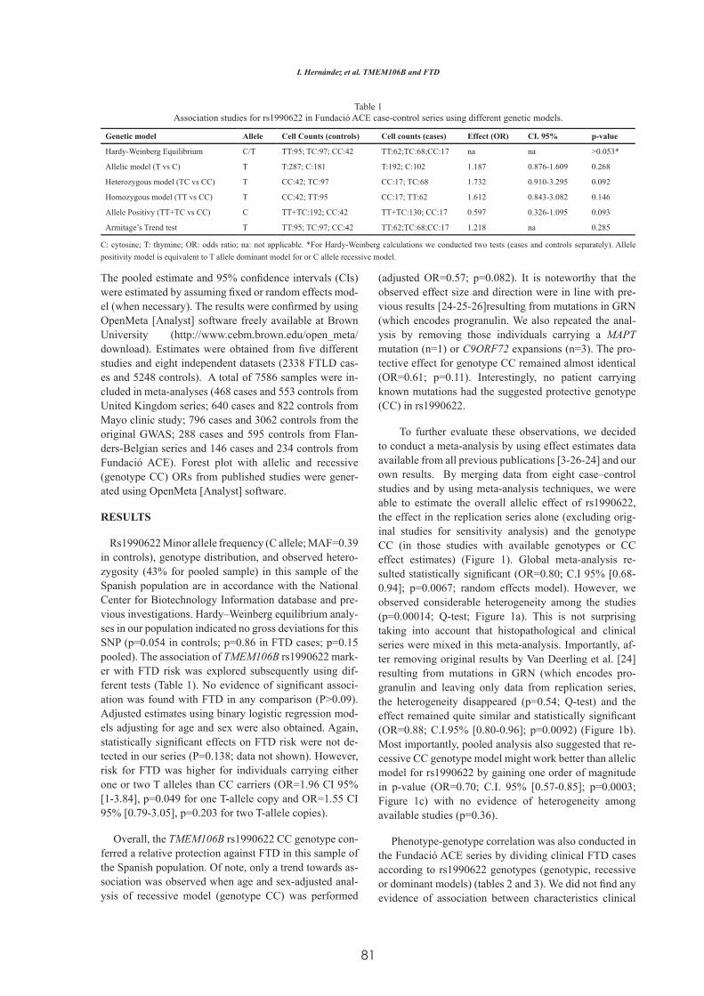
81
I. Hernández et al. TMEM106B and FTD
The pooled estimate and 95% confidence intervals (CIs) were estimated by assuming fixed or random effects mod-el (when necessary). The results were confirmed by using OpenMeta [Analyst] software freely available at Brown University (http://www.cebm.brown.edu/open_meta/download). Estimates were obtained from five different studies and eight independent datasets (2338 FTLD cas-es and 5248 controls). A total of 7586 samples were in-cluded in meta-analyses (468 cases and 553 controls from United Kingdom series; 640 cases and 822 controls from Mayo clinic study; 796 cases and 3062 controls from the original GWAS; 288 cases and 595 controls from Flan-ders-Belgian series and 146 cases and 234 controls from Fundació ACE). Forest plot with allelic and recessive (genotype CC) ORs from published studies were gener-ated using OpenMeta [Analyst] software.
RESULTS
Rs1990622 Minor allele frequency (C allele; MAF=0.39 in controls), genotype distribution, and observed hetero-zygosity (43% for pooled sample) in this sample of the Spanish population are in accordance with the National Center for Biotechnology Information database and pre-vious investigations. Hardy–Weinberg equilibrium analy-ses in our population indicated no gross deviations for this SNP (p=0.054 in controls; p=0.86 in FTD cases; p=0.15 pooled). The association of TMEM106B rs1990622 mark-er with FTD risk was explored subsequently using dif-ferent tests (Table 1). No evidence of significant associ-ation was found with FTD in any comparison (P>0.09). Adjusted estimates using binary logistic regression mod-els adjusting for age and sex were also obtained. Again, statistically significant effects on FTD risk were not de-tected in our series (P=0.138; data not shown). However, risk for FTD was higher for individuals carrying either one or two T alleles than CC carriers (OR=1.96 CI 95% [1-3.84], p=0.049 for one T-allele copy and OR=1.55 CI 95% [0.79-3.05], p=0.203 for two T-allele copies).
Overall, the TMEM106B rs1990622 CC genotype con-ferred a relative protection against FTD in this sample of the Spanish population. Of note, only a trend towards as-sociation was observed when age and sex-adjusted anal-ysis of recessive model (genotype CC) was performed
(adjusted OR=0.57; p=0.082). It is noteworthy that the observed effect size and direction were in line with pre-vious results [24-25-26]resulting from mutations in GRN (which encodes progranulin. We also repeated the anal-ysis by removing those individuals carrying a MAPT mutation (n=1) or C9ORF72 expansions (n=3). The pro-tective effect for genotype CC remained almost identical (OR=0.61; p=0.11). Interestingly, no patient carrying known mutations had the suggested protective genotype (CC) in rs1990622.
To further evaluate these observations, we decided to conduct a meta-analysis by using effect estimates data available from all previous publications [3-26-24] and our own results. By merging data from eight case–control studies and by using meta-analysis techniques, we were able to estimate the overall allelic effect of rs1990622, the effect in the replication series alone (excluding orig-inal studies for sensitivity analysis) and the genotype CC (in those studies with available genotypes or CC effect estimates) (Figure 1). Global meta-analysis re-sulted statistically significant (OR=0.80; C.I 95% [0.68-0.94]; p=0.0067; random effects model). However, we observed considerable heterogeneity among the studies (p=0.00014; Q-test; Figure 1a). This is not surprising taking into account that histopathological and clinical series were mixed in this meta-analysis. Importantly, af-ter removing original results by Van Deerling et al. [24]resulting from mutations in GRN (which encodes pro-granulin and leaving only data from replication series, the heterogeneity disappeared (p=0.54; Q-test) and the effect remained quite similar and statistically significant (OR=0.88; C.I.95% [0.80-0.96]; p=0.0092) (Figure 1b). Most importantly, pooled analysis also suggested that re-cessive CC genotype model might work better than allelic model for rs1990622 by gaining one order of magnitude in p-value (OR=0.70; C.I. 95% [0.57-0.85]; p=0.0003; Figure 1c) with no evidence of heterogeneity among available studies (p=0.36).
Phenotype-genotype correlation was also conducted in the Fundació ACE series by dividing clinical FTD cases according to rs1990622 genotypes (genotypic, recessive or dominant models) (tables 2 and 3). We did not find any evidence of association between characteristics clinical
Table 1Association studies for rs1990622 in Fundació ACE case-control series using different genetic models.
Genetic model Allele Cell Counts (controls) Cell counts (cases) Effect (OR) CI. 95% p-value
Hardy-Weinberg Equilibrium C/T TT:95; TC:97; CC:42 TT:62;TC:68;CC:17 na na >0.053*
Allelic model (T vs C) T T:287; C:181 T:192; C:102 1.187 0.876-1.609 0.268
Heterozygous model (TC vs CC) T CC:42; TC:97 CC:17; TC:68 1.732 0.910-3.295 0.092
Homozygous model (TT vs CC) T CC:42; TT:95 CC:17; TT:62 1.612 0.843-3.082 0.146
Allele Positivy (TT+TC vs CC) C TT+TC:192; CC:42 TT+TC:130; CC:17 0.597 0.326-1.095 0.093
Armitage’s Trend test T TT:95; TC:97; CC:42 TT:62;TC:68;CC:17 1.218 na 0.285
C: cytosine; T: thymine; OR: odds ratio; na: not applicable. *For Hardy-Weinberg calculations we conducted two tests (cases and controls separately). Allele positivity model is equivalent to T allele dominant model for or C allele recessive model.
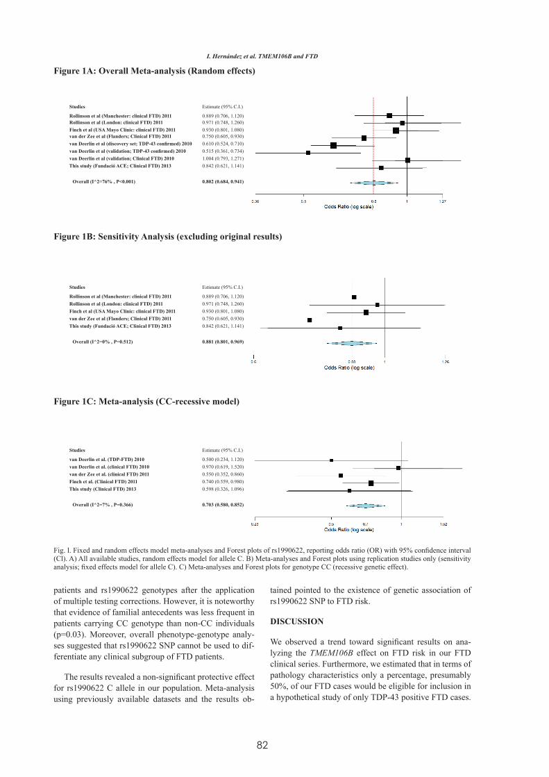
82
I. Hernández et al. TMEM106B and FTD
patients and rs1990622 genotypes after the application of multiple testing corrections. However, it is noteworthy that evidence of familial antecedents was less frequent in patients carrying CC genotype than non-CC individuals (p=0.03). Moreover, overall phenotype-genotype analy-ses suggested that rs1990622 SNP cannot be used to dif-ferentiate any clinical subgroup of FTD patients.
The results revealed a non-significant protective effect for rs1990622 C allele in our population. Meta-analysis using previously available datasets and the results ob-
tained pointed to the existence of genetic association of rs1990622 SNP to FTD risk.
DISCUSSION
We observed a trend toward significant results on ana-lyzing the TMEM106B effect on FTD risk in our FTD clinical series. Furthermore, we estimated that in terms of pathology characteristics only a percentage, presumably 50%, of our FTD cases would be eligible for inclusion in a hypothetical study of only TDP-43 positive FTD cases.
Studies Estimate (95% C.I.)
Rollinson et al (Manchester: clinical FTD) 2011 0.889 (0.706, 1.120)Rollinson et al (London: clinical FTD) 2011 0.971 (0.748, 1.260)Finch et al (USA Mayo Clinic: clinical FTD) 2011 0.930 (0.801, 1.080)van der Zee et al (Flanders; Clinical FTD) 2011 0.750 (0.605, 0.930)van Deerlin et al (discovery set; TDP-43 confirmed) 2010 0.610 (0.524, 0.710)van Deerlin et al (validation; TDP-43 confirmed) 2010 0.515 (0.361, 0.734)van Deerlin et al (validation; Clinical FTD) 2010 1.004 (0.793, 1.271)This study (Fundació ACE; Clinical FTD) 2013 0.842 (0.621, 1.141)
Overall (I^2=76% , P<0.001) 0.802 (0.684, 0.941)
Studies Estimate (95% C.I.)
Rollinson et al (Manchester: clinical FTD) 2011 0.889 (0.706, 1.120)Rollinson et al (London: clinical FTD) 2011 0.971 (0.748, 1.260)Finch et al (USA Mayo Clinic: clinical FTD) 2011 0.930 (0.801, 1.080)van der Zee et al (Flanders; Clinical FTD) 2011 0.750 (0.605, 0.930)This study (Fundació ACE; Clinical FTD) 2013 0.842 (0.621, 1.141)
Overall (I^2=0% , P=0.512) 0.881 (0.801, 0.969)
Studies Estimate (95% C.I.)
van Deerlin et al. (TDP-FTD) 2010 0.500 (0.234, 1.120)van Deerlin et al. (clinical FTD) 2010 0.970 (0.619, 1.520)van der Zee et al. (clinical FTD) 2011 0.550 (0.352, 0.860)Finch et al. (Clinical FTD) 2011 0.740 (0.559, 0.980)This study (Clinical FTD) 2013 0.598 (0.326, 1.096)
Overall (I^2=7% , P=0.366) 0.703 (0.580, 0.852)
Figure 1A: Overall Meta-analysis (Random effects)
Figure 1B: Sensitivity Analysis (excluding original results)
Figure 1C: Meta-analysis (CC-recessive model)
Fig. l. Fixed and random effects model meta-analyses and Forest plots of rs1990622, reporting odds ratio (OR) with 95% confidence interval (Cl). A) All available studies, random effects model for allele C. B) Meta-analyses and Forest plots using replication studies only (sensitivity analysis; fixed effects model for allele C). C) Meta-analyses and Forest plots for genotype CC (recessive genetic effect).
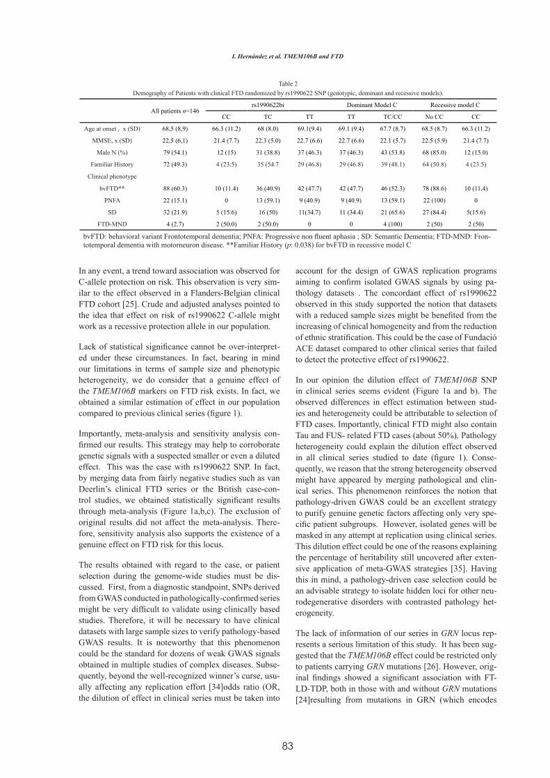
83
I. Hernández et al. TMEM106B and FTD
In any event, a trend toward association was observed for C-allele protection on risk. This observation is very sim-ilar to the effect observed in a Flanders-Belgian clinical FTD cohort [25]. Crude and adjusted analyses pointed to the idea that effect on risk of rs1990622 C-allele might work as a recessive protection allele in our population.
Lack of statistical significance cannot be over-interpret-ed under these circumstances. In fact, bearing in mind our limitations in terms of sample size and phenotypic heterogeneity, we do consider that a genuine effect of the TMEM106B markers on FTD risk exists. In fact, we obtained a similar estimation of effect in our population compared to previous clinical series (figure 1).
Importantly, meta-analysis and sensitivity analysis con-firmed our results. This strategy may help to corroborate genetic signals with a suspected smaller or even a diluted effect. This was the case with rs1990622 SNP. In fact, by merging data from fairly negative studies such as van Deerlin’s clinical FTD series or the British case-con-trol studies, we obtained statistically significant results through meta-analysis (Figure 1a,b,c). The exclusion of original results did not affect the meta-analysis. There-fore, sensitivity analysis also supports the existence of a genuine effect on FTD risk for this locus.
The results obtained with regard to the case, or patient selection during the genome-wide studies must be dis-cussed. First, from a diagnostic standpoint, SNPs derived from GWAS conducted in pathologically-confirmed series might be very difficult to validate using clinically based studies. Therefore, it will be necessary to have clinical datasets with large sample sizes to verify pathology-based GWAS results. It is noteworthy that this phenomenon could be the standard for dozens of weak GWAS signals obtained in multiple studies of complex diseases. Subse-quently, beyond the well-recognized winner’s curse, usu-ally affecting any replication effort [34]odds ratio (OR, the dilution of effect in clinical series must be taken into
account for the design of GWAS replication programs aiming to confirm isolated GWAS signals by using pa-thology datasets . The concordant effect of rs1990622 observed in this study supported the notion that datasets with a reduced sample sizes might be benefited from the increasing of clinical homogeneity and from the reduction of ethnic stratification. This could be the case of Fundació ACE dataset compared to other clinical series that failed to detect the protective effect of rs1990622.
In our opinion the dilution effect of TMEM106B SNP in clinical series seems evident (Figure 1a and b). The observed differences in effect estimation between stud-ies and heterogeneity could be attributable to selection of FTD cases. Importantly, clinical FTD might also contain Tau and FUS- related FTD cases (about 50%). Pathology heterogeneity could explain the dilution effect observed in all clinical series studied to date (figure 1). Conse-quently, we reason that the strong heterogeneity observed might have appeared by merging pathological and clin-ical series. This phenomenon reinforces the notion that pathology-driven GWAS could be an excellent strategy to purify genuine genetic factors affecting only very spe-cific patient subgroups. However, isolated genes will be masked in any attempt at replication using clinical series. This dilution effect could be one of the reasons explaining the percentage of heritability still uncovered after exten-sive application of meta-GWAS strategies [35]. Having this in mind, a pathology-driven case selection could be an advisable strategy to isolate hidden loci for other neu-rodegenerative disorders with contrasted pathology het-erogeneity.
The lack of information of our series in GRN locus rep-resents a serious limitation of this study. It has been sug-gested that the TMEM106B effect could be restricted only to patients carrying GRN mutations [26]. However, orig-inal findings showed a significant association with FT-LD-TDP, both in those with and without GRN mutations [24]resulting from mutations in GRN (which encodes
Table 2Demography of Patients with clinical FTD randomized by rs1990622 SNP (genotypic, dominant and recessive models).
All patients n=146 rs1990622bi Dominant Model C Recessive model C
CC TC TT TT TC/CC No CC CC
Age at onset , x (SD) 68,5 (8,9) 66.3 (11.2) 68 (8.0) 69.1(9.4) 69.1 (9.4) 67.7 (8.7) 68.5 (8.7) 66.3 (11.2)
MMSE, x (SD) 22,5 (6,1) 21.4 (7.7) 22.3 (5.0) 22.7 (6.6) 22.7 (6.6) 22.1 (5.7) 22.5 (5.9) 21.4 (7.7)
Male N (%) 79 (54.1) 12 (15) 31 (38.8) 37 (46.3) 37 (46.3) 43 (53.8) 68 (85.0) 12 (15.0)
Familiar History 72 (49.3) 4 (23.5) 35 (54.7 29 (46.8) 29 (46.8) 39 (48.1) 64 (50.8) 4 (23.5)
Clinical phenotype
bvFTD** 88 (60.3) 10 (11.4) 36 (40.9) 42 (47.7) 42 (47.7) 46 (52.3) 78 (88.6) 10 (11.4)
PNFA 22 (15.1) 0 13 (59.1) 9 (40.9) 9 (40.9) 13 (59.1) 22 (100) 0
SD 32 (21.9) 5 (15.6) 16 (50) 11(34.7) 11 (34.4) 21 (65.6) 27 (84.4) 5(15.6)
FTD-MND 4 (2.7) 2 (50.0) 2 (50.0) 0 0 4 (100) 2 (50) 2 (50)
bvFTD: behavioral variant Frontotemporal dementia; PNFA: Progressive non fluent aphasia ; SD: Semantic Dementia; FTD-MND: Fron-totemporal dementia with motorneuron disease. **Familiar History (p: 0.038) for bvFTD in recessive model C

84
progranulin. Moreover, the frequency of GRN mutations in Spanish FTD patients is quite low (<6%) [36]8 asymp-tomatic mutation carriers, and 10 control subjects as well as in brain tissue from 16 patients and 9 control subjects. RESULTS: Four novel mutations were associated with familial and sporadic FTLD and familial dementia asso-ciated with amyotrophic lateral sclerosis. We identified a close association between the IVS6-1G>A mutation in PGRN and corticobasal syndrome. Brain tissue was available for carriers of two of the four mutations (IVS6-1 G>A and P357HfsX3. This data makes it improbable that the obtained results in our series and meta-analysis could be only attributed to GRN mutation carriers. In re-gard to FTD diagnosis, it will be necessary to determine which genetic backgrounds are affected or modulated by TMEM106B markers. However, data derived from oth-er studies pointed to the idea that GRN mutation pene-trance could be modulated by this locus [26-37]. Inter-estingly, the only significant phenotypic characteristic of FTD patients that might be observed in patients carry-ing rs19906622-CC was a reduced frequency of famil-ial antecedents. This finding is consistent with a higher effect of this locus in GRN mutation carriers previously observed. However, the TMEM106B gene might modify other FTD mutations. As a recent publication suggested, TMEM106B genotype could act as a genetic modifier of the penetrance of C9ORF72 expansions. The observa-tion was restricted only to those patients displaying FTD clinical phenotype alone or in combination with MND. Notably, the effect on C9ORF72 expansion was more evident assuming a recessive model which was consis-tent with our meta-analysis [38]586 FTD patients lacking C9ORF72 expansions [with or without motor neuron dis-ease (MND. This observation, when replicated, would confirm that TMEM106B locus effect is not restricted to FTD patients carrying GRN mutations. Further research and independent replications are needed to corroborate these clinical observations.
In conclusion, the observed heterogeneity and statistical significance in the results observed during meta-analysis point to the idea that the TMEM106B locus is a genuine modifying genetic factor at least in a subgroup of FTD-TDP cases. Unfortunately, only a pathologically-con-firmed FLTD series would help to clarify this point. How-ever, the problem could be re-visited once radiotracers for distinct subtypes of FTD pathology become available. Finally, the molecular mechanism explaining observed association is still under investigation [26-34-35]. Re-cent functional studies suggested its role in progranulin pathways by affecting GRN degradation in lysosomal pathways [39]. This information might be relevant in the design of novel therapeutic strategies for FTD-TDP.
ACKNOWLEDGEMENTS
We thank donors and their families for generous blood donation for research. This work was carried out as part of
the doctoral program of I. Hernández at the Autonomous University of Barcelona. This work was partially support-ed by the Spanish Ministry of Health from Instituto de Salud Carlos III (Madrid) (FISS PI10/00945) and by the Agència d’Avaluació de Tecnologia i Recerca Mèdiques. Departament de Salut de la Generalitat de Catalunya (Health Department of the Catalan Government) (390). We are indebted to Trinitat Port-Carbó and her family for their support of the Fundació ACE research programs.
REFERENCES
[1] Rascovsky K, Hodges J. R, Knopman D, Men-dez MF, Kramer JH, Neuhaus J, van Swieten JC, Seelaar H, Dopper EGP, Onyike CU, Hillis AE, Josephs KA, Boeve BF, Kertesz A, Seeley WW, Rankin KP, Johnson JK, Gorno-Tempini ML, Rosen H, Prioleau-Latham CE, Lee A, Kipps C M, Lillo P, Piguet O, Rohrer JD, Rossor MN, Warren JD, Fox NC, Galasko D, Salmon DP, Black SE, Mesulam M, Weintraub S, Dickerson BC, Diehl-Schmid J, Pasquier F, Deramecourt V, Lebert F, Pijnenburg Y, Chow TW, Manes F, Grafman J, Cappa SF, Freed-man M, Grossman M, Miller BL. (2011) “Sensitiv-ity of revised diagnostic criteria for the behavioural variant of frontotemporal dementia. Brain 134 (Pt 9), 2456–2477.
[2] Gorno-Tempini ML, Hillis AE, Weintraub S, Kertesz A, Mendez M, Cappa SF, Ogar JM, Rohrer JD, Black S, Boeve BF, Manes F, Dronkers NF, Vandenberghe R, Rascovsky K, Patterson K, Mill-er BL, Knopman DS,. Hodges JR, Mesulam MM, Grossman M. (2011) “Classification of primary pro-gressive aphasia and its variants. Neurology 76 (11), 1006–1014.
[3] Rollinson S, Mead S, Snowden J, Richardson A, Rohrer J, Halliwell N, Usher S, Neary D, Mann D, Hardy J, Pickering-Brown S. (2011) Frontotem-poral lobar degeneration genome wide association study replication confirms a risk locus shared with amyotrophic lateral sclerosis. Neurobiology of ag-ing 32 (4), 758.e1–7.
[4] Achi EY, Rudnicki SA. (2012) ALS and Frontotem-poral Dysfunction: A Review. Neurology research international 2012, Article ID 806306, 9 pages.
[5] Boxer A L, Mackenzie IR, Boeve BF, Baker M, See-ley WW, Crook R, Feldman H, Hsiung GYR, Ruth-erford N, Laluz V, Whitwell J, Foti D, McDade E, Molano J, Karydas A, Wojtas A, Goldman J, Mirsky J, Sengdy P, Dearmond S, Miller BL, Rademakers R. (2011) Clinical, neuroimaging and neuropatho-logical features of a new chromosome 9p-linked FTD-ALS family. Journal of neurology, neurosur-gery, and psychiatry 82 (2), 196–203.
[6] Piguet O, Halliday GM, Reid WGJ, Casey B, Car-man R, Huang Y, Xuereb JH, Hodges JR, Kril JJ. (2011)Clinical phenotypes in autopsy-confirmed
I. Hernández et al. TMEM106B and FTD
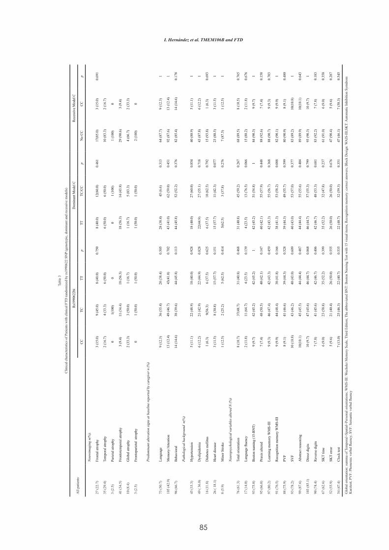
85
I. Hernández et al. TMEM106B and FTD
Tabl
e 3
Clin
ical
cha
ract
eris
tics o
f Pat
ient
s with
clin
ical
FTD
rand
omiz
ed b
y rs
1990
622
SNP
(gen
otyp
ic, d
omin
ant a
nd re
cess
ive
mod
els)
All
patie
nts
Rs1
9906
22bi
Dom
inan
t Mod
el C
Rec
essi
ve M
odel
C
CC
TCTT
pTT
TC/C
Cp
No
CC
CC
p
Neu
roim
agin
g n(
%).
27 (2
2.7)
Fron
tal a
troph
y3
(15.
0)9
(45.
0)8
(40.
0)0.
750
8 (4
0.0)
12(6
0.0)
0.46
117
(85.
0)3
(15.
0)0.
691
35 (2
9.4)
Tem
pora
l atro
phy
2 (1
6.7)
4 (3
3.3)
6 (5
0.0)
6 (5
0.0)
6 (5
0.0)
10 (8
3.3)
2 (1
6.7)
3 (2
.5)
Parie
tal a
troph
y0
1(10
0)0
01
(100
)1
(100
)0
41 (3
4.5)
Fron
tote
mpo
ral a
troph
y3
(9.4
)11
(34.
4)18
(56.
3)18
(56.
3)14
(43.
8)29
(90.
6)3
(9.4
)
10 (8
.4)
Glo
bal a
troph
y2
(33.
3)3
(50.
0)1
(16.
7)1
(16.
7)5
(83.
3)4
(66.
7)2
(33.
3)
3 (2
.5)
Fron
topa
rieta
l at
roph
y0
1 (5
0.0)
1 (5
0.0)
1 (5
0.0)
1 (5
0.0)
2 (1
00)
0
Pred
omin
ant a
ltera
tion
sign
s at b
asel
ine
repo
rted
by
care
give
r n (%
)
73 (5
0.7)
Lang
uage
9 (1
2.3)
36 (5
5.4)
28 (3
8.4)
0.50
528
(38.
4)45
(6.6
)0.
313
64 (8
7.7)
9 (1
2.3)
1
105
(42.
9)M
emor
y/A
ttent
ion
13 (1
2.4)
49 (4
6.7)
43(4
1.0)
0.70
243
(41.
0)62
(59.
0)0.
451
92 (8
7.6)
13 (1
2.4)
1
96 (6
6.7)
Beh
avio
ral
14 (1
4.6)
38 (3
9.6)
44 (4
5.8)
0.11
344
(45.
8)52
(52.
2)0.
376
82 (8
5.4)
14 (1
4.6)
0.17
8
Path
olog
ical
bac
kgro
und
n(%
)
45 (3
3.3)
Hyp
erte
nsio
n5
(11.
1)22
(48.
9)18
(40.
0)0.
928
18 (4
0.0)
27 (6
0.0)
0.85
440
(88.
9)5
(11.
1)1
49 (
36.0
)D
yslip
idem
ia6
(12.
2)21
(42.
9)22
(44.
9)0.
828
22(4
4.9)
27 (5
5.1)
0.71
843
(87.
8)6
(12.
2)1
16 (1
1.8)
Dia
bete
s mel
litus
1 (6
.3)
9(56
.3)
6 (3
7.5)
0.62
56
(37.
5)10
(62.
5)0.
792
15 (9
3.8)
1 (6
.3)
0.69
3
26 (
18.1
)H
eart
dise
ase
3 (1
1.5)
8 (3
0.8)
15 (5
7.7)
0.15
115
(57.
7)11
(42.
3)0.
077
23 (8
8.5)
3 (1
1.5)
1
8 (5
.9)
Min
or S
troke
1 (1
2.5)
2 (2
5.2)
5 (6
2.5)
0.41
45(
62.5
)3
(37.
8)0.
276
7 (8
7.5)
1 (1
2.5)
1
Neu
rops
ycho
logi
cal v
aria
bles
alte
red
N (%
)
76 (6
1.3)
Tota
l orie
ntat
ion
8 (1
0.7)
37(4
8.7)
31 (4
0.8)
0.46
831
(40.
8)45
(59.
2)0.
267
68 (8
9.5)
8 (1
0.5)
0.76
5
17 (1
4.0)
Lang
uage
flue
ncy
2 (1
1.8)
11 (6
4.7)
4 (2
3.5)
0.13
94
(23.
5)13
(76.
5)0.
066
15 (8
8.2)
2 (1
1.8)
0.67
6
93 (7
5.0)
Bos
ton
nam
ing
(15-
BN
T)9
(9.7
)42
(45.
2)42
(45.
2)1
42 (4
5.2)
51 (5
4.8)
184
(90.
3)9
(9.7
)1
95 (6
6.0)
Prax
is a
ltere
d7
(7.4
)48
(50.
5)40
(42.
1)0.
147
40 (4
2.1)
55 (5
7.9)
0.44
888
(92.
6)7
(7.4
)0.
158
97 (8
0.2)
Lear
ning
mem
ory
WM
S-II
I9
(9.3
)46
(47.
4)42
(43.
3)0.
459
42 (4
3.3)
55 (5
6.7)
0.36
888
(90.
7)9
(9.3
)0.
703
91 (7
6.5)
Rec
ogni
tion
mem
ory
WM
S-II
I9
(9.9
)44
(48.
4)38
(41.
8)0.
166
38 (4
1.3)
53 (5
8.2)
0.08
882
(90.
1)9
(9.9
)1
88 (7
5.9)
PVF
8 (9
.1)
41 (4
6.6)
39 (4
4.3)
0.52
839
(44.
3)49
(55.
7)0.
599
80 (9
0.9)
8 (9
.1)
0.48
0
93 (7
8.2)
SVF
10 (1
0.8)
43 (4
6.2)
40 (4
3.0)
0.60
940
(43.
0)53
(57.
0)0.
377
83 (8
9.2)
10(1
0.8)
1
99 (8
7.6)
Abs
tract
reas
onin
g10
(10.
1)45
(45.
5)44
(44.
4)0.
487
44 (4
4.4)
55 (5
5.6)
0.40
489
(89.
9)10
(10.
1)0.
643
103
(85.
1)D
irect
dig
its10
(9.7
)47
(45.
6)46
(44.
7)0.
868
46 (4
4.7)
57 (5
5.3)
0.79
993
(90.
3)10
(9.7
)1
90 (7
4.4)
Rev
erse
dig
its7
(7.8
)41
(45.
6)42
(46.
7)0.
406
42 (4
6.7)
48 (5
3.3)
0.68
183
(92.
2)7
(7.8
)0.
183
67 (6
2.6)
SKT
time
6 (9
.0)
23 (5
8.8)
35 (5
2.2)
0.39
935
(52.
2)32
(47.
8)0.
237
61 (9
1.0)
6 (9
.0)
0.35
8
52 (5
5.9)
SKT
erro
r5
(9.6
)21
(40.
4)26
(50.
0)0.
555
26 (5
0.0)
26 (5
0.0)
0.67
647
(90.
4)5
(9.6
)0.
287
54 (4
7.4)
Clo
ck te
st7
(13.
0)25
(46.
3)22
(40.
7)0.
535
22 (4
0.7)
32 (5
9.3)
0.35
147
(46.
1)7
(58.
3)0.
545
Glo
bal o
rient
atio
n: su
mm
a of
Tem
pora
l+Sp
atia
l+Pe
rson
al o
rient
atio
ns; W
MS-
III:
Wec
hsle
r Mem
ory
Scal
e, T
hird
Edi
tion;
The
abb
revi
ated
BN
T: B
osto
n N
amin
g Te
st w
ith 1
5 vi
sual
item
s; R
ecog
nitio
n m
emor
y: c
orre
ct a
nsw
ers;
Blo
ck D
esig
n: W
AIS
-III
;SK
T: A
utom
atic
Inhi
bitio
n Sy
mdr
om
Kur
ztes
t; PV
F: P
hone
mic
ver
bal fl
uenc
y; S
VF:
Sem
antic
ver
bal fl
uenc
y
86
I. Hernández et al. TMEM106B and FTD
Pick disease. Neurology 76 (3), 253–259.
[7] Piguet O, Hornberger M, Mioshi E, Hodges JR. (2011) Behavioural-variant frontotemporal demen-tia: diagnosis, clinical staging, and management. Lancet neurology 10 (2), 162–172.
[8] Roeber S, Mackenzie IRA, Kretzschmar HA, Neu-mann M. (2008) TDP-43-negative FTLD-U is a sig-nificant new clinico-pathological subtype of FTLD. Acta neuropathologica 116 (2), 147–157.
[9] Mackenzie IRA, Munoz DG, Kusaka H, Yokota O, Ishihara K, Roebe S, a Kretzschmar H, Cairns NJ, Neumann M. (2011) Distinct pathological subtypes of FTLD-FUS. Acta neuropathologica 121 (2), 207–218.
[10] Mackenzie IRA, Neumann M, Bigio EH, Cairns NJ, Alafuzoff I, Kril J, Kovacs GG, Ghetti B, Hal-liday G, Holm IE, Ince PG, Kamphorst W, Revesz T, Rozemuller AJM, Kumar-Singh S, Akiyama H, Baborie A, Spina S, Dickson DW, Trojanowski JQ, A. Mann DM. (2009) Nomenclature for neuro-pathologic subtypes of frontotemporal lobar degen-eration: consensus recommendations. Acta neuro-pathologica 117 (1), 15–18.
[11] Mori K, Weng SM, Arzberger T, May S, Rent-zsch K, Kremmer E, Schmid B, Kretzschmar HA, Cruts M, Van Broeckhoven C, Haass C, Edbauer D. (2013) The C9orf72 GGGGCC repeat is translated into aggregating dipeptide-repeat proteins in FTLD/ALS. Science 339 (6125), 1335–1338.
[12] Mackenzie IR, Arzberger T, Kremmer E, Troost D, Lorenzl S, Mori K, Weng SM, Haass C, Kretzsch-mar HA, Edbauer D, Neumann M. (2013) Dipep-tide repeat protein pathology in C9ORF72 mutation cases: clinico-pathological correlations. Acta neuro-pathologica 126 (6), 859–879.
[13] Baker M, Mackenzie IR, Pickering-Brown SM, Gass J, Rademakers R, Lindholm C, Snowden J, Adamson J, Sadovnick AD, Rollinson S, Cannon A, Dwosh E, Neary D, Melquist S, Richardson A, Dickson D, Berger Z, Eriksen J, Robinson T, Zehr C, Dickey CA, Crook R, McGowan E, Mann D, Boeve B, Feldman H, Hutton M. (2006) Mutations in progranulin cause tau-negative frontotemporal dementia linked to chromosome 17 Nature 442 (7105), 916–919.
[14] Cruts M, Kumar-Singh S, Van Broeckhoven C. (2006) Progranulin mutations in ubiquitin-positive frontotemporal dementia linked to chromosome 17q21. Current Alzheimer research 3 (5), 485–491.
[15] Hutton M, Lendon CL, Rizzu P, Baker M, Froelich S, Houlden H, Pickering-Brown S, Chakraverty S, Isaacs A, Grover A, Hackett J, Adamson J, Lincoln S, Dickson D, Davies P, Petersen RC, Stevens M, de Graaff E, Wauters E, van Baren J, Hillebrand M, Joosse M, Kwon JM, Nowotny P, Che LK, Norton J, Morris JC, Reed LA, Trojanowski J, Basun H,
Lannfelt L, Neystat M, Fahn S, Dark F, Tannenberg T, Dodd PR, Hayward N, Kwok JB, Schofield PR, Andreadis A, Snowden J, Craufurd D, Neary D, Owen F, Oostra BA, Hardy J, Goate A, van Swi-eten J, Mann D, Lynch T, Heutink P. (1998) Associ-ation of missense and 5’-splice-site mutations in tau with the inherited dementia FTDP-17. Nature 393 (6686), 702–705.
[16] Skibinski G, Parkinson NJ, Brown JM, Chakrabar-ti L, Lloyd SL, Hummerich H, Nielsen JE, Hodges JR, Spillantini MG, Thusgaard T, Brandner S, Brun A, Rossor MN, Gade A, Johannsen P, Sørensen SA, Gydesen S, Fisher EM, Collinge J (2005). Muta-tions in the endosomal ESCRTIII-complex subunit CHMP2B in frontotemporal dementia. Nature ge-netics 37 (8), 806–808.
[17] Deng HX, Chen W, Hong ST, Boycott KM, Gor-rie GH, Siddique N, Yang Y, Fecto F, Shi Y, Zhai H, Jiang H, Hirano M, Rampersaud E, Jansen GH, Donkervoort S, Bigio EH, Brooks BR, Ajroud K, Sufit RL, Haines JL, Mugnaini E, Pericak-Vance MA, Siddique T. (2011) Mutations in UBQLN2 cause dominant X-linked juvenile and adult-onset ALS and ALS/dementia. Nature 477 (7363), 211–215.
[18] Rubino E, Rainero I, Chiò A, Rogaeva E, Galimber-ti D, Fenoglio P, Grinberg Y, Isaia G, Calvo A, Gen-tile S, Bruni AC, St George-Hyslop PH, Scarpini E, Gallone S, Pinessi L; TODEM Study Group .(2012) SQSTM1 mutations in frontotemporal lobar degen-eration and amyotrophic lateral sclerosis Neurology 79 (15),1556–1562.
[19] Guerreiro RJ, Lohmann E, Brás JM, Gibbs JR, Rohrer JD, Gurunlian N, Dursun B, Bilgic B, Hana-gasi H, Gurvit H, Emre M, Singleton A, Hardy J .(2013) Using exome sequencing to reveal muta-tions in TREM2 presenting as a frontotemporal dementia-like syndrome without bone involvement. JAMA neurology 70 (1), 78–84.
[20] DeJesus-Hernandez M, Mackenzie IR, Boeve BF, Boxer AL, Baker M, Rutherford NJ, Nicholson AM, Finch NA, Flynn H, Adamson J, Kouri N, Wojtas A, Sengdy P, Hsiung GY, Karydas A, Seeley WW, Josephs KA, Coppola G, Geschwind DH, Wszolek ZK, Feldman H, Knopman DS, Petersen RC, Miller BL, Dickson DW, Boylan KB, Graff-Radford NR, Rademakers R. (2011) Expanded GGGGCC hexa-nucleotide repeat in noncoding region of C9ORF72 causes chromosome 9p-linked FTD and ALS. Neu-ron 72 (2), 245–256.
[21] Renton AE, Majounie E, Waite A, Simón-Sánchez J, Rollinson S, Gibbs JR, Schymick JC, Laaksovir-ta H, van Swieten JC, Myllykangas L, Kalimo H, Paetau A, Abramzon Y, Remes AM, Kaganovich A, Scholz SW, Duckworth J, Ding J, Harmer DW, Hernandez DG, Johnson JO, Mok K, Ryten M, Trabzuni D, Guerreiro RJ, Orrell RW, Neal J, Mur-ray A, Pearson J, Jansen IE, Sondervan D, Seelaar
87
I. Hernández et al. TMEM106B and FTD
H, Blake D, Young K, Halliwell N, Callister JB, Toulson G, Richardson A, Gerhard A, Snowden J, Mann D, Neary D, Nalls MA, Peuralinna T, Jansson L, Isoviita VM, Kaivorinne AL, Hölttä-Vuori M, Ikonen E, Sulkava R, Benatar M, Wuu J, Chiò A, Restagno G, Borghero G, Sabatelli M; ITALSGEN Consortium, Heckerman D, Rogaeva E, Zinman L, Rothstein JD, Sendtner M, Drepper C, Eichler EE, Alkan C, Abdullaev Z, Pack SD, Dutra A, Pak E, Hardy J, Singleton A, Williams NM, Heutink P, Pickering-Brown S, Morris HR, Tienari PJ, Traynor BJ (2011). A hexanucleotide repeat expansion in C9ORF72 is the cause of chro-mosome 9p21-linked ALS-FTD. Neuron 72 (2), 257–268.
[22] Gijselinck I, Van Langenhove T, van der Zee J, Sleegers K, Philtjens S, Kleinberger G, Janssens J, Bettens K, Van Cauwenberghe C, Pereson S, En-gelborghs S, Sieben A, De Jonghe P, Vandenber-ghe R, Santens P, De Bleecker J, Maes G, Bäumer V, Dillen L, Joris G, Cuijt I, Corsmit E, Elinck E, Van Dongen J, Vermeulen S, Van den Broeck M, Vaerenberg C, Mattheijssens M, Peeters K, Robberecht W, Cras P, Martin JJ, De Deyn PP, Cruts M, Van Broeckhoven C. (2012) A C9orf72 promoter repeat expansion in a Flanders-Belgian cohort with disorders of the frontotemporal lobar degeneration-amyotrophic lateral sclerosis spec-trum: a gene identification study. Lancet neurology 11 (1), 54–65.
[23] Sieben A, Van Langenhove T, Engelborghs S, Martin JJ, Boon P, Cras P, De Deyn PP, Santens P, Van Broeckhoven C, Cruts M. (2012) The ge-netics and neuropathology of frontotemporal lo-bar degeneration. Acta neuropathologica 124 (3), 353–372.
[24] Van Deerlin VM, Sleiman PM, Martinez-Lage M, Chen-Plotkin A, Wang LS, Graff-Radford NR, Dickson DW, Rademakers R, Boeve BF, Grossman M, Arnold SE, Mann DM, Pickering-Brown SM, Seelaar H, Heutink P, van Swieten JC, Murrell JR, Ghetti B, Spina S, Grafman J, Hodges J, Spillantini MG, Gilman S, Lieberman AP, Kaye JA, Woltjer RL, Bigio EH, Mesulam M, Al-Sarraj S, Troakes C, Rosenberg RN, White CL 3rd, Ferrer I, Lladó A, Neumann M, Kretzschmar HA, Hulette CM, Welsh-Bohmer KA, Miller BL, Alzualde A, Lopez de Munain A, McKee AC, Gearing M, Levey AI, Lah JJ, Hardy J, Rohrer JD, Lashley T, Macken-zie IR, Feldman HH, Hamilton RL, Dekosky ST, van der Zee J, Kumar-Singh S, Van Broeckhoven C, Mayeux R, Vonsattel JP, Troncoso JC, Kril JJ, Kwok JB, Halliday GM, Bird TD, Ince PG, Shaw PJ, Cairns NJ, Morris JC, McLean CA, DeCarli C, Ellis WG, Freeman SH, Frosch MP, Growdon JH, Perl DP, Sano M, Bennett DA, Schneider JA, Beach TG, Reiman EM, Woodruff BK, Cummings J, Vinters HV, Miller CA, Chui HC, Alafuzoff I, Hartikainen P, Seilhean D, Galasko D, Masliah E, Cotman CW, Tuñón MT, Martínez MC, Munoz DG, Carroll SL, Marson D, Riederer PF, Bog-
danovic N, Schellenberg GD, Hakonarson H, Tro-janowski JQ, Lee VM. (2010) Common variants at 7p21 are associated with frontotemporal lobar de-generation with TDP-43 inclusions. Nature genetics 42 (3), 234–239.
[25] van der Zee J, Van Langenhove T, Kleinberger G, Sleegers K, Engelborghs S, Vandenberghe R, San-tens P, Van den Broeck M, Joris G, Brys J, Matthei-jssens M, Peeters K, Cras P, De Deyn PP, Cruts M, Van Broeckhoven C. (2011) TMEM106B is asso-ciated with frontotemporal lobar degeneration in a clinically diagnosed patient cohort. Brain 134 (Pt 3), 808–815.
[26] Finch N, Carrasquillo MM, Baker M, Rutherford NJ, Coppola G, Dejesus-Hernandez M, Crook R, Hunter T, Ghidoni R, Benussi L, Crook J, Finger E, Hantanpaa KJ, Karydas AM, Sengdy P, Gonza-lez J, Seeley WW, Johnson N, Beach TG, Mesu-lam M, Forloni G, Kertesz A, Knopman DS, Uitti R, White CL 3rd, Caselli R, Lippa C, Bigio EH, Wszolek ZK, Binetti G, Mackenzie IR, Miller BL, Boeve BF, Younkin SG, Dickson DW, Petersen RC, Graff-Radford NR, Geschwind DH, Rademakers R. (2011) TMEM106B regulates progranulin levels and the penetrance of FTLD in GRN mutation car-riers. Neurology 76 (5), 467–474.
[27] Armstrong MJ, Litvan I, Lang AE, Bak TH, Bhatia KP, Borroni B, Boxer AL, Dickson DW, Grossman M, Hallett M, Josephs KA, Kertesz A, Lee SE, Mill-er BL, Reich SG, Riley DE, Tolosa E, Tröster AI, Vidailhet M, Weiner WJ. (2013) Criteria for the di-agnosis of corticobasal degeneration. Neurology 80 (5), 496–503.
[28] Rabinovici GD, Jagust WJ, Furst AJ, Ogar JM, Ra-cine CA, Mormino EC, O’Neil JP, Lal RA, Dronkers NF, Miller BL, Gorno-Tempini ML. (2008) Abeta amyloid and glucose metabolism in three variants of primary progressive aphasia. Annals of neurolo-gy 64 (4), 388–401.
[29] Pan XD, Chen XC. (2013) Clinic neuropathology and molecular genetics of frontotemporal dementia: a mini-review. Translational neurodegeneration 2 (1):8.
[30] Alegret M, Espinosa A, Vinyes-Junqué G, Valero S, Hernández I, Tárraga L, Becker JT, Boada M. (2012) Normative data of a brief neuropsycholog-ical battery for Spanish individuals older than 49. Journal of clinical and experimental neuropsychol-ogy 34 (2), 209–219.
[31] Xi Z, Zinman L, Grinberg Y, Moreno D, Sato C, Bilbao JM, Ghani M, Hernández I, Ruiz A, Boada M, Morón FJ, Lang AE, Marras C, Bruni A, Co-lao R, Maletta RG, Puccio G, Rainero I, Pinessi L, Galimberti D, Morrison KE, Moorby C, Stockton JD, Masellis M, Black SE, Hazrati LN, Liang Y, van Haersma de With J, Fornazzari L, Villagra R, Ro-jas-Garcia R, Clarimón J, Mayeux R, Robertson J,

88
I. Hernández et al. TMEM106B and FTD
St George-Hyslop P, Rogaeva E. (2012) Investiga-tion of C9orf72 in 4 Neurodegenerative Disorders. Archives of neurology 69 (12),1583-1590
[32] Hernández I, Espinosa A, Real LM, Galán JJ, Mauleón A, Roca MR, Tárraga L, Ruiz A, Boada M. (2012) Molecular evaluation of human Ubiqui-lin 2 gene PXX domain in familial frontotemporal dementia patients. Journal of neurology 259 (11), 2488–2490.
[33] Sasieni PD. (1997) From genotypes to genes: dou-bling the sample size. Biometrics 53 (4), 1253–1261.
[34] Zhong H, Prentice RL. (2010) Correcting ‘winner’s curse’ in odds ratios from genomewide association findings for major complex human diseases. Genet-ic epidemiology 34 (1), 78–91.
[35] Manolio TA, Collins FS, Cox NJ, Goldstein DB, Hindorff LA, Hunter DJ, McCarthy MI, Ramos EM, Cardon LR, Chakravarti A, Cho JH, Guttmach-er AE, Kong A, Kruglyak L, Mardis E, Rotimi CN, Slatkin M, Valle D, Whittemore AS, Boehnke M, Clark AG, Eichler EE, Gibson G, Haines JL, Mack-ay TF, McCarroll SA, Visscher PM. (2009) Finding the missing heritability of complex diseases. Nature 461 (7265),747–753.
[36] A. López de Munain, A. Alzualde, A. Gorostidi, D. Otaegui, J. Ruiz-Martínez, B. Indakoetxea, I. Ferrer, J. Pérez-Tur, A. Sáenz, A. Bergareche, M. Barandi-arán, J. J. Poza, R. Zabalza, I. Ruiz, M. Urtasun, I. Fernández-Manchola, B. Olasagasti, J. B. Espinal, J. Olaskoaga, M. Ruibal, F. Moreno, N. Carrera, and J. F. Martí Massó. (2008) Mutations in progranulin gene: clinical, pathological, and ribonucleic acid
expression findings. Biological psychiatry 63 (10), 946–952.
[37] Cruchaga C, Graff C, Chiang HH, Wang J, Hin-richs AL, Spiegel N, Bertelsen S, Mayo K, Nor-ton JB, Morris JC, Goate A. (2011) Association of TMEM106B gene polymorphism with age at onset in granulin mutation carriers and plasma granulin protein levels. Archives of neurology 68 (5), 581–586.
[38] van Blitterswijk M, Mullen B, Nicholson AM, Bi-eniek KF, Heckman MG, Baker MC, DeJesus-Her-nandez M, Finch NA, Brown PH, Murray ME, Hsi-ung GY, Stewart H, Karydas AM, Finger E, Kertesz A, Bigio EH, Weintraub S, Mesulam M, Hatanpaa KJ, White CL 3rd, Strong MJ, Beach TG, Wszolek ZK, Lippa C, Caselli R, Petrucelli L, Josephs KA, Parisi JE, Knopman DS, Petersen RC, Mackenzie IR, Seeley WW, Grinberg LT, Miller BL, Boy-lan KB, Graff-Radford NR, Boeve BF, Dickson DW, Rademakers R. (2014) TMEM106B protects C9ORF72 expansion carriers against frontotempo-ral dementia. Acta neuropathologica 127 (3), 397–406.
[39] Brady OA, Zheng Y, Murphy K, Huang M, Hu F. (2013) The frontotemporal lobar degeneration risk factor, TMEM106B, regulates lysosomal morphol-ogy and function. Human molecular genetics 22 (4), 685–695.

8989
VIII. Discusión

9191
VIII. Discusión
En este trabajo de tesis se constata la necesidad de integrar la colaboración entre la labor que desempeña el investigador clínico y los resultados que aporta el laborato-rio de análisis genético, puesto que en este estadio de la investigación la presencia del genotipo causal que explica la patología ya comienza a ser un dato habitual disponible, que requiere una interpretación apropiada.
La resultados entre lo que el clínico valora en su contacto diario con los pacientes, en base a su experiencia, protocolos y criterios clínicos, son datos cualitativos no exentos de error, mientras que el profesional de la genética interpreta en su muestreo ADN resultados cuantitativos y estadísticos, si bien la sobre-interpretación de los hallazgos genéticos también deben tenerse en cuenta. Por ello, es esencial una base de datos clínica rigurosa y de calidad para la identificación de los nuevos loci asociados a cada patología, a la que sumar la mejora de la interpretación de los hallazgos clíni-cos gracias a la incorporación de la información genética. Una estrecha colabora-ción entre ambos profesionales es indispensable para una mejor comprensión de las bases que conforman las enfermedades neurodegenerativas.
Como se ha descrito anteriormente, la DFT es un grupo de enfermedades neurode-generativas que se presentan de forma diferente, tanto desde el punto de vista clíni-co, como genético y neuropatológico. (Figura 2 y Figura 3).
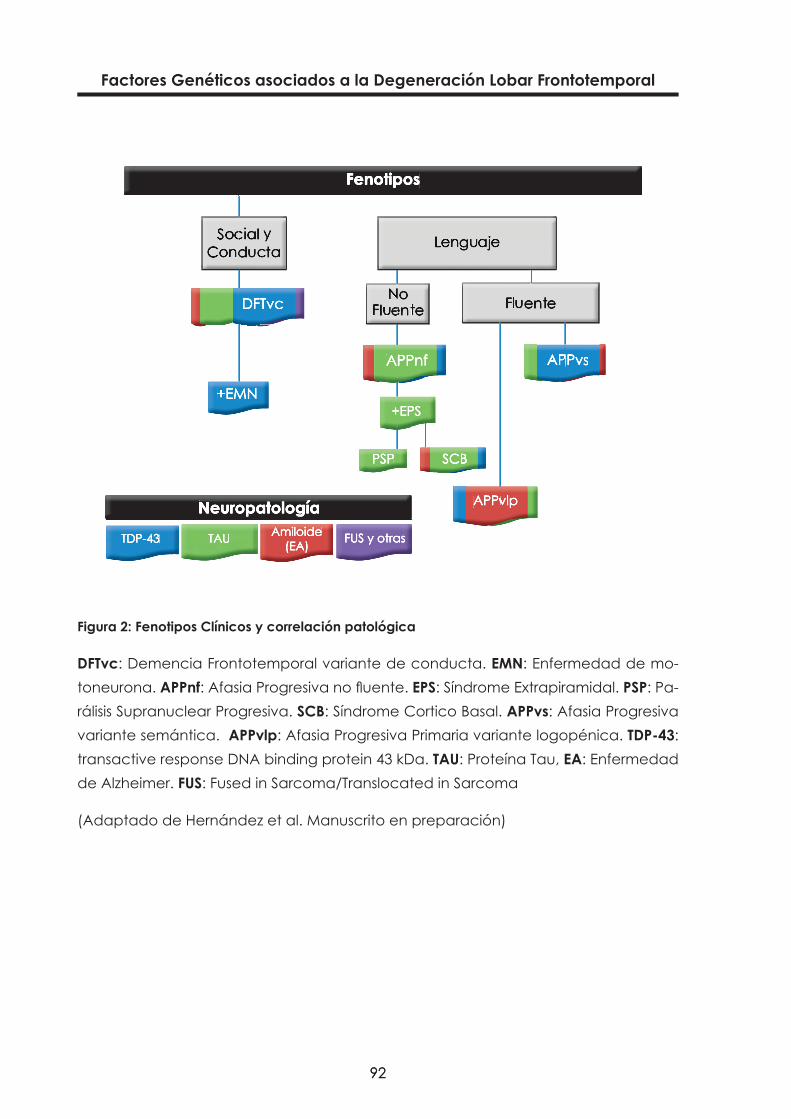
Factores Genéticos asociados a la Degeneración Lobar Frontotemporal
9292
Figura 2: Fenotipos Clínicos y correlación patológica
DFTvc: Demencia Frontotemporal variante de conducta. EMN: Enfermedad de mo-toneurona. APPnf: Afasia Progresiva no fluente. EPS: Síndrome Extrapiramidal. PSP: Pa-rálisis Supranuclear Progresiva. SCB: Síndrome Cortico Basal. APPvs: Afasia Progresiva variante semántica. APPvlp: Afasia Progresiva Primaria variante logopénica. TDP-43: transactive response DNA binding protein 43 kDa. TAU: Proteína Tau, EA: Enfermedad de Alzheimer. FUS: Fused in Sarcoma/Translocated in Sarcoma
(Adaptado de Hernández et al. Manuscrito en preparación)
FenotiposFenotiposFenotipos
NeuropatologíaNeuropatologíaNeuropatología
DFTvcDFTvcDFTvc
APPvsAPPvsAPPvsAPPvsAPPvsAPPvsAPPnfAPPnfAPPnfAPPnfAPPnfAPPnf
SCBSCBSCB
APPvlpAPPvlpAPPvlpAPPvlpAPPvlpAPPvlpAPPvlpAPPvlpAPPvlp
TDP-43TDP-43TDP-43 TAUTAUTAU AmiloideAmiloideAmiloide(EA)(EA)(EA) FUS y otrasFUS y otrasFUS y otras
+EMN+EMN+EMN
PSPPSPPSP
+EPS+EPS+EPS+EPS+EPS+EPS

9393
VIII. Discusión
Figura 3: Genotipos DLFT y correlación patológica
.MAPT: Microtubule associated protein Tau gen. TAU: Proteína Tau. DLFT-TAU: Demen-cia Lobar Frontotemporal por depósitos de proteína Tau, PiD: Enfermedad de Pick. PSP: Parálisis Supranuclear Progresiva. DCB: Degeneración Cortico Basal. FTLD-17: Demencia Frontotemporal con parkinsonismo asociado al Cr17. EGA: Enfermedad por Granos Argirófilos. MST: Taupatía Multisistémica. DLFT-TDP: Demencia Lobar Fron-totemporal con inclusiones de proteína TDP-43. C9orf72: chromosome 9 open reading frame 72. GRN: Gen Progranulina. VCP: Gen Valosina. TARDP: gen TAR DNA Binding Protein. TDP-43: transactive response DNA binding protein 43 kDa. DLFT-EMN; Demen-cia Frontotemporal con Enfermedad de motoneurona asociada. FUS: gen Fused in Sarcoma. CHMP2B: gen Charged multivesicular body protein. DLFT-UPS: Degenera-ción Lobar Frontotemporal con inmuno-histoquímica contra proteínas del sistema ubiquitina-proteasoma, DLFT-FUS: Degeneración Lobar Frontotemporal por depósitos de proteína FUS. DLFT-in: Degeneración Lobar Frontotemporal sin inclusiones.
(Adaptado de Hernández et al. manuscrito en preparación)
GenotiposGenotiposGenotipos NeuropatologíaNeuropatologíaNeuropatología ProteotiposProteotiposProteotiposy subtipos patológicosy subtipos patológicosy subtipos patológicos
Proteotiposy subtipos patológicos
ProteotiposProteotiposProteotiposy subtipos patológicos
Proteotiposy subtipos patológicos
Proteotiposy subtipos patológicos
ProteotiposProteotiposProteotiposy subtipos patológicos
ProteotiposFus y Fus y Fus y otrasotrasotrasTDP-43TDP-43TDP-43TAUTAUTAU
DLFT-TDPDLFT-TDPDLFT-TDP
Subtipos A-DSubtipos A-DSubtipos A-DDLFT-EMNDLFT-EMNDLFT-EMNTDP-43TDP-43TDP-43
C9orf72C9orf72C9orf72 (subtipo B) (subtipo B) (subtipo B)GRNGRNGRN (subtipo A) (subtipo A) (subtipo A)VCP (subtipo D)VCP (subtipo D)VCP (subtipo D)
TARDPTARDPTARDP (ALS) (ALS) (ALS)

Factores Genéticos asociados a la Degeneración Lobar Frontotemporal
9494
Respecto a la caracterización de la serie Clínica
La serie clínica utilizada en este trabajo de tesis ha sido un trabajo de observación y seguimiento clínico realizado a lo largo de 15 años. Los pacientes han sido valora-dos clínicamente con instrumentos comunes y seguidos en la mayoría de casos por el autor hasta su defunción. Esto ha permitido observar y objetivar la progresión y el cambio en algunos de sus fenotipos y disponer de información evolutiva de cada sujeto. Del mismo modo se ha obtenido un porcentaje aceptable de confirmaciones patológicas de una misma serie. El seguimiento continúa en muchos de los sujetos.
La evolución fenotípica de los 224 sujetos, desde su diagnóstico inicial, se muestra en Tabla 3. Clínicamente, los fenotipos evolutivos muestran que la DFTvc es el más frecuente, con el 50% de casos (n=112) (Rascovsky et al. 2011), seguido de la DS con el 15.2% (n=34) y la APNF con el 9.4% (n=21), lo que suponen el 24% de casos con fe-notipo APP (M L Gorno-Tempini et al. 2011). El resto de formas de presentación clínica las encontramos en el SCB con el 9.8% (n=22) (Armstrong et al. 2013) y la SPSP con el 10.7% (n=24)(Litvan et al. 1996). Sólo en el 3.6% (n=8) encontramos fenotipo DFT-EMN y en todos los casos en forma de DFTvc, probablemente porque, si su debut clínico es en forma de ELA son valorados en primera instancia en las Unidades específicas de Sistema Nervioso Periférico. Estos resultados son concordantes con lo que se observa en otras series (Johnson et al. 2005b). De los ocho pacientes inicialmente orientados como EA, 5 (62.5%) de ellos evolucionaron a DFTvc y 3 (37.5%) hacia SCB. Así mismo, una APNF y 2 DFTvc en diagnóstico inicial, evolucionaron clínicamente a EA.
En 7 casos de APNF (23%) la evolución permitió observar la presentación de un SPSP en 4 casos y de SCB en 3 casos, confirmándose en dos casos APNF de inicio, con evo-lución a SCB, una Taupatía de 4R. (I Ferrer et al. 2003) (Anexo).
Atendiendo a los datos de neuroimagen, observamos un predominio del patrón fron-totemporal con 53 sujetos (29.4%) seguido del patrón frontal en 47 sujetos (26.1%) y el temporal en 44 (24.4%), lo que suma un 80% de la serie y donde podríamos incluir los fenotipos DFTvc y las APP (Tabla 4). El restante 20% presenta un patrón de atrofia difuso o frontoparietal. En este % probablemente están representados los fenotipos SPSP, con un patrón más global y el SCB con un patrón más característico frontoparietal.
De toda la serie clínica a estudio se han conseguido hasta el momento 31(13.8%) confirmaciones anatomopatológicas (Tabla 5), donde el proteotipo Tau y TDP-43 su-ponen el 74.3 % de los casos. El proteotipo amiloide está presente en el 19.4% de la serie.

9595
VIII. Discusión
Tabla 3: Fenotipo evolutivo de la serie DFT clínica
Diagnóstico BasalFenotipo Evolutivo N (%)
N (%) DFTvc APNF DS DFT-EMN SCB PSP EA
Psiquiátrico 3 (1.3) 2 (66.7) -- 1 (33.3) -- -- -- --
SCB 14 (6.3) -- 1 (7.1%) -- -- 12 (85.7) 1 (7.1) --
PSP 16 (7.1) -- -- -- -- -- 16 (100) --
EA 8 (3.6) 5 (62.5) -- -- -- 3 (37.5) -- --
DCL 17 (7.6) 11 (64.7) 1 (5.9) 2 (11.8) 1 (5.9) 2 (11.8) --
APNF 32 (14.3) 3 (9.4) 19 (59.4) 1 (3.1) 1 (3,6) 3 (9.4) 4 (12.5) 1 (3.1)
DFTvc 99 (44.2) 88 (88.9) -- 1 (1.0) 4 (4.0) 1 (1.0) 3 (3.0) 2 (2.0)
DS 33 (14.7) 3 (9.1) -- 29 (87.9) 0 1 (3.0) -- --
DFT-EMN 2 (0.9) -- -- -- 2 (100) -- -- --
Total N (%) 224112
(50.0)21 (9.4) 34 (15.2) 8 (3.6) 22 (9.8) 24 (10.7) 3 (1.3)

Factores Genéticos asociados a la Degeneración Lobar Frontotemporal
9696
Tabla 4: Patrones de Neuroimagen
Diagnóstico Basal
AtrofiaenNeuroimagenN(%)
N (%) Frontal Temporal ParietalFron-
to-tem-poral
GlobalFron-
to-parie-tal
Psiquiátrico 3 (1.7) 1 (33.3) 1 (33.3) -- 1 (3.3 -- --
SCB 9 (5.0) 1 (11.1) 1 (11.1) 1 (11.1) 2 (22.2) 2 (22.2) 2 (22.2)
PSP 12 (6.7) 3 (25.0) -- 1 (8.3) 2 (16.7) 6 (50.0) --
EA 6 (3.3 1 (16.7) -- 1 (16.7) 3 (50.0) 1 (16.7) --
DCL 11 (6.1) 3 (27.3) 2 (18.2) -- 5 (45.5) -- 1 (9.1)
APNF 27 (15.0) 9 (3.3) 6 (22.2) 2 (7.4) 5 (18.5) 4 (14.8) 1 (3.7)
DFTvc 82 (45.6) 27 (32.9) 15 (18.3) 2 (2.4) 28 (34.1) 9 (11.0) 1 (1.2)
DS 30 (16.7) 2 (6.7) 19 (63.3) -- 7 (23.3) 2 (6.7) --
Total 180 47 (26.1) 44 (24.4) 7 (3.9) 53 (29.4) 24 (13.3) 5 (2.8)

9797
VIII. Discusión
Tabla 5: Neuropatología disponible de la serie clínica
Fenotipo al éxitusN (%)
Neuropatología N (%)
Amiloide Tau 4R Tau 3Rα-
sincucleinaCuerpos argirófilos TDP-43
IF-α-internexina
DFTvc 10 (32.3) 2 (20.0) 1 (10.0) 1 (10.0) -- 1 (10.0) 4 (40.0) 1(10.0)
SCB 6 (19.4)) 2 (33.3) 2 (33.3) -- 1 (16.7) -- 1 (16.7) --
PSP 5 (16.1) -- 5 (100) -- -- -- -- --
DFT-ELA 6 (19.4) 1 (16.7) -- -- -- 1 (16.7) 4 (66.7) --
APNF 2 (6.5) -- 2 (100) -- -- -- -- --
DS 2 (6.5) 1 (50) -- -- -- -- 1(50) --
Total 31 6 (19.4) 10 (32.3) 1 (3.2) 1 (3.4) 2 (6.5) 10 (32.3) 1 (3.2)
La caracterización y el diagnóstico clínico de cada síndrome van a depender de la realización de una anamnesis detallada, investigando en profundidad, general-mente con la persona informante, los signos iniciales del proceso. La identificación de los síntomas del paciente en las etapas iniciales será crucial de cara a establecer posibles tratamientos médicos y proporcionar el mejor seguimiento y soporte familiar a cada caso.

Factores Genéticos asociados a la Degeneración Lobar Frontotemporal
9898
Respecto al análisis genético de la serie
a. Objetivos relacionados con la tesis
Trabajo I: UBQLN2
Se examinaron 77 pacientes con historia familiar de demencia que poseían evalua-ción clínica completa. Es importante destacar que mantuvimos los pacientes con DFT de transmisión varón-varón (donde la trasmisión dominante ligada al X es imposible) como controles negativos de este estudio (n = 12).
No se encontraron mutaciones de UBQLN2 en nuestra población, por lo que la corre-lación genotipo-fenotipo fue desestimada.
Trabajo II: C9orf72
Los datos fueron obtenidos gracias a la colaboración con el “Tanz Center for Re-search in Neurodegenerative Diseases. Universidad de Toronto. Canadá” que realizó el estudio multicéntrico y la determinación de las mutaciones C9ofr72. (Xi et al. 2012).
Para este trabajo se aportaron a la serie a estudio 159 muestras de ADN. De ellas 102 (64.6%) sujetos con fenotipo DFTvc, 3 (1.9%) fenotipo DFT-EMN, 22 (13.9%) con fenoti-po APPnf y 31 (19.6%) con fenotipo DS. A estos hay que añadir la confirmación de la expansión G5C2 positiva posmortem de otro caso con fenotipo DFTvc:
► En los sujetos con fenotipo APPnf y DS (N: 53) no se obtuvieron expansiones positivas.
► De los 106 sujetos restantes, 5.7%, eran portadores de C9orf72: 83% fenotipo DFTvc y 16.6% fenotipo DFT-EMN, lo que concuerda con trabajos publicados (Hsiung et al. 2012), donde la expansión de C9orf72 sólo se ha observado en los fenotipos DFTvc y ELA.
► Las dos AP de las que se disponen, con fenotipo DFTvc ambas, fueron con-cluyentes con depósitos de TDP-43, proteotipo característico de la expansión.
9999
VIII. Discusión
Casos de la serie clínica de Fundació ACE publicados en el artículo. Descripciones (extraídas directamente de la información suplementaria del artículo (Xi et al. 2012).
► “M0010497. A 71 year old man with a 5 year history of memory problems man-ifested as losing objects and getting lost in the neighborhood. About 4 years prior, the patient’s wife assumed responsibility of his business affairs. Currently, the patient continues to suffer with memory problems with some spatial/time dis-orientation. The patient is completely dependent on his wife and is homebound, is not aggressive and does not suffer from delusions or hallucinations. However, the patient shows impulsive behavior with a tendency to compulsively buy food and shows a lack of initiative. Neuropsychological assessment concluded that the patient was disoriented to reality and he showed an impairment of attention, language, visual gnosis, praxis and prefrontal functions.
►M0024815. A 50 year old man with a strong history of dementia (father with bvFTD, grandfather and two paternal uncles with dementia) began to devel-op irritability and difficulty with executive function. Shortly after diagnosis, he stopped working. There were no problems with language. He is still able to car-ry out basic day to day functions without supervision. The neuropsychological assessment showed a significant slowing of cognitive processing and a severe impairment of verbal learning, verbal working memory, complex visual gnosis, visuoconstructive praxis, visuospatial abilities, inhibition of automatic responses and action verbal fluency. There was a mild impairment of attention, verbal free recall and recognition memory, writing, complex verbal comprehension, pho-nemic verbal fluency and abstract reasoning. In contrast, he showed a relative preservation of spontaneous speech, confrontational object naming, repetition of sentences, reading, simple visual gnosis, imitational and ideomotor praxis, and category verbal fluency.
►M0018127. A 61 year old woman with a family history of dementia (sister and father) showed behavioral changes and memory loss at age 60. She sings ex-cessively and speaks with the characters on the television. She is obsessed with money and is impulsive. She frequently calls in to TV contests and makes inap-propriate comments. However, she is still able to function independently most of the time. The neuropsychological assessment showed impairment of visual gnosis and visuoconstructive praxis; and a mild impairment of attention, verbal working memory, inhibition of automatic responses and abstract reasoning.

Factores Genéticos asociados a la Degeneración Lobar Frontotemporal
100100
► M0010098. A 66 year old man with family history of dementia (brother and father) began to suffer from memory problems and disorientation at age 60. The family notes that at that time he was unable to carry out normal activities of daily living. At present, the patient presents with temporal and spatial disorientation, language difficulties, apathy, sleep disorder, bulimia, and a sexual disorder. The neuropsychological assessment showed significant slowing of cognitive process-ing and a severe impairment of attention, verbal learning, verbal working mem-ory, spontaneous speech (tendency to mutism, stereotyped behaviour), writing, reading, complex verbal comprehension, confrontation object naming, visual gnosis, imitation, inhibition of automatic responses, phonemic and category ver-bal fluency, and abstract reasoning. There was also a mild impairment of verbal free recall and recognition memory (Figure 4).
Figura 4: Neuropathology of pacient M0010098
A-D: Hematoxylin-eosin stains showing moderate neuronal loss and gliosis in frontal cortex (A) associated to promi-nent microvacuolation of upper cortical laminae (B). There is in addition marked neuronal loss and gliosis of caudate nucleus (C). In D, a moderate reduction of neuromelanin-laden neurons of substantia nigra is observed, with extra-cellular pigment, gliosis and macrophages. E-L: Immunohistochemistry shows the presence of frequent small neuronal cytoplasmic inclusion bodies mainly immunoreactive for ubiquitin in frontal cortex (E), dentate gyrus of hippocampus (I) and granular neurons of cerebellar cortex (K). These are also immunoreactive for p62 (L). Some round neuronal nuclear inclusions are detected in basal ganglia (inset in E) and cerebellar granule cells (L, left of the image). Some ramified, skein-like inclusions are detected in some neurons of the n. ruber in the midbrain (G). TDP43-immunoreactive neuronal cytoplasmic inclusions are detected in frontal cortex (F, arrows), some of them with skein-like morphology (inset in F), in granule cells of dentate gyrus of the hippocampus (J) and single neurons of the inferior olivary nucleus (H). No TDP-43 positive inclusions are observed in cerebellar granule cell layer (not shown).
101101
VIII. Discusión
TrabajoIII:Polimorfismo ApoE
No se ha demostrado que la ApoE4 sea un polimorfismo de riesgo para la DFT, aunque numerosos artículos han intentado mostrar asociación. Esto posiblemente sea debido a que todas las series clínicas de DFT están contaminadas con sujetos que neuropa-tológicamente eran EA aunque su evolución clínica mostrara criterios de DFT.
En este trabajo se han combinado las variables clínicas la serie de DFT con el genoti-pado de ApoE y los casos con confirmación patológica. La hipótesis ha sido estable-cida con la premisa de crear un algoritmo discriminante que nos permitiera carac-terizar aquellos casos EA atípicos que están erróneamente clasificados como DFT. La metodología utilizada se describe ampliamente en el artículo.
Remarcar que el genotipado de ApoE sólo se utilizó en un primer paso, durante el análisis discriminante, para escoger las 14 variables clínicas que mostraban una ten-dencia estadística para discernir portadores y no portadores de ε4 dentro de la serie clínica. 30 casos de la serie fueron asignados por el discriminante a EAvf (EA variante frontal) y se compararon fenotípicamente con el resto de sujetos de la serie DFT, ob-teniendo los siguientes datos:
► El 26.3% de los 30 sujetos asignados a EAvf eran portadores de ε4 y de éstos, el 80 %, revisada su evolución fenotípica, mostraron signos de evolución a EA, pero de características atípicas (SCB, ACP, APPvl). El clasificador no discriminó en los casos SPSP.
► Se halló una frecuencia más alta de cambio de diagnóstico a lo largo del curso de la enfermedad en EAvf comparados con el resto (31% versus 16.9%; p=0.01), lo que indica un fenotipo evolutivo más inestable.
►En la visita basal, el 90% de los clasificados como EA manifestaban quejas de memoria frente al 34 % de las DFT.
► Los sujetos que el LDA selecciona como EA son de mayor edad y con más carga de ApoE (ε4: 21% <65 años vs 29.6% en >65 años).
► El fenotipo DFTvc es más frecuente en los más jóvenes (53% <65 años vs 36 % > 65 años).
► Como síntomas de inicio, la alteración de memoria estaba presente en el 26.7 % <65 frente al 46.3 >65 años (p= 0.018) y la conducta en el 61.4 % <65 fren-te al 38.0 >65 años (p= 0.007).

Factores Genéticos asociados a la Degeneración Lobar Frontotemporal
102102
Trabajo IV: TMEN106B
Un GWAS realizado en 515 sujetos con confirmación anátomo-patológica de DLFT-TDP, llevado a cabo por el equipo de la Universidad de Pennsylvania en 2010,(Deerlin et al. 2010) mostraba el efecto protector del SNP rs1990622 del gen TMEM106B (lo-calizado en 7p21). En concreto, el alelo menos frecuente (C), (los autores asignaron el alelo C como un factor protector y modificador de riesgo de la DLFT-TDP. Otros estudios han tratado de replicar dichos resultados, usando series clínicas, pero han mostrado resultados controvertidos (para detalles véase la introducción). La causa de esta controversia puede ser debida a que en las series clínicas DFT sólo el ≥50% pacientes son DLFT-TDP.
En este trabajo se ha estudiado el SNP rs19990622 en nuestra serie DFT clínica con tres modelos diferentes (genotípico, dominante y recesivo). Teniendo en consideración el número de la muestra (147 DFT y 234 controles), se optó por realizar un meta-análisis con los resultados, asociándolos a los datos disponibles de publicaciones previas y una correlación fenotipo-genotipo entre los diferentes subtipos de DFT.
► Aplicando el modelo recesivo (CC) en rs1990622 se observó una tendencia a la asociación con el riesgo de DFT (ajustado por edad y sexo) con un OR =0.57; p= 0.082
► El meta-análisis también apoyó el efecto del modelo recesivo para el genoti-po rs1990622 (OR =0.70; CI 95% [0.57-0.85] p=0.0003).
► Se observó que el riesgo para DFT era mayor para los individuos portadores de uno o dos alelos T (definidos como de riesgo) que para los portadores de CC (OR=1.96 CI 95% [1-3.84], p=0.049 para una copia del alelo T y OR=1.55 CI 95% [0.79-3.05] para dos copias del alelo T.
► La correlación fenotipo-genotipo llevada a cabo, para los tres modelos de randomización, por genotipo, recesivo y dominante, no mostró evidencia de asociación en ninguna de las variables clínicas utilizadas, salvo en los antece-dentes familiares de demencia, que fueron menos frecuentes en los individuos DFTvc portadores del genotipo de protección CC (p=0.03).
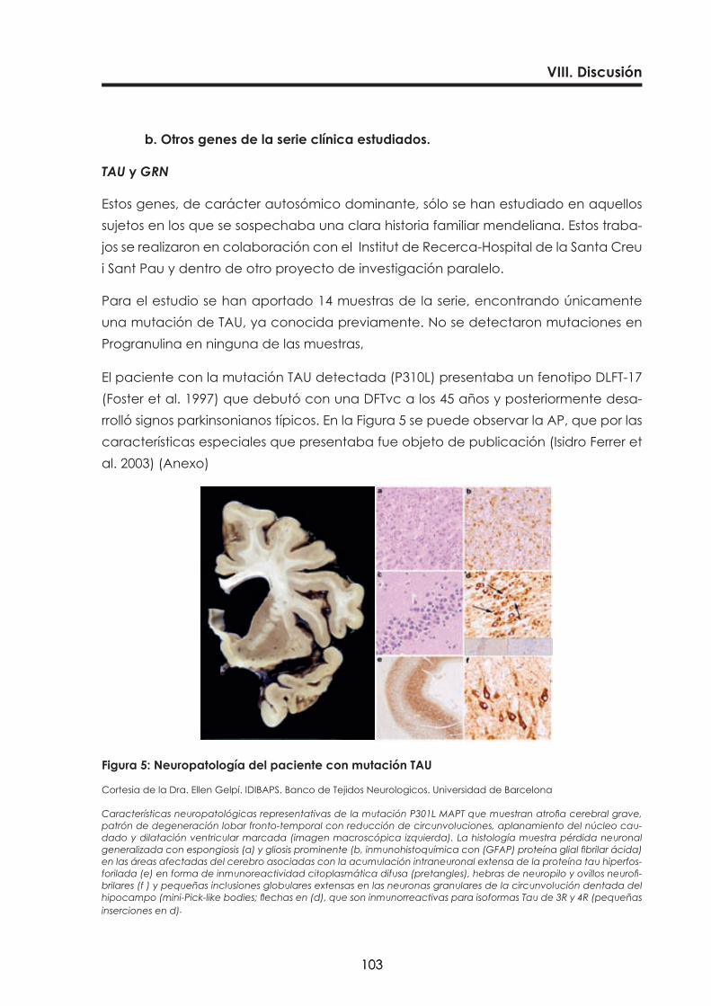
103103
VIII. Discusión
b. Otros genes de la serie clínica estudiados.
TAU y GRN
Estos genes, de carácter autosómico dominante, sólo se han estudiado en aquellos sujetos en los que se sospechaba una clara historia familiar mendeliana. Estos traba-jos se realizaron en colaboración con el Institut de Recerca-Hospital de la Santa Creu i Sant Pau y dentro de otro proyecto de investigación paralelo.
Para el estudio se han aportado 14 muestras de la serie, encontrando únicamente una mutación de TAU, ya conocida previamente. No se detectaron mutaciones en Progranulina en ninguna de las muestras,
El paciente con la mutación TAU detectada (P310L) presentaba un fenotipo DLFT-17 (Foster et al. 1997) que debutó con una DFTvc a los 45 años y posteriormente desa-rrolló signos parkinsonianos típicos. En la Figura 5 se puede observar la AP, que por las características especiales que presentaba fue objeto de publicación (Isidro Ferrer et al. 2003) (Anexo)
Figura 5: Neuropatología del paciente con mutación TAU
Cortesia de la Dra. Ellen Gelpí. IDIBAPS. Banco de Tejidos Neurologicos. Universidad de Barcelona
Características neuropatológicas representativas de la mutación P301L MAPT que muestran atrofia cerebral grave, patrón de degeneración lobar fronto-temporal con reducción de circunvoluciones, aplanamiento del núcleo cau-dado y dilatación ventricular marcada (imagen macroscópica izquierda). La histología muestra pérdida neuronal generalizada con espongiosis (a) y gliosis prominente (b, inmunohistoquímica con (GFAP) proteína glial fibrilar ácida) en las áreas afectadas del cerebro asociadas con la acumulación intraneuronal extensa de la proteína tau hiperfos-forilada (e) en forma de inmunoreactividad citoplasmática difusa (pretangles), hebras de neuropilo y ovillos neurofi-brilares (f ) y pequeñas inclusiones globulares extensas en las neuronas granulares de la circunvolución dentada del hipocampo (mini-Pick-like bodies; flechas en (d), que son inmunorreactivas para isoformas Tau de 3R y 4R (pequeñas inserciones en d).
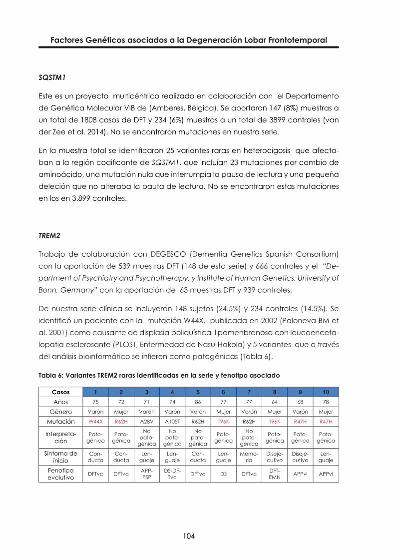
Factores Genéticos asociados a la Degeneración Lobar Frontotemporal
104104
SQSTM1
Este es un proyecto multicéntrico realizado en colaboración con el Departamento de Genética Molecular VIB de (Amberes, Bélgica). Se aportaron 147 (8%) muestras a un total de 1808 casos de DFT y 234 (6%) muestras a un total de 3899 controles (van der Zee et al. 2014). No se encontraron mutaciones en nuestra serie.
En la muestra total se identificaron 25 variantes raras en heterocigosis que afecta-ban a la región codificante de SQSTM1, que incluían 23 mutaciones por cambio de aminoácido, una mutación nula que interrumpía la pausa de lectura y una pequeña deleción que no alteraba la pauta de lectura. No se encontraron estas mutaciones en los en 3.899 controles.
TREM2
Trabajo de colaboración con DEGESCO (Dementia Genetics Spanish Consortium) con la aportación de 539 muestras DFT (148 de esta serie) y 666 controles y el “De-partment of Psychiatry and Psychotherapy, y Institute of Human Genetics. University of Bonn, Germany” con la aportación de 63 muestras DFT y 939 controles.
De nuestra serie clínica se incluyeron 148 sujetos (24.5%) y 234 controles (14.5%). Se identificó un paciente con la mutación W44X, publicada en 2002 (Paloneva BM et al. 2001) como causante de displasia poliquística lipomenbranosa con leucoencefa-lopatia esclerosante (PLOST, Enfermedad de Nasu-Hakola) y 5 variantes que a través del análisis bioinformático se infieren como patogénicas (Tabla 6).
Tabla6:VariantesTREM2rarasidentificadasenlaserieyfenotipoasociado
Casos 1 2 3 4 5 6 7 8 9 10Años 75 72 71 74 86 77 77 64 68 78
Género Varón Mujer Varón Varón Varón Mujer Varón Mujer Varón Mujer
Mutación W44X R62H A28V A105T R62H T96K R62H T96K R47H R47H
Interpreta-ción
Pato-génica
Pato-génica
No pato-
génica
No pato-
génica
No pato-
génica
Pato-génica
No pato-
génica
Pato-génica
Pato-génica
Pato-génica
Síntoma de inicio
Con-ducta
Con-ducta
Len-guaje
Len-guaje
Con-ducta
Len-guaje
Memo-ria
Diseje-cutivo
Diseje-cutivo
Len-guaje
Fenotipo evolutivo DFTvc DFTvc APP-
PSPDS-DF-
Tvc DFTvc DS DFTvc DFT-EMN APPvl APPvl
105105
VIII. Discusión
GWAS : Estudio de asociación de genoma completo de DFT
A este estudio, realizado en colaboración con “The National Institute of Neurological Disorders and Stroke and National Institute on Aging, the Wellcome/MRC Centre on Parkinson’s disease, Alzheimer’s Research UK, and Texas Tech University Health Scien-ces Center” se aportaron 237 controles y 155 muestras de la serie clínica: 99 DFTvc (63%), 22 APPnf (14%), 31 APPvs (20%) Y 3 DFT-ELA (2%) a un total de 2154 pacientes con DFT y 4308 controles. Nuestra serie se empleo para la fase de replicación y los genotipos están disponibles para estudios futuros.
Se han identificado 2 nuevos genes que superan el umbral de significación para es-tudio de genoma completo. Los loci identificados son el HLA en la posición 6p 21.3 y un marcador del genRAB38/CTSC en la posición 11p14.
Los resultados sugieren que las alteraciones de los procesos del sistema inmune (6p21.3) y, posiblemente, de la fisiología lisosomal y vías de la autofagia, (11q14) po-drían estar implicados en la DFT.

107107
IX. Conclusiones


109109
IX. Conclusiones
1. A nivel fenotípico se observa una variabilidad en la evolución clínica de la serie DFT. De ello se desprende la necesidad de un seguimiento clínico a largo plazo para identificar claramente los fenotipos, siendo, como en todas las se-ries clínicas publicadas, el fenotipo DFTvc el más prevalente.
2. A nivel genético, se ha observado que la expansión del C9orf72 es la mutación más frecuente dentro de la serie. Todos los portadores presentan el fenotipo DFTvc y/o ELA. Se confirma que la expansión de C9orf72 es la mayor causa de DFT en nuestra serie, de causa familiar y la primera a estudiar en esta variante fenotípica.
3. Dentro de los polimorfismos estudiados se concluye que la ApoE E4 no está asociada a la DFT y que dentro de las series clínicas DFT hay que sopesar la pro-bable contaminación de fenotipos EA. Los resultados están en concordancia con la hipótesis de que casos EA variante frontal están inflando la frecuencia alélica de ApoE4 dentro de las series DFT y esto podría ayudar a diferenciarlos clínicamente; corroborando esta observación, la existencia de “información clínica de EA” dentro de la serie DFT.
4. Respecto al polimorfismo TMEM106B, nuestros resultados indican que aplican-do el modelo recesivo (CC) se observa una tendencia a la asociación en rela-ción con el riesgo de DFT (ajustado por edad y sexo, con una odds ratio=0.57; p=0.082). Los meta-análisis de los estudios disponibles también apoyan el efecto recesivo para el genotipo CC rs1990622 (OR=0.70; C.I. 95% [0.57-0.85]; p=0.0003). Así, concluimos que TMEM106B está asociado con DFT, aunque el grado de este efecto es difícil de ser estimado usando series clínicas.
5. En la correlación fenotipo-genotipo del polimorfismo de TMEM106B, no se ob-jetivó evidencia de asociación en ninguno de los modelos utilizados (genoti-po, dominante y recesivo), excepto en los antecedentes familiares de demen-cia del fenotipo DFTvc, que fueron menos frecuentes en aquellos portadores del genotipo CC de protección; por lo que se concluye que el análisis del SNP rs1990622 no puede ser usado para diferenciar los diferentes subtipos clínicos.

Factores Genéticos asociados a la Degeneración Lobar Frontotemporal
110110
6. No se han encontrado mutaciones en el gen UBQLN2 en la población estudia-da, por lo que se deduce que es una mutación rara y a tener en cuenta sólo cuando se hayan descartado las más prevalentes.
7. Estudios realizados en colaboración (Tabla 7)
• Se ha confirmado una mutación de TAU y seis variantes potencialmente patogénicas de TREM2.
• El estudio GWAS detecta dos loci asociados al sistema inmune y la vía lisosomal y de autofagia, como potencialmente implicados en la DFT.
Tabla 7: Estudios en colaboración
GenGrupo
colaboradorMuestras cedidas
Mutacionesdetectadas
TAU Barcelona 14 1 (7%)GRN Barcelona 14 0
C9orf72 Toronto 159 6 (3.7%)SQSTM1 Bélgica 147 0TREM 2 Alemania 148 6 (4%)
111111
IX. Conclusiones
COROLARIO
En resumen, la variabilidad fenotípica y genotípica de las DLFT representa un impor-tante reto para el clínico, sobre todo en las fases precoces, aunque los criterios diag-nósticos utilizados en la actualidad, en parte debido a la introducción de los nuevos biomarcadores (bioquímicos y de neuroimagen), permiten una clasificación fenotípi-ca más acertada. La asociación de los marcadores genéticos al diagnóstico clínico como herramienta para conocer el sustrato molecular subyacente es fundamental para la búsqueda de nuevas terapias. En este sentido, los trabajos de esta monogra-fía intentan abordar la importancia del estudio de estos marcadores y su correlación con la clínica de cara a un mejor conocimiento de la comunidad científica.
Es importante resaltar la dificultad, desde el punto de vista del clínico y dentro de la Sanidad pública española y catalana, el acceso a la determinación de los marcado-res genéticos, que en la actualidad se han de realizar en centros de investigación o en Unidades específicas en base a convenios de colaboración, pero desconocidas para el clínico no experto en enfermedades neurodegenerativas. Probablemente un no despreciable porcentaje de casos con fenotipo DFTvc, sobre todo entre la pobla-ción psiquiátrica de debut más tardío, están infradiagnosticados, no permitiendo el acceso precoz de estos pacientes a los ensayos clínicos de DFT en el presente y un tratamiento específico en el futuro. La colaboración entre las diferentes áreas clínicas se muestra aquí fundamental.

112112

113113
X. Referencias

115115
X. Referencias
Achi, Eugene Y, and Stacy A Rudnicki. 2012. “ALS and Frontotemporal Dysfunction: A Review.” Neurology Research International 2012 (January): 806306. doi:10.1155/2012/806306. http://www.pubmedcen-tral.nih.gov/articlerender.fcgi?artid=3423946&tool=pmcentrez&rendertype=abstract.
Amador-Ortiz, Catalina, Wen-Lang Lin, Zeshan Ahmed, David Personett, Peter Davies, Ranjan Duara, Neill R Graff-Radford, Michael L Hutton, and Dennis W Dickson. 2007. “TDP-43 Immunoreactivity in Hippo-campal Sclerosis and Alzheimer’s Disease.” Annals of Neurology 61 (5) (May): 435–45. doi:10.1002/ana.21154. http://www.pubmedcentral.nih.gov/articlerender.fcgi?artid=2677204&tool=pmcen-trez&rendertype=abstract.
Appel, Stanley H, and Lewis P Rowland. 2012. “Amyotrophic Lateral Sclerosis, Frontotemporal Lobar De-mentia, and p62: A Functional Convergence?” Neurology 79 (15) (October 9): 1526–7. doi:10.1212/WNL.0b013e31826e26ec. http://www.ncbi.nlm.nih.gov/pubmed/22972643.
Arai, Tetsuaki, Masato Hasegawa, Haruhiko Akiyama, Kenji Ikeda, Takashi Nonaka, Hiroshi Mori, David Mann, et al. 2006. “TDP-43 Is a Component of Ubiquitin-Positive Tau-Negative Inclusions in Fronto-temporal Lobar Degeneration and Amyotrophic Lateral Sclerosis.” Biochemical and Biophysical Research Communications 351 (3) (December 22): 602–11. doi:10.1016/j.bbrc.2006.10.093. http://www.ncbi.nlm.nih.gov/pubmed/17084815.
Armstrong, Melissa J, Irene Litvan, Anthony E Lang, Thomas H Bak, Kailash P Bhatia, Barbara Borroni, Adam L Boxer, et al. 2013. “Criteria for the Diagnosis of Corticobasal Degeneration.” Neurology 80 (5) (January 29): 496–503. doi:10.1212/WNL.0b013e31827f0fd1. http://www.ncbi.nlm.nih.gov/pubmed/23359374.
Arvanitakis, Zoe. 2010. “Update on Frontotemporal Dementia.” The Neurologist. doi:10.1097/NRL.0b013e3181b1d5c6. http://www.ncbi.nlm.nih.gov/pubmed/22277388.
Baborie, a., T. D. Griffiths, E. Jaros, P. Momeni, I. G. McKeith, D. J. Burn, G. Keir, a. J. Larner, D. M. Mann, and R. Perry. 2012. “Frontotemporal Dementia in Elderly Individuals.” Archives of Neurology (April 23): 1–9. doi:10.1001/archneurol.2011.3323. http://archneur.ama-assn.org/cgi/doi/10.1001/arch-neurol.2011.3323.
Baker, Matt, Ian R Mackenzie, Stuart M Pickering-Brown, Jennifer Gass, Rosa Rademakers, Caroline Lind-holm, Julie Snowden, et al. 2006. “Mutations in Progranulin Cause Tau-Negative Frontotemporal De-mentia Linked to Chromosome 17.” Nature 442 (7105) (August 24): 916–9. doi:10.1038/nature05016. http://www.ncbi.nlm.nih.gov/pubmed/16862116.
Benajiba, Lina, Isabelle Le Ber, Agnès Camuzat, Mathieu Lacoste, Catherine Thomas-Anterion, Philippe Couratier, Solenn Legallic, et al. 2009. “TARDBP Mutations in Motoneuron Disease with Frontotem-poral Lobar Degeneration.” Annals of Neurology 65 (4) (April): 470–3. doi:10.1002/ana.21612. http://www.ncbi.nlm.nih.gov/pubmed/19350673.
Bird, Thomas, David Knopman, John VanSwieten, Sonia Rosso, Howard Feldman, Hirotaka Tanabe, Neil Graff-Raford, Daniel Geschwind, Patrice Verpillat, and Michael Hutton. 2003. “Epidemiology and Genetics of Frontotemporal dementia/Pick’s Disease.” Annals of Neurology 54 Suppl 5 (January): S29–31. doi:10.1002/ana.10572. http://www.ncbi.nlm.nih.gov/pubmed/12833366.
Boeve, Bradley F, Kevin B Boylan, Neill R Graff-Radford, Mariely DeJesus-Hernandez, David S Knopman, Otto Pedraza, Prashanthi Vemuri, et al. 2012. “Characterization of Frontotemporal Dementia And/or Amyotrophic Lateral Sclerosis Associated with the GGGGCC Repeat Expansion in C9ORF72.” Brain : A Journal of Neurology 135 (Pt 3) (March): 765–83. doi:10.1093/brain/aws004. http://www.pubmedcentral.nih.gov/articlerender.fcgi?artid=3286335&tool=pmcentrez&rendertype=abstract.
Boeve, Bradley F, and Mike Hutton. 2008. “Refining Frontotemporal Dementia with Parkinsonism Linked to Chromosome 17: Introducing FTDP-17 (MAPT) and FTDP-17 (PGRN).” Archives of Neurology 65 (4) (May 1): 460–4. doi:10.1001/archneur.65.4.460. http://archneur.jamanetwork.com/article.aspx?arti-cleid=795385.
Factores Genéticos asociados a la Degeneración Lobar Frontotemporal
116116
Cruts, Marc, Samir Kumar-Singh, and Christine Van Broeckhoven. 2006. “Progranulin Mutations in Ubiqui-tin-Positive Frontotemporal Dementia Linked to Chromosome 17q21.” Current Alzheimer Research 3 (5) (December): 485–91. http://www.ncbi.nlm.nih.gov/pubmed/17168647.
Deerlin, Vivianna M Van, Patrick M A Sleiman, Maria Martinez-lage, Alice Chen-, Li-san Wang, Neill R Graff-radford, Dennis W Dickson, et al. 2010. “NIH Public Access” 42 (3): 234–239. doi:10.1038/ng.536.Common.
DeJesus-Hernandez, Mariely, Ian R Mackenzie, Bradley F Boeve, Adam L Boxer, Matt Baker, Nicola J Ruth-erford, Alexandra M Nicholson, et al. 2011. “Expanded GGGGCC Hexanucleotide Repeat in Non-coding Region of C9ORF72 Causes Chromosome 9p-Linked FTD and ALS.” Neuron 72 (2) (October 20): 245–56. doi:10.1016/j.neuron.2011.09.011. http://www.pubmedcentral.nih.gov/articlerender.fcgi?artid=3202986&tool=pmcentrez&rendertype=abstract.
Deng, Han-Xiang, Wenjie Chen, Seong-Tshool Hong, Kym M Boycott, George H Gorrie, Nailah Siddique, Yi Yang, et al. 2011. “Mutations in UBQLN2 Cause Dominant X-Linked Juvenile and Adult-Onset ALS and ALS/dementia.” Nature 477 (7363) (September 8): 211–5. doi:10.1038/nature10353. http://www.pubmedcentral.nih.gov/articlerender.fcgi?artid=3169705&tool=pmcentrez&rendertype=abstract.
Dreyfuss, Gideon, V Narry Kim, and Naoyuki Kataoka. 2002. “Messenger-RNA-Binding Proteins and the Messages They Carry.” Nature Reviews. Molecular Cell Biology 3 (3) (March): 195–205. doi:10.1038/nrm760. http://dx.doi.org/10.1038/nrm760.
Ferrari, Raffaele, Dena G Hernandez, Michael a Nalls, Jonathan D Rohrer, Adaikalavan Ramasamy, John B J Kwok, Carol Dobson-Stone, et al. 2014. “Frontotemporal Dementia and Its Subtypes: A Genome-Wide Association Study.” Lancet Neurology 13 (7) (July): 686–99. doi:10.1016/S1474-4422(14)70065-1. http://www.ncbi.nlm.nih.gov/pubmed/24943344.
Ferrer, I, I Hernández, M Boada, A Llorente, M J Rey, A Cardozo, M Ezquerra, and B Puig. 2003. “Primary Progressive Aphasia as the Initial Manifestation of Corticobasal Degeneration and Unusual Tauop-athies.” Acta Neuropathologica 106 (5) (November): 419–35. doi:10.1007/s00401-003-0756-4. http://www.ncbi.nlm.nih.gov/pubmed/12955398.
Ferrer, Isidro, Isabel Hernández, Berta Puig, María Jesús Rey, Mario Ezquerra, Eduardo Tolosa, and Mer-ce Boada. 2003. “Ubiquitin-Negative Mini-Pick-like Bodies in the Dentate Gyrus in p301l Tauopa-thy.” Journal of Alzheimer’s Disease : JAD 5 (6) (December): 445–54. http://www.ncbi.nlm.nih.gov/pubmed/14757934.
Ferrer, Isidro, Gabriel Santpere, and Fred W van Leeuwen. 2008. “Argyrophilic Grain Disease.” Brain : A Journal of Neurology 131 (Pt 6) (June): 1416–32. doi:10.1093/brain/awm305. http://www.ncbi.nlm.nih.gov/pubmed/18234698.
Foster, N L, K Wilhelmsen, A A Sima, M Z Jones, C J D’Amato, and S Gilman. 1997. “Frontotemporal De-mentia and Parkinsonism Linked to Chromosome 17: A Consensus Conference. Conference Partic-ipants.” Annals of Neurology 41 (6) (June): 706–15. doi:10.1002/ana.410410606. http://www.ncbi.nlm.nih.gov/pubmed/9189031.
Gass, Jennifer, Ashley Cannon, Ian R Mackenzie, Bradley Boeve, Matt Baker, Jennifer Adamson, Richard Crook, et al. 2006. “Mutations in Progranulin Are a Major Cause of Ubiquitin-Positive Frontotemporal Lobar Degeneration.” Human Molecular Genetics 15 (20) (October 15): 2988–3001. doi:10.1093/hmg/ddl241. http://www.ncbi.nlm.nih.gov/pubmed/16950801.
Gijselinck, I, C Van Broeckhoven, and M Cruts. 2008. “Granulin Mutations Associated with Frontotemporal Lobar Degeneration and Related Disorders: An Update.” Human Mutation 29 (12) (December): 1373–86. doi:10.1002/humu.20785. http://www.ncbi.nlm.nih.gov/pubmed/18543312.
117117
X. Referencias
Gijselinck, Ilse, Tim Van Langenhove, Julie van der Zee, Kristel Sleegers, Stéphanie Philtjens, Gernot Klein-berger, Jonathan Janssens, et al. 2012. “A C9orf72 Promoter Repeat Expansion in a Flanders-Bel-gian Cohort with Disorders of the Frontotemporal Lobar Degeneration-Amyotrophic Lateral Sclerosis Spectrum: A Gene Identification Study.” Lancet Neurology 11 (1) (January): 54–65. doi:10.1016/S1474-4422(11)70261-7. http://www.ncbi.nlm.nih.gov/pubmed/22154785.
Gilberti, Nicola, Marinella Turla, Antonella Alberici, Valeria Bertasi, Patrizia Civelli, Silvana Archetti, Ales-sandro Padovani, and Barbara Borroni. 2012. “Prevalence of Frontotemporal Lobar Degeneration in an Isolated Population: The Vallecamonica Study.” Neurological Sciences : Official Journal of the Italian Neurological Society and of the Italian Society of Clinical Neurophysiology 33 (4) (August): 899–904. doi:10.1007/s10072-011-0865-0. http://www.ncbi.nlm.nih.gov/pubmed/22127750.
Gislason, T B, M Sjögren, L Larsson, and I Skoog. 2003. “The Prevalence of Frontal Variant Frontotemporal Dementia and the Frontal Lobe Syndrome in a Population Based Sample of 85 Year Olds.” Journal of Neurology, Neurosurgery, and Psychiatry 74 (7) (July): 867–71. http://www.pubmedcentral.nih.gov/articlerender.fcgi?artid=1738520&tool=pmcentrez&rendertype=abstract.
Goldman, J S, J M Farmer, E M Wood, J K Johnson, a Boxer, J Neuhaus, C Lomen-Hoerth, et al. 2005. “Comparison of Family Histories in FTLD Subtypes and Related Tauopathies.” Neurology 65 (11) (December 13): 1817–9. doi:10.1212/01.wnl.0000187068.92184.63. http://www.ncbi.nlm.nih.gov/pubmed/16344531.
Gorno-Tempini, M L, A E Hillis, S Weintraub, A Kertesz, M Mendez, S F Cappa, J M Ogar, et al. 2011. “Clas-sification of Primary Progressive Aphasia and Its Variants.” Neurology 76 (11) (March 15): 1006–14. doi:10.1212/WNL.0b013e31821103e6. http://www.pubmedcentral.nih.gov/articlerender.fcgi?ar-tid=3059138&tool=pmcentrez&rendertype=abstract.
Gorno-Tempini, Maria Luisa, Nina F Dronkers, Katherine P Rankin, Jennifer M Ogar, La Phengrasamy, Howard J Rosen, Julene K Johnson, Michael W Weiner, and Bruce L Miller. 2004. “Cognition and Anatomy in Three Variants of Primary Progressive Aphasia.” Annals of Neurology 55 (3) (March): 335–46. doi:10.1002/ana.10825. http://www.pubmedcentral.nih.gov/articlerender.fcgi?ar-tid=2362399&tool=pmcentrez&rendertype=abstract.
Grafman, J, I Litvan, and M Stark. 1995. “Neuropsychological Features of Progressive Supranuclear Palsy.” Brain and Cognition 28 (3) (August): 311–20. doi:10.1006/brcg.1995.1260. http://dx.doi.org/10.1006/brcg.1995.1260.
Grossman, Murray. 2012. “The Non-Fluent/agrammatic Variant of Primary Progressive Aphasia.” Lancet Neurology 11 (6) (June): 545–55. doi:10.1016/S1474-4422(12)70099-6. http://www.ncbi.nlm.nih.gov/pubmed/22608668.
Guyant-Maréchal, L, a Laquerrière, C Duyckaerts, C Dumanchin, J Bou, F Dugny, I Le Ber, T Frébourg, D Hannequin, and D Campion. 2006. “Valosin-Containing Protein Gene Mutations: Clinical and Neu-ropathologic Features.” Neurology 67 (4) (August 22): 644–51. doi:10.1212/01.wnl.0000225184.14578.d3. http://www.ncbi.nlm.nih.gov/pubmed/16790606.
Gydesen, S., J.M. Brown, A. Brun, L. Chakrabarti, A. Gade, P. Johannsen, M. Rossor, et al. 2002. “Chro-mosome 3 Linked Frontotemporal Dementia (FTD-3).” Neurology 59 (10) (November 26): 1585–1594. doi:10.1212/01.WNL.0000034763.54161.1F. http://www.neurology.org/content/59/10/1585.long.
Halliday, Glenda, Eileen H Bigio, Nigel J Cairns, Manuela Neumann, Ian R a Mackenzie, and David M a Mann. 2012. “Mechanisms of Disease in Frontotemporal Lobar Degeneration: Gain of Function ver-sus Loss of Function Effects.” Acta Neuropathologica. doi:10.1007/s00401-012-1030-4. http://www.ncbi.nlm.nih.gov/pubmed/22878865.
Factores Genéticos asociados a la Degeneración Lobar Frontotemporal
118118
Hernández, Isabel, Anna Espinosa, Luis Miguel Real, Jose Jorge Galán, Ana Mauleón, Maiteé Rosen-de Roca, Lluís Tárraga, Agustín Ruiz, and Mercè Boada. 2012. “Molecular Evaluation of Human Ubiquilin 2 Gene PXX Domain in Familial Frontotemporal Dementia Patients.” Journal of Neurol-ogy 259 (11) (November): 2488–90. doi:10.1007/s00415-012-6568-5. http://www.ncbi.nlm.nih.gov/pubmed/22729385.
Hodges, John R, and Karalyn Patterson. 2007. “Semantic Dementia: A Unique Clinicopathological Syn-drome.” Lancet Neurology 6 (11) (November): 1004–14. doi:10.1016/S1474-4422(07)70266-1. http://www.ncbi.nlm.nih.gov/pubmed/17945154.
Holm, Ida Elisabeth, Elisabet Englund, Ian R A Mackenzie, Peter Johannsen, and Adrian M Isaacs. 2007. “A Reassessment of the Neuropathology of Frontotemporal Dementia Linked to Chromosome 3.” Journal of Neuropathology and Experimental Neurology 66 (10) (October): 884–91. doi:10.1097/nen.0b013e3181567f02. http://www.ncbi.nlm.nih.gov/pubmed/17917582.
Hsiung, Ging-Yuek R, Mariely Dejesus-Hernandez, Howard H Feldman, Pheth Sengdy, Phoenix Bouch-ard-Kerr, Emily Dwosh, Rachel Butler, et al. 2012. “Clinical and Pathological Features of Familial Frontotemporal Dementia Caused by C9ORF72 Mutation on Chromosome 9p.” Brain : A Journal of Neurology 135 (Pt 3) (March): 709–22. doi:10.1093/brain/awr354. http://www.pubmedcentral.nih.gov/articlerender.fcgi?artid=3286328&tool=pmcentrez&rendertype=abstract.
Hutton, M, C L Lendon, P Rizzu, M Baker, S Froelich, H Houlden, S Pickering-Brown, et al. 1998. “Association of Missense and 5’-Splice-Site Mutations in Tau with the Inherited Dementia FTDP-17.” Nature 393 (6686) (July 18): 702–5. doi:10.1038/31508. http://www.ncbi.nlm.nih.gov/pubmed/9641683.
Ikejima, Chiaki, Fumihiko Yasuno, Katsuyoshi Mizukami, Megumi Sasaki, Satoshi Tanimukai, and Takashi Asada. 2009. “Prevalence and Causes of Early-Onset Dementia in Japan: A Population-Based Study.” Stroke; a Journal of Cerebral Circulation 40 (8) (August 1): 2709–14. doi:10.1161/STROKEA-HA.108.542308. http://stroke.ahajournals.org/content/40/8/2709.long.
Johnson, Julene K, Janine Diehl, Mario F Mendez, John Neuhaus, Jill S Shapira, Mark Forman, Dennis J Chute, et al. 2005a. “Frontotemporal Lobar Degeneration: Demographic Characteristics of 353 Patients.” Archives of Neurology 62 (6) (June): 925–30. doi:10.1001/archneur.62.6.925. http://www.ncbi.nlm.nih.gov/pubmed/15956163.
———. 2005b. “Frontotemporal Lobar Degeneration: Demographic Characteristics of 353 Patients.” Ar-chives of Neurology 62 (6) (June 1): 925–30. doi:10.1001/archneur.62.6.925. http://archneur.jama-network.com/article.aspx?articleid=788706.
Josephs, K a, J L Whitwell, D S Knopman, B F Boeve, P Vemuri, M L Senjem, J E Parisi, et al. 2009. “Two Dis-tinct Subtypes of Right Temporal Variant Frontotemporal Dementia.” Neurology 73 (18) (November 3): 1443–50. doi:10.1212/WNL.0b013e3181bf9945. http://www.pubmedcentral.nih.gov/articleren-der.fcgi?artid=2779005&tool=pmcentrez&rendertype=abstract.
Josephs, Keith A, John R Hodges, Julie S Snowden, Ian R Mackenzie, Manuela Neumann, David M Mann, and Dennis W Dickson. 2011. “Neuropathological Background of Phenotypical Variability in Fron-totemporal Dementia.” Acta Neuropathologica 122 (2) (August): 137–53. doi:10.1007/s00401-011-0839-6. http://www.pubmedcentral.nih.gov/articlerender.fcgi?artid=3232515&tool=pmcen-trez&rendertype=abstract.
Kertesz, A, L Hudson, I R Mackenzie, and D G Munoz. 1994. “The Pathology and Nosology of Primary Progressive Aphasia.” Neurology 44 (11) (November): 2065–72. http://www.ncbi.nlm.nih.gov/pubmed/7969961.
Kertesz, A, P Martinez-Lage, W Davidson, and D G Munoz. 2000. “The Corticobasal Degeneration Syn-drome Overlaps Progressive Aphasia and Frontotemporal Dementia.” Neurology 55 (9) (November 14): 1368–75. http://www.ncbi.nlm.nih.gov/pubmed/11087783.
119119
X. Referencias
Kertesz, Andrew, Mervin Blair, Paul McMonagle, and David G Munoz. 2007. “The Diagnosis and Course of Frontotemporal Dementia.” Alzheimer Disease and Associated Disorders 21 (2): 155–63. doi:10.1097/WAD.0b013e31806547eb. http://www.ncbi.nlm.nih.gov/pubmed/17545742.
Kimonis, Virginia E, Sarju G Mehta, Erin C Fulchiero, Dana Thomasova, Marzia Pasquali, Kym Boycott, Edward G Neilan, et al. 2008. “Clinical Studies in Familial VCP Myopathy Associated with Paget Dis-ease of Bone and Frontotemporal Dementia.” American Journal of Medical Genetics. Part A 146A (6) (March 15): 745–57. doi:10.1002/ajmg.a.31862. http://www.pubmedcentral.nih.gov/articleren-der.fcgi?artid=2467391&tool=pmcentrez&rendertype=abstract.
Kimonis, Virginia E, and Giles D J Watts. 2013. “Autosomal Dominant Inclusion Body Myopathy, Paget Disease of Bone, and Frontotemporal Dementia.” Alzheimer Disease and Associated Disorders 19 Suppl 1: S44–7. Accessed June 5. http://www.ncbi.nlm.nih.gov/pubmed/16317258.
Kwiatkowski, T J, D A Bosco, A L Leclerc, E Tamrazian, C R Vanderburg, C Russ, A Davis, et al. 2009. “Mu-tations in the FUS/TLS Gene on Chromosome 16 Cause Familial Amyotrophic Lateral Sclerosis.” Sci-ence (New York, N.Y.) 323 (5918) (February 27): 1205–8. doi:10.1126/science.1166066. http://www.ncbi.nlm.nih.gov/pubmed/19251627.
Lang, Christina M, Katrin Fellerer, Benjamin M Schwenk, Peer-Hendrik Kuhn, Elisabeth Kremmer, Dieter Edbauer, Anja Capell, and Christian Haass. 2012. “Membrane Orientation and Subcellular Local-ization of Transmembrane Protein 106B (TMEM106B), a Major Risk Factor for Frontotemporal Lobar Degeneration.” The Journal of Biological Chemistry 287 (23) (June 1): 19355–65. doi:10.1074/jbc.M112.365098. http://www.pubmedcentral.nih.gov/articlerender.fcgi?artid=3365973&tool=pmcen-trez&rendertype=abstract.
Le Ber, Isabelle, Agnès Camuzat, Didier Hannequin, Florence Pasquier, Eric Guedj, Anne Rovelet-Lecrux, Valérie Hahn-Barma, et al. 2008. “Phenotype Variability in Progranulin Mutation Carriers: A Clinical, Neuropsychological, Imaging and Genetic Study.” Brain : A Journal of Neurology 131 (Pt 3) (March 1): 732–46. doi:10.1093/brain/awn012. http://brain.oxfordjournals.org/content/131/3/732.long.
Le Ber, Isabelle, Julie van der Zee, Didier Hannequin, Ilse Gijselinck, Dominique Campion, Michèle Puel, Annie Laquerrière, et al. 2007. “Progranulin Null Mutations in Both Sporadic and Familial Fronto-temporal Dementia.” Human Mutation 28 (9) (September): 846–55. doi:10.1002/humu.20520. http://www.ncbi.nlm.nih.gov/pubmed/17436289.
Leroy, E, R Boyer, G Auburger, B Leube, G Ulm, E Mezey, G Harta, et al. 1998. “The Ubiquitin Pathway in Parkinson’s Disease.” Nature 395 (6701) (October 1): 451–2. doi:10.1038/26652. http://dx.doi.org/10.1038/26652.
Litvan, I, Y Agid, D Calne, G Campbell, B Dubois, R C Duvoisin, C G Goetz, et al. 1996. “Clinical Research Criteria for the Diagnosis of Progressive Supranuclear Palsy (Steele-Richardson-Olszewski Syndrome): Report of the NINDS-SPSP International Workshop.” Neurology 47 (1) (July): 1–9. http://www.ncbi.nlm.nih.gov/pubmed/8710059.
Lynch, T, M Sano, K S Marder, K L Bell, N L Foster, R F Defendini, A A Sima, C Keohane, T G Nygaard, and S Fahn. 1994. “Clinical Characteristics of a Family with Chromosome 17-Linked Disinhibition-Demen-tia-Parkinsonism-Amyotrophy Complex.” Neurology 44 (10) (October): 1878–84. http://www.ncbi.nlm.nih.gov/pubmed/7936241.
Mackenzie, I R A, Manuela Neumann, Eileen H Bigio, Nigel J Cairns, Irina Alafuzoff, Jillian Kril, Gabor G Ko-vacs, et al. 2009. “Nomenclature for Neuropathologic Subtypes of Frontotemporal Lobar Degener-ation: Consensus Recommendations.” Acta Neuropathologica 117 (1) (January): 15–8. doi:10.1007/s00401-008-0460-5. http://www.pubmedcentral.nih.gov/articlerender.fcgi?artid=2710877&tool=p-mcentrez&rendertype=abstract.
Factores Genéticos asociados a la Degeneración Lobar Frontotemporal
120120
Mackenzie, Ian R A, Dean Foti, John Woulfe, and Trevor A Hurwitz. 2008. “Atypical Frontotemporal Lo-bar Degeneration with Ubiquitin-Positive, TDP-43-Negative Neuronal Inclusions.” Brain : A Journal of Neurology 131 (Pt 5) (May 1): 1282–93. doi:10.1093/brain/awn061. http://brain.oxfordjournals.org/cgi/content/long/131/5/1282.
Mackenzie, Ian R a, David G Munoz, Hirofumi Kusaka, Osamu Yokota, Kenji Ishihara, Sigrun Roeber, Hans a Kretzschmar, Nigel J Cairns, and Manuela Neumann. 2011. “Distinct Pathological Subtypes of FT-LD-FUS.” Acta Neuropathologica 121 (2) (February): 207–18. doi:10.1007/s00401-010-0764-0. http://www.ncbi.nlm.nih.gov/pubmed/21052700.
Mackenzie, Ian R a, Manuela Neumann, Atik Baborie, Deepak M Sampathu, Daniel Du Plessis, Evelyn Jaros, Robert H Perry, John Q Trojanowski, David M a Mann, and Virginia M Y Lee. 2011. “A Har-monized Classification System for FTLD-TDP Pathology.” Acta Neuropathologica 122 (1) (July): 111–3. doi:10.1007/s00401-011-0845-8. http://www.pubmedcentral.nih.gov/articlerender.fcgi?ar-tid=3285143&tool=pmcentrez&rendertype=abstract.
Mahoney, Colin J, Jon Beck, Jonathan D Rohrer, Tammaryn Lashley, Kin Mok, Tim Shakespeare, Tom Yeat-man, et al. 2012. “Frontotemporal Dementia with the C9ORF72 Hexanucleotide Repeat Expansion: Clinical, Neuroanatomical and Neuropathological Features.” Brain : A Journal of Neurology 135 (Pt 3) (March): 736–50. doi:10.1093/brain/awr361. http://www.pubmedcentral.nih.gov/articlerender.fcgi?artid=3286330&tool=pmcentrez&rendertype=abstract.
Majounie, Elisa, Alan E Renton, Kin Mok, Elise Gp Dopper, Adrian Waite, Sara Rollinson, Adriano Chiò, et al. 2012. “Frequency of the C9orf72 Hexanucleotide Repeat Expansion in Patients with Amyo-trophic Lateral Sclerosis and Frontotemporal Dementia: A Cross-Sectional Study.” Lancet Neurol-ogy 4422 (12) (March 9): 1–8. doi:10.1016/S1474-4422(12)70043-1. http://www.ncbi.nlm.nih.gov/pubmed/22406228.
Mesulam, M M. 1982. “Slowly Progressive Aphasia without Generalized Dementia.” Annals of Neurology 11 (6) (June): 592–8. doi:10.1002/ana.410110607. http://www.ncbi.nlm.nih.gov/pubmed/7114808.
Millecamps, Stéphanie, Philippe Corcia, Cécile Cazeneuve, Séverine Boillée, Danielle Seilhean, Véro-nique Danel-Brunaud, Nadia Vandenberghe, et al. 2012. “Mutations in UBQLN2 Are Rare in French Amyotrophic Lateral Sclerosis.” Neurobiology of Aging 33 (4) (April): 839.e1–3. doi:10.1016/j.neurobi-olaging.2011.11.010. http://www.ncbi.nlm.nih.gov/pubmed/22169395.
Mummery, C J, K Patterson, C J Price, J Ashburner, R S Frackowiak, and J R Hodges. 2000. “A Voxel-Based Morphometry Study of Semantic Dementia: Relationship between Temporal Lobe Atrophy and Semantic Memory.” Annals of Neurology 47 (1) (January): 36–45. http://www.ncbi.nlm.nih.gov/pubmed/10632099.
Nakashima-Yasuda, Hanae, Kunihiro Uryu, John Robinson, Sharon X Xie, Howard Hurtig, John E Duda, Steven E Arnold, et al. 2007. “Co-Morbidity of TDP-43 Proteinopathy in Lewy Body Related Diseases.” Acta Neuropathologica 114 (3) (September): 221–9. doi:10.1007/s00401-007-0261-2. http://www.ncbi.nlm.nih.gov/pubmed/17653732.
Neary, D, J S Snowden, L Gustafson, U Passant, D Stuss, S Black, M Freedman, et al. 1998. “Frontotemporal Lobar Degeneration: A Consensus on Clinical Diagnostic Criteria.” Neurology 51 (6) (December): 1546–54. http://www.ncbi.nlm.nih.gov/pubmed/9855500.
Neumann, Manuela, Sigrun Roeber, Hans a Kretzschmar, Rosa Rademakers, Matt Baker, and Ian R a Mackenzie. 2009. “Abundant FUS-Immunoreactive Pathology in Neuronal Intermediate Filament In-clusion Disease.” Acta Neuropathologica 118 (5) (November): 605–16. doi:10.1007/s00401-009-0581-5. http://www.pubmedcentral.nih.gov/articlerender.fcgi?artid=2864784&tool=pmcentrez&render-type=abstract.

121121
X. Referencias
Neumann, Manuela, Deepak M Sampathu, Linda K Kwong, Adam C Truax, Matthew C Micsenyi, Thom-as T Chou, Jennifer Bruce, et al. 2006. “Ubiquitinated TDP-43 in Frontotemporal Lobar Degenera-tion and Amyotrophic Lateral Sclerosis.” Science (New York, N.Y.) 314 (5796) (October 6): 130–3. doi:10.1126/science.1134108. http://www.ncbi.nlm.nih.gov/pubmed/17023659.
Noble, Wendy, Diane P Hanger, Christopher C J Miller, and Simon Lovestone. 2013. “The Importance of Tau Phosphorylation for Neurodegenerative Diseases.” Frontiers in Neurology 4 (July) (Janu-ary): 83. doi:10.3389/fneur.2013.00083. http://www.pubmedcentral.nih.gov/articlerender.fcgi?ar-tid=3696910&tool=pmcentrez&rendertype=abstract.
Paloneva BM, J., T. Autti, R. Raininko, J. Partanen, O. Salonen, M. Puranen, P. Hakola, and M. Haltia. 2001. “CNS Manifestations of Nasu-Hakola Disease: A Frontal Dementia with Bone Cysts.” Neurology 56 (11) (June 12): 1552–1558. doi:10.1212/WNL.56.11.1552. http://www.neurology.org/cgi/doi/10.1212/WNL.56.11.1552.
Parkinson, N, P G Ince, M O Smith, R Highley, G Skibinski, P M Andersen, K E Morrison, et al. 2006. “ALS Phenotypes with Mutations in CHMP2B (charged Multivesicular Body Protein 2B).” Neurology 67 (6) (September 26): 1074–7. doi:10.1212/01.wnl.0000231510.89311.8b. http://www.ncbi.nlm.nih.gov/pubmed/16807408.
Pickering-Brown, Stuart M, Sara Rollinson, Daniel Du Plessis, Karen E Morrison, Anoop Varma, Anna M T Richardson, David Neary, Julie S Snowden, and David M a Mann. 2008. “Frequency and Clinical Characteristics of Progranulin Mutation Carriers in the Manchester Frontotemporal Lobar Degen-eration Cohort: Comparison with Patients with MAPT and No Known Mutations.” Brain : A Journal of Neurology 131 (Pt 3) (March): 721–31. doi:10.1093/brain/awm331. http://www.ncbi.nlm.nih.gov/pubmed/18192287.
Pietroboni, Anna M, Giorgio G Fumagalli, Laura Ghezzi, Chiara Fenoglio, Francesca Cortini, Maria Serpen-te, Claudia Cantoni, et al. 2011. “Phenotypic Heterogeneity of the GRN Asp22fs Mutation in a Large Italian Kindred.” Journal of Alzheimer’s Disease : JAD 24 (2) (January): 253–9. doi:10.3233/JAD-2011-101704. http://www.ncbi.nlm.nih.gov/pubmed/21258152.
Piguet, Olivier, Michael Hornberger, Eneida Mioshi, and John R Hodges. 2011. “Behavioural-Variant Fronto-temporal Dementia: Diagnosis, Clinical Staging, and Management.” Lancet Neurology 10 (2) (Febru-ary): 162–72. doi:10.1016/S1474-4422(10)70299-4. http://www.ncbi.nlm.nih.gov/pubmed/21147039.
Poorkaj, P, T D Bird, E Wijsman, E Nemens, R M Garruto, L Anderson, A Andreadis, W C Wiederholt, M Raskind, and G D Schellenberg. 1998. “Tau Is a Candidate Gene for Chromosome 17 Frontotempo-ral Dementia.” Annals of Neurology 43 (6) (June): 815–25. doi:10.1002/ana.410430617. http://www.ncbi.nlm.nih.gov/pubmed/9629852.
Rabinovici, Gil D, William J Jagust, Ansgar J Furst, Jennifer M Ogar, Caroline A Racine, Elizabeth C Mormi-no, James P O’Neil, et al. 2008. “Abeta Amyloid and Glucose Metabolism in Three Variants of Pri-mary Progressive Aphasia.” Annals of Neurology 64 (4) (October): 388–401. doi:10.1002/ana.21451. http://www.pubmedcentral.nih.gov/articlerender.fcgi?artid=2648510&tool=pmcentrez&render-type=abstract.
Rascovsky, Katya, John R Hodges, David Knopman, Mario F Mendez, Joel H Kramer, John Neuhaus, John C van Swieten, et al. 2011. “Sensitivity of Revised Diagnostic Criteria for the Behavioural Variant of Fron-totemporal Dementia.” Brain : A Journal of Neurology 134 (Pt 9) (September): 2456–77. doi:10.1093/brain/awr179. http://www.pubmedcentral.nih.gov/articlerender.fcgi?artid=3170532&tool=pmcen-trez&rendertype=abstract.
Ratnavalli, E., C. Brayne, K. Dawson, and J. R. Hodges. 2002. “The Prevalence of Frontotemporal Demen-tia.” Neurology 58 (11) (June 11): 1615–1621. doi:10.1212/WNL.58.11.1615. http://www.neurology.org/content/58/11/1615.long.
Factores Genéticos asociados a la Degeneración Lobar Frontotemporal
122122
Renton, Alan E, Elisa Majounie, Adrian Waite, Javier Simón-Sánchez, Sara Rollinson, J Raphael Gibbs, Jennifer C Schymick, et al. 2011. “A Hexanucleotide Repeat Expansion in C9ORF72 Is the Cause of Chromosome 9p21-Linked ALS-FTD.” Neuron 72 (2) (October 20): 257–68. doi:10.1016/j.neu-ron.2011.09.010. http://www.pubmedcentral.nih.gov/articlerender.fcgi?artid=3200438&tool=pm-centrez&rendertype=abstract.
Respondek, Gesine, Sigrun Roeber, Hans Kretzschmar, Claire Troakes, Safa Al-Sarraj, Ellen Gelpi, Carles Gaig, et al. 2013. “Accuracy of the National Institute for Neurological Disorders and Stroke/Soci-ety for Progressive Supranuclear Palsy and Neuroprotection and Natural History in Parkinson Plus Syndromes Criteria for the Diagnosis of Progressive Supranuclear Palsy.” Movement Disorders : Offi-cial Journal of the Movement Disorder Society 28 (4) (February 21): 504–9. doi:10.1002/mds.25327. http://www.ncbi.nlm.nih.gov/pubmed/23436751.
Roeber, Sigrun, Hansjoerg Bäzner, Michael Hennerici, Romy Porstmann, and Hans A Kretzschmar. 2006. “Neurodegeneration with Features of NIFID and ALS--Extended Clinical and Neuropatho-logical Spectrum.” Brain Pathology (Zurich, Switzerland) 16 (3) (July): 228–34. doi:10.1111/j.1750-3639.2006.00013.x. http://www.ncbi.nlm.nih.gov/pubmed/16911480.
Roeber, Sigrun, Ian R A Mackenzie, Hans A Kretzschmar, and Manuela Neumann. 2008. “TDP-43-Negative FTLD-U Is a Significant New Clinico-Pathological Subtype of FTLD.” Acta Neuropathologica 116 (2) (August): 147–57. doi:10.1007/s00401-008-0395-x. http://www.ncbi.nlm.nih.gov/pubmed/18536926.
Rohrer, J D, R Guerreiro, J Vandrovcova, J Uphill, D Reiman, J Beck, a M Isaacs, et al. 2009. “The Heritability and Genetics of Frontotemporal Lobar Degeneration.” Neurology 73 (18) (November 3): 1451–6. doi:10.1212/WNL.0b013e3181bf997a. http://www.pubmedcentral.nih.gov/articlerender.fcgi?ar-tid=2779007&tool=pmcentrez&rendertype=abstract.
Rohrer, Jonathan D, Gerard R Ridgway, Marc Modat, Sebastien Ourselin, Simon Mead, Nick C Fox, Martin N Rossor, and Jason D Warren. 2010. “Distinct Profiles of Brain Atrophy in Frontotemporal Lobar De-generation Caused by Progranulin and Tau Mutations.” NeuroImage 53 (3) (November 15): 1070–6. doi:10.1016/j.neuroimage.2009.12.088. http://www.pubmedcentral.nih.gov/articlerender.fcgi?ar-tid=2941039&tool=pmcentrez&rendertype=abstract.
Rollinson, Sara, Simon Mead, Julie Snowden, Anna Richardson, Jonathan Rohrer, Nicola Halliwell, Su-zanne Usher, et al. 2011. “Frontotemporal Lobar Degeneration Genome Wide Association Study Replication Confirms a Risk Locus Shared with Amyotrophic Lateral Sclerosis.” Neurobiology of Ag-ing 32 (4) (April): 758.e1–7. doi:10.1016/j.neurobiolaging.2010.12.005. http://www.ncbi.nlm.nih.gov/pubmed/21257233.
Rosso, Sonia M, Laura Donker Kaat, Timo Baks, Marijke Joosse, Inge de Koning, Yolande Pijnenburg, Daniëlle de Jong, et al. 2003. “Frontotemporal Dementia in The Netherlands: Patient Character-istics and Prevalence Estimates from a Population-Based Study.” Brain : A Journal of Neurology 126 (Pt 9) (September 1): 2016–22. doi:10.1093/brain/awg204. http://brain.oxfordjournals.org/con-tent/126/9/2016.long.
Rosso, Sonia M, and John C van Swieten. 2002. “New Developments in Frontotemporal Dementia and Parkinsonism Linked to Chromosome 17.” Current Opinion in Neurology 15 (4) (August): 423–8. http://www.ncbi.nlm.nih.gov/pubmed/12151838.
Ruiz, Agustín, Oriol Dols-Icardo, María J Bullido, Pau Pastor, Eloy Rodríguez-Rodríguez, Adolfo López de Munain, Marian M de Pancorbo, et al. 2014. “Assessing the Role of the TREM2 p.R47H Variant as a Risk Factor for Alzheimer’s Disease and Frontotemporal Dementia.” Neurobiology of Aging 35 (2) (February): 444.e1–4. doi:10.1016/j.neurobiolaging.2013.08.011. http://www.ncbi.nlm.nih.gov/pubmed/24041969.

123123
X. Referencias
Rutherford, Nicola J, Michael G Heckman, Mariely Dejesus-Hernandez, Matt C Baker, Alexandra I Soto-Or-tolaza, Sruti Rayaprolu, Heather Stewart, et al. 2012. “Length of Normal Alleles of C9ORF72 GGGG-CC Repeat Do Not Influence Disease Phenotype.” Neurobiology of Aging 33 (12) (December): 2950.e5–7. doi:10.1016/j.neurobiolaging.2012.07.005. http://www.ncbi.nlm.nih.gov/pubmed/22840558.
Sieben, Anne, Tim Van Langenhove, Sebastiaan Engelborghs, Jean-Jacques Martin, Paul Boon, Patrick Cras, Peter-Paul De Deyn, Patrick Santens, Christine Van Broeckhoven, and Marc Cruts. 2012. “The Genetics and Neuropathology of Frontotemporal Lobar Degeneration.” Acta Neuropathologica 124 (3) (September): 353–72. doi:10.1007/s00401-012-1029-x. http://www.pubmedcentral.nih.gov/articlerender.fcgi?artid=3422616&tool=pmcentrez&rendertype=abstract.
Skibinski, Gaia, Nicholas J Parkinson, Jeremy M Brown, Lisa Chakrabarti, Sarah L Lloyd, Holger Hummer-ich, Jørgen E Nielsen, et al. 2005. “Mutations in the Endosomal ESCRTIII-Complex Subunit CHMP2B in Frontotemporal Dementia.” Nature Genetics 37 (8) (August): 806–8. doi:10.1038/ng1609. http://dx.doi.org/10.1038/ng1609.
Spillantini, Maria Grazia, Thomas D. Bird, and Bernardino Ghetti. 2006. “Frontotemporal Dementia and Parkinsonism Linked to Chromosome 17: A New Group of Tauopathies.” Brain Pathology 8 (2) (April 5): 387–402. doi:10.1111/j.1750-3639.1998.tb00162.x. http://doi.wiley.com/10.1111/j.1750-3639.1998.tb00162.x.
Sreedharan, Jemeen, Ian P Blair, Vineeta B Tripathi, Xun Hu, Caroline Vance, Boris Rogelj, Steven Acker-ley, et al. 2008. “TDP-43 Mutations in Familial and Sporadic Amyotrophic Lateral Sclerosis.” Science (New York, N.Y.) 319 (5870) (March 21): 1668–72. doi:10.1126/science.1154584. http://www.ncbi.nlm.nih.gov/pubmed/18309045.
Statement, Consensus. 1994. Clinical and Neuropathological Criteria for Frontotemporal Dementia.
Strong, Michael J, Gloria M Grace, Morris Freedman, Cathy Lomen-Hoerth, Susan Woolley, Laura H Gold-stein, Jennifer Murphy, et al. 2009. “Consensus Criteria for the Diagnosis of Frontotemporal Cognitive and Behavioural Syndromes in Amyotrophic Lateral Sclerosis.” Amyotrophic Lateral Sclerosis : Offi-cial Publication of the World Federation of Neurology Research Group on Motor Neuron Diseases 10 (3) (June): 131–46. http://www.ncbi.nlm.nih.gov/pubmed/19462523.
Tedesco, Anna M, Francesca R Chiricozzi, Silvia Clausi, Michela Lupo, Marco Molinari, and Maria G Leg-gio. 2011. “The Cerebellar Cognitive Profile.” Brain : A Journal of Neurology 134 (Pt 12) (December): 3672–86. doi:10.1093/brain/awr266. http://www.ncbi.nlm.nih.gov/pubmed/22036960.
Teyssou, Elisa, Takahiro Takeda, Vincent Lebon, Séverine Boillée, Brahima Doukouré, Guillaume Bataillon, Véronique Sazdovitch, et al. 2013. “Mutations in SQSTM1 Encoding p62 in Amyotrophic Lateral Scle-rosis: Genetics and Neuropathology.” Acta Neuropathologica 125 (4) (April): 511–22. doi:10.1007/s00401-013-1090-0. http://www.ncbi.nlm.nih.gov/pubmed/23417734.
Thelen, Mathias, Cristina Razquin, Isabel Hernández, Ana Gorostidi, Raquel Sánchez-Valle, Sara Orte-ga-Cubero, Steffen Wolfsgruber, et al. 2014. “Investigation of the Role of Rare TREM2 Variants in Frontotemporal Dementia Subtypes.” Neurobiology of Aging (June 20). doi:10.1016/j.neurobiolag-ing.2014.06.018. http://www.ncbi.nlm.nih.gov/pubmed/25042114.
Turner, R S, L C Kenyon, J Q Trojanowski, N Gonatas, and M Grossman. 1996. “Clinical, Neuroimaging, and Pathologic Features of Progressive Nonfluent Aphasia.” Annals of Neurology 39 (2) (February): 166–73. doi:10.1002/ana.410390205. http://www.ncbi.nlm.nih.gov/pubmed/8967747.
Urwin, Hazel, Astrid Authier, Jorgen E Nielsen, Daniel Metcalf, Caroline Powell, Kristina Froud, Denise S Malcolm, et al. 2010. “Disruption of Endocytic Trafficking in Frontotemporal Dementia with CHMP2B Mutations.” Human Molecular Genetics 19 (11) (June 1): 2228–38. doi:10.1093/hmg/ddq100. http://hmg.oxfordjournals.org/content/19/11/2228.long.

Factores Genéticos asociados a la Degeneración Lobar Frontotemporal
124124
Van der Zee, Julie, Ilse Gijselinck, Lubina Dillen, Tim Van Langenhove, Jessie Theuns, Sebastiaan Engel-borghs, Stéphanie Philtjens, et al. 2013. “A Pan-European Study of the C9orf72 Repeat Associat-ed with FTLD: Geographic Prevalence, Genomic Instability, and Intermediate Repeats.” Human Mutation 34 (2) (February): 363–73. doi:10.1002/humu.22244. http://www.pubmedcentral.nih.gov/articlerender.fcgi?artid=3638346&tool=pmcentrez&rendertype=abstract.
Van der Zee, Julie, Tim Van Langenhove, Gernot Kleinberger, Kristel Sleegers, Sebastiaan Engelborghs, Rik Vandenberghe, Patrick Santens, et al. 2011. “TMEM106B Is Associated with Frontotemporal Lo-bar Degeneration in a Clinically Diagnosed Patient Cohort.” Brain : A Journal of Neurology 134 (Pt 3) (March): 808–15. doi:10.1093/brain/awr007. http://www.pubmedcentral.nih.gov/articlerender.fcgi?artid=3044834&tool=pmcentrez&rendertype=abstract.
Van der Zee, Julie, Tim Van Langenhove, Gabor G Kovacs, Lubina Dillen, William Deschamps, Sebastiaan Engelborghs, Radoslav Matěj, et al. 2014. “Rare Mutations in SQSTM1 Modify Susceptibility to Fron-totemporal Lobar Degeneration.” Acta Neuropathologica (June 5). doi:10.1007/s00401-014-1298-7. http://www.ncbi.nlm.nih.gov/pubmed/24899140.
Van Langenhove, T, J van der Zee, K Sleegers, S Engelborghs, R Vandenberghe, I Gijselinck, M Van den Broeck, et al. 2010. “Genetic Contribution of FUS to Frontotemporal Lobar Degeneration.” Neurolo-gy 74 (5) (February 2): 366–71. doi:10.1212/WNL.0b013e3181ccc732. http://www.ncbi.nlm.nih.gov/pubmed/20124201.
Vance, Caroline, Boris Rogelj, Tibor Hortobágyi, Kurt J De Vos, Agnes Lumi Nishimura, Jemeen Sreedha-ran, Xun Hu, et al. 2009. “Mutations in FUS, an RNA Processing Protein, Cause Familial Amyotrophic Lateral Sclerosis Type 6.” Science (New York, N.Y.) 323 (5918) (February 27): 1208–11. doi:10.1126/science.1165942. http://www.ncbi.nlm.nih.gov/pubmed/19251628.
Villa, Chiara, Laura Ghezzi, Anna M Pietroboni, Chiara Fenoglio, Francesca Cortini, Maria Serpente, Clau-dia Cantoni, et al. 2011. “A Novel MAPT Mutation Associated with the Clinical Phenotype of Progres-sive Nonfluent Aphasia.” Journal of Alzheimer’s Disease : JAD 26 (1) (January): 19–26. doi:10.3233/JAD-2011-102124. http://www.ncbi.nlm.nih.gov/pubmed/21558644.
Watts, Giles D J, Jill Wymer, Margaret J Kovach, Sarju G Mehta, Steven Mumm, Daniel Darvish, Alan Pes-tronk, Michael P Whyte, and Virginia E Kimonis. 2004. “Inclusion Body Myopathy Associated with Paget Disease of Bone and Frontotemporal Dementia Is Caused by Mutant Valosin-Containing Pro-tein.” Nature Genetics 36 (4) (April): 377–81. doi:10.1038/ng1332. http://dx.doi.org/10.1038/ng1332.
Weihl, C C. 2011. “Valosin Containing Protein Associated Frontotemporal Lobar Degeneration: Clinical Presentation, Pathologic Features and Pathogenesis.” Current Alzheimer Research 8 (3) (May): 252–60. http://www.pubmedcentral.nih.gov/articlerender.fcgi?artid=3246398&tool=pmcentrez&ren-dertype=abstract.
Whitwell, Jennifer L, and Keith a Josephs. 2012. “Neuroimaging in Frontotemporal Lobar Degeneration-Pre-dicting Molecular Pathology.” Nature Reviews. Neurology 8 (3) (January): 131–42. doi:10.1038/nr-neurol.2012.7. http://www.ncbi.nlm.nih.gov/pubmed/22290573.
Whitwell, Jennifer L, Scott a Przybelski, Stephen D Weigand, Robert J Ivnik, Prashanthi Vemuri, Jeffrey L Gunter, Matthew L Senjem, et al. 2009. “Distinct Anatomical Subtypes of the Behavioural Variant of Frontotemporal Dementia: A Cluster Analysis Study.” Brain : A Journal of Neurology 132 (Pt 11) (No-vember): 2932–46. doi:10.1093/brain/awp232. http://www.pubmedcentral.nih.gov/articlerender.fcgi?artid=2768663&tool=pmcentrez&rendertype=abstract.
Whitwell, Jennifer L, Stephen D Weigand, Bradley F Boeve, Matthew L Senjem, Jeffrey L Gunter, Mariely Dejesus-Hernandez, Nicola J Rutherford, et al. 2012. “Neuroimaging Signatures of Frontotemporal Dementia Genetics: C9ORF72, Tau, Progranulin and Sporadics.” Brain : A Journal of Neurology 135 (Pt 3) (March): 794–806. doi:10.1093/brain/aws001. http://www.pubmedcentral.nih.gov/articleren-der.fcgi?artid=3286334&tool=pmcentrez&rendertype=abstract.

125125
X. Referencias
Wood, Elisabeth M, Dana Falcone, Eunran Suh, David J Irwin, Alice S Chen-Plotkin, Edward B Lee, Sha-ron X Xie, Vivianna M Van Deerlin, and Murray Grossman. 2013. “Development and Validation of Pedigree Classification Criteria for Frontotemporal Lobar Degeneration.” JAMA Neurology 70 (11) (November): 1411–7. doi:10.1001/jamaneurol.2013.3956. http://www.pubmedcentral.nih.gov/arti-clerender.fcgi?artid=3906581&tool=pmcentrez&rendertype=abstract.
Xi, Zhengrui, Lorne Zinman, Yakov Grinberg, Danielle Moreno, Christine Sato, Juan M Bilbao, Mahdi Ghani, et al. 2012. “Investigation of C9orf72 in 4 Neurodegenerative Disorders.” Archives of Neu-rology (September 10): 1–8. doi:10.1001/archneurol.2012.2016. http://www.ncbi.nlm.nih.gov/pubmed/22964832.
Yokota, Osamu, Kuniaki Tsuchiya, Seishi Terada, Hideki Ishizu, Hirotake Uchikado, Manabu Ikeda, Kiyomit-su Oyanagi, et al. 2008. “Basophilic Inclusion Body Disease and Neuronal Intermediate Filament In-clusion Disease: A Comparative Clinicopathological Study.” Acta Neuropathologica 115 (5) (May): 561–75. doi:10.1007/s00401-007-0329-z. http://www.ncbi.nlm.nih.gov/pubmed/18080129.
Yu, Chang-en, Thomas D Bird, Lynn M Bekris, Thomas J Montine, James B Leverenz, Ellen Steinbart, Nichole M Galloway, et al. 2010. “The Spectrum of Mutations in Progranulin :” 67 (2): 161–170. doi:10.1001/archneurol.2009.328.The.
Zhao, Lihong, Christine Rosales, Kevin Seburn, David Ron, and Susan L Ackerman. 2010. “Alteration of the Unfolded Protein Response Modifies Neurodegeneration in a Mouse Model of Marinesco-Sjögren Syndrome.” Human Molecular Genetics 19 (1) (January 1): 25–35. doi:10.1093/hmg/ddp464. http://www.pubmedcentral.nih.gov/articlerender.fcgi?artid=2792147&tool=pmcentrez&rendertype=ab-stract.


127127
XI. Anexo


129129
XI. Anexo
OTRAS PUBLICACIONES RELACIONADAS CON LA SERIE CLINICAOBJETO DE TESIS 1. Molecular evaluation of human ubiquilin 2 gene PXX domain in familial fronto-
temporal dementia patients. Hernández I, Espinosa A, Real LM, Galán JJ, Mauleón A, Roca MR, Tárraga L, Ruiz A, Boada M. J Neurol. 2012 Nov;259(11):2488-90. doi: 10.1007/s00415-012-6568-5. Epub 2012 Jun 24.
IF: 3.578 (2012) Q1
2. Investigation of c9orf72 in 4 neurodegenerative disorders. Xi Z, Zinman L, Grinberg Y, Moreno D, Sato C, Bilbao JM, Ghani M, Hernández I, Ruiz A, Boada M, Morón FJ, Lang AE, Marras C, Bruni A, Colao R, Maletta RG, Puccio G, Rainero I, Pinessi L, Galimberti D, Morrison KE, Moorby C, Stockton JD, Masellis M, Black SE, Hazrati LN, Liang Y, van Haersma de With J, Fornazzari L, Villagra R, Rojas-Garcia R, Clar-imón J, Mayeux R, Robertson J, St George-Hyslop P, Rogaeva E.Arch Neurol. 2012 Dec;69(12):1583-90.
IF: 7.584 (2012) Q1
3. Expansion mutation in C9ORF72 does not influence plasma progranulin levels in frontotemporal dementia. Dols-Icardo O, Suárez-Calvet M, Hernández I, Amer G, Antón-Aguirre S, Alcolea D, Fortea J, Boada M, Tárraga L, Blesa R, Lleó A, Cla-rimón J. Neurobiol Aging. 2012 Aug; 33(8):1851.e17-9.doi: 10.1016/j.neurobiola-ging.2012.03.005. Epub 2012 Apr 11.
IF: 6.166 (2012) Q1
4. Plasma phosphorylated TDP-43 levels are elevated in patients with frontotemporal dementia carrying a C9orf72 repeat expansion or a GRN mutation. Suárez-Calvet M, Dols-Icardo O, Lladó A, Sánchez-Valle R, Hernández I, Amer G, Antón-Aguirre S, Alcolea D, Fortea J, Ferrer I, van der Zee J, Dillen L, Van Broeckhoven C, Molinuevo JL, Blesa R, Clarimón J, Lleó A. J Neurol Neurosurg Psychiatry. 2014 Jun; 85(6):684-91. doi: 10.1136/jnnp-2013-305972. Epub 2013 Dec 4.
IF: 4.764 (2013) Q1

Factores Genéticos asociados a la Degeneración Lobar Frontotemporal
130130
5. Characterization of the repeat expansion size in C9orf72 in amyotrophic lateral sclerosis and frontotemporal dementia. Dols-Icardo O, García-Redondo A, Ro-jas-García R, Sánchez-Valle R, Noguera A, Gómez-Tortosa E, Pastor P, Hernández I, Esteban-Pérez J, Suárez-Calvet M, Antón-Aguirre S, Amer G, Ortega-Cubero S, Blesa R, Fortea J, Alcolea D, Capdevila A, Antonell A, Lladó A, Muñoz-Blanco JL, Mora JS, Galán-Dávila L, Rodríguez De Rivera FJ, Lleó A, Clarimón J. Hum Mol Ge-net. 2014 Feb. 1; 23(3):749-54. doi: 10.1093/hmg/ddt460. Epub 2013 Sep 20.
I.F: 7.692 (2014) Q1
6. Assessing the role of the TREM2 p.R47H variant as a risk factor for Alzheimer’s dis-ease and frontotemporal dementia. Ruiz A, Dols-Icardo O, Bullido MJ, Pastor P, Rodríguez-Rodríguez E, López de Munain A, de Pancorbo MM, Pérez-Tur J, Alvarez V, Antonell A, López-Arrieta J, Hernández I, Tárraga L, Boada M, Lleó A, Blesa R, Frank-García A, Sastre I, Razquin C, Ortega-Cubero S, Lorenzo E, Sánchez-Juan P, Combarros O, Moreno F, Gorostidi A, Elcoroaristizabal X, Baquero M, Coto E, Sán-chez-Valle R, Clarimón J; Dementia genetic Spanish consortium (DEGESCO). Neu-robiol Aging. 2014 Feb; 35(2):444.e1-4. doi: 10.1016/j.neurobiolaging.2013.08.011. Epub 2013 Sep 13.
IF: 6.166 (2014) Q1
7. Frontotemporal dementia and its subtypes: a genome-wide association study. Fer-rari R, Hernandez DG, Nalls MA, Rohrer JD, Ramasamy A, Kwok JB, Dobson-Stone C, Brooks WS, Schofield PR, Halliday GM, Hodges JR, Piguet O, Bartley L, Thompson E, Haan E, Hernández I, Ruiz A, Boada M, Borroni B, Padovani A, Cruchaga C, Cairns NJ, Benussi L, Binetti G, Ghidoni R, Forloni G, Galimberti D, Fenoglio C, Serpente M, Scarpini E, Clarimón J, Lleó A, Blesa R, Waldö ML, Nilsson K, Nilsson C, Mackenzie IR, Hsiung GY, Mann DM, Grafman J, Morris CM, Attems J, Griffiths TD, McKeith IG, Thomas AJ, Pietrini P, Huey ED, Wassermann EM, Baborie A, Jaros E, Tierney MC, Pastor P, Razquin C, Ortega-Cubero S, Alonso E, Perneczky R, Diehl-Schmid J, Alexopoulos P, Kurz A, Rainero I, Rubino E, Pinessi L, Rogaeva E, St George-Hyslop P, Rossi G, Tagliavini F, Giaccone G, Rowe JB, Schlachetzki JC, Uphill J, Collinge J, Mead S, Danek A, Van Deerlin VM, Grossman M, Trojanowski JQ, van der Zee J, Deschamps W, Van Langenhove T, Cruts M, Van Broeckhoven C, Cappa SF, Le Ber I, Hannequin D, Golfier V, Vercelletto M, Brice A, Nacmias B, Sorbi S, Bagnoli S, Pia-
131131
XI. Anexo
ceri I, Nielsen JE, Hjermind LE, Riemenschneider M, Mayhaus M, Ibach B, Gasparoni G, Pichler S, Gu W, Rossor MN, Fox NC, Warren JD, Spillantini MG, Morris HR, Rizzu P, Heutink P, Snowden JS, Rollinson S, Richardson A, Gerhard A, Bruni AC, Maletta R, Frangipane F, Cupidi C, Bernardi L, Anfossi M, Gallo M, Conidi ME, Smirne N, Rademakers R, Baker M, Dickson DW, Graff-Radford NR, Petersen RC, Knopman D, Josephs KA, Boeve BF, Parisi JE, Seeley WW, Miller BL, Karydas AM, Rosen H, van Swi-eten JC, Dopper EG, Seelaar H, Pijnenburg YA, Scheltens P, Logroscino G, Capozzo R, Novelli V, Puca AA, Franceschi M, Postiglione A, Milan G, Sorrentino P, Kristiansen M, Chiang HH, Graff C, Pasquier F, Rollin A, Deramecourt V, Lebert F, Kapogiannis D, Ferrucci L, Pickering-Brown S, Singleton AB, Hardy J, Momeni P.
Lancet Neurol. 2014 Jul;13(7):686-99. doi: 10.1016/S1474-4422(14)70065-1.
IP: 23.917 (2014) Q1
8. Rare mutations in SQSTM1 modify susceptibility to frontotemporal lobar degener-ation. van der Zee J1, Van Langenhove T, Kovacs GG, Dillen L, Deschamps W, En-gelborghs S, Matěj R, Vandenbulcke M, Sieben A, Dermaut B, Smets K, Van Damme P, Merlin C, Laureys A, Van Den Broeck M, Mattheijssens M, Peeters K, Benussi L, Bi-netti G, Ghidoni R, Borroni B, Padovani A, Archetti S, Pastor P,Razquin C, Ortega-Cu-bero S, Hernández I, Boada M, Ruiz A, de Mendonça A, Miltenberger-Miltényi G, do Couto FS, Sorbi S, Nacmias B, Bagnoli S, Graff C,Chiang HH, Thonberg H, Pernec-zky R, Diehl-Schmid J, Alexopoulos P, Frisoni GB, Bonvicini C, Synofzik M, Maetzler W, Vom Hagen JM, Schöls L, Haack TB,Strom TM, Prokisch H, Dols-Icardo O, Clari-món J, Lleó A, Santana I, Almeida MR, Santiago B, Heneka MT, Jessen F, Ramirez A, Sanchez-Valle R, Llado A,Gelpi E, Sarafov S, Tournev I, Jordanova A, Parobkova E, Fabrizi GM, Testi S, Salmon E, Ströbel T, Santens P, Robberecht W, De Jonghe P, Martin JJ, Cras P,Vandenberghe R, De Deyn PP, Cruts M, Sleegers K, Van Broec-khoven C.
Acta Neuropathol. 2014 Jun 5. . [Epub ahead of print]
IF: 9.734. (2014) Q1
9. Investigation of the role of rare TREM2 variants in frontotemporal dementia sub-types. Thelen M, Razquin C, Hernández I, Gorostidi A, Sánchez-Valle R, Ortega-Cu-bero S, Wolfsgruber S, Drichel D, Fliessbach K, Duenkel T, Damian M, Heilmann S,

Factores Genéticos asociados a la Degeneración Lobar Frontotemporal
132132
Slotosch A, Lennarz M, Seijo-Martínez M, Rene R, Kornhuber J, Peters O, Luckhaus C, Jahn H, Hüll M, Rüther E, Wiltfang J, Lorenzo E, Gascon J, Lleó A, Lladó A, Camp-delacreu J, Moreno F, Ahmadzadehfar H; Dementia Genetics Spanish Consortium (DEGESCO), Fortea J, Indakoetxea B, Heneka MT, Wetter A, Pastor MA, Riverol M, Becker T, Frölich L, Tárraga L, Boada M, Wagner M, Jessen F, Maier W, Clarimón J, López de Munain A, Ruiz A, Pastor P, Ramirez A.
Neurobiol Aging. 2014 Jun 20. pii: S0197-4580(14)00437-0. doi: 10.1016/j.neurobio-laging.2014.06.018. [Epub ahead of print]
IF: 6.166 (2014) Q1
10. Ubicuitin-negative mini-pick-like bodies in the dentate gyrus in p301l tauopathy. Ferrer I, Hernández I, Puig B, Rey MJ, Ezquerra M, Tolosa E, Boada M. J Alzheimer’s Dis. 2003 Dec; 5(6):445-54.
11. Primary progressive aphasia as the initial manifestation of corticobasal degenera-tion and unusual tauopathies. Ferrer I, Hernández I, Boada M, Llorente A, Rey MJ, Cardozo A, Ezquerra M, Puig B. Acta Neuropathol. 2003 Nov;106(5):419-35. Epub 2003 Aug 29.
IF: 6.49 (2003) Q1

LETTER TO THE EDITORS
Molecular evaluation of human Ubiquilin 2 gene PXX domainin familial frontotemporal dementia patients
Isabel Hernandez • Anna Espinosa • Luis Miguel Real •
Jose Jorge Galan • Ana Mauleon • Maitee Rosende Roca •
Lluıs Tarraga • Agustın Ruiz • Merce Boada
Received: 28 March 2012 / Revised: 16 May 2012 / Accepted: 18 May 2012
� Springer-Verlag 2012
Dear Sirs,
Amyotrophic lateral sclerosis (ALS) and frontotemporal
dementia (FTD) phenotypes appear in the same pedigrees
[1] and can occur in the same individuals [2] (FTDALS,
OMIM 105550). In fact about 2–3 % of ALS patients
develop dementia [3]. Consequently, in some individuals,
there appears to be a common genetic etiology for both
diseases. Genetic variation identified in GRN, FUS, TDP-
43, and more recently, in C9ORF72 and UBQLN2 genes
has been associated to familial ALS (FALS, OMIN
104105), familial FTD (OMIM 600274) and/or FTDALS
consistent with the notion that both disorders may share
genetic components and, probably, functional pathogenic
mechanisms [4–8].
UBQLN2 gene was recently identified by Deng et al. [7]
as a novel dominant but not fully penetrant X-linked
FTDALS gene. Specifically, five segregating mutations in
four different proline residues within the PXX repeat
domain of UBQLN2 gene were identified in families
afflicted with ALS/dementia. Importantly, the authors also
found a correlation of hippocampal UBQLN2 pathology
with dementia in ALS cases with or without UBQLN2
mutations, suggesting that UBQLN2 gene could be
involved in ALS-related dementia, even without UBQLN2
mutations [7].
UBQLN2 gene encodes an ubiquitin-like protein (ubiq-
uilin) that shares a high degree of similarity with related
proteins in multiple eukaryotic organisms. Specifically,
ubiquilins contain an N-terminal ubiquitin-like domain and
a C-terminal ubiquitin-associated domain; therefore, UB-
QLN2 could be functionally linked to ubiquitination
enzymatic processes. Functional analysis showed that
mutations in UBQLN2 lead to an impairment of protein
degradation [7]. Of note, UBQLN2 protein has also been
shown to bind the Stch protein which is also involved in
aggresome malfunction [9]. The role of ubiquitination in
neurodegeneration is well established in different human
neurodegenerative disorders such Parkinson’s disease or
the Marinesco-Sjogren syndrome [10, 11]. Furthermore, it
has been suggested that there is a link between Alzheimer’s
disease and ubiquitins [12].
The purpose of the present study is to further explore the
role of UBQLN2 mutations in familial FTD [13]. We
examined 77 FTD index patients with a positive family
history of dementia who had complete clinical evaluations.
Importantly, we maintained FTD patients with observed
male to male transmission (which makes a dominant
X-linked transmission impossible) as negative controls of
this study (n = 12). For the purpose of this study, we
examined subjects with FTD and its subtypes (behavioral
variant FTD (bvFTD), semantic dementia, progressive non-
fluent aphasia), according to Neary et al. [14], as well as
patients within the FTD spectrum, such as with those with
combined phenotype of FTD and ALS, corticobasal syn-
drome or progressive supranuclear palsy (Table 1). All
individuals agreed to participate in the study and blood
samples were obtained after informed consent from
Electronic supplementary material The online version of thisarticle (doi:10.1007/s00415-012-6568-5) contains supplementarymaterial, which is available to authorized users.
I. Hernandez � A. Espinosa � A. Mauleon �M. R. Roca � L. Tarraga � A. Ruiz (&) � M. Boada
Memory Clinic of Fundacio ACE, Institut Catala de
Neurociencies Aplicades, Marques de Sentmenat 57,
08029 Barcelona, Spain
e-mail: [email protected]
L. M. Real � J. J. GalanDepartment of Structural Genomics, Neocodex, Sevilla, Spain
123
J Neurol
DOI 10.1007/s00415-012-6568-5
Author's personal copy
subjects and/or their legal representatives according to
GIPSY protocol, approved by our referral ethics committee
in the Hospital Clinic I Provincial (Barcelona, Spain) and
conforms to the World Medical Association’s Declaration
of Helsinki. DNA extraction from frozen blood was per-
formed automatically according to standard procedures
using the Magnapure DNA isolation system (Roche
Diagnostics, Mannheim, Germany). To perform PCRs, we
prepared aliquots of DNA at a concentration of 10 ng/ul.
The rest of the stock was cryopreserved at -20 �C.We employed automated DNA sequencing methods to
scan the specific UBQLN2 genomic region. The designed
primer pairs to amplify this region were Fw: 50 GTGCTGGGAACCGCTATAGG and Rv: 50 ATTGCGTTGAG
CTGTTCCAG. The PCR reactions were carried out in a final
volume of 20 lL containing: 20 ng of genomic DNA,
0.25 lM each amplification primer, 62.5 lM of each dNTP,
1 M betaine (Sigma-Aldrich, Madrid, Spain), 0.5 lL of
DMSO (Sigma-Aldrich), 2 lL of 10X KAPA Taq Buffer A
(Kapa Biosystem, MA, USA) and 0.5 UI Taq DNA Poly-
merase (Kapa Biosystem). Cycling conditions were: an ini-
tial denaturation step of 95 �C for 4 min, followed by 35
cycles of 95 �C for 1 min, 61 �C for 1 min, and 72 �C for
1 min, and a final step of 72 �C for 4 min. All PCRs were
performed in a Biometra thermal cycler (Biometra Tper-
sonal, Gottingen, Germany). After electrophoresis in 1.5 %
agarose gel, the PCR products were purified using the
Nucleo-Spin Gel and PCR clean-up kit (Macherey–Nagel,
Duren, Germany) according to the manufacturer’s instruc-
tions. Sequencing reactions were performed using the CEQ
Dye Terminator Cycle SequencingQuit Start Kit (Beckman-
Coulter, Inc) and the Rv primer according to the manufac-
turer’s instructions. Fluorograms were analyzed on CEQTM
8000Genetic Analysis System following themanufacturer’s
instructions.
Reference Genomic sequence containing PXX domain of
UBQLN2 gene was identified using tools at UCSC Genome
Bioinformatics server (http://genome.ucsc.edu/). We selec-
ted a 112 base pair (bp) containing 12 consecutive PXX
repeats for mutation analysis (Supplementary Figure 1).
Selected DNA fragments were manually inspected using
Chromas software (ver 1.43, �Conor McCarthy, Brisbane,
Australia). To conduct identity, similarity and gap analyses,
sequences were automatically evaluated using two different
computed-based software (EMBOSS-water and Clustalw2)
freely available at EBI-EMBL web site.
Themanual and automated inspections of the PXX domain
of UBQLN2 gene (77 independent reads) did not find any
evidence of PXX mutations in our patients. It is possible that
we could have had a potential false negative in this study.
However,webelieve that this is less likely sinceweperformed
current gold standard methodology for mutation detection,
which is the automatedDNAsequencing [15], and becausewe
have carefully controlled for any potential sequence alteration
or suspicious fluorescence peak in the electropherograms at
least twice, both times using independent PCR reactions.
Furthermore, automated alignment analyses indicated that
most base pairs analyzedwithin the PXX domain ofUBQLN2
gene have been safely resolved using our molecular method
with a high confidence (overall identity[97 %, overall sim-
ilarity 99.04 %, observed gaps\0.23 %). On the other hand,
multiple sequence alignment revealed that when the noise
appeared in the electropherograms itwas usually concentrated
in proline codons of PXX domain (Supplementary Figure 1).
This technical observation is not unusual because CpGs and
hairpin regions are generally concentrating sequence com-
pression and noise [16].Of note, noisy peaks could complicate
sequencing interpretation of critical codons and mutation
researchers have to be aware of it.
We failed to identify any causative mutations within the
PXX domain of UBQLN gene in familial FTD patients of
Spanish origin. Nevertheless, these negative results do not
invalidate previous findings in FTDALS families and do
not rule out the existence of other variants in another
UBQLN gene domain. However, our preliminary inspec-
tion of non PXX sequences and previous results obtained in
familial ALS (no mutations in 130 FALS cases) in French
FALS patients [17] suggested that UBQLN2 PXX domain
germline mutations are scarce in familial FTD or ALS.
Acknowledgments We would like to thank patients who partici-
pated in this project. We are indebted to Trinitat Port-Carbo and her
family who are supporting Fundacio ACE research programs.
Conflicts of interest None.
References
1. Pinsky L, Finlayson MH, Libman I, Scott BH (1975) Familial
amyotrophic lateral sclerosis with dementia: a second Canadian
family. Clin Genet 7:186–191
2. Robertson EE (1953) Progressive bulbar paralysis showing
heredofamilial incidence and intellectual impairment. AMA Arch
Neurol Psychiatry 69:196–207
Table 1 Observed clinical phenotypes in familial FTD patients
included in this study
Phenotype Acronym N %
FTD behavior variant FTDbv 47 61
Semantic dementia SD 11 14.3
Progressive non fluent aphasia PNFA 8 10.4
FTD plus motor neuron disease FTD-MND 3 3.9
Corticobasal syndrome CBS 4 5.2
Progressive supranuclear palsy PSP 2 2.6
Mixed phenotype – 2 2.6
J Neurol
123
Author's personal copy
3. Fecto F, Siddique T (2011) SIGMAR1 mutations, genetic heter-
ogeneity at the chromosome 9p locus, and the expanding etio-
logical diversity of amyotrophic lateral sclerosis. Ann Neurol
70:867–870
4. Ito D, Suzuki N (2011) Conjoint pathologic cascades mediated by
ALS/FTLD-U linked RNA-binding proteins TDP-43 and FUS.
Neurology 77:1636–1643
5. DeJesus-Hernandez M, Mackenzie IR, Boeve BF, Boxer AL,
Baker M, Rutherford NJ, Nicholson AM, Finch NA, Flynn H,
Adamson J et al (2011) Expanded GGGGCC hexanucleotide
repeat in noncoding region of C9ORF72 causes chromosome 9p-
linked FTD and ALS. Neuron 72:245–256
6. Renton AE, Majounie E, Waite A, Simon-Sanchez J, Rollinson S,
Gibbs JR, Schymick JC, Laaksovirta H, van Swieten JC, My-
llykangas L et al (2011) A hexanucleotide repeat expansion in
C9ORF72 is the cause of chromosome 9p21-linked ALS-FTD.
Neuron 72:257–268
7. Deng HX, Chen W, Hong ST, Boycott KM, Gorrie GH, Siddique
N, Yang Y, Fecto F, Shi Y, Zhai H et al (2011) Mutations in
UBQLN2 cause dominant X-linked juvenile and adult-onset ALS
and ALS/dementia. Nature 477:211–215
8. Sleegers K, Brouwers N,Maurer-Stroh S, van EsMA, VanDamme
P, van Vught PW, van der Zee J, Serneels S, De Pooter T, Van den
BroeckMet al (2008) Progranulin genetic variability contributes to
amyotrophic lateral sclerosis. Neurology 71:253–259
9. Kent WJ, Sugnet CW, Furey TS, Roskin KM, Pringle TH, Zahler
AM, Haussler D (2002) The human genome browser at UCSC.
Genome Res 12:996–1006
10. Zhao L, Rosales C, Seburn K, Ron D, Ackerman SL (2010)
Alteration of the unfolded protein response modifies neurode-
generation in a mouse model of Marinesco-Sjogren syndrome.
Hum Mol Genet 19:25–35
11. Leroy E, Boyer R, Auburger G, Leube B, Ulm G, Mezey E,
Harta G, Brownstein MJ, Jonnalagada S, Chernova T et al (1998)
The ubiquitin pathway in Parkinson’s disease. Nature 395:451–
452
12. Slifer MA, Martin ER, Haines JL, Pericak-Vance MA (2005) The
ubiquilin 1 gene and Alzheimer’s disease. N Engl J Med
352:2752–2753 author reply 2752-2753
13. Daoud H, Rouleau GA (2011) A role for ubiquilin 2 mutations in
neurodegeneration. Nat Rev Neurol 7:599–600
14. Neary D, Snowden JS, Gustafson L, Passant U, Stuss D, Black S,
Freedman M, Kertesz A, Robert PH, Albert M et al (1998)
Frontotemporal lobar degeneration: a consensus on clinical
diagnostic criteria. Neurology 51:1546–1554
15. Bellosillo B, Tusquets I (2006) Pitfalls and caveats in BRCA
sequencing. Ultrastruct Pathol 30:229–235
16. Yamakawa H, Nakajima D, Ohara O (1996) Identification of
sequence motifs causing band compressions on human cDNA
sequencing. DNA Res 3:81–86
17. Millecamps S, Corcia P, Cazeneuve C, Boillee S, Seilhean D,
Danel-Brunaud V, Vandenberghe N, Pradat PF, Le Forestier N,
Lacomblez L et al (2011) Mutations in UBQLN2 are rare in
French amyotrophic lateral sclerosis. Neurobiol Aging 33(4):
839.e1–839.e3
J Neurol
123
Author's personal copy
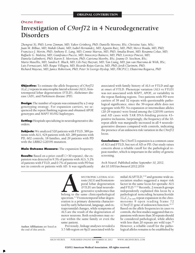
ONLINE FIRST
ORIGINAL CONTRIBUTION
Investigation of C9orf72 in 4 NeurodegenerativeDisordersZhengrui Xi, PhD; Lorne Zinman, MD; Yakov Grinberg, PhD; Danielle Moreno, BSc; Christine Sato, MSc;Juan M. Bilbao, MD; Mahdi Ghani, MD; Isabel Hernandez, MD; Agustın Ruiz, MD, PhD; Mercè Boada, MD, PhD;Francisco J. Moron, PhD; Anthony E. Lang, MD; Connie Marras, MD, PhD; Amalia Bruni, MD; Rosanna Colao, MD;Raffaele G. Maletta, MD; Gianfranco Puccio, MD; Innocenzo Rainero, MD, PhD; Lorenzo Pinessi, MD;Daniela Galimberti, PhD; Karen E. Morrison, PhD; Catriona Moorby, BSc; Joanne D. Stockton, BSc;Mario Masellis, MD; Sandra E. Black, MD; Lili-Naz Hazrati, MD; Yan Liang, MD; Jan van Haersma de With, BSc;Luis Fornazzari, MD; Roque Villagra, MD; Ricardo Rojas-Garcia, MD, PhD; Jordi Clarimon, PhD;Richard Mayeux, MD; Janice Robertson, PhD; Peter St George-Hyslop, MD, FRCP(C); Ekaterina Rogaeva, PhD
Objective: To estimate the allele frequency of C9orf72(G4C2) repeats in amyotrophic lateral sclerosis (ALS), fron-totemporal lobar degeneration (FTLD), Alzheimer dis-ease (AD), and Parkinson disease (PD).
Design: The number of repeats was estimated by a 2-stepgenotyping strategy. For expansion carriers, we se-quenced the repeat flanking regions and obtained APOEgenotypes and MAPT H1/H2 haplotypes.
Setting: Hospitals specializing in neurodegenerative dis-orders.
Subjects: We analyzed 520 patients with FTLD, 389 pa-tients with ALS, 424 patients with AD, 289 patients withPD, 602 controls, 18 families, and 29 patients with PDwith the LRRK2 G2019S mutation.
Main Outcome Measure: The expansion frequency.
Results: Based on a prior cutoff (�30 repeats), the ex-pansion was detected in 9.3% of patients with ALS, 5.2%of patients with FTLD, and 0.7% of patients with PD butnot in controls or patients with AD. It was significantly
associated with family history of ALS or FTLD and ageat onset of FTLD. Phenotype variation (ALS vs FTLD)was not associated with MAPT, APOE, or variability inthe repeat flanking regions. Two patients with PD werecarriers of 39 and 32 repeats with questionable patho-logical significance, since the 39-repeat allele does notsegregate with PD. No expansion or intermediate alleles(20-29 repeats) were found among the G2019S carriersand AD cases with TAR DNA-binding protein 43–positive inclusions. Surprisingly, the frequency of the 10-repeat allele was marginally increased in all 4 neurode-generative diseases compared with controls, indicatingthe presence of an unknown risk variation in the C9orf72locus.
Conclusions: The C9orf72 expansion is a common causeof ALS and FTLD, but not of AD or PD. Our study raisesconcern about a reliable cutoff for the pathological re-peat number, which is important in the utility of geneticscreening.
Arch Neurol. Published online September 10, 2012.doi:10.1001/archneurol.2012.2016
A MYOTROPHIC LATERAL SCLE-rosis (ALS) and frontotem-poral lobar degeneration(FTLD) are fatal neurode-generative syndromes that
belong to the same clinicopathologicalspectrum.1,2 Frontotemporal lobar degen-eration is a primary dementia character-ized by early behavioral, language, and ex-trapyramidal changes, while symptoms ofALS are the result of the degeneration ofmotor neurons. Both syndromes may oc-cur within the same family or even thesame patient.
Previously, linkage analyses revealed a3.7-Mb region on 9p21 associated with fa-
milial ALS/FTLD,3-10 and genome-wide as-sociation studies suggested a major riskfactor in the same locus for sporadic ALSand FTLD.11-15 Recently, 2 research groupsindependently explained this locus by apathological noncoding hexanucleotide(G4C2)30-1600 repeat expansion in the chro-mosome 9 open reading frame 72(C9orf72) gene of unknown function.16,17
Based on the allele frequencies in cases vscontrols, the first studies suggested that ex-pansions with more than 30 repeats shouldbe considered pathological, while alleleswith less than 20 repeats are wild type.16
However, a reliable cutoff for the patho-logical alleles remains to be established by
Author Affilthe end of th
Author Affiliations are listed atthe end of this article.
ARCH NEUROL PUBLISHED ONLINE SEPTEMBER 10, 2012 WWW.ARCHNEUROL.COME1
©2012 American Medical Association. All rights reserved.
Downloaded From: http://archneur.jamanetwork.com/ by a Universidad de Navarra User on 09/13/2012
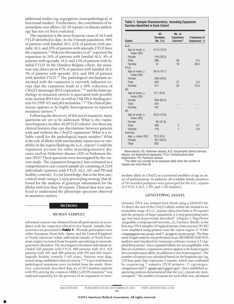
additional studies (eg, segregation, neuropathological, orfunctional studies). Furthermore, the contribution of in-termediate-size alleles (20-29 repeats) to disease pathol-ogy has not yet been evaluated.
The expansion is the most frequent cause of ALS andFTLD identified to date. In the Finnish population, 46%of patients with familial ALS, 21% of patients with spo-radic ALS, and 29% of patients with sporadic FTLD havethe expansion.16 DeJesus-Hernandez et al17 reported theexpansion in 24% of patients with familial ALS, 4% ofpatients with sporadic ALS, and 12% of patients with fa-milial FTLD. In the Flanders-Belgian cohort, the muta-tion was observed in 47% of patients with familial ALS,5% of patients with sporadic ALS, and 16% of patientswith familial FTLD.18 The pathological mechanism as-sociated with the expansion is currently unknown ex-cept that the expansion leads to a 50% reduction ofC9orf72 messenger RNA expression,17,18 and the brain pa-thology in mutation carriers is associated with possiblytoxic nuclear RNA foci, as well as TAR DNA-binding pro-tein 43 (TDP-43) and p62 inclusions.17,19 The clinical phe-notype appears to be highly heterogeneous in reportedmutation carriers.20
Following the discovery of this novel mutation, manyquestions are yet to be addressed. What is the expan-sion frequency in other ALS/FTLD cohorts? Are there anyclinical features that can discriminate between patientswith and without the C9orf72 expansion? What is a re-liable cutoff for the pathological repeat number? Whatis the role of alleles with intermediate repeat sizes or vari-ability in the region flanking the G4C2 repeat? Could theexpansion account for other neurodegenerative dis-eases, such as Alzheimer disease (AD) or Parkinson dis-ease (PD)? These questions were investigated by the cur-rent study. The expansion frequency was estimated in acomprehensive case-control sample set consisting of 2224individuals (patients with FTLD, ALS, AD, and PD andhealthy controls). To our knowledge, this is the first case-control study using a 2-step genotyping strategy that al-lowed for the analysis of genotype information for thealleles with less than 50 repeats. Clinical data were ana-lyzed to understand the phenotype spectrum observedin mutation carriers.
METHODS
HUMAN SAMPLES
Informed consent was obtained from all participants in accor-dance with the respective ethical review boards. Sample char-acteristics are presented in Table 1. All study participants wereeither European (from Italy, Spain, and the United Kingdom)or North American (white individuals mainly of North Euro-pean origin) recruited from hospitals specializing in neurode-generative disorders. The investigated unrelated individuals in-cluded 520 patients with FTLD, 389 patients with ALS, 424patients with AD, and 289 patients with PD and 602 neuro-logically healthy controls (�65 years). Patients were diag-nosed using established clinical criteria.21-24 Cases with knownpathological mutations were excluded from the study. How-ever, a previously described data set of 29 Canadian patientswith PD carrying the common LRRK2 G2019S mutation25 wasanalyzed separately for the presence of an expansion or inter-
mediate allele in C9orf72 as a potential modifier of age at on-set of parkinsonism. In addition, all available family membersof 18 extended pedigrees were genotyped for the G4C2 repeats(6 FTLD, 9 ALS, 1 PD, and 2 AD families).
GENOTYPING ASSAYS
Genomic DNA was isolated from blood using a QIAGEN kit.To detect the size of the C9orf72 alleles within the normal to in-termediate range of G4C2 repeats (detection limit is 50 repeats)and the presence of large expansions, a 2-step genotyping strat-egy was used as previously described17 (eFigure 1, http://www.joygrafika.com/projects/University_of_Toronto). Briefly, in thefirst step, DNA samples (10 ng/polymerase chain reaction [PCR])were amplified using primers near the repeat region (5�-FAM-caaggagggaaacaaccgcagcc and 5�-gcaggcaccgcaaccgcag). The frag-ment-length analysis was performed on an ABI PRISM 3100 DNAanalyzer and visualized by Genotyper software version 2.5 (Ap-plied Biosystems). Since expanded alleles are not amplifiable withthis set of primers, expansion carriers appear to be homozygousfor a normal repeat allele (in addition to true homozygotes). Thenumber of repeats was calculated based on the fragment size (eg,129 base pairs [bp] represents 2 repeats, which was confirmedby sequencing 7 samples [PCR primers: 5�-cgtcatcgca-catagaaaaca and 5�-ggagacagctcgggtactga]). Since published se-quencing analysis demonstrated that the G4C2 repeats are unin-terrupted,17 the number of repeats for each allele was calculated
Table 1. Sample Characteristics, Including ExpansionCarriers Identified in Each Cohort
Cohort
AllSamples,No. (%)
No. ofExpansionCarriersa
Frequency ofExpansion, %
ALSAge at onset, y,
mean (SD)57.6 (12.3)
Female 149 (38.3)Total 389 36 9.3Familial 47 18 38.3
FTLDAge at onset, y,
mean (SD)65.4 (10.1)
Female 258 (49.6)Total 520 27 5.2Familial 211 22 10.4
ADAge at onset, y,
mean (SD)72.1 (9.4)
Female 264 (62.3)Total 424 . . . . . .Familial 167 . . . . . .
PDAge at onset, y,
mean (SD)52.6 (13.0)
Female 92 (31.8)Total 289 2 0.7Familial 116 1 0.9
ControlsAge, y, mean (SD) 70.2 (9.5)Female 357 (59.3)Total 602 . . . . . .
Abbreviations: AD, Alzheimer disease; ALS, amyotrophic lateral sclerosis;ellipses, not detected/not applicable; FTLD, frontotemporal lobardegeneration; PD, Parkinson disease
aThe allele was counted as an expansion allele when the number ofrepeats was more than 30.
ARCH NEUROL PUBLISHED ONLINE SEPTEMBER 10, 2012 WWW.ARCHNEUROL.COME2
©2012 American Medical Association. All rights reserved.
Downloaded From: http://archneur.jamanetwork.com/ by a Universidad de Navarra User on 09/13/2012

using the following formula: (amplicon length − 117)/6. Samplesscored as homozygous were included in the second step to de-tect large expansions (�50 repeats) by a repeat-primed PCR. DNAsamples (100 ng/PCR) were amplified as described previouslyusing 3 primers (MRX-F: FAM-tgtaaaacgacggccagtcaaggagggaaa-caaccgcagcc, MRX-M13R: caggaaacagctatgacc, and MRX-R1:caggaaacagctatgaccgggcccgccccgaccacgccccggccccggccccgg),17 ex-cept the primer ratio was modified to increase PCR efficiency(MRX-F/MRX-M13R/MRX-R1 = 5/5/1). Data were analyzed usingGeneScan software version 3.1 (Applied Biosystems).
To search for sequence variability in regions flanking theG4C2 repeat, 2 PCR products were sequenced in 50 patients (44expansion carriers and 6 noncarriers) (eFigure 1B). The 5� flank-ing region was amplified with primers 5�-ccctaccagggtttgcagtand 5�-cgactcctgagttccagagc (616 bp). The 3� flanking regionwas amplified with primers 5�-tgcggttgcggtgcctgc and 5�-gaatggggagcacaccgacttgc (625 bp). The APOE polymorphismdefining the ε2 to ε4 alleles and the 238-bp insertion/deletionin intron 9 of MAPT defining the H1/H2 haplotypes was geno-typed as described previously in all patients with FTLD and ALSwith a pathological expansion in C9orf72, as well as in theirfamily members.26
STATISTICAL ANALYSES
Differences in sample characteristics (eg, sex, age at onset, andfamilial history between cases and controls) were analyzed usingthe �2 test, Fisher exact test, or independent-samples t test asappropriate. Allele frequencies within the normal to interme-diate range of repeats were calculated after excluding patientswho carry the pathological expanded allele, defined as a re-peat number more than 30 as previously suggested.16
The association between disease and alleles with less than30 repeats was assessed using CLUMP software that is basedon Monte Carlo tests for the evaluation of highly polymorphicloci.27 Empirical P values for T1 through T4 analyses were ob-tained after 2000 simulations. Further analyses to obtain P val-ues for each allele were also carried out as follows: allele countsfrom cases and controls were tested for significance using the�2 test after combining rare alleles. When any cell in the con-tingency table had an expected value less than 5, the corre-sponding allele was pooled with the neighboring allele (this pro-cess was repeated until no cell had an expected value �5). Eachallele group was compared in turn with the rest of the allelespooled together to calculate the �2 statistic. The pooling en-abled the calculation of odds ratios and 95% CIs.28 All the sta-tistical analyses were done using SPSS (version 20; IBM SPSS).Statistical significance was taken to be P � .05 (Bonferroni cor-rection for multiple testing was applied).
RESULTS
We used a previously suggested cutoff (�30 repeats) todistinguish the pathogenic expansion from the normalallele.16 None of the patients in our series were homo-zygous for the expansion allele, since all samples weresuccessfully amplified in the first step. Samples homo-zygous in the first step (33%) were evaluated for the largeexpansion (�50 repeats). Based on the electrophero-gram with sawtooth peaks (eFigure 1), 65 unrelated pa-tients were identified to be expansion carriers: 9.3% ofpatients with ALS (36 of 389), 5.2% of patients with FTLD(27 of 520), and 0.7% of patients with PD (2 of 289), butno expansions were detected in 424 patients with AD and602 controls (Table 1).
Details of the clinical data for expansion carriers vsnoncarriers are presented in Table 2. In addition, 14case reports on expansion carriers are available in the on-line-only material. The expansion allele was signifi-cantly associated with family history for both ALS andFTLD (P � .001). The frequency of the expansion was10.5% in patients with familial FTLD and 38.3% in pa-tients with familial ALS. The average age at onset of FTLDwas 6 years younger in expansion carriers than those with-out (P = .003). The disease subcategory of FTLD with mo-tor neuron disease was significantly enriched in expan-sion carriers (P � .001). However, there was no significantassociation with any evaluated clinical characteristic ofALS.
In expansion carriers, we did not observe an interme-diate or pathological number of repeats for the secondallele (2-11 repeats). Also, sequencing analysis of 44 ex-pansion carriers and 6 noncarriers did not reveal vari-ability in the regions immediately flanking the G4C2 re-peat that could be responsible for repeat instability,including a short tandem repeat (CGG)8 located 294 bpdownstream of the G4C2 repeat (eFigure 1B). Hence, itis unlikely that the number of repeats of the second al-lele or polymorphisms in the flanking region contributesignificantly to the disease phenotype. In addition, theMAPT H1/H2 and APOE genotypes were examined in 63expansion carriers (36 patients with ALS and 27 pa-tients with FTLD) and their family members, since bothgenes are well-known risk factors for several neurode-generative disorders including FTLD. However, statisti-cal analysis did not reveal a significant link between thesegenes and disease presentation (ALS vs FTLD) (eTable1 and eFigure 2).
For the segregation analysis, we genotyped all avail-able family members of probands with the expansion (6FTLD and 9 ALS families) and detected 30 additional mu-tation carriers. The expansion allele showed perfect coseg-regation with disease, and for those expansion carrierswho were asymptomatic (mean [SD] age, 46.3 [10.7]years; range, 24-64 years), follow-up is ongoing (Table3,Figure 1A, and eFigure 2A and B). Within the pedi-grees, there was no evidence of instability in the repeatsize for the normal alleles (Figure 1A and eFigure 2). Themethod used in the study did not allow the same ques-tion to be addressed for the expanded allele, and the DNAquality/quantity was not sufficient to conduct Southernblotting to estimate the size of the expanded allele. How-ever, in the 2 families with expansion carriers in 2 gen-erations, a younger age at onset in the subsequent gen-eration was observed (48 vs 74 years in ALS15 pedigreeand 45-47 years vs 60s in the FTLD TOR73 pedigree),indicating genetic anticipation (eFigure 2A).
Two patients with PD (without signs of ALS or de-mentia) were categorized as expansion carriers (39 and32 repeats). Family history of PD was known only for 1patient; however, the 39-repeat allele does not segregatewith PD, since the affected sibling did not inherit it(Figure 1B). The role of intermediate alleles (20-29 re-peats) in neurodegenerative diseases is currently un-known. The clinical characteristics of the patients withPD with the expansion and intermediate alleles are pre-sented in eTable 2. Intermediate alleles were observed
ARCH NEUROL PUBLISHED ONLINE SEPTEMBER 10, 2012 WWW.ARCHNEUROL.COME3
©2012 American Medical Association. All rights reserved.
Downloaded From: http://archneur.jamanetwork.com/ by a Universidad de Navarra User on 09/13/2012

only in 4 PD cases, 2 of which had parkinsonism fol-lowed by severe dementia. We also assessed if the pres-ence of intermediate alleles could influence the age at on-set of parkinsonism in LRRK2 G2019S mutation carriers(onset between age 40-80 years). However, none of the
29 LRRK2 mutation carriers had an expanded or inter-mediate repeat allele (�11 repeats).
Furthermore, our results do not suggest that the in-termediate C9orf72 alleles significantly contribute to ADrisk, since the frequency of such alleles was similar be-tween patients with AD and controls (about 1%), and thealleles with 23 and 21 repeats did not segregate with ADin 2 families (eFigure 2C). Importantly, 15 of the 31 au-topsied AD cases had TDP-43 inclusions (a frequent co-pathology in AD); however, no intermediate allele car-riers were found among these cases. This is of note becauseALS/FTLD caused by the expansion is associated withTDP-43 brain pathology.17,20,29,30 Hence, the TDP-43 pa-thology in AD could not be explained by an increasednumber of repeats in C9orf72.
Finally, a case-control association analysis was car-ried out using Monte Carlo tests. All carriers of alleleswith more than 30 repeats were excluded from this analy-sis to avoid association due to the pathological expan-sion. Analyses T1 and T2 revealed association betweennormal repeat alleles (�30) with FTLD and ALS (eTable4). Analysis T3 showed that the 10-repeat allele was sig-nificantly associated with FTLD. To get statistics for eachallele (eg, P value or odds ratio), we assessed the distri-bution of normal alleles in 4 disease groups vs controls(Figure 2 and eTable 3). In each group, the most fre-quent were the 2-, 5-, and 8-repeat alleles, which ac-count for about 75% of all observed alleles. A marginalprotective effect of the 5-repeat allele was detected in
Table 2. Comparison of the Clinical Statistics Between Expansion Carriers and Noncarriers
Cohort
No. (%)
P Value a ORb (95% CI)Noncarriers Expansion Carriers
ALS CasesNo. of ALS cases 353 36Age at onset, y, mean (SD) 57.6 (12.6) 57.8 (9.1) .92 . . .Female 134 (38.0) 15 (41.7) .68 . . .Familial cases 29 (8.2) 18 (50.0) 2.31 � 10−9 11.2 (5.2-23.8)Cases with FTLD 41 (11.6) 4 (11.1) .93 . . .Site of onset
Limb 235 (66.6) 24 (66.7) .99 . . .Bulbar 93 (26.3) 10 (28.7) .85 . . .Mixed 7 (2.0) . . . . . . . . .Unknown 18 (5.1) 2 (5.6) . . . . . .
FTLD Cases
No. of FTLD cases 493 27Age at onset, y, mean (SD) 65.7 (10.1) 59.6 (7.6) .003 . . .Female 248 (50.5) 10 (37.0) .17 . . .Familial cases 188 (38.1) 22 (81.5) 7.8 � 10−6 7.1 (2.7-19.2)Diagnosis subcategory
bvFTD 218 (44.2) 11 (40.7) .72 . . .FTLD unspecified 131 (26.6) 8 (29.6) .73 . . .SD 60 (12.2) . . . . . . . . .PNFA 59 (12.0) . . . . . . . . .FTLD-MND 20 (4.1) 8 (29.6) 2.8 � 10−5 10.0c (3.9-25.5)Other 5 (1.0) . . . . . . . . .
Abbreviations: ALS, amyotrophic lateral sclerosis; bvFTD, behavioral variant of frontotemporal dementia; ellipses, not detected/not applicable; FTLD,frontotemporal lobar degeneration; FTLD-MND, frontotemporal lobar degeneration with motor neuron disease; OR, odds ratio; PNFA, progressive nonfluentaphasia; SD, semantic dementia.
aA t test was used to compare the continuous variables. Categorical variables were compared by Pearson �2 test or Fisher exact test (when expected value �5).bThe OR was calculated only for variables showing P � .05.cThe OR was obtained by comparing FTLD-MND with the rest of the subcategories.
Table 3. Expansion Carriers Identified in the ALS and FTLDPedigrees
Status
Sample Size
AllExpansionCarriers Noncarriers
6 FTLDpedigrees
48 20 28
FTLD 8 8 . . .FTLD-MND 1 1 . . .bvFTD 2 2 . . .Relatives 30 9 21Spouses 7 . . . 7
9 ALSpedigrees
34 25 9
ALS 7 7 . . .ALS-FTLD 2 2 . . .FTLD 1 1 . . .Relatives 24 15 9
Abbreviations: ALS, amyotrophic lateral sclerosis; bvFTD, behavioralvariant of frontotemporal dementia; ellipses, not detected; FTLD,frontotemporal lobar degeneration; FTLD-MND, frontotemporal lobardegeneration with motor neuron disease.
ARCH NEUROL PUBLISHED ONLINE SEPTEMBER 10, 2012 WWW.ARCHNEUROL.COME4
©2012 American Medical Association. All rights reserved.
Downloaded From: http://archneur.jamanetwork.com/ by a Universidad de Navarra User on 09/13/2012

FTLD, ALS, and PD, as well as of the 11-repeat allele inALS and AD (nominal P � .05). The frequency of the 10-repeat allele was marginally increased in all 4 neurode-generative diseases vs the control group (nominalP � .05). The risk associated with this allele remainedsignificant in the FTLD data set even after Bonferroni cor-rection (P = .03; odds ratio, 2.14) (eTable 3).
COMMENT
Our findings further support the expansion in C9orf72as a common cause of ALS and FTLD, but not AD or PD.In contrast to the 1-step genotyping used in publishedcase-control studies of C9orf72, 2-step genotyping al-lowed us to obtain the exact genotype for each indi-vidual (except �50 repeats). No homozygous expan-sion carriers were found in the current study, suggestingthat such a genotype could be lethal. A heterozygous ex-pansion (�30 repeats) was detected in 9.3% of patientswith ALS and 5.2% of patients with FTLD and was sig-nificantly more frequent in cases with a family historyof ALS or FTLD. The frequency was doubled in our fa-
milial FTLD cases (10.4%), which is similar to that re-ported by DeJesus-Hernandez et al17 (11.7%).
We did not detect significant association between thepresence of the expansion and clinical characteristics ofpatients with ALS, while in the FTLD series, the expan-sion was associated with a younger age at onset and theFTLD with motor neuron disease phenotype. These find-ings are in line with previous observations.18,31 Notably,none of our FTLD cases with the expansion were diag-nosed with primary progressive aphasia, which is simi-lar to the Mayo clinic cohort32 and different from the Man-chester cohort wherein 4 primary progressive aphasiacases had the expansion.31 Wide clinical variability in ex-pansion carriers has been previously described (eg, ageat onset and disease duration),20,33 and future character-ization of such data in a large cohort of carriers couldgenerate a clinically useful algorithm to prioritize pa-tients for mutation analysis of C9orf72.34
In our study, a variable phenotype in expansion car-riers (ALS vs FTLD) was not associated with the APOEalleles or the extended MAPT H1/H2 haplotypes. How-ever, only an evaluation of a large data set could reach a
A
896 895
894 11691 11692 11689 11695 11690
11697Onset atage 56 y
APOE 3/2MAPT H1/H1
2 / exp
45588
APOE 3/3MAPT H1/H1
5 / 6
60 y
11701Onset atage 61 y
2 / exp
66 y
11698 11700 11703 11696Onset atage 60 y
2 / exp
77 y69 y
11699
APOE 3/2MAPT H1/H2
2 / 10
66 y
12054
APOE 3/2MAPT H1/H1
5/8
51 y
11702Onset atage 59 y
APOE 3/2MAPT H1/H1
10 / exp
60 y
45593
APOE 3/3MAPT H1/H1
2 / 6
40 y
45594
APOE 3/2MAPT H1/H2
2 / 5
38 y
45595
APOE 3/2MAPT H1/H2
6 / 10
30 y
11767
APOE 3/3MAPT H1/H2
2 / exp
47 y
12035
APOE 3/2MAPT H1/H2
2 / 2
48 y
12036
57 y 53 y
1170511707
APOE 3/2MAPT H1/H1
11 / exp
49 y
11706
APOE 3/2MAPT H1/H1
2 / exp
56 y
12055 11708
APOE 3/2MAPT H1/H2
2 / 11
45 y
11704
APOE 3/2MAPT H1/H2
2 / 11
82 y
11766
APOE 3/3MAPT H1/H2
APOE 3/3MAPT H1/H1
2 / 2
76 y
C family
B PD family 83 y
6855Onset atage 51 y
6 / 39
7953Onset atage 46 y
2 / 6
53 y
82 yUnaffected
PD
Unaffected
FTLD
Figure 1. Families with the G4C2 repeat expansion. A, Frontotemporal lobar degeneration (FTLD) pedigree. B, Parkinson disease (PD) pedigree. Individualgenotypes are shown beneath the corresponding diamond, including “at-risk” currently asymptomatic individuals. Arabic numbers indicate the repeat units (expindicates expansion). The age at the time of examination is shown in the upper right corner. Age at onset is indicated for patients below the ID number. Sex of thefamily members is masked to protect privacy.
ARCH NEUROL PUBLISHED ONLINE SEPTEMBER 10, 2012 WWW.ARCHNEUROL.COME5
©2012 American Medical Association. All rights reserved.
Downloaded From: http://archneur.jamanetwork.com/ by a Universidad de Navarra User on 09/13/2012

solid conclusion about APOE and MAPT as phenotypemodifiers. It is also possible that C9orf72 itself is respon-sible for the modifying effect because of coding se-quence variations, repeat size, and/or repeat instability.We demonstrated that the repeat size of the second al-lele in expansion carriers was within the normal range(2-11 repeats) and unlikely to contribute to the variabil-ity in disease phenotype. In addition, our sequencinganalysis did not reveal variations in regions flanking theG4C2 repeat that could be responsible for repeat insta-bility or the variable phenotype of expansion carriers. Fu-ture studies should assess intronic and coding varia-tions in the entire C9orf72 locus as potential phenotypemodifiers.
Instability of the G4C2 repeat region was suggested tobe a possible mechanism for the occurrence of the ex-pansion.16 However, a founder effect is more likely to beresponsible for the incidence of the mutation, since mostcarriers harbor a common haplotype.17,29 Despite the meth-od’s limitation (the size of large expansions could not bedetermined), we estimated the stability of 2 to 30 re-peats in the pedigrees. The number of repeats was stableacross generations.
Given the clinical/pathological overlap between neu-rodegenerative diseases,35-38 our study determined whetherthe expansion plays a role in AD or PD. Importantly, the
PD samples were previously collected to generate a dataset enriched in genetic predisposition to PD and mainlyconsisted of cases with either an early age at onset (mean[SD], 52.6 [13.04] years) and/or family history of PD(40%).39 However, the frequency of expansion alleles inour PD data set was unremarkable (0.7%). Only 2 pa-tients (without symptoms of ALS or dementia) were car-riers of 32 and 39 repeats, which is much smaller thanthe expansion estimated by Southern blot (700-1600repeats),17 and segregation analysis did not support thepathogenic nature of the 39-repeat allele. This raises con-cern about a reliable cutoff for a pathological repeat num-ber, which is important in the utility of genetic screen-ing in patient care. Likely, a higher cutoff or establishinga gray zone will be proposed in the future based on anincreasing number of observations from case-control andneuropathological studies.40,41 Among our ALS and FTLDmutation carriers, only 2 had an allele with question-able pathological significance (�40 repeats). The con-nection between the repeat number and pathological sig-nificance has to be carefully investigated. It is possiblethat the pathological cutoff is disease dependent (eg, ALSvs FTLD) and could be modulated by individual geneticbackground. Furthermore, future studies have to test thepossibility of somatic instability of the repeat region (thedisease-related tissues, such as the spinal cord, could have
0.0
0.6
0.5Al
lele
Fre
quen
cy 0.4
0.3
0.2
0.1
ControlALS
ControlFTLD
ControlPD
ControlAD
∗
0.0
0.6
0.5
Alle
le F
requ
ency
Repeat Number
0.4
0.3
0.2
0.1
2 3,4 5 6 7 8 9 10 11 12 13,14 15-17 18-30
Repeat Number2 3,4 5 6 7 8 9 10 11 12 13,14 15-17 18-30
∗
∗
∗
∗ ∗
∗ ∗
∗
∗
Figure 2. Comparison of the allele frequencies (repeats �30) between controls and cases in each disease cohort (AD indicates Alzheimer disease; ALS,amyotrophic lateral sclerosis; FTLD, frontotemporal lobar degeneration; and PD, Parkinson disease). Asterisks indicate a significant P value (nominal P � .05). Incomparison with controls, there were 2 significant differences observed for the allele frequencies for the patients with FTLD, 4 for the patients with ALS, 2 for thepatients with AD, and 2 for the patients with PD. A similar pattern was observed for all 4 diseases. The 5-repeat allele was significantly lower in FTLD, ALS, and PDthan in controls and the 10-repeat allele in disease was significantly higher in all 4 disease groups than in controls. Expansion carriers were excluded from thisanalysis: controls, n = 602; FTLD, n = 488; ALS, n = 353; AD, n = 424; and PD, n = 287.
ARCH NEUROL PUBLISHED ONLINE SEPTEMBER 10, 2012 WWW.ARCHNEUROL.COME6
©2012 American Medical Association. All rights reserved.
Downloaded From: http://archneur.jamanetwork.com/ by a Universidad de Navarra User on 09/13/2012
larger expansions than blood cells from the same indi-vidual).
Our results did not suggest that the expansion or in-termediate alleles are associated with AD. In contrast, therewas a report of 6 expansion carriers in a familial AD co-hort (�1%), 4 of whom were from the same family.42
However, autopsy indicated that 3 carriers actually hadamnestic FTLD. Whether the remaining carriers were alsoclinically misdiagnosed as having AD remains to be seen.In addition to typical AD pathology, 15 of our patientswith AD had TDP-43 inclusions, which are known to beassociated with the brain pathology of the expan-sion17,20,29,30; however, all of these patients with AD havegenotypes within the normal range (2-12 repeats).
Our case-control studies also assessed the frequencyof alleles within the normal range (�30 repeats) and ob-served a trend toward an association between the 10-repeat allele and risk for all 4 disorders (odds ratio, 1.72-2.14). It is tempting to speculate that this allele is in linkagedisequilibrium with an unknown C9orf72 risk varia-tion. Further genetic work has to validate this observa-tion, including follow-up case-control studies and se-quencing of 10-repeat carriers.
Accepted for Publication: May 30, 2012.Published Online: September 10, 2012. doi:10.1001/archneurol.2012.2016Author Affiliations: Tanz Centre for Research in Neu-rodegenerative Diseases (Drs Xi, Grinberg, Ghani, Haz-rati, Liang, Robertson, St George-Hyslop, and Rogaeva,Mss Moreno and Sato, and Mr van Haersma de With),Division of Neurology, Department of Medicine (Drs Lang,Marras, Masellis, Black, St George-Hyslop, and Ro-gaeva), and Department of Psychiatry, St. Michael’s Hos-pital (Dr Fornazzari), University of Toronto, Sunny-brook Health Sciences Centre (Drs Zinman, Bilbao,Masellis, and Black), Morton and Gloria Shulman Move-ment Disorders Clinic and the Edmond J. Safra Programin Parkinson’s Disease, Toronto Western Hospital(Drs Lang and Marras), and LC Campbell Cognitive Neu-rology Research Unit, Sunnybrook Research Institute(Drs Masellis and Black), Toronto, Ontario, Canada;Memory Clinic of Fundacio ACE, Institut Català de Neu-rociències Aplicades (Drs Hernandez, Ruiz, and Boada),Hospital Universitari Vall d’Hebron–Institut de Re-cerca, Universitat Autònoma de Barcelona (VHIR-UAB)(Dr Boada), and Neurology Department, Hospital de laSanta Creu i Sant Pau, Universitat Autònoma de Barce-lona (Drs Rojas-Garcia and Clarimon), Barcelona, De-partment of Structural Genomics, Neocodex, Seville(Dr Moron), and Center for Networker Biomedical Re-search in Neurodegenerative Diseases (CIBERNED), Ma-drid (Drs Rojas-Garcia and Clarimon), Spain; RegionalNeurogenetic Centre, Lamezia Terme, Azienda Sani-taria Provinciale Catanzaro (Drs Bruni, Colao, Maletta,and Puccio), and Neurology II, Department of Neuro-science, University of Torino, Turin (Drs Rainero andPinessi), and Department of Neurological Sciences, Uni-versity of Milan, Centro Dino Ferrari, Fondazione CàGranda, IRCCS Ospedale Maggiore Policlinico, Milan(Dr Galimberti), Italy; School of Clinical and Experi-mental Medicine, College of Medical and Dental Sci-
ences, University of Birmingham, Birmingham (Dr Mor-rison and Mss Moorby and Stockton), and CambridgeInstitute for Medical Research and the Department ofClinical Neurosciences, University of Cambridge, Cam-bridge (Dr St George-Hyslop), England; Salvador Hos-pital, University of Chile, Santiago (Dr Villagra); and TheTaub Institute for Research on Alzheimer’s Disease andthe Aging Brain, The Gertrude H. Sergievsky Center, De-partments of Neurology, Psychiatry, and Medicine, Col-lege of Physicians and Surgeons, Columbia University,New York, New York (Dr Mayeux).Correspondence: Ekaterina Rogaeva, PhD, Tanz Cen-tre for Neurodegenerative Diseases, 6 Queen’s Park Cres-cent West, Toronto, ON M5S 3H2, Canada ([email protected]) or Peter St George-Hyslop, MD,FRCP(C) ([email protected]).Author Contributions: Study concept and design: Xi, StGeorge-Hyslop, and Rogaeva. Acquisition of data: Xi, Zin-man, Grinberg, Moreno, Sato, Bilbao, Ghani, Hernan-dez, Ruiz, Boada, Moron, Lang, Marras, Bruni, Colao, Mal-etta, Puccio, Rainero, Pinessi, Galimberti, Morrison,Moorby, Stockton, Masellis, Black, Hazrati, Liang, vanHaersma de With, Fornazzari, Villagra, Rojas-Garcia, Clar-imon, Mayeux, Robertson, and Rogaeva. Analysis and in-terpretation of data: Xi, Grinberg, Marras, Masellis, andRogaeva. Drafting of the manuscript: Xi and Rogaeva. Criti-cal revision of the manuscript for important intellectual con-tent: Xi, Zinman, Grinberg, Moreno, Sato, Bilbao, Ghani,Hernandez, Ruiz, Boada, Moron, Lang, Marras, Bruni, Co-lao, Maletta, Puccio, Rainero, Pinessi, Galimberti, Mor-rison, Moorby, Stockton, Masellis, Black, Hazrati, Li-ang, van Haersma de With, Fornazzari, Villagra, Rojas-Garcia, Clarimon, Mayeux, Robertson, and St George-Hyslop. Statistical analysis: Xi. Obtained funding: Zinman,Grinberg, Bilbao, van Haersma de With, Mayeux, Rob-ertson, St George-Hyslop, and Rogaeva. Administrative,technical, and material support: Xi, Zinman, Moreno, Sato,Ghani, Hernandez, Ruiz, Boada, Moron, Bruni, Colao,Maletta, Puccio, Rainero, Pinessi, Galimberti, Morri-son, Moorby, Stockton, Black, Hazrati, Liang, Fornaz-zari, Rojas-Garcia, Clarimon, and Rogaeva. Study super-vision: Morrison, St George-Hyslop, and Rogaeva.Financial Disclosure: Dr Masellis has received speakerhonoraria from Novartis and EMD Serono Inc; serves asan associate editor for Current Pharmacogenomics and Per-sonalized Medicine; receives publishing royalties fromHenry Stewart Talks; has served as a consultant for BioS-cape Medical Imaging CRO; and receives research sup-port from the Canadian Institutes of Health Research, Par-kinson Society Canada, an Early Researcher Award fromthe Ministry of Economic Development and Innovationof Ontario, Teva Pharmaceutical Industries Ltd, and theDepartment of Medicine, Sunnybrook Health SciencesCentre.Funding/Support: This work was supported by grantsfrom the Ontario Research Fund, the Weston Founda-tion (Drs Rogaeva and St George-Hyslop), the HowardHughes Medical Institute, The Wellcome Trust and Medi-cal Research Council, the Alzheimer Society of Ontario(Dr St George-Hyslop), the Canadian Institutes of HealthResearch (Drs St George-Hyslop, Rogaeva and Black), aCenter of Excellence grant from the National Parkinson
ARCH NEUROL PUBLISHED ONLINE SEPTEMBER 10, 2012 WWW.ARCHNEUROL.COME7
©2012 American Medical Association. All rights reserved.
Downloaded From: http://archneur.jamanetwork.com/ by a Universidad de Navarra User on 09/13/2012
Foundation (Drs Marras and Lang), and the James HunterFamily ALS Initiative (Drs Robertson and Zinman).Online-Only Material: The eTables, eFigures, and casereports are available at http://www.joygrafika.com/projects/University_of_Toronto.
REFERENCES
1. Fecto F, Siddique T. Making connections: pathology and genetics link amyo-trophic lateral sclerosis with frontotemporal lobe dementia. J Mol Neurosci. 2011;45(3):663-675.
2. Lillo P, Hodges JR. Frontotemporal dementia and motor neurone disease: over-lapping clinic-pathological disorders. J Clin Neurosci. 2009;16(9):1131-1135.
3. Boxer AL, Mackenzie IR, Boeve BF, et al. Clinical, neuroimaging and neuropatho-logical features of a new chromosome 9p-linked FTD-ALS family. J Neurol Neu-rosurg Psychiatry. 2011;82(2):196-203.
4. Gijselinck I, Engelborghs S, Maes G, et al. Identification of 2 loci at chromo-somes 9 and 14 in a multiplex family with frontotemporal lobar degeneration andamyotrophic lateral sclerosis. Arch Neurol. 2010;67(5):606-616.
5. Le Ber I, Camuzat A, Berger E, et al; French Research Network on FTD/FTD-MND. Chromosome 9p-linked families with frontotemporal dementia associ-ated with motor neuron disease. Neurology. 2009;72(19):1669-1676.
6. Luty AA, Kwok JB, Thompson EM, et al. Pedigree with frontotemporal lobar de-generation–motor neuron disease and Tar DNA binding protein-43 positive neu-ropathology: genetic linkage to chromosome 9. BMC Neurol. 2008;8:32.
7. Morita M, Al-Chalabi A, Andersen PM, et al. A locus on chromosome 9p conferssusceptibility to ALS and frontotemporal dementia. Neurology. 2006;66(6):839-844.
8. Valdmanis PN, Dupre N, Bouchard JP, et al. Three families with amyotrophic lat-eral sclerosis and frontotemporal dementia with evidence of linkage to chromo-some 9p [published correction appears in Arch Neurol. 2007;64(6):909]. ArchNeurol. 2007;64(2):240-245.
9. Vance C, Al-Chalabi A, Ruddy D, et al. Familial amyotrophic lateral sclerosis withfrontotemporal dementia is linked to a locus on chromosome 9p13.2-21.3. Brain.2006;129(pt 4):868-876.
10. Pearson JP, Williams NM, Majounie E, et al. Familial frontotemporal dementiawith amyotrophic lateral sclerosis and a shared haplotype on chromosome 9p.J Neurol. 2011;258(4):647-655.
11. Laaksovirta H, Peuralinna T, Schymick JC, et al. Chromosome 9p21 in amyo-trophic lateral sclerosis in Finland: a genome-wide association study. Lancet Neurol.2010;9(10):978-985.
12. Shatunov A, Mok K, Newhouse S, et al. Chromosome 9p21 in sporadic amyo-trophic lateral sclerosis in the UK and seven other countries: a genome-wide as-sociation study. Lancet Neurol. 2010;9(10):986-994.
13. van Es MA, Veldink JH, Saris CG, et al. Genome-wide association study identi-fies 19p13.3 (UNC13A) and 9p21.2 as susceptibility loci for sporadic amyo-trophic lateral sclerosis. Nat Genet. 2009;41(10):1083-1087.
14. Van Deerlin VM, Sleiman PM, Martinez-Lage M, et al. Common variants at 7p21are associated with frontotemporal lobar degeneration with TDP-43 inclusions.Nat Genet. 2010;42(3):234-239.
15. Rollinson S, Mead S, Snowden J, et al. Frontotemporal lobar degeneration ge-nome wide association study replication confirms a risk locus shared with amyo-trophic lateral sclerosis. Neurobiol Aging. 2011;32(4):758, e1-e7.
16. Renton AE, Majounie E, Waite A, et al; ITALSGEN Consortium. A hexanucleotiderepeat expansion in C9ORF72 is the cause of chromosome 9p21-linked ALS-FTD.Neuron. 2011;72(2):257-268.
17. DeJesus-Hernandez M, Mackenzie IR, Boeve BF, et al. Expanded GGGGCC hexa-nucleotide repeat in noncoding region of C9ORF72 causes chromosome 9p-linked FTD and ALS. Neuron. 2011;72(2):245-256.
18. Gijselinck I, Van Langenhove T, van der Zee J, et al. A C9orf72 promoter repeatexpansion in a Flanders-Belgian cohort with disorders of the frontotemporal lo-bar degeneration-amyotrophic lateral sclerosis spectrum: a gene identificationstudy. Lancet Neurol. 2012;11(1):54-65.
19. Al-Sarraj S, King A, Troakes C, et al. p62 Positive, TDP-43 negative, neuronalcytoplasmic and intranuclear inclusions in the cerebellum and hippocampus de-
fine the pathology of C9orf72-linked FTLD and MND/ALS. Acta Neuropathol. 2011;122(6):691-702.
20. Hsiung GY, DeJesus-Hernandez M, Feldman HH, et al. Clinical and pathologicalfeatures of familial frontotemporal dementia caused by C9ORF72 mutation onchromosome 9p. Brain. 2012;135(pt 3):709-722.
21. Clinical and neuropathological criteria for frontotemporal dementia: the Lund andManchester Groups. J Neurol Neurosurg Psychiatry. 1994;57(4):416-418.
22. Brooks BR, Miller RG, Swash M, Munsat TL; World Federation of Neurology Re-search Group on Motor Neuron Diseases. El Escorial revisited: revised criteriafor the diagnosis of amyotrophic lateral sclerosis. Amyotroph Lateral Scler OtherMotor Neuron Disord. 2000;1(5):293-299.
23. McKhann G, Drachman D, Folstein M, Katzman R, Price D, Stadlan EM. Clinicaldiagnosis of Alzheimer’s disease: report of the NINCDS-ADRDA Work Group un-der the auspices of Department of Health and Human Services Task Force onAlzheimer’s Disease. Neurology. 1984;34(7):939-944.
24. Hughes AJ, Daniel SE, Lees AJ. Improved accuracy of clinical diagnosis of Lewybody Parkinson’s disease. Neurology. 2001;57(8):1497-1499.
25. Marras C, Schule B, Munhoz RP, et al. Phenotype in parkinsonian and nonpar-kinsonian LRRK2 G2019S mutation carriers [published correction appears in Neu-rology. 2011;77(15):1501]. Neurology. 2011;77(4):325-333.
26. Bernardi L, Maletta RG, Tomaino C, et al. The effects of APOE and tau gene vari-ability on risk of frontotemporal dementia. Neurobiol Aging. 2006;27(5):702-709.
27. Sham PC, Curtis D. Monte Carlo tests for associations between disease and allelesat highly polymorphic loci. Ann Hum Genet. 1995;59(pt 1):97-105.
28. McKnight AJ, Maxwell AP, Sawcer S, et al. A genome-wide DNA microsatelliteassociation screen to identify chromosomal regions harboring candidate genesin diabetic nephropathy. J Am Soc Nephrol. 2006;17(3):831-836.
29. Simon-Sanchez J, Dopper EG, Cohn-Hokke PE, et al. The clinical and pathologi-cal phenotype of C9ORF72 hexanucleotide repeat expansions. Brain. 2012;135(pt 3):723-735.
30. Troakes C, Maekawa S, Wijesekera L, et al. An MND/ALS phenotype associatedwith C9orf72 repeat expansion: abundant p62-positive, TDP-43-negative inclu-sions in cerebral cortex, hippocampus and cerebellum but without associatedcognitive decline [published online December 19, 2011]. Neuropathology. doi:10.1111/j.1440-1789.2011.01286.x.
31. Snowden JS, Rollinson S, Thompson JC, et al. Distinct clinical and pathologicalcharacteristics of frontotemporal dementia associated with C9ORF72 mutations.Brain. 2012;135(pt 3):693-708.
32. Boeve BF, Boylan KB, Graff-Radford NR, et al. Characterization of frontotempo-ral dementia and/or amyotrophic lateral sclerosis associated with the GGGGCCrepeat expansion in C9ORF72. Brain. 2012;135(pt 3):765-783.
33. Murray ME, DeJesus-Hernandez M, Rutherford NJ, et al. Clinical and neuro-pathologic heterogeneity of c9FTD/ALS associated with hexanucleotide repeatexpansion in C9ORF72. Acta Neuropathol. 2011;122(6):673-690.
34. Byrne S, Elamin M, Bede P, et al. Cognitive and clinical characteristics of pa-tients with amyotrophic lateral sclerosis carrying a C9orf72 repeat expansion: apopulation-based cohort study. Lancet Neurol. 2012;11(3):232-240.
35. Mathias JL, Morphett K. Neurobehavioral differences between Alzheimer’s dis-ease and frontotemporal dementia: a meta-analysis. J Clin Exp Neuropsychol.2010;32(7):682-698.
36. Spillantini MG, Bird TD, Ghetti B. Frontotemporal dementia and Parkinsonismlinked to chromosome 17: a new group of tauopathies. Brain Pathol. 1998;8(2):387-402.
37. Jellinger KA. Neuropathological aspects of Alzheimer disease, Parkinson dis-ease and frontotemporal dementia. Neurodegener Dis. 2008;5(3-4):118-121.
38. Jellinger KA. Interaction between pathogenic proteins in neurodegenerativedisorders. J Cell Mol Med. 2012;16(6):1166-1183.
39. Paisan-Ruız C, Lang AE, Kawarai T, et al. LRRK2 gene in Parkinson disease: mu-tation analysis and case control association study. Neurology. 2005;65(5):696-700.
40. Coffey SM, Cook K, Tartaglia N, et al. Expanded clinical phenotype of womenwith the FMR1 premutation. Am J Med Genet A. 2008;146A(8):1009-1016.
41. Bourgeois JA, Coffey SM, Rivera SM, et al. A review of fragile X premutation dis-orders: expanding the psychiatric perspective. J Clin Psychiatry. 2009;70(6):852-862.
42. Majounie E, Abramzon Y, Renton AE, et al. Repeat expansion in C9ORF72 in Alz-heimer’s disease. N Engl J Med. 2012;366(3):283-284.
ARCH NEUROL PUBLISHED ONLINE SEPTEMBER 10, 2012 WWW.ARCHNEUROL.COME8
©2012 American Medical Association. All rights reserved.
Downloaded From: http://archneur.jamanetwork.com/ by a Universidad de Navarra User on 09/13/2012
Expansion mutation in C9ORF72 does not influence plasma progranulinlevels in frontotemporal dementia
Oriol Dols-Icardoa,b,1, Marc Suárez-Calveta,b,1, Isabel Hernándezc, Guillermo Amerd,Sofía Antón-Aguirrea,b, Daniel Alcoleaa,b, Juan Forteaa,b, Mercè Boadac, Lluís Tárragac,
Rafael Blesaa,b, Alberto Lleóa,b, Jordi Clarimóna,b,*a Department of Neurology, Hospital de la Santa Creu i Sant Pau, Universitat Autònoma de Barcelona, Barcelona, Spain
b Center for Networker Biomedical Research in Neurodegenerative Diseases, Madrid, Spainc Memory Clinic of Fundació ACE, Institut Català de Neurociències Aplicades, Barcelona, Spain
d Department of Neurology, Hospital Universitari Son Espases, Mallorca, Spain
Received 23 January 2012; received in revised form 28 February 2012; accepted 8 March 2012
Abstract
A hexanucleotide repeat expansion in chromosome 9 open reading frame 72 (C9ORF72) gene has recently been described as a cause offamilial and sporadic frontotemporal lobar degeneration. The aim of this study was to assess whether plasma progranulin (GRN) levels couldbe modulated by the presence of this repeat expansion. Sixty-five patients diagnosed with frontotemporal dementia and 10 family memberswith familial aggregation of disease were screened for the presence of the hexanucleotide repeat expansion in C9ORF72 gene, using arepeat-primed polymerase chain reaction method. Plasma GRN levels were measured in all subjects through enzyme-linked immunosorbentassay. Seven individuals with the repeat expansion were identified. No differences were found between C9ORF72 repeat expansion carriersand noncarriers (116.4 � 21 ng/mL and 131.7 � 36 ng/mL, respectively, p � 0.3). Analysis of family members did not disclose anydifference in plasma GRN levels between carriers and noncarriers. In conclusion, plasma GRN levels are not influenced by thehexanucleotide repeat expansion in C9ORF72 gene, and therefore, cannot be used as a reliable biomarker to detect mutation carriers.© 2012 Elsevier Inc. All rights reserved.
Keywords: Frontotemporal dementia; Progranulin; C9ORF72; Genetics; Biomarkers
1. Introduction
Frontotemporal lobar degeneration (FTLD) comprises agroup of clinically heterogeneous neurodegenerative dis-eases with diverse neuropathological and genetic substrates.Currently, FTLD is pathologically classified based on thethree main proteins that aggregate in the central nervoussystem: transactive response (TAR) DNA-binding pro-tein43 (TDP-43), microtubule-associated protein tau(MAPT), and fused in sarcoma (Mackenzie et al., 2010).
Mutations in the progranulin gene (GRN; MIM: 138945)are the most common genetic cause of FTLD and are typ-ically associated with TDP-43 pathology (Cruts et al.,2006). Most GRN mutations cause protein haploinsuffi-ciency, leading to a significant decrease in progranulin(GRN) levels that can be detected in plasma of mutat-ion carriers (Ghidoni et al., 2008). A hexanucleotide(GGGGCC) repeat expansion in the noncoding region ofchromosome 9 open reading frame 72 gene (C9ORF72;MIM: 614260) was recently found to be a major cause offamilial and sporadic FTLD (DeJesus-Hernandez et al.,2011; Renton et al., 2011). All the cases with this mutationdescribed to date show TDP-43 pathology in the centralnervous system (Murray et al., 2011). The fact that GRNmutations and C9ORF72 expansions both show TDP-43pathology suggests they may share some disease mecha-
* Corresponding author at: Department of Neurology, Hospital de laSanta Creu i Sant Pau, Sant Antoni M. Claret 167, Barcelona 08025, Spain.Tel.: �34 932919050; fax: �34 935565602.
E-mail address: [email protected] (J. Clarimón).1 These authors contributed equally to this work.
Neurobiology of Aging 33 (2012) 1851.e17–1851.e19
www.elsevier.com/locate/neuaging
0197-4580/$ – see front matter © 2012 Elsevier Inc. All rights reserved.10.1016/j.neurobiolaging.2012.03.005
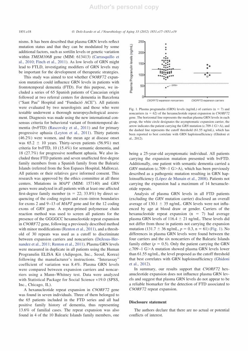
nisms. It has been described that plasma GRN levels reflectmutation status and that they can be modulated by someadditional factors, such as sortilin levels or genetic variationwithin TMEM106B gene (MIM: 613413) (Carrasquillo etal., 2010; Finch et al., 2011). As low levels of GRN mightlead to FTLD, investigating modifiers of GRN levels maybe important for the development of therapeutic strategies.
This study was aimed to test whether C9ORF72 expan-sion mutation could influence GRN levels in patients withfrontotemporal dementia (FTD). For this purpose, we in-cluded a series of 65 Spanish patients of Caucasian originfollowed at two referral centers for dementia in Barcelona(“Sant Pau” Hospital and “Fundació ACE”). All patientswere evaluated by two neurologists and those who weretestable underwent a thorough neuropsychological assess-ment. Diagnosis was made using the new international con-sensus criteria for behavioral variant of frontotemporal de-mentia (bvFTD) (Rascovsky et al., 2011) and for primaryprogressive aphasia (Leyton et al., 2011). Thirty patients(46.2%) were women, and the mean age at disease onsetwas 65.2 � 10 years. Thirty-seven patients (56.9%) metcriteria for bvFTD, 10 (15.4%) for semantic dementia, and18 (27.7%) for progressive nonfluent aphasia. We also in-cluded three FTD patients and seven unaffected first-degreefamily members from a Spanish family from the BalearicIslands (referred from the Son Espases Hospital, Mallorca).All patients or their relatives gave informed consent. Thisresearch was approved by the ethics committee at all threecenters. Mutations in MAPT (MIM: 157140) and GRNgenes were analyzed in all patients with at least one affectedfirst-degree family member (n � 22; 33.8%) by direct se-quencing of the coding region and exon–intron boundariesfor exons 2 and 9–13 of MAPT gene and for the 12 codingexons of GRN gene. A repeat-primed polymerase chainreaction method was used to screen all patients for thepresence of the GGGGCC hexanucleotide repeat expansionin C9ORF72 gene, following a previously described methodwith minor modifications (Renton et al., 2011), and a thresh-old of 30 repeats was used as a cutoff to discriminatebetween expansion carriers and noncarriers (DeJesus-Her-nandez et al., 2011; Renton et al., 2011). Plasma GRN levelswere measured in duplicate in all patients using the HumanProgranulin ELISA Kit (Adipogen, Inc., Seoul, Korea)following the manufacturer’s instructions. “Interassay”coefficient of variation was 8.4%. Plasma GRN levelswere compared between expansion carriers and noncar-riers using a Mann–Whitney test. Data were analyzedwith Statistical Package for Social Science v19.0 (SPSS,Inc., Chicago, IL).
A hexanucleotide repeat expansion in C9ORF72 genewas found in seven individuals. Three of them belonged tothe 65 patients included in the FTD series and all hadpositive family history of dementia, thus representing13.6% of familial cases. The repeat expansion was alsofound in 4 of the 10 Balearic Islands family members, one
being a 25-year-old asymptomatic individual. All patientscarrying the expansion mutation presented with bvFTD.Additionally, one patient with semantic dementia carried aGRN mutation (c.709–1 G�A), which has been previouslydescribed as a pathogenic mutation resulting in GRN hap-loinsufficiency (López de Munain et al., 2008). Patients notcarrying the expansion had a maximum of 14 hexanucle-otide repeats.
Analysis of plasma GRN levels in all FTD patients(excluding the GRN mutation carrier) disclosed an overallaverage of 130.1 � 35 ng/mL. GRN levels were not influ-enced by age at blood draw or gender. Carriers of thehexanucleotide repeat expansion (n � 7) had averageplasma GRN levels of 116.4 � 21 ng/mL. These levels didnot differ from those in patients not carrying the expansionmutation (131.7 � 36 ng/mL, p � 0.3, n � 61) (Fig. 1). Nodifferences in plasma GRN levels were found between thefour carriers and the six noncarriers of the Balearic Islandsfamily either (p � 0.5). Only the patient carrying the GRNc.709–1 G�A mutation showed plasma GRN levels lowerthan 61.55 ng/mL, the level proposed as the cutoff thresholdthat best correlates with GRN haploinsufficiency (Ghidoniet al., 2012).
In summary, our results support that C9ORF72 hex-anucleotide expansion does not influence plasma GRN lev-els and suggest that plasma GRN levels do not appear to bea reliable biomarker for the detection of FTD associated toC9ORF72 repeat expansion.
Disclosure statement
The authors declare that there are no actual or potentialconflicts of interest.
Fig. 1. Plasma progranulin (GRN) levels (ng/mL) of carriers (n � 7) andnoncarriers (n � 62) of the hexanucleotide repeat expansion in C9ORF72gene. The horizontal line represents the median plasma GRN levels in eachgroup, the white circle designates the asymptomatic expansion carrier, thearrow indicates the patient carrying the GRN mutation (c.709-1 G�A), andthe dashed line represents the cutoff threshold (61.55 ng/mL), which hasbeen reported to best correlate with GRN haploinsufficiency (Ghidoni etal., 2012).
1851.e18 O. Dols-Icardo et al. / Neurobiology of Aging 33 (2012) 1851.e17–1851.e19

Acknowledgements
The authors are indebted to all the patients and theirfamilies for their participation. They thank Dr. Agustín Ruizfor his helpful remarks, Carolyn Newey for editorial help,and Laia Muñoz and Inés M. Matas for technical assistance.This work was supported by a research grant from “Centerfor Networker Biomedical Research in NeurodegenerativeDiseases” and by the “Fondo de Investigaciones Sanitarias”(PI09/00098).
References
Carrasquillo, M.M., Nicholson, A.M., Finch, N., Gibbs, J.R., Baker, M.,Rutherford, N.J., Hunter, T.A., DeJesus-Hernandez, M., Bisceglio,G.D., Mackenzie, I.R., Singleton, A., Cookson, M.R., Crook, J.E.,Dillman, A., Hernandez, D., Petersen, R.C., Graff-Radford, N.R.,Younkin, S.G., Rademakers, R., 2010. Genome-wide screen identifiesrs646776 near sortilin as a regulator of progranulin levels in humanplasma. Am. J. Hum. Genet. 87, 890–897.
Cruts, M., Gijselinck, I., van der Zee, J., Engelborghs, S., Wils, H., Pirici,D., Rademakers, R., Vandenberghe, R., Dermaut, B., Martin, J.J., vanDuijn, C., Peeters, K., Sciot, R., Santens, P., De Pooter, T., Matthei-jssens, M., Van den Broeck, M., Cuijt, I., Vennekens, K., De Deyn,P.P., Kumar-Singh, S., Van Broeckhoven, C., 2006. Null mutations inprogranulin cause ubiquitin-positive frontotemporal dementia linked tochromosome 17q21. Nature 442, 920–924.
DeJesus-Hernandez, M., Mackenzie, I.R., Boeve, B.F., Boxer, A.L., Baker,M., Rutherford, N.J., Nicholson, A.M., Finch, N.A., Flynn, H., Adam-son, J., Kouri, N., Wojtas, A., Sengdy, P., Hsiung, G.Y., Karydas, A.,Seeley, W.W., Josephs, K.A., Coppola, G., Geschwind, D.H., Wszolek,Z.K., Feldman, H., Knopman, D.S., Petersen, R.C., Miller, B.L., Dick-son, D.W., Boylan, K.B., Graff-Radford, N.R., Rademakers, R., 2011.Expanded GGGGCC hexanucleotide repeat in noncoding region ofC9ORF72 causes chromosome 9p-linked FTD and ALS. Neuron 72,245–256.
Finch, N., Carrasquillo, M.M., Baker, M., Rutherford, N.J., Coppola, G.,Dejesus-Hernandez, M., Crook, R., Hunter, T., Ghidoni, R., Benussi,L., Crook, J., Finger, E., Hantanpaa, K.J., Karydas, A.M., Sengdy, P.,Gonzalez, J., Seeley, W.W., Johnson, N., Beach, T.G., Mesulam, M.,Forloni, G., Kertesz, A., Knopman, D.S., Uitti, R., White, C.L. 3rd,Caselli, R., Lippa, C., Bigio, E.H., Wszolek, Z.K., Binetti, G., Mack-enzie, I.R., Miller, B.L., Boeve, B.F., Younkin, S.G., Dickson, D.W.,Petersen, R.C., Graff-Radford, N.R., Geschwind, D.H., Rademakers,R., 2011. TMEM106B regulates progranulin levels and the penetranceof FTLD in GRN mutation carriers. Neurology 76, 467–474.
Ghidoni, R., Benussi, L., Glionna, M., Franzoni, M., Binetti, G., 2008. Lowplasma progranulin levels predict progranulin mutations in frontotem-poral lobar degeneration. Neurology 71, 1235–1239.
Ghidoni, R., Stoppani, E., Rossi, G., Piccoli, E., Albertini, V., Paterlini, A.,Glionna, M., Pegoiani, E., Agnati, L.F., Fenoglio, C., Scarpini, E.,Galimberti, D., Morbin, M., Tagliavini, F., Binetti, G., Benussi, L.,2012. Optimal Plasma Progranulin Cutoff Value for Predicting Null
Progranulin Mutations in Neurodegenerative Diseases: A MulticenterItalian Study. Neurodegener. Dis. 9, 121–127.
Leyton, C.E., Villemagne, V.L., Savage, S., Pike, K.E., Ballard, K.J.,Piguet, O., Burrell, J.R., Rowe, C.C., Hodges, J.R., 2011. Subtypes ofprogressive aphasia: application of the International Consensus Criteriaand validation using beta-amyloid imaging. Brain 134, 3030–3043.
López de Munain, A., Alzualde, A., Gorostidi, A., Otaegui, D., Ruiz-Martínez, J., Indakoetxea, B., Ferrer, I., Pérez-Tur, J., Sáenz, A.,Bergareche, A., Barandiarán, M., Poza, J.J., Zabalza, R., Ruiz, I.,Urtasun, M., Fernández-Manchola, I., Olasagasti, B., Espinal, J.B.,Olaskoaga, J., Ruibal, M., Moreno, F., Carrera, N., Martí-Massó, J.F.,2008. Mutations in progranulin gene: clinical, pathological, and ribo-nucleic acid expression findings. Biol. Psychiatry 63, 946–952.
Mackenzie, I.R., Neumann, M., Bigio, E.H., Cairns, N.J., Alafuzoff, I.,Kril, J., Kovacs, G.G., Ghetti, B., Halliday, G., Holm, I.E., Ince, P.G.,Kamphorst, W., Revesz, T., Rozemuller, A.J., Kumar-Singh, S.,Akiyama, H., Baborie, A., Spina, S., Dickson, D.W., Trojanowski, J.Q.,Mann, D.M., 2010. Nomenclature and nosology for neuropathologicsubtypes of frontotemporal lobar degeneration: an update. Acta. Neu-ropathol. 119, 1–4.
Murray, M.E., DeJesus-Hernandez, M., Rutherford, N.J., Baker, M., Du-ara, R., Graff-Radford, N.R., Wszolek, Z.K., Ferman, T.J., Josephs,K.A., Boylan, K.B., Rademakers, R., Dickson, D.W., 2011. Clinicaland neuropathologic heterogeneity of c9FTD/ALS associated withhexanucleotide repeat expansion in C9ORF72. Acta. Neuropathol. 122,673–690.
Rascovsky, K., Hodges, J.R., Knopman, D., Mendez, M.F., Kramer, J.H.,Neuhaus, J., van Swieten, J.C., Seelaar, H., Dopper, E.G., Onyike,C.U., Hillis, A.E., Josephs, K.A., Boeve, B.F., Kertesz, A., Seeley,W.W., Rankin, K.P., Johnson, J.K., Gorno-Tempini, M.L., Rosen, H.,Prioleau-Latham, C.E., Lee, A., Kipps, C.M., Lillo, P., Piguet, O.,Rohrer, J.D., Rossor, M.N., Warren, J.D., Fox, N.C., Galasko, D.,Salmon, D.P., Black, S.E., Mesulam, M., Weintraub, S., Dickerson,B.C., Diehl-Schmid, J., Pasquier, F., Deramecourt, V., Lebert, F.,Pijnenburg, Y., Chow, T.W., Manes, F., Grafman, J., Cappa, S.F.,Freedman, M., Grossman, M., Miller, B.L., 2011. Sensitivity of reviseddiagnostic criteria for the behavioural variant of frontotemporal demen-tia. Brain 134, 2456–2477.
Renton, A.E., Majounie, E., Waite, A., Simón-Sánchez, J., Rollinson, S.,Gibbs, J.R., Schymick, J.C., Laaksovirta, H., van Swieten, J.C., Myl-lykangas, L., Kalimo, H., Paetau, A., Abramzon, Y., Remes, A.M.,Kaganovich, A., Scholz, S.W., Duckworth, J., Ding, J., Harmer, D.W.,Hernandez, D.G., Johnson, J.O., Mok, K., Ryten, M., Trabzuni, D.,Guerreiro, R.J., Orrell, R.W., Neal, J., Murray, A., Pearson, J., Jansen,I.E., Sondervan, D., Seelaar, H., Blake, D., Young, K., Halliwell, N.,Callister, J.B., Toulson, G., Richardson, A., Gerhard, A., Snowden, J.,Mann, D., Neary, D., Nalls, M.A., Peuralinna, T., Jansson, L., Isoviita,V.M., Kaivorinne, A.L., Hölttä-Vuori, M., Ikonen, E., Sulkava, R.,Benatar, M., Wuu, J., Chiò, A., Restagno, G., Borghero, G., Sabatelli,M., ITALSGEN Consortium, Heckerman, D., Rogaeva, E., Zinman, L.,Rothstein, J.D., Sendtner, M., Drepper, C., Eichler, E.E., Alkan, C.,Abdullaev, Z., Pack, S.D., Dutra, A., Pak, E., Hardy, J., Singleton, A.,Williams, N.M., Heutink, P., Pickering-Brown, S., Morris, H.R., Tie-nari, P.J., Traynor, B.J., 2011. A hexanucleotide repeat expansion inC9ORF72 is the cause of chromosome 9p21-linked ALS-FTD. Neuron72, 257–268.
1851.e19O. Dols-Icardo et al. / Neurobiology of Aging 33 (2012) 1851.e17–1851.e19

RESEARCH PAPER
Plasma phosphorylated TDP-43 levels are elevatedin patients with frontotemporal dementia carryinga C9orf72 repeat expansion or a GRN mutationMarc Suárez-Calvet,1,2 Oriol Dols-Icardo,1,2 Albert Lladó,3 Raquel Sánchez-Valle,3
Isabel Hernández,4 Guillermo Amer,5 Sofía Antón-Aguirre,1,2 Daniel Alcolea,1,2
Juan Fortea,1,2 Isidre Ferrer,2,6 Julie van der Zee,7,8 Lubina Dillen,7,8
Christine Van Broeckhoven,7,8 José Luís Molinuevo,3 Rafael Blesa,1,2
Jordi Clarimón,1,2 Alberto Lleó1,2
▸ Additional material ispublished online only. To viewplease visit the journal online(http://dx.doi.org/10.1136/jnnp-2013-305972).
For numbered affiliations seeend of article.
Correspondence toDr Alberto Lleó, NeurologyDepartment, Hospital de laSanta Creu i Sant Pau, SantAntoni M Claret 167,Barcelona 08025, Spain;[email protected]
Received 28 May 2013Revised 27 September 2013Accepted 29 October 2013
To cite: Suárez-Calvet M,Dols-Icardo O, Lladó A,et al. J Neurol NeurosurgPsychiatry Published OnlineFirst: [please include DayMonth Year] doi:10.1136/jnnp-2013-305972
ABSTRACTObjectives About a half of patients withfrontotemporal dementia (FTD) has deposition ofphosphorylated TDP-43 protein (pTDP-43) in the brain.We studied pTDP-43 and total TDP-43 levels in plasmaand cerebrospinal fluid (CSF) in healthy controls andpatients with FTD, including those carrying a repeatexpansion in the C9orf72 gene or a mutation in GRN.Methods We included 88 plasma samples of 10C9orf72 expansion carriers, 5 GRN mutation carriers, 51patients with FTD without a known mutation and 22healthy controls. We also obtained CSF samples from 25patients with FTD (2 with C9orf72 expansion and 3 witha GRN mutation) and 22 healthy controls. We measuredpTDP-43 and total TDP-43 levels using sandwich ELISA.Results Patients carrying the C9orf72 repeat expansionor a GRN mutation had significantly higher plasma andCSF levels of pTDP-43 than the remaining patients withFTD (p<0.05). In addition, plasma pTDP-43 levels werehigher in patients with FTD carrying a C9orf72 expansionor GRN mutations than in healthy controls (p<0.05).Conclusions Our study shows that plasma pTDP-43levels may be increased in some genetic forms of FTDknown to be associated with TDP-43 proteinopathies.
INTRODUCTIONFrontotemporal dementia (FTD) comprises a groupof clinical syndromes characterised by varyingdegrees of behavioural disturbances, personalitychanges and language impairment.1 Based on thetopographical distribution and networks involved,three syndromes have been described: behaviouralvariant FTD (bvFTD), semantic dementia (SD) andprogressive non-fluent aphasia (PNFA).2 These clin-ical syndromes overlap with some extrapyramidalsyndromes and motor neuron disease (MND). FTDsyndromes are associated with a spectrum of under-lying histopathologies usually grouped under theumbrella term of frontotemporal lobar degener-ation (FTLD).3 The neuropathology of FTLD isheterogeneous and most cases can be classifiedaccording to the protein deposited in inclusionbodies in the central nervous system.4 According tothis nomenclature, cases can be assigned to one ofthree major molecular subgroups: FTLD-TDP,FTLD-tau or FTLD-fused in sarcoma (FUS),
depending on whether inclusions contain TARDNA-binding protein-43 (TDP-43), tau, or FUS,respectively. Family history is common in FTD, sug-gesting a strong genetic influence.5 Mutations inGRN (MIM: 138945) and MAPT (MIM: 157140)genes have been described to cause FTD.3 6–8
Recently, a hexanucleotide repeat expansion inC9orf72 (MIM: 614260) has been shown to be themost common genetic cause of FTD and MND.9–12
TDP-43 was identified in 2006 to be one of themajor protein components of the ubiquitinated inclu-sions present in approximately half of the cases ofFTLD and most cases of amyotrophic lateral sclerosis(ALS).13 14 Mutations in the TARDBP (MIM:605078) gene, which encodes for TDP-43, were sub-sequently identified in a few cases of ALS andFTD-MND.15 16 In spite of their low frequency,mutations in TARDBP confirm the primary role ofthe protein in disease pathogenesis. The presence ofTDP-43 inclusions as a protein signature acrossseveral neurodegenerative disorders has led to theadoption of the term TDP-43 proteinopathies.Despite the varying clinical presentation of eachTDP-43 proteinopathy, they possibly share acommon disease mechanism, biomarkers and thera-peutic strategies. It is still difficult, however, to accur-ately predict the presence of a TDP-43 proteinopathybased on the clinical syndrome alone. Therefore, thesearch for biomarkers for TDP-43 proteinopathieswould greatly facilitate early diagnosis and help inthe differential diagnosis of FTLD-TDP with otherforms of FTLD or other neurodegenerative diseases,a critical step for the introduction of disease-modifying drugs.17 Previous studies have investigatedthe role of plasma and cerebrospinal fluid (CSF)TDP-43 levels as a biomarker in patients with FTDor ALS, with variable results.18–21 In one of thesestudies,19 an ELISA was also developed to measureTDP-43 protein phosphorylated at the residues S409/410, which appeared to be more reliable than totalTDP-43 to distinguish FTLD-TDP from other formsof FTLD or from Alzheimer’s disease (AD).However, whether phosphorylated TDP-43(pTDP-43) is increased in CSF or in plasma ofgenetic forms of FTD remains unknown.In the present study, we measured pTDP-43 and
total TDP-43 levels in plasma and in CSF in a
Suárez-Calvet M, et al. J Neurol Neurosurg Psychiatry 2013;0:1–8. doi:10.1136/jnnp-2013-305972 1
Neurodegeneration JNNP Online First, published on December 4, 2013 as 10.1136/jnnp-2013-305972
Copyright Article author (or their employer) 2013. Produced by BMJ Publishing Group Ltd under licence.
group.bmj.com on February 20, 2014 - Published by jnnp.bmj.comDownloaded from
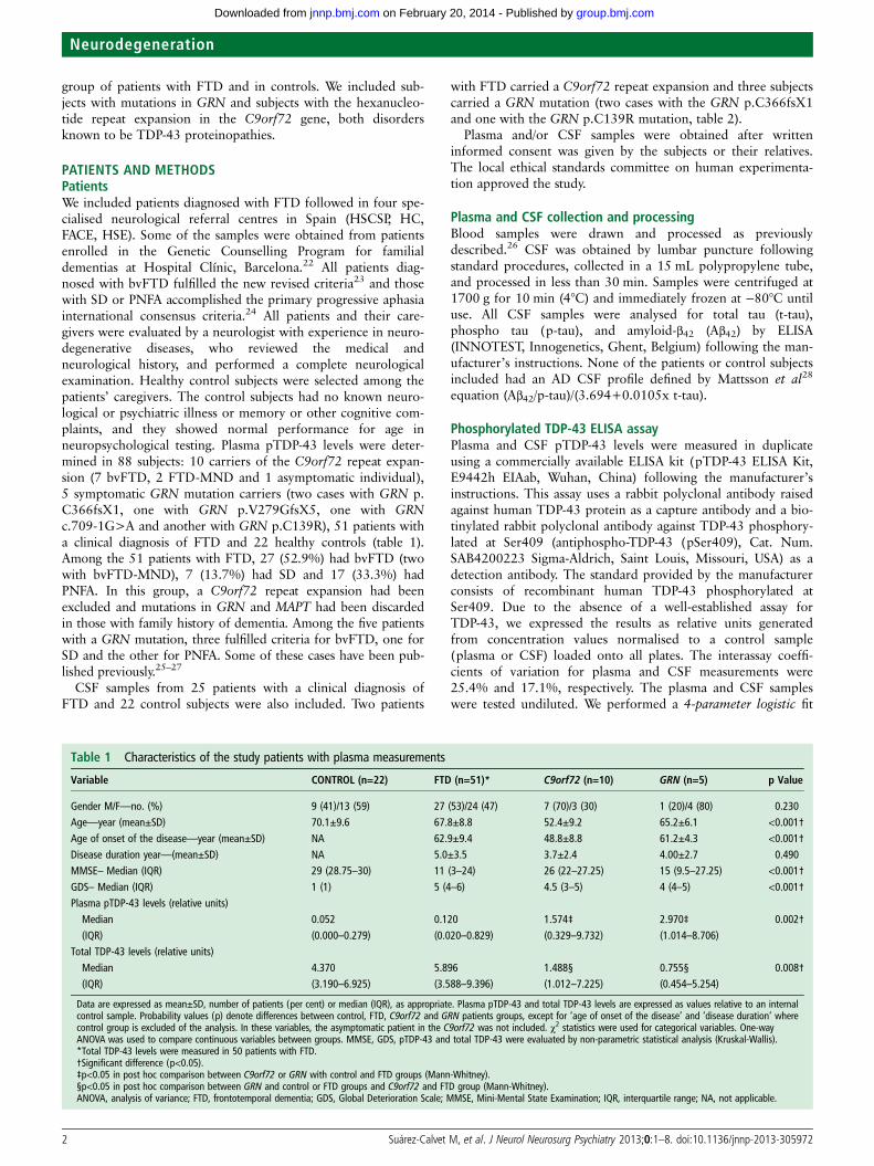
group of patients with FTD and in controls. We included sub-jects with mutations in GRN and subjects with the hexanucleo-tide repeat expansion in the C9orf72 gene, both disordersknown to be TDP-43 proteinopathies.
PATIENTS AND METHODSPatientsWe included patients diagnosed with FTD followed in four spe-cialised neurological referral centres in Spain (HSCSP, HC,FACE, HSE). Some of the samples were obtained from patientsenrolled in the Genetic Counselling Program for familialdementias at Hospital Clínic, Barcelona.22 All patients diag-nosed with bvFTD fulfilled the new revised criteria23 and thosewith SD or PNFA accomplished the primary progressive aphasiainternational consensus criteria.24 All patients and their care-givers were evaluated by a neurologist with experience in neuro-degenerative diseases, who reviewed the medical andneurological history, and performed a complete neurologicalexamination. Healthy control subjects were selected among thepatients’ caregivers. The control subjects had no known neuro-logical or psychiatric illness or memory or other cognitive com-plaints, and they showed normal performance for age inneuropsychological testing. Plasma pTDP-43 levels were deter-mined in 88 subjects: 10 carriers of the C9orf72 repeat expan-sion (7 bvFTD, 2 FTD-MND and 1 asymptomatic individual),5 symptomatic GRN mutation carriers (two cases with GRN p.C366fsX1, one with GRN p.V279GfsX5, one with GRNc.709-1G>A and another with GRN p.C139R), 51 patients witha clinical diagnosis of FTD and 22 healthy controls (table 1).Among the 51 patients with FTD, 27 (52.9%) had bvFTD (twowith bvFTD-MND), 7 (13.7%) had SD and 17 (33.3%) hadPNFA. In this group, a C9orf72 repeat expansion had beenexcluded and mutations in GRN and MAPT had been discardedin those with family history of dementia. Among the five patientswith a GRN mutation, three fulfilled criteria for bvFTD, one forSD and the other for PNFA. Some of these cases have been pub-lished previously.25–27
CSF samples from 25 patients with a clinical diagnosis ofFTD and 22 control subjects were also included. Two patients
with FTD carried a C9orf72 repeat expansion and three subjectscarried a GRN mutation (two cases with the GRN p.C366fsX1and one with the GRN p.C139R mutation, table 2).
Plasma and/or CSF samples were obtained after writteninformed consent was given by the subjects or their relatives.The local ethical standards committee on human experimenta-tion approved the study.
Plasma and CSF collection and processingBlood samples were drawn and processed as previouslydescribed.26 CSF was obtained by lumbar puncture followingstandard procedures, collected in a 15 mL polypropylene tube,and processed in less than 30 min. Samples were centrifuged at1700 g for 10 min (4°C) and immediately frozen at −80°C untiluse. All CSF samples were analysed for total tau (t-tau),phospho tau (p-tau), and amyloid-β42 (Aβ42) by ELISA(INNOTEST, Innogenetics, Ghent, Belgium) following the man-ufacturer’s instructions. None of the patients or control subjectsincluded had an AD CSF profile defined by Mattsson et al28
equation (Aβ42/p-tau)/(3.694+0.0105x t-tau).
Phosphorylated TDP-43 ELISA assayPlasma and CSF pTDP-43 levels were measured in duplicateusing a commercially available ELISA kit (pTDP-43 ELISA Kit,E9442h EIAab, Wuhan, China) following the manufacturer’sinstructions. This assay uses a rabbit polyclonal antibody raisedagainst human TDP-43 protein as a capture antibody and a bio-tinylated rabbit polyclonal antibody against TDP-43 phosphory-lated at Ser409 (antiphospho-TDP-43 (pSer409), Cat. Num.SAB4200223 Sigma-Aldrich, Saint Louis, Missouri, USA) as adetection antibody. The standard provided by the manufacturerconsists of recombinant human TDP-43 phosphorylated atSer409. Due to the absence of a well-established assay forTDP-43, we expressed the results as relative units generatedfrom concentration values normalised to a control sample(plasma or CSF) loaded onto all plates. The interassay coeffi-cients of variation for plasma and CSF measurements were25.4% and 17.1%, respectively. The plasma and CSF sampleswere tested undiluted. We performed a 4-parameter logistic fit
Table 1 Characteristics of the study patients with plasma measurements
Variable CONTROL (n=22) FTD (n=51)* C9orf72 (n=10) GRN (n=5) p Value
Gender M/F—no. (%) 9 (41)/13 (59) 27 (53)/24 (47) 7 (70)/3 (30) 1 (20)/4 (80) 0.230Age—year (mean±SD) 70.1±9.6 67.8±8.8 52.4±9.2 65.2±6.1 <0.001†Age of onset of the disease—year (mean±SD) NA 62.9±9.4 48.8±8.8 61.2±4.3 <0.001†Disease duration year—(mean±SD) NA 5.0±3.5 3.7±2.4 4.00±2.7 0.490MMSE– Median (IQR) 29 (28.75–30) 11 (3–24) 26 (22–27.25) 15 (9.5–27.25) <0.001†GDS– Median (IQR) 1 (1) 5 (4–6) 4.5 (3–5) 4 (4–5) <0.001†Plasma pTDP-43 levels (relative units)
Median 0.052 0.120 1.574‡ 2.970‡ 0.002†(IQR) (0.000–0.279) (0.020–0.829) (0.329–9.732) (1.014–8.706)
Total TDP-43 levels (relative units)Median 4.370 5.896 1.488§ 0.755§ 0.008†(IQR) (3.190–6.925) (3.588–9.396) (1.012–7.225) (0.454–5.254)
Data are expressed as mean±SD, number of patients (per cent) or median (IQR), as appropriate. Plasma pTDP-43 and total TDP-43 levels are expressed as values relative to an internalcontrol sample. Probability values (p) denote differences between control, FTD, C9orf72 and GRN patients groups, except for ‘age of onset of the disease’ and ‘disease duration’ wherecontrol group is excluded of the analysis. In these variables, the asymptomatic patient in the C9orf72 was not included. χ2 statistics were used for categorical variables. One-wayANOVA was used to compare continuous variables between groups. MMSE, GDS, pTDP-43 and total TDP-43 were evaluated by non-parametric statistical analysis (Kruskal-Wallis).*Total TDP-43 levels were measured in 50 patients with FTD.†Significant difference (p<0.05).‡p<0.05 in post hoc comparison between C9orf72 or GRN with control and FTD groups (Mann-Whitney).§p<0.05 in post hoc comparison between GRN and control or FTD groups and C9orf72 and FTD group (Mann-Whitney).ANOVA, analysis of variance; FTD, frontotemporal dementia; GDS, Global Deterioration Scale; MMSE, Mini-Mental State Examination; IQR, interquartile range; NA, not applicable.
2 Suárez-Calvet M, et al. J Neurol Neurosurg Psychiatry 2013;0:1–8. doi:10.1136/jnnp-2013-305972
Neurodegeneration
group.bmj.com on February 20, 2014 - Published by jnnp.bmj.comDownloaded from
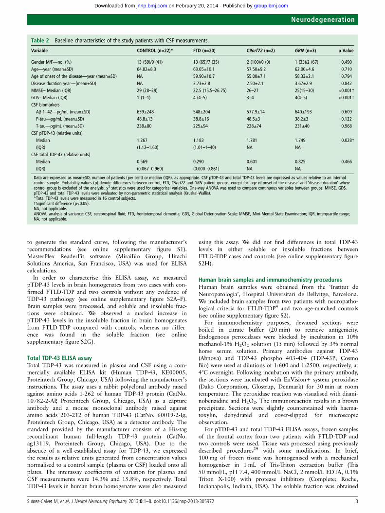
to generate the standard curve, following the manufacturer’srecommendations (see online supplementary figure S1).MasterPlex ReaderFit software (MiraiBio Group, HitachiSolutions America, San Francisco, USA) was used for ELISAcalculations.
In order to characterise this ELISA assay, we measuredpTDP-43 levels in brain homogenates from two cases with con-firmed FTLD-TDP and two controls without any evidence ofTDP-43 pathology (see online supplementary figure S2A–F).Brain samples were processed, and soluble and insoluble frac-tions were obtained. We observed a marked increase inpTDP-43 levels in the insoluble fraction in brain homogenatesfrom FTLD-TDP compared with controls, whereas no differ-ence was found in the soluble fraction (see onlinesupplementary figure S2G).
Total TDP-43 ELISA assayTotal TDP-43 was measured in plasma and CSF using a com-mercially available ELISA kit (Human TDP-43, KE00005,Proteintech Group, Chicago, USA) following the manufacturer’sinstructions. The assay uses a rabbit polyclonal antibody raisedagainst amino acids 1-262 of human TDP-43 protein (CatNo.10782-2-AP, Proteintech Group, Chicago, USA) as a captureantibody and a mouse monoclonal antibody raised againstamino acids 203-212 of human TDP-43 (CatNo. 60019-2-Ig,Proteintech Group, Chicago, USA) as a detector antibody. Thestandard provided by the manufacturer consists of a His-tagrecombinant human full-length TDP-43 protein (CatNo.ag13119, Proteintech Group, Chicago, USA). Due to theabsence of a well-established assay for TDP-43, we expressedthe results as relative units generated from concentration valuesnormalised to a control sample (plasma or CSF) loaded onto allplates. The interassay coefficients of variation for plasma andCSF measurements were 14.3% and 15.8%, respectively. TotalTDP-43 levels in human brain homogenates were also measured
using this assay. We did not find differences in total TDP-43levels in either soluble or insoluble fractions betweenFTLD-TDP cases and controls (see online supplementary figureS2H).
Human brain samples and immunochemistry proceduresHuman brain samples were obtained from the ‘Institut deNeuropatologia’, Hospital Universitari de Bellvitge, Barcelona.We included brain samples from two patients with neuropatho-logical criteria for FTLD-TDP4 and two age-matched controls(see online supplementary figure S2).
For immunochemistry purposes, dewaxed sections wereboiled in citrate buffer (20 min) to retrieve antigenicity.Endogenous peroxidases were blocked by incubation in 10%methanol-1% H2O2 solution (15 min) followed by 3% normalhorse serum solution. Primary antibodies against TDP-43(Abnova) and TDP-43 phospho 403-404 (TDP-43P; CosmoBio) were used at dilutions of 1:600 and 1:2500, respectively, at4°C overnight. Following incubation with the primary antibody,the sections were incubated with EnVision+ system peroxidase(Dako Corporation, Glostrup, Denmark) for 30 min at roomtemperature. The peroxidase reaction was visualised with diami-nobenzidine and H2O2. The immunoreaction results in a brownprecipitate. Sections were slightly counterstained with haema-toxylin, dehydrated and cover-slipped for microscopicobservation.
For pTDP-43 and total TDP-43 ELISA assays, frozen samplesof the frontal cortex from two patients with FTLD-TDP andtwo controls were used. Tissue was processed using previouslydescribed procedures29 with some modifications. In brief,100 mg of frozen tissue was homogenised with a mechanicalhomogeniser in 1 mL of Tris-Triton extraction buffer (Tris50 mmol/L, pH 7.4, 400 mmol/L NaCl, 2 mmol/L EDTA, 0.1%Triton X-100) with protease inhibitors (Complete; Roche,Indianapolis, Indiana, USA). The soluble fraction was obtained
Table 2 Baseline characteristics of the study patients with CSF measurements.
Variable CONTROL (n=22)* FTD (n=20) C9orf72 (n=2) GRN (n=3) p Value
Gender M/F—no. (%) 13 (59)/9 (41) 13 (65)/7 (35) 2 (100)/0 (0) 1 (33)/2 (67) 0.490Age—year (mean±SD) 64.82±8.3 63.65±10.1 57.50±9.2 62.00±4.6 0.710Age of onset of the disease—year (mean±SD) NA 59.90±10.7 55.00±7.1 58.33±2.1 0.794Disease duration year—(mean±SD) NA 3.73±2.8 2.50±2.1 3.67±2.9 0.842MMSE– Median (IQR) 29 (28–29) 22.5 (15.5–26.75) 26–27 25(15–30) <0.001†GDS– Median (IQR) 1 (1–1) 4 (4–5) 3–4 4(4–5) <0.001†CSF biomarkers
Aβ 1–42—pg/mL (mean±SD) 639±248 548±204 577.9±14 640±193 0.609P-tau—pg/mL (mean±SD) 48.8±13 38.8±16 48.5±3 38.2±3 0.122T-tau—pg/mL (mean±SD) 238±80 225±94 228±74 231±40 0.968
CSF pTDP-43 (relative units)Median 1.267 1.183 1.781 1.749 0.028†(IQR) (1.12–1.60) (1.01–1–40) NA NA
CSF total TDP-43 (relative units)Median 0.569 0.290 0.601 0.825 0.466
(IQR) (0.067–0.960) (0.000–0.861) NA NA
Data are expressed as mean±SD, number of patients (per cent) or median (IQR), as appropriate. CSF pTDP-43 and total TDP-43 levels are expressed as values relative to an internalcontrol sample. Probability values (p) denote differences between control, FTD, C9orf72 and GRN patient groups, except for ‘age of onset of the disease’ and ‘disease duration’ wherecontrol group is excluded of the analysis. χ2 statistics were used for categorical variables. One-way ANOVA was used to compare continuous variables between groups. MMSE, GDS,pTDP-43 and total TDP-43 levels were evaluated by non-parametric statistical analysis (Kruskal-Wallis).*Total TDP-43 levels were measured in 16 control subjects.†Significant difference (p<0.05).NA, not applicable.ANOVA, analysis of variance; CSF, cerebrospinal fluid; FTD, frontotemporal dementia; GDS, Global Deterioration Scale; MMSE, Mini-Mental State Examination; IQR, interquartile range;NA, not applicable.
Suárez-Calvet M, et al. J Neurol Neurosurg Psychiatry 2013;0:1–8. doi:10.1136/jnnp-2013-305972 3
Neurodegeneration
group.bmj.com on February 20, 2014 - Published by jnnp.bmj.comDownloaded from

after centrifugation at 14 000 rpm for 5 min at 4°C. The result-ing pellet was resuspended in 70% formic acid and then recen-trifuged at 22 000 rpm for 5 min at 4°C. The supernatant wasthen neutralised with 1 mol/L Tris buffer (pH 11), and used forthe ELISA assays. Protein concentration in these samples wasdetermined using the Bradford assay. Soluble fractions werediluted at 1:1000 in the pTDP-43 ELISA assay and at 1:500 inthe total TDP-43 ELISA assay. Insoluble fractions were dilutedat 1:100 in both assays.
DNA extraction, genetic analysis and Southern blottingDNA was extracted from whole blood using previouslydescribed standard protocols. All patients had been previouslyinvestigated for the presence of the GGGGCC hexanucleotiderepeat expansion in C9orf72 gene, and those with a positivefamily history of dementia were screened for mutations in GRNand MAPT genes, as previously described.26
A non-radioactive Southern blotting protocol was used to esti-mate the hexanucleotide repeat expansion size.30 Briefly, a totalamount of 20 mg of blood-derived DNA was digested with XbaIand electrophoresed on a 0.8% agarose gel. DNA was trans-ferred to a positively charged nylon membrane (Roche AppliedScience) by capillary blotting. Hybridisation was carried out at45°C overnight using a 954 base pairs probe labelled withdigoxigenin. Ready-to-use CDP-Star (Roche Applied Science)was used as the chemiluminescent substrate through the trans-parency technique and signals were visualised on a KODAKImage Station 4000MM PRO (Carestream Health, Rochester,New York, USA). The median number of repeats was used forstatistical analysis.
Statistical analysisThe χ2 test was used to compare differences in categorical vari-ables and one-way analysis of variance (ANOVA) was used tocompare age and age at onset between groups. Mini-MentalState Examination (MMSE), Global Deterioration Scale (GDS),pTDP-43 and total TDP-43 levels were evaluated by non-parametric statistical analysis (Kruskal-Wallis and Mann-Whitneypost hoc test). Correlation analysis of plasma and CSF pTDP-43and total TDP-43 levels, age, age of onset, disease duration,MMSE, GDS and median C9orf72 GGGGCC hexanucleotiderepeat number were performed using the Spearman’s r test.Statistical significance for all the analyses was set at 5%(α=0.05). All data were analysed using Statistical Package forthe Social Sciences (SPSS, Chicago, USA).
RESULTSPlasma pTDP-43 and total TDP-43 levels in patientswith FTD and controlsTable 1 summarises demographic and clinical data of all patientswith plasma sample included in this study.
Family history of dementia was present in 39.2% of patientsin the FTD group, and in 100% of subjects with the C9orf72expansion and GRN mutations. Patients carrying the C9orf72expansion were significantly younger than the other patients(p<0.001, one-way ANOVA). Their age of onset of the diseasewas also earlier than that of patients with FTD without muta-tions (p<0.001, one-way ANOVA). The three groups (C9orf72,GRN and FTD) did not differ in disease duration or GDS score,but patients with FTD had a lower MMSE, suggesting a moreadvanced stage of the disease (table 1).
We next investigated whether pTDP-43 plasma levels wereinfluenced by demographic variables. In the entire cohort(n=88), plasma pTDP-43 levels were not influenced by gender
(p=0.983, Mann-Whitney) but they correlated inversely withage (Spearman’s r=−0.318, p=0.003). In symptomatic subjects(we excluded healthy controls and the C9orf72 asymptomaticpatient, n=65), plasma pTDP-43 levels were not influenced bydisease duration (p=0.500, Spearman), MMSE (p=0.070,Spearman) or GDS (p=0.242, Spearman) scores, but they corre-lated inversely with age of disease onset (Spearman’s r=−0.37,p=0.002). No differences in plasma pTDP-43 levels wereobserved between the three syndromes of FTD (bvFTD, SD,PNFA). When we analysed plasma pTDP-43 levels according togenetic status, we found that levels differed between groups,although there was considerable variability and overlap (p=0.002,Kruskal-Wallis). Subjects carrying a C9orf72 repeat expansion(n=10) or GRN mutations (n=5) had significantly increasedlevels of plasma pTDP-43 compared with subjects with FTDwithout a mutation (n=51) and with controls (n=22) (p<0.05,Mann-Whitney, figure 1A, table 1). An ANOVA adjusted for ageand age of onset confirmed the statistically significant differencein pTDP-43 levels between patients with the mutation and thosewithout the mutation. We also calculated the upper 99% CI ofthe control group and found that 6 out of 10 (60%) subjects inthe C9orf72 group and 4 out of 5 (80%) subjects in the GRNgroup were above this value compared with 8 out of 51 (15.7%)in the FTD group (figure 1A). The levels of pTDP-43 in theC9orf72 and the GRN groups did not correlate with age, age ofonset, disease duration, FTD subtype or MMSE score. Noticeably,the only asymptomatic subject carrying the C9orf72 expansionhad the lowest plasma pTDP-43 levels (figure 1A).
Next, we measured total TDP-43 levels in plasma in thiscohort (except 1 patient with FTD for whom no sample wasavailable; n=87). In the entire group, plasma total TDP-43levels were not influenced by gender (p=0.610,Mann-Whitney) but they showed a trend towards a positivecorrelation with age (Spearman’s r=+0.21, p=0.051). Insymptomatic subjects (n=64), plasma total TDP-43 levels werenot influenced by disease duration (p=0.119, Spearman),MMSE (p=0.391, Spearman) or GDS (p=0.966, Spearman)scores, but they correlated positively with age of disease onset(Spearman’s r=+0.26, p=0.037). No differences in plasmatotal TDP-43 levels were observed between the three syn-dromes of FTD (bvFTD, SD, PNFA). When we analysedplasma total TDP-43 levels according to genetic status, wefound that, unlike pTDP-43, total TDP-43 levels were slightlydecreased in the C9orf72 (n=10) and the GRN groups (n=5)compared with the FTD group (n=50, p=0.016 and p=0.015,respectively, Mann-Whitney, figure 1B, table 1). Subjects withGRN mutations also showed decreased levels of plasma totalTDP-43 levels compared with controls (p=0.046,Mann-Whitney). Plasma pTDP-43 levels correlated inverselywith plasma total TDP-43 levels in the entire group(Spearman’s r=−0.23, p=0.030).
We next tested whether plasma pTDP-43 or total TDP-43levels were associated with the number of GGGGCC repeats insubjects with the C9orf72 expansion. To address this issue, weapplied a non-radioactive Southern blot technique in blood-derived DNA samples from the 10 subjects carrying theC9orf72 expansion.30 We did not find any correlation betweenplasma pTDP-43 or total TDP-43 levels and the median numberof repeats (see online supplementary figure S3).
CSF pTDP-43 and total TDP-43 levels in patientswith FTD and controlsWe subsequently investigated the differences in pTDP-43 andtotal TDP-43 levels in CSF. Table 2 shows demographic and
4 Suárez-Calvet M, et al. J Neurol Neurosurg Psychiatry 2013;0:1–8. doi:10.1136/jnnp-2013-305972
Neurodegeneration
group.bmj.com on February 20, 2014 - Published by jnnp.bmj.comDownloaded from
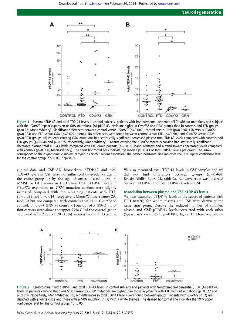
clinical data and CSF AD biomarkers. pTDP-43 and totalTDP-43 levels in CSF were not influenced by gender or age inthe entire group or by the age of onset, disease duration,MMSE or GDS scores in FTD cases. CSF pTDP-43 levels inC9orf72 expansion or GRN mutation carriers were slightlyincreased compared with the remaining patients with FTD(p=0.022 and p=0.014, respectively, Mann-Whitney; figure 2A,table 2) but not compared with controls (p=0.144 C9orf72 vscontrol; p=0.094 GRN vs control). Four out of 5 (80%) muta-tion carriers were above the upper 99% CI of the control groupcompared with 2 out of 20 (10%) subjects in the FTD group.
We also measured total TDP-43 levels in CSF samples and wedid not find differences between groups (p=0.466,Kruskal-Wallis, figure 2B, table 2). No correlation was observedbetween pTDP-43 and total TDP-43 levels in CSF.
Association between plasma and CSF pTDP-43 levelsWe next examined pTDP-43 levels in the subset of patients withFTD (n=20) for whom plasma and CSF were drawn at thesame time point. Despite the reduced number of samples,plasma and CSF pTDP-43 levels correlated with each other(Spearman’s r=+0.673, p=0.001, figure 3). However, plasma
Figure 2 Cerebrospinal fluid pTDP-43 and total TDP-43 levels in control subjects and patients with frontotemporal dementia (FTD). (A) pTDP-43levels in patients carrying the C9orf72 expansion or GRN mutations are higher than those in patients with FTD without mutations (p=0.022 andp=0.014, respectively, Mann-Whitney). (B) No differences in total TDP-43 levels were found between groups. Patients with C9orf72 (n=2) aredepicted with a white circle and those with a GRN mutation (n=3) with a white triangle. The dashed horizontal line indicates the 99% upperconfidence level for the control group. *p<0.05.
Figure 1 Plasma pTDP-43 and total TDP-43 levels in control subjects, patients with frontotemporal dementia (FTD) without mutations and subjectswith the C9orf72 repeat expansion or GRN mutations. (A) pTDP-43 levels are higher in C9orf72 and GRN groups than in controls and FTD groups(p<0.05, Mann-Whitney). Significant differences between control versus C9orf72 (p=0.002), control versus GRN (p=0.026), FTD versus C9orf72(p=0.004) and FTD versus GRN (p=0.022) groups. No differences were found between control versus FTD (p=0.204) and C9orf72 versus GRN(p=0.903) groups. (B) Patients carrying GRN mutations had statistically significant decreased plasma total TDP-43 levels compared with controls andFTD groups (p=0.046 and p=0.015, respectively, Mann-Whitney). Patients carrying the C9orf72 repeat expansion had statistically significantdecreased plasma total TDP-43 levels compared with FTD group patients (p=0.016, Mann-Whitney) and a trend towards decreased levels comparedwith controls (p=0.096, Mann-Whitney). The short horizontal bars indicate the median pTDP-43 or total TDP-43 levels per group. The arrowcorresponds to the asymptomatic subject carrying a C9orf72 repeat expansion. The dashed horizontal line indicates the 99% upper confidence levelfor the control group. *p<0.05; **p<0.01.
Suárez-Calvet M, et al. J Neurol Neurosurg Psychiatry 2013;0:1–8. doi:10.1136/jnnp-2013-305972 5
Neurodegeneration
group.bmj.com on February 20, 2014 - Published by jnnp.bmj.comDownloaded from

and CSF total TDP-43 levels did not correlate with each other(Spearman’s r=+0.096, p=0.688, n=20).
DISCUSSIONThe main finding in this study is that patients with FTD withthe C9orf72 repeat expansion or GRN mutations had higherplasma levels of pTDP-43 than patients with FTD withoutmutations or healthy controls. Conversely, subjects with theC9orf72 repeat expansion or GRN mutations had lower plasmalevels of total TDP-43 than patients with FTD without muta-tions. CSF pTDP-43 levels were also higher in patients withFTD with mutations than in those without mutations. Anotherrelevant finding in our study is that plasma and CSF pTDP-43levels correlated positively with each other, although plasmaand CSF total TDP-43 did not.
The discovery of a reliable biomarker to detect FTLD-TDP isan essential step to perform an accurate diagnosis and ultimatelyto develop therapeutic strategies aimed at altering the naturalcourse of the disease. This is particularly important in FTLDsince this condition is known to be associated with several under-lying pathologies that can present with the same clinical syn-drome.31 The search for biomarkers in neurodegenerativediseases over recent decades has yielded some positive results.The measurement of plasma and serum progranulin levels inFTD has proved to be a reliable biomarker to identify symptom-atic and asymptomatic carriers of GRN mutations.32–34 However,we have recently shown that plasma progranulin levels are notinfluenced by the C9orf72 repeat expansion.26 Progranulin prob-ably represents the most reliable plasma marker in neurodegen-erative diseases, but other promising biomarkers are beinginvestigated. CSF analysis has shown to be useful in the diagnosisof AD. The pattern of reduced Aβ42 and increased t-tau andp-tau levels accurately predicts AD pathology.28 35 CSF examin-ation can also be used to differentiate AD from FTLD withacceptable specificity.36–38 However, in FTD, current CSF bio-markers are more useful to exclude AD than to confirm thepathophysiological process characteristic of FTLD. In our study,
we only included cases without an AD CSF profile that couldhave biased our sample. Some other CSF biomarkers that havebeen tested in FTLD yielded variable results.17 There is, there-fore, a great need to develop markers to diagnose FTLD, detectthe underlying pathology in vivo, and eventually better guide thefuture application of specific disease-modifying therapies.
TDP-43 is a 414-aminoacid protein that is usually located in thecell nucleus. It belongs to the heterogeneous ribonucleoproteinfamily and it presumably participates in several cell functions suchas DNA transcription, mRNA splicing, mRNA export/import ormiRNA synthesis and regulation.39 TDP-43 inclusions in the brainand/or spinal cord in FTLD consist of abnormally aggregated, ubi-quitinated and hyperphosphorylated TDP-43.13 14 In particular,the importance of phosphorylation of the serine residues S409/410 of TDP-43 has been highlighted as a consistent feature inpathological inclusions of all TDP-43 proteinopathies.40
Previous studies have measured plasma TDP-43 and pTDP-43in FTD.18 19 They mainly found that levels of TDP-43 (totaland pTDP-43) in plasma are elevated in subjects withFTLD-TDP. More importantly, they demonstrated a good correl-ation between plasma pTDP-43 levels and brain deposition ofpTDP-43.19 These results suggest that plasma pTDP43 levelscould represent a good in vivo biomarker of TDP-43 pathology.
In this study, we found that subjects carrying a repeat expansionin C9orf72 or a GRN mutation had significantly higher plasmapTDP-43 levels than those from control subjects and the remainingpatients with FTD. In contrast, total TDP-43 levels in plasma weredecreased in subjects carrying a repeat expansion in C9orf72 or aGRN mutation. Three different hypotheses can be postulated toexplain these results. First, since C9orf72 expansion and GRNmutation carriers are known to be FTLD-TDP and plasmapTDP-43 correlates with brain pTDP-43 deposition,19 higherlevels of plasma pTDP-43 may consequently reflect an increase inbrain pTDP-43 burden compared with the remaining FTD cases(in whom other underlying pathologies such us FTLD-tau orFTLD-FUS may be present). Second, as we found a correlationbetween age and plasma TDP-43 levels, higher levels of pTDP-43in younger subjects may be the result of age rather than mutationstatus or brain pathology. This seems unlikely, however, since ourresults remained significant after adjusting for age. Finally, theC9orf72 expansion and GRN mutations could elevate directly orindirectly pTDP-43 levels regardless of the burden of brain depos-ition of pTDP-43. Our data favour this last hypothesis and suggestthat phosphorylated and non-phosphorylated forms of TDP-43are in equilibrium as they correlate inversely in plasma. It could bespeculated that the C9orf72 expansion or mutations in GRN alterthe balance between phosphorylated and non-phosphorylatedforms of TDP, promoting the former one. This imbalance wouldconsequently explain the increase of plasma pTDP-43 togetherwith the decrease in plasma total TDP-43 levels found in subjectswith the C9orf72 expansion or mutations in GRN. Further studiesare needed to explain the lack of correlation between pTDP-43and total TDP-43 levels in the CSF and to explore why levels ofCSF total TDP-43 in subjects with the C9orf72 expansion ormutations inGRN are similar to those in other groups.
Our results in patients with FTD without mutations differfrom previous studies that have examined TDP-43 in patientswith FTD. Two studies in particular have shown an increase intotal TDP-43 levels in plasma and CSF in FTD cases comparedwith controls.18 21 Although in our study the levels of totalTDP-43 in plasma were slightly increased in FTD comparedwith controls, none of the comparisons were significant.Differences in the assays used, sample size and study design mayexplain the discrepancies among findings.
Figure 3 Correlation between plasma and cerebrospinal fluid (CSF)pTDP-43 levels. The y-axis represents plasma pTDP-43 and the x-axisrepresents CSF pTDP-43. A positive correlation between plasma andCSF levels was detected (Spearman’s r=+0.673, p=0.001). Plasma andCSF samples of each patient were obtained at the same time.
6 Suárez-Calvet M, et al. J Neurol Neurosurg Psychiatry 2013;0:1–8. doi:10.1136/jnnp-2013-305972
Neurodegeneration
group.bmj.com on February 20, 2014 - Published by jnnp.bmj.comDownloaded from
Our study also addresses, for the first time, the relationshipbetween levels of pTDP43 and total TDP-43 in plasma and CSFin FTD cases and controls. The values obtained in CSF were farmore homogenous that those in plasma and we did not find dif-ferences in pTDP-43 or total TDP-43 levels between FTD casesand controls. The only significant finding was an increase inpTDP-43 levels in subjects with C9orf72 or GRN mutationscompared with the remaining patients with FTD. These resultsshould be interpreted with caution due to the low number ofpatients with the C9orf72 expansion (n=2) and GRN mutations(n=3) and the fact that most values in the whole FTD groupwere below the upper 99% CI limit of the control group. Ofinterest, CSF and plasma pTDP-43, but not total TDP-43, corre-lated in patients for whom both samples were available (n=20).This finding would suggest that plasma and CSF pTDP-43 levelsare in equilibrium and would support the role of plasma as areliable source of biochemical signatures. However, the lack ofcorrelation between total TDP-43 in plasma and CSF remainsunclear and deserves further investigation.
The strengths of this study are the inclusion of a well-characterised patient sample of mutation carriers, the use of CSFbiomarkers to exclude AD, and the simultaneous measure ofplasma and CSF in some patients. Nevertheless, this study hassome limitations. First, the reduced sample size may have limitedthe power to detect differences, especially regarding CSF measure-ments. Second, the lack of neuropathological confirmation mayhave biased our results. Finally, the ELISA assays used show largeinterassay variability, particularly when using plasma samples. Thishigh variability and the overlap among groups in plasma pTDP-43levels may preclude the application of this assay in clinical settings.Conversely, CSF values were more homogenous and the interassayvariability was lower. We acknowledge that these results are pre-liminary and should be confirmed in larger samples, preferablywith pathologically or genetically confirmed cases, and with theuse of other assays. To further determine whether pTDP-43 can beused as a reliable plasma biomarker for FTLD-TDP, patients withthe C9orf72 expansion or GRN mutations should be comparedwith patients with mutations inMAPT, known to cause FTLD-tau.
In conclusion, our results suggest that plasma pTDP-43 levelsare increased in patients carrying the C9orf72 repeat expansionor GRN mutations, conditions associated with TDP-43 protei-nopathies. If confirmed, pTDP-43 levels may represent a reliableplasma biomarker for FTLD-TDP.
Author affiliations1Neurology Department, Hospital de la Santa Creu i Sant Pau, Universitat Autònomade Barcelona, Barcelona, Spain2Center for Networker Biomedical Research in Neurodegenerative Diseases(CIBERNED), Madrid, Spain3Department of Neurology, Alzheimer’s Disease and other Cognitive Disorders Unit,Hospital Clínic, Barcelona, Spain4Memory Clinic of Fundació ACE, Institut Català de Neurociències Aplicades,Barcelona, Spain5Neurology Department, Hospital Universitari Son Espases, Majorca, Spain6Institut de Neuropatología, Servei Anatomia Patológica, IDIBELL, HospitalUniversitari de Bellvitge, University of Barcelona, Barcelona, Spain7Department of Molecular Genetics, Neurodegenerative Brain Disease Group, VIB,Antwerp, Belgium8Laboratory of Neurogenetics, Institute Born-Bunge, University of Antwerp, Antwerp,Belgium
Acknowledgements The authors thank all the patients and their families for theirparticipation. They thank Carolyn Newey for editorial help, and Laia Muñoz and InésMatas for technical assistance.
Contributors All authors are justifiably credited with authorship, according to theauthorship criteria. In detail: MS-C—conception, design, analysis and interpretationof data, drafting of the manuscript, critical revision of manuscript, final approval
given; OD-I—analysis and interpretation of data, critical revision of manuscript, finalapproval given; AL—analysis and interpretation of data, critical revision ofmanuscript, final approval given; RS-V—analysis and interpretation of data, criticalrevision of manuscript, final approval given; IH—analysis and interpretation of data,critical revision of manuscript, final approval given; GA—analysis and interpretationof data, critical revision of manuscript, final approval given; SA-A—analysis andinterpretation of data, critical revision of manuscript, final approval given; DA—analysis and interpretation of data, critical revision of manuscript, final approvalgiven; JF—analysis and interpretation of data, critical revision of manuscript, finalapproval given; IF—critical revision of manuscript, final approval given; JvdZ—analysis and interpretation of data, critical revision of manuscript, final approvalgiven; LD—analysis and interpretation of data, critical revision of manuscript, finalapproval given; CVB—critical revision of manuscript, final approval given; JLM—critical revision of manuscript, final approval given; RB—critical revision ofmanuscript, final approval given; JC—design, analysis and interpretation of data,critical revision of manuscript, final approval given; AL—conception, design, analysisand interpretation of data, drafting of the manuscript, final approval given.
Funding This work was supported by a research grant from ‘Centro deInvestigación en Red en Enfermedades Neurodegenerativas’ and by the ‘Fondo deInvestigaciones Sanitarias’ (PI09/00098). At the Antwerp site, the genetic screeningof C9orf72 for the repeat expansion was funded in part by the InteruniversityAttraction Poles program of the Belgian Science Policy Office; the MethusalemExcellence program of the Flemish Government; the Medical Foundation QueenElisabeth, the Research Foundation Flanders (FWO), Belgium The FWO provided apostdoctoral fellowship to JvdZ.
Competing interests None.
Ethics approval The ‘Hospital de la Santa Creu i Sant Pau’ ethical standardscommittee on human experimentation approved the study. Barcelona, Spain (code:5/2010).
Provenance and peer review Not commissioned; externally peer reviewed.
REFERENCES1 Kumar-Singh S, Van Broeckhoven C. Frontotemporal lobar degeneration: current
concepts in the light of recent advances. Brain Pathol 2007;17:104–14.2 Neary D, Snowden JS, Gustafson L, et al. Frontotemporal lobar degeneration: a
consensus on clinical diagnostic criteria. Neurology 1998;51:1546–54.3 Van Langenhove T, Van der Zee J, Van Broeckhoven C, et al. The molecular basis of
the frontotemporal lobar degeneration-amyotrophic lateral sclerosis spectrum. AnnMed 2012;44:817–28.
4 Mackenzie IR, Neumann M, Bigio EH, et al. Nomenclature and nosology forneuropathologic subtypes of frontotemporal lobar degeneration: an update. ActaNeuropathol 2010;119:1–4.
5 Stevens M, Van Duijn CM, Kamphorst W, et al. Familial aggregation infrontotemporal dementia. Neurology 1998;50:1541–5.
6 Baker M, Mackenzie IR, Pickering-Brown SM, et al. Mutations in progranulin causetau-negative frontotemporal dementia linked to chromosome 17. Nature 2006;442:916–9.
7 Cruts M, Gijselinck I, Van der Zee J, et al. Null mutations in progranulin causeubiquitin-positive frontotemporal dementia linked to chromosome 17q21. Nature2006;442:920–4.
8 Dumanchin C, Camuzat A, Campion D, et al. Segregation of a missense mutationin the microtubule-associated protein tau gene with familial frontotemporaldementia and parkinsonism. Hum Mol Genet 1998;7:1825–9.
9 DeJesus-Hernandez M, Mackenzie IR, Boeve BF, et al. Expanded GGGGCChexanucleotide repeat in noncoding region of C9ORF72 causes chromosome9p-linked FTD and ALS. Neuron 2011;72:245–56.
10 Gijselinck I, Van Langenhove T, Van der Zee J, et al. A C9orf72 promoter repeatexpansion in a Flanders-Belgian cohort with disorders of the frontotemporal lobardegeneration-amyotrophic lateral sclerosis spectrum: a gene identification study.Lancet Neurol 2012;11:54–65.
11 Renton AE, Majounie E, Waite A, et al. A hexanucleotide repeat expansion inC9ORF72 is the cause of chromosome 9p21-linked ALS-FTD. Neuron2011;72:257–68.
12 Van der Zee J, Gijselinck I, Dillen L, et al. A pan-European study of the C9orf72repeat associated with FTLD: geographic prevalence, genomic instability, andintermediate repeats. Hum Mutat 2013;34:363–73.
13 Arai T, Hasegawa M, Akiyama H, et al. TDP-43 is a component of ubiquitin-positivetau-negative inclusions in frontotemporal lobar degeneration and amyotrophiclateral sclerosis. Biochem Biophys Res Commun 2006;351:602–11.
14 Neumann M, Sampathu DM, Kwong LK, et al. Ubiquitinated TDP-43 infrontotemporal lobar degeneration and amyotrophic lateral sclerosis. Science2006;314:130–3.
15 Benajiba L, Le Ber I, Camuzat A, et al. TARDBP mutations in motoneuron diseasewith frontotemporal lobar degeneration. Ann Neurol 2009;65:470–3.
16 Sreedharan J, Blair IP, Tripathi VB, et al. TDP-43 mutations in familial and sporadicamyotrophic lateral sclerosis. Science 2008;319:1668–72.
Suárez-Calvet M, et al. J Neurol Neurosurg Psychiatry 2013;0:1–8. doi:10.1136/jnnp-2013-305972 7
Neurodegeneration
group.bmj.com on February 20, 2014 - Published by jnnp.bmj.comDownloaded from

17 Hu WT, Trojanowski JQ, Shaw LM. Biomarkers in frontotemporal lobardegenerations–progress and challenges. Prog Neurobiol 2011;95:636–48.
18 Foulds P, McAuley E, Gibbons L, et al. TDP-43 protein in plasma may index TDP-43brain pathology in Alzheimer’s disease and frontotemporal lobar degeneration. ActaNeuropathol 2008;116:141–6.
19 Foulds PG, Davidson Y, Mishra M, et al. Plasma phosphorylated-TDP-43 proteinlevels correlate with brain pathology in frontotemporal lobar degeneration. ActaNeuropathol 2009;118:647–58.
20 Kasai T, Tokuda T, Ishigami N, et al. Increased TDP-43 protein in cerebrospinal fluidof patients with amyotrophic lateral sclerosis. Acta Neuropathol 2009;117:55–62.
21 Steinacker P, Hendrich C, Sperfeld AD, et al. TDP-43 in cerebrospinal fluid ofpatients with frontotemporal lobar degeneration and amyotrophic lateral sclerosis.Arch Neurol 2008;65:1481–7.
22 Fortea J, Lladó A, Clarimón J, et al. PICOGEN: five years experience with a geneticcounselling program for dementia. Neurologia 2011;26:143–9.
23 Rascovsky K, Hodges JR, Knopman D, et al. Sensitivity of revised diagnosticcriteria for the behavioural variant of frontotemporal dementia. Brain2011;134:2456–77.
24 Gorno-Tempini ML, Hillis A, Weintraub S, et al. Classification of primary progressiveaphasia and its variants. Neurology 2011;76:1006–14.
25 Antonell A, Gil S, Sánchez-Valle R, et al. Serum progranulin levels in patients withfrontotemporal lobar degeneration and Alzheimer’s disease: detection of GRNmutations in a Spanish cohort. J Alzheimers Dis 2012;31:581–91.
26 Dols-Icardo O, Suárez-Calvet M, Hernández I, et al. Expansion mutation inC9ORF72 does not influence plasma progranulin levels in frontotemporal dementia.Neurobiol Aging 2012;33:1851.e17–19.
27 Costa S, Suárez-Calvet M, Antón S, et al. Comparison of 2 diagnostic criteria forthe behavioral variant of frontotemporal dementia. Am J Alzheimers Dis OtherDemen 2013;28:469–76.
28 Mattsson N, Zetterberg H, Hansson O, et al. CSF biomarkers and incipient Alzheimerdisease in patients with mild cognitive impairment. JAMA 2009;302:385–93.
29 Fukumoto H, Rosene DL, Moss MB, et al. Beta-secretase activity increases withaging in human, monkey, and mouse brain. Am J Pathol 2004;164:719–25.
30 Dols-Icardo O, García-Redondo A, Rojas-García R, et al. Characterization of therepeat expansion size in C9orf72 in amyotrophic lateral sclerosis and frontotemporaldementia. Hum Mol Genet Published Online First: 20 September 2013. doi:10.1093/hmg/ddt460
31 Josephs KA, Hodges JR, Snowden JS, et al. Neuropathological background ofphenotypical variability in frontotemporal dementia. Acta Neuropathol2011;122:137–53.
32 Finch N, Baker M, Crook R, et al. Plasma progranulin levels predict progranulinmutation status in frontotemporal dementia patients and asymptomatic familymembers. Brain 2009;132:583–91.
33 Ghidoni R, Benussi L, Glionna M, et al. Low plasma progranulin levels predictprogranulin mutations in frontotemporal lobar degeneration. Neurology2008;71:1235–9.
34 Sleegers K, Brouwers N, Van Damme P, et al. Serum biomarker for progranulin-associated frontotemporal lobar degeneration. Ann Neurol 2009;65:603–9.
35 Blennow K, Hampel H, Weiner M, et al. Cerebrospinal fluid and plasma biomarkersin Alzheimer disease. Nat Rev Neurol 2010;6:131–44.
36 Bian H, Van Swieten JC, Leight S, et al. CSF biomarkers in frontotemporal lobardegeneration with known pathology. Neurology 2008;70:1827–35.
37 Bibl M, Gallus M, Welge V, et al. Cerebrospinal fluid amyloid-β 2-42 is decreased inAlzheimer’s, but not in frontotemporal dementia. J Neural Transm 2012;119:805–13.
38 Kapaki E, Paraskevas GP, Papageorgiou SG, et al. Diagnostic value of CSFbiomarker profile in frontotemporal lobar degeneration. Alzheimer Dis Assoc Disord2008;22:47–53.
39 Buratti E, Baralle FE. The multiple roles of TDP-43 in pre-mRNA processing andgene expression regulation. RNA Biol 2010;7:420–9.
40 Neumann M, Kwong LK, Lee EB, et al. Phosphorylation of S409/410 of TDP-43 is aconsistent feature in all sporadic and familial forms of TDP-43 proteinopathies. ActaNeuropathol 2009;117:137–49.
8 Suárez-Calvet M, et al. J Neurol Neurosurg Psychiatry 2013;0:1–8. doi:10.1136/jnnp-2013-305972
Neurodegeneration
group.bmj.com on February 20, 2014 - Published by jnnp.bmj.comDownloaded from

doi: 10.1136/jnnp-2013-305972 published online December 4, 2013J Neurol Neurosurg Psychiatry
Marc Suárez-Calvet, Oriol Dols-Icardo, Albert Lladó, et al.
mutationGRNexpansion or a repeatC9orf72dementia carrying a
elevated in patients with frontotemporal Plasma phosphorylated TDP-43 levels are
http://jnnp.bmj.com/content/early/2013/12/04/jnnp-2013-305972.full.htmlUpdated information and services can be found at:
These include:
Data Supplement http://jnnp.bmj.com/content/suppl/2013/12/05/jnnp-2013-305972.DC1.html
"Supplementary Data"
References http://jnnp.bmj.com/content/early/2013/12/04/jnnp-2013-305972.full.html#ref-list-1
This article cites 39 articles, 6 of which can be accessed free at:
P<P Published online December 4, 2013 in advance of the print journal.
serviceEmail alerting
the box at the top right corner of the online article.Receive free email alerts when new articles cite this article. Sign up in
CollectionsTopic
(1605 articles)Immunology (including allergy) � (1161 articles)Memory disorders (psychiatry) �
(854 articles)Dementia � Articles on similar topics can be found in the following collections
(DOIs) and date of initial publication. publication. Citations to Advance online articles must include the digital object identifier citable and establish publication priority; they are indexed by PubMed from initialtypeset, but have not not yet appeared in the paper journal. Advance online articles are Advance online articles have been peer reviewed, accepted for publication, edited and
http://group.bmj.com/group/rights-licensing/permissionsTo request permissions go to:
http://journals.bmj.com/cgi/reprintformTo order reprints go to:
http://group.bmj.com/subscribe/To subscribe to BMJ go to:
group.bmj.com on February 20, 2014 - Published by jnnp.bmj.comDownloaded from
Characterization of the repeat expansion sizein C9orf72 in amyotrophic lateral sclerosisand frontotemporal dementia
Oriol Dols-Icardo1,2, Alberto Garcıa-Redondo3,4, Ricard Rojas-Garcıa1,2, Raquel Sanchez-Valle5,
Aina Noguera6, Estrella Gomez-Tortosa7, Pau Pastor2,8,9, Isabel Hernandez10, Jesus Esteban-
Perez3,4, Marc Suarez-Calvet1,2, Sofıa Anton-Aguirre1,2, Guillermo Amer11, Sara Ortega-
Cubero2,8,9, Rafael Blesa1,2, Juan Fortea1,2, Daniel Alcolea1,2, Aura Capdevila1, Anna Antonell5,
Albert Llado5, Jose Luıs Munoz-Blanco12, Jesus S. Mora13, Lucıa Galan-Davila14, Francisco Javier
Rodrıguez De Rivera15, Alberto Lleo1,2 and Jordi Clarimon1,2,∗
1Memory Unit and Neuromuscular Diseases Unit, Neurology Department and Sant Pau Biomedical Research Institute,
Hospital de la Santa Creu i Sant Pau, Universitat Autonoma de Barcelona, Barcelona, Spain 2Center for Networking
Biomedical Research in Neurodegenerative Diseases (CIBERNED), Madrid, Spain 3Department of Neurology, ALS Unit,
Instituto de Investigacion Biomedica Hospital 12 de Octubre, Madrid, Spain 4Centre for Biomedical Network Research on
Rare Diseases (CIBERER), Valencia, Spain 5Department of Neurology, Alzheimer’s Disease and Other Cognitive
Disorders Unit, Institute of Neurosciences, IDIBAPS, Hospital Clınic, Barcelona, Spain 6Department of Clinical Analysis,
Hospital Universitari Son Espases, Mallorca, Spain 7Department of Neurology, Fundacion Jimenez-Dıaz, Madrid, Spain8Neurogenetics Laboratory, Division of Neurosciences, Center for Applied Medical Research, University of Navarra,
Pamplona, Spain 9Department of Neurology, Clınica Universidad de Navarra, University of Navarra School of Medicine,
Pamplona, Spain 10Memory Clinic of Fundacio ACE, Institut Catala de Neurociencies Aplicades, Barcelona, Spain11Department of Neurology, Hospital Universitari Son Espases, Mallorca, Spain 12Department of Neurology, ALS Unit,
HospitalUniversitario Gregorio Maranon, Madrid, Spain 13Departmentof Neurology, ALS Unit,HospitalCarlos III, Madrid,
Spain 14Department of Neurology, ALS Unit, Hospital Clınico Universitario ‘San Carlos’, Madrid, Spain 15Department of
Neurology, ALS Unit, Hospital Universitario La Paz, Madrid, Spain
Received August 27, 2013; Revised and Accepted September 16, 2013
Hexanucleotide repeat expansionswithin theC9orf72gene are themost important genetic cause of amyotrophiclateral sclerosis (ALS) and frontotemporal dementia (FTD). The difficulty of developing a precise method to de-termine the expansion size has hampered the study of possible correlations between the hexanucleotide repeatnumber and clinical phenotype.Here we characterize, through anewnon-radioactive Southernblot protocol, theexpansion size range in a series of 38 ALS and 22 FTD heterozygous carriers of >30 copies of the repeat.Maximum, median and modal hexanucleotide repeat number were higher in ALS patients than in FTD patients(P < 0.05 in all comparisons). A higher median number of repeats correlated with a bigger range of repeatsizes (Spearman’s r 5 0.743, P 5 1.05 3 10–11). We did not find any correlation between age of onset or diseaseduration with the repeat size in neither ALS nor FTD mutation carriers. Clinical presentation (bulbar or spinal) inALS patients did not correlate either with the repeat length. We finally analyzed two families with affected andunaffected repeat expansion carriers, compared the size of the repeat expansion between two monozygotic(MZ) twins (one affected of ALS and the other unaffected), and examined the expansion size in two different tis-sues (cerebellum and peripheral blood) belonging to the same FTD patient. The results suggested that the length
∗To whom correspondence should be addressed at: Genetics of Neurodegenerative Disorders Unit, IIB-Sant Pau, Neurology Department, Hospital de laSanta Creu i Sant Pau. Sant Antoni M. Claret 167, 08025 Barcelona, Spain. Tel: +34 932919050 x8240; Fax: +34 935565602; Email: [email protected]
# The Author 2013. Published by Oxford University Press. All rights reserved.For Permissions, please email: [email protected]
Human Molecular Genetics, 2013 1–6doi:10.1093/hmg/ddt460
HMG Advance Access published October 8, 2013 at L
inkopings Universitet on O
ctober 9, 2013http://hm
g.oxfordjournals.org/D
ownloaded from
at L
inkopings Universitet on O
ctober 9, 2013http://hm
g.oxfordjournals.org/D
ownloaded from
at L
inkopings Universitet on O
ctober 9, 2013http://hm
g.oxfordjournals.org/D
ownloaded from
at L
inkopings Universitet on O
ctober 9, 2013http://hm
g.oxfordjournals.org/D
ownloaded from
at L
inkopings Universitet on O
ctober 9, 2013http://hm
g.oxfordjournals.org/D
ownloaded from
at L
inkopings Universitet on O
ctober 9, 2013http://hm
g.oxfordjournals.org/D
ownloaded from
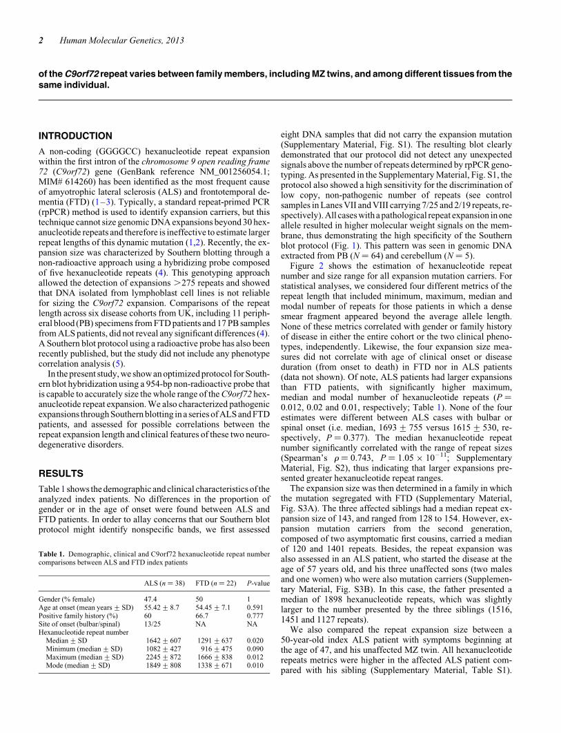
of the C9orf72 repeat varies between family members, including MZ twins, and among different tissues from thesame individual.
INTRODUCTION
A non-coding (GGGGCC) hexanucleotide repeat expansionwithin the first intron of the chromosome 9 open reading frame72 (C9orf72) gene (GenBank reference NM_001256054.1;MIM# 614260) has been identified as the most frequent causeof amyotrophic lateral sclerosis (ALS) and frontotemporal de-mentia (FTD) (1–3). Typically, a standard repeat-primed PCR(rpPCR) method is used to identify expansion carriers, but thistechnique cannot size genomic DNA expansions beyond 30 hex-anucleotide repeats and therefore is ineffective to estimate largerrepeat lengths of this dynamic mutation (1,2). Recently, the ex-pansion size was characterized by Southern blotting through anon-radioactive approach using a hybridizing probe composedof five hexanucleotide repeats (4). This genotyping approachallowed the detection of expansions .275 repeats and showedthat DNA isolated from lymphoblast cell lines is not reliablefor sizing the C9orf72 expansion. Comparisons of the repeatlength across six disease cohorts from UK, including 11 periph-eral blood (PB) specimens from FTD patients and 17 PB samplesfrom ALS patients, did not reveal any significant differences (4).A Southern blot protocol using a radioactive probe has also beenrecently published, but the study did not include any phenotypecorrelation analysis (5).
In the present study, we show an optimized protocol for South-ern blot hybridization using a 954-bp non-radioactive probe thatis capable to accurately size the whole range of the C9orf72 hex-anucleotide repeat expansion. We also characterized pathogenicexpansions through Southern blotting in a series of ALS and FTDpatients, and assessed for possible correlations between therepeat expansion length and clinical features of these two neuro-degenerative disorders.
RESULTS
Table 1 shows the demographic and clinical characteristics of theanalyzed index patients. No differences in the proportion ofgender or in the age of onset were found between ALS andFTD patients. In order to allay concerns that our Southern blotprotocol might identify nonspecific bands, we first assessed
eight DNA samples that did not carry the expansion mutation(Supplementary Material, Fig. S1). The resulting blot clearlydemonstrated that our protocol did not detect any unexpectedsignals above the number of repeats determined by rpPCR geno-typing. As presented in the Supplementary Material, Fig. S1, theprotocol also showed a high sensitivity for the discrimination oflow copy, non-pathogenic number of repeats (see controlsamples in Lanes VII and VIII carrying 7/25 and 2/19 repeats, re-spectively). All cases with a pathological repeat expansion in oneallele resulted in higher molecular weight signals on the mem-brane, thus demonstrating the high specificity of the Southernblot protocol (Fig. 1). This pattern was seen in genomic DNAextracted from PB (N ¼ 64) and cerebellum (N ¼ 5).
Figure 2 shows the estimation of hexanucleotide repeatnumber and size range for all expansion mutation carriers. Forstatistical analyses, we considered four different metrics of therepeat length that included minimum, maximum, median andmodal number of repeats for those patients in which a densesmear fragment appeared beyond the average allele length.None of these metrics correlated with gender or family historyof disease in either the entire cohort or the two clinical pheno-types, independently. Likewise, the four expansion size mea-sures did not correlate with age of clinical onset or diseaseduration (from onset to death) in FTD nor in ALS patients(data not shown). Of note, ALS patients had larger expansionsthan FTD patients, with significantly higher maximum,median and modal number of hexanucleotide repeats (P ¼0.012, 0.02 and 0.01, respectively; Table 1). None of the fourestimates were different between ALS cases with bulbar orspinal onset (i.e. median, 1693+ 755 versus 1615+ 530, re-spectively, P ¼ 0.377). The median hexanucleotide repeatnumber significantly correlated with the range of repeat sizes(Spearman’s r ¼ 0.743, P ¼ 1.05 × 10– 11; SupplementaryMaterial, Fig. S2), thus indicating that larger expansions pre-sented greater hexanucleotide repeat ranges.
The expansion size was then determined in a family in whichthe mutation segregated with FTD (Supplementary Material,Fig. S3A). The three affected siblings had a median repeat ex-pansion size of 143, and ranged from 128 to 154. However, ex-pansion mutation carriers from the second generation,composed of two asymptomatic first cousins, carried a medianof 120 and 1401 repeats. Besides, the repeat expansion wasalso assessed in an ALS patient, who started the disease at theage of 57 years old, and his three unaffected sons (two malesand one women) who were also mutation carriers (Supplemen-tary Material, Fig. S3B). In this case, the father presented amedian of 1898 hexanucleotide repeats, which was slightlylarger to the number presented by the three siblings (1516,1451 and 1127 repeats).
We also compared the repeat expansion size between a50-year-old index ALS patient with symptoms beginning atthe age of 47, and his unaffected MZ twin. All hexanucleotiderepeats metrics were higher in the affected ALS patient com-pared with his sibling (Supplementary Material, Table S1).
Table 1. Demographic, clinical and C9orf72 hexanucleotide repeat numbercomparisons between ALS and FTD index patients
ALS (n ¼ 38) FTD (n ¼ 22) P-value
Gender (% female) 47.4 50 1Age at onset (mean years+SD) 55.42+8.7 54.45+7.1 0.591Positive family history (%) 60 66.7 0.777Site of onset (bulbar/spinal) 13/25 NA NAHexanucleotide repeat number
Median+SD 1642+607 1291+637 0.020Minimum (median+SD) 1082+427 916+475 0.090Maximum (median+SD) 2245+872 1666+838 0.012Mode (median+SD) 1849+808 1338+671 0.010
2 Human Molecular Genetics, 2013
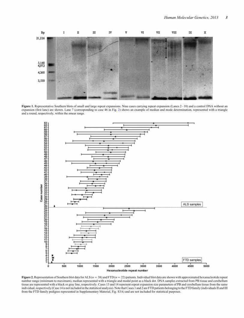
Figure 1. Representative Southern blots of small and large repeat expansions. Nine cases carrying repeat expansion (Lanes 2–10) and a control DNA without anexpansion (first lane) are shown. Lane 7 (corresponding to case 46 in Fig. 2) shows an example of median and mode determination, represented with a triangleand a round, respectively, within the smear range.
Figure 2. Representation of Southern blot data for ALS (n ¼ 38) and FTD (n ¼ 22) patients. Individual blot data are shown with approximated hexanucleotide repeatnumber range (minimum to maximum), median represented with a triangle and modal point as a black dot. DNA samples extracted from PB tissue and cerebellumtissue are represented with a black or gray line, respectively. Cases 13 and 14 represent repeat expansion size parameters of PB and cerebellum tissue from the sameindividual, respectively (Case 14 is not included in the statistical analysis). Note that Cases 1 and 2 are FTD patients belonging to the FTD family (individuals II and IIIfrom the FTD family pedigree represented in Supplementary Material, Fig. S3A) and are not included for statistical purposes.
Human Molecular Genetics, 2013 3

Finally, the hexanucleotide repeat number was determined inDNA samples from cerebellum and PB tissue belonging to thesame FTD patient. Expansion size estimates showed a moderate-ly higher number of hexanucleotide repeats in the cerebellumcompared with PB tissue (Supplementary Material, Table S1).
DISCUSSION
We have developed a non-radioactive Southern blotting protocolusing a 954-bp probe to characterize the hexanucleotide repeatexpansion size range in a series of ALS and FTD patients (Sup-plementary Material, Fig. S4). Our method allows the detectionof hexanucleotide repeats in C9orf72 ranging from 2 to �4500repeat units, thus covering the whole expansion size range andenabling the identification of both small and large pathogenicrepeat expansions. Our results suggest that ALS patients harboura higher number of hexanucleotide repeats than FTD patients.This is not in accordance with a recent report in which authorsdid not find repeat size differences between different neurode-generative diseases, including ALS and FTD (4). One possibilityfor this discrepancy is that their methodology, based on a hybrid-ization probe composed of five DIG-labelled hexanucleotiderepeats, was not sensitive to detect expansions at the low edgeof the mutation spectrum (from 30 to � 275 repeats), and there-fore DNA samples with low number of repeats could have beenmissed. This outcome clearly emphasizes the need of a genotyp-ing technique with enough sensitivity to cover the whole patho-genic expansion size range. Another explanation could be thatwe have analyzed a greater number of ALS and FTD patients,thus increasing our power to detect differences between bothneurodegenerative diseases.
A possible mechanistic explanation for the difference in therepeat length between ALS and FTD in our study might berelated to the recently described repeat-associated non-ATG-initiated (RAN) translation that occurs in C9orf72 expansionmutation carriers. This alteration in the translation processleads to the accumulation of insoluble dipeptide-repeat (DPR)protein aggregates in the central nervous system (6,7). Interest-ingly, the formation of incorrectly translated products seems tooccur in a hexanucleotide repeat length-dependent manner.That is, the longer the repeat size the greater quantity of aberrant-ly translated dipeptides will be produced. Furthermore, longerrepeat tracts can express multiple RAN-translated homopoly-meric proteins whereas shorter repeats only express a uniqueDPR (7). Taking all these novel data into account, it could beexpected that the length of the repeat could determine, to someextent, the molecular mechanisms driving the disease to eitherFTD or ALS. Nevertheless, our results should be interpreted cau-tiously as there is a substantial overlap of hexanucleotide repeatsizes between ALS and FTD index patients, and a threshold todifferentiate between ALS and FTD cannot be defined basedon our data. For example, index cases number 3, 4 and 26(Fig. 2), with estimated median allele sizes of 148, 130 and192 repeats, suffered from either FTD (Patients 3 and 4) orALS (Patient 26).
Our results also suggest that the hexanucleotide repeat numberdoes not correlate with disease duration or age at onset. This lackof correlation might be due to environmental, genetic or epigen-etic factors. In this sense, a recent study has reported that higher
levels of DNA methylation near the hexanucleotide repeat sig-nificantly correlated with shorter disease duration in ALSpatients (8).
We found a striking positive correlation between the mediannumber of hexanucleotide repeats and the range of the observedrepeat sizes. This might reflect a greater disruption of replication,repair and/or recombination machineries as the number ofrepeats increases, thus contributing to genetic instability (9).We also show a family in which the expansion mutation segre-gates with FTD and present evidence of an unstable hexanucleo-tide repeat number transference through generations. On thecontrary, we also present a family with an ALS affected fatherand his three healthy sons who carry similar, although slightlylower, expansion sizes. Similar expansion sizes (ranging from1000 to 1600 repeats) have been previously reported acrossfour family members suffering from FTD (10). These datacould explain the clinical heterogeneity between familymembers that has been described in related individuals carryingthis dynamic mutation (11), and clearly indicates the impossibil-ity to predict the expansion size carried by descendants of muta-tion carriers.
Finally, the fact that we have found subtle discordances in therepeat length between two tissues (PB and cerebellum) fromthe same patient, and also between MZ twins discordant forthe disease, strongly suggests that stochastic expansion eventsduring cell division, which results in somatic and/or germlinemosaicism, contributes to the intra- and inter-individual repeatexpansion variation.
In conclusion, we have developed a reproducible and opti-mized protocol of Southern blot hybridization that allows a reli-able method to approximate the whole repeat expansion sizerange in C9orf72 in DNA samples extracted from PB and braintissue. Our results suggest that larger expansions occur in ALScompared with index FTD patients. We also demonstrate thatinter-individual differences in repeat lengths might exist notonly between unrelated patients but also between familymembers, including MZ twins, and different tissues from thesame individual. These results could explain the high heterogen-eity of the phenotype presented by patients carrying the C9orf72expansion.
MATERIALS AND METHODS
Samples
DNA samples extracted from PB of 38 index ALS patients and18 index FTD patients were included in the analysis. Additional-ly, DNA samples extracted from cerebellum tissue of fourpatients with pathologically confirmed frontotemporal lobardegeneration were collected. PB-derived DNA samples of twofamilies, one consisting of three FTD affected siblings and twonon-affected first cousins, and the other composed of one ALSpatient and his four unaffected sons, were also recruited. DNAfrom the cerebellum of an index FTD patient was also analyzedin order to compare the expansion size with its corresponding PBtissue. Finally, we included DNA from an unaffected monozy-gotic (MZ) twin of an index ALS patient in order to comparethe number of hexanucleotide repeats between identical twins.ALS patients were diagnosed with definite or probable ALS,as defined by the El Escorial research criteria (12). FTD
4 Human Molecular Genetics, 2013

diagnoses were made according to the international consensuscriteria (13,14). All patients were previously identified as hetero-zygous carriers of a C9orf72 hexanucleotide expansion .30repeats through an rpPCR method (10,15–18). Written informedconsent was obtained from all participants and research ethicscommittees from the respective participating centres approvedthe study.
Southern blotting
A total of 15 mg of gDNA was digested overnight with XbaI re-striction enzyme (New England Biolabs, Ipswich, MA, USA).Electrophoresis of digested DNA samples was carried out in0.8% agarose gel with 1× tris–borate–EDTA buffer for 22 hat 1.4 V/cm. DNA was transferred to a positively chargednylon membrane (Roche Applied Science) by capillary blottingand was baked at 808C for 2 h. A 954-bp probe was synthesizedthrough a nested PCR method with FastStart PCR Master Mix(Roche Applied Science) using two PCR rounds and four oligo-nucleotide primers. The first PCR round was performed with theprimers CAACTGGTGAGTGATGGTGGTAG and CTGAGTTCCAGAGCTTGCTACAG. The 1350 bp PCR product wasthen purified and used as a template for a second PCR usingthe FastStart PCR Master Mix (Roche Applied Science) andthe oligonucleotides CAGAAGGTGTAGACGTTGAGAGC andCAGCGAGTACTGTGAGAGCAAG. Digoxigenin (DIG)-dUTP labelling was performed with 2.8 ml of the Vial 2, con-tained in the DIG probe synthesis kit (Roche Applied Science).Prehybridization was carried out at 488C for 3 h and hybridiza-tion at 458C overnight. Blots were then washed in 2× standardsodium citrate (SSC) and 0.1% sodium dodecyl sulphate(SDS) at room temperature for 10 min twice. High stringencywashes (0.1× SSC and 0.1% SDS) were carried out at 688Cfor 10 min five times. Ready-to-use CDP-Star (Roche AppliedScience) was used as the chemiluminescent substrate throughthe transparency technique and signals were visualized on aKODAK Image Station 4000MM PRO (Carestream HealthInc., Rochester, NY, USA).
Assessment of the number of repeats
Hexanucleotide repeat size was estimated by interpolation with aplot of log 10 bp number against migration distance. Minimum,maximum, median and modal sizes of the repeat expansion wereassessed and used for statistical analyses. DIG-labelled DNAmolecular weight marker III (Roche Applied Science) wasused as a ladder.
Statistical analyses
Chi-square analyses were performed for categorical data.Mann–Whitney test was used to compare disease phenotypesand clinical outcomes with minimum, maximum, median andmodal size of the repeat expansion. Spearman’s correlationswere performed between the four expansion size parametersand the clinical features of the disease. Data were analyzedwith the Statistical Package for Social Science v19.0 (SPSSInc., Chicago, IL, USA).
SUPPLEMENTARY MATERIAL
Supplementary Material is available at HMG online.
ACKNOWLEDGEMENTS
We thank patients, their families and controls for their participa-tion in the study. The authors thank the Neurological TissueBank of the Biobanc-Hospital Clınic-IDIBAPS (Barcelona)for providing human brain samples, and specially Dr. EllenGelpı for her valuable help.
Conflict of Interest statement. None declared.
FUNDING
This work was supported by grants from the Spanish Ministry ofEconomy and Competitiveness [grants number PI12/01311 andPI10/00092], CIBERNED and by the Foundation for AppliedMedical Research (FIMA) to S.O.-C. and P.P.
REFERENCES
1. DeJesus-Hernandez, M., Mackenzie, I.R., Boeve, B.F., Boxer, A.L., Baker,M., Rutherford, N.J., Nicholson, A.M., Finch, N.A., Flynn, H., Adamson, J.et al. (2011) Expanded GGGGCC hexanucleotide repeat in noncodingregion of C9ORF72 causes chromosome 9p-linked FTD and ALS. Neuron,72, 245–256.
2. Renton, A.E., Majounie, E., Waite, A., Simon-Sanchez, J., Rollinson, S.,Gibbs, J.R., Schymick, J.C., Laaksovirta, H., van Swieten, J.C.,Myllykangas, L. et al. (2011) A hexanucleotide repeat expansion inC9ORF72 is the cause of chromosome 9p21-linked ALS-FTD. Neuron, 72,257–268.
3. Gijselinck, I., Van Langenhove, T., van der Zee, J., Sleegers, K., Philtjens,S.,Kleinberger, G., Janssens, J., Bettens, K., Van Cauwenberghe, C., Pereson,S. et al. (2012) A C9orf72 promoter repeat expansion in a Flanders-Belgiancohort with disorders of the frontotemporal lobar degeneration-amyotrophiclateral sclerosis spectrum: a gene identification study. Lancet Neurol., 11,54–65.
4. Beck, J., Poulter, M., Hensman, D., Rohrer, J.D., Mahoney, C.J., Adamson,G., Campbell, T., Uphill, J., Borg, A., Fratta, P. et al. (2013) Large C9orf72hexanucleotide repeat expansions are seen in multiple neurodegenerativesyndromes and are more frequent than expected in the UK population.Am. J. Hum. Genet., 92, 345–353.
5. Buchman, V.L., Cooper-Knock, J., Connor-Robson, N., Higginbottom, A.,Kirby, J., Razinskaya, O.D., Ninkina, N. and Shaw, P.J. (2013) Simultaneousand independent detection of C9ORF72 alleles with low and high number ofGGGGCC repeats using an optimised protocol of Southern blothybridisation. Mol. Neurodegener., 8, 12.
6. Ash, P.E., Bieniek, K.F., Gendron, T.F., Caulfield, T., Lin, W.L.,Dejesus-Hernandez, M., van Blitterswijk, M.M., Jansen-West, K., Paul,J.W. 3rd, Rademakers, R. et al. (2013) Unconventional translation ofC9ORF72 GGGGCC expansion generates insoluble polypeptides specific toc9FTD/ALS. Neuron, 77, 639–646.
7. Mori, K., Weng, S.M., Arzberger, T., May, S., Rentzsch, K., Kremmer, E.,Schmid, B., Kretzschmar, H.A., Cruts, M., Van Broeckhoven, C. et al.(2013) The C9orf72 GGGGCC repeat is translated into aggregatingdipeptide-repeat proteins in FTLD/ALS. Science, 339, 1335–1338.
8. Xi, Z., Zinman, L., Moreno, D., Schymick, J., Liang, Y., Sato, C., Zheng, Y.,Ghani, M.,Dib, S., Keith, J. et al. (2013) Hypermethylation of the CpG Islandnear the G4C2 repeat in ALS with a C9orf72 expansion. Am. J. Hum. Genet.,92, 981–989.
9. Mirkin, S.M. (2007) Expandable DNA repeats and human disease. Nature,447, 932–940.
10. Dobson-Stone, C., Hallupp, M., Loy, C.T., Thompson, E.M., Haan, E., Sue,C.M., Panegyres, P.K., Razquin, C., Seijo-Martınez, M., Rene, R. et al.(2013) C9ORF72 repeat expansion in Australian and Spanishfrontotemporal dementia patients. PLoS ONE, 8, e56899.
Human Molecular Genetics, 2013 5

11. Van Langenhove, T., van der Zee, J. and Van Broeckhoven, C. (2012) Themolecular basis of the frontotemporal lobar degeneration-amyotrophiclateral sclerosis spectrum. Ann. Med., 44, 817–828.
12. Brooks, B.R., Miller, R.G., Swash, M. and Munsat, T.L.; World Federationof Neurology Research Group on Motor Neuron Diseases. (2000) El Escorialrevisited: revised criteria for the diagnosis of amyotrophic lateral sclerosis.Amyotroph. Lateral Scler. Other Motor Neuron Disord., 1, 293–299.
13. Rascovsky, K., Hodges, J.R., Knopman, D., Mendez, M.F., Kramer, J.H.,Neuhaus, J., van Swieten, J.C., Seelaar, H., Dopper, E.G., Onyike, C.U. et al.(2011) Sensitivity of revised diagnostic criteria for the behavioural variant offrontotemporal dementia. Brain, 134, 2456–2477.
14. Leyton, C.E., Villemagne, V.L., Savage, S., Pike, K.E., Ballard, K.J., Piguet,O., Burrell, J.R., Rowe, C.C. and Hodges, J.R. (2011) Subtypes ofprogressive aphasia: application of the international Consensus Criteria andvalidation using beta-amyloid imaging. Brain, 134, 3030–3043.
15. Garcıa-Redondo, A., Dols-Icardo, O., Rojas-Garcıa, R., Esteban-Perez, J.,Cordero-Vazquez, P., Munoz-Blanco, J.L., Catalina, I., Gonzalez-Munoz,
M., Varona, L., Sarasola, E. et al. (2013) Analysis of the C9orf72 gene inpatients with amyotrophic lateral sclerosis in Spain and different populationsworldwide. Hum. Mutat., 34, 79–82.
16. Dols-Icardo, O., Suarez-Calvet, M., Hernandez, I., Amer, G.,Anton-Aguirre, S., Alcolea, D., Fortea, J., Boada, M., Tarraga, L., Blesa, R.et al. (2012) Expansion mutation in C9ORF72 does not influence plasmaprogranulin levels in frontotemporal dementia. Neurobiol. Aging, 33,1851.e17–1851.19.
17. van der Zee, J., Gijselinck, I., Dillen, L., Van Langenhove, T., Theuns, J.,Engelborghs, S., Philtjens, S., Vandenbulcke, M., Sleegers, K., Sieben, A.et al. (2013) A pan-European study of the C9orf72 repeat associated withFTLD: geographic prevalence, genomic instability, and intermediaterepeats. Hum. Mutat., 34, 363–373.
18. Gil-Navarro, S., Llado, A., Rami, L., Castellvı, M., Bosch, B., Bargallo, N.,Lomena, F.,Rene, R., Montagut,N., Antonell,A. et al. (2013) Neuroimagingand biochemical markers in the three variants of primary progressive aplasia.Dement. Geriatr. Cogn. Disord., 35, 106–117.
6 Human Molecular Genetics, 2013

lable at ScienceDirect
Neurobiology of Aging xxx (2013) 1e4
Contents lists avai
Neurobiology of Aging
journal homepage: www.elsevier .com/locate/neuaging
Assessing the role of the TREM2 p.R47H variant as a risk factor for Alzheimer’sdisease and frontotemporal dementia
Agustín Ruiz a, Oriol Dols-Icardo b,c, María J. Bullido c,d,e, Pau Pastor c,f,g, Eloy Rodríguez-Rodríguez c,h,Adolfo López de Munain c,i, Marian M. de Pancorbo j, Jordi Pérez-Tur c,k, Victoria Álvarez l, Anna Antonell m,Jesús López-Arrieta e,n, Isabel Hernández a, Lluís Tárraga a, Mercè Boada a,o, Alberto Lleó b,c, Rafael Blesa b,c,Ana Frank-García c,e,p, Isabel Sastre c,d,e, Cristina Razquin c,f, Sara Ortega-Cubero c,f,g, Elena Lorenzo c,f,Pascual Sánchez-Juan c,h, Onofre Combarros c,h, Fermín Moreno c,i, Ana Gorostidi c,i,Xabier Elcoroaristizabal j, Miquel Baquero q, Eliecer Coto l, Raquel Sánchez-Vallem, Jordi Clarimón b,c,*,on behalf of The dementia genetic Spanish consortium (DEGESCO)aAlzheimer Research Center and Memory Clinic, Fundació ACE, Institut Català de Neurociències Aplicades, Barcelona, SpainbMemory Unit, Neurology Department, Hospital de Sant Pau (Sant Pau Biomedical Research Institute), Universitat Autònoma de Barcelona, Barcelona, SpaincCIBERNED, Center for Networked Biomedical Research into Neurodegenerative Diseases, Madrid, SpaindCentro de Biología Molecular Severo Ochoa (CSIC-UAM), Madrid, Spaine Institute of Sanitary Research ‘‘Hospital la Paz’’ (IdIPaz), Madrid, SpainfNeurogenetics Laboratory, Division of Neurosciences, Center for Applied Medical Research, University of Navarra, Pamplona, SpaingDepartment of Neurology, Clínica Universidad de Navarra, University of Navarra, School of Medicine, Pamplona, SpainhNeurology Service, University Hospital Marqués de Valdecilla (University of Cantabria and IFIMAV), Santander, SpainiNeuroscience Area, Institute Biodonostia, and Department of Neurosciences, University of Basque Contry EHU-UPV, San Sebastián, SpainjBIOMICs Research Group, Centro de Investigación “Lascaray” Ikergunea, Universidad del País Vasco UPV/EHU, Vitoria-Gasteiz, Spaink Institut de Biomedicina de ValenciaeCSIC, València, SpainlMolecular Genetics Laboratory, Genetics Unit, Hospital Universitario Central de Asturias, Oviedo, SpainmAlzheimer’s disease and other cognitive disorders, Neurology Department, Hospital Clínic, IDIBAPS, Barcelona, SpainnMemory Unit, University Hospital La Paz-Cantoblanco, Madrid, SpainoHospital Universitari Vall d’Hebron, Institut de Recerca, Universitat Autònoma de Barcelona (VHIR-UAB), Barcelona, SpainpNeurology Service, Hospital Universitario La Paz (UAM), Madrid, SpainqNeurology Service, Hospital Universitari i Politècnic La Fe, València, Spain
a r t i c l e i n f o
Article history:Received 8 May 2013Received in revised form 5 August 2013Accepted 10 August 2013
Keywords:Alzheimer’s diseaseFrontotemporal dementiaTREM2Genetic associationp.R47HRare variant
* Corresponding author at: Genetics of NeurodegeSant Pau, Neurology Department, Hospital de la SAntoni Ma Claret 167, 08025 Barcelona, Spain. Tel.fax: þ34 935565602.
E-mail address: [email protected] (J. Clarimón
0197-4580/$ e see front matter � 2013 Elsevier Inc. Ahttp://dx.doi.org/10.1016/j.neurobiolaging.2013.08.011
a b s t r a c t
A non-synonymous genetic rare variant, rs75932628-T (p.R47H), in the TREM2 gene has recently beenreported to be a strong genetic risk factor for Alzheimer’s disease (AD). Also, rare recessive mutationshave been associated with frontotemporal dementia (FTD). We aimed to investigate the role of p.R47Hvariant in AD and FTD through a multi-center study comprising 3172 AD and 682 FTD patients and 2169healthy controls from Spain. We found that 0.6% of AD patients carried this variant compared to 0.1% ofcontrols (odds ratio [OR] ¼ 4.12, 95% confidence interval [CI] ¼ 1.21e14.00, p ¼ 0.014). A meta-analysiscomprising 32,598 subjects from 4 previous studies demonstrated the large effect of the p.R47H variantin AD risk (OR ¼ 4.11, 95% CI ¼ 2.99e5.68, p ¼ 5.27�10�18). We did not find an association betweenp.R47H and age of onset of AD or family history of dementia. Finally, none of the FTD patients harboredthis genetic variant. These data strongly support the important role of p.R47H in AD risk, and suggest thatthis rare genetic variant is not related to FTD.
� 2013 Elsevier Inc. All rights reserved.
nerative Disorders Unit, IIB-anta Creu i Sant Pau, Sant: þ34 932919050 ext.8230;
).
ll rights reserved.
1. Introduction
The rapid population growth in recent centuries and theevolutionary forces that shape allelic variation over time have led toa large number of rare genetic variants in the human genome,and they seem to vastly exceed the number of common alleles
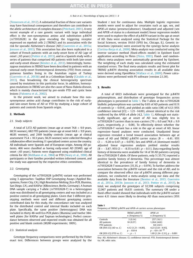
Table 1Frequency of TREM2 p.R47H and APOE-ε4 carriers across phenotypes
TREM2 p.R47Hn (frequency %)
APOE-ε4n (frequency %)
Controls 3 (0.14) 257 (16.1)AD patients 18 (0.57) 1,264 (44.8)FTD patients d 64 (24.5)OR [95% CI] (AD vs. C) 4.12 [1.21e14.00],
p ¼ 0.0144.23 [3.61e4.93],p ¼ 6.9�10�83
OR [95% CI] (FTD vs. C) d, p ¼ 1 1.69 [1.24e2.31], p ¼ 0.001
Key: AD, Alzheimer’s disease; CI, confidence interval; FTD, frontotemporal demen-tia; OR, odds ratio.
A. Ruiz et al. / Neurobiology of Aging xxx (2013) 1e42
(Tennessen et al., 2012). A substantial fraction of these rare variantsmay have functional consequences and therefore can contribute tothe allelic architecture of complex diseases (Pritchard, 2001). A veryrecent example of a rare genetic variant with large individualeffect is the non-synonymous amino acid substitution p.R47H(rs75932628) in the gene encoding the triggering receptorexpressed on myeloid cells 2 (TREM2) and its association with therisk for sporadic Alzheimer’s disease (AD) (Guerreiro et al., 2013a;Jonsson et al., 2013). This association has also been replicated in acohort of French patients with an early-onset form of AD (age ofonset�65 years) (Pottier et al., 2013), and in a Spanish study with aseries of patients that comprised AD patients with both late-onsetand early-onset disease (Benitez et al., 2013). Interestingly, homo-zygous mutations in TREM2 have been described in patients with anunusual early-onset frontotemporal dementia (FTD) from consan-guineous families living in the Anatolian region of Turkey(Guerreiro et al., 2013b) and in a Colombian family (Giraldo et al.,2013), thus broadening the clinical heterogeneity spectrumcaused by mutations in this particular locus. In addition, homozy-gous mutations in TREM2 are also the cause of Nasu-Hakola disease,which is mainly characterized by pre-senile FTD and cystic bonelesions (Paloneva et al., 2002).
In the present work, we aimed to evaluate how this non-synonymous amino acid change contributes to the risk of early-and late-onset forms of AD or FTD by studying a large cohort ofpatients and controls from Spanish origin.
2. Methods
2.1. Study subjects
A total of 3,172 AD patients (mean age at onset 74.8 � 9.9 years,68.5% women), 682 FTD patients (mean age at onset 64.8 � 9.9 years,46.8% women), and 2169 healthy controls (mean age at clinicalassessment 75.4�10.3 years, 58.2%women) were collected through acollaborative effort involving 11 specialized centers across the country.All individuals were Spanish and of European origin. Among AD pa-tients, 466 were classified as having early-onset AD (EOAD; age ofonset �65 years). Patients were diagnosed using established clinicalcriteria for AD (McKhann et al., 1984) or FTD (Neary et al., 1998). Allparticipants or their families provided written informed consent, andthe study was approved by the respective ethics committees.
2.2. Genotyping
Genotyping of the rs75932628 (p.R47H) variant was performedusing 3 approaches: TaqMan SNP Genotyping Assays (Applied Bio-systems, Foster City, CA), High ResolutionMelting (Eco-PCR, Illumina,San Diego, CA), and KASPar (KBioscience, Berlin, Germany). A humanDNA sample carrying a T-allele (rs75932628-T) in a heterozygousstate was distributed to all genotyping centers and was included as apositive control in all genotyping plates. Given that 3 different gen-otyping methods were used and different genotyping centerscontributed data for this study, the concordance rate was analyzedfor the distributed control and internal blanks included on eachplate. Specifically, the same positive (heterozygous) DNA wasincluded in thirty 48-well Eco PCR plates (Illumina) and twelve 384-well plates (for KASPar and Taqman technologies). Perfect concor-dance between observed and expected results was obtained for allpositive and blank controls (80/80 experiments; 100%).
2.3. Statistical analysis
Genotype frequency comparisons were performed by the Fisherexact test. Differences between groups were analyzed by the
Student t test for continuous data. Multiple logistic regressionmodels were used to adjust for covariates such as age, sex, andAPOE-ε4 status (presence/absence). Unadjusted and adjusted (sexand APOE-ε4 status in a dominant model) linear regression modelswere used to explore the effect of p.R47H variant in the age at onsetof AD. Data were analyzed using the Statistical Package for theSocial Sciences, version 19.0.0 (SPSS Inc, Chicago, IL). Genetic in-teractions (epistasis) were assessed by the synergy factor analysis(Cortina-Borja et al., 2009). Meta-analysis was conducted using theinverse variance method (fixed-effects model) in Episheet Excelapplication according to Fleiss (Fleiss, 1993). Fixed- and random-effects meta-analyses were automatically generated by Episheet.The weighting of each study was calculated using the estimatedstandard errors. The final meta-analysis results and forest plot forTREM2 p.R47H showing association results in all available datawere derived using OpenMeta (Wallace et al., 2009). Power calcu-lations were performed with PS software (version 2.1.30).
3. Results
A total of 6023 individuals were genotyped for the p.R47Hpolymorphism, and distribution of genotype frequencies acrossphenotypes is presented in Table 1. The T allele at the rs75932628biallelic polymorphismwas carried by 0.6% of AD patients and 0.1%of controls (p ¼ 0.014), and yielded an odds ratio (OR) of 4.12 (95%confidence interval [CI] ¼ 1.21e14.00), almost identical to the riskconferred by the APOE-ε4 allele (4.2, Table 1). Although not statis-tically significant, age at onset of AD was slightly less inrs75932628-T carriers than in non-carriers (70 � 6.0 and 74.8 � 9.9years, respectively, p ¼ 0.08). To effectively detect whether thers75932628-T variant has a role in the age at onset of AD, linearregressionebased analyses were conducted. Unadjusted linearregression revealed a trend toward association between age atonset of AD and TREM2 p.R47H carrier status (B ¼ �4.41; 95%CI ¼ �9.28 to 0.47; p ¼ 0.078). Sex and APOE-ε4 (dominant model)adjusted linear regression analysis yielded similar results(B¼�3.87; 95% CI¼�8.55 to 0.81; p ¼ 0.11). Data regarding familyhistory of dementia were available for 14 of 18 AD patients carryingthe rs75932628-T allele. Of these patients, only 5 (35.7%) reported apositive family history of dementia. This percentage was almostidentical to the prevalence of family history of dementia inrs75932628-T noncarriers (35.3%, p ¼ 0.974). To further address theassociation between the p.R47H variant and the risk of AD, and tocompare the observed effect size of p.R47H among different pop-ulations, we conducted a meta-analysis using our data and theavailable data from the literature (Benitez et al., 2013; Guerreiroet al., 2013a, 2013b; Jonsson et al., 2013; Pottier et al., 2013). Intotal, we analyzed the genotypes of 32,598 subjects comprising12,967 patients and 19,631 controls. The summary OR under afixed-effect model showed that individuals with the p.R47H variantwere 4.11 times more likely to develop AD than noncarriers (95%
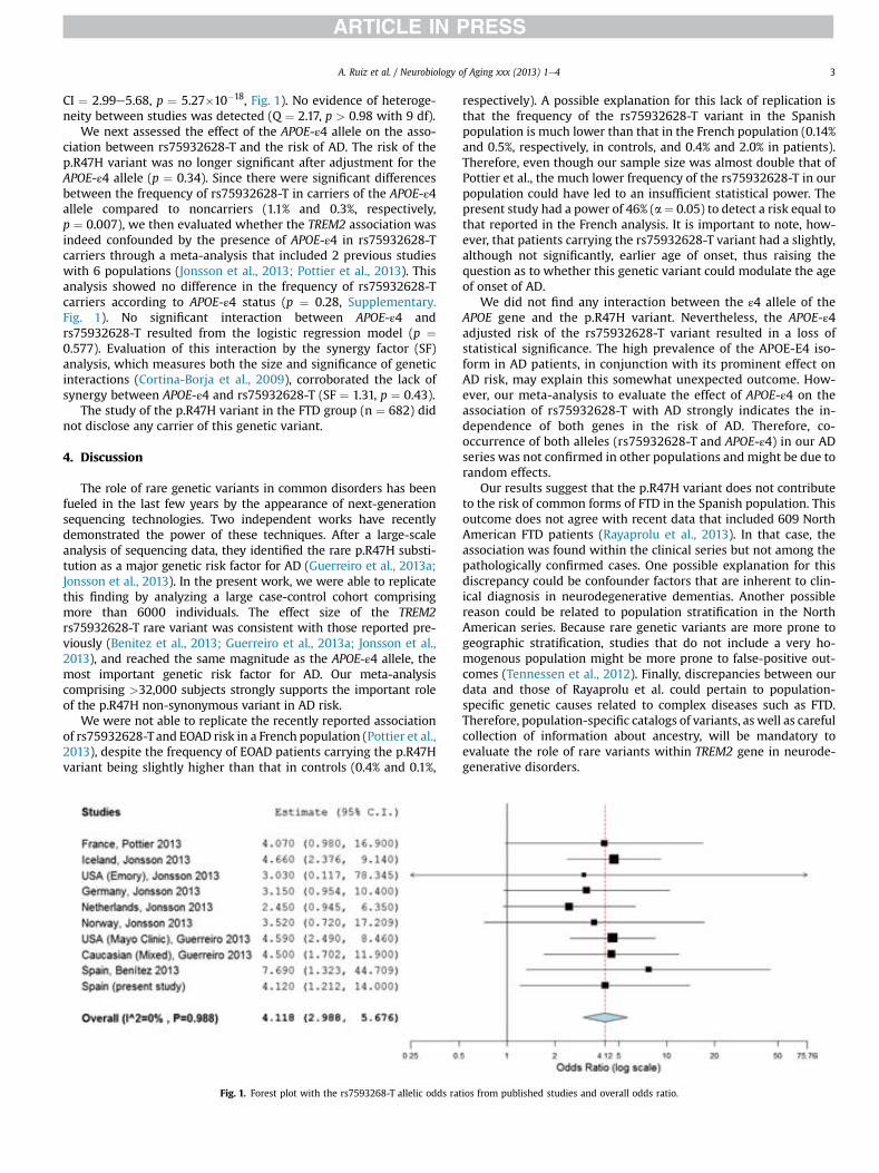
A. Ruiz et al. / Neurobiology of Aging xxx (2013) 1e4 3
CI ¼ 2.99e5.68, p ¼ 5.27�10�18, Fig. 1). No evidence of heteroge-neity between studies was detected (Q ¼ 2.17, p > 0.98 with 9 df).
We next assessed the effect of the APOE-ε4 allele on the asso-ciation between rs75932628-T and the risk of AD. The risk of thep.R47H variant was no longer significant after adjustment for theAPOE-ε4 allele (p ¼ 0.34). Since there were significant differencesbetween the frequency of rs75932628-T in carriers of the APOE-ε4allele compared to noncarriers (1.1% and 0.3%, respectively,p ¼ 0.007), we then evaluated whether the TREM2 association wasindeed confounded by the presence of APOE-ε4 in rs75932628-Tcarriers through a meta-analysis that included 2 previous studieswith 6 populations (Jonsson et al., 2013; Pottier et al., 2013). Thisanalysis showed no difference in the frequency of rs75932628-Tcarriers according to APOE-ε4 status (p ¼ 0.28, Supplementary.Fig. 1). No significant interaction between APOE-ε4 andrs75932628-T resulted from the logistic regression model (p ¼0.577). Evaluation of this interaction by the synergy factor (SF)analysis, which measures both the size and significance of geneticinteractions (Cortina-Borja et al., 2009), corroborated the lack ofsynergy between APOE-ε4 and rs75932628-T (SF ¼ 1.31, p ¼ 0.43).
The study of the p.R47H variant in the FTD group (n ¼ 682) didnot disclose any carrier of this genetic variant.
4. Discussion
The role of rare genetic variants in common disorders has beenfueled in the last few years by the appearance of next-generationsequencing technologies. Two independent works have recentlydemonstrated the power of these techniques. After a large-scaleanalysis of sequencing data, they identified the rare p.R47H substi-tution as a major genetic risk factor for AD (Guerreiro et al., 2013a;Jonsson et al., 2013). In the present work, we were able to replicatethis finding by analyzing a large case-control cohort comprisingmore than 6000 individuals. The effect size of the TREM2rs75932628-T rare variant was consistent with those reported pre-viously (Benitez et al., 2013; Guerreiro et al., 2013a; Jonsson et al.,2013), and reached the same magnitude as the APOE-ε4 allele, themost important genetic risk factor for AD. Our meta-analysiscomprising >32,000 subjects strongly supports the important roleof the p.R47H non-synonymous variant in AD risk.
We were not able to replicate the recently reported associationof rs75932628-Tand EOAD risk in a French population (Pottier et al.,2013), despite the frequency of EOAD patients carrying the p.R47Hvariant being slightly higher than that in controls (0.4% and 0.1%,
Fig. 1. Forest plot with the rs7593268-T allelic odds ra
respectively). A possible explanation for this lack of replication isthat the frequency of the rs75932628-T variant in the Spanishpopulation is much lower than that in the French population (0.14%and 0.5%, respectively, in controls, and 0.4% and 2.0% in patients).Therefore, even though our sample size was almost double that ofPottier et al., the much lower frequency of the rs75932628-T in ourpopulation could have led to an insufficient statistical power. Thepresent study had a power of 46% (a¼ 0.05) to detect a risk equal tothat reported in the French analysis. It is important to note, how-ever, that patients carrying the rs75932628-T variant had a slightly,although not significantly, earlier age of onset, thus raising thequestion as to whether this genetic variant could modulate the ageof onset of AD.
We did not find any interaction between the ε4 allele of theAPOE gene and the p.R47H variant. Nevertheless, the APOE-ε4adjusted risk of the rs75932628-T variant resulted in a loss ofstatistical significance. The high prevalence of the APOE-E4 iso-form in AD patients, in conjunction with its prominent effect onAD risk, may explain this somewhat unexpected outcome. How-ever, our meta-analysis to evaluate the effect of APOE-ε4 on theassociation of rs75932628-T with AD strongly indicates the in-dependence of both genes in the risk of AD. Therefore, co-occurrence of both alleles (rs75932628-T and APOE-ε4) in our ADseries was not confirmed in other populations and might be due torandom effects.
Our results suggest that the p.R47H variant does not contributeto the risk of common forms of FTD in the Spanish population. Thisoutcome does not agree with recent data that included 609 NorthAmerican FTD patients (Rayaprolu et al., 2013). In that case, theassociation was found within the clinical series but not among thepathologically confirmed cases. One possible explanation for thisdiscrepancy could be confounder factors that are inherent to clin-ical diagnosis in neurodegenerative dementias. Another possiblereason could be related to population stratification in the NorthAmerican series. Because rare genetic variants are more prone togeographic stratification, studies that do not include a very ho-mogenous population might be more prone to false-positive out-comes (Tennessen et al., 2012). Finally, discrepancies between ourdata and those of Rayaprolu et al. could pertain to population-specific genetic causes related to complex diseases such as FTD.Therefore, population-specific catalogs of variants, as well as carefulcollection of information about ancestry, will be mandatory toevaluate the role of rare variants within TREM2 gene in neurode-generative disorders.
tios from published studies and overall odds ratio.
A. Ruiz et al. / Neurobiology of Aging xxx (2013) 1e44
Disclosure statement
The authors declare no actual or potential conflicts of interest.
Acknowledgements
We would thank the patients, their families, and the controlsubjects for their participation in this study. Thes study was sup-ported by grants from Instituto de Salud Carlos III (PI12/01311 and12/00013) and grants from theMinistry of Science (SAF2010-15558,SAF2009-10434), Centro de Investigación Biomédica en Red sobreEnfermedades Neurodegenerativas (CIBERNED, Spain), Consolider(CSD2010-00045), Ayudas a proyectos de investigación sanitaria(Departamento de Sanidad y Consumo del Gobierno Vasco,Ref. 2009111083), and the Department of Health of the Governmentof Navarra (refs. 13085 and 3/2008). C.R. held, during the period2009e2013, a “Torres Quevedo” fellowship from the Spanish Min-istry of Science and Technology, co-financed by the European SocialFund. Fundació ACE researchers are indebted to Trinitat Port-Carbóand her family who are supporting Fundació ACE scientific pro-grams. We also would like to thank Drs P. Gil and P. Coria for theircooperation in the generation of the case-control samples, and DrsJosé Luis Molinuevo, Albert Lladó, and Lorena Rami (from HospitalClínic), Drs Juan Fortea and Daniel Alcolea (from Hospital Sant Pau),Dr J.A. Burguera (from Hospital Universitari i Politècnic La Fe), andDr Antonio Salazar (from Direcció General de Salut Pública de laGeneralitat Valenciana) for their effort in clinical evaluation andsample recruitment, and Carolyn Newey for editorial assistance.
Appendix A. Supplementary data
Supplementary data associated with this article can be found,in the online version, at http://dx.doi.org/10.1016/j.neurobiolaging.2013.08.011.
References
Benitez, B.A., Cooper, B., Pastor, P., Jin, S.C., Lorenzo, E., Cervantes, S., Cruchaga, C.,2013. TREM2 is associated with the risk of Alzheimer’s disease in Spanishpopulation. Neurobiol. Aging 34, 1711 e15e1711 e17.
Cortina-Borja, M., Smith, A.D., Combarros, O., Lehmann, D.J., 2009. The synergy factor:a statistic to measure interactions in complex diseases. BMC Res. Notes 2, 105.
Fleiss, J.L., 1993. The statistical basis of meta-analysis. Stat. Methods Med. Res. 2,121e145.
Giraldo, M., Lopera, F., Siniard, A.L., Corneveaux, J.J., Schrauwen, I., Carvajal, J.,Muñoz, C., Ramírez-Restrepo, M., Gaiteri, C., Myers, A.J., Caselli, R.J., Kosik, K.S.,Reiman, E.M., Huentelman, M.J., 2013. Variants in triggering receptor expressedon myeloid cells 2 are associated with both behavioral variant frontotemporallobar degeneration and Alzheimer’s disease. Neurbiol. Aging 34, 2077.e11e8.
Guerreiro, R., Wojtas, A., Bras, J., Carrasquillo, M., Rogaeva, E., Majounie, E.,Cruchaga, C., Sassi, C., Kauwe, J.S., Younkin, S., Hazrati, L., Collinge, J., Pocock, J.,Lashley, T., Williams, J., Lambert, J.C., Amouyel, P., Goate, A., Rademakers, R.,Morgan, K., Powell, J., St George-Hyslop, P., Singleton, A., Hardy, J., 2013a. TREM2variants in Alzheimer’s disease. N. Engl. J. Med. 368, 117e127.
Guerreiro, R.J., Lohmann, E., Bras, J.M., Gibbs, J.R., Rohrer, J.D., Gurunlian, N.,Dursun, B., Bilgic, B., Hanagasi, H., Gurvit, H., Emre, M., Singleton, A., Hardy, J.,2013b. Using exome sequencing to reveal mutations in TREM2 presenting as afrontotemporal dementia-like syndrome without bone involvement. JAMANeurol. 70, 78e84.
Jonsson, T., Stefansson, H., Steinberg, S., Jonsdottir, I., Jonsson, P.V., Snaedal, J.,Bjornsson, S., Huttenlocher, J., Levey, A.I., Lah, J.J., Rujescu, D., Hampel, H.,Giegling, I., Andreassen, O.A., Engedal, K., Ulstein, I., Djurovic, S., Ibrahim-Verbaas, C., Hofman, A., Ikram, M.A., van Duijn, C.M., Thorsteinsdottir, U.,Kong, A., Stefansson, K., 2013. Variant of TREM2 associated with the risk ofAlzheimer’s disease. N. Engl. J. Med. 368, 107e116.
McKhann, G., Drachman, D., Folstein, M., Katzman, R., Price, D., Stadlan, E.M., 1984.Clinical diagnosis of Alzheimer’s disease: report of the NINCDS-ADRDA WorkGroup under the auspices of Department of Health and Human Services TaskForce on Alzheimer’s Disease. Neurology 34, 939e944.
Neary, D., Snowden, J.S., Gustafson, L., Passant, U., Stuss, D., Black, S., Freedman, M.,Kertesz, A., Robert, P.H., Albert, M., Boone, K., Miller, B.L., Cummings, J.,Benson, D.F., 1998. Frontotemporal lobar degeneration: a consensus on clinicaldiagnostic criteria. Neurology 51, 1546e1554.
Paloneva, J., Manninen, T., Christman, G., Hovanes, K., Mandelin, J., Adolfsson, R.,Bianchin, M., Bird, T., Miranda, R., Salmaggi, A., Tranebjaerg, L., Konttinen, Y.,Peltonen, L., 2002. Mutations in two genes encoding different subunits of areceptor signaling complex result in an identical disease phenotype. Am. J.Hum. Genet. 71, 656e662.
Pottier, C., Wallon, D., Rousseau, S., Rovelet-Lecrux, A., Richard, A.C., Rollin-Sillaire, A.,Frebourg, T., Campion, D., Hannequin, D., 2013. TREM2 R47H Variant as a RiskFactor for Early-Onset Alzheimer’s Disease. J. Alzheimers Dis. 35, 45e49.
Pritchard, J.K., 2001. Are rare variants responsible for susceptibility to complexdiseases? Am. J. Hum. Genet. 69, 124e137.
Rayaprolu, S., Mullen, B., Baker, M., Lynch, T., Finger, E., Seeley, W.W., Hatanpaa, K.J.,Lomen-Hoerth, C., Kertesz, A., Bigio, E.H., Lippa, C., Josephs, K.A., Knopman, D.S.,White 3rd, C.L., Caselli, R., Mackenzie, I.R., Miller, B.L., Boczarska-Jedynak, M.,Opala, G., Krygowska-Wajs, A., Barcikowska, M., Younkin, S.G., Petersen, R.C.,Ertekin-Taner, N., Uitti, R.J., Meschia, J.F., Boylan, K.B., Boeve, B.F., Graff-Radford, N.R., Wszolek, Z.K., Dickson, D.W., Rademakers, R., Ross, O.A., 2013.TREM2 inneurodegeneration: evidence for associationof thep.R47Hvariantwithfrontotemporal dementia and Parkinson’s disease. Mol. Neurodegener. 8, 19.
Tennessen, J.A., Bigham, A.W., O’Connor, T.D., Fu, W., Kenny, E.E., Gravel, S.,McGee, S., Do, R., Liu, X., Jun, G., Kang, H.M., Jordan, D., Leal, S.M., Gabriel, S.,Rieder, M.J., Abecasis, G., Altshuler, D., Nickerson, D.A., Boerwinkle, E.,Sunyaev, S., Bustamante, C.D., Bamshad, M.J., Akey, J.M., 2012. Evolution andfunctional impact of rare coding variation from deep sequencing of humanexomes. Science 337, 64e69.
Wallace, B.C., Schmid, C.H., Lau, J., Trikalinos, T.A., 2009. Meta-Analyst: software formeta-analysis of binary, continuous and diagnostic data. BMC Med. Res.Methodol. 9, 80.

686 www.thelancet.com/neurology Vol 13 July 2014
Articles
Frontotemporal dementia and its subtypes: a genome-wide association studyRaff aele Ferrari*, Dena G Hernandez*, Michael A Nalls*, Jonathan D Rohrer*, Adaikalavan Ramasamy, John B J Kwok, Carol Dobson-Stone, William S Brooks, Peter R Schofi eld, Glenda M Halliday, John R Hodges, Olivier Piguet, Lauren Bartley, Elizabeth Thompson, Eric Haan, Isabel Hernández, Agustín Ruiz, Mercè Boada, Barbara Borroni, Alessandro Padovani, Carlos Cruchaga, Nigel J Cairns, Luisa Benussi, Giuliano Binetti, Roberta Ghidoni, Gianluigi Forloni, Daniela Galimberti, Chiara Fenoglio, Maria Serpente, Elio Scarpini, Jordi Clarimón, Alberto Lleó, Rafael Blesa, Maria Landqvist Waldö, Karin Nilsson, Christer Nilsson, Ian R A Mackenzie, Ging-Yuek R Hsiung, David M A Mann, Jordan Grafman, Christopher M Morris, Johannes Attems, Timothy D Griffi ths, Ian G McKeith, Alan J Thomas, P Pietrini, Edward D Huey, Eric M Wassermann, Atik Baborie, Evelyn Jaros, Michael C Tierney, Pau Pastor, Cristina Razquin, Sara Ortega-Cubero, Elena Alonso, Robert Perneczky, Janine Diehl-Schmid, Panagiotis Alexopoulos, Alexander Kurz, Innocenzo Rainero, Elisa Rubino, Lorenzo Pinessi, Ekaterina Rogaeva, Peter St George-Hyslop, Giacomina Rossi, Fabrizio Tagliavini, Giorgio Giaccone, James B Rowe, Johannes C M Schlachetzki, James Uphill, John Collinge, Simon Mead, Adrian Danek, Vivianna M Van Deerlin, Murray Grossman, John Q Trojanowski, Julie van der Zee, William Deschamps, Tim Van Langenhove, Marc Cruts, Christine Van Broeckhoven, Stefano F Cappa, Isabelle Le Ber, Didier Hannequin, Véronique Golfi er, Martine Vercelletto, Alexis Brice, Benedetta Nacmias, Sandro Sorbi, Silvia Bagnoli, Irene Piaceri, Jørgen E Nielsen, Lena E Hjermind, Matthias Riemenschneider, Manuel Mayhaus, Bernd Ibach, Gilles Gasparoni, Sabrina Pichler, Wei Gu, Martin N Rossor, Nick C Fox, Jason D Warren, Maria Grazia Spillantini, Huw R Morris, Patrizia Rizzu, Peter Heutink, Julie S Snowden, Sara Rollinson, Anna Richardson, Alexander Gerhard, Amalia C Bruni, Raff aele Maletta, Francesca Frangipane, Chiara Cupidi, Livia Bernardi, Maria Anfossi, Maura Gallo, Maria Elena Conidi, Nicoletta Smirne, Rosa Rademakers, Matt Baker, Dennis W Dickson, Neill R Graff -Radford, Ronald C Petersen, David Knopman, Keith A Josephs, Bradley F Boeve, Joseph E Parisi , William W Seeley, Bruce L Miller, Anna M Karydas, Howard Rosen, John C van Swieten, Elise G P Dopper, Harro Seelaar, Yolande A L Pijnenburg, Philip Scheltens, Giancarlo Logroscino, Rosa Capozzo, Valeria Novelli, Annibale A Puca, Massimo Franceschi, Alfredo Postiglione, Graziella Milan, Paolo Sorrentino, Mark Kristiansen, Huei-Hsin Chiang, Caroline Graff , Florence Pasquier, Adeline Rollin, Vincent Deramecourt, Florence Lebert, Dimitrios Kapogiannis, Luigi Ferrucci, Stuart Pickering-Brown, Andrew B Singleton†, John Hardy†, Parastoo Momeni†
SummaryBackground Frontotemporal dementia (FTD) is a complex disorder characterised by a broad range of clinical manifestations, diff erential pathological signatures, and genetic variability. Mutations in three genes—MAPT, GRN, and C9orf72—have been associated with FTD. We sought to identify novel genetic risk loci associated with the disorder.
Methods We did a two-stage genome-wide association study on clinical FTD, analysing samples from 3526 patients with FTD and 9402 healthy controls. To reduce genetic heterogeneity, all participants were of European ancestry. In the discovery phase (samples from 2154 patients with FTD and 4308 controls), we did separate association analyses for each FTD subtype (behavioural variant FTD, semantic dementia, progressive non-fl uent aphasia, and FTD overlapping with motor neuron disease [FTD-MND]), followed by a meta-analysis of the entire dataset. We carried forward replication of the novel suggestive loci in an independent sample series (samples from 1372 patients and 5094 controls) and then did joint phase and brain expression and methylation quantitative trait loci analyses for the associated (p<5 × 10–⁸) single-nucleotide polymorphisms.
Findings We identifi ed novel associations exceeding the genome-wide signifi cance threshold (p<5 × 10–⁸). Combined (joint) analyses of discovery and replication phases showed genome-wide signifi cant association at 6p21.3, HLA locus (immune system), for rs9268877 (p=1·05 × 10–⁸; odds ratio=1·204 [95% CI 1·11–1·30]), rs9268856 (p=5·51 × 10–⁹; 0·809 [0·76–0·86]) and rs1980493 (p value=1·57 × 10–⁸, 0·775 [0·69–0·86]) in the entire cohort. We also identifi ed a potential novel locus at 11q14, encompassing RAB38/CTSC (the transcripts of which are related to lysosomal biology), for the behavioural FTD subtype for which joint analyses showed suggestive association for rs302668 (p=2·44 × 10–⁷; 0·814 [0·71–0·92]). Analysis of expression and methylation quantitative trait loci data suggested that these loci might aff ect expression and methylation in cis.
Interpretation Our fi ndings suggest that immune system processes (link to 6p21.3) and possibly lysosomal and autophagy pathways (link to 11q14) are potentially involved in FTD. Our fi ndings need to be replicated to better defi ne the association of the newly identifi ed loci with disease and to shed light on the pathomechanisms contributing to FTD.
Funding The National Institute of Neurological Disorders and Stroke and National Institute on Aging, the Wellcome/MRC Centre on Parkinson’s disease, Alzheimer’s Research UK, and Texas Tech University Health Sciences Center.
Lancet Neurol 2014; 13: 686–99
This online publication has been corrected.
The corrected version fi rst appeared at thelancet.com/neurology on June 17, 2014
See Comment page 643
*Contributed equally
†Joint last authors
Laboratory of Neurogenetics, Department of Internal
Medicine, Texas Tech University Health Science Center, Lubbock,
Texas, USA (R Ferrari PhD, P Momeni PhD); Reta Lila
Weston Research Laboratories, Department of Molecular
Neuroscience, UCL Institute of Neurology, London, UK
(R Ferrari, D G Hernandez MSc, J D Rohrer PhD, A Ramasamy PhD, Prof J Hardy PhD); Laboratory of
Neurogenetics, National Institute on Aging, National
Institutes of Health, Bethesda, MD, USA (D G Hernandez,
M A Nalls PhD, A B Singleton PhD); Clinical Research Branch, National
Institute on Aging, Baltimore, MD, USA (L Ferrucci MD);
Institute of Brain, Behaviour and Mental Health, Faculty of Medical and Human Sciences,
University of Manchester, Manchester, UK
Articles
www.thelancet.com/neurology Vol 13 July 2014 687
IntroductionFrontotemporal dementia (FTD) is the second most common form of young-onset dementia after Alzheimer’s disease and comprises about 10–20% of all dementias worldwide.1 FTD occurs in about three to 15 per 100 000 individuals aged between 55 years and 65 years.2 The disease has a slow and subtle onset: it is familial in 30–50% of patients and aff ects men and women almost equally.3 The main clinical syndromes are the behavioural variant1,4 and the language variants (semantic dementia and progressive nonfl uent aphasia).1,5 FTD can also co-occur with motor neuron disease (FTD-MND), and atypical parkinsonian disorders.3 The molecular pathology is heterogeneous and based on the type of neuronal lesions and protein inclusions: 40% or more of patients have frontotemporal lobar degeneration (FTLD) with tau pathology (FTLD-tau), about 50% have TDP-43 (TAR DNA-binding protein 43) pathology (FTLD-TDP),6 and the remaining 10% have inclusions positive for fused in sarcoma (FUS; FTLD-FUS) or ubiquitin/p62 (FTLD-UPS [ubiquitin proteasome system]).7 Mutations in three main genes are commonly associated with FTD: the microtubule-associated protein tau (MAPT),8 granulin (GRN),9,10 and C9orf72.11–15 Mutations in the charged multivesicular body protein 2B (CHMP2B), the valosin-containing protein (VCP), and ubiquilin 2 (UBQLN2) genes are rare causes of disease.13,16 Findings from a previous genome-wide association study (GWAS) of neuropathologically confi rmed FTLD-TDP (515 patients vs 2509 controls) showed TMEM106B to be a disease risk factor.17
We did a larger GWAS in samples from people with clinical FTD, and we report results for the discovery, replication, and joint-phase analyses, as well as for assessment of the eff ect on expression and methylation quantitative trait loci (QTL) exerted by associated or suggestive SNPs. We aimed to identify novel genetic risk loci associated with FTD and its subtypes.
MethodsStudy population 44 international research groups (appendix) contributed samples to this two-stage (discovery phase and replication phase) GWAS of clinical FTD. The patients included in the discovery phase were diagnosed according to the Neary criteria1 for FTD, whereas those included in the replication phase were diagnosed according to the Neary criteria,1 or the revised criteria for behavioural FTD4 and the language variants of FTD5 at every collaborative site. For each patient, the diagnosis was made by a neurologist with an interest in FTD or, in a minority (70 [3%] of 2621 patients), by pathological diagnosis. To cover the most relevant FTD clinical signatures, we included patients diagnosed with behavioural FTD, semantic dementia, progressive nonfl uent aphasia, or FTD-MND.18 We reviewed all patients with a diagnosis of language impairment to exclude cases of the logopenic variant of
primary progressive aphasia,5 most of which are associated with Alzheimer’s disease pathology. Samples were obtained from Australia, Belgium, Denmark, France, Germany, Italy, the Netherlands, North America (USA and Canada), Spain, Sweden, and the UK and all patients were of confi rmed European ancestry (to reduce genetic heterogeneity).
DNA was collected at the three institutions leading this project: the Department of Molecular Neuroscience at University College London (UCL), UK; the Laboratory of Neurogenetics of the National Institute on Aging at the National Institutes of Health (NIH), MD, USA; and the Laboratory of Neurogenetics at the Texas Tech University Health Sciences Center (TTUHSC), TX, USA. All samples were anonymous and stored with a patient-specifi c coded identifi cation number. Each DNA sample was assessed for quality with gel electrophoresis and DNA concentrations were assessed via spectrophotometer (Nanodrop; Wilmington, DE, USA) or fl uorometer (Qubit; Life Technologies, Grand Island, NY, USA). Samples from non-overlapping patients were genotyped at the Laboratory of Neurogenetics of the National Institute on Aging, NIH (40%), or at the core facility at the Institute of Child Health, UCL (60%). We obtained standardised clinical, pathological, and genetic data for each patient from all the collaborating groups (appendix). Sporadic cases along with probands from FTD families were included in the study. We excluded carriers of mutations in MAPT and GRN. We did not exclude individuals with C9orf72 expansions because this locus was identifi ed subsequent to sample collection. After quality control of genotyping data and detailed assessment of the clinical diagnosis, we used 2154 and 1372 samples in the discovery phase and replication phase, respectively, for association analysis (table 1). In total, after quality control, we analysed 3526 FTD samples (table 1). Further details about cases included in the study are provided in the appendix.
Control samples for the discovery phase were taken from studies previously done at the Laboratory of Neurogenetics of the National Institute on Aging at the NIH or at UCL. Control individuals were matched to patients on the basis of population ancestry and genotyping platform. Aggregate data for control samples were merged based on overlapping single-nucleotide polymorphisms (SNPs). The selected 7444 control samples were from France, Germany, Italy, the Netherlands, Sweden, the UK, and USA, and were used as controls in previous GWAS;19 all individuals had given consent for their samples to be used as controls. All were free of neurological illness at the time of sampling, but most had not been screened for the absence of a family history of FTD. For each patient, at least two controls were matched based on compatibility of genetic ancestry estimates by principal components analysis to accommodate the lack of precisely matched clinical controls. After quality control, we included 4308
(Prof J S Snowden PhD, S Rollinson PhD, Prof S Pickering-Brown PhD); Neuroscience Research Australia, Sydney, NSW, Australia (J B J Kwok PhD, C Dobson-Stone PhD, W S Brooks MBBS, Prof P R Schofi eld DSc, Prof G M Halliday PhD, Prof J R Hodges MD, O Piguet PhD, L Bartley MSc); University of New South Wales, Sydney, NSW, Australia (J B J Kwok, C Dobson-Stone, W S Brooks, Prof P R Schofi eld, Prof G M Halliday, Prof J R Hodges, O Piguet); South Australian Clinical Genetics Service, SA Pathology at Women’s and Children’s Hospital, North Adelaide, SA, Australia (E Thompson MD, Prof E Haan MBBS); Department of Paediatrics, University of Adelaide, Adelaide, SA, Australia (E Thompson, Prof E Haan); Memory Clinic of Fundació ACE, Institut Català de Neurociències Aplicades, Barcelona, Spain (I Hernández MD, A Ruiz MD, M Boada MD); Hospital Universitari Vall d’Hebron–Institut de Recerca, Universitat Autonoma de Barcelona (VHIR-UAB), Barcelona, Spain (M Boada); Neurology Clinic, University of Brescia, Brescia, Italy (B Borroni MD, Prof A Padovani MD); Department of Psychiatry (C Cruchaga PhD), Hope Center (C Cruchaga, Prof N J Cairns PhD), Washington University School of Medicine, St Louis, Missouri, USA; Department of Pathology and Immunology, Washington University, St Louis, Missouri, USA (Prof N J Cairns); NeuroBioGen Lab—Memory Clinic, IRCCS Istituto Centro San Giovanni di Dio Fatebenefratelli, Brescia, Italy (L Benussi PhD, G Binetti MD); Proteomics Unit, IRCCS Istituto Centro San Giovanni di Dio Fatebenefratelli, Brescia, Italy (R Ghidoni PhD); Biology of Neurodegenerative Disorders, IRCCS Istituto di Ricerche Farmacologiche Mario Negri, Milano, Italy (G Forloni PhD); University of Milan, Milan, Italy (D Galimberti PhD, C Fenoglio PhD, M Serpente PhD, E Scarpini MD); Fondazione Cà Granda, IRCCS Ospedale Maggiore Policlinico, Milan, Italy (D Galimberti, C Fenoglio, M Serpente, E Scarpini); Memory Unit, Neurology Department
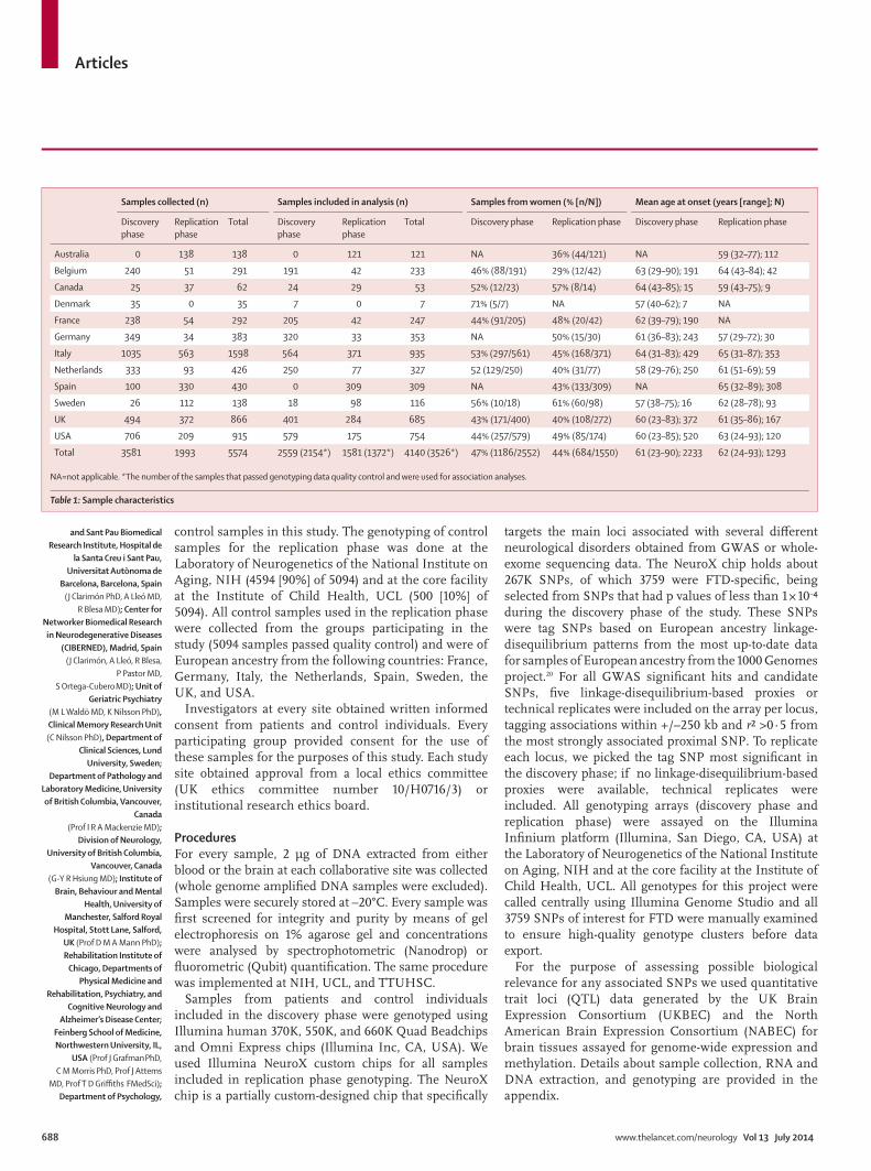
Articles
688 www.thelancet.com/neurology Vol 13 July 2014
and Sant Pau Biomedical Research Institute, Hospital de
la Santa Creu i Sant Pau, Universitat Autònoma de
Barcelona, Barcelona, Spain (J Clarimón PhD, A Lleó MD,
R Blesa MD); Center for Networker Biomedical Research
in Neurodegenerative Diseases (CIBERNED), Madrid, Spain
(J Clarimón, A Lleó, R Blesa, P Pastor MD,
S Ortega-Cubero MD); Unit of Geriatric Psychiatry
(M L Waldö MD, K Nilsson PhD), Clinical Memory Research Unit (C Nilsson PhD), Department of
Clinical Sciences, Lund University, Sweden;
Department of Pathology and Laboratory Medicine, University of British Columbia, Vancouver,
Canada (Prof I R A Mackenzie MD);
Division of Neurology, University of British Columbia,
Vancouver, Canada (G-Y R Hsiung MD); Institute of
Brain, Behaviour and Mental Health, University of
Manchester, Salford Royal Hospital, Stott Lane, Salford,
UK (Prof D M A Mann PhD); Rehabilitation Institute of
Chicago, Departments of Physical Medicine and
Rehabilitation, Psychiatry, and Cognitive Neurology and
Alzheimer’s Disease Center; Feinberg School of Medicine, Northwestern University, IL,
USA (Prof J Grafman PhD, C M Morris PhD, Prof J Attems
MD, Prof T D Griffi ths FMedSci); Department of Psychology,
control samples in this study. The genotyping of control samples for the replication phase was done at the Laboratory of Neurogenetics of the National Institute on Aging, NIH (4594 [90%] of 5094) and at the core facility at the Institute of Child Health, UCL (500 [10%] of 5094). All control samples used in the replication phase were collected from the groups participating in the study (5094 samples passed quality control) and were of European ancestry from the following countries: France, Germany, Italy, the Netherlands, Spain, Sweden, the UK, and USA.
Investigators at every site obtained written informed consent from patients and control individuals. Every participating group provided consent for the use of these samples for the purposes of this study. Each study site obtained approval from a local ethics committee (UK ethics committee number 10/H0716/3) or institutional research ethics board.
ProceduresFor every sample, 2 μg of DNA extracted from either blood or the brain at each collaborative site was collected (whole genome amplifi ed DNA samples were excluded). Samples were securely stored at –20°C. Every sample was fi rst screened for integrity and purity by means of gel electrophoresis on 1% agarose gel and concentrations were analysed by spectrophotometric (Nanodrop) or fl uorometric (Qubit) quantifi cation. The same procedure was implemented at NIH, UCL, and TTUHSC.
Samples from patients and control individuals included in the discovery phase were genotyped using Illumina human 370K, 550K, and 660K Quad Beadchips and Omni Express chips (Illumina Inc, CA, USA). We used Illumina NeuroX custom chips for all samples included in replication phase genotyping. The NeuroX chip is a partially custom-designed chip that specifi cally
targets the main loci associated with several diff erent neurological disorders obtained from GWAS or whole-exome sequencing data. The NeuroX chip holds about 267K SNPs, of which 3759 were FTD-specifi c, being selected from SNPs that had p values of less than 1 × 10–⁴ during the discovery phase of the study. These SNPs were tag SNPs based on European ancestry linkage-disequilibrium patterns from the most up-to-date data for samples of European ancestry from the 1000 Genomes project.20 For all GWAS signifi cant hits and candidate SNPs, fi ve linkage-disequilibrium-based proxies or technical replicates were included on the array per locus, tagging associations within +/–250 kb and r² >0·5 from the most strongly associated proximal SNP. To replicate each locus, we picked the tag SNP most signifi cant in the discovery phase; if no linkage-disequilibrium-based proxies were available, technical replicates were included. All genotyping arrays (discovery phase and replication phase) were assayed on the Illumina Infi nium platform (Illumina, San Diego, CA, USA) at the Laboratory of Neurogenetics of the National Institute on Aging, NIH and at the core facility at the Institute of Child Health, UCL. All genotypes for this project were called centrally using Illumina Genome Studio and all 3759 SNPs of interest for FTD were manually examined to ensure high-quality genotype clusters before data export.
For the purpose of assessing possible biological relevance for any associated SNPs we used quantitative trait loci (QTL) data generated by the UK Brain Expression Consortium (UKBEC) and the North American Brain Expression Consortium (NABEC) for brain tissues assayed for genome-wide expression and methylation. Details about sample collection, RNA and DNA extraction, and genotyping are provided in the appendix.
Samples collected (n) Samples included in analysis (n) Samples from women (% [n/N]) Mean age at onset (years [range]; N)
Discovery phase
Replication phase
Total Discovery phase
Replication phase
Total Discovery phase Replication phase Discovery phase Replication phase
Australia 0 138 138 0 121 121 NA 36% (44/121) NA 59 (32–77); 112
Belgium 240 51 291 191 42 233 46% (88/191) 29% (12/42) 63 (29–90); 191 64 (43–84); 42
Canada 25 37 62 24 29 53 52% (12/23) 57% (8/14) 64 (43–85); 15 59 (43–75); 9
Denmark 35 0 35 7 0 7 71% (5/7) NA 57 (40–62); 7 NA
France 238 54 292 205 42 247 44% (91/205) 48% (20/42) 62 (39–79); 190 NA
Germany 349 34 383 320 33 353 NA 50% (15/30) 61 (36–83); 243 57 (29–72); 30
Italy 1035 563 1598 564 371 935 53% (297/561) 45% (168/371) 64 (31–83); 429 65 (31–87); 353
Netherlands 333 93 426 250 77 327 52 (129/250) 40% (31/77) 58 (29–76); 250 61 (51–69); 59
Spain 100 330 430 0 309 309 NA 43% (133/309) NA 65 (32–89); 308
Sweden 26 112 138 18 98 116 56% (10/18) 61% (60/98) 57 (38–75); 16 62 (28–78); 93
UK 494 372 866 401 284 685 43% (171/400) 40% (108/272) 60 (23–83); 372 61 (35–86); 167
USA 706 209 915 579 175 754 44% (257/579) 49% (85/174) 60 (23–85); 520 63 (24–93); 120
Total 3581 1993 5574 2559 (2154*) 1581 (1372*) 4140 (3526*) 47% (1186/2552) 44% (684/1550) 61 (23–90); 2233 62 (24–93); 1293
NA=not applicable. *The number of the samples that passed genotyping data quality control and were used for association analyses.
Table 1: Sample characteristics
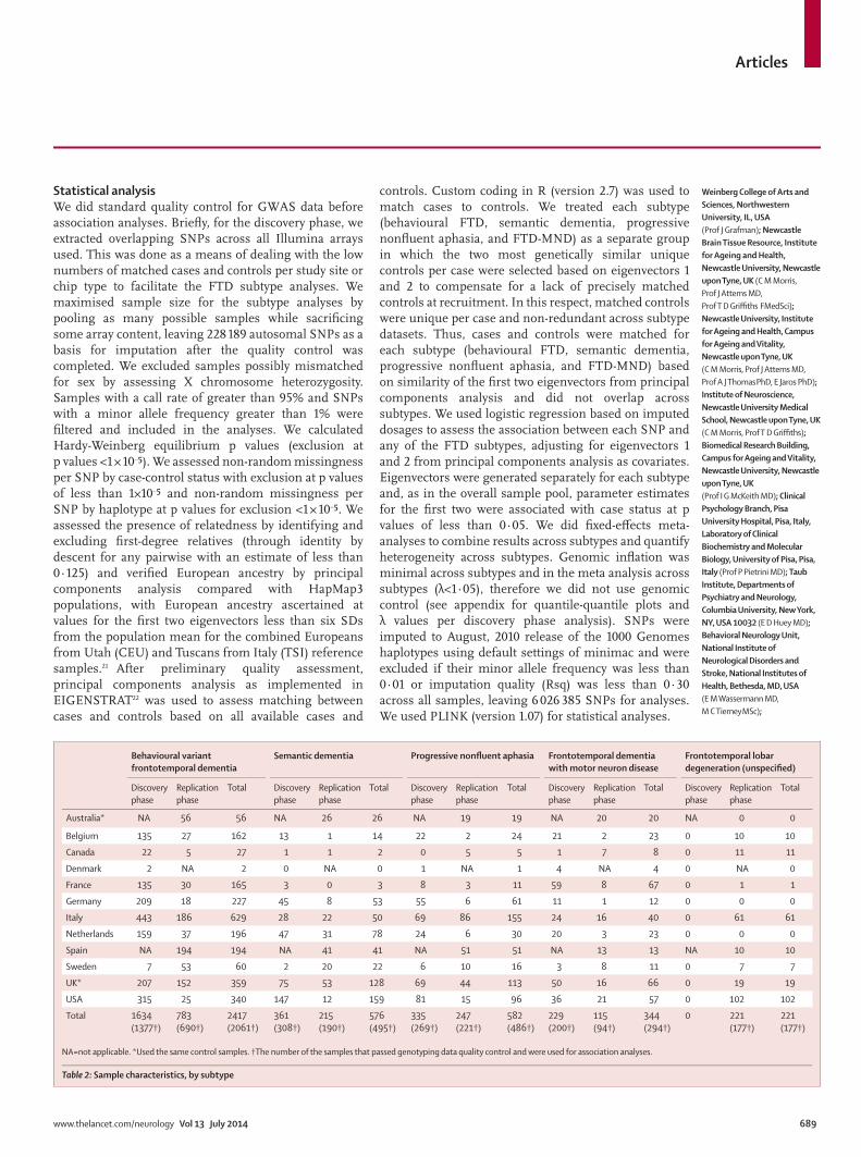
Articles
www.thelancet.com/neurology Vol 13 July 2014 689
Weinberg College of Arts and Sciences, Northwestern University, IL, USA (Prof J Grafman); Newcastle Brain Tissue Resource, Institute for Ageing and Health, Newcastle University, Newcastle upon Tyne, UK (C M Morris, Prof J Attems MD, Prof T D Griffi ths FMedSci); Newcastle University, Institute for Ageing and Health, Campus for Ageing and Vitality, Newcastle upon Tyne, UK (C M Morris, Prof J Attems MD, Prof A J Thomas PhD, E Jaros PhD); Institute of Neuroscience, Newcastle University Medical School, Newcastle upon Tyne, UK (C M Morris, Prof T D Griffi ths); Biomedical Research Building, Campus for Ageing and Vitality, Newcastle University, Newcastle upon Tyne, UK (Prof I G McKeith MD); Clinical Psychology Branch, Pisa University Hospital, Pisa, Italy, Laboratory of Clinical Biochemistry and Molecular Biology, University of Pisa, Pisa, Italy (Prof P Pietrini MD); Taub Institute, Departments of Psychiatry and Neurology, Columbia University, New York, NY, USA 10032 (E D Huey MD); Behavioral Neurology Unit, National Institute of Neurological Disorders and Stroke, National Institutes of Health, Bethesda, MD, USA (E M Wassermann MD, M C Tierney MSc);
Statistical analysis We did standard quality control for GWAS data before association analyses. Briefl y, for the discovery phase, we extracted overlapping SNPs across all Illumina arrays used. This was done as a means of dealing with the low numbers of matched cases and controls per study site or chip type to facilitate the FTD subtype analyses. We maximised sample size for the subtype analyses by pooling as many possible samples while sacrifi cing some array content, leaving 228 189 autosomal SNPs as a basis for imputation after the quality control was completed. We excluded samples possibly mismatched for sex by assessing X chromosome heterozygosity. Samples with a call rate of greater than 95% and SNPs with a minor allele frequency greater than 1% were fi ltered and included in the analyses. We calculated Hardy-Weinberg equilibrium p values (exclusion at p values <1 × 10–⁵). We assessed non-random missingness per SNP by case-control status with exclusion at p values of less than 1×10–⁵ and non-random missingness per SNP by haplotype at p values for exclusion <1 × 10–⁵. We assessed the presence of relatedness by identifying and excluding fi rst-degree relatives (through identity by descent for any pairwise with an estimate of less than 0·125) and verifi ed European ancestry by principal components analysis compared with HapMap3 populations, with European ancestry ascertained at values for the fi rst two eigenvectors less than six SDs from the population mean for the combined Europeans from Utah (CEU) and Tuscans from Italy (TSI) reference samples.21 After preliminary quality assessment, principal components analysis as implemented in EIGENSTRAT22 was used to assess matching between cases and controls based on all available cases and
controls. Custom coding in R (version 2.7) was used to match cases to controls. We treated each subtype (behavioural FTD, semantic dementia, progressive nonfl uent aphasia, and FTD-MND) as a separate group in which the two most genetically similar unique controls per case were selected based on eigenvectors 1 and 2 to compensate for a lack of precisely matched controls at recruitment. In this respect, matched controls were unique per case and non-redundant across subtype datasets. Thus, cases and controls were matched for each subtype (behavioural FTD, semantic dementia, progressive nonfl uent aphasia, and FTD-MND) based on similarity of the fi rst two eigenvectors from principal components analysis and did not overlap across subtypes. We used logistic regression based on imputed dosages to assess the association between each SNP and any of the FTD subtypes, adjusting for eigenvectors 1 and 2 from principal components analysis as covariates. Eigenvectors were generated separately for each subtype and, as in the overall sample pool, parameter estimates for the fi rst two were associated with case status at p values of less than 0·05. We did fi xed-eff ects meta-analyses to combine results across subtypes and quantify heterogeneity across subtypes. Genomic infl ation was minimal across subtypes and in the meta analysis across subtypes (λ<1·05), therefore we did not use genomic control (see appendix for quantile-quantile plots and λ values per discovery phase analysis). SNPs were imputed to August, 2010 release of the 1000 Genomes haplotypes using default settings of minimac and were excluded if their minor allele frequency was less than 0·01 or imputation quality (Rsq) was less than 0·30 across all samples, leaving 6 026 385 SNPs for analyses. We used PLINK (version 1.07) for statistical analyses.
Behavioural variant frontotemporal dementia
Semantic dementia Progressive nonfl uent aphasia Frontotemporal dementia with motor neuron disease
Frontotemporal lobar degeneration (unspecifi ed)
Discovery phase
Replication phase
Total Discovery phase
Replication phase
Total Discovery phase
Replication phase
Total Discovery phase
Replication phase
Total Discovery phase
Replication phase
Total
Australia* NA 56 56 NA 26 26 NA 19 19 NA 20 20 NA 0 0
Belgium 135 27 162 13 1 14 22 2 24 21 2 23 0 10 10
Canada 22 5 27 1 1 2 0 5 5 1 7 8 0 11 11
Denmark 2 NA 2 0 NA 0 1 NA 1 4 NA 4 0 NA 0
France 135 30 165 3 0 3 8 3 11 59 8 67 0 1 1
Germany 209 18 227 45 8 53 55 6 61 11 1 12 0 0 0
Italy 443 186 629 28 22 50 69 86 155 24 16 40 0 61 61
Netherlands 159 37 196 47 31 78 24 6 30 20 3 23 0 0 0
Spain NA 194 194 NA 41 41 NA 51 51 NA 13 13 NA 10 10
Sweden 7 53 60 2 20 22 6 10 16 3 8 11 0 7 7
UK* 207 152 359 75 53 128 69 44 113 50 16 66 0 19 19
USA 315 25 340 147 12 159 81 15 96 36 21 57 0 102 102
Total 1634 (1377†)
783 (690†)
2417 (2061†)
361 (308†)
215 (190†)
576 (495†)
335 (269†)
247 (221†)
582 (486†)
229 (200†)
115 (94†)
344 (294†)
0 221 (177†)
221 (177†)
NA=not applicable. *Used the same control samples. †The number of the samples that passed genotyping data quality control and were used for association analyses.
Table 2: Sample characteristics, by subtype

Articles
690 www.thelancet.com/neurology Vol 13 July 2014
Neuropathology Department, Walton Centre FT, Liverpool, UK
(A Baborie MD); Neuropathology/Cellular Pathology, Royal Victoria
Infi rmary, Newcastle upon Tyne, UK (E Jaros);
Neurogenetics Laboratory, Division of Neurosciences, Center for Applied Medical
Research, Universidad de Navarra, Pamplona, Spain
(P Pastor, C Razquin PhD, S Ortega-Cubero, E Alonso BSc);
Department of Neurology, Clínica Universidad de Navarra, University of Navarra School of
Medicine, Pamplona, Spain (P Pastor); Neuroepidemiology
and Ageing Research Unit, School of Public Health, Faculty
of Medicine, The Imperial College of Science, Technology
and Medicine, London, UK (R Perneczky MD); West London Cognitive Disorders Treatment
and Research Unit, West London Mental Health Trust,
London TW8 8 DS, UK (R Perneczky); Department of
Psychiatry and Psychotherapy, Technische Universität
München, Munich, Germany (R Perneczky, J Diehl-Schmid MD,
P Alexopoulos MD, A Kurz MD); Neurology I, Department of Neuroscience, University of Torino, Italy, AO Città della
Salute e della Scienza di Torino, Italy (I Rainero MD, E Rubino MD,
Prof L Pinessi MD); Tanz Centre for Research in
Neurodegenerative Diseases and Department of Medicine,
University of Toronto, Toronto, Ontario, Canada (E Rogaeva PhD,
P St George-Hyslop MD); Cambridge Institute for Medical
Research and the Department of Clinical Neurosciences, University of Cambridge,
Cambridge, UK (P St George-Hyslop); Division of
Neurology V and Neuropathology, Fondazione
IRCCS Istituto Neurologico Carlo Besta, Milano, Italy (G Rossi PhD,
F Tagliavini MD, G Giaccone MD); Cambridge University
Department of Clinical Neurosciences, Cambridge CB2
0SZ, UK (J B Rowe PhD); MRC Cognition and Brain Sciences
Unit, Cambridge, UK (J B Rowe); Behavioural and Clinical Neuroscience Institute,
Cambridge, UK (J B Rowe); Department of Psychiatry and
Psychotherapy, University of Freiburg Medical School,
Germany
Figure 1: Manhattan plots identifying regions with genome-wide signifi cant associations(A) Single–nucleotide polymorphisms (SNPs) with genome-wide signifi cant p values (p<5×10–⁸) are depicted as red dots and locate to 6p21.3. The associated SNPs map to intron 5 of BTNL2 and to the intergenic region between HLA-DRA and HLA-DRB. (B) Manhattan plot for the behavioural variant frontotemporal dementia subtype in the discovery phase. SNPs with genome-wide signifi cant p values (p<5×10–⁸) are depicted as red dots and locate to 11q14. The associated SNPs map to intron 1 of RAB38 and to the intergenic region between RAB38 and CTSC.
1
p22.2 p21.2 p21.1 p12.3 p12.1 q12 q13 q14.1 q15 q16.1 q16.3 q21 q22.31 q23.2 q24.1 q25.3 q26 q27q23.3
Chromosome 6:32 360 000 32 380 000 32 400 000 32 420 000 32 440 000 32 460 000 32 480 000 32 500 000
2 3 4 5 6 7 8 9 10 11 13 15 17 19 220
56789
10
–log 10
(P)
Chromosome
Chromosome 6
A Subtype meta-analysis
BTNL2
rs1980493
BTNL2NM_019602·1
rs9268856 rs9268877
HLA-DRA HLA-DRB5
HLA-DRANM_061984·2
HLA-DRB9NM_019111·4
HLA-DRB5NP_002116·2
1
p15.4 p15.2 p15.1 p14.3 p14.1 p13 p12 p11.2 q11 q12.1 q13.4 q14.1 q14.3 q21 q22.3 q23.3 q24.3q24.2 q25q22.1
Chromosome 11:87 850 000 87 900 000 87 950 000 88 000 000 88 050 000 88 100 000
2 3 4 5 6 7 8 9 10 11 12 13 15 17 19 220
5678
–log 10
(P)
Chromosome
Chromosome 11
B Behavioural variant frontotemporal dementia
RAB38
RAB38NM_022337·2
rs302652 rs74977128
CTSC
CTSC
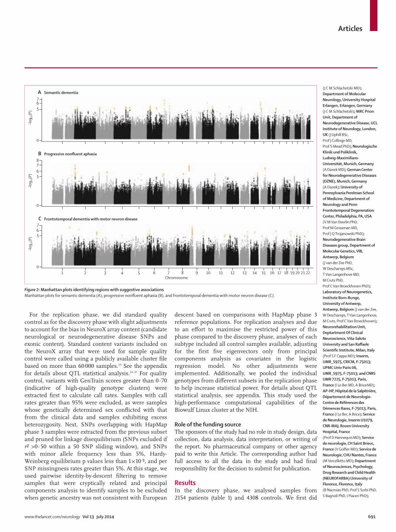
Articles
www.thelancet.com/neurology Vol 13 July 2014 691
(J C M Schlachetzki MD); Department of Molecular Neurology, University Hospital Erlangen, Erlangen, Germany (J C M Schlachetzki); MRC Prion Unit, Department of Neurodegenerative Disease, UCL Institute of Neurology, London, UK (J Uphill BSc, Prof J Collinge MD, Prof S Mead PhD); Neurologische Klinik und Poliklinik, Ludwig-Maximilians-Universität, Munich, Germany (A Danek MD); German Center for Neurodegenerative Diseases (DZNE), Munich, Germany (A Danek); University of Pennsylvania Perelman School of Medicine, Department of Neurology and Penn Frontotemporal Degeneration Center, Philadelphia, PA, USA (V M Van Deerlin PhD, Prof M Grossman MD, Prof J Q Trojanowski PhD); Neurodegenerative Brain Diseases group, Department of Molecular Genetics, VIB, Antwerp, Belgium (J van der Zee PhD, W Deschamps MSc, T Van Langenhove MD, M Cruts PhD, Prof C Van Broeckhoven PhD); Laboratory of Neurogenetics, Institute Born-Bunge, University of Antwerp, Antwerp, Belgium (J van der Zee, W Deschamps, T Van Langenhove, M Cruts, Prof C Van Broeckhoven); Neurorehabilitation Unit, Deptartment Of Clinical Neuroscience, Vita-Salute University and San Raff aele Scientifi c Institute, Milan, Italy (Prof S F Cappa MD); Inserm, UMR_S975, CRICM, F-75013; UPMC Univ Paris 06, UMR_S975, F-75013; and CNRS UMR 7225, F-75013, Paris, France (I Le Ber MD, A Brice MD); AP-HP, Hôpital de la Salpêtrière, Département de Neurologie-Centre de Références des Démences Rares, F-75013, Paris, France (I Le Ber, A Brice); Service de Neurologie, Inserm U1079, CNR-MAJ, Rouen University Hospital, France (Prof D Hannequin MD); Service de neurologie, CH Saint Brieuc, France (V Golfi er MD); Service de Neurologie, CHU Nantes, France (M Vercelletto MD); Department of Neurosciences, Psychology, Drug Research and Child Health (NEUROFARBA) University of Florence, Florence, Italy (B Nacmias PhD, Prof S Sorbi PhD, S Bagnoli PhD, I Piaceri PhD);
For the replication phase, we did standard quality control as for the discovery phase with slight adjustments to account for the bias in NeuroX array content (candidate neurological or neurodegenerative disease SNPs and exonic content). Standard content variants included on the NeuroX array that were used for sample quality control were called using a publicly available cluster fi le based on more than 60 000 samples.23 See the appendix for details about QTL statistical analysis.24–37 For quality control, variants with GenTrain scores greater than 0·70 (indicative of high-quality genotype clusters) were extracted fi rst to calculate call rates. Samples with call rates greater than 95% were excluded, as were samples whose genetically determined sex confl icted with that from the clinical data and samples exhibiting excess heterozygosity. Next, SNPs overlapping with HapMap phase 3 samples were extracted from the previous subset and pruned for linkage disequilibrium (SNPs excluded if r² >0·50 within a 50 SNP sliding window), and SNPs with minor allele frequency less than 5%, Hardy-Weinberg equilibrium p values less than 1 × 10–⁵, and per SNP missingness rates greater than 5%. At this stage, we used pairwise identity-by-descent fi ltering to remove samples that were cryptically related and principal components analysis to identify samples to be excluded when genetic ancestry was not consistent with European
descent based on comparisons with HapMap phase 3 reference populations. For replication analyses and due to an eff ort to maximise the restricted power of this phase compared to the discovery phase, analyses of each subtype included all control samples available, adjusting for the fi rst fi ve eigenvectors only from principal components analysis as covariates in the logistic regression model. No other adjustments were implemented. Additionally, we pooled the individual genotypes from diff erent subsets in the replication phase to help increase statistical power. For details about QTL statistical analysis, see appendix. This study used the high- performance computational capabilities of the Biowulf Linux cluster at the NIH.
Role of the funding sourceThe sponsors of the study had no role in study design, data collection, data analysis, data interpretation, or writing of the report. No pharmaceutical company or other agency paid to write this Article. The corresponding author had full access to all the data in the study and had fi nal responsibility for the decision to submit for publication.
ResultsIn the discovery phase, we analysed samples from 2154 patients (table 1) and 4308 controls. We fi rst did
0
567
–log 10
(P)
A Semantic dementia
0
5678
–log 10
(P)
B Progressive nonfluent aphasia
1 2 3 4 5 6 7 8 9 10 11 12 13 14 16 1815 17 19 20 21 220
567
–log 10
(P)
Chromosome
C Frontotemporal dementia with motor neuron disease
Figure 2: Manhattan plots identifying regions with suggestive associationsManhattan plots for semantic dementia (A), progressive nonfl uent aphasia (B), and frontotemporal dementia with motor neuron disease (C).
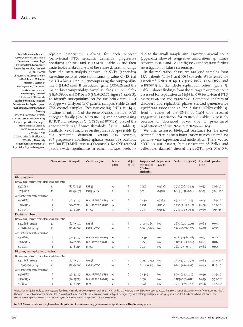
Articles
692 www.thelancet.com/neurology Vol 13 July 2014
Danish Dementia Research Centre, Neurogenetics Clinic,
Department of Neurology, Rigshospitalet, Copenhagen
University Hospital, Denmark (J E Nielsen MD,
L E Hjermind MD); Department of Cellular and Molecular
Medicine, Section of Neurogenetics, The Panum
Institute, University of Copenhagen, Denmark
(J E Nielsen, L E Hjermind); Saarland University Hospital,
Department for Psychiatry and Psychotherapy, Homburg/Saar,
Germany (Prof M Riemenschneider MD);
Saarland University, Laboratory for Neurogenetics, Kirrberger,
Homburg/Saar, Germany (Prof M Riemenschneider,
M Mayhaus PhD, G Gasparoni PhD, S Pichler MSc,
W Gu PhD); University Regensburg, Department of
Psychiatry, Psychotherapy and
separate association analyses for each subtype (behavioural FTD, semantic dementia, progressive nonfl uent aphasia, and FTD-MND; table 2) and then undertook a meta-analysis of the entire dataset. Findings from the meta-analysis showed 29 SNPs (appendix) exceeding genome-wide signifi cance (p value <5×10–⁸) at the HLA locus (6p21.3), encompassing the butyrophilin-like 2 (MHC class II associated) gene (BTNL2) and the major histocompatibility complex, class II, DR alpha (HLA-DRA), and DR beta 5 (HLA-DRB5; fi gure 1, table 3). To identify susceptibility loci for the behavioural FTD subtype we analysed 1377 patient samples (table 2) and 2754 control samples. Two non-coding SNPs at 11q14, locating to intron 1 of the gene RAB38, member RAS oncogene family (RAB38; rs302652) and encompassing RAB38 and cathepsin C (CTSC; rs74977128), passed the genome-wide signifi cance threshold (fi gure 1, table 3). Similarly, we did analyses on the other subtypes (table 1): 308 semantic dementia versus 616 controls, 269 progressive nonfl uent aphasia versus 538 controls, and 200 FTD-MND versus 400 controls. No SNP reached genome-wide signifi cance in either subtype, probably
due to the small sample size. However, several SNPs (appendix) showed suggestive associations (p values between 1 × 10–⁶ and 1 × 10–⁷; fi gure 2) and warrant further investigation in future screenings.
In the replication phase, we analysed samples from 1372 patients (table 1) and 5094 controls. We assessed the associated SNPs at 6p21.3 (rs9268877, rs9268856, and rs1980493) in the whole replication cohort (table 3). Table 3 shows fi ndings from the surrogate or proxy SNPs assessed for replication at 11q14 in 690 behavioural FTD cases: rs302668 and rs16913634. Combined analyses of discovery and replication phases showed genome-wide signifi cant association at 6p21.3 for all SNPs (table 3). Joint p values of the SNPs at 11q14 only revealed suggestive association for rs302668 (table 3) possibly because of decreased power due to proxy-based replication (r² of rs302652 to rs302668=0·65).
We then assessed biological relevance for the novel potential loci in human brain cortex tissues assayed for genome-wide expression and methylation. There was no eQTL in our dataset, but assessment of Zeller and colleagues’ dataset38 showed a cis-eQTL (p=5·05 × 10–³²;
Chromosome Base pair Candidate gene Minor allele
Major allele
Frequency of minor allele (r² when applicable)
Imputation quality
Odds ratio (95% CI) Standard error
p value
Discovery phase
Behavioural variant frontotemporal dementia
rs302652 11 87894831 RAB38 A T 0·259 0·9296 0·730 (0·65–0·82) 0·057 2·02×10–⁸
rs74977128 11 87936874 RAB38/CTSC C T 0·118 0·4182 1·815 (1·48–2·24) 0·107 3·06×10–⁸
All frontotemporal dementia*
rs9268877 6 32431147 HLA-DRA/HLA-DRB5 A G 0·440 0·7783 1·331 (1·22–1·45) 0·045 1·65×10–¹⁰
rs9268856 6 32429719 HLA-DRA/HLA-DRB5 A C 0·251 0·8563 0·752 (0·68–0·83) 0·050 1·30×10–⁸
rs1980493 6 32363215 BTNL2 C T 0·147 0·9642 0·720 (0·69–0·81) 0·060 4·94×10–⁸
Replication phase
Behavioural variant frontotemporal dementia
rs302668 (proxy) 11 87876911 RAB38 C T 0·325 (0·65) NA 0·877 (0.77–0.99) 0·064 0·041
rs16913634 (proxy) 11 87934068 RAB38/CTSC A G 0·104 (0·54) NA 0·964 (0.79–1.17) 0·098 0·710
All frontotemporal dementia*
rs9268877 6 32431147 HLA-DRA/HLA-DRB5 A G 0·449 NA 1·080 (0·98–1·18) 0·047 0·104
rs9268856 6 32429719 HLA-DRA/HLA-DRB5 A C 0·253 NA 0·878 (0·79–0·97) 0·053 0·014
rs1980493 6 32363215 BTNL2 C T 0·145 NA 0.85 (0·75–0·97) 0·068 0·020
Discovery and replication combined
Behavioural variant frontotemporal dementia
rs302668 (proxy) 11 87876911 RAB38 C T 0·292 (0·65) NA 0·814 (0·71–0·92) 0·064 2·44×10–⁷
rs16913634 (proxy)† 11 87934068 RAB38/CTSC A G 0·111 (0·54) NA 1·248 (1·14–1·37) 0·049 8·15×10–⁴
All frontotemporal dementia*
rs9268877† 6 32431147 HLA-DRA/HLA-DRB5 A G 0·4445 NA 1·204 (1·11–1·30) 0·039 1·05×10–⁸
rs9268856 6 32429719 HLA-DRA/HLA-DRB5 A C 0·252 NA 0·809 (0·76–0·86) 0·029 5·51×10–⁹
rs1980493 6 32363215 BTNL2 C T 0·146 NA 0·775 (0·69–0·86) 0·058 1·57×10–⁸
Replication and joint analyses were assessed for the same single-nucleotide polymorphisms (SNPs) at 6p21.3, whereas proxy SNPs were used to assess the association at 11q14 (for which r² values are included). The odds ratio is shown for the minor allele. NA=not applicable. *Denotes only minimal cross-subtype heterogeneity, with heterogeneity p values ranging from 0·793 to 0·944 based on Cochran’s Q test. †Heterogeneity p value <0·01 in the meta-analyses of the discovery and replication phases combined.
Table 3: Characteristics of single-nucleotide polymorphisms exceeding genome-wide signifi cance in the discovery phase
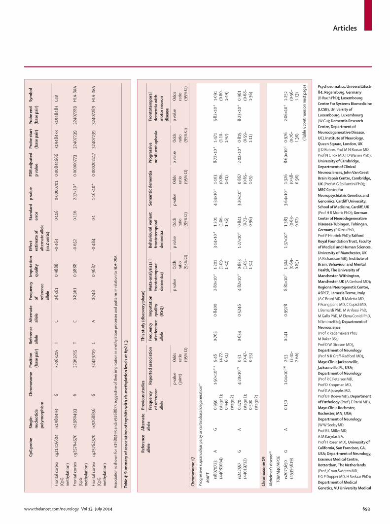
Articles
www.thelancet.com/neurology Vol 13 July 2014 693
Psychosomatics, Universitätsstr 84, Regensburg, Germany (B Ibach PhD); Luxembourg Centre For Systems Biomedicine (LCSB), University of Luxembourg, Luxembourg (W Gu); Dementia Research Centre, Department of Neurodegenerative Disease, UCL Institute of Neurology, Queen Square, London, UK (J D Rohrer, Prof M N Rossor MD, Prof N C Fox MD, J D Warren PhD); University of Cambridge, Department of Clinical Neurosciences, John Van Geest Brain Repair Centre, Cambridge, UK (Prof M G Spillantini PhD); MRC Centre for Neuropsychiatric Genetics and Genomics, Cardiff University, School of Medicine, Cardiff , UK (Prof H R Morris PhD); German Center of Neurodegenerative Diseases-Tübingen, Tübingen, Germany (P Rizzu PhD, Prof P Heutink PhD); Salford Royal Foundation Trust, Faculty of Medical and Human Sciences, University of Manchester, UK (A Richardson MB); Institute of Brain, Behaviour and Mental Health, The University of Manchester, Withington, Manchester, UK (A Gerhard MD); Regional Neurogenetic Centre, ASPCZ, Lamezia Terme, Italy (A C Bruni MD, R Maletta MD, F Frangipane MD, C Cupidi MD, L Bernardi PhD, M Anfossi PhD, M Gallo PhD, M Elena Conidi PhD, N Smirne BSc); Department of Neuroscience (Prof R Rademakers PhD, M Baker BSc, Prof D W Dickson MD), Department of Neurology (Prof N R Graff -Radford MD), Mayo Clinic Jacksonville, Jacksonville, FL, USA; Department of Neurology (Prof R C Petersen MD, Prof D Knopman MD, Prof K A Josephs MD, Prof B F Boeve MD), Department of Pathology (Prof J E Parisi MD), Mayo Clinic Rochester, Rochester, MN, USA; Department of Neurology (W W Seeley MD, Prof B L Miller MD, A M Karydas BA, Prof H Rosen MD), University of California, San Francisco, CA, USA; Department of Neurology, Erasmus Medical Centre, Rotterdam, The Netherlands (Prof J C van Swieten MD, E G P Dopper MD, H Seelaar PhD); Department of Medical Genetics, VU University Medical
CpG
prob
eSi
ngle
-nu
cleot
ide
poly
mor
phism
Chro
mos
ome
Posit
ion
(bas
e pa
ir)Re
fere
nce
alle
leAl
tern
ate
alle
leFr
eque
ncy
of
refe
renc
e al
lele
Impu
tatio
n qu
ality
Eff e
ct
estim
ate o
f al
tern
ate
alle
le
(in Z
uni
ts)
Stan
dard
er
ror
p va
lue
FDR
adjsu
ted
p va
lue
Prob
e st
art
(bas
e pa
ir)Pr
obe
end
(bas
e pa
ir)Sy
mbo
l
Fron
tal c
orte
x (C
pG
met
hyla
tion)
cg21
4156
04rs
1980
493
632
3632
15T
C0·
8361
0·98
88–0
·463
0·11
60·
0000
701
0·00
8346
6631
9484
3331
9484
83C4
B
Fron
tal c
orte
x (C
pG
met
hyla
tion)
cg25
7645
70rs
1980
493
632
3632
15T
C0·
8361
0·98
88–0
·652
0·11
62·
17×1
0– ⁸0·
0000
0773
3240
7239
3240
7289
HLA-
DRA
Fron
tal c
orte
x (C
pG
met
hyla
tion)
cg25
7645
70rs
9268
856
632
4297
19C
A0·
748
0·96
87–0
·484
0·1
1·16
×10– ⁶
0·00
0207
417
3240
7239
3240
7289
HLA-
DRA
Asso
ciatio
n is
show
n fo
r rs1
9804
93 a
nd rs
9268
877,
sugg
estiv
e of t
heir
impl
icatio
n in
met
hyla
tion
proc
esse
s and
pat
tern
s in
rela
tion
to H
LA-D
RA.
Tabl
e 4: S
umm
ary
of a
ssoc
iati
on o
f top
hit
s wit
h cis
-met
hyla
tion
leve
ls a
t 6p2
1.3
Re
fere
nce
alle
leAl
tern
ate
alle
lePr
evio
us st
udie
sTh
is st
udy
(disc
over
y ph
ase)
Freq
uenc
y of
refe
renc
e al
lele
Repo
rted
ass
ocia
tion
Freq
uenc
y of
refe
renc
e al
lele
Impu
tatio
n qu
ality
(R
SQ)
Met
a-an
alys
is (a
ll fr
onto
tem
pora
l de
men
tia)
Beha
viou
ral v
aria
nt
fron
tote
mpo
ral
dem
entia
Sem
antic
dem
entia
Prog
ress
ive
nonfl
uen
t aph
asia
Fron
tote
mpo
ral
dem
entia
with
m
otor
neu
ron
dise
ase
p va
lue
(join
t)O
dds
ratio
(9
5% C
I)
p
valu
e O
dds
ratio
(9
5% C
I)
p va
lue
Odd
s ra
tio
(95%
CI)
p va
lue
Odd
s ra
tio
(95%
CI)
p va
lue
Odd
s ra
tio
(95%
CI)
p va
lue
Odd
s ra
tio
(95%
CI)
Chro
mos
ome
17
Prog
ress
ive s
upra
nucle
ar p
alsy
or c
ortic
obas
al d
egen
erat
ion40
MAP
T
rs80
7072
3 (4
4081
064)
AG
0·95
0 (s
tage
1);
0·94
0 (s
tage
2)
1·50
×10– ¹¹⁶
5·46
(4
·72–
6·31
)
0·76
50·
8400
2·80
×10– ⁴
1·20
1 (1
·09–
1·32
)
3·14
×10– ³
1·20
1 (1
·06–
1·36
)
4·34
×10– ¹
1·10
3 (0
·86–
1·41
)
8·72
×10– ³
1·47
1 (1
·10–
1·97
)
5·82
×10– ¹
1·09
1 (0
·80–
1·49
)
rs24
2557
(4
4019
712)
GA
0·47
0 (s
tage
1);
0·50
0 (s
tage
2)
4·20
×10– ⁷⁰
0·51
(0
·47–
0·55
)
0·63
40·
5246
4·82
×10– ³
0·85
3 (1
·05–
1·31
)
1·27
×10– ²
0·84
1 (0
·73–
0·96
)
3·20
×10– ¹
0·86
7 (0
·65–
1·15
)
2·02
×10– ¹
0·81
5 (0
·59–
1·11
)
8·23
×10– ¹
0·96
1 (0
·68–
1·36
)
Chro
mos
ome
19
Alzh
eim
er’s
dise
ase41
TOM
M40
/APO
E
rs20
7565
0 (4
5395
619)
GA
0·15
01·
04×1
0– ²⁹⁵2·
53
(2·4
1–2·
66)
0·14
10·
9978
8·81
×10– ⁷
1·30
4 (0
·69–
0·85
)
1·37
×10– ⁶
1·38
3 (0
·63–
0·82
)
3·64
×10–2
1·32
6 (0
·58–
0·98
)
8·69
×10– ¹
0·97
6 (0
·76–
1·38
)
2·06
×10– ¹
1·25
2 (0
·56–
1·13
)
(Tab
le 5
cont
inue
s on
next
pag
e)
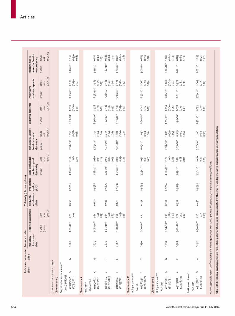
Articles
694 www.thelancet.com/neurology Vol 13 July 2014
Re
fere
nce
alle
leAl
tern
ate
alle
lePr
evio
us st
udie
sTh
is st
udy
(disc
over
y ph
ase)
Freq
uenc
y of
refe
renc
e al
lele
Repo
rted
ass
ocia
tion
Freq
uenc
y of
refe
renc
e al
lele
Impu
tatio
n qu
ality
(R
SQ)
Met
a-an
alys
is (a
ll fr
onto
tem
pora
l de
men
tia)
Beha
viou
ral v
aria
nt
fron
tote
mpo
ral
dem
entia
Sem
antic
dem
entia
Prog
ress
ive
nonfl
uen
t aph
asia
Fron
tote
mpo
ral
dem
entia
/ m
otor
ne
uron
dise
ase
p va
lue
(join
t)O
dds
ratio
(9
5% C
I)
p va
lue
Odd
s ra
tio
(95%
CI)
p va
lue
Odd
s ra
tio
(95%
CI)
p va
lue
Odd
s ra
tio
(95%
CI)
p va
lue
Odd
s ra
tio
(95%
CI)
p va
lue
Odd
s ra
tio
(95%
CI)
(Con
tinue
d fro
m p
revi
ous p
age)
Chro
mos
ome
9
Amyo
trop
hic l
ater
al sc
lero
sis39
C9or
f72/
MOB
3B
rs38
4994
2 (2
7543
281)
AG
0·26
01·
01×1
0–81·
23
(NA)
0·25
30·
9996
4·38
×10–4
1·16
6 (1
·07–
1·27
)
7·38
×10– ³
1·15
5 (0
·78–
0·96
)
9·89
×10– ¹
1·01
0 (0
·80–
1·25
)
9·03
×10– ¹
0·99
0 (0
·79–
1·31
)
2·12
×10– ⁶
1·95
7 (0
·39–
0·68
)
Chro
mos
ome
7
FTLD
-TDP
17
TMEM
106B
rs19
9062
2 (1
2283
787)
AG
0·67
91·
08×1
0– ¹¹1·
64
(1·4
1–1·
89)
0·60
00·
9588
7·88
×10– ²
1·08
0 (0
·99–
1·16
)
5·85
×10– ³
1·14
4 (1
·04–
1·26
)
8·36
×10– ¹
0·97
8 (0
·80–
1·20
)
8·98
×10– ¹
0·98
5 (0
·79–
1·23
)
3·11
×10– ¹
0·87
6 (0
·68–
1·13
)
rs69
6691
5 (1
2265
988)
CT
0·67
91·
63×1
0– ¹¹1·
64
(1·4
1–1·
89)
0·59
60·
9675
1·21
×10– ¹
1·07
0 (0
·87–
1·02
)
5·74
×10– ³
1·14
4 (1
·04–
1·26
)
5·27
×10– ¹
0·93
6 (0
·76–
1·15
)
7·26
×10– ¹
0·96
1 (0
·77–
1·20
)
3·62
×10– ¹
0·88
8 (0
·69–
1·14
)
rs10
2000
4 (1
2255
778)
TC
0·76
75·
00×1
0– ¹¹1·
66
(1·4
3–1.
96
0·69
30·
9538
4·59
×10– ¹
1·03
0 (0
·95–
1·12
)
5·71
×10– ²
1·10
4 (1
·00–
1·22
)
8·53
×10– ¹
0·98
0 (0
·79–
1·21
)
5·00
×10– ¹
0·92
1 (0
·72–
1·17
)
1·20
×10– ¹
0·80
5 (0
·61–
1·06
)
Chro
mos
ome
11
Mul
tiple
scle
rosis
44,4
5
RAB3
8
rs13
8633
0 (8
7819
427)
CT
0·13
02·
00×1
0– ⁶N
A0·
141
0·96
943·
35×1
0– ¹1·
050
(0·8
5–1·
06)
6·09
×10– ¹
1·04
0 (0
·84–
1·10
)
7·60
×10– ¹
1·04
0 (0
·72–
1·27
)
6·97
×10– ¹
1·06
0 (0
·68–
1·29
)
3·00
×10–1
0·82
9 (0
·58–
1·18
)
Chro
mos
ome
6
Mul
tiple
scle
rosis
43–4
5
HLA-
DRA
rs31
3538
8 (3
2413
051)
AG
0·23
08·
94×1
0– ⁸¹1·
99
(1·8
4–2·
15)
0·13
10·
9734
4·80
×10– ²
1·12
2 (1
·00–
1·26
)
2·10
×10– ¹
1·09
5 (0
·79–
1·05
)
1·25
×10– ¹
1·25
4 (0
·60–
1·06
)
5·02
×10– ¹
1·12
0 (0
·64–
1·24
)
6·10
×10– ¹
1·10
5 (0
·61–
1·33
)
rs31
2987
1 (3
2406
342)
AC
0·50
45·
70×1
0– ¹⁵1·
72
(1·5
9–1·
86)
0·33
70·
9379
3·43
×10– ¹
0·96
1 (0
·88–
1·04
)
3·15
×10– ¹
0·94
9 (0
·95–
1·16
)
4·94
×10– ¹
1·07
8 (0
·75–
1·15
)
8·24
×10– ¹
0·97
4 (0
·81–
1·30
)
2·72
×10– ¹
0·85
9 (0
·89–
1·53
)
Park
inso
n’s d
iseas
e46
HLA-
DRA
rs31
2988
2 (3
2409
530)
GA
0·45
01·
90×1
0– ¹⁰1·
26
(1·1
7–1·
35)
0·45
60·
9992
3·36
×10– ²
1·08
6 (0
·85–
0·99
)
3·27
×10– ²
1·10
6 (0
·82–
0·99
)
7·52
×10– ¹
1·03
3 (0
·79–
1·18
)
5·74
×10– ¹
1·06
5 (0
·75–
1·17
)
7·07
×10– ¹
1·04
9 (0
·74–
1·22
)
NA=
not a
pplic
able
. FLT
D-TD
P=fro
ntot
empo
ral l
obar
deg
ener
atio
n w
ith T
DP43
-pos
itive
inclu
sions
. RSQ
=r2 im
puta
tion
qual
ity co
effi c
ient
.
Tabl
e 5: B
idire
ctio
nal a
naly
sis o
f sin
gle-
nucl
eoti
de p
olym
orph
ism
s and
loci
ass
ocia
ted
wit
h ot
her n
euro
dege
nera
tive
dis
orde
rs a
nd o
ur st
udy
popu
lati
on
Articles
www.thelancet.com/neurology Vol 13 July 2014 695
Centre, Amsterdam, The Netherlands (Prof J C van Swieten); Alzheimer Centre and Department of Neurology, VU University Medical Centre, Amsterdam, The Netherlands (Y A L Pijnenburg MD, Prof P Scheltens MD); Department of Basic Medical Sciences, Neurosciences and Sense Organs of the Aldo Moro, University of Bari, Italy (G Logroscino MD, R Capozzo MD); Department of Medical and Molecular Genetics, King’s College London, Guy’s Hospital, London, UK (A Ramasamy); Department of Molecular Cardiology, IRCCS Fondazione S Maugeri, Pavia, Italy (V Novelli PhD); Cardiovascular Research Unit, IRCCS Multimedica, Milan, Italy (An A Puca MD); Department of Medicine and Surgery, University of Salerno, Baronissi (SA), Italy (A A Puca); Neurology Department, IRCCS Multimedica, Milan, Italy (M Franceschi MD); Department of Clinical Medicine and Surgery, University of Naples Federico II, Naples, Italy (Prof A Postiglione MD); Geriatric Center Frullone-ASL Napoli 1 Centro, Naples, Italy (G Milan MD, P Sorrentino MD); UCL Genomics, Institute of Child Health (ICH), UCL, London, UK (M Kristiansen PhD); Karolinska Institutet, Department NVS, KI-Alzheimer Disease Research Center, Stockholm, Sweden (H-H Chiang PhD, Prof C Graff MD); Department of Geriatric Medicine, Genetics Unit, Karolinska Universtiy Hospital, Stockholm (H-H Chiang, Prof C Graff ); Université Lille Nord de France, Lille, France (Prof F Pasquier MD, A Rollin MD, V Deramecourt MD, F Lebert MD); and Laboratory of Neurosciences, National Institute on Aging, National Institutes of Health, Bethesda, MD, USA (D Kapogiannis MD)
Correspondence to:Prof John Hardy, Reta Lila Weston Research Laboratories, Department of Molecular Neuroscience, University College London Institute of Neurology, London WC1N 3BG, [email protected]
See Online for appendix
appendix) at 11q14 for rs302652 (chr11:87894881, risk allele T) causing a decreased expression of RAB38 (Illumina ILMN_2134974 located on chr11:87846656-87846705) in monocytes. These data suggest a role in transcriptional processes in cis for this SNP. Furthermore, we identifi ed signifi cant cis-mQTL at 6p21.3 after multiple test correction for rs1980493 (risk allele T) that associated with changes in the methylation levels related to HLA-DRA in the frontal cortex (table 4).
To assess potential genetic overlap between FTD and closely related forms of neurodegenerative diseases we selected relevant SNPs for candidate loci and analysed them in our dataset. This analysis included published association studies for amyotrophic lateral sclerosis,39 progressive supranuclear palsy and corticobasal degeneration,40 Alzheimer’s disease,41 and FTLD-TDP.17 We also assessed whether the two loci identifi ed through this study had also been reported previously in other studies of neurological disorders.
For the C9orf72 locus (for amyotrophic lateral sclerosis), the SNP rs3849942 achieved a p value of 2·12 × 10–⁶ and an OR of 1·957 in the FTD-MND subtype consistent with our post-hoc analyses (about 23% of expansion carriers in this subtype; table 5, appendix). Association was modest in behavioural FTD (p=7·38 × 10–³; OR=1·155) as well as in the entire discovery cohort (p=4·38 × 10–⁴; OR=1·166), while there was no evidence for association in the semantic dementia or progressive nonfl uent aphasia subtypes (table 5). These results confi rm that the C9orf72 locus associates mainly with FTD- MND and to a lesser extent with behavioural FTD (appendix).
For the MAPT locus (PSP/CBD), the SNPs rs242557 and rs807072340 reached modest p values between 10–³ and 10–⁴
only in the entire cohort and in the behavioural FTD and progressive nonfl uent aphasia subtypes (rs8070723 only; table 5). The eff ect was small in our study although in the same direction as in the GWAS for progressive supranuclear palsy (5·4640 vs about 1·2–1·4 in our study; table 5). These results refl ect the fact that we excluded all known chromosome 17 mutation carriers and that tau pathology was a less common feature within our study population.
For the TOMM40/APOE locus (Alzheimer’s disease), the SNP rs2075650 reached a p value of 8·81 × 10–⁷ in the entire dataset and 1·37 × 10–⁶ in behavioural FTD, whereas the semantic dementia, progressive nonfl uent aphasia, and FTD-MND subtype p values were in the range of 10–¹ and 10–² (table 5). Several Alzheimer’s disease GWASs reported association with the minor allele of this SNP with ORs greater than 2·5,41 but in our study the OR was about 1·3 (table 5). This suggestive association might refl ect clinical overlap (about 15%) between patients with clinically diagnosed FTD and those with Alzheimer’s disease.42
For the TMEM106B locus (FTLD-TDP), we assessed the three associated SNPs reported by Van Deerlin and colleagues (rs1990622; rs6966915; rs1020004).17 All
achieved modest p values in the entire dataset with lowest p values in the range of 10–²–10–³ only in the behavioural FTD subtype (table 5). Van Deerlin and colleagues’ study17 was done on samples from patients with autopsy-confi rmed FTLD-TDP, whereas our cohort is mainly clinically defi ned. Additionally, the previous study included many GRN mutation carriers, who frequently present with behavioural FTD;17 in our study, GRN mutation carriers were excluded. Biochemical evidence has suggested that TMEM106B is directly related to GRN metabolism,13 thus we regard our data as a limited replication of the original fi nding (ie, they do not substantiate earlier fi ndings).
Finally, the RAB38 locus previously showed suggestive association in multiple sclerosis,43 whereas the HLA locus was reported to associate with multiple sclerosis,44,45 Parkinson’s disease,19,46 and Alzheimer’s disease.47 None of the SNPs reported in these studies, and which were assessed in our dataset (table 5),43–46 showed association with FTD, probably suggesting that diff erent risk haplotype sub-structures at the same loci associate with distinctive phenotypes.
DiscussionWe have identifi ed two novel potential loci for FTD: 6p21.3, encompassing the HLA locus (immune system), and 11q14, encompassing RAB38/CTSC (transcripts of which are related to lysosomal biology). Our data suggest that these loci might aff ect expression and methylation in cis and indicate that immune system processes, and possibly lysosomal and autophagy pathways, are potentially involved in the pathogenesis of FTD.
FTD is characterised by a broad range of clinical manifestations, diff erential pathological signatures, and substantial genetic variability, which imply complex disease mechanisms.15 In the search for novel disease risk loci associated with FTD we have done an extensive GWAS on a large cohort of mainly clinically diagnosed FTD samples from patients of European ancestry. Several limitations might apply to this study. In view of the phenotype heterogeneity of FTD, and considering that it is a rare neurodegenerative disorder,2 testing the hypothesis “common variant, common disease” for diseases of this kind is challenging and clearly benefi ts from large sample sizes. Additionally, our fi ndings might indicate association with specifi c loci without necessarily implying causality; low heritability due to common variability might also apply. However, the QQ plots and associated λ values (appendix) conformed to GWAS standards, lending support to our fi ndings.
We included samples from more than 3500 patients and, thus, we know of no larger GWAS for FTD. We have identifi ed two novel potential loci for FTD: 11q14, encompassing RAB38/CTSC, was suggestive for the behavioural FTD subtype, and 6p21.3, encompassing the HLA locus was statistically signifi cant for the entire cohort.

Articles
696 www.thelancet.com/neurology Vol 13 July 2014
For more on Biowulf see http://biowulf.nih.gov
For more on the expression of RAB38 see http://www.
genecards.org/cgi-bin/carddisp.pl?gene=RAB38
RAB3848 encodes the transmembrane protein RAB38, which is expressed in the thyroid, in elements of the immune system, and in the brain. From a functional perspective, RAB38 has been shown to mediate protein traffi cking to lysosomal-related organelles and maturation of phagosomes.49,50 CTSC is a lysosomal cysteine-proteinase that participates in the activation of serine proteinases in cells involved in immune and infl ammatory processes, including phagocytosis of pathogens and local activation and deactivation of infl ammatory factors (Online Mendelian Inheritance in Man [OMIM] number 602365). The SNP rs302652 at the RAB38/CTSC locus shows an eQTL in monocytes38 associated with decreased expression of RAB38, possibly indicating that a decreased function of RAB38 might be the mechanism by which the association at this locus is mediated. Both RAB38 and CTSC are implicated in lysosomal biology and an association with lysosomal and autophagic processes in FTD was previously suggested in two studies of GRN51 and TMEM106B.52 A possible role for autophagy has also been shown in Parkinson’s disease.53 Our fi ndings will need to be replicated in other FTD cohorts in follow-up studies (eg, fi ne-mapping studies) to lend support to the inference
that lysosomal biology and autophagy might be involved in the aetiology of FTD.54
The genetic association that we identifi ed with the HLA locus lends support to the notion of a link between FTD and the immune system. Our mQTL data showed that risk at this locus is associated with cis-changes in methylation levels of HLA-DRA in the frontal cortex. HLA associations have been previously reported in Alzheimer’s disease,47 Parkinson’s disease,19,46 and multiple sclerosis.44,45 Additionally, a general involvement of the innate and the adaptive immune responses has been suggested in the pathogenesis of neurodegenerative diseases,55,56 lending support to the idea that the immune system plays an important part within the spectrum of neurological disorders (panel).
Future studies should aim to replicate our fi ndings and elucidate the functional basis of FTD. Additionally, our data indicate that common pathways and processes might underlie diff erent forms of neurodegenerative disorders, including Alzheimer’s disease, Parkinson’s disease, multiple sclerosis, and FTD. Exploring the possibility of developing therapeutic measures targeting general damage responses could hold promise—after replication and validation of our fi ndings—for the development and implementation of treatment options for these neurological disorders, including FTD.ContributorsJH, PM, ABS, MAN, RF, and JDR designed the study. JDR, RF and JH did the clinical quality checks. RF coordinated sample collection, received samples at UCL and TTUHSC, and did material quality control for discovery and replication phases. DGH received samples at NIH and coordinated material quality control at NIH. JDR, JBJK, CDS, PRS, WSB, JRH, GMH, OP, LB, ET, EH, IH, AR, MB BB, AP, LB, GB, RG, GF, DG, ES, CF, MS, JC, AL, RB, MLW, KN, CN, IRAM, G-YRH, DMAM, JG, CMM, JA, TDG, IGM, AJT, PP, EDH, EMW, AB, EJ, MCT, PP, CR, SO-C, EA, RP, JDS, PA, AK, IR, ER, LP, ER, PStG-H, ER, GR, FT, GG, JBR, JCMS, JU, JC, SM, AD, VMVD, MG, JQT, JvdZ, TVL, CVB, WD, MC, SFC, ILB, AB, DH, VG, MV, BN, SS, SB, IP, JEN, LEH, MR, BI, MM, GG, SP, WG, MNR, NCF, JDW, MGS, HM, PR, PH, JSS, AG, AR, SR, ACB, RM, FF, CC, LB, MA, MG, MEC, NS, RR, MB, DWD, JEP, NRGR, RCP, DK, KAJ, BFB, WWS, BLM, AMK, HR, JCvS, EGPD, HS, YALP, PS, GL, RC, VN, AAP, MF, AP, GM, PS, H-HC, CG, FP, AR, VD, FL, DK, LF, and SPB collected and characterised samples. MK was responsible for genotyping at ICH. JH, PM, ABS, and SPB obtained funding for this study. JH, PM, and ABS supervised the study. MAN did statistical and association analyses. RF, MAN, and JH analysed and interpreted the data. AR helped in the interpretation of the e/mQTL data. RF, MAN, JH, and PM wrote the fi rst draft of the paper. All other co-authors participated in preparation of the paper by reading and commenting on drafts before submission.
Declaration of interestsCVB and MC are inventors on patent applications for GRN and C9orf72. PRS receives speaker fees from Janssen pharmaceutical. RR receives research support from the NIH (R01 NS080882, R01 NS065782, R01 AG026251, R01 NS076471, and P50 AG16574), the ALS Therapy Alliance, and the Consortium for Frontotemporal Degeneration Research, honoraria for lectures or educational activities not funded by industry. RR serves on the medical advisory board of the Association for Frontotemporal Degeneration and the board of directors of the International Society for Frontotemporal Dementia, and holds a patent on methods to screen for the hexanucleotide repeat expansion in the C9ORF72 gene. DWD is supported by NIH grants (P50 AG16574, P50 NS72187, P01 AG03949), the Mangurian Foundation, CurePSP, and the Robert E Jacoby Professorship for
Panel: Research in context
Systematic reviewWe searched PubMed for research and review articles on frontotemporal dementia using the following terms: “frontotemporal dementia AND genetics” and “frontotemporal dementia AND review”. 1,4,5,8–17,54 We compared our results to several previously published genome-wide association studies. We identifi ed only one directly relevant study that investigated a pathologically defi ned subtype of frontotemporal dementia (frontotemporal lobar degeneration with TDP43-positive inclusions; FTLD-TDP).17 The other studies were of related diseases such as amyotrophic lateral sclerosis,39 Alzheimer’s disease,41,47 progressive supranuclear palsy and corticobasal degeneration,40 multiple sclerosis,43–45 and Parkinson’s disease.19,46
InterpretationTo the best of our knowledge, ours is the fi rst genome-wide association study in samples from patients with clinical frontotemporal dementia. In view of the complexity and heterogeneity of the disease, mutations in only three main genes—MAPT, GRN, and C9orf72—have been associated with frontotemporal dementia, and these explain only a small proportion of cases. Most importantly, little is known about the mechanisms involved in the development of this disorder. Our fi ndings suggest that common variability in loci that point to immune processes and possibly to lysosomal biology and autophagy are involved in the pathobiology of the disease. These fi ndings provide a basis for future replication and functional studies.

Articles
www.thelancet.com/neurology Vol 13 July 2014 697
Alzheimer’s Research. NRGR is on the Scientifi c Advisory Board for Codman, TauRzx multicenter study, Consultation for CYTOX. RCP chairs a Data Monitoring Committee for Pfi zer and Janssen Alzheimer Immunotherapy, and is a consultant for GE Healthcare and Elan Pharmaceuticals. RCP receives royalties from Oxford University Press for Mild Cognitive Impairment. DK has served on a data safety monitoring board for Lilly Pharmaceuticals, as a consultant to TauRx, was an investigator in clinical trials sponsored by Baxter, Elan Pharmaceuticals, and Forest Pharmaceuticals in the past 2 years and receives research support from the NIH. BFB has served as an investigator for clinical trials sponsored by Cephalon Inc, Allon Pharmaceuticals, and GE Healthcare. BFB receives royalties from the publication of a book entitled Behavioral Neurology Of Dementia (Cambridge Medicine, 2009). BFB has received honoraria from the American Academy of Neurology. BFB serves on the Scientifi c Advisory Board of the Tau Consortium. BFB receives research support from the National Institute on Aging (P50 AG016574, U01 AG006786, RO1 AG032306, RO1 AG041797) and the Mangurian Foundation. BLM is on the Board Membership of The Larry L Hillblom Foundation, The John Douglas French Foundation, The Tau Consortium, Sagol School of Neuroscience Tel Aviv University. BLM holds consultancy for Tau Rx lts—Chair, Scientifi c Advisory Board bvFTD Trial Allon Therapeutics—Steering Committee AL-108-231 Study, Bristol-Myers Squibb-Advisory Board, Progressive Supranuclear Palsy (PSP), Neurology Scientifi c Advisory Board Meeting Siemens Molecular Imaging, and Eli Lilly US Alzheimer’s Disease Advisory Board, and receives royalties from Cambridge University Press Guilford Publications Inc, Neurocase. RF, DGH, MAN, JDR, AR, JBJK, CDS, WSB, GMH, JRH, OP, LB, ET, EH, IH, AR, MB, BB, AP, CC, NJC, LB, GB, RG, GF, DG, CF, MS, ES, JC, AL, RB, MLW, KN, CN, IRAM, G-YRH, DMAM, JG, CMM, JA, TDG, IGM, AJT, PP, EDH, EMW, AB, EJ, MCT, PP, CR, SO-C, EA, RP, JDS, PA, AK, IR, ER, LP, ER, PStG-H, GR, FT, GG, JBR, JCMS, JU, JC, SM, AD, VMVD, MG, JQT, JvdZ, WD, TVL, SFC, ILB, DH, VG, MV, AB, BN, SS, SB, IP, JEN, LEH, MR, MM, BI, GG, SP, WG, MNR, NCF, JDW, MGS, HRM, PR, PH, JSS, SR, AR, AG, ACB, RM, FF, CC, LB, MA, MG, MEC, NS, MB, KAJ, JEP, WWS, AMK, HR, JCvS, EGPD, HS, YALP, PS, GL, RC, VN, AAP, MF, AP, GM, PS, MK, H-HC, CG, FP, AR, VD, FL, DK, LF, SPB, JH, PM, and ABS declare no competing interests.
Acknowledgments We received intramural funding from the National Institute of Neurological Disorders and Stroke (NINDS) and National Institute on Aging (NIA), the Wellcome/MRC Centre on Parkinson’s disease, Alzheimer’s Research UK (ARUK, Grant ARUK-PG2012-18), and by the offi ce of the Dean of the School of Medicine, Department of Internal Medicine, at Texas Tech University Health Sciences Center. We thank Mike Hubank and Kerra Pearce at the Genomic core facility at the Institute of Child Health (ICH), UCL, for assisting RF in doing Illumina genotyping experiments (FTD-GWAS genotyping). The work done by the North American Brain Expression Consortium (NABEC) was supported in part by the Intramural Research Program of the National Institute on Aging, National Institutes of Health, part of the US Department of Health and Human Services (project number ZIA AG000932-04), and by a Research Grant from the Department of Defense (W81XWH-09-2-0128). Work done by the UK Brain Expression Consortium (UKBEC) was supported by the MRC through the MRC Sudden Death Brain Bank (CS), by a Project Grant (G0901254 to JH and MW), and by a Fellowship award (G0802462 to MR). DT was supported by the King Faisal Specialist Hospital and Research Centre, Saudi Arabia. Computing facilities used at King’s College London were supported by the National Institute for Health Research (NIHR) Biomedical Research Centre based at Guy’s and St Thomas’ NHS Foundation Trust and King’s College London. We thank AROS Applied Biotechnology AS company laboratories and Aff ymetrix for their valuable input. JBJK was supported by the National Health and Medical Resarch Council (NHMRC), Australia (project grants 510217 and 1005769). CDS was supported by NHMRC (project grants 630428 and 1005769). PRS was supported by NHMRC (project grants 510217 and 1005769) and acknowledges that DNA samples were prepared by Genetic Repositories Australia, supported by NHMRC Enabling Grant 401184. GMH was supported by NHMRC Research Fellowship 630434, Project Grant 1029538, and Program Grant 1037746. JRH was supported by the
Australian Research Council Federation Fellowship, NHMRC Project Grant 1029538 and NHMRC Program Grant 1037746. OP was supported by NHMRC Career Development Fellowship 1022684, Project Grant 1003139. IH, AR, and MB acknowledge the patients and controls who participated in this project and the Trinitat Port-Carbó (deceased) and her extended family who are supporting Fundació ACE research programmes. CC was supported by Grant P30-NS069329-01 and acknowledges that the recruitment and clinical characterisation of research participants at Washington University were supported by NIH P50 AG05681, P01 AG03991, and P01 AG026276. LB and GB were supported by the Ricerca Corrente, Italian Ministry of Health. RG was supported by Fondazione CARIPLO 2009-2633, Ricerca Corrente, Italian Ministry of Health. GF was supported by Fondazione CARIPLO 2009–2633. ES was supported by the Italian Ministry of Health. CF was supported by Fondazione Cariplo. MS was supported from the Italian Ministry of Health (Ricerca Corrente). MLW was supported by Government funding of clinical research within NHS Sweden (ALF). KN was supported by Thure Carlsson Foundation. CN was supported by Swedish Alzheimer Fund. IRAM and GYRH were supported by CIHR (grant 74580) PARF (grant C06-01). JG was supported by the NINDS intramural research funds for FTD research. CMM was supported by Medical Research Council UK, Brains for Dementia Research, Alzheimer’s Society, Alzheimer’s Research UK, National Institutes for Health Research, Department of Health, and Yvonne Mairy Bequest, and acknowledges that tissue samples made available for this study were provided by the Newcastle Brain Tissue Resource, which was funded in part by grants G0400074 and G1100540 from the UK MRC, the Alzheimer’s Research Trust and Alzheimer’s Society through the Brains for Dementia Research Initiative and an NIHR Biomedical Research Centre Grant in Ageing and Health, and NIHR Biomedical Research Unit in Lewy Body Disorders. CMM was supported by the UK Department of Health and Medical Research Council and the Research was supported by the National Institute for Health Research Newcastle Biomedical Research Centre based at Newcastle Hospitals Foundation Trust and Newcastle University and acknowledges that the views expressed are those of the authors and not necessarily those of the NHS, the NIHR or the Department of Health. JA was supported by MRC, Dunhill Medical Trust, and Alzheimer’s Research UK. TDG was supported by Wellcome Trust Senior Clinical Fellow. IGM was supported by NIHR Biomedical Research Centre and Unit on Ageing Grants and acknowledges the National Institute for Health Research Newcastle Biomedical Research Centre based at Newcastle Hospitals Foundation Trust and Newcastle University. The views expressed are those of the authors and not necessarily those of the NHS, the NIHR, or the Department of Health. AJT was supported by Medical Research Council, Alzheimer’s Society, Alzheimer’s Research UK, and the National Institutes for Health Research. EJ was supported by NIHR and Newcastle Biomedical Research Centre. PP, CR, SOC, and EA were supported partially by FIMA (Foundation for Applied Medical Research). PP acknowledges Manuel Seijo-Martínez (Department of Neurology, Hospital do Salnés, Pontevedra, Spain) and Ramon Rene, Jordi Gascon, and Jaume Campdelacreu (Department of Neurology, Hospital de Bellvitge, Barcelona, Spain) for providing FTD DNA samples. RP, JDS, PA, AK, and AD were supported by the German Federal Ministry of Education and Research (BMBF; grant number FKZ 01GI1007A—German FTLD consortium). IR was supported by Ministero dell’Istruzione, dell’Università e della Ricerca (MIUR) of Italy. PStG-H was supported by the Canadian Institutes of Health Research, Wellcome Trust, Ontario Research Fund. FT was supported by the Italian Ministry of Health (ricerca corrente) and MIUR grant RBAP11FRE9. GR and GG were supported by the Italian Ministry of Health (ricerca corrente). JBR was supported by Cambridge NIHR Biomedical Research Centre and Wellcome Trust (088324). JU, JC, and SM were supported by the MRC Prion Unit core funding and acknowledge MRC UK, UCLH Biomedical Research Centre, and Queen Square Dementia BRU. SM thanks John Beck, Tracy Campbell, Gary Adamson, Ron Druyeh, Jessica Lowe, and Mark Poulter. AD thanks Benedikt Bader, Manuela Neumann, Sigrun Roeber, Thomas Arzberger, and Hans Kretzschmar (deceased). VMVD and JQT were supported by grants AG032953, AG017586 and AG010124. MG was supported by Grants AG032953, AG017586, AG010124, and NS044266. VMVD thanks EunRan Suh for assistance with

Articles
698 www.thelancet.com/neurology Vol 13 July 2014
sample handling and Elisabeth McCarty-Wood for help in selection of patients. JQT thanks Terry Schuck, John Robinson, and Kevin Raible for assistance with neuropathological assessment of patients. CVB and the Antwerp site were in part funded by the MetLife Foundation Award for Medical Research (to CVB), the Belgian Science Policy Offi ce Interuniversity Attraction Poles programme; the Foundation for Alzheimer Research (SAO-FRA); the Medical Foundation Queen Elisabeth; the Flemish Government Methusalem Excellence award (to CVB); the Research Foundation Flanders (FWO); and the University of Antwerp Research Fund. JvdZ holds a postdoctoral fellowship of the FWO. CVB and the Antwerp site authors thanks neurologists S Engelborghs, P P De Deyn, A Sieben, and Rik Vandenberghe and neuropathologist J J Martin for the clinical and pathological diagnoses. Isabelle Leber and Alexis Brice were supported by the programme Investissements d’avenir ANR-10-IAIHU-06 and acknowledges the contribution of The French Research Network on FTLD/FTLD-ALS for the contribution in samples collection. BN, SS, SB, and IP were supported by Prin 2010-prot.2010PWNJXK; Cassa di Rispario di Firenze e Cassa di Risparmio di Pistoia e Pescia. JEN was supported by the Novo Nordisk Foundation, Denmark. MR was supported by the German National Genome Network (NGFN) and the German Ministry for Education and Research Grant Number 01GS0465. JDR, MNR, NCF, and JDW were supported by an MRC programme grant, the NIHR Queen Square Dementia Biomedical Research Unit and the Leonard Wolfson Experimental Neurology Centre. MGS was supported by MRC grant n G0301152, Cambridge Biomedical Research Centre, and thanks K Westmore for extracting DNA. HM was supported by the Motor Neuron Disease Association (Grant 6057). RR was supported by P50 AG016574, R01 NS080882, R01 NS065782, P50 NS72187, and the Consortium for Frontotemporal Dementia. DWD was supported by P50NS072187, P50AG016574, State of Florida Alzheimer Disease Initiative, and CurePSP Inc. NRGR, JEP, RCP, DK, and BFB were supported by P50 AG016574. KAJ was supported by R01 AG037491. WWS was supported by NIH AG023501, AG019724, Consortium for Frontotemporal Dementia Research. BLM was supported by P50AG023501, P01AG019724, Consortium for FTD Research. HR was supported by AG032306. JCvS was supported by Stichting Dioraphte Foundation (11 02 03 00), Nuts Ohra Foundation (0801-69), Hersenstichting Nederland (BG 2010-02), and Alzheimer Nederland. CG and HHC acknowledge families, patients, clinicians including Inger Nennesmo and Vesna Jelic, Laura Fratiglioni for control samples and Jenny Björkström, Håkan Thonberg, Charlotte Forsell, Anna-Karin Lindström, and Lena Lilius for sample handling. CG was supported by Swedish Brain Power (SBP), the Strategic Research Programme in Neuroscience at Karolinska Institutet (StratNeuro), the regional agreement on medical training and clinical research (ALF) between Stockholm County Council and Karolinska Institutet, Swedish Alzheimer Foundation, Swedish Research Council, Karolinska Institutet PhD-student funding, King Gustaf V, and Queen Victoria’s Free Mason Foundation. FP, AR, VD, and FL acknowledge Labex DISTALZ. RF acknowledges the help and support of June Howard at the Texas Tech University Health Sciences Center Offi ce of Sponsored Programs for tremendous help in managing Material Transfer Agreement at TTUHSC.
References1 Neary D, Snowden JS, Gustafson L, et al. Frontotemporal lobar
degeneration: a consensus on clinical diagnostic criteria. Neurology 1998; 51: 1546–54.
2 Rabinovici GD, Miller BL. Frontotemporal lobar degeneration: epidemiology, pathophysiology, diagnosis and management. CNS Drugs 2010; 24: 375–98.
3 Rohrer JD, Warren JD. Phenotypic signatures of genetic frontotemporal dementia. Curr Opin Neurol 2011; 24: 542–49.
4 Rascovsky K, Hodges JR, Knopman D, et al. Sensitivity of revised diagnostic criteria for the behavioural variant of frontotemporal dementia. Brain 2011; 134: 2456–77.
5 Gorno–Tempini ML, Hillis AE, Weintraub S, et al. Classifi cation of primary progressive aphasia and its variants. Neurology 2011; 76: 1006–14.
6 Mackenzie IR, Neumann M, Baborie A, et al. A harmonized classifi cation system for FTLD–TDP pathology. Acta Neuropathol 2011; 122: 111–13.
7 Halliday G, Bigio EH, Cairns NJ, Neumann M, Mackenzie IR, Mann DM. Mechanisms of disease in frontotemporal lobar degeneration: gain of function versus loss of function eff ects. Acta Neuropathol 2012; 124: 373–82.
8 Hutton M, Lendon CL, Rizzu P, et al. Association of missense and 5ʹ–splice–site mutations in tau with the inherited dementia FTDP–17. Nature 1998; 393: 702–05.
9 Baker M, Mackenzie IR, Pickering-Brown SM, et al. Mutations in progranulin cause tau–negative frontotemporal dementia linked to chromosome 17. Nature 2006; 442: 916–19.
10 Cruts M, Gijselinck I, van der Zee J, et al. Null mutations in progranulin cause ubiquitin-positive frontotemporal dementia linked to chromosome 17q21. Nature 2006; 442: 920–4.
11 DeJesus–Hernandez M, Mackenzie IR, Boeve BF, et al. Expanded GGGGCC hexanucleotide repeat in noncoding region of C9ORF72 causes chromosome 9p-linked FTD and ALS. Neuron 2011; 72: 245–56.
12 Renton AE, Majounie E, Waite A, et al. A hexanucleotide repeat expansion in C9ORF72 is the cause of chromosome 9p21-linked ALS-FTD. Neuron 2011; 72: 257–68
13 Ferrari R, Thumma A, Momeni P. Molecular Genetics of Frontotemporal Dementia. In: eLS. John Wiley & Sons, Ltd: Chichester. 2013. DOI: 10.1002/9780470015902.a0024457.
14 van der Zee J, Gijselinck I, Dillen L , et al. A Pan-European Study of the C9orf72 Repeat Associated with FTLD: Geographic Prevalence, Genomic Instability, and Intermediate Repeats. Hum Mutat 2013; 34: 363–73.
15 Rohrer JD, Rosen HJ. Neuroimaging in frontotemporal dementia. Int Rev Psychiatry 2013; 25: 221–29.
16 Ling SC, Polymenidou M, Cleveland DW. Converging Mechanisms in ALS and FTD: Disrupted RNA and Protein Homeostasis. Neuron 2013; 79: 416–38.
17 Van Deerlin VM, Sleiman PM, Martinez–Lage M, et al. Common variants at 7p21 are associated with frontotemporal lobar degeneration with TDP-43 inclusions. Nat Genet 2010; 42: 234–39.
18 Strong MJ, Grace GM, Freedman M, et al. Consensus criteria for the diagnosis of frontotemporal cognitive and behavioural syndromes in amyotrophic lateral sclerosis. Amyotroph Lateral Scler 2009; 10: 131–46.
19 International Parkinson Disease Genomics Consortium, Nalls MA, Plagnol V, et al. Imputation of sequence variants for identifi cation of genetic risks for Parkinson’s disease: a meta-analysis of genome-wide association studies. Lancet 2011; 377: 641–9.
20 1000 Genomes Project Consortium, Abecasis GR, Auton A, et al. An integrated map of genetic variation from 1092 human genomes. Nature 2012; 491: 56–65.
21 International HapMap 3 Consortium, Altshuler DM, Gibbs RA,, et al. Integrating common and rare genetic variation in diverse human populations. Nature 2010; 467: 52–58.
22 Price AL, Patterson NJ, Plenge RM, Weinblatt ME, Shadick NA, Reich D. Principal components analysis corrects for stratifi cation in genome-wide association studies. Nature Gen 2006; 38: 904–09.
23 Grove ML, Yu B, Cochran BJ, et al. Best Practices and Joint Calling of the HumanExome BeadChip: The CHARGE Consortium. PLoS One 2013; 8: e68095.
24 Millar T, Walker R, Arango JC, et al. Tissue and organ donation for research in forensic pathology: the MRC Sudden Death Brainand Tissue Bank. J Pathol 2007; 213: 369–75.
25 Beach TG, Sue LI, Walker DG, et al. The Sun Health Research Institute Brain Donation Program: description and experience, 1987–2007. Cell Tissue Bank 2008; 9: 229–45.
26 Hawrylycz MJ, Lein ES, Guillozet–Bongaarts AL, et al. An anatomically comprehensive atlas of the adult human brain transcriptome. Nature 2012; 489: 391–99.
27 Kang HJ, Kawasawa YI, Cheng F, et al. Spatio-temporal transcriptome of the human brain. Nature 2011; 478: 483–89.
28 Roth RB, Hevezi P, Lee J, et al. Gene expression analyses reveal molecular relationships among 20 regions of the human CNS. Neurogenetics 2006; 7: 67–80.
29 Trabzuni D, Ryten M, Walker R, et al. Quality control parameters on a large dataset of regionally dissected human control brains for whole genome expression studies. J Neurochem 2011; 119: 275–82.
30 Irizarry RA, Hobbs B, Collin F, et al. Exploration, normalization, and summaries of high density oligonucleotide array probe level data. Biostatistics 2003; 4: 249–64.
Articles
www.thelancet.com/neurology Vol 13 July 2014 699
31 Ramasamy A, Trabzuni D, Gibbs JR, et al. Resolving the polymorphism-in-probe problem is critical for correct interpretation of expression QTL studies. Nucleic Acids Res 2013; 41: e88.
32 International Parkinson’s Disease Genomics Consortium (IPDGC); Wellcome Trust Case Control Consortium 2 (WTCCC2). A two-stage meta-analysis identifi es several new loci for Parkinson’s disease. PLoS Genet 2011; 7: e1002142.
33 Li Y, Willer C, Sanna S, Abecasis G. Genotype imputation. Annu Rev Genomics Hum Genet 2009; 10: 387–406.
34 Li Y, Willer CJ, Ding J, Scheet P, Abecasis GR. MaCH: using sequence and genotype data to estimate haplotypes and unobserved genotypes. Genet Epidemiol 2010; 34: 816–34.
35 Gibbs JR, van der Brug MP, Hernandez DG, et al. Abundant quantitative trait loci exist for DNA methylation and gene expression in human brain. PLoS Genet 2010; 6: e1000952.
36 Barbosa-Morais NL, Dunning MJ, Samarajiwa SA, et al. A re-annotation pipeline for Illumina BeadArrays: improving the interpretation of gene expression data. Nucleic Acids Res 2010; 38: e1.7.
37 Shabalin AA. Matrix eQTL: Ultra fast eQTL analysis via large matrix operations. Bioinformatics 2012; 28: 1353–58.
38 Zeller T, Wild P, Szymczak S, et al. Genetics and beyond the transcriptome of human monocytes and disease susceptibility. PLoS One 2010; 5: e10693.
39 van Es MA, Veldink JH, Saris CG, et al. Genome-wide association study identifi es 19p13.3 (UNC13A) and 9p21.2 as susceptibility loci for sporadic amyotrophic lateral sclerosis. Nat Genet 2009; 41: 1083–87.
40 Höglinger GU, Melhem NM, Dickson DW, et al. Identifi cation of common variants infl uencing risk of the tauopathy progressive supranuclear palsy. Nat Genet 2011; 43: 699–705.
41 Seshadri S, Fitzpatrick AL, Ikram MA, et al. Genome-wide analysis of genetic loci associated with Alzheimer disease. JAMA 2010; 303: 1832–40.
42 van der Zee J, Sleegers K, Van Broeckhoven C. Invited article: the Alzheimer disease-frontotemporal lobar degeneration spectrum. Neurology 2008; 71: 1191–97.
43 Baranzini SE, Wang J, Gibson RA, et al. Genome wide association analysis of susceptibility and clinical phenotype in multiple sclerosis. Hum Mol Genet 2009; 18: 767–78.
44 International Multiple Sclerosis Genetics Consortium, Hafl er DA, Compston A, et al. Risk alleles for multiple sclerosis identifi ed by a genomewide study. N Engl J Med 2007; 357: 851–62.
45 Mero IL, Gustavsen MW, Sæther HS, et al. Oligoclonal band status in Scandinavian multiple sclerosis patients is associated with specifi c genetic risk alleles. PLoS One 2013; 8: e58352.
46 Hamza TH, Zabetian CP, Tenesa A, et al. Common genetic variation in the HLA region is associated with late-onset sporadic Parkinson’s disease. Nat Genet 2010; 42: 781–85.
47 Lambert JC, Ibrahim-Verbaas CA, Harold D, et al. Meta-analysis of 74 046 individuals identifi es 11 new susceptibility loci for Alzheimer’s disease. Nat Genet 2013; 45: 1452–458.
48 Jäger D, Stockert E, Jäger E, et al. Serological cloning of a melanocyte rab guanosine 5-prime-triphosphate-binding protein and a chromosome condensation protein from a melanoma complementary DNA library. Cancer Res 2000; 60: 3584–91.
49 Bultema JJ, Ambrosio AL, Burek CL, Di Pietro SM. BLOC-2, AP-3, and AP-1 proteins function in concert with Rab38 and Rab32 proteins to mediate protein traffi cking to lysosome–related organelles. J Biol Chem 2012; 287: 19550–63.
50 Seto S, Tsujimura K, Koide Y. Rab GTPAses regulating phagosome maturation are diff erentially recruited to mycobacterial phagosomes. Traffi c 2011; 12: 407–20.
51 Hu F, Padukkavidana T, Vægter CB, et al. Sortilin–mediated endocytosis determines levels of the frontotemporal dementia protein, progranulin. Neuron 2010; 68: 654–67.
52 Brady OA, Zheng Y, Murphy K, Huang M, Hu F. The frontotemporal lobar degeneration risk factor, TMEM106B, regulates lysosomal morphology and function. Hum Mol Genet 2013; 22: 685–95.
53 Westbroek W, Gustafson AM, Sidransky E. Exploring the link between glucocerebrosidase mutations and parkinsonism. Trends Mol Med 2011; 17: 485–93.
54 Hardy J, Rogaeva E. Motor neuron disease and frontotemporal dementia: sometimes related, sometimes not. Exp Neurol 2013; published online Nov 15. DOI:10.1016/j.expneurol.2013.11.006.
55 Amor S, Woodroofe MN. Innate and adaptive immune responses in neurodegeneration and repair. Immunology 2014; 141: 287–91.
56 Träger U, Tabrizi SJ. Peripheral infl ammation in neurodegeneration. J Mol Med 2013; 91: 673–81.

1 3
Acta NeuropatholDOI 10.1007/s00401-014-1298-7
OrIgINAl PAPer
Rare mutations in SQSTM1 modify susceptibility to frontotemporal lobar degeneration
Julie van der Zee · Tim Van Langenhove · Gabor G. Kovacs · Lubina Dillen · William Deschamps · Sebastiaan Engelborghs · Radoslav Matej · Mathieu Vandenbulcke · Anne Sieben · Bart Dermaut · Katrien Smets · Philip Van Damme · Céline Merlin · Annelies Laureys · Marleen Van Den Broeck · Maria Mattheijssens · Karin Peeters · Luisa Benussi · Giuliano Binetti · Roberta Ghidoni · Barbara Borroni · Alessandro Padovani · Silvana Archetti · Pau Pastor · Cristina Razquin · Sara Ortega‑Cubero · Isabel Hernández · Mercè Boada · Agustín Ruiz · Alexandre de Mendonça · Gabriel Miltenberger‑Miltényi · Frederico Simões do Couto · Sandro Sorbi · Benedetta Nacmias · Silvia Bagnoli · Caroline Graff · Huei‑Hsin Chiang · Håkan Thonberg · Robert Perneczky · Janine Diehl‑Schmid · Panagiotis Alexopoulos · Giovanni B. Frisoni · Christian Bonvicini · Matthis Synofzik · Walter Maetzler · Jennifer Müller vom Hagen · Ludger Schöls · Tobias B. Haack · Tim M. Strom · Holger Prokisch · Oriol Dols‑Icardo · Jordi Clarimón · Alberto Lleó · Isabel Santana · Maria Rosário Almeida · Beatriz Santiago · Michael T. Heneka · Frank Jessen · Alfredo Ramirez · Raquel Sanchez‑Valle · Albert Llado · Ellen Gelpi · Stayko Sarafov · Ivailo Tournev · Albena Jordanova · Eva Parobkova · Gian Maria Fabrizi · Silvia Testi · Eric Salmon · Thomas Ströbel · Patrick Santens · Wim Robberecht · Peter De Jonghe · Jean‑Jacques Martin · Patrick Cras · Rik Vandenberghe · Peter Paul De Deyn · Marc Cruts · Kristel Sleegers · Christine Van Broeckhoven
received: 10 February 2014 / revised: 12 May 2014 / Accepted: 20 May 2014 © The Author(s) 2014. This article is published with open access at Springerlink.com
disease of bone. In the present study, we analyzed the SQSTM1 coding sequence for mutations in an extended cohort of 1,808 patients with frontotemporal lobar degeneration (FTlD), ascertained within the european early-Onset Dementia consortium. As control dataset, we sequenced 1,625 european control individuals and analyzed whole-exome sequence data of 2,274 ger-man individuals (total n = 3,899). Association of rare SQSTM1 mutations was calculated in a meta-analysis of 4,332 FTlD and 10,240 control alleles. We identified 25 coding variants in FTlD patients of which 10 have
Abstract Mutations in the gene coding for Seques-tosome 1 (SQSTM1) have been genetically associated with amyotrophic lateral sclerosis (AlS) and Paget
On behalf of the BelNeU consortium and of the eU eOD consortium are given in Appendix.
The members of BelNeU consortium and eU eOD consortium are given in Appendix.
Electronic supplementary material The online version of this article (doi:10.1007/s00401-014-1298-7) contains supplementary material, which is available to authorized users.
J. van der Zee · T. Van langenhove · l. Dillen · W. Deschamps · A. Sieben · K. Smets · C. Merlin · A. laureys · M. Van Den Broeck · M. Mattheijssens · K. Peeters · A. Jordanova · P. De Jonghe · J.-J. Martin · M. Cruts · K. Sleegers · C. Van Broeckhoven Department of Molecular genetics, VIB, Antwerp, Belgium
J. van der Zee · T. Van langenhove · l. Dillen · W. Deschamps · S. engelborghs · A. Sieben · K. Smets · C. Merlin · A. laureys · M. Van Den Broeck · M. Mattheijssens · K. Peeters · A. Jordanova · P. De Jonghe · J.-J. Martin · P. Cras · P. P. De Deyn · M. Cruts · K. Sleegers · C. Van Broeckhoven (*) Institute Born-Bunge, University of Antwerp, Antwerp, Belgiume-mail: [email protected]
T. Van langenhove · K. Smets · P. De Jonghe · P. Cras Department of Neurology, Antwerp University Hospital, edegem, Belgium
g. g. Kovacs · T. Ströbel Institute of Neurology, Neurodegenerative Diseases group, Medical University of Vienna, Vienna, Austria
S. engelborghs · P. P. De Deyn Department of Neurology and Memory Clinic, Hospital Network Antwerp Middelheim and Hoge Beuken, Antwerp, Belgium
r. Matej · e. Parobkova Department of Pathology and Molecular Medicine, Thomayer Hospital, Prague, Czech republic

Acta Neuropathol
1 3
not been described. Fifteen mutations were absent in the control individuals (carrier frequency <0.00026) whilst the others were rare in both patients and control indi-viduals. When pooling all variants with a minor allele frequency <0.01, an overall frequency of 3.2 % was calculated in patients. rare variant association analy-sis between patients and controls showed no difference over the whole protein, but suggested that rare muta-tions clustering in the UBA domain of SQSTM1 may influence disease susceptibility by doubling the risk for FTlD (rr = 2.18 [95 % CI 1.24–3.85]; corrected p value = 0.042). Detailed histopathology demonstrated that mutations in SQSTM1 associate with widespread neuronal and glial phospho-TDP-43 pathology. With this study, we provide further evidence for a putative role of rare mutations in SQSTM1 in the genetic eti-ology of FTlD and showed that, comparable to other FTlD/AlS genes, SQSTM1 mutations are associated with TDP-43 pathology.
Keywords Sequestosome 1 · SQSTM1 · p62 · FTlD · AlS · rare variants
Introduction
Frontotemporal lobar degeneration (FTlD) represents a heterogeneous group of progressive neurodegenerative dementias, caused by local atrophy of frontal and/or tem-poral lobes. It is one of the most common forms of early-onset dementia (eOD), with the majority of FTlD patients developing disease between 45 and 65 years. About 15 % of FTlD patients present with a motor neuron disease (MND) syndrome, most commonly amyotrophic lateral sclerosis (AlS). like FTlD, AlS is a neurodegenerative disorder in which loss of motor neurons leads to progres-sive weakness of the voluntary muscles. FTlD and AlS show important genetic overlap with mutations identified in the same genes, e.g., the common g4C2 repeat expansion in the chromosome 9 open reading frame 72 gene (C9orf72) and less frequently, mutations in the valosin containing protein (VCP), fused in sarcoma (FUS), TAr DNA-binding protein (TARDBP) and ubiquilin 2 (UBQLN2) genes [5, 8, 30, 31, 37].
Sequencing of the gene coding for sequestosome 1 (SQSTM1) in AlS patients identified several rare mutations
r. Matej Department of Neurology, First Medical Faculty, Center of Clinical Neurosciences, Charles University in Prague, Prague, Czech republic
M. Vandenbulcke Brain and emotion laboratory, Department of Psychiatry, University of leuven, louvain, Belgium
M. Vandenbulcke · r. Vandenberghe Old Age Psychiatry, University Hospitals leuven and Department of Neurosciences, University of leuven, louvain, Belgium
A. Sieben · B. Dermaut · P. Santens Department of Neurology, University Hospital ghent, ghent, Belgium
B. Dermaut Center for Medical genetics, University Hospital ghent, ghent, Belgium
B. Dermaut Inserm U744, Institut Pasteur de lille, Université de lille Nord de France, lille, France
P. Van Damme · W. robberecht Department of Neurology, University Hospitals leuven and University of leuven, louvain, Belgium
P. Van Damme · W. robberecht laboratory for Neurobiology, Vesalius research Center, VIB, louvain, Belgium
l. Benussi · g. Binetti NeuroBiogen lab-Memory Clinic, IrCCS Istituto Centro San giovanni di Dio Fatebenefratelli, Brescia, Italy
r. ghidoni Proteomics Unit, IrCCS Istituto Centro San giovanni di Dio Fatebenefratelli, Brescia, Italy
B. Borroni · A. Padovani Neurology Unit, University of Brescia, Brescia, Italy
S. Archetti III laboratory of Analysis, Brescia Hospital, Brescia, Italy
P. Pastor · C. razquin · S. Ortega-Cubero Neurogenetics laboratory, Division of Neurosciences, Center for Applied Medical research, Universidad de Navarra, Pamplona, Spain
P. Pastor · S. Ortega-Cubero Department of Neurology, Clínica Universidad de Navarra, University of Navarra School of Medicine, Pamplona, Spain
P. Pastor · S. Ortega-Cubero Centro de Investigación Biomédica en red de enfermedades Neurodegenerativas, Instituto de Salud Carlos III, Madrid, Spain
I. Hernández · M. Boada · A. ruiz Memory Clinic of Fundació ACe, Institut Català de Neurociències Aplicades, Barcelona, Spain
A. de Mendonça · g. Miltenberger-Miltényi · F. S. do Couto Faculty of Medicine and Institute of Molecular Medicine, University of lisbon, lisbon, Portugal
F. S. do Couto Hospital Santa Maria, lisbon, Portugal

Acta Neuropathol
1 3
[7]. Some of these mutations had been associated with Paget disease of bone (PDB [9, 14, 16]), a localized chronic bone disorder characterized by abnormalities of bone archi-tecture and marrow fibrosis, resulting in an osteodystrophia deformans. Of interest, FTlD, PDB and inclusion body myopathy (IBM) had previously been genetically linked by mutations in VCP [13, 36, 38]. recently, rare mutations in SQSTM1 were also reported in FTlD patients [17, 28]. The observation of rare mutations (1–3 %) in both FTlD and AlS patients suggested an involvement of the protein SQSTM1, also known as p62, in these pathologies possibly through a common disease pathomechanism. The p62 pro-tein is a stress-responsive ubiquitin-binding protein shown to have a role in degradation of polyubiquitinated pro-teins via the proteasome pathway or autophagic processes [26]. It is present in neuronal and glial ubiquitin-positive inclusions in different tauopathies and synucleinopathies, including Alzheimer disease, FTlD, dementia with lewy bodies, Parkinson disease, Huntington disease and multiple system atrophy [15, 20, 23]. Also in FTlD, with or without AlS, p62 co-localizes with TDP-43 and FUS in brain and/
or spinal cord [2, 6, 32]. recently, p62 positive but TDP-43 negative immunoreactivity, extending to the pyramidal cell layer of the hippocampus, basal ganglia and cerebellum, has been recognized as a distinctive feature of C9orf72-associated FTlD and AlS [1, 34]. Aggregating dipeptide repeats (DPrs), translated from the expanded ggggCC repeat, were identified as the main component of these inclusions [21, 22]. In the present study, we aimed at deter-mining the genetic contribution of mutations in SQSTM1 to the etiology of FTlD. Hereto, we analyzed a large study population of 1,808 FTlD patients and compared mutation data to a set of 3,899 european control individuals, as well as 395 european AlS patients.
Materials and methods
The patient and control cohorts under investigation were ascertained through the european early-Onset Demen-tia (eU eOD) consortium (Supplementary table 1) [35]. For the present study, DNA and medical/demographic
S. Sorbi · B. Nacmias · S. Bagnoli Department of Neurosciences, Psychology, Drug research and Child Health (NeUrOFArBA), University of Florence, Florence, Italy
C. graff · H.-H. Chiang Karolinska Institutet, Department of Neurobiology, Care Sciences and Society (NVS), KI-Alzheimer Disease research Center, Stockholm, Sweden
C. graff · H.-H. Chiang · H. Thonberg genetics Unit, Department of geriatric Medicine, Karolinska University Hospital, Stockholm, Sweden
r. Perneczky Neuroepidemiology and Ageing research Unit, School of Public Health, Faculty of Medicine, The Imperial College of Science, Technology and Medicine, london W6 8rP, UK
r. Perneczky West london Cognitive Disorders Treatment and research Unit, West london Mental Health Trust, london TW8 8DS, UK
r. Perneczky · J. Diehl-Schmid · P. Alexopoulos Department of Psychiatry and Psychotherapy, Technische Universität München, 81675 Munich, germany
g. B. Frisoni Hôpitaux Universitaires de genève et Université de genève, geneva, Switzerland
g. B. Frisoni · C. Bonvicini IrCCS Fatebenefratelli, Brescia, Italy
M. Synofzik · W. Maetzler · J. M. vom Hagen · l. Schöls Department of Neurodegeneration, Hertie Institute for Clinical Brain research and Centre of Neurology, Tübingen, germany
M. Synofzik · W. Maetzler · J. M. vom Hagen · l. Schöls german research Center for Neurodegenerative Diseases (DZNe), Tübingen, germany
T. B. Haack · T. M. Strom · H. Prokisch Institute of Human genetics, Technische Universität München, 81675 Munich, germany
T. B. Haack · T. M. Strom · H. Prokisch Institute of Human genetics, Helmholtz Zentrum München, 85764 Neuherberg, germany
O. Dols-Icardo · J. Clarimón · A. lleó Department of Neurology, IIB Sant Pau, Hospital de la Santa Creu i Sant Pau, Universitat Autònoma de Barcelona, Barcelona, Spain
O. Dols-Icardo · J. Clarimón · A. lleó Center for Networker Biomedical research in Neurodegenerative Diseases (CIBerNeD), Madrid, Spain
I. Santana · B. Santiago Neurology Department, Centro Hospitalar Universitário de Coimbra, Coimbra, Portugal
I. Santana Faculty of Medicine, University of Coimbra, Coimbra, Portugal
M. r. Almeida Center for Neuroscience and Cell Biology, University of Coimbra, Coimbra, Portugal
M. T. Heneka Clinical Neuroscience Unit, Department of Neurology, University of Bonn, Bonn, germany

Acta Neuropathol
1 3
information on 1,808 FTlD patients, originating from Belgium, Italy, germany, Spain, Portugal, Sweden, Czech republic, Bulgaria and Austria, was contributed by mem-bers of the consortium. From this patient cohort, 1,706 patients were clinically diagnosed with FTlD and 102 with concomitant FTlD and AlS (FTlD-AlS) (for the remainder of the manuscript, the FTlD group will refer to the 1,706 FTlD patients plus the 102 FTlD-AlS patients). The research question of this study was whether genetic variations in SQSTM1 affect FTlD risk. Yet, because of the close relationship with FTlD, the contributed patient cohorts also included a number of AlS patients (n = 395). Patients were evaluated and diagnosed with FTlD accord-ing to the lund and Manchester group criteria [25] and for AlS according to the revised el escorial criteria [3]. Clinical diagnoses of behavioral variant frontotemporal dementia (bvFTD) were based on the international con-sensus criteria by rascovsky et al. [27] and of progressive supranuclear palsy (PSP) on the National Institute of Neu-rological Disorders and the Society for PSP criteria [18]. Neuropathological examination was performed in 105 autopsied patients, including 67 with FTlD-TDP, 2 FTlD-UPS, 3 FTlD-tau, 1 FTlD-ni, 4 FTlD unspecified, 21 AlS-TDP and 7 AlS unspecified. genetic mutation profil-ing of FTlD- and AlS-associated genes was performed for C9orf72 (n = 2,055), grN (n = 1,024), MAPT (n = 854), VCP (n = 159), CHMP2B (n = 153), TArDBP (n = 272) and FUS (n = 184) and revealed 150 C9orf72 repeat expan-sion mutations, 24 grN, 5 MAPT, 2 VCP, 1 CHMP2B, and
2 FUS mutations. A positive family history was defined for index patients with first- or second-degree relatives with symptoms of dementia or MND. Patients were classified as sporadic when no other affected family members were reported. Patients from whom no information on family history could be obtained were classified as ‘family his-tory undocumented’. As control group, we sequenced 1,625 age- and origin-matched Western-european individuals with no personal or family history of neurodegenerative or psychiatric diseases and a Mini Mental State examina-tion (MMSe) score >26. We further analyzed whole-exome sequencing (WeS) data of 2,274 german non-demented individuals [10, 40]. Together, 3,899 control persons were investigated for coding variants in SQSTM1.
For all participants, informed consent for participation in the genetic studies was obtained according to sampling protocols that were approved by the ethics Committee of the respective hospitals. The protocols for the genetic stud-ies were approved by the ethics Committee of the Univer-sity of Antwerp, Belgium.
Sample quality control
30 µl at 20 ng/µl of genomic DNA (gDNA) was requested, and concentration and purity were checked spectrophoto-metrically using the Trinean DropSense96 UV/VIS drop-let reader for all consortium samples. gender and DNA fingerprint were determined for all DNA samples using an in-house developed multiplex polymerase-chain reaction
M. T. Heneka · F. Jessen german Center for Neurodegenerative Diseases (DZNe), University of Bonn, Bonn, germany
F. Jessen · A. ramirez Department of Psychiatry and Psychotherapy, University of Bonn, Bonn, germany
A. ramirez Institute of Human genetics, University of Bonn, Bonn, germany
r. Sanchez-Valle · A. llado Alzheimer’s Disease and Other Cognitive Disorders Unit, Neurology Department, Hospital Clínic, IDIBAPS, Barcelona, Spain
e. gelpi Neurological Tissue Bank of the Biobanc-Hospital Clinic-Institut d’Investigacions Biomediques August Pi i Sunyer (IDIBAPS), Barcelona, Spain
S. Sarafov · I. Tournev Department of Neurology, Medical University Sofia, Sofia, Bulgaria
I. Tournev Department of Cognitive Science and Psychology, New Bulgarian University, Sofia, Bulgaria
A. Jordanova Department of Biochemistry, Molecular Medicine Center, Medical University, Sofia, Sofia, Bulgaria
g. M. Fabrizi · S. Testi Department of Neurological and Movement Sciences, University of Verona, Verona, Italy
e. Salmon Cyclotron research Centre, University of liege and Memory Clinic, CHU liege, liege, Belgium
r. Vandenberghe laboratory for Cognitive Neurology, Department of Neurology, University of leuven and University Hospitals leuven gasthuisberg, louvain, Belgium
P. P. De Deyn Department of Neurology and Alzheimer research Center, University of groningen and University Medical Center groningen, groningen, The Netherlands
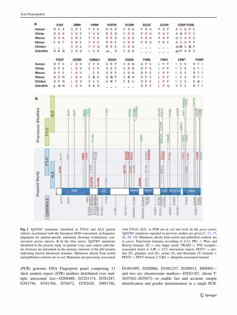
Acta Neuropathol
1 3
(PCr) genomic DNA Fingerprint panel comprising 13 short tandem repeat (STr) markers distributed over mul-tiple autosomal loci—D20S480, D22S1174, D3S1287, D3S1744, D3S1764, D7S672, D7S2426, D8S1746,
D14S1005, D20S866, D10S1237, D20S912, D6S965—and two sex chromosome markers—DXS1187, chrom Y: 2655362–2655672—to enable fast and accurate sample identification and gender determination in a single PCr.
Fig. 1 SQSTM1 mutations identified in FTlD and AlS patient cohorts ascertained with the european eOD consortium. a Sequence alignment for patient-specific mutations showing evolutionary con-servation across species. b In the blue panel, SQSTM1 mutations identified in the present study in patients (top) and control individu-als (bottom) are presented on the primary structure if the p62 protein indicating known functional domains. Mutations absent from tested and published controls are in red. Mutations not previously associated
with FTlD, AlS, or PDB are in red and bold. In the green panel, SQSTM1 mutations reported in previous studies are given [7, 11, 17, 28, 29, 33]. Mutations absent from tested and published controls are in green. Functional domains according to [11]: PB1 = Phox and Bem1p domain; ZZ = zinc finger motif; TrAF6 = TNF receptor–associated factor 6; lIr = lC3 interaction region; PeST1 = pro-line (P), glutamic acid (e), serine (S), and threonine (T) domain 1; PeST2 = PeST domain 2; UBA = ubiquitin-associated domain

Acta Neuropathol
1 3
After selective amplification of 20 ng gDNA under empiri-cally defined reaction conditions, amplification products were size separated on an ABI 3730 automatic sequencer (Applied Biosystems) using geneScan-600 lIZ (Applied
Biosystems) as internal size standard and genotypes were assigned using in-house developed TracI genotyping soft-ware (http://www.vibgeneticservicefacility.be). Duplicate samples, gender mismatches and failed samples due to low
Table 1 SQSTM1 mutations present only in patients and associated clinical phenotypes
Functional domains according to [11] (Fig. 1b)
bvFTD behavioral variant frontotemporal dementia, MND motor neuron disease, PSP progressive supranuclear palsy, PNFA progressive non-fluent aphasia, F familial, S sporadic, U family history undocumenteda Indicates variants not previously associated with AlS, FTlD or PDB [7, 11, 17, 28, 29, 33]. For a complete description of SQSTM1 muta-tions, see Supplementary table 2b Clinical subdiagnosis is given where documentedc After revision of the medical records of the mutation carriers, a diagnosis of possible PDB was made in hindsight in this patient
Mutation Functional domain
Origin gender Clinical diag-nosis
Sub-diagnosisb Family his-tory
Age at onset (years)
Age at death (years)
FTlD
p.Ala16Vala Italian M FTlD-AlS U 71 74
p.Asp80glua PB1 Italian F FTlD bvFTD F 71 85
p.Val90Met PB1 Portuguese F FTlD bvFTD U 41
p.Arg212Cysa Austrian M FTlD-AlS F 63 66
p.gly219Vala Portuguese M FTlD bvFTD F 52
p.Ser226Proa TrAF6 Spanish M FTlD bvFTD S 61
p.Pro228leu TrAF6 german M FTlD bvFTD S 57
p.Pro232Thra TrAF6 Portuguese F FTlDc bvFTD F 55 68
p.glu280dela PeST1 Italian F FTlD PSP F 73
p.Arg321His lIr Italian F FTlD U 68
p.Asp329glya lIr Spanish M FTlD bvFTD U 78 84
p.Pro348leu PeST2 Italian M FTlD PNFA F 74
p.Pro387leu UBA Italian F FTlD PNFA F 65
p.Pro387leu UBA Italian M FTlD bvFTD S 66
p.(glu396*) UBA Czech M FTlD bvFTD S 43 47
p.Thr430Proa UBA Portuguese M FTlD bvFTD S 58 63
AlS
p.Arg107Trpa Spanish F AlS MND S 58 62
p.Asp129Asna ZZ Flemish M AlS S 62
p.Asp258Asna german F AlS F 52 62
Table 2 SQSTM1 mutations present in patients and control individuals
Functional domains according to [11] (Fig. 1b)a Indicates variants not previously associated with AlS, FTlD or PDB [7, 11, 17, 28, 29, 33]. For a complete description of the SQSTM1 mutations, see Supplementary table 2
Mutation Functional domain FTlD n = 1,808 AlS n = 395 Controls n = 3,899
p.Ala17Vala 1 1
p.Ala33Val PB1 1 1
p.lys103Arg PB1 1 2
p.Ala117Val 1 3
p.Pro118Ser 1 2
p.Val153Ile ZZ 1 3
p.lys238glu TrAF6 17 6 14
p.glu274Asp PeST1 109 22 79
p.Arg321Cys lIr 3 1 1
p.Pro392leu UBA 15 3 11
p.Pro439leu UBA 2 2
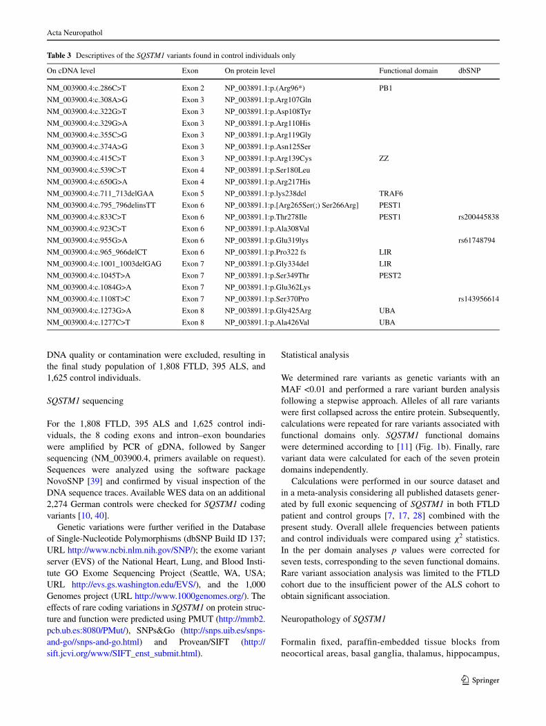
Acta Neuropathol
1 3
DNA quality or contamination were excluded, resulting in the final study population of 1,808 FTlD, 395 AlS, and 1,625 control individuals.
SQSTM1 sequencing
For the 1,808 FTlD, 395 AlS and 1,625 control indi-viduals, the 8 coding exons and intron–exon boundaries were amplified by PCr of gDNA, followed by Sanger sequencing (NM_003900.4, primers available on request). Sequences were analyzed using the software package NovoSNP [39] and confirmed by visual inspection of the DNA sequence traces. Available WeS data on an additional 2,274 german controls were checked for SQSTM1 coding variants [10, 40].
genetic variations were further verified in the Database of Single-Nucleotide Polymorphisms (dbSNP Build ID 137; Url http://www.ncbi.nlm.nih.gov/SNP/); the exome variant server (eVS) of the National Heart, lung, and Blood Insti-tute gO exome Sequencing Project (Seattle, WA, USA; Url http://evs.gs.washington.edu/eVS/), and the 1,000 genomes project (Url http://www.1000genomes.org/). The effects of rare coding variations in SQSTM1 on protein struc-ture and function were predicted using PMUT (http://mmb2.pcb.ub.es:8080/PMut/), SNPs&go (http://snps.uib.es/snps-and-go//snps-and-go.html) and Provean/SIFT (http://sift.jcvi.org/www/SIFT_enst_submit.html).
Statistical analysis
We determined rare variants as genetic variants with an MAF <0.01 and performed a rare variant burden analysis following a stepwise approach. Alleles of all rare variants were first collapsed across the entire protein. Subsequently, calculations were repeated for rare variants associated with functional domains only. SQSTM1 functional domains were determined according to [11] (Fig. 1b). Finally, rare variant data were calculated for each of the seven protein domains independently.
Calculations were performed in our source dataset and in a meta-analysis considering all published datasets gener-ated by full exonic sequencing of SQSTM1 in both FTlD patient and control groups [7, 17, 28] combined with the present study. Overall allele frequencies between patients and control individuals were compared using χ2 statistics. In the per domain analyses p values were corrected for seven tests, corresponding to the seven functional domains. rare variant association analysis was limited to the FTlD cohort due to the insufficient power of the AlS cohort to obtain significant association.
Neuropathology of SQSTM1
Formalin fixed, paraffin-embedded tissue blocks from neocortical areas, basal ganglia, thalamus, hippocampus,
Table 3 Descriptives of the SQSTM1 variants found in control individuals only
On cDNA level exon On protein level Functional domain dbSNP
NM_003900.4:c.286C>T exon 2 NP_003891.1:p.(Arg96*) PB1
NM_003900.4:c.308A>g exon 3 NP_003891.1:p.Arg107gln
NM_003900.4:c.322g>T exon 3 NP_003891.1:p.Asp108Tyr
NM_003900.4:c.329g>A exon 3 NP_003891.1:p.Arg110His
NM_003900.4:c.355C>g exon 3 NP_003891.1:p.Arg119gly
NM_003900.4:c.374A>g exon 3 NP_003891.1:p.Asn125Ser
NM_003900.4:c.415C>T exon 3 NP_003891.1:p.Arg139Cys ZZ
NM_003900.4:c.539C>T exon 4 NP_003891.1:p.Ser180leu
NM_003900.4:c.650g>A exon 4 NP_003891.1:p.Arg217His
NM_003900.4:c.711_713delgAA exon 5 NP_003891.1:p.lys238del TrAF6
NM_003900.4:c.795_796delinsTT exon 6 NP_003891.1:p.[Arg265Ser(;) Ser266Arg] PeST1
NM_003900.4:c.833C>T exon 6 NP_003891.1:p.Thr278Ile PeST1 rs200445838
NM_003900.4:c.923C>T exon 6 NP_003891.1:p.Ala308Val
NM_003900.4:c.955g>A exon 6 NP_003891.1:p.glu319lys rs61748794
NM_003900.4:c.965_966delCT exon 6 NP_003891.1:p.Pro322 fs lIr
NM_003900.4:c.1001_1003delgAg exon 7 NP_003891.1:p.gly334del lIr
NM_003900.4:c.1045T>A exon 7 NP_003891.1:p.Ser349Thr PeST2
NM_003900.4:c.1084g>A exon 7 NP_003891.1:p.glu362lys
NM_003900.4:c.1108T>C exon 7 NP_003891.1:p.Ser370Pro rs143956614
NM_003900.4:c.1273g>A exon 8 NP_003891.1:p.gly425Arg UBA
NM_003900.4:c.1277C>T exon 8 NP_003891.1:p.Ala426Val UBA

Acta Neuropathol
1 3
brainstem and cerebellum were evaluated. In addition to Hematoxylin and eosin staining, the following mono-clonal (mouse) antibodies were used for immunohisto-chemistry: monoclonal anti-p62 (1:1,000, BD Transduc-tion, lexington KY, USA), anti-tau AT8 (pS202/pT205, 1:200, Pierce Biotechnology, rockford, Il, USA),
anti-phospho-TDP-43 (pS409/410, 1:2,000, Cosmo Bio, Tokyo, Japan), anti-α-synuclein (1:2,000, clone 5g4, roboscreen, leipzig, germany; specific for disease-associated form), anti-Aβ (1:50, clone 6F/3D, Dako, glostrup, Denmark). The DAKO enVision© detection kit, peroxidase/DAB, rabbit/mouse (Dako, glostrup,
Fig. 2 Neuropathology observed in SQSTM1 mutation carriers. Immunostaining for phospho-TDP-43 in the temporal cortex (a, b) and in the granule cells of the dentate gyrus (c, d) in a patient with a SQSTM1 p.(glu396*) mutation (a, c) and a second patient with a SQSTM1 p.Arg212Cys—C9orf72 double mutation (b, d). Notably,
p62 immunoreactivity is less in the dentate gyrus (e, f) and lacking in the cerebellar granule cell layer (g, h) in the p.(glu396*) case (e, g) as compared to the p.Arg212Cys patient with the additional C9orf72 repeat expansion mutation (f, h). Scale bar represents 25 µm for all
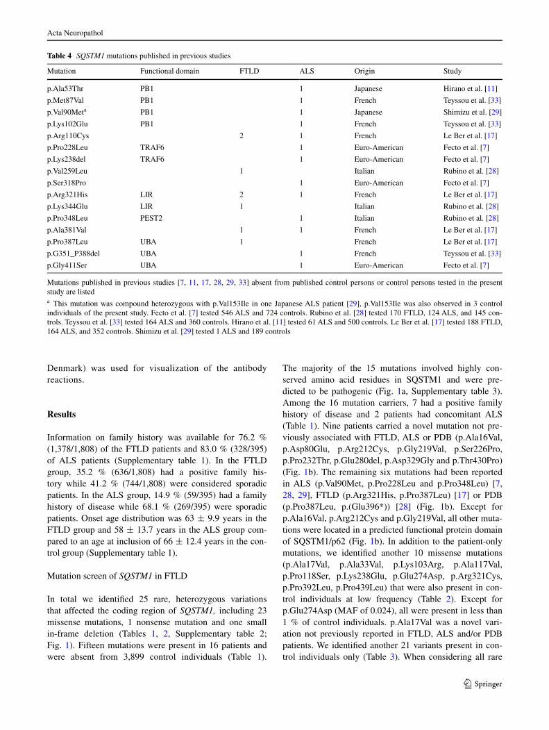
Acta Neuropathol
1 3
Denmark) was used for visualization of the antibody reactions.
Results
Information on family history was available for 76.2 % (1,378/1,808) of the FTlD patients and 83.0 % (328/395) of AlS patients (Supplementary table 1). In the FTlD group, 35.2 % (636/1,808) had a positive family his-tory while 41.2 % (744/1,808) were considered sporadic patients. In the AlS group, 14.9 % (59/395) had a family history of disease while 68.1 % (269/395) were sporadic patients. Onset age distribution was 63 ± 9.9 years in the FTlD group and 58 ± 13.7 years in the AlS group com-pared to an age at inclusion of 66 ± 12.4 years in the con-trol group (Supplementary table 1).
Mutation screen of SQSTM1 in FTlD
In total we identified 25 rare, heterozygous variations that affected the coding region of SQSTM1, including 23 missense mutations, 1 nonsense mutation and one small in-frame deletion (Tables 1, 2, Supplementary table 2; Fig. 1). Fifteen mutations were present in 16 patients and were absent from 3,899 control individuals (Table 1).
The majority of the 15 mutations involved highly con-served amino acid residues in SQSTM1 and were pre-dicted to be pathogenic (Fig. 1a, Supplementary table 3). Among the 16 mutation carriers, 7 had a positive family history of disease and 2 patients had concomitant AlS (Table 1). Nine patients carried a novel mutation not pre-viously associated with FTlD, AlS or PDB (p.Ala16Val, p.Asp80glu, p.Arg212Cys, p.gly219Val, p.Ser226Pro, p.Pro232Thr, p.glu280del, p.Asp329gly and p.Thr430Pro) (Fig. 1b). The remaining six mutations had been reported in AlS (p.Val90Met, p.Pro228leu and p.Pro348leu) [7, 28, 29], FTlD (p.Arg321His, p.Pro387leu) [17] or PDB (p.Pro387leu, p.(glu396*)) [28] (Fig. 1b). except for p.Ala16Val, p.Arg212Cys and p.gly219Val, all other muta-tions were located in a predicted functional protein domain of SQSTM1/p62 (Fig. 1b). In addition to the patient-only mutations, we identified another 10 missense mutations (p.Ala17Val, p.Ala33Val, p.lys103Arg, p.Ala117Val, p.Pro118Ser, p.lys238glu, p.glu274Asp, p.Arg321Cys, p.Pro392leu, p.Pro439leu) that were also present in con-trol individuals at low frequency (Table 2). except for p.glu274Asp (MAF of 0.024), all were present in less than 1 % of control individuals. p.Ala17Val was a novel vari-ation not previously reported in FTlD, AlS and/or PDB patients. We identified another 21 variants present in con-trol individuals only (Table 3). When considering all rare
Table 4 SQSTM1 mutations published in previous studies
Mutations published in previous studies [7, 11, 17, 28, 29, 33] absent from published control persons or control persons tested in the present study are listeda This mutation was compound heterozygous with p.Val153Ile in one Japanese AlS patient [29], p.Val153Ile was also observed in 3 control individuals of the present study. Fecto et al. [7] tested 546 AlS and 724 controls. rubino et al. [28] tested 170 FTlD, 124 AlS, and 145 con-trols. Teyssou et al. [33] tested 164 AlS and 360 controls. Hirano et al. [11] tested 61 AlS and 500 controls. le Ber et al. [17] tested 188 FTlD, 164 AlS, and 352 controls. Shimizu et al. [29] tested 1 AlS and 189 controls
Mutation Functional domain FTlD AlS Origin Study
p.Ala53Thr PB1 1 Japanese Hirano et al. [11]
p.Met87Val PB1 1 French Teyssou et al. [33]
p.Val90Meta PB1 1 Japanese Shimizu et al. [29]
p.lys102glu PB1 1 French Teyssou et al. [33]
p.Arg110Cys 2 1 French le Ber et al. [17]
p.Pro228leu TrAF6 1 euro-American Fecto et al. [7]
p.lys238del TrAF6 1 euro-American Fecto et al. [7]
p.Val259leu 1 Italian rubino et al. [28]
p.Ser318Pro 1 euro-American Fecto et al. [7]
p.Arg321His lIr 2 1 French le Ber et al. [17]
p.lys344glu lIr 1 Italian rubino et al. [28]
p.Pro348leu PeST2 1 Italian rubino et al. [28]
p.Ala381Val 1 1 French le Ber et al. [17]
p.Pro387leu UBA 1 French le Ber et al. [17]
p.g351_P388del UBA 1 French Teyssou et al. [33]
p.gly411Ser UBA 1 euro-American Fecto et al. [7]
Acta Neuropathol
1 3
variants (MAF <0.01) observed in FTlD patients an over-all frequency was calculated of 3.2 % (58/1,808).
Mutation screen of SQSTM1 in AlS
For comparison with our FTlD findings, we analyzed the 395 AlS patients that were concurrently ascertained within the eU eOD consortium (Fig. 1). We identified three novel missense mutations present only in the AlS patients (p.Arg107Trp, p.Asp129Asn and p.Asp258Asn) (Table 1) and five other missense mutations (p.Val153Ile, p.lys238glu, p.glu274Asp, p.Arg321Cys and p.Pro392leu) also present in control individuals at low fre-quency (Table 2, Supplementary table 2).
Association of rare SQSTM1 variants
We performed a burden analysis collapsing all rare vari-ants with an MAF <0.01 across the whole protein. The same frequency of rare alleles was calculated for FTlD patients 0.016 (58/3,616 rare variant alleles) as for control individuals 0.016 (125/7,798; p value = 0.997, n.s.). We repeated the same analysis for rare variants present in func-tional domains only, resulting in a shift in allele frequen-cies of 0.014 (52/3,616) in FTlD versus 0.011 (84/7,798) controls, although not reaching statistical significance (p = 0.098). To increase genetic power, we considered all published datasets that were generated by full exonic sequencing of SQSTM1 in both FTlD patient and control groups [7, 17, 28] and included the data in a meta-analysis with the present study comprising mutant allele frequencies in 4,332 FTlD and 10,240 control alleles. Burden analy-sis of all rare variants across the protein showed again no increase in patients (70/4,332 = 0.016 in FTlD versus 142/10,240 = 0.014 in controls; p value = 0.291, n.s.), but for the rare variants associated with functional domains a marked significant increase could be calculated of 0.014 (61/4,332 rare variant alleles) in FTlD patients versus 0.009 (97/10,240) in control individuals (relative risk over-all (rr) = 1.49 [95 % CI 1.08–2.06]; p value = 0.014).
Subsequently, we investigated which of the functional domains were most contributing to the association. rare variant data were calculated for each of the seven protein domains. In our source, FTlD cohort significant associa-tion was found with the C-terminal ubiquitin-associated (UBA) domain in patients (21/3,616 = 0.006 rare vari-ant alleles) versus control individuals (20/7,798 = 0.003) (rr = 2.27 [95 % CI 1.23–4.20]; p value = 0.007, cor-rected p value = 0.049). In the meta-dataset, statistically significant clustering in the UBA domain was confirmed in FTlD (23/4,332 = 0.005 rare variant alleles) ver-sus control individuals (25/10,240 = 0.002) (rr = 2.18 [95 % CI 1.24–3.85]; p value = 0.006, corrected p
value = 0.042). In addition, also the lC3 interaction region (lIr) domain showed suggestive clustering of rare vari-ants in FTlD (8/4,332 = 0.002) versus control individuals (5/10,240 = 0.0005) (rr = 3.79 [95 % CI 1.24–11.58]; p value = 0.012, corrected p value = 0.084).
Histopathology associated with SQSTM1 mutations
Detailed histopathology was performed on autopsy brain of two carriers of a nonsense mutation p.(glu396*) located in the UBA domain and a missense mutation p.Arg212Cys in a patient who also carried a C9orf72 repeat expansion. Both cases showed widespread phospho-TDP-43 immu-noreactive inclusions in neurons and glial cells. In the p.(glu396*) carrier, frontal and temporal cortical areas mainly displayed neuronal cytoplasmic inclusions. Neu-ronal cytoplasmic inclusions were further abundant in the granule cells of the dentate gyrus. In addition, many glial phospho-TDP-43 inclusions were seen in the white matter. Although this brain suffered severe ischemic/hypoxic dam-age, inclusion bodies were clearly recognized as a distinc-tive feature. In contrast, neuropathological features of the p.Arg212Cys carrier revealed alterations reminiscent of the C9orf72 pathology, including the characteristic p62-pos-itive but phospho-TDP-43-negative inclusions in the hip-pocampal pyramidal layer and cerebellar granule cells (Fig. 2).
Discussion
Previous studies in AlS (total n = 895) [7, 11, 28, 33], and FTlD (total n = 358) [17, 28] estimated the frequency of SQSTM1 coding mutations at 2.42–3.28 % in AlS and 1.76–2.13 % in FTlD. Because of this relatively low prevalence, large patient and control groups are needed to obtain a reliable estimation of the mutation frequency of SQSTM1. Mutations in SQSTM1 were first described in patients with PDB [16]. Up to one-third of patients with familial PDB are explained by a SQSTM1 mutation, with p.Pro392leu as the most commonly observed mutation [4]. A relationship between PDB and FTlD has been pre-viously demonstrated by the identification of causal muta-tions in VCP in families segregating the rare syndrome of inclusion body myopathy with Paget disease of bone and frontotemporal lobar degeneration (IBMPFD) [38]. later studies demonstrated that VCP mutations express signifi-cant clinical heterogeneity and patients can present with all, a combination of two or just one of the three core phe-notypes of IBMPFD [24, 36]. Also more recently, WeS identified a VCP mutation segregating in a family with AlS and subsequent screenings in AlS patients identified additional VCP mutations [12].

Acta Neuropathol
1 3
In this study, we analyzed a european cohort of 1,808 FTlD patients for mutations in SQSTM1 and identified 25 mutations in the coding sequence of which 15 muta-tions in 16 patients were absent from 3,899 control per-sons. This resulted in a mutation frequency for SQSTM1 of 0.9 % overall and 1.1 % in familial FTlD patients. Some of the mutations are listed in dbSNP Short genetic Variations database, the 1,000 genomes Project database or the exome variant server (eVS), yet at very low fre-quencies of MAF ≤0.001 (Supplementary table 2). The SQSTM1 mutation frequency that we calculated in FTlD is lower than in previous studies. This can potentially be explained by the fact that we excluded some of the pre-viously reported patient-only mutations from our calcu-lations since they were present in our extended control group, though at low frequencies <0.01 % (Fig. 1b). This was the case for p.Arg321Cys, p.Ser370Pro, p.gly425Arg [7]; p.Ala33Val, p.Pro392leu [7, 17]; p.lys238glu, p.glu319lys [28]; p.Pro439leu [11]; and p.Val153Ile [7, 29]. In Table 4, all SQSTM1 mutations are listed that have been published [7, 11, 17, 28, 29, 33] and were absent from published and tested control persons. When we considered all rare variants (MAF < 0.01), indifferent of whether or not they appeared in control individuals, we obtained a mutation frequency of 3.2 % in FTlD patients which is the same as calculated in the pooled data analy-sis of the present and published FTlD cohorts [17, 28]. Overall, no statistical association in patients versus control individuals was observed when pooling all rare SQSTM1 variants, not in our study nor in the meta-analysis with all published patient and control datasets [7, 17, 28]. Yet, when considering only domain-associated variants, a trend toward association was observed in our study and a sig-nificant increase in patients was reached in the more pow-erful meta-analysis dataset. Per domain analysis indicated that association was driven by the UBA domain and pos-sibly also the lIr domain. The C-terminal UBA domain, which is primarily affected in PDB, contained significant more variants in FTlD patients when compared to control individuals in both our study and the meta-analysis (0.5 versus 0.2 %) (rrmeta = 2.18 [95 % CI 1.24–3.85]; nomi-nal p valuemeta = 0.006; corrected p valuemeta = 0.042). In the lIr domain through which SQSTM1 binds the autophagy effector protein lC3, 0.2 % of FTlD patients carried a mutation versus 0.05 % of control individuals (rrmeta = 3.79 [95 % CI 1.24–11.58]; nominal p valuem-
eta = 0.012; corrected p valuemeta = 0.084). In contrast to rubino and colleagues who sequenced up to 1,700 bp into the SQSTM1 promoter and detected 4 variants in 4 out of 170 FTlD patients absent from 145 control individuals (c. −1,221 g>A, c. −1,165 C>T, c. −1,153 C>g, c. −673 T>C) [28], we did not investigate the SQSTM1 promoter for rare variants.
The pathogenicity of SQSTM1 mutations, even those that were absent from control individuals, remains unclear at this stage. Most of the patient-only mutations involved highly conserved amino acid residues, with 75 % of the mutations residing in predicted functional domains. In silico prediction programs of amino acid changes indicated that all mutations might be pathogenic by one or more of the in silico programs. Also, the three mutations that resided out-side predicted domains were scored as deleterious by one to all four in silico programs. Of course, these in silico predic-tions are merely indicative and should be interpreted with caution. In contrast to PDB-associated mutations which cluster at the UBA domain [11, 16], FTlD mutations are mostly distributed throughout the protein as also previously reported for AlS, though significantly more mutations seem to be located in the UBA domain. Further, le Ber and col-leagues provided evidence for co-segregation with FTlD for two mutations in the UBA domain, p.Pro387leu and p.Pro392leu [17]. The p.Pro387leu mutation was also pre-sent in two unrelated Italian FTlD patients in our study who did not share a common haplotype, suggesting a recurrent de novo mutation. No DNA of relatives of these two muta-tion carriers was available to test for co-segregation. Three mutation carriers (p.Ala16Val, p.Arg212Cys, p.gly219Val) also carried a pathogenic C9orf72 repeat expansion. At this stage, we have no clear indication that the co-existence of these mutations influences clinical expression of disease. Onset ages were not different from those in patients carry-ing only a SQSTM1 mutation. In a Japanese sporadic late-onset AlS patient with pathology confirmed predominant lower motor neuron disease, a compound heterozygous SQSTM1 mutation was detected, p.[Val90Met(;)Val153Ile] [29]. We did not observe compound heterozygous SQSTM1 mutation carriers, though both mutations present in the Jap-anese patient were also observed in our study, p.Val90Met in one FTlD patient and p.Val153Ile in one other FTlD patient and three control individuals. One could hypothesize that the rare SQSTM1 variants act as high to intermediate penetrant risk alleles and in case of the Japanese patient both had to be present to exert the clinical profile of AlS.
The hallmark lesions of FTlD-TDP are neuronal and glial inclusions with positive immunoreactivity for phospho-TDP-43. In addition to sporadic patients, until now mutations in VCP, GRN and C9orf72 have been associated consistently with TDP-43 pathology, while TArDBP mutations are less frequently observed in FTlD [19]. Here, we provide histo-pathological evidence that SQSTM1 mutations are associ-ated with TDP-43 pathological inclusions. Interestingly, the patient with only a SQSTM1 mutation (p.glu396*) showed widespread neuronal and glial phospho-TDP-43 pathol-ogy. The patient (p.Arg212Cys) who carried also a C9orf72 mutation showed C9orf72-related pathology without other noticeable distinctive features. Potential explanations could

Acta Neuropathol
1 3
be that the C9orf72 mutation masked the effect of the SQST-M1mutation, or that the C9orf72 pathology dominates the SQSTM1 pathology. Also the SQSTM1 p.Arg212Cys muta-tion may well have a lower penetrance and be associated with more subtle pathological changes than the (p.glu396*) mutation located in the UBA domain. These questions merit further investigation and more SQSTM1-positive patholo-gies will need to be evaluated to resolve these issues. Since mutations in SQSTM1 were first linked to PDB, we carefully reexamined medical records of all carriers of patient-only SQSTM1 mutations for indications of (sub) clinical symp-toms of PDB. Only in one Portuguese patient carrying the p.Pro232Thr mutation located in the TrAF6 domain, the diagnosis of possible PDB could be made at hindsight. We, however, cannot exclude that the presence of PDB might have been missed in the other patients since PDB often remains asymptomatic and a diagnosis requires radiography. likewise we cannot exclude that some of the control indi-viduals, who were selected based on absence of dementia-related symptoms, may have been at risk for/or suffered from PDB, in particular, those with the PDB-associated SQSTM1 p.P392l mutation.
In conclusion, our study represents one of the larg-est screening efforts of FTlD patients for mutations in SQSTM1. Further, we combined the data from our screen-ing with that of published datasets in a meta-analysis of rare SQSTM1 variants in a total of 4,432 FTlD and 10,240 con-trol alleles. Both our study and the meta-analysis calculated a mutation frequency of 3.2 % in FTlD patients. Also, the meta-analysis suggested that rare mutations clustering in the UBA domain of SQSTM1 may influence disease sus-ceptibility by doubling the risk for FTlD. Histopathology of autopsied brain of SQSTM1 mutation carriers demon-strated a widespread neuronal and glial phospho-TDP-43 pathology. Taken together, our findings provide additional evidence that SQSTM1 is implicated in the pathogenicity of FTlD/AlS spectrum diseases.
Acknowledgments The authors are grateful to the personnel of the genetic Service Facility for their support of the genetic analyses and to the different neurological centers for their contribution to the diag-nosis and sampling of patients. The data generation for this paper was in part funded by the Metlife Award for Medical research to C.V.B., USA; the Belgian Science Policy Office (BelSPO) Interuniversity Attraction Poles program, the Flemish government support to the european Initiative on Centers of excellence in Neurodegeneration (CoeN), the Flemish government initiated Methusalem excellence program, the Alzheimer research Foundation (SAO/FrA), the Medi-cal Foundation Queen elisabeth, the research Foundation Flanders (FWO), the Agency for Innovation by Science and Technology Flan-ders (IWT) and the University of Antwerp research Fund, Belgium. The FWO provided a postdoctoral fellowship to J.v.d.Z. and a clini-cal investigatorship to P.V.D. The Brescia IRCCS Fatebenefratelli site was funded by the ricerca Corrente, Italian Ministry of Health. r.g. was funded by Fondazione CArIPlO, grant Number 2009-2633. The Barcelona ACE site thank the patients and controls who participated
in this project. We are indebted to Trinitat Port-Carbó and her fam-ily who are supporting Fundació ACe research programs. The Lisbon site acknowledges a grant by grunenthal. The Florence site acknowl-edges Prin 2010-prot. 2010PWNJXK; Cassa di rispario di Firenze e Cassa di risparmio di Pistoia e Pescia. The Stockholm site was financially supported by the Programme in Neuroscience at Karolin-ska Institutet (StratNeuro); the regional agreement on medical train-ing and clinical research (AlF) between Stockholm County Council and Karolinska Institutet; Swedish Alzheimer Foundation; Swedish research Council; Karolinska Institutet PhD-student funding; King gustaf V and Queen Victoria’s Free Mason Foundation; the gun and Bertil Stohne’s Foundation, Foundation for Old Servants; Clinicians including Dr Vesna Jelic and Anne Börjesson-Hanson. For the Munich Institute of Human Genetics site H.P. was supported by the e-rare project geNOMIT (01gM1207), and the german Network for mito-chondrial disorders (mitoNeT 01gM1113C). T.B.H. was supported by the NBIA disorders association. The Barcelona Sant Pau site was in part funded by a grant from the Spanish Ministry of economy and Competitiveness (grant Number PI12/01311) and CIBerNeD. The Pamplona Center for Applied Medical Research is indebted the UTe project from the Foundation for Applied Medical research (FIMA) and CIBerNeD. The Barcelona Neurological Tissue Bank site is indebted to the Neurological Tissue Bank of the IDIBAPS Biobanc for sample and data procurement. The Neurological Tissue Bank acknowledges all brain donors and relatives for generous brain dona-tion for research and referring physicians. The Czech site was par-tially supported by grant IgA NT 12094-5 of grant Agency of the Czech Ministry of Health. The Verona site was in part supported by Fondazione Cariverona (grant Number 2009.1026 “Cognitive and behavioural disability in dementia and psychosis” to gMF). The Liege site was funded by the FNrS.
Conflict of interest The authors report no disclosure with regard to the reported findings.
Open Access This article is distributed under the terms of the Crea-tive Commons Attribution license which permits any use, distribu-tion, and reproduction in any medium, provided the original author(s) and the source are credited.
Appendix
BELNEU consortium Jonathan Baets (Department of Molecular genetics, VIB, Antwerp, Belgium; Institute Born-Bunge, University of Antwerp, Antwerp, Belgium; Antwerp University Hospital, edegem, Belgium); Dirk Nuytten (Hospital Network Antwerp Stuivenberg, Antwerp, Belgium); Jan De Bleecker (University Hospital ghent, ghent, Belgium); Jan Versijpt, Alex Michotte (University Hospital Brussels, Brussels, Belgium); Adrian Ivanoiu (Saint-luc University Hospital, Brussels, Belgium); Olivier Deryck, Bruno Bergmans (AZ Sint-Jan Brugge, Bruges, Belgium); Christiana Willems (Jessa Hospital, Hasselt, Belgium).
EU EOD consortium Anna Paterlini (Proteomics Unit, IrCCS Istituto Centro San giovanni di Dio Fatebenefra-telli, Brescia, Italy); elena Alonso (Neurogenetics labora-tory, Division of Neurosciences, Center for Applied Medi-cal research, Universidad de Navarra, Pamplona, Spain);
Acta Neuropathol
1 3
Manuel Seijo-Martínez (Department of Neurology, Hospital do Salnés, Pontevedra, Spain); ramon rene, Jordi gascon, Jaume Campdelacreu (Department of Neurology, Hos-pital de Bellvitge, Barcelona, Spain); Madalena Martins, Mafalda Matos, André Janeiro, Ana Verdelho (Faculty of Medicine and Institute of Molecular Medicine, University of lisbon, Portugal); Irene Piaceri (Department of Neu-rosciences, Psychology, Drug research and Child Health (NeUrOFArBA) University of Florence, Florence, Italy); Jenny Björkström, Anne Kinhult Ståhlbom, Marie Fallström (Department of geriatric Medicine, genetics unit, Karo-linska University Hospital, Stockholm, Sweden); Charlotte Forsell, Anna-Karin lindström, lena lilius (Karolinska Institutet, Department of Neurobiology, Care Sciences and Society (NVS), KI-Alzheimer Disease research Center, Stockholm, Sweden); Inger Nennesmo (Department of Clinical Pathology and Cytology, Karolinska University Hospital, Stockholm, Sweden); laura Fratiglioni (Aging research Center, Department of Neurobiology, Care Sci-ences and Society (NVS), Karolinska Institutet and Stock-holm University, Stockholm, Sweden);Tamara eisele (Department of Psychiatry and Psychotherapy, Technische Universität München, 81675 München, germany); Thomas Wieland (Institute of Human genetics, Technische Univer-sität München, 81675 Munich, germany); ricard rojas-garcía, Marc Suárez-Calvet (Department of Neurology, IIB Sant Pau, Hospital de la Santa Creu i Sant Pau, Universitat Autònoma de Barcelona, Barcelona, Spain; Center for Net-worker Biomedical research in Neurodegenerative Diseases (CIBerNeD), Madrid, Spain); João Massano (Depart-ment of Neurology Centro Hospitalar São João, Portugal; Department of Clinical Neuroscience and Mental Health, Faculty of Medicine University of Porto, Porto, Portugal); Maria Helena ribeiro (Faculty of Medicine, University of Coimbra, Coimbra, Portugal); Catarina Oliveira (Center for Neuroscience and Cell Biology, University of Coim-bra, Coimbra, Portugal); Jose l Molinuevo, Anna Antonell (Alzheimer’s disease and other cogntive disorders unit. Neurology department, Hospital); robert rusina, Zdenek rohan (Department of Pathology and Molecular Medi-cine, Thomayer Hospital, Prague, Czech republic); rob-ert rusina (Center of Clinical Neurosciences, Department of Neurology, First Medical Faculty, Charles University in Prague, Czech republic); Tiziana Cavallaro (Department of Neuroscience, AOUI Verona, Verona, Italy).
References
1. Al-Sarraj S, King A, Troakes C et al (2011) p62 positive, TDP-43 negative, neuronal cytoplasmic and intranuclear inclusions in the cerebellum and hippocampus define the pathology of C9orf72-linked FTlD and MND/AlS. Acta Neuropathol 122:691–702. doi:10.1007/s00401-011-0911-2
2. Arai T, Nonaka T, Hasegawa M et al (2003) Neuronal and glial inclusions in frontotemporal dementia with or without motor neu-ron disease are immunopositive for p62. Neurosci lett 342:41–44. doi:10.1016/S0304-3940(03)00216-7
3. Brooks Br, Miller rg, Swash M, Munsat Tl (2000) el escorial revisited: revised criteria for the diagnosis of amyotrophic lateral sclerosis. Amyotroph lateral Scler Other Motor Neuron Disord 32:505–514
4. Chung PYJ, Beyens g, guañabens N et al (2008) Founder effect in different european countries for the recurrent P392l SQSTM1 mutation in Paget’s disease of bone. Calcif Tissue Int 83:34–42. doi:10.1007/s00223-008-9137-2
5. Cruts M, gijselinck I, Van langenhove T et al (2013) Cur-rent insights into the C9orf72 repeat expansion diseases of the FTlD/AlS spectrum. Trends Neurosci 36:450–459. doi:10.1016/j.tins.2013.04.010
6. Deng HX, Zhai H, Bigio eH et al (2010) FUS-immunoreactive inclusions are a common feature in sporadic and non-SOD1 familial amyotrophic lateral sclerosis. Ann Neurol 67:739–748
7. Fecto F, Yan J, Vemula SP et al (2011) SQSTM1 mutations in familial and sporadic amyotrophic lateral sclerosis. Arch Neurol 68:1440–1446. doi:10.1001/archneurol.2011.250
8. gelpi e, van der Zee J, Turon estrada A et al (2014) TArDBP mutation p.Ile383Val associated with semantic dementia and complex proteinopathy. Neuropathol Appl Neurobiol 40:225–230. doi:10.1111/nan.12063
9. gennari l, gianfrancesco F, Di Stefano M et al (2010) SQSTM1 gene analysis and gene-environment interaction in Paget’s disease of bone. J Bone Miner res 25:1375–1384. doi:10.1002/jbmr.31
10. Haack TB, Kopajtich r, Freisinger P et al (2013) elAC2 muta-tions cause a mitochondrial rNA processing defect associated with hypertrophic cardiomyopathy. Am J Hum genet 93:211–223. doi:10.1016/j.ajhg.2013.06.006
11. Hirano M, Nakamura Y, Saigoh K et al (2013) Mutations in the gene encoding p62 in Japanese patients with amyotrophic lateral sclerosis. Neurology 80:458–463. doi:10.1212/WNl.0b013e31827f0fe5
12. Johnson JO, Mandrioli J, Benatar M et al (2010) exome sequenc-ing reveals VCP mutations as a cause of familial AlS. Neuron 68:857–864
13. Kimonis Ve, Fulchiero e, Vesa J, Watts g (2008) VCP disease associated with myopathy, Paget disease of bone and frontotem-poral dementia: review of a unique disorder. Biochim Biophys Acta 1782:744–748. doi:10.1016/j.bbadis.2008.09.003
14. Kurihara N, Hiruma Y (2007) Mutation of the sequestosome 1 (p62) gene increases osteoclastogenesis but does not induce Paget disease. J Clin Invest 117:133–142. doi:10.1172/JCI28267
15. Kuusisto e, Salminen A, Alafuzoff I (2001) Ubiquitin-binding protein p62 is present in neuronal and glial inclusions in human tauopathies and synucleinopathies. Neuroreport 12:2085–2090
16. laurin N, Brown JP, Morissette J, raymond V (2002) recur-rent mutation of the gene encoding sequestosome 1 (SQSTM1/p62) in Paget disease of bone. Am J Hum genet 70:1582–1588. doi:10.1086/340731
17. le Ber I, Camuzat A, guerreiro r, Campion D (2013) SQSTM1 mutations in French patients with frontotemporal dementia or frontotemporal dementia with amyotrophic lateral sclerosis. JAMA Neurol 70:1403–1410. doi:10.1001/jamaneurol.2013.3849
18. litvan I, Agid Y, Calne D et al (1996) Clinical research criteria for the diagnosis of progressive supranuclear palsy (Steele-rich-ardson-Olszewski syndrome): report of the NINDS-SPSP inter-national workshop. Neurology 47:1–9
19. Mackenzie IrA, Neumann M, Baborie A et al (2011) A harmo-nized classification system for FTlD-TDP pathology. Acta Neu-ropathol 122:111–113. doi:10.1007/s00401-011-0845-8
20. Mizuno Y, Amari M, Takatama M et al (2006) Immunoreactivities of p62, an ubiqutin-binding protein, in the spinal anterior horn

Acta Neuropathol
1 3
cells of patients with amyotrophic lateral sclerosis. J Neurol Sci 249:13–18. doi:10.1016/j.jns.2006.05.060
21. Mori K, Arzberger T, grässer FA et al (2013) Bidirectional tran-scripts of the expanded C9orf72 hexanucleotide repeat are trans-lated into aggregating dipeptide repeat proteins. Acta Neuro-pathol 126:881–893. doi:10.1007/s00401-013-1189-3
22. Mori K, Weng S-M, Arzberger T et al (2013) The C9orf72 ggggCC repeat is translated into aggregating dipeptide-repeat proteins in FTlD/AlS. Science 339:1335–1338. doi:10.1126/science.1232927
23. Nakaso K, Yoshimoto Y, Nakano T et al (2004) Transcrip-tional activation of p62/A170/ZIP during the formation of the aggregates: possible mechanisms and the role in lewy body formation in Parkinson’s disease. Brain res 1012:42–51. doi:10.1016/j.brainres.2004.03.029
24. Nalbandian A, Donkervoort S, Dec e et al (2011) The multiple faces of valosin-containing protein-associated diseases: inclu-sion body myopathy with Paget’s disease of bone, frontotempo-ral dementia, and amyotrophic lateral sclerosis. J Mol Neurosci 45:522–531. doi:10.1007/s12031-011-9627-y
25. Neary D, Snowden JS, gustafson l et al (1998) Frontotemporal lobar degeneration: a consensus on clinical diagnostic criteria. Neurology 51:1546–1554
26. Pankiv S, Clausen TH, lamark T et al (2007) p62/SQSTM1 binds directly to Atg8/lC3 to facilitate degradation of ubiquit-inated protein aggregates by autophagy. J Biol Chem 282:24131–24145. doi:10.1074/jbc.M702824200
27. rascovsky K, Hodges Jr, Knopman D et al (2011) Sensitiv-ity of revised diagnostic criteria for the behavioural variant of frontotemporal dementia. Brain 134:2456–2477. doi:10.1093/brain/awr179
28. rubino e, rainero I, Chiò A et al (2012) SQSTM1 mutations in frontotemporal lobar degeneration and amyotrophic lateral scle-rosis. Neurology 79:1556–1562. doi:10.1212/WNl.0b013e31826e25df
29. Shimizu H, Toyoshima Y, Shiga A et al (2013) Sporadic AlS with compound heterozygous mutations in the SQSTM1 gene. Acta Neuropathol 126:453–459. doi:10.1007/s00401-013-1150-5
30. Synofzik M, Born C, rominger A, et al. (2013) Targeted high-throughput sequencing identifies a TArDBP mutation as a cause of early-onset FTD without motor neuron disease. Neurobiol Aging 35:1212.e1–5. doi:10.1016/j.neurobiolaging.2013.10.092
31. Synofzik M, Maetzler W, grehl T et al (2012) Screening in AlS and FTD patients reveals 3 novel UBQlN2 mutations outside the PXX domain and a pure FTD phenotype. Neurobiol Aging 33(2949):e13–e17. doi:10.1016/j.neurobiolaging.2012.07.002
32. Tanji K, Zhang H-X, Mori F et al (2012) p62/sequestosome 1 binds to TDP-43 in brains with frontotemporal lobar degen-eration with TDP-43 inclusions. J Neurosci res 90:2034–2042. doi:10.1002/jnr.23081
33. Teyssou e, Takeda T, lebon V et al (2013) Mutations in SQSTM1 encoding p62 in amyotrophic lateral sclerosis: genetics and neuropathology. Acta Neuropathol 125:511–522. doi:10.1007/s00401-013-1090-0
34. Troakes C, Maekawa S, Wijesekera l et al (2011) An MND/AlS phenotype associated with C9orf72 repeat expansion: abundant p62-positive, TDP-43-negative inclusions in cerebral cortex, hippocampus and cerebellum but without associated cognitive decline. Neuropathol Off J Japanese Soc Neuropathol 32:505–514. doi:10.1111/j.1440-1789.2011.01286.x
35. van der Zee J, gijselinck I, Dillen l et al (2013) A Pan-european study of the C9orf72 repeat associated with FTlD: geographic prevalence, genomic instability, and intermediate repeats. Hum Mutat 34:363–373. doi:10.1002/humu.22244
36. van der Zee J, Pirici D, Van langenhove T et al (2009) Clinical heterogeneity in 3 unrelated families linked to VCP p.Arg159His. Neurology 73:626–632
37. Van langenhove T, van der Zee J, Van Broeckhoven C (2012) The molecular basis of the frontotemporal lobar degeneration-amyotrophic lateral sclerosis spectrum. Ann Med 44:817–828. doi:10.3109/07853890.2012.665471
38. Watts gDJ, Wymer J, Kovach MJ et al (2004) Inclusion body myopathy associated with Paget disease of bone and frontotem-poral dementia is caused by mutant valosin-containing protein. Nat genet 36:377–381
39. Weckx S, Del-Favero J, rademakers r et al (2005) novoSNP, a novel computational tool for sequence variation discovery. genome res 15:436–442. doi:10.1101/gr.2754005
40. Zimprich A, Benet-Pagès A, Struhal W et al (2011) A mutation in VPS35, encoding a subunit of the retromer complex, causes late-onset Parkinson disease. Am J Hum genet 89:168–175

lable at ScienceDirect
Neurobiology of Aging xxx (2014) 1e7
Contents lists avai
Neurobiology of Aging
journal homepage: www.elsevier .com/locate/neuaging
Investigation of the role of rare TREM2 variants in frontotemporaldementia subtypes
Mathias Thelen a, Cristina Razquin b,c, Isabel Hernández d, Ana Gorostidi c,e,Raquel Sánchez-Valle f, Sara Ortega-Cubero b,c, Steffen Wolfsgruber a,g, Dmitriy Drichel g,Klaus Fliessbach a,g, Tanja Duenkel h, Marinella Damian h, Stefanie Heilmann i,Anja Slotosch a, Martina Lennarz a, Manuel Seijo-Martínez j, Ramón Rene k,Johannes Kornhuber l, Oliver Petersm, Christian Luckhaus n, Holger Jahn o,Michael Hüll p, Eckart Rüther q, Jens Wiltfang q, Elena Lorenzo b,c, Jordi Gascon k,Alberto Lleó c,r, Albert Lladó f, Jaume Campdelacreu k, Fermin Moreno c,e,s,Hojjat Ahmadzadehfar t, Dementia Genetics Spanish Consortium (DEGESCO),Juan Fortea c,r, Begoña Indakoetxea c,e,s, Michael T. Heneka g,u, Axel Wetter v,Maria A. Pastorw,z, Mario Riverol w, Tim Becker g,x, Lutz Frölich h, Lluís Tárraga d,Mercè Boada d, Michael Wagner a,g, Frank Jessen a,g, Wolfgang Maier a,g,Jordi Clarimón c,r, Adolfo López de Munain c,e,s,y, Agustín Ruiz d, Pau Pastor b,c,w,Alfredo Ramirez a,i,*
aDepartment of Psychiatry and Psychotherapy, University of Bonn, Bonn, GermanybNeurogenetics Laboratory, Division of Neurosciences, Center for Applied Medical Research, University of Navarra School of Medicine, Pamplona, SpaincCenter for Network Biomedical Research into Neurodegenerative Diseases (CIBERNED), Instituto de Salud Carlos III, Madrid, SpaindMemory Clinic of Fundaciò ACE, Institut Català de Neurociències Aplicades, Barcelona, SpaineNeuroscience Area, BioDonostia Health Research Institute, San Sebastian, SpainfAlzheimer’s Disease and Other Cognitive Disorders Unit, Department of Neurology, Hospital Clinic, IDIBAPS, Barcelona, SpaingGerman Center for Neurodegenerative Diseases (DZNE), Bonn, GermanyhDepartment of Geriatric Psychiatry, Central Institute of Mental Health, Medical Faculty Mannheim, University of Heidelberg, Heidelberg, Germanyi Institute of Human Genetics, University of Bonn, Bonn, GermanyjDepartment of Neurology, Hospital do Salnés, Pontevedra, SpainkDepartment of Neurology, Hospital Universitari de Bellvitge, Barcelona, SpainlDepartment of Psychiatry and Psychotherapy, Friedrich-Alexander-University of Erlangen-Nuremberg, Erlangen, GermanymDepartment of Psychiatry, Charité, Berlin, GermanynDepartment of Psychiatry and Psychotherapy, Medical Faculty, Heinrich-Heine University, Düsseldorf, GermanyoDepartment of Psychiatry, University of Hamburg, Hamburg, GermanypCentre for Geriatric Medicine and Section of Gerontopsychiatry and Neuropsychology, Medical School, University of Freiburg, Freiburg, GermanyqDepartment of Psychiatry and Psychotherapy, University of Göttingen, Göttingen, GermanyrDepartment of Neurology, Hospital de la Santa Creu i Sant Pau, IIB-Sant Pau, Universitat Autònoma de Barcelona, Barcelona, SpainsDepartment of Neurology, Donostia University Hospital, San Sebastian, SpaintDepartment of Nuclear Medicine, University of Bonn, Bonn, GermanyuClinical Neuroscience Unit, Department of Neurology, University of Bonn, Bonn, GermanyvDepartment of Radiology, University of Essen, Essen, GermanywDepartment of Neurology, Clínica Universidad de Navarra, University of Navarra School of Medicine, Pamplona, Spainx Institute for Medical Biometry, Informatics, and Epidemiology, University of Bonn, Bonn, GermanyyDepartment of Neurosciences, University of Basque Country, San Sebastian, SpainzNeuroimaging Laboratory, Division of Neuroscience, Center for Applied Medical Research (CIMA), University of Navarra, Pamplona, Spain
MT, CR, PP, and AR contributed equally to the study.* Corresponding author at: Department of Psychiatry and Psychotherapy, University of Bonn, Sigmund-Freud-Strasse 25, Bonn D-53105, Germany. Tel.: þ49 (0)228 287
19323; fax: þ49 (0)228 287 16097.E-mail address: [email protected] (A. Ramirez).
0197-4580/$ e see front matter � 2014 Elsevier Inc. All rights reserved.http://dx.doi.org/10.1016/j.neurobiolaging.2014.06.018
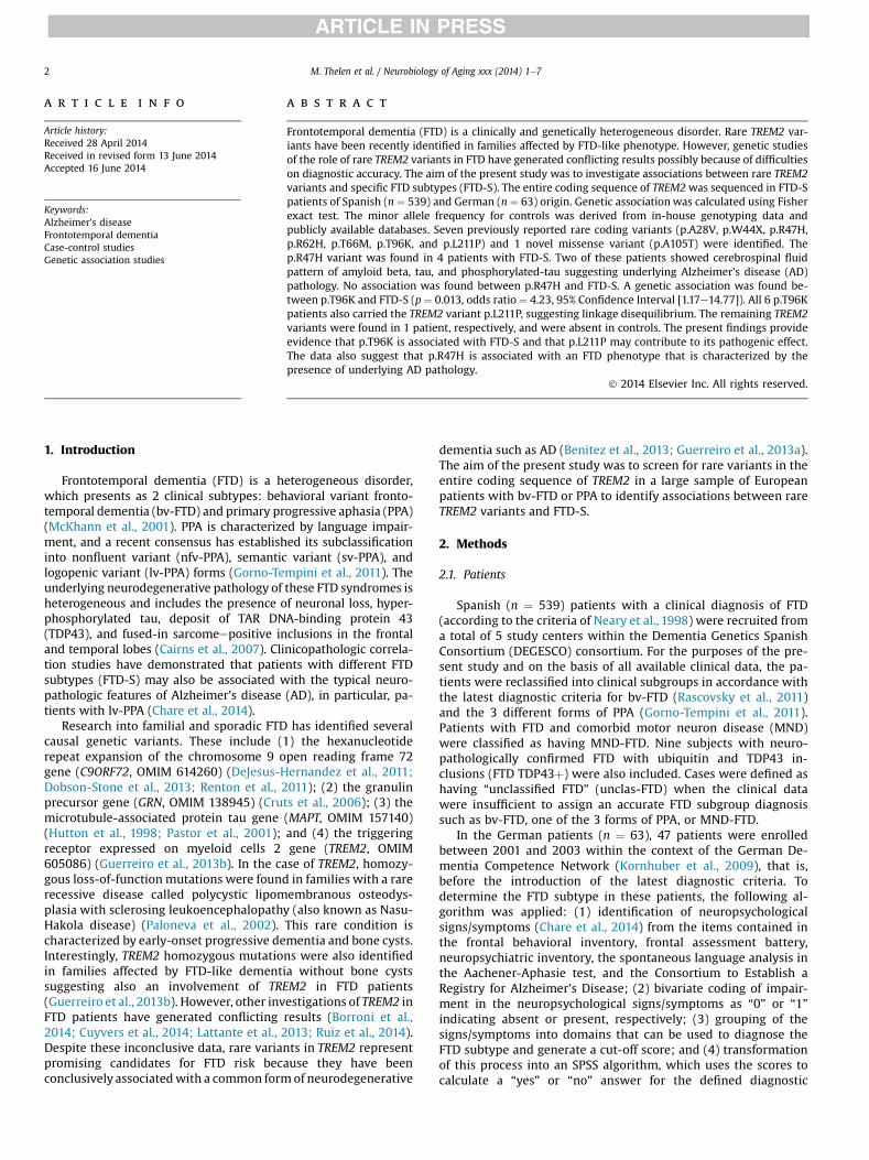
M. Thelen et al. / Neurobiology of Aging xxx (2014) 1e72
a r t i c l e i n f o
Article history:Received 28 April 2014Received in revised form 13 June 2014Accepted 16 June 2014
Keywords:Alzheimer’s diseaseFrontotemporal dementiaCase-control studiesGenetic association studies
a b s t r a c t
Frontotemporal dementia (FTD) is a clinically and genetically heterogeneous disorder. Rare TREM2 var-iants have been recently identified in families affected by FTD-like phenotype. However, genetic studiesof the role of rare TREM2 variants in FTD have generated conflicting results possibly because of difficultieson diagnostic accuracy. The aim of the present study was to investigate associations between rare TREM2variants and specific FTD subtypes (FTD-S). The entire coding sequence of TREM2was sequenced in FTD-Spatients of Spanish (n ¼ 539) and German (n ¼ 63) origin. Genetic associationwas calculated using Fisherexact test. The minor allele frequency for controls was derived from in-house genotyping data andpublicly available databases. Seven previously reported rare coding variants (p.A28V, p.W44X, p.R47H,p.R62H, p.T66M, p.T96K, and p.L211P) and 1 novel missense variant (p.A105T) were identified. Thep.R47H variant was found in 4 patients with FTD-S. Two of these patients showed cerebrospinal fluidpattern of amyloid beta, tau, and phosphorylated-tau suggesting underlying Alzheimer’s disease (AD)pathology. No association was found between p.R47H and FTD-S. A genetic association was found be-tween p.T96K and FTD-S (p ¼ 0.013, odds ratio¼ 4.23, 95% Confidence Interval [1.17e14.77]). All 6 p.T96Kpatients also carried the TREM2 variant p.L211P, suggesting linkage disequilibrium. The remaining TREM2variants were found in 1 patient, respectively, and were absent in controls. The present findings provideevidence that p.T96K is associated with FTD-S and that p.L211P may contribute to its pathogenic effect.The data also suggest that p.R47H is associated with an FTD phenotype that is characterized by thepresence of underlying AD pathology.
� 2014 Elsevier Inc. All rights reserved.
1. Introduction
Frontotemporal dementia (FTD) is a heterogeneous disorder,which presents as 2 clinical subtypes: behavioral variant fronto-temporal dementia (bv-FTD) and primary progressive aphasia (PPA)(McKhann et al., 2001). PPA is characterized by language impair-ment, and a recent consensus has established its subclassificationinto nonfluent variant (nfv-PPA), semantic variant (sv-PPA), andlogopenic variant (lv-PPA) forms (Gorno-Tempini et al., 2011). Theunderlying neurodegenerative pathology of these FTD syndromes isheterogeneous and includes the presence of neuronal loss, hyper-phosphorylated tau, deposit of TAR DNA-binding protein 43(TDP43), and fused-in sarcomeepositive inclusions in the frontaland temporal lobes (Cairns et al., 2007). Clinicopathologic correla-tion studies have demonstrated that patients with different FTDsubtypes (FTD-S) may also be associated with the typical neuro-pathologic features of Alzheimer’s disease (AD), in particular, pa-tients with lv-PPA (Chare et al., 2014).
Research into familial and sporadic FTD has identified severalcausal genetic variants. These include (1) the hexanucleotiderepeat expansion of the chromosome 9 open reading frame 72gene (C9ORF72, OMIM 614260) (DeJesus-Hernandez et al., 2011;Dobson-Stone et al., 2013; Renton et al., 2011); (2) the granulinprecursor gene (GRN, OMIM 138945) (Cruts et al., 2006); (3) themicrotubule-associated protein tau gene (MAPT, OMIM 157140)(Hutton et al., 1998; Pastor et al., 2001); and (4) the triggeringreceptor expressed on myeloid cells 2 gene (TREM2, OMIM605086) (Guerreiro et al., 2013b). In the case of TREM2, homozy-gous loss-of-function mutations were found in families with a rarerecessive disease called polycystic lipomembranous osteodys-plasia with sclerosing leukoencephalopathy (also known as Nasu-Hakola disease) (Paloneva et al., 2002). This rare condition ischaracterized by early-onset progressive dementia and bone cysts.Interestingly, TREM2 homozygous mutations were also identifiedin families affected by FTD-like dementia without bone cystssuggesting also an involvement of TREM2 in FTD patients(Guerreiro et al., 2013b). However, other investigations of TREM2 inFTD patients have generated conflicting results (Borroni et al.,2014; Cuyvers et al., 2014; Lattante et al., 2013; Ruiz et al., 2014).Despite these inconclusive data, rare variants in TREM2 representpromising candidates for FTD risk because they have beenconclusively associatedwith a common form of neurodegenerative
dementia such as AD (Benitez et al., 2013; Guerreiro et al., 2013a).The aim of the present study was to screen for rare variants in theentire coding sequence of TREM2 in a large sample of Europeanpatients with bv-FTD or PPA to identify associations between rareTREM2 variants and FTD-S.
2. Methods
2.1. Patients
Spanish (n ¼ 539) patients with a clinical diagnosis of FTD(according to the criteria of Neary et al., 1998) were recruited froma total of 5 study centers within the Dementia Genetics SpanishConsortium (DEGESCO) consortium. For the purposes of the pre-sent study and on the basis of all available clinical data, the pa-tients were reclassified into clinical subgroups in accordance withthe latest diagnostic criteria for bv-FTD (Rascovsky et al., 2011)and the 3 different forms of PPA (Gorno-Tempini et al., 2011).Patients with FTD and comorbid motor neuron disease (MND)were classified as having MND-FTD. Nine subjects with neuro-pathologically confirmed FTD with ubiquitin and TDP43 in-clusions (FTD TDP43þ) were also included. Cases were defined ashaving “unclassified FTD” (unclas-FTD) when the clinical datawere insufficient to assign an accurate FTD subgroup diagnosissuch as bv-FTD, one of the 3 forms of PPA, or MND-FTD.
In the German patients (n ¼ 63), 47 patients were enrolledbetween 2001 and 2003 within the context of the German De-mentia Competence Network (Kornhuber et al., 2009), that is,before the introduction of the latest diagnostic criteria. Todetermine the FTD subtype in these patients, the following al-gorithm was applied: (1) identification of neuropsychologicalsigns/symptoms (Chare et al., 2014) from the items contained inthe frontal behavioral inventory, frontal assessment battery,neuropsychiatric inventory, the spontaneous language analysis inthe Aachener-Aphasie test, and the Consortium to Establish aRegistry for Alzheimer’s Disease; (2) bivariate coding of impair-ment in the neuropsychological signs/symptoms as “0” or “1”indicating absent or present, respectively; (3) grouping of thesigns/symptoms into domains that can be used to diagnose theFTD subtype and generate a cut-off score; and (4) transformationof this process into an SPSS algorithm, which uses the scores tocalculate a “yes” or “no” answer for the defined diagnostic

M. Thelen et al. / Neurobiology of Aging xxx (2014) 1e7 3
categories. The remaining 16 FTD patients were recruited in theMemory Clinic at the University of Bonn. Clinical subgroups weredefined using the same procedure as with the Spanish sample.
2.2. Controls
Spanish control samples for the association study were obtainedfrom DESGECO consortium (Table 2). The frequency of p.R47H inSpanish healthy individuals was determined using the entireSpanish control sample used by DEGESCO (Ruiz et al., 2014). Todetermine the allele frequency of p.T96K, 539 cognitively normalSpanish controls were selected from the DEGESCO control sample.To obtain the allele frequency of p.R62H, 127 cognitively normalSpanish controls were drawn at random from the DEGESCO controlsample and genotyped.
The German controls were drawn from the AgeCoDe cohort (n¼939). AgeCoDe is an ongoing German multicenter prospectivecohort study. The original AgeCoDe cohort was drawn at randomfrom general practice registries in 6 German cities, and the selectedindividuals were contacted by letter to request participation. Themain inclusion criteria were age �75 years and the absence of de-mentia according to the clinical judgment of the respective generalpractitioner. A total of 3327 AgeCoDe participants were recruitedbetween January 2002 and November 2003 (Jessen et al., 2011;Luck et al., 2007). Each participant undergoes assessments at 18months intervals. All assessments are performed at the partici-pant’s home by a trained study psychologist or physician. For allAgeCoDe controls included in the present study, dementia andmildcognitive dementia had been excluded at the last follow-up visit.The study was approved by the Ethics Committee of each collabo-rating study center. All patients and controls provided writteninformed consent before inclusion.
2.3. TREM2 gene sequencing and genotyping
In the FTD-S patients, genomic DNA was isolated from periph-eral blood using standard procedures, and all coding exons wereamplified using polymerase chain reaction. The primer sequencesare presented in Supplementary Table 1. The entire coding region ofTREM2 was then screened through direct Sanger sequencing of thepolymerase chain reaction products. Mutation numbering wasassigned using the complementary DNA sequence of the GenBankentry NM_018965.3, in which position þ1 corresponds to the A ofthe ATG translation initiation codon. The software packagesPolyPhen-2 (http://genetics.bwh.harvard.edu/pph2/) and Meta-SNP (http://snps.biofold.org/meta-snp/) were used to predict thefunctional and structural effects of amino acid substitutions. Themeta-predictor Meta-SNP combines the output of 3 different pre-diction programs, that is, PhD-SNP, SIFT, and SNAP, to generate aprediction score for putative disease causing variants.
Direct genotyping of p.R62H in the Spanish controls was per-formed using Sequenom iPLEX (Sequenom). The p.T96K variant was
Table 1Demographics of the FTD-S and the control samples
bv-FTD nfv-PPA lv-PPA sv-PPA MND-FTD
Subjects (n) 313 105 59 58 18Sex (% female) 45 54 68 29 39AAO (mean � SD) 62.4 � 14.6 64.7 � 15.8 66.2 � 8.6 62.0 � 12.4 60.8 � 14.8Age (mean � SD)
Key: AAO, age at onset; bv, behavioral variant; FTD, frontotemporal dementia; FTD-S, Fvariant; MND-FTD, FTD with comorbid motor neuron disease; nfv, nonfluent variant; PPAFTD, unclassified FTD.
genotyped in the Spanish control sample using a custom-designedTaqMan assays (Life Technologies).
In the German healthy controls, the p.R47H and p.T96K variantswere genotyped using a custom-designed TaqMan assays (LifeTechnologies). In 1 healthy Spanish control, the p.R62H variant waspresent in a homozygous state. This variant was, therefore,excluded from the genotyping process in German controls.
2.4. Statistical analysis
To calculate the minor allele frequency (MAF) for controls,data from 4 different sources were combined: the published 1000genome (http://browser.1000genomes.org/) MAFs in Europeanpopulation; published data by Guerreiro et al. (2013a); the MAFfor each genotyped variant in the present Spanish control sam-ples obtained from DEGESCO; and the MAF for each genotypedvariant in the present German control sample. Association be-tween case-control status and variant carrier/noncarrier statuswas calculated using Fisher exact test for 2 � 2 contingency ta-bles. The statistical analyses were performed using the imple-mentation in R (The R Project for Statistical Computing, www.r-project.org). The alpha level for statistical significance was setat �0.05. The genotyping data were inspected to detect de-viations from Hardy-Weinberg equilibrium. All single-nucleotidepolymorphisms included in the association were in Hardy-Weinberg equilibrium (p > 0.05).
3. Results
3.1. Rare variants in TREM2 and their associated clinical phenotype
The demographic data of the samples are summarized in Table 1.The presence of at least 1 heterozygous rare TREM2 variant was
identified in 5.32% (32 of 602) of the FTD-S patients (Table 2). Mostof these variants were located in exon 2. These variants comprised7 previously reported rare coding variants (p.A28V, p.W44X,p.R47H, p.R62H, p.T66M, p.T96K, p.L211P) and 1 novel missensevariant p.A105T (Table 2). Four of these variants, that is, p.W44X,p.R47H, p.T66M, and p.T96K, were predicted to have a probablydamaging effect. The other 4 were predicted to be benign (Table 3).The most frequent variant was p.R62H (rs143332484). This waspresent in 19 patients, all of whom displayed heterozygous p.R62Hstatus. Eighteen carriers were of Spanish origin, and 1 wasGerman. Most of the p.R62H carriers were classified as bv-FTDcases (Table 2). The second most frequent rare TREM2 variantstatus was the simultaneous presence of p.T96K (rs2234253) andp.L211P (rs2234256). This was observed in 6 patients, 4 Spanish(lv-PPA [n ¼ 1], bv-FTD [n ¼ 2], FTD-MND [n ¼ 1]) and 2 German(lv-PPA [n ¼ 1] and sv-PPA [n ¼ 1]) patients. The third mostfrequent variant was p.R47H, which has previously been associ-ated with AD. This was found in 4 patients, 3 Spanish lv-PPA pa-tients and 1 German unclas-FTD patient (for clinical details, see
FTD TDP43þ Unclas-FTD Spanish controlsample p.T96K
Spanish controlsample p.R62H
Germancontrol sample
9 40 539 127 93975 37 54 53.5 63.575.0 � 5.5 61.9 � 9.1
62.4 � 12.7 76.57 � 4.89 86.9 � 3.2
TD subtypes; FTD TDP43þ, FTD with ubiquitin and TDP43 inclusions; lv, logopenic, primary progressive aphasia; SD, standard deviation; sv, semantic variant; unclas-
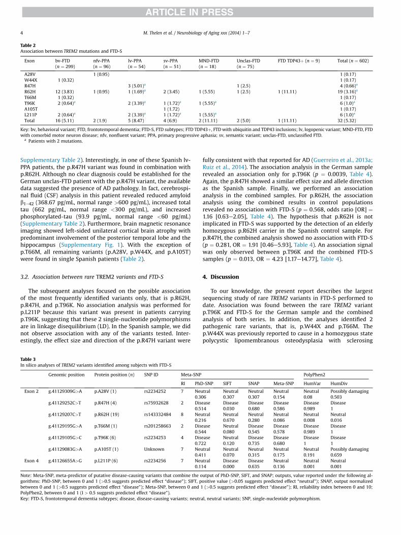
Table 2Association between TREM2 mutations and FTD-S
Exon bv-FTD(n ¼ 299)
nfv-PPA(n ¼ 96)
lv-PPA(n ¼ 54)
sv-PPA(n ¼ 51)
MND-FTD(n ¼ 18)
Unclas-FTD(n ¼ 75)
FTD TDP43þ (n ¼ 9) Total (n ¼ 602)
A28V 1 (0.95) 1 (0.17)W44X 1 (0.32) 1 (0.17)R47H 3 (5.01)a 1 (2.5) 4 (0.66)a
R62H 12 (3.83) 1 (0.95) 1 (1.69)a 2 (3.45) 1 (5.55) 1 (2.5) 1 (11.11) 19 (3.16)a
T66M 1 (0.32) 1 (0.17)T96K 2 (0.64)a 2 (3.39)a 1 (1.72)a 1 (5.55)a 6 (1.0)a
A105T 1 (1.72) 1 (0.17)L211P 2 (0.64)a 2 (3.39)a 1 (1.72)a 1 (5.55)a 6 (1.0)a
Total 16 (5.11) 2 (1.9) 5 (8.47) 4 (6.9) 2 (11.11) 2 (5.0) 1 (11.11) 32 (5.32)
Key: bv, behavioral variant; FTD, frontotemporal dementia; FTD-S, FTD subtypes; FTD TDP43þ, FTD with ubiquitin and TDP43 inclusions; lv, logopenic variant; MND-FTD, FTDwith comorbid motor neuron disease; nfv, nonfluent variant; PPA, primary progressive aphasia; sv, semantic variant; unclas-FTD, unclassified FTD.
a Patients with 2 mutations.
M. Thelen et al. / Neurobiology of Aging xxx (2014) 1e74
Supplementary Table 2). Interestingly, in one of these Spanish lv-PPA patients, the p.R47H variant was found in combination withp.R62H. Although no clear diagnosis could be established for theGerman unclas-FTD patient with the p.R47H variant, the availabledata suggested the presence of AD pathology. In fact, cerebrospi-nal fluid (CSF) analysis in this patient revealed reduced amyloidb1e42 (368.67 pg/mL, normal range >600 pg/mL), increased totaltau (662 pg/mL, normal range <300 pg/mL), and increasedphosphorylated-tau (93.9 pg/mL, normal range <60 pg/mL)(Supplementary Table 2). Furthermore, brain magnetic resonanceimaging showed left-sided unilateral cortical brain atrophy withpredominant involvement of the posterior temporal lobe and thehippocampus (Supplementary Fig. 1). With the exception ofp.T66M, all remaining variants (p.A28V, p.W44X, and p.A105T)were found in single Spanish patients (Table 2).
3.2. Association between rare TREM2 variants and FTD-S
The subsequent analyses focused on the possible associationof the most frequently identified variants only, that is p.R62H,p.R47H, and p.T96K. No association analysis was performed forp.L211P because this variant was present in patients carryingp.T96K, suggesting that these 2 single-nucleotide polymorphismsare in linkage disequilibrium (LD). In the Spanish sample, we didnot observe association with any of the variants tested. Inter-estingly, the effect size and direction of the p.R47H variant were
Table 3In silico analyses of TREM2 variants identified among subjects with FTD-S
Genomic position Protein position (n) SNP ID Meta-SNP
RI PhD
Exon 2 g.41129309G>A p.A28V (1) rs2234252 7 Neu0.3
g.41129252C>T p.R47H (4) rs75932628 2 Dis0.5
g.41129207C>T p.R62H (19) rs143332484 8 Neu0.2
g.41129195G>A p.T66M (1) rs201258663 2 Dis0.5
g.41129105G>C p.T96K (6) rs2234253 4 Dis0.7
g.41129083G>A p.A105T (1) Unknown 7 Neu0.4
Exon 4 g.41126655A>G p.L211P (6) rs2234256 7 Neu0.1
Note: Meta-SNP, meta-predictor of putative disease-causing variants that combine the ogorithms: PhD-SNP, between 0 and 1 (>0.5 suggests predicted effect “disease”); SIFT, pbetween 0 and 1 (>0.5 suggests predicted effect “disease”); Meta-SNP, between 0 and 1PolyPhen2, between 0 and 1 (I > 0.5 suggests predicted effect “disease”).Key: FTD-S, frontotemporal dementia subtypes; disease, disease-causing variants; neutr
fully consistent with that reported for AD (Guerreiro et al., 2013a;Ruiz et al., 2014). The association analysis in the German samplerevealed an association only for p.T96K (p ¼ 0.0039, Table 4).Again, the p.R47H showed a similar effect size and allele directionas the Spanish sample. Finally, we performed an associationanalysis in the combined samples. For p.R62H, the associationanalysis using the combined results in control populationsrevealed no association with FTD-S (p ¼ 0.568, odds ratio [OR] ¼1.16 [0.63e2.05], Table 4). The hypothesis that p.R62H is notimplicated in FTD-S was supported by the detection of an elderlyhomozygous p.R62H carrier in the Spanish control sample. Forp.R47H, the combined analysis showed no association with FTD-S(p ¼ 0.281, OR ¼ 1.91 [0.46e5.93], Table 4). An association signalwas only observed between p.T96K and the combined FTD-Ssamples (p ¼ 0.013, OR ¼ 4.23 [1.17e14.77], Table 4).
4. Discussion
To our knowledge, the present report describes the largestsequencing study of rare TREM2 variants in FTD-S performed todate. Association was found between the rare TREM2 variantp.T96K and FTD-S for the German sample and the combinedanalysis of both series. In addition, the analyses identified 2pathogenic rare variants, that is, p.W44X and p.T66M. Thep.W44X was previously reported to cause in a homozygous statepolycystic lipomembranous osteodysplasia with sclerosing
PolyPhen2
-SNP SIFT SNAP Meta-SNP HumVar HumDiv
tral Neutral Neutral Neutral Neutral Possibly damaging06 0.307 0.307 0.154 0.08 0.503ease Disease Disease Disease Disease Disease14 0.030 0.680 0.586 0.989 1tral Neutral Neutral Neutral Neutral Neutral16 0.670 0.280 0.086 0.008 0.016ease Neutral Disease Disease Disease Disease44 0.080 0.545 0.578 0.989 1ease Neutral Disease Disease Disease Disease22 0.120 0.735 0.680 1 1tral Neutral Neutral Neutral Neutral Possibly damaging11 0.070 0.315 0.175 0.191 0.659tral Disease Disease Neutral Neutral Neutral14 0.000 0.635 0.136 0.001 0.001
utput of PhD-SNP, SIFT, and SNAP; outputs, value reported under the following al-ositive value (>0.05 suggests predicted effect “neutral”); SNAP, output normalized(>0.5 suggests predicted effect “disease”); RI, reliability index between 0 and 10;
al, neutral variants; SNP, single-nucleotide polymorphism.

Table 4Association analysis of the most frequent TREM2 variants (p.R47H, p.R62H and p.T96K) in the present FTD-S cohort
TREM2 variant SNP ID Positiona Location MAF FTD-Spatients
All FTD-S Spanishcontrols
Germancontrols
Controlsb Controlsrecorded1000K(EUR)c
Allcontrols
MAFcontrols(%)
p Value(Fisherexact testd)
Odds ratio(95% CI)
Ce Non-Cf Ce Non-Cf Ce Non-Cf Ce Non-Cf Ce Non-Cf Ce Non-Cf
Combined analysisR47H rs75932628 41129252 Exon 2 0.0033 4 598 3 2166 4 917 5 1100 4 375 16 4558 0.00175 0.281 1.91 (0.46e5.93)R62H rs143332484 41129207 Exon 2 0.0158 19 583 4 123 0 0 31 1073 9 370 44 1566 0.0137 0.568 1.16 (0.63e2.05)T96K rs2234253 41129105 Exon 2 0.00498 6 596 4 526 0 939 3 1102 0 379 7 2946 0.00119 0.013 4.23 (1.17e14.77)
Spanish sampleR47H rs75932628 41129252 Exon 2 0.0028 3 536 3 2166 3 2166 0.00069 0.097 4.04 (0.53e30.23)R62H rs143332484 41129207 Exon 2 0.0167 18 521 4 123 4 123 0.0157 1 1.06 (0.34e4.39)T96K rs2234253 41129105 Exon 2 0.00371 4 534 4 526 4 526 0.00378 1 0.99 (0.18e5.31)
German sampleR47H rs75932628 41129252 Exon 2 0.008 1 62 4 917 4 917 0.0022 0.282 3.7 (0.07e38.02)R62H rs143332484 41129207 Exon 2 0.008 1 62 0 0 31 1073 9 370 40 1443 0.0135 1 0.58 (0.01e3.56)T96K rs2234253 41129105 Exon 2 0.0159 2 61 0 939 0 939 0 0.0039 Inf (2.82 to Inf)
Key: CI, confidence interval; FTD-S, frontotemporal dementia subtypes; MAF, minor allele frequency; MND-FTD, FTD with comorbid motor neuron disease.a The variant position is indicated in terms of base pairs and is based on hg19 (GRCh37).b Allele Counts are those reported in a previous study (Guerreiro et al., 2013a).c http://browser.1000genomes.org/.d As implemented in R (http://www.r-project.org/).e Carriers.f Noncarriers.
M. Thelen et al. / Neurobiology of Aging xxx (2014) 1e7 5
leukoencephalopathy (Paloneva et al., 2002). In the case ofp.T66M, this variant was identified in a homozygous state in aTurkish patient presenting clinically with an FTD-like syndromewithout bone involvement (Guerreiro et al., 2013b). In the pre-sent cohort, both variants were found each in a single subjectwith bv-FTD, suggesting that p.W44X and p.T66M may alsocontribute to the etiology of FTD-S. All pathogenic variants in thepresent series were located in exon 2, which encodes the IgV-setdomain. This region of TREM2 is more highly conserved thanother segments of the protein and may play a regulatory role inleukocyte activation (Allcock et al., 2003). Interestingly, most ofthe TREM2 variants identified in previous studies of AD, PD, andFTD were also located in exon 2. These findings, together with thepresent data, suggest that the IgV-set domain plays a central rolein the contribution of TREM2 to neurodegeneration. However,functional studies are warranted to confirm this hypothesis.
Our findings for p.T96K suggest that it is implicated in FTD-S.However, the positive association in the combined analysis ismainly driven by the association found in the German series.Therefore, this association must be interpreted cautiously as itmight be the result of a type I error because of stratification in asample of small size, particularly the German sample. On the otherhand, the p.T96K seems to be a rare variant among 939 Germanelder neurologically healthy individuals, whereas in 63 FTD-Spatient, 2 carriers were identified. This observation supports theassociation found in the German sample despite the small samplesize of the German FTD-S patient.
The present study did not support a major contribution of theAD-associated variant p.R47H to FTD-S. However, previous re-ports have found that p.R47H is 3 times more common in AD andFTD patients compared with controls (Cuyvers et al., 2014).Furthermore, Borroni et al. (2014) reported that the TREM2 var-iants p.Q33X, p.R47H, p.T66M, and p.S116C were associated withthe subdiagnosis of both sv-PPA and bv-FTD. However, in bothstudies, the observed association between FTD and p.R47H wasnegative. Interestingly, these studies and the present investiga-tion showed an OR and an allele direction that are consistentwith the effect seen in AD. One possible explanation is thatp.R47H is not as strong a risk factor for FTD-S as it is for AD. Inthis case, our sample would lack the statistical power required to
detect a significant association. Alternatively, the observed ORand allele direction in both the German and the Spanish FTD-Sseries may have been attributable to the presence in the cohortof misdiagnosed AD patients with the p.R47H variant. This pos-sibility is given credence by the fact that some early-onset ADcases (age at onset <65 years) present with clinical features thatoverlap with those observed in FTD (Balasa et al., 2014). In sup-porting this possibility, the p.R47H variant (rs75932628) wasfound in 4 patients in our study, 3 of whom had a diagnosis of lv-PPA. Evaluation of the clinical, CSF biomarker, and neuroimagingdata of these patients indicated possible underlying AD pathol-ogy in these lv-PPA patients (Supplementary Table 1). For thefourth German patient carrying the p.R47H, insufficient clinicaldata were available to assign a precise FTD-S subdiagnosis.However, abnormal CSF levels of amyloid-b42, total tau, andphosphorylated tau were present, which suggests an underlyingamyloid pathology. Interestingly, several reports have suggestedthat lv-PPA is commonly associated with AD pathology (Warrenet al., 2012). The phenotype of lv-PPA comprises specificimpaired naming and sentence repetition in the absence ofagrammatism (Gorno-Tempini et al., 2004). Whereas brain atro-phy in nfv-PPA and sv-PPA is restricted to the frontal and tem-poral lobes, neurodegeneration in lv-PPA occurs principally in theleft posterior temporal and inferior parietal lobes (Gorno-Tempiniet al., 2004). Furthermore, postmortem and amyloid-positronemission tomography studies of lv-PPA have shown that, incontrast with other FTD-S, its neuropathologic featuresfrequently correspond to AD. In light of these data, our findingsfor p.R47H suggest that this variant may lead primarily, but notexclusively, to 2 different clinical AD outcomes, that is, typical ADor lv-PPA, which share a common underlying pathology. Theidentification of individuals who were diagnosed with FTD buthad AD is important because the misdiagnosis may result in thefailure to provide appropriate medication for the patient or evenpotentially the prescription of inappropriate medication (Boxeret al., 2013; Hernandez et al., 2014). However, this clinicaldiscrimination is challenging because some impaired languageand executive functions are common in both FTD and AD leadingto misdiagnosis (Becker et al., 1988). This is clearly seen in ourseries despite the stringent clinical criteria applied. Research has

M. Thelen et al. / Neurobiology of Aging xxx (2014) 1e76
tried to identify biomarkers that discriminate AD and FTD(Hernandez et al., 2014). Unfortunately, a large-scale applicationof these biomarkers is difficult because they are either still in anexperimental phase or only available at highly specialized cen-ters. In this regard, a simple genetic test of TREM2 may help todefine the subgroup of FTD patients for whom AD pathology maybe excluded before a more definite diagnosis can be made.However, this genotype-phenotype correlation should be takenwith caution because of the limited sample size. Additionalstudies in larger FTD-S series are now warranted to confirm thisobservation.
Evidence was also generated for the existence of strong LDbetween p.T96K and p.L211P. In support of this observation,Lattante et al. (2013) reported an FTD patient with homozygousstatus for both p.T96K and p.L211P. Interestingly, Cuyvers et al.(2014) identified p.T96K and p.L211P in 2 patients, respectively.However, the authors do not state whether the 2 variants werecarried by the same patient. The identification of 6 patients withthe simultaneous presence of 2 rare variants raises severalquestions concerning the role of each variant in disease presen-tation and severity. The clinical presentation of the simultaneousp.T96K and p.L211P carriers did not differ from that observed inpatients carrying a single rare variant in TREM2 (SupplementaryTable 3). The present study identified another case with 2 rarevariants in TREM2, that is p.R47H and p.R62H (SupplementaryTable 1). This patient presented with a lv-PPA phenotype thatdid not differ from that of lv-PPA patients carrying a singlep.R47H variant. In contrast with the situation for p.T96K andp.L211P, no evidence of LD between the 2 variants was obtained,and the lack of additional family members precluded elucidationwhether or not they are in the same chromosome. Nevertheless,the available clinical data provided no support for the hypothesisthat an additional pathogenic effect was conferred by the pres-ence of 2 rare variants in the same gene. Thus, the most likelyexplanation is that p.L211P and p.R62H are neutral variants withno influence on disease phenotype and that their cooccurrencewith the pathogenic variants p.T96K and p.R47H is coincidental.This hypothesis is supported by the fact that most predictionprograms indicate that p.L211P and p.R62H are neutral. However,functional experiments are warranted to determine or excludetheir pathogenic contribution.
In one of the patients with both the p.T96K and p.L211Pvariants, the pathologic G4C2 repeat expansion in C9ORF72 wasalso observed (Supplementary Table 3). Previous authors havedescribed carriers of mutations in 2 different FTD genes (Cuyverset al., 2014; van Blitterswijk et al., 2013). For example, in FTDpatients carrying rare variants in TREM2, a second pathologicmutation was also identified in either C9ORF72 or VCP (p.R159H)(Cuyvers et al., 2014). In one study, the phenotype of these rarecases appeared to have an earlier age of disease onset, althoughstatistical comparisons were not possible because of the smallsample size (van Blitterswijk et al., 2013). Thus, heterozygousTREM2 variants may modify the phenotype caused by a second(pathogenic) mutation (Pastor, 2013). This may result, forexample, in a different dementia phenotype compared with thatobserved in single-mutation carriers. However, besides theapparent earlier disease onset, the bv-FTD phenotype was un-affected by the simultaneous presence of 2 mutations in differentgenes. Similarly, our patient presented also pure bv-FTD withoutadditional symptoms. Hence, no definite conclusions can bedrawn concerning the pathogenic contribution of rare TREM2variants to the phenotype observed in these patients. To addressthis issue, further studies of larger samples are required.
The present study had several limitations. First, despite beingthe largest systematically screened FTD-S series reported to date,
the size of the sample was small given the rarity of TREM2 muta-tions. This, together with the limited sample size for each FTDsubdiagnosis, prevented more detailed phenotype-genotype ana-lyses. Second, several studies have reported that rare variantsdisplay different MAF in healthy individuals from different pop-ulations. Although the MAF for controls was representative ofdifferent European populations, the proportion of individuals fromthe Spanish population in our control sample was small, with theexception of p.R47H carriers (Ruiz et al., 2014). Thus, the frequencyobserved in our control sample may not represent the MAFobserved in the Spanish population, which would in turn influencethe strength of our association with p.T96K. Furthermore, the lackof TREM2 sequencing data from controls precluded the perfor-mance of a burden test.
In summary, the present results support the hypothesis that rareTREM2 variants are implicated in the etiology of FTD-S, in particularp.T96K. Furthermore, our data suggest that the p.R47H variantmight be a causal factor for lv-PPA, an FTD-S that is possibly closelyrelated to AD. These data suggest that replication and functionalstudies in further FTD-S cohorts are warranted to elucidategenotype-phenotype correlations in TREM2 mutation carriers.These studies should involve analysis of CSF biomarkers andneuropathologic confirmation of FTD-S.
Disclosure statement
The authors have no actual or potential conflicts of interest.
Acknowledgements
We thank all participants for their contribution to this project.We are indebted to Mrs Trinitat Port-Carbó and her family for theirsupport to the Fundació ACE research programs. The workdescribed in the present publication was performed within thecontext of the German Research Network on Dementia (KND) andthe German Research Network on Degenerative Dementia (KNDD),which are funded by the German Federal Ministry of Education andResearch (grants KND:01GI0102, 01GI0420, 01GI0422, 01GI0423,01GI0429, 01GI0431, 01GI0433, 01GI0434; grants KNDD:01GI1007A, 01GI0710, 01GI0711, 01GI0712, 01GI0713, 01GI0714,01GI0715, 01GI0716, 01ET1006B). This work was supported bygrants from the Department of Health of the Government ofNavarra, Spain to PP (references 13085 and 3/2008). AR is sup-ported by grant PI13/02434 (Instituto de Salud Carlos III, Ministeriode Economía y Competitividad, Spain). This study was supportedin part by the Spanish Ministry of Economy and Competitiveness,Instituto de Salud Carlos III cofunded by FEDER (Fondo Europeo deDesarrollo Regional) (PI11/00234). This study was partially sup-ported by grants from Instituto de Salud Carlos III (PI12/01311).
Appendix A. Supplementary data
Supplementary data associated with this article can be found, inthe online version, at http://dx.doi.org/10.1016/j.neurobiolaging.2014.06.018.
References
Allcock, R.J., Barrow, A.D., Forbes, S., Beck, S., Trowsdale, J., 2003. The human TREMgene cluster at 6p21.1 encodes both activating and inhibitory single IgV domainreceptors and includes NKp44. Eur. J. Immunol. 33, 567e577.
Balasa, M., Sánchez-Valle, R., Antonell, A., Bosch, B., Olives, J., Rami, L., Castellví, M.,Molinuevo, J.L., Lladó, A., 2014. Usefulness of biomarkers in the diagnosis andprognosis of early-onset cognitive impairment. J. Alzheimers Dis 40, 919e927.
Becker, J.T., Huff, F.J., Nebes, R.D., Holland, A., Boller, F., 1988. Neuropsychologicalfunction in Alzheimer’s disease. Pattern of impairment and rates of progression.Arch. Neurol. 45, 263e268.

M. Thelen et al. / Neurobiology of Aging xxx (2014) 1e7 7
Benitez, B.A., Cooper, B., Pastor, P., Jin, S.C., Lorenzo, E., Cervantes, S., Cruchaga, C.,2013. TREM2 is associated with the risk of Alzheimer’s disease in Spanishpopulation. Neurobiol. Aging 34, 1711.e15e1711.e17.
Borroni, B., Ferrari, F., Galimberti, D., Nacmias, B., Barone, C., Bagnoli, S., Fenoglio, C.,Piaceri, I., Archetti, S., Bonvicini, C., Gennarelli, M., Turla, M., Scarpini, E.,Sorbi, S., Padovani, A., 2014. Heterozygous TREM2 mutations in frontotemporaldementia. Neurobiol. Aging 35, 934.e7e934.e10.
Boxer, A.L., Knopman, D.S., Kaufer, D.I., Grossman, M., Onyike, C., Graf-Radford, N.,Mendez, M., Kerwin, D., Lerner, A., Wu, C.K., Koestler, M., Shapira, J., Sullivan, K.,Klepac, K., Lipowski, K., Ullah, J., Fields, S., Kramer, J.H., Merrilees, J., Neuhaus, J.,Mesulam, M.M., Miller, B.L., 2013. Memantine in patients with frontotemporallobar degeneration: a multicentre, randomised, double-blind, placebo-controlled trial. Lancet Neurol. 12, 149e156.
Cairns, N.J., Bigio, E.H., Mackenzie, I.R., Neumann, M., Lee, V.M., Hatanpaa, K.J.,White III, C.L., Schneider, J.A., Grinberg, L.T., Halliday, G., Duyckaerts, C.,Lowe, J.S., Holm, I.E., Tolnay, M., Okamoto, K., Yokoo, H., Murayama, S., Woulfe, J.,Munoz, D.G., Dickson, D.W., Ince, P.G., Trojanowski, J.Q., Mann, D.M., 2007.Neuropathologic diagnostic and nosologic criteria for frontotemporal lobardegeneration: consensus of the Consortium for Frontotemporal Lobar Degen-eration. Acta Neuropathol. 114, 5e22.
Chare, L., Hodges, J.R., Leyton, C.E., McGinley, C., Tan, R.H., Kril, J.J., Halliday, G.M.,2014. New criteria for frontotemporal dementia syndromes: Clinical andpathological diagnostic implications. J. Neurol. Neurosurg. Psychiatry. http://dx.doi.org/10.1136/jnnp-2013-306948.
Cruts, M., Gijselinck, I., van der Zee, J., Engelborghs, S., Wils, H., Pirici, D.,Rademakers, R., Vandenberghe, R., Dermaut, B., Martin, J.J., van Duijn, C.,Peeters, K., Sciot, R., Santens, P., De Pooter, T., Mattheijssens, M., Van denBroeck, M., Cuijt, I., Vennekens, K., De Deyn, P.P., Kumar-Singh, S., VanBroeckhoven, C., 2006. Null mutations in progranulin cause ubiquitin-positivefrontotemporal dementia linked to chromosome 17q21. Nature 442, 920e924.
Cuyvers, E., Bettens, K., Philtjens, S., Van Langenhove, T., Gijselinck, I., van der Zee, J.,Engelborghs, S., Vandenbulcke, M., Van Dongen, J., Geerts, N., Maes, G.,Mattheijssens, M., Peeters, K., Cras, P., Vandenberghe, R., De Deyn, P.P., VanBroeckhoven, C., Cruts, M., Sleegers, K., 2014. Investigating the role of rareheterozygous TREM2 variants in Alzheimer’s disease and frontotemporal de-mentia. Neurobiol. Aging 35, 726.e11e726.e19.
DeJesus-Hernandez, M., Mackenzie, I.R., Boeve, B.F., Boxer, A.L., Baker, M.,Rutherford, N.J., Nicholson, A.M., Finch, N.A., Flynn, H., Adamson, J., Kouri, N.,Wojtas, A., Sengdy, P., Hsiung, G.Y., Karydas, A., Seeley, W.W., Josephs, K.A.,Coppola, G., Geschwind, D.H., Wszolek, Z.K., Feldman, H., Knopman, D.S.,Petersen, R.C., Miller, B.L., Dickson, D.W., Boylan, K.B., Graff-Radford, N.R.,Rademakers, R., 2011. Expanded GGGGCC hexanucleotide repeat in noncodingregion of C9ORF72 causes chromosome 9p-linked FTD and ALS. Neuron 72,245e256.
Dobson-Stone, C., Hallupp, M., Loy, C.T., Thompson, E.M., Haan, E., Sue, C.M.,Panegyres, P.K., Razquin, C., Seijo-Martinez, M., Rene, R., Gascon, J.,Campdelacreu, J., Schmoll, B., Volk, A.E., Brooks, W.S., Schofield, P.R., Pastor, P.,Kwok, J.B., 2013. C9ORF72 repeat expansion in Australian and Spanish fronto-temporal dementia patients. PLoS One 8, e56899.
Gorno-Tempini, M.L., Murray, R.C., Rankin, K.P., Weiner, M.W., Miller, B.L., 2004.Clinical, cognitive and anatomical evolution from nonfluent progressive aphasiato corticobasal syndrome: a case report. Neurocase 10, 426e436.
Gorno-Tempini, M.L., Hillis, A.E., Weintraub, S., Kertesz, A., Mendez, M., Cappa, S.F.,Ogar, J.M., Rohrer, J.D., Black, S., Boeve, B.F., Manes, F., Dronkers, N.F.,Vandenberghe, R., Rascovsky, K., Patterson, K., Miller, B.L., Knopman, D.S.,Hodges, J.R., Mesulam, M.M., Grossman, M., 2011. Classification of primaryprogressive aphasia and its variants. Neurology 76, 1006e1014.
Guerreiro, R., Wojtas, A., Bras, J., Carrasquillo, M., Rogaeva, E., Majounie, E.,Cruchaga, C., Sassi, C., Kauwe, J.S., Younkin, S., Hazrati, L., Collinge, J., Pocock, J.,Lashley, T., Williams, J., Lambert, J.C., Amouyel, P., Goate, A., Rademakers, R.,Morgan, K., Powell, J., St George-Hyslop, P., Singleton, A., Hardy, J., 2013a. TREM2variants in Alzheimer’s disease. New Engl. J. Med. 368, 117e127.
Guerreiro, R.J., Lohmann, E., Bras, J.M., Gibbs, J.R., Rohrer, J.D., Gurunlian, N.,Dursun, B., Bilgic, B., Hanagasi, H., Gurvit, H., Emre, M., Singleton, A., Hardy, J.,2013b. Using exome sequencing to reveal mutations in TREM2 presenting as afrontotemporal dementia-like syndrome without bone involvement. JAMANeurol. 70, 78e84.
Hernandez, I., Mauleon, A., Rosense-Roca, M., Alegret, M., Vinyes, G., Espinosa, A.,Sotolongo-Grau, O., Becker, J.T., Valero, S., Tarraga, L., Lopez, O.L., Ruiz, A.,Boada, M., 2014. Identification of misdiagnosed fronto-temporal dementia usingAPOE genotype and phenotype-genotype correlation analyses. Curr. Alzheimer.Res. 11, 182e191.
Hutton, M., Lendon, C.L., Rizzu, P., Baker, M., Froelich, S., Houlden, H., Pickering-Brown, S., Chakraverty, S., Isaacs, A., Grover, A., Hackett, J., Adamson, J.,Lincoln, S., Dickson, D., Davies, P., Petersen, R.C., Stevens, M., de Graaff, E.,Wauters, E., van Baren, J., Hillebrand, M., Joosse, M., Kwon, J.M., Nowotny, P.,Che, L.K., Norton, J., Morris, J.C., Reed, L.A., Trojanowski, J., Basun, H., Lannfelt, L.,Neystat, M., Fahn, S., Dark, F., Tannenberg, T., Dodd, P.R., Hayward, N., Kwok, J.B.,Schofield, P.R., Andreadis, A., Snowden, J., Craufurd, D., Neary, D., Owen, F.,Oostra, B.A., Hardy, J., Goate, A., van Swieten, J., Mann, D., Lynch, T., Heutink, P.,1998. Association of missense and 5’-splice-site mutations in tau with theinherited dementia FTDP-17. Nature 393, 702e705.
Jessen, F., Wiese, B., Bickel, H., Eifflander-Gorfer, S., Fuchs, A., Kaduszkiewicz, H.,Kohler, M., Luck, T., Mosch, E., Pentzek, M., Riedel-Heller, S.G., Wagner, M.,Weyerer, S., Maier, W., van den Bussche, H., 2011. Prediction of dementia inprimary care patients. PLoS One 6, e16852.
Kornhuber, J., Schmidtke, K., Frolich, L., Perneczky, R., Wolf, S., Hampel, H., Jessen, F.,Heuser, I., Peters, O., Weih, M., Jahn, H., Luckhaus, C., Hull, M., Gertz, H.J.,Schroder, J., Pantel, J., Rienhoff, O., Seuchter, S.A., Ruther, E., Henn, F., Maier, W.,Wiltfang, J., 2009. Early and differential diagnosis of dementia and mildcognitive impairment: design and cohort baseline characteristics of the GermanDementia Competence Network. Dement. Geriatr. Cogn. Disord. 27, 404e417.
Lattante, S., Le Ber, I., Camuzat, A., Dayan, S., Godard, C., Van Bortel, I., DeSeptenville, A., Ciura, S., Brice, A., Kabashi, E., 2013. TREM2 mutations are rare ina French cohort of patients with frontotemporal dementia. Neurobiol. Aging 34,2443.e1e2443.e2.
Luck, T., Riedel-Heller, S.G., Kaduszkiewicz, H., Bickel, H., Jessen, F., Pentzek, M.,Wiese, B., Koelsch, H., van den Bussche, H., Abholz, H.H., Moesch, E., Gorfer, S.,Angermeyer, M.C., Maier, W., Weyerer, S., 2007. Mild cognitive impairment ingeneral practice: age-specific prevalence and correlate results from the Germanstudy on ageing, cognition and dementia in primary care patients (AgeCoDe).Dement. Geriatr. Cogn. Disord. 24, 307e316.
McKhann, G.M., Albert, M.S., Grossman, M., Miller, B., Dickson, D., Trojanowski, J.Q.,2001. Clinical and pathological diagnosis of frontotemporal dementia: report ofthe Work Group on Frontotemporal Dementia and Pick’s Disease. Arch. Neurol.58, 1803e1809.
Neary, D., Snowden, J.S., Gustafson, L., Passant, U., Stuss, D., Black, S., Freedman, M.,Kertesz, A., Robert, P.H., Albert, M., Boone, K., Miller, B.L., Cummings, J.,Benson, D.F., 1998. Frontotemporal lobar degeneration: a consensus on clinicaldiagnostic criteria. Neurology 51, 1546e1554.
Paloneva, J., Manninen, T., Christman, G., Hovanes, K., Mandelin, J., Adolfsson, R.,Bianchin, M., Bird, T., Miranda, R., Salmaggi, A., Tranebjaerg, L., Konttinen, Y.,Peltonen, L., 2002. Mutations in two genes encoding different subunits of areceptor signaling complex result in an identical disease phenotype. Am. J.Hum. Genet. 71, 656e662.
Pastor, P., 2013. Comment: double mutants of frontotemporal dementia gen-esdsimple co-occurrence? Neurology 81, 1338.
Pastor, P., Pastor, E., Carnero, C., Vela, R., Garcia, T., Amer, G., Tolosa, E., Oliva, R., 2001.Familial atypical progressive supranuclear palsy associated with homozygosityfor the delN296 mutation in the tau gene. Ann. Neurol. 49, 263e267.
Rascovsky, K., Hodges, J.R., Knopman, D., Mendez, M.F., Kramer, J.H., Neuhaus, J., vanSwieten, J.C., Seelaar, H., Dopper, E.G., Onyike, C.U., Hillis, A.E., Josephs, K.A.,Boeve, B.F., Kertesz, A., Seeley, W.W., Rankin, K.P., Johnson, J.K., Gorno-Tempini, M.L., Rosen, H., Prioleau-Latham, C.E., Lee, A., Kipps, C.M., Lillo, P.,Piguet, O., Rohrer, J.D., Rossor, M.N., Warren, J.D., Fox, N.C., Galasko, D.,Salmon, D.P., Black, S.E., Mesulam, M., Weintraub, S., Dickerson, B.C., Diehl-Schmid, J., Pasquier, F., Deramecourt, V., Lebert, F., Pijnenburg, Y., Chow, T.W.,Manes, F., Grafman, J., Cappa, S.F., Freedman, M., Grossman, M., Miller, B.L., 2011.Sensitivity of revised diagnostic criteria for the behavioural variant of fronto-temporal dementia. Brain 134 (Pt 9), 2456e2477.
Renton, A.E., Majounie, E., Waite, A., Simon-Sanchez, J., Rollinson, S., Gibbs, J.R.,Schymick, J.C., Laaksovirta, H., van Swieten, J.C., Myllykangas, L., Kalimo, H.,Paetau, A., Abramzon, Y., Remes, A.M., Kaganovich, A., Scholz, S.W.,Duckworth, J., Ding, J., Harmer, D.W., Hernandez, D.G., Johnson, J.O., Mok, K.,Ryten, M., Trabzuni, D., Guerreiro, R.J., Orrell, R.W., Neal, J., Murray, A.,Pearson, J., Jansen, I.E., Sondervan, D., Seelaar, H., Blake, D., Young, K.,Halliwell, N., Callister, J.B., Toulson, G., Richardson, A., Gerhard, A., Snowden, J.,Mann, D., Neary, D., Nalls, M.A., Peuralinna, T., Jansson, L., Isoviita, V.M.,Kaivorinne, A.L., Holtta-Vuori, M., Ikonen, E., Sulkava, R., Benatar, M., Wuu, J.,Chio, A., Restagno, G., Borghero, G., Sabatelli, M., Heckerman, D., Rogaeva, E.,Zinman, L., Rothstein, J.D., Sendtner, M., Drepper, C., Eichler, E.E., Alkan, C.,Abdullaev, Z., Pack, S.D., Dutra, A., Pak, E., Hardy, J., Singleton, A., Williams, N.M.,Heutink, P., Pickering-Brown, S., Morris, H.R., Tienari, P.J., Traynor, B.J., 2011.A hexanucleotide repeat expansion in C9ORF72 is the cause of chromosome9p21-linked ALS-FTD. Neuron 72, 257e268.
Ruiz, A., Dols-Icardo, O., Bullido, M.J., Pastor, P., Rodriguez-Rodriguez, E., Lopez deMunain, A., de Pancorbo, M.M., Perez-Tur, J., Alvarez, V., Antonell, A., Lopez-Arrieta, J., Hernandez, I., Tarraga, L., Boada, M., Lleo, A., Blesa, R., Frank-Garcia, A.,Sastre, I., Razquin, C., Ortega-Cubero, S., Lorenzo, E., Sanchez-Juan, P.,Combarros, O., Moreno, F., Gorostidi, A., Elcoroaristizabal, X., Baquero, M.,Coto, E., Sanchez-Valle, R., Clarimon, J., 2014. Assessing the role of theTREM2 p.R47H variant as a risk factor for Alzheimer’s disease and fronto-temporal dementia. Neurobiol. Aging 35, 444.e1e444.e2.
van Blitterswijk, M., Baker, M.C., DeJesus-Hernandez, M., Ghidoni, R., Benussi, L.,Finger, E., Hsiung, G.Y., Kelley, B.J., Murray, M.E., Rutherford, N.J., Brown, P.E.,Ravenscroft, T., Mullen, B., Ash, P.E., Bieniek, K.F., Hatanpaa, K.J., Karydas, A.,Wood, E.M., Coppola, G., Bigio, E.H., Lippa, C., Strong, M.J., Beach, T.G.,Knopman, D.S., Huey, E.D., Mesulam, M., Bird, T., White 3rd, C.L., Kertesz, A.,Geschwind, D.H., Van Deerlin, V.M., Petersen, R.C., Binetti, G., Miller, B.L.,Petrucelli, L., Wszolek, Z.K., Boylan, K.B., Graff-Radford, N.R., Mackenzie, I.R.,Boeve, B.F., Dickson, D.W., Rademakers, R., 2013. C9ORF72 repeat expansions incaseswith previously identified pathogenicmutations. Neurology 81,1332e1341.
Warren, J.D., Fletcher, P.D., Golden, H.L., 2012. The paradox of syndromic diversity inAlzheimer disease. Nat. Rev. Neurol. 8, 451e464.

Journal of Alzheimer’s Disease 5 (2003) 445–454 445IOS Press
Ubiquitin-negative mini-pick-like bodies inthe dentate gyrus in p301l tauopathy
Isidro Ferrera,b,∗, Isabel Hernandezc, Berta Puiga, Marıa Jesus Reyb, Mario Ezquerrab,d,Eduardo Tolosab,d and Merce BoadacaInstitut de Neuropatologia, Servei Anatomia Patol ogica, Hospital de Bellvitge, Universitat de Barcelona,Hospitalet de Llobregat, SpainbBanc de Teixits Neurologics, Barcelona, SpaincFundacio ACE, Institut Catala de Neurociencies Aplicades, Barcelona, SpaindServei de Neurologoa, Hospital Clinic, Barcelona, Spain
Abstract. Neuropathological and biochemical findings are reported in a patient who had suffered from frontotemporal dementiaassociated with a P310L mutation in thetau gene and included in the H1 haplotype.Tau accumulation, as revealed with phospho-specific anti-tauantibodies Thr181, Ser199, Ser202, Ser214, Ser262, Ser396, Ser422and AT8 (Ser202 and Thr205), was found in neurons with pre-tangles, and astrocytes and oligodendrocytes through the brain.The most characteristic feature wastau immunoreactivity decorating the perinuclear region and small cytoplasmic aggregatesdesigned as mini-Pick-like bodies, mainly in the dentate gyrus. Inclusions were not stained with anti-ubiquitin antibodies anddid not recruit tubulins. Tau accumulation in individual cells was associated with increased expression of kinases linked withtauphosphorylation, mainly active (phosphorylated) stress kinases SAPK/JNK and p38 (SAPK/JNK-P and p38-P). PhosphorylatedGSK-3β at Ser9 (GSK-3β-P), that inactivates the kinase, was particularly abundant in mini-Pick-like bodies, thus suggestingalternative roles of GSK-3 probably involved in cell survival. Western blots of sarkosyl-insoluble fractions revealed a double bandpattern of phospho-tauof 68/66 kDa and 64 kDa in the hippocampus and white matter in the P310L mutation. Sarkosyl-insolublefractions of the hippocampus were enriched in p38-P and GSK-3β-P in Alzheimer’s disease (AD) cases, processed in parallelfor comparative purposes, but not in the P310L mutation. In addition, no bands of high molecular weight were found in P310Lin contrast with AD in these fractions. These findings indicate that the major sites oftau phosphorylation, and the expression ofkinases involved intau phosphorylation are active in P310L mutation as in AD and other tauopathies. Yet the P310L mutationhas particular phospho-tauinclusions that are not tag with ubiquitin and appear to be rather soluble when compared with AD.
1. Introduction
Tauopathies are neurodegenerative diseases charac-terized by the abnormal hyper-phosphorylationand de-position oftau in neurons and glial cells. Alzheimer’sdisease (AD), Pick’s disease (PiD), progressive supran-uclear palsy, corticobasal degeneration and argy-rophilic grain disease (AGD) are common tauopath-ies [7,27,38–40]. In addition, the term frontotem-
∗Corresponding author: Prof. I. Ferrer, Institut de Neuropatolo-gia, Servei Anatomia Patologica, Hospital Universitari de Bellvitge,carrer Feixa Llarga sn; 08907 Hospitalet de Llobregat, Spain. E-mail:[email protected] or [email protected].
poral dementia and parkinsonism linked to chromo-some 17 (FTDP-17) designates inherited tauopathiesassociated with mutations in thetau gene [7,16,23,27]. Tauopathies have been reproduced in transgenicmice [17,22,28].
The P301L mutation is a common cause of famil-ial tauopathy [1,6,26,31,33,36,37]. Like othertau mu-tations in the binding region encoded by exon 10,the P301L mutation greatly reduces the capacity topromote microtubule assembly but results in abnor-mal fibrillar aggregation [3,20,42]. Frontotemporalatrophy with ballooned neurons in the frontal cor-tex, widespreadtau-immunoreactive neurons and glialcells, and a pattern of two bands of 66/68 kDa and
ISSN 1387-2877/03/$8.00 2003 – IOS Press. All rights reserved
446 I. Ferrer et al. / Ubiquitin-negative mini-pick-like bodies in the dentate gyrus in p301l tauopathy
64 kDa on Western blots have been observed in previouscases [1,31,37]. In addition, some structural aspects oftau phosphorylation in neurons and glial cells associ-ated with the P301L mutation differ from one case toanother. Thus, neurofibrillary tangles, and astrocyticplaques and tuft-like astrocytes appear to be commonin some cases but are rare in others [1,31,16]. This in-dicates that, in addition to the common mutation, otherfactors, including the haplotype, may influence the phe-notype of the mutation [33]. Previous studies haveshown the P301L mutation in the H2 haplotype [41].Interestingly, the presence of ubiquitin-negative, mu-tatedtau-positive perinuclear reinforcements and smallcytoplasmic aggregates reminiscent of mini-Pick bod-ies in neurons of selected areas, mainly granule cellsof the dentate gyrus, has been recently emphasized intwo patients with frontotemporal dementia bearing theP301L mutation [1]. The P301L mutation has beengenerated in two transgenic lines in mice [19,29,30].In one line, Pick-like bodies are found in select brainregions [29].
The present study focuses on thetau deposits in neu-rons in a new case of P301L tauopathy with particu-lar attention totau phosphorylation and aggregation,and to the local expression of kinases involved intauphosphorylation.
2. Material and methods
2.1. Case report
The proband was a man who had suffered from dis-turbances in social behavior starting at the age of 45years, followed by intellectual impairment, languagedisorder, difficulty in calculation but with preservationof orientation and memory. The MRI carried out at theage of 49 showed frontotemporal atrophy. The courseof the disease was progressive with apathy, irritabil-ity, apraxia and epilepsy, together with marked loss oflanguage abilities. The patient died at the age of 52years. His mother was affected by a similar disease; noneurological examination and post-mortem study wasavailable.
The post-mortem delay between death and tissueprocessing was 3 hours.
2.2. Genetic study
Exons of thetau gene were amplified by usingprimers derived from 3′ and 5′ intronic sequences and
polymerasechain reaction (PCR) conditions previouslydescribed [21] and analyzed through single strand con-formation polymorphism (SSCP). After, electrophore-sis, the gel was silver stained as described [4]. TheSSCP analysis of exon 10 indicated the presence ofan abnormal pattern in the proband when comparedwith controls. The polymerase chain reaction (PCR)product corresponding to the sample with the abnor-mal SSCP pattern was sequenced using the ABIPRISMDye Terminator Cycle Sequencing Ready Reaction Kit(Perkin–Elmer, Foster City, CA, USA) following themanufacturer’s protocols and an automatic sequencer(ABI PRISM model 377). The exon 10 sequence re-vealed the presence of the P301L mutation in heterozy-gosis in both, the sense and the complementary strand.Tau haplotype H1/H1 was determined genotyping oneof the polymorphism previously described [10].
2.3. Neuropathological study
The neuropathological examination was carried outin formalin-fixed tissue for no less than three weeks;the tissue was then embedded in paraffin. De-waxedsections, 5µm thick, were stained with haematoxylinand eosin, and luxol fast blue-Kluver Barrera or pro-cessed for immunohistochemistry following the strep-tavidin LSAB method (Dako, Dakopats, Barcelona,Spain). After incubation with methanol and normalserum, the sections were incubated with one of the pri-mary antibodies at 4◦C overnight. Antibodies to phos-phorylated neurofilaments of 170 kD or 200 kD (clonesBF10 and RT97, Boehringer-Mannheim, Barcelona,Spain) were used at dilutions of 1:100 and 1:50, re-spectively. Antibodies to glial fibrillary acidic protein(GFAP, Dako),βA4-amyloid (Boehringer-Mannheim),and ubiquitin (Dako) were used at dilutions of 1:250,1:5, and 1:200, respectively. Antibodies toα-synuclein(Dako) were used at a dilution of 1:100. Antibodiesto pan-tau(Sigma, Madrid, Spain) were used at a di-lution of 1:10. In addition, the following phospho-specific tau rabbit polyclonal antibodies were used:Thr181, Ser199, Ser202, Ser214, Ser231, Ser262,Ser396 and Ser422 (all of them from Calbiochem,VWR, Barcelona, Spain). The antibodies were used ata dilution of 1:100, excepting anti-phospho-tauThr181,which was used at a dilution of 1:250. The monoclonalantibody AT8 (Dr. J. Avila), that recognizestau pro-tein phosphorylated at both Ser202 and Thr205 [17]was used at a dilution of 1:200. Monoclonal antibod-ies toα-tubulin (Neomarkers, Molecular Probes, Lei-den, The Netherlands) and rabbit polyclonal antibodies

I. Ferrer et al. / Ubiquitin-negative mini-pick-like bodies in the dentate gyrus in p301l tauopathy 447
to β2-tubulin (Dr. J. Avila) were used at dilutions of1:100 and 1:500, respectively. Following incubationwith the primary antibody, the sections were incubatedwith LSAB for 1 h at room temperature. The peroxi-dase reaction was visualized with 0.05% diaminoben-zidine and 0.01% hydrogen peroxide. Sections werecounterstained with haematoxylin. Sections processedfor phospho-tauimmunohistochemistry were boiled incitrate buffer prior to the incubation with the primaryantibody. Sections processed forβA4-amyloid andα-synuclein were pre-treated with 95% formic acid.
2.4. Kinase immunohistochemistry
Sections were processed with the streptavidin LSABmethod, as previously. Sections were boiled in citratebuffer and then processed for immunohistochemistry.The anti-MAP kinase phospho-specific (MAPK/ERK-P) rabbit polyclonal antibody (Calbiochem) is raisedagainst a synthetic phospho-tyrosine peptide corre-sponding to residues 196–209 of human p44 MAP ki-nase. The antibody detects phosphorylated Tyr204of p44 and p42 MAP kinases (phospho-ERK1 andERK2). The purified phospho-p38 MAP kinase(Thr180/Tyr182) (p38-P) rabbit polyclonal antibody(Cell Signaling, Izasa, Barcelona, Spain) detects p38MAP kinase only when activated by dual phospho-rylation at Thr180 and Tyr182. The purified rabbitpolyclonal phospho-SAPK/JNK (Thr183/Tyr185) an-tibody (SAPK/JNK-P) (Cell Signaling) is producedagainst a synthetic phospho-Thr183/Tyr185 peptidecorresponding to the residues of human SAPK/JNK.The antibody detects SAPK/JNK only when activatedby phosphorylation at Thr183/Tyr185. The antibodiesto MAPK/ERK-P and SAPK/JNK-P were used at a di-lution of 1:100. The antibody to p38-P was used at a di-lution of 1:200. The anti-GSK-3α/β monoclonal anti-body (StressGen, Bionova, Madrid, Spain) reacts with51 and 47 kDa proteins corresponding to the specificmolecular weight of GSK-3α and GSK-3β. The anti-body was used at a dilution of 1:100. The anti-phospho-specific GSK-3βSer9 antibody (Oncogene, Bionova,Madrid, Spain) is a rabbit polyclonal IgG antibody spe-cific for the Ser9 phosphorylated form of glycogen syn-thase kinase-3β. The antibody was used at a dilutionof 1:100.
2.5. Biochemical study
For gel electrophoresis and Western blotting, freshsamples of the P301L mutation were immediately ob-
tained at autopsy, frozen in liquid nitrogen, and storedat −80◦C until use. For comparative purposes, freshhippocampal samples of four patients with Alzheimer’sdisease stage V of Braak and Braak, with similar post-mortem delays (between 2 and 5 h), were processed inparallel.
Fresh samples from the hippocampus and white mat-ter (about 5 g) were homogenized in a glass tissuegrinder in 10 vol (w/v) of cold suspension buffer con-sisting of 10 mM Tris-HCl (pH=7.4), 0.8 M NaCl,1 mM EGTA, 10% sucrose, 0.1 mM phenylmethylsul-fonyl fluoride, 2µg/ml aprotinin, 10µg/ml leupeptinand 5µg/ml pepstatin. The homogenates were firstcentrifuged at 20,000× g, and the supernatant (S1)was retained. The pellet (P1) was re-homogenized in 5vol of homogenization buffer and re-centrifuged. Thetwo supernatants (S1+S2) were then mixed and incu-bated with N-lauroylsarcosynate 1% for 1 h at roomtemperature while shaking. Samples were then cen-trifuged for 1 h at 100,000× g in a Ti 70 Beck-man rotor. Sarkosyl-insoluble pellets (P3) were re-suspended (0.2 ml per g of starting material) in 50 mMTris-HCl (pH=7.4). Protein concentrations were de-termined by the BCA method and 10% sodium dode-cyl sulphate-polyacrylamide gel electrophoresis (SDS-PAGE) was run using a maxi-protean system (Bio-Rad,Madrid, Spain). 100–200µg of protein was loaded ineach lane with loading buffer containing 0.125 M Tris(pH=6.8), 20% glycerol, 10% mercaptoethanol, 4%SDS and 0.002% bromophenol blue. Samples wereheated at 95◦C for 5 min prior to gel loading. Totalhomogenates and fractions enriched with abnormal fil-aments were run in parallel. The proteins were thentransferred to nitrocellulose membranes (Amersham)using an electrophoretic chamber system (Trans-BlotElectrophoretic Transfer Cell, Bio-Rad). Non-specificbinding sites were blocked with Tris-buffered saline so-lution pH=7.4 with 0.1% Tween-20 (TBST) contain-ing 5% skimmed milk for 30 min, and incubated withone of the primary antibodies for 1 h at room temper-ature. Control of protein content in each lane was car-ried out by the staining of selected gels with Coomassieblue and of the membranes with Ponceau (Sigma). Therabbit polyclonal antibody to phospho-tauThr181 wasdiluted 1:250. The rabbit polyclonal antibodies anti-phospho-tauSer262 andSer422 (all from Calbiochem)were used at a dilution of 1:1500. The p38-P antibody(Cell Signaling) was used at a dilution of 1:200. Theanti-GSK-3βSer9 antibody (Oncogene) was used at adilution of 1:100. After washing, the membranes wereincubated with the secondary antibody labeled with

448 I. Ferrer et al. / Ubiquitin-negative mini-pick-like bodies in the dentate gyrus in p301l tauopathy
horseradish peroxidase (Dako) diluted 1:1000 for 1 h atroom temperature, washed again, and developed withthe chemiluminescence ECL Western Blotting system(Amersham, Barcelona, Spain). Membranes were thenexposed to autoradiographic films (Hyperfilm ECL,Amersham).
3. Results
3.1. General neuropathological findings
The macroscopical examination revealed moderateatrophy of the brain (1300 g) predominating in thefrontal and temporal lobes, and striatum and slight at-rophy of the thalamus. The cerebellum and brain stemwere normal with the exception of slight pallor in thesubstantia nigra.
The microscopic study showed severe neuron lossand increased numbers of astrocytes in the cerebral cor-tex, and spongiosis in the upper layers predominatingin the frontal and temporal lobes (Fig. 1 A). Balloonedneurons filled with phosphorylated neurofilaments andcontainingαB-crystallin were common in the frontalcortex (Fig. 1 B). The hippocampus and the dentategyrus were best preserved in haematoxylin and eosin-stained sections, although pale cytoplasmic inclusionswere seen in a few granule cells (Fig. 1 C). Slight neu-ron loss and astrocytic gliosis were present in the stria-tum, amygdala and substantia nigra. Slight myelin pal-lor, together with astrocytic gliosis, was found in thecentrum semi-ovale. Noα-synuclein inclusions andβA4-amyloid deposits were found.
Sections stained with anti-tauantibodies disclosedthe presence oftau accumulation in neurons in the up-per and inner layers of the cerebral neocortex (Fig. 2A), entorhinal cortex (Fig. 2 B), subiculum and cel-lular layer of the hippocampus (Fig. 2 C), thalamus,subthalamus, striatum (Fig. 2 D), dentate gyrus (Fig. 2E), amygdala and Meynert nucleus (Fig. 2 E), amongother gray nuclei. Interestingly,tau deposition in thecerebral cortex displayed a peculiar pattern involvinglayer II/III and layer V neurons. The majority ofneurons were pre-tangles, whereas neurofibrillary tan-gles were very rare. The most characteristic findingwas tau-immunoreactivity decorating the perinuclearregion and, particularly, small cytoplasmic aggregatesresembling mini-Pick-like bodies. Mini-Pick-like bod-ies were most common in the dentate gyrus (Fig. 2E) but were seldom encountered in the cerebral cor-tex (Fig. 2 B) and deep cerebral nuclei (Fig. 2 E). A
few tau-immunoreactesubstantia nigra, locus ceruleus,periaqueductal and periventricular nuclei, and reticularformation of the brain stem. In addition to neurons,tau-immunoreactive astrocytes were found in the cere-bral cortex (Fig. 2 G and H). Astrocytic plaques wereseen in the frontal cortex but not in the hippocampusand dentate gyrus. Tuft-like astrocytes were absent.Oligodendroglial inclusions, some of them reminiscentof coiled bodies, were abundant in the white matter(Fig. 2 I).
Tau-immunoreactive inclusions in neurons and glialcells were examined with antibodies to phospho-tau.Similar findings were found with the phospho-specificanti-tau antibodies AT8, and Thr181, Ser199, Ser202,Ser214, Ser262, Ser396 and Ser422. Pre-tangles, per-inuclear halos and cytoplasmic neuronal aggregationsdesigned as mini-Pick-like bodies were equally im-munostained. Interestingly, the vast majority of gran-ular neurons in the dentate gyrus were stained withanti-phospho-tauantibodies: more than a half con-tained mini-Pick-like bodies. Yet pre-tangles andtau-immunoreactive inclusions, including mini-Pick-likebodies, were not stained with anti-α-tubulin and anti-β-tubulin antibodies. Mini-Pick-like bodies, and thevast majority of neurons, excepting rare neurofibrillarytangles, were not stained with anti-ubiquitin antibodies(data not shown).
3.2. Phospho-kinase immunoreactivity
Neuronal immunostaining was rarely observed withanti-MAPK/ERK-P antibodies. Yet, small punctateSAPK/JNK-P-immunoreactive cytoplasmic granuleswere present in about 1/3 of granular neurons in thedentate gyrus (Fig. 3 A, C). Immunoreactivity to p-38-P was observed in more than a half of dentate gyrusgranular neurons (Fig. 3 A, D), and in many corticaland CA1 pyramidal neurons with pre-tangles (Fig. 3 B,E). No differences in GSK-3α/β immunostaining wasfound between neurons with and without abnormaltaudeposits (data not shown). Mini-Pick-like bodies weredecorated with anti-GSK-3β-P antibodies (Fig. 3 F).However, GSK-3β-P was rarely encountered in neu-rons with no mini-Pick-like bodies, including those ofthe frontal and temporal neocortex.
3.3. Biochemical studies
Biochemical studies of total homogenates andsarkosyl-insoluble fractions disclosed a pattern of twobands of phospho-tauof 68/66 kDa and 64 kDa in the
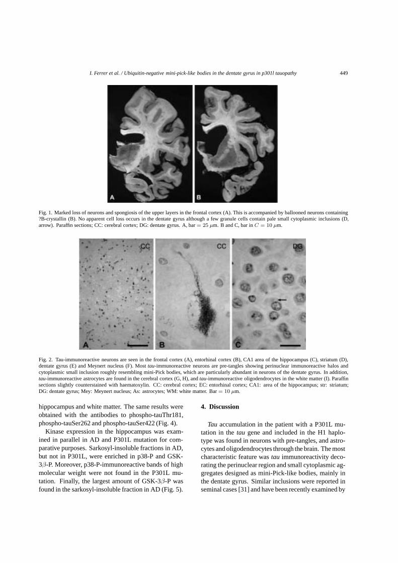
I. Ferrer et al. / Ubiquitin-negative mini-pick-like bodies in the dentate gyrus in p301l tauopathy 449
Fig. 1. Marked loss of neurons and spongiosis of the upper layers in the frontal cortex (A). This is accompanied by ballooned neurons containing?B-crystallin (B). No apparent cell loss occurs in the dentate gyrus although a few granule cells contain pale small cytoplasmic inclusions (D,arrow). Paraffin sections; CC: cerebral cortex; DG: dentate gyrus. A, bar= 25 µm. B and C, bar inC = 10 µm.
Fig. 2. Tau-immunoreactive neurons are seen in the frontal cortex (A), entorhinal cortex (B), CA1 area of the hippocampus (C), striatum (D),dentate gyrus (E) and Meynert nucleus (F). Mosttau-immunoreactive neurons are pre-tangles showing perinuclear immunoreactive halos andcytoplasmic small inclusion roughly resembling mini-Pick bodies, which are particularly abundant in neurons of the dentate gyrus. In addition,tau-immunoreactive astrocytes are found in the cerebral cortex (G, H), andtau-immunoreactive oligodendrocytes in the white matter (I). Paraffinsections slightly counterstained with haematoxylin. CC: cerebral cortex; EC: entorhinal cortex; CA1: area of the hippocampus; str: striatum;DG: dentate gyrus; Mey: Meynert nucleus; As: astrocytes; WM: white matter. Bar= 10 µm.
hippocampus and white matter. The same results wereobtained with the antibodies to phospho-tauThr181,phospho-tauSer262 and phospho-tauSer422 (Fig. 4).
Kinase expression in the hippocampus was exam-ined in parallel in AD and P301L mutation for com-parative purposes. Sarkosyl-insoluble fractions in AD,but not in P301L, were enriched in p38-P and GSK-3β-P. Moreover, p38-P-immunoreactive bands of highmolecular weight were not found in the P301L mu-tation. Finally, the largest amount of GSK-3β-P wasfound in the sarkosyl-insoluble fraction in AD (Fig. 5).
4. Discussion
Tau accumulation in the patient with a P301L mu-tation in thetau gene and included in the H1 haplo-type was found in neurons with pre-tangles, and astro-cytes and oligodendrocytes through the brain. The mostcharacteristic feature wastau immunoreactivity deco-rating the perinuclear region and small cytoplasmic ag-gregates designed as mini-Pick-like bodies, mainly inthe dentate gyrus. Similar inclusions were reported inseminal cases [31] and have been recently examined by
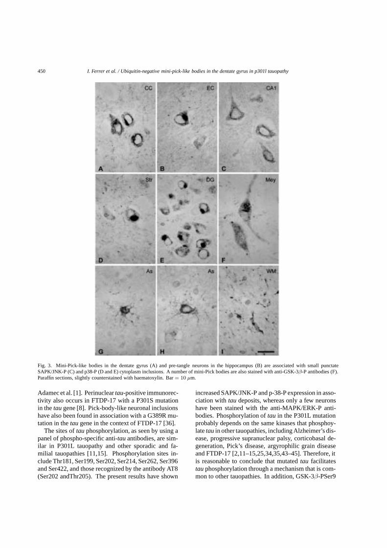
450 I. Ferrer et al. / Ubiquitin-negative mini-pick-like bodies in the dentate gyrus in p301l tauopathy
Fig. 3. Mini-Pick-like bodies in the dentate gyrus (A) and pre-tangle neurons in the hippocampus (B) are associated with small punctateSAPK/JNK-P (C) and p38-P (D and E) cytoplasm inclusions. A number of mini-Pick bodies are also stained with anti-GSK-3β-P antibodies (F).Paraffin sections, slightly counterstained with haematoxylin. Bar= 10 µm.
Adamec et al. [1]. Perinucleartau-positive immunorec-tivity also occurs in FTDP-17 with a P301S mutationin thetau gene [8]. Pick-body-like neuronal inclusionshave also been found in association with a G389R mu-tation in thetau gene in the context of FTDP-17 [36].
The sites oftau phosphorylation, as seen by using apanel of phospho-specific anti-tauantibodies, are sim-ilar in P301L tauopathy and other sporadic and fa-milial tauopathies [11,15]. Phosphorylation sites in-clude Thr181, Ser199, Ser202, Ser214, Ser262, Ser396and Ser422, and those recognized by the antibody AT8(Ser202 andThr205). The present results have shown
increased SAPK/JNK-P and p-38-P expression in asso-ciation with tau deposits, whereas only a few neuronshave been stained with the anti-MAPK/ERK-P anti-bodies. Phosphorylation oftau in the P301L mutationprobably depends on the same kinases that phosphoy-latetau in other tauopathies, including Alzheimer’s dis-ease, progressive supranuclear palsy, corticobasal de-generation, Pick’s disease, argyrophilic grain diseaseand FTDP-17 [2,11–15,25,34,35,43–45]. Therefore, itis reasonable to conclude that mutatedtau facilitatestau phosphorylation through a mechanism that is com-mon to other tauopathies. In addition, GSK-3β-PSer9

I. Ferrer et al. / Ubiquitin-negative mini-pick-like bodies in the dentate gyrus in p301l tauopathy 451
Fig. 4. Western blots of sarkosyl-insoluble fractions of the white matter and hippocampus showing a pattern of two bands of 68/66 kDa and64 kDa by using phospho-specific anti-tauantibodies Thr181, Ser262 and Ser422.
is present in neurons and glial cells in sporadic andfamilial tauopathies [11,12,15,34]. It is interesting tonote that GSK-3β-PSer9 decorates a few neurons inthe cerebral cortex but the majority of neurons withmini-Pick-like bodies in the dentate gyrus in the P301Lmutation. Phosphorylation of GSK-3βat Ser9 inacti-vates the kinase and then the capacity to phosphorylatesubstrates, but GSK-3β-PSer9 also prevents cell deathby apoptosis in several paradigms [9]. It is feasible thatmultiple signals can be triggered by GSK-3 dependingon the state and site of phosphorylation [5,24]. Gran-ule cells in the dentate gyrus are largely preserved innumber whereas neurons in the upper neocortical layers
are devastated. It is tempting to speculate that neuronsexpressing GSK-3β-PSer9 are best equipped to cancelcell death programs in P301L tauopathy.
Although phosphorylation sites intau and expressionof kinases in target cells kinases are similar in the P301Lmutation and other sporadic and familial tauopathies, itis important to note thattau deposits differ from thoseencountered in AD and PiD. A major point is the lackof staining with anti-ubiquitin antibodies in the vastmajority of tau-containing neurons, excepting the fewwith neurofibrillary tangles, and the lack of ubiquitina-tion of the perinuclear halo and mini-Pick-like bodiesin P301L tauopathy, as previously stressed by Adamec
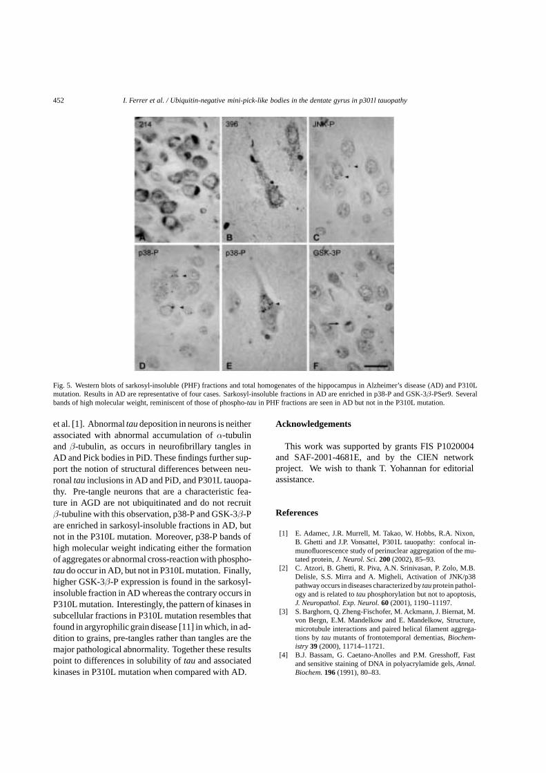
452 I. Ferrer et al. / Ubiquitin-negative mini-pick-like bodies in the dentate gyrus in p301l tauopathy
Fig. 5. Western blots of sarkosyl-insoluble (PHF) fractions and total homogenates of the hippocampus in Alzheimer’s disease (AD) and P310Lmutation. Results in AD are representative of four cases. Sarkosyl-insoluble fractions in AD are enriched in p38-P and GSK-3β-PSer9. Severalbands of high molecular weight, reminiscent of those of phospho-tauin PHF fractions are seen in AD but not in the P310L mutation.
et al. [1]. Abnormaltau deposition in neurons is neitherassociated with abnormal accumulation ofα-tubulinandβ-tubulin, as occurs in neurofibrillary tangles inAD and Pick bodies in PiD. These findings further sup-port the notion of structural differences between neu-ronaltau inclusions in AD and PiD, and P301L tauopa-thy. Pre-tangle neurons that are a characteristic fea-ture in AGD are not ubiquitinated and do not recruitβ-tubuline with this observation, p38-P and GSK-3β-Pare enriched in sarkosyl-insoluble fractions in AD, butnot in the P310L mutation. Moreover, p38-P bands ofhigh molecular weight indicating either the formationof aggregates or abnormal cross-reaction with phospho-tau do occur in AD, but not in P310L mutation. Finally,higher GSK-3β-P expression is found in the sarkosyl-insoluble fraction in AD whereas the contrary occurs inP310L mutation. Interestingly, the pattern of kinases insubcellular fractions in P310L mutation resembles thatfound in argyrophilic grain disease [11] in which, in ad-dition to grains, pre-tangles rather than tangles are themajor pathological abnormality. Together these resultspoint to differences in solubility oftau and associatedkinases in P310L mutation when compared with AD.
Acknowledgements
This work was supported by grants FIS P1020004and SAF-2001-4681E, and by the CIEN networkproject. We wish to thank T. Yohannan for editorialassistance.
References
[1] E. Adamec, J.R. Murrell, M. Takao, W. Hobbs, R.A. Nixon,B. Ghetti and J.P. Vonsattel, P301L tauopathy: confocal in-munofluorescence study of perinuclear aggregation of the mu-tated protein,J. Neurol. Sci. 200 (2002), 85–93.
[2] C. Atzori, B. Ghetti, R. Piva, A.N. Srinivasan, P. Zolo, M.B.Delisle, S.S. Mirra and A. Migheli, Activation of JNK/p38pathway occurs in diseases characterized bytau protein pathol-ogy and is related totau phosphorylation but not to apoptosis,J. Neuropathol. Exp. Neurol. 60 (2001), 1190–11197.
[3] S. Barghorn, Q. Zheng-Fischofer, M. Ackmann, J. Biernat, M.von Bergn, E.M. Mandelkow and E. Mandelkow, Structure,microtubule interactions and paired helical filament aggrega-tions by tau mutants of frontotemporal dementias,Biochem-istry 39 (2000), 11714–11721.
[4] B.J. Bassam, G. Caetano-Anolles and P.M. Gresshoff, Fastand sensitive staining of DNA in polyacrylamide gels,Annal.Biochem. 196 (1991), 80–83.

I. Ferrer et al. / Ubiquitin-negative mini-pick-like bodies in the dentate gyrus in p301l tauopathy 453
[5] R.V. Bhat and S.L. Budd, GSK-3beta signaling: casting a widenet in Alzheimer’s disease,Neurosignals 11 (2002), 251–261.
[6] T.D. Bird, D. Nochlin, P. Poorkaj, M. Charrier, J. Kayre,H. Payani, E. Reskind, T.H. Lampe, E. Nemens, P.S. Boyerand G.D. Schellenberg, A clinical pathological comparisonof three families with frontotemporal dementia and identicalmutations in thetau gene (P301L),Brain 122 (1999), 741–756.
[7] L. Buee, T. Bussi¤¤re, V. Buee-Cherrer, A. Delacourte andP.R. Hof, Tau protein isoforms, phosphorylation and role inneurodegenerative disorders,Brain Res Rev 33 (2000), 95–130.
[8] O. Bugiani, J.R. Murrell, G. Giacone, M. Hasegawa, G. Ghigo,M. Tabaton, M. Morbin, A. Primavera, F. Carella, C. Solaro,M. Grisoli, M. Savoiardo, M.G. Spillantini, F. Tagliavini, M.Goedert and B. Ghetti, Frontotemporal dementia and corti-cobasal degeneration in a family with a P301S mutation intau,J. Neuropathol. Exp. Neurol. 58 (1999), 667–677.
[9] P. Cohen and S. Frame, The renaissance of GSK3,Nat. Rev. 2(2001), 769–776.
[10] C. Conrad, C. Vianna and M. Freeman et al., A polymorphicgene nested within an intron of thetau gene: implicationsfor Alzheimer’s disease,Proc Natl Acad Sci USA 99 (2002),7751–7756.
[11] I. Ferrer, M. Barrachina, M. Tolnay, M.J. Rey, N. Vidal, M.Carmona, R. Blanco and B. Puig, Phosphorylated protein ki-nases associated with neuronal and glialtau deposits in argy-rophilic grain disease,Brain Pathol 13 (2003), 62–78.
[12] I. Ferrer, M. Barrachina and B. Puig, Glycogen synthasekinase-3 (GSK-3) is associated with neuronal and glial hyper-phosphorylatedtau deposits in Alzheimer’s disease, Pick’sdisease, progressive supranuclear palsy and corticobasal de-generation,Acta Neuropathol 104 (2002), 658–664.
[13] I. Ferrer, R. Blanco, M. Carmona and B. Puig, Phosphory-lated mitogen-activated protein kinase (MAPK/ERK-P), pro-tein kinase of 38 kDa (p38-P), stress-activated protein kinase(SAPK/JNK-P), and calcium/calmodulin-dependent kinase II(CaM kinase II) are differentially expressed intau deposits inneurons and glial cells in tauopathies,J. Neural. Transm. 108(2001), 1397–1415.
[14] I. Ferrer, R. Blanco, M. Carmona, R. Ribera, E. Goutan, B.Puig, M.J. Rey, A. Cardozo, F. Vinals and T. Ribalta, Phospho-rylated MAP kinase (ERK1, ERK2) expression is associatedwith early tau deposition in neurones and glial cells, but notwith increased nuclear DNA vulnerability and cell death, inAlzheimer disease, Pick’s disease, progressive supranuclearpalsy and corticobasal degeneration,Brain Pathol 11 (2001),144–158.
[15] I. Ferrer, P. Pastor, M.J. Rey, E. Munoz, B. Puig, E. Pas-tor, R. Oliva and E. Tolosa, Tau phosphorylation and kinaseactivation in familial tauopathy linked to delN296 mutation,Neuropathol. Appl. Neurobiol. 29 (2003), 23–34.
[16] B. Ghetti, M.L. Hutton and Z.K. Wszolek, Frontotemporaldementia and parkinsonism linked to chromosome 17 associ-ated withtau gene mutations (FTDP-17T), in:Neurodegen-eration: The molecular pathology of dementia and movementdisorders, D. Dickson, ed., ISN Neuropath Press, Basel 2003,pp. 86–102.
[17] M. Goedert, R. Jakes and E. Vanmechelen, Monoclonal anti-body AT8 recognizestau protein phosphorylated at both serine202 and threonine 205,Neurosci. Lett. 189 (1995), 167–169.
[18] J. Gotz, Tau and transgenic animal models,Brain Res. Rev. 35(2001), 266–286.
[19] J. Gotz, F. Chen, R. Barmettler and R.M. Nitsch, Tau filamentformation in transgenic mice expressing P301Ltau, J. Biol.Chem. 276 (2001), 529–534.
[20] M. Hasegawa, M.J. Smith and M. Goedert, Tau proteins withFTDP-17 mutations have a reduced ability to promote micro-tubule assembly,FEBS Lett 437 (1998), 207–210.
[21] M. Hutton, C.L. Lendon, P. Rizzu, M. Baker, S. Froelich, H.Houlden, S. Pickering-Brown, S. Chakraertz, A. Isaacs, A.Gover, J. Hackett, J. Adamson, S. Lincoln, D. Dickson, P.Davies, R.C. Petersen, M. Stevans, I. de Graaf, E. Wantens,J. van Baren, M. Hillebrand, J.M. Joosse, V. Nowotny and P.Heutriak et al., Association of a missense and 5’ splice-sitemutations intau with the inherited dementia FTDP-17,Nature393 (1998), 702–705.
[22] M. Hutton, H. Lewis, D. Dickson, S.H. Yen and E. McGowen,Analysis of tauopathies with transgenic mice,Trends Mol.Med. 7 (2001), 467–470.
[23] E.M. Ingram and M.G. Spillantini, Tau gene mutations: dis-secting the pathogenesis of FTDP-17,Trends Mol. Med. 8(2002), 556–562.
[24] M.D. Kaytor and H.T. Orr, The GSK3βsignaling cascade andneurodegenerative disease,Curr. Opin. Neurobiol. 12 (2002),275–278.
[25] R.B. Knowles, J. Chin, C.T. Ruff and B.T. Hyman, Demon-stration by fluorescence resonance energy transfer of a closeassociation between activated MAP kinase and neurofibrillarytangles: Implications for MAP kinase activation in Alzheimerdisease,J. Neuropathol. Exp. Neurol. 58 (1999), 1090–1098.
[26] T. Kobayashi, H. Mori, Y. Okuma, D.W. Dickson, N. Cookson,Y. Tsuboi, Y. Motoi, R. Tanaka, N. Miyashita, M. Anno, H.Narabayashi and Y. Mizzuno, Contrasting genotypes of thetau gene in two phenotypically distinct patients with P301Lmutation of frontotemporal dementia and parkinsonism linkedto chromosome 17,J. Neurol. 249 (2002), 669–675.
[27] M.V.Y. Lee, M. Goedert and J.Q. Trojanowski, Neurodegener-ative tauopathies,Annu. Rev. Neurosci. 24 (2001), 1121–1159.
[28] J. Lewis and D.V. Dickson, Transgenic animal models oftauopathies, in:Neurodegeneration: The molecular pathol-ogy of dementia and movement disorders, D. Dickson, ed.,ISNNeuropath Press, Basel 2003, pp. 150–154.
[29] J. Lewis, E. McGowan, J. Rockwood, H. Melrose, P.Nacharaju, M. van Slegtenhorst, K. Gwinn-Hardy, M. PaulMurphy, M. Baker, X. Yu, J. Hardy, A. Corral, W.L. Lin, S.H.Yen, D.W. Dickson, P. Davies and M. Hutton, Neurofibril-lary tangles, amyotrophy and progressive motor disturbancein mice expressing mutant (P301L)tau proteins,Nat. Genet.25 (2000), 402–405.
[30] W.L. Lin, J. Lewis, S.H. Yen, M. Hutton and D.W. Dickson,Filamentoustau oligodendrocytes and astrocytes of transgenicmice expressing the humantau isoform with the P301L muta-tion, Am. J. Pathol. 162 (2003), 213–218.
[31] S.S. Mirra, J.R. Murrell, M. Gearing, M.G. Spillantini, M.Goedert, A. Crowther, A.I. Levey, R. Jones, J. Green, J.M.Shioffner, B.H. Wainer, M.L. Schmidt, J.Q. Trojanowski andB. Guetti, Tau pathology in a family with a P301L mutationin tau, J. Neuropathol. Exp. Neurol. 58 (1999), 335–345.
[32] J.R. Murrell, M.G. Spillantini, P. Zolo, M. Guazzelli, M.J.Smith, M. Hasegawa, F. Redi, A. Crowther, P. Pietrini, B.Ghetti and M. Goedert, Tau gene mutation G389R causes atauopathy with abundant Pick Body-like inclusions and axonaldeposits,J. Neuropathol. Exp. Neurol. 58 (1999), 1207–1226.
[33] Z.S. Nasreddine, M. Loginov, L.N. Clark, J. Lamarche, B.L.Miller, A. Lamontagne, V. Zhurakeva, M.V. Lee, K.L. Wil-helmen and P.H. Geshwind, From genotype to phenotype: a

454 I. Ferrer et al. / Ubiquitin-negative mini-pick-like bodies in the dentate gyrus in p301l tauopathy
clinical pathological, and biochemical investigation of fron-totemporal dementia and parkinsonism (FTDP-17) caused bythe P301L mutation,Ann. Neurol. 45 (1999), 704–715.
[34] J.J. Pei, E. Braak, H. Braak, I. Grundke-Iqbal, K. Iqbal, B.Winblad and R.F. Cowburn, Distribution of glycogen synthasekinase 3 beta (GSK-3β) for Alzheimer disease neurofibrillarytangle,J. Neuropathol. Exp. Neurol. 58 (1999), 1010–1119.
[35] G. Perry, H. Roder, A. Nunomura, A. Takeda, A.L. Friedlich,X. Zhu, A.L. Raina, N. Holbrook, S.L. Siedlak, P.L.R. Harrisand M.A. Smith, Activation of neuronal extracellular receptorkinase (ERK) in Alzheimer disease links oxidative stress toabnormaltau phosphorylation,NeuroReport 10 (1999), 2411–2415.
[36] P. Rizzu, M. Joosse, R. Ravid, A. Hoogeveen, W. Kamphorst,J.C. van Swieten, R. Willemsen and P. Huetink, Mutation-dependent aggregation oftau protein and its selective depletionfrom the soluble fraction in brain of p310L FTDP-17 patients,Hum. Mol. Genet. 9 (2000), 3075–3082.
[37] M.G. Spillantini, R.A. Crowther, W. Kamphorst, P. Heutinkand J.C. van Swieten, Tau pathology in two Dutch familieswith mutations in the microtubule-binding region oftau, Am.J. Pathol. 153 (1998), 1359–1363.
[38] M.G. Spillantini and M. Goedert, Tau protein pathology inneurodegenerative diseases,Trends Neurosci 21 (1998), 428–433.
[39] T. Togo, N. Sahara, S.H. Yen, N. Cookson, T. Ishizawa, M.Hutton, R. De Silva, A. Lees and D.W. Dickson, Argyrophilic
grain disease is a sporadic 4-repeat tauopathy,J. Neuropathol.Exp. Neurol. 61 (2002), 547–556.
[40] M. Tolnay and A. Probst, Review: Tau protein: pathology inAlzheimer’s disease and related disorders,Neuropathol. Appl.Neurobiol. 25 (1999), 171–187.
[41] R.H. Walker, J. Friedman, J. Wiener, R. Hobler, K. Gwinn-Hardy, A. Adam, J. DeWolfe, R. Gibbs, M. Baker, M. Farrer,M. Hutton and J.A. Hardy, A family with atau P301L mutationpresenting with parkinsonism,Parkinsonism Relat Disord 9(2002), 121–123.
[42] S. H. Yen, M. Hutton, M. deTure, L.W. Ko and P. Nacharaju,Fibrillogenesis oftau: insights fromtau missense mutationsin FTDP-17,Brain Pathol 9 (1999), 695–705.
[43] X. Zhu, H.G. Lee, A.K. Raina, G. Perry and M.A. Smith,The role of mitogen-activated protein kinase pathways inAlzheimer’s disease,Neurosignals 1 (2002), 270–281.
[44] X. Zhu, A.K. Raina, C.A. Rottkamp, G. Aliev, G. Perry, H.Boux and M.A. Smith, Activation and re-distribution of c-JunN-terminal kinase/stress activated protein kinase in degenerat-ing neurons in Alzheimer’s disease,J. Neurochem. 76 (2001),435–441.
[45] X. Zhu, C.A. Rottkamp, H. Boux, A. Takeda, G. Perry andM.A. Smith, Activation of p38 kinase linkstau phospho-rylation, oxidative stress, and cell cycle-related events inAlzheimer disease,J. Neuropathol. Exp. Neurol. 59 (2000),880–888.
Abstract The clinical, neuroradiological, neuropatholog-ical and biochemical findings in four patients with primaryprogressive aphasia and tauopathy are described. The apha-sic syndrome preceded by several years the appearance ofother symptoms in every case. Asymmetrical apraxia withalien hand phenomenon occurred in one case. Frontotem-poral symptoms occurred in three cases, but progressed todramatic cognitive devastation in only one of these. Gen-eralized dementia consistent with probable Alzheimer’sdisease (AD) developed with time in another. Cerebralcomputer tomography scans, magnetic resonance imagingand SPECT studies revealed marked asymmetries in onecase, and showed nonspecific cerebral atrophy in the re-maining ones. The neuropathological examination revealedtypical corticobasal degeneration (CBD) in one case; CBDand AD in another; and atypical CBD, argyrophilic graindisease (AGD) and α-synucleinopathy consistent withParkinson’s disease in a third. Unique neuropathologicalfindings were found in the remaining case. This was char-acterized by severe cerebral atrophy, marked neuronalloss in the cerebral cortex and abnormal tau deposition inneurons of the cerebral cortex, diencephalon and brain
stem. Ballooned neurons, Pick bodies, generalized corti-cal neurofibrillary tangles and astrocytic plaques were ab-sent. However, massive globular inclusions, containing phos-pho-tau, occurred in glial cells, mainly oligodendrocytes,in the white matter. Biochemical studies of frontal ho-mogenates revealed four bands of 73/74, 68, 64 and 60 kDaof phosphorylated tau (using antibodies recognizingphospho-tau Thr181, Ser262 and Ser422) in the patientwith AD and CBD, suggesting a predominant AD patternin this case. Two bands of 68 and 64 kDa of phospho-tauwere recovered in the sarkosyl-insoluble fraction in theother three cases. This pattern is similar to that found inCBD, progressive supranuclear palsy and AGD. Taken to-gether, the present series further supports pure and com-bined CBD as causes of primary progressive aphasia, andthey extend the hypothesis that primary progressive apha-sia may be the initial symptom of distinct tauopathies.
Keywords Primary progressive aphasia · Corticobasaldegeneration · Tauopathy · Argyrophilic grain disease ·α-Synuclein
Introduction
The term ‘primary progressive aphasia’ was introduced todescribe a syndrome characterized by progressive wors-ening of language with preservation of the activities ofdaily life and non-verbal abilities [52, 53]. Although theseminal reports emphasized lack of dementia, subsequentobservations have shown variable cognitive impairmentand dementia in the majority of cases of primary progres-sive aphasia.
Several neurological diseases may produce primaryprogressive aphasia. These include Alzheimer’s disease(AD) [3, 28, 37, 60], Pick’s disease (PiD) [27, 29, 81], andCreutzfeldt-Jakob disease [51, 66, 82]. Nonspecific spongi-form changes and astrocytic gliosis of the upper corticallayers have also been reported [39, 40, 42, 43, 48, 58, 65,67, 77]. Cortical achromatic neurons have been reportedin some cases. Spongy change in the frontal cortex and
I. Ferrer · I. Hernández · M. Boada · A. Llorente ·M. J. Rey · A. Cardozo · M. Ezquerra · B. Puig
Primary progressive aphasia as the initial manifestation of corticobasal degeneration and unusual tauopathies
Acta Neuropathol (2003) 106 : 419–435DOI 10.1007/s00401-003-0756-4
Received: 7 April 2003 / Revised: 24 June 2003 / Accepted: 24 June 2003 / Published online: 29 August 2003
REGULAR PAPER
I. Ferrer (✉) · B. PuigInstitut de Neuropatologia, Servei Anatomia Patològica, Hospital Princeps d’Espanya (Bellvitge CSUB), carrer Feixa Llarga sn, 08907 Hospitalet de Llobregat, SpainTel.: +34-93-4035808e-mail: [email protected]
I. Ferrer · M. J. Rey · A. Cardozo · M. EzquerraBanc de Teixits Neurològics, Universitat de Barcelona/Hospital Clinic, Barcelona, Spain
I. Hernández · M. Boada · A. LlorenteFundaciò ACE, Institut Català de Neurociències Aplicades, Barcelona, Spain
I. HernándezSAP Nou Barris, ICS, CAPII Chafarinas Neurología, Barcelona, Spain
M. BoadaServei de Neurologia, Hospital General Universitari Valle Hebrón, Barcelona, Spain
© Springer-Verlag 2003

striatum accompanied by abnormally ubiquitinated neu-rites in the cerebral cortex [41] is a rare condition associ-ated with primary progressive aphasia. Corticobasal degen-eration (CBD) [24], formerly named corticodentatonigraldegeneration with neuronal achromasia [61], is a commoncause of primary progressive aphasia [1, 32, 45, 55, 62,83]. Finally, progressive supranuclear palsy (PSP) may de-velop with progressive nonfluent aphasia [7, 56].
Together, the cumulative clinical and pathological evi-dence supports the concept that primary progressive aphasiais not uncommon in a variety of diseases with frontotempo-ral atrophy, including AD, PiD, frontotemporal dementia(FTD), PSP and CBD [40]. The majority of these diseasesare associated with impaired tau metabolism, usually mani-fested as an accumulation of abnormally phosphorylated tauin neurons and glial cells. However, a subgroup of FTDcases have decreased tau levels in total brain homogenates[79]. Abnormal tau phosphorylation and deposition in thecytoplasm of neurons and glial cells are major biochemicaland structural anomalies in AD, CBD, PiD and PSP [11, 12,21, 26, 47]. However, AD, CBD, PiD and PSP have clini-cal, neuropathological and biochemical peculiarities. Phos-phorylation of tau occurs at different sites, and phosphory-lated bands from homogenates of fractions enriched withpaired helical filaments or with abnormal filaments showparticular patterns. AD displays four bands of 73/74, 68, 64and 60 kDa, whereas CBD and PSP are resolved into twobands of 68 and 64 kDa. PiD shows two bands of 64 and 60kDa [11, 12, 20, 26, 47]. Inherited tauopathies associatedwith mutations or deletions in the tau gene are manifestedas PiD-, PSP- or FTD-like syndromes, and can be resolvedinto different band patterns on Western blots of brain ho-mogenate fractions enriched in abnormal filaments [12, 33,47]. Finally, argyrophilic grain disease (AGD) is anothertauopathy with invariable limbic involvement that producestwo bands of 68 and 64 kDa [22, 72, 75].
Detailed clinical and neuropathological studies in pa-tients with primary progressive aphasia and sporadic tauopa-thy are important because they help to increase our under-standing of the spectrum of diseases with abnormal tauaccumulation that manifest as primary progressive apha-sia. The present study is focused on four patients with long-lasting primary progressive aphasia followed by cognitiveimpairment in which the neuropathological and biochem-ical study disclosed singular tauopathies.
Case reports
Case 1
The patient was a 66-year-old right-handed woman with a primaryschool education. There was no familiy history of neurological dis-ease. Moderate hypertension was controlled with diet and medica-tion. In 1987, she complained of a transient episode of difficulty inspeaking. A brain CT scan showed mild cerebral atrophy withoutother anomalies. She was diagnosed as having suffered from atransitory ischemic attack and received anti-aggregant drugs.
In 1990, the patient noticed slowly progressive difficulties inexpressing herself. A brain CT scan revealed a slight increase of thesylvian fissures compared with the previous study carried out 3 years
earlier. The cerebral magnetic resonance imaging (MRI) demon-strated cortical atrophy of the right perisylvian region and of theleft temporal lobe, together with hyperintense periventricular sig-nals in the white matter.
A neuropsychological study 2 years later revealed languageworsening, but reading and writing comprehensions were pre-served. A new study in April 1994 showed a reduction in fluency,with non-grammatical language and bucal and facial apraxia.Reading and writing remained intact. The MMSE showed scoresof 18/30, and the patient was able to maintain daily activities in-cluding the full organization of the family life. The patient was di-agnosed as suffering from progressive aphasia with slight damageof right frontal functions and buco-lingual apraxia. A cerebralSPECT performed in 1994 revealed inter-hemispheric asymmetrywith decreased flow in the right frontal cortex.
By the end of 1995, spasmodic laughter and difficulties in swal-lowing had appeared. A new neuropsychological examinationshowed a deterioration of the visual-spatial and visual-constructivefunctions, and almost complete loss of the language; however, thepatient was still capable of communicating by writing and by ges-tures (in order to say ‘yes’ she raised the thumb of her right hand,and placed it downwards to say ‘no’). In addition, the neurologicalexamination showed buco-facial-lingual apraxia, impaired swal-lowing of liquids and semi-liquids, and slight alteration of the vol-untary ocular movements. The rest of the neurological examina-tion was normal, showing no alterations in the pyramidal or extra-pyramidal area. The cerebral SPECT revealed perisylvian attenua-tion, which was more marked on the right side.
In September 1996, the patient had trouble performing commondomestic tasks, showed walking disorders, and suffered occasionalurinary incontinence. Swallowing had worsened. The neuropsycho-logical examination disclosed difficulties in understanding, as wellas agraphia. The neurological examination showed bucal, facialand lingual apraxia, mild paresia of the motor ocular nerves, spas-modic laughter and sobbing, bilateral hyper-reflexia, grasping pre-dominantly in the left side, axial rigidity, and left postural rigiditywith walking apraxia. The patient did not suffer from behavioraldisturbances excepting apathy, and her memory was preserved.
Dystonic movements appeared 1 year later. The neuropsycho-logical examination in November 1997 evidenced phonemic disin-tegration with mutism, as well as major difficulty in swallowing andsalivation, and reduced verbal comprehension. A cerebral SPECTshowed, in addition to perisylvian attenuation, reduced signal inthe posterior regions of the frontal and temporal lobes, which wasmore marked in the right hemisphere.
One year later, the patient was suffering from progressive wors-ening of mobility and cognitive deterioration. Her relatives askedfor percutaneous feeding, which was started by the middle of 1998.A few months later, the patient was confined to bed, decorticatedwith generalized hyperreflexia and hypertonia but still able to rec-ognize faces. During the following 2 years, the patient deterioratedand her mental status was very poor. The patient died in April 2002at the age of 81, 12 years after the beginning of aphasia.
Case 2
The patient was a right-handed woman with limited education.There was no familiy history of neurological disease. She was 72years old at the time of her first consultation. In May 1995 she pre-sented for a progressive language disorder that had started 2 yearsearlier. The neuropsychological examination showed language al-terations and difficulties in visual-spatial functions. The MMSEwas 27/30. A cerebral CT scan showed slight cortical and subcor-tical cerebral atrophy. A cerebral SPECT disclosed dilatation ofthe ventricular system without regional cortical perfusion defects.
Language deterioration was accompanied by increased depen-dence on relatives to communicate aspects of current life. A newneuropsychological evaluation was performed in 1997, whichshowed a marked language alteration with loss of fluency andanomy. Linguistic understanding was better preserved, but deficitsin the comprehension of complex orders were already detected atthis time. The patient’s writing was agraphic, and she had difficul-
420

ties in the written repetition of long sentences. Abstract reasoningskills were diminished and slight visual-spatial deficits were pre-sent.
The neuropsychological evaluation in November 1997 disclosedfurther deterioration of spontaneous language. Verbal understand-ing was preserved and reading comprehension was intact.
In August 1998, during a control examination, a mammary tu-mor was discovered, and a radical mastectomy was conducted, fol-lowed by chemotherapy and radiation therapy.
In February 2001, the patient required assisted feeding, becauseof severe buco-lingual apraxia. Marked rigidity predominated inthe right side. In addition, cognitive impairment and loss of mem-ory characterized the last months of her life. The patient died inApril 2002 at the age of 78, 9 years after the beginning of aphasia.
Case 3
The patient was a right-handed man with no family history of neu-rological disease, who had suffered from progressive disorder ofspeech starting in 1994. This was followed 3 years later by a be-havior disorder consisting of disinhibition and compulsive laugh-ing, asymmetrical rigidity and apraxia of the upper extremity, an-ticipating alien hand phenomenon 2 years later. The CT scan per-formed in November 1999 disclosed moderate frontal, parietal andtemporal atrophy, and enlargement of the left perisylvian region.The neurological status worsened during the year 2000 with markeddeterioration of mood and personality. Social difficulties due tonon-structured behavior contrasted with the preservation of mem-ory and orientation, as well as with the recognition of persons andplaces. Language was extremely impoverished leading to practicalmutism. Parkinsonism signs were not recorded at any time duringthe course of the disease. The patient died in November 2001 at theage of 73.
Case 4
The patient was a man aged 68 who developed, during the winterof 1990, progressive loss of language and verbal fluency contrast-ing with preserved comprehension, reading and naming skills.There was no familiy history of neurological disease. The neuro-logical examination in 1993 revealed no additional abnormalities,except slight left rigidity and dystonia. A cerebral CT scan revealedmoderate frontal, parietal and temporal atrophy. His mental statusrapidly deteriorated during the following year, with loss of the ca-pacity for learning new information, followed by loss of short-termmemory, and decline of motor skills. The patient was mute, rigid,disconnected and confined to a wheel-chair by 1996. The patientsurvived until November 1997, dying at the age of 73.
Neuropathological methods
The post-mortem delay between death and tissue process-ing varied from 2 to 15 h. Frozen samples for biochemicalstudies were available in every case.
A complete neuropathological examination was car-ried out in every case on tissue that had been formalin-fixed for no less than 3 weeks; the tissue was then embed-ded in paraffin. De-waxed sections, 7 µm thick, were stainedwith hematoxylin and eosin and Luxol fast blue-KlüverBarrera, or processed for immunohistochemistry follow-ing the avidin-biotin-peroxidase method (ABC kit, Vec-tastain, Vector). After blocking endogenous peroxidase,the sections were incubated with normal serum and incu-bated at 4°C overnight with one of the primary antibodies.Antibodies to phosphorylated 170- or 200-kDa neurofila-
ments (clones BF10 and RT97, Boehringer-Mannheim)were used at dilutions of 1:100 and 1:50, respectively.Rabbit polyclonal antibodies to αB-crystallin (Novocas-tra) were used at a dilution of 1:500. Rabbit polyclonalantibodies to α-synuclein (Chemicon) were used at a dilu-tion of 1:500. Antibodies to pan-tau (Sigma) were used ata dilution of 1:10. Antibodies to glial fibrillary acidic pro-tein (Dako, Dakopatts), βA4-amyloid (Boehringer-Mann-heim), and ubiquitin (Dako) were used at dilutions of1:250, 1:5, and 1:200, respectively. In addition, immuno-histochemistry to anti-phospho-specific tau antibodieswas carried out on de-waxed sections first boiled in citratebuffer and then stored overnight at room temperature. Thefollowing anti-phospho-specific tau rabbit polyclonal an-tibodies were used: Thr181, Ser202, Ser214, Ser262,Ser396 and Ser422 (all of them from Calbiochem). Theantibodies were used at a dilution of 1:100, excepting anti-phospho-tauThr181, which was used at a dilution of 1:250.Finally, the phosphorylation-independent tau antibody 7.51,which is raised against the binding region of tau [54], wasused at a dilution of 1:500.
Gel electrophoresis and Western blotting
For gel electrophoresis and Western blotting, fresh samplesof the frontal cortex were immediately obtained at autopsy,frozen in liquid nitrogen, and stored at –80°C until use.
A protocol to purify paired helical filaments in AD, de-scribed by Goedert et al. [26], was used in every case.Frozen samples of about 5 g were cut into pieces. Theywere gently homogenized in a glass tissue grinder in 10 vol(w/v) of cold suspension buffer consisting of 10 mM TRIS-HCl pH 7.4, 0.8 M NaCl, 1 mM EGTA, 10% sucrose, 0.1 mM phenylmethylsulfonyl fluoride, 2 µg/ml aprotinin,
421
Fig. 1 Case 1. A, B Coronal sections of the brain showing markedatrophy of the frontal and temporal lobes, including the hippocam-pus and perirhinal cortex, and the perisylvian region. Marked en-largement of the lateral ventricles, reduced white matter and atro-phy of the corpus callosum, and moderate atrophy of the caudate,putamen and thalamus, are additional remarkable features
422
Fig. 2 Case 1. Marked neuron loss and spongiosis are found in thefrontal cortex (A, B). This is accompanied by intense astrocyticgliosis, as revealed with glial fibrillary acidic protein immunohis-tochemistry (C), which is strongly immunostained with anti-αB-crystallin antibodies (D). Tau-immunoreactive deposits are foundin scattered neurons in the cerebral cortex (E), hippocampus (F)
and thalamus (G). Neuron loss and astrocytic gliosis is seen in thesubstantia nigra (H). Tau-immunoreactive inclusions are found inthe dorsal nucleus of the vagus nerve (I). A, B, H Hematoxylin andeosin, C–G, I immunohistochemical sections slightly counter-stained with hematoxylin; bars A 50 µm, I (also for B–H) 25 µm
10 µg/ml leupeptin and 5 µg/ml pepstatin. The homo-genates were first centrifuged at 20,000 g, and the super-natant (S1) was retained. The pellet (P1) was re-homoge-nized in 5 vol of homogenization buffer and re-centrifuged.The two supernatants (S1+S2) were then mixed and incu-bated with N-lauroylsarcosinate 1% for 1 h at room tem-perature while being shaken. Samples were then centri-fuged for 1 h at 100,000 g in a Ti 70 Beckman rotor. Sarko-syl-insoluble pellets (P3) were re-suspended (0.2 ml/gstarting material) in 50 mM TRIS-HCl pH 7.4. Protein con-centrations were determined by the BCA method and 10%
sodium dodecyl sulfate-polyacrylamide gel electrophore-sis (SDS-PAGE) was run using a maxi-protean system(Bio-Rad). 100–200 µg protein was loaded in each lane withloading buffer containing 0.125 M TRIS pH 6.8, 20% glyc-erol, 10% mercaptoethanol, 4% SDS and 0.002% bromo-phenol blue. Samples were heated at 95°C for 5 min priorto gel loading. Total frontal homogenates and fractionsenriched with abnormal filaments were run in parallel.The proteins were then transferred to nitrocellulose mem-branes (Amersham) using an electrophoretic chamber sys-tem (Trans-Blot Electrophoretic Transfer Cell, Bio-Rad).Non-specific binding sites were blocked with TRIS-bufferedsaline solution pH 7.4 with 0.1% Tween-20 (TBST) con-taining 5% skimmed milk for 30 min, and incubated withone of the primary antibodies for 1 h at room temperature.Control of protein content in each lane was carried out bythe staining of selected gels with Coomassie blue and ofthe membranes with Ponceau (Sigma).
The rabbit polyclonal antibody to phospho-tauThr181was diluted 1:250. The rabbit polyclonal antibodies anti-
423
Fig. 3 Case 1. Tau-immunoreactive inclusions in glial cells in thewhite matter of the frontal lobe (A), parietal lobe (B), internal cap-sule (C) and corpus callosum (D). Glial inclusions are ubiquiti-nated (E). The majority of these inclusions are globular, but rarecoiled bodies (white arrow) are seen in the vicinity of a neuron(asterisk) with a bizarre tau-immunoreactive inclusion in the ante-rior horn of the spinal cord (F). Immunohistochemical sectionsslightly counterstained with hematoxylin. Bar 25 µm

phospho-tauSer262 and phospho-tauSer422 (all from Cal-biochem) were used at a dilution of 1:2,500.
Genetic studies
Tau gene exons 9, 10, 12 and 13, were mutations in FTDpatients have been found previously, were amplified usingspecific primers derived from the 5’ and 3’ sequences andthe polymerase chain reaction (PCR) conditions previ-
ously described [30]. Direct sequenceanalysis was per-formed for these exons using an automated DNA se-quencer (ABI 310; Perkin Elmer/Applied Biosystems,Foster City, CA).
Results
Neuropathological findings
Case 1
Severe frontotemporal atrophy with marked enlargementof the lateral ventricles and atrophy of the perisylvian re-gion were observed on gross examination. Coronal sectionsalso revealed reduced white matter and marked atrophy ofthe corpus callosum, as well as moderate atrophy of thecaudate and putamen, and thalamus (Fig. 1). The micro-scopical study revealed marked neuron loss in the cerebral
424
Fig. 4 Case 1. Tau-immunoreactive inclusions in neurons andglial cells as revealed with several phospho-specific anti-tau anti-bodies (A–E) and with the phosphorylation-independent tau anti-body 7.51 (F). Note strong immunoreactivity to antibodies tophospho-tauSer202 (A), Ser262 (B), phospho-tauThr181 (C),Ser422 (D), Ser396 (E), and tau 7.51 (F). Immunohistochemicalsections slightly counterstained with hematoxylin. Bar 10 µm

cortex of the frontal, temporal, insular and cingulated cor-tices, which was accompanied by astrocytic gliosis (Fig.2A–C). Ballooned neurons were absent, but αB-crystallinimmunohistochemistry revealed large numbers of positivereactive astrocytes (Fig. 2D). Tau-immunoreactive neuronswere seen in the cerebral cortex, hippocampus, diencephalonand reticular formation (Fig. 2E–G). Pick bodies were ab-sent. Mild neuron loss and gliosis with no tau and α-synu-clein pathology was found in the substantia nigra pars com-pacta (Fig. 2H). Bizarre, tau-positive inclusions, resemblingskein inclusions in motor neuron disease in morphology,were found in the motor nuclei of the vagus nerve (Fig. 2I).
In addition to neurons, massive accumulation of abnor-mal tau was present in glial cells, mainly oligodendrocytes,and in the white matter of the cerebral hemispheres, includ-ing the parietal cortex and internal capsule (Fig. 3A–D).The morphology of glial inclusions was rather globularand the inclusions were ubiquitinated (Fig. 3E). Coiledbodies were seldom observed in the cerebrum, brain stem
or spinal cord (Fig. 3F). Tau deposition in neurons andglial cells was recognized with anti-phospho-specifictauThr181, Ser202, Ser214, Ser262, Ser396 and Ser422antibodies (Fig. 4A, B, D–F). Globular glial inclusions werealso strongly stained with the antibody 7.51 (Fig. 4C), butwere not with anti-α-synuclein antibodies.
The neuropathological diagnosis was multiple systemtauopathy.
Case 2
Atrophy of the temporal lobe, including the hippocampus,and the pre-central cortex, and mild enlargement of thelateral ventricles, were seen on gross examination (Fig. 5).Neuron loss, rare ballooned neurons and moderate astro-cytic gliosis were found in the cerebral cortex; markedloss of neurons occurred in the striatum and thalamus.Abnormal tau deposition in neurons and thread-like struc-tures in the cerebral cortex (Fig. 6A), striatum (Fig. 6B),thalamus (Fig. 6C) and brain stem (Fig. 6D) were ob-served. Coiled bodies in oligodendrocytes were abundantin the white matter (Fig. 6E).
Pre-tangles and grains were conspicuous in the hippo-campus, entorhinal cortex, amygdala and anterior cingu-late cortex (Figs. 6F, 8B). Grains were also observed inthe hypothalamus. Abnormal tau deposits were stained withanti-phospho-specific tau antibodies: pre-tangles, grainsand coiled bodies were stained with all the antibodiestested (Fig. 7A–C, E, F). Astrocytic plaques were clearlydistinguished with anti-phospho-tauThr181, Ser202, Ser262,Ser396 and Ser 422 (Fig. 7G), but not with anti-phospho-tau Ser214 antibodies. Tufted astrocytes were absent.
Ballooned neurons in the frontal and parietal cortexwere rare, although easily visualized with anti-phosphory-lated neurofilament and anti-αB-crystallin antibodies(Fig. 8A). However, αB-crystallin-immunoreactive balloonedneurons were very common in the amygdala and entorhi-nal cortex (Fig. 8C, D).
Finally, mild neuron loss and Lewy bodies were foundin the substantia nigra, locus ceruleus and brain stemreticular formation (Fig. 8E). α-Synuclein-immunoreactivedystrophic neurites were common in the lower brain stem,locus ceruleus, Meynert’s nucleus and amygdala (Fig. 8F).
The neuropathological diagnosis was CBD, AGD andα-synucleinopathy.
Case 3
The gross examination revealed perisylvian atrophy andatrophy of the upper parietal and frontal cortices. Therewas enlargement of the lateral ventricles, reduced subcor-tical white matter and atrophy of the corpus callosum(Fig. 9A). The microscopical examination showed moder-ate neuron loss and astrocytic gliosis in the cerebral cor-tex. Ballooned achromatic neurons containing phosphory-lated neurofilaments and αB-crystallin were found mainly inthe frontal cortex, but also in the limbic system (Fig. 9B–D).
425
Fig. 5 Case 2. A–D Coronal sections of the brain. Moderate atro-phy of the frontal cortex, precentral region, perisylvian region andtemporal lobe, including the hippocampus. There is also moderateenlargement of the lateral ventricles, but apparent preservation ofthe caudate, putamen and thalamus
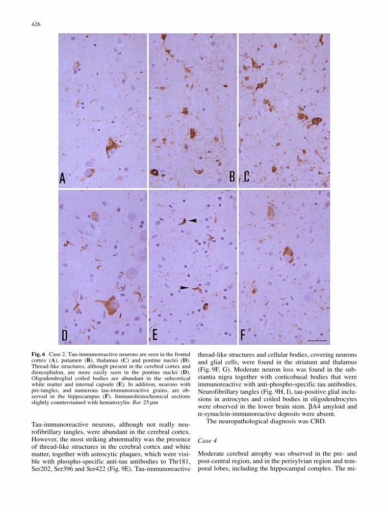
Tau-immunoreactive neurons, although not really neu-rofibrillary tangles, were abundant in the cerebral cortex.However, the most striking abnormality was the presenceof thread-like structures in the cerebral cortex and whitematter, together with astrocytic plaques, which were visi-ble with phospho-specific anti-tau antibodies to Thr181,Ser202, Ser396 and Ser422 (Fig. 9E). Tau-immunoreactive
thread-like structures and cellular bodies, covering neuronsand glial cells, were found in the striatum and thalamus(Fig. 9F, G). Moderate neuron loss was found in the sub-stantia nigra together with corticobasal bodies that wereimmunoreactive with anti-phospho-specific tau antibodies.Neurofibrillary tangles (Fig. 9H, I), tau-positive glial inclu-sions in astrocytes and coiled bodies in oligodendrocyteswere observed in the lower brain stem. βA4 amyloid andα-synuclein-immunoreactive deposits were absent.
The neuropathological diagnosis was CBD.
Case 4
Moderate cerebral atrophy was observed in the pre- andpost-central region, and in the perisylvian region and tem-poral lobes, including the hippocampal complex. The mi-
426
Fig. 6 Case 2. Tau-immunoreactive neurons are seen in the frontalcortex (A), putamen (B), thalamus (C) and pontine nuclei (D).Thread-like structures, although present in the cerebral cortex anddiencephalon, are more easily seen in the pontine nuclei (D).Oligodendroglial coiled bodies are abundant in the subcorticalwhite matter and internal capsule (E). In addition, neurons withpre-tangles, and numerous tau-immunoreactive grains, are ob-served in the hippocampus (F). Immunohistochemical sectionsslightly counterstained with hematoxylin. Bar 25 µm
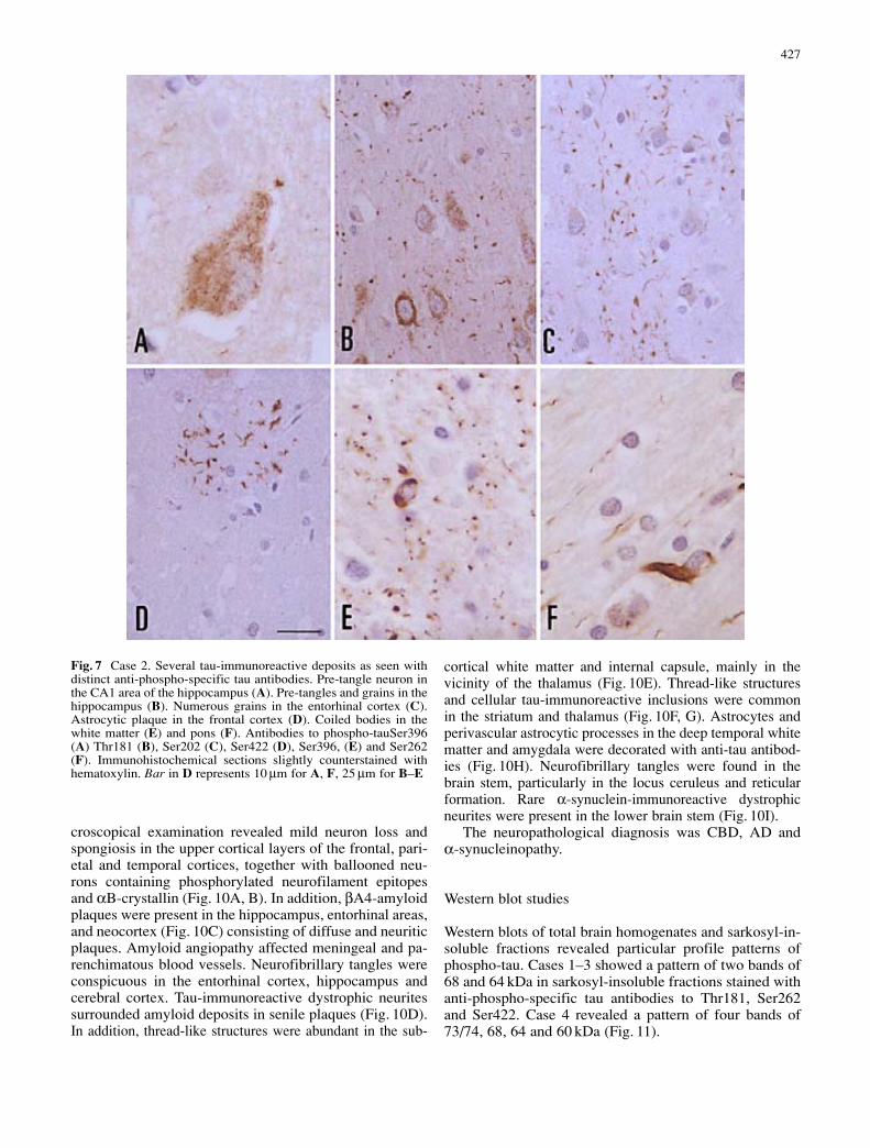
croscopical examination revealed mild neuron loss andspongiosis in the upper cortical layers of the frontal, pari-etal and temporal cortices, together with ballooned neu-rons containing phosphorylated neurofilament epitopesand αB-crystallin (Fig. 10A, B). In addition, βA4-amyloidplaques were present in the hippocampus, entorhinal areas,and neocortex (Fig. 10C) consisting of diffuse and neuriticplaques. Amyloid angiopathy affected meningeal and pa-renchimatous blood vessels. Neurofibrillary tangles wereconspicuous in the entorhinal cortex, hippocampus andcerebral cortex. Tau-immunoreactive dystrophic neuritessurrounded amyloid deposits in senile plaques (Fig. 10D).In addition, thread-like structures were abundant in the sub-
cortical white matter and internal capsule, mainly in thevicinity of the thalamus (Fig. 10E). Thread-like structuresand cellular tau-immunoreactive inclusions were commonin the striatum and thalamus (Fig. 10F, G). Astrocytes andperivascular astrocytic processes in the deep temporal whitematter and amygdala were decorated with anti-tau antibod-ies (Fig. 10H). Neurofibrillary tangles were found in thebrain stem, particularly in the locus ceruleus and reticularformation. Rare α-synuclein-immunoreactive dystrophicneurites were present in the lower brain stem (Fig. 10I).
The neuropathological diagnosis was CBD, AD and α-synucleinopathy.
Western blot studies
Western blots of total brain homogenates and sarkosyl-in-soluble fractions revealed particular profile patterns ofphospho-tau. Cases 1–3 showed a pattern of two bands of68 and 64 kDa in sarkosyl-insoluble fractions stained withanti-phospho-specific tau antibodies to Thr181, Ser262and Ser422. Case 4 revealed a pattern of four bands of73/74, 68, 64 and 60 kDa (Fig. 11).
427
Fig. 7 Case 2. Several tau-immunoreactive deposits as seen withdistinct anti-phospho-specific tau antibodies. Pre-tangle neuron inthe CA1 area of the hippocampus (A). Pre-tangles and grains in thehippocampus (B). Numerous grains in the entorhinal cortex (C).Astrocytic plaque in the frontal cortex (D). Coiled bodies in thewhite matter (E) and pons (F). Antibodies to phospho-tauSer396(A) Thr181 (B), Ser202 (C), Ser422 (D), Ser396, (E) and Ser262(F). Immunohistochemical sections slightly counterstained withhematoxylin. Bar in D represents 10 µm for A, F, 25 µm for B–E
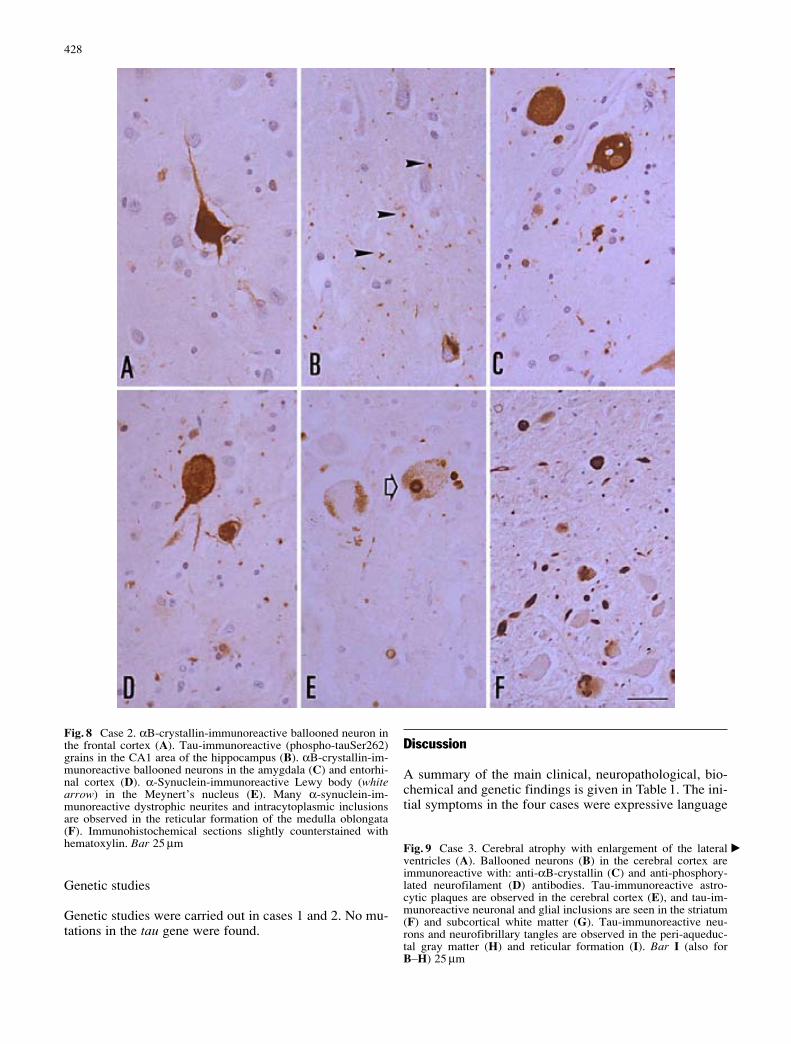
Genetic studies
Genetic studies were carried out in cases 1 and 2. No mu-tations in the tau gene were found.
Discussion
A summary of the main clinical, neuropathological, bio-chemical and genetic findings is given in Table 1. The ini-tial symptoms in the four cases were expressive language
428
Fig. 8 Case 2. αB-crystallin-immunoreactive ballooned neuron inthe frontal cortex (A). Tau-immunoreactive (phospho-tauSer262)grains in the CA1 area of the hippocampus (B). αB-crystallin-im-munoreactive ballooned neurons in the amygdala (C) and entorhi-nal cortex (D). α-Synuclein-immunoreactive Lewy body (whitearrow) in the Meynert’s nucleus (E). Many α-synuclein-im-munoreactive dystrophic neurites and intracytoplasmic inclusionsare observed in the reticular formation of the medulla oblongata(F). Immunohistochemical sections slightly counterstained withhematoxylin. Bar 25 µm Fig. 9 Case 3. Cerebral atrophy with enlargement of the lateral
ventricles (A). Ballooned neurons (B) in the cerebral cortex areimmunoreactive with: anti-αB-crystallin (C) and anti-phosphory-lated neurofilament (D) antibodies. Tau-immunoreactive astro-cytic plaques are observed in the cerebral cortex (E), and tau-im-munoreactive neuronal and glial inclusions are seen in the striatum(F) and subcortical white matter (G). Tau-immunoreactive neu-rons and neurofibrillary tangles are observed in the peri-aqueduc-tal gray matter (H) and reticular formation (I). Bar I (also forB–H) 25 µm
�
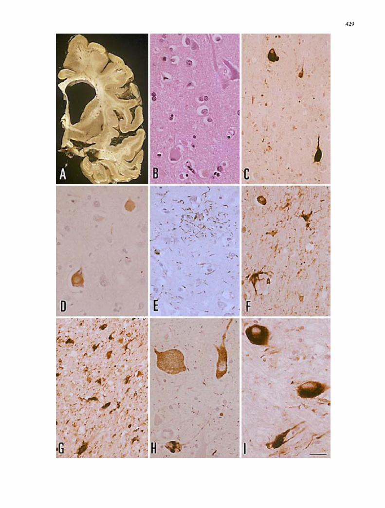
429

430
Fig. 10 Case 4. Moderate neuron loss and a ballooned neuron inthe frontal cortex (A). Ballooned neurons are stained with anti-αB-crystallin antibodies (B). βA4-amyloid deposits in the cerebralcortex (C). Neurofibrillary tangles and dystrophic neuritis sur-rounding amyloid cores in neuritic plaques (asterisk) are stainedwith anti-tau antibodies (D). Thread-like structures are abundant inthe subcortical white matter (E). Neurons and glial cells with ab-
normal tau accumulation are seen in the striatum (F) and thalamus(G). Reactive astrocytes and astroglial cell processes surroundingblood vessels (arrowhead) are tau immunoreactive (H). Isolatedα-synuclein-immunoreactive profile in the reticular formation ofthe medulla oblongata (I). Immunohistochemical sections slightlycounterstained with hematoxylin. Bar 25 µm
disorders, with difficulties in naming objects and articu-lating words. Patients 1 and 2 maintained their daily lifeactivities up until moderately advanced stages of the dis-ease (8 and 5 years, respectively), when the motor disor-ders marked the onset of their functional dependency.Speech disturbances followed by cognitive decline con-sistent with FTD occurred in patient 3, whereas isolatedlanguage disorder lasting for 3 years preceded rapidly pro-gressive probable AD in case 4.
Atrophy of the superior frontal gyrus and superior pari-etal cortex, together with atrophy of the superior temporalgyrus, opercular area and pre-central cortex, were com-mon, although very variable in severity, in every case ofthe present series. Aphasia correlated with perisylvian at-rophy in each of these four cases. Atrophy of the hippo-campus was marked in cases 1 and 4, but was moderate incases 2 and 3. Interestingly, cerebral CT scans and MRIstudies revealed asymmetries from the beginning of thedisease in patient 1. Similarly, SPECT studies revealedprogressive attenuation over time, predominating in the
right side. However, in case 2, no focal signs were noticedin the SPECT studies carried out 2 years after the begin-ning of the speech disturbances. Unfortunately, only twocerebral CT scans showing moderate global atrophy werecarried out in patients 3 and 4.
Case 1 shows rare clinical and neuropathological find-ings. The clinical symptoms were characterized by prolongedprimary aphasia, followed by frontotemporal symptomsand, finally, by dramatic generalized cognitive devasta-tion. The microscopical study revealed marked corticalneuronal loss with no ballooned neurons, Pick bodies, astro-cytic plaques, or massive cortical neurofibrillary pathology.Yet neurons bearing abnormal phospho-tau were seen inthe neocortex, diencephalons and brain stem. Moreover,large numbers of globular tau deposits were found in glialcells. Massive globular phospho-tau deposition in glial cells,as seen in case 1, has been observed in certain familialtauopathies associated with mutations and single aminoacid deletions in the tau gene [14, 23, 34, 68] and in spo-radic multiple system tauopathy with dementia [7]. Tau-
431
Fig. 11 Western blots of totalbrain homogenates (TH) andsarkosyl-insoluble fractions(PHF) in cases 1–4, processedwith anti-phospho-specific tauantibodies Thr181, Ser262 andSer422. Two bands of 68 and64 kDa are seen in cases 1–3,whereas four bands of 72/74,68, 64 and 60 kDa are seen incase 4

immunoreactive glial inclusions in those cases and in thepresent case differ from glial inclusions in multiple sys-tem atrophy (MSA), although both are ubiquitinated. Glialinclusions in MSA contain α-synuclein and non-phos-phorylated tau [2, 13, 18, 46, 69, 76, 80], whereas glial in-clusions in the present case were negative for α-synucleinbut strongly immunoreactive for phosphorylated tau andwith the phosphorylation-independent tau antibody 7.51.
Case 2 showed neuropathological findings of CBD,AGD and α-synucleinopathy. A diagnosis of CBD wasbased on the presence of tau-immunoreactive inclusionsin neurons and glial cells in the striatum, thalamus, cere-bral cortex and selected nuclei of the brain stem, thread-like structures in the diencephalon, white matter and pon-tine nuclei, and astrocytic plaques in the cerebral cortex.There was a paucity of ballooned neurons in the frontalcortex, but this does not exclude a diagnosis of CBD [16].In addition, the diagnosis of AGD was based on the pres-ence of pre-tangles and tau-immunoreactive grains in theanterior and posterior hippocampus, entorhinal cortex,amygdala, anterior gyrus cinguli and hypothalamus [8, 35,71, 74]. Ballooned neurons containing αB-crystallin in theamygdala and entorhinal cortex, and oligodendroglial coiledbodies, as seen in this case, are common in CBD andAGD [72]. However, the large number of ballooned neu-rons in the amygdala, in striking contrast with the limitednumber of such neurons in the frontal cortex, is likely dueto AGD rather than to CBD.
AGD has been reported in combination with AD, de-mentia with tangles, PSP and several α-synucleinopathies[8, 35, 52]. Argyrophilic grains were found in 2 cases ofCBD in a series of 16 incidental cases [52] and in 2 of 5cases of CBD in our files at the Barcelona and BellvitgeBanks of Nervous and Muscular Tissues (BBB) (unpub-lished observations). Lewy bodies (LBs) and α-synucleindystrophic neurites in select nuclei of the brain stem,Meynert’s nuclei and amygdala were observed in the pre-sent case. Co-occurrence of PD with PSP has been docu-mented [37, 57]. The combination of PD or diffuse Lewybody disease and tauopathy in the same patient is not ex-ceptional. LBs were found in 3 of 13 cases of PSP, 2 of
5 cases of CBD and 2 cases with combined PSP and CBDin our files at the BBB (unpublished observations). Pri-mary progressive aphasia has never been reported inAGD, and it is probably a very rare symptom in this dis-ease, mainly due to the restricted limbic distribution of thelesions in AGD, as in the present case. Therefore, primaryaphasia can be ascribed, rather, to CBD. However, cogni-tive impairment appearing late in the course of the diseasewas probably due to concomitant AGD, as cortical abnor-malities, excepting those consistent with limbic AGD, werescanty in this particular case. Asymmetrical rigidity ap-peared during the last year of the disease, but the clinicalrelevance of PD changes versus CBD-like striatal pathol-ogy in this patient remains obscure.
Case 3 of the present series had typical clinical andneuropathological features of CBD [4, 5, 16, 19, 39, 49,63]. These included ballooned neurons, immunoreactiveto αB-crystallin and phosphorylated neurofilaments, inthe frontal and parietal cortex, and astrocytic plaques inthe cerebral cortex. Constant accompanying features werescattered cortical neurons and large numbers of cell pro-cesses (thread-like structures) containing phosphorylatedtau in the cerebral cortex, white matter and diencephalicnuclei. Tau-containing neurons in the locus ceruleus andother areas of the brain stem often resembling cortico-basal bodies in monoaminergic nuclei were also observed.All these characteristics have been well documented inCBD [16, 17, 19, 20, 44, 50, 70, 78, 79]. Coiled bodies inoligodendrocytes, as seen in case 3, are present in CBD,but also in PiD, PSP and AGD [16, 19, 21, 31, 44]. Pri-mary progressive aphasia has been recognized as a possi-ble initial symptom in CBD [1, 4, 5, 45, 55, 62, 83]. Case3 lends further support to the hypothesis that CBD maystart with primary progressive aphasia followed by cogni-tive impairment.
Case 4 had combined findings of AD and CBD, as wellas rare α-synuclein inclusions in the lower brain stem.Neurofibrillary pathology corresponding to stage VI ofBraak and Braak, and βA4 amyloid deposition stage C,define advanced AD [10]. Yet massive thread-like struc-tures in the cerebral white matter, αB-crystallin-immuno-
432
Case Age atonset(years)
Gender Firstsymptoms
Course Familialantecedents
Age atdeath
Geneticstudy
Westernblot tau
Neuropathologicaldiagnosis
1 66 F PPA FTD No 81 68, 64 Multiple systemtauopathy
2 69 F PPA FTD +asymmetricrigidity
No 78 68, 64 CBD+ AGD +α-synucleinopathy
3 66 M PPA CBDsyndrome
No 73 ND 68, 64 CBD
4 68 M PPA Dementia No 73 ND 74, 68,64, 60
CBD + AD +α-synucleinopathy
Table 1 Summary of the main clinical, neuropathological, bio-chemical and genetic findings in this series. α-synucleinopathy in-dicates Lewy body disease with brain stem involvement (Parkin-son’s disease), Western blot tau: indicates the pattern of bands of
phospho-tau in sarkosyl-insoluble fractions (PPA primary progres-sive aphasia, FTD frontotemporal dementia, ND not done, CBDcorticobasal degeneration, AGD argyrophilic grain disease, ADAlzheimer’s disease)

reactive ballooned neurons in the frontal cortex, and abun-dant tau-positive inclusions in the striatum and thalamus,even in the absence of clear cortical astrocytic plaquespossibly masked by neuritic plaques, were consistent withCBD. Such a combination is not strange due to the preva-lence and increased risk of AD in the elderly [16]. How-ever, this is the first description of co-occurrence of ADand CBD in primary progressive aphasia. It is worth men-tioning that primary progressive aphasia has also been re-ported as the initial symptom in AD [3, 28, 37, 60]. Fi-nally, discrete α-synuclein inclusions and dystrophic neu-rites in the lower brain stem, as seen in this case, have alsobeen noted as a pre-clinical state in PD [14].
The biochemical patterns of phosphorylated tau in totalhomogenates and sarkosyl-insoluble fractions have shownfour bands of 73/74, 68, 64 and 60 kDa of phospho-tau insarkosyl-insoluble fractions in case 4, indicating the pre-dominance of AD-like phosphorylated tau deposition [11,12, 47]. Two phospho-tau bands of 68 and 64 kDa occurredin cases 1–3. This pattern is similar in CBD [11, 47].
Taken together, these observations from the present se-ries demonstrate that primary progressive aphasia may oc-cur in distinct tauopathies and that CBD, or CBD com-bined with other tauopathies, is a main cause of primary pro-gressive aphasia. Based on similar findings, other authorshave proposed including all these diseases under the termfrontotemporal degeneration or Pick complex [38]. Theseterms are useful operational tools in the current clinicalpractice, but help little to increase our understanding ofthe morphological, biochemical and genetic diversity thatis under the term Pick complex. In contrast, the presentmessage is focused on favoring the concept that completeclinical and neuropathological examination, together withbiochemical and genetic studies, are essential to delineateold and new tauopathies manifested as primary progres-sive aphasia.
Acknowledgements This work was supported in part by EU con-tract QLRI-CT-2000-66, CICYT SAF-2001-4681-E and FISP1020004. We greatly acknowledge Dr. J. Avila his comments andcriticism. We wish to thank M. Barrachina for help with the fig-ures and T. Yohannan for editorial assistance.
References
1. Arima K, Uesugi H, Fujita I, Sakurai Y, Oyanagi S, Andoh S,Izumiyama Y, Inose T (1994) Corticonigral degeneration withneuronal achromasia presenting with primary progressive apha-sia: ultrastructural and immunocytochemical studies. J NeurolSci 127:186–197
2. Arima K, Ueda K, Sunohara N, Arakawa K, Hirai S, NakamuraM, Tonozuka-Uehara H, Kawai M (1998) NACP/α-synucleinimmunoreactivity in fibrillary components of neuronal andoligodendroglial cytoplasmic inclusions in the pontine nucleiin multiple system atrophy. Acta Neuropathol 96:439–444
3. Benson DF, Zaias BW (1991) Progressive aphasia: a case withpost-mortem correlation. Neuropsychiatr Neuropsylchol BehavNeurol 4:215–223
4. Bergeron C, Pollanen M, Weyer L, Black S, Lang A (1996)Unusual clinical presentation of cortical-basal ganglionic de-generation. Ann Neurol 40:893–900
5. Bergeron C, Davis A, Lang AE (1998) Corticobasal ganglionicdegeneration and progressive supranuclear palsy presentingwith cognitive decline. Brain Pathol 8:355–365
6. Bigio EH, Lipton AM, Yen SH, Hutton ML, Baker M,Nacharaju P, White ChL, Davies P, Lin W, Dickson DW (2001)Frontal lobe dementia with novel tauopathy: sporadic multiplesystem tauopathy with dementia. J Neuropathol Exp Neurol 60:328–341
7. Boeve B, Dickson D, Duffy J, Bartleson J, Trenerry M, Pe-tersen R (2003) Progressive nonfluent aphasia and subsequentaphasic dementia associated with atypical progressive supranu-clear palsy. Eur Neurol 49:72–78
8. Braak H, Braak E (1989) Cortical and subcortical argyrophilicgrains characterize a disease associated with adult onset de-mentia. Neuropathol Appl Neurobiol 15:13–26
9. Braak H, Braak E (1998) Argyrophilic grain disease: frequencyof occurrence in different age categories and neuropathologicaldiagnostic criteria. J Neural Transm 105:801–819
10. Braak H, Braak E (1999) Temporal sequence of Alzheimer’sdisease-related pathology. Cereb Cortex 14:475–512
11. Buée L, Delacourte A (1999) Comparative biochemistry of tauin progressive supranuclear palsy, corticobasal degeneration,FTDP-17 and Pick’s disease. Brain Pathol 9:681–693
12. Buée L, Bussière T, Buée-Scherrer V, Delacourte A, Hof PR(2000) Tau isoforms, phosphorylation and role in neurodegen-erative disorders. Brain Res Rev 33:95–130
13. Burn DJ, Jaros E (2001) Multiple system atrophy: cellular andmolecular pathology. J Clin Pathol: Mol Pathol 54:419–426
14. Del Tredici K, Rüb U, Vos RAI de, Bohl JRE, Braak H (2002)Where does Parkinson disease pathology begin in the brain? J Neuropathol Exp Neurol 61:413–426
15. Delisle MB, Murrell JR, Richardson R, Trofatter JA, Rascol O,Soulages X, Mohr M, Calvas P, Ghetti B (1999) A mutation atcodon 279 (N279 K) in exon 10 of the tau gene causes tauopa-thy with dementia and supranuclear palsy. Acta Neuropathol98:62–77
16. Dickson DW (2002) Progressive supranuclear palsy and corti-cobasal degeneration. In: Hof PR, Mobbs CV (eds) Functionalneurobiology of aging. Academic Press, San Diego, pp 165–171
17. Dickson DW, Yen SH, Suzuki KI, Davies P, Garcia JH, HiranoA (1986) Ballooned neurons in select neurodegenerative dis-eases contain phosphorylated neurofilament epitopes. ActaNeuropathol 71:216–223
18. Dickson DW, Lin WL, Liu WK, Yen SH (1999) Multiple sys-tem atrophy: a sporadic synucleinopathy. Brain Pathol 9:721–732
19. Dickson DW, Bergeron C, Chin SS, Duyckaerts C, HoroupianD, Ikeda K, Jellinger K, Lantos PL, Lippa CF, Mirra SS, Taba-ton M, Vonsattel JP, Wakabayashi K, Litvan I (2002) Office ofrare diseases neuropathologic criteria for corticobasal degener-ation. J Neuropathol Exp Neurol 61:935–946
20. Feany MB, Dickson DW (1995) Widespread cytoskeletal pa-thology characterizes corticobasal degeneration. Am J Pathol146:1388–1396
21. Feany MB, Dickson DW (1996) Neurodegenerative disorderswith extensive tau pathology: a comparative study and review.Ann Neurol 40:139–148
22. Ferrer I, Barrachina M, Tolnay M, Rey MJ, Vidal N, CarmonaM, Blanco R, Puig B (2003) Phosphorylated protein kinases as-sociated with neuronal and glial tau deposits in argyrophylicgrain disease. Brain Pathol 13:62–78
23. Ferrer I, Pastor P, Rey MJ, Muñoz E, Puig B, Pastor E, OlivaR, Tolosa E (2003) Tau phosphorylation and kinase activationin familial tauopathy linked to delN296 mutation. NeuropatholAppl Neurobiol 29:23–34
24. Gibb WRG, Luthert PJ, Marsden CD (1989) Corticobasal de-generation. Brain 116:281–302
25. Goedert M, Spillantini MG, Cairns NJ, Crowtler RA (1992)Tau proteins of Alzheimer paired helical filaments: abnormalphosphorylation of all six brain isoforms. Neuron 8:159–168
433

26. Goedert M, Spillantini MG, Davis SW (1998) Filamentousnerve cell inclusions in neurodegenerative diseases. Curr OpinNeurobiol 8:619–632
27. Graff-Radford NR, Damasio AR, Hyman BT, Hart MN, TranelD, Damasio H, Van Hoesen GW (1990) Progressive aphasia ina patient with Pick’s disease: neuropsychological, radiologic,and anatomic study. Neurology 40:620–626
28. Green J, Moris JC, Sandson J, McKeel DW, Miller JW (1990)Progressive aphasia: a precursor of global dementia? Neurol-ogy 40:423–429
29. Holland AL, McBurney DH, Moossy J, Reinmuth OM (1985)The dissolution of language in Pick’s disease with neurofibril-lary tangles: a case study. Brain Lang 24:36–58
30. Hutton M, Lendon CL, Rizzu P, et al (1996) Association of amissense and 5’splice-site mutations in tau with the inheriteddementia FTDP-17. Nature 393:702–705
31. Ikeda K, Akiyama H, Kiondo H, Haga C (1995) A study of de-mentia with argyrophilic grains. Possible cytoskeletal abnor-mality in dendrospinal portion of neurons and oligodendroglia.Acta Neuropathol 89:409–414
32. Ikeda K, Akiyama H, Iritani S, Kase K, Arai T, Niizato K,Kuroki N, Kosaka K (1996) Corticobasal degeneration withprimary progressive aphasia and accentuated cortical lesion insuperior temporal gyrus: case report and review. Acta Neuro-pathol 92:534–539
33. Ingram EM, Spillantini MG (2002) Tau gene mutations: dis-secting the pathogenesis of FTDP-17. Trends Mol Med 8:555–562
34. Iseki E, Matsumura T, Marui W, Hino H, Odawara T, Sugi-yama N, Suzuki K, Sawada H, Arai T, Kosaka K (2001) Fa-milial frontotemporal dementia and parkinsonism with a novelN296H mutation in exon 10 of the tau gene and a widespreadtau accumulation in the glial cells. Acta Neuropathol 102:285–292
35. Jellinger KA (1998) Dementia with grains (argyrophilic graindisease). Brain Pathol 8:377–386
36. Judkins AR, Forman MS, Uryu K, Hinkle DA, Asbury AK,Lee VMY, Trojanowski JQ (2002) Co-occurrence of Parkin-son’s disease with progressive supranuclear palsy. Acta Neuro-pathol 103:526–530
37. Kempler D, Metter EJ, Riege WH, Jackson CA, Benson DF,Hanson WR (1990) Slowly progressive aphasia: three caseswith language, memory, CT and PET data. J Neurol NeurosurgPsychiatry 53:987–993
38. Kertesz A, Muñoz DG (2003) Primary progressive aphasia andPick complex. J Neurol Sci 206:97–107
39. Kertesz A, Hudson L, Mackenzie IR, Muñoz DG (1994) Thepathology and nosology of primary progressive aphasia. Neu-rology 44:2065–2072
40. Kertesz A, Martínez-Lage P, Davidson W, Muñoz DG (2000)The corticobasal degeneration syndrome overlaps progressiveaphasia and frontotemporal dementia. Neurology 55:1368–1375
41. Kinoshita A, Tomimoto H, Tachibana N, Suenaga T, Kawa-mata T, Kimura T, Akiguchi I, Kimura J (1996) A case of pri-mary progressive aphasia with abnormally ubiquitinated neu-rites in the cerebral cortex. Acta Neuropathol 92:520–524
42. Kirschner HS, Tanridag O, Thurman L, Whetsell WO (1987)Progressive aphasia without dementia: two cases with spongi-form degeneration. Ann Neurol 22:527–532
43. Knopman DS (1993) Overview of dementia lacking distinctivehistology: pathological designation of a progressive dementia.Dementia 4:132–136
44. Komori T (1999) Tau-positive glial inclusions in progressivesupranuclear palsy, corticobasal degeneration and Pick’s dis-ease. Brain Pathol 9:663–679
45. Lang AE (1992) Cortical basal ganglionic degeneration pre-senting with progressive loss of speech output and orofacialdyspraxia. J Neurol Neurosurg Psychiatry 55:1101
46. Lantos PL (1998) The definition of multiple system atrophy: areview of recent developments. J Neuropathol Exp Neurol 57:1099–1111
47. Lee VMY, Goedert M, Trojanowski JQ (2001) Neurodegener-ative tauopathies. Annu Rev Neurosci 24:1121–1159
48. Lippa CF, Cohen R, Smith TW, Drachman DA (1991) Primaryprogressive aphasia with focal neuronal achromasia. Neurol-ogy 41:882–886
49. Litvan I, Grimes DA, Lang AE (2000) Phenotypes and progno-sis: clinicopathological studies of corticobasal degeneration.Adv Neurol 82:183–196
50. Lowe J, Errington DR, Lennox G, Pike I, Spendlove I, LandonM, Mayer RJ (1992) Ballooned neurons in several neurodegen-erative diseases and stroke contain αB-crystallin. NeuropatholAppl Neurobiol 18:341–350
51. Mandell AM, Alexander MP, Carpenter S (1989) Creutzfeldt-Jakob disease presenting as isolated aphasia. Neurology 39:55–58
52. Martinez-Lage P, Muñoz DG (1997) Prevalence of diseases as-sociated with argyrophilic grains of Braak. J Neuropathol ExpNeurol 56:157–164
53. Mesulam MM (1982) Slowly progressive aphasia without gen-eralized dementia. Ann Neurol 11:592–598
54. Mesulam MM (1987) Primary progressive aphasia – differenti-ation from Alzheimer’s disease. Ann Neurol 22:533–534
55. Mimura M, Oda T, Tsuchiya K, Kato M, Ikeda K, Hori K,Kashima H (2001) Corticobasal degeneration presenting withnonfluent primary progressive aphasia: a clinicopathologicalstudy. J Neurol Sci183:19–26
56. Mochizuki A, Ueda Y, Komatsuzaki Y, Tsuchiya K, Arai T,Shoji S (2003) Progressive supranuclear palsy presenting withprimary progressive aphasia. Clinicopathological report of anautopsy. Acta Neuropathol 105:610–614
57. Mori H, Yoshimura M, Tomonaga M, Yamanouchi H (1986)Progressive supranuclear palsy with Lewy bodies. Acta Neuro-pathol 71:344–346
58. Neary D, Snowden JS, Mann DM (1993) Familial progressiveaphasia: its relationship to other forms of lobar atrophy. J Neu-rol Neurosurg Psychiatry 56:1122–1125
59. Novak M, Jakes PC, Edwards C, Milstein C, Wischik CM(1991) Difference between the tau protein of Alzheimer pairedhelical filament core and normal tau revealed by epitope analy-sis of monoclonal antibodies 423 and 7.51. Proc Natl Acad SciUSA 88:5837–5841
60. Pogakar S, Williams RS (1984) Alzheimer’s disease presentingas slowly progressive aphasia. RI Med J 67:181–185
61. Rebeiz JJ, Kolodny EH, Richardson EP (1968) Corticodenta-tonigral degeneration with neuronal achromasia. Arch Neurol18:20–33
62. Sakurai Y, Hashida H, Uesugi H, Arima K, Murayama S,Bando M, Iwata M, Momose T, Sakuta M (1996) A clinicalprofile of corticobasal degeneration presenting as primary pro-gressive aphasia. Eur Neurol 36:134-137
63. Scheltens P, David R, Kampshorst W (1994) Pathologic find-ings in a case of primary progressive aphasia. Neurology 44:279–282
64. Schneider JA, Watts RL, Gearing M, Brewer RP, Mirra SS(1997) Corticobasal degeneration: neuropathological and clini-cal heterogeneity. Neurology 48:959–969
65. Schwarz M, De Blesser R, Poeck K, Weis J (1998) A case ofprimary progressive aphasia. A 14-year follow-up study withneuropathological findings. Brain 121:115–126
66. Shuttleworth EC, Yates AJ, Paltan-Ortiz JD (1985) Creutz-feldt-Jakob disease presenting as progressive aphasia. J Natl MedAssoc 77:649–655
67. Snowden JS, Neary D, Mann DMA, Goulding PJ, Testa HJ(1992) Progressive language disorder due to lobar atrophy.Ann Neurol 31:174–183
68. Spillantini MG, Goedert M, Crowther RA, Murrell JR, FarlowMR, Ghetti B (1997) Familial multiple system tauopathy withpresenile dementia: a disease with abundant neuronal and glialtau filaments. Proc Natl Acad Sci USA 94:4113–4118
434
69. Spillantini MG, Crowther RA, Jakes R, Cairns NJ, Lantos PL,Goedert M (1998) Filamentous α-synuclein inclusions linkmultiple system atrophy with Parkinson’s disease and dementiawith Lewy bodies. Neurosci Lett 251:205–208
70. Takahashi T, Amano N, Hanihara T, Nagatiomo H, YagishitaS, Itoh Y, Yamaoka K, Toda H, Tanabe T (1996) Corticobasaldegeneration: widespread argentophilic threads and glia in ad-dition to neurofibrillary tangles. Similarities of cytoskeletal ab-normalities in corticobasal degeneration and progressive supra-nuclear palsy. J Neurol Sci 138:66–77
71. Togo T, Cookson N, Dickson DW (2001) Argyrophilic graindisease: neuropathology. Frequency in a dementia brain bankand lack of relationship with apolipoprotein E. Brain Pathol 12:45–52
72. Togo T, Sahara N, Yen SH, Cookson N, Ishizawa T, Hutton M,De Silva R, Lees A, Dickson DW (2002) Argyrophilic graindisease is a sporadic 4-repeat tauopathy. J Neuropathol ExpNeurol 61:547–556
73. Tolnay M, Probst A (1998) Ballooned neurons expressing α-B-crystallin as a constant feature of the amygdala in argyrophilicgrain disease. Neurosci Lett 246:165–168
74. Tolnay M, Schwietert M, Monsch AU, Stahelin HB, Langui D,Probst A (1997) Argyrophilic grain disease: distribution of grainsin patients with and without dementia. Acta Neuropathol 94:353–358
75. Tolnay M, Sergeant N, Ghestem A, Chalbot S, Vos RAI de,Jansen Steur ENH, Probst A, Delacourte A (2002) Argyro-phylic grain disease and Alzheimer’s disease are distinguishedby their different distribution of tau protein isoforms. ActaNeuropathol 104:425–434
76. Tu PH, Galvin JE, Baba M, Giasson B, Tomita T, Leigh S,Nakajo S, Iwatsuboto T, Trojanowski JQ, Lee VMY (1998)Glial cytoplasmic inclusions in white matter oligodendrocytesof multiple system atrophy brains contain insoluble α-synu-clein. Ann Neurol 44:415–422
77. Turner RS, Kenyon LC, Trojanowski JQ, Gonatas N, Gross-man M (1996) Clinical, neuroimaging, and pathologic featuresof progressive nonfluent aphasia. Ann Neurol 39:166–173
78. Uchihara T, Mitani K, Mori H, Kondo H, Yamada M, Ikeda K(1994) Abnormal cytoskeletal pathology peculiar to cortico-basal degeneration is different from that of Alzheimer’s diseaseor progressive supranuclear palsy. Acta Neuropathol 88:379–383
79. Wakabayashi K, Oyanagi K, Makifuchi T, Ikuta F, Honna A,Honna Y, Horikawa Y, Tokiguche S (1994) Corticobasal de-generation. Etiopathological significance of the cytoskeletal al-terations. Acta Neuropathol 87:545–553
80. Wakabayashi K, Hayashi S, Kakita A, Yamada M, ToyoshimaY, Yoshimoto M, Takahashi H (1998) Accumulation of α-sy-nuclein/NACP is a cytopathological feature common to Lewybody disease and multiple system atrophy. Acta Neuropathol96:445–452
81. Wechsler AF, Verity MA, Rosenschein S, Fried I, Scheibel AB(1982) Pick’s disease. A clinical, computed tomographic, andpathological study with Golgi impregnation observations. ArchNeurol 39:287–290
82. Yamanouchi H, Budka H, Vass K (1986) Unilateral Creutz-feldt-Jakob disease. Neurology 36:1517–1520
83. Yoshimura I, Yoshimura N, Hayashi S, Tasaki H, KukushimaY (1995) A clinicopathological study of senile dementia pre-senting as progressive sensory aphasia: a variant of corti-cobasal degeneration. Jpn J Clin Psychiatry 24:193–200
84. Zhukareva V, Vogelsberg-Ragaglia V, Van Deerlin VMD, BruceJ, Shuck T, Grossman M, Clark CM, Arnold SE, Masliah E,Galasko D, Trojanowski JQ, Lee VMY (2001) Loss of braintau defines novel sporadic and familial tauopathies with fron-totemporal dementia. Ann Neurol 49:165–175
435