Faces of the Civil War Wounded draft chapters
69
Faces of the Civil War Wounded by Blair Rogers & Michael Rhode Draft chapters by Michael Rhode, 5/31/1997. Unpublished as such.
-
Upload
independent -
Category
Documents
-
view
2 -
download
0
Transcript of Faces of the Civil War Wounded draft chapters

Faces of the Civil War Wounded
by Blair Rogers & Michael Rhode
Draft chapters by Michael Rhode, 5/31/1997.
Unpublished as such.

2
The Army Medical Museum
Like much of the rest of the country, the Army Medical Department was unprepared
when the war began on April 12, 1861. As J.J. Chisolm wrote in his preface to the Confederate
Manual of Military Surgery, "Most of those who now compose the surgical staff were general
practitioners whose country circuit gave them but little surgery, and very seldom presented a
gunshot wound. Moreover, as our country had been enjoying an uninterrupted state of peace, the
collecting of large bodies of men, and retaining them in health, or the hygiene of armies had been
a study without an object, and therefore without interest". (Chisolm, p. iii) America's last major
war had been the much smaller conflict with Mexico thirteen years earlier which began in 1846
and lasted until 1848. As a result, most doctors, whether career military officers or
newly-enlisted civilians, had almost no experience with gunshot wounds, especially those made
by the newly-developed Minié ball. Minié had developed a conical bullet that came out of a
rifled barrel; this high-speed bullet caused a significantly worse wound than the older soft lead
ball. (MSHWR, Surgical III, p. 694)
Although, contrary to popular belief, physicians did use anesthesia during the war,
medicine had not yet made the great advances now taken for granted. Since germ theory did not
exist, bacteria and viruses were not recognized as the cause of disease. Anti-sepsis would not be
practiced until the decade after the war. Blood typing did not exist and so transfusions were
extremely rare. William Roentgen would not discover x-rays for another 30 years. Penicillin and
antibiotics were 80 years in the future. Medical education was extremely simplistic, and the
familiar modern hospital-based training would not be instituted until after the turn of the century.
Amputation was a common treatment for a wound of a limb, although surgeons tried excision
(removal of the damaged bone) more frequently - sometimes causing more problems than if they
had amputated. The Department had no ambulance corps. It was not until the second year of the
war that Dr. Jonathan Letterman developed a standard procedure for removing the wounded
from the battlefield. To address some of these problems, William Alexander Hammond and John
Hill Brinton created the Army Medical Museum.
By the end of the spring of 1862, Surgeon General William Hammond's plans for revising
the Union Army's Medical Department were beginning to get underway. Secretary of War
Stanton had not liked Hammond's aged predecessor, Clement Finley, and had forced him to
retire. Hammond had then been appointed, regardless of seniority, to head the Medical
Department on April 25, 1862. His appointment was due to the Sanitary Commission's pressure
on Stanton for an younger, energetic Surgeon General who could revitalize the department.
(Gillett, p. 177, Lamb, p. 1)
Hammond was born on August 28, 1828. He earned his medical degree at age twenty
from the University of the City of New York. He joined the Army Medical Department in 1849
and served for ten years at various Western posts. He had begun to make a name for himself
with publications such as "Experimental Research Relative to the Nutritive Value and
Physiological Effects of Albumin, Starch and Gum, When Singly and Exclusively Used as a
Food," which won the American Medical Associations's 1857 prize. In 1860, Hammond resigned
from the Army to teach at the University of Maryland's medical school. He re-enlisted when the
war broke out and was promoted to the highest office in the Medical Department.
Hammond used himself as an experimental subject in his 1857 article, including this
3
description: "28 1/2 years of age, 6 feet 2 inches in height, and measure 38 1/2 inches around the
most prominent part of the chest. My weight during the last three years has ranged from 215 to
230 pounds. My habit of body is rather full, temperament sanguinonervous. I am of sedentary
habits, rarely taking much physical exercise, unless with some specific object in view other than
exercise. I have never indulged freely in alcoholic liquors and very seldom use them now:
tobacco I do not use in any form.” (Hammond in Drayton, p. 559) What is lacking in the physical
description is a feeling for the man. Hammond's personality, more than anything else, would
eventually lead to his downfall. Two days after being appointed by Stanton, Hammond was
telling him "I am not accustomed to be spoken to in that manner by any person, and I beg you
will address me in more respectful terms...during my service in the army, I have been thrown
with gentlemen, who, no matter what our relative rank was, treated me with respect. Now that I
have become Surgeon-General, I do not intend to exact anything less than I did when I was
Assistant Surgeon, and I will not permit you to speak to me in such language as you have just
used.” (Hammond, quoted in Gillett, p. 177)
At the height of his authority in May of 1862, the newly appointed Hammond had begun
making changes in the Medical Department. Most importantly for the study of medicine and
eventually its history, Hammond committed the resources of the Department to forming a
museum, which would use its collections and the records of the Surgeon General's Office to
compile a medical history of the war. Only a few weeks after taking over the Medical
Department, Hammond established the Army Medical Museum, the first federal medical research
facility. By creating the Museum, Hammond essentially began government-funded medical
research which is now seen as such a basic part of the role of government. As Dr. J. J.
Woodward, whom Hammond assigned to the Museum, pointed out years after the war:
The establishment of the Army Medical Museum was undoubtedly
suggested by a most pressing need experienced at the
commencement of the late war. There were at that time but
few persons in the United States who had any experience
whatever of military surgery, and there was no place in he
country to which the surgeon about to devote himself to the
military service could turn for definite information or
guidance beyond what he could obtain from foreign works. It
was natural that conscientious men, many of whom had never
seen a gunshot fracture in their lives, should feel a grave
regret that there was no place where, before assuming their
new responsibilities, they could obtain a more realistic
knowledge of the details of military surgery than they could
possibly gather from books and pictures alone.” (Woodward,
Lippincott, p. 241)
Hammond issued several orders to implement his ideas. These were published in the form
of "circular letters" which were intended to be passed through the Department until everyone had
seen them. In Circular No. 2, issued on May 21, 1862, Hammond specifically stated "Medical
Directors will furnish one copy of this circular to every medical officer in the department in
4
which they are serving." (Henry p. 12) This circular established the Museum, stating:
As it is proposed to establish in Washington, an Army
Medical Museum,1 Medical officers are directed diligently
to collect, and to forward to the office of the Surgeon
General, all specimens of morbid anatomy, surgical or
medical, which may be regarded as valuable; together with
projectiles and foreign bodies removed, and such other
matters as may prove of interest in the study of military
medicine or surgery.
These objects should be accompanied by short
explanatory notes.
Each specimen in the collection will have appended
the name of the medical officer by whom it was prepared.
Shortly after the initial circular letter was issued, Hammond recalled John Hill Brinton
from duty on the western battlefields. Brinton's orders were extremely laconic, telling him only to
report to Washington for special duty. (Brinton p. 166). Brinton arrived hoping to receive one of
the newly-created medical inspectorships, a job for which he felt well-qualified. Instead, he was
assigned to the examining board for surgeons, placed in charge of the Museum, and told to
prepare the surgical history of the war. Hammond's Circular No. 5, issued on June 9th, formally
created The Medical and Surgical History of the War of the Rebellion and placed the
responsibility for accomplishing it on Brinton and Dr. J.J. Woodward. Brinton was assigned the
Surgical part and Woodward the Medical.
Brinton, before meeting Hammond and being assigned to the joint projects of creating a
museum and medical history, had already been collecting a "quantity of shot, shell and projectile,
broken and mutilated weapons and preparations of gunshot wounds" (Brinton p. 167-8) for future
lectures. On August 1st, Hammond issued an order to Brinton enabling him to begin
requisitioning similar specimens from Army surgeons who had collected them for their personal
use prior to the establishment of the Museum.
Brinton was well-suited to the task. Born and raised in Philadelphia, he was thirty years
old when assigned to create the Museum. He had received his medical degree from Jefferson
Medical College in 1852 and a master's degree from the University of Pennsylvania a year later.
After a year of study in Paris and Vienna, the centers of medical education at that time, he
returned to practice in Philadelphia. He taught surgery at Jefferson Medical College until the
outbreak of the war. Enlisting as a volunteer surgeon, Brinton served in the West with Grant
and Sheridan before transferring to Washington to work on the Museum. (Haller, preface)
1 The name "Army Medical Museum" would become the Museum's formal name, but it was also referred to
in orders from this time as the Military Medical Museum or the Pathological Museum.
Brinton's colleague, Joseph Javier Woodward, had been assigned to the Surgeon
General's Office on May 19. (Lamb, p. 2) Woodward was also from Philadelphia, where he was
born on October 30, 1833. He received his medical degree from the University of Pennsylvania
5
in 1853 and, like Brinton, a master's degree afterwards. He began practicing medicine in
Philadelphia and, until the outbreak of the war, conducted research and published several papers
on cancer. He joined the Army on August 5, 1861, serving as an assistant surgeon with the Army
of the Potomac until he was recalled to Washington to organize several hospitals. After a term
spent in charge of the Patent Office Hospital, he was assigned to the Medical Museum.
(Hemmeter, p. 644-5) Woodward, as memorialized by Surgeon General R. Murray, was "of a
sensitive, highly strung, nervous organization." Murray also described him as:
Endowed with a retentive memory and of untiring industry,
he acquired a vast store of information which he held
available for use at will; fluent of speech, he took delight
in the expression of his views and opinions both in social
converse and in the arena of scientific debate.
His fund of knowledge, his strong convictions, his
tenacity of opinion and his quick perception made him a
controversialist of no low order. (Murray, p. 2)
The Museum's first home was in Brinton's room in the Surgeon General's Office.
Thirty-four years later, Brinton would recall "the beginning of the Museum in August, 1862 was
very modest, consisting of three dried and varnished specimens placed on the little shelf above
the ink stand on the desk of the recently appointed curator. These were duly inspected and
admired by the office officials and for a while, as a novelty, they had numerous visitors from
the surgeons on duty in and around the city. 'How is the Museum?' was the joking question of the
day." (Brinton 1896, p. 601) Dr. George Otis, Brinton's successor, wrote in 1876 that, in addition
to the three specimens, other objects typical of a medical facility such as wax models, were also
in the offices. (Otis, 1876 checklist, p. 3)
Other people besides Brinton and Wooodward helped establish the Museum. Frederick
Schafhirt, a German-trained anatomist, was hired in 1862 to prepare bones for the collection. He
had worked for Joseph Leidy at the University of Pennsylvania and "was adept in preparing and
mounting specimens for a museum.” (Brinton, p. 182) At various times, Schafhirt's two sons,
Adolph and Ernst, assisted him. (Henry, p. 22) He remained with the Museum until his death in
1880. Dr. William Moss initially aided Brinton as an assistant curator, taking responsibility for
the first Catalogue of the Army Medical Museum and much of the daily functioning of the
Museum. Moss was with the Museum from 1863 until 1864 when he left the Army. (Lamb, p. 9,
11, 23) Dr. Brinton Stone, one of Brinton's former students, replaced him. (Lamb, p. 26-7) Dr.
Edward Curtis joined the staff on April 13, 1864. Curtis did much of the Museum's work in
microscopy and performed President Lincoln's autopsy. Curtis stayed with the Museum he left
the Army in 1870. (Lamb, p. 27, 57) Daniel S. Lamb, who was with the Museum for sixty-five
years, joined the staff as a hospital steward on November 3, 1865. Lamb, a native of
Philadelphia, was born in 1843 and enlisted in the Army at age eighteen. He spent the war
serving in military hospitals. After joining the Museum, he earned his medical degree from
Georgetown University. Lamb became the staff pathologist and essentially ran the Museum from
1883 until the entry of America into World War I in 1917. On his death in 1929, he left his body
to the Museum. (Cobb) Many other men in various functions contributed to the work of the
Museum.
6
With an order on August 1, 1862, Hammond increased his personal support for Brinton.
In addition to collecting new specimens, Hammond ordered him to collect specimens that had
already been saved in military hospitals by interested surgeons for their own use, much as
Brinton had done himself in the West. Hammond directed Brinton:
to collect and properly arrange in the "Military Medical
Museum" all specimens of morbid anatomy, both medical and
surgical, which may have accumulated since the commencement
of the Rebellion in the various U.S. hospitals or which may
have been retained by any of the Medical officers of the
Army. You will also take efficient measures for the
procuring hereafter of all specimens of surgical and medical
interest that shall be afforded in the practice of the
different hospitals. Should any medical officer of the Army
decline or neglect to furnish such preparations for the
Museum, you will report the name of such officer to this
office.” (Brinton p. 180-1)
Notwithstanding this order, Brinton apparently kept his own specimens as he referred to having
them when he wrote his autobiography in 1891.
In those Memoirs, written for his family and not published until after his death, Brinton
revealed his thoughts on being assigned to work on the Museum:
The order of August 1st, to me, was the first step towards
really putting this notion of an Army Museum (sic) into
shape, and was a most welcome duty. My whole heart was in
the Museum and I felt that if the officers in the field and
those in charge of hospitals, could only be fairly
interested, its growth would be rapid as the future good of
such a grand national cabinet would be immense. By it the
results of the surgery of this war would be preserved for
all time, and the education of future generations of
military surgeons would be greatly assisted." (Brinton, p. 181)
Brinton began collecting by writing to doctors at hospitals throughout the country asking
them to send specimens to the new Museum. He also traveled to the Eastern battlefields, meeting
surgeons and collecting specimens. Brinton described the type of material and how it was
collected for the Museum:
First of all, the man had to be shot, or injured, to be
taken to the hospital for examination, and in a case for
operation, to be operated upon. If all this were taking
place in a city hospital, or a permanent general hospital,
the bones of a part removed would usually be partially
7
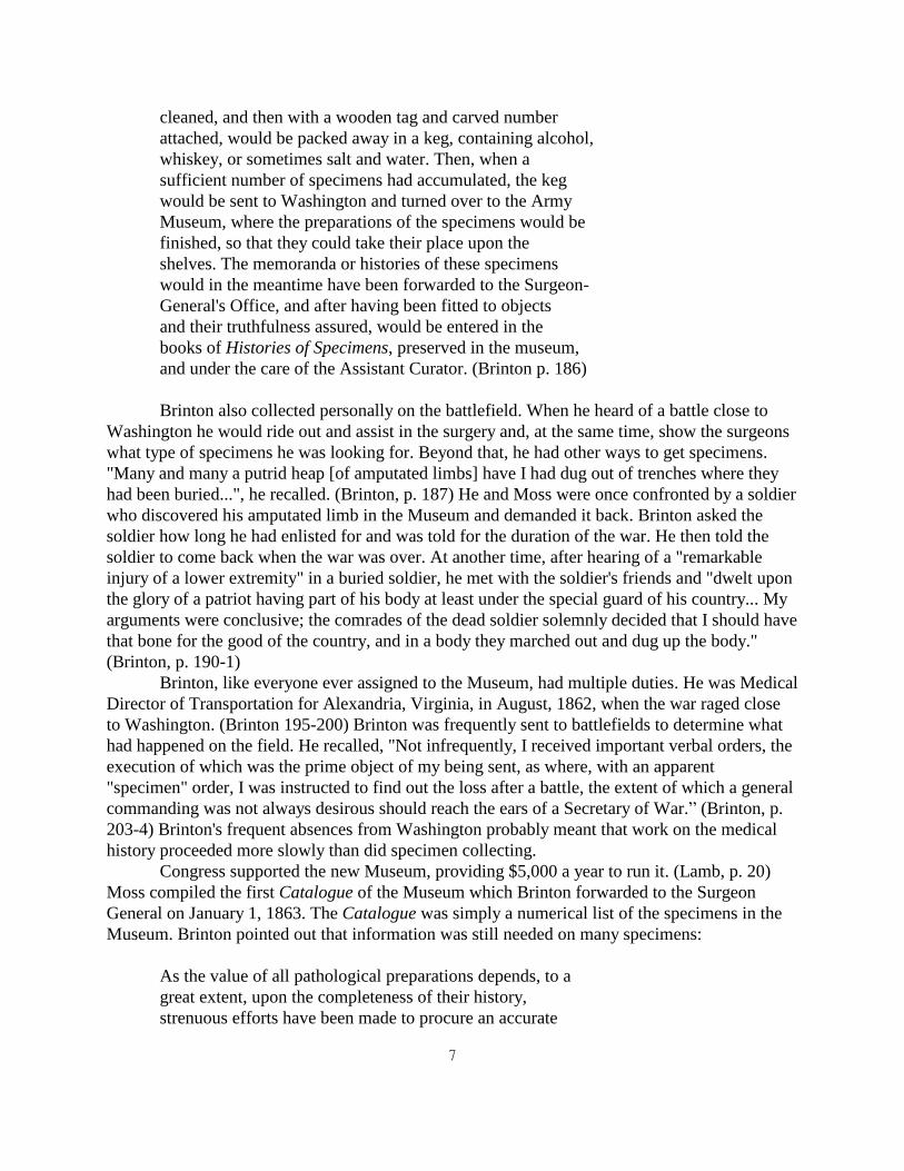
cleaned, and then with a wooden tag and carved number
attached, would be packed away in a keg, containing alcohol,
whiskey, or sometimes salt and water. Then, when a
sufficient number of specimens had accumulated, the keg
would be sent to Washington and turned over to the Army
Museum, where the preparations of the specimens would be
finished, so that they could take their place upon the
shelves. The memoranda or histories of these specimens
would in the meantime have been forwarded to the Surgeon-
General's Office, and after having been fitted to objects
and their truthfulness assured, would be entered in the
books of Histories of Specimens, preserved in the museum,
and under the care of the Assistant Curator. (Brinton p. 186)
Brinton also collected personally on the battlefield. When he heard of a battle close to
Washington he would ride out and assist in the surgery and, at the same time, show the surgeons
what type of specimens he was looking for. Beyond that, he had other ways to get specimens.
"Many and many a putrid heap [of amputated limbs] have I had dug out of trenches where they
had been buried...", he recalled. (Brinton, p. 187) He and Moss were once confronted by a soldier
who discovered his amputated limb in the Museum and demanded it back. Brinton asked the
soldier how long he had enlisted for and was told for the duration of the war. He then told the
soldier to come back when the war was over. At another time, after hearing of a "remarkable
injury of a lower extremity" in a buried soldier, he met with the soldier's friends and "dwelt upon
the glory of a patriot having part of his body at least under the special guard of his country... My
arguments were conclusive; the comrades of the dead soldier solemnly decided that I should have
that bone for the good of the country, and in a body they marched out and dug up the body."
(Brinton, p. 190-1)
Brinton, like everyone ever assigned to the Museum, had multiple duties. He was Medical
Director of Transportation for Alexandria, Virginia, in August, 1862, when the war raged close
to Washington. (Brinton 195-200) Brinton was frequently sent to battlefields to determine what
had happened on the field. He recalled, "Not infrequently, I received important verbal orders, the
execution of which was the prime object of my being sent, as where, with an apparent
"specimen" order, I was instructed to find out the loss after a battle, the extent of which a general
commanding was not always desirous should reach the ears of a Secretary of War.” (Brinton, p.
203-4) Brinton's frequent absences from Washington probably meant that work on the medical
history proceeded more slowly than did specimen collecting.
Congress supported the new Museum, providing $5,000 a year to run it. (Lamb, p. 20)
Moss compiled the first Catalogue of the Museum which Brinton forwarded to the Surgeon
General on January 1, 1863. The Catalogue was simply a numerical list of the specimens in the
Museum. Brinton pointed out that information was still needed on many specimens:
As the value of all pathological preparations depends, to a
great extent, upon the completeness of their history,
strenuous efforts have been made to procure an accurate

8
surgical and medical account of every case from which a
specimen has been taken. I regret to state that, in many
instances, the desired success has not been obtained. In
order that this evil may, as far as possible be remedied,
the number of every specimen in the Catalogue for which a
sufficient history has been received has been marked with an
asterisk. For all others, neither history nor description
have as yet been furnished. It is hoped that medical
officers, recognizing the objects contributed by them, will
exert themselves at once to remedy this deficiency.
(Brinton, Catalogue, p. 3)
Brinton used the Catalogue to increase interest in and contributions to the Museum. He stated in
his memoirs, "Very many specimens I had brought [to the Museum] from the battle-fields,
collected by myself. These I put into the first catalogue, assigning them to such medical officers,
as I could call to mind, and especially to those whom I knew to be lukewarm in Museum
interests. The effect of the procedure was good.” (Brinton p. 188)
Brinton acquired the Museum's third home, "Mr. Corcoran's School House" on H Street,
to hold the new Museum. In the fall of 1863, the Museum moved to its new location. Exhibited
to medical professionals were some of the 3,500 specimens collected. (Lamb, p. 19-20) When
Museum founder Surgeon General Hammond was court martialed on trumped-up political
charges in 1864, Brinton convinced Hammond's successor, Joseph Barnes, to keep the Museum
open. Barnes, another Philadelphia native and University of Pennsylvania graduate, was born in
1817. He joined the Army and served in the war with Mexico. During the Civil War, he was
Medical Director of several armies until becoming the Surgeon General. Barnes oversaw the
Museum until he retired in 1882. (Kelly, p. 63; Gillett, p. 201-3, 225-227)
Later in 1864, Brinton was relieved of duty at the Museum. He thought his transfer due to
any of three reasons: he was an appointee of Hammond, a cousin of McClellan, and the
proponent of a plan to bring in volunteer surgeons equal to the regular army ones at the close of
the war. He was assigned to normal medical duties and returned to Philadelphia to practice
medicine after the war ended. Brinton maintained a sense of humor about his transfer. He had
Hermann Faber, a Museum artist, draw him as "St. Denis leaving the Museum, head in hand, for
the region of the setting sun, with the bloody headman's sword, the unfinished work of the
Surgical History of the War, etc.” After a request by Barnes, Woodward had Faber make a copy
from memory and photographs of it were given to the new Surgeon General and much of his
staff. (Brinton p. 307-314)
Dr. George Alexander Otis replaced Brinton as curator. Otis served in the Museum for
seventeen years until his early death at age fifty in 1881. Under his direction, the second, much
larger Catalogue of the Army Medical Museum and the Medical and Surgical History of the War
of the Rebellion were published, as well as many shorter monographs. Otis was described by
Woodward as, "Hesitating, often embarrassed in his manner in ordinary conversation, especially
with strangers, he became eloquent when warmed by the discussion of any topic in which he took
interest.” Born in Boston on November 12, 1830, Otis lost his father before his first birthday. He
and his mother returned to her native Virginia. Undistinguished at Princeton, Otis preferred to

9
read French literature instead of the assigned material. Returning to Virginia, he privately studied
in Richmond and received his medical degree from the University of Pennsylvania in April,
1851. He spent the rest of that year and the next studying in Paris.2 There, Napoleon III's coup
d'etat gave him the opportunity to begin a first-hand study of military medicine. He returned to
Virginia in spring 1852 and the next year began the Virginia Medical and Surgical Journal.
The Journal, in competition with the older Stethoscope, was not a financial success. Otis sold a
partial interest to Dr. James McCaw3 and moved to Springfield, Massachusetts, but maintained
his connection with the Journal as corresponding editor. Otis enlisted as a surgeon in the 27th
Massachusetts Volunteers to participate in the war. (Kelly, p. 867-9) He moved over to the
regular Army as the war continued and joined the Museum staff as Brinton's assistant on July 22,
1864. On October 3, Otis replaced Brinton as Curator. (Lamb, p. 29, 31) (Within the split nature
of the Museum with its medical and surgical division, Otis and Woodward seem to have run their
sections of the Museum and written their sections of the History mostly independent of each
other. Since they worked together for seventeen years, their arrangement must have been
successful and in fact, Woodward was the executor of Otis' will.)
When the war ended, the Museum still had to fulfill its mission of creating a medical and
surgical history. Congress continued to fund the Museum to complete this task. By 1866, less
than a year after the war's conclusion, the Museum had expanded its scope of collecting. Otis
wrote to Dr. G. McCook congratulating him on his "successful case of ovariotomy" and
requesting "Unless you have decided on some other disposition of the pathological specimen, we
should be glad to mount it for the Army Medical Museum, which is no longer confined to
illustrations of military surgery but embraces the whole field of pathology.” (Otis to Cook, March
24, 1866)
Interest in the Museum continued to grow. As Hammond and Brinton had expected,
surgeons were eager to see their name in print as donors to the Museum. Otis wrote to Dr. Henry
Lyster in May 1866 acknowledging, "I am sensible that there was ground for complaint on the
part of the field surgeons that the specimens which they forwarded to the Army Medical Museum
were not always accredited to them. It is true that specimens often reached the Museum with very
insufficient memoranda, yet it cannot be denied that there was sometimes gross carelessness on
the part of those employed at the Museum.” Otis promised to credit Lyster while continuing to
look for the specimens that he had contributed. (Otis to Lyster, May 11, 1866) However, the
Museum was never intended to be a comprehensive collection of every limb amputated during
the war. In addition to contributions from military surgeons, Otis hoped to cooperate and trade
with other museums to build his collection. He wrote to Dr. Bache, curator of Philadelphia's
Mütter Museum, informing him "...that there are duplicate preparations illustrating gunshot
wounds at the Army Medical Museum, and that it is the wish of the Surgeon General to institute
a liberal system of exchanges with other museums of Surgery and Pathology.” (Otis to Bache,
February 23, 1866)
2 Hammond, Brinton and Otis were all educated in Europe, the center of medicine in the nineteenth century. They
were well-suited to establish a medical museum.
3 McCaw later became known for his organization of Chimbarozo Hospital in Richmond for the Confederacy.
10
The growing Museum required more space. The government purchased and renovated
Ford's Theatre for the Museum. The building also housed the Surgeon General's Library and the
Records and Pension division of the Surgeon General's Office. The move to Ford's Theatre in
December 1866 permitted the Museum to expand its range of interests. Otis wrote to Dr. William
Forwood, the surgeon at Fort Riley, Kansas, explaining the new situation: "We have removed the
Museum, as well as the Records Division of the S.G.O. to the large fire-proof building on Tenth
Street, remodelled by the Quartermasters' from the old Ford's Theatre structure, and have now an
abundance of room and are anxious that the Medical Officers at distant posts should send us
contributions of Indian weapons, and of specimens of
comparative anatomy.” (Otis to Forwood, January 4, 1867)
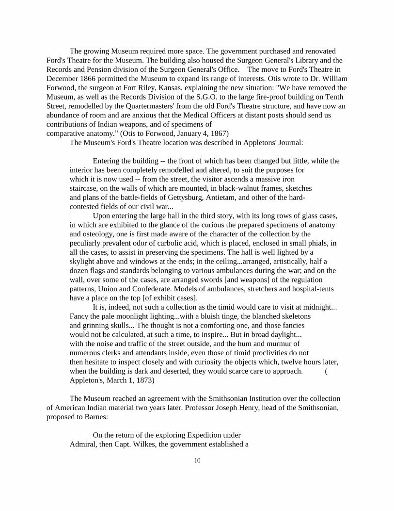
The Museum's Ford's Theatre location was described in Appletons' Journal:
Entering the building -- the front of which has been changed but little, while the
interior has been completely remodelled and altered, to suit the purposes for
which it is now used -- from the street, the visitor ascends a massive iron
staircase, on the walls of which are mounted, in black-walnut frames, sketches
and plans of the battle-fields of Gettysburg, Antietam, and other of the hard-
contested fields of our civil war...
Upon entering the large hall in the third story, with its long rows of glass cases,
in which are exhibited to the glance of the curious the prepared specimens of anatomy
and osteology, one is first made aware of the character of the collection by the
peculiarly prevalent odor of carbolic acid, which is placed, enclosed in small phials, in
all the cases, to assist in preserving the specimens. The hall is well lighted by a
skylight above and windows at the ends; in the ceiling...arranged, artistically, half a
dozen flags and standards belonging to various ambulances during the war; and on the
wall, over some of the cases, are arranged swords [and weapons] of the regulation
patterns, Union and Confederate. Models of ambulances, stretchers and hospital-tents
have a place on the top [of exhibit cases].
It is, indeed, not such a collection as the timid would care to visit at midnight...
Fancy the pale moonlight lighting...with a bluish tinge, the blanched skeletons
and grinning skulls... The thought is not a comforting one, and those fancies
would not be calculated, at such a time, to inspire... But in broad daylight...
with the noise and traffic of the street outside, and the hum and murmur of
numerous clerks and attendants inside, even those of timid proclivities do not
then hesitate to inspect closely and with curiosity the objects which, twelve hours later,
when the building is dark and deserted, they would scarce care to approach. (
Appleton's, March 1, 1873)
The Museum reached an agreement with the Smithsonian Institution over the collection
of American Indian material two years later. Professor Joseph Henry, head of the Smithsonian,
proposed to Barnes:
On the return of the exploring Expedition under
Admiral, then Capt. Wilkes, the government established a
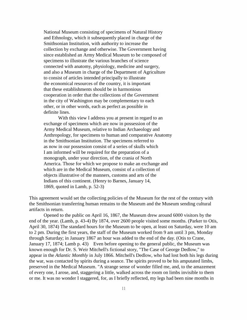
11
National Museum consisting of specimens of Natural History
and Ethnology, which it subsequently placed in charge of the
Smithsonian Institution, with authority to increase the
collection by exchange and otherwise. The Government having
since established an Army Medical Museum to be composed of
specimens to illustrate the various branches of science
connected with anatomy, physiology, medicine and surgery,
and also a Museum in charge of the Department of Agriculture
to consist of articles intended principally to illustrate
the economical resources of the country, it is important
that these establishments should be in harmonious
cooperation in order that the collections of the Government
in the city of Washington may be complementary to each
other, or in other words, each as perfect as possible in
definite lines.
With this view I address you at present in regard to an
exchange of specimens which are now in possession of the
Army Medical Museum, relative to Indian Archaeology and
Anthropology, for specimens to human and comparative Anatomy
in the Smithsonian Institution. The specimens referred to
as now in our possession consist of a series of skulls which
I am informed will be required for the preparation of a
monograph, under your direction, of the crania of North
America. Those for which we propose to make an exchange and
which are in the Medical Museum, consist of a collection of
objects illustrative of the manners, customs and arts of the
Indians of this continent. (Henry to Barnes, January 14,
1869, quoted in Lamb, p. 52-3)
This agreement would set the collecting policies of the Museum for the rest of the century with
the Smithsonian transferring human remains to the Museum and the Museum sending cultural
artifacts in return.
Opened to the public on April 16, 1867, the Museum drew around 6000 visitors by the
end of the year. (Lamb, p. 43-4) By 1874, over 2600 people visited some months. (Parker to Otis,
April 30, 1874) The standard hours for the Museum to be open, at least on Saturday, were 10 am
to 2 pm. During the first years, the staff of the Museum worked from 9 am until 3 pm, Monday
through Saturday; in January 1867 an hour was added to the end of the day. (Otis to Crane,
January 17, 1874; Lamb p. 43) Even before opening to the general public, the Museum was
known enough for Dr. S. Weir Mitchell's fictional story, "The Case of George Dedlow," to
appear in the Atlantic Monthly in July 1866. Mitchell's Dedlow, who had lost both his legs during
the war, was contacted by spirits during a seance. The spirits proved to be his amputated limbs,
preserved in the Medical Museum. "A strange sense of wonder filled me, and, to the amazement
of every one, I arose, and, staggering a little, walked across the room on limbs invisible to them
or me. It was no wonder I staggered, for, as I briefly reflected, my legs had been nine months in

12
the strongest alcohol.” (Mitchell) Undoubtedly, readers of the story would have wished to visit
the Museum to look for Dedlow's (fictional) limbs.
Woodward and Otis both oversaw large offices responsible for the medical records of the
war. Otis was in charge of the Division of Surgical Records while Woodward headed the Record
and Pension Division. Woodward described the divisions in 1873:
The first floor of the main building being nearly on a
level with the street, the visitor who glances through the
windows as he approaches the principle entrance is often
struck with the number of busy clerks he sees seated at
their desks or carrying record-books and papers about the
room. This floor, however, has nothing to do with the
museum. It is occupied by the record and pension division
of the surgeon-general's office. Here are filed the records
of the numerous military hospitals which existed during the
war, together with the monthly sick-reports received from
our armies during the rebellion, and those which still
continue to be forwarded monthly from the several military
posts. There are about sixteen thousand folio volumes of
hospital books, and several tons of sick-reports and
miscellaneous papers, all systematically filed in such a
manner as to permit ready access. To this branch of the
surgeon-general's office the commissioner of pensions
applies for official evidence of the cause of death or
nature of disability in almost all pension cases before
finally acting upon them. Similar information is also
continually asked for by the adjutant-general of the army
and other officials. Altogether, about two hundred thousand
applications from these sources have been responded to since
the war, and fresh cases are still received for
investigation at the rate of about fifteen hundred a month.
To facilitate these inquiries, the names of the dead, so far
as ascertained, have been indexed in a series of
alphabetical registers, which now contain very nearly three
hundred thousand names. About two hundred thousand
discharges for disability have been indexed in a similar
series of registers.
The second floor of the building is chiefly occupied by
the division comprising the surgical records of the surgeon-
general's office. Here are filed the reports made during
the war with regard to the wounded and those who had
undergone surgical operations, and from these a series of
record-books have been compiled, in which are entered the
histories of over two hundred thousand wounds and nearly

13
forty thousand surgical operations. These have been
arranged according to the nature of the wounds or
operations; amputations of the thigh, for example, being
entered in one set of books, amputations of the arm in
another, and so forth. These books are therefore available
for the preparation of the surgical history of the war.
Meanwhile, they have done good service by preventing frauds
in the matter of furnishing artificial limbs to disabled
soldiers, for which large sums of money have been
appropriated by Congress and ordered to be expended under
the direction of the surgeon-general. (Woodward,
Lippincott, p. 234)
The Surgeon General's library shared the second floor of the building with the Division of
Surgical Records.
Both Otis and Woodward complained of the workload of the Museum, the History and
the other duties they were assigned. Otis served on a board redesigning the standard Army
ambulance. Woodward accomplished much pioneering work in microscopy and
photomicroscopy during these years, introducing in America the use of aniline dyes to stain
microscopic specimens. For the Secretary of the Treasury and the National Academy of Sciences,
he investigated the content of wool fabrics for adulteration. In 1876, he was responsible for the
Medical Department's exhibit at the Centennial Exposition in Philadelphia.
Otis wrote to the Surgeon General asking to hire Dr. John Stearns as an assistant:
In endeavoring to carry out the Surgeon General's
instructions to prepare for the press with the greatest
rapidity consistent with accuracy the remaining portions of
the Surgical History, and at the same time to keep up the
current business of the Division of Surgical Records and of
the Anatomical and Surgical branches of the Museum, I find
my time engrossed with multifarious details to such a
degree, that I am unable to [do] that careful study and
reflections upon the general conclusions that the reader
will naturally expect to be deduced from the vast
accumulation of facts in the Surgical History. (Otis to
Crane, March 6, 1874)
Woodward had similar problems, especially with the Record and Pension Division. Woodward
estimated that 106 clerks were necessary to do the work of checking pensions and the other
duties of the division, including keeping meteorological records for Army posts. Woodward
supervised only 68 clerks. The Commissioner of Pensions complained about the slow rate that
information on pensioner's wounds were reported to his office from the Army, but would not
detail any men to Woodward. (Woodward to Barnes, Oct 26, 1875; Woodward to Crane,
December 4, 1875) Both Otis and Woodward suffered from ill-health and early deaths, possibly

14
directly related to their workload.
Through its publications and its contact with the medical profession, the Museum
functioned as a clearinghouse for information learned during the war. Woodward pointed out
other roles for the Museum in 1871:
...Specimens bearing upon disputed points or upon
subjects incompletely understood accumulate and increase in
number year after year, with carefully recorded histories of
the cases, until series are formed that serve for
comparison, and for a more exhaustive study of the questions
involved, which not unfrequently (sic) decides the dispute
or solves the difficulty.
The connection between the results of such studies and
the choice of the best method of treatment is perhaps most
obvious in surgery. For example: any intelligent person who
examines the unequalled series of over four hundred and
fifty specimens of gunshot fractures of the thigh-bone
preserved in the museum will have little difficulty
realizing their importance in connection with the vexed
question of amputation for this injury. He will only need
to examine a few of the specimens from cases in which
injudicious efforts were made to save limbs, and life was
lost after protracted suffering for months or years, to
understand the duty of preserving these mute witnesses. If
he happens to remember the grave differences of opinion
existing among our military surgeons during the late war as
to the proper cases for this operation, and the efforts made
in certain quarters to compel a false conservatism in all
cases and at all hazards, he cannot but feel thankful that
the results of that dreadful experience exist in a tangible
form for future guidance. Many similar examples might be
readily cited from the surgical domain.
On the medical side, although the connection between
morbid anatomy and the treatment of disease is less easily
understood by the non-professional mind, it is none the less
intimate. Our modes of treatment are so bound up with our
notions as to the nature of the affections with which we
deal, and those notions are so dependent upon the state of
our knowledge of morbid anatomy, that improved methods of
dealing with disease have in the past invariably followed
every advance in this knowledge, whether in the direction of
establishing firmly the connection of symptoms with
anatomical alterations, or in the direction of that better
acquaintance with the nature of the alterations themselves

15
which is attained only by the aid of the microscope...
Another use of pathological museums is too important to
be overlooked. They serve as valuable aids in enabling new
generations of medical men to identify with certainty the
descriptions of their predecessors, and thus to utilize
their experience. (Woodward, Lippincott, p. 240-1)
Otis stayed with the Museum through a stroke in 1877 until his death on February 23,
1881. He continued working on Museum projects even after the stroke made him an invalid.
Woodward, already in poor health, broke his leg on New Year's Day, 1881. That summer,
Woodward was one of President Garfield's doctors. Garfield died several months after being
shot, with his physicians having been unable to help him. (Lamb, p. 77-83) Woodward, now
elected President of the American Medical Association, was "prostrated with nervous depression
and hypochondria, and was taken to Europe in the hope that rest and change of scene would
restore him. He returned after some months without having found the benefit hoped for, and it
soon became necessary to place him in the asylum where he ... died.” (Cox, p. 254) Woodward
passed away on August 17, 1884, at age fifty-one. (Heaton, p. 537) In October 1883, Barnes'
successor Surgeon General C.H. Crane died and Robert Murray became the head of the Medical
Department. Murray combined the Museum and the Surgeon General's Library into a single
Museum and Library Division under John Shaw Billings. At the same time, efforts began to
acquire a new building for the new Division. Congress provided land and money for a new
building in 1885. In 1887, the Museum and Library moved to their new home on the National
Mall at Independence Avenue and Seventh Street, SW. The Record and Pension Division,
formerly under Woodward, was transferred to the Adjutant General's Office in 1889. (Pacheli, p.
2) The Museum was fortunate to be out of Ford's Theatre; it was subsequently occupied by the
clerks of the Division, of whom twenty-two were killed and sixty-three injured when the interior
collapsed in 1893. (Miles, p. 162-8)
16
CHAPTER:
The Medical & Surgical History of the War of the Rebellion
The records collected by the Museum during the war, and indeed the entire idea of the
Museum, were to produce the Medical and Surgical History of the War of the Rebellion.1 This
was not the first time that a medical history of a war had been written; the British had produced a
book on their medical experiences in the Crimea, and a similar account by the French was in
preparation when America's war began. (Circ. 6, p. 2) However, the American Civil War was of
a different order of magnitude and so was its History. The six-volume set attempted to discuss
every aspect of military medicine encountered during the Civil War. The massive bureaucracy
and organization of the North was replicated in a smaller scale in the Surgeon General's Office.
Hammond, brought in to replace the superannuated Finley and shake up the hidebound
Department, created his own, far larger, bureaucracy with its own policies, forms and regulations.
Reports were revised time and again to ensure the clear flow of knowledge back to Washington.
A large, sometimes stultifying, bureaucracy was the only way the History could ever be done. By
the time it was done, both editions of the six volumes apparently cost well over $100,000.
(Woodward to Crane, July 29, 1875.) The plates for the second editions of the second and third
Medical volumes alone cost $29,510.
This giant undertaking, a triumph of medical research which eventually took twenty-three
years and over 6,000 pages to complete and weighed fifty-six pounds, first was assigned by
Surgeon General William Hammond to Museum curators John Hill Brinton and Dr. J.J.
Woodward. Brinton had the responsibility to compile the Surgical section and Woodward the
Medical section2. When Brinton left the Museum, George Otis took over the Surgical section.
Both Otis and Woodward had been recommended for their assignments by Assistant Surgeon
General Crane. (Hemmeter, p. 647) Barnes and Crane's support for the project never flagged as
the years went by. Accounts were solicited from surgeons and doctors, including Confederates,
and the records of the Pension Office were heavily utilized to follow up cases. Museum
specimens were photographed; engravings, lithographs and photomechanical prints were made to
illustrate the text. Books, photographs, specimens, equipment - all were purchased although not
at the rate the authors would have preferred.
Both Otis and Woodward brought an immense learning to the project, surveying all that
was known on a subject before drawing any conclusions. Woodward wrote, "In view of the
many errors of fact scattered through the text-books, some of which have been repeated for ages
by authors copying from each other, I early resolved that, so far as possible, I would cite no
authority not before me when I wrote; and that, for the convenience of subsequent students, I
would in every case give not only the name of the author but the edition and page to which each
citation referred.” (History, Medical II, p. iv)
1Hereafter referred to in the text as the History. Cited in notes as MSHWR, Medical or Surgical Vol. #.
2The terms Surgical and Medical were used in a way that is slightly archaic today; to put it simply, Surgical
cases required surgery and were often the results of an injury while Medical cases were the result of a disease.
Otis and Woodward traced diseases through history. Woodward asked Otis, "Will you do

17
me the very great favor to request one of the distinguished French Physicians with whom you
have the honor to correspond, to obtain for me at the Bibliotheque Nationale of Paris a textual
copy of that part of the Greek Manuscript of AEtius which corresponds with the heading and first
sentence of ... the Latin translation of J. Cornarius, Basel Ed. of 1532...” Woodward wished to
ensure the two translations he had were both from the original Greek document on diarrhea.
(Woodward to Otis, December 27, 1875) The books collected to fulfill this need to review
directly all known information on a subject, formed the foundation, under John Shaw Billings'
direction, of the National Library of Medicine. A product of the nineteenth century's philosophy
of natural history, the History is a systematic, statistical compilation of the types of injuries and
diseases a military surgeon could expect to treat, along with discussions of and examples of
treatments. It was not a textbook but rather a reference book, a compendium of experience. Since
the History itself was a distillation of millions of pages of medical information, its creation and a
few points of interest, rather than the contents of each volume, will be reviewed here.
On June 9, 1862, the History was publicly announced to the Medical Department.
Hammond's Circular No. 5 (see fig ) assigned the writing of the History to Woodward and
Brinton and requested many types of information from medical officers. To promote
compliance, the announcement promised to put one's name in print for the ages.
Charged with writing the History, while also creating the Medical Museum, both
Woodward and Brinton had their own ideas on how to proceed. Woodward began looking at
statistics, publishing Circular No. 15: Sickness and Mortality of the Army during the first year of
the War, an eight-page survey, on September 8, 1863. Brinton soon published his statistical
survey, Circular No. 9: Consolidated Statement of Gunshot Wounds and then proceeded to
ignore the cold, dry numbers and solicited descriptive battlefield reports.
Brinton envisioned the surgical history as a chronological one, following the war, battle
by battle. In the first Surgical volume, Otis described Brinton's plan:
In the preparation of the surgical portion of the Medical
and Surgical History of the War of the Rebellion, it was at
first proposed to treat the surgery in connection with the
military operations in the several battles and campaigns.
... After giving a general account of a campaign,
enumerating the troops engaged, the mode of transporting the
injured, and the available hospital accommodations, the
wounds and operations of each engagement were discussed, the
reports of medical directors, and all other reliable sources
of information being brought into requisition. Among these
were observations personally made in the base and field
hospitals of the armies of the Potomac and of the West,
after the great battles, where much valuable surgical
material was collected, including admirable illustrations of
the graver injuries, pathological specimens, and a series of
excellent surgical drawings.” (MSHWR, Surgical I, p. xiii)
In a letter to Surgeon Alonzo J. Phelps, based in Nashville (a city precariously held by the
18
North), Brinton delineated his needs. He wanted to know everything related to surgery that
occurred at a battle:
I to-day had an opportunity of examining your "Casualty
report" of the battle of Murfreesboro. As you perhaps know,
I have been directed by the Surgeon General to prepare the
Surgical history of the War. I am exceedingly desirous of
obtaining a good Surgical account of the above mentioned
battle. Have you time Doctor to prepare such an account?
If you have, I should be glad to receive it, and I will see
that due credit be given you in the forthcoming history.
The chief points to be noticed, are number of killed and
wounded, character of missile used by the Enemy, the
Surgical appliances at hand in the battle-field, general
character of wounds, -- Hospitals Ambulances, what was done
with the wounded, Erysipelas or Tetanus, if either were
subsequently prevalent, Resections, Amputations, Names of
Operators. In fact Doctor a full account of the battle as
looked at by a Practical Military Surgeon from his own point
of view. I sincerely trust that you will have leisure to
write something. If you have, may I ask you to furnish me
with the name of some one, whom you may think capable of
doing this important Subject justice... (Brinton to Phelps,
February 9, 1863).
Phelps' detailed report was abstracted in the first Medical volume's appendix.
Brinton was dutiful in requesting information from the field, even when he would rather
have been at the battle himself. He wrote to his comrade Surgeon H.S. Heust, who was with
Grant, requesting information on the Union victory at Vicksburg:
My dear Heust:
I want some of these days to know all about the great
Vicksburg battle. I envy you your position. If you have
force enough, can you not direct some young assistant
Surgeon to keep notes (Surgically) of what is going on --
what hospitals are established +c -- + what have been your
arrangements in your glorious sequence of victories. The
enclosed blanks will be suggestive. I know you will not
think me annoying, in asking you now, in the midst of what I
know are arduous labors, -- But I have learned, that, unless
this information is collected on the field, it is apt to be
lost -- + I, if no one else, am sure to be the loser. Kind
remembrances to all my friends, + to the best General of
them all. (Brinton to Heust, June 6, 1863)
19
In keeping with his plan to track surgery in the war through each battle, Brinton needed
maps of the battlefields. "A great desideratum is a map of the locality with the position of the
hospital correctly laid down. This I could have engraved and inserted in the Book," he wrote to
Surgeon John Craven who had reported on skirmishes on Morris Island, South Carolina. (Brinton
to Craven, August 22, 1863) In later years, Brinton felt that he did not get enough credit for his
initial work on the surgical portion. In his autobiography, he wrote:
By the way, I may add that this map [of Antietam] and nearly
all the other field hospital maps, etc., of great battles in
that book, except the extreme southern campaigns, were
prepared under my direction when stationed in the Surgeon-
General's office in Washington. The were modified and
reduced by an artist named [Pohlers], from the topographical
maps, and the position of the hospitals I usually had
located by any medical inspectors or other medical officers,
who might know the ground well. My name does not appear in
any of this work, but it was designed by me, and much of
it executed under my direct superintendence. Some was done
by my successor after my departure from Washington.
(Brinton, p. 206)
The maps were published, along with the field reports solicited by Brinton, in the appendix of the
first Medical volume. They were credited to Woodward and Otis; Brinton's name was not
mentioned although Otis credited him in the opening paragraph of the first Surgical volume.
Brinton encountered difficulties in gathering information to research bullet wounds. He
found reports coming into the Museum listed gunshot wounds without accompanying
information such as part of the body wounded or results of surgery. Another problem was
patients being counted as new cases each time they were transferred from hospital to hospital.
(Brinton 249-50) Brinton was forced to state, "The inadequacy of the entries in the ...monthly
report of sick and wounded was early acknowledged, and it was officially declared that previous
to September, 1862, 'the surgical statistics of the war were absolutely worthless,' and that 'the
only information procurable is such as can be derived from the examination of a mass of reports,
all of which present merely certain figures under the vague and unsatisfactory heading, Vulnus
sclopeticum [gunshot wound]." (Brinton, Consolidated Statements of Gunshot Wounds, quoted
in MSHWR, Surgical I, p. xxv) New forms, reports and registers were designed in late 1863 by
a special board of medical officers. A small register was given to officers to use in the field. Two
larger ones, one for information on the sick and wounded and one for surgical operations, were
used in hospitals with new quarterly report forms. The new information, as well as specimens,
arrived in the Museum to be processed by doctors and clerks for use in the History. (Circular 6,
p. 3-6)
Specimens were preserved in alcohol seized by the Provost Marshal for the Museum.
Brinton recalled:
...an enormous amount of alcoholic beverages was poured into
20
the Museum, everything from champagne to the commonest
rum. Our side lot was piled with kegs, bottles, demijohns
and cases, to say nothing of an infinite variety of tins,
made so as to fit unperceived on the body, and thus permit
the wearer to smuggle liquor into camp. ... When the whisky
was strong enough for preservative purposes, [Schafhert]
kept it in the package; when it was not, it went into the
still. This under Schafhert's watchful care, ran
incessantly, and furnished the Museum with a large amount of
very fair alcohol, not only for putting up our specimens,
but for furnishing the various depots in the Army where
fresh specimens were being collected, so that they could be
kept from decomposition, and reach the Museum in good
condition.
When Brinton discovered that the Museum's whiskey was being tapped en route, he added tartar
emetic to a barrel. He got a report of many vomiting railroad employees and had no more
problem with losses. Brinton also used confiscated cherry brandy to smooth his travels on the
road; he traded kegs with the 5th Cavalry in exchange for the use of their horses when traveling.
(Brinton, p. 191-3) War-time supplies of alcohol were still preserving new Museum specimens
ten years later. (Woodward to Tyson, March 10, 1876)
Woodward, like Brinton, had problems gathering information. Soon after he was placed
in charge of the Division of Medical Records, Woodward realized that the then-current forms
with spaces for 143 diseases worked fine during peace but were inadequate in war. In July, 1862,
a board consisting of Woodward, Brinton, Surgeon Lewis A. Edwards and Assistant Surgeon
M.J. Asch met in Washington to revise the Army's sick reports. The reports were changed from
quarterly to monthly, and then forwarded directly to the Surgeon General from each unit without
being edited or consolidated by the Medical Directors of the individual Armies. At the same
time, the board modified the Classification of Diseases. The board selected the classification
system devised in 1855 by England's Dr. William Farr and (with some modifications, such as
leaving out diseases of women) recommended it to the Surgeon General. A section of the
classification scheme, barely comprehensible now, sharply shows the limits of nineteenth-century
understanding of disease:
The class Zymotic diseases is intended to embrace epidemic,
endemic, or contagious affections, supposed to be induced by
some specific body, or by anomalies in the quantity or
quality of the food. The order Miasmatic diseases includes
affections believed to be due to various atmospheric
influences, such as the products of vegetable and animal
decomposition, specific emanations from the human body in a
state of disease, and the so-called marsh miasms. The order
Enthetic diseases includes those disorders which are
transmitted by the inoculation of morbid matters. The order
21
Dietic diseases includes those which are caused by errors in
the quantity and quality of the food. (MSHWR, Med I, p.
xviii-xix)
Miasmas, vague unhealthy influences, were supposed to be floating through the air, infecting
soldiers. Mosquitos and other disease carriers would not be suspected for years after the war
ended.
When Otis succeeded Brinton as curator of the Medical Museum, he also became
responsible for the Surgical section of the History. Before the close of the war, Otis realized that
Brinton's plan to write a surgical history of each battle would not work due to the sheer amount
of information being received. "During that year [1864] there were no less than two thousand
skirmishes, actions, or battles, and to have given a correct analysis of the casualties from the
returns from the field and base hospitals would have been impossible.” Instead, Otis decided to
arrange the Surgical volumes by type of wound and region of the body. The battlefield reports
that Brinton had already collected would be an appendix to the first Medical book. He promoted
his new plan in the Museum's first major post-war publication, Circular No. 6: Reports on the
Extent and Nature of the Materials available for the preparation of a Medical and Surgical
History of the Rebellion.3 (MSHWR, intro, p. xiiv)
3Hereafter cited as Circular 6.
Otis and Woodward published Circular No. 6, heavily illustrated with woodcuts and
lithographs, on November 1, 1865. The Circular became the blueprint for the History. In keeping
with the planned format of the History, Otis wrote the Surgical report while Woodward produced
the Medical one. The book, which it certainly is at 166 pages, purports to be a report to Surgeon
General Barnes from Otis, "in response to your inquiries relative to the nature, extent, and value
of the surgical data that have accumulated in the department of your office under my charge."
(Circular 6, p.1) Barnes knew very well what Otis and Woodward were doing, especially in light
of the considerable financial costs of the project. Circular 6 was even published by the noted
medical printer J.B. Lippincott of Philadelphia. The first edition of 5,000 copies cost well over
$6,000. When Lippincott raised the issue of selling copies of the book, Otis replied, "I am
directed by the Surgeon General to acknowledge your communication of the 22nd inst. and to
instruct you to refer all applicants for copies of Circular No. 6, S.G.O. 1865, to this office. The
Surgeon General does not entertain favorably the proposition to supply demands from
booksellers, but decides that all issues must be made gratuitously from this office.” (Otis to
Lippincott & Co, February 24, 1866) Circular 6 was aimed at the medical officers of the Army,
to whom it was distributed freely, to encourage their continued enthusiasm for, and participation
in, the projected History. Otis wrote to a prospective contributor, "In reply to your
communication I would state that Circular No. 6, S.G.O. 1865, is distributed to all medical
officers who rendered faithful service during the late war and who have notified this office of
their P.O. addresses... It is proposed to distribute future publications relative to the medical and
surgical history of the war to those medical men who have contributed or who shall contribute to
the material from which such publications may be compiled." (Otis to Roberts, May 11, 1866).
Much of the expense of such free publications was made possible by the transfer to the Museum

22
of slush funds from the closing hospitals. (Lamb, p. 37) The fund from Lovell General Hospital
of Portsmouth Grove, Rhode Island, alone was $9,952.83. (Otis to Samuel Ramsey, December 4,
1865) On the other hand, the hospitals in Nashville, Tennessee, only produced $226.25 (Otis to
George Cooper, December 8, 1865).
Both Woodward and Otis wrote extensive introductions on their materials and methods
before presenting specific examples of information available for the History. Otis began his
report by discussing the types and quality of the data available to him:
The materials in the office relating to the surgery of the
late war consist of the reports of the medical officers
engaged in it, and of illustrations of these reports in the
shape of pathological specimens, drawings and models. The
documentary data are of three kinds; first, the numerical
returns, in which the number alone of the different forms of
wounds, accidents, injuries, and surgical diseases is given;
secondly, what might be called the nominal returns, in which
are furnished the name and military description of each
patient, and the particulars of the case, with more or less
of detail; and thirdly, the miscellaneous reports... In the
third class are included the reports of medical directors
of armies in regard to the operations of the Medical
Department, and the succor given to the wounded; reports and
dissertations on new methods and modes of treatment, and
modifications of surgical apparatus and appliances;
pathological researches on morbid processes pertaining to
surgery, as hospital gangrene, osteomyelitis, pyaemia, and
the like; plans for ambulance organization, and the
transportation of the wounded by land and water. (Circular
6, p. 1-2)
"The extent of these materials is simply enormous," Otis continued. By comparison with the
British and French experience in the Crimea, where the combined armies had suffered 653
gunshot fractures of the femur, over 5,000 such cases were reported to Otis. Otis recognized and
explained the value of this material, saying:
It may be emphatically said that they throw much light
on some of the great moot points in surgery; that they
comprise on some subjects, as, for example, on the question
of the propriety of excising the head of the femur for
injury, fuller data than are now extant in the entire range
of surgical literature; and that it may be hoped, without
temerity, that they include the elements for the solution of
many grave surgical problems. (Circular 6, p. 3)
23
Otis laid out Circular 6 the way he anticipated organizing the History. He covered wounds of the
body, starting with the head and working down. He then examined surgical treatments such as
excision and amputation. A brief overview of the medical department staff was followed by a
review of the medical resources, including transportation, available to the surgeons. Otis
optimistically ended his report,
In conclusion, it has been estimated that it will be
possible, by judicious condensation, to include in one large
quarto volume the statistics of the graver injuries, as
fractures of the extremities and wounds implicating the
joints or great cavities, and of the major surgical
operations they have involved, the individual cases, their
progress and results, being concisely recorded; while a
second quarto volume could comprise numerical tables of the
less serious injuries, an historical summary, and a
discussion of the lessons derived from the statistical
records of the war. (Circular 6, p. 88)
In fact, none of these estimates were accurate, and Otis' plan for the Surgical history was much
altered when it began appearing. An additional 600-page volume proved necessary to cover the
topic adequately.
Woodward's report to Barnes followed much the same pattern as Otis's. He listed the
available material that he would be using:
The matter collected is partly statistical, partly
pathological. The first category embraces the medical
statistics of the several armies and general hospitals. The
second consists of a number of memoirs and reports by
medical officers on the causes, symptoms and treatment of
the more important camp diseases, of numerous histories of
cases and autopsies, of the fine series of medical and
microscopical specimens in the Army Medical Museum, and of
the results of the pathological studies conducted under my
direction of the basis of these collections. In addition,
there are a large number of descriptions and plans of
general hospitals, of reports on hospital organization, and
some other miscellaneous matters. (Circular 6, p. 89)
Woodward's statistical work led him to conclude that fewer troops died from disease in the
Union Army than during any previous war, but that the mortality rate for soldiers due to disease
was more than five times higher than expected for a similar group of men during peacetime. The
rate of deaths due to disease was also far higher than that from injuries. Facts like these enabled
Woodward to state unequivocally the value of the History:
24
Such a publication, therefore, becomes one of the most
important duties of the Medical Department of the army; a
duty the evasion or neglect of which would be a grave crime
against the army of the United States, and against every
American citizen who, in future wars, volunteers in the
defence (sic) of his country." (Circular 6, p. 90)
Woodward's primary interest for his first volume lay in statistics. He defended his data
against criticism of two types. Diagnostic errors would be eliminated by combining similar
diseases that might have been confused by doctors. Diseases of the eye would all be put together
under "ophthalmia" to highlight the most important fact -- disease of the eye occurred at a certain
rate in the Army. Errors of negligence were a bit more difficult for Woodward to address.
Woodward answered these critics, saying the reports during the war to the Surgeon General
"contain internal evidence of the care with which they were prepared, and, it is believed, will
compare favorably with any other set of statistical papers in existence.” It was true that some
reports were never made, were lost or were badly done, but in light of the sheer mass of
information, in Woodward's view, they became statistically insignificant.
Woodward, like Otis, organized the Circular as he would the History. He discussed the
mortality rate of the Army and statistically examined the disease rate. He previewed the Medical
and Microscopical sections, the two Museum sections under his care, relating details of
individual cases. A full color lithograph by F. Moras after artwork by Hermann Faber was
included; plates by this team would be published in the third Medical volume twenty-three years
later. Woodward closed with a discussion of the design of hospitals during the war. He included
lithographs that were not reproduced in the History such as a "Birds Eye View of Lincoln
General Hospital."
Woodward's projections for his volumes were more accurate than Otis':
In conclusion, I may express the opinion that, with the
utmost brevity and care, it may be hoped to digest the
material above sketched, not including surgical cases, into
three quarto volumes... The prominent subjects in these
volumes would be the medical statistics of the several
armies, with the principal facts in their medical histories;
the medical statistics of the several general hospitals,
with descriptions of their construction and administration,
illustrated by a sufficient number of ground plans and
perspective views to give a just idea of them; and lastly,
an account of the causes, history, symptoms, pathology, and
treatment of the principal diseases of the troops, based
upon statistical facts, contributed papers, histories of
cases and autopsies, and observations made in the medical
and microscopical sections of the Museum. ... Of the
subjects thus indicated, about one-half of the statistical
matter is compiled, and most of the material required for

25
the remaining portion of the work is collected, and can be
prepared for the press with reasonable rapidity. (Circular 6, p. 166)
Woodward correctly estimated that three volumes would be necessary while Otis only planned
for two of the final three; neither, however, would live long enough to see the work completed.
Circular 6 proved to be extremely popular. Two months after it was printed, Dr. Lemuel
Dale, a contributor to the Museum, was writing to Otis to find out how he could receive a copy of
the History, wondering whether he could request one before it was completed. Fortunately for
Dale, Otis told him that he could request one at any time provided he specified where and when
he had been in the Medical Department and what data he had provided to the Surgeon General's
Office (Otis to Dale, January 12, 1867). On February 9, 1866, four months after it was printed,
Otis requested a bid from Lippincott for a second edition of 2,500 copies. No substantial changes
were to be made. "With the exception of the correction of typographical errors, the second
edition must be in every respect uniform with the first," were his instructions. (Otis to Lippincott,
February 9, 1866) Otis negotiated with Lippincott over the price of the second edition; he was
willing to pay $1.28 per copy for a total cost of $3200, not $3475 as Lippincott had requested
(Otis to Lippincott, February 15, 1866). Circular 6 was the last volume that Lippincott, who had
printed forms and books for the Museum through the war, would handle; in the future, Museum
publications were usually done by the Government Printing Office.
For the next five years, the two men continued working on the first book in each of their
respective specialties. Otis pointed out to inquirers, "The Medico-chirurgical history of the
British Army in the Crimea was not published, if you recollect till 1858, while the French
statistics on the same subject only saw the light a few months ago." (Otis to Lyster, September
25, 1865; the Crimean war was from 1853-1856) Assistant Surgeons like Woodhull and Curtis
helped produce catalogs of the three collections in 1866 and 1867, with brief descriptions of
cases that could be expanded on and woodblock illustrations that could be reused for the History.
Before much research work on Museum specimens could be done, a cataloging system
was needed to arrange the Museum's vast number of specimens in a logical order. During the
war, Woodward and Brinton had divided the Museum's collection into three sections: Surgical
(Section I), Medical (Section II), and Microscopical (Section III). Items in each section were
numbered consecutively starting at 1. The Surgical Section contained material related to surgical
operations, including skeletal specimens, wet tissue, bullets, photographs and plaster casts. The
Medical Section contained specimens of various diseases which were not treated surgically.
These include specimens of typhoid fever, swamp fever, tuberculosis, and especially diarrhea and
dysentery. The Microscopical Section consisted of prepared microscopic tissue sections and
photographs taken of them.
By the end of 1865, it was apparent that these sections would have to be expanded.
Human Anatomy (Section IV) and Miscellaneous (Section VI) sections were placed under Otis.
The Human Anatomy Section contained samples of normal, not pathological, human anatomy.
The Miscellaneous Section held equipment such as medical kits, models, and anthropological
artifacts. Woodward dealt with Comparative Anatomy (Section V) which had veterinary
specimens, normal and pathological. (Lamb, p. 38). In the following decade, the Surgical and
Medical Sections were merged into one Pathological Section.
Once a specimen, photograph, or object was placed in one of these sections, it was then
26
assigned to a particular pathology category. These categories were developed by Museum doctors
and reflected contemporary medical knowledge. The specimen, photograph, or object was then
assigned a unique identification number. The first Catalogue of the Army Medical Museum,
compiled by assistant curator William Moss under Brinton's direction, was published on January
1, 1863. The Catalogue was "offered simply as a numerical list of the objects in the Army
Medical Museum. No attempt has as yet been made to classify the various injuries, nor has any
description of the preparations been entered upon. Such a work must be deferred for the future. It
will then be found to demand volumes. The labor of the present hour is simply to collect and to
preserve -- the study must be made hereafter.” (Brinton, Catalogue, p. 3-4)
Three catalogues, corresponding to the three main sections of the Museum, were
published soon after Circular 6. These catalogues were listings of the Museum's collections,
arranged for the most part by body section. The Government Printing Office published the first
catalogue in 1866. The Catalogue of the Surgical Section of the United States Army Medical
Museum, credited to Alfred A. Woodhull, was worked on by Woodhull, Otis and John Shaw
Billings. (Otis to Lyster, May 11, 1866) Otis did not like Woodhull's work. He wrote to a
correspondent who had complained about a lack of credit, Woodhull "no longer consulted me
as to the compilation and adapted many arrangements of which I much disapproved. Among
these was the practice of referring specimens to the commissioned officer of the hospital from
which they were sent.” Otis planned to correct Woodhull's work in a second edition of the
Catalogue, but it was never redone. (Otis to L. K. Baldwin, April 4, 1876) Woodward's
Catalogue of the Medical Section of the United States Army Medical Museum and Curtis'
Catalogue of the Microscopical Section of the United States Army Medical Museum followed
in 1867. All three catalogues included short description of each specimen, the name of the patient
who provided the specimen, where he was wounded, his doctor's name and other facts of the
case. This information could be expanded for the Museum's other publications.
An extraordinarily complex numbering system, which varied slightly for each section,
was adopted for each catalogue. It was designed to combine specimen identification numbers and
types of objects with pathology categories. The specimen's unique number was preceded by a
Roman numeral code, pertaining to a particular pathological category. For instance, "XXVI,
4629 A 3 101" refers to one photograph of a "group of officers who had undergone amputation
for gunshot injury.” In this case XXVI corresponds to the twenty-sixth category of objects
relating to surgery, namely "Photographs, drawings and maps," and 4629 is the unique
consecutive item number. 'A' means "the Army Medical Museum Series of Photographs". '3' is
the third volume of photographs taken at the Museum. 101 indicates that this photograph
number 101 within a consecutive numbering series. Photographic collections eventually began to
receive names reflective of their content. Our example "XXVI, 4629 A 3 101" became Surgical
Photograph 101 (SP 101).
This system was far too cumbersome to maintain given the rapid rate of acquisition of
specimens and objects, especially since it was necessary to refer back to a catalogue. Specimens
were tracked and referred to by their Section number instead of the larger cataloging system. This
inability to devise a pathological cataloguing system plagued the Museum and other medical
professionals well into the twentieth century. (Lamb, p. 126-135)
Other duties and publications intervened with the work on the History. Woodward spent
time researching the cholera epidemic of 1866, producing Circular No. 5: Report on Epidemic
27
Cholera in the Army of the United States During the Year 1866 in 1867. His format for this was
very similar to that of Circular 6. He gave a brief history of the epidemic (out of 12,780 men in
the Army, 2,708 caught the disease and 1,207 died), followed by statistical tables and excerpts
from field reports on the disease. The circular ends with an interesting report on disinfectants
from Dr. B.F. Craig of the Surgeon General's Office. Craig concluded that disinfectants such as
bleach, alcohol, and chlorine were effective in checking the disease. Unfortunately, he also
thought, "The theory of the mode of propagation of cholera which is the most widely received
and has in its favor the greatest amount of evidence is that the virus4 is not eliminated as such
from the bodies of cholera patients, but that it is formed in their discharges by some specific
process of decomposition, a process which is supposed to go on only in alkaline fluids.” (Circular
5, p. 63-65) Woodward concluded and clearly stated that there was no good treatment for the
disease. After using epidemiology to pinpoint the two centers and subsequent spread of the
epidemic, Woodward lamented that, "As to the question of therapeutic agencies, it cannot be said
that any new light has been shed upon the existing obscurity of the subject by the army
experience.” He then recommended that attention be instead focused on 'hygienic precautions'
such as Craig discussed and T.A. McParlin's recommendation on "the efficacy of the use of pure
drinking water in arresting the spread of the disease, even after it has made its appearance."
(Circular 5, p. xvi-xviii) He returned to the subject the following year with Circular No. 1:
Report on Epidemic Cholera and Yellow Fever in the Army of the United States During the Year
1867.
4 'Virus' is used in its older meaning of a "specific agent of an infectious disease", and not its modern meaning,
"a group of microbes which with exceptions are capable of passing through fine filters that retain most bacteria,
and are incapable of growth or reproduction apart from living cells.” (Steadman p. 1559) Modern virology was not
established until the end of the nineteenth century in the great expansion of laboratory medicine.
Woodward also did much important research in microscopy during these years, research
which contributed to his understanding of the diseases he would be writing on in the History.
Otis, an excellent surgeon, continued to publish on topics of interest to him that could be used in
the History. In July 1867, Circular No. 7: A Report on Amputations at the Hip-Joint in Military
Surgery was published. Otis was much interested in this rare operation, in which the entire leg
was removed. He could eventually fully document fifty cases. He had sent letters imploring
acquaintances, "Can you not hear among your rebel friends of any unreported amputations at the
hip-joint? We have thirty on record. The Confederates but two - I have a third not yet in print.
Try and put me on the track of some new cases.” (Otis to W.F. Smith, January 21, 1867). Otis
stated his rationale for his monographs clearly, "It may reasonably be hoped that the analysis of
large numbers of surgical cases, due attention being paid to essential particulars, must result in
the establishment of rules for the performance of operations more nearly approaching scientific
method and accuracy than those that now prevail, and in the selection of proper cases for
operation at the stages and under the circumstances most conducive to success." (Circular 7, p. 6;
italics added) For Otis, the Civil War was a great learning experience which would become a
greater teaching experience. He also wrote a small companion publication Drawings,
Photographs and Lithographs illustrating the histories of seven survivors of the operation of
amputation at the hip joint, during the War of the Rebellion, together with abstracts of the seven
28
successful cases. (Lamb, p. 46)
Otis produced another monograph, Circular No. 2: A Report on Excisions of the Head of
the Femur for Gunshot Injury in January 1869. Unlike amputations, in excisions only the
damaged portion of the bone --in this case the head of the femur-- is removed, with the aim of
leaving a partially-functioning limb. Otis felt "it must be admitted that this report comprizes (sic)
the elements for solving the grave problem of the appropriate treatment of gunshot injuries
involving the hip-joint." (Circular 2, p. 6) He responded to critics of his previous publication,
who had questioned his classification of the importance of time elapsed before surgery, saying,
"If a study of the histories of over twenty thousand major amputations, and of more than four
thousand excisions of the larger joints, performed during the late war, may permit me to speak
authoritatively on this point, I would say that no doctrine in military surgery is supported by more
ample evidence than that which teaches that in operations for traumatic causes, there is a wide
difference in the results of those performed immediately after the reception of the injury, those
performed during the existence of inflammatory action, and those done after the symptomatic
fever and inflammatory symptoms have abated.” (Circular 2, p. 6) Otis continued defending his
arrangement for years, writing in 1870, "Dr. Taylor's classification may be excellent for the
British army, with its corps of trained medical officers; it could not have been advantageously
introduced in our service, chiefly attended by surgeons hastily called from civil life." (MSHWR,
Surgical I, p. xxvi) Indeed, many of the medical problems of the Civil War arose from the lack of
a large, well-trained medical force.
For the femur excision report, Otis had assembled the artistic team he preferred to work
with. J. Bien had produced the lithographs and H.H. Nichols had produced the woodcuts over the
preceding five years. The amount of people working on such a large task was never very big. The
maximum staff Otis had working on the project was "one medical officer, one clerk, and sixteen
hospital stewards, occasionally aided by one acting assistant surgeon." (MSHWR, Surg I, p. xxvi)
The following year saw the publication of the initial parts in both the Medical and
Surgical volumes of the History. Unfortunately an odd system was adopted: Woodward's book
was designated Part I, Volume I Medical History, and Otis' work became Part II, Volume I
Surgical History; for the sake of convenience they will be referred to here as first Medical, first
Surgical, et cetera.
Barnes credited Secretary of War Stanton with promoting the publication to Congress; on
June 8, 1868, Congress appropriated money and authorized the Government Printing Office to
print five thousand copies of each volume. Work began on March 3, 1869, and by November
1870 the books were completely done. Barnes, in his ornate style, states, "Through the
liberality of the Government, in its beneficent pension laws, it has been found practicable to
obtain accurate histories of many thousand wounded or mutilated men for years subsequent to
their discharge from service. As in the official returns of the casualties of the French and English
Armies in the Crimean War, the cases were dropped when the men were invalided, pensioned, or
discharged from service, this information was considered peculiarly desirable.” Barnes' point,
that these volumes were the first long-term follow-ups of surgical cases, sets the stage for the
development of modern medical studies conducted on groups over a period of years. Otis
concurred with Barnes, stating, "Much important and otherwise unattainable information
regarding the ulterior consequences of the more important and rare injuries has been collected by
private correspondence with invalided soldiers and their surgical advisers. More than fifteen
29
hundred cases have been examined in this way.” Otis also credited the "former medical officers
of the Confederate army" for providing much information. He was also able to use Confederate
hospital records that had been captured by the Union. Occasionally, Otis would even advertise in
newspapers for information on specific cases. But the lion's share of information came from
pension records. "[T]he principal sources from which the remote results of wounds, injuries, and
operations were ascertained, were the reports of pension examiners, and communications from
the surgeons general and adjutants general of States.” (MSHWR, Surg I, p. xix)
Otis had changed the format and content of his volumes in the five years since Circular 6
was published. The volumes were not arranged into one volume of graver injuries and one
volume of lesser injuries, followed by a historical summary and a discussion of lessons learned.
Nor were cases of injuries discussed followed by a separate discussion of treatments as in
Circular 6. Instead, "As nearly as practicable, the wounds and injuries and surgical disease of
each region of the body have been arranged together, as the simplest and most natural order that
could be adopted. The most interesting clinical histories have been printed in full, or in abstracts
including the attainable essential details, and the remaining cases, or sometimes the whole
number of cases of the class, are set forth in tabular statements.” In other words, each area of the
body would have its wounds described and treatments discussed. Otis opened his book with a
daily chronological summary of the battles and engagements the Army fought. Then he examined
wounds of the upper body. Due to space limitations, in the first Surgical volume only the head,
face, spine, and chest were included.
Whenever possible, the clinical histories were credited to the doctor who had treated the
patient, preferably in his own words. Otis could not quote every bit of information he received.
He wrote to R.G. Jennings, the Secretary of the State Medical Society of Arkansas, "You will
readily understand that it is impossible for me to furnish even a synopsis of all individual cases in
the immense number of surgical facts reported to this office. I am compelled to select, and am
expected to select those instances that convey the most original information. I feel sure,
therefore, that you will approve my course in referring the reader to the volumes in which your
important case was published.” (Otis to Jennings, April 28, 1876)
After the information was presented, Otis discussed it, drawing conclusions and pointing
out relevant facts. He was still planning to finish the Surgical section of the History in two
volumes, but had been forced to move the 'Wounds and Injuries of the Head and Trunk' to the
second Surgical volume due to space considerations. (MSHWR, Surg I, p. XXXII) Otis' volume
was heavily illustrated with woodblock engravings, lithographs and chromolithographs of
patients and specimens.
While compiling his research, Otis considered the lessons of the war still useful:
That the experience acquired during the war should have
added largely to every subject connected with military
surgery was not to be anticipated. But it may be safely
asserted that, in many directions, it has advanced the
boundaries of our knowledge. ... [W]hile, before the war,
there were few surgeons who chose to undertake operations on
the great vessels [the arteries in the neck], there are now
thousands who know well when and how a great artery shall be
30
tied. ... Without further illustration, we may claim that
the additions to surgical knowledge acquired in the war are
of real and practical value.” (MSHWR, surg I, p. xxix)
Therefore, while "we must perhaps be content to wait until some genius as sublime as Newton's
shall explain the laws of life by a generalization as simple and perfect as the law of gravitation,
before the physiological sciences shall be recognized among the strictly exact sciences," Otis was
confident that the study of the war wounded would advance medical science. (MSHWR, Surg I,
p. xxix)
Woodward, in contrast with Otis, continued his plan of doing a volume "consist[ing] of a
series of statistical tables presenting a summary view of the facts embodied in the monthly
reports made to the Surgeon General with regard to the Sickness of the Army, the Deaths, and the
Discharges from service on surgeon's certificate of disability.” He could do so because, unlike a
surgical case that could take years for the final resolution of the injury, most medical cases ended
relatively quickly. "The writer regrets greatly the necessity of publishing first these dry and
severe details, in which it can hardly be expected the general medical reader will be able to take
much interest. This course was rendered imperative, however, by the fact that in the subsequent
volumes it will frequently be necessary to refer to statistical considerations, which could not be
accurately done until the tabulations of the present volume were complete; and the tabulations
having been completed, it appeared a duty to place them as soon as possible at the disposal of the
other students of this subject, throughout the world.” By doing the statistical work first,
Woodward was able to report the primary cause of death in the Union Army was diarrhea and
dysentery. As a result, he would concentrate the whole of the second Medical volume on those
diseases (although he had planned to discuss all types of disease in the second volume and save a
'somewhat detailed account of the General Hospital System' for the third volume). (MSHWR,
Med I, p. XIII, xlii, xliii, xxiv) Woodward did not use any illustrations in his first volume
except for charts and graphs.
In his statistics, Woodward included the fiscal year following the war, ending on June 30,
1866, "because, although after the close of the war the great volunteer army was disbanded
with unprecedented rapidity, the unsettled state of the country rendered it necessary to keep a
certain number of volunteer troops in service for some time after the cessation of actual
hostilities, and it was thought that tables showing the sickness and mortality from disease during
the year of peace following the war would be valuable for comparison with the diseases and
deaths of similar bodies of troops in the same regions during the period of active operations."
(MSHWR, Med I, p. xx)
Woodward split his statistics to show the differences between 'White' and 'Colored'
troops. His justification was a humanitarian one:
The propriety of endeavoring to present separately such
facts as it has been possible to collect, with regard to the
sickness and mortality of Colored Soldiers, would appear too
obvious to require extended remark in this place. Aside
from all considerations of a scientific or historical
nature, motives of humanity would seem to dictate that the

31
statistics should be presented in the form most likely to
render them serviceable as a contribution to our knowledge
of the influence of race-peculiarities of disease. These
motives acquire, if possible, additional importance from the
fact that several thousand Colored Men still continue to
form a part of the United States Army. (MSHWR, Med I,
p.xiii)
Woodward, in true scientific spirit, would have preferred to compare more groups than
just 'White' and 'Colored':
Many other interesting subdivisions suggested themselves,
such as the comparisons between the Regular and Volunteer
troops, between Officers and Men, between Infantry,
Artillery, and Cavalry, and between soldiers of American and
those of Foreign birth. All attempts at such subdivision
were, however, reluctantly abandoned, as involving a degree
of labor for which there was no adequate clerical force and
which would have required a system of returns entirely
different from those in use at the breaking out of the war
or those which it was afterwards thought practicable to
adopt. For the same reason it was deemed inadvisable to
attempt to prepare for publication the separate regimental
returns which have served as a basis for the present tables.
In extent and costliness such a publication would have
exceeded all reasonable limits. (MSHWR, Med I, p. xxii)
Woodward's volume closed with a 365-page appendix of the battlefield reports and maps
sent in by the doctors during the war. These are the reports and maps that Brinton had planned to
use in his surgical history. Woodward and Otis compiled and edited them, observing "either a
chronological or geographical sequence in the arrangement of the documents, in order to
approximate a connected narrative.” (MSHWR, Med I, "Note by the editors")
Writing during a memorializing age when much of Washington was being filled with
statues of war heroes, Barnes hoped, "In carrying out the intentions of Congress, it has been my
earnest endeavor to make this Medical and Surgical History of the War, not only a contribution
to science, but an enduring monument to the self-sacrificing zeal and professional ability of the
Volunteer and Regular Medical Staff; and the unparalleled liberality of our Government, which
provided so amply for the care of its sick and wounded soldiers." (Barnes, Prefatory, MSHWR v.
1) It seems that his wish came true; in 1875, Congress authorized the printing of another five
thousand copies of the History, reserving four thousand for its own use and giving only one
thousand to the Surgeon General. (MSHWR, Surg I, 2nd ed., Memorandum, p. 2). This
distribution system was not the best. Dr. A. T. Bartlett, a contributor to the Museum, contacted
Otis for a copy of the History. Otis "referred him to his representative Mr. Morrison, but as he
apprehended, being a republican, his application was unsuccessful.” Otis recommended adding
32
him to the Surgeon General's distribution list. (Otis to Crane, June 5, 1876) Surprisingly, extra
copies of the second edition were still in stock in the Surgeon General's Library during World
War II. (Miles, p. 286)
The second Surgical book came out in 1876, six years after the first volume and eleven
years after the end of the war. The same year Woodward was promoted to full Surgeon, thirteen
years after being appointed Assistant Surgeon. (Woodward to Thomas M. Vincent, July 29,
1876) The rest of the History had been authorized by Congress in June, 1872, but Otis and his
staff were unable to complete the remaining 1800 pages in the two years for which they had
appropriations. Congress renewed their appropriation in June, 1875, with provisions made for a
second edition, which was printed at the same time as the first. Six presses ran for four weeks at
the Government Printing Office in early 1876 to print both editions. (Otis to Crane, February 26,
1876). Woodblock engravings, lithographs, chromolithographs (colored lithographs), and
woodburytype and heliotype photomechanical prints were all utilized to illustrate the book, as
they would be in the third surgical and second and third medical volumes. Continuing his survey
of war wounds by body area, Otis covered injuries of the abdomen, pelvis, back and upper
extremities (arms and hands) in this book. The amazing scope of the book can be seen from the
88,741 cases of wounds of the upper extremities reviewed, 817 of them in detail. (MSHWR,
Surg II, p. III)
When President Abraham Lincoln was assassinated, Museum doctors performed his
autopsy. Contributing to the knowledge of head wounds, Otis gave Lincoln a semi-anonymous
entry in the History:
Another case of alleged fracture by contre-coup of both
orbital plates of the frontal by the transmitted shock from
the perforation of the occipital by a pistol ball, has been
much commented on:
Case. --A. L-----, aged 56 years, was shot in the head,
at Washington, on the evening of April 14th, 1865, by a
large round ball, from a Derringer pistol, in the hands
of an assassin... (MSHWR, Surg I, p. 305-6)
Otis devoted over a full page of small type to Lincoln's injury and autopsy.
Woodward followed Otis by three years and put his second book out in 1879, "with much
labor and after serious interruptions" such as the Centennial Exposition. Woodward's lecture at
the fair revealed how he was able to do so many tasks:
The process [photomicroscopy] by which the photographs I am
about to exhibit were prepared, was, as most of you are
aware, brought to its present state of perfection by my own
original investigations. A complete misconception exists in
certain quarters as to the time I am able to devote to work
of this class. It has been simply my amusement -- my
relaxation from the daily routine and toil of the other
33
labors which I have outlined to you to-night. For years all
the work of this kind I have been able to accomplish has
been done on Sundays and holidays. (Woodward, 1876, p. 22)
The topic of diarrhea and dysentery had proven larger than he had expected in 1870. The
"alvine fluxes," as Woodward referred to them, took up the entire volume, forcing him to move
to a third book the rest of the diseases that affected the army. Woodward felt that the discussion
of the rest of the diseases, the camp fevers and scurvy among others, would not be discussed as
exhaustively as the fluxes which caused 1,739,735 illnesses with 44,558 deaths in the Union
Army alone. (Hemmeter, p. 640) Woodward's 314-page exploration of the pathology and
almost 200-page discussion of treatments, including local bloodletting, antimony, castor oil,
opium, alum, arsenic, and enemas, are a massive statement of medical knowledge at the time of
the war. (MSHWR, Surg II, p. iii-x)
Woodward labored, as had every physician before him, under the difficulty of not
knowing what caused disease:
Undoubtedly, the chief reason why our best preventive
measures fall so far short of our needs is because we have
so little exact knowledge of the causes of disease. I know
it is fashionable for certain sanitarians to talk flippantly
in public addresses, as if we knew all about these causes,
and I am often moved by scorn and pity at the complacency
with which they utter their platitudes.” (Woodward to
Bowditch, January 19, 1876)
When neither Woodward nor Otis survived to complete the final volumes in their series,
the Surgeon General was forced to find replacements for them. Upon Otis' death in 1881, his
third volume was finished by Dr. David L. Huntington, who replaced him as Curator of the
Museum. Another University of Pennsylvania graduate, he had served during the war with Grant
and Sherman. After the war, he served at various posts and was in charge of the Soldier's Home
in Washington from 1875 until 1880. (Kelly, p. 581) Huntington completed the volume, much of
which was probably already done, in 1883. Huntington said, "no change in, or deviation from, the
original plan has been attempted; that, so far as known, his [Otis] wishes and intentions with
regard to the arrangement and development of the History have been scrupulously regarded.”
Huntington was helped by the Museum's chief clerk, C.J. Meyers, who had been assisting on the
History since its beginning. As Otis had desired, the third book covered the lower extremities, the
legs and feet. Sections on bullets, operations and treatments, surgical tools, surgical diseases, and
anesthetics were included. The section on anesthetics contained the interesting conclusion that
ether and chloroform "were used in no less than eighty thousand (80,000) instances.” (MSHWR,
Surg III, p. 887) The book ended with a 'brief historical sketch of the Medical Staff' explaining
how the Army's system was modified for the war. The final part, with some of the engravings
dating from Circular 6, is a comprehensive overview of the large-scale evacuation of the
wounded that was first practiced during the Civil War. Huntington, echoing Barnes' wish of
thirteen years earlier, said, "It is hoped that its short-comings may not seriously impair or detract

34
from the beauty and harmony of the masterpiece, which must remain a living monument to the
intelligent industry, perseverance, and professional learning of the late Surgeon GEORGE A.
OTIS.” (MSHWR, Surg II, p. iii-iv)
Woodward died on Sunday, August 17, 1884, after a series of illnesses. He was
succeeded in his work by Dr. Charles Smart, another veteran of the war. Smart had not wanted
the assignment. "In July, 1883, the late Surgeon General CRANE expressed to me his desire that
I should undertake the Third Part of the work; and, in view of my reluctance to assume this
heavy responsibility, he gave me to understand that his wish in this instance was intended to
carry the weight of an order.” Crane imposed no conditions on the contents of the book except
that the plates that Woodward and Faber had begun working on so long ago were to be used.
Smart adhered to Woodward's original plan for the third volume published in 1888. He used
most of the volume to discuss fevers such as malaria, typhoid, typhus, measles, smallpox, scarlet
fever, yellow fever and mumps. All of these were considered the 'miasmatic fevers.' Miasmas
were thought to be emanations from the toxic ground that caused diseases. Smart continued the
historic error of grouping diseases by their clinical effect on the body as these diseases have little
in common. After the fevers, Smart looked at other diseases common to armies, such as scurvy,
consumption (tuberculosis), and pneumonia. A short chapter included perennial military
problems such as nostalgia (depression due to homesickness), army itch (an ill-defined skin
disease), alcoholism and venereal disease.5 The book closed with the valuable, and even then
historic, discussion of general hospitals that Woodward had worked on twenty-three years earlier
in Circular 6. This final volume brought to a close the Museum's main interest in the Civil War,
an ending underscored by the Museum's move into a new building near the Smithsonian Castle.
It is difficult to say how useful the History was. Some dismissed it as a "mere compilation
of other people's writings.” (Henry p. 89, quoting the Washington Sunday Herald, April 1, 1883)
But there was enough interest in the set to require a second edition even before the first had been
distributed. Although Woodward felt that more attention was paid to their work in Europe than
America, contemporary reviews of the volumes were favorable. The Philadelphia Medical Times
had a typical response, "We think that a just pride will be felt by the American medical
profession, and indeed by our countrymen generally, in these admirable volumes.” George
Shrady's editorial in the Medical Record concluded:
These documents illustrate the real medical history of the
war. They are most interesting and valuable historical
records, and indicate a high degree of intelligence, not
alone professional, but administrative, military, and
comprehensive. They show that the profession of the
country, whether trained in the army, in metropolitan
centres, or sparsely and recently settled districts, was
5 The rate for white troops was 8.2% versus 8.7% before the war. The rate for colored troops was considerably
lower at 3.38% for syphilis and 4.39% for gonorrhea. (MSHWR, Medical III, p. 891)
capable of grasping and dealing with the great emergency in
which the country found itself suddenly involved. They

35
breathe the purest spirit of humanity irrespective of
politics, and reveal a high degree of cultivation of both
mind and heart. They indicate a degree of medical culture
beyond what we are apt to acknowledge in our periodical
onslaughts on the medical colleges. More than this, they
display the acute practical common-sense of the American
mind which often rises to the level of genius. The war
developed first-class soldiers, and first-class military
surgeons, out of what, at first sight, and to a European,
would have seemed the most unpromising and even hopeless
material. (Curatorial Records: Notices of AMM publications)
Only two slightly critical reviews are preserved in the Museum (which did keep critical notices).
One British reviewer, possibly from The Lancet, thought "Dr. Otis criticises (sic) with some little
asperity certain statements made by Professor Longmore..." The North Western Medical and
Surgical Journal felt "both volumes are as handsome as they are valuable, except the bindings
which are only worthy of a cheap novel.” (Curatorial Records: Notices of AMM Publications)
Although Otis frequently referred to himself as the editor of his volumes, this simplistic
view does not acknowledge the massive amount of information digested and the great knowledge
that both Otis and Woodward brought to their task. Woodward and Otis were writing just at the
dawn of a great revolution in medical thought and can hardly be held accountable for not
foreseeing the basic tools and principles of modern medicine. No one could have predicted the
x-ray. Woodward certainly laid some of the foundations for pathology and histology in his work,
while Otis clearly showed that a conservative approach to surgery aided the patient. The careful
and extensive research that he and Otis did, reviewing literally everything written on a topic,
would never be equalled again and is certainly not possible now. The great pathologist Rudolf
Virchow said of the History:
Whoever takes up and reads the extensive publications of the
American medical staff will be constantly astonished at the
wealth of experience therein found. The greatest exactness
in detail, careful statistics even in the smallest matters,
and a scholarly statement embracing all sides of medical
experience are here united, in order to preserve and
transmit to contemporaries and posterity in the greatest
possible completeness, the knowledge purchased at so vast an
expense. (Henry p. 90)
William Gerry Morgan, president-elect of the American Medical Association, wrote in
1930 that the History "is still a valuable source of information for the physician whether in the
military service or in civil practice.” (Morgan, p. 781) Dr. John C. Hemmeter, author of Diseases
of the Intestines, surveyed Woodward's work on the second medical volume in 1923. Hemmeter
concedes that Woodward did not realize that bacteria caused dysentery and diarrhea, but points
out that neither did anyone else in the 204 years that passed between Leuwenhoeck's sight of

36
bacteria through a microscope and Woodward's publication of his work. Hemmeter continues,
"In America, it was largely due to Surgeon General Geo. Miller Sternberg (1838-1915) and to J.J.
Woodward that American medical schools were led to accept the great importance of
bacteriology as an absolutely necessary science for the understanding of diseases. ...J.J.
Woodward deserves the credit for his diligent and penetrating research into the bacteriological
literature of the world of his day, for the direct applications of this science to the causation and
treatment of intestinal diseases.” (Hemmeter, p. 639) Victor C. Vaughn, author of Epidemiology
and Public Health, wrote to Hemmeter, "I do not think that you can say anything too eulogistic of
Woodward. He certainly was far ahead of his time and the 'Medical and Surgical History of the
War of the Rebellion' is a monument to his learning and industry.” (Hemmeter, p. 643-4)
Certainly the philosophy of the History survived, even though its exact format -- a survey
of the entirety of medical literature and discussion of hundreds of individual soldier's cases --
became less possible and less necessary. The Surgeon General's Office produced the
fifteen-volume Medical Department of the United States Army in the World War. The Army's
Center for Military History produced the large series Medical Department U.S. Army World War
II and individual volumes on specific medical aspects of both the Korean and Vietnam wars. The
Textbook of Military Medicine series, currently being produced by the Surgeon General's Office,
mostly draws lessons from the Vietnam War, but displays its lineage by including information on
gunshot wounds abstracted from the History. The History, intended as a reference work and a
monument, succeeded as both and remains a unique history, even now that advances in medicine
and surgery have made much of its hard-won knowledge obsolete.
37
CHAPTER: Photography and other Medical Illustration
Medical illustration includes photographs, engravings, drawings, or paintings designed to
demonstrate facets of medicine. During wartime, weapons produced wounds generally unseen by
civilian practitioners; medical illustration could display the wound and show the methods used to
treat it. During the Civil War, medical art remained popular even though the recently-developed
medium of photography could produce a "truer" image. Daguerre invented photography in 1839
and by the time of the Civil War, it had become a useful tool instead of a curiosity. Yet it had
limitations. The best method, a collodion process which used silver nitrate suspended in
cellulose that had been dissolved in ether, could not tolerate motion or depict color. The heavy
glass plates that were used as negatives had to be individually sensitized with collodion just
before use, exposed for several seconds, and then immediately developed. Camera equipment
was bulky and expensive. Additionally, "early photographs of diseased organs proved
disappointing. Although photography could represent the relative sizes and shapes of
pathological specimens, the detail in the pictures was indecipherable. This was because
photography did not imitate the conventions which lithographers and engravers employed to
distinguish the textures and colors of pathological specimens -- cross hatching, for example. (Fox
& Lawrence, p. 24) Notwithstanding the difficulties with photography, its use for medical
subjects was becoming popular in the years prior to the war. (Burns) The Museum would use all
modes of medical illustration but began by employing artists.
Brinton, after being assigned his section of the History, gathered artists for his
staff in Washington. He had them enlisted as hospital stewards and assigned to duty in the
Surgeon General's office. Brinton recalled, "The supply of artists in Washington available for my
purposes was not very great. The only person I could at first lay my hands on was a German
artist, who had been on topographical duty in the War Department (his name was Pohlers),
excellent in his line, but not capable of drawing and coloring from the human figure.” Although
he did do some surgical drawings, Pohlers primarily worked on the maps which Brinton needed
for the first volume of the History. Brinton's major artist was Edward Stauch, "who had enlisted,
tempted by the bounty [money paid to volunteers], or to avoid the draft, and had immediately
been detailed on this special duty as water colorist at the Surgeon-General's Office." (Brinton, p.
231). Brinton thought highly of Stauch's work, if not of Stauch personally:
E. Stauch was a German water-colorist. His work was very
fine, and his coloring exquisite. He came to me in the
early part of 1863, or the end of 1862. He accompanied me
in several visits to the army in the field, and frequently
visited the hospitals with me, especially to make pictures
and sketches of the hospital gangrene cases, occurring in
our troops sent north and exchanged from the Southern
prisons. ... He was a most excellent artist, and when in a
good humor, or well satisfied, could, and would, work well,
and with tolerable rapidity. Like so many artists, however,
he was capricious and irritable, and when these fits were on
him, he could not be depended on. When I took him with me

38
to the army, I always took great care of his bed and food,
far more than I did of my own. I worked with him thus. I
first selected the patients to be pictured on the field or
in the hospital. Then the point of injury, say the wounds
of entrance, were carefully painted by Stauch in oil. Next
a pencil outline sketch was taken of the general locality.
This work he did with great rapidity, and then when he
reached Washington again, the beautiful pictures you will
see in the Surgical History were elaborated. (Brinton, p.
285-6)
Unfortunately, Stauch did not survive the war. He died on a trip home to Philadelphia after
successfully sketching wounds at the siege of Petersburg, Virginia. Brinton, who had gone to
Philadelphia to find Stauch, thought his illness "probably of some nervous type, the result of
exposure or of mental disturbance.” (Brinton, p. 287-8) Stauch's death left a hole in the
Museum's ability to produce medical illustrations, a hole both Brinton and Otis deplored. Otis,
crediting Stauch's work years after his death, wrote, "The gifted artist, Mr. STAUCH, whose
services, Surgeon Brinton had fortunately secured, after preparing many water-color drawings of
recent injuries, at the field hospitals, died from pernicious fever contracted before Petersburg,
without completing the exquisite studies of embolism, cranial abscess, false aneurism,
osteomyelitis, and gangrene, which he had drawn from dissections made at the Museum.”
(MSHWR, Surg I, p. xxviii)
Hermann Faber was the Museum's other main artist. Faber, a trained artist who emigrated
to America from Germany, has been described with his sons as "the founders of medical
illustration as a profession in this country.” He did many illustrations for the Museum, mostly for
Woodward's Medical History. After the war, he moved to Philadelphia and continued his career
as a medical illustrator along with two of his sons. (Ars Medica, p. 2-10) Faber also made a
pencil drawing of Lincoln's deathbed. He sketched the room in the Petersen house where the
President died immediately after the body was removed. Barnes, who was among the doctors
attending the dying president, approved the drawing for accuracy. Of the many artistic versions
of Lincoln's deathbed, Faber was the only artist actually drawing from the scene. (Henry, p.
44-5)
Stauch and Faber were the two main artists for the Museum, but others like Pohlers were
sometimes employed. Peter Baumgras joined the staff around March, 1865. (Otis to Blanchard
and Mohun, March 2, 1865) Baumgras seems to have been given Stauch's duties of drawing in
the field. On January 20, 1866, Otis asked Assistant Surgeon General Crane to send him to
Alexandria, Virginia. The curator's "object in sending Mr. Baumgras to Alexandria is to obtain a
drawing of Dr. Bentley's successful hip-joint amputation." (Otis to Crane, January 20, 1866)
Baumgras also travelled to a New Jersey hospital to illustrate Eben Smith's case of amputation at
the hip for Otis.1 Louis Bagger noted Faber's earlier illustration of Smith when visiting the
Museum:
1 An unidentified artists' drawing was photographed and published as Surgical Photograph #29. Baumgras'
drawing, showing Smith with a cigar, has never been published.

39
There is on the wall, in the northwest corner of the room, a
small gallery of watercolor paintings, mounted in frames,
which illustrate peculiarly dangerous and difficult surgical
operations that have been performed successfully. Among
these is one, the picture of a young man, a mere boy of
eighteen or nineteen, resting on a couch after amputation at
the hip-joint. It is a beautiful face, almost perfect in
its contour, with hazel eyes, and long, wavy, brown hair;
but there is such an expression in the eyes and features as
tells -- oh, what a tale! -- of suffering, long and
patiently borne. The left arm is placed under the head,
while the fingers of the right clasp the palm-leaf fan with
which the patient has been seeking to impart coolness to his
fevered cheeks. That pale, youthful face, with the large
brown eyes, sank deep into my memory; only one other face
have I seen, a painting, also, that possessed for me the
same fascination -- that of Guido's "Beatrice Cenci.”
(Bagger, p. 296)
William Schultze was on duty in Baltimore when Otis heard he was a 'competent
draughtsman and colorist' and requested his transfer to the Museum in September, 1865. (Otis to
Crane, September 13, 1865)
Artwork was sometimes donated to the Museum. Dr. Daniel S. Young, Surgeon of the
21st Ohio Volunteers, sent in copies of artwork that he had sketched of some of his patients.
These 32 pieces were in pencil with pastel colors. Young had intended to produce his own
medical history of the war. Several chapters illustrated with his work were published in the
Cincinnati Journal of Medicine in 1866 and he had advertised Field Observations in Surgery
with forty lithographic plates. Upon hearing about the Surgeon General's major effort, Young
decided to donate his work instead. Otis wrote to Young upon receipt of the sketches, saying,
"The value of the histories of these cases is greatly diminished by the omission of the names and
military designations of the patients. It is impossible to decide whether or not the cases are
already recorded on the registers of this office. Is it not practicable for you to supply this
deficiency?” (Otis to Young, April 16, 1866) Otis returned to the need to identify the patients in
June of 1866, inquiring, "Can you supply the names of these patients?, if so, they will be of much
value in completing the histories of these cases.” (Otis to Young, June 15, 1866). Young
probably did not know the names of the wounded men and did not provide Otis with the
information. A few other donations of artwork were listed in the Surgical Catalogue, but far more
photographs were given to the Museum.
Documentary, portrait, clinical (patients displaying their medical problem), gross
(removed pathological specimens), and microscopic photographs were all used by the Museum.
Several large photographic collections were created during or soon after the war,
including the Contributed, Surgical, Medical, Microscopical and Anatomical, which loosely
paralleled the arrangement of the Museum. Staff photographers were kept constantly employed.
40
Their work was distributed as photographs and also engraved for publications.
Brinton began the photographic work that became such an important part of the Museum.
In early 1863, he was requesting photographs be taken for the Museum. He wrote to Thomas
McParlin, "[The Surgeon General] directs that Photograph representations be taken of all cases of
interest to accompany this report [on hospital gangrene.] The expenses of such photography
will be defrayed out of the sum arising from the sale of rags, etc., etc., as reported on hand by you
in a previous communication.” (Brinton to T.A. McParlin, February 9, 1863) Brinton claimed
that the Museum had a "full photographic outfit" around January, 1863, (Brinton, p. 188) but Otis
continued to purchase the things necessary to set up a good photographic studio. In July, 1865, he
reported to the Surgeon General, "About the same period [in November 1864] a commodious
photograph gallery was erected in the yard of the Museum by the Quartermaster's department,
and supplied with water, baths, screens, shelving, etc. A few articles of furniture were procured
from the Medical Purveyor. A sufficient amount of apparatus of high order of excellence, as is
believed, was purchased from the Museum fund at a cost of about $1200.00." (Lamb, p. 35) All
of the equipment and supplies were ordered from major New York and Philadelphia suppliers.
Wilson & Hood, James A. Queen & Co. and John H. Simmons of Philadelphia, and E. & H.T.
Anthony & Co. and Holmes, Booth & Haydens of New York received orders from the Museum
in the 1860's. Otis also ordered directly from J.H. Dallmeyer of London, purchasing "a pair of
[carte-de-visite] tubes with the camera so arranged as to take four pictures by means of a
repeating back.” (Otis to Dallmeyer, September 5, 1866) Wilson & Hood supplied a 'posing
chair' requested by Museum photographer William Bell in June, 1865, perhaps the same one that
appears in so many of the Museum's photographs. (Otis to Wilson & Hood, June 13, 1865)
Otis also was willing to purchase books and journals on photography. On June 5, 1866,
he ordered from Wilson & Hood a subscription to the British Journal of Photography, beginning
from the past January. A subscription to the Philadelphia Photographer as well as Photographic
Mosaics and Newman's Manual of Harmonious Coloring were ordered from Benerman &
Wilson of Philadelphia on January 27, 1865. He asked Lippincott & Co. to procure Towler's
Silver Sunbeam 4th ed., Hardwick's Photographic Chemistry, Towler's American Photographic
Almanac 1864 and 1865, M.A. Root's The Camera and the Pencil, and Photographic News. (Otis
to Washburne, October 25, 1865)
A description of the Museum's studio behind the Corcoran school house building was
published in the Philadelphia Photographer in 1866:
A nicely arranged and convenient atelier adjoins the Museum,
and all the conveniences of a well-regulated, first-rate
gallery are there... [T]he principal work of the
photographer is to photograph shattered bones, broken
skulls, and living subjects, before and after surgical
operations have been performed on them. Of course, all
these subjects were created by the war. In most cases the
fatal ball is plainly visible in the bone that it had caused
to be shattered and broken.... These bones are photographed
principally to aid the engraver in making wood-cuts for the
illustrations of works upon army surgery. We were shown
41
some photographs of the wounded, before and after operations
had been performed on them, and certainly photography is the
only medium by which surgery could so plainly make known its
handiwork. We saw a picture of one poor fellow as he came
from the field, with his face almost torn asunder by a
shell. After surgery had exercised its skill upon him, he
was again photographed, and looked much better than any one
could be expected to look with his lower jaw gone. (quoted
in Keith, p. 160)
The studio was vacated soon after the description appeared when the Museum moved to Ford's
Theatre. Notwithstanding the kind description of the gallery, Bell was probably glad to move. A
burglary of $496 worth of equipment including a Voightlander portrait combination lens, two
carte-de-visite lenses and a card camera box occurred on March 20, 1866. No real precautions
were taken before the theft, Woodhull wrote because, "The building itself is so slight in
construction that the only adequate protection would be a watchman [who would have to be
detailed from the Museum's daytime staff]. Either lock could be readily removed, a door broken
down, or with little labor or risk a section of the floor might be cut out.” (Woodhull to Otis,
March 22, 1866) Later that year, on the night of Monday, September 24, a fire broke out,
apparently from "a wooden box of ashes of waste silvered albumen paper, of rags, sticks, etc, of
all waste in short supposed to contain any residue of salts of silver, on the floor at the northwest
corner of the room, at the locality at which the fire originated.... The fire was discovered a little
after eleven o'clock. A negro woman living in the house adjacent to the grounds of the Museum
gave the alarm. She testifies that she knocked at the door of the Museum for five minutes before
the watchman came and that he was then partially undressed and appeared to be very sleepy.” In
spite of continuing problems such as a lack of water buckets, the fire was put out without any
damage to equipment. (Otis to Barnes, September 28, 1866)
The Museum moved to Ford's Theatre in November and December 1866 after Lincoln's
assassination when Ford could no longer attract an audience. Ford's Theatre also housed the
Surgeon General's Library2 and the Records & Pensions Division which worked closely with the
Interior Department's Bureau of Pensions. (Lamb, p. 41) The pension records were used by Otis
and his staff to compile the Medical and Surgical History, and sharing the same building meant
other opportunities as well. Ex-soldiers needing information from the Army for their pension
request came to the building where Museum doctors asked them to have their photograph taken.
In the first Surgical volume of the History, Otis acknowledged, "The numerous survivors of
grave wounds and mutilations who have visited Washington to prosecute their pension claims, or
to solicit places under Government, or to obtain orders for artificial limbs, generally visit the
Army Medical Museum, and the writer has thus had the opportunity of personally examining
such cases, and of preparing six quarto volumes of photographs of the more remarkable
examples. The Museum also possesses fourteen quarto volumes of contributed photographs, and
2Now the National Library of Medicine located on the National Institutes of Health campus in Bethesda,
Maryland, just outside of Washington.
42
a vast number of card-size pictures, indexed and classified, but not bound.” (MSHWR, Surg I, p.
xiix-xix)
Museum photographers primarily photographed specimens and sick and wounded
soldiers for use in Museum publications. The photographers commonly enlisted for a three year
term in the Army, much like the artists had done, and were assigned to the Museum as hospital
stewards. The standard size of negatives used at the Museum was 10 x 12" and Otis preferred to
buy 20 x 24" glass plates which could be cut in half without any excess. (Otis to John H.
Simmons, September 19, 1865) The negatives were extremely heavy, being made with 1/4"
thick glass. These had to be individually sensitized with collodion and silver salts, then exposed
while still wet and developed. Many of them have clear areas in the corners where the
photographer held the plate while spreading the developing solution. Years later, George G.
Rockwood, a Civil War photographer, described the difficult process of making a negative in the
field, which was similar to the studio work:
First, all the plain glass plates in various sizes, usually
8 x 10, had to be carefully cleaned and carried in dust-
proof boxes. When ready for action, the plate was carefully
coated with 'collodion' which carried in solution the
'excitants' -- bromide and iodide of potassium, or ammonia,
or cadmium. Collodion is made by the solution of gun-cotton
in about equal parts of sulphuric (sic) ether and 95-proof
alcohol. The salts above mentioned are then added, making
the collodion a vehicle for obtaining the sensitive surface
on the glass plate. The coating of plates was a delicate
operation even in the ordinary well-organized studio. After
coating the plate with collodion and letting the ether and
alcohol evaporate to just the right degree of 'stickiness,'
it was lowered carefully into a deep 'bath holder' which
contained a solution of nitrate of silver about 60 degrees
for quick field work. This operation created the sensitive
condition of the plate, and had to be done in total darkness
except subdued yellow light. When properly coated (from
three to five minutes) the plate was put into a 'slide' or
'holder' and exposed to the action of the light in the
camera. When exposed, it was returned to the darkroom and
developed. (Horan, p. 41)
After a negative was made, it could be printed onto albumen paper by exposure to sunlight.
Brinton had Hospital Steward Wills assigned to do the Museum's photography. With the
essentially free (to the Museum) labor of the stewards and the apparently simple nature of
photography, Brinton was able to tell J.S. Bereher of Harrisburg, Pennsylvania's Barnete &
Weldon that "no photographic artist is required at this office" at the same time he was looking for
a replacement for Stauch. (Brinton to Bereher, September 19, 1864) Hospital Steward Wills was
assisted by Private Real Bradley of the 2nd Battalion Veteran Reserve Corps of unassigned men.
43
Bradley was recommended to Otis as a 'practical photographer.' (Otis to Barnes, October 8,
1864) Otis seems to have assigned more value to the position of 'photographic artist' than
Brinton did. Most of the Army Medical Museum's early photographic work was done by William
Bell in the Museum's studios. Bell joined the Museum staff, reenlisting in the Army as a hospital
steward, on February 22, 1865.3 A professional photographer in civilian life, Bell replaced
Wills as the main photographer in the Museum while Wills stayed as his assistant. (Lamb, 33,
35) Bell, a Philadelphian, might have been recommended to Otis by Constant Guillion, the
president of the Philadelphia Photographic Society.4 Otis had written to Guillion, requesting
advice on the purchase of camera equipment as Otis felt much of the equipment he purchased
was defective. (Otis to Guillion, December 13, 1864) Otis undoubtedly needed Bell; two weeks
earlier, Otis had had to turn to L. W. Walker, the Treasury Department photographer, to test a
camera that he had ordered for $182.00. (Otis to Holmes, Booth and Haydens, February 9, 1865)
Bell, born in Liverpool, England, was described as being 5' 7 1/2" tall with a ruddy
complexion, blue eyes and brown hair on his discharge in 1868. He had served in the Mexican
War with the 6th Louisiana Regiment, U.S. Volunteers. After the war, he moved to Philadelphia
and worked as a daguerreotypist. In 1862, he again enlisted in the Army, this time in the 1st
Pennsylvania Volunteers, fighting at Antietam and Gettysburg.
Other hospital stewards were assigned to assist Bell in the photograph gallery. G.O.
Brown joined the staff on November 2, 1865. (Lamb, p. 37) In March 1866, Thomas Heavers
worked in the gallery at laborious duties which precluded him being a night watchman.
(Woodhull to Otis, March 22, 1966) Frank Langley resumed his duties under Bell on March 30,
1866. (Otis to Langley, March 30, 1866)
3 Although Brinton says in his biography, "In the latter part of the summer [or 1864], a photographic bureau was
added to the Museum, and I had to see to engaging the proper artists and outfit. I succeeded after much trouble in
procuring an excellent artist, named Bell...” p. 284.
4 Unfortunately letters arriving in the Museum have been missing since at least 1917. Lamb labored under the
same difficulty in producing his History. Lamb p. 144.
In addition to photographing specimens and wounded veterans who visited the Museum,
Bell did other photographic work, little of which has been credited to him. Bell, and later Ward,
took dozens of portraits of notable Washington personalities and visitors. Bell photographed
General Seth Williams at the Medical Museum. Otis recalled, "The picture was taken at the
Army Medical Museum, and a few prints were prepared, after which the negative was sent to
Brady, from whom a number of General Williams' friends have procured copies of the
photograph. The negative is still in the keeping of Mr. M.P. Brady, (Brady & Co) 352 Penna
Avenue, Washington, D.C.” (Otis to George E. Hastings, June 15, 1866) The negative, taken
with a carte de visite camera, actually had four images of Williams on the plate. Broken in half,
one section remains in the Medical Museum while the other is in the National Archives credited
to Brady. In 1865, Bell and Dr. Reed Bontecou, a proponent of medical photography, roamed
Virginia battlefields taking photographs including stereographs of the Wilderness battlefield. One
hundred and twenty-one negatives of the Wilderness were taken, although 21 were missing by
1874; they had not been printed since Bell's departure from the Museum in 1868. (Otis to Keen,
March 8, 1879; Otis to Bontecou, October 8, 1866; Parker to Otis, February 9, 1874, none are
44
still in the Museum) Bell took stereograph negatives of the Spotsylvania battlefield. Otis wrote to
Billings in 1873:
I send you thirty nine (39) of the stereo prints made by
Bell in the Spotsylvania region which are all that are on
hand. There are about sixty (60) other negatives and I have
directed Parker [the Museum clerk] to have duplicate prints
made from them, when the printer can find time and these you
can use for exchange. It will be necessary to have a new
set of labels printed as the supply of the old and very
incorrect ones prepared by Bell is exhausted.” (Otis to
Billings, December 16, 1873)
The Museum also printed 1,500 carte-de-visite photographs of three of Lincoln's
assassins, John Wilkes Booth, David Herold, and John Surratt, to be mounted on a reward poster
in April 1865. (Henry, p. 46-8) Bell copied drawings for General Rucker, perhaps of the
ambulance Rucker designed, and was told to "also print, and send to General Rucker one copy of
the large picture of the Hon. Secretary of War," implying that the Museum, like the Brady studio,
took portraits. (Otis to Bell, July 2, 1866) Bell also took pictures for Quartermaster General
Montgomery Meigs, a noted amateur photographer and connoisseur. Otis wrote to Meigs, "I am
instructed by the Surgeon General to send you, with his compliments, the accompanying
photograph which was recently taken by Mr. Bell of the Army Medical Museum with a
six-and-a-half inch Dallmeyer triplet lens. It is thought to be a good representation of one of the
most celebrated monuments of your service as an engineer.” (Otis to Meigs, September 25, 1866)
Meigs, called "the medium's most important official patron during the war," ordered the taking
of many photographs for his department during the war. (Davis, p. 162-165). The subject of the
photograph is almost certainly the U.S. Capitol building, which Bell and Treasury Department
photographer L.W. Walker photographed on September 12. The photograph was published in
July 1867's Philadelphia Photographer. (Pitts, p. 26-7) Meigs may have been familiar with
Bell's work earlier as Otis had sent him a portfolio of photographs from the Museum in June,
1865 (Otis to Meigs, June 24, 1865) Bell also did small jobs for other government agencies such
as photographing crania for the Smithsonian Institution. (Otis to Gill, January 11, 1866)
When his second term of enlistment lapsed on February 22, 1868, Bell returned to
Philadelphia and opened the Bell & Silver photographic studio. He continued working for the
Museum, writing to Otis on May 28, 1868, soon after leaving the Museum, "I express today to
the Army Medical Museum, Negatives and Prints of 'Durkins' Case... the Quality of the
Negatives and Prints will I feel assured equal any done while under your Orders in Washington."5
In an undated letter to Otis, Bell said "While there [at the Philadelphia Pennsylvania Hospital]
endeavored to see if there was anything that would be of value to the Museum but saw nothing...”
5 The picture was published as Surgical Photograph #194. Otis' text for the photograph claims that Durkins
visited the Museum to have his picture taken. This seems unlikely in light of the ornate mirror and rug in the
picture.

45
(Both letters; AMM correspondence: photography) Bell continued working for the Army and
went with Lieutenant Wheeler's expedition to the West in 1872. He returned to Philadelphia in
1875 and was the photographer for the Pennsylvania Railroad until once again joining an official
expedition, this one to Patagonia in 1882 to photograph "The Transit of Venus" across the face of
the sun. Bell did early work in designing a dry plate negative, making photography far easier,
safer and cheaper. He died in Philadelphia in January, 1910. (Bell collection)
Bell was replaced in the Museum by E.J. Ward although a "Mr. Rice" had taken at least
two photographs in the Museum in April 1871. (Curatorial Records: AMM Collection Logbooks,
Box 18, MM 8800, p. 7) Ward, "a photographer of Washington" had accompanied Curtis on a
scientific expedition in the summer of 1869 and may have been hired based on that experience.
(Curtis, p. 123) Ward, like Bell, experimented with using dry plates which did not needing
coating with collodion immediately prior to use. Ward worked with the technique in 1876 when
it was first publicized in England. Woodward want to use it for his photomicroscopical work, but
apparently Ward could not make it work. (Woodward to Col. H. Stuart Wortley, January 31,
1876)
Ward photographed Museum specimens and visiting patients throughout the 1870s. Henri
Rouseau, a former French marine, was photographed by Ward. "On the morning of September
29, 1874, when Dr. Otis was on his way to the office, he saw him singing in front of a beer
saloon, and he sent a messenger out for him and had his photograph taken, gave him two dollars
for his trouble.” (MM 8800, SP 351) Ward was a prolific photographer. He shot 255 specimens
with 229 prints made in November 1873. Four months later in April 1874, he made 253
specimen photographs, two Surgical Photographs, five prints on wood and 558 albumen prints.
(Parker to Otis, December 1, 1873; Parker to Otis, April 30, 1874)
As a result of the abilities of Bell and Ward, many of the Museum's photographs, even as
the patient displays his horrific wound, look similar to Matthew Brady's portraiture; this is
perhaps not surprising since some of Bell's photographs have been attributed to Brady. While the
photographs were made for scientific study, as professional photographers, Bell and Ward used
the styles and conventions they were familiar with. (Nordstrom, p. 6) An example of the portrait
style can be seen in Major General Barnum's pose (SP 93, see fig ). At the same sitting, a true
portrait of Barnum was taken. (Woodward 3881B) The photographs look very similar, as "when
picturing the sick, the earliest photographers employed the conventions used in making ordinary
portraits.” The photograph from Barnum's pension file (see fig) shows how odd early clinical
photographs could look when taken by a photographer who did not use the same portrait
conventions as the Museum photographers. This photographer seemed to view Barnum as a
freak. (Fox & Lawrence, p. 5-13, 25) Other conventions of the time are disturbing now; Otis
published a photograph of Jesse Smith (SP 185), a sixteen-year old African-American cattle
drover, with a removed fragment from his skull, as a mounted Museum specimen, balanced on
his head. Smith's pose contrasts strongly with Julius Fabry's dignified pose, (SP 276) his removed
femur resting on a table alongside his chair. Fabry's reamputation at the hip (a second amputation
was performed when the bone became infected due to the first one) was a difficult operation with
only nine previous successes, and one that Otis appeared proud of accomplishing successfully.
Otis took so much interest in the four surviving pensioners still alive in 1875 that he asked Dr.
Crane "if it would be proper for me to prepare a statement of the facts in the cases of these four
(or five) pensioners, on which the Surgeon General might predicate a special recommendation in

46
their behalf, or if there is any other suitable mode in which I could promote their wishes.” (Otis
to Crane, ...., 1875)
By far the most common photograph taken by the Museum were of specimens in a
carte-de-visite size. This popular French size for photographs swept America just prior to the
war. Matching the size of the engravings to be made for the History, Museum photographers took
thousands of these pictures. The "Card Photos" were mounted in large albums, arranged by
section of the body, in the Museum.
The "Contributed Photographs" collection, as it came to be known, consisted of
photographs donated or contributed to the Museum. Photographs arriving during and after the
war were usually added to the Surgical Section and numbered like the bones were. Many
photographs were sent by doctors who wished to see their cases included in the History. Doctors
such as Reed Bontecou of Harewood Hospital in Washington, J.C. McKee of Lincoln General
Hospital in Washington (who also provided surplus photographic equipment after the Museum's
burglary), and J.H. Armsby of Ira Harris General Hospital in Albany, New York, contributed
dozens of photographs at the end of the war. They received photographs from the Museum in
exchange. Most of the photographs given to the Museum were albumen prints, but infrequently a
tintype (a photograph printed on thin metal) was donated. (Otis to Lyster, May 11, 1866)
Tintypes were never as popular as other photographs. (Welling, p. 117) Their dark background
made medical subjects harder to see and reproduce in print.
Otis frequently wrote to surgeons requesting a photograph of a specific case which he
would then have engraved for the History. He also wrote to patients asking them to have their
wound photographed. Otis wrote to Charles Lapham, who had been with Co. K of the 1st
Vermont Cavalry:
The interesting report of your case, which is recorded
in this office, leads me to desire to possess if possible, a
photograph which shall farther illustrate it. The Surgeon
General possesses photographs of a number of the very rare
cases in which patients have survived after the very grave
mutilation of the removal of both thighs, and has instructed
me to request you to have a photograph prepared, the expense
to be defrayed by this office.
It would be well to have two pictures taken: one
representing the stumps, the other the appearance with
artificial limbs attached.
The photographer might take two or three prints of each
to be retained by you, and then should forward the
negatives, carefully packed to this office, by express,
enclosing at the same time the bill for his services.
I enclose copies of a photograph of the size desired.
(Otis to Lapham, May 25, 1865)
Lapham had the work done and two photographs were added to the collection.
Otis commissioned physicians such as E.D. Hudson of New York City to take
47
photographs for him. Writing to Hudson, Otis said "I am anxious to obtain photographs of double
amputations of the thigh or leg and of other cases of unusual interest, and am willing to pay for
such. I hereby authorize you to have photographs taken of cases of especial interest. As near as
may be they should be uniform in size with those taken at the Army Medical Museum, of some
of which you have copies.” In the same letter, Otis sent a list of soldiers who had survived the
operation of the excision of their humerus. Hudson, a maker of prosthetics, undoubtedly
appreciated Otis' fulfilling his request for the names. Otis and Hudson's arrangements to look out
for each others interests, resulted in striking photographs such as the two of Columbus Rush, a
young Confederate from Georgia who lost both legs. (Otis to Hudson, February 7, 1866) Otis and
Hudson cooperated so closely that Hudson was able to display his prosthetics in the Medical
Department's exhibit at the Centennial fair. (Otis to Hudson, March 8, 1876)
For many years, these photographs received a Surgical Section number and were bound in
volumes labeled Photographs of Surgical Cases. (Otis to Washburne, April 4, 1866) The
photographs donated to the Museum were often rephototographed to be included in the Surgical
Photograph series. Roland Ward's plastic surgery after the destruction of his lower jaw (SP
167-170, 186) is an example. Columbus Rush's photograph, in which he demonstrates his
Hudson-made artificial legs, was copied and sent out as part of the series. Otis also purchased
photographs from studios, buying "two dozen of the war views for the Museum" from E. & H.T.
Anthony & Co. (Otis to Anthony, September 25, 1865)
Contributors of photographs like Hudson also used the pictures themselves. Dr. Gurdon
Buck is particularly noteworthy for his use of photographs. He had engravings made of "before
and after" photographs for his 1876 text on plastic surgery, Contributions to Reparative Surgery.
In the engravings, Buck used drawn lines to explain his operation. Buck deposited a set of his
photographs in the Army Medical Museum soon after the end of the war. (Rogers)
About 1876, as photographs of many sizes and from many people continued to arrive, the
collection was removed from the Surgical Section and named the Contributed Photographs. Otis
no longer had the photographs bound in albums. All of the photographs were renumbered from
the beginning in red ink with the identifying "Cont. Photo." or the initials "C.P."6 Some of the
best photographs were copied in the Museum and published as part of the Surgical Photograph
series. Others were engraved for the History. Some photographs almost certainly taken by the
Museum such as the one of Neil Wicks, probably by Bell,7 were added to the collection after the
original negatives disappeared. Unfortunately, many photographs were given away by Daniel
Lamb in 1915 including scores to Reed Bontecou's son.
6 These abbreviations never stood for "contract photograph" as has been surmised by earlier authors.
7 The photograph is listed in a logbook of Museum stereographs (MM 8797), p. 20, Curatorial Records: AMM
Collection Logbooks, Box 18.

48
Otis had 8 x 10" photographs taken of interesting surgical cases or specimens throughout
his tenure at the Museum. There were 400 Surgical Photographs8 published in eight volumes
between 1865 and 1882. They were also known as the Photographic Series, especially in
contemporary Museum correspondence, but as more series were created this term proved
inadequate. Most of these photographs were taken at the Army Medical Museum in the 1860s
and 1870s to illustrate interesting surgical operations or difficulties. Bell took approximately the
first 200 and Ward the remainder. The photographs usually show either a damaged bone or a
soldier showing his wound. As the war receded into the past, the pictures began including
civilians and women. The very last pictures are long-term follow-ups of specimens from two
soldiers eighteen and twelve years after they were wounded during the war. Following a patient
over the course of years is common now, but was rarely done before the Civil War. The
continuing publication of the pictures seventeen years after the war ended shows that they were
still useful. Some of the photographs were engraved to illustrate cases in the Medical and
Surgical History. Some were distributed individually when requested. A small catalogue, Army
Medical Museum Photographic Series, listing 109 photographs, was published in 1865. The
photographs were also bound in volumes of 50 to make an eight-volume set titled Photographs
of Surgical Cases and Specimens. The first volume of 50 photographs was printed in an edition
of 40 sets which were distributed to Medical Directors in the Union Army. (Lamb, p. 35) Otis
"hoped, that by distributing to Department and Army Headquarters, a series of illustrations of the
choicest specimens of the Museum, that the collection may be made more generally useful to
medical officers, and that their interest in it will be augmented - that they will see what has
already been effected, principally by their zeal and what yet remains to be supplied by their
exertions.” (Otis to Sloan, March 18, 1865). The first two volumes in the series were ready to be
sent to the Universal Exposition at Paris of 1867 by the end of the year. (Otis to J.C. Derby,
December 13, 1865) By January 1869, volumes one through four, containing photographs 1
through 200, were being made available to interested parties, including the College of Physicians
of Philadelphia and the Royal College of Surgeons in Dublin, Ireland (Lamb, p. 52; Curatorial
Records: AMM Collection Logbooks, Box 19, MM8795).
8 Although a logbook compiled by Lamb lists a number 401, a shot wound of Johnson's face. Another logbook
lists the photograph of John C. Johnston as SP 377, taken at the Museum on May 26, 1884 by Mr. C. Trought. SP
377 was published as a photograph of Private Francis Ahearn. Curatorial Records: AMM Collection Logbooks,
Box 18, MM 8646, p. 213; MM 8800 377 entry.
In 1871, the first five volumes were formally published as Photographs of Surgical Cases
and Specimens taken at the Army Medical Museum. A separate companion volume "Histories of
296 surgical photographs prepared at the Army Medical Museum, 1866-1871" was published as
well. (Lamb, p. 61) Volume six was ready for the Centennial in 1876. The final volume had
apparently been published by late 1882 to complete the set, with the photographs after number
375 selected by Huntington. At the end of the series, Huntington included five photographs (SP
385-389) of President Garfield's spine. Garfield had been shot by Charles Guiteau, a disgruntled
office-seeker, on July 2, 1881. The President survived in pain at the New Jersey shore until
September 19th. He was attended by Barnes and Woodward among others. Garfield was never
properly treated for his wound because his doctors, lacking x-ray technology, could not find the
49
bullet. Only months after the President died, Huntington published photographs of his vertebrae
showing the bullet's actual location, which probably helped to settle some of the controversy
surrounding Garfield's treatment. One also wonders if the Museum included five prints, more
than almost any other case, due to the celebrity of the patient and the notoriety of the
assassination. After his hanging, Guiteau's skeleton joined the President's vertebrae at the
Museum.
Garfield was not the first famous politician to have a Surgical Photograph taken. An
image of General Daniel Sickles's shattered tibia and fibula was published years earlier. Sickles,
renowned and notorious for his actions at Gettysburg, had presented his amputated leg to the
Museum and frequently brought visitors to see it. With the publication of the photograph in
1864, the flamboyant Sickles could have had a wider audience for his heroism, much to his
satisfaction, one would suppose. Sickles also sat for a portrait, conspicuously not showing his
stump. Selections of Surgical Photographs were also bound as Photographs of Cases of
Consolidated Gunshot Fractures of the Femur by George Otis.9 This version was exhibited at
the 1876 Centennial Exhibition and includes a listing of the 59 photographs in a table of
contents. All of the cases were successful cases of excision or resection surgery as opposed to
amputation. Only two copies of are still extant, and since it is not mentioned in Woodward's list
of Otis's publications, it must have had a very small circulation.
Glass negatives are of course fragile; in some photographs with the same number, the
pose is varied or completely different, presumably printed from a different negative when the
first glass plate was broken. Similarly, on some soldiers, fig leaves have been discreetly placed to
cover their genitals. This seems to have been done for the Centennial Exhibition when the
pictures would be available to anyone at the fair to examine. "A full set of the publications of the
Surgeon General's Office" was sent by Woodward for the Exhibition. (Woodward to Lyford,
August 2, 1875) The work was probably done the previous summer:
I have the honor to report that many of the negatives
of the Surgical Photograph Series, prepared during the past
ten years, have become damaged, either by cracking of the
films through age, or by scratches or defacements from
careless handling. The prints, in many cases, are
consequently such as I dislike to issue as work of the
office.
Unhappily this observation applies especially to the
photographs of consolidated shot fractures of the femur,
many of which were originally bad, being enlargements from
card pictures, many of them prepared at hospitals or country
towns by amateurs inexpert in their art.
It is the opinion of Mr. Ward, that most of the defects
may be obviated by retouching the negatives. There are over
9 The photographs are SP 45, 46, 47, 48, 49, 50, 55, 64, 67, 68, 69, 71, 72, 73, 76, 83, 84, 85, 89, 90, 91, 92,
106, 107, 111, 114, 115, 116, 117, 118, 119, 128, 129, 130, 131, 139, 140, 141, 142, 151, 152, 153, 156, 157, 158,
161, 178, 179, 187, 194, 235, 236, 246, 265, 266, 273, 296, 302, 308. SP 64 is placed out of order behind SP 76.
50
two hundred of the regular series that require this. Mr.
Miller, an expert in this business, some of whose work you
have seen from time to time, offered to restore the whole
series for two hundred dollars ($200.00). With the
additional negatives requiring repair, it is estimated that
the work can be done for two hundred and fifty dollars
($250.00). All along I have had repairs made by some of the
clerks who have facility in using water-colors, but it seems
best now to have a thorough restoration and revision of the
whole series. I think the cost will be amply repaid by the
contributions to the Library that Dr. Billings can obtain in
exchange for the Photographs.
For several years, I have had comparatively few prints
prepared. Now the photograph room has been thoroughly
repaired. The negatives have been cleaned, classified and
rearranged, and everything is so systematized, that the work
can be pushed on expeditiously and economically. I hope
therefore that you will approve of the necessary outlay to
put the negatives in order. (Otis to Barnes, July 15,
1875).
The letter was endorsed "Approved by order of the Surgeon General.” Otis had done something
similar years earlier; he had a lithographer cover the soldier's genitals when a plate for the
History was being made. (Otis to Rosenthal, January 11, 1867) A very few soldiers, like Jonathan
Wallace, are actually wearing a real leaves in the pictures; most appear resigned to their nudity.
The mounts also were changed and labelled "International Exposition Philadelphia, 1876.”
Some earlier users of these photographs have been confused by the fact that individual
photographs were distributed and frequently bound in albums with other photographs or in the
wrong order. It is worth noting that the collection does consist of a fixed number of 400
photographs. The photographs run consecutively and are always of the same individual, although
the pose or view might vary if a negative was broken and replaced by a new one.
Otis wrote 375 labels for the Surgical Photographs and Huntington completed the last 25.
Otis's labels on the reverse of the photographs are packed with information. The number of the
photograph was usually on both the mount and the label. Early printings listed the number as
Photographic Series, but as has been noted, this is the same as the Surgical Photograph number.
Each picture was given a title that concisely summed up the information to follow. These titles
were frequently printed in order and bound in the front of the volume to aid the user in finding
cases of interest such as amputations. After the title, the patient was identified, usually by
name, rank, company, regiment and state. Modern notions of privacy due in clinical situations
had not yet become established, although some soldiers were only identified by initials. The
particulars of the case, including the battle the soldier fell in, were summarized with proper credit
given to the doctors concerned in the case. The Museum's Surgical Section number was
mentioned if a specimen from the case ever arrived in the Museum. A History citation was given
if it was available. Since the photographs were usually taken years before the case was written

51
for the History, frequently there is no cross-reference.
Many other photographs were taken at the Museum in the years following the Civil War,
but these do not show men with their wounds. Instead they are of pathological material, much
like the Surgical Photographs of bones. A small run of Medical Series photographs, consisting of
pictures of colons removed from dead soldiers, were made for Woodward's research. These were
probably first taken by Bell, with some definitely done by Ward. The pictures are of colons
suspended in glass jars full of preserving alcohol. Woodward used the photographs in his second
volume of the History on the "alvine fluxes" or dysentery and diarrhea where they were
photomechanically reproduced as woodburytypes or heliotypes. Each photograph had a printed
label on the reverse giving information about the soldier's illness and describing the pathology.
He also distributed individual copies when requested and collected a small volume of them as
Camp Fever and Camp Dysentery.
Otis' Anatomical Series photographs compared skulls of aboriginal people throughout the
world. This work stemmed from the arrangement with the Smithsonian Institution, under which
the Army Medical Museum became the government's home for human anthropological remains
while the Smithsonian handled cultural remains. Otis had plans for a larger publication which he
referred to as a catalogue of the craniological collection. (Otis to Joseph Barnard Davis, March
11, 1874) He may have intended a publication similar to the Surgical Photographs, since the
format of the pictures was much the same with an extensive label on the back of the mount. He
began compiling a checklist of the specimens which was published for the 1876 Centennial.
Congress was not interested in funding this project, and most of the photographs and remains
were given to the Smithsonian some years after Otis's death.
Woodward, initially assisted by Curtis, also took thousands of Microscopical Series
photographs in which he experimented with photomicrographs using sunlight, artificial lights and
specialized stains. Woodward created masterful photomicrographs while using an entire
darkened room as his camera. The lens was a microscope in a window, with a heliograph
focusing sunlight though the slide and microscope. The image was projected onto a glass plate
negative mounted in a wood box, two to four feet from the microscope. Woodward discussed his
technique in detail in several reports. Some of these photographs were reproduced in the second
Medical volume of the History as engravings or woodburytypes. Woodward, ever the
perfectionist, was quite concerned that he, not Curtis, get the credit for taking the photographs.
He wrote:
I found, as every investigator will find, that the only way
to get the results I desired was to take the photographs
myself. ... My mode of work is to employ a dark-room man
(of course) but I handle the microscope, get the image on
the screen, regulate the exposure, and in short, take the
picture. The dark-room man is a convenience to me, (not a
necessity.) I save time and patience by using him. But any
dark-room man will answer my purpose, who can do clean map
copying, whether he ever saw a microscope before or not;
while no dark-room man can take my place, unless he
understands the use of the microscope and the structure of
52
the tissues as well as I do. I make this explanation not in
the way of reclamation for credit deserved but not received
-- for this I care nothing -- but because I hear of
continual disappointment on the part of medical gentlemen
who endeavor to get results similar to those I obtain,
without correctly understanding how I really get them.
(Woodward to Austin Flint, December 20, 1875)
Curtis, accompanied by E. J. Ward, hospital steward A. E. Le Merle, "a practical
photographer of long experience," and Private M. S. Brennan, traveled to Des Moines, Iowa, in
July of 1869 to photograph the total eclipse of the sun. This expedition was led by Commodore
B.F. Sands of the U.S. Naval Observatory in Washington. The four men took 119 negatives
during the two hours of the eclipse. Curtis discussed their methods in great detail, including the
formula for collodion that Le Merle prepared, in Sands' published report. (Curtis in Sands, p.
123-128)
Illustrations based on photographs became increasingly popular in the years following the
war. The Philadelphia Photographer noted in 1875, "It is difficult to go into a bookstore now
without seeing serious works illustrated by means of one or more of the various photomechanical
processes.” (Welling, p. 235) The Medical Museum was no exception and used many techniques
including photoengravings, woodburytypes, heliotypes, lithographs and chromolithographs to
illustrate the History.
Engravings were made from photographs taken of museum specimens to illustrate the
History, the Catalogue and other publications of the Museum. This was by far the most common
way the Museum published its photographs. When Circular 6 was published soon after the war,
the Museum had two staff engravers, probably hospital stewards. The specimens in the Museum
were already being photographed for the engravers. (Circ 6, p. 7)
Henry H. Nichols was hired as a wood-engraver for $100 a month, soon after the end of
the war. He had been working in the Museum as a private in the Veteran Reserve Corps until he
was discharged. (Otis to Barnes, July 21, 1865; Otis to Nichols, July 31, 1865). The following
year Nichols seems to have recommended Ephraim M. Wells to Otis. (Otis to Wells, November
21, 1866) Otis sent him two photographs to copy in pencil on Bristol Board to test his work.
Wells, "a draughtsman of unusual merit, now employed in the New York Central Railroad Car
Shops," was hired as a "draughtsman for wood engraving" for $125 per month, "to be paid from
the funds appropriated at the last session of Congress for the preparation of a medical and
surgical history of the war." (Otis To Barnes, December 24, 1866) The following year, Otis
recommended that Wells and Nichols receive a proposed twenty percent raise, but not the
colored laborers who were receiving $40 per month. (Otis to Barnes, April 30, 1867) The artists
were obviously valued; eight years later, Acting Assistant Surgeon Lamb requested a raise from
$100 to $125 per month, just matching the draughtsman's 1866 salary. (Woodward to Surgeon
General, June 12, 1875)
Nichols was responsible for making wood engravings to be used for photoengraving. In
1875, Harper's published R.W. Bowker's description of the process:
The engraver has upon his table a smooth block of boxwood,
53
upon whose surface appears, reversed, the drawing or a
photograph from the picture which he is to reproduce.
Modern photography has been able to coat the wood with a
sensitive film which takes an exact photograph, reversed, of
a picture to be copied, leaving the picture itself as a
guide to the engraver. This is a double gain, and most
artists now draw directly on paper in wash or body color, in
preference to drawing backward on the wood itself, a design
which the engraver's tool must destroy as he interprets it.
The block is placed upon a cushion on the engraver's table,
and between the block and his eye is a magnifying-glass
supported from a frame, through which the eye directs and
follows the hand. Thus equipped, the engraver uses
otherwise only the simplest tools -- gravers of well-
tempered steel, sharpened occasionally on a whetstone near
at hand, and sometimes the multiple graver or "tint tool,"
which has a cutting series like a comb, and cuts parallel
furrows. This last is seldom used by the best men. Line by
line, with exquisite patience, the engraver pursues his
wonderful work, in whose highest reach there is no secret
beyond the eye careful to see, the hand deft to cut, the
artistic judgement which dictates the right kind, direction,
and width of line to interpret the artist's feeling. The
graver cuts away the furrows in the wood, leaving ridges
which are to be the lines of the print, so that a magnified
wood block is simply a carefully ploughed field. (Welling,
p. 235).
The Museum could not achieve the technique of printing a photograph on wood for its early
publications such as Circular 6. In June, 1866, Otis wrote to William Washburne of Lippincott &
Co. requesting advice on photoengraving:
Can you do me the favor to enquire of the wood
engravers in Philadelphia what process they find most
available for securing photographic impressions on wood
blocks. I have tried a great variety of plans, using black
coatings and making ambrotypes on the block, and white
coatings on which, after silvering, the object is printed
from a negative in the usual way. But all my results are
unsatisfactory to the engravers, and practically I am
compelled to continue to use photographic prints on albumen
paper, which are subsequently traced and transferred by
draughtsmen on the wood blocks.
I know that Sears and others in New York have a simple

54
method of photographing directly on the wood; but I cannot
find out from any one here, nor from any of the books on
photography what the method is.
If you could get the merest hint from the engravers, we
could follow up the clue and perfect the process.
I don't expect to get better pictures by photographing
on wood directly; but the profession will perhaps be better
satisfied of the exact fidelity of the illustrations, if
this plan is adopted. (Otis to Washburn, June 19, 1866)
Even in 1874, few photographs were printed directly on wood in the Museum; most were still
being drawn by the draughtsman for the engraver. In April 1874, only five photographs were
made on wood by Ward, while forty-one drawings and fifty engravings were done. (Parker to
Otis, April 30, 1874)
Nichols and Wells' work was still considered technically excellent. Otis had tried
contracting some of the work to Lippincott and Co., but:
The critics here do not esteem the work equal to that
done by our own engravers. It is fine and delicate, but
does not represent the texture of the unbroken and fractured
or necrosed surfaces of bone as accurately as is done by the
engraver (sic) who work steadily on this particular branch.
Such at least is the verdict of the artists and officers
here, who are perhaps not impartial judges. I think in
future I shall have the mechanical drawing done in Phila.,
and the pathological illustrations illustrated here. (Otis
to Washburn, May 23, 1866)
After Wells completed the image's transfer and Nichols finished his engraving, the wood block
was sent to Philadelphia or New York to be electrotyped. The wood block engraving was
pressed in wax and then copper or nickel was electrolyzed to cover the wax and produce the
electrotype printing plate. The electrotype block, which could be set in with the type for the book
to be printed, was easier and cheaper to use than lithographs or photomechanical prints, which
had to be individually printed and then bound into the typeset pages. It could also be reused; for
the Catalogue, Otis sent Woodhull ninety-one electrotypes that had previously been made for
other Museum publications. (Otis to Woodhull, October 18, 1866) With 150 electrotypes to be
used in the catalogue, reusing three-fifths from earlier publications made for significant savings,
important to the always cash-strapped Museum. When the first Surgical volume was reprinted in
1875, Otis reported "The electrotyper at the Congressional Printers Office assures me that the
electrotypes will hold good for another edition of 5000 copies, and, indeed, Captain Brian,
Foreman at that Office, thinks that they will be good for 50,000 impressions. (Otis to Barnes,
April 7, 1875) Charles Sears of New York did most of the electrotyping for the Museum in the
years following the war. Otis, protecting the Museum's budget, paid 90 cents per block with 5
cents to finish each block instead of the 95 cents with 50 cents for finishing that Sears requested.

55
(Otis to Sears, December 8, 1865) The woodblocks and electrotypes had to be carefully printed to
make the illustration work best. Otis made this point to the superintendent of the Printing Office.
He reported to Barnes that he had written to Superintendent Clapp, stating, "I would like
occasion to say, that many of these wood-cuts are prepared from photographs made in country
villages, or other out of the way localities, and that while it is difficult to obtain acceptable
draughtings and engravings on wood, it is impossible, after the work is carved and electrotyped,
to get satisfactory results, unless great pains (sic) is taken in the printing." (Otis to Barnes, March
31, 1875) Woodward's delicate pictures of microscopic views could not be wood-engraved
electrotypes. A. E. Lent of Philadelphia engraved them directly on steel. (Woodward to Billings,
September 18, 1875)
Nichols was discharged from the Museum at the end of March 1874, "hereafter to be
employed only by the piece," and Otis began a search for a new engraver. (Otis to Barnes, March
25, 1874) He offered the job, still paying $100 per month, to Mary Jacob of the Cooper Institute
of New York City. In a curious third person letter Otis wrote:
At the suggestion of Miss McLaiw, Dr. Otis takes the liberty
of writing to Miss Jacob, with reference to the execution of
wood engraving for the Surgeon General's Office.... He is
authorized to employ a wood engraver at a salary of one
hundred dollars a month, and also to contract for
additional work as it may be required. The position of
engraver was vacated at the end of March, and is now offered
to Miss Jacob, should she be willing to come immediately to
Washington and assume the duties. These consist in
engraving woodblocks, on which the drawings are prepared,
for six hours daily, (from nine to three) Sundays and
holidays excepted. An annual leave of absence of thirty
days is also allowed, without curtailment of pay. The
engraver is at liberty to do additional work, at the market
rate of compensation, out of office hours, and the engravers
previously employed have thus largely increased their
compensation. (Otis to Jacob, April 2, 1874)
Jacob did not take the job of engraver, which was abolished in July in favor of contracting
by the piece. Otis still wished her to come to Washington, writing, "As I have informed you
heretofore, it is impracticable to send the blocks and specimens away from the Museum. Though
the office of Engraver has been abolished since July 1st, yet the appropriation for the Med. and
Surg. History, and its requirements in the way of wood-engraving are such, that I think I am safe
in assuring you steady employment with equal renumeration to that you received as engraver.”
(Otis to Jacob, September 26, 1874) When Jacobs did not move to Washington, Otis continued
searching for a local engraver or one who would move to Washington. He wrote to Miss C.A.
Powell, "Some very well executed proofs of wood engravings from you were brought to this
office to-day, and I would respectfully inform you that such work is much required here; and, that
if it is practicable for you to come to Washington, I think steady employment in wood engraving
56
could be furnished you." (Otis to Powell, December 4, 1874)
Lithography was the Museum's initial choice for reproducing artwork and photographs in
publications. In a letter to Toudy & Co., an inquiring contractor, Otis discussed the Museum's
lithography needs:
The work consists chiefly of drawing on stone
chromolithographic plates, 8 1/4 by 6 6/10 inches,
representing pathological subjects, which commonly require
from six to ten stones, and printing these plates in
editions of five thousand copies. There are also similar
plates printed in black and white, and a few maps and plans
to be executed by transferring upon stone from steel plates
that have been prepared here.
All of the plates are of the dimensions above given and
are to be printed in editions of 5000 [later expanded to
10,000 for the second edition].
No form for estimates is furnished by this office.
Those making offers estimate: 1st, for drawing a plate
on stone; 2nd, for printing 5000 copies, exclusive of the
paper, which is provided by this office, in order to secure
uniformity in the plates. (Otis to H.J. Toudy & Co, June
17, 1867)
Many lithographers were used by the Museum, although Toudy was told, "The specimens
enclosed in your letter are of a character of work which would not be at all satisfactory to this
office.” John Cassin of Bowen & Co. in Philadelphia did two-color chromolithographs for
Circular 6 at a rate of no more than $150 per 1000 prints. (Otis to Cassin, November 3, 1866)
Thomas Sinclair of Philadelphia did the lithographs for the circular. Sinclair & Son and Julius
Bien of New York city did most of the chromolithograph plates for the Surgical volumes. (Otis to
Sinclair, January 26, 1876) The Bufford Brothers of New York did plate twenty-four in the third
Surgical volume. Huntington probably commissioned them, since the plate did not have the
standardized style Otis preferred.
L.N. Rosenthal corresponded about doing chromolithographs for Circular No. 7. Otis
sent instructions to Rosenthal to do one based on a lithograph in Circular 6 with some additional
artwork done to modify the original and guide the coloring. Otis sent instructions and two
now-lost drawings to him. Otis's revealed how the lithographs could be altered from a strict
representation of reality:
I sent you yesterday a copy of Circular No. 6, S.G.O. 1865,
which contains a lithograph of the case of Kelly. In the
chromo-lithograph which you have undertaken to prepare for
me, I wish the drawing of the lithograph in Circular No. 6,
to be followed except as regards the arrangements of the
drapery or blanket about the genitals. I wish the latter to
57
be concealed as in the water-colored drawing marked (A). In
drawing the head you may modify the features, being: the
guided by the water-colored drawing marked "B.” I wish you
to follow the color of the water-colored drawing "A," except
in regard to the hair and complexion for which the drawing
"B" will be your guide. (Otis to Rosenthal, January 11, 1867)
Rosenthal did the lithograph, but James Kelly was represented by a woodcut in the History.
Changes were made when lithographs or chromolithographs were based on photographs
as well. In a letter to Julius Bien, a New York lithographer, Otis tells him, "I send you by to-days
mail a drawing in water-color and a photograph of the same subject, to be reproduced in
chromo-lithography as soon as it can be done consistently with thoroughly good and faithful
execution of the work. The alterations which we verbally discussed can be made in the coloring
and accessories, but the man's features, and especially the appearance of the stump must be
faithfully copied.” (Otis to Bien, January 11, 1867) Bien had to modify some photographs
extensively for the lithograph. Otis wrote to Bien, "I regret very much that the photograph is so
badly illuminated, that the details of the stump are almost irrecognizable (sic). But I could
procure nothing better... [Y]ou will insert your own draughtsman's name as the delineator.” The
two-color lithograph of Private Vick was published in Circular 7; in the History, an engraving
'from a photograph' was published instead. (Otis to Bien, May 22, 1867; History, Surg III, p. 157)
Otis was an exacting taskmaster, frequently requiring modifications in the
chromolithographs. He wished them to be exact in a medical sense with correct coloration of
tissue and injuries, but he had aesthetic requirements as well. Rosenthal was told "I find the flesh
tints too highly colored, the face especially too florid. The man had been confined to a hospital
for more than a year and was probably pale and less fleshy than would be inferred from your
representation of his remaining leg. The gray of the cap and jacket has too much blue in it. You
must give the regular pepper and salt dirty Confederate gray. In the stump you should follow the
drawing sent you quite closely. Do not omit the eight dots which represent the scars of the
punctures through which the stitches passed.” (Otis to Rosenthal, April 11, 1867) (See figure
page 53)
Otis was willing to countenance extensive modifications of the original work to get a
good print. F. Moras of Philadelphia, who did most of Woodward's chromolithographs in the
History, was asked to do a print for Otis' Circular 7 and later reprinting in the History:
By this mail I send you a photograph and colored sketch
of a patient exhibiting the results of a successful
amputation at the hip-joint. I wish to have a
chromolithograph prepared of this case to illustrate a
circular about to be issued by this office...
The photograph is poor, being enlarged from a carte de
visite, and the colored sketch is execrable both in drawing
and color. But they were the best pictures I could get of
the case.
If possible, I wish to engage you to have a presentable
58
drawing made from the photograph and water-color picture.
What I chiefly wish to illustrate is the appearance
of the cicatrix and stump. The painter has rendered the
appearance of the healed wound tolerably well, though his
tints are bricky. He has dislocated the right ankle and
knee by turning the foot out too far, and has immensely
exaggerated the size of the face. Please to have an outline
prepared for me of such a sketch as you would be pleased to
have published over your name. Put some accessory such as
your taste suggests in place of the unsightly table, and
modify the dreadful formal floor. Leave out the gilt watch
chain and substitute something for the particolored neck-
tie. The vest must be of army blue. The background seems
to me well enough. I like the arrangement of the shirt in
the aquarelle better than in the photograph, though in the
former it is brought down rather too low on the stump. With
the aid of the photograph I have confidence that your artist
can give a correct anatomical drawing, and I trust that he
will also produce a pleasing chromo-lithograph... (Otis to
Moras, January 25, 1867)
Otis bought a lithographic press in January 1866 for $185. (Otis to Barnes, December 29,
1866) Perhaps he was getting frustrated by trying to get dozens of lithographs done to his
satisfaction though the mails or looking to save money. He might also have feared that the
Museum's work would be pirated, for he wrote to his lithographers, "It is understood that in this
and future work entrusted to you by this office you will take every precaution to prevent parties
unconnected with the office from seeing the original illustrations or proofs, and, in fact, regard
the work as confidential.” (Otis to Moras, January 25, 1867) Writing to S.C. Duval a few days
after purchasing the press, he said, "I have submitted your proposition to supervise the work to
the Surgeon General and have been instructed by him to enquire what monthly compensation you
would expect for such service.” (Otis to Duval, January 3, 1867) Otis decided that Duval was not
the man for the work after all, writing to P.S. Duval, Son & Co., in March 1867:
I would not be willing to insert prints from these
plates in the official publications of the government. As I
do not think it probable that the defects can be remedied, I
should prefer to pay for the trouble you have already had,
and suffer the plates to be rubbed out, than to make further
experiments. If you, however, are confident that you can
still make these plates acceptable, I will not insist on
their abandonment.
I object first that all three proofs are palpably
retouched and worked up after printing. This defect is
fundamental. I like the backgrounds in all the proofs, and
59
desire no alteration in that respect.
I pointed out to your artist my objections in detail to
the first two proofs. The third is somewhat better; but
still the drawing does not conform to the original, which,
in this case was hardly susceptible of amendment. Your
artist has seen fit to alter the anatomy of the dresser's
hand, and has not, in my opinion improved it. In depicting
the gangrenous sore he has lost entirely the wet tow
appearance of the mortified cellular tissue and has
neglected many of the little details in the colors of the
sore which in Stauch's drawing gave such a natural
appearance to the ulcer. The effects of texture which
should have been obtained by printing, he has sought to get
from washing with gum and colors.
The Surgeon General has instructed me to decline the
proposition of the younger Mr. Duval to undertake the
supervision of the lithographic work here at a compensation
of one hundred and fifty dollars a month. (Otis to P.S.
Duval, Son & Co., March 14, 1867)
The lithograph press was sold in January, 1869, never having been used for some reason. (Lamb,
p. 52)
For the second edition of the first Surgical volume and the second and third Surgical
volumes, Otis had most of the chromolithographs done by Thomas Sinclair. Bien, described by
Otis in 1867 as "the best lithographer we employ," could not meet Sinclair's prices. (Otis to
Sinclair & Son, August 17, 1874; Otis to Fauntleroy, May 22, 1867; Otis to Bien, September 19,
1874) Bien and Sinclair were the only lithographers Otis used after the first edition of the first
Surgical volume. He wrote to the Surgeon General that "I can learn of no other lithographers
willing and competent to undertake the work.” (Otis to Barnes, June 21, 1874) Sinclair and Son
were paid $1,080 for 10,200 copies of a chromolithograph for the Second surgical books. (Otis to
Sinclair and Son, July 7, 1875) Woodward had his chromolithographs done by F. Moras. The
colors were supervised on contract by Hermann Faber, who had done the original artwork a
decade earlier while at the Museum. (Woodward to Moras, July 3, 1875)
For the second book in both the Medical and Surgical series, Otis and Woodward began
using woodburytype photomechanical prints in place of black and white lithographs. The
woodburytype looks extraordinarily like a photograph, tipped-in on the page. Walter B.
Woodbury patented the process in 1864. A positive gelatin relief was printed from a collodion
negative and hardened by exposure to light followed by an alum bath. The relief was dried and
placed in a hydraulic press with a lead plate to form a lead mold. The lead mold was filled with a
colored gelatin, which was used as the 'ink' for the photograph, and pressed on paper. When the
gelatin cooled, it glued to the paper and a print was formed. After hardening in the alum bath, the
print was trimmed and tipped onto a mount. It could then be bound into a book. The prints were
described as "the finest pictures ... perfect in light and shade, of exquisite tone and brilliancy, and
leaving nothing desirable but color to make complete pictures." (Welling, p. 202) John Carbutt
60
bought the American rights to the process. In Philadelphia, he established the American
Photo-Relief Printing Company, and in 1871, he was able to begin printing. (Welling, p. 204)
Carbutt began working for the Museum that year, printing a plate for Woodward's Report to the
Surgeon General of the United States Army on an Improved Method of Photographing
Histological Preparations by Sunlight. (Woodward to Barnes, March 4, 1875)
The Museum, instead of sending Carbutt the original glass plate negatives, made the
positive gelatin relief and shipped that. Otis wrote,
"I shall send you on Monday the
gelatine films of specimens 2749, and of 4170. These are
designed for photo-relief prints to be numbered respectively
Plates XIX and XLIV of the Second part of the Surgical
History. Congress having directed a second edition of this
work, the Surgeon General instructs me to order ten thousand
two hundred (10,200) instead of five thousand one hundred (5,100)."
(Otis to John Carbutt, Sept 12, 1874)
Like the lithographers, Carbutt printed the plate page titles, tipped-in the photograph and
returned a completed page for binding by the printers. Surprisingly enough the cost for a
reproduction as either a lithograph or a woodburytype was about the same. Otis said, "By either
method the editions would cost about three hundred and fifty dollars for five thousand and a little
less than double that amount for ten thousand copies.” (Otis to Barnes, November 24, 1874) Otis
preferred to use woodburytypes, writing to Carbutt, "...I have regarded this process as most
valuable for scientific illustrations, and have wished to largely avail of it.” (Otis to Carbutt,
February 15, 1875)
Unfortunately, in early 1875, Woodward began finding problems with his prints:
In examining the edition of No. 820 I find the film has
parted from the paper on several of the plates, which I
return for your information. I have several times talked to
you of the possibility of this accident, and you have always
assured me that you had taken such precautions in the method
you are now using that it would not occur. I am free to
admit that the proof returned a few days ago and these few
prints are the only ones I have seen behave thus as yet; my
portfolio of proof copies of the several plates has been
quite roughly handled, and nothing of the kind has happened.
Can it be that freezing while the plates were still damp has
done it, or has something else gone wrong? (Woodward to
Carbutt, January 25, 1875)
Several weeks later, Woodward wrote to Carbutt again about the problem:
I have just completed the examination of the last box of the

61
plates of 820. I regret to say that I find about two
hundred of the plates peeled, quite like those I returned to
you a short time ago. Worse than that, of those which
appeared perfect, I find that very little handling will
loosen the film so that I am full of uneasiness with regard
to the usefulness of the edition of this plate. I shall be
compelled to call the attention of the Surgeon General to
the matter, which I shall do to-morrow. The box of No. 124
has not yet arrived, I await it with anxiety. I cannot deny
that the full confidence which I had placed in the
permanence of these plates is somewhat rudely shaken by
this untoward event.” (Woodward to Carbutt, February 10,
1875)
Two days later, Woodward wrote to James R. Osgood and Co., who had offered a
competing process, the Heliotype, to the Museum the previous summer. He stated:
Your letter of July 21st addressed to the Surgeon General,
enclosing [a] circular of your heliotype process, was handed
to me by the Assistant-Surgeon General at the time, but it
did not seem likely that we should require it.
Circumstances have, however, arisen which may make us wish
to employ your process for some contemplated work, and I
write therefore to ask you to send me, to lay before the
Surgeon General, a few samples to illustrate your success in
reproducing photographic portraits from life, or photographs
of landscapes, or scientific objects. (Woodward to Osgood &
Co, February 12, 1875)
The same day, after writing to Osgood, Woodward wrote to Carbutt:
I laid this matter before the Surgeon General yesterday. He
was, of course, greatly surprised and annoyed, and will not
authorize the commencement of any more plates unless this
matter can be explained in such a manner as to guarantee the
safety not only of the future, but of past work. For it
must be admitted that the clean manner in which the gelatin
film separates in these prints, and the apparent want of any
incorporation of the ink with the face of the paper, is
calculated to make us uneasy about the permanence even of
those plates heretofore completed for our work, which as yet
appear to be all right, and suggests the possibility that
they may sooner or late crumble away from the paper.”
(Woodward to Carbutt, February 12, 1875)
62
Woodward found problems with the next two plates that Carbutt shipped. He began
writing to others who were using the process in their publications, wondering if they had similar
problems. (Woodward to Carbutt, February 13, 1875; Woodward to Count Pourtales, February
16, 1875) Otis discovered similar problems with his prints. (Otis to Carbutt, February 15, 1875)
Carbutt sent new sample prints to Woodward, who was able to peel them from the mount as well.
Woodward also found earlier prints dating back to 1871 were cracking. "I cannot avoid asking
whether this is to be their ultimate fate; whether sooner or later, perhaps within a few years after
the book goes out, the plates will not begin to break up, and whether, under the circumstances, it
is profitable for us to go any further in this direction,” Woodward wondered. "At the same time
these plates so faithfully represent the several subjects that I am loth to come hastily to the
conclusion that we must abandon them for the second edition, and therefore I shall await any
explanations you can offer before coming to a positive determination.” (Woodward to Carbutt,
February 20, 1875) The same day, Woodward sent photographs of the intestines in the Medical
Series to Osgood & Co., asking if they could do the work and at what price. (Woodward to
Osgood, February 20, 1875)
On March 4th, Woodward reported on the situation to Dr. Barnes, recounting the
difficulties with the plates received from December through February. He stated after meeting
with Carbutt in Philadelphia:
...Mr Carbutt seems to think their condition due to the
disturbing influence of the late unusually cold weather
while they were being made, claims to have introduced such
modifications in the process as will secure that future
plates shall be fully equal to those formerly supplied, and
expresses a willingness to replace free of charge all those
which ultimately prove unserviceable. I gather, however,
that his pecuniary ability to fulfil this offer, even if no
other plates should spoil that the boxes above mentioned,
will depend upon his continuing to receive work from this
office.
I regret to say, moreover, that the inquiries I have
made have increased the uncertainty first suggested to my
mind by this accident, as to the permanency even of those
Woodburytype plates which, when first prepared, appeared
perfectly satisfactory. I have observed in a number of the
oldest of Mr. Carbutt's plates which I have examined since
the accident two forms of destructive changes: in some of
them portions of the film of moderate size have separated
either spontaneously or on slight handling, while in others
great numbers of fine cracks have formed, resembling on a
small scale those seen in old oil paintings, and in such
prints, on a little handling, minute morsels of the film
chip off. I have observed both these accidents in a few of

63
the earlier plates prepared for the Medical history, and in
a few sample prints procured from Mr. Carbutt (prior to any
order being given to him for this office) which were
perfectly free from these defects at the time they were
first received and for a year or more subsequently.
Insignificant as the number of such spoiled prints appear to
be, they lead me to believe that molecular changes in the
gelatine film of these prints mag go on after they are dry,
and make me fear that even those prints which now, several
years after their preparation, appear perfectly sound are
not secure against future destructive changes.
I need not say how much I regret to arrive at this
conclusion, for the Woodburytype plates thus far prepared
for our work by Mr. Carbutt, give a more faithful and
accurate representation of the pathological specimens
selected than could be obtained by any other method with
which I am acquainted. It is only just to state that a
portfolio in which a single proof from each edition hitherto
prepared for the Medical history was placed when first
received lies on a table near my desk, and that although
these proofs have been much handled, not one shows as yet
any signs of deterioration....
Mr. Carbutt expresses himself as hopeful that the great
majority of the prints already made, which are now in good
condition, will remain permanently so, and endeavors to
explain the act that a certain number have spoiled, on the
hypothesis of accidental imperfections in the original
preparation of these individual prints which are not shown
by the rest of the edition. I have every confidence in his
sincerity and good faith, but fear, in view of all the
circumstances above reported, that he may prove mistaken.
(Woodward to Barnes, March 4, 1875)
Four days later, Woodward wrote to Carbutt that he had "received official instructions to the
following effect: 'The Surgeon General directs that no more orders be given to Mr. Carbutt for
execution, by ourself or Assistant Surgeon Otis, until the "Surgeon General may so direct" which
of course implies the indefinite postponement of any further work from us.” (Woodward to
Carbutt, March 8, 1875)
The first editions of the History were illustrated mostly with the woodburytypes, but the
second had many of them replaced. The second Surgical volume (1876), second Medical volume
(1879), third Surgical volume (1883) and third Medical volume (1888) all have some tipped-in
woodburytype prints since Woodward and Otis had purchased enough of some of the plates for
both editions.
Since the work was designed as a "monument" to the medical staff, the Museum hired

64
James R. Osgood & Co. to make heliotypes for some prints and substituted lithographs for
others. The heliotype, or collotype as it was usually known, was another photomechanical
process. The process used a glass sheet covered with gelatin which was then hardened. A second,
photographically-sensitized gelatin layer was then painted over the first. The gelatin was exposed
by a negative and developed in cool water and glycerine. The unexposed, and therefore
unhardened by light, areas, absorbed water and stayed moist. The hardened areas had become
water repellent by exposure to light and so did not absorb water. Lithographic ink, which stuck to
the hardened areas but not the moist ones, was then put on the plate for printing. The plate could
be varnished to give it a photographic appearance, but this was not done for the History.
Heliotypes reversed the images, but Woodward thought "this will not injure their truthfulness.”
(Woodward to Hayes, December 29, 1875)
Osgood worked mostly with Woodward, making prints of colons for the second and third
Medical volumes. Otis and Huntington used their remaining stock of woodburytypes and then
replaced them with lithographs by Sinclair and Son. Otis contracted with Osgood for heliotypes
at $274.00 per 5100 but was not pleased with the quality. (Otis to Osgood, October 13, 1875;
February 23, 1876) He had most of the illustrations for the second and third Surgical done by
Bien and Sinclair. Woodward used the process extensively for the second and third Medical
books.
As with the woodburytypes, the Museum did not always send original negatives but
instead began the work of making the photomechanical print especially when the negative was a
precious photomicroscopic one. Woodward, recovering from the loss of the Woodbury process,
wrote:
I send to-day by express a transferred negative of a
microscopic subject for the Medical History of the War, of
which I would like you to prepare a proof. If approved, the
edition will be 5100 copies. One edition of the same number
of copies of this plate has already been printed by the
Woodburytype process, and of this I send you herewith a
print. I think your method gives a better result for this
subject. By the Woodbury method I was limited in the width
of the picture; but as it is different in your method, I
would like the mask cut so as to allow all of the negative
to print except the indistinct edges. (Woodward to Osgood,
July 7, 1875)
Photography, more than any other form of illustration, made the Museum's publications
possible. Less than two hundred pieces of artwork were made for the History, while thousands of
photographs were used. Photographs were not directly reproduced though; halftone illustrations
were not invented until 1880 and did not become common until after 1890. (Welling, p. 263-4)
The photographs of specimens and soldiers still had to be interpreted by the engraver or
lithographer. Color was only possible when an artist's work was reproduced via lithography, and
the nature of some of the medical illustration required it. By using all of the techniques of
medical illustration, the Museum's staff produced a monumental work.

65
Acknowledgements
My wife, Cathy Hunter; Joan Redding, Assistant Archivist who edited my manuscript time and
again; colleagues Adrianne Noe, Paul Sledzik, Alan Hawk, Robert Goler; Robert Montgomery;
NARA staff Connie Potter & Bill Lind, NPG Mary Panzer, NMAH Helena Wright, Museum
volunteers Peggy Garner & Marcy Leverenz, Dan Bennett, NLM Stephen Greenberg, photohist
list
66
Bibliography
Adams, George Worthington. Doctors in Blue: The Medical History of the Union Army in the
Civil War. New York: Henry Schuman, 1952.
Ars Medica. Medical Affairs University of Pennsylvania, October 1968, pps 2-10. [on Hermann
Faber]
Bagger, Louis. "The Army Medical Museum in Washington.” Appleton's Journal IX:206, March
1, 1873, pps. 294-297.
Beatty, William K. "S. Weir Mitchell and the Ghosts," Journal of the American Medical
Association 220:1, April 3, 1972, pps. 7680.
Bengston, Bradley P. and Julian E. Kuz, editors. Photographic Atlas of Civil War Injuries. Grand
Rapids: Medical Staff Press, 1996. Reprinting of the National Museum of Health and Medicine's
400 Surgical Photographs, published in 1871-1881 as Photographs of Surgical Cases and
Specimens taken at the Army Medical Museum.
Brinton, John Hill. Personal Memoirs of John H. Brinton, Civil War Surgeon, 1861-1865,
Carbondale: Southern Illinois University Press, 1996.
Brinton, John Hill and William Moss. Catalogue of the Army Medical Museum, Washington:
Government Printing Office, 1863.
Burns, Stanley. Early Medical Photography in America (1839-1883), New York: The Burns
Archive, 1983.
Chisolm, J. Julian. A Manual of Military Surgery, Richmond: West and Johnston, 1861.
Cobb, W. Montague. "Daniel Smith Lamb, M.D., 1843-1929," Journal of the National Medical
Association 50:1, January 1958, pps. 62-65.
Crawford, William. The Keepers of the Light: A History & Working Guide to Early
Photographic Processes. Dobbs Ferry, NY: Morgan & Morgan, 1979.
Cunningham, H. H. Doctors in Grey: The Confederate Medical Service. Gloucester, MA: Peter
Smith, 1970.
Davis, Keith. "A Terrible Distinctness": Photography of the Civil War Era, in Sandweiss, Martha
A. Photography in Nineteenth-Century America, New York: Harry N. Abrams, 1991.
Denney, Robert E. Civil War Medicine: Care & Comfort of the Wounded. New York: Sterling
Publishing Co., Inc., 1994.
67
Drayton, Evelyn S. "William Alexander Hammond 1828-1900: Founder of the Army Medical
Museum," Military Surgeon, 109:4 (October 1951), pps. 559-565.
Duncan, Louis C. The Medical Department of the United States Army in the Civil War.
Gaithersburg, MD: Olde Soldier Books, Inc., 1987.
Fox, Daniel M. & Christopher Lawrence. Photographing Medicine: Images and Power in Britain
and America since 1840, Westport, CT: Greenwood Press, 1988.
Fralin, Frances and Jane Livingston. The Indelible Image: Photographs of War - 1846 to the
Present, Washington: Corcoran Gallery of Art, 1985.
Freemon, Frank R. Microbes and Minie Balls: An Annotated Bibliography of Civil War
Medicine. London and Toronto: Associated University Presses, 1993.
Gillett, Mary C. The Army Medical Department, 1818-1865. Washington, D.C.: US Army Center
for Military History, 1987.
Gladstone, William. "The Career of Alfred A. Stratton,” Photographica, XI:2 (February 1979),
pps. cover, 1-2.
Gladstone, William. "Medical Photography in the Civil War,” Photographica, XI:2 (February
1979), pps. 8-10.
Hammond, William A. The Official Correspondence between Surgeon-General William A.
Hammond, U.S.A., and the Adjutant-General of the Army, Relative to the Founding of the Army
Medical Museum, and the Inauguration of the Medical and Surgical History of the War, New
York: D. Appleton & Company, 1883.
Henry, Robert S. The Armed Forces Institute of Pathology: Its First Century 1862-1962,
Washington: Office of the Surgeon General, 1964.
Herschbach, Lisa. "The greatest presentation of the effects of war on the bodies of men:
Professional and Public Remembrance at the U.S. Army Medical Museum.” Unpublished paper
(July 1995) in the Museum's archives.
Horan, James D. Mathew Brady: Historian With A Camera, New York: Crown Publishers, 1955
Imperato, Pascal James. "A History of the New York State Medical Journal," New York State
Medical Journal, July 1989, p. 403-414.
"In Memorium: Daniel Schuyler Young, M.D.” Albany Medical Annals, 1902, pps. 222-226.
Miles, Wyndham Davis. A History of the National Library of Medicine: The Nation's Treasury of
68
Medical Knowledge, Bethesda: US Department of Health and Human Services, 1982.
Mitchell, S. Weir (as Anonymous). "The Case of George Dedlow," Atlantic Monthly, 18:105
(July 1866), pps. 1-11.
Morgan, William Gerry. "Contributions of the Medical Department of the United States Army to
the Advancement of Knowledge," Military Surgeon 66:6 (June 1930), pps. 779-790.
Otis, George. List of the Specimens in the Anatomical Section of the United States Army Medical
Museum, Washington: Army Medical Museum, 1880.
Pacheli, Willna A. Index to General Correspondence of the Record and Pension Office
1889-1904 (Pamphlet Accompanying Microcopy No. 686), Washington: National Archives,
1967.
Papp, Charles S. Manual of Scientific Illustration, 3rd ed. Sacramento: American Visual Aid
Books, 1976.
Pitts, Terence R. William Bell: Philadelphia Photographer, Thesis, University of Arizona, 1987.
Ramlo-Halsted, Barbara. The Army Medical Museum and its Roots in the American Natural
History Museum: A Late-Eighteenth Century Paradox, Thesis, Yale University School of
Medicine, 1993.
United States Army Surgeon General's Office. The Medical and Surgical History of the Civil
War. Wilmington, NC: Broadfoot Publishing Company, 1990. 15 volumes. Originally published
by the Army Medical Museum as The Medical and Surgical History of the War of the Rebellion
between 1870 and 1888.
Welling, William. Photography in America: The Formative Years 1839-1900, Albuquerque:
University of New Mexico Press, 1987.
Woodward, J.J. "The Army Medical Museum at Washington," Lippincott's Magazine, March
1871, pps. 233-242.
Woodward, J.J. Report to the Surgeon General of the United States Army on an Improved
Method of Photographing Histological Preparations by Sunlight, Washington: War Department,
Surgeon General's Office, 1871.
Woodward, J.J. Report to the Surgeon General of the United States Army on the Magnesium and
Electric Lights, as applied to Photo-Micrography, Washington: War Department, Surgeon
General's Office, 1870.
Wright, Helena E. Imperishable Beauty: Pictures Printed in Collotype. Washington, DC:
69
National Museum of American History, 1988.
Young, Daniel S. "Gunshot Injuries of the Elbow Joint," Cincinnati Journal of Medicine 1:7, July
1866, pps. 309-314.
Young, Daniel S. "Gunshot Injuries of the Head of the Tibia," Cincinnati Journal of Medicine
1:5, May 1866, pps. 214-218.
Young, Daniel S. "Gunshot Wounds of the Inferior Maxillary," Cincinnati Journal of Medicine
1:9, September 1866, pps. 405-411.
Young, Daniel S. "Proposals For the Publications of a New Work entitled Field Observations in
Surgery.” On file in National Museum of Health and Medicine accession file SS 4641.
Young, Daniel S. "Symptoms and Pathology of Gunshot Wounds in the neck of the Femur",
Cincinnati Journal of Medicine 1:3, March 1866, pps. 114-118.