Experimental formation of a microbial death mask
11
PALAIOS, 2012, v. 27, p. 293–303 Research Article DOI: 10.2110/palo.2011.p11-059r EXPERIMENTAL FORMATION OF A MICROBIAL DEATH MASK SIMON A.F. DARROCH, 1 MARC LAFLAMME, 1,2 * JAMES D. SCHIFFBAUER, 3,4 and DEREK E.G. BRIGGS 1,5 1 Department of Geology and Geophysics, Yale University, PO Box 208109, New Haven, Connecticut 06520-8109, USA, simon.darroch @ yale.edu; 2 Current address: Smithsonian Postdoctoral Fellow, Department of Paleobiology, MRC-121, National Museum of Natural History, Washington, D.C. 20013-7012, USA, laflammem @ si.edu; 3 Nanoscale Characterization and Fabrication Laboratory, Institute for Critical Technology and Applied Science, Virginia Polytechnic Institute and State University, Blacksburg, Virginia 24061, USA, jdschiffbauer @ gmail.com; 4 Department of Geosciences, Virginia Polytechnic Institute and State University, Blacksburg, Virginia 24061, USA; 5 Peabody Museum of Natural History, Yale University, P.O. Box 208118, New Haven, Connecticut 06520-8118, USA, derek.briggs @ yale.edu ABSTRACT This study represents a first attempt to observe soft-tissue decay in association with microbial mats, in order to recreate the death-mask model proposed for terminal Neoproterozoic Lagersta ¨tten. This model explains the precipitation of authigenic iron sulfide minerals on, and around, decaying carcasses in association with microbial mats, cementing the sediment as a sole veneer and retaining the external morphology of the organism in relief on the upper and lower surface of coarse-grained sandy event beds. Although this model has been substantiated by the discovery of abundant microbially induced sedimentary structures (MISS) and pyrite veneers in close association with Ediacaran fossils, it has not been tested previously by experimental taphonomic studies under controlled labora- tory conditions. Arthropod larvae that decayed on top of a cyanobacterial mat demonstrated higher quality preservation of fine-scale anatomy than larvae that decayed in the absence of a mat. Decay experiments involving bacterial mats and organic-rich sands generated a black ring extending radially from the decaying carcasses. When this precipitate was analyzed using XPS and ESEM-EDS it revealed the presence of likely iron sulfides, or at least spatially associated Fe and S, and localized concentrations of common aluminosilicate elements (Al, K, Fe, and Mg), which is a composition that has been documented in association with Ediacaran fossil preservation. INTRODUCTION Taphonomic experiments under controlled laboratory conditions have played a pivotal role in exploring the chemical and physical influences on organismal decay and the preservation of fossils (Briggs, 1995, 2003). Physicochemical controls can be investigated systematically in order to distinguish causes from consequences, thereby assisting in the interpre- tation of death, decay, and diagenesis (Allison, 1988; Briggs, 1995). Sansom et al. (2010) demonstrated that in some groups, decay can compromise taxonomic identification; understanding organismal decay rates and patterns, therefore, is crucial to interpreting biological affinity. The various physical and chemical processes leading to preservation of labile soft tissues continue to be studied (Briggs, 2003). Taphonomic investigations of modern organisms can contribute significantly to our understanding of extinct groups (e.g., Briggs and Kear, 1994). However, there are invariably more enigmatic soft-bodied fossils awaiting taphonomic investigation than there are workers pursuing such research. Nowhere is this deficiency more striking than in the case of the enigmatic Ediacaran organisms (Xiao and Laflamme, 2009). The Ediacara biota represents a polyphyletic group of soft-bodied organisms that was distributed worldwide in the latest Neoproterozoic. Phylogenetic relationships among and within the Ediacara biota are contentious, due, at least in part, to taphonomic features that have yet to be investigated in the laboratory. Over the course of ,60 years of study, various Ediacaran organisms have been allied with cnidarians (Sprigg, 1949), stem- (Fedonkin and Waggoner, 1997; Sperling et al., 2007) and crown-group (Glaessner and Wade, 1966; Gehling, 1991; Sperling and Vinther, 2010) metazoans, lichens and fungi (Retallack, 1994), or placed in an extinct kingdom or phylum—the Vendozoa/ Vendobionta (Seilacher, 1989, 1992). An increasing body of evidence suggests that the Ediacara biota represents a diversity of unrelated organisms sharing a common mode of preservation (Narbonne, 2005; Xiao and Laflamme, 2009; Erwin et al., 2011). Central to this taphonomic scenario is the microbial death-mask model proposed by Gehling (1999; see below). This model emphasizes the role played by sulfur-reducing bacteria in precipitating iron sulfide minerals, which in turn ultimately form mineralized, pyritic death masks around carcasses. HISTORY OF EDIACARAN EXPERIMENTAL TAPHONOMY AND THE MICROBIAL DEATH-MASK MODEL Early taphonomic experiments by Norris (1989) and Bruton (1991) attempted to simulate Ediacara-type preservation of soft-bodied organisms, but their experiments were restricted largely to cnidarians and did not incorporate microbial mats in the experimental design. Norris (1989) devised a series of protocols to target scyphozoans and pennatulids. However, even with instant burial and lithification, only simple structures were replicated and organisms under 3–4 cm in diameter were not preserved (Norris, 1989). These results are at odds with the Ediacaran fossil record, which includes numerous taxa with well-defined structures smaller than this 3–4 cm threshold (Clapham et al., 2003; Narbonne, 2004; Droser et al., 2006; Narbonne et al., 2009). Buried pennatulids almost invariably became distorted in Norris’s (1989) experiments (much more so than morphologically comparable Ediacaran fronds; see Laflamme and Narbonne, 2008), prompting him to argue that the integument in Ediacaran organisms must have been substantially stiffer or more recalcitrant than in extant cnidarians. These experiments also demonstrated the importance of organic compounds in controlling the fidelity of preservation: specimens buried in clean (i.e., with organic content removed) sands immersed in seawater decayed completely within a week of burial without leaving any recognizable impressions; identical specimens buried in organic-rich sand produced excellent impressions, stabilized by the persistence of an organic membrane. Bruton (1991) conducted experimental studies on stranded jellyfish. He noted that cast-and-mold–type preservation could occur where jellyfish were washed ashore and desiccated on a beach strandline, while taphonomic experiments conducted using the same jellyfish submerged in water did not produce any impressions due to their virtually neutral buoyancy. However, the results achieved on strandlines may not be relevant to any known Ediacaran localities, as they are all interpreted as representing subtidal paleoenvironments (Narbonne, 1998; Gehling, 2000; Grazhdankin, 2004; Wood et al., 2003; Wilby et al., 2011). This * Corresponding author. Published Online: May 2012 Copyright G 2012, SEPM (Society for Sedimentary Geology) 0883-1351/12/0027-293$3.00
-
Upload
vanderbilt -
Category
Documents
-
view
1 -
download
0
Transcript of Experimental formation of a microbial death mask

PALAIOS, 2012, v. 27, p. 293–303
Research Article
DOI: 10.2110/palo.2011.p11-059r
EXPERIMENTAL FORMATION OF A MICROBIAL DEATH MASK
SIMON A.F. DARROCH,1 MARC LAFLAMME,1,2* JAMES D. SCHIFFBAUER,3,4 and DEREK E.G. BRIGGS 1,5
1Department of Geology and Geophysics, Yale University, PO Box 208109, New Haven, Connecticut 06520-8109, USA, [email protected]; 2Current address:
Smithsonian Postdoctoral Fellow, Department of Paleobiology, MRC-121, National Museum of Natural History, Washington, D.C. 20013-7012, USA,
[email protected]; 3Nanoscale Characterization and Fabrication Laboratory, Institute for Critical Technology and Applied Science, Virginia Polytechnic Institute and
State University, Blacksburg, Virginia 24061, USA, [email protected]; 4Department of Geosciences, Virginia Polytechnic Institute and State University,
Blacksburg, Virginia 24061, USA; 5Peabody Museum of Natural History, Yale University, P.O. Box 208118, New Haven, Connecticut 06520-8118, USA,
ABSTRACT
This study represents a first attempt to observe soft-tissue decay inassociation with microbial mats, in order to recreate the death-maskmodel proposed for terminal Neoproterozoic Lagerstatten. This modelexplains the precipitation of authigenic iron sulfide minerals on, andaround, decaying carcasses in association with microbial mats, cementingthe sediment as a sole veneer and retaining the external morphology of theorganism in relief on the upper and lower surface of coarse-grained sandyevent beds. Although this model has been substantiated by the discovery ofabundant microbially induced sedimentary structures (MISS) and pyriteveneers in close association with Ediacaran fossils, it has not been testedpreviously by experimental taphonomic studies under controlled labora-tory conditions. Arthropod larvae that decayed on top of a cyanobacterialmat demonstrated higher quality preservation of fine-scale anatomy thanlarvae that decayed in the absence of a mat. Decay experiments involvingbacterial mats and organic-rich sands generated a black ring extendingradially from the decaying carcasses. When this precipitate was analyzedusing XPS and ESEM-EDS it revealed the presence of likely ironsulfides, or at least spatially associated Fe and S, and localizedconcentrations of common aluminosilicate elements (Al, K, Fe, andMg), which is a composition that has been documented in association withEdiacaran fossil preservation.
INTRODUCTION
Taphonomic experiments under controlled laboratory conditions haveplayed a pivotal role in exploring the chemical and physical influences onorganismal decay and the preservation of fossils (Briggs, 1995, 2003).Physicochemical controls can be investigated systematically in order todistinguish causes from consequences, thereby assisting in the interpre-tation of death, decay, and diagenesis (Allison, 1988; Briggs, 1995).Sansom et al. (2010) demonstrated that in some groups, decay cancompromise taxonomic identification; understanding organismal decayrates and patterns, therefore, is crucial to interpreting biological affinity.The various physical and chemical processes leading to preservation oflabile soft tissues continue to be studied (Briggs, 2003). Taphonomicinvestigations of modern organisms can contribute significantly to ourunderstanding of extinct groups (e.g., Briggs and Kear, 1994). However,there are invariably more enigmatic soft-bodied fossils awaitingtaphonomic investigation than there are workers pursuing such research.Nowhere is this deficiency more striking than in the case of the enigmaticEdiacaran organisms (Xiao and Laflamme, 2009).
The Ediacara biota represents a polyphyletic group of soft-bodiedorganisms that was distributed worldwide in the latest Neoproterozoic.Phylogenetic relationships among and within the Ediacara biota arecontentious, due, at least in part, to taphonomic features that have yet
to be investigated in the laboratory. Over the course of ,60 years ofstudy, various Ediacaran organisms have been allied with cnidarians(Sprigg, 1949), stem- (Fedonkin and Waggoner, 1997; Sperling et al.,2007) and crown-group (Glaessner and Wade, 1966; Gehling, 1991;Sperling and Vinther, 2010) metazoans, lichens and fungi (Retallack,1994), or placed in an extinct kingdom or phylum—the Vendozoa/Vendobionta (Seilacher, 1989, 1992). An increasing body of evidencesuggests that the Ediacara biota represents a diversity of unrelatedorganisms sharing a common mode of preservation (Narbonne, 2005;Xiao and Laflamme, 2009; Erwin et al., 2011). Central to thistaphonomic scenario is the microbial death-mask model proposed byGehling (1999; see below). This model emphasizes the role played bysulfur-reducing bacteria in precipitating iron sulfide minerals, which inturn ultimately form mineralized, pyritic death masks around carcasses.
HISTORY OF EDIACARAN EXPERIMENTAL TAPHONOMY
AND THE MICROBIAL DEATH-MASK MODEL
Early taphonomic experiments by Norris (1989) and Bruton (1991)attempted to simulate Ediacara-type preservation of soft-bodiedorganisms, but their experiments were restricted largely to cnidariansand did not incorporate microbial mats in the experimental design.Norris (1989) devised a series of protocols to target scyphozoans andpennatulids. However, even with instant burial and lithification, onlysimple structures were replicated and organisms under 3–4 cm indiameter were not preserved (Norris, 1989). These results are at oddswith the Ediacaran fossil record, which includes numerous taxa withwell-defined structures smaller than this 3–4 cm threshold (Clapham etal., 2003; Narbonne, 2004; Droser et al., 2006; Narbonne et al., 2009).Buried pennatulids almost invariably became distorted in Norris’s (1989)experiments (much more so than morphologically comparable Ediacaranfronds; see Laflamme and Narbonne, 2008), prompting him to argue thatthe integument in Ediacaran organisms must have been substantiallystiffer or more recalcitrant than in extant cnidarians. These experimentsalso demonstrated the importance of organic compounds in controllingthe fidelity of preservation: specimens buried in clean (i.e., with organiccontent removed) sands immersed in seawater decayed completely withina week of burial without leaving any recognizable impressions; identicalspecimens buried in organic-rich sand produced excellent impressions,stabilized by the persistence of an organic membrane.
Bruton (1991) conducted experimental studies on stranded jellyfish.He noted that cast-and-mold–type preservation could occur wherejellyfish were washed ashore and desiccated on a beach strandline, whiletaphonomic experiments conducted using the same jellyfish submergedin water did not produce any impressions due to their virtually neutralbuoyancy. However, the results achieved on strandlines may not berelevant to any known Ediacaran localities, as they are all interpreted asrepresenting subtidal paleoenvironments (Narbonne, 1998; Gehling,2000; Grazhdankin, 2004; Wood et al., 2003; Wilby et al., 2011). This
* Corresponding author.
Published Online: May 2012
Copyright G 2012, SEPM (Society for Sedimentary Geology) 0883-1351/12/0027-293$3.00
diminishes the likelihood of desiccation prior to fossilization (Gehling,1991; Briggs, 1995). Even though they failed to replicate the nature ofEdiacaran fossils, the experiments by Norris (1989) and Bruton (1991)showed that the Ediacaran taphonomic window involved more thansimple burial of metazoans by storm events.
Gehling (1999) was the first to propose that Ediacaran-typepreservation was not simply controlled by biological factors of thepreserved organism, such as integument strength, but also relied onexternal agents—principally the role of microbial mats in generatingextracellular polymeric substances (or exopolysaccharides, EPS). In theabsence of effective metazoan grazing and bioturbation, thick microbialmats accumulated in normal, shallow-marine settings (Seilacher, 1984),thus isolating dysoxic-anoxic pore waters from the oxygenated waterabove the sediment-water interface. Under these conditions, sulfate-reducing bacteria (SRBs) converted sulfate (SO4
22) to hydrogen sulfide(H2S), which in turn combined with iron in pore water to form a pyritic(FeS2) death mask around buried and decaying organisms (Gehling,1999; Gehling et al., 2005).
The death-mask model predicts that, following the rapid burial ofbenthic Ediacaran communities (along with associated microbial mats),a new mat will colonize the top of the storm layer and new sediment-water interface, sealing off the underlying sediment from dissolved gastransport. Following the collapse of labile tissues and subsequentcompaction, sediment fills the voids in the underlying bacterial mat,forming a positive structure on the base of the bed (positive hyporelief).Organisms with more recalcitrant integument, on the other hand, resistdecay for significantly longer (days to weeks), allowing SRB-generatedH2S to precipitate iron sulfides that coat the outer surface of theorganisms, resulting in a void in the overlying sediment (negativehyporelief). Uncemented sediment from below is pushed upward intothe cavity left when the organism decayed, forming a cast of the uppersurface of the organism on the underlying bed (positive epirelief; seeGehling, 1999, fig. 11).
The death-mask hypothesis is supported by a number of lines ofevidence: (1) Partings associated with fossiliferous horizons in theFlinders Ranges commonly are stained with hematite and limonite,both interpreted as weathering products of pyrite that formed inassociation with anaerobic decay of organisms and associated microbialmats (Gehling, 1999; Callow and Brasier, 2009). (2) Microbial textures(e.g., Kinneyia- and Arumberia-type wrinkle structures) and microbiallyinduced sedimentary structures (MISS) are common in beds thatpreserve Ediacaran fossils (Hagadorn and Bottjer, 1997; see table 1 inGehling, 1999; Laflamme et al., 2012). In the Flinders Ranges, thesestructures are useful indicators of the position of fossiliferous beds inthe field (Gehling, 1999). In rare cases microbial filaments are preservedin pyrite on bedding surfaces, most spectacularly in the fossilized matsfrom the White Sea locality (Fedonkin and Waggoner, 1997; Steinerand Reitner, 2001; Gehling et al., 2005; Grazhdankin and Gerdes, 2007;Callow and Brasier, 2009). (3) Pyrite framboids and iron oxidepseudomorphs have been found in direct association with three-dimensionally preserved Aspidella holdfasts (Laflamme et al., 2011). (4)Ediacaran fossils from the Windermere Supergroup, northwesternCanada, are associated with carbonaceous partings suggestive oforganic rich substrates (Narbonne, 1998). (5) A significant number ofPhanerozoic localities preserving soft tissues also reveal the presenceof microbial mats or bacterial biofilms (e.g., Wilby et al., 1996), inparticular Lower Cretaceous limestone units at Las Hoyas, Spain(Gupta et al., 2008), and the beds that yield exceptionally preservedvertebrate tissues at Libros, Spain (McNamara et al., 2009).
Narbonne (2005) outlined four distinct pathways that account formost of the variability in the preservation of Ediacaran fossils: (1)Conception-type preservation: fossils in positive and negative epireliefcast by volcanic ash, (2) Nama-type preservation: three-dimensionalfossils within event beds, (3) Flinders-type preservation: fossils inpositive and negative hyporelief in shallow marine settings, and (4)
Fermeuse-type preservation: fossils in negative hyporelief in deepmarine settings. Narbonne (2005) invoked the influence of bacterialmats for Flinders-type preservation, and suggested that deep-waterheterotrophic mats were likely responsible for Fermeuse- and, to alesser degree, Conception-type preservation. However, the influence ofbacterial mats on the preservation of both gross morphology and labiletissues remained to be demonstrated in the laboratory. Liu et al. (2011)suggested that several Ediacaran forms from Newfoundland are inreality taphomorphs brought about by postmortem microbial decay,(although see Wilby et al., 2011; Laflamme et al., 2012), bringing manyinterpretations of census diversity and paleoecology into question.Taphonomic experiments involving microbial mats represent anopportunity to test these hypotheses.
MATERIALS AND METHODS
Study Organisms
The larva of Galleria mellonella (Lepidoptera; Pyralididae) used fordecay experiments were chosen for their relatively simple externalmorphology, which allowed simple quantification of taphonomic stateand stages. G. mellonella larvae are well studied and frequently used inscientific research as model organisms (e.g., Miyata et al., 2003). Inparticular, the responses of G. mellonella cuticle to enzymatic hydrolysisare well known (Samsinakova et al., 1971; Gupta et al., 1992, 1994). Alllarvae were 1–2 cm in length, purchased from commercial pet stores,and alive at the start of experimentation.
Mats
Mats were collected from freshwater ponds of the North BranfordTraprock Quarry, Connecticut (41u30951.120N, 73u12956.030W). Aftercollection, they were incubated in tanks for a minimum of 6 months;light was administered in diurnal 12-hour cycles, and tap water wasadded to prevent desiccation. The top ,0.5 cm of the mat was coherentand easily peeled away from the silty substrate on the quarry floor. Theunderside of the mat revealed dense pitting presumably resulting fromgas generation at the mat-sediment interface. An intense color zonationdeveloped over the course of several weeks’ incubation in thelaboratory, which represented depth-related zonation of microbialmetabolic processes and associated redox boundaries. Centimeter-scalefenestrae developed after 2 weeks, typically elongate parallel to thesurface. The upper surfaces of mats became wrinkled, and in some caseslarge gas bubbles were evident trapped at the mat-water interface.
DNA extraction, bacteria-specific 16S PCR, and cloning wereperformed on mat samples (methods outlined in Supplementary Data11). The bacterial community sequences from the mat sample are shownin a phylogenetic tree (Supplementary Data 21), and are compared withsequences entered in the Ribosomal Database Project (Cole et al.,2009). Overall, microbial diversity at this site was high; bacteria fromour clone library had good matches with Acidobacteria, Betaproteo-bacteria, Cyanobacteria, Planctomycetes, Verrucomicrobia, Sphingo-bacteria, and Deltaproteobacteria (see Supplementary Data 11). Thesematches agree well with expectations of the types of organisms found inthe microbial mat we sampled. Mats were also observed to host adiverse meiofauna of ostracodes and diatoms.
Experimental Procedure
Small (,3 cm2) patches of mat were placed in shallow glass dishes(Fig. 1) and left to settle for 2 hours under tap water. The composition ofthis water was determined by ion chromatography (Supplementary Data31). Larvae of G. mellonella were killed by crushing the head with forceps.
1 palaios.ku.edu.
294 DARROCH ET AL. PALAIOS

Freshly killed individuals were then placed on top of the mat, and excesswater pipetted off so that ,1 mm remained. In order to facilitate removaland splitting for sampling, sheets of plastic film (standard store-boughtfood wrap) were placed at the bottom of dishes and on the tops of mats,with a circle cut out to allow contact between larva and mat (in the mannerof Norris, 1989). Sand was layered carefully on top, taking care not toflatten or deform the carcass. The sand was collected from the strandline atLighthouse Point Beach, New Haven, Connecticut (41u14953.010N,72u54913.990W), and sieved using a graded series (#’s 5, 10, 18, 40, and60) of mesh sizes. All sand used in experiments was limited to the fine-grained fraction (sieve #40 5 250–125 mm). Experiments were saturated infresh water (until liquid was evident on top of the sand layers) and sealed inplastic bags before incubation (Fig. 1).
Two sets of control experiments were performed to test the influenceof microbial mats on the fidelity of preservation. In the first set (C1) nomat was used; larvae were buried between layers of the same sand usedin the mat experiments. In the second set (C2), again in the absence of amat, the sand used was first cleaned of organic material by rinsing in80% bleach solution for a period of 24 hours (vigorously stirred every6–8 hours), and then washed in deionized water.
Decay experiments were incubated for a period of 6 weeks at aconstant 33 uC. Experiments were sampled at intervals of 6 hours, 1 day(24 hours), 1 week (168 hours), 2 weeks (336 hours), and 6 weeks(1008 hours). Four replicate experiments were performed for each set ofconditions (Mat, C1, C2), resulting in a total of 60 experiments. Forsampling, experiments were removed from the incubator, frozen for24 hours at 260 uC, and then split to reveal the decomposed larva andpreserved morphology. All larval remains were carefully removed fromthe cavity, and the resulting impressions imaged under light microsco-py. Due to the relatively high relief of the structures generated, between9 and 25 images were taken of each example at different focal planes toincorporate the full depth of field, and these images were compositedusing Helicon Focus software. All the structures examined were fromthe underside of the overlying sand layer (negative hyporelief ofSeilacher et al., 1985) so that the quality of impressions could becompared to fossils found at the Flinders Range locality where this isthe most common mode of preservation. A simple taphonomic index(see below), based on the presence or absence of evidence for thoracicsegments and abdominal prolegs in impressions, was used to comparethe quality of preservation between experimental conditions.
Taphonomic Indices
Taphonomic indices have been used in previous studies as asemiquantitative method for documenting rates and patterns of decay(Briggs and Kear, 1994). The morphology of the G. mellonella larvaallowed a series of simple decay indices to be defined. The body issegmented, with several sets of abdominal prolegs protruding from theventral side. Carcasses were oriented with the prolegs projectingupward into overlying sand. Taphonomic indices (Fig. 2; Table 1) weredefined as follows: (1) Evidence of prolegs and segments retained(exceptional); (2) Evidence of segments but no prolegs (good); (3) Nostructure retained although a cavity survives (poor); and (4) Noindication that a larva was present. Experiments that split in a mannerthat prevented taphonomic quantification of the impression received adesignation of not recoverable (NR).
Analytical Protocol
Samples were subjected to X-ray and electron microbeam analyses,including elemental surveys via X-ray photoelectron spectrometrymicroprobe (XPS: PHI Quantera SXM) and elemental point spectraland mapping analyses via energy dispersive X-ray spectroscopy in anenvironmental scanning electron microscope (ESEM-EDS: BrukerAXS Quantax 400 SDD housed in an FEI Company Quanta 600F).Samples were prepared for analysis by removing black-stained sedimentproximal to the ventral side of the G. mellonella larval trace andspreading it onto a copper mounting medium (to aid charge dispersaland provide compositional contrast between the mounting medium andsediment grains) attached to a 1 cm2 precleaned glass slide. Sedimentswere characterized initially via XPS using operating conditions asfollows: power 5 48.1 W, pass energy 5 280 eV, spot size 5 200 mm,and take-off angle 5 45u. The samples were then affixed to standardaluminum electron microscopy stubs and sputter-coated with 4.5–5 nmof Au-Pd (to aid further in charge dispersal) for ESEM-EDS analyses.All ESEM-EDS analyses were performed with identical operatingconditions: high vacuum mode, working distance 5 11.5 mm,accelerating voltage 5 20 keV, spot size 5 6.0, point collection time5 100 sec live time, and elemental map collection time 5 20 minuteslive time. For each sample, .30 EDS points were randomly assigned onseparate grains (one spot per grain; Table 2). For EDS point analyses,
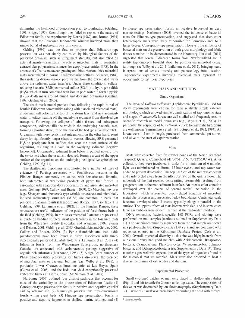
FIGURE 1—Experimental protocol. A) Fine-grained (250–125 mm) sand was packed in the bottom of a glass petri dish and covered by a thin (,1 cm) microbial mat. A dead
larva was placed in the center of the mat (as shown in C). A clear plastic film was placed over the mat, with a hole cut around the larva and a portion of the mat immediately
surrounding it, and subsequently covered by fine-grained sand. Experiments were contained within sealed plastic bags to limit contamination. B) Idealized cross section through
the negative hyporelief ‘‘fossil’’ mold in the bottom of the overlying bed, with the dark black rind as shown in (D).
PALAIOS EXPERIMENTAL MICROBIAL DEATH MASK 295

measures were taken to avoid potential topographic influence orvariation in operating conditions, including: (1) independent adjust-ment of working distance to 11.5 mm between each analyzed point; (2)selection of flatter, centrally located portions of grains for placement ofpoints (grain edges were avoided), which serves to maintain a consistenttake-off angle; and (3) monitoring of X-ray signal throughout pointanalyses to avoid including aberrant data within the dataset (if thesignal was abnormally low compared to other points, collection wasimmediately halted and the point was repositioned). Elemental X-raymaps may still include topographically induced artifacts, although
efforts were taken to map only regions that exhibited the least variationin grain height and size.
RESULTS
Recovery of Potential Fossils
90% recovery was achieved, with 54 out of 60 experiments yieldingimpressions that could be scored using the taphonomic index. Of the6 examples that were not recoverable, 5 belonged to experiments
FIGURE 2—Results of the decay experiments. Upper frames represent the three taphonomic indices used, from exceptional (segments and prolegs), through good (segments
only) to poor (general outline only). Experiments were run on three different media: microbial mats, fine-grained marine sands, and sterilized fine-grained sands. Experiments
were recovered at intervals of 6 hours, 1 day (24 hours), 1 week (168 hours), 2 weeks (336 hours), and 6 weeks (1008 hours). Four replicates (black circles) were recovered at each
stage for each of the three media: only 6 of 60 could not be scored. Trend lines correspond with median taphonomic index scores at each sample interval. Shaded area indicates
the distribution of black precipitate, with density corresponding to prevalence of aluminosilicate precursors, and can be interpreted as a representation of the optimal window
for preservation.
296 DARROCH ET AL. PALAIOS

performed with microbial mats; sediment tended to be caked aroundcarcasses in these experiments inhibiting splitting along laminaeimposed by sheets of plastic film. Recovery became progressively moredifficult with successive stages of decay: incubation for 2 weeks andbeyond led to desiccation of the carcass and decay of the internalmusculature, leaving a void typically lined with the remains of cuticle.After 6 weeks, carcasses had deteriorated noticeably less in experimentswith mats than in either of the controls: a significant volume of bothliquidized muscle tissue and cuticle remained.
Taphonomic Indices
Sets of experiments yielded consistent results; replicates rarelyexhibited large variation in the taphonomic index (with the exceptionof C1, see Fig. 2). After freezing at 260 uC for a period of several days,ice crystals developed on the surface of the potential fossils whichserved to highlight preserved structures in a manner similar to coatingwith ammonium chloride powder. Impressions of abdominal prolegs(taphonomic index 1) were developed in all experiments sampledat intervals of 6 hours and 1 day (Figs. 2–3). Proleg impressionsdisappeared in both control experiments after 1 week, but persisted inexperiments with mats up until 2 weeks. Taphonomic indices remainedhigher in those experiments using mats (Fig. 2): in 3 of 4 suchexperiments, impressions of segments were recovered after 6 weeks ofdecay, whereas in the control experiments the vast majority of samplespreserved only the vaguest impression of the shape of the carcass(Fig. 3). Higher fidelity in the preservation of fine-scale structures wasalso associated with the generation of a black precipitate within andradiating outward from the void left by the decaying carcass (Fig. 1D;Fig. 4 bottom left).
Black Precipitate
In several experiments a ring of black precipitate formed inassociation with the carcass, developing first around the decayinglarva, and then spreading outward along the contact between decaysubstrate and overlying sand. In some cases black precipitate alsoformed in the overlying (but rarely the underlying) sediment, resultingin a 3-dimensional black ring around the carcass (Figs. 1D, 4).Precipitate (measured as maximum distance from the edge of the
carcass outline) was most commonly and most extensively developedin experiments with mats (Table 1). This black material tended todisappear after extensive periods of decay possibly due to sulfide and/or aluminosilicate precursor oxidation following the decay of the mostlabile tissues (Briggs and Kear, 1994; Sagemann et al., 1999).Precipitate was formed in mat experiments after 1 day, reached amaximum extent after 2 weeks, and in one case persisted for at least6 weeks. Precipitate also formed within 2 weeks to a more limitedextent in 3 out of 4 control experiments using unsterilized sand,but none persisted. Precipitate formed in only one out of the 20experiments using sterilized sand.
Chemical Composition
Three samples were analyzed via XPS and ESEM-EDS to representthe spectrum of resulting conditions: C1/6HRS (no precipitate formed;see Table 1), MAT/1WEEK (moderate precipitate formed), andC1/1WEEK (heavy precipitate formed). These are referred to belowas samples 1–3 respectively. XPS elemental surveys indicated thesurficial presence of C, N, O, Na, Cl, Si, Ca, and Fe in all threeanalyzed sediment samples. ESEM-EDS elemental mapping indicatesthat sediment without black precipitate (sample 1) was composedprimarily of siliceous grains (Fig. 4). These analyses also illustrate aprogressive dirtying of the sediment grains in samples 2 and 3 by anelemental consortium of predominantly Al, K, and Fe (i.e., analuminosilicate-like composition) simultaneously with the formationof the black precipitate (Fig. 4). Elemental mapping of grains fromsample 1 (in which no precipitate formed) showed an inversecorrelation between Si, and both Al and K. EDS point data yieldedmean normalized weight percentages (n 5 31) for all analyzed grains asfollows: Si 5 31.2 6 2.0%, Al 5 3.6 6 0.8%, and K 5 0.8 6 0.3%. Insamples 2 (moderate precipitate) and 3 (heavy precipitate), Al (4.2 6
0.7% and 5.8 6 0.6%) and K (1.7 6 0.5% and 2.4 6 0.6%) becameincreasingly larger in concentration (Table 2; Fig. 4), and surfaceconcentrations of Si were significantly lessened as apparent from pointanalyses (28.5 6 1.4% and 27.9 6 1.2%) and a diminished Si signal in agreater number of grains in corresponding elemental maps.
Two other common aluminosilicate elements, Mg and Fe, showeddifferent trends. While the mean normalized weight percentages of theseelements did not show a significant increase in concentration in samples1–3 (Table 2), proportionally more grains per sample showeddetectable quantities of these elements. Point analyses for sample 1resulted in mean normalized weight percentages for Mg and Fe of 0.7 6
0.3% and 1.7 6 0.6%, respectively, and detectable quantities of boththese elements were present in 52% of the analyzed grains (16 of31 points). In sample 2, the mean Mg and Fe normalized weightpercentages were 0.8 6 0.2% and 2.4 6 0.7%, and from point analyses,these elements were detectable in 57% (Mg, 26 of 46 points) and 70%
(Fe, 32 of 46 points) of the analyzed grains. Sample 3 showed meannormalized weight percentages of 0.7 6 0.1% and 1.8 6 0.7%
respectively for Mg and Fe, but 64% (Mg, 35 of 55 points) and 75%
(Fe, 41 of 55 points) of the analyzed grains contained these elements.
Sulfur was nearly absent from sample 1 (only present in 1 analyzedpoint, normalized weight percentage 5 0.9%), but appeared insignificant concentrations (up to 3.7% normalized weight percentagein individual spots) in samples 2 and 3 (mean normalized weightpercentages of 0.3 6 0.1% and 0.2 6 0.1% respectively). In sample 1,the one S-bearing spot showed no correspondence with Fe. In samples 2and 3, in contrast, S was always found in conjunction with pointscontaining Fe (13% of the Fe-bearing spots [9% of total spots] insample 2 and 17% of the Fe-bearing spots [13% of total spots] in sample3 also contained detectable S), indicating the likely presence of ironsulfides on the surface of grains. Other elements present consistentlywithin point analyses include Ca, Na, and Cl (in all cases, meannormalized weight percentage #3.2%).
TABLE 1—Taphonomic indices; 3 different experimental setups were run for a
period of 6 weeks, with sampling intervals at 6 hours, 1 day, 1 week, 2 weeks, and
6 weeks. Left columns indicate taphonomic index (T.I.); right columns indicate
maximum distance (in mm; maximum distance from the edge of the carcass outline)
reached by the black precipitate from carcass. 0 values indicate that precipitate was
confined to the impression of the carcass. MAT 5 experiments performed with
microbial mats; C1 5 experiments performed with no mat and untreated sand; C2 5
experiments performed with no mat and sand treated with 80% bleach solution;
PPT 5 precipitate; NR 5 not recoverable.
Exp. And
replicate
6 HRS 1 DAY 1 WK 2 WK 6 WK
T.I. PPT T.I. PPT T.I. PPT T.I. PPT T.I. PPT
MAT 1 1 - 1 18 2 12 1 22 2 2
2 1 - 1 - 1 21 2 16 2 -
3 1 - 1 - 1 9 2 16 3 0
4 NR - NR - NR - NR - 3 -
C1 1 1 - 1 - 1 - 2 16 3 -
2 2 - 2 - 2 - 2 20 2 -
3 3 - 1 - 1 - 3 0 NR -
4 1 - 2 - 2 - 3 - NR -
C2 1 1 - 1 - 2 - 2 6 3 -
2 1 - 1 - 1 - 2 - 3 -
3 2 - 1 - 2 - 3 - 3 -
4 2 - 1 - 2 - 3 - 4 -
PALAIOS EXPERIMENTAL MICROBIAL DEATH MASK 297

FIGURE 3—Decay through time: Representative samples of the negative hyporelief ‘‘fossil’’ molds recovered during the experiment. Samples collected between 6 hrs and
1 week were consistently better preserved (typically exceptional to good preservation) than those recovered later. Initial decay is necessary for the observed increase of
aluminosilicate elements, which increased preservational potential in later (post–1-week) samples. The presence of a bacterial mat is associated with higher-quality preservation,
while relatively poor preservation is achieved in sand-only samples after one week.
298 DARROCH ET AL. PALAIOS

FIGURE 4—Elemental Mapping: ESEM-EDS analyses and backscattered electron (BSE; z-contrast) imaging of clean (1) to dirty (3) samples of fine-grained sand. Clean sands
were composed almost entirely of quartz. Dirty samples were collected in close proximity to the decayed larva within the distinct black ring. Dirty grains are coated
progressively by a milieu of aluminosilicate elements (Al-K-Fe mapped here). In addition, these grains also revealed the likely presence of iron sulfide (mapped for sample 3),
potentially a precursor to the pyrite coatings proposed to accompany Ediacaran death-mask preservation (Gehling, 1999). The time series of grains shows that the Si X-ray
signal dulls with a corresponding brightening of the Al signal. Bottom left: 3-dimensional black ring of clay-sized material precipitated around decaying carcass.
PALAIOS EXPERIMENTAL MICROBIAL DEATH MASK 299

DISCUSSION AND CONCLUSIONS
Experimental Support for the Microbial Death-Mask Hypothesis
Our results provide experimental support for Gehling’s (1999)microbial death-mask model. There is a clear correlation between thepresence of organic matter in the sand used to bury the organism, theformation of a black precipitate as a ring around the decaying carcassand, ultimately, the fidelity of preservation (Fig. 2). The absence of ablack precipitate in those experiments that used sand cleaned of organicmaterial (with the exception of just one sample at 2 weeks) confirmsthe importance of organic matter in the sediment to this type ofpreservation (also see Norris, 1989). Removing the organic materialfrom the sand in Control 2 eliminated not only the microbial substratebut also the majority of microbes living on the surface of sedimentgrains. This slowed initial decay after burial, preventing both the swiftgeneration of local anoxia (see Briggs and Kear, 1994) and cementationof grains required for fine-scale structural preservation (Gehling, 1999).In contrast, black rings formed in experiments with microbial matswithin 1 day (and persisted for at least 6 weeks), likely reflecting boththe high organic carbon content of the sediment, and the mat as asource of decay-promoting microbes. However, in control experimentsusing unsterilized sand, rings formed in 3 out of 4 experiments after2 weeks, indicating that bacterial communities living on the surface ofsediment grains, on the surface of larval cuticle, and in the gut of larvaelikely also played a role. The disappearance of black rings over timereflects the slow diffusion of oxygen back into the vicinity of the carcassas decay slowed, causing dissipation/reoxygenation of the localmicroenvironment and reoxidation of reduced iron minerals (Sagemannet al., 1999; Martin et al., 2004). Gehling’s (1999) model posited that thesurface of storm beds was recolonized swiftly by mats, inhibiting thedownward movement of oxygen and sulfate ions from the sediment-water interface and maintaining anaerobic conditions around theburied carcass. While no mat developed on the sediment surface overthe 6-week duration of the experiment, the sheet of plastic film insertedto facilitate splitting may have inhibited vertical migration and surfacecolonization by the buried mat.
The fidelity of preservation is related to the concentration of organicmatter (both the presence or absence of mats, and organic matter in thesand) and the formation of black rings. Fine-scale structures survived longerin experiments with microbial mats, where black rings also formed moreconsistently. Inferences based on taphonomic indices should, however, beviewed with caution: sampling was destructive and the dynamics ofchanging variables were not recorded. However, the consistency of results inreplicate experiments (Fig. 2) suggests that our patterns are robust.
Black Rings and Enhanced Preservation
The results of these experiments provide several explanations for therelationship between the formation of a black precipitate and enhancedfidelity of preservation.
1. ESEM imaging indicates that the black precipitate forms inassociation with an active biofilm coating the outside of sedimentgrains. ESEM-EDS analysis revealed evidence of likely iron sulfides, orat least spatially restricted associations of Fe and S, on the surfaces ofgrains in both samples 2 and 3 (Fig. 4), consistent with the microbial
death-mask model (Gehling, 1999), although pyrite framboids were notobserved via electron imaging. However, the presence of iron sulfide isnot extensive and there is little evidence to suggest that it forms acement that would preserve fine-scale morphology. Microbial sealing,sensu Gehling (1999), and a longer period of decay may be required forthe formation of a true pyritic death mask (Donald and Southam, 1999;Grimes et al., 2001).
2. Microbial populations derived from both mat and surroundingsand, which multiply in the vicinity of the carcass, may produce asignificant volume of sediment-binding extracellular polymeric substance(EPS). The sediment trapping properties of EPS are well documented andmay play a critical role in both micro- and regional-scale biosedimentaryprocesses (see Riding, 2000). The generation of EPS additionally may beresponsible for early cementation of grains and the retention of details ofthe organism. The role played by biofilms in stabilizing sediments andpromoting mineral replication of tissues during decay has been emphasizedby several authors (Briggs and Kear, 1994; Wilby et al., 1996; Briggs et al.,2005; McNamara et al., 2009). Our observation that sediment appeared tobe caked around carcasses in experiments using mats (impeding the recoveryof impressions) suggests that EPS derived from microbial blooms may trapand bind grains.
3. Both XPS and ESEM-EDS analyses revealed local elevations, eithercompositionally or proportionally, in the aluminosilicate elemental suiteof Al, K, Fe, and Mg within the black precipitate surrounding thecarcasses (Fig. 4, Table 2). While compositional variability undoubtedlyexists in the natural sands, such variability is an unlikely explanation forthe directional trends observed in the EDS data over the course of theexperiment. Over time the sands showed: (1) higher concentrations of Aland K; (2) a higher proportion of grains with detectable Mg, Fe, and S;and (3) a corresponding decrease in the relative concentration of Si withinthe given electron beam interaction volume. While this should not beinterpreted as a loss of Si from individual sand grains, we can infer thatthe EDS elemental maps captured a greater volume of the black coatingper unit beam interaction volume as the experiments progressed.Although we did not observe the precipitation of true fine-grained clays,as such particles would have been revealed by electron imaging, ourcompositional data suggest a thicker aluminosilicate coating over time.
Martin et al. (2004) demonstrated the attachment of quartz andkaolinite to the surfaces of decaying lobster eggs in the presence ofmetabolizing bacteria; sediment was derived from the immediateenvironment and subsequently concentrated on the surfaces of eggsover several months of the experiment. Our results provide evidencethat microbial activity may facilitate the precipitation of aluminosilicateminerals by concentrating aluminosilicate elemental constituentsaround decaying carcasses. If the generation of true clay mineralsfollows, then clay-sized particles generated around a carcass may serveto cast fine-scale morphology that otherwise might not be preservablegiven the coarser grain sizes of surrounding sediment. A layer of clay-sized particles would also provide a relatively impermeable barrierpreventing oxygenated pore water from diffusing back to the vicinity ofthe carcass, and protecting pyrite from oxidation. Some support for thisinterpretation is provided by fossil material. Toporski et al. (2002)reported local enrichment of Al, Mg, S, Fe, and Ti in a fossilizedbiofilm from the Oligocene of Germany. Furthermore, Laflamme et al.(2011) reported the occurrence of nearly identical elemental consortia to
TABLE 2—Summary of primary elemental compositions (reported as mean normalized weight percentage 6 standard error) determined by ESEM-EDS point analyses (1
point per grain). Sample numbers: 1 5 no precipitate (31 grains analyzed), 2 5 intermediate precipitate (46 grains analyzed), and 3 5 heavy precipitate (55 grains analyzed).
Minor constituents observed but not reported here include Na, Cl, Mn, and Ti. See Supplementary Data 41 for complete analysis.
# Si O C Al K Mg Ca Fe S
1 31.2 6 2.0 49.5 6 1.5 9.8 6 2.2 3.6 6 0.7 0.8 6 0.3 0.7 6 0.3 0.5 6 0.3 1.7 6 0.6 0.0 6 0.0
2 28.5 6 1.3 47.6 6 1.7 8.1 6 0.9 4.1 6 0.7 1.7 6 0.5 0.8 6 0.2 1.2 6 0.4 2.4 6 0.7 0.3 6 0.1
3 27.9 6 1.2 51.1 6 1.1 6.0 6 0.7 5.8 6 0.6 2.4 6 0.6 0.7 6 0.1 0.4 6 0.1 1.8 6 0.7 0.2 6 0.1
300 DARROCH ET AL. PALAIOS

those in our experiments associated with Aspidella discs from theAvalon Peninsula where aluminosilicates are concentrated in a distinctfine-grained sediment layer surrounding holdfasts. Mapstone andMcIlroy (2006) discovered concentrations of illite, chlorite, andsmectite on hyporelief surfaces of discoidal fossils from the Neoproter-ozoic of central Australia, although they interpreted them as the resultof background and fair-weather sedimentation, rather than as in situprecipitates related to decay. Similar associations of clay minerals andpyrite have been documented from carbonaceous compression-typeEdiacaran microfossils from the Doushantuo and Denying Formationsof South China (Anderson et al., 2011). Such observations suggest acommon theme within multiple late Neoproterozoic and earlyPhanerozoic taphonomic windows.
Timing of Preservation
Our experiments suggest that formation of the black precipitatearound a carcass may precede authigenic precipitation of iron sulfideand clay minerals, and furthermore, that the persistence of these blackrings (and ultimately the development of a pyrite death mask) isdependent on the exclusion of oxygen. Experiments performed byBriggs and Kear (1993, 1994) and Sagemann et al. (1999) on shrimpdemonstrated that the concentration of O2 around a decaying carcassdropped to zero (complete anoxia) within 24 hours of death even inopen conditions, but generally recovered to starting values within 10+days (strongly dependant on the mass of individual carcasses), placingan upper temporal constraint on the mineralization of soft tissues. Inour experiments, the expansion and contraction of the ring of blackprecipitate provides a proxy for the progress of anoxia and subsequentrediffusion of oxygen into the sediment surrounding the decaying larva.The time taken for oxygen to return to the carcass provides a constrainton the length of the preservational window. Black rings formed inexperiments using microbial mats after 1 day, reached a maximum after2 weeks, and virtually disappeared by 6 weeks. Thus it appears that6 weeks would represent an upper limit for microbial mats to recolonizethe surfaces of storm beds in order to prevent diffusion of oxygen backtoward a decaying carcass and allow a death mask to form, at least inthe particular conditions represented in our experiments. This windowis considerably shorter in depositional settings lacking microbial mats(see C1 experiments), and pyritized death masks are less likely to formwhere organic matter is absent (C2 experiments).
It is unlikely that the cuticle of Ediacaran organisms was identical tothat of G. mellonella. Although molecular studies support an Ediacaranage for Ecdysozoa (Wheeler et al., 2009; Erwin et al., 2011), there islittle evidence for the presence of chitinous exoskeletons prior to theCambrian (Miller, 1991). Chitin is a complex structural polysaccharidethat is resistant to decay when complexed with protein in invertebratecuticles (Baas et al., 1995; Flannery et al., 2001; Gupta et al., 2006).Under laboratory conditions Stankiewicz et al. (1998) demonstrated nostructural degradation in chitin before decay had proceeded for 8 weeks,although considerable chemical changes occurred after 1 to 2 weeks. Aplacozoan affinity for the Ediacaran taxon Dickinsonia (as proposed bySperling and Vinther, 2010) would suggest a considerably more labileintegument. Our experiments using G. mellonella likely provide anupper limit for the formation of a death mask, although the persistenceof anoxia around the carcass due to microbial sealing (not simulated inour experiments, and an important component of Gehling’s [1999]model) would have extended mineralization.
Another limitation of our experiments is the use of fresh-waterrather than marine mats to perform our experiments. The formation ofpyrite death masks in the Ediacaran was presumably influenced bylocal concentrations of seawater sulfate. Modern, naturally occurringfresh water is typically sulfate-limited, and the water used in ourexperiments was particularly sulfate poor (,0.6 mM, see Supplemen-tary Data 31). However, evidence of sulfur isotope records (Kah et al.,
2004; Fike et al., 2006) and iron chemistry (Li et al., 2010) indicatethat marine sulfate levels were low in the Neoproterozoic (as low as0.2 mM in the Marinoan; Melezhik et al., 2005), and may haveremained low until the upper Cambrian (2–12 mM; Gill et al., 2010).Our experiments simulated sulfate levels at the low end of rangesestimated for the Ediacaran, and provide evidence for the precipitationof iron sulfide despite extremely low SO4. The effect of sulfateconcentrations on the formation of pyrite death masks remains to beinvestigated experimentally.
Our decay experiments support the basic tenets of the death-maskhypothesis and highlight the necessity for large amounts of organicmatter (in the form of microbial mats) and anoxic conditions to allowthe formation of external molds in coarse sediments. The rapidprecipitation of aluminosilicate and iron sulfide precursors provides ataphonomic and diagenetic pathway allowing for the early lithifica-tion of sediment surrounding decaying organisms. The Ediacaransedimentary regime, dominated by large expanses of microbial matsand an absence of destructive vertical burrowing, can explain theextensive preservation of relief impressions in Ediacaran sandstonesworldwide.
ACKNOWLEDGMENTS
We thank L. Stout for construction of clone libraries and sequencingof our microbial mats, and H. Ernstberger for performing waterchemistry analyses. R. Blake, M. Casey, D. Erwin, S. Gabbott, T.Hegna, J. Hunter, M. Purnell, L. Rochas, R. Sansom, and A. Wallaceprovided valuable assistance and discussion. Reviews by E. Sperling(Harvard University) and an anonymous reviewer improved a previousversion of this manuscript. This work was supported by the YalePeabody Museum of Natural History. JDS acknowledges support fromthe Virginia Tech Institute of Critical Technology and Applied Science.ML acknowledges generous support from the Natural Sciences andEngineering Research Council of Canada (NSERC), a BatemanPostdoctoral Fellowship, and a Smithsonian Institution PostdoctoralFellowship.
REFERENCES
ANDERSON, E.P., SCHIFFBAUER, J.D., and XIAO, S., 2011, Taphonomic study of
Ediacaran organic-walled fossils confirms the importance of clay minerals and
pyrite in Burgess Shale-type preservation: Geology, v. 39, p. 643–646.
ALLISON, P.A., 1988, The decay and mineralization of proteinaceous macrofossils:
Paleobiology, v. 14, p. 139–154.
BAAS, M., BRIGGS, D.E.G., VAN HEEMST, J.D.H., KEAR, A.J., and DE LEEUW, J.W.,
1995, Selective preservation of chitin during the decay of shrimp: Geochimica et
Cosmochimica Acta, v. 59, p. 945–951.
BRIGGS, D.E.G., 1995, Experimental taphonomy: PALAIOS, v. 10, p. 539–550.
BRIGGS, D.E.G., 2003, The role of decay and mineralization in the preservation of
soft-bodied fossils: Annual Review of Earth and Planetary Sciences, v. 31, p. 275–
301.
BRIGGS, D.E.G., and KEAR, A.J., 1993, Fossilization of soft tissue in the laboratory:
Science, v. 259, p. 1439–1444.
BRIGGS, D.E.G., and KEAR, A.J., 1994, Decay and mineralization of shrimps:
PALAIOS, v. 9, p. 431–456.
BRIGGS, D.E.G., MOORE, R.A., SHULTZ, J.W., and SCHWEIGERT, G., 2005,
Mineralization of soft-part anatomy and invading microbes in the horseshoe crab
Mesolimulus from the Upper Jurassic Lagerstatte of Nusplingen, Germany:
Proceedings of the Royal Society B, v. 272, p. 627–632.
BRUTON, D.L., 1991, Beach and laboratory experiments with the jellyfish Aurelia and
remarks on some fossil ‘‘medusoid’’ traces, in Simonetta, A.M., and Conway
Morris, S., eds., The Early Evolution of Metazoa and the Significance of
Problematic Taxa: Cambridge University Press, Cambridge, UK, p. 125–129.
CALLOW, R.H.T., and BRASIER, M.D., 2009, Remarkable preservation of microbial
mats in Neoproterozoic siliciclastic settings: Implications for Ediacaran tapho-
nomic models: Earth-Science Reviews, v. 96, p. 207–219.
CLAPHAM, M.E., NARBONNE, G.M., and GEHLING, J.G., 2003, Paleoecology of the
oldest known animal communities: Ediacaran assemblages at Mistaken Point,
Newfoundland: Paleobiology, v. 29, p. 527–544.
PALAIOS EXPERIMENTAL MICROBIAL DEATH MASK 301

COLE, J.R., WANG, Q., CARDENAS, E., FISH, J., CHAI, B., FARRIS, R.J., KULAM-SYED-
MOHIDEEN, A.S., MCGARRELL, D.M., MARSH, T., GARRITY, G.M., and TIEDJE,
J.M., 2009, The Ribosomal Database Project: Improved alignments and new tools
for rRNA analysis: Nucleic Acids Research, v. 37, Database issue, p. D141–D145.
DONALD, R., and SOUTHAM, G., 1999, Low temperature anaerobic bacterial diagenesis
of ferrous monosulphide to pyrite: Geochimica et Cosmochimica Acta, v. 63, p.
2019–2023.
DROSER, M.L., GEHLING, J.G., and JENSON, S., 2006, Assemblage palaeoecology of the
Ediacara biota: The unabridged edition?: Palaeogeography, Palaeoclimatology,
Palaeoecology, v. 232, p. 131–147.
ERWIN, D.H., LAFLAMME, M., TWEEDT, S.M., SPERLING, E.A., PISANI, D., and
PETERSON, K.J., 2011, The Cambrian conundrum: Early divergence and later
ecological success in the early history of animals: Science, v. 334, p. 1091–1097.
FEDONKIN, M.A., and WAGGONER, B.M., 1997, The Late Precambrian fossil
Kimberella is a mollusc-like bilaterian organism: Nature, v. 388, p. 868–871.
FIKE, D.A., GROTZINGER, J.P., PRATT, L.M., and SUMMONS, R.E., 2006, Oxidation of
the Ediacaran ocean: Nature, v. 444, p. 744–747.
FLANNERY, M.B., STOTT, A.W., BRIGGS, D.E.G., and EVERSHED, R.P., 2001, Chitin in
the fossil record: Identification and quantification of d-glucosamine: Organic
Geochemistry, v. 32, p. 745–754.
GEHLING, J.G., 1991, The case for Ediacaran fossil roots to the metazoan tree:
Geological Society of India Memoir, v. 20, p. 181–224.
GEHLING, J.G., 1999, Microbial mats in terminal Proterozoic siliciclastics: Ediacaran
death masks: PALAIOS, v. 14, p. 40–57.
GEHLING, J.G., 2000, Environmental interpretation and a sequence stratigraphic
framework for the terminal Proterozoic Ediacara Member within the Rawnsley
Quartzite, South Australia: Precambrian Research, v. 100, p. 65–95.
GEHLING, J.G., DROSER, M.L., JENSEN, S., and RUNNEGAR, B.N., 2005, Ediacaran
organisms: Relating form to function, in Briggs, D.E.G., ed., Evolving Form and
Function: Fossils and Development, Proceedings of a Symposium Honoring Adolf
Seilacher for his Contributions to Paleontology in Celebration of his 80th Birthday:
Peabody Museum of Natural History, Yale University, New Haven, Connecticut,
p. 43–67.
GILL, B.C., LYONS, T.W., YOUNG, S.A., KUMP, L.R., KNOLL, A.H., and SALTZMAN,
M.R., 2010, Geochemical evidence for widespread euxinia in the Later Cambrian
ocean: Nature, v. 469, p. 80–83.
GLAESSNER, M.F., and WADE, M., 1966, The late Precambrian fossils from Ediacara,
South Australia: Palaeontology, v. 9, p. 599–628.
GRAZHDANKIN, D., 2004, Patterns of distribution in the Ediacaran biotas: Facies
versus biogeography and evolution: Paleobiology, v. 30, p. 203–221.
GRAZHDANKIN, D., and GERDES, G., 2007, Ediacaran microbial colonies: Lethaia,
v. 40, p. 201–210.
GRIMES, S.T., BROCK, F., RICKARD, D., DAVIES, K.L., EDWARDS, E., BRIGGS, D.E.G.,
and PARKES, R.J., 2001, Understanding fossilization: Experimental pyritization of
plants: Geology, v. 29, p. 123–126.
GUPTA, S.C., LEATHERS,T.D., EL-SAYED, G.N., and IGNOFFO, C.M., 1992, Insect
cuticle-degrading enzymes from the entomogenous fungus Beauveria bassiana:
Experimental Mycology, v. 16, p. 132–137.
GUPTA, S.C., LEATHERS,T.D., EL-SAYED, G.N., and IGNOFFO, C.M., 1994, Relation-
ships among enzyme activities and virulence parameters in Beauveria bassiana
infections of Galleria mellonella and Trichoplusia ni: Journal of Invertebrate
Pathology, v. 64, p. 13–17.
GUPTA, N.S., MICHELS, R., BRIGGS, D.E.G., EVERSHED, R.P., and PANCOST, R.D.,
2006, The organic preservation of fossil arthropods: An experimental study:
Proceedings of the Royal Society B, v. 273, p. 2777–2783.
GUPTA, N.S., CAMBRA-MOO, O., BRIGGS, D.E.G., LOVE, G.D., FREGENAL-MARTINEZ,
M.A., and SUMMONS, R.E., 2008, Molecular taphonomy of macrofossils from the
Cretaceous Las Hoyas Formation, Spain: Cretaceous Research, v. 29, p. 1–8.
HAGADORN, J.W., and BOTTJER, D.J., 1997, Wrinkle structures: Microbially mediated
sedimentary structures in siliciclastic settings at the Proterozoic-Phanerozoic
transition: Geology, v. 25, p. 1047–1050.
KAH, L.C., LYONS, T.W., and FRANK, T.D., 2004, Low marine sulphate and
protracted oxygenation of the Proterozoic biosphere: Nature, v. 431, p. 834–838.
LAFLAMME, M., and NARBONNE, G.M., 2008, Ediacaran fronds: Palaeogeography,
Palaeoclimatology, Palaeoecology, v. 258, p. 162–179.
LAFLAMME, M., SCHIFFBAUER, J.D., NARBONNE, G.M., and BRIGGS, D.E.G., 2011,
Microbial biofilms and the preservation of the Ediacara biota: Lethaia, v. 44,
p. 203–213.
LAFLAMME, M., SCHIFFBAUER, J.D., and NARBONNE, G.M., 2012, Deep-water
microbially induced sedimentary structures (MISS) in deep time: The Ediacaran
fossil Ivesheadia, in Noffke, N.K., and Chafetz, H., eds., Microbial Mats in
Siliciclastic Depositional Systems Through Time: SEPM Special Publication
No. 101. p. 111–123.
LI, C., LOVE, G.D., LYONS, T.W., FIKE, D.A., SESSIONS, A.L., and CHU, X., 2010, A
stratified redox model for the Ediacaran ocean: Science, v. 328, p. 80–83.
LIU, A.G., MCILROY, D., ANTCLIFFE, J.B., and BRASIER, M.D., 2011, Effaced
preservation in the Ediacara biota and its implications for the early macrofossil
record: Palaeontology, v. 54, p. 607–630.
MAPSTONE, N.B., and MCILROY, D., 2006, Ediacaran fossil preservation: Taphonomy
and diagenesis of a discoid biota from the Amadeus Basin, central Australia:
Precambrian Research, v. 149, p. 126–148.
MARTIN, D., BRIGGS, D.E.G., and PARKES, R.J., 2004, Experimental attachment of
clay minerals to invertebrate eggs and the preservation of soft-bodied fossils:
Journal of the Geological Society, London, v. 161, p. 735–738.
MCNAMARA, M., ORR, P.J., KEARNS, S.L., ANADON, P., ALCALA, L., and PENALVER-
MOLLA, E., 2009, Soft tissue preservation in Miocene frogs from Libros (Spain):
Insights into the genesis of decay microenvironments: PALAIOS, v. 24, p. 104–117.
MELEZHIK, V.A., FALLICK, A.E., and POKROVSKY, B.G., 2005, Enigmatic nature of
thick sedimentary carbonates depleted in 13C beyond the canonical mantle value:
The challenges to our understanding of the terrestrial carbon cycle: Precambrian
Research, v. 137, p. 131–165.
MILLER, R.F., 1991, Chitin paleoecology: Biochemical Systematics and Ecology:
v. 19, p. 401–411.
MIYATA, S., CASEY, M., FRANK, D.W., AUSUBEL, F.M., and DRENKARD, E., 2003, Use
of the Galleria mellonella caterpillar as a model host to study the role of the Type
III secretion system in Pseudomonas aeruginosa pathogenesis: Infection and
Immunity, v. 71, v. 2404–2413.
NARBONNE, G.M., 1998, The Ediacara biota: A terminal Neoproterozoic experiment
in the evolution of life: GSA Today, v. 8, p. 1–6.
NARBONNE, G.M., 2004, Modular construction of early Ediacaran complex life forms:
Science, v. 305, p. 1141–1144.
NARBONNE, G.M., 2005, The Ediacara biota: Neoproterozoic origin of animals and
their ecosystems: Annual Review of Earth and Planetary Sciences, v. 33, p. 421–442.
NARBONNE, G.M., LAFLAMME, M., GREENTREE, C., and TRUSLER, P., 2009,
Reconstructing a lost world: Ediacaran rangeomorphs from Spaniard’s Bay,
Newfoundland: Journal of Paleontology, v. 83, p. 503–523.
NORRIS, R.D., 1989, Cnidarian taphonomy and affinities of the Ediacara biota:
Lethaia, v. 22, p. 381–393.
RETALLACK, G.J., 1994, Were the Ediacaran fossils lichens?: Paleobiology, v. 20,
p. 523–544.
RIDING, R., 2000, Microbial carbonates: The geological record of calcified bacterial-
algal mats and biofilms: Sedimentology, v. 47, p. 174–214.
SAGEMANN, J., BALE, S.J., BRIGGS, D.E.G., and PARKES, R.J., 1999, Controls on the
formation of authigenic minerals in association with decaying organic matter: An
experimental approach: Geochimica et Cosmochimica Acta, v. 63, p. 1083–1095.
SAMSINAKOVA, A., MISIKOVA, S., and LEOPOLD, J., 1971, Action of enzymatic systems
of Beauvaria bassiana on the cuticle of the greater wax moth larvae (Galleria
mellonella): Journal of Invertebrate Pathology, v. 18, p. 322–330.
SANSOM, R.S., GABBOTT, S.E., and PURNELL, M.A., 2010, Non-random decay of
chordate characters causes bias in fossil interpretation: Nature, v. 463, p. 797–800.
SEILACHER, A., 1984, Late Precambrian and Early Cambrian metazoa: Preservation or
real extinctions?, in Holland, H.D., and Trendall, A.F., eds., Patterns of Change in
Earth Evolution, (Dahlem Konferenzen): Springer-Verlag, Berlin, p. 159–168.
SEILACHER, A., 1989, Vendozoa: Organismic construction in the Proterozoic
biosphere: Lethaia, v. 22, p. 229–239.
SEILACHER, A., 1992, Vendobionta and Psammocorallia: Lost constructions of
Precambrian evolution: Journal of the Geological Society, London, v. 149, p. 607–613.
SEILACHER, A., REIF, W.-E., and WESTPHAL, F., 1985, Sedimentological, ecological
and temporal patterns of fossil Lagerstatten: Philosophical Transactions of the
Royal Society of London, v. 311, p. 5–23.
SPERLING, E.A., and VINTHER, J., 2010, A placozoan affinity for Dickinsonia and the
evolution of late Proterozoic metazoan feeding modes: Evolution and Develop-
ment, v. 12, p. 201–209.
SPERLING, E.A., PISANI, D., and PETERSON, K.J., 2007, Poriferan paraphyly and its
implications for Precambrian paleobiology: Geological Society of London Special
Publication, v. 286, p. 355–368.
SPRIGG, R.C., 1949, Early Cambrian ‘‘Jellyfishes’’ of Ediacara, South Australia, and
Mt. John, Kimberley District, Western Australia: Transactions of the Royal
Society of South Australia, v. 73, p. 72–99.
STANKIEWICZ, B.A., MASTALERZ, M., HOFF, C.H.J., BIERSTEDT, A., FLANNERY, M.B.,
BRIGGS, D.E.G., and EVERSHED, R.B., 1998, Biodegradation of the chitin-protein
complex in crustacean cuticle: Organic Geochemistry, v. 28, p. 67–76.
STEINER, M., and REITNER, J., 2001, Evidence of organic structures in Ediacara-type
fossils and associated microbial mats: Geology, v. 29, p. 1119–1122.
TOPORSKI, J.K.W., STEELE, A., WESTALL, F., AVCI, R., MARTILL, D.M., and MCKAY,
D.S., 2002, Morphologic and spectral investigation of exceptionally well-preserved
bacterial biofilms from the Oligocene Enspel formation, Germany: Geochimica et
Cosmochimica Acta, v. 66, p. 1773–1791.
WHEELER, B.M., HEIMBERG, A.M., MOY, V.N., SPERLING, E.A., HOLSTEIN, T.W.,
HEBER, S., and PETERSON, K.J., 2009, The deep evolution of metazoan microRNAs:
Evolution and Development, v. 11, p. 50–68.
302 DARROCH ET AL. PALAIOS

WILBY, P.R., BRIGGS, D.E.G., BERNIER, P., and GAILLARD, C., 1996, The role of
microbial mats in the fossilization of soft tissues: Geology, v. 24, p. 787–790.
WILBY, P.R., CARNEY, J.N., and HOWE, M.P.A., 2011, A rich Ediacaran assemblage
from eastern Avalonia: Evidence of early widespread diversity in the deep ocean:
Geology, v. 39, p. 655–658.
WOOD, D.A., DALRYMPLE, R.W., NARBONNE, G.M., GEHLING, J.G., and CLAPHAM,
M.E., 2003, Paleoenvironmental analysis of the late Neoproterozoic Mistaken
Point and Trepassey formations, southeastern Newfoundland: Canadian Journal
of Earth Sciences, v. 40, p. 1375–1391.
XIAO, S., and LAFLAMME, M., 2009, On the eve of animal radiation: Phylogeny, ecology,
and evolution of the Ediacara biota: Trends in Ecology and Evolution, v. 24, p. 31–40.
ACCEPTED NOVEMBER 21, 2011
PALAIOS EXPERIMENTAL MICROBIAL DEATH MASK 303