Effects of glycinergic inhibition failure on respiratory rhythm and pattern generation
14
CHAPTER Effects of Glycinergic Inhibition Failure on Respiratory Rhythm and Pattern Generation 2 Natalia A. Shevtsova* ,1 , Dietrich Bu ¨ sselberg { , Yaroslav I. Molkov* ,{ , Anne M. Bischoff },} , Jeffrey C. Smith # , Diethelm W. Richter },} , Ilya A. Rybak* * Department of Neurobiology and Anatomy, Drexel University College of Medicine, Philadelphia, PA, USA { Weill Cornell Medical College in Qatar, Education City, Doha, Qatar { Department of Mathematical Sciences, Indiana University—Purdue University Indianapolis, Indianapolis, IN, USA } Department of Neuro- and Sensory Physiology, University ofGo¨ttingen, Go¨ttingen, Germany } Excellence Cluster Nanoscale Microscopy and Molecular Physiology of the Brain, Go¨ttingen,Germany # Cellular and Systems Neurobiology Section, National Institute of Neurological Disorders and Stroke, National Institutes of Health, Bethesda, MD, USA 1 Corresponding author: Tel.: þ1-215-9918597; Fax: þ1-215-8439082, e-mail address: [email protected] Abstract Inhibitory interactions between neurons of the respiratory network are involved in rhythm gen- eration and pattern formation. Using a computational model of brainstem respiratory net- works, we investigated the possible effects of suppressing glycinergic inhibition on the activity of different respiratory neuron types. Our study revealed that progressive suppression of glycinergic inhibition affected all neurons of the network and disturbed neural circuits in- volved in termination of inspiration. Causal was a dysfunction of postinspiratory inhibition targeting inspiratory neurons, which often led to irregular preterm reactivation of these neu- rons, producing double or multiple short-duration inspiratory bursts. An increasing blockade of glycinergic inhibition led to apneustic inspiratory activity. Similar disturbances of glyciner- gic inhibition also occur during hypoxia. A clear difference in prolonged hypoxia, however, is that the rhythm terminates in expiratory apnea. The critical function of glycinergic inhibition for normal respiratory rhythm generation and the consequences of its reduction, including in pathological conditions, are discussed. Progress in Brain Research, Volume 209, ISSN 0079-6123, http://dx.doi.org/10.1016/B978-0-444-63274-6.00002-3 © 2014 Elsevier B.V. All rights reserved. 25
Transcript of Effects of glycinergic inhibition failure on respiratory rhythm and pattern generation

CHAPTER
Effects of GlycinergicInhibition Failure onRespiratory Rhythmand Pattern Generation
2Natalia A. Shevtsova*,1, Dietrich Busselberg{, Yaroslav I. Molkov*,{,
Anne M. Bischoff},}, Jeffrey C. Smith#, Diethelm W. Richter},}, Ilya A. Rybak**Department of Neurobiology and Anatomy, Drexel University College of Medicine,
Philadelphia, PA, USA{Weill Cornell Medical College in Qatar, Education City, Doha, Qatar
{Department of Mathematical Sciences, Indiana University—Purdue University Indianapolis,
Indianapolis, IN, USA}Department of Neuro- and Sensory Physiology, University of Gottingen, Gottingen, Germany
}Excellence Cluster Nanoscale Microscopy and Molecular Physiology of the Brain,
Gottingen, Germany#Cellular and Systems Neurobiology Section, National Institute of Neurological Disorders and
Stroke, National Institutes of Health, Bethesda, MD, USA1Corresponding author: Tel.: þ1-215-9918597; Fax: þ1-215-8439082,
e-mail address: [email protected]
AbstractInhibitory interactions between neurons of the respiratory network are involved in rhythm gen-
eration and pattern formation. Using a computational model of brainstem respiratory net-
works, we investigated the possible effects of suppressing glycinergic inhibition on the
activity of different respiratory neuron types. Our study revealed that progressive suppression
of glycinergic inhibition affected all neurons of the network and disturbed neural circuits in-
volved in termination of inspiration. Causal was a dysfunction of postinspiratory inhibition
targeting inspiratory neurons, which often led to irregular preterm reactivation of these neu-
rons, producing double or multiple short-duration inspiratory bursts. An increasing blockade
of glycinergic inhibition led to apneustic inspiratory activity. Similar disturbances of glyciner-
gic inhibition also occur during hypoxia. A clear difference in prolonged hypoxia, however, is
that the rhythm terminates in expiratory apnea. The critical function of glycinergic inhibition
for normal respiratory rhythm generation and the consequences of its reduction, including in
pathological conditions, are discussed.
Progress in Brain Research, Volume 209, ISSN 0079-6123, http://dx.doi.org/10.1016/B978-0-444-63274-6.00002-3
© 2014 Elsevier B.V. All rights reserved.25

Keywordscomputational modeling, respiratory rhythm, pre-Botzinger complex, glycinergic inhibition,
apneusis, apnea, hypoxia, translational medicine
1 INTRODUCTIONInhibitory interactions between neurons of the brainstem respiratory network have
been proposed to play a critical role in respiratory rhythm generation and the
adjustment of normal breathing movements in vivo (Richter et al., 1986, 1992).
Disturbances of these synaptic interactions may become dangerously life threatening
as they can lead to an abnormally prolonged inspiration (apneusis) producing breath
holdings or to complete cessation of breathing (apnea). There are reports of several
diseases in which such disturbances of breathing originate from a failure of
glycinergic inhibition that may even cause sudden death, for example, in Rett
Syndrome (Stettner et al., 2007), hyperekplexia (Busselberg et al., 2001a; Harvey
et al., 2008; Markstahler et al., 2002), developmental disorders, such as olivo-
ponto-cerebellar atrophy (OPCA) (Richter et al., 2003), and also brainstem infarction
(El-Khatib et al., 2003).
The circuit mechanisms underlying inhibitory regulation of respiratory network
activity and the associated emergent pathogenic processes contributing to network
dysfunctions are difficult to analyze in a complex system such as the brainstem
respiratory network. Specifically, there is incomplete understanding about the effects
of such disturbances on the activity of different respiratory neuron types. In this
study, we tried to fill this gap using computational modeling of the effects of
progressive depression of glycinergic inhibition on the activity of various types of
respiratory neurons. We believe that such an approach is promising as it not only
predicts network behavior but allows theoretical and experimental testing of
therapeutic strategies to recover breathing as was successfully performed for
opioid-induced apnea by potentiating glycinergic synaptic transmission
(Shevtsova et al., 2011).
As synaptic transmission of inhibitory neurons is also very sensitive to hypoxia
and fades quickly during reduced levels of brain oxygen (Congar et al., 1995), we
used our simulations to investigate the possible effects of progressive suppression
of glycinergic inhibition to gain insights into basic mechanisms of respiratory
rhythm generation and pattern formation and to explore potential inhibitory mech-
anisms involved in hypoxia-related disturbances of respiratory network activity.
Based on our simulations and their comparisons to experimental data, we were able
to identify and interpret some of the stages of hypoxic perturbations of neural
activity at both the neuronal and the network levels. We suggest that identifying
different states of these disturbances can be used to diagnose the degree of severity
of disruptions of network inhibitory processes, which might be beneficial for
protective medicine.
26 CHAPTER 2 Effects of Glycinergic Inhibition Failure

2 MATERIALS AND METHODS2.1 Modeling MethodsThe computational model of the brainstem respiratory network used in this study had
been developed and described in detail by Shevtsova et al. (2011). All neurons were
modeled in the Hodgkin–Huxley style (single-compartment models) and incorpo-
rated known biophysical properties and available information on channel kinetics
as previously characterized in respiratory neurons in vitro. In the model, each pop-
ulation contained 20–50 neurons. Heterogeneity of neurons within each population
was set by a random distribution of neuronal parameters and initial conditions to
produce physiological variations of baseline membrane potential levels, calcium
concentrations, and channel conductances. Each neural population received an ad-
ditional tonic excitatory drive. To simulate a progressive reduction of the strength
of glycinergic inhibition in the network, all weights of glycinergic synapses in the
network were equally reduced by 5% steps from their initial default values to zero
(100% suppression).
All simulations were performed using the simulation package NSM 3.0 devel-
oped at Drexel University by S. N. Markin, I. A. Rybak, and N. A. Shevtsova.
Differential equations were solved using the exponential Euler integration method
with a step of 0.1 ms. Additional details of modeling and simulation methods can
be found in Shevtsova et al. (2011).
2.2 Experimental StudiesThe computational data were compared with experimental investigations in the arte-
rially perfused in situ brainstem-spinal cord preparation of wild-type mice, in which
specific blockade of glycine receptors (GlyRs) (Jonas et al., 1998) was achieved by
adding strychnine at concentrations as low as 0.07–0.3 mM to the perfusate
(Busselberg et al., 2001b, 2003). In vivo hypoxia data were obtained from anesthetized
cats, which as described in the original publications were ventilated with gases of var-
iable O2 and constant CO2 partial pressures (Richter et al., 1991).
3 RESULTS3.1 Model Description and Operation in Control ConditionsIn this study, we used our computational model of the brainstem respiratory network
(Shevtsova et al., 2011) that was specially developed to simulate and theoretically
analyze the possible neural mechanisms involved in the recovery of breathing after
opioid-induced apnea by potentiating glycinergic inhibition via the 5-HT1A receptor
agonist 8-OH-DPAT as demonstrated in experimental studies (Manzke et al., 2010).
This model was the first computational model of the brainstem respiratory network in
which the two different types of synaptic inhibition found in this network,
273 Results

glycinergic and GABAergic, were separated and performed different functions in the
network as described by Schmid et al. (1996). The model (Fig. 1) was developed as
an extension of the previous model of Smith et al. (2007) that simulated the core
excitatory and inhibitory interactions between respiratory neuronal populations
within and between the pre-Botzinger (pre-BotC) and Botzinger (BotC) complexes
and the rostral ventral respiratory group (rVRG). In contrast to that model, however,
the model developed by Shevtsova et al. differentiated glycinergic and GABAergic
inhibitory populations.
Asshown inFig.1anddescribedinShevtsovaetal. (2011), theBotCcompartment in
the model contains two separate glycinergic postinspiratory/decrementing-expiratory
FIGURE 1
Model schematic. See text for details.
Modified from Shevtsova et al. (2011) with permission.
28 CHAPTER 2 Effects of Glycinergic Inhibition Failure

populations (referred to as post-I and dec-E) and two populations of augmenting expi-
ratory neurons: one GABAergic (aug-E(1)) and the other glycinergic (aug-E(2)). The
pre-BotC compartment has an excitatory (glutamatergic) population of preinspira-
tory/inspiratory (pre-I/I) neurons some of which have intrinsic pacemaker bursting
properties and two populations of early-inspiratory neurons: one GABAergic (early-I
(1)) and the other glycinergic (early-I(2)). The rVRG compartment contains an excit-
atory (output)populationofaugmenting (ramping) ramp-Ineuronsprojecting tophrenic
motoneurons (not included in themodel) and an inhibitory (glycinergic) early-I(3) pop-
ulation shaping the augmentingpatternof ramp-Ineuronsduring inspiration.Theexper-
imental basis for the neuronal properties and network interconnections incorporated in
this model can be found in our previous papers (Rybak et al., 1997, 2007; Shevtsova
et al., 2011; Smith et al., 2007).
Model performance under control conditions is illustrated in Fig. 2A that shows
traces of membrane potentials of one representative neuron from each population and
FIGURE 2
Simulations of effects of depressing glycinergic inhibition on the activity of different respiratory
neuron types and the output motor pattern. Traces of membrane potentials of the
representative single neurons from each population in the network (Fig. 1) and the integrated
histogram of ramp-I output (bottom trace) under normal conditions (A) and following a graded
reduction of glycinergic inhibition in the network (by 40%, 50%, 60%, and 100%, in B–E,
respectively). In all diagrams, the inspiratory phase defined by the activity of the ramp-I
population is highlighted by gray background.
293 Results

the integrated histogram of activity of the ramp-I population that is considered as a
surrogate for the phrenic output defining the phase of inspiration (marked in Fig. 2 by
gray background).
In control conditions, the model generated a typical three-phase respiratory
rhythmic pattern (Richter et al., 1986) similar to that in our previous models
(Rybak et al., 1997, 2007; Shevtsova et al., 2011; Smith et al., 2007) and reproduced
the typical firing patterns and membrane potential changes of different types of
respiratory neurons (Richter, 1982). Specifically (see Fig. 2A), the pre-I/I neurons
start firing prior to the onset of inspiration (as defined by the ramp-I histogram at
the bottom). All three early-I neuron types (GABAergic, early-I(1), and glycinergic,
early-I(2) and early-I(3)) exhibit similar decrementing activity patterns during inspi-
ration. The aug-E neurons, GABAergic aug-E(1) and glycinergic aug-E(2), are both
active in late expiration. The glycinergic post-I neuron type is active in the postin-
spiratory phase, whereas the glycinergic dec-E neurons demonstrate adaptive re-
sponses throughout expiration.
Inhibitory interactions define firing behaviors of different neuron populations dur-
ing the respiratory cycle. Specifically, during expiration, all four expiratory
populations of BotC inhibit all inspiratory populations of pre-BotC and rVRG. The
glycinergic post-I population of BotC stops firing in the postinspiratory phase because
of inhibition provided by both aug-E populations of BotC. Before the end of expiration,
the pre-I/I excitatory population of pre-BotC starts firing when released from the
decreasing inhibition from the dec-E population, which exhibits spike frequency ad-
aptation in the model and is progressively inhibited by the aug-E(1) population. The
pre-I/I neurons then excite both of the pre-BotC early-I populations. These populations
fire, inhibit all expiratory populations of BotC, and drive the start of inspiration (the
onset of ramp-I population activity in rVRG). During inspiration both of the early-I
populations exhibit progressively reduced activity because of their Ca2þ-induced spikefrequency adaptation and at some moment allow a release of activity in the two
glycinergic populations of BotC (post-I and dec-E neurons). Firing of these two popu-
lations provides switching to expiration with inhibition of all inspiratory neurons in
pre-BotC and rVRG. Then the cycle processes repeat.
3.2 Simulating the Effects of Progressive Reduction of GlycinergicInhibition in the Network
Figure 2 shows the representative traces of membrane potentials of single neurons
from each population in the network and the integrated ramp-I output (bottom trace)
under control conditions (panel A) and following a graded reduction of glycinergic
inhibition in the network (by 40%, 50%, 60%, and 100%, see panels (B–E),
respectively).
After blockade of glycinergic inhibition by 40% (Fig. 2B), post-I neurons escape
the glycinergic inhibition by early-I(2) neurons and their activity shifts into the in-
spiratory phase. At the same time, the reduction of inhibition of the pre-I/I and both
early-I populations by the glycinergic post-I and dec-E populations results in a
30 CHAPTER 2 Effects of Glycinergic Inhibition Failure

prolonged activity of the pre-I/I and early-I(1) neurons. The dec-E and early-I(2)
neurons have mutual inhibiting glycinergic synaptic interactions that are reduced si-
multaneously, which results in a short postinspiratory phase, leading to secondary
inspiratory bursts also seen by the double bursts in the ramp-I output. Progressive
increase of the blockage of glycinergic inhibition (e.g., by 50%; Fig. 2C) leads to
the generation of a series of multiple inspiratory bursts discharged at short intervals.
Further increased blockade of glycinergic inhibition (by 60% in Fig. 2D) results in
long-lasting output bursts with inspiratory discharges of steady intensity resembling
apneusis. The model revealed that this inspiratory prolongation originates preferen-
tially from a repetitive alternation between pre-I/I and early-I and aug-E neuronal
discharges. A full blockade of glycinergic inhibition in the model leads to the full
cessation of rhythmic oscillations and continuous (apneustic) inspiratory output
activity (Fig. 2E).
3.3 Comparison of the Simulation Results with Experimental DataExperimental suppression of glycinergic inhibition by its pharmacological blockers
(e.g., strychnine) in small concentrations/doses (to maintain blocker specificity) is
never 100% complete. Therefore, we compared the experimental data showing
the effect of strychnine with our simulations that predicted the possible changes
in the activity of different neuron types and the output motor patterns following par-
tial suppression of glycinergic inhibition. Such comparisons are shown in Fig. 3.
The analysis of cellular data obtained by intracellular recordings in the perfused
in situ rat preparation after administration of strychnine revealed several critical
changes in respiratory network operation accompanied by, and probably strongly
connected with, the observed changes in the activity of postinspiratory neurons
(Busselberg et al., 2001b, 2003). Specifically these experimental studies showed that
strychnine application can cause a shift in the onset of post-I neuron discharges into
the inspiratory phase (Fig. 3A1). A similar shift in the onset of post-I activity was
reproduced in our simulations after 30% suppression of glycinergic inhibition (see
Fig. 3B1). This post-I activity shift occurred in the model because the reduced inhi-
bition from the glycinergic early-I neurons allowed the post-I neurons to start firing
within inspiration.
In contrast to the post-I neurons, the activity of the dec-E neurons, following the
reduction of glycinergic inhibition, did not shift to inspiration in both the experimen-
tal recordings from such neurons (Busselberg et al., 2003) and our simulations
(Fig. 3B2). Our simulations support the proposal that in contrast to the post-I neu-
rons, the dec-E type of neurons receive mostly GABAergic inhibition during inspi-
ration, which keeps these neurons inhibited during inspiration even when glycinergic
inhibition is suppressed. Note also the obvious irregularity in the expiratory duration
leading to occasional doubling of the inspiratory bursts, which is seen in both exper-
imental records and our simulations with 40% inhibition suppression.
Figures 3A3 and B3 compare the effect of suppressing glycinergic inhibition
on the activity of aug-E neurons, recorded in the intact rat brainstem
313 Results

FIGURE 3
Comparison of simulation results with experimental data on perturbations of the activity
of different respiratory neuron types and the output motor pattern by partial suppression
of glycinergic inhibition. Experimental data (A1–A6) and the corresponding simulation
results (B1–B6) illustrate disturbances of activity of post-I (A1 and B1), dec-E (A2 and B2),
aug-E (A3 and B3), early-I (A4 and B4), and aug-I/ramp-I (A5 and B5) neuron types,
and output motor activity (PN, phrenic nerve activity; A6 and B6). Original data in
A1 (unpublished) and A3 are from the study of Busselberg et al. (2001b), and for A2, A4, A5,
and A6 from Busselberg et al. (2003). See text for details.
32 CHAPTER 2 Effects of Glycinergic Inhibition Failure

(Busselberg et al., 2001b; panel A3), and from our model simulations (panel B3, an
aug-E(1) neuron after 30% suppression of glycinergic inhibition). In both experi-
mental and modeling traces, following the suppression of glycinergic inhibition, the
aug-E neurons started firing just after termination of phrenic nerve discharges,
which can be explained by the shift of post-I activity into inspiration and the re-
duced expiratory glycinergic inhibition from the post-I neurons.
Figure 3A4 represents intracellular recordings from an early-I neuron (from
Busselberg et al., 2003) in control conditions and after administration of strychnine,
and in Fig. 3B4, we show the corresponding traces of an early-I(2) neuron from our
simulations under control conditions and following 40% suppression of glycinergic
inhibition. The decreased inhibition of inspiratory neurons during the postinspiratory
period by the glycinergic post-I and dec-E neurons causes the predicted appearance
of the secondary inspiratory bursts seen in both the early-I neuron and phrenic ac-
tivities (Busselberg et al., 2003).
Figures 3A5 and B5 compare the effect of glycinergic inhibition blockade on the
activity of presumably an aug-I neuron (panel A5, from Busselberg et al., 2003) and
from a ramp-I neuron from our simulations (45% of glycinergic inhibition suppres-
sion). As seen in these panels and in the panels A6 and B6 (showing only experimen-
tally recorded phrenic activity and integrated ramp-I population activity from our
simulation), the partial blockade of glycinergic inhibition produces double, triple,
and sometimes multiple inspiratory discharges as also seen in other panels of
Fig. 3. The model showed that the post-I neurons start to fire action potentials shortly
after the onset of phrenic nerve bursts (Manzke et al., 2010; Shevtsova et al., 2011)
allowing only a short period of undisturbed evolution of inspiratory activity. The
post-I neurons continue to fire throughout the plateau of the recorded prolonged
(apneustic) phrenic nerve discharge and decline thereafter. This residual post-I dis-
charge following the apneustic bursts, however, seems to be too weak to suppress all
pre-I/I discharges, which may trigger very brief discharges (not illustrated) or the
secondary bursts of phrenic nerve activity (Busselberg et al., 2001b).
4 DISCUSSIONUnder normal conditions, the three-phase pattern of rhythmic breathing depends on
intact excitatory and inhibitory synaptic interactions between populations of respi-
ratory neurons in the complex respiratory network (Richter, 1982; Smith et al.,
2007). Inhibitory interactions include glycinergic inhibition involved in termination
of respiratory phases and GABAergic inhibition that stabilizes antagonistic inspira-
tory and expiratory phases of oscillation (Schmid et al., 1996). The specific roles of
glycinergic versus GABAergic inhibitory interactions in the respiratory network
have not been definitively established, although the results from pharmacological
experiments analyzed here suggest that glycinergic inhibition may importantly con-
tribute to normal rhythm generation and pattern formation. Recent studies have dem-
onstrated that more than 50% of respiratory neurons in the pre-BotC are glycinergic
(Manzke et al., 2009; Winter et al., 2009) and there is a denser concentration of
334 Discussion
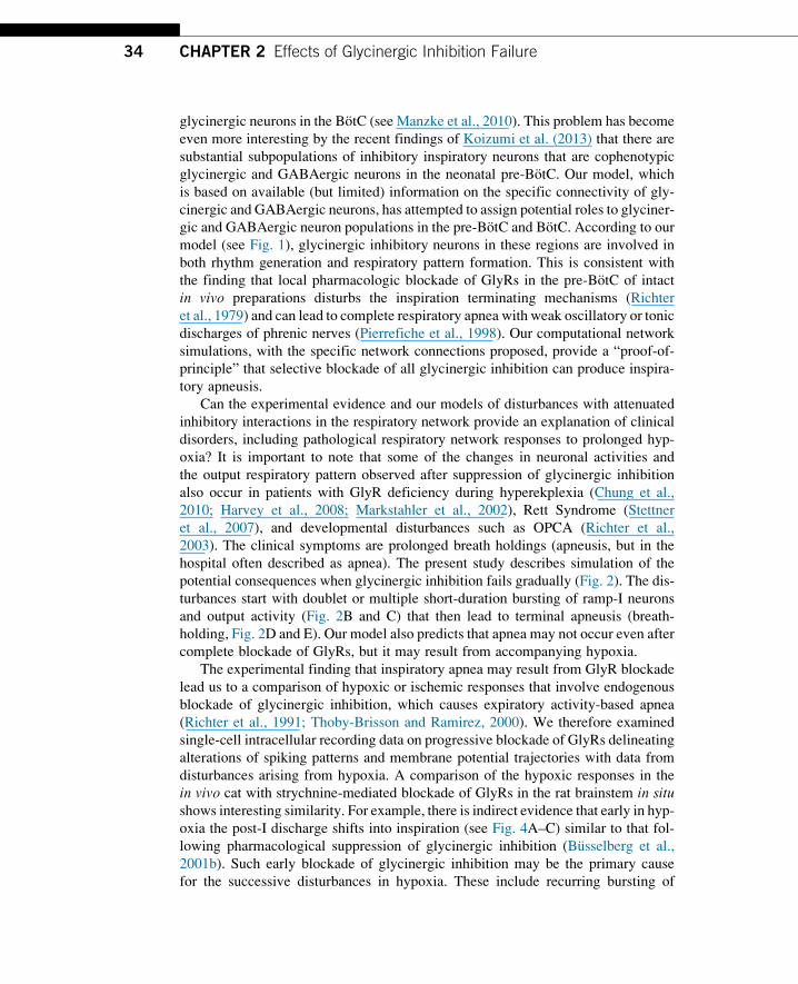
glycinergic neurons in the BotC (see Manzke et al., 2010). This problem has become
even more interesting by the recent findings of Koizumi et al. (2013) that there are
substantial subpopulations of inhibitory inspiratory neurons that are cophenotypic
glycinergic and GABAergic neurons in the neonatal pre-BotC. Our model, which
is based on available (but limited) information on the specific connectivity of gly-
cinergic and GABAergic neurons, has attempted to assign potential roles to glyciner-
gic and GABAergic neuron populations in the pre-BotC and BotC. According to our
model (see Fig. 1), glycinergic inhibitory neurons in these regions are involved in
both rhythm generation and respiratory pattern formation. This is consistent with
the finding that local pharmacologic blockade of GlyRs in the pre-BotC of intact
in vivo preparations disturbs the inspiration terminating mechanisms (Richter
et al., 1979) and can lead to complete respiratory apnea with weak oscillatory or tonic
discharges of phrenic nerves (Pierrefiche et al., 1998). Our computational network
simulations, with the specific network connections proposed, provide a “proof-of-
principle” that selective blockade of all glycinergic inhibition can produce inspira-
tory apneusis.
Can the experimental evidence and our models of disturbances with attenuated
inhibitory interactions in the respiratory network provide an explanation of clinical
disorders, including pathological respiratory network responses to prolonged hyp-
oxia? It is important to note that some of the changes in neuronal activities and
the output respiratory pattern observed after suppression of glycinergic inhibition
also occur in patients with GlyR deficiency during hyperekplexia (Chung et al.,
2010; Harvey et al., 2008; Markstahler et al., 2002), Rett Syndrome (Stettner
et al., 2007), and developmental disturbances such as OPCA (Richter et al.,
2003). The clinical symptoms are prolonged breath holdings (apneusis, but in the
hospital often described as apnea). The present study describes simulation of the
potential consequences when glycinergic inhibition fails gradually (Fig. 2). The dis-
turbances start with doublet or multiple short-duration bursting of ramp-I neurons
and output activity (Fig. 2B and C) that then lead to terminal apneusis (breath-
holding, Fig. 2D and E). Our model also predicts that apnea may not occur even after
complete blockade of GlyRs, but it may result from accompanying hypoxia.
The experimental finding that inspiratory apnea may result from GlyR blockade
lead us to a comparison of hypoxic or ischemic responses that involve endogenous
blockade of glycinergic inhibition, which causes expiratory activity-based apnea
(Richter et al., 1991; Thoby-Brisson and Ramirez, 2000). We therefore examined
single-cell intracellular recording data on progressive blockade of GlyRs delineating
alterations of spiking patterns and membrane potential trajectories with data from
disturbances arising from hypoxia. A comparison of the hypoxic responses in the
in vivo cat with strychnine-mediated blockade of GlyRs in the rat brainstem in situshows interesting similarity. For example, there is indirect evidence that early in hyp-
oxia the post-I discharge shifts into inspiration (see Fig. 4A–C) similar to that fol-
lowing pharmacological suppression of glycinergic inhibition (Busselberg et al.,
2001b). Such early blockade of glycinergic inhibition may be the primary cause
for the successive disturbances in hypoxia. These include recurring bursting of
34 CHAPTER 2 Effects of Glycinergic Inhibition Failure

the ramp-I output neurons, which might be responsible for intractable hiccups indi-
cating onset of brainstem damage during hypoxia (Mandala et al., 2010).
The striking difference to the partial blockade of GlyRs, however, is the persis-
tence of tonic expiratory discharges. The final disturbance of network activity during
progressive hypoxia is shifting of late expiratory neuronal discharges into the period
of long apneustic inspiratory bursts and their vanishing after-discharges (Fig. 4A and
B). After a short interval of hypoxia, however, blockade of GlyR seems to reach
levels beyond 70% and the phrenic nerve output declines to very weak tonic dis-
charges, while expiratory neurons fire tonically (Fig. 4C). This may result from
breakdown of mutual inhibition after additional failure of GABAergic inhibition
(Khazipov et al., 1995). In severe hypoxia, there is only a transient increase and then
collapse of GABA release (Richter et al., 1999). Therefore, from a therapeutic stand-
point, it seems reasonable to start protective treatment already before complete
breakdown of inhibitory processes by pharmacotherapeutical reinforcement of gly-
cinergic inhibition as successfully performed in a patient with surgical lesions in the
FIGURE 4
Hypoxic responses indicating early blockade of glycinergic inhibition ultimately causing
expiratory apnea (anesthetized cat, Richter, unpublished). (A) With prolonged hypoxia,
early-I inhibition of the aug-E neuron is progressively shortened to early-inspiratory periods
(see gray regions). This indicates a shift of postinspiratory discharge into inspiration
which continues throughout the apneustic inspiratory after-discharges. (B) An aug-E neuron
revealing oscillatory fluctuations of EPSPs and IPSPs during the apneustic period of
phrenic nerve discharge. (C) With enduring hypoxia, aug-E neuronal discharges shift into
the inspiratory phase, when the duration of early-I inhibition is very short. Later during
hypoxia, aug-E neurons discharge almost tonically, producing expiratory apnea. Normal
activity patterns are fully recovered shortly after restoration of normoxia.
354 Discussion

pontine-brainstem junction (Wilken et al., 1997), and also in a patient with OPCA
(Richter et al., 2003) and in brainstem stroke patients (El-Khatib et al., 2003).
AcknowledgmentsThis study was supported in the United States by National Institutes of Health, grants R33
HL087377, R01 NS057815, and R01 NS069220 to I. A. R., in part by the Intramural Research
Program of the NIH, NINDS (J. C. S.), and in Germany by the Excellence Cluster “Nanoscale
microscopy and molecular physiology of the brain” (CNMPB) funded by the DFG and BMBF
(D. W. R.).
ReferencesBusselberg, D., Bischoff, A.M., Becker, K., Becker, C.M., Richter, D.W., 2001a. The respi-
ratory rhythm in mutant oscillator mice. Neurosci. Lett. 316, 99–102.
Busselberg, D., Bischoff, A.M., Paton, J.F., Richter, D.W., 2001b. Reorganisation of respira-
tory network activity after loss of glycinergic inhibition. Pflugers Arch. 441, 444–449.
Busselberg, D., Bischoff, A.M., Richter, D.W., 2003. A combined blockade of glycine and
calcium-dependent potassium channels abolishes the respiratory rhythm. Neuroscience
122, 831–841.
Chung, S.K., Vanbellinghen, J.F., Mullins, J.G., Robinson, A., Hantke, J., Hammond, C.L.,
Gilbert, D.F., Freilinger, M., Ryan, M., Kruer, M.C., Masri, A., Gurses, C., Ferrie, C.,
Harvey, K., Shiang, R., Christodoulou, J., Andermann, F., Andermann, E., Thomas, R.H.,
Harvey, R.J., Lynch, J.W., Rees, M.I., 2010. Pathophysiological mechanisms of dominant
and recessive GLRA1 mutations in hyperekplexia. J. Neurosci. 30, 9612–9620.
Congar, P., Khazipov, R., Ben-Ari, Y., 1995. Direct demonstration of functional disconnection
by anoxia of inhibitory interneurons from excitatory inputs in rat hippocampus. J. Neuro-
physiol. 73, 421–426.
El-Khatib, M.F., Kiwan, R.A., Jamaleddine, G.W., 2003. Buspirone treatment for apneustic
breathing in brain stem infarct. Respir. Care 48, 956–958.
Harvey, R.J., Topf, M., Harvey, K., Rees, M.I., 2008. The genetics of hyperekplexia: more
than startle! Trends Genet. 24, 439–447.
Jonas, P., Bischofberger, J., Sandkuhler, J., 1998. Corelease of two fast neurotransmitters at a
central synapse. Science 281, 419–424.
Khazipov, R., Congar, P., Ben-Ari, Y., 1995. Hippocampal CA1 lacunosum-moleculare inter-
neurons: comparison of effects of anoxia on excitatory and inhibitory postsynaptic cur-
rents. J. Neurophysiol. 74, 2138–2149.
Koizumi, H., Koshiya, N., Chia, J., Cao, F., Nugent, J., Zhang, R., Smith, J.C., 2013.
Structural-functional properties of identified excitatory and inhibitory interneurons within
pre-Botzinger complex respiratory microcircuits. J. Neurosci. 33, 2994–3009.
Mandala, M., Rufa, A., Cerase, A., Bracco, S., Galluzzi, P., Venturi, C., Nuti, D., 2010. Lateral
medullary ischemia presenting with persistent hiccups and vertigo. Int. J. Neurosci. 120,
226–230.
Manzke, T., Dutschmann, M., Schlaf, G., Morschel, M., Koch, U.R., Ponimaskin, E.,
Bidon, O., Lalley, P.M., Richter, D.W., 2009. Serotonin targets inhibitory synapses to
36 CHAPTER 2 Effects of Glycinergic Inhibition Failure

induce modulation of network functions. Philos. Trans. R. Soc. Lond. B Biol. Sci. 364,
2589–2602.
Manzke, T., Niebert, M., Koch, U.R., Caley, A., Vogelgesang, S., Hulsmann, S.,
Ponimaskin, E., Muller, U., Smart, T.G., Harvey, R.J., Richter, D.W., 2010. Serotonin re-
ceptor 1A-modulated phosphorylation of glycine receptor alpha3 controls breathing in
mice. J. Clin. Invest. 120, 4118–4128.
Markstahler, U., Kremer, E., Kimmina, S., Becker, K., Richter, D.W., 2002. Effects of func-
tional knock-out of alpha 1 glycine-receptors on breathing movements in oscillator mice.
Respir. Physiol. Neurobiol. 130, 33–42.
Pierrefiche, O., Schwarzacher, S.W., Bischoff, A.M., Richter, D.W., 1998. Blockade of syn-
aptic inhibition within the pre-Botzinger complex in the cat suppresses respiratory rhythm
generation in vivo. J. Physiol. 509 (Pt. 1), 245–254.
Richter, D.W., 1982. Generation and maintenance of the respiratory rhythm. J. Exp. Biol. 100,
93–107.
Richter, D.W., Camerer, H., Meesmann, M., Rohrig, N., 1979. Studies on the synaptic inter-
connection between bulbar respiratory neurones of cats. Pflugers Arch. 380, 245–257.
Richter, D.W., Ballantyne, D., Remmers, J.E., 1986. How is the respiratory rhythm generated?
A model. News Physiol. Sci. 1, 109–112.
Richter, D.W., Bischoff, A., Anders, K., Bellingham, M., Windhorst, U., 1991. Response of
the medullary respiratory network of the cat to hypoxia. J. Physiol. 443, 231–256.
Richter, D.W., Ballanyi, K., Schwarzacher, S., 1992. Mechanisms of respiratory rhythm
generation. Curr. Opin. Neurobiol. 2, 788–793.
Richter, D.W., Schmidt-Garcon, P., Pierrefiche, O., Bischoff, A.M., Lalley, P.M., 1999.
Neurotransmitters and neuromodulators controlling the hypoxic respiratory response in
anaesthetized cats. J. Physiol. 514 (Pt. 2), 567–578.
Richter, D.W., Manzke, T., Wilken, B., Ponimaskin, E., 2003. Serotonin receptors: guardians
of stable breathing. Trends Mol. Med. 9, 542–548.
Rybak, I.A., Paton, J.F.R., Schwaber, J.S., 1997. Modeling neural mechanisms for genesis of
respiratory rhythm and pattern: II. Network models of the central respiratory pattern gen-
erator. J. Neurophysiol. 7, 2007–2026.
Rybak, I.A., Abdala, A.P., Markin, S.N., Paton, J.F., Smith, J.C., 2007. Spatial organization
and state-dependent mechanisms for respiratory rhythm and pattern generation. Prog.
Brain Res. 165, 201–220.
Schmid, K., Foutz, A.S., Denavit-Saubie, M., 1996. Inhibitions mediated by glycine and
GABAA receptors shape the discharge pattern of bulbar respiratory neurons. Brain
Res. 710, 150–160.
Shevtsova, N.A., Manzke, T., Molkov, Y.I., Bischoff, A., Smith, J.C., Rybak, I.A.,
Richter, D.W., 2011. Computational modelling of 5-HT receptor-mediated reorganization
of the brainstem respiratory network. Eur. J. Neurosci. 34, 1276–1291.
Smith, J.C., Abdala, A.P., Koizumi, H., Rybak, I.A., Paton, J.F., 2007. Spatial and functional
architecture of the mammalian brain stem respiratory network: a hierarchy of three oscil-
latory mechanisms. J. Neurophysiol. 98, 3370–3387.
Stettner, G.M., Huppke, P., Brendel, C., Richter, D.W., Gartner, J., Dutschmann, M., 2007.
Breathing dysfunctions associated with impaired control of postinspiratory activity in
Mecp2-/y knockout mice. J. Physiol. 579, 863–876.
Thoby-Brisson, M., Ramirez, J.M., 2000. Role of inspiratory pacemaker neurons in mediating
the hypoxic response of the respiratory network in vitro. J. Neurosci. 20, 5858–5866.
37References

Wilken, B., Lalley, P., Bischoff, A.M., Christen, H.J., Behnke, J., Hanefeld, F., Richter, D.W.,
1997. Treatment of apneustic respiratory disturbance with a serotonin-receptor agonist.
J. Pediatr. 130, 89–94.
Winter, S.M., Fresemann, J., Schnell, C., Oku, Y., Hirrlinger, J., Hulsmann, S., 2009. Glyci-
nergic interneurons are functionally integrated into the inspiratory network of mouse med-
ullary slices. Pflugers Arch. 458, 459–469.
38 CHAPTER 2 Effects of Glycinergic Inhibition Failure