アユ由来Edwardsiella ictaluriの強毒株と弱毒株間におけ る ...
9
アユ由来Edwardsiella ictaluriの強毒株と弱毒株間におけ るタンパク質の比較定量解析 誌名 誌名 魚病研究 ISSN ISSN 0388788X 著者 著者 坂井, 貴光 桑田, 知宣 武藤, 義範 高野, 倫一 湯浅, 啓 大迫, 典久 巻/号 巻/号 53巻1号 掲載ページ 掲載ページ p. 29-35 発行年月 発行年月 2018年3月 農林水産省 農林水産技術会議事務局筑波産学連携支援センター Tsukuba Business-Academia Cooperation Support Center, Agriculture, Forestry and Fisheries Research Council Secretariat
-
Upload
khangminh22 -
Category
Documents
-
view
0 -
download
0
Transcript of アユ由来Edwardsiella ictaluriの強毒株と弱毒株間におけ る ...

アユ由来Edwardsiella ictaluriの強毒株と弱毒株間におけるタンパク質の比較定量解析
誌名誌名 魚病研究
ISSNISSN 0388788X
著者著者
坂井, 貴光桑田, 知宣武藤, 義範高野, 倫一湯浅, 啓大迫, 典久
巻/号巻/号 53巻1号
掲載ページ掲載ページ p. 29-35
発行年月発行年月 2018年3月
農林水産省 農林水産技術会議事務局筑波産学連携支援センターTsukuba Business-Academia Cooperation Support Center, Agriculture, Forestry and Fisheries Research CouncilSecretariat

ffe.Jjjg-lvf~ Fish Pathology, 53 (1), 29-35, 2018. 3
Research article
© 2018 The Japanese Society of Fish Pathology
Comparative Proteomic Analysis Between Virulent and Less Virulent Strains of Edwardsiella ictaluri
Isolated from Ayu Plecoglossus altivelis
Takamitsu Sakai1*, Tomonori Kuwada2, Yoshinori Muto2
, Tomokazu Takano3,
Kei Yuasa3 and Norihisa Oseko3
1Tamaki Laboratory, National Research Institute of Aquaculture, Fisheries Research and Education Agency, Mie 519-0423, Japan
2Gifu Prefectural Research Institute for Fisheries and Aquatic Environments, Gifu 501-6021, Japan
3Nansei Main Station, National Research Institute of Aquaculture, Fisheries Research and Education Agency, Mie 516-0193, Japan
(Received November 15, 2017)
ABSTRACT-Outbreaks of disease caused by the infection of Edwardsiella ictaluri in ayu Plecoglossus altivelis have been reported in rivers in Japan since 2007. In this study, we performed comparative proteomic analysis between the virulent strain FPC1091 and the less virulent strain FPC1214 to identify virulence factors of E. ictaluri to ayu. In the experimental infection, cumulative mortalities of the ayu intraperitoneally injected with FPC1091 (1.2 x 10⁷ CFU/fish) and FPC1214 (2.2 x 10⁷ CFU/fish) were 100% and 26.7%, respectively. The genetic fingerprint profile of FPC1214 was identical to that of FPC1091 in the amplified-fragment length polymorphism (AFLP) analysis. The analysis of two-dimensional polyacrylamide gel electrophoresis and mass spectrometry between FPC1091 and FPC 1214 revealed 23 proteins that were either specific to or produced in greater amounts in the virulent strain FPC1091. These proteins included components of the type Ill and type VI secretion systems, and the proteins that seem to be involved in the resistance against oxidative stress by host phagocytic cells, suggesting that these proteins are the virulence factors of E. ictaluri to ayu.
Key words: Edwardsiella ictaluri, Plecoglossus altive/is, 2D-PAGE, Proteomic analysis
Edwardsiella ictaluri, a member of the family Enterobacteriaceae, is known as one of the important pathogens in cultured fish. The disease caused by the bacterium was first described in 1976 as the enteric septicemia of channel catfish lctalurus punctarus (Hawke et al., 1981 ). Subsequently, the bacterium has also been isolated from other species of fish, rosy barbs, Puntius conchonius (Humphrey et al., 1986); danio, Dania devario (Waltman et al., 1985); green knife fish, Eigemannia virescens (Kent and Lyons, 1982); farmed rainbow trout, Oncorhynchus mykiss (Keskm et al., 2004); and ayu, Plecoglossus altivelis (Sakai et al., 2008). In Japan, the disease was first observed in wild ayu in 2007, and the bacterium has been often isolated from freshwater fish such as ayu or bagrid catfish Pelteobagrus nudiceps (Sakai et al., 2009a) since then. The bacterial isolates from different places in Japan are considered to have originated from a single clone,
* Corresponding author E-mail: [email protected]
because DNA polymorphism was not found among the isolates (Sakai et al., 2009a). In addition, Hassan et al. (2010) reported that the isolates from Japan were antigenically homogeneous.
Ayu suffering from E. ictaluri infection show reddening on the body surface including anus and bases of fins, exophthalmos, and hemorrhagic ascites (Sakai et al., 2008). Death with these clinical signs has been observed in the fish experimentally challenged with the bacterium. Virulence properties such as serum resistance or intracellular replication in macrophages are known for E. ictaluri isolated from channel catfish (Booth et al., 2006). O-polysaccharide and type III secretion system (T3SS) have been identified as virulence factors associated with serum resistance and intracellular replication in macrophages, respectively (Thune et al., 2007). However, virulence factors in the strain isolated from ayu have not been investigated yet.
In the present study, we describe the identification of putative virulence factors of E. ictaluri for ayu, by the comparative proteomic analysis with two-dimensional

30 T. Sakai, T. Kuwada, Y. Muto, T. Takano, K. Yuasa and N. Oseko
polyacrylamide gel electrophoresis (2O-PAGE) using the virulent strain and relatively low virulent strain of the bacterium.
Materials and Methods
Bacteria Two E. ictaluri isolates, FPC1091 (Sakai et al.,
2008) and FPC1214 were used to identify virulence factors; both bacteria were isolated from ayu in Japan. A relatively low virulent E. ictaluri FPC1214 was isolated from ayu in Japan in 2012, and the virulence was confirmed by the challenge to ayu in the present study. This strain was confirmed to be E. ictaluri with PCR test reported by Sakai et al. (2009b) and biochemical characterization with the LIM, SC, SIM and TSI test agar (Nissui), and the cytochrome oxidase test paper (Nissui).
In addition, several different E. ictaluri strains were used in the analysis of the genetic lineages (Table 1 ). Each bacterium was cultured on LB agar, Lennox (BO Bioscience) at 28°C for 36 h.
Analysis of genetic lineage Total DNA of E. ictaluri was extracted by the Max
well16 automated purification system (Promega) with the Maxwell16 tissue DNA purification kit (Promega). The DNA of each strain was subjected to amplified-fragment length polymorphism (AFLP) analysis by use of the AFLP TM Microbial Fingerprinting kit (Applied Biosystems). Selective PCR in the AFLP reaction was performed with a selective primer set of EcoRl-0 and Msel-0 according to the condition described by Sakai et al. (2009a). The product was loaded on a 6% polyacrylamide gel containing urea (TBE-Urea Gels, Thermo Fisher Scientific), and the electrophoresis was performed in 1 x TBE buffer (Bio-Rad Laboratories) at 180 V constant voltage for 50 min. The gel was scanned with the FMBIO Ill Multi-View fluorescence image analyzer (Hitachi).
Challenge test E. ictaluri strains FPC1091 and FPC1214 were
used. Each bacterium cultured on LB was suspended in 10 mM phosphate-buffered saline (PBS, pH 7 .2). The number of viable cells in each bacterial suspension was estimated according to the method of Miles et al. (1938). Juvenile ayu were obtained from a private hatchery (Marine Tech Co.) and reared in a tank filled with 400 L of running, aerated well water at 17°C for 30 days by feeding commercial pellet diet until the challenge test. For the challenge experiment, 45 fish (average body weight: 2.2 g) were taken and divided into 3 groups (15 fish/group). Each group was maintained in a 30-L aquarium with flow-through water at 20°c. These 3 groups were intraperitoneally injected either with the bacterial suspension of the strain FPC1091 at a dose of 1.2 x 10⁷ CFU/50 µUfish, FPC1214 at a dose of 2.2 x 10⁷ CFU/50 µL/fish, or the same volume of PBS without bacteria. Mortality was recorded for 14 days.
2D-PAGE Protein of E. ictaluri was extracted from 15 mg of
the bacterial cells using the TRlzol Reagent (Thermo Fisher Scientific) according to the manufacture's instruction and dissolved in 200 µL of a solution containing 7 M Urea, 2 M thiourea, 4% CHAPS and 2 mM tributyl phosphine (Bio-Rad Labolatories). The protein concentration was determined by use of the Quick Start Bradford Protein Assay (Bio-Rad Labolatories). After incubation for 1 h at room temperature, 150 µg protein in the sample was adjusted to 340 µL with the Destreak Rehydration Solution (GE Healthcare) containing 2% IPG buffer pH 3-10 NL (GE Healthcare), and this solution was applied to the lmmobiline Dry Strip (18 cm, pH 3-10 NL, GE Healthcare). lsoelectric focusing electrophoresis was performed with the CoolPhoreStar IPG-IEF (Anatech) at 500 V for 2 h, then ramped linearly from 500 V to 3,500 V for 6 h, and finally maintained at 3,500 V for 10 h. After the electrophoresis, the strip gel was equilibrated with 25 mM Tris-HCI (pH 6.8) containing 6 M urea, 2% SOS, 30% glycerol and 0.5% dithiothreitol for 30 min, and the alkylation was performed with 4.5% iodoacetamide solution containing 25 mM Tris-HCI (pH 6.8), 6 M urea, 2% SOS and 30% glycerol for 20 min.
Table 1. E. ictaluri strains used for the analysis of genetic lineages
Strain*
ATCC33202**
ATCC33828 ATCC33829 ATCC33830 ATCC33831 FPC1091 FPC1214
Origin
Fish
Channel catfish lctalurus punctatus Channel catfish
Channel catfish White catfish lctalurus catus Channel catfish Ayu Plecoglossus altivelis Ayu
* ATCC; American Type Culture Collection.
Location Year of isolation Reference
Georgia late 1970s Georgia late 1970s Alabama late 1970s Maryland late 1970s Mississippi late 1970s Yamaguchi, Japan 2007 Sakai et al. (2008) Gifu, Japan 2008
** Edw. ictaluri ATCC33202 was obtained as JCM1680 from Japan Collection of Microorganisms, RIKEN BioResource Center.

Proteomic analysis of E. ictaluri from ayu 31
Marker ATCC33828 ATCC33829 ATCC33830 ATCC33831 ATCC33202 FPC1091 FPC1214
- I I JI. I fl i I ,,
I • I I
I
11 I I t ,, . l / ,,
I
l
I t t I
Fig. 1. AFLP fingerprints of E. ictaluri strains. Marker lane: GeneScan 500 TAMRA Size Standard (Applied Biosystems). The arrowheads indicate polymorphic bands among E. ictaluri strains.
SOS-PAGE of the equilibrated strip gel was run with a 10%-20% gradient polyacrylamide gel (Bio Craft). Proteins in the SOS-PAGE gel were stained with the Oriol Fluorescent Gel Stain (Bio-Rad Labolatories) and detected using the FluoroPhoreStar 3000 (Anatech). Spot detection, matching and relative quantification across the gels were performed with the POQuest 2-0 gel analysis software (Bio-Rad Labolatories) . The above mentioned 2-0 PAGE and subsequent analysis was triplicated for each of FPC1091 and FPC1214.
Protein identification Proteins detected by the 20-PAGE analyses were
identified by mass spectrometry. The samples were prepared as follows. Each protein spot in the 20-PAGE gel was excised using the FluoroPhoreStar 3000 (Anatech) and subjected to microwave-assisted tryptic digestion (Juan et al., 2005; Sun et al., 2006) . The tryptic digests were extracted using 50% acetonitrile (ACN) containing 0.1 % trifluoroacetic acid (TFA) for 15 min . In addition, the extraction was performed with 75% ACN containing 0.1 % TFA. The mixture of these extracts was dried by use of a centrifugal concentrator (Eppendorf) and re-dissolved in 50% ACN containing 0.1 % TFA. Each sample was mixed with an equal volume of OHBA matrix solution (Shimadzu) and subjected to MS/ MS analysis with MALOI-QIT-TOF MS (AXIMA Resonance, Shimadzu). The MS/MS data was checked to identify the protein against a database of all protein-coding sequences on the genome of E. ictaluri 93-146 (GenBank NC_012779) by use of MS/MS ion search program (MASCOT, Matrix Science) .
Results
Biochemical and genetic characteristics of E. ictaluri FPC1214
E. ictaluri FPC1214 showed the same biochemical characteristics as FPC1091 described by Nagai et al. (2008) (Table 2). In addition, there was no difference in the results of fermentation test for sucrose, lactose and glucose between these two strains.
The electrophoresis gel image of AFLP analysis is shown in Fig . 1. The banding pattern is identical for FPC1091 and FPC1214. In contrast, the polymorphic bands are detected between these two strains and
Table 2. Characteristics of E. ictaluri FPC121 4 and EPC1091 observed in biochemical tests
Characteristic
Motility
Cytochrome oxidase Simmon's citrate
Lysine decarboxylase
H2S
lndole
Fermentation of
Sucrose
Lactose
Glucose
FPC1 214 FPC1091
Weak Weak*
*
+ +*
Weak Weak*
+ +
The data with asterisks were obtained from Nagai et al. (2008) .
100
~ 90
~ 80
ro 70 t::
60 0 E 50 Q)
·"' 40 ro :S 30 E 20 :::, 0
10 0
0 1 2 3 4 5 6 7 8 9 10 11 12 13 14
Days
-o-FPC1091 -6-FPC1214 -o.-Control
Fig. 2. Cumulative mortalities of ayu after intraperitoneal injections with either E. ictaluri FPC1214, FPC1091 , or PBS. The injection doses were 2.2 x 10⁷ CFU/fish for FPC1214 and 1.2 x 10⁷ CFU/fish for FPC1091.
ATCC strains.
Mortalities Cumulative mortality of the fish challenged with the
strain FPC1214 was significantly lower than the group challenged with the strain FPC1091 (p < 0.05 in Fisher's exact probability test) (Fig. 2). For both groups , all dead fish showed reddening of head or anus, hemorrhage in the peritoneal cavity, or hypertrophy of the kidney. The injected bacteria were re-isolated from these dead fish .

32
C
T. Sakai , T. Kuwada, Y. Muto, T. Takano, K. Yuasa and N. Oseko
150 kDa -100 kDa -
75 kDa -
50 kDa -
37 kDa -
25 kDa -
20 kDa -
15 kDa-
10 kDa -
150 kDa -100 kDa -
75 kDa -
50 kDa -
37 kDa -
25 kDa -
20 kDa -
15 kDa -
10 kDa -
3 p/ 10
A
J) - -: _______ ,
B
A-j)_
Fig. 3. Two-dimensional electrophoresis of proteins of E. ictaluri. (A) FPC1901 . (B) FPC1214. (C) Magnified images of gels indicated by rectangles in (A) and (B). The proteins of the spots with numbers were either expressed in larger amounts in FPC1091 or expressed only in FPC1091. These proteins were subjected to MS/MS analyses.

Proteomic analysis of E. ictaluri from ayu 33
Table 3. Description of proteins detected in E. ictaluri FPC1091 by comparative two-dimentional gel electrophoresis (2D-PAGE)
Ratio of average Results of mascot search against E. icta/uri 93-146 Spot fluorescence intensity of
P-value*2 Accession Molecular no. protein spot*' Proteins no. *4 mass*5 Score
(FPC1091/FPC1214) _*3 Aconitate hydratase B (AcnB) ACR67984 92811 54
2 Type VI secretion system protein (EvpB) ACR69906 54423 73
3 Type VI secretion system protein (EvpB) ACR69906 54423 74
4 Pentapeptide repeat family protein (Pen) ACR67864 47307 182
5 3.15 9.06E-04 Type VI secretion system protein (VasE) ACR69917 52544 76
6 Phosphonopyruvate decarboxylase (Ppd) ACR69870 42043 64
7 Riboflavin biosynthesis protein (RibD) ACR68268 40434 47
8 Phosphoenolpyruvate phosphomutase (Ppm) ACR69871 33925 92
9 2.67 4.07E-04 Hypothetical protein ACR69483 31984 179
10 2.48 9.86E-03 Hypothetical protein ACR69483 31984 87
11 Type Ill secretion system tlanslocon component (EseD) ACR68162 20937 35
12 2.36 2.17E-05 Opacity family porin protein (LomR) ACR67492 22467 79
13 3.39 1.5E-02 Opacity family porin protein (LomR) ACR67492 22467 84
14 Type VI secretion system protein (Hcp2) ACR69905 19364 54
15 Type VI secretion system protein (Hcp2) ACR69905 19364 34
16 Type VI secretion system effector (Hep) ACR69907 17771 36
17 Type VI secretion system effector (Hep) ACR69907 17771 64
18 Type VI secretion system effector (Hep) ACR69907 17771 61
19 Type Ill secretion system low calcium response chaper-
ACR68164 17504 43 one (EscA)
20 1.89 2.35E-04 S-Ribosylhomocysteinase (LuxS) ACR70372 18959 51
21 2.1 9.28E-05 S-Ribosylhomocysteinase (LuxS) ACR70372 18959 92
22 2.18 2.11E-03 Flavodoxin family protein (Fld) ACR68997 22378 69
23 2.21 4.1E-02 Monofunctional chorismate mutase (Mcm) ACR70573 20663 42
* 1 Average fluorescence intensity of protein spot was determined by triplicate 2D-PAGE analyses of each strain. *2 P-values were determined by Student's t test between average fluorescence intensities of spots in the triplicated 2D-PAGE gels of FPC1091 and
FPC1214. *3 Proteins found only in FPC1091. *4 Registration no. in GenBank database. *5 Nominal mass calculated from the amino acid sequence.
Proteins identified by the comparative proteomic analysis
Gel images of 2O-PAGE with the strains FPC1091 and FPC1214 are shown in Fig. 3. Clear spots recognized by the PDQuest analysis software (315 spots in the strain FPC1091 and 307 spots in the strain FPC1214), were applied to the differential quantitative analysis of this software. Proteins of 23 spots were expressed in larger amounts in the strain FPC1091 than in the strain FPC1214 in the analyses of the fluorescence intensity of the spots. Sixteen proteins were identified by the MS/MS analysis from the 23 spots (Table 3). Putative virulence factors were found among these proteins, such as proteins for T3SS (EscA and EseD) and type VI secretion systems (T6SS: EvpB, Hep, Hcp2, VasE). Nine of these 16 proteins, including proteins for T3SS and T6SS, were only found in FPC1091. There is no information about the virulence for 3 proteins (4 spots).
Discussion
T3SS and T6SS are most likely involved in the virulence of FPC1091 to ayu. Proteins for these secretion
systems are only found in FPC1091 in this study. For E. tarda, T3SS and T6SS participate in the survival and replication in the host phagocytes (Srinivasa Rao et al., 2004; Okuda et al., 2006), and mutations in EscA, EseD, or EvpB decreases the replication rates of the bacterium in host macrophages (Srinivasa Rao et al., 2004; Tan et al., 2005). The T3SS interferes with apoptosis and destabilizes microtubules in host phagocytes (Okuda et al., 2006; Xie et al., 2010), and the T6SS inhibits the activation of NLRP3 inflammasome (Chen et al., 2017). In addition, genetic organization of gene clusters of T3SS and T6SS are similar between E. tarda and E. ictaluri, and the nucleotide sequence of each gene is highly conserved between these bacteria (Thune et al., 2007).
Although the role of RibD, which is one of the enzymes for riboflavin biosynthetic pathway, in the pathogenesis has not yet been studied in Edwardsiella species, the riboflavin pathway may also be important for the survival of FPC1091 in ayu. In the present study, RibD was observed only in FPC1091. Relationship of riboflavin biosynthetic pathway and the survival in macrophages has been observed in Bruce/la abortus (Bonomi et al., 2010).

34 T. Sakai, T. Kuwada, Y. Muto, T. Takano, K. Yuasa and N. Oseko
AcnB an Fld are likely to be involved in the resistance to H20 2 from the phagocytes of ayu. Respiratory burst activity of neutrophils of ayu is significantly higher than in the other fresh water fishes (Moritomo et al., 2003), and therefore, the resistance to oxidative stress must be of great importance for the survival of E. ictaluri in ayu. Enhancement of susceptibility to H20 2 has been observed in an AcnB mutant of E. coli (Tang et al., 2002) and a Fld mutant of Pseudomonas aeruginosa (Moyano eta/., 2014).
E. ictaluri are able to adhere to olfactory mucosa of channel catfish or fish cells, and this character is important for the infection to fish (Wolfe et al., 1998). The amino acid sequence of the opacity family porin protein (LomR) detected in the strain FPC1091 shares 50% similarity with that of the adhesin/virulence factor Hek of E. coli (Fagan and Smith, 2007). Thus, these proteins may contribute to infectivity of E. ictaluri for ayu.
In our previous study, the AFLP profiles of E. ictaluri were identical among the strains isolated from ayu in Japan (Sakai et al., 2009a). Furthermore, the polymorphic DNA bands were confirmed between the E. ictaluri strains in Japan and ATCC strains isolated in America. Thus, AFLP results of the present study suggest that the strain FPC1214 has been attenuated from the genetic lineage of FPC1091. The virulence of FPC1241 has not been lost completely, however, because the bacterium killed some fish in the experimental challenge and the dead fish showed typical clinical signs of E. ictaluri infection. Thus, the virulence factors in the strain FPC1214 still remain but seem to be reduced or suppressed as compared to the strain FPC1091.
The expression of the virulence factors described above is controlled by other regulatory systems. For example, the EsrA-EsrB or PhoP-PhoQ known as the "two-component system" contributes to the expression of T3SS or T6SS (Zheng et al., 2005; Chakraborty et al., 2011; Rogge and Thune, 2011 ). Reduced production of some virulence factors in the strain FPC1214 could be caused by mutations in the genes of those factors, although it is rather unlikely in the light of AFLP results. Transcription of EsrA is influenced by quorum sensing signal autoinducer 2 (Al-2), and S-ribosylhomocysteinase (LuxS), which is expressed in significantly larger amount in FPC1091 than in FPC1214, participates in the synthesis of Al-2 (Schauder et al., 2001; Zhang and Sun, 2012). Although LuxS is expressed in significantly larger amount in FPC1091 than in FPC1214, EsrA and related proteins were not detected in the comparative analysis between these strains. Other factors might participate in the reduction of virulence of FPC1214. Interrelationship between pyruvate pathway and expression of T3SS has been reported in Aeromonas hydrophila, E. coli or Salmonella enterica (Wilharm and Heider, 2014). In the present study, phosphonopyruvate decarboxylase (Ppd) and phosphoenolpyruvate
phosphomutase (Ppm) were found only in FPC1091. These are enzymes participating in conversion of phosphoenolpyruvic acid, which is a precursor of pyruvate acid, to phosphonoacetaldehyde (McGrath et al., 2013). Therefore, the pathway via Ppd and Ppm may be involved in the regulation of T3SS of E. ictaluri.
Although Pen or Mcm was found more abundantly in FPC1091 than in FPC1214, there has been no information so far on their involvement in the virulence of bacteria and it is not certain whether these proteins play any roles in the pathogenesis of E. ictaluri.
The results of the present study suggest that E. ictaluri has many virulence factors in common with E. tarda and that most of those virulence factors are related to the survival of the bacteria in host phagocytes. In fish infected with E. tarda, typically many abscesses are formed in organs such as the liver or kidney, and the bacterial cells are usually observed in host phagocytes in those abscesses (Evans et al., 2011 ). Therefore, it is very plausible that the factors facilitating the survival of the bacterium in phagocytes contribute greatly to the virulence. On the other hand, the bacterium is often observed to be free in host tissues and bacterial cells that are ingested in host phagocytes are not so frequently observed in fish infected with E. ictaluri as in fish infected with E. tarda (Sakai et al., 2008; Evans et al., 2011 ). Nevertheless, this does not necessarily undermine the importance of the ability of the bacterium to survive in host phagocytes. Granulomatous inflammation and severe infiltration of macrophages are characteristic of enteric septicaemia of catfish, which is caused by E. ictaluri (Evans et al., 2011 ), and the respiratory burst activity is markedly high in the neutrophils of ayu, as mentioned earlier. The knowledge of virulence factors obtained in the present study may be utilized to detect highly virulent E. ictaluri for the development of an effective vaccine in future studies.
References
Bonomi, H. R., M. I. Marchesini, S. Klinke, J. E. Ugalde, V. Zylberman, R. A. Ugalde, D. J. Comerci and F. A. Goldbaum (2010): An atypical riboflavin pathway is essential for Bruce/la abortus virulence. Plos One., 25, e9435.
Booth, N. J., A. Elkamel and R. L. Thune (2006): Intracellular replication of Edwardsiella ictaluri in channel catfish macrophages. J. Aquat. Anim. Health, 18, 101-108.
Chakraborty, S., J. Sivaraman, K. Y. Leung and Y. K. Mok (2011 ): Two-component PhoB-PhoR regulatory system and ferric uptake regulator sense phosphate and iron to control virulence genes in type Ill and VI secretion systems of Edwardsiella tarda. J. Biol. Chem., 286, 39417-39430.
Chen, H., D. Yang, F. Han, J. Tan, L. Zhang, J. Xiao, Y. Zhang and Q. Liu (2017): The bacterial T6SS effector evpP prevents NLRP3 inflammasome activation by inhibiting the Ca2•-dependent MAPK-Jnk pathway. Ce// Host Microbe, 21, 47-58.
Evans, J. J., P. H. Klesius, J. A. Plumb and C. A. Shoemaker

Proteomic analysis of E. ictaluri from ayu 35
(2011): Edwardsiella septicaemias, In "Fish diseases and disorders, vol. 3." (ed. by P. T. K. Woo and D. W. Bruno). CAB International Oxfordshire, UK, pp. 512-569.
Fagan, R. P. and S. G. Smith (2007): The Hek outer membrane protein of Escherichia coli is an auto-aggregating adhesin and invasin. FEMS Microbial. Lett., 269, 248-255.
Hassan, E. S., M. M. Mahmoud, N. H. Dung, K. Yuasa and T. Nakai (2010): Serological characterization of Edwardsiella ictaluri strains isolated from wild ayu Plecoglossus altivelis. Fish Pathol., 45, 43-46.
Hawke, J. P., A. C. McWhorter, A. G. Steigerwalt and D. J. Brenner (1981 ): Edwardsiella ictaluri sp. nov., the causative agent of enteric septicemia of catfish. Int. J. Syst. Bacterial., 31, 396-400.
Humphrey, J. D., C. Lancaster, N. Gudkovs and W. McDonald (1986): Exotic bacterial pathogens Edwardsiel/a tarda and Edwardsiella ictaluri from imported ornamental fish Betta sp/endens and Puntius conchonius, respectively: isolation and quarantine significance. Aust. Vet. J., 63, 369-371.
Juan, H. F., S. C. Chang, H. C. Huang and S. T. Chen (2005): A new application of microwave technology to proteomics. Proteomics, 5, 840-842.
Kent, M. L. and J. M. Lyons (1982): Edwardsiella ictlauri in the green knife fish, Eigemannia virescens. Fish Health News, 2, 2.
Keskin, 0., S. Seger, M. izg0r, S. TOrkyilmaz and R. S. Mkakosya (2004): Edwardsiella ictlauri infection in rainbow trout (Oncorhynchus mykiss). Turk. J. Vet. Anim. Sci., 28, 649-653.
McGrath, J. W., J.P. Chin and J.P. Quinn (2013): Organophosphonates revealed: new insights into the microbial metabolism of ancient molecules. Nat. Rev. Microbial., 11, 412-419.
Miles, A. A., S. S. Misra and Irwin (1938): The estimation of the bactericidal power of the blood. J. Hyg (Land.)., 38, 732-749.
Moritomo, T., K. Serata, K. Teshirogi, H. Arikawa, Y. Inoue, T. ltou and T. Nakanishi (2003): Flow cytometric analysis of the neutrophil respiratory burst of ayu, Plecoglossus altive/is: comparison with other fresh water fish. Fish Shellfish lmmunol., 15, 29-38.
Moyano, A. J., R. A. Tabares, Y. S. Rizzi, A. R. Krapp, J. A. Mondotte, J. L. Bacco, M. C. Saleh, N. Carrillo and A. M. Smania (2014): A long-chain flavodoxin protects Pseudomonas aeruginosa from oxidative stress and host bacterial clearance. PloS Genet., 10, e1004163.
Nagai, T., E. Iwamoto, T. Sakai, T. Arima, K. Tensha, Y. Iida, T. Iida and T. Nakai (2008): Characterization of Edwardsiella ictaluri isolated from wild ayu Plecoglossus altivelis in Japan. Fish Paha/., 43, 158-163.
Okuda, J., Y. Arikawa, Y. Takeuchi, M. M. Mahmoud, E. Suzaki, K. Kataoka, T. Suzuki, Y. Okinaka and T. Nakai (2006): Intracellular replication of Edwardsiella tarda in murine macrophage is dependent on the type Ill secretion system and induces an up-regulation of anti-apoptotic NF-KB target genes protecting the macrophage from staurosporineinduced apoptosis. Microb. Pathogenesis, 41, 226-240.
Rogge, M. L. and R. L. Thune (2011 ): Regulation of the Edwardsiella ictaluri type Ill secretion system by pH and phosphate concentration through EsrA, EsrB, and EsrC. Appl. Environ. Microbial., 77, 4293-4302.
Sakai, T., T. Kamaishi, M. Sano, K. Tensha, T. Arima, Y. Iida, T. Nagai, T. Nakai and T. Iida (2008): Outbreaks of Edwardsiella ictaluri infection in ayu Plecog/ossus altivelis in Japanese rivers. Fish Pathol., 43, 152-157.
Sakai, T., K. Yuasa, A. Ozaki, M. Sano, R. Okuda, T. Nakai and T. Iida (2009a): Genotyping of Edwardsiella ictaluri isolates in Japan using amplified-fragment length polymorphism analysis. Lett. Appl. Microbial., 49, 443-449.
Sakai, T., K. Yuasa, M. Sano and T. Iida (2009b): Identification of Edwardsiella ictaluri and E. tarda by species-specific polymerase chain reaction targeted to the upstream region of the fimbrial gene. J. Aquat. Anim. Health, 21, 124-132.
Schauder, S., K. Shokat, M. G. Surette and B. L. Bassler (2001 ): The LuxS family of bacteria autoinducers: biosynthesis of a novel quorum-sensing signal molecule. Mo/. Microbial., 41, 463-476.
Srinivasa Rao, P. S., Y. Yamada, Y. P. Tan and K. Y. Leung (2004): Use of proteomics to identify novel virulence determinants that are required for Edwardsiella tarda pathogenesis. Mo/. Microbial., 53, 573-586.
Sun, W., S. Gao, L. Wang, Y. Chen, S. Wu, X. Wang, D. Zheng and Y. Gao (2006): Microwave-assisted protein preparation and enzymatic digestion in proteomics. Mo/. Cell. Proteomics, 5, 769-776.
Tan, Y. P., J. Zheng, S. L. Tung, I. Rosenshine and K. Y. Leung (2005): Role of type Ill secretion in Edwardsiella tarda virulence. Microbiology, 151, 2301-2313.
Tang, Y., M. A. Quail, P. J. Artymiuk, J. R. Guest and J. Green (2002): Escherichia coli aconitases and oxidative stress: post-transcriptional regulation of sodA expression. Microbiology, 148, 1027-1037.
Thune, R. L., D. H. Fernandez, J. L. Benoit, M. Kelly-Smith, M. L. Rogge, N. J. Booth, C. A. Landry and R. A. Bologna (2007): Signature-tagged mutagenesis of Edwardsiella ictaluri identifies virulence-related genes, including a salmonella pathogenicity island 2 class of type 111 secretion systems. Appl. Environ. Microbial., 73, 7934-7946.
Waltman, W. D., E. B. Shotts and V. S. Blazer (1985): Recovery of Edwardsiella ictaluri from danio (Dania devario). Aquaculture, 46, 63-66.
Wilharm, G. and C. Heider (2014): Interrelationship between type three secretion system and metabolism in pathogenic bacteria. Front. Cell. Infect. Microbial., 27, 150.
Wolfe, K. G., J. A. Plumb and E. E. Morrison (1998): Lectin Binding Characteristics of the Olfactory Mucosa of Channel Catfish: Potential Factors in Attachment of Edwardsiella. J. Aquat. Anim. Health, 10, 348-360.
Xie, H. X., H. B. Yu, J. Zheng, P. Nie, L. J. Foster, Y.-K. Mok, B. B. Finlay and K. Y. Leung (2010): EseG, an effector of the type Ill secretion system of Edwardsiella tarda, triggers microtubule destabilization. Infect. lmmun., 78, 5011-5021.
Zhang, M. and L. Sun (2012): Edwardsiella ictaluri LuxS: activity, expression, and involvement in pathogenicity. Pol. J. Microbial., 61, 263-271.
Zheng, J., S. L. Tung and K. Y. Leung (2005): Regulation of a type Ill and a putative secretion system in Edwardsiella tarda by EsrC is under the control of a two-component system, EsrA-EsrB. Infect. lmmun., 73, 4127-4137.
アユ由来Edwardsiellaictaluriの強毒株と弱毒株間に
おけるタンバク質の比較定量解析
坂井貴光・桑田知宣・ 武藤義範・高野倫一
湯浅啓・大迫典久
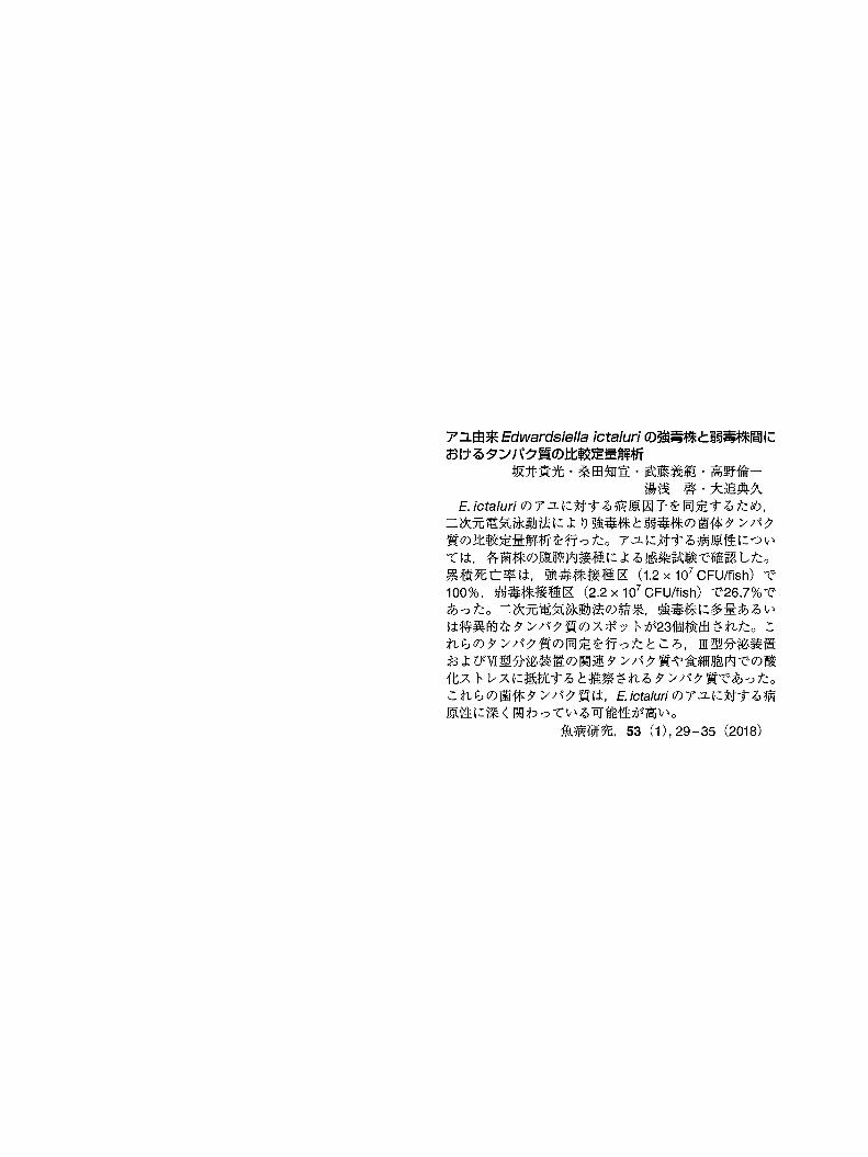
E. ictaluriのアユに対する病原因子を同定するため,
二次元電気泳動法により強毒株と弱毒株の菌体タンパク
質の比較定量解析を行った。アユに対する病原性につい
ては,各菌株の腹腔内接種による感染試験で確認した。
累積死亡率は,強毒株接種区 (1.2x 10⁷ CFU/fish) で
100%, 弱毒株接種区 (2.2x 10⁷ CFU/fish) で26.7%で
あった。二次元電気泳動法の結果,強毒株に多醤あるい
は特異的なタンパク質のスポットが23個検出された。こ
れらのタンパク質の同定を行ったところ,皿型分泌装置
およびVI型分泌装置の関連タンパク質や食細胞内での酸
化ストレスに抵抗すると推察されるタンパク質であった。
これらの菌体タンパク質は, E.icta/uriのアユに対する病
原性に深く関わっている可能性が高い。
魚病研究, 53(1), 29-35 (2018)