Diseño, desarrollo y aplicación de envases comestibles ...
400
UNIVERSIDAD COMPLUTENSE DE MADRID FACULTAD DE VETERINARIA Departamento de Nutrición, Bromatología y Tecnología de los Alimentos TESIS DOCTORAL Diseño, desarrollo y aplicación de envases comestibles potencialmente bioactivos MEMORIA PARA OPTAR AL GRADO DE DOCTOR PRESENTADA POR Ana María López de Lacey Directoras M. Elvira López Caballero y M. Pilar Montero García Madrid, 2013 © Ana María López de Lacey, 2012
-
Upload
khangminh22 -
Category
Documents
-
view
4 -
download
0
Transcript of Diseño, desarrollo y aplicación de envases comestibles ...

UNIVERSIDAD COMPLUTENSE DE MADRID
FACULTAD DE VETERINARIA
Departamento de Nutrición, Bromatología y Tecnología de los Alimentos
TESIS DOCTORAL
Diseño, desarrollo y aplicación de envases comestibles potencialmente bioactivos
MEMORIA PARA OPTAR AL GRADO DE DOCTOR
PRESENTADA POR
Ana María López de Lacey
Directoras
M. Elvira López Caballero y M. Pilar Montero García
Madrid, 2013
© Ana María López de Lacey, 2012
UNIVERSIDAD COMPLUTENSE DE MADRID
FACULTAD DE VETERINARIA
DISEÑO, DESARROLLO Y APLICACIÓN
DE ENVASES COMESTIBLES
POTENCIALMENTE BIOACTIVOS
TESIS DOCTORAL
Ana María López de Lacey
Madrid, 2012


Departamento de Nutrición,
Bromatología y Tecnología
de los Alimentos
Instituto de Ciencia y Tecnología
de Alimentos y Nutrición
DISEÑO, DESARROLLO Y APLICACIÓN
DE ENVASES COMESTIBLES
POTENCIALMENTE BIOACTIVOS
Memoria que presenta Ana María López de Lacey para optar al grado de Doctor
por la Universidad Complutense de Madrid
Bajo la dirección de la Dra. M. Elvira López Caballero y la Dra. M. Pilar Montero
García, actuando como tutora la Dra. M. Dolores Selgas Cortecero
INSTITUTO DE CIENCIA Y TECNOLOGÍA DE ALIMENTOS Y NUTRICIÓN (CSIC)
Madrid, junio del 2012


DOCTORA MARÍA ELVIRA LÓPEZ CABALLERO, Científico Titular del Instituto de Ciencia y
Tecnología de Alimentos y Nutrición, ICTAN (CSIC), y DOCTORA MARÍA PILAR MONTERO
GARCÍA, Profesora de Investigación de Instituto de Ciencia y Tecnología de Alimentos y
Nutrición, ICTAN (CSIC),
CERTIFICAN:
Que la presente memoria titulada Diseño, desarrollo y aplicación de envases comestibles
potencialmente bioactivos, presentada por ANA MARÍA LÓPEZ DE LACEY para optar al grado
de Doctor, ha sido realizada en el Instituto de Ciencia y Tecnología de Alimentos y Nutrición
(CSIC) bajo su dirección, y que, hallándose concluida, autorizan su presentación para que
pueda ser juzgada por el tribunal correspondiente.
Y para que así conste a los efectos oportunos, firman la presente cerfiticación en Madrid, a
uno de junio de dos mil doce.
Dra. Mª. Elvira López Caballero Dra. Mª. Pilar Montero García
Directora de la Tesis Doctoral Directora de la Tesis Doctoral

ESTA TESIS DOCTORAL HA SIDO REALIZADA GRACIAS A LA FINANCIACIÓN DE DIFERENTES
AYUDAS Y PROYECTOS:
- Ayuda para la formación de investigadores del Consejo Superior de Investigaciones
Científicas, programa “Junta para la Ampliación de Estudios” (Programa JAE), durante el
período de enero de 2008-2011 (JAEPre_07_00667) y asociada al proyecto “AGL2005-
02380”.
- Ayudas para estancias breves en el extranjero para beneficiarios de ayudas CSIC I3P-
Ventorrillo para personal investigador formación:
Department of Agroenvironmental Science and Technology. University of Bologna.
Italy. Enero-julio de 2009.
Institute of Food Research. Norwich. United Kingdom. Septiembre-diciembre de
2010.
- Proyecto AGL2008-00231/ALI, del Plan Nacional de Investigación Científica, Desarrollo e
Innovación Tecnológica (I+D+I).
- Programa CYTED. I+D Acción 309AC0382. Área 3.- Promoción del Desarrollo Industrial.
Línea 3.5.- Recursos del agro: Obtención de materiales aditivos a partir de subproductos
vegetales de la región y su aplicación en el desarrollo de envases biodegradables de uso
agroalimentario y nutracéutico. Agrobioenvase.


AGRADECIMIENTOS
Quiero expresar mi más profundo agradecimiento a todas aquellas personas, sin cuya
ayuda no hubiera sido posible la realización de esta Tesis Doctoral.
A las doctoras Mª Elvira López Caballero y Mª Pilar Montero García, por brindarme la
oportunidad de realizar la tesis con ellas, por su constante apoyo, plena dedicación y sus
valiosos consejos que me han servido de guía a lo largo de estos años. Gracias, sobretodo, por
sacrificar muchas veces vuestro tiempo libre para que la tesis llegue a buen puerto.
A la Dra. Mª Carmen Gómez Guillén por su valiosa ayuda durante mis primeros años en el
laboratorio y por mostrarme siempre su completa disposición para responderme cualquier
duda que me surgiera durante el desarrollo de la Tesis.
Al Dr. Joaquín Gómez Estaca por enseñarme muchas de las técnicas de laboratorio
durante mis primeros años en el ICTAN y por su importante contribución a la Tesis, no habría
podido avanzar tan rápido en la Tesis sin su estimable ayuda.
Al Dr. Efrén Pérez Santín por enseñarme muchos de los conceptos de Química que tenía
olvidados, por guiarme o aconsejarme con el desarrollo de los experimentos y por su
participación activa en la Tesis, realizando la mayor parte de los análisis con el HPLC, actividad
antioxidante y antihipertensiva.
A la Dra. Begoña Giménez Castillo por su magnífica aportación a la Tesis y por revisarme
de forma rigurosa y exhaustiva el trabajo de Norwich. Te estaré eternamente agradecida por
tu enorme contribución.
A los profesores Prof. Bruno Biavati, Dr. Giovanni Dinelli, Dra. Diana di Goia y la Dra. Ilaria
Marotti por acogerme en su laboratorio (Facultad de Agraria, Universidad de Bolonia) y
hacerme sentirme como en casa y por introducirme en el maravilloso mundo de los
probióticos.
Al Dr. Richard Faulks y a la Dra. Giusy Mandalari por permitirme trabajar en su laboratorio,
Institute of Food Research (Norwich), ponerme a su disposición su digestor y por participar
activamente en el diseño y desarrollo experimental del trabajo de digestión de las películas.
Por su puesto, quiero darle las gracias a todos mis compañeros de laboratorio que me han
ayudado alguna vez en mis experimentos, permitiendo que pueda ir más rápido, facilitándome
muchas veces el trabajo o simplemente animándome, apoyándome o haciéndome más amena
la tarea (espero no olvidar a ninguno): Elena, Óscar, Estela, Eva, Inma, Carmen, Rubén, Cristina
de las Heras, Amaia, Eva, Pilar, Torsten, Tati, Fernando, Inés, Gonzalo, Cristina Fernández,
Ailén, Gemma, Inés, Nacho, Lorena, Candelas, etc. En especial, a Elena porque me enseñó
muchos de los conocimientos que actualmente sé de microbiología. Tampoco puedo olvidarme

de Carmen de la Mata por estar siempre disponible cuando he necesitado ayuda, tanto en el
laboratorio como dándome apoyo moral en mis días bajos con la Tesis.
A todos mis compañeros de laboratorio durante mi estancia en Italia: Erwin, Cecilia,
Lorenzo, Irene, Ilaria, Francesca y Loredana. Especialmente a Erwin por transmitirme muchos
de sus valiosos conocimientos en microbiología y que me han sido de gran utilidad para la
realización de esta Tesis Doctoral, pero sobretodo, por ser como un hermano mayor para mí
durante el tiempo que estuve en Italia. Gracias Erwin también por contagiarme ese entusiasmo
tuyo por la investigación.
A mis amigos Cecilia y Lorenzo por dedicarme un pequeño hueco de su tiempo cuando
más lo he necesitado y por animarme con la Tesis.
A todos mis buenos amigos fuera y dentro del ICTAN: Isa, Mamen, Mari Val, Carlos, Luis,
Gonzalo, Nuria, Mauri, Pablo, Ruth, Bea, María, y tantos otros. Todos vosotros me habéis
animado en algún momento o escuchado cuando más lo he necesitado y me habéis dado
fuerzas para seguir adelante y no rendirme. Gracias simplemente por estar allí.
A Mauri, en especial, por el pez y la flor de la portada de la tesis (realizado con las
películas de agar de la presente Memoria) y por la realización de la fotografía de la portada con
su manífica cámara.
A mi “pequeña” familia: hermanos Antonio, Elsa, Diego y Beatriz; cuañados Mercedes,
Carlos y Jesús; y sobrinas Marta, Lucía y Elsa.
A mi hermana, y madrina, Joanna Teresa López de Lacey por el diseño de la portada de la
tesis y por ayudarme a realizar el formato final de la tesis cuando estaba ya en las últimas.
A Kiko, con gran afecto, porque durante este último año de escritura de la tesis me has
escuchado atentamente, soportado, aconsejado, apoyado y ayudado en todo momento.
Contigo ha sido todo más fácil. Gracias por estar a mi lado.
Y finalmente, y en especial, a mis padres, Antonio López Agudo y Joan Therese de Lacey.
Por ayudarme con la Tesis, por estar siempre apoyándome para que siga y no decaiga en los
momentos más difíciles. Por animarme a buscar la beca y no dejarme desistir nunca, porque
sin su tenacidad, no lo habría intentado siquiera.
Sin duda, me considero afortunada por recibir tan magnífica y valiosa ayuda de tantas
personas. Cada uno de vosotros habéis contribuido en un pedacito de esta tesis. Gracias.

~ i ~
I. INTRODUCCIÓN ................................................................................................................ 1
1. ENVASES ALIMENTARIOS .......................................................................................................... 1
1.1. Envases biodegradables comestibles: películas y recubrimientos ........................................ 2
1.1.1. Principales funciones y aplicaciones de los envases comestibles ............................... 3
1.1.2. Principales componentes de películas y recubrimientos comestibles ...................... 7
1.1.2.1. Materiales formadores de películas y recubrimientos comestibles ......................... 8
1.1.2.1.1. Películas y recubrimientos constituidos a base de hidrocoloides ......................... 8
1.1.2.1.1.1. Películas y recubrimientos constituidos a base de proteínas ............................. 9
1.1.2.1.1.1.1. Gelatina .......................................................................................................... 10
1.1.2.1.1.2. Películas y recubrimientos constituidos a base de polisacáridos ..................... 11
1.1.2.1.1.2.1. Quitosano como polímero con capacidad filmogénica................................. 12
1.1.2.1.1.2.2. Agar ............................................................................................................... 14
1.1.2.1.2. Películas y recubrimientos constituidos a base de lípidos .................................. 15
1.1.2.1.3. Películas y recubrimientos constituidos a base de mezclas de
biopolímeros……………. ......................................................................................................... 15
1.1.2.2. Aditivos ................................................................................................................... 16
1.1.2.2.1. Plastificantes ........................................................................................................ 16
1.1.2.2.2. Otros aditivos ....................................................................................................... 17
1.2. Envases activos y bioactivos comestibles ............................................................................ 18
1.2.1. Agentes activos incorporados a películas y recubrimientos comestibles .................. 20
1.2.1.1. Compuestos activos naturales de origen animal ................................................... 22
1.2.1.1.1. Quitosano ............................................................................................................. 22
1.2.1.2. Compuestos activos naturales de origen vegetal. .................................................. 26
1.2.1.2.1. Aceites esenciales ................................................................................................ 29
1.2.1.2.2. Extracto de té verde ............................................................................................. 34
1.2.2. Bacterias incorporadas a envases comestibles .......................................................... 40
1.3. Digestión de envases activos .............................................................................................. 44
1.4. Legislación relacionada con las películas y recubrimientos comestibles. ............................ 46
II. HIPÓTESIS ...................................................................................................................... 51
III. OBJETIVOS .................................................................................................................... 55
ÍNDICE

Índice
~ ii ~
IV. DISEÑO EXPERIMENTAL ................................................................................................ 59
V. TRABAJO EXPERIMENTAL ............................................................................................... 63
Artículo 1. Antimicrobial activity of composite edible films based on fish gelatin and chitosan
incorporated with clove essential oil ........................................................................................... 67
Artículo 2. Biodegradable gelatin-chitosan films incorporated with essential oils as
antimicrobial agents for fish preservation .................................................................................. 77
Artículo 3. Antioxidant and antimicrobial activities of green tea (Camellia sinensis L.) as an
expression of its chemical composition ....................................................................................... 87
Artículo 4. Release of active compounds from agar and agar-gelatin films with green tea
extract ....................................................................................................................................... 121
Artículo 5. Functionality of lactic acid bacteria incorporated to edible coatings and films ...... 153
Artículo 6. Survival and metabolic activity of probiotic in green tea ........................................ 175
Artículo 7. Biotransformation and resulting biological properties of green tea polyphenols
by probiotic bacteria ................................................................................................................. 211
Artículo 8. Agar films containing green tea extract and probiotic bacteria for extending fish
self-life ....................................................................................................................................... 235
Artículo 9. Bioaccessibility of green tea polyphenols incorporated into an edible agar film
during simulated human digestion ........................................................................................... 255
VI. DISCUSIÓN INTEGRADORA .......................................................................................... 285
1. DESARROLLO DE ENVASES COMESTIBLES CON ACEITES ESENCIALES Y APLICACIÓN EN
PRODUCTOS PESQUEROS .......................................................................................................... 285
1.1. Selección de aceites esenciales antimicrobianos para su incorporación a matrices
complejas (gelatina-quitosano) ......................................................................................... 285
1.2. Aplicación de recubrimientos a la conservación de pescado refrigerado: salmón
(Salmo salar) y bacalao (Gadus morhua) ........................................................................... 290
2. ESTUDIO DE LA COMPOSICIÓN DE EXTRACTOS ACUOSOS DE TÉ VERDE (Camellia sinensis) EN
RELACIÓN A SU FUNCIONALIDAD Y DESARROLLO DE ENVASES COMESTIBLES ACTIVOS ......... 293
2.1. Selección de un extracto de té verde (Camellia sinensis) según sus propiedades
biológicas entre distintas variedades en base a su composición química ......................... 293
2.2. Caracterización, evaluación de propiedades biológicas y liberación de compuestos
activos de películas con extracto de té verde .................................................................... 302
3. INCORPORACIÓN DE BACTERIAS LÁCTICAS EN ENVASES COMESTIBLES Y ESTUDIO DE SU
VIABILIDAD DURANTE LA CONSERVACIÓN DE FILETE DE MERLUZA ......................................... 313
3.1. Funcionalidad de bacterias lácticas incorporadas a coberturas y películas comestibles .
........................................................................................................................................... 313

Índice
~ iii ~
4. DESARROLLO DE PELÍCULAS COMPLEJAS PARA LA BIOCONSERVACIÓN DE PRODUCTOS
PESQUEROS ............................................................................................................................... 321
4.1. Viabilidad de los probióticos en presencia de los polifenoles mayoritarios del té verde
y extracto de té verde ....................................................................................................... 321
4.2. Biotransformación de los compuestos polifenólicos mayoritarios del té verde
yextracto de té verde ......................................................................................................... 321
4.3. Bioconservación de productos pesqueros mediante la aplicación de películas
complejas constituidas por probióticos y extractos de té verde ...................................... 332
5. BIOACCESIBILIDAD DE COMPUESTOS PROCEDENTES DE PELÍCULAS COMESTIBLES MEDIANTE
MODELOS DE DIGESTIÓN GASTROINTESTINAL IN VITRO.......................................................... 341
5.1. Recuperación acumulada de compuestos en el Simulador Gastro Intestinal (SGI) .... 342
5.2. Actividad antioxidante y antimicrobiana recuperada acumulativa ........................... 346
VII. CONCLUSIONES ......................................................................................................... 351
VIII. BIBLIOGRAFÍA ........................................................................................................... 357


Los residuos procedentes del procesado de pescado representan tras el fileteado
aproximadamente un 75% del peso total. En la mayoría de las industrias procesadoras, el
aprovechamiento o la gestión de estos residuos constituyen un serio problema a nivel mundial.
Un porcentaje importante de dichos residuos son pieles, tejido conectivo y espinas ricos en
colágeno. Por hidrólisis de éste se obtiene la gelatina, compuesto proteico considerado en sí
mismo como un alimento dietético de alta digestibilidad. Otro gran residuo, en este caso de la
industria procesadora de crustáceos, es la quitina, a partir del cual se obtiene el quitosano,
polímero muy versátil y de probada funcionalidad.
Una vía que en los últimos años está mostrando un gran interés por su aplicabilidad y
beneficios medioambientales es la utilización de diversos materiales a partir de residuos de la
industria agraria, pesquera y acuicultura, como matrices poliméricas para el desarrollo de
envases comestibles y/o biodegradables. La presente memoria se centra principalmente en la
utilización de gelatina, quitosano y agar. Estos envases pueden tener capacidad funcional,
principalmente antioxidante y antimicrobiana, contribuyendo a conferir estabilidad y calidad a
los alimentos.
Los envases diseñados a los que se incorporan, tanto compuestos polifenólicos como
microrganismos probióticos, actúan como bioconservadores en los alimentos y pueden,
además, incidir en la salud del consumidor. En la literatura apenas se encuentran trabajos
sobre ello, de ahí la novedad del tema propuesto.
Además, se han realizado estudios basados en la capacidad de ciertas cepas de
bifidobacterias para biotransformar los compuestos polifenólicos de su entorno, modificando
el perfil disponible de estos compuestos, así como su actividad.
Por otro lado, dado el carácter comestible de estos envases, se plantea evidenciar la
bioaccesibilidad de los compuestos activos que forman parte del envase con objeto de valorar
su potencial bioactivo tras su ingesta.
RESUMEN

The waste from fish processing after filleting represents approximately 75% of the total
weight. In most processing industries, the harvesting or management of such waste is a serious
worldwide problem. A significant proportion of these residues are rich in collagen (skin, bones
and connective tissue). The collagen by hydrolysis is converted into gelatin that can be
considered a highly digestible diet food. Another great waste, in this case from the crustacean
processing industry, is chitosan, of proven functionality.
In recent years, the use of different materials such as polymeric matrix for development of
food packaging and/or biodegradable is showing great interest because of their applicability
and environmental benefits. This work has focused primarily on the use of gelatin, chitosan
and agar as biopolymers. These matrixes may have functional capacity, mainly antioxidant and
antimicrobial, by adding of active compounds that confer stability and help to maintain food
quality.
Edible packaging incorporates both polyphenolic compounds and probiotics that could act
as biopreservatives in food and may also contribute to be healthy products. There are scarcely
any studies found in literature about this research, hence the novelty of the theme.
In addition, some studies have been conducted based on the ability of certain strains of
bifidobacteria to biotransform polyphenolic compounds from their environment, modifying
the available phenolic profile and their activity.
On the other hand, due to film`s edible character, it was planned to show the
bioaccesibility of the active compounds that constitute part of the package in order to assess
their potential bioactive
SUMMARY


~ 1 ~
1. ENVASES ALIMENTARIOS
Los envases juegan un papel fundamental en la industria alimentaria ya que realizan
importantes funciones como la de “contener, proteger, manipular, distribuir y presentar
mercancías, desde materias primas hasta artículos acabados, y desde el fabricante hasta el
usuario o el consumidor” (Directiva 94/62/CE del Parlamento Europeo y del Consejo). Entre
estas funciones destaca la acción protectora de los envases, ya que contribuyen al retraso del
deterioro, aumentan la vida útil y mantienen la calidad y seguridad de los alimentos
envasados.
Los envases protegen a los alimentos y bebidas de una serie de agentes externos
procedentes del ambiente como son el calor, la luz, humedad, oxígeno, presión, enzimas,
olores indeseables, microorganismos, insectos, suciedad y partículas de polvo o emisiones de
gases, entre otros (Restuccia y cols., 2010) que suponen un detrimento de su calidad o
seguridad. Por otra parte, desde un punto de vista comercial los envases se emplean para
identificar un producto determinado, y también para proporcionar información importante
como, por ejemplo, el peso, ingredientes o valor nutricional (Restuccia y cols., 2010).
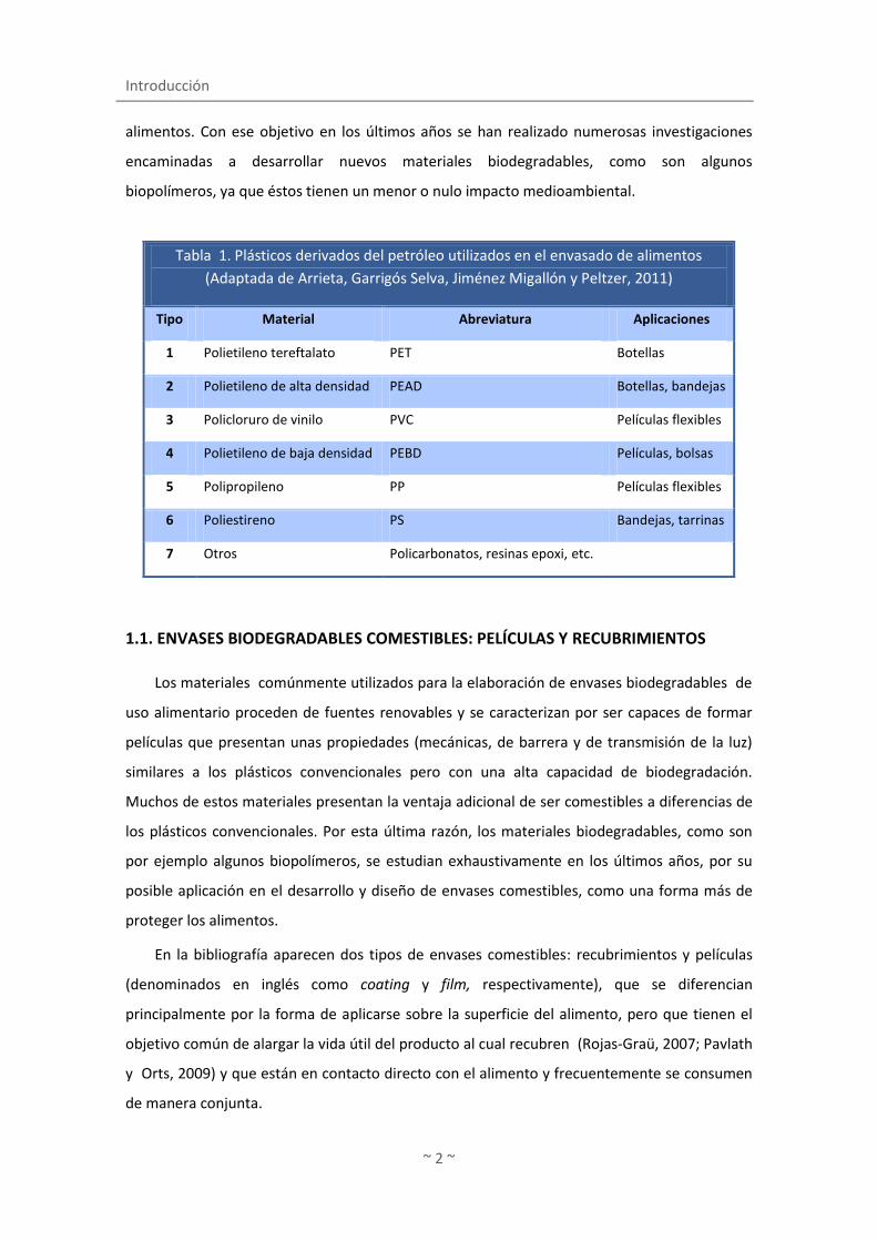
Los materiales más utilizados para el envasado de alimentos y bebidas son los plásticos
sintéticos, que se clasifican en la actualidad en siete categorías (Tabla 1). Estos polímeros
sintéticos se emplean por las múltiples ventajas que presentan, como ser químicamente
inertes, ligeros, resistentes, cómodos e higiénicos, y por su versatilidad de forma, tamaño, etc.
(García-Díaz y Macías-Matos, 2008). Sin embargo, al ser compuestos sintéticos, no
biodegradables y derivados del petróleo, su utilización supone serios problemas ecológicos
debidos principalmente a la contaminación medioambiental que causan, tanto por su
fabricación e incineración como por su aporte en la generación y acumulación de residuos. Por
otro lado, estos polímeros sintéticos pueden vehiculizar sustancias tóxicas o no deseables
como monómeros, plastificantes, antioxidantes sintéticos, aditivos, etc. presentes en su propia
composición y que pueden migrar al alimento que envuelven (Nerín de la Puerta, 2009).
Además de lo anteriormente expuesto, no hay que olvidar otros problemas que conllevan el
uso del petróleo como materia prima, como son la disminución de las reservas petrolíferas y su
precio elevado.
Por lo tanto, existe un interés tanto político como social por encontrar nuevos materiales
procedentes de fuentes renovables, menos contaminantes o de fácil reciclado para envasar los
I. INTRODUCCIÓN
Introducción
~ 2 ~
alimentos. Con ese objetivo en los últimos años se han realizado numerosas investigaciones
encaminadas a desarrollar nuevos materiales biodegradables, como son algunos
biopolímeros, ya que éstos tienen un menor o nulo impacto medioambiental.
1.1. ENVASES BIODEGRADABLES COMESTIBLES: PELÍCULAS Y RECUBRIMIENTOS
Los materiales comúnmente utilizados para la elaboración de envases biodegradables de
uso alimentario proceden de fuentes renovables y se caracterizan por ser capaces de formar
películas que presentan unas propiedades (mecánicas, de barrera y de transmisión de la luz)
similares a los plásticos convencionales pero con una alta capacidad de biodegradación.
Muchos de estos materiales presentan la ventaja adicional de ser comestibles a diferencias de
los plásticos convencionales. Por esta última razón, los materiales biodegradables, como son
por ejemplo algunos biopolímeros, se estudian exhaustivamente en los últimos años, por su
posible aplicación en el desarrollo y diseño de envases comestibles, como una forma más de
proteger los alimentos.
En la bibliografía aparecen dos tipos de envases comestibles: recubrimientos y películas
(denominados en inglés como coating y film, respectivamente), que se diferencian
principalmente por la forma de aplicarse sobre la superficie del alimento, pero que tienen el
objetivo común de alargar la vida útil del producto al cual recubren (Rojas-Graü, 2007; Pavlath
y Orts, 2009) y que están en contacto directo con el alimento y frecuentemente se consumen
de manera conjunta.
Tabla 1. Plásticos derivados del petróleo utilizados en el envasado de alimentos
(Adaptada de Arrieta, Garrigós Selva, Jiménez Migallón y Peltzer, 2011)
Tipo Material Abreviatura Aplicaciones
1 Polietileno tereftalato PET Botellas
2 Polietileno de alta densidad PEAD Botellas, bandejas
3 Policloruro de vinilo PVC Películas flexibles
4 Polietileno de baja densidad PEBD Películas, bolsas
5 Polipropileno PP Películas flexibles
6 Poliestireno PS Bandejas, tarrinas
7 Otros Policarbonatos, resinas epoxi, etc.

Introducción
~ 3 ~
Un recubrimiento o cobertura comestible (coating) se define como una capa delgada de
material comestible formado como un revestimiento sobre el alimento, mientras una película
(film) comestible es una capa preformada y delgada elaborada con material comestible que
una vez preparada puede disponerse sobre el alimento o entre los componentes del mismo
(Krochta y De Mulder-Johnston, 1997). De forma general puede decirse que los recubrimientos
se aplican en forma líquida sobre el alimento, normalmente por inmersión del producto en la
solución con capacidad filmogénica, mientras que las películas elaboradas como láminas
sólidas se aplican posteriormente sobre el alimento como envoltura (McHugh y Senesi, 2000).
1.1.1. PRINCIPALES FUNCIONES Y APLICACIONES DE LOS ENVASES COMESTIBLES
Los envases comestibles se aplican en alimentos de muy diversa naturaleza (frutas,
verduras, carnes, dulces, cereales, pescados, etc.), normalmente en combinación con otras
tecnologías de conservación (ej. refrigeración, atmósferas modificadas o controladas,
tratamientos térmicos, etc.) con el fin de mejorar su calidad, seguridad o aumentar su vida útil,
ya que estos envases pueden realizar diferentes funciones beneficiosas sobre el alimento
como son la de actuar de barrera, mejora de las propiedades, proteger pequeñas porciones,
adherir diferentes partes de un alimento o servir de soporte de aditivos, entre otras
(Debeaufort y cols., 1998; Kester y Fennema, 1986; Pavlath y Orts, 2009). A continuación se
detallan algunas de estas funciones y sus aplicaciones.
1.1.1.1. Barrera a la transferencia de materia y a la luz
Algunos envases tienen la capacidad de actuar de barrera frente a la transferencia de
determinados componentes presentes en el alimento o en el ambiente. Esta característica
resulta interesante cuando la calidad de un producto está vinculada a la pérdida o ganancia de
algunos componentes como por ejemplo el agua, compuestos volátiles (aromas deseables o
indeseables) o solutos (aceite, azúcares o sales).
La deshidratación o pérdida de agua representa uno de los principales problemas
responsables de la pérdida de calidad en fruta fresca cortada (Rojas-Graü y cols., 2007), ya que
ésta causa diversos cambios como son la pérdida de turgencia y firmeza (Olivas y Barbosa-
Cánovas, 2009). Así por ejemplo, la cobertura comercial Seal gum, Spray gumTM a base de
ésteres de sacarosa (Pavlath y Orts, 2009) se utiliza para evitar la pérdida de humedad en este
tipo de productos. En ocasiones se producen fenómenos contrarios y la absorción de agua
supone cambios indeseables en algunos alimentos o en diferentes partes de un producto

Introducción
~ 4 ~
constituido por varios alimentos (ej. pizza, tarta de manzana, caramelos). La pizza congelada
sería un buen ejemplo de esto último ya que es un producto complejo que contiene diversos
tipos de alimentos con diferente contenido en agua (masa e ingredientes). En este caso, la
aplicación de una cobertura entre la masa y los ingredientes reduce la transferencia de agua
desde los ingredientes húmedos (con alta actividad de agua) a la masa seca (con baja actividad
de agua), que en caso contrario provocan cambios indeseables en su textura, como es la de
pérdida de dureza.
Esta propiedad de barrera de los envases comestibles es muy útil también en frituras,
puesto que algunos recubrimientos tienen la capacidad de impedir la absorción excesiva de
aceite durante su cocinado. Estas coberturas presentan dos aspectos positivos: un producto
más saludable con menos aceite y una reducción de los costes al disminuir la cantidad de
aceite utilizada en cada fritura. Por ejemplo, la cobertura comestible a base de pectinato de
calcio Fry ShieldTM reduce la absorción de grasa durante la fritura de pescado, patatas y otros
vegetales (Pavlath y Orts, 2009).
Algunas películas y recubrimientos se han aplicado con el fin de controlar la transferencia
de determinados gases (oxígeno, dióxido de carbono, etc.), de tal manera que se genera
dentro del envase una atmósfera idónea que retrasa el deterioro. Esta atmósfera varía en
función del tipo de alimento; en frutas y verduras se utilizan películas y recubrimientos
semipermeables, es decir, tienen cierta permeabilidad al oxígeno, ya que las envolturas
extremadamente impermeables pueden inducir a la creación de un ambiente anaeróbico que
provoca cambios indeseables en este tipo de productos, como por ejemplo la pérdida de
aromas. Por otro lado, las películas impermeables al oxígeno se aplican sobre todo en
productos ricos en grasa (pescado azul, frutos secos, etc.) puesto que la calidad de estos
productos disminuye principalmente por la oxidación de sus lípidos. La oxidación de la grasa
genera compuestos con sabores desagradables (sabor rancio) u olores indeseables. En este
sentido, Lee y Krochta (2002) y Lee, Trezza, Guinard y Krochta (2002) redujeron el
enranciamiento oxidativo de los cacahuetes y, por lo tanto, aumentaron su vida útil mediante
la utilización de una cobertura a base de proteínas de suero.
Por otro lado, la luz, en particular la radiación ultravioleta (UV), es un potente activador
de la oxidación, por lo tanto los envases comestibles opacos a la luz UV constituyen una
manera más de frenar la oxidación. La Figura 1 representa de forma esquemática la función de
barrera ejercida por los envases comestibles frente a diversos componentes y agentes.
Introducción
~ 5 ~
1.1.1.2. Mejora de las propiedades sensoriales del alimento
Los envases comestibles se aplican en los alimentos para mejorar las propiedades
sensoriales de un alimento: apariencia, color, brillo, transparencia, rugosidad, textura, etc.
(Debeaufort, Quezada-Gallo y Voilley, 1998; Kramer, 2009). En este sentido, la empresa
Viscofan, líder mundial en la producción y distribución de envolturas comestibles en productos
cárnicos, comercializa un tipo de lámina comestible a base de colágeno denominada Naturin®.
Estas películas se aplican como “envolturas comestibles invisibles” en jamón cocido y carne
asada con el objetivo de mejorar distintas propiedades, tales como el aspecto visual, sabor,
retención del jugo de la carne, y absorción del color ahumado, entre otras.
Otras coberturas interesantes que mejoran las propiedades sensoriales son las que se
aplican en los donuts glaseados. El glasé de los donuts tiende a desaparecer como resultado de
la absorción de agua o porque simplemente funde. En este caso, los recubrimientos a base de
agar (opción más cara) o de kappa-carragenano (opción más barata) consiguen minimizar la
fusión del azúcar (Nieto, 2009) y por lo tanto mantener durante más tiempo el buen aspecto
del glaseado.
Otro ejemplo de películas comestibles con esta función son las que elabora la empresa
NewGemFoodsTM, utilizadas como envolturas alternativas al sushi o pescado crudo
(denominadas como Origami Sushi Wraps®) que son visualmente más atractivas que la alga
nori, o bien como sustitutos saludables de la tortilla que se usa para envolver los burritos
(GemWraps®).
Compuestos aromáticos
Gases (O2, CO2) ALIMENTO
AMBIENTE Luz
UV
Vapor de agua
Solutos
lípidos
sales
Figura 1. Función de barrera de los recubrimientos y películas comestibles (Adaptada de Debeaufort, Quezada-Gallo y Voilley, 1998)

Introducción
~ 6 ~
1.1.1.3. Protección mecánica
Los envases también se utilizan para mejorar las propiedades mecánicas de algunos
alimentos frágiles, como los cereales o los alimentos liofilizados, de tal manera que faciliten su
manipulación y transporte (Debeaufort y cols., 1998; Fernández Pan y Maté Caballero, 2011).
Los recubrimientos a veces protegen frente a daños mecánicos provocados por el
rozamiento entre los componentes individuales de un producto (ej. cacahuetes de una bolsa).
En este sentido, la cobertura shellac (goma laca) se aplica en especial en alimentos recubiertos
con chocolate (tipo M&M) para proporcionar principalmente brillo, para proteger frente a los
rasguños producidos por el rozamiento (Kramer, 2009) o para evitar que el chocolate se funda
durante su almacenamiento o manejo (Martín-Belloso, Rojas-Graü y Soliva-Fortuny, 2009).
1.1.1.4. Soporte de aditivos
Los envases comestibles se emplean como una manera eficaz de añadir aditivos (por
ejemplo, colorantes, especias, aromas) que impliquen una mejora en las propiedades
sensoriales o que supongan incluso un aumento de las propiedades nutricionales (vitaminas,
minerales, etc.).
Las películas y recubrimientos permiten añadir también otros compuestos con
propiedades activas, antimicrobianos y antioxidantes principalmente, que protejan de la
oxidación o inhiban el crecimiento microbiano tanto de patógenos como de responsables del
deterioro. Recientemente se ha visto que las películas son adecuadas como portadoras de
sustancias o bacterias con propiedades bioactivas (probióticos), con el fin de mejorar no sólo la
conservación sino también para aportar determinadas propiedades beneficiosas a los
alimentos. El desarrollo y diseño de envases con propiedades activas o bioactivas ha permitido
sobretodo mejorar la vida útil de alimentos tan perecederos como son los productos
pesqueros. Este tipo de envases se detallará en el apartado de envases activos.

Introducción
~ 7 ~
1.1.1.5. Otras funciones
La envoltura o tripa a base de colágeno representa probablemente una de las películas
comestibles comerciales con más éxito desde un punto de vista comercial, ya que se emplean
como sustitutos de las tripas naturales en la elaboración de productos cárnicos tipo salchicha o
embutido. Estas envolturas se utilizan sobre todo para mantener la integridad estructural de
los productos cárnicos (Chapman y Potter, 2004), pero también tienen otras funciones
beneficiosas como la de retrasar la pérdida de humedad, oxidación lipídica, decoloración,
mejora de la apariencia del producto o como portador de aditivos alimentarios (Catherine
Nettles, 2006; Gennadios, Hanna y Kurth, 1997; Véronique, 2008).
Otra aplicación de los envases comestibles consiste en su empleo como pegamento para
adherir determinados condimentos o diferentes componentes de un alimento. Como ejemplo
los tentempiés tipo barrita y snacks de cereales (Kramer, 2009), en los que se requiere unir
diferentes componentes entre sí (cereales, semillas, frutos secos).
Todas estas funciones a las que hace referencia este capítulo pueden desarrollarse en
menor o mayor medida, dependiendo de los componentes, estructura y composición de los
envases comestibles. Además, en función del alimento interesará potenciar unas acciones
frente a otras, ya que no todos los alimentos tienen las mismas necesidades en cuanto al
mantenimiento de su calidad o de su seguridad. Por esta razón, la elección de los materiales
(ej. biopolímeros) y aditivos debe realizarse de acuerdo al objetivo, naturaleza del producto y
el método de aplicación (Debeaufort y cols., 1998).
1.1.2. PRINCIPALES COMPONENTES DE PELÍCULAS Y RECUBRIMIENTOS COMESTILBES
La elaboración de los envases comestibles requiere de al menos un componente capaz de
formar una matriz estructural. Esta capacidad la poseen algunos biopolímeros y lípidos, y por
tanto, suelen ser la base de los envases comestibles. Muchas veces resulta imprescindible la
adicción de aditivos como los plastificantes a la formulación de estos envases, puesto que sin
ellos la película resultante sería excesivamente frágil y muy poco flexible. Además de los
plastificantes se pueden incluir otros aditivos, tal vez no tan estrictamente necesarios como
los anteriores pero que su inclusión en la fórmula supone una mejora en las propiedades
tecnológicas y funcionales de las envolturas. Por lo tanto, la presente memoria se centrará en
los principales componentes de las películas (biopolímeros, lípidos y plastificantes), aunque
también se hará mención a otros aditivos que resultan interesantes en la formulación de los
envases comestibles.

Introducción
~ 8 ~
1.1.2.1. Materiales formadores de películas y recubrimientos comestibles
Los materiales utilizados en la preparación de recubrimientos y películas proceden de
diversas fuentes del reino animal y vegetal, tanto terrestre como marino, y así como
procedente de los microorganismos (Tharanathan, 2003). Algunos de estos biopolímeros se
obtienen a partir de los residuos generados de la pesca, de la agricultura o de la ganadería.
Desde un punto de vista medio ambiental, el aprovechamiento de residuos resulta de gran
interés puesto que se consigue obtener un rendimiento y valorizar estos desechos, reducir su
cantidad y por lo tanto los costes y problemas de su eliminación.
La naturaleza de estos compuestos es muy variada, siendo principalmente de origen
proteico (gelatina, proteína del suero de la leche, zeína, gluten, proteína de soja, etc.),
polisacárido (celulosa, gomas, almidón, quitosano, agar, pectinas, etc.) y lipídico (ceras, grasas,
aceites). Las características de las películas y recubrimientos vienen determinadas, en parte,
por la naturaleza de estos compuestos y por esta razón, para explicar de forma simplificada las
propiedades de los envases comestibles, se han clasificado en tres categorías en función del
material de base utilizado en su formulación:
Hidrocoloide: proteína y polisacáridos (biopolímeros).
Lípidos.
Mezclas: hidrocoloides y lípidos.
1.1.2.1.1. Películas y recubrimientos constituidos a base de hidrocoloides
La capacidad de los biopolímeros para interaccionar entre si y con el resto de los
componentes durante la formación del recubrimiento viene dado por su naturaleza, peso
molecular, cargas, etc., es decir, la estructura del biopolímero condicionará la función del
recubrimiento.
En líneas muy generales, la formación de una red macromolecular de un biopolímero tipo
hidrocoloide requiere de algunas etapas: en primer lugar la solubilización (parcial o total) que
permita una ruptura de enlaces intermoleculares de baja energía que estabilicen a los
polímeros en su estado nativo; de esta manera se facilita un reordenamiento y orientación de
las cadenas poliméricas y una interacción con el resto de componentes que forman la película
(esta estructura se estabiliza durante el secado) (Cuq, Gontard, Cuq y Guilbert, 1998; Mauri y
Añón, 2008).
Introducción
~ 9 ~
1.1.2.1.1.1. Películas y recubrimientos constituidos a base de proteínas
Para la elaboración de películas y recubrimientos comestibles a base de proteína se han
utilizado proteínas de diferente origen, tanto animal como vegetal. Así se pueden encontrar en
la literatura películas de gelatina (Giménez, Gómez-Estaca, Alemán, Gómez-Guillén y Montero,
2009), caseína, proteína aislada o concentrada del suero lácteo (Banerjee y Chen, 1995), gluten
de trigo (Gontard y Ring, 1996) y proteína de soja (Brandenburg, Weller y Testin, 1993), entre
otras.
Los biopolímeros proteicos forman redes macromoleculares tridimensionales que se
estabilizan mediante diversos tipos de enlaces (interacciones electrostáticas, puentes de
hidrógeno, fuerzas de Van der Waals, enlaces covalentes y puentes disulfuro), los cuales
dependen de su composición aminoacídica. Los enlaces se pueden favorecer durante el
procesado, tanto por las soluciones en las que se encuentra como por el tratamiento térmico y
modo de secado. Así, por ejemplo, una película a base de proteína de huevo, que contiene
gran cantidad de cisteína, puede favorecer la formación de enlaces covalentes tipo puentes
disulfuro en condiciones térmicas adecuadas, lo cual favorece la insolubilización de la película
(Giménez, Gómez-Guillén, López-Caballero, Gómez-Estaca y Montero, 2012). Asimismo, la
forma de la proteína es de gran importancia para la formación de estas redes que conforman
la matriz. Las proteínas de alto peso molecular y fibrilares -como el colágeno, la gelatina y las
proteínas miofibrilares- pueden formar redes más amplias con buenas propiedades mecánicas
(Guillbert y Graille, 1994). En cambio, las proteínas globulares, frecuentemente de bajo peso
molecular (como las proteínas aisladas de soja y proteínas sarcoplásmicas), hacen redes más
compactas y menos elásticas, con menor resistencia (Mauri y Añón, 2008). También se puede
modificar la estructura de la proteína por desnaturalización y agregación, pudiendo ofrecer de
esta manera variaciones en las propiedades que generan al constituir la red filmogénica.
Todas estas variables y la gran diversidad de características de las distintas proteínas
permiten obtener un amplio abanico de posibilidades y propiedades de las películas
constituidas a partir de estos biopolímeros. En general, si bien las películas a base de proteínas
presentan buenas propiedades de barrera frente al oxígeno y dióxido de carbono, son
susceptibles a la humedad (Cha y Chinnan, 2004; Krochta y De Mulder-Johnston, 1997).
La capacidad antioxidante que poseen determinadas proteínas aporta un valor añadido a
las películas y recubrimientos elaborados a partir de ellas. Varios estudios describen las
propiedades antioxidantes de las proteínas tanto de origen animal como vegetal tales como las
proteínas de la leche (Cervato, Cazzola y Cestaro, 1999), zeína de maíz (Wang, Fujimoto,

Introducción
~ 10 ~
Miyazawa y Endo, 1991), gliadina del trigo (Iwami, Hattori y Ibuki, 1987) o gelatina de pescado
(Alemán, Giménez, Montero y Gómez-Guillén, 2011).
Entre la gran variedad de proteínas utilizadas para la elaboración de envases comestibles,
la gelatina posee excelentes propiedades físicas y fácil manejo, por lo que se seleccionó para la
elaboración de las películas y los recubrimientos comestibles de la presente memoria. Por este
motivo se describe con más detalle.
1.1.2.1.1.1.1. Gelatina
La gelatina se obtiene a partir de la hidrólisis parcial del colágeno, el cual se encuentra
ampliamente distribuido en la naturaleza formando parte de la piel, tendones, sistema
vascular, huesos, espinas, escamas y tejido conectivo de los animales. Esta proteína se obtiene
principalmente a partir de la piel y huesos de mamíferos terrestres, fundamentalmente vacuno
y porcino. Sin embargo recientemente se ha incrementado la producción de gelatina a partir
de productos de la pesca. Este aumento se debe a la sustitución de la gelatina de origen
vacuno y porcino por la gelatina de origen marino. Las razones inicialmente han sido socio-
culturales (productos kosher y del islam) o sanitarias (ej. encefalopatía espongiforme), si bien
últimamente se ha incrementado el interés en el aprovechamiento de subproductos y residuos
generados en la industria pesquera y de acuicultura (ej. pieles y huesos), como fuente de
gelatina (Gómez-Guillén, Giménez, López-Caballero y Montero, 2011).
La gelatina se utiliza ampliamente en la industria farmacéutica, cosmética y alimentaria
debido a sus excelentes propiedades gelificantes, hidratantes, formadora y estabilizadora de
emulsiones y espumas, propiedades viscoeláticas o filmogénicas. En esta última propiedad se
basa el desarrollo y diseño de envases comestible. La gelatina es rica en aminoácidos tales
como la prolina, hidroxiprolina, lisina e hidroxilisina, los cuales interaccionan durante la
preparación de las películas y como consecuencia de ello, forman enlaces cruzados intra- e
intramoleculares entre las cadenas proteicas (Dangaran, Tomasula y Qi, 2009). Las películas o
recubrimientos basadas en gelatinas se han diseñado para recubrir los alimentos con el fin de
reducir el transporte de agua, oxígeno y grasas en productos cárnicos (Gennadios, McHugh,
Weller y Krochta, 1994). Si bien este tipo de películas presentan buenas propiedades de
barrera a los gases (oxígeno y al dióxido de carbono), sus valores de permeabilidad al vapor de
agua suele ser altos debido a que la gelatina es altamente hidrofílica (Ioannis, 2002), al igual
que la mayoría de las proteínas y otros hidrocoloides. Por esta razón, para la formulación de
películas o recubrimientos se recurre a la combinación de la gelatina con otras proteínas para
mejorar las propiedades mecánicas y de permeabilidad al vapor de agua de la gelatina.
Introducción
~ 11 ~
Este es el caso por ejemplo de la formación de películas complejas utilizando proteína
aislada de soja y gelatina en diversas proporciones (Denavi y cols., 2009). Estos autores
observaron que la formulación que contenía un 25% de proteína aislada de soja y 75% de
gelatina de piel de bacalao mostró una mejora de la fuerza a la rotura hasta 1,8 o 2,8 veces
mayor que la obtenida por las formulaciones con solo gelatina o proteína de soja,
respectivamente, mientras que la elevadísima elasticidad que presentan las películas de
gelatina de bacalao y la relativa baja permeabilidad al vapor de agua de las películas de aislado
de soja se mantuvo.
Las propiedades de las películas varían en función de la procedencia de la gelatina puesto
que la composición de aminoácidos de gelatinas de distintas especies es diferente,
especialmente en lo que respecta a los aminoácidos mayoritarios de la gelatina (glicina, prolina
e hidroxiprolina) (Gómez-Guillén y cols., 2011). Así se ha visto recientemente que las películas
a base de gelatina de atún, que contienen un bajo número de residuos de prolina e
hidroxiprolina, presentaron valores de deformación a la ruptura aproximadamente 10 veces
mayores que los obtenidos por las películas a base de gelatina de piel bovina (Gómez-Estaca,
Gómez-Guillén, Fernández-Martín y Montero, 2011). Del mismo modo, Avena-Bustillos, Olsen
y cols. (2006) observaron que los valores de permeabilidad al vapor de agua de las películas a
base de gelatina de pescado de aguas frías fueron significativamente más bajos que los de las
películas a base de gelatina de pescado de aguas templadas o de mamífero, atribuyendo este
hecho a la composición de aminoácidos de la gelatina de pescado de aguas frías (con un alto
contenido en aminácidos hidrofóbicos y bajo nivel de hidroxiprolina).
1.1.2.1.1.2. Películas y recubrimientos constituidos a base de polisacáridos
Los polisacáridos son polímeros hidrosolubles de cadena larga, que se emplean en la
industria alimentaria para compactar, espesar y gelificar o bien para proporcionar dureza y
textura crujiente a los alimentos (Catherine y Susan, 2002). Entre los polisacáridos utilizados en
la preparación de películas y recubrimientos se encuentran la celulosa y sus derivados,
almidón, pectinas, alginatos, carragenatos, quitosano, entre otros.
Los polisacáridos pueden ser lineales o ramificados y se componen de la repetición de un
mismo monosacárido o varios. Asimismo se pueden encontrar polisacáridos con carga neutra
(ej. agar, metilcelulosa), carga negativa (alginato de sodio, carragenano, pectina) o carga
positiva (quitosano) en función de los grupos químicos unidos a los monosacáridos. Estas
características estructurales determinan las diferencias entre un polímero y otro en cuanto a
su solubilidad, propiedades gelificantes, emulsificantes, espesantes, sinergia o
Introducción
~ 12 ~
incompatibilidad entre polisacáridos o entre diferentes componentes (ej. proteínas, minerales,
ácidos y lípidos), e incluso determinan sus propiedades formadoras de películas.
Las características de las películas preparadas a partir de este tipo de materiales vienen
determinadas también por la estructura del polisacárido, ya que influye el número de enlaces
de hidrógeno intermoleculares establecidos entre las cadenas del polímero. El peso molecular
del polisacárido también juega un papel importante en las propiedades finales de las películas.
Los polímeros lineares de alto peso molecular y no iónico forman películas fuertes, como es el
caso del agar y la metilcelulosa. En cambio, los polisacáridos más ramificados, con o sin carga
aniónica, forman películas más débiles (Nieto, 2009).
En general, los polisacáridos forman películas con buenas propiedades mecánicas y de
barrera al O2 y CO2, pero no a la humedad (al igual que las proteínas) debido a que son también
muy hidrofílicos (García, Martino y Zaritzky, 1998; Kester & Fennema, 1986; Nisperos-Carriedo,
1994), si bien es cierto que su resistencia al agua es menor que el de las películas basadas en
proteínas.
Como ejemplos de biopolímeros polisacáridos en este capítulo se describirán sólo el
quitosano y el agar por ser los materiales utilizados para la elaboración de los recubrimientos y
películas en la presente memoria. El quitosano se seleccionó principalmente por sus conocidas
propiedades antimicrobianas y antioxidantes, mientras que el agar se eligió por ser un
biopolímero de carga neutra, relativamente inerte, por lo que presenta menos interacción con
los componentes del alimento o del envase.
1.1.2.1.1.2.1. Quitosano como polímero con capacidad filmogénica
El quitosano es un polímero lineal derivado de la N-desacetilación parcial de la quitina
(véase Figura 2). La quitina se localiza en el exoesqueleto de los crustáceos, en la pared de los
hongos y en otros materiales biológicos (algas verdes) y representa uno de los biopolímeros
más abundantes de la naturaleza, después de la celulosa. La estructura del quitosano está
formado por unidades de glucosamina y N-acetil D-glucosamina unidos por enlaces β-(1→4)
(Figura 2).
Bajo el nombre común de quitosano se esconde en realidad un amplio grupo de polímeros
que se diferencian entre sí principalmente por su peso molecular (50 KDa a 2000 KDa) o valor
de viscosidad y grado de desacetilación o porcentaje de grupos amino que quedan libres en la
molécula del quitosano (50-98%) (López-Caballero, Gómez-Guillén, Pérez-Mateos y Montero,
2005; Rinaudo, 2006). Estos tres parámetros son básicos en la caracterización de los
quitosanos ya que sus características o propiedades vienen determinadas en parte por ellos.

Introducción
~ 13 ~
Debido a las excelentes propiedades funcionales y biológicas que posee, el quitosano se
ha empleado tanto solo como en combinación con otros polímeros naturales (almidón,
gelatina, alginatos y otros), en la industria alimentaria, farmacéutica, textil, agraria,
tratamiento de aguas y cosmética (Kong, Chen, Xing y Park, 2010). En los últimos años este
polímero se ha aplicado en la elaboración de películas y recubrimientos comestibles por ser un
material biodegradable, no tóxico y biocompatible y con capacidad filmogénica. En general, las
películas y recubrimientos de quitosano son claros, fuertes, flexibles y con buenas propiedades
de barrera al oxígeno y dióxido de carbono, pero como biopolímero polisacárido presentan
una alta permeabilidad al vapor de agua. Sin embargo, algunos autores describen que las
propiedades de las películas varían en función del grado de acetilación y peso molecular del
quitosano utilizado para la formación de las mismas. En este sentido, Park, Marsh y Rhim
(2002) observaron que los quitosanos con pesos moleculares altos forman películas más
fuertes. Las propiedades mecánicas de las películas también aumentaron en este estudio
cuando el ácido acético se utilizó como solvente para la preparación de las mismas (Park y
cols., 2002). Por otra parte, Ki Myong, Jeong Hwa, Sung-Koo, Weller y Hanna (2006)
demostraron que los quitosanos con un grado de acetilación alto forman películas con valores
bajos de permeabilidad al vapor de agua, los cuales no se modificaron por la variación de pH
producida por la utilización de ácido acético como solvente. Pero además de los factores
intrínsecos del propio quitosano como peso molecular promedio, grado de desacetilación y
viscosidad, son otros muchos los factores a tener en cuenta para evaluar su efecto sobre las
Figura 2. Estructura de la quitina y del quitosano.
Quitina Quitosano
Desacetilación básica
Introducción
~ 14 ~
propiedades físico químicas de los recubrimientos y películas, y sobre sus propiedades activas
como antimicrobianos y antioxidantes; entre estos factores extrínsecos caben destacar, por
ejemplo: pH, ácido utilizado para solubilizar el quitosano, presencia de plastificantes, grado de
humedad de la cobertura, etc.
Los recubrimientos de quitosano se aplican en su mayor parte en frutas y vegetales (Davis
D., 1989; El Ghaouth, Arul, Ponnampalam y Boulet, 1991a, 1991b) por su capacidad para
formar coberturas semi-permeables (Nisperos-Carriedo, 1994). Este tipo de coberturas al ser
semi-permeables pueden alterar la atmosfera interna, de tal modo que producen un retraso
de la maduración y una disminución en la velocidad de transpiración en frutas y vegetales
(Bourtoom, 2008; Nisperos-Carriedo, 1994). Estas películas y recubrimientos también se han
utilizado para aumentar la calidad y extender la vida útil de otros alimentos, como por
ejemplo, el pescado (López-Caballero, Gómez-Guillén, Pérez-Mateos y Montero, 2005),
principalmente por las propiedades antioxidantes y antimicrobianas que el quitosano posee, y
no tanto por las propiedades físicas (ej. permeabilidad selectiva frente a determinados gases).
El quitosano como ingrediente activo con propiedades antioxidantes y antimicrobianas se
describe en el apartado 1.2.1.1.1.
1.1.2.1.1.2.2. Agar
El agar (también denominado agar-agar) se obtiene a partir de dos algas rojas: Gelidium
sp. y Gracilaria sp., principalmente; y está constituido por una mezcla heterogénea de dos
clases de polisacáridos: agarosa (fracción gelificante) y agaropectina (fracción no-gelificante),
la cual se encuentra ligeramente ramificada y sulfatada (Rhim, Lee y Hong, 2011). La
proporción de cada una de las fracciones varía en función de la especie de alga y las
condiciones ambientales. Esta proporción afecta a las propiedades fisicoquímicas, mecánicas y
reológicas del agar (O'Sullivan y cols., 2010). El agar de uso alimentario se compone
primordialmente por agarosa puesto que la agaropectina es eliminada durante su fabricación.
La agarosa es un polímero lineal con un peso aproximado de 120 kDa y está constituida por
unidades repetitivas del disacárido agarobiosa, cuya estructura es (1→4)-β-D-galactopiranosa-
(1→3)-α-3,6-anhidro-L-galactosa.
El agar se utiliza en microbiología, principalmente para la preparación de medios de
cultivo, pero también como laxante, como espesante para sopas, gelatinas vegetales, helados y
algunos postres o como agente aclarador de la cerveza. Además de estos usos, recientemente
se ha empleado en la preparación de películas y recubrimientos comestibles debido a las
interesantes características que posee (Catherine y Susan, 2002). Las películas de agar son
Introducción
~ 15 ~
claras y fuertes en general, aunque son frágiles y poco flexibles. Además tiene la
particularidad de ser insolubles en agua en condiciones ambientales, al igual que el agar solo.
La naturaleza lineal y no iónica del agar permite que las moléculas hidratadas se asocien más
estrechamente, formando una red que se estabiliza por enlaces de hidrógeno intermoleculares
durante el secado de la película (Nieto, 2009). La incorporación de antimicrobianos a este tipo
de coberturas alarga la vida útil de la carne de ave y ternera (Ayres, 1959; Meyer, Winter y
Weiser, 1959), aunque por otro lado, no evita las pérdidas de humedad (Catherine y Susan,
2002).
1.1.2.1.2. Películas y recubrimientos constitudios a base de lípidos
Las ceras y las grasas fueron los primeros materiales utilizados para cubrir los alimentos.
Las ceras se emplean desde hace siglos en China para la conservación de frutas, con datos que
se remontan al siglo XII (Gontard, Thibault, Cuq y Guilbert, 1996; Krochta y Baldwin, 1994),
mientras que la utilización de las grasas data del siglo XVI para prevenir la contracción de la
carne (Baker, Baldwin y Nisperos-Carriedo, 1994; Kester y Fennema, 1986). Hoy en día, los
lípidos solos o en combinación con otros compuestos, se aplican como envases comestibles en
carnes, pescados, frutas, vegetales, semillas, caramelos, quesos, alimentos frescos, curados,
congelados o procesados (Rhim y Shellhammer, 2005).
En la actualidad, para la preparación de películas con características hidrofóbicas se
utilizan, en orden de eficacia como películas de barrera: ceras, lacas (shellac), ácidos grasos y
alcoholes, glicéridos acetilados y compuestos a base de cacao y sus derivados. La mayor o
menor eficacia para actuar como barrera depende de la composición química de la molécula,
es decir, de la presencia de elementos polares, longitud de la cadena hidrocarbonada y el
grado de insaturación o acetilación (Debeaufort y Voilley, 2009).
En general este tipo de coberturas actúan como barrera al agua ya que son poco polares,
si bien forman películas gruesas y frágiles (Bourtoom, 2008) que pueden adherirse mal a las
superficies hidrofílicas (Ben y Kurth, 1995) y que en algunos casos pueden tener incluso una
escasa permeabilidad a O2, CO2 y etileno (Hernández, 1994).
1.1.2.1.3. Películas y recubrimientos constituidos a base de mezclas de biopolímeros
Los biopolímeros de diferente naturaleza o estructura se pueden combinar entre sí de tal
manera que se compensen las ventajas y desventajas de cada uno de ellos. Así se han descrito
películas y recubrimientos basados en mezclas de proteínas y polisacáridos, proteínas y lípidos
o polisacáridos y lípidos. Estas combinaciones se consiguen: a) incorporando el componente
inmiscible (lípido) dentro de la solución filmogénica (hidrocoloide) mediante la formación de
Introducción
~ 16 ~
una emulsión, suspensión o dispersión, b) incorporando los diferentes componentes en
sucesivas capas (películas y recubrimientos multicapa), o por último c) mezclando los
compuestos con un disolvente en el que los diversos biopolímeros sean miscibles (Bourtoom,
2008; Kamper y Fennema, 1985; Krochta y De Mulder-Johnston, 1997). De este modo las
propiedades mecánicas, de barrera a gases y humedad o la adhesión de las coberturas se
mejoran con la combinación de varios biopolímeros (Baldwin, Nisperos-Carriedo y Baker,
1995). Las posibilidades son infinitas y en la literatura existe una gran variedad de copolímeros
con diferentes propiedades (Bourtoom, 2008). En este sentido, la adición de lípidos a las
películas y recubrimientos constituidos a base de hidrocoloides (carbohidrato o proteínas)
mejora las propiedades de barrera a la humedad y al oxígeno de las mismas. Así, por ejemplo,
la incorporación de aceite de girasol (2 g/L) a películas basadas en almidón disminuye los
valores de permeabilidad al vapor de agua debido a la hidrofobicidad que aporta (García,
Martino y Zaritzky, 2000). Otras veces, para mejorar las propiedades mecánicas de las películas
y recubrimientos se recurre a la combinación de varios hidrocoloides. Esto es el caso de las
películas basadas en quitosano, a las que se han añadido otros agentes formadores de
películas y recubrimientos tipo hidrocoloide, con el propósito de incrementar sus propiedades
mecánicas, como por ejemplo, almidón (Vásconez, Flores, Campos, Alvarado y Gerschenson,
2009; Xu, Kim, Hanna y Nag, 2005), proteínas séricas (Ferreira, Nunes, Delgadillo y Lopes-da-
Silva, 2009) o gelatina (Arvanitoyannis, Nakayama y Aiba, 1998), entre otros. Xu y cols. (2005)
elaboraron películas con quitosano y almidón, y descubrieron que las películas con mayor
contenido en almidón mostraron una menor tasa de transmisión de vapor al agua y mayores
valores de tensión máxima y elongación a la rotura.
1.1.2.2. Aditivos
Los aditivos son componentes que se añaden a las películas o recubrimientos para
proporcionarles las características o cualidades de las que carecen o para mejorar las que
poseen. El grupo más importante dentro de los aditivos lo conforman los plastificantes puesto
que su adicción resulta a veces imprescindible para la formación de un envase comestible,
especialmente en el caso de las películas y por esta razón merece una sección aparte.
1.1.2.2.1. Plastificantes
Las películas a base de polisacáridos o proteínas suelen ser quebradizas y poco flexibles
por lo que requieren de la adicción de plastificantes (Gennadios y cols., 1994). Los
plastificantes son compuestos de pequeño peso molecular que se añaden a las coberturas para
mejorar su flexibilidad y propiedades mecánicas (Dangaran y cols., 2009). La adición de
Introducción
~ 17 ~
plastificantes modifica la organización polimérica de la red proteica tridimensional,
disminuyendo las fuerzas de atracción intermoleculares, incrementando el volumen libre y
favoreciendo la movilidad de las cadenas (Banker, Gore y Swarbrick, 1966). Numerosos autores
han estudiado el efecto que produce el tipo y concentración de los plastificantes hidrofílicos en
las propiedades de las películas de proteínas (Bourtoom, Chinnan, Jantawat y Sanguandeekul,
2006; Coupland, Shaw, Monahan, Dolores O'Riordan y O'Sullivan, 2000; Cuq, Gontard, Cuq y
Guilbert, 1997; Nathalie Gontard, Guilbert y Cuq, 1993; Gounga, Xu y Wang, 2010; Sobral, Dos
Santos y García, 2005). El efecto plastificante se evidencia más cuando la molécula empleada
es más pequeña y más hidrofílica. Hay que restringir su uso, ya que un exceso puede tener
efectos negativos sobre las propiedades de barrera de las películas. Entre los plastificantes más
frecuentes se encuentran los polioles (sorbitol, glicerol, polietilenglicoles y los derivados del
glicerol), azúcares y ácidos grasos. Los polioles son particularmente efectivos como
plastificantes, siendo el glicerol el más comúnmente empleado en las formulaciones de
películas de proteínas. Por otro lado, el sorbitol es un agente crioprotector además de
plastificante, y combinado con el glicerol aumenta la permeabilidad al vapor del agua y la
resistencia mecánica de las películas, aunque reduzca un poco la flexibilidad que le aporta el
glicerol (Chick y Ustunol, 1998). Este hecho, unido a que el glicerol produce además mayor
absorción de humedad que el sorbitol (Cho y Rhee, 2002), hace que normalmente se utilice
una combinación a partes iguales entre ambos polioles para conseguir de este modo unos
efectos intermedios.
1.1.2.2.2. Otros aditivos
Además de los plastificantes existen otros aditivos que se añaden para mejorar las
propiedades tecnológicas de las películas o recubrimientos. Los antiadherentes, humectantes
o emulsionantes (ej. lecitina) son algunos de los ejemplos de esta clase de aditivos (Kramer,
2009). En este sentido los compuestos antiadherentes (ej. polisorbatos) se añaden a las
películas o recubrimientos basados en almidón para hacerlas menos pegajosas y de esta forma
evitar que tanto las películas como la superficie de los alimentos recubiertos se adhieran unas
con otras. Los humectantes mantienen la película hidratada de forma que garantizan su
flexibilidad y elasticidad en condiciones ambientales con baja humedad (Kramer, 2009). Por
otra parte los emulsionantes son sustancias que se añaden a la solución filmogénica para la
formación o estabilización de las emulsiones, especialmente en la elaboración de mezclas de
biopolímeros de distinta naturaleza (ej. proteínas, o carbohidratos y lípidos). A veces se utilizan
para aumentar la adherencia entre el alimento y el recubrimiento, o se dispone entre dos
Introducción
~ 18 ~
capas de películas de diferente polaridad como ocurre en los sistemas multicomponente
(Quezada-Gallo, 2009).
Asimismo hay otros aditivos que se añaden a las películas y recubrimientos para mejorar
las propiedades sensoriales o nutricionales de los alimentos o de los mismos envases, como es
el caso de los saborizantes, aromatizantes, colorantes, nutrientes o nutracéuticos, etc.
Otros aditivos habitualmente empleados son aquellos que confieren propiedades activas
principalmente antimicrobianos o antioxidantes, a los envases, o incluso bioactivas. La adición
de estos aditivos conlleva un aumento de las propiedades funcionales de las películas y
recubrimientos comestibles y que implican, en definitiva, una mejora en la calidad y seguridad
del alimento cuando se protege con este tipo de envases. Dada la importancia que este tipo de
aditivos tiene en la presente memoria se dedicará a continuación un capítulo aparte.
1.2. ENVASES ACTIVOS Y BIOACTIVOS COMESTIBLES
En los últimos años se han desarrollado diferentes e innovadoras estrategias que
prolonguen la vida útil de los alimentos o incluso aumenten la calidad o seguridad de los
mismos con una mayor eficacia que las tecnologías tradicionales (pasteurización, irradiación,
refrigeración, atmósferas modificadas, etc.). En este sentido han ido surgiendo diversas
tecnologías, como las altas presiones, fluidos supercríticos, altas frecuencias, los pulsos
luminosos, los pulsos eléctricos, los ultrasonidos o los envases activos, cuya aplicación en la
industria son una realidad, aunque los productos tratados con estas tecnologías todavía no son
muy numerosos, al menos con algunas de ellas. La novedad en sí principalmente reside en la
utilización conjunta de algunas de estas tecnologías para mejorar su efectividad sin que su
tratamiento sea muy drástico, retomando así el concepto de tecnologías de barrera y de
mínimo procesado.
Entre estas tecnologías, los envases activos han cobrado gran protagonismo en los últimos
años. Estos envases se realizan con materiales a los cuales se les ha añadido agentes activos
(antimicrobiano, antioxidante, etc.), con el objeto de alargar la vida útil y mantener o incluso
aumentar la calidad o seguridad del alimento. Para el diseño y desarrollo de envases activos se
utilizan como base materiales no comestibles (papel, cartón, plásticos, metales o una
combinación de ellos) (Dainelli, Gontard, Spyropoulos, Zondervan van den Beuken y Tobback,
2008) y materiales comestibles (biopolímeros) (Martín-Belloso y cols., 2009), aunque en este
último caso en menor escala. Este capítulo se centrará sólo en los envases activos comestibles.

Introducción
~ 19 ~
Es importante señalar que el envase bioactivo comestible se diferencia del envase activo
en que aquel tiene además una repercusión sobre la salud del consumidor, ya que da lugar a
alimentos más saludables. Algunos ejemplos de envases bioactivos son las películas o
recubrimientos que contienen vitaminas, fibra dietética, fitoquímicos, prebióticos, enzimas o
probióticos (Martín-Belloso y cols., 2009), que contienen compuestos con reconocido efecto
positivo en el organismo del consumidor.
Los agentes activos con o sin propiedades bioactivas se incorporan a la formulación del
envase comestible de tal forma que la liberación de éstos se realice por contacto directo entre
la película y el alimento. Una vez liberado el compuesto debe ejercer su acción biológica sobre
el alimento. Dicho así parece una tarea sencilla, pero no lo es tanto porque en ocasiones los
componentes de la formulación del envase dificultan la liberación del principio activo,
mientras que también puede suceder que el principio activo no difunda porque interacciona
con el alimento, etc. Así por ejemplo, para la selección de los principios activos que formarán
parte del envase comestible se debe tener en cuenta no sólo su efectividad frente al
microorganismo diana sino también las posibles interacciones entre el principio activo, envase
y los componentes del alimento. Estas interacciones pueden modificar la actividad
antimicrobiana o antioxidante, la liberación o difusión del principio activo o modificar las
características finales de los envases comestibles.
A pesar de ello, la incorporación de compuestos activos a través de los envases
comestibles ofrece una mayor ventaja que la aplicación directa del compuesto ya que en este
último caso se requiere mayor cantidad de conservante en contacto con el alimento para
producir el mismo efecto protector. Asimismo, la adición de agentes activos a los envases
comestibles permite controlar la velocidad de difusión, por ejemplo del agente antimicrobiano,
de tal manera que se consiguen concentraciones altas de los compuestos activos en la
superficie del producto (donde la contaminación es frecuente) durante un periodo de tiempo
más largo (López, Sánchez, Batlle y Nerin, 2007).
La protección de un alimento mediante el empleo de envases activos comestibles se
realiza abordando diferentes estrategias, dependiendo del tipo de deterioro o daño que
queramos evitar (oxidación, pardeamiento, degradación de grasas o proteínas, contaminación
microbiológica, cambios de textura, etc.), y en base a esto se seleccionan los materiales y
agentes activos más convenientes para alcanzarlo. Por lo tanto las posibilidades que ofrecen
las películas y recubrimientos para el diseño de envases activos con diferentes propiedades
funcionales son enormes. Sin embargo no siempre resulta empresa fácil diseñar envases
comestibles con ciertas propiedades activas y en este sentido, el desarrollo y aplicación de

Introducción
~ 20 ~
películas y recubrimientos comestibles con características antimicrobianas en productos muy
perecederos (ej. pescado) aún representa un reto para los investigadores.
1.2.1. AGENTES ACTIVOS INCORPORADOS A PELÍCULAS Y RECUBRIMIENTOS COMESTIBLES
Como ya se ha mencionado con anterioridad, una forma de conseguir envases con
propiedades activas es mediante la incorporación de agentes activos. Para el diseño y
desarrollo de los envases activos comestibles de la presente memoria se han utilizado agentes
potencialmente bioactivos con propiedades antioxidantes y antimicrobianas, puesto que la
oxidación y el crecimiento microbiano (bacterias, levaduras y mohos) son dos procesos
íntimamente relacionados con el deterioro de los alimentos.
Se entiende por antioxidante aquellas moléculas capaces de retardar o prevenir la
oxidación de otros compuestos tales como lípidos, proteínas o ácidos nucleicos (Gülçin, 2012;
Halliwell, Murcia, Chirico y Aruoma, 1995). Estas moléculas actúan mediante uno o varios de
los siguientes mecanismos: 1) “secuestro” de los radicales libres o especies reactivas (llamados
oxidantes o proxidantes) y posterior transformación de los mismos en radicales estables,
inertes o de baja reactividad; 2) prevención de la formación enzimática de especies reactivas,
inhibiendo la expresión, la síntesis o la actividad de enzimas pro-oxidantes (ej. xantina oxidasa,
óxido nítrico sintasa, ciclooxigenasa, etc.); 3) inhibición de la formación de especies reactivas
dependiente de metales; 4) activación o inducción de la actividad de enzimas antioxidantes (ej.
superóxido dismutasa, catalasa, glutatión peroxidasa, etc.); 5) absorción de la luz UV; 6)
creación de una capa protectora entre el aceite y la superficie del aire (ej. fosfolípidos); 7)
regeneración parcial de otros antioxidantes, como por ejemplo, la vitamina E, y 8) mediante la
captación de oxígeno (Gramza y Korczak, 2005; Magalhaes, Segundo, Reis y Lima, 2008; Singh
y Singh, 2008). Los métodos más frecuentemente utilizados para determinar la actividad
antioxidante de un compuesto se describen en el apartado 1.2.1.2.1.
Los antioxidantes añadidos a los envases comestibles permiten aumentar la estabilidad de
los componentes de los alimentos al inhibir o retrasar la oxidación de lípidos u otras
compuestos (proteínas, vitaminas, etc.), manteniendo su valor nutricional, sabor y color al
prevenir la rancidez oxidativa, degradación y decoloración (Quezada-Gallo, 2009). Asimismo
estos compuestos al ingerirse con el envase pueden ejercer una acción protectora frente a los
efectos perjudiciales producidos por los radicales libres en el organismo del consumidor o
retrasar el progreso de muchas enfermedades crónicas (Gulcin, 2012). Los antioxidantes
incorporados a recubrimientos y películas comestibles son numerosos, por ejemplo, se
Introducción
~ 21 ~
encuentran el ácido ascórbico, ácido cítrico, glutatión, cisteína, entre otros (Martín-Belloso y
cols., 2009).
Por otra parte, los compuestos con propiedades antimicrobianas se utilizan para aumentar
la vida útil o seguridad de los alimentos mediante el control de la microbiota, ya sea propia o
adquirida. Entre los antimicrobianos más comúnmente empleados en envases comestibles
destacan los ácidos orgánicos, quitosano, polipéptidos como la nisina, sistema lactoperoxidasa,
extractos de plantas y aceites esenciales (Campos, Gerschenson y Flores, 2011).
En general, para la elaboración de envases activos comestibles, la tendencia es elegir
antimicrobianos y antioxidantes seguros (no tóxicos) y procedentes de fuentes naturales,
debido en parte a que muchos consumidores demandan productos más frescos, poco o nada
procesados, seguros y más saludables, pero con una menor cantidad de aditivos sintéticos
(Burt, 2004; Devlieghere, Vermeiren y Debevere, 2004). En este sentido, los antimicrobianos y
antioxidantes de origen natural son preferibles a los sintéticos para el diseño de envases
comestibles, ya que al ser compuestos naturales están menos asociados a los efectos adversos
ocasionados por los conservantes sintéticos. Sin embargo, la toxicidad de los agentes activos
naturales empleados en este tipo de tecnologías (envasado activo comestible) debe evaluarse
igualmente (Jung, 2002).
En la literatura se describen una gran variedad de ingredientes activos/bioactivos
naturales o no, utilizados o propuestos para el diseño y desarrollo de envases activos
comestibles con objeto de evitar el deterioro de los alimentos. Sin embargo, a pesar de la
extensa lista de sustancias activas, existen grandes limitaciones en su uso o en su efectividad.
Por ejemplo, los aceites esenciales tienen un olor muy intenso, por lo que se deben utilizar en
pequeña cantidad; otros son poco efectivos en las dosis permitidas en la legislación (ej.
sorbatos, etc.). Por lo tanto, hoy por hoy sigue siendo un reto encontrar el compuesto activo
ideal para el desarrollo de envases activos comestibles.
Esta memoria se centra en los agentes activos con propiedades antimicrobianas o
antioxidantes procedentes de fuentes naturales, tanto de origen animal como vegetal y en
agentes de naturaleza microbiana con propiedades bioactivas. En base a esto, los ingredientes
activos utilizados se clasifican en función del origen: animal, vegetal y microbiano. Asimismo se
expondrá la estructura química y las propiedades antioxidantes y antimicrobianas de los
agentes activos que han tenido mayor relevancia en cada categoría, tales como el quitosano,
extractos de plantas (aceites esenciales y el extracto de té verde) y las bacterias ácido lácticas.
En la memoria también se revisan las propiedades antimicrobianas y antioxidantes y la

Introducción
~ 22 ~
aplicación de los envases activos o bioactivos comestibles preparados a partir de estos mismos
compuestos.
1.2.1.1. Compuestos activos naturales de origen animal
Muchos de los sistemas antimicrobianos de origen animal están con frecuencia implicados
en los mecanismos de defensa del huésped (Tiwari y cols., 2009). La naturaleza de estos
antimicrobianos naturales es muy variada puesto que se encuentran enzimas, péptidos,
proteínas, lípidos o polisacáridos con propiedades antimicrobianas (Tiwari y cols., 2009) y esto
implica que tanto sus propiedades activas como su mecanismo de acción sea muy variada
también. Dentro de este grupo el quitosano destaca por el doble papel que puede desempeñar
en los envases comestibles: como biopolímero y como compuesto activo. Esta última función
se expondrá a continuación ya que las características como biopolímero se mencionaron en el
apartado 1.1.2.1.1.2.1.
1.2.1.1.1. Quitosano como compuesto activo
El quitosano posee un potencial extraordinario y numerosas propiedades en el campo de
la tecnología de alimentos basadas en sus propiedades funcionales (Rinaudo, 2006). Entre
ellas, desde el punto de vista tecnológico cabe destacar su poder antimicrobiano, de formación
de películas protectoras, y como antioxidante, por lo que contribuye a aumentar la vida útil de
los alimentos (Dallan y cols., 2007; Kumar, Muzzarelli, Muzzarelli, Sashiwa y Domb, 2004).
Otras aplicaciones en la industria están relacionadas con la inmovilización de enzimas y en la
encapsulación de compuestos nutracéuticos (Shahidi y Abuzaytoun, 2005). Desde el punto de
vista fisiológico, el quitosano tiene también una gran importancia por actuar como agente
reductor de la absorción intestinal de lípidos y regulador de la motilidad del tránsito intestinal
(Shahidi, Arachchi y Jeon, 1999); agente hipocolesterolémico, por lo que se utiliza como
suplemento dietético (Gallaher y cols., 2002) y efecto sobre la restauración de lesiones y
actividad inmune, etc. (Dallan y cols., 2007). Dada su importancia en la conservación de
alimentos, a continuación se describen con más detalle las propiedades antioxidantes y
antimicrobianas.
Propiedades antimicrobianas del quitosano
La capacidad antimicrobiana del quitosano se ha demostrado frente a varias bacterias
patógenes y del deterioro, hongos filamentosos y levaduras (Campos y cols., 2011; Kong y
cols., 2010) presentes en diversos alimentos (Friedman y Juneja, 2010).

Introducción
~ 23 ~
Hoy en día, los mecanismos por los cuales el quitosano ejerce su actividad antimicrobiana
aún no han sido esclarecidos (Lárez Velásquez, 2008). Sin embargo, algunos autores proponen
varios mecanismos que se mencionan a continuación, y que se relacionan con su carácter
catiónico:
1) Las interacciones electroestáticas entre las cargas (+) del quitosano y las cargas (-) de la
membrana celular pueden modificar las propiedades de barrera de la membrana exterior del
microorganismo (Helander, Nurmiaho-Lassila, Ahvenainen, Rhoades y Roller, 2001), impedir el
flujo normal de nutrientes/deshechos (Chung y cols., 2004) o bien producir daños a la
membrana con la consecuente salida de material intracelular (Liu, Du, Wang y Sun, 2004;
Raafat, von Bargen, Haas y Sahl, 2008).
2) Quelación del quitosano con metales divalentes de la pared celular que son cruciales
para el mantenimiento de su estabilidad (Rabea, Badawy, Stevens, Smagghe y Steurbaut,
2003).
3) Interacción selectiva del quitosano con trazas de metales que pueden causar la
inhibición de la producción de toxinas y del crecimiento microbiano (Sudarshan, Hoover y
Knorr, 1992).
La capacidad antimicrobiana del quitosano y los diferentes modos de acción mencionados
vienen determinados por varios factores, como el peso molecular o tiempo de
almacenamiento del quitosano, tipo de microorganismo (ej. bacterias Gram-negativos (G-) o
Gram-positivos (G+) o el pH del medio. Así, por ejemplo, si el pH del medio es inferior al pKa
del quitosano (pH~ácido), las interacciones electroestáticas jugarán un papel importante en la
actividad antimicrobiana, ya que se producirá la protonación de los grupos amino presentes en
cada una de las unidades de glucosamina del quitosano, que se cargará positivamente
formando una estructura llamada polielectrolito catiónico. Este policatión es más soluble en
medio acuoso y según muchos autores presenta una mayor capacidad antimicrobiana. El
quitosano cargado positivamente interacciona con los componentes cargados negativamente
presentes en la superficie de la membrana del microorganismo, ejerciendo su actividad. Sin
embargo, si el pH del medio es superior al pKa, la actividad antimicrobiana está mediada por
otros factores como pueden ser la capacidad quelante del quitosano (Kong y cols., 2010).
Propiedades antioxidantes del quitosano
Las propiedades antioxidantes del quitosano se deben a su capacidad para captar
radicales libres y su capacidad quelante.

Introducción
~ 24 ~
La capacidad secuestradora del quitosano se relaciona con la formación de un radical
macromolecular estable por reacción entre el grupo amino libre residual del quitosano (NH2)
en estado protonado (NH3+) y el radical libre (Siripatrawan y Harte, 2010; Xue, Yu, Hirata, Terao
y Lin, 1998; Yen, Yang y Mau, 2008). Esta capacidad, al igual que la actividad antimicrobiana
está influida por factores como el peso molecular del quitosano, concentración (Kim y Thomas,
2007) o grado de acetilación (Park, Je y Kim, 2004). Kim y Thomas (2007) investigaron el efecto
antioxidante de varios quitosanos con diferente peso molecular (30, 90 y 120kDa) a diferente
concentración (0.2%, 0.58% y 1%) en salmón (Salmo salar). La capacidad secuestradora fue
mayor en los quitosanos de menor peso molecular (30 kDa) y mayor concentración, excepto en
el quitosano de 120 kDa que no mostró diferencias a ninguna concentración.
Como ya se mencionó, la actividad antioxidante del quitosano también se relaciona con su
capacidad quelante. Así el quitosano retrasa la oxidación de los lípidos en sistemas modelo de
pescado mediante la quelación del ión ferroso, presente en la ferritina, hemoglobina y
mioglobina (Friedman y Juneja, 2010), eliminando de este modo la actividad pro-oxidante del
ión ferroso o previniendo su conversión a ión férrico (No, Meyers, Prinyawiwatkul y Xu, 2007).
Se cree que en la quelación del ión Fe2+ participan el grupo hidroxilo y el grupo amino
presentes en carbono 6 y 2 del quitosano, respectivamente (Yen y cols., 2008). Asimismo, Xue
y cols. (1998) proponen que la capacidad antioxidante del quitosano no sólo se debe a la
formación de complejos con los iones metálicos sino también a la combinación de éste con los
lípidos. De acuerdo con Qin (1993) la habilidad del quitosano para quelar iones está
fuertemente relacionada con el grado de acetilación, ya que los quitosanos altamente
acetilados poseen una baja capacidad antioxidante debido al bajo número de grupos amino
libres. Yen y cols. (2008) obtuvieron resultados similares en quitosanos con diferente grado de
acetilación.
Envases comestibles con quitosano
Los recubrimientos y películas formulados con quitosano se han aplicado a una gran
variedad de alimentos (frutas, vegetales, pescado, carne, etc.) con el fin de alargar su vida útil
(C. S. Chen, Liau y Tsai, 1998; Krochta y De Mulder-Johnston, 1997; López-Caballero y cols.,
2005; Nisperos-Carriedo, 1994; No y cols., 2007; Simpson, Gagne, Ashie y Noroozi, 1997).
Este aumento de la vida útil se debe en parte a las propiedades antimicrobianas que
aporta el quitosano a las películas y recubrimientos. En este sentido, López-Caballero y cols.
(2005) elaboraron una cobertura con una mezcla de gelatina y quitosano y lo aplicaron en
hamburguesas de bacalao. Estas coberturas impidieron el deterioro del pescado al disminuir

Introducción
~ 25 ~
los valores de nitrógeno básico volátil total y los recuentos microbianos, en particular, los
recuentos de bacteria Gram-negativos y de algunos organismos específicos del deterioro. Las
películas y recubrimientos a base de quitosano se han aplicado también con éxito en zanahoria
(Durango, Soares y Andrade, 2006), fresas (Park, Stan, Daeschel y Zhao, 2005) o jamón
(Ouattar, Simard, Piett, Bégin y Holley, 2000) con el mismo fin, inhibir el crecimiento de
microorganismos patógenos o del deterioro.
Algunos autores mencionan que la efectividad del quitosano como antimicrobiano en los
envases comestibles depende en cierto modo de la técnica aplicada, ya que su disponibilidad
para actuar como conservante es mayor cuando está disuelto en la solución filmogénica que
cuando se encuentra formando parte de una película (Vásconez, Flores, Campos, Alvarado y
Gerschenson, 2009; Zivanovic, Chi y Draughon, 2005). Con el objeto de aumentar la acción
antimicrobiana, el quitosano se suele combinar con otros agentes antimicrobianos como el
sorbato potásico, nisina y aceites esenciales (Hosseini, Razavi y Mousavi, 2009; Pranoto,
Rakshit y Salokhe, 2005; Vásconez y cols., 2009). Aunque la incorporación de otros agentes
antimicrobianos supone un aumento de la actividad antimicrobiana, a la vez implica una
modificación de las propiedades físicas y mecánicas de las coberturas. Hosseini y cols. (2009)
describen que las películas de quitosano con aceites esenciales de tomillo, clavo y canela, en
general inhibieron el crecimiento de Listeria monocytogenes, Staphylococcus aureus,
Salmonella enteriditis y Pseudomonas aeruginosa, siendo el tomillo el aceite que mostró
mayor actividad. Las películas sin aceites esenciales no mostraron inhibición del crecimiento,
hecho atribuido a la incapacidad del quitosano a difundir a través del agar (medio de cultivo).
Por otra parte, los aceites esenciales modificaron las propiedades físicas y mecánicas de las
películas, causando incluso rotura de la red que fue inducida por los aceites esenciales.
Pranoto y cols. (2005) obtuvieron resultados semejantes en películas de quitosano con aceite
de ajo.
Los envases formulados con quitosano también exhiben propiedades antioxidantes que
permiten alargar la vida útil de los alimentos, si bien los trabajos en la literatura al respecto
son menos numerosos. Como ejemplo, se mencionan las coberturas a base de quitosano y
aceite de pescado elaboradas por Duan, Cherian y Zhao (2010). Estos recubrimientos
incrementaron hasta tres veces los lípidos totales y los ácidos grasos omega-3, redujeron la
oxidación de lípidos e inhibieron el crecimiento de microorganismos psicotróficos en bacalao
fresco (Ophidian elongates) y no produjeron cambios de color en el pescado, aunque sí
disminuyeron el pH y el contenido de humedad de las muestras. En otro estudio, Souza y cols.
(2010) consiguen aumentar la vida útil 3 días en salmón (Salmo salar) conservado a 0 °C
Introducción
~ 26 ~
mediante la utilización de recubrimientos a base de quitosano, lo que se manifiesta por una
disminución de los recuentos de los aerobios totales y una reducción significativa en los
valores de ácido tiobarbitúrico (TBA) a partir del día 9 de conservación.
1.2.1.2. Compuestos activos naturales de origen vegetal
Desde la antigüedad, las hierbas y especias han sido empleadas como condimentos y
como conservantes naturales para prevenir el deterioro y aumentar la vida útil de los
alimentos debido a las propiedades antioxidantes y antimicrobianas que exhiben (Davidson,
Post, Bramen y McCurdy, 1983.; Lewis, 1984; Morros, 2009; Nakatani, 1994; Viuda-Martos,
Ruiz-Navajas, Fernández-López y Pérez-Álvarez, 2011). Sin embargo, el interés por los extractos
y aceites esenciales de hierbas y especies ha crecido en los últimos años debido a la tendencia
actual por reducir o incluso remplazar el uso de los conservantes convencionales químicos,
menos “naturales”, y por aportar las propiedades beneficiosas de estos productos al alimento
y al consumidor.
La eficacia antimicrobiana y antioxidante de estos extractos vegetales se determina por la
estructura química de sus componentes y por la concentración de los mismos. Los principales
grupos presentes en los extractos de plantas son los compuestos fenólicos (fenoles sencillos,
ácidos fenólicos y sus derivados, cumarinas, lignanos, taninos, flavonoides, quinonas, etc.),
alcaloides, terpenoides, lecitina y otros compuestos (poliaminas, isotiocianatos, tiosulfinatos y
glucosidos) (Cowan, 1999). Así, los compuestos fenólicos presentes en gran porcentaje son los
principales responsables de la actividad antioxidante y antimicrobiana de los extractos
vegetales (Beuchat y Golden, 1989). Los compuestos fenólicos o polifenoles son un amplio
grupo de sustancias, con diferente estructura química y actividad, que provienen del
metabolismo secundario de las plantas (Figura 3). Se localizan en raíces, tallos, troncos, hojas y
frutos donde llevan a cabo diversas funciones como protección frente a factores externos
(radiación UV, mordiscos de animales y ataque de insectos, bacterias, virus u hongos) o
participación en los mecanismos de atracción o repulsión (olor, color).

Introducción
~ 27 ~
Malvidina
1. Ácidos fenólicos:
3. Estilbenos:
4. Lignanos:
5. Otros
2.5. Chalconas:
2.2. Flavonoles: 2. Flavonoides:
2.3. Flavanoles:
2.4. Flavanonas:
Resveratrol
2.1. Antocianos:
Naringenina
Ácido clorogénico
Quercetina
2.6. Otros:
Phloridzina
Genisteína
Seco-iso-lariciresinol
Figura 3. Algunos ejemplos de polifenoles.
POLIFENOLES
Catequina

Introducción
~ 28 ~
Los extractos de plantas contienen también otros compuestos minoritarios, que
contribuyen también a las propiedades antioxidantes y antimicrobianas ya que tienen un
efecto sinérgico o sumatorio en combinación con los componentes mayoritarios (ej.
polifenoles). De hecho, en varios estudios se concluye que la actividad antimicrobiana de los
aceites esenciales es mayor que la mezcla de sus componentes mayoritarios (Canillac y
Mourey, 2001; Gill, Delaquis, Russo y Holley, 2002; Rota, Herrera, Martínez, Sotomayor y
Jordán, 2008), lo que sugiere que los componentes minoritarios también son críticos en la
actividad antimicrobiana (Burt, 2004). Asimismo se ha demostrado un efecto sinérgico entre
diferentes componentes mayoritarios, tales como el limoneno/1,8-cineol (van Vuuren y
Viljoen, 2007), cinamaldehído/eugenol, timol/eugenol, carvacrol/eugenol y timol/carvacrol
(Iten, Saller, Abel y Reichling, 2009; Pei, Zhou, Ji y Xu, 2009).
El efecto antimicrobiano o antioxidante de los extractos vegetales potencialmente
bioactivos sobre un determinado producto varía en función de los siguientes factores (Ceylan y
Fung, 2004; Zaika, 1975):
- Composición del extracto: cualitativa (componentes activos) y cuantitativa
(concentración). En la composición influyen, a su vez, las características de la planta (región,
geográfica, clima, parte de la planta, momento y temporada de recolección), tipo de
extracción, estabilidad de los componentes, etc.
- Método de análisis: por ejemplo el método utilizado para medir la actividad
antimicrobiana (método de difusión en disco de agar, pocillo en agar, etc.) o antioxidante
(ABTS, radical superóxido, etc.), tipo de microorganismo analizado (cepa, estado de la célula,
etc.), concentración (tamaño de inóculo).
- Producto analizado: tipo (medio de cultivo, bebida o alimento), presencia de aditivos
(antimicrobianos, antioxidantes, etc.), carga microbiana, composición del alimento (contenido
en proteínas, etc.), etc. En este sentido, el efecto antimicrobiano de los extractos de plantas
depende del pH y la solubilidad del extracto en el sistema modelo ya que los extractos de
plantas con pH bajos y alta solubilidad suelen ser más efectivos (Perumalla y Hettiarachchy,
2011).
Todos estos factores hacen que exista una gran variedad de extractos con diferentes
actividades. La relación es enorme y por esta razón dentro de este grupo sólo se abordarán
aquellos extractos que han tenido mayor relevancia para la presente memoria, es decir, los
aceites esenciales (oleosos y aromáticos) y el extracto de té verde (acuoso).

Introducción
~ 29 ~
1.2.1.2.1. Aceites esenciales
Los aceites esenciales son líquidos aromáticos oleosos que se obtienen a partir de
diferentes partes de las plantes: flores, brotes, semillas, hojas, ramitas, corteza, hierbas,
madera, frutos y raíces (Burt, 2004). El método más comúnmente empleado para la
elaboración de estos aceites es mediante la destilación por arrastre de vapor.
Los aceites se consideran como metabolitos secundarios que juegan un papel importante
en la defensa de las plantas, puesto que muchos de ellos poseen propiedades antimicrobianas,
antiparasitarias, insecticidas, antivirales, antifúngicas y antioxidantes. Su composición cambia
en función de la especie de planta, pero en general, los aceites esenciales contienen entre 85-
95% de compuestos volátiles y un 1-15% de compuestos no volátiles. Los compuestos volátiles
son una mezcla de terpenos, terpenoides y otros constituyentes aromáticos y alifáticos, todos
caracterizados por su bajo peso molecular (Sánchez-González, Vargas, González-Martínez,
Chiralt y Chafer, 2011).
Propiedades antimicrobianas de los aceites esenciales
Los aceites esenciales tienen actividad frente a un amplio espectro de microorganismos,
como por ejemplo frente a bacterias responsables del deterioro o patógenos transmitidas por
alimentos (Sánchez-González y cols., 2011). Los principales componentes de los aceites
esenciales de plantas, hierbas y especias responsables del efecto antimicrobiano son los
compuestos fenólicos, terpenos, alcoholes alifáticos, aldehídos, cetonas, ácidos e
isoflavonoides (Tiwari y cols., 2009).
Burt (2004) revisa las propiedades antimicrobianas de los aceites esenciales y describe de
forma general y aproximada varios aceites esenciales en función de su actividad
antimicrobiana (de mayor a menor): orégano / clavo / canela > tomillo > menta > romero >
mostaza > salvia. Del mismo modo algunos componentes de los aceites esenciales se clasifican
por orden de eficacia (de mayor a menor): eugenol>carvacrol> ácido cinámico> estragol>
cinamaldehido> citral/geraniol, si bien algunos autores demuestran que el eugenol es menos
efectivo que el carvacrol (Ben Arfa, Combes, Preziosi-Belloy, Gontard y Chalier, 2006;
Gutiérrez-Larraínzar y cols., 2012). En general, en la literatura se describe que los aceites
esenciales con altos niveles de eugenol (pimienta inglesa, clavo, laurel, hoja canela), carvacrol,
timol, aldehído cinámico (corteza canela y aceite de casia) y citral (mirto limón y lima) son
potentes antimicrobianos (Lambert, Skandamis, Coote y Nychas, 2001; Lis-Balchin, Deans y
Eaglesham, 1998; Naidu y Davidson, 2000).

Introducción
~ 30 ~
El mecanismo de acción de los aceites esenciales no ha sido aún dilucidado, pero se cree
posible que varios mecanismos actúen conjuntamente, debido básicamente a la gran variedad
de compuestos químicos que contiene un único aceite esencial (Carson, Mee y Riley, 2002;
Skandamis y Nychas, 2001). Algunos de estos mecanismos no se encuentran separados entre sí
y pueden ser una consecuencia del anterior.
La mayoría de los autores coinciden en que la actividad antimicrobiana está relacionada
con su hidrofobicidad. Esta característica les permite atravesar la pared celular y membrana
citoplasmática y ocasionar importantes daños funcionales y estructurales en la bacteria:
degradación o deterioro de la pared celular, daño de la membrana citoplasmática y
mitocondrial, cambios en la permeabilidad de la membrana celular, inhibición de enzimas,
supresión de la síntesis de toxinas bacterianas, daño de las proteínas, salida de contenido
celular, coagulación del citoplasma y fallo de la fuerza protón-motriz (Burt, 2004; Sánchez-
González y cols., 2011).
Para estudiar el modo de acción de un aceite esencial en concreto se suelen utilizar los
compuestos mayoritarios puros, solos o en combinación, lo que simplifica mucho el estudio y
permite analizar el efecto antimicrobiano de un compuesto en concreto y determinar si existe
un efecto sinérgico o incluso antagónico entre diferentes componentes. Por esta razón, la
mayoría de los trabajos sobre el mecanismo de acción de los aceites esenciales se centran más
en los compuestos mayoritarios y no tanto en un aceite esencial en concreto, del que se
estudia si tiene actividad antimicrobiana o no, y sus componentes mayoritarios. En este
sentido en la bibliografía se describe el mecanismo y espectro de acción de compuestos como
el eugenol, carvacrol, timol, etc. A modo de ejemplo, la actividad antimicrobiana del eugenol,
componente mayoritario del aceite esencial de clavo, está relacionada con su capacidad para
permeabilizar la membrana celular e interactuar con las enzimas de la bacteria (Hyldgaard,
Mygind y Meyer, 2012). Se cree que el grupo hidroxilo del eugenol participa en la interacción e
inhibición de las enzimas. Así, el eugenol inhibe enzimas de la bacteria como la ATPasa,
histidina descarboxilasa, amilasa y proteasa (Burt, 2004; Gill y Holley, 2006; Thoroski, 1989;
Wendakoon y Morihiko, 1995).
Propiedades antioxidantes de los aceites esenciales
La actividad antioxidante de varios aceites esenciales se ha evaluado por diferentes
métodos físico-químicos que determinan diferentes capacidades como: la inhibición de la
oxidación de los lípidos (ej. método tiocianato férrico, test del β-caroteno-ácido linoleico,
técnica de medida de las sustancias reactivas al ácido tiobarbitúrico-TBARS, método Rancimat);

Introducción
~ 31 ~
secuestradora de radicales libres (ej. ensayo del 2,2-Difenil-1-picrilhidracilo-DPPH-, 2,2´-azino-
bis (3-etilbezotiazolin-6-sulfonato)-ABTS,radicales hidroxilo o anión superóxido); reductora
(ej.Ferric ion Reducing Antioxidant Power o Ferric Reducing Ability of Plasma-FRAP) o quelante,
etc. (Goze y cols., 2010; Miguel, 2010; Viuda-Martos, Ruiz Navajas, Sánchez Zapata, Fernández-
López y Pérez-Álvarez, 2010; Yang, Jeon, Lee, Shim y Lee, 2010). El método más empleado para
la medida de la capacidad antioxidante de los aceites esenciales es el ensayo DPPH debido a su
simplicidad y sensibilidad, y en muchas ocasiones se usa como único método para hacer una
primera prospección (Miguel, 2010). Es más, si este test da positivo se suele comprobar la
capacidad antioxidante por otros métodos como los ya mencionados. De este modo se ha
demostrado la capacidad antioxidante de varios aceites esenciales procedentes de plantas
muy conocidas como el romero (Rosmarinus officinalis), tomillo (Thymus vulgaris L.), clavo
(Eugenia caryophillus o Syzygium aromaticum) orégano (Origanum vulgare L.), albahaca
(Ocimum basilicum), lavanda (Lavaudula angustifolia), romero (Rosmarinus officinalis L.), salvia
(Salvia officinalis) (Viuda-Martos y cols., 2010; Wei y Shibamoto, 2010; Yang y cols., 2010), etc.
o de variedades más raras como el achachairú (Garcinia brasiliensis) (Martins y cols., 2008),
pterocarya de hojas de fresno (Pterocarya fraxinifolia) (Ebrahimzadeh, Nabavi y Nabavi, 2009),
shen jar (Psammogeton canescens) (Gholivand, Rahimi-Nasrabadi, Batooli y Ebrahimabadi,
2010), etc. Muchos trabajos comparan capacidad antioxidante de diferentes aceites esenciales
medida por diferentes métodos. Así, por ejemplo Viuda-Martos y cols. (2010) estudiaron la
actividad antioxidante de 5 tipos de aceites esenciales (tomillo, clavo, orégano, salvia y
romero) por los métodos DPPH, FRAP, TBARS, capacidad para quelar el Fe2+ y Rancimat; y
encontraron que el clavo fue el aceite esencial que mostró los valores más altos por el método
DPPH y FRAP, mientras que el tomillo, romero y orégano presentaron los niveles más altos en
TBARS, capacidad quelante de metales y método Rancimat, respectivamente. Asimismo, es
muy frecuente comparar la actividad antioxidante de varios aceites con otros conservantes
sintéticos como el BHT (hidroxitolueno butilado). En este sentido se ha visto que, por ejemplo,
los aceites esenciales de romero y salvia poseen una actividad antioxidante mayor al BHT en
cuanto a su capacidad para inhibir el deterioro oxidativo en paté (Estévez, Ramírez, Ventanas y
Cava, 2007).
La mayoría de los estudios sobre capacidad antioxidante intentan buscar una correlación
lineal entre la actividad antioxidante y la composición, especialmente con los compuestos
mayoritarios de un aceite esencial en concreto. La actividad antioxidante se relaciona con la
presencia de compuestos fenólicos y terpenólicos (Sánchez-González y cols., 2011). Sin
embargo, de acuerdo con Adorjan y Buchbauer (2010) la actividad antioxidante de los aceites

Introducción
~ 32 ~
esenciales se debe a la presencia de los compuestos fenólicos (ej. timol, carvacrol, eugenol,
etc.) más que a los mono- y sesquiterpenos. Así, por ejemplo, la actividad antioxidante del
aceite esencial de clavo se ha relacionado con su contenido rico en eugenol, ya que éste
compuesto ha demostrado tener diferentes propiedades, tales como secuestradora de
radicales libres, quelante o reductora (Chaieb y cols., 2007; Gutiérrez-Larraínzar y cols., 2012;
Jirovetz y cols., 2006; Ogata, Hoshi, Urano y Endo, 2000). Algunos estudios sobre la relación
estructura-actividad del eugenol indican que el anillo fenólico y la cadena lateral juegan un
papel importante en esta actividad antioxidante (M. Ito, Murakami y Yoshino, 2005).
Envases comestibles con aceites esenciales
Las películas y recubrimientos de diferente origen (proteico, polisacárido, etc.) incorporan
una gran variedad de aceites esenciales con el objetivo de crear envases activos con
propiedades antimicrobianas/antioxidantes que supongan una mejora en la conservación de
varios tipos de alimentos: frutas (Rojas-Graü y cols., 2007; Sánchez-González y cols., 2011),
carne (Chi, Zivanovic y Penfield, 2006; Emiroglu, Yemis, Coskun y Candogan, 2010; Gill y cols.,
2002) y pescado (Ojagh, Rezaei, Razavi y Hosseini, 2010). Así, se ha observado que su
aplicación en los alimentos puede significar una reducción o inhibición del crecimiento
microbiano o incluso una reducción de la oxidación de sus compuestos y por lo tanto una
extensión de la vida útil del producto recubierto. En esta línea, Oussalah, Caillet, Salmiéri,
Saucier y Lacroix (2004) evaluaron la capacidad de las películas basadas en proteína de leche
con 1% de aceite esencial de orégano o pimiento para controlar el crecimiento de
Pseudomonas spp. y E. coli 0157:H7 que fueron previamente inoculadas en carne de vacuno.
Las películas con aceite esenciale redujeron los recuentos microbianos a lo largo de los 7 días
de conservación; las películas con mayor inhibición del crecimiento microbiano fueron las de
orégano, mientras que las que contenían pimiento presentaron una mayor actividad
antioxidante.
Las propiedades físicas de las coberturas se ven afectadas por la presencia de los aceites
esenciales. Así se ha visto que la incorporación de aceites esenciales dentro de la matriz
polimérica produce una mejora en los valores de permeabilidad al vapor de agua y gases
(Rojas-Graü y cols., 2007; Sánchez-González, Cháfer, Chiralt y González-Martínez, 2010;
Sánchez-González, González-Martínez, Chiralt y Cháfer, 2010). Generalmente los valores de
permeabilidad al agua de las películas disminuyen en proporción a la cantidad de aceite
esencial. Por ejemplo, las películas de quitosano sin aceites esenciales poseen escasa barrera
a la humedad a los 20 °C, pero la adición del aceite esencial de bergamota (3%) reduce de

Introducción
~ 33 ~
forma significativa los valores de permeabilidad al vapor de agua casi el 50% (Sánchez-
González, Cháfer y cols., 2010).
Los aceites esenciales producen modificaciones en las propiedades mecánicas de las
películas. Rojas-Graü y cols. (2007) observaron una reducción significativa de las propiedades
mecánicas (tensión) de películas basadas en alginato-puré de manzana con la incorporación de
diferentes aceites esenciales, si bien la permeabilidad al vapor de agua y oxígeno no se
modificó.
Asimismo, la adición de aceites a las películas y recubrimientos reducen la transparencia,
brillo y color de las coberturas (Sánchez-González, Cháfer y cols., 2010; Sánchez-González,
González-Martínez y cols., 2010). Sin embargo, el cambio de color de las película es
insignificante cuando la concentración de aceite empleada en la elaboración de las películas es
baja (Pranoto, Salokhe y Rakshit, 2005; Zinoviadou, Koutsoumanis y Biliaderis, 2009).
La mayor parte de los autores coincide en señalar que el empleo de aceites esenciales en
la conservación de los alimentos imparte propiedades sensoriales indeseables a los alimentos,
(Burt, 2004). Sin embargo de acuerdo con Sánchez-González y cols. (2011) una forma para
paliar los intensos aromas de los aceites u otros efectos indeseables (toxicidad) es mediante la
incorporación de estos compuestos naturales en películas biodegradables, ya que de este
modo, se requiere menos cantidad de aceite que cuando se administran directamente en el
alimento. En este sentido Ojagh y cols. (2010) estudiaron el efecto de los recubrimientos
basadas en quitosano con aceite esencial de canela sobre trucha conservada a 4 °C y
observaron una mejora en la calidad del pescado (cuatro días), al disminuir la oxidación
lipídica y el crecimiento microbiano, sin afectar a las propiedades sensoriales evaluadas
(textura, color, sabor, olor y apariencia general). Sin embargo, los recubrimientos a partir de
puré de manzana, alginato y aceites esencial de limoncillo aplicados en manzana cortada
produjeron cambios inaceptables en el color y la textura de la fruta a partir del día 7 de
conservación (Rojas-Graü y cols., 2007).
Salvo algunos casos excepcionales, los aceites esenciales en las películas aportan olores y
sabores indeseables a los alimentos, y por ello en ocasiones es recomendable la utilización de
otros agentes antimicrobianos y antioxidantes que aporten menos olores u sabores.

Introducción
~ 34 ~
1.2.1.2.2. Extracto de té verde
El té es una infusión preparada a partir de los brotes y hojas secas de la planta del té
(Camellia sinensis variedad sinensis y assamica) y representa una de las bebidas más
consumidas del mundo, después del agua (Graham, 1992), por encima incluso del café,
cerveza, vino y bebidas gaseosas sin alcohol (Costa, Gouveia y Nobrega, 2002; Rietveld y
Wiseman, 2003). Los tés se clasifican en función del grado de fermentación al que han sido
sometidas las hojas de té durante el proceso de fabricación, en tres grupos principales: té no
fermentado (té verde), fermentado (té negro/rojo, Pu-Erh) y semi-fermetado (té oolong)
(Cabrera, Artacho y Giménez, 2006). En los últimos años el té verde ha recibido una mayor
atención, debido en parte a los múltiples beneficios para la salud que poseen sus extractos, ya
que éstos presentan propiedades antioxidantes, antimicrobianas, antiinflamatorias,
antihipertensivas y anticancerígenas, entre otras.
La composición química del té o de sus extractos es compleja y está constituida por
polifenoles (flavonoides: catequinas y flavonoles principalmente), alcaloides (cafeína,
teobromina, teofilina, etc.), compuestos volátiles, pigmentos, carbohidratos, aminoácidos,
vitaminas (vitamina C), minerales y otros compuestos no caracterizados (Karori, Wachira,
Wanyoko y Ngure, 2007). Muchos de estos compuestos se consideran responsables de las
propiedades beneficiosas del té verde (Bolling, Chen y Blumberg, 2009; Isiguki, Takakuwa y
Takeo; Weisburger y Chung, 2002); si bien es cierto que la mayoría de las actividades biológicas
del té verde se atribuyen a la presencia de los polifenoles (Higdon y Frei, 2003). Los polifenoles
mayoritarios del té son los flavanoles, principalmente catequinas y sus esteres de galato, y
flavonoles como quercetina, kaempferol y sus glucósidos (Figura 4) (Rice-Evans, Miller y
Paganga, 1996).
En concreto, las catequinas tienen un peso importante en el té dada su alta concentración
en comparación con los otros compuestos fenólicos. Dentro de este grupo destacan la
epicatequina (EC), epgigalocatequina (EGC), epicatequina-3-galato (ECG) y epigalocatequina-3-
galato (EGCG) (Del Rio et al, 2004). No obstante, también nos podemos encontrar en el té los
epímeros de dichas catequinas: catequina (C), galocatequina (GC), catequina-3-galato (CG) y
galocatequina-3-galato (GCG) (Figura 5) (Victoria K. Ananingsih y cols.). El EGCG es la catequina
más característica, abundante y activa del té verde por lo que se suele utilizar como indicador
de calidad (Pelillo y cols., 2002; R. Wang, Zhou y Jiang, 2008).

Introducción
~ 35 ~
Las características, y por tanto, las propiedades beneficiosas de los extractos están
determinadas por su composición, que depende del tipo y condiciones de cultivo de la planta
del té (variedad, edad de la hoja, clima, prácticas de horticultura, lugar, etc.) (Graham, 1992;
Lee y cols., 2010; Vinson y Dabbagh, 1998). La composición también depende de las
condiciones de extracción: cantidad, solventes (agua, etanol, etc.), tiempo y tipo de
presentación (polvo, hojas sueltas o bolsitas) (Komes, Horžić, Belščak, Ganić y Vulić, 2010;
Rusak, Komes, LDkić, Horžić y Kovač, 2008.), etc. Aunque los extractos de té se pueden
elaborar por diferentes vías, la mayoría de los compuestos fenólicos presentes en el té se
obtienen mediante una extracción sencilla con agua caliente (Lin, Chen y Harnly, 2008), sin
perder de este modo las propiedades beneficiosas (Hamilton-Miller, 1995; Dufresne y
Farnworth, 2001; Cabrera y cols., 2006; Friedman, 2007). Pero además, los extractos acuosos
se pueden aplicar en numerosos productos alimentarios sin modificar de forma acusada sus
propiedades sensoriales, como se ha visto en la aplicación de coberturas con té verde para la
conservación de productos frescos cortados. En este caso, el producto adquiere los beneficios
del té, pero sin perder sus características (Chiu y Lai, 2010). A pesar de las muchas diferencias
atribuidas a las distintas variedades, no hay apenas referencias en la literatura sobre la
variabilidad en la composición de los diferentes extractos de té verde, y el modo en el cual
puede repercutir en sus propiedades biológicas.
Flavonoles
(13.2%)
Derivados del
ácido gálico
(13.2%)
Hidroxicinamatos
(7.6%)
Flavan-3-ols
(77.1%)
Figura 4. Porcentaje de los diferentes tipos de polifenoles en té verde (Adaptada de Del Rio y cols.,
2004).

Introducción
~ 36 ~
CATEQUINAS DEL TÉ
A) Epicatequinas Abrev. R1 R2
(-)- Epigalocatequina galato (-)-EGCG OH G
(-)-Epicatequina galato (-)-ECG H G
(-)-Epigalocatequina (-)-EGC OH OH
(-)-Epicatequina (-)-EC H OH
B) No epicatequinas Abrev. R3 R4
(-)-Galocatequina galato (-)-GCG OH G
(-)-Galocatequina (-)-GC OH OH
(-)-Catequina galato (-)-CG H G
Figura 5. Estructura química de las catequinas del té verde: A) epicatequinas, B) no epicatequinas
y C)(+)-catequina. Fuente adaptada de Ananingsih, Sharma y Zhou (2011).
C) Catequina Abrev. R3 R4
(+)-Catequina (+)-C H OH
A
B
C
Introducción
~ 37 ~
Propiedades antimicrobianas del té verde
Los polifenoles del té verde han demostrado tener un efecto inhibitorio sobre diferentes
tipos de bacterias, tanto Gram + como Gram - (Gadang, Hettiarachchy, Johnson y Owens,
2008). En este sentido, el té ha mostrado actividad frente a un amplio número de bacterias,
entre las que se encuentran bacterias patógenas transmitidos por los alimentos como Listeria
monocytogenes, E. coli O157:H7, Samonella typhimurium, Campylobacter jejuni, bacterias del
deterioro o frente a bacterias responsables de las caries como Streptococcus mutans, entre
otras (Friedman, 2007; Perumalla y Hettiarachchy, 2011). No obstante, se ha visto que los
extractos acuosos de té verde no ejercen ningún efecto sobre bacterias intestinales, lácticas o
bacterias probióticas beneficiosas (Almajano, Carbó, Jiménez y Gordon, 2008).
Algunos autores proponen diferentes mecanismos de acción para explicar el efecto
antimicrobiano producido por los compuestos activos del té, principalmente catequinas.
Alteración de la morfología celular (Sivarooban, Hettiarachchy y Johnson, 2008b), inhibición de
enzimas esenciales (Gradišar, Pristovšek, Plaper y Jerala, 2007; Navarro-Martínez y cols., 2005),
inhibición de la energía del metabolismo (Chinnam y cols., 2010), inhibición de la síntesis de la
pared y membrana celular (Cushnie y Lamb, 2011), inhibición de la síntesis de ADN y ARN
bacteriano (Mori, Nishino, Enoki y Tawata, 1987) o producción de peróxido de hidrógeno a
partir de la oxidación de las catequinas (Arakawa, Maeda, Okubo y Shimamura, 2004), son
algunos de los mecanismos propuestos en la literatura.
La estructura química de las catequinas influye en su actividad antimicrobiana, ya que
según sea la estructura permitirá una mayor o menor interacción con los componentes de la
pared celular bacteriana. La presencia de esteres de ácido gálico en las catequinas favorece la
interacción con la bicapa lipídica, y por lo tanto ocasionar daños a la estructura de la
membrana (Hashimoto, Kumazawa, Nanjo, Hara y Nakayama, 1999). Así, por ejemplo, EGCG y
ECG son dos de las catequinas más potentes debido al grupo galoil presente en su estructura
(Shimamura, 2007).
Propiedades antioxidantes del té verde
Los compuestos fenólicos presentes en el té verde, principalmente flavonoides,
demuestran tener buenas propiedades antioxidantes en numerosos estudios relacionados con
su aplicación en alimentos, debido a su capacidad para actuar como: donantes de hidrógeno,
agentes reductores o quelantes de iones metálicos (Gramza y cols., 2006). Los grupos hidroxilo
presentes en los polifenoles del té forman la parte activa de la molécula, ya que gracias a ellos

Introducción
~ 38 ~
los compuestos fenólicos pueden interaccionar con los radicales libres que intervienen en la
oxidación lipídica e inhibir este proceso (Mitsumoto, O'Grad, Kerry y Buckley, 2005). Asimismo,
los polifenoles del té verde tienen también capacidad secuestradora de radicales libres (Rice-
Evans, Miller y Paganga, 1997), radicales superóxido, peroxinitrito o capacidad quelante de
cobre y hierro. En este último caso, los polifenoles previenen la formación de radicales libres
catalizados por metales (Lin y Liang, 2000). La capacidad secuestradora de las catequinas está
relacionada con su estructura química, en concreto con la presencia del doble enlace entre los
carbonos C2 y C3 y los grupos O-dihidroxi y O-hidroxiketo (Rice-Evans y cols., 1997). La EGCG
tiene mayor capacidad secuestradora de radicales libres, seguido de ECG, EC y EGC. Este dato
indica que la actividad antioxidante de las catequinas depende de la estructura química,
posición y número de hidroxilos (Perumalla y Hettiarachchy, 2011). Además de la estructura
química, las propiedades antioxidantes de los extractos de té verde dependen de la
concentración, solubilidad, accesibilidad de los grupos activos a los compuestos oxidantes y la
estabilidad del producto (Guo y cols., 1999).
Envases comestibles con extracto de té verde
El interés por utilizar los extractos de té verde como aditivos de los alimentos ha
aumentado y se prevé que continúe creciendo en la industria alimentaria debido a las
interesantes propiedades biológicas (antioxidantes y antimicrobianas, entre otras) que
presentan y que suponen una mejora en la conservación de los productos. Además y a modo
de ejemplo, la adición de extracto de té verde en un determinado producto alimenticio
significa una mejora en el mismo, incluso bajo un punto de vista comercial. Este es el caso de
bebidas comerciales como Juver Té Verde® o TriNa T- Apple®, por la buena imagen que tiene
el té verde en la actualidad debido a sus conocidas propiedades beneficiosas. Sin embargo la
incorporación de extractos de té verde en películas y recubrimientos es muy reciente y apenas
hay trabajos sobre sus propiedades antimicrobianas/antioxidantes y su aplicación en
alimentos.
Los envases comestibles con extracto de té verde se han aplicado recientemente en
contados productos: salchichas (Ku, Hong y Song, 2008; Siripatrawan y Noipha, 2012;
Theivendran, Hettiarachchy y Johnson, 2006), lomo de cerdo (Hong, Lim y Song, 2009) y
ensalada de fruta, lechuga y filetes de cerdo (Chiu y Lai, 2010), con el objetivo de mejorar la
seguridad, mantener su calidad o extender su vida útil. Hong y cols. (2009) estudiaron el
efecto antimicrobiano y antioxidante de las películas basadas en gelatina-Gelidium corneum a
las que se incorporó extracto de té verde (2.8%) sobre la conservación del lomo de cerdo (4°C),

Introducción
~ 39 ~
previamente inoculado con Escherichia coli O157:H7 y Listeria monocytogenes. Las muestras
recubiertas con la película con extracto de té verde mostraron una reducción en los recuentos
de E. coli O157:H7 y L. monocytogenes de 1.11 y 1.14 unidades logarítmicas (Log UFC/g),
respectivamente, en comparación con el control a partir del día 4 de conservación. Sin
embargo, sólo se observó una disminución significativa en los valores de rancidez oxidativa
medida por índice del ácido tiobarbitúrico (peroxidación lipídica) de 0.59 mg MDA/kg a partir
del día 10 con respecto al control. En otro estudio se aplicaron películas basadas en aislado de
proteína de soja, a las que se incorporó extracto de té verde al 1% en salchichas (tipo Turkey
Frankfurt) previamente inoculadas con L. monocytongenes y conservadas a 4 °C y 10 °C; y no
obtuvieron una inhibición significativa en los recuentos de Listeria a lo largo del estudio de
conservación (Theivendran y cols., 2006). Los autores lo atribuyeron a que las películas
contenían una baja concentración de extracto de té verde, ya que la adición de más cantidad
de extracto a la formulación les causaba problemas durante su preparación.
La actividad antimicrobiana del extracto de té no se pierde una vez incorporado al envase,
aunque si se aumenta la concentración del extracto se consigue un efecto inhibitorio en
detrimento de la calidad de la película. Este hecho coincide con lo descrito por Kim y cols.
(2006) en películas preparadas a partir de aislado de proteína de soja y extracto de té verde.
Las películas con un 4% de extracto de té verde inhibieron en mayor medida a Streptococcus
mutants y Staphylococcus aureus que las que contenían un 2%. Estos autores observaron que a
mayor concentración de extracto, las películas adquirieron un color parduzco y sus
propiedades físicas (resistencia a la tracción y resistencia a la rotura) empeoraron, atribuido a
que el té verde interfiere en la formación de las interacciones entre proteína-proteína, que
posibilita la formación de las películas. Sin embargo otros autores ensayaron extracto de té
verde en diferentes concentraciones (comprendidas entre 0 y 20%) y obtuvieron una mejora
en los valores de resistencia a la tracción en películas preparadas a base de quitosano con
concentraciones de té superiores al 5% (Siripatrawan & Harte, 2010). Estos mismos autores
observaron una menor transparencia de las películas, mientras que las propiedades de
permeabilidad al vapor de agua incrementaron con la adicción de concentraciones superiores
de extracto de té verde. Las diferencias en cuanto a las propiedades físicas entre estos dos
trabajos (Siripatrawan y Harte, 2010; Kim y cols., 2006) posiblemente se deban al tipo de
biopolímero utilizado en la preparación de las películas, ya que los polifenoles del té
interaccionan con las proteínas o con el quitosano perjudicando o favoreciendo la estabilidad
de la red polimérica.
Introducción
~ 40 ~
Con respecto a la modificación sensorial que puede producir la aplicación de
recubrimientos y películas con extracto de té verde sobre los alimentos, sólo se han
encontrado dos trabajos en la literatura, uno en lechuga (Chiu y Lai, 2010) y otro en salchichas
(Siripatrawan y Noipha, 2012). Chiu y Lai (2010) evaluaron las propiedades antimicrobianas de
recubrimientos a base de almidón de tapioca/goma de hoja decolorada hsian-tsao y extracto
de té verde. Posteriormente se estudió su aplicación en lechuga conservada a 4 °C
previamente inoculada con Staphylococcus aureus o Listeria monocytogenes y, por último, se
hizo el análisis sensorial (apariencia, sabor, aroma, aceptabilidad e intención de compra). Los
recubrimientos mostraron actividad antimicrobiana, redujeron drásticamente los recuentos de
S.aureus y L.monocytogenes a partir de las 48h de conservación en 6 y 3 unidades logarítmicas,
respectivamente, y mejoraron las propiedades sensoriales evaluadas en la lechuga en el
momento de aplicación, aunque a las 48 h no hubo diferencias significativas con respecto al
control. Siripatrawan y Noipha (2012) estudiaron la utilización de películas formadas a partir
de quitosano y extracto de té verde como posible envase activo para la extensión de la vida
útil de salchichas de cerdo. Las muestras recubiertas con las películas exhibieron los valores
más bajos en TBA (peroxidación lipídica), análisis microbiológico (recuento de
microorganismos totales y mohos y levaduras), color y textura. La aceptación global de las
muestras problema fue mayor que en el control ya que el quitosano y del extracto de té verde
redujeron los efectos del deterioro microbiológico y oxidativo
A pesar de estos resultados prometedores, no se conocen más aplicaciones en otros
alimentos, como por ejemplo el pescado. La aplicación de envases comestibles de distinta
naturaleza a los que se incorpora extractos de té verde, solos o en combinación con otros
compuestos, constituye por tanto un campo abierto de estudio de gran interés.
1.2.2. BACTERIAS INCORPORADAS A ENVASES COMESTIBLES
En los últimos años, una de las alternativas en la conservación es incorporar bacterias o
sus metabolitos a los alimentos con el fin de frenar el deterioro y contaminación
microbiológica de los alimentos, con pocos o mínimos cambios de sus cualidades sensoriales.
Esta alternativa de conservación se engloba dentro del término conocido como
bioconservación, y generalmente se utiliza en combinación con otras técnicas tradicionales
(tratamientos térmicos, refrigeración, reducción de la actividad de agua o pH, etc.).

Introducción
~ 41 ~
1.2.2.1. Bioconservación
La bioconservación se define como la extensión de la vida útil y el aumento de la
seguridad de un alimento a través del uso de una microflora natural o controlada y de sus
productos antimicrobianos (Stiles, 1996). Las bacterias más empleadas en este método de
conservación son las bacterias ácido lácticas (BAL), que pueden estar habitualmente presentes
o no en la microflora natural del alimento al cual se aplican (Rodgers, 2001). Dentro de este
grupo de bacterias se incluye un amplio número de microorganismos Gram +, que presentan la
característica común de producir como metabolito mayoritario el ácido láctico, a partir de la
fermentación de los carbohidratos (Ananou, Maqueda, Martínez-Bueno y Valdivia, 2007). Las
bacterias ácido lácticas pertenecen a los géneros: Lactobacillus, Lactococcus, Leuconostoc,
Pediococcus, Streptococcus, Aerococcus, Carnobacterium, Enterococcus, Oenoccus,
Sporalactobacillus, Tetragenococcus, Vagococcus, Weissella y Bifidobacterium (Leroi, 2010).
Esta flora se encuentra de forma natural en muchos alimentos (lácteos, carne, fruta, vegetales,
etc.), así como en la cavidad oral, intestino y órganos genitales de animales y humanos (Leroi,
2010). Este tipo de bacterias se utilizaron hace miles de años para la fermentación de la leche,
carne, vegetales y frutas. La fermentación permite la producción de nuevos alimentos que se
caracterizan principalmente por poseer mayor estabilidad que los productos originales. Sin
embargo, a diferencia de la fermentación, en la bioconservación se busca que los productos
sean estables pero sin producirse cambios nutricionales, sensoriales (olor o sabores nuevos) ni
físico-químicos en el alimento (Leroi, Arbey, Joffraud y Chevalier, 1996).
La bioconservación se ha aplicado en diferentes alimentos, sobre todo en productos
lácteos, vegetales y cárnicos, y en menor proporción en productos marinos (Leroi, 2010), con
el objeto de inhibir el crecimiento de microorganismos patógenos o microorganismos del
deterioro de los alimentos y favorecer el crecimiento de otros microorganismo, de naturaleza
generalmente ácido láctica, que alteren mínimamente el alimento. En ocasiones el consumo
de estas bacterias lácticas supone un beneficio para el consumidor una vez ingeridos. Este es el
caso de los probióticos, que se definen como microorganismos vivos que aportan efectos
beneficiosos sobre el huésped, ya que mejoran el balance microbiano intestinal (Adhikari,
Mustapha, Grün y Fernando, 2000) cuando se ingieren en cantidades adecuadas, en torno a
106 UFC/g (Samona y Robinson, 1991). Los probióticos más estudiados en modelos
alimentarios incluyen los géneros Lactobacillus y Bifidobacterium (Soccol y cols., 2010). Los
beneficios potenciales y funciones biológicas de los probióticos en humanos incluyen
producción de ácido acético y láctico, inhibición de patógenos, reducción del riesgo de cáncer
de colon, reducción de los niveles de colesterol en el suero, mejora de la absorción de calcio y

Introducción
~ 42 ~
la activación del sistema inmune, entre otros (Gibson y Roberfroid, 1995; Kim, Tanaka, Kumura
y Shimazaki, 2002; Mitsuoka, 1990). Los beneficios que aportan los probióticos al consumidor
conllevan un amplio desarrollo de los probióticos en la industria alimentaria y a numerosas
investigaciones para estudiar su aplicación en diferentes alimentos (productos lácteos, soja,
frutas, vegetales, legumbres, cereales, etc.) (Champagne, 2009; Soccol y cols., 2010).
Mecanismo de acción de la BAL y sus metabolitos
Las bacterias ácido lácticas pueden inhibir o impedir el crecimiento de bacterias patógenos
o del deterioro mediante la producción ácido láctico u otros ácidos orgánicos que provocan la
acidificación del medio y, por tanto, una disminución del pH en los alimentos que perjudica a
las bacterias ácido-sensibles. También pueden ejercer su acción por competición con los
patógenos por el sustrato (vitaminas, minerales, elementos traza y péptidos), modificación
redox o producción de otras moléculas inhibitorias, como peróxido de hidrógeno, diacetilo,
acetaldehído, compuestos no proteicos de bajo peso molecular y bacteriocinas (Leroi, 2010).
Otra forma de inhibir el crecimiento de microorganismos es mediante la utilización de
metabolitos microbianos. Así las bacteriocinas son proteínas o péptidos bactericidas de bajo
peso molecular sintetizados en el ribosoma de las BAL. Son, en general, termoresistentes,
aparentemente hipoalergénicas, inactivas frente a células eucariotas y fácilmente degradables
por enzimas proteolíticas del intestino humano (Ananou y cols., 2007). Algunos autores
consideran que el espectro de inhibición de estas proteínas o péptidos es reducido, ya que
actúan sobre microorganismos relacionados taxonómicamente, es decir, frente a Gram +
(Requena y Peláez, 1995). Sin embargo estudios recientes han demostrado que la actividad
antagónica de las bacteriocinas producidas por bacterias Gram positivas también se ha
observado contra algunos microorganismos Gram-negativos (Todorov y Dicks, 2005). Las
bacteriocinas inhiben a otros microorganismos mediante el bloqueo de la síntesis de la pared
celular o bien mediante la formación de poros en la membrana citoplasmática de células
bacterianas sensibles. La formación de estos poros provoca una pérdida de iones K, ATP y, en
algunos casos aminoácidos y moléculas pequeñas, que a su vez originan la pérdida del
potencial de membrana, consumo de las reservas energéticas celulares, disminución en la
síntesis de ADN, ARN y proteínas, provocando finalmente la muerte celular (Ojcius y Young,
1991; Todd, 1993). Entre ellas, la bacteriocina más conocida y caracterizada es la nisina debido
a su seguridad y efectividad frente a bacterias Gram + patógenas transmitidas por los
alimentos y del deterioro (Ananou y cols., 2007; Requena y Peláez, 1995).

Introducción
~ 43 ~
Envases comestibles con bacterias
La aplicación directa de las bacterias y sus metabolitos sobre los alimentos constituye una
de las formas más clásicas de bioconservación. Sin embargo, los envases comestibles ofrecen
la posibilidad de proporcionar bacterias, con características protectoras o probióticas o sus
metabolitos, a los alimentos a los que se aplican. En este sentido, la mayoría de los trabajos se
han enfocado principalmente en estudiar el efecto antimicrobiano de los envases comestibles
con bacteriocinas, principalmente nisina (Campos y cols., 2011), sobre determinados alimentos
o bacterias, y apenas hay trabajos sobre la efectividad de las películas y recubrimientos con
bacterias ácido lácticas aplicadas en el envasado de alimentos. Esto es debido, seguramente, a
que conllevan más dificultades trabajar con cultivos de bacterias que con compuestos aislados
(bacteriocinas). Así, por ejemplo, uno de los retos que conlleva trabajar con cultivos puros es
asegurar la supervivencia de las bacterias en las películas y recubrimientos, y que una vez
aplicados en el alimento pasen a él. Los primeros trabajos fueron los realizados por Tapia y
cols. (2007). En este estudio, B. lactics B12, bacteria probiótica, se añadió en la formulación de
recubrimientos basados en alginato y gelano, que posteriormente se aplicaron en fruta fresca
cortada. Así, B. lactis B12 pasaron a la fruta tras la aplicación de la cobertura y los valores de
esta bacteria se mantuvieron por encima de los 106 UFC/g durante 10 días en papaya y
manzana. Estos resultados abrieron, por lo tanto, un nuevo campo de investigación como es la
incorporación de probióticos a los alimentos a través de la utilización de envases comestibles.
Es también muy importante que las bacterias sobrevivan o que incluso crezcan en la
película o solución filmogénica con aditivos antimicrobianos/antioxidantes. Del mismo modo,
resulta interesante estudiar cómo las bacterias lácticas pueden interaccionar con los diferentes
componentes de la película, especialmente si ésta contiene aditivos antimicrobianos/
antioxidantes de tipo polifenólico, puesto que es conocido que este tipo de bacterias pueden
metabolizar estos compuestos.
Además de la viabilidad de las películas con bacterias, se ha estudiado el efecto
protector de estas coberturas. Así, el primer estudio relacionado con este tema corresponde
al trabajo de Gialamas, Zinoviadou, Biliaderis y Koutsoumanis (2010). En este trabajo se añadió
Lactobacillus sakei en películas comestibles preparadas a partir de caseinato de sodio, con el
objetivo de estudiar el efecto antimicrobiano sobre el crecimiento de Listeria monocytogenes
(previamente inoculada en carne de vacuno fresca). L. sakei migró a la carne, dónde se
multiplicó e inhibió el crecimiento de L. monocytogenes. Así se demostró que el envasado de

Introducción
~ 44 ~
los alimentos mediante el uso de películas con bacterias protectoras inmovilizadas contribuye
a aumentar la seguridad de los alimentos.
1.3. DIGESTIÓN DE ENVASES ACTIVOS
La digestión de alimentos se puede afrontar mediante experimentos in vivo e in vitro. Los
estudios in vitro están más extendidos que los estudios in vivo, puesto que s son más
económicos, rápidos y sobretodo menos invasivos que los estudios in vivo (Boisen y Eggum,
1991). Además, los estudios in vitro resultan más útiles cuando se quiere estudiar la digestión
de compuestos tóxicos. Por ello se han diseñado diferentes modelos de digestión in vitro que
simulan las condiciones y reacciones que ocurren en la boca, estómago, intestino delgado y
grueso para estudiar principalmente algunos de los siguientes análisis.
Estos modelos son de muy diferente grado de complejidad dependiendo de lo que se
desee evaluar:
cambios estructurales que pueden sufrir por ejemplo determinados compuestos presentes
en el alimento bajo el efecto de enzimas digestivos, condiciones de pH, y/o de las bacterias
de la microflora del colon (Aura, 2005).
digestibilidad de los componentes, es decir, la facilidad con la cual los nutrientes se
convierten en el aparato digestivo en compuestos útiles para la nutrición. Comprende dos
procesos: 1) la digestión, que incluye la transformación (hidrólisis) de las moléculas
complejas de los alimentos en moléculas más pequeñas, y 2) la absorción de estas
pequeñas moléculas en el intestino (Manríquez, 1994).
bioaccesibilidad, que indica la fracción de compuesto que después de ser digerido es
susceptible de ser absorbido (Tagliazucchi, Verzelloni, Bertolini y Conte, 2010).
biodisponibilidad, que determina la fracción de compuesto ingerido que finalmente
alcanza la circulación sistémica (Holst y Williamson, 2008).
liberación de los componentes alimentarios bajo condiciones gastrointestinales simuladas
(Mandalari y cols., 2010).
Los diferentes modelos de digestión que hay en la literatura se diferencian principalmente
en cuatro aspectos (Hur, Lim, Decker y McClements, 2011):
a) Número y tipo de pasos incluidos en la digestión: boca, estómago, intestino delgado y
grueso. Dependiendo de la parte que incluya el modelo se estudiarán diferentes parámetros.
Así, por ejemplo, los modelos del colón sirven para aclarar el papel de la microbiota en el
metabolismo de las formas no digeridas de la dieta.

Introducción
~ 45 ~
b) Composición y pH de los fluidos digestivos utilizados en cada paso de la digestión (enzimas,
sales, buffers, etc.). Éstos deben seleccionarse de acuerdo al fluido (gástrico, pancreático,
hepático, etc.), objeto de estudio o tipo de muestra (Boisen y Eggum, 1991).
c) Fuerzas mecánicas y flujos de fluido utilizados en cada paso de la digestión.
d) Tipo de parámetro medido: en función del objetivo se puede estudiar
digestibilidad/degradación, bioaccesibilidad, biodisponibilidad, estabilidad de la muestra y
cambios de localización (ej. liberación de componentes encapsulados), estructurales y
químicos.
Otros parámetros suelen ser comunes a todos los modelos. Así la temperatura suele ser
de 37 °C y el tiempo de tránsito de la muestra en cada punto de la digestión está en torno a 2 h
aproximadamente (Hur y cols., 2011).
Las diferencias que pueden existir entre un modelo y otro dificultan la comparación de
resultados entre diversos estudios. Además los resultados in vitro distan de los resultados in
vivo, debido en parte a la dificultad en simular con exactitud los fenómenos físico-químicos y
fisiológicos altamente complejos que ocurren en el tracto digestivo humano. Asimismo, en la
digestión in vivo influyen además otros muchos factores que en ocasiones no se toman en
consideración en este tipo de modelos, como son: factores individuales (edad, sexo, estado
mental, etc.), ambientales (momento del día, etc.), propiedades del alimento (cantidad total,
composición, tamaño de partícula, etc.) (McClements, Decker y Park, 2008). Por ello los
estudios in vitro siempre constituyen una aproximación a lo que acontece realmente a nivel
fisiológico.
La digestión in vitro se ha aplicado en una gran variedad de alimentos de diferente origen:
vegetal (almidón, té, arroz o pan), cárnicos, lácteos, marinos y emulsiones (Hur y cols., 2011).
No obstante, no se han encontrado trabajos en la literatura relacionados con la digestión de
envases comestibles, a pesar de que éstos se pueden ingerir conjuntamente con el alimento y
que en algunos casos contienen compuestos activos que se podrían liberar en el aparato
digestivo y ser susceptibles de sufrir cambios o bien digeridos o absorbidos. Durante la
formulación y procesado de alimentos se pueden producir determinadas interacciones entre
los distintos componentes, como en el caso particular de los recubrimientos. Estas
interacciones son muy diferentes, y en algunas ocasiones se establecen enlaces que dificultan
la digestión y liberación de compuestos en el tracto gastrointestinal.

Introducción
~ 46 ~
1.4. LEGISLACIÓN RELACIONADA CON LAS PELÍCULAS Y RECUBRIMIENTOS
COMESTIBLES
Las películas y recubrimientos se pueden considerar como alimentos, ingredientes
alimentarios, aditivos alimentarios, sustancias en contacto con alimentos o envases
alimentarios (Debeaufort y cols., 1998). Por lo tanto, estos productos deben estar regulados
de acuerdo a la función/es que desempeñen:
Como alimento o ingrediente alimentario debe contener sustancias seguras, aptas para
uso alimentario (grado alimentario) y no tóxicas. Pero, además todas las instalaciones de
proceso deben ser aprobadas para la elaboración de alimentos y cumplir estrictamente
con las actuales Buenas Prácticas de Fabricación (Good Manufacturing Practice) (Guilbert,
Gontard y Gorris, 1996; Guilbert y Gontard, 1995; Jung, 2002; Nussinovitch, 2007). Los
diferentes componentes utilizados en los envases comestibles deben ser sustancias
seguras, y utilizarse dentro de las limitaciones marcadas por la Autoridad Europea de
Seguridad Alimentaria (en inglés European Food Safety Authority, EFSA) (Morros, 2009).
Como vehículo de aditivo alimentarios deben seguir la normativa con respecto al uso de
aditivos alimentarios. En Europa sólo se permiten los aditivos aprobados por la Comunidad
Europea y publicados en las Listas Positivas, que son nombradas con unos números de
identificación de carácter internacional que deben figurar en los envases y que comienzan
por la letra E (Europa). Si la autorización es sólo para un país irá precedida de la inicial del
país correspondiente (Morros, 2009). La utilización de los alimentos está regulado por el
Reglamento (CE) Nº 1333/2008 del Parlamento Europeo y del Consejo sobre aditivos
alimentarios.
Como envase activo, en caso de contener aditivos antimicrobianos/antioxidantes por
ejemplo, debe ajustarse a las normas marcadas en este tipo de productos. En Europa
existe el Reglamento (CE) Nº 450/2009 de la Comisión sobre materiales y objetos activos e
inteligentes destinados a entrar en contacto con los alimentos. Este reglamento define los
envases activos como aquellos materiales y objetos activos “destinados a prolongar la vida
útil o a mantener o mejorar el estado del alimento envasado. Están diseñados para
incorporar intencionadamente componentes que liberarán sustancias en el alimento
envasado o en su entorno o absorberán sustancias del alimento o su entorno”. Sin
embargo esta normativa no contempla los envases comestibles, y solo considera el envase
activo que no se consume y está en contacto con el alimento.

Introducción
~ 47 ~
Por otro lado, los envases pueden contener determinados ingredientes que provoquen
trastornos de salud. Uno de los casos más típicos son las alergias en ciertas personas, como es
el caso de las coberturas preparadas a partir de proteína del trigo (gluten), leche (suero,
caseína), soja y proteína de cacahuete (Rojas-Graü, Soliva-Fortuny y Martín-Belloso, 2009). En
estos casos, la presencia de este tipo de alérgenos en las películas y recubrimientos se debe
indicar de forma clara en el etiquetado (Franssen y Krochta, 2003).


~ 51 ~
La utilización de envases comestibles previene el deterioro de los alimentos.
Continuamente se busca mejorar las propiedades de los envases comestibles mediante la
incorporación de compuestos activos, principalmente antioxidantes y antimicrobianos.
La utilización de compuestos activos (extractos vegetales, hidrocoloides, probióticos, etc.)
en los envases biodegradables y comestibles constituye una oportunidad, ya que los envases
pueden actuar como vehículo de estos compuestos y permitir que lleguen al alimento.
Además, los envases los liberan y difunden de forma controlada, para lograr una mayor
efectividad en la difusión de los mencionados compuestos durante un tiempo más prolongado.
Estos efectos se pueden alcanzar, modificar o adaptar con distintas formulaciones de los
envases. Los polímeros constituyen la matriz para formar el envase, que puede ser de diversos
orígenes y cuya naturaleza determinará las propiedades del envase.
La mayoría de los biopolímeros utilizados proceden de los residuos de la industria
agroalimentaria, pesca y acuicultura, lo cual favorece la sostenibilidad medioambiental, no
solo por la utilización de fuentes alternativas al plástico convencional sino por reducir el
volumen de residuos a tratar, confiriendo un valor añadido.
La naturaleza de los compuestos activos es muy variada, aunque se tiende a utilizar
agentes activos naturales. Entre estos compuestos naturales se encuentran los extractos
vegetales, ampliamente difundidos en la naturaleza. Su utilización se ve limitada por modificar
las propiedades sensoriales de los alimentos. La búsqueda de compuestos efectivos que no
confieran propiedades organolépticas diferentes es complicado; esto se agrava especialmente
en el caso de pescados magros, carentes de pigmentación y con sabor muy suave. Por tanto,
en caso de utilizar aceites esenciales (más efectivos como antimicrobianos y antioxidantes y, a
su vez más olorosos y de color intenso) la aplicación ha de ser más diluida, para que no llegue a
afectar a las propiedades sensoriales del alimento. Sin embargo, muchos extractos acuosos
organolépticamente no producen tantos cambios pero no son tan efectivos.
Una de las novedades es la incorporación de probióticos a los envases comestibles. El
envase puede vehiculizar el probiótico, para que éste pueda manifestar sus propiedades y/o
características como bioconservantes y como probióticos propiamente dicho.
La biotransformación de ciertos compuestos polifenólicos de los extractos seleccionados
por las bacterias probióticas podrían modificar las propiedades del envase.
II. HIPÓTESIS
Hipótesis
~ 52 ~
Dado que son comestibles, estos envases, por su composición, presentan valor nutricional.
Además, los compuestos bioactivos, tanto si han difundido al alimento como si permanecen en
el envase, pueden ejercer su potencial bioactivo al ser ingeridos, siempre y cuando los
compuestos se liberen de la matriz polimérica para ser susceptibles de ser absorbidos durante
el proceso de digestión gastrointestinal.

~ 53 ~


~ 55 ~
El objetivo general de la presente Memoria consiste en el desarrollo y aplicación de
recubrimientos comestibles para conferir estabilidad y alargar la vida útil de los productos
pesqueros.
Para abordar dicho objetivo, se plantearon los siguientes objetivos parciales:
1. Desarrollo de envases comestibles con aceites esenciales y aplicación en productos
pesqueros.
1.1. Selección de aceites esenciales antimicrobianos para su incorporación a matrices
complejas (gelatina-quitosano).
1.2. Aplicación de envases en la conservación de pescado refrigerado: salmón (Salmo
salar) y bacalao (Gadus morhua).
2. Estudio de la composición de extractos acuosos de té verde (Camellia sinensis) en
relación a su funcionalidad y desarrollo de envases comestibles.
2.1. Selección de un extracto de té verde (Camellia sinensis) según sus propiedades
biológicas entre distintas variedades en base a su composición química.
2.2. Caracterización, evaluación de propiedades activas y liberación de compuestos activos de
películas con extracto de té verde.
3. Incorporación de bacterias lácticas en envases comestibles y estudio de su viabilidad
durante la conservación de filete de merluza.
4. Desarrollo de películas complejas para la bioconservación de productos pesqueros.
4.1. Viabilidad de los probióticos en presencia de los polifenoles mayoritarios del té verde
y del extracto de té verde.
4.2. Biotransformación de los compuestos polifenólicos mayoritarios del té verde y del
extracto de té verde.
4.3. Bioconservación de productos pesqueros mediante la aplicación de películas
complejas constituidas por probióticos y extracto de té verde.
5. Bioaccesibilidad de compuestos procedentes de películas comestibles mediante un
modelo de digestión gastrointestinal simulada in vitro.
III. OBJETIVOS


~ 59 ~
Figura 1. Representación esquemática del diseño experimental.
BIOACCESIBILIDAD
ENVASES COMESTIBLES ACTIVOS
Matriz simple
agar Viabilidad de bacterias
Matriz simple
agar
Selección de variedad té verde
Caracterización de películas
Propiedades activas
Liberación de compuestos
Con extractos acuosos de té
verde
Matriz compleja
gelatina-quitosano
Selección de extracto oleoso
Desarrollo de películas con
clavo
Con aceites esenciales
IV.DISEÑO EXPERIMENTAL
Películas de agar
+ extracto de té
+ bacterias probióticas
Viabilidad de bacterias con
extracto de té verde
Transformación de compuestos
fenólicos
APLICACIÓN EN PRODUCTOS PESQUEROS
Con bacterias probióticas


~ 61 ~
-


~ 63 ~
1. DESARROLLO DE ENVASES COMESTIBLES CON ACEITES ESENCIALES Y APLICACIÓN
EN PRODUCTOS PESQUEROS
Objetivo 1.1. Selección de aceites esenciales antimicrobianos para su incorporación a
matrices complejas (gelatina-quitosano).
Para realizar la selección de los aceites esenciales se estudió la actividad antimicrobiana
de 8 tipos de aceites (clavo, hinojo, ciprés, lavanda, tomillo, verbena, pino, y romero) frente a
18 géneros de bacterias, seleccionadas en base a su importancia como microorganismos del
deterioro, patógenos y probióticos (artículo 2, primera parte). El clavo fue el aceite esencial
con mayor actividad antimicrobiana por lo que se escogió para el diseño de las películas
basadas en gelatina con o sin quitosano. La gelatina provenía de dos orígenes diferentes:
pescado (artículo 1, primera parte) y bovina (artículo 2, primera parte). Posteriormente las
propiedades antimicrobianas de estas películas se ensayaron frente a 4 microorganismos
(Pseudomonas fluorescens, Lactobacillus acidophillus, Listeria innocua y Escherichia coli) con el
objeto de comprobar si el aceite esencial de clavo mantenía su actividad una vez incorporado a
la matriz compleja.
Objetivo 1.2. Aplicación de envases en la conservación de pescado refrigerado: salmón
(Salmo salar) y bacalao (Gadus morhua).
La películas basadas en gelatina de pescado (artículo 1, segunda parte) y bovina (artículo
2, segunda parte) formuladas con quitosano y aceite de clavo se aplicaron en la conservación
de salmón (artículo 1) y bacalao (artículo 2). Estos trabajos se recogen en los siguientes
artículos:
Artículo 1. Antimicrobial Activity of Composite Edible Films Based on Fish Gelatin and
Chitosan Incorporated with Clove Essential Oil. Journal of Aquatic Food Product
Technology, 18(1-2), 46-52. doi: 10.1080/10498850802581252.
Artículo 2. Biodegradable gelatin-chitosan films incorporated with essential oils as
antimicrobial agents for fish preservation. Food Microbiology, 27(7), 889-896.
V. TRABAJO EXPERIMENTAL

Trabajo experimental
~ 64 ~
2. ESTUDIO DE LA COMPOSICIÓN DE EXTRACTOS ACUOSOS DE TÉ VERDE (Camellia
sinensis) EN RELACIÓN A SU FUNCIONALIDAD Y DESARROLLO DE ENVASES
COMESTIBLES
Objetivo 2.1. Selección de un extracto de té verde (Camellia sinensis) según sus
propiedades biológicas entre distintas variedades en base a su composición química.
El objetivo de este estudio fue evaluar y comparar la actividad antioxidante y
antimicrobiana de extractos acuosos de 6 variedades de té verde (Japan Sencha Makinohara,
Japan Gyokuro Asahi, Canela Té Verde, Lung Ching, China White Hair y Wu Lu Mountain),
obtenidos en las mismas condiciones. La selección de entre 6 variedades de té se realizó en
base a su composición química (polifenoles, carbohidratos, minerales y aminoácidos) y sus
propiedades antioxidantes (FRAP, ABTS, Photochem) y antimicrobianas. Para ello, un análisis
multivariante establece correlaciones entre los diferentes parámetros estudiados y determina
si las diferencias en la composición química de las 6 variedades afectaron a su actividad. La
variedad de té verde Wu-Lu-Mountain fue seleccionada para la formulación de las películas
por su alto contenido en polifenoles y sus propiedades antioxidantes. Este trabajo está
recogido en el artículo 3 de la presente memoria.
Artículo 3. Antioxidant and antimicrobial activities of green tea (Camellia sinensis L.) as an
expression of its chemical composition. LWT - Food Science and Technology.
Objetivo 2.2. Caracterización, evaluación de propiedades activas y liberación de
compuestos activos de películas con extracto de té verde.
En este trabajo se caracterizaron las propiedades fisicoquímicas y activas de películas
comestibles simples de agar y complejas de agar/gelatina con extracto de té verde (Camellia
sinensis, variedad Wu Lu Mountain), además de la liberación de compuestos polifenólicos. Los
resultados de este trabajo se muestran en el artículo 4:
Artículo 4. Release of active compounds from agar and agar-gelatin films with green tea
extract. Food Hydrocolloids.

Trabajo experimental
~ 65 ~
3. INCORPORACIÓN DE BACTERIAS LÁCTICAS EN ENVASES COMESTIBLES Y ESTUDIO
DE SU VIABILIDAD DURANTE LA CONSERVACIÓN DE FILETE DE MERLUZA
Este estudio evaluó la supervivencia de dos especies de bacterias lácticas (Lactobacillus
acidophillus y Bifidobacterium bifidum) incorporadas en envases comestibles (películas y
recubrimientos). Posteriormente los envases de gelatina con bifidobacterias se aplicaron en
merluza refrigerada sometida con/sin alta presión, para establecer su papel durante la
conservación. Estos resultados se recogen en el artículo 5 de la presente memoria:
Artículo 5. Functionality of lactic acid bacteria incorporated to edible coatings and films.
Innovative Food Science and Emerging Technologies.
4. DESARROLLO DE PELÍCULAS COMPLEJAS PARA LA BIOCONSERVACIÓN DE
PRODUCTOS PESQUEROS
Objetivo 4.1. Viabilidad de los probióticos en presencia de los polifenoles mayoritarios del
té verde y extracto de té verde.
Objetivo 4.2. Biotransformación de los compuestos polifenólicos mayoritarios del té verde
y extracto de té verde.
En primer lugar se evaluó la supervivencia de las cepas probióticas comerciales
(Lactobacillus paracasei LAFTI@L26, Lactobacillus acidophillus LAFTI@L10 y Bifidobacterium
animalis ssp. Lactis LAFTI@B94) en los seis tipos de extractos acuosos de té verde (las mismas
variedades de té estudiadas en el objetivo 2.1) y en los polifenoles mayoritarios del té verde 5
catequinas y 5 flavonoles (estándares), y por último se determinó las actividades enzimática β-
glucosidasa, β-galactosidasa y α-ramnosidasa de las tres cepas.
Posteriormente se estudiaron los cambios producidos en la concentración de polifenoles
y la capacidad biológica, tanto del extracto acuoso de té Wu Lu Mountain como en las
soluciones estándares, antes y después de la incubación con el probiótico B. animalis B94
durante 72 h.
Los resultados de estos estudios se recogen en los artículos 6 y 7 de la presente memoria:
Artículo 6. Survival and metabolic activity of probiotic in green tea. LWT - Food Science and
Technology.
Artículo 7. Biotransformation and resulting biological properties of green tea polyphenols
by probiotic bacteria. Journal Agricultural and Food Chemistry.

Trabajo experimental
~ 66 ~
Objetivo 4.3. Bioconservación de productos pesqueros mediante la aplicación de películas
complejas constituidas por probióticos y extractos de té verde
Este experimento investigó la aplicación de películas complejas constituidas a base de
agar, extracto de té verde y probióticos (L. paracasei L26 y B. animalis B94) en la conservación
(4 °C, 15 días) de filetes de merluza previamente inoculada con Shewanella putrefaciens y
Photobacterium phosphoreum con el objeto de simular un proceso de deterioro. Los
resultados de esta investigación se recogen en el artículo 8 de la memoria
Artículo 8. Agar films containing green tea extract and probiotic bacteria for extending fish
self-life. Food Microbiology.
5. BIOACCESIBILIDAD DE COMPUESTOS PROCEDENTES DE PELÍCULAS COMESTIBLES
MEDIANTE UN MODELO DE DIGESTIÓN GASTROINTESTINAL SIMULADA IN VITRO
Las películas basadas en agar con extracto de té verde (objetivos 2.2. y 4.2), además de
proteger al pescado durante la conservación, también podrían ser una fuente de compuestos
activos beneficiosas (ej. polifenoles) siempre y cuando estos compuestos se liberen de la
película durante la digestión, siendo susceptibles de absorberse. En este trabajo estudia la
bioaccesibilidad de los polifenoles presentes en las películas de agar-extracto de té verde
durante las condiciones de digestión simuladas del tracto gastrointestinal superior mediante la
utilización de un modelo gástrico dinámico y un modelo duodenal estático. Los resultados de
este trabajo se recogen en el artículo 9:
Artículo 9. Bioaccessibility of green tea polyphenols incorporated into an edible film during
simulated human digestion. Food Research International. doi:
10.1016/j.foodres.2012.04.024.

~ 67 ~
ARTÍCULO 1
Gómez-Estaca, J., López de Lacey, A., Gómez-Guillén, M.
C., López-Caballero, M. E. y Montero, P. (2009)
Antimicrobial Activity of Composite Edible Films Based on
Fish Gelatin and Chitosan Incorporated with Clove
Essential Oil
Journal of Aquatic Food Product Technology, 18(1-2), 46-
52. doi: 10.1080/10498850802581252

Artículo 1
~ 69 ~

Artículo 1
~ 70 ~

Antimicrobial activity of composite edible films based on fish gelatin and chitosan incorporated with clove essential oil
~ 71 ~
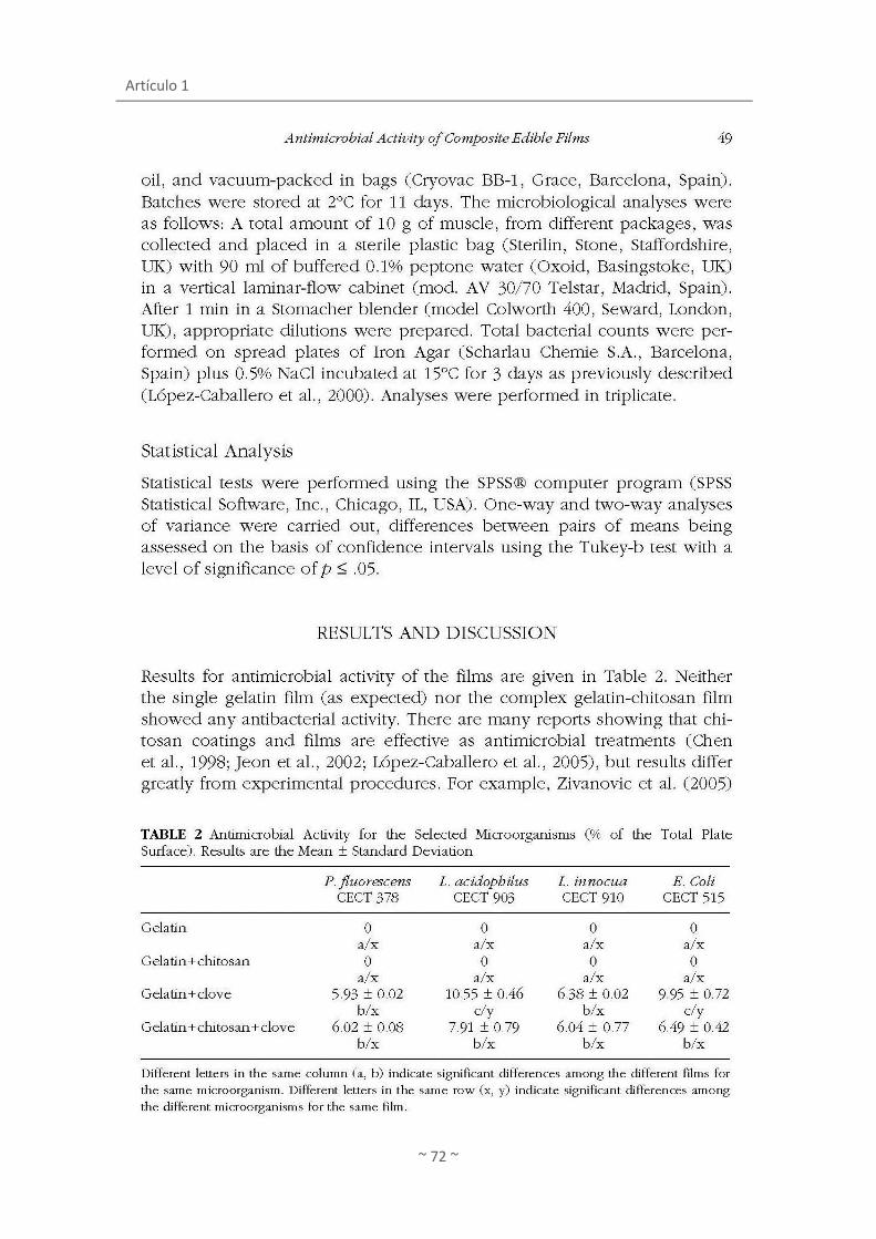
Artículo 1
~ 72 ~
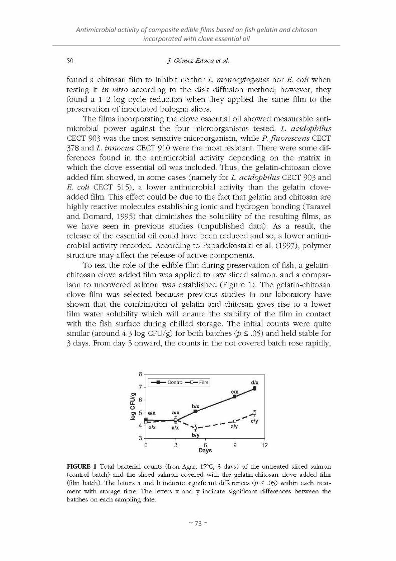
Antimicrobial activity of composite edible films based on fish gelatin and chitosan incorporated with clove essential oil
~ 73 ~
Artículo 1
~ 74 ~

Antimicrobial activity of composite edible films based on fish gelatin and chitosan incorporated with clove essential oil
~ 75 ~


~ 77 ~
ARTÍCULO 2
Gómez-Estaca, J., López de Lacey, A., López-Caballero, M.
E., Gómez-Guillén, M. C. y Montero, P. (2010)
Biodegradable gelatin-chitosan films incorporated with
essential oils as antimicrobial agents for fish preservation
Food Microbiology, 27(7), 889-896


Artículo 2
~ 79 ~
Artículo 2
~ 80 ~

Biodegradable gelatin-chitosan films incorporated with essential oils as antimicrobial agents for fish preservation
~ 81 ~
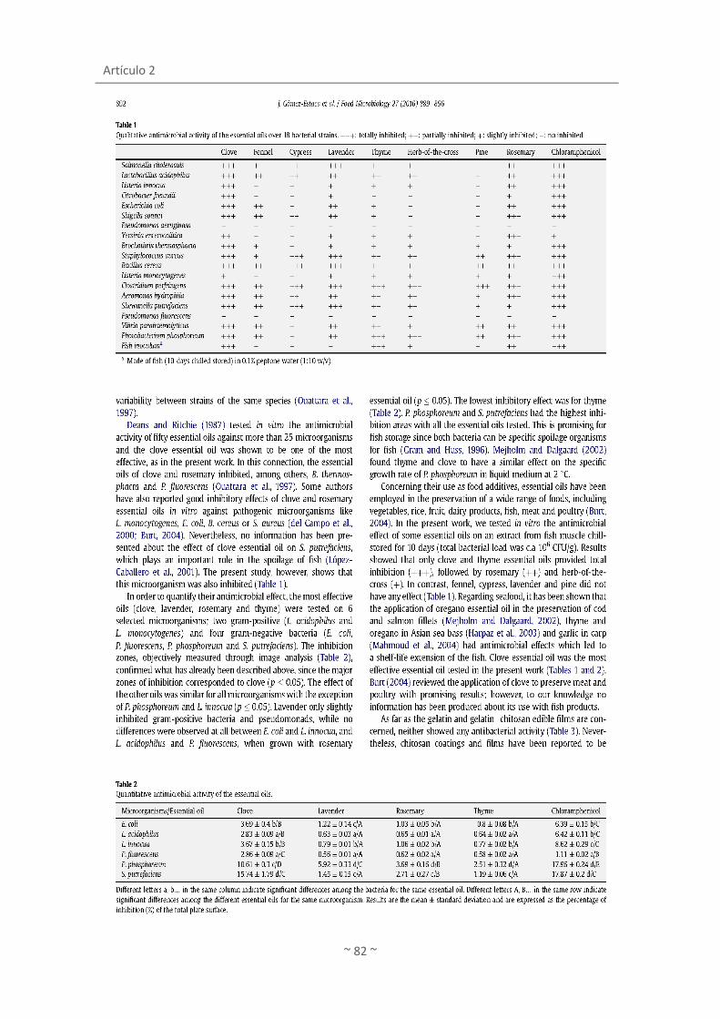
Artículo 2
~ 82 ~
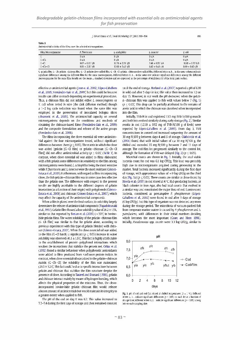
Biodegradable gelatin-chitosan films incorporated with essential oils as antimicrobial agents for fish preservation
~ 83 ~

Artículo 2
~ 84 ~
Biodegradable gelatin-chitosan films incorporated with essential oils as antimicrobial agents for fish preservation
~ 85 ~

Artículo 2
~ 86 ~

~ 87 ~
ARTÍCULO 3
López de Lacey, A.M., Pérez Santín, E., López-Caballero,
M. E. y Montero, P. (2012)
Antioxidant and antimicrobial activities of green tea
(Camellia sinensis L.) as an expression of its chemical
composition
LWT - Food Science and Technology


Artículo 3
~ 89 ~
ANTIOXIDANT AND ANTIMICROBIAL ACTIVITIES OF
GREEN TEA (Camellia sinensis L.) AS AN EXPRESSION OF
ITS CHEMICAL COMPOSITION
Ana M. López de Laceya, Efrén Pérez Santína, M. Elvira López-Caballeroa, *, Pilar Monteroa
a Institute of Food Science, Technology and Nutrition, (ICTAN), CSIC. (formerly Institute of
Refrigeration), C/ José Antonio Nováis, 10, 28040. Madrid, Spain.
*Corresponding author: E-mail address: [email protected]
Tel.: +34 91 5492300; fax: +34 91 5493627.

Artículo 3
~ 90 ~
ABSTRACT
The biological activity of different varieties of green tea (Camellia sinensis L.) aqueous
extracts, (Japan Sencha Makinohara, Japan Gyokuro Asahi, Cinnamon Green, Lung Ching, China
White Hair and Wu Lu Mountain) was evaluated in terms of their antioxidant and antimicrobial
properties and chemical composition. Principal components analysis revealed a positive
correlation between the content of polyphenols, amino acids and antioxidant activity, and a
negative correlation to microorganisms and minerals. The extracts with higher levels of total
catechin, flavonol and amino acids (Cinnamon Green and Wu Lu Mountain) also had higher
antioxidant activity. However, these varieties did not present marked difference compared to
others regarding antimicrobial activity. China White Hair, with the lowest total catechin and
amino acid content, medium flavonol levels, and low carbohydrate and mineral
concentrations, had the lowest antioxidant activity. The antimicrobial activity of this variety
against Vibrio parahaemolyticus and Pseudomonas fluorecens was superior to other varieties
of tea with higher polyphenol content. The results showed that aqueous extracts from
different varieties within green tea presented differences in their chemical composition, that
the bioactive properties of extracts were not related to each other and that it was the whole
set of components that defined the biological properties of tea extracts.
Key words: Green tea aqueous extracts, composition, polyphenols, antioxidant and
antimicrobial activity, pattern recognition.

Antioxidant and antimicrobial activities of green tea (Camellia sinensis L.) as an expression of its chemical composition
~ 91 ~
1. INTRODUCTION
Tea produced from the leaves and buds of the Camellia sinensis L. plant is the second
most consumed beverage in the world, well ahead of coffee, beer, wine and carbonated soft
drinks (Costa, Gouveia, & Nobrega, 2002). Depending on the manufacturing process, teas are
classified into several major types, with green tea being one of the non-fermented varieties.
Green tea has perhaps the most beneficial properties and its effects on health have been
widely described (Cabrera, Artacho, & Gimenez, 2006). The beneficial effects of green tea have
been attributed to the presence of polyphenols, pigments, amino acids, vitamins,
carbohydrates, minerals and purine alkaloids (Isiguki, Takakuwa, & Takeo, 1991; Weisburger &
Chung, 2002; Bolling, Chen, & Blumberg, 2009). The major green tea polyphenols are catechins
and their derivates, epicatechin (EC), epigallocatechin (EGC), epicatechin-3-gallate (ECG) and
epigallocatechin-3-gallate (EGCG) (Del Rio et al, 2004). Tea is also a source of flavonols such as
derivatives of quercetin and kaempferol (Finger, Engelhardt, & Wray, 1991; Del Rio et al,
2004). There are also other compounds present in tea which are related to its biological
properties. Thus, tea contains polysaccharides which have multi-functions such as
hypoglycemic and hypolypidemic effects, antioxidant activity, immune system stimulation
(Chen, Zhang, & Xie, 2005; Wang, Zhou, Li, Hou, Sun, & Wang, 2008), and could be a good
source of dietary minerals (Fernández, Pablos, Martín, & González, 2002). Other interesting tea
compounds are the amino acids that can be used as indicators of tea quality (Yao et al., 2006)
because of their contribution to taste and colour.
Although the extracts obtained from tea may be of different types, most of the green tea
phenolic compounds can be obtained by hot water (Lin, Chen, & Harnly, 2008) while
maintaining their biological properties (Hamilton-Miller, 1995; Dufresne & Farnworth, 2001;
Cabrera et al, 2006; Friedman, 2007). An additional advantage is the safety of the aqueous
extracts compared to those obtained with organic solvents; in addition, they could be
incorporated in food products without changing their sensory properties. In this regard, green
tea extract is already being used as an active compound in coatings used to preserve freshly
cut food (Chiu & Lai, 2010). In this way, the product acquires the benefits of green tea while at
the same time maintaining its attributes.
Although many studies have been published dealing with the composition of herbal teas
prepared for consumption, the results are not comparable because the raw materials and
extraction methods are different. Most of the studies establish differences in composition
Artículo 3
~ 92 ~
between different tea processes used (green, black, oolong, etc.), but to our knowledge there
are few studies that characterize varieties and compare them in terms of their composition
and bioactive potential. The aim of this work was to evaluate and compare the antioxidant and
antimicrobial activities of six varieties of green tea. To this end, multivariate analysis
established correlations between the different parameters studied and determined if the
differences in the chemical composition of the six varieties of green tea affected their
activities.
2. MATERIALS AND METHODS
2.1. Chemical
Minerals content: nitric acid and hydrogen peroxide (Panreac, Moncada i Reixac,
Barcelona, Spain). Determination of carbohydrates: phenol, 96% sulphuric acid, low-alkalinity
copper reagent, arsenomolybdate reagent, glucose (Sigma-Aldrich Chemie, Madrid, Spain).
Analysis of amino acids: HCl, phenol and norleucine (Sigma-Aldrich, St. Louis, Mo., USA).
Determination of total phenols: Folin-Ciocalteau reagent, gallic acid (Sigma-Aldrich, St. Louis,
Mo., USA). Standard polyphenol compounds: (-)-epigallocatechin (EGC, CAS: 970-74-1), (+)-
catequin (C, CAS:154-23-4), (-)-epicatequin (EC, CAS: 490-46-0), (-)-epicatechin-3-gallate (ECG,
CAS: 1257-08-5), (-)-epigallocatechin gallate (EGCG, CAS: 989-51-5), rutin (CAS: 153-18-4),
Hyperoside (CAS: 482-36-0), quercetin-3-O-glucoside (CAS: 482-35-9), kaempferol-3-O-
rutinoside (CAS: 17650-84-9), kaempferol-3-O-glucoside (CAS: 480-10-4) were purchased from
Extrasynthese (Genay, Cedex, France). HPLC-MS: HPLC grade acetonitrile and formic acid (VWR
international, Inc., Barcelona, Spain). HPLC grade water was prepared from distilled water
using a Milli-Q system (Millipore Laboratory Bedford, MA). Antimicrobial activity: glycerol
(Panreac, Moncada i Reixac, Barcelona, Spain). Antioxidant activity; FRAP: (2,4,6-tripyridyl-s-
triazine), FeCl3 and FeSO4 (Sigma-Aldrich, St. Louis, Mo., USA). ABTS: [2,2’-azino-bis-(3-
ethylbenzothiazoline-6-sulfonic acid)], potassium persulphate, Vitamin C (Sigma-Aldrich, St.
Louis, Mo., USA). Photochemiluminescence assay: ACW and ACL kit, reagent 2, reagent 3
(Photochem®, Analytik Jena AG). Ascorbic acid and trolox (Sigma-Aldrich, St. Louis, Mo., USA).
2.2. Extraction of green tea
Six green teas in loose leaf were purchased form a local specialized tea store (La Antigua
Plantación, Madrid, Spain) in spring 2010. Teas of different origin were chosen to be of great

Antioxidant and antimicrobial activities of green tea (Camellia sinensis L.) as an expression of its chemical composition
~ 93 ~
use. The varieties selected were: Japan Sencha Makinohara, Japan Gyokuro Asahi, Cinnamon
Green Tea, Lung Ching, China White Hair and Wu Lu Mountain (the first three varieties from
Japan and the last three from China). Dry green tea was ground into fine powder using a
blender (Osterizer, By/Par Sunbeam, mo. 4153-50, México). To prepare water extract, the
powder (35 g) was mixed with Milli-Q water (350 mL) at 80 °C for 30 minutes (Komes, Horžić,
Belščak, Ganić, & Vulić, 2010), with continuous stirring. The slurry mixture was centrifuged at
12000 g for 10 minutes at 5 °C. The supernatant was filtered two times through Whatman
filter papers Nº 1. Finally, the filtered extract was stored at -20 °C before the analysis.
2.3. Determination of phenol compounds
2.3.1. Determination of Total Phenolic
The total phenolic content in tea extracts was determined spectrophotometrically
according to a modified method of Slinkard and Singleton, (1977) with Folin-Ciocalteau´s
reagent. An aliquot of 20 µL of extract was mixed with 1.58 mL distilled water and oxidized
with 100 µL Folin-Ciocalteau reagent sample. The reaction was neutralized with 300 µL sodium
carbonate solution and incubated for 2 hours at room temperature. The absorbance of the
resulting blue colour was measured in a spectrophotometer at 765 nm. Using gallic acid as
standard total phenolic content was expressed as mg GA equivalent/mL of aqueous extract
tea. All determinations were performed at least in triplicate.
2.3.2. Quantification by HPLC-MS
Reverse phase high performance liquid chromatography was performed to analyse
phenolic compounds present in the aqueous green tea extract. The separation module
consisted of an Agilent 1100 series HPLC (Agilent Technologies, Waldbroon, Germany),
equipped with a C18 size column (250 x 4.6 mm, 5µm, C18 Tracer Excel ODS-A column,
Teknokroma) and a diode array detector (DAD). The samples were eluted with a gradient
system consisting of solvent A (deionized water) and solvent B (acetonitrile) both containing
1% formic acid, used as the mobile phase, with a flow rate of 1 mL/min.
The temperature of the column was maintained at 25 °C and the injection volume was 20
µL. The gradient system started at 90% solvent A and decreased to 74% A within 40 min,
followed by a decreased to 35% solvent A in 10 min. The final conditions were held for an
additional 5 min. The peaks of the phenolic compounds were monitored by atmospheric

Artículo 3
~ 94 ~
pressure electrospray ionization (ESI) source, operated in negative ion mode, with the
electrospray capillary voltage set to 3000 V, a nebulizing gas flow rate 12 L/h, and a drying
temperature of 350 °C. Mass spectrometry data were acquired in the scan mode (mass range
m/z 200-700) and in the sim mode (m/z 289, 305, 441, 441, 447, 457, 609).
The quantification was carried out using the external standard method. Solutions of each
standard at various concentration levels of 0.62, 1.25, 2.5, 5, 10 and 20 mg/L. were injected
into the column and the elution was performed in the same manner with the samples. Each
individual compound was quantified using a calibration curve of the corresponding standard
compound. All determinations were performed at least in triplicate.
2.4. Determination of carbohydrates
2.4.1. Determination of total carbohydrates (reducing and non-reducing sugars)
Carbohydrate content in the green tea extracts was determined by Dubois, Gilles,
Hamilton, Rebers, and Smith, (1956) with a slight modification. To an aliquot (10 µL) of sample,
500 µL of 4% aqueous solution of phenol was added, and then 2.5 mL of concentrated
sulphuric acid was directly added to the surface of the solution. The mixture was heated in a
water bath at 95 °C for 15 minutes. After heating, the samples were cooled-down in a water
bath at room temperature for 5 min protected from light. The absorbance was read at 490 nm
by means of an UV-1601 spectrophotometer (Model CPS-240, Shimadzu, Kyoto, Japan).
Colorimetric response was compared to a standard curve based on D-glucose. The
carbohydrate content was expressed as mg/mL of glucose. All determinations were performed
at least in triplicate.
2.4.2. Determination of reducing sugars. The determination for reducing sugars was
Carried out using the Nelson-Somogyi method, (Nelson, 1944). An aliquot (10 µL) of
sample, 1 mL low-alkalinity copper reagent was added. The final solution was heated in a bath
at 100 °C for 10 minutes. After incubation 1 mL of arsenomolybdate reagent was added, and
the solution was diluted to 5 mL with distilled water and was allowed to stand at least 15 min
at room temperature. The absorbance was read at 500 nm by means of an UV-1601
spectrophotometer (Model CPS-240, Shimadzu, Kyoto, Japan). Colorimetric response was
compared to a standard curve based on D-glucose, The carbohydrate content was expressed
as mg/mL of glucose. All determinations were performed at least in triplicate.

Antioxidant and antimicrobial activities of green tea (Camellia sinensis L.) as an expression of its chemical composition
~ 95 ~
2.5. Minerals content
The microwave digestion system (Microwave digestion LabStation, Milestone Inc.,
Shelton, USA) was used to digest sample in presence of nitric acid and hydrogen peroxide
(Panreac). Samples were made up to 100 mL with Milli-Q water. A Perkin-Elmer model 5100 Pc
atomic absorption spectrophotometer (Massachusetts, USA) with an air- and acetylene-
oxidizing flame was used to determine calcium (Ca), magnesium (Mg), sodium (Na), potassium
(K), zinc (Zn), manganese (Mn), iron (Fe) and copper (Cu) cations in these solutions. Results
were expressed in mg/100 mL of tea extract. All determinations were performed at least in
triplicate.
2.6. Amino acids
An amount of 50 μL of sample was dried and hydrolysed in vacuum-sealed glass tubes at
110 °C for 24 h in the presence of constant boiling 6 mol/L HCl containing 0.1% phenol and
using norleucine (Sigma-Aldrich, Inc., St. Louis, Mo., USA) as the internal standard. After
hydrolysis, samples were again vacuum-dried, dissolved in application buffer and injected into
a Biochrom 20 amino acid analyser (Pharmacia, Barcelona, Spain). Theanine and serine were
eluted at the same retention time, therefore their values are shown together.
2.7. Antioxidant activity
2.7.1. FRAP
The ferric reducing/antioxidant power (FRAP) assay was carried out according to the
method described by Benzie and Strain, (1996) with some modification. This assay measures
the change in absorbance at 595 nm owing to the formation of a blue colored FeII-
tripyridyltriazine compound from the colourless oxidized FeIII form by the action of electron
donating antioxidants (Benzie, Chung, & Strain, 1999). An amount of 60 μL of sample was
incubated (37 °C) with 60 μL of Milli-Q water and 900 μL of FRAP reagent, containing 10
mmol/L of TPTZ (2,4,6-tripyridyl-s-triazine) and 20 mmol/L of FeCl3. Absorbance values were
read at 595 nm after 30 min, using an UV-1601 spectrophotometer (Model CPS-240, Shimadzu,
Japan). Results were expressed as μmol Fe2+ equivalents/g of sample based on a standard
curve of FeSO4·7H2O. All determinations were performed at least in triplicate.

Artículo 3
~ 96 ~
2.7.2. ABTS
The ABTS radical [2,2’-azino-bis-(3-ethylbenzothiazoline-6-sulfonic acid)] scavenging
capacity was carried out according to the method of Re, Pellegrini, Proteggente, Pannala, Yang,
and Rice-Evans (1999). The amount of ABTS radical cation scavenged by antioxidants is
measured by monitoring the decrease of absorbance of ABTS radical cation, and compared
with the decrease of absorbance produced by the addition of known amount of vitamin C. The
stock solution of ABTS radical consisted of 7 mmol/L ABTS in potassium persulphate 2.45
mmol/L, kept in the dark at room temperature for 16 h. An aliquot of stock solution was
diluted with Milli-Q water in order to prepare the working solution of ABTS radical with
absorbance at 734 nm of 0.70 ± 0.02. A 20 µL aliquot of sample was mixed with 980 μL of ABTS
reagent. The mixture was then left to stand in the dark at 30 °C for 10 min and absorbance
values were read at 734 nm using an UV-1601 spectrophotometer (Model CPS-240, Shimadzu,
Japan). Results were expressed as mg Vitamin C Equivalent Antioxidant Capacity (VCEAC)/g of
sample based on a standard curve of Vitamin C. All determinations were performed at least in
triplicate.
2.7.3. Photochemiluminescence assay
This assay (PCL) involves the photochemical generation of superoxide (O2-) free radical
combined with chemiluminescence detection of luminol, which acts as a photosensitizer as
well as an oxygen radical detection reagent. This reaction takes place in the PHOTOCHEM®
(Analytik Jena AG). The ACW and ACL kits provided by the manufacturer were used to measure
hydrophilic and lipophilic antioxidant capacity, respectively (Popov & Lewin, 1999). The ACW
kit, 1.5 mL reagent 1 (water), 1 mL reagent 2 (buffer solution pH 10.5), 25 mL reagent 3
(luminol), and 10-30 mL standard solution or appropriately diluted antioxidant samples were
mixed and measured. The ACL kit, 2.3 mL reagent 1 (methanol), 200 mL reagent 2 buffer
solution), 25 mL reagent 3 (luminol), and 10-30 mL standard solution or appropriately diluted
antioxidant samples were mixed and measured. These are standardized conditions. All samples
were appropriately dissolved and diluted in reagent 1 of the ACL and ACW kits before analysis
in each measurement. Ascorbic acid and trolox were used as the calibration reagents for ACW
and ACL, respectively, at measuring concentration ranges of 1-3 nmol. All samples were
measured at least in duplicate. The percentage relative standard deviation was less than 10%.
The antioxidant capacity was determined by means of the lag phase (L) in seconds, L = L0 -
L1 (where L0 and L1 are the respective parameters of the blank and sample), or by means of the

Antioxidant and antimicrobial activities of green tea (Camellia sinensis L.) as an expression of its chemical composition
~ 97 ~
degree of PCL inhibition (I), I = 1 - S/S0 (where S0 is the integral under the blank curve and S is
the integral under the sample curve), for the ACW or ACL values, respectively. The results were
expressed in the nanomolar range and calculated to micromoles per 100 mL ascorbic acid or
trolox equivalents (µmol TE or µmol AAE/100 mL).
2.8. Antimicrobial activity
2.8.1. Bacterial strains and growth conditions.
The antimicrobial activity of the green teas was tested over the following bacterial strains
obtained from the Spanish Type Culture Collection (CECT): Lactobacillus acidophilus CETC 903,
Salmonella choleraesuis CECT 4300, Listeria innocua CECT 910, Citrobacter freundii CECT 401,
Escherichia coli CECT 515, Shigella sonnei CECT 4887, Pseudomonas aeruginosa CECT 110,
Yersinia enterocolitica CECT 4315, Brochothrix thermosphacta CECT 847, Staphylococcus
aureus CECT 240, Bacillus cereus CECT 148, Listeria monocytogenes CECT 4032, Aeromonas
hydrophila CECT 839T, Photobacterium phosphoreum CECT 4192, Shewanella putrefaciens
CECT 5346T, Pseudomonas fluorescens CECT 4898, Vibrio parahaemolyticus CECT 511T,
Lactobacillus helveticus DSM 20075, Bifidobacterium bifidum DSMZ 20215 and Bifidobacterium
animalis subspecies lactis DSMZ 10140. The strains were stored at -80 °C in Brain Heart
Infusion Broth (Oxoid, Basingstoke, UK) with 25% glycerol (Panreac, Moncada i Reixac,
Barcelona, Spain) until use. All the strains were grown in BHI broth (Oxoid) (supplemented with
3% NaCl for V. parahaemolyticus, 1% NaCl for P. phosphoreum and S. putrefaciens) excepting L.
acidophilus, L. helveticus, B. bifidum and B. animalis were grown in 0.05% cysteine MRS broth.
Organisms were incubated at 37 °C excepting Bacillus cereus, Y. enterocolitica, A. hydrophila, P.
fluorecens, S. putrefaciens, incubated at 30 °C; B. thermosphacta at 25 °C and P. phosphoreum
at 15 °C. In addition, L. acidophilus was incubated under CO2 flow and B. bifidum and B.
animalis under anaerobic conditions (Gas-Pack, Anaerogen; Oxoid).
2.8.2. Quantitative antimicrobial activity
The antimicrobial activity of the six green teas was tested over twenty selected
microorganisms. Spread plates of medium were inoculated with 100 µL of overnight grown
bacterial suspensions (105 -106 CFU/mL) overnight grown. Filter paper disks (0.5 cm diameter),
soaked with 50 μL of each green tea infusion were put on the plate surface. The time and
temperature of incubation was the same as previously described above. After incubation the
inhibition area (mm) was measured using specific software for digital image analysis (MIP 4

Artículo 3
~ 98 ~
ADV, ver. 1. Consulting de Imagen Digital, S.L. & Microm España). Results are expressed as the
percentage of inhibition respecting to the total plate surface. A minimum of three replicates
disks was used for each green tea extract.
2.8.3. Minimal inhibitory concentration determination
The minimal inhibitory concentration determination of the six green teas was performed
over six selected microorganisms: Vibrio parahaemolyticus, Pseudomonas fluorescens,
Aeromonas hydrophila, Shewanella putrefaciens and Photobacterium phosphoreum. Six
different dilutions of each aqueous green tea infusion were tested: 10, 20, 40, 60, 80 and 100%
(v/v). Spread plates of medium were inoculated with 100 µL of bacterial suspensions (105-106
CFU/mL) and then, a 50 µL drop of each aqueous extract was placed on the plate. All the plates
were incubated at the same time and temperature as described above. The minimal inhibitory
concentration was defined as the lowest concentration which produced inhibition zone of
growth.
2.9. Statistical analyses.
All analyses were performed at least in triplicate and a completely randomized design was
used. Analysis of variance (ANOVA) was performed and mean comparisons were done by
Duncan´s multiple range tests. Furthermore, a multivariate analysis was performed on all data
values obtained from the different analyses, with the chemical composition (phenolics,
carbohydrates, minerals, amino acids) antioxidant activity (FRAP, ABTS, PCL assay) and
antimicrobial activity as a whole set. Factorial analysis by principal components analysis (PCA)
was done. Correlation among properties was also calculated. The level of significance setting
was P ≤ 0.05. All descriptive statistic and statistical tests were performed using the SPSS pack
(SPSS 19.0 for Windows, SPSS Inc, Chicago, IL, USA).
3. RESULTS AND DISCUSSION
The total phenolic content of water extract from green tea is shown in Table 1. All tested
teas are rich in phenolic compounds, although some differences could be observed among the
varieties, depending on the source. The highest total polyphenol content was detected in
Cinnamon Green Tea with 9770 mg/L equivalents of gallic acid, while the lowest concentration
corresponded to China White Hair with 4712 mg/L equivalents of gallic acid.

Antioxidant and antimicrobial activities of green tea (Camellia sinensis L.) as an expression of its chemical composition
~ 99 ~
In the present work, six water extracts of green tea were subjected to identification and
quantification. The most abundant bioactive constituents of green tea were catechins (flavan-
3-ols) followed by flavonols, although in much lower quantities (Table 1). Consequently, the
chromatographic determination of the polyphenolic profile shows 5 major peaks of catechin
compounds: (-)-epigallocatechin, (+)-catechin, (-)-epicatechin, (-)-epicatechin-3-gallate, (-)-
epigallocatechin gallate) and five flavonols: rutin, hyperoside, quercetin-3-O-glucoside,
kaempferol-3-O-rutinoside and kaempferol-3-O-glucoside. These compounds can constitute
16-30% of the dry weight of fresh leaf tea (Graham, 1992).
Total catechins ranged from 5.36 g/L in China White Hair to 11.27 g/L in Wu Lu Mountain.
Catechins are colourless and water soluble, and contribute to the bitterness and astringency of
green tea infusions (Del Rio et al, 2004). The prevailing phenolic compound in all green teas
was (-)-epigallocatechin gallate, registering 6.82 g/L in Wu Lu Mountain tea (Table 1). The
lowest concentration corresponded to catechin, which barely reached 47.5 mg/L in the same
variety of tea, indicating that the extract had not been degraded (Ito, Yamamoto, Kodama,
Kato, Yoshimura, Matsunaga, & Nakazawa, 2003). These results are consistent with other
reports dealing with several varieties of green tea (Graham 1992, Lin et al 2008), while in other
types of tea, for example in black tea, epigallocatechin gallate and catechin were found in
smaller amounts. Reto, Figueira, Filipe, and Almeida (2007) found that in 9 varieties of green
tea the total minority compounds from catechin are CE and C, with EGCG and EGC, in similar
quantities, being the most abundant. Some published data give higher values for EGCG (even
115 mg/g), in methanol extracts of fresh tea shoots (Yao et al, 2004).
The flavonol compounds (in much lower concentrations) ranged from 84.7 mg/L in Lung
Ching tea to 237.6 mg/L in Cinnamon Green tea. The major flavonols were quercetin and
kaempferol, together with their glycosylated forms, although in some cases, myricetin and its
glycosides were also detected in green tea (Graham, 1992; Del Rio et al, 2004). In the 6
varieties of tea selected, no predominance of any one of the flavonols is clearly seen, although
there are variations of the same compound between teas. Thus, the kaempferol-3-O-
rutinoside varies from 10 to 67 mg/L for Lung Ching and Cinnamon Green teas, respectively
(Table 1). Contrary to the total catechins, the flavonol content of Wu Lu Mountain tea was not
significant (p ≤ 0.05).
Aqueous extracts of tea make it possible to obtain infusions with high amounts of
bioactive compounds and a minor degradation of the phenolic compounds (Perva-Uzunalić,
Skerget, Knez, Weinreich, Otto, & Grüner, 2006; Komes et al, 2010). The aforementioned
authors showed that catechins tend to degrade during prolonged extraction, and after

Artículo 3
~ 100 ~
applying high extraction temperatures. The catechin and flavonol levels referred to in the
present work were obtained with aqueous extracts at 80 °C/30 min (Table 1). The conditions of
extraction used in this study were similar to that reported by Sharma, Gulati, and
Ravindranath, (2005), who observed that catechins (especially EGCG, EGC and EC) showed
marked differences when extracted at different temperatures and that the best extraction
results were achieved at 90 °C for 80 min. The total phenolic content of various green tea
aqueous extracts obtained by heating was as much as 20% of dry weight (Lin et al 2008). These
authors confirmed that under these conditions, the extracts contained most of the tea
polyphenolics with the exception of flavonol acylate glucosides. In a review on green tea,
Peterson et al (2005) observed catechin values of only 13.6% of dry weight.
Varieties of Japan Sencha Makinohara and Cinnamon Green teas had very similar values
for total carbohydrates in mg/mL (Table 2), whereas the other extracts registered significant
differences (p ≤ 0.05) between them. The carbohydrate content does not seem to be related
to any specific components or to the origin of the tea. As no information was found dealing
with these varieties, it has been difficult to make any comparisons.
There were higher values for the amount of reducing sugars present in the extracts than
for the total carbohydrate content (Table 2).This could be due to the determination method
used, since in the Somogyi-Nelson assay, all substances with reducing or oxidative properties
might cause interference (Paleg, 1959). The results in the present study show that tea extracts
with higher amounts of reducing sugars (Cinnamon Green Tea and Wu Lu Mountain) also had
more polyphenols (Table 1 and 2). By their nature, polyphenols may affect the determination
of reducing sugars by the interference technique mentioned above.
Table 3 shows the mineral content of the green teas studied. The highest mineral
concentration found for all the samples was K, followed by Mg and Mn; other minerals
occurred in much lower amounts, becoming just traces in the case of Cu and Fe. These results
are consistent with other studies (Fernández et al, 2002; Gallaher, Gallaher, Marshall, &
Marshall, 2006; Musa Özcan, Ünver, Uçar, & Arslan, 2008; Reto et al, 2007), where K is
reported to be the major mineral in green tea infusions. However, Fernandez-Cáceres, Martín,
and González (2001) found that the major minerals in both green and black tea are Ca, Mg and
Al in tea, but with variations depending on the geographical location of the tea. Gallaher et al,
(2006) and Reto et al, (2007) studied the mineral extraction percentage from dry teas of
different herbs, including green tea, and found that K was particularly soluble, with excellent
extraction rates while Fe had the worst extraction rate and was the least soluble mineral. This
may explain the considerable difference found between K and Fe levels.

Antioxidant and antimicrobial activities of green tea (Camellia sinensis L.) as an expression of its chemical composition
~ 101 ~
As can be seen from Table 3, mineral concentration was different depending on the
variety of tea (p ≤ 0.05). Musa et al (2008) stated that harvesting times, soil properties and
climate could explain the differences in the mineral composition of herbal tea infusions.
Fernández et al (2002) studied the mineral composition of different types of green teas,
including Sencha, one of the varieties in the present study. Although no information has been
found concerning the mineral contents of the specific green tea infusions used in the present
work, Fernández et al (2002) obtained lower values for K, Mg, Mn and Zn in green tea
infusions, but with similar Na and Ca (Table 3). The levels of K, Mg, Mn and Zn reported by
Gallaher et al (2006) and Reto et al (2007) in green tea infusions (1.5% extract) were also lower
although the Na content was higher. The Mg, Mn and Zn concentrations were similar to those
reported by Musa Özcan et al (2008) in green tea infusions (1.5% extract), however K and Fe
were higher in this study and they did not detect Ca. It appears that the aqueous extracts in
this study should have a higher concentration of minerals, because, compared to other studies,
a higher percentage of dry tea was used (the more dilute infusions of many studies were
between 1.5 and 2%). It seems that in some cases the proportion of dry tea does not affect
mineral concentration, which could be because large quantities of dry tea and low volumes of
water do not permit a good release, although this effect was not found when comparing this
study with the literature. Furthermore, the different preparations of tea extract could affect
the mineral composition (Fernández et al, 2002), or according to Gallaher et al (2006), the
relationship of pH to mineral solubility might explain the variability in extraction rates.
The most abundant amino acids in the teas studied included glutamic acid (Glu), aspartic
acid (Asp), serine (Ser) and arginine (Arg), (Table 4). There were quantitative differences
among the varieties of tea studied but no qualitative variations (p ≤ 0.05). The predominance
of some amino acids varies from one publication to another and could be attributed to
differences between varieties and types of tea extracts (some proteins are more easily
extracted than others in some cases or have different amounts of free amino acids). In this
sense, Graham (1992) indicated that green tea leaves contain about 4% of free amino acids
and 15% protein. Wang and Ruan (2009), who carried out the analysis of amino acids in green
tea infusions (Longjing teas), found that theanine (Thea) was the predominant free amino acid
followed by glutamic acid (Glu), proline (Pro), aspartic acid (Asp), serine (Ser), arginine (Arg)
and histidine (His). The mentioned amino acids were among the majority in the present study
(Table 4). No information has been found regarding the amino acid contents of the specific
green tea infusions used in this study.

Artículo 3
~ 102 ~
Some authors have reported that free amino acids can be good chemical descriptors to
differentiate green, black and oolong tea infusions, and the composition of the other minor
amino acids can be useful as well, for differentiating kinds of tea or for determining tea quality
(Wang et al, 2010). These authors found that Asp, Glu and Ser levels were more abundant in
green teas than in the other teas tested. The amino acid content was determined in green,
oolong, black and Pu-erh tea water extracts and most of the green teas had theanine, serine
and valine as majority components (Syu, Lin, Huang, & Lin, 2008), while theanine, glutamic acid
and aspartic acid were the most abundant amino acids in all the teas studied (Alcázar et al,
2007).
It is important to note that some amino acids can interact with other compounds in tea
such as polyphenols. The formation of covalent bonds has been described between SH groups
of cysteine and EGCG (Ishii, Mori, Tanaka, Mizuno, Yamaji, & Kumazawa, 2008; Chen, Wang,
Zhang, Ren, & Zeng, 2011). Other amino acids can cause weaker interactions with polyphenols,
either by hydrogen bonds or electrostatic interactions. Pro has a strong affinity for the
hydroxyl groups of catechins (Pocent-Legrand, Edelman, Putaux, Cartalade, Sarni-Manchado, &
Vernhet, 2006), and these authors suggest that frequent consumption of tea with whole milk
can alter the activity of the infusion alone. In addition, saliva proteins, also rich in proline, can
establish interactions with the tea infusion and alter their biological properties. It has been
reported that the interaction of poly(L-pro) with EGCG, ECG and C is immediate and its stability
depends on the relationship between protein and polyphenol, while there are no interactions
with EC and EGC (Poncet-Legrand et al, 2006). These interactions could change the potentially
bioactive polyphenols contained in tea (Arts et al, 2002). Nevertheless, these studies are
performed in model systems, but not in a real system like food where many other complex
conditions may occur. Other authors observed that certain proteins (for example, bovine
gelatine with a high degree of aggregation), interacts less with polyphenols than with gelatine
with lower aggregation (for example from fish) that has more active groups available for
interaction (Gómez-Estaca, López de Lacey, López-Caballero, Gómez-Guillén, & Montero,
2010).
Antioxidant activity of green tea, FRAP, ABTS and PCL assays are shown in Table 5. Wu Lu
Mountain green tea has the highest antioxidant capacity (FRAP 188.48 mmol/L Fe2+, ABTS
15.98 mg/mL equiv. Vit C), while China White Hair green tea shows the lowest (FRAP 88.37
mmol/L Fe2+, ABTS 7.20 mg/mL equiv. Vit C and PCL 3.9 mg/mL equiv. Vit C). Cinnamon Green
Tea showed the highest antioxidant activity determined by Photochem (19.0 mg/mL equiv. Vit
C).

Antioxidant and antimicrobial activities of green tea (Camellia sinensis L.) as an expression of its chemical composition
~ 103 ~
The degree of antioxidant capacity in green tea extracts is in accordance with the total
polyphenol content determined by the Folin-Ciocalteu method, and confirmed by a high linear
correlation obtained between the results (rFRAP/tpc = 0,983, rABTS/tpc = 0,926, rPCL/tpc = 0,926),
which implies that the antioxidant potential of green tea is directly related to phenolic content.
Toschi, Bordoni, Hrelia, Bendini, Lercker, and Biagi (2000) observed that the antioxidant
activity of green tea in a lipid model system is higher in teas that contain larger quantities of
EGCG and EGC. This is consistent with the present results, since the variety of tea with the
most antioxidant activity; Wu Lu Mountain also possessed the most Epigallocatechin gallate
and Epigallocatechin (Table 1). According to previous findings by Rice-Evans, Miller, and
Paganga (1996) who studied the antioxidant activity of the polyphenolic constituents of green
tea in relation to their relative compositions, the contribution to antioxidant effectiveness in
green tea was in the following order: EGC ≈ EGCG >> ECG ≈ EC > C. The scavenging abilities of
EGCG and ECG are stronger even if the steric hindrance of EGC, GC, EC and (+)-C is smaller than
that of EGCG and GCG, indicating that the presence of a gallate group at the 3 position plays
the most important role in their abilities to scavenge free radicals (Guo, Zhao, Shen, Hou, Hu,
& Xin, 1999).
According to Pinelo, Manzocco, Nunez, and Nicoli, (2004), the increase in antioxidant
capacity could be explained by the strong tendency of polyphenols to undergo polymerization
reactions. When the degree of polymerization exceeds a critical value, increased molecular
complexity and steric hindrance reduce the availability of hydroxyl groups in reactions with
radicals, which causes a decrease in antiradical capacity. On the other hand, the interaction of
polyphenols with proteins has been widely described and may result in decreased antioxidant
potential. Arts et al (2002) indicated that the antioxidant activity of some components of green
tea (and other teas) with casein and albumin is not additive, and a part of the total activity is
masked due to interactions. The authors pointed out that the casein interaction (β, α and κ)
showed a range of masking of antioxidant activity directly proportional to proline content.
The antimicrobial activity of the six green teas was tested on twenty microorganisms as
described in the M&M section. In five cases the extracts were found to be antimicrobially
active (Table 6). Some authors reported that aqueous extracts of green tea (varieties other
than those studied) had antimicrobial activity against Gram-positive and Gram-negative
bacteria (Almajano, Carbó, Jiménez, & Gordon, 2008; Singh Arora, Jeet Kaur, & Kaur, 2009).
P. phosphoreum had the highest inhibition zone diameter in the presence of all tea
extracts tested, and most of the time, S. putrefaciens was the second most sensitive strain in
the presence of green tea. In contrast A. hydrophila, V.parahaemolyticus and P.fluorescens

Artículo 3
~ 104 ~
were the least sensitive to the effects of Japan Sencha Makinohara, Japan Gyokuro Asahi and
Cinnamon Green teas. The results demonstrated that the infusions tested are more effective
against some specific spoilage organisms of fish (López-Caballero et al, 2005). This fact is
promising as tea extracts could help to increase the shelf life of fish. Microorganism sensitivity
to polyphenols (Taguri, Tanaka, & Kouno, 2004) and tea extracts (Chou, Lin, & Chung, 1999)
varies between species.
In general, Japan Sencha Makinohara was shown to be one of the most effective in terms
of antimicrobial activity and Wu Lu Mountain the least. Apart from polyphenol antimicrobial
activity (Arakawa, Maeda, Okubo, & Shimamura, 2004; Friedman, 2007), all of the other tea
components also play a part. The chemical composition of the teas used in this study could
help to explain this activity (Tables 1-5), differentiating not only between the different types of
tea (green, black, etc.) but also between varieties of the same green tea. Among the six
varieties studied, Wu Lu Mountain has a high concentration of flavanols, which translates into
more antioxidant activity but not into greater antimicrobial activity (Tables 5-6). It may be that
the differences in their chemical composition (different content of flavonols, carbohydrates
and minerals in Japan Sencha Makinohara in comparison with Wu Lu Mountain), for example
might influence their activity, since extracts are comparable because they are made in the
same way. Moreover, as mentioned previously, protein-polyphenol interactions may modify
the biological activity of the latter. In this regard, the antimicrobial effect of polyphenols (from
green tea infusions) in the presence of whey proteins increased with the reduction of whey
protein concentration (von Staszweski, Pilosof, & Jagus, 2011). These authors found that the
effect of green tea polyphenols is concentration dependant and that the food matrix can
decrease the antimicrobial performance of these compounds. In-depth studies are needed to
clarify these issues.
On the other hand, it is important to note that green teas were inactive against the
Lactobacillus and Bifidobacterium strains studied. These data results are interesting because
some extracts rich in polyphenols have antimicrobial activity against a large number of strains,
including those strains beneficial for the intestine. This fact has been reported by Almajano et
al, (2008) because Lactobacillus acidophilus offers exceptional resistance to green tea
infusions.
Table 7 shows the minimal inhibitory concentration. The data confirm that P.
phosphoreum and S. putrefaciens were more susceptible to the tea infusions because low tea
concentrations could inhibit their growth. In contrast, higher concentrations of teas were
necessary in some cases for the inhibition of P. fluorescens, A. hydrophyla and V.

Antioxidant and antimicrobial activities of green tea (Camellia sinensis L.) as an expression of its chemical composition
~ 105 ~
parahaemolitycus (Table 7). The activity of phenolic compounds, corresponding to their
chemical structure, appears to be associated with damage to the cytoplasmic membrane
(Puupponen-Pimiä, Nohynek, Alakomi, & Oksman-Caldentey, 2005), among other factors, by
reducing the pH gradient, thus acting as permeabilisers by inhibiting extracellular microbial
enzymes, etc. In this respect, Arakawa et al. (2004) suggested that the bactericidal effect
resulted from hydrogen peroxide levels produced by epigallocatechin gallate when reacting
with the dissolved oxygen in aqueous solutions. No antimicrobial activity was detected in any
commercial green tea extract against fish spoilage bacteria mentioned above (Corbo, Di Giulio,
Conte, Speranza, Sinigaglia, & Del Nobile, 2009).
In order to compare the activity of the six varieties of green tea and to establish the
differences between them, an analysis of principal components (PC) was carried out. Given the
large number of variables and their overlap in the graphical representation of the new
components, the variables were placed in three groups (flavonoids, amino acids and minerals),
keeping all other variables constant.
Multivariate group analysis with flavonoids (including all variables except amino acids and
minerals), showed two components explaining 38.4% and 30.9% of the variance, respectively.
Total catechins, ECG and EGCG were grouped in the first component, while C, EC, EGC was
weight distributed between the first and second components. Flavonols were clustered more
heavily in the second dimension, except for hyperoside that was in the first. There are
negatively correlated microorganisms, distributing their weight in both dimensions, except for
P. fluorescens with most weight in PC 1. Measurements of antioxidant activity are grouped
together and maintain a strong relationship with total catechins, but not flavonols.
The green teas under study were clearly differentiated by these variables distributed
according to two dimensions. Tea 3 (Cinnamon Green) was associated with a higher content of
flavonols, tea 5 (China White Hair) was the most active against P. fluorescens, tea 2 (Japan
Gyokuro Asahi) was very active for the microorganisms near the graphical representation (e.g.
V. parahaemolyticus) and tea 6 (Wu Lu Mountain) was characterized by a higher content of
EGCG and total catechins.
Multivariate group analysis including all variables except the individual flavonoids and
minerals showed that the first two components explain 77.2% and 9.5% respectively of the
variance. Most amino acids are explained in the first component, producing a strong positive
correlation with the total content of catechins and negative microorganisms. The flavonols
have greater weight in the second component, which also shows a correlation with certain

Artículo 3
~ 106 ~
microorganisms such as S. putrefaciens. The antioxidant activities were positively correlated in
both components, with high weight in PC1. Again, the teas are fully differentiated. Tea 3
(Cinnamon Green Tea) and 6 (Wu Lu Mountain) have higher levels of amino acids and
polyphenols, both catechins and flavonols and higher antioxidant activity. This suggests that
either antioxidant activity is not diminished by the presence of amino acids, or that there
might not be any interaction between both compounds.
PC of the variables tested, except for individual flavonoids and amino acids showed that
the first two components explain 39.8% and 30.2% of the variance, respectively. Catechin
content and antioxidant activity are strongly and positively correlated in PC 1 but negatively
correlated with minerals. Minerals in general are correlated most positively for the PC2, along
with most of the organisms, indicating that minerals can promote in some way the inhibition
of microorganisms, eg. S. putrefaciens in the presence of Ca and Na. Teas are also completely
different in the plot, with Cinnamon Green tea (tea 3) and Wu Lu Mountain (tea 6), registering
the lowest mineral content.
4. CONCLUSIONS
In summary, the comparison ratios of the six selected kinds of green tea revealed that
varieties differed in terms of chemical composition and properties, and the bioactive
properties of extracts were not related to each other. Therefore the biological properties of
the teas can be defined as the combination of all these individual components working
together in unison. The teas with higher antioxidant activity, Cinnamon Green tea and Wu Lu
Mountain, are characterized by higher total catechin, flavonol and amino acid content and
lower mineral and medium carbohydrate content. However, with regard to antimicrobial
activity, there was no marked difference between any of the varieties. The lowest antioxidant
activity corresponded to the China White Hair variety, which was also characterized by the
lowest total catechin and aminoacid content, medium flavonol and low mineral and
carbohydrate content. Nevertheless, China White Hair had a higher inhibition capacity against
some microorganisms than other varieties of tea with higher levels of polyphenols. Japan
Makinohara Sencha exhibited the greatest antimicrobial activity, whereas little differentiation
of antimicrobial activity was observed for the other varieties. The fact that some spoilage
organisms were sensitive to green tea was a promising development in terms of food
preservation, and future research on how to confer stability to food by the incorporation of
such tea extracts might prove beneficial.

Antioxidant and antimicrobial activities of green tea (Camellia sinensis L.) as an expression of its chemical composition
~ 107 ~
Acknowledgements
Funding for this research was provided by Ministerio de Ciencia e Innovación under project
AGL2008-00231/ALI. Programs JAE-CSIC predoctoral and Ángeles Alvariño, Xunta de Galicia
(European Social Fund), are fully acknowledged.
ICTAN-CSIC has implemented and maintains a Quality Management System which fulfils the
requirements of the ISO standard 9001:2000.
References
Alcázar, A., Ballesteros, O., Jurado, J. M., Pablos, F., Martín, M. J., Vilches, J. L., & Navalón, A.
(2007). Differentiation of green, white, black, Oolong, and Pu-erh teas according to
their free amino acids content. Journal of Agricultural and Food Chemistry, 55(15),
5960-5965.
Almajano, M. P., Carbó, R., Jiménez, J. A. L., & Gordon, M. H. (2008). Antioxidant and
antimicrobial activities of tea infusions. Food Chemistry, 108(1), 55-63.
Arakawa, H., Maeda, M., Okubo, S., & Shimamura, T. (2004). Role of hydrogen peroxide in
bactericidal action of catechin. Biological and Pharmaceutical Bulletin, 27(3), 277-281.
Arts, M. J., Haenen, G. R., Wilms, L. C., Beetstra, S. A., Heijnen, C. G., Voss, H. P., & Bast, A.
(2002). Interactions between flavonoids and proteins: effect on the total antioxidant
capacity. Journal of Agriculture and Food Chemistry, 50(5), 1184-1187.
Benzie, I. F. F., Chung, W. Y., & Strain, J. J. (1999). "Antioxidant" (reducing) efficiency of
ascorbate in plasma is not affected by concentration. J. Nutr. Biochem., 10, 146-150.
Benzie, I. F. F., & Strain, J. J. (1996). The Ferric Reducing Ability of Plasma (FRAP) as a Measure
of "Antioxidant Power": The FRAP Assay. Analytical Biochemistry, 239(1), 70-76.
Bolling, B. W., Chen, C. Y. O., & Blumberg, J. B. (2009). Tea and health: Preventive and
therapeutic usefulness in the elderly? Current Opinion in Clinical Nutrition and
Metabolic Care, 12(1), 42-48.
Cabrera, C., Artacho, R., & Giménez, R. (2006). Beneficial effects of green tea - A review.
Journal of the American College of Nutrition, 25(2), 79-99.
Corbo, M. R., Di Giulio, S., Conte, A., Speranza, B., Sinigaglia, M., & Del Nobile, M. A. (2009).
Thymol and modified atmosphere packaging to control microbiological spoilage in
packed fresh cod hamburgers. International Journal of Food Science and Technology,
44(8), 1553-1560.

Artículo 3
~ 108 ~
Costa, L. M., Gouveia, S. T., & Nobrega, J. A. (2002). Comparison of heating extraction
procedures for Al, Ca, Mg, and Mn in tea samples. Anal. Sci., 18(3), 313-318.
Chen, H., Zhang, M., & Xie, B. (2005). Components and antioxidant activity of polysaccharide
conjugate from green tea. Food Chemistry, 90(1-2), 17-21.
Chen, R., Wang, J. B., Zhang, X. Q., Ren, J., Zeng, C. M. (2011). Green tea polyphenol
epigallocatechin-3-gallate (ECGC) induced intermolecular cross-linking of membrane
proteins. Archivess of Biochemistry and Biophysics, 507(2), 343-349.
Chiu, P. E., & Lai, L. S. (2010). Antimicrobial activities of tapioca starch/decolorized hsian-tsao
leaf gum coatings containing green tea extracts in fruti-based salads, romaine hearts and
pork slices. International Journal of Food Microbiology, 139(1-2), 23-30.
Chou, C. C., Lin, L. L., & Chung, K. T. (1999). Antimicrobial activity of tea as affected by the
degree of fermentation and manufacturing season. International Journal of Food
Microbiology, 48(2), 125-130.
Del Rio, D., Stewart, A. J., Mullen, W., Burns, J., Lean, M. E. J., Brighenti, F., & Crozier, A. (2004).
HPLC-MSn Analysis of Phenolic Compounds and Purine Alkaloids in Green and Black
Tea. Journal of Agricultual and Food Chemistry, 52(10), 2807-2815.
Dubois, M., Gilles, K. A., Hamilton, J. K., Rebers, P. A., & Smith, F. (1956). Colorimetric method
for determination of sugars and related substances. Anal. Chem., 28, 350-356.
Dufresne, C. J., & Farnworth, E. R. (2001). A review of latest research findings on the health
promotion properties of tea. Journal of Nutritional Biochemistry, 12(7), 404-421.
Fernández, P. L., Pablos, F., Martín, M. J., & González, A. G. (2002). Multi-element analysis of
tea beverages by inductively coupled plasma atomic emission spectrometry. Food
Chemistry, 76(4), 483-489.
Fernandez-Caceres, P. M. J., Martin, M.P., & Gonzalez, A.G. (2001). Differentiation of tea
(Camellia sinensis) varieties and their geographical origin according to their metal
content. Journal of Agricultural and Food Chemistry, 49(10), 4775-4779.
Finger, A., Engelhardt, U. H., & Wray, W. (1991). Flavonol triglycosides containing galactose in
tea. Phytochemistry, 30(6), 2057-2060.
Friedman, M. (2007). Overview of antibacterial, antitoxin, antiviral, and antifungal activities of
tea flavonoids and teas. Molecular Nutrition and Food Research, 51(1), 116-134.

Antioxidant and antimicrobial activities of green tea (Camellia sinensis L.) as an expression of its chemical composition
~ 109 ~
Gallaher, R. N., Gallaher, K., Marshall, A. J., & Marshall, A. C. (2006). Mineral analysis of ten
types of commercially available tea. Journal of Food Composition and Analysis,
19(Supplement 1), S53-S57.
Gómez-Estaca, J., López de Lacey, A., López-Caballero, M. E., Gómez-Guillén, M. C., & Montero,
P. (2010). Biodegradable gelatin-chitosan films incorporated with essential oils as
antimicrobial agents for fish preservation. Food Microbiology, 27(7), 889-896.
Graham, H. N. (1992). Green tea composition, consumption, and polyphenol chemistry.
Preventive Medicine, 21(3), 334-350.
Guo, Q., Zhao, B., Shen, S., Hou, J., Hu, J., & Xin, W. (1999). ESR study on the structure-
antioxidant activity relationship of tea catechins and their epimers. Biochimica et
Biophysica Acta (BBA) - General Subjects, 1427(1), 13-23.
Hamilton-Miller, J. M. T. (1995). Antimicrobial properties of tea (Camellia sinensis L.).
Antimicrobial Agents and Chemotherapy, 39(11), 2375-2377.
Ishii, T., Mori, T., Tanaka, T., Mizuno, D., Yamaji, R., & Kumazawa, S. (2008). Covalent
modification of proteins by green tea polyphenol (-)-epigallocatechin-3-gallate though
autoxidation. Free Radical Biology and Medicine, 45(10), 1384-1394.
Isiguki, K., Takakuwa, T., & Takeo, T. (1991). Anti-diabeties mellitus effect of water-soluble tea
polysaccharide. In Proceedings of International Symposium on Tea Science. Shizuka,
Japan: The Organizing Committee of ISTS. 240-241.
Ito R., Yamamoto, A., Kodama, S., Kato, K., Yoshimura Y., Matsunaga, A., & Nakazawa H.
(2003). A study of the change of enantiomeric purity of catechins in gree tea infusion.
Food Chemistry, 83(4), 563-568.
Komes, D., Horžić, D., Belščak, A., Ganić, K. K., & Vulić, I. (2010). Green tea preparation and its
influence on the content of bioactive compounds. Food Research International, 43(1),
167-176.
Lin, L. Z., Chen, P., & Harnly, J. M. (2008). New phenolic components and chromatographic
profiles of green and fermented teas. Journal of Agricultural and Food Chemistry,
56(17), 8130-8140.
López-Caballero, M. E., Gómez-Guillén, M. C., Pérez-Mateos, M., & Montero, P. (2005). A
chitosan-gelatin blend as a coating for fish patties. Food Hydrocolloids, 19(2), 303-311.
Musa Özcan, M., Ünver, A., Uçar, T., & Arslan, D. (2008). Mineral content of some herbs and
herbal teas by infusion and decoction. Food Chemistry, 106(3), 1120-1127.

Artículo 3
~ 110 ~
Nelson, N. (1944). A photometric adaptation of the Somogyi method for the determination of
glucose. The Journal of Biological Chemistry, 153, 375-380.
Paleg, L. G. (1959). Citric acid interference in the estimation of reducing sugars with alkaline
copper reagents. Analytical Chemistry, 31, 1902-1904.
Perva-Uzunalic, A., Skerget, M., Knez, Z., Weinreich, B., Otto, F., & Grüner, S. (2006). Extraction
of active ingredients from green tea (Camellia sinensis): Extraction efficiency of major
catechins and caffeine. Food Chemistry, 96(4), 597-605.
Peterson, J., Dwyer, J., Bhagwar, S., Haytowitz, D., Holden, J., Eldridge, A. L., Beecher, G., &
Aladesanmi, J. (2005). Major flavonoids in dry tea. Journal of Food Composition and
Analysis, 18(6) 487-501.
Pinelo, M., Manzocco, L., Nunez, M. J., & Nicoli, M. C. (2004). Interaction among phenols in
food fortification: Negative synergism on antioxidant capacity. Journal of Agricultural
and Food Chemistry, 52(5), 1177-1180.
Poncet-Legrand, C., Edelman, A., Putaux, J. L., Cartalade, D., Sarni-Manchado, P., & Vernhet, A.
(2006). Poly(L-proline) interactions with flavan-3-ols units: influence of the molecular
structure and the polyphenol/protein ratio. Food Hydrocolloids, 20(5), 687-697.
Popov, I., & Lewin, G. (1999). Antioxidative homeostasis: characterization by means of
chemiluminescent technique. Methods in Enzymology, 300(Oxidants and Antioxidants,
Part B), 437-456.
Puupponen-Pimiä, R., Nohynek, L., Alakomi, H. L., & Oksman-Caldentey, K. M. (2005). Bioactive
berry compounds-novel tools against human pathogens. Applied Microbiology and
Biotechnology, 67(1), 8-18.
Re, R., Pellegrini, N., Proteggente, A., Pannala, A., Yang, M., & Rice-Evans, C. (1999).
Antioxidant activity applying an improved ABTS radical cation decolorization assay.
Free Radical Biology and Medicine, 26(9-10), 1231-1237.
Reto, M., Figueira, M. E., Filipe, H. M., & Almeida, C. M. M. (2007). Chemical composition of
green tea (Camellia sinensis) infusions commercialized in Portugal. Plant Foods for
Human Nutrition, 62(4), 139-144.
Rice-Evans, C. A., Miller, N. J., & Paganga, G. (1996). Structure-antioxidant activity relationships
of flavonoids and phenolic acids. Free Radical Biology and Medicine, 20(7), 933-956.

Antioxidant and antimicrobial activities of green tea (Camellia sinensis L.) as an expression of its chemical composition
~ 111 ~
Sharma, V., Gulati, A., & Ravindranath, S. D. (2005). Extractability of tea catechins as a function
of manufacture procedure and temperature of infusion. Food Chemistry, 93(1), 141-
148.
Singh Arora, D., Jeet Kaur, G., & Kaur, H. (2009). Antibacterial activity of tea and coffee: Their
extracts and preparations. International Journal of Food Properties, 12(2), 286-294.
Slinkard, K., & Singleton, V. L. (1977). Total phenol analysis: automation and comparison with
manual methods. American Journal of Enology and Viticulture, 28(1), 49-55.
Syu, K. Y., Lin, C. L., Huang, H. C., & Lin, J. K. (2008). Determination of theanine, GABA, and
other amino acids in green, oolong, black, and Pu-erh teas with dabsylation and high-
performance liquid chromatography. Journal of Agricultural and Food Chemistry,
56(17), 7637-7643.
Taguri, T., Tanaka, T., & Kouno, I. (2004). Antimicrobial activity of 10 different plant
polyphenols against bacteria causing food-borne disease. Biological and
Pharmaceutical Bulletin, 27(12), 1965-1969.
Toschi, T. G., Bordoni, A., Hrelia, S., Bendini, A., Lercker, G., & Biagi, P. L. (2000). The protective
role of different green tea extracts after oxidative damage Is related to their catechin
composition. Journal of Agricultural and Food Chemistry, 48(9), 3973-3978.
Vinson, J. A., & Dabbagh, Y. A. (1998). Tea phenols: Antioxidant effectiveness of teas, tea
components, tea fractions and their binding with lipoproteins. Nutrition Research,
18(6), 1067-1075.
von Staszweski, M., Pilosof, A. M. R., & Jagus, R. J. (2011). Antioxidant and antimicrobial
performance of different Argentinean green tea varieties as affected by whey proteins.
Food Chemistry, 125(1), 186-192.
Wang, D., Zhou, X., Li, L., Hou, Y., Sun, J., & Wang, J. (2008). A rapid quantitative method for
polysaccharides in green tea and oolong tea. European Food Research and Technology,
226(4), 691-696.
Wang, K., & Ruan, J. (2009). Analysis of chemical components in green tea in relation with
perceived quality, a case study with Longjing teas. International Journal of Food
Science and Technology, 44(12), 2476-2484.
Wang, L., Xu, R., Hu, B., Li, W., Sun, Y., Tu, Y., & Zeng, X. (2010). Analysis of free amino acids in
Chinese teas and flower of tea plant by high performance liquid chromatography
combined with solid-phase extraction. Food Chemistry, 123(4), 1259-1266.
Artículo 3
~ 112 ~
Weisburger, J. H., & Chung, F. L. (2002). Mechanisms of chronic disease causation by
nutritional factors and tobacco products and their prevention by tea polyphenols.
Food and Chemical Toxicology, 40(8), 1145-1154.
Yao, L., Liu, X., Jiang, Y., Caffin, N., D'Arcy, B., Singanusong, R., Datta, N., & Xu, Y. (2006).
Compositional analysis of teas from Australian supermarkets. Food Chemistry, 94(1),
115-122.
Yao, L. H., Jiang, Y. M., Datta, N., Singanusong, R., Liu, X., Duan, J., Raymont, K., Lisle, A., & Xu,
Y. (2004). HPLC analyses of flavanols and phenolic acids in the fresh young shoots of
tea (Camellia sinensis) grown in Australia. Food Chemistry, 84(2), 253-263.

Antioxidant and antimicrobial activities of green tea (Camellia sinensis L.) as an expression of its chemical composition
~ 113 ~
Tables
Table 1. Content of the catechins, flavonols and total phenolics in aqueous extracts of
green tea.
Table 2. Content of Reducing Sugar and Total Carbohydrates in aqueous extracts of green
tea.
Table 3. The mineral content in aqueous extracts of green tea.
Table 4. The amino acids content in aqueous extracts of green tea.
Table 5. Antioxidant activity in aqueous extracts of green tea evaluated by using ABTS,
FRAP and Photochemiluminescence assay.
Table 6. Antimicrobial activity in aqueous extracts of green tea on different
microorganism.
Table 7. Minimum inhibitory concentrations expressed in volume of aqueous extract of
green tea/100 mL of dissolution.

Artículo 3
~ 114 ~

Antioxidant and antimicrobial activities of green tea (Camellia sinensis L.) as an expression of its chemical composition
~ 115 ~

Artículo 3
~ 116 ~

Antioxidant and antimicrobial activities of green tea (Camellia sinensis L.) as an expression of its chemical composition
~ 117 ~

Artículo 3
~ 118 ~

Antioxidant and antimicrobial activities of green tea (Camellia sinensis L.) as an expression of its chemical composition
~ 119 ~

Artículo 3
~ 120 ~

~ 121 ~
ARTÍCULO 4
Giménez, B., López de Lacey, A., Pérez-Santín, E., López-
Caballero, M.E y Montero, P. (2012)
Release of active compounds from agar and agar-gelatin
films with green tea extract
Food Hydrocolloids


Artículo 4
~ 123 ~
RELEASE OF ACTIVE COMPOUNDS FROM AGAR AND
AGAR-GELATIN FILMS WITH GREEN TEA EXTRACT
Giménez, B.*, López de Lacey, A., Pérez-Santín, E., López-Caballero, M.E, and Montero, P.
Institute of Food Science, Technology and Nutrition (ICTAN) CSIC**, C/ José Antonio Novais,
10, 28040 Madrid, Spain
* Corresponding author: Giménez, B. ([email protected]). Tel.: +34 91 5492300; fax: +34
91 5493627.
** This centre has implemented and maintains a Quality Management System which fulfils the
requirements of the ISO standard 9001:2000.

Artículo 4
~ 124 ~
ABSTRACT
Active biodegradable films based on agar and agar-fish gelatin were developed by the
incorporation of green tea aqueous extract to the film forming solution. The effect of the
partial replacement of agar by fish skin gelatin as well as the addition of the green tea extract
on the physical properties of the resultant films was evaluated. Special attention was given to
the release of antioxidant and antimicrobial compounds from the agar film matrices with and
without gelatin. Agar-gelatin films were less resistant and more deformable than agar films.
The inclusion of green tea extract decreased tensile strength and elongation at break in both
agar and agar-gelatin films. Water vapour permeability and water resistance was not affected
either by the replacement of agar by gelatin or the addition of green tea extract, but the water
solubility noticeably increased in the films containing green tea extract. The presence of
gelatin in the agar-green tea matrix film hindered the release of total phenolic compounds,
catechins and flavonols in water. As a consequence, the antioxidant power released by the
films was lower in the case of films containing gelatin. However, the antimicrobial activity of
the films was not affected by the presence of gelatin.
Keywords
Agar, fish gelatin, green tea, films, antioxidant, antimicrobial.
Release of active compounds from agar and agar-gelatin with green tea extract
~ 125 ~
1. INTRODUCTION
Nowadays there is an increasing interest to find biodegradable packaging materials for
environmental concerns over the nonbiodegradable petrochemical-based plastics
(Tharanathan, 2003). These biopolymers come from recycling material or renewable sources,
and mainly include naturally occurring polysaccharides and proteins from plant and animal
origin, as well as those synthesized chemically from naturally derived monomers such as lactic
acid (Bordes, Pollet & Avérous, 2009).
Regarding polysaccharides, their wide variety of structures gives films with a wide range of
properties. Agar is a fibrous polysaccharide extracted from marine algae such as Gelidium sp.
and Gracilaria sp., consisting in a mixture of agarose and agaropectin, which is slightly
branched and sulfated (Rhim, Lee & Hong, 2011). This thermoplastic, biodegradable and
biocompatible polysaccharide has shown high mechanical strength with moderate water
resistance as packaging material (Freile-Pelegrín, Madera-Santana, Robledo, Veleva, Quintana
& Azamar, 2007; Phan, Debeaufort, Luu & Voilley, 2005; Phan et al., 2008; Wu, Geng, Chang,
Yu & Ma, 2009); and has been used in blends with other biopolymers such as starch, soy or
milk protein to improve the mechanical and water barrier properties (Wu et al., 2009; Tian, Xu,
Yang & Guo, 2011; Letendre, D’Aprano, Lacroix, Salmieri, Sr-Gelais, 2002). Gelatin has been
known to form clear, flexible, strong and oxygen-impermeable films (Zhao, Torley & Halley,
2008). In comparison with agar, gelatins, and especially fish gelatins, usually yield weaker but
more deformable films. Composite films have also been prepared from fish gelatin and
different polysaccharides such as pectin or chitosan, seeking to improve the physical
properties of these edible films (Liu, Liu, Fishman & Hicks, 2007). However, to our knowledge,
no much work has been done on agar/gelatin blends (Hong, Lim & Song, 2009; Jang, Lim &
Song, 2010).
A number of recent studies have dealt with extending the functional properties of
biodegradable films by adding different compounds with antioxidant or antimicrobial activities
in order to yield a biodegradable active packaging material. Due to safety concerns associated
to synthetic active compounds, extensive research has been performed to seek natural active
compounds as alternative to synthetic ones (Kim, Ko, Lee, Park, & Hanna, 2006; Ku & Song,
2007; Oussalah, Caillet, Salmiéri, Saucier, & Lacroix, 2004; Seydim & Sarikus, 2006; Zivanovic,
Chi, & Draughon, 2005; Giménez, Gómez-Guillén, López-Caballero, Gómez-Estaca & Montero,
2012). Green tea (Camellia sinensis L.) is a source of polyphenolic compounds having strong
Artículo 4
~ 126 ~
antioxidant and antimicrobial activity (Chan, Lim, & Chew, 2007; Almajano, Carbó, Jiménez, &
Gordon, 2008). Catechins are the major polyphenols in tea leaves. Besides flavanols, various
flavonols and flavones are also present in tea leaves (Chan et al., 2007; Yilmaz, 2006). Green
tea polyphenols have been reported to act as antioxidants by donation of hydrogen atoms,
acceptors of free radicals, interrupters of chain oxidation reactions or by chelating metals
(Gramza & Korczak, 2004), and they have shown inhibitory effects in vitro against food
spoilage and pathogenic microorganisms (Hamilton-Miller, 1995; Perumalla & Hettiarachchy,
2011). Furthermore, green tea extract has been already incorporated in food products to
extend its shelf life (Martín-Diana, Rico & Barry-Ryan, 2008; Wanasundara & Shahidi, 1998;
Gramza-Michalowska & Regula, 2007), and more recently it has been used as active compound
in packaging films (Chiu & Lai, 2010; Siripatrawan & Noipha, 2012; López-de-Dicastillo, Nerín,
Alfaro, Catalá, Gavara & Hernández-Muñóz, 2011). However, the successful use of green tea
extract as source of antioxidant and antimicrobial compounds in packaging films greatly
depends on the ability of these compounds to migrate from the film matrix to the food. The
release of active compounds from polymeric matrices is influenced mainly by the properties of
both the polymer and the active compound as well as the characteristics of the food product
(López-de Dicastillo et al., 2011).
The objective of this study was to evaluate the release of antioxidant and antimicrobial
compounds from agar and agar-gelatin biodegradable films containing green tea extract as
active compound, as well as the effect of the incorporation of both gelatin and green tea
extract on the physical properties of agar films.
2. MATERIALS AND METHODS
2.1. Materials
Agar was supplied by Hispanagar (Goldagar, Burgos, Spain). Glycerol was obtained from
Panreac (Barcelona, Spain). Fish skin gelatin, mainly obtained from warm water species (Bloom
265/285), was acquired from Rousselot SAS (Courbevoie, France). The standard polyphenol
compounds were purchased from Extrasynthese (Genay, Cedex, France): (-)-epigallocatechin
(CAS: 970-74-1), (+)-catequin (CAS:154-23-4), (-)-epicatequin (CAS: 490-46-0), (-)-epicatechin-3-
gallate (CAS: 1257-08-5), (-)-epigallocatechin gallate (CAS: 989-51-5), rutin (CAS: 153-18-4),
hyperoside (CAS: 482-36-0), quercetin-3-O-glucoside (CAS: 482-35-9), kaempferol-3-O-
rutinoside (CAS: 17650-84-9), kaempferol-3-O-glucoside (CAS: 480-10-4). Acetonitrile (HPLC
grade) and formic acid were supplied by VWR International (Barcelona, Spain). The reagents

Release of active compounds from agar and agar-gelatin with green tea extract
~ 127 ~
used for determination of total phenolic compounds (Folin-Ciocalteau, gallic acid) and
antioxidant activity (2,4,6-tripyridyl-s-triazine, FeCl3, FeSO4·7H2O, [2,2’-azino-bis-(3-
ethylbenzothiazoline-6-sulfonic acid)], potassium persulphate, vitamin C) were acquired from
Sigma-Aldrich (Madrid, Spain).
2.2. Green tea extract
Chinese green tea known as Wu Lu Mountain (Camellia sinensis L.) was purchased from a
local specialized tea store (Madrid, Spain). The dry green tea was ground into powder using a
blender (Oster, Madrid, Spain). The powder (35 g) was mixed with miliQ water (350 ml) and
heated at 80 °C for 30 min with continuous stirring. The slurry was centrifuged at 12000 g for
10 min at 5 °C. The decanted supernatant was filtered twice through Whatman nº 1. The
filtered extract was stored at -20 °C until preparation of the films.
2.3. Preparation of films
Agar film forming solution (control, C) was prepared by dissolving agar (1.5 g/100 ml) in
distilled water. Agar films containing green tea extract (CT) were obtained by dissolving agar
(1.5 g/100 ml) in a 50/50 v/v mixture of distilled water and green tea extract. Agar was
replaced (33%) by fish skin gelatin to obtain agar-gelatin and agar-gelatin-tea films (CG and
CGT, respectively). Glycerol was used as plasticizer (66% w/w of agar powder). The mixtures
were stirred to obtain a good blend, and the films made by casting 40 ml on 144 cm2-square
plates, drying afterwards at 40 °C in a forced-air oven for 16-18h to yield a uniform thickness in
all cases [200 μm (p≤0.05)]. Films were conditioned in desiccators for 2 days at 22 °C at 58%
relative humidity.
2.4. Characterization of the films
2.4.1. Mechanical properties
Tensile strength (TS) and elongation at break (EB) of the films were determined using a
TA.XT. plus Texture Analyser (SMS, Surrey, UK), as described by Núñez-Flores Giménez,
Fernández-Martín, López-Caballero, Montero and Gómez-Guillén (2012).

Artículo 4
~ 128 ~
2.4.2. Water solubility
Pieces of films were weighed (~300 mg) and placed in beakers with 40 ml of distilled water
during 16 h at 22 °C. At 15 min, 90 min and 16 h, the solution was filtered through Whatman
no.1 filter paper to recover the remaining undissolved film, which was desiccated at 105 °C for
24 h. Film solubility was calculated at each time by the equation FS (%) = ((Wo-Wf)/Wo)•100,
where Wo was the initial weight of the film expressed as dry matter and Wf was the weight of
the undissolved desiccated film residue. All tests were carried out in triplicate.
2.4.3. Water resistance
Films were fixed onto the opening of calibrated cells (area 15.90 cm2) and the cells were
placed in desiccators with distilled water. Distilled water (5 ml) was poured over the film
surface. The film deformation (cm), the time when the water filtration started (h) and the
broken time (h) were monitored during 15 days. All tests were carried out at least in triplicate.
2.4.4. Water vapour permeability
Water vapour permeability (WVP) was determined following the gravimetric method
described by Núñez-Flores et al. (2012). All tests were carried out at least in triplicate. Results
were expressed as g•mm•h-1•cm-2•Pa-1.
2.5. Release of phenolic compounds and antioxidant activity in water
Aliquots of the filtrates obtained from the determination of the film water solubility at 15
min, 90 min and 16 h were used as samples to analyze the release of both phenolic
compounds and the antioxidant capacity from the film matrix.
2.5.1. Determination of total phenolic compounds
The total phenolic compounds released from the film matrix at 15 min, 90 min and 16 h
was determined spectrophotometrically in triplicate using gallic acid as a standard according to
a modified method of (Slinkard & Singleton, 1977) with the Folin-Ciocalteau´s reagent. An
aliquot of 20 µL of sample was mixed with 1.58 ml of distilled water and oxidized with 100 µL
of Folin-Ciocalteau reagent sample. The reaction was neutralized with 300 µL of sodium
carbonate solution and incubated for 2 h at room temperature. The absorbance of the
Release of active compounds from agar and agar-gelatin with green tea extract
~ 129 ~
resulting blue colour was measured at 765 nm (UV-1601, model CPS-240, Shimadzu, Kyoto,
Japan). Total phenolic content was expressed as mg gallic acid (GA) equivalent/g of film.
2.5.2. Quantification by HPLC-MS
Reverse phase high performance liquid chromatography was performed to analyse
phenolic compounds released from the film matrix at 15 min, 90 min and 16 h. The separation
module consisted of an Agilent 1100 series HPLC (Agilent Technologies, Waldbroon, Germany),
equipped with a C18 size column (250 x 4.6 mm, 5 µm, C18 Tracer Excel ODS-A column,
Teknokroma) and a diode array detector (DAD). The samples were eluted with a gradient
system consisting of solvent A (deionized water) and solvent B (acetonitrile) both containing
1% formic acid, used as the mobile phase, with a flow rate of 1 ml/min. The temperature of the
column was maintained at 25 °C and the injection volume was 20 µL. The gradient system
started at 90% solvent A and decreased to 74% A within 40 min, followed by a decreased to
35% solvent A in 10 min. The final conditions were held for an additional 5 min. The peaks of
the phenolic compounds were monitored by atmospheric pressure electrospray ionization
(ESI) source, operated in negative ion mode, with the electrospray capillary voltage set to 3000
V, a nebulizing gas flow rate 12 L/h, and a drying temperature of 350 °C. Mass spectrometry
data were acquired in the scan mode (mass range m/z 200-700) and in the sim mode (m/z 289,
305, 441, 441, 447, 457, 609). The quantification was carried out using solutions of each
standard at various concentrations. Individual compounds were quantified using a calibration
curve of the corresponding standard compound. All analyses were performed in triplicate.
2.5.3. Antioxidant activity
The antioxidant activity released from the matrices containing tea extract after 15 min, 90
min and 16 h in water was monitored by both FRAP and ABTS assays. The method used for the
FRAP and ABTS assays was previously described by Gómez-Estaca, Giménez, Montero and
Gómez-Guillén (2009). The results were expressed as µmol equivalents of FeSO47H20 per g of
film for FRAP and mg of Vitamin C Equivalent Antioxidant Capacity (VCEAC) per g of film for
ABTS, based on standard curves previously prepared for both compounds.
2.5.4. Antimicrobial properties
The antimicrobial activity of the films composed of agar, gelatin, tea and their respective
blends was determined by the diffusion method in agar as previously described (Gómez-
Artículo 4
~ 130 ~
Estaca, López de Lacey, López-Caballero, Gómez-Guillén & Montero, 2010). Discs of 1.5 cm
diameter, made of film with different formulations, were placed on the surface of agar plates
previously inoculated with each of the selected microorganisms. The activity was tested
against 26 microbial strains selected because of its importance in health (such as probiotics or
pathogens) or for being responsible for food spoilage. Strains were obtained from the Spanish
Type Culture Collection (CECT): Aeromonas hydrophila CECT 839T, Aspergilus niger CECT 2088,
Bacillus cereus CECT 148, Bacillus coagulans CECT 561, Bifidobacterium animalis subespecie
lactis DSMZ 10140, Bifidobacterium bifidum DSMZ 10140, Brochothrix thermosphacta CECT
847, Citrobacter freundii CECT 401, Clostridium perfringens CECT 486, Debaryomyces hansenii
CECT 11364, Enterococcus faecium DSM 20477, Escherichia coli CECT 515, Lactobacillus
acidophilus CETC 903, Lactobacillus helveticus DSM 20075, Listeria innocua CECT 910, Listeria
monocytogenes CECT 4032, Penicilium expansum DSMZ 62841, Photobacterium phosphoreum
CECT 4192, Pseudomonas aeruginosa CECT 110, Pseudomonas fluorescens CECT 4898,
Salmonella cholerasuis CECT 4300, Shewanella putrefaciens CECT 5346T, Shigella sonnei CECT
4887, Staphylococcus aureus CECT 240, Vibrio parahaemolyticus CECT 511T and Yersinia
enterocolitica CECT 4315. After incubation, the inhibition zone -considered the antimicrobial
activity- was measured with Adobe Acrobat software ver. 7. Results were expressed as zone of
growth inhibition respecting to the total plate surface. Each determination was performed in
duplicate.
2.6. Statistical analysis
Statistical tests were performed using the SPSS computer program (SPSS Statistical
Software Inc., Chicago, IL, USA). One-way analysis of variance was carried out. Differences
between pairs of means were assessed on the basis of confidence intervals using the Tukey-b
test. The level of significance was p ≤ 0.05. Pearson correlation among variables was
performed.
3. RESULTS AND DISCUSSION
3.1. Characterization of the films
3.1.1. Mechanical properties of the films
Tensile strength (TS) and elongation at break (EB) of the control films (C) obtained in this
study were 18.48 MPa and 62.35%, respectively (Table 1). The mechanical properties reported

Release of active compounds from agar and agar-gelatin with green tea extract
~ 131 ~
in the literature for agar-based films are variable, mainly attributed to the different amounts of
both polymer and plasticizers used in the film formulation. Usually, the higher the polymer
content in the film forming solution the higher the resistance of the films; whereas the
plasticizer content is directly related to the film deformation. Ku, Hong & Song (2008) obtained
edible films from agar-rich red algae (Gelidium corneum) powder (1.5%) with poor mechanical
properties (TS 2.32 MPa; EB 28.96%), probably due to the fact that they used nonpurified
polymer. Phan et al. (2005) and Phan, Debeaufort, Voilley and Luu (2009) reported TS and EB
values of 42.11 MPa and 3.27%, respectively, for 3% agar-based films plastiziced with 15%
glycerol. Furthermore, the TS and EB values obtained by Rhim et al. (2011) for 2.66% agar films
containing 50% glycerol as plasticizer were 29.7 MPa and 45.3%, respectively.
Agar has been used in blends with other biopolymers such as potato starch or soy protein
to improve the mechanical properties of the films (Tian et al., 2011; Wu et al., 2009). Agar may
act as a reinforcing agent and forms a compact and homogeneous three-dimensional network
gel prior to setting of the film (Tian et al., 2011; Phan et al., 2005; Wu et al., 2009). When 33%
of agar was replaced by fish skin gelatin in the film formulation to obtain CG films, the TS
slightly decreased and the EB increased (p≤0.05). The mechanical properties of blend films are
highly associated with the nature of film-forming materials, their interactions as well as the
processing methods (Tian et al., 2011). In comparison with agar, gelatins, and especially fish
gelatins, usually yield weaker but more deformable films. Giménez et al. (2012) reported TS
and EB as 5.57 MPa and 338%, respectively, for fish skin gelatin films prepared with the same
gelatin used in this study.
The inclusion of green tea extract in C and CG films (CT and CGT) decreased both the
tensile strength and the elongation at break (p≤0.05). Similar results have been reported for
agar-based films containing other polyphenolic extracts. Lim et al. (2010) described that the TS
of agar-based films containing nano-clays decreased with increasing the concentration of a
grape seed extract in the film matrix up to 1.2%, probably due to the loss of intermolecular
interactions among agar molecules. In the same study, EB tended to decrease with the highest
concentrations of grape extract (1% and 1.2%). In contrast, other studies have reported an
improvement in the mechanical properties of different polymeric matrices such as agar-
gelatin, chitosan or soy protein incorporated with green tea or grape seed extracts, although at
lower concentrations than those used in this study in which the green tea extract makes up the
50% of the film forming solution. This effect may be due to the interactions established
between polyphenolic compounds and the polymeric matrix (Siripatrawan & Harte, 2010;
Hong et al., 2009; Sivarooban, Hettiarachchy & Johnson, 2008).

Artículo 4
~ 132 ~
3.1.2. Water vapour permeability
WVP values obtained for the films studied in this work are shown in Table 1. The values
obtained for C films were in the same order of magnitude as those of films based on other
polymers such as corn starch, hydroxypropylmethylcellulose or mammalian and fish gelatins
(García, Martino & Zartizky, 2000; Gennadios, Weller & Gooding, 1994; Sobral & Habitante,
2001; Giménez, Gómez-Estaca, Alemán, Gómez-Guillén & Montero, 2009). The replacement of
33% of agar by fish skin gelatin in the film formulation (CG films), did not induce any significant
change on the water vapour permeability when compared to the control (p>0.05). However,
the inclusion of green tea extract in both C and CG films (CT and CGT) slightly decreased the
WVP, although this decrease was not significant (p>0.05). In contrast with this, the WVP of
edible films from agar-rich red algae (Gelidium corneum) powder decreased with increasing
concentrations of both green tea extract and catechins in the film formulation, attributed to
the formation of a compact and less permeable network (Ku et al., 2008; Hong et al., 2009).
3.1.3. Water solubility
The water solubility is a measure of water soluble matter of a film sample, which is used
as an indicator for the resistance of the film sample to water. Although C films became rubbery
when dipped in water, they maintained their integrity. The solubility of the C films after 16 h in
water was 24.1±1.4% (Figure 1). These films were less water resistant than the agar films
formulated by Rhim (2011) and Rhim et al. (2011), where values of 17.9 and 14.7%,
respectively, were reported for water solubility. This difference may be due in part to
differences in the amount of glycerol used as plasticizer in the film formulations. A higher
amount of glycerol has been reported to increase the solubility of protein films because of the
solubilization of glycerol itself (Cuq, Gontard, Cuq & Guilbert, 1997) and the decrease of the
polymer interactions in the film matrix (Mahmoud & Savello, 1993). When compared with the
water solubility of films based on other biopolymers, agar films show significantly lower water
solubility than films prepared with methylcellulose or fish gelatins, especially from cold-water
species which are extremely high water-soluble (García, Pinotti, Martino & Zaritzky, 2004;
Pérez-Mateos, Montero & Gómez-Guillén, 2009; Carvalho et al., 2008).
The replacement of 33% of agar by fish skin gelatin in the film formulation (CG films), did
not induce any significant change on the water resistant when compared to the control
(p>0.05). However, the incorporation of the green tea extract both in C and CG films (CT and

Release of active compounds from agar and agar-gelatin with green tea extract
~ 133 ~
CGT) gave approximately a 2-fold increase in water solubility, probably due to the hydrophilic
nature of the tea extract.
The solubilization of the films was studied at 15 min, 90 min and 16 h (Figure 1). In all the
samples, the water solubility reached the maximum value after 15 min in water, and
noticeable increases were not further observed in the solubility from 15 min onwards (p>0.05).
3.1.4. Water resistance
In spite of the differences found in water solubility between films with and without green
tea extract, all the samples showed a similar water resistance. Films were monitored during 15
days. The water filtration started after 14 h of exposure in all the films and persisted during all
the monitored time. None of the films were deformed or broken during the monitored time.
When compared with the water resistance of films based on other biopolymers, agar films
showed a water resistance significantly higher than films prepared with muscle squid protein
extracted at both pH 3 and pH 10, which were broken after 5 min and 48 h of exposure,
respectively; but lower than squid protein films obtained from concentrated muscle protein
extracted at pH 10, which did not show water filtration after 7 days of exposure (Blanco &
Montero, 2011).
3.2. Release of phenolic compounds and antioxidant activity in water
3.2.1. Release of total phenolic compounds, catechins and flavonols
The release of phenolic compounds from CT and CGT matrices was determined after 15
min, 90 min and 16 h in water at 22 °C (Figure 2), which is considered as an aqueous food
simulant by European law. The release of active compounds from polymeric matrices is
influenced mainly by the properties of both the polymer and the active compound as well as
the characteristics of the food product (López-de Dicastillo et al., 2012). The release of
phenolic compounds in water from both CT and CGT film matrices occurred mainly during the
first 15 min, showing both films similar values after this time (~ 50 mg gallic acid equivalents/g
film; p>0.05). In the case of CT films, a 1.5-fold increase was observed in the release of
polyphenols from 15 to 90 min, and only a slight but significant increase from 90 min
afterwards (p≤0.05). However, in the case of CGT films any noticeable increase was detected in
the amount of phenolic compounds released from the matrix in the same interval (15 min to

Artículo 4
~ 134 ~
90 min; p>0.05), and only a 1.2-fold increase was observed from 90 min to 16 h (p≤0.05),
reaching values of ~ 60 mg gallic acid equivalents/g film after this time.
The release of the major catechins and the most abundant glycosylated flavonols found in
the green tea extract (López de Lacey, Pérez-Santín, López-Caballero & Montero, 2011) from
CT and CGT matrices was also determined after 15 min, 90 min and 16 h in water (Table 2). The
most abundant polyphenols found in the green tea extract, epigallocatechin gallate and
epigallocatechin (López de Lacey et al., 2011), were also released in greater amounts from the
matrices, showing values that were two orders of magnitude higher than the rest of catechins
and flavonols both in CT and CGT films. As described above for total phenolic compounds,
catechins and flavonols were released from both CT and CGT films mainly during the first 15
min in water. After 16 h in water, all the polyphenols studied were released in higher
quantities by CT films (p≤0.05), with the exception of epicatechin and epicatechin-3-gallate.
Therefore, the replacement of 33% of agar by fish skin gelatin in the film formulation seemed
to hinder the release of the polyphenols from the film matrix. Furthermore, gelatin
solubilization has been reported from films containing gelatin when these films are placed in
water (Giménez et al., 2012), which involves the possible release of protein-polyphenol
complexes or the formation of complexes between polyphenols and protein once released
from the matrix. It is well known the ability of polyphenols to interact with proteins. The
formation of these protein-polyphenol complexes occurred through multiple weak interactions
(mainly hydrophobic) and is dependent on pH, temperature, nature and concentration of the
protein and the polyphenol, etc. (Luck et al., 1994; Papadopoulou, Green, & Frazier, 2005; von
Staszewski, Pilosof, & Jagus, 2011).
Regarding catechins, there was a significant increase in the release of epigallocatechin,
epicatechin-3-gallate and epigallocatechin gallate from CT films with time (p≤0.05), although
69, 46 and 68% of each compound was released during the first 15 min. In contrast, increasing
concentrations of epicatechin were not detected from 15 min onwards (p>0.05), and only
slight rises were observed in the catechin concentration in water from 90 min to 16 h (p≤0.05).
The release of catechins from the CGT films occurred in a lesser extent and was dependent on
the compound. Whereas significant increases were not observed in the amount of catechin or
epicatechin released in water from 15 min onwards (p>0.05), a slight but significant increase
(p≤0.05) occurred in the case of epigallocatechin and epigallocatechin gallate (from 15 to 90
min) as well as epicatechin-3-gallate (from 15 min to 16 h).
In the case of CT films, approximately 50% of the flavonols quantified in water after 16 h
were released during the first 15 min. Furthermore, a significant increase in the release with
Release of active compounds from agar and agar-gelatin with green tea extract
~ 135 ~
increasing time was observed in all the flavonols determined (p≤0.05). Thus, there were rises
between 30 and 45% depending on the compound from 15 min to 90 min, whereas increases
from 40 to 50% were observed in time interval 90 min-16 h. Regarding CGT films, none of the
flavonols analyzed was significantly released in water from 15 min to 16 h (p>0.05), probably
due to the polyphenol-protein interactions in the film matrix.
3.2.2. Antioxidant activity
The antioxidant activity released from the CT and CGT films was monitored by both FRAP
and ABTS assays after 15 min, 90 min and 16 h in water. In the case of CT films, increasing
values of FRAP (Figure 3a) and ABTS (Figure 3b) were observed with time (p≤0.05), which
indicates the increasing release of antioxidant compounds from the film matrix. However,
approximately 60% of the antioxidant power measured by FRAP and ABTS after 16 h in water
was released during the first 15 min. When compared to CT films, the reducing power released
by CGT films was significantly lower from 15 min onwards (p≤0.05), whereas the radical
scavenging activity was lower during all the monitored time (p≤0.05). Furthermore, significant
increases in the antioxidant activity released by CGT films were only observed in the time
interval 90 min - 16 h. Therefore, the replacement of agar by fish skin gelatin in the film
formulation hindered the release of antioxidant activity from the matrix film, as a consequence
of the lower release of polyphenolic compounds as it was described in Figure 2 and Table 2,
probably due to protein-polyphenol interactions. Furthermore, the possible formation of
polyphenol-protein complexes in water once released from the matrix film may be detrimental
to the antioxidant activity. The interaction of flavonoids with proteins negatively affects the
antioxidant efficacy of these compounds. The degree of masking of the antioxidant activity
depends on the polyphenol composition and on the type of protein (Arts et al., 2002; von
Staszewski, et al., 2011). However, these polyphenol-protein complexes may retain part of the
antioxidant activity, if some hydroxyl groups remain free to allow the molecule to act as an
antioxidant (Almajano, Delgado & Gordon, 2007).
Polyphenolic compounds in green tea have been reported to exhibit strong antioxidant
properties in numerous studies. They can act as antioxidants by donation of a hydrogen atom,
as an acceptor of free radicals, interrupting chain oxidation reactions or by chelating metals
(Gramza & Korczak, 2004). The release of phenolic compounds from both films (Figure 2)
showed a positive correlation with the antioxidant activity measured by FRAP (r= 0.939 and
r=0.951 for CT and CGT, respectively; p≤0.01) and ABTS (r= 0.955 and r=0.923 for CT and CGT,

Artículo 4
~ 136 ~
respectively; p≤0.01). Moreover, epigallocatechin gallate and epigallocatechin have been
reported as the catechins that contribute the most to the antioxidant effectiveness in green
tea (Toschi, Bordoni, Hrelia, Bendini, Lercker & Biagi, 2000; Komes, Horžić, Belščak, Ganić, &
Vulić, 2010), due to the presence of a gallate group at 3’ position of C ring (epigallocatechin
gallate) and a hydroxyl group at the 5’ position of B ring (both compounds) (Guo, Zhao, Shen,
Hou, Hu & Win, 1999). The release of these compounds in water from CT films showed a highly
significant correlation with FRAP (r= 0.985 and r=0.979, respectively; p≤0.01), and to a lesser
extent with ABTS (r= 0.871 and r=0.872, respectively; p≤0.05). However, in the case of CGT the
release of epigallocatechin and epigallocatechin gallate showed no significant correlation with
antioxidant activity (p>0.05).
3.2.3. Antimicrobial properties
Films containing green tea (CT, CGT) showed antimicrobial activity against certain
microorganisms (Table 3). The antimicrobial activity of green tea Lu Wu Mountain, the same
variety used in our experiment, was previously reported (López de Lacey et al. 2011). These
authors found that the aqueous extract of this green tea inhibited mainly Vibrio
parahaemolyticus, Photobacterium phosphoreum, Shewanella putrefaciens, Pseudomonas
fluorescens and Aeromonas hydrophila. The results of our work revealed that the antimicrobial
activity is maintained when the green tea extract is incorporated into a film, except for A.
hydrophila. However, films of agar and / or gelatin incorporated with tea slightly inhibited the
growth of P. aeruginosa too (Table 3). Agar and / or gelatin films did not present antimicrobial
activity.
Agar films allow diffusion of green tea to exert their antimicrobial activity. Moreover, the
presence of gelatin in the films does not modify the activity of tea extract in most cases
(p>0.05) (Table 3). Polyphenol protein interaction has been referred in the literature. Thus, von
Staszewski et al. (2011) observed that the antimicrobial effect of the polyphenol from green
tea infusions in the presence of whey proteins increased with the reduction of whey protein
concentration. In our experiment, this phenomenon was not observed since the microbial
inhibition achieved in the CT and CGT films was similar, unlike what was observed in the
release of phenolic compounds and antioxidant properties (Table 2, Fig. 2 and 3)
V. parahaemolyticus and P. phosphoreum showed the highest sensitivity, with inhibition
zone of 166 and 177 mm, respectively (which represents a c.a. 7% inhibition of total surface of
the plate). The antimicrobial properties of tea have been described in literature. In the

Release of active compounds from agar and agar-gelatin with green tea extract
~ 137 ~
overview by Friedman (2007) mentioned several microorganisms sensitive to tea flavonoids
and teas, among which Gram-positive and Gram-negative bacteria as B. cereus, C. perfringens,
E. coli, etc. Different strains of the same microorganisms have been found to exhibit different
susceptibilities to inactivation by tea flavonoids (Hamilton-Miller, 1995). This fact would help
to explain the lack of antimicrobial activity of Wu Lu Mountain respecting to some microbial
species studied in our work. Our results showed that just Gram-negatives bacteria were
sensitive to green tea, including some specific spoilage organisms of fish (López-Caballero et al,
2005). To our best knowledge, the sensitivity of S. putrefaciens and P. phophoreum to the
flavonoids from green tea has been firstly described in aqueous tea extract (López de Lacey et
al., 2011) and in films of agar and / or gelatin incorporated with aqueous extract of tea in the
present work. This fact is promising in terms of food preservation, and further studies in depth
on how to confer stability to food by the application of such films incorporated tea extracts
might be of great interest.
Regarding with the applications, a concentrated green tea extract (Camellia sinensis)
inhibited sporulation and growth of C. perfringens in ground meat and turkey products during
chilling, suggesting that the use of polyphenols could contribute in reduction of temperatures
used in thermal processing of foods (Juneja, Bari, Inatsu, Kawamoto & Friedman, 2007). On the
other hand, the agar and/or gelatin films containing green tea were inactive against the
studied Lactobacillus and Bifidobacterium strains (p>0.05), microorganisms potentially
beneficial for the intestine (Table 3). Accordingly, (M. P. Almajano, Carbó, Jiménez, & Gordon,
2008) reported Lactobacillus acidophilus to offer exceptional resistance to green tea infusions.
It is well known that flavonoids, specifically flavan-3-ol and their derivatives or catechins
including epigallocatechin-3-gallate (EGCG) are responsible for the antimicrobial activity of
green tea (Friedman, 2007, Graham 1992). In addition, other minor components such as
minerals, amino acids, etc., also influence its biological properties (Graham, 1992; López de
Lacey et al., 2012). These latter authors found that the biological properties of six varieties of
green tea can be defined as the combination of all these individual components working
together in unison, since the teas differed in terms of chemical composition and properties,
and the bioactive properties of extracts were not related to each other. Cushine and Lamb
(2011) referred that the mechanism of action of flavonoids can be summarized in cytoplasmic
membrane damage, inhibition of nucleic acid synthesis, inhibition of energy metabolism and
recently inhibition of the cell wall synthesis. This same work described that flavonoids, among
which the epicatechin-gallate is found, causes bacteria aggregation and therefore a decrease in
colony forming unit may be no longer equal to decreases in viability. In our study we have

Artículo 4
~ 138 ~
determined inhibition zones produced in certain microorganisms regarding the total plate seed
surface and the inhibition zones have been determined despite this phenomenon, in case that
it occurred.
4. CONCLUSIONS
Agar seems a good matrix to obtain active biodegradable packaging films since allows the
release of antioxidant and antimicrobial compounds when a green tea extract is incorporated
as active compound. However, the incorporation of green tea extract to the agar matrix film,
both in presence and absence of gelatin, negatively affected the mechanical properties of the
resulting films. Furthermore, the replacement of agar by gelatin in the matrix film gave more
deformable films, but hindered the release of both phenolic compounds and antioxidant
activity from the films when the green tea extract was incorporated. However, the
antimicrobial activity of the films with green tea extract was not affected by the presence of
gelatin.
Acknowledgements
This study was carried out under the CYTED Agrobioenvase project (Action 309AC0382) and
was sponsored by the National project (MICNN) AGL2008-00231/ALI and Xunta de Galicia
(Program Ángeles Alvariño).
References
Arts, M. J., Haenen, G. R., Wilms, L. C., Beetstra, S. A., Heijnen, C. G., Voss, H. P., & Bast, A.
(2002). Interactions between flavonoids and proteins: effect on the total antioxidant
capacity. Journal of Agriculture and Food Chemistry, 50(5), 1184-1187.
Almajano, M. P., Delgado, M. E., & Gordon, M. H. (2007). Albumin causes a synergistic increase
in the antioxidant activity of green tea catechins in oil-in-water emulsions. Food
Chemistry, 102, 1375-1382.
Almajano, M. P., Carbó, R., Jiménez, J. A. L., & Gordon, M. H. (2008). Antioxidant and
antimicrobial activities of tea infusions. Food Chemistry, 108(1), 55-63.
Blanco, N., & Montero, P. (2011). Desarrollo de películas de músculo de calamar. III Jornadas
Internacionais sobre avanços na tecnología de filmes e coberturas funcionais em
alimentos. Campinas, Brasil.
Release of active compounds from agar and agar-gelatin with green tea extract
~ 139 ~
Bordes, P., Pollet, E., & Avérous, L. (2009). Nano-biocomposites: biodegradable
polyester/nanoclay systems. Progress in Polymer Science, 34, 125-155.
Carvalho, R. A. Sobral, P. J. A., Thomazine, M., Habitante, A. M. Q. B., Giménez, B., Gómez-
Guillén, M. C., & Montero, P. (2008). Development of edible films based on differently
processed Atlantic halibut (Hippoglossus hippoglossus) skin gelatin. Food
Hydrocolloids, 22(6), 1117-1123.
Chan, E. W. C., Lim, Y. Y. & Chew, Y. L. (2007). Antioxidant activity of Camellia sinensis leaves
and tea from a lowland plantation in Malaysia. Food Chemistry, 102, 1214-1222.
Chiu, P. -E., & Lai, L. -S. (2010). Antimicrobial activities of tapioca starch/decolorized hsian-tsao
leaf gum coatings containing green tea extracts in fruit-based salads, romaine hearts
and pork slices. International Journal of Food Microbiology, 139, 23-30.
Cuq, B., Gontard, N., Cuq, J. -L., & Guilbert, S. (1997). Selected functional properties of fish
myofibrillar protein-based films as affected by hydrophilic plasticizers. Journal of
Agricultural and Food Chemistry, 45, 622–626.
Cushnie, T. P. T., & Lamb, A., J. (2011). Recent advances in understanding the antibacterial
properties of flavonoids. International Journal of Antimicrobial Agents, 38, 99-107.
Freile-Pelegrín, Y., Madera-Santana, T., Robledo, D., Veleva, L., Quintana, P., & Azamar, J. A.
(2007). Degradation of agar films in a humid tropical climate: thermal, mechanical,
morphological and structural changes. Polymer Degradation and Stability, 92, 244-252.
Friedman, M. (2007). Overview of antibacterial, antitoxin, antiviral, and antifungal activities of
tea flavonoids and teas. Molecular Nutrition and Food Research, 51(1), 116-134.
García, M. A., Martino, M. N., & Zartizky, N. E. (2000). Microstructural characterization of
plasticized starch-based films. Starch/Stärke, 52(4), 118-124.
García, M. A., Pinotti, A., Martino, M. N., & Zaritzky, N. E. (2004). Characterization of composite
hydrocolloid films. Carbohydrate Polymers, 56, 339-345.
Gennadios, A., Weller, C. L., & Gooding, C. H. (1994). Measurements errors in water vapor
permeability of highly permeable, hydrophilic edible films. Journal of Food
Engineering, 21, 395-409.
Giménez, B., Gómez-Estaca, J., Alemán, A., Gómez-Guillén, M. C., & Montero, P. (2009).
Physico-chemical and film forming properties of giant squid (Dosidicus gigas) gelatin.
Food Hydrocolloids, 23, 3, 585-592.
Artículo 4
~ 140 ~
Giménez, B., Gómez-Guillén, M. C., López-Caballero, M. E., Gómez-Estaca, J., & Montero, P.
(2012). Role of sepiolite in the release of active compounds from gelatin-egg white
films. Food Hydrocolloids, 27, 475-486.
Gómez-Estaca J, López de Lacey A, López-Caballero M. E, Gómez-Guillén M. C, & Montero P.
(2010). Biodegradable gelatin-chitosan films incorporated with essential oils as
antimicrobial agents for fish preservation. Food Microbiology, 27, 889-896.
Gómez-Estaca, J., Giménez, B., Montero, P., & Gómez-Guillén, M. C. (2009). Incorporation of
antioxidant borage extract into edible films based on sole skin gelatin or a commercial
fish gelatin. Journal of Food Engineering, 92(1), 78-85.
Graham, H. N. (1992). Green tea composition, consumption, and polyphenol chemistry.
Preventive Medicine, 21(3), 334-350.
Gramza, A., & Korczak, J. (2004). Tea extracts influence on catalytical properties of Fe2+ in
lipids. Polish Journal of Environmental Studies, 13, 143-146.
Gramza-Michalowska, A., & Regula, J. (2007). Use of tea extracts (Camellia sinensis) in jelly
candies as polyphenols sources in human diet. Asia Pacific Journal of Clinical Nutrition,
16, 43-46.
Guo, Q., Zhao, B., Shen, S., Hou, J., Hu, J., & Xin, W. (1999). ESR study on the structure-
antioxidant activity relationship of tea catechins and their epimers. Biochimica et
Biophysica Acta (BBA) - General Subjects, 1427(1), 13-23.
Hamilton-Miller, J. M. T. (1995). Antimicrobial properties of tea (Camellia sinensis).
Antimicrobial Agents and Chemotherapy, 39, 2375-2377.
Hong, Y. -H., Lim, G. -O., & Song, K. B. (2009). Physical properties of Gelidium corneum-gelatin
blends films containing grapefruit seed extract or green tea extract and its application
in the packaging of pork loins. Journal of Food Science, 74(1), C6-C10.
Jang, S. -A., Lim, G. -O., & Song, K. B. (2010). Use of nano-clay (Cloisite Na+) improves tensile
strength and vapour permeability in agar rich red algae (Gelidium corneum) gelatin
composite films. International Journal of Food Science and Technology, 45, 1883-1888.
Juneja, V. K., Bari, M. L., Inatsu, Y., Kawamoto, S., & Friedman, M. (2007). Control of
Clostridium perfringens spores by green tea leaf extracts during cooling of cooked
ground beef, chicken, and pork. Journal of Food Protection, 70(6), 1429-1433.

Release of active compounds from agar and agar-gelatin with green tea extract
~ 141 ~
Kim, K. M., Ko, J. A., Lee, J. S., Park, H. J., & Hanna, M. A. (2006). Effect of modified atmosphere
packaging on the shelf-life of coated, whole and sliced mushrooms. LWT - Food
Science and Technology, 39(4), 364-371.
Komes, D., Horžić, D., Belščak, A., Ganić, K. K., & Vulić, I. (2010). Green tea preparation and its
influence on the content of bioactive compounds. Food Research International, 43(1),
167-176.
Ku, K. -J., Hong, Y. -H., & Song, K. B. (2008). Mechanical properties of a Gelidium corneum
edible film containing catechin and its application in sausages. Journal of Food Science,
73(3), C217-C211.
Ku, K., & Song, K. B. (2007). Physical properties of nisin-incorporated gelatin and corn zein films
and antimicrobial activity against Listeria monocytogenes. Journal of Microbiology and
Biotechnology, 17(3), 520-523.
Letendre, M., D’Aprano, G., Lacroix, M., Salmieri, S., & Sr-Gelais, D. (2002). Physicochemical
properties and bacterial resistance of biodegradable milk protein films containing agar
and pectin. Journal of Agriculture and Food Chemistry, 50, 6017-6022.
Lim, G. -O., Jang, S. -A., & Song, K. B. (2010). Physical and antimicrobial properties of Gelidium
corneum/nano-clay composite film containing grapefruit seed extract and thymol.
Journal of Food Engineering, 98, 415-420.
Liu, L., Liu, C., Fishman, M. L., & Hicks, K. B. (2007). Composite films from pectin and fish skin
gelatin or soybean flour protein. Journal of Agricultural and Food Chemistry, 55(6),
2349-2355.
López-Caballero, M. E., Gómez-Guillén, M. C., Pérez-Mateos, M., & Montero, P. (2005). A
chitosan-gelatin blend as a coating for fish patties. Food Hydrocolloids, 19(2), 303-311.
López-de-Dicastillo, C., Nerín, C., Alfaro, A., Catalá, R., Gavara, R., & Hernández-Muñóz, P.
(2011). Development of new antioxidant active packaging films based on ethylene
vinyl alcohol copolymer (EVOH) and green tea extract. Journal of Agricultural and Food
Chemistry, 59, 7832-7840.
López de Lacey, A. M., Pérez-Santín, E., López-Caballero, M. E., & Montero P. 2011. Biological
activity of green tea (Camellia sinensis L.) based on its chemical composition. LWT-
Food Science and Technology (submitted).

Artículo 4
~ 142 ~
Mahmoud, R., & Savello, P. A. (1993). Solubility and hydrolyzability of films produced by
transglutaminase catalytic crosslinking of whey protein. Journal of Dairy Science, 76,
29–35.
Martín-Diana, A. B., Rico, D., & Barry-Ryan, C. (2008). Green tea extract as a natural antioxidant
to extend the shelf-life of fresh-cut lettuce. Innovative Food Science and Emerging
Technologies, 9, 593-603.
Núñez-Flores, R., Giménez, B., Fernández-Martín, F., López-Caballero, M. E., Montero, M. P., &
Gómez-Guillén, M. C. (2012). Role of lignosulphonate in properties of fish gelatin films.
Food Hydrocolloids, 27, 60-71.
Oussalah, M., Caillet, S., Salmi ri, S., Saucier, L., & Lacroix, M. (2004). Antimicrobial and
antioxidant effects of milk protein-based film containing essential oils for the
preservation of whole beef muscle. Journal of Agricultural and Food Chemistry, 52(18),
5598-5605.
Papadopoulou, A., Green, R. J., & Frazier, R. A. (2005). Interaction of flavonoids with bovine
serum albumin: A fluorescence quenching study. Journal of Agricultural and Food
Chemistry, 53(1), 158-163.
Pérez-Mateos, M., Montero, P., & Gómez-Guillén, M. C. (2009). Formulation and stability of
biodegradable films made from cod gelatin and sunflower oil blends. Food
Hydrocolloids, 23, 53-61.
Perumalla, A. V. S., & Hettiarachchy, N. S. (2011). Green tea and grape seed extracts –
potential applications in food safety and quality. Food Research International, 44, 827-
839.
Phan, T. D., Debeaufort, F., Luu, D., & Voilley, A. (2005). Functional properties of edible agar-
based and starch-based films for food quality preservation. Journal of Agricultural and
Food Chemistry, 53, 973-981.
Phan, T. D., Debeaufort, F., Luu, D., & Voilley, A. (2007). Moisture barrier, wetting and
mechanical properties of shellac/agar or shellac/cassava starch bilayer bio-membrane
for food applications. Journal of Membrane Science, 325, 277-283.
Phan, T. D., Debeaufort, F., Voilley, A., & Luu, D. (2009). Biopolymer interactions affect the
functional properties of edible films based on agar, cassava starch and arabionoxylan
blends. Journal of Food Engineering, 90, 548-558.
Release of active compounds from agar and agar-gelatin with green tea extract
~ 143 ~
Rhim, J. -W. (2011). Effect of clay contents on mechanical and water vapor barrier properties
of agar-based nanocomposite films. Carbohydrate Polymers, 86, 691-699.
Rhim, J. -W., Lee, S. -B., & Hong, S. -I. (2011). Preparation and characterization of agar/clay
nanocomposite films: the effect of clay type. Journal of Food Science, 76(3), N40-N48.
Seydim, A. C., & Sarikus, G. (2006). Antimicrobial activity of whey protein based edible films
incorporated with oregano, rosemary and garlic essential oils. Food Research
International, 39(5), 639-644.
Siripatrawan, U., & Harte, B. R. (2010). Physical properties and antioxidant activity of an active
film from chitosan incorporated with green tea extract. Food Hydrocolloids, 24, 770-
775.
Sivarooban, T., Hettiarachchy, N. S., & Johnson, M. G. (2008). Physical and antimicrobial
properties of grape seed extract, nisin, and EDTA incorporated soy protein edible films.
Food Research International, 41, 781-785.
Siripatrawan, U., & Noipha, S. (2012). Active film from chitosan incorporating green tea extract
for shelf life extension of pork sausages. Food Hydrocolloids, 27, 102-18.
Slinkard, K., & Singleton, V. L. (1977). Total phenol analysis: automation and comparison with
manual methods. American Journal of Enology and Viticulture, 28(1), 49-55.
Sobral, P. J. A., & Habitante, A. M. Q. B. (2001). Phase transitions of pigskin gelatin. Food
Hydrocolloids, 15(4-6), 377-382.
Stapleton, P. D., Shah, S., Ehlert, K., Hara, Y., & Taylor, P. W. (2007). The β-lactam-resistance
modifier (-)epicatechin gallate alter the architecture of the cell wall of Staphylococcus
aureus. Microbiology, 153, 2093-103.
Tharanathan, R. N. (2003). Biodegradable films and composite coatings: Past, present and
future. Trends in Food Science and Technology, 14(3), 71-78.
Tian, H., Xu, G., Yang, B., & Guo, G. (2011). Microstructure and mechanical properties of soy
protein/agar blend films: effect of composition and processing methods. Journal of
Food Engineering, 107, 21-26.
Toschi, T. G., Bordoni, A., Hrelia, S., Bendini, A., Lercker, G., & Biagi, P. L. (2000). The protective
role of different green tea extracts after oxidative damage Is related to their catechin
composition. Journal of Agricultural and Food Chemistry, 48(9), 3973-3978.
Artículo 4
~ 144 ~
von Staszewski, M., Pilosof, A. M. R., & Jagus, R. J. (2011). Antioxidant and antimicrobial
performance of different Argentinean green tea varieties as affected by whey proteins.
Food Chemistry, 125(1), 186-192.
Wanasundara, U. N., & Shahidi, F. (1998). Antioxidant and pro-oxidant activity of green tea
extracts in marine oils. Food Chemistry, 63, 335-342.
Wu, Y., Geng, F., Chang, P. R., Yu, J., & Ma, X. (2009). Effect of agar on the microstructure and
performance of potato starch film. Carbohydrate Polymers, 76, 299-304.
Yilmaz, Y. (2006). Novel uses of catechins in foods. Trends in Food Science and Technology, 17,
64-71.
Zhao, R., Torley, P., & Halley, P. J. (2008). Emerging biodegradable materials: starch- and
protein-based bio-nanocomposites. Journal of Materials Science, 43, 3058-3071.
Zivanovic, S., Chi, S., & Draughon, A. F. (2005). Antimicrobial activity of chitosan films enriched
with essential oils. Journal of Food Science, 70(1), M45-M51.

Release of active compounds from agar and agar-gelatin with green tea extract
~ 145 ~
Legends to Figures
Figure 1. Water solubility of the films with different formulations as a function of time. C
(control film), CG (agar-gelatin film), CT (agar-tea film) and CGT (agar-gelatin-tea
film). Different letters in the same sample (x, y) indicate significant differences
with time. Different letters in the same time (a, b) indicate significant differences
among samples.
Figure 2. Total phenolic compounds released from CT (agar-tea) and CGT (agar-gelatin-tea)
films at different times. Different letters in the same sample (a, b, c) indicate
significant differences with time. Different letters in the same time (x, y) indicate
significant differences between samples.
Figure 3. Antioxidant activity released from film matrices as a function of time (3a) FRAP
assay and (3b) ABTS assay. CT (agar-tea film), CGT (agar-gelatin-tea film). Different
letters in the same sample (a, b, c) indicate significant differences with time.
Different letters in the same time (x, y) indicate significant differences between
samples.
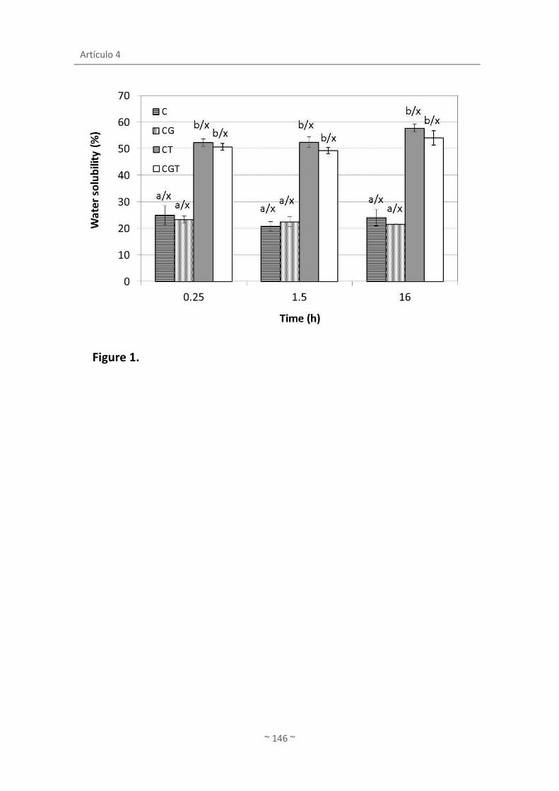
Artículo 4
~ 146 ~
Figure 1.

Release of active compounds from agar and agar-gelatin with green tea extract
~ 147 ~
Figure 2.

Artículo 4
~ 148 ~
Figure 3.
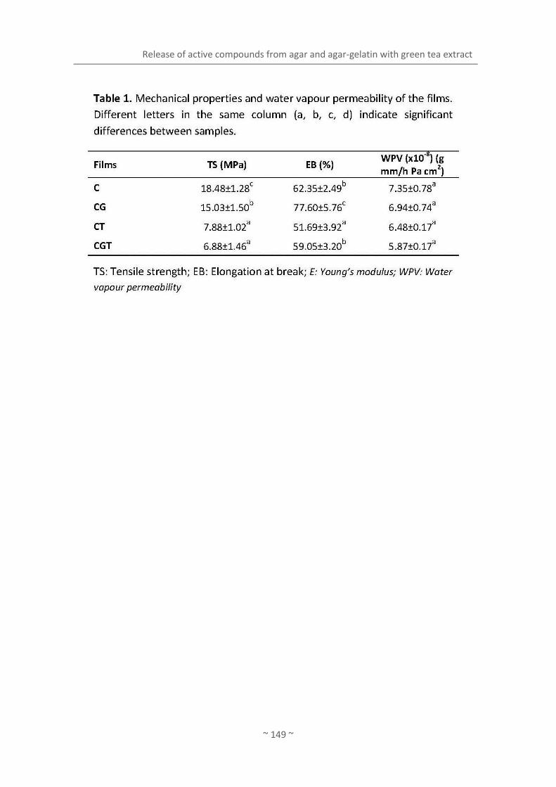
Release of active compounds from agar and agar-gelatin with green tea extract
~ 149 ~

Artículo 4
~ 150 ~

Release of active compounds from agar and agar-gelatin with green tea extract
~ 151 ~

~ 153 ~
ARTÍCULO 5
López de Lacey, A.M., López-Caballero, M.E., Gómez-
Guillén, M.C. y Montero, P. (2012)
Functionality of lactic acid bacteria incorporated to
edible coatings and films
Innovative Food Science and Emerging Technologies


Artículo 5
~ 155 ~
FUNCTIONALITY OF LACTIC ACID BACTERIA
INCORPORATED TO EDIBLE COATINGS AND FILMS
López de Lacey, A.M., López-Caballero, M.E*, Gómez-Guillén, M.C., Montero, P.
Institute of Food Science, Technology and Nutrition, (ICTAN), CSIC. (formerly Institute of
Refrigeration), C/ José Antonio Nováis, 10, 28040. Madrid, Spain.
*Corresponding author: [email protected]
Tel.: +34 91 5492300; fax: +34 91 5493627.

Artículo 5
~ 156 ~
ABSTRACT
This work deals with the design and formulation of gelatin edible coatings and films of
incorporated with probiotic bacteria Lactobacillus acidophillus and Bifidobacterium bifidum. A
gelatin coating incorporated with B. bifidum was applied to fish to assess its effect during
chilled storage. B. bifidum remained viable during the storage and H2S-producers
microorgansims were reduced in 2 log cycles. In a further experiment, fish coated with gelatin
films incorporated with bifidobacteria was treated with a high pressure (200 MPa/10 min/20 °
C). At the end of the storage, a reduction of total viable counts (<2 log cycles) were obtained.
Moreover, the combined treatments drastically reduced the content of volatile bases and
lowered the pH by more than 1 unit. Furthermore, the application of gelatin edible packaging
incorporated with bacteria can be promising for fish preservation, especially when combined
with other technologies such as a high-pressure.
Key words: edible packaging, bacteria, fish, shelf-life, high pressure.

Functionality of lactic acid bacteria incorporated to edible coating and films
~ 157 ~
1. INTRODUCTION
In recent years, the demand and consumption of minimally processed food and additive-
free commodities which present few changes at sensory level has increased. This fact leads to
the development of new technologies and systems for preservation that control the quality
and guarantee food safety. This tendency has also led to research on developing new
biodegradable packaging materials from natural polymers in order to achieve a partial
alternative to plastic packaging. The films can be obtained from several sources
(polysaccharide, lipid, protein) (Tharanathan, 2003) which are in many cases waste products
from fishing, agriculture or cattle farming. These films act as oxygen and water barriers,
thereby slowing oxidation reactions and retaining moisture, thus enhancing quality and
extending storage life (Gennadios, Hanna, & Kurth, 1997).
In the formulations of edible packaging the addition of certain compounds to impart
antimicrobial properties and/or antioxidants is often consider (Gennadios et al., 1997). The
compounds to be incorporated are of a different nature, and may be limited to maintain
quality and food safety (active packaging), or may even lead to a potential beneficial effect on
consumer health (bioactive packaging). The compounds added more frequently are, among
others, polyphenols from plants (Burt, 2004), chitosan (Lopez-Caballero, Gomez-Guillen, Perez-
Mateos, & Montero, 2005), acids (Beverlya, Janes, Prinyawiwatkula, & No, 2008), antioxidant
and antimicrobial peptides (Gómez-Guillén, Giménez, López-Caballero, & Montero, 2011), etc.
Recently the incorporation of lactic acid bacteria or probiotics to films and coatings have been
described: alginate-gellan incorporated with Bifidobacterium lactis Bb-12 for coating of fresh
fruits (Tapia et al., 2007) and Lactobacillus sakei incorporated in sodium-caseinate films to
control Listeria monocytogenes in culture medium and in fresh beef (Gialamas, Zinoviadou,
Biliaderis, & Koutsoumanis, 2010).
Probiotics are live microorganisms which upon ingestion in certain numbers exert a
beneficial effect on health (FAO/WHO, 2002) and an estimated daily intake of around 106-109
CFU/day is recommended to ensure a therapeutic effect. Excellent reviews (Do Espírito Santo,
Perego, Converti, & Oliveira, 2011; Prado, Parada, Pandey, & Soccol, 2008; Soccol et al., 2010)
describe the characteristics and latest progress related to this group of bacteria, among which
are species of the genera Lactobacillus and Bifidobacterium, including Lactobacillus
acidophillus and Bifidobacterium bifidum, species used in our work. While dairy products are
undoubtedly the main source of intake of probiotics, lactose intolerance and cholesterol

Artículo 5
~ 158 ~
content are the main concern related to these products (Prado, et al., 2008). Among the latest
trends (Do Espírito Santo, et al., 2011; Soccol, et al., 2010) vegetable and juice beverages are
good candidates as probiotics vehicles, although in this case bacteria could need protection
from the acid condition in the fruit juice. In this connection, edible coatings and films, as above
mentioned, are presented as another real possibility of carrier matrices for probiotics.
In general, edible films are applied to various foods, including fish, to confer stability
during storage and to increase shelf-life (Gómez-Estaca, López de Lacey, Gómez-Guillén, López-
Caballero, & Montero, 2009; Gómez-Estaca, López de Lacey, López-Caballero, Gómez-Guillén,
& Montero, 2010; Lopez-Caballero, et al., 2005). Moreover, the application of edible films in
combination with high pressure processing has already been shown to be a very promising
technology for fish preservation of cold-smoked sardine (covered by active plant extract-
gelatin composite films, [Gómez-Estaca, Montero, Giménez, & Gómez-Guillén, 2007]) and
salmon (covered by active lignin-gelatin composite films, [Ojagh, Núñez-Flores, López-
Caballero, Montero, & Gómez-Guillén, 2011]). In this sense, we do not know about any
references on the incorporation of lactic acid bacteria in coatings/films based on gelatin (or
other polymers) for fish preservation, alone or in combination with high pressure treatments.
The aim of this work was to design and formulate the edible packaging (coatings and films)
incorporated with Lactobacillus acidophillus and Bifidobacterium bifidum and evaluation of the
survival of these species in the coatings/films. Subsequently, edible packaging incorporated
with bacteria was applied in fish to determine their effect on the growth of natural microbiota
and shelf-life during chilled storage. Isostatic high pressure was also applied to study the effect
of combined treatments.
2. MATERIAL AND METHODS
2.1. Formulation of the films
The film forming coating solutions were prepared using gelatin from bovine-hide (Sancho
de Borja S. L., Zaragoza, Spain). Gelatin film forming solution was prepared by dissolving 8 g of
gelatin per100 ml of distilled water. Glucose (2%) and Cysteine (0.05%) were also added.
Sorbitol (Panreac) (0.15 g/g gelatin) and glycerol (Panreac) (0.15 g/g gelatin) were used as
plasticizers. Gelatin was dissolved in distilled water. The mixtures were warmed and stirred at
45 °C for 15 min to obtain a good blend. Two species, Lactobacillus acidophillus CECT 903 and
Bifidobacterium bifidum DSMZ 20215 were selected to formulate the bioactive packaging. The

Functionality of lactic acid bacteria incorporated to edible coating and films
~ 159 ~
strains were stored at -80 °C in Brain Heart Infusion Broth (Oxoid, Basingstoke, UK) with 25 %
glycerol (Panreac, Moncada i Reixac, Barcelona, Spain) until their use. For regeneration, a little
portion of the frozen culture were transferred to MRS (de Man, Rogosa and Shape, Oxoid,
Basingstoke, UK) for L. acidophilus, and MRS+0.05% Cisteine for B. bifidum, and then incubated
at 37 °C under CO2 flow and at 37 °C under anaerobic conditions (Gas-Pack, Anaerogen;
Oxoid), respectively. Then, cells from each culture were centrifugated (6000 rpm/15 min/5 °C),
harvested and washed with Ringer´s solution. L. acidophillus and B. bifidum were added to
their respective film forming coating solutions to attain a final concentration of 109 cfu/ml.
The respective films were obtained by casting an amount of 40 ml in square plastic dishes (144
cm2) and drying at 45 °C in a forced-air oven for 15h to yield a uniform thickness [200 μm
(p≤0.05)] in all cases. Uniformity was verified by measuring the thickness in 9 points randomly.
The films were stored in desiccators for 2 d at 22 °C over a saturated solution of NaBr (58%
relative humidity) until analyses (c.a. 12-24 h.). Thus four samples were then obtained in total:
film forming solution+L. acidophillus (SLa); film forming solution+B. bifidum (SBb); film+L.
acidophillus (FLa); and film+B. bifidum (FBb).
2.2. Survival of lactic acid bacteria strains
To determine the viability of lactic acid bacteria incorporated into the film forming
solutions or films, counts on specific culture media were periodically performed. For film
forming solution, a total amount of 1 mL was collected and placed in a sterile plastic bag
(Sterilin, Stone, Staffordshire, UK) with 9 ml of buffered 0.1 % peptone water (Oxoid) in a
vertical laminar-flow cabinet (mod. AV 30/70 Telstar, Madrid, Spain). After 1 min in a
Stomacher blender (model Colworth 400, Seward, London, UK), appropriate dilutions were
prepared. For the films, 1 g was collected and placed into a sterile bottle with 99 ml of
buffered 0.1 % peptone water previously heated (~37 °C). Then, peptone water containing the
film was stirred in an orbital at the same temperature for 1 h to disolve the film partially and
allow microorganisms to release into the peptone water. Later appropriate dilutions were
prepared. Counts for L. acidophillus and B. bifidum were performed in MRS agar and MRS agar
+0.05% cysteine, respectively, both in film-forming solutions and in films. Culture conditions
for these two microorganisms were the same as previously described. Analyses were
performed in triplicate.

Artículo 5
~ 160 ~
2.3. Fish storage trial
Hake (Merluccius merluccius) purchased at a local market were cut in portions of (c.a. 100
g each). Gelatin film forming solution (coating) added or not with B. bifidum was prepared as
described above. The portions were dipped into the solutions (with or without bacteria) for 5
min., then removed and vacuum-packed in bags (Cryovac BB-1, Grace, Barcelona, Spain) by
using a Multivac 155 packaging machine (A-NG, 85021, GS, Germany). Two batches were then
obtained: hake dipped into solution (HS) and hake dipped into solution+B. bifidum (HSB). Hake
without any treatment was used for control purposes (H). Batches were stored at 2±1 °C.
2.4. Fish storage trial treated with high pressure
In this trial, hake portions of 100 g were covered individually with a gelatin film added
with B. bifidum (films with bacteria were prepared as described above). Hake wrapped with
films were vacuum-packed and treated in pilot high-pressure unit (ACB 665, GEC Alsthom,
Nantes, France); the temperature of the immersion medium (distilled water) being regulated
by a thermocouple connected to a programmed temperature control equipment (model
IA/2230AC, INMASA, Barcelona, Spain). High pressure treatment was carried out at
200MPa/10 min/20 °C. Pressure was increased by 2.5MPa/s and after high pressure treatment
was completed, the time for pressure dropping back to atmospheric was around 3 s. Three lots
were obtained: control hake (H), hake film+B. bifidum (HF) and hake film+B. bifidum-
pressurized (HFP). Batches were stored at 2±1 °C.
2.5. Microbiological assays
The microbiological analyses were as follows: a total amount of 10 g of muscle, from at
least 2 different packages, was collected and placed in a sterile plastic bag with 90 ml of
buffered 0.1 % peptone water in a vertical laminar-flow cabinet. After 1 min in a Stomacher
blender, appropriate dilutions were prepared for the following microorganism determinations:
(i) total bacterial counts (TBC) on spread plates of Iron Agar 1% NaCl incubated at 15 °C for 3
days; (ii) H2S-producers organisms, as black colonies, on pour plates of Iron Agar incubated at
15 °C for 3 days; (iii) luminescent bacteria on spread plates of Iron Agar 1% NaCl incubated at
15 °C for 5 days; (iv) lactic acid bacteria on double-layered plates of MRS agar incubated at 30
°C for 72 h; (v) bifidobacteria counts on MRS+0.05% Cys incubated at 37 °C under anaerobic

Functionality of lactic acid bacteria incorporated to edible coating and films
~ 161 ~
conditions. All microbiological counts were expressed as the log of the colony-forming units
per gram (log cfu/g) of sample. All analyses were performed in triplicate.
2.6. pH
Approximately 5-10 grams of muscle were homogenized with a double quantity (g/ml) of
distilled water. After five minutes at ambient temperature, pH was determined with a pHm93
pH-meter and a combined pH electrode (Radiometer, Copenhagen, Denmark). The
experiments were repeated at least in triplicate.
2.7. Total volatile basic nitrogen (TVB-N)
Total volatile basic nitrogen (TVB-N) determinations were carried out in triplicate over the
storage period, using the method of Antonacopoulos and Vyncke (1989). 10 g +/- 0.1 g of the
ground sample were weighed in a suitable container and homogenized with 100 ml 6 %
perchloric acid solution for two minutes. After filtering, the extract was alkalinized with a 20 %
sodium hydroxide solution and submitted to steam distillation. The volatile base components
were absorbed by an acid receiver and determined by titration.
2.8. Statistical analyses
Statistical tests were performed using the SPSS® computer program, version 15.0 (SPSS
Statistical Software, Inc., Chicago, IL, USA). One-way and two-way analysis of variance (ANOVA)
was carried out. Differences between pairs of means were assessed on the basis of confidence
intervals using the Tukey-b test. The level of significance was p≤0.05.
3. RESULTS AND DISCUSSION
3.1. Survival of lactic acid bacteria strains
The counts for L. acidophilus and B. bifidum, both in film forming solution (coating) and
film, are shown in Figure 1. From an initial concentration of 109 cfu/ml, both L. acidophilus and
B. bifidum remained fairly constant for 6 days (Figs. 1a-b). Both species remained viable and
were able to grow in culture media, although some declines in the counts (p≤0.05) could be
expected due to the adaptation to the new substrate and to the temperature used to preserve
the solutions and films (2 ± 1 °C). Previously, different specific culture media for the recovery

Artículo 5
~ 162 ~
of bifidobacteria in plaque were tested (MRS-raffinose [Tabasco, Paarup, Janer, Peláez, &
Requena, 2007] and MRS-NPNL [Roy, 2001]), and all of them resulted in very similar counts
(data not shown). In this connection, (Tapia, et al., 2007) reported that B. lactis Bb-12 (at a
concentration of 109 cfu/g) was incorporated in coatings of alginate or gellan with glycerol and
then applied to apple and papaya cylinders. After application, the counts of B. lactis decreased
about 2 log units (which were attributed to dilution phenomena), while the population of
bifidobacteria remained viable and constant for 10 days at 2 °C.
The formulation of our edible packaging (coating and films) contained sorbitol in their
composition. Sorbitol and related polyols act as protective agents of microorganisms in
situations of aw decreased as freeze-drying of L. platarum and L. rhamnosus (Carvalho et al.,
2004). The mechanism by which they exert this protective effect could be related to the
interaction between sugars and phospholipid membranes, helping to maintain its fluidity
(Santivarangkna, Kulozik, Kienberger, & Foerst, 2009). However, Gialamas et al. (2010)
incorporated Lactobacillus sakei (106 cfu/cm2, directly before casting or spray) in sodium
caseinate edible films stored at 4 and 15 °C; when the films did not contain sorbitol in their
formulation, there was an increase in the viability of bacteria of the film kept at 4 °C, possibly
because of decreased metabolism experienced by the organisms at low temperatures.
3.2. Fish storage trial
Initial counts were 4.3 hake log cfu/g. These counts were similar to those reported for this
same species (Lopez-Caballero, 1998). Changes in total viable counts were barely detected to
day 6. From there, the control batch underwent a slight but significant increase (p≤0.05),
registering close to 7 log cfu/g at the end of the storage. Hake with coating (HS), after some
fluctuation, reached figures close to control batch (Fig. 2a). Counts of hake with coating
incorporated with bacteria (HSB) remained practically constant during storage and attained 1
log unit lower (p≤0.05) than control at day 10 (Fig, 2a). However, the most relevant was the
significant inhibition achieved by the coating (with or without bacteria) in H2S-producing
microorganisms (Fig. 2b), presumably considered as Shewanella putrefaciens, specific
organisms involved in fish spoilage (López-Caballero, Martínez-Alvarez, Gómez-Guillén, &
Montero, 2007).
The lactic acid bacteria counts were in a similar range for all batches, about 4 log cfu/g at
the end of the period studied (Fig. 2c) for H and HS batches (p≤0.05). It is worth noting the
survival of bifidobacteria during storage, which decreased only 1 log cfu/g compared to the

Functionality of lactic acid bacteria incorporated to edible coating and films
~ 163 ~
initial concentration (Fig. 2d). The differences between the counts in MRS agar and MRS-
Cysteine (Fig. 2c, d) could be explained by the difficulty of B. bifidum to grow in the culture
media without the addition of Cys. Luminescent colonies were not detected in this assay.
Lactic acid bacteria improves health when consumed, but they also play a protective effect
against pathogens microorganisms in the product during storage through competition with
pathogens for nutrients, producing metabolites as organic acids and bacteriocines, etc. (Rydlo,
Miltz, & Mor, 2006; García, Martín, Sanz, & Hernández, 1995). In this sense, the use of lactic
acid bacteria against pathogenic organisms has been referred to previously in the literature.
Thus, in shrimps contaminated with L. monocytogenes, the addition of Lactobacillus plantarum
LKE5 inhibited even 3 log units the growth of listeria (Huss, Jeppesen, Johansen, & Gram,
1995), this effect being higher if the experimental conditions were close to the limit of growth
of the pathogen (in terms of pH, etc.). In another trial, apple wedges were dipped with
Lactobacillus rhamnosus GG (108 cfu/ml) and although Salmonella was not affected when co-
inoculated with L. rhamnosus GG, L. monocytogenes was reduced in the presence of the
lactobacilli 1-log unit (Alegre, Viñas, Usall, Anguera, & Abadias, 2011). In addition, apple
wedges quality were not affected by the incorporation of L. rhamnosus GG, while the probiotic
population maintained around 107 cfu/g during the 28 days of storage at 5 and 10 °C.
The incorporation of bacteria in covers and packages, as coating/films, offers new
possibilities and could be advantageous, and to our best knowledge, there are only two
references in the literature (Gialamas, et al., 2010; Tapia, et al., 2007), in addition to the
present work. The advantage of incorporating bacteria to the covers (coating/film) may be
related to the spread and concentration of bacteria on the product surface. Kristo,
Koutsoumanis, and Biliaderis (2008) described the effect of microbial metabolites, such as
nisin, against pathogens as L. monocytogenes, is greater when nisin takes part of a coating of
sodium-casinate than if nisin is applied directly to the culture medium. This fact could be due
to the decrease in the rate of diffusion from the cover/packaging to the product, in which a
high concentration of active compound in the product surface was maintained (Gialamas, et
al., 2010). These authors pointed out that the use of L. sakei incorporated into sodium-
caseinate films constitutes an effective way to control L. monocytogenes (up to 4 log cycles)
and for improving food safety, both in culture media and in beef slices at constant (4 °C) and
fluctuating temperature (5-15 °C).
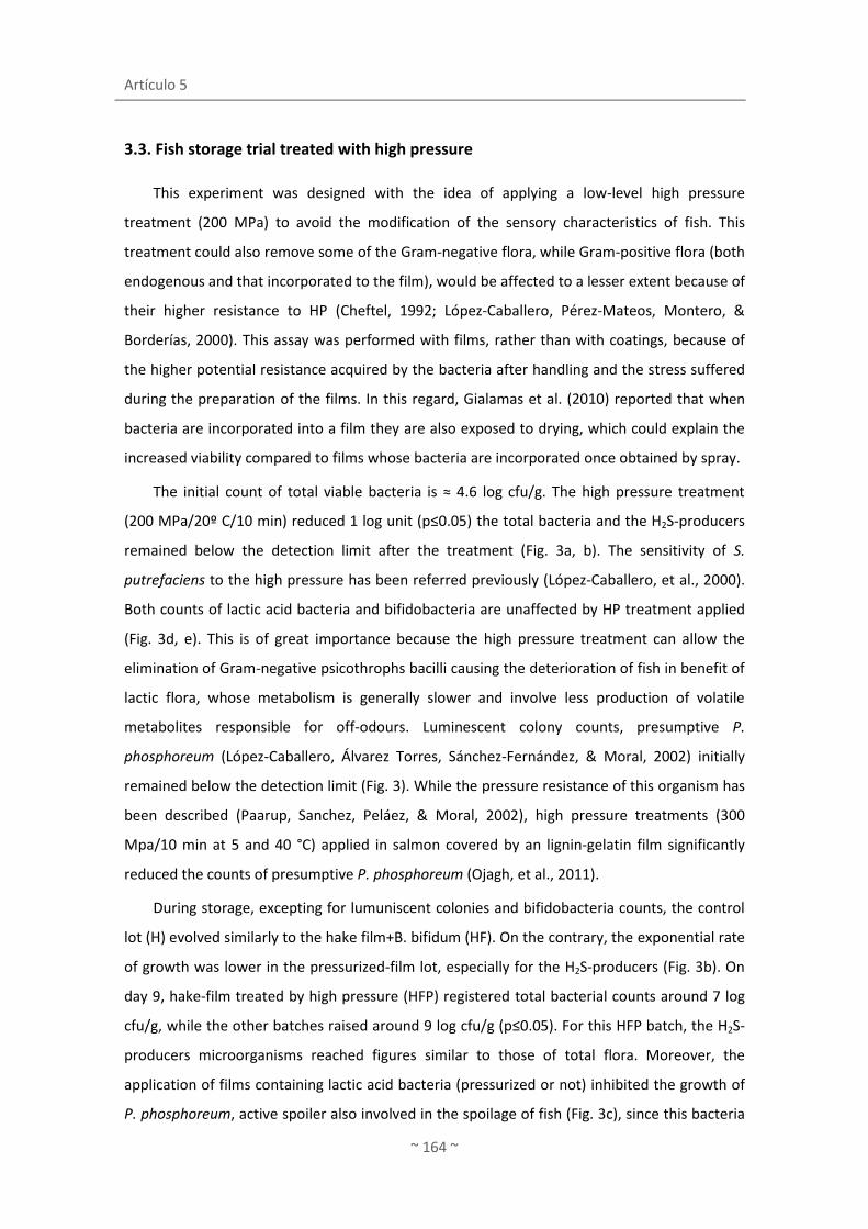
Artículo 5
~ 164 ~
3.3. Fish storage trial treated with high pressure
This experiment was designed with the idea of applying a low-level high pressure
treatment (200 MPa) to avoid the modification of the sensory characteristics of fish. This
treatment could also remove some of the Gram-negative flora, while Gram-positive flora (both
endogenous and that incorporated to the film), would be affected to a lesser extent because of
their higher resistance to HP (Cheftel, 1992; López-Caballero, Pérez-Mateos, Montero, &
Borderías, 2000). This assay was performed with films, rather than with coatings, because of
the higher potential resistance acquired by the bacteria after handling and the stress suffered
during the preparation of the films. In this regard, Gialamas et al. (2010) reported that when
bacteria are incorporated into a film they are also exposed to drying, which could explain the
increased viability compared to films whose bacteria are incorporated once obtained by spray.
The initial count of total viable bacteria is ≈ 4.6 log cfu/g. The high pressure treatment
(200 MPa/20º C/10 min) reduced 1 log unit (p≤0.05) the total bacteria and the H2S-producers
remained below the detection limit after the treatment (Fig. 3a, b). The sensitivity of S.
putrefaciens to the high pressure has been referred previously (López-Caballero, et al., 2000).
Both counts of lactic acid bacteria and bifidobacteria are unaffected by HP treatment applied
(Fig. 3d, e). This is of great importance because the high pressure treatment can allow the
elimination of Gram-negative psicothrophs bacilli causing the deterioration of fish in benefit of
lactic flora, whose metabolism is generally slower and involve less production of volatile
metabolites responsible for off-odours. Luminescent colony counts, presumptive P.
phosphoreum (López-Caballero, Álvarez Torres, Sánchez-Fernández, & Moral, 2002) initially
remained below the detection limit (Fig. 3). While the pressure resistance of this organism has
been described (Paarup, Sanchez, Peláez, & Moral, 2002), high pressure treatments (300
Mpa/10 min at 5 and 40 °C) applied in salmon covered by an lignin-gelatin film significantly
reduced the counts of presumptive P. phosphoreum (Ojagh, et al., 2011).
During storage, excepting for lumuniscent colonies and bifidobacteria counts, the control
lot (H) evolved similarly to the hake film+B. bifidum (HF). On the contrary, the exponential rate
of growth was lower in the pressurized-film lot, especially for the H2S-producers (Fig. 3b). On
day 9, hake-film treated by high pressure (HFP) registered total bacterial counts around 7 log
cfu/g, while the other batches raised around 9 log cfu/g (p≤0.05). For this HFP batch, the H2S-
producers microorganisms reached figures similar to those of total flora. Moreover, the
application of films containing lactic acid bacteria (pressurized or not) inhibited the growth of
P. phosphoreum, active spoiler also involved in the spoilage of fish (Fig. 3c), since this bacteria
Functionality of lactic acid bacteria incorporated to edible coating and films
~ 165 ~
was just detected in control batch. Lactic acid bacteria counts did not differed between
batches until day 9 (p≤0.05), and bifidobacteria remained viable in both batches covered with
film (HF and HFP) during the storage time (Fig. 3e).
Initially the pH of the fish ranged close to 7. During the storage there was a slight decrease
in pH values, especially in HFP batch (p≤0.05), probably due to the presence of lactic acid
bacteria in the film which passed to the muscle (Fig. 4a). The control lot reached levels close to
8 to 9 days.
At the beginning of the trial, hake registered values close to 10 mg N-TVB/100 g muscle of
total volatile basic nitrogen. These figures remained below 20 mg N-TVB/100 g muscle at 9
days of storage in the hake-film pressurized (HFP), while the control lot (H) and hake-film non-
pressurized lot (HF) reached values higher (p≤0.05) than 60 mg-NBVT/100 g (Fig. 4b). These
levels were according to the microbial counts (total bacteria counts and H2S-producers
microorganisms) registered for these batches, although pH recorded in the HF and HFP lots did
not reflect the volatile basis levels achieved, respectively, for each of them. The combined
treatment, edible film+ bacteria and high pressure presented certain synergism in the
inhibition of volatile bases, as hake pressurized under the same conditions as those applied in
the current assay obtained values > 35-40 mg N-TVB/100 g muscle at 8-12 days of storage.
Moreover, the application of the film did not confer strange odors to fish. Perhaps the
presence of bifidobacteria could interfere in any way with the production of volatile basic
compounds (e.g. competition for nutrients, etc.), by microorganisms such as S. putrefaciens,
reducing its production to some extent, while the high pressure treatment inactivates or
causes damage to these species involved in the fish spoilage.
In view of these results, we conclude that gelatin edible coatings and films could act as a
matrix to incorporate lactic acid bacteria, since L. acidophillus and B. bifidum in major extend,
remained viable even for 13 days. The application of these edible coatings and films was
promising for fish preservation, especially in combination with high pressure treatment. Far
from competing with other matrix as dairy products, vegetable beverages, etc., the
incorporation of lactic acid bacteria to edible packages could be a way for these bacteria to be
ingested together with fish. The combination of treatment allows reducing the level of high
pressure applied so as not to adversely affect lactic acid bacteria incorporating to the edible
coating and film, while maintaining the sensory characteristics of the raw product. Further
studies are needed to study the possible competition between microbial species as well as to
establish rate of diffusion from bacteria to muscle.

Artículo 5
~ 166 ~
Acknowledgements
This research was financed by the Ministerio de Educación y Ciencia, under projects I3-
200670l141, AGL2008-00231/ALI, AGL2011-27607, CYTED 309ACO382. Program JAE-CSIC
predoctoral is fully acknowledged. The author thanks Joan Therese de Lacey for the English
language revision of the article.
ICTAN-CSIC has implemented and maintains a Quality Management System which fulfils the
requirements of the ISO standard 9001:2000.
References
Alegre, I., Viñas, I., Usall, J., Anguera, M., & Abadias, M. (2011). Microbiological and
physicochemical quality of fresh-cut apple enriched with the probiotic strain
Lactobacillus rhamnosus GG. Food Microbiology, 28(1), 59-66.
Antonacopoulos, N., & Vyncke, W. (1989). Determination of volatile basic nitrogen in fish: A
third collaborative study by the West European Fish Technologists' Association
(WEFTA). Zeitschrift für Lebensmittel-Untersuchung und -Forschung, 189(4), 309-316.
Beverlya, R. L., Janes, M. E., Prinyawiwatkula, W., & No, H. K. (2008). Edible chitosan films on
ready-to-eat roast beef for the control of Listeria monocytogenes. Food Microbiology,
25(3), 534-537.
Burt, S. (2004). Essential oils: their antibacterial properties and potential applications in foods -
a review. International Journal of Food Microbiology, 94(3), 223-253. doi:
10.1016/j.ijfoodmicro.2004.03.022
Carvalho, A. S., Silva, J., Ho, P., Teixeira, P., Malcata, F. X., & Gibbs, P. (2004). Effects of Various
Sugars Added to Growth and Drying Media upon Thermotolerance and Survival
throughout Storage of Freeze-Dried lactobacillus delbrueckii ssp. bulgaricus.
Biotechnology Progress, 20(1), 248-254.
Cheftel, J.C. (1992). Effect of high hydrostatic pressure on food constituents: an overview. In C.
Balny, R. Hayashi, K. Heremans, and P. Masson (Eds.), High Pressure and Biotechnology
(pp. 195–209). Colloque INSERM/John Libbey Eurotext Ltd, Montrouge.
Do Espírito Santo, A. P., Perego, P., Converti, A., & Oliveira, M. N. (2011). Influence of food
matrices on probiotic viability - A review focusing on the fruity bases. Trends in Food
Science and Technology, 22(7), 377-385.

Functionality of lactic acid bacteria incorporated to edible coating and films
~ 167 ~
FAO/WHO. 2002. Working Group Report on Drafting Guidelines for the Evaluation of Probiotics
in Food. London, Ontario, Canada, April 30 and May 1, pp 11.
García, T., Martín, R., Sanz, B., & Hernández, P-E. (1995). Revisión: Extensión de la vida útil de
la carne fresca. I: envasado en atmósferas modificadas y utilización de bacterias
lácticas y bacteriocinas. Revista Española de Ciencia y Tecnología de Alimentos, 35(1),
1-18.
Gennadios, A., Hanna, M. A., & Kurth, L. B. (1997). Application of Edible Coatings on Meats,
Poultry and Seafoods: A Review. LWT - Food Science and Technology, 30(4), 337-350.
doi: 10.1006/fstl.1996.0202
Gialamas, H., Zinoviadou, K. G., Biliaderis, C. G., & Koutsoumanis, K. P. (2010). Development of
a novel bioactive packaging based on the incorporation of Lactobacillus sakei into
sodium-caseinate films for controlling Listeria monocytogenes in foods. Food Research
International, 43(10), 2402-2408. doi: 10.1016/j.foodres.2010.09.020
Gómez-Estaca, J., López de Lacey, A., Gómez-Guillén, M. C., López-Caballero, M. E., & Montero,
P. (2009). Antimicrobial Activity of Composite Edible Films Based on Fish Gelatin and
Chitosan Incorporated with Clove Essential Oil. Journal of Aquatic Food Product
Technology, 18(1-2), 46-52. doi: 10.1080/10498850802581252
Gómez-Estaca, J., López de Lacey, A., López-Caballero, M. E., Gómez-Guillén, M. C., & Montero,
P. (2010). Biodegradable gelatin-chitosan films incorporated with essential oils as
antimicrobial agents for fish preservation. Food Microbiology, 27(7), 889-896.
Gómez-Estaca, J., Montero, P., Giménez, B., & Gómez-Guillén, M. C. (2007). Effect of functional
edible films and high pressure processing on microbial and oxidative spoilage in cold-
smoked sardine (Sardina pilchardus). Food Chemistry, 105(2), 511-520.
Gómez-Guillén, M. C., Giménez, B., López-Caballero, M. E., & Montero, M. P. (2011).
Functional and bioactive properties of collagen and gelatin from alternative sources: A
review. Food Hydrocolloids, 25(8), 1813-1827. doi: 10.1016/j.foodhyd.2011.02.007
Huss, H. H., Jeppesen, V. F., Johansen, C., & Gram, L. (1995). Biopreservation of Fish Products—
A Review of Recent Approaches and Results. Journal of Aquatic Food Product
Technology, 4(2), 5-26. doi: 10.1300/J030v04n02_02
Kristo, E., Koutsoumanis, K. P., & Biliaderis, C. G. (2008). Thermal, mechanical and water vapor
barrier properties of sodium caseinate films containing antimicrobials and their
inhibitory action on Listeria monocytogenes. Food Hydrocolloids, 22(3), 373-386.

Artículo 5
~ 168 ~
López Caballero, M.E. 1998. Flora alterante de merluza (Merluccius merluccius, L.) refrigerada
sometida a la acción de concentraciones variables de CO2 y O2. Identificación y
actividad bioquímica de bacterias implicadas en el deterioro. Doctoral Thesis.
López-Caballero, M. E., Álvarez Torres, M. D., Sánchez-Fernández, J. A., & Moral, A. (2002).
Photobacterium phosphoreum isolated as a luminescent colony from spoiled fish,
cultured in model system under controlled atmospheres. European Food Research and
Technology, 215(5), 390-395.
Lopez-Caballero, M. E., Gomez-Guillen, M. C., Perez-Mateos, M., & Montero, P. (2005). A
chitosan-gelatin blend as a coating for fish patties. Food Hydrocolloids, 19(2), 303-311.
doi: 10.1016/j.foodhyd.2004.06.006
López-Caballero, M. E., Martínez-Alvarez, O., Gómez-Guillén, M. D. C., & Montero, P. (2007).
Quality of thawed deepwater pink shrimp (Parapenaeus longirostris) treated with
melanosis-inhibiting formulations during chilled storage. International Journal of Food
Science and Technology, 42(9), 1029-1038.
López-Caballero, M. E., Pérez-Mateos, M., Montero, P., & Borderías, A. J. (2000). Oyster
preservation by high-pressure treatment. Journal of Food Protection, 63(2), 196-201.
Ojagh, S. M., Núñez-Flores, R., López-Caballero, M. E., Montero, M. P., & Gómez-Guillén, M. C.
(2011). Lessening of high-pressure-induced changes in Atlantic salmon muscle by the
combined use of a fish gelatin-lignin film. Food Chemistry, 125(2), 595-606.
Paarup, T., Sanchez, J. A., Peláez, C., & Moral, A. (2002). Sensory, chemical and bacteriological
changes in vacuum-packed pressurised squid mantle (Todaropsis eblanae) stored at
4°C. International Journal of Food Microbiology, 74(1-2), 1-12.
Prado, F. C., Parada, J. L., Pandey, A., & Soccol, C. R. (2008). Trends in non-dairy probiotic
beverages. Food Research International, 41(2), 111-123.
Roy, D. (2001). Media for the isolation and enumeration of bifidobacteria in dairy products.
International Journal of Food Microbiology, 69(3), 167-182.
Rydlo, T., Miltz, J., & Mor, A. (2006). Eukaryotic antimicrobial peptides: Promises and premises
in food safety. Journal of Food Science, 71(9), R125-R135.
Santivarangkna, C., Kulozik, U., Kienberger, H., & Foerst, P. (2009). Changes in membrane fatty
acids of Lactobacillus helveticus during vacuum drying with sorbitol. Letters in Applied
Microbiology, 49(4), 516-521.

Functionality of lactic acid bacteria incorporated to edible coating and films
~ 169 ~
Soccol, C. R., Vandenberghe, L. P. S., Spier, M. R., Medeiros, A. B. P., Yamaguishi, C. T., De Dea
Lindner, J., & Thomaz-Soccol, V. (2010). The potential of probiotics: A review. Food
Technology and Biotechnology, 48(4), 413-434.
Tabasco, R., Paarup, T., Janer, C., Peláez, C., & Requena, T. (2007). Selective enumeration and
identification of mixed cultures of Streptococcus thermophilus, Lactobacillus
delbrueckii subsp. bulgaricus, L. acidophilus, L. paracasei subsp. paracasei and
Bifidobacterium lactis in fermented milk. International Dairy Journal, 17(9), 1107-1114.
Tapia, M. S., Rojas-Graü, M. A., Rodríguez, F. J., Ramírez, J., Carmona, A., & Martin-Belloso, O.
(2007). Alginate- and Gellan-Based Edible Films for Probiotic Coatings on Fresh-Cut
Fruits. Journal of Food Science, 72(4), E190-E196. doi: 10.1111/j.1750-
3841.2007.00318.x
Tharanathan, R. N. (2003). Biodegradable films and composite coatings: past, present and
future. Trends in Food Science & Technology, 14(3), 71-78. doi: 10.1016/s0924-
2244(02)00280-7
Artículo 5
~ 170 ~
Figures
Figure 1. Survival of L. acidophilus and B. bifidum counts (Log cfu/g) into film forming
solutions (coating) and films stored at 2±1 °C. a) (SLa): Films forming solution+L.
acidophilus, (SBb): film forming solution+B. bifidum; b) (FLa): film+L. acidophilus,
(FBb): film+B. bifidum.
Figure 2. Microbial counts of hake coated with edible film forming solution during storage
at 2±1 °C. (H): control hake; (HS): hake dipped into solution; (HSB): hake dipped
into solution+B. bifidum
Figure 3. Microbial counts of hake covered with edible films under high pressure treatmen
during storage at 2±1 °C. (H): control hake; (HF): hake film+B. bifidum; (HFP): hake
film+B. bifidum-pressurized
Figure 4. a)p pH and b) total volatile bases of hake covered with edible films under high
pressure treatment during storage at 2±1 °C. (H): control hake; (HF): hake film+B.
bifidum; (HFP): hake film+B. bifidum-pressurized.

Functionality of lactic acid bacteria incorporated to edible coating and films
~ 171 ~
Figure 1.

Artículo 5
~ 172 ~
Figu
re 2
.

Functionality of lactic acid bacteria incorporated to edible coating and films
~ 173 ~
Figu
re 3
.

Artículo 5
~ 174 ~
Figure 4.

~ 175 ~
ARTÍCULO 6
López de Lacey, A.M., Pérez Santín, E., López-Caballero,
M.E. y Montero, P. (2012)
Survival and metabolic activity of probiotic in green tea
LWT - Food Science and Technology


Artículo 6
~ 177 ~
SURVIVAL AND METABOLIC ACTIVITY OF PROBIOTIC
BACTERIA IN GREEN TEA
A. M. López de Lacey, E. Pérez Santín, M. E. López-Caballero*, and P. Montero
Instituto de Ciencia y Tecnología de Alimentos y Nutrición (ICTAN), CSIC. (formerly Instituto del
Frío), C/ José Antonio Nováis, 10, 28040. Madrid, Spain.
*Corresponding author: E-mail address: [email protected]
Tel.: +34 91 5492300; fax: +34 91 5493627.

Artículo 6
~ 178 ~
ABSTRACT
The aim of this study was to determine the enzymatic activity and survival of three
probiotic strains (Lactobacillus paracasei LAFTI@L26, Lactobacillus acdidophillus LAFTI@L10
and Bifidobacterium animalis ssp. Lactis LAFTI@B94) during incubation in six different varieties
of green tea extracts. The polyphenol content, antioxidant properties, ACE-inhibition and
antimicrobial activities of two standards (epigallocatechin-3-gallate and rutin) were also
investigated, and in the Wu Lu Mountain green tea variety before and after incubation with B.
animalis B94.
The tea extracts in general permitted the survival of the selected probiotic strains, with B.
animalis B94 maintaining the highest levels of viable cells, around 6 log CFU/mL, at the end of
incubation. The selected probiotic bacteria exhibited β-glucosidase, β-galactosidase and α-
ramnosidase activity. The antioxidant properties and ACE-inhibition of standard solutions of
epigallocatechin-3-gallate and rutin increased after incubation with B. animalis B94, caused by
polyphenol content reduction and the formation of other more highly biologically active
metabolites. However, during the incubation of green tea with probiotic bacteria, changes in
the concentration of the most abundant green tea polyphenols did not enhance the biological
capacity. Thus, the antimicrobial activity and ACE-inhibition of Wu Lu Mountain green tea,
incubated with or without B. animalis B94, registered similar behaviour throughout incubation.
Key words: green tea, polyphenols, probiotic bacteria, biological activity.

Survival and metabolic activity of probiotic bacteria in green tea
~ 179 ~
1. INTRODUCTION
Green tea, one the most consumed beverages, is produced with unfermented leaves and
buds from the plant Camellia sinensis L. (Costa, Gouveia, & Nobrega, 2002). These components
are rich in flavan-3-ols (epicatechin, catechin and galloylated derivates), which are responsible
for the majority of the beneficial properties of green tea. Other major polyphenols present in
green tea are the flavonols, mainly derivatives of quercetin and kaempferol (principally
glycosylated). Green tea also contains other compounds like tea pigments, amino acids,
vitamins, carbohydrates, minerals and purine alkaloids (Graham, 1992). The beneficial effects
of green tea have been attributed to all these compounds, but principally to polyphenol
phenolic compounds because of their high antioxidant properties (Bolling, Chen, & Blumberg,
2009; Isiguki, Takakuwa, & Takeo, 1991; Weisburger & Chung, 2002).
Probiotic bacteria, with a wide variety of positive health effects, are principally members
of the Bifidobacterium and Lactobacillus family. Probiotics have been extensively studied and
explored commercially in many different products, including dairy products like cheese,
yoghurt and ice cream, and non-dairy products such as soy, fruit, vegetables, legumes and
cereals (Champagne, 2009; Soccol et al., 2010). Probiotic microorganisms contribute to
intestinal balance, play a role in maintaining health and can even improve the quality of some
products formulated with them (Arora, Jood, & Khetarpaul, 2010; Soccol et al., 2010).
The fermentation of foods with probiotic microorganisms could improve or add additional
beneficial properties to the product. This is because some probiotic bacteria possess certain
enzymatic activities (such as deglycosylation, ring-fission, dehydroxylation, demethylation or
decarboxylation, etc.) (Aura, 2008), that are capable in some cases, of transforming
polyphenols into compounds which are even more bioavailable or bioactive than the original
polyphenol. (Donkor & Shah, 2008; Tsangalis, Ashton, McGill, & Shah, 2002; Uskova,
Kravchenko, Avrenjeva, & Tutelyan, 2010). However, despite the beneficial properties
contained in the composition of green tea, green tea has rarely been used as a food matrix for
probiotics. Nevertheless, Kombucha is a microbial fermented tea made from tea extract
supplemented with sugar, yeast and acetic-acid bacteria, the latter being responsible for the
fermentation (Mo, Zhu, & Chen, 2008). Furthermore, no published reports have been found
that deal with the effect of the commercial bacteria selected for this study (Lactobacillus
acidophilus LAFTI®L10, Bifidobacterium lactis LAFTI®B94 and Lactobacillus paracasei
LAFTI®L26) on green tea polyphenols. The aim of this work was to study the survival of these
Artículo 6
~ 180 ~
three different probiotic bacteria in different varieties of green tea infusions, determine their
enzymatic activity and if they were able to transform polyphenols in the infusions while
maintaining the initial properties of the teas tested. The survival of these probiotic bacteria in
the two different polyphenol solutions selected, and the transformation of polyphenols by the
bacteria was also studied.
2. MATERIALS AND METHODS
2.1. Chemicals
Standard polyphenol compounds: (-)-epigallocatechin gallate (CAS: 989-51-5) and rutin
(CAS: 153-18-4) were purchased from Extrasynthese (Genay, Cedex, France).
Reagents for enzyme activity assay: ρ-nitrophenyl-β-D-glucopyranoside, ρ-nitrophenyl-β-
D-galactopyranoside, ρ-nitrophenyl-α-D-ramnopyranoside, ρ-nitrophenol were provided by
Sigma-Aldrich (Stenheim, Germany).
HPLC-MS: HPLC grade methanol, acetonitrile and formic acid (VWR international, Inc.,
Barcelona, Spain). HPLC grade water was prepared from distilled water using a Milli-Q system
(Millipore Laboratory Bedford, MA).
Antioxidant activity; FRAP: (2,4,6-tripyridyl-s-triazine), FeCl3 and FeSO4 (Sigma-Aldrich, St.
Louis, Mo., USA). ABTS: [2,2’-azino-bis-(3-ethylbenzothiazoline-6-sulfonic acid)], potassium
persulphate, Vitamin C (Sigma-Aldrich, St. Louis, Mo., USA).
Reagents for ACE-inhibition: HHL (Hippuryl-L-Histidyl-L-Leucine), ACE (CAS: 9015-82-1),
potassiumphosphate buffer, containing 300 mM NaCl, pH 8.3 (Sigma-Aldrich, St. Louis, Mo.,
USA).
2.2. Bacterial strains and growth conditions
Lyophilized strains of Lactobacillus paracasei LAFTI@L26, Lactobacillus acdidophillus
LAFTI@L10 and Bifidobacterium animalis ssp. Lactis LAFTI@B94 were purchased from DSM
(DSM Food Specialties Ltd., Sydney, Australia) and kept at -20 °C.
For experiments, one portion of each lyophilized strain was dissolved in broth medium.
Then one aliquot of this solution was streaked on agar plates which were incubated at 37 °C
for 24 h. B. animalis B94 were incubated under anaerobic conditions. Then, one colony of each
strain was transferred into broth medium and grown at 37 °C for 24 h. Lactobacillus paracasei

Survival and metabolic activity of probiotic bacteria in green tea
~ 181 ~
LAFTI@L26 and Lactobacillus acidophillus LAFTI@L10 were incubated in MRS, but the MRS
culture medium for B. animalis B94 was supplemented with 0.05% cysteine.
2.3. Enzyme activity assay of probiotic strains
Activity assays were carried out using the method of Ávila et al. (2009), but with some
modifications. Probiotic bacteria were grown as explained above and then the cells were
harvested by centrifugation (6,000 g, 10 min, 5 °C) and washed twice with 50 mM sodium
phosphate buffer, pH = 7. The harvested cells were suspended in the same buffer to an OD 620
~ 0.6.
β-Glucosidase, β-galactosidase and α-ramnosidase activity was measured in triplicate with
ρ-nitrophenyl-β-D-glucopyranoside, ρ-nitrophenyl-β-D-galactopyranoside and ρ-nitrophenyl-α-
D-ramnopyranoside respectively as substrate. This was done first by mixing 450 µL of 5 mM
substrate prepared in 50 mM sodium phosphate buffer, pH 7, with 50 µL of the cell´s
suspension and incubated at 37 °C for 110 min. Then the reactions were stopped on ice by
adding 500 µL of Na2CO3 0.2 M. and the amount of ρ-nitrophenol released was measured at
410 nm spectrophotometerically. One unit of enzyme activity was defined as the amount of β-
glucosidase or β-galactosidase or α-ramnosidase that 1 nanomole of ρ-nitrophenol released
from the substrate per millilitre per minute under the assay conditions (Otieno, Ashton, &
Shah, 2005).
2.4. Infusion preparation
Six kinds of green teas were tested in this study: Japan Sencha Makinohara, Japan
Gyokuro Asahi, Cinnamon Green Tea, Lung Ching, China White Hair and Wu Lu Mountain. All
these green teas were purchased from a specialized local shop (La Antigua Plantación, Madrid,
Spain).
The preparation of green tea extract was carried out according to the method described
by López de Lacey, López-Caballero, and Montero (2011). Briefly, 35 g of each leaves was
pulverized in a blender (Osterizer) and mixed with 350 mL of water (milli-Q) at 80 °C for 30
minutes, with constant shaking and was then centrifuged at 12,000 g for 10 minutes at 5 °C.
The supernatant was filtered two times through Whatmann N°1 filter papers. The infusion
filtered was stored at – 20 °C before the analysis.

Artículo 6
~ 182 ~
2.5. Survival of bacterial strains in green tea extracts
First the strains (L. paracasei L26, L. acidophillus L10 and B. animalis B94) were grown as
previously explained. The cells were then harvested by centrifugation (6,000 g, 10 min, 5 °C),
washed twice with 0.9% NaCl solution and suspended in the same solution 0.9% NaCl to an OD
620 ~ 0.6. One aliquot of 50 µL of harvested cells adjusted to OD 620 ~ 0.6 was mixed with 950 µL
of green tea extract to study the survival rate of bacteria in green tea extracts. 50 µl of 0.9%
NaCl solution was used as control. The bacterial strains were incubated at 37 °C for 72 h as
mentioned above.The final concentration of bacteria was approximately 106 CFU/mL.
The viable cells were determined at 0, 24, 48 and 72 h of incubation in the green tea
extracts by analysing 100 μL of each culture with the microdot method (Strahsburger, Baeza,
Monasterio, & Lagos, 2005) on MRS agar for the Lactobacillus strain and 0.05% cysteine MRS
agar for B. animalis B94. All the counts were performed at least in triplicate.
2.6 Survival of B. animalis B94 in two separately prepared polyphenol standard
solutions: epigallocatechin 3-gallate and rutin
Determining the survival of B. animalis B94 in two standard polyphenol solutions was
performed, first by mixing 1 mL of harvested cells adjusted to OD 620 ~ 0.6 in 19 mL of 0.9%
NaCl solution. The standards were firstly dissolved in HPLC grade methanol before being added
to the NaCl solution. The same concentration of standards was used in all the samples in order
to compare the effect on probiotic survival of each compound. The final concentration of the
bacteria was approximately 106 CFU/mL and the final concentration of each standard in this
solution was 25 µg/mL. B. animalis B94 growth was not affected by the methanol. The
incubation conditions and the counting of B. animalis B94 was carried out as explained above.
2.7. Biotransformation of green tea polyphenols and standard solutions by B.
animalis B94
The study of polyphenol biotransformations by B. animalis B94 was performed in the Wu
Lu Mountain green tea variety and in two separately prepared polyphenol standard solutions,
epigallocatechin-3-gallate and rutin. Both green tea polyphenol and standard solution samples
incubated with the bacteria, were prepared exactly as described in the preceding section. In
the case of green tea extract, 1 mL of harvested cells adjusted to OD 620 ~ 0.6 were mixed
with 19 mL of Wu Lu Mountain infusion. One aliquot of the solution was taken for the different
analyses (counts, HPLC-MS, antioxidant activity and antimicrobial activity) at 0, 24, 48 and 72 h

Survival and metabolic activity of probiotic bacteria in green tea
~ 183 ~
of incubation at 37 °C. The viable cells were measured using the microdot method explained
above. After counting, each aliquot was centrifuged for 10 min. at 6,000 g, 5 °C and the
supernatant was kept for the rest of the analyses. All the analyses were performed at least in
triplicate, except for the HPLC-MS analysis which was carried out in duplicate.
2.8. Quantification of polyphenols by HPLC-MS
Reverse phase high performance liquid chromatography was performed to analyse
phenolic compounds present in the aqueous green tea extract. The separation module
consisted of an Agilent 1100 series HPLC (Agilent Technologies, Waldbroon, Germany),
equipped with a C18 size column (250 x 4.6 mm, 5 µm, C18 Tracer Excel ODS-A column,
Teknokroma) and a diode array detector (DAD). The samples were eluted with a gradient
system consisting of solvent A (deionized water) and solvent B (acetonitrile) both containing
1% formic acid, used as the mobile phase, with a flow rate of 1 mL/min.
The temperature of the column was maintained at 25 °C and the injection volume was 20
µL. The gradient system started at 90% solvent A and decreased to 74% A within 40 min,
followed by a decreased to 35% solvent A in 10 min. The final conditions were held for an
additional 5 min. The peaks of the phenolic compounds were monitored by atmospheric
pressure electrospray ionization source (ESI), operated in negative ion mode, with the
electrospray capillary voltage set to 3000 V, a nebulizing gas flow rate 12 L/h, and a drying
temperature of 350 °C. Mass spectrometry data were acquired in the scan mode (mass range
m/z 200-700) and in the sim mode (m/z 289, 305, 441, 441, 447, 457, 609).
The quantification was carried out using the external standard method. Solutions of each
standard at various concentration levels of 0.62, 1.25, 2.5, 5, 10 and 20 mg/L. were injected
into column and the elution was performed in the same manner with the samples. Individual
compound was quantified using a calibration curve of the corresponding standard compound.
2.9. Antioxidant activity before and after biotransformation
2.9.1. FRAP
The ferric reducing/antioxidant power (FRAP) assay was carried out according to the
method described by (Benzie & Strain, 1996) with some modification (Alemán et al., 2011).
This assay measures the change in absorbance at 595 nm owing to the formation of a blue
colored FeII-tripyridyltriazine compound from the colourless oxidized FeIII form by the action

Artículo 6
~ 184 ~
of electron donating antioxidants (Benzie, Chung, & Strain, 1999). An amount of 60 μL of
sample was incubated (37 °C) with 60 μL of Milli-Q water and 900 μL of FRAP reagent,
containing 10 mM of TPTZ (2,4,6-tripyridyl-s-triazine) and 20 mM of FeCl3. Absorbance values
were read at 595 nm after 30 min, using an UV-1601 spectrophotometer (Model CPS-240,
Shimadzu, Japan). Results were expressed as μmol Fe2+ equivalents/g of sample based on a
standard curve of FeSO4·7H2O. All determinations were performed at least in triplicate.
2.9.2 ABTS
The ABTS radical [2,2’-azino-bis-(3-ethylbenzothiazoline-6-sulfonic acid)] scavenging
capacity was carried out according to the method of Re et al., (1999). The amount of ABTS
radical cation scavenged by antioxidants is measured by monitoring the decrease of
absorbance of ABTS radical cation, and compared with the decrease of absorbance produced
by the addition of known amount of vitamin C. The stock solution of ABTS radical consisted of
7 mM ABTS in potassium persulphate 2.45 mM, kept in the dark at room temperature for 16 h.
An aliquot of stock solution was diluted with Milli-Q water in order to prepare the working
solution of ABTS radical with absorbance at 734 nm of 0.70 ± 0.02. An aliquot of 20 µL of
sample was mixed with 980 μL of ABTS reagent. The mixture was then left to stand in the dark
at 30 °C for 10 min and absorbance values were read at 734 nm using an UV-1601
spectrophotometer (Model CPS-240, Shimadzu, Japan). Results were expressed as mg Vitamin
C Equivalent Antioxidant Capacity (VCEAC)/g of sample based on a standard curve of vitamin C.
All determinations were performed at least in triplicate.
2.10. ACE-inhibition
The reverse-phase high performance liquid chromatography (RP-HPLC) was used to
determine the ACE inhibition capacity of the green tea polyphenols and green tea extract. The
method was according to Wu, Aluko, and Muir (2002) with some modifications (Alemán et al.,
2011). The total reaction volume was 230 mL, made up of 50 mL of 5 mM HHL (Hippuryl-L-
Histidyl-L-Leucine), 160 mL of 0.025 U/ml of ACE and 20 mL of sample (all prepared with 100
mM potassiumphosphate buffer, containing 300 mM NaCl, pH 8.3). The mix was incubated at
37 °C for 120 min in a water bath with continuous agitation at 160 shaking/min (OSL 200,
Grant Instruments Ltd., Cambridge, England) and the reaction was quenched by addition of
100 mL 0.1 M HCl. The released hippuric acid (HA) was quantified by RP-HPLC (model SPE-
MA10AVP, Shimadzu, Kyoto, Japan) on a C18 column (Tracel excel, 120 ODSA 5 mm,

Survival and metabolic activity of probiotic bacteria in green tea
~ 185 ~
Teknokroma, Barcelona, Spain). The injection volume was 50 mL and the flow rate 0.8 mL/min
using an acetonitrile gradient from 20% to 60% in 0.1% trifluoroacetic acid (TFA) (v/v) for 26
min. The HA and HHL were monitored at 228 nm and eluted at 8.30 and 15.70 min,
respectively. All determinations were carried out at least in triplicate.
2.11. Quantitative antimicrobial activity
The antimicrobial activity of the Wu Lu Mountain green tea before and after incubation
with B animalis ssp LAFTI@B94 was tested over the following bacterial strains obtained from
the Spanish Type Culture Collection (CECT): Aeromonas hydrophila CECT 839T, Photobacterium
phosphoreum CECT 4192, Shewanella putrefaciens CECT 5346T, Pseudomonas fluorescens
CECT 4898, Vibrio parahaemolyticus CECT 511T. The strains were stored at -80 °C in Brain
Heart Infusion Broth (Oxoid, Basingstoke, UK) with 25% glycerol (Panreac, Moncada i Reixac,
Barcelona, Spain) until use. In order to perform the tests, all the strains were previously grown
in BHI broth (Oxoid) (supplemented with 3% NaCl for V. parahaemolyticus, 1% NaCl for P.
phosphoreum and S. putrefaciens). Vibrio parahaemolyticus was incubated at 37 °C; A.
hydrophila, P. fluorecens and S. putrefaciens were incubated at 30 °C and P. phosphoreum at
15 °C.
The antimicrobial activity of the green teas was tested over five selected microorganisms.
Spread plates of BHI agar (supplemented as needed) were inoculated with 100 µL of bacterial
suspensions microorganism (105 -106 CFU/mL). Filter paper disks (0.5 cm diameter), soaked
with 50 μL of each green tea solutions, and were put on the plate surface. The time and
temperature of incubation was the same as previously described above. After incubation, the
diameter of growth inhibition (in mm) was considered as a measurement of the antimicrobial
activity. A minimum of three replicates disks was used.
2.12. Statistical analyses
Statistical tests were performed using the SPSS® computer program, version 19.0 (SPSS
Statistical Software, Inc., Chicago, IL, USA). One-way and two-way analysis of variance (ANOVA)
was carried out. Differences between pairs of means were assessed on the basis of confidence
intervals using the Tukey-b test. The level of significance was p 0.05.

Artículo 6
~ 186 ~
3. RESULTS AND DISCUSSION
3.1. Probiotic bacteria activity
The enzyme activities (β-Glucosidase, β-galactosidase and α-ramnosidase) of the three
probiotic strains were tested after 110 min of incubation at 37 °C (Table 1). The importance of
analysing these activities is because the deglycosylation catalysed by these enzymes is
considered the first step in glycosylated flavonol metabolism (Aura, 2008; Braune & Blaut,
2011; Serra et al., 2012). All the strains presented the three activities, but with α-rhamnosidase
registering the lowest activity levels in all the probiotic organisms tested. Rutinosides are only
metabolized by intestinal microflora (Scalbert & Williamson, 2000) since no human α-
rhamnosidases exist. The production of α-rhamnosidases by some strains of Bacteroides and
Lactobacillus has also been described by other authors (Avila et al., 2009; Beekwilder et al.,
2009; Bokkenheuser, Shackleton, & Winter, 1987). However, to our knowledge no α-
rhamnosidase activity has been found in any literature with regard to the Bifidobacterium
species. L. paracasei L26 and B. animalis B94 showed more β-galactosidase activity than β-
glucosidase, but it was L. acidophilus L10 that exhibited the highest values for the majority of
the enzymes, except for α-rhamnosidase which was the lowest value. In this respect, Donkor
et al. (2008) and Otieno, Ashton, and Shah (2006) found that L.acidophillus L10 presented the
highest β-glucosidase activity compared to B. animalis B94 and L. paracasei L26 between 24-
36 h of incubation and Bifidobacterium spp. and L. paracasei L26 at 24 h of fermentation in soy
milk.
In our work, L. acidophilus had the higher levels at 110 minutes of incubation for both
activities, producing the highest β-glucosidase activity compared with the other probiotics
tested over a very short period. These results might have been affected by the cell stage
(stationary or exponential phase), the carbon source of the growth medium or the
concentration of substrate during analysis. Raimondi et al. (2009) observed that the
production of the aglycone form, daidzein, from the glycosylated form, daidzin, by the action
of β-glucosidase, present in several strains of Bifidobaterium, occurred mostly during the
exponential growth phase. Different values for Bifidobacterium β-glucosidase activity were
detected by Tsangalis et al. (2002) depending on the carbon source of the growth medium.
L. paracasei L26 and B. animalis B94 behaved in a similar way and no significant
differences were observed except for β-galactosidase which was higher in L. paracasei L26.
Survival and metabolic activity of probiotic bacteria in green tea
~ 187 ~
According to Donkor et al. (2008) and Otieno et al. (2005) the differences between the β-
glucosidase activities of various bacteria could be due to strain specificity.
3.2. Survival of bacteria strains in green tea
Three commercial probiotic strains were selected to determine which would be able to
survive in the different teas. We started by deciding that six teas could be used as a potential
power source since β-Glucosidase, β-galactosidase and α-ramnosidase were active in the three
selected strains.
The total viable counts (expressed as log CFU/mL) of three commercial probiotic strains in
six green teas extracts incubated for 72 h at 37 °C are shown in table 2. These bacteria cannot
survive for more than 1 day in 0.9% NaCl solution (control) with the exception of L. paracasei
L26 which can survive for 2 days, even though the L. paracasei L26 count is nearly halved at 48
h of incubation. These results were as expected, taking into account the fact that the strains
survived at 37 °C without any source of energy. Similar results were obtained by Santini et al.
(2010), who studied the survival of eleven strains belonging to the Lactobacillus, Enterococcus
and Bifidobacterium sp. during nutrient starvation at 37 °C in phosphate buffered saline (PBS)
for 0, 6, 12 and 24 h.
All the infusions, except for cinnamon green tea, permitted a higher cell survival rate
during incubation than the control. None of the three commercial probiotics were able to
survive in the cinnamon green tea. This fact could be attributed to some cinnamon
components that caused the death of the cells. Antimicrobial activities of cinnamon water
infusions and oils against several species including Lactobacillus sp. have been reported
(Ceylan & Fung, 2004). In general, bacteria viability was very similar in all the infusions though
some values fell on the third and fourth days. L. acidophilus L10 seemed to need higher
requirements to survive than the other bacteria grown in GTEs because the number of cells fell
at 24 h (< 20%) and died at 48 h of incubation independent of the tea used. However, L.
acidophillus L10 was more susceptible to Lung Ching tea extract, which had a lower flavonoid
content than the other teas (López de Lacey et al., 2011). Japan Sencha Makinohara, Gyokuro
Asahi and Wu Lu Mountain teas permitted a greater survival of probiotics, mainly L. paracasei
L26 and B. animails B94, followed by China White Hair and Lung Ching teas. L. acidophillus L10
probably needs to take its energy from other sources present in GTEs, and bacteria with
glucosidase activity could utilize the sugar moiety of glycosylated flavonoids (Schneider,
Schwiertz, Collins, & Blaut, 1999). L. acidophilus L10, which was the strain with the highest
Artículo 6
~ 188 ~
levels of β-glucosidase and β-galactosidase, might be more active over a shorter period of time
than the others and might consume the sugars from the glycosylated flavonoids quicker than
the other species. In addition, L. acidophilus L10 had very low levels of α-rhamnosidase
compared to the other two probiotics. Moreover, the flavonoids linked to rhamnose are
metabolized by L. acidophillus L10 in minor quantities than the other strains. The selected
green tea infusions have among their main flavonoids, rutin and kaempferol-3-O-rutinoside
which contain rhamnose in their structures (López de Lacey A. M., 2011).
L. paracasei 26 maintained a level of counts above 5.7 log CFU/mL (≥ 95%) after about 72
h of incubation in almost all infusions except Lung Ching and China White Hair (< 20%). In
contrast, B. animalis B94 maintained a level of counts above 6.14 log CFU/mL after
approximately 72 h of incubation in all GTEs except in Japan Sencha Makinohara where counts
fell partially (3 units). From the foregoing it can be concluded that in general B. animalis B94
maintained higher levels of viable cells at the end of incubation (72 h) than L. paracasei L26 in
GTEs. Diminishing nutrients could explain some of the reductions in the number of viable cells
(Table 2).
No information has been published on probiotic survival in GTEs. This is because most
studies are focused on probiotic growth in yoghurt, fermented milks or soy (recently appearing
on the market) as they are the most important fermented products containing probiotic
bacteria (Champagne, 2009). Recent research carried out by Shah, Ding, Fallourd, and Leyer
(2010), studied the stability of probiotic bacteria in model fruit juices using antioxidants such
as green tea extracts (the type of tea was not specified). These authors showed that the
probiotic bacteria improved when green tea extract was incorporated in the model juice and
concluded that green tea extract creates a more favourable anaerobic environment for
probiotic bacteria due to its oxygen-scavenging and antioxidants properties.
In general, some green tea extracts can serve as good media for probiotic strains without
the addition of extra sugars because the viable counts at 37 °C are maintained for at least 72 h
of incubation in some cases. After that time the number of cells diminishes gradually,
therefore the viability of probiotics cannot be confirmed after that period. However, the
survival of probiotics in these GTEs could be improved by adding sugars from other sources.
Many ingredients could be added to the teas to permit optimum survival or growth of the
probiotic strains, for example, the fermented green tea Kombucha is supplemented with sugar.

Survival and metabolic activity of probiotic bacteria in green tea
~ 189 ~
3.3. Survival of Bifidobacterium animalis B. animalis B94 in standard polyphenol
solutions
As polyphenolic compounds do not all have the same effect on the survival rate of
microorganisms, some polyphenols were selected (as standard) to evaluate the survival of a
probiotic (B. aminalis B94) in the presence of each of them, as a unique source of energy. The
selection of the two polyphenols was based on the phenolic compound composition of the
green teas, choosing the major catechin and flavonols (López de Lacey et al., 2011).
The viable counts of B. animalis B94 in two different solutions, prepared with two types of
standard polyphenols, are shown in table 3. A decrease of viable counts was observed at 24 h
of incubation in all the solutions, and after 24 h, the counts were below the control values
(88.73%) in all samples. A 50% viable cell reduction was observed in the epigallocatechin-3-
gallate solution after 24 h of incubation. It is important to note that the rutin solution
prolonged the survival time of B. animalis B94 up to at least 48h of incubation, thus surviving
more time in the presence of rutin than in epigallocatechin-3-gallate. The enzymes β-
Glucosidase, β-galactosidase and α-rhamnosidase are capable of breaking down the O-
glucosidic, galactosidic and rhamnosidic linkages, respectively, which are present in
glycosylated flavonoids. B. animalis B94 can produce these enzymes during incubation in order
to obtain the sugars linked to the flavonols, thus permitting the bacteria to survive longer. It is
also important to note that though the probiotic strain survived longer, it was with fewer cells.
The reason why the sugar obtained from the rutin did not permit the survival of more cells
could be due to there not being sufficient standard rutin. The concentration of polyphenols
was very small for the bacteria populations and a larger concentration of phenolic compounds
might have prolonged the survival of more cells. Short periods of incubation (less than 24 h)
might facilitate the observation of major differences between the control and sample.
3.4. Biotransformation of standard polyphenols by the presence of B. animalis B94
Figure 1 shows the concentration of epigallocatechin-3-gallate (EGCG) and rutin during
incubation with and without B. animalis B94 for 0, 24, 48 and 72 h at 37 °C.
Significant differences between samples were observed after 24 h of incubation in the
EGCG. In the case of rutin, the significant differences also detected after 48 h of incubation
were attributed to less α-ramnosidase activity, which would mean that this strain would need
more time to act. Serra et al. (2012) observed the same in microflora from rats, since the
metabolism of flavonoids was produced between 24 and 48 h of incubation.

Artículo 6
~ 190 ~
A slight drop in EGCG solution concentration was observed when no bacteria were
present. This decrease in concentration could be due to polyphenol instability during the
incubation because of the pH of the solutions (pH ~ 6). Stability studies have demonstrated
that green tea catechins can undergo many changes such as oxidation and epimerization in
solutions with pH > 6 (Ananingsih, Sharma, & Zhou, 2011; Neilson et al., 2007; Roginsky &
Alegria, 2005). Although the oxidation of EGCG can be reduced under low O2 partial pressure
(condition of experiments), epimerization can still occur (Sang et al., 2005). This reduction in
the polyphenol EGCG was small compared with the decrease in the presence of probiotic
bacteria. EGCG suffered a practically total loss of concentration after 24 h incubation with the
strain.
On the other hand, rutin levels incubated without bacteria were maintained constant
throughout the period studied; therefore rutin offered more stability than EGCG in the
absence of bacteria. The decrease in rutin levels was less pronounced than the reduction of
EGCG concentration after incubation with bacteria. This could indicate either a minor affinity
by B. animalis B94 to these phenols, or that rutin suffers less transformation than EGCG.
3.5. Biotransformation of the majority of polyphenols present in green tea extract
(Wu Lu Mountain) by the presence of B. animalis B94
Figure 2 shows the concentration of each polyphenol during incubation in green tea
extract with and without B. animalis B94 for 0, 24, 48 and 72 h at 37 °C. It is important to note
that the concentration of the ten tested polyphenols was variable. The majority of the
polyphenols were EGCG and EGC followed by ECG and EC. Catechins and the other flavonols
registered values of between 50-25 µg/mL. Thus, it is important to take into account that the
concentration of EGCG and rutin in the Wu Lu Mountain infusion was nearly 300-fold and 2-
fold higher, respectively, than the concentration used in the standard solutions.
The flavonoid concentration levels decreased in all the samples, with and without
bacteria, but the glycosylated flavonoid concentration in tea was higher when incubated
without B. animalis B94 (p < 0.05) On the other hand, the differences between the flavan-3-ols
concentrations of treated and untreated tea were less pronounced. For example, there were
differences between the epicatechin-3-gallate concentration of tea incubated alone and with
bacteria (fig. 2.4) (p < 0.05), although the epigallocatechin concentrations of tea, with or
without bacteria, were statistically equal during the whole incubation (fig. 2.1.) This fact could
be due either to the high concentrations of catechin extracts in green tea (low bacteria

Survival and metabolic activity of probiotic bacteria in green tea
~ 191 ~
quantitates for transformation of high catechin quantitates) or be associated with the
instability of green tea solutions at 37 °C. The polyphenols incubated without bacteria were
more unstable in green tea solutions than in pure solutions (pH > 6) in spite of tea infusions
having an inferior pH (~5.7). Suzuki et al. (2003) observed that the epimerization rates of
authentic tea catechins in distilled water are much lower than those in tea infusion or in pH 6.0
buffer solutions. They indicated that the metal ions in tea infusions could affect the rate of
epimerization of catechins, although other compounds present in this green tea variety, such
as carbohydrates, amino acids or minerals might also have an influence on the instability of
green tea polyphenols.
Flavan-3-ols can be transformed by bacteria and degraded at the same time at different
rates. The rate of catechin degradation in green tea solutions at 37 °C could be faster than the
rate of catechin transformation by bacteria. If so, this could explain the slight differences
between tea extracts with and without B. animalis B94 at certain points in the incubation
process, like for example in catechins, epicatechins and epigallocatechin-3-gallate (Fig. 2.2, 2.3,
and 2.5).
The catechins of one green tea extract (Wu Lu Mountain) incubated with this probiotic
bacteria were less transformed than in the standard solutions, suggesting that the
biotransformation of catechins by B. animalis B94 is affected by the matrix in which the
bacteria grows. The glycosylated flavonoids were biotransformed by B. animalis B94 in spite of
their low concentration compared with that of catechins. In this case, it seems that the
degradation rate might be inferior to the transformation rate by the bacteria at 37 °C.
The flavonol quercetin 3-O-galactoside was the most affected in green tea extract by the
presence of the bacteria, followed by rutin. Kaempferol 3-O-rutinoside was the least
biotransformed by B. animalis B94 compared with other flavonols. The results obtained for
quercetin 3-O-galactoside might be due to enzyme activity because β-galactosidase was more
active in B. animalis B94 than β-glucosidase and α-rhamnosidase. Wu Lu tea extract contains
catechins (33.3%) and flavonols (approx. 0.49%), but also an important amino acid (53.78%)
and carbohydrate (9.51%) content, together with an appreciable total mineral content (2.92%),
(López de Lacey et al., 2011). As it seems that there are other tea compounds that are
consumed by bacteria before they transform polyphenols, it would be interesting to study the
effect of the amount of bacteria in relation to tea in future studies.

Artículo 6
~ 192 ~
3.6. Antioxidant properties before and after biotransformations
3.6.1. EGCG and rutin solutions
The antioxidant capacity of EGCG and rutin during incubation with and without B. animalis
B94 for 0, 24, 48 and 72 h at 37 °C are described in figure 3 in terms of radical scavenging
capacity (ABTS) and iron reducing power (FRAP). A solution with the probiotic commercial
organism was prepared and incubated under equal conditions in order to determine the
possible antioxidant activities contributed by the bacteria, as bacteria could exhibit antioxidant
properties as reported by Kullisaar et al. (2002). In any case, the presence of bacteria without
polyphenolic compounds showed no antioxidant activity (by ABTS or by FRAP) and no bacterial
metabolites that would complement this activity (Fig. 3).
The antioxidant activity by ABTS (fig. 3.1 and 3.3) of the two untreated polyphenols
remained quite stable during the whole incubation period (72 h). However, EGCG without
bacteria showed greater activity than rutin incubated alone. The presence of bacteria
produced a noticeable increment in EGCG antioxidant properties, doubling even the initial
value. This rise in antioxidant activity would seem to indicate that EGCG was transformed by
the bacteria, because there was a direct relationship between the disappearance of EGCG
during incubation with the bacteria (fig 1.1) and the increase in antioxidant activity (fig 3.1).
The antioxidant activity produced by ABTS, as shown by rutin, is significantly lower than that of
EGCG, and no differences were observed when the polyphenol was incubated with the
bacteria. Although there were differences at 24 h of incubation, the variations were no
greater. In the case of rutin, antioxidant activity did not seem to be more closely related to
changes in polyphenol content suffered during the whole incubation period (with and without
bacteria) and small variations in the activity of radical scavengers. Theoretically, bacterial
activity can transform glycosylated flavonoid forms into their respective aglycones which
normally possess more antioxidant activity. In fact, the quercetin aglycone exhibits more
antioxidant activity (ABTS) than rutin (Cai et al., 2006). Thus, the aglycone might have been
formed previously by the bacteria and then transformed into other metabolites such as
phenylacetic or hydroxyphenylacetic acids with different antioxidant activities (Aura,et al.,
2002; Serra et al., 2012).
The disappearance of rutin might be due to its deglycosylation by two enzymes: β-
glucosidases and α-rhamnosidades (Uskova et al., 2010). The decrease in glycosylated forms
Survival and metabolic activity of probiotic bacteria in green tea
~ 193 ~
could occur to a lesser extent in the early hours of incubation than towards the end, close to
72 hours, so this does not apply to the majority of surviving bacteria.
Figures 3.2 and 3.4 show the FRAP analysis of EGCG and rutin respectively during
incubation with and without B. animalis B94 for 72 h at 37 °C. The antioxidant values of EGCG
incubated without bacteria suffered a slow and progressive decline (fig 3.3).
Increments in the FRAP analysis of EGCG were minor compared with those in the ABTS
analysis mainly in the first 24-48 h, which coincides with the largest number of surviving
bacteria (fig 3.1 and fig 3.2). It could occur that bacteria transform the EGCG into compounds
which show greater antioxidant power when analysed by ABTS, although further studies are
needed to clarify this fact.
The presence of probiotic bacteria also produced an increase in the iron reducing activity
of rutin. Although this increase was not very pronounced compared to EGCG, it was enough to
show that the bacteria are transformed into other metabolites from aglycone forms which
probably have greater reducing power. In fact, some metabolites like 2-(3,4-dihydroxyphenyl)
acetic acid produced by the colonic degradation of flavonoids can exhibit higher values in the
FRAP assay than quercetin, but smaller values than quercetin in the ABTS assay (Dueñas et al.,
2011). In this case, the possibility of sugars being released into the environment and remaining
there must be considered, which might alter the reducing activity of iron, or be consumed by
the surviving bacteria. Uskova et al. (2010) observed that Lactobacillus casei 114001 increased
the effectiveness of rutin biological activity (antioxidant capacity in plasma) when probiotics
and flavonoids were administrated to rats.
3.6.2. Green tea extracts
Figure 3.5 shows the ABTS analysis of green tea extracts during incubation with and
without B. animalis B94 for 0, 24, 48 and 72 h at 37 °C. The antioxidant capacity values of
green tea incubated with and without bacteria were significantly different after 24 h of
incubation (p < 0.05). The antioxidant activity (ABTS) decreased throughout the whole period
of incubation in both cases, but the presence of bacteria diminished antioxidant activity to a
greater extent than in the control sample. The presence of other compounds such as metals,
proteins, carbohydrates, etc. and even those compounds transformed by bacterial activity, did
not favour the individual radical chelating activity potential of polyphenols contained in the tea
extract. This may be due to multiple mechanisms that interfere with the presence of
polyphenol in a free manner. In this regard, one of the main causes, or at least the most widely

Artículo 6
~ 194 ~
reported is the interaction with proteins and / or amino acids, and metal ions (Argyri, Proestos,
Komaitis, & Kapsokefalou, 2005; Arts et al., 2002).
Figure 3.6 shows the FRAP analysis of tea infusions during incubation with and without B.
animalis B94 for 72 h at 37 °C in green tea extract. Significant differences between teas with
and without B. animalis B94 were observed after 24 h of incubation. The antioxidant activity
(FRAP) of untreated tea extract did not change throughout the whole period of incubation,
whereas the presence of bacteria decreased the antioxidant activity of this extract. Other
bacteria-induced transformations could also negatively affect the antioxidant activity of GTE.
No information has been found on the effect of probiotic bacteria on polyphenol antioxidant
capacity in extracts made of tea GTE. However, a recent study of the effect of enzymes from
bacteria on the antioxidant activity of polyphenols demonstrated that green tea treated with
tannase (an enzyme from Paecilomyces variotti) showed a greatly increased antioxidant
capacity in vitro. (Macedo, Battestin, Ribeiro, & Macedo, 2011). This enzyme may be able to
hydrolyse the substrate contained in green teas, and the products of hydrolysis may
significantly increase the antioxidant properties of green teas
Just as with the ability to chelate radicals, the lower iron reducing power of tea extracts
may be due to the masking of potential constituent polyphenols by interactions with other
components (eg protein interaction, etc.) or even by bacteria consuming one of them.
3.7. Antihypertensive activity after and before biotransformations
3.7.1. EGCG and rutin
Figures 4.1 and 4.2 show the ACE inhibition analysis of EGCG and rutin, respectively,
during incubation with and without B. animalis B94 for 72 h at 37 °C. A solution with the
probiotic commercial organism was prepared and incubated in equal conditions in order to
determine possible ACE inhibition contributed by the bacteria. No ACE inhibition was displayed
in this sample.
Marked differences between the polyphenols alone or in the presence of B. animalis B94
were observed, mainly after 24 h of incubation. The ACE inhibition values of EGCG alone
diminished throughout the whole period of incubation and the decrease was noticeable at 72
h (fig. 4.1). In contrast, ACE inhibition percentages for EGCG were always higher in the
solutions with the bacteria than in the control sample after 24 h of incubation. Furthermore,
ACE inhibition of treated EGCG increased during incubation, the highest value being observed

Survival and metabolic activity of probiotic bacteria in green tea
~ 195 ~
at 48 h. The EGCG could have suffered transformations and formed different compound/s with
different ACE inhibition capacities since this capacity changed during the whole period of
incubation. Thus, overall EGCG composition must vary during incubation and these variations
affect the results of ACE inhibition but not the results of antioxidant capacities.
The rutin solution did not show any antihypertensive activity. However, when incubated
with bacteria, the rutin solution presented antihypertensive activity at 24 h of incubation
which then remained throughout the period. In contrast, an ACE inhibition of EGCG was
observed, that was slightly reduced after 48 h. This could be related to the modification or
secondary transformation of polyphenols in compounds with less antihypertensive activity.
3.7.2. Green tea extract
Figure 4.3 shows the ACE inhibition analysis of green tea extract during incubation with
and without B. animalis B94 for 0, 24, 48 and 72 h at 37 °C. The control sample and the sample
with B. animalis B94 showed significant differences after 24 h of incubation (p < 0.05), but the
ACE inhibition percentages in GTE with and without bacteria did not change during incubation.
However, GTE without bacteria seemed to lose more ACE inhibition capacity during the
process than the GTE incubated with bacteria, whose ACE properties remained constant. Thus,
this slight loss of inhibition created the differences between the two samples at each point of
incubation. The transformations performed by the bacteria did not seem to improve the
inhibition capacity of the tea infusion.
3.8. Antimicrobial activity of green tea polyphenols
The antimicrobial activity of tea extract incubated with or without B. animalis B94 was
tested against V. parahaemolyticus, S. putrefaciens and P. phosphoreum (Table 4), since
previous work had demonstrated antimicrobial activity against these species (López de Lacey
et al., 2011). The presence of B. animalis B94 produced some changes in antimicrobial activity,
evidenced by the fact that the inhibition zone exhibited by GTE against S. putrefaciens
increased after 72 h of incubation with the bacteria (p < 0.05).
In contrast, when comparing the diverse treatments, few significant differences were
detected in antimicrobial activity (with and without bacteria) against the same bacteria with
the exception of V. parahaemolyticus and S. putrefaciens (as previously mentioned). It seems
that no big changes can be detected with this technique and perhaps a more accurate
technique would reveal more differences between GTE and GTE + B. animalis B94. In this

Artículo 6
~ 196 ~
connection, a study by Su, Henriksson, Nilsson, and Mitchell (2008) observed a synergistic
effect between L. acidophilus L10, B. animalis B94 and L.casei L26 and a green tea extract
(“TEAVIGOTM”) relative to the inhibition of in vitro growth of S. aureus and S. pyogenes. These
different results could be due to many factors: the inhibition zone varies with the tea, the way
the extract is prepared (extraction conditions affect the final concentration), the technique
used to determine the antimicrobial activity and the strain (concentration of inoculum, type…).
P. phosphoreum had the highest inhibition area (0.5 cm diameter) followed by S.
putrefaciens. Our results demonstrated that the tested infusions are effective against some
specific food spoilage organisms (López-Caballero et al., 2005). Taguri, Tanaka, and Kouno
(2004) found that S.aureus and the genus Vibrio were more susceptible to polyphenols than
the genus Salmonella and E. coli.
4. CONCLUSIONS
To summarise, green tea from different varieties could be a potential food matrix for
probiotic bacteria since the three strains tested survived in the extracts. B. animalis B94
survived better in the green teas than in the EGCG and rutin solutions, caused principally by
minor quantities of nutrients in these solutions. The transformations produced by the bacteria
in the standard solutions gave rise to an increment in antioxidant properties and ACE-
inhibition, whereas in green tea extracts, only a small reduction or no change at all was
observed in the biological activities tested. Further studies of the metabolites formed by the
bacteria from EGCG and rutin would be necessary in order to determine the cause of this
increment in biological activities.
Acknowledgements
Funding for this research was provided by Ministerio de Ciencia e Innovación under project
AGL2008-00231/ALI. Programs JAE-CSIC predoctoral and Ángeles Alvariño, Xunta de Galicia
(European Social Fund), are fully acknowledged. The authors wish to thank Mr. Chiverton for
revising the English version.
This centre has implemented and maintains a Quality Management System which fulfils the
requirements of the ISO standard 9001:2000.
Survival and metabolic activity of probiotic bacteria in green tea
~ 197 ~
References
Alemán, A., Pérez-Santín, E., Bordenave-Juchereau, S., Arnaudin, I., Gómez-Guillén, M. C., &
Montero, P. (2011). Squid gelatin hydrolysates with antihypertensive, anticancer and
antioxidant activity. Food Research International, 44(4), 1044-1051.
Ananingsih, V. K., Sharma, A., & Zhou, W. (2011). Green tea catechins during food processing
and storage: A review on stability and detection. Food Research International,
doi:10.1016/j.foodres.2011.03.004
Argyri, K., Proestos, C., Komaitis, M., & Kapsokefalou, M. (2005). Phenolic compounds in red
wine digested in vitro in the presence of iron and other dietary factors. International
Journal of Food Sciences and Nutrition, 56(3), 213-222.
Arora, S., Jood, S., & Khetarpaul, N. (2010). Effect of germination and probiotic fermentation
on nutrient composition of barley based food mixtures. Food Chemistry, 119(2), 779-
784.
Arts, M. J. T. J., Haenen, G. R. M. M., Wilms, L. C., Beetstra, S. A. J. N., Heijnen, C. G. M., Voss,
H. P., & Bast, A. (2002). Interactions between flavonoids and proteins: Effect on the
total antioxidant capacity. Journal of Agricultural and Food Chemistry, 50(5), 1184-
1187.
Aura, A. M. (2008). Microbial metabolism of dietary phenolic compounds in the colon.
Phytochemistry Reviews, 7(3), 407-429.
Aura, A. M., O'Leary, K. A., Williamson, G., Ojala, M., Bailey, M., Puupponen-Pimiä, R., Nuutila,
A. M., Oksman-Caldentey, K. M., & Poutanen, K. (2002). Quercetin derivatives are
deconjugated and converted to hydroxyphenylacetic acids but not methylated by
human fecal flora in vitro. Journal of Agricultural and Food Chemistry, 50(6), 1725-
1730.
Ávila, M., Hidalgo, M., Sánchez-Moreno, C., Pelaez, C., Requena, T., & Pascual-Teresa, S. D.
(2009). Bioconversion of anthocyanin glycosides by Bifidobacteria and Lactobacillus.
Food Research International, 42(10), 1453-1461.
Avila, M., Jaquet, M., Moine, D., Requena, T., Pelaez, C., Arigoni, F., & Jankovic, I. (2009).
Physiological and biochemical characterization of the two alpha-L-rhamnosidases of
Lactobacillus plantarum NCC245. Microbiology-Sgm, 155, 2739-2749.

Artículo 6
~ 198 ~
Beekwilder, J., Marcozzi, D., Vecchi, S., de Vos, R., Janssen, P., Francke, C., van Hylckama Vlieg,
J., & Hall, R. D. (2009). Characterization of Rhamnosidases from Lactobacillus
plantarum and Lactobacillus acidophilus. Appl. Environ. Microbiol., 75(11), 3447-3454.
Benzie, I. F. F., Chung, W. Y., & Strain, J. J. (1999). Antioxidant (reducing) efficiency of ascorbate
in plasma is not affected by concentration. J. Nutr. Biochem., 10, 146-150.
Benzie, I. F. F., & Strain, J. J. (1996). The Ferric Reducing Ability of Plasma (FRAP) as a Measure
of Antioxidant Power: The FRAP Assay. Analytical Biochemistry, 239(1), 70-76.
Bokkenheuser, V. D., Shackleton, C. H. L., & Winter, J. (1987). Hydrolysis of dietary flavonoid
glycosides by strains of intestinal Bacteroides from humans. Biochemical Journal,
248(3), 953-956.
Bolling, B. W., Chen, C. Y. O., & Blumberg, J. B. (2009). Tea and health: Preventive and
therapeutic usefulness in the elderly? Current Opinion in Clinical Nutrition and
Metabolic Care, 12(1), 42-48.
Braune, A., & Blaut, M. (2011). Deglycosylation of puerarin and other aromatic C-glucosides by
a newly isolated human intestinal bacterium. Environmental Microbiology, 13(2), 482-
494.
Cai, Y. Z., Mei, S., Jie, X., Luo, Q., & Corke, H. (2006). Structure-radical scavenging activity
relationships of phenolic compounds from traditional Chinese medicinal plants. Life
Sciences, 78(25), 2872-2888.
Ceylan, E., & Fung, D. Y. C. (2004). Antimicrobial activity of spices. Journal of Rapid Methods
and Automation in Microbiology, 12(1), 1-55.
Costa, L. M., Gouveia, S. T., & Nobrega, J. A. (2002). Comparison of heating extraction
procedures for Al, Ca, Mg, and Mn in tea samples. Anal. Sci., 18, 313-318.
Champagne, C. P. (2009). Some Technological Challenges in the Addition of Probiotic Bacteria
to Foods. In D. Charalampopoulos & R. A. Rastall (Eds.), Prebiotics and Probiotics
Science and Technology, (pp. 761-804): Springer New York.
Donkor, O. N., & Shah, N. P. (2008). Production of β-glucosidase and hydrolysis of isoflavone
phytoestrogens by Lactobacillus acidophilus, Bifidobacterium lactis, and Lactobacillus
casei in soymilk. Journal of Food Science, 73(1), M15-M20.
Dueñas, M., Surco-Laos, F., González-Manzano, S., González-Paramás, A. M., & Santos-Buelga,
C. (2011). Antioxidant properties of major metabolites of quercetin. European Food
Research and Technology, 232(1), 103-111.

Survival and metabolic activity of probiotic bacteria in green tea
~ 199 ~
Graham, H. N. (1992). Green tea composition, consumption, and polyphenol chemistry.
Preventive Medicine, 21(3), 334-350.
Isiguki, K., Takakuwa, T., & Takeo, T. (1991). Anti-diabetes mellitus effect of water soluble tea
polysaccharide. In Proceedings of international symposium on tea science (pp. 240-
242). Tea Science Society of Japan: Shizuoka, Japan
Kullisaar, T., Zilmer, M., Mikelsaar, M., Vihalemm, T., Annuk, H., Kairane, C., & Kilk, A. (2002).
Two antioxidative lactobacilli strains as promising probiotics. International Journal of
Food Microbiology, 72(3), 215-224.
López de Lacey, A. M., Pérez Santín, E., López-Caballero, M. E., & Montero P. (2011). Biological
activity of green tea (Camellia sinensis L) based on its chemical composition. LWT-
Food Science and Technology. Submitted.
Macedo, J. A., Battestin, V., Ribeiro, M. L., & Macedo, G. A. (2011). Increasing the antioxidant
power of tea extracts by biotransformation of polyphenols. Food Chemistry, 126, 491-
497.
Mo, H., Zhu, Y., & Chen, Z. (2008). Microbial fermented tea - a potential source of natural food
preservatives. Trends in Food Science and Technology, 19(3), 124-130.
Neilson, A. P., Hopf, A. S., Cooper, B. R., Pereira, M. A., Bomser, J. A., & Ferruzzi, M. G. (2007).
Catechin degradation with concurrent formation of homo- and heterocatechin dimers
during in vitro digestion. Journal of Agricultural and Food Chemistry, 55(22), 8941-
8949.
Otieno, D. O., Ashton, J. F., & Shah, N. P. (2005). Stability of β-glucosidase activity produced by
Bifidobacterium and Lactobacillus spp. in fermented soymilk during processing and
storage. Journal of Food Science, 70(4), M236-M241.
Otieno, D. O., Ashton, J. F., & Shah, N. P. (2006). Evaluation of enzymic potential for
biotransformation of isoflavone phytoestrogen in soymilk by Bifidobacterium animalis,
Lactobacillus acidophilus and Lactobacillus casei. Food Research International, 39(4),
394-407.
Raimondi, S., Roncaglia, L., De Lucia, M., Amaretti, A., Leonardi, A., Pagnoni, U. M., & Rossi, M.
(2009). Bioconversion of soy isoflavones daidzin and daidzein by Bifidobacterium
strains. Applied Microbiology and Biotechnology, 81(5), 943-950.
Artículo 6
~ 200 ~
Re, R., Pellegrini, N., Proteggente, A., Pannala, A., Yang, M., & Rice-Evans, C. (1999).
Antioxidant activity applying an improved ABTS radical cation decolorization assay.
Free Radical Biology and Medicine, 26(9-10), 1231-1237.
Roginsky, V., & Alegria, A. E. (2005). Oxidation of tea extracts and tea catechins by molecular
oxygen. Journal of Agricultural and Food Chemistry, 53(11), 4529-4535.
Sang, S., Lee, M. J., Hou, Z., Ho, C. T., & Yang, C. S. (2005). Stability of Tea Polyphenol (−)-
Epigallocatechin-3-gallate and Formation of Dimers and Epimers under Common
Experimental Conditions. Journal of Agricultural and Food Chemistry, 53(24), 9478-
9484.
Santini, C., Baffoni, L., Gaggia, F., Granata, M., Gasbarri, R., Di Gioia, D., & Biavati, B. (2010).
Characterization of probiotic strains: An application as feed additives in poultry against
Campylobacter jejuni. International Journal of Food Microbiology, 141(SUPPL.), S98-
S108.
Scalbert, A., & Williamson, G. (2000). Dietary Intake and Bioavailability of Polyphenols. The
Journal of Nutrition, 130(8), 2073S-2085S.
Schneider, H., Schwiertz, A., Collins, M. D., & Blaut, M. (1999). Anaerobic transformation of
quercetin-3-glucoside by bacteria from the human intestinal tract. Archives of
Microbiology, 171(2), 81-91.
Serra, A., MacIà, A., Romero, M. P., Reguant, J., Ortega, N., & Motilva, M. J. (2012). Metabolic
pathways of the colonic metabolism of flavonoids (flavonols, flavones and flavanones)
and phenolic acids. Food Chemistry, 130(2), 383-393.
Shah, N. P., Ding, W. K., Fallourd, M. J., & Leyer, G. (2010). Improving the stability of probiotic
bacteria in model fruit juices using vitamins and antioxidants. Journal of Food Science,
75(5), M278-M282.
Soccol, C. R., Vandenberghe, L. P. S., Spier, M. R., Medeiros, A. B. P., Yamaguishi, C. T., De Dea
Lindner, J., Pandey, A., & Thomaz-Soccol, V. (2010). The potential of probiotics: A
review. Food Technology and Biotechnology, 48(4), 413-434.
Strahsburger, E., Baeza, M., Monasterio, O., & Lagos, R. (2005). Cooperative uptake of Microcin
E492 by receptors FepA, Fiu, and Cir and inhibition by the siderophore enterochelin
and its dimeric and trimeric hydrolysis products. Antimicrobial Agents and
Chemotherapy, 49(7), 3083-3086.
Survival and metabolic activity of probiotic bacteria in green tea
~ 201 ~
Su, P., Henriksson, A., Nilsson, C., & Mitchell, H. (2008). Synergistic effect of green tea extract
and probiotics on the pathogenic bacteria, Staphylococcus aureus and Streptococcus
pyogenes. World Journal of Microbiology and Biotechnology, 24(9), 1837-1842.
Suzuki, M., Sano, M., Yoshida, R., Degawa, M., Miyase, T., & Maeda-Yamamoto, M. (2003).
Epimerization of tea catechins and O-methylated derivatives of (-)-Epigallocatechin-3-
O-gallate: Relationship between epimerization and chemical structure. Journal of
Agricultural and Food Chemistry, 51(2), 510-514.
Taguri, T., Tanaka, T., & Kouno, I. (2004). Antimicrobial activity of 10 different plant
polyphenols against bacteria causing food-borne disease. Biological and
Pharmaceutical Bulletin, 27(12), 1965-1969.
Tsangalis, D., Ashton, J. F., McGill, A. E. J., & Shah, N. P. (2002). Enzymic transformation of
isoflavone phytoestrogens in soymilk by β-glucosidase-producing bifidobacteria.
Journal of Food Science, 67(8), 3104-3113.
Uskova, M. A., Kravchenko, L. V., Avrenjeva, L. I., & Tutelyan, V. A. (2010). Effect of
Lactobacillus casei 114001 Probiotic on Bioactivity of Rutin. Bulletin of Experimental
Biology and Medicine, 149(5), 578-582.
Weisburger, J. H., & Chung, F. L. (2002). Mechanisms of chronic disease causation by
nutritional factors and tobacco products and their prevention by tea polyphenols.
Food and Chemical Toxicology, 40(8), 1145-1154.
Wu, J., Aluko, R. E., & Muir, A. D. (2002). Improved method for direct high-performance liquid
chromatographic assay of angiotensin-converting enzyme-catalyzed reactions. J.
Chromatgr. A, 950, 125-130.

Artículo 6
~ 202 ~
Figure 1. Comparison of the disappearance of EGCG and rutin solutions prepared separated, a) in control
sample (without bacteria) and b) in the presence of B. animalis B94. The results are expressed as mean ±
standard µg//mL (n = 3). a, b, c
Means with different lower case letters indicate significant differences
among treatments for the same time (p < 0.05). A, B, C
Means with different upper case letters indicate
differences among times for the same treatment (p < 0.05).

Survival and metabolic activity of probiotic bacteria in green tea
~ 203 ~
Figure 2. Comparison of the disappearance of green tea polyphenols present in Wu Lu Mountain, a) in
control sample (without bacteria) and b) in the presence of B. animalis B94. The results are expressed
as mean ± standard µg//mL (n = 3). a, b, c
Means with different lower case letters indicate significant
differences among treatments for the same time (p < 0.05). A, B, C
Means with different upper case
letters indicate differences among times for the same treatment (p < 0.05).

Artículo 6
~ 204 ~
Figure 3. Comparison of vitamin C equivalents (ABTS) and Fe2+
equivalents (FRAP) of 2 phenolic
(epigallocatechin 3-gallate and rutin) compounds solutions prepared separated and of Wu Lu Mountain,
a) in control sample (without bacteria) and b) in the presence of B. animalis B94. The results are
expressed as mean ± standard (n = 3). a, b, c
Means with different lower case letters indicate significant
differences among treatments for the same time (p < 0.05). A, B, C
Means with different upper case letters
indicate differences among times for the same treatment (p < 0.05).
ABTS FRAP

Survival and metabolic activity of probiotic bacteria in green tea
~ 205 ~
Figure 4. Comparison of ACE inhibition of 2 phenolic compounds solutions prepared separated and of
Wu Lu Mountain Tea, a) in control sample (without bacteria) and b) in the presence of B. animalis B94.
The results are expressed as mean ± standard % (n = 3). a, b, c
Means with different lower case letters
indicate significant differences among treatments for the same time (p < 0.05). A, B, C
Means with
different upper case letters indicate differences among times for the same treatment (p < 0.05).

Artículo 6
~ 206 ~

Survival and metabolic activity of probiotic bacteria in green tea
~ 207 ~

Artículo 6
~ 208 ~

Artículo 6
~ 209 ~


~ 211 ~
ARTÍCULO 7
López de Lacey, A.M., Pérez-Santín, E., López-Caballero,
M. E. y Montero P. (2012)
Biotransformation and resulting biological properties of
green tea polyphenols by probiotic bacteria
Journal Agricultural and Food Chemistry


Artículo 7
~ 213 ~
BIOTRANSFORMATION AND RESULTING BIOLOGICAL
PROPERTIES OF GREEN TEA POLYPHENOLS PRODUCED
BY PROBIOTIC BACTERIA
Ana M. López de Laceya, Efrén Pérez-Santína, M. Elvira López-Caballeroa*, and Pilar Monteroa
a Institute of Food Science, Technology and Nutrition, ICTAN (CSIC) (formerly Institute or
Refrigeration), C/ José Antonio Nováis, 10, 28040. Madrid, Spain.
*Corresponding author: E-mail address: [email protected]
Tel.: +34 91 5492300; fax: +34 91 5493627.
Artículo 7
~ 214 ~
ABSTRACT
The aim of this study was to evaluate the antioxidant properties and ACE inhibition
capacities of eight individual flavonoids before and after incubation with Bifidobacterium
animalis ssp. Lactis LAFTI@B94®. Changes in flavonoid content and in the survival of this
commercial probiotic strain, incubated in individual flavonoid solutions as the only source of
carbon, were also determined to assess their effect on biological activities. The glycosylated
flavonoids permitted a higher survival rate of the probiotic bacteria with survival percentages
above 50% at 48 h of incubation. The incubation of B. animalis B94 with individual catechins
would supposedly produce an increase in all the biological activities tested (ABTS, FRAP and
ACE inhibition), but the increments were minor or imperceptible in the flavonol solutions.
Epigallocatechin, catechin and epicatechin solutions registered the most important changes in
ABTS, FRAP and ACE inhibition capacity, in some cases even doubling their biological activities
after incubation with the bacteria. The metabolites produced from glycosylated flavonoids
possessed better reducing power (FRAP) than radical scavenging capacity (ABTS). These results
are encouraging as the incorporation of B. animalis B94 in various polyphenol-rich foods could
be a way of improving the antioxidant properties of the foods. In addition, the polyphenol-rich
food could act as a matrix for probiotic strains.
Key words: polyphenols, probiotic bacteria, survival, antioxidant properties, ACE inhibition

Biotransformation and resulting biological properties of green tea polyphenols by probiotic bacteria
~ 215 ~
1. INTRODUCTION
Flavanols, particularly those of the catechin and catechin-gallate ester family, together
with the flavonols quercetin, kaempferol and their glycosides, are the major polyphenols in
green teas (Rice-Evans, Miller & Paganga, 1996). The biological activity of green teas has been
associated principally with this group of polyphenol compounds (Higdon & Frei, 2003).
Numerous in vitro assays have demonstrated the well-known strong antioxidant properties of
catechins (Majchrzak, Mitter & Elmadfa, 2004). However, the beneficial effects of polyphenols
depend on their bioavailability (Walle, 2004).
Probiotic bacteria, live cells with a wide variety of positive health effects, are principally
members of the Bifidobacterium and Lactobacillus family, although some strains of Bacillus,
Pediococus and yeast have also been found to be suitable candidates (Soccol et al., 2010).
These microorganisms contribute to intestinal microbial balance and play a role in maintaining
health (Soccol et al., 2010). The recent interest in adding probiotics to some foods rich in
phenolic compounds (Marotti, Bonetti, Biavati, Catizone & Dinelli, 2007; Otieno, Ashton &
Shah, 2006) is because certain enzymatic activities such as deglycosylation, ring-fission,
dehydroxylation, demethylation or decarboxylation, etc. (Aura, 2008) are present in some
probiotics. These enzymatic activities can, in some cases, convert polyphenols into more
bioavailable or bioactive forms than the original phenolic compounds (Donkor & Shah, 2008;
Tsangalis, Ashton, McGill & Shah, 2002; Uskova, Kravchenko, Avrenjeva & Tutelyan, 2010). The
fermentation of flavonol glycosides with some probiotic microorganisms with glucosidase
activity, such as β-glucosidase, β-galactosidase or α-rhamnosidase, could improve their
bioavailability. This bioavailability is increased since the bacteria transform the flavonol
glycoside into its aglycone which is more absorbable (Izumi et al., 2000). Moreover, the
deglycosylation of flavonol glycosides such as rutin or quercetin rutinoside to quercetin,
improves their antioxidant capacity (Rice-Evans et al., 1996).
Although some studies have been published dealing with flavonoid biotransformations
produced by Bifidobacterium or Lactobacillus sp. (Ávila, Hidalgo, Sánchez-Moreno, Pelaez,
Requena & Pascual-Teresa, 2009; Donkor et al., 2008; Marotti et al., 2007), there are not many
published reports on the effect of probiotic bacteria on flavonol antioxidant activity.
Catechin fermentation with bacteria is even less studied than flavonols. The work done by
Macedo, Battestin, Ribeiro and Macedo (2011) demonstrated that green tea polyphenols
treated with Paecilomyces variotti tannase exhibited a greatly increased antioxidant capacity in

Artículo 7
~ 216 ~
vitro. However, no additional studies have been found dealing with the biological activities of
catechin metabolites produced by a specific strain, since research is focused principally on
catechin metabolism by faecal species (Aura, Mattila, Seppänen-Laakso, Miettinen, Oksman-
Caldentey & Orešič, 2008; Meselhy, Nakamura & Hattori, 1997).
Thus, the objectives of this study were to investigate the survival of Bifidobacterium
animalis ssp. Lactis LAFTI@B94 in different polyphenols solutions and explore the changes in
the composition of polyphenols incubated with these probiotics. The antioxidant properties
and ACE activity of the metabolites derived from polyphenol solutions in the presence of
probiotic bacteria were also studied.
2. MATERIALS AND METHODS
2.1. Chemicals
Standard polyphenol compounds: (-)-epigallocatechin (CAS: 970-74-1), (+)-catequin
(CAS:154-23-4), (-)-epicatequin (CAS: 490-46-0), (-)-epicatechin-3-gallate (CAS: 1257-08-5),
quercetin-3-O-galactoside (CAS: 482-36-0), quercetin-3-O-glucoside (CAS: 482-35-9),
kaempferol-3-O-rutinoside (CAS: 17650-84-9), kaempferol-3-O-glucoside (CAS: 480-10-4) were
purchased from Extrasynthese (Genay, Cedex, France).
Reagents for enzyme activity assay: ρ-nitrophenyl-β-D-glucopyranoside, ρ-nitrophenyl-β-
D-galactopyranoside, ρ-nitrophenyl-α-D-ramnopyranoside, ρ-nitrophenol were provided by
Sygma-Aldrich (Stenheim, Germany).
HPLC-MS: HPLC grade methanol, acetonitrile and formic acid (VWR international, Inc.,
Barcelona, Spain). HPLC grade water was prepared from distilled water using a Milli-Q system
(Millipore Laboratory Bedford, MA).
Antioxidant activity; FRAP: (2,4,6-tripyridyl-s-triazine), FeCl3 and FeSO4 (Sigma-Aldrich, St.
Louis, Mo., USA). ABTS: [2,2’-azino-bis-(3-ethylbenzothiazoline-6-sulfonic acid)], potassium
persulphate, Vitamin C (Sigma-Aldrich, St. Louis, Mo., USA).
Reagents for ACE-inhibition: HHL (Hippuryl-L-Histidyl-L-Leucine), ACE (CAS: 9015-82-1),
potassiumphosphate buffer, containing 300 mM NaCl, pH 8.3 (Sigma-Aldrich, St. Louis, Mo.,
USA).
Culture media: MRS (de Man, Rogosa and Shape, Oxoid, Basingstoke, UK).

Biotransformation and resulting biological properties of green tea polyphenols by probiotic bacteria
~ 217 ~
2.2. Bacterial strains and growth conditions
Bifidobacterium animalis ssp. Lactis LAFTI@B94 (B. animalis B94) was selected as a
commercial protibiotic strain. This strain was purchased lyophilized in DSM (DSM Food
Specialties Ltd., Sydney, Australia) and kept at -20 °C until use.
For experiments, one portion of lyophilized strain was dissolved in 0.05% cysteine MRS
broth. Then one aliquot of this solution was streaked on 0.05% cysteine MRS agar plates,
which were incubated at 37 °C for 24 h under anaerobic conditions. After the incubation, one
colony of B. animalis B94 was transferred into broth medium and was grown at 37 °C under
anaerobic conditions for 24 h.
2.3. Infusion preparation
Chinese green tea Wu Lu Mountain was used for preparing the extract. Wu Lu Mountain
was purchased from a local specialized tea store. The preparation of green tea extract was
carried out according to the method described by López de Lacey, Pérez Santín, López-
Caballero and Montero (2011). Briefly, 35 g of each leaves was pulverized in a blender
(Osterizer sunbeam model nº 4153-50) and mixed with 350 mL of water (milli-Q) at 80 °C for
30 minutes, with constant shaking and was then centrifuged at 12,000 g for 10 minutes at 5 °C.
The supernatant was filtered two times through Whatmann N°1 filter papers. The infusion
filtered was stored at -20 °C until analysis.
2.4. Survival of B. animalis B94 in eight separately prepared polyphenol standard
solutions
B. animalis B94 was grown as mentioned previously. Then the cells were harvested by
centrifugation (6,000 g, 10 min, 5 °C), washed twice with 0.9% NaCl solution and suspended in
the same solution to an OD 620 ~ 0.6. One aliquot of 50 µL of harvested cells adjusted to OD 620
~ 0.6 were mixed with 950 µL of green tea extract in order to study bacterial survival in green
tea extracts. Another 50 µL of 0.9% NaCl solution was used for control purpose.
To study B. animalis B94 survival in polyphenol standards, 1 mL of harvested cells adjusted
to OD 620 ~ 0.6 were mixed with 19 mL of 0.9% NaCl solution. The standards were first
dissolved in HPLC grade methanol before being added to the NaCl solution to a final
concentration of 25 µg/mL. The same concentration of standards (25 µg/mL) was used for all
the samples in order to compare the effect on probiotic survival of each compound. The final

Artículo 7
~ 218 ~
bacterial concentration was approximately 106 CFU/mL. The bacterial strains were incubated
at 37 °C for 72 h in anaerobic conditions.
Cell viability was assessed by determining the viable population present at 0, 24, 48 and
72 h of incubation by analysing 100 μL of each culture using the microdot method
(Strahsburger, Baeza, Monasterio & Lagos, 2005) on 0.05% cysteine MRS agar. All the counts
were performed at least in triplicate.
2.5. Biotransformation of phenolic compounds
B. animalis B94 was used for the study of polyphenol biotransformation. The bacteria
solutions (OD 620 ~ 0.6) were prepared and mixed with each standard separately as explained
above. The final bacterial concentration was approximately 106 CFU/mL and the final
concentration of each standard in this solution was 25 µg/mL. The bacterial strains were
incubated at 37 °C for 72 h and in anaerobic conditions.
One aliquot of the solution was taken for the different analyses (counts, HPLC-MS,
antioxidant activity and ACE inhibition) at 0, 24, 48 and 72 h of incubation at 37 °C. After the
counts each aliquot was centrifuged 6,000 g, 10 min and 5 °C and the supernatant was kept for
the rest of the analyses. All the analyses were performed at least in triplicate, except for the
HPLC-MS analysis which was carried out in duplicate.
2.6. Quantification of polyphenols by HPLC-MS
Reverse phase high performance liquid chromatography was performed to analyse
phenolic compounds present in the aqueous green tea extract. The separation module
consisted of an Agilent 1100 series HPLC (Agilent Technologies, Waldbroon, Germany),
equipped with a C18 size column (250 x 4.6 mm, 5 µm, C18 Tracer Excel ODS-A column,
Teknokroma) and a diode array detector (DAD). The samples were eluted with a gradient
system consisting of solvent A (deionized water) and solvent B (acetonitrile) both containing
1% formic acid, used as the mobile phase, with a flow rate of 1 mL/min.
The temperature of the column was maintained at 25 °C and the injection volume was 20
µL. The gradient system started at 90% solvent A and decreased to 74% A within 40 min,
followed by a decreased to 35% solvent A in 10 min. The final conditions were held for an
additional 5 min. The peaks of the phenolic compounds were monitored by atmospheric
pressure electrospray ionization source (ESI), operated in negative ion mode, with the
electrospray capillary voltage set to 3000 V, a nebulizing gas flow rate 12 L/h, and a drying

Biotransformation and resulting biological properties of green tea polyphenols by probiotic bacteria
~ 219 ~
temperature of 35 °C. Mass spectrometry data were acquired in the scan mode (mass range
m/z 200-700) and in the sim mode (m/z 289, 305, 441, 441, 447, 457, 609).
The quantification was carried out using the external standard method. Solutions of each
standard at various concentration levels of 0.62, 1.25, 2.5, 5, 10 and 20 mg/L. were injected
into column and the elution was performed in the same manner with the samples. Individual
compound was quantified using a calibration curve of the corresponding standard compound.
2.7. Antioxidant activity before and after biotransformation
2.7.1. FRAP
The ferric reducing/antioxidant power (FRAP) assay was carried out according to the
method described by Benzie and Strain (1996) with some modification (Alemán, Pérez-Santín,
Bordenave-Juchereau, Arnaudin, Gómez-Guillén & Montero, 2011). This assay measures the
change in absorbance at 595 nm owing to the formation of a blue colored FeII-tripyridyltriazine
compound from the colourless oxidized FeIII form by the action of electron donating
antioxidants (Benzie, Chung & Strain, 1999). An amount of 60 μL of sample was incubated
(37 °C) with 60 μL of Milli-Q water and 900 μL of FRAP reagent, containing 10 mM of TPTZ
(2,4,6-tripyridyl-s-triazine) and 20 mM of FeCl3. Absorbance values were read at 595 nm after
30 min, using an UV-1601 spectrophotometer (Model CPS-240, Shimadzu, Japan). Results were
expressed as μmol Fe2+ equivalents/g of sample based on a standard curve of FeSO4·7H2O. All
determinations were performed at least in triplicate.
2.7.2. ABTS
The ABTS radical [2,2’-azino-bis-(3-ethylbenzothiazoline-6-sulfonic acid)] scavenging
capacity was carried out according to the method of Re, Pellegrini, Proteggente, Pannala, Yang
and Rice-Evans (1999). The amount of ABTS radical cation scavenged by antioxidants is
measured by monitoring the decrease of absorbance of ABTS radical cation, and compared
with the decrease of absorbance produced by the addition of known amount of vitamin C. The
stock solution of ABTS radical consisted of 7 mM ABTS in potassium persulphate 2.45 mM,
kept in the dark at room temperature for 16 h. An aliquot of stock solution was diluted with
Milli-Q water in order to prepare the working solution of ABTS radical with absorbance at 734
nm of 0.70 ± 0.02. An aliquot of 20 µL of aliquot of sample was mixed with 980 μL of ABTS
reagent. The mixture was then left to stand in the dark at 30 °C for 10 min and absorbance

Artículo 7
~ 220 ~
values were read at 734 nm using an UV-1601 spectrophotometer (Model CPS-240, Shimadzu,
Japan). Results were expressed as mg Vitamin C Equivalent Antioxidant Capacity (VCEAC)/g of
sample based on a standard curve of vitamin C. All determinations were performed at least in
triplicate.
2.8. ACE inhibition capacity before and after biotransformation
The reverse-phase high performance liquid chromatography (RP-HPLC) was used to
determine the ACE inhibition capacity of the green tea polyphenols and green tea extract. The
method was according to Wu, Aluko and Muir (2002) with some modifications (Alemán et al.,
2011). The total reaction volume was 230 mL, made up of 50 mL of 5 mM HHL (Hippuryl-L-
Histidyl-L-Leucine), 160 mL of 0.025 U/mL of ACE and 20 mL of sample (all prepared with 100
mM potassiumphosphate buffer, containing 300 mM NaCl, pH 8.3). The mix was incubated at
37 °C for 120 min in a water bath with continuous agitation at 160 shaking/min (OSL 200,
Grant Instruments Ltd., Cambridge, England) and the reaction was quenched by addition of
100 mL 0.1 M HCl. The released hippuric acid (HA) was quantified by RP-HPLC (model SPE-
MA10AVP, Shimadzu, Kyoto, Japan) on a C18 column (Tracel excel, 120 ODSA 5 mm,
Teknokroma, Barcelona, Spain). The injection volume was 50 mL and the flow rate 0.8 mL/min
using an acetonitrile gradient from 20% to 60% in 0.1% trifluoroacetic acid (TFA) (v/v) for 26
min. The HA and HHL were monitored at 228 nm and eluted at 8.30 and 15.70 min,
respectively. A solution with the probiotic commercial organism and incubated in equal
conditions was prepared in order to determine the possible ACE inhibition contributed by the
bacteria. All determinations were carried out at least in triplicate.
2.9. Statistical analyses
Statistical tests were performed using the SPSS® computer program, version 19.0 (SPSS
Statistical Software, Inc., Chicago, IL, USA). One-way and two-way analysis of variance (ANOVA)
was carried out. Differences between pairs of means were assessed on the basis of confidence
intervals using the Tukey-b test. The level of significance was p 0.05.

Biotransformation and resulting biological properties of green tea polyphenols by probiotic bacteria
~ 221 ~
3. RESULTS AND DISCUSSION
3.1. Survival of Bifidumbacteria animalis B94 in standard polyphenol solutions
Polyphenolic compounds do not all have the same effect on microorganism survival, so
some polyphenols were selected (as standard) to evaluate the survival of a probiotic (B.
aminalis B94) in the presence of each of them as the only source of food. The selection of the
eight polyphenols was based on the phenolic compound composition of the green teas.
The viable counts of B. animalis B94 in the eight different solutions prepared with the
eight polyphenol standards are shown in table 1. B. animalis B94 growth was not affected by
the methanol in which the standard polyphenols had been dissolved (Table 1). A decrease of
viable counts was observed at 24 h of incubation in all the solutions. After 24 h, the counts
were below the control values (88.73%) in all the samples except for catechin (98.49%),
epigallocatechin (91.97%) and quercetin-3-O-galactoside (91.96%). A 50% reduction of viable
cells was observed in the epicatechin-3-gallate solutions. It is significant that the glycosylated
flavonoids permitted B. animalis B94 to prolong survival for at least 48 h of incubation. Thus,
this strain survives longer in the presence of flavonoids linked to sugars than in the presence of
catechins. The enzymes β-glucosidase, β-galactosidase and α-rhamnosidase are capable of
breaking down the O-glucosidic, galactosidic and rhamnosidic linkages, respectively, present in
glycosylated flavonoid. B. animalis B94 can produce these enzymes during incubation to obtain
the sugars linked to the flavonols (Schneider, Schwiertz, Collins & Blaut, 1999) so this would
permit the bacteria to survive longer. In general, the lowest reduction in viable cells during
incubation in the glycosylated flavonoid solutions, was observed after 2 days in the solutions
with quercetin-3-O-galactoside and kaempferol-3-O-glucoside. On the other hand, the largest
reduction in the number of viable cells was detected in the solution with kaempferol-3-O-
rutinoside, which possesses rhamnose in its structure. These results are consistent with B.
animalis B94 enzyme activity values, because this bacterium is better at hydrolysing
galactosidic and glucosidic bonds than rhamnosidic bonds (data not shown). It is also
important to note that the probiotic strain survived longer but with fewer cells. This might
indicate that the sugar obtained from the flavonoid glycosides prevented the survival of a
larger number of cells. The number of cells decreased between 24 and 72 h due to a
diminishing source of energy. The polyphenol concentration was very small for the bacteria
populations, so perhaps a greater concentration of phenolic compounds might have prolonged

Artículo 7
~ 222 ~
the survival of more cells. Shorter periods of incubation (less than 24 h) might permit the
observation of major differences between control and sample.
No information has been found on the influence of polyphenols on the growth and
viability of this probiotic strain.
3.2. Biotransformation of standard polyphenol solutions by B. animalis B94
Figure 1 shows the concentration of each polyphenol during incubation with and without
B. animalis B94 for 72 h at 37 °C. Significant differences were observed between the sample
with and those without B. animalis B94 after 24 h of incubation in all the solutions. Catechin
(C), epicatechin (EC) and kaempferol-3-O-glucoside levels incubated without bacteria remained
stable for 72 h. A drop in concentration was observed in the epigallocatechin (EGC) solution
but the slight decreases in the epicatechin-3-gallate (ECG), quercetin-3-O-galactoside and
quercetin-3-O-glucoside solutions were not significant in the samples without bacteria. This
decrease in the concentration could be due, in part, to the instability of polyphenols during
incubation in solutions with pH ~ 6. Stability studies have demonstrated that green tea
catechins can undergo many changes such as oxidation and epimerization in solutions with pH
> 6 (Ananingsih, Sharma & Zhou, 2011; Neilson, Hopf, Cooper, Pereira, Bomser & Ferruzzi,
2007; Roginsky & Alegria, 2005). However, this reduction in the control samples was minor
compared with the decreases in the presence of probiotic bacteria. Moreover, the presence of
bacteria without polyphenol compounds showed that no phenols had been liberated, in this
sense indicating the absence of bacterial metabolites (data not shown).
The highest rate of polyphenol disappearance was clearly evident in solutions incubated
with bacteria. Catechins were more affected than the glycosylated flavonoids by the presence
of B. animalis B94 because a strong fall in catechin concentration was detected during
incubation with this probiotic organism. EGC suffered a progressive diminution during
incubation and practically a total loss of concentration at 72 h. Decreases for the other
catechins were not so drastic and remained around 50% (between 12.16-14.18 µg/mL) after 72
h of incubation with the strain. Despite this, reductions were different for the first two days,
being gradual for C and EC and more drastic for ECG, where the fall occurred during the first 48
hours and then remained constant. Serra, MacIà, Romero, Reguant, Ortega and Motilva (2012)
observed the same behaviour with flavonoid metabolism taking place between 24-48 h of
incubation with rat microflora.

Biotransformation and resulting biological properties of green tea polyphenols by probiotic bacteria
~ 223 ~
Glycosylated flavonoid levels were maintained throughout the period studied, scarcely
losing 2-5% on some occasions, thus indicating flavonoid stability in comparison with catechins
(figure 1). This ratio decreased during incubation with the probiotic bacteria, but again, to a
lesser extent than catechins, the largest decrease being for quercetin-3-O-galactosidase and
just perceptible for the rest. This could mean that B. animalis B94 has less affinity for these
phenols, or that the flavonols could suffer less transformation.
Our results show a flavonol concentration decline between 24-72 h of incubation with
bacteria, which coincides with a decrease in cell counts < 2 log CFU/mL (Fig. 1.1-4, Table 1). It
was somewhat more pronounced in quercitin-3-O-galactoside than in the other flavonols,
which presented only a slight fall in concentrations with the microbial counts not falling below
50% (Fig. 1.5, Table 1). Thus, no clear relationship was observed between the number of cells
and the reduction caused by a transformation of polyphenols in other compounds. However,
Donkor et al. (2008) studied the hydrolysis of isoflavone phytoestrogens by B. animalis B94
and observed that the maximum concentration of aglycones was produced when the cell
population was high.
3.3. Antioxidant properties before and after biotransformations
The antioxidant capacity of each polyphenol during incubation with and without B.
animalis B94 for 72 h at 37 °C was determined by radical scavenging capacity (ABTS) (Fig. 2)
and iron reducing power (FRAP) (Figure 3). A solution with this probiotic commercial organism
was prepared and incubated under equal conditions in order to detect any possible
antioxidant activities contributed by the bacteria. It could happen that the bacteria themselves
present antioxidant properties as reported by Kullisaar et al. (2002). However, bacteria
without polyphenolic compounds showed no antioxidant activity (by ABTS or FRAP), or
indicative bacterial metabolites that complement this activity (Fig. 3).
The antioxidant activity assayed by ABTS showed a similar range of values for all flavon-3-
ol (catechins) of between 30 and 40 µg/mL, and was quite stable throughout the period (72 h)
although slight drops in the ABTS values of some flavan-3-ol solutions (catechin and
epicatechin) were observed. This fact is related to polyphenol concentration in the standard
solutions observed above (Figure 1) since there were also slight variations in the
concentrations of catechins. The most unstable catechin in the sample controls during
incubation was EGC (Figure 1), but the antioxidant activity was not affected by these changes
in concentration in spite of the decrease of EGC. This could indicate that the product/s formed

Artículo 7
~ 224 ~
from EGC could have had the same antioxidant activity as EGC at the beginning of incubation.
In general, untreated catechins, especially gallocatechins and catechin gallates, showed
greater activity than flavonol glycosides. Thus, the ABTS values of untreated catechins are in
accord with Rice-Evans et al. (1996): a greater number of hydroxyl groups and gallic acid in the
catechin structure produces an increment in antioxidant activity.
The antioxidant activity of polyphenol standards increased sharply when these
compounds were incubated in the presence of bacteria. Their values doubled in most cases
with the exception of ECG, which only increased by 42%. After 24 hours antioxidant activity
remained practically constant. This rise in antioxidant activity (by ABTS) confirms that
catechins were transformed by the bacteria because there was a direct relationship between
the disappearance of catechins during incubation with the bacteria and the increase of
antioxidant activity.
The antioxidant activity by ABTS as shown by the different flavonols, was significantly
lower than that of the flavan-3-ol (catechins), ranging between 20 and 30 µg/mL. Although
there were slight differences during the 72 h period, the variations are no greater than 5% in
all cases. With regard to flavonols, there seems to be a more limited relationship between
changes in polyphenol content and smaller variations in radical scavenger activity during the
72 h period (with and without bacteria).
Previous studies have demonstrated that B. animalis B94 has β-glucosidase, β-
galactosidase and α-ramnosidase activities. These enzymes catalyse the deglycosylation of
glycosylated flavonoids to their corresponding aglycones, which possess more antioxidant
activity than the glycoside forms (Cai, Mei, Jie, Luo & Corke, 2006). This reaction is considered
to be the first step in glycosylated flavonoid metabolism (Aura, 2008; Braune & Blaut, 2011;
Serra et al., 2012). Despite the reduction of the glycosylated form, antioxidant activity did not
increase (Fig 2.6, 2.7 and 2.8). Thus, the disappearance of flavonols could be associated with
the formation of aglycones, which are transformed into other metabolites with different
antioxidant activities. (Serra et al., 2012).
The flavonol most affected by the presence of bacteria was quercetin-3-O-galactoside. The
other flavonols incubated with B. animalis B94 all reached similar values between 22.97-23.32
µg/mL (Fig. 2). The results obtained for quercetin-3-O-galactoside are also consistent with
enzyme activity because in a previous study it was shown that B. animalis B94 exhibited more
β-galactosidase and β-glucosidase than α-rhamnosidase activity (data nor shown). The

Biotransformation and resulting biological properties of green tea polyphenols by probiotic bacteria
~ 225 ~
decrease in glycosylated forms occurs to a lesser extent in the early hours of incubation than
towards the end, close to 72 hours, so it does not match a majority of surviving bacteria.
Figure 3 shows the FRAP analysis of polyphenols studied during incubation with and
without B. animalis B94 for 0, 24, 48 and 72 h at 37 °
C. The reducing power of iron by catechins (flavan-3-ols) showed different values ranging
from 400 for EGC, EC and C, and from about 600 for ECG. All values suffered a slow,
progressive decline, with the exception of EGC and EC that experienced a dramatic fall of
approximately 25% and 20%, respectively. Unlike the radical chelating activity, the increases
obtained in FRAP activity by flavanols in the presence of probiotic bacteria were less dramatic,
approximately 25% (EC and EG) mainly during the first 24-48 h, which coincides with the
largest number of surviving bacteria. Catechin increases in the FRAP analysis were lower than
those registered in the ABTS analysis. Thus, bacteria could have transformed these
compounds, indicated by greater antioxidant power in the FRAP assay.
The behaviour of flavonols with respect to the reducing power of iron is more
heterogeneous than the radical scavenger activity. The presence of probiotic bacteria revealed
increased activity in the reducing power of iron for all flavonols tested. Quercetin-3-O-
galactoside and quercetin-3-O-glucoside were more active, whereas kaempferol-3-O-
rutinoside and kaempferol-3-O-glucoside were significantly less active. The increase was not
very pronounced, only ranging between 15 and 36%, but sufficient to show that the bacteria
had been transformed into other compounds which exhibited a greater reducing power. In
fact, some metabolites like 2-(3,4-dihydroxyphenyl)acetic acid produced by colonic
degradation of flavonoids can exhibit higher values in FRAP assays than quercetin, but lower
values than quercetin in ABTS assays (Dueñas, Surco-Laos, González-Manzano, González-
Paramás & Santos-Buelga, 2011).
3.4. Antihypertensive activity before and after transformation
Figure 4 shows the ACE inhibition analysis of studied polyphenols during incubation with
and without B. animalis B94 for 0, 24, 48 and 72 h at 37 °C. Marked differences between the
polyphenols alone or in the presence of B. animalis B94 were observed, mainly after 24 h of
incubation. Initially ACE inhibition values for the different catechins alone were very low with
ECG showing the most stability during 72 h. On the other hand EGC and EC showed a
progressive loss of activity. The lack of C activity, with even an occasional hypertensive effect,
but low intensity is significant. No ACE inhibition was displayed in the sample used as a control.
Artículo 7
~ 226 ~
The ACE inhibition percentages for catechins were always 10-27% higher than their
respective controls after 24 h of incubation when incubated with B. animalis B94, but
behaviour during the 72 h study was different in each case. The EGC and catechin solutions
showed their highest values at 48 and 72 h, EGC being very stable throughout this period. EC
and ECG values differed significantly from the control but to a lower extent (no more than 5%
inhibition). The catechin solution produced the most outstanding result because there was no
ACE inhibition until the probiotic organism was added to the solution which gave rise to an ACE
inhibition of up to 30%.
The behaviour of the flavonols was also heterogeneous and their values were not as
prominent as those presented by most of the catechins. Almost all the flavonols exhibited
stable activity, except for kaempferol-3-O-glucoside which showed no antihypertensive effect
after 72 h.
The presence of bifidobacteria in the different flavonol solutions had a heterogeneous
effect. Quercitin-3-O-galactoside exhibited 30% inhibition at 24 h, although it was quite
unstable with the percentage tending to drop to around 12-15% at 48 and 72 h. Similar
behavior but with less pronounced instability was observed in kaempferol-3-O-glucoside. Of all
the flavonols, the greatest antihypertensive effect was observed in kaempferol-3-O-rutinoside
in the presence of bifidobacteria.
In summary, incubation of B. animalis B94 in polyphenol solutions generally supposed an
increase in the majority of biological activities of the flavonoids tested. In addition, the
bacteria was able to survive longer in solutions with glycosylated flavonoids, although only 24
h more than in the catechin solutions. These results are encouraging as the incorporation of B.
animalis B94 in various polyphenol-rich food sources could be a way of improving the
antioxidant properties of these foods. In addition, the polyphenol-rich food could act as a
matrix for probiotic strains. Further research will be necessary to study the metabolites formed
by the bacteria from each polyphenol in order to determine the cause of the increase in
biological activities.

Biotransformation and resulting biological properties of green tea polyphenols by probiotic bacteria
~ 227 ~
Acknowledgements
Funding for this research was provided by Ministerio de Ciencia e Innovación under project
AGL2008-00231/ALI. Programs JAE-CSIC predoctoral and Ángeles Alvariño, Xunta de Galicia
(European Social Fund), are fully acknowledged. The authors wish to thank Mr. Chiverton for
revising the English version.
ICTAN-CSIC has implemented and maintains a Quality Management System which fulfils the
requirements of the ISO standard 9001:2000.
References
Alemán, A., Pérez-Santín, E., Bordenave-Juchereau, S., Arnaudin, I., Gómez-Guillén, M. C., &
Montero, P. (2011). Squid gelatin hydrolysates with antihypertensive, anticancer and
antioxidant activity. Food Research International, 44(4), 1044-1051.
Ananingsih, V. K., Sharma, A., & Zhou, W. (2011). Green tea catechins during food processing
and storage: A review on stability and detection. Food Research International,
doi:101016/jfoodres201103004.
Aura, A. M., Mattila, I., Seppänen-Laakso, T., Miettinen, J., Oksman-Caldentey, K. M., & Orešič,
M. (2008). Microbial metabolism of catechin stereoisomers by human faecal
microbiota: Comparison of targeted analysis and a non-targeted metabolomics
method. Phytochemistry Letters, 1(1), 18-22.
Aura, A. M. (2008). Microbial metabolism of dietary phenolic compounds in the colon.
Phytochemistry Reviews, 7(3), 407-429.
Ávila, M., Hidalgo, M., Sánchez-Moreno, C., Pelaez, C., Requena, T., & Pascual-Teresa, S. D.
(2009). Bioconversion of anthocyanin glycosides by Bifidobacteria and Lactobacillus.
Food Research International, 42(10), 1453-1461.
Benzie, I. F. F., Chung, W. Y., & Strain, J. J. (1999). Antioxidant (reducing) efficiency of ascorbate
in plasma is not affected by concentration. J Nutr Biochem, 10, 146-150.
Benzie, I. F. F., & Strain, J. J. (1996). The Ferric Reducing Ability of Plasma (FRAP) as a Measure
of Antioxidant Power: The FRAP Assay. Analytical Biochemistry, 239(1), 70-76.
Braune, A., & Blaut, M. (2011). Deglycosylation of puerarin and other aromatic C-glucosides by
a newly isolated human intestinal bacterium. Environmental Microbiology, 13(2), 482-
494.
Cai, Y. Z., Mei, S., Jie, X., Luo, Q., & Corke, H. (2006). Structure-radical scavenging activity
relationships of phenolic compounds from traditional Chinese medicinal plants. Life
Sciences, 78(25), 2872-2888.
Donkor, O. N., & Shah, N. P. (2008). Production of β-glucosidase and hydrolysis of isoflavone
phytoestrogens by Lactobacillus acidophilus, Bifidobacterium lactis, and Lactobacillus
casei in soymilk. Journal of Food Science, 73(1), M15-M20.
Dueñas, M., Surco-Laos, F., González-Manzano, S., González-Paramás, A. M., & Santos-Buelga,
C. (2011). Antioxidant properties of major metabolites of quercetin. European Food
Research and Technology, 232(1), 103-111.

Artículo 7
~ 228 ~
Higdon, J. V., & Frei, B. (2003). Tea Catechins and Polyphenols: Health Effects, Metabolism, and
Antioxidant Functions. Critical Reviews in Food Science and Nutrition, 43(1), 89-143.
Izumi, T., Piskula, M. K., Osawa, S., Obata, A., Tobe, K., Saito, M., Kataoka, S., Kubota, Y., &
Kikuchi, M. (2000). Soy isoflavone aglycones are absorbed faster and in higher
amounts than their glucosides in humans. Journal of Nutrition, 130(7), 1695-1699.
Kullisaar, T., Zilmer, M., Mikelsaar, M., Vihalemm, T., Annuk, H., Kairane, C., & Kilk, A. (2002).
Two antioxidative lactobacilli strains as promising probiotics. International Journal of
Food Microbiology, 72(3), 215-224.
López de Lacey, A. M., Pérez Santín, E., López-Caballero, M. E., & Montero, P. (2011). Biological
activity of green tea (Camelia sinensis) based on its chemical composition. LWT- Food
Science and Technology, Submitted.
Macedo, J. A., Battestin, V., Ribeiro, M. L., & Macedo, G. A. (2011). Increasing the antioxidant
power of tea extracts by biotransformation of polyphenols. Food Chemistry, 126(2),
491-497.
Majchrzak, D., Mitter, S., & Elmadfa, I. (2004). The effect of ascorbic acid on total antioxidant
activity of black and green teas. Food Chemistry, 88(3), 447-451.
Marotti, I., Bonetti, A., Biavati, B., Catizone, P., & Dinelli, G. (2007). Biotransformation of
common bean (Phaseolus vulgaris L) flavonoid glycosides by Bifidobacterium species
from human intestinal origin. Journal of Agricultural and Food Chemistry, 55(10), 3913-
3919.
Meselhy, M. R., Nakamura, N., & Hattori, M. (1997). Biotransformation of (-)-epicatechin-3-O-
gallate by human intestinal bacteria. Chemical and Pharmaceutical Bulletin, 45(5), 888-
893.
Neilson, A. P., Hopf, A. S., Cooper, B. R., Pereira, M. A., Bomser, J. A., & Ferruzzi, M. G. (2007).
Catechin degradation with concurrent formation of homo- and heterocatechin dimers
during in vitro digestion. Journal of Agricultural and Food Chemistry, 55(22), 8941-
8949.
Otieno, D. O., Ashton, J. F., & Shah, N. P. (2006). Evaluation of enzymic potential for
biotransformation of isoflavone phytoestrogen in soymilk by Bifidobacterium animalis,
Lactobacillus acidophilus and Lactobacillus casei. Food Research International, 39(4),
394-407.
Re, R., Pellegrini, N., Proteggente, A., Pannala, A., Yang, M., & Rice-Evans, C. (1999).
Antioxidant activity applying an improved ABTS radical cation decolorization assay.
Free Radical Biology and Medicine, 26(9-10), 1231-1237.
Rice-Evans, C. A., Miller, N. J., & Paganga, G. (1996). Structure-antioxidant activity relationships
of flavonoids and phenolic acids. Free Radical Biology and Medicine, 20(7), 933-956.
Roginsky, V., & Alegria, A. E. (2005). Oxidation of tea extracts and tea catechins by molecular
oxygen. Journal of Agricultural and Food Chemistry, 53(11), 4529-4535.
Schneider, H., Schwiertz, A., Collins, M. D., & Blaut, M. (1999). Anaerobic transformation of
quercetin-3-glucoside by bacteria from the human intestinal tract. Archives of
Microbiology, 171(2), 81-91.

Biotransformation and resulting biological properties of green tea polyphenols by probiotic bacteria
~ 229 ~
Serra, A., MacIà, A., Romero, M. P., Reguant, J., Ortega, N., & Motilva, M. J. (2012). Metabolic
pathways of the colonic metabolism of flavonoids (flavonols, flavones and flavanones)
and phenolic acids. Food Chemistry, 130(2), 383-393.
Soccol, C. R., Vandenberghe, L. P. S., Spier, M. R., Medeiros, A. B. P., Yamaguishi, C. T., De Dea
Lindner, J., Pandey, A., & Thomaz-Soccol, V. (2010). The potential of probiotics: A
review. Food Technology and Biotechnology, 48(4), 413-434.
Strahsburger, E., Baeza, M., Monasterio, O., & Lagos, R. (2005). Cooperative uptake of Microcin
E492 by receptors FepA, Fiu, and Cir and inhibition by the siderophore enterochelin
and its dimeric and trimeric hydrolysis products. Antimicrobial Agents and
Chemotherapy, 49(7), 3083-3086.
Tsangalis, D., Ashton, J. F., McGill, A. E. J., & Shah, N. P. (2002). Enzymic transformation of
isoflavone phytoestrogens in soymilk by β-glucosidase-producing bifidobacteria.
Journal of Food Science, 67(8), 3104-3113.
Uskova, M. A., Kravchenko, L. V., Avrenjeva, L. I., & Tutelyan, V. A. (2010). Effect of
Lactobacillus casei 114001 Probiotic on Bioactivity of Rutin. Bulletin of Experimental
Biology and Medicine, 149(5), 578-582.
Walle, T. (2004). Absorption and metabolism of flavonoids. Free Radical Biology and Medicine,
36(7), 829-837.
Wu, J., Aluko, R. E., & Muir, A. D. (2002). Improved method for direct high-performance liquid
chromatographic assay of angiotensin-converting enzyme-catalyzed reactions. J
Chromatgr A, 950, 125-130.

Artículo 7
~ 230 ~
Figure 1. Comparison of the disappearance of 8 phenolic compounds solutions prepared separated, a) in
control sample (without bacteria) and b) in the presence of B. animalis B94. The results are expressed as
mean ± standard µg//mL (n=3).a, b, c
Means with different lower case letters indicate significant
differences among treatments for the same time (p < 0.05). A, B, C
Means with different upper case letters
indicate differences among times for the same treatment (p < 0.05).

Biotransformation and resulting biological properties of green tea polyphenols by probiotic bacteria
~ 231 ~
Figure 2. Comparison of vitamin C equivalents (ABTS) of 8 phenolic compounds solutions prepared
separated, a) in control sample (without bacteria) and b) in the presence of B. animalis B94. The results
are expressed as mean ± standard µg//mL (n=3).a, b, c
Means with different lower case letters indicate
significant differences among treatments for the same time (p < 0.05). A, B, C
Means with different upper
case letters indicate differences among times for the same treatment (p < 0.05).
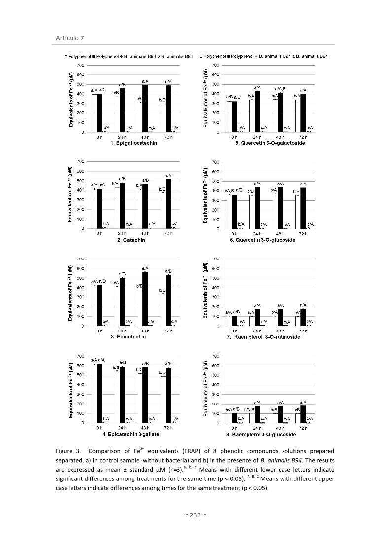
Artículo 7
~ 232 ~
Figure 3. Comparison of Fe2+
equivalents (FRAP) of 8 phenolic compounds solutions prepared
separated, a) in control sample (without bacteria) and b) in the presence of B. animalis B94. The results
are expressed as mean ± standard µM (n=3).a, b, c
Means with different lower case letters indicate
significant differences among treatments for the same time (p < 0.05). A, B, C
Means with different upper
case letters indicate differences among times for the same treatment (p < 0.05).
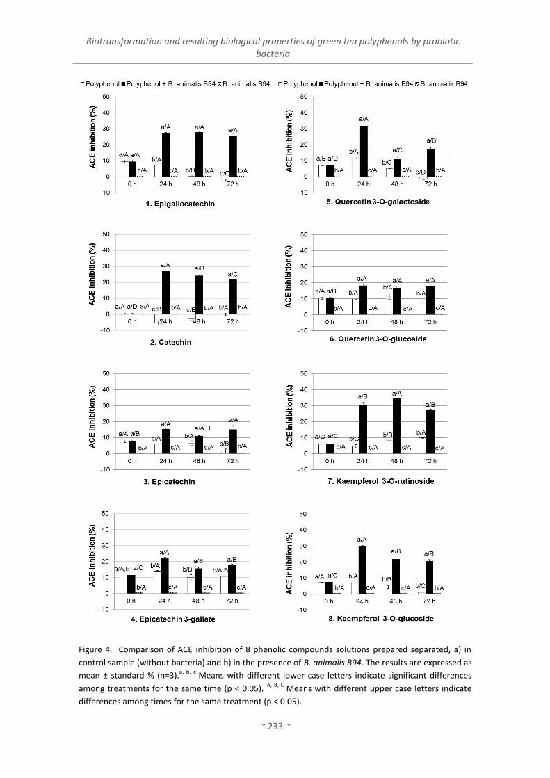
Biotransformation and resulting biological properties of green tea polyphenols by probiotic bacteria
~ 233 ~
Figure 4. Comparison of ACE inhibition of 8 phenolic compounds solutions prepared separated, a) in
control sample (without bacteria) and b) in the presence of B. animalis B94. The results are expressed as
mean ± standard % (n=3).a, b, c
Means with different lower case letters indicate significant differences
among treatments for the same time (p < 0.05). A, B, C
Means with different upper case letters indicate
differences among times for the same treatment (p < 0.05).
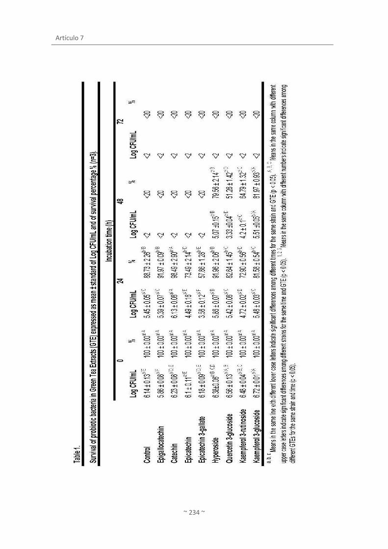
Artículo 7
~ 234 ~

~ 235 ~
ARTÍCULO 8
López de Lacey, A.M., López-Caballero, M.E. y Montero, P.
(2012)
Agar films containing green tea extract and probiotic
bacteria for extending fish self-life
Food Microbiology

Artículo 8
~ 237 ~
AGAR FILMS CONTAINING GREEN TEA EXTRACT AND
PROBIOTIC BACTERIA FOR EXTENDING FISH SHELF-LIFE
López de Lacey A.M., López-Caballero M.E*., Montero P.
Instituto de Ciencia y Tecnología de Alimentos y Nutrición (ICTAN-CSIC) (formerly Instituto del
Frío), C/ José Antonio Novais, 10, 28040. Madrid, Spain.
*Corresponding author: [email protected]
Tel.: +34 91 5492300; fax: +34 91 5493627.

Artículo 8
~ 238 ~
ABSTRACT
A bioactive film composed of agar, incorporating green tea extract and probiotic strains
(Lactobacillus paracasei L26 and Bifidobacterium lactis B94) was applied on hake fillets in order
to evaluate the effect of the films during the fish storage. Hake was previously inoculated with
Shewanella putrefaciens and Photobacterium phosphoreum to simulate a spoilage process. The
green tea and/or probiotic film provoked a reduction, particularly of H2S-producing bacteria
counts and inhibition of the luminescent colonies, and also reduced the total microflora,
Pseudomonas sp. and Enterobacteriaceae throughout the storage period. The probiotic strains
(B. animalis B94 and L. acidophilus L26) added to the film passed to the fish producing an
increment of lactic acid bacterial counts, even in the presence of green tea extract. The effect
of the films also caused a decreased in the indexes of fish quality (NBVT, TMA and pH). The
total viable counts and Enterobacteriaceae were maintained above the limits of acceptability
during 7 days for the fillet covered with the green tea + probiotic film, compared to only 2 days
for the rest of the samples. Films with green tea and probiotic films were able to extend shelf-
life of hake and increase the beneficial lactic acid bacteria in fish.
Key words: active packaging, film, green tea, probiotic bacteria, fish shelf life.

Agar films containing green tea extract and probiotic bacteria for extending fish shelf-life
~ 239 ~
1. INTRODUCTION
Fish is highly perishable during refrigerated storage, mainly due to rapid microbial growth
of microorganisms naturally present in fish or from contamination, which can occasionally
result in either economic or health-related problems (Gómez-Estaca, López de Lacey, López-
Caballero, Gómez-Guillén, & Montero, 2010). In this connection, Shewanella putrefaciens and
Photobacterium phosphoreum are some of the specific spoilage microorganisms that cause,
among other things, unpleasant off-odors in fish like the production of trimethylamine from
trimethylamine oxide (López-Caballero, Álvarez Torres, Sánchez-Fernández & Moral, 2002;
López-Caballero, Sánchez-Fernández & Moral, 2001; Dalgaard, 1995). For this reason, some
strategies have been recently developed with the objective to inhibit spoilage and pathogenic
microorganisms.
One of these strategies is the application of active edible films which have proved to
enhance shelf-life or even safety of fish (Gómez-Estaca, López de Lacey, Gómez-Guillén, López-
Caballero, & Montero, 2009; Gómez-Estaca, et al., 2010). In addition to being an alternative to
plastic packaging, edible films can avoid moisture loss, gas exchange, oxidation and photo-
degradation. Furthermore, safety, nutritional and even sensory properties of edible films can
be improved by the addition of several active ingredients into the polymer matrix (Gómez-
Estaca, et al., 2009; Gómez-Estaca, et al., 2010; Rojas-Graü, Soliva-Fortuny, & Martín-Belloso,
2009).
Along with these ingredients, green teas possess, among other healthy beneficial effects,
antimicrobial and antioxidant properties, primarily due to its own composition rich in
polyphenols and other compounds like minerals, amino acids (Graham, 1992; López de Lacey,
Pérez Santín, López-Caballero, & Montero, 2012a). These activities make tea extracts very
suitable for its use as an active ingredient in edible films. Lately green tea extracts added to
edible films, formulated with Gelidium corneum-gelatin blend or soy protein isolate, have been
reported to improve their physical and antimicrobial properties of these films (Hong, Lim, &
Song, 2009; Kim et al., 2006).
On the other hand, recently in the literature appears papers reporting that the
incorporation of probiotic bacteria to films contribute to the stability and security of food: that
is alginate-gellan incorporated with Bifidobacterium lactis Bb-12 for coating of fresh fruits
(apple and papaya) (Tapia et al., 2007) and in sodium-caseintate films incorporated with
Lactobacillus sakei to control Listeria monocytogenes in culture medium and in fresh beef

Artículo 8
~ 240 ~
(Gialamas et al., 2010). However, no references of the joint incorporation of probiotic bacteria
and phenolic compounds in edible films have been reported.
Thus, the aim of this work was to investigate the effect of applying the films incorporating
green tea extract and probiotic strains to fish during chilled storage. Changes on the natural
microflora, paying special attention to S. putrefaciens and P. phosphoreum, and to the
biochemical changes in muscle, were studied in order to evaluate the role of the films during
the spoilage of fish.
2. MATERIALS AND METHODS
2.1. Extraction of green tea
Chinese green tea (Camellia sinensis L.), known as Wu Lu Mountain, was used for
preparing the extract. Wu Lu Mountain was purchased from a local specialized tea store. Dry
green tea was ground into fine powder using a blender (Osterizer sunbeam model nº 4153-50).
To prepare water extract, the powder (35 g) was mixed with distilled water (350 mL) at 80 °C
for 30 minutes, with continuous stirring. The slurry mixture was centrifuged at 12000 g for 10
minutes at 5 °C. The supernatant was filtered twice through Whatman filter papers Nº 1.
Finally, the filtered extract was stored at - 20 °C before the preparation of the film and
analysis.
2.2. Bacterial strains and culture conditions
Two spoilage bacteria, obtained from the Spanish Type Culture Collection (CECT), were
used for the inoculation of the fish: Photobacterium phosphoreum CECT 4192 and Shewanella
putrefaciens CECT 5346T. The strains were stored at -80 °C in Brain Heart Infusion Broth
(Oxoid, Basingstoke, UK) with 25% glycerol (Panreac, Moncada i Reixac, Barcelona, Spain) until
use. The strains were grown in BHI broth (Oxoid) supplemented with 1% NaCl, and were
incubated at 30 °C for S. putrefaciens and at 15 °C for P. phosphoreum for 24 h. Then, each
bacteria suspension, obtained after incubation, was appropriately diluted in 0.9 % NaCl
solution in order to obtain a target inoculum of 103-104 CFU/mL.
Two commercial probiotic bacteria purchased in DSM (DSM Food Specialties Ltd., Sydney,
Australia) were used for the preparation of the bioactive films: Lactobacillus paracasei spp.
LAFTI@L26 and Bifidobacterium animalis spp. lactis LAFTI@B94. The selection of the two
strains was based on its antimicrobial properties against Escherichia coli, Listeria

Agar films containing green tea extract and probiotic bacteria for extending fish shelf-life
~ 241 ~
monocytogenes and Salmonella typhimurium (Mahoney & Henriksson, 1999; Pidcock, Heard, &
Henriksson, 2002), and its resistance to green tea extract studied. These strains were acquired
lyophilized, and were kept at -20 °C. Appropriate quantity of each lyophilized bacteria was
suspended in 0.9 % NaCl solution with the aim to obtain a target inoculum of 108-109 CFU/mL.
2.3. Preparation of films
Agar film forming solutions were prepared by dissolving 1.5 g of agar (Gold Agar,
Hispanagar, Burgos, Spain), 1 g of glycerol, 2 g of glucose in 100ml of water (agar film) or a
50/50 v/v mixture of distilled water and green tea extract (agar-green tea film). Glycerol was
employed as a plasticizer. The mixtures were stirred to obtain a good blend, and the films
made by casting 40ml on 144 cm2-square plates, drying afterwards at 40 °C in a forced-air oven
for 16-18h to yield a uniform thickness in all cases [200 μm (p≤0.05)]. The films were
conditioned in desiccators for 2 days at 22 °C at 63% relative humidity.
For preparing the bioactive film, 100 µL of each probiotic bacteria solution (Lactobacillus
paracasei LAFTI@L26 and Bifidobacterium animalis lactis LAFTI@B94), made as it was
explained above in section 2.2., were spread on each squared film (12×12 cm) with a sterile
loop. The inoculum was applied on agar and agar-green tea film. Four different types of films
were then obtained: (i) agar film, (ii) agar probiotic film, (iii) agar green tea film and (iv) agar
probiotic green tea film.
2.4. Fish preparation, inoculation and storage
Fillets of defrosted hake (Merluccius capensis,), purchased at a local market, were cut in
portions of (c.a. 100 g each). Each portion was inoculated with 5 mL of each spoilage bacteria
suspensions prepared as it was explained above in section 2.2., with the objective to have an
initial concentration of 103-4 CFU/g. Finally, each piece of fish was covered with two squared
films, which had the same dimensions (12 × 12 cm), and then vacuum-packed in bags (Cryovac
BB-1, Grace, Barcelona, Spain). Batches were stored at 4 °C during 15 days. Five lots were
prepared, all of them from hake inoculated with the microorganisms already mentioned: hake
(H), hake with film (HF), hake with film and probiotic bacteria (HFP), hake with green tea film
(HFT) and hake with green tea film and probiotic bacteria (HFTP).

Artículo 8
~ 242 ~
2.5. Microbiological analysis
The microbial counts on the sample were performed immediately after inoculation and
after 2, 7, 10 and 15 days of storage. The microbiological analyses were as follows: a total
amount of 10 g of fish, from at least 3 different packages, was collected and placed in a sterile
plastic bag (Sterilin, Stone, Staffordshire, UK) with 90 mL of buffered 0.1% peptone water
(Oxoid, Basingstoke, UK) in a vertical laminar-flow cabinet (mod. AV 30/70 Telstar, Madrid,
Spain.) After 1 min in a Stomacher blender (model Colworth Stomacher 400), appropriate
dilutions were prepared for the following microorganisms determinations: (i) luminescents
bacteria on spread plates of Iron Agar 1% NaCl incubated at 15 °C for 5 days as presumptive
Photobacterium phosphoreum; (ii) H2S-producing bacteria, as black colonies and presumptive
Shewanella putrefaciens, on spread plates of Iron Agar 1% NaCl incubated at 15 °C for 72 h; (iii)
total bacteria counts (TBC) on spread plates of Iron Agar 1% NaCl incubated at 15 °C for 72 h;
(iv) Enterobacteriaceae on double-layered plates of Violet Red Bile Glucose agar (VRBG, Oxoid)
incubated at 30 °C for 24 h; (v) Pseudomonas on spread plates of Pseudomonas Agar Base
(Oxoid) with added CFC (Cetrimide, Fucidine, Cephalosporine) supplement for Pseudomonas
spp. (Oxoid) incubated at 25 °C for 48 h; (vi) lactic acid bacteria on double-layered plates of
MRS Agar (Oxoid) incubated at 30 °C for 72 h; (vii) total viable bacteria on pour plates of PCA
incubated at 30 °C for 72 h. All microbial counts are expressed as the log of the colony-forming
units per gram (log CFU/g) of sample. All analyses were performed in triplicate.
2.6. pH
Approximately 5 g of muscle were homogenized with a double quantity (g/mL) of distilled
water. After 5 min at ambient temperature, pH was determined with a pHm93 pH meter and a
combined pH electrode (Radiometer, Copenhagen, Denmark). The experiments were repeated
at least in triplicate.
2.7. Total volatile basic nitrogen (TVB-N) and trimethylamine nitrogen (TMA-N)
Total volatile basic nitrogen (TVB-N) and trimethylamine nitrogen (TMA-N) determinations
were performed by the methods described by Antonacopoulos and Vyncke (1989) and Malle
and Tao (1987), respectively. Briefly, 10 g of the ground sample were weighed in a suitable
container and homogenized with 100 mL 6% perchloric acid solution for 2 min. After filtering,
the extract was alkalinized with 20% sodium hydroxide solution for TVB-N determination or
with 20% sodium hydroxide solution and formaldehyde for the TMA-N determination. Then,

Agar films containing green tea extract and probiotic bacteria for extending fish shelf-life
~ 243 ~
the extracts were submitted to steam distillation. The volatile base components were
absorbed by an acid receiver and determined by titration.
2.8. Instrumental colour analysis
The colour parameters lightness (L*), redness (a*), and yellowness (b*) were measured
using a Konica Minolta CM-3500d colorimeter (Aquateknica S.A., Valencia, Spain). Iluminant
D65 and 10º observer angle were used. Measurements were taken at a number of 4 locations
in different fish portions and each point is the mean of at least 16 measurements.
2.9. Statistical analysis
The results are expressed as mean ± standard deviation (sd). Statistical tests were
performed using the SPSS® computer program, version 19.0 (SPSS Statistical Software, Inc.,
Chicago, IL, USA). One-way and two-way analysis of variance (ANOVA) was carried out.
Differences between pairs of means were assessed on the basis of confidence intervals using
the Tukey-b test. The level of significance was p 0.05.
3. RESULTS AND DISCUSSION
3.1. Microbial counts
Microbial counts (Log CFU/g) of hake fillet covered with different films are shown in Figure
1. Fillets were previously inoculated with two spoilage bacteria S. putrefaciens and P.
phosphoreum, in order to simulate the fish spoilage and then to assess the effect of the films
during storage. In spite of the inoculation of fish with P. phosphoreum, the luminescent
bacteria could not be detected during the first days of storage (Fig. 1d). This is attributed to
the adaptation of the strain to the storage conditions.
All the bacterial counts in hake sample (H) increased through storage, except for lactic acid
bacteria (Fig. 1, Table 1). The lactic acid bacteria counts gradually decreased until small lower
initial values were reached at the end of storage (Fig 1f), which could be due to the low storage
temperature of fish. At the end of storage, H2S-producing bacteria became the dominant
group in hake (H) since their counts constitute the majority percentage of the total flora at 15
°C. The prevalence of S. putrefaciens was also observed by Baixas-Nogueras, Bover-Cid,
Veciana-Nogu s and Vidal-Carou (2003) in Mediterranean hake during storage.

Artículo 8
~ 244 ~
In general, the application of films on hake fillet during the storage, especially those that
contain green tea, reduced the major part of the microorganisms studied (p≤0.05). This effect
is much lower or even insignificant on films that do not contain tea (with or without bacteria),
whose counts were close or similar to those found in the control lot (Fig. 1).
Hake covered with the film + probiotic (HFP) showed an increment of the lactic flora at
day 2 of storage, which is corresponded to the increase of the total flora (30 °C) (Fig. 1b, f). The
increment of lactic acid bacteria can be due to the passage of probiotic bacteria from film to
muscle. A slight reduction of HzS-producing bacteria and Enterobacteriaceae was observed in
this lot at the early stages of the storage (Fig. 1c, g). The effect of probiotic bacteria against the
mentioned bacteria seems to be lost along the preservation of fish. However, other authors
observed an important improvement in the production and safety of the Hungarian salami by
the use of probiotic bacteria cultures (the same used in the present work) (Pidcock, et al.,
2002). In that case, salami was fermented with the probiotic bacteria at 25 °C, followed by
maturation at 15 °C; whereas in our study the conditions of fish storage were at 4 °C. This low
temperature may not favour the growth of the probiotic bacteria, and therefore the bacteria
might not be in a good condition to exert an effect on fish microflora.
The films containing green tea extract (samples HFT and HFTP) reduced or even inhibited
most of the studied microorganisms. Green tea extract is likely the principal responsible for
the inhibitory activity, especially for those counts of psychrotropic organisms (total bacteria at
15 °C, H2S-microorganisms, Pseudomonas sp., and luminiscent colonies) (Fig 1). The most
sensitive microorganisms were the luminescent colonies, presumptive P. phosphoreum (Fig.
1d), since the green tea films inhibited completely its growth, while lactic acid bacteria (Fig. 1f)
remained more or less constant during the storage. The decrease in counts was more
noticeable in H2S-producing bacteria and total flora (15 °C), with reductions up to 4 logarithmic
cycles (p≤0.05). It is also interesting to note that the inhibitory effect of green tea films is lost
over time in some cases. Thus, for example, Enterobacteriaceae was reduced to a large extent
at 10 days (p≤0.05) rather than at 15 days of storage. This could be due to some reasons: the
effect of tea might last a period of time and lose activity during the whole storage; the strains
might adapt to antimicrobial compounds of green tea and develop resistance to these
compounds or that the green tea films could not be able to inhibit the growth by the effect of
the bacterial proliferation.
The antimicrobial activity of the green tea extract (Wu Lu Mountain) added to the film has
been demonstrated against Gram negative bacteria, such us Photobacterium phosphoreum,
Shewanella putrefaciens, Pseudomonas fluorescens and Vibrio parahaemolyticus (López de

Agar films containing green tea extract and probiotic bacteria for extending fish shelf-life
~ 245 ~
Lacey, et al., 2012a). In this connection, green tea extracts (both aqueous and ethanolic ones)
showed inhibition on Gram-positive microorganisms (including Staphylococcus aureus, Bacillus
cereus and Listeria monocytogenes), but not on Gram-negative microorganisms (E. coli or
Salmonella enterica), (Chiu et al., 2010). These authors found that when green tea extract was
incorporated to edible coatings (based on tapioca starch/decolorized hsian-tsao leaf gun), a
clear antimicrobial activity on Gram-positive bacteria was also found since the active
compounds could migrate from the coating and increase the inhibition area.
Catechins, group of polyphenols, present abundantly in green tea extract, have been
reported as responsible for the antimicrobial action of tea infusions against Gram negative and
positive bacteria (Almajano, Carbó, Jiménez, & Gordon, 2008; Singh Arora, Jeet Kaur, & Kaur,
2009; Sivarooban, Hettiarachchy, & Johnson, 2008). Ku, Hong and Song, 2008 obtained that
the incorporation of catechin into the Gelidium–corneum edible film increased the degree of
inhibition of E. coli O157:H7, with an increase in the catechin concentration. Several effects of
catechins on the bacteria, proposed by different authors (Cushnie & Lamb, 2011), could
provoke the inhibition or the reduction of the bacterial growth such us: alteration of the cell
morphology (Sivarooban, et al., 2008), inhibition of essential enzymes (Gradišar, Pristovšek,
Plaper, & Jerala, 2007; Navarro-Martínez et al., 2005), inhibition of energy metabolism
(Chinnam et al., 2010) or the production of hydrogen peroxide by oxidation of catechins
(Arakawa, Maeda, Okubo, & Shimamura, 2004). According to Shimamura, Zhao and Hu (2007)
the inhibition caused by the attack of (-)-epigallocatechin-3-gallate (EGCG) to the lipid bilayer
of bacterial cell membrane may be minor in Gram-negative bacteria since this membrane is
protected by an outer cell wall. Our results showed that the sensitivity of Gram-negative
bacteria to catechins natural, presented in green tea incorporated into an edible film, could be
variable, ranging from major (P. phosphoreum) to minor inhibition (Pseudomonas sp.).
Previous studies have demonstrated that green tea do not affect the growth of
Bifidobacterium B94 and Lactobacillus L26 (López de Lacey et al., 2012b). For this reason, the
counts of lactic acid bacteria in both lots containing bacteria with tea (HFPT) or without tea
(HFP) should have been similar. However, >2 log units of difference was found (the lower
counts corresponding to HFPT) (Fig. 1f). Thus, it might be possible that green tea film could
affect the growth of lactic acid bacteria of hake somehow, but not affecting to the probiotic
inoculated.
In general, hake covered with green tea film and probiotic (HFTP) presented the lowest
values of microbial counts at the end of storage (p≤0.05). Thus, although green tea is the
principal responsible for the reduction of the microbial counts, the joint presence of probiotic

Artículo 8
~ 246 ~
bacteria could have a certain additive effect on microbial growth (Fig 1a, g, e). In this sense, Su,
Henriksson, Nilsson and Mitchell (2008) observed a clear synergistic effect between L.
acidophilus L10, B. animalis B94 and L.casei L26 and a green tea extract (“TEAVIGOTM”) in
relation to the inhibition of growth in vitro of S. aureus and Streptococcus pyogenes. Even
although previous references on the joint incorporation of tea extract and probiotics in an
edible film are not known, the relationship between probiotics and other polyphenolic
compounds is established. Thus, flavonoids have been reported to have effects on the gut
microflora as an increase in the count of beneficial bacterias (Lactobacillus rhamnosus),
enhance adherence of this bacterium on the gut wall and inhibition of the proliferation of
enteropahogens like S. aureus or Salmonella thyphimurium (Parkar, Stevenson & Skinner,
2008).
3.2. pH
Changes in pH of hake are shown in Figure 2a. Hake (H) used in the experiment had an
initial pH of 7.02. This pH remains constant until day 10, reaching pH>7.7 at 15 days (p≤0.05).
Similar behaviour was observed in HF and HFP lots, being the progression of this last sample
slower. On the contrary, the hake covered with green tea film, with or without probiotic
bacteria (HFT, HFTP), maintained the initial levels of pH ~7 at the end of storage period. This
result can be a consequence of the low bacterial counts observed in these samples since the
bacterial activity was reduced, and therefore, could produce minor quantities of some volatile
base compounds, which can provoke an increment of pH (Reddy, Villanueva, & Kautter, 1995).
3.3. Total volatile basic nitrogen (TVB-N) and trimethylamine nitrogen (TMA-N)
The initial TVB-N was 9.95 mg/100 g fish (Figure 2b). The TVB-N values increased gradually
throughout the conservation in all the samples, except in the hake covered with the green tea
film, without (HFT) or with probiotic (HFTP). Thus, the production of TVB-N of these samples
was markedly reduced by about 75 and 62 %, respectively, comparing with the control at day
15 of storage. These results are consistent with the higher antimicrobial capacity of these films
(Fig 1). It is also worth noting that the hake covered with green tea maintained the levels of
TVB-N above the limit of acceptability (30 to 35 mg/100 g) set by the European Union
regulation (EC, 1995). The initial TMA-N in hake was 9.43 mg/100 g fish (Figure 2c). The major
reduction in these values was also noticed in the HFT and HFTP lots at the end of the storage,
with decreases around 80 % compared with the control sample (H).

Agar films containing green tea extract and probiotic bacteria for extending fish shelf-life
~ 247 ~
In both indexes, TMA and TVB, the behavior of the lots is similar until 10 day, from which
the lots that do not contain green tea extract in their film formulation presented an
exponential rise of the amine volatile concentration. This fact was consistent with the
lessening of H2S-producing bacteria and luminescent colonies, which were also observed from
day 10. Thus, green tea film seems to be the cause of the reduced levels of TVB-N and TMA-N,
caused mainly by the inhibition or reduced growth of bacteria such us S. putrefaciens and P.
phosphoreum, which are involved in the production of basic compounds (TVB-N and TMA-N) in
fish (López-Caballero et al., 2001, 2002; Dalgaard, 1995).
3.4. Colour
All the samples maintained stable values of lightness (L*) throughout the conservation and
although significant small differences were founded in some cases, they do not differ by more
than 10 per cent. The low values of redness (a*) are according to the lack of hematic pigment,
etc. characteristic of hake. The samples H and HF did not present modifications (p≤0.05) of the
tendency towards yellowness value (b*). However, the application of the green tea film
produced a slight yellow tone in the fish which can be attributed to the polyphenols present in
green tea. This colour was stable throughout the storage (p≤0.05).
In summary, the application of green tea films on hake delayed or even prevented the
growth of microorganisms in fish and, therefore, reduced the spoilage indexes. The probiotic
strains passed to the fish, producing an increment of lactic acid bacterial counts, even in the
presence of green tea extract; but they had a smaller effect over the chemical spoilage
indicators. Probiotic strains alone exerted a biopreservation on fish by reducing the H2S-
producing organisms. Thus, it can be concluded that films with green tea and probiotic films
could extend shelf-life of hake and, at the same time, it could be a way to incorporate
beneficial probiotics strains to the fish.
Acknowledgements
Funding for this research was provided by Ministerio de Ciencia e Innovación under projects I3-
200670l141, AGL2008-00231/ALI and CYTED 309ACO382. Program JAE-CSIC predoctoral is fully
acknowledged. The author thanks Joan Therese de Lacey for the English language revision of
the article.
This centre has implemented and maintains a Quality Management System which fulfils the
requirements of the ISO standard 9001:2000.

Artículo 8
~ 248 ~
References
Almajano, M. P., Carbó, R., Jiménez, J. A. L., & Gordon, M. H. (2008). Antioxidant and
antimicrobial activities of tea infusions. Food Chemistry, 108(1), 55-63.
Antonacopoulos, N., & Vyncke, W. (1989). Determination of volatile basic nitrogen in fish: A
third collaborative study by the West European Fish Technologists' Association
(WEFTA). Zeitschrift für Lebensmittel-Untersuchung und -Forschung, 189(4), 309-316.
Arakawa, H., Maeda, M., Okubo, S., & Shimamura, T. (2004). Role of hydrogen peroxide in
bactericidal action of catechin. Biological and Pharmaceutical Bulletin, 27(3), 277-281.
Baixas-Nogueras, S., Bover-Cid, S., Veciana-Nogu s, M. T., & Vidal-Carou, M. C. (2003). Amino
acid-decarboxylase activity in bacteria associated with Mediterranean hake spoilage.
European Food Research and Technology, 217(2), 164-167.
Cushnie, T. P. T., & Lamb, A. J. (2011). Recent advances in understanding the antibacterial
properties of flavonoids. International Journal of Antimicrobial Agents, 38(2), 99-107.
Chinnam, N., Dadi, P. K., Sabri, S. A., Ahmad, M., Kabir, M. A., & Ahmad, Z. (2010). Dietary
bioflavonoids inhibit Escherichia coli ATP synthase in a differential manner.
International Journal of Biological Macromolecules, 46(5), 478-486. doi:
10.1016/j.ijbiomac.2010.03.009
Chiu, P.-E., & Lai, L.-S. (2010). Antimicrobial activities of tapioca starch/decolorized hsian-tsao
leaf gum coatings containing green tea extracts in fruit-based salads, romaine hearts
and pork slices. International Journal of Food Microbiology, 139(1–2), 23-30. doi:
10.1016/j.ijfoodmicro.2010.01.010
Dalgaard, P. (1995). Qualitative and quantitative characterization of spoilage bacteria from
packed fish. International Journal of Food Microbiology, 26(3), 319-333.
EC. The Commission of the European Communities 95/149/EC (1995). Determination of the
concentration of volatile nitrogenous bases (TVB-N) in fish and fish products: a
reference procedure. Official Journal of the European Communities, Nº L 97:84-7.
Gialamas, H., Zinoviadou, K. G., Biliaderis, C. G., & Koutsoumanis, K. P. (2010). Development of
a novel bioactive packaging based on the incorporation of Lactobacillus sakei into
sodium-caseinate films for controlling Listeria monocytogenes in foods. Food Research
International, 43(10), 2402-2408. doi: 10.1016/j.foodres.2010.09.020
Gómez-Estaca, J., López de Lacey, A., Gómez-Guillén, M. C., López-Caballero, M. E., & Montero,
P. (2009). Antimicrobial Activity of Composite Edible Films Based on Fish Gelatin and

Agar films containing green tea extract and probiotic bacteria for extending fish shelf-life
~ 249 ~
Chitosan Incorporated with Clove Essential Oil. Journal of Aquatic Food Product
Technology, 18(1-2), 46-52. doi: 10.1080/10498850802581252
Gómez-Estaca, J., López de Lacey, A., López-Caballero, M. E., Gómez-Guillén, M. C., & Montero,
P. (2010). Biodegradable gelatin-chitosan films incorporated with essential oils as
antimicrobial agents for fish preservation. Food Microbiology, 27(7), 889-896.
Gradišar, H., Pristovšek, P., Plaper, A., & Jerala, R. (2007). Green tea catechins inhibit bacterial
DNA gyrase by interaction with its ATP binding site. Journal of Medicinal Chemistry,
50(2), 264-271.
Graham, H. N. (1992). Green tea composition, consumption, and polyphenol chemistry.
Preventive Medicine, 21(3), 334-350. doi: 10.1016/0091-7435(92)90041-f
Hong, Y. H., Lim, G. O., & Song, K. B. (2009). Physical properties of Gelidium corneum-gelatin
blend films containing grapefruit seed extract or green tea extract and its application
in the packaging of pork loins. Journal of Food Science, 74(1), C6-C10.
Kim, K. M., Lee, B. Y., Kim, Y. T., Choi, S. G., Lee, J., Cho, S. Y., & Choi, W. S. (2006).
Development of antimicrobial edible film incorporated with green tea extract. Food
Science and Biotechnology, 15(3), 478-481.
Ku, K. J., Hong, Y. H., & Song, K. B. (2008). Mechanical properties of a Gelidium corneum edible
film containing catechin and its application in sausages. Journal of Food Science, 73(3),
C217-C221. doi: 10.1111/j.1750-3841.2008.00700.x.
López-Caballero, M. E., Álvarez Torres, M. D., Sánchez-Fernández, J. A., & Moral, A. (2002).
Photobacterium phosphoreum isolated as a luminescent colony from spoiled fish,
cultured in model system under controlled atmospheres. European Food Research and
Technology, 215(5), 390-395.
López-Caballero, M. E., Sánchez-Fernández, J. A., & Moral, A. (2001). Growth and metabolic
activity of Shewanella putrefaciens maintained at 0 °C under different CO2 and O2
concentrations. International Journal of Food Microbiology, 64 (3):277-287.
López de Lacey, A. M., Pérez Santín, E., López-Caballero, M. E., & Montero, P. (2012a).
Antioxidant and antimicrobial activities of green tea (Camellia sinensis L.) as an
expression of its chemical composition. (Submitted).
López de Lacey, A. M., Pérez Santín, E., López-Caballero, M. E., & Montero, P. (2012b). Survival
and metabolic activity of probiotic bacteria in green tea. (Submitted).

Artículo 8
~ 250 ~
Mahoney M., H. G., & Henriksson A. (1999). Novel probiotic lactic acid bacteria that grow in
curing salts and inhibit pathogens associated with fermented meat products. In:
Seventeenth International Conference of the International Committee on Food
Microbiology and Hygiene (ICFMH, Food Micro '99), Veldhoven, The Netherlands,
1999.
Malle, P., & Tao, S. H (1987). Rapid quantitative determination of trimethylamine using steam
distillation. Journal of Food Protection, 50(9), 756–760.
Navarro-Martínez, M. D., Navarro-Perán, E., Cabezas-Herrera, J., Ruiz-Gómez, J., García-
Cánovas, F., & Rodríguez-López, J. N. (2005). Antifolate activity of epigallocatechin
gallate against Stenotrophomonas maltophilia. Antimicrobial Agents and
Chemotherapy, 49(7), 2914-2920.
Parkar, S. G., Stevenson, D. E., & Skinner, M. A. (2008). The potential influence of fruit
polyphenols on colonic microflora and human gut health. International Journal of Food
Microbiology, 124(3), 295-298.
Pidcock, K., Heard, G. M., & Henriksson, A. (2002). Application of nontraditional meat starter
cultures in production of Hungarian salami. International Journal of Food Microbiology,
76(1-2), 75-81.
Reddy, N. R., Villanueva, M., & Kautter, D. A. (1995). Shelf life of modified-atmosphere-
packaged fresh tilapia fillets stored under refrigeration and temperature-abuse
conditions. Journal of Food Protection, 58(8), 908-914.
Rojas-Graü, M. A., Soliva-Fortuny, R., & Martín-Belloso, O. (2009). Edible coatings to
incorporate active ingredients to fresh-cut fruits: a review. Trends in Food Science and
Technology, 20(10), 438-447.
Shimamura, T., Zhao, W. H., & Hu, Z. Q. (2007). Mechanism of action and potential for use of
tea catechin as an anti-infective agent. Anti-Infective Agents in Medicinal Chemistry,
6(1), 57-62.
Singh Arora, D., Jeet Kaur, G., & Kaur, H. (2009). Antibacterial activity of tea and coffee: Their
extracts and preparations. International Journal of Food Properties, 12(2), 286-294.
Sivarooban, T., Hettiarachchy, N. S., & Johnson, M. G. (2008). Transmission electron
microscopy study of Listeria monocytogenes treated with nisin in combination with
either grape seed or green tea extract. Journal of Food Protection, 71(10), 2105-2109.

Agar films containing green tea extract and probiotic bacteria for extending fish shelf-life
~ 251 ~
Su, P., Henriksson, A., Nilsson, C., & Mitchell, H. (2008). Synergistic effect of green tea extract
and probiotics on the pathogenic bacteria, Staphylococcus aureus and Streptococcus
pyogenes. World Journal of Microbiology and Biotechnology, 24(9), 1837-1842.
Tapia, M. S., Rojas-Graü, M. A., Rodríguez, F. J., Ramírez, J., Carmona, A., & Martin-Belloso, O.
(2007). Alginate- and Gellan-Based Edible Films for Probiotic Coatings on Fresh-Cut
Fruits. Journal of Food Science, 72(4), E190-E196. doi: 10.1111/j.1750-
3841.2007.00318.x

Artículo 8
~ 252 ~
Figure legends
Figure 1. Microbial counts in hake during chilled storage (4 °C). (H), hake-film (HF), hake-
film + probiotic (HFP), hake-green tea film (HFT) and hake-green tea film +
probiotic bacteria (HFTP)
Figure 2. pH, total volatile base nitrogen (TVB-N) and trimethylamine nitrogen (TMA-N) of
hake during chilled storage (4 °C). Hake (H), hake-film (HF), hake-film + probiotic
(HFP), hake-green tea film (HFT) and hake-green tea film + probiotic bacteria
(HFTP).

Agar films containing green tea extract and probiotic bacteria for extending fish shelf-life
~ 253 ~
Figure 1.
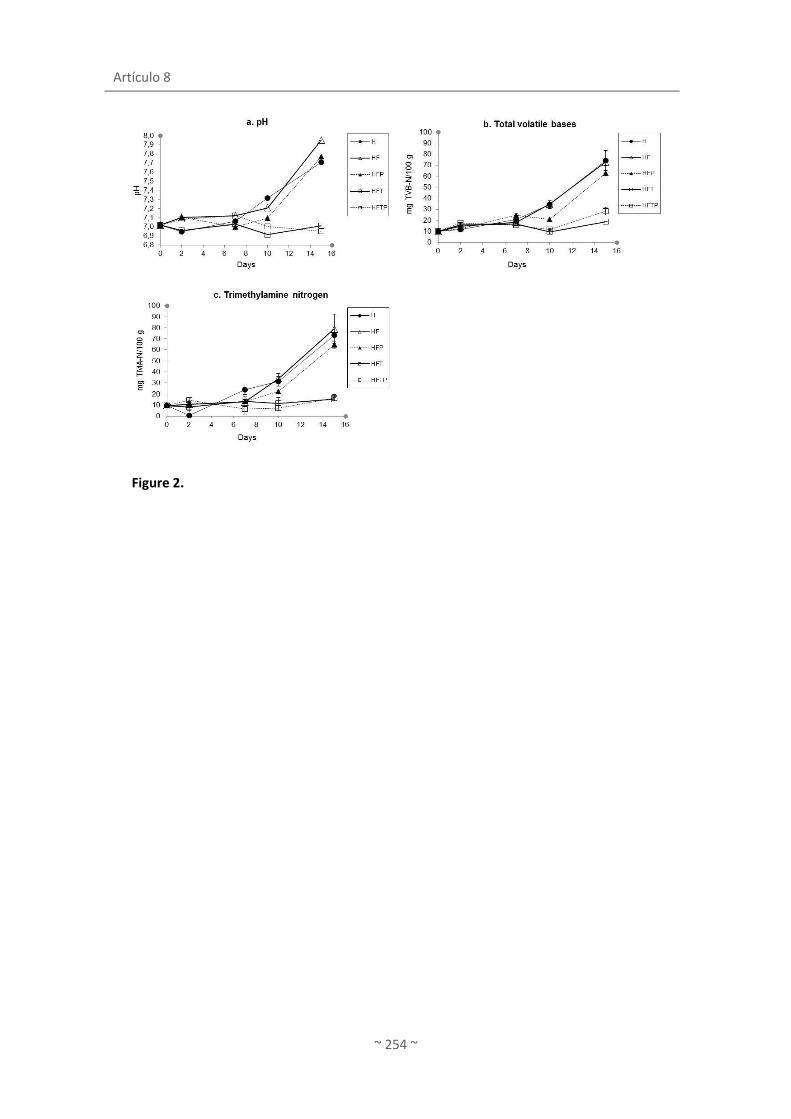
Artículo 8
~ 254 ~
Figure 2.

~ 255 ~
ARTÍCULO 9
López de Lacey, A. M., Giménez, B., Pérez-Santín, E.,
Faulks R., Mandalari, G., López-Caballero, M. E. y
Montero, P. (2012)
Bioaccesibility of green tea polyphenols incorporated into
an edible agar film during simulated human digestion
Food Research International.
doi:10.1016/j.foodres.2012.04.024.


Artículo 9
~ 257 ~

Artículo 9
~ 258 ~
BIOACCESSIBILITY OF GREEN TEA POLYPHENOLS
INCORPORATED INTO AN EDIBLE AGAR FILM DURING
SIMULATED HUMAN DIGESTION
López de Lacey, A. M.a, Giménez, B.a,*, Pérez-Santín, E.a, Faulks R.b, Mandalari, G.b,c, López-
Caballero, M. E.a, Montero, P.a
a Instituto de Ciencia y Tecnología de Alimentos y Nutrición (ICTAN), CSIC**, C/ José Antonio
Novais, 10, 28040. Madrid, Spain.
b Model Gut Platform, Institute of Food Research, Norwich Research Park, Colney, Norwich,
NR4 7UA, United Kingdom.
c Pharmaco-Biological Department, University of Messina, Vill. SS. Annunziata 98168 Messina,
Italy
* Corresponding author: e-mail: [email protected]; Tel.: +34 91 5492300; fax: +34 91
5493627.
** This centre has implemented and maintains a Quality Management System which fulfils the
requirements of the ISO standard 9001:2000.

Biaccessibility of green tea polyphenols incorporated into an edible agar film during simulated human digestion
~ 259 ~
ABSTRACT
Simple edible films can be manufactured to meet not only their primary protective
purpose but can be easily manipulated to meet sensory expectations and to contain
compounds which enhance the protective properties or even have the potential to deliver
health benefits. However, the use of such edible films not only to protect the food but as a
vehicle to deliver health benefits has not been investigated. In this paper we study agar films
containing an aqueous extract of green tea, rich in polyphenol compounds, and the
bioaccessibility of these compounds during simulated digestion in the upper gastro-intestinal
tract using a dynamic gastric model (DGM) and a static duodenal model. It is concluded that
the recovery of the tea compounds added to the agar film mainly occurs in the stomach (50-
80%) and that little or no additional recovery is observed in the duodenum. Furthermore, the
green tea compounds recovered show both reducing power and radical scavenging ability, but
not antimicrobial activity. The bioaccessibility of the green tea flavonols is reduced in the
presence of gelatin used to simulate the presence of protein in the stomach, but it is not clear
if this is due to reduced release or sequestration of released compounds by the gelatin.
Key words
Edible film, green tea, polyphenols, bioaccessibility, digestion
Abbreviations
EGC, (-)-epigallocatechin; C, (+)-catechin; EC, (-)-epicatechin; ECG, (-)-epicatechin-3-gallate;
EGCG, (-)-epigallocatechin gallate; DGM, dynamic gastric model; HPLC, high performance liquid
chromatography; FRAP, ferric reducing ability of plasma; ABTS, 2,2’-azino-bis-(3-
ethylbenzothiazoline-6-sulfonic acid); VCEAC, vitamin C equivalent antioxidant capacity; T,
green tea extract; FT, agar-green tea film; TG, green tea extract + gelatin; FTG, agar-green tea
film + gelatin.

Artículo 9
~ 260 ~
1. INTRODUCTION
The quality and safety of fresh and processed food can be enhanced by the application of
edible films and coatings. These edible films can avoid moisture loss, gas exchange, oxidation,
photo degradation and control surface microbial spoilage and contamination. Furthermore,
safety, nutritional and even sensory properties of edible films and coatings can be improved by
the addition of several active ingredients into the polymer matrix (Gómez-Estaca, López De
Lacey, Gómez-Guillén, López-Caballero, & Montero, 2009; Gómez-Estaca, López de Lacey,
López-Caballero, Gómez-Guillén, & Montero, 2010; Rojas-Graü, Soliva-Fortuny, & Martín-
Belloso, 2009). Green tea extract has been recently used as a safe active ingredient in edible
films for the improvement of physical and antimicrobial properties (Hong, Lim, & Song, 2009;
Kim et al., 2006).
Green teas are rich in flavan-3-ols (epicatechin, catechin and galloylated derivates), which
are responsible for the majority of biological activities. Other polyphenols present in green
teas are the flavonols which are mainly glycosylated derivatives of quercetin and kaempferol.
Green tea also contains pigments, amino acids, vitamins, carbohydrates, minerals and purine
alkaloids (Graham, 1992). The beneficial effects of green tea have been widely described
(Graham, 1992) and attributed to many of these compounds, but mainly to phenolic
compounds and their antioxidant properties (Bolling, Chen, & Blumberg, 2009; Weisburger &
Chung, 2002). When incorporated into edible films, green tea extract can have two functions:
active packaging (improving shelf life and quality of food) (Hong et al., 2009) and as a vehicle
to deliver compounds which may have potential beneficial effects when the edible film is
consumed and releases the polyphenols.
In the literature there are scarcely any studies on the simulated digestibility of edible films
and/or on the release of active compounds incorporated into them. However, in order to
retain the potential health benefits of green tea components incorporated into edible films, it
is essential to know if they are released during digestion. Only the polyphenols released from
solid matrices become bioaccessible and are potentially available for absorption by the gastro-
intestinal tract and, therefore, able to exert their beneficial effects in the human body
(Tagliazucchi, Verzelloni, Bertolini, & Conte, 2010). Other factors involved in the
bioaccessibility of polyphenols are the transformations (degradation, epimerization, hydrolysis
and oxidation) suffered by polyphenols under gastro-intestinal conditions and the interaction
between these compounds and food components, which may also modify the biological
Biaccessibility of green tea polyphenols incorporated into an edible agar film during simulated human digestion
~ 261 ~
activity of the phenolic compounds. Interactions between phenolic compounds and some
dietary factors, for example proteins and iron, can alter the biological properties and
bioaccessibility of polyphenols (Argyri, Proestos, Komaitis, & Kapsokefalou, 2005). Proteins can
link to polyphenols and form protein-polyphenol complexes by multiple weak interactions
(mainly hydrophobic) or strong interactions (covalent bonds) (Ishii et al., 2008; von Staszewski,
Pilosof, & Jagus, 2011). These interactions are affected by the nature of the protein, the
temperature of the system and the presence of other components (von Staszewski et al.,
2011).
In this study green tea extract and green tea edible films were subjected to simulated
human digestion in the upper gastrointestinal (GI) tract (gastric and duodenal digestion). The
release of total polyphenols as well as the major flavan-3-ol and flavonols of the green tea was
determined in aliquots collected at different times during in vitro GI digestion of both tea
extract and edible films. Furthermore, the antioxidant and antimicrobial activities were
determined in these aliquots and compared with those of the green tea extract. To assess the
effect of the well known interactions between protein and polyphenols, gelatin was added to
simulate the presence of dietary protein during the digestion.
2. MATERIALS AND METHODS
2.1. Chemical and enzymes
Standard polyphenol compounds: (-)-epigallocatechin (EGC; CAS: 970-74-1), (+)-catechin
(CAS:154-23-4), (-)-epicatechin (EC; CAS: 490-46-0), (-)-epicatechin-3-gallate (ECG; CAS: 1257-
08-5), (-)-epigallocatechin gallate (EGCG; CAS: 989-51-5), rutin (CAS: 153-18-4), Hyperoside
(CAS: 482-36-0), quercetin-3-O- glucoside (CAS: 482-35-9), kaempferol-3-O-rutinoside (CAS:
17650-84-9), kaempferol-3-O-glucoside (CAS: 480-10-4) were purchased from Extrasynthese
(Genay, Cedex, France).
In vitro digestion: Egg L-α-phosphatidylcholine (PC, lecithin grade 1, 99% purity) was
obtained from Lipid Products (South Nutfield, Surrey, U.K.). Porcine gastric mucosa pepsin
(activity of 3,300 U/mg of protein calculated by using hemoglobin as substrate), bovine α-
chymotrypsin (activity of 40 U/mg of protein using benzoyl-L-tyrosine ethyl ester as substrate),
α-amylase (activity 10 U/mg of solid using starch as substrate), porcine trypsin (activity of
13,800 U/mg of protein using benzoyl-L-arginine ethyl ester as substrate), porcine pancreatic
lipase (activity of 25,600 U/mg protein), cholesterol, porcine colipase, sodium taurocholate

Artículo 9
~ 262 ~
and sodium glycoldeoxycholate were obtained from Sigma (Poole, Dorset, U.K.). Lipase for the
gastric phase of digestion was a gastric lipase analogue of fungal origin (F-AP15; activity of
≥150 U/mg) from Amano Enzyme (Nagoya, Japan). Porcine gelatin (60 bloom), 2,4,6-tripyridyl-
s-triazine, FeCl3 , FeSO4, ABTS radical [2, 2’-azino-bis-(3-ethylbenzothiazoline-6-sulfonic acid)],
potassium persulphate, and L-ascorbic acid were purchased from Sigma-Aldrich, St. Louis, Mo.,
USA.
2.2. Extraction of green tea
Chinese green tea known as Wu Lu Mountain (Camellia sinensis L.) was purchased from a
local specialized tea store. The dry green tea was ground into powder using a blender (model
4253-50, Oster, Madrid, Spain). To prepare water extract, the powder (35 g) was mixed with
MiliQ water (350 mL) at 80 °C for 30 min, with continuous stirring. The slurry was centrifuged
at 12 000 g for 10 min at 5 °C. The decanted supernatant was filtered twice through Whatman
Nº 1. The filtered extract was stored at - 20 °C until preparation of the film and analysis.
2.3. Preparation of films
Agar film forming solutions were prepared by dissolving 1.5 g of agar (Gold Agar,
Hispanagar, Burgos, Spain) and 1 g of glycerol in 100ml of water (agar film, F) or a 50/50 v/v
mixture of distilled water and green tea extract (agar-green tea film, FT). The mixtures were
stirred to obtain a good blend, and the films made by casting 40ml on 144 cm2-square plates,
drying afterwards at 40 °C in a forced-air oven for 16-18h to yield a uniform thickness in all
cases [200 μm (p≤0.05)]. Films were conditioned in desiccators for 2 days at 22 °C at 58%
relative humidity.
2.4. In vitro digestion studies
Duplicate in vitro digestions under gastric and duodenal conditions with and without
gelatin (16 mL of porcine gelatin solution at 0.5 % per digestion) were carried out on: green tea
extract (T), agar-green tea film (FT), green tea extract + gelatin (TG) and agar-green tea film +
gelatin (FTG). The agar film without green tea (F) extract was used as a control to check for
interference in the analysis of polyphenols by digestion products.
Before performing the simulated human digestion, 20 mL of green tea extract, volume
necessary to prepare 1.1 g of film, was mixed with 20 mL of simulated gastric secretion and
made up to 240 mL with sterile water. The film (1.1 g) was mixed with the same proportions of
Biaccessibility of green tea polyphenols incorporated into an edible agar film during simulated human digestion
~ 263 ~
sterile water (240 mL) and gastric solution (20 mL) before the in vitro digestion studies. Porcine
gelatin solution (16 mL) at 0.5 % was added to the sterile water in the samples with gelatin,
keeping the same proportion previously mentioned. The samples thus dissolved were fed to
the dynamic gastric model (DGM), used to provide a realistic and predictive simulation of
human gastric processing. The DGM is a computer controlled gastric model which incorporates
the chemical, biochemical, physical environment and processes of the human stomach, based
on the kinetic data derived from Echo planar -MRI and data on the rates of GI digestion
obtained from human studies via naso-gastric and naso-duodenal aspiration and ileostomy
patients (Marciani et al., 2007; Marciani et al., 2008).
The simulated gastric secretions, bile and pancreatic bile, were prepared as previously
reported (Pitino et al., 2010) with some modifications. The DGM works in real time to produce
a variable number of samples simulating the digesta that would be emptied from the antrum
into the duodenum (Pitino et al., 2010). The digestion conditions of each sample were such as
to provide 6 samples over the total gastric digestion time of 24 min, and a gastric emptying
rate of approximately 11.3 mL/min. Each gastric sample was weighed, the pH measured and
adjusted to 6.8 with NaOH in order to inhibit gastric enzyme activity before performing the
determinations.
2.4.1. Simulated gastric secretions
The simulated gastric acid solution contained HCl (0.2 M), NaCl (58 mM), KCl (30 mM),
CaCl2 (0.5 mM) and NaH2PO4 (0.9 mM). The simulated gastric enzyme solutions were prepared
by dissolving porcine gastric mucosa pepsin (8962 U/mL) and gastric lipase (60 U/mL) in the
above described salt mixture (no acid). A solution of single shelled lecithin liposomes prepared
as described by (Mandalari et al., 2008) was added to the gastric enzyme solution at a final
concentration of 0.38 mM.
2.4.2. Simulated bile
Simulated bile contained lecithin (6.5 mM), cholesterol (3 mM, sodium taurocholate (12.5
mM) and sodium glycodeoxycholate (12.5 mM) in a salt solution made of NaCl (146.0 mM),
CaCl2 (2.6 mM) and KCl (4.8 mM). The suspension was covered with a blanket of nitrogen,
adjusted to pH 6.8 and equilibrated in an orbital shaking incubator (170 rpm) at 37 °C.
Artículo 9
~ 264 ~
2.4.3. Simulated pancreatic juice
Pancreatic juice solution contained NaCl (146 mM), KCl (4.8 mM), CaCl2 (2.6 mM), MgCl2
(0.7 mM) and ZnSO4·7H2O (36.9 µM). Porcine pancreatic lipase (500 U/mL), porcine colipase
(0.2 mg/mL), porcine trypsin (11 U/mL), bovine α-chymotrypsin (24 U/mL) and porcine α-
amylase (300 U/mL) were added to pancreatic juice.
2.4.4. Simulated duodenal digestion
The serial samples obtained from the DGM were incubated under duodenal conditions as
previously reported (Pitino et al., 2010). An aliquot (3.3 mL) of each gastric sample was used
for simulated duodenal digestion and 1 mL of hepatic mix and 2.8 mL of pancreatic enzymes
solution were added. Samples were digested for 2 h at 37 °C, and aliquots (1 mL) were taken
every 30 min for the different analyses.
2.5. Determination of phenol compounds and antioxidant activity
The different determinations were carried out on aliquots of a timed series of samples
collected during simulated gastric and duodenal digestion. These determinations permitted
the calculation of the percentage of total phenolic compounds, eleven green tea flavonoids
(HPLC), reducing power (FRAP assay) and radical scavenging capacity (ABTS assay) released
over time during each stage of the digestion.
2.5.1. Determination of total phenolic
Although the Folin-Ciocalteu or total phenolic assay has been used for many years as a
measure of total phenolics in natural products, this method actually measures the sample´s
reducing capacity and therefore can be considered as an antioxidant capacity assay (Prior, Wu,
& Schaich, 2005; Huang, Ou, & Prior, 2005). The total phenolic content in the samples was
determined spectrophotometrically (UV-1601, model CPS-240, Shimadzu, Kyoto, Japan) in
triplicate using gallic acid as a standard according to a modified method of Slinkard and
Singleton (1977) with the Folin-Ciocalteau´s reagent. An aliquot of 20 µL of sample was mixed
with 1.58 mL of distilled water and oxidized with 100 µL of Folin-Ciocalteau reagent sample.
The reaction was alkalinized with 300 µL of 20% sodium carbonate solution and incubated for
2 h at room temperature. The absorbance of the resulting blue colour was measured at 765
nm. Total phenolic content was expressed as mg gallic acid (GA) equivalent/mL of sample.

Biaccessibility of green tea polyphenols incorporated into an edible agar film during simulated human digestion
~ 265 ~
2.5.2. Quantification by HPLC
Reverse phase high performance liquid chromatography was performed to analyse
phenolic compounds present in the aqueous green tea extract. The separation module
consisted of an model SPE-MA10AVP HPLC-UV (Shimadzu, Kyoto, Japan), equipped with a C18
column (25 x 0.78 cm, 5 µm, C18 Tracer Excel 120 ODS-A column, Teknokroma, Barcelona,
Spain) and a UV-Vis detector (SPD-10V). The samples were eluted with a gradient system
consisting of a solvent A (MiliQ water) and solvent B (acetonitrile) both containing 1% formic
acid at a flow rate of 2.8 mL/min. The temperature of the column was maintained at 25 °C, and
the injection volume was 20 µL. The gradient system started at 95% solvent A and decreased
to 75% A over 70 min, followed by a further decrease to 35% solvent A over 10 min. The final
conditions were held for 10 min. The peaks of the phenolic compounds were monitored by
absorbance at 210 nm and 350 nm.
The quantification was carried out using solutions of each standard at various
concentrations. Individual compounds were quantified using a calibration curve of the
corresponding standard compound. All analyses were performed in triplicate.
2.5.3. Antioxidant activity
2.5.3.1. FRAP
The ferric reducing ability of plasma (FRAP) assay was carried out according to the method
described by (Benzie & Strain, 1996) with some modification. The sample (60 μl) was incubated
(37 °C) with 60 μl of MilliQ water and 900 μl of FRAP reagent, containing 10 mM TPTZ (2, 4,6-
tripyridyl-s-triazine) and 20 mM FeCl3. Absorbance values were read at 595 nm after 30 min,
using an UV-1601 spectrophotometer (Model CPS-240, Shimadzu, Japan). Results were
expressed as μmol Fe2+ equivalents/g of sample based on a standard curve of FeSO4·7H2O. All
determinations were performed at least in triplicate.
2.5.3.2. ABTS
The ABTS radical [2,2’-azino-bis-(3-ethylbenzothiazoline-6-sulfonic acid)] scavenging
capacity was carried out according to the method of (Re, Pellegrini, Proteggente, Pannala,
Yang, & Rice-Evans, 1999). The stock solution of ABTS (7 mM ABTS in potassium persulphate
2.45 mM,) was kept in the dark at room temperature for 16 h. An aliquot of stock solution was
diluted with MilliQ water in order to prepare the working solution of ABTS radical with
Artículo 9
~ 266 ~
absorbance at 734 nm of 0.70 ± 0.02. A 20 µL aliquot of sample was mixed with 980 μL of ABTS
reagent. The mixture was then left to stand in the dark at 30 °C for 10 min and absorbance
values were read at 734 nm. Results were expressed as mg of Vitamin C Equivalent Antioxidant
Capacity (VCEAC)/g of sample based on a standard curve of L-ascorbic acid. All determinations
were performed at least in triplicate.
2.6. Antimicrobial activity
2.6.1. Bacterial strains and growth conditions
The antimicrobial activity of the samples was determined by the disk diffusion method in
agar against 26 microbial strains (including Gram-positive and Gram-negative bacteria, yeast
and molds) from the Spanish Type Culture Collection (CECT) as previously described by Gómez-
Estaca et al. (2010). Briefly, spread plates of medium were inoculated with 100 µL of bacterial
suspensions microorganism (105-106 CFU/mL) overnight grown. Filter paper disks (0.5 cm
diameter), soaked with 40 μl of sample were put on the plate surface. After incubation the
inhibition area (mm) was measured using specific software for digital image analysis (MIP 4
ADV, ver. 1. Consulting the Imagen Digital, S.L. & Microm España). Results were expressed as
the percentage of inhibition with respect to the total plate surface. A minimum of three
replicates disks were used for each sample.
2.7. Cumulative recovery of polyphenols and antioxidant activity during GI digestion
The cumulative amount of total polyphenols recovered from the samples at each time of
the digestion (gastric + duodenal) was calculated and expressed as a % of the initial total
phenolic content before digestion as following:
% recovered each time of digestion = (PRD/PRI)*100
where PRD= the total phenolic content (µg) after each step of digestion , PRI= the initial
phenolic content (µg) before digestion.
In the same way, the cumulative recovery of the major green tea polyphenols, ABTS and
FRAP was calculated and expressed as % of the initial content of each corresponding phenolic
compound, vitamin C equivalents and Fe2+ equivalents, respectively.
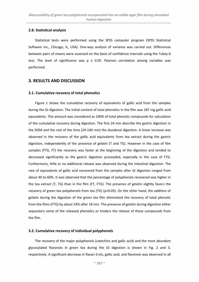
Biaccessibility of green tea polyphenols incorporated into an edible agar film during simulated human digestion
~ 267 ~
2.8. Statistical analysis
Statistical tests were performed using the SPSS computer program (SPSS Statistical
Software Inc., Chicago, IL, USA). One-way analysis of variance was carried out. Differences
between pairs of means were assessed on the basis of confidence intervals using the Tukey-b
test. The level of significance was p ≤ 0.05. Pearson correlation among variables was
performed.
3. RESULTS AND DISCUSSION
3.1. Cumulative recovery of total phenolics
Figure 1 shows the cumulative recovery of equivalents of gallic acid from the samples
during the GI digestion. The initial content of total phenolics in the film was 187 mg gallic acid
equivalents. This amount was considered as 100% of total phenolic compounds for calculation
of the cumulative recovery during digestion. The first 24 min describe the gastric digestion in
the DGM and the rest of the time (24-140 min) the duodenal digestion. A linear increase was
observed in the recovery of the gallic acid equivalents from tea extract during the gastric
digestion, independently of the presence of gelatin (T and TG). However in the case of film
samples (FTG, FT) the recovery was faster at the beginning of the digestion and tended to
decreased significantly as the gastric digestion proceeded, especially in the case of FTG.
Furthermore, little or no additional release was observed during the intestinal digestion. The
rate of equivalents of gallic acid recovered from the samples after GI digestion ranged from
about 40 to 60%. It was observed that the percentage of polyphenols recovered was higher in
the tea extract (T, TG) than in the film (FT, FTG). The presence of gelatin slightly favors the
recovery of green tea polyphenols from tea (TG) (p>0.05). On the other hand, the addition of
gelatin during the digestion of the green tea film diminished the recovery of total phenolic
from the films (FTG) by about 33% after 18 min. The presence of gelatin during digestion either
sequesters some of the released phenolics or hinders the release of these compounds from
the film.
3.2. Cumulative recovery of individual polyphenols
The recovery of the major polyphenols (catechins and gallic acid) and the most abundant
glycosylated flavonols in green tea during the GI digestion is shown in Fig. 2 and 3,
respectively. A significant decrease in flavan-3-ols, gallic acid, and flavonols was observed in all

Artículo 9
~ 268 ~
the samples after the simulated digestion, except for catechin, which gave recoveries greater
than 100%. Most of the release of polyphenols from tea extract or film, with or without gelatin
(TG, T, FTG, FT), occurred in the gastric phase of digestion, whereas little or no increase was
observed in the duodenal phase.
3.2.1. Catechins and gallic acid
In contrast with the pattern described for the recovery of total polyphenols during gastric
digestion (Fig. 1), the recovery rates of gallic acid and catechins showed a random behavior,
both in tea extract and film, independently of the presence of gelatin during the digestion.
The percentages of cumulative recovery of EGC after the GI digestion were significantly
lower than those of EGCG in all the samples (Fig. 2a, b). The low recovery of EGC compared to
EGCG may be due to a higher degradation, together with a lower release from the film. In the
case of EGC, the major recovery was observed in TG samples, although the values did not
reach 40% (Fig. 2a). The recovery of both EGC and EGCG was especially low in FT samples, with
percentages of about 5% and 25% for EGC and EGCG, respectively.
In the case of gallic acid (Fig. 2c), the recovery from the film samples was significantly
lower (p≤0.05), irrespectively of the presence of gelatin (30 and 20% for FTG and FT,
respectively). A different behavior was shown in Figure 2d, since the infusions and the films
showed a higher recovery of ECG when the simulated digestion was performed in the presence
of gelatin (75-85% of recovery; p≤0.05), whereas no more than 40% was recovered in the
absence of gelatin. Regarding EC (Fig. 2e), all the samples showed a similar trend, with 80-90%
of recovery (p>0.05).
The results show that the recovery of catechins and gallic acid almost stops after the
simulated gastric digestion of both green tea extract and films when the samples are moved to
simulated duodenal conditions. This may due to a lack of further release from the matrix film
or to a balance between release and transformation of the polyphenols that have been
released. It is well known the instability of polyphenols at near neutral or alkaline pH. Stability
studies have demonstrated that green tea catechins can undergo many chemical changes such
as oxidation and epimerization in solutions with pH>6 (Ananingsih, Sharma, & Zhou, 2011;
Green, Murphy, Schulz, Watkins, & Ferruzzi, 2007; Neilson et al., 2007; Roginsky & Alegria,
2005). Tagliazucchi et al. (2010) observed a loss of bio-accessible total polyphenols, flavonoids
and anthocyanins of grape during in vitro digestion, attributed to the transition from acidic
gastric to the mild alkaline intestinal environment. The interaction of tea polyphenols with

Biaccessibility of green tea polyphenols incorporated into an edible agar film during simulated human digestion
~ 269 ~
digestive enzymes can also contribute to the losses observed in the recovery (He, Lv, & Yao,
2006). However, other authors claim that digestive enzymes are not involved in the decrease
of polyphenols recoveries (Bermúdez-Soto, Tomás-Barberán, & García-Conesa, 2007; Green et
al., 2007; Neilson et al., 2007).
Comparing each individual sample, the cumulative recovery of the ECG and EC (fig. 2d and
2e) was higher than the recovery of EGC and EGCG (fig.2a and 2b). Thus, the recovery of EGC,
EGCG, ECG and EC from the film (FT) was less than 5, 20, 40 and 80 %, respectively. This result
is in accordance to the stability of polyphenols in different buffer conditions. Sang, Lee, Hou,
Ho, and Yang (2005) observed that EGC and EGCG which presented a similar stability, were
more instable than ECG and EC due to the oxidation of the trihydroxy structure on the B-ring.
In our study, the presence of gelatin seems to allow a significantly higher release of EGC
and ECG (fig. 2a and 2d) from both tea extract and film samples (p≤0.05), as well as EGCG and
gallic acid from the film sample (p≤0.05). It is well known the ability of polyphenols to interact
with proteins. These interactions may be through covalent bonds (Chen, Wang, Zhang, Ren, &
Zeng, 2011; Ishii, et al., 2008) or by hydrophobic and hydrogen bonds (von Staszewski et al.,
2011). However, the interactions established between polyphenols and gelatin in this study
seemed to be weak, allowing, therefore, an easy release of polyphenols. In this sense, it has
been observed that when protein is highly aggregated (e.g. bovine gelatin), the ability to
interact with polyphenols is lower than when protein is less aggregated (e.g. from marine
origin), giving a higher release of phenolic compounds (Gómez-Estaca, Montero, Fernández-
Martín, & Gómez-Guillén, 2009).
Another reason of the major recovery from samples with gelatin may be that EGC and
EGCG form complexes with gelatin that protect them from degradation, although those
complexes formed may be weak. This mechanism of protection of EGC and EGCG during
digestion was also observed by Green et al. (2007) when green tea catechins were formulated
with milk. Furthermore, Roginsky and Alegria (2005) found that the addition of milk to green
tea extracts reduced the oxidation of polyphenols, since polyphenols bound to milk proteins
are less accessible for oxidation. By the contrary, Argyri et al. (2005) observed a decrease in
the concentration of the phenolic compounds in red wine after in vitro digestion with protein
(meat and casein). All these discrepancies could be due to the different polyphenols affinity to
different proteins.
Regarding catechin (Fig. 2 f), the percentages of recovery overcame the initial quantity
(100-140%) in all the samples after digestion. Catechin has been described to derive from

Artículo 9
~ 270 ~
other phenolic compounds: epimerization of EC and hydrolysis of ECG and its epimer catechin
gallate (CG) (Ito et al., 2003). However, according to the percentages of recovery of EC and ECG
obtained in this study (Figures 2d and 2e), this mechanism seems unlikely.
3.2.2. Flavonols
The recovery of flavonols after the simulated digestion ranged from 30 to >70%,
depending on the individual compound and the sample (Figure 3a-e). Tavares et al. (2012)
observed similar recoveries of quercetin derivatives to those observed in this study from
blackberry extract after gastrointestinal digestion (40->80%), whereas the recoveries of
flavonols after gastric and pancreatic digestion of chokeberry juice was higher (70-85%)
(Bermúdez-Soto et al., 2007).
Rutin seemed to be the most stable flavonol at the experimental conditions, showing the
highest percentage of recovery (45-75%; Fig. 3d), whereas quercetin 3-glucoside showed the
lowest recoveries, especially in TG and FTG (around 30%, Fig. 3b). In general, gelatin had a
negative effect in the recovery of all the determined flavonols, probably due to polyphenol-
protein interactions. Thus, the flavonols showed significantly higher percentages of recovery
from T and FT than from the corresponding samples with gelatin, TG and FTG, after
gastrointestinal digestion (p≤0.05); with the exception of rutin which was recovered in similar
percentages from T and TG samples (p>0.05). Furthermore, whereas a linear increase was
observed in the recovery of all the flavonols from T and TG during the gastric digestion, the
recovery of flavonols from the film samples (FTG, FT) was faster at the beginning of the
digestion and tended to decreased significantly as the gastric digestion proceeded, especially
in the case of FTG.
As in the case of catechins, whereas flavonols are highly stable at in vitro gastric digestion,
partial degradation has been reported under intestinal conditions due to neutral or slightly
alkaline conditions (Bermúdez-Soto et al., 2007). Some losses may be also due to the
transformation of flavonoid glycosides into aglycones during the simulated digestion. Tarko,
Duda-Chodak, Sroka, Satora, and Michalik (2009) found that the polyphenols of selected fruits
were hydrolyzed to aglycones during the simulated digestion process, especially quercetin and
cyanogenic glycosides.

Biaccessibility of green tea polyphenols incorporated into an edible agar film during simulated human digestion
~ 271 ~
3.3. Cumulative recovery of antioxidant and antimicrobial activity
The cumulative recovery of compounds with reducing power and radical scavenging
capacity during gastrointestinal digestion is illustrated in Figures 4 a (FRAP) and b (ABTS),
respectively. The initial antioxidant capacity of the film, measured by FRAP and ABTS, was
given by the antioxidant capacity of the total amount of green tea extract added to the film:
3.77 mmol Fe2+ equivalents and 319.5 mg vitamin C equivalents for FRAP and ABTS,
respectively. These values were considered as 100% of FRAP and ABTS for calculation of the
cumulative recovery during digestion.
The profiles of both Fe2+ and vitamin C equivalents recovered from the samples were in
accordance with the total phenolic compounds recovered (Fig.1), with values which tended to
be higher in T and TG than in FT and FTG. The antioxidant activity recovered from both extract
and film after GI digestion was below 40%, higher in terms of FRAP (25-37.5%; Fig. 4a) than in
ABTS (7-17%; Fig. 4b). This remarkable decrease in antioxidant properties of all the samples
may be indicative of a low release of polyphenols from the matrix. The transformation
(degradation, epimerization, hydrolysis and oxidation) during simulated digestion could affect
their activity. It has been reported that EGCG, EGC, and ECG are extensively degraded at near-
neutral pH conditions such as those occurring during intestinal digestion. B-ring homodimers
and heterodimers have been described as auto-oxidation products formed from these
catechins under simulated digestive conditions (Neilson et al., 2007; Neilson, Song, Sapper,
Bomser, & Ferruzi, 2010). Some of these catechins B-ring dimers have been reported to show
antioxidant activities equal or higher than the catechins precursors (Yoshino, Suzuki, Sasaki,
Miyase, & Sano, 1999), which may contribute to the antioxidant activity recovered.
The interaction of polyphenols with proteins may involve a decrease in antioxidant
potential. Arts et al. (2002) indicated that the antioxidant activity of some components of
green tea is masked due to interactions with casein and albumin. In our work, the antioxidant
properties of green tea extract (T) and green tea film (FT) were affected differently when
gelatin was present during the digestion. In the case of the tea extract, the observed
increment in catechins recovery when the gelatin was present during the digestion was
accompanied by a higher recovery of antioxidant activity (fig. 4a, b). However, in the case of
the film with gelatin, in spite of a major release of catechins, no increment of the antioxidant
capacity was detected. In this connection, sometimes the interaction gelatin-polyphenol does
not necessarily affect negatively their ability to act as an antioxidant. According to Almajano,
Delgado, and Gordon (2006) the polyphenol-proteins complexes would still retain some

Artículo 9
~ 272 ~
antioxidant activity when the hydroxyl groups on aromatic ring of the polyphenol molecule,
responsible of antioxidant capacity, are not involved in the interaction.
As could be observed in Figure 2 (a-e), a higher recovery of catechins was obtained from
the films when gelatin was present during the digestion (FTG). Furthermore, the polyphenols
from FTG seem to be released at a faster rate at the beginning of the gastric digestion (Fig. 2,
3). Therefore, they could easily form complexes with the gelatin or even be more modified
because they are exposed during a longer time in the digestion environment. These complexes
can be reversible or irreversible with major or minor affinity, depending on flavonoid and
protein concentration, pH, temperature, type of protein and polyphenol, etc. (Luck et al.,
1994; Papadopoulou, Green, & Frazier, 2005; von Staszewski et al., 2011). Hence, the
discrepancies between samples might be due to the wide number of factors that can influence
the protein-complexes. Another factor to be taken into account is the addition of proteases
(pepsin, trypsin and chymotrypsin) that hydrolyze the protein in peptides and amino acids
which may bind to the polyphenols, resulting in less aggregated complexes than those formed
with the non-hydrolysed protein.
The antimicrobial activity from both green tea extract and film was not observed after the
simulated digestion process. Nevertheless, the green tea Wu Lu Mountain showed
antimicrobial activity in previous studies performed in our laboratory, especially against P.
phosphoreum and S. putrefaciens (data non-shown). Factors referred to previously, as the low
stability of polyphenols at intestine pH, together with the polyphenol-protein interacctions,
could explain the lack of antimicrobial activity of the phenolic compounds of tea.
At the end of the gastrointestinal digestion there remain some indigestible compounds,
especially the polymeric matrix of agar and a certain amount of tea extract material retained in
the film, evident by the color that the digested film retains. This colon-available indigestible
residue represents the polyphenols potentially available after upper gastrointestinal digestion,
which may exert some beneficial effect in the colon. Okello, McDougall, Kumar and Seal (2011)
observed protective effects of colon-available extract of green tea against hydrogen peroxide
and beta-amyloid induced cytotoxicity in PC12 cells used as a model of neuronal cells.
According to these authors, this colon available green tea extract was depleted in flavan-3-ol
and relatively enriched in certain flavonols, hydroxycinnamates, caffeine, theobromine and a
range of unidentified phenolic components.

Biaccessibility of green tea polyphenols incorporated into an edible agar film during simulated human digestion
~ 273 ~
4. CONCLUSION
The results obtained in our study demonstrate that green tea polyphenols contained in
the agar films are bioaccessible and, therefore, susceptible for absorption during human
digestion. The antioxidant activity recovered from the agar-green tea films (FT) was 25%
(reducing power) and 15% (radical scavenging ability), probably because not all the
polyphenols were released from the polymer matrix and/or they were modified during the
digestion process. Furthermore, the presence of gelatin during the digestion of green tea films
(FTG) can affect both the bioaccesibility and efficacy of polyphenols. In general, green tea
extract, with or without the presence of gelatin, showed a higher antioxidant activity.
However, the green tea films can have other additional properties and applications, as an
edible active packaging material in contact with foods, improving their quality, shelf-life and
safety.
Acknowledgements
This study was carried out under the CYTED Agrobioenvase project (Action 309AC0382) and
was sponsored by the National project (MICNN) AGL2008-00231/ALI, Xunta de Galicia
(Program: Ángeles Alvariño) and CSIC (Program: JAE-CSIC).
References
Almajano, M. P., Delgado, M. E., & Gordon, M. H. (2006). Changes in the antioxidant properties
of protein solutions in the presence of epigallocatechin gallate. Food Chemistry,
101(1), 126-130.
Ananingsih, V. K., Sharma, A., & Zhou, W. (2011). Green tea catechins during food processing
and storage: A review on stability and detection. Food Research International, in press.
Argyri, K., Proestos, C., Komaitis, M., & Kapsokefalou, M. (2005). Phenolic compounds in red
wine digested in vitro in the presence of iron and other dietary factors. International
Journal of Food Sciences and Nutrition, 56(3), 213-222.
Arts, M. J., Haenen, G. R., Wilms, L. C., Beetstra, S. A., Heijnen, C. G., Voss, H. P., & Bast, A.
(2002). Interactions between flavonoids and proteins: effect on the total antioxidant
capacity. Journal of Agriculture and Food Chemistry, 50(5), 1184-1187.
Benzie, I. F. F., & Strain, J. J. (1996). The Ferric Reducing Ability of Plasma (FRAP) as a Measure
of "Antioxidant Power": The FRAP Assay. Analytical Biochemistry, 239(1), 70-76.
Artículo 9
~ 274 ~
Bermúdez-Soto, M. J., Tomás-Barberán, F. A., & García-Conesa, M. T. (2007). Stability of
polyphenols in chokeberry (Aronia melanocarpa) subjected to in vitro gastric and
pancreatic digestion. Food Chemistry, 102(3), 865-874.
Bolling, B. W., Chen, C. Y. O., & Blumberg, J. B. (2009). Tea and health: Preventive and
therapeutic usefulness in the elderly? Current Opinion in Clinical Nutrition and
Metabolic Care, 12(1), 42-48.
Chen, R., Wang, J. B., Zhang, X. Q., Ren, J., Zeng, C. M. (2011). Green tea polyphenol
epigallocatechin-3-gallate (ECGC) induced intermolecular cross-linking of membrane
proteins. Archivess of Biochemistry and Biophysics, 507(2), 343-349.
Gómez-Estaca, J., López De Lacey, A., Gómez-Guillén, M. C., López-Caballero, M. E., &
Montero, P. (2009). Antimicrobial activity of composite edible films based on fish
gelatin and chitosan incorporated with clove essential oil. Journal of Aquatic Food
Product Technology, 18(1-2), 46-52.
Gómez-Estaca, J., López de Lacey, A., López-Caballero, M. E., Gómez-Guillén, M. C., & Montero,
P. (2010). Biodegradable gelatin-chitosan films incorporated with essential oils as
antimicrobial agents for fish preservation. Food Microbiology, 27(7), 889-896.
Gómez-Estaca, J., Montero, P., Fernández-Martín, F., & Gómez-Guillén, M. C. (2009). Physico-
chemical and film-forming properties of bovine-hide and tuna-skin gelatin: A
comparative study. Journal of Food Engineering, 90(4), 480-486.
Graham, H. N. (1992). Green tea composition, consumption, and polyphenol chemistry.
Preventive Medicine, 21(3), 334-350.
Green, R. J., Murphy, A. S., Schulz, B., Watkins, B. A., & Ferruzzi, M. G. (2007). Common tea
formulations modulate in vitro digestive recovery of green tea catechins. Molecular
Nutrition and Food Research, 51(9), 1152-1162.
He, Q., Lv, Y., & Yao, K. (2006). Effects of tea polyphenols on the activities of α-amylase,
pepsin, trypsin and lipase. Food Chemistry, 101(3), 1178-1182.
Hong, Y. H., Lim, G. O., & Song, K. B. (2009). Physical properties of Gelidium corneum-gelatin
blend films containing grapefruit seed extract or green tea extract and its application
in the packaging of pork loins. Journal of Food Science, 74(1), C6-C10.
Huang, D., Ou, B., & Prior, R. L. (2005). The chemistry behind antioxidant capacity assays.
Journal of Agricultural and Food Chemistry, 53, 1841-1856.

Biaccessibility of green tea polyphenols incorporated into an edible agar film during simulated human digestion
~ 275 ~
Ishii, T., Mori, T., Tanaka, T., Mizuno, D., Yamaji, R., Kumazawa, S., Nakayama, T., & Akagawa,
M. (2008). Covalent modification of proteins by green tea polyphenol (-)-
epigallocatechin-3-gallate through autoxidation. Free Radical Biology and Medicine,
45(10), 1384-1394.
Ito, R., Yamamoto, A., Kodama, S., Kato, K., Yoshimura, Y., Matsunaga, A., & Nakazawa, H.
(2003). A study on the change of enantiomeric purity of catechins in green tea
infusion. Food Chemistry, 83(4), 563-568.
Kim, K. M., Lee, B. Y., Kim, Y. T., Choi, S. G., Lee, J., Cho, S. Y., & Choi, W. S. (2006).
Development of antimicrobial edible film incorporated with green tea extract. Food
Science and Biotechnology, 15(3), 478-481.
Luck, G., Liao, H., Murray, N. J., Grimmer, H. R., Warminski, E. E., Williamson, M. P., Lilley, T. H.,
& Haslam, E. (1994). Polyphenols, astringency and proline-rich proteins.
Phytochemistry, 37(2), 357-371.
Mandalari, G., Faulks, R. M., Rich, G. T., Lo Turco, V., Picout, D. R., Lo Curto, R. B., Bisignano, G.,
Dugo, P., Dugo, G., Waldron, K. W., Ellis, P. R., & Wickham, M. S. J. (2008). Release of
protein, lipid, and vitamin E from almond seeds during digestion. Journal of
Agricultural and Food Chemistry, 56(9), 3409-3416.
Marciani, L., Wickham, M. S .J., Singh, G., Bush, D., Pick, D., Cox, E., Fillery-Travis, A., Faulks, R.,
Marsden, C., Gowland, P., & Spiller, R. (2007). Enhancement of intragastric acid
stability of a fat emulsion meal delays gastric emptying and increases cholecystokinin
release and gall bladder contraction. American Journal of Physics, 292, G1607-G1613.
Marciani, L., Faulks, R., Wickham, M. S. J., Bush, D., Barbara, D., Wright, J., Cox, E., Fillery-
Travis, A., Gowland, P., & Spiller, R., (2008). Effect of intragastric acid stability of fat
emulsions on gastric emptying, plasma lipid profile and postprandial satiety. British
Journal of Nutrition, 5, 1-10.
Neilson, A. P., Hopf, A. S., Cooper, B. R., Pereira, M. A., Bomser, J. A., & Ferruzzi, M. G. (2007).
Catechin degradation with concurrent formation of homo- and heterocatechin dimers
during in vitro digestion. Journal of Agricultural and Food Chemistry, 55(22), 8941-
8949.
Neilson, A. P., Song, B. J., Sapper, T. N., Bomser, J. A., & Ferruzi, M. G. (2010). Tea catechin
auto-oxidation dimers are accumulated and retained by Caco-2 human intestinal cells.
Nutrition Research, 30, 327-340.

Artículo 9
~ 276 ~
Okello, E. J., McDougall, C. G., Kumar, S., & Seal, C. J. (2011). In vitro protective effects of
colon-available extract of Camellia sinensis (tea) against hydrogen peroxide and beta-
(1-42)) induced cytotoxicity in differentiated PC12 cells. Phytomedicine, 18,
691-696.
Papadopoulou, A., Green, R. J., & Frazier, R. A. (2005). Interaction of flavonoids with bovine
serum albumin: A fluorescence quenching study. Journal of Agricultural and Food
Chemistry, 53(1), 158-163.
Pitino, I., Randazzo, C. L., Mandalari, G., Lo Curto, A., Faulks, R. M., Le Marc, Y., Bisignano, C.,
Caggia, C., & Wickham, M. S. J. (2010). Survival of Lactobacillus rhamnosus strains in
the upper gastrointestinal tract. Food Microbiology, 27(8), 1121-1127.
Prior, R. L., Wu, X., & Schaich, D. (2005). Standardized methods for the determination of
antioxidant capacity and phenolics in foods and dietary supplements. Journal of
Agricultural and Food Chemistry, 53, 4290-4302.
Re, R., Pellegrini, N., Proteggente, A., Pannala, A., Yang, M., & Rice-Evans, C. (1999).
Antioxidant activity applying an improved ABTS radical cation decolorization assay.
Free Radical Biology and Medicine, 26(9-10), 1231-1237.
Roginsky, V., & Alegria, A. E. (2005). Oxidation of tea extracts and tea catechins by molecular
oxygen. Journal of Agricultural and Food Chemistry, 53(11), 4529-4535.
Rojas-Graü, M. A., Soliva-Fortuny, R., & Martín-Belloso, O. (2009). Edible coatings to
incorporate active ingredients to fresh-cut fruits: a review. Trends in Food Science and
Technology, 20(10), 438-447.
Sang, S., Lee, M. J., Hou, Z., Ho, C. T., & Yang, C. S. (2005). Stability of tea polyphenol (-)-
epigallocatechin-3-gallate and formation of dimers and epimers under common
experimental conditions. Journal of Agricultural and Food Chemistry, 53(24), 9478-
9484.
Slinkard, K., & Singleton, V. L. (1977). Total phenol analysis: automation and comparison with
manual methods. Am. J. Enol. Vitic., 28(Copyright (C) 2011 American Chemical Society
(ACS). All Rights Reserved.), 49-55.
Tagliazucchi, D., Verzelloni, E., Bertolini, D., & Conte, A. (2010). In vitro bio-accessibility and
antioxidant activity of grape polyphenols. Food Chemistry, 120(2), 599-606.
Biaccessibility of green tea polyphenols incorporated into an edible agar film during simulated human digestion
~ 277 ~
Tarko, T., Duda-Chodak, A., Sroka, P., Satora, P., & Michalik, J. (2009). Transformations of
Phenolic Compounds in an in vitro Model Simulating the Human Alimentary Tract.
Food Technology and Biotechnology, 47(4), 456-463.
Tavares, L., Figueira, I., Macedo, D., McDougall, G. J., Leitao, M. C., Vieira, H. L. A., Stewart, D.,
Alves, P. M., Ferreira, R. B., & Santos, C. N. (2012). Neuroportective effect of
blackberry (Rubus sp.) polyphenols is potentiated after simulated gastrointestinal
digestion. Food Chemistry, 131, 1443-1452.
von Staszewski, M., Pilosof, A. M. R., & Jagus, R. J. (2011). Antioxidant and antimicrobial
performance of different Argentinean green tea varieties as affected by whey proteins.
Food Chemistry, 125(1), 186-192.
Weisburger, J. H., & Chung, F. L. (2002). Mechanisms of chronic disease causation by
nutritional factors and tobacco products and their prevention by tea polyphenols.
Food and Chemical Toxicology, 40(8), 1145-1154.
Yoshino, K., Suzuki, M., Sasaki, K., Miyase, T., & Sano, M. (1999). Formation of antioxidants
from (-)-epigallocatechin gallate in mild alkaline fluids, such as authenthic intestinal
juice and mouse plasma. Journal of Nutrititonal Biochemistry, 10, 223–229.

Artículo 9
~ 278 ~
Legends to Figures
Figure 1. Cumulative recovery of equivalents of gallic acid (%) from the samples during
simulated in vitro digestion. FT, green tea film; FTG, green tea film + gelatin, T,
green tea extract; TG, green tea extract + gelatin. n =2, three replicates.
Figure 2. Cumulative recovery of gallic acid and catechins (%) from the samples during
simulated in vitro digestion. FT, green tea film; FTG, green tea film + gelatin, T,
green tea extract; TG, green tea extract + gelatin. n =2, three replicates.
Figure 3. Cumulative recovery of flavonols (%) from the samples during simulated in vitro
digestion. FT, green tea film; FTG, green tea film + gelatin, T, green tea extract; TG,
green tea extract + gelatin. n =2, three replicates.
Figure 4. Cumulative recovery (%) of Fe2+ equivalents (FRAP) and vitamin C equivalents
(ABTS) from the samples during under simulated in vitro digestion. FT, green tea
film; FTG, green tea film + gelatin, T, green tea extract; TG, green tea extract +
gelatin. n =2, three replicates.
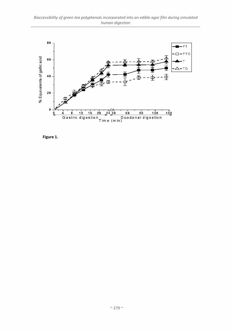
Biaccessibility of green tea polyphenols incorporated into an edible agar film during simulated human digestion
~ 279 ~
Figure 1.

Artículo 9
~ 280 ~
Figu
re 2
.

Biaccessibility of green tea polyphenols incorporated into an edible agar film during simulated human digestion
~ 281 ~
Figu
re 3

Artículo 9
~ 282 ~
Figure 4.

~ 283 ~


~ 285 ~
1. DESARROLLO DE ENVASES COMESTIBLES CON ACEITES ESENCIALES Y
APLICACIÓN EN PRODUCTOS PESQUEROS
1.1. Selección de aceites esenciales antimicrobianos para su incorporación a matrices
complejas (gelatina-quitosano)
En la actualidad existe un gran interés por encontrar alternativas menos contaminantes
para los envases plásticos convencionales que se utilizan principalmente en la industria
alimentaria. Con este objetivo, en los últimos años se están desarrollando nuevos materiales
biodegradables para el envasado de alimentos a partir de materiales naturales, tales como
polisacáridos, lípidos y proteínas (Tharanathan, 2003), que en algunos casos se obtienen a
partir de desechos de la pesca, agricultura o ganadería. A estos envases, con bajo impacto
ambiental, se les puede añadir determinados compuestos que le confieran propiedades tales
como antimicrobianas o antioxidantes. De este modo, estos envases biodegradables se
pueden utilizar en el envasado de alimentos, prolongar la vida útil del producto, y reducir o
inhibir microorganismos patógenos (Zivanovic y cols., 2005).
Estas propiedades resultan de gran interés en alimentos como el pescado. El pescado
refrigerado es un alimento altamente perecedero debido al crecimiento microbiano (tanto de
la flora endógena como de la adquirida por contaminación), lo que ocasionalmente origina
pérdidas económicas o problemas de salud. Algunos aceites esenciales de origen vegetal
presentan propiedades antimicrobianas frente a un amplio número de microorganismos (Burt,
2004; Deans y Ritchie, 1987; Dorman y Deans, 2000; Hammer, Carson y Riley, 1999), actividad
que se debe a la propia composición de los aceites, que contienen principalmente compuestos
fenólicos (carvacol, timol, eugenol, etc.) (Cosentino y cols., 1999).
Además de la incorporación de aceites esenciales, también se considera la incorporación
del quitosano, polímero que presenta propiedades filmogénicas, antioxidantes y
antimicrobianas (Jeon, Kamil y Shahidi, 2002; López-Caballero y cols., 2005). Existen en la
literatura varios estudios sobre las propiedades antimicrobianas de películas que contienen
quitosano y aceites esenciales (romero, orégano, clavo, etc.), con muy buenos resultados
(Deans y Ritchie, 1987; Oussalah y cols., 2004; Seydim y Sarikus, 2006). También se han
realizado estudios sobre la formulación de películas plásticas con aceite esencial de clavo
VI. DISCUSIÓN INTEGRADORA
Discusión integradora. Objetivo 1
~ 286 ~
(López y cols., 2007). Sin embargo, en la literatura no se han encontrado estudios sobre la
incorporación de aceite esencial del clavo en la matriz de gelatina o la aplicación de estas
películas con aceite esencial de clavo sobre alimentos, lo que condujo a plantear el presente
trabajo.
Por este motivo, el primer objetivo de este trabajo fue estudiar la actividad antimicrobiana
de 8 tipos de aceites esenciales frente a bacterias patógenas o responsables del deterioro de
los alimentos, principalmente del pescado. Posteriormente se elaboraron películas
biodegradables basadas en gelatina procedente de dos fuentes distintas (bovina y de pescado)
solo o formando matrices complejas junto con el quitosano, a las que se incorporó aceite
esencial de clavo (el aceite más activo), evaluando su actividad antimicrobiana frente a
diversas cepas seleccionadas por su patogenicidad, por ser causantes del deterioro o por su
efecto potencialmente probiótico.
Los aceites esenciales que presentaron mayor actividad antimicrobiana frente a las cepas
seleccionadas fueron el clavo, lavanda, romero y tomillo; siendo el clavo el más activo.
También se comprobó que de las 18 cepas estudiadas, las más sensibles fueron Clostridium
perfringens, Bacillus cereus y Staphylococcus aureus, mientras que las más resistentes fueron
Pseudomonas spp., Citrobacter freundii, Yersinia enterocolítica y Listeria spp. Esto se debe
probablemente a que los lipopolisacáridos de la pared celular de las bacterias Gram-negativas
impiden que los compuestos activos lleguen a la membrana citoplasmática (Ouattara, Simard,
Holley, Piette y Bégin, 1997).
En la bibliografía se menciona que los aceites esenciales de clavo y romero presentan una
marcada actividad antimicrobiana frente a Brochothrix thermosphacta y Pseudomonas
fluorescens (Ouattara y cols., 1997) o frente a bacterias patógenas como Listeria
monocytogenes, Escherichia coli, Bacillus cereus o Staphylococcus aureus (Burt, 2004; Del
Campo, Amiot y Nguyen-The, 2000). No obstante, no hay estudios del aceite esencial de clavo
sobre Shewanella putrefaciens, un microorganismo de gran importancia en el deterioro del
pescado (López-Caballero, Sánchez-Fernández y Moral, 2001). El presente trabajo revela que el
clavo posee actividad antimicrobiana frente a este tipo de microorganismos.
Los aceites esenciales más activos en el análisis cualitativo (clavo, lavanda, romero y
tomillo) se ensayaron frente a dos bacterias Gram-positivas (Lactobacillus acidophillus y
Listeria monocytogenes) y cuatro Gram-negativas (Escherichia coli, Pseudomonas fluorescens,
Photobacterium phosphoreum y Shewanella putrefaciens) por el método de difusión en disco.
El halo de inhibición medido por el método de análisis de imagen confirmó que la mayor área

Desarrollo de envases comestibles con aceites esenciales y aplicación en productos pesqueros
~ 287 ~
de inhibición (15,74 ±1,79%) correspondió al clavo (p ≤ 0.05). Los microorganismos más
sensibles al efecto de los aceites fueron P. phosphoreum (10,61 ±0,3%) y S. putrefaciens (15,74
±1,79%), que son los principales responsables del deterioro del pescado (Gram y Huss, 1996;
López-Caballero y cols., 2005), lo que resulta de gran interés por su uso potencial en la
conservación del pescado. A modo de ejemplo en la Figura 1 se muestran los halos de
inhibición producidos por los aceites esenciales a las 48 h de incubación de placas previamente
inoculadas con S. putrefaciens. El clavo fue el aceite que causó una mayor inhibición del
crecimiento de la cepa, después del romero.
Teniendo en cuenta estos resultados se seleccionó el clavo para la elaboración de las
películas con propiedades antimicrobianas. Las películas de gelatina procedentes de dos
fuentes distintas (pescado y bovina) se formularon con aceite esencial de clavo y/o quitosano
en su composición. En la Figura 2 se muestran las cuatro películas preparadas a base de
gelatina bovina, donde se aprecian diferencias entre las películas en cuanto a su transparencia
(ej. el fondo sólo se aprecia en las películas A y B) y su color (ej. más amarillas en las que
contienen quitosano). Las películas de quitosano fueron aparentemente más rígidas y más
opacas, y las películas constituidas a base de gelatina de pescado tuvieron un aspecto similar
Figura 1. Halos de inhibición producidos por los aceite esenciales de clavo (A), lavanda (B), romero (C) y
tomillo (D) en las placas inoculadas con S. putrefaciens.
A B
C D

Discusión integradora. Objetivo 1
~ 288 ~
(“Biodegradable gelatin-chitosan films with essential oils as antimicrobial agents for fish
preservation”).
Una vez elaboradas las películas se estudiaron sus propiedades antimicrobianas con el fin
de comprobar si el clavo seguía manteniendo su actividad después de añadirse a las películas
con o sin quitosano. Así se observó que las películas basadas en gelatina, tanto de pescado
como bovina, y con/sin quitosano, no presentaron actividad antimicrobiana frente a las cepas
seleccionadas. Sin embargo hay trabajos dónde sí se ha visto la efectividad de las películas y
los recubrimientos basados en quitosano como tratamientos antimicrobianos (Chen y cols.,
1998; Jeon y cols., 2002; López-Caballero y cols., 2005), si bien los resultados difieren en gran
medida por las técnicas experimentales utilizadas. Las películas basadas en gelatina bovina y
de pescado con clavo presentaron actividad frente a los cuatro microorganismos del estudio (L.
acidophillus, P. fluorescens, L. innocua y E. coli), mientras que las películas sin clavo no
A. Gelatina B. Gelatina + clavo
Figura 2. Películas preparadas a base de gelatina bovina (A), gelatina con clavo (B), gelatina con
quitosano (C) y gelatina con clavo y quitosano (D).
C. Gelatina + quitosano D. Gelatina + clavo + quitosano

Desarrollo de envases comestibles con aceites esenciales y aplicación en productos pesqueros
~ 289 ~
presentaron actividad. En la Figura 3 se representan los valores de actividad antimicrobiana
de las películas compuestas con aceite esencial de clavo y/o quitosano.
En la Figura 3 se observa que las películas constituidas a base de gelatina de pescado
inhibieron el crecimiento microbiano en menor medida que las preparadas a partir de gelatina
bovina. Estas diferencias en las propiedades antimicrobianas se deben probablemente a que el
grado de interacción entre la gelatina, los polifenoles y/o el quitosano (Gómez-Estaca,
Montero, Giménez y Gómez-Guillén, 2007) varía en función del origen de la proteína;
dependiendo de esta interacción, se produce una menor o mayor liberación o disponibilidad
de los compuestos activos. Por otro lado, los valores de actividad antimicrobiana de las
películas basadas en gelatina de pescado y bovina variaron también en algunos casos en
función de la matriz en la que estuviera incluido el aceite esencial de clavo (presencia o no de
quitosano) (Figura 2). La película con quitosano y clavo presentó en algunos casos menor
actividad que las películas que no contienen quitosano, especialmente en las de gelatina de
origen marino. Este hecho se atribuye a la diferente solubilidad que presentan las películas,
dependiendo de la matriz. En este sentido, cuando la película se coloca sobre la superficie del
Figura 3. Actividad antimicrobiana de las películas con aceite esencial de clavo y/o quitosano frente a
las cuatro cepas de estudio.

Discusión integradora. Objetivo 1
~ 290 ~
alimento, la liberación de los compuestos antimicrobianos contenidos en ella viene
determinada, en gran parte, por su solubilidad (Papadokostaki, Amarantos y Petropoulos,
1998). Así, la película de gelatina bovina presentó una baja solubilidad (26,2 ±1,5%), semejante
al estudio de Bertan, Tanada-Palmu, Siani y Grosso (2005) en películas de gelatina bovina. La
película de gelatina-quitosano presentó una solubilidad parecida a la película anterior. Cuando
el aceite esencial de clavo se incorporó en las películas de gelatina hubo un incremento
significativo (p≤ 0.05) en la solubilidad (41,1 ±1,5%). Este hecho se puede atribuir a las
interacciones entre el polifenol y la proteína mediante enlaces iónicos o puentes de hidrógeno
(Taravel y Domard, 1995), que desestabilizan la red proteica, produciendo una disminución en
la solubilidad de las películas preparadas a partir de estos dos compuestos. Sin embargo, la
solubilidad se mantuvo cuando el aceite esencial se incorporó a la matriz gelatina-quitosano
(29,5 ±3,2%), probablemente por las interacciones entre la gelatina y el quitosano, que
estabilizan la estructura de la película a pesar de la presencia del aceite esencial de clavo. Este
hecho implica una menor liberación de antimicrobianos y, por otro lado, una mayor integridad
de la película cuando ésta se aplica al pescado.
1.2. Aplicación de envases en la conservación de pescado refrigerado: salmón (Salmo
salar) y bacalao (Gadus morhua)
Las películas basadas en gelatina de pescado a las que se añadió quitosano-clavo se
aplicaron a salmón loncheado crudo refrigerado a 2 °C, con el fin de estudiar su papel en la
conservación del pescado. Así se observó que los recuentos de microorganismos totales en el
pescado sin película se incrementaron rápidamente a lo largo de todo el periodo de
conservación, mientras que en el pescado recubierto con la película se mantuvieron por
debajo, y sólo a partir del día 9 se percibió un ligero aumento en estos valores. Al final de la
conservación los recuentos en el pescado cubierto con la película fueron 2 unidades
logarítmicas menos que los del control. La vida útil del salmón loncheado cubierto con estas
películas se prolongó por lo menos hasta los 11 días. Al igual que en este estudio, en otros
trabajos se ha visto que otras películas basadas en quitosano u otros aceites esenciales
también presentaron buenas propiedades antimicrobianas en carne (Oussalah y cols., 2004),
mortadela (Zivanovic y cols., 2005) o sardina ahumada (Gómez-Estaca y cols., 2007).
Dado que los resultados fueron satisfactorios, se planteó realizar un nuevo estudio,
profundizando más exhaustivamente en los índices del deterioro y en los microorganismos
causantes del mismo, evaluando el posible retraso en presencia de las películas con clavo. Para
ello se escogieron matrices complejas de gelatina-quitosano; y en este caso la gelatina

Desarrollo de envases comestibles con aceites esenciales y aplicación en productos pesqueros
~ 291 ~
seleccionada fue la de bovino para favorecer la interacción entre gelatina y quitosano, dejando
más libres los polifenoles (Gómez-Estaca, Bravo, Gómez-Guillén, Alemán y Montero, 2009;
Gómez-Estaca, López de Lacey, Gómez-Guillén, López-Caballero y Montero, 2009; Gómez-
Estaca y cols., 2007). El pescado seleccionado para la conservación fue el bacalao por su suave
color y sabor, de manera que cualquier modificación fuera fácilmente perceptible. La
aplicación de las películas basadas en gelatina bovina con aceite esencial de clavo para mejorar
la conservación del bacalao, también dió resultados prometedores y prolongó la vida útil.
En el análisis microbiológico de este estudio con bacalao se apreció una disminución e
incluso una inhibición de los microorganismos estudiados en el bacalao recubierto con la
película gelatina-quitosano-clavo (p ≤ 0.05). En este caso se detectó una inhibición en el
crecimiento de las bacterias productoras de H2S y de las enterobacterias a los 3 y 9 días,
respectivamente. También se observó una disminución en los recuentos de totales y
Pseudomonas, y un descenso más moderado en las bacterias lácticas. Estos resultados
estuvieron en consonancia con los índices bioquímicos del deterioro.
Inicialmente el pH del bacalao sin película fue de 6,7 y fue subiendo hasta un 7,3-7,4
durante los primeros días, manteniéndose así hasta el final de la conservación (11 días). Por
otro lado, el pH del bacalao envuelto con la película permaneció por debajo de 7 (p ≤ 0.05). El
valor inicial del N-BVT fue de 13,5 mg N-BVT/100 g de músculo, valor que se incrementa a lo
largo de la conservación (hasta alcanzar 35 mg N-BVT/100 g a los 4-5 días de conservación).
Estos resultados fueron similares a los obtenidos por (López-Caballero y cols., 2005) y
(Olafsdottir, Lauzon, Martinsdóttir, Oehlenschläger y Kristbergsson, 2006). En los dos lotes de
pescado (bacalao y bacalao con película) se observó un incremento progresivo a lo largo de la
conservación, si bien los valores fueron inferiores (p ≤ 0.05) en el bacalao con la película. Por
lo tanto, la aplicación de las películas con gelatina-quitosano-clavo es útil para alargar la vida
útil del pescado.


Discusión integradora. Objetivo 2
~ 293 ~
2. ESTUDIO DE LA COMPOSICIÓN DE EXTRACTOS ACUOSOS DE TÉ VERDE
(Camellia sinensis L.) EN RELACIÓN A SU FUNCIONALIDAD Y
DESARROLLO DE ENVASES COMESTIBLES
2.1. Selección de un extracto de té verde (Camellia sinensis) según sus propiedades
biológicas entre distintas variedades en base a su composición química
Tal y como se ha visto en el apartado anterior, la capacidad antimicrobiana de los aceites
esenciales es evidente; no obstante, es importante destacar que su aroma es muy intenso y su
aplicación al alimento, incluso formando parte de un envase, en ocasiones origina
características sensoriales indeseables.
En este sentido, la utilización de extractos acuosos de plantas (hojas, semillas, tallos, etc.)
resulta más conveniente por presentar sabores y olores menos agresivos, siendo más fáciles y
versátiles de aplicar, sin repercutir negativamente en las características sensoriales del
alimento.
La mayoría de los extractos presentan propiedades antioxidantes manifiestas, tanto del
extracto libre, como cuando se encuentran formando parte de la película, si bien es
relativamente frecuente que las propiedades antioxidantes de las películas sean inferiores a la
del extracto libre. Este hecho se atribuye a la interacción de los componentes antioxidantes del
extracto con la matriz biopolimérica, ya que si la interacción es muy fuerte puede disminuir
drásticamente la actividad.
Sin embargo, son menos numerosos los extractos que presentan propiedades
antimicrobianas frente a un gran número de microrganismos, ya sean patógenos o del
deterioro de alimentos, y este número disminuye teniendo en cuenta que en determinadas
matrices continúan manifestando su actividad. Este hecho condujo a la búsqueda de extractos
que por su coloración, aroma y sabor influyeran lo menos posible en las características
sensoriales del alimento a utilizar, y a elaborar un extracto acuoso que manifestara tanto
propiedades antioxidantes como antimicrobianas.
El extracto seleccionado fue el té verde (Camellia sinensis). El té verde es quizá una de las
infusiones con más efectos beneficiosos que se conocen, y están ampliamente descritos
(Cabrera, Artacho y Giménez, 2006). Sus efectos beneficiosos se atribuyen, principalmente, a la
presencia de polifenoles, pigmentos, aminoácidos, carbohidratos, minerales y alcaloides
presentes (Bolling y cols., 2009; Isigaki, Takakuwa, Takeo, 1991; Weisburger y Chung, 2002).

Discusión integradora. Objetivo 2
~ 294 ~
Aunque los extractos pueden ser muy diversos, la gran mayoría de los estudios de té se
refieren a infusiones, es decir, a extractos acuosos en agua caliente (Lin, Chen y Harnly, 2008),
apreciándose que en esas condiciones mantiene sus propiedades biológicas (Cabrera, Artacho
y Giménez, 2006; Dufresne y Farnworth, 2001; Friedman, 2007; Hamilton-Miller, 1995).
A pesar de los muchos trabajos realizados sobre las propiedades que confieren diferentes
extractos de té, apenas hay estudios que evalúen las diferencias y composición de extractos de
té verde y su relación con las propiedades biológicas que manifiestan. En los pocos estudios
publicados al respecto se presentan extractos realizados de diferente manera, por lo que su
comparación es muy difícil. Por ello, se estudiaron 6 variedades de extractos de té realizados
en las mismas condiciones y, de este modo, poder evaluar la relación que pudiera existir entre
la composición y sus propiedades bioactivas. Los extractos de las diferentes variedades de té
verde (Japan Sencha Makinohara, Japan Gyokuro Asahi, Canela Té Verde, Lung Ching, China
White Hair y Wu Lu Mountain) presentaron distinta composición. Como se refirió
anteriormente (“Antioxidant and antimicrobial activities of green tea (Camellia sinensis L.) as
an expression of its chemical composition”) no siempre es el mismo extracto el que muestra
mayores cantidades de uno de los componentes, y hay grandes diferencias entre los valores,
incluso del doble en algunas ocasiones. Así, el extracto de té Canela Té Verde es el que
contiene más polifenoles totales (9770 mg/L equivalentes de ácido gálico) y flavonoles (236.6
mg/L), mientras que el de mayor contenido en catequinas fue el extracto de Wu Lu Mountain
(11268.1 mg/L). Por el contrario, los valores más bajos, tanto de contenido en fenoles totales
(4712 mg/L equivalentes de ácido gálico), como de catequinas (5361.4 mg/L), correspondió al
extracto de China White Hair. El menor contenido en flavonoles totales se obtuvo en la
variedad Lung Ching tea (84.7 mg/L).
Los 6 tipos de té verde se caracterizan por tener un alto contenido en catequinas, siendo
la más abundante la epigalocatequina-3-galato (EGCG). Estos resultados concuerdan con los
obtenidos por otros autores en varios extractos de té, en los cuales la EGCG aparece como una
de las catequinas mayoritarias (Graham, 1992; Lin y cols., 2008), mientras que en otros tipos
de té, como el té negro presentaron un contenido menor. En el estudio de composición
realizado por Reto, Figueira, Filipe y Almeida (2007) en 9 variedades distintas de té verde,
también es la EGCG una de las catequinas más abundantes.
El contenido de carbohidratos totales varió desde 1.75 mg/mL para Japan Gyokuro Asahi
a 5.32 mg/L para Lung Ching (p ≤ 0.05). No se ha encontrado información acerca del contenido
de azúcares en extractos de té verde, por lo que la comparación con otros trabajos resulta
difícil.

Estudio de la composición de extractos acuosos de té verde (Camellia sinensis ) en relación a su funcionalidad y desarrollo de envases comestibles activos
~ 295 ~
En cuanto al contenido en minerales, el potasio (K) fue el más abundante en todos los
extractos, seguido de magnesio (Mg) y manganeso (Mn), mientras que el resto están en menor
concentración, o incluso en forma de trazas como es el caso del cobre (Cu) y el hierro (Fe).
Estos resultados coinciden con otros trabajos realizados en infusiones de té verde, dónde el K
fue el mineral mayoritario (Fernández, Pablos, Martín y González, 2002; Gallaher, Gallaher,
Marshall y Marshall, 2006; Musa Özcan, Ünver, Uçar y Arslan, 2008; Reto y cols., 2007). Sin
embargo, Fernández-Cáceres, Martín, Pablos y González (2001) encontraron que los minerales
mayoritarios, tanto en té verde como en té negro, fueron calcio (Ca), magnesio (Mg) y
aluminio (Al), pero con variaciones, dependiendo de la localización geográfica del té verde.
El perfil de aminoácidos presente en las seis variedades de té verde fue semejante, y los
aminoácidos más abundantes fueron el ácido glutámico (Glu), el ácido aspártico (Asp), serina
(Ser) y arginina (Arg). El contenido en aminoácidos totales se indica en orden decreciente para
Wu Lu Mountain, Canela Té Verde, Japan Gyokuro Asahi, Lung Ching, Japan Sencha
Makinohara and China White Hair. El predominio de un aminoácido en concreto varía de un
estudio a otro, lo que se atribuye a las diferencias entre variedades de té y tipos de extractos.
(Wang y Ruan, 2009) analizaron la composición en aminoácidos de varias infusiones de té
verde (variedad Longjing) y encontraron que la teanina fue el aminoácido más abundante,
seguido del ácido glutámico (Glu), prolina (Pro), aspártico (Asp), serina (Ser), arginina (Arg) e
histidina (His), lo que coincide con los resultados de la presente memoria. De acuerdo con
Wang y cols. (2010), los aminoácidos permiten diferenciar entre distintos tipos de tés, como
por ejemplo, entre el té verde, negro u Oolong o incluso valorar la calidad de un determinado
tipo de té. Estos mismos autores encontraron que el té verde posee mayor contenido de Asp,
Glu y Ser, que el té negro y el Ooolong. La composición en aminoácidos también se ha
determinado en extractos acuosos de té verde, Oolong , negro y Pu-erh, y la mayoría de los tés
verdes presentaron como aminoácidos mayoritarios la teanina, serina y valina (Syu, Lin, Huang
y Lin, 2008), mientras que la teanina, ácido glutámico y aspártico fueron los aminoácidos más
abundantes en todos los tés estudiados (Alcázar y cols., 2007).
El porcentaje de cada uno de los componentes estudiados, con respecto al contenido total
(resultado de sumar las concentraciones obtenidas de cada uno de ellos), se muestra en la
Tabla 1. Estos resultados permiten ver el peso que tienen los diferentes componentes en un
mismo extracto de té. Como se observa en la tabla, los extractos de té tienen un patrón similar
en cuanto a su composición relativa, ya que el mayor porcentaje está en los aminoácidos
(44.65-53.78%) y en las catequinas (28.43-33.64%), seguido de los carbohidratos (7.44-
21.03%), minerales (2.92-6.01%) y flavonoles (0.33-0.88%). Resulta difícil comprar estos

Discusión integradora. Objetivo 2
~ 296 ~
resultados con los de otros trabajos ya que no se han encontrado estudios que evalúen la
composición de varios componentes a la vez y en un mismo extracto acuoso de té, ya que la
mayor parte de los trabajos se centran en estudiar un solo componente (polifenoles,
aminoácidos, minerales, etc.).
La actividad antioxidante fue distinta entre las variedades estudiadas. Wu Lu Mountain
mostró la actividad más alta (FRAP 188.48 mmol/L Fe2+, ABTS 15.98 mg/mL equivalentes de
vitamina C) y China White Hair la más baja (FRAP 88.37 mmol/L Fe2+, ABTS 7.20 mg/mL
equivalentes de vitamina C y PCL 3.93 mg/mL equivalentes de vitamina C). Sin embargo, el té
Canela Té Verde obtuvo la actividad antioxidante determinada por Photochem (19.0 mg/mL
equivalentes de vitamina C) más alta. A la vista de estos resultados, el grado de actividad
antioxidante muestra una correlación alta con el contenido total en polifenoles determinado
por el método de Folin-Ciocallteu, (rFRAP/tpc = 0,983, rABTS/tpc = 0,926, rPCL/tpc = 0,926). Hay que
tener en cuenta que los tres métodos son ensayos de transferencia de electrón y miden el
poder reductor de las muestras (Huang, Ou y Prior, 2005). Toschi y cols. (2000) observaron que
la actividad antioxidante de los tés verdes (en un sistema modelo lipídico), es más alta en los
tés con mayor cantidad de EGCG y EGC. En el presente trabajo, el extracto de té verde que
más actividad mostró (Wu Lu Mountain) es el que tiene mayor contenido en EGCG y EGC. En
estudios anteriores ya se ha estimado la relación que existe entre la composición relativa de
los distintos polifenoles del té verde y la actividad antioxidante (Rice-Evans y cols., 1996),
Tabla 1. Composición de los componentes mayoritarios de los extractos de té verde expresado
en porcentaje (%)
Tipo
Catequinas
totales
Flavonoles
totales
Carbohidratos
totales
Minerales
totales
Aminoácidos
totales
1. Japan Sencha
Makinohara 29.14 ± 0.23 0.87 ± 0.01 17.99 ± 0.30 5.21 ± 0.03 46.79 ± 0.41
2. Japan Gyokuro
Asahi 33.64 ± 0.53 0.77 ± 0.02 7.44 ± 0.26 5.27 ± 0.06 52.88 ± 0.55
3. Canela Té Verde 29.34 ± 0.15 0.81 ± 0.02 14.86 ± 0.07 3.44 ± 0.00 51.56 ± 0.21
4. Lung Ching 29.45 ± 0.67 0.33 ± 0.00 21.03 ± 0.40 4.53 ± 0.06 44.65 ± 0.72
5. China White Hair 28.43 ± 0.82 0.88 ± 0.00 13.81 ± 0.43 6.01 ± 0.04 50.87 ± 0.38
6. Wu Lu Mountain 33.30 ± 0.30 0.49 ± 0.00 9.51 ± 0.32 2.92 ± 0.03 53.78 ± 0.44

Estudio de la composición de extractos acuosos de té verde (Camellia sinensis ) en relación a su funcionalidad y desarrollo de envases comestibles activos
~ 297 ~
indicando que la contribución de las catequinas a la actividad antioxidante del té verde sigue el
siguiente orden: EGCG≈ EGC>>ECG=EC>C. La capacidad secuestradora de la EGCG y la ECG es
más fuerte que en las otras catequinas, como la EGC, GC, EC y C, a pesar de que estas últimas
tengan un menor impedimento estérico, por lo que se demuestra que la presencia del grupo
galato juega un papel importante en la capacidad secuestradora de radicales libres (Guo y
cols., 1999).
La capacidad antimicrobiana de los distintos extractos fue asimismo diferente. Las cepas
más sensibles fueron Photobacterium phosphoreum y Shewanella putrefaciens para todos los
extractos. Por el contrario, Aeromonas hydrophila, Vibrio parahaemolyticus y Pseudomonas
fluorescens se mostraron menos sensibles frente a los extractos de té Japan Sencha
Makinohara, Japan Gyokuro Asahi y Canela Té Verde. Estos resultados demuestran que las
infusiones de té verde son activas frente a organismos específicos del deterioro del pescado
(López-Caballero y cols., 2005), lo que hace de estos extractos buenos candidatos para su
utilización como antimicrobianos en productos pesqueros. Asimismo, este hecho tiene una
gran relevancia dado que la actividad antimicrobiana de los tés se ha demostrado frente a un
gran número de cepas, (Friedman, 2007; Singh Arora, Jeet Kaur y Kaur, 2009), pero no frente a
P. phosphoreum y S. putrefaciens. La actividad antimicrobiana se ha asociado al contenido alto
en polifenoles, especialmente catequinas de los tés verdes (Arakawa y cols., 2004; Cushnie y
Lamb, 2011). En la revisión realizada por Friedman (2007) se menciona que los flavonoides del
té son activos frente a cepas Gram positivas y Gram negativas, como por ejemplo Bacillus
cereus, Clostridium perfringens o Escherichia coli. En este trabajo solo fueron sensibles al
extracto las bacterias Gram negativas. Este resultado se explica por el hecho de que cepas
diferentes de un mismo microrganismo pueden expresar distinto grado de sensibilidad a los
flavonoides del té (Hamilton-Miller, 1995).
Ninguno de los extractos de té mostró actividad antimicrobiana frente a las cepas
Lactobacillus y Bifidobacterium estudiadas. Este dato resulta muy interesante puesto que
muchos extractos ricos en polifenoles, como el extracto de té verde, presentan actividad
antimicrobiana frente a un amplio número de microorganismos, incluidos aquellos que son
beneficiosos para el intestino. Almajano y cols. (2008) constataron este mismo hecho cuando
probó la actividad antimicrobiana de varias infusiones de té verde frente a Lactobacillus
acidophillus, la cual mostró una excepcional resistencia frente a los extractos acuosos de té
investigados.
Japan Sencha Makinohara fue uno de los extractos más activos en cuanto a sus
propiedades antimicrobianas y el té Wu Lu Mountain el que menos, a pesar del alto contenido

Discusión integradora. Objetivo 2
~ 298 ~
en catequinas y actividad antioxidante. La diferente actividad entre los extractos de té
probablemente se deba a la propia composición química de cada variedad puesto que todos
los extractos se prepararon del mismo modo.
A la vista de estos resultados, es difícil extraer conclusiones de cual es el mejor extracto
como antioxidante o antimicrobiano. Es posible que determinados componentes de los
extractos tengan alguna actividad sobre otros, que puede incidir en las propiedades
antioxidantes y antimicrobianas que manifiestan.
Con el objeto de comparar la actividad de las seis variedades de té (en términos de
composición y actividad biológica) y establecer las diferencias entre ellas, se realizó un análisis
por componentes principales (CP). Dado el gran número de variables y para evitar que se
solaparan en la representación de los nuevos componentes, las variables se agruparon de
acuerdo a tres combinaciones: flavonoides (Figura 4), aminoácidos (Figura 5) y minerales
(Figura 6), manteniéndose el resto constante (los tés se superponen en las Figuras para mejor
comprensión).
El análisis multivariante con flavonoides (incluye todas las variables excepto aminoácidos y
minerales) mostró que los nuevos componentes explican el 38.4% y 30.9% de la varianza,
respectivamente (Figura 4)1. Las catequinas totales, ECG y EGCG se agrupan en el primer
componente, mientras que el peso de C, EC y EGC se distribuye entre el primer y segundo
componente. Los flavonoles se reúnen en la segunda dimensión, a excepción del hiperósido
que se sitúa en la primera. Los microorganismos que distribuyen su peso en ambas
dimensiones se correlacionan negativamente con los anteriores a excepción de P. fluorescens,
con mayor peso en el CP1. Las medidas de la actividad antioxidante se sitúan juntas y
mantienen una fuerte relación con las catequinas totales, pero no con los flavonoles.
Los extractos de té se diferencian claramente con la distribución de variables (por ej.
flavonoles y catequinas) de acuerdo a las dos nuevas dimensiones (Figura 4). El té 3 (Canela Té
Verde) se asocia con un alto contenido de flavonoles, el té 5 (China White Hair) fue el más
activo frente a P. fluorescens, el té 2 (Japan Gyokuro Asahi) lo fue frente a los microorganismos
1 Los números dentro del recuadro indican el tipo de extracto y se corresponden con la numeración marcada en la
tabla D1.
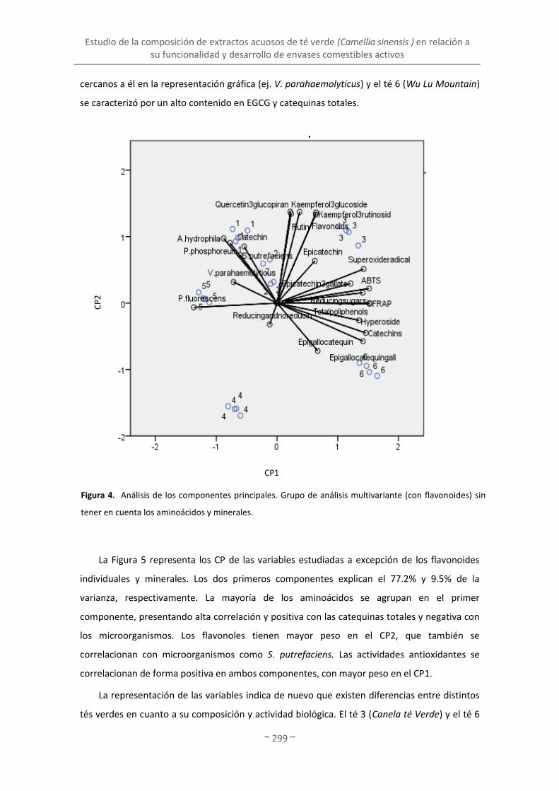
Estudio de la composición de extractos acuosos de té verde (Camellia sinensis ) en relación a su funcionalidad y desarrollo de envases comestibles activos
~ 299 ~
cercanos a él en la representación gráfica (ej. V. parahaemolyticus) y el té 6 (Wu Lu Mountain)
se caracterizó por un alto contenido en EGCG y catequinas totales.
La Figura 5 representa los CP de las variables estudiadas a excepción de los flavonoides
individuales y minerales. Los dos primeros componentes explican el 77.2% y 9.5% de la
varianza, respectivamente. La mayoría de los aminoácidos se agrupan en el primer
componente, presentando alta correlación y positiva con las catequinas totales y negativa con
los microorganismos. Los flavonoles tienen mayor peso en el CP2, que también se
correlacionan con microorganismos como S. putrefaciens. Las actividades antioxidantes se
correlacionan de forma positiva en ambos componentes, con mayor peso en el CP1.
La representación de las variables indica de nuevo que existen diferencias entre distintos
tés verdes en cuanto a su composición y actividad biológica. El té 3 (Canela té Verde) y el té 6
Figura 4. Análisis de los componentes principales. Grupo de análisis multivariante (con flavonoides) sin
tener en cuenta los aminoácidos y minerales.
CP
2
CP1

Discusión integradora. Objetivo 2
~ 300 ~
(Wu Lu Mountain) obtuvieron altos niveles de aminoácidos y polifenoles (tanto de catequinas
totales como de flavonoles) y alta capacidad antioxidante (Figura 5). Estos datos sugieren que
la actividad antioxidante no disminuye por la presencia de aminoácidos, o bien que no existe
una interacción entre ambos (aminoácidos y polifenoles).
El análisis de componentes principales de las variables investigadas, excepto para los
flavonoides individuales y aminoácidos, se muestra en la Figura 6. Los CPs representan el
38.8% y 30.2% de la varianza explicada, respectivamente. El contenido de catequinas se
relaciona en gran medida y positivamente con la actividad antioxidante en el PC1, pero de
forma negativa con los minerales. En general los minerales están más asociados con la
inhibición de los microorganismos, indicando que los minerales pudieran promover, de algún
modo, la inhibición de los microorganismos (ej. S. putrefaciens en presencia de sodio y calcio).
Figura 5. Análisis de los componentes principales. Grupo de análisis multivariante (con aminoácidos) sin
tener en cuenta los flavonoides por separado y los minerales.
CP
2
CP1

Estudio de la composición de extractos acuosos de té verde (Camellia sinensis ) en relación a su funcionalidad y desarrollo de envases comestibles activos
~ 301 ~
Así también se observa que los tés son también muy diferentes entre sí, y los valores más
bajos de minerales correspondieron a los extractos de Canela Té Verde (té 3) y Wu Lu
Mountain (6).
En resumen, estos resultados muestran que las distintas variedades presentan diferente
composición y las propiedades de los extractos bioactivos no guardan relación entre sí, siendo
el conjunto de componentes lo que definen las propiedades biológicas de cada extracto.
Aquellos con niveles más altos de catequinas totales, flavonoles y aminácidos (Canela Té Verde
y Wu Lu Mountain) también tienen la más elevada actividad antioxidante. Sin embargo, estas
variedades no presentan una diferencia muy marcada, comparada con las otras, en cuanto a
actividad antimicrobiana.
Figura 6. Análisis de los componentes principales. Grupo de análisis multivariante (con minerales) sin
tener en cuenta los flavonoides por separado y aminoácidos.
CP
2
CP1
Discusión integradora. Objetivo 2
~ 302 ~
Por otro lado, China White Hair, fue la variedad que mostró la menor actividad
antioxidante, coincidiendo con el hecho de que posee el menor contenido en catequinas y
aminoácidos, valores intermedios de flavonoles, y bajo contenido en carbohidratos y
minerales. En paralelo, está variedad mostró una actividad antimicrobiana frente a Vibrio
parahaemolyticus y Pseudomomnas fluorecens, muy superior a los extractos de las otras
variedades de té verde, que poseían mayor contenido en fenoles totales. Japan Sencha
Makinohara exhibió la mayor actividad antimicrobiana, mientras se apreciaron pocas
diferencias con el resto de variedades estudiadas.
El hecho de que microorganismos del deterioro sean sensibles a la presencia de extractos
acuosos de té verde resulta prometedor por su aplicación en la conservación de alimentos.
Según las necesidades, puede ser más interesante la selección de una u otra variedad, y más
aún si se pretende inhibir una cepa en concreto. En la presente memoria se pretende utilizar el
extracto para la conservación de productos pesqueros y, por tanto, es de interés que muestren
ambas propiedades (antioxidante y antimicrobiana) de manera simultánea. Por este motivo se
seleccionó la variedad Wu Lu Mountain, ya que presenta excelentes propiedades antioxidantes
y actividad antimicrobiana.
2.2. Caracterización, evaluación de propiedades activas y liberación de compuestos
activos de películas con extracto de té verde
Una vez seleccionado el extracto acuoso con propiedades antimicrobianas y antioxidantes,
se procedió a la búsqueda del biopolímero o mezclas de los mismos que sirviera como matriz
filmogénica para formar el envase. Dado que los extractos acuosos poseen una actividad
moderada, comparada con los aceites esenciales, es importante que mantenga sus
propiedades dentro del envase. En general, los extractos activos, una vez incorporados al
envase, presentan menor actividad, ya que interaccionan con el resto de componentes de la
matriz filmogénica, mostrándose menos activos como antioxidantes y antimicrobianos
(Gómez-Estaca, Bravo y cols., 2009; Gómez-Estaca, Giménez, Montero y Gómez-Guillén, 2009;
Gómez-Estaca y cols., 2007). Es frecuente que las películas cuya matriz polimérica presenta
carga neta elevada mantengan una interacción más fuerte, como es el caso de las proteínas.
Numerosos estudios citan la interacción entre proteínas y polifenoles (tanto en sistema
acuoso como formando parte de películas), aunque la interacción es diferente dependiendo de
la proteína. Gómez-Estaca, Giménez y cols. (2009) observaron que la gelatina de vacuno está
más agregada (e interacciona más consigo misma), por lo que la interacción que muestra con
los polifenoles es inferior a la de gelatina de pescado, que tiene disponibles sus cargas. La

Estudio de la composición de extractos acuosos de té verde (Camellia sinensis ) en relación a su funcionalidad y desarrollo de envases comestibles activos
~ 303 ~
liberación de los compuestos activos, por tanto, depende de las propiedades del biopolímero y
del componente activo, así como de las características del alimento (López De Dicastillo y cols.,
2011).
En este experimento uno de los objetivos fue la búsqueda de un biopolímero
notablemente menos cargado que interaccionara en menor medida con los componentes del
extracto y ejercer así su actividad funcional. Entre los polisacáridos hay múltiples estructuras
que pueden formar películas, confiriendo una amplia gama de propiedades. Una de las
posibilidades es el agar, polisacárido extraído de algas marinas como Gelidium sp. y Gracilaria
sp., y que consiste en una mezcla de agarosa y agaropectina, escasamente ramificadas y
sulfatadas, por lo que se considera con poca carga (Rhim, Lee y Hong, 2011).
Para llevar a cabo este estudio se prepararon películas comestibles simples de agar y
complejas agar-gelatina, con el fin de estudiar el efecto de la incorporación de la gelatina de
pescado y del extracto de té sobre las propiedades físicas y biológicas de las películas. Asimismo
se evalúo la liberación de compuestos activos (antioxidantes y antimicrobianos) del té en los dos
tipos de matrices, simples y complejas (“Release of active compounds from agar and agar gelatin
films with green tea extract”).
Las cuatro películas preparadas en este trabajo: agar (A), agar-gelatina (B), agar-extracto de
té verde Wu Lu Mountain (C) y agar-gelatina-extracto de té verde (D) se exponen en la Figura 7.
Las películas de agar son más opacas que las que contienen agar-gelatina (Figura 2B). Al añadir el
extracto de té verde se produce un cambio de color en la película. Estos cambios de color y
transparencia también se han detectado en otros trabajos de películas con extracto de té verde
(Kim y cols., 2006).
2.2.1. Propiedades mecánicas y físicas de las películas
La tensión (T) y elongación hasta rotura (E) de las películas de agar fueron 18.48 MPa y
62.35%, respectivamente. Sin embargo la sustitución de un 33% del agar por gelatina de
pescado provocó una ligera disminución en los valores de T y un aumento en la elongación.
Este hecho se atribuye a que as propiedades mecánicas de las películas formadas por mezclas
de biopolímeros están relacionadas con la naturaleza de los materiales, así como las
interacciones entre ellos y el método de procesado (Tian, Xu, Yang y Guo, 2011). Las películas
basadas en gelatina, especialmente si son de pescado, son más débiles pero mucho más
deformables que las formadas a partir de agar (Giménez y cols., 2012). Por otro lado, la adición
de extracto de té verde en las películas con agar y agar-gelatina reducen los valores de T y E.
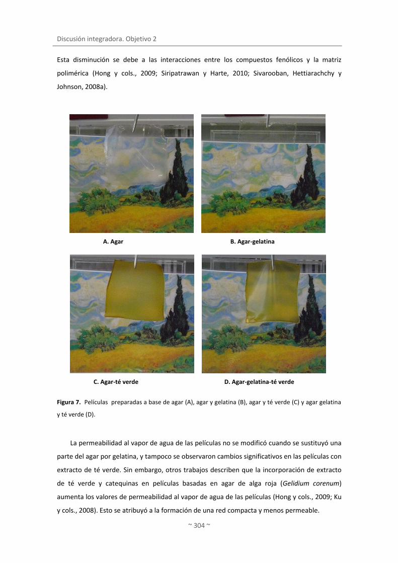
Discusión integradora. Objetivo 2
~ 304 ~
Esta disminución se debe a las interacciones entre los compuestos fenólicos y la matriz
polimérica (Hong y cols., 2009; Siripatrawan y Harte, 2010; Sivarooban, Hettiarachchy y
Johnson, 2008a).
La permeabilidad al vapor de agua de las películas no se modificó cuando se sustituyó una
parte del agar por gelatina, y tampoco se observaron cambios significativos en las películas con
extracto de té verde. Sin embargo, otros trabajos describen que la incorporación de extracto
de té verde y catequinas en películas basadas en agar de alga roja (Gelidium corenum)
aumenta los valores de permeabilidad al vapor de agua de las películas (Hong y cols., 2009; Ku
y cols., 2008). Esto se atribuyó a la formación de una red compacta y menos permeable.
Figura 7. Películas preparadas a base de agar (A), agar y gelatina (B), agar y té verde (C) y agar gelatina
y té verde (D).
B. Agar-gelatina
C. Agar-té verde D. Agar-gelatina-té verde
A. Agar

Estudio de la composición de extractos acuosos de té verde (Camellia sinensis ) en relación a su funcionalidad y desarrollo de envases comestibles activos
~ 305 ~
La solubilidad en agua es una medida de la materia soluble en agua de la película y se
utiliza habitualmente como un indicador de la resistencia al agua de la película. En la Figura 8
se muestran las películas de agar con extracto de té después de estar un tiempo sumergidas en
agua. Las películas de agar se vuelven gomosas después de estar en contacto con el agua pero
mantienen su integridad.
La solubilidad de las películas de agar después de 16 h sumergidas en agua fue de 24,1
±1,4%, mostrando menos resistencia al agua que las elaboradas por Rhim (2011) y Rhim y cols.
(2011), cuyos valores de solubilidad fueron 17.9 and 14.7%, respectivamente. No obstante, las
películas de agar son bastantes insolubles si se comparan con otro tipo de películas basadas en
otros biopolímeros, como metilcelulosa o gelatina de pescado, en especial de aguas frías, las
cuales son extremadamente solubles (Carvalho y cols., 2008; García, Pinotti, Martino y
Zaritzky, 2004; Pérez-Mateos, Montero y Gómez-Guillén, 2009). La sustitución de un 33% de
Figura 8. Aspecto de las películas de agar y extracto de té verde después de estar en contacto con el
agua.

Discusión integradora. Objetivo 2
~ 306 ~
agar por gelatina en la película no induce ninguna modificación significativa en la solubilidad.
Sin embargo, la adición del extracto de té verde favoreció drásticamente la solubilización de
ambos tipos de película (agar y agar/gelatina), atribuyendo este efecto a su naturaleza
hidrofílica. En las cinéticas de solubilización se aprecia un plateau a los 15 min, no
evidenciándose más allá ningún efecto a tener en cuenta.
Todas las muestras registraron valores similares de resistencia al agua durante los 15 días
de estudio, a pesar de las diferencias encontradas en solubilidad en agua entre las películas
con y sin extracto de té verde. En la literatura apenas hay trabajos en los que se utilice una
técnica similar; no obstante se considera de gran importancia, ya que a pesar de su simplicidad
ofrecen gran información sobre el potencial de la película a la hora de aplicarla a diversos
alimentos, especialmente en aquellos que contienen alto contenido en agua y no posean piel u
otro recubrimiento natural. La filtración de agua se inició después de las 14 h de exposición en
todas las películas y persistió durante todo el tiempo que se registró. Ninguna de las películas
se deforma o rompe durante este tiempo, lo que les confiere una buena aptitud para ser
utilizadas en diversos alimentos.
2.2.2. Liberación de compuestos polifenólicos y actividad antioxidante en agua
La liberación de compuestos fenólicos de las películas con extracto de té en agua se midió
a 22 °C y en varios tiempos durante 16 h. Esto nos permite evaluar, por un lado la liberación
de compuestos y, por otro, la difusión de los mismos hacia los alimentos de carácter
hidrofílico, ya que según la legislación Europea el agua se considera un simulador de alimento
acuoso.
La liberación de componentes activos desde las matrices poliméricas depende tanto de las
propiedades de los polímeros, de los compuestos activos, como del producto alimenticio
(López De Dicastillo y cols., 2011). Durante los primeros 15 min se produce la máxima
liberación de compuestos fenólicos en ambos tipos de películas, y alcanzan cifras similares (~
50 mg equivalentes de ácido gálico/g película; p > 0.05). A partir de este momento ambas
películas evolucionan de forma diferente: las de agar continúa liberando polifenoles
transcurridos 90 min; cuando hay gelatina presente apenas hay más liberación hasta los 90
min, y solo con el tiempo se liberan paulatinamente de nuevo más componentes del extracto,
alcanzando valores de ~60 mg equivalentes de ácido gálico/g película después de 16 h. Por
tanto, la presencia de gelatina en la película dificulta la liberación de los compuestos
polifenólicos. Es conocida la alta capacidad que tiene la gelatina de interaccionar con los

Estudio de la composición de extractos acuosos de té verde (Camellia sinensis ) en relación a su funcionalidad y desarrollo de envases comestibles activos
~ 307 ~
polifenoles de la película y durante la solubilización de la misma en agua, dando lugar a la
liberación de complejos proteína-polifenol o bien formándose complejos una vez que los
polifenoles se han liberado de la matriz (Giménez y cols., 2012). Estas interacciones puede ser
muy diversas, aunque frecuentemente son interacciones débiles, principalmente hidrofóbicas,
las cuales dependen del pH, temperatura, naturaleza, y concentración de la proteína y del
polifenol (Papadopoulou y cols., 2005; von Staszewski y cols., 2011).
Además de la liberación de los polifenoles totales, se examinó la liberación de las
catequinas y flavonoles mayoritarios del extracto de té incluido en la película. Las catequinas
más abundantes del té, epigalocatequina-3-galato y epigalocatequina, fueron las que más se
liberaron en ambos tipos de matrices, con valores dos órdenes de magnitud superiores al del
resto de catequinas y flavonoides. Tal y como se ha comentado en la liberación del contenido
en fenoles totales, las catequinas y flavonoles se liberan de igual manera a partir de las
películas, principalmente en los primeros 15 min. Después de 16 h en agua, todos los
polifenoles estudiados se liberaron en mayor cantidad en las películas basadas en agar con
extracto de té, con la excepción de epicatequina y epicatequina-3-galato.
En cuanto a las catequinas, hubo un aumento significativo en la liberación de
epigalocatequina, epicatequina-3-galato y epigalocatequina-3-galato a partir de las películas
de agar-extracto de té con el tiempo (p ≤ 0.05), aunque un 69, 46 y 68% de cada compuesto se
liberó durante los primeros 15 minutos. En cambio, no se detectaron concentraciones
crecientes de epicatequina después de los 15 minutos (p > 0.05), y sólo se observó un ligero
aumento en la concentración de catequina entre los 90 minutos y las 16 h (p ≤ 0.05). La
liberación de catequinas en las películas de agar/gelatina-extracto de té se produjo en menor
grado y varió en función del polifenol. También se produjo un aumento leve, pero significativo
(p ≤ 0.05), en el caso de epigalocatequina y epigalocatequina-3-galato (de 15 a 90 min), así
como epicatequina-3-galato (de 15 min a 16 h).
En el caso de las películas basadas en agar con extracto de té, aproximadamente el 50% de
los flavonoles cuantificados en agua después de las 16 h se liberaron durante los primeros 15
minutos. Además, la liberación de todos los flavonoles aumentó de forma significativa a mayor
tiempo de contacto (p ≤ 0.05). Así, hubo subidas entre 30 y 45%, dependiendo del
compuesto, de los 15 min a los 90 min, mientras que se observó un aumento de 40 a 50% en el
intervalo de tiempo de 90 minutos-16 horas. En cuanto a las películas de agar/gelatina con
extracto de té, ninguno de los flavonoles analizados se liberó de manera significativa en el
agua entre los 15 min y las 16 h (p > 0.05), probablemente debido a las interacciones
establecidas entre los polifenoles del té y la gelatina en la matriz polimérica.

Discusión integradora. Objetivo 2
~ 308 ~
2.2.3. Capacidad antioxidante liberada por las películas
Los datos obtenidos (FRAP y ABTS) de este estudio se reflejan en la Figura 9. En las
películas con agar y extracto de té, aproximadamente el 60% de la capacidad antioxidante
(medida por FRAP y ABTS) se liberó después de 16 h en agua durante los primeros 15 minutos.
La capacidad reductora liberada por las películas de agar/gelatina con extracto de té fue
significativamente menor que la de las películas sin gelatina a partir de los 15 min (p ≤ 0.05),
mientras que la capacidad secuestradora fue menor en las primeras durante todo el tiempo
evaluado (p ≤ 0.05). El aumento significativo de la actividad antioxidante en las películas
complejas de agar/gelatina solo se observó entre los 90 min y las 16 h. Esto pone de nuevo de
manifiesto como la presencia de gelatina en la matriz obstaculiza la liberación de los
compuestos polifenólicos, probablemente por la interacción proteína-polifenol ya comentada
El grado de enmascaramiento de la actividad antioxidante depende de la composición de
polifenoles y del tipo de proteína (Arts y cols., 2002; von Staszewski y cols., 2011). Sin
embargo, estos complejos de proteínas-polifenoles pueden retener parte de la actividad
antioxidante, puesto que algunos grupos hidroxilo de la molécula quedan libres, lo que
permite que estos compuestos mantengan sus propiedades antioxidantes (Almajano, Delgado
y Gordon, 2007).
La liberación de los compuestos fenólicos de las dos películas mostró una correlación
positiva con la actividad antioxidante medida por FRAP (r = 0,939 y r = 0,951 para la película
simple de agar y extracto de té y la compuesta por agar/gelatina y extracto de té,
respectivamente, p ≤ 0,01) y ABTS (r = 0,955 y r = 0,923 para la simple y compleja,
respectivamente, p ≤ 0,01). Por otra parte, la epicatequina y epigalocatequina-3-galato son
dos de las catequinas que más contribuyen a la eficacia antioxidante del té verde (Komes y
cols., 2010; Toschi y cols., 2000), debido a la presencia de un grupo galato en la posición 3ʾ del
anillo C (epigalocatequina-3-galato) y un grupo hidroxilo en la posición 5ʾ del anillo B (ambos
compuestos) (Guo y cols., 1999). La liberación de estos dos compuestos en el agua a partir de
las películas simples de agar y extracto de té mostraron correlación altamente significativa con
el FRAP (r = 0,985 y r = 0,979, respectivamente, p ≤ 0.01), y en menor medida con el ABTS (r =
0,871 y r = 0,872, respectivamente, p ≤ 0.05). Sin embargo, en el caso de la película compleja
no se observó una correlación significativa entre la actividad antioxidante (p > 0.05) y la
liberación de epigalocatequina y galato de epigalocatequina.

Estudio de la composición de extractos acuosos de té verde (Camellia sinensis ) en relación a su funcionalidad y desarrollo de envases comestibles activos
~ 309 ~
2.2.4. Propiedades antimicrobianas de las películas
Tal y como se describe al comienzo del apartado el extracto acuoso del té verde Wu Lu
Mountain inhibe a Vibrio parahaemolyticus, Photobacterium phosphoreum, Shewanella
putrefaciens, Pseudomonas fluorescens y Aeromonas hydrophila. Los resultados revelaron
además que la actividad antimicrobiana del extracto de té se mantuvo en la película, a
excepción de A. hydrophila. Sin embargo, las películas de agar y/o gelatina con extracto de té,
también inhibieron ligeramente el crecimiento de P. aeruginosa. Por otro lado, las películas
Figura 9. Capacidad antioxidante liberada por FRAP (A) y ABTS (B) a partir de las películas de agar/té
verde (CT) y de agar-gelatina/té verde (CGT) (Gimenez, López de Lacey, Pérez-Santín, López-Caballero y
Montero, P. , 2012).
A. FRAP
B. ABTS
Discusión integradora. Objetivo 2
~ 310 ~
simples y compuestas sin extracto de té no mostraron actividad antimicrobiana, como cabía
esperar. Las películas de agar permiten la difusión de té verde para que ejerza su actividad
antimicrobiana. En este caso, la presencia de gelatina en las películas no modificó la actividad
del extracto de té en la mayoría de los casos (p > 0.05), por lo que la interacción proteína-
polifenol no pareció afectar a esta capacidad. Von Staszewski y cols. (2011) observaron que la
actividad antimicrobiana de los polifenoles del té verde en presencia de proteínas del suero
aumentaba al disminuir la concentración de estas proteínas. En este experimento no se
observó este cambio, ya que la actividad antimicrobiana de las películas con y sin gelatina fue
similar, a pesar del retraso y menor liberación de polifenoles y actividad antioxidante en los
recubrimentos que contienen gelatina. Las películas de agar con extracto de té inhibieron el
crecimiento de P. phosphoreum, S. putrefaciens, A. hydrophila, V. parahaemolyticus y P.
fluorescens (tal como se describe en el trabajo “Antioxidant and antimicrobial activities of green
tea (Camellia sinensis L.) as an expression of its chemical composition”. En el presente estudio se
demuestra que el extracto de té no pierde sus cualidades como antimicrobiano cuando forma
parte de la formulación de la película. Este hecho es prometedor en términos de conservación
de los alimentos, ya que es poco frecuente que las películas con extractos acuosos de plantas
muestren su actividad una vez incorporados a las películas. Por lo tanto, es posible obtener
películas con propiedades antioxidantes y antimicrobianas a partir de películas simples en base
a agar y de películas complejas en base a agar/gelatina con la incorporación de extracto
acuoso de té verde. Si se comparan los resultados de actividad antimicrobiana del extracto de
té con los obtenidos en el aceite esencial de clavo, el aceite esencial de clavo tiene un mayor
espectro de acción porque actúa frente a un mayor número de cepas (Gómez-Estaca, López de
Lacey, López-Caballero, Gómez-Guillén y Montero, 2010), y su inhibición es mayor solo o
incorporado en las películas. En la Figura 10 se muestran los halos de inhibición producidos por
el aceite esencial de clavo y el extracto de té frente a las cepas S. putrefaciens y P.
phosphoreum, y la inhibición es mayor en las películas con aceite esencial de clavo. No
obstante, a pesar de que el aceite esencial es más potente frente al crecimiento de estas cepas
tiene la desventaja de aportar un olor y un sabor más intenso a los alimentos, como ya se
mencionó.
Otra desventaja que tiene el aceite esencial de clavo es que inhibe ligeramente el
crecimiento de bacterias que son beneficiosas, como las bacterias lácticas (ej. L. acidophillus),
a diferencia de los extractos de té verde que no producen ningún efecto inhibitorio en este
tipo de bacterias. Por lo tanto, hay que valorar las ventajas e inconvenientes de utilizar un
extracto u otro, y la elección en cada caso dependerá de la finalidad última del envase. En este

Estudio de la composición de extractos acuosos de té verde (Camellia sinensis ) en relación a su funcionalidad y desarrollo de envases comestibles activos
~ 311 ~
Aceite esencial de clavo Extracto de té verde
A. P. phosphoreum B. P. phosphoreum
C. S. putrefaciens D. S. putrefaciens
Figura 10. Halos de inhibición producidos por las películas con aceite esencial de clavo (A y C) y
extracto de té verde (B y D) frente a P. phosphoreum y S. putrefaciens.
caso el extracto de té verde Wu Lu Mountain se considera un candidato ideal para el desarrollo
y aplicación de envases comestibles potencialmente bioactivos.


Discusión integradora. Objetivo 3
~ 313 ~
3. INCORPORACIÓN DE BACTERIAS LÁCTICAS EN ENVASES COMESTIBLES
Y ESTUDIO DE SU VIABILIDAD DURANTE LA CONSERVACIÓN DE FILETE
DE MERLUZA
3.1. Funcionalidad de bacterias lácticas incorporadas a coberturas y películas
comestibles
El desarrollo de envases comestibles a los que se incorporan aceites esenciales
procedentes de plantas o extractos acuosos como el té verde confieren estabilidad y
garantizan la seguridad a los productos a los que se aplican, contribuyendo en parte a cubrir la
demanda del consumidor por alimentos mínimamente procesados, con pocos aditivos y sin
apenas cambios a nivel sensorial. Cuando los compuestos incorporados a los envases
mantienen la calidad y seguridad del alimento se constituye un envasado activo; además, los
compuestos añadidos al envase pueden producir un efecto potencialmente beneficioso en el
consumidor, constituyendo así un envasado bioactivo. La incorporación de bacterias lácticas o
probióticos a envases comestibles es de gran interés, ya que los envases pueden actuar como
vehículos para las bacterias. Los envases comestibles con bacterias se encuadran en el
concepto de bioconservación, como extensión de la vida útil y el aumento de la seguridad de
un alimento a través del uso de una microflora natural o controlada y de sus productos
antimicrobianos (Stiles, 1996).
Uno de los objetivos de la presente memoria consistió en el diseño y la formulación de
envases (películas y recubrimientos) a los que se incorporaron bacterias lácticas: Lactobacillus
acidophillus y Bifidobacterium bifidum, y en el estudio de su efecto durante la conservación de
merluza refrigerada. Como obstáculo adicional se aplicó un tratamiento de alta presión, ya que
estudios previos revelan que la combinación de tratamientos -envases comestibles y alta
presión- es un método eficaz para aumentar la vida del pescado.
La Figura 11 muestra los datos obtenidos de supervivencia de las bacterias estudiadas (L.
acidophillus y B. bifidum), tanto en las películas como en los recubrimientos. Los resultados
indican que los envases (películas y recubrimientos) comestibles de gelatina pueden
vehiculizar bacterias lácticas, y tanto L. acidophilus como B. bifidum permanecen viables
incluso hasta 13 días (Figura 11 B). Las bacterias fueron capaces de crecer en los medios de
cultivo, aunque se espera un descenso en los recuentos debido a la adaptación de las bacterias
al nuevo sustrato y a las temperaturas utilizadas para la conservación (2 ± 1 °C).

Discusión integradora. Objetivo 3
~ 314 ~
En esta línea, Tapia y cols. (2007) describen que la concentración de B. lactis Bb-12
incorporado a coberturas de alginato o gellan (109 UFC/g) disminuyó cuando se aplicó a trozos
de papaya y manzanas (aproximadamente en 2 unidades logarítmicas), hecho que se atribuye
a fenómenos de dilución, si bien esta población de bifidobacterias permanece viable y
constante durante 10 días a 2 °C. Por otro lado, la composición del envase es importante para
la supervivencia de las bacterias. Así, los envases del presente ensayo (películas y
recubrimientos) contenían sorbitol en su composición. El sorbitol y poli-oles actúan como
agentes protectores de los microorganismos en situaciones de descenso de actividad de agua,
como son la congelación-deshidratación de Lactobacillus plantarum y Lactobacillus rhamnosus
Figura 11. Supervivencia de las bacterias L.acidophillus y B. bifidum (recuentos microbianos expresados
en UFC/mL) en recubrimiento de gelatina (A) y película de gelatina (B), conservadas a 2±1 °C (López de
Lacey, López-Caballero, Gómez-Guillén y Montero, 2012).
A. RECUBRIMIENTOS
B. PELÍCULAS

Incorporación de bacterias lácticas en envases comestibles y estudio de su viabilidad durante la conservación de filete de merluza
~ 315 ~
(Carvalho y cols., 2004). El mecanismo por el cual el sorbitol ejerce su efecto protector puede
estar relacionado con la interacción entre los azúcares y los fosfolípidos de membrana,
ayudándoles a mantener su fluidez (Santivarangkna, Kulozik, Kienberger y Foerst, 2009).
Gialamas y cols. (2010) incorporaron Lactobacillus sakei (106 UFC/cm2, directamente antes del
moldeo o por spray) en películas comestibles de caseinato sódico y las mantuvieron a 4 y 15
°C; cuando las películas no contenían sorbitol se observó un incremento en la viabilidad de las
bacterias a 4 °C, que se atribuyó a un descenso en el metabolismo experimentado por los
organismos debido a la bajas temperaturas.
El desarrollo de envases comestibles con bacterias y su aplicación al pescado constituye
un reto desde el punto de vista tecnológico, no sólo por mantener las bacterias viables en un
medio que ofrece menores y limitadas posibilidades como sustrato para crecer, sino también
por la temperatura de refrigeración utilizada, lejos de la óptima de incubación de estas
bacterias. La formulación debe permitir, además, que el envase comestible mantenga la
integridad, a la vez que permita la difusión de las bacterias al músculo. En el caso de
tratamientos combinados con alta presión, las condiciones de tratamiento (nivel de presión/
temperatura/ tiempo) deberán ser tales que cumplan con el objetivo para el cual se aplica (en
este caso reducción de la flora total del pescado, especialmente flora psicotrofa Gram-negativa
y mantener el aspecto crudo del producto), minimizando el efecto sobre la flora láctica
incorporada en el envase. Es importante señalar que no se han encontrado referencias
anteriores en la literatura sobre el papel de estos envases durante la conservación del pescado
en refrigeración.
Durante la conservación en refrigeración (2±1 °C), la aplicación de recubrimientos
comestibles de gelatina a filetes de merluza con B. bifidum redujo la flora total en 1 unidad
logarítmica respecto al control. Sin embargo, lo más llamativo fue la inhibición observada en la
merluza con recubrimiento (con o sin bacterias) en los microorganismos productores de H2S,
presuntamente considerados como Shewanella putrefaciens, organismo involucrado en el
deterioro del pescado (López-Caballero, Martínez-Alvarez, Gómez-Guillén y Montero, 2007). Es
importante tener en cuenta la supervivencia de las bifidobacterias durante la conservación,
con el descenso de 1 unidad logarítmica comparado con la concentración inicial. Los recuentos
de bacterias lácticas (en medio MRS) fueron similares en todos los lotes, alcanzando 4 log
UFC/g al final del periodo estudiado. Las diferencias entre los recuentos de bacterias lácticas y
los de bifidobacterias (8 log UFC/g) se atribuyeron a la dificultad que tiene la cepa B. bifidum
de crecer en medios de cultivo sin la adición de cisteína. Algunos de los datos más relevantes
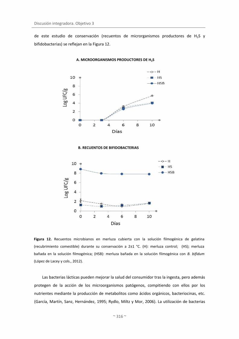
Discusión integradora. Objetivo 3
~ 316 ~
de este estudio de conservación (recuentos de microrganismos productores de H2S y
bifidobacterias) se reflejan en la Figura 12.
Las bacterias lácticas pueden mejorar la salud del consumidor tras la ingesta, pero además
protegen de la acción de los microorganismos patógenos, compitiendo con ellos por los
nutrientes mediante la producción de metabolitos como ácidos orgánicos, bacteriocinas, etc.
(García, Martín, Sanz, Hernández, 1995; Rydlo, Miltz y Mor, 2006). La utilización de bacterias
Figura 12. Recuentos microbianos en merluza cubierta con la solución filmogénica de gelatina
(recubrimiento comestible) durante su conservación a 2±1 °C. (H): merluza control; (HS); merluza
bañada en la solución filmogénica; (HSB): merluza bañada en la solución filmogénica con B. bifidum
(López de Lacey y cols., 2012).
A. MICROORGANISMOS PRODUCTORES DE H2S
B. RECUENTOS DE BIFIDOBACTERIAS

Incorporación de bacterias lácticas en envases comestibles y estudio de su viabilidad durante la conservación de filete de merluza
~ 317 ~
lácticas frente a microorganismos patógenos se ha descrito en la literatura para diversos
alimentos. Así, la incorporación de Lactobacillus plantarum LKE5 a gambas contaminadas con
Listeria monocytogenes inhibió incluso 3 ciclos logarítmicos el crecimiento del patógeno (Huss,
Jeppesen, Johansen y Gram, 1995), siendo este efecto mayor en condiciones experimentales
cercanas al límite de crecimiento del patógeno (en términos de pH, temperatura, etc.). En otro
experimento se realizó un baño con Lactobacillus rhamnosus GG (108 UFC/ml) en manzana
fresca cortada. Los resultados mostraron que, si bien los recuentos de Salmonella no se
afectaron al inocular el patógeno conjuntamente con L. rhamnosus GG, L. monocytogenes, se
redujo 1 unidad logarítmica en presencia del lactobacilo (Alegre, Viñas, Usall, Anguera y
Abadias, 2011). Además, la calidad de la manzana no se vio afectada por la incorporación del L.
rhamnosus GG, y la población de probióticos se mantuvo sobre 107 UFC/g durante 28 días de
conservación a 5 y 10 °C.
La incorporación de bacterias a envases comestibles (películas y recubrimientos) puede
ser de gran interés y ofrecer, por tanto, nuevas posibilidades. Según nuestro conocimiento sólo
hay dos referencias en la literatura al respecto (Gialamas y cols., 2010; Tapia y cols., 2007),
además del trabajo desarrollado en la presente memoria (“Functionality of lactic acid bacteria
incorporated to edible coatings and films”, López de Lacey y cols., 2012).
La ventaja de incorporar bacterias a un envase frente a la de incorporar las bacterias
directamente al alimento, por ejemplo mediante baño, guarda relación con la distribución y
concentración de bacterias en el envase. Kristo, Koutsoumanis y Biliaderis (2008) describieron
que el efecto de metabolitos microbianos como la nisina, contra patógenos como L.
monocytogenes, es mayor cuando la nisina forma parte de un recubrimiento (caseinato sódico)
que cuando se aplica directamente en el medio de cultivo. Este hecho se puede atribuir al
descenso en la tasa de difusión del envase al producto, manteniéndose así concentraciones
altas del compuesto activo en la superficie (Gialamas y cols., 2010). Estos autores señalan que
Lactobacillus sakei incorporado a películas de caseinato sódico inhibe efectivamente el
crecimiento de Listeria monocytogenes (hasta 4 ciclos logarítmicos), tanto en medio de cultivo
como en filetes de ternera (a 5 y 15 °C), incrementando así la seguridad de los alimentos.
Los tratamientos combinados con alta presión se aplicaron a 200 MPa para evitar la
modificación de las características sensoriales del pescado. Con este tratamiento también se
eliminó, en parte, la flora tipo Gram-negativa, mientras que la Gram-positiva (endógena y la
incorporada al envase comestible) se afectó en menor medida por ser más resistente a la alta
presión (López-Caballero, Pérez-Mateos, Montero y Borderías, 2000). Este ensayo se realizó en
películas y no en recubrimientos por la potencial resistencia adquirida por las bacterias, debido
Discusión integradora. Objetivo 3
~ 318 ~
al manejo y estrés sufrido en la preparación del envase (deshidratación, exposición a
temperaturas moderadas, etc.), fomentando así su viabilidad. En relación a este hecho,
Gialamas y cols. (2010) indicaron que cuando las bacterias se incorporan a la solución
filmogénica (que posteriormente da lugar a la película) se exponen al secado, lo que podría
explicar el incremento de viabilidad de bacterias incorporadas por este método en
comparación con las incorporadas a la película mediante spray (ya que este método apenas
aporta estrés a las bacterias).
El tratamiento aplicado en este experimento (200 MPa/20 °C/10 min) redujo en 1 unidad
logarítmica los recuentos de bacterias totales, y los microorganismos productores de H2S
permanecieron por debajo del límite de detección. La sensibilidad de S. putrefaciens a la alta
presión se ha referido con anterioridad (López-Caballero y cols., 2000). Tanto la flora láctica
como las bifidobacterias no se afectaron por el tratamiento. Asimismo no se detectó daño
subletal ni estrés reversible. Estos resultados son de gran interés porque el tratamiento
aplicado permite la eliminación de parte de los bacilos psicrotrofos Gram-negativos que
producen la alteración del pescado en beneficio de la flora láctica, cuyo metabolismo es
generalmente más lento e implica una menor producción de metabolitos responsables de la
producción de olores desagradables. Las colonias luminiscentes, presuntos P. phosphoreum
(López-Caballero, Álvarez Torres, Sánchez-Fernández y Moral, 2002) inicialmente estuvieron
por debajo del límite de detección. Aunque la resistencia de este género a tratamientos de alta
presión se ha descrito (Paarup, Sánchez, Peláez y Moral, 2002), la aplicación de (300 Mpa/10
min / 5 y 40 °C) a salmón recubierto con películas de gelatina-lignina reduce significativamente
los recuentos de P. phosphoreum (Ojagh, Núñez-Flores, López-Caballero, Montero y Gómez-
Guillén, 2011). Durante la conservación, el lote control (merluza sin tratamiento) evolucionó
de forma similar al lote cubierto con película + B. bifidum. Por el contrario, la tasa de
crecimiento exponencial fue menor en el lote con película presurizado, en especial para los
microorganismos productores de H2S. Al final de la conservación (9 días), la flora total en el
lote presurizado fue 2 unidades logarítmicas inferior a la del resto de los lotes; en este lote, los
recuentos de totales fueron similares a los de microorganismos productores de H2S. La
aplicación de envases comestibles con bacterias (sometidos o no a tratamiento de alta
presión) inhibió el crecimiento de P. phosphoreum, microorganismo alterante activo implicado
en el deterioro del pescado, ya que sólo se detectó en el lote sin tratamiento. En relación a las
bacterias lácticas no se encontraron diferencias entre lotes y las bifidobacterias permanecieron
viables en ambos lotes (presurizado o no) durante el tiempo de conservación.

Incorporación de bacterias lácticas en envases comestibles y estudio de su viabilidad durante la conservación de filete de merluza
~ 319 ~
En cuanto a los índices físico-químicos (Figura 13), la presencia de bacterias y su paso al
músculo en los lotes con películas de gelatina originó un pequeño descenso en los niveles de
pH durante la conservación. Asimismo, la concentración de nitrógeno básico volátil total
permaneció por debajo de 20 mg N-TVB/100 g músculo en el lote recubierto con película y
presurizado, mientras que el resto registraron valores 60 mg-NBVT/100 g músculo. Estos
niveles estuvieron en consonancia con los recuentos de microorganismos (bacterias totales y
productores de H2S) obtenidos por estos lotes, si bien el pH alcanzado en los lotes cubiertos
con películas de gelatina no reflejaron los niveles de compuestos volátiles de cada uno de
ellos. La combinación de tratamientos (alta presión) y envases comestible con bacterias,
presentaron cierto efecto aditivo en la inhibición de las bases volátiles, ya que la merluza
presurizada en idénticas condiciones a la utilizada en el presente ensayo y con una carga
microbiana similar, alcanzó valores de 35-40 mg N-TVB/100 g músculo aproximadamente a los
10 días de conservación. Es más, la aplicación de las películas con B. bifidum no confirió olores
extraños al pescado. Quizás la presencia de bifidobacterias interfiera de algún modo en la
producción de compuestos básicos volátiles (por ejemplo, compitiendo por nutrientes, etc.),
con microorganismos tales como S. putrefaciens, disminuyendo así los niveles registrados en
cierta medida. Está previsto en el futuro establecer la tasa de difusión de las bacterias al
músculo y estudiar cómo se afecta la calidad sensorial del pescado en presencia de la flora
láctica.

Discusión integradora. Objetivo 3
~ 320 ~
A. pH
Figura 13. A) pH y B) bases volátiles totales de la merluza cubierta con la película de gelatina y
sometida a alta presión (200 MPa/20 °C/10 min) durante su conservación a 2±1 °C. (H): merluza
control; (HS); merluza película + B. bifidum; (HSB): merluza presurizada película + B. bifidum
(López de Lacey y cols.., 2012).
B. BASES VOLÁTILES TOTALES

Discusión integradora. Objetivo 4
~ 321 ~
4. DESARROLLO DE PELÍCULAS COMPLEJAS PARA LA BIOCONSERVACIÓN
DE PRODUCTOS PESQUEROS
4.1. Viabilidad de los probióticos en presencia de los polifenoles mayoritarios del té
verde y extracto de té verde/4.2. Biotransformación de los compuestos polifenólicos
mayoritarios del té verde y extracto de té verde
En apartados anteriores han quedado patente las propiedades antioxidantes y
antimicrobianas de las películas de té verde (“Release of active compounds from agar and
agar-gelatin films with green tea extract”) y la utilización de envases comestibles como
vehículo de bacterias lácticas (“Functionality of lactic acid bacteria incorporated to edible
coatings and films”). Por ello se planteó la posibilidad de combinar el extracto de té verde con
bacterias probióticas con la incertidumbre de si los polifenoles del té podrían inhibir a estas
bacterias, ser incapaces de crecer en estos sustratos o bien que las bacterias fueran capaces de
transformar los polifenoles en otros productos con diferente actividad biológica.
La actividad biológica del té verde se ha asociado principalmente a su contenido rico
en polifenoles ya que éstos presentan interesantes propiedades. Sin embargo, los efectos
beneficiosos de los polifenoles sobre el organismo humano dependen de su biodisponibilidad
en el mismo (Walle, 2004). En este sentido, el reciente interés por añadir probióticos a los
alimentos ricos en polifenoles (Marotti, Bonetti, Biavati, Catizone y Dinelli, 2007; Otieno,
Ashton y Shah, 2006) se debe a que éstos poseen determinadas actividades enzimáticas
[deglicosilación, dehidroxilación, demetilación o decarboxilación, etc. (Aura, 2008)] que
permiten transformar los polifenoles en formas más biodisponibles o activas que el compuesto
fenólico de origen (Donkor y Shah, 2008; Tsangalis, Ashton, McGill y Shah, 2002; Uskova,
Kravchenko, Avrenjeva y Tutelyan, 2010). En esta línea, se han publicado algunos trabajos
sobre la biotransformación de flavonoides producidas por Bifidobacterium sp. o Lactobacillus
sp. (Ávila y cols., 2009; Donkor y Shah, 2008; Marotti y cols., 2007). Sin embargo, no hay
mucha información en la literatura sobre el efecto de las bacterias probióticas en la actividad
antioxidante del flavonol. La fermentación de las catequinas con bacterias probióticas ha sido
incluso menos estudiada que la de los flavonoles.
Los objetivos de este estudio se centraron en evaluar la viabilidad de tres cepas
probióticas comerciales (Lactobacillus paracasei LAFTI@L26, Lactobacillus acidophillus
LAFTI@L10 y Bifidobacterium animalis ssp. Lactis LAFTI@B94) en los polifenoles mayoritarios o
más característicos del té (5 tipos de catequinas y 5 flavonoles), la transformación de estos

Discusión integradora. Objetivo 4
~ 322 ~
compuestos y la actividad antimicrobiana, antioxidante (FRAP, ABTS) y antihipertensiva
resultante de la incubación de los compuestos con la bacteria probiótica. Este mismo estudio
se realizó en el extracto de té verde Wu Lu Mountain.
Como primer paso se determinó la actividad enzimática β-glucosidasa, β-galactosidasa y
α-ramnosidasa de los tres probióticos. Estas enzimas realizan la deglicosilación de los
polifenoles, considerada como el primer paso del metabolismo de los flavonoides glicosilados
(Aura, 2008; Braune y Blaut, 2011; Serra y cols., 2012). Las tres bacterias probióticas mostraron
actividad β-glucosidasa, β-galactosidasa y α-ramnosidasa, siendo esta última actividad la que
presentó los niveles más bajos. Esta actividad α-ramnosidasa está sólo presente en la flora
microbiana intestinal (Scalbert y Williamson, 2000) y su producción se ha descrito en algunas
especies bacterianas como, por ejemplo, Bacteroides y Lactobacillus (Avila y cols., 2009;
Beekwilder y cols., 2009; Bokkenheuser, Shackleton y Winter, 1987). En la literatura no se ha
encontrado información acerca de esta actividad en especies como Bifidobacterium. De entre
todas las cepas estudiadas, L. acidophillus L10 exhibió los valores más altos de actividad
enzimática, excepto para α-rhamnosidasa, que registró los menores. Donkor y Shah (2008) y
Otieno y cols. (2006) encontraron también que L.acidophillus L10 presentó los niveles más
altos de actividad β-glucosidasa, en comparación con B. animalis B94 y L. paracasei L26, entre
las 24-36 h de incubación; y de Bifidobacterium spp. y L. paracasei L26 a las 24 h de la
fermentación de la leche de soja. L. paracasei L26 y B. animalis B94 tuvieron un
comportamiento similar y apenas se observaron diferencias entre ellas, excepto en la actividad
β-galactosidasa (más alta en L. paracasei L26). De acuerdo con Donkor y Shah (2008) y Otieno,
Ashton y Shah (2005) las diferencias entre las actividades β-glucosidasa de diversas bacterias
se atribuyen a la especificidad de cada especie.
Este trabajo se diseñó con el objetivo final de utilizar las bacterias probióticas en
combinación con el extracto de té verde, por lo que se consideró de gran importancia
determinar la supervivencia de las tres bacterias lácticas en los 6 extractos de té verde ya
estudiados (apartado 2.1): Japan Sencha Makinohara, Japan Gyokuro Asahi, Canela Té Verde,
Lung Ching, China White Hair y Wu Lu Mountain. La supervivencia de las tres cepas en las seis
variedades se muestra en la Figura 14 y 15. Los extractos de té verde, a excepción del té verde
de canela, permitieron la supervivencia de las tres cepas probióticas investigadas durante más
tiempo que en la solución control que contenía sólo 0.9% de NaCl.

Desarrollo de películas complejas para la bioconservación de productos pesqueros
~ 323 ~
A. SUPERVIVENCIA DE L. acidophillus L10 EN LOS 6 TÉS
B. SUPERVIVENCIA DE L. paracasei L26 EN LOS 6 TÉS
Figura 14. Supervivencia de la bacteria L.acidophillus L10 (A) y L. paracasei L26 (B) en los 6 extractos de té
verde a 37 °C. Los datos están expresados en porcentaje de supervivencia (% ) (López de Lacey, Pérez
Santín y cols., 2012b).
La supervivencia fue menor en el té verde con canela quizás porque los componentes de
la canela son tóxicos para estas bacterias probióticas. En este sentido, las infusiones acuosas
de canela y aceites tienen actividad antimicrobianas frente a varias especies, incluyendo
Lactobacillus sp. (Ceylan y Fung, 2004). L. acidophillus L10 fue la especie con más
requerimientos para sobrevivir porque su número descendió a las 24 h de incubación (<20%) y
murió a las 48 h (independientemente de la variedad de té). En especial, L. paracasei L26 y B.

Discusión integradora. Objetivo 4
~ 324 ~
animalis B94 sobrevivieron más tiempo en Japan Sencha Makinohara, Asahi Gyokuro y Wu Lu
Mountain, seguido por China White Hair y de Lung Ching. L. acidophillus L10 probablemente
requiere energía de otras fuentes presentes en el té como es, por ejemplo, la obtención de
glucosa a partir de los flavonoides glicosilados mediante la producción de enzimas con
actividad glucosidasa (Schneider, Schwiertz, Collins y Blaut, 1999). A pesar de que ésta cepa
presentó los niveles más altos de actividad glucolítica, su supervivencia fue menor con
respecto a las otras bacterias. Tal vez este hecho se deba a que los azúcares procedentes de
los flavonoides glicosilados se toman más rápidamente que en las otras especies. Asimismo, L.
acidophillus mostró una menor actividad α-ramnosidasa.
Por el contrario, B. animalis B94 fue la cepa que exhibió un mayor número de células
viables (alrededor de 6 unidades logarítmicas) al final de las 72h de incubación en todas las
variedades de té excepto en Japan Sencha Makinohara (dónde sus recuentos disminuyeron en
3 unidades). No se ha encontrado información acerca de la supervivencia de bacterias
probióticas en extractos acuosos de té verde ya que la mayoría de las publicaciones están
enfocadas a estudiar el crecimiento en alimentos que son fermentados principalmente por
SUPERVIVENCIA DE B. animalis B94 EN LOS 6 TÉS
Figura 15. Supervivencia de la bacteria B. animalis B94 en los 6 extractos de té verde a 37 °C y en
anaerobiosis. Los datos están expresados en porcentaje de supervivencia (%) (López de Lacey, Pérez
Santín, López-Caballero y Montero, 2012b).
Desarrollo de películas complejas para la bioconservación de productos pesqueros
~ 325 ~
bacterias probióticas, como son el yogurt, leche fermentada o soja (Champagne, 2009). Sin
embargo, recientemente se han realizado estudios de supervivencia de bacterias probióticas
en otros productos, como el zumo con té verde como antioxidante (Shah, Ding, Fallourd y
Leyer, 2010), dónde se observó que el té verde crea un ambiente anaeróbico favorable para las
bacterias probióticas debido a sus propiedades antioxidantes.
Después de evaluar la supervivencia de los probióticos, se consideró importante
determinar qué polifenoles del té permiten una mayor supervivencia de estas cepas. Para la
realización de este estudio se seleccionó a B. animalis B94, ya que es la que mejor sobrevive en
el té. Para ello se incubó la bacteria probiótica a 37 °C durante 72 h en 10 soluciones salinas,
las cuales contenían como única fuente de energía cada uno de los 10 tipos de polifenoles
estándares seleccionados para el estudio (5 catequinas y 5 flavonoles mayoritarios del extracto
de té verde). Durante la incubación de la cepa probiótica en el control (solución salina) y en los
diferentes estándares de polifenoles se detectó una disminución de los recuentos a las 24 h de
incubación. Tras este tiempo, los recuentos disminuyeron por debajo del control (88.73%) en
todas las muestras, excepto en la catequina (98.49%), epigalocatequina (91.97%) y quercetina-
3-O-galactosido (91.96%). El hecho más destacado fue que los flavonoides glicosilados
prolongaron la supervivencia de las bacterias hasta pasadas las 48 h de incubación. Las
enzimas β-glucosidasa, β-galactosidasa y α-ramnosidasa rompen los enlaces O-glucosídicos,
galactosídicos y rhamnosidicos, respectivamente, presentes en los flavonoides glicosilados. B.
animalis B94 es capaz de producir estas enzimas durante la incubación y obtener los azúcares
unidos a estos flavonoides (Schneider y cols., 1999). Por esta razón posiblemente la bacteria
consigue sobrevivir más tiempo. En general, la reducción más baja en el número de células
viables se detectó a las 48 h en la solución con quercetina 3-0-galactosido y kaempferol-3-O-
glucosido. Por otro lado, la disminución más acusada se observó en la solución con
kaempferol-3-O-rutinosido, que posee en su estructura la ramnosa. Estos resultados están en
consonancia con los valores de actividad de B. animalis B94, ya que esta bacteria hidroliza más
fácilmente los enlaces glucosídicos y galactosídicos que los ramnosídicos.
Por otro lado, las catequinas con o sin bacteria sufrieron cambios en su concentración a lo
largo de la incubación a 37 °C. Los cambios observados en la concentración de la catequinas
incubadas sin la bacteria se deben seguramente a la inestabilidad de estos compuestos
durante las condiciones de experimentación. Algunos estudios han demostrado que las
catequinas pueden sufrir cambios en su concentración debidos a su oxidación y epimerización
en condiciones de pH superiores a 6 (Ananingsih, Sharma y Zhou; Neilson y cols., 2007;
Roginsky y Alegria, 2005). A pesar de esto, la desaparición de las catequinas fue mayor en las

Discusión integradora. Objetivo 4
~ 326 ~
soluciones incubadas con la bacteria. La presencia de la bacteria afectó más a la concentración
de las catequinas que a la de los flavonoides glicosilados. Los cambios más notables fueron en
el galato de epigalocatequina (EGCG) y en epigalocatequina (EGC) (en la Figura 16).
El galato de epigalocatequina desapareció después de 24 h de incubación con la bacteria,
mientras que la epigalocatequina disminuyó progresivamente a lo largo de la incubación de la
bacteria, y desapareció a las 72h. El resto de las catequinas analizadas no tuvieron un
descenso tan drástico y se mantuvieron más o menos constante (alrededor del 50%) después
de las 72 h de incubación con la bacteria. Los niveles de flavonoides glicosilados se
mantuvieron a lo largo de todo el periodo y apenas se observaron cambios en la concentración
(~2-5%). Este hecho indica que los flavonoides glicosilados fueron más estables que las
catequinas en las condiciones de experimentación ensayadas. Sin embargo, cuando estos
flavonoides se incubaron con la bacteria se detectó una bajada en la concentración, aunque en
menor proporción que en las catequinas. El mayor descenso se encontró en la solución con
rutina y quercetina-3-O-galactoside, lo que puede significar que B. animalis B94 tiene menor
afinidad por estos compuestos y los transforma en menor medida.
No se encuentra una relación directa entre la desaparición de polifenoles y el número de
células viables visto en el estudio de supervivencia. Donkor y Shah (2008) estudiaron la
hidrólisis de loa isoflavonas por B. animalis B94 y observó que la máxima concentración de
agliconas se produjo cuando la población de células era alta.
Figura 16. Comparación de la desaparición de EGCG (A) y EGC (B) en las soluciones preparadas
independientemente: a) muestra control (sin bacteria) y b) en la presencia de B. animalis B94 (López de
Lacey, Pérez Santín, López-Caballero y Montero, 2012a).

Desarrollo de películas complejas para la bioconservación de productos pesqueros
~ 327 ~
Este mismo estudio se realizó también con el té verde (variedad Wu Lu Mountain) con el
objeto de comprobar los cambios ocasionados por la misma bacteria probiótica en el propio
extracto, con la diversidad y cantidad de polifenoles que hay en él. La concentración de los
flavonoides disminuyó en todas las muestras (con o sin bacteria), pero la reducción en la
concentración de los flavonoides glicosilados en el té fue mayor cuando se incubó con la
bacteria B. animalis B94. Por otra parte, las diferencias de concentración entre los flavan-3-ol
tratadas o no con la bacteria fueron menos pronunciadas, excepto en el 3-gallato de
epigalocatequina. Además se encontró que los polifenoles incubados sin la bacteria fueron
más inestables en el té (pH~ 5.7) que en las soluciones puras (pH >6). Suzuki y cols. (2003)
observaron que la epimerización de las catequinas del té en agua destilada es menor que en la
infusión de té o en soluciones buffer de pH 6.0. Posiblemente los flavan-3-ols en el té verde se
transforman por la bacteria y se degradan al mismo tiempo, pero a diferente velocidad, y por
eso no se detectaron tantas diferencias como en los estándares individuales. Puede ser que la
velocidad de degradación de las catequinas sea mayor que la velocidad de transformación por
la bacteria, por lo que la transformación de las catequinas por B. animalis B94 está afectada
por la matriz en la que está incluida. Los flavonoides glicosilados se transformaron por la
bacteria B. animalis B94, a pesar de su baja concentración en comparación con las catequinas.
En este caso, parece ser que la velocidad de degradación pudo haber sido inferior a la
velocidad de transformación por la bacteria a 37 °C.
El flavonol quercetina 3-O-galactosido fue el más afectado por la presencia de la bacteria,
seguido de la rutina. En cambio el kaempferol 3-O-rutinoside fue el que menos se transformó
por el probiótico en comparación a los otros flavonoles. Estos resultados se deben
posiblemente a la actividad enzimática de la bacteria, puesto que B. animalis B94 posee una
mayor actividad β-glucosidasa que β-galactosidasa y α-ramnosidasa.
En este trabajo también se evaluó la capacidad antioxidante (ABTS, FRAP),
antihipertensiva y antimicrobiana de las soluciones estándares y del té, antes y después de ser
incubada con B. animalis B94. La actividad antimicrobiana sólo se determinó en el extracto de
té verde ya que para realizar esta medida se requerían de concentraciones de polifenoles
estándares muy superiores a las utilizadas en este experimento.
A pesar de los cambios producidos en la concentración de las distintas soluciones de
catequinas incubadas sin la bacteria, no se detectaron modificaciones en la actividad
antioxidante medida por ABTS. Los productos formados a partir de la degradación de las
catequinas presentaron en conjunto la misma actividad antioxidante que los polifenoles
iniciales. La actividad antioxidante (ABTS) de las catequinas aumentó considerablemente tras

Discusión integradora. Objetivo 4
~ 328 ~
la incubación con la bacteria. En la mayoría de los casos la actividad antioxidante se
incrementó a excepción del ECG, que alcanzó un 42%. Este incremento en la actividad se
mantuvo constante después de 24 h de incubación. El aumento de actividad antioxidante por
ABTS confirma que la bacteria transformó las catequinas en otros compuestos, ya que existe
una relación directa entre la desaparición de catequinas durante la incubación con la bacteria y
el incremento en la actividad antioxidante. Por otra parte, la capacidad antioxidante por ABTS
de los flavonoides glicosilados fue significativamente menor que en los flavan-3-ols. Con
respecto a este grupo de polifenoles, parece existir una relación entre los cambios producidos
en el contenido de polifenoles y las pequeñas variaciones detectadas en la capacidad
secuestradora de radicales a lo largo de las 72 h de incubación. Asimismo, la desaparición de
polifenoles está más relacionada con la trasformación de las formas no glicosiladas en otros
metabolitos con diferente actividad antioxidante (Serra y cols., 2012). Teóricamente, la
actividad de la bacteria puede transformar los flavonoides glicosilados en sus correspondientes
aglicones que normalmente poseen más actividad antioxidante. De hecho, el aglicón
quercetina exhibe una mayor actividad antioxidante (ABTS) que la rutina (Cai, Mei, Jie, Luo y
Corke, 2006). Por lo tanto, posiblemente los aglicones se han formado previamente por la
bacteria y después se han transformado en otros metabolitos, como ácidos fenilacéticos o
hidroxifenilacéticos, con actividad antioxidante diferente (Aura y cols., 2002; Serra y cols.,
2012).
La capacidad antioxidante medida por FRAP de las catequinas disminuyó
considerablemente a lo largo de la incubación. Sin embargo, al igual que en el ABTS, la
presencia de la bacteria originó un incremento en la capacidad para reducir el hierro (FRAP),
(aunque algo menor si comparamos con el análisis ABTS), principalmente durante las primeras
24-48 h, lo que coincide con la concentración celular más alta. Sin embargo, el
comportamiento de los flavonoles con respecto a la capacidad reductora fue un poco más
heterogéneo que en el ABTS. Se detectó un aumento en la actividad antioxidante por FRAP en
las soluciones incubadas con la bacteria. Este pequeño incremento demuestra que los
flavonoles glicosilados se transformaron en otros metabolitos con mayor poder reductor. De
hecho, algunos metabolitos como el ácido acético 2-(3,4-dihidroxifenil) producido por la
degradación colónica de los flavonoides pueden exhibir una mayor capacidad reductora que la
quercetina y una menor capacidad secuestradora de radicales libres (ABTS) (Dueñas, Surco-
Laos, González-Manzano, González-Paramás y Santos-Buelga, 2011). En la Figura 17 se
muestran los cambios producidos en la actividad antioxidante por FRAP y ABTS del EGCG y de

Desarrollo de películas complejas para la bioconservación de productos pesqueros
~ 329 ~
la rutina durante su incubación con la cepa, como ejemplos más destacados de este
experimento.
Este mismo estudio, pero en el extracto acuoso de té verde Wu Lu Mountain, dio
resultados un tanto diferentes a los esperados porque la capacidad antioxidante del té,
medida tanto por ABTS y FRAP, descendió en mayor proporción en las muestras que contenían
la bacteria B. animalis B94. Posiblemente, la presencia de otros compuestos del extracto de té
como metales, proteínas, carbohidratos, etc. o incluso los compuestos formados por la
Figura 17. Comparación de los equivalentes de vitamina C (ABTS) y equivalentes de Fe2+
(FRAP) de las
soluciones de EGCG (A y B) y rutina (C y D) preparadas independientemente: a) muestra control (sin
bacteria) y b) en la presencia de B. animalis B94 (López de Lacey y cols., 2012b).
ABTS FRAP

Discusión integradora. Objetivo 4
~ 330 ~
actividad de la bacteria no favorecen a la actividad antioxidante de los polifenoles contenidos
en el té. En este sentido, no se ha encontrado información acerca de las modificaciones
producidas en la actividad antioxidante por bacterias probióticas. Sin embargo, en una
publicación reciente se analizó el efecto de una enzima bacteriana sobre la actividad
antioxidante de los polifenoles (Macedo, Battestin, Ribeiro y Macedo, 2011). Estos autores
demostraron que el extracto de té verde tratado con tanasa (una enzima producida por
Paecilomyces variotti) mostró una capacidad antioxidante considerablemente mayor que el
extracto de té sin tratar. Esta enzima es capaz de hidrolizar el sustrato contenido en el té
verde, y los productos de la hidrólisis pueden aumentar significativamente las propiedades
antioxidantes del té verde.
En cuanto a la actividad antihipertensiva de los polifenoles estándares, se encontraron
marcadas diferencias entre las muestras incubadas con o sin bacteria. Así, los porcentajes de
inhibición de la ACE de las catequinas se incrementaron (10-27%) tras ser incubada con la cepa
probiótica. La epigalocatequina y la catequina mostraron sus valores más altos entre las 48 h y
las 72 h, siendo la epigalocatequina muy estable a lo largo del periodo. El dato más
sorprendente fue el de la catequina, ya que este compuesto no presentó actividad
antihipertensiva hasta que no se incubó con B. animalis B94. Los flavonoles incrementaron sus
valores de inhibición en presencia de la bacteria, aunque no fue superior al de las catequinas,
salvo en la rutina. Quercetina 3-O-galactoside exhibió una inhibición a las 24 h, aunque ésta
fue bastante inestable y su valor disminuyó entre un 12-15% a las 48-72 h de incubación. De
todos los flavonoles, el efecto antihipertensivo más alto se encontró en el kaempferol-3-O-
rutinoside y en la rutina incubados con la bacteria. En este último caso se produjo un efecto
similar al de la catequina ya que la rutina no mostró actividad antihipertensiva en ausencia de
la bacteria. En la Figura 18 se exponen los resultados obtenidos en estos dos polifenoles, como
ejemplos más llamativos de este estudio.
La transformación realizada por la bacteria B. animalis B94 en los polifenoles del té no
mejoró la capacidad inhibitoria del extracto de té dado que no hubo un aparente incremento
de inhibición de la ACE en esta muestra. Sin embargo es importante destacar que esta
actividad se mantuvo en la muestra con la bacteria y no se perdió, al igual que en la muestra
incubada sin la bacteria.
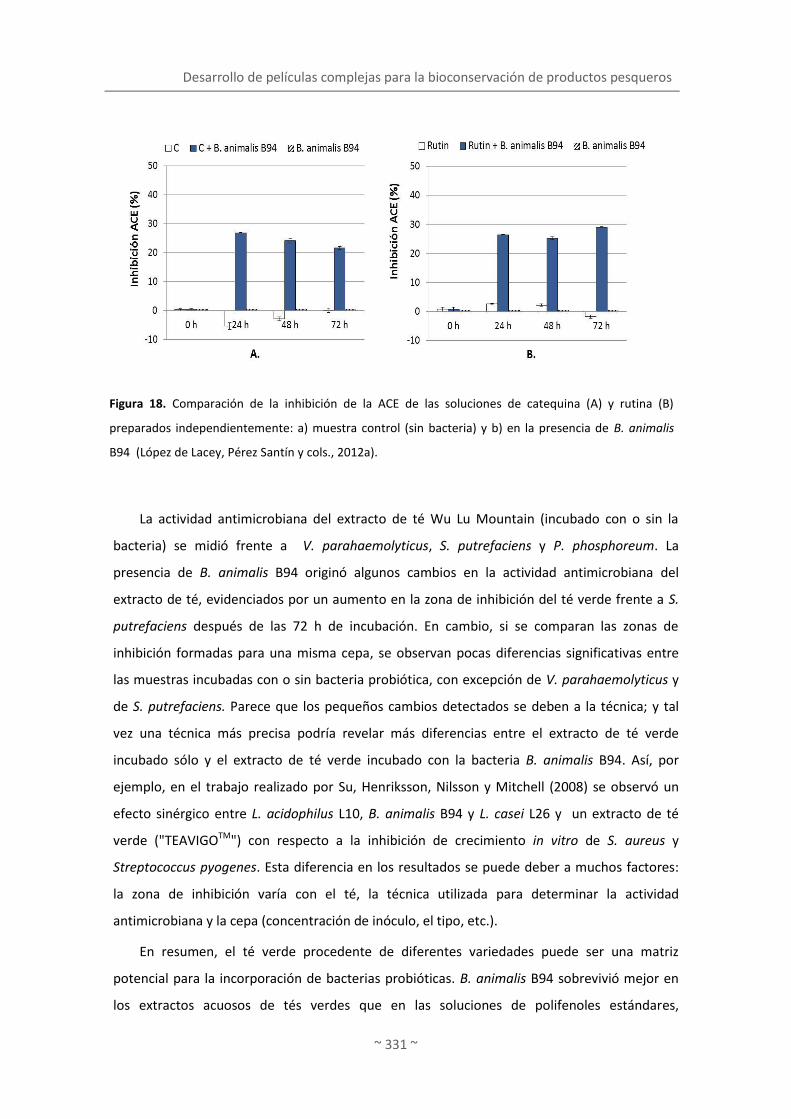
Desarrollo de películas complejas para la bioconservación de productos pesqueros
~ 331 ~
La actividad antimicrobiana del extracto de té Wu Lu Mountain (incubado con o sin la
bacteria) se midió frente a V. parahaemolyticus, S. putrefaciens y P. phosphoreum. La
presencia de B. animalis B94 originó algunos cambios en la actividad antimicrobiana del
extracto de té, evidenciados por un aumento en la zona de inhibición del té verde frente a S.
putrefaciens después de las 72 h de incubación. En cambio, si se comparan las zonas de
inhibición formadas para una misma cepa, se observan pocas diferencias significativas entre
las muestras incubadas con o sin bacteria probiótica, con excepción de V. parahaemolyticus y
de S. putrefaciens. Parece que los pequeños cambios detectados se deben a la técnica; y tal
vez una técnica más precisa podría revelar más diferencias entre el extracto de té verde
incubado sólo y el extracto de té verde incubado con la bacteria B. animalis B94. Así, por
ejemplo, en el trabajo realizado por Su, Henriksson, Nilsson y Mitchell (2008) se observó un
efecto sinérgico entre L. acidophilus L10, B. animalis B94 y L. casei L26 y un extracto de té
verde ("TEAVIGOTM") con respecto a la inhibición de crecimiento in vitro de S. aureus y
Streptococcus pyogenes. Esta diferencia en los resultados se puede deber a muchos factores:
la zona de inhibición varía con el té, la técnica utilizada para determinar la actividad
antimicrobiana y la cepa (concentración de inóculo, el tipo, etc.).
En resumen, el té verde procedente de diferentes variedades puede ser una matriz
potencial para la incorporación de bacterias probióticas. B. animalis B94 sobrevivió mejor en
los extractos acuosos de tés verdes que en las soluciones de polifenoles estándares,
Figura 18. Comparación de la inhibición de la ACE de las soluciones de catequina (A) y rutina (B)
preparados independientemente: a) muestra control (sin bacteria) y b) en la presencia de B. animalis
B94 (López de Lacey, Pérez Santín y cols., 2012a).

Discusión integradora. Objetivo 4
~ 332 ~
probablemente porque contienen menor cantidad de polifenoles que el té verde, además el
extracto de té contiene otra serie de compuestos de los que carece la solución de polifenol
estándar. La bacteria fue capaz de sobrevivir más tiempo en las soluciones con flavonoides
glicosilados (24 h más que en la solución de catequinas). Las transformaciones producidas por
las bacterias en las soluciones estándar dieron lugar a un aumento de las propiedades
antioxidantes e inhibición de la ACE, mientras que en el extracto de té verde Wu Lu Mountain
sólo se observó una pequeña reducción o ningún cambio en las actividades biológicas
ensayadas. Son necesarios más estudios en los que se investigue sobre los metabolitos
formados por las bacterias a partir de los polifenoles estándares del té para determinar la
causa del incremento observado en las actividades biológicas.
4.3. BIOCONSERVACIÓN DE PRODUCTOS PESQUEROS MEDIANTE LA APLICACIÓN DE
PELÍCULAS COMPLEJAS CONSTITUIDAS POR PROBIÓTICOS Y EXTRACTOS DE TÉ VERDE
4.3.1. Películas complejas comestibles de agar con extracto de té verde y bacterias
probióticas para aumentar la vida útil del pescado
Los resultados obtenidos en los trabajos anteriores, tales como la supervivencia de las
cepas prebióticas (Lactobacillus paracasei LAFTI@L26 y Bifidobacterium animalis ssp. lactis
LAFTI@B94) incubadas en extracto acuoso de té verde Wu Lu Mountain (“Survival and
metabolic activity of probiotic bacteria in green tea”, López de Lacey y cols., 2012) y en los
flavonoides mayoritarios del té (“Biotransformation and resulting biological properties of green
tea polyphenols produced by probiotic bacteria”, López de Lacey y cols., 2012), el aumento de
la actividad antioxidante en ambos casos tras la incubación de las bacterias y la supervivencia
de las bacterias lácticas en los recubrimientos y las películas durante la conservación en
refrigeración (incluso tras un tratamiento de alta presión), (“Functionality of lactic acid
bacteria incorporated to edible coatings and films”, López de Lacey y cols., 2012), abrieron la
posibilidad a un nuevo estudio combinando extractos polifenólicos de té verde con probióticos
en un envase comestible.
Así pues, el objetivo de este nuevo experimento fue investigar el papel de las películas
comestibles a las que se incorporó el extracto de té verde y las bacterias probióticas, y su
aplicación a filetes de merluza durante la conservación en refrigeración. Previamente el
pescado se inoculó con S. putrefaciens y P. phosphoreum para simular el proceso de deterioro.
En este ensayo se dedicó especial atención a los cambios producidos en la microbiota natural,
con énfasis en la flora alterante, así como a los cambios bioquímicos acaecidos en el músculo.

Desarrollo de películas complejas para la bioconservación de productos pesqueros
~ 333 ~
Es importante señalar que es la primera vez que se diseña un envase comestible que contenga
de forma conjunta bacterias probióticas y compuestos polifenólicos, del que no existen
referencias previas en la literatura. Para la realización de este experimento se utilizó como
matriz polimérica el agar, que permite la liberación de los compuestos fenólicos del extracto
(“Agar films containing green tea extract and probiotic bacteria for extending fish shelf-life”,
López de Lacey y cols., 2012). Las interacciones polifenol-proteína se han referido en la
literatura y también han sido objeto de estudio en la presente memoria (“Release of active
compounds from agar and agar-gelatin films with green tea extract”, Giménez y cols., 2012). Si
bien nuestros resultados mostraron que la presencia de gelatina en películas de agar con té
verde no influye en la actividad antimicrobiana del envase (p ≤ 0.05), la interacción entre
películas de gelatina y extractos de naturaleza polifenólica procedentes de plantas ya se ha
descrito anteriormente (“Antimicrobial activity of composite edible films based on fish gelatin
and chitosan incoporated with clove essential oil”, Gómez-Estaca y cols., 2009; “Biodegradable
gelatin-chitosan film incorporated with essential oils as antimicrobial agents for fish
preservation”, Gómez-Estaca y cols., 2010).
El pescado refrigerado es altamente perecedero, debido principalmente al rápido
crecimiento de microorganismos presentes de forma natural en el pescado o adquirido por
contaminación, lo que ocasionalmente puede suponer pérdidas económicas o incluso producir
problemas de salud (Gómez-Estaca y cols., 2010). Shewanella putrefaciens y Photobacterium
phosphoreum son organismos específicos del deterioro que causan, entre otros, olores
desagradables en el pescado como la producción de trimetilamina a partir del óxido de
trimetilamina (Dalgaard, 1995; Gram y cols., 2002). Por esta razón se han desarrollado
estrategias con el fin de inhibir los microorganismos patógenos y del deterioro, entre las que
se encuentran la aplicación de envases complejos a los que se incorporan compuestos
polifenólicos y probióticos.
En este estudio se elaboraron 4 tipos de películas basadas en a) agar, b) agar al que se
incorporó los probióticos (L. paracasei L26 y B. animalis B94), c) agar con el extracto de té
verde y d) agar con el extracto de té y las bacterias probióticas ya mencionadas. Estas películas
se aplicaron en merluza previamente inoculada con las bacterias S. putrefaciens y P.
phosphoreum, y se estudió el efecto de éstas sobre los recuentos microbianos y parámetros
bioquímicos de la merluza conservada en refrigeración.
Los recuentos de microrganismos en el lote control (sin película) aumentaron durante la
conservación en refrigeración, con la excepción de las bacterias lácticas. Los recuentos de este
grupo gradualmente disminuyeron hasta alcanzar valores muy bajos, probablemente debido a

Discusión integradora. Objetivo 4
~ 334 ~
la temperatura de refrigeración (2±1 °C). Al final del periodo estudiado (15 días), los
microorganismos productores de H2S, considerados como presuntas S. putrefaciens (López-
Caballero y cols., 2001) fueron el grupo mayoritario en la merluza control, constituyendo un
porcentaje mayoritario en la flora total a 15 °C (Figura 19). La prevalencia de S. putrefaciens
también se ha descrito con anterioridad durante la conservación de la merluza mediterránea
(Baixas-Nogueras, Bover-Cid, Veciana-Nogu s y Vidal-Carou, 2003).
La aplicación de películas durante la conservación de merluza, en especial aquellas que
contienen té, redujeron la mayor parte de los microorganismos estudiados. Este efecto fue
menor o incluso insignificante en los envases sin té (con o sin bacterias), cuyos recuentos
fueron similares a los del lote control. La merluza recubierta con película y probióticos (L26 y
B94) mostró un incremento en la flora láctica a los 2 días de conservación, lo que se
corresponde con un incremento en la flora total (30 °C) y que se atribuyó al paso de los
probióticos del envase al músculo. La reducción en los recuentos de Enterobacteriaceae y
productores de HzS no fue muy acusada en las primeras etapas, si bien el efecto de los
probióticos frente a las mencionadas bacterias se perdió según progresa la conservación. Por
el contrario, otros autores describen una importante mejora en la producción y seguridad del
salami húngaro utilizando cultivos de bacterias probióticas, las mismas utilizadas en el
presente trabajo (Pidcock, Heard y Henriksson, 2002). Es ese caso el salami se fermentó con los
probióticos a 25 °C, seguido de maduración a 15 °C. Sin embargo, en nuestro estudio la
conservación del pescado se realizó a 4 °C; las bajas temperaturas no favorecen el crecimiento
de los probióticos y el desarrollo de su actividad, y por consiguiente las bacterias pudieran no
estar en condiciones óptimas para ejercer su efecto sobre la flora del pescado.
Las películas con té (con o sin probióticos) redujeron o incluso inhibieron a los
microorganismos estudiados. Los más sensibles fueron las colonias luminiscentes (presuntos P.
phosphoreum (López-Caballero y cols., 2002), ya que el extracto de té verde inhibió por
completo su crecimiento. Sin embargo, la flora láctica permaneció más o menos constante
durante la conservación. El descenso más acusado fue el de los microorganismos productores
de H2S y la flora total a 15 °C, con reducciones de incluso 4 ciclos logarítmicos. Es interesante
resaltar que en ocasiones el efecto inhibidor del extracto de té verde se pierde con el tiempo.

Desarrollo de películas complejas para la bioconservación de productos pesqueros
~ 335 ~
Así, la reducción en los recuentos de Enterobacteriaceae es mayor a los 10 días que a los 15.
Este hecho puede atribuirse a que el extracto de té verde pierde actividad, si bien puede
ocurrir que las cepas se adapten a los compuestos antimicrobianos del extracto de té verde y
que desarrollen resistencia. Además, también pudiera suceder que las películas con extracto
de té verde no inhiban a los microorganismos por encima de un nivel de crecimiento por
efecto de la proliferación antimicrobiana. La actividad antimicrobiana de extracto de té verde
Figura 19. Recuentos microbianos (Log UFC/mL) de la merluza (H), merluza-película (HF), merluza-
película + probióticos (HFP), merluza-película con extracto de té verde (HFT) y merluza-película con
extracto de té verde + probióticos (HFTP) durante su conservación a 4 °C (López de Lacey, López-
Caballero y Montero, 2012).

Discusión integradora. Objetivo 4
~ 336 ~
(Wu Lu Mountain) incorporado a las películas de agar y/o gelatina se ha demostrado frente a
bacterias Gram-negativas como Photobacterium phosphoreum, Shewanella putrefaciens,
Pseudomonas fluorescens y Vibrio parahaemolyticus (“Antioxidant and antimicrobial activities
of green tea (Camellia sinensis L.) as an expression of its chemical composition”, López de Lacey
y cols., 2012). En relación a este hecho, los extractos de té verde (tanto acuosos como
etanólicos) inhibieron algunos organismos Gram-positivos (incluyendo S. aureus, Bacillus
cereus y Listeria monocytogenes), pero no a los microorganismos Gram-negativos como E. coli
o Salmonella enterica, (Chiu y Lai, 2010). Estos autores sostienen que cuando el extracto de té
verde se incorpora a una cobertura comestible (basada en almidón de tapioca/goma de hoja
hsian-tsao decolorada) se aprecia una clara actividad antimicrobiana sobre las bacterias Gram-
positivas, ya que los compuestos activos pueden migrar desde la cobertura, incrementando así
el área de inhibición.
Los estudios realizados en la presente memoria demuestran que el extracto de té verde
no afecta el crecimiento de Bifidobacterium B94 y Lactobacillus L26 (“Survival and metabolic
activity of probiotic in green tea”, López de Lacey y cols., 2012). Por esa razón, los recuentos de
bacterias lácticas en los lotes que contienen bacterias (con o sin extracto de té verde),
deberían ser similares. Sin embargo, existen diferencias superiores a 2 unidades logarítmicas
entre ellos. Podría especularse que el extracto de té verde de las películas afecta de algún
modo al crecimiento de la flora láctica nativa de la merluza, pero no a los probióticos
incorporados. Excepto por este hecho, la merluza cubierta con película que contiene té y
probióticos presenta los menores recuentos microbianos al final de la conservación. Si bien el
extracto de té verde el principal responsable de la inhibición, la presencia conjunta de té y
probióticos pudieran tener cierto efecto inhibidor aditivo sobre el crecimiento microbiano. En
la misma línea, Su, Henriksson, Nilsson y Mitchell (2008) observaron un efecto sinérgico claro
entre L. acidophilus L10, B. animalis B94 and L. casei L26 y un extracto de té verde
(“TEAVIGOTM”), en relación a la inhibición del crecimiento in vitro de S. aureus y S. pyogenes.
Aunque no se conocen referencias previas sobre la incorporación conjunta de extracto de té
verde y probióticos en una película comestible, la relación entre probióticos y otros polifenoles
está establecida. En este contexto se ha descrito que los flavonoides tienen efecto sobre la
microflora intestinal, traduciéndose en un incremento de los recuentos de bacterias
beneficiosas (Lactobacillus rhamnosus), aumento en la adherencia de esta bacteria en la pared
intestinal e inhibición de la proliferación de enteropatógenos como Staphylococcus aureus o
Salmonella thyphimurium (Parkar, Stevenson y Skinner, 2008).

Desarrollo de películas complejas para la bioconservación de productos pesqueros
~ 337 ~
En relación con los índices bioquímicos indicados en la Figura 20, los cambios en el pH no
son muy evidentes durante la primera semana de conservación. A partir de ese momento, los
lotes cubiertos con películas con té (con o sin bacterias) mantienen sus niveles de pH
constantes (finalizando la conservación con valores cercanos a la neutralidad), mientras que el
resto de los lotes sufren un aumento exponencial en los valores. Los pocos cambios
acontecidos en el pH son consecuencia de los bajos recuentos observados en estas muestras,
ya que la actividad microbiana se reduce por efecto del té y produciendo menor cantidad de
algunos compuestos básicos que provocan un incremento en el pH (Reddy, Villanueva y
Kautter, 1995). En cuanto a los compuestos volátiles básicos, la concentración se incrementó
gradualmente durante la conservación en todas las muestras, excepto en la merluza cubierta
con película de té (con o sin bacterias). La producción de esta fracción nitrogenada se reduce
entre el 62-75% en ambos lotes comparado con el lote control a los 15 días de conservación.
Estos resultados guardan relación con la capacidad antimicrobiana de estas películas. Es
interesante tener en cuenta que las películas con extracto de té verde mantienen los niveles
de N-BVT en merluza, por debajo del límite de aceptabilidad establecido por la Unión Europea
(EEC, 1995). En relación a otro índice del deterioro, la trimetilamina, inicialmente los recuentos
son < 10 mg N-TMA/100 g músculo. El mayor descenso corresponde a los lotes que contienen
té en su formulación, con una reducción del 80% comparado con el lote control al final de la
conservación. Para ambos índices N-TMA y N-BVT, el comportamiento de los lotes es similar
hasta los 10 días, momento a partir del cual los lotes que no contienen extracto de té verde en
su formulación presentaron un aumento exponencial de la concentración de aminas volátiles.
Este hecho es consistente con la disminución de las bacterias productoras de H2S y colonias
luminiscentes en los lotes con té. Por lo tanto, la película de té verde parece ser la causa de la
disminución en la producción de bases volátiles totales y trimetilamina causados
principalmente por la inhibición o la reducción del crecimiento de bacterias como S.
putrefaciens y P. phosphoreum, principales productores de estos compuestos básicos durante
el deterioro del pescado (Dalgaard, 1995; López-Caballero y cols., 2005).
En cuanto al color, las muestras mantuvieron estable los valores de luminosidad (L*)
durante la conservación, y en el caso de detectarse diferencias no superaron el 10%. Los
valores bajos registrados para el rojo (a*) fueron acordes a la falta de pigmentos hemáticos,
etc., característicos de la merluza. Sin embargo, cabe destacar que la aplicación de películas de
té verde produjo un ligero tono amarillento/dorado apenas perceptible (b*) en el pescado,
atribuido a los polifenoles presentes en el té verde y que fue estable durante la conservación.

Discusión integradora. Objetivo 4
~ 338 ~
Es interesante destacar que las películas no perdieron su integridad durante todo el
periodo de conservación, sirvieron como absorbedores de humedad, engrosando y, por tanto,
modificando su aspecto y fueron perfectamente manipulables. A modo de ejemplo, en la
Figura 21 se muestra una serie de fotografías dónde se aprecia el aspecto que tiene el pescado
(atún) envuelto con las películas de agar y extracto de té verde.
Figura 20. pH, nitrógeno básico volátil total (N-BVT) y trimetilamina (N-TMA) de la merluza (H), merluza-
película (HF), merluza-película + probióticos (HFP), merluza-película con extracto de té verde (HFT) y
merluza-película con extracto de té verde + probióticos (HFTP) durante su conservación a 4 °C (López de
Lacey, López-Caballero y cols., 2012).

Desarrollo de películas complejas para la bioconservación de productos pesqueros
~ 339 ~
En resumen, se puede concluir que la aplicación de películas con té verde retrasó o incluso
previno el crecimiento de microorganismos y, consecuentemente, el de los niveles de pH,
bases volátiles y trimetilamina. Las bacterias probióticas difundieron del envase al músculo,
produciendo un incremento de la flora láctica incluso en presencia del extracto de té verde, si
bien tuvieron poco efecto sobre los indicadores químicos del deterioro. Por ello, las películas
comestibles con té verde aumentan la vida útil de la merluza y, al mismo tiempo, puede ser un
vehículo para incorporar bacterias probióticas al pescado.
Figura 21. Película de agar con extracto de té verde envuelto en pescado (atún).


Discusión integradora. Objetivo 5
~ 341 ~
5. BIOACCESIBILIDAD DE COMPUESTOS PROCEDENTES DE PELÍCULAS
COMESTIBLES MEDIANTE MODELOS DE DIGESTIÓN
GASTROINTESTINAL IN VITRO
Las películas comestibles se elaboran con el fin de proteger el alimento durante la
conservación pero, además, pueden constituir un alimento en sí mismas por su propio diseño y
características sensoriales. Las películas comestibles frecuentemente incorporan compuestos
activos con diversas propiedades de protección, sensoriales, etc., y que en ocasiones ofrecen
potenciales beneficios para la salud. El uso de estas películas comestibles como un vehículo
para transportar y difundir estos compuestos potencialmente bioactivos no ha sido investigado
hasta el momento.
En la literatura apenas se encuentran estudios sobre la digestión simulada de películas
comestibles y/o sobre la liberación de compuestos activos. Sin embargo, con el fin de
conservar los beneficios potenciales para la salud de los componentes bioactivos incorporados
en películas comestibles es esencial conocer si se liberan durante la digestión. Sólo aquellos
polifenoles que se liberan de matrices sólidas se pueden considerar bioaccesibles y
potencialmente disponibles para la absorción por el tracto gastro-intestinal y, por tanto, capaz
de ejercer sus efectos beneficiosos en el organismos (Tagliazucchi y cols., 2010). Otros factores
de importancia que intervienen en la bioaccesibilidad de los polifenoles son todas aquellas
transformaciones que pueden sufrir una vez liberados: degradación, epimerización, hidrólisis y
oxidación sufridas por los polifenoles en las condiciones gastrointestinales, así como la
interacción entre estos compuestos y los componentes de los alimentos, que también pueden
modificar la actividad biológica de los compuestos fenólicos.
Las interacciones entre los polifenoles y algunos componentes de la dieta, por ejemplo,
algunas proteínas y hierro, puede alterar las propiedades biológicas de los polifenoles y su
bioaccesibilidad (Argyri, Proestos, Komaitis y Kapsokefalou, 2005). Las proteínas se pueden
unir a los polifenoles y formar complejos polifenoles-proteínas por múltiples interacciones
débiles (sobre todo hidrofóbica) o fuertes (enlaces covalentes) (Ishii y cols., 2008; von
Staszewski y cols., 2011). Estas interacciones se ven afectadas por la naturaleza de la proteína,
la temperatura del sistema y la presencia de otros componentes (von Staszewski y cols., 2011).
Para llevar a cabo este objetivo se escogió extracto acuoso de té verde (Camellia sinensis
L, variedad Wu Lu Mountain), como extracto polifenólico con manifiesta actividad antioxidante
y antimicrobiana. En cuanto al biopolímero, se seleccionó el agar para constituir la matriz
formadora de la película. El agar constituye una matriz débilmente unida a los componentes

Discusión integradora. Objetivo 5
~ 342 ~
del té, por lo que facilita la liberación de los mismos (Objetivo 2). Además posee otra
característica de gran interés, y es que no se degrada en el tracto gastrointestinal, por lo que
resulta especialmente beneficioso a la hora de evaluar la liberación y accesibilidad de los
compuestos activos.
El modelo gastrointestinal seleccionado posee gran parte de los factores a tener en cuenta
en complejidad de enzimas, tampones, condiciones de pH, etc. También se consideró la
adición de un alimento proteico, ya que en las dietas no solo se ingiere el compuesto a
estudiar, sino formando parte de una ingesta, y durante la digestión pueden darse una serie de
interacciones que modifiquen la accesibilidad de los compuestos a evaluar.
Como se describe en el trabajo “Bioaccesibility of green tea polyphenols incorporated into an
edible films during simulated human digestión” el extracto de té verde y las películas
comestibles de agar con extracto de este té se sometieron a una simulación de la digestión
humana en su fase gastrointestinal (GI).
5.1. Recuperación acumulada de compuestos en el Simulador Gastro Intestinal (SGI)
5.1.1. Fenoles totales
El contenido inicial de polifenoles en la película fue de 187 mg de equivalentes de ácido
gálico. Esta cantidad se consideró como el 100% de compuestos polifenoles que contiene el
extracto para calcular la recuperación acumulativa en el proceso.
Durante la digestión gástrica del extracto de té se observó un incremento lineal en la
recuperación de los equivalentes de ácido gálico, independientemente de la presencia de
gelatina (T y TG). Sin embargo, en el caso de la digestión gástrica de las películas, en presencia
o no de proteína (FTG, FT), la recuperación fue más rápida en los primeros minutos, con
tendencia a disminuir significativamente a medida que avanza la digestión, especialmente en
la que contiene gelatina. Es importante destacar que durante la digestión duodenal apenas se
recupera nada; en esta fase de la digestión probablemente se dé un equilibrio entre la
liberación y la transformación de los polifenoles una vez se han liberado, junto con la ausencia
de una mayor liberación en la fase de simulación intestinal.
El porcentaje de ácido gálico recuperado durante la digestión simulada osciló entre 40 a
60%, mayor en el extracto de té ((T, TG) que en la película (FT, FTG). La presencia de gelatina
no repercute en la recuperación de los polifenoles del extracto de té verde (TG); sin embargo
disminuye la recuperación de fenoles totales de las películas (FTG) en aproximadamente 33%

Bioaccesibilidad de compuestos procedentes de películas comestibles mediante modelos de digestión gastrointestinal in vitro
~ 343 ~
después de 18 min, por lo que la gelatina favorece el secuestro de algunos compuestos
fenólicos o al menos dificulta la liberación de estos compuestos de la película.
5.1.2. Catequinas y ácido gálico
La recuperación acumulativa de los polifenoles (catequinas, ácido gálico y flavonoles
glicosilados más abundantes en el té verde) durante la digestión gastrointestinal, disminuyó
significativamente, a excepción de catequina, que se recuperó en un porcentaje superior al
100%. La mayor parte de la liberación de los polifenoles de extracto de té o de la película, con
o sin la gelatina (TG, T, FTG, FT), se produjo en la fase gástrica de la digestión, y prácticamente
no se observó en la fase duodenal.
En contraste con el patrón descrito para la recuperación de polifenoles totales durante la
digestión gástrica, las tasas de recuperación de ácido gálico y catequinas mostraron un
comportamiento aleatorio, tanto en el extracto de té como en la película,
independientemente de la presencia de gelatina durante la digestión.
Los porcentajes de recuperación acumulada de EGC después de la digestión gastrointestinal
fueron significativamente menores que los de EGCG en todas las muestras. La escasa
recuperación de EGC, en comparación con EGCG, se atribuye a una mayor degradación, junto
con una menor liberación del compuesto de la película. El EGC fue el compuesto que más se
recuperó en todas las muestras con gelatina, aunque los valores no alcanzaron el 40%.
La recuperación tanto de EGC y EGCG fue especialmente baja en las muestras de películas
con té (FT), con porcentajes de alrededor del 5% y 25% para EGC y EGCG, respectivamente.
En el caso del ácido gálico, la recuperación acumulada a partir de muestras de película fue
notablemente más baja, independientemente de la presencia de gelatina (30 y 20% para FTG y
FT, respectivamente). El ECG se muestra de modo diferente, ya que las infusiones y las
películas recuperaron un porcentaje diferente de ECG cuando la digestión simulada se realizó
en presencia de gelatina (70-80% de recuperación), si bien no sobrepasó el 40% en ausencia
de gelatina. En cuanto a EC, todas las muestras presentaron una tendencia similar (80-90% de
recuperación).
La recuperación de las catequinas y ácido gálico prácticamente se detiene después de la
digestión gástrica simulada, al pasar las muestras a condiciones duodenales simuladas. Este
hecho se atribuye a una falta de liberación adicional de la película en esa fase, o a un equilibrio
entre la liberación y la transformación de los polifenoles liberados. Es bien conocida la
inestabilidad de los polifenoles causada por un cambio de pH, neutro o alcalino como ocurre

Discusión integradora. Objetivo 5
~ 344 ~
después de la digestión gástrica. Los estudios de estabilidad han demostrado que catequinas
del té verde pueden sufrir muchos cambios químicos, como la oxidación y la epimerización en
soluciones con pH > 6 (Ananingsih, Sharma y Zhou, 2011; Green, Murphy, Schulz, Watkins y
Ferruzzi, 2007; Neilson y cols., 2007; Roginsky y Alegria, 2005). Tagliazucchi y cols. (2010)
observaron una pérdida de la bioaccesibilidad de los polifenoles totales, flavonoides y
antocianinas de la uva durante un ensayo de digestión in vitro, que atribuyeron a la transición
de condiciones desde un medio ácido en la etapa gástrica a un ambiente intestinal levemente
alcalino. Por otro lado, la interacción de los polifenoles del té con las enzimas digestivas
contribuyen a las pérdidas observadas en la recuperación (He, Lv y Yao, 2006). Sin embargo,
otros autores afirman que las enzimas digestivas no están involucrados en la disminución de la
recuperación de polifenoles (Bermúdez-Soto, Tomás-Barberán y García-Conesa, 2007; Green y
cols., 2007; Neilson y cols., 2007).
La recuperación acumulada del ECG y la EC fue mayor que la de EGC y EGCG. Así, la
recuperación de EGC, EGCG, ECG y EC de la película (FT) fue inferior a 5, 20, 40 y 80%,
respectivamente. Este resultado está de acuerdo con la estabilidad de polifenoles en
condiciones de diferentes búferes. Sang, Lee, Hou, Ho y Yang (2005) observaron que EGC y
EGCG son más inestables que el ECG y la EC debido a la oxidación de la estructura tri-hidroxi en
el anillo-B.
La presencia de gelatina, al parecer, permite una mayor liberación de EGC, EGCG, ácido
gálico y el ECG, tanto para extracto de té (TG) como para películas con té (FTG). Ya se ha
comentado la capacidad de los polifenoles para interactuar con las proteínas. Estas
interacciones pueden ser a través de enlaces covalentes (Chen, Wang, Zhang, Ren y Zeng,
2011; Ishii y cols., 2008) o por enlaces hidrofóbicos e hidrógeno (von Staszewski y cols., 2011).
Sin embargo, las interacciones que se establece entre los polifenoles y gelatina en este estudio
parecen ser débiles, permitiendo, por tanto, una fácil liberación de polifenoles. En este
sentido, otros autores han observado que cuando la proteína está muy agregada (por ejemplo,
gelatina bovina), la capacidad de interactuar con polifenoles es menor que cuando la proteína
está menos agregada (por ejemplo, gelatina de origen marino), permitiendo la matriz proteica
una mayor liberación de compuestos fenólicos (Gómez-Estaca, Montero, Fernández-Martín y
Gómez-Guillén, 2009).
Otra de las razones de la importancia de la recuperación de compuestos es que el EGC y
EGCG forman complejos con la gelatina, protegiéndolos de este modo de la degradación, a
pesar de que los complejos formados pueden ser débiles. Este mecanismo de protección de
EGC y EGCG durante la digestión también se observó por Green y cols. (2007), cuando las

Bioaccesibilidad de compuestos procedentes de películas comestibles mediante modelos de digestión gastrointestinal in vitro
~ 345 ~
catequinas del té verde se formularon con la leche. Por otra parte, Roginsky y Alegria (2005)
encontraron que la adición de leche a los extractos de té verde reduce la oxidación de los
polifenoles, ya que los polifenoles enlazados a proteínas de la leche son menos accesibles para
la oxidación. Por el contrario, Argyri y cols. (2005) observaron una disminución en la
concentración de los compuestos fenólicos en el vino tinto después de la digestión in vitro con
la proteína (de carne y caseína). Estas discrepancias podrían atribuirse a la distinta afinidad de
ciertos polifenoles a las diferentes proteínas.
En cuanto a la catequina, el porcentaje de recuperación superó la cantidad inicial
(100-140%) en todas las muestras después de la digestión. Se ha descrito que la catequina se
deriva de otros compuestos fenólicos, ya sea mediante epimerización de la EC y de la hidrólisis
del ECG, así como de su epímero catequina galato (CG) (R. Ito y cols., 2003). Sin embargo, de
acuerdo con los porcentajes de recuperación de EC y ECG obtenidos en este estudio, este
mecanismo parece poco probable.
5.1.3. Flavonoles
La recuperación de flavonoles después de la digestión simulada fue de 30 a > 70% para el
extracto de té y la película. Tavares y cols. (2012) observaron recuperaciones similares de los
derivados de quercetina a partir de extracto de mora después de la digestión gastrointestinal
(40 -> 80%), mientras que las recuperaciones de los flavonoles después de la digestión gástrica
y pancreática de jugo de frutos del bosque fue mayor (70-85%) (Bermúdez-Soto y cols., 2007).
La rutina parece ser el más estable de los flavonoles en las condiciones experimentales
ensayadas, con el mayor porcentaje de recuperación (45 a 75%), mientras que la quercetina 3-
glucósido presentó la menor recuperación (alrededor del 30%), especialmente en presencia de
gelatina, tanto en los extractos de té como de películas (TG y FTG).
En general, la gelatina tiene un efecto negativo en la recuperación de todos los flavonoles,
probablemente debido a las interacciones de proteínas y polifenoles. Por lo tanto, los
extractos de té y las películas (T y FT) presentaron mayores porcentajes de recuperación,
después de la digestión gástrica y duodenal, que las muestras correspondientes con gelatina:
TG y FTG, respectivamente. Además, se observó un aumento lineal en la recuperación de
todos los flavonoles del extracto de té, con y sin gelatina presente (T y TG), durante la
digestión gástrica, mientras que la recuperación de flavonoles de las muestras de película
(FTG, FT) fue más rápida al principio de la digestión, con tendencia a disminuir

Discusión integradora. Objetivo 5
~ 346 ~
significativamente con el avance de la digestión gástrica, especialmente en el caso de la
película con gelatina (FTG).
Como en el caso de las catequinas, los flavonoles son altamente estable a la digestión
gástrica in vitro, si bien se produce una degradación parcial en condiciones intestinales debido
al pH neutro o ligeramente alcalino (Bermúdez-Soto y cols., 2007). La transformación de
glucósidos flavonoides en agliconas durante la digestión simulada también puede contribuir a
la pérdida de estos compuestos. Tarko, Duda-Chodak, Sroka, Satora y Michalik (2009)
encontraron que los polifenoles de frutas seleccionadas se hidrolizaron a agliconas durante el
proceso de digestión simulada, en especial la quercetina y los glucósidos cianogénicos.
5.2. Actividad antioxidante y antimicrobiana recuperada acumulativa
La capacidad antioxidante inicial del té medida por FRAP y por ABTS se expresa por la
cantidad total de extracto de te verde añadido en la película: 3.77 mmol equivalentes de Fe 2+
para FRAP y 319,5mg equivalentes de vitamina C para ABTS. Estos valores se consideraron
como el 100% de FRAP y ABTS para calcular la recuperación acumulativa durante la digestión.
Los perfiles de equivalentes de Fe2 + y vitamina C que se recuperaron de las muestras están
en consonancia con los compuestos fenólicos totales recuperados, con valores más altos en el
extracto (T y TG) que en la película (FT y FTG). Hay que tener en cuenta que los tres métodos
son ensayos de transferencia de electrón y miden el poder reductor de las muestras (Huang y
cols., 2005). Si bien el ensayo de Folin-Ciocalteu o fenoles totales se ha utilizado durante
muchos años como medida de los fenoles totales en productos naturales, el método en la
actualidad mide la capacidad reductora de las muestras y, por tanto, puede considerarse como
un ensayo de la capacidad antioxidante.
La actividad antioxidante del extracto y de la película después de la digestión
gastrointestinal fue inferior al 40% (siendo superior en FRAP: 25-37,5% que en ABTS: 7-17%).
Esta marcada disminución de las propiedades antioxidantes de todas las muestras puede
indicar la escasa liberación de los polifenoles de la matriz. La transformación (ya sea
degradación, epimerización, hidrólisis o/y oxidación) durante la digestión simulada puede
afectar por tanto a su actividad. Se ha sido descrito que EGCG, EGC y ECG se degradan en
condiciones de pH cercanos a la neutralidad o condiciones ligeramente alcalinas, tal y como
ocurre en las condiciones de digestión intestinal. Los homodímeros y hetorodímeros de anillo-
B son productos de oxidación formados a partir de catequinas en condiciones de digestión
simulada, tal y como describe (Neilson y cols., 2007). Algunos de estos productos de oxidación

Bioaccesibilidad de compuestos procedentes de películas comestibles mediante modelos de digestión gastrointestinal in vitro
~ 347 ~
(anillos-B) muestran actividades antioxidantes igual o superior que sus catequinas precursoras
(Yoshino, Suzuki, Sasaki, Miyase y Sano, 1999), lo que puede contribuir a la actividad
antioxidante valorada en el digerido.
La interacción de los polifenoles con proteínas también favorece la disminución del
potencial antioxidante. Arts y cols. (2002) indicaron que la actividad antioxidante de algunos
componentes del té verde se enmascara debido a las interacciones con caseína y albúmina. En
el presente trabajo, las propiedades antioxidantes del extracto de té verde (T) y película (FT) se
afectan en presencia de gelatina durante la digestión. En el extracto de té, cuando se adiciona
gelatina se aumenta la recuperación de catequinas y la actividad antioxidante. En el caso de la
película con la gelatina, a pesar de una liberación importante de catequinas, no se detectó
incremento de la capacidad antioxidante. A este respecto, a veces la interacción de gelatina y
polifenol no necesariamente afecta de manera negativa a su capacidad de actuar como un
antioxidante. De acuerdo con Almajano, Delgado y Gordon (2006) los complejos de proteínas y
polifenoles aún conservan cierta actividad antioxidante cuando los grupos hidroxilo en el anillo
aromático de la molécula de polifenoles, responsables de la capacidad antioxidante, no están
involucrados en la interacción.
Durante la digestión de las películas se recupera mayor porcentaje de catequinas que
cuando estaba presente la gelatina (FTG). Además, los polifenoles de la película, en estas
condiciones (FTG), se liberan a mayor velocidad al principio de la digestión gástrica, formando
complejos con la gelatina o incluso sufren modificaciones por estar expuestos durante más
tiempo a la digestión. Estos complejos pueden ser reversibles o irreversibles y con una afinidad
mayor o menor, dependiendo de flavonoides y la concentración de proteínas, el pH,
temperatura, tipo de proteínas y polifenoles, etc. (Luck y cols., 1994; Papadopoulou y cols.,
2005; von Staszewski y cols., 2011). Por lo tanto, las discrepancias entre las muestras pueden
atribuirse al gran número de factores que pueden influir en los complejos con proteínas. Otro
factor a tener en cuenta es la adición de proteasas (pepsina, tripsina y quimotripsina) que
hidrolizan la proteína en péptidos y aminoácidos, los cuales pueden unirse a los polifenoles
dando lugar a complejos menos agregados que los que se forman con la proteína no
hidrolizada.
En cuanto a la actividad antimicrobiana, tanto del extracto de té como de la película, no
se evidenció después del proceso de digestión simulada en ninguna de las etapas. El té verde
Wu Lu Mountain mostró actividad antimicrobiana en los estudios anteriores, tanto en el
extracto como en la películas de agar y agar/gelatina, sobre todo frente a P. phosphoreum y S.
putrefaciens (Giménez, 2012; López de Lacey, Pérez Santín, López-Caballero y Montero, 2012),
Discusión integradora. Objetivo 5
~ 348 ~
tal como se refiere en capítulos anteriores. Los factores ya mencionados, así como la baja
estabilidad de los polifenoles en el pH intestinal, junto con las interacciones proteínas-
polifenoles, podrían explicar la falta de actividad antimicrobiana de los compuestos fenólicos
del té.
Al final de la digestión gastrointestinal quedan algunos compuestos indigeribles,
especialmente la matriz polimérica de agar y una cierta cantidad de material retenido del
extracto de té en la película, que se evidencian por el color que conserva la película digerida.
Este residuo colónico indigerible representa los polifenoles potencialmente disponibles
después de la digestión gastrointestinal, los cuales podrían ejercer algún efecto beneficioso en
el colon. Okello, McDougall, Kumar y Seal (2011) observaron efectos protectores de los
extracto de té verde disponibles en el colon frente a la citotoxicidad inducida en las células
PC12 (utilizados como un modelo de células neuronales) del peróxido de hidrógeno y de la
beta-amiloide. Según estos autores, la parte del extracto de té verde disponible en el colon
resultaba escaso en compuestos flavan-3-ol y relativamente enriquecido en flavonoles, ciertos
hidroxicinamatos, cafeína, teobromina y una gama de componentes fenólicos no identificados.
Durante la digestión gástrica se observó un incremento lineal de recuperación de
polifenoles, tanto en el extracto de té como en la película de agar con té, si bien la pendiente e
intensidad fue diferente para cada uno de los compuestos evaluados. Prácticamente no se
observó variación en la simulación de la fase duodenal. Los resultados obtenidos en el
presente estudio demuestran que los polifenoles de té verde contenidos en las películas de
agar son bioaccesibles y, por tanto, susceptibles de absorción durante la digestión humana. La
actividad antioxidante que se recuperó de las películas de agar-té verde (FT) fue del 25%
(potencial reductor) y 15% (capacidad de secuestrar radicales libres), probablemente debido a
que no todos los polifenoles se liberan de la matriz del polímero y/o que se modifican durante
el proceso de digestión. La presencia de gelatina durante la digestión de las películas de agar-
té verde (FTG) puede afectar, tanto a la bioaccesibilidad, como a la eficacia de polifenoles. El
extracto de té verde (con o sin gelatina) mostró una mayor actividad antioxidante que las
películas. Sin embargo, las películas de agar-té verde puede tener otras propiedades y
aplicaciones adicionales, como un material de envase comestible activo en contacto con los
alimentos, la mejora de su calidad, vida útil y la seguridad, o incluso ser vehículo para arrastrar
ciertos compuestos del extracto de té hasta el colon.

~ 349 ~

~ 351 ~
1. Las películas de gelatina-quitosano con aceite esencial de clavo muestran actividad
antioxidante y antimicrobiana. La interacción de estas dos matrices poliméricas mejora la
integridad de la película y facilita la difusión de los compuestos activos al alimento.
2. La conservación de filetes de bacalao y salmón con películas de gelatina-quitosano redujo
drásticamente la microbiota, en especial la flora Gram-negativa asociada al deterioro, con la
consecuente disminución de los índices bioquímicos y aumento de vida útil.
3. Las seis variedades de té verde (Camellia sinensis L.) estudiadas mostraron actividades
antioxidantes y antimicrobianas muy diferentes, como expresión de su composición química.
La interacción de sus componentes condiciona dichas actividades y es distinta para cada
variedad.
4. Las películas de agar y agar-gelatina con extracto acuoso de té verde mostraron efecto
antioxidante y antimicrobiano, difundiendo los compuestos activos al alimento.
5. Los envases comestibles de gelatina son matrices adecuadas para vehiculizar bacterias
lácticas (Lactobacillus acidophillus y Bifidobacterium bifidum), las cuales permanecen viables
en coberturas y películas a temperaturas de refrigeración.
6. La incorporación de bacterias lácticas a coberturas y películas comestibles de gelatina y
posterior aplicación a filetes de merluza confiere estabilidad y retrasa su deterioro durante la
conservación en refrigeración del pescado, en especial en tratamientos combinados con alta
presión.
7. La bacteria probiótica Bifidobacterium animalis ssp. Lactis LAFTI@B94 transforma los
compuestos polifenólicos del extracto del té verde, aumentando su supervivencia en presencia
de flavonoles, mientras que aumenta la actividad antioxidante e hipotensora con la
transformación de catequinas. Sin embargo, la incubación del extracto acuoso de té con
bacterias probióticas no mejoró las propiedades biológicas del extracto.
VII. CONCLUSIONES

Conclusiones
~ 352 ~
8. La bioconservación de filetes de merluza recubiertos con películas con té verde y probióticos
aumenta su vida útil por prevenir o inhibir el crecimiento de microrganismos alterantes
implicados en el deterioro, como P. phosphoreum y S. putrefaciens y su actividad metabólica,
así como reduce la flora potencialmente patógena. Si bien este efecto inhibidor del
crecimiento microbiano se atribuye principalmente al extracto de té, la incorporación de
bacterias al envase produjo un efecto aditivo.
9. La bioaccesibilidad de los compuestos polifenólicos del extracto de té verde tras la digestión
de recubrimiento de agar con extracto de té fue patente, obteniéndose un perfil de polifenoles
diferentes, que manifiestan propiedades antioxidantes pero no antimicrobianas.
CONCLUSIÓN GENERAL
Es posible obtener recubrimientos comestibles con propiedades antioxidantes y
antimicrobianas que aplicados a productos pesqueros estabilizan y alargan la vida útil de los
mismos.
~ 353 ~
1. The films based on gelatin-chitosan with essential oils of clove show antioxidant and
antimicrobial activity. The interaction of these two polymeric matrix maintain higher integrity
of the films and provide the diffusion of the active compounds to the food.
2. The preservation of cod and salmon fillets with films of gelatin-chitosan reduced drastically
the spoilage, especially in the Gram negative bacteria associated to the deterioration, with the
consistent decrease of the biochemist index.
3. The six varieties of green tea (Camellia sinensis L.) studied showed very different antioxidant
and antimicrobial activity, as an expression of their different chemical composition. The
interaction of their components conditions the afore-mentioned activities and is different for
each one.
4. The agar and agar-gelatin films with aqueous extract of green tea showed antioxidant and
antimicrobial effect, diffusing the active compounds to the food.
5. The edible packaging of gelatin is an appropriate matrix for carrying lactic acid bacteria
(Lactobacillus acidophilus and Bifidobacterium bifidum) that remains viable in films and
coatings at refrigeration temperatures.
6. The incorporation of lactic acid bacteria to edible films and coatings based on gelatin and
subsequent application to hake fillets contribute to give stability during the preservation in
refrigeration of the fish, especially in combined treatments with High Pressure.
7. The probiotic bacteria Bifidobacterium animalis ssp. lactis LAFTI@B94 transform the
polyphenolic compounds of the green tea extract, increasing their survival with the greater
presence of flavonols, whereas the antioxidant activity (FRAP and ABTS) and the inhibition of
ACE increase, with the transformation of catequins. However, the incubation of aqueous
extract of tea with probiotic bacteria does not improve the biological properties of the extract.
VII. CONCLUSIONS

Conclusions
~ 354 ~
8. The biopreservation of hake fillets with films incorporated with green tea and probiotics
increase the shelf life because it prevents or inhibits the growth of spoilage bacteria such us P.
phosphoreum and S. putrefaciens and their metabolism activity. Although this effect is mainly
attributed to the tea extract, the incorporation of probiotic bacteria to the packing produced
an additive effect.
9. The bioaccessibility of the phenolic compounds of the green tea extract, after the digestion
of tea agar-extract, was evident, obtaining a profile of different polyphenols, which show
antioxidant properties but not antimicrobial properties.
GENERAL CONCLUSION
It is possible to obtain edible films and coatings with antioxidant and antimicrobial properties
which applied to fishing products stabilize and extend the shelf life of them.


~ 357 ~
Adhikari, K., Mustapha, A., Grün, I. U. y Fernando,
L. (2000). Viability of Microencapsulated
Bifidobacteria in Set Yogurt During
Refrigerated Storage. Journal of dairy
science, 83(9), 1946-1951.
Adorjan, B. y Buchbauer, G. (2010). Biological
properties of essential oils: An updated
review. Flavour and Fragrance Journal,
25(6), 407-426.
Alcázar, A., Ballesteros, O., Jurado, J. M., Pablos,
F., Martín, M. J., Vilches, J. L. y Navalón,
A. (2007). Differentiation of green, white,
black, Oolong, and Pu-erh teas according
to their free amino acids content. Journal
of Agricultural and Food Chemistry,
55(15), 5960-5965.
Alegre, I., Viñas, I., Usall, J., Anguera, M. y
Abadias, M. (2011). Microbiological and
physicochemical quality of fresh-cut
apple enriched with the probiotic strain
Lactobacillus rhamnosus GG. Food
Microbiology, 28(1), 59-66.
Alemán, A., Giménez, B., Montero, P. y Gómez-
Guillén, M. C. (2011). Antioxidant activity
of several marine skin gelatins. [Article].
Lwt-Food Science and Technology, 44(2),
407-413. doi: 10.1016/j.lwt.2010.09.003.
Almajano, M. P., Carbó, R., Jiménez, J. A. L. y
Gordon, M. H. (2008). Antioxidant and
antimicrobial activities of tea infusions.
Food Chemistry, 108(1), 55-63.
Almajano, M. P., Delgado, M. E. y Gordon, M. H.
(2006). Changes in the antioxidant
properties of protein solutions in the
presence of epigallocatechin gallate.
Food Chemistry, 101(1), 126-130.
Almajano, M. P., Delgado, M. E. y Gordon, M. H.
(2007). Albumin causes a synergistic
increase in the antioxidant activity of
green tea catechins in oil-in-water
emulsions. Food Chemistry, 102(4), 1375-
1382.
Ananingsih, V. K., Sharma, A. y Zhou, W. Green tea
catechins during food processing and
storage: A review on stability and
detection.Food Research International(0).
doi: 10.1016/j.foodres.2011.03.004.
Ananou, S., Maqueda, M., Martínez-Bueno, M. y
Valdivia, E. (2007). Biopreservation, an
ecological approach to improve the
safety and shelf-life of foods. En A.
Méndez-Vilas (Ed.), Communicating
Current Research and Educational Topics
and Trends in Applied Microbiology (pp.
475-486).
Arakawa, H., Maeda, M., Okubo, S. y Shimamura,
T. (2004). Role of hydrogen peroxide in
bactericidal action of catechin. Biological
and Pharmaceutical Bulletin, 27(3), 277-
281.
Argyri, K., Proestos, C., Komaitis, M. y
Kapsokefalou, M. (2005). Phenolic
compounds in red wine digested in vitro
in the presence of iron and other dietary
factors. International Journal of Food
Sciences and Nutrition, 56(3), 213-222.
Arrieta, M. P., Garrigós Selva, M. C., Jiménez
Migallón, A. y Peltzer, M. A. (2011).
Envases alimentarios sostenibles.
Seguridad y medio ambiente 2011 (pp.
46-56). Dep. Química Analítica Nutrición
Bromatología Univ. Alicante, Alicante,
España.
Arts, M. J. T. J., Haenen, G. R. M. M., Wilms, L. C.,
Beetstra, S. A. J. N., Heijnen, C. G. M.,
Voss, H. P. y Bast, A. (2002). Interactions
between flavonoids and proteins: Effect
on the total antioxidant capacity. Journal
of Agricultural and Food Chemistry, 50(5),
1184-1187.
Arvanitoyannis, I. S., Nakayama, A. y Aiba, S.
(1998). Chitosan and gelatin based edible
films: State diagrams, mechanical and
VIII. BIBLIOGRAFÍA
Bibliografía
~ 358 ~
permeation properties. Carbohydrate
Polymers, 37(4), 371-382.
Aura, A.-M. (2005). In vitro digestion models for
dietary phenolic compounds. Tesis
doctoral, Department of Chemical
Technology, Helsinki University of
Technology, Espoo, Finland.
Aura, A. M. (2008). Microbial metabolism of
dietary phenolic compounds in the colon.
Phytochemistry Reviews, 7(3), 407-429.
Aura, A. M., O'Leary, K. A., Williamson, G., Ojala,
M., Bailey, M., Puupponen-Pimiä, R.,
Poutanen, K. (2002). Quercetin
derivatives are deconjugated and
converted to hydroxyphenylacetic acids
but not methylated by human fecal flora
in vitro . Journal of Agricultural and Food
Chemistry, 50(6), 1725-1730.
Ávila, M., Hidalgo, M., Sánchez-Moreno, C.,
Peláez, C., Requena, T. y Pascual-Teresa,
S. d. (2009). Bioconversion of
anthocyanin glycosides by Bifidobacteria
and Lactobacillus. Food Research
International, 42(10), 1453-1461.
Avila, M., Jaquet, M., Moine, D., Requena, T.,
Peláez, C., Arigoni, F. y Jankovic, I. (2009).
Physiological and biochemical
characterization of the two alpha-L-
rhamnosidases of Lactobacillus
plantarum NCC245. [Article].
Microbiology-Sgm, 155, 2739-2749. doi:
10.1099/mic.0.027789-0.
Ayres, J. C. (1959). Use of coating materials or film
impregnated with chlortetracycline to
enhance color and storage life of fiesh
beef. Food Technology, 13, 512-515.
Baixas-Nogueras, S., Bover-Cid, S., Veciana-
Nogu s, M. T. y Vidal-Carou, M. C.
(2003). Amino acid-decarboxylase activity
in bacteria associated with
Mediterranean hake spoilage. European
Food Research and Technology, 217(2),
164-167.
Baker, R. A., Baldwin, E. A., y Nisperos-Carriedo,
M. O. 1994. Edible coatings and films for
processed foods.En Krochta, J.M., Balwin,
E. A. and Niperos-Carriedo M.O. (Eds.).
Edible Coatings and Films to Improve
Food Quality, p. 89-104. Lancaster, Basel:
Technomic Publishing.
Baldwin, E. A., Nisperos-Carriedo, M. O. y Baker,
R. A. (1995). Use of edible coatings to
preserve quality of lightly (and slightly)
processed products. Critical Reviews in
Food Science and Nutrition, 35(6), 509-
524.
Banerjee, R. y Chen, H. (1995). Functional
Properties of Edible Films Using Whey
Protein Concentrate. Journal of dairy
science, 78(8), 1673-1683. doi:
10.3168/jds.S0022-0302(95)76792-3.
Banker, G. S., Gore, A. Y. y Swarbrick, J. (1966).
Water vapour transmission properties of
free polymer films. Journal of Pharmacy
and Pharmacology, 18(7), 457-466.
Beekwilder, J., Marcozzi, D., Vecchi, S., de Vos, R.,
Janssen, P., Francke, C. y Hall, R. D.
(2009). Characterization of
Rhamnosidases from Lactobacillus
plantarum and Lactobacillus acidophilus.
Appl. Environ. Microbiol., 75(11), 3447-
3454. doi: 10.1128/aem.02675-08.
Ben, A. y Kurth, L. B. (1995). Edible film coatings
for meat cuts and primal meat 95.The
Australian Meat Industry Research
Conference, CSIRO, 10-12 September.
Ben Arfa, A., Combes, S., Preziosi-Belloy, L.,
Gontard, N. y Chalier, P. (2006).
Antimicrobial activity of carvacrol related
to its chemical structure. Letters in
Applied Microbiology, 43(2), 149-154.
Bermúdez-Soto, M. J., Tomás-Barberán, F. A. y
García-Conesa, M. T. (2007). Stability of
polyphenols in chokeberry (Aronia
melanocarpa) subjected to in vitro gastric
and pancreatic digestion. Food
Chemistry, 102(3), 865-874.

Bibliografía
~ 359 ~
Bertan, L. C., Tanada-Palmu, P. S., Siani, A. C. y
Grosso, C. R. F. (2005). Effect of fatty
acids and 'Brazilian elemi' on composite
films based on gelatin. Food
Hydrocolloids, 19(1), 73-82.
Beuchat, L. R. y Golden, D. A. (1989).
Antimicrobials occurring naturally in
foods. Food Technology, 43(1), 134-142.
Bokkenheuser, V. D., Shackleton, C. H. L. y Winter,
J. (1987). Hydrolysis of dietary flavonoid
glycosides by strains of intestinal
Bacteroides from humans. Biochemical
Journal, 248(3), 953-956.
Bolling, B. W., Chen, C. Y. O. y Blumberg, J. B.
(2009). Tea and health: Preventive and
therapeutic usefulness in the elderly?
Current Opinion in Clinical Nutrition and
Metabolic Care, 12(1), 42-48.
Bourtoom, T. (2008). Edible films and coatings:
characteristics and properties.
International Food Research Journal,
15(3), 11.
Bourtoom, T., Chinnan, M. S., Jantawat, P. y
Sanguandeekul, R. (2006). Effect of
plasticizer type and concentration on the
properties of edible film from water-
soluble fish proteins in surimi wash-
water. Food Science and Technology
International, 12(2), 119-126.
Brandenburg, A. H., Weller, C. L. y Testin, R. F.
(1993). Edible Films and Coatings from
Soy Protein. Journal of Food Science,
58(5), 1086-1089. doi: 10.1111/j.1365-
2621.1993.tb06120.x
Braune, A. y Blaut, M. (2011). Deglycosylation of
puerarin and other aromatic C-glucosides
by a newly isolated human intestinal
bacterium. Environmental Microbiology,
13(2), 482-494.
Burt, S. (2004). Essential oils: their antibacterial
properties and potential applications in
foods - a review. International Journal of
Food Microbiology, 94(3), 223-253. doi:
10.1016/j.ijfoodmicro.2004.03.022.
Cabrera, C., Artacho, R. y Giménez, R. (2006).
Beneficial effects of green tea - A review.
Journal of the American College of
Nutrition, 25(2), 79-99.
Cai, Y. Z., Mei, S., Jie, X., Luo, Q. y Corke, H. (2006).
Structure-radical scavenging activity
relationships of phenolic compounds
from traditional Chinese medicinal
plants. Life Sciences, 78(25), 2872-2888.
Campos, C. A., Gerschenson, L. N. y Flores, S. K.
(2011). Development of Edible Films and
Coatings with Antimicrobial Activity. Food
and Bioprocess Technology, 4(6), 849-
875. doi: 10.1007/s11947-010-0434-1.
Canillac, N. y Mourey, A. (2001). Antibacterial
activity of the essential oil of Picea
excelsa on Listeria, Staphylococcus
aureus and coliform bacteria. Food
Microbiology, 18(3), 261-268.
Carson, C. F., Mee, B. J. y Riley, T. V. (2002).
Mechanism of action of Melaleuca
alternifolia (tea tree) oil on
Staphylococcus aureus determined by
time-kill, lysis, leakage, and salt tolerance
assays and electron microscopy.
Antimicrobial Agents and Chemotherapy,
46(6),1914-1920.
doi:10.1128/aac.46.6.1914-1920.2002.
Carvalho, A. S., Silva, J., Ho, P., Teixeira, P.,
Malcata, F. X. y Gibbs, P. (2004). Effects
of Various Sugars Added to Growth and
Drying Media upon Thermotolerance and
Survival throughout Storage of Freeze-
Dried lactobacillus delbrueckii ssp.
bulgaricus. Biotechnology Progress, 20(1),
248-254.
Carvalho, R. A., Sobral, P. J. A., Thomazine, M.,
Habitante, A. M. Q. B., Giménez, B.,
Gómez-Guillén, M. C. y Montero, P.
(2008). Development of edible films
based on differently processed Atlantic
halibut (Hippoglossus hippoglossus) skin
gelatin. Food Hydrocolloids, 22(6), 1117-
1123.
Catherine, C. y Susan, S. (2002). Application Of
Edible Coatings On Muscle Foods Protein-
Based Films and Coatings: CRC Press.

Bibliografía
~ 360 ~
Catherine Nettles, C. (2006). Opportunities for
bio-based packaging technologies to
improve the quality and safety of fresh
and further processed muscle foods.
Meat Science, 74(1), 131-142. doi:
10.1016/j.meatsci.2006.04.023
Cervato, G., Cazzola, R. y Cestaro, B. (1999).
Studies on the antioxidant activity of milk
caseins. International Journal of Food
Sciences and Nutrition, 50(4), 291-296.
Ceylan, E. y Fung, D. Y. C. (2004). Antimicrobial
activity of spices. Journal of Rapid
Methods and Automation in
Microbiology, 12(1), 1-55.
Cosentino, S., Tuberoso, C. I. G., Pisano, B., Satta,
M., Mascia, V., Arzedi, E. y Palmas, F.
(1999). In-vitro antimicrobial activity and
chemical composition of Sardinian
Thymus essential oils. Letters in Applied
Microbiology, 29(2), 130-135.
Costa, L. M., Gouveia, S. T. y Nobrega, J. A. (2002).
Comparison of heating extraction
procedures for Al, Ca, Mg, and Mn in tea
samples. Analytical Sciences, 18(3), 313-
318. doi: 10.2116/analsci.18.313.
Coupland, J. N., Shaw, N. B., Monahan, F. J.,
Dolores O'Riordan, E. y O'Sullivan, M.
(2000). Modeling the effect of glycerol on
the moisture sorption behavior of whey
protein edible films. Journal of Food
Engineering, 43(1), 25-30.
Cowan, M. M. (1999). Plant products as
antimicrobial agents. Clinical
Microbiology Reviews, 12(4), 564-582.
Cuq, B., Gontard, N., Cuq, J. L. y Guilbert, S.
(1997). Selected Functional Properties of
Fish Myofibrillar Protein-Based Films As
Affected by Hydrophilic Plasticizers.
Journal of Agricultural and Food
Chemistry, 45(3), 622-626.
Cuq, B., Gontard, N., Cuq, J. L. y Guilbert, S.
(1998). Packaging films based on
myofibrillar proteins: Fabrication,
properties and applications. Nahrung -
Food, 42(3-4), 260-263.
Cushnie, T. P. T. y Lamb, A. J. (2011). Recent
advances in understanding the
antibacterial properties of flavonoids.
International Journal of Antimicrobial
Agents, 38(2), 99-107. doi:
10.1016/j.ijantimicag.2011.02.014.
Cha, D. S. y Chinnan, M. S. (2004). Biopolymer-
based antimicrobial packaging: A review.
Critical Reviews in Food Science and
Nutrition, 44(4), 223-237. doi:
10.1080/10408690490464276.
Chaieb, K., Hajlaoui, H., Zmantar, T., Kahla-Nakbi,
A. B., Rouabhia, M., Mahdouani, K. y
Bakhrouf, A. (2007). The chemical
composition and biological activity of
clove essential oil, Eugenia caryophyllata
(Syzigium aromaticum L. Myrtaceae): A
short review. Phytotherapy Research,
21(6), 501-506.
Champagne, C. P. (2009). Some Technological
Challenges in the Addition of Probiotic
Bacteria to Foods. In D.
Charalampopoulos & R. A. Rastall (Eds.),
Prebiotics and Probiotics Science and
Technology (pp. 761-804): Springer New
York.
Chapman ,S., Potter, L. (2004). Edible films and
coatings: a review. Campdem &
Chorleywood. Food Research Asociation
Group. Review Nº 45.
Chen, C. S., Liau, W. Y. y Tsai, G. J. (1998).
Antibacterial effects of N-sulfonated and
N-sulfobenzoyl chitosan and application
to oyster preservation. Journal of Food
Protection, 61(9), 1124-1128.
Chen, R., Wang, J.-B., Zhang, X.-Q., Ren, J. y Zeng,
C.-M. (2011). Green tea polyphenol
epigallocatechin-3-gallate (EGCG)
induced intermolecular cross-linking of
membrane proteins. Archives of
Biochemistry and Biophysics, 507(2), 343-
349. doi: 10.1016/j.abb.2010.12.033.
Chi, S., Zivanovic, S. y Penfield, M. P. (2006).
Application of chitosan films enriched
with oregano essential oil on Bologna -
Active compounds and sensory
attributes. Food Science and Technology
International, 12(2), 111-117. doi:
10.1177/1082013206063845.

Bibliografía
~ 361 ~
Chick, J. y Ustunol, Z. (1998). Mechanical and
barrier properties of lactic acid and
rennet precipitated casein-based edible
films. Journal of Food Science, 63(6),
1024-1027.
Chinnam, N., Dadi, P. K., Sabri, S. A., Ahmad, M.,
Kabir, M. A. y Ahmad, Z. (2010). Dietary
bioflavonoids inhibit Escherichia coli ATP
synthase in a differential manner.
International Journal of Biological
Macromolecules, 46(5), 478-486. doi:
10.1016/j.ijbiomac.2010.03.009.
Chiu, P. E. y Lai, L. S. (2010). Antimicrobial
activities of tapioca starch/decolorized
hsian-tsao leaf gum coatings containing
green tea extracts in fruit-based salads,
romaine hearts and pork slices.
International Journal of Food
Microbiology, 139(1–2), 23-30. doi:
10.1016/j.ijfoodmicro.2010.01.010.
Cho, S. Y. y Rhee, C. (2002). Sorption
characteristics of soy protein films and
their relation to mechanical properties.
LWT - Food Science and Technology,
35(2), 151-157.
Chung, Y. C., Su, Y. P., Chen, C. C., Jia, G., Wang, H.
I., Wu, J. C. G. y Lin, J. G. (2004).
Relationship between antibacterial
activity of chitosan and surface
characteristics of cell wall. Acta
Pharmacologica Sinica, 25(7), 932-936.
Dainelli, D., Gontard, N., Spyropoulos, D.,
Zondervan van den Beuken, E. y Tobback,
P. (2008). Active and intelligent food
packaging: legal aspects and safety
concerns. Trends in Food Science &
Technology, 19(Suppl. 1), S99-S108. doi:
10.1016/j.tifs.2008.09.011.
Dalgaard, P. (1995). Qualitative and quantitative
characterization of spoilage bacteria
from packed fish. International Journal of
Food Microbiology, 26(3), 319-333.
Dallan, P. R. M., Moreira, P. D. L., Petinari, L.,
Malmonge, S. M., Beppu, M. M., Genari,
S. C. y Moraes, A. M. (2007). Effects of
chitosan solution concentration and
incorporation of chitin and glycerol on
dense chitosan membrane properties.
Journal of Biomedical Materials Research
- Part B Applied Biomaterials, 80(2), 394-
405.
Dangaran, K., Tomasula, P. M. y Qi, P. (2009).
Structure and Function of Protein-Based
Edible Films and Coatings. Edible Films
and Coatings for Food Applications. In K.
C. Huber & M. E. Embuscado (Eds.), (pp.
25-56): Springer New York.
Davidson, P. M., Post, L. S., Bramen, A. L., &
McCurdy, A. R. (1983 ). Naturally
occuring and miscellaneous food
antimicrobials. In A. L. Branen & P. M.
Davidson (Eds.), Antimicrobials in Foods,
(pp. 371-419). New York: Marcel Dekker
Inc.
Davis, D. H., Elson, C. M. y Hayes, E. R. (1989).
N,O-Carboxymethyl chitosan, a new
water soluble chitin derivative. In T. A. G.
Skjak-Braek, and P. Sandford (Ed.), Chitin
and chitosan: Sources, chemistry,
biochemistry, physical properties and
applications. (pp. 467-472.). New York.:
Elsevier.
Deans, S. G. y Ritchie, G. (1987). Antibacterial
properties of plant essential oils.
International Journal of Food
Microbiology, 5(2), 165-180. doi:
10.1016/0168-1605(87)90034-1.
Debeaufort, F., Quezada-Gallo, J.-A. y Voilley, A.
(1998). Edible Films and Coatings:
Tomorrow's Packagings: A Review.
Critical Reviews in Food Science and
Nutrition, 38(4), 299-313. doi:
10.1080/10408699891274219.
Debeaufort, F. y Voilley, A. (2009). Lipid-Based
Edible Films and Coatings. Edible Films
and Coatings for Food Applications. In K.
C. Huber & M. E. Embuscado (Eds.), (pp.
135-168): Springer New York.
Del Campo, J., Amiot, M. J. y Nguyen-The, C.
(2000). Antimicrobial effect of rosemary
extracts. Journal of Food Protection,
63(10), 1359-1368.
Del Rio, D., Stewart, A. J., Mullen, W., Burns, J.,
Lean, M. E. J., Brighenti, F. y Crozier, A.

Bibliografía
~ 362 ~
(2004). HPLC-MSn analysis of phenolic
compounds and purine alkaloids in green
and black tea. Journal of Agricultural and
Food Chemistry, 52(10), 2807-2815. doi:
10.1021/jf0354848.
Denavi, G. A., Pérez-Mateos, M., Añón, M. C.,
Montero, P., Mauri, A. N. y Gómez-
Guillén, M. C. (2009). Structural and
functional properties of soy protein
isolate and cod gelatin blend films. Food
Hydrocolloids, 23(8), 2094-2101. doi:
10.1016/j.foodhyd.2009.03.007.
Devlieghere, F., Vermeiren, L. y Debevere, J.
(2004). New preservation technologies:
Possibilities and limitations. International
Dairy Journal, 14(4), 273-285. doi:
10.1016/j.idairyj.2003.07.002
Donkor, O. N. y Shah, N. P. (2008). Production of
β-glucosidase and hydrolysis of
isoflavone phytoestrogens by
Lactobacillus acidophilus,
Bifidobacterium lactis, and Lactobacillus
casei in soymilk. Journal of Food Science,
73(1), M15-M20.
Dorman, H. J. D. y Deans, S. G. (2000).
Antimicrobial agents from plants:
Antibacterial activity of plant volatile oils.
Journal of Applied Microbiology, 88(2),
308-316.
Duan, J., Cherian, G. y Zhao, Y. (2010). Quality
enhancement in fresh and frozen lingcod
(Ophiodon elongates) fillets by
employment of fish oil incorporated
chitosan coatings. Food Chemistry,
119(2), 524-532.
Dueñas, M., Surco-Laos, F., González-Manzano, S.,
González-Paramás, A. M. y Santos-
Buelga, C. (2011). Antioxidant properties
of major metabolites of quercetin.
European Food Research and Technology,
232(1), 103-111.
Dufresne, C. J. y Farnworth, E. R. (2001). A review
of latest research findings on the health
promotion properties of tea. The Journal
of Nutritional Biochemistry, 12(7), 404-
421. doi: 10.1016/s0955-2863(01)00155-
3.
Durango, A. M., Soares, N. F. F. y Andrade, N. J.
(2006). Microbiological evaluation of an
edible antimicrobial coating on minimally
processed carrots. Food Control, 17(5),
336-341.
doi: 10.1016/j.foodcont.2004.10.024.
Ebrahimzadeh, M. A., Nabavi, S. F. y Nabavi, S. M.
(2009). Essential oil composition and
antioxidant activity of Pterocarya
fraxinifolia. Pakistan Journal of Biological
Sciences, 12(13), 957-963.
EEC (1995). European Economic Commission.
Determinación de la conetración de
bases nitrogenadas volátiles (NBVT) en
pescados y productos de la pesca
procedimiento de referencia. Diario
Oficial de las Comunidades Europeas Nº L
97:84-7.
El Ghaouth, A., Arul, J., Ponnampalam, R. y Boulet,
M. (1991a). Chitosan Coating Effect on
Storability and Quality of Fresh
Strawberries. Journal of Food Science,
56(6), 1618-1620. doi: 10.1111/j.1365-
2621.1991.tb08655.x.
El Ghaouth, A., Arul, J., Ponnampalam, R. y Boulet,
M. (1991b). USE of CHITOSAN COATING
to REDUCE WATER LOSS and MAINTAIN
QUALITY of CUCUMBER and BELL PEPPER
FRUITS. Journal of Food Processing and
Preservation, 15(5), 359-368. doi:
10.1111/j.1745-4549.1991.tb00178.x.
Emiroglu, Z. K., Yemis, G. P., Coskun, B. K. y
Candogan, K. (2010). Antimicrobial
activity of soy edible films incorporated
with thyme and oregano essential oils on
fresh ground beef patties. Meat Science,
86(2), 283-288.
doi: 10.1016/j.meatsci.2010.04.016.
Estévez, M., Ramírez, R., Ventanas, S. y Cava, R.
(2007). Sage and rosemary essential oils
versus BHT for the inhibition of lipid
oxidative reactions in liver pâté. LWT -
Food Science and Technology, 40(1), 58-
65. doi: 10.1016/j.lwt.2005.07.010.

Bibliografía
~ 363 ~
Fernández-Cáceres, P. L., Martín, M. J., Pablos, F. y
González, A. G. (2001). Differentiation of
tea (Camellia sinensis) varieties and their
geographical origin according to their
metal content. Journal of Agricultural
and Food Chemistry, 49(10), 4775-4779.
Fernández Pan, I. y Maté Caballero, J. I. (2011).
Películas y recubrimientos comestibles
como herramienta emergente para la
industria alimentaria Los retos actuales
de la Industria alimentaria (pp. 27-50).
Madrid: Instituto Tomás Pascual Sanz.
Fernández, P. L., Pablos, F., Martín, M. J. y
González, A. G. (2002). Multi-element
analysis of tea beverages by inductively
coupled plasma atomic emission
spectrometry. Food Chemistry, 76(4),
483-489.
Ferreira, C. O., Nunes, C. A., Delgadillo, I. y Lopes-
da-Silva, J. A. (2009). Characterization of
chitosan-whey protein films at acid pH.
Food Research International, 42(7), 807-
813.
Franssen, L. R. y Krochta, J. M. ( 2003). Edible
coatings containing natural
antimicrobials for processed foods. In S.
Roller (Ed.), Natural antimicrobials for
the minimal processing of foods (pp. 250-
262): CRC Press.
Friedman, M. (2007). Overview of antibacterial,
antitoxin, antiviral, and antifungal
activities of tea flavonoids and teas.
Molecular Nutrition & Food Research,
51(1), 116-134.
doi: 10.1002/mnfr.200600173
Friedman, M. y Juneja, V. K. (2010). Review of
Antimicrobial and Antioxidative Activities
of Chitosans in Food. Journal of Food
Protection, 73(9), 1737-1761.
Gadang, V. P., Hettiarachchy, N. S., Johnson, M. G.
y Owens, C. (2008). Evaluation of
Antibacterial Activity of Whey Protein
Isolate Coating Incorporated with Nisin,
Grape Seed Extract, Malic Acid, and EDTA
on a Turkey Frankfurter System. Journal
of Food Science, 73(8), M389-M394. doi:
10.1111/j.1750-3841.2008.00899.x.
Gallaher, D. D., Gallaher, C. M., Mahrt, G. J., Carr,
T. P., Hollingshead, C. H., Hesslink Jr, R. y
Wise, J. (2002). A glucomannan and
chitosan fiber supplement decreases
plasma cholesterol and increases
cholesterol excretion in overweight
normocholesterolemic humans. Journal
of the American College of Nutrition,
21(5), 428-433.
Gallaher, R. N., Gallaher, K., Marshall, A. J. y
Marshall, A. C. (2006). Mineral analysis of
ten types of commercially available tea.
Journal of Food Composition and
Analysis, 19(SUPPL.), S53-S57.
García-Díaz, G. y Macías-Matos, C. (2008). Temas
de Higiene de los alimentos. In A.
Caballero (Ed.), Peligros toxicológicos de
los envases plásticos. (Vol. Capítulo 11,
pp. 154). Ciencias médicas La Habana
(Cuba).
García, M. A., Martino, M. N. y Zaritzky, N. E.
(1998). Starch-based coatings: Effect on
refrigerated strawberry (Fragaria
ananassa) quality. Journal of the Science
of Food and Agriculture, 76(3), 411-420.
García, M. A., Martino, M. N. y Zaritzky, N. E.
(2000). Lipid addition to improve barrier
properties of edible starch-based films
and coatings. Journal of Food Science,
65(6), 941-947.
García, M. A., Pinotti, A., Martino, M. N. y Zaritzky,
N. E. (2004). Characterization of
composite hydrocolloid films.
Carbohydrate Polymers, 56(3), 339-345.
García, T., Martín, R., Sanz, B., Hernández, P-E. .
(1995). Revisión: Extensión de la vida útil
de la carne fresca. I: envasado en
atmósferas modificadas y utilización de
bacterias lácticas y bacteriocinas. Revista
Española de Ciencia y Tecnología de
Alimentos, 35(1), 1-18.
Gennadios, A., Hanna, M. A. y Kurth, L. B. (1997).
Application of Edible Coatings on Meats,
Poultry and Seafoods: A Review. LWT -
Food Science and Technology, 30(4), 337-
350. doi: 10.1006/fstl.1996.0202

Bibliografía
~ 364 ~
Gennadios, A., McHugh, T. H., Weller, C. L. y
Krochta, J. M. (1994). Edible coatings and
films based on proteins. In E. A. B. J.M.
Krochta, and M. Nisperos-Carriedo (Ed.),
Edible coatings and films to improve food
quality (pp. 207-278). . Technomic
Publishing, Lancaster, PA.
Gholivand, M. B., Rahimi-Nasrabadi, M., Batooli,
H. y Ebrahimabadi, A. H. (2010). Chemical
composition and antioxidant activities of
the essential oil and methanol extracts of
Psammogeton canescens. Food and
Chemical Toxicology, 48(1), 24-28.
Gialamas, H., Zinoviadou, K. G., Biliaderis, C. G. y
Koutsoumanis, K. P. (2010). Development
of a novel bioactive packaging based on
the incorporation of Lactobacillus sakei
into sodium-caseinate films for
controlling Listeria monocytogenes in
foods. Food Research International,
43(10), 2402-2408. doi:
10.1016/j.foodres.2010.09.020.
Gibson, G. R. y Roberfroid, M. B. (1995). Dietary
modulation of the human colonic
microbiota - introducing the concept of
prebiotics. Journal of Nutrition, 125(6),
1401-1412.
Gill, A. O., Delaquis, P., Russo, P. y Holley, R. A.
(2002). Evaluation of antilisterial action
of cilantro oil on vacuum packed ham.
International Journal of Food
Microbiology, 73(1), 83-92.
Gill, A. O. y Holley, R. A. (2006). Inhibition of
membrane bound ATPases of Escherichia
coli and Listeria monocytogenes by plant
oil aromatics. International Journal of
Food Microbiology, 111(2), 170-174.
Giménez, B., Gómez-Estaca, J., Alemán, A.,
Gómez-Guillén, M. C. y Montero, M. P.
(2009). Physico-chemical and film
forming properties of giant squid
(Dosidicus gigas) gelatin. Food
Hydrocolloids, 23(3), 585-592.
Giménez, B., Gómez-Guillén, M. C., López-
Caballero, M. E., Gómez-Estaca, J. y
Montero, P. (2012). Role of sepiolite in
the release of active compounds from
gelatin-egg white films. Food
Hydrocolloids, 27(2), 475-486.
Giménez, B., López de Lacey, A., Pérez-Santín, E.,
López-Caballero, M. E., Montero, P.
(2012). Release of active compounds
from agar and agar-gelatin films with
green tea extract. Aceptado en el Food
Hydrocolloids.
Gómez-Estaca, J., Bravo, L., Gómez-Guillén, M. C.,
Alemán, A. y Montero, P. (2009).
Antioxidant properties of tuna-skin and
bovine-hide gelatin films induced by the
addition of oregano and rosemary
extracts. Food Chemistry, 112(1), 18-25.
Gómez-Estaca, J., Giménez, B., Montero, P. y
Gómez-Guillén, M. C. (2009).
Incorporation of antioxidant borage
extract into edible films based on sole
skin gelatin or a commercial fish gelatin.
Journal of Food Engineering, 92(1), 78-85.
Gómez-Estaca, J., Gómez-Guillén, M. C.,
Fernández-Martín, F. y Montero, P.
(2011). Effects of gelatin origin, bovine-
hide and tuna-skin, on the properties of
compound gelatin–chitosan films. Food
Hydrocolloids, 25(6), 1461-1469. doi:
10.1016/j.foodhyd.2011.01.007.
Gómez-Estaca, J., López de Lacey, A., Gómez-
Guillén, M. C., López-Caballero, M. E. y
Montero, P. (2009). Antimicrobial Activity
of Composite Edible Films Based on Fish
Gelatin and Chitosan Incorporated with
Clove Essential Oil. Journal of Aquatic
Food Product Technology, 18(1-2), 46-52.
doi: 10.1080/10498850802581252.
Gómez-Estaca, J., López de Lacey, A., López-
Caballero, M. E., Gómez-Guillén, M. C. y
Montero, P. (2010). Biodegradable
gelatin-chitosan films incorporated with
essential oils as antimicrobial agents for
fish preservation. Food Microbiology,
27(7), 889-896.
Gómez-Estaca, J., Montero, P., Fernández-Martín,
F. y Gómez-Guillén, M. C. (2009). Physico-
chemical and film-forming properties of
bovine-hide and tuna-skin gelatin: A

Bibliografía
~ 365 ~
comparative study. Journal of Food
Engineering, 90(4), 480-486.
Gómez-Estaca, J., Montero, P., Giménez, B. y
Gómez-Guillén, M. C. (2007). Effect of
functional edible films and high pressure
processing on microbial and oxidative
spoilage in cold-smoked sardine (Sardina
pilchardus). Food Chemistry, 105(2), 511-
520.
Gómez-Guillén, M. C., Giménez, B., López-
Caballero, M. E. y Montero, M. P. (2011).
Functional and bioactive properties of
collagen and gelatin from alternative
sources: A review. Food Hydrocolloids,
25(8), 1813-1827.
doi: 10.1016/j.foodhyd.2011.02.007.
Gontard, N., Guilbert, S. y Cuq, J.-L. (1993). Water
and Glycerol as Plasticizers Affect
Mechanical and Water Vapor Barrier
Properties of an Edible Wheat Gluten
Film. Journal of Food Science, 58(1), 206-
211.
doi:10.1111/j.1365-2621.1993.tb03246.x.
Gontard, N. y Ring, S. (1996). Edible Wheat Gluten
Film: Influence of Water Content on
Glass Transition Temperature. Journal of
Agricultural and Food Chemistry, 44(11),
3474-3478.
Gontard, N., Thibault, R., Cuq, B. y Guilbert, S.
(1996). Influence of Relative Humidity
and Film Composition on Oxygen and
Carbon Dioxide Permeabilities of Edible
Films. Journal of Agricultural and Food
Chemistry, 44(4), 1064-1069. doi:
10.1021/jf9504327.
Gounga, M. E., Xu, S.-Y. y Wang, Z. (2010). Film
forming mechanism and mechanical and
thermal properties of whey protein
isolate-based edible films as affected by
protein concentration, glycerol ratio and
pullulan content. Journal of Food
Biochemistry, 34(3), 501-519. doi:
10.1111/j.1745-4514.2009.00294.x.
Goze, I., Alim, A., Cetinus, S. A., Çetin, A., Durmus,
N., Atas, A. T. y Vural, N. (2010). In vitro
antimicrobial, antioxidant, and
antispasmodic activities and the
composition of the essential oil of
origanum acutidens (Hand.-Mazz.)
Ietswaart. Journal of Medicinal Food,
13(3), 705-709.
Gradišar, H., Pristovšek, P., Plaper, A. y Jerala, R.
(2007). Green tea catechins inhibit
bacterial DNA gyrase by interaction with
its ATP binding site. Journal of Medicinal
Chemistry, 50(2), 264-271.
Graham, H. N. (1992). Green tea composition,
consumption, and polyphenol chemistry.
Preventive Medicine, 21(3), 334-350.
Gram, L. y Huss, H. H. (1996). Microbiological
spoilage of fish and fish products.
International Journal of Food
Microbiology, 33(1), 121-137.
Gram, L., Ravn, L., Rasch, M., Bruhn, J. B.,
Christensen, A. B. y Givskov, M. (2002).
Food spoilage - Interactions between
food spoilage bacteria. International
Journal of Food Microbiology, 78(1-2),
79-97.
Gramza, A., Khokhar, S., Yoko, S., Gliszczynska-
Swiglo, A., Hes, M. y Korczak, J. (2006).
Antioxidant activity of tea extracts in
lipids and correlation with polyphenol
content. European Journal of Lipid
Science and Technology, 108(4), 351-362.
doi: 10.1002/ejlt.200500330.
Gramza, A. y Korczak, J. (2005). Tea constituents
(Camellia sinensis L.) as antioxidants in
lipid systems. Trends in Food Science and
Technology, 16(8), 351-358.
Green, R. J., Murphy, A. S., Schulz, B., Watkins, B.
A. y Ferruzzi, M. G. (2007). Common tea
formulations modulate in vitro digestive
recovery of green tea catechins.
Molecular Nutrition and Food Research,
51(9), 1152-1162.
Guilbert, S., Gontard, N. y Gorris, L. G. M. (1996).
Prolongation of the Shelf-life of
Perishable Food Products using
Biodegradable Films and Coatings. LWT -
Food Science and Technology, 29(1–2),
10-17. doi: 10.1006/fstl.1996.0002.
Bibliografía
~ 366 ~
Guilbert, S. y Gontard, N. (1995). Edible and
biodegradable food packaging. In J. M.
Ackermann P, Ohlsson T (Ed.), Foods and
Packaging Materials-Chemical
Interactions (pp. 159-168). England: The
Royal Society of Chemistry.
Guillbert, S. y Graille, J. (1994). Biomateriaux et
molecules. In G. J. (Ed.), Valorisations
Non-Alimentaires des Grandes Production
Agricoles. Les colloques 71 (pp. 195-206).
Paris, France: INRA Editions.
Gulcin, I. (2012). Antioxidant activity of food
constituents: an overview. Archives of
Toxicology, 86(3), 345-391. doi:
10.1007/s00204-011-0774-2.
Gülçin, I. (2012). Antioxidant activity of food
constituents: An overview. Archives of
Toxicology, 86(3), 345-391.
Guo, Q., Zhao, B., Shen, S., Hou, J., Hu, J. y Xin, W.
(1999). ESR study on the structure-
antioxidant activity relationship of tea
catechins and their epimers. Biochimica
et Biophysica Acta - General Subjects,
1427(1), 13-23.
Gutiérrez-Larraínzar, M., Rúa, J., Caro, I., de
Castro, C., de Arriaga, D., García-
Armesto, M. R. y del Valle, P. (2012).
Evaluation of antimicrobial and
antioxidant activities of natural phenolic
compounds against foodborne pathogens
and spoilage bacteria. Food Control,
26(2), 555-563.
Halliwell, B., Murcia, M. A., Chirico, S. y Aruoma,
O. I. (1995). Free radicals and
antioxidants in food and in vivo: what
they do and how they work. Critical
Reviews in Food Science and Nutrition,
35(1-2), 7-20.
Hamilton-Miller, J. M. T. (1995). Antimicrobial
properties of tea (Camellia sinensis L.).
Antimicrobial Agents and Chemotherapy,
39(11), 2375-2377.
Hammer, K. A., Carson, C. F. y Riley, T. V. (1999).
Antimicrobial activity of essential oils and
other plant extracts. Journal of Applied
Microbiology, 86(6), 985-990. doi:
10.1046/j.1365-2672.1999.00780.x.
Hashimoto, T., Kumazawa, S., Nanjo, F., Hara, Y. y
Nakayama, T. (1999). Interaction of tea
catechins with lipid bilayers investigated
with liposome systems. Bioscience
Biotechnology and Biochemistry, 63(12),
2252-2255. doi: 10.1271/bbb.63.2252.
He, Q., Lv, Y. y Yao, K. (2006). Effects of tea
polyphenols on the activities of α-
amylase, pepsin, trypsin and lipase. Food
Chemistry, 101(3), 1178-1182.
Helander, I. M., Nurmiaho-Lassila, E. L.,
Ahvenainen, R., Rhoades, J. y Roller, S.
(2001). Chitosan disrupts the barrier
properties of the outer membrane of
Gram-negative bacteria. International
Journal of Food Microbiology, 71(2-3),
235-244.
doi: 10.1016/s0168-1605(01)00609-2.
Hernández, E. (1994). Edible coatings from lipids
and resins. In B. E. A. Krochta JM,
Nisperos-Carriedo M, Editors (Ed.), Edible
coatings and films to improve food
quality (pp. 279-304): Technomic Pub Co.
Lancaster Pa.
Higdon, J. V. y Frei, B. (2003). Tea Catechins and
Polyphenols: Health Effects, Metabolism,
and Antioxidant Functions. Critical
Reviews in Food Science and Nutrition,
43(1), 89-143.
Holst, B. y Williamson, G. (2008). Nutrients and
phytochemicals: from bioavailability to
bioefficacy beyond antioxidants. Current
Opinion in Biotechnology, 19(2), 73-82.
doi: 10.1016/j.copbio.2008.03.003.
Hong, Y. H., Lim, G. O. y Song, K. B. (2009).
Physical Properties of Gelidium
corneum–Gelatin Blend Films Containing
Grapefruit Seed Extract or Green Tea
Extract and Its Application in the
Packaging of Pork Loins. Journal of Food
Science, 74(1), C6-C10. doi:
10.1111/j.1750-3841.2008.00987.x.
Hosseini, M. H., Razavi, S. H. y Mousavi, M. A.
(2009). Antimicrobial, physical and
mechanical properties of chitosan-based
films incorporated with thyme, clove and
cinnamon essential oils. Journal of Food

Bibliografía
~ 367 ~
Processing and Preservation, 33(6), 727-
743.
Huang, D., Ou, B. y Prior, R. L. (2005). The
Chemistry behind Antioxidant Capacity
Assays. Journal of Agricultural and Food
Chemistry, 53(6), 1841-1856. doi:
10.1021/jf030723c.
Hur, S. J., Lim, B. O., Decker, E. A. y McClements,
D. J. (2011). In vitro human digestion
models for food applications. Food
Chemistry, 125(1), 1-12. doi:
10.1016/j.foodchem.2010.08.036.
Huss, H. H., Jeppesen, V. F., Johansen, C. y Gram,
L. (1995). Biopreservation of Fish
Products—A Review of Recent
Approaches and Results. Journal of
Aquatic Food Product Technology, 4(2), 5-
26. doi: 10.1300/J030v04n02_02.
Hyldgaard, M., Mygind, T. y Meyer, R. L. (2012).
Essential oils in food preservation: mode
of action, synergies, and interactions
with food matrix components. [Review].
Frontiers in Microbiology, 3. doi:
10.3389/fmicb.2012.00012.
Ioannis, A. (2002). Formation And Properties Of
And Gelatin Films And Coatings Protein-
Based Films and Coatings: CRC Press.
Ishii, T., Mori, T., Tanaka, T., Mizuno, D., Yamaji,
R., Kumazawa, S., Akagawa, M. (2008).
Covalent modification of proteins by
green tea polyphenol (-)-
epigallocatechin-3-gallate through
autoxidation. Free Radical Biology and
Medicine, 45(10), 1384-1394.
Isigaki, K., Takakuwa, T. y Takeo, T. (1991). Anti-
diabetes mellitus effect of water-soluble
tea polysaccharide. In Proceedings of
international symposium on tea science
(1991), Shizuka, Japan: The Organizing
Committee of ISTS. pp. 240-241.
Iten, F., Saller, R., Abel, G. y Reichling, J. (2009).
Additive antmicrobial effects of the
active components of the essential oil of
thymus vulgaris - Chemotype carvacrol.
Planta Medica, 75(11), 1231-1236.
Ito, M., Murakami, K. y Yoshino, M. (2005).
Antioxidant action of eugenol
compounds: role of metal ion in the
inhibition of lipid peroxidation. Food and
Chemical Toxicology, 43(3), 461-466. doi:
10.1016/j.fct.2004.11.019.
Ito, R., Yamamoto, A., Kodama, S., Kato, K.,
Yoshimura, Y., Matsunaga, A. y
Nakazawa, H. (2003). A study on the
change of enantiomeric purity of
catechins in green tea infusion. Food
Chemistry, 83(4), 563-568.
Iwami, K., Hattori, M. y Ibuki, F. (1987). Prominent
antioxidant effect of wheat gliadin on
linoleate peroxidation in powder model
systems at high water activity. Journal of
Agricultural and Food Chemistry, 35(4),
628-631. doi: 10.1021/jf00076a046.
Jeon, Y. J., Kamil, J. Y. V. A. y Shahidi, F. (2002).
Chitosan as an edible invisible film for
quality preservation of herring and
Atlantic cod. Journal of Agricultural and
Food Chemistry, 50(18), 5167-5178.
Jirovetz, L., Buchbauer, G., Stoilova, I., Stoyanova,
A., Krastanov, A. y Schmidt, E. (2006).
Chemical composition and antioxidant
properties of clove leaf essential oil.
Journal of Agricultural and Food
Chemistry, 54(17), 6303-6307.
Jung, H. (2002). Protein- Based Edible Films And
Coatings Carrying Antimicrobial Agents
Protein-Based Films and Coatings: CRC
Press.
Kamper, S. L. y Fennema, O. (1985). Use of an
edible film to maintain water-vapor
gradients in foods. Journal of Food
Science, 50(2), 382-384.
Karori, S. M., Wachira, F. N., Wanyoko, J. K. y
Ngure, R. M. (2007). Antioxidant capacity
of different types of tea products. African
Journal of Biotechnology, 6(19), 2287-
2296.
Kester, J. J. y Fennema, O. R. (1986). Edible films
and coatings - a review. Food Technology,
40(12), 47-59.
Ki Myong, K., Jeong Hwa, S., Sung-Koo, K., Weller,
C. L. y Hanna, M. A. (2006). Properties of
chitosan films as a function of pH and
Bibliografía
~ 368 ~
solvent type. Journal of Food Science,
71(3), E119-E124.
Kim, K. M., Lee, B.-Y., Kim, Y. T., Choi, S.-G., Lee, J.,
Cho, S. Y. y Choi, W.-S. (2006).
Development of antimicrobial edible film
incorporated with green tea extract.
Food Science and Biotechnology, 15(3),
478-481.
Kim, K. W. y Thomas, R. L. (2007). Antioxidative
activity of chitosans with varying
molecular weights. [Article]. Food
Chemistry, 101(1), 308-313. doi:
10.1016/j.foodchem.2006.01.038.
Kim, W. S., Tanaka, T., Kumura, H. y Shimazaki, K.
(2002). Lactoferrin-binding proteins in
Bifidobacterium bifidum. Biochemistry
and Cell Biology-Biochimie Et Biologie
Cellulaire, 80(1), 91-94. doi:
10.1139/o01-226.
Komes, D., Horžić, D., Belščak, A., Ganić, K. K. y
Vulić, I. (2010). Green tea preparation
and its influence on the content of
bioactive compounds. Food Research
International, 43(1), 167-176.
Kong, M., Chen, X. G., Xing, K. y Park, H. J. (2010).
Antimicrobial properties of chitosan and
mode of action: A state of the art review.
International Journal of Food
Microbiology, 144(1), 51-63. doi:
10.1016/j.ijfoodmicro.2010.09.012.
Kramer, M. E. (2009). Structure and Function of
Starch-Based Edible Films and Coatings.
Edible Films and Coatings for Food
Applications. In K. C. Huber & M. E.
Embuscado (Eds.), (pp. 113-134):
Springer New York.
Kristo, E., Koutsoumanis, K. P. y Biliaderis, C. G.
(2008). Thermal, mechanical and water
vapor barrier properties of sodium
caseinate films containing antimicrobials
and their inhibitory action on Listeria
monocytogenes. Food Hydrocolloids,
22(3), 373-386.
Krochta, J. M. y De Mulder-Johnston, C. (1997).
Edible and biodegradable polymer films:
Challenges and opportunities. [Review].
Food Technology, 51(2), 61-74.
Krochta, J. M. y Baldwin, E. A. (1994). Edible
Coatings and Films to Improve Food
Quality: Lancaster, PA: Technomic Public
Company.
Ku, K. J., Hong, Y. H. y Song, K. B. (2008).
Mechanical properties of a Gelidium
corneum edible film containing catechin
and its application in sausages. Journal of
Food Science, 73(3), C217-C221. doi:
10.1111/j.1750-3841.2008.00700.x.
Kumar, M. N. V. R., Muzzarelli, R. A. A., Muzzarelli,
C., Sashiwa, H. y Domb, A. J. (2004).
Chitosan chemistry and pharmaceutical
perspectives. Chemical Reviews, 104(12),
6017-6084.
Lambert, R. J. W., Skandamis, P. N., Coote, P. J. y
Nychas, G. J. E. (2001). A study of the
minimum inhibitory concentration and
mode of action of oregano essential oil,
thymol and carvacrol. Journal of Applied
Microbiology, 91(3), 453-462. doi:
10.1046/j.1365-2672.2001.01428.x.
Lárez Velásquez, C. (2008). Algunas
potencialidades de la quitina y el
quitosano para usos relacionados con la
agricultura en Latinoamérica. Revista
UDO Agrícola, 8(1), 1-22.
Lee, J.-E., Lee, B.-J., Chung, J.-O., Hwang, J.-A., Lee,
S.-J., Lee, C.-H. y Hong, Y.-S. (2010).
Geographical and Climatic Dependencies
of Green Tea (Camellia sinensis)
Metabolites: A 1H NMR-Based
Metabolomics Study. Journal of
Agricultural and Food Chemistry, 58(19),
10582-10589. doi: 10.1021/jf102415m.
Lee, S. Y. y Krochta, J. M. (2002). Accelerated Shelf
Life Testing of Whey-Protein-Coated
Peanuts Analyzed by Static Headspace
Gas Chromatography. Journal of
Agricultural and Food Chemistry, 50(7),
2022-2028. doi: 10.1021/jf010501j.
Lee, S. Y., Trezza, T. A., Guinard, J. X. y Krochta, J.
M. (2002). Whey-Protein-Coated Peanuts
Assessed by Sensory Evaluation and
Static Headspace Gas Chromatography.
Journal of Food Science, 67(3), 1212-

Bibliografía
~ 369 ~
1218.doi:10.1111/j.13652621.2002.tb09
479.x.
Leroi, F. (2010). Occurrence and role of lactic acid
bacteria in seafood products. Food
Microbiology, 27(6), 698-709. doi:
10.1016/j.fm.2010.05.016.
Leroi, F., Arbey, N., Joffraud, J.-J. y Chevalier, F.
(1996). Effect of inoculation with lactic
acid bacteria on extending the shelf-life
of vacuum-packed cold smoked salmon.
International Journal of Food Science &
Technology, 31(6), 497-504. doi:
10.1046/j.1365-2621.1996.00366.x.
Lewis, Y. S. (1984). Spices and Herbs for the Food
Industry. Food Trade Press, Orpington,
1984.
Lin, J. K., & Liang, Y. C. (2000). Cancer
chemoprevention by tea polyphenols.
Proceedings of National Science Council
ROC (B), 24(1), 1-13.
Lin, L. Z., Chen, P. y Harnly, J. M. (2008). New
phenolic components and
chromatographic profiles of green and
fermented teas. Journal of Agricultural
and Food Chemistry, 56(17), 8130-8140.
Lis-Balchin, M., Deans, S. G. y Eaglesham, E.
(1998). Relationship between bioactivity
and chemical composition of commercial
essential oil. Flavour and Fragrance
Journal, 13(2), 98-104.
Liu, H., Du, Y., Wang, X. y Sun, L. (2004). Chitosan
kills bacteria through cell membrane
damage. International Journal of Food
Microbiology, 95(2), 147-155. doi:
10.1016/j.ijfoodmicro.2004.01.022.
López-Caballero, M. E., Álvarez Torres, M. D.,
Sánchez-Fernández, J. A. y Moral, A.
(2002). Photobacterium phosphoreum
isolated as a luminescent colony from
spoiled fish, cultured in model system
under controlled atmospheres. European
Food Research and Technology, 215(5),
390-395.
López-Caballero, M. E., Gómez-Guillén, M. C.,
Pérez-Mateos, M. y Montero, P. (2005). A
chitosan-gelatin blend as a coating for
fish patties. Food Hydrocolloids, 19(2),
303-311.
doi: 10.1016/j.foodhyd.2004.06.006.
López-Caballero, M. E., Martínez-Alvarez, O.,
Gómez-Guillén, M. D. C. y Montero, P.
(2007). Quality of thawed deepwater
pink shrimp (Parapenaeus longirostris)
treated with melanosis-inhibiting
formulations during chilled storage.
International Journal of Food Science and
Technology, 42(9), 1029-1038.
López-Caballero, M. E., Pérez-Mateos, M.,
Montero, P. y Borderías, A. J. (2000).
Oyster preservation by high-pressure
treatment. Journal of Food Protection,
63(2), 196-201.
López-Caballero, M. E., Sánchez-Fernández, J. A. y
Moral, A. (2001). Growth and metabolic
activity of Shewanella putrefaciens
maintained under different CO2 and O2
concentrations. International Journal of
Food Microbiology, 64(3), 277-287.
López De Dicastillo, C., Nerín, C., Alfaro, P., Catalá,
R., Gavara, R. y Hernández-Muñoz, P.
(2011). Development of new antioxidant
active packaging films based on ethylene
vinyl alcohol copolymer (EVOH) and
green tea extract. Journal of Agricultural
and Food Chemistry, 59(14), 7832-7840.
López de Lacey, A. M., López-Caballero, M. E.,
Gómez-Guillén, M. C. y Montero, P.
(2012). Functionality of lactic acid
bacteria incorporated to edible coatings
and films. Enviado al Innovative Food
Science and Emerging Technologies.
López de Lacey, A. M., López-Caballero, M. E. y
Montero, P. (2012). Agar films containing
green tea extract and probiotic bacteria
for extending fish shelf-life. Enviado al
Food Microbiology.
López de Lacey, A. M., Pérez Santín, E., López-
Caballero, M. E. y Montero, P. (2012).
Antioxidant and antimicrobial activities of
green tea (Camellia sinensis L.) as an
expression of its chemical composition.
Enviado al LWT-Food Science and
Technology.
Bibliografía
~ 370 ~
López de Lacey, A. M., Pérez Santín, E., López-
Caballero, M. E. y Montero, P. (2012a).
Biotransformation and resulting
biological properties of green tea
polyphenols produced by probiotic
bacteria. Enviado al Journal Agricultural
and Food Chemistry.
López de Lacey, A. M., Pérez Santín, E., López-
Caballero, M. E. y Montero, P. (2012b).
Survival and metabolic activity of
probiotic bacteria in green tea. Enviado al
LWT-Food Science and Technology.
López, P., Sánchez, C., Batlle, R. y Nerin, C. (2007).
Development of flexible antimicrobial
films using essential oils as active agents.
Journal of Agricultural and Food
Chemistry, 55(21), 8814-8824. doi:
10.1021/jf071737b.
Luck, G., Liao, H., Murray, N. J., Grimmer, H. R.,
Warminski, E. E., Williamson, M. P.,
Haslam, E. (1994). Polyphenols,
astringency and proline-rich proteins.
Phytochemistry, 37(2), 357-371.
Macedo, J. A., Battestin, V., Ribeiro, M. L. y
Macedo, G. A. (2011). Increasing the
antioxidant power of tea extracts by
biotransformation of polyphenols. Food
Chemistry, 126(2), 491-497. doi:
10.1016/j.foodchem.2010.11.026
Magalhaes, L. M., Segundo, M. A., Reis, S. y Lima,
J. L. F. C. (2008). Methodological aspects
about in vitro evaluation of antioxidant
properties. Analytica Chimica Acta,
613(1), 1-19.
doi: 10.1016/j.aca.2008.02.047.
Mandalari, G., Tomaino, A., Rich, G. T., Lo Curto,
R., Arcoraci, T., Martorana, M., Wickham,
M. S. J. (2010). Polyphenol and nutrient
release from skin of almonds during
simulated human digestion. Food
Chemistry, 122(4), 1083-1088. doi:
10.1016/j.foodchem.2010.03.079.
Manríquez H., J. A. (1994). La digestibilidad como
criterio de evaluación de alimentos-Su
aplicación en peces y en la conservación
del medio ambiente. Control de calidad
de insumos y dietas acuicolas.FAO-Italia.
Documento de campo nº 16.
Marotti, I., Bonetti, A., Biavati, B., Catizone, P. y
Dinelli, G. (2007). Biotransformation of
common bean (Phaseolus vulgaris L.)
flavonoid glycosides by Bifidobacterium
species from human intestinal origin.
Journal of Agricultural and Food
Chemistry, 55(10), 3913-3919.
Martín-Belloso, O., Rojas-Graü, M. A. y Soliva-
Fortuny, R. (2009). Delivery of Flavor and
Active Ingredients Using Edible Films and
Coatings. Edible Films and Coatings for
Food Applications. In K. C. Huber y M. E.
Embuscado (Eds.), (pp. 295-313):
Springer New York.
Martins, F. T., Doriguetto, A. C., de Souza, T. C., de
Souza, K. R. D., dos Santos, M. H.,
Moreira, M. E. C. y Barbosa, L. C. A.
(2008). Composition, and anti-
inflammatory and antioxidant activities
of the volatile oil from the fruit peel of
Garcinia brasiliensis. Chemistry and
Biodiversity, 5(2), 251-258.
Mauri, A. N. y Añón, M. C. (2008). Mechanical and
physical properties of soy protein films
with pH-modified microstructures. Food
Science and Technology International,
14(2), 119-125.
McClements, D. J., Decker, E. A. y Park, Y. (2008).
Controlling Lipid Bioavailability through
Physicochemical and Structural
Approaches. Critical Reviews in Food
Science and Nutrition, 49(1), 48-67. doi:
10.1080/10408390701764245.
McHugh, T. H. y Senesi, E. (2000). Apple wraps: A
novel method to improve the quality and
extend the shelf life of fresh-cut apples.
Journal of Food Science, 65(3), 480-485.
doi:10.1111/j.1365-2621.2000.tb16032.x.
Meyer, R., C., Winter, A. R. y Weiser, H. H. (1959).
Edible protective coatings for extending
the shelf life of poultry. Food Technology,
13(2), 146-148.
Miguel, M. G. (2010). Antioxidant and anti-
inflammatory activities of essential oils: A

Bibliografía
~ 371 ~
short review. Molecules, 15(12), 9252-
9287.
Mitsumoto, M., O'Grad, M. N., Kerry, J. P. y
Buckley, D. J. (2005). Addition of tea
catechins and vitamin C on sensory
evaluation, colour and lipid stability
during chilled storage in cooked or raw
beef and chicken patties. Meat Science,
69(4), 773-779.
doi: 10.1016/j.meatsci.2004.11.010.
Mitsuoka, T. (1990). Bifidobacteria and their role
in human health. Journal of Industrial
Microbiology & Biotechnology, 6(4), 263-
267. doi: 10.1007/bf01575871.
Mori, A., Nishino, C., Enoki, N. y Tawata, S. (1987).
Antibacterial activity and mode of action
of plant flavonoids against Proteus
vulgaris and Staphylococcus aureus.
Phytochemistry, 26(8), 2231-2234.
Morros Galváez, M. M. (2009). Aditivos
alimentarios, una garantía de seguridad y
salubridad en los alimentos Seguridad
alimentaria e higiene de los alimentos
(pp. 49-83). Madrid: Instituto Tomás
Pascual Sanz para la nutrición y la salud.
Musa Özcan, M., Ünver, A., Uçar, T. y Arslan, D.
(2008). Mineral content of some herbs
and herbal teas by infusion and
decoction. Food Chemistry, 106(3), 1120-
1127.
Naidu, A. y Davidson, P. (2000). Phyto-phenols
Natural Food Antimicrobial Systems: CRC
Press.
Nakatani, N. (1994). Antioxidant and antimicrobial
constituents of herbs and spices. In G.
Charalambous (Ed.), Spices, Herbs and
Edible Fungi (pp. 251-271). New York:
Elsevier.
Navarro-Martínez, M. D., Navarro-Perán, E.,
Cabezas-Herrera, J., Ruiz-Gómez, J.,
García-Cánovas, F. y Rodríguez-López, J.
N. (2005). Antifolate activity of
epigallocatechin gallate against
Stenotrophomonas maltophilia.
Antimicrobial Agents and Chemotherapy,
49(7), 2914-2920.
Neilson, A. P., Hopf, A. S., Cooper, B. R., Pereira,
M. A., Bomser, J. A. y Ferruzzi, M. G.
(2007). Catechin degradation with
concurrent formation of homo- and
heterocatechin dimers during in vitro
digestion. Journal of Agricultural and
Food Chemistry, 55(22), 8941-8949.
Nerín de la Puerta, C. (Ed.). (2009). Seguridad
alimentaria en materiales en contacto
con los alimentos. En: Seguridad
alimentaria e higiene de los alimentos
(pp. 127-146). Madrid: Instituto Tomás
Pascual Sanz para la nutrición y la salud.
Nieto, M. B. (2009). Structure and Function of
Polysaccharide Gum-Based Edible Films
and Coatings. Edible Films and Coatings
for Food Applications. In K. C. Huber & M.
E. Embuscado (Eds.), (pp. 57-112):
Springer New York.
Nisperos-Carriedo, M. O. (1994). Edible Coatings
and Films Based on Polysaccharides. In E.
A. B. John M. Krochta, Myrna O.
Nisperos-Carriedo (Ed.), Edible coatings
and films to improve food quality (pp.
305-335): Lancaster, PA: Technomic
Public Company.
No, H. K., Meyers, S. P., Prinyawiwatkul, W. y Xu,
Z. (2007). Applications of chitosan for
improvement of quality and shelf life of
foods: A review. Journal of Food Science,
72(5), R87-R100. doi: 10.1111/j.1750-
3841.2007.00383.x.
Nussinovitch, A. (2007). Water-Soluble Polymer
Applications in Foods. Oxford, UK:
Blackwell Science Ltd.
O'Sullivan, L., Murphy, B., McLoughlin, P., Duggan,
P., Lawlor, P. G., Hughes, H. y Gardiner,
G. E. (2010). Prebiotics from Marine
Macroalgae for Human and Animal
Health Applications. Marine Drugs, 8(7),
2038-2064. doi: 10.3390/md8072038
Ogata, M., Hoshi, M., Urano, S. y Endo, T. (2000).
Antioxidant activity of eugenol and
related monomeric and dimeric
compounds. Chemical and
Pharmaceutical Bulletin, 48(10), 1467-
1469.

Bibliografía
~ 372 ~
Ojagh, S. M., Núñez-Flores, R., López-Caballero,
M. E., Montero, M. P. y Gómez-Guillén,
M. C. (2011). Lessening of high-pressure-
induced changes in Atlantic salmon
muscle by the combined use of a fish
gelatin-lignin film. Food Chemistry,
125(2), 595-606.
Ojagh, S. M., Rezaei, M., Razavi, S. H. y Hosseini, S.
M. H. (2010). Effect of chitosan coatings
enriched with cinnamon oil on the quality
of refrigerated rainbow trout. Food
Chemistry, 120(1), 193-198. doi:
10.1016/j.foodchem.2009.10.006.
Ojcius, D. M. y Young, J. D. E. (1991). Cytolytic
pore-forming proteins and peptides: is
there a common structural motif? Trends
in Biochemical Sciences, 16(0), 225-229.
doi: 10.1016/0968-0004(91)90090-i.
Okello, E. J., McDougall, G. J., Kumar, S. y Seal, C.
J. (2011). In vitro protective effects of
colon-available extract of Camellia
sinensis (tea) against hydrogen peroxide
and beta-amyloid (Aβ(1-42)) induced
cytotoxicity in differentiated PC12 cells.
Phytomedicine, 18(8-9), 691-696.
Olafsdottir, G., Lauzon, H. L., Martinsdóttir, E.,
Oehlenschläger, J. y Kristbergsson, K.
(2006). Evaluation of shelf life of
superchilled cod (Gadus morhua) fillets
and the influence of temperature
fluctuations during storage on microbial
and chemical quality indicators. Journal
of Food Science, 71(2), S97-S109.
Olivas, G. I. I. y Barbosa-Cánovas, G. (2009). Edible
Films and Coatings for Fruits and
Vegetables. Edible Films and Coatings for
Food Applications. In K. C. Huber & M. E.
Embuscado (Eds.), (pp. 211-244):
Springer New York.
Otieno, D. O., Ashton, J. F. y Shah, N. P. (2005).
Stability of β-glucosidase activity
produced by Bifidobacterium and
Lactobacillus spp. in fermented soymilk
during processing and storage. Journal of
Food Science, 70(4), M236-M241.
Otieno, D. O., Ashton, J. F. y Shah, N. P. (2006).
Evaluation of enzymic potential for
biotransformation of isoflavone
phytoestrogen in soymilk by
Bifidobacterium animalis, Lactobacillus
acidophilus and Lactobacillus casei. Food
Research International, 39(4), 394-407.
Ouattar, B., Simard, R. E., Piett, G., Bégin, A. y
Holley, R. A. (2000). Inhibition of surface
spoilage bacteria in processed meats by
application of antimicrobial films
prepared with chitosan. International
Journal of Food Microbiology, 62(1-2),
139-148.
Ouattara, B., Simard, R. E., Holley, R. A., Piette, G.
J. P. y Bégin, A. (1997). Antibacterial
activity of selected fatty acids and
essential oils against six meat spoilage
organisms. International Journal of Food
Microbiology, 37(2-3), 155-162.
Oussalah, M., Caillet, S., Salmiéri, S., Saucier, L. y
Lacroix, M. (2004). Antimicrobial and
antioxidant effects of milk protein-based
film containing essential oils for the
preservation of whole beef muscle.
Journal of Agricultural and Food
Chemistry, 52(18), 5598-5605.
Paarup, T., Sánchez, J. A., Peláez, C. y Moral, A.
(2002). Sensory, chemical and
bacteriological changes in vacuum-
packed pressurised squid mantle
(Todaropsis eblanae) stored at 4°C.
International Journal of Food
Microbiology, 74(1-2), 1-12.
Papadokostaki, K. G., Amarantos, S. G. y
Petropoulos, J. H. (1998). Kinetics of
release of particulate solutes
incorporated in cellulosic polymer
matrices as a function of solute solubility
and polymer swellability. II. Highly
soluble solute. Journal of Applied
Polymer Science, 69(7), 1275-1290.
Papadopoulou, A., Green, R. J. y Frazier, R. A.
(2005). Interaction of flavonoids with
bovine serum albumin: A fluorescence
quenching study. Journal of Agricultural
and Food Chemistry, 53(1), 158-163.
Park, P. J., Je, J. Y. y Kim, S. K. (2004). Free radical
scavenging activities of differently

Bibliografía
~ 373 ~
deacetylated chitosans using an ESR
spectrometer. Carbohydrate Polymers,
55(1), 17-22.
doi: 10.1016/j.carbpol.2003.05.002
Park, S. I., Stan, S. D., Daeschel, M. A. y Zhao, Y.
(2005). Antifungal coatings on fresh
strawberries (Fragaria x ananassa) to
control mold growth during cold storage.
Journal of Food Science, 70(4), M202-
M207.
Park, S. Y., Marsh, K. S. y Rhim, J. W. (2002).
Characteristics of different molecular
weight chitosan films affected by the
type of organic solvents. [Article]. Journal
of Food Science, 67(1), 194-197. doi:
10.1111/j.1365-2621.2002.tb11382.x.
Parkar, S. G., Stevenson, D. E. y Skinner, M. A.
(2008). The potential influence of fruit
polyphenols on colonic microflora and
human gut health. International Journal
of Food Microbiology, 124(3), 295-298.
Pavlath, A. E. y Orts, W. (2009). Edible Films and
Coatings: Why, What, and How?. Edible
Films and Coatings for Food Applications.
In K. C. Huber & M. E. Embuscado (Eds.),
(pp. 1-23): Springer New York.
Pei, R. S., Zhou, F., Ji, B. P. y Xu, J. (2009).
Evaluation of combined antibacterial
effects of eugenol, cinnamaldehyde,
thymol, and carvacrol against E. coli with
an improved method. Journal of Food
Science, 74(7), M379-M383.
Pelillo, M., Biguzzi, B., Bendini, A., Gallina Toschi,
T., Vanzini, M. y Lercker, G. (2002).
Preliminary investigation into
development of HPLC with UV and MS-
electrospray detection for the analysis of
tea catechins. Food Chemistry, 78(3),
369-374.
Pérez-Mateos, M., Montero, P. y Gómez-Guillén,
M. C. (2009). Formulation and stability of
biodegradable films made from cod
gelatin and sunflower oil blends. Food
Hydrocolloids, 23(1), 53-61.
Perumalla, A. V. S. y Hettiarachchy, N. S. (2011).
Green tea and grape seed extracts -
Potential applications in food safety and
quality. Food Research International,
44(4), 827-839.
doi: 10.1016/j.foodres.2011.01.022.
Pidcock, K., Heard, G. M. y Henriksson, A. (2002).
Application of nontraditional meat
starter cultures in production of
Hungarian salami. International Journal
of Food Microbiology, 76(1-2), 75-81.
Pranoto, Y., Rakshit, S. K. y Salokhe, V. M. (2005).
Enhancing antimicrobial activity of
chitosan films by incorporating garlic oil,
potassium sorbate and nisin. Lwt-Food
Science and Technology, 38(8), 859-865.
doi: 10.1016/j.lwt.2004.lwt.2004.09.014.
Pranoto, Y., Salokhe, V. M. y Rakshit, S. K. (2005).
Physical and antibacte rial properties of
alginate-based edible film incorporated
with garlic oil. Food Research
International, 38(3), 267-272. doi:
10.1016/j.foodres.2004.04.009.
Qin, Y. (1993). The chelating properties of
chitosan fibers. Journal of Applied
Polymer Science, 49(4), 727-731. doi:
10.1002/app.1993.070490418.
Quezada-Gallo, J.-A. (2009). Delivery of Food
Additives and Antimicrobials Using Edible
Films and Coatings. Edible Films and
Coatings for Food Applications. In K. C.
Huber & M. E. Embuscado (Eds.), (pp.
315-333): Springer New York.
Raafat, D., von Bargen, K., Haas, A. y Sahl, H.-G.
(2008). Insights into the mode of action
of chitosan as an antibacterial
compound. Applied and Environmental
Microbiology, 74(12), 3764-3773. doi:
10.1128/aem.00453-08.
Rabea, E. I., Badawy, M. E. T., Stevens, C. V.,
Smagghe, G. y Steurbaut, W. (2003).
Chitosan as antimicrobial agent:
Applications and mode of action.
Biomacromolecules, 4(6), 1457-1465. doi:
10.1021/bm034130m.
Reddy, N. R., Villanueva, M. y Kautter, D. A.
(1995). Shelf life of modified-
atmosphere-packaged fresh tilapia
fillets stored under refrigeration
andtemperature-abuse conditions.

Bibliografía
~ 374 ~
Journal of Food Protection, 58(8), 908-
914.
Requena, T. y Peláez, C. (1995). Actividad
antimicrobiana de bacterias lácticas.
Producción de bacteriocinas. Revista
Española Ciencia Tecnología Alimentos,
35(1), 19-44.
Restuccia, D., Spizzirri, U. G., Parisi, O. I., Cirillo, G.,
Curcio, M., Iemma, F., Picci, N. (2010).
New EU regulation aspects and global
market of active and intelligent
packaging for food industry applications.
Food Control, 21(11), 1425-1435. doi:
10.1016/j.foodcont.2010.04.028.
Reto, M., Figueira, M. E., Filipe, H. M. y Almeida,
C. M. M. (2007). Chemical composition of
green tea (Camellia sinensis) infusions
commercialized in Portugal. Plant Foods
for Human Nutrition, 62(4), 139-144.
Rhim, J.-W., Lee, S.-B. y Hong, S.-I. (2011).
Preparation and Characterization of
Agar/Clay Nanocomposite Films: The
Effect of Clay Type. Journal of Food
Science, 76(3), N40-N48. doi:
10.1111/j.1750-3841.2011.02049.x.
Rhim, J. W. (2011). Effect of clay contents on
mechanical and water vapor barrier
properties of agar-based nanocomposite
films. Carbohydrate Polymers, 86(2), 691-
699.
Rhim, J. W., Lee, S. B. y Hong, S. I. (2011).
Preparation and characterization of
Agar/Clay nanocomposite films: The
effect of clay type. Journal of Food
Science, 76(3), N40-N48.
Rhim, J. W. y Shellhammer, T. H. (2005). 21 - Lipid-
based edible films and coatings. In H. H.
Jung (Ed.), Innovations in Food Packaging
(pp. 362-383). London: Academic Press.
Rice-Evans, C., Miller, N. y Paganga, G. (1997).
Antioxidant properties of phenolic
compounds. Trends in Plant Science, 2(4),
152-159.
doi: 10.1016/s1360-1385(97)01018-2.
Rice-Evans, C. A., Miller, N. J. y Paganga, G. (1996).
Structure-antioxidant activity
relationships of flavonoids and phenolic
acids. Free Radical Biology and Medicine,
20(7), 933-956. doi: 10.1016/0891-
5849(95)02227-9.
Rietveld, A. y Wiseman, S. (2003). Antioxidant
effects of tea: Evidence from human
clinical trials. Journal of Nutrition,
133(10), 3285S-3292S.
Rinaudo, M. (2006). Chitin and chitosan:
Properties and applications. Progress in
Polymer Science (Oxford), 31(7), 603-632.
Rodgers, S. (2001). Preserving non-fermented
refrigerated foods with microbial cultures
- a review. Trends in Food Science &
Technology, 12(8), 276-284.
Roginsky, V. y Alegria, A. E. (2005). Oxidation of
tea extracts and tea catechins by
molecular oxygen. Journal of Agricultural
and Food Chemistry, 53(11), 4529-4535.
Rojas-Graü, M. A., Avena-Bustillos, R. J., Olsen, C.,
Friedman, M., Henika, P. R., Martín-
Belloso, O., McHugh, T. H. (2007). Effects
of plant essential oils and oil compounds
on mechanical, barrier and antimicrobial
properties of alginate–apple puree edible
films. Journal of Food Engineering, 81(3),
634-641.
doi: 10.1016/j.jfoodeng.2007.01.007.
Rojas-Graü, M. A., Raybaudi-Massilia, R. M.,
Soliva-Fortuny, R. C., Avena-Bustillos, R.
J., McHugh, T. H. y Martín-Belloso, O.
(2007). Apple puree-alginate edible
coating as carrier of antimicrobial agents
to prolong shelf-life of fresh-cut apples.
Postharvest Biology and Technology,
45(2), 254-264.
doi: 10.1016/j.postharvbio.2007.01.017.
Rojas-Graü, M. A., Soliva-Fortuny, R. y Martín-
Belloso, O. (2009). Edible coatings to
incorporate active ingredients to fresh-
cut fruits: a review. Trends in Food
Science & Technology, 20(10), 438-
447. doi: 10.1016/j.tifs.2009.05.002.

Bibliografía
~ 375 ~
Rojas-Graü, M. A., Tapia, M. S., & Martín Belloso,
O. (2007). Empleo de recubrimientos
comestibles en frutas frescas cortadas:
nuevo enfoque de conservación y
desarrollo de productos. Alimentaria:
Revista de tecnología e higiene de los
alimentos, 382, 13.
Rota, M. C., Herrera, A., Martínez, R. M.,
Sotomayor, J. A. y Jordán, M. J. (2008).
Antimicrobial activity and chemical
composition of Thymus vulgaris, Thymus
zygis and Thymus hyemalis essential oils.
Food Control, 19(7), 681-687.
Rusak, G., Komes, D., Likić, S., Horžić, D. y Kovač,
M. (2008). Phenolic content and
antioxidative capacity of green and white
tea extracts depending on extraction
conditions and the solvent used. Food
Chemistry, 110(4), 852-858.
Rydlo, T., Miltz, J. y Mor, A. (2006). Eukaryotic
antimicrobial peptides: Promises and
premises in food safety. Journal of Food
Science, 71(9), R125-R135.
Boisen, S., & Eggum, B. O. (1991). Critical
Evaluation of in Vitro Methods for
Estimating Digestibility in Simple-
Stomach Animals. Nutrition Research
Reviews, 4, 141-162.
Samona, A. y Robinson, R. K. (1991). Enumeration
of bifidobacteria in dairy products.
International Journal of Dairy Technology,
44(3), 64-66. doi: 10.1111/j.1471-
0307.1991.tb00639.x.
Sánchez-González, L., Cháfer, M., Chiralt, A. y
González-Martínez, C. (2010). Physical
properties of edible chitosan films
containing bergamot essential oil and
their inhibitory action on Penicillium
italicum. Carbohydrate Polymers, 82(2),
277-283.
doi: 10.1016/j.carbpol.2010.04.047.
Sánchez-González, L., González-Martínez, C.,
Chiralt, A. y Cháfer, M. (2010). Physical
and antimicrobial properties of chitosan–
tea tree essential oil composite films.
Journal of Food Engineering, 98(4), 443-
452.
doi: 10.1016/j.jfoodeng.2010.01.026.
Sánchez-González, L., Pastor, C., Vargas, M.,
Chiralt, A., González-Martínez, C. y
Cháfer, M. (2011). Effect of
hydroxypropylmethylcellulose and
chitosan coatings with and without
bergamot essential oil on quality and
safety of cold-stored grapes. Postharvest
Biology and Technology, 60(1), 57-63.
doi: 10.1016/j.postharvbio.2010.11.004.
Sánchez-González, L., Vargas, M., González-
Martínez, C., Chiralt, A. y Chafer, M.
(2011). Use of Essential Oils in Bioactive
Edible Coatings. Food Engineering
Reviews, 3(1), 1-16. doi: 10.1007/s12393-
010-9031-3.
Sang, S., Lee, M. J., Hou, Z., Ho, C. T. y Yang, C. S.
(2005). Stability of tea polyphenol (-)-
epigallocatechin-3-gallate and formation
of dimers and epimers under common
experimental conditions. Journal of
Agricultural and Food Chemistry, 53(24),
9478-9484.
Santivarangkna, C., Kulozik, U., Kienberger, H. y
Foerst, P. (2009). Changes in membrane
fatty acids of Lactobacillus helveticus
during vacuum drying with sorbitol.
Letters in Applied Microbiology, 49(4),
516-521.
Scalbert, A. y Williamson, G. (2000). Dietary Intake
and Bioavailability of Polyphenols. The
Journal of Nutrition, 130(8), 2073S-
2085S.
Schneider, H., Schwiertz, A., Collins, M. D. y Blaut,
M. (1999). Anaerobic transformation of
quercetin-3-glucoside by bacteria from
the human intestinal tract. Archives of
Microbiology, 171(2), 81-91.
Serra, A., MacIà, A., Romero, M. P., Reguant, J.,
Ortega, N. y Motilva, M. J. (2012).
Metabolic pathways of the colonic
metabolism of flavonoids (flavonols,
flavones and flavanones) and phenolic
acids. Food Chemistry, 130(2), 383-393.
Seydim, A. C. y Sarikus, G. (2006). Antimicrobial
activity of whey protein based edible
films incorporated with oregano,

Bibliografía
~ 376 ~
rosemary and garlic essential oils. Food
Research International, 39(5), 639-644.
doi: 10.1016/j.foodres.2006.01.013.
Shah, N. P., Ding, W. K., Fallourd, M. J. y Leyer, G.
(2010). Improving the stability of
probiotic bacteria in model fruit juices
using vitamins and antioxidants. Journal
of Food Science, 75(5), M278-M282.
Shahidi, F. y Abuzaytoun, R. (2005) Chitin,
Chitosan, and Co-Products: Chemistry,
Production, Applications, and Health
Effects. Vol. 49 (pp. 93-135).
Shahidi, F., Arachchi, J. K. V. y Jeon, Y. J. (1999).
Food applications of chitin and chitosans.
Trends in Food Science and Technology,
10(2), 37-51.
Shimamura, T. Z., Wei-Hua; Hu, Zhi-Qing. ( 2007 ).
Mechanism of Action and Potential for
Use of Tea Catechin as an Antiinfective
Agent Anti-Infective Agents in Medicinal
Chemistry (Formerly Current Medicinal
Chemistry - Anti-Infective Agents), 6(1),
57-62.
Simpson, B. K., Gagne, N., Ashie, I. N. A. y Noroozi,
E. (1997). Utilization of chitosan for
preservation of raw shrimp (Pandalus
borealis). Food Biotechnology, 11(1), 25-
44.
Singh Arora, D., Jeet Kaur, G. y Kaur, H. (2009).
Antibacterial activity of tea and coffee:
Their extracts and preparations.
International Journal of Food Properties,
12(2), 286-294.
Singh, S. y Singh, R. P. (2008). In vitro methods of
assay of antioxidants: An overview. Food
Reviews International, 24(4), 392-415.
Siripatrawan, U. y Harte, B. R. (2010). Physical
properties and antioxidant activity of an
active film from chitosan incorporated
with green tea extract. Food
Hydrocolloids, 24(8), 770-775. doi:
10.1016/j.foodhyd.2010.04.003.
Siripatrawan, U. y Noipha, S. (2012). Active film
from chitosan incorporating green tea
extract for shelf life extension of pork
sausages. Food Hydrocolloids, 27(1), 102-
108. doi: 10.1016/j.foodhyd.2011.08.011.
Sivarooban, T., Hettiarachchy, N. S. y Johnson, M.
G. (2008a). Physical and antimicrobial
properties of grape seed extract, nisin,
and EDTA incorporated soy protein
edible films. Food Research International,
41(8), 781-785.
Sivarooban, T., Hettiarachchy, N. S. y Johnson, M.
G. (2008b). Transmission electron
microscopy study of Listeria
monocytogenes treated with nisin in
combination with either grape seed or
green tea extract. Journal of Food
Protection, 71(10), 2105-2109.
Skandamis, P. N. y Nychas, G. J. E. (2001). Effect of
oregano essential oil on microbiological
and physico-chemical attributes of
minced meat stored in air and modified
atmospheres. Journal of Applied
Microbiology, 91(6), 1011-1022. doi:
10.1046/j.1365-2672.2001.01467.x.
Slinkard, K. y Singleton, V. L. (1977). Total phenol
analysis: automation and comparison
with manual methods. Am. J. Enol. Vitic.,
28(Copyright (C) 2011 American Chemical
Society (ACS). All Rights Reserved.), 49-
55.
Sobral, P. J. D. A., Dos Santos, J. S. y García, F. T.
(2005). Effect of protein and plasticizer
concentrations in film forming solutions
on physical properties of edible films
based on muscle proteins of a Thai
Tilapia. Journal of Food Engineering,
70(1), 93-100.
Soccol, C. R., Vandenberghe, L. P. S., Spier, M. R.,
Medeiros, A. B. P., Yamaguishi, C. T., De
Dea Lindner, J., Thomaz-Soccol, V. (2010).
The potential of probiotics: A review.
Food Technology and Biotechnology,
48(4), 413-434.
Souza, B. W. S., Cerqueira, M. A., Ruiz, H. A.,
Martins, J. T., Casariego, A., Teixeira, J. A.
y Vicente, A. A. (2010). Effect of Chitosan-
Based Coatings on the Shelf Life of
Salmon (Salmo salar). [Article]. Journal of
Agricultural and Food Chemistry, 58(21),
11456-11462. doi: 10.1021/jf102366k.

Bibliografía
~ 377 ~
Stiles, M. E. (1996). Biopreservation by lactic acid
bacteria. Antonie van Leeuwenhoek,
70(2), 331-345. doi:
10.1007/bf00395940.
Su, P., Henriksson, A., Nilsson, C. y Mitchell, H.
(2008). Synergistic effect of green tea
extract and probiotics on the pathogenic
bacteria, Staphylococcus aureus and
Streptococcus pyogenes. World Journal
of Microbiology and Biotechnology,
24(9), 1837-1842.
Sudarshan, N. R., Hoover, D. G. y Knorr, D. (1992).
Antibacterial action of chitosan. Food
Biotechnology, 6(3), 257-272.
Suzuki, M., Sano, M., Yoshida, R., Degawa, M.,
Miyase, T. y Maeda-Yamamoto, M.
(2003). Epimerization of tea catechins
and O-methylated derivatives of (-)-
Epigallocatechin-3-O-gallate: Relationship
between epimerization and chemical
structure. Journal of Agricultural and
Food Chemistry, 51(2), 510-514.
Syu, K.-Y., Lin, C.-L., Huang, H.-C. y Lin, J.-K. (2008).
Determination of Theanine, GABA, and
Other Amino Acids in Green, Oolong,
Black, and Pu-erh Teas with Dabsylation
and High-Performance Liquid
Chromatography. Journal of Agricultural
and Food Chemistry, 56(17), 7637-7643.
doi: 10.1021/jf801795m.
Tagliazucchi, D., Verzelloni, E., Bertolini, D. y
Conte, A. (2010). In vitro bio-accessibility
and antioxidant activity of grape
polyphenols. Food Chemistry, 120(2),
599-606.
doi: 10.1016/j.foodchem.2009.10.030.
Tapia, M. S., Rojas-Graü, M. A., Rodríguez, F. J.,
Ramírez, J., Carmona, A. y Martin-
Belloso, O. (2007). Alginate- and Gellan-
Based Edible Films for Probiotic Coatings
on Fresh-Cut Fruits. Journal of Food
Science, 72(4), E190-E196. doi:
10.1111/j.1750-3841.2007.00318.x.
Taravel, M. N. y Domard, A. (1995). Collagen and
its interaction with chitosan: II. Influence
of the physicochemical characteristics of
collagen. Biomaterials, 16(11), 865-871.
Tarko, T., Duda-Chodak, A., Sroka, P., Satora, P. y
Michalik, J. (2009). Transformations of
Phenolic Compounds in an in vitro Model
Simulating the Human Alimentary Tract.
Food Technology and Biotechnology,
47(4), 456-463.
Tavares, L., Figueira, I., Macedo, D., McDougall, G.
J., Leitao, M. C., Vieira, H. L. A., Stewart,
D., Alves, P. M., Ferreira, R. B., & Santos,
C. N. (2012). Neuroportective effect of
blackberry (Rubus sp.) polyphenols is
potentiated after simulated
gastrointestinal digestion. Food
Chemistry, 131, 1443-1452.
Tharanathan, R. N. (2003). Biodegradable films
and composite coatings: past, present
and future. Trends in Food Science &
Technology, 14(3), 71-78. doi:
10.1016/s0924-2244(02)00280-7.
Theivendran, S., Hettiarachchy, N. S. y Johnson,
M. G. (2006). Inhibition of Listeria
monocytogenes by Nisin Combined with
Grape Seed Extract or Green Tea Extract
in Soy Protein Film Coated on Turkey
Frankfurters. Journal of Food Science,
71(2), M39-M44. doi: 10.1111/j.1365-
2621.2006.tb08905.x.
Thoroski, J., Blank, G., Biliaderis, C. (1989).
Eugenol induced inhibition of
extracellular enzyme production by
Bacillus cereus Journal of Food Protection
(Vol. 56, pp. 399-403).
Tian, H., Xu, G., Yang, B. y Guo, G. (2011).
Microstructure and mechanical
properties of soy protein/agar blend
films: Effect of composition and
processing methods. Journal of Food
Engineering, 107(1), 21-26.
Tiwari, B. K., Valdramidis, V. P., O'Donnell, C. P.,
Muthukumarappan, K., Bourke, P. y
Cullen, P. J. (2009). Application of Natural
Antimicrobials for Food Preservation.
Journal of Agricultural and Food
Chemistry, 57(14), 5987-6000. doi:
10.1021/jf900668n.
Todd R, K. (1993). Genetics of bacteriocins
produced by lactic acid bacteria. FEMS

Bibliografía
~ 378 ~
Microbiology Reviews, 12(1–3), 39-85.
doi: 10.1016/0168-6445(93)90057-g.
Todorov, S. D. y Dicks, L. M. T. (2005).
Lactobacillus plantarum isolated from
molasses produces bacteriocins active
against Gram-negative bacteria. Enzyme
and Microbial Technology, 36(2–3), 318-
326.doi:10.1016/j.enzmictec.2004.09.009
Toschi, T. G., Bordoni, A., Hrelia, S., Bendini, A.,
Lercker, G. y Biagi, P. L. (2000). The
protective role of different green tea
extracts after oxidative damage is related
to their catechin composition. Journal of
Agricultural and Food Chemistry, 48(9),
3973-3978.
Tsangalis, D., Ashton, J. F., McGill, A. E. J. y Shah,
N. P. (2002). Enzymic transformation of
isoflavone phytoestrogens in soymilk by
β-glucosidase-producing bifidobacteria.
Journal of Food Science, 67(8), 3104-
3113.
Uskova, M. A., Kravchenko, L. V., Avrenjeva, L. I. y
Tutelyan, V. A. (2010). Effect of
Lactobacillus casei 114001 Probiotic on
Bioactivity of Rutin. Bulletin of
Experimental Biology and Medicine,
149(5), 578-582.
van Vuuren, S. F. y Viljoen, A. M. (2007).
Antimicrobial activity of limonene
enantiomers and 1,8-cineole alone and in
combination. Flavour and Fragrance
Journal, 22(6), 540-544.
Vásconez, M. B., Flores, S. K., Campos, C. A.,
Alvarado, J. y Gerschenson, L. N. (2009).
Antimicrobial activity and physical
properties of chitosan-tapioca starch
based edible films and coatings. Food
Research International, 42(7), 762-769.
doi: 10.1016/j.foodres.2009.02.026.
Vásconez, M. B., Flores, S. K., Campos, C. A.,
Alvarado, J. y Gerschenson, L. N. (2009).
Antimicrobial activity and physical
properties of chitosan-tapioca starch
based edible films and coatings. Food
Research International, 42(7), 762-769.
Véronique, C. (2008). Bioactive packaging
technologies for extended shelf life of
meat-based products. Meat Science,
78(1–2), 90-103.
doi: 10.1016/j.meatsci.2007.07.035.
Vinson, J. A. y Dabbagh, Y. A. (1998). Tea phenols:
Antioxidant effectiveness of teas, tea
components, tea fractions and their
binding with lipoproteins. Nutrition
Research, 18(6), 1067-1075. doi: Doi:
10.1016/s0271-5317(98)00089-x.
Viuda-Martos, M., Ruiz-Navajas, Y., Fernández-
López, J. y Pérez-Álvarez, J. A. (2011).
Spices as functional foods. Critical
Reviews in Food Science and Nutrition,
51(1), 13-28.
Viuda-Martos, M., Ruiz Navajas, Y., Sánchez
Zapata, E., Fernández-López, J. y Pérez-
Álvarez, J. A. (2010). Antioxidant activity
of essential oils of five spice plants widely
used in a Mediterranean diet. Flavour
and Fragrance Journal, 25(1), 13-19.
von Staszewski, M., Pilosof, A. M. R. y Jagus, R. J.
(2011). Antioxidant and antimicrobial
performance of different Argentinean
green tea varieties as affected by whey
proteins. Food Chemistry, 125(1), 186-
192.
Walle, T. (2004). Absorption and metabolism of
flavonoids. Free Radical Biology and
Medicine, 36(7), 829-837.
Wang, J. Y., Fujimoto, K., Miyazawa, T. y Endo, Y.
(1991). Antioxidative mechanism of
maize zein in powder model systems
against methyl linoleate: effect of water
activity and coexistence of antioxidants.
Journal of Agricultural and Food
Chemistry, 39(2), 351-355. doi:
10.1021/jf00002a025.
Wang, K. y Ruan, J. (2009). Analysis of chemical
components in green tea in relation with
perceived quality, a case study with
Longjing teas. International Journal of
Food Science & Technology, 44(12), 2476-
2484.
doi:10.1111/j.1365-2621.2009.02040.x.

Bibliografía
~ 379 ~
Wang, L., Xu, R., Hu, B., Li, W., Sun, Y., Tu, Y. y
Zeng, X. (2010). Analysis of free amino
acids in Chinese teas and flower of tea
plant by high performance liquid
chromatography combined with solid-
phase extraction. Food Chemistry, 123(4),
1259-1266.
doi: 10.1016/j.foodchem.2010.05.063.
Wang, R., Zhou, W. y Jiang, X. (2008). Reaction
kinetics of degradation and epimerization
of epigallocatechin gallate (EGCG) in
aqueous system over a wide temperature
range. Journal of Agricultural and Food
Chemistry, 56(8), 2694-2701.
Wei, A. y Shibamoto, T. (2010).
Antioxidant/lipoxygenase inhibitory
activities and chemical compositions of
selected essential oils. Journal of
Agricultural and Food Chemistry, 58(12),
7218-7225.
Weisburger, J. H. y Chung, F. L. (2002).
Mechanisms of chronic disease causation
by nutritional factors and tobacco
products and their prevention by tea
polyphenols. Food and Chemical
Toxicology, 40(8), 1145-1154.
Wendakoon, C. N. y Morihiko, S. (1995). Inhibition
of amino acid decarboxylase activity of
enterobacter aerogenes by active
components in spices. Journal of Food
Protection, 58(3), 280-283.
Xu, Y. X., Kim, K. M., Hanna, M. A. y Nag, D.
(2005). Chitosan–starch composite film:
preparation and characterization.
Industrial Crops and Products, 21(2), 185-
192. doi: 10.1016/j.indcrop.2004.03.002.
Xue, C., Yu, G., Hirata, T., Terao, J. y Lin, H. (1998).
Antioxidative Activities of Several Marine
Polysaccharides Evaluated in a
Phosphatidylcholine-liposomal
Suspension and Organic Solvents.
Bioscience, Biotechnology and
Biochemistry, 62(2), 206-209.
Yang, S. A., Jeon, S. K., Lee, E. J., Shim, C. H. y Lee,
I. S. (2010). Comparative study of the
chemical composition and antioxidant
activity of six essential oils and their
components. [Article]. Natural Product
Research, 24(2), 140-151. doi:
10.1080/14786410802496598.
Yen, M.-T., Yang, J.-H. y Mau, J.-L. (2008).
Antioxidant properties of chitosan from
crab shells. Carbohydrate Polymers,
74(4), 840-844.
doi: 10.1016/j.carbpol.2008.05.003.
Yoshino, K., Suzuki, M., Sasaki, K., Miyase, T. y
Sano, M. (1999). Formation of
antioxidants from (-)-epigallocatechin
gallate in mild alkaline fluids, such as
authentic intestinal juice and mouse
plasma. Journal of Nutritional
Biochemistry, 10(4), 223-229.
Zaika, L. (1975). Spices and herbs: their
antimicrobial activity and its
determination. Journal Food Safety, 9,
97-118.
Zinoviadou, K. G., Koutsoumanis, K. P. y Biliaderis,
C. G. (2009). Physico-chemical properties
of whey protein isolate films containing
oregano oil and their antimicrobial action
against spoilage flora of fresh beef. Meat
Science, 82(3), 338-345. doi:
10.1016/j.meatsci.2009.02.004.
Zivanovic, S., Chi, S. y Draughon, A. F. (2005).
Antimicrobial activity of chitosan films
enriched with essential oils. Journal of
Food Science, 70(1), M45-M51.