Developmental Changes in Structural and Functional Properties of Hippocampal AMPARs Parallels the...
11
Behavioral/Cognitive Developmental Changes in Structural and Functional Properties of Hippocampal AMPARs Parallels the Emergence of Deliberative Spatial Navigation in Juvenile Rats Margaret G. Blair,* Nhu N.-Q. Nguyen,* Sarah H. Albani, Matthew M. L’Etoile, Marina M. Andrawis, Leanna M. Owen, Rodrigo F. Oliveira, Matthew W. Johnson, Dianna L. Purvis, Erin M. Sanders, Emily T. Stoneham, Huaying Xu, and Theodore C. Dumas Molecular Neuroscience Department, Krasnow Institute for Advanced Study, George Mason University, Fairfax, Virginia 22030 The neural mechanisms that support the late postnatal development of spatial navigation are currently unknown. We investigated this in rats and found that an increase in the duration of AMPAR-mediated synaptic responses in the hippocampus was related to the emergence of spatial navigation. More specifically, spontaneous alternation rate, a behavioral indicator of hippocampal integrity, increased at the end of the third postnatal week in association with increases in AMPAR response duration at SC-CA1 synapses and synaptically driven postsynaptic discharge of CA1 pyramidal neurons. Pharmacological prolongation of glutamatergic synaptic transmission in juveniles increased the spontaneous alternation rate and CA1 postsynaptic discharge and reduced the threshold for the induction of activity- dependent synaptic plasticity at SC-CA1 synapses. A decrease in GluA1 and increases in GluA3 subunit and transmembrane AMPAR regulatory protein (TARP) expression at the end of the third postnatal week provide a molecular explanation for the increase in AMPAR response duration and reduced efficacy of AMPAR modulators with increasing age. A shift in the composition of AMPARs and increased association with AMPAR protein complex accessory proteins at the end of the third postnatal week likely “turns on” the hippocampus by increasing AMPAR response duration and postsynaptic excitability and reducing the threshold for activity- dependent synaptic potentiation. Introduction The hippocampus integrates multimodal sensory input to en- code spatial memories (Morris et al., 2003; Stella and Treves, 2011) and develops late postnatally (Dumas, 2005a). Human children first express spatial memory at 2–3 years of age (Hut- tenlocher, 2008), whereas rats display intact spatial navigation just after the end of the third postnatal week (Douglas et al., 1973; Green and Stanton, 1989; Rudy et al., 1987; Dumas, 2004). In rats, neurons in the hippocampus that encode self-location (place cells) are evident at 16 d of age (Langston et al., 2010; Wills et al., 2010), albeit they may be fewer in number and somewhat less stable than in adulthood (Scottet al., 2011). As shown by perfor- mance on various conditioning tasks, proximal (gustatory, olfac- tory, somatosensory) and distal sensory abilities (auditory, visual) are substantially mature by 18 d of age (Dumas, 2005b). Because spatial navigation reflective of normal hippocampal pro- cessing does not appear for almost another week, factors other than inadequate sensory input should also be considered when attempting to identify the physiological processes that regu- late hippocampal maturation. Prior electrophysiological findings implicate modifications in excitatory synaptic transmission in the maturation of hippocam- pal behaviors. Recordings from acutely prepared hippocampal slices have shown that, as early as 2 weeks of age, intrinsic mem- brane properties of hippocampal pyramidal neurons are not dif- ferent from adults (Spigelman et al., 1992; Costa et al., 1994; Dumas and Foster, 1995; Hsia et al., 1998). In addition, in awake and behaving animals exploring a small arena, the average dis- charge rate did not change from 16 d of age to adulthood (Langs- ton et al., 2010). These data suggest that changes in basic membrane properties and intrinsic excitability levels are not lim- iting factors in the final maturation of the hippocampus. Instead, measures of excitatory synaptic function (Dumas and Foster, 1995; Hsia et al., 1998; Hussain and Carpenter, 2001; Wang et al., 2002), postsynaptic excitation produced by activation of excit- Received Oct. 11, 2012; revised June 5, 2013; accepted June 11, 2013. Author contributions: L.M.O. and T.C.D. designed research; M.G.B., N.N.-Q.N., S.H.A., M.M.L., M.M.A., L.M.O., R.F.O., M.W.J., D.L.P., E.M.S., E.T.S., H.X., and T.C.D. performed research; M.G.B., N.N.-Q.N., D.L.P., H.X., and T.C.D. analyzed data; M.G.B., N.N.-Q.N., and T.C.D. wrote the paper. This work was supported by the Thomas F. Jeffress and Kate Miller Jeffress Memorial Trust, the Department of Defense (Office of Naval Research Grant #N00014-10-1-0198), and the Krasnow Institute for Advanced Study. AMPAKINEs were generously supplied by Cortex Pharmaceuticals. We thank Dr. Kim Blackwell for critical evaluation of the manuscript. *M.G.B. and N.N.-Q.N. contributed equally to this work. The authors declare no competing financial interests. Correspondence should be addressed to Dr. Theodore C. Dumas, Molecular Neuroscience Department, Krasnow Institute for Advanced Study, George Mason University, 4400 University Drive, MS 2A1, Fairfax, VA 22030. E-mail: [email protected]. DOI:10.1523/JNEUROSCI.4827-12.2013 Copyright © 2013 the authors 0270-6474/13/3312218-11$15.00/0 12218 • The Journal of Neuroscience, July 24, 2013 • 33(30):12218 –12228
-
Upload
independent -
Category
Documents
-
view
1 -
download
0
Transcript of Developmental Changes in Structural and Functional Properties of Hippocampal AMPARs Parallels the...

Behavioral/Cognitive
Developmental Changes in Structural and FunctionalProperties of Hippocampal AMPARs Parallels theEmergence of Deliberative Spatial Navigation in JuvenileRats
Margaret G. Blair,* Nhu N.-Q. Nguyen,* Sarah H. Albani, Matthew M. L’Etoile, Marina M. Andrawis, Leanna M. Owen,Rodrigo F. Oliveira, Matthew W. Johnson, Dianna L. Purvis, Erin M. Sanders, Emily T. Stoneham, Huaying Xu,and Theodore C. DumasMolecular Neuroscience Department, Krasnow Institute for Advanced Study, George Mason University, Fairfax, Virginia 22030
The neural mechanisms that support the late postnatal development of spatial navigation are currently unknown. We investigated this inrats and found that an increase in the duration of AMPAR-mediated synaptic responses in the hippocampus was related to the emergenceof spatial navigation. More specifically, spontaneous alternation rate, a behavioral indicator of hippocampal integrity, increased at theend of the third postnatal week in association with increases in AMPAR response duration at SC-CA1 synapses and synaptically drivenpostsynaptic discharge of CA1 pyramidal neurons. Pharmacological prolongation of glutamatergic synaptic transmission in juvenilesincreased the spontaneous alternation rate and CA1 postsynaptic discharge and reduced the threshold for the induction of activity-dependent synaptic plasticity at SC-CA1 synapses. A decrease in GluA1 and increases in GluA3 subunit and transmembrane AMPARregulatory protein (TARP) expression at the end of the third postnatal week provide a molecular explanation for the increase in AMPARresponse duration and reduced efficacy of AMPAR modulators with increasing age. A shift in the composition of AMPARs andincreased association with AMPAR protein complex accessory proteins at the end of the third postnatal week likely “turns on” thehippocampus by increasing AMPAR response duration and postsynaptic excitability and reducing the threshold for activity-dependent synaptic potentiation.
IntroductionThe hippocampus integrates multimodal sensory input to en-code spatial memories (Morris et al., 2003; Stella and Treves,2011) and develops late postnatally (Dumas, 2005a). Humanchildren first express spatial memory at �2–3 years of age (Hut-tenlocher, 2008), whereas rats display intact spatial navigationjust after the end of the third postnatal week (Douglas et al., 1973;Green and Stanton, 1989; Rudy et al., 1987; Dumas, 2004). Inrats, neurons in the hippocampus that encode self-location (placecells) are evident at 16 d of age (Langston et al., 2010; Wills et al.,2010), albeit they may be fewer in number and somewhat less
stable than in adulthood (Scottet al., 2011). As shown by perfor-mance on various conditioning tasks, proximal (gustatory, olfac-tory, somatosensory) and distal sensory abilities (auditory,visual) are substantially mature by 18 d of age (Dumas, 2005b).Because spatial navigation reflective of normal hippocampal pro-cessing does not appear for almost another week, factors otherthan inadequate sensory input should also be considered whenattempting to identify the physiological processes that regu-late hippocampal maturation.
Prior electrophysiological findings implicate modifications inexcitatory synaptic transmission in the maturation of hippocam-pal behaviors. Recordings from acutely prepared hippocampalslices have shown that, as early as 2 weeks of age, intrinsic mem-brane properties of hippocampal pyramidal neurons are not dif-ferent from adults (Spigelman et al., 1992; Costa et al., 1994;Dumas and Foster, 1995; Hsia et al., 1998). In addition, in awakeand behaving animals exploring a small arena, the average dis-charge rate did not change from 16 d of age to adulthood (Langs-ton et al., 2010). These data suggest that changes in basicmembrane properties and intrinsic excitability levels are not lim-iting factors in the final maturation of the hippocampus. Instead,measures of excitatory synaptic function (Dumas and Foster,1995; Hsia et al., 1998; Hussain and Carpenter, 2001; Wang et al.,2002), postsynaptic excitation produced by activation of excit-
Received Oct. 11, 2012; revised June 5, 2013; accepted June 11, 2013.Author contributions: L.M.O. and T.C.D. designed research; M.G.B., N.N.-Q.N., S.H.A., M.M.L., M.M.A., L.M.O.,
R.F.O., M.W.J., D.L.P., E.M.S., E.T.S., H.X., and T.C.D. performed research; M.G.B., N.N.-Q.N., D.L.P., H.X., and T.C.D.analyzed data; M.G.B., N.N.-Q.N., and T.C.D. wrote the paper.
This work was supported by the Thomas F. Jeffress and Kate Miller Jeffress Memorial Trust, the Department ofDefense (Office of Naval Research Grant #N00014-10-1-0198), and the Krasnow Institute for Advanced Study.AMPAKINEs were generously supplied by Cortex Pharmaceuticals. We thank Dr. Kim Blackwell for critical evaluationof the manuscript.
*M.G.B. and N.N.-Q.N. contributed equally to this work.The authors declare no competing financial interests.Correspondence should be addressed to Dr. Theodore C. Dumas, Molecular Neuroscience Department, Krasnow
Institute for Advanced Study, George Mason University, 4400 University Drive, MS 2A1, Fairfax, VA 22030. E-mail:[email protected].
DOI:10.1523/JNEUROSCI.4827-12.2013Copyright © 2013 the authors 0270-6474/13/3312218-11$15.00/0
12218 • The Journal of Neuroscience, July 24, 2013 • 33(30):12218 –12228
atory synapses recorded in vitro (Harris and Teyler, 1983;Kudryashov and Kudryashov, 2001), and theta amplitude re-corded in vivo (Wills et al., 2010) are altered at the age whenhippocampal-dependent behaviors are first observed. Thesefindings, along with direct investigation of postnatal develop-ment of the AMPAR protein complex (Ritter et al., 2002; Tomitaet al., 2003; Fukaya et al., 2005), suggest functional alterations atexcitatory synapses as limiting factors in the developmentalemergence of spatial navigation. Therefore, we investigated thelate postnatal development of excitatory synaptic transmission inrelation to spatial navigation in rats. We found that a modifica-tion in the composition of the AMPAR protein complex and aresultant increase in the duration of AMPAR responses at SC-CA1 synapses act to enhance postsynaptic excitability and reducethe threshold for activity-dependent synaptic plasticity, likelypermitting the emergence of adult-like spatial navigation at theend of the third postnatal week.
Materials and MethodsSubjects. Male and female Long–Evans rats bred in the Krasnow InstituteAnimal Facility served as subjects for this study. Original and replace-ment breeders were purchased from Charles River Laboratories. An-imals were maintained in individually ventilated cages (Animal CareSystems) on a 12:12 h light/dark cycle with lights coming on at 7:00A.M. Water and food were available ad libitum. All procedures wereperformed in accordance with the regulations stated in the Guide forCare and Use of Laboratory Animals by the National Research Counciland approved by the George Mason University institutional animalcare and use committee.
Behavior testing. The Y-maze and elevated plus maze (EPM) tests wereperformed in a square room (12 by 12 feet) painted off-white with dis-tinct spatial features (a stainless steel counter and cabinets across onewall, a dark gray door on an adjacent wall, and large black shapes appliedto a third wall). The Y-maze (arm length: 26 cm, arm width: 10 cm, wallheight: 12 cm) was placed on the floor in the center of the room. The EPM(arm length: 40 cm, arm width: 9 cm, wall height: 15 cm) was placed inthe center of the room on a pedestal (70 cm tall). The home cage con-taining the dam and entire litter was transported to the testing room �15min before testing. Animals to be tested were weighed, marked at the baseof the tail with indelible ink, injected with drug (testing order was coun-terbalanced across doses), and placed back in the home cage for 30 min.All subjects underwent one exposure to one maze. Each animal wasplaced in the center of the maze to start the test and allowed to freelyexplore for 8 min. The mazes were wiped with 70% ethanol immediatelyafter each run and allowed to dry before the subsequent test. CX614 andCX929 (kind gifts from Cortex Pharmaceuticals) were dissolved in cyc-lohexamide (Sigma-Aldrich). (�)-Bicuculline methiodide (BIC; TocrisBioscience) was dissolved in PBS.
Electrophysiological recording. Methods for extracellular field record-ing in hippocampal slices have been described previously (Dumas, 2012).Briefly, slices were prepared in ice-chilled, oxygenated (95% O2, 5%CO2) artificial CSF (ACSF) containing the following (in mM): 124 NaCl,2 KCl, 2 MgSO4, 2 CaCl2, 1.25 KH2PO4, 26 NaHCO3, and 10 glucose, pH7.4). Slices were perfused with oxygenated ACSF (1 ml/min) and incu-bated at room temperature in a covered interface chamber for 2 h beforerecording. A bipolar Teflon-coated platinum-iridium simulating elec-trode was placed in stratum radiatum of area CA1 near the CA2 border.Population SC-CA1 synaptic responses and population discharge eventswere recorded with ACSF-filled glass microelectrodes (1– 6 M�) placedin area CA1 in the stratum radiatum and stratum pyramidale, respec-tively. Stimulation pulses (100 �s duration) were varied in intensity tocreate input-output (I/O) curves for the fiber potential (FP), EPSP, andpopulation spike (PS). For some experiments, pairs of stimulation pulseswere applied and the interstimulus interval (ISI) was varied to assesspaired-pulse facilitation (PPF) of the EPSP or paired-pulse inhibition(PPI) of the PS. BIC and CX614 were dissolved in DMSO. 2-Amino-5-phosphonopentanoic acid (AP5) was dissolved in ACSF. Drug stocks
were mixed at 1000� and diluted in ACSF just before use. For LTPexperiments, the PS was set to a 1 mV amplitude at baseline and EPSPsranged between 0.5 and 1.5 mV. Files were discarded if the EPSP slopechanged by �10% during baseline or during drug application beforeinduction stimulation. Signals were amplified (100�) and band-passfiltered between 1 and 1000 Hz (AxoClamp 2B or MultiClamp; Molecu-lar Devices) before being digitized at 10 kHz (DigiData 1200 or 1440ASeries A/D interface; Molecular Devices). Experimental protocols wereexecuted using pClamp (Molecular Devices).
For intracellular recording, animals were anesthetized (isoflurane va-por in a bell jar) and decapitated. Whole brain was quickly removed andimmersed in ice-cold ACSF slicing solution containing the following (inmM): 125 NaCl, 2.8 KCl, 7 MgSO4, 1.25 NaH2PO4, 26 NaHCO3, 0.5CaCl2, 10 D-glucose, and 120 sucrose. Transverse brain/hippocampalslices (450 �m) were cut with a vibratome in ice-cold slicing solutionequilibrated with 95% O2 and 5% CO2. Slices were incubated for 1 h at32°C in oxygenated ACSF containing the following (in mM): 125 NaCl,2.5 KCl, 2 MgSO4, 1.25 NaH2PO4, 26 NaHCO3, 2 CaCl2, and 10D-glucose and then maintained at room temperature. Individual sliceswere transferred to the recording chamber and continuously superfusedwith oxygenated ACSF at 32°C. Somatic whole-cell voltage-clamp re-cordings were made in CA1 pyramidal cells with 4 –7 M� glass electrodesfilled with an internal solution containing the following (in mM): 120K-gluconate, 10 KCl, 2 EGTA, 10 HEPES, 4 Mg-ATP, 2 MgCl2, and 10Na2-phosphocreatine, pH adjusted to 7.4. The seal resistance of all re-cordings was 2– 8 G�. Recordings were acquired with Axopatch Multi-clamp 700B amplifier, Digidata 1440A digitizer, and pClamp 10.3software (Molecular Devices). The sampling rate was set at 10 kHz involtage-clamp mode and low-pass filtered at 2 kHz. Cells were held at�70 mV and spontaneous AMPAR miniature EPSCs (mEPSCs) wereisolated by applying 20 �M BIC and 50 �M AP5 to block GABAA andNMDA receptor responses, respectively. After 5 min of baseline record-ing, CX614 was applied (20 �M for 5 min). All AMPA mEPSCs werecompletely blocked by 10 �M CNQX at the end of the experiments.CX614 stock solution was prepared daily in 2000� DMSO and diluted inACSF. mEPSC peak amplitude and decay time (�) were extracted withClampfit 10.2. Baseline parameters were calculated from 300 to 2000mEPSCs. Drug treatment parameters were calculated from 60 to 2000mEPSCs. For each cell, amplitude and � values during drug were normal-ized by the baseline mean.
Western blots. Five litters of rats were used to collect whole hippocam-pal protein samples at P17–P19 and P22–P24. Three to four animals wererepresented in each age group for each litter. Hippocampi were homog-enized in ice-cold homogenization buffer (0.32 M sucrose, 4 mM HEPES,and protease inhibitors) and centrifuged at 1000 � g (15 min, 4°C).Pellets were resuspended in homogenization buffer and centrifuged at10,000 � g. This crude fraction was lysed before a final ultracentrifuge at25,000 � g for 20 min at 4°C for the isolation of the synaptosomalmembrane fraction. Protein samples (30 �g) and standard ladders wererun through 7% SDS-polyacrylamide gels and transferred overnight tonitrocellulose membranes (0.45 �m). �-GluR1 (1:100; Millipore),�-GluR3 (1:50; Millipore), �-Stargazin (1:50; Millipore), or �-actin (1:200; Millipore) primary antibodies were applied to separate nitrocellu-lose strips containing electrophoretically separated protein bands fromboth age groups and subsequently incubated in alkaline phosphatase-conjugated secondary antibody (goat anti-rabbit or goat anti-mouse IgG;Millipore). Strips were washed in blocking solution and developed inNBT/BCIP. Band intensities were extracted using GeneTools software(GBox Chemi XT4; Syngene). GluA1, GluA3, and Stargazin (transmem-brane AMPAR regulatory proteins [TARP]) band intensities were nor-malized by actin band intensities within each protein sample. Relativeband sizes of interest were as follows: GluA1, �106 kDa; GluA3, �110kDa, and Stargazin, �35 kDa (according to the manufacturer and con-firmed experimentally).
Statistical analyses: Two-way ANOVAs (age � drug) were used tocompare across behavior groups followed by Tukey HSD and Bonferronitests to identify individual group differences. Two-way repeated-measures ANOVAs were used to compare across electrophysiological I/Oand ISI curves, followed by Tukey and Bonferroni tests. One-sample t
Blair, Nguyen et al. • Longer AMPAR Response Spurs Hippocampal Maturation J. Neurosci., July 24, 2013 • 33(30):12218 –12228 • 12219
tests were used to determine age-related changes in protein levels (older/younger protein ratios compared with 1) and to identify LTP induction(normalized EPSP slope values compared with 100%). A one-wayANOVA was applied to evaluate group differences in LTP magnitude.Pearson correlation coefficients were calculated and two-way ANOVAinteraction analyses were performed on the vicarious trial and error(VTE) alternation plots. All bar graphs show means with error bars thatare SEM. All statistical analyses were performed with Excel (Microsoft)and SPSS software.
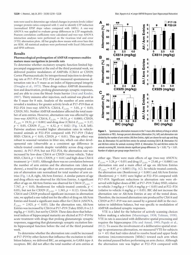
ResultsPharmacological prolongation of AMPAR responses enablesmature maze navigation in juvenile ratsTo determine whether excitatory synaptic function limited hip-pocampal engagement at the end of the third postnatal week, wedelivered positive modulators of AMPARs (CX614 or CX929;Cortex Pharmaceuticals) by intraperitoneal injection to develop-ing rats at P17–P19 or P22–P24 and measured spontaneous al-ternation rate in a Y-maze as an index of hippocampal integrity(Douglas et al., 1973). These drugs reduce AMPAR desensitiza-tion and deactivation, prolong glutamatergic synaptic responses,and are able to cross the blood– brain barrier (Arai and Kessler,2007). Thirty minutes after injection, each animal was placed inthe Y-maze for 8 min. Analysis of the number of arm entriesrevealed a tendency for greater activity levels at P17–P19 than atP22–P24 (two-way ANOVA: CX614, F(2,55) � 5.01, p � 0.03;CX929, NS). Neither AMPAR modulator affected the total num-ber of arm entries. However, alternation rate was affected by age(two-way ANOVA: CX614, F(1,55) � 19.51, p � 0.0001; CX929,F(1,47) � 19.51, p � 0.001) and drug dose (CX614: F(2,55) � 3.17,p � 0.05, CX929: F(2,47) � 4.32, p � 0.02) (Fig. 1A,B, left).Pairwise analyses revealed higher alternation rates in vehicle-treated animals at P22–P24 compared with P17–P19 (TukeyHSD: CX614, p � 0.01; CX929, p � 0.01). This age effect wasrobust to seasonal variation in litter size, which can alter devel-opmental rate (observable as a consistent age difference invehicle-treated controls despite variability across drug experi-ments). At P17–P19, but not P22–P24, the alternation rate wasincreased by low-dose CX614 or CX929 (versus vehicle, TukeyHSD, CX614, p � 0.01; CX929, p � 0.01) and high-dose CX614treatment (p � 0.05). Although there was no correlation betweenthe number of arm entries and the alternation rate (data notshown), a trend for an age effect on arm entries prompted anal-ysis of alternation rate normalized for total number of arm en-tries (Fig. 1A,B, right, Alt/Arm Entries). A similar pattern of ageand drug effects was observed for Alt/Arm Entries. A significanteffect of age on Alt/Arm Entries was observed for CX614 (F(1,61) �7.767, p � 0.01; Bonferroni for vehicle-treated controls, p �0.05), but not for CX929 (F(1,52) � 1.365, p � 0.12). Given thatCX614 and CX929 produced significant increases in alternationrate, we applied a one-tailed test for the effect of drug on Alt/ArmEntries and found a significant main effect for CX614 (ANOVA,F(2,61) � 2.925, p � 0.05). Like the alternation rate, Alt/ArmEntries was increased by CX614 at P17–P19 (Tukey HSD, 0 vs 2.5mg/kg, p � 0.05), but not by CX929. These data show that behav-ioral indices of hippocampal maturity are elicited at P17–P19 byacute treatment with drugs that prolong glutamatergic synapticresponses, suggesting that glutamatergic synaptic excitation lim-its hippocampal function before the end of the third postnatalweek.
To determine whether the alternation rate could be increasedat P17–P19 by other factors that influence the excitation to inhi-bition balance, we delivered BIC, an antagonist, to GABA type-Areceptors. BIC did not affect the total number of arm entries at
either age. There were main effects of age (two-way ANOVA:F(2,55) � 3.28, p � 0.05) and drug (F(2,55) � 23.60, p � 0.0001) onalternation rate and a main effect of age on Alt/Arm Entries(F(1,65) � 8.97, p � 0.005) (Fig. 1C). In vehicle-treated controls,the alternation rate (Bonferroni: p � 0.001) and Alt/Arm Entries(Bonferroni: p � 0.05) were higher at P22–P24 compared withP17–P19. Significant reductions in alternation rate were ob-served with higher doses of BIC at P17–P19 (Tukey HSD, relativeto vehicle: 3 mg/kg: p � 0.05, 6 mg/kg: p � 0.05) and at P22–P24(relative to vehicle: 6 mg/kg: p � 0.05). BIC did not increase thealternation rate or Alt/Arm Entries at any of the tested doses.Therefore, the increased alternation rate produced by CX614 andCX929 at P17–P19 was not caused by a general shift in the exci-tation to inhibition balance, but was specific to modulation ofAMPAR-mediated synaptic responses.
VTE is a label for the behavior of actively scanning choicesbefore making a selection (Muenzinger, 1938; Tolman, 1938).VTE in rats is associated with deliberative spatial processing andrequires the hippocampus (Hu and Amsel, 1995; Papale et al.,2012). To assess the involvement of a deliberative spatial strat-egy in spontaneous alternation, we measured VTE for subjects(n � 45) that had video detail to resolve head and upper bodymotions (micromovements [MMs]) toward each arm whenthe animal paused before performing an arm choice. Althoughthe alternation rate was higher at P22–P24 compared with
Figure 1. Spontaneous alternation measures in the Y-maze after delivery of drug or vehicle(cyclodextrin or PBS). Average percent alternation [Alternation (%), left] and alternation ratedivided by the number of arm entries (Alt/Arm Entries, right) are shown for each age and drugdose. A, Alternation (%) and Alt/Arm entries for animals receiving CX614. B, Alternation (%)and Alt/Arm entries for animals receiving CX929. C, Alternation (%) and Alt/Arm entries foranimals receiving BIC. Asterisks denote significant group differences: *p � 0.05; **p � 0.01.Number of subjects per group ranges from 6 to 13.
12220 • J. Neurosci., July 24, 2013 • 33(30):12218 –12228 Blair, Nguyen et al. • Longer AMPAR Response Spurs Hippocampal Maturation
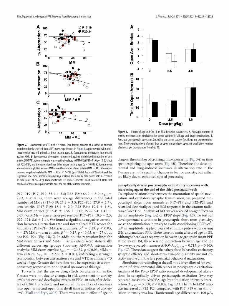
P17–P19 (P17–P19: 55.1 � 3.8; P22–P24: 66.9 � 3.9; t(44) �2.63, p � 0.02), there were no age differences in the totalnumber of MMs (P17–P19: 27.3 � 3.5; P22–P24: 27.9 � 2.7),arm entries (P17–P19: 18.1 � 2.0; P22–P24: 19.4 � 1.8),MMs/arm entries (P17–P19: 1.56 � 0.10; P22–P24: 1.45 �0.07), or MMs � arm entries per session (P17–P19: 10.3 � 2.3;P22–P24: 8.6 � 1.4). We found a significant negative correla-tion between alternation rate and normalized VTE scores foranimals at P17–P19 (MMs/arm entries, R 2 � 0.19, p � 0.03,n � 27; MMs � arm entries, R 2 � 0.17, p � 0.05, n � 27), butnot P22–P24 (Fig. 2 B, C). In addition, the regression lines forMMs/arm entries and MMs � arm entries were statisticallydifferent across age groups (two-way ANOVA interactionanalysis: MMs/arm entries: t(25) � �2.439, p � 0.03; MMs �arm entries: t(25) � �2.222, p � 0.05), indicating a strongerrelationship between alternation rate and VTE in animals �3weeks of age. Greater deliberation may be expressed as a com-pensatory response to poor spatial memory at this age.
To verify that the age or drug effects on alternation in theY-maze were not due to changes in risk assessment or anxietylevels, we exposed developing rats to an EPM 30 min after deliv-ery of CX614 or vehicle and measured the number of crossingsinto open arms and open arm dwell time as indices of anxietylevel (Walf and Frye, 2007). There was no main effect of age or
drug on the number of crossings into open arms (Fig. 3A) or timespent exploring the open arms (Fig. 3B). Therefore, the develop-mental and drug-induced increases in alternation rate in theY-maze are not a result of changes in fear or anxiety, but ratherare likely due to enhanced spatial processing.
Synaptically driven postsynaptic excitability increases withincreasing age at the end of the third postnatal weekTo explore relationships between the maturation of spatial navi-gation and excitatory synaptic transmission, we prepared hip-pocampal slices from animals at P17–P19 and P22–P24 andrecorded electrically evoked field responses in the stratum radia-tum of area CA1. Analysis of I/O curves revealed no age effects onthe FP amplitude (Fig. 4A) or EPSP slope (Fig. 4B). To test fordevelopmental alterations in presynaptic short-term plasticity,we set the stimulation intensity to evoke conditioning EPSPs of 1mV in amplitude, applied pairs of stimulus pulses with varyingISIs, and analyzed PPF. There were no main effects of age or ISI.Although there was a separation between age groups in mean PPFat the 25 ms ISI, there was no interaction between age and ISI(two-way repeated-measures ANOVA: F(3,43) � 0.713, p � 0.403;Fig. 4C). These data suggest that alterations in baseline excitatorysynaptic efficacy and short-term synaptic plasticity are not di-rectly involved in the late postnatal behavioral maturation.
Simultaneous recording at the cell body layer allowed for eval-uation of developmental differences in postsynaptic excitation.Analysis of the PS to EPSP ratio revealed developmental altera-tions in synaptically driven postsynaptic excitation (two-wayrepeated-measures ANOVA, age by stimulation intensity inter-action: F(4,43) � 5.808, p � 0.002; Fig. 5A). The PS to EPSP ratiowas increased at P22–P24 compared with P17–P19 when stimu-lation intensity was low (Bonferonni: age difference at 100 �A,
Figure 2. Assessment of VTE in the Y-maze. This dataset consists of a subset of animalspseudorandomly selected from all Y-maze experiments in Figure 1 supplemented with addi-tional vehicle-treated animals at both testing ages. A, Spontaneous alternation rate plottedagainst MMs. B, Spontaneous alternation rate plotted against MM divided by number of armentries (MM/AE). Alternation rate was negatively related to MM/AE at P17–P19 ( p�0.03), butnot P22–P24, and the regression lines differ across testing ages ( p � 0.03). C, Spontaneousalternation rate plotted against MM minus the number of arm entries (MM � AE). Alternationrate was negatively related to MM � AE at P17–P19 ( p � 0.05), but not P22–P24, and theregression lines differ across testing ages ( p � 0.05). There are 27 data points at P17–P19 and18 data points at P22–P24. Data points with red borders indicate CX614 treatment. Note thatnearly all of these data points reside near the top of the alternation scale.
Figure 3. Effects of age and CX614 on EPM behavior parameters. A, Averaged number ofentries into open arms (including the center square) for all age and drug combinations. B,Averaged time spent in open arms (including the center square) for all age and drug combina-tions. There were no effects of age or drug on open arm entries or open arm dwell time. Numberof subjects per group ranges from 9 to 15.
Blair, Nguyen et al. • Longer AMPAR Response Spurs Hippocampal Maturation J. Neurosci., July 24, 2013 • 33(30):12218 –12228 • 12221

p � 0.02), but increased at P17–P19 compared with P22–P24when stimulation intensity was high (Bonferonni: age differenceat 400 �A: p � 0.05). Because responses to lower stimulationintensities are more reflective of normal physiological condi-tions, it appears that the ability of excitatory synapses to drivepostsynaptic discharge increases at the end of the third postnatalweek. Because we did not observe age differences in basic param-eters of excitatory synaptic transmission that would drive thedevelopmental increase in postsynaptic excitability, we evaluatedPPI (the ratio of the second PS to the first PS amplitude) as ameasure of GABAergic synaptic inhibition (Rogers and Hunter,1992). The ratio of the second to the first PS amplitude was con-sistently smaller at P22–P24 than at P17–P19 (two-wayrepeated-measures ANOVA: F(1,38) � 17.49, p � 0.0001), indicat-ing greater PPI and suggesting more fully developed GABAergic in-hibition at P22–P24 (Fig. 5B). Therefore, the reduction in
postsynaptic excitability observed at P17–P19 at lower stimulationintensity is likely not a result of enhanced GABAergic transmission,as one would predict the opposite age effect on PPI. Instead, thedevelopmental increase in postsynaptic excitability is more directlyrelated to stronger coupling between excitatory synaptic transmis-sion and postsynaptic discharge.
Pharmacological prolongation of AMPAR responses injuveniles enhances postsynaptic excitability more at P17–P19than at P22–P24To test the notion that postsynaptic excitability could be differ-entially regulated by excitatory synapses at the end of the thirdpostnatal week, we recorded evoked EPSPs and PSs in area CA1during a 15 min application of CX614 at P17–P19 and P22–P24.CX614 did not affect the EPSP slope (Fig. 6A,B), but increasedthe PS at both testing ages (Fig. 6C,D). The drug-induced increasein the PS was greater at P17–P19 compared with P22–P24 at 20�M (unpaired t test: t(29) � 3.00, p � 0.006), but not at 40 �M.These data agree with the behavioral results showing the greatestage difference in the effects of moderate doses of CX614 andCX929 on alternation rate and further substantiate the hypothe-sis that reduced EPSP duration at SC-CA1 synapses limits post-synaptic excitation in area CA1 and restricts hippocampalcontribution to behavior in juvenile animals.
Evoked AMPAR EPSPs and spontaneous AMPAR mEPSCsdecay more slowly at P22–P24 than at P17–P19The initial EPSP slope, which is typically used to measure synap-tic strength, may not capture other meaningful changes in the
Figure 4. Baseline I/O and ISI curves for electrically evoked field responses recorded fromstratum radiatum of area CA1 in hippocampal slices. A, Average FP amplitude is plotted againststimulation intensity for both age groups. The FP amplitude increased with increasing stimula-tion intensity (two-way repeated-measures ANOVA: p � 0.0001). B, Average EPSP slope isplotted against mean FP amplitude for both age groups. EPSP slope increased across FP levels(two-way repeated-measures ANOVA: p � 0.0001). C, The ratio of the test EPSP slope to theconditioning EPSP slope (PPF) is plotted against ISI for both age groups. PPF was altered as afunction of ISI (two-way repeated-measures ANOVA: p � 0.0001), but not age. Inset showsaveraged traces (10 sweeps each) from one experiment at 50 ms ISI for each age group. Scalebars, 10 ms and 0.5 mV. Number of slices per group ranges from 19 to 25.
Figure 5. Baseline I/O and ISI curves for electrically evoked field responses recorded in stra-tum pyramidale of hippocampal slices. Stimulation intensity and paired-pulse ISIs were varied.A, Average PS to EPSP ratio is plotted against stimulation intensity for both age groups. Aster-isks represent significant group differences: **p � 0.01. B, Ratio of the test PS amplitude to theconditioning PS amplitude is plotted against ISI for both age groups. Inset shows averagedtraces (10 sweeps each) from one experiment at 50 ms ISI for each age group. Scale bars, 10 msand 0.5 mV. Number of slices per group ranges from 20 to 25.
12222 • J. Neurosci., July 24, 2013 • 33(30):12218 –12228 Blair, Nguyen et al. • Longer AMPAR Response Spurs Hippocampal Maturation

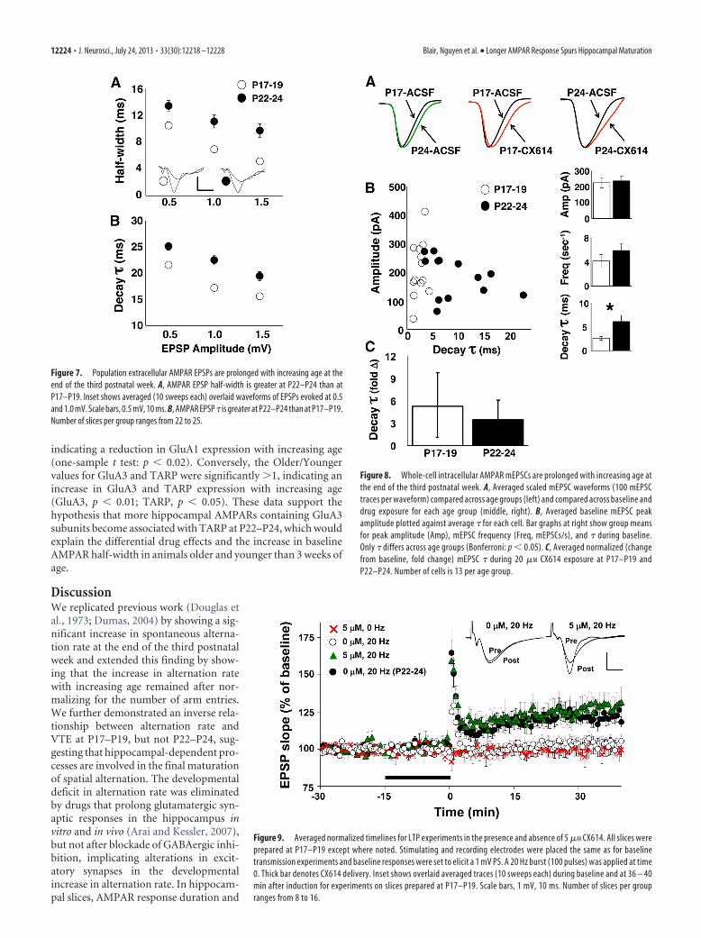
dynamics of synaptic excitation. Because CX614 increases theduration of hippocampal glutamatergic EPSPs (Arai et al., 2000)and increases spontaneous alternation rate at P17–P19, it is pos-sible that the age-related increase in alternation rate might alsoresult from increased duration of synaptic responses. We investi-gated the possibility that AMPAR response duration is altered at theend of the third postnatal week by recording pharmacologically iso-lated AMPAR responses in hippocampal slices prepared from ani-mals at both testing ages. We applied AP5 (10 �M) to blockNMDARs and BIC (2 �M) to block GABA type-A receptors andadjusted the stimulation intensity to evoke AMPAR responses atamplitudes of 0.5, 1.0, and 1.5 mV. AMPAR response half-width and� were increased at P22–P24 compared with P17–P19 (two-wayrepeated-measures ANOVA: half-width: F(1,38) � 15.173, p � 0.001;�: F(1,51) � 5.41, p � 0.05; Fig. 7A,B). Half-width and � decreasedwith increasing EPSP amplitude (half-width: F(2,37) � 72.640, p �0.0001; �: F(2,86) � 149.490, p � 0.001) due to the reflection of the PSin the synaptic field recording. However, this does not explain theage difference in the AMPAR response dynamics. Previous workshowed that pharmacological blockade of AMPAR deactivation, butnot desensitization, resulted in longer duration synaptic responses(Arai et al., 2000). Therefore, the current finding of prolongedAMPAR responses suggests a developmental reduction in AMPARdeactivation. Moreover, prolonged AMPAR responses would ex-plain the enhanced postsynaptic excitability with increasing age andpossibly the increase in spontaneous alternation rate at P22–P24.
Because the later phases of evoked excitatory synaptic poten-tials in extracellular field recordings are subject to contaminationby other physiological events (disynaptic transmission, postsyn-aptic discharge), we performed whole-cell intracellular recordingfrom CA1 pyramidal neurons in slices collected at P17–P19 andP22–P24. In voltage-clamp mode, without stimulation, sponta-neous mEPSCs could be observed (�70 mV holding potential).As performed for the field recordings, we pharmacologically iso-lated AMPAR mEPSCs with AP5 (50 �M) and BIC (20 �M) andthen applied CX614 (20 �M, 5 min). There were no differences in
mean mEPSC amplitude (pA) or fre-quency (mEPSCs/s) across age groups.There were significant effects of age(two-way repeated-measures ANOVA:F(1,24) � 4.402, p � 0.05) and drug(F(1,24) � 13.579, p � 0.005) on the � ofrecorded mEPSCs. The baseline AMPARmEPSC � was increased at P22–P24 com-pared with P17–P19 (Bonferroni: p �0.05; Fig. 8). mEPSC � was increased byCX614 at P17–P19 (Bonferroni: p �0.002) and at P22–P24 (Bonferroni: p �0.002), but there was no age difference innormalized � values (unpaired t test).These results are in close agreement withfindings from extracellular recordingsand confirm an increase in AMPAR re-sponse duration with increasing age at theend of the third postnatal week.
Threshold for induction of activity-dependent synaptic potentiation isreduced by CX614 at P17–P19We previously showed that the activationthreshold for induction of LTP at SC-CA1synapses decreased at the end of the thirdpostnatal week (Dumas, 2012), support-
ing the hypothesis that circuit deficits before the end of the thirdpostnatal week included insufficient sensitivity to plasticity in-duction in area CA1. In addition, it has been shown that positiveallosteric modulators of AMPARs facilitate LTP induction in thehippocampus (Arai and Kessler, 2007). Therefore, it is possiblethat CX614 increased alternation rates at P17–P19 by reducingthe threshold for LTP induction. To test this, we applied a peri-threshold stimulation frequency (100 pulses at 20 Hz derivedfrom Dumas, 2012) in the absence or presence of a low concen-tration of CX614 (5 �M, 15 min terminating at induction) inslices prepared at P17–P19. In the absence of LTP-inducing stim-ulation, CX614 did not affect the EPSP slope and, without CX614present, LTP was not induced by perithreshold stimulation (Fig.9). In contrast, with CX614 present, perithreshold stimulationinduced LTP (one-way ANOVA, F(3,44) � 6.70, p � 0.001; 20Hz/5 �M vs 20 Hz/0 �M: p � 0.05). In addition, LTP was inducedin slices prepared at P22–P24 in the absence of CX614. These dataindicate that CX614 overcame the developmental impedance toLTP induction in slices prepared from at P17–P19, giving cre-dence to the notion that the CX614-dependent increase in alter-nation rate at P17–P19 resulted from facilitation of the inductionof activity-dependent synaptic potentiation.
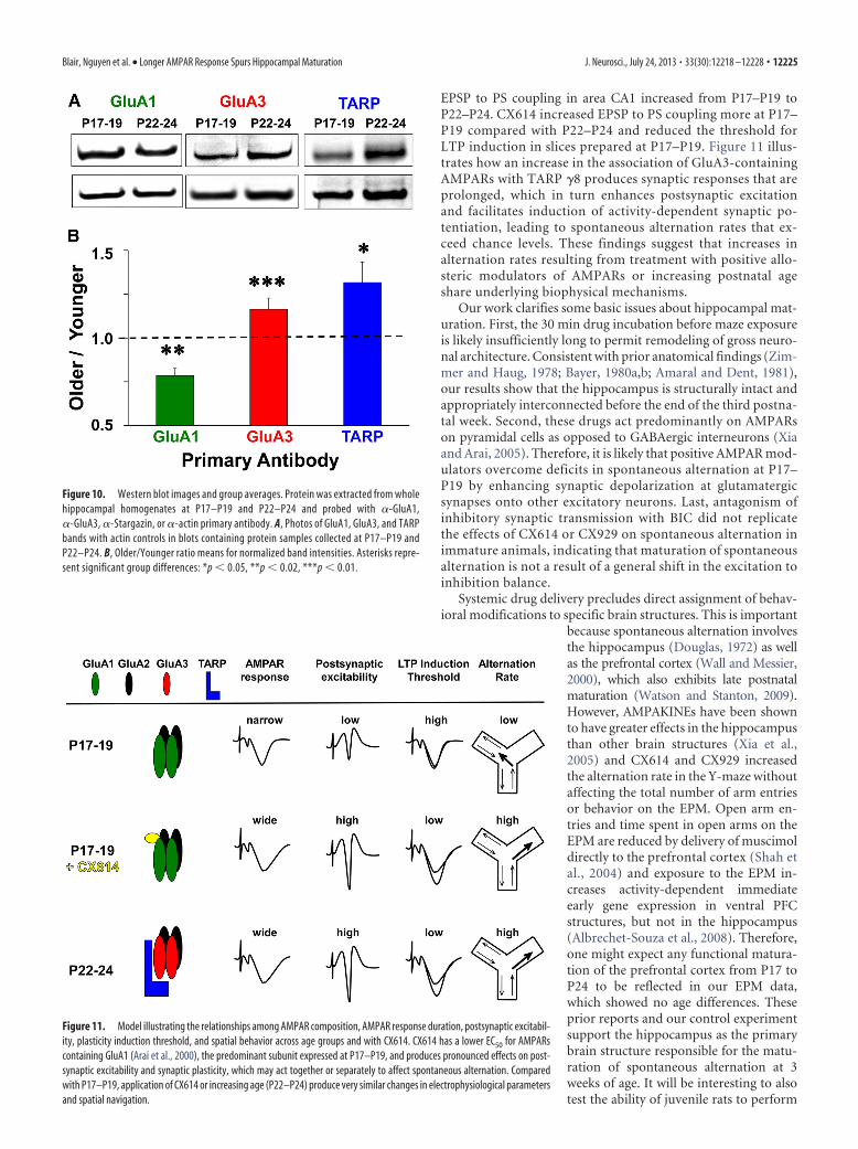
GluA1 subunit expression is reduced, whereas GluA3 subunitand TARP expression levels are increased, at the end of thethird postnatal weekTo investigate the molecular basis for the differential effects ofAMPAR modulators on spatial behavior and hippocampal syn-aptic physiology, we performed Western blot assays on hip-pocampal protein extracts for AMPAR subunits and TARPsknown to alter AMPAR desensitization and deactivation (Tomitaet al., 2003; Suzuki et al., 2008). Example bands for each antibodyat each age group are shown in Figure 10A. To compare acrossages, we divided normalized band intensities at P22–P24 by P17–P19 to obtain an Older to Younger ratio (Fig. 10B). The Older/Younger value for GluA1 was significantly lower than 1,
Figure 6. Effects of CX614 on EPSP slope and PS amplitude at P17–P19 and P22–P24. Stimulating and recording electrodeswere placed the same as for I/O and ISI experiments. Timelines display normalized EPSP slope (A, B) and PS values (C, D) duringbaseline, drug exposure, and washout. Drug delivery is indicated by a solid bar below the timelines (A, C, 20 �M; B, D, 40 �M). Insetin A shows overlaid averaged traces (10 sweeps each) taken during baseline and 20 �M drug exposure. Scale bars, 1 mV, 10 ms.Asterisks represent significant group differences: **p � 0.01. Number of slices per group ranges from 9 to 20.
Blair, Nguyen et al. • Longer AMPAR Response Spurs Hippocampal Maturation J. Neurosci., July 24, 2013 • 33(30):12218 –12228 • 12223
indicating a reduction in GluA1 expression with increasing age(one-sample t test: p � 0.02). Conversely, the Older/Youngervalues for GluA3 and TARP were significantly �1, indicating anincrease in GluA3 and TARP expression with increasing age(GluA3, p � 0.01; TARP, p � 0.05). These data support thehypothesis that more hippocampal AMPARs containing GluA3subunits become associated with TARP at P22–P24, which wouldexplain the differential drug effects and the increase in baselineAMPAR half-width in animals older and younger than 3 weeks ofage.
DiscussionWe replicated previous work (Douglas etal., 1973; Dumas, 2004) by showing a sig-nificant increase in spontaneous alterna-tion rate at the end of the third postnatalweek and extended this finding by show-ing that the increase in alternation ratewith increasing age remained after nor-malizing for the number of arm entries.We further demonstrated an inverse rela-tionship between alternation rate andVTE at P17–P19, but not P22–P24, sug-gesting that hippocampal-dependent pro-cesses are involved in the final maturationof spatial alternation. The developmentaldeficit in alternation rate was eliminatedby drugs that prolong glutamatergic syn-aptic responses in the hippocampus invitro and in vivo (Arai and Kessler, 2007),but not after blockade of GABAergic inhi-bition, implicating alterations in excit-atory synapses in the developmentalincrease in alternation rate. In hippocam-pal slices, AMPAR response duration and
Figure 8. Whole-cell intracellular AMPAR mEPSCs are prolonged with increasing age atthe end of the third postnatal week. A, Averaged scaled mEPSC waveforms (100 mEPSCtraces per waveform) compared across age groups (left) and compared across baseline anddrug exposure for each age group (middle, right). B, Averaged baseline mEPSC peakamplitude plotted against average � for each cell. Bar graphs at right show group meansfor peak amplitude (Amp), mEPSC frequency (Freq, mEPSCs/s), and � during baseline.Only � differs across age groups (Bonferroni: p � 0.05). C, Averaged normalized (changefrom baseline, fold change) mEPSC � during 20 �M CX614 exposure at P17–P19 andP22–P24. Number of cells is 13 per age group.
Figure 9. Averaged normalized timelines for LTP experiments in the presence and absence of 5 �M CX614. All slices wereprepared at P17–P19 except where noted. Stimulating and recording electrodes were placed the same as for baselinetransmission experiments and baseline responses were set to elicit a 1 mV PS. A 20 Hz burst (100 pulses) was applied at time0. Thick bar denotes CX614 delivery. Inset shows overlaid averaged traces (10 sweeps each) during baseline and at 36 – 40min after induction for experiments on slices prepared at P17–P19. Scale bars, 1 mV, 10 ms. Number of slices per groupranges from 8 to 16.
Figure 7. Population extracellular AMPAR EPSPs are prolonged with increasing age at theend of the third postnatal week. A, AMPAR EPSP half-width is greater at P22–P24 than atP17–P19. Inset shows averaged (10 sweeps each) overlaid waveforms of EPSPs evoked at 0.5and 1.0 mV. Scale bars, 0.5 mV, 10 ms. B, AMPAR EPSP � is greater at P22–P24 than at P17–P19.Number of slices per group ranges from 22 to 25.
12224 • J. Neurosci., July 24, 2013 • 33(30):12218 –12228 Blair, Nguyen et al. • Longer AMPAR Response Spurs Hippocampal Maturation
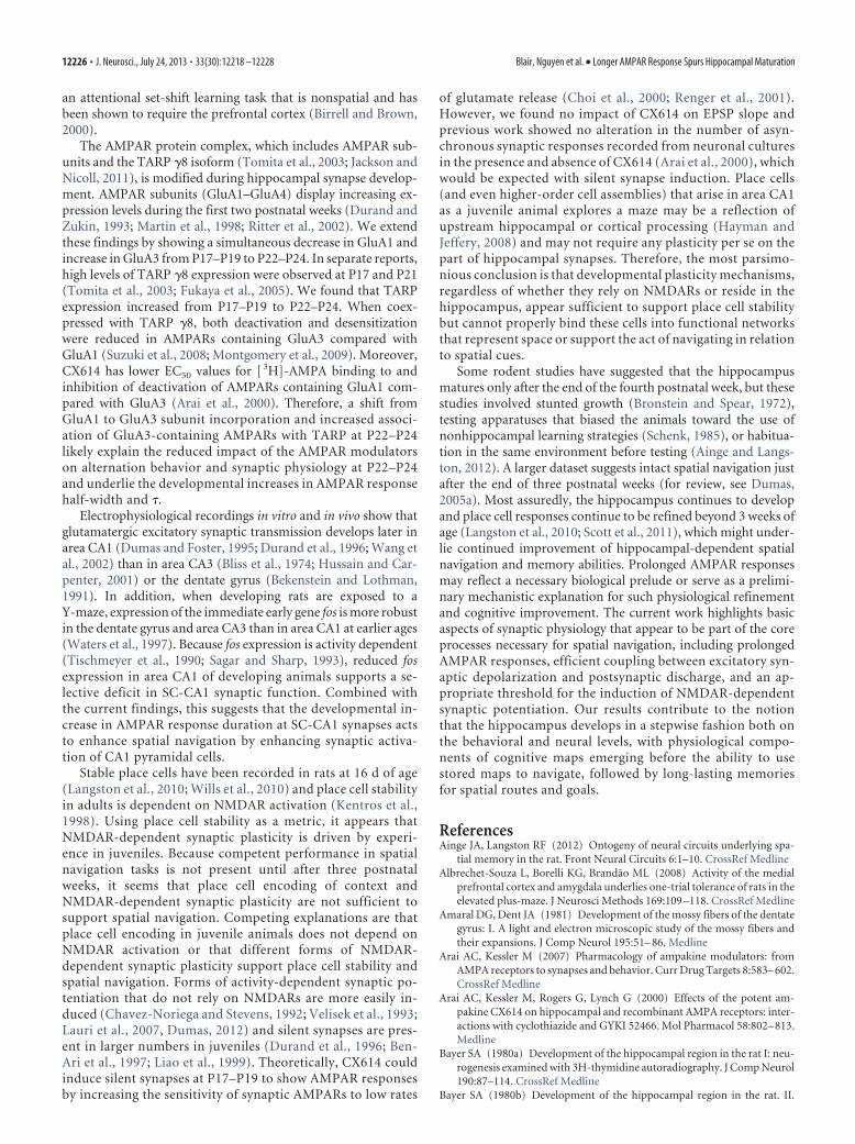
EPSP to PS coupling in area CA1 increased from P17–P19 toP22–P24. CX614 increased EPSP to PS coupling more at P17–P19 compared with P22–P24 and reduced the threshold forLTP induction in slices prepared at P17–P19. Figure 11 illus-trates how an increase in the association of GluA3-containingAMPARs with TARP �8 produces synaptic responses that areprolonged, which in turn enhances postsynaptic excitationand facilitates induction of activity-dependent synaptic po-tentiation, leading to spontaneous alternation rates that ex-ceed chance levels. These findings suggest that increases inalternation rates resulting from treatment with positive allo-steric modulators of AMPARs or increasing postnatal ageshare underlying biophysical mechanisms.
Our work clarifies some basic issues about hippocampal mat-uration. First, the 30 min drug incubation before maze exposureis likely insufficiently long to permit remodeling of gross neuro-nal architecture. Consistent with prior anatomical findings (Zim-mer and Haug, 1978; Bayer, 1980a,b; Amaral and Dent, 1981),our results show that the hippocampus is structurally intact andappropriately interconnected before the end of the third postna-tal week. Second, these drugs act predominantly on AMPARson pyramidal cells as opposed to GABAergic interneurons (Xiaand Arai, 2005). Therefore, it is likely that positive AMPAR mod-ulators overcome deficits in spontaneous alternation at P17–P19 by enhancing synaptic depolarization at glutamatergicsynapses onto other excitatory neurons. Last, antagonism ofinhibitory synaptic transmission with BIC did not replicatethe effects of CX614 or CX929 on spontaneous alternation inimmature animals, indicating that maturation of spontaneousalternation is not a result of a general shift in the excitation toinhibition balance.
Systemic drug delivery precludes direct assignment of behav-ioral modifications to specific brain structures. This is important
because spontaneous alternation involvesthe hippocampus (Douglas, 1972) as wellas the prefrontal cortex (Wall and Messier,2000), which also exhibits late postnatalmaturation (Watson and Stanton, 2009).However, AMPAKINEs have been shownto have greater effects in the hippocampusthan other brain structures (Xia et al.,2005) and CX614 and CX929 increasedthe alternation rate in the Y-maze withoutaffecting the total number of arm entriesor behavior on the EPM. Open arm en-tries and time spent in open arms on theEPM are reduced by delivery of muscimoldirectly to the prefrontal cortex (Shah etal., 2004) and exposure to the EPM in-creases activity-dependent immediateearly gene expression in ventral PFCstructures, but not in the hippocampus(Albrechet-Souza et al., 2008). Therefore,one might expect any functional matura-tion of the prefrontal cortex from P17 toP24 to be reflected in our EPM data,which showed no age differences. Theseprior reports and our control experimentsupport the hippocampus as the primarybrain structure responsible for the matu-ration of spontaneous alternation at 3weeks of age. It will be interesting to alsotest the ability of juvenile rats to perform
Figure 10. Western blot images and group averages. Protein was extracted from wholehippocampal homogenates at P17–P19 and P22–P24 and probed with �-GluA1,�-GluA3, �-Stargazin, or �-actin primary antibody. A, Photos of GluA1, GluA3, and TARPbands with actin controls in blots containing protein samples collected at P17–P19 andP22–P24. B, Older/Younger ratio means for normalized band intensities. Asterisks repre-sent significant group differences: *p � 0.05, **p � 0.02, ***p � 0.01.
Figure 11. Model illustrating the relationships among AMPAR composition, AMPAR response duration, postsynaptic excitabil-ity, plasticity induction threshold, and spatial behavior across age groups and with CX614. CX614 has a lower EC50 for AMPARscontaining GluA1 (Arai et al., 2000), the predominant subunit expressed at P17–P19, and produces pronounced effects on post-synaptic excitability and synaptic plasticity, which may act together or separately to affect spontaneous alternation. Comparedwith P17–P19, application of CX614 or increasing age (P22–P24) produce very similar changes in electrophysiological parametersand spatial navigation.
Blair, Nguyen et al. • Longer AMPAR Response Spurs Hippocampal Maturation J. Neurosci., July 24, 2013 • 33(30):12218 –12228 • 12225
an attentional set-shift learning task that is nonspatial and hasbeen shown to require the prefrontal cortex (Birrell and Brown,2000).
The AMPAR protein complex, which includes AMPAR sub-units and the TARP �8 isoform (Tomita et al., 2003; Jackson andNicoll, 2011), is modified during hippocampal synapse develop-ment. AMPAR subunits (GluA1–GluA4) display increasing ex-pression levels during the first two postnatal weeks (Durand andZukin, 1993; Martin et al., 1998; Ritter et al., 2002). We extendthese findings by showing a simultaneous decrease in GluA1 andincrease in GluA3 from P17–P19 to P22–P24. In separate reports,high levels of TARP �8 expression were observed at P17 and P21(Tomita et al., 2003; Fukaya et al., 2005). We found that TARPexpression increased from P17–P19 to P22–P24. When coex-pressed with TARP �8, both deactivation and desensitizationwere reduced in AMPARs containing GluA3 compared withGluA1 (Suzuki et al., 2008; Montgomery et al., 2009). Moreover,CX614 has lower EC50 values for [ 3H]-AMPA binding to andinhibition of deactivation of AMPARs containing GluA1 com-pared with GluA3 (Arai et al., 2000). Therefore, a shift fromGluA1 to GluA3 subunit incorporation and increased associ-ation of GluA3-containing AMPARs with TARP at P22–P24likely explain the reduced impact of the AMPAR modulatorson alternation behavior and synaptic physiology at P22–P24and underlie the developmental increases in AMPAR responsehalf-width and �.
Electrophysiological recordings in vitro and in vivo show thatglutamatergic excitatory synaptic transmission develops later inarea CA1 (Dumas and Foster, 1995; Durand et al., 1996; Wang etal., 2002) than in area CA3 (Bliss et al., 1974; Hussain and Car-penter, 2001) or the dentate gyrus (Bekenstein and Lothman,1991). In addition, when developing rats are exposed to aY-maze, expression of the immediate early gene fos is more robustin the dentate gyrus and area CA3 than in area CA1 at earlier ages(Waters et al., 1997). Because fos expression is activity dependent(Tischmeyer et al., 1990; Sagar and Sharp, 1993), reduced fosexpression in area CA1 of developing animals supports a se-lective deficit in SC-CA1 synaptic function. Combined withthe current findings, this suggests that the developmental in-crease in AMPAR response duration at SC-CA1 synapses actsto enhance spatial navigation by enhancing synaptic activa-tion of CA1 pyramidal cells.
Stable place cells have been recorded in rats at 16 d of age(Langston et al., 2010; Wills et al., 2010) and place cell stabilityin adults is dependent on NMDAR activation (Kentros et al.,1998). Using place cell stability as a metric, it appears thatNMDAR-dependent synaptic plasticity is driven by experi-ence in juveniles. Because competent performance in spatialnavigation tasks is not present until after three postnatalweeks, it seems that place cell encoding of context andNMDAR-dependent synaptic plasticity are not sufficient tosupport spatial navigation. Competing explanations are thatplace cell encoding in juvenile animals does not depend onNMDAR activation or that different forms of NMDAR-dependent synaptic plasticity support place cell stability andspatial navigation. Forms of activity-dependent synaptic po-tentiation that do not rely on NMDARs are more easily in-duced (Chavez-Noriega and Stevens, 1992; Velisek et al., 1993;Lauri et al., 2007, Dumas, 2012) and silent synapses are pres-ent in larger numbers in juveniles (Durand et al., 1996; Ben-Ari et al., 1997; Liao et al., 1999). Theoretically, CX614 couldinduce silent synapses at P17–P19 to show AMPAR responsesby increasing the sensitivity of synaptic AMPARs to low rates
of glutamate release (Choi et al., 2000; Renger et al., 2001).However, we found no impact of CX614 on EPSP slope andprevious work showed no alteration in the number of asyn-chronous synaptic responses recorded from neuronal culturesin the presence and absence of CX614 (Arai et al., 2000), whichwould be expected with silent synapse induction. Place cells(and even higher-order cell assemblies) that arise in area CA1as a juvenile animal explores a maze may be a reflection ofupstream hippocampal or cortical processing (Hayman andJeffery, 2008) and may not require any plasticity per se on thepart of hippocampal synapses. Therefore, the most parsimo-nious conclusion is that developmental plasticity mechanisms,regardless of whether they rely on NMDARs or reside in thehippocampus, appear sufficient to support place cell stabilitybut cannot properly bind these cells into functional networksthat represent space or support the act of navigating in relationto spatial cues.
Some rodent studies have suggested that the hippocampusmatures only after the end of the fourth postnatal week, but thesestudies involved stunted growth (Bronstein and Spear, 1972),testing apparatuses that biased the animals toward the use ofnonhippocampal learning strategies (Schenk, 1985), or habitua-tion in the same environment before testing (Ainge and Langs-ton, 2012). A larger dataset suggests intact spatial navigation justafter the end of three postnatal weeks (for review, see Dumas,2005a). Most assuredly, the hippocampus continues to developand place cell responses continue to be refined beyond 3 weeks ofage (Langston et al., 2010; Scott et al., 2011), which might under-lie continued improvement of hippocampal-dependent spatialnavigation and memory abilities. Prolonged AMPAR responsesmay reflect a necessary biological prelude or serve as a prelimi-nary mechanistic explanation for such physiological refinementand cognitive improvement. The current work highlights basicaspects of synaptic physiology that appear to be part of the coreprocesses necessary for spatial navigation, including prolongedAMPAR responses, efficient coupling between excitatory syn-aptic depolarization and postsynaptic discharge, and an ap-propriate threshold for the induction of NMDAR-dependentsynaptic potentiation. Our results contribute to the notionthat the hippocampus develops in a stepwise fashion both onthe behavioral and neural levels, with physiological compo-nents of cognitive maps emerging before the ability to usestored maps to navigate, followed by long-lasting memoriesfor spatial routes and goals.
ReferencesAinge JA, Langston RF (2012) Ontogeny of neural circuits underlying spa-
tial memory in the rat. Front Neural Circuits 6:1–10. CrossRef MedlineAlbrechet-Souza L, Borelli KG, Brandao ML (2008) Activity of the medial
prefrontal cortex and amygdala underlies one-trial tolerance of rats in theelevated plus-maze. J Neurosci Methods 169:109 –118. CrossRef Medline
Amaral DG, Dent JA (1981) Development of the mossy fibers of the dentategyrus: I. A light and electron microscopic study of the mossy fibers andtheir expansions. J Comp Neurol 195:51– 86. Medline
Arai AC, Kessler M (2007) Pharmacology of ampakine modulators: fromAMPA receptors to synapses and behavior. Curr Drug Targets 8:583– 602.CrossRef Medline
Arai AC, Kessler M, Rogers G, Lynch G (2000) Effects of the potent am-pakine CX614 on hippocampal and recombinant AMPA receptors: inter-actions with cyclothiazide and GYKI 52466. Mol Pharmacol 58:802– 813.Medline
Bayer SA (1980a) Development of the hippocampal region in the rat I: neu-rogenesis examined with 3H-thymidine autoradiography. J Comp Neurol190:87–114. CrossRef Medline
Bayer SA (1980b) Development of the hippocampal region in the rat. II.
12226 • J. Neurosci., July 24, 2013 • 33(30):12218 –12228 Blair, Nguyen et al. • Longer AMPAR Response Spurs Hippocampal Maturation
Morphogenesis during embryonic and early postnatal life. J Comp Neurol190:115–134. Medline
Bekenstein JW, Lothman EW (1991) A comparison of the ontogeny of ex-citatory and inhibitory neurotransmission in the CA1 region and dentategyrus of the rat hippocampal formation. Brain Res Dev Brain Res 63:237–241. Medline
Ben-Ari Y, Khazipov R, Leinekugel X, Caillard O, Gaiarsa JL (1997)GABAA, NMDA and AMPA receptors: a developmentally regulated “mé-nage à trois”. Trends Neurosci 20:523–529. Medline
Birrell JM, Brown VJ (2000) Medial frontal cortex mediates perceptual at-tentional set shifting in the rat. J Neurosci 20:4320 – 4324. Medline
Bliss TV, Chung SH, Stirling RV (1974) Proceedings: structural and func-tional development of the mossy fibre system in the hippocampus of thepost-natal rat. J Physiol 239:92P–94P. Medline
Bronstein PM, Spear NE (1972) Acquisition of a spatial discrimination byrats as a function of age. J Comp Physiol Psychol 78:208 –212. CrossRefMedline
Chavez-Noriega LE, Stevens CF (1992) Modulation of synaptic efficacy infield CA1 of the rat hippocampus by forskolin. Brain Res 574:85–92.CrossRef Medline
Choi S, Klingauf J, Tsien RW (2000) Postfusional regulation of cleft gluta-mate concentration during LTP at ’silent synapses’. Nat Neurosci 3:330 –336. CrossRef Medline
Costa PF, Santos AI, Ribiero MA (1994) Potassium currents in acutely iso-lated maturing rat hippocampal CA1 neurones. Brain Res Dev Brain Res83:216 –223. Medline
Douglas RJ (1972) Pavlovian conditioning and the brain. In: Inhibition andlearning (Boakes RA, Halliday MS, eds), 529 –553. New York: Academic.
Douglas RJ, Peterson JJ, Douglas DP (1973) The ontogeny of ahippocampus-dependent response in two rodent species. Behav Biol8:27–37. CrossRef Medline
Dumas TC (2004) Early eyelid opening enhances spontaneous alternationand accelerates the development of perforant path synaptic strength in thehippocampus of juvenile rats. Dev Psychobiol 45:1–9. CrossRef Medline
Dumas TC (2005a) Late postnatal maturation of excitatory synaptic trans-mission permits the expression of adult-like hippocampal-dependent be-haviors. Hippocampus 15:562–578. CrossRef Medline
Dumas TC (2005b) Developmental regulation of cognitive abilities: Modi-fied composition of a molecular switch turns on associative learning. ProgNeurobiol 76:189 –211. CrossRef Medline
Dumas TC (2012) Postnatal alterations in induction threshold and expres-sion magnitude of long-term potentiation and long-term depression athippocampal synapses. Hippocampus 22:188 –199. CrossRef Medline
Dumas TC, Foster TC (1995) Developmental increase in CA3-CA1 presyn-aptic function in the hippocampal slice. J Neurophysiol 73:1821–1828.Medline
Durand GM, Zukin RS (1993) Developmental regulation of mRNAs encod-ing rat brain kainate/AMPA receptors: a northern analysis study. J Neu-rochem 61:2239 –2246. CrossRef Medline
Durand GM, Kovalchuk Y, Konnerth A (1996) Long-term potentiation andfunctional synapse induction in developing hippocampus. Nature 381:71–75. CrossRef Medline
Fukaya M, Yamazaki M, Sakimura K, Watanabe M (2005) Spatial diversityin gene expression for VDCCg subunit family in developing and adultmouse brains. Neurosci Res 53:376 –383. CrossRef Medline
Green RJ, Stanton ME (1989) Differential ontogeny of working memoryand reference memory in the rat. Behav Neurosci 103:98 –105. CrossRefMedline
Harris KM, Teyler TJ (1983) Evidence for late development of inhibition inarea CA1 of the rat hippocampus. Brain Res 268:339 –343. CrossRefMedline
Hayman RM, Jeffery KJ (2008) How heterogeneous place cell respondingarises from homogeneous grids–a contextual gating hypothesis. Hip-pocampus 18:1301–1313. CrossRef Medline
Hsia AY, Malenka RC, Nicoll RA (1998) Development of excitatory cir-cuitry in the hippocampus. J Neurophysiol 79:2013–2024. Medline
Hu D, Amsel A (1995) A simple test of the vicarious trial-and-error hypoth-esis of hippocampal function. Proc Natl Acad Sci U S A 92:5506 –5509.Medline
Hussain RJ, Carpenter DO (2001) Development of synaptic responses andplasticity at the SC -CA1 and MF-CA3 synapses in rat hippocampus. CellMol Neurobiol 21:357–368. Medline
Huttenlocher J (2008) Coding location: the view from toddler studies. AmPsychol 63:641– 648. CrossRef Medline
Jackson AC, Nicoll RA (2011) The expanding social network of ionotropicglutamate receptors: TARPs and other transmembrane auxiliary sub-units. Neuron 70:178 –199. CrossRef Medline
Kentros C, Hargreaves E, Hawkins RD, Kandel ER, Shapiro M, Muller RV(1998) Abolition of long-term stability of new hippocampal place cellmaps by NMDA receptor blockade. Science 280:2121–2126. CrossRefMedline
Kudryashov IE, Kudryashova IV (2001) Ontogeny of synaptic transmissionin the rat hippocampus. Brain Res 892:263–268. CrossRef Medline
Langston RF, Ainge JA, Couey JJ, Canto CB, Bjerknes TL, Witter MP, MoserEI, Moser MB (2010) Development of the spatial representation systemin the rat. Science 328:1576 –1580. CrossRef Medline
Lauri SE, Palmer M, Segerstrale M, Vesikansa A, Taira T, Collingridge GL(2007) Presynaptic mechanisms involved in the expression of STPand LTP at CA1 synapses in the hippocampus. Neuropharmacology52:1–11. CrossRef Medline
Liao D, Zhang X, O’Brien R, Ehlers MD, Huganir RL (1999) Regulation ofmorphological postsynaptic silent synapses in developing hippocampalneurons. Nat Neurosci 2:37– 43. CrossRef Medline
Martin LJ, Furuta A, Blackstone CD (1998) Ampa receptor protein in de-veloping rat brain: glutamate receptor-1 expression and localizationchange at regional, cellular and subcellular levels with maturation. Neu-roscience 83:917–928. CrossRef Medline
Montgomery KE, Kessler M, Arai AC (2009) Modulation of agonist bindingto AMPA receptors by 1-(1,4-benzodioxan-6-ylcarbonyl)piperidine(CX546): differential effects across brain regions and GluA1-4/trans-membrane AMPA receptor regulatory protein combinations. J Pharma-col Exp Ther 331:965–974. CrossRef Medline
Morris RG, Moser EI, Riedel G, Martin SJ, Sandin J, Day M, O’Carroll C(2003) Elements of a neurobiological theory of the hippocampus: therole of activity-dependent synaptic plasticity in memory. Philos Trans RSoc Lond B Biol Sci 358:773–786. CrossRef Medline
Muenzinger KF (1938) Vicarious trial and error at a point of choice: I. Ageneral survey of its relation to learning efficiency. Journal of GeneticPsychology 53:75– 86.
Papale AE, Stott JJ, Powell NJ, Regier PS, Redish AD (2012) Interactionsbetween deliberation and delay-discounting in rats. Cogn Affect BehavNeurosci 12:513–526. CrossRef Medline
Renger JJ, Egles C, Liu G (2001) A developmental switch in neurotransmit-ter flux enhances synaptic efficacy by AMPA receptor activation. Neuron29:469 – 484. CrossRef Medline
Ritter LM, Vazquez DM, Meador-Woodruff JH (2002) Ontogeny of iono-tropic glutamate receptor subunit expression in the rat hippocampus.Brain Res Dev Brain Res 139:227–236. CrossRef Medline
Rogers CJ, Hunter BE (1992) Chronic ethanol treatment reduces inhibi-tion in CA1 neurons of the rat hippocampus. Brain Res Bull 28:587–592. CrossRef Medline
Rudy JW, Stadler-Morris S, Albert P (1987) Ontogeny of spatial navigationbehaviors in the rat: dissociation of “proximal”- and “distal”-cue-basedbehaviors. Behav Neurosci 101:62–73. CrossRef Medline
Sagar SM, Sharp FR (1993) Early response genes as markers of neuronalactivity and growth factor action. Adv Neurol 59:273–284. Medline
Schenk F (1985) Development of place navigation in rats from weaning topuberty. Behav Neural Biol 43:69 – 85. CrossRef Medline
Scott RC, Richard GR, Holmes GL, Lenck-Santini PP (2011) Maturationaldynamics of hippocampal place cells in immature rats. Hippocampus21:347–353. CrossRef Medline
Shah AA, Sjovold T, Treit D (2004) Inactivation of the medial prefrontalcortex with the GABAA receptor agonist muscimol increases open-armactivity in the elevated plus-maze and attenuates shock-probe burying inrats. Brain Res 1028:112–115. CrossRef Medline
Spigelman I, Zhang L, Carlen PL (1992) Patch-clamp study of postnataldevelopment of CA1 neurons in rat hippocampal slices: membrane excit-ability and K� currents. J Neurophysiol 68:55– 69. Medline
Stella F, Treves A (2011) Associative memory storage and retrieval: involve-ment of theta oscillations in hippocampal information processing. NeuralPlast. Article ID 683961.
Suzuki E, Kessler M, Arai AC (2008) The fast kinetics of AMPA GluR3 re-ceptors is selectively modulated by the TARPs �4 and �8. Mol Cell Neu-rosci 38:117–123. CrossRef Medline
Blair, Nguyen et al. • Longer AMPAR Response Spurs Hippocampal Maturation J. Neurosci., July 24, 2013 • 33(30):12218 –12228 • 12227
Tischmeyer W, Kaczmarek L, Strauss M, Jork R, Matthies H (1990) Accu-mulation of c-fos mRNA in rat hippocampus during acquisition of abrightness discrimination. Behav Neural Biol 54:165–171. CrossRefMedline
Tolman EC (1938) The determiners of behavior at a choice point. Psycho-logical Review 46:318 –336.
Tomita S, Chen L, Kawasaki Y, Petralia RS, Wenthold RJ, Nicoll RA, BredtDS (2003) Functional studies and distribution define a family oftransmembrane AMPA receptor regulatory proteins. J Cell Biol 161:805– 816. CrossRef Medline
Velísek L, Moshe SL, Stanton PK (1993) Age dependence of homosynapticnon-NMDA-mediated long-term depression in field CA1 of rat hip-pocampal slices. Brain Res Dev Brain Res 75:253–260. CrossRef Medline
Walf AA, Frye CA (2007) The use of the elevated plus maze as an assay ofanxiety-related behavior in rodents. Nat Protoc 2:322–328. CrossRefMedline
Wall PM, Messier C (2000) U-69,593 microinjection in the infralimbic cor-tex reduces anxiety and enhances spontaneous alternation memory inmice. Brain Res 856:259 –280. CrossRef Medline
Wang DS, Inokuchi H, Tanaka E, Isagai T, Li J-S, Higashi H (2002) Postna-
tal changes in the overall postsynaptic currents evoked in CA1 pyramidalneurons of the rat hippocampus. Life Sci 72:341–353. CrossRef Medline
Waters NS, Klintsova AY, Foster TC (1997) Insensitivity of the hippocam-pus to environmental stimulation during postnatal development. J Neu-rosci 17:7967–7973. Medline
Watson DJ, Stanton ME (2009) Medial prefrontal administration of MK-801 impairs T-maze discrimination reversal learning in weanling rats.Behav Brain Res 205:57– 66. CrossRef Medline
Wills TJ, Cacucci F, Burgess N, O’Keefe J (2010) Development the hip-pocampal cognitive map in preweanling rats. Science 328:1487–1488.CrossRef Medline
Xia YF, Arai AC (2005) AMPA receptor modulators have different impact on hip-pocampal pyramidal cells and interneurons. Neuroscience 135:555–567.CrossRef Medline
Xia YF, Kessler M, Arai AC (2005) Positive alpha-amino-3-hydroxy-5-methyl-4-isoxazolepropionic acid (AMPA) receptor modulators havedifferent impact on synaptic transmission in the thalamus and hippocam-pus. J Pharmacol Exp Ther 313:277–285. CrossRef Medline
Zimmer J, Haug FM (1978) Laminar differentiation of the hippocampus,fascia dentata and subiculum in developing rats, observed with the Timmsulphide silver method. J Comp Neurol 179:581– 617. CrossRef Medline
12228 • J. Neurosci., July 24, 2013 • 33(30):12218 –12228 Blair, Nguyen et al. • Longer AMPAR Response Spurs Hippocampal Maturation