Cytokine profiles as markers of disease severity in sepsis: a multiplex analysis
8
Open Access Available online http://ccforum.com/content/11/2/R49 Page 1 of 8 (page number not for citation purposes) Vol 11 No 2 Research Cytokine profiles as markers of disease severity in sepsis: a multiplex analysis Fernando A Bozza 1,2 , Jorge I Salluh 3,4 , André M Japiassu 1,2,5 , Marcio Soares 4 , Edson F Assis 6 , Rachel N Gomes 6 , Marcelo T Bozza 7 , Hugo C Castro-Faria-Neto 6 and Patrícia T Bozza 6 1 ICU, Instituto de Pesquisa Clínica Evandro Chagas, Fundação Oswaldo Cruz, Av Brasil 4365, Rio de Janeiro, Brazil 2 ICU, Hospital Universitário Clementino Fraga Filho, Universidade Federal do Rio de Janeiro, Rio de Janeiro, Brazil 3 ICU, Hospital Barra D'Or, Av. Ayrton Senna, 2541, Rio de Janeiro, 22775-001, Brazil 4 ICU, Instituto Nacional do Câncer, Rio de Janeiro, Brazil 5 ICU, Hospital Quinta D'Or, R. Almirante Baltazar 435, Rio de Janeiro, 20941-150, Brazil 6 Laboratório de Imunofarmacologia, Departamento de Fisiologia e Farmacodinâmica, IOC, Fundação Oswaldo Cruz, Av Brasil 4365, Rio de Janeiro, 21045-900, Brazil 7 Departamento de Imunologia, Instituto de Microbiologia, Universidade Federal do Rio de Janeiro, Rio de Janeiro, RJ, 21941-590, Brazil Corresponding author: Patrícia T Bozza, [email protected] Received: 3 Jan 2007 Revisions requested: 16 Feb 2007 Revisions received: 3 Apr 2007 Accepted: 21 Apr 2007 Published: 21 Apr 2007 Critical Care 2007, 11:R49 (doi:10.1186/cc5783) This article is online at: http://ccforum.com/content/11/2/R49 © 2007 Bozza et al.; licensee BioMed Central Ltd. This is an open access article distributed under the terms of the Creative Commons Attribution License (http://creativecommons.org/licenses/by/2.0 ), which permits unrestricted use, distribution, and reproduction in any medium, provided the original work is properly cited. Abstract Introduction The current shortage of accurate and readily available, validated biomarkers of disease severity in sepsis is an important limitation when attempting to stratify patients into homogeneous groups, in order to study pathogenesis or develop therapeutic interventions. The aim of the present study was to determine the cytokine profile in plasma of patients with severe sepsis by using a multiplex system for simultaneous detection of 17 cytokines. Methods This was a prospective cohort study conducted in four tertiary hospitals. A total of 60 patients with a recent diagnosis of severe sepsis were included. Plasma samples were collected for measurement of cytokine concentrations. A multiplex analysis was performed to evaluate levels of 17 cytokines (IL-1β, IL-2, IL-4, IL-5, IL-6, IL-7, IL-8, IL-10, IL-12, IL-13, IL-17, interferon-γ, granulocyte colony-stimulating factor [G-CSF], granulocyte-macrophage colony-stimulating factor, monocyte chemoattractant protein [MCP]-1, macrophage inflammatory protein-1 and tumour necrosis factor-α). Cytokine concentrations were related to the presence of severe sepsis or septic shock, the severity and evolution of organ failure, and early and late mortality. Results Concentrations of IL-1β, IL-6, IL-7, IL-8, IL-10, IL-13, interferon-γ, MCP-1 and tumour necrosis factor-α were significantly higher in septic shock patients than in those with severe sepsis. Cytokine concentrations were associated with severity and evolution of organ dysfunction. With regard to the severity of organ dysfunction on day 1, IL-8 and MCP-1 exhibited the best correlation with Sequential Organ Failure Assessment score. In addition, IL-6, IL-8 and G-CSF concentrations during the first 24 hours were predictive of worsening organ dysfunction or failure of organ dysfunction to improve on day three. In terms of predicting mortality, the cytokines IL-1β, IL-4, IL-6, IL-8, MCP-1 and G-CSF had good accuracy for predicting early mortality (< 48 hours), and IL-8 and MCP-1 had the best accuracy for predicting mortality at 28 days. In multivariate analysis, only MCP-1 was independently associated with prognosis. Conclusion In this exploratory analysis we demonstrated that use of a multiple cytokine assay platform allowed identification of distinct cytokine profiles associated with sepsis severity, evolution of organ failure and death. APACHE = Acute Physiology and Chronic Health Evaluation; AUROC = area under the receiver operating characteristic curve; CI = confidence inter- val; ELISA = enzyme-linked immunosorbent assay; G-CSF = granulocyte colony-stimulating factor; IFN = interferon; IL = interleukin; MCP = monocyte chemoattractant protein; ROC = receiver operating characteristic; SOFA = Sequential Organ Failure Assessment; TNF = tumour necrosis factor.
-
Upload
independent -
Category
Documents
-
view
2 -
download
0
Transcript of Cytokine profiles as markers of disease severity in sepsis: a multiplex analysis
Available online http://ccforum.com/content/11/2/R49
Open AccessVol 11 No 2ResearchCytokine profiles as markers of disease severity in sepsis: a multiplex analysisFernando A Bozza1,2, Jorge I Salluh3,4, André M Japiassu1,2,5, Marcio Soares4, Edson F Assis6, Rachel N Gomes6, Marcelo T Bozza7, Hugo C Castro-Faria-Neto6 and Patrícia T Bozza6
1ICU, Instituto de Pesquisa Clínica Evandro Chagas, Fundação Oswaldo Cruz, Av Brasil 4365, Rio de Janeiro, Brazil2ICU, Hospital Universitário Clementino Fraga Filho, Universidade Federal do Rio de Janeiro, Rio de Janeiro, Brazil3ICU, Hospital Barra D'Or, Av. Ayrton Senna, 2541, Rio de Janeiro, 22775-001, Brazil4ICU, Instituto Nacional do Câncer, Rio de Janeiro, Brazil5ICU, Hospital Quinta D'Or, R. Almirante Baltazar 435, Rio de Janeiro, 20941-150, Brazil6Laboratório de Imunofarmacologia, Departamento de Fisiologia e Farmacodinâmica, IOC, Fundação Oswaldo Cruz, Av Brasil 4365, Rio de Janeiro, 21045-900, Brazil7Departamento de Imunologia, Instituto de Microbiologia, Universidade Federal do Rio de Janeiro, Rio de Janeiro, RJ, 21941-590, Brazil
Corresponding author: Patrícia T Bozza, [email protected]
Received: 3 Jan 2007 Revisions requested: 16 Feb 2007 Revisions received: 3 Apr 2007 Accepted: 21 Apr 2007 Published: 21 Apr 2007
Critical Care 2007, 11:R49 (doi:10.1186/cc5783)This article is online at: http://ccforum.com/content/11/2/R49© 2007 Bozza et al.; licensee BioMed Central Ltd. This is an open access article distributed under the terms of the Creative Commons Attribution License (http://creativecommons.org/licenses/by/2.0), which permits unrestricted use, distribution, and reproduction in any medium, provided the original work is properly cited.
Abstract
Introduction The current shortage of accurate and readilyavailable, validated biomarkers of disease severity in sepsis is animportant limitation when attempting to stratify patients intohomogeneous groups, in order to study pathogenesis ordevelop therapeutic interventions. The aim of the present studywas to determine the cytokine profile in plasma of patients withsevere sepsis by using a multiplex system for simultaneousdetection of 17 cytokines.
Methods This was a prospective cohort study conducted in fourtertiary hospitals. A total of 60 patients with a recent diagnosisof severe sepsis were included. Plasma samples were collectedfor measurement of cytokine concentrations. A multiplexanalysis was performed to evaluate levels of 17 cytokines (IL-1β,IL-2, IL-4, IL-5, IL-6, IL-7, IL-8, IL-10, IL-12, IL-13, IL-17,interferon-γ, granulocyte colony-stimulating factor [G-CSF],granulocyte-macrophage colony-stimulating factor, monocytechemoattractant protein [MCP]-1, macrophage inflammatoryprotein-1 and tumour necrosis factor-α). Cytokineconcentrations were related to the presence of severe sepsis orseptic shock, the severity and evolution of organ failure, andearly and late mortality.
Results Concentrations of IL-1β, IL-6, IL-7, IL-8, IL-10, IL-13,interferon-γ, MCP-1 and tumour necrosis factor-α weresignificantly higher in septic shock patients than in those withsevere sepsis. Cytokine concentrations were associated withseverity and evolution of organ dysfunction. With regard to theseverity of organ dysfunction on day 1, IL-8 and MCP-1exhibited the best correlation with Sequential Organ FailureAssessment score. In addition, IL-6, IL-8 and G-CSFconcentrations during the first 24 hours were predictive ofworsening organ dysfunction or failure of organ dysfunction toimprove on day three. In terms of predicting mortality, thecytokines IL-1β, IL-4, IL-6, IL-8, MCP-1 and G-CSF had goodaccuracy for predicting early mortality (< 48 hours), and IL-8 andMCP-1 had the best accuracy for predicting mortality at 28days. In multivariate analysis, only MCP-1 was independentlyassociated with prognosis.
Conclusion In this exploratory analysis we demonstrated thatuse of a multiple cytokine assay platform allowed identificationof distinct cytokine profiles associated with sepsis severity,evolution of organ failure and death.
Page 1 of 8(page number not for citation purposes)
APACHE = Acute Physiology and Chronic Health Evaluation; AUROC = area under the receiver operating characteristic curve; CI = confidence inter-val; ELISA = enzyme-linked immunosorbent assay; G-CSF = granulocyte colony-stimulating factor; IFN = interferon; IL = interleukin; MCP = monocyte chemoattractant protein; ROC = receiver operating characteristic; SOFA = Sequential Organ Failure Assessment; TNF = tumour necrosis factor.
Critical Care Vol 11 No 2 Bozza et al.
IntroductionIdentifying high-risk patients with sepsis is a great challenge inthe care of critically ill patients [1,2]. Most decisions in patientswith severe sepsis are based on clinical and laboratory datawith poor accuracy [3]. Therefore, efforts to enhance knowl-edge of the pathophysiology of systemic inflammation and toidentify more accurate predictors of prognosis are important[4,5]. Ideally, biomarkers should provide valuable informationregarding diagnosis and prognosis, and should permit one tomonitor the patient's response to treatment [6].
Although for decades prognostication in critically ill patientshas been achieved by quantifying the degree of physiologicalderangement, such indices do not take into account thepatient's specific alteration in immune status. Recently, a newclinical staging system for sepsis was proposed [7], and in thismodel identification of biomarkers that play central roles in thepathogenesis of sepsis is crucial [8]. Cytokines are proteinsthat are secreted by components of the innate and adaptiveimmune systems, and they act as effectors or modulators ofinflammatory response, which in turn play prominent roles inthe development of sepsis [9].
New technologies for cytokine quantification have recentlybeen developed [10-12]. Among those, the multiplex analysissystem, which uses a combination of fluorescently dyed micro-spheres associated with a two-laser flow cytometry based sys-tem [13], permits the simultaneous analysis of up to 100different biomolecules (proteins, peptides, or nucleic acids) ina single microplate well using small samples [14]. Recentstudies indicate that this multiplex analysis system could beused to measure cytokine concentrations in lipopolysaccha-ride-stimulated human plasma samples, demonstrating that itwould be feasible to detect and quantify cytokines and otherpotential biomarkers in a complex milieu such as human septicplasma [15].
The aim of the present study was to determine the cytokineprofile in plasma of patients with severe sepsis by using a mul-tiplex system that permits simultaneous detection of 17cytokines. We conducted an exploratory analysis and foundthat the multiple cytokine assay platform was able to identifydistinct cytokine profiles associated with sepsis severity, evo-lution of organ failure and death.
Materials and methodsPatientsOur ethics committee approved the present study, and signedinformed consent was obtained from all participants. We pro-spectively included 60 patients who, based on strong suspi-cion of infection, were admitted to the medical-surgicalintensive care units at the Hospital Universitário ClementinoFraga Filho-UFRJ, Hospital Espanhol, Hospital Barra D'or andHospital Quinta D'or (Rio de Janeiro, Brazil). Patients were eli-gible for inclusion if they fulfilled criteria for systemic inflamma-
tory response syndrome and had an obvious source ofinfection. Systemic inflammatory response syndrome, severesepsis and septic shock were defined in accordance with theAmerican College of Chest Physicians/Society of CriticalCare Medicine Consensus Conference [16]. Severity of ill-ness was assessed by calculating the Acute Physiology andChronic Health Evaluation (APACHE) II score for the first 24hours [17] and the Sequential Organ Failure Assessment(SOFA) score [18] on days 1 and 3. Based on variations in theSOFA score between days 1 and 3, patients were categorizedas 'improved' if the SOFA category decreased by 1 point ormore, or 'not improved' if the category remained the same orincreased by 1 point or more (modified from Levy an cowork-ers [19]). Patients were excluded in case of death within sixhours of admission or if they were under 18 years old. None ofthe patients received anti-inflammatory agents, corticoster-oids, or other sepsis-modifying agents before enrolment orduring the study period. Early mortality was defined as deathoccurring during the first 48 hours. The main outcome meas-ure of interest was 28-day mortality.
Multiplex cytokine assayBlood samples were collected between 10:00 and 12:00hours using an arterial line or a peripheral vein. Blood was puton ice and plasma was collected by centrifugation at 800 g for15 min at 4°C, aliquoted and stored at -70°C until analysis. Amultiplex cytokine kit (IL-1β, IL-2, IL-4, IL-5, IL-6, IL-7, IL-8, IL-10, IL-12, IL-13, IL-17, IFN-γ, granulocyte colony-stimulatingfactor [G-CSF], granulocyte-macrophage colony-stimulatingfactor, monocyte chemoattractant protein [MCP]-1, macro-phage inflammatory protein-1 and tumour necrosis factor[TNF]-α) was obtained and the assay performed in accord-ance with the manufacturer's instructions (Bio-Rad, Hercules,CA, USA).
In brief, the appropriate cytokine standards and samples (50μl), diluted in plasma dilution buffer, were added to wells of afiltered plate. The samples were incubated with 50 μl of theantibody-coupled microsphere set (2,000 beads/well) at roomtemperature for 30 min on a plate shaker (set to 300 rpm) inthe dark and filter washed three times with 100 μl wash buffer.Freshly diluted secondary/detection antibody (25 μl/well) wasadded to the wells and then incubated at room temperature ona plate shaker for 30 min in the dark and filter washed threetimes with 100 μl wash buffer. Fifty microlitres of streptavidin-PE (16 μg/ml in assay buffer) was added to the wells, andincubation at room temperature continued for the first 10 minon a plate shaker. Unbound analytes were filtered through thewells using the vacuum manifold and the bound beads werewashed three times with 100 μl/wash buffer. After the lastwash step, 125 μl of assay buffer was added to each well andthe plate placed for 1 min on a plate shaker set at 500 rpm andthen for 3 min at the reduced speed of 300 rpm.
Page 2 of 8(page number not for citation purposes)
Available online http://ccforum.com/content/11/2/R49
Fifty microlitres of sample was analyzed on the Bio-Plex sys-tem (Bio-Rad) in accordance with the manufacturer's instruc-tions. Data analyses for all assays were performed using theBio-Plex Manager software. Cytokine detection using multiplexbead array assays exhibits high degrees of intra-assay (< 10%variation) and inter-assay (10% to 20% variation) precision[13,20]. Cytokine detection by Luminex xMAP technology iscomparable to that with enzyme-linked immunosorbent assay(ELISA; correlation coefficient r ranges from 0.75 to 0.99)[13,20,21]. Accordingly, when we compared IL-6 detection byLuminex technology and by conventional ELISA (R&D System,Minneapolis, MN, USA) in 31 septic patients, we observedgood correlation between the two technologies (r = 0.815; P< 0.001).
Statistical analysisStatistical analyses were performed using SPSS for Windows10.0 (SPSS Inc., Chicago, IL, USA) and GraphPad Prism ver-sion 3.0 for Windows (GraphPad Software, San Diego, CA,USA). Numeric variables are expressed as median (interquar-tile range) and were assessed using Mann-Whitney U-test andKruskal-Wallis test. Dichotomous variables were analyzedusing χ2 and Fisher's exact test (with Yates correction as indi-cated). Spearman analysis was employed top detect correla-tions among continuous variables. Receiver operatingcharacteristic (ROC) curves were constructed by plotting thesensitivity versus 1 – specificity, and area under the ROCcurve (AUROC) was used to evaluate the ability of eachcytokine level to discriminate survivors from nonsurvivors andto predict the evolution of organ dysfunction [22]. Univariateand multivariate logistic regression were used to identify fac-tors associated with hospital mortality. Linearity between con-tinuous variables and the dependent variable wasdemonstrated using locally weighted scatterplot smoothing(Lowess).
Cytokine concentrations required a log transformation to sat-isfy the linearity assumption. Variables yielding P values below0.2 by univariate analysis were entered into a forward multivar-iate logistic regression analysis [23]. Multivariate analysisresults were summarized by estimating odds ratios andrespective 95% confidence intervals (CIs). The other covari-ates were entered into the model with critical entry andremoval P values of 0.05 and 0.1. Effects on covariate coeffi-cients were also considered. Two-tailed P values below 0.05were considered statistically significant.
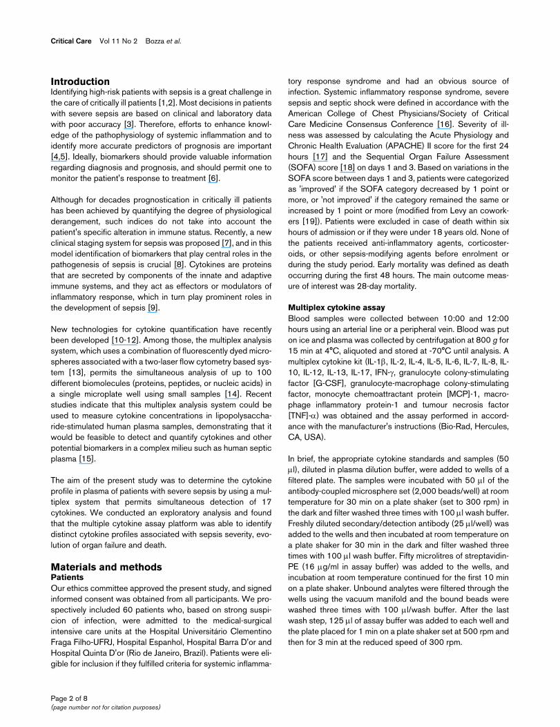
ResultsPatients characteristicsSixty patients were included in this study; 31 (51.7%) patientssurvived and 29 (48.3%) died. Demographic, clinical andmicrobiological data for the survivors and nonsurvivors aresummarized in Table 1. Patients who died had higherAPACHE II and SOFA scores, as expected, compared withsurvivors. The leading source of infection was the respiratory
tract. Micro-organisms were isolated in 50 out of 60 septicpatients (83.3%), with a predominance of Gram-negative bac-teria (76.0%).
Cytokine concentrationsWhen data from all 1,020 assays were analyzed, the multiplexcytokine system was able to detect plasma cytokines in 710(69.6%) assays. (Here we define 'assay' as each cytokinemeasured, which was performed on each individual plasmasample.) Concentrations of IL-6, IL-8, IL-10 and macrophageinflammatory protein-1 were detectable in more than 95% ofindividual assays.
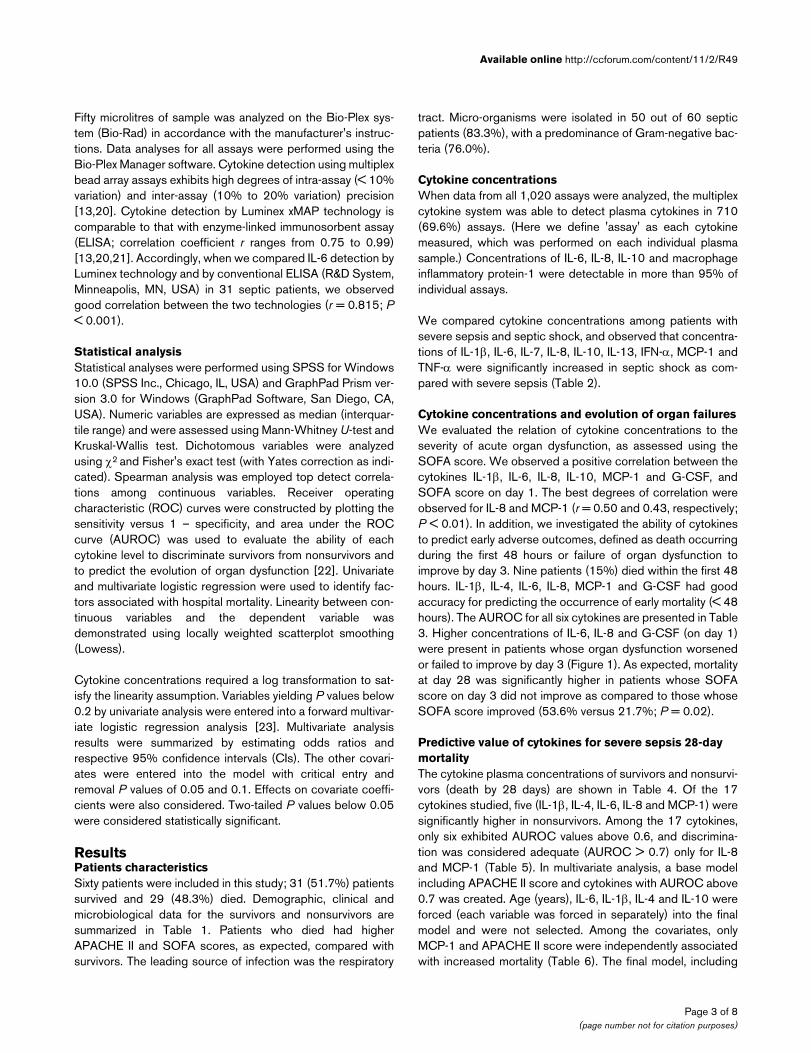
We compared cytokine concentrations among patients withsevere sepsis and septic shock, and observed that concentra-tions of IL-1β, IL-6, IL-7, IL-8, IL-10, IL-13, IFN-α, MCP-1 andTNF-α were significantly increased in septic shock as com-pared with severe sepsis (Table 2).
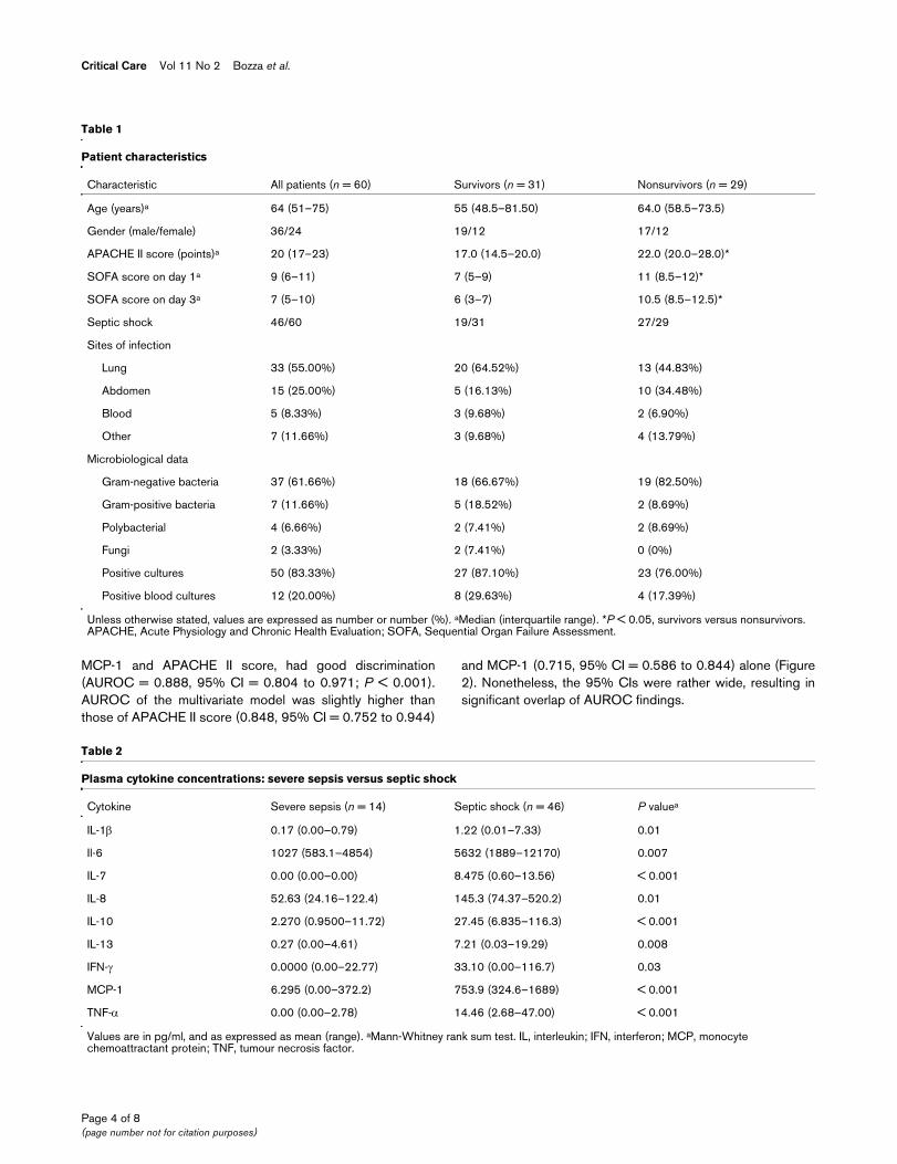
Cytokine concentrations and evolution of organ failuresWe evaluated the relation of cytokine concentrations to theseverity of acute organ dysfunction, as assessed using theSOFA score. We observed a positive correlation between thecytokines IL-1β, IL-6, IL-8, IL-10, MCP-1 and G-CSF, andSOFA score on day 1. The best degrees of correlation wereobserved for IL-8 and MCP-1 (r = 0.50 and 0.43, respectively;P < 0.01). In addition, we investigated the ability of cytokinesto predict early adverse outcomes, defined as death occurringduring the first 48 hours or failure of organ dysfunction toimprove by day 3. Nine patients (15%) died within the first 48hours. IL-1β, IL-4, IL-6, IL-8, MCP-1 and G-CSF had goodaccuracy for predicting the occurrence of early mortality (< 48hours). The AUROC for all six cytokines are presented in Table3. Higher concentrations of IL-6, IL-8 and G-CSF (on day 1)were present in patients whose organ dysfunction worsenedor failed to improve by day 3 (Figure 1). As expected, mortalityat day 28 was significantly higher in patients whose SOFAscore on day 3 did not improve as compared to those whoseSOFA score improved (53.6% versus 21.7%; P = 0.02).
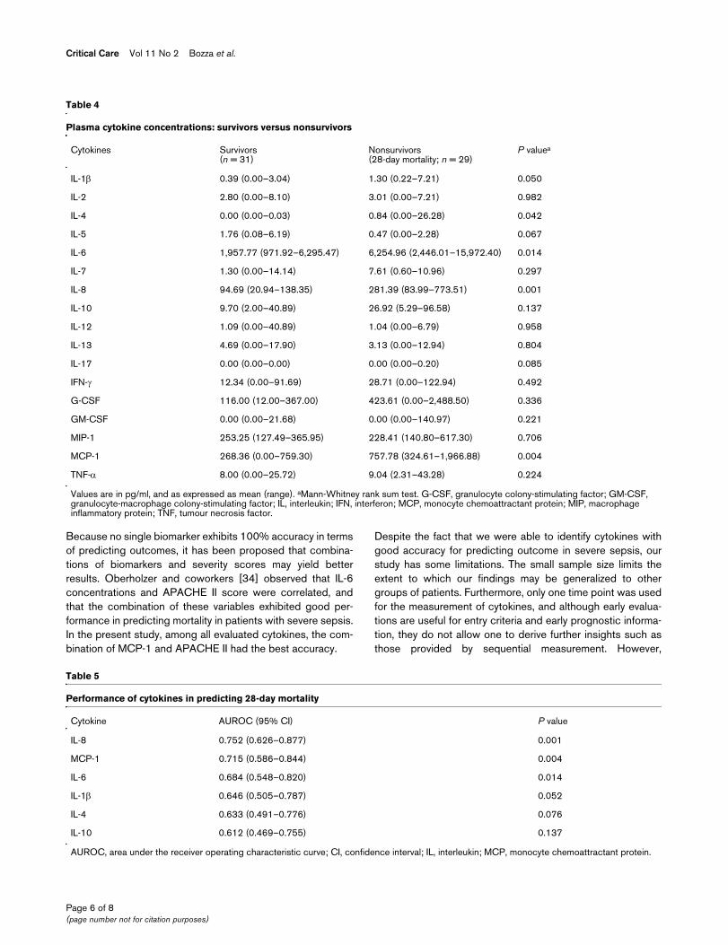
Predictive value of cytokines for severe sepsis 28-day mortalityThe cytokine plasma concentrations of survivors and nonsurvi-vors (death by 28 days) are shown in Table 4. Of the 17cytokines studied, five (IL-1β, IL-4, IL-6, IL-8 and MCP-1) weresignificantly higher in nonsurvivors. Among the 17 cytokines,only six exhibited AUROC values above 0.6, and discrimina-tion was considered adequate (AUROC > 0.7) only for IL-8and MCP-1 (Table 5). In multivariate analysis, a base modelincluding APACHE II score and cytokines with AUROC above0.7 was created. Age (years), IL-6, IL-1β, IL-4 and IL-10 wereforced (each variable was forced in separately) into the finalmodel and were not selected. Among the covariates, onlyMCP-1 and APACHE II score were independently associatedwith increased mortality (Table 6). The final model, including
Page 3 of 8(page number not for citation purposes)
Critical Care Vol 11 No 2 Bozza et al.
MCP-1 and APACHE II score, had good discrimination(AUROC = 0.888, 95% CI = 0.804 to 0.971; P < 0.001).AUROC of the multivariate model was slightly higher thanthose of APACHE II score (0.848, 95% CI = 0.752 to 0.944)
and MCP-1 (0.715, 95% CI = 0.586 to 0.844) alone (Figure2). Nonetheless, the 95% CIs were rather wide, resulting insignificant overlap of AUROC findings.
Table 1
Patient characteristics
Characteristic All patients (n = 60) Survivors (n = 31) Nonsurvivors (n = 29)
Age (years)a 64 (51–75) 55 (48.5–81.50) 64.0 (58.5–73.5)
Gender (male/female) 36/24 19/12 17/12
APACHE II score (points)a 20 (17–23) 17.0 (14.5–20.0) 22.0 (20.0–28.0)*
SOFA score on day 1a 9 (6–11) 7 (5–9) 11 (8.5–12)*
SOFA score on day 3a 7 (5–10) 6 (3–7) 10.5 (8.5–12.5)*
Septic shock 46/60 19/31 27/29
Sites of infection
Lung 33 (55.00%) 20 (64.52%) 13 (44.83%)
Abdomen 15 (25.00%) 5 (16.13%) 10 (34.48%)
Blood 5 (8.33%) 3 (9.68%) 2 (6.90%)
Other 7 (11.66%) 3 (9.68%) 4 (13.79%)
Microbiological data
Gram-negative bacteria 37 (61.66%) 18 (66.67%) 19 (82.50%)
Gram-positive bacteria 7 (11.66%) 5 (18.52%) 2 (8.69%)
Polybacterial 4 (6.66%) 2 (7.41%) 2 (8.69%)
Fungi 2 (3.33%) 2 (7.41%) 0 (0%)
Positive cultures 50 (83.33%) 27 (87.10%) 23 (76.00%)
Positive blood cultures 12 (20.00%) 8 (29.63%) 4 (17.39%)
Unless otherwise stated, values are expressed as number or number (%). aMedian (interquartile range). *P < 0.05, survivors versus nonsurvivors. APACHE, Acute Physiology and Chronic Health Evaluation; SOFA, Sequential Organ Failure Assessment.
Table 2
Plasma cytokine concentrations: severe sepsis versus septic shock
Cytokine Severe sepsis (n = 14) Septic shock (n = 46) P valuea
IL-1β 0.17 (0.00–0.79) 1.22 (0.01–7.33) 0.01
Il-6 1027 (583.1–4854) 5632 (1889–12170) 0.007
IL-7 0.00 (0.00–0.00) 8.475 (0.60–13.56) < 0.001
IL-8 52.63 (24.16–122.4) 145.3 (74.37–520.2) 0.01
IL-10 2.270 (0.9500–11.72) 27.45 (6.835–116.3) < 0.001
IL-13 0.27 (0.00–4.61) 7.21 (0.03–19.29) 0.008
IFN-γ 0.0000 (0.00–22.77) 33.10 (0.00–116.7) 0.03
MCP-1 6.295 (0.00–372.2) 753.9 (324.6–1689) < 0.001
TNF-α 0.00 (0.00–2.78) 14.46 (2.68–47.00) < 0.001
Values are in pg/ml, and as expressed as mean (range). aMann-Whitney rank sum test. IL, interleukin; IFN, interferon; MCP, monocyte chemoattractant protein; TNF, tumour necrosis factor.
Page 4 of 8(page number not for citation purposes)
Available online http://ccforum.com/content/11/2/R49
DiscussionCytokine profiling of patients with severe sepsis may representa valuable tool for delineating different patterns of immunolog-ical response, thus allowing identification of groups of patientswith homogeneous biological derangements [6,24]. In thepresent study, a multiplex analysis of plasma cytokines inpatients with severe sepsis and septic shock was able to iden-tify cytokine profiles associated with early and late mortality, aswell as evolution of organ dysfunction.
When we compared the cytokine profiles of septic shockpatients with the profiles of patients with severe sepsis, weobserved a significant increase in nine out of the 17 cytokinesanalyzed. Significant increases were observed in bothproinflammatory and immunomodulatory cytokines. Thisincluded traditionally evaluated cytokines (IL-1β, IL-6, IL-8, IL-10 and TNF-α) and a number of cytokines that are not com-monly associated with sepsis (IL-7, IL-13, IFN-γ and MCP-1).Moreover, for none of the cytokines evaluated was theconcentration significantly lower in septic shock patients thanin patients with severe sepsis.
It is increasingly recognized that the inflammatory responseand deregulated cytokine production play key roles in thedevelopment of multiple organ dysfunction [5]. Clinicallydefined, early sequential analysis of organic dysfunction insevere sepsis has proven to be a good predictor of outcome[19,25]. However, the cytokine patterns associated with theevolution of organ dysfunction are not well established. A morerestricted panel of cytokines (only six out of 17 cytokines),namely IL-1β, IL-6, IL-8, IL-10, MCP-1 and G-CSF, were foundto correlate positively with organ dysfunction, as assessed bythe SOFA score on day 1. Of these six cytokines, IL-8 andMCP-1 exhibited the best performance. In addition, concentra-tions of IL-6, IL-8 and G-CSF within the first 24 hours werepredictive of worsening organ dysfunction or failure of organdysfunction to improve on day 3. In contrast, although TNF-αconcentrations failed to predict the evolution of organ
dysfunction, or early or late mortality, TNF-α concentrationswere significantly higher in patients with septic shock than inthose with severe sepsis. These findings are in accordancewith the recently proposed hypothesis that different patternsof cytokine profiles may be mirrored by distinct clinical presen-tations and severity [24].
Cytokines such as IL-6 and IL-8 are predictors of outcome insevere sepsis, a finding that is confirmed by our study; how-ever, on multivariate analysis, they were not found to be inde-pendently associated with mortality. Interestingly, the bestpredictor of outcome in our study was MCP-1. The MCP-1 isa potent chemoattractant of mononuclear cells and a regula-tory mediator in sepsis. Although its role is not entirely clear,its involvement in sepsis has been demonstrated during thepast decade. Its pathophysiological role has been linked to theactivated protein C pathway and its induced genes [26,27]. Inanimal models of sepsis, neutralization of MCP-1 was associ-ated with significantly increased mortality [28,29]. Recently,our group demonstrated that endogenous MCP-1 positivelyregulates IL-10 but negatively controls macrophage migrationinhibitory factor in experimental peritoneal sepsis, suggestingan important immunomodulatory role for MCP-1 in controllingthe balance between proinflammatory and anti-inflammatoryfactors in sepsis [30]. In the clinical setting, only a few investi-gators have identified increased concentrations of MCP-1 inplasma [31] and bronchoalveolar lavage fluid [32] from septicpatients, and those findings were not correlated with out-comes. Recently, Vermont and coworkers [33] reported thatserum concentrations of MCP-1 in patients with meningococ-cal sepsis are predictive of mortality and correlate stronglywith disease severity.
Table 3
Performance of cytokines in predicting early mortality (48 hours)
Cytokine AUROC (95% CI) P value
IL-8 0.780 (0.621–0.93) 0.012
IL-4 0.767 (0.587–0.94) 0.011
IL-6 0.756 (0.602–0.91) 0.015
MCP-1 0.738 (0.571–0.905)
0.024
G-CSF 0.727 (0.526–0.92) 0.041
IL-1β 0.716 (0.532–0.90) 0.040
AUROC, area under the receiver operating characteristic curve; CI, confidence interval; G-CSF, granulocyte colony-stimulating factor; IL, interleukin; IFN, interferon; MCP, monocyte chemoattractant protein.
Figure 1
Receiver operating characteristic curve analysis of granulocyte colony-stimulating factor (G-CSF), IL-6 and IL-8 predicting organ dysfunctionReceiver operating characteristic curve analysis of granulocyte colony-stimulating factor (G-CSF), IL-6 and IL-8 predicting organ dysfunction. Shown are the areas under the receiver operating characteristic curve (AUROCs) for granulocyte colony-stimulating factor (G-CSF), IL-6 and IL-8 predicting failure of organ dysfunction to improve by day 3. The val-ues shown in parentheses are the 95% confidence intervals.
Page 5 of 8(page number not for citation purposes)
Critical Care Vol 11 No 2 Bozza et al.
Because no single biomarker exhibits 100% accuracy in termsof predicting outcomes, it has been proposed that combina-tions of biomarkers and severity scores may yield betterresults. Oberholzer and coworkers [34] observed that IL-6concentrations and APACHE II score were correlated, andthat the combination of these variables exhibited good per-formance in predicting mortality in patients with severe sepsis.In the present study, among all evaluated cytokines, the com-bination of MCP-1 and APACHE II had the best accuracy.
Despite the fact that we were able to identify cytokines withgood accuracy for predicting outcome in severe sepsis, ourstudy has some limitations. The small sample size limits theextent to which our findings may be generalized to othergroups of patients. Furthermore, only one time point was usedfor the measurement of cytokines, and although early evalua-tions are useful for entry criteria and early prognostic informa-tion, they do not allow one to derive further insights such asthose provided by sequential measurement. However,
Table 4
Plasma cytokine concentrations: survivors versus nonsurvivors
Cytokines Survivors(n = 31)
Nonsurvivors(28-day mortality; n = 29)
P valuea
IL-1β 0.39 (0.00–3.04) 1.30 (0.22–7.21) 0.050
IL-2 2.80 (0.00–8.10) 3.01 (0.00–7.21) 0.982
IL-4 0.00 (0.00–0.03) 0.84 (0.00–26.28) 0.042
IL-5 1.76 (0.08–6.19) 0.47 (0.00–2.28) 0.067
IL-6 1,957.77 (971.92–6,295.47) 6,254.96 (2,446.01–15,972.40) 0.014
IL-7 1.30 (0.00–14.14) 7.61 (0.60–10.96) 0.297
IL-8 94.69 (20.94–138.35) 281.39 (83.99–773.51) 0.001
IL-10 9.70 (2.00–40.89) 26.92 (5.29–96.58) 0.137
IL-12 1.09 (0.00–40.89) 1.04 (0.00–6.79) 0.958
IL-13 4.69 (0.00–17.90) 3.13 (0.00–12.94) 0.804
IL-17 0.00 (0.00–0.00) 0.00 (0.00–0.20) 0.085
IFN-γ 12.34 (0.00–91.69) 28.71 (0.00–122.94) 0.492
G-CSF 116.00 (12.00–367.00) 423.61 (0.00–2,488.50) 0.336
GM-CSF 0.00 (0.00–21.68) 0.00 (0.00–140.97) 0.221
MIP-1 253.25 (127.49–365.95) 228.41 (140.80–617.30) 0.706
MCP-1 268.36 (0.00–759.30) 757.78 (324.61–1,966.88) 0.004
TNF-α 8.00 (0.00–25.72) 9.04 (2.31–43.28) 0.224
Values are in pg/ml, and as expressed as mean (range). aMann-Whitney rank sum test. G-CSF, granulocyte colony-stimulating factor; GM-CSF, granulocyte-macrophage colony-stimulating factor; IL, interleukin; IFN, interferon; MCP, monocyte chemoattractant protein; MIP, macrophage inflammatory protein; TNF, tumour necrosis factor.
Table 5
Performance of cytokines in predicting 28-day mortality
Cytokine AUROC (95% CI) P value
IL-8 0.752 (0.626–0.877) 0.001
MCP-1 0.715 (0.586–0.844) 0.004
IL-6 0.684 (0.548–0.820) 0.014
IL-1β 0.646 (0.505–0.787) 0.052
IL-4 0.633 (0.491–0.776) 0.076
IL-10 0.612 (0.469–0.755) 0.137
AUROC, area under the receiver operating characteristic curve; CI, confidence interval; IL, interleukin; MCP, monocyte chemoattractant protein.
Page 6 of 8(page number not for citation purposes)
Available online http://ccforum.com/content/11/2/R49
cytokine concentrations on day 1 were associated with sever-ity of organ failure on the first day and with failure of organ dys-function to improve by day 3. Accordingly, both early and latemortality were also predicted. Although it is tempting to spec-ulate that there is a direct correlation between cytokine con-centrations and pathophysiology of organ injury, we believeone cannot attribute the full burden of disease severity to aparticular cytokine. Cytokines may be increased simply asmarkers of tissue damage, without necessarily playing a directrole.
The multiplex system provides the opportunity to establish apanel of sepsis biomarkers that could include not only cur-rently evaluated cytokines that have diagnostic/prognosticvalue but also other valuable sepsis biomarkers, such asincluding migration inhibitory factor [35], procalcitonin [36]and triggering receptor expressed on myeloid cells-1 [37]. Wefully acknowledge that the complexity of sepsis requires newperspectives if we are to achieve an integrated understandingof the intricate interactions that occur during the disease proc-ess, rather than describing isolated aspects of it [38].
ConclusionSimultaneous evaluation of multiple cytokines in early severesepsis may reveal cytokine patterns that reflect the inflamma-tory response associated with evolution of organ dysfunctionas well as early and late mortality. A knowledge of the cytokineprofiles associated with distinct clinical presentations and out-comes may be useful in the design of future studies of biomar-kers in sepsis that involve larger patient populations.
Competing interestsThe authors declare that they have no competing interests.
Authors' contributionsFAB contributed to the study conception and design, carriedout clinical studies, and participated in data analysis anddrafted the manuscript. JIS and AMJ carried out the clinicalstudies and participated in the data analysis. EFA and RNGcarried out the Luminex immunoassays and participated in thedata analysis. MS performed the statistical analysis. MTB,HCFN and PTB conceived the study, and participated in itsdesign and coordination, supervised data analysis and helpedto draft the manuscript. All authors read and approved the finalmanuscript.
Table 6
Multivariate analysis of factors associated with increased hospital mortality
Coefficient Odds ratio (95% CI) P value
APACHE II score (points) 0.313 1.37 (1.12–1.66) 0.002
Ln MCP-1 0.341 1.41 (1.02–1.93) 0.036
Constant -8.335
APACHE, Acute Physiology and Chronic Health Evaluation; CI, confidence interval; Ln, log transformed; MCP, monocyte chemoattractant protein.
Figure 2
Receiver operating characteristic curves of Acute Physiology and Chronic Health (APACHE) II score, monocyte chemoattractant protein (MCP)-1 and a composite variable (APACHE II + MCP-1) predicting mortalityReceiver operating characteristic curves of Acute Physiology and Chronic Health (APACHE) II score, monocyte chemoattractant protein (MCP)-1 and a composite variable (APACHE II + MCP-1) predicting mortality. Shown are areas under receiver operating characteristic curves (AUROCs) for APACHE II score, monocyte MCP-1 and a com-posite variable (APACHE II + MCP-1), created according to the final model from multivariable analysis (Table 4).
Key messages
• Simultaneous analysis of multiple cytokines proved use-ful in identifying cytokine patterns of inflammatory response associated with evolution of organ dysfunc-tion as well as early and late mortality in patients with severe sepsis and septic shock.
• Among the 17 cytokines evaluated, IL-8 and MCP-1 exhibited the best correlation with organ dysfunctions on day 1; in addition, IL-6, IL-8 and G-CSF concentra-tions within the first 24 hours were able to predict wors-ening organ dysfunction or failure of organ dysfunction to improve by day 3.
• In terms of predicting mortality, the cytokines IL-1β, IL-4, IL-6, IL-8, MCP-1 and G-CSF had good accuracy for predicting early mortality (< 48 hours), and IL-8 and MCP-1 had the best accuracy for predicting 28-day mortality; in the multivariate analysis only MCP-1 was independently associated with prognosis.
Page 7 of 8(page number not for citation purposes)
Critical Care Vol 11 No 2 Bozza et al.
AcknowledgementsThis work was supported by Fundação Oswaldo Cruz (PDTSP and PAPES 4), Conselho de Desenvolvimento Científico e Tecnológico (CNPq), Fundação de Amparo à Pesquisa do Estado do Rio de Janeiro (FAPERJ), and Fundação José Bonifácio (FuJB). The authors thank the Program for Technological Development in Tools for Health-PDTIS-FIOCRUZ for use of its Luminex facilities.
References1. Levy MM, Fink MP, Marshall JC, Abraham E, Angus D, Cook D,
Cohen J, Opal SM, Vincent JL, Ramsay G: 2001 SCCM/ESICM/ACCP/ATS/SIS International Sepsis Definitions Conference.Crit Care Med 2003, 31:1250-1256.
2. Dellinger RP, Carlet JM, Masur H, Gerlach H, Calandra T, CohenJ, Gea-Banacloche J, Keh D, Marshall JC, Parker MM, et al.: Sur-viving Sepsis Campaign guidelines for management of severesepsis and septic shock. Crit Care Med 2004, 32:858-873.
3. Vincent JL, Abraham E: The last 100 years of sepsis. Am JRespir Crit Care Med 2006, 173:256-263.
4. Cohen J, Guyatt G, Bernard GR, Calandra T, Cook D, Elbourne D,Marshall J, Nunn A, Opal S: New strategies for clinical trials inpatients with sepsis and septic shock. Crit Care Med 2001,29:880-886.
5. Riedemann NC, Guo RF, Ward PA: The enigma of sepsis. J ClinInvest 2003, 112:460-467.
6. Marshall JC: Biomarkers of sepsis. Curr Infect Dis Rep 2006,8:351-357.
7. Gerlach H, Dhainaut JF, Harbarth S, Reinhart K, Marshall JC, LevyM: The PIRO concept: R is for response. Crit Care 2003,7:256-259.
8. Marshall JC, Vincent JL, Fink MP, Cook DJ, Rubenfeld G, Foster D,Fisher CJ Jr, Faist E, Reinhart K: Measures, markers, and medi-ators: toward a staging system for clinical sepsis. A report ofthe Fifth Toronto Sepsis Roundtable, Toronto, Ontario, Can-ada, October 25–26, 2000. Crit Care Med 2003, 31:1560-1567.
9. Cohen J: The immunopathogenesis of sepsis. Nature 2002,420:885-891.
10. Tam SW, Wiese R, Lee S, Gilmore J, Kumble KD: Simultaneousanalysis of eight human Th1/Th2 cytokines using microarrays.J Immunol Methods 2002, 261:157-165.
11. Sennikov SV, Krysov SV, Injelevskaya TV, Silkov AN, Grishina LV,Kozlov VA: Quantitative analysis of human immunoregulatorycytokines by electrochemiluminescence method. J ImmunolMethods 2003, 275:81-88.
12. Knight PR, Sreekumar A, Siddiqui J, Laxman B, Copeland S, Chin-naiyan A, Remick DG: Development of a sensitive microarrayimmunoassay and comparison with standard enzyme-linkedimmunoassay for cytokine analysis. Shock 2004, 21:26-30.
13. Vignali DA: Multiplexed particle-based flow cytometric assays.J Immunol Methods 2000, 243:243-255.
14. Oliver KG, Kettman JR, Fulton RJ: Multiplexed analysis of humancytokines by use of the FlowMetrix system. Clin Chem 1998,44:2057-2060.
15. Prabhakar U, Eirikis E, Davis HM: Simultaneous quantification ofproinflammatory cytokines in human plasma using the Lab-MAP assay. J Immunol Methods 2002, 260:207-218.
16. Bone RC, Balk RA, Cerra FB, Dellinger RP, Fein AM, Knaus WA,Schein RM, Sibbald WJ: Definitions for sepsis and organ failureand guidelines for the use of innovative therapies in sepsis.The ACCP/SCCM Consensus Conference Committee. Ameri-can College of Chest Physicians/Society of Critical CareMedicine. Chest 1992, 101:1644-1655.
17. Knaus WA, Draper EA, Wagner DP, Zimmerman JE: APACHE II: aseverity of disease classification system. Crit Care Med 1985,13:818-829.
18. Vincent JL, Moreno R, Takala J, Willatts S, De Mendonca A, Bruin-ing H, Reinhart CK, Suter PM, Thijs LG: The SOFA (Sepsis-related Organ Failure Assessment) score to describe organdysfunction/failure. On behalf of the Working Group on Sep-sis-Related Problems of the European Society of IntensiveCare Medicine. Intensive Care Med 1996, 22:707-710.
19. Levy MM, Macias WL, Vincent JL, Russell JA, Silva E, TrzaskomaB, Williams MD: Early changes in organ function predict even-
tual survival in severe sepsis. Crit Care Med 2005,33:2194-2201.
20. de Jager W, te Velthuis H, Prakken BJ, Kuis W, Rijkers GT: Simul-taneous detection of 15 human cytokines in a single sample ofstimulated peripheral blood mononuclear cells. Clin Diagn LabImmunol 2003, 10:133-139.
21. Khan SS, Smith MS, Reda D, Suffredini AF, McCoy JP Jr: Multi-plex bead array assays for detection of soluble cytokines:comparisons of sensitivity and quantitative values among kitsfrom multiple manufacturers. Cytometry B Clin Cytom 2004,61:35-39.
22. Zweig MH, Campbell G: Receiver-operating characteristic(ROC) plots: a fundamental evaluation tool in clinicalmedicine. Clin Chem 1993, 39:561-577.
23. Hosmer DW, Lemeshow S: Applied Logistic Regression 2nd edi-tion. New York: Wiley-Interscience; 2000.
24. Ulloa L, Tracey KJ: The 'cytokine profile': a code for sepsis.Trends Mol Med 2005, 11:56-63.
25. Ferreira FL, Bota DP, Bross A, Melot C, Vincent JL: Serial evalu-ation of the SOFA score to predict outcome in critically illpatients. JAMA 2001, 286:1754-1758.
26. Riewald M, Petrovan RJ, Donner A, Mueller BM, Ruf W: Activationof endothelial cell protease activated receptor 1 by the proteinC pathway. Science 2002, 296:1880-1882.
27. Brueckmann M, Marx A, Weiler HM, Liebe V, Lang S, Kaden JJ,Zieger W, Borggrefe M, Huhle G, Konstantin Haase K: Stabiliza-tion of monocyte chemoattractant protein-1-mRNA by acti-vated protein C. Thromb Haemost 2003, 89:149-160.
28. Zisman DA, Kunkel SL, Strieter RM, Tsai WC, Bucknell K,Wilkowski J, Standiford TJ: MCP-1 protects mice in lethalendotoxemia. J Clin Invest 1997, 99:2832-2836.
29. Matsukawa A, Hogaboam CM, Lukacs NW, Lincoln PM, StrieterRM, Kunkel SL: Endogenous monocyte chemoattractant pro-tein-1 (MCP-1) protects mice in a model of acute septic peri-tonitis: cross-talk between MCP-1 and leukotriene B4. JImmunol 1999, 163:6148-6154.
30. Gomes RN, Figueiredo RT, Bozza FA, Pacheco P, Amancio RT,Laranjeira AP, Castro-Faria-Neto HC, Bozza PT, Bozza MT:Increased susceptibility to septic and endotoxic shock inmonocyte chemoattractant protein 1/CC chemokine ligand 2-deficient mice correlates with reduced interleukin 10 andenhanced macrophage migration inhibitory factor production.Shock 2006, 26:457-463.
31. Bossink AW, Paemen L, Jansen PM, Hack CE, Thijs LG, VanDamme J: Plasma levels of the chemokines monocyte chemo-tactic proteins-1 and -2 are elevated in human sepsis. Blood1995, 86:3841-3847.
32. Goodman RB, Strieter RM, Martin DP, Steinberg KP, Milberg JA,Maunder RJ, Kunkel SL, Walz A, Hudson LD, Martin TR: Inflam-matory cytokines in patients with persistence of the acute res-piratory distress syndrome. Am J Respir Crit Care Med 1996,154:602-611.
33. Vermont CL, Hazelzet JA, de Kleijn ED, van den Dobbelsteen GP,de Groot R: CC and CXC chemokine levels in children withmeningococcal sepsis accurately predict mortality and dis-ease severity. Crit Care 2006, 10:R33.
34. Oberholzer A, Souza SM, Tschoeke SK, Oberholzer C, Abou-hamze A, Pribble JP, Moldawer LL: Plasma cytokine measure-ments augment prognostic scores as indicators of outcome inpatients with severe sepsis. Shock 2005, 23:488-493.
35. Bozza FA, Gomes RN, Japiassu AM, Soares M, Castro-Faria-NetoHC, Bozza PT, Bozza MT: Macrophage migration inhibitory fac-tor levels correlate with fatal outcome in sepsis. Shock 2004,22:309-313.
36. Simon L, Gauvin F, Amre GK, Saint-Louis P, Lacroix J: Serum pro-calcitonin and C-reactive protein levels as markers of bacterialinfection: a systematic review and meta-analysis. Clin InfectDis 2004, 39:206-217.
37. Gibot S, Kolopp-Sarda MN, Bene MC, Cravoisy A, Levy B, FaureGC, Bollaert PE: Plasma level of a triggering receptorexpressed on myeloid cells-1: its diagnostic accuracy inpatients with suspected sepsis. Ann Intern Med 2004,141:9-15.
38. Tjardes T, Neugebauer E: Sepsis research in the next millen-nium: concentrate on the software rather than the hardware.Shock 2002, 17:1-8.
Page 8 of 8(page number not for citation purposes)
http://www.ncbi.nlm.nih.gov/entrez/query.fcgi?cmd=Retrieve&db=PubMed&dopt=Abstract&list_uids=9733011
http://www.ncbi.nlm.nih.gov/entrez/query.fcgi?cmd=Retrieve&db=PubMed&dopt=Abstract&list_uids=9733011
http://www.ncbi.nlm.nih.gov/entrez/query.fcgi?cmd=Retrieve&db=PubMed&dopt=Abstract&list_uids=1303622
http://www.ncbi.nlm.nih.gov/entrez/query.fcgi?cmd=Retrieve&db=PubMed&dopt=Abstract&list_uids=1303622
http://www.ncbi.nlm.nih.gov/entrez/query.fcgi?cmd=Retrieve&db=PubMed&dopt=Abstract&list_uids=1303622
http://www.ncbi.nlm.nih.gov/entrez/query.fcgi?cmd=Retrieve&db=PubMed&dopt=Abstract&list_uids=3928249
http://www.ncbi.nlm.nih.gov/entrez/query.fcgi?cmd=Retrieve&db=PubMed&dopt=Abstract&list_uids=3928249
http://www.ncbi.nlm.nih.gov/entrez/query.fcgi?cmd=Retrieve&db=PubMed&dopt=Abstract&list_uids=8844239
http://www.ncbi.nlm.nih.gov/entrez/query.fcgi?cmd=Retrieve&db=PubMed&dopt=Abstract&list_uids=8844239
http://www.ncbi.nlm.nih.gov/entrez/query.fcgi?cmd=Retrieve&db=PubMed&dopt=Abstract&list_uids=8844239
http://www.ncbi.nlm.nih.gov/entrez/query.fcgi?cmd=Retrieve&db=PubMed&dopt=Abstract&list_uids=8472349
http://www.ncbi.nlm.nih.gov/entrez/query.fcgi?cmd=Retrieve&db=PubMed&dopt=Abstract&list_uids=8472349
http://www.ncbi.nlm.nih.gov/entrez/query.fcgi?cmd=Retrieve&db=PubMed&dopt=Abstract&list_uids=8472349
http://www.ncbi.nlm.nih.gov/entrez/query.fcgi?cmd=Retrieve&db=PubMed&dopt=Abstract&list_uids=9185504
http://www.ncbi.nlm.nih.gov/entrez/query.fcgi?cmd=Retrieve&db=PubMed&dopt=Abstract&list_uids=9185504
http://www.ncbi.nlm.nih.gov/entrez/query.fcgi?cmd=Retrieve&db=PubMed&dopt=Abstract&list_uids=7579352
http://www.ncbi.nlm.nih.gov/entrez/query.fcgi?cmd=Retrieve&db=PubMed&dopt=Abstract&list_uids=7579352
http://www.ncbi.nlm.nih.gov/entrez/query.fcgi?cmd=Retrieve&db=PubMed&dopt=Abstract&list_uids=8810593
http://www.ncbi.nlm.nih.gov/entrez/query.fcgi?cmd=Retrieve&db=PubMed&dopt=Abstract&list_uids=8810593