Connecting mind, brain, and behavior in mathematics ed - ERIC
16
AERA 2009 PAPER PRESENTED TO THE BRAIN, NEUROSCIENCE, AND EDUCATION SIG, APRIL 14, 2009 SESSION Neuroscience in Math and Reading Education TITLE Electrooculography: Connecting mind, brain, and behavior in mathematics education research AUTHORS Olga V. Shipulina FACULTY OF EDUCATION EMAIL: OSHIPULI@SFU.CA SIMON FRASER UNIVERSITY PHONE: 1-778-782-7173 8888 UNIVERSITY DRIVE FAX: 1-778-782-7187 BURNABY, BC, V5A 1S6 WEB: WWW.ENGRAMMETRON.NET CANADA Stephen R. Campbell O. Arda Cimen FACULTY OF EDUCATION, SIMON FRASER UNIVERSITY ABSTRACT This paper reports on the potential roles and importance of electrooculography (EOG) for mathematics educational neuroscience research. EOG enables accurate measurements of eye- related behavior (i.e., blinks & movements) by recording changes in voltage potentials generated by eye-related behavior. We identify and discuss three main uses of EOG. First, EOG provides insights into cognitive function. Secondly, it is used to attenuate eye-related artifacts in electroencephalography (EEG). Thirdly, EOG serves as a helpful means for calibrating covert brain activity with overt behavior. We provide an overview of the first two application areas, and we illustrate the third application using an example data set capturing an "aha moment" in our research in the area of mathematical problem solving. CITE AS Shipulina, O. V., Campbell, S. R., & Cimen, O. A. (2009, April). Electrooculography: Connecting mind, brain, and behavior in mathematics education research. Paper presented to the American Educational Research Association: Brain, Neuroscience, and Education SIG. San Diego, CA, U.S.A. WORD COUNTS ABSTRACT (117 WORDS); PAPER (2879WORDS); REFERENCES (439 WORDS). RUNNING HEAD Electrooculography...
-
Upload
khangminh22 -
Category
Documents
-
view
4 -
download
0
Transcript of Connecting mind, brain, and behavior in mathematics ed - ERIC
AERA 2009 PAPER PRESENTED TO THE
BRAIN, NEUROSCIENCE, AND EDUCATION SIG, APRIL 14, 2009
SESSION
Neuroscience in Math and Reading Education
TITLE
Electrooculography: Connecting mind, brain, and behavior in mathematics
education research
AUTHORS
Olga V. Shipulina
FACULTY OF EDUCATION EMAIL: [email protected]
SIMON FRASER UNIVERSITY PHONE: 1-778-782-7173
8888 UNIVERSITY DRIVE FAX: 1-778-782-7187
BURNABY, BC, V5A 1S6 WEB: WWW.ENGRAMMETRON.NET
CANADA
Stephen R. Campbell
O. Arda Cimen
FACULTY OF EDUCATION, SIMON FRASER UNIVERSITY
ABSTRACT
This paper reports on the potential roles and importance of electrooculography (EOG) for
mathematics educational neuroscience research. EOG enables accurate measurements of eye-
related behavior (i.e., blinks & movements) by recording changes in voltage potentials generated
by eye-related behavior. We identify and discuss three main uses of EOG. First, EOG provides
insights into cognitive function. Secondly, it is used to attenuate eye-related artifacts in
electroencephalography (EEG). Thirdly, EOG serves as a helpful means for calibrating covert
brain activity with overt behavior. We provide an overview of the first two application areas, and
we illustrate the third application using an example data set capturing an "aha moment" in our
research in the area of mathematical problem solving.
CITE AS
Shipulina, O. V., Campbell, S. R., & Cimen, O. A. (2009, April). Electrooculography:
Connecting mind, brain, and behavior in mathematics education research. Paper presented to the
American Educational Research Association: Brain, Neuroscience, and Education SIG. San
Diego, CA, U.S.A.
WORD COUNTS
ABSTRACT (117 WORDS); PAPER (2879WORDS); REFERENCES (439 WORDS).
RUNNING HEAD
Electrooculography...

Electrooculography… Shipulina, Campbell, & Cimen
1
Introduction
Many educational researchers are concerned with understanding how teachers and learners are
thinking. That is to say, many educational researchers are concerned with understanding what is
going on in the minds of teachers and students to better assess what they know, how they know
it, and thereby to help design pedagogies that can lead to improvements in helping these
populations to be educated more effectively. Traditionally, educational researchers engaged in
the empirical study of cognition and learning have relied upon overt behavioral data gathered
from interviews, field notes, self-reports, and audiovisual recordings. In focusing on overt
behavior, educational researchers have typically not been privy to what is happening covertly,
with regard to the physiology of these populations. To the extent that cognition and learning are
embodied behaviors, it would be helpful to augment these traditional data sets with physiological
data sets whenever possible and to the extent that it is practical to acquire them. We know from
cognitive neuroscience and psychophysiology, that there is much to be gained from informing
educational research with results from these well-established fields. Educational neuroscience is
attempting to accomplish just that. As educational neuroscientists, one challenge we have
encountered in so doing concerns how best to connect our traditional overt data sets with the
covert behavioral data sets, specifically, in our case, electroencephalography (EEG), to obtain
deeper educational insights into cognition and learning.
One way of investigating connections between these overt audiovisual (AV) data sets
with covert physiological recordings is through eye-related behavior (ERB), which can be
observed using electrooculography (EOG) and eye-tracking (ET) technology. In this paper, we
focus on the potential roles and importance of EOG for mathematics educational neuroscience
research. Electrooculography enables accurate measurements of ERB (i.e., blinks & movements)
by recording changes in voltage potentials generated as a consequence of ERB. We identify and

Electrooculography… Shipulina, Campbell, & Cimen
2
discuss three main uses of EOG. First, EOG provides insights into cognitive function. Secondly,
it is used to attenuate eye-related artifacts in EEG. Thirdly, EOG serves as a helpful means for
calibrating covert brain activity with overt behavior. In this paper, we present some background
regarding educational studies concerning ERB using ET and describe EOG. We then provide an
overview of the first two of the three aforementioned application areas for EOG. We illustrate
the third application area using an example data set capturing an "aha moment" obtained in the
course of our research in the area of mathematical problem solving.
Background
A major reason for growing interest in educational neuroscience in mathematics education
research, aside from the now widespread recognition that cognitive constructs are embodied, is a
need for better empirical grounds for developing theories of mental functions and processes
(Campbell, 2006 a, b). This, in turn, can reconstitute the conceptual basis for more effective
forms of mathematical learning and instruction. According to Byrnes (2001), brain research is
relevant to the field of psychology and education to the extent that it fosters better
understandings of mind, development and learning. The validity, reliability, and relevance of
psychological theories of teaching and learning developed from traditional psychological
experiments may variously be corroborated, refined, or refuted through neuroscientific studies or
the use of neuroscientific tools and methods to test hypothesizes of any particular theoretical
account (cf. Byrnes, 2001; Kosslyn & Koening, 1992).
Conversely, research in the neurosciences can also benefit from the more situated and
ecologically grounded insights into cognition and learning that typifies the concerns and aims of
educational research. Accordingly, educational research that informs and is informed by
neuroscientific research, while incorporating methods of cognitive neuroscience with the further
aim toward corroborating, refining, or refuting certain models of cognition and learning — that is

Electrooculography… Shipulina, Campbell, & Cimen
3
to say, educational neuroscience — should become of abiding interest and concern to educators
and educational researchers alike. This interest includes, beyond standard methods of data
collection in educational research, analyses of EEG data sets measuring bioelectrical signals
generated from brain activities engaging cognitive processes, such as those involved in
mathematical thinking.
Theoretical considerations concerning brain dynamics and embodied cognition aside,
there are also methodological challenges in isolating brain activity with EEG and in calibrating
EEG with other eye-related behavioral data sets, such as EOG and ET, along with traditional
behavioral data sets, such as AV recordings. As our main objective here is methodological, we
turn now to ways in which EOG can help address these matters.
Electrooculography
In the middle of nineteenth century Emil du Bois-Reymond observed that the cornea of the eye is
electrically positive relative to the back of the eye, forming a so-called corneoretinal potential in
the range of 0.4 - 1.0 mV (Malmivuo & Plonsey, 1995). Since this potential was not affected
sufficiently by light or any other environmental conditions, it was considered as a resting
potential. As the eyes move, each of them behave like single dipoles oriented from the retina to
the cornea. Eye movements produced these moving (rotating) dipole sources and signals from
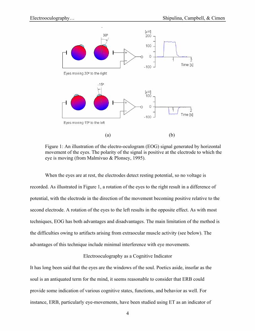
these sources are measured by EOG in the range of 5-20!V/° (see Figure 1, below). Data are
acquired by electrodes placed around the eyes; these electrodes detect the rotations of the
electrostatic dipoles. Figure 1 illustrates the kinds of signals generated from horizontal eye
movements, as recorded by a bipolar pair of electrodes attached to the sides of the left and right
eyes, lateral to the external canthi (just laterally and external to the left and right of each eye).
Electrooculography… Shipulina, Campbell, & Cimen
4
(a) (b)
Figure 1: An illustration of the electro-oculogram (EOG) signal generated by horizontal
movement of the eyes. The polarity of the signal is positive at the electrode to which the
eye is moving (from Malmivuo & Plonsey, 1995).
When the eyes are at rest, the electrodes detect resting potential, so no voltage is
recorded. As illustrated in Figure 1, a rotation of the eyes to the right result in a difference of
potential, with the electrode in the direction of the movement becoming positive relative to the
second electrode. A rotation of the eyes to the left results in the opposite effect. As with most
techniques, EOG has both advantages and disadvantages. The main limitation of the method is
the difficulties owing to artifacts arising from extraocular muscle activity (see below). The
advantages of this technique include minimal interference with eye movements.
Electrooculography as a Cognitive Indicator
It has long been said that the eyes are the windows of the soul. Poetics aside, insofar as the
soul is an antiquated term for the mind, it seems reasonable to consider that ERB could
provide some indication of various cognitive states, functions, and behavior as well. For
instance, ERB, particularly eye-movements, have been studied using ET as an indicator of

Electrooculography… Shipulina, Campbell, & Cimen
5
attention and memory (Kramer & McCarley, 2003), and pupillary response has been studied
as an indication of cognitive load (Just, Carpenter, & Miyake, 2003).
Many kinds of mathematical activity are connected with ERB, which can variously be
voluntary or involuntary. Reading (including reading mathematical texts) serves as an example
of voluntary eye movements and eye movements can be predictable to some extent. Less
deliberate to involuntary eye-related behavior are eye-blinking and saccades (rapid macro and
micro eye-movements). Behaviorally, the specific character of eye movement during reading is
summarized in Duchowski (2002). His study is based on ET research and its application.
Duchowski hypothesizes and cites evidence that as the text becomes conceptually more difficult,
fixation durations increase and saccade lengths decrease. Eye movement and eye fixation
analyses have also been used as a method of research into strategies of successful and
unsuccessful arithmetic word problem solvers (Hegarty, Mayer, and Monk, 1995). According to
Hegarty, et al's study, less successful problem solvers fixate their eyes on numbers and relational
terms when they re-read parts of arithmetic word problem, whereas more successful problem
solvers fixate eyes on variable names. They propose that successful problem solvers construct
problem models and concentrate their attention on appropriate variable names, whereas
unsuccessful problem solvers attempt to directly translate key propositions of these problems
into a computational procedure, and thereby remain predominantly focused on numbers.
More generally, studies using EOG provide evidence that blinks and saccades are
indicators of cognitive function. For instance, it has long been known that blink rates decrease
significantly during visually demanding tasks (e.g., Fogarty & Stern, 1988). Moreover, blinks
and saccades evidently serve to punctuate shifting foci of attention, be they exogenous shifts in
attention from one object to another, or endogenous shifts in reflection from one line of thought
to another (Bonfiglio, Sello, Andre, Carboncini, Arrighi, & Rossi, 2009).
Electrooculography… Shipulina, Campbell, & Cimen
6
Electrooculography for Artifact Correction
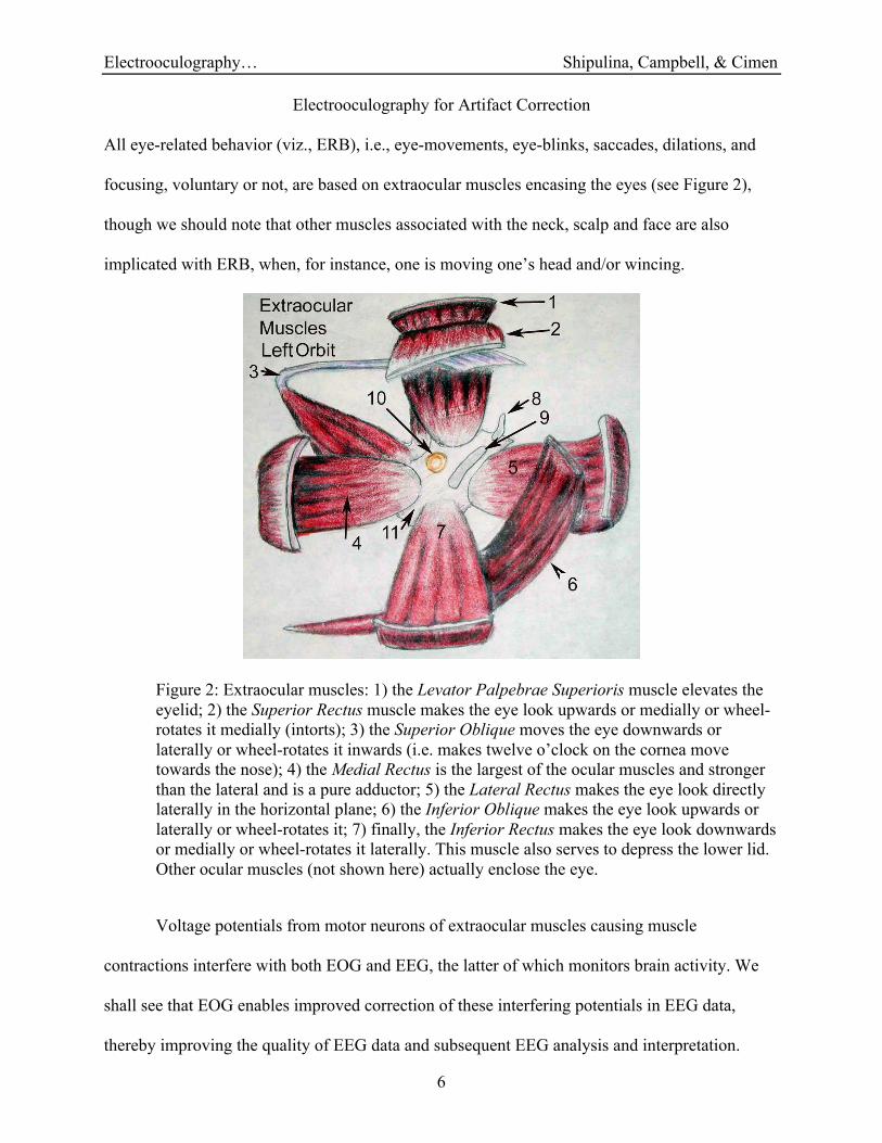
All eye-related behavior (viz., ERB), i.e., eye-movements, eye-blinks, saccades, dilations, and
focusing, voluntary or not, are based on extraocular muscles encasing the eyes (see Figure 2),
though we should note that other muscles associated with the neck, scalp and face are also
implicated with ERB, when, for instance, one is moving one’s head and/or wincing.
Figure 2: Extraocular muscles: 1) the Levator Palpebrae Superioris muscle elevates the
eyelid; 2) the Superior Rectus muscle makes the eye look upwards or medially or wheel-
rotates it medially (intorts); 3) the Superior Oblique moves the eye downwards or
laterally or wheel-rotates it inwards (i.e. makes twelve o’clock on the cornea move
towards the nose); 4) the Medial Rectus is the largest of the ocular muscles and stronger
than the lateral and is a pure adductor; 5) the Lateral Rectus makes the eye look directly
laterally in the horizontal plane; 6) the Inferior Oblique makes the eye look upwards or
laterally or wheel-rotates it; 7) finally, the Inferior Rectus makes the eye look downwards
or medially or wheel-rotates it laterally. This muscle also serves to depress the lower lid.
Other ocular muscles (not shown here) actually enclose the eye.
Voltage potentials from motor neurons of extraocular muscles causing muscle
contractions interfere with both EOG and EEG, the latter of which monitors brain activity. We
shall see that EOG enables improved correction of these interfering potentials in EEG data,
thereby improving the quality of EEG data and subsequent EEG analysis and interpretation.
Electrooculography… Shipulina, Campbell, & Cimen
7
EOG data also enable time synchronization and integration of EEG with ET and AV data sets by
calibrating EOG data with these data sets.
According to Kierkels, Boxtel, and Vogten (2006), before brain activity measured by
EEG through scalp voltage potentials is analysed — especially, we would add, in single trial
(non-replicable) conditions — ERB artifacts should be identified and attenuated using signal
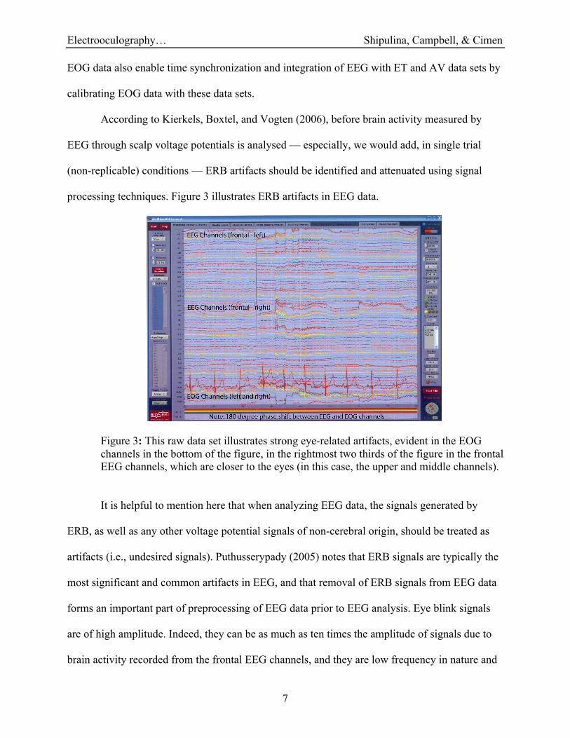
processing techniques. Figure 3 illustrates ERB artifacts in EEG data.
Figure 3: This raw data set illustrates strong eye-related artifacts, evident in the EOG
channels in the bottom of the figure, in the rightmost two thirds of the figure in the frontal
EEG channels, which are closer to the eyes (in this case, the upper and middle channels).
It is helpful to mention here that when analyzing EEG data, the signals generated by
ERB, as well as any other voltage potential signals of non-cerebral origin, should be treated as
artifacts (i.e., undesired signals). Puthusserypady (2005) notes that ERB signals are typically the
most significant and common artifacts in EEG, and that removal of ERB signals from EEG data
forms an important part of preprocessing of EEG data prior to EEG analysis. Eye blink signals
are of high amplitude. Indeed, they can be as much as ten times the amplitude of signals due to
brain activity recorded from the frontal EEG channels, and they are low frequency in nature and
Electrooculography… Shipulina, Campbell, & Cimen
8
affect the low frequency range of EEG signals. Kierkels et al (2006) point out that ERB signals
have large disturbing effects on EEG recordings because the eyes are located so close to the
brain.
There are many methods for eye movement correction in EEG recordings reported in
literature, with varying degrees of success (Kierkels, et al, 2006; Croft, Chandler, Barry, Cooper,
& Clark, 2005). Ille, Berg, and Scherg (2002), for instance, distinguish between methods that
remove ERB signals from EEG data without considering brain activity and those that do attempt
to separate artifacts and brain activity. In the former case, in multi-trial studies, signals that
exceed a threshold criteria, such as maximum amplitude, beyond which are typical of eye-blinks,
are rejected whole scale, together with the EEG data. In the latter case, these authors also
consider signal processing methods, such as Principle Component Analysis (PCA), Independent
Component Analysis (ICA), Multiple Source Eye Correction (MSEC), that are designed to
separate ERB artifacts and EEG brain activity, as promising methods for future development. In
our presentation (see Appendix I), we demonstrate the ICA method for separation ERB signals
from brain activity signals by subtracting components related to ERB from EEG data.
Electrooculography for Calibrating Data Sets
We use EOG recordings in tandem with ET and AV data sets to synchronize with EEG data sets,
which are indirect recordings of the electrical component of the biopotential field recorded on the
scalp being generated from brain activity. We illustrate our approach with a data set capturing an
“aha moment” during a mathematical problem solving task. It should be mentioned here that the
mathematical problem solving experiments are principally of so-called ‘single trial’ type !"#$%&'#
(!))*$#+&#,-..'#/&0.1(!$&2#*/#/&0&!$&23#&1$%&/#41$%#/&5!/2#$*#"$16-.1#*/#$%*-5%$#0/*(&""&"7
Every such experiment is a unique combination of cognitive process that requires observations of
high signal fidelity and integrity for analysis. In integrating overt and covert behaviors associated

Electrooculography… Shipulina, Campbell, & Cimen
9
with problem solving events, we aim to explore the extent to which valid and reliable
information can be obtained from single trial studies in mathematical problem solving.
In the single trial data set we present, the mathematical problem task was based on the
paradigm of Dehaene, Izard, Pica, & Spelke (2006). These data involve a slide comprised of six
diagrams of different kinds of quadrangles: Five of the six diagrams were connected by a
common mathematical concept of ‘diagonals of the quadrangles’. Note that the dots inside of
five of the six diagrams were located exactly at the intersection the diagonals; the task was to
identify the diagram that did not conform, that is, the diagram where the dot inside the
quadrangle is not located at that intersection. This paradigm was presented on the screen of a
computer to one male participant, aged 38 years and 3 months.
Although we are certainly interested in removing ERB signals from EEG data (again, see
Appendix I), we are also interested in using EOG data to synchronize brain activities manifest in
EEG with our eye tracking data and with our audiovisual behavioral data (see Figure 4).
(a) (b)
Figure 4: An integrated time-synchronized data set. Notice that EOG channels 4 and 7
(from the top in figure 4a) express the dipole characteristics schematized in Figure 1b.
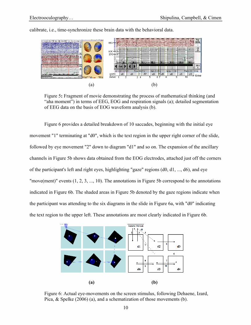
Figure 5 is a frame from a movie file demonstrating the process of thinking on this problem in
terms of EEG and EOG signals. We demonstrate the utility of EOG data to help accurately
Electrooculography… Shipulina, Campbell, & Cimen
10
calibrate, i.e., time-synchronize these brain data with the behavioral data.
(a) (b)
Figure 5: Fragment of movie demonstrating the process of mathematical thinking (and
“aha moment”) in terms of EEG, EOG and respiration signals (a); detailed segmentation
of EEG data on the basis of EOG waveform analysis (b).
Figure 6 provides a detailed breakdown of 10 saccades, beginning with the initial eye
movement "1" terminating at "d0", which is the text region in the upper right corner of the slide,
followed by eye movement "2" down to diagram "d1" and so on. The expansion of the ancillary
channels in Figure 5b shows data obtained from the EOG electrodes, attached just off the corners
of the participant's left and right eyes, highlighting "gaze" regions (d0, d1, ..., d6), and eye
"move(ment)" events (1, 2, 3, ..., 10). The annotations in Figure 5b correspond to the annotations
indicated in Figure 6b. The shaded areas in Figure 5b denoted by the gaze regions indicate when
the participant was attending to the six diagrams in the slide in Figure 6a, with "d0" indicating
the text region to the upper left. These annotations are most clearly indicated in Figure 6b.
(a) (b)
Figure 6: Actual eye-movements on the screen stimulus, following Dehaene, Izard,
Pica, & Spelke (2006) (a), and a schematization of those movements (b).
Electrooculography… Shipulina, Campbell, & Cimen
11
Getting accurate calibrations are crucial to properly associating covert brain activities
with overt problem solving tasks. Once these data sets are synchronized, a more integrated
approach can be taken in connecting brain and behavior. Such integrated approach serves as
method for validation and reliability of mathematical problem solving data interpretation. Our
interpretation of these data is an entire topic of its own, and as such, is left for another time.
Conclusion
Eye-related behavior can be used to infer cognitive function and to operationalize human
performance. From a cognitive perspective, it can be variously considered as sensory-driven,
emotionally-responsive, or goal-directed. In terms of performance, eye-related behavioral
indexes, like fixation patterns and gaze durations, can operationalize efficiency and workload.
Hence there are implications for both studies in cognition and learning, and instructional design
and usability of educational software. Eye related behavior manifest in EOG signals thereby
serve as indicators of various aspects of cognition and performance, including aspects of
attention and memory evident in mathematical problem solving. Eye-related behavior evident in
EOG can be both a blessing and a curse, however, insofar as ERB activity interferes with EEG
recordings that may provide further insights and indicators in this regard. Fortunately, EOG
signals can be used with signal processing methods such as ICA to help attenuate those artifacts
from EEG data. Finally, it is evident that EOG data can serve as a bridge in connecting EEG and
other physiological data sets with more overt behavioral data sets such as eye-tracking and
audiovisual data sets. Indeed, EOG serves well in a variety of ways in connecting brain and
behavior, thereby increasing the validity and reliability of single trail studies. We hope to have
demonstrated that EOG has important methodological roles to play in helping to establish
educational neuroscience as an important and viable new area of educational research.
Electrooculography… Shipulina, Campbell, & Cimen
12
References
Byrnes, J. (2001). Minds, brains, and learning: Understanding the psychological and
Educational relevance of neuroscientific research. New York, London: The Guilford
Press
Bonfiglio, L., Sello, S., Andre, P., Carboncini, M. C., Arrighi, P., & Rossi, B. (2009). Blink-
related delta oscillations in the resting-state EEG: A wavelet analysis. Neuroscience
Letters, 449: 57-60.
Campbell, S. R. (2006a). Educational neuroscience: New horizons for research in mathematics
education. In J. Novotná, H. Moraová, M. Krátká, & N. Steliková (Eds.) Proceedings of
the 30th Conference of the International Group for the Psychology of Mathematics
Education (vol. 2; pp. 257-264). Prague, Czech Republic: Charles University. ERIC
Document ED495203.
Campbell, S. R. (2006b). Defining mathematics educational neuroscience. In Alatorre, S.,
Cortina, J. L., Sáiz, M., & Méndez, A. (Eds.). Proceedings of the 28th annual meeting of
the North American Chapter of the International Group for Psychology in Mathematics
Education (PME-NA), (vol. 2; pp. 442-449). Mérida, México: Universidad Pedagógica
Nacional.
Croft, R., Chandler, J., Barry, R., Cooper, N., and Clarke, A. (2005). EOG correction: A
comparison of four methods. Psychophysiology, 42, 16-24.
Dehaene, S., Izard, V., Pica, P., & Spelke, E. (2006). Core knowledge of geometry in an
Amazonian indigene group [Electronic version]. Science, 311, 381-384.
Duchowski, A. (2002). A breadth-first survey of eye tracking applications. Behavior Research
Methods, Instruments, and Computers, 34(4), 455-470.
Electrooculography… Shipulina, Campbell, & Cimen
13
Fogarty, C., & Stern, J. A. (1989). Eye movements and blinks: Their relationship to higher
cognitive processes. International Journal of Psychophysiology, 8: 35-42.
Hegarty, M., Mayer, R., & Monk, C. (1995). Comprehension of arithmetic word problems: A
comparison of successful and unsuccessful problem solvers. Journal of Educational
Psychology, 87(1), 18-32.
Ille, N., Berg, P., & Scherg, M. (2002). Artifact correction of the ongoing EEG using spatial
filters based on artifact and brain signal topographies. Journal of Clinical
Neurophysiology, 19(2), 113-124.
Just, M. A., Carpenter, P. A., & Miyake, A. (2003). Neuroindices of cognitive workload:
Neuroimaging, puillometric and event-related potential studies of brain work. Theoretical
Issues in Ergonomic Science, 4(1-2), 56-88.
Kramer, A. F., & McCarley, J. S. (2003). Oculomotor behavior as a reflection of attention and
memory processes: Neural mechanisms and applications to human factors. Theoretical
Issues in Ergonomic Science, 4(1-2), 21-55.
Kierkels, J., van Boxtel, G., & Vogten, L. (2006). A model-based objective evaluation of eye
movement correction in EEG recordings. IEEE Transactions on Biomedical Engineering,
53(2), 246-253.
Kosslyn, S., & Koening, O. (1992). Wet mind: The new cognitive neuroscience. New York: Free
Press.
Malmivuo, J., & Plonsey, R. (1995). Bioelectromagnetism. Principles and Applications of
Bioelectric and Biomagnetic Fields. Oxford, New York: Oxford University Press.
Puthusserypady, S. (2005). Adaptive filters for eye blink artifact minimization from
electroencephalogram. IEEE Signal Processing Letters, 12(12), 816-819.

Electrooculography… Shipulina, Campbell, & Cimen
14
Appendix I
Figure A1: Raw EEG data.
Figure A2: Raw EEG data with vertical (A3) and horizontal (A4) eye movements removed.

Electrooculography… Shipulina, Campbell, & Cimen
15
Figure A3: Vertical eye movement component extracted using ICA.
Figure A4: Horizontal eye movement component extracted using ICA.