Comprehensive Review of Neurophysiologic Basis and ...
52
Background: Understanding the neurophysiological basis of chronic spinal pain and diagnostic interventional techniques is crucial in the proper diagnosis and management of chronic spinal pain.Central to the understanding of the structural basis of chronic spi- nal pain is the provision of physical diagnosis and validation of patient symptomatology. It has been shown that history, physical examination, imaging, and nerve conduction stud- ies in non-radicular or discogenic pain are unable to diagnose the precise cause in 85% of the patients. In contrast, controlled diagnostic blocks have been shown to determine the cause of pain in as many as 85% of the patients. Objective: To provide evidence-based clinical practice guidelines for diagnostic inter- ventional techniques. Design: Best evidence synthesis. Methods: Strength of evidence was assessed by the U.S. Preventive Services Task Force (USPSTF) criteria utilizing 5 levels of evidence ranging from Level I to III with 3 subcate- gories in Level II. Diagnostic Criteria: Diagnostic criteria established by systematic reviews were uti- lized with controlled diagnostic blocks. Diagnostic criteria included at least 80% pain re- lief with controlled local anesthetic blocks with the ability to perform multiple maneuvers which were painful prior to the diagnostic blocks for facet joint and sacroiliac joint blocks, whereas for provocation discography, the criteria included concordant pain upon stimula- tion of the target disc with 2 adjacent discs producing no pain at all. Results: The indicated level of evidence for diagnostic lumbar, cervical, and thoracic fac- et joint nerve blocks is Level I or II-1. The indicated evidence is Level II-2 for lumbar and cervical discography, whereas it is Level II-3 for thoracic provocation discography. The evi- dence for diagnostic sacroiliac joint nerve blocks is Level II-2. Level of evidence for selec- tive nerve root blocks for diagnostic purposes is Level II-3. Limitations: Limitations of this guideline preparation include a continued paucity of lit- erature and conflicts in preparation of systematic reviews and guidelines. Conclusion: These guidelines include the evaluation of evidence for diagnostic inter- ventional procedures in managing chronic spinal pain and recommendations. However, these guidelines do not constitute inflexible treatment recommendations. These guide- lines also do not represent a “standard of care.” Key words: Diagnostic interventional techniques, chronic spinal pain, facet joint inter- ventions, epidural procedures, provocation discography, sacroiliac joint blocks, post lum- bar surgery syndrome, spinal stenosis, provocation discography Pain Physician 2009; 12:E71-E121 Comprehensive Review of Neurophysiologic Basis and Diagnostic Interventions in Managing Chronic Spinal Pain From: 1,5 Pain Management Center of Paducah, Paducah, KY; 2 Texas Tech University Health Sciences Center, Lubbock, TX; 3 Pain Diagnostics Associates, Niagara, WI; 4 Spinal Diagnostics & Treatment Center, Daly City, CA; 6 Mid Atlantic Spine & Pain Specialists, Newark, DE; 7 Vanderbilt University Medical Center, Nashville, TN; 8 Albany Medical College, Albany, NY; and 9 Massachusetts General Hospital and Harvard Medical School, Boston, MA Additional author affiliation information is available on E98. Address Correspondence: Laxmaiah Manchikanti, MD 2831 Lone Oak Road Paducah, Kentucky 42003 E-mail: [email protected] Disclaimer: There was no external funding in preparation of this manuscript. The authors are solely responsible for the content of this article. No statement in this article should be construed as an official position of ASIPP. Conflicts of Interest: Dr. Hirsch is a consultant for Cardinal Healthcare and Medtronic, he has previously served as a consultant for Arthrocare, he serves on the Steering Committee for KAVIAR trial (volunteer position), and on the Data and Safety Monitoring Board (DSMB): CEEP trial (volunteer position).Dr. Datta receives research support from Sucampo Pharmaceuticals and an honorarium from Smith and Nephew. Dr. Derby has stock options of less than $10,000 with Laurimed and Kyphon. Manuscript received: 05/20/2009 Revised manuscript received 06/04/2009 Accepted for publication: 06/15/2009 Free full manuscript: www.painphysicianjournal.com Laxmaiah Manchikanti, MD 1 , Mark V. Boswell, MD, PhD 2 , Vijay Singh, MD 3 , Richard Derby, MD 4 , Bert Fellows, MA 5 , Frank JE Falco, MD 6 , Sukdeb Datta, MD 7 , Howard S. Smith, MD 8 , and Joshua A. Hirsch, MD 9 www.painphysicianjournal.com Pain Physician 2009; 12:E71-E121 • ISSN 2150-1149 Evidence-Based Medicine
-
Upload
khangminh22 -
Category
Documents
-
view
1 -
download
0
Transcript of Comprehensive Review of Neurophysiologic Basis and ...

Background: Understanding the neurophysiological basis of chronic spinal pain and diagnostic interventional techniques is crucial in the proper diagnosis and management of chronic spinal pain.Central to the understanding of the structural basis of chronic spi-nal pain is the provision of physical diagnosis and validation of patient symptomatology. It has been shown that history, physical examination, imaging, and nerve conduction stud-ies in non-radicular or discogenic pain are unable to diagnose the precise cause in 85% of the patients. In contrast, controlled diagnostic blocks have been shown to determine the cause of pain in as many as 85% of the patients.
Objective: To provide evidence-based clinical practice guidelines for diagnostic inter-ventional techniques.
Design: Best evidence synthesis.
Methods: Strength of evidence was assessed by the U.S. Preventive Services Task Force (USPSTF) criteria utilizing 5 levels of evidence ranging from Level I to III with 3 subcate-gories in Level II.
Diagnostic Criteria: Diagnostic criteria established by systematic reviews were uti-lized with controlled diagnostic blocks. Diagnostic criteria included at least 80% pain re-lief with controlled local anesthetic blocks with the ability to perform multiple maneuvers which were painful prior to the diagnostic blocks for facet joint and sacroiliac joint blocks, whereas for provocation discography, the criteria included concordant pain upon stimula-tion of the target disc with 2 adjacent discs producing no pain at all.
Results: The indicated level of evidence for diagnostic lumbar, cervical, and thoracic fac-et joint nerve blocks is Level I or II-1. The indicated evidence is Level II-2 for lumbar and cervical discography, whereas it is Level II-3 for thoracic provocation discography. The evi-dence for diagnostic sacroiliac joint nerve blocks is Level II-2. Level of evidence for selec-tive nerve root blocks for diagnostic purposes is Level II-3.
Limitations: Limitations of this guideline preparation include a continued paucity of lit-erature and conflicts in preparation of systematic reviews and guidelines.
Conclusion: These guidelines include the evaluation of evidence for diagnostic inter-ventional procedures in managing chronic spinal pain and recommendations. However, these guidelines do not constitute inflexible treatment recommendations. These guide-lines also do not represent a “standard of care.”
Key words: Diagnostic interventional techniques, chronic spinal pain, facet joint inter-ventions, epidural procedures, provocation discography, sacroiliac joint blocks, post lum-bar surgery syndrome, spinal stenosis, provocation discography
Pain Physician 2009; 12:E71-E121
Comprehensive Review of Neurophysiologic Basis and Diagnostic Interventions in Managing Chronic Spinal Pain
From: 1,5Pain Management Center of Paducah, Paducah, KY; 2Texas Tech University Health Sciences Center,
Lubbock, TX; 3Pain Diagnostics Associates, Niagara, WI; 4Spinal Diagnostics &
Treatment Center, Daly City, CA; 6Mid Atlantic Spine & Pain Specialists, Newark, DE; 7Vanderbilt University Medical Center,
Nashville, TN; 8Albany Medical College, Albany, NY; and 9Massachusetts General
Hospital and Harvard Medical School, Boston, MA
Additional author affiliation information is available on E98.
Address Correspondence:Laxmaiah Manchikanti, MD
2831 Lone Oak RoadPaducah, Kentucky 42003
E-mail: [email protected]
Disclaimer: There was no external funding in preparation of this manuscript. The authors are solely responsible for the
content of this article. No statement in this article should be construed as an
official position of ASIPP.
Conflicts of Interest:Dr. Hirsch is a consultant for Cardinal
Healthcare and Medtronic, he has previously served as a consultant for Arthrocare, he serves on the Steering Committee for KAVIAR trial (volunteer position), and on the Data and Safety Monitoring Board (DSMB): CEEP trial
(volunteer position).Dr. Datta receives research support from Sucampo
Pharmaceuticals and an honorarium from Smith and Nephew.
Dr. Derby has stock options of less than $10,000 with Laurimed and Kyphon.
Manuscript received: 05/20/2009Revised manuscript received 06/04/2009
Accepted for publication: 06/15/2009
Free full manuscript:www.painphysicianjournal.com
Laxmaiah Manchikanti, MD1, Mark V. Boswell, MD, PhD2, Vijay Singh, MD3, Richard Derby, MD4, Bert Fellows, MA5, Frank JE Falco, MD6, Sukdeb Datta, MD7, Howard S. Smith, MD8, and Joshua A. Hirsch, MD9
www.painphysicianjournal.com
Pain Physician 2009; 12:E71-E121 • ISSN 2150-1149
Evidence-Based Medicine

Pain Physician: July/August 2009:12:E71-E121
E72 www.painphysicianjournal.com
model believe that the complex, multidimensional nature of persistent spinal pain does not lend itself to the clean reductionist program of the biomedical model. Consequently, the clinician is presented with a set of biologic and psychosocial factors, with which to explain why people have persistent spinal pain and a set of alternative tools, addressing these factors, with which to treat patients (22,23). However, multiple con-cerns related to the biopsychosocial model have been described (7). These concerns include the reliance on self-reporting of outcomes, the disconnection be-tween physical pathology and self-reporting, and the scientific status of the biopsychosocial model.
The scientific status of the biomedical and the biopsychosocial models have been questioned. It has been argued that the biopsychosocial model lacks the key ingredient of scientific theories — that they are attestable or falsifiable (24,25). Consequently, the bio-psychosocial model is based on the premise that ill-ness is a complex synthesis of biologic, cognitive, psy-chological, and social factors. Another concern is the ubiquity of biopsychosocial “pathology” (7,26,27).
Thus, in the 1990s the biopsychosocial approach dominated chronic spinal pain management, at least among academicians, with the introduction of “psy-chosocial” approaches, most often without the “bio.” Even then, purists and proponents of the biopsycho-social model continue to describe this as the best and only model, whereas others argue that the biological model is also equally important, specifically when psy-chosocial variables do not play a role and a pathoana-tomical diagnosis is made.
The multidimensional mechanism of pain and mul-tidisciplinary management has taken different mean-ings for different specialties, sometimes ignoring the fundamental facts that pain is not explained by pure theories of either physical or psychological origins. Thus, pain management, in some circles, has reached a stage of psychosocial reductionism, which has essen-tially eliminated the bio part from the biopsychosocial approach, leaving “psychosocial,” “psychological,” or “functional” approaches. While the biopsychosocial model is under questioned could possibly be accepted by all without significant modifications, the concept of psychogenic pain has stimulated controversy in the field of pain medicine, not only regarding its preva-lence, but indeed, its very existence (28,29). Unfortu-nately, the diagnosis of psychogenic pain not only fails to provide a valid organic diagnosis, but also fails to provide validation of patient symptomatology and complaints (30,31).
Interventional pain management is a rapidly growing and evolving specialty (1-6). Consequently, multiple forces at work, both traditional medical and extra
medical, continue to emerge and alter the manner in which we practice interventional pain management and maintain access for interventional techniques for our patients. All the forces in interventional pain management specifically, and spine care in general, both positive and negative, can be considered in a practical and philosophical manner (7,8). Chronic spinal pain is a multifactorial disorder with many possible etiologies. Thus, apart from political, bureaucratic, scientific, ethical, intersociety, interspecialty, and intra-specialty motives, the influence of evidence-based interventional pain management and the biopsychosocial model of illness have had and also will continue to have, significant implications in the way interventional pain management is practiced. While many of the implications are positive, little attention has been paid to the concerns that arise with the implementation of the biopsychosocial model or evidence-based practice. In essence, the structural or neurophysiological basis of pain is crucial in both the biomedical and biopsychosocial models. With the sole focus on the biological aspects of pain the behavioral aspects will be missed, whereas the sole focus on the behavioral aspects will miss biological aspects. Thus, central to the understanding of the structural basis of chronic spinal pain is the provision of a physical diagnosis and validation of patient symptomatology, whenever it is feasible.
1.0 Neurophysiological Basis of spiNal paiN
In the mid-1840s, with the emergence of patho-logical anatomy as the fundamental science of medi-cine, disease was envisioned outside of its embodi-ment in particular patients and imagined as an entity unto itself (7). The subsequent and continued growth of this “biomedical model” of disease succeeded in facilitating the treatment of patients for multiple dis-ease and illness states.
The birth and rise of the biopsychosocial model is credited to some difficult and important medical problems which have proven resistant to the biomedi-cal model (7-21). Along with many of the difficult problems such as pelvic pain, facial pain, myofascial pain syndromes, and some psychiatric illness, persis-tent spinal pain, with a societal and health care im-pact in the billions of dollars, has been included in this category of problems resistant to the biomedical model (7-20). The proponents of the biopsychosocial

www.painphysicianjournal.com E73
Review of Neurophysiologic Basis and Diagnostic Interventions in Managing Spinal Pain
2.0 DiagNosis of spiNal paiN
The initial diagnosis of spinal pain poses numerous challenges due to the clinician’s inability to diagnose accurately. The primary function of the evaluation, af-ter ruling out non-spinal or serious spinal pathology and nerve root pain, is to identify the cause of spi-nal pain that is without nerve root pain. Even though this type of pain has been classified as “non-specific” pain, that may not be justifiable as this label results in disappointment, disillusion, lack of coverage, and finally being labeled as psychological pain. However, this creates a huge dilemma in that modern technol-ogy, including magnetic resonance imaging (MRI), computed axial tomographic scanning (CT or CAT scan), neurophysiological testing, and comprehensive physical examination with psychological examination, can identify the cause of low back pain in only 15% of patients in the absence of disc herniation and neuro-logical deficit (6,27,32-67).
Rubinstein and van Tulder (46) provided a best-evidence review of diagnostic procedures for neck and low back pain. They commented that it was quite remarkable that the many named orthopedic tests of the neck and low back often illustrated in orthopae-dic textbooks had very little evidence to support their diagnostic accuracy, and therefore, their use in clini-cal practice. Consistent with clinical experience, many studies have demonstrated that the physical examina-tion serves primarily to confirm suspicions raised dur-ing the history.
Vroomen et al (47) found no systematic reviews which examined the diagnostic accuracy of history tak-ing in patients with neck pain. Further, individual red flags did not necessarily mean the presence of serious pathology with low back pain. Vroomen et al (48) in a systematic review showed that the straight leg raise (SLR) test was the only sign that was consistently sensi-tive for sciatica due to disc herniation with a pooled sensitivity of 0.85 (95% CI, 0.38 – 0.98), but with low specificity of 0.52 (95% CI 0.25 – 0.76). However, diag-nostic accuracy of other neurological signs including paresis, sensory loss, and reflex loss was unclear. Fur-ther, in another systematic review (49), the diagnostic accuracy of the SLR test was concluded to be limited by its low specificity.
In a systematic review by van Tulder et al (50), the most commonly used examination procedures by clini-cians in patients with low back pain, including iden-tifying the spinal level, passive accessory movements, establishing a comparable level, passive physiological movements, evaluation of muscle tension or spasm,
and determining the existence of a fixation or ma-nipulative lesion, and instability tests, were all shown to have conflicting results or low reliability. Further, Hancock et al (51), in evaluating the accuracy of vari-ous tests utilizing and diagnosing pain originating from the disc, facet joint, and sacroiliac joint, showed that the tests of the facet joint as the source of pain have limited or no diagnostic validity. Among all the tests evaluating the disc as a source of pain, central-ization was the only clinical feature found to increase the likelihood of the disc as a source of pain. No single manual test seemed to be useful, including the thigh thrust or sacral thrust test, in the diagnosis of sacroili-ac joint pain, but a combination of the tests seemed to be useful in increasing the likelihood of the sacroiliac joint as a source of pain.
Numerous imaging studies have been shown to lack accuracy and reliability in the absence of disc her-niation and radiculopathy in the diagnosis of chronic low back pain (50,67-86).
3.0 coNtrolleD DiagNostic iNterveNtioNal techNiques
In contrast to the mixed picture provided by his-tory, physical examination, imaging, and nerve con-duction studies in non-radicular or discogenic pain, controlled diagnostic blocks have been shown to de-termine the cause of pain in as many as 85% of the patients in contrast to 15% of the patients with other available techniques (87-89).
3.1 Purpose Diagnostic injections are performed to confirm or
exclude a pain generator. A diagnostic injection is only indicated when the diagnosis is in question despite less invasive testing and further invasive treatment is indi-cated, and in the absence of clear-cut radiculopathy or disc herniation. Bogduk postulated that for a struc-ture to be deemed as a cause of back pain (37): ♦ The structure should have a nerve supply.♦ The structure should be capable of causing pain
similar to that seen clinically, ideally demonstrat-ed in normal volunteers.
♦ The structure should be susceptible to diseases or injuries that are known to be painful.
♦ The structure should have been shown to be a source of pain in patients, using diagnostic tech-niques of known reliability and validity.Facet joint pain, discogenic pain, and sacroiliac
joint pain have been proven to be common causes of pain with proven diagnostic techniques (90-105).

Pain Physician: July/August 2009:12:E71-E121
E74 www.painphysicianjournal.com
3.2 RationaleThe popularity of neural blockage as a diag-
nostic tool in painful conditions is due to several features. Idiopathic low back pain has confounded health care practitioners for decades and the cellular and neural mechanisms leading to facet joint pain, discogenic pain, and sciatica are not well under-stood (106). Multiple diagnostic challenges in chron-ic spinal pain include its characteristics, which are purely subjective, and the conditions which are, in most cases, inexactly defined with uncertain patho-physiology (91-108). Precision diagnostic blocks are used to clarify these challenging situations, in order to determine the pathophysiology of clinical pain, the site of nociception, and the pathway of afferent neural signals.
3.3 ValidityA critical property of any diagnostic test is that
it must be valid. If an invalid diagnostic test is ap-plied, the information obtained is not only wrong providing a wrong diagnosis, but also will lead to in-appropriate treatment which may fail. Consequent-ly, with any other diagnostic test, diagnostic neural blocks are subject to the requirement of validity. Various subtypes of validity have been described in the scientific literature with variable terminology. The terminology utilized in conjunction with diag-nostic blocks is concept validity, content validity, face validity, construct validity, and predictive valid-ity (109).
The theoretical basis of controlled diagnostic blocks is based on the fact that if a patient genuine-ly has pain from a particular target structure, com-plete relief of that pain should be obtained consis-tently whenever that structure is anesthetized, and repeating the diagnostic block tests either or both, the consistency of response and the effect of dif-ferent agents. Further, there should not be relief if some other structure is anesthetized or if any inac-tive agent is used to block the target structure. If a patient responds to a first block, but fails to respond appropriately to subsequent controlled blocks, their initial response is deemed to have been false-posi-tive (109).
3.3.1 Concept ValidityConcept validity is that the procedure appears
in theory to have a reasonable anatomical or physi-
ological basis. Diagnostic blocks have concept valid-ity on the grounds that it sounds reasonable that if a structure is a source of pain, anesthetizing it will relieve that pain (109). Thus, the thrust of concept validity is the theoretical basis of the test.
3.3.2 Content ValidityContent validity essentially defines the test
accurately and ensures that the procedure is per-formed consistently in the same manner (109). Thus, content validity does not render the procedure itself valid, but it ensures that the name of the procedure is used consistently to mean the same thing.
3.3.3 Face ValidityFor a diagnostic block to have face validity it
must be shown that the block actually does what it is supposed to do in an anatomical or physiologi-cal sense (109). If a particular structure is said to be the target, it must be shown that the structure is anesthetized. Face validity can be tested and estab-lished either by a study which replicates the results, or testing for face validity in each and every case. The face validity may be established by fluoroscopy and injection of contrast or by a physiological ap-proach utilizing a detectable and testable function other than pain (i.e., sympathetic block). In addition to reaching the intended target to demonstrate the face validity, it also must be demonstrated that po-tentially confounding targets are not affected. Con-sequently the objective of face validity is to show that the intended target is selectively or discretely anesthetized. Thus, flooding everything in the vicin-ity of the target with local anesthetic does not se-cure face validity.
However, just because an injection is aimed at a particular structure, it is not certain that ei-ther the structure will be anesthetized or that only that structure will be anesthetized. This is an issue with almost all interventional techniques including epidurals, facet joint injections, and sympathetic blocks. In many cases the flow of injectate depends on the technique used. Fluoroscopic guidance is the only means available at present by which the face validity of diagnostic blocks can be confidently dem-onstrated except for superficial, palpable, or easily accessible nerves. Utilizing an inappropriate tech-nique and injecting low volumes can also corrupt face validity assumptions.

www.painphysicianjournal.com E75
Review of Neurophysiologic Basis and Diagnostic Interventions in Managing Spinal Pain
3.3.4 Construct ValidityConstruct validity is considered as the most criti-
cal of all the subtypes of validity. It establishes if the test actually achieves what it is supposed to achieve by measuring the extent to which a test correctly distin-guishes the presence, but also the absence, of the con-dition that the test is supposed to detect. Construct validity measures if the test actually works or not, and how well it works (109).
Evaluation of the diagnostic accuracy tests is an important, dynamic, and emerging part of medicine. Testing a test involves comparing, in the same sample of patients, the results of a test with unknown validity with the results of some other test whose validity is beyond question or a criterion standard formerly also known as the gold standard. In reality, however, no test is perfect and no criterion standard is absolute. Thus, a criterion standard is a test whose results vary substan-tially less than the test undergoing scrutiny. Criterion standard may be defined by imaging findings, opera-tive findings, pathological findings, or long-term fol-low-up. Simply put, the criterion standard is usually a test that allows a more direct detection of the con-dition in question than the test under scrutiny, and which is less subject to errors of observation.
Sensitivity and specificity of the test are used to determine the validity. Sensitivity is the extent to which the test correctly detects the condition that the test is supposed to detect. In contrast, specificity is the extent to which the test correctly detects the absence of the condition. Thus, sensitivity is also known as the true-positive rate whereas specificity is known as true-negative rate. A comparison statistic is the false-posi-tive rate. This is the proportion of cases who did not have the condition, but in whom the test was, incor-rectly, positive.
Failure to recognize both the occurrence and the prevalence of false-positive responses continues to be one of the major issues in medicine in general and in-terventional pain management in particular.
For diagnostic interventional techniques, there is no conventional criterion standard, such as imaging findings, operative findings, or pathological findings. However, long-term relief may be used to provide a criterion standard for certain types of blocks. Thus, Bogduk (109) has developed testing for construct va-lidity of diagnostic blocks by other means. Features such as the false-positive rate can be estimated by determining how often a diagnostic block is positive in patients who should not, or demonstrably do not,
have the condition in question. Once the false-positive rate is known, the specificity of the test can be derived as the complement of the false-positive rate.
Overall, 3 types of controls can be used (109). Anatomical controls involve deliberately anesthetiz-ing some adjacent structure that is not the suspected source of pain. Construct validity is achieved if the pa-tient obtains relief whenever the suspected source is anesthetized, but not when the adjacent structure is anesthetized. Any other pattern of response consti-tutes a false-positive response.
The second form of control involves using a place-bo agent in which the protocol requires a sequence of 3 blocks. The first block must involve an active agent, in order to establish, prima facie, that the target struc-ture does appear to be the source of pain. The other 2 agents are administered on a randomized double-blind basis. Under these conditions, a true-positive response would be the one in which the patient obtained relief on each occasion that an active agent was used, but no relief when the inactive agent was used.
A third more pragmatic approach, most com-monly utilized in the United States, is to use com-parative local anesthetic blocks. The blocks are per-formed on separate occasions using local anesthetic agents with different durations of action (110-117). In this approach, the consistency and duration of re-sponse are tested. Failure to respond to the second block constitutes inconsistency, and indicates that the first response was a false-positive. A response con-cordant with the expected duration of action of the agent used strongly suggests a genuine, physiologic response, even though lack of concordance does not invalidate the response. Comparative blocks are con-founded by a peculiar property of local anesthetics, particularly lidocaine and bupivacaine. Patients can obtain prolonged effects from lidocaine and bupi-vacaine (115,118-136). It was believed that a concor-dant response results in a false-positive rate of 14%; whereas complete, but prolonged responses may re-sult in false-positive responses of 35% (109,115,116). However, current data on the long duration of local anesthetic effect (118-136) may refute the potential false-positive rate of 14% to 35% with prolonged positive response. While comparative blocks reduce the false-positives, they may not prove that the re-sponse is true-positive with certainty. If the number of repetitions is increased but the responses remain consistent, the probability that the responses are false becomes dwindlingly small (109).

Pain Physician: July/August 2009:12:E71-E121
E76 www.painphysicianjournal.com
4.0 assessmeNt of DiagNostic accuracy stuDies
The world of diagnostic tests is highly dynamic. New tests are developed at a fast rate and technology of existing tests is continuously being improved (137). Exaggerated and biased results from poorly designed and reported diagnostic studies can trigger their pre-mature dissemination and lead physicians into mak-ing incorrect treatment decisions. Since the diagnosis is a critical component of health care, clinicians, policy makers, and patients routinely face a range of ques-tions regarding diagnostic tests (138). Well-designed diagnostic test accuracy studies can help in making appropriate diagnosis, improving outcomes, and in designing practice guidelines (139).
4.1 Definition of Diagnostic AccuracyIn studies of diagnostic accuracy, the outcomes
from one or more tests under evaluation are com-pared with outcomes from the reference standard, both measured in subjects who are suspected of hav-ing the condition of interest. The term “test” refers to any method of obtaining additional information on a patient’s health status. It includes information from history and physical examination, laboratory tests, imaging tests, function tests, and histopathology. In this framework, the reference standard is considered to be the best available method for establishing the presence or absence of the condition of interest. The reference standard can be a single method or a combi-nation of methods to establish the presence of the tar-get condition. It can include laboratory tests, imaging tests, pathology, and also dedicated clinical follow-up of subjects.
4.2 STARD Initiative The Standards for Reporting of Diagnostic Ac-
curacy (STARD) established reporting guidelines for diagnostic accuracy studies to improve the quality of reporting (137). They developed a checklist for the reporting of studies of diagnostic accuracy which in-cluded 25 items in 5 sections: title/abstract/key words, introduction, methods, results, and discussion. They also have provided a prototypical flow diagram of a diagnostic accuracy study.
4.3 Bias and Variation in Studies of Diagnostic Accuracy
In a classic diagnostic accuracy study, a consecu-tive series of patients who are suspected of having
the target condition, undergo the index test, then all patients are verified by the same reference standard. The index test and reference standard are then read by persons blinded to the results of each and various measures of agreement are calculated including sensi-tivity, specificity, likelihood ratios, and diagnostic odds ratios. The classic design has many variations, includ-ing differences in the way patients are selected for the study, in test protocol, in the verification of patients, and in the way the index test and reference standard are read. Some of the differences may bias the results of a study and others may limit the applicability of re-sults (140).
Variations arise from the differences among stud-ies in terms of population, setting, test protocol, or definition of the target disorder (141). The variabil-ity does not lead to biased estimates of the test per-formance; rather, it limits the applicability of results. Consequently, it is an important consideration when evaluating studies of diagnostic accuracy.
While bias and variation are different, the dis-tinctions are not (142). The design features associ-ated with significant overestimations of diagnostic accuracy are inclusion of severe cases and healthy controls, non-consecutive inclusion of patients, and retrospective data collection. In interventional pain management settings, prevalence estimations of fac-et joint pain in the cervical and lumbar regions have been higher in prospective studies with inclusion of consecutive patients rather than in retrospective de-signs with consecutive patient population evaluations (89,143) compared to prospective consecutive assign-ments (118,119,144-150).
4.4 Quality AssessmentSeveral instruments have been designed for meth-
odologic quality assessment of diagnostic studies. West et al (151), in the Agency for Healthcare Research and Quality (AHRQ) evidence report of technology assess-ment, provided pertinent evidence to rating the quality of individual articles including the studies of diagnos-tic tests. AHRQ developed 5 key domains for making judgments about the quality of diagnostic test reports: study population, adequate description of the test, ap-propriate reference standard, blinded comparison of test and reference, and avoidance of verification bias. This methodology has been applied in multiple system-atic reviews (94-96,98-100,104). In addition, a tool for the Quality Assessment of Diagnostic Accuracy Studies (QUADAS) included in systematic reviews (152) was de-

www.painphysicianjournal.com E77
Review of Neurophysiologic Basis and Diagnostic Interventions in Managing Spinal Pain
veloped by combining empirical evidence and expert opinion in a formal consensus method.
4.5 Systematic Reviews of Diagnostic Test Accuracy
Diagnostic test accuracy systematic reviews face 2 major challenges. First, they are limited by the qual-ity and availability of primary test accuracy stud-ies that address important relevant questions. More studies are needed that recruit suitable spectrums of participants, make direct comparisons between tests, use rigorous methodology, and clearly report their methods and findings. Second, more development is needed in the area of interpretation and presentation of results of diagnostic test accuracy reviews. Multiple systematic reviews of diagnostic accuracy studies in in-terventional pain management have been published (46,51,91-100,102-105,107).
4.6 Level of EvidenceThe translation of systematic reviews into practice
recommendations requires the determination of level of evidence (153-155). Often, the same information can be interpreted in different ways by different panelists, resulting in the provision of different guidance. Level of evidence is derived from quality assessment and results of individual studies. While there is no universally accept-ed approach to presenting levels of evidence, a rigorous approach in widespread use was developed by the U.S. Preventive Services Task Force (USPSTF) (153) (Table 1).
4.7 Review of Systematic ReviewsMethodologic quality assessment of systematic
reviews is crucial for guideline preparation and rec-ommendations. West et al (151) described a set of high-performing scales or checklists pertaining to sys-tematic reviews with 7 key domains: study question, search strategy, inclusion and exclusion criteria, data abstraction, study quality and validity, data synthesis and analysis, and funding or sponsorship.
5.0 low Back paiN
Facet joint pain, discogenic pain, and sacroiliac joint pain have been proven to be common causes of pain with proven diagnostic techniques (6,37,50,51, 55,57,63,65,68,87,88,91-93,97,98,102-105,143-148). In a prospective evaluation (88), the relative contribu-tions of various structures in patients with chronic low back pain who failed to respond to conservative mo-dalities of treatments (physical therapy, chiropractic, and drug therapy), with a lack of radiological evidence to indicate disc protrusion or radiculopathy, were eval-uated utilizing controlled, comparative, double diag-nostic blocks. In this study, 40% of the patients were shown to have facet joint pain, 26% discogenic pain, 2% sacroiliac joint pain, and possibly 13% segmental dural/nerve root pain. No cause was identified in 13% (87) and 19% (88) of the patients.
5.1 Lumbar Facet or Zygapophysial JointsThe facet or zygapophysial joints are paired di-
arthrodial articulations between posterior elements of the adjacent vertebrae (156,157). The term “facet joint” was coined in the 1970s, when surgeons became interested in the small joints of the lumbar spine as a source of back pain (157). Goldthwaite (158) has been generally credited for initiating interest in the lumbar facet joints as a source of pain, but his paper actu-ally focused on their role in protecting the L5 vertebra from spondylolisthesis. Ghormley (159), in 1933, raised the clinical profile of lumbar facet joints, and intro-duced the oblique view of the lumbar spine to show the spaces of these joints and the degree to which they might be affected by osteoarthritis.
As true synovial joints, each facet joint contains a distinct joint space capable of accommodating between 1 mL and 1.5 mL of fluid, a synovial mem-brane, hyaline cartilage surfaces, and a fibrous capsule (156,160,161).
Lumbar facet joints are well innervated by the me-dial branches of the dorsal rami (162-165). Each facet
Table 1. Modified quality of evidence developed by USPSTF.
I Evidence obtained from multiple properly conducted diagnostic accuracy studies.
II-1 Evidence obtained from at least one properly conducted diagnostic accuracy study of adequate size.
II-2 Evidence obtained from at least one properly designed small diagnostic accuracy study.
II-3 Evidence obtained from diagnostic studies of uncertainty.
III Opinions of respected authorities, based on clinical experience descriptive studies and case reports or reports of expert committees.
Adapted and modified from the U.S. Preventive Services Task Force (USPSTF) (153).

Pain Physician: July/August 2009:12:E71-E121
E78 www.painphysicianjournal.com
joint receives dual innervation from medial branches arising from posterior primary rami at the same level and one level above the joint. Typically, the inferior pole of the L4/5 facet joint receives innervation from the L4 medial branch and its superior pole is innervated by the L3 medial branch, which are typically blocked on the transverse processes of L5 and L4, respectively. The medial branches of L1 to L4 dorsal rami course across the top of their respective transverse processes, one level below the named spinal nerve (e.g., L3 crosses the transverse process of L4), traversing the dorsal leaf of the intertransverse ligament at the base of the trans-verse process. Each nerve then runs downward along the junction of the transverse and superior articular processes, passing beneath the mamilloaccessory liga-ment and dividing into multiple branches as it crosses the vertebral lamina. In addition to the 2 facet joints, the medial branches also innervate the multifidus mus-cle, the interspinous muscle and ligament, and perios-teum of the neural arch (156,162,163,166). The L5 nerve differs from L1 to L4 dorsal rami in that it is the dorsal ramus itself that runs along the junction of the sacral ala and superior articular process of the sacrum (162).
Neuroanatomical studies have demonstrated free and encapsulated nerve endings in facet joints, as well as nerves containing substance P (SP) and calcitonin gene-related peptide (165,167-169). Neurophysiologi-cal studies have also shown that lumbar facet joint capsules contain low-threshold mechanoreceptors, mechanically sensitive nociceptors, and silent nocicep-tors (165-177). The presence of low-threshold, rapidly adapting mechanosensitive neurons suggest that in addition to transmitting nociceptive information, the facet joint capsule also serves a proprioceptive func-tion. A substantial percentage of nerve endings in facet capsules have also been found containing neu-ropeptide Y, indicating the presence of sympathetic efferent fibers (178,179). In addition, nerve fibers have been found in subchondral bone and the intraarticular inclusion of facet joints, signifying that facet joint pain may be contributed to by structures other than the joint capsule (174,180,181).
Inflammation leads to decreased thresholds of nerve endings in facet capsules as well as elevated baseline discharge rates (165,173-175,182-185). In degenerative lumbar spinal disorders, inflammatory mediators such as prostaglandins and the inflamma-tory cytokine interleukin (IL) 1 beta, IL 6 and tumor necrosis factor (TNF) alpha have been found in facet joint cartilage and synovial tissue (182). Biomechanical
studies have shown that lumbar facet joint capsules can undergo high strains during spine-loading (165). These studies have confirmed the contribution of the facets to load transmission in the spine and have in-dicated the possibility of facet overload resulting in stiffness or rigidity through prolonged immobiliza-tion, even without degenerative or other pathologic findings on diagnostic imaging (186-189).
Inflammation, injury, and degeneration of facet joints can lead to pain upon joint motion, pain leads to restriction of motion, which eventually leads to overall physical deconditioning (161,186-201). It has been assumed that degeneration of the disc would lead to associated facet joint degeneration and sub-sequent spinal pain. These assumptions were based on the pathogenesis of the degenerative cascade in the context of the 3 joint complex that involves the articulation between 2 vertebrae consisting of the in-tervertebral disc and adjacent facet joints, as changes within each member of this joint complex will result in changes in the others (190-201).
In a review of lumbar facet joint osteoarthritis (202), it was shown that risk factors for lumbar fac-et joint osteoarthritis include advanced age and a background of intervertebral disc degeneration. An anatomic study of cadaveric specimens (203) showed that the evidence of facet arthrosis appears early in the lumbar spine, with up to 57% of the specimens in their third decade. In a cross-sectional study of facet joint osteoarthritis and low back pain in a communi-ty-based population (204), the results showed a high prevalence of facet joint osteoarthritis in the commu-nity-based population in 59.6% of males and 66.7% of females. The study also showed the increase of preva-lence of facet joint osteoarthritis with age, reaching 89.2% in individuals 60 to 69 years old. However, this study showed that individuals with facet joint osteo-arthritis identified by CT at any spinal level showed no association with low back pain, similar to other studies (68,202,203,205-207). Degeneration and loss of struc-tural integrity of the intervertebral discs have been shown to result in concomitant degenerative changes in the facet joints (199,208-210). Degeneration and motion abnormalities at the facet joints can also in-duce and accelerate degeneration of the interverte-bral discs (211-213).
Mooney and Robertson (214) demonstrated that facet joints could be a source of back pain in normal volunteers; and certain patients could be relieved of their pain by anesthetizing these joints. Other impor-

www.painphysicianjournal.com E79
Review of Neurophysiologic Basis and Diagnostic Interventions in Managing Spinal Pain
tant findings were that pain from facet joints could be referred distally into the lower limb and could be accompanied by hamstring tightness that limited SLR, mimicking some of the features of sciatica. These find-ings with respect to pain in normal volunteers (215) and pain being relieved in patients by anesthetizing the joints have been reproduced (216). Others also have described the distribution of referral patterns of pain from lumbar zygapophysial joints (217-219).
5.1.1 Diagnosis of Lumbar Facet Joint PainWhile historical, physical, radiological, or other
studies do not provide specific markers of facet joint pain, facet joint blocks seem to do so (202-207). In a systematic review of the tests to identify the disc, sac-roiliac joint, or facet joint as the source of low back pain, Hancock et al (51) concluded that the results of studies investigating the facet joint as the source of patients symptoms suggest that the currently available tests have limited or no diagnostic validity (56,91-96,101). The majority of the published clini-cal investigations report no correlation between the clinical symptoms of low back pain and degenerative spinal changes observed on radiologic imaging stud-ies, including radiographs, MRI, CT, single photon emission computed tomography (SPECT), and radio-nuclide bone scanning (68,101,118,145,204,220-227). Specifically, the association between degenerative changes of the spine and symptomatic low back pain remains unclear and is a subject of ongoing debate (51,68,202-204,206,220-232).
5.1.2 Lumbar Facet or Zygapophysial Joint Blocks Diagnostic blocks of a facet or zygapophysial joint
can be performed by anesthetizing the joint with in-jections of local anesthetic intraarticularly or on the medial branches of the dorsal rami that innervate the target joint, to test whether the joint is the source of pain. Valid information is only obtained by perform-ing controlled blocks, either in the form of placebo injections of normal saline or comparative local an-esthetic blocks, in which on 2 separate occasions, the same joint is anesthetized using local anesthetics with different durations of action.
5.1.2.1 RationaleThe rationale for using facet joint blocks for diag-
nosis is based on the fact that facet joints are capable of causing pain and they have a nerve supply (106,160-163,214-219). They have been shown to be a source of
pain in patients using diagnostic techniques of known reliability and validity (91-93,101,141-146,208,220,233). The value, validity, and clinical effectiveness of diag-nostic facet joint nerve blocks was also illustrated by application of therapeutic modalities based on the di-agnosis with controlled comparative local anesthetic blocks (6,91-93,95,119-126,234,235). The response has been claimed to be superior after the diagnosis was es-tablished with dual controlled local anesthetic blocks rather than single blocks (91-93,95,119-126,234-241).
5.1.2.2 ValidityThe face validity and specificity (101,109,163,164)
and construct validity (101,109,115,117) of lumbar medial branch or facet joint nerve blocks has been es-tablished. Provocation response was shown to be un-reliable in one study (55). The false-negative rate of di-agnostic facet joint blocks was shown to be 8% due to unrecognized intravascular injection of local anesthetic (163). False-positive rates were evaluated in multiple in-vestigations (88,91-93,101,143-148,241) with an overall false-positive rate of 30% (95% CI, 27% – 33%) with a single block. The minimal effect of sedation (242,243), and lack of influence of psychological factors (244,245), and opioid exposure (246) on the validity of controlled lumbar diagnostic local anesthetic blocks of facet joints were demonstrated. Lack of influence of age (247) and variables of gender, smoking, and occupational injury (248) were also evaluated.
5.1.2.3 Cost EffectivenessDiagnostic facet joint nerve blocks were not eval-
uated for cost effectiveness systematically. However, the feasibility and cost effectiveness of appropriately performed controlled comparative local anesthetic blocks have been described (88,249-252).
5.1.2.4 Safety and ComplicationsSafety of facet joint interventions with
intraarticular injections and medial branch blocks has been demonstrated. Though rare and minor, the com-monly reported complications of facet joint injections or nerve blocks are related to needle placement and drug administration. These complications include hem-orrhage, dural puncture, spinal cord trauma, infection, intraarterial or intravenous injection, chemical men-ingitis, neural trauma, paralysis, radiation exposure, facet capsule rupture, hematoma formation, steroid side effects, and epidural, subdural, or subarachnoid spread (6,91-93,95,253-273).
Pain Physician: July/August 2009:12:E71-E121
E80 www.painphysicianjournal.com
5.1.2.5 Evidence AssessmentOur search yielded 5 systematic reviews (51,91-
93,95) and multiple other manuscripts (55,68,88,91-93,95,101,123,143-148,163,164,220,233,245-248).
The recent systematic review by Datta et al (95) utilized 7 studies (88,143,144,147,148,221,233) meet-ing inclusion criteria with 80% pain relief and the ability to perform previously painful movements with controlled diagnostic blocks. These studies were sub-jected to methodologic quality assessment.
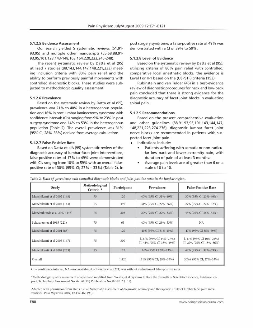
5.1.2.6 PrevalenceBased on the systematic review by Datta et al (95),
prevalence was 21% to 40% in a heterogenous popula-tion and 16% in post lumbar laminectomy syndrome with confidence intervals (CIs) ranging from 9% to 23% in post surgery syndrome and 14% to 53% in the heterogenous population (Table 2). The overall prevalence was 31% (95% CI; 28%–33%) derived from average calculations.
5.1.2.7 False-Positive RateBased on Datta et al’s (95) systematic review of the
diagnostic accuracy of lumbar facet joint interventions, false-positive rates of 17% to 49% were demonstrated with CIs ranging from 10% to 59% with an overall false-positive rate of 30% (95% CI; 27% – 33%) (Table 2). In
post surgery syndrome, a false-positive rate of 49% was demonstrated with a CI of 39% to 59%.
5.1.2.8 Level of EvidenceBased on the systematic review by Datta et al (95),
utilizing criteria of 80% pain relief with controlled, comparative local anesthetic blocks, the evidence is Level I or II-1 based on the (USPSTF) criteria (153).
Rubinstein and van Tulder (46) in a best-evidence review of diagnostic procedures for neck and low-back pain concluded that there is strong evidence for the diagnostic accuracy of facet joint blocks in evaluating spinal pain.
5.1.2.9 RecommendationsBased on the present comprehensive evaluation
and other guidelines (88,91-93,95,101,143,144,147, 148,221,223,274-276), diagnostic lumbar facet joint nerve blocks are recommended in patients with sus-pected facet joint pain.♦ Indications include:
• Patients suffering with somatic or non-radicu-lar low back and lower extremity pain, with duration of pain of at least 3 months.
• Average pain levels are of greater than 6 on a scale of 0 to 10.
Table 2. Data of prevalence with controlled diagnostic blocks and false-positive rates in the lumbar region.
StudyMethodological
Criteria *Participants Prevalence False-Positive Rate
Manchikanti et al 2002 (148) 75 120 40% (95% CI 31%–49%) 30% (95% CI 20%–40%)
Manchikanti et al 2004 (144) 75 397 31% (95% CI 27%–36%) 27% (95% CI 22%–32%)
Manchukonda et al 2007 (143) 75 303 27% (95% CI 22%–33%) 45% (95% CI 36%–53%)
Schwarzer et al 1995 (221) 75 63 40% (95% CI 29%–53%) NA
Manchikanti et al 2001 (88) 75 120 40% (95% CI 31%-49%) 47% (95% CI 35%-59%)
Manchikanti et al 2003 (147) 75 300 I. 21% (95% CI 14%–27%)II. 41% (95% CI 33%–49%)
I. 17% (95% CI 10%–24%)II. 27% (95% CI 18%–36%)
Manchikanti et al 2007 (233) 75 117 16% (95% CI 9%–23%) 49% (95% CI 39%–59%)
Overall 1,420 31% (95% CI; 28%–33%) 30%# (95% CI; 27%–33%)
CI = confidence interval; NA =not available; # Schwarzer et al (221) was without evaluation of false-positive rates.
*Methodologic quality assessment adapted and modified from West S, et al. Systems to Rate the Strength of Scientific Evidence, Evidence Re-port, Technology Assessment No. 47. AHRQ Publication No. 02-E016 (151).
Adapted with permission from Datta S et al. Systematic assessment of diagnostic accuracy and therapeutic utility of lumbar facet joint inter-ventions. Pain Physician 2009; 12:437-460 (95).

www.painphysicianjournal.com E81
Review of Neurophysiologic Basis and Diagnostic Interventions in Managing Spinal Pain
• Pain is at least intermittent or continuous causing functional disability.
• Condition has failed to respond to more con-servative management, including physical therapy modalities with exercises, chiroprac-tic management, and non-steroidal anti-in-flammatory agents.
• Lack of preponderance of evidence of either lumbar discogenic or sacroiliac joint pain and lack of lumbar disc herniation or evidence of radiculitis.
• No evidence of contraindications is present for the needle placement and injection of lo-cal anesthetics.
• Presence of contraindications or inability to undergo physical therapy, chiropractic man-agement, or inability to tolerate non-steroi-dal anti-inflammatory drugs.
♦ A positive response is based on the following evidence:• Patient has met the above indications.• Patient responds positively to controlled local
anesthetic blocks either with placebo control or comparative local anesthetic blocks with appropriate response to each local anesthetic of < 1 mL for each nerve or joint.
• At least 80% relief as criterion standard with the ability to perform previously painful movement without deterioration of the relief (i.e., extension, lateral rotation, flexion, etc.).
• The patient’s response should be recorded independently by an assessor – generally a registered nurse familiar with the patient or another physician.
5.2 Lumbar Intervertebral DiscThe human intervertebral disc is a unique struc-
ture with 3 major components, the nucleus pulposus (NP), annulus fibrosis (AF), and vertebral endplates (VE), and 2 major regions, the outer ring, the AF, and the inner part, the NP (277). The disc is attached to the adjacent vertebral bodies by the vertebral endplates centrally and the ligamentous attachments of the AF peripherally. These components form a joint-like struc-ture that allows for movements in the sagittal, hori-zontal, and coronal planes (278). The disc is supported posteriorly by 2 zygapophysial joints, the components of the “3-joint” structure.
The healthy human intervertebral disc is essential-ly avascular, with its nutrition being supplied through
the vertebral endplates and AF via diffusion. The nucleus itself has no blood supply. The annulus con-tains blood vessels only in its most superficial lamel-lae. Nutrients that pass through the endplates come from the arteries supplying the vertebral bodies. Any number of factors can contribute to a breakdown in the functional capacity of the disc, including inflam-matory mediators, changes in pH, and nutritional de-ficiencies (279-282). In fact, it was noted that vascular changes occurred before degeneration of the disc at every lumbar level, suggesting that disc disturbances in the nutritional supply may precede degeneration (283). In a study of 280 discs from L1/2 to L5/S1, 40 out of 280 discs (14.3%) demonstrated intravascular uptake utilizing real-time fluoroscopy (279). There was no statistical correlation between the degree of disc degeneration and the incidence of intravascular uptake.
Early studies failed to demonstrate nerve fibers or nerve endings within the discs (284,285). In subse-quent studies, it was reported that even though in a normal intervertebral disc, the NP is devoid of nerve fibers, the outer AF contains an extensive network of sensory nerve fibers (166,284,286-290). It has also been demonstrated that a variety of free and complex nerve endings were present in the outer third of the annulus (284,291-303). Nerve endings in degenerated discs have been found in the deeper layers of the AF; in some studies, nerve endings have been found ex-tending even into the NP (295-298,304). These nerve fibers transmit both nociceptive and non-nociceptive information (166,286,290,292,305-313).
5.2.1 Pathophysiology of Lumbar Disc-Related Pain
Kuslich et al (90) used progressive regional an-esthesia in 193 patients who were about to undergo lumbar decompressive surgery for disc herniation or spinal stenosis. Pain was reported by 30% of the pa-tients who had stimulation of the paracentral annulus and by 15% who had stimulation of the central annu-lus with blunt surgical instruments or through an elec-tric current of low voltage. Soon after the description of disc herniation in the American literature by Mixter and Barr (314) in 1934, Mixter and Ayers (315) in 1935 demonstrated that radicular pain can occur without disc herniation. This was followed by reports from nu-merous investigators describing pain syndromes ema-nating from the lumbar intervertebral disc without mechanically compressed neural structures (87,88,316-

Pain Physician: July/August 2009:12:E71-E121
E82 www.painphysicianjournal.com
330). There is no established and causal relationship between disc degeneration and spinal pain. However, the biochemical behaviors of the disc may explain the pathophysiology of discogenic pain (326). Painful discs have a lower pH than non-painful discs in humans (331). Discography on canine discs that are deformed normally and experimentally reveals an increase in concentrations of neuropeptides, i.e., substance P (SP) and vasoactive intestinal peptide (VIP) in the dor-sal root ganglion (327). Inflammatory factors may be responsible in some cases for which epidural steroid injections provide relief (253,254,332-339). Chemi-cal nociception is supported by numerous studies (277,306,340-383). Elevated levels of nitric oxide, pros-taglandin E2, interleukin (IL)-2, IL-6, IL-8, phospholi-pase A2, leukotriene B4, thromboxane B2, and tumor necrosis factor -α (TNF-α) in diseased intervertebral discs have been demonstrated. Thus, in combination, chemical and mechanical factors provide the explana-tion for disc-related pain (340-396). Mechanism also illustrates the role of dorsal root ganglion (397-411).
Internal disc disruption (IDD) is a condition in which the internal architecture of the disc is disrupted, but its external appearance remains essentially normal (328). IDD can be experimentally induced by endplate dam-age (396). Likewise, experimentally induced annular tears can lead to adverse and progressive mechanical changes in the disc. Annular degeneration has been shown to appear at an early age in lumbar discs and is clearly related to back pain (329). Disrupted discs may not exhibit either bulging or herniation. These features with a normal or near normal contour of discs produc-ing back pain, but with no evidence of herniation or prolapse, were described by Crock (328) in 1976 as IDD.
5.2.2 Diagnosis of Lumbar Discogenic PainDiagnostic tests, such as clinical history, physical
exam, and non-provocative imaging studies, have low sensitivity and specificity in diagnosing whether the disc is a source low back pain (51). Hancock et al (51), in a systematic review of tests to identify the disc as a pain generator, concluded that centralization was the only clinical feature found to increase the likelihood of the disc as the source of pain, based on review of mul-tiple studies (60,412-414). Certain findings on physical exam have been purported to aid in the diagnosis of the cause of lower back pain but these have been dif-ficult to confirm by scientific inquiry (36,48,49,415).
MRI scans and radiologic images of discography are both sensitive for diagnosing the presence of de-
generative disc disease (DDD) (36,88,317,416-422). However, it has been demonstrated that these changes are present in patients asymptomatic of low back pain in as many as 64% to 89% (322,325-329,416,423-428). Hancock et al (51) described that among the various features observed on the MRI, absence of degenera-tion was the only test found to reduce the likelihood of the disc as the source of the pain.
Conversely, there is evidence that subtle but pain-ful lesions may be present in discs that appear to be morphologically normal on MRI scans. Discography has been shown to reveal abnormalities in symptom-atic patients with normal MRI scans (417-432).
Therefore, the detection of morphologic abnor-malities consistent with degeneration or the lack there-of is becoming increasingly less relevant to therapeutic decision-making. This phenomenon has also validated the importance of lumbar discography as a diagnostic tool to aid in therapeutic decision making. The provo-cation of pain with real time imaging as an indicator of the presence of discogenic pain is the current rai-son d’être for performing discography. Appropriately performed, with care taken to optimize the accuracy of the patient’s response, discography is considered to enhance the sensitivity and specificity compared to non-provocative imaging. This in turn can improve clin-ical outcomes and prognostication through appropri-ate decision-making and proper selection of therapies. Just as importantly, it reduces the risk of inappropriate treatment of discs that are not the source of pain.
5.2.2.1 Lumbar Provocation DiscographyDiscography is a procedure that is used to charac-
terize the pathoanatomy/architecture of the interver-tebral disc and to determine if the intervertebral disc is a source of chronic spinal pain. Implicitly, discogra-phy is an invasive diagnostic test that should only be applied to those chronic spinal pain patients in whom one suspects a discogenic etiology. Discography lit-erally means the opacification of the NP of an inter-vertebral disc to render it visible under radiographs (422,433,434).
5.2.2.1.1 RationaleFormal studies have shown that the discs are in-
nervated and can be a source of pain that has patho-morphologic correlates (166,284-313,314,315,318,325-329,357,359,392,393,435-440). Even though the specific neurobiological events involved in how dis-cography causes pain have not been elucidated, sound

www.painphysicianjournal.com E83
Review of Neurophysiologic Basis and Diagnostic Interventions in Managing Spinal Pain
anatomic, histopathological, radiological, and biome-chanical evidence suggests that lumbar discography may help to identify symptomatic and pathological intervertebral discs (97,98,416-418).
5.2.2.1.2 ValidityExaminations of cadaver discs typically confirm
the presence of annular tears and disc degeneration, as revealed by discograms (441-444). Multiple authors also have investigated the accuracy of lumbar disco-graphic and CT/discographic findings based on the ability to demonstrate accurate pathology confirmed at the time of surgery. There is a high inter- and in-tra-observer agreement in assessing discographic mor-phology, i.e., the Adams classification (419,441,445). It was reported that the exact reproduction of pain was more likely in ruptured or fissured discs and less likely in degenerative discs, based on the Adams classifica-tion (441).
Lumbar discography was compared with myelog-raphy, CT, MRI, and results of surgical and conserva-tive management. CT discography was reported to be more accurate than myelography (420,441,446-451). On similar grounds, discography was shown to be superior to plain CT (421,451-454). While compar-ing the results of lumbar discography with MRI, some found discography to be as good as MRI, even though MRI was preferable as it was non-invasive and al-lowed assessment of more levels with one test, with minimal risk of complications and minimal discomfort (455,456). However, others have identified advantag-es of discography with pain provocation, when MRIs were normal or equivocal (416,424,450). Strong cor-relation was demonstrated between MR/discography and CT/discography in assessing annular tears and de-generation of lumbar discs (454,457,458).
A good correlation between MRI, discography, and the high intensity zone (HIZ) has been estab-lished by some (457-463), while others have reported a poor correlation and limited value of discography (464-471).
Lei et al (467) correlated a new MRI classification of disc degeneration found to have good intra- and inter-observer agreement, with discography. The sen-sitivity and specificity of MRI in predicting a painful disc was 94% and 77%, which favorably compared to endplate signal changes and HIZs, which were found to have sensitivities of 32% and 27%, respectively. The authors concluded that an MRI is an excellent tool for assessing disc morphology, but should be used in
conjunction with discography for planning surgical treatment.
O’Neill et al (468) evaluated the accuracy of MRI in diagnosing discogenic pain in 143 patients, tak-ing into consideration the interdependence of MRI parameters. Moderate loss of nuclear signal and disc bulging had the best sensitivity (79.8%) and specificity (79.3%). Accounting for either moderate loss of disc height or the presence of a HIZ reduced sensitivity but improved specificity. Notably, the incorporation of a HIZ reduced sensitivity (73.6%) and improved specific-ity (92.6%).
Scuderi et al (469) prospectively conducted a bio-chemical analysis of disc leakage fluid obtained dur-ing discography. They found only weak correlations between demographic variables, Pfirrman grading (MRI), and discography. The authors concluded that pain provocation during discography cannot be pre-dicted by non-invasive means, including biomarker assays.
Derincek et al (470) performed discography on a series of patients with back pain and MRI evidence of DDD. Those patients experiencing pain during injec-tion into a morphologically normal disc were studied. These individuals underwent repeat discograms on the morphologically normal disc, but the morphologi-cally abnormal (adjacent disc) was anesthetized. None of their patients experienced pain during the repeat discogram. The authors recommended anesthetizing the morphologically abnormal disc before testing po-tentially normal (control) discs.
The technique of lumbar discography is standard-ized by the International Association for the Study of Pain (IASP) criteria (433) and has been well stud-ied (97,98,472-477). The definition of a positive dis-cogram, per International Spine Intervention Society (ISIS) guidelines (434) is pain > 7/10, concordance, pres-sure ≤ 50 psi a.o, Grade III anular tear, and a painless control disc.
The greatest challenge concerning discography continues to be the gold standard problem. Three systematic reviews exhaustively discussed these is-sues (97,98,107). Treatment, particularly controversial treatments should not serve as the “gold standard” for a diagnostic test.
The sensitivity and specificity of intervertebral disc morphology are 81% and 64%, respectively. A recent meta-analysis of provocation discography in asymp-tomatic subjects obtained a specificity of 94% (95% CI; 89%–98%) and a false-positive rate of 6% (417).

Pain Physician: July/August 2009:12:E71-E121
E84 www.painphysicianjournal.com
5.2.2.1.3 Cost EffectivenessThere are no cost effectiveness studies of lumbar
provocation discography available in the literature.
5.2.2.1.4 Safety and Complications Complications related to discography include disci-
tis, subdural abscess, spinal cord injury, vascular injury, epidural and prevertebral abscess, annular strain, and toxicity of antibiotics (6,97,98,422,433,434,478-498).
5.2.2.1.5 Evidence AssessmentThe literature search provided 6 systematic re-
views (51,97,98,107,276,417). All of the systematic reviews met the inclusion criteria. Hancock et al (51) focused on the diagnostic criteria comparing discogra-phy with other tests. Wolfer et al (417) evaluated false-positive rates. Shah et al (98), Buenaventura et al (97), and Manchikanti et al (107,276) performed systematic assessments of the value of provocation discography utilizing West et al’s AHRQ criteria for systematic re-views. Manchikanti et al (107) utilized modified IASP criteria (433). For a disc to be judged positive, stimula-tion of the target disc produces concordant pain with an intensity of at least 6 on a 10-point pain measure-ment scale and 2 adjacent discs with provocation dis-cography do not produce any pain at all except for the L5-S1 disc wherein only one negative disc is required. Manchikanti et al (107) utilized 9 studies meeting strict inclusion criteria and considered all other stud-ies performed under controlled conditions. Wolfer et al (417) utilized multiple studies with methodologic quality evaluation and scoring of lumbar discographic studies in their evaluations.
Thus, the 2 latest systematic reviews by Manchikanti et al (107) and Wolfer et al (417) were utilized in the evidence synthesis for these guidelines.
5.2.2.1.6 Prevalence of Lumbar Discogenic PainPrevalence of pain due to IDD was reported to be
39% of patients suffering with chronic low back pain in the United States (317). In contrast, primary disco-genic pain was reported in 26% of patients suffering with chronic low back pain in the United States (88). Table 3 illustrates the data of prevalence of lumbar discogenic pain utilizing IASP criteria.
5.2.2.1.7 False-Positive RateA series of published studies specifically investi-
gated the potential false-positive rate of lumbar dis-cography (446,485,496-510). The Holt study (502) was performed on prisoners, with outdated techniques and noxious, irritating contrast dye (503). Wolfer et al (417) pooled all the available data (from 1968 to 2008) on asymptomatic volunteers without confound-ing factors (somatization disorder, chronic pain, or discectomy), illustrating that there were a total of 33 patients and 48 discs. The data showed a false-positive rate of 3.0% (1/33) per patient (95% CI; 0%–9%) and 2.1% (1/48) per disc (95% CI; 0%–6%), utilizing both the Carragee criteria and ISIS/IASP criteria, even when the provocation stimulus measured by intradiscal pres-sure is uncontrolled (417).
5.2.2.1.8 Level of EvidenceBased on the AHRQ (151) and USPSTF (153) cri-
teria, the indicated evidence is Level II-2 for lumbar discography.
5.2.2.1.9 RecommendationsThe recommendation for lumbar provocation dis-
cography must include appropriate indications with patients with low back pain to prove the diagnostic hypothesis of the discogenic pain specifically after ex-clusion of other sources of lumbar pain and identifica-tion of the disc that should be targeted for treatment, or to establish either that no disc or too many discs are symptomatic, in which case surgery may not be indicated.
Table 3. Data of prevalence of lumbar discogenic pain utilizing IASP criteria.
StudyMethodological Quality Scoring
Participants Prevalence
Schwarzer et al 1995 (317) 7092 consecutive patients with chronic low back pain and no history of previous lumbar surgery referred for discography
The diagnostic criteria for internal disc disruption were fully satisfied in 39% of the patients, most commonly at L5/S1 and L4/5.
Manchikanti et al 2001 (88) 70From a group of 120 patients with low back pain, 72 patients negative for facet joint pain underwent discography.
The prevalence of discogenic pain was es-tablished in 26% of total patient sample and 43% of patients negative for facet joint pain.

www.painphysicianjournal.com E85
Review of Neurophysiologic Basis and Diagnostic Interventions in Managing Spinal Pain
The discography should be performed utilizing ap-propriate criteria and results are considered positive only if the stimulation of the target disc produces con-cordant pain with an intensity of at least 7 on a 10-point pain measurement scale or reproduces at least 70% of the most severe pain the patient has experienced (i.e., 5 of 7) and 2 adjacent discs with low volume contrast injection with low pressure discography do not produce any pain at all.
5.2.3 Diagnosis of Lumbar Radiculitis In a systematic review of epidemiologic studies
and prevalence estimates, definitions of sciatica var-ied widely with a prevalence from different studies ranging from 1.2% to 43% (511). Sciatica can be pre-cisely diagnosed in the majority of cases with available technology using MRI, CT, and nerve conduction stud-ies. Diagnostic selective nerve blocks are utilized occa-sionally in patients with persistent pain when history, examination, imaging, electrophysiologic testing, and other precision diagnostic injections do not identify the pain generator.
5.2.3.1 Lumbar Transforaminal Epidural Injections or Selective Nerve Root Blocks
Transforaminal epidural injection (modern no-menclature) or a selective nerve root block (old no-menclature) consist of injection of contrast, local anes-thetic, or other substances around spinal nerves under fluoroscopy (6,105,512). They have been described as 2 separate and distinct techniques. However, over the years authors have used them interchangeably.
5.2.3.1.1 Rationale Lumbar transforaminal epidural or selective
nerve root blocks provide clinically useful information (275,513,514). The validity of provocative and analge-sic spinal injections was recognized as early as 1938 (513). The value of diagnostic, selective nerve root blocks in the preoperative evaluation of patients with negative or inconclusive imaging studies and clinical findings and in the diagnosis of the source of radic-ular pain when imaging studies suggested possible compression of several nerve roots has been reported (206,514-542).
5.2.3.1.2 Validity In a review of the use of transforaminal epidurals
for managing spinal disease, Young et al (542) con-cluded that as a tool for predicting surgical outcome,
epidural spinal injection was found to have a sensi-tivity between 65% and 100%, a specificity between 71% and 95%, and a positive predictive value as high as 95% for one year surgical outcome. Rubinstein and van Tulder (46) concluded that there was moderate evidence for transforaminal epidural injections. How-ever, North et al (535) showed that false-positive re-sults were common and specificity was low.
The face validity of lumbosacral selective nerve root blocks may be accomplished by providing the blockade under fluoroscopic visualization utilizing contrast and a small volume of local anesthetic and with provocative and/or analgesic response. However, Furman et al (543), in a quantitative evaluation of con-trast flow level and its selectivity during fluoroscopical-ly guided lumbosacral transforaminal epidural steroid injections, showed that 30% of the transforaminal in-jections performed were not selective for the specified root level with injection of 0.5 mL of contrast. In addi-tion, with injection of 1 mL of contrast, 67% of trans-foraminal injections performed were no longer selec-tive for the specified root level, with injection of 1.5 mL of contrast, 87% were not selective, whereas, with injection of 2.5 mL of contrast, 90% were not selective for the specified root level. They concluded that diag-nostic selective nerve root blocks limiting injectate to a single, ipsilateral segment level cannot be reliably considered diagnostically selective with volumes ex-ceeding 0.5 mL. Others also have described contrast flow patterns and intravascular injections (544-549) with inadvertent vascular injection ranging from 9% to 26%; intradiscal filling of contrast during a trans-foraminal epidural injection (550,551), dural puncture and subdural injection (552), and other techniques to increase safety have been reported (540,545). Due to a multitude of these factors, which may result in an incomplete block or a block without selectivity involv-ing more than one nerve root, face validity continues to be questioned.
The construct validity of selective nerve root blocks has not been established. As with facet joint block or sacroiliac joint blocks and provocative dis-cography, no standards have been established to eliminate false-positive responses with transforaminal epidural injections. However, true-positive responses may be secured by performing controlled blocks with placebo injections of normal saline. Comparative local anesthetic blocks that have been shown to be valid in the diagnosis of facet joint pain have not been stud-ied for transforaminal usage. The only study that com-

Pain Physician: July/August 2009:12:E71-E121
E86 www.painphysicianjournal.com
pared a short-acting local anesthetic (lidocaine) with a long-acting local anesthetic (bupivacaine) in selective nerve root blocks used 2 test blocks in a random order to test the validity of the block response (534). Howev-er, no differences in effect were found between lido-caine and bupivacaine. Further, multiple confounding factors of psychological issues and sedation have not been studied for selective nerve root blocks.
5.2.3.1.3 Cost EffectivenessCost effectiveness of diagnostic transforaminal
epidural injections or selective nerve root blocks has not been evaluated. However, the feasibility and cost-effectiveness of appropriately performed controlled comparative local anesthetic blocks have been de-scribed (87,249-251).
5.2.3.1.4 Safety and Complications Reported complications of transforaminal epidu-
ral injections are related to dural puncture, subdural injection, dorsal root ganglion (DRG) trauma, infec-tion, intravascular injection, air embolism, vascular trauma, particulate embolism, cerebral thrombosis, epidural hematoma, neural or spinal cord damage, and complications related to administration of steroids (253,254,267,543-567). Recent reports of paraplegia, vertebral artery dissection, neurological disorders, and death are concerning. A blunt needle has been recom-mended to avoid multiple complications ascribed to transforaminal epidurals (555-557,568).
5.2.3.1.5 Evidence AssessmentThe search showed 2 systematic reviews
(105,512) and multiple other publications (366,515-519,523,524,527,529-531,534,535). Datta et al (105) showed moderate evidence in the diagnosis of con-founding factors with selective nerve root blocks. The conclusion was also adapted by Rubinstein and van Tulder (46). Further, Young et al (542) also concluded that transforaminal epidural injections have been found to have a sensitivity between 65% and 100%, a specificity between 71% and 95%, and a positive predictive value as high as 95% for one year surgical outcome.
5.2.3.1.6 Level of Evidence Based on multiple evaluations, the indicated level
of evidence for lumbar transforaminal epidural injec-tions or selective nerve root blocks is II-3 based on USPSTF criteria (153).
5.2.3.1.7 RecommendationsThe diagnostic lumbosacral selective nerve root
block must be performed utilizing fluoroscopy and contrast with a small dose (1.0 mL or less, preferably 0.5 mL) of local anesthetic injection with assessment of appropriate pain relief (≥ 80%) and the ability to perform previously painful movements. Lumbosacral nerve root blocks may be performed for diagnostic purposes prior to therapeutic injection and/or if it is indicated for diagnostic purposes in patients with clinical features which do not implicate a particular, single spinal nerve as responsible for the symptoms and when imaging suggests that symptoms could be arising from more than one particular segment if sur-gery is contemplated.
5.3 Sacroiliac JointThe sacroiliac joint is accepted as a potential source
of low back and/or buttock pain with or without low-er extremity pain (88,102-104,569-576). The sacroiliac joint receives innervation from the lumbosacral nerve roots (577-587). Neurophysiological studies have dem-onstrated both nociceptive and proprioceptive afferent units in the sacroiliac joint (584,585,588,589). Referral patterns based on sacroiliac joint provocation and an-algesic response to local anesthetics in asymptomatic volunteers (590) and patients with pain (591,592) have been published.
5.3.1 Diagnosis of Sacroiliac Joint PainIn a systematic review evaluating a battery of
tests to identify the disc, sacroiliac joint, or facet joint as the source of low back pain, Hancock et al (51) suggested that a combination of sacroiliac joint pain provocative maneuvers appears to be useful in pin-pointing the sacroiliac joint as the principal source of symptoms in patients with pain below the fifth lumbar vertebra. They also concluded that although a positive bone scan has high specificity, it is associ-ated with a very low sensitivity, which means that the majority of patients with the sacroiliac joint pain will not be accurately identified.
In a systematic review by Szadek et al (593), the authors evaluated the diagnostic validity of the IASP criteria for sacroiliac joint pain. The meta-analysis showed that the thigh thrust test, the compression test, and 3 or more positive stressing tests contain suf-ficient discriminative power for diagnosing sacroiliac joint pain. They concluded that in view of the lack of a gold standard for sacroiliac joint pain, the diagnostic
www.painphysicianjournal.com E87
Review of Neurophysiologic Basis and Diagnostic Interventions in Managing Spinal Pain
validity of tests for sacroiliac joint pain should be re-garded with caution.
Song et al’s (594) systematic review evaluating the diagnostic value of scintigraphy in assessing sac-roiliitis and ankylosing spondylitis concluded that scintigraphy is at best of limited value in establish-ing a diagnosis of ankylosing spondylitis. Three sys-tematic reviews evaluated the role of diagnostic intraarticular injections (102-104) in establishing the sacroiliac joint(s) as the primary pain generator. In a best-evidence review of diagnostic procedures for neck and low back pain that focused on previously published systematic reviews (51,102,103), Rubin-stein and van Tulder (46) also concluded that there was moderate evidence for diagnostic sacroiliac joint blocks. Rupert et al (104) in the recent systematic re-view concluded the indicated evidence for validity of diagnostic sacroiliac joint injections is Level II-2 based on the USPSTF criteria (153).
5.3.2 Sacroiliac Joint Blocks
5.3.2.1 RationaleDue to the inability to make the diagnosis of
sacroiliac joint pain with non-invasive tests, sac-roiliac joint blocks appear to be the evaluation of choice to provide appropriate diagnosis. Further, controlled studies have established the sacroiliac joints as a potential source of low back and lower extremity pain (6,88,102-104,570-577). Based on con-trolled diagnostic blocks, the sacroiliac joint has been implicated as the primary source of pain in 10% to 38% of patients with suspected sacroiliac joint pain (6,88,102-104,571,572,594,595).
5.3.2.2 ValidityThe face validity of the sacroiliac joint blocks
has been established by injecting small volumes of local anesthetic with contrast into the joint. Con-struct validity of sacroiliac joint blocks has been established by determining the false-positive rate of 20% to 54% with single, uncontrolled, sacroiliac joint injections (88,571,572,596). False-positive re-sponses may occur with the extravasation of an an-esthetic agent out of the joint due to defects in the joint capsule (596). False-negative results may occur from faulty needle placement, intravascular injec-tion, or the inability of the local anesthetic to reach the painful portion of the joint due to loculations (102-104,574,576,597-602).
5.3.2.3 Cost Effectiveness There are no studies evaluating the cost effective-
ness of diagnostic sacroiliac joint blocks.
5.3.2.4 Safety and ComplicationsComplications of sacroiliac joint injection include
infection, trauma to the sciatic nerve, embolic phe-nomena, and complications related to drug adminis-tration (6,102-104).
5.3.2.5 Evidence Assessment Rupert et al (104) provided the latest evidence
with inclusion of 5 studies which met methodologic quality assessment (88,571,572,595,596).
5.3.2.6 PrevalenceThe prevalence of sacroiliac joint pain is estimated
to range between 10% and 38% with 95% CIs of 0% – 5% (88,571,572,595,596) (Table 4).
5.3.2.7 False-Positive RateThe false-positive rate of a single block is esti-
mated to range between 20% and 54% with 95% CIs of 3% – 64% (88,571,572,596). However, in one study (595) the false-positive rate was 0%.
5.3.2.8 Level of EvidenceThe indicated evidence for the accuracy of sac-
roiliac joint diagnostic injections is Level II-2 for the diagnosis of sacroiliac joint pain utilizing controlled diagnostic blocks.
5.3.2.9 RecommendationsControlled sacroiliac joint blocks with placebo or
controlled comparative local anesthetic blocks are rec-ommended when indications are satisfied. A positive response is considered ≥ 80% relief with the ability to perform previously painful movements.
The primary indication for sacroiliac joint blocks is the need to know if a patient’s pain is arising from the sacroiliac joint or not. Key indicators would be pa-tients with chronic low back pain that is maximal be-low the level of L5 vertebra, with or without somatic referred pain in the lower limb, in whom no other di-agnosis is readily apparent, in whom no other possible diagnosis is more likely, in whom a diagnosis has been made or cannot be made using less invasive options, whose pain is not evolving with the passage of time or conservative therapy, and fails to respond to conserva-tive therapy.

Pain Physician: July/August 2009:12:E71-E121
E88 www.painphysicianjournal.com
5.4 Other Causes of Low Back Pain
5.4.1 Post Lumbar Surgery SyndromePost surgery lumbar syndrome and other syn-
onyms, such as failed back surgery syndrome (FBSS) or post lumbar laminectomy syndrome, represent a cluster of syndromes following spine surgery where-in the expectations of the patient and spine surgeon are not met (603-612). In fact, FBSS is considered to be the major disadvantage of surgical intervention on the lumbar spine with the addition of 80,000 or so patients a year with continued chronic disabling back pain (613). In this review (613), it has been pointed out that there were at least 392,000 surgeries to treat low back pain in 2000 and the rate of spine surgery has continued to rise since then. The study conclud-ed that best estimates suggest that although 60% or more of initial back surgeries have a successful out-come, many are not successful. They noted that the long-term reoperation rate after spine surgery is high —19% over 11 years and that a substantial proportion of patients end up with chronic disabling symptoms (614). Based on these numbers, they have estimated
that there may be over 80,000 failed back surgeries per year. They also described that surgical success rates dropped to roughly 30% after a second surgery, 15% after a third, and 5% after a fourth (615). Carragee et al (616) noted that many interventions are performed for axial back pain associated with common degenera-tive conditions, sometimes with weak or absent evi-dence of efficacy.
Animal models of post laminectomy syndrome demonstrate paraspinous muscle spasms, tail con-tractures, pain behaviors, tactile allodynia, epidural and perineural scarring, and nerve root adherence to the underlying disc and pedicle (617-623). Specu-lated causes of postlaminectomy syndrome include acquired stenosis, adjacent segment degeneration, IDD, recurrent disc herniation, retained disc fragment, spondylolisthesis, epidural or intraneural fibrosis, de-generative disc disease, radiculopathy, radicular pain, deconditioning, facet joint pain, sacroiliac joint pain, discitis, arachnoiditis, pseudoarthrosis, segmental in-stability, and others (233,607-611,624-635). Ultimately, many of these etiologies are interrelated. Facet joint
CI = confidence interval
Methodological criteria and scoring adapted from West S et al. Systems to Rate the Strength of Scientific Evidence, Evidence Report, Technology Assessment No. 47. AHRQ Publication No. E016 (151).
Adapted from Rupert MP et al. Evaluation of sacroiliac joint interventions: A systematic appraisal of the literature. Pain Physician 2009; 12:399-418 (104).
Table 4. Data of prevalence of sacroiliac joint pain based on controlled diagnostic blocks.
StudyMethodologic
Quality Assessment Score
# of Subjects Prevalence Estimates False-Positive Rate
Manchikanti et al (88) 65 20 10% (95% CI, 0% – 23% ) 22% (95% CI, 3% – 42%)
Maigne et al (571) 65 54 18.5% (95% CI, 8% – 29%) 20% (95% CI, 8% – 33%)
Irwin e et al (572) 65 158 26.6% (95% CI, 20% – 34%) 53.8% (95% CI, 43% – 64%)
Laslett et al (595) 65 43/48 25.6% (95% CI, 12% – 39%) 0%
van der Wurff et al (596) 65 60 38% (95% CI, 26% – 51%) 21% (95% CI, 7% – 35%)

www.painphysicianjournal.com E89
Review of Neurophysiologic Basis and Diagnostic Interventions in Managing Spinal Pain
involvement in chronic pain following lumbar surgery has been shown to be present in approximately 8% to 16% of the patients (233). The prevalence of sacro-iliac joint pain following lumbar fusion has been dem-onstrated in a study using a single block to be 35% (578).
Epidural fibrosis may occur following an annular tear, disc herniation, hematoma, infection, surgical trauma, vascular abnormalities, or intrathecal con-trast media (277,617-623,633-644). Epidural fibrosis may account for as much as 20% to 36% of all cases of FBSS (624,625,645,646). There may be a final com-mon pathway with all these etiologies, which results in peripheral and central facilitation potentiated by inflammatory and nerve injury mechanisms (617-623). Paraspinal muscles may also become denervated and involved in the pathogenesis of FBSS (647).
5.4.2 Spinal StenosisSpinal stenosis can be defined as a narrowing
of the spinal canal, resulting in symptoms and signs caused by entrapment and compression of the intra-spinal vascular and nervous structures (648,649). Disc bulging, protrusion, and herniation in the cervical, as well as lumbar area, combined with osteophytes and arthritic changes of the facet joints can cause a nar-rowing of the spinal canal, encroachment on the con-tents of the dural sac, or localized nerve root canal ste-nosis (648-654). The pain and disability associated with lumbar spinal stenosis can interfere with a patient’s lifestyle (655).
A systematic review of the accuracy of diagnostic tests in lumbar spinal stenosis (656) was performed by de Graaf et al. After a comprehensive search, 24 articles were included in the review with 15 concern-ing imaging tests, 7 evaluating clinical tests, and 2 studies reporting on other diagnostic tests. The re-sults showed that the overall quality was poor; only 5 studies scored positive on more than 50% of the quality items. Estimates of the diagnostic value of the tests differed considerably. The imaging stud-ies showed no superior accuracy between myelog-raphy, CT, or MRI. Overall, there was considerable variation in the clinical tests; some studies showing high sensitivity and others showing high specificity. They concluded that because of the heterogeneity and overall poor quality, no firm conclusions about the diagnostic performance of the different tests can be drawn.
6.0 Neck paiN
Cervical intervertebral discs and facet joints, at-lanto-axial and atlanto-occipital joints, ligaments, fas-cia, muscles, and nerve root dura have been shown to be capable of transmitting pain in the cervical spine with resulting symptoms of neck pain, upper extrem-ity pain, and headache. However, very little is known about the causes of neck pain since the epidemiologic studies do not describe either the source or cause of the pain. Yin and Bogduk (89) demonstrated the prev-alence of discogenic pain in 16%, zygapophysial joint pain in 42%, and lateral atlanto-axial joint pain in 9%, in 143 patients with chronic neck pain in a private practice pain clinic in the United States. Consequently, a diagnosis remained elusive in over 40% of those pa-tients who completed investigations.
6.1 Cervical Facet or Zygapophysial JointsCervical facet or zygapophysial joints have been
shown to be a source of pain in the neck and referred pain in the head and upper extremities (89,143,144, 148,149,150,657-665). Cervical facet joints are in-nervated by the medial branches of the dorsal rami (165,666-669). Neuroanatomic studies have demon-strated free and encapsulated nerve endings in facet joints, as well as nerves containing SP and calcitonin gene-related peptide (165,670). Neurophysiologic studies have shown that cervical facet joint capsules contain low-threshold mechanoreceptors, mechani-cally sensitive nociceptors, and silent nociceptors (176,671-676).
Inflammation leads to decreased thresholds of nerve endings in facet capsules as well as elevated base-line discharge rates (165). Biomechanical studies have shown that cervical facet joint capsules can undergo high strains during spine-loading (165,677-685).
6.1.1 Cervical Facet or Zygapophysial Joint Blocks Diagnostic blocks of a cervical facet or zygapoph-
ysial joint can be performed by anesthetizing the joint by intraarticular injections of local anesthetic or by medial branch blocks. Valid information is only ob-tained by performing controlled blocks.
6.1.1.1 RationaleThe rationale for using cervical facet joint blocks
for diagnosis is based on the fact that facet joints are capable of causing pain, have a nerve supply (165,666-669), and have been shown to be a source of pain in

Pain Physician: July/August 2009:12:E71-E121
E90 www.painphysicianjournal.com
patients using diagnostic techniques of known reliabil-ity and validity (89,143,144,148,149,245,247). Conven-tional clinical and radiologic techniques are unreliable in diagnosing cervical facet or zygapophysial joint pain. In addition, the value, validity, and clinical effectiveness of cervical diagnostic facet joint nerve blocks was illus-trated by application of therapeutic modalities based on the diagnosis with controlled comparative local an-esthetic blocks (6,91-93,96,101,119,121,686-694).
6.1.1.2 ValidityControlled diagnostic blocks of cervical facet
joints with 2 local anesthetics (or placebo-controlled) are the means of confirming the diagnosis of facet joint pain. The face validity of cervical medial branch blocks has been established by injecting small vol-umes of local anesthetic and contrast material onto the target points for these structures and by deter-mining the spread of contrast medium in postero-anterior and lateral radiographs (668). Construct validity of cervical facet joint blocks to eliminate pla-cebo effect as the source of confounding results and to secure true-positive results has been illustrated (54,115,117,145,146,149,150,657,658,660).
The validity of comparative local anesthetic blocks was determined not only by short-term relief with controlled diagnostic blocks and the ability to perform movements which were painful prior to the blocks, but also with the application of another appropriate reference standard (long-term follow-up) as described in the literature (119,121,235,239,276,686-696). Po-tential and real confounding factors were assessed in several studies (243,245-247,657,697,698). Influence of age, surgery, and psychopathology were evaluated in 3 reports and found not to have significant impact on the prevalence of cervical facet joint related chronic neck pain (245,247,657).
6.1.1.3 Cost EffectivenessDiagnostic cervical facet joint nerve blocks were
not evaluated for cost effectiveness systematically. However, multiple authors (88,250,251) described the feasibility and cost-effectiveness of appropriately performed controlled comparative local anesthetic blocks.
6.1.1.4 Safety and ComplicationsThe safety of cervical facet joint interventions
with intraarticular injections and medial branch blocks has been demonstrated. Though rare and mi-
nor, the common reported complications of cervi-cal facet joint injections or nerve blocks are related to needle placement and drug administration. These complications include hemorrhage, dural puncture, spinal cord trauma, infection, intraarterial or intra-venous injection, chemical meningitis, neural trauma, paralysis, pneumothorax, radiation exposure, facet capsule rupture, hematoma formation, steroid side ef-fects, and epidural, subdural, or subarachnoid spread (6,91-93,96,101,239,255,256,267,270-272).
6.1.1.5 Evidence Assessment Our search yielded 4 systematic reviews (91-93,96).
The recent systematic review by Falco et al (96) uti-lized 9 studies (89,143,144,148-150,657,659,660) meet-ing methodologic quality assessment inclusion criteria with 80% pain relief and the ability to perform previ-ously painful movements with controlled diagnostic blocks.
6.1.1.6 PrevalenceBased on the systematic review by Falco et al (96)
which included controlled diagnostic blocks with a cri-terion standard of 80% or greater relief and included 9 studies (89,143,144,148-150,657,659,660) meeting methodologic quality criteria. The prevalence was es-timated as 36% to 54% with CIs ranging from 27% to 75% in patients in a heterogenous population with an average of 49% with 95% CI of 45% to 52%. In addi-tion, the prevalence was shown to be 36% with 95% CI of 22% to 51% in patients after surgical interven-tion (Table 5).
6.1.1.7 False-Positive Rate Based on the systematic review by Falco et al
(96), utilizing strict criteria with controlled diagnostic blocks and methodologic quality assessment, false-positive rates with a single block are 27% to 63% with CIs ranging from 15% to 78% with an average of 49% with 95% CI of 44% to 54% (Table 5).
6.1.1.8 Level of EvidenceBased on the systematic review by Falco et al (96),
the evidence for diagnosis of cervical facet joint pain is Level I or II-1 based on the USPSTF criteria (151).
6.1.1.9 Recommendations Based on the present comprehensive evaluation
and other described evaluations (91-93,96,101,699, 700), diagnostic cervical facet joint nerve blocks are
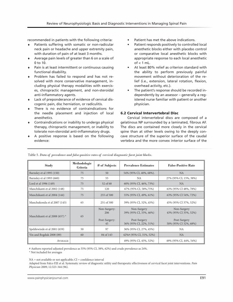
www.painphysicianjournal.com E91
Review of Neurophysiologic Basis and Diagnostic Interventions in Managing Spinal Pain
recommended in patients with the following criteria:♦ Patients suffering with somatic or non-radicular
neck pain or headache and upper extremity pain, with duration of pain of at least 3 months.
♦ Average pain levels of greater than 6 on a scale of 0 to 10.
♦ Pain is at least intermittent or continuous causing functional disability.
♦ Problem has failed to respond and has not re-solved with more conservative management, in-cluding physical therapy modalities with exercis-es, chiropractic management, and non-steroidal anti-inflammatory agents.
♦ Lack of preponderance of evidence of cervical dis-cogenic pain, disc herniation, or radiculitis.
♦ There is no evidence of contraindications for the needle placement and injection of local anesthetics.
♦ Contraindications or inability to undergo physical therapy, chiropractic management, or inability to tolerate non-steroidal anti-inflammatory drugs.
♦ A positive response is based on the following evidence:
• Patient has met the above indications.• Patient responds positively to controlled local
anesthetic blocks either with placebo control or comparative local anesthetic blocks with appropriate response to each local anesthetic of < 1 mL.
• At least 80% relief as criterion standard with the ability to perform previously painful movement without deterioration of the re-lief (i.e., extension, lateral rotation, flexion, overhead activity, etc.).
• The patient’s response should be recorded in-dependently by an assessor – generally a reg-istered nurse familiar with patient or another physician.
6.2 Cervical Intervertebral DiscCervical intervertebral discs are composed of a
gelatinous NP surrounded by a laminated, fibrous AF. The discs are contained more closely in the cervical spine than at other levels owing to the deeply con-cave structure of the superior surface of the caudal vertebra and the more convex interior surface of the
Table 5. Data of prevalence and false-positive rates of cervical diagnostic facet joint blocks.
StudyMethodologic
Criteria# of Subjects Prevalence Estimates False-Positive Rate
Barnsley et al 1995 (150) 75 50 54% (95% CI, 40%, 68%). NA
Barnsley et al 1993 (660) 75 55 NA 27% (95% CI, 15%, 38%)
Lord et al 1996 (149) 75 52 of 68 60% (95% CI, 46%, 73%) NA
Manchikanti et al 2002 (148) 75 120 67% (95% CI, 58%,75%) 63% (95% CI 48%, 78%)
Manchikanti et al 2004 (144) 75 255 of 500 55% (95% CI, 49%, 61%) 63% (95% CI 54%, 72%)
Manchukonda et al 2007 (143) 65 251 of 500 39% (95% CI, 32%, 45%) 45% (95% CI 37%, 52%)
Manchikanti et al 2008 (657) * 65
Non-Surgery:206
Post-Surgery:45
Non-Surgery 39% (95% CI, 33%, 46%)
Post-Surgery36% (95% CI, 22%, 51%)
Non-Surgery43% (95% CI 35%, 52%)
Post-Surgery50% (95% CI 32%, 68%)
Speldewinde et al 2001 (659) 50 97 36% (95% CI, 27%, 45%) NA
Yin and Bogduk 2008 (89) 60 84 of 143 42%# (95% CI, 31%, 52%) NA
Average 49% (95% CI, 45%, 52%) 49% (95% CI, 44%, 54%)
# Authors reported adjusted prevalence as 55% (95% CI, 38%, 62%) and crude prevalence as 24%.* Not included for averages
NA = not available or not applicable; CI = confidence intervalAdapted from Falco FJE et al. Systematic review of diagnostic utility and therapeutic effectiveness of cervical facet joint interventions. Pain Physician 2009; 12:323-344 (96).

Pain Physician: July/August 2009:12:E71-E121
E92 www.painphysicianjournal.com
rostral vertebra. The AF forms the outer boundary of each disc. In the cervical spine, the discs are thicker an-teriorly than posteriorly and are entirely responsible for the normal cervical lordosis. They do not conform completely to the surfaces of the vertebral bodies with which they are connected, being slightly smaller in width than the vertebral bodies. The discs bulge an-teriorly beyond the adjacent vertebrae. The NP in the cervical spine is located more anteriorly than in other portions of the spine (701).
It was controversial whether or not the cervical in-tervertebral discs received innervation. Cloward (702) stimulated cervical discs mechanically and electrically to verify that evoked pain originated in the discs them-selves, rather than from irritation of adjacent struc-tures. Cloward also proposed that disc pain is mediated through sinuvertebral nerves, which in the cervical re-gion are very small and undetectable by conventional dissection methods. However, subsequent anatomical studies did visually identify cervical sinuvertebral nerves and confirmed Cloward’s experimental observations and inferences. Now it is believed that intervertebral disc innervation in the cervical spine is analogous to that in the lumbar spine, with cervical discs receiving innervation posteriorly from the sinovertebral nerves, laterally from the vertebral nerve, and anteriorly from the sympathetic trunks (702-705).
6.2.1 Pathophysiology of Cervical Disc-Related PainIntervertebral disc-related pain can be caused by
structural abnormalities, such as disc degeneration or disc herniation; correspondingly, biochemical ef-fects such as inflammation (706) can also be the cause. The incidence of cervical disc herniation is less com-mon than lumbar disc herniations (707-712). Clearly, the mechanical compression on the nerve root that is being irritated by the herniated disc material is an important factor in the production of neck and up-per extremity pain. However, the mechanical, chemi-cal, and inflammatory components produce ischemic neuropathy from alteration of blood flow patterns or defects in the neuronal transport mechanism of the nerve root itself (713). Radicular pain may occur in the absence of nerve root compression secondary to NP extrusion or inflammatory reaction to the chemicals (714,715).
Okada et al (707) showed progressive degen-eration of cervical spine on MRI in over 81% of the patients during a 10-year period, with development of symptoms in 34% of subjects. Consequently, they
concluded that aging of the cervical spine inevitably occurs in everyone. Advances in basic research on disc degeneration have revealed its possible mechanism including a decrease in proteoglycan contents and water concentration (708), involvement of inflamma-tory cytokines such as IL-1 (714) and iTNF-α (368), and some genetic factors (715). The cervical intervertebral disc is one of the tissues subject to the early aging pro-cess, starting as early as 20 years of age, and is often a source of cervical spinal disorders causing neck pain and related symptoms.
6.2.2 Diagnosis of Cervical Discogenic Pain Imaging studies such as radiographs, myelography,
CT, CT-myelography, and MRI are incapable of identify-ing a degenerated disc as painful (97,98,716-718). The referral patterns can only be used to suggest which seg-ment is most likely to be the source of pain and, there-fore the levels at which the investigation should focus (719). In addition, multiple studies have demonstrated that age-related changes of the cervical spine are wide-ly present in asymptomatic healthy subjects (720-722). MRI findings of degenerative discs were recognized frequently in subjects 40 years and older despite the absence of symptoms on MRI (720). In another study, some abnormal findings were recognized in 62% of the subjects 40 years and older, whereas abnormal findings were rare in those younger than 40 years (721).
6.2.2.1 Cervical Provocation DiscographyCervical provocation discography, an image-guid-
ed procedure in which a contrast agent is injected into the NP of the intervertebral disc, includes disc stimula-tion and morphological assessment. It is intended to both identify a painful cervical intervertebral disc and depict internal derangements (716,719,723,724).
6.2.2.1.1 Rationale Imaging studies such as radiographs, myelogra-
phy, CT, CT-myelography, and MRI are incapable of identifying a degenerated disc as painful (97,98,717-719,723-729). Thus, cervical provocation discography is the test which can diagnose discogenic pain without disc herniation and radiculitis.
Over 50 years ago, Smith and Nichols (730,731) em-phasized pain reproduction as the principal feature of cervical discography. Cloward (702,703,732) described 2 types of pain during cervical disc stimulation: pain arising from IDD (i.e., discogenic pain) and neurogenic pain that stems from a herniated disc fragment caus-

www.painphysicianjournal.com E93
Review of Neurophysiologic Basis and Diagnostic Interventions in Managing Spinal Pain
ing nerve root or dural irritation.
6.2.2.1.2 Validity In a report published in 1964, Holt (733) ques-
tioned the validity and role of cervical discography, citing a high false-positive rate in asymptomatic sub-jects. He based this assumption on the contention that fissures and pain provocation were normal features in people without neck pain. Klafta and Collis (734,735) also found that cervical discography was less accurate than myelography in predicting surgical findings.
Studies conducted in cadavers and patients have re-examined Holt’s conclusions (717,736-741). These studies have established fissures to be normal age-re-lated findings that do not necessarily indicate symp-tomatology, and that demonstrating them with discog-raphy is immaterial (719,738). Supporting this assertion, Schellhas et al (717) found that pressurizing normal discs failed to provoke pain in both symptomatic and asymptomatic patients, whereas abnormal discs tended to produce concordant pain. Roth (739) and Kofoed (740) proposed the concept of analgesic discography.
The major obstacle confronting proponents of cervical discography is the lack of consensus as to what constitutes a positive response. Widespread vari-ations in criteria exist not only for pain provocation (i.e., designation of concordance and threshold for a positive response), but also for morphological clas-sification. While some investigators have interpreted certain patterns of contrast dispersion as being indica-tive of disc pathology, others have found a lack of cor-relation between morphology and pain reproduction (717,719,723,724,730,731,735-738,741-743).
Multiple questions have been raised regarding the utility of cervical discography, including the high report-ed false-positive rate; the lack of standardization; the discrepancies regarding the need for “control levels,” pain concordance, and pain intensity threshold; and uti-lization (19,97,98,100,716,719,723,724,729,744,745).
Validity is exemplified by disc stimulation symptom mapping (98-100,717,746) in pain patients and asymp-tomatic volunteers. Ohnmeiss et al (747) found a sig-nificant relationship between imaging and symptom provocation, with 86% of normal-looking discs either producing no pain (60%) or atypical pain (26%). Con-versely, 78% of disrupted discs were clinically painful on injection. Viikari-Juntura et al (748) demonstrated that discography provides additional information regarding structural changes not available by any other non-in-vasive and non-irradiative methods of examination. In general, nuclear signal changes observed on MRI in ca-
davers tended to underestimate the degree of patholo-gy appreciated with discography or gross examination. Parfenchuck and Janssen (741) found that while certain MRI patterns correlated well with positive and negative cervical discography responses, many other patterns re-vealed equivocal responses. They concluded that MRI is a useful adjunct to cervical discography, but that some MRI patterns should not be considered pathologic, and discography is necessary to identify a painful disc(s).
The proportion of cervical discs identified as symp-tomatic varies among studies. Grubb and Kelly (737) found that 50% of discs are capable of producing concordant pain upon injection. Schellhas et al (717) reported that among 11 discs that appeared normal on MRI in pain patients, 10 proved to have annular tears discographically. Two of these 10 elicited con-cordant pain with an intensity rating exceeding 6/10. Discographically normal discs (n = 8) were never pain-ful in either pain patients or an asymptomatic cohort, whereas intensely painful discs all exhibited tears of both the inner and outer annulus.
Hamasaki et al (749) retrospectively reviewed 15 cases of foraminal cervical disc herniations. Using MRI and CT-myelography, less than half of the cases were identified. In contrast, all were clearly noted on CT-dis-cography. These findings are similar to those found by Lejeune et al (750) in a study evaluating the diagnosis and outcomes for foraminal lumbar disc herniation. The authors concluded that a majority of foraminal-type cervical disc herniations may be overlooked with conventional MRI or CT-myelography, but correctly di-agnosed with CT discography.
Zheng et al (725) evaluated cervical discography re-sults at 161 disc levels. There were 79 positive levels, yield-ing a per disc prevalence rate of 49%. Fifty-nine percent of small herniated and torn discs were discographically positive. The false-positive rate of MRI was calculated to be 51% and the false-negative rate was 27%. The most important criterion for determining a symptomatic disc was moderate or severe reproduction of the patient’s typical pain. The presence of a control disc was not con-sidered a diagnostic criterion in this study.
Holt’s 1964 study (733) in asymptomatic prison-ers reflected negatively on cervical discography. But these studies (502,733) have been repeatedly refuted and better overriding data have since been generated (503). Holt utilized an irritant contrast and failed to employ fluoroscopic guidance. Even aside from these significant flaws, the technique itself was suspect. Ex-travasation of contrast material was noted with every injection, which continued even after reducing the

Pain Physician: July/August 2009:12:E71-E121
E94 www.painphysicianjournal.com
volume. Furthermore, Holt considered “pain provoca-tion” as being “without value.”
6.2.2.1.3 Cost EffectivenessThere are no cost effectiveness studies of provoca-
tive discography available in the literature.
6.2.2.1.4 Safety and ComplicationsComplications related to cervical discography
include discitis, subdural abscess, spinal cord injury, vascular injury, and epidural and prevertebral abscess (719,724,751-754).
6.2.2.1.5 Evidence SynthesisThree systematic reviews were identified evaluat-
ing cervical discography (98-100). Of these, the recent systematic review of cervical discography utilized 3 studies with methodologic quality scoring (89,742,755). This systematic review also included various outcome studies comparing surgical outcomes.
6.2.2.1.6 PrevalenceBased on IASP criteria (723), the data show a
prevalence rate ranging between 16% and 20% (89,742,755).
6.2.2.1.7 False-Positive RateThe main criticism regarding studies attempting
to quantify false-positive discography rates is that disc stimulation in asymptomatic volunteers may not reflect pain provocation in non-painful discs in sub-jects with spine pain (745). Moreover, the hallmark of a positive discogram has become concordant pain provocation, which is not possible in people devoid of spine symptoms. “False” pain provocation may be pro-duced in markedly degenerative discs in the lumbar spine, especially in the elderly (745,756-759). Cohen and Larkin et al (277,760) estimated that 15% to 25% of degenerative discs failed to elicit concordant pain during disc stimulation in the lumbar spine.
Overall, false-positive results with cervical provo-cation discography are a serious concern, with cited prevalence rates exceeding 50%. But these rates vary as a function of the diagnostic criteria. The results of studies requiring the presence of a control disc(s) shows a prevalence rate between 16% and 20% (89,742).
False-positive responses to disc stimulation can arise if the threshold for reproduction of pain is set too low. A disc is not necessarily the source of a patient’s pain if the pain that is reproduced is minor or trivial. Schellhas
et al (717) compared the responses to discography in asymptomatic volunteers and patients with neck pain. They found that the numerical rating pain score pro-duced by discography in asymptomatic subjects was sig-nificantly lower (P ≤ 0.0001) than in patients with neck pain. It was unusual for volunteers to report pain great-er than 5/10 and no asymptomatic subject experienced pain exceeding 6/10. Consequently, Schellhas et al (717) recommended adding an operational criterion where-by the patient must rate the intensity of produced pain as ≥ 7 on a 10-point numerical pain rating scale or an equivalent magnitude on another suitable scale. The emphasis then shifts from the baseline pain score to how intensely the patient rates the evoked pain. Bog-duk (719) pointed out that this criterion guards against diagnosing a moderately painful disc that could nev-ertheless be asymptomatic. The downside of this ar-gument is the intrinsic potential for contradictions. Theoretically, a functional patient with 10/10 baseline pain could be deemed “positive” if 7/10 pain is elicited (i.e., 70% of baseline pain was provoked), whereas a disabled patient with 4/10 pain in whom disc stimula-tion provokes 6/10 pain (i.e., 150% of baseline) would be designated as “negative.” Thus, Manchikanti et al (100) recommended as a criterion standard to evaluate reproduction of at least 70% of the most severe pain the patient has experienced (i.e., 5 of 7) and lack of pro-duction of pain at all in 2 adjacent discs.
6.2.2.1.8 Level of EvidenceBased on the systematic review (100) utilizing 3
studies in the performance of cervical discography (89,742,755), the indicated level of evidence is Level II-2 based on the modified USPSTF criteria (153).
6.2.2.1.9 RecommendationsBased on the systematic review (100), IASP criteria
(723), and ISIS criteria (719), the following recommen-dations are made:1) Cervical discography is indicated to test the diag-
nostic hypothesis of discogenic pain of the cervi-cal spine in individuals who have been properly selected and screened to eliminate other sources of cervical pain.
2) TThe discography should be performed utilizing ap-propriate criteria and results are considered positive only if the stimulation of the target disc produces concordant pain with an intensity of at least 7 on a 10-point pain measurement scale or reproduces at least 70% of the most severe pain the patient has

www.painphysicianjournal.com E95
Review of Neurophysiologic Basis and Diagnostic Interventions in Managing Spinal Pain
experienced (i.e., 5 of 7) and 2 adjacent discs with low volume contrast injection with low pressure dis-cography do not produce any pain at all.
7.0 thoracic paiN
The multiple structures which may be responsible for chronic thoracic pain include thoracic facet joints and intervertebral discs. Thoracic facet joints have been evaluated with controlled diagnostic techniques.
7.1 Thoracic Facet or Zygapophysial JointsSimilar to lumbar and cervical facet joints, thorac-
ic facet joints are paired diarthrodial articulations be-tween posterior elements of the adjacent vertebrae. The role of facet joints as a cause of chronic upper or mid back pain has received very little attention with only a few publications discussing these joints as the source of pain (761-763). However, the description of the involvement of thoracic facet joints as a cause of chronic mid back and upper back pain started in 1987 (764). Thoracic facet joint pain patterns were described in 1994 and 1997 (765,766).
Thoracic facet joints have been shown to have abundant nerve supply (37,91,172,173,177,765-773). The joints also have been shown to be capable of caus-ing pain similar to that seen clinically, in normal vol-unteers with persistent mid back and upper back pain and referred pain into the chest wall (765,766). The joints also have been shown to be affected by osteo-arthritis, rheumatoid arthritis, spondylitis, degenera-tion, inflammation, and injury leading to pain upon joint motion and restriction of motion (101,774).
7.1.1 Facet or Zygapophysial Joint BlocksDiagnostic blocks of thoracic facet or zygapophy-
sial joint provide valid information.
7.1.1.1 Rationale The rationale for using thoracic facet joint blocks
for diagnosis is based on the fact that facet joints are capable of causing pain and they have a nerve supply (101,765-773). They have been shown to be a source of pain in patients using diagnostic techniques of known reliability and validity (143,144,775). Conventional clinical and radiologic techniques are unreliable in di-agnosing thoracic facet joint pain. Various patterns of referred pain described for thoracic facet joints in the spine are similar to other structures such as discs. In addition, any maneuver to stress the facet joint is also likely to stress several other structures simultaneously,
especially the discs and muscles. The value, validity, and clinical effectiveness of
thoracic diagnostic facet joint nerve blocks was illus-trated by application of therapeutic modalities based on the diagnosis with controlled, comparative local anesthetic blocks (94,121,125).
7.1.1.2 ValidityThe face validity of medial branch blocks by inject-
ing small volumes of local anesthetic and contrast ma-terial onto the target joints for these structures and by determining the spread of contrast medium has been established for lumbar and cervical medial branches (163,164,668). Construct validity of thoracic facet joint blocks to eliminate placebo effect as the source of con-founding results has been demonstrated by controlled, comparative local anesthetic blocks. False-positive rates were evaluated in 3 separate studies (143,144,775).
7.1.1.3 Cost Effectiveness Diagnostic thoracic facet joint nerve blocks were
not evaluated for cost effectiveness systematically. However, multiple authors (88,242,274) have described the feasibility and cost-effectiveness of appropriately performed controlled local anesthetic blocks.
7.1.1.4 Safety and ComplicationsThe safety of thoracic facet joint interventions
with intraarticular injections and medial branch blocks has been demonstrated. Though rare and minor, the reported complications of facet joint injections or nerve blocks are related to needle placement and drug administration. These complications include hemor-rhage, dural puncture, spinal cord trauma, infection, intraarterial or intravenous injection, chemical men-ingitis, neural trauma, paralysis, pneumothorax, ra-diation exposure, facet capsule rupture, hematoma formation, steroid side effects, and epidural, subdural or subarachnoid spread (6,91-93,101,231,245,246,249-252,256-263,270-272,687,761).
7.1.1.5 Evidence AssessmentOur search yielded 4 systematic reviews (91-94).
The recent systematic review by Atluri et al (94) uti-lized 3 studies (143,144,775), meeting the inclusion criteria for methodologic quality assessment. Table 6 illustrates the prevalence and false-positive data.
7.1.1.6 Prevalence Based on the systematic review by Atluri et al (94),

Pain Physician: July/August 2009:12:E71-E121
E96 www.painphysicianjournal.com
which included controlled local anesthetic blocks, the prevalence was shown as 34% to 48% with CIs rang-ing from 22% to 62%. The average prevalence was 40% (95% CI; 33% to 48%) (Table 6).
7.1.1.7 False-Positive RateBased on the systematic review by Atluri et al (94)
false-positive rates of single local anesthetic blocks have been shown to range from 42% to 58% with CIs ranging from 26% to 78%. The average false-positive rate was 42% (95% CI; 33%–51%) (Table 6).
7.1.1.8 Level of EvidenceBased on the systematic review (94) the evidence
for the diagnosis of thoracic facet joint pain with con-trolled comparative local anesthetic blocks is Level I or II-1 based on USPSTF criteria (153).
7.1.1.9 RecommendationsCommon indications for diagnostic thoracic facet
joint interventions include (94,776,777):♦ Somatic or nonradicular upper back or chest wall
pain.♦ Duration of pain at least of 3 months.♦ Average pain levels of greater than 6 on a scale of
0 to 10. ♦ Intermittent or continuous pain causing function-
al disability. ♦ Failure to respond to more conservative manage-
ment, including physical therapy modalities with exercises, chiropractic management, and nonste-roidal anti-inflammatory agents.
♦ Lack of obvious evidence for thoracic discogenic pain, thoracic disc herniation, and evidence of thoracic radiculitis or intercostal neuritis.
♦ No contraindications with understanding of con-sent, nature of the procedure, needle placement, or sedation.
♦ No history of allergy to contrast administration, lo-cal anesthetics, or other drugs potentially utilized.
♦ Contraindications or inability to undergo physical therapy, chiropractic management, or inability to tolerate nonsteroidal anti-inflammatory drugs.
♦ A positive response is based on the following evidence:• Patient has met the above indications.• Patient responds positively to controlled an-
esthetic blocks either with placebo control or comparative local anesthetic blocks (< 1 mL) with appropriate response to each local anesthetic.
• At least 80% relief as criterion standard with the ability to perform previously painful movements without deterioration of the re-lief (i.e., extension, flexion, lateral rotation, lateral flexion, etc.).
• The patient’s response should be recorded independently by an assessor – generally a registered nurse familiar with the patient or another physician.
7.2 Thoracic Intervertebral DiscSimilar to lumbar and cervical intervertebral
discs, thoracic intervertebral discs are composed of 3 major components with the nucleus pulposus, annulus fibrosis, and vertebral endplates (277). De-generation of the thoracic disc along with endplate irregularities and changes due to osteophyte forma-tion are common findings (774,778). However, the contribution of disc and facet joints as sources of
Table 6. Data of prevalence and false-positive rates of thoracic facet joint pain.
Study Methodological Quality Scoring
(AHRQ) Participants Prevalence False-Positive Rate
Manchikanti et al 2002 (775) 70 46 48% (95% CI 34%–62%) 58% (95% CI 38%–78%)
Manchikanti et al 2004 (144) 70 72 42% (95% CI 30%–53%) 55% (95% CI 39%–78%)
Manchukonda et al 2007 (143) 60 65 34% (95% CI 22%–47%) 42% (95% CI 26%–59%)
Combined Results (Average) 66.66 173 40% (95% CI 33%–48%) 42% (95% CI 33%–51%)
AHRQ=Agency for Healthcare Research and Quality; CI = confidence interval
Adapted from Atluri S et al. Systematic review of diagnostic utility and therapeutic effectiveness of thoracic facet joint interventions. Pain Physician 2008; 11:611-629 (94).

www.painphysicianjournal.com E97
Review of Neurophysiologic Basis and Diagnostic Interventions in Managing Spinal Pain
thoracic spinal pain have received only scant atten-tion (93,121,125,143,144,764-766,775,779-787). The proportion of patients suffering from chronic upper or mid back pain secondary to thoracic disorders is relatively small compared to chronic low back and neck pain (6,788). Imaging studies including MRI, CT, myelography, and radiographs are incapable of iden-tifying a degenerated disc as painful in the thoracic spine similar to the lumbar spine (97-99).
Thoracic discs are innervated structures and have been shown to elicit pain (290,292,302,303,306,308, 310,345,347,781). Further, thoracic discs have been shown to cause chronic upper back and mid back pain (449,780,781,786,787).
7.2.1 Diagnosis of Thoracic Discogenic Pain Imaging studies including radiographs, myelog-
raphy, CT, CT-myelography, and MRI are inaccurate in determining if a thoracic disc is responsible for a patient’s pain complaints or the presence or absence of disc pathology (781). In addition, the patterns for thoracic discogenic pain are expected to be indistin-guishable from those of thoracic facet joint pain, as in the lumbar and cervical regions (434,719).
Simmons and Segil (449), in 1975, described tho-racic discography and nucleography in the evalua-tion of a man with mid-thoracic radicular pain with a diagnosis of a posterior annular tear that reproduced his thoracic symptoms. In 1994, Schellhas et al (786) published a retrospective review of 100 outpatient thoracic discographies performed on patients whose MRI findings revealed thoracic disc degeneration. In 1999, Wood et al (787) published a prospective study of MRI and thoracic discography in asymptomatic and symptomatic individuals. Over the past few de-cades, thoracic discography has been used as a safe procedure by skilled interventionalists, with its main purpose of precisely identifying and localizing the disc level or levels which are the source of chronic thoracic spinal pain.
7.2.1.1 Provocation Thoracic Discography Thoracic discography continues to be in the na-
scent stages of clinical application and specifically in the arena of evidence-based medicine with the first descriptions of thoracic discography appearing in 1975 (449), approximately 30 years after the description of lumbar discography (324).
7.2.1.1.1 Rationale
Thoracic discs are innervated structures and elicit pain. Thoracic discs have been shown to cause chronic upper back and mid back pain. In addition, present clinical radiological evaluation does not lend to a di-agnosis of thoracic discogenic pain.
7.2.1.1.2 Validity The IASP Task Force (785) defined thoracic dis-
cogenic pain as thoracic spinal pain, with or without referred pain. The key diagnostic criteria of thoracic discogenic pain is that the patient’s pain must be shown conclusively to stem from an intervertebral disc by provocation discography of the putatively symptomatic disc with reproduction of the patient’s accustomed pain with provocation of at least 2 adja-cent intervertebral discs clearly not reproducing the patient’s pain, and provided that the pain cannot be ascribed to some other source innervated by the same segments that innervate the putatively symptomatic disc. The Task Force (785) cautioned that thoracic dis-cography alone is insufficient to conclusively establish a diagnosis of discogenic pain because of the propen-sity for false-positive responses, either because of the apprehension on the part of the patient or because of the coexistence of a separate source of pain within the segment under investigation.
Wood et al (787) evaluated the validity of the con-cordant pain and the role of false-positive responses. They reported the mean pain response in the asymp-tomatic volunteers as 2.4/10 even though 3 discs ex-hibiting prominent endplate irregularities and an-nular tears typical of thoracolumbar Scheuermann’s disease were intensely painful. Further, of the 48 discs studied, only 21 appeared normal on MRI and only 10 were judged as normal after provocation discography. The discs which exhibited concordant pain (24 of 48 or 50%) exhibited a pain response of 8.5/10, statisti-cally higher pain levels than the 17 discs that exhibited non-concordant pain pressure with an average pain of 4.8/10, and 5 discs with no pain response at all.
Schellhas et al (786) evaluated concordant pain and also at least one nearby controlled level disc. They dem-onstrated clinical concordance in approximately 50% of the discs, with controlled levels being painless.
7.2.1.1.3 Cost EffectivenessThere are no cost effectiveness studies of thoracic
provocation discography available in the literature.
7.2.1.1.4 Safety and Complications

Pain Physician: July/August 2009:12:E71-E121
E98 www.painphysicianjournal.com
Complications relating to thoracic discography in-clude discitis, trauma to spinal cord, nerve root injury, epidural abscess, allergic contrast reaction, subarach-noid puncture, chemical meningitis, pneumothorax, and trauma to retroperitoneal structures including the kidney and the spleen (779).
7.2.1.1.5 Evidence SynthesisWhile there were 3 systematic reviews evaluating
thoracic discography (97-99), there was only one sys-tematic review (99) evaluating thoracic discography as a diagnostic test separately. This study utilized IASP criteria and methodologic quality assessment criteria.
7.2.1.1.6 Prevalence The prevalence of thoracic discography has not
been determined.
7.2.1.1.7 False-Positive Rate Utilizing the data by Wood et al (787), it appears
that the false-positive rate with thoracic discograms is 0 if a pain response of 7 or above is considered as positive with concordant pain with negative contigu-ous discs. However, in patients with severe pathology, pain may be produced in 20% of the patients. Consid-ering the clinical realities which dictate provocation thoracic discography to be performed only in symp-tomatic patients, utilizing the IASP criteria (785), and that these positive patients may have been dormant and fall within the range of the prevalence of disco-genic pain, it is concluded that the false-positive rate with thoracic provocation discography is low.
7.2.1.1.8 Level of EvidenceBased on the systematic review by Singh et al
(99) and the 2 studies meeting the inclusion criteria (786,787), the indicated level of evidence is Level II-3 for thoracic discography.
7.2.1.1.9 RecommendationsThe recommendations based on IASP criteria
(785), ISIS guidelines (779), systematic review (99), and
this comprehensive review are as follows:1) The thoracic discography is indicated to decide if
an intervertebral disc is painful or not.2) The discography should be performed utilizing
appropriate criteria and results are considered positive only if the stimulation of the target disc produces concordant pain with an intensity of at least 7 on a 10-point pain measurement scale or re-produces at least 70% of the most severe pain the patient has experienced (i.e., 5 of 7) and 2 adjacent discs with low volume contrast injection with low pressure discography do not produce any pain at all.
ackNowleDgmeNts
The authors wish to thank the editorial board of Pain Physician, for review and criticism in improving the manuscript; Sekar Edem for his assistance in the litera-ture search; and Tonie M. Hatton and Diane E. Neihoff, transcriptionists (Pain Management Center of Paducah), for their assistance in preparation of this manuscript.
author affiliatioNDr. Manchikanti is Medical Director of the Pain Management Center of Paducah, Paducah, KY. Dr. Boswell is Chairman of the Department of Anesthesiology and Director of the International Pain Center, Texas Tech University Health Sciences Center, Lubbock, TX. Dr. Singh is Medical Director of Pain Diagnostics Associates, Niagara, WI. Dr. Derby is Medical Director of Spinal Diagnostics & Treatment Center, Daly City, CA, and Associate Professor, Department of Physical Medicine and Rehabilitation, Stanford University, Stanford, CA. Mr. Fellows is Director Emeritus of Psychological Services at the Pain Management Center of Paducah, Paducah, KY. Dr. Falco is Medical Director of the Mid Atlantic Spine & Pain Specialists of Newark, DE, and Clinical Assistant Professor, Temple University Medical School, Philadelphia, PA. Dr. Datta is Director, Vanderbilt University Interventional Pain Program, Associate Professor, Dept. of Anesthesiology, Vanderbilt University Medical Center, Nashville, TN. Dr. Smith is Associate Professor and Academic Director of Pain Management for Albany Medical College Department of Anesthesiology, Albany, NY. Dr. Hirsch is Chief of Minimally Invasive Spine Surgery, Depts. of Radiology and Neurosurgery, Massachusetts General Hospital and Assistant Professor of Radiology, Harvard Medical School, Boston, MA.
refereNces
1. Manchikanti L, Singh V, Pampati V, Smith HS, Hirsch JA. Analysis of growth of interventional techniques in manag-ing chronic pain in Medicare popula-tion: A 10-year evaluation from 1997 to 2006. Pain Physician 2009; 12:9-34.
2. Manchikanti L, Giordano J. Physician payment 2008 for interventionalists: Current state of health care policy. Pain Physician 2007; 10:607-626.
3. Manchikanti L, Hirsch JA. Issues in health care: Interventional pain man-
agement at the crossroads. Health Policy Update. Pain Physician 2007; 10:261-284.
4. Manchikanti L. Interventional pain management: Past, present, and fu-ture. The Prithvi Raj lecture: Presented
Review of Neurophysiologic Basis and Diagnostic Interventions in Managing Spinal Pain
www.painphysicianjournal.com E99
the final voyage? Health policy update. Pain Physician 2007; 10:725-741.
16. Friedly J, Chan L, Deyo R. Increases in lumbosacral injections in the Medicare population: 1994 to 2001. Spine 2007; 32:1754-1760.
17. Friedly J, Chan L, Deyo R. Geographic variation in epidural steroid injection use in Medicare patients. J Bone Joint Surg Am 2008; 90:1730-1737.
18. Manchikanti L. The growth of interven-tional pain management in the new millennium: A critical analysis of uti-lization in the Medicare population. Pain Physician 2004; 7:465-482.
19. Manchikanti L, Boswell MV. Interven-tional techniques in ambulatory surgi-cal centers: A look at the new payment system. Pain Physician 2007; 10:627-650.
20. Katz JN. Lumbar disc disorders and low-back pain: Socioeconomic factors and consequences. J Bone Joint Surg Am 2006; 88:21-24.
21. Engel GL. The need for a new medical model: A challenge for biomedicine. Science 1977; 196:129-136.
22. White P. Biopsychosocial Medicine: An Integrated Approach to Understanding Illness. Oxford University Press, Ox-ford, 2005.
23. Rosenberg CE. They tyranny of diag-nosis: Specific entities and individu-al experiences. Millbank Quart 2002; 80:237-260.
24. Notturno MA. On Popper. Cambridge University Press, Cambridge, 2004.
25. McLaren N. A critical review of the bio-spychosocial model. Aust NZJ Psychia-try 1998; 32:86-92.
26. Ahlberg J, Suvinen TI, Rantala M, Lind-holm H, Nikkilä H, Savolainen A, Niss-inen M, Kaarento K, Sarna S, Könönen M. Distinct biopsychosocial profiles emerge among nonpatients. J Psycho-som Res 2002; 53:1077-1081.
27. Manchikanti L, Staats P, Singh V, Schul-tz DM, Vilims BD, Jasper JF, Kloth DS, Trescot AM, Hansen HC, Falasca TD, Racz GB, Deer T, Burton AW, Helm S, Lou L, Bakhit CE, Dunbar EE, Atluri SL, Calodney AK, Hassenbusch S, Feler CA. Evidence-based practice guidelines for interventional techniques in the man-agement of chronic spinal pain. Pain Physician 2003; 6:3-81.
28. Covington EC. Psychogenic pain–what it means, why it does not exist, and how to diagnose it. Pain Med 2000;
1:287-294.
29. Gagliese L, Katz J. Medically unex-plained pain is not caused by psycho-pathology. Pain Res Manage 2000; 5:251-257.
30. Hendler N, Bergson C, Morrison C. Overlooked physical diagnoses in chronic pain patients involved in lit-igation. Part 2. The addition of MRI, nerve blocks, 3-D CT, and qualitative flow meter. Psychosomatics 1996; 37:509-517.
31. Hendler NH, Kolodny AL. Using medi-cation wisely in chronic pain. Patient Care 1992; 26:125-139.
32. Bogduk N, McGuirk B. Causes and sources of chronic low back pain. In Medical Management of Acute and Chronic Low Back Pain. An Evidence-Based Approach: Pain Research and Clinical Management, Vol. 13. Elsevier Science BV, Amsterdam, 2002, pp 115-126.
33. Bogduk N, McGuirk B. An algorithm for precision diagnosis. In Bogduk N, Mc-Guirk B (eds). Medical Management of Acute and Chronic Low Back Pain. An Evidence-Based Approach: Pain Re-search and Clinical Management, Vol. 13.Elsevier Science BV, Amsterdam, 2002, pp 177-186.
34. Deyo RA, Weinstein JN. Low back pain. N Engl J Med 2001; 344:363-370.
35. Deyo RA. Fads in the treatment of low back pain. N Engl J Med 1991; 325:1039-1040.
36. Deyo RA, Rainville J, Kent DL. What can the history and physical examina-tion tell us about low back pain? JAMA 1992; 268:760-765.
37. Bogduk N. Low back pain. Clinical Anatomy of Lumbar Spine and Sa-crum. 4th edition. Churchill Living-stone, New York, 2005, pp 183-216.
38. Carragee EJ. Clinical practice. Persis-tent low back pain. N Engl J Med 2005; 352:1891-1898.
39. Mooney V. Where is the pain coming from? Spine 1987; 12:754-759.
40. Spitzer WO, LeBlanc FE, Dupuis M. Scientific approach to the assessment and management of activity-related spinal disorders: A monograph for cli-nicians. Report of Quebec Task Force on Spinal Disorders. Spine 1987; 12:S1-S59.
41. Henschke N, Maher CG, Refshauge KM. Screening for malignancy in low back pain patients: A systematic re-view. Eur Spine J 2007; 16:1673-1679.
at the 4th World Congress-World In-stitute of Pain, Budapest, 2007. Pain Pract 2007; 7:357-371.
5. Manchikanti L, Boswell MV, Raj PP, Racz GB. Evolution of intervention-al pain management. Pain Physician 2003; 6:485-494.
6. Boswell MV, Trescot AM, Datta S, Schultz DM, Hansen HC, Abdi S, Se-hgal N, Shah RV, Singh V, Benyamin RM, Patel VB, Buenaventura RM, Col-son JD, Cordner HJ, Epter RS, Jasper JF, Dunbar EE, Atluri SL, Bowman RC, Deer TR, Swicegood JR, Staats PS, Smith HS, Burton AW, Kloth DS, Gior-dano J, Manchikanti L. Intervention-al techniques: Evidence-based prac-tice guidelines in the management of chronic spinal pain. Pain Physician 2007; 10:7-111.
7. Weiner BK. Spine update: The biopsy-chosocial model and spine care. Spine 2008; 33:219-223.
8. Gatchel RJ, Turk DC. Criticisms of the biopsychosocial model in spine care: Creating and then attacking a straw person. Spine 2008; 33:2831-2836.
9. Deyo RA, Mirza SK, Turner JA, Martin BI. Overtreating chronic back pain: Time to back off? J Am Board Fam Med 2009; 22:62-68.
10. Asche CV, Kirkness CS, McAdam-Marx C, Fritz JM. The societal costs of low back pain: Data published between 2001 and 2007. J Pain Palliat Care Pharmacother 2007; 21:25-33.
11. Weinstein JN, Lurie JD, Olson PR, Bronner KK, Fisher ES. United States’ trends and regional variations in lum-bar spine surgery: 1992-2003. Spine 2006; 31:2707-2714.
12. McCrory DC, Turner DA, Patwardhan MB, Richardson WL. Spinal fusion for degenerative disc disease affecting the lumbar spine (draft evidence re-port/technology review prepared for the Medicare Coverage Advisory Com-mittee meeting), 2006; www.cms.hhs.gov/determinationprocess/down-loads/id41ta.pdf.
13. Manchikanti L. Health care reform in the United States: Radical surgery needed now more than ever. Pain Phy-sician 2008; 11:13-42.
14. Manchikanti L, Hirsch JA. Obama health care for all Americans: Practi-cal implications. Pain Physician 2009; 12:289-304.
15. Manchikanti L, McMahon EB. Physi-cian refer thyself: Is Stark II, Phase III

Pain Physician: July/August 2009:12:E71-E121
E100 www.painphysicianjournal.com
42. Cohen SP, Argoff CE, Carragee EJ. Man-agement of low back pain. BMJ 2008; 338:a2718.
43. Koes BW, van Tulder MW, Thomas S. Diagnosis and treatment of low back pain. BMJ 2006; 332:1430-1434.
44. Carragee EJ. Validity of self-report-ed history in patients with acute back or neck pain after motor vehicle acci-dents. Spine J 2008; 8:311-319.
45. Don AS, Carragee EJ. Is the self-report-ed history accurate in patients with persistent axial pain after a motor ve-hicle accident? Spine J 2009; 9:4-12.
46. Rubinstein SM, van Tulder M. A best-evidence review of diagnostic pro-cedures for neck and low-back pain. Best Pract Res Clin Rheumatol 2008; 22:471-482.
47. Vroomen PC, de Krom MC, Wilmink JT, Kester AD, Knottnerus JA. Diagnostic value of history and physical examina-tion in patients suspected of lumbosa-cral nerve root compression. J Neurol Neurosurg Psychiatry 2002; 72:630-634.
48. Vroomen PC, de Krom MC, Knottnerus JA. Diagnostic value of history and physical examination in patients sus-pected of sciatica due to disc herni-ation: A systematic review. J Neurol 1999; 246:899-906.
49. Devillé WL, van der Windt DA, Dzaferagić A, Bezemer PD, Bouter LM. The test of Lasègue: Systematic review of the accuracy in diagnosing herniated discs. Spine 2000; 25:1140-1147.
50. van Tulder MW, Assendelft WJ, Koes BW, Bouter LM. Spinal radiograph-ic findings and nonspecific low back pain. A systematic review of obser-vational studies. Spine 1997; 22:427-434.
51. Hancock MJ, Maher CG, Latimer J, Spindler MF, McAuley JH, Laslett M, Bogduk N.. Systematic review of tests to identify the disc, SIJ or facet joint as the source of low back pain. Eur Spine J 2007; 16:1539-1550.
52. Bogduk N, McGuirk B. Sources and causes of neck pain. In Management of Acute and Chronic Neck Pain. An Evidence-Based Approach. Elsevier, 2006, pp 9-20.
53. Bogduk N, McGuirk B. Acute neck pain: Algorithm for acute neck pain. In Management of Acute and Chron-ic Neck Pain. An Evidence-Based Ap-proach. Elsevier, 2006, pp 69-77.
54. Bogduk N, Lord S. Cervical zygapoph-
ysial joint pain. Neurosurg Q 1998; 8:107-117.
55. Schwarzer AC, Derby R, Aprill CN, For-tin J, Kine G, Bogduk N. The value of the provocation response in lumbar zygapophysial joint injections. Clin J Pain 1994; 10:309-313.
56. Revel M, Poiraudeau S, Auleley GR, Payan C, Denke A, Nguyen M, Chevrot A, Fermanian J. Capacity of the clinical picture to characterize low back pain relieved by facet joint anesthesia. Proposed criteria to identify patients with painful facet joints. Spine 1998; 23:1972-1977.
57. Schwarzer AC, Derby R, Aprill CN, For-tin J, Kine G, Bogduk N. Pain from the lumbar zygapophysial joints: A test of two models. J Spinal Disord 1994; 7:331-336.
58. Laslett M, Oberg B, Aprill CN, McDon-ald B. Zygapophysial joint blocks in chronic low back pain: A test of Rev-el’s model as a screening test. BMC Musculoskelet Disord 2004; 5:43.
59. Pneumaticos SG, Chatziioannou SN, Hipp JA, Moore WH, Esses SI. Low back pain: Prediction of short-term outcome of facet joint injection with bone scintigraphy. Radiology 2006; 238:693-698.
60. Young S, Aprill C, Laslett M. Correla-tion of clinical examination charac-teristics with three sources of chronic low back pain. Spine J 2003; 460-465.
61. Seffinger MA, Najm WI, Mishra SI, Ad-ams A, Dickerson VM, Murphy LS, Re-insch S. Reliability of spinal palpation for diagnosis of back and neck pain: A systematic review of the literature. Spine 2004; 29:E413-E425.
62. Sandmark H, Nisell R. Validity of five common manual neck pain provok-ing tests. Scand J Rehabil Med 1995; 27:131-136.
63. Schwarzer AC, Scott AM, Wang SC, Ho-schl R, Wiseman JC, Copper RA. The role of bone scintigraphy in chronic low back pain: A comparison of SPECT and planar images and zygapophysi-al joint injection. Aust NZJ Med 1992; 22:185.
64. Haig AJ, Tong HC, Yamakawa KS, Parres C, Quint DJ, Chiodo A, Miner JA, Phal-ke VC, Hoff JT, Geisser ME. Predictors of pain and function in persons with spinal stenosis, low back pain, and no back pain. Spine 2006; 31:2950-2957.
65. Laslett M, McDonald B, Aprill CN, Tropp H, Oberg B. Clinical predictors of screening lumbar zygapophyseal
joint blocks: Development of clinical prediction rules. Spine J 2006; 6:370-379.
66. Petersen T, Olsen S, Laslett M, Thors-en H, Manniche C, Ekdahl C, Jacobsen S. Inter-tester reliability of a new di-agnostic classification system for pa-tients with non-specific low back pain. Aust J Physiother 2004; 50:85-94.
67. Laslett M, McDonald B, Tropp H, Aprill CN, Oberg B. Agreement between di-agnoses reached by clinical exami-nation and available reference stan-dards: A prospective study of 216 pa-tients with lumbopelvic pain. BMC Musculoskelet Disord 2005; 6:28.
68. Schwarzer AC, Wang SC, O’Driscoll D, Harrington T, Bogduk N, Laurent R. The ability of computed tomography to identify a painful zygapophysial joint in patients with chronic low back pain. Spine 1995; 20:907-912.
69. Binet EF, Moro JJ, Marangola JP, Hodge CJ. Cervical spine tomography in trau-ma. Spine 1977; 2:163-172.
70. Abel MS. Occult traumatic lesions of the cervical vertebrae. Critical Rev Clin Radiol Nuclear Med 1975; 6:469-553.
71. Woodring JH, Goldstein SJ. Fractures of the articular processes of the cer-vical spine. Am J Roentgenol 1982; 139:341-344.
72. Kim KYA, Wang MY. Magnetic reso-nance image-based morphological predictors of single photon emission computed tomography-positive facet arthropathy in patients with axial back pain. Neurosurgery 2006; 59:147-155.
73. Berlin L. Accuracy of diagnostic proce-dures: Has it improved over the past five decades. AJR Am J Roentgenol 2007; 188:1173-1178.
74. Dreyfuss P, Michaelsen M, Pauza K, McLarty J, Bogduk N. The value of medical history and physical exami-nation in diagnosing sacroiliac joint pain. Spine 1996; 21:2594-2602.
75. Dreyfuss P, Dryer S, Griffin J, Hoffman J, Walsh N. Positive sacroiliac screen-ing tests in asymptomatic adults. Spine 1994; 19:1138-1143.
76. Ebraheim NA, Mekhail AO, Wiley WF, Jackson WT, Yeasting RA. Radiolo-gy of the sacroiliac joint. Spine 1997; 22:869-876.
77. Vogler JB 3rd, Brown WH, Helms CA, Genant HK. The normal sacroiliac joint: A CT study of asymptomatic pa-tients. Radiology 1984; 151:433-437.
78. Resnik CS, Resnick D. Radiology of

Review of Neurophysiologic Basis and Diagnostic Interventions in Managing Spinal Pain
www.painphysicianjournal.com E101
disorders of the sacroiliac joints. JAMA 1985; 253:2863-2866.
79. Slipman CW, Sterenfeld EB, Chou LH, Herzog R, Vresilovic E. The value of ra-dionuclide imaging in the diagnosis of sacroiliac joint syndrome. Spine 1996; 21:2251-2254.
80. Maigne JY, Boulahdour H, Charellier G. Value of quantitative radionuclide bone scanning in the diagnosis of sac-roiliac joint syndrome in 32 patients with low back pain. Eur Spine J 1998; 7:328-331.
81. Goldberg RP, Genant HK, Shimshak R, Shames D. Applications and lim-itations of quantitative sacroiliac joint scintigraphy. Radiology 1978; 128:683-686.
82. Lantto T. The scintigraphy of sacroili-ac joints: A comparison of 99-mTc-VPB and 99mTc-MDP. Eur J Nucl Med 1990; 16:677-681.
83. Lentle BC, Russell AS, Percy JS, Jack-son FI. The scintigraphic investigation of sacroiliac disease. J Nucl Med 1977; 6:529-533.
84. Verlooy H, Mortelmans L, Vleugels S, De Roo M. Quantitative scintigraphy of the sacroiliac joints. Clin Imaging 1992; 16:230-233.
85. Hanly JG, Mitchell MJ, Barnes DC, Mac-Millan L. Early recognition of sacroili-itis by magnetic resonance imaging and single photon emission computed tomography. J Rheum 1994; 21:2088-2095.
86. Bredella MA, Steinbach LS, Morgan S, Ward M, Davis JC. MRI of the sacroiliac joints in patients with moderate to se-vere ankylosing spondylitis. AJR Am J Roentgenol 2006; 187:1420-1426.
87. Pang WW, Mok MS, Lin ML, Chang DP, Hwang MH. Application of spinal pain mapping in the diagnosis of low back pain—analysis of 104 cases. Acta An-aesthesiol Sin 1998; 36:71-74.
88. Manchikanti L, Singh V, Pampati V, Damron K, Barnhill R, Beyer C, Cash K. Evaluation of the relative contribu-tions of various structures in chronic low back pain. Pain Physician 2001; 4:308-316.
89. Yin W, Bogduk N. The nature of neck pain in a private pain clinic in the Unit-ed States. Pain Med 2008; 9:196-203.
90. Kuslich SD, Ulstrom CL, Michael CJ. The tissue origin of low back pain and sciatica: A report of pain response to tissue stimulation during operation on the lumbar spine using local an-esthesia. Orthop Clin North Am 1991;
22:181-187.
91. Boswell MV, Singh V, Staats PS, Hirsch JA. Accuracy of precision diagnos-tic blocks in the diagnosis of chronic spinal pain of facet or zygapophysial joint origin: A systematic review. Pain Physician 2003; 6:449-456.
92. Sehgal N, Shah RV, McKenzie-Brown A, Everett CR. Diagnostic utility of fac-et (zygapophysial) joint injections in chronic spinal pain: A systemat-ic review of evidence. Pain Physician 2005; 8:211-224.
93. Sehgal N, Dunbar EE, Shah RV, Col-son JD. Systematic review of diagnos-tic utility of facet (zygapophysial) joint injections in chronic spinal pain: An update. Pain Physician 2007; 10:213-228.
94. Atluri S, Datta S, Falco FJ, Lee M. Sys-tematic review of diagnostic utility and therapeutic effectiveness of tho-racic facet joint interventions. Pain Physician 2008; 11:611-629.
95. Datta S, Lee M, Falco FJE, Bryce DA, Hayek SM. Systematic assessment of diagnostic accuracy and therapeutic utility of lumbar facet joint interven-tions. Pain Physician 2009; 12:437-460.
96. Falco FJE, Erhart S, Wargo BW, Bryce DA, Atluri S, Datta S, Hayek SM. Sys-tematic review of diagnostic utility and therapeutic effectiveness of cervi-cal facet joint interventions. Pain Phy-sician 2009; 12:323-344.
97. Buenaventura RM, Shah RV, Patel V, Benyamin RM, Singh V. Systematic re-view of discography as a diagnostic test for spinal pain: An update. Pain Physician 2007; 10:147-164.
98. Shah RV, Everett CR, McKenzie-Brown AM, Sehgal N. Discography as a diag-nostic test for spinal pain: A systemat-ic and narrative review. Pain Physician 2005; 8:187-209.
99. Singh V, Manchikanti L, Shah RV, Dun-bar EE, Glaser SE. Systematic review of thoracic discography as a diagnos-tic test for chronic spinal pain. Pain Physician 2008; 11:631-642.
100. Manchikanti L, Dunbar EE, Wargo BW, Shah RV, Derby R, Cohen SP. Systemat-ic review of cervical discography as a diagnostic test for chronic spinal pain. Pain Physician 2009; 12:305-321.
101. Bogduk N. International Spinal Injec-tion Society guidelines for the perfor-mance of spinal injection procedures. Part 1. Zygapophysial joint blocks. Clin J Pain 1997; 13:285-302.
102. McKenzie-Brown AM, Shah RV, Seh-gal N, Everett CR. A systematic review of sacroiliac joint interventions. Pain Physician 2005; 8:115-125.
103. Hansen HC, McKenzie-Brown AM, Co-hen SP, Swicegood JR, Colson JD, Manchikanti L. Sacroiliac joint inter-ventions: A systematic review. Pain Physician 2007; 10:165-184.
104. Rupert MP, Lee M, Manchikanti L, Dat-ta S, Cohen SP. Evaluation of sacroil-iac joint interventions: A systematic appraisal of the literature. Pain Physi-cian 2009; 12:399-418.
105. Datta S, Everett CR, Trescot AM, Schul-tz DM, Adlaka R, Abdi S, Atluri SL, Smith HS, Shah RV. An updated sys-tematic review of diagnostic utility of selective nerve root blocks. Pain Phy-sician 2007; 10:113-128.
106. Cavanaugh JM, Ozaktay AC, Yamashi-ta T, Avramov A, Getchell TV, King AI. Mechanisms of low back pain: A neu-rophysiologic and neuroanatomic study. Clin Orthop 1997; 335:166-180.
107. Manchikanti L, Glaser S, Wolfer L, Der-by R, Cohen SP. Systematic review of lumbar discography as a diagnostic test for chronic low back pain. Pain Physician 2009; 12:541-560.
108. Hogan QH, Abram SE. Neural blockade for diagnosis and prognosis. Anesthe-siology 1997; 86:216-241.
109. Bogduk N. Principles of diagnostic blocks. In Slipman C, Derby R, Sime-one FA, Mayer TG (eds). Intervention-al Spine: An Algorithmic Approach. Saunders Elsevier, Philadelphia, 2008, pp 187-192.
110. Buckley FP. Regional anesthesia with local anesthetics. In: Loeser JD (ed). Bonica’s Management of Pain, 3rd Edition. Lippincott Williams & Wilkins, Philadelphia, 2001, pp 1893-1952.
111. Bonica JJ. Local anesthesia and re-gional blocks. In: Wall PD, Melzack R (eds). Textbook of Pain, 2nd Edi-tion. Churchill Livingstone, Edinburgh, 1989, pp 724-743.
112. Bonica JJ, Buckley FP. Regional anal-gesia with local anesthetics. In Bonica JJ (ed). The Management of Pain, Sec-ond Edition. Lea & Febiger, Philadel-phia, 1990; pp 1883-1966.
113. Boas RA. Nerve blocks in the diagno-sis of low back pain. Neurosurg Clin N Am 1991; 2:807-816.
114. Bonica JJ, Butler SH. Local anaesthe-sia and regional blocks. In: Wall PD, Melzack R (eds). Textbook of Pain, 3rd Edition. Churchill Livingstone, Edin-

Pain Physician: July/August 2009:12:E71-E121
E102 www.painphysicianjournal.com
burgh, 1994, pp 997-1023.
115. Barnsley L, Lord S, Bogduk N. Compar-ative local anaesthetic blocks in the diagnosis of cervical zygapophysial joint pain. Pain 1993; 55:99-106.
116. Bogduk N. Diagnostic nerve blocks in chronic pain. Best Pract Res Clin An-aesthesiol 2002; 16:565-578.
117. Lord SM, Barnsley L, Bogduk N. The utility of comparative local anesthet-ic blocks versus placebo-controlled blocks for the diagnosis of cervical zygapophysial joint pain. Clin J Pain 1995; 11:208-213.
118. Manchikanti L, Pampati RR, Fellows B, Bakhit CE The diagnostic validity and therapeutic value of medial branch blocks with or without adjuvants. Curr Rev Pain 2000; 4:337-344.
119. Manchikanti L, Singh V, Falco FJ, Cash KA, Fellows B. Cervical medial branch blocks for chronic cervical facet joint pain: A randomized double-blind, con-trolled trial with one-year follow-up. Spine 2008; 33:1813-1820.
120. Nath S, Nath CA, Pettersson K. Percu-taneous lumbar zygapophysial (facet) joint neurotomy using radiofrequency current, in the management of chronic low back pain. A randomized double-blind trial. Spine 2008; 33:1291-1297.
121. Manchikanti L, Singh V, Falco FJE, Cash KA, Pampati V. Effectiveness of thorac-ic medial branch blocks in managing chronic pain: A preliminary report of a randomized, double-blind controlled trial; Clinical trial NCT00355706. Pain Physician 2008; 11:491-504.
122. Manchikanti L, Singh V, Falco FJ, Cash KA, Pampati V. Lumbar facet joint nerve blocks in managing chronic fac-et joint pain: One-year follow-up of a randomized, double-blind controlled trial: Clinical Trial NCT00355914. Pain Physician 2008; 11:121-132.
123. Manchikanti L, Singh V, Pampati V. Are diagnostic lumbar medial branch blocks valid? Results of 2-year follow up. Pain Physician 2003; 6:147-153.
124. Manchikanti L, Manchikanti K, Man-chukonda R, Cash KA, Damron KS, Pampati V, McManus CD. Evaluation of lumbar facet joint nerve blocks in the management of chronic low back pain: A preliminary report of a randomized, double-blind controlled trial. Clinical Trial NCT000355914. Pain Physician 2007; 10:425-440.
125. Manchikanti L, Manchikanti KN, Man-chukonda R, Pampati V, Cash KA. Eval-uation of therapeutic thoracic medial
branch block effectiveness in chronic thoracic pain: A prospective outcome study with minimum 1-year follow up. Pain Physician 2006; 9:97-105.
126. Manchikanti L, Damron KS, Cash KA, Manchukonda R, Pampati V. Thera-peutic medial branch blocks in man-aging chronic neck pain: A prelimi-nary report of a randomized, dou-ble-blind, controlled trial: Clinical Tri-al NCT0033272. Pain Physician 2006; 9:333-346.
127. Manchikanti L, Pampati V, Rive-ra JJ, Beyer CD, Damron KS, Barnhill RC. Caudal epidural injections with Sarapin steroids in chronic low back pain. Pain Physician 2001; 4:322-335.
128. Manchikanti L, Cash KA, McManus CD, Pampati V, Smith HS. Preliminary re-sults of randomized, equivalence tri-al of fluoroscopic caudal epidural in-jections in managing chronic low back pain: Part 1. Discogenic pain with-out disc herniation or radiculitis. Pain Physician 2008; 11:785-800.
129. Manchikanti L, Singh V, Cash KA, Pam-pati V, Damron KS, Boswell MV. Pre-liminary results of randomized, equiv-alence trial of fluoroscopic caudal epi-dural injections in managing chronic low back pain: Part 2. Disc herniation and radiculitis. Pain Physician 2008; 11:801-815.
130. Manchikanti L, Singh V, Cash KA, Pam-pati V, Datta S. Preliminary results of randomized, equivalence trial of fluo-roscopic caudal epidural injections in managing chronic low back pain: Part 3. Post surgery syndrome. Pain Physi-cian 2008; 11:817-831.
131. Manchikanti L, Cash KA, McManus CD, Pampati V, Abdi S. Preliminary results of randomized, equivalence trial of flu-oroscopic caudal epidural injections in managing chronic low back pain: Part 4. Spinal stenosis. Pain Physician 2008; 11:833-848.
132. Manchikanti L, Singh V, Rivera JJ, Pam-pati V, Beyer CD, Damron KS, Barnhill RC. Effectiveness of caudal epidural injections in discogram positive and negative chronic low back pain. Pain Physician 2002; 5:18-29.
133. Pasqualucci A, Varrassi G, Braschi A, Peduto VA, Brunelli A, Marinangeli F, Gori F, Colò F, Paladín A, Mojoli F. Epi-dural local anesthetic plus corticoste-roid for the treatment of cervical bra-chial radicular pain: Single injection verus continuous infusion. Clin J Pain 2007; 23:551-557.
134. Riew KD, Park JB, Cho YS, Gilula L, Pa-
tel A, Lente LG, Bridwell KH. Nerve root blocks in the treatment of lumbar ra-dicular pain. A minimum five-year fol-low-up. J Bone Joint Surg Am 2006; 88:1722-1725.
135. Tachihara H, Sekiguchi M, Kikuchi S, Konno S. Do corticosteroids produce additional benefit in nerve root in-filtration for lumbar disc herniation. Spine 2008; 33:743-747.
136. North RB, Han M, Zahurak M, Kidd DH. Radiofrequency lumbar facet dener-vation: Analysis of prognostic factors. Pain 1994; 57:77-83.
137. Bossuyt PM, Reitsma JB, Bruns DE, Gatsonis CA, Glasziou PP, Irwig LM, Li-jmer JG, Moher D, Rennie D, de Vet HC; Standards for Reporting of Diagnostic Accuracy. Towards complete and accu-rate reporting of studies of diagnos-tic accuracy: The STARD initiative. Clin Chem 2003; 49:1-6.
138. Leeflang MM, Deeks JJ, Gatsonis C, Bossuyt PM; Cochrane Diagnostic Test Accuracy Working Group. Systemat-ic reviews of diagnostic test accuracy. Ann Intern Med 2008; 149:889-897.
139. Manchikanti L, Derby R, Wolfer L, Singh V, Datta S, Hirsch JA. Evidence-based medicine, systematic reviews, and guidelines in interventional pain management: Part 5. Diagnostic ac-curacy studies. Pain Physician 2009; 12:517-540.
140. Whiting P, Rutjes AW, Reitsma JB, Glas AS, Bossuyt PM, Kleijnen J. Sources of variation and bias in studies of diag-nostic accuracy: A systematic review. Ann Intern Med 2004; 140:189-202.
141. Bossuyt PM, Reitsma JB, Bruns DE, Gatsonis CA, Glasziou PP, Irwig LM, Moher D, Rennie D, de Vet HC, Lijmer JG; Standards for Reporting of Diag-nostic Accuracy. The STARD statement for reporting studies of diagnostic ac-curacy: Explanation and elaboration. Clin Chem 2003; 49:7-18.
142. Rutjes AW, Reitsma JB, Di Nisio M, Smidt N, van Rijn JC, Bossuyt PM. Ev-idence of bias and variation in diag-nostic accuracy studies. CMAJ 2006; 174:469-476.
143. Manchukonda R, Manchikanti KN, Cash KA, Pampati V, Manchikanti L. Facet joint pain in chronic spinal pain: An evaluation of prevalence and false-positive rate of diagnostic blocks. J Spinal Disord Tech 2007; 20:539-545.
144. Manchikanti L, Boswell MV, Singh V, Pampati V, Damron KS, Beyer CD. Prevalence of facet joint pain in chron-

Review of Neurophysiologic Basis and Diagnostic Interventions in Managing Spinal Pain
www.painphysicianjournal.com E103
ic spinal pain of cervical, thoracic, and lumbar regions. BMC Musculoskelet Disord 2004; 5:15.
145. Manchikanti L, Pampati V, Fellows B, Pakanati RR. Prevalence of lumbar fac-et joint pain in chronic low back pain. Pain Physician 1999; 2:59-64.
146. Manchikanti L, Pampati V, Fellows B, Baha A. The inability of the clinical picture to characterize pain from fac-et joints. Pain Physician 2000; 3:158-166.
147. Manchikanti L, Hirsch JA, Pampati V. Chronic low back pain of facet (zyg-apophysial) joint origin: Is there a dif-ference based on involvement of sin-gle or multiple spinal regions? Pain Physician 2003; 6:399-405.
148. Manchikanti L, Singh V, Pampati V, Damron K, Beyer C, Barnhill R. Is there correlation of facet joint pain in lum-bar and cervical spine? Pain Physician 2002; 5:365-371.
149. Lord SM, Barnsley L, Wallis BJ, Bog-duk N. Chronic cervical zygapophysi-al joint pain with whiplash: A place-bo-controlled prevalence study. Spine 1996; 21:1737-1744.
150. Barnsley L, Lord SM, Wallis BJ, Bogduk N. The prevalence of chronic cervical zygapophysial joint pain after whip-lash. Spine 1995; 20:20-26.
151. West S, King V, Carey TS, Lohr KN, McKoy N, Sutton SF, Lux L. Systems to Rate the Strength of Scientific Evi-dence, Evidence Report, Technology Assessment No. 47. AHRQ Publication No. 02-E016. Rockville, MD: Agency for Healthcare Research and Quality, 2002. www.thecre.com/pdf/ahrq-sys-tem-strength.pdf
152. Whiting P, Rutjes A, Reitsma J, Bossuyt P, Kleijnen J. The Development of QUA-DAS: A tools for the quality assess-ment of studies of diagnostic accuracy included in systematic reviews. BMC Med Res Methodol 2003; 3:25.
153. Berg AO, Allan JD. Introducing the third U.S. Preventive Services Task Force. Am J Prev Med 2001; 20:S3-S4.
154. Schünemann HJ, Oxman AD, Brozek J, Glasziou P, Jaeschke R, Vist GE, Wil-liams JW Jr, Kunz R, Craig J, Monto-ri VM, Bossuyt P, Guyatt GH; GRADE Working Group. Grading quality of ev-idence and strength of recommenda-tions for diagnostic tests and strate-gies. BMJ 2008; 336:1106-1110.
155. Deeks JJ. Systematic reviews in health care: Systematic reviews of evalu-ations of diagnostic and screening
tests. BMJ 2001; 323:157-162.
156. Bogduk N. The zygapophysial joints. In Clinical Anatomy of the Lumbar Spine and Sacrum, 4th Edition. Churchill Liv-ingstone, New York, 2005, pp 33-42.
157. Bogduk N. Evidence-informed man-agement of chronic low back pain with facet injections and radiofrequency neurotomy. Spine J 2008; 8:56-64.
158. Goldthwaite JE. The lumbo-sacral ar-ticulation. An explanation of many cases of ‘‘lumbago’’, ‘‘sciatica’’ and paraplegia. Boston Med Surg J 1911; 164:365-372.
159. Ghormley RK. Low back pain with special reference to the articular fac-ets, with presentation of an opera-tive procedure. J Am Med Assoc 1933; 101:1773-1777.
160. Cohen SP, Raja SN. Pathogenesis, di-agnosis, and treatment of lumbar zyg-apophysial (facet) joint pain. Anesthe-siology 2007; 106:591-614.
161. Glover JR. Arthrography of the joints of the lumbar vertebral arches. Orthop Clin North Am 1977; 8:37-42.
162. Bogduk N, Wilson AS, Tynan W. The human lumbar dorsal rami. J Anat 1982; 134:383-397.
163. Dreyfuss P, Schwarzer AC, Lau P, Bog-duk N. Specificity of lumbar medial branch and L5 dorsal ramus blocks. Spine 1997; 22:895-902.
164. Kaplan M, Dreyfuss P, Halbrook B, Bog-duk N. The ability of lumbar medial branch blocks to anesthetize the zyg-apophysial joint. Spine 1998; 23:1847-1852.
165. Cavanaugh JM, Lu Y, Chen C, Kallakuri S. Pain generation in lumbar and cer-vical facet joints. J Bone Joint Surg Am 2006; 88 Suppl 2:63-67.
166. Hirsch C, Ingelmark BE, Miller M. The anatomical basis for low back pain: Studies on the presence of sensory nerve endings in ligamentous, capsu-lar and intervertebral disc structures in the human lumbar spine. Acta Or-thop Scand 1963; 33:1-17.
167. Miyagi M, Ohtori S, Ishikawa T, Aoki Y, Ozawa T, Doya H, Saito T, Moriya H, Takahashi K. Up-regulation of TN-Falpha in DRG satellite cells following lumbar facet joint injury in rats. Eur Spine J 2006; 15:953-958.
168. Ohtori S, Takahashi K, Chiba T, Yamaga-ta M, Sameda H, Moriya H. Substance P and calcitonin gene-related peptide immunoreactive sensory DRG neurons innervating the lumbar facet joints in
rats. Auton Neurosci 2000; 86:13-17.
169. Suseki K, Takahashi Y, Takahashi K, Chiba T, Tanaka K, Moriya H. CGRP-immunoreactive nerve fibers project-ing to lumbar facet joints through the paravertebral sympathetic trunk in rats. Neurosci Lett 1996; 221:41-44.
170. Ohtori S, Takahashi K, Chiba T, Yamagata M, Sameda H, Moriya H. Brain-derived neurotrophic factor and vanilloid receptor subtype 1 immuno-reactive sensory DRG neurons inner-vating the lumbar facet joints in rats. Auton Neurosci 2001; 94:132-135.
171. McLain RF, Pickar JG. Mechanoreceptor endings in human thoracic and lumbar facet joints. Spine 1998; 23:168-173.
172. Boszczyk BM, Boszczyk AA, Putz R, Buttner A, Benjamin M, Milz S. An im-munohistochemical study of the dor-sal capsule of the lumbar and thorac-ic facet joints. Spine 2001; 26:E338-E343.
173. Yamashita T, Cavanaugh JM, Ozak-tay AC, Avramov AI, Getchell TV, King AI. Effect of substance P on mechano-sensitive units of tissues around and in the lumbar facet joint. J Orthop Res 1993; 11:205-214.
174. Beaman DN, Graziano GP, Glover RA, Wojtys EM, Chang V. Substance P in-nervation of lumbar spine facet joints. Spine 1993; 18:1044-1049.
175. Ohtori S, Takahashi K, Chiba T, Yamaga-ta M, Sameda H, Moriya H. Phenotyp-ic inflammation switch in rats shown by calcitonin gene-related peptide immunoreactive dorsal root ganglion neurons innervating the lumbar facet joints. Spine 2001; 26:1009-1013.
176. Lu Y, Chen C, Kallakuri S, Patwardhan A, Cavanaugh JM. Development of an in vivo method to investigate biome-chanical and neurophysiological prop-erties of spine facet joint capsules. Eur Spine J 2005; 14:565-572.
177. Bucknill AT, Coward K, Plumpton C, Tate S, Bountra C, Birch R, Sandison A, Hughes SP, Anand P. Nerve fibers in lumbar spine structures and in-jured spinal roots express the senso-ry neuron-specific sodium channels SNS/PN3 and NaN/SNS2. Spine 2002; 27:135-140.
178. Ashton IK, Ashton BA, Gibson SJ, Polak JM, Jaffray DC, Eisenstein SM. Morpho-logical basis for back pain: The dem-onstration of nerve fibers and neuro-peptides in the lumbar facet joint cap-sule but not in ligamentum flavum. J Orthop Res 1992; 10:72-78.

Pain Physician: July/August 2009:12:E71-E121
E104 www.painphysicianjournal.com
179. El-Bohy AA, Cavanaugh JM, Getchell ML, Bulas T, Getchell TV, King AI. Lo-calization of substance P and neuro-filament immunoreactive fibers in the lumbar facet joint capsule and supra-spinous ligament of the rabbit. Brain Res 1988; 460:379-382.
180. Giles LG. Human lumbar zygapophyse-al joint inferior recess synovial folds: A light microscope examination. Anat Rec 1988; 220:117-124.
181. Giles LG, Taylor JR. Innervation of lumbar zygapophyseal joint synovial folds. Acta Orthop Scand 1987; 58:43-46.
182. Willburger RE, Wittenberg RH. Pros-taglandin release from lumbar disc and facet joint tissue. Spine 1994; 19:2068-2070.
183. Igarashi A, Kikuchi S, Konno S, Ol-marker K. Inflammatory cytokines re-leased from the facet joint tissue in degenerative lumbar spinal disorders. Spine 2004; 29:2091-2095.
184. Ozaktay AC, Cavanaugh JM, Blagoev DC, Getchell TV, King AI. Effects of a carrageenan-induced inflammation in rabbit lumbar facet joint capsule and adjacent tissues. Neurosci Res 1994; 20:355-364.
185. Ohtori S, Takahashi K, Moriya H. In-flammatory pain mediated by a phe-notypic switch in brain-derived neu-rotrophic factor-immunoreactive dor-sal root ganglion neurons innervating the lumbar facet joints in rats. Neuro-sci Lett 2002; 323:129-132.
186. Mayer TG, Gatchel RJ, Keeley J, Mc-Geary D, Dersh J, Anagnostis C. A ran-domized clinical trial of treatment for lumbar segmental rigidity. Spine 2004; 29:2199-2205.
187. Ianuzzi A, Khalsa PS. Comparison of human lumbar facet joint capsule strains during simulated high-veloci-ty, low-amplitude spinal manipulation versus physiological motions. Spine J 2005; 5:277-290.
188. Yang KH, King AI. Mechanism of fac-et load transmission as a hypothesis for low-back pain. Spine 1984; 9:557 -565.
189. Mayer T, Robinson R, Pegues P, Kohles S, Gatchel RJ. Lumbar segmental rigid-ity: Can its identification with facet in-jections and stretching exercises be useful? Arch Phys Med Rehabil 2000; 81:1143-1150.
190. Kirkaldy-Willis WH, Wedge JH, Yong-Hing K, Reilly J. Pathology and patho-genesis of lumbar spondylosis and
stenosis. Spine 1978; 3:319-327.
191. Fujiwara A, Tamai K, Yamato M, An HS, Yoshida H, Saotome K, Kurihashi A.. The relationship between facet joint osteoarthritis and disc degeneration of the lumbar spine: An MRI study. Eur J Spine 1999; 8:396-401.
192. Grobler LJ, Robertson PA, Novotny JE, Pope MH. Etiology of spondylolisthe-sis: Assessment of the role played by lumbar facet joint morphology. Spine 1993; 18:80-91.
193. Kirkaldy-Willis WH, Farfan HF. Insta-bility of the lumbar spine. Clin Orthop 1982; 165:110-123.
194. Farfan HF. Effects of torsion on the in-tervertebral joints. Can J Surg 1969; 12:336-341.
195. Fujiwara A, Tamai K, An HS. The rela-tionship between disc degeneration, facet joint osteoarthritis, and stabili-ty of the degenerative lumbar spine. J Spinal Disord 2000; 13:444-450.
196. Thompson RE, Pearcy MJ, Downing KJW. Disc lesions and the mechanics of the intervertebral joint complex. Spine 2000; 25:3026-3035.
197. Fujiwara A, Lim T, An H, Tanaka N, Jeon CH, Andersson GB, Haughton VM. The effect of disc degeneration and facet joint osteoarthritis on the segmental flexibility of the lumbar spine. Spine 2000; 25:3036-3044.
198. Moore RJ, Crotti TN, Osti OL, Fraser RD, Vernon-Roberts B. Osteoarthrosis of the facet joints resulting from anu-lar rim lesions in sheep lumbar discs. Spine 1999; 24:519-525.
199. Indehl A, Kaigle AM, Reikeräs O, Holm SH. Interaction between the porcine lumbar intervertebral disc, zygapoph-ysial joints, and paraspinal muscles. Spine 1997; 22:2834-2840.
200. Vernon-Roberts B, Pirie CJ. Degenera-tive changes in the intervertebral discs of the lumbar spine and their sequel-ae. Rheum Rehabil 1977; 16:13-21.
201. Ziv I, Maroudas C, Robin G, Marou-das A. Human facet cartilage: Swelling and some physicochemical character-istics as a function of age. Part 2: Age changes in some biophysical param-eters of human facet joint cartilage. Spine 1993; 18:136-146.
202. Kalichman L, Hunter DJ. Lumbar facet joint osteoarthritis: A review. Semin Arthritis Rheum 2007; 37:69-80.
203. Eubanks JD, Lee MJ, Cassinelli E, Ahn NU. Prevalence of lumbar facet arthro-sis and its relationship to age, sex, and race: An anatomic study of cadav-
eric specimens. Spine 2007; 32:2058-2062.
204. Kalichman L, Li L, Kim DH, Guermazi A, Berkin V, O’Donnell CJ, Hoffmann U, Cole R, Hunter DJ. Facet joint os-teoarthritis and low back pain in the community-based population. Spine 2008; 33:2560-2565.
205. Badgley CE. The articular facets in re-lation to low-back pain and sciatic ra-diation. J Bone Joint Surg 1941;23:481-496.
206. Nachemson AL. Newest knowledge of low-back pain: A critical look. Clin Or-thop 1992; 179:8-20.
207. Selby DK, Paris SV. Anatomy of facet joints and its correlation with low back pain. Contemp Orthop 1981; 312:1097-1103.
208. Schwarzer AC, Aprill CN, Derby R, For-tin J, Kine G, Bogduk N. The relative contributions of the disc and zyg-apophyseal joint in chronic low back pain. Spine 1994; 19:801-806.
209. Gotfried Y, Bradford DS, Oegema TR Jr. Facet joint changes after chemonucle-olysis induced disc space narrowing. Spine 1986; 11:944-954.
210. Panjabi MM, Krag MH, Chung TQ. Ef-fects of disc injury on mechanical be-havior of the human spine. Spine 1984; 9:707-713.
211. Adams MA, Hutton WC. The effect of posture on the role of the apophysial joints in resisting intervertebral com-pressive forces. J Bone Joint Surg Br 1980; 62:358-362.
212. Haher TR, O’Brien M, Dryer JW, Nucci R, Zipnick R, Leone DJ. The role of the lumbar facet joints in spinal stability. Spine 1994; 19:2667-2671.
213. Adams MA, Freeman BJ, Morrison HP, Nelson IW, Dolan P. Mechanical initi-ation of intervertebral disc degenera-tion. Spine 2000; 25:1625-1636.
214. Mooney V, Robertson J. The facet syn-drome. Clin Orthop 1976; 115:149-156.
215. McCall IW, Park WM, O’Brien JP. In-duced pain referral from posterior lumbar elements in normal subjects. Spine 1979; 4:441-446.
216. Fairbank JC, Park WM, McCall IW, O’Brien JP. Apophyseal injection of local anesthetic as a diagnostic aid in primary low-back pain syndromes. Spine 1981; 6:598-605.
217. Marks R. Distribution of pain provoked from lumbar facet joints and related structures during diagnostic spinal in-filtration. Pain 1989; 39:37-40.

Review of Neurophysiologic Basis and Diagnostic Interventions in Managing Spinal Pain
www.painphysicianjournal.com E105
218. Fukui S, Ohseto K, Shiotani M, Ohno K, Karasawa H, Naganuma Y. Distribu-tion of referral pain from the lumbar zygapophyseal joints and dorsal rami. Clin J Pain 1997; 13:303-307.
219. Windsor RE, King FJ, Roman SJ, Tata N, Cone-Sullivan LA, Thampi S, Acebey M, Gilhool JJ, Rao R, Sugar R. Electri-cal stimulation induced lumbar medi-al branch referral patterns. Pain Physi-cian 2002; 5:347-353.
220. Schwarzer AC, Aprill CN, Derby R, For-tin J, Kine G, Bogduk N. Clinical fea-tures of patients with pain stemming from the lumbar zygapophyseal joints. Is the lumbar facet syndrome a clinical entity? Spine 1994;10:1132-1137.
221. Schwarzer AC, Wang SC, Bogduk N, McNaught PJ, Laurent R. Prevalence and clinical features of lumbar zyg-apophysial joint pain: A study in an Australian population with chronic low back pain. Ann Rheum Dis 1995; 54:100-106.
222. Jackson RP, Jacobs RR, Montesano PX. Facet joint injection in low back pain: A prospective study. Spine 1988; 13:966-971.
223. Raymond J, Dumas JM. Intra-articular facet block. Diagnostic tests or ther-apeutic procedure? Radiology 1989; 151:333-336.
224. Revel ME, Listrat VM, Chevalier XJ, Dougados M, N’guyen MP, Vallee C, Wybier M, Gires F, Amor B. Facet joint block for low back pain: Identifying predictors of a good response. Arch Phys Med Rehabil 1992; 73:824-828.
225. Kalichman L, Kim DH, Li L, Guermazi A, Berkin V, Hunter DJ. Spondylolysis and spondylolisthesis: Prevalence and as-sociation with low back pain in the adult community-based population. Spine 2009; 34:199-205.
226. Chou R, Fu R, Carrino JA, Deyo RA. Im-aging strategies for low-back pain: Systematic review and meta-analysis. Lancet 2009; 373:463-472.
227. Russo RB. Diagnosis of low back pain: Role of imaging studies. Clin Occup Environ Med 2006; 5:571-589.
228. Konno S, Kikuchi S, Tanaka Y, Yamaza-ki K, Shimada Y, Takei H, Yokoyama T, Okada M, Kokubun S. A diagnostic support tool for lumbar spinal steno-sis: A self-administered, self-reported history questionnaire. BMC Musculo-skelet Disord 2007; 8:102.
229. Quiroz-Moreno R, Lezama-Suárez G, Gómez-Jiménez C. Disc alterations of lumbar spine on magnetic resonance
images in asymptomatic workers. Rev Med Inst Mex Seguro Soc 2008; 46:185-190.
230. Chiodo A, Haig AJ, Yamakawa KS, Quint D, Tong H, Choksi VR. Needle EMG has a lower false positive rate than MRI in asymptomatic older adults being eval-uated for lumbar spinal stenosis. Clin Neurophysiol 2007; 118:751-756.
231. Haig AJ, Geisser ME, Tong HC, Yamak-awa KS, Quint DJ, Hoff JT, Chiodo A, Miner JA, Phalke VV. Electromyograph-ic and magnetic resonance imaging to predict lumbar stenosis, low-back pain, and no back symptoms. J Bone Joint Surg Am 2007; 89:358-366.
232. Haig AJ, Tong HC, Yamakawa KS, Quint DJ, Hoff JT, Chiodo A, Miner JA, Chok-si VR, Geisser ME, Parres CM. Spinal stenosis, back pain, or no symptoms at all? A masked study comparing ra-diologic and electrodiagnostic diag-noses to the clinical impression. Arch Phys Med Rehabil 2006; 87:897-903.
233. Manchikanti L, Manchukonda R, Pam-pati V, Damron KS, McManus CD. Prev-alence of facet joint pain in chronic low back pain in postsurgical patients by controlled comparative local anes-thetic blocks. Arch Phys Med Rehabil 2007; 88:449-455.
234. Dreyfuss P, Baker R, Leclaire R, Fortin L, Lambert R, Bergeron Y, Rossignol M. Radiofrequency facet joint denerva-tion in the treatment of low back pain: A placebo-controlled clinical trial to assess efficacy. Spine 2002; 27:556-567.
235. Manchikanti L, Singh V, Vilims BD, Hansen HC, Schultz DM, Kloth DS. Me-dial branch neurotomy in manage-ment of chronic spinal pain: Systemat-ic review of the evidence. Pain Physi-cian 2002; 5:405-418.
236. van Kleef M, Barendse GA, Kessels A, Voets HM, Weber WE, de Lange S. Ran-domized trial of radiofrequency lum-bar facet denervation for chronic low back pain. Spine 1999; 24:1937-1942.
237. Cohen SP, Stojanovic MP, Crooks M, Kim P, Schmidt RK, Shields CH, Croll S, Hurley RW. Lumbar zygapophysial (facet) joint radiofrequency denerva-tion success as a function of pain re-lief during diagnostic medial branch blocks: A multicenter analysis. Spine J 2008; 8:498-504.
238. Manchikanti L, Singh V. Are the re-sults of a multicenter analysis of ra-diofrequency denervation success as a function of single diagnostic block
reliable? Spine J 2008; Sep 12 [Epub ahead of print].
239. Boswell MV, Colson JD, Sehgal N, Dun-bar E, Epter R. A systematic review of therapeutic facet joint interventions in chronic spinal pain. Pain Physician 2007; 10:229-253.
240. Nocom G, Ho KY, Perumal M. Inter-ventional management of chronic pain. Ann Acad Med Singapore 2009; 38:150-155.
241. Schwarzer AC, Aprill CN, Derby R, For-tin J, Kine G, Bogduk N. The false-pos-itive rate of uncontrolled diagnostic blocks of the lumbar zygapophysial joints. Pain 1994; 58:195-200.
242. Manchikanti L, Damron KS, Rivera J, McManus C, Jackson S, Barnhill R, Martin J. Evaluation of effect of seda-tion as a confounding factor in the di-agnostic validity of lumbar facet joint pain: A prospective, randomized, dou-ble-blind, placebo-controlled evalua-tion. Pain Physician 2004; 7:411-417.
243. Manchikanti L, Pampati V, Damron KS, McManus CD, Jackson SD, Barnhill RC, Martin JC. The effect of sedation on di-agnostic validity of facet joint nerve blocks: An evaluation to assess sim-ilarities in population with involve-ment in cervical and lumbar regions. Pain Physician 2006; 9:47-52.
244. Manchikanti L, Pampati V, Fellows B, Rivera J, Damron K, Beyer C, Cash K. Influence of psychological factors on the ability of diagnose chronic low back pain of facet joint origin. Pain Physician 2001; 4:349-357.
245. Manchikanti L, Cash KA, Pampati V, Fellows B. Influence of psychologi-cal variables on the diagnosis of fac-et joint involvement in chronic spi-nal pain. Pain Physician 2008; 11:145-160.
246. Manchikanti L, Giordano J, Boswell MV, Fellows B, Manchukonda R, Pam-pati V. Psychological factors as predic-tors of opioid abuse and illicit drug use in chronic pain patients. J Opioid Manage 2007; 3:89-100.
247. Manchikanti L, Manchikanti K, Cash KA, Singh V, Giordano J. Age-related prevalence of facet joint involvement in chronic neck and low back pain. Pain Physician 2008; 11:67-75.
248. Manchikanti L, Singh V, Fellows B, Pampati V. Evaluation of influence of gender, occupational injury, and smoking on chronic low back pain of facet joint origin: A subgroup analysis. Pain Physician 2002; 5:30-35.

Pain Physician: July/August 2009:12:E71-E121
E106 www.painphysicianjournal.com
249. Manchikanti L, Singh V. An algorithmic approach to diagnosis and manage-ment of low back pain. In Manchikanti L, Slipman CW, Fellows B (eds), In-terventional Pain Management: Low Back Pain – Diagnosis and Treatment. ASIPP Publishing, Paducah, KY 2002, 597-604.
250. Bogduk N, Holmes S. Controlled zyg-apophysial joint blocks: The travesty of cost-effectiveness. Pain Med 2000; 1:24-34.
251. Manchikanti L, Rivera JJ, Pampati V, Beyer C, Damron K, Barnhill R. Effec-tiveness of clinical guidelines in in-terventional pain management. Pain Physician 2002; 5:127-132.
252. Bogduk N. Diagnosing lumbar zyg-apophysial joint pain.Pain Med 2005; 6:139-142.
253. Manchikanti L. Role of neuraxial ste-roids in interventional pain manage-ment. Pain Physician 2002; 5:182-199.
254. Manchikanti L. Pharmacology of neur-axial steroids. In Manchikanti L, Singh V (eds). Interventional Techniques in Chronic Spinal Pain, ASIPP Publish-ing, Paducah, KY, 2007; pp 167-184.
255. Raj PP, Shah RV, Kaye AD, Denaro S, Hoover JM. Bleeding risk in interven-tional pain practice: Assessment, management, and review of the liter-ature. Pain Physician 2004; 7:3-51.
256. Windsor RE, Storm S, Sugar R. Pre-vention and management of complica-tions resulting from common spinal in-jections. Pain Physician 2003; 6:473-484.
257. Manchikanti L, Schultz DM, Singh V, Falco FJ. Lumbar facet joint interven-tions. In Manchikanti L, Singh V (eds.) Interventional Techniques in Chron-ic Spinal Pain, ASIPP Publishing, Paducah KY 2007; pp 253-276.
258. Orpen NM, Birch NC. Delayed presen-tation of septic arthritis of a lumbar facet joint after diagnostic facet joint injection. J Spinal Disord Tech 2003; 16:285-287.
259. Alcock E, Regaard A, Browne J. Fac-et joint injection: A rare form cause of epidural abscess formation. Pain 2003; 103:209-210.
260. Falagas ME, Bliziotis IA, Mavrogenis AF, Papagelopoulos PJ. Spondylodis-citis after facet joint steroid injection: A case report and review of the litera-ture. Scand J Infect Dis 2006; 38:295-299.
261. Dizdar O, Alyamac E, Onal IK, Uzun O.
Group B streptococcal facet joint ar-thritis: Case report. Spine 2005; 30:E414-E416.
262. Ogura T, Mikami Y, Hase H, Mori M, Hayashida T, Kubo T. Septic arthritis of a lumbar facet joint associated with epidural and paraspinal abscess. Or-thopedics 2005; 28:173-175.
263. Magee M, Kannangara S, Dennien B, Lonergan R, Emmett L, van der Wall H. Paraspinal abscess complicating fac-et joint injection. Clin Nucl Med 2000; 25:71-73.
264. Doita M, Nishida K, Miyamoto H, Yoshi-ya S, Kurosaka M, Nabeshima Y. Sep-tic arthritis of bilateral lumbar facet joints: Report of a case with MRI find-ings in the early stage. Spine 2003; 28:E198-E202.
265. Cook NJ, Hanrahan P, Song S. Paraspi-nal abscess following facet joint injec-tion. Clin Rheumatol 1999; 18:52-53.
266. Coscia MF, Trammell TR. Pyogenic lum-bar facet joint arthritis with intradural extension: A case report. J Spinal Dis-ord Tech 2002; 15:526-528.
267. Benyamin RM, Vallejo R, Kramer J, Rafeyan. Corticosteroid induced psy-chosis in the pain management set-ting. Pain Physician 2008; 11:917-920.
268. Thomson SJ, Lomax DM, Collett BJ. Chemical meningism after lumbar fac-et joint block with local anaesthet-ic and steroids. Anaesthesia 1993; 46:563-564.
269. Berrigan T. Chemical meningism after lumbar facet joint block. Anaesthesia 1992; 47:905-906.
270. Manchikanti L, Cash KA, Moss TL, Pampati V. Effectiveness of protective measures in reducing risk of radiation exposure in interventional pain man-agement: A prospective evaluation. Pain Physician 2003; 6:301-305.
271. Manchikanti L, Cash KA, Moss TL, Pampati V. Radiation exposure to the physician in interventional pain man-agement. Pain Physician 2002; 5:385-393.
272. Manchikanti L, Cash K, Moss T, Rivera JJ, Pampati V. Risk of whole body ra-diation exposure and protective mea-sures in fluoroscopically guided inter-ventional techniques: A prospective evaluation. BMC Anesthesiol 2003; 3:2.
273. Zhou Y, Singh N, Abdi S, Wu J, Craw-ford J, Furgang F. Fluoroscopy radia-tion safety for spine interventional pain procedures in university teaching hospitals. Pain Physician 2005; 8:49-
53.
274. Merskey H, Bogduk N. Lumbar zyg-apophysial joint pain. In Classifica-tion of Chronic Pain. Descriptions of Chronic Pain Syndromes and Defini-tion of Pain Terms, 2nd ed. Task Force on Taxonomy of the International As-sociation for the Study of Pain. IASP Press, Seattle, 1994, pp 181-182.
275. Bogduk N. Lumbar medial branch blocks. In Practice Guidelines for Spi-nal Diagnostic and Treatment Proce-dures, 1st edition. International Spine Intervention Society (ISIS), San Fran-cisco, 2004, pp 47-65.
276. Manchikanti L, Singh V, Derby R, Schultz DM, Benyamin RM, Prager JP, Hirsch JA. Reassessment of evidence synthesis of occupational medicine practice guidelines for intervention-al pain management. Pain Physician 2008; 11:393-482.
277. Cohen SP, Larkin TM, Barna SA, Palmer WE, Hecht AC, Stojanovic MP. Lumbar discography: A comprehensive review of outcome studies, diagnostic accu-racy, and principles. Reg Anesth Pain Med 2005; 30:163-183.
278. Bogduk N. Clinical Anatomy of Lum-bar Spine and Sacrum, 4th edition. Churchill Livingstone, New York, 2005.
279. Goodman BS, Lincoln CE, Deshpande KK, Poczatek RB, Lander PH, DeVivo MJ. Incidence of intravascular uptake during fluoroscopically guided lumbar disc injections: A prospective obser-vational study. Pain Physician 2005; 8:263-266.
280. Ferguson WR. Some observations on the circulation in fetal and infant spine. J Bone Joint Surg Am 1950; 32:640-648.
281. Ratcliffe JF. The arterial anatomy of the adult human lumbar vertebral body: A microarteriographic study. J Anat 1980; 131:57-79.
282. Whalen JL, Parke WW, Mazur JM, Stauffer ES. The intrinsic vasculature of developing vertebral end plates and its nutritive signifIcance to the intervertebral discs. J Pediatr Orthop 1985; 5:403-410.
283. Kauppila LI. Ingrowth of blood vessels in disc degeneration. J Bone Joint Surg 1995; 77:26-31.
284. Bogduk N. Nerves of the lumbar spine. Clinical Anatomy of Lumbar Spine and Sacrum. 4th edition. Churchill Living-stone, New York, 2005, pp 123-139.
285. Wiberg G. Back pain in relation to the

Review of Neurophysiologic Basis and Diagnostic Interventions in Managing Spinal Pain
www.painphysicianjournal.com E107
nerve supply of the intervertebral disc. Acta Orthop Scand 1947; 19:211-221.
286. Bogduk N, Tynan W, Wilson AS. The nerve supply to the human lumbar in-tervertebral discs. J Anat 1981; 132:39-56.
287. Bogduk N. The anatomy of the lumbar intervertebral disc syndrome. Med J Aust 1976; 1:878-881.
288. McCarthy PW, Carruthers B, Martin D, Petts P. Immunohistochemical demon-stration of sensory nerve fibers and endings in lumbar intervertebral discs of the rat. Spine 1991; 16:653-655.
289. Cavanaugh JM, Kallakuri S, Ozak-tay AC. Innervation of the rabbit lum-bar intervertebral disc and posteri-or longitudinal ligament. Spine 1995; 20:2080-2085.
290. Roofe PG. Innervation of annulus fi-brosus and posterior longitudinal lig-ament. Arch Neurol Psychiatry 1940; 44:100-103.
291. Zhang Y, Kerns JM, Anderson DG, Lee YS, Chen EY, Tannoury C, An HS. Sen-sory neurons and fibers from multiple spinal cord levels innervate the rabbit lumbar disc. Am J Phys Med Rehabil 2006; 85:865-871.
292. Malinsky J. The ontogenetic develop-ment of nerve terminations in the in-tervertebral discs of man. Acta Anat 1959; 38:96-113.
293. Rabischong P, Louis R, Vignaud J, Mas-sare C. The intervertebral disc. Anat Clin 1978; 1:55-64.
294. Yoshizawa H, O’Brien JP, Thomas-Smith W, Trumper M. The neuropathol-ogy of intervertebral discs removed for low back pain. J Pathol 1980; 132:95-104.
295. Peng B, Wu W, Hou S, Li P, Zhang C, Yang Y. The pathogenesis of discogen-ic low back pain. J Bone Joint Surg Br 2005; 87:62-67.
296. Freemont AJ, Peacock TE, Goupille P, Hoyland JA, O’Brien J, Jayson MI. Nerve ingrowth into diseased intervertebral disc in chronic back pain. Lancet 1997; 350:178-181.
297. Coppes MH, Marani E, Thomeer RT, Oudega M, Groen GJ. Innervation of annulus fibrosus in low back pain. Lancet 1990; 336:189-190.
298. Coppes MH, Marani E, Thomeer RT, Groen GJ. Innervation of “painful” lum-bar discs. Spine 1997; 22:2342-2350.
299. Nakamura S, Takahashi K, Takahashi Y, Morinaga T, Shimada Y, Moriya H. Origin of nerves supplying the poste-
rior portion of lumbar intervertebral discs in rats. Spine 1996; 21:917-924.
300. Morinaga T, Takahashi K, Yamagata M, Chiba T, Tanaka K, Takahashi Y, Naka-mura S, Suseki K, Moriya H. Senso-ry innervation to the anterior portion of lumbar intervertebral disc. Spine 1996; 21:1848-1851.
301. Ohtori S, Takahashi Y, Takahashi K, Yamagata M, Chiba T, Tanaka K, Hi-rayama J, Moriya H. Sensory innerva-tion of the dorsal portion of the lum-bar intervertebral disc in rats. Spine 1999; 24:2295-2299.
302. Groen G, Baljet B, Drukker J. Nerves and nerve plexuses of the human vertebral column. Am J Anat 1990; 188:282-296.
303. Fagan A, Moore R, Vernon Roberts B, Blumbergs P, Fraser R. The innervation of the intervertebral disc: A quantita-tive analysis. Spine 2003; 28:2570-2576.
304. Brisby H. Pathology and possible mechanisms of nervous system re-sponse to disc degeneration. J Bone Joint Surg Am 2006; 88 Suppl 2:68-71.
305. Palmgren T, Gronblad M, Virri J, Kaapa E, Karaharju E. An immunohistochemi-cal study of nerve structures in the an-ulus fibrosus of human normal lum-bar intervertebral discs. Spine 1999; 24:2075-2079.
306. Ashton IK, Roberts S, Jaffray DC, Po-lak JM, Eisenstein SM. Neuropeptides in the human intervertebral disc. J Or-thop Res 1994; 12:186-192.
307. Shinohara H. Lumbar disc lesion, with special reference to the histological significance of nerve endings of the lumbar discs. J Jap Orthop Assoc 1970; 44:553-570.
308. Roberts S, Eisenstein SM, Menage J, Evans EH, Ashton IK. Mechanore-ceptors in intervertebral discs. Spine 1995; 24:2645-2651.
309. Aoki Y, Ohtori S, Takahashi K, Ino H, Takahashi Y, Chiba T, Moriya H. Inner-vation of the lumbar intervertebral disc by nerve growth factor-depen-dent neurons related to inflammatory pain. Spine 2004; 29:1077-1081.
310. Ohtori S, Takahashi K, Chiba T, Yamagata M, Sameda H, Moriya H. Sensory innervation of the dorsal por-tion of the lumbar intervertebral discs in rats. Spine 2001; 26:946-950.
311. Palmgren T, Gronblad M, Virri J, Seit-salo S, Ruuskanen M, Karaharju E. Im-munohistochemical demonstration of
sensory and autonomic nerve termi-nals in herniated lumbar disc tissue. Spine 1996; 21:1301-1306.
312. Ohtori S, Inoue G, Koshi T, Ito T, Doya H, Moriya H, Takahashi K. Substance P-saporin down-regulates substance P receptor immunoreactive sensory dor-sal root ganglion neurons innervating the lumbar intervertebral discs in rats. Spine 2006; 31:2987-2991.
313. Hurri H, Karppinen J. Discogenic pain. Pain 2004; 112:225-228.
314. Mixter WJ, Barr JS. Rupture of the in-tervertebral disc with involvement of the spinal canal. N Eng J Med 1934; 211:210-215.
315. Mixter WJ, Ayers JB. Herniation or rup-ture of the intervertebral disc into the spinal canal. N Engl J Med 1935; 213:385-395.
316. Kauppila LI. Atherosclerosis and Disc Degeneration/Low-Back Pain - A Sys-tematic Review. Eur J Vasc Endovasc Surg 2009; Mar 25. [Epub ahead of print]
317. Schwarzer AC, Aprill CN, Derby R, For-tin J, Kine G, Bogduk N. The prevalence and clinical features of internal disc disruption in patients with chronic low back pain. Spine 1995; 20:1878-1883.
318. Ohnmeiss DD, Vanharanta H, Ekholm J. Relationship of pain drawings to invasive tests assessing interverte-bral disc pathology. Eur Spine J 1999; 8:126-131.
319. Crock HV. A reappraisal of interver-tebral disc lesions. Med J Aust 1970; 1:983-989.
320. Ohnmeiss DD, Vanharanta H, Ekholm J. Degree of disc disruption and lower extremity pain. Spine 1997; 22:1600-1605.
321. Wheeler AH, Murrey DB. Chronic lum-bar spine and radicular pain: Patho-physiology and treatment. Curr Pain Headache Rep 2002; 6:97-105.
322. Fernstrom U. A discographical study of ruptured lumbar discs. Acta Chirurg Scand 1960; 258:1-60.
323. Hirsch C. An attempt to diagnose lev-el of disc lesion clinically by disc punc-ture. Acta Orthop Scand. 1948; 18:132-140.
324. Lindblom K. Diagnostic puncture of in-tervertebral disks in sciatica. Acta Or-thop Scand 1948; 17:231–239.
325. Milette PC, Fontaine S, Lepanto L, Bret-on G. Radiating pain to the lower ex-tremities caused by lumbar disc rup-ture without spinal nerve root involve-

Pain Physician: July/August 2009:12:E71-E121
E108 www.painphysicianjournal.com
ment. Am J Neuroradiol 1995; 16:1605-1615.
326. Wheeler AH, Murrey DB. Spinal Pain: Pathogensis, Evolutionary Mecha-nisms, and Management. In: Pappa-gallo M (ed). The Neurological Basis of Pain. McGraw-Hill, 2005, New York, pp 421-452.
327. Weinstein JN, Claverie W, Gibson S. The pain of diskography. Spine 1988:13:1344-1348.
328. Crock HV. Isolated lumbar disc resorp-tion as a cause of nerve root canal ste-nosis. Clin Orthop 1976; 115:109-115.
329. Videman T, Nurminen M. The occur-rence of anular tears and their relation to lifetime back pain history: A cadav-eric study using barium sulfate dis-cography. Spine 2004; 29:2668-2676.
330. Shamji MF, Allen KD, So S, Jing L, Ad-ams SB Jr, Schuh R, Huebner J, Kraus VB, Friedman AH, Setton LA, Richard-son WJ. Gait abnormalities and in-flammatory cytokines in an autol-ogous nucleus pulposus model of radiculopathy. Spine 2009; 34:648-654.
331. Nachemson AL. Intradiskal measure-ments of pH in patients. Acta Orthop Scand 1969; 40:23-41.
332. Manchikanti L, Dunbar EE. Correlation of spinal canal dimensions to efficacy of epidural steroid injection in spinal stenosis. J Spinal Disord Tech 2007; 20:546-547.
333. Byrod G, Otani K, Brisby H, Rydevik B, Olmarker K. Methylprednisolone re-duces the early vascular permeability increase in spinal nerve roots induced by epidural nucleus pulposus applica-tion. J Orthop Res 2000; 18:983-987.
334. Flower RJ, Blackwell GJ. Anti-inflam-matory steroid induced biosynthesis of a phospholipase A2 inhibitor which prevents prostaglandin generation. Nature 1979; 278:456-459.
335. Lundin A, Magnuson A, Axelsson K, Nilsson O, Samuelsson L. Corticoste roids preoperatively diminishes dam-age to the C-fibers in microscopic lum-bar disc surgery. Spine 2005; 30:2362-2367.
336. Hua SY, Chen YZ. Membrane receptor-mediated electrophysiological effects of glucocorticoid on mammalian neu-rons. Endocrinology 1989; 124:687-691.
337. Hayashi N, Weinstein JN, Meller ST, Lee HM, Spratt KF, Gebhart GF. The ef-fect of epidural injection of betameth-asone or bupivacaine in a rat model
of lumbar radiculopathy. Spine 1998; 23:877-885.
338. Lee HM, Weinstein JN, Meller ST, Hayashi N, Spratt KF, Gebhart GF. The role of steroids and their effects on phospholipase A2: An animal model of radiculopathy. Spine 1998; 23:1191-1196.
339. Minamide A, Tamaki T, Hashizume H, Yoshida M, Kawakami M, Hayashi N. Effects of steroids and lipopolysac-charide on spontaneous resorption of herniated intervertebral discs: An ex-perimental study in the rabbit. Spine 1998; 23:870-876.
340. Larsson K, Rydevik B, Olmarker K. Disc related cytokines inhibit axonal out-growth from dorsal root ganglion cells in vitro. Spine 2005; 30:621-624.
341. Kang JD, Georgescu HI, McIntyre-Lar-kin L, Stefanovic-Racic M, Donaldson WF 3rd, Evans CH. Herniated lumbar intervertebral discs spontaneously produce matrix metalloproteinases, nitric oxide, interleukin-6, and prosta-glandin E2. Spine 1996; 21:271-277.
342. Ahmed M, Bjurholm A, Kreicbergs A, Schultzberg M. Neuropeptide Y, tyro-sine hydroxylase and vasoactive in-testinal polypeptide-immunoreac-tive nerve fibers in the vertebral bod-ies, discs, dura mater, and spinal liga-ments of the rat lumbar spine. Spine 1993; 18:268-273.
343. Ashton IK, Eisenstein SM. The effect of substance P on proliferation and pro-teoglycan deposition of cells derived from rabbit intervertebral disc. Spine 1996; 21:421-426.
344. McCarthy PW. Sparse substance P-like immunoreactivity in intervertebral discs. Nerve fibers and endings in the rat. Acta Orthop Scand 1993; 64:664-668.
345. Freemont AJ, Watkins A, Le Maitre C, Baird P, Jeziorska M, Knight MT, Ross ER, O’Brien JP, Hoyland JA. Nerve growth factor expression and innerva-tion of the painful intervertebral disc. J Pathol 2002; 197:286-292.
346. Burke JG, Watson RW, McCormack D, Dowling FE, Walsh MG, Fitzpatrick JM. Intervertebral discs which cause low back pain secrete high levels of pro-inflammatory mediators. J Bone Joint Surg Br 2002; 84:196-201.
347. Burke JG, G Watson RW, Conhyea D, McCormack D, Dowling FE, Walsh MG, Fitzpatrick JM. Human nucleus pulp-osus can respond to a pro-inflamma-tory stimulus. Spine 2003; 28:2685-
2693.
348. Kang JD, Stefanovic-Racic M, McIntyre LA, Georgescu HI, Evans CH. Toward a biochemical understanding of human intervertebral disc degeneration and herniation. Contributions of nitric ox-ide, interleukins, prostaglandin E2, and matrix metalloproteinases. Spine 1997; 22:1065-1073.
349. Tanaka N, Ishida T, Hukuda S, Horiike K. Purification of a low-molecular-weight phospholipase A(2) associated with soluble high-molecular-weight acidic proteins from rabbit nucleus pulposus and its comparison with a rabbit splenic group IIa phospholi-pase A(2). J Biochem (Tokyo) 2000; 127:985-991.
350. Miyahara K, Ishida T, Hukuda S, Horiike K, Okamoto M, Tojo H. Human group II phospholipase A2 in normal and dis-eased intervertebral discs. Biochim Biophys Acta 1996; 1316:183-190.
351. Franson RC, Saal JS, Saal JA. Human disc phospholipase A2 is inflammato-ry. Spine 1992; 17: S129-S132.
352. Nygaard OP, Mellgren SI, Osterud B. The inflammatory properties of con-tained and noncontained lumbar disc herniation. Spine 1997; 22:2484-2488.
353. Ashton IK, Walsh DA, Polak JM, Eisen-stein SM. Substance P in interverte-bral discs. Binding sites on vascu-lar endothelium of the human annu-lus fibrosus. Acta Orthop Scand 1994; 65:635-639.
354. Igarashi T, Kikuchi S, Shubayev V, My-ers RR. Exogenous tumor necrosis fac-tor-alpha mimics nucleus pulposus-in-duced neuropathology. Molecular, his-tologic, and behavioral comparisons in rats. Spine 2000; 25:2975-2980.
355. Séguin CA, Pilliar RM, Roughley PJ, Kandel RA. Tumor necrosis factor-al-pha modulates matrix production and catabolism in nucleus pulposus tis-sue. Spine 2005; 30:1940-1948.
356. Ohtori S, Inoue G, Ito T, Koshi T, Oza-wa T, Doya H, Saito T, Moriya H, Taka-hashi K. Tumor necrosis factor-immu-noreactive cells and PGP 9.5-immuno-reactive nerve fibers in vertebral end-plates of patients with discogenic low back pain and modictype 1 or type 2 changes on MRI. Spine 2006; 31:1026-1031.
357. McCarron RF, Wimpee MW, Hudkins PG, Laros GS. The inflammatory ef-fects of nucleus pulposus: A possible element in the pathogenesis of low

Review of Neurophysiologic Basis and Diagnostic Interventions in Managing Spinal Pain
www.painphysicianjournal.com E109
back pain. Spine 1987; 12:760-764.
358. Olmarker K, Blomquist J, Stromberg J, Nannmark U, Thomsen P, Rydevik B. Inflammatogenic properties of nucle-us pulposus. Spine 1995; 20:665-669.
359. Marshall LL, Trethewie ER, Curtain CC. Chemical radiculitis: A clinical, physio-logical, and immunological study. Clin Orthop Rel Res 1977; 190:61-67.
360. Aoki Y, Rydevik B, Kikuchi S, Olmark-er K. Local application of disc-related cytokines on spinal nerve roots. Spine 2002; 27:1614-1617.
361. Igarashi T, Kikuchi S, Shubayev V, My-ers RR. 2000 Volvo Award winner in basic science studies: Exogenous tu-mor necrosis factor-alpha mimics nu-cleus pulposus-induced neuropathol-ogy. Molecular, histologic, and behav-ioral comparisons in rats. Spine 2000; 25:2975-2980.
362. Kallakuri S, Takebayashi T, Ozaktay AC, Chen C, Yang S, Wooley PH, Cava-naugh JM. The effects of epidural ap-plication of allografted nucleus pulp-osus in rats on cytokine expression, limb withdrawal and nerve root dis-charge. Eur Spine J 2005; 14:956-964.
363. Otani K, Arai I, Mao GP, Konno S, Ol-marker K, Kikuchi S. Nucleus pulpo-sus-induced nerve root injury: Rela-tionship between blood flow and mo-tor nerve conduction velocity. Neuro-surgery 1999; 45:614-69l.
364. Murata Y, Nannmark U, Rydevik B, Takahashi K, Olmark K. Nucleus pulp-osus-induced apoptosis in dorsal root ganglion following experimental disc herniation in rats. Spine 2006; 31:382-390.
365. Olmarker K, Nordborg C, Larsson K, Rydevik B. Ultrastructural changes in spinal nerve roots induced by autolo-gous nucleus pulposus. Spine 1996; 21:411-414.
366. Murata Y, Onda A, Rydevik B, Taka-hashi K, Olmarker K. Distribution and appearance of tumor necrosis factor-alpha in the dorsal root ganglion ex-posed to experimental disc herniation in rats. Spine 2004; 29:2235-2241.
367. Takahashi N, Kikuchi S, Shubayev VI, Campana WM, Myers RR. TNF-alpha and phosphorylation of ERK in DRG and spinal cord: Insights into mecha-nisms of sciatica. Spine 2006; 31:523-529.
368. Weiler C, Nerlich AG, Bachmeier BE, Boos N. Expression and distribution of tumor necrosis factor alpha in human lumbar intervertebral discs: A study in
surgical specimen and autopsy con-trols. Spine 2005; 30:44-53.
369. Le Maitre CL, Hoyland JA, Freemont AJ. Catabolic cytokine expression in de-generate and herniated human inter-vertebral discs: IL-1beta and TNFalpha expression profile. Arthritis Res Ther 2007; 9:R77.
370. Murata Y, Olmarker K, Takahashi I, Takahashi K, Rydevik B. Effects of se-lective tumor necrosis factor-alpha in-hibition to pain-behavioral changes caused by nucleus pulposus-induced damage to the spinal nerve in rats. Neurosci Lett 2005; 382:148-152.
371. Murata Y, Onda A, Rydevik B, Taka-hashi K, Olmarker K. Selective inhi-bition of tumor necrosis factor-alpha prevents nucleus pulposus-induced histologic changes in the dorsal root ganglion. Spine 2004; 29:2477-2484.
372. Olmarker K, Rydevik B. Selective inhi-bition of tumor necrosis factor-alpha prevents nucleus pulposus-induced thrombus formation, intraneural ede-ma, and reduction of nerve conduc-tion velocity: Possible implications for future pharmacologic treatment strat-egies of sciatica. Spine 2001; 26:863-869.
373. Onda A, Yabuki S, Kikuchi S. Effects of neutralizing antibodies to tumor ne-crosis factor-alpha on nucleus pulp-osus-induced abnormal nocirespons-es in rat dorsal horn neurons. Spine 2003; 28:967-972.
374. Genevay S, Stingelin S, Gabay C. Effi-cacy of etanercept in the treatment of acute, severe sciatica: A pilot study. Ann Rheum Dis 2004; 63:1120-1123.
375. Kawaguchi S, Yamashita T, Yokogushi K, Murakami T, Ohwada O, Sato N. Immunophenotypic analysis of the inflammatory infiltrates in herniat-ed intervertebral discs. Spine 2001; 26:1209-1214.
376. Murata Y, Rydevik B, Takahashi K, Takahashi I, Olmarker K. Macrophage appearance in the epineurium and en-doneurium of dorsal root ganglion ex-posed to nucleus pulposus. J Peripher Nerv Syst 2004; 9:158-164.
377. Habtemariam A, Grönblad M, Virri J, Seitsalo S, Ruuskanen M, Karaharju E. Immunocytochemical localization of immunoglobulins in disc herniations. Spine 1996; 21:1864-1869.
378. Olmarker K, Rydevik B, Nordborg C. Autologous nucleus pulposus in-duces neurophysiologic and histo-logic changes in porcine cauda equi-
na nerve roots. Spine 1993; 18:1425-1432.
379. Igarashi T, Kikuchi S, Shubayev V, My-ers RR. 2000 Volvo Award winner in basic science studies: Exogenous tu-mor necrosis factor-alpha mimics nu-cleus pulposus-induced neuropathol-ogy. Molecular, histologic, and behav-ioral comparisons in rats. Spine 2000; 25:2975-2980.
380. Homma Y, Brull SJ, Zhang JM. A com-parison of chronic pain behavior fol-lowing local application of tumor ne-crosis factor alpha to the normal and mechanically compressed lumbar ganglia in the rat. Pain 2002; 95:239-246.
381. Kawakami M, Tamaki T, Hashizume H, Weinstein JN, Meller ST. The role of phospholipase A2 and nitric oxide in pain-related behavior produced by an allograft of intervertebral disc materi-al to the sciatic nerve of the rat. Spine 1997; 22:1074-1079.
382. Saal JS, Franson RC, Dobrow R, Saal JA, White AH, Goldthwaite N. High lev-els of inflammatory phospholipase A2 activity in lumbar disc herniations. Spine 1990; 15:674-678.
383. Huang KY, Lin RM, Chen WY, Lee CL, Yan JJ, Chang MS. IL-20 may contrib-ute to the pathogenesis of human in-tervertebral disc herniation. Spine 2008; 33:2034-2040.
384. Olmarker K, Rydevik B, Holm S. Ede-ma formation in spinal nerve roots in-duced by experimental, graded com-pression: An experimental study on the pig cauda equina with special ref-erence to differences in effects be-tween rapid and slow onset of com-pression. Spine 1989; 14:569-573.
385. Olmarker K, Rydevik B, Holm S, Bag-ge U. Effects of experimental graded compression on blood flow in spinal nerve roots: A vital microscopic study on the porcine cauda equina. J Orthop Res 1989; 7:817-823.
386. Olmarker K, Holm S, Rydevik B. Impor-tance of compression onset rate for the degree of impairment of impulse propagation in experimental compres-sion injury of the porcine cauda equi-na. Spine 1990; 35:416-419.
387. Rydevik BL. The effects of compres-sion on the physiology of nerve roots. J Manipul Physiol Ther 1992; 1:62-66.
388. Olmarker K, Holm S, Rosenqvist AL, Rydevik B. Experimental nerve root compression. Presentation of a model for acute, graded compression of the

Pain Physician: July/August 2009:12:E71-E121
E110 www.painphysicianjournal.com
porcine cauda equina, with analysis of neural and vascular anatomy. Spine 1992; 16:61-69.
389. Yabuki S, Kikuchi S, Olmarker K, My-ers RR. Acute effects of nucleus pulpo-sus on blood flow and endoneurial flu-id pressure in rat dorsal root ganglia. Spine 1998; 23:2517-2523.
390. Yabuki S, Igarashi T, Kikuchi S. Ap-plication of nucleus pulposus to the nerve root simultaneously reduc-es blood flow in dorsal root ganglion and corresponding hindpaw in the rat. Spine 2000; 25:1471-1476.
391. Peng BG, Wu WW, Hou SX, Zhang CL, Yang Y, Wang XH, Fu XB. The patho-genesis of discogenic low back pain. Zhonghua Wai Ke Za Zhi 2004; 42:720-724.
392. O’Neill CW, Kurgansky ME, Derby R, Ryan DP. Disc stimulation and patterns of referred pain. Spine 2002; 27:2776-2781.
393. Derby R, Eek B, Lee SH, Seo S, Kim BJ. Comparison of intradiscal restorative injections and intradiscal electrother-mal treatment (IDET) in the treatment of low back pain. Pain Physician 2004; 7:63-66.
394. Aoki Y, Takahashi Y, Ohtori S, Moriya H, Takahashi K. Distribution and im-munocytochemical characterization of dorsal root ganglion neurons innervat-ing the lumbar intervertebral disc in rats: A review. Life Sci 2004; 74:2627-2642.
395. Benneker LM, Heini PF, Anderson SE, Alini M, Ito K Correlation of radio-graphic and MRI parameters to mor-phological and biochemical assess-ment of intervertebral disc degenera-tion. Eur Spine J 2005; 14:27-35.
396. Holm S, Holm AK, Ekstrom L, Karlada-ni A, Hansson T. Experimental disc de-generation due to endplate injury. J Spinal Disord Tech 2004; 17:64-71.
397. Aota Y, Onari K, An HS, Yoshikawa K. Dorsal root ganglia morphologic fea-tures in patients with herniation of the nucleus pulposus. Assessment us-ing magnetic resonance myelography and clinical correlation. Spine 2001; 26:2125-2132.
398. Rydevik BL, Myers RR, Powell HC. Pres-sure increase in the dorsal root gan-glion following mechanical compres-sion: Closed compartment syndrome in nerve roots. Spine 1989; 14:574-576.
399. Weinstein J. Mechanisms of spinal pain: The dorsal root ganglion and its
role as a pain mediator of low-back pain. Spine 1986; 11:999-1001.
400. Harrington JF, Messier AA, Bereiter D, Barnes B, Epstein MH. Herniated lum-bar disc material as a source of free glutamate available to affect pain sig-nals through the dorsal root ganglion. Spine 2000; 25:929-936.
401. Cavanaugh JM, Ozaktay AC, Vaidyana-than S. Mechano- and chemosensitiv-ity of lumbar dorsal roots and dorsal root ganglia: An in vitro study. Trans Orthop Res Soc 1994; 19:109.
402. Iwabuchi M, Rydevik B, Kikuchi S, Ol-marker K. Effects of anulus fibrosus and experimentally degenerated nu-cleus pulposus on nerve root con-duction velocity: Relevance of previ-ous experimental investigations using normal nucleus pulposus. Spine 2001; 26:1651-1655.
403. Takebayashi T, Cavanaugh JM, Cuneyt Ozaktay A, Kallakuri S, Chen C. Effect of nucleus pulposus on the neural ac-tivity of dorsal root ganglion. Spine 2001; 26:940-945.
404. Aoki Y, Ohtori S, Ino H, Douya H, Ozawa T, Saito T, Moriya H, Takahashi K. Disc inflammation potentially promotes ax-onal regeneration of dorsal root gan-glion neurons innervating lumbar in-tervertebral disc in rats. Spine 2004; 29:2621-2626.
405. Ohtori S, Inoue G, Koshi T, Ito T, Doya H, Saito T, Moriya H, Takahashi K. Up-regulation of acid-sensing ion channel 3 in dorsal root ganglion neurons fol-lowing application of nucleus pulpo-sus on nerve root in rats. Spine 2006; 31:2048-2052.
406. Inoue G, Ohtori S, Aoki Y, Ozawa T, Doya H, Saito T, Ito T, Akazawa T, Mori-ya H, Takahashi K. Exposure of the nu-cleus pulposus to the outside of the anulus fibrosus induces nerve injury and regeneration of the afferent fibers innervating the lumbar intervertebral discs in rats. Spine 2006; 31:1433-1438.
407. Murata Y, Rydevik B, Takahashi K, Larsson K, Olmarker K. Incision of the intervertebral disc induces disintegra-tion and increases permeability of the dorsal root ganglion capsule. Spine 2005; 30:1712-1716.
408. Aoki Y, Ohtori S, Takahashi K, Ino H, Douya H, Ozawa T, Saito T, Moriya H. Expression and co-expression of VR1, CGRP, and IB4-binding glycoprotein in dorsal root ganglion neurons in rats: Differences between the disc afferents
and the cutaneous afferents. Spine 2005; 30:1496-1500.
409. Onda A, Murata Y, Rydevik B, Larsson K, Kikuchi S, Olmarker K. Nerve growth factor content in dorsal root ganglion as related to changes in pain behavior in a rat model of experimental lumbar disc herniation. Spine 2005; 30:188-193.
410. Obata K, Yamanaka H, Dai Y, Mizushi-ma T, Fukuoka T, Tokunaga A, Yoshika-wa H, Noguchi K. Contribution of de-generation of motor and sensory fi-bers to pain behavior and the chang-es in neurotrophic factors in rat dor-sal root ganglion. Exp Neurol 2004; 188:149-160.
411. Ohtori S, Takahashi K, Moriya H, My-ers RR. TNF-alpha and TNF-alpha re-ceptor type 1 upregulation in glia and neurons after peripheral nerve inju-ry: Studies in murine DRG and spinal cord. Spine 2004; 29:1082-1088.
412. Donelson R, Aprill C, Medcalf R, Grant W. A prospective study of centraliza-tion of lumbar and referred pain. A pre-dictor of symptomatic discs and anu-lar competence. Spine 1997; 22:1115-1122.
413. Laslett M, Oberg B, Aprill CN, McDon-ald B. Centralization as a predictor of provocation discography results in chronic low back pain, and the influ-ence of disability and distress on di-agnostic power. Spine J 2005; 5:370-380.
414. Laslett M, April CN, McDonald B, Oberg B. Clinical predictors of lumbar provo-cation discography: A study of clinical predictors of lumbar provocation dis-cography. Eur Spine J 2006; 15:1473-1484.
415. van den Hoogen HM, Koes BW, van Eijk JT, Bouter LM. On the accuracy of his-tory, physical examination, and eryth-rocyte sedimentation rate in diagnos-ing low back pain in general practice. A criteria-based review of the litera-ture. Spine 1995; 20:318-327.
416. Zucherman J, Derby R, Hsu K, Picetti G, Kaiser J, Schofferman J, Goldthwaite N, White A. Normal magnetic reso-nance imaging with abnormal discog-raphy. Spine 1988; 13:1355-1359.
417. Wolfer L, Derby R, Lee JE, Lee SH. Sys-tematic review of lumbar provocation discography in asymptomatic subjects with a meta-analysis of false-positive rates. Pain Physician 2008; 11:513-538.
418. Guyer RD, Ohnmeiss DD; NASS. Lum-

Review of Neurophysiologic Basis and Diagnostic Interventions in Managing Spinal Pain
www.painphysicianjournal.com E111
bar discography. Spine J 2003; 3:S11-S27.
419. Antti-Poika I, Soini J, Tallroth K, Yr-jonen T, Konttinen YT. Clinical rele-vance of discography combined with CT scanning. A study of 100 patients. J Bone Joint Surg Br 1990; 72:480-485.
420. Birney TJ, White JJ Jr, Berens D, Kuhn G. Comparison of MRI and discography in the diagnosis of lumbar degenera-tive disc disease. J Spinal Disord 1992; 5:417-423.
421. Sachs BL, Vanharanta H, Spivey MA, Guyer RD, Videman T, Rashbaum RF, Johnson RG, Hochschuler SH, Mooney V. Dallas discogram description. A new classification of CT/discography in low back disorders. Spine 1987; 12:287-294.
422. Bogduk N. Diskography. APS J 1994; 3:149-154.
423. Modic MT, Obuchowski NA, Ross JS, Brant-Zawadzki MN, Grooff PN, Ma-zanec DJ, Benzel EC. Acute low back pain and radiculopathy: MR imaging findings and their prognostic role and effect on outcome. Radiology 2005; 237:597-604.
424. Horton WC, Daftari TK. Which disc as visualized by magnetic resonance im-aging is actually a source of pain? A correlation between magnetic reso-nance imaging and discography. Spine 1992; 17:S164-S171.
425. Boden SD, Davis DO, Dina TS, Patro-nas NJ, Wiesel SW. Abnormal mag-netic-resonance scans of the lumbar spine in asymptomatic subjects. A prospective investigation. J Bone Joint Surg Am 1990; 72:403-408.
426. Buirski G, Silberstein M. The symp-tomatic lumbar disc in patients with low back pain. Magnetic resonance imaging appearances in both a symp-tomatic and control population. Spine 1993; 18:1808-1811.
427. Jensen MC, Brant-Zawadzki MN, Obu-chowski N, Modic MT, Malkasian D, Ross JS. Magnetic resonance imaging of the lumbar spine in people without back pain. N Engl J Med 1994 ;331:69-73.
428. Weiner BK, Patel R. The accuracy of MRI in the detection of lumbar disc containment. J Orthop Surg 2008; 3:46.
429. Boos N, Semmer N, Elfering A, Schade V, Gal I, Zanetti M, Kissling R, Bucheg-ger N, Hodler J, Main CJ. Natural his-tory of individuals with asymptomat-ic disc abnormalities in magnetic reso-
nance imaging: Predictors of low back pain-related medical consultation and work incapacity. Spine 2000; 25:1484-1492.
430. Boos N, Dreier D, Hilfiker E, Schade V, Kreis R, Hora J, Aebi M, Boesch C. Tis-sue characterization of symptomatic and asymptomatic disc herniations by quantitative magnetic resonance im-aging. J Orthop Res 1997; 15:141-149.
431. Dora C, Wälchli B, Elfering A, Gal I, Weishaupt D, Boos N. The significance of spinal canal dimensions in discrim-inating symptomatic from asymptom-atic disc herniations. Eur Spine J 2002; 11:575-581.
432. Boos N, Rieder R, Schade V, Spratt KF, Semmer N, Aebi M. 1995 Volvo Award in clinical sciences. The diagnostic ac-curacy of magnetic resonance imag-ing, work perception, and psychoso-cial factors in identifying symptomatic disc herniations. Spine 1995; 20:2613-2625.
433. Merskey H, Bogduk N. Lumbar disco-genic pain. In Classification of Chron-ic Pain. Descriptions of Chronic Pain Syndromes and Definition of Pain Terms, 2nd ed. Task Force on Taxono-my of the International Association for the Study of Pain. IASP Press, Seattle, 1994, pp 180-181.
434. Bogduk N. Lumbar disc stimulation (provocation discography). In Practice Guidelines for Spinal Diagnostic and Treatment Procedures, 1st edition. In-ternational Spine Intervention Soci-ety, 2004, pp 20-46.
435. Rosen S, Falco F. Radiofrequency stim-ulation of intervertebral discs. Pain Physician 2003; 6:435-438.
436. Walsh TR, Weinstein JN, Spratt KF, Lehmann TR, Aprill C, Sayre H. Lum-bar discography in normal subjects. J Bone Joint Surg AM 1990; 72:1081-1088.
437. Rajasekaran S, Babu JN, Arun R, Arm-strong BR, Shetty AP, Murugan S. ISSLS prize winner: A study of diffu-sion in human lumbar discs: A seri-al magnetic resonance imaging study documenting the influence of the end-plate on diffusion in normal and de-generate discs. Spine 2004; 29:2654-2667.
438. Haefeli M, Kalberer F, Saegesser D, Nerlich AG, Boos N, Paesold G. The course of macroscopic degeneration in the human lumbar intervertebral disc. Spine 2006; 31:1522-1531.
439. Boos N, Weissbach S, Rohrbach H,
Weiler C, Spratt KF, Nerlich AG. Classi-fication of age-related changes in lum-bar intervertebral discs: 2002 Volvo Award in basic science. Spine 2002; 27:2631-2644.
440. Roberts S, Urban JP, Evans H, Eisen-stein SM. Transport properties of the human cartilage endplate in relation to its composition and calcification. Spine 1996; 21:415-420.
441. Adams MA, Dolan P, Hutton WC. The stages of disc degeneration as re-vealed by discograms. J Bone Joint Surg BR 1986; 68:36-41.
442. Yasuma T, Ohno R, Yamauchi Y. False-negative lumbar discograms: Corre-lation of discographic and histologic findings in postmortem and surgical specimens. J Bone Joint Surg AM 1988; 70:1279-1290.
443. Yu SW, Haughton VM, Sether LA, Wag-ner M. Comparison of MR and discog-raphy in detecting radial tears of the annulus: A post-mortem study. AJNR Am J Neuroradiol 1989; 10:1077-1081.
444. Saternus KS, Bornscheuer HH. Com-parative radiologic and pathologic-an-atomic studies on the value of discog-raphy in the diagnosis of acute intra-vertebral disk injuries in the cervical spine. Fortschr Geb Rontgenstr Nuk-learmed 1983; 139:651-657.
445. Agorastides ID, Lam KS, Freeman BJ, Mulholland RC. The Adams classifica-tion for cadaveric discograms: Inter- and intra-observer error in the clinical setting. Eur Spine J 2002; 11:76-79.
446. Jackson RP, Glah JJ. Foraminal and ex-traforaminal lumbar disc herniation: Diagnosis and treatment. Spine 1987; 12:577-585.
447. Gresham JL, Miller R. Evaluation of the lumbar spine by diskography and its use in selection of proper treatment of the herniated disk syndrome. Clin Or-thop 1969; 67:29-41.
448. Brodsky AE, Binder WF. Lumbar dis-cography: Its value in diagnosis and treatment of lumbar disc lesions. Spine 1979; 4:110-120.
449. Simmons EH, Segil CM. An evaluation of discography in the localization of symptomatic levels in discogenic dis-ease of the spine. Clin Orthop 1975; 108:57-69.
450. Abdelwahab IF, Gould ES. The role of diskography after negative postmy-elography CT scans: Retrospective review. AJNR Am J Neuroradiol 1988; 9:187-190.
451. Grubb SA, Lipscomb HJ, Guilford WB.

Pain Physician: July/August 2009:12:E71-E121
E112 www.painphysicianjournal.com
The relative value of lumbar roentgen-ograms, metrizamide myelography, and discography in the assessment of patients with chronic low back syn-drome. Spine 1987; 12:282-286.
452. Lehmer SM, Dawson MH, O’Brien JP. Delayed pain response after lumbar discography. Eur Spine J 1994; 3:28-31.
453. Milette PC, Raymond J, Fontaine S. Comparison of high-resolution com-puted tomography with discography in the evaluation of lumbar disc herni-ations. Spine 1990; 15:525-533.
454. Lim CH, Jee WH, Son BC, Kim DH, Ha KY, Park CK. Discogenic lumbar pain: Association with MR imaging and CT discography. Eur J Radiol 2005; 54:431-437.
455. Gibson MJ, Buckley J, Mawhinney R, Mulholland RC, Worthington BS. Mag-netic resonance imaging and discogra-phy in the diagnosis of disc degenera-tion: A comparative study of 50 discs. J Bone Joint Surg BR 1986; 68:369-373.
456. Schneiderman G, Flannigan B, Kings-ton S, Thomas J, Dillin WH, Watkins RG. Magnetic resonance imaging in the diagnosis of disc degeneration: Correlation with discography. Spine 1987; 12:276-282.
457. Huang TS, Zucherman JF, Hsu KY, Sha-piro M, Lentz D, Gartland J. Gadopen-tetate dimeglumine as an intradiscal contrast agent. Spine 2002; 27:839-843.
458. Shin DA, Kim HI, Jung JH, Shin DG, Lee JO. Diagnostic relevance of pressure-controlled discography. J Korean Med Sci 2006; 21:911-916.
459. Schellhas KP, Pollei SR, Gundry CR, Heithoff KB. Lumbar disc high-intensi-ty zone. Correlation of magnetic reso-nance imaging and discography. Spine 1996; 21:79-86.
460. Saifuddin A, Braithwaite I, White J, Tay-lor BA, Renton P. The value of lumbar spine magnetic resonance imaging in the demonstration of anular tears. Spine 1998; 23:453-457.
461. Smith BM, Hurwitz EL, Solsberg D, Ru-binstein D, Corenman DS, Dwyer AP, Kleiner J. Interobserver reliability of detecting lumbar intervertebral disc high-intensity zone on magnetic res-onance imaging and association of high-intensity zone with pain and an-ular disruption. Spine 1998; 23:2074-2080.
462. Lappalainen AK, Kääpä E, Lamminen A, Laitinen OM, Gronblad M. The di-
agnostic value of contrast-enhanced magnetic resonance imaging in the detection of experimentally induced anular teras in sheep. Spine 2002; 27:2806-2810.
463. Ricketson R, Simmons JW, Hauser BO. The prolapsed intervertebral disc. The high-intensity zone with discogra-phy correlation. Spine 1996; 21:2758-2762.
464. Peng B, Hou S, Wu W, Zhang C, Yang Y. The pathogenesis and clinical signifi-cance of a high-intensity zone (HIZ) of lumbar intervertebral disc on MR im-aging in the patient with discogen-ic low back pain. Eur Spine J 2006; 15:583-587.
465. Carragee EJ, Paragiodakis SJ, Khura-na S. Lumbar high-intensity zone and discography in subjects without low back problems. Spine 2000; 25:2987-2992.
466. Lam KS, Carlin D, Mulholland RC. Lum-bar disc high-intensity zone: The val-ue and significance of provocative dis-cography in the determination of the discogenic pain source. Eur Spine J 2000; 9:36-41.
467. Lei D, Rege A, Koti M, Smith FW, Ward-law D. Painful disc lesion: Can modern biplanar magnetic resonance imaging replace discography? J Spinal Disord Tech 2008; 21:430-435.
468. O’Neill C, Kurgansky M, Kaiser J, Lau W. Accuracy of MRI for diagnosis of discogenic pain. Pain Physician 2008; 11:311-326.
469. Scuderi GJ, Brusovanik GV, Golish SR, Demeo R, Hyde J, Hallab N, Vaccaro AR. A critical evaluation of discogra-phy in patients with lumbar interverte-bral disc disease. Spine J 2008; 8:624-629.
470. Derincek A, Mehbod A, Schellhas K, Pinto M, Transfeldt E. Discography: Can pain in a morphologically normal disc be due to an adjacent abnormal disc? Arch Orthop Trauma Surg 2007; 127:699-703.
471. Byun WM, Ahn SH, Ahn MW. Signifi-cance of perianular enhancement as-sociated with anular tears on magnet-ic resonance imagings in diagnosis of radiculopathy. Spine 2008; 33:2440-2443.
472. Derby R, Howard MW, Grant JM, Let-tice JJ, Van Peteghem PK, Ryan DP. The ability of pressure-controlled discog-raphy to predict surgical and nonsur-gical outcomes. Spine 1999; 24:364-371.
473. O’Neill C, Kurgansky M. Subgroups of positive discs on discography. Spine 2004; 29:2134-2139.
474. Derby R, Lee SH, Kim BJ, Chen Y, Aprill C, Bogduk N. Pressure-controlled lum-bar discography in volunteers with low back pain symptoms. Pain Med 2005; 6:213-221.
475. Derby R, Kim BJ, Lee SH, Chen Y, Seo KS, Aprill C. Comparison of disco-graphic findings in asymptomatic sub-ject discs and negative discs of chron-ic LBP patients: Can discography dis-tinguish asymptomatic discs among morphologically abnormal discs? Spine J 2005; 5:389-394.
476. Derby R, Kim BJ, Chen Y, Seo KS, Lee SH. The relation between annular dis-ruption on computed tomography scan and pressure-controlled diskog-raphy. Arch Phys Med Rehabil 2005; 86:1534-1538.
477. Manchikanti L, Singh V, Pampati V, Fel-lows B, Beyer C, Damron K, Cash K. Provocative discography in low back pain patients with or without soma-tization disorder: A randomized pro-spective evaluation. Pain Physician 2001; 4:227-239.
478. Johnson RG. Does discography injure normal discs? An analysis of repeat discograms. Spine 1989; 14:424-426.
479. Reitman CA, Hipp JA, Kirking BC, Haas S, Esses SI. Posterior annular strains during discography. J Spinal Disord 2001; 14:347-352.
480. Whitworth ML. Endplate fracture as-sociated with intradiscal dextrose in-jection: A case report. Pain Phyisican 2002; 5:379-384.
481. Fraser RD, Osti OL, Vernon-Roberts B. Discitis after discography. J Bone Joint Surg 1987; 69-B:26-35.
482. Schreck RI, Manion WL, Kambin P, Sohn M. Nucleus pulposus pulmonary embolism. A case report. Spine 1997; 22:927-928.
483. Tsuji N, Igarashi S, Koyama T. Spinal epidural abscess. No Shinkei Geka 1987; 15:1079-1085.
484. Junila J, Niinimäki T, Tervonen O. Epi-dural abscess after lumbar discogra-phy. Spine 1997; 22:2191-2193.
485. Willems PC, Jacobs W, Duinkerke ES, De Kleuver M. Lumbar discography: Should we use prophylactic antibiot-ics? A study of 435 consecutive disco-grams and a systematic review of the literature. J Spinal Disord Tech 2004; 17:243-247.

Review of Neurophysiologic Basis and Diagnostic Interventions in Managing Spinal Pain
www.painphysicianjournal.com E113
486. Guiboux JP, Cantor JB, Small SD, Zer-vos M, Herkowitz HN. The effect of prophylactic antibiotics on iatrogenic intervertebral disc infections. A rabbit model. Spine 1995; 20:685-688.
487. Koroglu A, Acar O, Ustun ME, Tiras B, Eser O. The penetration of cefopera-zone and sulbactam into the lumbar intervertebral discs. J Spinal Disord 2001; 14:453-454.
488. Boswell M, Wolfe J. Intrathecal ce-fazolin-induced seizures following at-tempted discography. Pain Physician 2004; 7:103-106.
489. Fraser HF, Osti OL, Vernon-Roberts B. Iatrogenic discitis: The role of intra-venous antibiotics in prevention and treatment. Spine 1989; 14:1025-1032.
490. Osti OL, Fraser HF, Vernon-Roberts B. Discitis after discography. The role of prophylactic antibiotics. J Bone Joint Surg Br 1990; 72:271-274.
491. Guyer RD, Collier R, Stith WJ, Ohn-meiss DD, Hochschuler SH, Rashbaum RF, Regan JJ. Discitis after discography. Spine 1988; 13:1352-1354.
492. Szypryt EP, Hardy JG, Hinton CE, Worthington BS, Mulholland RC. A comparison between magnetic reso-nance imaging and scintigraphic bone imaging in the diagnosis of disc space infection in an animal model. Spine 1988; 13:1042-1048.
493. Modic MT, Feiglin D, Pirano D, Boum-phrey F, Weinstein MA, Duchesneau PM, Rehm S. Vertebral osteomyeli-tis: Assessment using MR. Radiology 1985; 157:157-166.
494. Arrington JA, Murtagh FR, Silbiger ML, Rechtine GR, Nokes SR. Magnetic res-onance imaging of post-discogram discitis and osteomyelitis in the lum-bar spine. Case report. J Fla Med Assoc 1986; 73:192-194.
495. Elliott DM, Yerramalli CS, Beckstein JC, Boxberger JI, Johannessen W, Vre-silovic EJ. The effect of relative needle diameter in puncture and sham injec-tion animal models of degeneration. Spine 2008; 33:588-596.
496. Aoki Y, Takahashi Y, Takahashi K, Chi-ba T, Kurokawa M, Ozawa T, Moriya H. Sensory innervation of the lateral por-tion of the lumbar intervertebral disc in rats. Spine J 2004; 4:275-280.
497. Carragee EJ, Tanner CM, Khurana S, Hayward C, Welsh J, Date E, Truong T, Rossi M, Hagle C. The rates of false-positive lumbar discography in select patients without low back symptoms. Spine 2000; 25:1373-1380.
498. Carragee EJ, Chen Y, Tanner CM, Hay-ward C, Rossi M, Hagle C. Can discog-raphy cause long-term back symp-toms in previously asymptomatic sub-jects? Spine 2000; 25:1803-1808.
499. Carragee EJ, Alamin TF, Miller J, Grafe M. Provocative discography in volun-teer subjects with mild persistent low back pain. Spine J 2002; 2:25-34.
500. Carragee EJ, Chen Y, Tanner CM, Tru-ong T, Lau E, Brito JL. Provocative dis-cography in patients after limited lum-bar discectomy: A controlled, random-ized study of pain response in symp-tomatic and asymptomatic subjects. Spine 2000; 25:3065-3071.
501. Carragee EJ, Tanner CM, Yang B, Brito JL, Truong T. False-positive findings on lumbar discography. Reliability of sub-jective concordance assessment dur-ing provocative disc injection. Spine 1999; 24:2542-2547.
502. Holt EP Jr. The question of lumbar dis-cography. J Bone Joint Surg Am 1968; 50:720-726.
503. Simmons JW, Aprill CN, Dwyer AP, Brodsky AE. A reassessment of Holt’s data on: “The question of lumbar dis-cography.” Clin Orthop 1988; 237:120-124.
504. Carragee EJ, Alamin TF, Carragee JM. Low-pressure positive discography in subjects asymptomatic of significant low back pain illness. Spine 2006; 31:505-509.
505. Carragee EJ, Lincoln T, Parmar VS, Al-amin T. A gold standard evaluation of the “discogenic pain” diagnosis as de-termined by provocative discography. Spine 2006; 31:2115-2123.
506. Massie WK, Steven DB. A critical eval-uation of discography. J Bone Joint Surg Am 1967; 49A:1243-1244.
507. Seo KS, Derby R, Date ES, Lee SH, Kim BJ, Lee CH. In vitro measurement of pressure differences using manome-try at various injection speeds during discography. Spine J 2007; 7:68-73.
508. Simmons JW, Emery SF, McMillin JN, Landa D, Kimmich SJ. Awake discogra-phy. A comparison study with magnet-ic resonance imaging. Spine 1991; 16:S216-S221.
509. Carragee EJ, Alamin TF, Miller JL, Car-ragee JM. Discographic, MRI, and psy-chosocial determinants of low back pain disability and remission: A pro-spective study in subjects with benign persistent back pain. Spine J 2005; 5:24-35.
510. Cohen SP, Larkin T, Fant GV, Oberfoell
R, Stojanovic M. Does needle inser-tion site affect diskography results? A retrospective analysis. Spine 2002; 27:2279-2283.
511. Konstantinou K, Dunn KM. Sciatica: Review of epidemiological studies and prevalence estimates. Spine 2008; 33:2464-2472.
512. Everett CR, Shah R, Sehgal N, McKen-zie-Brown AM. A systematic review of diagnostic utility of selective nerve root blocks. Pain Physician 2005; 8:225-233.
513. Steindler A. Luck JV. Differential diag-nosis of pain in the low back: Alloca-tion of the source of the pain by the procaine hydrochloride method. JAMA 1938; 110:106-113.
514. MacNab I. Negative disc exploration: An analysis of the causes of nerve root involvement in sixty-eight patient. J Bone Joint Surg AM 1971; 53:5891-5903.
515. Schutz H, Lougheed WM, Wortzman G, Awerbuck BG. Intervertebral nerve-root in the investigation of chronic lumbar disc disease. Can J Surg 1973; 16:217-221.
516. Krempen JF, Smith BS. Nerve root in-jection: A method for evaluating the etiology of sciatica. J Bone Joint Surg AM 1974; 56:1435-1444.
517. Haueisen DC, Smith BS, Myers SR, Pryce ML. The diagnostic accuracy of spinal nerve injection studies. Clin Or-thop 1985; 198:179-183.
518. Dooley JF, McBroom RJ, Taguchi T, Macnab I. Nerve root infiltration in the diagnosis of radicular pain. Spine 1988; 13:79-83.
519. Stanley D, McLaren MI, Euinton HA, Getty CJ. A prospective study of nerve root infiltration in the diagnosis of sci-atica: A comparison with radiculogra-phy, computed tomography, and oper-ative findings. Spine 1990; 6:540-543.
520. Derby R, Kine G, Saal JA, Reynolds J, Goldthwaite N, White AH, Hsu K, Zuch-erman J. Response to steroid and du-ration of radicular pain as predictors of surgical outcome. Spine 1992; 17 (Suppl):176-183.
521. Castro WH, van Akkerveeken PF. Der diagn ostische Wert der selektiven lumbalen Nervenwurzelblockde. Z or-thop Ihre Grenzgeb 1991; 129:374-379.
522. Kikuchi S, Hasue M, Nishiyama K. Ana-tomic and clinical studies of radicular symptoms. Spine 1984; 9:23-30.

Pain Physician: July/August 2009:12:E71-E121
E114 www.painphysicianjournal.com
523. Van Akkerveeken PF. The diagnostic value of nerve root sheath infiltration. Acta Orthop Scand 1993; 251:61-63.
524. Herron LD. Selective nerve root block in patient selection for lumbar sur-gery: Surgical results. J Spinal Disord 1989; 2:75-79.
525. Hoppenstein R. A new approach to the failed back syndrome. Spine 1980; 5:371-379.
526. Strobel K, Pfirrmann CW, Schmid M, Hodler J, Boos N, Zanetti M. Cervical nerve root blocks: Indications and role of MR imaging. Radiology 2004; 233:87-92.
527. Wolff AP, Groen GJ, Crul BJ. Diagnostic lumbosacral segmental nerve blocks with local anesthetics: A prospective double-blind study on the variabil-ity and interpretation of segmental effects. Reg Anesth Pain Med 2001; 26:147-155.
528. Pfirrmann CW, Oberholzer PA, Zanet-ti M, Boos N, Trudell DJ, Resnick D, Hodler J. Selective nerve root blocks for the treatment of sciatica: Evalua-tion of injection site and effectiveness – a study with patients and cadavers. Radiology 2001; 221:704-711.
529. Faraj AA, Mulholland RC. The value of nerve root infiltration for leg pain when used with a nerve stimulator. Eur Spine J 2006; 15:1495-1499.
530. Wolff AP, Groen GJ, Wilder-Smith OH, Richardson J, van Edmond J, Crul BJ. Do diagnostic segmental nerve root blocks in chronic low back pain pa-tients with radiation to the leg lack distinct sensory effects? A preliminary study. Br J Anaesth 2006; 96:253-258.
531. Wolff AP, Groen GJ, Wilder-Smith OH. Influence of needle position on lum-bar segmental nerve root block se-lectivity. Reg Anesth Pain Med 2006; 31:523-530.
532. Koning HM, Koning AJ. Prolonged pain relief following selective nerve root infiltration. Pain Clinic 2003; 14:225-233.
533. White A. Injection techniques for the diagnosis and treatment of low back pain. Orthop Clin N Am 1983; 14:553-567.
534. Anderberg L, Annertz M, Brandt L, Saveland H. Selective diagnostic cervi-cal nerve root block – correlation with clinical symptoms and MRI pathology. Acta Neurochir 2004; 146:559-565.
535. North RB, Kidd DH, Zahurak M, Pianta-dosi S. Specificity of diagnostic nerve blocks: A prospective, randomized
study of sciatica due to lumbosacral spine disease. Pain 1996; 65:77-85.
536. Riboud C, Lerais JM, Sailley N, Kastler B. Evaluation of the efficacy of CT-guid-ed epidural and transforaminal steroid injections in patients with diskogenic radiculopathy. J Radiol 2008; 89:775-782.
537. Fish DE, Shirazi EP, Pham Q. The use of electromyography to predict function-al outcome following transforaminal epidural spinal injections for lumbar radiculopathy. J Pain 2008; 9:64-70.
538. Yang SC, Fu TS, Lai PL, Niu CC, Chen LH, Chen WJ. Transforaminal epidural steroid injection for discectomy candi-dates: An outcome study with a min-imum of two-year follow-up. Chang Gung Med J 2006; 29:93-99.
539. Karaeminoćullari O, Sahin O, Boyvat F, Akgün RC, Gürün U, Demirörs H, Tun-cay IC, Tandoćan RN. Transforaminal epidural steroid injection under com-puted tomography guidance in reliev-ing lumbosacral radicular pain. Acta Orthop Traumatol Turc 2005; 39:416-420.
540. Lee IS, Kim SH, Lee JW, Hong SH, Choi JY, Kang HS, Song JW, Kwon AK. Com-parison of the temporary diagnos-tic relief of transforaminal epidur-al steroid injection approaches: Con-ventional versus posterolateral tech-nique. AJNR Am J Neuroradiol 2007; 28:204-208.
541. Tajima T, Furukawa K, Kuramochi E. Selective lumbosacral radiculography and block. Spine 1980; 1:68-77.
542. Young IA, Hyman GS, Packia-Raj LN, Cole AJ. The use of lumbar epidural/transforaminal steroids for managing spinal disease. J Am Acad Orthop Surg 2007; 15:228-238.
543. Furman MB, Lee TS, Mehta A, Simon JI, Cano WG. Contrast flow selectivi-ty during transforaminal lumbosacral epidural steroid injections. Pain Phy-sician 2008; 11:855-861.
544. Manchikanti L, Cash KA, Pampati V, Damron KS, McManus CD. Evaluation of lumbar transforaminal epidural in-jections with needle placement and contrast flow patterns: A prospective, descriptive report. Pain Physician 2004; 7:217-223.
545. Jasper JF. Lumbar retrodiscal trans-foraminal injection. Pain Physician 2007; 10:501-510.
546. Smuck M, Fuller BJ, Chiodo A, Benny B, Singaracharlu B, Tong H, Ho S. Ac-
curacy of intermittent fluoroscopy to detect intravascular injection during transforaminal epidural injections. Spine 2008; 7:E205-E210.
547. Sullivan WJ, Willick SE, Chira-Adisai W, Zuhosky J, Tyburski M, Dreyfuss P, Prather H, Press JM. Incidence of intra-vascular uptake in lumbar spinal injec-tion procedures. Spine 2000; 25:481-486.
548. Furman MB, O’Brien EM, Zgleszews-ki TM. Incidence of intravascular pen-etration in transforaminal lumbosa-cral epidural steroid injections. Spine 2000; 25:2628-2632.
549. Smuck M, Fuller BJ. Incidence of si-multaneous epidural and vascular in-jection during cervical transforaminal epidural injection. Spine 2006; 6:90S-91S.
550. Vallejo R, Benyamin R, Yousuf N, Kram-er J. Intradiscal intravasation of con-trast during a transforaminal epidural injection. Pain Pract 2007; 7:285-287.
551. Cohen SP, Maine DN, Shockey SM, Kudchadkar S, Griffith S. Inadvertent disk injection during transforaminal epidural steroid injection: Steps for prevention and management. Pain Med 2008; 9:688-694.
552. Goodman BS, Bayazitoglu M, Mallem-pati S, Noble BR, Geffen JF. Dural punc-ture and subdural injection: A compli-cation of lumbar transforaminal epi-dural injections. Pain Physician 2007; 10:697-705.
553. Elias M. A rare cause of radiculopathy following transforaminal epidural ste-roid injection. Pain Clinic 1998; 11:159-160.
554. Houten JK, Errico TJ. Paraplegia after lumbosacral nerve root block: Report of three cases. Spine J 2002; 2:70-75.
555. Schultz D. Risk of transforaminal epi-dural injections. Pain Physician 2004; 7:289-290.
556. Helm S, Jasper J, Racz G. Complica-tions of transforaminal epidural in-jections. Pain Physician 2003; 6:389-390.
557. Glaser SE, Falco F. Paraplegia follow-ing a thoracolumbar transforaminal epidural Steroid injection. Pain Physi-cian 2005; 8:309-314.
558. Huston CW, Slipman CW, Garvin C. Complications and side effects of cer-vical and lumbosacral selective nerve root injections. Arch Phys Med Reha-bil 2005; 86:277-283.
559. Bilir A, Gulec S. Cauda equina syn-

Review of Neurophysiologic Basis and Diagnostic Interventions in Managing Spinal Pain
www.painphysicianjournal.com E115
drome after epidural steroid injection: A case report. J Manipulative Physiol Ther 2006; 29:492-494.
560. Tripathi M, Nath SS, Gupta RK. Para-plegia after intracord injection during attempted epidural steroid injection in an awake-patient. Anesth Analg 2005; 101:1209-1211.
561. Finn KP, Case JL. Disk entry: A compli-cation of transforaminal epidural in-jection–a case report. Arch Phys Med Rehabil 2005; 86:1489-1491.
562. Hooten WM, Mizerak A, Carns PE, Huntoon MA. Discitis after lumbar epi-dural corticosteroid injection: A case report and analysis of the case report literature. Pain Med 2006; 7:46-51.
563. Kabbara A, Rosenberg SK, Untal C. Methicillin-resistant Staphylococcus aureus epidural abscess after trans-foraminal epidural steroid injection. Pain Physician 2004; 7:269-272.
564. Wybier M. Transforaminal epidur-al corticosteroid injections and spi-nal cord infarction. Joint Bone Spine 2008; 75:523-525.
565. Quintero N, Laffont I, Bouhmidi L, Rech C, Schneider AE, Gavardin T, Di-zien O. Transforaminal epidural ste-roid injection and paraplegia: Case re-port and bibliographic review. Ann Re-adapt Med Phys 2006; 49:242-247.
566. Weil L, Gracer RI, Frauwirth N. Trans-foraminal epidural blood patch. Pain Physician 2007; 10:579-582.
567. Puljak L, Kojundzic SL, Hogan QH, Sa-punar D. Lidocaine injection into the rat dorsal root ganglion causes neu-roinflammation. Anesth Analg 2009; 108:1021-1026.
568. Heavner JE, Racz GB, Jenigiri B, Lehm-an T, Day MR. Sharp versus blunt nee-dle: A comparative study of penetra-tion of internal structures and bleed-ing in dogs. Pain Pract 2003; 3:226-231.
569. Foley BS, Buschbacher RM. Sacroili-ac joint pain: Anatomy, biomechanics, diagnosis, and treatment. Am J Phys Med Rehabil 2006; 85:997-1006.
570. Cohen SP. Sacroiliac joint pain: A com-prehensive review of anatomy, diag-nosis and treatment. Anesth Analg 2005; 101:1440-1453.
571. Maigne JY, Aivakiklis A, Pfefer F. Re-sults of sacroiliac joint double block and value of sacroiliac pain provoca-tion test in 54 patients with low back pain. Spine 1996; 21:1889-1892.
572. Irwin RW, Watson T, Minick RP, Ambro-
sius WT. Age, body mass index, and gender differences in sacroiliac joint pathology. Am J Phys Med Rehabil 2007; 86:37-44.
573. Schwarzer AC, Aprill CN, Bogduk M. The sacroiliac joint in chronic low back pain. Spine 1995; 20:31-37.
574. Maigne JY, Planchon CA. Sacroiliac joint pain after fusion. A study with anesthetic blocks. Eur Spine J 2005; 14:654-658.
575. Merskey H, Bogduk N. Sacroiliac joint pain. In Classification of Chronic Pain: Descriptions of Chronic Pain Syn-dromes and Definition of Pain Terms, 2nd ed. Task Force on Taxonomy of the International Association for the Study of Pain. IASP Press, Seattle, 1994, pp 190-191.
576. Bogduk N. Sacroiliac joint blocks. In Practice Guidelines for Spinal Diag-nostic and Treatment Procedures, 1st edition. International Spine Inter-vention Society (ISIS), San Francisco, 2004, pp 66-86.
577. Forst SL, Wheeler MT, Fortin JD, Vilen-sky JA. The sacroiliac joint: Anatomy, physiology and clinical significance. Pain Physician 2006; 9:61-67.
578. Zelle BA, Gruen GS, Brown S, George S. Sacroiliac joint dysfunction: Eval-uation and management. Clin J Pain 2005; 21:446-455.
579. Bogduk N. The sacroiliac joint. Clini-cal Anatomy of Lumbar Spine and Sa-crum. 4th edition. Churchill Living-stone, New York, 2005; pp 173-181.
580. Murata Y, Takahashi K, Yamagata M, Takahashi Y, Shimada Y, Moriya H. Or-igin and pathway of sensory nerve fi-bers to the ventral and dorsal sides of the sacroiliac joint in rats. J Orthop Res 2001; 19:379-383.
581. Fortin JD, Kissling RO, O’Connor BL, Vilensky JA. Sacroiliac joint inner-vation and pain. Am J Orthop 1999; 28:687-690.
582. Grob KR, Neuhuber WL, Kissling RO. Innervation of the sacroiliac joint of the human. Z Rheumatol 1995; 54:117-122.
583. Ikeda R. Innervation of the sacroiliac joint. Macroscopical and histological studies. Nippon Ika Daigaku Zasshi 1991; 58:587-596.
584. Vilensky JA, O’Connor BL, Fortin JD, Merkel GJ, Jimenez AM, Scofield BA, Kleiner JB. Histologic analysis of neu-ral elements in the human sacroiliac joint. Spine 2002; 27:1202-1207.
585. Sakamoto N, Yamashita T, Takebayas-
hi T, Sekine M, Ishii S. An electrophys-iologic study of mechanoreceptors in the sacroiliac joint and adjacent tis-sues. Spine 2001; 26:E468-E471.
586. Solonen KA. The sacroiliac joint in the light of anatomical, roentgenologi-cal and clinical studies. Acta Orthop Scand 1957; 27:1-27.
587. Minaki Y, Yamashita T, Ishii S. An elec-trophysiological study on the mecha-noreceptors in the lumbar spine and adjacent tissues. Neurol Orthop 1996; 20:23-35.
588. Yamashita T, Cavanaugh JM, el Bohy AA, Getchell TV, King AI. Mechanosen-sitive afferent units in the lumbar fac-et joint. J Bone Joint Surg Am 1990; 72;865-870.
589. Szadek KM, Hoogland PV, Zuurmond WW, de Lange JJ, Perez RS. Nocicep-tive nerve fibers in the sacroiliac joint in humans. Reg Anesth Pain Med 2008; 33:36-43.
590. Fortin JD, Dwyer AP, West S, Pier J. Sac-roiliac joint: Pain referral maps upon applying a new injection/arthrogra-phy technique. Part I: Asymptomat-ic volunteers. Spine 1994; 19:1475-1482.
591. Fortin JD, Aprill CN, Ponthieux B, Pier J. Sacroiliac joints: Pain referral maps upon applying a new injection/ar-thrography technique. Part II: Clinical evaluation. Spine 1994; 19:1483-1489.
592. Slipman CW, Jackson HB, Lipetz JS, Chan KT, Lenrow D, Vresilovic EJ. Sac-roiliac joint pain referral zones. Arch Phys Med Rehabil 2000; 81:334-338.
593. Szadek KM, van der Wurff P, van Tul-der MW, Zuurmond WW, Perez RR. Di-agnostic validity of criteria for sacro-iliac joint pain: A systematic review. J Pain 2009; 10:354-368.
594. Song IH, Carrasco-Fernández J, Rud-waleit M, Sieper J. The diagnostic val-ue of scintigraphy in assessing sac-roiliitis in ankylosing spondylitis: A systematic literature research. Ann Rheum Dis 2008; 67:1535-1540.
595. Laslett M, Young SB, Aprill CN, McDon-ald B. Diagnosing painful sacroiliac joints: A validity study of a McKenzie evaluation and sacroiliac provocation tests. Aust J Physiother 2003; 49:89-97.
596. van der Wurff P, Buijs EJ, Groen GJ. A multitest regimen of pain provocation tests as an aid to reduce unnecessary minimally invasive sacroiliac joint pro-cedures. Arch Phys Med Rehabil 2006;

Pain Physician: July/August 2009:12:E71-E121
E116 www.painphysicianjournal.com
87:10-14.
597. Berthelot JM, Labat JJ, Le Goff B, Gouin F, Maugars Y. Provocative sacroiliac joint maneuvers and sacroiliac joint block are unreliable for diagnosing sacroiliac joint pain. Joint Bone Spine 2006; 73:17-23.
598. Hansen HC, Helm S. Sacroiliac joint pain and dysfunction. Pain Physician 2003; 6:179-189.
599. Rosenberg JM, Quint TJ, de Rosayro AM. Computerized tomographic lo-calization of clinically-guided sacroil-iac joint injections. Clin J Pain 2000; 16:18-21.
600. Hansen H. Is fluoroscopy necessary for sacroiliac joint injections? Pain Physician 2003; 6:155-158.
601. Fortin J, Tolchin R. Sacroiliac arthro-grams and post-arthrography com-puterized tomography. Pain Physician 2003; 6:287-290.
602. Fortin JD, Vilensky JA, Merkel GF. Can the sacroiliac joint cause sciatica? Pain Physician 2003; 6:269-271.
603. Thomson S, Jacques L. Demographic characteristics of patients with severe neuropathic pain secondary to failed back surgery syndrome. Pain Pract 2009; 9:206-215.
604. Sahin N, Müslümanoćlu L, Karatać O, Cakmak A, Ozcan E, Berker E. Evalua-tion of sympathetic response in cas-es with failed back surgery syndrome. Agri 2009; 21:10-15.
605. Chopra P, Smith HS, Deer TR, Bowman RC. Role of adhesiolysis in the man-agement of chronic spinal pain: A sys-tematic review of effectiveness and complications. Pain Physician 2005; 8:87-100.
606. Trescot AM, Chopra P, Abdi S, Dat-ta S, Schultz DM. Systematic review of effectiveness and complications of adhesiolysis in the management of chronic spinal pain: An update. Pain Physician 2007; 10:129-146.
607. Schofferman J, Reynolds J, Herzog R, Covington E, Dreyfuss P, O’Neill C. Failed back surgery: Etiology and di-agnostic evaluation. Spine J 2003; 3:400-403.
608. Slipman CW, Shin CH, Patel RK, Isaac Z, Huston CW, Lipetz JS, Lenrow DA, Braverman DL, Vresilovic EJ Jr. Etiolo-gies of failed back surgery syndrome. Pain Med 2002; 3:200-214.
609. Waguespack A, Schofferman J, Slosar P, Reynolds J. Etiology of long-term failures of lumbar spine surgery. Pain Med 2002; 3:18-22.
610. Sampath P, Bendebba M, Davis JD, Ducker T. Outcome in patients with cervical radiculopathy. Prospective, multicenter study with independent clinical review. Spine 1999; 24:591-597.
611. Waddell G, Kummel EG, Lotto WN, Gra-ham JD, Hall H, McCulloch JA. Failed lumbar disc surgery and repeat sur-gery following industrial injury. J Bone Joint Surg Am 1979; 61:201-207.
612. Lieberman IH. Disc bulge bubble: Spine economics 101. Spine J 2004; 4:609-613.
613. Ragab A, Deshazo RD. Management of back pain in patients with previous back surgery. Am J Med 2008; 121:272-278.
614. Martin BI, Mirza SK, Comstock BA, Gray DT, Kreuter W, Deyo RA. Are lum-bar spine reoperation rates falling with greater use of fusion surgery and new surgical technology? Spine 2007; 32:2119-2126.
615. Hazard RG. Failed back surgery syn-drome: Surgical and nonsurgical ap-proaches. Clin Orthop Relat Res 2006; 443:228-232.
616. Carragee EJ, Deyo RA, Kovacs FM, Peul WC, Lurie JD, Urrútia G, Corbin TP, Schoene ML. Clinical research: Is the spine field a mine field? Spine 2009; 34:423-430.
617. Massie JB, Huang B, Malkmus S, Yaksh TL, Kim CW, Garfin SR, Akeson WH. A preclinical post laminectomy rat mod-el mimics the human post laminec-tomy syndrome. J Neurosci Methods 2004; 137:283-289.
618. Massie JB, Schimizzi AL, Huang B, Kim CW, Garfin SR, Akeson WH. Topi-cal high molecular weight hyaluronan reduces radicular pain post laminecto-my in a rat model. Spine J 2005; 5:494-502.
619. Harrington JF, Messier AA, Hoffman L, Yu E, Dykhuizen M, Barker K. Physio-logical and behavioral evidence for focal nociception induced by epidu-ral glutamate infusion in rats. Spine 2005; 30:606-612.
620. Haq I, Cruz-Almeida Y, Siqueira EB, Norenberg M, Green BA, Levi AD. Post-operative fibrosis after surgical treat-ment of the porcine spinal cord: A comparison of dural substitutes. Invit-ed submission from the Joint Section Meeting on Disorders of the Spine and Peripheral Nerves, March 2004. J Neu-rosurg Spine 2005; 2:50-54.
621. Buvanendran A, Kroin JS, Kerns JM, Nagalla SN, Tuman KJ. Characteriza-tion of a new animal model for eval-uation of persistent postthoracoto-my pain. Anesth Analg 2004; 99:1453-1460.
622. Faustmann PM. Neuroanatomic ba-sis for discogenic pain. Z Orthop Ihre Grenzgeb 2004; 142:706-708.
623. Kim KD, Wang JC, Robertson DP, Brodke DS, BenDebba M, Block KM, diZerega GS. Reduction of leg pain and lower-extremity weakness for 1 year with Oxiplex/SP gel following laminecto-my, laminotomy, and discectomy. Neu-rosurg Focus 2004; 17:ECP1.
624. Ross JS, Robertson JT, Frederickson RC, Petrie JL, Obuchowski N, Mod-ic MT, de-Tribolet N. Association be-tween peridural scar and recurrent ra-dicular pain after lumbar discectomy: Magnetic resonance evaluation. Neu-rosurgery 1996; 38:855-863.
625. Fritsch EW, Heisel J, Rupp S. The failed back surgery syndrome. Reasons, in-traoperative findings, and long-term results: A report of 182 operative treatments. Spine 1996; 21:626-633.
626. North RB, Campbell JN, James CS, Conover-Walker MK, Wang H, Pianta-dosi S, Rybock JD, Long DM. Failed back surgery syndrome: 5-year fol-low-up in 102 patients undergoing re-peated operation. Neurosurgery 1991; 28:685-691.
627. Osterman H, Sund R, Seitsalo S, Kes-kimaki I. Risk of multiple reoperations after lumbar discectomy: A popula-tion-based study. Spine 2003; 28:621-627.
628. Bono CM, Lee CK. Critical analysis of trends in fusion for degenerative disc disease over the past 20 years: Influ-ence of technique of fusion rate and clinical outcome. Spine 2004; 29:455-463.
629. Cherkin DC, Deyo RA, Loeser JD, Bush T, Waddell G. An international com-parison of back surgery rates. Spine 1994; 19:1201-1206.
630. Law JD, Lehman RAW, Kirsch WM. Re-operation after lumbar interverte-bral disc surgery. J Neurosurg 1978; 48:259-263.
631. Kuntz KM, Snider RK, Weinstein JN, Pope MH, Katz JN. Cost-effectiveness of fusion with and without instrumen-tation for patients with degenerative spondylolisthesis and spinal stenosis. Spine 2000; 25:1132-1139.
632. Manchikanti L, Bakhit CE, Pampati V.

Review of Neurophysiologic Basis and Diagnostic Interventions in Managing Spinal Pain
www.painphysicianjournal.com E117
Role of epidurography in caudal neu-roplasty. Pain Digest 1998; 8:277-281.
633. Manchikanti L, Pampati V, Baha A, Fel-lows B, Damron K, Barnhill R. Contri-bution of facet joints to chronic low back pain in postlumbar laminectomy syndrome: A controlled comparative prevalence evaluation. Pain Physician 2001; 4:175-180.
634. Viesca C, Racz G, Day M. Spinal tech-niques in pain management: Lysis of adhesions. Anesthesiol Clin North America 2003; 21:745-766.
635. Manchikanti L, Bakhit CE. Percutane-ous lysis of epidural adhesions. Pain Physician 2000; 3:46-64.
636. McCarron RF. Epidural fibrosis: Exper-imental model and therapeutic alter-natives. In Racz GB (ed). Techniques of Neurolysis. Kluwer Academic Publish-ers, Boston, 1989; pp 87-94.
637. LaRocca H, Macnab I. The laminecto-my membrane: Studies in its evolu-tion, characteristics, effects and pro-phylaxis in dogs. J Bone Joint Surg BR 1974; 56:545-550.
638. Cooper RG, Freemont AJ, Hoyland JA, Jenkins JP, West CG, Illingworth KJ, Jay-son MI. Herniated intervertebral disc-associated periradicular fibrosis and vascular abnormalities occur without inflammatory cell infiltration. Spine 1995; 20:591-598.
639. Hoyland JA, Freemont AJ, Jayson MI. Intervertebral foramen venous ob-struction. A cause of periradicular fi-brosis? Spine 1989; 14:558-568.
640. Songer M, Ghosh L, Spencer D. Effects of sodium hyaluronate on peridural fi-brosis after lumbar laminectomy and discectomy. Spine 1990; 15:550-554.
641. Pawl RP. Arachnoiditis and epidural fibrosis: The relationship to chronic pain. Curr Rev Pain 1998; 2:93-99.
642. Cervellini P, Curri D, Volpin L, Bernar-di L, Pinna V, Benedetti A. Computed tomography of epidural fibrosis after discectomy. A comparison between symptomatic and asymptomatic pa-tients. Neurosurgery 1988; 6:710-713.
643. Benoist M, Ficat C, Baraf P, Cauchoix J. Post operative lumbar epiduroarach-noiditis: Diagnostic and therapeutic aspects. Spine 1980; 5:432-436.
644. Key JA, Ford LT. Experimental interver-tebral disc lesions. J Bone Joint Surg 1948; 30:621-630.
645. Maliszewski M, Tymowski M, Lelek P, Bierzynska-Macyszyn G, Majchrzak H. An attempt to use Gore-Tex surgi-
cal membrane in lumbar disc surgery. Neurol Neurochir Pol 2004; 38:271-277.
646. Kayaoglu CR, Calikoglu C, Binler S. Re-operation after lumbar disc surgery: Results in 85 cases. J Int Med Res 2003; 31:318-323.
647. Zoidl G, Grifka J, Boluki D, Willburg-er RE, Zoidl C, Kramer J, Dermiet-zel R, Faustmann PM. Molecular evi-dence for local denervation of para-spinal muscles in failed-back surgery/post discotomy syndrome. Clin Neuro-pathol 2003; 22:71-77.
648. Amundsen T, Weber H, Nordal HJ, Magnaes B, Abdelnoor M, Lilleas F. Lumbar spinal stenosis: Conservative or surgical management? Spine 2000; 25:1424-1436.
649. Morishita Y, Hida S, Naito M, Arimizu J, Matsushima U, Nakamura A. Measure-ment of the local pressure of the in-tervertebral foramen and the electro-physiologic values of the spinal nerve roots in the vertebral foramen. Spine 2006; 31:3076-3080.
650. Yuan P, Albert T. Nonsurgical and sur-gical management of lumbar spi-nal stenosis. J Bone Joint Surg 2004; 10:2320-2330.
651. Naderi S, Mertol T. Simultaneous cer-vical and lumbar surgery for combined symptomatic cervical and lumbar spi-nal stenoses. J Spin Disord Tech 2002; 15:229-232.
652. Fritz JM, Delitto A, Welch WC, Erhard RE. Lumbar spinal stenosis: A review of current concepts in evaluation, management, and outcome measure-ments. Arch Phys Med Rehabil 1998; 79:700-708.
653. Drew R, Bhandari M, Kulkarni AV, Louw D, Reddy K, Dunlop B. Reliabil-ity in grading the severity of lumbar spinal stenosis. J Spinal Disord 2000; 13:253-258.
654. Treatment of degenerative lumbar spi-nal stenosis. Evidence Report/Tech-nology Assessment No. 32 University of North Carolina: Agency for Health-care Research and Quality. AHRQ Pub-lication No. 01-E048; March 2001.
655. Tong HC, Haig AJ, Geisser ME, Yamak-awa KS, Miner JA. Comparing pain se-verity and functional status of older adults without spinal symptoms, with lumbar spinal stenosis, and with axi-al low back pain. Gerontology 2006; 53:111-115.
656. de Graaf I, Prak A, Bierma-Zeinstra S, Thomas S, Peul W, Koes B. Diagnosis
of lumbar spinal stenosis: A systemat-ic review of the accuracy of diagnostic tests. Spine 2006; 31:1168-1176.
657. Manchikanti L, Manchikanti K, Pampa-ti V, Brandon D, Giordano J. The prev-alence of facet joint-related chron-ic neck pain in postsurgical and non-postsurgical patients: A comparative evaluation. Pain Pract 2008; 8:5-10.
658. Manchikanti L, Singh V, Rivera J, Pam-pati, V. Prevalence of cervical facet joint pain in chronic neck pain. Pain Physician 2002; 5:243-249.
659. Speldewinde G, Bashford G, Davidson I. Diagnostic cervical zygapophyseal joint blocks for chronic cervical pain. Med J Aust 2001; 174:174-176.
660. Barnsley L, Lord S, Wallis B, Bogduk N. False-positive rates of cervical zyg-apophysial joint blocks. Clin J Pain 1993; 9:124-130.
661. Fukui S, Ohseto K, Shiotani M, Ohno K, Karasawa H, Naganuma Y, Yuda Y. Re-ferred pain distribution of the cervical zygapophyseal joints and cervical dor-sal rami. Pain 1996; 68:79-83.
662. Dwyer A, Aprill C, Bogduk N. Cervical zygapophyseal joint pain patterns: A study in normal volunteers. Spine 1990; 15:453-457.
663. Aprill C, Dwyer A, Bogduk N. The prev-alence of cervical zygapophyseal joint pain patterns II: A clinical evaluation. Spine 1990; 15:458-461.
664. Pawl RP. Headache, cervical spondylo-sis, and anterior cervical fusion. Surg Ann 1977; 9:391-498.
665. Windsor RE, Nagula D, Storm S. Elec-trical stimulation induced cervical me-dial branch referral patterns. Pain Phy-sician 2003; 6:411-418.
666. Bogduk N. The clinical anatomy of the cervical dorsal rami. Spine 1982; 7:319-330.
667. Ohtori S, Takahashi K, Chiba T, Yamaga-ta M, Sameda H, Moriya H. Sensory in-nervation of the cervical facet joints in rats. Spine 2001; 26:147-150.
668. Barnsley L, Bogduk N. Medial branch blocks are specific for the diagnosis of cervical zygapophyseal joint pain. Reg Anesth 1993; 18:343-350.
669. Zhang J, Tsuzuki N, Hirabayashi S, Saiki K, Fujita K. Surgical anatomy of the nerves and muscles in the poste-rior cervical spine. Spine 2003; 1379-1384.
670. Chen C, Lu Y, Kallakuri S, Patwardhan A, Cavanaugh JM. Distribution of A-delta and C-fiber receptors in the cer-

Pain Physician: July/August 2009:12:E71-E121
E118 www.painphysicianjournal.com
vical facet joint capsule and their re-sponse to stretch. J Bone Joint Surg Am 2006; 88:1087-1816.
671. Inami S, Shiga T, Tsujino A, Yabuki T, Okado N, Ochiai N. Immunohisto-chemical demonstration of nerve fi-bers in the synovial fold of the human cervical facet joint. J Orthop Res 2001; 19:593-596.
672. Kallakuri S, Singh A, Chen C, Cavana-ugh JM. Demonstration of substance P, calcitonin gene-related peptide, and protein gene product 9.5 containing nerve fibers in human cervical facet joint capsules. Spine 2004; 29:1182-1186.
673. McLain RF. Mechanoreceptors ending in human cervical facets joints. Spine 1994; 5:495-501.
674. Chen C, Lu Y, Cavanaugh JM, Kallakuri S, Patwardhan A. Recording of neural activity from goat cervical facet joint capsule using custom-designed minia-ture electrodes. Spine 2006; 30:1367-1672.
675. Ohtori S, Takahashi K, Moriya H. Cal-citonin gene-related peptide immuno-reactive DRG neurons innervating the cervical facet joints show phenotypic switch in cervical facet injury in rats. Eur Spine J 2003; 12:211-215.
676. Lu Y, Chen C, Kallakuri S. Patwardhan A, Cavanaugh JM. Neurophysiological and biomechanical characterization of goat cervical facet joint capsules. J Or-thop Res 2005; 30:779-787.
677. Borchgrevink GE, Smevik O, Nordby A, Rinck PA, Stiles TC, Lereim I. MR imag-ing and radiography of patients with cervical hypertension-flexion injuries after car accidents. Acta Radiol 1995; 36:425-428.
678. Ronnen HR, de Korte PJ, Brink PR, van der Bijl HJ, Tonino AJ, Franke CL. Acute whiplash injury: Is there a role for MR imaging?—a prospective study of 100 patients. Radiology 1996; 201:93-96.
679. Siegmund GP, Myers BS, Davis MB, Bohnet HF, Winkelstein BA. Mechan-ical evidence of cervical facet cap-sule injury during whiplash: A ca-daveric study using combined shear, compression, and extension loading. Spine 2001; 26:2095-2101.
680. Winkelstein BA, Nightingale RW, Rich-ardson WJ, Myers BS. Cervical fac-et joint mechanics: Its application to whiplash injury. Stapp Car Crash J. 1999; 43:243-252.
681. Onan OA, Heggeness MH, Hipp JA. A motion analysis of the cervical facet
joint. Spine 1998; 23:430-439.
682. Stemper BD, Yoganandan N, Pintar FA. Gender- and region-dependent local facet joint kinematics in rear impact: Implications in whiplash injury. Spine 2004; 29:1764-1771.
683. Stemper BD, Yoganandan N, Gennarel-li TA, Pintar FA. Localized cervical fac-et joint kinematics under physiologi-cal and whiplash loading. J Neurosurg Spine 2005; 3:471-476.
684. Holm LW, Carroll LJ, Cassidy JD, Hogg-Johnson S, Côté P, Guzman J, Peloso P, Nordin M, Hurwitz E, van der Velde G, Carragee E, Haldeman S; Bone and Joint Decade 2000-2010 Task Force on Neck Pain and Its Associated Disor-ders. The burden and determinants of neck pain in whiplash-associated dis-orders after traffic collisions: Results of the Bone and Joint Decade 2000-2010 Task Force on Neck Pain and Its Associated Disorders. Spine 2008; 33:S52-S59.
685. Carroll LJ, Holm LW, Hogg-Johnson S, Côté P, Cassidy JD, Haldeman S, Nor-din M, Hurwitz EL, Carragee EJ, van der Velde G, Peloso PM, Guzman J; Bone and Joint Decade 2000-2010 Task Force on Neck Pain and Its Associat-ed Disorders. Course and prognostic factors for neck pain in whiplash-as-sociated disorders (WAD): Results of the Bone and Joint Decade 2000-2010 Task Force on Neck Pain and Its Asso-ciated Disorders. Spine 2008; 33:S83-S92.
686. Manchikanti L, Manchikanti K, Damron K, Pampati V. Effectiveness of cervical medial branch blocks in chronic neck pain: A prospective outcome study. Pain Physician 2004; 7:195-201.
687. Boswell MV, Colson JD, Spillane WF. Therapeutic facet joint interventions in chronic spinal pain: A systematic review of effectiveness and complica-tions. Pain Physician 2005; 8:101-114.
688. Manchikanti L, Singh V. Diagnostic ac-curacy of the clinical variables of lum-bar zygapophysial joint pain. Spine J 2007; 7:259-260.
689. Sapir DA, Gorup JM. Radiofrequency medial branch neurotomy in litigant and non-litigant patients with cervical whiplash. Spine 2001; 26:E268-E273.
690. Eden J, Wheatley B, McNeil B, Sox H. Knowing What Works in Health Care: A Roadmap for the Nation. Nation-al Academies Press, Washington, DC, 2008.
691. McDonald G, Lord S, Bogduk N. Long-
term follow-up of patients treated with cervical radiofrequency neuroto-my for chronic spinal pain. Neurosur-gery 1999; 45:61-67.
692. Lord S, Barnsley L, Wallis B, McDonald G, Bogduk N. Percutaneous radio-fre-quency neurotomy for chronic cervi-cal zygapophyseal-joint pain. N Engl J Med 1996; 335:1721-1726.
693. Barnsley L. Percutaneous radiofre-quency neurotomy for chronic neck pain: Outcomes in a series of consec-utive patients. Pain Med 2005; 6:282-286.
694. Wallis B, Lord S, Bogduk N. Resolution of psychological distress of whiplash patients following treatment by radio-frequency neurotomy: A randomized, double-blind, placebo-controlled trial. Pain 1997; 73:15-22.
695. Lord SM, Barnsley L, Bogduk N. Percu-taneous radiofrequency neurotomy in the treatment of cervical zygapophysi-al joint pain: A caution. Neurosurgery 1995; 35:732-739.
696. Govind J, King W, Bailey B, Bogduk N. Radiofrequency neurotomy for the treatment of third occipital headache. J Neurol Neurosurg Psychiatry 2003; 74:88-93.
697. Manchikanti L, Pampati V, Damron KS, MCManus CD, Jackson SD, Barnhill RC, Martin JC. A randomized, prospective, double-blind, placebo-controlled eval-uation of the effect of sedation on di-agnostic validity of cervical facet joint pain. Pain Physician 2004; 7:301-309.
698. Smith HS, Chopra P, Patel VB, Frey ME, Rastogi R. Systematic review on the role of sedation in diagnostic spinal interventional techniques. Pain Physi-cian 2009; 12:195-206.
699. Merskey H, Bogduk N. Cervical zyg-apophysial joint pain. In Classifica-tion of Chronic Pain. Descriptions of Chronic Pain Syndromes and Defini-tion of Pain Terms, 2nd ed. Task Force on Taxonomy of the International As-sociation for the Study of Pain. IASP Press, Seattle, 1994, pp 108-109.
700. Bogduk N. Cervical medial branch blocks. In Practice Guidelines for Spi-nal Diagnostic and Treatment Proce-dures, 1st edition. International Spine Intervention Society, 2004, pp 112-137.
701. Borenstein DG, Wiesel SW, Boden SD. Anatomy and biomechanics of the cer-vical and lumbar spine. In Low Back and Neck Pain Comprehensive Diag-

Review of Neurophysiologic Basis and Diagnostic Interventions in Managing Spinal Pain
www.painphysicianjournal.com E119
nosis and Management, Third Edition. Saunders, Philadelphia, 2004, pp 3-36.
702. Cloward RB. Cervical diskography. A contribution to the etiology and mech-anism of neck, shoulder and arm pain. Ann Surg 1959; 150:1052-1064.
703. Cloward RB. Cervical discography. Technique, indications and use in the diagnosis of ruptured cervical discs. AJR 1958; 79:563-574.
704. Bogduk N, Windsor M, Inglis A. The in-nervation of the cervical intervertebral discs. Spine 1989; 13:2-8.
705. Mendel T, Wink CS, Zimny ML. Neural elements in human cervical interver-tebral discs. Spine 1992; 17:132-135.
706. Borenstein DG, Wiesel SW, Boden SD. Sources of spinal pain. In Low Back and Neck Pain Comprehensive Diag-nosis and Management, Third Edition. Saunders, Philadelphia, 2004, pp 49-85.
707. Okada E, Matsumoto M, Ichihara D, Chiba K, Toyama Y, Fujiwara H, Mo-moshima S, Nishiwaki Y, Hashimoto T, Ogawa J, Watanabe M, Takahata T. Ag-ing of the cervical spine in healthy vol-unteers: A 10-year longitudinal mag-netic resonance imaging study. Spine 2009; 34:706-712.
708. Buckwalter JA. Aging and degenera-tion of the human intervertebral disc. Spine 1995; 20:1307-1314.
709. Kondo K, Molgaard CA, Kurland LT, Onofrio BM. Protruded intervertebral cervical disk: Incidence and affected cervical level in Rochester, Minnesota, 1950 through 1974. Minn Med 1981; 64:751-753.
710. Kelsey JL, Githens PB, O’Conner T, Weil U, Calogero JA, Holford TR, White AA 3rd, Walter SD, Ostfeld AM, Southwick WO. Acute prolapsed lumbar interver-tebral disc. An epidemiologic study with special reference to driving auto-mobiles and cigarette smoking. Spine 1984; 9:608-613.
711. Kelsey JL, Githens PB, Walter SD, Southwick WO, Weil U, Holford TR, Ostfeld AM, Calogero JA, O’Connor T, White AA 3rd. An epidemiological study of acute prolapsed cervical in-tervertebral disc. J Bone Joint Surg Am 1984; 66:907-914.
712. Kelsey JL, Githens PB, White AA 3rd, Holford TR, Walter SD, O’Connor T, Ost-feld AM, Weil U, Southwick WO, Calo-gero JA. An epidemiologic study of lift-ing and twisting on the job and risk for acute prolapsed lumbar intervertebral
disc. J Orthop Res 1984; 2:61-66.
713. Borenstein DG, Wiesel SW, Boden SD. Epidemiology of neck and low back pain. In Low Back and Neck Pain Com-prehensive Diagnosis and Manage-ment, Third Edition. Saunders, Phila-delphia, 2004, pp 37-48.
714. LeMaitre CL, Freemont AJ, Hoyland JA. The role of interleukin-1 in the patho-genesis of human intervertebral disc degeneration. Arthritis Res Ther 2005; 7:732-745.
715. Mio F, Chiba K, Hirose Y, Kawaguchi Y, Mikami Y, Oya T, Mori M, Kamata M, Matsumoto M, Ozaki K, Tanaka T, Takahashi A, Kubo T, Kimura T, Toyama Y, Ikegawa S. A functional polymor-phism in COL11A1, which encodes the alpha 1 chain of type XI collagen, is as-sociated with susceptibility to lumbar disc herniation. Am J Hum Genet 2007; 81:1271-1277.
716. Singh V. The role of cervical discog-raphy in interventional pain manage-ment. Pain Physician 2004; 7:249-255.
717. Schellhas KP, Smith MD, Gundry CR, Pollei SR. Cervical discogenic pain: Prospective correlation of magnetic resonance imaging and discography in asymptomatic subjects and pain suf-ferers. Spine 1996; 21:300-311.
718. Sneider SE, Winslow OP Jr., Prylor TH. Cervical diskography: Is it relevant? JAMA 1963; 185:163-165.
719. Bogduk N. Cervical disc stimulation (provocation discography). In Practice Guidelines for Spinal Diagnostic and Treatment Procedures, 1st edition. In-ternational Spine Intervention Soci-ety, 2004, pp 95-111.
720. Boden SD, McCowin PR, Davis PO, Dina TS, Mark AS, Wiesel S. Abnormal magnetic-resonance scans of the cer-vical spine in asymptomatic subjects. A prospective investigation. J Bone Joint Surg Am 1990; 72:1178-1184.
721. Lehto IJ, Tertti MO, Komu ME, Paaja-nen HE, Tuominen J, Kormano MJ. Age-related MRI changes at 0.1 T in cervical discs in asymptomatic subjects. Neu-roradiology 1994; 36:49-53.
722. Ernst CW, Stadnik TW, Peeters E, Breucq C, Osteaux MJ. Prevalence of annular tears and disc herniations on MR images of the cervical spine in symptom free volunteers. Eur J Radiol 2005; 55:409-414.
723. Merskey H, Bogduk N. Cervical disco-genic pain. In Classification of Chron-ic Pain. Descriptions of Chronic Pain
Syndromes and Definition of Pain Terms, 2nd ed. Task Force on Taxono-my of the International Association for the Study of Pain. IASP Press, Seattle, 1994, p 108.
724. Falco FJE, Irwin L, Zhu J, Kim D, On-yewu CO. Cervical discography. In: Manchikanti L, Singh V (eds). Inter-ventional Techniques in Chronic Spi-nal Pain, ASIPP Publishing, Paducah, KY, 2007, pp 567-580.
725. Zheng Y, Liew SM, Simmons ED. Value of magnetic resonance imaging and discography in determining the lev-el of cervical discectomy and fusion. Spine 2004; 29:2140-2145.
726. Sohn HM, You JW, Lee JY. The relation-ship between disc degeneration and morphologic changes in the interver-tebral foramen of the cervical spine: A cadaveric MRI and CT study. J Korean Med Sci 2004; 19:101-106.
727. Dai L. Disc degeneration and cervi-cal instability. Correlation of magnetic resonance imaging with radiography. Spine 1998; 23:1734-1738.
728. Siebenrock KA, Aebi M. Cervical dis-cography in discogenic pain syndrome and its predictive value for cervical fu-sion. Arch Orthop Trauma Surg 1994; 113:199-203.
729. Motimaya A, Arici M, George D, Rams-by G. Diagnostic value of cervical dis-cography in the management of cervi-cal discogenic pain. Conn Med 2000; 64:395-398.
730. Smith GW, Nichols P Jr. Technique for cervical discography. Radiology 1957; 68:718-720.
731. Smith GW. The normal cervical disk-ogram; with clinical observations. Am J Roentgenol Radium Ther Nucl Med 1959; 81:1006-1010.
732. Cloward RB. Cervical discography. Acta Radiol Diagn (Stockh) 1963; 1:675-688.
733. Holt EP Jr. Fallacy of cervical discogra-phy. Report of 50 cases in normal sub-jects. JAMA 1964; 188:799-801.
734. Klafta LA Jr., Collis JS Jr. An analysis of cervical discography with surgical ver-ification. J Neurosurg 1969; 30:38-41.
735. Klafta LA Jr., Collis JS Jr. The diagnostic inaccuracy of the pain response in cer-vical discography. Cleve Clin Q 1969; 36:35-39.
736. Schellhas KP, Garvey TA, Johnson BA, Rothbart PJ, Pollei SR. Cervical diskog-raphy: Analysis of provoked respons-es at C2-C3, C3-C4, and C4-C5. AJNR Am J Neuroradiol 2000; 21:269-275.

Pain Physician: July/August 2009:12:E71-E121
E120 www.painphysicianjournal.com
737. Grubb SA, Kelly CK. Cervical discogra-phy: Clinical implications from 12 years experience. Spine 2000; 25:1382-1389.
738. Oda J, Tanaba H, Tsyzuk N. Interverte-bral disc changes with aging of human cervical vertebra. From the neonate to the eighties. Spine 1988; 13:1205-1211.
739. Roth DA. Cervical analgesic discogra-phy. A new test for the definitive di-agnosis of the painful disc syndrome. JAMA 1976; 235:1713-1714.
740. Kofoed H. Thoracic outlet syndrome. Clin Orthop Rel Res 1981; 156:145-148.
741. Parfenchuck TA, Janssen ME. A corre-lation of cervical magnetic resonance imaging and discography/computed tomographic discograms. Spine 1994; 19:2819-2825.
742. Bogduk N, Aprill C. On the nature of neck pain, discography, and cervi-cal zygapophyseal joint blocks. Pain 1993; 54:213-217.
743. Stuck RM. Cervical discography. Am J Roentgenol Radium Ther Nucl Med 1961; 86:975-982.
744. Kikuchi S, Macnab I, Moreau P. Locali-sation of the level of symptomatic cer-vical disc degeneration. J Bone Joint Surg Br 1981; 63-B:272-277.
745. Cohen SP, Hurley RW. The ability of diagnostic spinal injections to pre-dict surgical outcomes. Anesth Analg 2007; 105:1756-1775.
746. Slipman CW, Plastaras C, Patel R, Isaac Z, Chow D, Garvan C, Pauza K, Fur-man M. Provocative cervical discogra-phy symptom mapping. Spine J 2005; 5:381-388.
747. Ohnmeiss DD, Guyer RD, Mason SL. The relation between cervical disco-graphic pain responses and radio-graphic images. Clin J Pain 2000; 16:1-5.
748. Viikari-Juntura E, Raininko R, Vide-man T, Porkka L. Evaluation of cervical disc degeneration with ultralow field MRI and discography. An experimen-tal study on cadavers. Spine 1989; 14:616-619.
749. Hamasaki T, Baba I, Tanaka S, Sumida T, Manabe H, Tanaka N, Ochi M. Clin-ical characterizations and radiologic findings of pure foraminal-type cervi-cal disc herniation: CT discography as a useful adjuvant in its precise diagno-sis. Spine 2005; 30:E591-E596.
750. Lejeune JP, Hladky JP, Cotten A, Vin-
chon M, Christiaens JL. Foraminal lum-bar disc herniation. Experience with 83 patients. Spine 1994; 19:1905-1908.
751. Lownie SP, Ferguson GG. Spinal sub-dural empyema complicating cervi-cal discography. Spine 1989; 14:1415-1417.
752. Laun A, Lorenz R, Agnoli NL. Complica-tions of cervical discography. J Neuro-surg Sci 1981; 25:17-20.
753. Connor PM, Darden BV II. Cervical dis-cography complications and clinical efficacy. Spine 1993; 18:2035-2038.
754. Zeidman SM, Thompson K, Ducker TB. Complications of cervical discog-raphy: Analysis of 4400 diagnostic disc injections. Neurosurgery 1995; 37:414-417.
755. Palit M, Schofferman J, Goldthwaite N, Reynolds J, Kerner M, Keaney D, Law-rence-Miyasaki L. Anterior discecto-my and fusion for the management of neck pain. Spine 1999; 24:2224-2228.
756. Vanharanta H, Guyer RD, Ohnmeiss DD, Stith WJ, Sachs BL, Aprill C, Spiv-ey M, Rashbaum RF, Hochschuler SH, Videman T, Selby DK, Terry A, Mooney V. Disc deterioration in low back syn-dromes. A prospective, multi-center CT/discography study. Spine 1988; 13:1349-1351-106
757. Vanharanta H, Sachs BL, Ohnmeiss DD, Aprill C, Spivey M, Guyer RD, Rash-baum RF, Hochschuler SH, Stith WJ, Mooney V. Pain provocation and disc deterioration by age. A CT/discogra-phy study in a low-back pain popula-tion. Spine 1989; 14:420-423.
758. Moneta GB, Videman T, Kaivanto K, Aprill C, Spivey M, Vanharanta H, Sachs BL, Guyer RD, Hochschuler SH, Raschbaum RF, Mooney V. Reported pain during lumbar discography as a function of anular ruptures and disc degeneration. Spine 1994; 19:1968-1974.
759. Maezawa S, Muro T. Pain provocation at lumbar discography as analyzed by computed tomography/discography. Spine 1992; 17:1309-1315.
760. Cohen SP, Larkin TM. Lumbar discogra-phy. In: Benzon HT, Rathmell JP, Wu CL, Turk DC, Argoff CE (eds). Raj’s Practical Management of Pain. 4th ed. Elsevier Science, Philadelphia 2008, pp 1079-1108.
761. Manchikanti L, Schultz DM, Falco FJ, Singh V. Thoracic facet joint interven-tions. In Manchikanti L, Singh V (eds).
Interventional Techniques in Chron-ic Spinal Pain, ASIPP Publishing, Paducah, KY, 2007; 277-294.
762. Manchikanti L, Pampati VS. Research designs in interventional pain man-agement: Is randomization superior, desirable or essential? Pain Physician 2002; 5:275-284.
763. Stolker RJ, Vervest AC, Groen GJ. Per-cutaneous facet denervation in chron-ic thoracic spinal pain. Acta Neurochir 1993; 122:82-90.
764. Wilson PR. Thoracic facet joint syn-drome – a clinical entity? Pain Suppl 1987; 4:S87.
765. Dreyfuss P, Tibiletti C, Dreyer SJ. Tho-racic zygapophyseal joint pain pat-terns: A study in normal volunteers. Spine 1994; 19:807-811.
766. Fukui S, Ohseto K, Shiotani M. Pat-terns of pain induced by distending the thoracic zygapophyseal joints. Reg Anesth 1997; 22:332-336.
767. Stolker RJ, Vervest AC, Groen GJ. Pa-rameters in electrode positioning in thoracic percutaneous facet denerva-tion: An anatomical study. Acta Neuro-chir 1994; 128:32-39.
768. Stolker RJ, Vervest AC, Groen GJ. The treatment of chronic thoracic segmen-tal pain by radiofrequency percuta-neous partial rhizotomy. J Neurosurg 1994; 80:986-992.
769. Stolker RJ, Vervest AC, Ramos LM, Groen GJ. Electrode positioning in tho-racic percutaneous partial rhizoto-my: An anatomical study. Pain 1994; 57:241-251.
770. Dreyfuss P, Tibiletti C, Dreyer S. So-bel J. Thoracic zygapophyseal pain: A review and description of an intraarticular block technique. Pain Digest 1994; 4:44-52.
771. Chua WH, Bogduk N. The surgical anatomy of thoracic facet denerva-tion. Acta Neurochir 1995; 136:140-144.
772. Stolker RJ, Vervest AC, Groen GJ, De Ruiter JW, Hansen L. On the innerva-tion of the dorsal compartment of the thoracic spine. In Stolker RJ, Vervest AC (eds). Pain Management by Radio-frequency Procedures in the Cervical and Thoracic Spine: A Clinical and An-atomical Study. Utrecht, Thesis, 1994, pp 133-144.
773. Stilwell DL. The nerve supply of the vertebral column and its associated structures in the monkey. Anat Rec 1956; 125:139-169.

Review of Neurophysiologic Basis and Diagnostic Interventions in Managing Spinal Pain
www.painphysicianjournal.com E121
774. Bland JH. Diagnosis of thoracic pain syndromes. In Giles LGF, Singer KP (eds). Clinical Anatomy and Man-agement of Thoracic Spine Pain, Vol. 2. Butterworth-Heinemann, Oxford, 2000, pp 145-156.
775. Manchikanti L, Singh V, Pampati VS, Beyer CD, Damron KS. Evaluation of the prevalence of facet joint pain in chronic thoracic pain. Pain Physician 2002; 5:354-359.
776. Merskey H, Bogduk N. Thoracic zyg-apophysial joint pain. In Classifica-tion of Chronic Pain. Descriptions of Chronic Pain Syndromes and Defini-tion of Pain Terms, 2nd ed. Task Force on Taxonomy of the International As-sociation for the Study of Pain. IASP Press, Seattle, 1994, pp 116-117.
777. Bogduk N. Thoracic provocation dis-cography. In Practice Guidelines for Spinal Diagnostic and Treatment Pro-cedures, 1st edition. International Spine Intervention Society (ISIS), San Francisco, 2004, pp 287-294.
778. McInerney J, Ball PA. The pathophysi-ology of thoracic disc disease. Neuro-surg Focus 2000; 9:e1.
779. Falco FJE, Zhu J, Irwin L, Onyewu CO, Kim D. Thoracic discography. In Manchikanti L, Singh V (eds). Inter-ventional Techniques in Chronic Spi-nal Pain, ASIPP Publishing, Paducah, KY, 2007, pp 553-566.
780. Winter RB, Schellhas KP. Painful adult thoracic Scheuermann’s disease: Di-agnosis by discography and treatment by combined arthrodesis. Am J Orthop 1996; 25:783-786.
781. Singh V. Thoracic discography. Pain Physician 2004; 7:451-458.
782. Fortin JD. Precision diagnostic disc in-jections. Pain Physician 2000; 3:271-288.
783. Wood KB, Garvey TA, Gundry C, Heithoff KB. Thoracic MRI evaluation of asymptomatic individuals. J Bone Joint Surg [Am] 1995; 77:1631-1638.
784. Wood KB, Blair JM, Aepple DM, Schen-del MJ, Garvey TA, Gundry CR, Heithoff
KB. The natural history of asymptom-atic thoracic disc herniations. Spine 1997; 22:525-529.
785. Merskey H, Bogduk N. Thoracic disco-genic pain. In Classification of Chron-ic Pain. Descriptions of Chronic Pain Syndromes and Definition of Pain Terms, 2nd ed. Task Force on Taxono-my of the International Association for the Study of Pain. IASP Press, Seattle, 1994, p 116.
786. Schellhas KP, Pollei SR, Dorwart RH. Thoracic discography. A safe and reli-able technique. Spine 1994; 19:2103-2109.
787. Wood KB, Schellhas KP, Garvey TA, Aeppli D. Thoracic discography in healthy individuals. A controlled pro-spective study of magnetic resonance imaging and discography in asymp-tomatic and symptomatic individuals. Spine 1999; 24:1548-1555.
788. Linton SJ, Hellsing AL, Hallden K. A population based study of spinal pain among 35-45-year-old individuals. Spine 1998; 23:1457-1463.