Companion Animal Tuberculosis - Edinburgh Research Archive
499
This thesis has been submitted in fulfilment of the requirements for a postgraduate degree (e.g. PhD, MPhil, DClinPsychol) at the University of Edinburgh. Please note the following terms and conditions of use: This work is protected by copyright and other intellectual property rights, which are retained by the thesis author, unless otherwise stated. A copy can be downloaded for personal non-commercial research or study, without prior permission or charge. This thesis cannot be reproduced or quoted extensively from without first obtaining permission in writing from the author. The content must not be changed in any way or sold commercially in any format or medium without the formal permission of the author. When referring to this work, full bibliographic details including the author, title, awarding institution and date of the thesis must be given.
-
Upload
khangminh22 -
Category
Documents
-
view
0 -
download
0
Transcript of Companion Animal Tuberculosis - Edinburgh Research Archive

This thesis has been submitted in fulfilment of the requirements for a postgraduate degree
(e.g. PhD, MPhil, DClinPsychol) at the University of Edinburgh. Please note the following
terms and conditions of use:
This work is protected by copyright and other intellectual property rights, which are
retained by the thesis author, unless otherwise stated.
A copy can be downloaded for personal non-commercial research or study, without
prior permission or charge.
This thesis cannot be reproduced or quoted extensively from without first obtaining
permission in writing from the author.
The content must not be changed in any way or sold commercially in any format or
medium without the formal permission of the author.
When referring to this work, full bibliographic details including the author, title,
awarding institution and date of the thesis must be given.

i
Companion Animal Tuberculosis
Clinical Presentations, Outbreak Investigations, Improved
Diagnostics & The Early Macrophage Response
Conor O’Halloran
BVSc MSc MRCVS
This thesis is presented for the fulfilment of the requirements for the
Degree of Doctor of Philosophy at The University of Edinburgh
2019

ii
Declaration
I declare that this thesis, entitled ‘Companion Animal Tuberculosis: Clinical Presentations,
Outbreak Investigations, Improved Diagnostics & The Early Macrophage Response’, has been
composed by myself and the work is entirely my own or I have made a substantial contribution
to the work, with such contribution being clearly indicated. I declare that the work is orignial
research which has not been submitted for any other degree or professional qualification.
Where publications are included in this thesis I confirm that, as the author, I retain the copyright
to the content of all of the works.
Conor O’Halloran

iii
Acknowledgments
First and foremost, I owe a huge debt of gratitude to my fantastic supervisors; Professor
Daniélle Gunn-Moore and Professor Jayne Hope. Thank you for taking me on for this project
and thank you both for your constant help, unwavering support, invaluable insights and
continuous ecouragement – I coudn’t have done it without you!
Secondly, I have to thank the BBSRC for financially supporting this studentship which has
made this project possible and allowed me to take this amazing opportunity. Thanks also
have to go to the organisations and charities that supplied additional funding along the way;
PetPlan Charitable Trust, the British Small Animal Veterinary Association and the American
Kennel Club.
I owe a great deal of thanks to Biobest Laboratories; namely Dr Paul Burr and Kieran
McDonald, not only for the industrial CASE collaboration with this PhD sutudentship but all
of the help and advice along the way.
Special thanks have to go to all of the members of the Hope Lab group and beyond - both
past and present - who have taught me so much in the last four years; Jo, Kirsty, Irene,
Carly, Heather, Anna, Lyndsey, Rachel and Jordan – I would not have learnt so much
without you all. I have to thank all of the other past and present PhD students, Residents
and Interns in The Roslin Institute and the Royal (Dick) School of Veterinary Studies –
Glynn, Camilla, Petra, Craig, Fiona, Rose, Greg, Ben, James and Omar - thank you for all
the cakes, dinners, drinks, laughs and jokes as well as the many much needed hugs!
A particular thanks has to go to all of my friends, near and far, who got me to this point in
one piece; Gwen, you encouraged me to start this journey even when I didn’t think I could
do it. Andrea and Ceel, I know you think I am crazy for doing this but thank you for always
being there anyway. Stef, Izzy, Becky and Greg, what can I say other than thank you and
is it time for a gin? And my Liverpool Vets – our crazy weekends away are always such a
welcome escape and I miss you all being down the road from me. Cameron, thank for
always being ready with a ‘real’ dinner and glass of wine whenever they were needed.

iv
And finally, to my brilliant and amazing family; there will never be the words to express how
much I love you all so all I will say is, we did it Team O’Halloran.
Scientific Abstract
Tuberculosis caused by the Mycobacterium (M.) tuberculosis-complex (MTBC) of organisms
remains one of the most prevalent and deadly infectious diseases of man and other animals.
The mycobacteria responsible are a highly conserved group of pleomorphic acid-fast bacilli
which cause chronic granulomatous infections. Tuberculous infections in humans and cattle
often remain latent for prolonged periods of time before progressing to disease that has
severe, negative consequences for the health and welfare of the infected host. Some of the
organisms within the MTBC are highly specialised and limited to just a single or small number
of host species whereas others, such as M. bovis, can infect a broad range of mammals
including humans. Companion animals are susceptible to MTBC infections and understanding
of the significance and frequency of these infections has grown in recent years. Cats and dogs
share unrivalled proximity to their owners and therefore pose a small but real risk for the
zoonotic transmission of tuberculous infections. Despite the high frequency of mycobacterial
infections observed in companion animals, diagnostic tests to identify the commonly
encountered mycobacterial species are lacking.
The first aim of this work was therefore to improve on the currently available diagnostic test
methodologies for companion animals. A diagnostic PCR assay was developed and applied
to 380 histologically confirmed feline and eight canine mycobacteriosis samples. This novel
assay specifically targeted the mycobacterial species most frequently identified by
mycobacterial culture (M. bovis and M. microti) and was optimised for use with formalin-fixed
tissue; a prerequisite for the safe handling of tuberculous tissue from companion animals by
UK laboratories. The assay was suitable for both feline and canine tissue with a significantly
quicker turnaround time, higher rate of test positive results and a significant increase in the
proportion of M. microti diagnoses compared to culture results.

v
Since evaluation of cytokines has shown diagnostic potential in other species, this project
explored the potential of cytokine profiling in cats for the rapid and sensitive detection of
mycobacteriosis. By evaluating serum/plasma from 116 naturally infected cats, this study
demonstrated a consistent elevation in the cytokines associated with macrophage activation
and antigenic stimulation compared to control cats. Sub-group analysis showed that elevations
in PDGF-BB were specifically associated with M. microti infections whereas elevated TNF-α
sensitively identified cats infected with M. bovis.
Investigation of an unprecedented outbreak of M. bovis associated with the ingestion of a
putatively contaminated raw food product led to the use of the interferon-γ release assay
(IGRA) as a screening test for clinically healthy cats, a purpose for which it had not previously
been evaluated. Nearly a third of clinically normal IGRA test-positive cats were subsequently
found to have structural disease detected by diagnostic imaging. Though this raises questions
regarding the specificity of the IGRA in clinically normal cats, it was an invaluable diagnostic
tool to evaluate individual cats involved in the outbreak.
The work on feline TB was complemented by similar investigations of canine TB. When an
outbreak of M. bovis tuberculosis occurred in a kennel of 164 working Foxhounds, a testing
strategy to successfully bring the outbreak under control and investigate the cause was
developed. Collaborative work undertaken to screen at risk humans exposed to the hounds
identified a latently infected person, highlighting the zoonotic risk posed by M. bovis infections
in companion animals. Eight novel and existing diagnostic testing methodologies were
evaluated for use in dogs of which a cell-based IGRA, and three serological tests comprising
a novel comparative peptide ELISA, the Chembio DPP VetTB assay and the Idexx M. bovis
Ab ELISA showed diagnostic potential for canine TB. Additional analysis employing a
Bayesian latent class modelling approach revealed that the IGRA developed herein was as
sensitive and specific as comparable tests in other species. The serological assays were
shown to have markedly lower sensitivity than the IGRA but had higher specificities. All four
tests had positive and negative predictive value estimates which indicate that these tests can
be informative to clinicians who suspect cases of canine TB.

vi
A review of 1012 cases of canine TB highlighted an apparently lower incidence of infections
in dogs compared to cats, but an increased severity of clinical signs when disease occurred.
To investigate this discrepancy a protocol to derive macrophages from canine and feline bone
marrow was developed. These cells acquired cell surface molecules indicative of a
macrophage phenotype during ten days of culture with recombinant CSF-1. The response of
primary macrophages and the DH82 canine histiocytic cell line was assessed following
stimulation with LPS, infection with M. bovis Bacille Calmette Guerin, M. bovis AF2122/97 (the
reference strain) or a clinical isolate of M. bovis. These investigations consistently revealed
that DH82 cells do not accurately represent primary canine macrophage biology which was
associated with altered morphology, lack of nitrite production and significantly reduced
secretion of the pro-inflammatory cytokines IL-6 and TNF-α. Overall, the work presented in
this thesis demonstrates novel advancement of the diagnostic methodologies for identifying
cases of companion animal mycobacteriosis and in particular cases of TB. It further begins to
explore the immunological basis for the clinical differences seen between species that may
further contribute to novel testing and treatment strategies in the future.

vii
Lay Summary
Tuberculosis (TB) caused by the Mycobacterium tuberculosis-complex (MTBC) of organisms
remains one of the most prevalent and deadly infectious diseases of man and other animals.
TB infections in humans and cattle often remain latent for prolonged periods of time before
progressing to disease. Some of the organisms within the MTBC are highly specialised and
limited to just a single or small number of host species whereas others, such as
Mycobacterium bovis, can infect a broad range of mammals including humans and pets.
Our understanding of the significance and frequency of these infections in pet cats, and to
some extent in dogs, has grown in recent years. Cats and dogs share unrivalled proximity
to their owners and therefore pose a small but real risk for the zoonotic transmission of
tuberculous infections. Despite the high frequency of mycobacterial infections observed in
companion animals, diagnostic tests to identify the commonly encountered mycobacterial
species are lacking.
The aim of this project was to improve on the currently available diagnostic tests for
companion animals and in doing so also developed a greater understanding of the immune
response of cats and dogs to these infections which will allow future research to explore
ways to intervene to treat tuberculosis or even vaccinate cats and dogs against infection.

viii
Abbreviations
1,25-dihydroxyvitamin D3 1,25(OH)2D3
10kDa culture filtrate protein CFP-10
16-23S internal transcribed spacer ITS
25-hydroxyvitamin D 25(OH)D
65kDa heat shock protein hsp65
6kDa early-secreted antigenic target ESAT-6
Acid-fast bacilli AFB
Agriculture and Horticulture Development Board AHDB
Alkaline phosphatase ALP
Animal and Plant Health Agency APHA
Antibody Ab
Approved finishing unit AFU
Biologically appropriate raw food BARF
Body Mass Index BMI
Bone marrow derived macrophages BMDM
Bovine serum albumin BSA
Bovine tuberculosis bTB
British Cattle Veterinary Association BCVA
Canine leproid granuloma syndrome CLG
C-C motif ligand CCL
Cell mediated immunity CMI
Central nervous system CNS
Centre for Disease Control and Prevention CDC
Cluster of differentiation CD

ix
Colony forming unit CFU
Colony stimulating factor CSF
Computed tomography CT
Confidence interval CI
Credibility interval CrI
Dendritic-cell specific intracellular adhesion molegule non-integrin DC-SIGN
Department for the Environment, Food and Rural Affairs DEFRA
Deviance information criterion DIC
Discrimination between infected and vaccinated animals DIVA
DNA gyrase gyr
Domestic shorthair [cat] DSH
Dual path platform DPP
Enzyme linked immunosorbent assay ELISA
Every q
Fas/Fas ligand FasL
Feline Coronavirus FCoV
Feline Immunodeficiency virus FIV
Feline infectious peritonitis FIP
Feline Leukaemia virus FeLV
Fine needle aspirate/aspiration FNA
Fms-like tyrosine kinase 3 ligand Flt3L
Food standards agency FSA
Formalin-fixed paraffin-embedded FFPE
Gastrointestinal GI
Granulocyte-monocyte colony stimulating factor GM-CSF
Health protection agency HPA
Health Protection England HPE
Health protection team HPT
High risk area HRA

x
Horseradish peroxidase HRP
Human Immunodeficiency virus HIV
Inducible nitric oxide synthetase iNOS
Inflammatory bowel disease IBD
Insertion sequence IS
Interferon gamma induced protein IP-10
Interferon gamma release assay IGRA
Interferon gamma IFN-γ
Interleukin IL
Keratinocyte chemoattractant KC
Lipopolysaccharide LPS
Low risk area LRA
M. bovis bacille Calmette–Guérin M. bovis-BCG
Major histocompatibility complex class II MHC class II
Masters of Foxhounds Association MFHA
Median fluorescence intensity MFI
Member of the Royal College of Veterinary Surgeons MRCVS
Mitogen-activated protein kinase MAPK
Monocyte chemoattractant protein MCP
Monocyte drived macrophages MDM
Monocyte-macrophage lineage MML
Mycobacteria other than tuberculosis MOTT
Mycobacterium M.
Mycobacterium avium subspecies paratuberculosis MAP
Mycobacterium interspersed repetitive unit and variable number of tandem repeats
MIRU-VNTR
Mycobacterium-avium-intracellulare complex MAC
Mycobacterium-tuberculosis complex MTBC
National Farmers Union NFU

xi
Natural killer [cell] NK
Negative predictive value NPV
Nitric oxide NO
Nitric oxide synthase two NOS2
Non-steroidal anti-inflammatory drugs NSAID
Nucleotide-binding oligomerisation domain NOD
Officially tuberculosis free OTF
Pathogen associated molecular patterns PAMPS
Pattern recognition receptors PRR
Per os PO
Peripheral blood mononuclear cells PBMC
Personal protective equipment PPE
Phosphate buffered saline PBS
Platelet derived growth factor – dimeric for beta units PDGF-ββ
Polymerase chain reaction PCR
Positive predictive value PPV
Post mortem examination PME
PPD from M. avium PPDA
PPD from M. bovis PPDB
Purified protein derived PPD
Raw meat based diet RMBD
Reactive nitrogen species RNS
Recombinant canine rc
Recombinant human rh
Recombinant porcine rp
Reference interval RI
Region of [genomic] difference RD
Registered Veterinary Nurse RVN
Relative light units RLU
xii
Restriction fragment length polymorphism RFLP
Reverse-Transcription quantitative PCR RT-qPCR
Room temperature RT
Sensitivity Se
Single intradermal comparative cervical tuberculin skin test SICCT
Soluble Fas sFas
Specificity Sp
Standard deviation SD
Stem cell factor SCF
Stromal derived factor SDF
Systemic Inflammatory Response Syndrome SIRS
T cell receptor TCR
TIR-domain containing adaptor-inducing interferon beta TRIF
Toll-like receptor TLR
Transforming growth factor beta TGF-β
Tuberculin skin test TST
Tuberculosis TB
Tumour necrosis factor alpha TNF-α
Unites States USA
Visible lesions VL
Workshop cluster WC
World Organisation for Animal Health OIE
Ziehl-Neelsen ZN
Β-subunit of RNA polymerase rpob

xiii
Table of Contents
Chapter 1: Introduction to Companion Animal Mycobacterial Disease 1
1.1 Feline Mycobacteriosis 3
1.1.1 Feline Tuberculosis 3
1.1.2 Non-Tuberculous Mycobacterioisis 9
1.2 Canine Mycobacterioisis 15
1.2.1 Canine Tuberculosis 15
1.2.2 Non-Tuberculous Mycobacteriosis 16
1.3 Diagnosing Mycobacterial Disease in Companion Animals 18
1.3.1 Laboratory Abnormalities 19
1.3.2 Diagnostic Imaging 20
1.3.3 Specific Investigations 22
1.4 Treatrment of Mycobacterial Disease in Companion Animals 32
1.4.1 Treatment of Feline Tuberculosis 32
1.4.2 Treatment of Feline NTM Infections 40
1.4.3 Treating Canine Tuberculosis 44
1.4.4 Treating Canine NTM Infections 46
1.5 Public Health Risks of Companion Animal Mycobacteriosis 48
1.5.1 Specific Risk Factors for Zoonotic Transmission 49
1.6 The Immune Response to Mycobacterial Infection 50
1.6.1 Cells of the Monocyte-Macrophage Lineage 50
1.6.2 The Initiation of Infection 54
1.6.3 Phagocytosis and Anitgen Presentation 56
1.6.4 The Role of B-cells in Mycobacterial Infection 58

xiv
1.6.5 Mycobacterial Avoidance of Host Immunity 62
1.6.6 Granuloma Formation 64
1.7 Aims and Objectives 69
1.8 References 70
Chapter 2: Development of a PCR Assay for the Detection of Mycobacteria spp. in Companion
Animal Formalin Fixed Paraffin Embedded Tissue Biopsies. 106
2.1 Introduction 107
2.2 Materials and Methods 109
2.2.1 Tissue Acquisition 109
2.2.2 Selection of Genes and Primer Design 110
2.2.3 DNA Extraction from FFPE Tisuues 112
2.2.4 PCR Amplification 114
2.2.5 Culture and DNA Extraction from Mycobacterial
Referecne Strains 114
2.2.6 Sequencing and Molecular Speciation 116
2.2.7 Bacillary Grading System 117
2.3 Results 119
2.4 Discussion 126
2.5 References 132
Chapter 3. Cytokine and Chemokine Concentrations as Biomarkers of Feline Mycobacteriosis.
136
3.1 Introduction 138
3.2 Materials and Methods 141
3.2.1 Blood Sample Collection from Mycobacteria spp.
Infected Cats 141
3.2.2 Speciation of Mycobacteria Infecting Case Animals 142
3.2.3 Collection of Control Cat Blood Samples 143
3.2.4 Collection of Hospitalised Cat Blood Samples 143
3.2.5 Chemokine and Cytokine Measurements 143

xv
3.2.6 Statistical Analysis 145
3.3 Results 147
3.3.1 Patient Characteristics 149
3.3.2 Chemokine and Cytokine Concentrations 149
3.4 Discussion 154
3.5 Conclusion 154
3.6 References 160
Chapter 4: A Feline Outbreak of M. bovis in the UK Associated with Feeding a Commercial
Raw Food Diet. 168
PART 1 169
PART 2 170
4.1 Introduction 172
4.2 Case Details 175
4.3 Follow-up Investigations 188
4.4 Discussion 190
4.5 Conclusion 195
4.6 References 195
PART 3 201
4.7 Introduction 207
4.8 Clinical Investigations 208
4.8.1 Clinical Presentation of Cases 208
4.8.2 Mycobacterial Testing 213
4.8.3 Sub-clinical Cases 217
4.8.4 Current Approach to Suspect Cases 218
4.8.5 Public Health Considerations 219
4.9 Epidemiological Investigation 220
4.10 Discussion 224
4.11 References 228

xvi
Chapter 5: An outbreak of tuberculosis due to Mycobacterium bovis infection in a pack of
English Foxhounds (2016-2017). 232
5.1 Introduction 235
5.2 Outbreak Investigation 237
5.2.1 The Index Case 237
5.2.2 Testing Regime 240
5.2.3 Screening of At Risk, In-contact Dogs 247
5.2.4 Post Mortem of Test Positive Animals and Subsequent Mycobacterial
Culture 248
5.2.5 Repeat IGRA Testing Protocol 251
5.3 Screening of At Risk, In-contact Humans 251
5.4 Epidemiological Assessment 252
5.4.1 Risk Pathway Identification 252
5.4.2 Movement of Infected Hounds into the Kennel 252
5.4.3 Feeding of M. bovis-infected Fallen Stock 252
5.4.4 Exposure to Infected Livestock or Wildlife at Exercise 253
5.4.5 Exposure to Infected Wildlife at the Kennel 254
5.5 Statutory Changes Resulting from The Assessment 254
5.6 Discussion 256
5.7 References 260
Chapter 6: Bovine Tuberculosis in Working Foxhounds: Lessons Learned from a Complex
Public Health Investigation. 266
6.1 Introduction 268
6.2 Materials and Methods 269
6.3 Results 269
6.3.1 Background Epidemiology of M. bovis in the UK 269
6.3.2 Initial Risk Assessment 270
6.3.3 Confirming the Diagnosis 271
6.3.4 Veterinary Epidemiological Investigation 272

xvii
6.3.5 Identifying At Risk Groups 272
6.3.6 Communications Strategy 273
6.3.7 Screening & Further Public Health Actions 274
6.4 Discussion 277
6.5 Conclusions 278
6.6 References 279
Chapter 7: Comparison of Potential Ante Mortem Assays for the Identification of
Mycobacterium bovis-Infected Domestic Dogs (Canis lupus familiaris). 281
7.1 Introduction 282
7.2 Materials and Methods 285
7.2.1 Sample Collection 285
7.2.2 IGRA Using Isolated PBMC 287
7.2.3 TNF-α Assay 289
7.2.4 IGRA Using Stimulated Whole Blood 289
7.2.5 Serological Testing with Commercial Tests 290
7.2.6 Serological Testing by In-House Developed PPD ELISA 291
7.2.7 Serum IFN-γ and TNF-α ELISA 292
7.3 Results 292
7.3.1 IGRA and TNF-α Stimulation Assays 292
7.3.2 Serological Assays 297
7.3.3 Serum IFN-γ and TNF-α ELISA 300
7.3.4 Routine Haematology and Serum Biochemistry 300
7.4 Discussion 301
7.5 References 307
Chapter 8: Bayesian Latent Class Estimation of the Performance Parameters of Diagnostic
Tests to Identify Mycobacterium bovis Infected Dogs. 313
8.1 Introduction 314
8.2 Materials and Methods 316
8.2.1 Study Population and Test Data 316

xviii
8.2.2 Bayesian Latent Class Analysis 317
8.3 Results 319
8.4 Discussion 322
8.5 Conclusion 325
8.6 References 326
Chapter 9: Characterising Antigen-specific Cytokine Profiles for the Diagnosis of Canine
Mycobacterium bovis Infections. 331
9.1 Introduction 332
9.2 Materials and Methods 335
9.2.1 Sample Collection 335
9.2.2 Chemokine and Cytokine Measurements 336
9.2.3 Statistical Analysis 337
9.3 Results 338
9.4 Discussion 344
9.5 References 348
Chapter 10: Canine Tuberculosis - A review of 19 new UK cases and 993 global historical
cases. 355
10.1 Introduction 356
10.2 Materials and Methods 357
10.3 Results 358
10.3.1 Signalment 363
10.3.2 Clinical Signs 363
10.3.3 Diagnostic Tests 368
10.3.4 Treatment and Outcome 373
10.4 Discussion 377
10.5 References 381
Chapter 11: Investigating the Early Response of Canine Bone Marrow Derived Macrophages
to Mycobacteria. 389
11.1 Introduction 390
xix
11.2 Materials and Methods 392
11.2.1 Isolation of Canine Bone Marrow Cells 392
11.2.2 Cryopreservation and Recovery of Canine Bone
Marrow Cells 393
11.2.3 Culture of Canine Bone Marrow Derived Macrophages 394
11.2.4 Determination of Cell-surface Marker Expression by
Single and Dual-colour Flow Cytometry 394
11.2.5 Comparative Efficiency of Recombinant Canine, Porcine and
Human CSF-1 for Generating Canine Macrophages 397
11.2.6 Phagocytosis Assay 398
11.2.7 Culture of Mycobacterial Reference Strains 399
11.2.8 Infection of Canine Bone Marrow Derived Macrophages
With Mycobacteria 400
11.2.9 Culture of DH82 Cells 401
11.2.10 Stimulation of Canine Bone Marrow Derived
Macrophages with Lipopolysaccharide 402
11.2.11 Quantification of Cytokine Secretion by ELISA 403
11.2.12 Quantification of Nitrite Production by Griess Assay 404
11.2.13 Quantification of Cytokine Gene Expression by
Reverse-Transcription Quantitative PCR 405
11.3 Results 408
11.3.1 Canine Bone Marrow Cells Become Macrophages when
Cultured in rh-CSF-1 408
11.3.2 Canine Bone Marrow Cells Cultured in rh-CSF-1
Acquire Cell-surface Markers Typical of Macrophages within
Ten Days 411
11.3.3 Both Recombininat Porcine and Canine CSF-1 can derive
Macrophages from Canine Bone Marrow 419
11.3.4 Canine Bone Marrow Derived Macrophages are
xx
Potent Phagocytes 421
11.3.5 Canine Bone Marrow Derived Macrophages are Permissive
To Mycobacterial Infection and are Mycobactericidal 422
11.2.6 Canine Bone Marrow Derived Macrophages and DH82 cells
Respond to Lipopolysaccharide and Mycobacteria by Secreting
Pro-inflammatory Cytokines But Do Not Produce Nitrite 425
11.2.7 Canine Bone Marrow Derived Macrophages and DH82 cells
Show Different Gene Responses to Stimulation 427
11.4 Discussion 434
11.5 References 438
Chapter 12 Final Discussion 442
12.1 References 447
Appendices 450
Project Outcomes 460
Table of Figures
Figure 1.1: A typical feline skin granuloma due to M. bovis infection 7
Figure 1.2 A typical feline skin granuloma due to M. microti infection 8
Figure 1.3: A radiograph of interstitial pathology of feline mycobacteriosis 9
Figure 1.4: A typical feline skin granuloma due to M. avium infection 12
Figure 1.5: Subcutaneous panniculitis due to M. smegmatis infection 13
Figure 1.6: A radiograph of a dog with pulmonary tuberculoisis 15
Figure 1.7: The ear of a dog with canine leproid granuloma 17
Figure 1.8: A CT scan of a cat with mycobacterial pulmonary pathology 21
Figure 1.9a: Cytology of a typical feline mycobacterial granuloma 22
Figure 1.9b: Ziehl-Neelsen stained aspirate of a feline granuloma 23
Figure 1.10: Histological section of a feline mycobacterial granuloma 24
Figure 1.11: Radiograph of a cat with lytic articular mycobacterial infection 33
Figure 1.12a and 1.12b: Pre- and post-treatment radiographs of a cat with

xxi
interstitial lung pathology secondary to a mycobacterial infection 38
Figure 1.13: Graphic representation of antigen presentation 58
Figure 1.14: The Dual Path-Platform assay for elephants 61
Figure 1.15: Schematic representation of a mycobacterial granuloma 65
Figure 2.1: Agarose gel visualization of feline and canine GAPDH PCR products 120
Figure 2.2 Vailidation of the selected six-gene mycobacterial PCR panel using
DNA extracted from reference mycobacterial strains 121
Figure 2.3 A flow diagram of the mycobacterial PCR results on 380 samples 122
Figure 2.4 A representative agarose gel of the hsp65 genes used for sequencing 123
Figure 2.5: A representative nucleotide trace of a hsp65 gene sequence 124
Figure 2.6: Bailiary grading score for intralesional acid-fast bacilli 125
Figure 3.1: Cytokines decreased in mycobacteria-infected cats compared to
uninfected control cats 150
Figure 3.2: Cytokines increased in mycobacteria-infected cats compared to
uninfected control cats 150
Figure 3.3: Comparison of cytokine concentrations between mycobacteria-infected,
uninfected control cats and hospitalised sick cats 151
Figure 3.4: Cytokines significantly different between mycobacteria-infected,
uninfected control cats and hospitalised sick cats 152
Figure 3.5: Comparison of cytokine concentrations between M. bovis, M. microti,
and NTM infected with uninfected control cats 153
Figure 3.6: Comparison of cytokine concentrations between MTBC infected
with NTM and uninfected control cats 153
Figure 4.1: A map of the distirubtion of cases in the feline M. bovis outbreak 175
Figure 4.2: Chest CT of the clinical case in Cluster 1 178
Figure 4.3: Abdominal CT of the clinical case in Cluster 1 178
Figure 4.4a: Cytology of a granuloma from the clinical case in Cluster 2 181
Figure 4.4b: ZN stained cytology of a granuloma the clinical case in Cluster 2 181
Figure 4.5: ZN stained cytology of a granuloma the clinical case in Cluster 3 183

xxii
Figure 4.6a: Post-mortem examination of the clinical case in Cluster 4 184
Figure 4.6b: Post-mortem examination of the clinical case in Cluster 4 184
Figure 4.7: Granuloma removed from the abdomen of the case in Cluster 5 186
Figure 4.8: Chest radiograph of the case in Cluster 5 187
Figure 4.9: Natural Instinct, Wild Venison food implicated in an M. bovis outbreak 189
Figure 4.10: Abdonimal lymph node pathology in a cat fed Natural Instinct,
Wild Venison food that developed clinical tuberculosis 202
Figure 4.11: The recalled Natural Instinct, Wild Venison food implicated
in an M. bovis outbreak 203
Figure 4.12a: Chest radiograph of a cat infected by the raw food diet 209
Figure 4.12b: Post mortem examination of the cat radiographed in Figure 4.12 210
Figure 4.13: The abnormal spleen of a cat infected by the raw food diet 211
Figure 4.14: An ultrasound of an abdominal granuloma in an affected cat 211
Figure 4.15: The abnormal mesenteric lymph node of a cat infected by the
raw food diet 212
Figure 4.16: The skin lesion of a cat infected with M. bovis by the raw diet 212
Figure 4.17: Ocular pathology secondary to an M. bovis infection in a cat 213
Figure 5.1: The abnormal kidney of the canine M. bovis index case 238
Figure 5.2: Routine histological stain of the kidney from the canine index case 239
Figure 5.3: ZN stain of the kidney from the canine index case 240
Figure 5.4: Results of serological testing of the Foxhounds exposed to M. bovis 246
Figure 6.1: Hierarchy of public health screening pools 276
Figure 7.1a: PBMC IGRA results for the high risk group of dogs 294
Figure 7.1b: PBMC IGRA results for the low risk and control groups of dogs 294
Figure 7.2: Results of the TNF-α stimulation assay 295
Figure 7.3: Production of TNF-α and IFN-γ by Stimulated Canine PBMC 296
Figure 7.4: Results of the Whole Blood IGRA 297
Figure 7.5: Results of the Idexx M. bovis Ab ELISA 298
Figure 7.6: Results of the DPP Chembio VetTB for Cervids Assay 299

xxiii
Figure 7.7: Results of the Comparative PPD ELISA 300
Figure 9.1: Cytokines measured in stimulated PBMC supernatants 340
Figure 9.2: Correlation between TNF-α and IFN-γ in stimulated cell supernatants 343
Figure 10.1: Chest radiograph of a dog with primary pulmonary tuberculosis 364
Figure 10.2: Necrotic, enlarged lymph node from a dog with primary pulmonary
tuberculosis 365
Figure 10.3: A tuberculosis showing marked loss of muscle condition 366
Figure 10.4: A skin granuloma on a dog due to a tuberculous infection 367
Figure 10.5: A corneal granuloma on a dog due to a tuberculous infection 368
Figure 10.6: Severe skin reaction on a dog secondary to rifampicin therapy 375
Figure 11.1: Canine bone marrow cells in culture 409
Figure 11.2: Canine bone marrow derived macrophages from three dogs 410
Figure 11.3: Canine bone marrow derived macrophages from cryopreserved
and then recovered cells 411
Figure 11.4: Representative flow cytometry gating strategy 413
Figure 11.5: Acquisition of cell surface markers on canine bone marrow cells
over ten days in culture with recombinant CSF-1 419
Figure 11.6: MTT assay results for Dog 4 comparing rp-CSF-1 and rh-CSF-1 426
Figure 11.7: Comparison of the morphology of cells grown in either
rp-CSF-1 or rc-CSF-1 427
Figure 11.8: Canine bone marrow derived macrophages phagocytosing Zymosan
Compared to a saline control 428
Figure 11.9 CFU of M. bovis recovered from canine bone marrow derived
macrophages over 24 hours of infection 430
Figure 11.10 Cytokine production by canine bone marrow derived
Macrophages compared to DH82 cells 432
Figure 11.11: Dissociation curves of RT-qPCR candidate reference genes 434
Figure 11.12: Fold change in gene expression for genes of interest compared to
Reference genes following canine bone marrow derived macrophage or DH82 cell

xxiv
stimulation with lipopolysaccharide or infection with mycobacteria 438
Table of Tables
Table 1.1: Mycobacterial culture results from UK cats 4
Table 1.2: First line anti-tuberculous drugs for use in cats 36
Table 1:3: Second line anti-tuberculous drugs for use in cats 39
Table 1.4: Medications for the treatment of feline NTM infections 44
Table 1.5: Enrofloxacin for use in the dog 46
Table 2.1: Selected Mycobacteria spp PCR primer sequences 111
Table 2.2: Baciliary scoring system for intralesional acid fast bacilli 118
Table 3.1: Cytokine concentrations in peripheral blood of mycobacteria-infected,
control and hosptialised cats 147
Table 3.2: Cytokine concentrations in peripheral blood of cats infected with M. bovis,
M. microti or NTM 148
Table 4.1: Timeline of an outbreak of M. bovis in cats due to a raw food diet 176
Table 4.2: Haematological and serum biochemical abnormalities of cats infected
with M. bovis by Natural Instinct, Wild Venison diet. 177
Table 5.1: Post-mortem examination findings of test-positive animals and
subsequent mycobacterial cultures 250
Table 7.1: Source of control canine blood samples 286
Table 8.1: Beta distribution and initial values used in the Bayesian Latent Class
Analysis 318
Table 8.2: Categorical test results for the 164 dogs used in the model 319
Table 8.3: Posterior test accuracy estimates produced by Model 1, 2 and 3 320
Table 9.1: Cytokines produced by cells stimulated with PPDB 339
Table 9.2: Cytokines productd by cells stimulated with ESAT-6/CFP-10 339

xxv
Table 10.1: Summary details of the 1012 canine tuberculosis cases reviewed 359
Table 11.1: Antibodies used for single and dual-colour flow cytometry 396
Table 11.2: Canine RT-qPCR primers for genes of interest 406
Table 11.3: Signalment of Dogs 1-3 used to obtain bone marrow 409
Table 11.4: Signalment of Dogs 4 and 5 used to obtain bone marrow 412
Table 11.5: Percentage of cells expressing macrophage-typical surface markers 415
Table 11.6: MTT assay results comparing the activity of rh-CSF-1, rc-CSF-1
and rp-CSF-1 419
Table 11.7: Concentrations of rc-CSF-1 and rp-CSF-1 with equivalent biological
activity to 104IU/mL rh-CSF-1 420
Table 11.8: Canine macrophage MOI of M. bovis following challenge with various
Concentrations of M. bovis for different durations 423
Table 11.9: Signalment of Dogs 6-11 used to obtain bone marrow 424
Table 11.10: Mean duplicate Ct values for candidate reference genes 429


1
Chapter 1: Introduction to Companion Animal
Mycobacterial Disease.
The genus Mycobacterium sits within the family Mycobacteriaceae, order Actinomycetales.
Mycobacteria are aerobic (or in some cases, microaerophilic), non-spore-forming, non-motile,
pleomorphic bacterial rods1-4. They are more resistant to heat, extremes of pH, and routine
disinfection than almost all other bacteria, properties conferred by their notably thick,
hydrophobic cell wall, which contains high concentrations of mycolic acids and lipids1-4. The
cell wall also provides resistance to decolourisation with acid-alcohol1-4. This acid-fast staining
property, combined with their morphology, make species of the genus recognisable
microscopically, including in histopathological and cytological specimens1-4.
The genus contains an increasingly recognised number of species that have been
documented to cause widely divergent clinical syndromes1,2. At one extreme, these species
have among them some of the most ancient, notorious, and arguably successful obligate
pathogens of both people and animals. The most widely known and best characterised
mycobacterial diseases of people are tuberculosis (most commonly caused by Mycobacterium
(M.) tuberculosis) and leprosy (caused by M. lepromatosis and M. leprae)2,5. Nine thousand-
year-old human remains in the Eastern Mediterranean contain evidence of the DNA of
mycobacteria belonging to the M. tuberculosis-complex (MTBC)6, and MTBC DNA has been
recovered from the diseased bones of a bison trapped in permafrost circa 17,000 years ago7;
Using the most recent methods to exclude environmental contamination has identified MTBC
DNA in samples that date from up to circa 5000 years ago8. The MTBC are of significant global
health concern; one third of the current global human population is estimated to be infected
with a MTBC organism, of which 5-10% will likely develop active clinical disease9,10, and it is
estimated that up to 85% of all African bovids reside in areas with a high prevalence of poorly
controlled bovine tuberculosis11.

2
At the other end of the spectrum are numerous potentially pathogenic environmental non-
tuberculous mycobacteria (NTM). Being opportunistic pathogens in certain circumstances,
they are now recognized as an increasing global public health problem, particularly in
immunocompromised patient groups such as cystic fibrosis sufferers12. Of these
environmental mycobacteria, members of the M. avium-intracellulare-complex (MAC) in
particular are an emerging threat to human health, with a growing body of evidence that
hospital admissions due to NTM-associated pulmonary disease are increasing, particularly in
countries with a low incidence of tuberculosis, typically ascribed to increasing numbers of
these patients surviving for longer, the use of advanced diagnostic tools, and a larger clinical
awareness of the role of NTM in disease12.
The taxonomy of the genus is notoriously complex, and a major overhaul of current taxonomic
nomenclature may soon become necessary. Two primary systems exist for the taxonomic
division of the genus. The Runyon scheme was developed in the latter half of the 20th century
and has dominated the scientific literature until very recently13. It is based on an isolate’s
laboratory growth characteristics including; substrate usage, growth time until colonies are
visible, and subsequent colony phenotype. As it is not possible to grow all species under
artificial (i.e. laboratory) conditions, and with the advent of advancing molecular techniques
(such as next generation whole genome sequencing), there is an increasing departure from
the Runyon system towards using molecular phylogeny as the taxonomic basis for speciating
the genus14.
Conceptually, mycobacteria can most easily be divided into two main groups:
1. The members of the MTBC (M. tuberculosis, M. bovis, M. bovis bacille Calmette–
Guérin [M. bovis-BCG] M. africanum, M. pinnipedii, M. caprae, M. canettii, M. orygis,
M. suricattae, M. mungi and M. microti), which are considered obligate intracellular
pathogens15,16.
2. Opportunistic saprophytes which are variably distributed in the environment,
especially in soil and water systems (both natural and man-made)17. These are
generally known as the NTM but are alternatively referred to as ‘mycobacteria other

3
than tuberculosis’ (MOTT) or ‘atypical’ mycobacteria18. Of the more than 170 uniquely
characterised NTM species, approximately 25 have been shown to be potentially
pathogenic in humans and animals14. This group can be further subdivided in to (i)
slow-growing (e.g. M. avium). (ii) rapid-growing (e.g. M. chelonae) or (iii) fastidious
mycobacteria (e.g. M. lepraemurium). The latter group of organisms cannot be
cultured by routine laboratory methods and have an enigmatic ecological niche (the
so called “fastidious mycobacteria”). Genetic studies of these organisms typically
group some of them amongst the slow growers, while others form a separate group
containing M. leprae (and related bacteria, M. lepromatosis, Candidatus ‘M.
lepraefelis’19, as well as possibly M. tarwinense, M. visibile and the causative agent of
bovine mycobacterial thelitis20).
Although much is known regarding tuberculous mycobacterial infections in humans and cattle,
fewer studies have reported tuberculosis in companion animals. The importance of
mycobacterial infections in companion animals has become ever increasingly apparent in the
last two decades19,21-26. Approximately 1% of all feline biopsy samples submitted for
histopathological analysis in the UK have been shown to display changes consistent with
mycobacteriosis, and a third of these have demonstrable acid-fast bacilli (AFB) when stained
with carbol-fushin using the Ziehl-Neelsen (ZN) method, with a thin rod-like appearance
indicative of the presence of mycobacteria21. Currently, comparable data do not exist for dogs,
but the recognition of canine mycobacteriosis is increasing.
1.1 Feline Mycobacteriosis
1.1.1 Feline Tuberculosis
The MTBC consists of phylogenetically highly related species of mycobacteria capable of
causing tuberculosis in man and/or other animals. Of these species, only M. bovis and M.
microti have frequently been detected in cats22-26. Successfully cultured samples from cats
with suspected mycobacterial infections in the only UK study published to date confirmed that
19% of feline mycobacterial infections were caused by M. microti and a further 15% by M.

4
bovis (data reproduced in Table 1.1)24. Therefore, any given cat with a mycobacterial infection
in the UK has a greater than a third probability of having tuberculosis. Similar data have not
been published for other tuberculosis-endemic locations such as the United States (USA).
Table 1.1: Mycobacterial culture results from UK cats. Samples had histological findings
indicative of mycobacteriosis and were submitted to the Animal Health and Veterinary
Laboratories Agency (now Animal and Plant Health Agency [APHA]) for mycobacterial culture
between January 2005 and December 200824.
Species of
mycobacteria
cultured
Number Percentage of total
submissions
Percentage of culture positive
submissions
M. microti 63 19 40
M. bovis 52 15 33
M. avium 24 7 15
M. malmoense 4 1 3
M. fortuitum 4 1 3
M. celatum 1 <1 <1
M. intracellulare 1 <1 <1
Unclassified 10 2 6
No growth 180 53 -
Culture positive total 159 47 -
Grand total 339 100 -
M. tuberculosis infection, the leading cause of human tuberculosis, is reported as being ‘very
rare’ in the cat and it is possible that some (if not all) of the small number of cases of feline M.
tuberculosis infections reported in the literature could, in fact, have been due to other MTBC
mycobacteria as historic typing methods are now widely accepted to have been inaccurate for
sub-classification of the MTBC27. Furthermore, it has been demonstrated that cats have a

5
natural resistance to disease caused by this pathogen based on experimental challenge
studies28, though the underlying mechanism for this remains unclear.
Consistent strong geographical predispositions exist wthin feline infections with mycobacteria
in general. In the UK, M. bovis infections are strongly co-incident with where there are high
levels of endemic infection in local bovine and wildlife populations such as the south-west of
England24,29. In comparison, M. microti infections are much more common in areas with high
prevalence of this infection in the wild rodent population, typically south-east of London, the
north of England and throughout Scotland24,29.
Tuberculosis is most frequently diagnosed in adult male cats with a history of frequent hunting
behaviour24. The median age at which clinical disease presents is three years for M. bovis and
eight years for M. microti. Unlike human tuberculosis, there is no link between feline MTBC
infections and classical immunosuppression caused by exogenous retroviral infections i.e.
feline immunodeficiency virus (FIV) and feline leukaemia virus (FeLV) infection, with
diagnoses of these infections being made as frequently in mycobacteria-infected cats as the
general UK cat population.
When the anatomical distribution of lesions is considered in the context that the highest
incidence of infections occurs in young hunting cats (particularly those catching small rodents),
that data show mice and voles are permissive hosts for M. microti26 and that mice, rats, stoats,
mink and ferrets (as well as a wide range of larger mammals) can harbour M. bovis29,30, this
cumulatively provides good evidence that cats are infected through injuries sustained while
hunting and fighting. Interestingly, however, this does not fully explain the male predilection
for clinical disease caused by these infections as female cats are frequently more avid hunters
than their male counters which should logically increase their exposure to infection. Whether
or not this difference therefore represents sex-linked differences in innate immune function
has not been examined to date.
Spoligotyping and/or genotyping of isolates of both M. microti and M. bovis shows almost
perfectly matching geographic distributions of feline, bovine and wildlife strains of these

6
mycobacteria in the UK24,29,30. In the case of M. bovis, prey species most probably act as
intermediate hosts, picking up infections from environmental sources such as areas around
infected badger setts or other contaminated areas where M. bovis can persist for extended
periods within the environment30.
Cases of feline tuberculosis appear to have a lower incidence outwith the UK. This may reflect
differences in the epidemiology of these organisms – for example, M. bovis has been
eliminated from much of central Europe, Australia and some states in the USA31-34. However,
there does appear to be an increase in the incidence of M. microti (in companion animals as
well as wildlife such as wild boar) in some countries, including France, Switzerland and
Germany, where M. bovis is no longer considered to be a major concern31-34.
Cases of tuberculosis have occurred where cats within the same household or in close
geographical proximity have presented with clinical signs of active disease within a short time
of each other35,36. It is difficult to interpret the epidemiological significance of such cases as
these cats will share a single source of prey that may harbour infection (e.g. local woodland);
it is therefore possible that such infections are independently acquired rather than being
representative of cat-to-cat transmission. However, multiple within-household cases have
occurred where at least one cat has had no history of outdoor access (Professor Gunn-Moore;
unpublished observation). In these cases, cat-to-cat transmission is the putative route of
infection, probably by close contact such as grooming. Indeed, direct cat-to-cat transmission
was documented in an outbreak of M. bovis infection in a group of research cats37. It should
be noted that the number of these cases remains small and the risk to healthy,
immunocompetent cats in contact with an infected cat is generally considered low.
Nosocomial transmission of M. bovis has been reported38. In one outbreak, the index case,
with a purulent ulcerated submandibular lesion, was euthanased and two cats that underwent
elective surgery in the same practice soon after developed confirmed tuberculous disease. It
is significant to note that the subsequent cases developed severe systemic disease in a much
shorter timescale than is typical of disease acquired through sylvatic infection38. While only
this single well documented instance exists in published literature, this likely reflects under-

7
recognition in clinical practice. Similarly, although it has not been definitively demonstrated,
there is strong circumstantial evidence for humans with active tuberculosis infecting their pet
cats39,40. This is unsurprising given the small infectious dose of as few as ten mycobacteria
such as M. bovis required to cause disease in most mammals, including cattle and
presumptively also cats41,42.
Clinical Signs
Most tuberculosis cases present with localised nodular cutaneous disease (Figure 1.1),
frequently with a degree of ulceration, and occasionally with a draining sinus tract23-25. The
lesions are typically distributed around the face, extremities and tail base – the so-called “fight
and bite sites” (Figure 1.2). Skin lesions may be accompanied by a localised or occasionally
a generalised lymphadenopathy. Lymphadenopathy, usually of the submandibular or popliteal
nodes, may alternatively be the only presenting sign (termed an incomplete primary
complex)23.
Figure 1.1: A typical alopecic cutaneous nodule (granuloma) above the right eye of a four-
year-old, male, neutered, retrovirus negative (i.e. negative for FIV and FeLV infections)
domestic short hair (DSH) cat caused by M. bovis infection. Image courtesy for Rory Lyndon
(Member of the Royal College of Veterinary Surgeons [MRCVS]).

8
Figure 1.2: A facial granuloma (a “fight and bite site”) over the rostro-lateral aspect of the
temporomandibular joint of a four-year-old, female, neutered, retrovirus negative, DSH cat due
to M. microti infection.
A predominately gastrointestinal (GI) form of the disease exists, where granulomas form in the
intestines24. There is thickening of the intestine, accompanied by multicentric abdominal lymph
node involvement, almost always including the mesenteric nodes. The subsequent intestinal
malabsorption causes weight loss, diarrhoea, vomiting and anaemia24. This form of the
disease was traditionally associated with cats drinking tuberculous cows’ milk and therefore,
since the introduction of statutory pasteurisation, this presentation has declined in incidence.
Pulmonary lesions can occur when bacteria are inhaled, resulting in classical tubercle
formation in the lungs and associated lymph nodes (e.g. sternal, bronchial and hilar). Much
more common, however, is pulmonary disease secondary to the putative haematogenous
spread of bacteria from the site of inoculation in the skin. This generates a diffuse interstitial
pattern of disease43 (Figure 1.3) which eventually becomes bronchial and is clinically
observable as progressive dyspnoea followed by the development of a soft productive cough.
Radiographically this differs from primary pulmonary infection which more frequently causes
cavitating lesions23.
9
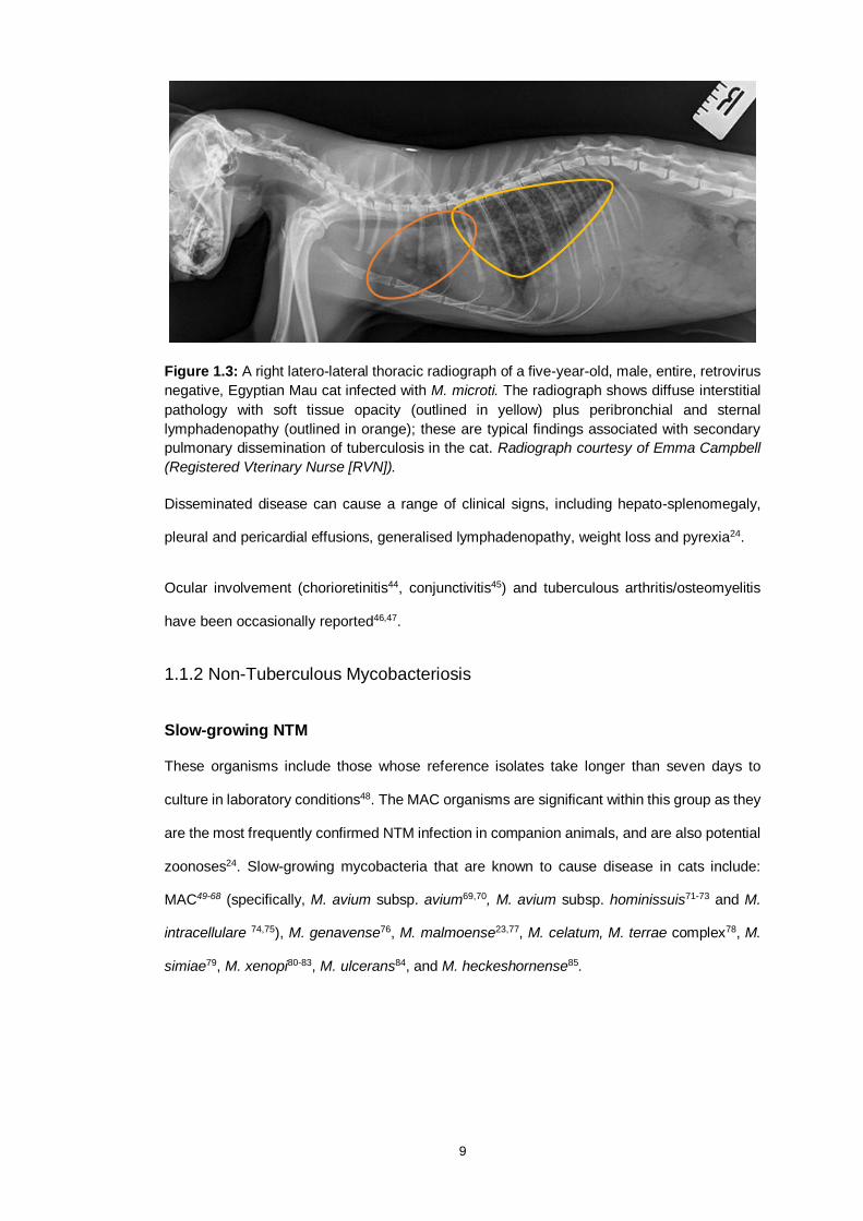
Figure 1.3: A right latero-lateral thoracic radiograph of a five-year-old, male, entire, retrovirus
negative, Egyptian Mau cat infected with M. microti. The radiograph shows diffuse interstitial
pathology with soft tissue opacity (outlined in yellow) plus peribronchial and sternal
lymphadenopathy (outlined in orange); these are typical findings associated with secondary
pulmonary dissemination of tuberculosis in the cat. Radiograph courtesy of Emma Campbell
(Registered Vterinary Nurse [RVN]).
Disseminated disease can cause a range of clinical signs, including hepato-splenomegaly,
pleural and pericardial effusions, generalised lymphadenopathy, weight loss and pyrexia24.
Ocular involvement (chorioretinitis44, conjunctivitis45) and tuberculous arthritis/osteomyelitis
have been occasionally reported46,47.
1.1.2 Non-Tuberculous Mycobacteriosis
Slow-growing NTM
These organisms include those whose reference isolates take longer than seven days to
culture in laboratory conditions48. The MAC organisms are significant within this group as they
are the most frequently confirmed NTM infection in companion animals, and are also potential
zoonoses24. Slow-growing mycobacteria that are known to cause disease in cats include:
MAC49-68 (specifically, M. avium subsp. avium69,70, M. avium subsp. hominissuis71-73 and M.
intracellulare 74,75), M. genavense76, M. malmoense23,77, M. celatum, M. terrae complex78, M.
simiae79, M. xenopi80-83, M. ulcerans84, and M. heckeshornense85.

10
Rapid-growing NTM
These organisms are defined as those whose reference isolates can be grown in laboratory
culture conditions within seven days at temperatures of between 24oC and 45oC. Documented
feline infections include members of the M. fortuitum group23,86-93 (specifically, M. fortuitum,73,94
M. porcinum,95 and M. alvei95), M. smegmatis group75,87,96,97 (M. smegmatis sensu stricto,73,95
M. goodii,73,88,95 M. wolinskyi73), M. chelonae/abscessus group86,88,91,98 (including M.
abscessus subsp. bolletti [formerly M. massiliense99 ]), M. mageritense group,95 M.
mucogenicum group,75 M. falvenscens,86 M. phlei,100 and M. thermoresistible.73,101-103. Cases
have been reported from tropical (Brazil94), subtropical (south eastern and south western
USA88,91,100), and temperate regions, including Australia,95,97,102 New Zealand,75 Canada,75
Finland,104 Germany,103 UK23 and the Netherlands98,101. Geographical differences exist with
respect to species incidence; most infections in cats in eastern Australia are caused by M.
smegmatis group (followed by M. fortuitum),87,105-107 whereas, in southwestern USA, cats are
most commonly infected with M. fortuitum group, followed by M chelonae/abscessus
group86,88,90.
Fastidious NTM
To date; M. lepraemurium (closely related to M. avium), Candidatus “M. tarwinense” (related
to the M. simiae group), M. visibile, and Candidatus “M. lepraefelis” (both related to M. leprae
and M. lepromatosis) have been confirmed as capable of causing “feline leprosy syndrome”
(FLS) in cats. These infections are reported from New Zealand,108,109 eastern Australia,110,111
western Canada,112,113 the UK,114,115 south-western USA,116 the Netherlands117. France,118,119
New Caledonia, Italy,120 the Greek island of Kythira,121 and Japan122.
Similar to the MTBC (as described in Section 1.1.1), the NTM bacteria most often infect cats
via presumed cutaneous inoculation, which is reflected in the typical distribution of lesions23.
Inoculation of the organism directly into subcutaneous adipose tissue appears to increase the
severity of rapid-growing NTM disease in cats, to which overweight cats are unsurprisingly
predisposed.

11
Risk factors that have been identified for NTM disease in cats include, the administration of
immunosuppressive drugs such as exogenous corticosteroid administration69, multidrug
chemotherapy protocols for lymphoma treatment,20 cyclosporine treatment in the context of
renal transplantation53, exogenous retroviral infection76,123 (plus lymphoma in one case85),
idiopathic αβ-T cell lymphopenia (particularly CD4+ αβ-T-cells)83, or concurrent comorbidities
such as advanced chronic kidney disease and cryptococcosis50. Studies of human
tuberculosis and NTM infected patients have revealed low body mass index (BMI), specifically
a low body fat percentage124,125, as well decreased interleukin (IL)-10 and interferon gamma
(IFN-γ) production capacity by peripheral circulating leukocytes as risk factors for disease
following infection126,127. Equivalent studies have not been conducted in companion animals,
so it is unclear how relevant these factors are to veterinary patients. The MAC infections are
seen more frequently in Somali, Abyssinian51 and Siamese59 breeds of cat, but there is no
recognised breed predisposition for the other NTM.
The MAC organisms have been the most frequently successfully cultured of the NTM from UK
cats23 (Table 1.1); however, the culture data for NTM infections, in general, is inevitably
skewed by the fact that fastidious organisms cannot be cultured, and by the routine use of
media most sensitive for MTBC culture (e.g. Middlebrook 7H11 OADC) by many reference
laboratories in the UK. Therefore, more data need to be collected with regards to the molecular
epidemiology of mycobacterial infections in cats on a worldwide basis.
Clinical Signs
Disease caused by NTM is more variable in clinical presentation than tuberculosis, but
generally results in (i) (sub)cutaneous nodules, (ii) granulomatous panniculitis, or (iii)
disseminated disease. The nodular and disseminated forms can have a very similar
presentation and distribution to tuberculous lesions (i.e. the head, limbs and trunk, Figure 1.4).
Skin nodules can be haired, alopecic or ulcerated but are typically non-painful and freely
mobile.

12
Figure 1.4: An alopecic, erythematous granuloma on the lower lip of a six-year-old, male,
neutered, retrovirus negative, DSH cat caused by M. avium infection. The lesion is notable for
its similarity in gross appearance to tuberculous lesions such as those shown in Figures 1.1
and 1.2. Image courtesy of Laura de los Santos (RVN).
Granulomatous panniculitis is the most common manifestation of disease caused by the rapid-
growing NTM. 75,86,88-91,96-97,104,107,107-111 It is characterised by multiple punctate drainage tracts
and subcutaneous nodules which can coalesce to form large areas of (mostly) non-painful,
non-healing ulcerated skin overlaying inflamed fat pads (Figure 1.5). Initially, cats tend to
present with a circumscribed plaque or nodule of the skin and subcutis.

13
Figure 1.5: Extensive subcutaneous panniculitis affecting the (shaved) ventral skin of an eight-
year-old, male, neutered, retrovirus negative, DSH cat caused by M. smegmatis infection.
Image courtesy of Faye Swinborne (MRCVS, Willows Referral Centre).
Later in the clinical course, the subcutaneous tissue becomes thickened, the overlying skin
becomes ulcerated, alopecic and punctuated with fistulae discharging a watery to oily exudate.
Infection often tends to start in the inguinal region, although it can begin in the axillae, flanks
or dorsum. The disease may subsequently spread to contiguous areas of the lateral and
ventral abdominal wall, perineum and tail-base. Severely affected cats may become pyrexic,
anorexic and reluctant to move, particularly if lesions are secondarily infected with skin
commensals such as Staphylococcus and/or Streptococcus spp.
Occasionally, pyogranulomatous pneumonia caused by rapid-growing NTM has been reported
in cats. These cases usually present with coughing, dyspnoea, fever, malaise, and weight loss.
These infections have been caused by M. fortuitum in two cats (after aspiration of orally
administered lactulose in one case)93,133 and M. thermoresistibile, following a bath in
another102,134.
Uncommonly reported clinical presentations include peripheral vestibular disease, generalised
involvement of lymphoid tissue, pulmonary disease, GI disease and intracranial infection
causing subsequent neurological signs.

14
Infections caused by the fastidious NTM typically manifest as single or multiple cutaneous
and/or subcutaneous nodules often located on the head, limbs or trunk, and regional lymph
nodes may be involved135,136. As with the slow-growing NTM, nodules are typically non-painful
and non-adherent to underlying tissues. The skin over the lesions may be intact, alopecic or
ulcerated. Lesions caused by M. lepraemurium and Candidatus ‘M. lepraefelis’ tend to be
found on the head, limbs and trunk (although anywhere on the integument can theoretically
be involved), or may be widespread, involving many cutaneous sites135. Lesions caused by
Candidatus ‘M. tarwinense’ are also mostly found on the head (particularly the eyes, lips or
mouth and front limbs), and it has not been shown to cause widespread cutaneous disease136.
Cases of M. visibile infection,137 and at least one case of Candidatus ‘M. lepraefelis’,8 have
had systemic involvement confirmed at post-mortem examination.

15
1.2 Canine Mycobacteriosis
1.2.1 Tuberculosis
There is little published data regarding the incidence of tuberculous mycobacterial infections
in dogs, and most of the information about this disease’s clinical presentation comes from
sporadic case reports, mostly of M. tuberculosis138-143 and M. bovis infections144-150. There are
only very rare individual cases of M. microti infection151. Anecdotally, the incidence of
tuberculosis in the UK appears to be much lower in dogs than cats, but the clinical severity
much greater. Dogs typically present with extensive GI disease (weight loss, vomiting, and
diarrhoea) and pulmonary pathology (Figure 1.6). In the UK, dogs also seem more likely to
become infected with MTBC pathogens than the other mycobacterial species, though this may
be an artefact of which infections can definitely speciated with the limited availability of
diagnostic tests for dogs, rather than a reflection of true incidence. As with cats, the range of
clinical signs is wide and has been known to include reticulo-endothelial, musculoskeletal,
cutaneous and/or neurological signs146,147.
Figure 1.6: A latero-lateral thoracic radiograph of a six-year-old, male, neutered, Jack Russell
terrier dog infected with M. bovis. The radiograph shows multifocal to coalescing areas of soft
tissue opacity (granulomas; outlined in yellow) distributed throughout the lung fields with
associated marked peri-bronchial lymphadenopathy.

16
The greatest risk factor reported for a dog acquiring an MTBC infection is for it to be cohabitant
or in close contact with a human suffering from active tuberculous disease; most cases of M.
tuberculosis infections in dogs have occurred because of reverse zoonotic transmission from
an infected human i.e. anthropozoonosis152,153.
1.2.2 Non-Tuberculous Mycobacteriosis
Slow-growing NTM
Typically, MAC organisms are the most frequent slow-growing NTM mycobacteria recovered
from dogs154-166. Miniature Schnauzers and Basset Hounds are reportedly predisposed154-166.
M. avium subspecies avium, M. avium subspecies paratuberculosis (MAP) and M. avium
subspecies hominisuis have all been identified from canine samples154-166.
Clinical presentations of canine MAC infection include multi-systemic disease, including any
combination of generalised lymphadenopathy, anaemia, weight loss, hepatopathy and/or
splenomegaly154-166. As with feline infections, many of these presentations are clinically
indistinguishable from tuberculosis.
M. kansasii has been isolated from a three-year-old Whippet with chronic pleural effusion167
and also from a mammary mass of a two-year-old Chihuahua168. The latter case also had
chronic generalised demodicosis implying an underlying immune deficit was present, and it
subsequently succumbed to the mycobacterial infection after prednisolone treatment for
cutaneous lymphoma. Disseminated M. genavense infection was diagnosed in a two-year-old
pug presenting with generalised lymphadenopathy, abdominal organomegaly, pyrexia and
limb pain169.
Localised dermal infections in dogs due to M. ulcerans, the causative agent of Buruli Ulcer,
have been reported in endemic areas of Victoria, Australia170. This organism produced a
cytotoxin, mycolactone, which is responsible for the deep skin ulcerations that are a hallmark
of this disease. Although the exact ecology of M. ulcerans has not been determined, it has a
worldwide but highly focal endemicity, with most human infections occurring in West Africa

17
and the coastal regions of South-eastern Australia. It is suspected that the organism gains
entry via breaches in the epidermis, possibly in some cases involving mechanical insect
vectors171. Animal cases have only been reported from Australia, and also include
possums,172,173 koalas,174 a long-footed potoroo172, alpacas175, horses176 and a cat84.
Rapid-growing NTM
Sporadic canine infections have been reported. M. goodii was isolated from a dog with
uncontrolled hyperadrenocorticism177. Systemic M. smegmatis group infection was diagnosed
in a young Bassett Hound178 (perhaps due to the same immunological defect that renders this
breed susceptible to MAC infections). M. fortuitum group skin and pulmonary infections have
also been reported. 179-183
Fastidious NTM
Canine leproid granuloma (CLG) is caused by an, as-yet uncharacterised, fastidious
mycobacterium related to Candidatus ‘M. tarwinense’ and M. simiae. It has greatest
prevalence in Australia,184 New Zealand,185,186 Brazil187-189 and the USA190. Zimbabwe (where
the first case dates back to 1973191), and Colombia have also reported cases. In 2014, a case
was diagnosed in Europe (Italy) for the first time192 and cases have since been suspected in
the UK (O’Halloran, manuscript in preparation).
Over-represented breeds include the Boxer dog and its crosses, Staffordshire bull terriers,
Foxhounds and Doberman Pinchers. Short-coated hunting dogs, especially Spaniel breeds,
have also been found to be predisposed and active hunting work has been shown to be a risk
factor for the disease.186,190
The route of infection is proposed to be an arthropod vector due to the location of lesions which
inoculates the mycobacteria into the skin and gives rise to the sites of disease and case
predilection.
Clinically, the disease is characterized by cutaneous nodules on the head, especially involving
the dorsal fold of the pinnae (Figure 1.7), but may also be located on the lateral trunk, rump

18
and/or limbs. Lesions may be singular or coalesce to cover large areas in some animals. As
lesions get larger, they may ulcerate and can become pruritic if secondarily infected.193
Affected dogs are typically systemically well and spread of the disease to draining lymph
nodes, contiguous structures, as well as internal organs does not occur.
Figure 1.7: The first suspected case from the UK of ‘canine leproid granuloma’ lesions on the
dorsal pinna of a three-year-old male neutered Labrador retriever dog. Image courtesy of
Fiona Fahy (MRCVS).
1.3 Diagnosing Mycobacterial Disease in Companion Animals
A correct diagnosis of mycobacteriosis can be challenging to achieve, in companion animals
as well as other species including humans and cattle. This is the case for a variety of reasons;
importantly, several infectious agents can produce overlapping clinical signs whilst diagnostic
tests can lack sensitivity.
Importantly, different mycobacterial species carry differing prognoses, optimal treatment
choice, and zoonotic potential.
Feline cases that would warrant a high index of suspicion for mycobacterial infection include
skin nodules or abscesses that do not heal and/or are only partially responsive to first-line
antibiotic treatment such as amoxicillin. This is particularly true for geographical locations
where there is traditionally a high prevalence of MTBC (e.g. UK) or feline leprosy infections
(e.g. south eastern Australia, New Zealand).
Differential diagnoses of nodular lesions of the skin and subcutaneous tissues include
infections due to other saprophytic bacterial species (such as Nocardia spp.), fungi and algae,
eosinophilic granuloma complex, or primary or metastatic neoplasia.

19
1.3.1 Laboratory Abnormalities
During the clinical examination of any case of suspected mycobacterial infection, it is essential
to fully establish the extent of any local disease and detect any cases with disseminated
disease or systemic involvement.
Serum biochemistry and haematology
If abnormal, these typically reveal non-specific changes e.g. a stress leukogram. More
significant abnormalities are usually reflective of more severe or systemic disease and include
anaemia and an elevated serum calcium concentration.194
Hypercalcemia and hypovitaminosis D
Hypercalcemia has been described in human adults with most granulomatous disorders,
including mycobacterial disease, although the majority of hypercalcemic patients are
asymptomatic for their hypercalcemia. Reported rates of hypercalcemia in M. tuberculosis-
infected (human) adults vary widely from 6% to 48%195. However, symptomatic hypercalcemia
in human tuberculosis cases is uncommon, with rates of ∼3% in adults196. The equivalent rate
of hypercalcemia in companion animals in response to mycobacteria is unknown, but some
cases have been reported,24,39,194 whether this was causing or contributing to any of their
clinical signs e.g. anorexia, is unclear.
Intimately linked with calcium metabolism is that of vitamin D. Briefly, vitamin D is hydroxylated
in the liver to 25-hydroxyvitamin D (25(OH)D), the form most widely measured to assess
vitamin D status, and is further metabolized by 1α-hydroxylase in the kidney to 1,25
dihydroxyvitamin D3 (1,25(OH)2D3; (calcitriol) which is the hormonally active form of vitamin
D.194-197 Calcitriol increases the concentration of calcium in the blood through increasing
intestinal absorption, reducing renal excretion and inducing osteoclast-mediated reabsorption
from bone.194-197
There is a long-standing association between vitamin D concentrations and mycobacterial
immunity, and it has recently been shown that calcitriol has a directly anti-mycobacterial

20
activity in vitro, as macrophages are capable of producing calcitriol upon Toll-like receptor
(TLR)-2 stimulation, when they phagocytose mycobacteria198-200. The result is that circulating
25(OH)D concentration decreases, and intracellular ionised calcium concentration increases.
If the extent of infection is large, then these effects become detectable as systemic
hypovitaminosis D and hypercalcemia.198-200 This phenomenon has been long established in
human medicine and more recently in feline patients with tuberculosis.194,197 Similarly, in both
species, the presence of either factor at diagnosis is a poor prognostic indicator. What remains
unclear, however, is whether these findings are merely a marker of disease which has already
disseminated (and so will inevitably be harder to treat), or whether they inherently influence
disease outcome. Some clinical trials in humans have attempted to establish the benefit, if
any, of supplementing tuberculosis patients with vitamin D during therapy as an alternative or
in combination with reducing sub-clinical hypercalcemia201-205, but the results to date have
been discordant, and its use is not currently included in any treatment guidelines.
The exogenous retroviral status of cats should be established as these infections may be a
poor prognostic indicator. That being said, FIV and/or FeLV-positivity has not been established
as a predisposing factor for feline tuberculosis, unlike in humans where disseminated
mycobacteriosis can be an “Acquired immunodeficiency syndrome-defining” infection,206 and
where Human Immunodieficiency virus (HIV) infection is a potent risk factor for tuberculosis
and confers a particularly poor prognosis.207
1.3.2 Diagnostic Imaging
Radiography
Radiography is useful for detecting systemic involvement, especially pulmonary
dissemination, and for monitoring disease progression and/or treatment response.208 While
radiographic changes of mycobacterial infection are variable, pathology is most frequently
seen in the thorax, consisting typically of a diffuse interstitial, alveolar or bronchial pattern with
peri-bronchial and sternal lymph node involvement observed with increasing disease
severity.208 It is important to note that no pattern is pathognomonic for mycobacteriosis (e.g. it

21
may also be consistent with a diagnosis of neoplasia or other infection e.g. toxoplasmosis)
and that observed lung pathology can be mixed.
Computed tomography
Similar findings are seen with the use of computed tomography (CT) imaging, though the
sensitivity of detection is increased with this modality, and a diffuse structured interstitial lung
pattern is most common, being either nodular or reticulonodular in nature209,210 (Figure 1.8).
Both imaging modalities also play a key role in monitoring a patient’s response to therapy (see
Prognosis).
Figure 1.8: A transverse CT scanned section through the thorax of a three-year-old, male,
neutered, retrovirus negative, Oriental cat caused by M. microti infection. The scan reveals a
diffuse structured interstitial pattern (black arrow) comprising mixed nodular (orange arrow)
and linear structures (red arrow), characteristic of a reticulonodular pattern which is typical of
feline tuberculous pulmonary pathology. Image courtesy of Alison Major (MRCVS).
Abdominal imaging
This can be achieved using radiography, CT and/or ultrasonography, and can reveal
hepatosplenomegaly, abdominal masses, mineralised or granulomatous mesenteric lymph
nodes, and/or ascites.23,24

22
1.3.3 Specific Investigations
Cytology
Cytology can be very useful in the diagnosis of mycobacterial disease. Fine needle aspiration
(FNA) of lesions can be used to prepare glass slides of sampled tissue. Subsequent
Romanowski-type (e.g. Diff QuikTM) staining may reveal granulomatous to pyogranulomatous
inflammation with “negatively staining rods” or “ghost bacilli” either within the cytoplasm of
reactive macrophages and giant cells, or within the extracellular space (Figure 1.9a).
Subsequent slides are then frequently re-examined following acid fast-staining (e.g. using ZN
or Fite’s method) to confirm the presence of mycobacteria. A modified ZN-staining procedure
is occasionally needed for rapid-growing mycobacteria, as they are not as acid-fast as other
species (Figure 1.9b). An FNA has the advantage of being less invasive than an incisional or
excisional biopsy, but is likely to have lower sensitivity; particularly in tuberculosis lesions
which frequently have few (if any) visible mycobacteria.
Figure 1.9a: Left: Cytological appearance of a FNA from a dermal granuloma in the skin of a
three-year-old male neutered DSH cat due to M. bovis infection, stained with DiffQuick. Fields
of view (x40 magnification) have been put together to display reactive epithelioid macrophages,
which contain intracytoplasmic non-staining (“ghost”) bacilli consistent with the presence of
mycobacteria (black arrows). Right: A Ziehl-Neelsen (ZN)-stained slide made from the same
lesion taken at x100 magnification under oil emersion.

23
Figure 1.9b: A ZN-stained section (x100 magnification) of a granulomatous lesion biopsied
from the nose of a one-year-old, male neutered, retrovirus negative, DSH cat caused by
infection with M. microti. Staining reveals numerous positively stained (pink), slender, curved
bacilli both intracellularly and within the extracellular space suggestive of mycobacteriosis.
Histopathology
Histopathology is frequently the first line of diagnostic investigation in these cases. The most
typical approach taken is to surgically remove or biopsy a non-healing skin lesion,
subcutaneous mass or chronically enlarged lymph node. In some cases, if there is only a
single cutaneous lesion, this may prove curative.211
Histopathology alone cannot speciate mycobacteria, and this is needed to establish the risk to
owners and other animals in the household, potential treatment options, and prognosis.
However, typical histopathologic findings in most cats with mycobacteriosis include
granulomatous inflammation with foamy, epithelioid and/or palisading macrophages,
occasionally with significant infiltration of lymphocytes distributed at the periphery of the
granuloma and/or neutrophils (i.e. pyogranulomatous inflammation; Figure 1.10).211 ZN-
staining may reveal highly variable numbers of AFB which is idiosyncratically influenced by
the immune response of the host, the location of the granuloma, and the species of infecting
organism (MTBC and rapid-growing NTM infections are generally considered have fewer
visible AFB compared to slow-growing and fastidious NTM infections, though this is not always

24
the case, and is not diagnostic). The use of modified Fite’s or fluorescent Auramine-O stain
may increase the sensitivity of detection of mycobacteria in histological samples.212
Figure 1.10: Histological appearance of granulomatous inflammation in a two-yeaar-old male
neutered DSH cat caused by M. bovis infection demonstrating a typical focal, sub-dermal
organising granuloma with a large area (outlined in black) of central necrosis. This section is
stained with haematoxylin and eosin and imaged at x10 magnification. Higher magnification
revealed extensive effacement of the normal tissue architecture with numerous palisading
macrophages with large nuclei and foamy cytoplasm infiltrating the tissue surrounding the
necrotic area. ZN-staining of the same biopsy (not shown) revealed moderate numbers of AFB
within the area of necrosis. Image courtesy of Shannon Street (MRCVS).
Historically, the pathological picture seen with feline leprosy was divided into a tuberculoid
form of disease which classically affected young cats with few mycobacteria being seen within
the lesions, and a separate, leproid form which affected older cats, with numerous
mycobacteria being seen within the lesions.213,214 Greater insights have been gained from
recent studies, and the pathology now appears to vary more with infective species of
mycobacteria than patient demographics, with most infections caused by Candidatus ‘M.
tarwinense’ and Candidatus ‘M. lepraefelis’ being lepromatous in nature (91-95%), whereas
just over half (58%) of the M. lepraemurium cases were of this pathologic type in a recent
study.213,214

25
Specialist Mycobacterial Culture
Isolation of mycobacteria following laboratory culture is currently the gold standard diagnostic
test for confirmation of mycobacterial infection in UK companion animals24. However, in some
laboratories it has a low sensitivity and may fail, even when AFB have been demonstrated in
the lesion24. Ideally, samples should be sent to a recognised reference laboratory e.g. the
Animal and Plant Health Agency (APHA) laboratory Weybridge in the UK. When culture is
successful it can take a protracted period for some species to grow visible colonies, e.g. M.
microti generally requires a minimum of 12 weeks to culture, during which time treatment
needs to be instigated based on a presumptive diagnosis, especially if this is supported by
previous histological evidence24,211.
Drug susceptibility testing of rapid-growing NTM is not only useful for clinical purposes, but
historically has also been used to provide phenotypic data for typing of isolates (for example,
susceptibility to trimethoprim, polymyxin B, and tobramycin). The National Committee for
Clinical Laboratory Standards Mycobacterial Subcommittee has nominated broth dilution as
the gold standard,5 although this can be technically demanding and is not offered routinely by
many diagnostic laboratories.
Molecular Diagnostics
Polymerase chain reaction (PCR) testing provides a rapid and typically accurate diagnosis
(excluding contamination) and can be performed on fresh, frozen or formalin-fixed-paraffin-
embedded tissue and Romanowsky-stained (but not ZN stained) cytology slides.215 There are
currently no commercially available animal specific molecular-based diagnostic tests for the
detection of mycobacterial species of veterinary importance. Available methodologies in the
UK are those used for the diagnosis of active mycobacterial diseases in human patients.
These tests have a high sensitivity for MTBC organisms i.e. those that are significant human
pathogens, but are not validated for others that are uniquely of veterinary importance e.g. M.
lepraemurium. The relative probability of successfully achieving a diagnosis from veterinary
submissions is likely to be related to the number of AFB present in any given biopsy sample.
Though exceptions have been documented216, as a general rule, it is thought that samples

26
with moderate to large numbers of AFB are reasonably likely to yield a diagnostic quantity and
quality of mycobacterial DNA; whereas those with few or no AFB are unlikely to provide useful
results and other diagnostic methodologies are usually considered preferable in these
instances.
There is not yet a consensus on a single gene that should be used for the genetic speciation
of all mycobacteria. Even within the MTBC, the reductive evolution of these organisms
complicates the picture. The most frequently utilized genes for mycobacterial identification are
those which encode for; 16S rRNA,215 the 65kDA heat shock protein (hsp65),217 the β-subunit
of bacterial RNA polymerase (rpoB),218 16-23S rRNA internal transcribed spacer ITS
region219,220 and, less frequently, the DNA gyrases (gyr). Assays targeting the insertion
sequence (IS)6110 element for MTB complex,221 IS2404 for M. ulcerans,222 and multiplex
IS901 and IS1245 for differentiation of M. avium subsp. avium and M. avium subsp.
hominissuis223 have also been utilised in the diagnosis of small animal infections.53,66
The MTBC mycobacteria have identical sequences at the 16S rRNA gene, and 16-23S internal
transcribed spacer (ITS) region and all contain IS6110, therefore other genetic methods,
including genomic deletion analysis,224 spoligotyping225 and mycobacterium interspersed
repetitive unit and variable number of tandem repeats (MIRU-VNTR) analysis226 must be used
to differentiate the members of this group.
Although its popularity has reduced with the accessibility of genetic sequencing, PCR
restriction fragment analysis or restriction fragment length polymorphism [RFLP] analysis is a
commonly used DNA profiling technique for identification of NTMs. This technique involves
amplification of a particular gene (e.g. ITS227, hsp65228,229, IS1245 for M. avium230), and
subsequent restriction enzyme digestion, followed by analysis via agarose gel electrophoresis.

27
Matrix-assisted laser desorption ionization–time of flight mass spectrometry
Matrix-assisted laser desorption ionization–time of flight mass spectrometry (MALDI-TOF-MS)
can identify bacterial organisms quickly and cheaply. Although primarily used for cultured
samples, it is now being used directly on clinical samples. Since the equipment used is
relatively new, most veterinary laboratories have limited access to this technology; however,
its use is likely to grow as the technology becomes cheaper and more available. The sample
is mixed with an energy absorbing organic compound (known as ‘matrix’). The resultant
crystalized sample and matrix is then ionized by a laser beam. This results in the addition of
protons to the liberated ions of the sample, after which they are then accelerated at a set
potential towards a detector. The ions become separated according to the mass to charge
ratio and the time it takes to reach the detector is known as the ‘time of flight’. The resultant
mass spectrometry produces a characteristic spectrum known as a peptide mass fingerprint.
It is this fingerprint that is then compared to known results in a database, and the resultant
organism can be identified. The available spectral database for mycobacteria is steadily
improving. Mycobacteria require prior inactivation and disruption of the mycolic acid-rich cell
wall, both for biosafety reasons (in the case of the MTBC) and to liberate proteins within the
cells. Success rates vary but the differentiation between MBTC and NTM is usually very
reliable.231 MALDI-TOF MS requires more biomass compared to molecular techniques, which
means substantially longer turnaround times for slow-growing mycobacteria.
Interferon Gamma Release Assay
The IFN-γ release assays (IGRA) were developed on the principle of quantitatively evaluating
IFN-γ production by peripherally circulating antigen-specific effector T-memory cells upon in
vitro stimulation with mycobacterial antigens, to aid the diagnosis of both active and latent
tuberculosis in bovines, and subsequently adapted for human patients.22 These assays have
been widely adopted for the diagnosis of tuberculosis in cattle and humans in the form of the
BOVIGAMTM and QuantiFERON-TB Gold testTM 203 respectively. In humans, the QuantiFERON
test has been shown to have far greater sensitivity (80%) than the previously used tuberculin

28
skin test (also known as the Mantoux test; see later section) which has a reported sensitivity
of just 28%232.
The basis of IGRA tests is that either whole blood or peripherally-circulating blood
mononuclear cells (PBMC) are incubated in-vitro in the presence of mycobacterial antigens
restricted to specific subsets of mycobacterial species. The amount of IFN-γ produced is then
quantified by enzyme linked immunosorbent assay (ELISA).232-235 This value is then
interpreted to support a diagnosis, in the context of clinical signs and other diagnostic
investigations.232-235
The IGRA tests have several advantages over other diagnostic techniques for tuberculosis;
they are significantly quicker at generating results than culture or even PCR methods, they are
relatively non-invasive requiring only a single peripheral blood sample, and they can be
repeated if necessary as conducting the assay does not alter the systemic immune response,
unlike the tuberculin skin test232-235.
Since 2008, an adapted IGRA has been validated for use in cats, and the QuantiFERON –TB
GoldTM assay has been useful in assessing M. tuberculosis infections in dogs.236-238 For the
feline assay, PBMC are isolated from heparinised peripheral blood samples obtained from
domestic cats with a high index of suspicion of mycobacterial disease e.g. they have clinical
signs and/or histological findings indicative of mycobacterial disease236,237. These cells are
then incubated for four days in vitro across several conditions;
a) complete cell culture media i.e. negative control,
b) a mitogen - a potent stimulator of IFN-γ production by all capable cells (i.e. a positive
control),
c) purified protein derived from M. avium-complex (PPDA),
d) purified protein derived from M. bovis (PPDB) and
e) a combination of recombinant peptides 6kDa early-secreted antigenic target (ESAT-
6) and 10kDa culture filtrate protein (CFP-10).

29
After four days, the amount of IFN-γ which has been released into the cell culture supernatant
is measured by ELISA. A response to PPDB antigen which is greater than that to PPDA is
indicative of MTBC infection with a sensitivity of up to 100% in cats.236,237 The response to
ESAT-6 and CFP-10 can be used to subdivide the MTBC as M. microti does not produce these
molecules. Both ESAT-6 and CFP-10 are the major secreted antigens coded for at the esx-1
locus on the region of difference (RD)-1 of the genome239,240. M. microti and M. bovis-BCG are
RD-1 gene deletion strains as they do not possess the esx-1 locus, but all other members of
the MTBC do and so encode for these two proteins.236-240 Therefore, in dogs the IFN-γ
secretion levels that follow a pattern “PPDB > PPDA, and ESAT-6/CFP-10 positive”, would be
observed in response to either M. tuberculosis or M. bovis infection238. However, in cats this
pattern is considered diagnostic of M. bovis, as this species is so rarely, if ever, infected with
M. tuberculosis236,237. Unfortunately, some animals (bovine and feline) infected with M. bovis
do not respond to the ESAT-6 and CFP-10 peptides by production of antigen-specific IFN-γ;
a small study showed that this occurred in 20% of cats and a similar proportion (~18%) of
cattle, so this test does not always reliably discriminate between M. bovis and M.
microti.236,237,240
If the response to PPDA peptide is greater than PPDB (and ESAT-6/CFP-10 is negative), this
is indicative of a MAC infection, while no response to any non-control peptides but where
mycobacteria have been identified e.g. histologically, indicates an infection with another NTM
organism240.
Orthologues of the MTBC molecules ESAT-6 and CFP-10 are encoded by a small number of
NTM (M. kansasii, M. szulgai, M. marinum, M. riyadhense, M. genavense, M. bohemicum, M.
interjectum, M. flavescens, M. xenopi, and M. malmoense)242 and so it is possible for patients
to show a significant response to this protein combination only or in combination with PPDA
as a result of infections with these organisms. Though no such results have been published,
they have anecdotally been observed. Given that reports of domestic animals being infected
with these species are limited to individual case reports243,244, the likelihood of the IGRA false-
diagnosing MTBC or MAC would seem to be low.

30
Screening in-contact animals
Where animals are known to be in epidemiologically significant contact with a patient that has
clinically active disease but remains asymptomatic it is possible, unlike with all other testing
strategies, to submit blood samples for IGRA testing. However, results obtained from such
testing must be interpreted with caution. Large prospective cohort studies of human patients
have shown that those with a positive tuberculin skin test (TST: see below) have a 1.4 to 1.7-
fold higher rate of developing active tuberculosis within one year compared to those with a
negative TST result.245 Three studies provided incidence rate ratios of tuberculosis stratified
by IGRA as well as TST status at baseline.245 The association with subsequent incident
tuberculosis in test-positive individuals compared to test-negatives was higher but not
statistically significant leading to the conclusion by the World Health Organisation (WHO) that
both IGRA and TST methods appear to have only modest predictive value, suboptimal
sensitivity, and do not help identify those who were at highest risk of progression to disease.245
Therefore, the decision of whether to test in-contact animals and then to instigate therapy or
not based singularly on IGRA results is currently made at the discretion of individual clinicians
and animal owners.
Tuberculin Skin Testing
The TST (also known as the Mantoux test, Mendel–Mantoux test, Mantoux screening test,
tuberculin sensitivity test, Pirquet test, or PPD test) is a tool for screening human patients for
tuberculosis.245-250 The Mantoux method of the TST is the major tuberculin skin test used
around the world, largely replacing multiple-puncture tests such as the Tine test. 245-250 The
Heaf test, a form of Tine test, was used until 2005 in the UK, when it was replaced by the
Mantoux method.251 The TST is endorsed by the American Thoracic Society and Center for
Disease Control and Prevention (CDC).252,253 Purified protein derivative (PPD) tuberculin is a
precipitate of species-non-specific molecules obtained from filtrates of sterilised, concentrated
mycobacterial cultures.248 The tuberculin reaction was first described in humans by Robert
Koch in 1890 and the test was first developed and described by the German physician Felix
Mendel in 1908.245 A standard dose of five tuberculin units in 0.1 ml, according to the CDC,253

31
or two tuberculin units of Statens Serum Institute tuberculin in 0.1 ml solution, according to the
National Health Service,251 is injected intra-dermally and the test is ‘read’ 48 to 72 hours
later251-253. A person who has been infected with mycobacteria will undergo a type IV (delayed)
hypersensitivity reaction leading to a quantifiable swelling at the site of inoculation.254 The
reaction is read by measuring the diameter of induration (palpably raised, hardened area)
across the forearm in millimeters.251-254 The test is considered positive if it is greater than a
pre-defined threshold which is altered depending on; the background risk or the patient, the
clinical index of suspicion, the endemic prevalence of the country/region and any underlying
immunological disorders.251-254.
In cattle; official M. bovis eradication campaigns also use intradermal tuberculin testing as
statutory diagnostic assays for the detection of M. bovis specific cell-mediated immune
responses developed during the infection.255-257 Single intradermal skin testing (SIT) detects a
delayed-type hypersensitivity reaction to the intradermal injection of PPDB, administered at
the cervical site in Europe or at the caudal fold of the tail in USA and New Zealand.255-257 Three
days (72 +/- 4 hours) after inoculation, the skin fold thickness at the site of injection is examined
and measured.255-257
The single intradermal comparative cervical tuberculin test (SICCT), i.e. comparison between
PPDA and PPDB, is used in high prevalence areas of bovine M. bovis infections and allows
better discrimination between animals infected with M. bovis and those infected with MAC or
environmental mycobacteria which leads to improved test specificity.257
The use of tuberculin skin testing has been evaluated in both cats and dogs with mycobacterial
infections and led to unreliable results which did not correlate with the findings of other
established diagnostic tests and so is not used clinically.238,259 Recent work in cattle has shown
that performing the SIT/SICCT in ruminants can go on to boost both the cell-mediated and
humoral immune responses of the individual animals leading to an increase in sensitivity for
subsequently performed IGRA or antibody assays.258 This effect has not been explored in
companion animals to date.

32
1.4 Treatment of Mycobacterial Disease in Companion Animals
Once a diagnosis has been achieved (or while pending culture results but based on clinical
signs and histopathological findings) the next challenge for clinicians and owners is managing
a mycobacterial infection in a companion animal. If treatment is to be attempted, obtaining a
successful outcome requires a prolonged course of multi-antibiotic therapy, and success is
dramatically influenced both by owner and patient compliance. Drug toxicity, particularly in
feline cases, and the costs associated with medications and disease monitoring can make
optimal regimes difficult to maintain.
Drug-resistant tuberculosis with the detection of multi-drug, extensive-drug or total-drug
resistant isolates, as well as the continuing human HIV pandemic, has dominated the focus of
many of the recent changes to anti-mycobacterial drug therapies, including anti-tuberculosis
therapy.260-262 This is of even greater importance for drugs used in human protocols.
1.4.1 Treatment of Feline Tuberculosis
Surgery
As discussed, by far the most frequent clinical sign of feline tuberculosis is a single or small
number of skin nodules.23,24 In most cases, these are removed by excisional biopsy facilitating
a diagnosis to be made. In these cases, complete surgical resection may be curative.211
However, this cannot always be guaranteed, and adjunctive medical therapy is almost always
indicated.211 Surgery may be considered for any lesions remaining at the time of diagnosis or
that may have subsequently arisen. However, there is a significant risk of wound dehiscence
in many cases. There is no evidence that revision surgery confers a benefit to rigorous medical
management,211 so is not widely advocated.
In cases of intra-articular tuberculosis or significant osteomyelitis, medical management alone
has consistently been found to be inadequate for successful management.47,263,264 This is
thought to be due to the lack of penetration of anti-tuberculosis medications into the synovial
space, as well as the aggressive osteolysis caused by these pathogens, often before the time

33
of diagnosis (Figure 1.11). In such instances amputation of all or part (at least one joint above
the lesion) of the limb with adjunctive medical therapy is often required for a successful
outcome.47
Figure 1.11: A cranio-caudal radiograph of the right distal forelimb of a four-year-old, male,
neutered, retrovirus negative Maine Coon cat with an intra-articular infection with M. microti.
The radiograph demonstrates severe osteolysis of the carpal bones as well as the distal radius
and ulna (outlined in yellow) surrounded by extensive soft tissue swelling (white arrows).
Image courtesy of Professor Noel Fitzpatrick MRCVS.
Medical therapy
The current CDC and WHO guidance on the appropriate drug combination for human
treatment has changed and treatment with rifampicin, isoniazid, pyrazinamide and ethambutol
(combined) for six months is now advocated.265 This regime cannot be readily translated
directly into veterinary medicine. Isoniazid and ethambutol have been associated with severe
nephrotoxicity, hepatotoxicity, and neurotoxicity in companion animals, and isoniazid carries
the further risk of inducing optic neuritis, as such these drugs should not be used as first-line
therapies for companion animals.266-270 Pyrazinamide is ineffective against M. bovis271 and has

34
also been associated with hepatotoxicity in cats.272 Given the lack of diagnostic capacity to
rapidly and definitively diagnose the causative agent in companion animal mycobacterial
disease, the use of pyrazinamide is currently considered contraindicated as a first line agent.
As an alternative regime, standard first line therapy for feline tuberculosis should comprise a
fluoroquinolone, a macrolide/azalide and rifampicin.
The fluoroquinolone of choice is pradofloxacin (where available) due to its excellent safety
profile in the cat and its known in vitro activity against mycobacteria when compared to older
fluoroquinolones.95 Where this is not available the use of moxifloxacin is advocated as it has
a very similar pharmacological profile to pradofloxacin.105 Alternative fluoroquinolones that
could be considered would be the older drug marbofloxacin or the human drug ciprofloxacin
(the active metabolite of enrofloxacin273). Enrofloxacin itself is an unacceptable choice in the
cat due to its association with acute irreversible retinopathy.274 The macrolide/azalide of choice
is azithromycin due to a dosage pattern of once every 24 hours which eases adherence to
treatment, plus its concentration within pulmonary macrophages.275,276
Rifampicin has long been a cornerstone of anti-tuberculosis, and more general anti-
mycobacterial medical therapy. It belongs to the rifamycin class of antibiotics and acts to inhibit
DNA-dependent RNA-polymerase activity in susceptible bacteria.277 Importantly, it has good
efficacy against both extracellular mycobacteria as well as those slowly and persistently
replicating within macrophages following phagocytosis.227 However, it should never be used
as a monotherapy for mycobacterial disease as resistance develops rapidly.278,279 Rifampicin
is known to be a potent inducer of the hepatic cytochrome P450 system and so has the
potential to be significantly hepatotoxic when used chronically.280,281 Before starting treatment,
it is advised that clinicians assess a patient’s liver function by evaluation of serum biochemistry
and haematology as well as considering bile acid stimulation testing if there are concerns.
Treatment with rifampicin should be stopped if circulating levels of liver enzymes significantly
exceed the upper reference interval, hyperbilirubinemia develops, or patients develop clinical
signs attributable to hepatic dysfunction. Liver support drugs e.g. S-adenyl methionine (SAMe)
have been used in a small number of cases both to prevent and reverse increases in liver

35
enzyme activity of feline patients on long term anti-mycobacterial therapy. Once recovered,
the rifampicin can be re-introduced at half of the previous dose, while continuing with SAMe,
plus close monitoring to ensure the hepatopathy does not recur. Rifampicin is excreted in all
bodily fluids and may cause urine, tears, and saliva to discolour to red-orange in color.282 This
is not clinically significant, but it can be distressing for clients who are unprepared, especially
as there is the potential for the discoloration to be mistaken for haemorrhage. Deposition of
the drug within the skin can cause skin discoloration, irritation and, occasionally, hyperesthesia
in cats; the complete converse of humans where it is used as a therapy for cholestatic
pruritus.283,284 Rifampicin is unevaluated a potential teratogen and so should only be handled
by owners wearing gloves and should not be administered to pregnant queens.

36
Table 1.2: First-line anti-tuberculous medications for use in cats; a triple combination
comprising one of the fluoroquinolones, a macrolide/azalide and rifampicin is frequently
used.285
Drug Product Dosing Contraindications Side Effects
Pradofloxacin Veraflox™
25mg/ml
suspension
or 15mg
tablets
5-7.5mg/kg
PO
q 24 hours
Not in dogs <12
months of age (18
months in giant
breeds) or cats <6
weeks of age due
to potential
adverse effects on
cartilage. Do not
use in animals with
epilepsy.
Neutropenia with high
doses and/or long courses
Marbofloxacin Marbocyl™
20mg
tablets
2mg/kg PO q
24 hours
As for
pradofloxacin.
Intermittent vomiting and
transient diarrhoea on
instigation of treatment.
Moxifloxacin Avelox™
400mg
tablets**
10mg/kg PO
q 24 hours
As for
pradofloxacin.
Occasional vomiting.
Ocular toxicity has not been
thoroughly assessed.
Ciprofloxacin 5-15mg/kg
PO q 12
hours
As for
pradofloxacin.
Occasional vomiting.
Azithromycin Zithromax™
250mg
capsule
5-15mg/kg
PO q 24
hours
Pre-existing
cardiopathy esp.
arrhythmogenic
diseases.
Intermittent vomiting and
diarrhoea possible but
uncommon.

37
50mg/ml
suspension
Pre-existing
hepatopathy.
Clarithromycin Generic
500mg
tablet
50mg/ml
suspension
7.5mg/kg PO
q 12 hours
Concurrent use of
NSAIDs or
antacids.
Pre-existing
hepatopathy.
Pinnal erythema.
Generalized erythema.
Hepatotoxicity.
Rifampicin Rifadin™
300mg
capsule
20mg/ml
suspension
5-10 mg/kg
PO q 24
hours by
mouth
Pre-existing
hepatopathy.
Pregnant queens
Hepatotoxicity; induction of
liver enzymes, anorexia,
generalized erythema and
pruritus.
CNSa signs.
Discoloration of body fluids.
PO; per os a Central nervous system b NSAIDs; non-steroidal anti-inflammatory drugs.
The duration of treatment is dependent on the clinical signs present at the time of diagnosis.
Three months of triple therapy as outlined should be considered a minimum in all cases.285
Treatment should be extended for two months beyond the resolution of all clinical signs which
here also includes any pulmonary dissemination detectable on standard radiography (Figures
1.13a and 1.13b).211,285 Treatment for cutaneous plus pulmonary disease is typically for six
months. Monitoring for the resolution of pulmonary changes using CT studies is more difficult;
due to the increased sensitivity of this modality, subtle changes such as minor scars remain
detectable and may persist indefinitely beyond the need for treatment.210

38
Figure 1.12a: A latero-lateral thoracic radiograph of a three-year-old, female, entire, retrovirus negative, DSH cat infected with M. bovis. The radiograph, taken at the time of diagnosis shows multifocal to coalescing areas of soft tissue opacity (granulomas) distributed throughout the lung fields (yellow arrows) with associated peri-bronchial lymph node enlargement (outlined in orange).
Figure 1.12b: A lateral thoracic radiograph taken from the same cat following three month of
triple antibiosis consisting of rifampicin (10mg/kg), azithromycin (10mg/kg) and pradofloxacin
(5mg/kg) given orally every 24 hours. The radiograph shows complete resolution of the
previously identified pathology.

39
Therapy for refractory cases is more difficult as it requires the use of drugs which have
increased frequency and severity of side effects. Rifampicin therapy should be maintained
whenever possible and can be further combined with isoniazid and ethambutol.211,285
Treatment should still last for a minimum of three months and two months beyond the
resolution of clinical signs. 211,285
Human patients who are believed to have latent infection (as they are IGRA positive for
tuberculosis, but do not show any clinical signs), may be offered a six-month course of
isoniazid monotherapy.286 Currently there is no strong evidence to suggest that this reduces
the incidence of those who go on to develop active disease in cats. However, such prophylactic
therapy could be considered by individual clinicians on a case by case basis.
Table 1.3: Second line anti-tuberculous medications for use in cats; a triple combination
should be used comprising both isoniazid, ethambutol and rifampicin (see Table 1.2).285
Drug Dosing Contraindications Side Effects
Isoniazid 10-20 mg/kg PO q 24hours Poorly evaluated in
cats.
Hepatic dysfunction.
History of seizures.
Hepatotoxicity.
Peripheral neuritis.
Seizures.
Acute kidney injury.
Ethambutol 10-25 mg/kg PO q 24hours Poorly evaluated in
cats.
Optic neuritis.

40
Prognosis
If dosing is maintained, then the prognosis is considered to be fair, with one review stating that
40% of cases reached and maintained complete clinical remission, though it remains unclear
as to whether this constitutes bacteriological cure.211 The remaining 60% of cases showed
variable responses from temporary or partial remission to no response to treatment.211
However, these data relate to a retrospective cohort, many of which were treated with what
would be considered sub-optimal therapies, such as short courses of fluoroquinolone as
monotherapy. With the approach outlined above, clinical experience suggests a positive
prognosis of 70-80% of feline tuberculosis cases, particularly those with cutaneous and/or
pulmonary involvement.285
1.4.2 Treating Feline NTM Infections
Surgical intervention is often a pre-requisite to obtaining a diagnosis in these cases. However,
many feline NTM infections are progressive and are likely to recur following the excision of
lesions, even when clear surgical margins have been achieved. It is, therefore, advisable to
follow up surgical intervention with adjunctive medical treatment.
Slow-growing NTM
The treatment of some NTM infections (particularly M. avium) may be more difficult and less
rewarding than for tuberculous infections. This is because many of these organisms do not
readily grow in laboratory conditions, so it has not been possible to generate good sensitivity
data across many clinical isolates from companion animals. Additionally, the drug resistance
patterns of these organisms vary widely, not just between species but also between isolates
of the same species.287 Furthermore, in vitro drug susceptibility testing results do not always
correlate with in vitro drug activity, as some of the genes which confer resistance (e.g. to
macrolides) are inducible, so their presence does not necessarily correlate with observed
susceptibility profiles, especially for MAC organisms.287 The Clinical and Laboratory Standards
Institute criteria for the antimicrobial susceptibility testing of NTM recommends broth
41
microdilution as the gold standard, preferably performed by a reference laboratory, or similarly
experienced facility.288
Most published susceptibility data apply to members of the MAC complex, M. kansasii and
rapid-growing mycobacterial species.288 Recommendations for susceptibility testing of
uncommonly isolated species, which have been assessed to be clinically significant in people,
may include testing against rifampicin, clarithromycin, amikacin, ciprofloxacin, ethambutol,
linezolid, moxifloxacin, and trimethoprim-sulfamethoxazole.288
The optimal drug regime is hard to readily determine. It is also important to note that MAC
isolates are typically resistant to most commonly used second generation fluoroquinolones
(e.g. enrofloxacin, marbofloxacin, orbifloxacin). Significant pharmacokinetic interactions have
also been reported in human patients with NTM infections; e.g. rifampicin can reduce the
bioavailability of both isoniazid and moxifloxacin, resulting in lower serum concentrations but
extended half-life.289,290 How far this contributes to the emergence of drug-resistant isolates
and/or worse clinical outcomes is not clear and its implications for feline and canine patient
treatment are currently unknown.
Anecdotally, the most successful protocols appear to consist of three drugs from; a macrolide
(clarithroymycin or azithromycin), a fluoroquinolone (pradofloxacin, moxifloxacin or possibly
marbofloxacin [in the case of isolates other than M. avium]), plus rifampicin or clofazimine. As
with tuberculous infections, drug therapy should be continued for two to three months from the
time of clinical remission to reduce the risk of recurrence. Some cats require extended (up to
a year) or indefinite therapy to remain asymptomatic. Relapses have also been reported.23
Rapid-growing NTM
The management of mycobacterial panniculitis in cats is well described, particularly those
caused by strains of M. fortuitum in the USA.291-293 The use of lipophilic antimicrobial agents
directed by susceptibility data and, where appropriate, surgical resection and reconstructive
techniques has made this an eminently treatable disease in many instances.291-293 All
infections caused by rapid-growing NTM should undergo culture and drug susceptibility testing

42
but generally; members of the M. smegmatis-complex are usually susceptible to doxycycline
and fluroquinolones but are often resistant to clarithromycin.294,295 M. fortuitum tends to show
higher levels of resistance to antimicrobials in general, although also tends to be susceptible
to fluroquinolones and clarithromycin,296,297 while M. chelonae tends to be resistant to all
commonly used drugs except clarithromycin, gatifloxacin, and linezolid.296-299 Minimum
inhibitory concentrations for ciprofloxacin, enrofloxacin, moxifloxacin and pradofloxacin using
the broth microdilution method have been reported for feline M. fortuitum, M. goodii and M.
smegmatis sensu stricto isolates.77,88
In areas such as Australia, where M. smegmatis and M. fortuitum-complex infections
predominate, treatment should commence empirically with one of the newer generation
fluoroquinolones (pradofloxacin or moxifloxacin) 19,213,214, whereas in the USA where M.
chelonae infections are more commonly diagnosed, the best first line choice is clarithromycin
or azithromycin.296 Multi-drug therapy is ideal to reduce to induction of resistant clones297.
Thus, the addition of additional drugs (e.g. doxycycline) based on in vitro susceptibility data is
recommended. In refractory cases, clofazimine, cefoxitin, linezolid or amikacin may be used if
indicated by drug susceptibility data.19,213,214
Cases are reassessed every few weeks for evidence of adequate clinical response. In some
cases, a cure may be achieved with medical therapy alone; however, some may eventually
become refractory to treatment, requiring wide, sometimes radical, surgical en bloc resection
of residual infection, and then continuation of appropriate antibiotics for a further few
months.130 The total duration of therapy for mycobacterial panniculitis caused by rapid-growing
mycobacteria is usually three to 12 months (ideally, one to two months past clinical resolution
of signs, as with other mycobacterial classes).130
Pneumonia caused by rapid-growing mycobacteria, depending on geographical location
empirical treatment with pradofloxacin and/or clarithromycin should be started immediately
upon the finding of mycobacteria within diagnostic specimens and then adjusted upon receipt
of drug susceptibility data.92,103 Treatment should always consist of using two or three suitable
agents and should be continued for one to two months past resolution of signs.285 Pulmonary

43
lesions that are refractory to appropriate medical therapy may need to be surgically resected,
and antibiotic therapy should then be continued post-operatively for several months.
Fastidious NTM
There have been rare instances of spontaneous resolution of feline leprosy infections,
especially those caused by M. lepraemurium,300,301 although most cases require interventional
treatment to achieve a cure. Definitive treatment guidelines for each of the fastidious species
are yet to be established because the antibiotic susceptibility of these organisms is largely
unknown due to the difficulty in culturing these organisms on cell-free media, and a lack of
data from prospective treatment trials.
M. lepraemurium grown in cell-culture viability assays suggests that it is highly susceptible to
rifampicin and clofazamine.302 Although M. lepraemurium appears to be somewhat susceptible
to dapsone,302 the potential side effects of hepato- and neurotoxicity and blood dyscrasias
limits the use of this drug use in cats.
Wide surgical resection of nodular skin lesions, plus clarithromycin combined with rifampicin
or a newer generation fluoroquinolone, such as pradofloxacin or moxifloxacin (with or without
clofazamine) appears to be the most efficacious treatment regimen (Table 1.4).19,213,214 As with
other mycobacterial infections, it is recommended that medical therapy be continued for at
least two to three months past the resolution of clinical signs or following the resection of all
visible lesions.

44
Table 1.4: Additional medications for the treatment of NTM infections of cats, in addition to
those in Table 1.2.19,213,214
Drug Dosing Contraindications Side Effects
Clofazimine 4-10mg/kg PO q 24 h
OR 25-50mg/cat PO q 24 h
Poorly evaluated in
cats
Hepatotoxicity.
Gastrointestinal signs.
Pitting corneal lesions.
Photosensitisation.*
Doxycycline
monohydrate
10mg/kg PO q 24 h Dysphagia
Concurrent
barbiturate
administration
Gastrointestinal signs; most
frequently vomiting.
Oesophagitis with the
doxycycline hyclate salt of
the drug
Amikacin 10-15mg/kg q 24 h by
intravenous, intramuscular
or subcutaneous injection
Poorly evaluated in
cats
Nephrotoxicity.
Ototoxicity.
*Due to the potential for photosensitization, cats should be kept indoors for the duration of
treatment.
Prognosis
Data regarding the prognosis of feline NTM infections suggest variable success rates but
resolution occurs in approximately 60% though the outcome of each case is likely multifactorial
depending on several factors including the causative agent and the extent of the infection, with
systemic disease carrying the worst prognosis.19,213,214 Generally, localised infections caused
by any species have a relatively favourable prognosis if treated with an appropriate
combination of drugs and surgery, if necessary. Given the nature of the often expensive and
time-consuming schedule of multidrug therapy for many months many treatment failures result
from lack of compliance.

45
1.4.3 Treating Canine Tuberculosis
The basis of therapy for canine MTBC infections is almost identical to that developed for feline
infections. It consists predominately of medical therapy comprised of triple combination
antibiosis. As for cats, the preferred combination of rifampicin, a macrolide/azalide and a
fluoroquinolone (ideally pradofloxacin) should be administered for a minimum of three months
and for at least two months beyond clinical resolution. In the dog, in addition to the
fluroquinolones described for use in cats, enrofloxacin is a possible treatment option (Table
1.5). Because dogs are generally more compliant with the oral administration of medications
clarithromycin is the preferred choice of macrolide due to its proven in vitro anti-mycobacterial
efficacy.296 It is also less expensive than azithromycin (because it is available as a generic
product in the UK) which may be an important consideration when treating large breed dogs.
Zoonotic transmission of MTBC agents from dogs to their owners has historically been thought
most likely when the dog has cavitating lung granulomas (tubercles) and a productive cough
resulting in the expectoration of organisms which can aerosolise and then be inhaled by
humans in close contact with the patient.41,146,259,304 There has been recent advocacy towards
the inclusion of antitussive agents such as codeine phosphate into the treatment of dogs that
are actively coughing in order to reduce the zoonotic risk.

46
Table 1.5: Data for the use of enrofloxacin in the dog.216
Drug Product Dosing Contraindications Side Effects
Enrofloxacin Baytril™ 5-20 mg/kg
PO q 24 h
Not in dogs <12
months of age (18
months in giant
breeds) due to
potential adverse
effects on cartilage.
Do not use in animals
with epilepsy.
Gastrointestinal signs.
The prognosis for canine tuberculosis is considered guarded to poor. This is likely a reflection
of the advanced degree of systemic disease and consequent multiple organ dysfunction that
is typically present at the time of diagnosis. It may also reflect an increase in virulence
associated with M. tuberculosis infection which is relatively common among MTBC infected
dogs. It is possible, however, that if M. bovis or M. microti infections were recognised early
and appropriate therapy instigated, then similar outcomes as those seen for feline tuberculosis
could be achieved.
1.4.4 Treating Canine NTM Infections
There is a paucity of canine-specific data due to the rarity of infections, but successful
outcomes have been achieved following the same therapeutic protocols as those used for
feline NTM infections. One dog was successfully treated for disseminated M. genavense with
clarithromycin, ethambutol, and enrofloxacin.169
Cases of CLG typically resolve spontaneously within one to three months of diagnosis.184,189,303
The efficacy of reported treatments is difficult to establish; however, occasionally persistent
infections of greater than three to six months are observed, and in these instances treatment

47
with anti-mycobacterial agents is probably warranted. Where lesions are localised, minimal
surgical resection and then adjunctive medical therapy is frequently curative. Combination
antibiotics, known to have efficacy against the slow-growing mycobacteria are appropriate
choices, for example, pradofloxacin/moxifloxacin, rifampicin, clofazimine and/or
clarithromycin. Some authors have reported successful use of topical clofazimine in silver
sulphasalazine, with or without dimethyl sulfoxide.303
Treatment of M. ulcerans infection in dogs typically involves eight to ten weeks of a third-
generation fluoroquinolone (e.g. enrofloxacin) plus either clarithromycin or rifampicin.170
Occasionally, surgical debridement is helpful, particularly for recalcitrant lesions.

48
1.5 Public Health Risks of Companion Animal Mycobacteriosis
Before beginning treatment, it is important to ensure that owners and persons in close contact
with the patient are fully informed of, and clearly understand the potential zoonotic risks
associated with being in contact with an infected animal. The greatest risk is posed by
members of the MTBC of mycobacteria:
M. tuberculosis: Though rare, infection of any companion animal with M. tuberculosis
would be considered a significant zoonotic risk.39,41,138,139-143 Finding an infected
companion animal should trigger a search for a possible infecting human. Infected
companion animals should be euthanased and their bodies cremated (not buried).
M. bovis: Currently, only ~1% of human tuberculosis cases in the UK are caused by M.
bovis infection.11 Globally, in the last 150 years, only six cases of human M. bovis-
tuberculosis have been published that resulted from exposure to cats, and where
infections have occurred the cats have had skin lesions discharging purulent material
containing many AFB.285 Since 2014, Public Health England, Public Health Wales and
Health Protection Scotland have all considered the risk to humans from M. bovis infected
pets to be “very low”.305 That said, the risk is still present and should be considered
seriously in the context of humans with specific risk factors for transmission (see below)
and the clinical signs present. Extensive and/or purulent lesions pose the greatest risk to
human health and are generally less responsive to treatment. By comparison, single non-
ulcerated skin lesions and/or regional lymphadenopathy carry very low zoonotic risk and
may be very amenable to treatment.285
M. microti: The risk to humans of M. microti is significantly lower than that of M. bovis.306
Fewer than 30 human cases of M. microti infection have been documented in published
literature and 11 (~40%) of these had specific risk factors (see below) – none have been
shown to have resulted from exposure to an infected cat or dog.306
MAC: Whilst not members of the MTBC, this group of organisms can infect humans in the
presence of specific risk factors (see below) – again, none have been shown to have
resulted from exposure to an infected companion animal.307

49
1.5.1 Specific Risk Factors for Zoonotic Transmission
This list has been compiled from advice published by public health organisations from across
public health services across the UK304,305, the CDC252 as well as WHO guidelines245. Humans
are considered at heightened risk if they:
are under five years old (some sources suggest 12 years)
are pregnant
are HIV-positive
suffer from substance misuse
have been diagnosed with diabetes mellitus
suffer (severe) kidney disease
have ever received a solid organ transplant
are a cancer patient receiving chemotherapy or radiation therapy
have any medical condition requiring treatment with systemic corticosteroids
require specialized treatment for rheumatoid arthritis or Crohn’s disease
are receiving tumour necrosis factor alpha (TNF-α) inhibitors
have undergone gastrectomy or jejunoileal bypass
suffer from silicosis
In any of the above situations, owners and veterinarians are strongly discouraged away from
animal treatment.
50
1.6 The Immune Response to Mycobacterial Infection
The innate immune system is a crucial component of the early response to mycobacterial
infections.308-314 Myeloid cells of the innate system initiate antimicrobial pathways early and
throughout infection which are key drivers that can act to limit clinical disease, but conversely
these cells can also act as persistence niches for mycobacteria. 308-316 Established doctrine
dictates that, ultimately, protective immunity against mycobacterial infection/disease lies within
the development of an effective adaptive immune response, and a strong CD4+ αβ T-cell
response in particular, forming classical adaptive cell-mediated immunity (CMI) within infected
hosts. 313,314,317,318.
Cellular immunity is frequently defined as the protective mechanisms of the immune response
other than antibody production, usually mediated by T lymphocytes, which is readily induced
in response to intracellular pathogens.313 Whilst the innate and adaptive responses are often
examined in isolation it is far more probable that in vivo these systems act contemporaneously
and synergistically.311-313.
1.6.1 Cells of the Monocyte-Macrophage Lineage
Many immune cell phenotypes are involved in the response to challenge, infection, control and
potential elimination of mycobacteria once an individual has been exposed including
macrophages, classical dendritic cells (cDC), both CD4+ and CD8+ CD3+αβ T-cells, γδ T-cells,
B cells and natural killer (NK) cells.307-315
Of all these cells, arguably one of the most critical is the macrophage, as their maturation and
activation at the site of infection dominates the early stages of the innate immune response
against mycobacteria.311-314 Cells of the monocyte-macrophage lineage (MML) originate from
bone marrow and precursors enter peripheral circulation from where they egress into tissues
and become mature phagocytes.315,319-322 Committed progenitor cells within the MML progress
through a series of well-defined and morphologically distinct stages; a common myeloid
progenitor shared with granulocytes gives rise to monoblasts, pro-monocytes and then

51
monocytes which migrate into the blood and extravasate into tissues where they are
particularly associated with mucosal surfaces. 315,319-322 Similarly, cDC form a phagocytic
network protecting mucosal and external body surfaces.323,324
During this differentiation process, macrophages acquire the surface expression of proteins
which aid the functionality and identification of cells from the lineage.323-326 Such
immunophenotypic markers include, but are not limited to, cluster of differentiation (CD)14,
CD172a (also referred to as SIRPα), CD16, CD163 and ADGRE1 (also referred to as
F4/80).320, 323-326
The production of mononuclear phagocytes from progenitor cells is directed by colony-
stimulating factors, which are to some extent lineage restricted and hierarchical in their
actions.319-322 These include macrophage colony-stimulating factor (CSF-1, also known as M-
CSF), IL-34 (an alternative ligand of the CSF-1 receptor [CD115]), granulocyte-macrophage
colony-stimulating factor (GM-CSF, also known as CSF-2) and fms-like tyrosine kinase 3
ligand (Flt3L).315-326 These growth factors instruct the common myeloid progenitor to adopt a
mononuclear phagocyte differentiation fate.319-322
The biological roles of cells from the MML are complex compared to other mature phagocytic
cell phenotypes; for example, compared to neutrophilic granulocytes they possess marked
longevity, play a pivotal role in embryonic development and in post-natal life are critical to
wound and tissue repair.315-326 However, the defining functional features of mononuclear
phagoctyes include; the ability to sense and migrate towards gradients of microbial products
and/or signalling molecules such as certain chemokines, phagocytose large particles such as
whole microbes (e.g., bacteria, fungi) or dying cells, present antigens to the adaptive immune
system, secrete chemokine and cytokine mediators resulting in the migration and activation of
immune and non-immune cells and they exhibit cytotoxic activity against neoplastic or
senescent cells. 315-326
Whilst the majority of scientific literature regarding these cells focusses on murine and human
models, previous work has shown that mature macrophages can be generated from both

52
circulating feline and canine monocytes (monocyte derived macrophages, MDM) cultured in
vitro in the presence of the recombinant human (rh)-CSF-1 protein.321,325,327-329 These cells
were defined as being macrophage-like based on their in vitro morphology and surface
expression of molecules including CD14 and major histocompatibility complex clas II (MHC
class II) for feline cells, and CD163 on canine cells. 327-329
Similarly, bone marrow cells cultured in the presence of rh-CSF-1 or recombinant CSF-2 has
been shown to reliably generate mature macrophages (bone marrow derived macrophages;
BMDM) in a number of species including sheep,330 cattle,331,332 goats, buffalo, pigs320,334 and
horses,333 although, to date, not companion animals or humans.334 These studies have all
shown that these cells fulfil the earlier functional definition of mononuclear phagocytes.330-334
The generation of cDC in vivo is complex but it is widely accepted that it is Flt3L-dependent.335-
337 In vitro, along with recombinant CSF-2, the terminal differentiation of dendritic cells in vitro
from CD14+ monocytes also requires the presence of the cytokine IL-4.338,339
Monocytes, macrophages, and cDC express a large number of cell surface proteins that
mediate functional roles.315,340-343 Microbial pattern recognition receptors (PRRs) are an
essential component of innate immunity, they detect and recognise conserved pathogen
associated molecular patterns (PAMPS), resulting in the activation of stimulated MML cells
(and neutrophils) as part of the host response to eradicate invading pathogens whilst
maintaining immunological tolerance by recognition of self.311-316 An important class of PRRs
is the TLR family which recognise a wide range of microbial pathogens and pathogen-related
products.311-316 TLRs are expressed to a far higher degree by monocytes than
neutrophils.315,316 Upon binding of specific ligands, TLRs signal via a pathway involving the
adaptor protein MyD88, or via a MyD88-independent pathway involving TIR-domain-
containing adapter-inducing interferon-β (TRIF), to activate NF-κβ and stimulate pro-
inflammatory cytokine production from a range of cells including monocytes and
macrophages.311-316 Other cell-membrane associated receptors cooperate with specific TLRs
to enhance pathogen recognition, for example, CD14 and MD-2 bind lipopolysaccharide (LPS)

53
and interact with TLR-4 to facilitate recognition and enhance eradication of Gram-negative
bacilli from circulation and infected tissue sites.344,345
In response to mycobacteria, tissue resident macrophages become classically activated, in
murine studies referred to as acquisition of the M1 phenotype, and produce a plethora of pro-
inflammatory cytokines including TNF-α, IL-1β, IL-6 and IL-10.346-348 Although the mechanisms
of activation and TLR agonism appear to be remarkably conserved across mammalian
species, the results of this activation can lead to differing outcomes across the variety of
species for which this has been studied.332-334 The TLR4 interaction with its co-receptor MD-2,
CD14 and LPS is perhaps the best-characterised of these activation pathways.344,345 A large
body of evidence suggests that TLR-4 agonism induces TNF-α expression by macrophages
from a wide range of species.315,328,329,339,348 In contrast however, whereas mouse and chicken
BMDM respond to LPS stimulation by upregulating arginine metabolism, inducible nitric oxide
synthetase (iNOS) and nitric oxide synthase 2 (NOS2) to produce free radical ‘reactive
nitrogen species’ (RNS);334,349 stimulated BMDM/MDM from rabbits, sheep, goats, monkeys,
horses, and badgers do not349-356. Previous evaluation of bovine alveolar macrophages has
conflictingly shown that these cells, unlike BMDM derived from this species, are able to induce
NOS2 mRNA and produce nitric oxide (NO) in response to LPS.356-359 It is not currently known
if this reflects differences in study protocols or functional and/or true metabolic differences
between the cell types of possibly differing maturation states and disparate locations. Recently
published comparative genome analysis has hypothesised that the species differences may
be explained by variations in distant upstream gene promoters which are relatively poorly
conserved.356
There are also pathogen associated differences, for example M. tuberculosis does not induce
NO production from human MDM in vitro whilst M. avium induces a significantly greater NO
response than uninfected control cells.360 This may be part of the explanation for the host
restriction of mycobacteria so that some species such as humans can be persistently infected
with M. tuberculosis without always developing clinical disease.

54
Overall, however, the relative role that alternatively activated metabolic pathways may have in
contributing to the comparative susceptibility or resistance of an individual or species to
mycobacterial infection or disease remains untested. Whilst a theoretical contribution has been
argued for in the literature,354 no link has yet been proven and to date there is no experimental
evidence to definitively support the hypothesis that macrophages which do not produce NO
are more permissive to mycobacterial replication or less competent at mycobacterial killing
than those that do.
1.6.2 The Initiation of Infection
Infection with Mycobacteria spp. begins once the physical barriers to infection are
breached.361-368 Primarily in humans, cattle and in occasional companion animal cases,
tuberculous infections occur via the respiratory tract.285,361-368 Small aerosols consisting of
droplet nuclei which contain an infectious dose of MTBC mycobacteria are expectorated by an
individual animal (of the same or different species) with active pulmonary disease; this dose
is considered to be as few as one to ten viable colony forming units (CFU) for most species.361-
368 Due to the small size of the droplets they can remain aerosolised for extended periods of
time, and are subsequently inhaled by a susceptible individual. 361-368
Also by virtue of their small size, these inhaled particles are able to avoid the physical bronchial
defence mechanisms and penetrate into the terminal alveolar space whereupon the
mycobacteria present are recognised and engulfed by phagocytes; predominantly alveolar
macrophages and cDC. 361-368 Mycobacteria may also interact with non-phagocytic cells in the
alveolar space including alveolar endothelial cells and both Type 1 and Type 2
pneumocytes.361-368 The interaction between parenchymal cells and infectious organisms is
thought to be enhanced by the presence of IFN-γ, which is itself produced in response to such
an infectious challenge.369
In cases where the respiratory tract is not the primary site of infection, as occurs in the majority
of feline mycobacteriosis cases, the skin is breached e.g. by a bite from an infected rodent,
leading to direct intradermal inoculation of the bacteria.23,24,285 This almost certainly then allows

55
them to be directly exposed to resident (i.e. mature) tissue macrophages and cDC, as occurs
with intradermal vaccination in other species.370,371 This interaction is known to be extremely
rapid; studies of intradermal vaccination of neonatal calves with M. bovis-BCG demonstrated
trafficking of mycobacteria-laden antigen presenting cells to the local lymph node within half
an hour of inoculation and MAP has been shown to translocate across the bovine gut with
similarly rapid kinetics.372-376
In gastrointestinal cases of mycobacterial disease (Section 1.1.1), the bacteria are understood
to be phagocytosed by gut-associated macrophages.373-376 Macrophages and antigen
presenting microfold cells of the gastrointestinal mucosa play a unique role in host immunity
and tolerance.377,378 These cells reside predominately within the lamina propria (with fewer
present deeper within the muscularis and sub-muscosal gut layers) and gut-associated
lymphoid tissues such as the Peyer’s patches.375,376 Microfold cells exist within close proximity
to vast numbers of commensal bacteria and under normal healthy state conditions murine
cells are known to constitutively possess an anti-inflammatory gene expression profile and
transcriptome, for example producing IL-10 and transforming growth factor (TGF)-β.379 Holding
this state is believed to be essential for maintaining tolerance to commensal bacteria and food
antigens and can become dysregulated in disease states such as inflammatory bowel disease
(IBD).377-380
The mechanism of invasion of the alimentary system by mycobacteria in companion animals
has yet to be investigated; however, the example of the comparatively well studied bovine
model of paratuberculosis (Johne’s disease) may provide some insights into the mechanisms
involved in other species. Johne’s disease in ruminants is caused by MAP which breaches the
host gastrointestinal defences to establish a chronic infection.375,376 The preferred route of
entry for MAP is thought to be direct microfold cell invasion mediated by the formation of a
fibronectin bridge between MAP organisms and microfold cell surface integrins.381 The
mycobacteria then traffic rapidly to the mesenteric lymph nodes where they establish
persistence niches, in part by preventing the formation/maturation of the phagolysosome.382 A
supplementary cellular invasion mechanism for MAP is direct entry into epithelial cells via the

56
apical surface, whereupon an as yet ill-defined phenotypic change in the mycobacteria
facilitate increased direct invasion of both other epithelial cells and gut-associated MML
cells.375
Studies of white-tailed deer (Odocoileus virginianus) naturally infected with M. bovis have
identified a high frequency of lesions (over 76% of infected animals in one study) with lesions
in the palatine tonsils.383,384 Similarly in cattle a lower but still significant proportion (40%) of
skin test positive cattle naturally infected with M. bovis had lesions within the palatine
tonsils.385,386 This suggests that the tonsils may play an important role in the pathogenesis of
tuberculosis in ruminants and possibly in other species too.383-387 The tonsils are ideally located
to sample bacteria entering through the oral or nasal cavities; mycobacteria may become
trapped within tonsillar crypts, where follicle-associated epithelia actively take up
microorganisms via microfold cells, a process which may have a significant impact on the
efficacy of oral mycobacterial vaccines.386-390
It is possible that the same or similar mechanisms to these may allow NTM and MTBC
organisms to invade the alimentary system of companion animals and cause clinical disease.
1.6.3 Phagocytosis and Antigen Presentation
Phagocytosis of pathogens, apoptotic cells and debris is a key feature of MML cell function in
host defence and tissue homeostasis.315,316,319,321,323 Phagocytosis is a process by which
macrophages and other phagocytic cells take up relatively large, extracellular particulate
material, such as bacteria and senescent cells, degrade them in a controlled manner and
subsequently present antigen epitopes to T-cells. 313,315,316,319,321,323
The agonism of PRR on MML cell surfaces induces a cascade of signalling events; the
phosphorylation of multiple tyrosine kinases, cellular membrane extension, pseudopodia
formation, actin polymerisation, reorganisation of the actin-based cytoskeleton, formation of a
cell membrane bounded vesicle enclosing the phagocytosed particle and its engulfment into
the cytoplasm to generate the structure of the early phagosome which continues to develop

57
and mature.391-400 Phagocytosis of mycobacteria can occur through a diverse range of
mechanisms, such as opsonic phagocytosis whereby foreign material is coated in proteins
present in serum, namely complement proteins and antibodies, which subsequently bind to
specific receptors on the surface of phagocytic cells i.e. complement receptors or
immunoglobulin-G constant region receptors (FcγR1 [CD64], FcγR2 [CD32] and FcγR3
[CD16]).391-400 In addition, non-opsonic phagocytosis can occur when phagocytes recognise
and directly bind PAMPs on the surface of pathogens via receptors such as CD14 and
CD163.391-400
Phagocytosis is a major target of mycobacterial immune evasion (Section 1.6.5), but in
instances were phagosome maturation is completed, these organelles fuse with lysosomes
which carry an arsenal of hydrolytic enzymes and anti-microbial peptides to form the
phagolysosome.391-400 The acidic and highly oxidative environment of the phagolysosome is
essential for the activation of cathepsins, oxidants and cationic peptides with the goal of
eventually leading to pathogen lysis and clearance.391-400
A further essential downstream function of phagocytosis is the presentation of antigen to
CD3+αβ T-cells (Figure 1.13).401-416 This process requires pathogen components generated by
phagolysosome-mediated lysis to be loaded onto major histocompatibility complex proteins,
to be trafficked through the Golgi apparatus to the cell surface where the antigen epitope is
presented to the T-cell receptor of naïve cells.401-416 Mature, tissue resident cells of the MML
(i.e. macrophages and cDC) possess all three of the MHC subtypes; class I , class II and class
III.401-416 The mononuclear phagocytes predominantly present antigen in association with MHC
class II molecules.401-416 In addition B-lymphocytes (also referred to as B-cells) are also
professional antigen presenting cells despite their absence from mucosal surfaces and their
inability to phagocytose.401-416 Instead the uptake of antigen is via receptor-mediated
endocytosis.401-416 The B-cell receptor has uniquely high affinity for a given antigen and the
processing of antigen onto MHC class II is therefore extremely efficient, even when the
quantity of antigen is low. 401-416 This interaction in shown in Figure 1.13 and the role of B-cells
in mycobacterial infections is discussed in Section 1.6.4.

58
Classically, antigen derived from phagocytosis or endocytosis becomes surface-associated
with MHC class II and is then presented singularly to naïve CD4+CD3+ T-cells whilst cytosol
derived antigens are presented on MHC class I to CD8+CD3+ T-cells.401-416 However, cDC
have enhanced antigen presentation capacity compared to macrophages and will readily
“cross present” phagocytosed antigen epitopes to both CD3+ T-cell subsets.401-416 This is
shown schematically in Figure 1.13.
Figure 1.13: A schematic representation of antigen presentation pathways utilised by the
professional antigen presenting cell phenotypes: macrophages, classical dendritic cells and
B-lymphocytes to naïve CD4+ and CD8+ CD3+ αβ-T-cells subsets via MHC classes I and II.
Reproduced from Lybaert et al. (2017) In view of personalized immune-therapy. Advanced
Science, 4(6):1-8.
1.6.4 The Role of B-cells in Mycobacterial Infection
The intracellular nature of mycobacterial pathogenesis results in the development of a strong
CMI response with the production of antibodies historically only thought to occur in the late
stages of disease progression.417-421 Activated B-cells, defined (in mice) by surface CD19
expression, and plasma cells, defined (in mice) by surface CD138 expression, are generally

59
considered to play a supportive role in the response to MTBC organisms as opposed to an
essential or critical one.417-421 However, B-cell aggregates have been demonstrated in
proximity to MTBC mycobacteria in infected mice, cattle, humans and non-human
primates.422,423 The role of these co-localised B-cells may include enhancement of the CMI
response by providing an alternative mechanism for antigen presentation, antibody dependent
cytotoxicity and/or activation of the classical complement cascade which requires antibody-
antigen complex formation.417-423 For example, the opsonisation of M. tuberculosis with
antibodies present in autologous heat-inactivated sera has been shown to enhance
phagocytosis, intracellular mycobacterial killing and pro-inflammatory cytokine production by
macrophages,424 suggesting that there may be an enhancement of the immune response
against mycobacteria by the activation of B-cells, and possibly for the mycobacteria of the
MTBC in particular.
Though considered to be a proportionately minor part of the adaptive immune response to
MTBC, the presence of specific antibodies in circulation provide an attractive option for the
development of novel diagnostic tests.257,430 Serodiagnostic assays by their nature are
comparatively cheaper and quicker than tests relying on the detection of delayed
hypersensitivity (i.e. CMI) responses to inoculated mycobacterial antigens (tuberculin, or PPD)
such as the human TST or bovine SICCT430 which are described in Section 1.1.3. Antibody-
based tests are additionally beneficial as they can be used to test an individual animal at a
single time point,257 which is particularly useful for wildlife reservoirs of infection such as the
Eurasian badger (Meles meles) in the UK425,426 and white tailed deer in the USA compared to
tuberculin testing which require the test to be “read” after a set time interval.427-429
Early investigations into the utility of serodiagnostics for the detection of bovine M. bovis
infections initially utilised relatively crude mycobacterial lysates such as PPDB but found these
assays to have poor specificities, presumably at least in part due to the large number of shared
antigens between MTBC and environmental mycobacteria, to which many cattle are likely
sensitised in the course of grazing pasture.430 The elucidation of (bovine) immunodominant
antigens within PPDB which are specific to M. bovis (and other MTBC mycobacteria) such as

60
MPB70 (a soluble secreted protein), MPB83 (a glycosylated lipoprotein), ESAT-6 and CFP-10
have greatly improved the specificity of these assays.431-433 A number of such testing protocols
and products are now commercially available and these have reported specificities of up to
98%.431-433
The IDEXX M. bovis Ab assay (IDEXX Laboratories, USA) is a conventional antibody capture
ELISA based system which detects circulating antibodies against MPB70 and MPB83.434,435
This system can be automated, providing the capacity for whole herd screening programmes
which are both rapid and economical.434 Waters et al. (2011) evaluated this assay using sera
collected from naturally infected cattle from four countries; the USA (n=122), the UK (n=188),
Eire (n=130) and New Zealand (n=42).434 The sensitivity varied greatly by country with as high
as 77% reported for UK cattle but only 40% for New Zealand cattle.434 It is probable that the
drop in sensitivity is a reflection of the lower prevalence of infection in New Zealand (and the
USA) compared to the UK as this, by definition, has a profound impact on the sensitivity and
positive predictive value of any diagnostic test. Alternatively, it may reflect differing stages of
disease with more advanced disease (such as a lesioned phenotype) increasing the circulating
antibody titres and therefore sensitivity of this and other serodiagnostic assays.
Enfer Scientific (Eire) have developed a multiplex fluorescence immunoassay which utilises
antigen arrays to detect antibodies specific against up to seven M. bovis antigen preparations
in several species, including cattle, goats, banded mongooses (Mungos mungo) and alpacas
(Vicugna pacos).435-440 The sensitivity and specificity obtained in cattle with this immunoassay
has varied with the population under study.435-437 In goats confirmed as M. bovis infected by
SICTT, histopathology and culture, the multiplex test detected 57/60 (95.0%) of positive
animals in one herd and 120/120 (100%) in a second herd and gave positive signals in a
further 4% of SICTT-negative animals.439,441 It has been reported as having a lower sensitivity
in the UK for detecting M. bovis infections in cattle when compared to the BOVIGAMTM IGRA
(by approximately 10%) but critically it has the capacity to differentiate between M. bovis
infected and M. bovis-BCG vaccinated calves,436,437 making it potentially useful as a DIVA
assay (discrimination between infected and vaccinated animals) which is an essential pre-

61
requisite to the widespread introduction to cattle herds of M. bovis-BCG as a vaccine against
bovine tuberculosis in the UK.
Chembio Diagnostic Systems (USA) have developed rapid animal-side testing platforms
including two dual path platform (DPP) assays; the VetTB Assay for Cervids and the VetTB
assay for Elephants which detect M. bovis and M. tuberculosis infections respectively.442-444
The DPP technology involves two nitrocellulose strips which are connected in a “T” shape
inside a cassette device (Figure 1.14). This allows independent delivery of the test sample and
the antibody-detecting reagent, in contrast to the single-strip format used previously e.g. in the
CervidTB STAT-PAK test.430 The DPP VetTB test requires as little as 5μL of serum sample
and provides a cage side result that can be obtained within approximately 20 minutes.442 The
presence and intensity of the visible line produced in response to either of the two separate
antigens used, MPB83 and a CFP10/ESAT-6 fusion protein can be assessed.442
Figure 1.14: The VetTB Assay for Cervids. The sample well (top left) and the buffer well
(bottom centre) form a ‘T-shape’ nitrocellulose strip. A positive antibody response leads to the
development of a visible line within the result window (top right) after approximately 20 minutes
incubation at room temperature.

62
1.6.5 Mycobacterial Avoidance of Host Immunity
Following phagocytosis, rather than initiating the mechanisms described above for pathogen
degradation and subsequent antigen presentation, mycobacteria have evolved mechanisms
to evade immune mediated destruction.446-449 This may lead to intracellular replication within
MML cells, and for mycobacteria to be able to survive for an extended period prior to the
development of an effective CMI response.446-449 Development of CMI has been shown to
take approximately two to eight weeks to reach (diagnostically) detectable levels in humans
following M. tuberculosis infection235 and approximately four weeks following experimental
bovine infection with M. bovis.445 This delay in the induction of CMI may occur in part due to
virulent mycobacteria possessing a multitude of evasion tactics which aim to subvert the early
phases of the host innate immune response and thus allow mycobacterial replication and the
establishment of infection. They inhibit host defence mechanisms such as phagosome
maturation, phagolysosome fusion, antigen processing and presentation as well as IFN-γ
signalling.446-449 These mycobacteria may also escape the phagosome into the cell cytoplasm
whereupon they reinstate or maintain an environment conducive to their own survival and
replication by scavenging toxic oxygen radicals and RNS produced by the host to kill them.446-
449
The RNS, are toxic to mycobacteria and are produced by macrophages via calcium-insensitive
iNOS upregulation.198,396 MTBC organisms can avoid host-mediated killing by preventing the
fusion of RNI-containing lysosomes with phagosomes and by directly detoxifying RNIs.446 For
example the PPE2 protein encoded by M. tuberculosis travels to the host nucleus and inhibits
NO formation.4501,451 At least a further 30 genes are upregulated in MTBC mycobacteria when
cultured in the presence of RNS including two haemoglobin-like molecules which may act as
“nitrogen sinks”, demonstrating that these pathogens have evolved a significant number of
mechanisms to avoid host mediated destruction by this mechanism.450,451 During this process
mycobacteria become established within the protected niche of the innate immune cells, where
they can persist, potentially indefinitely.

63
As discussed in Section 1.6.1, the recognition of highly conserved PAMPS by specific PRR is
a pivotal early stage in the co-ordination of the host immune response against mycobacteria.346
Cellular components of mycobacteria are recognised by host receptors including TLR-2 and
TLR-4, nucleotide-binding oligomerisation domain (NOD)-like receptors, dendritic cell-specific
intercellular adhesion molecule grabbing non-integrin (DC-SIGN), dectin-1 and C-type lectins
including CD206 (the mannose receptor).344,345,452 These receptors may be expressed by both
immune and non-immune cells e.g. fibroblasts and epithelial cells.452 An intracellular signalling
cascade is initiated following PAMP recognition by TLR which leads to altered gene expression
downstream through transcriptional regulation.452 Binding to other receptors such as DC-
SIGN, CD206 and complement receptors may provide alternative routes of MML cell entry for
mycobacterial species; for example M. tuberculosis has been shown to specifically target DC-
SIGN on human pulmonary cDC (but not alveolar macrophages) as a mechanism of entry,
following which, further cDC maturation is impaired reducing IL-12 secretion whilst they are
induced to produce IL-10 and so act to supress the activation of other local immune cell
phenotypes.453,454
Stimulation of the TLR-2 and TLR-4 receptors as well as the TLR-1 and TLR-6 receptors by
mycobacterial derived ligands results in downstream activation of the nuclear transcription
factor NF-κβ which in turn leads to the up-regulation of genes encoding pro-inflammatory
cytokines TNF-α, IL-1β and IL-6. 344,345 The expression and secretion of a range of
chemokines, reactive oxygen and nitrogen species including NO is also induced. The pro-
inflammatory response is regulated by a negative feedback mechanism in order to prevent
host tissue damage from a cytokine storm or systemic inflammatory response syndrome
(SIRS).455-457 This feedback is mediated by a family of tyrosine kinases (Tyro3/Axl/Mer aka
TAM) which restrict TLR mediated signalling.455-457 This endogenous negative feedback is
exploited by the MTBC. Mycobacteria directly agonise TLR-2 for prolonged periods, resulting
in endogenous downregulation of MHC class II in a subset of macrophages, reducing their
ability to present antigens.455-457 It has been suggested that this subset of macrophages
cannot, therefore, subsequently prime naïve T-cells and so provide a niche for mycobacteria
to persist out with host recognition.455-457

64
The evasion of macrophage-mediated destruction allows mycobacteria to replicate within the
phagosome and induce necrosis of the engulfing cell. The released bacilli can continue to
replicate extracellularly and are subsequently taken up by surrounding phagocytes. Whilst
macrophages continue to fail at halting mycobacterial replication, cDC migrate to the local
lymph node and prime CD3+ T-cells (both CD4+and CD8+ subtypes) against mycobacterial
antigens. As discussed above, the subversion mechanisms mycobacteria enact differ between
cDC and macrophages, the persistence mechanisms which target cDC in particular may
facilitate wider dissemination of infection via lymphatic drainage whereas intra-macrophage
persistence would seem more likely to allow local infection to establish at the initial point of
challenge. The delay in infected cells reaching lymphoid centres has the effect of delaying the
instigation of the adaptive immune response and therefore allows mycobacterial persistence.
Apoptosis, an alternate mechanism of cell death, can also be manipulated by mycobacteria. It
has been shown that there is increased CD4+ and γδ T-cell apoptosis when cells from (human)
tuberculosis patients are cultured with M. tuberculosis antigens compared to those from
healthy controls.458-461 Similarly, pulmonary tuberculosis is associated with increased soluble
FAS ligand concentrations, decreased lymphocyte proliferative responses, reduced IL-2
secretion and IL-2 receptor expression whilst T-cell apoptosis also occurs in areas of caseous
necrosis within tuberculous granulomas.458-461
1.6.6 Granuloma Formation
The hallmark lesion of mycobacterial infections, including tuberculosis, is the granuloma; a
compact, organised aggregate of immune cells formed as part of the host immune response
to the persistent stimuli of pathogens.462-464 The formation and structure of mycobacterial
granulomas have been studied in significant detail in recent years with the development of a
zebrafish (Danio rerio) model using infection with M. marinum.465 The most impressive feature
of this model is the ability to perform non-invasive, high-resolution, long-term time-lapse and
time-course experiments to visualise infection dynamics with fluorescent markers due to the
transparent nature of the host.465 This, coupled with the ease of genetic manipulation since

65
the advent of CRISPR-Cas9 technology, has allowed studies to be performed which had
previously been impossible in other model species. For example, one recent study compared
the time taken to achieve granuloma formation, or lack of it, in zebrafish deficient in the RAG1
gene which is essential for the development of an adaptive lymphocyte immune
response.466,467 In-depth characterisation of bovine and human granulomas formed during M.
bovis and M. tuberculosis infections respectively has shown a remarkable level of cross-
species conservation of the key features of granuloma formation.468-470
Immune cell types present within a granuloma include macrophages, cDC, neutrophils, αβ T-
cell subtypes, γδ T-cells, NK cells and reactive stromal cells; all of these cells produce a
complex array of cytokines and chemokines which act to drive granuloma development (Figure
1.15).468-470 As discussed above, following phagocytosis, mycobacteria-induced inhibition of
phagosome-lysosome fusion enables the intracellular survival and replication of pathogen
within macrophages which rupture, releasing mycobacteria into the extracellular space leading
to the local recruitment of macrophages and dendritic cells.
Figure 1.15: Structure and cellular constituents of the tuberculous granuloma. Reproduced
from Ramakrishnan et al. Revisiting the role of the granuloma in tuberculosis Nature Reviews
Immunology 2012;(12):352–366.

66
Within a granuloma, some macrophages undergo transformation to have tightly interdigitated
cell membranes that link adjacent cells; these epithelioid cells can be highly phagocytic but in
some cases do not contain bacteria at all. 468-470 Granuloma-associated macrophages can
also fuse into multinucleated giant cells or differentiate further into foam cells, which are
characterised by lipid accumulation that givens them their foamy appearence.468-470 Foam
cells have been noted to be most frequently located at the rim of the necrotic centre of a mature
tuberculous granuloma. The consequences of these changes are not well understood, but in
general, foam cells and multinucleated giant cells have been reported to contain only a few
mycobcateria, if any. Mycobacteria are most commonly present in the central necrotic areas
in which dead and dying macrophages can be seen.468-470
Mycobacteria laden cDC migrate via lymphatics to the local lymph nodes and present antigen
via MHC class I and upregulated surface MHC class II to naïve T-cells resulting in priming of
both CD4+ and CD8+ subsets.468-470 The primed antigen specific T-cells migrate back to the
focus of infection under the influence of chemokines such as the CXCL11-CXCR3 axis.468-470
Subsequently, an accumulation of activated, occasionally necrotic, macrophages and mature
cDC exist in the centre of the granuloma surrounded by tightly adhered (palisading)
macrophages which develop an epithelioid phenotype.468-470 Surrounding this are activated
fibroblasts which being to lay down a fibrous capsule which is then, in turn, ringed by primed
T-cells, giving rise to the formation of a “classical” granuloma at the site of infection as is
frequently seen in human and bovine tuberculosis infections (Figure 1.15).468-470
Within a granuloma, the secretion of cytokines is a key driver of continued control of infection
and maintenance of latency.446,447 One the most important is the production of IFN-γ by CD4+
T-cells, NK and γδ T-cells.445,471,472 IFN-γ has been shown to activate macrophages, inducing
increased synthesis and secretion of cytokines, particularly TNF-α, the production of both
nitrogen and oxygen radicals and the enhancement of phagolysosome fusion.445
The γδ T-cells are a unique population of lymphocytes, like other non-conventional T-cell
subsets bearing invariant T-cell receptors (TCRs), such as CD1d-restricted NK cells, γδ T-
cells exhibit several characteristics that place them at the border between the innate immune

67
system.471,472 The γδ T-cells may be considered a component of adaptive immunity in that they
rearrange T-cell receptor genes to produce functional diversity and will develop a memory
phenotype. However, the various subsets may also be considered part of the innate immune
response where a restricted T-cell receptor may be used as a PRR.471-475 For example,
according to this paradigm, large numbers of (human) Vγ9/Vδ2 T cells respond within hours
to mycobacteria, and highly restricted intraepithelial Vδ1 T cells will respond to stressed
epithelial cells bearing sentinels of danger. Recent work has shown that human Vγ9/Vδ2 T
cells are also capable of phagocytosis, a function previously thought to be exclusive to cells
of the MML.481,482 In cattle, a broadly equivalent subset are the γδ T-cells bearing the workshop
cluster (WC)1 which accumulate rapidly at the site of infection. A number of bovine studies
have shown that recruitment of γδ T-cells leads to the rapid expansion of the local population
at the site of infection within six hours of intradermal inoculation (for example with PPDB or M.
bovis-BCG).471-475 The recruited cells infiltrate the granuloma and in vitro studies using bovine
WC1+ γδ T-cells has demonstrated their significant capacity for the secretion of IFN-γ and
other chemokines which may aid the development of an appropriately organised
granuloma.471-475 Very little is known about γδ T-cells of companion animals, beyond rare case
reports of lymphomas caused by their neoplastic replication.476-480
The exact relationship between the granuloma and a host’s attempt at mycobacterial
clearance is still not yet fully understood. The current paradigm asserts that the granuloma
acts to physically wall off bacilli and limit further dissemination of infection into local tissues
whilst simultaneously producing an ideal microenvironment for the interaction of different
immune cells and the cytokines that they produce.313,364,369 At the same time, the hypoxic,
acidic environment created at the centre of the granuloma induces the expression of a number
of genes within the mycobacteria such as plcA, PlcB and PhoP which are associated with the
induction of a state of dormancy and replication cycle arrest.483 This complex, ongoing process
of interactions between the innate and adaptive immune system and mycobacteria is thought
to be the means by which human hosts achieve latency.446

68
Studies have shown that only approximately 5-10% of individuals infected with M. tuberculosis
will succumb to clinical disease within the first two years after infection (termed “primary
tuberculosis”), a further 5-10% will develop disease later in life (termed “post-primary
tuberculosis”), whilst the remainder develop life-long latent infection with no signs of disease
at any time.245,265 In companion animals, the relative frequency of the different possible clinical
outcomes of infection i.e. sterilising immunity, primary or post-primary disease, or latency, is
unknown. It may also be significantly different when infection is caused by different members
of the MTBC or NTM species.
Recrudescence of infection into post-primary disease is most frequently believed to result
following a local or systemic decline in the hosts immune response and therefore capacity to
dynamically contain mycobacteria with granulomas, examples of which include the occurrence
of an immunosuppressive/modulatory myeloid disorder e.g. lymphoma, prolonged periods of
stress, the development of chronic kidney disease or diabetes mellitus, or old age.245,265,446 In
companion animals, many of these conditions occur frequently within the aged population, and
in cats in particular, so the possibility of recrudescence of any mycobacterial infections must
be considered to be of potential clinical significance.

69
1.7 Aims and Objectives
The aim of this project is to improve our current understanding of mycobacterial infections in
companion animal species, and tuberculosis in particular.
In order to achieve this aim, this project will meet the following objectives:
1. Improve on the diagnostic tests currently available for cats and dogs by developing a
molecular assay for formalin-fixed paraffine embedded (FFPE) tissue, which can
identify the presence of mycobacterial DNA in tissue samples and discriminate
members of the TB complex.
2. Use samples acquired clinical cases and from outbreak situations to identify the
potential diagnostic methods for canine mycobacterial diseases given the paucity of
currently available options
3. Examine the feline and canine immune responses to mycobacteria in depth to
increase our understanding and to identify future diagnostic candidates.
4. Develop an in vitro model that can be used to futher study the early-host pathogen
interaction between companion animals and mycobacteria in order to allow future
research that may develop novel diagnostic or therapeutic intervensions.

70
1.8 References
1. Jianli D, Chen Y and Lauzardo M. Web-Accessible Database of hsp65 Sequences
from Mycobacterium Reference Strains. Journal of Clinical Microbiology 2011;49(6):2296-
2303.
2. Turenne, C.Y. Nontuberculous mycobacteria: Insights on taxonomy and evolution
Infection, Genetics and Evolution. (2019) In press.
3. Bhamidi S. Mycobacterial Cell Wall Arabinogalactan. Bacterial Polysaccharides:
Current Innovations and Future Trends. (2009) Caister Academic Press. ISBN 978-1-904455-
45-5.
4. Niederweis M, Danilchanka O, Huff J, Hoffmann C, Engelhardt H. Mycobacterial outer
membranes: in search of proteins. Trends in Microbiology. 2010;18(3):109–16.
5. Tortoli, E. Impact of genotypic studies on mycobacterial taxonomy: The new
mycobacteria of the 1990s, Clinical Microbiology Reviews, 2003;16(2):319-354.
6. Hershkovitz I, Donoghue HD, Minnikin DE, et al. Detection and molecular
characterization of 9000-year-old Mycobacterium tuberculosis from a Neolithic settlement in
the Eastern Mediterranean. PloS one. 2008;3(10):e3426
7. Rothschild BM, Martin LD, Lev G, et al. Mycobacterium tuberculosis complex DNA
from an extinct bison dated 17,000 years before the present. Clinical Infectious Diseases.
2001;33(3):305-311.
8. Achtman M. How old are bacterial pathogens? Paper presented at: Proc. R. Soc.
B2016.
9. Dye, C., Scheele, S., Dolin, P., Pathania, V., Raviglione, M.C. Global burden of
tuberculosis: Estimated incidence, prevalence, and mortality by country. Journal of the
American Medical Association, 1999;282(7):677-686.
10. Dolin, P.J., Raviglione, M.C., Kochi, A. Global tuberculosis incidence and mortality
during 1990-2000. Bulletin of the World Health Organization, 1994;72(2): 213-220
11. Cosivi O, Grange J, Daborn C, et al. Zoonotic tuberculosis due to Mycobacterium
bovis in developing countries. Emerging Infectious Diseases. 1998;4(1):59.
12. Griffith DE, Aksamit T, Brown-Elliott BA, et al. An official ATS/IDSA statement:
diagnosis, treatment, and prevention of nontuberculous mycobacterial diseases. Am J Respir
Crit Care Med. 2007;175(4):367-416.
13. Runyon EH. Identification of mycobacterial pathogens utilizing colony characteristics.
Am J Clin Pathol. 1970;54(4):578-586.
14. Jagielski T, Minias A, van Ingen J, et al. Methodological and Clinical Aspects of the
Molecular Epidemiology of Mycobacterium tuberculosis and Other Mycobacteria. Clin
Microbiol Rev. 2016;29(2):239-290.

71
15. Warner, D.F., Mizrahi, V. Translating genomics research into control of tuberculosis:
Lessons learned and future prospects. Genome Biology, 2014;15(11): art. no. 514.
16. Baker, E.N. Structural genomics as an approach towards understanding the biology
of tuberculosis. Journal of Structural and Functional Genomics, 2007;8 (2-3):57-65.
17. Dowdell, K., Haig, S.-J., Caverly, L.J., et al. Nontuberculous mycobacteria in drinking
water systems – the challenges of characterization and risk mitigation. Current Opinion in
Biotechnology, 2019;57:127-136.
18. Bhattacharya, J., Mohandas, S. and Goldman, D.L. Nontuberculous mycobacterial
infections in children. Pediatrics in Review, 2019;40 (4):179-190.
19. O'Brien CR, Malik R, Globan M, Reppas G, McCowan C, Fyfe JA. Feline leprosy due
to Candidatus ‘Mycobacterium lepraefelis’: further clinical and molecular characterisation of
eight previously reported cases and an additional 30 cases. Journal of Feline Medicine &
Surgery. 2017; In Press.
20. Pin D, Guerin-Faublee V, Garreau V, et al. Mycobacterium species related to M.
leprae and M. lepromatosis from cows with bovine nodular thelitis. Emerg Infect Dis.
2014;20(12):2111-2114.
21. Gunn-Moore DA, Gaunt C, Shaw DJ. Incidence of mycobacterial infections in cats in
Great Britain: estimate from feline tissue samples submitted to diagnostic laboratories.
Transbound Emerg Dis. 2013;60(4):338-344.
22. Rufenacht S, Bogli-Stuber K, Bodmer T, Jaunin VF, Jmaa DC, Gunn-Moore DA.
Mycobacterium microti infection in the cat: a case report, literature review and recent clinical
experience. J Feline Med Surg. 2011;13(3):195-204.
23. Gunn-Moore DA, Jenkins PA, Lucke VM. Feline tuberculosis: a literature review and
discussion of 19 cases caused by an unusual mycobacterial variant. Vet Rec. 1996;138(3):53-
58.
24. Gunn-Moore DA, McFarland SE, Brewer JI, et al. Mycobacterial disease in cats in
Great Britain: I. Culture results, geographical distribution and clinical presentation of 339
cases. J Feline Med Surg. 2011;13(12):934-944.
25. Haligur M, Vural SA, Sahal M, Ural K, Berkin S. Generalised tuberculosis in a cat.
Bulletin-Veterinary Institute In Pulawy. 2007;51(4):531.
26. Cavanagh R, Begon M, Bennett M, et al. Mycobacterium microti infection (vole
tuberculosis) in wild rodent populations. J Clin Microbiol. 2002;40(9):3281-3285.
27. Blunden, A.S., Smith, K.C. A pathological study of a mycobacterial infection in a cat
caused by a variant with cultural characteristics between Mycobacterium tuberculosis and M
bovis. Veterinary Record, 1996;138 (4):87-88.
28. Francis, J. The virulence and toxicity of mycobacteria for a variety of species. Acta
tuberculosea Scandinavica, (1957); 33 (1-2):93-102.

72
29. Delahay RJ, Smith GC, Barlow AM, et al. Bovine tuberculosis infection in wild
mammals in the South-West region of England: a survey of prevalence and a semi-quantitative
assessment of the relative risks to cattle. Vet J. 2007;173(2):287-301.
30. Sweeney F, Courtenay O, Hibberd V, et al. Environmental monitoring of
Mycobacterium bovis in badger feces and badger sett soil by real-time PCR, as confirmed by
immunofluorescence, immunocapture, and cultivation. Applied and environmental
microbiology. 2007;73(22):7471-7473.
31. Beerli, O., Blatter, S., Boadella, M. et al. Towards harmonised procedures in wildlife
epidemiological investigations: A serosurvey of infection with Mycobacterium bovis and closely
related agents in wild boar (Sus scrofa) in Switzerland. Veterinary Journal, 2015; 203 (1):131-
133.
32. Boniotti, M.B., Gaffuri, A., Gelmetti, D. et al. Detection and molecular characterization
of Mycobacterium microti isolates in wild boar from northern Italy. Journal of Clinical
Microbiology, 2014; 52 (8): 2834-2843.
33. Schöning, J.M., Cerny, N., Prohaska, S., et al. Surveillance of Bovine Tuberculosis
and Risk Estimation of a Future Reservoir Formation in Wildlife in Switzerland and
Liechtenstein, PLoS ONE; 2013, 8 (1): art. no. e54253.
34. Moser, I., Ternes, K., Peters, M. et al. Comments on the presence of tuberculosis in
animals except bovine tuberculosis in cattle in Germany: Findings from 2002-2014
[Anmerkungen zum Vorkommen der Tuberkulose bei Tieren, ausgenommen die bovine
Tuberkulose bei Rindern, in Deutschland: Befunde aus den Jahren 2002 bis 2014]
Tierarztliche Umschau, 2015; 70 (5):167-176.
35. Ramdas KE, Lyashchenko KP, Greenwald R, Robbe-Austerman S, McManis C,
Waters WR. Mycobacterium bovis infection in humans and cats in same household, Texas,
USA, 2012. Emerg Infect Dis. 2015;21(3):480-483.
36. Roberts T, O'connor C, Nuñez-Garcia J, de la Rua-Domenech R, Smith N. Short
communication: Unusual cluster of Mycobacterium bovis infection in cats. The Veterinary
Record. 2014;174(13):326.
37. Isaac J, Whitehead J, Adams JW, Barton MD, Coloe P. An outbreak of Mycobacterium
bovis infection in cats in an animal house. Aust Vet J. 1983;60(8):243-245.
38. Murray A, Dineen A, Kelly P, et al. Nosocomial spread of Mycobacterium bovis in
domestic cats. J Feline Med Surg. 2015;17(2):173-180.
39. Hawthorne VM, Lauder IM. Tuberculosis in man, dog, and cat. Am Rev Respir Dis.
1962;85(6):858-869.
40. Parodi A, Fontaine M, Brion A, Tisseur H, Goret P. Mycobacterial infections of
domestic carnivores. Present epidemiology of tuberculosis in dogs and cats. Recueil de
Medecine Veterinaire. 1965;141:421-432.

73
41. Snider WR, Cohen D, Reif JS, Stein SC, Prier JE. Tuberculosis in canine and feline
populations. Study of high risk populations in Pennsylvania, 1966-1968. Am Rev Respir Dis.
1971;104(6):866-876.
42. Saini D, Hopkins GW, Seay SA, et al. Ultra-low dose of Mycobacterium tuberculosis
aerosol creates partial infection in mice. Tuberculosis. 2012;92(2):160-165.
43. Talwar A, McGown A, Langham T, Abid M, Pilling J. A very strange tail. Thorax.
2014;69(12):1159-1160.
44. Formston C. Retinal detachment and bovine tuberculosis in cats. Journal of Small
Animal Practice. 1994;35(1):5-8.
45. Monies B, Jahans K, Rua Rdl. Bovine tuberculosis in cats. Veterinary Record.
2006;158(7):245-246.
46. Monti F. [A clinical and radiological study of tuberculous osteo-arthritis (in a cat)
treated with isoniazid]. Veterinaria italiana. 1956;7:871-884.
47. Lalor, S. M., Clarke, S., Pink, J. et al. Tuberculosis joint infections in four domestic
cats. Journal of Feline Medicine and Surgery Open Reports. 2017;
https://doi.org/10.1177/2055116917719401
48. Kim C-J, Kim N-H, Song K-H, et al. Differentiating rapid-and slow-growing
mycobacteria by difference in time to growth detection in liquid media. Diagnostic microbiology
and infectious disease. 2013;75(1):73-76.
49. Riviere D, Pingret JL, Etievant M, et al. Disseminated Mycobacterium avium
subspecies infection in a cat. J Feline Med Surg. 2011;13(2):125-128.
50. Graham KJ, Brain PH, Spielman D, Martin PA, Allan GS, Malik R. Concurrent infection
with Cryptococcus neoformans/gattii species complex and Mycobacterium avium affecting the
subcutis and bone of a pelvic limb in a cat. J Feline Med Surg. 2011;13(10):776-780.
51. Baral RM, Metcalfe SS, Krockenberger MB, et al. Disseminated Mycobacterium avium
infection in young cats: overrepresentation of Abyssinian cats. J Feline Med Surg.
2006;8(1):23-44.
52. Sieber-Ruckstuhl NS, Sessions JK, Sanchez S, Latimer KS, Greene CE. Long-term
cure of disseminated Mycobacterium avium infection in a cat. Vet Rec. 2007;160(4):131-132.
53. Griffin A, Newton AL, Aronson LR, Brown DC, Hess RS. Disseminated Mycobacterium
avium complex infection following renal transplantation in a cat. J Am Vet Med Assoc.
2003;222(8):1097-1101.
54. Knippel A, Hetzel U, Baumgartner W. Disseminated Mycobacterium avium-
intracellulare Infection in a Persian cat. J Vet Med B Infect Dis Vet Public Health.
2004;51(10):464-466.
55. Malik R, Gabor L, Martin P, Mitchell DH, Dawson DJ. Subcutaneous granuloma
caused by Mycobacterium avium complex infection in a cat. Aust Vet J. 1998;76(9):604-607.

74
56. Miller MA, Fales WH, McCracken WS, O'Bryan MA, Jarnagin JJ, Payeur JB.
Inflammatory pseudotumor in a cat with cutaneous mycobacteriosis. Vet Pathol.
1999;36(2):161-163.
57. Drolet R. Disseminated tuberculosis caused by Mycobacterium avium in a cat. J Am
Vet Med Assoc. 1986;189(10):1336-1337.
58. Stewart L, White S, Kennedy F, Pavletic M. Cutaneous mycobacterium avium infection
in a cat. Veterinary dermatology. 1993.
59. Jordan HL, Cohn LA, Armstrong PJ. Disseminated Mycobacterium avium complex
infection in three Siamese cats. J Am Vet Med Assoc. 1994;204(1):90-93.
60. Suter MM, von Rotz A, Weiss R, Mettler C. [Atypical mycobacterial skin granuloma in
a cat in Switzerland]. Zentralbl Veterinarmed A. 1984;31(9):712-718.
61. Kaufman AC, Greene CE, Rakich PM, Weigner DD. Treatment of localized
Mycobacterium avium complex infection with clofazimine and doxycycline in a cat. J Am Vet
Med Assoc. 1995;207(4):457-459.
62. Perkins PC, Grindem CB, Levy JK. What is your diagnosis? An 11-year-old domestic
longhaired cat with anemia. Veterinary Clinical Pathology. 1995;24(3):77, 97-98.
63. Kipar A, Schiller I, Baumgartner W. Immunopathological studies on feline cutaneous
and (muco)cutaneous mycobacteriosis. Vet Immunol Immunopathol. 2003;91(3-4):169-182.
64. Matthews J, Liggitt H. Disseminated mycobacteriosis in a cat [Mycobacterium avium].
Journal of the American Veterinary Medical Association (USA). 1983.
65. Barry M, Taylor J, Woods JP. Disseminated Mycobacterium avium infection in a cat.
Can Vet J. 2002;43(5):369-371.
66. Hummel P. Occurrence of atypical mycobacteria in dogs and cats. Zentralblatt fur
Veterinarmedizin. 1966;13B:51-61.
67. Hix JW, Jones TC, Karlson AG. Avian tubercle bacillus infection in the cat. J Am Vet
Med Assoc. 1961;138(12):641-647.
68. Morita Y, Kimur H, Kozawa K, et al. Cutaneous infection in a cat caused by
Mycobacterium avium complex serovar 6. Vet Rec. 2003;152(4):120.
69. de Groot PH, van Ingen J, de Zwaan R, Mulder A, Boeree MJ, van Soolingen D.
Disseminated Mycobacterium avium subsp. avium infection in a cat, the Netherlands. Vet
Microbiol. 2010;144(3-4):527-529.
70. Thegerstrom J, Marklund BI, Hoffner S, Axelsson-Olsson D, Kauppinen J, Olsen B.
Mycobacterium avium with the bird type IS1245 RFLP profile is commonly found in wild and
domestic animals, but rarely in humans. Scand J Infect Dis. 2005;37(1):15-20.
71. Kriz P, Novakova B, Nagl I, Parmova I, Bartejsova I, Pavlik I. [Mycobacterium avium
subsp. hominissuis: generalized infection of cats]. Veterinarstvi. 2012;62:680-684.
72. Buergelt C, Fowler J, Wright P. Disseminated avian tuberculosis in a cat
[Mycobacterium avium]. California Veterinarian. 1982;36.

75
73. Higgins J, Camp P, Farrell D, Bravo D, Pate M, Robbe-Austerman S. Identification of
Mycobacterium spp. of veterinary importance using rpoB gene sequencing. BMC veterinary
research. 2011;7(1):1.
74. Deykin AR, Wigney DI, Smith JS, Young BD. Corneal granuloma caused by
Mycobacterium intracellulare in a cat. Australian Veterinary Practitioner. 1996;26(1):23..26-
23..26.
75. Davies JL, Sibley JA, Myers S, Clark EG, Appleyard GD. Histological and genotypical
characterization of feline cutaneous mycobacteriosis: a retrospective study of formalin-fixed
paraffin-embedded tissues. Vet Dermatol. 2006;17(3):155-162.
76. Hughes MS, Ball NW, Love DN, et al. Disseminated Mycobacterium genavense
infection in a FIV-positive cat. J Feline Med Surg. 1999;1(1):23-29.
77. Hetzel U, Dixon CE, Just AE, Birtles RJ, Kipar A. Mycobacterium malmoense as an
extrapulmonary pathogen of cats. Veterinary Record. 2012;171(11):274.
78. Henderson SM, Baker J, Williams R, Gunn-Moore DA. Opportunistic mycobacterial
granuloma in a cat associated with a member of the Mycobacterium terrae complex. J Feline
Med Surg. 2003;5(1):37-41.
79. Dietrich U, Arnold P, Guscetti F, Pfyffer GE, Spiess B. Ocular manifestation of
disseminated Mycobacterium simiae infection in a cat. J Small Anim Pract. 2003;44(3):121-
125.
80. De Lorenzi D, Solano-Gallego L. Tracheal granuloma because of infection with a novel
mycobacterial species in an old FIV-positive cat. J Small Anim Pract. 2009;50(3):143-146.
81. MacWilliams PS, Whitley N, Moore F. Lymphadenitis and peritonitis caused by
Mycobacterium xenopi in a cat. Vet Clin Pathol. 1998;27(2):50-53.
82. Tomasovic AA, Rac R, Purcell DA. Mycobacterium xenopi in a skin lesion of a cat.
Aust Vet J. 1976;52(2):103.
83. Meeks C, Levy JK, Crawford PC, et al. Chronic disseminated Mycobacterium xenopi
infection in a cat with idiopathic CD4+ T lymphocytopenia. J Vet Intern Med. 2008;22(4):1043-
1047.
84. Elsner L, Wayne J, O'Brien CR, et al. Localised Mycobacterium ulcerans infection in
a cat in Australia. J Feline Med Surg. 2008;10(4):407-412.
85. Elze J, Grammel L, Richter E, Aupperle H. First description of Mycobacterium
heckeshornense infection in a feline immunodeficiency virus-positive cat. J Feline Med Surg.
2013;15(12):1141-1144.
86. Jang SS, Hirsh DC. Rapidly growing members of the genus Mycobacterium affecting
dogs and cats. J Am Anim Hosp Assoc. 2002;38(3):217-220.
87. Malik R, Shaw SE, Griffin C, et al. Infections of the subcutis and skin of dogs caused
by rapidly growing mycobacteria. J Small Anim Pract. 2004;45(10):485-494.

76
88. Horne KS, Kunkle GA. Clinical outcome of cutaneous rapidly growing mycobacterial
infections in cats in the south-eastern United States: a review of 10 cases (1996-2006). J
Feline Med Surg. 2009;11(8):627-632.
89. Dewevre PJ, McAllister HA, Schirmer RG, Weinacker A. Mycobacterium fortuitum
infection in a cat. Journal of the American Animal Hospital Association. 1977;13(1):68-70.
90. Wilkinson GT, Kelly WR, O'Boyle D. Cutaneous granulomas associated with
Mycobacterium fortuitum infection in a cat. J Small Anim Pract. 1978;19(6):357-362.
91. Kunkle GA, Gulbas NK, Fakok V, Halliwell REW, Connelly M. Rapidly growing
mycobacteria as a cause of cutaneous granulomas: report of five cases. Journal of the
American Animal Hospital Association. 1983;19(4):513-521.
92. Michaud AJ. The use of clofazimine as treatment for Mycobacterium fortuitum in a cat.
Feline Practice. 1994;22(3):7-9.
93. Couto SS, Artacho CA. Mycobacterium fortuitum pneumonia in a cat and the role of
lipid in the pathogenesis of atypical mycobacterial infections. Vet Pathol. 2007;44(4):543-546.
94. da Silva DA, Gremiao IDF, Menezes RC, et al. Autochthonous Case of Feline Atypical
Cutaneous Mycobacteriosis in the Municipality of Rio de Janeiro-Brazil. Acta Scientiae
Veterinariae. 2010;38(3):327-331.
95. Govendir M, Norris JM, Hansen T, et al. Susceptibility of rapidly growing mycobacteria
and Nocardia isolates from cats and dogs to pradofloxacin. Vet Microbiol. 2011;153(3-4):240-
245.
96. Wilkinson GT, Kelly WR, O'Boyle D. Pyogranulomatous panniculitis in cats due to
Mycobacterium smegmatis. Australian Veterinary Journal. 1982;58(2):77-78.
97. Studdert VP, Hughes KL. Treatment of opportunistic mycobacterial infections with
enrofloxacin in cats. J Am Vet Med Assoc. 1992;201(9):1388-1390.
98. Jassies-van der Lee A, Houwers DJ, Meertens N, van der Zanden AG, Willemse T.
Localised pyogranulomatous dermatitis due to Mycobacterium abscessus in a cat: a case
report. Vet J. 2009;179(2):304-306.
99. Albini S, Mueller S, Bornand V, et al. Cutaneous atypical mycobacteriosis due to
Mycobacterium massiliense in a cat. SAT, Schweizer Archiv fur Tierheilkunde.
2007;149(12):553-558.
100. White SD, Ihrke PJ, Stannard AA, et al. Cutaneous atypical mycobacteriosis in cats.
J Am Vet Med Assoc. 1983;182(11):1218-1222.
101. Willemse T, Groothuis DG, Koeman JP, Beyer EG. Mycobacterium thermoresistibile:
extrapulmonary infection in a cat. J Clin Microbiol. 1985;21(5):854-856.
102. Foster SF, Martin P, Davis W, Allan GS, Mitchell DH, Malik R. Chronic pneumonia
caused by Mycobacterium thermoresistibile in a cat. J Small Anim Pract. 1999;40(9):433-438.
103. Kirpal G, Merkt M, Rohde J. [Dermatitis in a cat caused by mycobacteria]. Praktische
Tierarzt. 2010;91(6):472-476.

77
104. Alander-Damsten YK, Brander EE, Paulin LG. Panniculitis due to Mycobacterium
smegmatis, in two Finnish cats. Journal of Feline Medicine and Surgery. 2003;5(1):19-26.
105. Govendir M, Hansen T, Kimble B, et al. Susceptibility of rapidly growing mycobacteria
isolated from cats and dogs, to ciprofloxacin, enrofloxacin and moxifloxacin. Veterinary
Microbiology. 2011;147(1):113-118.
106. Govendir M, Hansen T, Kimble B, Wigney DI, Malik R. Clinical efficacy of moxfloxacin
for treating rapidly growing mycobacteria (RGM) infections in cats. Journal of Veterinary
Pharmacology. 2009;32 (S1):71.
107. Malik R, Wigney DI, Dawson D, Martin P, Hunt GB, Love DN. Infection of the subcutis
and skin of cats with rapidly growing mycobacteria: a review of microbiological and clinical
findings. J Feline Med Surg. 2000;2(1):35-48.
108. Brown LR, May CD, Williams SE. A non-tuberculous granuloma in cats. New Zealand
Veterinary Journal. 1962;10(1):7-9.
109. Thompson EJ, Little PB, Cordes DO. Observations of cat leprosy. N Z Vet J.
1979;27(11):233-235.
110. Allan GS, Wickham N. Mycobacterial granulomas in a cat diagnosed as leprosy.
Feline Practice. 1976;6(5):33,35-36.
111. Lawrence WE, Wickham N. Cat leprosy: infection by a bacillus resembling Myco-
bacterium lepraemurium. Australian Veterinary Journal. 1963;39:390-393.
112. Schiefer B, Gee BR, Ward GE. A disease resembling feline leprosy in western
Canada. J Am Vet Med Assoc. 1974;165(12):1085-1087.
113. Gee BR, Schiefer B, Ward GE. Letter: Disease resembling feline leprosy. Can Vet J.
1975;16(1):30.
114. Wilkinson GT. A non-tuberculous granuloma of the cat associated with an acid-fast
bacillus. Veterinary Record. 1964;76:777-778.
115. Robinson M. Skin granuloma of cats associated with acid-fast bacilli. J Small Anim
Pract. 1975;16(9):563-567.
116. Frye FL, Carney JD, Loughman WD. Feline lepromatous leprosy. Vet Med Small Anim
Clin. 1974;69(10):1272-1273.
117. Poelma FG, Leiker DL. Cat leprosy in the Netherlands. Int J Lepr Other Mycobact Dis.
1974;42(3):307-311.
118. Hughes MS, James G, Taylor MJ, et al. PCR studies of feline leprosy cases. J Feline
Med Surg. 2004;6(4):235-243.
119. Laprie C, Duboy J, Malik R, Fyfe J. Feline cutaneous mycobacteriosis: a review of
clinical, pathological and molecular characterization of one case of Mycobacterium microti skin
infection and nine cases of feline leprosy syndrome from France and New Caledonia. Vet
Dermatol. 2013;24(6):561-569, e133-564.

78
120. Lamagna B, Paciello O, Ragozzino M, Papparella S, Montagnaro S, Lamagna F.
Isolated lepromatous conjunctivo-corneal granuloma in a cat from Italy. Vet Ophthalmol.
2009;12(2):97-101.
121. Courtin F, Huerre M, Fyfe J, Dumas P, Boschiroli ML. A case of feline leprosy caused
by Mycobacterium lepraemurium originating from the island of Kythira (Greece): diagnosis and
treatment. J Feline Med Surg. 2007;9(3):238-241.
122. Goto Y, Iwakiri A, Tabaru H, Yamaguchi R, Shinjo T. Differential diagnosis of a case
of feline leprosy. Journal of the Japan Veterinary Medical Association. 2002;55(7):437-441.
123. Latimer KS, Jameson PH, Crowell WA, Duncan JR, Currin KP. Disseminated
Mycobacterium avium complex infection in a cat: presumptive diagnosis by blood smear
examination. Vet Clin Pathol. 1997;26(2):85-89.
124. Hirose, W., Harigai, M., Uchiyama, T., et al. Low body mass index and
lymphocytopenia associate with Mycobacterium avium complex pulmonary disease in patients
with rheumatoid arthritis, Modern Rheumatology, 2019;29 (1): 105-112.
125. Lönnroth, K., Williams, B.G., Cegielski, P., Dye, C. A consistent log-linear relationship
between tuberculosis incidence and body mass index. International Journal of Epidemiology,
2010;39 (1), art. no. dyp308:149-155.
126. Zembrzuski, V.M., Basta, P.C., Callegari-Jacques, S.M.et al. Cytokine genes are
associated with tuberculin skin test response in a native Brazilian population. Tuberculosis,
2010; 90 (1):44-49.
127. Al-Herz W, Bousfiha A, Casanova JL, et al. Primary immunodeficiency diseases: an
update on the classification from the international union of immunological societies expert
committee for primary immunodeficiency. Front Immunol. 2011;2:54.
128. Thorel MF, Boisvert H. [Abscess due to Mycobacterium chelonei in a cat]. Bulletin de
l'Academie Veterinaire de France. 1974;47(8/9):415-422.
129. Monroe WE, August JR, Chickering WR. Atypical mycobacterial infections in cats.
Compendium on Continuing Education for the Practicing Veterinarian. 1988;10(9):1044-1048.
130. Malik R, Hunt GB, Goldsmid SE, Martin P, Wigney DI, Love DN. Diagnosis and
treatment of pyogranulomatous panniculitis due to Mycobacterium smegmatis in cats. Journal
of Small Animal Practice. 1994;35(10):524-530.
131. Sameh Y, Archambault M, Parker W, Yager J. Pyogranulomatous panniculitis in a cat
associated with infection by the Mycobacterium fortuitum/peregrinum group. Canadian
Veterinary Journal. 2002;43(4):285-287.
132. Beccati M, Peano A, Gallo MG. Pyogranulomatous panniculitis caused by
Mycobacterium alvei in a cat. J Small Anim Pract. 2007;48(11):664.
133. Wilkinson GT, Mason KV. Clinical Aspects of Mycobacterial Infections of the Skin. In:
August JR, ed. Consultations in Feline Internal Medicine. Philaelphia: WB Saunders;
1991:129-136.

79
134. Foster SF, Martin P, Allan GS, Barrs VR, Malik R. Lower respiratory tract infections in
cats: 21 cases (1995-2000). J Feline Med Surg. 2004;6(3):167-180.
135. Malik R, Hughes MS, James G, et al. Feline leprosy: two different clinical syndromes.
J Feline Med Surg. 2002;4(1):43-59.
136. Fyfe JA, McCowan C, O'Brien CR, et al. Molecular characterization of a novel
fastidious mycobacterium causing lepromatous lesions of the skin, subcutis, cornea, and
conjunctiva of cats living in Victoria, Australia. J Clin Microbiol. 2008;46(2):618-626.
137. Appleyard GD, Clark EG. Histologic and genotypic characterization of a novel
Mycobacterium species found in three cats. J Clin Microbiol. 2002;40(7):2425-243
138. Parsons SD, Gous TA, Warren RM, van Helden PD. Pulmonary Mycobacterium
tuberculosis (Beijing strain) infection in a stray dog. J S Afr Vet Assoc. 2008;79(2):95-98.
139. Foster ES, Scavelli TD, Greenlee PG, Gilbertson SR. Cutaneous lesion caused by
Mycobacterium tuberculosis in a dog. J Am Vet Med Assoc. 1986;188(10):1188-1190.
140. Sykes JE, Cannon AB, Norris AJ, et al. Mycobacterium tuberculosis complex infection
in a dog. J Vet Intern Med. 2007;21(5):1108-1112.
141. Martinho AP, Franco MM, Ribeiro MG, et al. Disseminated Mycobacterium
tuberculosis infection in a dog. Am J Trop Med Hyg. 2013;88(3):596-600.
142. Botelho A, Perdigao J, Canto A, et al. Pre-multidrug-resistant Mycobacterium
tuberculosis Beijing strain associated with disseminated tuberculosis in a pet dog. J Clin
Microbiol. 2014;52(1):354-356.
143. Engelmann N, Ondreka N, Michalik J, Neiger R. Intra-abdominal Mycobacterium
tuberculosis infection in a dog. J Vet Intern Med. 2014;28(3):934-938.
144. Aranaz A, Liébana E, Pickering X, Novoa C, Mateos A, Domínguez L. Use of
polymerase chain reaction in the diagnosis of tuberculosis in cats and dogs. The Veterinary
Record. 1996;138(12):276-280.
145. Clercx C, Coignoul F, Jakovljevic S, et al. Tuberculosis in dogs: a case report and
review of the literature. The Journal of the American Animal Hospital Association (USA). 1992.
146. Liu S, Weitzman I, Johnson GG. Canine tuberculosis. J Am Vet Med Assoc.
1980;177(2):164-167.
147. Lovell R, White E. Naturally occurring tuberculosis in dogs and some other species of
animals. British Journal of Tuberculosis. 1940;34(3-4):117-133.
148. Une Y, Mori T. Tuberculosis as a zoonosis from a veterinary perspective. Comparative
immunology, microbiology and infectious diseases. 2007;30(5):415-425.
149. Rocha VC, Figueiredo SC, Rosales CA, et al. Infection by Mycobacterium bovis in a
dog from Brazil. Braz J Microbiol. 2017;48(1):109-112
150. Turinelli V, Ledieu D, Guilbaud L, Marchal T, Magnol JP, Fournel-Fleury C.
Mycobacterium tuberculosis infection in a dog from Africa. Vet Clin Pathol. 2004;33(3):177-
181.

80
151. Deforges L, Boulouis HJ, Thibaud JL, et al. First isolation of Mycobacterium microti
(Llama-type) from a dog. Vet Microbiol. 2004;103(3-4):249-253.
152. Hackendahl NC, Mawby DI, Bemis DA, Beazley SL. Putative transmission of
Mycobacterium tuberculosis infection from a human to a dog. J Am Vet Med Assoc.
2004;225(10):1573-1577, 1548.
153. Erwin PC, Bemis DA, McCombs SB, et al. Mycobacterium tuberculosis transmission
from human to canine. Emerg Infect Dis. 2004;10(12):2258-2210.
154. Gow AG, Gow DJ. Disseminated Mycobacterium avium complex infection in a dog.
Vet Rec. 2008;162(18):594-595.
155. Eggers JS, Parker GA, Braaf HA, Mense MG. Disseminated Mycobacterium avium
infection in three miniature schnauzer litter mates. J Vet Diagn Invest. 1997;9(4):424-427.
156. Miller M, Greene C, Brix A. Disseminated Mycobacterium avium--intracellulare
complex infection in a miniature schnauzer. Journal of the American Animal Hospital
Association. 1994;31(3):213-216.
157. Bauer N, Burkhardt S, Kirsch A, Weiss R, Moritz A, Baumgaertner W.
Lymphadenopathy and diarrhea in a Miniature Schnauzer. Vet Clin Pathol. 2002;31(2):61-64.
158. Haist V. Mycobacterium avium subsp. hominissuis Infection in 2 Pet Dogs, Germany-
Volume 14, Number 6—June 2008-Emerging Infectious Disease journal-CDC. 2008.
159. Campora L, Corazza M, Zullino C, Ebani VV, Abramo F. Mycobacterium avium
subspecies hominissuis disseminated infection in a Basset Hound dog. J Vet Diagn Invest.
2011;23(5):1083-1087.
160. Gerber K, Hargreaves J, Iveson A, Worth D. Disseminated Mycobacterium avium in a
young Basset Hound located in a suburban area in the United Kingdom. European Journal of
Companion Animal Practice. 2009;19(1):31-36.
161. Bende B, Jakab C, Balka G, et al. Systemic Mycobacterium avium subsp. hominissuis
infection in a Miniature Schnauzer. Magyar Allatorvosok Lapja. 2013;135(3):138-148.
162. Saari S, Koljonen M, Katila M-L, Syrjala P, Seppanen J. Disseminated Mycobacterium
avium-intracellulare complex infection in a Miniature Schnauzer-A case report. SUOMEN
ELAINLAAKARILEHTI. 2001;107(1):7-13.
163. Kontos V, Papadogiannakis E, Mantziaras G, Styliara M, Kanavaki S. A case of
disseminated Mycobacterium avium infection in a dog in Greece. Case Reports in Veterinary
Medicine. 2014;2014.
164. Shackelford CC, Reed WM. Disseminated Mycobacterium avium infection in a dog. J
Vet Diagn Invest. 1989;1(3):273-275.
165. Armas F, Furlanello T, Camperio C, Trotta M, Novari G, Marianelli C. Molecular
characterization and drug susceptibility profile of a Mycobacterium avium subspecies avium
isolate from a dog with disseminated infection. J Med Microbiol. 2016;65(4):278-285.
166. Hobi S, Bettenay S, Majzoub M, Mueller R, Moser I. Mycobacterium avium subspecies
hominissuis infection in a dog from Germany with multifocal alopecia, exfoliative dermatitis,
81
hypercalcaemia and subsequent sebaceous atrophy. Veterinary Record Case Reports.
2015;3(1):e000168.
167. Pressler BM, Hardie EM, Pitulle C, Hopwood RM, Sontakke S, Breitschwerdt EB.
Isolation and identification of Mycobacterium kansasii from pleural fluid of a dog with persistent
pleural effusion. J Am Vet Med Assoc. 2002;220(9):1336-1340, 1313-1334.
168. Murai A, Maruyama S, Nagata M, Yuki M. Mastitis caused by Mycobacterium kansasii
infection in a dog. Vet Clin Pathol. 2013;42(3):377-381.
169. Kiehn TE, Hoefer H, Bottger EC, et al. Mycobacterium genavense infections in pet
animals. J Clin Microbiol. 1996;34(7):1840-1842.
170. O'Brien CR, McMillan E, Harris O, et al. Localised Mycobacterium ulcerans infection
in four dogs. Aust Vet J. 2011;89(12):506-510.
171. Merritt RW, Walker ED, Small PL, et al. Ecology and transmission of Buruli ulcer
disease: a systematic review. PLoS Negl Trop Dis. 2010;4(12):e911.
172. Fyfe JA, Lavender CJ, Handasyde KA, et al. A major role for mammals in the ecology
of Mycobacterium ulcerans. PLoS Negl Trop Dis. 2010;4(8):e791.
173. O'Brien CR, Handasyde KA, Hibble J, et al. Clinical, microbiological and pathological
findings of Mycobacterium ulcerans infection in three Australian Possum species. PLoS Negl
Trop Dis. 2014;8(1):e2666.
174. Mitchell PJ, Jerrett IV, Slee KJ. Skin ulcers caused by Mycobacterium ulcerans in
koalas near Bairnsdale, Australia. Pathology. 1984;16(3):256-260.
175. O'Brien C, Kuseff G, McMillan E, et al. Mycobacterium ulcerans infection in two
alpacas. Aust Vet J. 2013;91(7):296-300.
176. van Zyl A, Daniel J, Wayne J, et al. Mycobacterium ulcerans infections in two horses
in south-eastern Australia. Aust Vet J. 2010;88(3):101-106.
177. Bryden SL, Burrows AK, O'Hara A J. Mycobacterium goodii infection in a dog with
concurrent hyperadrenocorticism. Vet Dermatol. 2004;15(5):331-338.
178. Grooters AM, Couto CG, Andrews JM, Johnson SE, Kowalski JJ, Esplin RB. Systemic
Mycobacterium smegmatis infection in a dog. J Am Vet Med Assoc. 1995;206(2):200-202.
179. Wylie KB, Lewis DD, Pechman RD, Cho DY, Roy A. Hypertrophic osteopathy
associated with Mycobacterium fortuitum pneumonia in a dog. J Am Vet Med Assoc.
1993;202(12):1986-1988.
180. Fox LE, Kunkle GA, Homer BL, Manella C, Thompson JP. Disseminated
subcutaneous Mycobacterium fortuitum infection in a dog. J Am Vet Med Assoc.
1995;206(1):53-55.
181. Jang SS, Eckhaus MA, Saunders G. Pulmonary Mycobacterium fortuitum infection in
a dog. J Am Vet Med Assoc. 1984;184(1):96-98.
182. Turnwald GH, Pechman RD, Turk JR, et al. Survival of a dog with pneumonia caused
by Mycobacterium fortuitum. J Am Vet Med Assoc. 1988;192(1):64-66.

82
183. Irwin PJ, Whithear K, Lavelle RB, Parry BW. Acute bronchopneumonia associated
with Mycobacterium fortuitum infection in a dog. Aust Vet J. 2000;78(4):254-257.
184. Malik R, Love DN, Wigney DI, Martin P. Mycobacterial nodular granulomas affecting
the subcutis and skin of dogs (canine leproid granuloma syndrome). Aust Vet J.
1998;76(6):403-407, 398.
185. Smits B, Willis R, Fyfe J. Confirmation of canine leproid granuloma syndrome in New
Zealand. New Zealand Veterinary Journal. 2011;59(3):153-153.
186. Smits B, Willis R, Malik R, et al. Case clusters of leproid granulomas in foxhounds in
New Zealand and Australia. Vet Dermatol. 2012;23(6):465-e488.
187. Conceicao LG, Acha LM, Borges AS, Assis FG, Loures FH, Fonseca e Silva F.
Epidemiology, clinical signs, histopathology and molecular characterization of canine leproid
granuloma: a retrospective study of cases from Brazil. Vet Dermatol. 2011;22(3):249-256.
188. Maruyama S, Dias G, Otsuka M, et al. Canine Leproid Granuloma (clg) In SÃo Paulo:
A Retrospective Study Of 37 Cases (1990 To 2009). Journal of Veterinary Internal Medicine.
2011;25(3):712.
189. Maruyama S. Clinical and epidemiological study of cases of canine leproid granuloma
diagnosed by histopathology and by polymerase chain reaction (PCR). São Paulo, Brazil:
Faculdade de Medicina Veterinária e Zootecnia, Universidade de São Paulo; 2010.
190. Foley JE, Borjesson D, Gross TL, Rand C, Needham M, Poland A. Clinical,
microscopic, and molecular aspects of canine leproid granuloma in the United States. Vet
Pathol. 2002;39(2):234-239.
191. Smith RLE. Canine skin tuberculosis. Rhod Vet J. 1973;3:63-64.
192. Dedola C, Zobba R, Pinna Parpaglia ML, et al. First report of canine leprosy in Europe:
molecular and clinical traits. Vet Rec. 2014;174(5):120.
193. Malik R, Smits B, Reppas G, Laprie C, O'Brien C, Fyfe J. Ulcerated and nonulcerated
nontuberculous cutaneous mycobacterial granulomas in cats and dogs. Vet Dermatol.
2013;24(1):146-153 e132-143.
194. Lalor, S.M., Mellanby, R.J., Friend, E.J., et al. Domesticated Cats with Active
Mycobacteria Infections have Low Serum Vitamin D (25(OH)D) Concentrations
Transboundary and Emerging Diseases, 2012;59 (3): 279-281.
195. Chan TY, Chan CH, Shek CC. The prevalence of hypercalcaemia in pulmonary and
miliary tuberculosis--a longitudinal study. Singapore Med J. 1994;35(6):613-615.
196. Roussos A, Lagogianni I, Gonis A, et al. Hypercalcaemia in Greek patients with
tuberculosis before the initiation of anti-tuberculosis treatment. Respiratory medicine.
2001;95(3):187-190.
197. Morley J, Gunn-moore D, Hope J. Investigating vitamin D metabolism in cats with
tuberculosis caused by infection withmycobacterium bovis and Mycobacterium microti. Journal
of Small Animal Practice. 2016;57:60.

83
198. García-Barragán, Á., Gutiérrez-Pabello, J.A., Alfonseca-Silva, E. Calcitriol increases
nitric oxide production and modulates microbicidal capacity against My obacterium bovis in
bovine macrophages Comparative Immunology, Microbiology and Infectious Diseases, 2018;
59:17-23.
199. Gelder, C.M., Hart, K.W., Williams, O.M., et al. Vitamin D receptor gene polymorphisms
and susceptibility to Mycobacterium malmoense pulmonary disease Journal of Infectious
Diseases, 2000; 181 (6): 2099-2102.
200. Moloney, D.P., Chawke, L., Crowley, M.T., O'Connor, T.M. Fluctuating hypercalcaemia
caused by cavitary Mycobacterium bovis pulmonary infection BMJ case reports, 2018,
201. Peces, R., Díaz Corte, C. Association of hypercalcemia, elevated levels of calcitriol and
tuberculosis in patients on hemodialysis [Asociación de hipercalcemia, niveles elevados de
calcitriol y tuberculosis en hemodiálisis.] Nefrología : publicación oficial de la Sociedad
Española Nefrologia, 2000; 20 (5): 448-454.
202. Santos, J.M., Monteiro, P., Fortes, A., et al. Tuberculosis and hypercalcemia
[Tuberculose e hipercalcemia] Acta Medica Portuguesa, 1996;9 (7-9): 275-277.
203. Selvaraj, P. Vitamin D, Vitamin D Receptor, and Cathelicidin in the Treatment of
Tuberculosis Vitamins and Hormones, 2011;86: 307-325.
203. Akşit İlki, A. Tuberculosis and vitamin d [Tüberküloz ve D vitamini] Marmara Medical
Journal, 2014; 27 (2): 85-88.
204. Mitchell, K., Griffiths, C.J., Martineau, A.R. Vitamin D and Tuberculosis
Current Respiratory Medicine Reviews 2011; 7 (6): 435-439.
205. Sachan, M., Srivastava, A., Ranjan, R., Gupta, A., Pandya, S., Misra, A. Opportunities
and challenges for host-directed therapies in tuberculosis Current Pharmaceutical Design,
2016; 22 (17), 2599-2604.
206. Schneider E, Whitmore S, Glynn K, et al. Revised surveillance case definitions for HIV
infection among adults, adolescents, and children aged< 18 months and for HIV infection and
AIDS among children aged 18 months to< 13 years—United States, 2008. MMWR Recomm
Rep. 2008;57(RR-10):1-12.
207. Corbett EL, Watt CJ, Walker N, et al. The growing burden of tuberculosis: global trends
and interactions with the HIV epidemic. Arch Intern Med. 2003;163(9):1009-1021.
208. Bennett AD, Lalor S, Schwarz T, Gunn-Moore DA. Radiographic findings in cats with
mycobacterial infections. J Feline Med Surg. 2011;13(10):718-724.
209. Major A, Holmes A, Warren-Smith C, et al. Computed tomographic findings in cats
with mycobacterial infection. Journal of feline medicine and surgery. 2016;18(6):510-517.
210. Major A, O’Halloran C, Holmes A, et al. Use of computed tomography imaging during
long-term follow-up of nine feline tuberculosis cases. Journal of Feline Medicine and Surgery.
2017:1098612X17699476.

84
211. Gunn-Moore DA, McFarland SE, Schock A, et al. Mycobacterial disease in a
population of 339 cats in Great Britain: II. Histopathology of 225 cases, and treatment and
outcome of 184 cases. J Feline Med Surg. 2011;13(12):945-952.
212. Ryan GJ, Shapiro HM, Lenaerts AJ. Improving acid-fast fluorescent staining for the
detection of mycobacteria using a new nucleic acid staining approach. Tuberculosis.
2014;94(5):511-518.
213. O'Brien CR, Malik R, Globan M, Reppas G, Fyfe JA. Feline leprosy due to
Mycobacterium lepraemurium: further clinical and molecular characterization of 23 previously
reported cases and an additional 42 cases. Journal of Feline Medicine & Surgery. 2017;In
Press.
214. O'Brien CR, Malik R, Globan M, Reppas G, McCowan C, Fyfe JA. Feline leprosy due
to Candidatus ‘Mycobacterium tarwinense’: further clinical and molecular characterization of
15 previously reported cases and an additional 27 cases. Journal of Feline Medicine &
Surgery. 2017;In Press.
215. Tortoli E. Standard operating procedure for optimal identification of mycobacteria
using 16S rRNA gene sequences. Standards in genomic sciences. 2010;3(2):145.
216. Sharp, E., Taylor, S., O’Halloran, C. Unusual presentation of canine Mycobacterium
avium infection Veterinary Record 2019; 184: 800.
217. Dai J, Chen Y, Lauzardo M. Web-accessible database of hsp65 sequences from
Mycobacterium reference strains. J Clin Microbiol. 2011;49(6):2296-2303.
218. Adekambi T, Colson P, Drancourt M. rpoB-based identification of nonpigmented and
late-pigmenting rapidly growing mycobacteria. J Clin Microbiol. 2003;41(12):5699-5708.
219. Park H, Jang H, Kim C, et al. Detection and identification of mycobacteria by
amplification of the internal transcribed spacer regions with genus-and species-specific PCR
primers. Journal of clinical microbiology. 2000;38(11):4080-4085.
220. Roth A, Fischer M, Hamid ME, Michalke S, Ludwig W, Mauch H. Differentiation of
phylogenetically related slowly growing mycobacteria based on 16S-23S rRNA gene internal
transcribed spacer sequences. J Clin Microbiol. 1998;36(1):139-147.
221. Savelkoul PHM, Catsburg A, Mulder S, et al. Detection of Mycobacterium tuberculosis
complex with Real Time PCR: Comparison of different primer-probe sets based on the IS6110
element. Journal of microbiological methods. 2006;66(1):177-180.
222. Fyfe JA, Lavender CJ, Johnson PD, et al. Development and application of two
multiplex real-time PCR assays for the detection of Mycobacterium ulcerans in clinical and
environmental samples. Appl Environ Microbiol. 2007;73(15):4733-4740.
223. Slana I, Kaevska M, Kralik P, Horvathova A, Pavlik I. Distribution of Mycobacterium
avium subsp. avium and M. a. hominissuis in artificially infected pigs studied by culture and
IS901 and IS1245 quantitative real time PCR. Vet Microbiol. 2010;144(3):437-443.

85
224. Parsons Lm Fau - Brosch R, Brosch R Fau - Cole ST, Cole St Fau - Somoskovi A, et
al. Rapid and simple approach for identification of Mycobacterium tuberculosis complex
isolates by PCR-based genomic deletion analysis. (0095-1137 (Print)).
225. Driscoll JR. Spoligotyping for molecular epidemiology of the Mycobacterium
tuberculosis complex. Molecular Epidemiology of Microorganisms: Springer; 2009:117-128.
226. Sola C, Filliol I, Legrand E, et al. Genotyping of the Mycobacterium tuberculosis
complex using MIRUs: association with VNTR and spoligotyping for molecular epidemiology
and evolutionary genetics. Infection, Genetics and Evolution. 2003;3(2):125-133.
227. Roth A, Reischl UDO, Streubel A, et al. Novel diagnostic algorithm for identification of
mycobacteria using genus-specific amplification of the 16S-23S rRNA gene spacer and
restriction endonucleases. Journal of clinical microbiology. 2000;38(3):1094-1104.
228. Telenti A, Marchesi F, Balz M, Bally F, Bottger EC, Bodmer T. Rapid identification of
mycobacteria to the species level by polymerase chain reaction and restriction enzyme
analysis. J Clin Microbiol. 1993;31(2):175-178.
229. Steingrube VA, Gibson JL, Brown BA, et al. PCR amplification and restriction
endonuclease analysis of a 65-kilodalton heat shock protein gene sequence for taxonomic
separation of rapidly growing mycobacteria. J Clin Microbiol. 1995;33(1):149-153.
230. van Soolingen D, Bauer J, Ritacco V, et al. IS1245 restriction fragment length
polymorphism typing of Mycobacterium avium isolates: proposal for standardization. J Clin
Microbiol. 1998;36(10):3051-3054.
231. Ceyssens P-J, Soetaert K, Timke M, et al. MALDI-TOF for Combined Species
Identification and Drug Sensitivity Testing in Mycobacteria. Journal of Clinical Microbiology.
2016:JCM. 02089-02016.
232. Whitworth HS, Scott M, Connell DW, Dongés B, Lalvani A. IGRAs–the gateway to T
cell based TB diagnosis. Methods. 2013;61(1):52-62.
233. Pollock NR, Kashino SS, Napolitano DR, et al. Evaluation of the effect of treatment of
latent tuberculosis infection on QuantiFERON-TB gold assay results. Infection Control &
Hospital Epidemiology. 2009;30(04):392-395.
234. Bienek D, Chang C. Evaluation of an interferon-gamma release assay, T-SPOT®. TB,
in a population with a low prevalence of tuberculosis. The International Journal of Tuberculosis
and Lung Disease. 2009;13(11):1416-1421.
235. Khalili T, Nakhaee A, Salehi M, Kariminia A. A validity study of the QuantiFERON-TB
Gold (QFT-TB) method for the diagnosis of pulmonary tuberculosis in a high risk population.
Swiss medical weekly. 2010;140(5-6):95-96.
236. Rhodes SG, Gruffydd-Jones T, Gunn-Moore D, Jahans K. Adaptation of IFN-gamma
ELISA and ELISPOT tests for feline tuberculosis. Vet Immunol Immunopathol. 2008;124(3-
4):379-384.
237. Rhodes SG, Gunn-Mooore D, Boschiroli ML, et al. Comparative study of IFNgamma
and antibody tests for feline tuberculosis. Vet Immunol Immunopathol. 2011;144(1-2):129-134.

86
238. Parsons SD, Warren RM, Ottenhoff TH, Gey van Pittius NC, van Helden PD.
Detection of Mycobacterium tuberculosis infection in dogs in a high-risk setting. Res Vet Sci.
2012;92(3):414-9.
239. Pym AS, Brodin P, Brosch R, Huerre M, Cole ST. Loss of RD1 contributed to the
attenuation of the live tuberculosis vaccines Mycobacterium bovis BCG and Mycobacterium
microti. Molecular microbiology. 2002;46(3):709-717.
240. Rhodes SG, Gruffydd-Jones T, Gunn-Moore D, Jahans K. Interferon-gamma test for
feline tuberculosis. Vet Rec. 2008;162(14):453-455.
241 B. Sidders, C. Pirson, P.J. Hogarth, R.G. Hewinson, N.G. Stoker, H.M. Vordermeier,
K. Ewer. Screening of highly expressed mycobacterial genes identifies Rv3615c as a useful
differential diagnostic antigen for the Mycobacterium tuberculosis complex Infect Immun,
2008;76: 3932-3939.
242. Scherrer, S., Landolt, P., Friedel, U., & Stephan, R. Distribution and expression of
esat-6 and cfp-10 in non-tuberculous mycobacteria isolated from lymph nodes of slaughtered
cattle in Switzerland. Journal of Veterinary Diagnostic Investigation. 2019; 31(2): 217–221.
243. S.-H. Lee, D.-M. Go, S.-H. Woo, H.-T. Park, E. Kim, H.S. Yoo, D.-Y. Kim, Systemic
Mycobacterium kansasii Infection in a Domestic Shorthair Cat, Journal of Comparative
P.athology, 2017; 157 (2–3): 215-219.
244. Hughes MS, Ball NW, Love DN, Canfield PJ, Wigney DI, Dawson D, Davis PE, Malik
R Disseminated Mycobacterium genavense infection in a FIV-positive cat. J Feline Med Surg.
1999;1(1):23-9.
245. World Health Organisation. Global tuberculosis report 2018. Available at:
https://www.who.int/tb/publications/global_report/en/ Accessed 25 July 2019.
246. Kuźniar, T.J., Kasibowska-Kuźniar, K., Sporn, P.H.S. Tuberculin skin test in diagnosis
of latent tuberculosis infection. Advances in Clinical and Experimental Medicine, 2005; 14 (4):
799-806.
247. Regatieri, A., Abdelwahed, Y., Perez, M.T., Bush, L.M. Testing for tuberculosis: The
roles of tuberculin skin tests and interferon gamma release assays. Laboratory Medicine,
2011; 42 (1), 11-16
248. Nagai, S., Nagasuga, T., Matsumoto, J., Kohda, K. Isolation of tuberculin skin test
reactive proteins from heated culture filtrate of Mycobacterium tuberculosis H37Rv.
AMER.REV.RESP.DIS., 1974; 109 (1), 17-28.
249. Duchin, J.S., Jereb, J.A., Nolan, C.M., Smith, P., Onorato, I.M. Comparison of
sensitivities to two commercially available tuberculin skin test reagents in persons with recent
tuberculosis Clinical Infectious Diseases, 1997; 25 (3): 661-665.
250. De Kantor, I.N., Ritacco, V. Is the tuberculin skin test still suitable to diagnose
tuberculosis infection? [¿Es suficiente la prueba tuberculinica para el diagnostico de la
infeccion tuberculosa?] Medicina, 2009; 69 (3), 359-369.

87
251. Joint Tuberculosis Committee of the British Thoracic Society, Control and prevention
of tuberculosis in the United Kingdom: Code of Practice 2000 BMJ 2000:55:887-901.
252. Lewinsohn, D.M., Leonard, M.K., Lobue, P.A., Cohn, D.L., Daley, C.L., Desmond, E.,
Keane, J., Lewinsohn, D.A., Loeffler, A.M., Mazurek, G.H., O'Brien, R.J., Pai, M., Richeldi, L.,
Salfinger, M., Shinnick, T.M., Sterling, T.R., Warshauer, D.M., Woods, G.L. Official American
thoracic society/Infectious diseases society of America/Centers for disease control and
prevention clinical practice guidelines: diagnosis of tuberculosis in adults and children. Clinical
Infectious Diseases, 2017; 64 (2): 111-115.
253. Center for Disease Control. Tuberculosis - Tuberculin skin testing. Available at
https://www.cdc.gov/tb/publications/factsheets/testing/skintesting.html Accessed 27 July
2019.
254. Froeschle, J.E., Ruben, F.L., Bloh, A.M. Immediate hypersensitivity reactions after
use of tuberculin skin testing. Clinical infectious diseases 2002; 34(1):E12-3.
255. Schiller, I., RayWaters, W., Vordermeier, H.M., Jemmi, T., Welsh, M., Keck, N.,
Whelan, A., Gormley, E., Boschiroli, M.L., Moyen, J.L., Vela, C., Cagiola, M., Buddle, B.M.,
Palmer, M., Thacker, T., Oesch, B. Bovine tuberculosis in Europe from the perspective of an
officially tuberculosis free country: Trade, surveillance and diagnostics. Veterinary
Microbiology, 2011; 151 (1-2): 153-159.
256. Praud, A., Boschiroli, M.L., Meyer, L., Garin-Bastuji, B., Dufour, B. Assessment of the
sensitivity of the gamma-interferon test and the single intradermal comparative cervical test
for the diagnosis of bovine tuberculosis under field conditions Epidemiology and Infection,
2015; 143 (1), 157-166.
257. Nuñez-Garcia, J., Downs, S.H., Parry, J.E., Abernethy, D.A., Broughan, J.M.,
Cameron, A.R., Cook, A.J., de la Rua-Domenech, R., Goodchild, A.V., Gunn, J., More, S.J.,
Rhodes, S., Rolfe, S., Sharp, M., Upton, P.A., Vordermeier, H.M., Watson, E., Welsh, M.,
Whelan, A.O., Woolliams, J.A., Clifton-Hadley, R.S., Greiner, M. Meta-analyses of the
sensitivity and specificity of ante-mortem and post-mortem diagnostic tests for bovine
tuberculosis in the UK and Ireland Preventive Veterinary Medicine, 2018; 153 94-107.
258. Roupie, V., Alonso-Velasco, E., Van Der Heyden, S., Holbert, S., Duytschaever, L.,
Berthon, P., Van Dosselaer, I., Van Campe, W., Mostin, L., Biet, F., Roels, S., Huygen, K.,
Fretin, D. Evaluation of mycobacteria-specific gamma interferon and antibody responses
before and after a single intradermal skin test in cattle naturally exposed to M. avium subsp.
paratuberculosis and experimentally infected with M. bovis. Veterinary Immunology and
Immunopathology, 2018; 196, 35-47.
259. Snider, W.R. Tuberculosis in canine and feline populations. Review of the literature..
American Review of Respiratory Disease, 1971; 104 (6), 877-887.
260. Engel NC. The making of a public health problem: multi-drug resistant tuberculosis in
India. Health policy and planning. 2012:czs069.

88
261. Udwadia ZF. Totally drug‐resistant tuberculosis in India: Who let the djinn out?
Respirology. 2012;17(5):741-742.
262. Maeurer M, Schito M, Zumla A. Totally-drug-resistant tuberculosis: hype versus hope.
The Lancet Respiratory medicine. 2014;2(4):256.
263. Yun J, Sung H, Kim T, et al. Comparison of the clinical characteristics of
Mycobacterium tuberculosis and nontuberculous mycobacteria patients with joint involvement.
Infection. 2015;43(2):207-209.
264. Vincent V. Bone and joint infections due to opportunistic mycobacteria, a review of
French experience. International Journal of Mycobacteriology. 2015;4:8.
265. Organization WH, Initiative ST. Treatment of tuberculosis: guidelines. World Health
Organization; 2010.
266. Frank I, Lahav D, Aroch I. Myocardial necrosis and severe metabolic acidosis
associated with isoniazid poisoning in a dog. Veterinary record. 2002;151(21):638-639.
267. Tateishi J. Comparative study of neurotoxicity of clioquinol, ethambutol, isoniazid and
chloramphenicol in dogs. Psychiatry and Clinical Neurosciences. 1974;28(1):11-17.
268. Haburjak J, Spangler W. Isoniazid‐induced seizures with secondary rhabdomyolysis
and associated acute renal failure in a dog. Journal of small animal practice. 2002;43(4):182-
186.
269. Chin L, Sievers ML, Herrier RN, Picchioni AL. Convulsions as the etiology of lactic
acidosis in acute isoniazid toxicity in dogs. Toxicology and applied pharmacology.
1979;49(2):377-384.
270. Doherty T. Isoniazid poisoning in a dog. Veterinary Record. 1982;111(20):460-460.
271. De Jong BC, Onipede A, Pym AS, et al. Does resistance to pyrazinamide accurately
indicate the presence of Mycobacterium bovis? Journal of clinical microbiology.
2005;43(7):3530-3532.
272. Wardhan, H., Singh, S., Singh, V. A study of the oxidative stress and the role of
antioxidants in ATT induced hepatotoxicity in tuberculosis patients. Indian Journal of Public
Health Research and Development, 2016; 7 (2), 243-249.
273. Cester C, Toutain P. A comprehensive model for enrofloxacin to ciprofloxacin
transformation and disposition in dog. Journal of pharmaceutical sciences. 1997;86(10):1148-
1155.
274. Wilson D. Feline visual changes associated with enrofloxacin. The Canadian
Veterinary Journal. 2002;43(3):163.
275. Murphy BS, Sundareshan V, Cory TJ, Hayes D, Anstead MI, Feola DJ. Azithromycin
alters macrophage phenotype. Journal of Antimicrobial Chemotherapy. 2008;61(3):554-560.
276. Baldwin D, Wise R, Andrews J, Ashby J, Honeybourne D. Azithromycin concentrations
at the sites of pulmonary infection. European Respiratory Journal. 1990;3(8):886-890.

89
277. De La Iglesia A, Morbidoni YH. Mecanismos de acción y de resistencia a rifampicina
e isoniacida en Mycobacterium tuberculosis: nueva información sobre viejos conocidos.
Revista argentina de microbiología. 2006;38(2):97-109.
278. Schön T, Miotto P, Köser C, Viveiros M, Böttger E, Cambau E. Mycobacterium
tuberculosis drug-resistance testing: challenges, recent developments and perspectives.
Clinical Microbiology and Infection. 2016.
279. Javid B, Sorrentino F, Toosky M, et al. Mycobacterial mistranslation is necessary and
sufficient for rifampicin phenotypic resistance. Proceedings of the National Academy of
Sciences. 2014;111(3):1132-1137.
280. Kyokawa Y, Nishibe Y, Wakabayashi M, et al. Induction of intestinal cytochrome P450
(CYP3A) by rifampicin in beagle dogs. Chemico-biological interactions. 2001;134(3):291-305.
281. Reinach B, de Sousa G, Dostert P, Ings R, Gugenheim J, Rahmani R. Comparative
effects of rifabutin and rifampicin on cytochromes P450 and UDP-glucuronosyl-transferases
expression in fresh and cryopreserved human hepatocytes. Chemico-biological interactions.
1999;121(1):37-48.
282. de Sousa JS, Almeida V, Pinheiro JB. Red child syndrome. Archives of disease in
childhood. 1987;62(11):1181-1181.
283. Gregorio, G.V., Ball, C.S., Mowat, A.P., Mieli-Vergani, G. Effect of rifampicin in the
treatment of pruritus in hepatic cholestasis. Archives of Disease in Childhood, 1993; 69 (1),
141-143.
284. Prince, M.I., Burt, A.D., Jones, D.E.J. Hepatitis and liver dysfunction with rifampicin
therapy for pruritus in primary biliary cirrhosis. Gut, 2002; 50 (3), 436-439
285. O'Halloran, C., Gunn-Moore, D. Mycobacteria in cats: An update. In Practice, 32017;
9 (9), 399-406.
286. Vernon A. Treatment of latent tuberculosis infection. Paper presented at: Seminars in
respiratory and critical care medicine2013.
287. Doucet-Populaire F, Buriánková K, Weiser J, Pernodet J-L. Macrolide Resistance in
Mycobacteria. Medicinal Chemistry Reviews-Online. 2005;2(6):511-523.
288. Woods GL, Browm-Elliott BA, Conville PS. Susceptibility testing of Mycobacteria,
Nocardiae, and other aerobic actinomycetes; Approved Standard—Second edition, CLSI
document M24-A2. Clinical and Laboratory Standards Institute (CLSI), Wayne, PA. ISBN 1-
56238-746-4, ISSN 0273-3099;2011.
289. Dooley K, Flexner C, Hackman J, et al. Repeated administration of high-dose
intermittent rifapentine reduces rifapentine and moxifloxacin plasma concentrations.
Antimicrobial agents and chemotherapy. 2008;52(11):4037-4042.
290. Shishoo C, Shah S, Rathod I, Savale S, Vora M. Impaired bioavailability of rifampicin
in presence of isoniazid from fixed dose combination (FDC) formulation. International journal
of pharmaceutics. 2001;228(1):53-67.
90
291. Clark, N.M. Treatment of non-tuberculous mycobacterium infections. The AST
Handbook of Transplant Infections, 2011; 77-78.
292. van Ingen, J. Treatment of pulmonary disease caused by non-tuberculous
mycobacteria. The Lancet Respiratory Medicine, 2015; 3 (3), 179-180.
293. McGrath, E.E., McCabe, J., Anderson, P.B. Guidelines on the diagnosis and treatment
of pulmonary non-tuberculous mycobacteria infection. International Journal of Clinical
Practice, 2008; 62 (12), 1947-1955.
294. Wallace RJ, Nash DR, Tsukamura M, Blacklock ZM, Silcox VA. Human disease due
to Mycobacterium smegmatis. Journal of Infectious Diseases. 1988;158(1):52-59.
295. Brown BA, Springer B, Steingrube VA, et al. Mycobacterium wolinskyi sp. nov. and
Mycobacterium goodii sp. nov., two new rapidly growing species related to Mycobacterium
smegmatis and associated with human wound infections: a cooperative study from the
International Working Group on Mycobacterial Taxonomy. Int J Syst Bacteriol. 1999;49 Pt
4(4):1493-1511.
296. Brown BA, Wallace RJ, Jr., Onyi GO, De Rosas V, Wallace RJ, 3rd. Activities of four
macrolides, including clarithromycin, against Mycobacterium fortuitum, Mycobacterium
chelonae, and M. chelonae-like organisms. Antimicrob Agents Chemother. 1992;36(1):180-
184.
297. Swenson JM, Wallace RJ, Jr., Silcox VA, Thornsberry C. Antimicrobial susceptibility
of five subgroups of Mycobacterium fortuitum and Mycobacterium chelonae. Antimicrob
Agents Chemother. 1985;28(6):807-811.
298. Brown-Elliott BA, Wallace RJ, Jr., Crist CJ, Mann L, Wilson RW. Comparison of in
vitro activities of gatifloxacin and ciprofloxacin against four taxa of rapidly growing
mycobacteria. Antimicrob Agents Chemother. 2002;46(10):3283-3285.
299. Wallace RJ, Jr., Brown-Elliott BA, Ward SC, Crist CJ, Mann LB, Wilson RW. Activities
of linezolid against rapidly growing mycobacteria. Antimicrob Agents Chemother.
2001;45(3):764-767.
300. Roccabianca P, Caniatti M, Scanziani E, Penati V. Feline leprosy: spontaneous
remission in a cat. J Am Anim Hosp Assoc. 1996;32(3):189-193.
301. O'Brien C, Brumer D, Porter J, Fyfe J, Stinear T. Genomic insights into Mycobacterium
lepraemurium. In Preparation. 2016.
302. Mendoza-Aguilar M, Almaguer-Villagrán L, Jiménez-Arellanes A, Arce-Paredes P,
Cid-Gutiérrez JL, Rojas-Espinosa O. The use of the microplate alamar blue assay (MABA) to
assess the susceptibility of Mycobacterium lepraemurium to anti-leprosy and other drugs.
Journal of Infection and Chemotherapy. 2012;18(5):652-661.
303. Malik R, O'Brien CR, Fyfe JA. Canine leproid granulomas (canine leprosy). In: Greene
CE, ed. Infectious Diseases of the Dog and Cat. 4th ed. St Louis: Elsevier; 2011:513-515.

91
304. De la Rua-Domenech R. Human Mycobacterium bovis infection in the United
Kingdom: incidence, risks, control measures and review of the zoonotic aspects of bovine
tuberculosis. Tuberculosis. 2006;86(2):77-109.
305. Human Animal Infections and Risk Surveillance Group. Qualitative assessment of the
risk that cats infected with Mycobacterium bovis present to human health. Available at
https://assets.publishing.service.gov.uk/government/uploads/system/uploads/attachment_dat
a/file/457151/M_bovis_cats_Risk_Assessment_Web_FINAL.pdf Accessed 27 July 2019.
306. Emmanuel FX, Seagar A-L, Doig C, Rayner A, Claxton P, Laurenson I. Human and
animal infections with Mycobacterium microti, Scotland. Emerging infectious diseases.
2007;13(12):1924.
307. Thorel M, Huchzermeyer H, Michel A. Mycobacterium avium and Mycobacterium
intracellulare infection in mammals. Revue scientifique et technique (International Office of
Epizootics). 2001;20(1):204-218.
308. Martin, S.F. Adaptation in the innate immune system and heterologous innate
immunity Cellular and Molecular Life Sciences, 2014; 71 (21): 4115-4130.
309. Kobayashi, K.S., Flavell, R.A. Shielding the double-edged sword: Negative regulation of
the innate immune system Journal of Leukocyte Biology, 2004; 75 (3), 428-433.
310. Hato, T., Dagher, P.C. How the innate immune system senses trouble and causes trouble
Clinical Journal of the American Society of Nephrology, 2015; 10 (8):1459-1469.
311. Sia, J.K., Georgieva, M., Rengarajan, J. Innate Immune Defenses in Human
Tuberculosis: An Overview of the Interactions between Mycobacterium tuberculosis and
Innate Immune Cells Journal of Immunology Research, 2015; art. no. 747543,
312. Carvalho, R.V., Kleijn, J., Meijer, A.H., Verbeek, F.J. Modeling innate immune
response to early mycobacterium infection Computational and Mathematical Methods in
Medicine, 2012; art. no. 790482.
313. Barber-Mayer, K.D., Barber, D.L. Innate and adaptive cellular immune responses to
Mycobacterium tuberculosis infection Cold Spring Harbor Perspectives in Medicine, 2015; 5
(12), art. no. a017806,
314. Tomlinson, G.S., Cashmore, T.J., Elkington, P.T.G., Yates, J., Lehloenya, R.J.,
Tsang, J., Brown, M., Miller, R.F., Dheda, K., Katz, D.R., Chain, B.M., Noursadeghi, M.
Transcriptional profiling of innate and adaptive human immune responses to mycobacteria in
the tuberculin skin test European Journal of Immunology, 2011; 41 (11); 3253-3260.
315. Hume, D.A., Irvine, K.M., Pridans, C. The Mononuclear Phagocyte System: The
Relationship between Monocytes and Macrophages Trends in Immunology, 2019; 40(2):98-
112.
316. Jenkins, S.J., Hume, D.A. Homeostasis in the mononuclear phagocyte system Trends
in Immunology, 2014; 35 (8):358-367.

92
317. Serbina, N.V., Lazarevic, V., Flynn, J.L. CD4+ T cells are required for the development
of cytotoxic CD8+ T cells during mycobacterium tuberculosis infection, Journal of Immunology,
2001; 167 (12):6991-7000.
318. Chackerian, A.A., Perera, T.V., Behar, S.M. Gamma interferon-producing CD4+ T
lymphocytes in the lung correlate with resistance to infection with Mycobacterium tuberculosis.
Infection and Immunity, 2001; 69 (4):2666-2674.
319. Fairbairn, L., Kapetanovic, R., Sester, D.P., Hume, D.A. The mononuclear phagocyte
system of the pig as a model for understanding human innate immunity and disease Journal
of Leukocyte Biology, 2011; 89 (6): 855-871.
320. Waddell, L.A., Lefevre, L., Bush, S.J., Raper, A., Young, R., Lisowski, Z.M.,
McCulloch, M.E.B., Muriuki, C., Sauter, K.A., Clark, E.L., Irvine, K.M., Pridans, C., Hope, J.C.,
Hume, D.A. ADGRE1 (EMR1, F4/80) is a rapidly-evolving gene expressed in mammalian
monocyte-macrophages Frontiers in Immunology, 2018; art. no. 2246.
321. Hume, D.A. Differentiation and heterogeneity in the mononuclear phagocyte system
Mucosal Immunology, 2008; 1 (6), 432-441.
322. Castaño, D., García, L.F., Rojas, M. Differentiation of human mononuclear
phagocytes increases their innate response to Mycobacterium tuberculosis infection.
Tuberculosis, 2014; 94 (3): 207-218.
323. Summerfield, A., Auray, G., Ricklin, M. Comparative dendritic cell biology of veterinary
mammals Annual Review of Animal Biosciences, 2015; 3: 533-557.
324. Bradford, B.M., Sester, D.P., Hume, D.A., Mabbott, N.A. Defining the anatomical
localisation of subsets of the murine mononuclear phagocyte system using integrin alpha X
(Itgax, CD11c) and colony stimulating factor 1 receptor (Csf1r, CD115) expression fails to
discriminate dendritic cells from macrophages Immunobiology, 2011; 216 (11):1228-1237.
325. Mabbott, N.A., Kenneth Baillie, J., Hume, D.A., Freeman, T.C. Meta-analysis of
lineage-specific gene expression signatures in mouse leukocyte populations
Immunobiology, 2010; 215 (9-10):724-736.
326. Geissmann, F., Gordon, S., Hume, D.A., Mowat, A.M., Randolph, G.J. Unravelling
mononuclear phagocyte heterogeneity Nature Reviews Immunology, 2010; 10 (6), 453-460.
327. Gow, D.J., Garceau, V., Pridans, C., Gow, A.G., Simpson, K.E., Gunn-Moore, D.,
Hume, D.A. Cloning and expression of feline colony stimulating factor receptor (CSF-1R) and
analysis of the species specificity of stimulation by colony stimulating factor-1 (CSF-1) and
interleukin-34 (IL-34) Cytokine, 2013; 61 (2), pp. 630-638.
328. Sprague, W.S., Pope, M., Hoover, E.A. Culture and comparison of feline myeloid
dendritic cells vs macrophages Journal of Comparative Pathology, 2005; 133 (2-3), pp. 136-
145.
329. Heinrich, F., Lehmbecker, A., Raddatz, B.B., Kegler, K., Tipold, A., Stein, V.M.,
Kalkuhl, A., Deschl, U., Baumgärtner, W., Ulrich, R., Spitzbarth, I. Morphologic, phenotypic,
93
and transcriptomic characterization of classically and alternatively activated canine blood-
derived macrophages in vitro PLoS ONE, 2017; 12 (8), art. no. e0183572.
330. Foulon, E., Foucras, G. Two populations of ovine bone marrow-derived dendritic cells
can be generated with recombinant GM-CSF and separated on CD11b expression. Journal of
Immunological Methods, 2008; 339 (1), 1-10.
331. Xiao, J., Xie, R., Li, Q., Chen, W., Zhang, Y. Generation and characterization of bovine
bone marrow-derived macrophage cell line Cell Biology International, 2016; 40 (5), 603-608.
332. Adler, H., Adler, B., Peveri, P., Werner, E.R., Wachter, H., Peterhans, E., Jungi, T.W.
Differential regulation of inducible nitric oxide synthase production in bovine and caprine
macrophages Journal of Infectious Diseases, 1996; 173 (4), 971-978.
333. Swardson, C.J., Lichtenstein, D.L., Wang, S., Montelaro, R.C., Kociba, G.J. Infection
of bone marrow macrophages by equine infectious anemia virus. American Journal of
Veterinary Research, 1997; 58 (12), 1402-1407.
334. Kapetanovic, R., Fairbairn, L., Beraldi, D., Sester, D.P., Archibald, A.L., Tuggle, C.K.,
Hume, D.A. Pig bone marrow-derived macrophages resemble human macrophages in their
response to bacterial lipopolysaccharide. Journal of Immunology, 2012; 188 (7), 3382-3394.
335. Karsunky, H., Merad, M., Cozzio, A., Weissman, I.L., Manz, M.G. Flt3 ligand regulates
dendritic cell development from Flt3+ lymphoid and myeloid-committed progenitors to Flt3+
dendritic cells in vivo. Journal of Experimental Medicine, 2003; 198 (2), 305-313.
336. Guimond, M., Freud, A.G., Mao, H.C., Yu, J., Blaser, B.W., Leong, J.W., Vandeusen,
J.B., Dorrance, A., Zhang, J., Mackall, C.L., Caligiuri, M.A. In vivo role of Flt3 ligand and
dendritic cells in NK cell homeostasis Journal of Immunology, 2010; 184 (6), 2769-2775.
337. Moghaddami, M., Swart, B., Reynolds, P., Diener, K., Brown, M.P. Flt3 ligand expands
dendritic cell numbers in normal and malignant murine prostate Immunology and Cell Biology,
2002; 80 (4), 370-381.
338. Langelaar, M., Koets, A., Müller, K., Van Eden, W., Noordhuizen, J., Howard, C.,
Hope, J., Rutten, V. Mycobacterium paratuberculosis heat shock protein 70 as a tool in control
of paratuberculosis Veterinary Immunology and Immunopathology, 2003; 87 (3-4), 239-244.
339. Werling, D., Hope, J.C., Howard, C.J., Jungi, T.W. Differential production of cytokines,
reactive oxygen and nitrogen by bovine macrophages and dendritic cells stimulated with Toll-
like receptor agonists Immunology, 2004; 111 (1), 41-52.
340. Ridley, M.J., Ridley, D.S., Turk, J.L. Surface markers on lymphocytes and cells of the
mononuclear phagocyte series in skin sections in leprosy (1978) The Journal of Pathology,
1978 125 (2), 91-98.
341. Skold, C.M., Eklund, A., Hallden, G., Hed, J. Different cell surface and phagocytic
properties in mononuclear phagocytes from blood and alveoli. A comparative study of blood
monocytes and alveolar macrophages from human nonsmokers using flow cytofluorometry
APMIS, 1990; 98 (5), 415-422.

94
342. Keller, R., Joller, P.W., Keist, R. Surface phenotype of rat bone marrow-derived
mononuclear phagocytes Cellular Immunology, 1989, 120 (1), 277-285.
343. Keller, R., Keist, R., Joller, P., Groscurth, P. Mononuclear phagocytes from human
bone marrow progenitor cells; Morphology, surface phenotype, and functional properties of
resting and activated cells Clinical and Experimental Immunology, 1993; 91 (1), 176-182.
344. Ryu, J.-K., Kim, S.J., Rah, S.-H., Kang, J.I., Jung, H.E., Lee, D., Lee, H.K., Lee, J.-O.,
Park, B.S., Yoon, T.-Y., Kim, H.M. Reconstruction of LPS Transfer Cascade Reveals
Structural Determinants within LBP, CD14, and TLR4-MD2 for Efficient LPS Recognition and
Transfer Immunity, 2017; 46 (1), 38-50.
345. Shin, H.J., Lee, H., Park, J.D., Hyun, H.C., Sohn, H.O., Lee, D.W., Kim, Y.S. Kinetics
of binding of LPS to recombinant CD14, TLR4, and MD-2 proteins Molecules and Cells, 2007;
24 (1), 119-124.
346. Tomioka, H., Tatano, Y., Sano, C., Shimizu, T. M1 and M2 macrophage populations:
Those induced and activated by mycobacterial infections Kekkaku, 2016; 91 (2), 75-82.
347. Qin, Y., Sun, X., Shao, X., Cheng, C., Feng, J., Sun, W., Gu, D., Liu, W., Xu, F., Duan,
Y. Macrophage-Microglia Networks Drive M1 Microglia Polarization After Mycobacterium
Infection Inflammation, 2015; 38 (4), 1609-1616.
348. Refai, A., Gritli, S., Barbouche, M.-R., Essafi, M. Mycobacterium tuberculosis virulent
factor ESAT-6 drives macrophage differentiation toward the pro-inflammatory M1 phenotype
and subsequently switches it to the anti-inflammatory M2 phenotype. Frontiers in Cellular and
Infection Microbiology, 2018; 8 art. no. 327,
349. Hussain, I., Qureshi, M.A. Nitric Oxide Synthase Activity and mRNA Expression in
Chicken Macrophages Poultry Science, 1997; 76 (11), 524-1530.
350. Saldarriaga O. A., B. L. Travi, G. G. Choudhury, P. C. Melby. Identification of hamster
inducible nitric oxide synthase (iNOS) promoter sequences that influence basal and inducible
iNOS expression. J. Leukoc. Biol. 2012; 92: 205–218.
351. Rajagopalan-Levasseur P., D. Lecointe, G. Bertrand, M. Fay, M. A. Gougerot-
Pocidalo. Differential nitric oxide (NO) production by macrophages from mice and guinea pigs
infected with virulent and avirulent Legionella pneumophila serogroup 1. Clin. Exp. Immunol.
1996; 104: 48–53.
352. Dörger M., N. K. Jesch, G. Rieder, M. R. Hirvonen, K. Savolainen, F. Krombach, K.
Messmer. Species differences in NO formation by rat and hamster alveolar macrophages in
vitro. Am. J. Respir. Cell Mol. Biol. 1997;16: 413–420.
353. Jungi T. W., H. Adler, B. Adler, M. Thöny, M. Krampe, E. Peterhans. Inducible nitric
oxide synthase of macrophages. Present knowledge and evidence for species-specific
regulation. Vet. Immunol. Immunopathol. 1996; 54: 323–330.
354. Bilham K., A. C. Boyd, S. G. Preston, C. D. Buesching, C. Newman, D. W. Macdonald,
A. L. Smith. Badger macrophages fail to produce nitric oxide, a key anti-mycobacterial effector
molecule. Sci. Rep. 2017;7: 45470.

95
355. Karagianni A. E., R. Kapetanovic, B. C. McGorum, D. A. Hume, S. R. Pirie. The equine
alveolar macrophage: functional and phenotypic comparisons with peritoneal macrophages.
Vet. Immunol. Immunopathol. 2013; 155: 219–228.
356. Young, R., Bush, S.J., Lefevre, L., McCulloch, M.E.B., Lisowski, Z.M., Muriuki, C.,
Waddell, L.A., Sauter, K.A., Pridans, C., Clark, E.L., Hume, D.A. Species-Specific
Transcriptional Regulation of Genes Involved in Nitric Oxide Production and Arginine
Metabolism in Macrophages ImmunoHorizons 2018; 2 (1) 27-37.
357. Mason, G.L., Yang, Z., Olchowy, T.W.J., Jian, Z., Bochsler, P.N. Nitric oxide
production and expression of inducible nitric Oxide synthase by bovine alveolar macrophages
Veterinary Immunology and Immunopathology, 1996; 53 (1-2), 15-27.
358. Widdison, S., Ashley, G.R., Howard, C.J., Coffey, T.J. Characterisation of bovine
inducible nitric oxide synthase Veterinary Immunology and Immunopathology, 2007; 117 (3-
4), 302-309.
359. Zelnickova, P., Matiasovic, J., Pavlova, B., Kudlackova, H., Kovaru, F., Faldyna, M.
Quantitative nitric oxide production by rat, bovine and porcine macrophages
Nitric Oxide - Biology and Chemistry, 2008; 19 (1), 36-41.
360. Dumarey, C.H., Labrousse, V., Rastogi, N., Vargaftig, B.B. Selective Mycobacterium
avium-induced production of nitric oxide by human monocyte-derived macrophages Journal
of Leukocyte Biology, 1994; 56 (1), 36-40.
361. Denga, W., Tang, X., Hou, M., Li, C., Xie, J. New insights into the pathogenesis of
tuberculosis revealed by Mycobacterium marinum: The zebrafish model from the systems
Biology perspective Critical Reviews in Eukaryotic Gene Expression, 2011; 21 (4),337-346.
362. Shiloh, M.U., DiGiuseppe Champion, P.A. To catch a killer. What can mycobacterial
models teach us about Mycobacterium tuberculosis pathogenesis? Current Opinion in
Microbiology, 2010; 13 (1), 86-92.
363. Mascart, F., Locht, C. Integrating knowledge of Mycobacterium tuberculosis
pathogenesis for the design of better vaccines Expert Review of Vaccines, 2015; 14 (12),
1573-1585.
364. Prusa, J., Zhu, D.X., Stallings, C.L. The stringent response and Mycobacterium
tuberculosis pathogenesis Pathogens and Disease, 2018; 76 (5), art. no. fty054
365. Neill, S.D., Bryson, D.G., Pollock, J.M. Pathogenesis of tuberculosis in cattle
Tuberculosis, 2001; 81 (1-2), 79-86.
366. Cassidy, J.P. The pathogenesis and pathology of bovine tuberculosis with insights
from studies of tuberculosis in humans and laboratory animal models
(2006) Veterinary Microbiology, 2006; 112 (2-4 SPEC. ISS.), 151.
367. Pollock, J.M., Rodgers, J.D., Welsh, M.D., McNair, J. Pathogenesis of bovine
tuberculosis: The role of experimental models of infection Veterinary Microbiology, 2006; 112
(2-4 SPEC. ISS.), 141-150.

96
368. Rodgers, J.D., Connery, N.L., McNair, J., Welsh, M.D., Skuce, R.A., Bryson, D.G.,
McMurray, D.N., Pollock, J.M. Experimental exposure of cattle to a precise aerosolised
challenge of Mycobacterium bovis: A novel model to study bovine tuberculosis Tuberculosis,
2007; 87 (5), 405-414.
369. Dorhoi, A., Kaufmann, S.H.E. Perspectives on host adaptation in response to
Mycobacterium tuberculosis: Modulation of inflammation Seminars in Immunology, 2014; 26
(6), 533-542.
370. Jagannath, C., Lindsey, D.R., Dhandayuthapani, S., Xu, Y., Hunter Jr., R.L., Eissa,
N.T. Autophagy enhances the efficacy of BCG vaccine by increasing peptide presentation in
mouse dendritic cells Nature Medicine, 2009; 15 (3), 267-276.
371. Kowalewicz-Kulbat, M., Krawczyk, K., Rudnicka, W. Role of dendritic cells in host-
mycobacterium bovis BCG interactions Forum on Immunopathological Diseases and
Therapeutics, 2015; 6 (3-4), 251-261.
372. Hope, J.C., Guzman, E., Cubillos-Zapata, C., Stephens, S.A., Gilbert, S.C., Prentice,
H., Sopp, P., Howard, C.J., Charleston, B. Migratory sub-populations of afferent lymphatic
dendritic cells differ in their interactions with Mycobacterium bovis Bacille Calmette Guerin
Vaccine, 2012; 30 (13), 2357-2367.
373. Hope, J.C., Thom, M.L., McCormick, P.A., Howard, C.J. Interaction of antigen
presenting cells with mycobacteria Veterinary Immunology and Immunopathology, 2004; 100
(3-4),187-195.
374. Hope, J.C., Kwong, L.S., Sopp, P., Collins, R.A., Howard, C.J. Dendritic cells induce
CD4+ and CD8+ T-cell responses to Mycobacterium bovis and M. avium antigens in Bacille
Calmette Guerin vaccinated and nonvaccinated cattle Scandinavian Journal of Immunology,
2000; 52 (3), 285-291.
375. Bermudez, L.E., Petrofsky, M., Sommer, S., Barletta, R.G. Peyer's patch-deficient
mice demonstrate that Mycobacterium avium subsp. paratuberculosis translocates across the
mucosal barrier via both M cells and enterocytes but has inefficient dissemination Infection
and Immunity, 2010; 78 (8), 3570-3577.
376. Ponnusamy, D., Periasamy, S., Tripathi, B.N., Pal, A. Mycobacterium avium subsp.
paratuberculosis invades through M cells and enterocytes across ileal and jejunal mucosa of
lambs Research in Veterinary Science, 2013 94 (2), 306-312.
377. Powell, J.J., Thoree, V., Pele, L.C.
Dietary microparticles and their impact on tolerance and immune responsiveness of the
gastrointestinal tract British Journal of Nutrition, 2007; 98 (SUPPL. 1), S59-S63.
378. Yadav, S.K., Boppana, S., Ito, N., Mindur, J.E., Mathay, M.T., Patel, A., Dhib-Jalbut,
S., Ito, K. Gut dysbiosis breaks immunological tolerance toward the central nervous system
during young adulthood Proceedings of the National Academy of Sciences of the United States
of America, 2017; 114 (44), E9318-E9327.

97
379. Morhardt, T.L., Hayashi, A., Ochi, T., Quirós, M., Kitamoto, S., Nagao-Kitamoto, H.,
Kuffa, P., Atarashi, K., Honda, K., Kao, J.Y., Nusrat, A., Kamada, N. IL-10 produced by
macrophages regulates epithelial integrity in the small intestine Scientific Reports, 2019; (1),
art. no. 1223
380. Comito, D., Cascio, A., Romano, C. Microbiota biodiversity in inflammatory bowel
disease. Italian Journal of Pediatrics, 2014; 40 (1), art. no. 32.
381. Secott, T.E., Lin, T.L., Wu, C.C. Mycobacterium avium subsp. paratuberculosis
fibronectin attachment protein facilitates M-cell targeting and invasion through a fibronectin
bridge with host integrins Infection and Immunity, 2004; 72 (7). 3724-3732.
382. Valentin-Weigand, P., Goethe, R. Pathogenesis of Mycobacterium avium subspecies
paratuberculosis infections in ruminants: Still more questions than answers Microbes and
Infection, 1999; 1 (13), 1121-1127.
383. Palmer, M.V., Whipple, D.L., Butler, K.L., Fitzgerald, S.D., Bruning-Fann, C.S.,
Schmitt, S.M. Tonsillar lesions in white-tailed deer (Odocoileus virginianus) naturally infected
with Mycobacterium bovis Veterinary Record, 2002; 151 (5), 149-150.
384. Palmer, M.V., Whipple, D.L., Payeur, J.B., Alt, D.P., Esch, K.J., Bruning-Fann, C.S.,
Kaneene, J.B. Naturally occurring tuberculosis in white-tailed deer Journal of the American
Veterinary Medical Association, 2000; 216 (12), 921-1924.
385. Cassidy, J.P., Bryson, D.G., Neill, S.D. Tonsillar lesions in cattle naturally infected with
Mycobacterium bovis Veterinary Record, 1999; 144 (6) 139-142.
386. Whipple, D.L., Bolin, C.A., Miller, J.M. Distribution of lesions in cattle infected with
Mycobacterium bovis Journal of Veterinary Diagnostic Investigation, 1996; 8 (3), 351-354.
387. Griffin, J.F.T., Rodgers, C.R., Liggett, S., Mackintosh, C.G. Tuberculosis in ruminants:
Characteristics of intra-tonsilar Mycobacterium bovis infection models in cattle and deer
Tuberculosis, 2006; 86 (6), 404-418.
388. Durucu, C., Baglam, T., Karatas, E., Oz, A., Bakir, K., Kanlikama, M. Simultaneous
mycobacterial infection of tonsil and cervical lymph node: Evidence to portal of entry
International Journal of Pediatric Otorhinolaryngology Extra, 2010; 5 (3), 97-98.
389. Lugton, I.W. Mucosa-associated lymphoid tissues as sites for uptake, carriage and
excretion of tubercle bacilli and other pathogenic mycobacteria Immunology and Cell Biology,
1999; 77 (4), 364-372.
390. Hulinova Stromerova, N., Faldyna, M. Mycobacterium avium complex infection in pigs:
A review Comparative Immunology, Microbiology and Infectious Diseases, 2018; 57, 62-68.
391. Ślopek, S., Skurski, A., Michalska, E., Dabrowski, L. Phagocytosis and the antigenic
structure of bacteria Nature, 1958; 182 (4644), 1243-1245.
392. Schoenberg, M.D., Gilman, P.A., Mumaw, V., Moore, R.D. Proliferation of the
reticuloendothelial system and phagocytosis Experimental and Molecular Pathology, 1963; 2
(2), 126-143.

98
393. Laopajon, W., Takheaw, N., Kasinrerk, W., Pata, S. Simultaneous flow cytometric
measurement of antigen attachment to phagocytes and phagocytosis Journal of Immunoassay
and Immunochemistry, 2016; 37 (5), 527-539
394. Chan, H.-T., Kedzierska, K., O'Mullane, J., Crowe, S.M., Jaworowski, A. Quantifying
complement-mediated phagocytosis by human monocyte-derived macrophages Immunology
and Cell Biology, 2001; 79 (5), 429-435.
395 Anand, R.J., Kohler, J.W., Cavallo, J.A., Li, J., Dubowski, T., Hackam, D.J. Toll-like
receptor 4 plays a role in macrophage phagocytosis during peritoneal sepsis Journal of
Pediatric Surgery, 2007;42 (6), 927-933.
396. Preissler, J., Grosche, A., Lede, V., Le Duc, D., Krügel, K., Matyash, V., Szulzewsky,
F., Kallendrusch, S., Immig, K., Kettenmann, H., Bechmann, I., Schöneberg, T., Schulz, A.
Altered microglial phagocytosis in GPR34-deficient mice GLIA, 2015; 63 (2), 206-215.
397. Bryniarski, K., Biedroń, R., Petrovska, L., Free, P., Chain, B., Marcinkiewicz, J.
Phagocytosis of bacteria by mouse bone marrow-derived dendritic cells affects their ability to
process a heterologous soluble antigen in vitro Central-European Journal of Immunology,
2000; 25 (4), 210-215.
398. Fanger, N.A., Wardwell, K., Shen, L., Tedder, T.F., Guyre, P.M. Type I (CD64) and
type II (CD32) Fcγ receptor-mediated phagocytosis by human blood dendritic cells Journal of
Immunology, 1996; 157 (2), 541-548.
399. Zhu, K.-J., Zhou, W.-F., Zheng, M. 1α,25-dihydroxyvitamin D3 and its analogues
modulate the phagocytosis of human monocyte-derived dendritic cells (2002) Yaoxue Xuebao,
2002; 37 (2), 94-97.
400. Elizondo, D.M., Andargie, T.E., Haddock, N.L., Boddie, T.A., Lipscomb, M.W. Drebrin
1 in dendritic cells regulates phagocytosis and cell surface receptor expression through
recycling for efficient antigen presentation Immunology, 2019; 156 (2), 136-146.
401. Yamahira, A., Narita, M., Nakamura, T., Watanabe, N., Kaji, M., Taniguchi, T.,
Hashimoto, S., Furukawa, T., Toba, K., Aizawa, Y., Kuzushima, K., Takahashi, M. Generation
of antigen-specific cytotoxic T lymphocytes using a leukemic plasmacytoid dendritic cell line
as antigen presenting cells Leukemia Research, 2011; 35 (6), 793-799.
402. King, C., Mueller Hoenger, R., Malo Cleary, M., Murali-Krishna, K., Ahmed, R., King,
E., Sarvetnick, N. Interleukin-4 acts at the locus of the antigen-presenting dendritic cell to
counter-regulate cytotoxic CD8+ T-cell responses Nature Medicine, 2001; 7 (2), 206-214.
403. Malo, C.S., Huggins, M.A., Goddery, E.N., Tolcher, H.M.A., Renner, D.N., Jin, F.,
Hansen, M.J., Pease, L.R., Pavelko, K.D., Johnson, A.J. Non-equivalent antigen presenting
capabilities of dendritic cells and macrophages in generating brain-infiltrating CD8 + T cell
responses Nature Communications, 2018; 9 (1), art. no. 633.
404. Res, P., Kapsenberg, M.L., Bos, J.D., Stiekema, F. The crucial role of human dendritic
antigen-presenting cell subsets in nickel-specific T-cell proliferation Journal of Investigative
Dermatology, 1987; 88 (5), 550-554.

99
405. Monocyte-derived dendritic cells as antigen-presenting cells in T-cell proliferation and
cytokine production. Methods in molecular medicine, 2008; 138, 97-106.
406. Lewinsohn, D.A., Lines, R.A., Lewinsohn, D.M. Human dendritic cells presenting
adenovirally expressed antigen elicit Mycobacterium tuberculosis-specific CD8+ T cells
American Journal of Respiratory and Critical Care Medicine, 2002; 166 (6), 843-848.
407. Takashima, A., Kitajima, T. T cell-mediated terminal maturation of dendritic cells, a
critical transition into fully potent antigen presenting cells Biologie, 1998;46 (1), 53-60.
408. Kibbi, N., Sobolev, O., Girardi, M., Edelson, R.L. Induction of anti-tumor CD8 T cell
responses by experimental ECP-induced human dendritic antigen presenting cells
Transfusion and Apheresis Science, 2016; 55 (1), 146-152.
409. Yewdall, A.W., Drutman, S.B., Jinwala, F., Bahjat, K.S., Bhardwaj, N. CD8+ T cell
priming by dendritic cell vaccines requires antigen transfer to endogenous antigen presenting
cells PLoS ONE, 2010; 5 (6), art. no. e11144
410. Kapsenberg, M.L., Teunissen, M.B.M., Stiekema, F.E.M., Keizer, H.G. Antigen‐
presenting cell function of dendritic cells and macrophages in proliferative T cell responses to
soluble and particulate antigens European Journal of Immunology, 1986; 16 (4), 345-350.
411. Li, H., Shao, S., Cai, J., Burner, D., Lu, L., Chen, Q., Minev, B., Ma, W. Artificial human
antigen-presenting cells are superior to dendritic cells at inducing cytotoxic T-cell responses
Immunology, 2017; 152 (3), 462-471.
412. Mollov, J.L., Lucas, C.L., Haspot, F., Kurtz, J., Gaspar, C., Guzman, A., Sykes, M.
Recipient dendritic cells, but not B Cells, Are Required Antigen-Presenting Cells for Peripheral
Alloreactive CD8+ T-Cell Tolerance American Journal of Transplantation, 2010; 10 (3), 518-
526.
413. Raftery, M.J., Schwab, M., Diesner, S., Egerer, G., Schönrich, G. Dendritic cells cross-
presenting viral antigens derived from autologous cells as a sensitive tool for visualization of
human cytomegalovirus-reactive CD8 + T cells Transplantation, 2002; 73 (6), 998-1002.
414. Hartung, E., Becker, M., Bachem, A., Reeg, N., Jäkel, A., Hutloff, A., Weber, H.,
Weise, C., Giesecke, C., Henn, V., Gurka, S., Anastassiadis, K., Mages, H.W., Kroczek, R.A.
Induction of potent CD8 T cell cytotoxicity by specific targeting of antigen to cross-presenting
dendritic cells in vivo via murine or human XCR1 Journal of Immunology, 2015; 194 (3), 1069-
1079.
415. Pletneva, M., Fan, H., Park, J.-J., Radojcic, V., Jie, C., Yu, Y., Chan, C., Redwood,
A., Pardoll, D., Housseau, F. IFN-producing killer dendritic cells are antigen-presenting cells
endowed with T-cell cross-priming capacity Cancer Research, 2009; 69 (16), 6607-6614.
416. Akbar, S.M.F., Yamamoto, K., Miyakawa, H., Ninomiya, T., Abe, M., Hiasa, Y.,
Masumoto, T., Horiike, N., Onji, M. Peripheral blood T-cell responses to pyruvate
dehydrogenase complex in primary biliary cirrhosis: Role of antigen-presenting dendritic cells
European Journal of Clinical Investigation, 2001; 31 (7), 639-646.

100
417. Achkar, J.M., Chan, J., Casadevall, A. Role of B cells and antibodies in acquired
immunity against Mycobacterium tuberculosis
Cold Spring Harbor Perspectives in Medicine, 2015; 5 (3), art. no. a018432.
418. Achkar, J.M., Chan, J., Casadevall, A. B cells and antibodies in the defense against
Mycobacterium tuberculosis infection (2015) Immunological Reviews, 264 (1), 167-181.
419. Maglione, P.J., Chan, J. How B cells shape the immune response against
Mycobacterium tuberculosis European Journal of Immunology, 2009; 39 (3), 676-686.
420. Chan, J., Mehta, S., Bharrhan, S., Chen, Y., Achkar, J.M., Casadevall, A., Flynn, J.
The role of B cells and humoral immunity in Mycobacterium tuberculosis infection Seminars in
Immunology, 2014; 26 (6), 588-600.
421. Kozakiewicz, L., Phuah, J., Flynn, J., Chan, J. The role of B cells and humoral
immunity in mycobacterium Tuberculosis infection Advances in Experimental Medicine and
Biology, 2013; 783, 225-250.
422. Ulrichs, T., Kosmiadi, G.A., Trusov, V., Jorg, S., Pradl, L. et al. Human tuberculous
granulomas induce peripheral lymphoid follicle-like structures to orchestrate local host defense
in the lung. Journal of Pathology 2004; 204 (2):217-228.
423. Tsai, M.C., Chakravarty, S., Zhu, G., Xu, J., Tanaka, K., Koch, C., Tufariello, J., Flynn,
J., Chan, J. Characterization of the tuberculous granuloma in murine and human lungs:
Cellular composition and relative tissue oxygen tension Cellular Microbiology, 2006; 8 (2), 218-
232.
424. Kumar, S.K., Singh, P., Sinha, S. Naturally produced opsonizing antibodies restrict the
survival of Mycobacterium tuberculosis in human macrophages by augmenting phagosome
maturation Open Biology, 2015; 5 (12), art. no. 150171.
425. Stewart, L.D., Tort, N., Meakin, P., Argudo, J.M., Nzuma, R., Reid, N., Delahay, R.J.,
Ashford, R., Montgomery, W.I., Grant, I.R. Development of a novel immunochromatographic
lateral flow assay specific for Mycobacterium bovis cells and its application in combination with
immunomagnetic separation to test badger faecesBMC Veterinary Research, 2017; 13 (1),
art. no. 131.
426. Corner, L.A.L., Murphy, D., Gormley, E. Mycobacterium bovis Infection in the Eurasian
Badger (Meles meles): The Disease, Pathogenesis, Epidemiology and Control
Journal of Comparative Pathology, 2011; 144 (1), 1-24.
427. Lyashchenko, K.P., Greenwald, R., Esfandiari, J., O'Brien, D.J., Schmitt, S.M., Palmer,
M.V., Waters, W.R. Rapid detection of serum antibody by dual-path platform VetTB assay in
white-tailed deer infected with Mycobacterium bovis Clinical and Vaccine Immunology, 2013;
20 (6), 945-953.
428. Nol, P., Lyashchenko, K.P., Greenwald, R., Esfandiari, J., Waters, W.R., Palmer, M.V.,
Nonnecke, B.J., Keefe, T.J., Thacker, T.C., Rhyan, J.C., Aldwell, F.E., Salman, M.D. Humoral
immune responses of white-tailed deer (Odocoileus virginianus) to Mycobacterium bovis BCG

101
vaccination and experimental challenge with M. bovis Clinical and Vaccine Immunology, 2009;
16 (3), 323-329.
429. Waters, W.R., Palmer, M.V., Bannantine, J.P., Whipple, D.L., Greenwald, R., Esfandiari,
J., Andersen, P., McNair, J., Pollock, J.M., Lyashchenko, K.P. Antigen recognition by serum
antibodies in white-tailed deer (Odocoileus virginianus) experimentally infected with
Mycobacterium bovis Clinical and Diagnostic Laboratory Immunology, 2004; 11 (5), 849-855.
430. Buddle, B.M., de Lisle, G.W., Waters, R., Vordermeier, M.H. Diagnosis of Mycobacterium
bovis infection in cattle. Tuberculosis, Leprosy and Mycobacterial Diseases of Man and
Animals.2015 CABI, Oxfordshire, UK
431. Bezos, J., Casal, C., Romero, B., Schroeder, B., Hardegger, R., Raeber, A.J., López,
L., Rueda, P., Domínguez, L. Current ante-mortem techniques for diagnosis of bovine
tuberculosis Research in Veterinary Science, 2014; 97 (S), S44-S52.
432. de la Rua-Domenech, R., Goodchild, A.T., Vordermeier, H.M., Hewinson, R.G.,
Christiansen, K.H., Clifton-Hadley, R.S.Ante mortem diagnosis of tuberculosis in cattle: A
review of the tuberculin tests, γ-interferon assay and other ancillary diagnostic techniques
Research in Veterinary Science, 2006; 81 (2), 190-210.
433. Buddle, B.M., Livingstone, P.G., De Lisle, G.W. Advances in ante-mortem diagnosis
of tuberculosis in cattle New Zealand Veterinary Journal, 2009; 57 (4), 173-180.
434. Waters, W.R., Buddle, B.M., Vordermeier, H.M., Gormley, E., Palmer, M.V., Thacker,
T.C., Bannantine, J.P., Stabel, J.R., Linscott, R., Martel, E., Milian, F., Foshaug, W., Lawrence,
J.C. Development and evaluation of an enzyme-linked immunosorbent assay for use in the
detection of bovine tuberculosis in cattle Clinical and Vaccine Immunology, 2011; 18 (11):
1882-1888.
435. Casal, C., Díez-Guerrier, A., Álvarez, J., Rodriguez-Campos, S., Mateos, A., Linscott,
R., Martel, E., Lawrence, J.C., Whelan, C., Clarke, J., O'Brien, A., Domínguez, L., Aranaz, A.
Strategic use of serology for the diagnosis of bovine tuberculosis after intradermal skin testing
Veterinary Microbiology, 2014; 170 (3-4), 342-351.
436. Whelan, C., Shuralev, E., Kwok, H.F., Kenny, K., Duignan, A., Good, M., Davis, W.C.,
Clarke, J. Use of a multiplex enzyme-linked immunosorbent assay to detect a subpopulation
of mycobacterium bovis-infected animals deemed negative or inconclusive by the single
intradermal comparative tuberculin skin test Journal of Veterinary Diagnostic Investigation,
2011; 23 (3), 499-503.
437. Whelan, C., Whelan, A.O., Shuralev, E., Kwok, H.F., Hewinson, G., Clarke, J.,
Vordermeier, H.M. Performance of the enferplex TB assay with cattle in Great Britain and
assessment of its suitability as a test to distinguish infected and vaccinated animals Clinical
and Vaccine Immunology, 2010; 17 (5), 813-817.
438. Brüns, A.C., Tanner, M., Williams, M.C., Botha, L., O’Brien, A., Fosgate, G.T., van
Helden, P.D., Clarke, J., Michel, A.L. Diagnosis and implications of Mycobacterium bovis

102
infection in banded mongooses (Mungos mungo) in the Kruger National Park, South Africa
Journal of Wildlife Diseases, 2017; 53 (1), 19-29.
439. O'Brien, A., Whelan, C., Clarke, J.B., Hayton, A., Watt, N.J., Harkiss, G.D. Serological
analysis of tuberculosis in goats by use of the enferplex caprine TB multiplex test Clinical and
Vaccine Immunology, 2017; 24 (2), art. no. e00518.
440. Rhodes, S., Holder, T., Clifford, D., Dexter, I., Brewer, J., Smith, N., Waring, L.,
Crawshaw, T., Gillgan, S., Lyashchenko, K., Lawrence, J., Clarke, J., De La Rua-Domenech,
R., Vordermeier, M. Evaluation of gamma interferon and antibody tuberculosis tests in alpacas
Clinical and Vaccine Immunology, 2012; 19 (10), 1677-1683.
441. Shanahan, A., Good, M., Duignan, A., Curtin, T., More, S.J. Tuberculosis in goats on
a farm in Ireland: Epidemiological investigation and control. Veterinary Record, 2011; 168 (18),
485.
442. Lyashchenko, K.P., Greenwald, R., Esfandiari, J., O'Brien, D.J., Schmitt, S.M.,
Palmer, M.V., Waters, W.R. Rapid detection of serum antibody by dual-path platform VetTB
assay in white-tailed deer infected with Mycobacterium bovis Clinical and Vaccine
Immunology, 2013; 20 (6), 945-953.
443. Lyashchenko, K.P., Gortázar, C., Miller, M.A., Waters, W.R. Spectrum of antibody
profiles in tuberculous elephants, cervids, and cattle Veterinary Microbiology, 2018; 214, 89-
92.
444. Nelson, J.T., Orloski, K.A., Lloyd, A.L., Camacho, M., Schoenbaum, M.A., Robbe-
Austerman, S., Thomsen, B.V., Hall, S.M. Evaluation of serodiagnostic assays for
Mycobacterium bovis infection in elk, white-tailed deer, and reindeer in the United States
Veterinary Medicine International, 2012, art. no. 563293.
445. Walravens, K., Wellemans, V., Weynants, V., Boelaert, F., DeBergeyck, V., Letesson,
J.-J., Huygen, K., Godfroid, J. Analysis of the antigen-specific IFN-γ producing T-cell subsets
in cattle experimentally infected with Mycobacterium bovis Veterinary Immunology and
Immunopathology, 2002; 84 (1-2), 29-41.
446. Gupta, A., Kaul, A., Tsolaki, A.G., Kishore, U., Bhakta, S. Mycobacterium tuberculosis:
Immune evasion, latency and reactivation Immunobiology, 2012; 217 (3), 363-374.
447. Liu, C.H., Liu, H., Ge, B. Innate immunity in tuberculosis: Host defense vs pathogen
evasion
Cellular and Molecular Immunology, 2017; 14 (12), 963-975.
448. Ernst, J.D. Mechanisms of M. tuberculosis Immune Evasion as Challenges to TB
Vaccine Design Cell Host and Microbe, 2018; 24 (1), 34-42.
449. Flynn, J.L., Chan, J. Immune evasion by Mycobacterium tuberculosis: Living with the
enemy Current Opinion in Immunology, 2003; 15 (4), 450-455.
450. Bhat, K.H., Srivastava, S., Kotturu, S.K., Ghosh, S., Mukhopadhyay, S. The PPE2
protein of Mycobacterium tuberculosis translocates to host nucleus and inhibits nitric oxide
production Scientific Reports, 2017; 7, art. no. 39706.
103
451. Bhat, K.H., Das, A., Srikantam, A., Mukhopadhyay, S. PPE2 protein of Mycobacterium
tuberculosis may inhibit nitric oxide in activated macrophages Annals of the New York
Academy of Sciences, 2013; 1283 (1), 97-101.
452. Murphey, K. & Weaver, C., 2016. Janeway’s Immunobiology 9th ed., Garland Science.
453. Tailleux, L., Pham-Thi, N., Bergeron-Lafaurie, A., Herrmann, J.-L., Charles, P.,
Schwartz, O., Scheinmann, P., Lagrange, P.H., De Blic, J., Tazi, A., Gicquel, B., Neyrolles, O.
DC-SIGN induction in alveolar macrophages defines privileged target host cells for
mycobacteria in patients with tuberculosis PLoS Medicine, 2012; 2 (12), art. no. e381, 1269-
1279.
454. Gagliardi, M.C., Teloni, R., Giannoni, F., Pardini, M., Sargentini, V., Brunori, L.,
Fattorini, L., Nisini, R. Mycobacterium bovis Bacillus Calmette-Guérin infects DC-SIGN -
dendritic cell and causes the inhibition of IL-12 and the enhancement of IL-10 production
Journal of Leukocyte Biology, 2005; 78 (1), 106-113.
455. Wehrstedt, S., Kubis, J., Zimmermann, A., Bruns, H., Mayer, D., Grieshober, M.,
Stenger, S. The tyrosine kinase inhibitor dasatinib reduces the growth of intracellular
Mycobacterium tuberculosis despite impairing T-cell function European Journal of
Immunology, 2-18; 48 (11), 1892-1903.
456. Bruns, H., Stegelmann, F., Fabri, M., Döhner, K., Van Zandbergen, G., Wagner, M.,
Skinner, M., Modlin, R.L., Stenge, S. Abelson tyrosine kinase controls phagosomal
acidification required for killing of Mycobacterium tuberculosis in human macrophages Journal
of Immunology, 2012; 189 (8), 4069-4078.
457. Shaw, T.C., Thomas, L.H., Friedland, J.S. Regulation of IL-10 secretion after
phagocytosis of Mycobacterium tuberculosis by human monocytic cells Cytokine, 2000; 12
(5), 483-486.
458. Danelishvili, L., McGarvey, J., Li, Y.-J., Bermudez, L.E. Mycobacterium tuberculosis
infection causes different levels of apoptosis and necrosis in human macrophages and
alveolar epithelial cells Cellular Microbiology, 2003; 5 (9), 649-660.
459. Arcila, M.L., Sánchez, M.D., Ortiz, B., Barrera, L.F., García, L.F., Rojas, M. Activation
of apoptosis, but not necrosis, during Mycobacterium tuberculosis infection correlated with
decreased bacterial growth: Role of TNF-α, IL-10, caspases and phospholipase A2 Cellular
Immunology, 2007; 249 (2), 80-93.
460. Park, J.S., Tamayo, M.H., Gonzalez-Juarrero, M., Orme, I.M., Ordway, D.J. Virulent
clinical isolates of Mycobacterium tuberculosis grow rapidly and induce cellular necrosis but
minimal apoptosis in murine macrophages Journal of Leukocyte Biology, 2006; 79 (1). 80-86.
461. Wu, S.-H., Chu, J.-J., Chiang, C.-D. Increased soluble Fas ligand concentration in
tuberculous pleural effusion Journal of the Formosan Medical Association, 2001; 100 (1), pp.
32-34.

104
462. Algood, H.M.S., Lin, P.L., Flynn, J.L. Tumor necrosis factor and chemokine
interactions in the formation and maintenance of granulomas in tuberculosis Clinical Infectious
Diseases, 2005; 41 (3 SUPPL.), S189-S193.
463. Saunders, B.M., Frank, A.A., Orme, I.M. Granuloma formation is required to contain
bacillus growth and delay mortality in mice chronically infected with Mycobacterium
tuberculosis Immunology, 1999; 98 (3), 324-328.
464. Parasa, V.R., Rahman, M.J., Hoang, A.T.N., Svensson, M., Brighenti, S., Lerm, M.
Modeling Mycobacterium tuberculosis early granuloma formation in experimental human lung
tissue Disease Models and Mechanisms, 2014; 7 (2), 281-288.
465. Davis, J.M., Clay, H., Lewis, J.L., Ghori, N., Herbomel, P., Ramakrishnan, L. Real-
time visualization of Mycobacterium-macrophage interactions leading to initiation of
granuloma formation in zebrafish embryos Immunity, 2002; 17 (6), 693-702.
466. Wienholds, E., Schulte-Merker, S., Walderich, B., Plasterk, R.H.A. Target-selected
inactivation of the zebrafish rag1 gene Science, 2002; 297 (5578), pp. 99-102.
467. Weerdenburg, E.M., Abdallah, A.M., Mitra, S., De Punder, K., Van der Wel, N.N., Bird,
S., Appelmelk, B.J., Bitter, W., Van der Sar, A.M. ESX-5-deficient Mycobacterium marinum is
hypervirulent in adult zebrafish Cellular Microbiology, 2012; 14 (5), pp. 728-739.
468. Thoen, C.O., Barletta, R.G. Pathogenesis of tuberculosis caused by Mycobacterium
bovis Zoonotic Tuberculosis: Mycobacterium bovis and Other Pathogenic Mycobacteria: 3rd
Edition, 2014; 51-62.
469. McGill, J.L., Sacco, R.E., Baldwin, C.L., Telfer, J.C., Palmer, M.V., Ray Waters, W.
The role of gamma delta T cells in immunity to Mycobacterium bovis infection in cattle
Veterinary Immunology and Immunopathology, 2014; 159 (3-4), 133-143.
470. Palmer, M.V., Waters, W.R., Thacker, T.C. Lesion development and
immunohistochemical changes in granulomas from cattle experimentally infected with
Mycobacterium bovis Veterinary Pathology, 2007; 44 (6) 863-874.
471. La Manna, M.P., Guadagnino, G., De Luca, A.M., Iaria, C., Scarlata, F., Serra, N.,
Rea, T., Mancuso, S., Mililli, D., Gioè, C., Romanin, B., Colomba, C. Conventional T cells and
non-conventional T cells during Mycobacterium tuberculosis infection. An emerging issue in
adult and children population Gazzetta Medica Italiana Archivio per le Scienze Mediche, 2018;
177 (10), 537-542.
472. Huang, S. Targeting innate-like T cells in tuberculosis Frontiers in Immunology, 2016;
7 art. no. 594,
473. Guzman, E., Price, S., Poulsom, H., Hope, J. Bovine γδ T cells: Cells with multiple
functions and important roles in immunity
Veterinary Immunology and Immunopathology, 2012; 148 (1-2), 161-167.
474. Price, S., Davies, M., Villarreal-Ramos, B., Hope, J.
Differential distribution of WC1 + γδ TCR + T lymphocyte subsets within lymphoid tissues of
the head and respiratory tract and effects of intranasal M. bovis BCG vaccination

105
(2010) Veterinary Immunology and Immunopathology, 136 (1-2), 133-137.
475. Price, S.J., Hope, J.C. Enhanced secretion of interferon-γ by bovine γδ T cells induced
by coculture with Mycobacterium bovis-infected dendritic cells: evidence for reciprocal
activating signals Immunology, 2009; 126 (2), 201-208.
476. Deravi, N., Berke, O., Woods, J.P., Bienzle, D. Specific immunotypes of canine T cell
lymphoma are associated with different outcomes Veterinary Immunology and
Immunopathology, 2017; 191, 5-13.
477. Bonnefont-Rebeix, C., Fournel-Fleury, C., Ponce, F., Belluco, S., Watrelot, D.,
Bouteille, S.E., Rapiteau, S., Razanajaona-Doll, D., Pin, J.-J., Leroux, C., Marchal, T.
Characterization of a novel canine T-cell line established from a spontaneously occurring
aggressive T-cell lymphoma with large granular cell morphology Immunobiology, 2016; 221
(1), 12-22.
478. Ortiz, A.L., Carvalho, S., Leo, C., Riondato, F., Archer, J., Cian, F. Gamma delta T-
cell large granular lymphocyte lymphoma in a dog Veterinary Clinical Pathology, 2015; 44 (3),
442-447.
479. Gress, V., Wolfesberger, B., Fuchs-Baumgartinger, A., Nedorost, N., Saalmüller, A.,
Schwendenwein, I., Rütgen, B.C., Hammer, S.E. Characterization of the T-cell receptor
gamma chain gene rearrangements as an adjunct tool in the diagnosis of T-cell lymphomas in
the gastrointestinal tract of cats Research in Veterinary Science, 2016; 107, 261-266.
480. Andrews, C., Operacz, M., Maes, R., Kiupel, M. Cross Lineage Rearrangement in
Feline Enteropathy-Associated T-cell Lymphoma Veterinary Pathology, 2016; 53 (3), 559-562.
481. Davey, M.S., Willcox, C.R., Hunter, S., Kasatskaya, S.A., Remmerswaal, E.B.M.,
Salim, M., Mohammed, F., Bemelman, F.J., Chudakov, D.M., Oo, Y.H., Willcox, B.E. The
human Vδ2+ T-cell compartment comprises distinct innate-like Vγ9+ and adaptive Vγ9-
subsets Nature Communications, 2018; 9 (1), art. no. 1760.
482. Kallemeijn, M.J., Kavelaars, F.G., van der Klift, M.Y., Wolvers-Tettero, I.L.M., Valk,
P.J.M., van Dongen, J.J.M., Langerak, A.W. Next-generation sequencing analysis of the
human TCRγδ+ T-cell repertoire reveals shifts in Vγ- and Vδ-usage in memory populations
upon aging Frontiers in Immunology, 2018; 9 art. no. 448.
483. Peddireddy, V., Doddam, S.N., Ahmed, N. Mycobacterial dormancy systems and host
responses in tuberculosis Frontiers in Immunology, 2017; 8 art. no. 84.
106
Chapter 2: Development of a PCR Assay for the
Detection of Mycobacteria spp. in Companion Animal
Formalin Fixed Paraffin Embedded Tissue Biopsies.
Abstract
Molecular methods are increasingly being used for the diagnosis of mycobacterial infections
in human patients but no validated molecular testing methods are currently commercially
available for the assessment of mycobacteria present in feline or canine tissue samples in the
UK. This study is the first to develop a polymerase chain reaction assay for the speciation of
mycobacterial infections that focusses on the UK companion animal population. In line with
molecular techniques described to diagnose lepromatous diseases in both cats and dogs, this
study extracted amplifiable DNA from the majority of formalin-fixed paraffin-embedded tissue
samples despite most of them containing few acid-fast organisms when Ziehl–Neelsen stained
sections were graded using a newly developed bacillary grading system. The six-gene
mycobacterial panel targeting the hsp65, 16s rRNA, Ag85-B, Rv1506c, ESAT-6 and IS-1311
genes was validated using cultured reference isolates of mycobacteria. Subsequently, the
assay was used to examine 380 feline formalin-fixed paraffin-embedded tissues and eight
canine samples where mycobacterial infection was suspected. Importantly, no cases of M.
tuberculosis infection were identified. In the feline tissues the prevalence of M. bovis infections
was found to be 11.3%, broadly similar to previous analysis of mycobacterial culture results
whilst the prevalence of M. microti (30.6%) was significantly higher in this study population
than has previously been reported. M. avium-intracellulare-complex infections were only rarely
identified in this cohort of feline samples. Sequencing of the hsp65 gene fragment diagnosed
nine feline infections caused by M. malmoense (n=3), M. smegmatis (n=3), M. kansasii (n=2)
and M. fortuitum (n=1). All eight canine samples were found to be infected with M. bovis. Whilst
the sensitivity of this assay for tuberculous infections was high (88.2%), future work is required
to improve the sensitivity for diagnosis non-tuberculous mycobacterial infections.
107
2.1 Introduction
The identification of mycobacterial infections in human medicine has been dramatically
improved by the introduction of molecular methods aimed to reduce the time to diagnosis as
compared with culture (Dorn-In, 2019; Wang, 2019). For companion animals in the UK,
histopathological examination of clinical samples to detect acid-fast bacilli (AFB) is almost
always the first microbiological test for the diagnosis of mycobacterial infection (O’Halloran
and Gunn-Moore, 2017). However, this method has a limited sensitivity and specificity and
does not provide any information on the mycobacterial species (O’Halloran et al. 2016). In
particular, microscopy cannot distinguish mycobacteria of the Mycobacterium (M.)
tuberculosis-complex (MTBC) from the non-tuberculous mycobacteria (NTM) which have
different zoonotic risks and optimal treatment protocols (Gunn-Moore, 2014).
Currently, specialist mycobacterial culture represents the gold standard and official reference
method for the microbiological diagnosis of mycobacterial infections in companion animals
(Middlemiss and Clark, 2018). Culture provides a pure isolate for subsequent genotypic
identification and in some laboratories, antimicrobial susceptibility testing (Harvell et al. 2000).
However, culture is affected by the growth rate of mycobacteria, especially for the slow-
growing species which includes all of the MTBC organisms, as they need more than seven
days to form a colony as compared with ~48–72 hours for the more rapidly growing
mycobacteria (when starting with a pure inoculum) (Harvell et al. 2000). The slow growing
species such as M. bovis and M. microti can take two to four months to isolate from tissue
samples (Gunn-Moore et al. 2011a). In addition, infections due to some mycobacteria may not
be detected by conventional culture because of the requirement of specific nutrients such as
Mycobactin J for M. avium subspecies paratuberculosis (MAP), specific temperature culture
conditions e.g. 30 °C for M. marinum or 42 °C for M. xenopi, whilst some mycobacteria, such
as M. lepraemurium, remain uncultivable on synthetic media in vitro (Malik et al. 2013;
Nakanaga et al, 2013).
Molecular methods have the potential to shorten the diagnosis from a minimum of several
weeks to just days as well as offering the opportunity to widen the breadth of identifiable
108
species (Wang et al. 2019). The only molecular diagnostic test available to companion animals
in the UK is available via the National Mycobacterial Reference Laboratories, e.g. Leeds
University Mycobacterial Reference Laboratory (LUMRL), established and run to diagnose
human cases of mycobacterial disease (O’Halloran et al. 2019). These laboratories have the
advantage of using well-established methods for polymerase chain reaction (PCR) based
diagnosis but lack validation for animal samples (O’Halloran et al. 2019). Due to the pathology
of human tuberculosis as a predominantly pulmonary disease, human laboratory testing is
optimised for testing small volumes of fresh sputum samples (e.g. GenoType MTBC; Hain
Life-Sciences, Germany). The effect of formalin fixation and the use of whole tissue as the
starting material on test performance (e.g. sensitivity and specificity) has not been assessed.
A further drawback is the high financial cost of the test which has generally precluded its
mainstream use for companion animals to date.
Culture data for UK cats has identified M. microti as the most frequently diagnosed
mycobacterial infection, with M. bovis the second most common; therefore one third (34%) of
UK feline mycobacterial infections are defined as tuberculous i.e. caused by organisms within
the MTBC (Gunn-Moore et al. 2011a).
The consistent genetic differences between members of the MTBC are genomic regions of
difference (RD) which can be used as markers to distinguish species within the complex (Teo
et al. 2013). Though rare in companion animals, disease due to M. tuberculosis infection is
extremely important to diagnose due to the inherent risk to human health. M. tuberculosis
encodes eight RD, numbered 4-11, which have been lost from M. bovis through reductive
evolution (Liébana et al. 1996; Huard et al. 2006). The MTBC organisms frequently isolated
from feline tissue; M. bovis and M. microti, can be discriminated from one another by RD-1
(which encodes a number of virulence factors) and its loss is the reason for the relative
attenuation of M. microti in humans (Pym et al. 2002).
Other than the MTBC, members of the M. avium-intracellulare-complex (MAC) represented
the most frequently identified group of mycobacteria cultured from UK cats (Gunn-Moore et al.
2011a). The molecular identification of these organisms is more challenging than speciation

109
of the MTBC as it typically depends on the presence or absence of a variety of insertion
sequences (IS) (Collins et al. 1997). The ability of these IS to move position within the genome
sometimes generates mutations and so brings into question the sensitivity of individual IS
sequences to identify particular MAC species (Kim et al. 2018). There are similar concerns
about the specificity of these targets; for example, there has been a considerable debate
surrounding the specificity of IS-900 to MAP (Englund et al., 2002; Keller et al. 2002; Sohal et
al. 2002; Johansen et al. 2005; Semret et al. 2006)
The principal aim of this study was to evaluate the usefulness of a novel mycobacterial gene
PCR panel for use on companion animal formalin-fixed paraffin-embedded (FFPE) tissue
biopsies. The gene panel was designed to include pan-mycobacterial markers; the heat shock
protein 65kDA (hsp65) and 16S rRNA encoding genes along with the additional PCR targets
of; the antigen 85 complex protein B (Ag85-B), RD-4, RD-1 and a reportedly MAC-specific IS,
IS-1311.
2.2 Materials and Methods
This study was conducted with prospective approval from the School of Veterinary Medicine
Ethical Review Committee at the University of Edinburgh and all relevant guidelines and
regulations were adhered to throughout.
2.2.1 Tissue Acquisition
A total of 380 FFPE tissue biopsies of mycobacteria-induced granulomas were obtained from
two groups of domestic cats.
The first group comprised 154 biopsies from cases diagnosed from across the UK that were
treated for mycobacterial infections confirmed by at least one of; histological tissue
examination, specialist mycobacterial culture, interferon gamma release assay (IGRA) and/or
mycobacterial PCR performed by LUMRL. These cats were diagnosed and clinically managed
under the supervision of the author and/or Professor Gunn-Moore with biopsy material

110
donated for the purposes of this research with informed owner consent once the material was
no longer required for diagnostic and/or clinical purposes.
The second group of 226 samples came from cats that had tissue biopsies evaluated
histologically between 2009 and 2015 at a large private diagnostic veterinary laboratory; Finn
Pathologists, Norfolk. These samples were diagnosed by routine haematoxylin and eosin
staining as containing pyo/granulomatous inflammation. Where possible, the aetiology was
confirmed as mycobacterial by the identification of intra-lesional AFB with typical
mycobacterial morphology.
Canine mycobacterial infections are diagnosed much less frequently in the UK compared to
feline infections (Snider and Cohen, 1972). Therefore, canine biopsy material was collected
on an ad hoc basis from different diagnostic laboratories and submitted by general practice
veterinarians who could obtain owner consent for the use of the remnant tissue for this project.
In total only eight canine tissue samples were analysed.
2.2.2 Selection of Genes and Primer Design
Glyceraldehyde 3-phosphate dehydrogenase (GAPDH) is frequently used to normalise
quantitative gene expression in RNA analyses as a so-called “housekeeping gene” due to its
ubiquitous expression in mammalian cells (Leutenegger et al. 1999; Kipar et al. 2001;
Taglinger et al. 2008). In this study, oligonucleotide primers (Table 2.1) specific for a section
of the feline (297bp) and canine (230bp) GAPDH genes were designed to act as a control for
successful extraction of amplifiable DNA from the FFPE biopsy samples. Primers specific for
GAPDH genes were designed using the annotated nucleotide sequences of the reference
genomes for the domestic cat (Felis silvestris catus; Ensembl accession number
NM_001009307.1) and dog (Canis lupus familiaris; Ensembl accession number AF327898.1)
respectively. Primers were assessed using genomic DNA extracted from remnant whole blood
samples using the Qiagen DNeasy Blood and Tissue kit (Qiagen, Germany) according to the
manufacturer’s instructions and the PCR amplification protocol as outlined below.

111
Six genes were selected for inclusion in a panel of mycobacterial genes. Firstly; a frequently
analysed gene that can be sequenced for the speciation of mycobacteria, a 439 base pair (bp)
partial section of the hsp65, which is present in all mycobacteria. It was amplified using primers
Tb11 and Tb12 as first described by Telenti et al. (1993), shown in Table 2.1.
Table 2.1: The oligonucleotide primer sequences used in this study. hsp65: 65kDa heat shock
protein as described by Telenti et al. (1993), Ag85-B: Antigen 85 complex protein B, ESAT-6:
6kDA early-secreted antigenic target, GAPDH: glyceraldehyde 3-phosphate dehydrogenase,
bp: base pair.
Gene Forward oligonucleotide
sequence (5’-3’)
Reverse oligonucleotide
sequence (5’-3’)
Expected
product
length (bp)
hsp65 ACCAACGATGGTGTGTCCAT CTTGTCGAACCGCATACCCT 439
16s rRNA ACTGAGATACGGCCCAGACT TCACGAACAACGCGACAAAC 279
Ag85-B TACCAGTCGGGACTGTCGAT ACATGTCTGCGGCCTTGTAA 355
Rv1506c TCTTGGAGTACCTGCGCTTG ACTACGTTTCGAAGGTCGGG 290
ESAT-6 CCAGGGAAATGTCACGTCCA ATGCGAACATCCCAGTGACG 233
IS-1311 CCCCTCGAACGACTCAACAA AACGGATTGGTCGGCTGAAT 201
Feline
GAPDH
CACCATCTTCCAGGAGCGAG CTCCATGGTGGTGAAGACCC 297
Canine
GAPDH
GTGAACGGGAAGCTCACTGG TCCGATGCCTGCTTCACTAC 230
The second mycobacterial gene targeted alongside hsp65 was the 16s rRNA gene, also
common to all mycobacteria (Gürtler, et al. 2006). This was designed to amplify a smaller
product than the hsp65 sequence in order to try to increase the sensitivity of the PCR assay.
The proteins A, B and C of the antigen 85 complex (Ag85) are among of those most abundantly
produced by MTBC organisms in culture. Each protein is encoded by a separate gene within
the genome and each has almost perfect sequence homology across the MTBC group
Karbalaei-Zadeh-Babaki et al. 2017).
The Rv1506c gene encoded on the RD-4 genome segment is present in M. tuberculosis but
has been lost from the other MTBC of importance in companion animals (M. bovis and M.
microti), and so it was included as a screening marker to identify any potential M. tuberculosis
cases (Ru et al. 2017). In order to discriminate M. tuberculosis and M. bovis from M. microti,

112
primers targeting the virulence factor 6kDa early-secreted antigenic target (ESAT-6) encoded
on the RD-1 segment of the genome was included into the gene panel (Pym et al. 2002).
Finally, IS-1311 which is reportedly common to all of the MAC organisms, since it is present
at high copy numbers per mycobacteria it is a likely sensitive diagnostic marker for this group
of mycobacteria (Keller et al. 2002).
Apart from the hsp65 gene, all other oligonucleotide primers were designed specifically for
inclusion in this study using Primer3 software. Primers to identify pan-mycobacterial genes
and those common to the MTBC were designed using the National Center for Biotechnology
Information (NCBI) type strain genomes; M. tuberculosis H37Rv or M. bovis AF2122/97 as the
reference sequence, whilst the NCBI type strain M. avium subspecies paratuberculosis K10
genome was used as the reference for IS-1311. As only short DNA fragments are usually
recoverable from FFPE tissues all mycobacterial gene primers were designed to be 20bp long
with a melting temperature of 59ºC±1ºC whilst generating a product of 300bp±150bp in size.
2.2.3 DNA Extraction from FFPE Tissues
Each FFPE tissue block to be tested was mounted onto a HM325 rotary microtome (Thermo
Fisher Scientific, USA), and blocks were trimmed in to remove excess paraffin with a
disposable Feather S35 microtome blade. Once the full tissue surface was exposed, three
sequential 20µm sections (or all of the tissue if less than this was present) were cut and placed
into a certified DNAse free 1.5mL screw-top Eppendorf tube. The paraffin was removed by
incubating the cut sections in 1mL of xylene at 65ºC on a heat block for 15 minutes, the xylene
was discarded and this step was then repeated. The tissue was rehydrated by resuspension
through a sequential alcohol gradient comprising 100% ethanol, 99% ethanol, 95% ethanol
and then 70% ethanol solutions. At each stage of the gradient, tissue was vortexed for 30
seconds and then incubated at room temperature for at least one hour. Between each step,
samples were briefly centrifuged (10 seconds) at 6,000 x g and the supernatant was removed
and discarded.

113
Rehydrated tissue was re-suspended in 300µL of an enzymatic digestion buffer (50mM Tris-
HCl [pH7.5], 10mM EDTA, 0.5% sodium dodecyl sulphate [SDS] and 50nM NaCl) containing
lysozyme (Sigma Aldrich, UK) to a final concentration of 20mg/mL which was incubated at
37ºC for a minimum of 48 hours. Proteinase K (Thermo Fisher Scientific, USA) was added to
each of the samples to a final concentration of 300mg/mL, which were then incubated at 56ºC
for one hour. The proteinase K was inactivated by heating the samples to 95ºC for 10 minutes.
Macerated tissue was transferred, with the digestion buffer, to a 2mL tube pre-filled with sterile
(acid washed) 0.1mm silica beads (Scientific Laboratory Supplies, UK). The samples were
homogenised using a FastPrep bead beater (MP Biomedical, UK) setting P60 for 40 seconds
following the addition of 180µL of tissue lysis buffer (Buffer ATL, DNeasy Blood and Tissue
Kit; Qiagen, Germany).
Once samples had been returned to room temperature by incubation on ice, 200µL each of
100% ethanol and buffer AL (DNeasy Blood and Tissue Kit; Qiagen, Germany) were added
and samples were mixed by vortexing for 20 seconds.
The tubes were centrifuged at 6,000 x g for one minute and the supernatant was transferred
to a DNA isolation column (DNeasy Blood and Tissue Kit; Qiagen, Germany). The column was
centrifuged at 6,000 x g for one minute and the collection tube containing flow through was
discarded; 500µL of buffer AW1 (DNeasy Blood and Tissue Kit; Qiagen, Germany) was
pipetted onto the column which was then centrifuged at 6,000 x g for one minute followed by
the addition of 500µL of buffer AW2 (DNeasy Blood and Tissue Kit; Qiagen, Germany).
Columns were centrifuged at 8,000 x g for two minutes and then transferred to a clean 1.5mL
DNase free Eppendorf tube. DNA was eluted from the column by adding 50µL of elution buffer
(Buffer AE, DNeasy Blood and Tissue Kit; Qiagen, Germany) directly to the membrane and
incubating at room temperature for five minutes. Elution of the DNA was performed by
centrifuging the columns at 8,000 x g for one minute. DNA was quantified using a Nanodrop
spectrophotometer ND-1000 and stored at -20ºC until needed for further analysis.

114
In addition to the mycobacteria containing tissue samples, a non-mycobacteria infected (i.e.
negative) control skin sample was included. This tissue was a FFPE skin biopsy from a cat
with a known cowpox virus infection that was mycobacteria-free.
2.2.4 PCR Amplification
All PCR reactions were performed in 96 well PCR plates in a final volume of 20µL containing;
10µL of GoldStar 2x PCR Mix (Eurogentec, Belgium), 0.15µL 25mM MgCl2, 0.25µL 25pmol/µL
combined forward and reverse oligonucleotide primers, 6.25µL nuclease-free water and 2µL
of template DNA. Each PCR plate contained a positive template control for the gene being
assessed (DNA extracted from reference strain culture colonies, described in Section 2.2.5),
a sample of the DNA extracted from the non-mycobacteria skin sample as well as a no
template (water only) negative control.
The PCR plates were sealed with adhesive plate sealant, centrifuged and transferred to a
thermocycler. The same programme was used for all reactions comprising an initial step of
heating the samples to 95°C for three minutes followed by 45 cycles of; 95°C for 30 seconds,
60°C for 30 seconds and 72°C for 45 seconds, followed by a final elongation step of 72°C for
five minutes.
The final reaction samples were mixed with 2µL of 10x gel loading buffer (Thermo Fisher
Scientific, USA) and analysed by 2% agarose gel electrophoresis containing SYBR Safe
(Thermo Fisher Scientific, USA) at 120V for 45 minutes in order to visualise the products using
GeneSNAP software and partner gel imager.
2.2.5 Culture and DNA Extraction from Mycobacterial Reference Strains
Reference strains of M. bovis (strain AF2122/97; ATCC BAA-935 obtained from Professor
Jayne Hope, The Roslin Institute), M. bovis bacille Calmette–Guérin (M. bovis-BCG, strain
1173P2 [Pasteur], obtained from Dr Kirsty Jensen, The Roslin Institute) MAP (strain K10;
ATCC BAA-968; obtained from Dr Heather Baird; The Roslin Institute) were grown in the
containment level two and three (for M. bovis) laboratory facilities of The Roslin Institute,

115
University of Edinburgh. Single aliquots containing 1mL of mycobacteria suspended in culture
media were added to 9mL of Middlebrook 7H9 media (Sigma Aldrich, UK) supplemented with
Middlebrook ADC growth supplement (Sigma Aldrich, UK) to 10% by volume and 0.5g/500mL
Tween 80 (Sigma Aldrich, UK). The culture media for MAP was additionally supplemented
with 2 mg/L of Mycobactin J (Allied Monitor, Inc., USA).
Cultures were incubated at 37oC/5% CO2 with agitation in a shaking incubator for 14 days,
after this time the optical density (OD) was measured daily using a spectrophotometer until an
OD600 of 0.6 was reached, indicating that the mycobacteria had reached the exponential
growth phase. At this time, the 10mL culture was added to a further 90mL of Middlebrook 7H9
ADC media and returned to the incubator. Again, the cultures were grown 37oC/5% CO2 with
agitation for 14 days before daily assessment of the OD value. Once an OD600 of 0.6 had been
reached, the mycobacteria were aliquoted into sterile 1mL screw-topped Eppendorf tubes and
stored at -80 oC until further use.
A single inoculum of M. smegmatis type strain (NCTC 8159 [Cornell 3], ATCC 19420; obtained
from Dr Jo Stevens, The Roslin Institute) was grown overnight (16 hours) in 250mL of
Middlebrook 7H9 media (Sigma Aldrich, UK) supplemented with Middlebrook ADC growth
supplement (Sigma Aldrich, UK) to 10% by volume and 0.5g/500mL Tween 80 (Sigma Aldrich,
UK) at 37°C/5% CO2 with agitation. The following day it was confirmed that an OD600 of 0.6
had been reached and the mycobacteria were aliquoted into sterile 1mL screw-topped
Eppendorf tubes and stored at -80 oC until further use.
Genomic DNA was extracted from each species of mycobacteria as needed by thawing single
1mL aliquots and centrifuging the Eppendorf at 16,000 x g for 15 minutes to pellet the
mycobacteria and the supernatants were discarded. The cells were resuspended in 180 μL of
Enzymatic Lysis Buffer, comprising 20 mM Tris-Cl pH8.0, 2 mM sodium EDTA, 1.2% Triton X-
100 and 20mg/mL lysozyme, prepared immediately before use). The solution was incubated
at 37°C for at least 60 minutes before 25μL proteinase K and 200μL buffer AL (DNeasy Blood
and Tissue kit; Qiagen, Germany) and mixed thoroughly by vortexing. Following an incubation
at 56°C for 30 minutes; 200μL 100% ethanol was added and again mixed by vortexing. The

116
mycobacterial lysate was transferred to a Qiagen DNeasy column which was then centrifuged
at 6,000 x g for 60 seconds. The flow-through was discarded from the collection tube, 500μL
of buffer AW1 (Qiagen DNeasy Blood and Tissue kit) was added to the column which was
then centrifuged at 6,000 x g for 60 seconds. The flow-through was discarded and this wash
process was repeated with 500μL of buffer AW2 with centrifugation at 16,000 x g for two
minutes. The column was dried by further centrifugation at 16,000 x g for two minutes. To elute
the DNA from the column 50μL of buffer AE (Qiagen DNeasy Blood and Tissue kit) directly
onto the membrane and left to incubate at room temperature for five minutes. The column was
then placed into a nuclease-free Eppendorf and centrifuged at 6,000 x g for 60 seconds. The
resulting DNA was analysed using a Nanodrop spectrophotometer ND-1000 and stored at -
20°C when not in use.
The Roslin Institute does not currently hold Health and Safety Executive approval to culture
M. tuberculosis so extracted DNA (from strain H37Rv; ATCC 25618) was obtained directly
from Dr Robin Skuce, Agri-food & Biosciences Institute.
2.2.6 Sequencing and Molecular Speciation
All of the PCR primers used in this study were validated by sequencing of the product
produced, similarly, where a PCR product was obtained for the mycobacterial hsp65 gene
from a sample, this was retrieved for sequencing. The agarose gel containing the sample was
visualised by ultraviolet trans-illumination and the product band was removed using a sterile
disposable scalpel blade. The DNA was removed from the agarose using a PureLink Quick
Gel Extraction Kit (Invitrogen, USA) by weighing the gel extract and adding three times the
volume of gel solubilisation buffer (Buffer L3, PureLink Quick Gel Extraction Kit) and heating
to 50oC until fully dissolved.
One volume of isopropanol was added to the agarose solution and the solution was mixed by
vortexing. The entire volume was then transferred to a DNA mini-column. Columns were
centrifuged at 6,000g for a minute, the flow-through was discarded and 500µL of wash buffer
(Buffer W1, PureLink Quick Gel Extraction Kit) was added to the membrane. Columns were

117
centrifuged at 6,000 x g for two minutes and the flow-through was discarded. The DNA was
eluted into 50µL of nuclease free water by centrifugation at 8,000 x g for two minutes after
incubating the membrane for five minutes at room temperature.
The quantity of DNA was estimated by comparing the original PCR band density to the known
concentrations in the nucleotide ladder (1Kb DNA Ladder; Promega, USA) using ImageJ
software. Purified DNA was also assessed using a Nanodrop spectrophotometer ND-1000
and diluted to a final concentration of 20ng in 30µL prior to being sent to the University of
Dundee for Sanger sequencing by independent reactions using 3.2pmole of the forward
oligonucleotide primer sequence (University of Dundee, 2019).
Generated sequence data was analysed using FinchTV software to obtain a consensus
sequence. The mycobacterial species was determined by Basic Local Alignment Search Tool
(BLAST) analysis.
2.2.7 Bacillary Grading System
The Ziehl–Neelsen (ZN) staining method was used to grade the number of AFB present in
each biopsy. For the biopsy material examined by a private diagnostic pathology service, the
pathologists’ assessment was converted into a score (Table 2.2). For the blocks included via
the Edinburgh cohort, tissue slides were produced, stained and analysed. For these tissues,
4µm thick sections were cut from the FFPE tissue samples using a HM325 Rotary Microtome
with disposable Feather S35 blades (Thermo Fisher Scientific, USA). These were mounted
onto Superfrost Plus slides (Thermo Fisher Scientific, USA) and incubated in an oven at 65°C
for a minimum of 16 hours. The tissue sections were dewaxed and rehydrated, flooded with
filtered dilute carbol-fuschin stain containing phenol (ProLab Diagnostics Inc., UK) and heated
over a flame inside a vented fume-cabinet until visible fumes were produced. The slides were
left to stain for ten minutes. Following thorough rinsing with water, decolourisation was carried
out using an acid-alcohol solution of 5% sulphuric acid by volume diluted in methanol. The
slides were counterstained by immersing in haematoxylin for seven seconds, followed by
Scott’s Tap Water for seven seconds. The tissue sections were dehydrated and coverslips

118
were mounted using Pertex mounting medium (Pioneer Research Chemicals Ltd., UK). Slides
were examined under oil emersion light microscopy and the number of AFB was graded 0-5
using a modified scoring system, similar to those previously reported for bovine and
companion animal samples (Malik et al. 2002; Garbaccio et al. 2019), detailed in Table 2.2.
Table 2.2: Grade descriptors for the modified mycobacterial burden grading system used in
this study. Grades were based on the evaluation of three HPF fields. HPF; high power field
(oil emersion, light microscopy), AFB; acid fast bacilli.
Score Finn Pathologist description of
mycobacterial burden
Approximate number of
mycobacteria
0 Negative None seen in section examined
1 Rare/solitary Single AFB per HPF or fewer
2 Few/scattered Up to ten AFB per HPF
3 Moderate Ten to 50 AFB per HPF
4 Large number 50-100 AFB per HPF
5 Vast/Innumerable Too many AFB to count accurately
6 Not counted Insufficient material after PCR to
perform staining protocol

119
2.3 Results
2.3.1 PCR Assay
A cohort of 380 feline and eight canine FFPE biopsy samples were examined using the novel
mycobacterial PCR protocol developed in this study. Of the feline samples examined, 154 cats
had been treated under the supervision of the author and/or Professor Gunn-Moore. All of
these cases were referred due to the clinical suspicion of mycobacterial infection based on the
presence of compatible clinical signs that included combinations of; single or multiple skin
nodules/masses, sometimes with enlarged local lymph nodes, unexplained weight loss and/or
respiratory disease with typical pathology visible on diagnostic imaging. Pulmonary interstitial
disease was the most frequently reported abnormality diagnosed on thoracic imaging although
bronchial changes were seen occasionally; only rarely were pulmonary tubercles visible.
Mycobacterial infection was confirmed in the University of Edinburgh sub-set by identifying
AFB with mycobacterial morphology on histological examination of diseased tissue alone in
only 36 cats (of 154, 23%). The remaining cats had at least one diagnostic test performed
(independent to this study), these included an interferon gamma release assay (IGRA; n=71,
46%), mycobacterial culture (n=49, 32%) and/or PCR at LUMRL (n=8, 5%). Ten cats had both
an IGRA and culture performed, but all were culture negative so the IGRA diagnosis was
accepted. Four cats had an IGRA and PCR test performed which were in agreement, and no
cats had both PCR and culture performed. Based on the results of these independent
mycobacterial test results, 118 cats of these 154 were given a specific diagnosis comprising
60 M. microti, 36 M. bovis, 14 MTBC infections (without further discrimination), four MAC and
one each of M. fortuitum, M. kutatamoesnse, M. malmoense and M. chelonae infections.
Tissue blocks collected from Finn Pathologists, Norfolk all had a histological finding of
(pyo)granulomatous inflammation suggestive of mycobacterial infections. The overwhelming
majority (219, 97%) of these lesions were associated with AFB that displayed typical
mycobacterial morphology. No specific aetiological agent was observed in the remaining
seven tissue samples when sections were stained with ZN, Periodic acid–Schiff and Giemsa

120
stains. However, the granulomatous inflammatory infiltrate in all seven was dominated by
aggregates of reactive macrophages surrounded or interspersed with moderate numbers of
neutrophils and surrounded by small numbers of lymphocytes, so any cause other than
mycobacterial infection was deemed to be extremely unlikely and therefore all samples in this
study were included as mycobacteria-infected.
Histopathological examination identified AFB with mycobacterial morphology in all of the
canine samples. All eight of the dogs were infected with M. bovis, as indicated by IGRA and
confirmed by specialist mycobacterial culture in two cases.
Primers targeting the feline and canine GAPDH gene were tested using feline and canine
genomic DNA respectively, extracted from remnant whole blood samples. Both of the primer
sets selected produced only a single amplicon of the predicted product size and resulted in no
non-specific amplification under no template control conditions, Figure 2.1.
Figure 2.1: The GAPDH gene product was successfully amplified from feline (297 base pairs [bp], left) and canine (230bp, right) genomic DNA (fg-DNA and cg-DNA respectively) templates and not from a no template control (NTC). PCR products (black arrows) were loaded into a 2% agarose gel, alongside a 1Kb DNA ladder (200bp and 300bp markers are indicated by white arrows).
Once DNA had been extracted from all 380 feline and eight canine tissue biopsies, all samples
were shown to contain DNA by Nanodrop spectrophotometer ND-1000 analysis. To establish
that the DNA was amplifiable by PCR, amplification of GAPDH from host genomic DNA was
attempted. This was successful from 346 (91%) of the feline samples and all eight (100%)
canine tissues.
The set of six mycobacterial genes were chosen for inclusion in the diagnostic mycobacterial
panel; hsp65, 16s rRNA, Ag85-B, Rv1506c, ESAT-6 and IS-1311. A total of 30 primer pairs
were screened against DNA from the reference isolate mycobacteria; M. tuberculosis H37Rv,
M. bovis AF2122/97, M. bovis-BCG Pasteur (the present tuberculosis vaccine strain), MAP

121
type strain K10 and M. smegmatis type strain ATCC 607. The final panel of primers (Table
2.1) were selected as those which produced only a single PCR product which matched the
predicted size (determined by agarose gel electrophoresis, Figure 2.2), with no amplification
under no template control conditions or from the feline tissue sample not infected with
mycobacteria. In addition, when the amplified product was extracted and sequenced, it was
required to match the reference sequence as determined by BLAST analysis.
A
B C
D E Figure 2.2: Products produced by the final six-gene mycobacterial panel primers selected analysed by 2% agarose gel electrophoresis alongside a 1Kb DNA ladder (L, black arrows indicate 500bp marker). The anticipated pattern of bands was found to be present in the DNA extracted from M. tuberculosis H37Rv (A), M. bovis AF2122/97 (B), M. bovis-BCG Pasteur (C) MAP strain K10 (D) and M. smegmatis type strain ATCC 607 (C); 16s rRNA (lane 1, 279bp), hsp65 (lane 2, 439bp), Ag85-B (lane 3, 355bp), ESAT-6 (lane 4, 233bp), Rv1506c (lane 5, 290bp) and IS-1311 (lane 6, 201bp).
L 1 2 3 4 5
L 1 2 3 4 5
L 1 2 6
L 1 2 3 4 5
L 1 2

122
A total of 346 feline samples where amplifiable DNA was extracted were analysed using the
six-gene mycobacterial panel and produced gene products (Figure 2.3); 59 (17.1%) produced
an hsp65 gene product whilst 270 (78.0%) were positive for 16s rRNA. All samples giving rise
to an hsp65 product also produced a 16s rRNA amplicon.
Of the 346 samples, 145 (41.9%) were positive for Ag85-B, diagnosing tuberculosis, of which
no (0%) samples gave an Rv1506c product (i.e. there were no M. tuberculosis infections)
whilst 39 (11.3% of 346) samples were also positive for ESAT-6, consistent with a diagnosis
of M. bovis. These results indicate that 106 samples (30.6%) were infected with M. microti (i.e.
they were Ag85-B positive but ESAT-6 negative). All of the samples from which Ag85-B and
ESAT-6 products were amplified also produced 16s rRNA amplicons. Only two (0.9%) cat
biopsies amplified DNA for IS-1311 (consistent with MAC infections); both also produced 16s
rRNA products, but only one amplified hsp65.
Figure 2.3: A flow diagram demonstrating the results generated from sequential PCR amplification of feline GAPDH and the six-gene mycobacterial panel for the feline biopsy samples used in this study.

123
In total, 59 of the feline samples analysed produced an amplicon for the hsp65 gene fragment
that has been used in previous studies to speciate mycobacterial infections (Telenti et al. 1993,
Figure 2.4). The hsp65 PCR products were extracted from the agarose gels, purified and
sequenced using the forward primer sequence by the University of Dundee (Figure 2.5). The
consensus nucleotide sequence generated was entered into a BLAST search to match it back
to any registered genomes. Of the 59 hsp65 sequences generated, 19 sequences most closely
matched the sequences for M. tuberculosis H37Rv, Beijing 2014 and similar clinical isolates,
M. tuberculosis variant bovis strain 1 (more commonly referred to as strain AF2122/97) and
M. bovis-BCG, as the hsp65 gene has a conserved sequence across the MTBC organisms
(Borgers et al. 2019). An additional nine hsp65 sequences matched to specific mycobacterial
sequences in the NCBI database; three M. malmoense; three M. smegmatis, two M. kansasii
and one M. fortuitum. For all 28 samples matched to a species, the nucleotide sequence
identity was high, ranging from 85.71% to 96.22% (median 95.99%).
Figure 2.4: A representative image of hsp65 gene products amplified from template DNA
extracted from feline FFPE biopsy tissues, analysed by 2% agarose gel electrophoresis
against a 1Kbp DNA ladder (500bp markers are indicated by white arrows). In this image, nine
of the 30 samples show amplification of a gene product (439bp) with variable band intensity.

124
Figure 2.5: A representative hsp65 gene sequencing trace displayed using FinchTV software.
The trace was obtained from extracting and sequencing the hsp65 gene product from agarose
gel as shown in Figure 2.4. The consensus nucleotide sequence for the sequencing trace was
extracted and matched to recorded sequences in the NCBI database by BLAST search.
Of the remaining hsp65 gene products, nine sequences could only be identified to the genus
level as belonging to a Mycobacterium species, but the species was not identifiable, and for
the final 22 samples, no significant similarity was found between the consensus nucleotide
sequence and any genome within the NCBI database.
As described above, 119 cats with biopsy material available to this study had previously
received a species diagnosis from independent mycobacterial testing; of the 60 diagnosed
with M. microti infections, 53 (88.3%) were similarly diagnosed in this study. A group of 36 cats
were previously diagnosed with M. bovis infections, which was matched in this study for 30
(83.3%) of them. For 14 cats, where previous diagnostic testing only identified their infections
as belonging to the MTBC group, this study, based on the PCR results for ESAT-6, identified
nine as M. bovis and five as M. microti. Therefore, this study identified MTBC infections to the
species level in 97 cases of the 110 (88.2% sensitivity) biopsies that had previously been
independently diagnosed as MTBC. The remaining 13 samples were in the group of biopsies
which produced a 16s rRNA gene product, confirming mycobacteriosis, but no additional
mycobacterial genes were identified.
Eight cats had been previously diagnosed with non-tuberculous mycobacterial infection to the
species level. Four cats had been diagnosed with MAC infections of which two (50%) were
found to be PCR positive for IS-1311 in this analysis. The individual cats with M. fortuitum and
M. malmoense infection were both successfully identified as these species in this study by

125
hsp65 gene sequencing in this study. This meant that the protocol developed in the current
had a 75% sensitivity for identifying non-tuberculous mycobacterial infections.
Of the eight canine samples from dogs previously diagnosed with M. bovis infections, just one
produced an hsp65 gene product (12.5%). When sequenced, this shared 99.98% sequence
identity with M. tuberculosis variant bovis strain 1 (i.e. M. bovis AF2122/97). All eight samples
(100%) produced 16s rRNA, Ag85-B and ESAT-6 amplicons but not Rv1506c or IS-1311.
2.3.2 Bacillary Grading
All tissue samples were assigned a mycobacterial burden grade based on i) evaluation by a
single diagnostic laboratory (Finn Pathologists, Norfolk) or ii) the number of AFB visible on ZN
stained slides examined by light microscopy under oil emersion. A five point system was
developed for slides stained as part of this study (exemplified in Figure 2.6) from paucibacillary
infections with no visible AFB (Grade 0) or sporadic individual bacilli (Grade 1) to multibacillary
infections associated with very large numbers of AFB (Grade 5).
Figure 2.6: Images taken from representative sections of slides assigned different mycobacterial grades. Slide A was scored at Grade 1 as it only contains a single AFB (white arrow). Slide B was assigned Grade 3, a number of cells with macrophage morphology contain moderate numbers of AFB (red arrow). Slide C was assigned Grade 5 due to the large accumulations of AFB (black arrows) within multiple aggregates of macrophages throughout the section. All sections were stained following the ZN protocol. Slides were visualized under oil emersion x100 magnification for sections A and B or x40 magnification for section C and images were taken with Zen Blue software.
For 44 of 380 (11.6%) feline FFPE tissue samples, there was insufficient tissue remaining
following PCR analysis to complete the ZN stain and to assign a mycobacterial grade. A small
number (11, 2.9%) of tissue sections did not contain any visible AFB. Of the AFB containing
A B C

126
sections 44 (11.6%) were assigned Grade 1, 122 Grade 2 (31.9%), 62 Grade 3 (16.4%), 36
(9.5%) were given a Grade 4 and 61 (15.9%) were allocated Grade 5.
In order to test the hypothesis that higher grade samples would be more likely to give a positive
PCR result, the percentage of samples within each grade from which a 16s rRNA PCR product
(as the most frequently identified gene) was successfully obtained was compared using the
Chi-squared (χ2) statistical test. Statistical analysis showed significantly unequal distribution of
positive results (χ2 = 131, p<0.001) with samples graded 0 and 1 significantly less likely to give
a positive PCR product for 16s rRNA than samples assigned higher grades.
2.4 Discussion
The data presented in this study represents the first reported attempt to develop a PCR assay
for the speciation of mycobacterial infections focussing on the UK companion animal
population. Previously published PCR protocols developed for companion animal
mycobacteriosis have primarily been used for diagnosing lepromatous diseases; in particular
canine leproid granuloma (CLG) and feline leprosy syndrome (Foley et al. 2002; Huges, et al.
2002; Conceicao et al. 2011; Reppas et al. 2013; O’Brien et al. 2017). Both of these clinical
entities are frequently diagnosed in countries without endemic tuberculosis in cattle or wildlife
(and therefore cats), such as Australia, and so there has not been a need for assays which
include MTBC targets (Malik et al. 2013; Reppas et al. 2013; O’Brien et al. 2017). By contrast,
MTBC infections represent the most commonly identified feline mycobacterial infections in UK
cats (Gunn-Moore et al. 2011a).
In each of the previously described protocols, the majority of DNA samples were extracted
from fresh tissue, methanol-fixed stained or unstained cytology samples (typically aspirates of
cutaneous nodules) with a minority being derived from FFPE tissue (; Foley et al. 2002; Huges,
et al. 2002; Conceicao et al. 2011; Reppas et al. 2013; O’Brien et al. 2017). In the UK, samples
suspected of originating from animals with mycobacterial disease are nearly always formalin
fixed due to the concern that they may contain viable M. bovis or M. microti which are defined
by the UK Health and Safety Executive as Hazard Group 3 organisms, which demarcates

127
pathogens that can cause severe human disease and can spread through the community
easily (HSE, 2013). The fixation of tissue in formalin can lead to a reduction in the sensitivity
of DNA based diagnostic techniques due to the damage and fragmentation of DNA which
inevitably occurs, especially when the contact time between the tissue and fixative exceeds
48 hours (Malik et al. 2013). However, a molecular assay for the detection of Mycobacterium
spp. DNA within CLG FFPE specimens was successful in all 24 samples tested (Reppas et
al. 2013).
Nearly half of the samples used in this study were FFPE samples acquired retrospectively
from a variety of diagnostic laboratories from across the UK and so tissue processing, and
notably the duration of tissue fixation, was not a controllable factor. Despite this, GAPDH was
amplifiable from 91.1% of feline samples and 100% of canine samples. Similarly, the sensitivity
for MTBC genes within feline tissues was relatively high; 88.3% for M. microti, 83.3% for M.
bovis and 88.2% for the MTBC overall, and 100% for canine samples, though the canine
sample size was very small. Compared to the currently available feline IGRA, this PCR test
has a lower sensitivity for MTBC which is reported as 100% for the IGRA but has comparable
sensitivity for M. bovis which one study found to be 80% for the IGRA (Rhodes et al. 2008;
Rhodes et al. 2011). Similar to the IGRA, the sensitivity of the assay developed in this study
only identified 50% of the cats infected with MAC organisms (Rhodes et al. 2008; Rhodes et
al. 2011). The reason for this is not immediately clear but it may be a result of choosing a
single IS (IS-1311) as the target to identify this group; due to the documented problems of
sensitivity and specificity of IS PCR targets for the MAC it may be necessary to include a
combination of several such targets into the gene panel to improve the test accuracy (Englund
et al., 2002; Keller et al. 2002; Sohal et al. 2002; Johansen et al. 2005; Semret et al. 2006).
Although the LUMRL PCR is used clinically for feline mycobacterial samples in the UK, its
performance has not been systematically evaluated for companion animal tissues and so it is
not currently possible to compare sensitivities with the PCR panel developed here.
Further work to enhance the sensitivity of this assay would be beneficial if this test were to be
used clinically. As formalin fixation fragments DNA, targeting smaller regions of the genes of

128
interest may yield higher rates of positive results. The PCR products used in this assay are
currently between ~200bp and ~450bp in size, which is relatively large. One possibility for
future development would be to target internal regions of the current gene products by
quantitative PCR (qPCR), as this typically amplifies much smaller segments of DNA (e.g.
between 80bp and 150bp) and a similar qPCR approach has already been successful for the
diagnosis of CLG. A potential drawback of a qPCR approach however would be the probable
loss of sequencing ability. Whilst it would still be possible to purify and sequence any gene
products, the smaller size would inevitably reduce the potential number of nucleotide variations
present, so there would be a reduction in the ability of the product sequences to assign a
species diagnosis. As is currently the case with the hsp65 gene sequencing in this study, the
MTBC genome homology means that sequencing these products is less useful than
determining the presence, or absence, of specific RD, this reduction in gene product size
could be limiting in terms of identifying the less commonly speciated mycobacteria i.e. those
outside the MTBC and MAC.
However, hsp65 gene sequencing was only possible from 15.5% of feline tissues and 12.5%
of canine samples in this study. The sequence data obtained did not add diagnostic
information in more than half of these (31/59) as the sequence was either not found in the
NCBI database or was only identifiable to the genus level, which had already been
demonstrated by the amplification of the product. Furthermore, for cases where hsp65 gene
sequencing identifies MTBC sequences (19 cats and all eight dogs in this study population),
further molecular diagnostics are needed to discriminate between M. tuberculosis, M. bovis
and M. microti as these organisms pose markedly different zoonotic risks to the owners of
infected animals, so this further distinction is critical. The results of hsp65 gene sequencing
were most beneficial for the nine cases of non-tuberculosis mycobacteriosis caused by M.
malmoense, M. smegmatis, M. kansasii and M. fortuitum that would not have otherwise been
identified by this study. However, these nine cats only represent 2.4% of the total population
of 380 samples. Consequently, it may be considered a worthwhile trade off to move to a qPCR
based system with an overall increase in sensitivity but losing the ability to speciate some of
the less commonly encountered mycobacteria in UK cats. It may also be possible to identify

129
gene targets that could be specific to these organisms and add them to any new diagnostic
panel, especially as M. malmoense and M. fortuitum represent the most abundantly identified
species from the mycobacterial culture of feline biopsies after the MTBC and MAC organisms.
Only one published study has examined the results of mycobacterial culture in relation to feline
samples in the UK and none have examined canine submissions. In that publication, Gunn-
Moore et al. (2011a) examined 339 culture results which is a comparable size to the findings
presented here (n=380). The study reported that M. microti was isolated from 18.6% of
submissions, M. bovis from 15.3% and MAC from 7.4% (including both M. avium and M.
intracellulare). These organisms accounted for 41.3% of all submissions and 88% of culture-
positive samples, whilst 53.1% of submissions yielded no growth (Gunn-Moore et al. 2011a).
In contrast, the data presented in this study identified gene patterns consistent with M. microti
in 30.6% of the tissues tested which is significantly higher than the results produced from
tissue culture, meanwhile the frequency with which M. bovis infections were identified is similar
between the two methods, 15.3% compared to 11.3% respectively.
One reason for the discrepancy in prevalence with respect to M. microti could be the relative
difficulty in culturing this organism, which has remained problematic since the original studies
by Wells from the late 1930’s (Wells and Oxon, 1937; Wells, 1946). The very slow in vitro
growth of M. microti compared to other organisms within the MTBC, and its variable tolerance
to glycerol means that to be sensitive, culture methods require markedly extended incubation
times for clinical samples, typically 12-16 weeks compared to the 4-8 weeks for M. bovis, plus
the use multiple media types, both with and without glycerol (Wells and Oxon, 1937; Wells,
1946; Xavier et al. 2007). It is therefore likely that the previously reported prevalence of M.
microti in UK cats based on culture results is an underestimate and it may in fact be
significantly higher than previously thought. Although M. microti is relatively avirulent in
humans, a number of people have been infected and of particular concern is that a number of
these patients were immunocompetent (as far as their physicians could assess) which raises
questions with regards to the potential for zoonotic transmission from cats (Pym et al., 2002;
Xavier et al. 2007). Despite these concerns, it remains the case that to date there are no
instances of zoonotic transmission of M. microti from cats in the literature.

130
Importantly in this study, no M. tuberculosis infections were identified in any of the feline
samples tested. This is likely to be because cats seem to be resistant to developing clinical
disease due to M. tuberculosis, as has been demonstrated in experimental challenge studies,
but whether or not they can carry latent infections remains unproven (Francis, 1958). Similarly,
no dogs in this study were found to have M. tuberculosis infections. However, the canine
sample size was very small and so may not be representative of all canine mycobacterial
infections in the UK. Tuberculosis in dogs has historically been reported following both M.
tuberculosis and M. bovis infections, with canine cases of M. microti occurring only very rarely
(Snider and Cohen, 1972; Deforges et al. 2004). It is therefore an important benefit of this
assay that the distinction between M. tuberculosis and M. bovis can be accurately made. For
the first time, the MTBC organisms were identified in more than half of the feline samples;
54.0% of the 270 that were positive for at least one mycobacterial gene demonstrating that
veterinary clinicians in the UK should have a high suspicion of tuberculosis in cases with
compatible lesions.
Organisms of the MAC were rarely identified in this study, only 0.5% of the total 380 samples
were positive for IS-1311, significantly lower than the percentage diagnosed by culture results.
However, this probably reflects a lack of sensitivity of this marker rather than any significant
decline in prevalence. For example, independent testing (IGRA and/or LUMR PCR) had
already identified that twice the number of cases included in this study were infected with MAC
organisms than this assay indicated. Further work is therefore needed to improve the
sensitivity to detect MAC infections before any conclusions can be made regarding this result.
In agreement with previous studies considering feline mycobacterial diseases, the number of
AFB in these sections was highly variable (Gunn-Moore et al. 2011b; Malik et al. 2013). Herein,
the overwhelming majority (97%) of samples collected from the primary diagnostic lab (Finn
pathologists) contained visible AFB which likely reflects the time given by these veterinary
pathologists to search granulomatous sections for foci of AFB. This is a significant increase
on a previous UK study in which primary diagnostic laboratories found AFB in 64% (125/195
cats) of the cases tested (Gunn-Moore et al. 2011b), suggesting pathologists may be more

131
aware of the significance of mycobacterial disease in UK cats and therefore they might be
more likely to persist in examining suspect sections for AFB.
This study found that although AFB numbers were inconsistent between sections, when ZN
slides were scored, by far the most frequently attributed grade was Grade 2, defined as those
slides that contained few or scattered organisms and/or up to ten AFB per high power field
under oil emersion light microscopy. This finding is consistent with the large prevalence of
tuberculous infections in this study population that are typically paucibacillary. As was initially
hypothesised, those sections which were graded as Grade 1 or 2 were significantly less likely
to give a positive PCR result for the 16s rRNA gene than sections that scored a higher grade,
probably because the amount of mycobacterial DNA in the template material was greater for
these samples. Importantly however, it was possible to amplify genes from some samples in
every group, including those assigned Grade 0 (negative for AFB). This has also been
demonstrated for samples analysed by LUMRL and in terms of culture, 41% of samples that
were AFB negative were successfully cultured (Gunn-Moore et al. 2011a; Sharp et al. 2019).
This may reflect either the imperfect sensitivity of the ZN method for staining all mycobacteria
present in a sample or it may be simply that the section stained and examined contained no
organisms but that they existed elsewhere in the lesion, so scoring sequential sections may
have given different results. In either case, it is clinically important that a molecular diagnosis
can still be possible even with a negative ZN stain, particularly if the entire lesion has been
fixed and so mycobacterial culture is impossible.
In conclusion, this study successfully developed a molecular method for the diagnosis of feline
and canine mycobacterial infections in the UK. This six-gene mycobacterial assay identified a
significantly higher proportion of cats with M. microti infections than previous studies, indicating
that the prevalence of this infection may have been underestimated previously. Critically, no
M. tuberculosis cases were identified and so the current UK wide advice that companion
animal mycobacterial infections pose very low risk to their owners appears to be justified. This
assay was found to have high sensitivity (88.2%) for the MTBC group of mycobacteria but was
low (50%) for the MAC group, so future work to improve the sensitivity further is warranted.

132
2.5 References
Borgers, K., Ou, J.-Y., Zheng, P.-X., Tiels, P., Van Hecke, A. Plets, E., Michielsen, G.,
Festjens, N., Callewaert, N. and Lin, Y.-C. (2019) Reference genome and comparative
genome analysis for the WHO reference strain for Mycobacterium bovis BCG Danish, the
present tuberculosis vaccine Under Review
Collins, D.M., Cavaignac, S. & De Lisle, G.W. (1997) Use of four DNA insertion sequences
to characterize strains of the Mycobacterium avium complex isolated from animals.
Molecular and Cellular Probes, 11 (5), 373-380.
Conceicao, L.G., Acha, L.M. & Borgess, A.S. (2011) Epidemiology, clinical signs,
histopathology and molecular characterization of canine leproid granuloma: a retrospective
study of cases from Brazil. Veterinary Dermatology, 22, 249-256.
Deforges, L., Boulouis, H.J. & Thibaud, J.L. (2004) First isolation of Mycobacterium microti
(Llama-type) from a dog. Veterinary Microbiology, 103 (3-4), 249-253
Dorn-In, S., Gareis, M. & Schwaiger, K. (2019) Differentiation of live and dead
Mycobacterium tuberculosis complex in meat samples using PMA qPCR. Food Microbiology,
84, art. no. 103275, .
Englund, S., Bölske, G. & Johansson, K.-E. (2002) An IS900-like sequence found in a
Mycobacterium sp. other than Mycobacterium avium subsp. Paratuberculosis. FEMS
Microbiology Letters, 209 (2), 267-271.
Foley, J.E., Borjesson, D. & Gross, T.L.. (2002) Clinical, microscopic and molecular aspects
of canine leproid granuloma in the Unites States Veterinary Pathology, 39, 234-239.
Francis, J. (1958) Tuberculosis in animals and man – A Study in Comparative Pathology.
London; Cassell
Garbaccio, S., Macias, A., Shimizu, E., Paolicchi, F., Pezzone, N., Magnano, G., Zapata, L.,
Abdala, A., Tarabla, H., Peyru, M., Caimi, K., Zumárraga, M., Canal, A. and Cataldi, A. (2014)
Association between spoligotype-VNTR types and virulence of Mycobacterium bovis in cattle
Virulence, 5 (2), pp. 297-302.
Guinn, K.M., Hickey, M.J., Mathur, S.K., Zakel, K.L., Grotzke, J.E., Lewinsohn, D.M., Smith,
S. and Sherman, D.R. (2004) Individual RD1 -region genes are required for export of ESAT-
6/CFP-10 and for virulence of Mycobacterium tuberculosis. Molecular Microbiology 51 (2) pp.
359-370.
Gunn-Moore, D. (2014) Feline tuberculosis caused by Mycobacterium bovis Veterinary
Record, 174 (13), 322-323.
Gunn-Moore, D.A., McFarland, S.E., Brewer, J.I., Crawshaw, T.R., Clifton-Hadley, R.S.,
Kovalik, M. and Shaw, D.J. (2011a) Mycobacterial disease in cats in Great Britain: I. Culture
results, geographical distribution and clinical presentation of 339 cases. Journal of Feline
Medicine and Surgery, 13 (12), pp. 934-944.

133
Gunn-Moore, D.A., McFarland, S.E., Schock, A., Brewer, J.I., Crawshaw, T.R., Clifton-Hadley,
R.S., Shaw, D.J. (2011b) Mycobacterial disease in a population of 339 cats in Great Britain:
II. Histopathology of 225 cases, and treatment and outcome of 184 cases. Journal of Feline
Medicine and Surgery, 13 (12), 945-952.
Gürtler, V., Harford, C., Bywater, J. and Mayall, B.C. (2006) Direct identification of slowly
growing Mycobacterium species by analysis of the intergenic 16S-23S rDNA spacer region
(ISR) using a GelCompar II database containing sequence based optimization for restriction
fragment site polymorphisms (RFLPs) for 12 enzymes. Journal of Microbiological Methods, 64
(2), pp. 185-199.
Harvell, J.D., Hadley, W.K. & Ng, V.L. (2000) Increased sensitivity of the BACTEC 460
mycobacterial radiometric broth culture system does not decrease the number of respiratory
specimens required for a definitive diagnosis of pulmonary tuberculosis. Journal of Clinical
Microbiology, 38 (10), 3608-3611.
HSE (2013) The Approved List of Biological Agents. Available at
http://www.hse.gov.uk/pubns/misc208.pdf Accessed 12 Sept. 2019.
Huard, R.C., Fabre, M., De Haas, P., Lazzarini, L.C.O., Van Soolingen, D., Cousins, D. &
Ho, J.L. (2006) Novel genetic polymorphisms that further delineate the phylogeny of the
Mycobacterium tuberculosis complex. Journal of Bacteriology, 188 (12), 4271-4287.
Hughes, M.S., James, G. &Ball, N. (2000) Identification by 16s rRNA gene analyses of a
potential novel maycobacterial species as an etiological agent of canine leproid granuloma
syndrome. Journal of Clinical Microbiology. 38, 953-959.
Johansen, T.B., Djønne, B., Jensen, M.R. & Olsen, I. (2005) Distribution of IS1311 and
IS1245 in Mycobacterium avium subspecies revisited. Journal of Clinical Microbiology, 43 (5)
2500-2502.
Karbalaei-Zadeh-Babaki, M., Soleimanpour, S. & Rezaee, S.A. (2017) Antigen 85 complex
as a powerful Mycobacterium tuberculosis immunogene: Biology, immune-pathogenicity,
applications in diagnosis, and vaccine design Microbial Pathogenesis, 112, 20-29.
Keller, A.P., Beggs, M.L., Amthor, B., Bruns, F., Meissner, P. & Haas, W.H (2002) Evidence
of the presence of IS1245 and IS1311 or closely related insertion elements in
nontuberculous mycobacteria outside of the Mycobacterium avium complex. Journal of
Clinical Microbiology. 40 (5), 1869-1872.
Kim, J., Ryu, D., Cha, C. and Park, S.H. (2018) Paradigm for diagnosing mycobacterial
disease: direct detection and differentiation of Mycobacterium tuberculosis complex and non-
tuberculous mycobacteria in clinical specimens using multiplex real-time PCR Journal of
Clinical Pathology; 71, 774-780.
Kipar, A., Leutenegger, C.M, Hetzel, U., Akens, M.K, Mislin, C.N, Reinacher, M. & Lutz, H.
(2001) Cytokine mRNA levels in isolated feline monocytes. Veterinary Immunology and
Immunopathology, 78 (3-4), 305-315.

134
Leutenegger, C.M., Mislin, C.N., Sigrist, B., Ehrengruber, M.U., Hofmann-Lehmann, R. &
Lutz, H. (1999) Quantitative real-time PCR for the measurement of feline cytokine mRNA.
Veterinary Immunology and Immunopathology, 71 (3-4), 291-305.
Liébana, E., Aranaz, A., Francis, B. & Cousins, D. (1996) Assessment of genetic markers for
species differentiation within the Mycobacterium tuberculosis complex. Journal of Clinical
Microbiology, 34 (4), 933-938.
Malik, R., Hughes, M.S., James, G., Martin, P., Wigney, D.I., Canfield, P.J., Chen, S.C.A.,
Mitchell, D.H. and Love, D.N. (2002) Feline leprosy: Two different clinical syndromes Journal
of Feline Medicine and Surgery, 4 (1), pp. 43-59.
Malik, R., Smits, B., Reppas, G., Laprie, C., O’Brien, C. & Fyfe, J. (2013) Ulcerated and
nonulcerated nontuberculous cutaneous mycobacterial granulomas in cats and dogs.
Veterinary Dermatology. 24, 146-e33.
Middlemiss, C. & Clark, J. (2018) Mycobacterium in pets Veterinary Record, 183 (18), 571.
Nakanaga, K., Hoshino, Y., Yotsu, R.R., Makino, M. & Ishii, N. (2013) Laboratory procedures
for the detection and identification of cutaneous non-tuberculous mycobacterial infections
Journal of Dermatology, 40 (3), 151-159.
O’Brien, C.R., Malik, R., Globan, M., Reppas, G., McCowan, C. & Fyfe, J.A. (2017) Feline
leprosy due to Candidatus ‘Mycobacterium lepraefelis’: Further clinical and molecular
characterisation of eight previously reported cases and an additional 30 cases. Journal of
Feline Medicine and Surgery, 19 (9), 919-932.
O’Halloran, C., Gunn-Moore, D.A. & Hope, J.C. (2016) Diagnosis of Feline Mycobacteriosis
Veterinary Record 178 (6) 145.
O'Halloran, C. & Gunn-Moore, D. (2017) Mycobacteria in cats: An update In Practice, 39 (9),
399-406.
O'Halloran, C., Hope, J.C., Dobromylskyj, M., Burr, P., McDonald, K., Rhodes, S., Roberts, T.,
Dampney, R., De la Rua-Domenech, R., Robinson, N. and Gunn-Moore, D.A. (2018) An
outbreak of tuberculosis due to Mycobacterium bovis infection in a pack of English Foxhounds
(2016–2017). Transboundary and Emerging Diseases 65 (6) pp. 1872-1884
O’Halloran, C., Ioannidi, O. & Reed, N. (2019) Tuberculosis due to Mycobacterium bovis in
pet cats associated with feeding a commercial raw food diet. Journal of Feline Medicine and
Surgery; 21: 667–681.
Pym, A.S., Brodin, P., Brosch, R., Huerre, M. & Cole, S.T. (2002) Loss of RD1 contributed to
the attenuation of the live tuberculosis vaccines Mycobacterium bovis BCG and
Mycobacterium microti. Molecular Microbiology, 46 (3), 709-717.
Reppas, G., Fyfe, J., Foster, S., Smits, B., Martin, P., Jardine, J., Lam, A., O'Brien, C. and
Malik, R. (2013) Detection and identification of mycobacteria in fixed stained smears and
formalin-fixed paraffin-embedded tissues using PCR. Journal of Small Animal Practice, 54
(12), 638-646.

135
Rhodes, S.G., Gruffydd-Jones, T., Gunn-Moore, D. and Jahans, K. (2008) Interferon-γ test for
feline tuberculosis. Veterinary Record 162 (14) pp. 453-455.
Rhodes, S.G., Gunn-Mooore, D., Boschiroli, M.L., Schiller, I., Esfandiari, J., Greenwald, R.
and Lyashchenko, K.P. (2011) Comparative study of IFN-γ and antibody tests for feline
tuberculosis. Veterinary Immunology and Immunopathology, 144 (1-2) pp. 129-134.
Ru, H., Liu, X., Lin, C., Yang, J., Chen, F., Sun, R., Zhang, L. & Liu, J. (2017) The impact of
genome region of difference 4 (RD4) on mycobacterial virulence and BCG efficacy. Frontiers
in Cellular and Infection Microbiology, 7, art. no. 239.
Semret, M., Turenne, C.Y. & Behr, M.A. (2006) Insertion sequence IS900 revisited. Journal
of Clinical Microbiology, 44 (3), 1081-1083.
Sharp, E., Taylor, S. & O'Halloran, C. (2019) Unusual presentation of canine Mycobacterium
avium infection. Veterinary Record, 184 (26), 800.
Snider, W.R. and D. Cohen (1972) The role of dogs and cats in the epidemiology of bovine
tuberculosis. First International Seminar on Bovine TB for the Americas, Pan-American Health
Organization pp. 1–100.
Sohal, J.S., Singh, S.V., Singh, A.V. & Singh, P.K. (2010) Strain diversity within
mycobacterium avium subspecies paratuberculosis - a review. Indian Journal of
Experimental Biology, 48 (1), 7-16.
Taglinger, K., Van Nguyen, N., Helps, C.R., Day, M.J. & Foster, A.P. (2008) Quantitative
real-time RT-PCR measurement of cytokine mRNA expression in the skin of normal cats and
cats with allergic skin disease. Veterinary Immunology and Immunopathology, 122 (3-4),
216-230.
Telenti, A., Marchesi, F., Balz, M., Bottger, E.C. and Bodmer, T. (1993) Rapid identification of
mycobacteria to the species level by polymerase chain reaction and restriction enzyme
analysis. Journal of Clinical Microbiology 31(2) pp. 175-178.
Teo, J.W., Cheng, J.W., Jureen, R. and Lin, R.T. (2013) Clinical utility of RD1, RD9 and hsp65
based PCR assay for the identification of BCG in vaccinated children BMC Research Notes,
6 (1) art. no. 434.
University of Dundee (2019) https://www.dnaseq.co.uk Accessed 10 June 2019
Wang, H.-Y., Lu, J.-J., Chang, C.-Y., Chou, W.-P., Hsieh, J.C.-H., Lin, C.-R. & Wu, M.-
H.(2019) Development of a high sensitivity TaqMan-based PCR assay for the specific
detection of Mycobacterium tuberculosis complex in both pulmonary and extrapulmonary
specimens. Scientific Reports, 9 (1), art. no. 113
Wells, A. Q. (1946). The murine type of tubercle bacillus (the vole acid-fast bacillus). Sir
William Dunn School of Pathology, University of Oxford. Special Report Series Medicine
259:1-48.
Wells, A. Q., and D. M. Oxon. (1937). Tuberculosis in wild voles. Lancet I 1221.
Xavier E., F., Seagar, A. L., Doig, C., Rayner, A., Claxton, P., & Laurenson, I. (2007). Human
and animal infections with Mycobacterium microti, Scotland. Emerging Infectious Diseases,
13(12), 1924–1927.

136
Chapter 3: Cytokine and Chemokine Concentrations as
Biomarkers of Feline Mycobacteriosis.
Preface
The following Chapter presents the findings of a study, conducted as part of this studenship
programme that has been published in the journal Scientific Reports.
The manuscript derived from this Chapter is available as an eponymous Open Access article:
O’Halloran, C., McCulloch, L., Rentoul, L., Alexander, J., Hope, J.C. and Gunn-Moore D.A.
(2018) Cytokine and Chemokine Concentrations as Biomarkers of Feline Mycobacteriosis.
Scientific Reports. 8, Article number: 17314.
Author contributions:
C. O’Halloran, the candidate, conducted the experiment, performed the data analysis and
wrote the manuscript. Dr McCulloch and Mr Rentoul provided technical assistance conducting
the assays. Dr Alexander provided the study with control animal sera. Professors Hope and
Gunn-Moore jointly supervised and secured funding for the study.
Acknowledgments
We would like to thank all of the owners and veterinary surgeons of the cats in this study for
donating samples to us, in particular the Waltham Centre for Pet Nutrition who kindly gifted
control cat sera to this study. We are very grateful to the BBSRC for ongoing financial support.
Conor O’Halloran is supported by a CASE studentship BB/M0149894/1.

137
Abstract
Mycobacteriosis is an emerging zoonotic disease of domestic cats and timely, accurate
diagnosis is currently challenging. To identify differential cytokine/chemokine concentrations
in serum/plasma of cats, which could be diagnostic biomarkers of infection we analysed
plasma/serum from 116 mycobacteria-infected cats, 16 healthy controls and six cats
hospitalised for unrelated reasons using the Milliplex MAP Feline Cytokine Magnetic Bead
multiplex assay. Three cytokines; sFAS, IL-13 and IL-4 were reduced while seven; GM-CSF,
IL-2, PDGF-BB, IL-8, KC, RANTES and TNF-α were elevated in mycobacteria-infected cats
compared to healthy controls. However, IL-8 and KC concentrations were not significantly
different from cats hospitalised for other reasons. Elevations in TNF-α and PDGF-BB could
have diagnostic potential to identify M. bovis and M. microti infected cats specifically while
GM-CSF, IL-2 and FLT3L were increased in MTBC infected cats. This study demonstrates
potential use of feline tuberculosis as a spontaneously occurring model of this significant
human disease. Cytokine profiling has clear diagnostic potential for mycobacteriosis of cats
and could be used discriminate tuberculous from non-tuberculous disease to rapidly inform on
zoonotic risk. Future work should focus on the in-field utility of these findings to establish
diagnostic sensitivity and specificity of these markers.

138
3.1 Introduction
Tuberculosis has been the cause of considerable morbidity and mortality in the human
population for centuries, and remains the leading cause of death from any single infectious
agent (Mycobacterium (M.) tuberculosis) worldwide 1–3. Historically, the significance of this
disease has resulted in extensive biomedical research interest and, due to the growing threat
of drug resistance, tuberculosis is a major focus of collaborative global healthcare investment
4–8. Meanwhile, tuberculosis in domestic cats receives relatively little research attention,
despite being more common and clinically significant than previously thought 9–11. In the UK,
approximately 1% of all feline biopsies submitted for routine histopathological analysis show
changes consistent with mycobacteriosis and a third of these contain Ziehl-Neelsen (ZN)
positive organisms when stained, with morphology indicative of the presence of mycobacteria
10,12.
The various mycobacterial species that have been identified in companion animals, including
cats, can be grouped into the same two major categories as human mycobacterial disease;
those belonging to the Mycobacterium tuberculosis-complex (MTBC) and the non-tuberculous
mycobacteria (NTM, also referred to as ‘atypical mycobacteria’ or ‘mycobacteria other than
tuberculosis’, MOTT) 11,13–19.
The MTBC consists of ten highly genetically related species of mycobacteria which are
capable of causing tuberculosis in both man and other animals, and are some of the oldest
recorded zoonotic diseases known to both human and veterinary medicine 20–26. All member
species of the complex share identical sequences across the 16s rRNA gene and 99.5%
sequence homology across the remainder of the genome 27. The most discriminating features
between the species at the nucleotide level are genomic deletions, termed regions of
difference (RD) 28–31. The MTBC organisms which infect cats, Mycobacterium microti and
Mycobacterium bovis, differ by RD-1 12,32–34. This region is absent from M. microti but present
in all other MTBC organisms 28. It has been shown to encode a variety of molecules which act
as virulence factors which are also immuno-dominant proteins such as; 6kDa early-secreted
antigenic target (ESAT-6) and the 10kDa culture filtrate protein (CFP-10) 35–37.

139
In companion animals, such as cats, MTBC infections pose a potential zoonotic risk to their
owners; additionally these cases may act as a potential source of environmental contamination
with mycobacteria such as M. bovis, a pathogen of major animal health significance in the UK
and other countries as the causative agent of bovine tuberculosis (bTB) 38–41.
In stark contrast to human tuberculosis cases, the majority of feline tuberculosis cases present
with localised nodular cutaneous disease, frequently with a degree of ulceration and
occasionally with a draining sinus tract 9,13,14,34. The lesions are typically distributed around the
face, extremities and tail base – the so-called “fight and bite sites” 15,34. Skin lesions may be
accompanied by a localised or even generalised lymphadenopathy, or lymphadenopathy
(usually of the submandibular, pre-scapular or popliteal nodes) may be the only presenting
sign, termed an incomplete primary complex 14,33,34.
Pulmonary lesions do occur in cats, but rarely, and result from bacteria being inhaled and
causing typical tubercle formation in the lungs and hilar lymph nodes 9,14. Much more
commonly, pulmonary disease is secondary to the putative haematogenous spread of bacteria
from the site of inoculation in the skin 12,14. This generates a diffuse interstitial pattern of
disease which eventually becomes bronchial and is clinically observable as progressive
dyspnoea followed eventually by a soft productive cough 14,34. Radiographically this presents
interstitial patterd differs from primary pulmonary infection which more frequently causes the
“classical” tuberculous cavitating lesions 34. Disseminated disease can cause a range of
clinical signs including hepato-splenomegaly, pleural and pericardial effusions, generalised
lymphadenopathy, weight loss and pyrexia 9,13,33,34,42.
With such variable clinical presentation and non-specific clinical signs, diagnosing feline
mycobacterial infections rapidly and accurately is challenging. Mycobacterial culture
conducted by the Animal and Plant Health Agency (APHA), is currently the recognised ‘gold
standard test’ for the diagnosis of mycobacterial disease in UK companion animals; however,
it has several limiting features. Firstly, it has been shown to have a relatively poor sensitivity
of ~50%, secondly it requires weeks from the time that a contaminated tissue biopsy is
submitted for results to be obtained e.g. the average culture time is 12-16 weeks for M. microti

140
- the most frequently cultured organism 9,34,43. During this time, if untreated, a patient will
remain a potential source of infection to both its owner and the local environment.
As such, alternative diagnostic tests have been developed which aim to reduce the amount of
time between clinical presentation and a definitive diagnosis being reached. Interferon-gamma
(IFN-γ) release assays (IGRA) were developed on the principle of quantitatively evaluating
IFN-γ production by peripherally circulating antigen-specific effector T-memory cells upon in
vitro stimulation, in order to aid the diagnosis of bTB in cattle 44–47. These have subsequently
been adapted to identify both active and latent tuberculosis in human patients with both greater
sensitivity and specificity than the previously utilised tuberculin skin test 48–50. Where
intradermal testing has been shown to be of unreliable clinical utility in the cat, the IGRA has
been used successfully 14,46.
The IGRA tests have several advantages over other diagnostic techniques; they are
significantly quicker at generating results than culture and are cheaper than many
commercially available PCR and subsequent sequencing methods, they are also relatively
non-invasive requiring only a single peripheral blood sample 46,49. Unlike intradermal skin
testing, they can be repeated if necessary as conducting the assay does not alter the systemic
immune response 45,51,52. In 2008, an adapted methodology of the IGRA was validated for
diagnostic use in cats; this assay has a reported 100% sensitivity to indicate MTBC infection
46.
Recently, within the field of human diagnostics of mycobacterial diseases, and of tuberculosis
in particular, there has been a marked increase in research focussed on the identification of
circulating cytokine biomarkers for the diagnosis of both active and latent MTBC infections in
humans 53–58. The goal of such assays is to cheaply, sensitively and rapidly identify infected
and infectious individuals, in order to combat what remains one of the most incident infectious
diseases of man 59. Such studies have utilised multiplex cytokine assays, most commonly
Luminex xMAP technologies, with promising results 55,60,61. Studies of multiple assays have
found circulating cytokine concentrations and combinations of cytokines which can sensitively
and specifically diagnose both active and latent tuberculosis in humans 53–58. For example;

141
CXCL10 and CCL2 plasma concentrations can be combined with circulating levels of IFN-γ to
more accurately discriminate between active and latent TB states, whilst the quantification of
vascular endothelial growth factor can help differentiate patients with tuberculous pleural
effusion from those with neoplastic pleural effusion 58,62. Similar assays have also been used
successfully in the study of a limited number of companion animal diseases, including canine
lymphoma and feline cystitis 63–66.
As the timely and accurate diagnosis of feline mycobacteriosis, and the identification of the
causative species is currently challenging, the aim of this study was to evaluate whether
cytokine profiling from peripheral blood samples of infected cats demonstrate the same clinical
utility as has been shown for humans. We therefore hypothesised that a number of cytokines
would be differentially detectable in cats infected with mycobacterial infections when compared
to both healthy controls and sick cats hospitalised for other reasons. We further hypothesised
that the cytokine response would differ according to the species of mycobacteria present in
the patient.
3.2 Materials and Methods
3.2.1 Blood Sample Collection from Mycobacteria spp. Infected Cats
Blood samples analysed in this study were either heparinised plasma or separated serum.
Archived remnant samples obtained opportunistically by vets and donated by the cats’ owners
were used in this project. Cats diagnosed with mycobacterial infection by histological
identification of (pyo)granulomatous inflammation in lesion biopsy material with the presence
of AFB morphologically indicative of mycobacteria were considered eligible for inclusion in this
study. Private veterinary surgeons who contacted the Royal (Dick) School of Veterinary
Studies (RDSVS) for clinical assistance with case management were asked to retain any blood
samples remaining after diagnostic procedures had been performed. With the owner’s
consent, these samples were then sent directly to the RDSVS where they were retained frozen
prior to the analysis of chemokine and cytokine concentrations.

142
Cats were excluded from the study if, prior to blood sample collection, they had been known
to be treated with immunomodulatory medications e.g. non-steroidal anti-inflammatory drugs
(NSAIDs), chemotherapeutic agents or corticosteroids within 14 days of sample collection.
Cats were additionally excluded if they were pre-treated with antibiotics with efficacy against
mycobacteria, including fluoroquinolones, macrolides/azides or doxycycline within the same
time period. Cats were not excluded if they had been treated with antimicrobial agents if these
were likely to be ineffective against mycobacteria, such as a penicillin or cephalosporin.
Similarly, cats were not excluded if they had been treated with non-immunomodulatory
analgesic medications e.g. opioids.
3.2.2 Speciation of Mycobacteria Infecting Case Animals
All 116 cats included in this study had positive histological diagnoses of mycobacterial infection
as outlined above (see Section 2.2.1). Of these, cases which had undergone additional IGRA,
PCR or culture testing were sub-classified by the species of infecting organism identified.
Cases were defined as being infected with an MTBC mycobacteria if the cat had an IGRA test
which showed an antigen specific IFN-γ response biased to purified protein derived from M.
bovis (PPDB) above that generated in response to purified protein derived from M. avium
(PPDA). The MTBC cases which additionally responded to the ESAT-6/CFP-10 peptide
combination were classified as M. bovis infected. Cats which did not respond to the ESAT-
6/CFP-10 peptide cocktail but showed a PPDB biased response were defined as being
infected with M. microti. Cats with a positive IGRA test that did not meet the criteria for MTBC
infection were classified as being infected with NTM. Both the test method and interpretation
have previously been described by Rhodes et al. (2008).
In addition to IGRA testing, where available the results of specialist mycobacterial culture
conducted at Weybridge reference laboratory by the APHA from tissue biopsies were used to
speciate infections. In some cases, PCR results were used to speciate infections: these were
conducted by Leeds University Mycobacterial Reference Laboratory (LUMRL). In total, 80 of
the 116 cats tested could have the infecting species of mycobacteria identified.

143
3.2.3 Collection of Control Cat Blood Samples
Remnant serum samples from 16 healthy control cats were kindly gifted from the Waltham
Centre for Pet Nutrition. At the time of sampling each control animal underwent its annual
routine health check comprising compete physical examination by the attending veterinary
surgeon, plus full haematological testing and serum biochemical analyses. All cats were
required to have been found to be healthy and their blood test results to be unremarkable to
be included as control animals in this study. Separated serum were aliquoted, sent directly to
the RDSVS and stored -80°C prior to analysis of chemokine and cytokine concentrations.
3.2.4 Collection of Hospitalised Cat Blood Samples
Remnant serum was obtained from six cats under treatment at the RDSVS. These cats had
no clinical signs consistent with mycobacterial disease and were only included if an alternative
definitive diagnosis had been reached. Once obtained, samples were kept at 4°C and assayed
within 12 hours.
Cats were excluded from this group for any one or more of the following: a diagnosis of stage
V neoplasia (i.e. bone marrow involvement was confirmed), myelodysplastic disorder,
exogenous retroviral (FIV/FeLV) infection, or if they had received treatment with any
immunomodulatory medicines including NSAIDs, corticosteroids or chemotherapeutic agents.
3.2.5 Cytokine and Chemokine Measurements
Cytokine and chemokine concentrations were measured in all of the samples using a
commercial, feline specific, antibody-coated microsphere-based multiplex cytokine
immunoassay shown to be able to quantify 19 cytokines contemporaneously using 25µL of
each patient’s serum (FCTYOMAG-20K MILLIPLEX MAP Feline Cytokine/Chemokine
Magnetic Bead Panel, Premix 19 Plex Kit, MERCK Millipore Corporation, Billerica, MA, USA).
The cytokines measured were; cluster of differentiation (CD) 95 (also known as sFas), Fms-
related tyrosine kinase three (Flt-3) ligand, granulocyte-macrophage colony-stimulating factor

144
(GM-CSF), IFN-γ, interleukin (IL)-1β, IL-2, IL-4, IL-6, IL-8, IL-12-(p40), IL-13, IL-18,
keratinocyte chemoattractant (KC), monocyte chemoattractant protein (MCP)-1, platelet
derived growth factor dimeric for subunit β (PDGF-BB), C-C motif ligand 5 (CCL-5, also known
as ‘regulated on activation, normal T-cell expressed and secreted’ or RANTES), stem cell
factor (SCF), stromal-cell derived factor (SDF)-1 and tumour necrosis factor (TNF)-α.
All reagents as well as the serum/plasma samples were initially brought to room temperature.
The pre-mixed bead solution was sonicated for 30 seconds and then vortexed for one minute.
Quality Control vials 1 and 2 (FCTYOMAG-20K Kit) were each reconstituted with 250µL of
deionised water. Serum Matrix (FCTYOMAG-20K Kit) was made by the addition of 1mL of
deionised water and lyophilised Serum Matrix which was allowed to reconstitute at room
temperature for ten minutes before a further 2mL of Assay Buffer (FCTYOMAG-20K Kit) was
added. Patient and control animal sera were diluted in a 1:3 in Serum Matrix before analysis.
The standard curve panel was produced by conducting a seven step 4-fold dilution series of
the Feline Cytokine Panel Standard (FCTYOMAG-20K Kit) in Assay Buffer following
reconstitution in 250µL of deionised water. Each assay plate was initially coated with
200µL/well of Assay Buffer which was left for ten minutes on a plate shaker prior to the Buffer
being removed.
An aliquot of 25µL of appropriate Standard, Quality Control or diluted serum sample was
added to each well as appropriate; all samples were assayed in duplicate and mean values
analysed. An additional 25µL of Assay Buffer was added to the 0pg/mL standard (background)
wells and to each well containing test serum, whilst the same volume of serum matrix was
added to the Standard and Control wells. Finally, 25µL of the pre-mixed bead solution was
added to each well before the plate was covered with aluminium foil and incubated overnight
(16 hours) at 4oC on a plate shaker.
The following day, the contents of each well was removed and each well was washed twice
with 200µL of Wash Buffer. The Detection Antibody solution (FCTYOMAG-20K Kit) was
allowed to warm to room temperature before 25µL was added to each well. The plates were
sealed, covered with aluminium foil and incubated at room temperature on a plate shaker for

145
an hour; 25µL of streptavidin-phycoerythrin (FCTYOMAG-20K Kit) was then added to each
well and the plate was incubated at room temperature on a plate shaker for an additional half
an hour. The contents of each well was removed and each well was washed twice with 200µL
of Wash Buffer (FCTYOMAG-20K Kit).
The plates were read with a multiplex plate reader and companion software (Luminex 200).
All cytokine and chemokine concentrations were calculated from the median fluorescent
intensity (MFI) prior to statistical analysis and the results are therefore reported in pg/mL.
3.2.6 Statistical Analysis
Statistical analysis was performed using commercially available statistical software (GraphPad
Prism 7.0). Mean MFI values of duplicate samples for each cat were used for analysis. For the
purposes of statistical analysis, values that fell below the limit of detection of the assay were
assigned a concentration of 0 pg/mL.
Initially, to compare all mycobacteria positive samples (n=116) to healthy control cats (n=16),
the D’Agostino & Pearson omnibus test was used to confirm a Gaussian distribution of the
control group data for each of the cytokines/chemokines measured. As no reference intervals
currently exist for these biomarkers in the cat, these were generated from the healthy control
cat population to include values 1.96 standard deviations above and below the mean of the
control group (i.e. to encompass ~95% of the healthy population) for each cytokine/chemokine
measured. The mean concentration, standard deviation and reference interval (RI) generated
for each molecule measured.
For mycobacteria-infected cats, 95% confidence intervals (CI) around the mean were
generated by logarithmic-transformation of the cytokine values so that the data conformed to
a Gaussian distribution. On this basis, a 95% confidence interval was then generated for the
transformed data which was then reverse transformed to give the confidence intervals
provided in Table 3.1.

146
Cytokine concentrations were considered to be statistically significantly different between
mycobacteria-infected and healthy control cats if there was no overlap between the RI
generated from the control data and CI of data generated from infected cats (shown in bold in
Table 3.1).
A second comparison was made between mycobacteria-infected, healthy control cats and
hospitalised cats. Confidence intervals for the cytokine levels of hospitalised cats were
generated in the same way as described for mycobacteria-infected cats and are shown in
Table 3.1. Again, cytokine levels were considered to be significantly different between groups
if there was no overlap between the RI generated from the control data and CI of the
concentrations measured in serum from sick cats or mycobacteria infected cats.
A sub-group analysis was conducted to compare healthy control (n=16), M. bovis infected
(n=22), M. microti infected (n=43) and NTM infected cats (n=15) with each other. Of these four
groups, only cytokine concentrations within the healthy control group followed a Gaussian
distribution as assessed by D’Agostino & Pearson omnibus test. Therefore, a Kruskal-Wallis
test by ranks was used to determine if differences existed between the groups for each
cytokine measured. Where significant differences were found (defined as p<0.05), each group
was compared in a consecutive pairwise manner using Mann-Whitney U tests with post-hoc
Bonferroni correction applied to accommodate for multiple comparisons. This resulted in a
value of p≤0.003 being considered statistically significant for sub-group analyses. The median,
range and 95% confidence interval for the cytokine concentrations measured within each sub-
group of mycobacteria-infected cats are shown in Table 3.2.

147
3.3 Results
Table 3.1: Cytokine concentrations detected for each group of cats; healthy controls,
cats infected with Mycobacteria spp. and cats hospitalised for other reasons. All
concentrations are given in pg/ml. Rows in bold denote a statistically significant
difference between at least two of the three groups. Legend: N; total number of cats per
group, SD; standard deviation.
Cytokine/ chemokine
Control group (N=16)
Mean ± SD (Reference interval)
Mycobacteria infected (N=116)
Median; range (95% confidence interval)
Hospitalised cats (N=6) Median; range
(95% confidence interval)
sFAS 33.09 ± 18.16 (24.18-41.89)
12.25; 0-191.92 (5.32-19.06)
32.17; 23.06-45.35 (28.73-47.07)
Flt-3L 43.34 ± 15.37 (35.81-50.87)
69.20; 20.76-223.63 (41.46-77.59)
49.74 (2.44-473.41)
GM-CSF 11.53 ± 4.65 (9.25-13.81)
24.82; 0-60.17 (19.19-29.37)
10.20; 0-13.72 (4.81-17.85)
IFN-γ 71.59 ± 55.17 (44.55-98.62)
76.93; 2.22-5499.63 (0-220.14)
125.45; 34.29-300.30 (12.06-612.74)
IL-1β 28.79 ± 9.09 (24.33-33.25)
18.97; 0-272.72 (15.74-32.19)
5.00; 0-7.83 (1.23-15.67)
IL-2 0 ± 0 (0-0)
86.62; 0-1129.91 (22.65-135.28)
0; 0-0 (0-0)
PDGF-BB 0 ± 0 (0-0)
450.33; 0-1323.25 (371.34-529.31)
214.55; 0-267.54 (128.14-344.52)
IL-12 (p40) 157.16 ± 93.26 (111.47-202.86)
359.62; 77.03-236.74 (100.91-460.51)
358.32; 64.19-643.79 (53.01-1494.22)
IL-13 44.40 ± 18.28 (35.44-53.36)
20.05; 2.29-118.58 (14.99-25.89)
7.17; 0-8.65 (1.74-12.97)
IL-4 212.03 ± 183.34 (122.19-301.87)
9.38; 2.65-68.58 (3.82-14.90)
568.28; 74.05-2500.93 (38.95-6357.42)
IL-6 65.84 ± 47.44 (42.59-89.10)
429.86; 1.51-1938.58 (0-1184.39)
37.31; 3.94-79.67 (3.57-208.10)
IL-8 16.10 ± 17.61 (7.48-24.73)
137.80; 14.72-3644.62 (46.21-346.12)
50.95; 2.18-267.69 (4.55-232.43)
KC 3.18 ± 1.58 (2.41-3.95)
16.10; 1.21-624.47 (11.40-32.31)
125.99; 2.12-186.32 (0.00-2588.71)
SDF-1 658.01 ± 265.07 (528.13-787.89)
332.31; 39.34-2671.33 (50.68-705.69)
304.70; 0-428.28 (122.73-671.10)
RANTES 9.45 ± 3.72 (7.62-11.28)
19.28; 2.41-505.13 (16.21-51.97)
5.68; 0.23-5.36 (2.21-13.44)
SCF 74.03 ± 19.89 (64.28-83.77)
75.14; 9.58-827.30 (53.14-106.28)
66.27; 5.69-205.36 (32.21-151.14)
MCP-1 1497.87 ± 779.95 (1115.69-1880.04)
1297.77; 37.62-13449.58 (341.74-1320.36)
1348.69; 560.01-2142.26 (445.38-3369.35)
TNF-α 34.41 ± 15.07 (27.02-41.79)
54.84; 10.57-1527.12 (51.25-128.27)
16.42; 0-18.82 (12.20-21.78)
IL-18 200.80 ± 194.17 (105.66-295.94)
133.40; 0-1932.34 (0-108.39)
756.99; 539.00-974.28 (411.54-1314.89)

148
Table 3.2: Cytokine concentrations detected for each sub-group of cats infected with
Mycobacteria spp. designated into those cases infected with M. bovis, M. microti or non-
tuberculous (NTM) mycobacteria. All concentrations are given in pg/ml. Rows in bold denote
a statistically significant difference (P≤0.003) between at least one of these groups and the
healthy control cats (Table I). Legend: N; total number of cats per group.
Cytokine/ chemokine
M. bovis-infected cats N=22
Median (95% confidence interval)
M. microti-infected cats N=43
Median (95% confidence interval)
NTM infected cats N=15
Median (95% confidence interval)
sFAS 8.26 (4.30-13.79)
7.28 (2.28-12.58)
15.70 (12.02-23.79)
Flt-3L 80.79 (62.69-90.61)
73.54 (64.47-101.37)
42.87 (31.80-54.62)
GM-CSF 27.63 (20.92-31.74)
27.88 (20.85-31.78)
11.25 (10.87-11.57)
IFN-γ 89.74 (35.18-111.13)
270.21 (79.63-337.83)
52.58 (28.95-89.39)
IL-1β 24.52 (21.69-26.28)
27.93 (25.60-31.48)
27.87 (27.14-76.00)
IL-2 132.59 (115.52-155.35)
176.97 (149.59-185.48)
14.08 (2.04-17.07)
PDGF-BB 0 (0-0)
363.47 (148.47-511.47)
0 (0-0)
IL-12 (p40) 309.53 (289.67-646.77)
457.38 (332.72-550.28)
283.89 (200.40-486.63)
IL-13 40.74 (21.08-70.01)
28.42 (23.87-59.31)
14.16 (2.24-112.20)
IL-4 55.89 (23.66-146.82)
154.55 (86.89-355.49)
76.49 (20.08-137.54)
IL-6 15.91 (5.65-28.98)
33.98 (20.51-99.72)
29.64 (9.08-64.30)
IL-8 105.84 (59.86-215.20)
164.68 (51.89-201.06)
500.67 (137.47-818.24)
KC 9.18 (2.39-20.12)
15.16 (2.33-19.75)
35.79 (1.88-97.68)
SDF-1 2.88 (2.00-26.89)
20.41 (2.81-26.54)
20.28 (4.59-94.51)
RANTES 43.18 (23.92-58.29)
27.18 (24.94-59.96)
45.24 (9.94-71.63)
SCF 73.87 (56.86-103.04)
192.57 (90.03-208.47)
69.25 (43.43-90.25)
MCP-1 472.40 (202.24-1946.22)
1136.32 (441.82-2398.05)
70.77 (54.21-193.52)
TNF-α 162.47 (155.53-194.18)
41.58 (18.02-55.98)
12.84 (11.07-39.67)
IL-18 117.04 (114.00-222.90)
45.62 (39.06-166.44)
38.05 (22.97-147.84)

149
3.3.1 Patient Characteristics
All cats included in this study were adult cats from the UK. In total, serum/plasma from 116
cases of feline mycobacterial disease met the inclusion criteria for the study, and serum from
16 healthy control cats and six hospitalised cats was also analysed.
Within the group of cats with clinical mycobacteriosis, the infecting organism was speciated
based on IGRA, culture and/or PCR for 80 of the 116 (69.0%) cats. Of these, 22 (27.5%) were
infected with M. bovis, 43 (53.8%) were infected with M. microti and the remaining 15 (18.7%)
infected with NTM.
For the cats hospitalised for other diseases the final diagnoses were established as
hyperthyroidism (n=2), congestive heart failure due to hypertrophic cardiomyopathy (n=1),
hepatic lipidosis (n=1), lysosomal storage disease (n=1), chronic kidney disease (n=1) and
diabetes mellitus (n=1).
3.3.2 Cytokine and Chemokine Concentrations
All mycobacteria-infected cats compared to healthy control cats
The mean/median and standard deviation/range for each cytokine concentration for each of
these groups is shown in Table 3.1. Three cytokines; sFAS, IL-13 and IL-4 were found to be
significantly reduced in the mycobacteria-infected group compared to healthy control cats
(Figure 3.1). By contrast, seven cytokines; GM-CSF, IL-2, PDGF-BB, IL-8, KC, RANTES and
TNF-α were present in significantly elevated concentrations within the peripheral circulation of
mycobacteria infected cats when compared to healthy control animals (Figure 3.2).

150
Figure 3.1: Cytokines found to have significantly reduced concentrations in the peripheral
blood of cats with mycobacterial disease compared to healthy controls. Data are shown as the
median for the group with error bars indicting the 95% confidence interval.
Figure 3.2: Cytokines found to have significantly increased concentrations in the peripheral
blood of cats with mycobacterial disease compared to healthy controls. Data are shown as the
median for the group with error bars indicting the 95% confidence interval.
There was no significant difference between the two groups in their circulating concentrations
of Flt-3 ligand, IFN-γ, IL-1β, IL-12 (p40), IL-6, IL-18, SDF-1, MCP-1, or SCF.

151
Comparison of all mycobacteria-infected cats to hospitalised cats and controls
To begin to assess the specificity of changes in cytokine concentrations between feline
mycobacteriosis patients and healthy controls, serum from six hospitalised cats was analysed
and compared to both of these groups, data shown in Table 3.1.
The cytokines GM-CSF, IL-2, PDGF-BB, RANTES and TNF-α were found at significantly
elevated concentrations in mycobacteria infected cats, whilst IL-4 and sFAS were detected at
reduced concentrations in mycobacteria infected cats compared to both hospitalised non
mycobacteria-infected cats and healthy controls (Figure 3.3).
Figure 3.3: Cytokines found to have significantly altered concentrations in the peripheral blood
of cats with mycobacterial disease compared to healthy controls and/or cats hospitalised for
other reasons. Data are shown as the median for the group with error bars indicting the 95%
confidence interval.
The concentrations of IL-13 and IL-1β were found to be significantly reduced in hospitalised
cats when compared to both control cats and those with mycobacterial infections (Figure 3.4).
Whilst differences in the concentrations of IL-8 and KC were observed between healthy cats
and those infected with mycobacteria, there was no difference in the concentrations of these
cytokines between cats infected with mycobacteria and cats hospitalised for unrelated
conditions (Figure 3.4).

152
Figure 3.4: Cytokines found to have significantly altered concentrations in the peripheral blood
of cats with mycobacterial disease compared to healthy controls and/or cats hospitalised for
other reasons. Statistically significant differences between groups were determined by
Kruskal-Wallis test by ranks (P<0.05), each group was compared in a consecutive pairwise
manner using Mann-Whitney U tests with post-hoc Bonferroni correction applied to
accommodate for multiple comparisons (P≤0.003). Data are shown as the median for the
group with error bars indicting the 95% confidence interval.
Comparison of cats infected with different mycobacterial species
Of the 116 cats enrolled in the study, 80 had their mycobacterial infections definitively
speciated to one of three causative agents, or group of causative agents; M. bovis, M. microti
and NTM infections. These groups were compared to one another and also against the healthy
control group. This analysis revealed the concentration of TNF-α to be significantly increased
in the M. bovis-infected group when compared to all other groups; while the concentration of
PDGF-BB was found to be significantly increased in cats infected with M. microti compared to
all remaining groups (Figure 3.5). Concentrations of GM-CSF, IL-2 and Flt3-L were
significantly increased in both MTBC groups (M. bovis and M. microti) when compared to the
remaining two groups (Figure 3.6).

153
Figure 3.5: Cytokines found to have significantly altered concentrations in the peripheral blood
of cats with mycobacterial disease due to M. bovis or M. microti, respectively, compared to
healthy controls and cats infected with non-tuberculous mycobacterial (NTM) species. Data
are shown as the median for the group with error bars indicting the 95% confidence interval.
Figure 3.6: Cytokines found to have significantly altered concentrations in the peripheral blood
of cats with mycobacterial disease due to TB-complex infections (M. bovis or M. microti
respectively), compared to healthy controls and cats infected with non-tuberculous
mycobacterial (NTM) species. Data are shown as the median for the group with error bars
indicting the 95% confidence interval.
The group of cats infected with NTM species had a significant reduction in the concentration
of MCP-1 in comparison to the healthy controls (data not shown).

154
Concentrations of cytokine IL-8 were increased in all three mycobacteria-infected groups when
compared to healthy control animals (data not shown), with the greatest increase observed in
the cats with NTM infections, though this trend was not statistically significant (p=0.07).
3.4 Discussion
Current routine clinical methods for the diagnosis of feline mycobacterial disease, and
tuberculosis in particular, have a number of drawbacks. Multiplex immunoassays have proven
to be sensitive, specific, rapid, require very small sample volumes, and show a broad analytical
and dynamic range 67-71. The benefits of the multiplex assay approach used in this study
include more rapid availability of results than is currently achievable with culture or IGRA, and
that this analysis requires only a small volume of serum or plasma to analyse (25uL) 67–71.
This population of cats consisted of adult cats resident in the UK; previous studies have shown
that male cats are over-represented in the population of cats presenting with mycobacterial
infections and cats are usually younger when they present with M. bovis compared to M.
microti infections 13,34. It was not possible for us to determine if any such bias existed within
our study population as signalment data was infrequently reported to us by primary veterinary
clinicians. The reasons for this are unclear but it may be a reflection of the predominately
outdoor and semi-feral lifestyles of many of these cats meaning that there is limited information
definitively known about them.
All samples used in this study were opportunistically obtained remnants collected as part of
other diagnostic investigations and processed by primary veterinary clinicians. This approach
meant that samples could be sourced ethically and provides demonstrable ‘proof of concept’
that such methodology is potentially practicable and achievable within a clinical setting. Due
to the remnant nature of samples analysed in this study, one must be cognisant of the
possibility that post-collection processing may not have been consistent across samples e.g.
storage temperature, which may have influence on individual findings 72,73. However, it seems
reasonable to presume that such adverse effects would not impact any one group of cats in
this study in particular, but occur randomly throughout the dataset generated and so the results

155
obtained in this study are still (clinically) interpretable. Similarly, due to the opportunistic nature
of sample collection, this study included two different types of sample media; serum and
heparinised plasma. Previous work with the same commercially available multiplex assay kit
has demonstrated strong correlation between cytokine/chemokine concentrations obtained in
paired serum and heparinised plasma samples taken from the same cat 71,74, and so again it
would seem the results obtained in this study remain reliable and interpretable.
The inflammatory cascade induced by mycobacterial infection is complex and is poorly
characterised in the cat. The hallmark lesion of mycobacterial infections, including TB, is the
granuloma which is a compact, organised aggregate of immune cells formed as part of the
host immune response to the persistent stimuli of the pathogen 15,75–77. Upon phagocytosis,
inhibition of phagosome-lysosome fusion enables the intracellular survival of mycobacteria
within these phagocytes 78. The internalised bacilli are then believed to stimulate the infected
macrophage to invade the local tissues 79,80. The stimulation of macrophage Toll-like receptors
(TLR) by mycobacteria induces the production of numerous cytokines, predominantly TNF-α,
which drives the recruitment of mononuclear cells and neutrophils from surrounding blood
vessels. In particular, there is recruitment of activated T-cells which interact with activated
macrophages and secrete IFN-γ leading to mycobacterial killing 81–85. Additionally, each group
of recruited cells also release their own assortment of cytokines and chemokines which
perpetuate the inflammatory cascade and the formation of a stable granuloma structure 82,86.
In this study, we demonstrated that changes in the concentrations of eight critically important
cytokines gave a sensitive and specific indication of mycobacterial infection in this feline
population. Elevated concentrations of the cytokines GM-CSF, IL-2, PDGF-BB, IL-8, KC,
RANTES and TNF-α in mycobacteria infected cats with concurrent reductions in sFAS, IL-13
and IL-4 are suggestive of a pro-inflammatory process occurring in these patients, which is
dominated by the recruitment and maturation of monocyte-macrophage lineage cells, the
recruitment of cytotoxic T-cells, the proliferation of fibroblasts and T-cells and the suppression
of humoral immunity.

156
Granulocyte-macrophage colony-stimulating-factor (GM-CSF) is a critically important cytokine
involved in the innate immune response against mycobacteria, specifically via influencing
macrophage and dendritic cell function 87–93. GM-CSF deficient mice are extremely susceptible
to mycobacterial disease and their macrophages fail to effectively inhibit intracellular pathogen
replication 91. Similarly, humans with detectable titres of anti-GM-CSF auto-antibodies show
an increased susceptibility to pulmonary tuberculosis compared to healthy controls, whilst GM-
CSF immunotherapy can contribute to the resolution of disease due to drug-resistant M.
tuberculosis 94,95. GM-CSF activated cells, principally macrophages, go on to propagate the
inflammatory cascade in particular via the production of TNF-α 87.
GM-CSF has been shown by previous multiplex cytokine analyses to be critical in the early
response and possible clearance of M. tuberculosis in humans and mice, so that the use of
recombinant GM-CSF as an adjunctive therapy for TB has been posited; the findings of this
study suggest that this may also be of benefit to feline patients.
TNF-α is an agonist of both NF-kB and mitogen-activated protein kinase (MAPK) which lead
to downstream upregulation of pro-inflammatory immune functions including but not limited to;
increased cell adhesion (e.g. giant cell formation) and macrophage apoptosis which are
beneficial to the control of mycobacterial infections 96–98. One such downstream effect is the
increased expression and secretion of chemokines including RANTES and interleukin IL-8 99–
101. These molecules are major drivers in the recruitment and migration of leukocytes towards
an inflammatory focus 99–101. Additionally, RANTES can combine synergistically with other
locally secreted cytokines, such as IL-2 released from T-cells and IL-12(p40) released from
pathogen-activated antigen-presenting cells e.g. mature dendritic cells, in order to stimulate
the activation and proliferation of natural killer (NK) cells, which ultimately leads to a local
increase in mycobactericidal activity 99–101.
TNF-α, one of the most studied cytokines in the human mycobacterial response, has been
repeatedly shown to be of significant diagnostic utility for mycobacterial infections in humans,
with concentrations varying between TB and non-TB patients as well as being discriminatory
between active and latent infections and generally being found to decline with successful

157
therapy 54–57. Similarly, a number of patients receiving TNF-α antagonists such as infliximab
have been reported to contract and even die from mycobacterial infections 102–104. In this study,
it is notable that this cytokine was the only one to be uniquely indicative of infection with M.
bovis. This is potentially due to the secretion of RD-1 proteins during the course of these
infections generating a more pro-inflammatory state in M. bovis infected cats. The possibility
that cats infected with mycobacteria and with elevated TNF-α could have more severe or active
disease and hence pose a more serious zoonotic threat to humans is one that warrants further
investigation.
An essential step in the formation of an effective granuloma structure is fibroblast proliferation,
mediated at least in part by the local production of PDGF-BB 105. Similar to TNF-α, it has
recently been demonstrated that circulating levels of this cytokine are elevated in human
patients with active pulmonary tuberculosis, and that these levels decline significantly over a
six month course of therapy 55. However, in contrast to TNF-α, within our feline population,
PDGF-BB was only detected at significant levels in cats infected with M. microti. This may
indicate a difference in the immune response by cats to this species of mycobacteria, which
was used as the human vaccine strain prior to the availability of M. bovis-BCG, and may
therefore be considered attenuated 106. Future analysis and comparison of the granuloma
structures of M. microti and M. bovis infected cats may show physical manifestations of such
differences, and may be useful to identify prognostic features of relevance to feline disease.
The Fas/Fas-ligand (FasL) system is an important molecule for immunological regulation;
specifically it maintains T-cell homeostasis by the induction of apoptosis in order to limit T-cell
expansion following antigenic stimulation 107,108. It has been reported that mycobacteria exploit
this system in order to subvert and evade the adaptive immune response of an infected animal
by generating an immune-privileged niche 107,109. Soluble Fas (sFAS) can antagonise cell
surface FasL by competitive inhibition and so allow continued T-cell proliferation, a
phenomenon shown to be significant in lymphoproliferative disorders such as non-Hodgkin’s
lymphoma 110.

158
This is the first time, to our knowledge, that evidence of such an extensively conserved
systemic immune response to these infectious agents, directly comparable to that seen in
many human and animal models of mycobacterial disease, has been demonstrated in the cat.
This suggests that the occurrence of mycobacterial infections in this species could act as a
naturally occurring model of human disease. This capacity could be particularly significant in
overcoming the known shortcomings of murine models of mycobacterial disease, particularly
as feline cases occur with relatively high clinical frequency and so could provide a naturally-
occurring reliable source of study data.
Within the variety of mycobacterial species which are known to infect cats; those belonging to
the MTBC, and M. bovis in particular, pose the greatest risk of zoonotic spread to in-contact
humans 39–41. Currently the gold standard diagnostic test to identify these organisms which
can threaten human health is mycobacterial culture; however, M. bovis takes a minimum of
six to eight weeks to confirm by culture and M. microti requires even longer at 12-16 weeks
9,14,43. Within the cytokines analysed in this study; Flt-3L, GM-CSF and IL-2 were all
significantly elevated in cats with a diagnosis of infection with an MTBC organism compared
to all other groups. Therefore, it may be possible that these cytokines in particular could be
used to more rapidly identify those infections that pose zoonotic risk to owners and the general
public, allowing for earlier intervention.
Cytokine profiling in companion animal medicine has been the subject of a number of recent
studies 63,65,66. One limitation of such studies to date is the study design to compare cytokine
profiles in animals with the designated diagnosis of interest and apparently healthy animals as
controls. In our investigations, we additionally included a small number of cats that had been
hospitalised for reasons unrelated to mycobacterial disease. This allowed us to identify that
the changes in concentration of the cytokines KC and IL-8 were not specific to mycobacterial
infection, but rather may simply occur when cats were ill or stressed. These findings indicate
that including such a group in experimental design is and will be important for the assessment
of diagnostic accuracy, including sensitivity and specificity, of cytokine profiling in the future.
Greater accuracy would be achieved by expanding on our small sample size (n=6) and also

159
by matching the diagnoses in this group to those with similar aetiopathogenesis to the
diagnosis of interest. For example, such a study could include cats with other infectious
granulomatous disease such as feline infectious peritonitis (FIP), where multiplex expression
profile analysis of serum samples have also shown elevations in RANTES and GM-CSF
concentrations 65.
A limitation of the study we report here is its retrospective nature; this meant that it was not
possible to have a standardised diagnostic assessment of each cat and samples from each
cat did not undergo the gold standard test of mycobacterial culture. The majority of cats in this
study were diagnosed by IGRA which has been shown to have a higher sensitivity for MTBC
than NTM infections, which may have introduced some bias towards MTBC infected
individuals in our population 46. Similarly, the amount of time elapsed between the time of
infection, the infective dose received, and the time of blood sampling was not known for these
cats. It is therefore possible that the changes seen could reflect different stages or magnitude
of the feline response to mycobacteria; for example, MTBC infections may be diagnosed
earlier in the course of infection than NTM infections and so the differences seen may simply
reflect an early compared to late immune response. It was therefore not possible for us to
generate sensitivity or specificity data for these cytokines nor to assess their diagnostic
capacity in combination as has been achieved in human medicine. To further investigate the
potential for cytokine profiling in feline mycobacterial disease, the prospective recruitment of
cases would allow for additional control over the time of blood sampling in relation to patients
being presented to a veterinary surgeon. Such a study would additionally further allow changes
in cytokine concentrations to be tracked over time, and in response to treatment. This would
allow much greater accuracy regarding the cessation of antimicrobial therapy in these cases,
which is not currently possible.

160
3.5 Conclusion
In this study, we demonstrate a conserved immunological response to mycobacteria by
domestic cats analogous to that of other species. Further research into feline tuberculosis as
a spontaneously occurring model of a significant human disease is now required. These data
show that cytokine profiling has the practical and immunological potential to be developed as
a sensitive and specific diagnostic test for the presence of mycobacteria in feline infections, to
readily speciate them and help inform on zoonotic risk to exposed humans. The Milliplex MAP
Feline Cytokine Magnetic Bead multiplex assay was shown to be a practicable and reliable
platform for these measurements and further work should focus on the prospective diagnostic
utility of the findings reported in this study.
3.6 References
1. Naghavi, M. et al. Global, regional, and national age-sex specific all-cause and cause-
specific mortality for 240 causes of death, 1990-2013: A systematic analysis for the
Global Burden of Disease Study 2013. Lancet 385, 117–171 (2015).
2. Vos, T. et al. Years lived with disability (YLDs) for 1160 sequelae of 289 diseases and
injuries 1990-2010: A systematic analysis for the Global Burden of Disease Study
2010. Lancet 380, 2163–2196 (2012).
3. Lozano, R. et al. Global and regional mortality from 235 causes of death for 20 age
groups in 1990 and 2010: a systematic analysis for the Global Burden of Disease Study
2010. Lancet 380, 2095–2128 (2012).
4. Chakroborty, A. Drug-resistant tuberculosis: An insurmountable epidemic?
Inflammopharmacology 19, 131–137 (2011).
5. Walter, N. D. et al. Translating basic science insight into public health action for
multidrug- and extensively drug-resistant tuberculosis. Respirology 17, 772–791
(2012).
6. Dooley, K. E. et al. World health organization group 5 drugs for the treatment of drug-
resistant tuberculosis: Unclear efficacy or untapped potential? J. Infect. Dis. 207, 1352–
1358 (2013).
7. Diel, R. et al. Negative and positive predictive value of a whole-blood interferon-γ
release assay for developing active tuberculosis: An update. Am. J. Respir. Crit. Care
Med. 183, (2011).
8. Velayati, A. A., Farnia, P. & Farahbod, A. M. Overview of drug-resistant tuberculosis
worldwide. Int. J. Mycobacteriology 5, S161 (2016).

161
9. O’Halloran, C. & Gunn-Moore, D. Mycobacteria in cats: An update. In Pract. 39, 399–
406 (2017).
10. Gunn-Moore, D. A., Gaunt, C. & Shaw, D. J. Incidence of mycobacterial infections in
cats in great britain: Estimate from feline tissue samples submitted to diagnostic
laboratories. Transbound. Emerg. Dis. 60, 338–344 (2013).
11. O’Brien, C. R. et al. Feline leprosy due to Candidatus ‘Mycobacterium tarwinense’:
Further clinical and molecular characterisation of 15 previously reported cases and an
additional 27 cases. J. Feline Med. Surg. 19, 498–512 (2017).
12. O’Halloran, C. & Gunn-Moore, D. Mycobacteria in cats: an update. In Pract. 39, 399–
406 (2017).
13. Gunn-Moore, D. A. et al. Mycobacterial disease in a population of 339 cats in Great
Britain: II. Histopathology of 225 cases, and treatment and outcome of 184 cases. J.
Feline Med. Surg. 13, 945–952 (2011).
14. Gunn-Moore, D. A. Feline mycobacterial infections. Vet. J. 201, 230–238 (2014).
15. Malik, R. et al. Ulcerated and nonulcerated nontuberculous cutaneous mycobacterial
granulomas in cats and dogs. Vet. Dermatol. 24, (2013).
16. O’Brien, C. R. et al. Feline leprosy due to Mycobacterium lepraemurium : Further
clinical and molecular characterisation of 23 previously reported cases and an
additional 42 cases. J. Feline Med. Surg. 19, 737–746 (2017).
17. O’Brien, C. R. et al. Feline leprosy due to Candidatus ‘Mycobacterium lepraefelis’:
Further clinical and molecular characterisation of eight previously reported cases and
an additional 30 cases. J. Feline Med. Surg. 19, 919–932 (2017).
18. Porvaznik, I., Solovič, I. & Mokrý, J. Non-tuberculous mycobacteria: Classification,
diagnostics, and therapy. Advances in Experimental Medicine and Biology 944, (2017).
19. Lockwood, D. N. Leprosy. BMJ Clin. Evid. 2007, (2007).
20. Hingley-Wilson, S. M., Sambandamurthy, V. K. & Jacobs, W. R. Survival perspectives
from the world’s most successful pathogen, Mycobacterium tuberculosis. Nat.
Immunol. 4, 949–955 (2003).
21. Donoghue, H. D. et al. Ancient DNA analysis - An established technique in charting the
evolution of tuberculosis and leprosy. Tuberculosis 95, (2015).
22. Lee, O. Y.-C. et al. Mycobacterium tuberculosis complex lipid virulence factors
preserved in the 17,000-year-old skeleton of an extinct bison, bison antiquus. PLoS
One 7, (2012).
23. Crubézy, E. et al. Pathogeny of archaic mycobacteria at the emergence of urban life in
Egypt (3400 BC). Infect. Genet. Evol. 6, (2006).
24. Crubézy, E. et al. Identification of Mycobacterium DNA in an Egyptian Pott’s disease
of 5400 years old | Identification d’ADN de Mycobacterium dans un mal de Pott
egyptien de 5400 ans. Comptes Rendus l’Academie des Sci. - Ser. III 321, (1998).
25. Sylvester, T. T. et al. Prevalence and Risk Factors for Mycobacterium bovis Infection

162
in African Lions ( Panthera leo ) in the Kruger National Park. J. Wildl. Dis. 53, 372–376
(2017).
26. Viljoen, I. M., van Helden, P. D. & Millar, R. P. Mycobacterium bovis infection in the lion
(Panthera leo): Current knowledge, conundrums and research challenges. Vet.
Microbiol. 177, 252–260 (2015).
27. Sheen, P. et al. A multiple genome analysis of Mycobacterium tuberculosis reveals
specific novel genes and mutations associated with pyrazinamide resistance. BMC
Genomics 18, (2017).
28. Warren, R. M. et al. Differentiation of Mycobacterium tuberculosis complex by PCR
amplification of genomic regions of difference. Int. J. Tuberc. Lung Dis. 10, 818–822
(2006).
29. Gey Van Pittius, N. C. et al. Evolution and expansion of the Mycobacterium tuberculosis
PE and PPE multigene families and their association with the duplication of the ESAT-
6 (esx) gene cluster regions. BMC Evol. Biol. 6, (2006).
30. Jiang, Y. et al. Genetic diversity of immune-related antigens in Region of Difference 2
of Mycobacterium tuberculosis strains. Tuberculosis 104, 1–7 (2017).
31. Reddington, K. et al. Novel multiplex real-time PCR diagnostic assay for identification
and differentiation of Mycobacterium tuberculosis, Mycobacterium canettii, and
Mycobacterium tuberculosis complex strains. J. Clin. Microbiol. 49, 651–657 (2011).
32. Burthe, S. et al. Tuberculosis (Mycobacterium microti) in wild field vole populations.
Parasitology 135, (2008).
33. Laprie, C. et al. Feline cutaneous mycobacteriosis: A review of clinical, pathological
and molecular characterization of one case of Mycobacterium microti skin infection and
nine cases of feline leprosy syndrome from France and New Caledonia. Vet. Dermatol.
24, (2013).
34. Gunn-Moore, D. A. et al. Mycobacterial disease in cats in Great Britain: I. Culture
results, geographical distribution and clinical presentation of 339 cases. J. Feline Med.
Surg. 13, 934–944 (2011).
35. El-Seedy, F. R. et al. The correlation between M. bovis isolation and ELISA using PPD-
B and ESAT6-CFP10 mixture on the sera of tuberculin reactor cattle and buffaloes. J.
Food, Agric. Environ. 11, 489–494 (2013).
36. Wang, X.-Y. et al. Expression of the fusion protein CFP10-ESAT6 of Mycobacterium
tuberculosis and the study of its immunogenicity. J. Sichuan Univ. (Medical Sci. Ed. 37,
353–356 (2006).
37. Guo, S. et al. The CFP10/ESAT6 complex of Mycobacterium tuberculosis may function
as a regulator of macrophage cell death at different stages of tuberculosis infection.
Med. Hypotheses 78, 389–392 (2012).
38. Sless, R. Bovine TB: Mycobacterium bovis infection in cats and people. Vet. Rec. 174,
384 (2014).

163
39. Talwar, A. et al. A very strange tail. Thorax 69, 1159–1160 (2014).
40. Ramdas, K. E. et al. Mycobacterium bovis infection in humans and cats in same
household, texas, usa, 2012. Emerg. Infect. Dis. 21, 480–483 (2015).
41. Roberts, T. et al. Unusual cluster of Mycobacterium bovis infection in cats. Vet. Rec.
174, 326 (2014).
42. O’Brien, C. R. et al. Feline leprosy due to Mycobacterium lepraemurium: Further clinical
and molecular characterisation of 23 previously reported cases and an additional 42
cases. J. Feline Med. Surg. 19, 737–746 (2017).
43. Smith, N. H., Crawshaw, T., Parry, J. & Birtles, R. J. Mycobacterium microti: More
diverse than previously thought. J. Clin. Microbiol. 47, 2551–2559 (2009).
44. Ringshausen, F. C., Schablon, A. & Nienhaus, A. Interferon-gamma release assays for
the tuberculosis serial testing of health care workers: A systematic review. J. Occup.
Med. Toxicol. 7, (2012).
45. Pai, M. et al. Serial testing of health care workers for tuberculosis using interferon-γ
assay. Am. J. Respir. Crit. Care Med. 174, (2006).
46. Rhodes, S. G., Gruffydd-Jones, T., Gunn-Moore, D. & Jahans, K. Interferon-γ test for
feline tuberculosis. Vet. Rec. 162, 453–455 (2008).
47. Bezos, J. et al. Current ante-mortem techniques for diagnosis of bovine tuberculosis.
Res. Vet. Sci. 97, S44–S52 (2014).
48. Thillai, M., Pollock, K., Pareek, M. & Lalvani, A. Interferon-gamma release assays for
tuberculosis: Current and future applications. Expert Rev. Respir. Med. 8, 67–78
(2014).
49. Jurcev-Savicevic, A., Katalinić-Janković, V., Miše, K. & Gudelj, I. The role of interferon-
gamma release assay in tuberculosis control. Arh. Hig. Rada Toksikol. 63, 49–59
(2012).
50. Pollock, L., Basu Roy, R. & Kampmann, B. How to use: Interferon γ release assays for
tuberculosis. Arch. Dis. Child. Educ. Pract. Ed. 98, 99–105 (2013).
51. Chung, W. K. et al. Serial testing of interferon-γ-release assays for the diagnosis of
latent tuberculosis in hemodialysis patients. J. Infect. 61, (2010).
52. Torres Costa, J., Silva, R., Sá, R., Cardoso, M. J. & Nienhaus, A. Serial testing with
the interferon-γ release assay in Portuguese healthcare workers. Int. Arch. Occup.
Environ. Health 84, (2011).
53. Yao, X. et al. Multiplex analysis of plasma cytokines/chemokines showing different
immune responses in active TB patients, latent TB infection and healthy participants.
Tuberculosis 107, 88–94 (2017).
54. Wu, J. et al. Multiple cytokine responses in discriminating between active tuberculosis
and latent tuberculosis infection. Tuberculosis 102, 68–75 (2017).
55. Clifford, V. et al. Mycobacteria-specific cytokine responses as correlates of treatment
response in active and latent tuberculosis. J. Infect. 75, 132–145 (2017).

164
56. Gomez-Pastrana, D. & Domínguez, J. Diagnosis of tuberculosis in children using
mycobacteria-specific cytokine responses: Are there reasons for hope? Am. J. Respir.
Crit. Care Med. 192, 409–410 (2015).
57. Tebruegge, M. et al. Mycobacteria-specific cytokine responses detect tuberculosis
infection and distinguish latent from active tuberculosis. Am. J. Respir. Crit. Care Med.
192, 485–499 (2015).
58. Nonghanphithak, D., Reechaipichitkul, W., Namwat, W., Naranbhai, V. & Faksri, K.
Chemokines additional to IFN-γ can be used to differentiate among Mycobacterium
tuberculosis infection possibilities and provide evidence of an early clearance
phenotype. Tuberculosis 105, 28–34 (2017).
59. Dye, C., Scheele, S., Dolin, P., Pathania, V. & Raviglione, M. C. Global burden of
tuberculosis: Estimated incidence, prevalence, and mortality by country. JAMA 282,
677–686 (1999).
60. Gourgouillon, N. et al. TNF-α/IL-2 ratio discriminates latent from active tuberculosis in
immunocompetent children: A pilot study. Pediatr. Res. 72, 370–374 (2012).
61. Sutherland, J. S., Adetifa, I. M., Hill, P. C., Adegbola, R. A. & Ota, M. O. C. Pattern and
diversity of cytokine production differentiates between Mycobacterium tuberculosis
infection and disease. Eur. J. Immunol. 39, 723–729 (2009).
62. Kim, H.-R. et al. Diagnostic significance of measuring vascular endothelial growth
factor for the differentiation between malignant and tuberculous pleural effusion.
Tohoku J. Exp. Med. 242, 137–142 (2017).
63. Parys, M., Yuzbasiyan-Gurkan, V. & Kruger, J. M. Serum Cytokine Profiling in Cats
with Acute Idiopathic Cystitis. J. Vet. Intern. Med. 32, 274–279 (2018).
64. Gasparetto, N. D. et al. Density measurement of Demodex canis by qPCR and analysis
of serum cytokine levels in dogs with different clinical forms of demodicosis. Vet.
Parasitol. 257, 1–4 (2018).
65. Safi, N. et al. Expression profiles of immune mediators in feline Coronavirus-infected
cells and clinical samples of feline Coronavirus-positive cats. BMC Vet. Res. 13,
(2017).
66. Calvalido, J. et al. Comparison of serum cytokine levels between dogs with multicentric
lymphoma and healthy dogs. Vet. Immunol. Immunopathol. 182, 106–114 (2016).
67. Elshal, M. F. & McCoy, J. P. Multiplex bead array assays: Performance evaluation and
comparison of sensitivity to ELISA. Methods 38, 317–323 (2006).
68. Villar-Vázquez, R. et al. Development of a novel multiplex beads-based assay for
autoantibody detection for colorectal cancer diagnosis. Proteomics 16, 1280–1290
(2016).
69. Young, S.-H., Antonini, J. M., Roberts, J. R., Erdely, A. D. & Zeidler-Erdely, P. C.
Performance evaluation of cytometric bead assays for the measurement of lung
cytokines in two rodent models. J. Immunol. Methods 331, 59–68 (2008).

165
70. Dunbar, S. A., Ritchie, V. B., Hoffmeyer, M. R., Rana, G. S. & Zhang, H. Luminex ®
multiplex bead suspension arrays for the detection and serotyping of Salmonella spp.
Methods in Molecular Biology 1225, (2015).
71. Gruen, M. E. et al. A comparison of serum and plasma cytokine values using a
multiplexed assay in cats. Vet. Immunol. Immunopathol. 182, 69–73 (2016).
72. Skogstrand, K. et al. Effects of blood sample handling procedures on measurable
inflammatory markers in plasma, serum and dried blood spot samples. J. Immunol.
Methods 336, 78–84 (2008).
73. Tvedt, T. H. A., Rye, K. P., Reikvam, H., Brenner, A. K. & Bruserud, Ø. The importance
of sample collection when using single cytokine levels and systemic cytokine profiles
as biomarkers — a comparative study of serum versus plasma samples. J. Immunol.
Methods 418, 19–28 (2015).
74. Halpin, R. E. et al. Evaluation of a feline-specific multiplex, bead-based assay for
detection of cytokines, chemokines, growth factors, and other immunologically active
proteins in serum and plasma samples from cats. Am. J. Vet. Res. 77, 495–504 (2016).
75. Dreher, D. & Nicod, L. P. Dendritic cells in the mycobacterial granuloma are involved
in acquired immunity. Am. J. Respir. Crit. Care Med. 165, 1577–1578 (2002).
76. Shakila, H., Jayasankar, K. & Ramanathan, V. D. The clearance of tubercle bacilli and
mycobacterial antigen vis a vis the granuloma in different organs of guineapigs. Indian
J. Med. Res. 110, 4–10 (1999).
77. Puissegur, M.-P. et al. An in vitro dual model of mycobacterial granulomas to
investigate the molecular interactions between mycobacteria and human host cells.
Cell. Microbiol. 6, 423–433 (2004).
78. Simeone, R. et al. Phagosomal rupture by Mycobacterium tuberculosis results in
toxicity and host cell death. PLoS Pathog. 8, (2012).
79. Wu, C.-W. et al. Invasion and persistence of Mycobacterium avium subsp.
paratuberculosis during early stages of Johne’s disease in calves. Infect. Immun. 75,
2110–2119 (2007).
80. Middleton, A. M. et al. Investigation of mycobacterial colonisation and invasion of the
respiratory mucosa. Thorax 58, 246–251 (2003).
81. Qu, H.-Q., Fisher-Hoch, S. P. & McCormick, J. B. Molecular immunity to mycobacteria:
Knowledge from the mutation and phenotype spectrum analysis of Mendelian
susceptibility to mycobacterial diseases. Int. J. Infect. Dis. 15, (2011).
82. Collins, F. M. Cellular mechanisms of anti-mycobacterial immunity. Adv. Exp. Med.
Biol. 162, 157–182 (1983).
83. Iseman, M. D. & Fischer, A. Tumor necrosis factor-α at the intersection of
mycobacterial immunity and pathogenesis: An important new address in medicine.
Clin. Infect. Dis. 46, 1741–1742 (2008).
84. Cooke, G. S. & Siddiqui, M. R. Host genetics and the dissection of mycobacterial

166
immunity. Clin. Exp. Immunol. 135, 9–11 (2004).
85. Ryffel, B. et al. Innate immunity to mycobacterial infection in mice: Critical role for toll-
like receptors. Tuberculosis 85, 395–405 (2005).
86. May, R. D. & Fung, M. Strategies targeting the IL-4/IL-13 axes in disease. Cytokine 75,
89–116 (2015).
87. Becher, B., Tugues, S. & Greter, M. GM-CSF: From Growth Factor to Central Mediator
of Tissue Inflammation. Immunity 45, 963–973 (2016).
88. Denis, M. & Ghadirian, E. Granulocyte-macrophage colony-stimulating factor restricts
growth of tubercle bacilli in human macrophages. Immunol. Lett. 24, 203–206 (1990).
89. Szeliga, J. et al. Granulocyte-macrophage colony stimulating factor-mediated innate
responses in tuberculosis. Tuberculosis 88, 7–20 (2008).
90. Zamanzadeh, Z. et al. In Silico Perspectives on the Prediction of the PLP’s Epitopes
involved in Multiple Sclerosis. Iran. J. Biotechnol. 15, 10–21 (2017).
91. Gonzalez-Juarrero, M. et al. Disruption of granulocyte macrophage-colony stimulating
factor production in the lungs severely affects the ability of mice to control
Mycobacterium tuberculosis infection. J. Leukoc. Biol. 77, 914–922 (2005).
92. Higgins, D. M. et al. Relative levels of M-CSF and GM-CSF influence the specific
generation of macrophage populations during infection with Mycobacterium
tuberculosis. J. Immunol. 180, 4892–4900 (2008).
93. Rothchild, A. C. et al. Role of granulocyte-macrophage colony-stimulating factor
production by T cells during mycobacterium tuberculosis infection. MBio 8, (2017).
94. Kim, K. et al. Levels of anti-cytokine antibodies may be elevated in patients with
pulmonary disease associated with non-tuberculous mycobacteria. Cytokine 66, 160–
163 (2014).
95. Zhang, Y. R. et al. Immunotherapy using IL-2 and GM-CSF is a potential treatment for
multidrug-resistant Mycobacterium tuberculosis. Sci. China Life Sci. 55, 800–806
(2012).
96. Lawlor, C. et al. Treatment of mycobacterium tuberculosis-infected macrophages with
poly(lactic-co-glycolic acid) microparticles drives NFKB and autophagy dependent
bacillary killing. PLoS One 11, 1DUMMY (2016).
97. Toossi, Z., Salvekar, A., Hamilton, B. D. & Elloer, J. J. Activation of nuclear factor kappa
b and its inhibitor, ikb/mad3, during tuberculosis and by ppd of mycobacterium
tuberculosis. J. Investig. Med. 44, (1996).
98. Lancellotti, M., Pereira, R. F. C., Cury, G. G. & De Hollanda, L. M. Pathogenic and
opportunistic respiratory bacteria-induced apoptosis. Brazilian J. Infect. Dis. 13, 226–
231 (2009).
99. Murthy, M. K., Kaliappan, T. & Raja, A. Cytokine and chemokine responses to selected
early secreted antigenic target-6 and culture filtrate protein-10 peptides in tuberculosis.
J. Interf. Cytokine Res. 31, 299–307 (2011).

167
100. Rivas-Santiago, B., Vieyra-Reyes, P. & Araujo, Z. Cell immunity response in human
pulmonary tuberculosis. Review | Respuesta de inmunidad celular en la tuberculosis
pulmonar. Revisión. Invest. Clin. 46, (2005).
101. Saha, P. K., Sharma, P. K., Sharma, S. K., Singh, A. & Mitra, D. K. Recruitment of Th1
effector cells in human tuberculosis: Hierarchy of chemokine receptor(s) and their
ligands. Cytokine 63, 43–51 (2013).
102. Gori, A., Fabroni, C., Prignano, F. & Lotti, T. Unusual presentation of tuberculosis in an
infliximab-treated patient - Which is the correct TB screening before starting a biologic?
Dermatol. Ther. 23, (2010).
103. Sorrentino, D., Avellini, C. & Zearo, E. Colonic sarcoidosis, infliximab, and tuberculosis:
A cautionary tale. Inflamm. Bowel Dis. 10, 438–440 (2004).
104. Puri, A. S., Desai, D., Sood, A. & Sachdeva, S. Infliximab-induced tuberculosis in
patients with UC: Experience from India—a country with high prevalence of
tuberculosis. J. Gastroenterol. Hepatol. 32, 1191–1194 (2017).
105. Hilhorst, M., Shirai, T., Berry, G., Goronzy, J. J. & Weyand, C. M. T cell-macrophage
interactions and granuloma formation in vasculitis. Front. Immunol. 5, (2014).
106. Jones, T. Vaccination against bovine tb with Mycobacterium microti. Vet. Rec. 166,
214–215 (2010).
107. Mustafa, T., Mogga, S. J., Mfinanga, S. G. M., Mørkve, O. & Sviland, L. Significance of
Fas and Fas ligand in tuberculous lymphadenitis. Immunology 114, 255–262 (2005).
108. Perosso, J. et al. Alteration of sFAS and sFAS ligand expression during canine visceral
leishmaniosis. Vet. Parasitol. 205, 417–423 (2014).
109. Mustafa, T., Phyu, S., Nilsen, R., Bjune, G. & Jonsson, R. Increased expression of Fas
ligand on Mycobacterium tuberculosis infected macrophages: A potential novel
mechanism of immune evasion by Mycobacterium tuberculosis? Inflammation 23, 507–
521 (1999).
110. Zoi-Toli, O. et al. Expression of Fas and Fas-ligand in primary cutaneous T-cell
lymphoma (CTCL): Association between lack of Fas expression and aggressive types
of CTCL. Br. J. Dermatol. 143, 313–319 (2000).

168
Chapter 4: A Feline Outbreak of M. bovis in the UK
Associated with Feeding a Commercial Raw Food Diet.
Preface
The following Chapter presents the findings of a study, conducted as part of this studenship
programme investigating an unprecedented outbreak of tuberculosis in domestic cats
putatively due to the contamination of a raw pet food product. The findings are presented in
four parts; the first three parts are presented as a chronological assembly of publications which
document the outbreak and investigation in real time. Each publication contains a statement
detailing the contribution of each author.
The fourth part of this Chapter comprises a detailed retrospective analysis of the outbreak; at
the time of writing no new cases had been confrmed in the preceeding three months. Part four
(O’Halloran, manuscript in preparation), joins together the epidemiological evidence
established at the time of submission of this thesis with the clinical investigation findings to
identify the most likely risk pathways by which the outbreak occurred; this would not have been
possible without the assistance and resources of Tony Roberts of the Animal and Plant Health
Agency who was instrumental in the epidemiological investigation.
Acknowledgments
Thanks go to all of the owners and veterinary surgeons of the cats in this study for donating
samples and cadavers to us to allow the investigations to be conducted and for giving
permission for the findings to be published.

169
PART 1
Mycobacterium bovis in pet cats
Veterinary Record, October 2018
Dear Editor,
We are writing with regard to three clinical cases of feline tuberculosis due to Mycobacterium
(M.) bovis recently confirmed in two households in England. What makes these cases unusual
is that these cases occurred in young pedigree cats (under two years old) with no history of
outdoor access since they were adopted as kittens. Furthermore, both households are located
well within areas of the country deemed to be low risk for M. bovis in cattle and other species
including cats. These cases are therefore not typically consistent with how we currently think
most cats become challenged with M. bovis – by hunting infected prey. Notably all cats were
fed a commercial raw food diet but the significance of this in the epidemiology of these cases
is not yet clear. Given the clinical particulars of these cases, it may be possible to prioritise
differential diagnoses such as gastrointestinal lymphoma, or feline infectious peritonitis (FIP),
but we would like to urge clinicians to be aware of the possibility of tuberculosis in young
pedigree cats, even with a lack a history of outdoor access and that may have been fed on a
raw food diet. If colleagues are suspicious of any cases, or have diagnosed any similar cases,
we would be very interested in hearing from them.
Yours sincerely,
Conor O’Halloran & Danièlle Gunn-Moore; Royal (Dick) School of Veterinary Studies and The
Roslin Institute, University of Edinburgh
Nicki Reed; Wear Referrals, Stockton-on-Tees
Julie Vickers; TDDS Ltd, Exeter
Eili Dettmering; Top Cat Veterinary Centre, Brighton
Olympia Ioannidi & Kevin Murtagh; Anderson Moores Veterinary Specialists, Winchester
Author contributions:
C. O’Halloran (the candidate), collated the case data and wrote the publication. Drs Reed,
Vickers, Dettmering, Ioannidi and Murtagh provided case details. Professor Gunn-Moore
supervised the project.

170
PART 2
Tuberculosis due to Mycobacterium bovis in pet cats associated
with feeding a commercial raw food diet.
Journal of Feline Medicine and Surgery, April 2019
Conor O’Halloran1, Olympia Ioannidi2, Nicki Reed3, Kevin Murtagh2, Eili Dettemering4, Stefaan
Van Poucke5, John Gale6, Julie Vickers7, Paul Burr8, Deborah Gascoyne-Binzi9, Raymond
Howe10, Melanie Dobromylskyj11, Jordan Mitchell1, Jayne Hope1 and Danielle Gunn-Moore1
1. Royal (Dick) School of Veterinary Studies and The Roslin Institute, University of
Edinburgh, UK
2. Anderson Moores Veterinary Specialists, Winchester, UK
3. Wear Referrals, Stockton-on-Tees, UK
4. Top Cat Veterinary Centre, Brighton, UK
5. Millenium Veterinary Practice, Braintree, UK
6. City Vets, Exeter, UK
7. TDDS Ltd, Exeter, UK
8. Biobest Laboratories, Edinburgh, UK
9. Department of Microbiology, Leeds Teaching Hospitals NHS Trust, Leeds, UK
10. Wymondham Vet Clinic, Norfolk, UK
11. Finn Pathologists, Norfolk, UK
Author contributions:
O’Halloran, C. conducted investigations, collated the clinical case details, supervised the
clinical management of cases and wrote the manuscript.
Drs Ioannidi, Reed, Murtagh, Dettemering, Van Poucke, Gale and Howe diagnosed cases
in practice, provided case details for publication and clinically managed cases in
collaboration with the Univeristy of Edinburgh. Drs Vickers and Dobromylsky are
experienced veterinary pathologists and provided diagnostic assistance for these cases.
Dr Paul Burr, supervised the IGRA testing whilst Dr Gascoyne-Binzi conducted the PCR
testing. Professors Hope and Gunn-Moore jointly supervised the investigations.
171
Abstract
Objectives: Mycobacterium bovis, a member of the Mycobacterium tuberculosis-complex,
can infect cats and has proven zoonotic risks for owners. Infected cats typically present with
a history of outdoor lifestyle and hunting behaviour, and cutaneous granulomas are most
commonly observed. Six young cats, living exclusively indoors in five different households
across England were presented to separate veterinarians across the UK with various clinical
signs due to tuberculous disease.
Methods: Investigations into the pyogranulomatous lesions, lymphadenopathy and/or
pulmonary disease of these cases consistently identified infection with Mycobacterium bovis.
Infection was confirmed by PCR where possible or was indicated with a positive interferon
gamma release assay (IGRA) where material for PCR was unavailable. In-contact, cohabiting
cats were screened by IGRA and follow-up testing undertaken/advised where these were
positive. A lifestyle investigation was undertaken to identify the source of infection.
Results: Six clinically sick cats and seven in-contacts have been identified with evidence of
M. bovis infection. Five clinical cases were either too sick to treat or deteriorated despite
therapy, giving a mortality rate of 83%. Lifestyle investigations revealed the common factors
between clusters to be; that affected cats had mycobacterial infections speciated to M. bovis,
were exclusively indoor cats and were fed a commercially available raw food product produced
by a single manufacturer. The Food Standards Agency, Animal and Plant Health Agency,
Public Health England and the food manufacturer concerned have been notified/informed.
Other possible sources of exposure for these cats to M. bovis were explored and were
excluded; including wildlife contact, access to raw milk, the presence of rodent populations
inside the buildings in which the cats lived, and exposure to known infectious humans.
Conclusions and relevance: Upon investigations, our results provide compelling, if
circumstantial, evidence of an association between the commercial raw diet of these cats and
their M. bovis infections.
172
4.1 Introduction
The increasing importance of mycobacterial infections in companion animals in the UK has
become apparent in recent years1,2. It has been demonstrated that approximately 1% of all
feline biopsies submitted for histopathological analysis show changes consistent with
mycobacteriosis (pyo/granulomatous inflammation dominated by epithelioid macrophages)
and at least one third of these have demonstrable Ziehl-Neelsen (ZN) positive organisms when
stained, with a thin rod-like appearance, indicative of mycobacteria; these are also referred to
as acid or alcohol fast bacilli (AFB)3-5.
Clinically, about a third of feline mycobacteriosis cases in the UK are caused by
Mycobacterium tuberculosis-complex (MTBC) pathogens, with Mycobacterium (M.) microti
being cultured from 19% of all cases of feline mycobacteriosis, and a further 15% caused by
M. bovis6,7.
There is a strong geographical predisposition to feline infections with members of the MTBC6.
M. bovis infections are strongly co-incident with where there are high levels of endemic
infection in local bovine and wildlife populations, such as the South-West of England6-8. Feline
MTBC infections, particularly those caused by M. bovis, pose a potential zoonotic risk to their
owners9; they may also act as a potential source of environmental contamination, which is
critically important, as M. bovis is a pathogen of major animal health significance in the UK
and other countries as it is the causative agent of bovine tuberculosis (bTB)10.
In contrast to human tuberculosis, the majority of feline tuberculosis cases present with
localised nodular cutaneous disease, frequently with a degree of ulceration and occasionally
with draining sinus tracts1,2,6,7,11,12. The lesions are typically distributed around the face,
extremities and tail base; the so-called “fight and bite sites” 1,2,6,7,11,12. Skin lesions may be
accompanied by a localised or occasionally generalised lymphadenopathy, or
lymphadenopathy (usually of the submandibular, pre-scapular or popliteal lymph nodes) may
be the only presenting sign, termed an incomplete primary complex1,2,6,7,11,12.

173
Primary pulmonary lesions do occur in cats, but rarely1,6. They can result from bacteria being
inhaled and causing tubercle formation in the lungs and hilar lymph nodes1,2,6,7,11,12. However,
much more commonly, pulmonary disease is secondary to the putative haematogenous
spread of bacteria from the site of inoculation in the skin1,2,6,7,11,12. This generates a diffuse
interstitial pattern of disease that eventually becomes bronchial, and is clinically observable
as progressive dyspnoea followed eventually by a productive cough1,2,6,7,11,12. Disseminated
disease can cause a range of clinical signs including hepato-splenomegaly, pleural and
pericardial effusions, generalised lymphadenopathy and weight loss1,2,6,7,11,12. Historically,
tuberculosis in cats commonly presented as alimentary disease, caused by cats drinking
tuberculous cow’s milk, but following the introduction of milk pasteurisation, this form of
tuberculosis is now only seen extremely rarely1,2,6,7,11,12.
Due to the putative transmission of M. bovis and M. microti to cats by hunting infected prey,
tuberculosis is most frequently diagnosed in adult cats, with a consistent history of hunting
reported in almost every instance6. Male cats are over-represented, possibly secondary to
contamination of fight-inflicted wounds6. The median age of infection is three years for M. bovis
and eight years for M. microti1,2,6,7,11,12. There appears to be no link between MTBC infection
and classical immunosuppression i.e. feline immunodeficiency virus (FIV) and feline
leukaemia virus (FeLV) infection in cats6.
The diagnosis of mycobacterial disease in cats is challenging; the traditional tuberculin skin
testing technique has been shown to be ineffective in domestic cats13. Molecular techniques
such as PCR and DNA hybridisation1,14,15 are available through human Mycobacterial
Reference Laboratories in the UK but remain expensive, and in some circumstances
prohibitively so. These techniques also require the organism to be present, and are most
sensitive when performed on a cultured isolate. Where this is not possible for a cat, then an
IGRA is available16,17. This test uses specific mycobacterial proteins to stimulate peripheral
immune cells in order to determine if a patient has been infected with an organism which either
contains or secretes those peptides16-18. The production of interferon gamma in response to
protein purified derived (PPD) from M. bovis (PPDB) at a greater concentration than that
174
produced in response to PPD from M. avium (PPDA), confirms infection with an MTBC
organism16,17. A further peptide cocktail of the 6kDa early-secreted antigenic target (ESAT-6)
and 10kDa culture filtrate protein (CFP-10) can be used to determine if a patient has been
infected with an MTBC organism which encodes the genetic region RD-1; M. tuberculosis and
M. bovis do encode this region, whilst M. microti does not19,20. Experimental challenge studies
have shown that M. tuberculosis does not cause clinical disease in cats, so a positive response
to ESAT-6/CFP-10 and PPDB in a feline patient indicates infection with M. bovis21. This is not
the case for canine patients which can present with disease due to M. tuberculosis22.
Contrary to the common presentation of feline tuberculosis as cutaneous lesions on non-
pedigree adult cats that hunt, we recently published our findings that we had become aware
of three highly unusual systemic/abdominal cases of tuberculosis caused by M. bovis that had
occurred in young pedigree cats with no outdoor access in two households in England. Of
particular note, both of these households are located well within areas of the country deemed
to be low risk for M. bovis in cattle and other animal species. However, we noted that all three
cats had been fed a commercially available complete raw food diet and that the
epidemiological significance of this was unclear at the time. Following the publication of letters
in the Veterinary Record23 and Veterinary Times24, we have now identified and investigated a
total of five households infected with M. bovis involving 13 cats; here we report on all of the
households and show that the raw food diet is epidemiologically implicated as a possible
source of these infections. Further to these cases, we have investigated a continually growing
number of cases diagnosed since our original publication which has allowed us to begin a
more extensive epidemiological investigation into this outbreak which will be published
separately upon its completion.

175
4.2 Case details
Each cluster is designated by its regional geographical location which are represented on the
map below; the locations given are not exact (county level) in order to comply with client data
protection (Figure 4.1). Furthermore, a timetable of the key dates for the outbreak are given in
Table 4.1.
Figure 4.1: A map of the UK showing the approximate location of each of the clusters
throughout England. Exact locations are withheld for client data protection. The map is
adapted from TBhub (https://www.tbhub.co.uk accessed 3 April 2019), a joint industry
initiative, supported by the Agriculture and Horticulture Development Board (AHDB), the
Animal & Plant Health Agency (APHA), the British Cattle Veterinary Association (BCVA), the
Department for Environment, Food and Rural Affairs (DEFRA), Landex and the National
Farmers Union (NFU)

176
Table 4.1: A timetable of the dates on which the first cat in each cluster was diagnosed with
tuberculosis and when the relevant government authorities were alerted to our concerns.
Date (2018) Event
15th August First cat in Cluster 1 diagnosed by NR, Wear Referrals
21st September First cat in Cluster 2 diagnosed by ED, Top Cat Veterinary Centre
24th September University of Edinburgh (DGM/COH) notify Animal and Plant Health
Agency (APHA) and begin a collaborative epidemiological
investigation.
29th September University of Edinburgh (DGM/COH) open investigations with Public
Health England
10th October University of Edinburgh (DGM/COH) contact food manufacturer with
concerns
29th October First cat in Cluster 3 diagnosed by RH, Wymondham Vet Clinic
3rd November First cat in Cluster 4 diagnosed by JG, City Vets
14th November First cat in Cluster 5 diagnosed by SVP, Millenium Veterinary Practice
11th December Food manufacturer recalls suspect product
Cluster 1: Durham
A two year old, male neutered Siamese cat was referred with lethargy, hyporexia and pyrexia
(40.5°C) of two weeks’ duration. The cat had been acquired from a breeder at 12 weeks of
age, had not received any vaccinations since the kitten course, and was indoor-only with one
other cat (see below). The cat had been fed only a single commercial frozen raw food diet
since acquisitiona. Routine haematology and serum biochemistry (Table 4.2) documented a
mild non-regenerative anaemia. The cat tested negative for FeLV antigen and anti-FIV
antibodies. An abdominal mass attributed to an enlarged mesenteric lymph node was palpated
and surgically biopsied, with pyogranulomatous, necrotising lymphadenitis identified
histopathologically.

177
Table 4.2: Abnormalities identified on routine haematology and serum biochemistry analysis
of the presenting cat in each Cluster of cats.
Analyte Measured value Reference Interval
Cluster 1 Haematocrit 25.8% 27.0-45.0%
Red blood cell count 4.7x10¹²/L 5-10x1012/L
Neutrophil count 12.85x10⁹/L 2.5-12.8x10⁹/L
Urea 15.4mmol/L 2.8-11mmol/L
Glucose 8.1mmol/L 4.5-8mmol/L
Cluster 2 Haematocrit 21.8% 27.0-45.0%
Neutrophil count 25.00x10⁹/L 1.48-10.29x10⁹/L
Monocyte count 2.00x10⁹/L 0.07-0.85x10⁹/L
Cluster 3 Packed cell volume
(PCV)
14% 25-45%
Neutrophil count 14.34x10⁹/L 1.48-10.29x10⁹/L
Symmetric
dimethylarginine
(SDMA)
16µg/dL Less than 14µg/dL
Cluster 4 Haematocrit 25.2% 30.3-52.3%
Total white blood
cell count
18.16x10⁹/L 2.87-17.02x10⁹/L
Neutrophil count 13.49x10⁹/L 1.48-10.29x10⁹/L
Cluster 5 Haematocrit 21.0% 27.0-45.0%
Neutrophil count 24.32x10⁹/L 1.48-10.29x10⁹/L
On presentation at the referral centre, a thin body condition was noted (body condition score
3/9; weight 4.3kg). The respiratory rate was elevated at 42 breaths/min, with wheezing
detected on inspiration and expiration. An 8cm firm mass was palpable in the mid-abdomen,
without evidence of pain.

178
Repeated routine haematology and serum biochemistry (Table 4.2) identified marginal mature
neutrophilia, mild hyperglycaemia and mildly elevated urea. A computed tomography (CT)
scan of the thorax showed a diffuse ground-glass appearance to the lung parenchyma, which
also contained disseminated micro-nodular lesions (Figure 4.2). The sternal lymph node was
mildly enlarged. A CT scan of the abdomen showed the enlarged and lobulated appearance
of the mesenteric lymph nodes (Figure 4.3).
Figure 4.2: Transverse computed tomography (CT) scan of the thorax of the first clinical case
from Cluster 1 showing an extensive multifocal nodular and alveolar lung pattern, consistent
with mycobacteriosis.
Figure 4.3: A transverse CT scan of the abdomen of the first clinical case in Cluster 1. The
scan shows a large, multinodular mass occupying approximately 2/3 of the abdominal cavity.
Lymph node biopsies showed pyogranulomatous lymphadenitis with no infectious organisms
seen on Zeihl-Neelson staining. To begin to investigate the differential diagnoses of
pyogranulomatous lymphadenitis in the cat, feline coronavirus (FCoV) antibodies, serum

179
alpha-1-acid glycoprotein and serum albumin:globulin ratio were tested; they were not
suggestive of feline infectious peritonitis (FIP), Toxoplasma gondii serology was negative, and
Bartonella species serology was weakly positive (6 [RI <5.5]). On suspicion of mycobacterial
infection, an IGRA was conducted at Biobest Laboratories and indicated infection with M.
bovis.
The cat is currently being treated with the recommended anti-mycobacterial triple antibiotic
therapy1,2, comprising pradofloxacin (Veraflox™, Bayer, 3mg/kg every [q] 24 hours by mouth
[PO]), azithromycin (Zithromax™, Pfizer, 9mg/kg q 24 hours PO) and rifampicin (generic,
11mg/kg q 24 hours PO). Two months after starting medication the abdominal mass was no
longer palpable and the cat’s weight had increased to 5.12kg. Treatment is on-going at the
time of writing.
A two-year-old female neutered Oriental cat that resided in the same household as the case
described above was referred to the same centre with a chronic intermittent cough, and a three
week history of increased respiratory rate and effort, lethargy and hyporexia. The cat was
acquired from a different breeder at the same time as the above cat, had not received any
vaccinations since the kitten course, and was indoor-housed only. Both cats were fed
exclusively on the same commercial complete raw dieta. Prior to referral, pyrexia (39.8°C) had
been recorded and routine serum biochemistry had identified a significant hyperbilirubinaemia
(64 μmol/L; RI 0-12 μmol/L), whilst haematology was unremarkable.
On presentation at the referral centre, the cat’s respiratory rate was 100 breaths/min, with
bilateral moist rales on thoracic auscultation and a tracheal wheeze on auscultation of the
cervical neck. A thoracic CT scan showed a marked diffuse nodular lung pattern with areas of
alveolar infiltrate and an IGRA test confirmed infection with M. bovis.
This cat was not amenable to medication for behavioural reasons, and her evident respiratory
signs increased the risk of transmission to the owners, so she was euthanased. No post-
mortem examination was conducted.
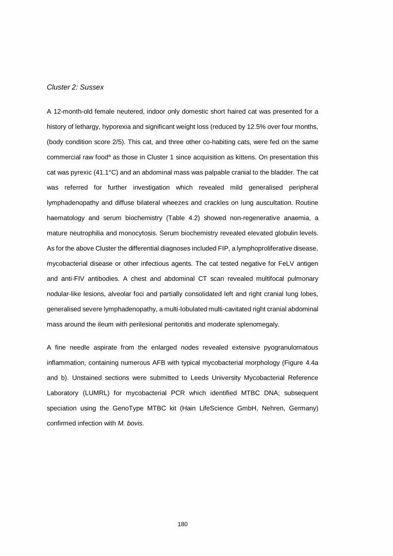
180
Cluster 2: Sussex
A 12-month-old female neutered, indoor only domestic short haired cat was presented for a
history of lethargy, hyporexia and significant weight loss (reduced by 12.5% over four months,
(body condition score 2/5). This cat, and three other co-habiting cats, were fed on the same
commercial raw fooda as those in Cluster 1 since acquisition as kittens. On presentation this
cat was pyrexic (41.1°C) and an abdominal mass was palpable cranial to the bladder. The cat
was referred for further investigation which revealed mild generalised peripheral
lymphadenopathy and diffuse bilateral wheezes and crackles on lung auscultation. Routine
haematology and serum biochemistry (Table 4.2) showed non-regenerative anaemia, a
mature neutrophilia and monocytosis. Serum biochemistry revealed elevated globulin levels.
As for the above Cluster the differential diagnoses included FIP, a lymphoproliferative disease,
mycobacterial disease or other infectious agents. The cat tested negative for FeLV antigen
and anti-FIV antibodies. A chest and abdominal CT scan revealed multifocal pulmonary
nodular-like lesions, alveolar foci and partially consolidated left and right cranial lung lobes,
generalised severe lymphadenopathy, a multi-lobulated multi-cavitated right cranial abdominal
mass around the ileum with perilesional peritonitis and moderate splenomegaly.
A fine needle aspirate from the enlarged nodes revealed extensive pyogranulomatous
inflammation, containing numerous AFB with typical mycobacterial morphology (Figure 4.4a
and b). Unstained sections were submitted to Leeds University Mycobacterial Reference
Laboratory (LUMRL) for mycobacterial PCR which identified MTBC DNA; subsequent
speciation using the GenoType MTBC kit (Hain LifeScience GmbH, Nehren, Germany)
confirmed infection with M. bovis.
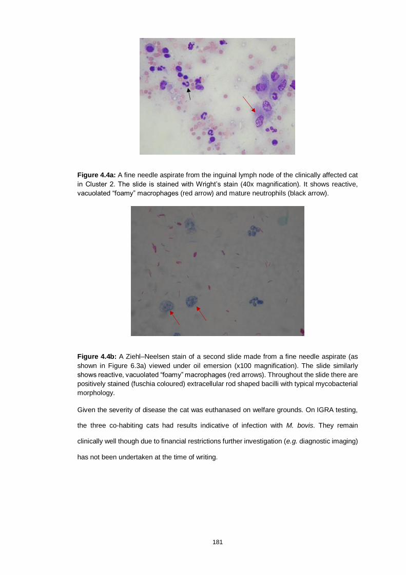
181
Figure 4.4a: A fine needle aspirate from the inguinal lymph node of the clinically affected cat
in Cluster 2. The slide is stained with Wright’s stain (40x magnification). It shows reactive,
vacuolated “foamy” macrophages (red arrow) and mature neutrophils (black arrow).
Figure 4.4b: A Ziehl–Neelsen stain of a second slide made from a fine needle aspirate (as
shown in Figure 6.3a) viewed under oil emersion (x100 magnification). The slide similarly
shows reactive, vacuolated “foamy” macrophages (red arrows). Throughout the slide there are
positively stained (fuschia coloured) extracellular rod shaped bacilli with typical mycobacterial
morphology.
Given the severity of disease the cat was euthanased on welfare grounds. On IGRA testing,
the three co-habiting cats had results indicative of infection with M. bovis. They remain
clinically well though due to financial restrictions further investigation (e.g. diagnostic imaging)
has not been undertaken at the time of writing.

182
Cluster 3: Norfolk
A 15-month-old, male neutered domestic long-haired cat was presented, it had been
exclusively indoor housed since acquisition as a kitten along with a female sibling. Both cats
were fed exclusively the same brand of raw fooda as the other Clusters from the time that they
were kittens. The cat was presented with diarrhoea and on examination had pale mucus
membranes, abdominal distention, poor muscle condition, and marked generalised superficial
lymphadenomegaly involving the submandibular, prescapular and popliteal lymph nodes, in
addition to an abdominal mass assumed to be enlarged mesenteric lymph node(s). The cat
was borderline pyrexic (39.5°C). Routine haematology and serum biochemistry (Table 4.2)
revealed anaemia, a mature neutrophilia and a mild increase in SMDA. Fine needle aspirates
of the abdominal mass/lymph node showed large numbers of macrophages (Figure 4.5) and
intracytoplasmic AFB; confirmatory mycobacterial PCR and speciation at LUMRL on the
remaining slide, confirmed MTBC DNA was present but as the slide had been previously
stained the DNA was of insufficient quality to differentiate between MTBC organisms i.e. M.
bovis or M. microti. An IGRA test showed a weakly positive result suggestive of M. bovis
infection. Treatment was attempted with pradofloxacin (Veraflox™, Bayer, 3mg/kg q 24 hours
by mouth PO), azithromycin (Zithromax™, Pfizer, 9mg/kg q 24 hours PO) and doxycycline
(generic, Summit, 10mg/kg PO q 24 hours). Unfortunately, the cat continued to deteriorate
and was euthanased on welfare grounds. The sibling remains clinically well at the time of
writing and screening by IGRA test gave a positive result for infection with a non-MTBC
mycobacterium species.
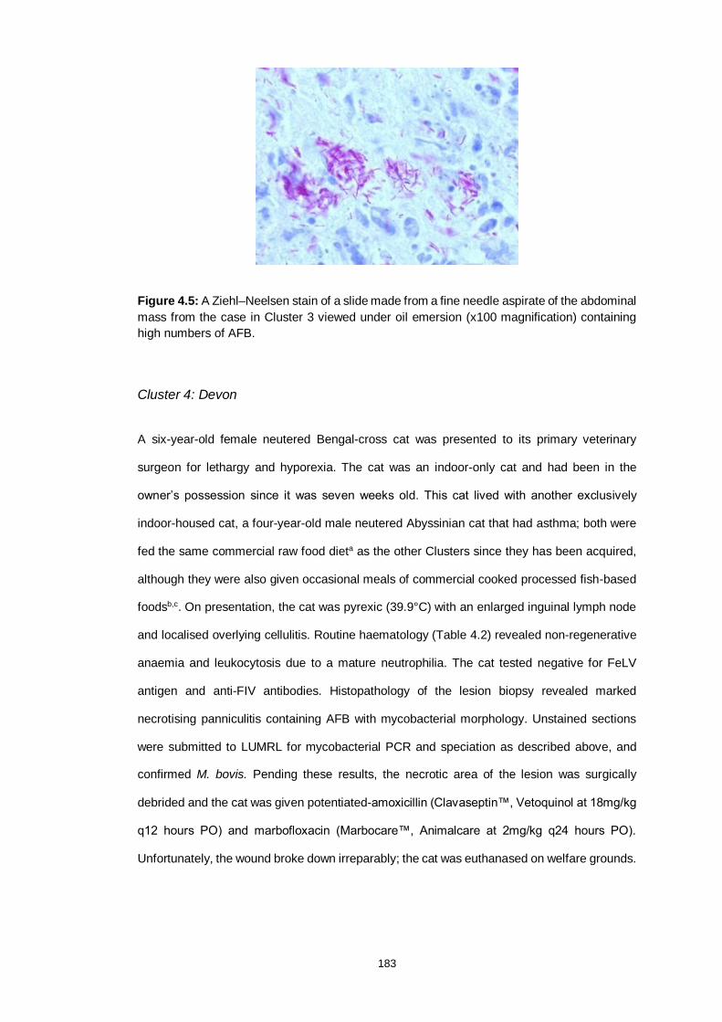
183
Figure 4.5: A Ziehl–Neelsen stain of a slide made from a fine needle aspirate of the abdominal
mass from the case in Cluster 3 viewed under oil emersion (x100 magnification) containing
high numbers of AFB.
Cluster 4: Devon
A six-year-old female neutered Bengal-cross cat was presented to its primary veterinary
surgeon for lethargy and hyporexia. The cat was an indoor-only cat and had been in the
owner’s possession since it was seven weeks old. This cat lived with another exclusively
indoor-housed cat, a four-year-old male neutered Abyssinian cat that had asthma; both were
fed the same commercial raw food dieta as the other Clusters since they has been acquired,
although they were also given occasional meals of commercial cooked processed fish-based
foodsb,c. On presentation, the cat was pyrexic (39.9°C) with an enlarged inguinal lymph node
and localised overlying cellulitis. Routine haematology (Table 4.2) revealed non-regenerative
anaemia and leukocytosis due to a mature neutrophilia. The cat tested negative for FeLV
antigen and anti-FIV antibodies. Histopathology of the lesion biopsy revealed marked
necrotising panniculitis containing AFB with mycobacterial morphology. Unstained sections
were submitted to LUMRL for mycobacterial PCR and speciation as described above, and
confirmed M. bovis. Pending these results, the necrotic area of the lesion was surgically
debrided and the cat was given potentiated-amoxicillin (Clavaseptin™, Vetoquinol at 18mg/kg
q12 hours PO) and marbofloxacin (Marbocare™, Animalcare at 2mg/kg q24 hours PO).
Unfortunately, the wound broke down irreparably; the cat was euthanased on welfare grounds.

184
A post-mortem examination was conducted within the Containment Level 3 facility of the
Roslin Institute, University of Edinburgh; there was a surgical defect in the right inguinal area
where the affected skin, fat and lymph node had been removed as well as pathological tissue
break down (Figure 4.6a and b). On examination of the abdomen many small white, well
demarcated hard lesions were present throughout the spleen on both the visceral and cut
surfaces. Within the thorax, similar lesions were visible in the caudal lung lobes and
pericardium, there was a moderate degree of pericardial effusion.
Figure 4.6a: Post-mortem examination of a six-year-old female neutered Bengal-cross cat
(Cluster 4) shows evidence of the extensive nature of surgical resection required to remove
the granulomatous panniculitis present.
Figure 4.6b: Evidence of the breakdown of the surgical wound and surrounding tissue
damage which ultimately led to the euthanasia of this patient on welfare grounds.

185
Grossly pathological tissues were taken from the lung and spleen, homogenised in 1%
collagenase solution containing 10% penicillin-G, decontaminated with 4% NaOH for 30
minutes and sown onto OADC supplemented 7H11 (Middlebrook, UK) slopes, Löwenstein–
Jensen medium with pyruvate and Stonebrink slopes in order to isolate and genotype the
causative strain. After nine weeks, small colonies were visible on the plates with typical
morphological appearance of M. bovis. In-house PCR testing at Edinburgh followed by PCR
performed at LUMRL confirmed the colonies to be M bovis; the APHA was notified and sent a
second tissue sample for independent culture analysis, which is pending. The in-contact cat
remains clinically well at the time of writing but the results of a screening IGRA test indicate
M. bovis infection. Thoracic radiographs and abdominal ultrasound have identified no
pathology. However, this cat has experienced significant recent weight loss (~12%
bodyweight), so the owner has elected to treat with standard anti-mycobacterial therapy (drugs
as dosages as above in Cluster 1).
Cluster 5: Essex
A 15-month-old, indoor-only, male neutered Maine Coon cat was presented for lethargy and
constipation of approximately 48 hours. The cat had been fed exclusively on the same dieta
as the other Clusters since it was acquired at 12-weeks-old. Physical examination revealed
pyrexia (40.0°C) and a palpable, mobile, non-painful abdominal mass in the region of the
mesentery. Ultrasound examination confirmed the presence of a 9x13cm mass adjacent to an
enlarged mesenteric lymph node and a small amount of free fluid. Fine needle aspirates of the
mass revealed large, foamy macrophages, mesenchymal cells and histiocytic infiltration.
Routine haematology and serum biochemistry (Table 4.2) revealed non-regenerative anaemia
and marked mature neutrophilia.
An exploratory laparotomy was performed and the fluid, mass and mesenteric lymph node
were resected (Figure 4.7) and submitted for histological examination. Large numbers of
macrophages were observed and a presumptive diagnosis of FIP was made. However, further
investigation to confirm FIP, including immunohistochemistry on the biopsy material, reverse
transcription quantitative PCR (RT-qPCR) for mutated FCoV on the abdominal fluid, and

186
serum FCoV titre quantification and alpha-1 acid glycoprotein was not compatible with FIP.
During this investigation, retrospective staining of the original biopsy revealed individual AFB
with mycobacterial morphology scattered in large areas of necrosis. The cat improved clinically
post-surgery but six weeks later developed a cough; thoracic radiography revealed a mass
dorsal to the carina (Figure 4.8), whilst a repeat abdominal ultrasound revealed a 2cm mid-
ventral mass caudal to the spleen. Cytology of the abdominal mass again revealed a
predominance of macrophage infiltration with intracellular non-staining rods present. Cytology
with ZN-staining revealed AFB, initial PCR testing at the University of Edinburgh indicated
infection with M. bovis (protocol as outlined in Chapter 2) and the remaining slides were
submitted for mycobacterial PCR and speciation at LUMRL but it was not possible to isolate
sufficient mycobacterial DNA for definitive speciation. The IGRA screening tests on the two
co-habiting domestic short haired cats which ate mainly commercial dry food, separately from
the Maine Coon cat, showed positive responses indicative of M. bovis infection though they
were clinically well.
Figure 4.7: The Maine Coon in Cluster 5 had a large abdominal mass (centre) measuring
8x13cm removed during an exploratory laparotomy. The local draining mesenteric lymph node
(left) was also removed during surgery as was the ~12ml of ascitic fluid (right). The fluid was
tested for mutated feline coronavirus by RT-qPCR but was negative. Retrospective
histopathological examination of the mass (centre) showed scattered mycobacteria

187
Figure 4.8: A left latero-lateral of the thorax of the Maine Coon cat from Cluster 5. There is a
mass (total diameter 1.2cm) at the level of the carina. A cough had become clinically evident
at the time this radiograph was taken. It resolved within three days of anti-mycobacterial triple
antibiotic therapy.
The Maine Coon was treated with the recommended anti-mycobacterial triple antibiotic
therapy1,2 comprising marbofloxacin (Marbocyl™, Vetquinol, 2mg/kg every [q] 24 hours PO),
azithromycin (Zithromax™, Pfizer, 9mg/kg q 24 hours PO) and rifampicin (generic, 11mg/kg q
24 hours PO). Two months after starting medication the abdominal mass was no longer
palpable and the cat’s weight had increased. However, following owner-led cessation of
therapy at this stage, the cat subsequently relapsed and was euthanased on welfare grounds
and has been submitted to APHA officials for post-mortem examination, the results of which
are pending at the time of writing. Due to the owner being immunocompromised, the two in-
contact IGRA positive cats were also euthanased.

188
4.3 Follow-up investigations
The current legislation applicable to England (The Tuberculosis (England) Order 2014) Article
625 states that suspicion of tuberculosis in the carcass of any pet mammal is notifiable, and as
such the Secretary of State has been duly notified of all of the cases presented here via the
APHA. However, there is no statutory obligation of investigation in non-bovine animals and so
current investigations have been led by the University of Edinburgh and largely financed,
where conducted, by the University and/or owners.
Public Health England (PHE) have similarly been informed of the cases and all owners have
been offered the opportunity to discuss the zoonotic aspects that these cases present should
they wish to do so with PHE. PHE are currently adopting a precautionary public health
approach similar to that which has been described previously26. The owners of two of the
affected cats were found to be IGRA and Mantoux test (tuberculin skin test) positive
respectively, one has required the instigation of anti-tuberculosis medical therapy.
Potential sources of exposure to M. bovis were explored with the owners; all except the raw
commercial food were excluded. Excluded sources included wildlife contact (all cats were
solely indoor cats), the presence of rodent populations inside the buildings in which the cats
lived, access to raw milk, and exposure to any known infectious humans.
As the only common factors between all clusters are that they were diagnosed with the same
infection (M. bovis), were exclusively indoor cats and were fed a commercially available raw
food product produced by a single manufacturera, the Food Standards Agency (FSA) have
similarly been informed and they are investigating appropriately.
The authors at the University of Edinburgh have been sent detailed batch information and
photographs of and/or actual samples of the diet from three of the five clusters, as was being
fed to the cats at the time the diagnoses were made (Figure 4.9a-c). Notably, all three of these
samples were the venison product produced by the same company. On questioning the
owners; the venison product from the single manufacturer comprised ~80% of the ration of

189
one cat (Cluster 4), the sole intake of another (Cluster 5) and the “vast majority” of the diet of
a third (Cluster 2). The remaining two clusters (Cluster 1 and 3) fed a variety of the products
from the same company, including the venison product.
As such, the evidence we have collected during our investigations of these cases provide
compelling, if only circumstantial, evidence of an association between the diet of these cats
and their M. bovis infections.
The company was alerted, and an internal investigation by them led to the voluntary withdrawal
of the venison version of their food from sale; ‘because some of the ingredients were not
inspected in line with EU requirements. The absence of inspection means the safety of the
product cannot be confirmed and may therefore carry a potential risk.’ The batches withdrawn
are those dated as best before between March 2019 and August 2019 and pre-date those
submitted for testing at the University of Edinburgh. The FSA are now undertaking this area of
the outbreak investigation independently.
Figure 4.9: Three of the five Clusters had retained the food product that the affected cats were
being fed at the time that they became ill. One owner sent photos taken by themselves (4.9a
- left), the other two owners sent the food to the University of Edinburgh by refrigerated courier
and the photos were taken inside the Containment Level 3 facility prior to further testing (4.9b
– centre and 4.9C - right).

190
4.4 Discussion
This outbreak currently includes 13 cats, in five separate Clusters, that were found to be
infected with M. bovis; six cats were clinically unwell at diagnosis (five of these are now dead),
while seven were found to be positive by IGRA i.e. they had evidence that they have been
infected with M. bovis, although how many of these latter cats’ are likely to become ill is
unclear. Three have not been assessed further by their veterinarian (Cluster 2), and one
(Cluster 4) has already lost a great deal of weight, potentially related to M. bovis infection (this
cat has been treated for M. bovis infection and has since recovered all its previously lost
weight). While still circumstantial, the only probable source of the infection is that M. bovis
infected the cats when they were all exposed to the same commercially available raw food.
New cases are still occurring and further data collection is in progress at the time of writing,
but the authors intend to make readers aware of the details and the scale of this problem as
soon as practically possible. The cases reported here appear to be limited to cats which have
been fed this feline-specific food (i.e. no dogs have yet been affected) and no owners are self-
reporting as unwell.
The feeding of commercial raw meat based diets (RMBD) to companion animals has increased
substantially in popularity in recent years, not just in the UK but globally27,28. In the USA, sales
of RMBD doubled in the five years to October 2017 and have expanded by 15.9% in last year
alone, making the industry worth an estimated US$195 to the United States economy29. In
Europe, a recently published study from the Netherlands found that 51% of dog owners fed
their animals either completely or partially on RMBD30. A similar increase has been seen in
the UK, with what were only a handful of companies registered as producers of RMBD a few
years ago having increased to a current excess of 80 registered with the Department for the
Environment, Farming and Rural Affairs (DEFRA)27. Shapiro et al (2017)31 found in a survey
of Australian cat breeders that raw meat was fed as an integral constituent of the diet by 89%
of respondents. The reasons behind so many people adopting RMBD feeding are numerous
and complex. Some owners feel that it is physiologically more “natural” for their pet to consume
food similar to that they would have eaten at an earlier stage in their evolution (i.e. pre-

191
domestication)31-33. There have been speculative suggestions that RMBD feeding may be
beneficial in preventing the onset of dental disease, food responsive inflammatory bowel
disease and atopy, and a recent paper has shown that the microbiome of dogs fed RMBD is
more diverse than when fed processed diets31-34. The topic of what the veterinary profession
should be advising clients with respect to RMBD is highly emotive on both sides of the debate,
and the evidence for and against is frequently either low quality or simply anecdotal reports
with a lack of rigorous, properly controlled trials31-35. There is however, consensus that home-
prepared RMBD should be generally avoided as it is extremely difficult to make them
nutritionally balanced35. This concern has been overcome by the introduction of commercially
available complete RMBD which comply with the strict legislative requirements, overseen by
the European Pet Food Industry Federation (FEDIAF), to ensure nutritional and energy
requirements are met if any given product was provided as the sole ration for the life time of
the animal in question36-38.
Unfortunately, statutory regulations governing RMBD only account for the presence or
absence of a limited number of infectious organisms39. It is one of the few areas regarding
RMBD on which there is agreement; that there is a significant increase in the risk of infectious
diseases to both pets and owners through the consumption and handling of such products on
a regular basis40-46. Almost all RMBD are sold frozen in order to extend their expiry dates and,
in the case of some organisms (e.g. nematodes such as Trichinella species) reduce or
eliminate contamination47. In studies of the zoonotic infectious agents present within thawed
RMBD, Salmonella species have received the most attention44-46. Although subclinical
infections occur frequently in animals, Salmonella species can cause gastroenteritis and even
septicaemia48-50. For example, Stiver et al. reported fatal septicaemic salmonellosis after
RMBD feeding in two cats51. Other studies have successfully recovered a variety of viable
zoonotic pathogens including Escherichia (E.) coli serotype O157:H7, Listeria
monocytogenes, Sarcocystis spp. Campylobacter spp. and Toxoplasma gondii from RMBD44-
46,52. At least one such RMBD contaminant, Shiga Toxin releasing E. coli (STEC) has been
linked to human deaths in the UK, and Public Health England are attempting to educate
owners about the risks associated with RMBD53. In the USA, raw cat food has similarly had to

192
be recalled due to contamination with Listeria monocytogenes54. At this stage, as cases are
continuing to be diagnosed there is insufficient evidence to ascertain if this is a single, one off
event or an ongoing problem. However, in either instance this outbreak raises concerns over
the strength of current regulation with regards to meat inspection procedures and may
necessitate an increase in trained veterinary meat inspection of a carcass before it is passed
as fit for animal consumption. With particular reference to wild venison, one such alteration
could be that the entire gralloch (all viscera) be brought to the slaughter-house for veterinary
inspection.
Our study is the first to report infection of companion animals with an MTBC pathogen which
appears to be associated with feeding a RMBD, with 13 cats in five households infected, as
identified to date. Not only does this imply that the owners were handling contaminated
material, but the gastrointestinal presentation means that the cats may have been shedding
M. bovis into their home environment, creating the potential for even greater exposure to
owners. Of further concern, the Pet Food Manufacturers Association guidance to owners
handling RMBD is that they should clean surfaces, utensils and animal food bowls with “soap
and hot water”55. Pasteurisation requires milk to be heated to 71.5°C and held at that
temperature for a minimum of 15 seconds to inactivate M. bovis56; it is highly unlikely that
owners would be able to achieve this in a domestic setting, so M. bovis is very likely to be able
to persist on kitchen surfaces even after the owners feel that they have been “cleaned”.
Furthermore, M. bovis is able to survive for at least 60 days in standing water57, such as might
be found in or around a kitchen sink, over which time further organisms may be added with
each prepared meal making it highly plausible that the small dose needed to infect a human
could be reached.
The severity of clinical disease that was seen in the cats we report here is much greater than
we would typically expect to see through infections acquired through hunting infected rodents.
The clinical signs of pyrexia, anaemia and leukocytosis (caused by a mature neutrophilia) are
not typically present in feline tuberculosis cases and the young age of many of the cats affected
is also striking, as is that the cats were indoor only, and not from areas of England known to

193
have endemic M. bovis in rodents, badgers, other wildlife and cattle58-60. These factors may
mislead clinicians presented with future cases to not consider tuberculosis as a potential
differential diagnosis, delaying the time until correct diagnosis and thus exposing owners to
possible infection for extended periods.
Many of the cats presented in these clusters were either too sick to attempt treatment, or
clinically deteriorated despite attempted therapies giving a case fatality rate, to date, of 83%
compared to our more usual 70-80% successful treatment rate2. The severity of infection was
similar to that which we previously reported in an outbreak of M. bovis tuberculosis in a pack
of working Foxhounds where it was concluded that the source of infection was most likely the
feeding of contaminated raw meat61. That outbreak resulted in a higher than typical clinical
attack rate for tuberculosis (approximately 10% of the group) and when the hounds started to
display clinical signs, they deteriorated so rapidly that euthanasia was required on welfare
grounds within hours to days61. Additionally, feline abdominal M. bovis infections acquired in
a nosocomial setting and cats with disseminated M. bovis infection that appeared to have
followed extensive ingestion, also displayed rapid clinical progression, and severe signs such
that euthanasia on welfare grounds was quickly required62,63. The parallels between these
fulminant outbreaks suggests that there may be an underlying difference in the
immunopathogenesis of the disease, whereby gastrointestinal/peritoneal challenge of
companion animals with M. bovis results in more severe disease that is less amenable to
treatment and may thus have implications for the clinical management of gastrointestinal
tuberculosis cases in comparison to the more typical dermal presentation. Alternatively, or
additionally, it may be that strains of M. bovis which are capable of establishing infections in
companion animals following gastrointestinal/peritoneal challenge may have or may utilise
additional virulence factors which make these infections clinically more aggressive. The latter
scenario would have profound implications on the zoonotic risk to owners if they were being
exposed to M. bovis with inherently greater virulence.
The IGRA was used to detect M. bovis infection in all six of the unwell and a further six of the
in-contact cats. However, one in-contact cat (Cluster 3) was IGRA tested and found to have a

194
greater response to PPDA than PPDB, i.e. a pattern not consistent with that expected of M.
bovis infection. It may be that this cat has not been challenged with M. bovis but has co-
incidentally and/or previously been sensitised to antigens of the M.avium-intracellulare-
complex. However, studies in calves sensitised to environmental mycobacteria and infected
with M. bovis, have concluded that M. bovis infection may be concealed for some time in this
situation64. It is therefore possible that this cat may develop a different response, i.e. that
typical of M. bovis, if it were to be retested in the future.
Work by our team at the University of Edinburgh is ongoing to develop a rapid PCR assay
which we hope can be performed on tissue lysates, (unstained) cytology slides and formalin-
fixed biopsy tissues. Where this was used in these cases it proved extremely useful; but, its
use outwith a research capacity will require more extensive evaluation before this is made
available to clinicians. Similarly, the team is continuing to work with Biobest Laboratories to
validate to use of the feline IGRA in the detection and monitoring of tuberculosis in cats.
Urgent work is now needed to establish the extent of any contamination of the RMBD pet food
chain in the UK, and a full epidemiological investigation is underway to establish where lessons
can be learned in order to future safeguard animals being fed RMBD.
Management of suspect cases
The team at the University of Edinburgh are always keen to hear from clinicians who may
have, or have had, suspicions about similar cases and can offer our experience in both
diagnosing and managing these patients. Currently, we are advising that animals known to
have been exposed to the recalled food, or who have been exposed to a confirmed case but
are displaying no clinical signs should be tested by IGRA at least four weeks after the last
known exposure. If these tests are positive then diagnostic imaging should be undertaken to
assess for structural (i.e. active) disease (full body CT or radiographs and, ideally, abdominal
ultrasonography). Where present, active disease should be treated appropriately; as outlined
in O’Halloran and Gunn-Moore, 20171. There is currently no evidence on which to base the
appropriate course of action for animals that are IGRA test positive but lack evidence of

195
structural disease. It may be appropriate in these cases to closely monitor body weight,
condition score and resting respiratory rate and to investigate further if these decline.
Alternatively some owners may elect to give prophylactic therapy; isoniazid (which is
occasionally used in humans for this purpose) can be used for six months but experience of
its use in cats is limited and toxicities may occur. Instead, some owners may choose to treat
using the triple combination of anti-tuberculous therapy for three months, given that the use of
this protocol is well established and potential side effects are well documented.
4.5 Conclusion
This report follows our initial alert regarding an ongoing outbreak of M. bovis tuberculosis in
pet cats across the UK. It describes six clinically sick cats and seven in-contacts with a
mortality rate of 83%. Lifestyle investigation revealed the common factors between clusters
were that the cats were exclusively indoor only and fed a commercially available raw food
product produced by a single manufacturer. The Food Standards Agency, Animal and Plant
Health Agency, Public Health England and the food manufacturer concerned have been
notified/informed. Our results provide compelling, if circumstantial, evidence of an association
between the commercial raw diet of these cats and their M. bovis infections.
4.6 References
aNatural Instinct®; https://www.naturalinstinct.com
bSainsbury's Delicious Recipes 1+ Adult Cat Food Tuna in Jelly
cWHISKAS® 1+ Years Complete Dry Cat Food with Tuna 340g
dhttps://www.tbhub.co.uk/ Accessed 3rd April 2019
1. O’Halloran, C. & Gunn-Moore, D. Mycobacteria in cats: An update. In Pract. 39, 399–406
(2017).
2. O’Halloran, C. & Dobromylskyj. Clinical mycobacterial diseases of companion animals:
part 2. Management of companion animal mycobacteriosis. Companion Animal. DOI
doi.org/10.12968/coan.2017.22.11.652

196
3. Gunn-Moore, D. A., Gaunt, C. & Shaw, D. J. Transbound. Emerg. Dis. 60, 338–344 (2013).
4. O’Brien, C. R. et al. Feline leprosy due to Candidatus ‘Mycobacterium tarwinense’: Further
clinical and molecular characterisation of 15 previously reported cases and an additional
27 cases. J. Feline Med. Surg. 19, 498–512 (2017).
5. Gunn-Moore, D. A. et al. Mycobacterial disease in a population of 339 cats in Great Britain:
II. Histopathology of 225 cases, and treatment and outcome of 184 cases. J. Feline Med.
Surg. 13, 945–952 (2011).
6. Gunn-Moore, D. A. et al. Mycobacterial disease in cats in Great Britain: I. Culture results,
geographical distribution and clinical presentation of 339 cases. J. Feline Med. Surg. 13,
934–944 (2011).
7. Gunn-Moore, D. A. Feline mycobacterial infections. Vet. J. 201, 230–238 (2014).
8. Pesciaroli, M. et al. Tuberculosis in domestic animal species. Research in Veterinary
Science 97(S), S78-S85 (2014).
9. Roberts, T. et al. Unusual cluster of Mycobacterium bovis infection in cats. Veterinary
Record 174(13), 326 (2014).
10. Pollock, J.M. and Neill, S.D. Mycobacterium bovis infection and tuberculosis in cattle.
Veterinary Journal 163, 115-127 (2002).
11. Laprie, C., Duboy, J., Malik, R. & Fyfe, J. Feline cutaneous mycobacteriosis: A review of
clinical, pathological and molecular characterization of one case of Mycobacterium microti
skin infection and nine cases of feline leprosy syndrome from France and New Caledonia.
Vet. Dermatol. 24(6), 561-569 (2013).
12. Gibbens, N. Mycobacterium bovis infection in cats. Veterinary Record 174(13), 331-332
(2014).
13. Broughan, J.M. et al. Mycobacterium bovis infections in domesticated non-bovine
mammalian species. Part 2: A review of diagnostic methods Veterinary Journal 198(2),
346-351 (2013).
14. Warren, R. M. et al. Differentiation of Mycobacterium tuberculosis complex by PCR
amplification of genomic regions of difference. Int. J. Tuberc. Lung Dis. 10, 818–822
(2006).
197
15. Hussain, A. et al. COMPARISON OF FASTSURE TB DNA AND MGIT 960 FOR THE
DETECTION OF MYCOBACTERIUM TUBERCULOSIS COMPLEX IN CLINICAL
SPECIMENS. Pakistan Armed Forces Medical Journal, 63(1) (2013)
16. Rhodes, S. G., et al. Adaptation of IFN-gamma ELISA and ELISPOT tests for feline
tuberculosis. Veterinary Immunology and Immunopathology 124(3-4), 379-384 (2008)
17. Rhodes, S. G., et al. Interferon-γ test for feline tuberculosis. Veterinary Record 162(14),
453-455 (2008)
18. Adams L.G. et al. In vivo and in vitro diagnosis of Mycobacterium bovis infection. Revue
Scientifique et Technique de L’Office Internationale Des Epizooies 20, 304-324 (2001).
19. Mostowy, S., Cousins, D., Behr, M.A. Genomic Interrogation of the Dassie Bacillus
Reveals It as a Unique RD1 Mutant within the Mycobacterium tuberculosis Complex.
Journal of Bacteriology 186 (1),104-109 (2004).
20. Smith, N.H. et. al. Mycobacterium microti: More diverse than previously thought Journal
of Clinical Microbiology, 47 (8), 2551-2559 (2009).
21. Jennings AR. The distribution of tuberculous lesions in the dog and cat with reference to
the pathogenesis. Veterinary Record 61:380-384 (1949).
22. Parsons, S. D. C. et al. Detection of Mycobacterium tuberculosis infection in dogs in a
high-risk setting. Research in Veterinary Science 92(3), 414-419 (2012).
23. O’Halloran, C., et al. Mycobacterium bovis in pet cats. Veterinary Record 183 (16), 510
(2018).
24. Vet Times. Investigations under way in wake of feline TB outbreak In Press (2018).
Available at https://www.vettimes.co.uk/news/investigations-under-way-in-wake-of-feline-
tb-outbreak/
25. Annon. The Tuberculosis (England) Order 2014. Accessed 6 Nov 2018 Available at:
http://www.legislation.gov.uk/uksi/2014/2383/made
26. Phipps, E. et al. Bovine tuberculosis in working foxhounds: Lessons learned from a
complex public health investigation. Epidemiology and Infection. In press (2018).
27. Waters, A. Raw diets: are we at a turning point? Veterinary Record 181(15), 384 (2017).
28. Finley, R. et al. Human health implications of Salmonella-contaminated natural pet treats

198
and raw pet food. Clinical Infectious Diseases 42(5), 686-691 (2006).
29. Sprinkle, D. US pet food market. Pet-Food-Industry.com Accessed 6 Nov 2018 Available
at: https://www.petfoodindustry.com/articles/6826-us-pet-food-market-to-reach-us27-
billion-in-2018?v=preview
30. Corbee RJ, Breed RD, Hazewinkel HAW. Feeding practice of dog owners active on
internet forums. Poster session presented at: 17th European Society of Veterinary and
Comparative Nutrition congress; 2013 Sep 19-21; Ghent, Belgium.
31. 12305 Shapiro, A. J., Norris, J. M., Bosward, K. L. and Heller J. Q Fever (Coxiella
burnetii) Knowledge and Attitudes of Australian Cat Breeders and Their Husbandry
Practices Zoonoses and Public Health, 64, 252–261 (2017) doi: 10.1111/zph.
32. Handl S. The "BARF" trend - advantages, drawbacks and risks. Veterinary Focus
24(3),16-23 (2014).
33. Schlesinger D & Joffe D. Raw food diets in companion animals: a critical review. Can Vet
J 52(1), 50-4. (2011)
34. Sandri, M. et al. Raw meat based diet influences faecal microbiome and end products of
fermentation in healthy dogs. BMC Veterinary Research 13, 65 (2017).
35. Weeth LP. Home-prepared diets for dogs and cats. Vetlearn. 35(3) (2013).
36. EFSA (European Food Safety Authority), ECDC (European Centre for Disease Prevention
and Control). The European Union summary report on trends and sources of zoonoses,
zoonotic agents and food-borne outbreaks in 2014. EFSA Journal 13, 4329 (2015).
37. EC Regulation 1069/2009 Accessed 6 Nov 2018 Available at:
https://ec.europa.eu/food/safety/animal-by-products_en
38. Pet Food Association: Legislation. Accessed 6 Nov 2018 Availale at
https://www.pfma.org.uk/uk-pet-food-legislation
39. Commission of the European Communities. Commission Regulation (EC) No 2073/2005
on microbiological criteria for foodstuffs. Official Journal of the European Union
073/2005(microbiological criteria for foodstuffs) (2005).
40. Bojanić, K. et al. Isolation of Campylobacter spp. from Client-Owned Dogs and Cats, and
Retail Raw Meat Pet Food in the Manawatu, New Zealand. Zoonoses and Public Health

199
64(6), 438-449 (2017)
41. Baede, V.O. et al. Raw pet food as a risk factor for shedding of extended-spectrum beta-
lactamase-producing Enterobacteriaceae in household cats. PLoS ONE 12(11),
e0187239 (2017).
42. Stogdale, L. et al. Just how pathogenic to pets and humans are the bacteria in the
concentrations found in raw pet foods (multiple letters) Canadian Veterinary Journal
46(11), 967-971 (2005).
43. Annon. Study finds Listeria in raw pet foods. Journal of the American Veterinary Medical
Association 245(8), 876 (2014).
44. Annon. Raw feeding of pets: safe and nutritious - or reckless and irresponsible? Veterinary
Record 181(15), pp. 386-387 (2017).
45. Finley, R. et al. (2008) The occurrence and antimicrobial susceptibility of Salmonellae
isolated from commercially available canine raw food diets in three Canadian cities
Zoonoses and Public Health 55(8-10), 462-469 (2008).
46. Nemser, S. et al. Investigation of Listeria, Salmonella, and toxigenic Escherichia coli in
various pet foods. Foodborne Pathogens and Disease 11(9), 706-709 (2014).
47. Center for Disease Control. Parasitology – Trichinellosis Accessed 6 Nov 2018 Available
at: https://www.cdc.gov/parasites/trichinellosis/prevent.html
48. Tauni, M.A. et al. Outbreak of Salmonella typhimurium in cats and humans associated
with infection in wild birds Journal of Small Animal Practice 41(8), 339-341 (2000).
49. Wall P.G. et al. Chronic carriage of multidrug resistant Salmonella typhimurium in a cat
Journal of Small Animal Practice 36(6), 279-281 (1995).
50. Fauth, E. et al. Salmonella bacteriuria in a cat fed a Salmonella-contaminated diet. Journal
of the American Veterinary Medical Association 247(5), 525-530 (2015).
51. Stiver, S.L. et al. Septicemic salmonellosis in two cats fed a raw-meat diet. Journal of the
American Animal Hospital Association 39(6), 538-542 (2003). .
52. Tenter, A.M. Toxoplasma gondii: From animals to humans. International Journal for
Parasitology 30(12-13), 1217-1258 (2000).
53. Limb, M. Human deaths linked to raw pet food. Veterinary Record 83 (17) (2018).

200
54. Quality Assurance and Food Safety. Raw Pet Food Recalled Due to Listeria, E. Coli
Contamination Concerns. Accessed 6 Nov 2018 Available at:
https://www.qualityassurancemag.com/article/radagast-food-recalled-listeria-e-coli/
55. Pet Food Manufacturer Association. Raw Feeding Factsheet. Accessed 6 Nov 1028.
Available at https://www.pfma.org.uk/raw-feeding-factsheet
56. The Dairy Products (Hygiene) Regulations 1995
57. Vaerewijck, M.J.M.et al. Mycobacteria in dr. inking water distribution systems: Ecology
and significance for human health. FEMS Microbiology Reviews 29(5), 911-934 (2005).
58. Jenkins, H. E. et al. The prevalence, distribution and severity of detectable pathological
lesions in badgers naturally infected with Mycobacterium bovis. Epidemiology and
Infection 136(10), 1350-136 (2008).
59. Anon. Online map shows TB breakdowns. Veterinary Record. 177 (2) (2015).
60. Cavanagh, R. et al. Mycobacterium microti infection (vole tuberculosis) in wild rodent
populations Journal of Clinical Microbiology 40(9), 3281-3285 (2002).
61. O’Halloran et al. An outbreak of tuberculosis due to Mycobacterium bovis infection in a
pack of English Foxhounds (2016-2017). Transboundary and Emerging Diseases. In
press.
62. Murray, A. et al. Nosocomial spread of Mycobacterium bovis in domestic cats. Journal of
Feline Medicine and Surgery. 17 (2) 173-180 (2015).
63. Černá, P., O'Halloran, C., SjatkovskaJ, O. et al. Outbreak of tuberculosis caused by
Mycobacterium bovis in a cattery of Abyssinian cats in Italy. Transboundary and Emerging
Diseases, In Press (2018).
64. Amadori, M., S. et al. Diagnosis of Mycobacterium bovis infection in calves sensitized by
mycobacteria of the avium/intracellulare group. J. Vet. Med. B Infect. Dis. Vet. Public
Health 49, 89-96 (2002).

201
PART 3
Tuberculosis in UK cats associated with a commercial raw food
diet.
Editorial, Journal of Feline Medicine and Surgery, August 2019
Conor O’Halloran & Danièlle Gunn-Moore
Royal (Dick) School of Veterinary Studies and The Roslin Institute, University of Edinburgh,
Easter Bush Campus.
Johann Schonlein coined the term “tuberculosis” in the year 1834, 48 years before the
causative agent Mycobacterium (M.) tuberculosis was first visualised by Robert Koch. At that
time, one in seven of the world’s population was thought to be infected and despite significant
efforts in the intervening almost two hundred years, this prevalence has actually increased to
an estimated one in three. Whilst M. tuberculosis may have been the first tuberculous
organism identified, it is restricted to infecting a very small number of species. By comparison,
the causative agent of ‘zoonotic tuberculosis’, i.e. M. bovis, has a very wide host range. So
named for is predilection for bovine hosts, M. bovis can actively infect an exceptionally broad
range of species which include but are not limited to; cattle, badgers, deer, camelids, a variety
of rodents, human and non-human primates, racoons, possums, and coyotes. Not surprisingly,
our feline patients are also susceptible.
Last year, we were alerted to a highly unusual group of feline M. bovis tuberculosis cases
(O’Halloran et al. 2018a). Initially, the striking feature was that these cases presented with a
rare manifestation of disease; alimentary tuberculosis (Figure 4.10): this compares to most
‘typical’ cases of tuberculosis in cats in the UK, which are cutaneous and/or pulmonary. The
investigation became even more complex when diagnostic tests confirmed M. bovis infections;
this was unexpected as these cats were living in areas of the UK which have either a very low

202
prevalence of M. bovis in cattle and badger (such as Lancashire) or are officially M. bovis free
(such as Scotland).
Figure 4.10: The grossly abnormal intestine and associated lymph nodes removed for
specialist mycobacterial culture from a one year old, male neutered Persian cat who was
weaned onto the Natural Instinct Wild Venison product and continued to eat it as a sole ration
until developing marked chronic weight loss, hyporexia and an expiratory wheeze. He was
euthanased on welfare grounds due to the severity of clinical signs.
We investigated the lifestyle histories of the cases to try and identify any possible routes of
exposure to M. bovis. We discovered that all of the cats were indoor-only (i.e. they were at
negligible risk from wildlife or rodents), they were of different breeds and were mostly young,
none had been fed raw (unpasteurised) milk, or lived with people with active tuberculosis. The
only common feature was that all were fed (in varying proportions) a commercial raw food diet;
Natural Instinct, Wild Venison (Figure 4.11). On discussion with the company and from their
subsequent investigations, it was concluded that some of the ingredients had not been meat-
inspected in line with EU requirements so the safety of the product could not be confirmed;
the company voluntarily recalled the food.
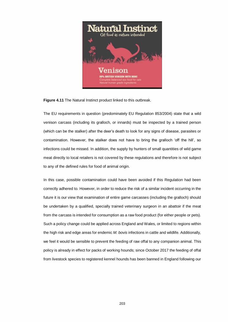
203
Figure 4.11 The Natural Instinct product linked to this outbreak.
The EU requirements in question (predominately EU Regulation 853/2004) state that a wild
venison carcass (including its gralloch, or innards) must be inspected by a trained person
(which can be the stalker) after the deer’s death to look for any signs of disease, parasites or
contamination. However, the stalker does not have to bring the gralloch ‘off the hill’, so
infections could be missed. In addition, the supply by hunters of small quantities of wild game
meat directly to local retailers is not covered by these regulations and therefore is not subject
to any of the defined rules for food of animal origin.
In this case, possible contamination could have been avoided if this Regulation had been
correctly adhered to. However, in order to reduce the risk of a similar incident occurring in the
future it is our view that examination of entire game carcasses (including the gralloch) should
be undertaken by a qualified, specially trained veterinary surgeon in an abattoir if the meat
from the carcass is intended for consumption as a raw food product (for either people or pets).
Such a policy change could be applied across England and Wales, or limited to regions within
the high risk and edge areas for endemic M. bovis infections in cattle and wildlife. Additionally,
we feel it would be sensible to prevent the feeding of raw offal to any companion animal. This
policy is already in effect for packs of working hounds; since October 2017 the feeding of offal
from livestock species to registered kennel hounds has been banned in England following our

204
previous work investigating another raw-food associated M. bovis outbreak and no new cases
have since been reported (Anonymous, 2017; O’Halloran et al. 2018b).
Our initial investigation into this current outbreak has been reported in this publication
previously (O’Halloran et al. 2019) and new cases continue to occur. To date we have
diagnosed 45 cases with active clinical disease, 73 with inactive (i.e. latent) infection and
screened in excess of 50 well cats where owners are concerned regarding historical exposure
to the contaminated product. A more up to date report of our findings is in preparation and will
be published as soon as possible.
Author Contributions:
O’Halloran, C. wrote the manuscript under the supervision of Professor Gunn-Moore.
References:
Anonymous (2017). Authorisation A6 in: Derogations from product controls under Regulation
(EC) 1069/2009 and Commission Regulation (EU) 142/2011. Authorisations by the Secretary
of State to enable derogations to be used in England. May 2011 (last updated: October
2017). Available at:
https://www.gov.uk/government/uploads/system/uploads/attachment_data/file/650731/abp-
exemptions-201710.pdf Accessed on 7 June 2019.
Food Standards Agency (2018) Natural Instinct recalls Wild Venison Available at:
https://www.food.gov.uk/news-alerts/alert/fsa-prin-65-2018 Accessed on 7 June 2019.
O’Halloran, C., Gunn-Moore, D., Reed, N., et al. (2018a) Mycobacterium bovis in pet cats
Veterinary Record 183, 510.
O'Halloran, C., Hope, J.C., Dobromylskyj, M., et al. (2018b) An outbreak of tuberculosis due
to Mycobacterium bovis infection in a pack of English Foxhounds (2016–2017).
Transboundary and Emerging Diseases. 65: 1872– 1884.
O’Halloran, C., Ioannidi, O., Reed, N., et al. (2019) Tuberculosis due to Mycobacterium bovis
in pet cats associated with feeding a commercial raw food diet. Journal of Feline Medicine and
Surgery. In press https://doi.org/10.1177/1098612X19848455

205
PART 4
Feline tuberculosis caused by Mycobacterium bovis infection of
domestic UK cats associated with feeding a commercial raw food
diet.
Acknowledgements
Thanks go to all of the veterinarians and owners of these cats for facilitating the investigation
and giving permission for the findings to be published. Additional thanks go to the APHA staff
who assisted with co-ordinating the epidemiological investigation and conducting post mortem
examinations and laboratory testing; Tony Roberts (APHA Wallingford), Jeff Jones (APHA
Carmarthen), Livio Pittalis & Cornelia Bidewell (APHA Bury St Edmunds), Carlo Bianco &
Pedro Sanchez-Cordon (APHA Weybridge), Harriet Stacey (APHA Starcross), Sheila Corrieri
(APHA Perth), Malgorzata (Gosia) Dabrowska-Jones (APHA veterinary adviser for non-
bovines), Ricardo de la Rua-Domenech (APHA Veterinary Adviser (policy) to DEFRA TB
Programme), Eleftheria Palkopoulou and Vivienne MacKinnon (Veterinary Lead Field Services
North Scotland).
206
Abstract
Mycobacterium (M.) bovis can infect cats and has proven zoonotic risks for owners. We
describe an outbreak of M. bovis in cats from across England and Scotland associated with
feeding a commercial raw food diet. Investigations of (pyo)granulomatous lesions,
lymphadenopathy, and pulmonary and/or alimentary disease of 45 affected cases over one
year identified infections with M. bovis genotype 10:a.
Infection was confirmed by culture in a small number of cases (n=5); PCR was used in
combination with or as an alternative to culture (n=12) and/or infection was indicated by
positive interferon gamma release assay (IGRA; n=34). At-risk cats were screened by IGRA;
follow-up testing identified 77 sub-clinically infected cats.
The five culture-positive cases were distributed across England and Scotland, within areas of
the UK considered to be low-risk for endemic M. bovis. Whole genome sequence analysis of
recovered isolates revealed identical nucleotide sequences providing evidence that these
infections are epidemiologically linked.
Lifestyle investigations revealed that affected cats were mainly indoor-only, all of whom had
been fed Natural Instinct’s Wild Venison food as at least part of their diet. As far as possible,
other possible sources of infection were explored and were excluded; including wildlife contact,
access to raw milk, the presence of rodent populations inside the buildings in which the cats
lived, and exposure to known infectious humans.
Owners and examining vets of test-positive cats were referred to their local health
professionals; four owners and one veterinary surgeon were found to have a high likelihood of
latent tuberculosis infections. One pregnant owner underwent prophylactic treatment. Though
it is not possible to conclusively demonstrate a zoonotic origin for these infections, the potential
for this should be considered when possible cases are identified.
Our results provide strong evidence that the commercial raw diet of these cats was the likely
route of their M. bovis infections.

207
4.7 Introduction
Mycobacterium (M.) bovis is one of the member species of the Mycobacterium tuberculosis-
complex (MTBC) which are capable of causing tuberculosis across a broad taxonomy of
species including but not limited to humans, cattle, deer, dogs and cats (Sales et al. 2001;
Philips et al. 2003; Allen, 2017).
Feline infections with M. bovis are strongly co-localised to areas of the UK with a high
prevalence of infections in cattle (Gunn-Moore et al. 2011); termed high risk areas (HRA) of
the UK by the Department for Environment, Food and Rural Affairs (DEFRA). Conversely, M.
bovis infections are very rare in low risk areas (LRA) and somewhat higher incidence in the
edge zones between LRA and HRA regions as evidenced by a culter of M. bovis infections in
cats in Berkshire in 2013 (Roberts et al. 2013).
Within the MTBC group of pathogens, M. bovis stands out as the least host restricted and thus
has significant zoonotic potential (Sales et al. 2001; Biet et al. 2005; Suzuki et al. 2010).
Disease in humans due to infection with M. bovis, termed ‘zoonotic tuberculosis’ by the World
Health Organisation (WHO), is a major global public health priority which resulted in nearly
150,000 cases and at least 12,500 deaths worldwide in 2010 (World Health Organisation,
2017). Clinically, about a third of feline mycobacteriosis cases in the UK are caused by MTBC
pathogens; in the only published study, M. microti was cultured from 19% of all submitted
cases of feline mycobacteriosis, and 15% were caused by M. bovis (Gunn-Moore et al. 2011a;
O’Halloran and Gunn-Moore, 2017).
Characterisation of the typical clinical manifestations of feline mycobacterial diseases in the
UK has shown that 74% of cases present with single or multiple cutaneous lesions, and 10-
16% with pulmonary or systemic signs (Gunn-Moore et al. 2011a). The remainder of cases
are small numbers of relatively rare presentations such as alimentary, joint or ocular
mycobacteriosis (Gunn-Moore 2014; Lalor et al. 2017; Stavinohova, et al. 2019).

208
We have previously reported on a highly unusual group of 13 feline M. bovis tuberculosis
cases (O’Halloran et al 2018a; O’Halloran et al. 2019). Initially, the striking feature was that
these cases all presented with one of the rarest manifestations of disease, alimentary
tuberculosis. The investigation became more complex when diagnostic tests indicated M.
bovis infections as these cats were living in areas of the UK which have either a very low
prevalence of M. bovis in cattle and badger (such as the North East of England) or Scotland,
which holds officially M. bovis free status (for cattle (APHA, 2017; O’Halloran et al. 2019).
Suspicions were raised with regards to a possible link to diet as all cats were indoor-only, had
no access to alternative sources of infection as far as we could determine, and they were all
fed a single commercial raw food product, Natural Instinct Wild Venison (Food Standards
Agency, 2018; O’Halloran et al. 2019).
We concluded that these initial findings provided circumstantial evidence of an association
between the commercial raw diet of these cats and their M. bovis infections. Since then, further
cases have been diagnosed and an epidemiological investigation undertaken; the purpose of
this publication is to outline in full our findings and conclusions from both the clinical and
epidemiological investigations.
4.8 Clinical Investigations
4.8.1 Clinical Presentation of Cases
Forty-five cats from 41 households have been diagnosed with active tuberculous disease
linked to this outbreak at the time of writing. All 45 cases were presented to their primary
veterinary surgeons over the course of one year (July 2018 to July 2019). The majority of
affected cats (39/45, 86.7%) were pedigree breeds including; eight Bengal cats, seven
Persians, seven Maine Coons, six Siamese, four British Shorthairs, two Ragdolls and one
each of Burmese, Toyger, British Blue, Sphinx and one Bengal cross cat. The remaining six
cats were non-pedigree domestic shorthairs. Cases came from all over England and mainland
Scotland.

209
The age at presentation ranged from three-month-old kittens to 13 years, with a median age
of two years. There was no statistical difference between the number of male or female cats
presenting with disease (Χ21=1.48, p>0.05); half of the cats (22/45, 48.8%) were neutered
males, ten cats were neutered females (22.5%), whilst nine were entire females (20.0%) and
four (8.9%) were entire males.
Clinical signs noted by the owners of these cats were most frequently non-specific signs of ill
health, including combinations of lethargy (18/45 cats, 40.0%), hyporexia (15/45 cats, 33.3%)
and poor or declining body weight/condition (15/45 cats, 33.3%).
The clinical abnormality most frequently reported by examining vets was “respiratory signs”,
which was recorded in 75.6% (34/45) of cases. The severity of these signs ranged from mild
increases in respiratory rate and/or effort through to a persistent cough and pneumonia
causing life-threatening dyspnoea; it was the recorded reason for euthanasia in two cases
(Figures 4.12a and 4.121b). In a further three cases (6.7%) thoracic lymphadenomegaly of the
peribronchial, sternal and/or mediastinal lymph nodes was the only gross lesion detected on
investigation of the primary presenting complaint (lethargy in two cats and hyporexia in one
cat).
Figure 4.12a: A latero-lateral thoracic radiograph of an affected cat (a four-year-old female
neutered indoor-only Bengal cat) showing two cavitating lesions (red box) within the lower
respiratory tract. The cat clinically deteriorated and was euthanased on welfare grounds.

210
Figure 4.12b: Post mortem examination of the same cat radiographed in Fig. 4.12a revealed
extensive fibrinopurulent exudate (black arrows) in the thoracic cavity and the extent of the
granulomatous lesions. Lung samples submitted for mycobacterial culture were positive for M.
bovis.
Abdominal masses (granulomas) were palpable in 23 of the 45 cats (51.1%), whilst the
abdominal viscera (excluding lymph nodes) were found to be structurally abnormal (either
enlarged or containing granulomatous lesions, Figure 4.13) in a further four cases (8.9%) as
visualised by abdominal radiography, ultrasonography and/or computed tomography (Figure
4.14). The abdominal lymph nodes associated with the alimentary system (Figure 4.15) were
enlarged in four cats (8.9%) and enlarged as part of a generalised lymphadenomegaly in a
further eight cats, hence 12 cases (26.7%) had abdominal lymphadenomegaly. Overall, the
alimentary tract and associated structures were found to contain gross pathology in 31 of the
45 cases (68.9%), but clinical signs directly relatable to the gastrointestinal system (vomiting,
diarrhoea and constipation) where the main presenting sign in only six cats (13.3%).

211
Figure 4.13: The spleen of an 18-month-old male neutered Burmese cat at laparotomy
surgery showing multifocal raised white-pink circular lesions later confirmed as granulomatous
on histopathology and containing acid-fast bacilli with mycobacterial morphology. The liver of
this cat was similarly affected
Figure 4.14: An ultrasound examination of an 18-month-old male neutered indoor only Maine
Coon cat showing an approximately 2x2.5cm mass (red box) in the abdominal cavity.

212
Figure 4.15: A markedly enlarged mesenteric lymph node (black arrow) from the same cat as
Figure 4.14 visualised during the laparotomy surgery.
Uncommon clinical signs included non-healing skin lesions (Figure 4.16) in four cats (8.9%),
orthopaedic involvement in two cats (4.4%) and optic disease (Figure 4.17) in two cats (4.4%).
Figure 4.16: The non-healing skin wound over the left pelvis of a two-year-old male, indoor
only, neutered domestic shorthair cat that was subsequently culture positive for M. bovis.

213
Figure 4.17: Optic examination of a two-year-old indoor only female neutered British Shorthair
cat presented for conjunctival hyperaemia revealed a mass over the optic nerve, later
diagnosed as granulomatous chorio-retinitis with acid-fast bacilli as well as the observable
retinal detachment that was also confirmed histologically following enucleation.
At clinical examination pyrexia was present in 17 cats (37.8%); where recorded, the
temperature ranged from 39.8ºC to 41.1ºC, with a median of 40.0ºC (normal range 37.7°C -
39.1°C). Routine haematological evaluation revealed a non-regenerative anaemia in a further
ten cats (22.2%) and a mild to moderate mature neutrophilia in six cats (13.3%). Serum
biochemistry was largely unremarkable; only two cats (4.4%) showed increased liver enzyme
activity, affecting both alanine aminotransferase and alkaline phosphatase. One of these cats
had a large hepatic granuloma extending into the biliary duct, whilst the cause of these
elevations was not identified in the second cat.
When tested, all cats were found to be negative for Feline Leukaemia Virus antigen and anti-
Feline Immunodeficiency Virus antibodies.
4.8.2 Mycobacterial Testing
Specialist mycobacterial culture and subsequent genotyping at a national reference laboratory
(Animal and Plant Health Agency [APHA], Weybridge in the UK) remains the only test that can
214
result in official confirmation of M. bovis infection of companion animals (Middlemiss and Clark,
2018). From this outbreak, 11 grossly diseased tissue samples were obtained at post mortem
examination and submitted for mycobacterial culture at APHA Weybridge. All post mortem
examinations were conducted either at APHA veterinary investigation centres by APHA
pathologists or at the Roslin Institute, University of Edinburgh under Containment Level 3
conditions by the authors (COH/JH). The results of ten cultures are available at the time of
writing with the results from one case still pending. Five of these ten cultures (50.0%) resulted
in positive results and genotype analysis has identified all five isolates as genotype 10:a. The
five cats were resident across the UK in LRA locations. Subsequent whole genome analysis
of the cultured isolates revealed them to be identical at the nucleotide level.
Although it is the gold standard test, mycobacterial culture does have a number of limitations.
For example, as a prerequisite it requires the acquisition and submission of fresh biopsy
samples; however, given the location of many of the lesions in these cases inside body cavities
(i.e. the chest and abdomen) this made retrieving enough sample for culture ante mortem an
invasive procedure that many of these cases were not clinically fit to undergo. For most of
these cats it was only possible to obtain very small samples by needle aspiration or
tracheal/bronchial wash samples for cytological examination (Figure 4.18). In a number of
cases where larger biopsies had been obtained, they had been formalin-fixed and submitted
for histopathological examination and as such were unavailable for culture. For these cats, we
utilised molecular testing methods (GenoTypeMTBC; Hain Lifescience GmbH, Germany) at
Leeds University Mycobacterial Reference Laboratory (LUMRL). This methodology is used as
standard of care in the diagnosis of human tuberculosis, and although not specifically validated
for companion animal tissue samples, this assay is able to diagnose human mycobacterial
infections as being caused by MTBC organisms and then specific gene probes are used to
determine the infecting species within this group, including the identification of M. bovis
(National Institute for Health and Care Excellence, 2016). Twelve cat samples of the 15 tested
generated positive PCR test results; one indicated infection with an MTBC organism but there
was insufficient DNA for speciation and 11 were identified to the species level as M. bovis

215
infections. Two of the M. bovis positive test results were subsequently confirmed by
mycobacterial culture of lesioned tissue samples.
Figure 4.18: A cytological sample obtained by fine needle aspirate from the submandibular
lymph node of a two-year-old entire, indoor-only female Bengal cat. The sample is stained with
a commercial Romanowsky stain variant (Diff-QuikTM) and reveals pyogranulomatous
inflammation typical of feline mycobacterial infections with non-staining ‘ghost’ bacilli typical of
Mycobacteria spp. (black arrow). Image taken under oil emersion at x100 magnification.
The cat identified by PCR as being infected with an MTBC organism was included in this
outbreak investigation even though molecular testing did not identify M. bovis DNA. This cat
was an 18-month-old indoor-only domestic shorthair which had been fed exclusively on the
implicated diet since its acquisition by the owners at eight weeks of age, it had a
contemporaneous interferon-gamma release assay (IGRA) result indicative of M. bovis
infection, as well as lymph node aspirate cytology which confirmed the presence of acid-fast
bacilli with mycobacterial morphology. Cumulatively these findings were taken to support its
inclusion as an affected cat.
Immunodiagnostic assays for the diagnosis of tuberculosis are used globally to test human
and bovine populations (Wood and Jones, 2001; McCormick-Baw et al. 2018). The most

216
successful of these to date is the IGRA; first developed to aid in the eradication of M. bovis
from cattle populations in Australia, the assay detects antigen-specific T-cell responses to
mycobacterial antigens from circulating leukocytes to diagnose infection (Rothel et al. 1992;
Wood and Jones, 2001). This first test, the BOVIGAMTM assay, is now widely used to detect
the cell-mediated immune response to M. bovis in cattle, sheep, goats, buffalo and bison
(Wood and Jones 2001; Pesciaroli, M. et al. 2014, Anusz et al. 2017). With small adaptations
the test protocol has proved to be extremely useful in humans, and the WHO’s new 2018
guidelines on the management of human tuberculosis now support the global use of IGRA
testing at-risk populations (Chee et al., 2018, McCormick-Baw et al. 2018). In 2008,
adaptations to the test procedure were undertaken by the TB Research Group at the then
Veterinary Laboratories Agency, now APHA, to optimise the test for domestic cats (Rhodes et
al. 2008a). The resultant feline IGRA is now commercially available via Biobest Laboratories,
Edinburgh, UK and although the number of tested cats remains too small to allow official
validation to World Organisation for Animal Health (OIE) standards, several publications have
indicated that this is a useful test to generate rapid information on the infection status of cats
(Rhodes et al. 2008b, Rhodes et al. 2011, Gunn-Moore et al. 2014). The methodology and
interpretation of this test has been previously published and the same protocol has been used
for cats involved in this outbreak (Rhodes et al. 2008a; O’Halloran et al. 2019).
The advantages of IGRA tests compared to other mycobacterial tests are that they can be
performed rapidly, only require venipuncture of the patient, rather than more invasive tissue
biopsy, and they can support a diagnosis in the absence of an aetiological agent as is common
in feline tuberculous granulomas, which are usually paucibacillary (Wood and Jones, 2001;
Gunn-Moore et al. 2011b). It can be particularly useful for the diagnosis of cases where the
lesion is completely inaccessible, such as the two cases of chorio-retinal disease. Of particular
relevance to this investigation is that this test is currently the cheapest option available to
owners. The combination of lack of invasiveness, speed of generating results, and lower cost
are the likely reasons for the high number of IGRA tests in this instance rather than the
validated gold standard culture.

217
During this outbreak 34 of the 45 clinical cases (75.6%) tested positive using the feline IGRA
and gave test results indicative of M. bovis infection. Of these, eight cases (23.5% of positive
IGRA tests) were supported/confirmed by either PCR test (five cats) or subsequent positive
cultures (three cats).
4.8.3 Sub-clinical Cases
As the outbreak continued and the commercial raw diet was implicated, owners who fed the
diet became concerned that their cats could be infected. In addition, some of the affected cats
lived with other cats, so some owners were worried that their other cats could be at risk. Since
the IGRA is the only test for mycobacterial infection that can be performed on an animal with
no visible lesions many of these owners chose to have their cats tested using this assay. To
date, 143 at-risk, clinically well cats have been tested by IGRA, of which 83 (58.0%) results
indicate likely M. bovis infections.
In a previous outbreak, we utilised an IGRA for screening kennel-housed Foxhounds with a
similar test protocol to the feline IGRA (O’Halloran et al. 2018b). At that time, we considered
that a positive interferon gamma response to mycobacterial proteins/peptides was indicative
of a significant antigen specific T-cell response. Furthermore, such a response was thought to
be suggestive of significant challenge and probable infection, rather than just exposure to
these mycobacteria. We therefore applied the same criteria to these cats and advised that the
83 cats that generated positive IGRA results should be further evaluated for the presence of
lesions by full clinical veterinary examination and diagnostic imaging. In six cases (7.2%),
imaging revealed mild interstitial pulmonary disease (Figure 7) and associated
lymphadenomegaly that were not causing clinical signs, this allowed the clients to treat these
cats at an early stage of infection (see below).
The remaining 77 cats were IGRA positive with no detectable clinical disease. These cats
were deemed to be likely to be sub-clinically infected and owners were advised that they
should be closely monitored for the onset of clinical signs and/or prophylactic therapy could
be instigated at their discretion.

218
4.8.4 Current Approach to Suspect Cases
The team at the University of Edinburgh are always keen to hear from clinicians who may
have, or have had, suspicions about similar cases to continue to gather evidence about this
outbreak as well as offering our experience in the diagnosis and management of these
patients.
Currently, the number of newly diagnosed clinical cases appears to be in decline. The first
cases were presented in July 2018 and peaked in June 2019, six months after the implicated
product was recalled. No new clinically active cases have been diagnosed, to our knowledge,
since the middle of July 2019.
The ante mortem testing of any suspect cases can be performed as described earlier – by
specialist mycobacterial culture at APHA Weybridge (the gold standard confirmatory test) or
by PCR and/or IGRA. Where present, the treatment of active feline tuberculous disease
should be carefully considered; there is no requirement to euthanase M. bovis infected pets in
the UK, but appropriate therapy requires long-courses (typically 3-6 months) of combination
antibiotic administration which can be difficult to achieve in cats, can cause side effects and
be costly for owners. If treatment is to be attempted then it must be done appropriately,
consisting of a combination of a macrolide/azalide, a fluoroquinolone and rifampicin
administered daily as outlined in O’Halloran and Gunn-Moore (2017), to prevent the induction
of antimicrobial resistance to these medications and to protect public health.
Veterinary clinicians in the UK should be aware that, under the Tuberculosis (England) Order
2014 and equivalent Statutory Instruments in Scotland and Wales (Anon, 2014), in the event
that non-bovine animals are euthanased or die when there is suspicion of M. bovis infection
then this is notifiable to the Secretary of State (via APHA). The carcase must be detained on
the premises pending APHA advice.
It is currently advisable that animals known to have been exposed to Natural Instinct Wild
Venison food, or who have been exposed to a confirmed case but are displaying no clinical

219
signs, should not be tested by IGRA for at least four weeks after the last known exposure. The
time required for cats to mount a detectable T-cell response to M. bovis is unknown as no
controlled challenge studies have been performed in this species; therefore the estimate of
four weeks is extrapolated from bovine data where such time-course challenge studies have
been conducted (Rhodes et al. 2000). If first line tests are positive then diagnostic imaging
should be undertaken to assess for structural (i.e. active) disease; this should comprise full
body computed tomography (CT) scan or thoracic radiographs and, ideally, abdominal
ultrasonography or alternatively, abdominal radiographs. There is currently no evidence on
which to base the appropriate course of action for animals that are IGRA positive but lack
evidence of structural disease. It is likely appropriate in these cases to closely monitor body
weight, condition score and resting respiratory rate, and to investigate further if these change.
Alternatively, some owners may elect to give prophylactic therapy; isoniazid (which is
occasionally used in humans for this purpose) can be given for six months, but experience of
its use in cats is limited, it has a narrow therapeutic index (in humans and mice) and therefore
toxicity may occur (El-Sayed et al. 2010; Fernandes, G. et al. 2017). Instead, owners may
choose to treat these animals using the triple combination of anti-tuberculous therapy (above)
for three months, given that the use of this protocol is well established and the potential side
effects are well documented (O’Halloran and Gunn-Moore, 2017). In this outbreak, the
commonly reported side effects (defined as an incidence of between 1 in 10 and 1 in 100
patients) to treatment included pruritic skin erythema caused by rifampicin and/or neutropenia
induced by chronic fluoroquinolone administration.
4.8.5 Public Health Considerations
The incidence of zoonotic transmission of M. bovis from cats is very rare with only six cases
reported in the literature (Lewis-Jonsson, 1946; Isaac, et al. 1983; Une and Mori 2007; Roberts
et al. 2014). However, there may be particular risk to owners who have compromised immune
function such as those receiving chemotherapy, radiotherapy or tumour necrosis factor alpha
(TNF-α) inhibitors (Hofland, et al. 2013; Simonsen, et al. 2017). Therefore, owners of cats with

220
active disease should be advised to seek medical advice if they are at elevated risk and/or if
cats have lesions that are discharging (purulent) material or if they have a productive cough.
Owner screening tests (QuantiFERON GoldTM IGRA and/or tuberculin skin tests) conducted
to date have identified likely latent tuberculosis infections (LTBI) in four owners and one vet.
All of these people had been exposed to severe feline cases with purulent lesions, however,
it is unclear at this time if this represents zoonotic transmission from these cats or if these
individuals were infected at an earlier time. Public health agencies in the UK have previously
assessed the risk to immunocompetent owners from M. bovis infected pets and have
concluded that there is ‘very low’ risk of zoonotic transmission (Public Health England, 2014).
4.9 Epidemiological Investigation
In general terms, the only common factors linking these cases were the raw cat food consumed
(Natural Instinct, Wild Venison) and being mostly indoor cats.
Two breeding colonies were affected; one in the north of England had three clinical cases, two
IGRA positive cats and two IGRA negative cats in the colony. Tracing rehomed kittens
identified ten with clinically signs and two that were IGRA positive but clinically well. The
second breeder, located in the south of Scotland, had eight breeding cats of which one was
clinically sick and four were well but IGRA positive. Ten kittens from two litters rehomed from
the second breeder were traced; half of these were tested by IGRA and all five were negative.
There were no other common associations between the remaining cats.
The alimentary tract and associated lymphoid structures were found to contain lesions in 31
of the 45 cases (68.9%) which is consistent with challenge via the gastrointestinal route,
supporting the hypothesis of food as the common source of infection. Five cats presented with
large lung abscesses, which could be associated with spread via the retropharyngeal lymph
nodes, inhalation of M. bovis from contaminated food during eating or may represent
dissemination of disease.

221
All cases were presented to their primary veterinary surgeons between July 2018 and July
2019. A working estimate for the incubation period is two and five months for alimentary
infections of tuberculosis in cats. This estimate is based on experimental data from the 1900’s
presented by Francis (1958) in which very large doses (1-10mg) of M. bovis when given by
mouth and produced lesions 40 to 121 days later. However, extrapolating for more advanced
pathology and the potentially lower doses encountered in any contaminated raw pet food could
extend this significantly. Beyond this, the kinetics of feline M. bovis infection following oral
challenge are unknown; however, it is likely that in at least some cats, a long sub-clinical phase
is seen, as in cattle and humans (where it is termed latency), and this may extend the upper
range of incubation to many months or even years.
Identifying a likely infection window and determining whether the cats were all infected by one
particular batch of contaminated food are difficult questions to answer. The earliest signs were
reported in the first weeks of July 2018; if a minimum incubation period is approximately two
months, then a contaminated batch of raw pet food manufactured in early May 2018 would be
implicated. With the apparent last cases being identified in July 2019, this theoretically includes
all batches of food made as late as January 2019 when the production of this product was
ceased by Natural Instinct. The age of the cats affected limits the potential infection window to
some degree; excluding kittens weaned directly onto Natural Instinct Wild Venison, the
youngest case was one year old when signs were first seen in August 2018, which extends
the potential time of potential challenge back into 2017.
With the possibility of long sub-clinical chronic infections adding a considerable degree of
open-endedness to the upper incubation period range, contaminated batches could
theoretically have been produced during the whole of 2018, possibly 2017, or even earlier.
The temporal clustering of the majority of the early cases from July to September 2018
suggests a most likely possible common infection window of February to July 2018 (calculated
from a most likely incubation period range of two to five months). Subsequent cases therefore
either include a period of sub-clinical infection or represent the later consumption of stored
food. The breeder in the north of England was clear about the infection window for their cats;

222
it had to be limited to June 2018 as this was the only time the cats had been fed Natural
Instinct, Wild Venison.
An important factor that potentially explains the extended period of this cluster is long storage
of the pet food in home freezers. Since Natural Instinct, Wild Venison was marketed with
‘Limited Availability’ owners reported that they tended to bulk purchase when it was available.
The freezer shelf life was several months (calculated as nine months from data on packaging)
and so a single contaminated batch could feasibly be responsible for this whole cluster.
Overall, the most likely infection window was probably March to June 2018 and all cases
associated with this raw pet food could plausibly fit within this window, with stored food and
sub-clinical incubation giving rise to later cases. The estimated two to five month incubation
period is supported by the currently observed decline in the presentation of new cases
following the recall of the food in December 2018.
Transmission to the cats from the local environment was considered to be very unlikely
because a) the cats were kept indoors, with a few cats having limited usually supervised
outdoor access, and b) the majority of cases were located in the LRA for endemic M. bovis.
Infected vermin entering the houses cannot be ruled out for cases in only two households
located in higher incidence bovine tuberculosis areas. However, they were not in regions with
endemic M. bovis genotype 10:a infections.
A contaminated raw commercial cat food (Natural Instinct, Wild Venison) is the most likely
route of infection for these cats. However, alternative pathways cannot be completely ruled
out. Most owners of pedigree cats were located in the LRA and obtained kittens from within
the LRA, but other cats in the same households had prior ownership which could not be fully
investigated, so they could have had previous outdoor access in the HRA. Some of the non-
pedigree cats had vague histories, including an in-contact cat that was obtained from a website
with no history at all. Incomplete information creates some uncertainty for several of the cases
and failure to confirm infection by the gold standard test for the majority of cases adds a degree
of uncertainty as to the true number of M. bovis cases seen.

223
Within this group of 45 clinical cases, M. bovis genotype 10:a has been cultured from five
cases from different areas of England and Scotland, with an additional Scottish sample where
culture is still pending. Whole genome sequencing of the five M. bovis 10:a isolates by APHA
Weybridge found them to be identical to the nucleotide level, i.e. they were identical isolates
of M. bovis, which provides very strong evidence in support of the dietary infection hypothesis.
Furthermore, the nearest cattle isolates with only one single nucleotide polymorphism (SNP)
difference form a tight cluster of six farms in Oxfordshire. An M. bovis isolate from at least one
of two wild fallow deer shot in this area had an identical genome sequence to these cattle
isolates suggesting that wild deer from this area may be the origin of contaminated venison.
Precise information detailing the source of venison meat and offal is needed to definitively tie
the pet food and deer from this area together which is the subject of an ongoing investigation
by the Food Standards Agency (FSA) and the local authority. However, an example record list
for a known venison supplier to the pet food manufacturer includes one estate in west
Buckinghamshire that is very close to the farm cluster and provides a tentative but plausible
risk pathway for these cat cases consistent with the whole genome sequence analyses.
Further analysis of pet food obtained from owners could provide definitive evidence of the
transmission route. However, batches matching the earlier period of the most likely infection
window are unsurprisingly not available and no M. bovis has been isolated from the few
samples tested by APHA, the FSA or the University of Edinburgh to date.
During this investigation, M. bovis was cultured from additional cats that were fed Natural
Instinct Wild Venison and were euthanased, often with severe mesenteric lymph node
pathology. However, the cultured isolates were found to be 11:e and 25:e. Therefore, there
were thought to most likely represent alternative sources of infection. Conversely, M. bovis
genotype 10:a was isolated from a cat during the timeframe of this outbreak which triggered a
further investigation but this cat was located within the homerange of this genotype, had a
superficial skin lesion consistent with inoculation via a bite wound and had no history of being
fed a raw food diet so was excluded from the group of outbreak cases.

224
4.10 Discussion
This cluster of cases represents the only documented outbreak of feline tuberculosis putatively
caused by feeding pet cats a contaminated commercial raw food diet. Previous examination
of feline mycobacterial cases in the UK showed the highest frequency of infections in non-
pedigree, male cats with frequently reported hunting behaviour that then develop skin lesions,
probably following inoculation of mycobacteria directly into the site of the lesion (Gunn-Moore
et al. 2011a, Gunn-Moore 2014). By contrast, the majority of cases affected in this outbreak
were pedigree or pedigree-cross breeds (86.7%) and there was no statistically significant
predilection for males to be affected.
The reason for the high proportion of pedigree cats affected is likely that owners of these cats
are more likely to feed raw food diets. Although this has not been studied systematically,
anecdotal evidence suggests that a growing number of pedigree cat breeders in the UK (and
elsewhere) are advocating feeding so-called biologically appropriate raw food (BARF) diets to
owners acquiring kittens (Handl, 2014; Waters, 2017). The evidence supporting the use of
these diets remains poor despite their increasing popularity in recent years (Schlesinger and
Joffe, 2011; EFSA, 2015). There have been speculative suggestions that BARF feeding may
be beneficial in preventing the onset of dental disease, may alleviate food responsive
inflammatory bowel disease and atopy, and a recent study demonstrated that the microbiome
of dogs fed BARF diets is more diverse than when fed processed diets - though no health
benefits were identified as a result of this diversity (Handl, 2014; Sandri et al. 2017). Kittens
raised on a rabbit-based raw diet had better stool quality, assessed with a visual grading
system, than their cooked diet fed peers, but both groups grew similarly (Glasgow et al. 2002).
However, some of the kittens fed the rabbit-based diet developed dilated cardiomyopathy
secondary to taurine deficiency after several months of feeding (Glasgow et al. 2002). A critical
review by Schlesinger and Joffe (2011) concluded that the evidence advanced for the claimed
health benefits of raw feeding amounted to opinions and claims that were, at best, supported
by data that was of low relevance.

225
A major and growing concern relating to feeding pets these diets are the risks posed by
microbiological contamination that are not addressed by cooking. The bacteriological quality
of raw commercial pet foods, assessed by total bacterial, coliform or Escherichia (E.) coli
counts, has been noted to fail threshold levels for raw human meat products in a high
proportion of sampled foods in both Europe and North America (Freeman and Michel, 2001;
Towell, 2008; Nemser et al. 2014; Kölle and Schmidt, 2015; Nilsson, 2015). The most recent
systematic review, conducted by Davies et al. (2019), found evidence that the bacteria in these
diets often encode resistance to critically important antibiotics such as extended-spectrum
cephalosporins, and raw-fed pets create an elevated risk of shedding these resistant bacteria
(Nilsson, 2015; Baede et al. 2017; Davies et al. 2019). Despite this evidence, the vast majority
of owners interviewed (94%) in a separate survey believed raw diets to be “absolutely” safe
for their dogs (Davies et al. 2019). During the current investigation, health screening of
exposed individuals identified four likely LTBI owners and one veterinary surgeon. Although it
has not been demonstrated that these likely infections originated from handling the implicated
food or their affected cats, in light of these findings, the precautionary advice against raw
feeding issued by various professional bodies, such as the World Small Animal Veterinary
Association, appears to be justified. Both veterinary and public health practitioners need to
continue to exercise their responsibility to communicate these risks clearly to consumers of
raw pet food.
More than half of clinical cases in this outbreak presented with palpable abdominal masses
(granulomas) and gastrointestinal lymphadenopathy which supports the hypothesis that this
outbreak was due to a contaminated food product. In a study reporting the clinical presentation
of feline mycobacterial infections likely to have been acquired from hunting, the only alimentary
associated sign reported was weight loss, identified in 15.8% of cases none of which had
directly palpable masses (Gunn-Moore et al 2011a). In the current cluster, twice the proportion
of affected cats (33.3%) showed declining body weight/condition. Additionally, cats in this
cluster were frequently anaemic and pyrexic which suggests that there may be a more severe
phenotype of disease associated with this cluster than would be expected from sylvatic
infections (which are rarely pyrexic; data unpublished). This is also supported by the very high

226
mortality rate reported for the early cases (O’Halloran et al. 2019). The reasons for this are not
immediately clear; it may be that these cats were exposed to the largest amount of
contamination, or it may be that these cats, being largely pedigree breeds, responded
differently to mycobacteria; for example, susceptibility to M. avium-intracellulare complex
infection and disease has previously been reported in Abyssinian cats (Baral et al. 2006).
Alternatively, it may be that genotype 10:a of M. bovis is particularly virulent in the context of
companion animal infections; for example, when the same genotype infected a group of
Foxhounds it caused fatal fulminant infections in a number of animals (O’Halloran et al.
2018b). Further work investigating the host-pathogen interaction of M. bovis strains and
companion animals is needed to examine this further as it may be significant in predicting
patient prognosis and qualifying zoonotic risk to owners.
Many of these cats presented with respiratory signs that was unexpected. The presence of
individual demarcated granulomas (as shown in Figures 4.12a and 4.12b) appears to be most
consistent with primary inhalation of bacilli into the lungs, presumptively as the cats were
eating contaminated food. In a number of cases, the pattern of lung pathology was more
suggestive of disease spreading from a separate focus (such as the gastrointestinal tract). The
interstitial lung pattern seen when imaging these cases indicates likely haematogenous spread
of infection and influx of inflammatory cells into the lung parenchyma. Importantly, many of the
IGRA-positive cats with no detectable respiratory signs on owner history or clinical
examination were found to have thoracic pathology on imaging, highlighting the need for
veterinary surgeons to investigate cases thoroughly if there are concerns over potential
exposure.
The epidemiological investigation into this outbreak has demonstrated that the most likely
source of infection of these cats was the consumption of a raw food diet (Natural Instinct, Wild
Venison) and that one contaminated batch could have potentially been responsible for all of
the cases diagnosed. In order to increase the certainty of these investigations it would be
beneficial to have a greater number of samples submitted for the gold standard test of
mycobacterial culture as genotyping and sequencing isolates adds significant clarity. However,

227
this was hindered here by the lack of state funding for most companion animal samples so
that the cost to owners for official confirmation of infection through APHA are now estimated
to be in excess of £500 (including post mortem examination). Many owners saw this as
prohibitive and there was significantly greater uptake of this option when the decision was
taken by APHA to relax the eligibility criteria for funded submissions after the initial cases
presented. As an alternative, owners chose to use non-validated but less costly tests; further
work to validate these tests would be beneficial for the investigation of any future outbreaks.
Definitive proof that the implicated raw food was the source of infection in these cats would
require the isolation of viable M. bovis organism from a sample of the food from the at-risk
period. However, a small number of samples have been tested to date and have been found
to be negative. This is likely to be because the food samples submitted were from the time
that the cats were diagnosed rather than the point at which they were infected. Additionally,
the bulk purchasing of food by owners further creates problems for test sensitivity when large
batches of food need examining.
In conclusion, this is the first report of an outbreak of M. bovis tuberculosis in pet cats where
there is compelling evidence that a contaminated raw food product was the origin of infections.
All culture positive tissues had a genetically identical genotype of M. bovis isolated, which has
not been detected in the areas local to the cats. Based on our estimates of incubation times
and from the epidemiological investigation, it is possible that a single batch of contaminated
food produced in late 2017 or early 2018 resulted in the whole cluster of cases. Four cat
owners and one vet were diagnosed with suspected latent TB, potentially due to exposure to
infected cats with purulent lesions and/or their contaminated feed, though this remains
unproven. Though the risk posed by M. bovis infected cats to humans is low, this outbreak
demonstrates that clinicians and pet owners must continue to be made aware of the
microbiological risks of raw food diets to themselves and their pets.

228
4.11 References
Allen, A.R. (2017) One bacillus to rule them all? – Investigating broad range host adaptation
in Mycobacterium bovis. Infection, Genetics and Evolution, 53: 68-76.
Animal and Plant Health Agency (2017) Testing for TB in your herd in Scotland. Available
at:www.gov.uk/government/publications Accessed 26 Aug. 2019.
Anon. (2014) The Tuberculosis (England) Order 2014 (and equivalent Statutory Instruments
in Scotland and Wales) Available at www.legislation.gov.uk/uksi/2014/2383/contents
Accessed 26 Aug. 2019.
Anusz, K. et al. (2017). Ante-mortem and post-mortem tuberculosis diagnostics in three
European Bison (Bison bonasus caucasicus) from the enclosure in Bukowiec in the
Bieszczady National Park in Poland.
Baede, V.O. et al. (2017) Raw pet food as a risk factor for shedding of extended-spectrum
beta-lactamase-producing Enterobacteriaceae in household cats. PLoS ONE 12: e0187239.
Baral, R.M. et al. (2006) Disseminated mycobacterium avium infection in young cats:
Overrepresentation of Abyssinian cats. Journal of Feline Medicine and Surgery, 8: 23-44.
Biet, F. et al. (2005) Zoonotic aspects of Mycobacterium bovis and Mycobacterium avium-
intracellulare complex (MAC). Veterinary Research, 36: 411-436.
Chee, C.B.E. et al. (2018) Latent tuberculosis infection: Opportunities and challenges.
Respirology, 23: 893-900.
Davies, R.H. et al. (2019) Raw diets for dogs and cats: a review, with particular reference to
microbiological hazards. Journal of Small Animal Practice, 60: 329-339.
EFSA (2015) The European Union summary report on trends and sources of zoonoses,
zoonotic agents and food-borne outbreaks in 2014. EFSA Journal 13, 4329.
El-Sayed et al. (2010) Effects of isatin-isoniazid derivatives on drug metabolizing and
chemoprotective enzymes in mice. Drug Development Research, 71: 313-322.
Fernandes, G. et al. (2017) Isoniazid: A Review of Characteristics, Properties and Analytical
Methods. Critical Reviews in Analytical Chemistry, 47: 298-308.

229
Food Standards Agency (2018) Natural Instinct recalls Wild Venison (Natural Cat Food) as
some of the ingredients were not inspected in line with EU requirements. Available at
https://www.food.gov.uk/news-alerts/alert/fsa-prin-65-2018 Accessed 26 Aug. 19
Francis, J. (1958). Tuberculosis in animals and man: A study in comparative pathology.
London: Cassell.
Freeman, L. M. and Michel, K. E. (2001) Evaluation of raw food diets for dogs. Journal of the
American Veterinary Medical Association 218: 705‐709.
Glasgow, A.G. et al. (2002) Role of diet in the health of the feline intestinal tract and in
inflammatory bowel disease. University of California, Davies. Available at:
https://www.semanticscholar.org/paper/Role‐ of ‐ Diet‐ in‐ the‐ Health‐ of ‐ the‐
Feline ‐ Intestinal ‐ Glasgow ‐ Cave/af2f916e5892a344347291bc403aa883c10b5638.
Accessed 26 Aug. 2019.
Gunn-Moore, D.A., et al. (2011a) Mycobacterial disease in cats in Great Britain: I. Culture
results, geographical distribution and clinical presentation of 339 cases. Journal of Feline
Medicine and Surgery, 13: 934-944.
Gunn-Moore, D.A., et al. (2011b) Mycobacterial disease in cats in Great Britain: II.
Histopathology of 225 cases, and treatment and outcome of 184 cases. Journal of Feline
Medicine and Surgery, 13: 945-952.
Gunn-Moore, D.A. (2014) Feline mycobacterial infections. Veterinary Journal, 201: 230-238
Handl S. (2014) The "BARF" trend - advantages, drawbacks and risks. Veterinary Focus 24:
16-23.
Hofland, R.W. et al. (2013) Tuberculosis during TNF-a inhibitor therapy, despite screening.
Thorax, 68: 1079-1080.
Isaac, J. et al. (1983) An outbreak of Mycobacterium bovis infection in cats in an animal house.
Aust Vet J. 60:243-245.
Kölle, P. and Schmidt, M. (2015) BARF (Biologisch Artgerechte Rohfütterung) als
Ernährungsform bei Hunden [Raw‐meat‐based diets (RMBD) as a feeding principle for
dogs]. Tierarztliche Praxis 43: 409‐419.

230
Lewis-Jonsson J. (1946) Transmission of tuberculosis from cats to human beings. Acta Tuberc
Scand. 20:102.
McCormick-Baw, C. et al. (2018) Diagnosis of Latent Mycobacterium tuberculosis Infection in
the Era of Interferon Gamma Release Assays. Clinical Microbiology Newsletter, 40: 139-144.
Medycyna Weterynaryjna, 73: 642-646.
Middlemiss, C. and Clark, J. (2018) Mycobacterium in pets. Veterinary Record, 183: 571.
National Institute for Health and Care Excellence (2016) Available at:
https://www.nice.org.uk/guidance/ng33/chapter/Recommendations#active-tb Accessed 26
Aug. 2019.
Nemser, S. et al. (2014) Investigation of Listeria, Salmonella, and toxigenic Escherichia coli in
various pet foods. Foodborne Pathogens and Disease 11: 706-709.
Nilsson, O. (2015) Hygiene quality and presence of ESBL‐producing Escherichia coli in raw
food diets for dogs. Infection Ecology & Epidemiology 5: 28758.
O'Halloran, C. and Gunn-Moore, D.A. (2017) Mycobacteria in cats: An update. In Practice, 39:
399-406.
O'Halloran, C. et al. (2018a) Mycobacterium bovis in pet cats. Veterinary Record, 183: 510.
O'Halloran, C. et al. (2018b) An outbreak of tuberculosis due to Mycobacterium bovis infection
in a pack of English Foxhounds (2016–2017) Transboundary and Emerging Diseases, 65:
1872-1884.
O’Halloran, C. et al. (2019). Tuberculosis due to Mycobacterium bovis in pet cats associated
with feeding a commercial raw food diet. Journal of Feline Medicine and Surgery, 21: 667–
681.
Pesciaroli, M. et al. (2014) Tuberculosis in domestic animal species. Research in Veterinary
Science, 97: S78-S85.
Phillips, C.J.C., et al. (2003) The transmission of Mycobacterium bovis infection to cattle.
Research in Veterinary Science, 74: 1-15.
Public Health England (2014) Cases of TB in domestic cats and cat-to-human transmission:
risk to public very low. Available at: https://www.gov.uk/government/news/cases-of-tb-in-

231
domestic-cats-and-cat-to-human-transmission-risk-to-public-very-low Accessed 26 Aug.
2019.
Rhodes, S.G. et al (2000) Antigen specificity in experimental bovine tuberculosis. Infection
and Immunity, 68: 2573-2578.
Rhodes, S.G. et al. (2008a) Interferon-γ test for feline tuberculosis. Veterinary Record, 162:
453-455.
Rhodes, S.G. et al. (2008b) Adaptation of IFN-gamma ELISA and ELISPOT tests for feline
tuberculosis. Veterinary Immunology and Immunopathology, 124: 379-384.
Rhodes, S.G. et al. (2011) Comparative study of IFNγ and antibody tests for feline tuberculosis
Veterinary Immunology and Immunopathology, 144: 129-134.
Roberts, T. et al. (2014) Unusual cluster of Mycobacterium bovis infection in cats. Vet Rec.174:
326.
Rothel, J.S. et al. (1992) The gamma-interferon assay for diagnosis of bovine tuberculosis in
cattle: conditions affecting the production of gamma-interferon in whole blood culture.
Australian veterinary journal, 69: 1-4.
Sales, M.P.U., et al. (2001) Genetic diversity among mycobacterium bovis isolates: A
preliminary study of strains from animal and human sources. Journal of Clinical Microbiology,
39: 4558-4562.
Sandri, M. et al. (2017) Raw meat based diet influences faecal microbiome and end products
of fermentation in healthy dogs. BMC Veterinary Research 13: 65
Schlesinger, D. and Joffe, D. (2011) Raw food diets in companion animals: a critical review.
Can Vet J 52: 50-4.
Simonsen, D.F. et al. (2017) Increased risk of active tuberculosis after cancer diagnosis.
Journal of Infection, 74: 590-598.
Stavinohova, R., et al. (2019) Feline Ocular Mycobacteriosis: Clinical Presentation,
Histopathological Features, and Outcome. Veterinary Pathology, 56 (5), 749-760
Suzuki, Y., et al. (2010) Zoonotic aspects of tuberculosis caused by mycobacterium bovis.
Kekkaku, 85: 79-86.

232
Towell, T.L. (2008) Alternative & raw food diets: what do we know? Proceedings of the North
American Veterinary Conference, Volume 22. Orlando, FL, USA:147‐150
Une, Y. and Mori, T. (2007) Tuberculosis as a zoonosis from a veterinary perspective. Comp
Immunol Microbiol Infect Dis., 30:415-425.
Waters, A. (2017) Raw diets: are we at a turning point? Veterinary Record 181: 384.
Wood, P.R. and Jones, S.L. (2001) BOVIGAM™: An in vitro cellular diagnostic test for bovine
tuberculosis. Tuberculosis, 81: 147-155.
World Health Organization (2017) Roadmap for zoonotic tuberculosis Available at
https://apps.who.int/iris/bitstream/handle/10665/259229/9789241513043-
eng.pdf;jsessionid=1966108C8570E5D0A2E69704406A7957?sequence=1 Accessed 26
Aug. 2019.
Chapter 5: An outbreak of tuberculosis due to
Mycobacterium bovis infection in a pack of English
Foxhounds (2016-2017).
Preface
The following Chapter presents the findings of a study, conducted as part of this studenship
programme that has been published in the journal Transboundary and Emerging Diseases.

233
The manuscript derived from this Chapter is available as an eponymous Open Access article:
O'Halloran, C., Hope, J.C., Dobromylskyj, M., Burr, P., McDonald, K., Rhodes, S., Roberts, T.,
Dampney, R., De la Rua-Domenech, R., Robinson, N., Gunn-Moore, D.A. An outbreak of
tuberculosis due to Mycobacterium bovis infection in a pack of English Foxhounds (2016–
2017) (2018) Transboundary and Emerging Diseases, 65 (6), 1872-1884.
Author contributions:
C. O’Halloran, the candidate, conducted the investigation, diagnostic testing, performed the
data analysis and wrote the manuscript.
Dr Dobromylskyj diagnosed the Index case. Dr Burr and Mr McDonald provided laboratory
facilities and assisted the candidate with performing the IGRA tests described herein. Dr
Rhodes provided expert advice on developing mycobacterial testing protocols for new species.
Due to difficulties in obtaining DPP tests commercially, Dr Rhodes ran those assays and
provided the results to the candidate for analysis and interpretation. Professor Hope provided
assistance with Containment Level 3 work. The Animal and Plant Health Agency team
(Roberts, Dampney, De la Rue-Domenach and Robinson) provided advice and support to the
testing protocol and assistance with the epidemiological investigation. Professor Gunn-Moore
supervised the project.
Acknowledgements
We wish to thank all of the staff and management team of the kennels affected by this outbreak
for facilitating the investigation and giving permission for its findings to be published.
Abstract
Mycobacterium bovis can cause tuberculosis in social mammals including lions, cattle and
man, but canine infections are considered rare.
In 2016/17 we investigated a M. bovis TB outbreak in a pack of approximately 180 Foxhounds
within the bovine tuberculosis Edge Area of England. We employed a combination of
234
immunological tests including an interferon gamma release assay (IGRA) and a serological
assay (DPP VetTB, Chembio). Test-positive hounds were euthanased and subjected to post-
mortem examination (PME).
Overall 164 hounds were tested; 97 (59%) responded positively to at least one test. Eighty-
five (52%) dogs responded to M. bovis antigens by IGRA whilst only 21 (12.9%) had detectable
serological responses. At PME three hounds (3.1%) had visible lesions (VL) due to M. bovis
infection, later confirmed by culture. Samples from 24 non-VL hounds were cultured and M.
bovis infection was confirmed in a further three hounds (11%).
This study is the first investigation and report of an outbreak of M. bovis TB in a canine species.
We establish that, in principle, diagnostic tests used for identifying infected individuals of other
species can effectively be used in the dog. Further work is urgently needed to establish the
sensitivity and specificity of the testing approach used in this study for future clinical
application.
5.1 Introduction
Mycobacterium (M.) bovis is one of the nine member species of the Mycobacterium
tuberculosis complex (MTBC) which are capable of causing tuberculosis, across a broad
taxonomy of social mammal including but not limited to humans, lions, elephant and meerkats
(Parsons, 2013; Angkawanish, 2013; Miller, 2012; Drewe, 2010; Brosch, 2002). Within this
group of pathogens, M. bovis stands out as the least host restricted and thus has significant
zoonotic potential (Miller, 2013; Michel, 2013; Broughan, 2013; Palmer, 2007). Disease in
humans due to infection with M. bovis, termed ‘zoonotic tuberculosis’ by the World Health
Organisation (WHO), is a major global public health priority which resulted in nearly 150,000

235
cases and at least 12,500 deaths in 2010 worldwide (Olea-Popelka, 2017; Dürr, 2013).
Additionally, the lack of diagnostic discrimination between M. tuberculosis and M. bovis in
some human cases by reference laboratories means that the true mortality and morbidity
caused by zoonotic tuberculosis is possibly underestimated (Olea-Popelka, 2017; Dürr, 2013).
Discriminating between tuberculosis caused by different species of the MTBC is challenging
as all member species share identical sequences across the 16s rRNA gene and 99.5%
sequence homology across the remainder of the genome (Brodin, 2002). The most
discriminating features between the species at the nucleotide level are genomic deletions,
termed regions of difference (RD) (Teo, 2013). These have been shown to encode a variety
of different virulence factors e.g. RD-1 is present on all MTBC mycobacteria other than M.
bovis bacille-Calmette-Guérin (M. bovis-BCG) and M. microti and encodes for the
immunodominant proteins and key virulence factors; a 6kDa early-secreted antigenic target
(ESAT-6) and 10kDa culture filtrate protein (CFP-10) (Junqueira-Kipnis, 2006; Guinn, 2004;
Gao, 2004).
The human burden of disease is highest across Africa, South-East Asia and the Western
Pacific. In South Africa, dogs have been shown to become infected with M. tuberculosis, as
diagnosed using human interferon gamma release assays (IGRA), when exposed to high risk
humans i.e. those with active tuberculous disease (Parsons, 2012). Similarly, African wild dogs
(Lycaon pictus) have been found to be infected with M. bovis, presumptively from hunting
exposure (Ayele, 2004). In the UK, however, the significance of M. bovis in companion animals
is largely limited to domestic cats, were frequent diagnoses are made (Pesciaroli, 2014; Gunn-
Moore, 2014; Broughan, 2013). Canine incidence of tuberculosis in the UK is currently
considered to be rare, and almost all reported cases are limited to individual sporadic infection
or small numbers of epidemiologically unrelated cases (Szaluś-Jordanow, 2016; Park, 2016;
Pesciaroli, 2014; Parsons, 2012; Posthaus, 2011; Van Der Bürgt, 2009; Shrikrishna, 2009;
Ellis, 2006; Gay, 2000; Liu, 1980; Snider, 1971).

236
Between the end of 2016 and July 2017, a large outbreak of M. bovis infection occurred in a
kennel of working Foxhounds within the Edge Area of England; which is a buffer zone of
intermediate bovine tuberculosis incidence separating the high and low risk bovine
tuberculosis areas and subject to additional surveillance and controls for this disease. This
report describes the clinical features of the outbreak, the testing approach taken and the
epidemiological aspects of the spread of disease as they are currently understood. Work to
evaluate the testing approach further is currently ongoing and will be published separately
(Chapters 7 and 8). We go on to explain the new statutory controls which have been
implemented in England as a result of this incident to minimise the low risk of spreading
tuberculosis through the feeding of fallen stock to hounds in registered kennels.
5.2 Outbreak Investigation
5.2.1 The Index Case
The hunt kennel where this outbreak occurred is situated in the south of England, UK, and
housed up to 180 working Foxhounds ranging in age from juvenile puppies (less than ten
weeks old, n=32), young adults (up to and including a year old, n=21) and adults up to eight
years old (median age of four years).

237
The kennel is situated within the designated Edge Area of bovine TB incidence. The hounds
work across six counties, four of which are also within the Edge Area, and two in the Low Risk
Area, up to three times weekly during the peak of their hunting season (August to April).
In a similar fashion to many hunt kennels, the hounds were predominately fed raw meat,
permitted offal and bone from fallen stock (so called “flesh feeding”), as permitted under
Animal By-Products legislation (Article 18, Commission Regulation (EC) No. 1069/2009).
The index case (Case 0) occurred in December 2016. The pack had experienced an outbreak
of upper respiratory tract disease in the previous weeks, resulting in a number of individual
hounds suffering from reduced body condition, lethargy and poor appetite. Two of these
hounds developed marked polyuria and polydipsia and died. Clinical suspicion at this time was
of leptospirosis infection as routine vaccination is not used and clinical signs were compatible
(Rissi, 2014). The hounds were not subjected to any confirmatory diagnostic testing at this
time and carcasses were disposed of. When a third hound, a four-year-old entire male
Foxhound with no previous history of ill health, developed anorexia, plus marked polyuria and
polydipsia, blood samples were taken for routine haematology, serum biochemistry and
leptospirosis IgG titre testing.
These tests revealed; an inflammatory leukocytosis (21.8x10⁹/L [reference interval, RI, 6-
15x109/L]) consisting of a mature neutrophilia (17x109/L [RI 3.6-12x109/L]), lymphopenia (0.4
x109/L [RI 0.7-4.8x10⁹/L]), monocytosis (3x10⁹/L [RI 0-1.5 x10⁹/L]) and eosinopenia (0x109/L
[RI 2-10 x109/L]). Serum biochemical analysis showed a marked azotaemia (urea 18.7mmol/L
[RI, 1.7-7.4mmol/L], creatinine 457µmol/L [RI 40-132µmol/L]), hypoalbuminaemia (18g/L [RI
26-35g/L]) and hypocalcaemia (total calcium 2.04mmol/L [RI 2.30-3.00mmol/L]). Serological
testing returned only a very low positive combined leptospirosis IgG titre (1+), indicating that
active leptospirosis was unlikely to be the cause of the azotaemia and other clinical signs.
The dog was euthanased and subjected to post mortem examination (PME). Both kidneys
were found to be grossly diffusely grey in colour and firmly textured. Across both the visceral
and cut surfaces were multifocal to coalescing nodular, approximately round, areas containing

238
hard white and soft black material (Figure 5.1). Representative samples were collected and
fixed in formalin for histological evaluation by one of the authors (MD, FRCPath and
experienced veterinary pathologist). Additional samples were frozen and submitted for
mycobacterial culture and PCR.
Figure 5.1: The left kidney of Case 0, removed at PME, the visceral and cut surfaces show
diffuse discolouration and there are observable granulomas on the visceral surface.
Examination of haematoxylin and eosin stained sections showed extensive, multifocal to
coalescing areas of severe, chronic-active, necrotising, granulomatous to pyogranulomatous
nephritis (Figure 5.2). Silver staining for the identification of spirochetes was negative, as was
periodic acid-Schiff staining for fungi (not shown), while a Ziehl-Neelsen stain revealed
moderate numbers of positive staining (acid-fast) bacilli morphologically typical of
mycobacteria within the cytoplasm of epithelioid macrophages, indicative of mycobacterial
infection (Figure 5.3).

239
Figure 5.2: A section of kidney from case 0 (shown grossly in Figure 7.1), stained with
haematoxylin and eosin. Image A shows the section at low power (x10 magnification) where
multifocal areas of pathology (granulomas) are visible. Image B is a high power view of the
same section (x40 magnification) and shows numerous epithelioid macrophages with
neutrophils. The kidney is affected by severe, chronic active necrotising granulomatous to
pyogranulomatous nephritis.

240
Figure 5.3: A section of kidney from Case 0 stained with Ziehl-Neelsen stain shown under oil
emersion x100 magnification which reveals moderate numbers of intracellular and
extracellular acid-fast organisms morphologically typical of Mycobacteria spp.
The Animal and Plant Health Agency (APHA) was notified of the findings and remaining fresh
tissue was submitted for both routine mycobacterial culture (APHA, Weybridge) and for
mycobacterial PCR (Leeds University Mycobacterial Reference Laboratory [LUMRL]), both of
which confirmed infection with M. bovis subsp. bovis which was genotyped to 10:a.
5.2.2 Testing Regime
Once M. bovis infection had been confirmed a prospective test and cull policy was
implemented at the kennels in order to contain the spread of infection within the pack and
remove any potentially infected animals. This was combined with immediate voluntary
movement restrictions implemented by the kennel and increased biosecurity measures e.g.
increased kennel disinfection protocols, cessation of feeding fallen stock and immediate
repairs to limit wildlife access to the hounds.
a) Interferon-gamma (IFN-γ) release assays (IGRA)
IGRA tests were originally developed on the principle of quantitatively evaluating IFN-γ
production by peripherally circulating antigen-specific effector memory T-cells upon in vitro
stimulation, in order to aid the diagnosis of M. bovis tubserculosis in cattle, where it has a
reported sensitivity of 81.8-100% and specificity of 88-99% (Vordermeier et al., 2006; Schiller
et al., 2009; Bezos, 2014; Wood, 2001). The IGRA has subsequently been adapted to identify
active and latent tuberculosis in human patients with at least equivalent sensitivity and
increased specificity than the tuberculin skin test, as well as being practically easier to perform
(Thillai, 2014; Eisenhut, 2014; Zhou, 2011; Kim, 2011). An IGRA test has been validated for
use in pet cats with up to 100% sensitivity for the detection of MTBC infections (Rhodes, 2011;
Rhodes, 2008a; Rhodes 2008b). Whereas intra-dermal testing has been shown to be of
unreliable clinical utility in the dog, an IGRA has been used successfully for the detection of
M. tuberculosis infections in dogs in a high risk setting (Parsons, 2012). The in vitro nature of

241
the test allowed us to test individual animal responses to multiple antigen combinations with
technical replicates.
To interrogate the immune response of the hounds to mycobacteria, the antigens purified
protein derived (PPD) from M. avium (PPDA), PPD from M. bovis (PPDB) and a cocktail of the
RD-1 specific immunodominant proteins ESAT6 and CFP-10 were selected. The PPDA is
frequently used in both IGRA and tuberculin skin testing to assess for exposure/sensitisation
to, or infection with, environmental mycobacteria species (Rhodes, 2008a; Pai, 2007; Wood,
2001). Infection with an MTBC mycobacteria is confirmed if the IFN-γ response of any animal
is greater to PPDB than to PPDA (Rhodes, 2008a; Wood, 2001). The presence of a concurrent
response to the immunodominant antigens ESAT-6/CFP-10 indicates infection with an RD-1
positive MTBC mycobacteria (i.e. excludes infection with M. microti or previous vaccination
with M. bovis-BCG) (Teo, 2013; Guinn, 2004).
The IGRA assays were conducted at Biobest Laboratories, Edinburgh in line with the assay
protocol validated and previously published for use in the cat (Rhodes, 2011; Rhodes, 2008a).
A 5ml heparinised whole blood sample was taken from each hound and transported to the
laboratory at ambient temperature within 18 hours. Upon receipt, peripheral blood
mononuclear cells (PBMC) were removed; blood was diluted 1:1 with Hanks Balanced Salt
Solution (HBSS, Gibco, UK) and layered over Histopaque 1077 (Sigma, UK) before
centrifugation at 800 x g for 40 minutes at room temperature. PBMC were removed from the
resulting interface, washed with HBSS and re-suspended in complete culture media (RPMI
1640 containing 100µg/ml L-glutamine, 10% foetal bovine serum, 100µg/ml penicillin, 100U/ml
streptomycin, 5x10⁻⁵M 2-mercaptoethanol and non-essential amino acids) to a density of
2x10⁶/ml. 100l of PBMC suspension were stimulated in duplicate with PPDA or PPDB
(Lelystad, Prionics, Netherlands); both at a final concentration of 10µg/ml, as well as a peptide
cocktail of ESAT-6/CFP-10 at a final concentration of 5ug/ml (Lionex, Germany), a mitogen
positive control of phorbol myristate acetate plus calcium ionophore (PMA/Ca, Sigma, UK,
50ng/ml and 1µg/ml respectively), and finally a complete culture medium only (i.e. negative)
control.

242
Cells were incubated for 4 days at 37oC/5% CO2, after which the supernatants were removed,
and duplicates pooled, for quantification of IFN-γ by ELISA. Supernatants were either directly
assayed or stored at -80oC until required. The IFN-γ ELISA was conducted using a
commercially available canine specific ELISA kit (DY781B, R&D Systems, Europe Ltd., UK)
according to the manufacturer’s instructions. Supernatant from each cell culture condition was
assayed in duplicate. Optical density (OD) values were measured at a wavelength of 450nm
and the replicate values for each condition were averaged and standard deviations calculated.
Where replicates differed by more than 30% from each other, the test was considered invalid.
Prior to the instigation of testing, a prospective case definition was set to determine which
hounds would be considered positive; it was decided that a statistically significant response to
any of the three test antigens (PPDB, PPDA or ESAT6/CFP10) above the negative (medium
control) condition was plausibly indicative of a biologically significant antigen specific T-cell
response. Furthermore, such a response would be consistent with not just exposure to
mycobacteria but rather would be suggestive of significant challenge and probable infection.
To achieve maximum diagnostic sensitivity, and due to the fact that very little is known about
the infection dynamics, clinical progression or probability of mycobacterial shedding by dogs
once they are infected with mycobacteria, it was assumed that any hound with a significant
IFN-γ response to at least one test antigen was infected and potentially infectious. Any such
hound would therefore pose a risk to human and animal health as well as to the environment
and so should be removed from the pack and euthanased.
As no cut off values or reference intervals exist for the dog, we defined the threshold for
responsiveness to antigen stimulation as the mean OD value of the negative control replicates,
plus two standard deviations (�̅� + 2𝑆𝐷). Antigen responses were defined as the mean OD
value of the replicates, minus two standard deviations ( �̅� − 2𝑆𝐷). If a calculated antigen
response value was greater than that calculated for the negative control, then the response
was considered positive. For a test to be considered interpretable, the PMA/Ca (positive
control) response had to be positive by the same criteria when compared to the negative
control.

243
In total 164 hounds were tested by IGRA, only 11 tests failed and required repetition; the most
frequent reason for this (n=6) was that there was insufficient response to the PMA/Ca positive
control, followed by a clotted blood sample which precluded the isolation of PBMC (n=4) and
only one test required repetition due to significant disagreement between replicate values. All
IGRA test result data are provided in Appendix 1.1.
Of the 164 hounds, 85 (52.0%) were found to be positive by the above case definition. Within
those found to be positive, 77 (90.1%) displayed a significant response bias to PPDB which
was greater than the PPDA response (which was also significantly elevated above the
negative condition in 37, 48.1%, of these hounds), a pattern considered indicative of MTBC
infection.
Within the group of hounds displaying a typical MTBC infection response, 48 (62.3%) also
responded to the peptide cocktail ESAT-6/CFP-10. Five of the test positive individuals (3.1%)
responded to ESAT-6/CFP-10 and no other test antigens. These individuals were considered
positive by the case definition and so were removed from the pack and euthanased. Similarly,
only three individuals (1.8%) responded to PPDA and no other test antigens; whilst it was
considered unlikely that these were infected with MTBC mycobacteria, they were removed
from the pack, and euthanased, as a precaution.
b) Serological testing
Concurrently to the IGRA testing, serological assessment of each hound was conducted.
Blood was taken from each hound at the same time as the whole blood was collected for IGRA
testing, 5mls of whole blood was placed into plain blood tubes and left upright at room
temperature to allow clot formation. On arrival at the laboratory, blood was centrifuged at 800
x g for 15 minutes at room temperature. Serum was removed and frozen at -20oC in 500µL
aliquots until needed.
The Dual Path Platform (DPP) VetTB test for Cervids (Chembio Diagnostic Systems Inc., USA)
consists of two nitrocellulose strips inside a cassette that allows independent delivery of the
test sample and antibody-detecting reagent to the two test antigens (MPB83 and combined

244
ESAT-6/CFP-10) and the test control. One cassette was used per hound. Tests were carried
out at APHA Starcross. Thirty microliters of serum was dispensed into the sample well followed
by two drops of sample buffer. After five minutes, a further four drops of sample buffer was
added to the buffer-only well. Cassettes were incubated for a further 20 minutes. This protocol
is used by APHA for Eurasian badger (Meles meles) with preliminary test estimates of test
sensitivity of 55.3% (38-71.4%) and specificity of 97.5% (86.8-99.9%).Test results were
obtained by inserting each cassette into a hand-held optical reader device (Chembio
Diagnostic Systems Inc., USA), measuring reflectance in relative light units (RLU). An RLU
numerical value for the control band (to show the test was valid) and the test band was
provided by the reader.
Test results were obtained first qualitatively by visual inspection and to check the cassette
quality control band and the presence/absence of antigen binding by serum antibody, and then
quantitatively by inserting each cassette into an Optricon DPP Reader (Chembio Diagnostic
Systems Inc., USA), to measure reflectance of antigen binding by antibody in relative light
units (RLU).
A total of 164 hounds were tested; of these 11 (6.7%) generated positive test bands visible by
eye; of these, nine had antibodies directed against antigen MPB83 and two against ESAT-
6/CFP-10. No hounds were positive to both MPB83 and ESAT6/CFP10. Hounds which
produced a visibly positive test were considered to be infected, removed from the pack and
euthanised.
Additionally, a further 10 hounds (6.1%) showed RLU values greater than that of the majority
of the remaining population and were assigned as “intermediate positive” test results (Figure
5.4). Whilst interpreting these results is challenging, these hounds were considered to be at
increased risk of infection in comparison to the background population and so were also
removed from the group and euthanased. All test result data for the Chembio DPP VetTB test
are provided in Appendix 1.2.

245
Figure 5.4: Dual Path Platform (DPP VetTB, Chembio, USA) serology test results (given as
Relative Light Units [RLU]) for each of 163 hounds tested. Responses (RLU) to the two test
antigens MPB83 (circles) and ESAT6/CFP10 (squares) are shown. Hounds producing a
visible/qualitative test-positive result are represented by red symbols, those giving an
intermediate result are represented by orange symbols and test negative animals are shown
as black open symbols. Negative samples scoring zero RLU were assigned as “0.1” to allow
their appearance on this log-scale graph.

246
5.2.3 Screening of at risk, in-contact dogs
During the course of the outbreak, a number of dogs were identified out with the pack which
had considerable potential for exposure to the hounds at the affected kennels. These dogs fell
into three groups; (1) those dogs kept as pets by individuals who lived at or worked closely
with the kennels and who spent a considerable amount of time with the hounds, (2) individual
bitches from the affected kennel, that had been sent within the previous 18 months to other
kennels for the purpose of breeding and remained at those kennels, and (3) a group of bitches
that were sent from the affected kennel during the 18 months prior to the outbreak, to other
kennels for purposes of breeding and were then returned, possibly bringing the infection with
them.
After consultation with owners, 19 at-risk pet dogs (group 1) were subjected to IGRA testing
according to the same protocol as the hounds, outlined above (Section 7.2.2 sub-section a).
Of these dogs only two were found to produce significant responses to PPDB and with a
PPDB>PPDA bias and neither of were responsive to ESAT-6/CFP-10. Retrospective
investigation revealed that one of these dogs was a hound which had been retired from the
affected pack due to age-related poor performance only a few weeks prior to Case 0 becoming
clinically sick. The second was a terrier dog, used as a “cutting room scrap dog” i.e. it was
regularly close to the preparation of carcasses before they were fed to the hounds.
In total, 13 bitches were sent from the affected kennel to one of two other kennels for breeding
purposes, (groups 2 and 3); both of these kennels are in the High Risk Area (HRA) of endemic
bTB of England. Seven of these bitches were still away at the other kennels at the time of
testing (i.e. group 2), where they were assessed by IGRA and DPP VetTB test assay (as
outlined above) and found to be negative on both tests. Six bitches had been off site for
breeding purposes in the 18 months preceding the outbreak and returned to the kennels before
the first case occurred (i.e. group 3). Only three of these animals were still alive at the time of
the outbreak; one was found to be IGRA positive (PPDB> PPDA) but was DPP VetTB test-
negative and had no visible lesions at PME. The remaining two live hounds were test negative

247
by both assays. The reason for, and timing of, euthanasia or death of the three untested
hounds is not known as it was not recorded by kennel staff.
5.2.4 Post-mortem examination of test positive animals and subsequent
mycobacterial culture (Table 5.1)
During the course of the outbreak, from December 2016 until July 2017, a total of eight hounds
became clinically unwell. All hounds displayed similar clinical signs to Case 0 including acute
onset anorexia, lethargy, polyuria and polydipsia. Only one of these hounds was subjected to
IGRA testing ante mortem and was found to be responsive to the antigen cocktail of ESAT-
6/CFP-10, the remainder had to be euthanased for welfare reasons before samples could be
obtained.
All IGRA and serology test positive but clinically healthy animals were euthanased and
carcasses disposed of by incineration. Prior to incineration, all carcasses were subjected to a
minimum of a gross PME; the exterior of the carcass was assessed, the abdominal and
thoracic cavities were opened and all viscera visually inspected. Overall, 97 of the 164 hounds
tested in the initial screening tests were euthanased. Of these 97, three (3.1%) were found to
have grossly visible renal pathology (visible lesions, VL) as described for Case 0; lesioned
tissues were removed for mycobacterial culture and histological examination. In all cases,
acid-fast bacilli were noted on histology and M. bovis was cultured from the fresh tissue (see
below).
Additionally, 24 (25% of the 97) non-visible lesioned (NVL) hounds were randomly selected to
have a full PME with samples taken for mycobacterial culture and histological examination.
For each of these hounds the submandibular, pre-scapular, axillary, bronchial and mesenteric
lymph nodes were removed with one (left hand side for paired nodes) or one half (for unpaired
nodes) formalin fixed and the other kept in sterile containers for mycobacterial culture. Given
the frequency of renal lesions in previously confirmed cases, representative internal samples
of both kidneys as well as urine obtained by cystocentesis were taken for mycobacterial
culture.

248
Post mortem samples were assessed by mycobacterial culture either at APHA, Weybridge
(clinically sick animals only) or the Roslin Institute, University of Edinburgh (clinically sick and
clinically well animals). Samples from two of the eight clinically sick animals (25%) were tested
in both laboratories to check for culture agreement and both laboratories were able to grow M.
bovis from both samples.
All samples for mycobacterial culture were treated according to APHA's standard operating
procedure by both laboratories. Briefly, inside a Containment Level 3 (CL3) laboratory up to
20 g of tissue was homogenised, decontaminated with 5% oxalic acid or 10% sodium
hydroxide, and centrifuged, the pellet was re-suspended in sterile phosphate-buffered saline
(PBS) and centrifuged again. Urine was centrifuged and removed from the sediment. The
homogenate or urine sediment respectively was then re-suspended in PBS and sown onto
solid Middlebrook 7H11 OADC (Sigma, UK) and liquid Middlebrook 7H9 ADC (Sigma, UK)
culture media.
Cultures were read at six weeks of incubation and again at 14 weeks in order to allow sufficient
time required for any MTBC organism (i.e. either M. bovis or M. microti) to grow in culture if
present in the tissue sample.
A total of 14 hounds (8.2%) were confirmed as infected with M. bovis by mycobacterial culture
(Table 5.1). Of these; all eight hounds which showed signs of clinical disease were found to
be culture positive at six weeks. Six hounds were sub-clinically infected; three were identified
as being visible lesion positive at PME, in each case M. bovis was isolated from at least one
lymph node, at least one kidney and from urine samples. All cultures in these animals were
positive by six weeks. Three hounds were NVL at PME but were culture positive, as with the
VL hounds, in each case M. bovis was isolated from at least one lymph node, at least one
kidney and from urine samples, however, all cultures were only found to be positive at 14
weeks.
Positive cultures were harvested and heat killed before genotyping; M. bovis was identified on
the basis of colony morphology and genotyping by APHA. Genotyping was performed using
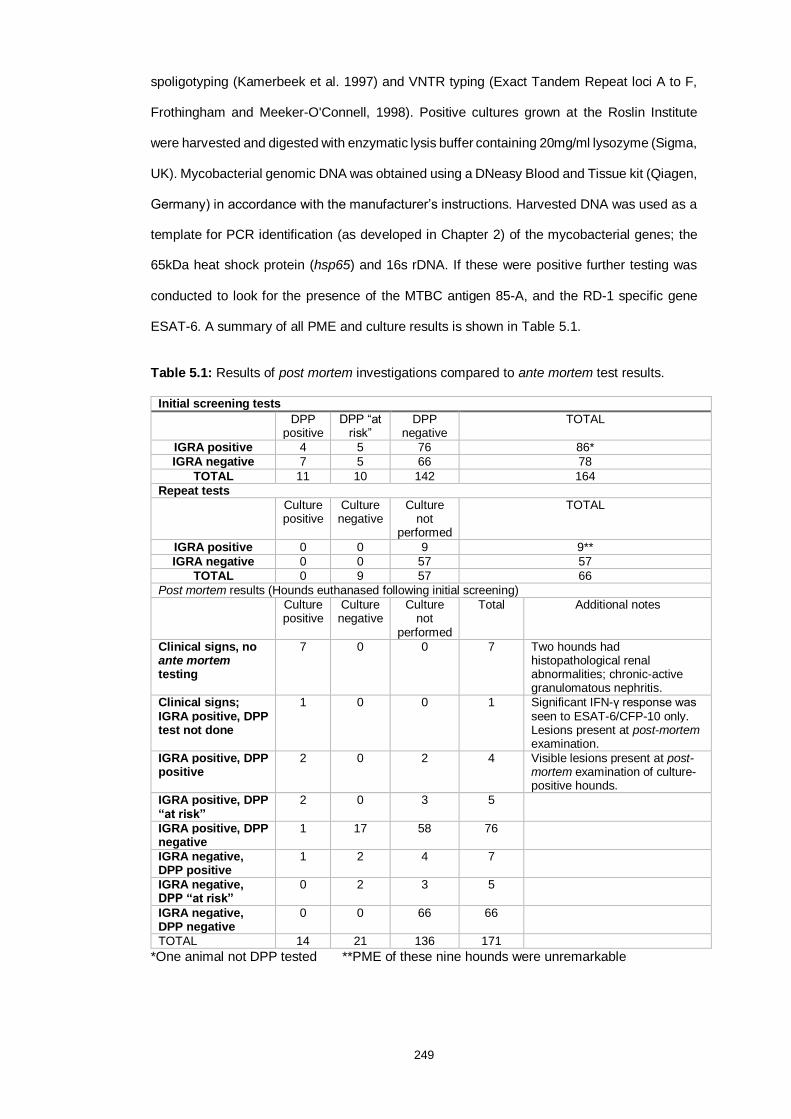
249
spoligotyping (Kamerbeek et al. 1997) and VNTR typing (Exact Tandem Repeat loci A to F,
Frothingham and Meeker-O'Connell, 1998). Positive cultures grown at the Roslin Institute
were harvested and digested with enzymatic lysis buffer containing 20mg/ml lysozyme (Sigma,
UK). Mycobacterial genomic DNA was obtained using a DNeasy Blood and Tissue kit (Qiagen,
Germany) in accordance with the manufacturer’s instructions. Harvested DNA was used as a
template for PCR identification (as developed in Chapter 2) of the mycobacterial genes; the
65kDa heat shock protein (hsp65) and 16s rDNA. If these were positive further testing was
conducted to look for the presence of the MTBC antigen 85-A, and the RD-1 specific gene
ESAT-6. A summary of all PME and culture results is shown in Table 5.1.
Table 5.1: Results of post mortem investigations compared to ante mortem test results.
Initial screening tests
DPP positive
DPP “at risk”
DPP negative
TOTAL
IGRA positive 4 5 76 86*
IGRA negative 7 5 66 78
TOTAL 11 10 142 164
Repeat tests
Culture positive
Culture negative
Culture not
performed
TOTAL
IGRA positive 0 0 9 9**
IGRA negative 0 0 57 57
TOTAL 0 9 57 66
Post mortem results (Hounds euthanased following initial screening)
Culture positive
Culture negative
Culture not
performed
Total Additional notes
Clinical signs, no ante mortem testing
7 0 0 7 Two hounds had histopathological renal abnormalities; chronic-active granulomatous nephritis.
Clinical signs; IGRA positive, DPP test not done
1
0 0 1 Significant IFN-γ response was seen to ESAT-6/CFP-10 only. Lesions present at post-mortem examination.
IGRA positive, DPP positive
2 0 2 4 Visible lesions present at post-mortem examination of culture-positive hounds.
IGRA positive, DPP “at risk”
2 0 3 5
IGRA positive, DPP negative
1 17 58 76
IGRA negative, DPP positive
1 2 4 7
IGRA negative, DPP “at risk”
0 2 3 5
IGRA negative, DPP negative
0 0 66 66
TOTAL 14 21 136 171
*One animal not DPP tested **PME of these nine hounds were unremarkable

250
5.2.5 Repeat IGRA testing protocol
Once the initial screening testing had been completed, the 66 hounds that remained were kept
in isolation for 60 days. At this time, because the test accuracy of the two testing methods is
unknown, the IGRA test was repeated. Nine hounds were found to be IGRA positive, none
were visibly lesioned at PME.
Following these results it was decided that the remaining 57 hounds were unlikely to be
infected/infectious and so voluntary restrictions on the kennels were lifted. The hounds
continue to be closely monitored for any change in appetite, body weight and body condition
score. All of the remaining hounds remain well at the time of writing.
5.3 Screening of at risk, in-contact humans
M. bovis is a known zoonotic bacteria. Once infection was confirmed by laboratory culture from
samples taken from the hounds, Health Protection England (HPE) was informed to assess the
risk to human health. An in-depth risk assessment was conducted, and contacts were stratified
in to risk pool. A ‘stone in the pond’ approach was adopted for screening, with those at highest
risk of exposure screened initially. This investigation and its findings are the subject of an
additional study, where 11 people were screened (Phipps et al., 2018). One asymptomatic
exposed person has tested positive for tuberculosis on initial screening by IGRA and has since
been diagnosed with latent tuberculosis. Due to the nature of the contact between this
individual and the infected hounds, it remains possible but unproven that the person was
infected by contact with either the hounds and/or contaminated bovine material.

251
5.4 Epidemiological assessment
5.4.1 Risk pathway identification
The APHA conducted an epidemiological investigation into the source of the outbreak to
investigate possible transmission pathways. A qualitative risk assessment approach was used
to address the likelihood of each of these routes of infection. The more probable contenders
in order of likelihood were:
5.4.2 Movement of infected hounds into the kennels
(inhalation/ingestion/biting)
Thirteen female hounds moved to, and six returned from, other kennels for breeding purposes
during the 12 months prior to clinical signs in case 0. This included locations in the HRA that
were also within the home-range for genotype 10:a of M. bovis, where infected carcases would
have been more likely to have been fed. Within the previous three years, but prior to the 18
months investigation in Section 5.2.3; three hounds, two male and one female, moved onto
the premises from three other kennels in the HRA. The likelihood of these hounds becoming
infected from fallen stock at these kennels or through other transmission pathways in the HRA
was assessed as medium and therefore the most likely. This likelihood was somewhat
mitigated by the fact that only one of the tested bitches was positive to the screening tests,
and she had no visible lesions at PME. However, not all animals could be followed up because
of missing records or mortalities, only ten of the 16 traced animals were alive and available for
testing at the time of the outbreak.
5.4.3 Feeding of M. bovis-infected fallen stock (ingestion)
Details of the fallen stock collected from farms and fed to the hounds in the kennels over the
12-month period prior to the first hound death were examined to provide an evaluation of the
likely risk posed. A total of 24 carcases were sourced from an Approved Finishing Unit (AFU)
for negative-testing cattle from bovine tuberculosis restricted farms. However, no tuberculous

252
lesions had been confirmed at post mortem meat inspection in the abattoir in any cattle from
the unit over the previous three years, reflecting the fact that most were sourced from low
bovine tuberculosis incidence areas i.e. from across the Low Risk Area and/or low incidence
parts of the Edge Area. Apart from the AFU, six carcases were collected from farms with a
history of TB breakdowns caused by genotype 10:a of M. bovis – two dating back to
breakdowns in 2014 and one in early 2016 due to the purchase of one infected cow. However,
none of these incidents had ongoing infection within the herds in question and all had been
subject to parallel IGRA and tuberculin skin testing, with negative results, which reduces the
probability of residual infection.
The feeding of fallen stock from local cattle herds with undisclosed M. bovis 10:a infection
remains a low likelihood considering the low tuberculosis incidence in the area, annual
tuberculosis testing of herds, and the herd level test sensitivity with a cut-off of one reactor.
Only one farm was in a four year testing parish, but was assessed as low risk after considering
its purchase history. No alpaca carcases or lungs had been fed. However, it is possible that
kennel staff may not have recognised tuberculous lesions in carcases if present. The likelihood
of feeding infected material to the hounds at this kennel was consequently assessed as low,
but with a medium level of uncertainty regarding prevalence of carcase infection and dose-
response in dogs.
5.4.4 Exposure to infected livestock or wildlife during exercising
(inhalation/ingestion/biting) either directly or via M. bovis-contaminated
environment
About 85% of the geographical area to which the hounds were exposed had low cattle
tuberculosis incidence and no indication of wildlife infection. The remaining 15% had some
tuberculous cattle breakdowns where APHA investigation indicated possible wildlife sources.
The very infrequent reporting of M. bovis in farm dogs in UK suggests that farm-based
transmission pathways are not very effective. Kennel staff reported that direct contact between
wildlife and working hounds during exercise was unlikely. However, the worst case scenario

253
of an encounter with an infectious badger, or infected carcase could have been missed. The
likelihood of infection whilst exercising was considered very low, but with high uncertainty
associated with wildlife prevalence and general lack of quantification of the transmission
pathways from the environment.
5.4.5 Exposure to infected local wildlife at the kennels
(inhalation/ingestion/biting)
The kennels are located in an area of the country where cattle tuberculosis incidence is low
and there is no evidence of wildlife infection. Access to the kennels by larger wildlife such as
badgers was very unlikely. Likelihood was considered very low with low uncertainty.
Once in the kennels, infection appears to have spread horizontally between the hounds,
suggesting that in certain conditions such as intensive housing of large packs, relatively high
rates of transmission can occur within dog populations. It is known that badgers can excrete
up to 100,000 M. bovis colony forming units (CFU) per ml of urine where kidneys are infected
(Corner, 2012). Dogs with similar pathology may have similarly high excretory rates. Inhalation
of infected urine aerosols or ingestion of such high doses are valid and likely transmission
pathways in this scenario to account for dog to dog spread. However, the fact that no gross
tuberculous pathology was found in the respiratory and digestive systems of the clinical cases
or test positive dogs adds a level of uncertainty as to what the exact transmission route was.
5.5 Statutory changes resulting from the assessment
This outbreak prompted a policy review of the feeding of raw flesh from fallen stock to hounds,
permitted under Article 18 of Commission Regulation (EC) No. 1069/2009. A condition of this
regulation is that fallen stock fed are not killed or have not died as a result of the presence or
suspected presence of a disease communicable to humans or animals. An interpretation of
this is that TB reactors, normally removed by APHA, and inconclusive reactors, should not be
fed. The Department for Environment, Food and Rural Affairs (DEFRA) commissioned a risk
assessment from APHA to consider the likelihood of M. bovis infection occurring in hounds at

254
Animal By-Products (ABP) registered kennels through the routine feeding of fallen stock
sourced from farms in the HRA of England.
The overall likelihood of at least one foxhound becoming infected with M. bovis within the next
five years, if current practices remained unchanged, was considered to be medium. This
assessment was subject to a high level of uncertainty associated with the true prevalence of
M. bovis infection in fallen stock carcases in the HRA and the dose-response following
ingestion of infected material in dogs. However, the fact that this is the first reported case in
a kennel in the UK, may suggest that the actual risk is lower. On one hand this conclusion is
caveated by the possibility of under-reporting through lack of systematic surveillance for TB in
dogs. Alternatively, the conclusion is supported by a survey of culled hounds from ten Irish
hunting kennels, over a three year period (2003-2005), which identified M. bovis infection in
one of 52 foxhounds submitted for PME as part of the survey (Jahns, 2011).
As a proportionate risk mitigation strategy, DEFRA has introduced tighter restrictions on the
collection and feeding of fallen stock to hounds in registered kennels. Since 10 October 2017
the feeding of offal from livestock species to dogs from recognised kennels or packs of hounds
has been banned in England (Anon. 2017). Hunt kennel operators must also carry out
additional examinations for lesions of TB in fallen stock originating from ‘high risk’ premises
(defined as farms under movement restrictions due to a bovine TB breakdown, or which have
been released from such restrictions in the previous 12 months). Additionally, APHA has
developed training materials and guidance on the identification of TB in carcases of cattle and
other livestock, for collectors and kennels feeding fallen stock to hounds. This should raise
awareness of TB and assist recognition of tuberculous lesions in fallen stock for notification of
suspicion of TB in carcases to APHA, as legally required under The Tuberculosis (England)
Order 2014. The hunt kennel industry is also strengthening the voluntary code of practice and
guidance for its members, to recommend PME of all hounds that die of unexplained or suspect
causes.

255
5.6 Discussion
To the authors’ knowledge this outbreak represents the only documented occurrence of
tuberculosis in a canine species with evidence of onward dog-to-dog transmission within the
affected group. Previous reports of tuberculosis in dogs have been of single sporadic cases
or small case series of unrelated infections. The reason for, and source of, this outbreak
remains unproven; however, given the known ability of M. bovis to spread readily between
social animals, it is not surprising that a large group of kennelled hounds presented a pool of
competent, susceptible hosts once the pathogen was introduced. Also remarkable in this
outbreak was its fulminant nature, from the occurrence of the index case until the resolution of
the outbreak (seven months), nearly one in ten animals on the premises became clinically
sick. Tuberculosis is usually considered a chronic disease with only a small percentage of
infected individuals becoming clinically unwell (Eisenhut, 2014). Therefore this rapid
progression to disease in a relatively high proportion of animals can be considered unusual,
although not unheard of, as sporadic fulminant human cases of renal tuberculosis leading to
death by acute renal function impairment have been reported (Pathan, 2001; Dissanayake,
2004; Adzic-Vukicevic, 2017; Punia, 2008; da Silva Junior, 2016; Isao, 2006).
The reasons for the fulminant nature of this outbreak are likely to be multifactorial; a
combination of pathogen virulence, host genetics and phenotype, as well as environmental
conditions. There is a possibility that the pathogen became host-adapted. However, all animals
from which viable organism was isolated were found to be infected with genotype 10:a. It may
therefore also be the case that this strain of M. bovis in particular poses some genetic virulence
with respect to the dog, or that it became tolerant to a new host and so spread amongst the
hounds once introduced.
From a host perspective of transmission dynamics, the kennel stocking density was higher
than that recommended by the Masters of Foxhounds Association (MFHA). This would have
created an ideal opportunity for spread of disease once the index case became clinically sick
and infectious. Similarly, the physiological stress of overcrowding may have made individuals

256
more susceptible to infection. A number of the kennels were in suboptimal states of repair.
This meant that once the kennels became contaminated, it was almost impossible to
appropriately disinfect them. They have since been destroyed completely and the kennel has
moved to a new, clean site. Foxhounds per se are known to be at risk of mycobacterial
infections with outbreaks of canine leproid granulomas reported in New Zealand and Australia
(Smits, 2012). These outbreaks all affected closely related individuals and so the high level of
genetic relatedness between individuals within this pack could imply that if a genetic
susceptibility, such as a defect in cellular immune response did exist, then it would likely have
high penetrance.
The majority of the clinical signs in sick hounds were non-specific and compatible with those
seen due to M. bovis infections in other species; lethargy, anorexia, weight loss and
lymphadenopathy (O'Halloran, 2017; Murray, 2015; Isaac, 1983). Unusually for companion
animals, every clinically ill animal, those with lesions at PME as well as NVL hounds that were
found to be culture positive, had renal infections. The kidney is not generally considered a so-
called “target organ” for mycobacterial infections in companion animals, however, it is the most
common site of extra-pulmonary TB in humans, other than lymph nodes (O'Halloran, 2017;
Adzic-Vukicevic, 2017; da Silva Junior, 2016; Murray, 2015; Pesciaroli, 2014). Renal
tuberculous infections are often considered to be insidious and present with mild or even
subclinical disease, a significant proportion of cases are incidental findings at PME (Punia,
2008; Isao, 2006). In human cases of infection where irreversible kidney damage occurs, there
are commonly co-morbidities. Amyloidosis is one such co-morbidity, particularly in India, which
predisposes to glomerulonephritis and potentially fatal nephrotic syndrome (Sanz-Martín,
2016; Chong, 2017). This may also be the case for dogs; Foxhounds have previously been
reported to be affected by renal amyloidosis (Mason, 1996), and this kennel has had previously
confirmed clinical cases, whilst at least one of the M. bovis hounds was found to be positive
for renal amyloidosis (data not shown).
Other conditions which adversely affect the outcome of renal tuberculosis are the deposition
of anti-mycobacterial antibodies within the glomeruli and/or hypercalcaemia which is a

257
common complication of tuberculosis and is directly nephrotoxic (Zhao, 2017; Bellendir, 1978;
Ko, 2004; Chan, 1994). It is possible that either of these conditions could have had an impact
on the number of dogs affected and the severity of disease seen in this outbreak.
The relatively high level of renal immune-privilege may have allowed mycobacteria to replicate
there whilst avoiding immune surveillance and clearance (Kurts, 2001). All of the hounds with
renal lesions also had positive urine cultures (where these were available), which suggests
that this could be a major source of infection for onward dog-to-dog transmission. However, if
contaminated urine was the main source of infectious material, e.g. via aerosolisation, then
lung pathology would be expected, but there were no pulmonary lesions in any of the dogs
subjected to PME. Additionally, the urine cultures were only found to be positive after 14 weeks
of culture suggesting that the initial number of organisms present was low. Based on the
distribution of lesions it would appear that the mycobacteria arrived in the kidney by
haematogenous spread. If so this indicates a distant source of infection which has not been
identified.
Infected urine was identified as a key risk factor for potential human exposure during the risk
assessment of kennel staff, predominately due to the use of power washers to clean excreta
from the hard standing, which posed a significant risk of aerosolisation (Phipps et al.,
manuscript submitted). The outcome of tuberculosis screening for at-risk humans at the
kennels was that a single individual was diagnosed with latent tuberculosis (Phipps et al.,
manuscript submitted). If urine from infected dogs was heavily contaminated with M. bovis it
would have been likely that a higher proportion of individuals at risk would have been test
positive. Based on these observational data, it is possible to conclude that M. bovis infected
dogs are likely to be infectious to each other, and their urine may contaminate their local
environment, but that this may not be the only or even the major route of transmission, and
that the risk to human health is low.
No ante mortem tests have been validated for the diagnosis of M. bovis infection in dogs; we
therefore combined two established testing methods for other species to try and maximise our

258
diagnostic sensitivity and bring this outbreak under control. The tests used here target the
disparate arms of the immune response, with the IGRA evaluating the cell mediated Th-1
response whilst the serological assay used to detect a humoral Th-2 response. The
percentage of tested animals deemed to be positive by each test was markedly different with
52% of dogs classed as infected by IGRA at initial screening but only 12.9% of dogs classed
as infected or “at risk” by serology. This difference is likely due to the dominance of cell
mediated immunity in an animals’ response to mycobacterial infection (Khan et al. 2006) but
further investigation of the canine immune response to M. bovis and mycobacterial antigens
is needed before this can be shown conclusively, this work is currently ongoing and will be
published separately.
Whilst uncertainty exists with regards to the source of infection, it seems likely that
contaminated fallen stock carcasses were involved. All culture positive animals had the same
genotype isolated, which has not been frequently detected in the area local to the kennels.
This suggests a single introduction with a large dose of infectious material was likely and that
infection then spread between cohabiting hounds (Reynolds, 2006). One kennel worker was
also diagnosed with latent tuberculosis, potentially due to exposure to infected hounds and/or
their contaminated feed, though this remains unproven. Though the risk posed by M. bovis
infected hounds to other hounds, cattle herds, local wildlife, their environment and their human
keepers is low, this outbreak demonstrates that the risk is plausible and real. Resultantly, to
try to prevent such an outbreak from occurring again, the conditions for the collection and use
of animal by-products for feeding to dogs in recognised kennels or packs of hounds have now
been amended to ban the feeding of offal from fallen stock. Similarly, health surveillance
requirements for hounds have been increased by the MFHA. To be successful, these policy
changes will need to be aided, however, by the future evaluation of diagnostic test accuracy
and test development for canine tuberculosis.

259
5.7 References
Adzic-Vukicevic, T., et al. (2017). First reported case of fulminant TB with progression of
infection from lungs to the genitourinary region. Revista do Instituto de Medicina Tropical de
Sao Paulo 59.
Angkawanish, T., et al. (2013). The elephant interferon gamma assay: A contribution to
diagnosis of tuberculosis in elephants. Transboundary and Emerging Diseases 60(SUPPL1):
53-59.
Ayele, W. Y., et al. (2004). Bovine tuberculosis: An old disease but a new threat to Africa.
International Journal of Tuberculosis and Lung Disease 8(8): 924-937.
Anonymous (2017). Authorisation A6 in: Derogations from product controls under Regulation
(EC) 1069/2009 and Commission Regulation (EU) 142/2011. Authorisations by the Secretary
of State to enable derogations to be used in England. May 2011 (last updated: October 2017).
Document available at:
https://www.gov.uk/government/uploads/system/uploads/attachment_data/file/650731/abp-
exemptions-201710.pdf
Bellendir, E. N., et al. (1978). Circulating antibodies titres in experimental tuberculosis of the
kidneys. Problemy Tuberkuleza 56(1): 79-83.
Bezos, J., et al. (2014). Current ante-mortem techniques for diagnosis of bovine tuberculosis.
Research in Veterinary Science 97(S): S44-S52.
Brosch, R., et al. (2002). A new evolutionary scenario for the Mycobacterium tuberculosis
complex. Proceedings of the National Academy of Sciences of the United States of America
99(6): 3684-3689.
Broughan, J. M., et al. (2013). Mycobacterium bovis infections in domesticated non-bovine
mammalian species. Part 1: Review of epidemiology and laboratory submissions in Great
Britain 2004-2010. Veterinary Journal 198(2): 339-345.
Chan, C. H. S., et al. (1994). Severe hypercalcaemia associated with miliary tuberculosis.
Journal of Tropical Medicine and Hygiene 97(3): 180-182.
Chong, S. G., et al. (2017). Renal amyloidosis complicating multidrugresistant tuberculosis.
International Journal of Tuberculosis and Lung Disease 21(4): 476-477.
Ciszewski, M., et al. (2015). New bacterial zoonotic pathogens. New Medicine 19(2): 59-61.

260
Corner, L. A. L., et al. (2012). The distribution of Mycobacterium bovis infection in naturally
infected badgers. Veterinary Journal 194(2): 166-172.
da Silva Junior, G. B., et al. (2016). Chronic kidney disease related to renal tuberculosis: A
case report. Revista da Sociedade Brasileira de Medicina Tropical 49(3): 386-388.
Dissanayake, S. (2004). Relative lack of clinical disease among household contacts of
tuberculosis patients compared to leprosy households. Transactions of the Royal Society of
Tropical Medicine and Hygiene 98(3): 156-164.
Drewe, J. A. (2010). Who infects whom? Social networks and tuberculosis transmission in wild
meerkats. Proceedings of the Royal Society B: Biological Sciences 277(1681): 633-642.
Dürr, S., et al. (2013). Differences in Primary Sites of Infection between Zoonotic and Human
Tuberculosis: Results from a Worldwide Systematic Review. PLoS Neglected Tropical
Diseases 7(8).
Eisenhut, M. (2014). The evidence for greater sensitivity of interferon-γ release assays
compared to tuberculin skin test in diagnosing latent mycobacterium tuberculosis infection.
Clinical Pediatrics 53(14): 1413.
Ellis, M. D., et al. (2006). Mycobacterium bovis infection in a dog. Veterinary Record 159(2):
46-48.
Frothongham, R. & Meeker-O'Connell, W. A. (1998) Genetic diversity in the Mycobacterium
tuberculosis complex based on variable numbers of tandem DNA repeats. Microbiology 144
(Pt 5), 1189-1196.
Gao, L. Y., et al. (2004). A mycobacterial virulence gene cluster extending RD1 is required for
cytolysis, bacterial spreading and ESAT-6 secretion. Molecular Microbiology 53(6): 1677-
1693.
Gavier-Widén, D., et al. (2009). A review of infection of wildlife hosts with mycobacterium bovis
and the diagnostic difficulties of the ‘no visible lesion’ presentation. New Zealand Veterinary
Journal 57(3): 122-131.
Gay, G., et al. (2000). Pulmonary mycobacterium bovis infection in a dog. New Zealand
Veterinary Journal 48(3): 78-81.
Greenwald, R., et al. (2003). Improved serodetection of Mycobacterium bovis infection in
badgers (Meles meles) using multiantigen test formats. Diagnostic Microbiology and Infectious
Disease 46(3): 197-203.

261
Guarino, F., et al. (2009). Renal tuberculosis in a thirteen-month-old female infant. Pediatric
Infectious Disease Journal 28(8): 757-758.
Guinn, K. M., et al. (2004). Individual RD1 -region genes are required for export of ESAT-
6/CFP-10 and for virulence of Mycobacterium tuberculosis. Molecular Microbiology 51(2): 359-
370.
Gunn-Moore, D. (2014). Feline tuberculosis caused by Mycobacterium bovis. Veterinary
Record 174(13): 322-323.
Isaac, J., et al. (1983). An outbreak of Mycobacterium bovis infection in cats in an animal
house. Australian Veterinary Journal 60(8): 243-245.
Isao, M., et al. (2006). Renal tuberculosis (Mortar Kidney). Internal Medicine 45(2): 111-112.
Jahns, H., et al. (2011). Post-mortem Findings in Irish Culled Hounds. Journal of Comparative
Pathology 145(1): 59-67.
Jaroso, R., et al. (2010). Ante-mortem testing wild fallow deer for bovine tuberculosis.
Veterinary Microbiology 146(3-4): 285-289.
Junqueira-Kipnis, A. P., et al. (2006). Mycobacteria lacking the RD1 region do not induce
necrosis in the lungs of mice lacking interferon-γ. Immunology 119(2): 224-231.
Kamerbeek, J., Schould, L., Kolk, A., Van Agterveld, M., Van Soolingen, D., Kuijper, S.,
Bunschoten, A., Molhuizen, H., Shaw, R., Goyal, M. and Van Embden, J. (1997) Simultaneous
detection and strain differentiation of Mycobacterium tuberculosis for diagnosis and
epidemiology. J Clin Microbiol 35, 907-914.
Khan, A., et al. (2016) Emerging role of mesenchymal stem cells during tuberculosis: The
fifth element in cell mediated immunity. Tuberculosis, 101, pp. S45-S52.
Khalilzadeh, S. and M. R. Boloorsaz (2006). Renal tuberculosis in a four-year-old child.
Tanaffos 5(4): 71-74.
Kim, Y. Y., et al. (2011). Sensitivity of whole-blood interferon-gamma release assay according
to the severity and the location of disease in patients with active tuberculosis. Tuberculosis
and Respiratory Diseases 70(2): 125-131.
Ko, Y. C., et al. (2004). Hypercalcaemia and haemophagocytic syndrome: Rare concurrent
presentations of disseminated tuberculosis in a dialysis patient. International Journal of Clinical
Practice 58(7): 723-725.

262
Kurts, C., et al. (2001). Kidney protection against autoreactive CD8+ T cells distinct from
immunoprivilege and sequestration. Kidney International 60(2): 664-671.
Liu, S., et al. (1980). Canine tuberculosis. Journal of the American Veterinary Medical
Association 177(2): 164-167.
Mason, N. J. and M. J. Day (1996). Renal amyloidosis in related English foxhounds. Journal
of Small Animal Practice 37(6): 255-260.
Michel, A. L., et al. (2013). Mycobacterium tuberculosis at the Human/Wildlife interface in a
high TB Burden Country. Transboundary and Emerging Diseases 60(SUPPL1): 46-52.
Michel, A. L., et al. (2010). Mycobacterium bovis at the animal-human interface: A problem, or
not? Veterinary Microbiology 140(3-4): 371-381.
Miller, M., et al. (2012). Detection of antibodies to tuberculosis antigens in free-ranging lions
(Panthera leo) infected with Mycobacterium bovis in Kruger National Park, South Africa.
Journal of Zoo and Wildlife Medicine 43(2): 317-323.
Miller, M. and F. Olea-Popelka (2013). One Health in the shrinking world: Experiences with
tuberculosis at the human-livestock-wildlife interface. Comparative Immunology, Microbiology
and Infectious Diseases 36(3): 263-268.
Murray, A., et al. (2015). Nosocomial spread of Mycobacterium bovis in domestic cats. Journal
of Feline Medicine and Surgery 17(2): 173-180.
O'Halloran, C. and Gunn-Moore, D.A. (2017). Mycobacteria in cats: An update. In Practice
39(9): 399-406.
Olea-Popelka, F., et al. (2017). Zoonotic tuberculosis in human beings caused by
Mycobacterium bovis—a call for action. The Lancet Infectious Diseases 17(1): e21-e25.
Pai, M., et al. (2007). T-cell assays for the diagnosis of latent tuberculosis infection: moving
the research agenda forward. Lancet Infectious Diseases 7(6): 428-438.
Palmer, M. V. (2007). Tuberculosis: A reemerging disease at the interface of domestic animals
and wildlife. Current Topics in Microbiology and Immunology. 315: 195-215.
Park, H. A., et al. (2016). Pulmonary Mycobacterium tuberculosis infection with giant tubercle
formation in a dog: A case report. Veterinarni Medicina 61(2): 102-109.
Parsons, S. D. C., et al. (2013). Novel cause of tuberculosis in meerkats, South Africa.
Emerging Infectious Diseases 19(12): 2004-2007.

263
Parsons, S. D. C., et al. (2012). Detection of Mycobacterium tuberculosis infection in dogs in
a high-risk setting. Research in Veterinary Science 92(3): 414-419.
Pathan, A. A., et al. (2001). Direct ex vivo analysis of antigen-specific IFN-γ-secreting CD4 T
cells in mycobacterium tuberculosis-infected individuals: Associations with clinical disease
state and effect of treatment. Journal of Immunology 167(9): 5217-5225.
Pesciaroli, M., et al. (2014). Tuberculosis in domestic animal species. Research in Veterinary
Science 97(S): S78-S85.
Posthaus, H., et al. (2011). Accidental infection of veterinary personnel with Mycobacterium
tuberculosis at necropsy: A case study. Veterinary Microbiology 149(3-4): 374-380.
Punia, V. P. S. and S. Kumar (2008). Cement kidney: Renal tuberculosis. Journal, Indian
Academy of Clinical Medicine 9(3): 216-217.
Reynolds, D (2006) A review of tuberculosis science and policy in Great Britain. Veterinary
Microbiology 112 (2–4): 119-126.
Rhodes, S. G., et al. (2008a). Adaptation of IFN-gamma ELISA and ELISPOT tests for feline
tuberculosis. Veterinary Immunology and Immunopathology 124(3-4): 379-384.
Rhodes, S. G., et al. (2008b). Interferon-γ test for feline tuberculosis. Veterinary Record
162(14): 453-455.
Rhodes, S. G., et al. (2011). Comparative study of IFNγ and antibody tests for feline
tuberculosis. Veterinary Immunology and Immunopathology 144(1-2): 129-134.
Rissi, D. R. and C. A. Brown (2014). Diagnostic features in 10 naturally occurring cases of
acute fatal canine leptospirosis. Journal of Veterinary Diagnostic Investigation 26(6): 799-804.
Rocha, V. C. F., et al. (2017). Infection by Mycobacterium bovis in a dog from Brazil. Brazilian
Journal of Microbiology 48(1): 109-112.
Sanz-Martín, N., et al. (2016). Renal amyloidosis in leprosy, an infrequent cause of nephrotic
syndrome in Europe. BMJ Case Reports 2016.
Schiller, I., et al. (2009). Optimization of a whole-blood gamma interferon assay for detection
of Mycobacterium bovis-infected cattle. Clinical and Vaccine Immunology 16(8): 1196-1202.
Shrikrishna, D. et al. (2009). Human and canine pulmonary Mycobacterium bovis infection in
the same household: re-emergence of an old zoonotic threat? Thorax 64, 89-91.

264
[http://thorax.bmj.com/content/64/1/89.full]
Smits, B., Willis, R., Malik, R., Studdert, V., Collins, D., Kawakami, P., Graham, D., and Fyfe,
J.A. (2012) Clusters of leroid granulomas in foxhounds in New Zealand and Australia.
Veterinary Dermatology 23: 465-e88.
Snider, W. R. (1971). Tuberculosis in canine and feline populations. Review of the literature.
American Review of Respiratory Disease 104(6): 877-887.
Szaluś-Jordanow, O., et al. (2016). Intracardiac tuberculomas caused by Mycobacterium
tuberculosis in a dog. BMC Veterinary Research 12(1).
Teo, J. W., et al. (2013). Clinical utility of RD1, RD9 and hsp65 based PCR assay for the
identification of BCG in vaccinated children. BMC Research Notes 6(1).
Thillai, M., et al. (2014). Interferon-gamma release assays for tuberculosis: Current and future
applications. Expert Review of Respiratory Medicine 8(1): 67-78.
Van Der Bürgt, G. M., et al. (2009). Mycobacterium bovis infection in dogs. Veterinary Record
165(21): 634.
Vordermeier, M., Whelan, A., Ewer, K., Goodchild, T., Clifton-Hadley, R., Williams, J. and R.G.
Hewinson (2006) The BOVIGAM assay as an ancillary test to the tuberculin skin test. Gov.
Vet. J., vol. 16(1), p72-80.
Weiss, G. and G. A. Polk (1958). Renal tuberculosis in childhood; a case report. The Harlem
Hospital bulletin 11(2): 45-50.
Wilkins, M. J., et al. (2008). Absence of Mycobacterium bovis infection in dogs and cats
residing on infected cattle farms: Michigan, 2002. Epidemiology and Infection 136(12): 1617-
1623.
Wood, P. R. and S. L. Jones (2001). BOVIGAM™: An in vitro cellular diagnostic test for bovine
tuberculosis. Tuberculosis 81(1-2): 147-155.
Zhao, N., et al. (2017). Early diagnosis of tuberculosis-associated IgA nephropathy with ESAT-
6. Tohoku Journal of Experimental Medicine 241(4): 271-279.
Zhou, Q., et al. (2011). Diagnostic accuracy of T-cell interferon-γ release assays in tuberculous
pleurisy: A meta-analysis. Respirology 16(3): 473-480.

265
Chapter 6: Bovine Tuberculosis in Working
Foxhounds: Lessons Learned from a Complex Public
Health Investigation.
Preface
The following Chapter presents the findings of a study, conducted as part of this studenship
programme that has been published in the journal Epidemiology and Infection.
The manuscript derived from this Chapter is available as an eponymous Open Access article:
Phipps, E., McPhedran, K., Edwards, D., Russell, K., O'Connor, C., Gunn-Moore, D.A.,
O’Halloran, C., Roberts, T. and Morris, J. (2019). Bovine tuberculosis in working foxhounds:
Lessons learned from a complex public health investigation. Epidemiology and Infection, 147,
E24.
Author contributions:
This public health investigation was devised and planned with input from the collective group
of co-authors. The investigation was then implemented by Public Health England, supervised
by Dr Morris and the manuscript prepared by Dr Phipps.

266
Abstract
In 2017, Public Health England South East health protection team were involved in the
management of an outbreak of Mycobacterium bovis (the causative agent of bovine
tuberculosis) in a pack of working foxhounds. This paper summarises the actions taken by the
team in managing the public health aspects of the outbreak, and lessons learned to improve
the management of future potential outbreaks.
A literature search was conducted to identify relevant publications on M. bovis. Clinical notes
from the health protection database were reviewed and key points extracted. Animal and
public health stakeholders involved in the management of the situation provided further
evidence through unstructured interviews and personal communications.
The PHE South East team initially provided ‘inform and advise’ letters to human contacts whilst
awaiting laboratory confirmation to identify the infectious agent. Once M. bovis had been
confirmed in the hounds, an in-depth risk assessment was conducted, and contacts were
stratified in to risk pools. Eleven out of twenty exposed persons with the greatest risk of
exposure underwent tuberculosis screening and one tested positive, but had no evidence of
active infection.
The number of human contacts working with foxhound packs can be large and varied. Health
protection teams should undertake a comprehensive risk assessment of all potential routes of
exposure, involve all other relevant stakeholders from an early stage, and undertake regular
risk assessments. Current guidance should be revised to account for the unique risks to
human health posed by exposure to infected working dogs.

267
6.1 Introduction
Detected infections of dogs with Mycobacterium bovis, the causative agent of bovine
tuberculosis (bTB), are extremely rare, with only seven cases being reported to the Animal
and Plant Health Agency (APHA) between 2004 and 2010 (1). The risk of transmission of M.
bovis from dogs to humans is also considered rare, with expert consensus concluding that
dogs are a ‘spill over’ host and not a significant source of transmission (2). However, this 2017
outbreak of M. bovis amongst a pack of working foxhounds in the South of England sparked
considerable interest and concern from members of the public, media, veterinary and health
professionals, and led to a co-ordinated response from Public Health England (PHE) health
protection teams, the University of Edinburgh and the APHA (3; 4; 5; 6).
This was the first recorded outbreak of M. bovis in working foxhounds in England. Although
public health guidance on the management of M. bovis associated with animals in general is
available, there is no specific guidance describing the management of the human health risks
associated with an outbreak of M. bovis in working foxhounds and the complex exposures this
context presents. Given the increased awareness of M. bovis infection associated with working
dogs (including foxhounds as seen here or other farm dogs) amongst the veterinary
community, it is likely that assessments of the risks to human health will be required again in
the future (1; 2). Such situations are likely to attract wider public interest due to concerns over
the spread of bTB.
Management of this unique outbreak in working foxhounds required PHE to work
collaboratively with veterinary stakeholders to identify exposed persons and limit the spread
of M. bovis amongst both humans and animals. This paper summarises the actions taken by
PHE South East in managing the public health aspects of this situation, and lessons that can
be learned to improve the management of similar situations following notification of potential
outbreaks of M. bovis in dogs or other working animals, especially in the non-household
setting.

268
6.2 Methods
A literature search was conducted on PubMed and CAB Abstracts to identify relevant
publications on M. bovis in dogs ((dog OR pack OR foxhound OR hound) AND (TB OR
tuberculosis OR mycobacterium), all papers available on databases with English language
translation available). Additional papers of relevance to this case study not identified through
the literature search were suggested by the authors of the publication that arose from this
work.
6.3 Results
6.3.1 Background epidemiology of M. bovis in the UK
Bovine tuberculosis is an important disease of cattle with a wide range of wild and domestic
animal hosts (7; 8).The causative agent, M. bovis, is a slow growing, aerobic bacterium that
typically causes infection of the lungs in humans, but can affect any organ in the body (9).
Human infection with M. bovis typically occurs through ingestion, inhalation or contact with
mucous membranes or skin abrasions of an infected animal (7). The main sources of zoonotic
transmission of M. bovis infection are the ingestion of unpasteurised milk and dairy products,
or prolonged exposure to aerosolised bacilli excreted from the respiratory tract of diseased
animals (7; 10). Localised non-pulmonary M. bovis lesions can occur rarely through handling
infected animals or carcases and theoretically through aerosolised exposure in abattoirs (10).
Human to human transmission is extremely rare (9). In the United Kingdom, infection in
humans with M. bovis is much less common than M. tuberculosis; between 2002 and 2014
there were 357 reported human cases of M. bovis compared to 96,887 cases of M.
tuberculosis (9; 11). It is thought that most cases of M. bovis infection in humans in the UK are
likely due to reactivation of latent infection acquired before compulsory pasteurisation of milk
and bTB control programmes, contact with a human tuberculosis case, or infection acquired
abroad (7; 9).

269
The incidence rate of M. bovis in cattle in England is one of the highest in Europe (11.0 new
herd incidents per 100 herd-years at risk in 2017) (12). The UK government has implemented
stringent controls to minimise the spread of bovine tuberculosis, with some counties in the
South West and South East of England being deemed High Risk Area and separate adjacent
buffer zone (Edge Area), and so cattle in these areas are subject to additional testing and
surveillance (12). Non-bovine farmed animals such as South American camelids (alpacas),
sheep, goats, pigs and deer are also sporadically infected, usually through direct or indirect
contact with cattle or badgers (the two maintenance hosts of the bacterium in the UK) (13; 14).
Infection of companion animals does occur, and there have been numerous reported cases of
pets testing positive for M. bovis over the past few years, predominantly domestic cats (2; 12;
13; 15). The majority of cases of dogs infected with M. bovis in the literature come from
countries other than England and have identified risk factors such as being strays or having
exposure to infected non-domestic animals (1; 15; 16; 17).
The potential for zoonotic transmission of M. bovis from companion animals to human contacts
was confirmed in 2014 when cases of human M. bovis infection followed transmission from
infected cats in Berkshire, England (18; 19; 20). Lessons learned from the management of
exposure to infected companion animals in 2014 justified a measured and co-ordinated public
health response in order to protect human health.
6.3.2 Initial Risk Assessment
The responsible health protection team in the South East of England were initially made aware
in early 2017 of the potential risk of zoonotic transmission of M. bovis by a private veterinarian
with clinical suspicion of this diagnosis in working foxhounds cared for at the practice. A
number of veterinary staff may have come into contact with infected hounds during their
routine work. Key questions asked by the public health team in the initial assessment are
summarised in Box 6.1, based on core principles of health protection practice and adaptation
of existing general guidelines on the management of human health risks of tuberculosis (7;
21). Before implementing public health actions it is recommended to wait for laboratory

270
confirmation (7). Veterinary practice staff were therefore reassured and ‘inform and advise’
letters offered for distribution in the workplace, and the situation highlighted to the APHA in
case of further action required.
Box 6.1: Questions asked by the public health team in the initial assessment
6.3.3 Confirming the Diagnosis
Primary concerns regarding the health of the kennel hounds were first raised in late 2016, a
number of hounds had been euthanased for welfare grounds for reasons of deteriorating
health within the preceding months. When a new hound starting showing similar clinical signs
(weight loss, lethaergy, hyporexia, polyuria and polydipsia) it was euthanased and submitted
for post-mortem examination. Gross renal pathology was confirmed histopathologically as
granulomatous, and the presence of acid-fast bacilli with mycobacterial morphology was
confirmed by examination of Zeihl-Neelsen stained sections of diseased tissue.
This finding instigated a veterinary disease outbreak investigation by the Univeristy of
Edinburgh, Biobest Laboratories and APHA (detailed in reference 24). Briefly, 164 hounds in
the kennel were tested using an experimental interferon-gamma release assay (IGRA) test at
Biobest Laboratories and a serological assay originally developed for cervid species (Dual
Path Platform VetTB test for cervids).
Of the 164 hounds tested, 85 (52%) were diagnosed as being test positive as per the
prospective case definition set. Test positive hounds and clinically sick hounds were
euthanased and M. bovis infection was confirmed by culture in 14 cases. The isolated
Box 6.1: Initial Assessment
What is the name of the hunt and where is it located
How many animals are involved and how many have undergone testing
What clinical signs did the hound(s) present with
What type of testing has been undertaken (e.g. PCR/culture/IGRA)
When are results available yet to confirm the diagnosis
Any at risk groups in contact with the hounds e.g. immunocompromised staff
Any symptomatic staff or household members?

271
organism was genetically tested confirming genotype 10:a with the first laboratory confirmation
occurring in February 2017.
6.3.4 Veterinary Epidemiological Investigation
Once infection with M. bovis 10a had been confirmed evaluation of the risk to human health
was undertaken (see below). Simultaneously, an epidemiological investigation began to
identify the risk pathways by which the hounds may have initially become infected. These are
detailed in (24) and comprised of, in order of considered likelihood; a) movement of infected
hounds into the kennels b) feeding M. bovis infected fallen stock to the hounds c) exposure to
infected livestock or wildlife during work and/or d) exposure to infected local wildlife at the
kennels.
Qualitative assessment of each pathway was undertaken with the evidence available. Tracing
of hound movements indicated that 21 had been moved onto the affected premises within the
three years prior to the diagnosis of the index case, of which 18 were moved in the preceding
18 months. Some kennels of origin of these hounds were located within the geographical
region of the UK designated as the High Risk Area (HRA) for M. bovis incidence with respect
to bovine infections. Furthermore, a number of these were located within the home-range of
M. bovis 10a. It was therefore considered to be medium risk (and therefore most probable)
that whilst at these kennels, hounds were fed fallen stock infected with M. bovis 10a and this
was the source of infection.
Due to the outbreak-kennel location within the Edge Area for M. bovis incidence, being out
with the home range of M. bovis 10a and with no locally identified wildlife infections with M.
bovis the remaining pathways were deemed to be possible sources of infection but low risk.
6.3.5 Identifying At Risk Groups
Following culture confirmation of M. bovis from the hound tissue samples, PHE undertook a
formal risk assessment to identify potential routes of zoonotic transmission, understand levels
of exposure and determine future public health actions, including possible screening of human

272
contacts. Available guidance related to M. bovis in livestock was reviewed and tailored to this
specific situation (7). The key points of this risk assessment are summarised in Box 6.2, and
involved collecting information from staff caring for the foxhounds, the local veterinary practice
and the APHA. Again, this risk assessment was undertaken based on key principles of health
protection practice and adaptation of general guidance on the management of human health
risks related to bTB (7; 21).
Box 6.2: Outline of the agreed basis for HPE risk assessment
6.3.6 Communications Strategy
A multi-agency incident control team (ICT) was convened to facilitate effective information
gathering and to agree on suitable actions for all involved stakeholders. This collective
Box 6.2: Summary Details of the Public Health Risk Assessment
Potential routes of exposure
Identify all persons who were in contact with the symptomatic foxhounds including temporary
and previous staff members at the kennels and veterinary practice
Define the level of contact and activities undertaken e.g. preparing food, grooming, cleaning
environment, dressing wounds, undertaking invasive procedures
Whether anyone may have been bitten by infected foxhounds
Risk of exposure
Level of exposure – total time spent with infected foxhounds by each person involved
Use of personal protective equipment (PPE) during contact – consistency and type worn
Whether any contact could have generated aerosols
Environmental factors
Environments in which contact takes place e.g. kennels, household, vehicles and veterinary
practice
Condition of kennels e.g. cracked concrete and other porous surfaces
Ventilation and cleanliness of environments
Frequency and level of cleaning undertaken e.g. sweeping, pressure washing, disinfecting
Veterinary assessment
List of veterinary practices caring for the working foxhound pack
Prevalence of bTB in the surrounding area amongst bovine and non-bovine animals
Potential route of initial infection e.g. contact with confirmed bovine cases or being fed
potentially contaminated meat
Follow up being undertaken by APHA
Any plans to test the rest of the working foxhound pack

273
approach, also involving specialist communication colleagues from all agencies, ensured that
consistent messages were delivered, with the aim of reducing misinformation and managing
perceptions of risk amongst the public (22).
The foxhounds were exercised on land and at events over several counties in the South East.
Local health protection teams (HPTs) across the region received calls from members of the
public, veterinary and public health professionals enquiring about the pack and associated
risks of infection. Calls to HPTs were recorded on a case and incident management database
HPZone (inFact Shipley, 2012). In order to facilitate co-ordination between teams and quick
referencing, a unique identifier for the situation was created that could be linked to all incoming
enquiries taken by the teams involved. The response was coordinated across several HPTs
by the incident lead, and an email circulation list was used to keep the incident management
team informed of all actions undertaken and updates on the situation. PHE and DEFRA
communications teams were also involved at this point to agree on what proactive and reactive
statements would be appropriate. DEFRA and veterinary colleagues experienced a higher
volume of interview and statement requests than the public health teams, likely due to the high
profile of fox hunting and bTB in the media.
6.3.7 Screening & Further Public Health Actions
A ‘stone in the pond’ approach was adopted in determining which human individuals should
initially undergo screening for tuberculosis (23). Screening does not differentiate between M.
bovis and M. tuberculosis infections, but does identify persons requiring further investigations
and confirmatory testing. PHE identified close contacts with the highest risk of potential
transmission for initial screening, and then planned to expand the screening pool to more
casual contacts if the first round of screening suggested significant transmission had occurred.
The individuals identified for the first round of screening were those with the greatest degree
of contact with infected foxhounds, and included persons who had conducted invasive
procedures on symptomatic dogs (such as post-mortem examinations and surgical
procedures) without appropriate personal protective equipment (PPE), and kennel workers

274
involved with food preparation and cleaning of kennels using a pressure washer. These were
hypothesised to be the highest risk exposures to potential sources of M. bovis, e.g. infected
tissue, aerosolised fluids and potential contaminated fallen stock used as feed. The use of a
pressure washer was of particular interest as the presence of kidney lesions in infected
foxhounds (24) suggests that M. bovis bacilli could have been excreted in the hounds’ urine.
Persons in the initial screening group were referred to their local tuberculosis service for
screening. All other identified potential contacts were sent an ‘inform and advise’ letter whilst
results from the initial round of screening were awaited (see Box 6.3 for outline summary). In
addition, HPT staff visited the veterinary practice to discuss and allay staff concerns related to
M. bovis and their risk of acquiring tuberculosis.
Box 6.3: Agreed purpose of communication strategy with at risk individuals
In total, eleven out of seventeen people were considered to have potential exposure to infected
foxhounds as outlined in Figure 6.1 and were offered tuberculosis screening. Of these, one
person tested positive for TB (using QuantiFERONTM TB Gold). Following a further
assessment including computed tomography (CT) scanning and culture of tissue samples,
there was no evidence of active tuberculosis infection and latent tuberculosis was diagnosed.
This person had not had previous testing for tuberculosis, did not have other risk factors, but
was involved in all high risk activities including post-mortem examination without PPE,
preparation of fallen carcases for feeding, cleaning of kennels using a pressure washer and
care of open wounds on infected animals. Seven contacts screened negative for tuberculosis,
Box 6.3: Key information for inform and advise letters
Reassure that the risk of transmission from animals to humans is low
Some persons may be unusually susceptible such as immunocompromised persons
Description of the symptoms of tuberculosis
Advice to contact GP if experiencing any of these symptoms, mentioning the possible route
of exposure through the hunting pack
Screening of high risk persons is currently taking place and we may be in contact again to
arrange screening if initial tests indicate that transmission of bTB has occurred

275
and three declined the offer of screening. Based on this outcome of the first screening round,
it was agreed by the ICT that further screening would not be offered to more casual contacts.
Figure 6.1 Hierarchy of screening pools used during the public health investigation resulting
from the kennel outbreak of Mycobacterium bovis tuberculosis in working foxhounds.
Screening of the foxhounds continued over several months with two further screening rounds;
a total of 164 hounds were tested with a 52% IGRA positivity rate (24). All foxhounds testing
IGRA positive were euthanised. A second meeting of the original multi-stakeholder ICT was
convened to discuss how to manage the ongoing exposure to IGRA positive hounds since the
initial diagnosis. Staff members who had declined screening initially were encouraged to
attend given this ongoing exposure. Three new staff members (bringing the total number of
screened persons up to 20) who had joined the hunt since the initial diagnosis were identified
and they were provided with ‘inform and advise’ letters to highlight their potential risk of
exposure to infected foxhounds during both rounds of animal screening, and recommended
actions if they develop symptoms indicative of potential tuberculosis infection.

276
6.4 Discussion
The management of this situation has highlighted several learning points for veterinary and
public health stakeholders. Outbreaks of potentially zoonotic infections amongst working
foxhound packs are a unique challenge for both health protection teams and veterinary
investigation teams, as they are comprised of large populations (in this case over 150)
individuals, which are highly mobile (this kennel covered an area spanning six counties) and
hounds are legislatively defined neither as companion animals nor agricultural livestock,
meaning for example, that they can be fed fallen stock. The hounds from working packs may
span both categories, spending time in both domestic and farming settings. This makes
quantifying the unique exposures and risk of transmission associated with this context
particularly difficult as they are not specifically covered by any existing guidelines. Moreover,
IGRA testing is not validated for use in diagnosing M. bovis infections dogs, so the sensitivity
and specificity of any results are unknown, adding some uncertainty to whether public health
action should be warranted in light of a positive test (25). In this scenario, given the high
number of positive IGRA tests, confirmatory tissue cultures and foxhounds with clinical signs
consistent with tuberculosis, the decision to take action was relatively straightforward, but
could be difficult in the face of less conclusive results.
Bovine tuberculosis is an understandably emotive subject for many stakeholders, including
kennel staff, farmers, veterinarians and the general public, and there was considerable media
interest. It was crucial to ensure that confidentiality of the working foxhound pack was
protected as much as possible by agencies involved, and that information sharing activities
were coordinated and agreed on by all parties. The geographical area covered by the pack
during outings spanned several counties, and required the involvement of multiple HPTs and
tuberculosis services to coordinate a response. Effective communication between these teams
is extremely important to ensure that resources are used efficiently and messages are
consistent in order to address public concern. By visiting the veterinary practice in person, the
health protection team were able to build relationships and foster trust from stakeholders to
manage concern successfully. This also presented an opportunity for highlighting the

277
importance of appropriate wearing appropriate personal protective equipment when
performing post-mortem examinations of animals, which would have significantly reduced the
risk of exposure to M. bovis to veterinary and kennel staff.
This situation highlighted how dynamic risk assessments should be undertaken frequently
during the ongoing process of outbreak management, as the foxhound pack is likely to come
in to contact with new persons such as temporary staff and members of the public as well as
those potential contacts identified in the initial assessment. IGRA testing of the whole pack
was a significant undertaking for the kennel and University of Edinburgh staff, who were unable
to secure additional funding for testing as the foxhounds were deemed to be non-livestock
species, and took several months to complete. During this period, workers continued to be
exposed to hounds that were eventually found to be IGRA positive. Managing this unknown
and unquantifiable risk of potential exposure whilst waiting for IGRA results was particularly
challenging, and involved concerted effort from the HPT to communicate effectively with
stakeholders to address and allay concerns. Senior managers of the foxhound kennels were
very receptive to both public health requirements and advice regarding animal health
management. It is worth noting that as the hounds are not classed as livestock, the APHA
have no regulatory powers to enforce euthanasia or other control methods.
6.5 Conclusion
This paper highlights the unique and unusual health protection scenario of managing potential
working foxhound to human transmission of M. bovis. The number of human contacts with
working packs can be large and varied, and the animals are not considered as domestic
companion animals or livestock. Health protection teams involved in the management of such
situations must ensure to undertake a comprehensive risk assessment of all potential routes
of exposure, involve all other relevant stakeholders from an early stage in developing
management and communication plans, and undertake regular risk assessments as new
information becomes available. Current guidance should be revised to account for the unique
risks to human health posed by exposure to infected working dogs.

278
6.6 References
1. Mycobacterium bovis infections in domesticated non-bovine mammalian species. Part 1:
review of epidemiology and laboratory submissions in Great Britain 2004-2010. Broughan J,
Downs S, Crawshaw T, Upton P, Brewer J, Clifton-Hadley R. 2013, The Veterinary Journal,
Vol. 198, pp. 339-345.
2. Mycobacterium bovis infection in a dog. Ellis M, Davies S, McCandlish I, Monies R,
Jahans K, De La Rua-Domenech R. 2006, The Veterinary Record, Vol. 159, pp. 46-48.
3. Are hunting dogs spreading bovine TB? Veterinary Record. 2017, The Veterinary Record,
Vol. 180, p. 583.
4. Docksey, L. Foxhounds and bovine TB edges (finally) into the news. The Ecologist. [Online]
23 06 2017. [Cited: 19 09 2017.]
http://www.theecologist.org/News/news_analysis/2989067/foxhounds_and_bovine_tb_edges
_finally_into_the_news.html.
5. Glaze, B. Top Tory's fox hunt kills off 25 of its own hounds after bovine TB outbreak. The
Mirror. [Online] 07 03 2017. [Cited: 19 09 2017.] http://www.mirror.co.uk/news/politics/top-
torys-fox-hunt-kills-9982581.
6. Starkey, J. Bovine tuberculosis ‘spread by packs of fox hounds’. The Times. [Online] 06 06
2017. [Cited: 19 09 2017.] https://www.thetimes.co.uk/article/bovine-tuberculosis-spread-by-
packs-of-fox-hounds-6gq0xn69k.
7. Public Health England. Bovine tuberculosis: guidance on management of the public health
consequences of tuberculosis in cattle and other animals (England). London : Public Health
England, 2014.
8. The importance of Mycobacterium bovis as a zoonosis. Thoen C, Lobue P, de Kantor I.
2006, Veterinary Microbiology, Vol. 112, pp. 339-345.
9. Epidemiology of Mycobacterium bovis disease in humans in England, Wales and Northern
Ireland, 2002-2014. Davidson J, Loutet M, O'Connor C, Kearns C, Smith R, Maeve L,
Thomas H, Abubakar I, Zenner D. 3, 2017, Emerging Infectious Diseases, Vol. 23, pp. 337-
386.
10. Abattoir associated zoonoses. Collins, J. 1983, Journal Society of Occupational Medicine,
Vol. 33, pp. 24-27.
11. Public Health England. Tuberculosis in England: 2015 report (presenting data to end of
2014). London : Public Health England, 2015.
12. Department for Environment Food and Rural Affairs. Quarterly publication of National
Statistics on the incidence and prevalence of tuberculosis (TB) in cattle in Great Britain- to end
June 2017. London : Department for Environment Food and Rural Affairs, 2017.
13. Department for Environment, Food and Rural Affairs. Bovine TB in non-bovine
species- Combined 2016 Data. London : Department for Environment Food and Rural Affairs,
2017.

279
14. The spatio-temporal distribution of Mycobacterium bovis (bovine tuberculosis) infection in
a high density badger (Meles meles) population. Delahay R, Langton S, Smith G, Clifton-
Hadley R, Cheeseman C. Journal of Animal Ecology, 2000, Vol. 69, pp. 1-15.
15. Mycobacterium bovis infection in dogs. G Van Der Burgt, T Crawshaw, A Foster, D
Denny, A Schock. 21, 2009, The Veterinary Record, Vol. 165, p. 634.
16. Tuberculosis in domestic animal species. Pesciaroli M, Alvarez J, Boniotti MB, Cagiola
M, Di Marco V, Marianelli C, Pacciarini M, Pasquali P. Research in Veterinary Science,
2014, Vol. 97, pp. S78-S85.
17. Post-mortem findings in Irish culled hounds. Jahns H, Callanan JJ, McElroy MC,
Sammin DJ, Bassett HF. 1, Journal of Comparative Pathology, 2011, Vol. 145, pp. 59-67.
18. Public Health England. Cases of TB in domestic cats and cat-to-human transmission:
risk to public very low. [Online] 2014. [Cited: 05 10 2017.]
https://www.gov.uk/government/news/cases-of-tb-in-domestic-cats-and-cat-to-human-
transmission-risk-to-public-very-low.
19. Cat-to-human transmission of bovine TB: risk to public 'very low'. Veterinary Record. 174,
2014, The Veterinary Record, p. 337.
20. Human Animals Infections and Risk Surveillance (HAIRS) Group. Qualitative
assessment of the risk that cats infected with Mycobacterium bovin present to human health.
Public Health England, 2014.
21. National Institute for Health and Care Excellence. NICE guideline NG33: Tuberculosis.
London : National Institute for health and Care Excellence, 2016.
22. Sandman, P. Responding to Community Outrage: Strategies for Effective Risk
Communication. USA : American Industrial Hygiene Association, 1993.
23. Microepidemics of tuberculosis: the stone-in-the-pond principle. Veen, J. 2, 1992, Tubercle
and Lung Disease, Vol. 73, pp. 73-76.
24. O'Halloran C et al. [manuscript submitted]. The University of Edinburgh and Biobest
Laboratories, 2018.
25. Dempsey, R. Personal communication. 02 02 2017.

280
Chapter 7: Comparison of Potential Ante Mortem
Assays for the Identification of Mycobacterium bovis-
Infected Domestic Dogs (Canis lupus familiaris).
Acknowledgements
Thanks go to all of the staff and management team of the kennels affected by this outbreak
for facilitating the investigation and giving permission for its findings to be included in this
thesis.
Specific thanks go to Dr Shelley Rhodes (Senior Animal and Plant Health Agency Scientist
and TB Test Consultant) who performed the commercial serological assays (Section 7.2.5).
Additional funding for this project was provided by PetPlan Charitable Trust
(OHALLORAN806: Validation of diagnostic assays for canine tuberculosis).
Abstract
The domestic dog (Canis lupus familiaris) is a competent host for Mycobacterium (M.) bovis
infection but no ante-mortem diagnostic tests have been validated for this species. The aim of
this study was to compare the performance of potential ante-mortem diagnostic tests across
samples collected from dogs considered to be at high or low risk of sub-clinical M. bovis
infection. We tested 164 dogs at high risk of infection and 119 dogs at low risk of infection with
a combination of cell-based and/or serological diagnostic assays previously described for use
in non-canid species. The interferon-gamma release assay (IGRA) using peripheral blood
mononuclear cells (PBMC) identified the highest number of test positive animals (85, 52%),
with a suggested specificity of 97.3%, whilst a whole blood IGRA was found to be unreliable.
The production of antigen-specific tumour necrosis factor-alpha (TNF-α) by PBMC in response
to a cocktail of ESAT-6 and CFP-10 antigens correlated very strongly with the overall IGRA

281
results and has future diagnostic potential. All three serological assays employed in this study
(Idexx M. bovis Ab ELISA, [Idexx Laboratories, USA], DPP VetTB lateral flow assay [Chembio,
USA] and comparative PPD ELISA [in-house]) identified seropositive dogs but, overall, the
test-positive rate for the serological assays was only one third that of the cellular based assays.
Circulating serum cytokine concentrations of interferon gamma (IFN-γ) and TNF-α were not
statistically different between high and low risk groups of dogs. While many dogs in the high
risk group had serum biochemical abnormalities, these did not correlate with the findings of
other diagnostic tests.
This study demonstrates, for the first time, the potential utility of two cellular and three
serological assays for detecting sub-clinical M. bovis infections of dogs. Whilst the data
suggest high test specificity for all assays evaluated, further work is needed to validate the
sensitivity and specificity of individual or combinations of tests using sufficient numbers of dogs
of known infection status.
7.1 Introduction
The domestic dog (Canis lupus familiaris) is, like many mammalian species, a competent host
for Mycobacterium (M.) bovis infection (Snider et al. 1971; Ellis et al. 2006; Broughan et al.
2013a). Although clinical cases of tuberculosis (TB) due to infection with M. bovis, or other
members of the M. tuberculosis-complex (MTBC), are considered rare in the dog and generally
only occur sporadically (Broughan et al. 2013b), we previously investigated a fulminant
disease outbreak caused by M. bovis infection within a large group of kennel-housed working
Foxhounds in the UK (O’Halloran et al. 2018). Investigation of this outbreak was impeded in
its speed, and by the lack of available ante-mortem diagnostic tests for identifying M. bovis
infections in domestic dogs (Snider et al. 1971; Snider et al. 1975; Broughan et al. 2013b).
The tests employed therefore had to be adapted and interpreted in real time, based on
extrapolation of performance data from non-canine species.
Interferon gamma (IFN-γ) release assay (IGRA) tests have been developed on the principle
of quantitatively evaluating antigen-specific IFN-γ production by peripheral circulating effector

282
memory T-cells following in vitro stimulation (Adams 2001; de la Rua Domenach et al. 2006;
Schiller et al. 2009). The first IGRA was designed to increase the sensitivity of M. bovis TB
testing in cattle (Wood et al. 1990), and the resulting BOVIGAMTM assay holds World
Organisation for Animal Health (OIE) validation, with a reported in-field sensitivity of 81.8-
100% and specificity of 88-99% (Wood, 2001; Vordermeier et al., 2006; Schiller et al., 2009;
Bezos, 2014). IGRA tests have subsequently been adapted to identify active and latent TB in
human patients with at least equivalent sensitivity and increased specificity when compared
to the intradermal tuberculin skin test (TST) (Kim, 2011; Zhou, 2011; Eisenhut, 2014; Thillai,
2014). Similarly, IGRA test protocols have been developed for use in domestic cats with a
suggested high sensitivity and specificity for the detection of MTBC infections, and for the
detection of M. tuberculosis infections in dogs in a high risk setting (Rhodes et al. 2011;
Parsons et al. 2012; Broughan et al. 2013b); while the same study found the intradermal TST
to be unreliable in the dog (Parsons et al. 2012).
The IGRA used in this study was specifically developed to test dogs for subclinical M. bovis
infection, and was based on the protocol used for testing domestic cats (Rhodes et al. 2011);
the antigens purified protein derived (PPD) from M. avium (PPDA), PPD from M. bovis (PPDB)
and a cocktail of proteins derived from the immunodominant proteins; 6kDa early-secreted
antigenic target (ESAT-6) and 10kDa culture filtrate protein (CFP-10) were selected. PPDA is
frequently used with PPDB as a comparator in both IGRA and TST tests to assess and mitigate
for exposure/sensitisation to, or infection with, environmental mycobacteria species. Infection
with a MTBC mycobacteria is confirmed if the IFN-γ response of an animal is greater to PPDB
than to PPDA (Wood, 2001; Vordermeier et al. 2007).
The proteins ESAT-6 and CFP-10 are both encoded on the RD-1 region of most MTBC,
including M. bovis, and a small number of non-tuberculous mycobacteria (NTM) (Harboe et al.
1996; Brosch et al. 2002). They are secreted proteins that form a heterodimeric complex in a
1:1 ratio which has been consistently associated with phagocyte lysis and pathogen virulence
(Gao et al. 2004; Guinn et al. 2004; Junqueira-Kipins et al. 2006). The presence of a
concurrent response to the recombinant combination of ESAT-6/CFP-10, as well as PPDB,

283
indicates infection with an RD-1 positive MTBC mycobacteria (i.e. it excludes infection with M.
microti or previous vaccination with M. bovis-BCG) (Harboe et al. 1996; Buddle et al. 2001;
Brosch et al. 2002).
A number of studies have tried to improve the sensitivity and/or specificity of IGRA tests. The
overall performance of an IGRA, when used to test cattle for M. bovis or humans for M.
tuberculosis infection, can be increased by the measurement of additional cytokines secreted
by antigen specific cells within the blood (Jones; 2010; Elnagger et al. 2017). This includes
tumour necrosis factor-alpha (TNF-α), which has a well described role in the mammalian host
immune response to mycobacteria (Allie et al. 2013, Leem et al. 2018).
The bovine (BOVIGAMTM) and human (QuantiFERONTM TB Gold) IGRA assays are both
performed on whole blood without first needing to isolate peripheral blood mononuclear cells
(PBMC), as is required for IGRA testing cats, camelids and the T-SPOT.TBTM test (Hesketh et
al. 1994; Gutierrez, et al. 1998; Vordermeier et al. 2007; Rhodes et al. 2008a; Igari et al. 2018;
Leem et al. 2018). The use of whole blood rather than PBMC makes the former assays quicker
and cheaper to conduct and so would be an ideal development for canine TB testing.
Both IGRA and TST assays evaluate the cell mediated adaptive immune response (CMI) of
an animal which dominates the reaction to intracellular pathogens, including mycobacteria
(Allie et al. 2013). However, skin test responses in dogs have been shown to be unreliable
(Parsons et al. 2012). Diagnostic serological assays, while generally recognised as having
lower sensitivity than CMI assays, have been used both experimentally and commercially for
the diagnosis of MTBC infections in a number of species (Min et al. 2011; Rhodes et al., 2012
El-Seedy et al. 2013; Casal et al. 2014; Roos et al. 2016; Waters et al., 2017, Chambers et
al., 2010). It is recognised that combining serological and CMI diagnostic tests may provide
added value by increasing infection detection, e.g. Bezos et al. (2018) showed an increase in
overall diagnostic sensitivity for both cattle and goats with MTBC infections.
M. bovis is an endemic pathogen of major significance to the UK farming industry as the
causative agent of bovine tuberculosis (bTB) (Vordermeier et al. 2007). In England and Wlaes

284
the regions with high prevalence of M. bovis, as defined by the incidence of bovine infections,
comprise the High Risk Area (HRA), and lower but still significant disease prevalence occurs
within counties of the Edge Area. Control measures implemented for cattle infections amount
to an estimated cost of £100 million annually, and while the most recent government statistics
show a decrease in herd incidence in England, there is a mixed picture, with an increase in
the number of cattle slaughtered annually (DEFRA, 2018).
Non-bovine reservoirs of infection are increasingly being investigated for their potential role in
the epidemiology of the disease; either as possible reservoirs of infection or as sentinel
species, but species-specific diagnostic tests are often lacking which impedes such
investigations (Buddle et al. 2000; Delahay et al. 2002). As the UK moves towards its goal of
M. bovis elimination, having diagnostic tests with known accuracy for such spill over species
will be critical.
The aim of this study is to compare the performance of ante-mortem diagnostic tests across
samples collected from individual dogs considered to be at either high or low risk of sub-clinical
M. bovis infection, and (presumed) TB-free dogs.
7.2 Materials and Methods
7.2.1 Sample collection
Diagnostic blood samples from animals considered to be at high risk of sub-clinical infection
with M. bovis (high risk group, HRG) were those taken from 164 kennel-housed foxhounds
during an outbreak of TB due to M. bovis which occurred between December 2016 and July
2017 (Table 7.1) (O’Halloran et al. 2018). Whole blood and PBMC stimulation assays were
conducted at the time of sampling, whilst serological assays were performed subsequently on
aliquots of serum separated and frozen at -80ºC within 24 hours of collection.
To allow for a comparison of test performance in populations with predicted differences of
infection prevalence, we also tested a total of 119 opportunistically collected blood samples

285
(i.e. remnant blood from samples taken for clinical reasons unrelated to this project); 77 from
presumed TB-free dogs (TB-free group) and 42 dogs with some degree of potential exposure
to cats or dogs with a mycobacterial infection (low risk group, LRG). The TB-free group
provided 45 serum samples and 18 PBMC samples, an additional 42 PBMC samples were
available from the LRG dogs, as shown in Table 7.1.
Table 7.1: Origin of blood samples used in this study: *serum was unavailable for one
Foxhound, HfSA; Hospital for Small Animals; **All samples from dogs resident in Scotland
which has held Officially TB Free (OTF) Status since 2009.
High risk or
Low risk
groups
Group of dogs Total number Number of
serum samples
obtained
Number of
PBMC samples
obtained
High risk Kennel Foxhounds
where the M. bovis
outbreak occurred
164 163* 164
TB-free Canine patients at
the HfSA, University
of Edinburgh**
45 45 0
TB-free Blood donor dogs at
the HfSA, University
of Edinburgh**
24 0 24
TB-free Canine geriatric
health screen
patients, HfSA
University of
Edinburgh**
8 0 8
Low risk Pet dogs in contact
with a household
(animal) case of
mycobacterial
disease
10 0 10
Low risk Pet dogs of kennel
staff where a
previous M. bovis
outbreak occurred
32 0 32

286
Remnant samples were only included in this study if, prior to blood sample collection, the dog
had not been treated with immunomodulatory medications e.g. non-steroidal anti-inflammatory
drugs (NSAIDs), chemotherapeutic agents or corticosteroids within 14 days prior to sample
collection. Dogs were excluded if they were pre-treated with antibiotics with efficacy against
mycobacteria, including fluoroquinolones, macrolides/azides or doxycycline within the same
14 day period. Dogs were not excluded if they had been treated with antimicrobial agents if
these would be ineffective against mycobacteria, such as a penicillin or cephalosporin.
Similarly, dogs were not excluded if they had been treated with non-immunomodulatory
analgesic medications e.g. opioids.
This study was conducted following approval from the School of Veterinary Medicine Ethical
Review Committee at the University of Edinburgh; all relevant guidelines and regulations were
adhered to throughout.
7.2.2 IGRA using isolated PBMC
The IGRA assays for the 164 dogs in the HRG were conducted as previously reported
(O’Halloran et al. 2018). The same assay was used to test PBMC isolated from the 74 dogs in
the TB-free and LRG groups (Table 7.1). The assay protocol had been validated and
previously published for use in the domestic cat and was modified for use in the dog (Rhodes
et al. 2011). A sample of up to 5ml heparinised whole blood was taken from each animal and
transported to the laboratory at ambient temperature within 18 hours. Upon receipt, blood was
diluted 1:1 with Hanks Balanced Salt Solution (HBSS, Gibco, UK) and layered over Histopaque
1077 (Sigma, UK) before centrifugation at 800 x g for 40 minutes at room temperature. The
PBMC were removed from the resulting interface, washed with HBSS and re-suspended in
complete culture media (RPMI 1640 containing 100µg/ml L-glutamine, 10% foetal bovine
serum, 100µg/ml penicillin, 100U/ml streptomycin, 5x10⁻⁵M 2-mercaptoethanol and non-
essential amino acids) to a density of 2x10⁶/ml. A 100l sample of the PBMC suspension was
stimulated in duplicate with PPDA or PPDB (Lelystad, Prionics, Netherlands); both at a final
concentration of 10µL, as well as a peptide cocktail of ESAT-6/CFP-10 at a final concentration

287
of 5ug/ml (Lionex, Germany), a mitogen positive IFN-γ control of phorbol myristate acetate
plus calcium ionophore (PMA/Ca, Sigma, UK, 50ng/ml and 1µg/ml respectively), and finally a
complete culture medium only (i.e. negative) control.
Cells were incubated for four days at 37oC/5% CO2, after which the supernatants were
removed for quantification of IFN-γ by ELISA. Supernatants were either directly assayed or
stored at -80oC until required. The IFN-γ ELISA was conducted using a commercially available
canine specific ELISA kit (DY781B, R&D Systems, Europe Ltd., UK) according to the
manufacturer’s instructions.
Supernatant from each cell culture condition was assayed in duplicate. Optical density (OD)
values were measured at a wavelength of 450nm and 630nm; the replicate OD (450nm-
630nm) values for each condition were averaged to give the final OD values and standard
deviations were calculated. Where replicates differed by more than 30% from the mean, the
test was considered invalid.
During the outbreak, a prospective case definition was set to determine which hounds would
be considered IGRA test positive (O’Halloran et al. 2018). It was determined that a statistically
significant response to any of the three test antigens (PPDB, PPDA or ESAT6/CFP10) above
the negative condition (media control) was indicative of a biologically significant T-cell
response. The responsiveness threshold for each dog was defined as the mean OD value of
the media negative control plus two standard deviations (2SD). The mean antigen-specific
response minus two standard deviations must exceed this for response to be considered
positive test (i.e. there must be non-overlapping 2SD). Similarly a PPDB-biased response was
one where the mean PPDB response minus 2SD was greater than the mean PPDA response
plus 2SD. For the test to be considered valid, the PMA/Ca (sample positive control) response
must also be positive by the same criteria when compared to the negative sample control. As
many of the PBMC positive control and antigen specific response values were above the linear
range of the test kit standard curve, the OD values were used for test result interpretation.

288
7.2.3 TNF-α assay
The concentration of TNF-α was measured in PBMC IGRA supernatants. Cell supernatants
from a randomly selected subset of 45 dogs from the HRG were assayed by commercial ELISA
(DY1507, R&D Systems, Europe Ltd., UK) according to the manufacturer’s instructions. Since
PMA/Ca stimulation, as used as a positive control for the production of IFN-γ from lymphocytes
in the IGRA, did not induce TNF-α in any dog sample tested, and antigen-specific TNF-
responses were observed, Concanavalin A (ConA) was added to the positive control mitogen
mix in a supporting experiment using three healthy dogs (from the TB-free blood donor subset,
Table 7.1). The PBMC were isolated and incubated with PMA/Ca in the same conditions as
described above but with the addition of ConA at a final concentration of 25µg/ml. After
incubation for four days at 37ºC/5% CO2, the culture supernatant was assayed for TNF-α and
IFN-γ by ELISA following the manufacturer’s instructions.
Samples from the selected 45 dogs were interpreted as positive where the mean antigen-
specific TNF-α response minus 2SD was greater than the mean media sample negative
control plus 2SD. Similarly a PPDB-biased response was one where the mean PPDB
response minus 2SD was greater than the mean PPDA response plus 2SD.
7.2.4 IGRA using antigen stimulated whole blood
To compare the diagnostic results of whole blood and PBMC stimulation assays, a whole blood
assay was conducted on the same subset of 45 dogs in the HRG tested in Section 7.2.3.
Whole blood stimulation was performed using heparinised whole blood. One millilitre of
heparinised whole blood was added to each of five separate wells of a 12-well tissue culture
plate (ThermoFisher Scientific, USA) to which the following had been added; (a) 25µL
phosphate-buffered saline (PBS, pH 7.2) (b) 25µL PMA/Ca (Sigma, UK) to a final
concentration of 50ng/ml and 1µg/ml respectively (c) PPDA or PPDB, to a final concentration
of 10µg/mL (Lelystad, Prionics, Netherlands), (d) 25µL of purified ESAT-6 to a final
concentration of 5µg/ml and (e) 25µL of combined ESAT-6/CFP-10, to a final concentration of
5µg/ml each. The plates were incubated overnight (approximately 16 hours) at 37ºC/5% CO2.

289
The following day, the plates were centrifuged at 800 x g for 15 minutes and the plasma was
removed from the cellular fraction before being stored frozen at -80ºC. The concentration of
IFN-γ in the samples was subsequently determined by ELISA (DY781B, R&D Systems,
Europe Ltd., UK). Plasma samples from each condition were assayed in duplicate. The mean
replicate OD (450-630nm) values were used to determine the concentration of IFN-γ against
a recombinant canine IFN- standard curve (R&D Systems) and values are reported in pg/ml.
7.2.5 Serological testing with commercial tests; the DPP VetTB Assay and
Idexx M. bovis antibody ELISA
Sera from each of the 163 HRG dogs and the 45 sera from TB-free dogs were tested by Dr
Shelley Rhodes (APHA) using the DPP VetTB test (Chembio Diagnostic Systems, Inc.,
Medfords, New York, USA) as previously reported (O’Halloran et al. 2018). Briefly 30l of
serum was applied to a single cassette and washed across two antigen lines (MPB83 and
ESAT-6/CFP-10) with the kit buffer. After five minutes a further buffer application was applied
as per the manufacturer’s instructions to wash the colloidal gold detecting reagent across the
antigen and test control lines. After 15 minutes each cassette was visually inspected for QC
purposes (to confirm the presence of a visible positive control line) and the antigen-specific
antibody binding was quantified by inserting the cassette into an optical reader (Optricon DPP
Reader, Chembio) which measures the reflectance of the response produced to both antigens,
MPB83 and ESAT-6/CFP-10, individually as relative light units (RLU).
Sera from the same animals (HRG and TB-free) were also tested by Dr Rhodes using the
Idexx M. bovis antibody (Ab) ELISA (Idexx Laboratories, Inc., Westbrook, ME), performed with
minor modifications to detect canine antibodies. ELISA plates pre-coated with mycobacterial
antigens (MPB83 and MPB70) were supplied by Idexx, together with positive and negative
controls and a secondary antibody to bovine IgG antibody. The kit secondary antibody, while
retained for the kit-positive and -negative bovine plate controls, was replaced with Protein-G
conjugated to horseradish peroxidase (HRP) which binds the constant region of antibodies of
numerous species. Serum samples were diluted 1:50 and added to the ELISA plate in

290
duplicate, together with plate positive and negative controls, and ELISA plates were incubated
for one hour at room temperature. Plates were washed with wash buffer (supplied with the kit),
and then incubated with Protein-G-HRP for 30 minutes at room temperature and then washed
again. Substrate (supplied with kit) was added to each well (100µL per well), the plates were
developed for 15 minutes at room temperature, and the reaction was stopped by adding 100µL
per well of stop buffer (supplied with kit). Absorbance values were read at 450nm as per the
manufacturer’s instructions and the mean of each sample replicate calculated as a final OD
value test result.
The HRG and TB-free group results were compared by Mann Whitney U test with a P value
of less than 0.05 considered to be significant.
7.2.6 Serological testing by in-house comparative PPD ELISA
ELISA test plates (NUNC Maxisorp Immmunoplate F96, Scientific Laboratory Supplies Ltd.,
UK) were coated with 100µL of test antigen (PPDA or PPDB) diluted to 10µg/mL in carbonate-
bicarbonate buffer (pH 9.6, Sigma Aldrich, UK). Plates were sealed and incubated at room
temperature (RT) overnight. Plates were washed with PBS containing 0.05% Tween 20 and
were blocked for one hour at RT with blocking buffer comprised of 4% bovine serum albumin
(BSA; Sigma Aldrich, UK) in PBS. Serum from each dog (HRG, n=163 and TB-free, n=45) was
diluted 1:50 in 2% BSA in PBS. After the plates were washed, 50µL of diluted serum was
added to test wells in duplicate, blocking buffer was added to two wells on each plate (blank
control). Plates were sealed and incubated at RT for two hours. Plates were washed again
and 50µL of detection antibody (HRP-conjugated goat-anti-canine IgG; Euroimmun, Lübeck,
Germnany) was added to each well and incubated for 45 minutes at RT. Following incubation,
plates were again washed and 50µL of 3,3’,5,5’-tetramethylbenzidine (TMB; Sigma Aldrich,
UK) added. After a 15 minute incubation, 25µL of 2M H2SO4 stop solution was added.
Absorbance was read at 450nm and 630nm, test results for each dog were calculated as mean
OD (450-630nm) of replicate wells with the mean OD (450-630nm) of blank control wells from

291
that plate subtracted. For the comparative test, the final OD value for PPDA was subtracted
from the value for PPDB, giving an overall PPD OD value.
The HRG and TB-free group results were compared using Mann Whitney U test with a P value
of less than 0.05 considered to be significant.
7.2.7 Serum IFN-γ and TNF-α ELISA
Serum from sampled dogs (HRG, n=163 and LRG, n=45) was assayed for IFN- and TNF-
by commercial ELISA as described above (Sections 8.2.2 and 8.2.3); 50µL of serum diluted
1:10 in PBS was assayed in duplicate for each dog. The calculated OD value (mean of the
replicate OD (450-630nm) values) was used to determine the concentration of IFN-γ or TNF-
α respectively, against the standard curves. Values were calculated in pg/mL; the results were
statistically compared by Mann Whitney U test with a P value of less than 0.05 considered to
be significant.
7.3 Results
7.3.1 IGRA and TNF-α PBMC stimulation assays
In total, 164 dogs at high risk of subclinical M. bovis infection were tested by PBMC IGRA,
results are shown in Figure 7.1a. Only 11 tests failed and required repetition to provide a viable
result; the most frequent reason for this (n=6) was that there was a statistically insignificant
response to the PMA/Ca positive control; in some cases there was clotting of the blood sample
which precluded the isolation of PBMC (n=4), and one test required repetition due to significant
disagreement between replicate ELISA values.
Of the 164 high risk dogs, 85 (52.0%) were found to be test positive by the case definition
(positive to any of PPDA, PPDB or ESAT-6/CFP-10). Seventy seven (90.1%) of these dogs
displayed a significant response bias to PPDB over the PPDA response, i.e. 47% of all HRG
dogs tested displayed a response pattern indicative of MTBC infection. Within this group of 77
dogs, 48 (62.3%) also responded to the ESAT-6/CFP-10 combination, i.e. a response pattern

292
highly suggestive of M. bovis. In addition, five test positive individuals responded to ESAT-6
and CFP-10 and no other test antigens whilst another three responded only to PPDA.
The IGRA results for the 74 LRG and TB-free dogs are shown in Figure 7.1b. Among these
dogs 14 (19.4%) showed a significant antigen-specific response according to the case
definition (positive to any of PPDA, PPDB or ESAT-6/CFP-10). However, in contrast to the
HRG, 12 (85.7% of these 14) of these responses were biased to PPDA compared to PPDB,
suggestive of exposure to/infection with environmental mycobacteria rather than MTBC
species. Only two animals in this group showed a positive bias to PPDB above PPDA,
indicative of MTBC infection, suggesting a test specificity might be 97.3%. Neither of these
dogs responded to the ESAT-6/CFP-10 peptides. Retrospective investigation identified that
these two dogs had been previously cohabitant with the HRG. Were these dogs to be removed
as dangerous contacts the test specificity would be higher at ~100%.

293
Neg
ati
ve C
on
tro
l
Po
sit
ive C
on
tro
l
PP
DA
PP
DB
ES
AT
-6/C
FP
-10
0
1
2
3
4
5
F IG .1 a - P B M C IG R A : H ig h R is k G ro u p (n = 1 6 4 )
OD
Va
lue
(OD
:45
0-O
D:6
30
)
Neg
ati
ve C
on
tro
l
Po
sit
ive C
on
tro
l
PP
DA
PP
DB
ES
AT
-6/C
FP
-10
0
1
2
3
4
5
F IG .1 b - P B M C IG R A : L o w R is k & T B -fre e G ro u p (n = 7 4 )
OD
Va
lue
(OD
:45
0-O
D:6
30
)
Figure 7.1: Results of the PBMC IGRA test for 164 HRG (Δ) (Fig. 7.1a), and 32 dogs from the
LRG (○) and 42 from the TB-free group (□) (Fig. 7.1b) dogs. Each symbol shows the mean
OD value of duplicate IFN- ELISA absorbance measurements for an individual dog. Solid
symbols show a positive test where PPDB>PPDA within the low risk and TB-free groups.
Figure 7.1a
Figure 7.1b
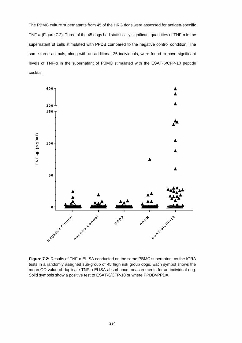
294
The PBMC culture supernatants from 45 of the HRG dogs were assessed for antigen-specific
TNF- (Figure 7.2). Three of the 45 dogs had statistically significant quantities of TNF-α in the
supernatant of cells stimulated with PPDB compared to the negative control condition. The
same three animals, along with an additional 25 individuals, were found to have significant
levels of TNF-α in the supernatant of PBMC stimulated with the ESAT-6/CFP-10 peptide
cocktail.
Neg
ati
ve C
on
tro
l
Po
sit
ive C
on
tro
l
PP
DA
PP
DB
ES
AT
-6/C
FP
-10
0
5 0
1 0 0
1 5 0
3 0 0
6 0 0
F IG .2 - P B M C T N F - : H ig h R is k G ro u p (n = 4 5 )
TN
F-
(p
g/m
l)
Figure 7.2: Results of TNF-α ELISA conducted on the same PBMC supernatant as the IGRA
tests in a randomly assigned sub-group of 45 high risk group dogs. Each symbol shows the
mean OD value of duplicate TNF-α ELISA absorbance measurements for an individual dog.
Solid symbols show a positive test to ESAT-6/CFP-10 or where PPDB>PPDA.

295
Comparing the concentration of antigen-specific TNF-α produced in response to ESAT-6/CFP-
10 stimulation only with the overall IGRA results (positive with either a PPDB bias or ESAT-
6/CFP-10 response), showed that these two tests, interpreted in parallel, identified 29 (64.4%)
test positive and 16 test negative individuals. These tests showed very strong agreement
(Cohen’s kappa coefficient, κ, =0.83).
However, the mitogen PMA/Ca used as a sample positive control for the IGRA test did not
stimulate the production/secretion of canine TNF-. As the HRG dog stimulations could not
be re-run to include an additional mitogen, we compared the IFN- and TNF- production from
three healthy dog PBMC samples stimulated with a cocktail of PMA/Ca and ConA. In this
instance TNF-and IFN-were both produced (Figure 7.3). Therefore the lack of TNF-
response to the PMA/Ca positive control in the HRG group does not invalidate the positive
antigen-specific responses observed to PPDB and ESAT-6/CFP10, rather it indicates that the
positive control was not optimal and that future assays to measure this, and potentially other
cytokines should consider a wider mitogen cocktail as a sample positive control.
Do
g 1
Do
g 2
Do
g 3
0
2 0 0
4 0 0
6 0 0
8 0 0
1 0 0 0
F IG .3 a - P M A C a /C o n A -s t im u la te d T N F -
TN
F-
(p
g/m
l)
Do
g 1
Do
g 2
Do
g 3
0
1 0 0 0 0
2 0 0 0 0
3 0 0 0 0
4 0 0 0 0
5 0 0 0 0
F IG .3 b - P M A C a /C o n A -s tim u la te d IF N -
IFN
- (
pg
/ml)
Figure 7.3: Concentration (replicate mean; error bars show standard deviation) of TNF-α (left)
and IFN- (right) produced by PBMC stimulated for four days at 37ºC/5% CO2 with
PMA/Ca/ConA. Addition of Con A to the mitogen mix induced TNF- in all three dogs, whereas
all previous PBMC incubations with PMA/Ca did not induce detectable TNF-.
The same subset of 45 HRG dogs was tested using a whole blood IGRA (Figure 7.4). Overall
responses were low, with three individuals showing responses indicative of MTBC infection
(PPDB>PPDA), two of which also showed responses to peptide cocktail and ESAT-6 protein.
Comparing these results with those from the PBMC IGRA showed poor agreement between
the two tests (κ= 0.2).

296
Po
sit
ive C
on
tro
l
Neg
ati
ve C
on
tro
l
PP
DA
PP
DB
ES
AT
-6
ES
AT
-6/C
FP
-10
0
1 0 0
2 0 0
3 0 0
4 0 0
5 0 0
6 0 0
7 0 0
8 0 0
9 0 0
1 0 0 0
1 5 0 0
2 0 0 0
2 5 0 0
F IG .4 - W h o le b lo o d IG R A : H ig h R is k G ro u p (n = 4 5 )
IFN
- (
pg
/ml)
Figure 7.4: Results of the whole blood IGRA test for a randomly assigned sub-group of 45 of
the high risk group of 164 dogs. Concentrations are calculated using the mean of duplicate
IFN- ELISA absorbance measurements against the standard curve. Solid symbols show a
positive test to ESAT-6/CFP-10 peptides or ESAT-6 or where PPDB>PPDA.
7.3.2 Serological Assays
The Idexx M. bovis Ab ELISA (Figure 7.5) showed a statistically significant difference between
the OD values for the HRG (n=163) and TB-free (n=45) dog groups (Mann Whitney U = 880.5,
P<0.001). In deciding upon a preliminary test cut-off for the canine samples tested with the
Idexx M. bovis Ab assay, we took into account both the cattle Idexx test protocol and the
modified Idexx test as applied to camelid TB testing at APHA, both of which approximate to
an OD cut-off value of 0.3 to provide high test specificity. Application of a cut off value of 0.3
to the canine samples tested with the Idexx M. bovis Ab test, the data resulted in 100% test
specificity for this TB-free group, and identified 12 test-positive dogs within the HRG (7.4%).

297
HR
G
TB
-fre
e
0 .1
1
1 0
OD
45
0n
m
F IG .5 - Id e x x M b o v is a n t ib o d y a s s a y
Figure 7.5: Serology results for dogs in the high risk group (HRG, n=163) and TB-free group
(n=45) obtained using the Idexx M. bovis Ab test. Results shown are the mean OD values of
duplicate measurements for individual dog samples at 450nm absorbance. The test-positive
cut off value of OD 0.3 is shown by a horizontal line. Solid symbols show test-positive samples.
The DPP VetTB test (Figure 7.6) showed significantly higher RLU results for the HRG
compared to the TB-free dogs (Mann Whitney U = 2731, P=0.003). In deciding upon
preliminary cut-offs for the MPB83 and ESAT6/CFP-10 antigens in this test, a preliminary ROC
analysis for the MPB83 RLU values suggested a cut-off of >95 to provide a specificity of 100%.
A similar analysis for ESAT-6/CFP-10 was not viable due to the very low number (n=2) of sero-
positives; however, there was a clear divide between these two sero-positives and test-
negatives, of between 40-80 RLU, therefore >60 RLU was chosen as the cut-off for the ESAT-
/CFP-10 antigen line for the purpose of this study. Applying these cut-offs to the study cohorts
provided a 100% specificity, and identified 11 HRG dogs (6.7%) as test-positive; nine of these
were positive to MPB83 only, whilst two were positive to ESAT-6/CFP-10 only.

298
HR
G M
PB
83
TB
-fre
e M
PB
83
HR
G E
6/C
FP
TB
-fre
e E
6/C
FP
0 .1
1
1 0
1 0 0
1 0 0 0
1 0 0 0 0
RL
U
F IG .6 - D P P V e tT B a s s a y
M P B 8 3 c u t-o ff
E 6 /C F P c u t-o ff
Figure 7.6: Serology results for dogs in the high risk group (HRG, n=163) and TB-free group
(n=45) obtained using the DPP VetTB Assay. Results given as Optricon relative light units
(RLU). The test-positive cut off values for each antigen are shown by the horizontal lines at 95
RLU for MPB83 and 60 RLU for ESAT-6/CFP-10 (E6/CFP). Solid symbols indicate a positive
sample. As the axis is logarithmic, only values greater than zero can be plotted. For this graph,
291 values overall were zero or negative and are therefore not included on the graph.
The comparative ELISA (Figure 7.7) found generally higher responses to PPDA than to PPDB
in the TB-free population. The test cut-off was calculated as the mean OD [PPDB-PPDA] of
the TB-free group plus three standard deviations, providing a cut-off value of 0.14. Using this
threshold gave a 100% specificity and identified 27 test-positive HRG dogs (16.6%).

299
HR
G
LR
G/T
B-f
ree
-0 .5
-0 .4
-0 .3
-0 .2
-0 .1
0 .0
0 .1
0 .2
0 .3
0 .4
0 .5
0 .6
F IG .7 - C o m p a ra t iv e P P D E L IS A
PP
DB
-PP
DA
(O
D 4
50
nm
)
Figure 7.7: Serology results for dogs in both the high risk group (HRG, n=163) and TB-free
group (n=45) obtained using a comparative PPD antibody ELISA. Results shown are mean
values of duplicate measurements calculated by subtracting the PPDA OD480-630 from the
PPDB OD480-630 values. The test-positive cut off (OD 0.14) is shown by a horizontal line. Each
symbol represents one dog sample. Test-positives are shown as solid symbols.
7.3.3 Serum IFN-γ & TNF-α ELISA
No statistical difference was found between the quantities of either TNF-α or IFN-γ present in
the peripheral circulation of the dogs comprising the HRG and LRG (data not shown).
7.3.4 Haematology and Serum Biochemistry
Standard haematological and serum biochemical analysis was conducted on samples
collected from half (n=83) of the HRG dogs, selected at random. The only haematological
abnormality identified frequently was an increase in the total leukocyte count (n=22, 26.5%);
in most cases this was due to the presence of a mature neutrophilia (n= 18, 81.2%) while band

300
neutrophils were also present in a minority of cases (n=4, 4.8%). One dog was found to have
each of eosinophilia, monocytosis and reticulocytosis.
Only five dogs of the 83 tested (6.0%) were found to have all serum biochemical tests within
the reference interval (RI). The most frequently detected abnormality was elevated serum urea
which was above RI in 44 of the dogs (53.0%), three of which also had elevated bile acid
concentrations. Total calcium was low in 18 samples (21.7%) whilst alkaline phosphatase
(ALP) activity was reduced in 25 samples (30.1%) and increased in six. More than a quarter
(22, 26.5%) of dogs were found to have elevated globulin levels; 20 of them were elevated
enough to increase the total protein concentration of the serum to greater than RI with normal
albumin concentrations. No association was found between these changes and the results of
the other tests.
7.4 Discussion
Almost every species of mammal, including the domestic dog, is susceptible to infection with
MTBC organisms, including M. bovis (Brosch, 2002; Drewe, 2010; Miller, 2012; Parsons,
2013; Angkawanish, 2013). Canine disease due to M. bovis has been documented in a
number of clinical case reports over many decades, with several reports demonstrating inter-
species transmission, including to and from humans (Snider, 1971; Liu, 1980; Gay, 2000;
Ayele, 2004; Ellis, 2006; Van Der Bürgt, 2009; Shrikrishna, 2009; Posthaus, 2011; Pesciaroli,
2014; Park, 2016, Szaluś-Jordanow, 2016). Despite this, ante-mortem diagnostic testing for
TB in the dog is limited. There is a single study describing the use of an IGRA for the detection
of M. tuberculosis in a group of 40 dogs living in close contact with sputum-positive pulmonary
TB owners (Parsons et al. 2012). To our knowledge, no other ante mortem tests have been
evaluated as effective for the diagnosis of canine MTBC infections; intradermal TST for both
M. bovis and M. tuberculosis is unreliable, as are complement fixation and haemagglutination
methods (Snider et al. 1971; Snider et al. 1975; Parsons et al. 2012 Broughan et al. 2013b).
In the UK, M. bovis infection is endemic, with the highest prevalence in the counties comprising
the HRA, and lower but still significant prevalence within the Edge Area, defined by the
301
incidence of bovine infections. In these areas many species other than cattle are also infected
with M. bovis and it was from within the Edge Area that we previously reported an outbreak of
M. bovis disease in a pack of kennelled foxhounds (Delahay et al. 2002, O’Halloran et al 2018).
The lack of reliable diagnostic assays meant that the prevalence of subclinical M. bovis
infection within the group of exposed animals was challenging to determine, and potentially
led to the outbreak lasting longer, and costing more to bring under control, than if such a test
or tests had been available.
To begin to address this shortcoming, we have in this study applied commercially available
tests produced for other species (Idexx for cattle, DPP VetTB for cervids) with an in-house
serology test, and cellular cytokine release assays (IFN- and TNF-) using commercial
reagents, for their ability to detect subclinical infections in dogs from both high and low
expected prevalence populations. All of the tests chosen evaluate the host immune response
rather than relying on direct pathogen identification or isolation. Previous studies have
demonstrated that the major immunological consequences of MTBC infections are broadly
conserved across host species (Cousins and Florisson, 2005; Broughan, et al. 2013b). Once
mycobacteria are phagocytosed, macrophages and dendritic cells present antigen from lysed
bacilli to CD4+ and CD8+ αβ as well as γδ T-cell subsets in the context of cell surface MHC
class II molecules. The reciprocal activation between cells of these immunophenotypes,
through the secretion of IFN-γ and TNF-α along with other cytokines, generates the classical
cell mediated response required for the control of an intracellular pathogen.
The first diagnostic test we assessed was an adapted IGRA, identifying animals with IFN-γ
producing antigen-specific T-cells. Similar to domestic cats, lion, alpaca and other new world
camelids, we found that it was necessary to isolate PBMC prior to antigen stimulation in order
to reliably generate interpretable test data under these conditions (Hesketh et al. 1994;
Gutierrez et al. 1998; Cousins and Florisson, 2005; Rhodes et al. 2008a; Leem et al. 2018).
This was due to the failure of cells within the whole blood of these species to respond
significantly to either positive control or antigen stimulation, unlike the whole blood responses

302
observed in cattle, goats and humans (Vordermeier et al. 2006; Vordermeier et al. 2007; Bezos
et al. 2018; Leem et al. 2018).
Examining the responses generated by the HRG dogs, we found that 77 (47.0%) showed a
PPDB biased response indicating subclinical MTBC infection; compared to the LRG groups
where only two animals (2/32, 6.3%) showed this response; interestingly, these were the two
dogs most in-contact with the HRG hounds whilst no animals in the TB-free group showed an
MTBC response. These results suggest this test method and interpretation has a high
specificity of 97.3-100%, which is within the range of 90-100% consistently reported for IGRA
assays in cattle, cats and goats (Vordermeier et al. 2006; Rhodes et al. 2012; Bezos et al.
2018). Of the 77 HRG dogs showing a PPDB biased response, 62% simultaneously showed
a significant response the ESAT-6/CFP-10, strongly suggestive of infection with an RD-1
positive MTBC organism; this is slightly lower than the proportion reported for both cattle
(~85% in the high specificity IGRA where both PPD and peptide must be positive, and 82%
for peptide alone as a diagnostic, Vordermeier et al., 2016) and cats (80% but only ten cats
were tested, Rhodes et al., 2011). This difference may reflect a lesser immunological
significance of these antigens in canine infections and/or may be an artefact of the unknown
incubation time from infection to blood sampling in the HRG dogs; had infections been allowed
to continue to incubate, the number of dogs responding to these peptides may have increased.
A further five dogs in the HRG only responded to the peptide cocktail ESAT-6/CFP-10. With a
lack of an antigen-specific PPDB response, it is questionable whether these animals are truly
infected with M. bovis or not, since some NTM (notably M. kansasii and M. leprae, among
others), encode orthologs of these proteins, and as the HRG in this study comprised working
Foxhounds there was significant potential for them to be exposed to/infected with confounding
NTM. However, infected cattle are known to respond to ESAT6/CFP-10 in the absence of a
positive PPD response, and in a setting of confirmed M. bovis infection such as the HRG dogs,
a positive response to these peptides suggests an individual at high risk of being infected.
Furthermore, no animals in the LRG/TB-free groups demonstrated ESAT-6/CFP-10
responses, despite many having similar opportunity for NTM sensitisation as the HRG. So,
303
whilst the potential for confounding influence of NTM is important for interpreting individual
animal responses, at a group level the data presented here would suggest that a response to
ESAT-6/CFP-10, even alone, is sufficient to indicate high risk of M. bovis challenge.
In our previous assessment of test responses, individual dogs with a significant response to
any test antigen were considered test-positive (O’Halloran et al. 2018); this included three
dogs that responded to PPDA alone. Analysis of the LRG tests found that this response
occurred in 12 of the 74 dogs (16.2%) suggesting that this should not be seen as indicative of
being at risk of M. bovis infection. Therefore, the definition of IGRA-positive animals in the
context of subclinical M. bovis infections, should be revised to consider only individuals that
show a statistically significant response above the media control to PPDB that is greater than
the PPDA response, and/or those who respond significantly to the ESAT-6/CFP-10
combination.
The diagnostic sensitivity of assessing antigen-specific TNF- responses has only recently
been tested clinically and on relatively small populations of M. tuberculosis-infected people,
with inconclusive results; however, it shows promise for discriminating between active and
latent disease (Harari et al. 2011; Wang et al. 2013). The approach we used, combining two
ELISA cytokine measurements (IFN- and TNF-) on the same stimulated cell supernatants,
identified additional test-positive individuals suggesting that a dual cytokine test approach may
have a higher diagnostic sensitivity without the need to take any additional blood from the
animals being tested. However, going forward there would need to be a modification of the
positive mitogen control to include ConA, since PMA/Ca alone did not stimulate canine TNF-
release.
The detection of specific antibody responses has been useful in the development of new
diagnostic tests for mycobacterial infections, particularly for species where repeat capture of
the same individual at a fixed time point is difficult, and therefore precludes the use of TST
(Gao et al. 2007; Lesellier et al. 2008; Bezos et al. 2018) or where reagents for IGRA tests are

304
not available. We therefore examined the antibody responses of dogs using a combination of
serological tests already in use for other species.
As expected, the total number of test-positive animals identified by the serological assays was
significantly lower than for the PBMC IGRA; 16.5%, 7.4% and 6.7% for PPD ELISA, Idexx
ELISA and DPP VetTB tests respectively, compared to 52% IGRA-positive in the HRG dogs).
However the simplicity of the antibody tests compared with the IGRA test does provide relative
speed and a lower test cost compared to the IGRA, which could facilitate an initial blood test
screening of large numbers of animals in confirmed breakdown situations, and/or provide a
serological option where blood sampling and rapid, temperature controlled transport (for IGRA)
may not be possible.
Of the three antibody tests the comparative PPD ELISA found a much higher number of test-
positive dogs, 27 compared to 12 (Idexx) and 11 (DPP VetTB). Whether this is due to PPD
providing the largest epitope repertoire of all assays tested, or whether the test cut-off is not
optimal remains to be explored. Many LRG/TB-free dogs had a PPDA-biased response
resulting in a very low test-cut-off (0.14) compared to the Idexx ELISA (cut-off 0.3). However,
25 of the 27 comparative PPD positive dogs were positive to at least one other test (PMBC
IGRA [20], DPP VetTB Assay [7] and/or Idexx ELISA [6]) and so this does provide confidence
in the PPD ELISA results.
Evaluating all of the test responses for each individual dog, it became clear that a small
minority of individuals showed much higher responses to the tests than the rest of the test
positive population on each assay. Further examination showed that this was consistently due
to three individuals for the cell based and cytokine assays, and a single individual for the
antibody based assays. The relevance of the magnitude of this test response is unclear. There
is conflicting evidence in the scientific literature as to whether the concentration of cytokine in
serum and/or produced in in vitro diagnostic assays is proportionate to the severity or
progression of clinical disease in vivo (Hasan et al. 2009; Ruhwald et al. 2009; Jonnalagadda
et al. 2013; Xu et al. 2018), whilst there is some evidence indicating that ESAT-6/CFP-10-

305
specific IFN-γ responses correlate with disease severity in cattle (Vordermeier et al. 2002) and
that the titre of detectable antibody correlates with the occurrence of a lesioned phenotype
(Roos et al. 2016). This may be true for dogs; however, data in this study did not support this
view, possibly because of to the subclinical nature of the HRG tested.
Haematological abnormalities were identified infrequently in the 83 dogs in the HRG that were
tested. The high total leukocyte counts, mature and band neutrophilia seen in a small number
of dogs did not correlate with any of the mycobacteria-specific diagnostic tests; they probably
reflect an unknown inflammatory stimulus in these dogs.
Serum biochemistry of the same dogs revealed multiple frequent abnormalities. The most
frequent, elevated urea concentration, likely reflects the high protein diet of the working hounds
as they were fed raw meat, rather than any underlying pathology. Half of the 22 dogs with
hyperglobulinaemia were also found to be positive to at least one of the serology assays
indicating that in some cases hyperglobulinaemia may have been related to the presence of
antigen-specific antibodies. However, the overall specificity of hyperglobulinaemia with
respect to the remainder of the tests was poor.
This study was unable to evaluate test sensitivity as too few confirmed infections were
identified – in total seven confirmed clinical cases of M. bovis infection occurred during the
course of the kennel outbreak. Due to the severity of the disease, these affected animals were
euthanased on welfare grounds before diagnostic blood samples could be taken. The
diagnosis of M. bovis infection in these animals was confirmed by mycobacterial culture of
visibly lesioned tissues collected post-mortem in each of these cases. The rate of false
negative test results (type II error) in this study is therefore also unknown.
In summary, this study demonstrates, for the first time, the utility of two cellular (IGRA and
TNF-) and three serological assays (DPP VetTB, Idexx and comparative PPD ELISA) for
detecting sub-clinical M. bovis infections of dogs. Whilst the data suggest high test specificity
for all assays evaluated, further work is needed to robustly validate the specificity of the tests,
and investigate test sensitivity using sufficient numbers of dogs of confirmed infection status.

306
7.5 References
Adams L.G. (2001) In vivo and in vitro diagnosis of Mycobacterium bovis infection. Revue
Scientifique et Technique de L’Office Internationale Des Epizooies 20, 304-324
Ayele, W. Y., et al. (2004). Bovine tuberculosis: An old disease but a new threat to Africa.
International Journal of Tuberculosis and Lung Disease 8(8): 924-937
Bezos J. et al. (2018) The use of serological tests in combination with the intradermal
tuberculin test maximizes the detection of tuberculosis infected goats. Veterinary Immunology
and Immunopathology 199:43-52
Bezos, J., et al. (2014). Current ante-mortem techniques for diagnosis of bovine tuberculosis.
Research in Veterinary Science 97(S): S44-S52
Bossuyt, P.T. et al. (2015) STARD 2015: an updated list of essential items for reporting
diagnostic accuracy studies. BMJ 351:h5527
Brosch, R., et al. (2002). A new evolutionary scenario for the Mycobacterium tuberculosis
complex. Proceedings of the National Academy of Sciences of the United States of America
99(6): 3684-3689
Broughan, J. M., et al. (2013a). Mycobacterium bovis infections in domesticated non-bovine
mammalian species. Part 1: Review of epidemiology and laboratory submissions in Great
Britain 2004-2010. Veterinary Journal 198(2): 339-345.
Broughan, J. M., et al. (2013b). Mycobacterium bovis infections in domesticated non-bovine
mammalian species. Part 2: A review of diagnostic methods. Veterinary Journal 198: 346-351.
Buddle B.M. et al. (2001) Use of ESAT-6 in the interferon-gamma test for diagnosis of bovine
tuberculosis following skin testing. Veterinary Microbiology. 80: 37-46
Buddle, B.M. et al (2000) Immunological approaches to the control of tuberculosis in wildlife
reservoirs Veterinary Immunology and Immunopathology, 74 (1-2), pp. 1-16.
Casal, C.et al. (2014) Strategic use of serology for the diagnosis of bovine tuberculosis after
intradermal skin testing. Veterinary Microbiology, 170 (3-4), pp. 342-351.
Cousins D.V., Florisson, N. (2005) A review of tests available for the use in the diagnosis of
tuberculosis in non-bovine species. Revue Scientifique et Technique de L’Office Internationale
Des Epizooies 24, 1039-1059.

307
Department for the Environment, Farming and Rural Affairs [DEFRA] (2018)
https://www.gov.uk/government/statistics/incidence-of-tuberculosis-tb-in-cattle-in-
great-britain
De la Rua-Domenech, R., et al. (2006) Ante mortem diagnosis of tuberculosis in cattle: A
review of the tuberculin tests, gamma-interferon assay and other ancillary diagnostic
techniques. Research in Veterinary Science 81, 190-210.
Delahay, R.J. et al (2001) Wildlife disease reservoirs: The epidemiology of Mycobacterium
bovis infection in the Eeuropean badger (Meles meles) and other British mammals.
Tuberculosis, 81 (1-2), pp. 43-49
Delahay, R.J. et al. (2003) Vaccinating badgers (Meles meles) against Mycobacterium bovis:
The ecological considerations. Veterinary Journal, 166 (1), pp. 43-51
Delahay, R.J.et al. (2002) The status of Mycobacterium bovis infection in UK wild mammals:
A review. Veterinary Journal, 164 (2), pp. 90-105
Eisenhut, M. (2014). The evidence for greater sensitivity of interferon-γ release assays
compared to tuberculin skin test in diagnosing latent mycobacterium tuberculosis infection.
Clinical Pediatrics 53(14): 1413
Ellis, M. D., et al. (2006). Mycobacterium bovis infection in a dog. Veterinary Record 159(2):
46-48.
Elnaggar M.M. et al. (2017) Evaluation of antigen specific interleukin-1β as a biomarker to
detect cattle infected with Mycobacterium bovis. Tuberculosis 105: 53-59
El-Seedy, F.R. et al. (2013) The correlation between M. bovis isolation and ELISA using PPD-
B and ESAT6-CFP10 mixture on the sera of tuberculin reactor cattle and buffaloes. 2013)
Journal of Food, Agriculture and Environment, 11 (1), pp. 489-494.
Gao, L. Y., et al. (2004). A mycobacterial virulence gene cluster extending RD1 is required for
cytolysis, bacterial spreading and ESAT-6 secretion. Molecular Microbiology 53(6): 1677-1693
Gao, M.Q. et al. (2007) The clinical significance of serum tuberculosis specific antigen
antibody in the diagnosis of tuberculosis. Chinese journal of tuberculosis and respiratory
diseases, 30 (12), pp. 918-920.
Gay, G., et al. (2000). Pulmonary mycobacterium bovis infection in a dog. New Zealand
Veterinary Journal 48(3): 78-81.

308
Guinn, K. M., et al. (2004). Individual RD1 -region genes are required for export of ESAT-
6/CFP-10 and for virulence of Mycobacterium tuberculosis. Molecular Microbiology 51(2): 359-
370.
Gutierrez, M. et al. (1998) Evaluation of cellular and serological diagnostic tests for the
detection of Mycobacterium bovis infected goats. Veterinary Microbiology 62: 281-290
Harai, A. et al. (2010) Dominant TNF-alpha+ Mycobacterium tuberculosis-specific CD4+ T cell
responses discriminate between latent infection and active disease. Nature Medicine 17: 372-
376
Hasan, Z. et al. (2009) Relationshp between circulating levels of IFN-γ, IL-10, CXCL9 and
CCL2 in pulmonary and extrapulmonary tuberculosis is dependent on disease severity.
Clinical Immunology, 69 pp. 259-267
Hesketh, J. B. et al. (1994) Development of a diagnostic blood test for tuberculosis in alpacas
(Lama pacos). New Zealand Veterinary Journal 42: 104-109
Hope, J.C. and Villarreal-Ramos, B. (2008) Bovine TB and the development of new vaccines.
Comparative Immunology, Microbiology and Infectious Diseases, 31 (2-3), pp. 77-100.
Houlihan, M. G. et al. (2008) Outbreak of bovine tuberculosis featuring anergy to the skin test,
udder lesions and milkborne disease in young calves. Veterinary Record 163: 357-361
Jonnalagadda, S.R. et al. (2013) Predictive value of interferon-gamma release assays for
postpartum active tuberculosis in HIV-1-infected women. International Journal of Tuberculosis
and Lung Disease, 17 (12), pp. 1552-1557+i-iv
Junqueira-Kipnis, A. P., et al. (2006). Mycobacteria lacking the RD1 region do not induce
necrosis in the lungs of mice lacking interferon-γ. Immunology 119(2): 224-231.
Kim, Y. Y., et al. (2011). Sensitivity of whole-blood interferon-gamma release assay according
to the severity and the location of disease in patients with active tuberculosis. Tuberculosis
and Respiratory Diseases 70(2): 125-131
Leem, A.Y et al. (2018) Changes in cytokine responses to TB antigens ESAT-6, CFP-10 and
TB 7.7 and inflammatory markers in peripheral blood during therapy(2018) Scientific Reports,
8 (1), art. no. 1159
Leonard, D. (2017) Bovine TB controls in the UKVeterinary Record, 181 (9), p. 241

309
Lesellier, S. et al. (2008) Antigen specific immunological responses of badgers (Meles meles)
experimentally infected with Mycobacterium bovis. Veterinary Immunology and
Immunopathology, 122 (1-2), pp. 35-45
Liu, S., et al. (1980). Canine tuberculosis. Journal of the American Veterinary Medical
Association 177(2): 164-167.
Marassi, C.D. et al. (2014) Detection of specific antibodies in cows after injection of PPD.
Pesquisa Veterinaria Brasileira, 34 (3), pp. 241-243
Min, F. et al. (2011) Serum antibody responses to 10 Mycobacterium tuberculosis proteins,
purified protein derivative, and old tuberculin in natural and experimental tuberculosis in rhesus
monkeys. Clinical and Vaccine Immunology, 18 (12), pp. 2154-2160
O’Halloran, C. et al. (2018) An outbreak of tuberculosis due to Mycobacterium bovis infection
in a pack of English foxhounds (2016-2017). Transboundary and Emerging Diseases
DOI:10.1111/tbed.12969
Office International des Epizooties [OIE] (2016) Manual of Diagnostic Tests for Aquatic
Animals Chapter 1.1.2
Park, H. A., et al. (2016). Pulmonary Mycobacterium tuberculosis infection with giant tubercle
formation in a dog: A case report. Veterinarni Medicina 61(2): 102-109.
Parsons, S. D. C., et al. (2012). Detection of Mycobacterium tuberculosis infection in dogs in
a high-risk setting. Research in Veterinary Science 92(3): 414-419
Parsons, S. D. C., et al. (2013). Novel cause of tuberculosis in meerkats, South Africa.
Emerging Infectious Diseases 19(12): 2004-2007.
Pesciaroli, M., et al. (2014). Tuberculosis in domestic animal species. Research in Veterinary
Science 97(S): S78-S85
Pollock, J.M. and Neill, S.D (2002) Mycobacterium bovis infection and tuberculosis in cattle.
Veterinary Journal 163: 115-127
Posthaus, H., et al. (2011). Accidental infection of veterinary personnel with Mycobacterium
tuberculosis at necropsy: A case study. Veterinary Microbiology 149(3-4): 374-380
Rhodes, S. G., et al. (2008a). Adaptation of IFN-gamma ELISA and ELISPOT tests for feline
tuberculosis. Veterinary Immunology and Immunopathology 124(3-4): 379-384

310
Rhodes, S. G., et al. (2008b). Interferon-γ test for feline tuberculosis. Veterinary Record
162(14): 453-455
Roos, E.O. et al. (2016) Test performance of three serological assays for the detection of
Mycobacterium bovis infection in common warthogs (Phacochoerus africanus. Veterinary
Immunology and Immunopathology, 182, pp. 79-84 DOI: 10.1016/j.vetimm.2016.10.006
Ruhwald, M. et al (2009)Biomarkers of latent TB infection. Expert Review of Respiratory
Medicine, 3 (4), pp. 387-401.
Schiller, I., et al. (2009). Optimization of a whole-blood gamma interferon assay for detection
of Mycobacterium bovis-infected cattle. Clinical and Vaccine Immunology 16(8): 1196-1202
Shrikrishna, D. et al. (2009). Human and canine pulmonary Mycobacterium bovis infection in
the same household: re-emergence of an old zoonotic threat? Thorax 64, 89-91
Snider, W. R. (1971). Tuberculosis in canine and feline populations. Review of the literature.
American Review of Respiratory Disease 104(6): 877-887
Szaluś-Jordanow, O., et al. (2016). Intracardiac tuberculomas caused by Mycobacterium
tuberculosis in a dog. BMC Veterinary Research 12(1)
Thillai, M., et al. (2014). Interferon-gamma release assays for tuberculosis: Current and future
applications. Expert Review of Respiratory Medicine 8(1): 67-78
Trost, B. et al.(2016) Investigation of the cause of geographic disparities in IDEXX ELISA
sensitivity in serum samples from Mycobacterium bovis-infected cattle. Scientific Reports, 6,
art. no. 22763 DOI: 10.1038/srep22763
Vordermeier, M., Whelan, A., Ewer, K., Goodchild, T., Clifton-Hadley, R., Williams, J. and R.G.
Hewinson (2006) The BOVIGAM assay as an ancillary test to the tuberculin skin test. Gov.
Vet. J., vol. 16(1), 72-80
Vordermeier, M., Chambersw, M.A, Cockle, P.J., Whelan, A.O., Simmons, J. and Hewison,
R.G. (2002) Correlation of ESAT-6-specific gamma interferon production with pathology in
cattle following Mycobacterium bovis BCG vaccination against experimental bovine
tuberculosis. Infect. Immunol. 70 (6) 3026-3032.
Vordermeier, H.M., Jones, G.J., Buddle, B.M. and R.G. Hewinson. (2016). Development of
immunological reagents to diagnose bovine tuberculosis in cattle. Vet. Immunol.
Immunopathol., 181:10-14.

311
Wang, G., et al (2013) Mycobacterium tuberculosis-specific TNF-α is a potential biomarker for
the rapid diagnosis of active tuberculosis disease in Chinese population. PLOS One 8(11):
e79431
Wood P R, Corner L A and Plackett P. (1990) Development of a simple, rapid in vitro cellular
assay for bovine tuberculosis based on the production of γ-interferon. Res Vet Sci 1990; 49:
46-49
Wood, P. R. and S. L. Jones (2001). BOVIGAM™: An in vitro cellular diagnostic test for bovine
tuberculosis. Tuberculosis 81(1-2): 147-155
Xu, J.-C.et al. (2018) More significance of TB-IGRA except for the diagnosis of tuberculosis.
Journal of Clinical Laboratory Analysis, 32 (1), art. no. e22183
Yearsley, D. et al. (1998) An evaluation of an anamnestic ELISA for the detection of
tuberculous cattle. Irish Veterinary Journal, 51 (6), pp. 303-306
Zhou, Q., et al. (2011). Diagnostic accuracy of T-cell interferon-γ release assays in tuberculous
pleurisy: A meta-analysis. Respirology 16(3): 473-480

312
Chapter 8: Bayesian Latent Class Estimation of the
Performance Parameters of Diagnostic Tests to
Identify Mycobacterium bovis Infected Dogs.
Abstract
There are currently no diagnostic tests available for the ante-mortem identification of
Mycobacterium (M.) bovis infections in domestic dogs (Canis lupus familiaris). We previously
reported the use of a combination of eight tests to identify infected individuals during an
outbreak of M. bovis tuberculosis in a pack of Foxhounds (Chapters 5 and 7); four of these
tests were shown to be promising for the detection of infected animals. A retrospective
Bayesian latent class analysis of the combined four test results of 164 hounds was conducted
in order to estimate the sensitivity, specificity, positive and negative predictive values of each
test as well as the prevalence of infection in the pack. The cell-based interferon gamma release
assay (IGRA) was shown to be the most sensitive test and had the highest positive and
negative predictive values with moderate specificity. The three serological assays were shown
to have higher specificities but lower sensitivities than the IGRA. Of the serological tests, the
comparative purified protein derivative (PPD) ELISA performed best for all parameters
estimated, followed by the Idexx M. bovis Ab ELISA with the DPP VetTB assay performing
least well. This differential performance between the cell-based IGRA and serological assays
suggests that testing protocols combining both approaches may be most useful for identifying
and controlling future outbreaks.The model was improved by the addition of informed priors
but was not significantly affected by the inclusion of covariance between the serological
assays. This study provides useful test performance data that could not have been derived
using a traditional frequentist approach. Further work is now needed to improve the credibility
of the initial estimates produced in this analysis by expanding the population of dogs with test
data available, particularly those whose infection status is definitively known.

313
8.1 Introduction
Tuberculosis due to infection with Mycobacterium (M.) bovis is a chronic zoonotic infectious
disease currently endemic among cattle and wildlife populations in England and Wales1-3. The
major impact of infection is within the production cattle population and disease control
measures cost the UK government in excess of £100 million annually3,4. Despite the
implementation of a strict statutory testing policy, disease elimination has proven difficult; likely
due to the complex and multifactorial nature of the host, pathogen and environmental
interaction which results in disease transmission before infected animals can be detected3-5.
Further to this, there are a number of significant maintenance and spill over hosts including
the Eurasian badger (Meles meles)4, various deer species6,7, North American camelids8,9 as
well as companion animals including both the domestic cat (Felis catus)10 and dog (Canis
lupus familiaris)11.
Recently, we investigated a large outbreak of M. bovis tuberculosis in a large working pack of
Foxhounds in England12. At the time there was discussion in the veterinary and national press
regarding whether working hounds contribute to the contamination with M. bovis of the
environment where cattle and other susceptible species may be co-habitant or use as grazing
pasture13-17. A complete and robust epidemiological investigation conducted jointly by our
group and the Animal and Plant Health Agency (APHA) concluded that there was no evidence
to suggest that dogs play a significant role in the persistence of M. bovis in England and Wales
or that scent and drag hunting with dogs contributes to the spread of bovine tuberculosis
amongst cattle or wildlife (reported in this thesis, Chapter 5)12.
Whilst the risk posed by hounds was being evaluated, there was a concurrent time delay in
establishing control over the outbreak as there are no validated ante-mortem diagnostic tests
for canine M. bovis infections12. The only diagnostic test recognised in statute, and therefore
the test designated as the ‘gold standard’, is post-mortem specialist mycobacterial culture of
visibly lesioned (granulomatous) tissues18. However, the concept of a “gold standard” in this
case can be considered misleading; the term was only intended to be applied to a test which

314
has (near) perfect performance relating to both sensitivity (Se) and specificity (Sp)19. Logically,
the isolation of viable M. bovis organisms from lesions obtained from cadavers gives culture
an assumed (near) perfect Sp for infection with that organism, provided rigorous laboratory
protocols are in place to prevent sample contamination during handling. The Se of culture for
canine derived samples has not been assessed, but in domestic cats the overall Se of all
mycobacterial culture is under 50%20 whilst the Se of M. bovis culture is reported to be
approximately 80% in cattle21. These data suggest that it may be inappropriate to refer to post-
mortem tissue culture as the ‘gold standard’ test in the outbreak of M. bovis studied herein.
Furthermore, during the investigation, it was only practically possible to conduct post-mortem
examinations on a subset of the hounds that were euthanased, meaning that in most cases
‘gold standard’ test data were not available. In addition, in a number of samples no growth of
M. bovis was observed from tissue samples after 16 weeks12 which is difficult to interpret for
a test with unknown Se.
Taking this into account made accurate estimation of the performance parameters for the
testing strategy that we used challenging whilst relying on frequentist analytical methods to
compare our results to mycobacterial culture. An alternative method for such an analysis in
the absence of an appropriate or accurate gold standard test is to use a Bayesian latent class
modelling approach22-24. This statistical methodology does not require the true infection status
of an individual to be known but instead models this as a latent (unknown) variable along with
the test performance parameters22-24. Methods adhering to a Bayesian rather than frequentist
framework have been successfully utilised since the development of the two-test, two-
population model introduced by Hui and Walter in 198024. Since then, the World Organisation
for Animal Health (the OIE) has encouraged the use of these models; particularly in cases, as
presented here, where the acquisition of sufficient numbers of samples from animals of known
infection-status would be extremely difficult due to the low prevalence of disease, where
current gold standard diagnostic testing methods have known limitations and/or the
experimental infection of animals to produce robust test performance data would be
considered unethical25.

315
The methodology established by Hiu and Walter is dependent on the critical assumptions that;
a) when comparing multiple populations, the underlying disease prevalence differs between
them, but that b) the test performance is the same across them and, c) the (two) tests are
conditionally independent from one another24. In our outbreak scenario, however, there was
only a single epidemiological population without a second that we could be sure adequately
satisfies these assumptions. This limitation is not uncommonly encountered when studying
infectious diseases as it is inherently challenging to sample populations of animals which have
differing infection prevalence but do not respond differently to the tests being conducted26.
Therefore, an extension to the original methodology has been proposed whereby three or
more tests are performed on a single study population26,27, an approach which has previously
been adopted for the estimation of test performance relating to bovine M. bovis infections5.
The aim of the study presented here was to analyse a panel of four tests that we have
previously established as promising in their application to detect subclinical M. bovis infections
in dogs using samples collected ante mortem28 (reported in Chapter 7) and employ a Bayesian
latent class analysis to estimate the performance parameters (Se, Sp, positive predictive value
[PPV] and negative predictive value [NPV]) of each test and the underlying infection
prevalence in the population.
We demonstrate that the cell-based interferon gamma release assay (IGRA) was shown to be
the most sensitive test and had the highest positive and negative predictive values with
moderate specificity. The three serological assays were shown to have higher specificities but
lower sensitivities than the IGRA.
8.2 Materials and Methods:
8.2.1 Study Population & Test Data
We previously investigated a fulminant disease outbreak caused by M. bovis infection within
a large group of kennel-housed working Foxhounds in England12 (Chapter 5). A total of 164
hounds were sampled over the course of the outbreak and so had test data available for

316
analysis in this study12. The samples were previously analysed using eight test protocols that
were either previously developed for the detection of M. bovis infections in non-canid species
or newly developed by our group12,28.
We demonstrated that four of these assays produced results which overall gave were able to
distinguish between high and low risk groups of individuals, indicating that they had good
potential to be developed into diagnostic assays28. These included a cell-based interferon
gamma release assay (coded here as “IGRA”), the Idexx M. bovis Ab ELISA (Idexx, USA;
coded as “Idexx”), the DPP lateral flow VetTB Assay for Cervids (Chembio, USA; coded as
“DPP”) and a newly developed comparative purified protein derivative (PPD) ELISA (coded as
“CompPPD”)28.
8.2.2 Bayesian Latent Class Analysis
As previously described, we tested 119 M. bovis-free and M. bovis-‘low risk’ dogs using the
same protocols as the high risk dogs in order to determine specific cut-off values for each test
methodology28. We therefore ascribed each of the 164 hounds a binary positive or negative
status for each of the four tests allocating each individual to one of 16 possible combinations
of positive and negative test results.
Bayesian latent class models were used to estimate diagnostic test Se, Sp, PPV, NPV and
the prevalence of infection. The computations were implemented using WinBugs version 1.4.3.
All models utilised a four-test one-population approach. As no data are available on the
performance of these assays in domestic dogs, model one was coded (Appendix 2) to include
uninformative flat uniform priors; beta (1, 1). Throughout the study all models were run for
100,000 iterations with the first 5000 discarded as a “burn in”. Chain convergence after the
initial burn-in was assessed by visual inspection of the Markov chain Monte Carlo outputs.
For tests to be of potential benefit to clinicians, the PPV and/or NPV of a test needs to at least
be greater than 50% (i.e. better than chance) or else no decisions can be made based on the
test result. Subsequently to evaluating model one, informed priors were included into a second

317
model in order to improve the clinical utility of the posterior estimates. The values assigned
were based on data obtained from a review of relevant bovine literature29-35. The sensitivity of
IGRAs reported in the literature ranged from 55% to 93% with 74% most frequently reported
and their specificity ranged from 89% to 100% with a consensus around 95%29-35. These
values were converted into beta (15.04, 5.28) and beta (64, 3.7) values respectively; the beta
distributions and designated initial values used for all parameters in model two are provided in
Table 8.1. Where published data were unavailable, such as for the in-house comparative
ELISA, the posterior estimates obtained from model one were used as a priori information for
models two and three.
Table 8.1: The beta distributions and designated initial values for each variable used in models
two and three.
In models one and two, we had followed the implicit model assumption that the different tests
are conditionally independent of one another. However, as three of the four tests are
serological assays, albeit of different types, we included undefined covariance terms into
model three to allow for possible interdependence between these test results to be accounted
for. Model three included the same prior distributions as model two which are provided in Table
8.1.
Variable Beta Distribution Initial value
SeIGRA (15.04, 5.28) Se=0.74
SpIGRA (64, 3.7) Sp=0.95
SeDPP (1, 1) Se=0.65
SpDPP (1, 1) Sp=0.98
SeCompPPD (1, 1) Se=0.10
SpCompPPD (1, 1) Sp=0.20
SeIdexx (98.77, 261.40) Se=0.5
SpIdexx (137.64, 6.81) Sp=0.97

318
8.3 Results
Between December 2016 and August 2017 we tested a total of 164 Foxhounds kept at a single
kennel premises to try and identify all of the individual dogs infected with M. bovis during a
tuberculosis outbreak12. The combination and frequency of the results generated by the four
testing protocols retrospectively analysed in this study are shown in Table 8.2.
Table 8.2: Test result categories for the 164 dogs included in this study in descending order
of frequency.
IGRA Idexx Ab ELISA DPP VetTB
assay
Comparative PPD ELISA Frequency
(Total = 164)
Negative Negative Negative Negative 66
Positive Negative Negative Negative 62
Positive Positive Negative Negative 15
Positive Positive Positive Positive 4
Negative Positive Positive Negative 3
Negative Negative Negative Positive 3
Positive Positive Negative Negative 2
Negative Negative Positive Negative 2
Positive Positive Positive Negative 2
Positive Negative Positive Negative 1
Positive Negative Negative Positive 1
Negative Negative Positive Positive 1
Negative Positive Positive Positive 1
Positive Positive Negative Positive 1
Negative Positive Negative Positive 0
Positive Negative Positive Positive 0
The posterior parameter estimates for all three models generated in this study are provided in
Table 8.3. In model one, the IGRA test had the greatest Se with a median posterior estimate
of 51% (95% credibility interval [CrI] 42-59%). The IGRA and the comparative PPD ELISA
were the only assays shown to have a clinically useful PPV, defined as greater than 50%, with
a median estimate of 89% (CrI 69-96%) and 55% (CrI 5-79%); however, only the IGRA had
the full 95% credibility interval above 50%. The Idexx M. bovis Ab ELISA was shown to have
a specificity estimate of 56%, however the PPV (40%) and NPV (6%) estimates were too low
for this to be considered a clinically useful test. All of the remaining test parameter estimates

319
in model one had posterior parameter estimates below 50% and broad credibility intervals
which further indicated the need to include a priori information into the model.
Table 8.3: Interferon gamma release assay (‘IGRA’), Idexx M.bovis Ab ELISA (‘Idexx’), DPP
VetTB assay (‘DPP’) and Comparative PPD ELISA (‘CompPPD’) diagnostic test parameter
estimation based on the test results of 164 individuals. Posterior parameter estimates are
given as proportions. 95%CrI: 95% credibility interval, Se: test sensitivity, Sp: test specificity,
PPV: test positive predictive value, NPV: test negative predictive value.
Variable Model one Model two Model three
Mean Median 95%
CrI
Mean Median 95%
CrI
Mean Median 95%
CrI
SeIGRA 0.51 0.51 0.42-
0.59
0.68 0.64 0.51-
0.90
0.72 0.77 0.53-
0.92
SpIGRA 0.38 0.37 0.13-
0.67
0.57 0.55 0.43-
0.79
0.59 0.59 0.44-
0.80
PPV IGRA 0.89 0.88 0.69-
0.96
0.57 0.74 0.39-
0.98
0.58 0.75 0.39-
0.98
NPV IGRA 0.08 0.08 0.02-
0.18
0.56 0.82 0.08-
0.99
0.56 0.82 0.08-
0.99
SeDPP 0.03 0.02 0.00-
0.07
0.31 0.15 0.02-
0.83
0.24 0.18 0.16-
0.53
SpDPP 0.31 0.29 0.02-
0.70
0.56 0.86 0.01-
0.99
0.62 0.76 0.48-
0.73
PPV DPP 0.25 0.23 0.02-
0.58
0.57 0.56 0.17-
0.96
0.63 0.57 0.11-
0.99
NPV DPP 0.04 0.04 0.03-
0.16
0.51 0.59 0.04-
0.98
0.51 0.59 0.04-
0.98
SeCompPPD
ELISA
0.10 0.12 0.01-
0.17
0.38 0.36 0.10-
0.77
0.42 0.32 0.12-
0.74
SpCompPPD
ELISA
0.20 0.19 0.18-
0.49
0.55 0.76 0.02-
0.97
0.56 0.77 0.02-
0.97
PPV
CompPPD
ELISA
0.51 0.55 0.05-
0.79
0.59 0.63 0.19-
0.89
0.59 0.61 0.19-
0.89
NPV
CompPPD
ELISA
0.03 0.02 0.00-
0.09
0.52 0.67 0.04-
0.98
0.50 0.65 0.04-
0.98
SeIdexx 0.03 0.03 0.00-
0.07
0.27 0.14 0.03-
0.74
0.24 0.17 0.03-
0.74
SpIdexx 0.55 0.56 0.22-
0.83
0.71 0.89 0.17-
0.99
0.71 0.89 0.17-
0.99
PPV Idexx 0.40 0.40 0.11-
0.72
0.64 0.64 0.35-
0.92
0.64 0.64 0.35-
0.92
NPV Idexx 0.07 0.06 0.01-
0.18
0.50 0.61 0.04-
0.98
0.50 0.61 0.04-
0.98
Prevalence
of infection
0.89 0.90 0.78-
0.96
0.53 0.55 0.05-
0.96
0.53 0.55 0.05-
0.96

320
Model two and three both included informed priors with respect to test performance. The
parameter estimates generated from model two indicate, in agreement with model one, that
the IGRA is the most sensitive of the tests analysed (median estimate 64%, CrI 51-90%)
followed by the comparative PPD ELISA (median estimate 36%, CrI 10-77%), with the DPP
lateral flow assay and the Idexx M. bovis Ab ELISA performing very similarly for this parameter
estimate (DPP lateral flow median sensitivity estimate 15%, CrI 2-83%; Idexx M. bovis Ab
ELISA median sensitivity estimate 14%, CrI 3-74%). By contrast, all three of the antibody
detection assays were estimated to have greater specificities than the IGRA, which was
estimated by this model to have a specificity of 55% (CrI 43-79%), ranging from 76-89% but
with wide 95% credibility intervals such as 1-99% for the DPP lateral flow assay.
In model two, all tests were shown to meet our definition of a clinically useful test. The model
two analysis again showed that the IGRA was the best performing test with a PPV estimate of
74% (CrI 39-98%) and a NPV estimate of 82% (CrI 8-99%). By these estimated performance
measures the comparative PPD ELISA (PPV 63%, CrI 19-89%; NPV 67%, CrI 4-98%) and the
Idexx M. bovis Ab ELISA (PPV 64%, CrI 35-92%; NPV 61%, CrI 4-98%) performed similarly
to one another with the DPP lateral flow assay appearing to be the least useful with the lowest
predictive value estimates (PPV 56%, CrI 17-96%; NPV 59%, CrI 4-98%).
In model three, covariance terms were included to allow for the possibility of conditional
dependence between the antibody-based test assays. Overall bivariate correlations were very
low between these three assays (ρ=0.16, 0.17 and 0.26 respectively) and including the terms
into the model did not significantly alter most of the posterior parameter estimates; only the
sensitivity estimate of the IGRA increased to a median estimate of 77% (CrI 53-92%) while
the remaining median estimate values remained within 5% of those generated in model two.
Further comparison of the deviance information criterion (DIC) values between model two and
three showed only a small change (less than three) indicating that the inclusion of the
covariance terms did not significantly improve the fit of the model to the data.

321
8.4 Discussion
Bayesian latent class analysis provides an accepted alternative methodology for estimating
diagnostic test performance when an affordable, reliable and/or non-invasive reference
standard does not exist23-27. These methods are particularly useful for cases of infectious
disease which have long subclinical latent periods, such as tuberculosis; this is because the
latent class models do not require the true infection status of any of the animals in the test
population to be known22. The OIE has continuously endorsed the use of latent class models
for the assessment of novel diagnostic test performance in animal populations in recent
years25. Using this technique we modelled the test performance of four previously unevaluated
diagnostic tests for their ante mortem accuracy to diagnose subclinical M. bovis infections in
domestic dogs.
All three of the latent class models that we developed during this study consistently showed
that the cell based IGRA test had the greatest sensitivity for detecting subclinical infections.
As an ex vivo T-cell stimulation assay, this IGRA test detects an antigen-specific cell mediated
immune response to mycobacteria which is typically formed in response to intracellular
pathogens including M. bovis36,37. The IGRA test assays are therefore considered to be both
sensitive and relatively specific indicators of infection36,37. Model three produced a median
posterior sensitivity estimate for the canine IGRA of 77% (CrI 53-92%) for the detection of
subclinical M. bovis infections. This result is similar to those reported for the approved human
test to detect both latent and active M. tuberculosis infections, the QuantiFERONTM-TB Gold
IGRA test36,38,39. Two recent studies independently evaluated the Se and Sp of the
QuantiFERONTM-TB Gold test comparing its performance to the gold standard of tuberculin
skin testing and found it be 75% (95% confidence interval 53.1–88.8%) sensitive and 80%
(95% confidence interval 64.1–90.0%) specific respectively38,39. In addition, a study evaluating
the sensitivity of IGRA testing in cats for culture positive M. bovis infections, using almost
exactly the same methodology as the test evaluated here, found the Se to be 80% compared
to the gold standard of specialist mycobacterial culture40. It is not possible to compare these
performance data to cattle sensitivity studies evaluating the BOVIGAMTM assay as recent

322
bovine sensitivity data was used as prior information to build model three. Overall therefore,
this study demonstrates that the canine IGRA here performs in line with assays designed to
detect tuberculous infections in other animals and can therefore be considered a sensitive test.
This study also analysed the performance of three serological assays for their use in canine
populations. Almost all antibody tests for the detection of M. bovis infections to date have
suffered from a relative lack of Se in comparison to T-cell based assays because infected
animals less frequently mount a significant humoral immune response to infection41-43.
However, bovine data suggests that positive antibody test results can correlate with the
presence of a visibly lesioned phenotype at post mortem carcass inspection and therefore
likely reflect an animal which is not only infected, but also infectious to naïve animals and the
environment that the animal is in42. A number of these heavily infected cattle will also not
respond positively to the BOVIGAMTM test (an IGRA assay) due to T-cell anergy following
prolonged antigenic stimulation42,43. Serological assays have additional technical advantages
compared to IGRA tests in that they are simpler and are therefore quicker and cheaper to
perform42,43. As well as this, because serological assays can be developed to include a small
number of very specific antigen targets, the specificity of these assays can be greater than
those seen for T-cell assays which frequently utilise antigen cocktails. These antigen cocktails
may have shared epitopes with other mycobacteria which impedes the test specificity44. These
factors mean that a combination of testing methods can be maximally effective in bringing M.
bovis outbreaks under control, a strategy which has recently validated in an outbreak of M.
bovis in cattle and goats45,46. Our previous study showed that the comparative PPD ELISA that
we have developed identified the largest number of test-positive dogs of the serological assays
tested with high Sp (set at 99% in that study). Here, we show that the same test retains a
higher test Se than the other two serological assays with a median estimate of 32% (CrI 12-
74%) compared to the DPP lateral flow VetTB assay and the Idexx M. bovis Ab ELISA which
performed approximately half as well with median Se estimates of 17% and 18% respectively.
This suggests that a combination of the IGRA and the comparative PPD ELISA might be the
most efficient combination of two tests to use when investigating any similar outbreaks in the
future though further research would be needed to confirm this.
323
Both of the assays shown to be the least sensitive in this analysis have contrastingly been
shown to have much greater sensitivities in the species for which they were originally
developed for use. For example, the DPP lateral flow VetTB assay performed with a sensitivity
of 84.6% in a group of naturally and experimentally infected red deer (Cervus elaphus)47 whilst
the Idexx M. bovis Ab ELISA has a reported sensitivity of 63%48. The reasons for the
differences in performance of these tests across the different species are not immediately
obvious, however, the higher sensitivity of the comparative PPD ELISA which contains the
greatest number of proteins suggests that the peptides included in the assays do not contain
the immunodominant epitopes of M. bovis as recognised by the canine adaptive immune
response. Therefore, the development of more sensitive serological assays will require greater
interrogation of which antigen or antigen combinations are the most frequently recognised
within M. bovis infected dogs.
An approach of combining serological testing subsequent to intradermal tuberculin injections
may further increase the sensitivity of serological assays, as has been shown in other
species46,48, making this a useful area of focus for future research.
As would be predicted, all three of the serological assays demonstrated high specificities with
median posterior Sp estimates of 76%, 77% and 89% respectively. These Sp values were all
higher than the Sp estimate of the IGRA, estimated at 59% (CrI 44-80%). However, though
lower than the serological assays, the Sp of the IGRA is within the range of values that would
be expected for T-cell test assays, for example it is not significantly different to the specificity
reported for the QuantiFERONTM-TB Gold test in human populations with high tuberculosis
burdens (70.8%; 95 % confidence interval 67.5–73.9 %)36,39.
Because the data used in this study referred to a single outbreak we were able to estimate the
prevalence of infection and we were also able to effectively estimate the positive and negative
predictive values of the tests to give a clearer evaluation of the clinical utility of the tests.
The median posterior prevalence estimate produced from models two and three was 55%; this
is similar to the percentage of animals that tested positive to at least one of our diagnostic

324
tests in the original outbreak investigation12, however, this estimate was also linked to very a
high degree of uncertainty with a 95% credibility interval of 5-96%.
As would be predicted, the IGRA was found to have both the highest estimated predictive
values of the tests studied (median PPV 75%, median NPV 82%) whereas the serological
assays had lower predictive values and performed comparably with one another. The posterior
parameter estimates for the PPV and NPV produced from model three indicated that all of the
tests we evaluated in this study met the criteria for being clinically informative by having values
greater than 50%. This further demonstrates that these tests have the potential to be used in
the diagnosis of canine M. bovis infections. Further work is needed to build on this analysis;
the estimates for most of the modelled parameters sit within relatively wide credibility intervals
due to the small size of the population of dogs tested (n=164), these can be improved by the
inclusion of additional test data and would particularly benefit from data from dogs of known
infection status.
8.5 Conclusion
This is the first study to analyse the test performance parameters of a number of diagnostic
tests to detect M. bovis infections in domestic dogs. These results show that the IGRA test
has the greatest Se, SP, PPV and NPV of the tests evaluated, it therefore has the greatest
potential to be useful as a clinical diagnostic test. The three serological assays had markedly
reduced Se estimates compared to the IGRA but despite this all three had predictive values
which indicate they also have potential for use as diagnostic test. The comparative PPD ELISA
assay that we developed was shown to be the best performing of the serological assays,
possibly due to the inclusion of a broader pool of antigens. The development of serological
tests for canine M. bovis infections could therefore benefit from the identification of specific
immunodominant antigens in dogs and their inclusion in any novel assay development.
The use of a Bayesian latent class modelling approach was critical to this analysis given the
lack of a gold standard test and that the true status of the individuals tested is unknown,
however, it proved necessary to include informed priors to produce reliable posterior

325
parameter estimates. Future research evaluating the diagnostic test performance of these
assays should include a greater number of animals, particularly those of known infection
status, to establish greater credibility in the estimates produced. Additional work to assess the
use of tests in combination with intradermal tuberculin injections may be beneficial to
establishing the optimal testing strategies for future outbreak investigation and control.
8.6 References
1. Allen, A.R., Minozzi, G, Glass, E.J., Skuce, R.A., McDowell, S.W.J., Wooliams et
al. Bovine tuberculosis: the genetics of host susceptibility. Proceedings of the
Royal Society of London B: Biological Sciences. 2010; 227, 2737-2745.
2. Cosivi O, Grange J, Daborn C, et al. Zoonotic tuberculosis due to Mycobacterium
bovis in developing countries. Emerging Infectious Diseases. 1998; 4(1):59.
3. Delahay RJ, Smith GC, Barlow AM, et al. Bovine tuberculosis infection in wild
mammals in the South-West region of England: a survey of prevalence and a
semi-quantitative assessment of the relative risks to cattle. Vet J.
2007;173(2):287-301.
4. Sweeney F, Courtenay O, Hibberd V, et al. Environmental monitoring of
Mycobacterium bovis in badger feces and badger sett soil by real-time PCR, as
confirmed by immunofluorescence, immunocapture, and cultivation. Applied and
environmental microbiology. 2007;73(22):7471-7473.
5. Lahuerta-Marin, A, Milne, M.G., McNair, J., Skuce, R.A, McBride, S.H., Menzies,
F.D. et al. Bayesian latent class estimation of sensitivity and specificity parameters
of diagnostic tests for bovine tuberculosis in chronically infected herds in Northern
Ireland. The Veterinary Journal. 2018; 238, 15-21.
6. Busch, F., Bannerman, F., Liggett, S., Griffin, F., Clarke, J., Lyashchenko, K.P.,
Rhodes S. Control of bovine tuberculosis in a farmed red deer herd in England.
Vet Rec. 2017; 180 (3):68. doi: 10.1136/vr.103930.
7. Ward, A.I., Smith, G.C., Etherington, T.R., Delahay, R.J. Estimating the risk of
cattle exposure to tuberculosis posed by wild deer relative to badgers in England
and Wales. J Wildl Dis. 2009; 45 (4):1104-20. PubMed PMID: 19901384.
8. Twomey DF, Collins R, Cranwell MP, Crawshaw TR, Higgins RJ, Dean GS,
Vordermeier HM, Hollingdale A, de la Rua-Domenech R. Controlling tuberculosis
in a llama (Lama glama) herd using clinical signs, tuberculin skin testing and
serology. Vet J. 2012; 192( 2):246-8. doi: 10.1016/j.tvjl.2011.05.014.
9. Rhodes S, Holder T, Clifford D, Dexter I, Brewer J, Smith N, Waring L, Crawshaw
T, Gillgan S, Lyashchenko K, Lawrence J, Clarke J, de la Rua-Domenech R,

326
Vordermeier M. Evaluation of gamma interferon and antibody tuberculosis tests
in alpacas. Clin Vaccine Immunol. 2012; 19 (10):1677-83.
10. Gunn-Moore DA, Jenkins PA, Lucke VM. Feline tuberculosis: a literature review
and discussion of 19 cases caused by an unusual mycobacterial variant. Vet Rec.
1996; 138(3):53-58.
11. Snider WR, Cohen D, Reif JS, Stein SC, Prier JE. Tuberculosis in canine and
feline populations. Study of high risk populations in Pennsylvania, 1966-1968. Am
Rev Respir Dis. 1971; 104(6):866-876.
12. O'Halloran C, Hope JC, Dobromylskyj M, Burr P, McDonald K, Rhodes S, Roberts
T, Dampney R, De la Rua-Domenech R, Robinson N, Gunn-Moore DA. An
outbreak of tuberculosis due to Mycobacterium bovis infection in a pack of English
Foxhounds (2016-2017). Transbound Emerg Dis. 2018 65(6):1872-1884. doi:
10.1111/tbed.12969.
13. Elkins P. Hunting hounds and bovine TB. Vet Rec. 2018 6;183(13):419-420. doi:
10.1136/vr.k4144.
14. Blowey R. Hunting hounds and bovine TB. Vet Rec. 2018 6;183(13):419. doi:
10.1136/vr.k4143.
15. Spreading of bovine TB by hunting hounds. Vet Rec. 2018 15;183(10):328-329.
doi: 10.1136/vr.k3911.
16. Eastwood B, Menache A, Dalzell F, Puddifoot J, Elliott P, Pell S, Hill S, Saunders
R, Langdale T, St Pierre N, O'Connor M, Newby P, Miles SF, Hinde J, Shepherd
C, Tipping K, Blakeway S, Irving B, Knight A, Jones M, McGill I. Spreading of
bovine TB by hunting hounds. Vet Rec. 2018 Sep 15;183(10):327-328. doi:
10.1136/vr.k3874. Erratum in: Vet Rec. 2018 22;183(11):356.
17. 'Plausible' risk posed by bovine TB in hounds. Vet Rec. 2018; 18;183(7):205. doi:
10.1136/vr.k3554.
18. Middlemiss C, Clark J. Mycobacterium in pets. Vet Rec. 2018; 10;183(18):571.
doi: 10.1136/vr.k4689.
19. Geogiadis, M.P., Johnson, W.O., Gardner, I. Sample size determination for
estimation of the accuracy of two conditionally independent tests in the absence
of a gold standard. Preventative Veterinary Medicine. 2005; 71, 1-10.
20. Gunn-Moore DA, McFarland SE, Brewer JI, et al. Mycobacterial disease in cats in
Great Britain: I. Culture results, geographical distribution and clinical presentation
of 339 cases. J Feline Med Surg. 2011;13(12):934-944.
21. Gormley E, Corner LA, Costello E, Rodriguez-Campos S. Bacteriological
diagnosis and molecular strain typing of Mycobacterium bovis and Mycobacterium
caprae. Res Vet Sci. 2014;97 Suppl:S30-43. doi: 10.1016/j.rvsc.

327
22. Branscum, A.J., Gardner, L.A., Johnson, W.O. Estimation of diagnostic-test
sensitivity and specificity through Bayesian modelling. Preventative Veterinary
Medicine. 2005; 68, 145-163.
23. Kostoulas, P., Nielsen, S.S., Barnscum, .J., Johnson, W.O., Dendukuri, N.,
Dhand, N.K>, Toft, N., Gardner, I.A. STARD-BLCM: Standards for the Reporting
of Diagnostic accuracy studies that use Bayesian Latent Class Models.
Preventative Veterinary Medicine. 2017; 138, 37-47.
24. Hui, S.L., Walter, S.D. Estimating the error rates of diagnostic tests. Biometrics.
1980, 36, 167-171.
25. World Organisation for Animal Health [OIE]. Principles and Methods of Validation
Of Diagnostic Assays for Infectious Diseases. Available at
http://www.oie.int/fileadmin/Home/eng/Health_standards/aahm/current/chapitre_
validation_diagnostics_assays.pdf. Accessed 17 July 2019.
26. Toft, N, Jorgensen, E., Hojstaard, S. Diagnosing diagnostic tests: Evaluating the
assumptions underlying the estimation of sensitivity and specificity in the absence
of a gold standard. Preventative Veterinary Medicine. 2005; 68, 19-33.
27. Bronsvoort, B.M., Koterwas, B., Land, F., Handel, I.G., Tucker, J., Morgan, K.L.
et al Comparison of a flow assay for brucellosis antibodies with the reference
cELISA test in West African Bos indicus. Public Library of Science One 2009; 4,
e5221.
28. O’Halloran, C., Rhodes, R., Hope, J.C, Gunn-Moore, D.A. Comparison of
diagnostics tests for canine tuberculosis. Scientific Reports Under review.
29. Roupie V, Alonso-Velasco E, Van Der Heyden S, Holbert S, Duytschaever L,
Berthon P, Van Dosselaer I, Van Campe W, Mostin L, Biet F, Roels S, Huygen K,
Fretin D. Evaluation of mycobacteria-specific gamma interferon and antibody
responses before and after a single intradermal skin test in cattle naturally
exposed to M. avium subsp. paratuberculosis and experimentally infected with M.
bovis. Vet Immunol Immunopathol. 2018;196:35-47. doi:
10.1016/j.vetimm.2017.12.007.
30. Eirin ME, Macias A, Magnano G, Morsella C, Mendez L, Blanco FC, Bianco MV,
Severina W, Alito A, Pando MLA, Singh M, Spallek R, Paolicchi FA, Bigi F, Cataldi
AA. Identification and evaluation of new Mycobacterium bovis antigens in the in
vitro interferon gamma release assay for bovine tuberculosis diagnosis.
Tuberculosis (Edinb). 2015; 95(6):795-801. doi: 10.1016/j.tube.2015.07.009.
31. Praud A, Boschiroli ML, Meyer L, Garin-Bastuji B, Dufour B. Assessment of the
sensitivity of the gamma-interferon test and the single intradermal comparative
cervical test for the diagnosis of bovine tuberculosis under field conditions.
Epidemiol Infect. 2015;143(1):157-66. doi: 10.1017/S0950268814000338.

328
32. Bass KE, Nonnecke BJ, Palmer MV, Thacker TC, Hardegger R, Schroeder B,
Raeber AJ, Waters WR. Clinical and diagnostic developments of a gamma
interferon release assay for use in bovine tuberculosis control programs. Clin
Vaccine Immunol. 2013;20(12):1827-35. doi: 10.1128/CVI.00519-13.
33. Veerasami M, Moorthy L, Das D, Parthasarathy S, Chandran D, Lingala R,
Srinivasan VA. Assessment of critical parameters in blood processing for the
bovine interferon-gamma ELISPOT assay to detect Mycobacterium bovis infected
cattle in India. Vet Ital. 2011; 47(1):25-34.
34. Denis M, Wedlock DN, McCarthy AR, Parlane NA, Cockle PJ, Vordermeier HM,
Hewinson RG, Buddle BM. Enhancement of the sensitivity of the whole-blood
gamma interferon assay for diagnosis of Mycobacterium bovis infections in cattle.
Clin Vaccine Immunol. 2007;14(11):1483-9.
35. Rothel JS, Jones SL, Corner LA, Cox JC, Wood PR. The gamma-interferon assay
for diagnosis of bovine tuberculosis in cattle: conditions affecting the production
of gamma-interferon in whole blood culture. Aust Vet J. 1992; 69(1):1-4. PubMed
PMID: 1554334.
36. Sosa LE, Njie GJ, Lobato MN, Bamrah Morris S, Buchta W, Casey ML, Goswami
ND, Gruden M, Hurst BJ, Khan AR, Kuhar DT, Lewinsohn DM, Mathew TA,
Mazurek GH, Reves R, Paulos L, Thanassi W, Will L, Belknap R. Tuberculosis
Screening, Testing, and Treatment of U.S. Health Care Personnel:
Recommendations from the National Tuberculosis Controllers Association and
CDC, 2019. MMWR Morb Mortal Wkly Rep. 2019;68(19):439-443. doi:
10.15585/mmwr.mm6819a3.
37. Whitworth HS, Scott M, Connell DW, Dongés B, Lalvani A. IGRAs–the gateway
to T cell based TB diagnosis. Methods. 2013;61(1):52-62.
38. Hoffmann, H. et al. Equal sensitivity of the new generation QuantiFERON-TB Gold
plus in direct comparison with the previous test version QuantiFERON-TB Gold
IT Clinical Microbiology and Infection. 2016, 22(8), 701-703.
39. Takwoingi Y, Whitworth H, Rees-Roberts M, Badhan A, Partlett C, Green N,
Boakye A, Lambie H, Marongiu L, Jit M, White P, Deeks JJ, Kon OM, Lalvani A.
Interferon gamma release assays for Diagnostic Evaluation of Active tuberculosis
(IDEA): test accuracy study and economic evaluation. Health Technol Assess.
2019 May;23(23):1-152. doi: 10.3310/hta23230.
40. Rhodes SG, Gruffydd-Jones T, Gunn-Moore D, Jahans K. Interferon-gamma test
for feline tuberculosis. Vet Rec. 2008;162(14):453-455.
41. Abebe F. Synergy between Th1 and Th2 responses during Mycobacterium
tuberculosis infection: A review of current understanding. Int Rev Immunol. 2019;
27:1-8.

329
42. Pollock JM, Neill SD. Mycobacterium bovis infection and tuberculosis in cattle. Vet
J 2002; 163:115-127.
43. Plackett P, Ripper J, Corner LA, Small K, de Witte K, Melville L, Hides S, Wood
PR. An ELISA for the detection of anergic tuberculous cattle. Aust Vet J. 1989
Jan;66(1):15-9.
44. Infantes-Lorenzo JA, Moreno I, Roy A, Risalde MA, Balseiro A, de Juan L, Romero
B, Bezos J, Puentes E, Åkerstedt J, et al. Specificity of serological test for
detection of tuberculosis in cattle, goats, sheep and pigs under different
epidemiological situations. BMC Vet Res. 2019; 15(1):70.
45. Casal C, Infantes JA, Risalde MA, Díez-Guerrier A, Domínguez M, Moreno I,
Romero B, de Juan L, Sáez JL, Juste R, Gortázar C, Domínguez L, Bezos J.
Antibody detection tests improve the sensitivity of tuberculosis diagnosis in cattle.
Res Vet Sci. 2017 Jun;112:214-221.
46. Gutiérrez M, Tellechea J, García Marín JF. Evaluation of cellular and serological
diagnostic tests for the detection of Mycobacterium bovis-infected goats.Vet
Microbiol. 1998;62(4):281-90.
47. Buddle BM, Wilson T, Denis M, Greenwald R, Esfandiari J, Lyashchenko KP,
Liggett S, Mackintosh CG. Sensitivity, specificity, and confounding factors of novel
serological tests used for the rapid diagnosis of bovine tuberculosis in farmed red
deer (Cervus elaphus). Clin Vaccine Immunol. 2010; (4):626-30. doi:
10.1128/CVI.00010-10.
48. Casal C, Díez-Guerrier A, Álvarez J, Rodriguez-Campos S, Mateos A, Linscott R,
Martel E, Lawrence JC, Whelan C, Clarke J, O'Brien A, Domínguez L, Aranaz A.
Strategic use of serology for the diagnosis of bovine tuberculosis after intradermal
skin testing. Vet Microbiol. 2014; 170(3-4):342-51.

330
Chapter 9: Characterising Antigen-specific Cytokine
Profiles for the Diagnosis of Canine Mycobacterium
bovis Infections.
Acknowledgements
Thanks go to Dr Laura McCulloch, for technical assistance with the Luminex platform.
Additional funding for this study was provided by an American Kennel Club Acorn Grant:
CHF Grant No. O’Halloran C. 02647-A: Investigation of cytokine biomarkers for the diagnosis
of canine tuberculosis.
Abstract
Tuberculosis (TB) has been the cause of considerable morbidity and mortality in the human
population and zoonotic TB caused by Mycobacterium (M). bovis is an increasingly recognised
pathogen of domestic companion animals. There are currently no validated ante mortem tests
to diagnose TB in dogs. We previously measured mycobacterial-antigen specific interferon
gamma (IFN-γ) as a marker of infection in a group of M. bovis exposed Foxhounds. The
measurement of cytokines in addition to IFN-γ has been shown to improve TB diagnostic
sensitivity in other species. Therefore we assessed the cytokine profile of antigen-stimulated
peripheral blood mononuclear cells derived from M. bovis infected and uninfected (control)
dogs to determine whether this might be of diagnostic potential for dogs. Supernatants from
antigen stimulated cells were assessed using the Milliplex MAP Canine Cytokine Magnetic
Bead multiplex assay. The pro-inflammatory cytokines, IL-8 and keratinocyte-derived
chemokine (KC) concentrations were not significantly different between infected and control
dogs whilst three cytokines; TNF-α, IL-6 and IL-10 showed potential for use as canine TB
diagnostic markers along with IFN-γ. Future work should focus on the in-field utility of these
findings to establish diagnostic sensitivity and specificity of these markers. The study also
identified a number of similarities in the cytokine profile of TB affected dogs when compared

331
with more commonly studied species such as humans and cattle, indicating that further work
studying the kinetics of canine TB infections could have translational benefits.
9.1 Introduction
Tuberculosis (TB) has been the cause of considerable morbidity and mortality in the human
population for centuries, and remains the leading cause of death from any single infectious
agent (Mycobacterium (M.) tuberculosis) worldwide 1–3. Historically, the significance of this
disease has resulted in extensive biomedical research interest and, due to the growing threat
of drug resistance, TB is a major focus of collaborative global healthcare investment 4–8.
Meanwhile, TB in domestic companion animals receives relatively little research attention,
despite being more common and clinically significant than previously thought 9–11. In the UK,
approximately 1% of all feline biopsies submitted for routine histopathological analysis show
changes consistent with mycobacteriosis and a third of these contain Ziehl-Neelsen (ZN)
positive organisms when stained, with morphology indicative of the presence of mycobacteria
10,12. Culture results indicate that a third of these infections are tuberculous in cats 10,12, whilst
equivalent data do not currently exist for dogs.
The various mycobacterial species that have been identified in companion animals, including
dogs, can be grouped into the same two major categories as human mycobacterial disease;
those belonging to the Mycobacterium tuberculosis-complex (MTBC) and the non-tuberculous
mycobacteria (NTM, also referred to as ‘atypical mycobacteria’ or ‘mycobacteria other than
tuberculosis’, MOTT) 11,13–19.
The MTBC consists of ten highly genetically related species of mycobacteria which are
capable of causing TB in both man and other animals, and are some of the oldest recorded
zoonotic diseases known to both human and veterinary medicine 20–26. All member species of
the complex share identical sequences across the 16s rRNA gene and 99.5% sequence
homology across the remainder of the genome 27. The most discriminating features between
the species at the nucleotide level are genomic deletions, termed regions of difference (RD)
28–31. The MTBC organisms which frequently infect dogs, M. tuberculosis and M. bovis, both

332
encode RD-1 12,32–34. This region is absent from M. microti and the vaccine strain; M. bovis
bacille Calmette-Guérin (M. bovis-BCG) but present in all other MTBC organisms 28. It has
been shown to encode a variety of molecules which act as virulence factors such as the 6kDa
early-secreted antigenic target (ESAT-6) and 10kDa culture filtrate protein (CFP-10) 35–37.
In companion animals, MTBC infections pose a potential zoonotic risk to their owners due to
the close contact between owners and their pets and additionally these cases may act as
potential sources of environmental contamination with mycobacteria such as M. bovis, a
pathogen of major animal health significance in the UK as the causative agent of bovine
tuberculosis (bTB) 38–41.
Although relatively rare, in contrast to human TB cases, all clinical canine TB cases diagnosed
in an outbreak in a kennel displayed advanced renal pathology (reported in Chapter 5) 71. Urine
samples collected from these animals were shown to be culture positive for M. bovis which
further increases the risk of environmental contamination and zoonotic transmission 71.
Pulmonary lesions in dogs have been reported to result from bacteria being inhaled, causing
typical tubercle formation in the lungs and peri-bronchial lymph nodes 9,14. Additionally,
pulmonary disease is thought to occur secondarily to the putative haematogenous spread of
bacteria from a site of inoculation elsewhere e.g. in the skin 12,14. This generates a diffuse
interstitial pattern of disease which eventually becomes bronchial and is clinically observable
as progressive dyspnoea followed eventually by a productive cough 14,34. Disseminated
tuberculous disease has been reported to cause a range of clinical signs in dogs, including
hepato-splenomegaly, pleural and pericardial effusions, generalised lymphadenopathy, weight
loss and pyrexia 42.
With such variable reported clinical presentations and non-specific clinical signs, diagnosing
canine MTBC infections rapidly and accurately is challenging. Mycobacterial culture
conducted by the Animal and Plant Health Agency (APHA), is currently the recognised ‘gold
standard test’ for the diagnosis of mycobacterial disease in UK companion animals; however,
it has several limiting features. Firstly, when feline submissions were analysed it was shown
to have a relatively poor sensitivity of less than 50% 9,34,43. Secondly, it requires weeks from

333
the time that a contaminated tissue biopsy is submitted for results to be obtained e.g. the
average culture time is 6-8 weeks for M. bovis 9,34,43. During this time, if untreated, a patient
will remain a potential source of infection to both its owner and the local environment.
As such, alternative diagnostic tests have been developed which aim to reduce the amount of
time between clinical presentation and a definitive diagnosis being reached. Interferon-gamma
(IFN-γ) release assays (IGRA) were developed on the principle of quantitatively evaluating
IFN-γ production by peripherally circulating antigen specific effector T-memory cells upon in
vitro stimulation, in order to aid the diagnosis of bTB in cattle 44–47. These have subsequently
been adapted to identify both active and latent TB in human patients with greater sensitivity
and specificity than the previously utilised tuberculin skin test 48–50. Where intra-dermal testing
has been shown to be of unreliable clinical utility in the dog, the IGRA has been used
successfully 14,46,71. IGRAs have several advantages over other diagnostic techniques; they
are significantly quicker at generating results than culture and are cheaper than many
commercially available PCR and subsequent sequencing methods, they are also relatively
non-invasive requiring only a single peripheral blood sample 46,49. Unlike intra-dermal skin
testing, they can be repeated if necessary as conducting the assay does not alter the systemic
immune response 45,51,52.
Recently within the field of human diagnostics of mycobacterial diseases, and of TB in
particular, there has been a marked increase in research focus on the identification of cytokine
biomarkers for the diagnosis of both active and latent infections 53–58. The goal of such assays
is to cheaply, sensitively and rapidly identify infected and infectious individuals, in order to
combat what remains one of the most incident infectious diseases of man 59. Such studies
have utilised multiplex cytokine assays, most commonly Luminex xMAP technologies, with
promising results 55,60,61. Studies of multiple assays have found cytokine concentrations and
combinations of cytokines which can sensitively and specifically diagnose both active and
latent TB in humans 53–58. For example; the concentration of IL-2 within the IGRA supernatant
can be combined with circulating levels of IFN-γ to more accurately discriminate between
active and latent TB states 58,62. Similar assays have also been used successfully in the study

334
of a limited number of companion animal diseases, including canine lymphoma, feline cystitis
and feline mycobacterial infections (reported in Chapter 2) 63–66.
As the timely and accurate diagnosis of canine mycobacteriosis as well as the identification of
the causative species is currently challenging, the aim of this study was to evaluate whether
cytokine profiling using supernatants from stimulated peripheral blood mononuclear cells
(PBMC) in dogs would demonstrate the same clinical utility as has been shown for humans. It
was hypothesised that a number of cytokines would be differentially detectable in samples
from dogs infected with M. bovis compared to uninfected controls. Since previous studies have
shown that a proportion of TB affected dogs had antibodies specific for M. bovis antigens
(Chapters 5 and 7), this study analysed whether the cytokine profile also differed in these
individuals.
9.2 Materials and Methods
This study was conducted with prospective approval from the School of Veterinary Medicine
Ethical Review Committee at the University of Edinburgh and all relevant guidelines and
regulations were adhered to throughout.
9.2.1 Sample Collection
Cell based IGRA assays for the 164 kennel hounds which were tested as part of an outbreak
of TB caused by M. bovis infection were conducted as previously described (Chapter 5) 71.
The stored supernatants from PBMC stimulated by purified protein derived from M. bovis
(PPDB) or a combined peptide cocktail of ESAT-6 and CFP-10 antigens were compared to
supernatants from unstimulated PBMC. Samples from 60 hounds were selected for analysis.
The hounds were classified into one of three groups based on the combined outcome of the
diagnostic tests performed previously. As detailed in O’Halloran et al. (2018) 71, dogs with
significant mycobacterial antigen specific IFN-γ responses were classed as IGRA positive. In
addition to the IGRA, the sera were tested with the DPP lateral flow assay, the Idexx M. bovis

335
Ab ELISA and a comparative PPD antibody ELISA 72. Here, these results were combined to
class hounds as either seropositive if they were shown to have responded positively in at least
one of the antibody tests, or seronegative if they did not.
The previous test results were used to establish the three groups of dogs analysed in this
current study. Firstly, dogs which were test-negative by IGRA and all three serological assays
on two occasions 30 days apart (uninfected hounds, control group). This group was used as
the control group against which the cytokine responses of the other groups could be
compared. Secondly, dogs which were found to be positive to the IGRA test but which were
negative to all of the serological assays (IGRA+, seronegative) and thirdly, dogs which were
IGRA test positive and positive to at least one serological assay (IGRA+, seropositive). No
dogs in this study were test positive on the serological assays and test negative by IGRA.
9.2.2 Cytokine and Chemokine Measurements
Cytokine and chemokine concentrations were measured in supernatant samples diluted 1:2 in
RPMI 1640 cell culture media (Gibco, Thermo Fisher Scientific, UK) using a commercial,
canine specific, antibody-coated microsphere-based multiplex cytokine immunoassay shown
to be able to quantify 13 cytokines contemporaneously using 25µL of each sample
(CYTOMAG-90K MILLIPLEX MAP Canine Cytokine/Chemokine Magnetic Bead Panel,
Premix 19 Plex kit, MERCK Millipore Corporation, Billerica, MA, USA).
The following cytokines were measured: granulocyte-macrophage colony-stimulating factor
(GM-CSF), IFN-γ, interleukin (IL)-2, IL-6, IL-7, IL-8, IL-10, IL-15, interferon gamma-induced
protein (IP)-10, IL-18, keratinocyte-derived chemokine-like protein (KC-like), monocyte
chemoattractant protein 1 (MCP-1, also known as CCL2) and tumour necrosis factor (TNF)-α.
All reagents as well as the supernatant samples were brought to room temperature. The pre-
mixed bead solution was sonicated for 30 seconds and then vortexed for a minute. Quality
Control vials 1 and 2 (CCTYOMAG-90K Kit) were each reconstituted with 250µL of deionised
water. The standard curve panel was produced by conducting a seven step 4-fold dilution

336
series of the Canine Cytokine Panel Standard in Assay Buffer (CCTYOMAG-90K Kit) following
reconstitution in 250µL of deionised water. Each assay plate was initially coated with
200µL/well of Assay Buffer which was left for ten minutes on a plate shaker prior to being
removed. An aliquot of 25µL of kit Standard, Control or supernatant sample was added to each
well as appropriate; all samples were assayed in duplicate and mean values analysed. An
additional 25µL of Assay Buffer was added to the 0pg/mL standard (background) wells and to
each well containing test sample whilst the same volume of Assay Buffer was added to the
Standard and Control wells. Finally, 25µL of the pre-mixed bead solution was added to each
well before the plate was covered with aluminium foil and incubated overnight (at least 16
hours) at 4oC on a plate shaker.
The following day, the contents of each well were removed and each well was washed twice
with 200µL of Wash Buffer. The Detection Antibody solution (CCTYOMAG-90K Kit) was
allowed to warm to room temperature before 25µL was added to each well. The plates were
sealed, covered with aluminium foil and incubated at room temperature on a plate shaker for
an hour before 25µL of streptavidin-phycoerythrin (CCTYOMAG-90K Kit) was then added to
each well. The plate again was incubated at room temperature on a plate shaker for an
additional 30 minutes. The contents of each well was removed and each well was washed
twice with 200µL of Wash Buffer.
The plates were read with a multiplex plate reader and companion software (Luminex 200).
All cytokine and chemokine concentrations were calculated by the software prior to statistical
analysis and the results are reported in pg/mL.
9.2.3 Statistical Analysis
Statistical analysis was performed using commercially available statistical software (GraphPad
Prism 7.0). Mean concentration values of duplicate samples for each condition were doubled
prior to analysis to allow for the dilution factor. For the purposes of statistical analysis, values
that fell below the limit of detection of the assay were assigned a concentration of 0 pg/mL.

337
The amount of antigen specific cytokine was determined by subtracting the concentration in
the unstimulated cells from both the PPBD stimulated and ESAT-6/CFP-10 stimulated wells.
The 60 dogs tested were divided into three groups based on their previous test results; IGRA
negative and seronegative (controls, n=22), IGRA+ seronegative (n=22) or IGRA+
seropositive (n=16).
A D’Agostino & Pearson omnibus test was used to assess the data. This showed that the data
did not conform to a Gaussian distribution and therefore non-parametric tests were used for
the analysis; Kruskal-Wallis tests were used to determine if there were significant differences
between the three groups of dogs. If this was significant then pairwise Mann-Whitney tests
were performed to determine which groups of dogs were significantly different. The two sets
of data, from the PPDB stimulated samples and the ESAT-6/CFP-10 samples were analysed
independently. Statistical significance for all tests was set a p<0.05.
9.3 Results
Cytokine concentrations were measured in the supernatants from antigen-stimulated PBMC
from three groups of dogs; IGRA negative seronegative (uninfected controls), IGRA+
seronegative and IGRA+ seropositive. The median concentration and associated 95%
confidence intervals (CI) are given for all cytokines separated into each of the three groups of
dogs; Table 9.1 shows the cytokine concentrations measured from the supernatant of PPDB
stimulated cells and Table 9.2 shows the equivalent values present within the supernatants of
ESAT-6/CFP-10 stimulated cells. The concentrations of cytokines that were found to be
significantly different between two or more of the three groups of dogs are shown in Figure
9.1.

338
Table 9.1: Cytokine concentrations measured from the supernatant of PPDB stimulated cells;
values are given as the median and associated 95% confidence interval (CI). All
concentrations are presented in pg/mL.
IGRA negative & Seronegative (controls)
IGRA positive & Seronegative
IGRA positive & Seropositive
Median Upper 95% CI
Lower 95% CI
Median Upper 95% CI
Lower 95% CI
Median Upper 95% CI
Lower 95% CI
GM-CSF 0.00 0.00 0.00 0.00 158.50 0.00 0.00 412.60 0.00
IFN-γ 0.33 9.10 0.00 252.30 503.20 204.30 13.65 185.90 0.00
IL-2 0.00 0.00 0.00 0.00 1.05 0.00 0.00 0.00 0.00
IL-6 0.00 33.45 0.00 34.39 110.40 6.92 25.45 61.01 8.42
IL-7 0.00 0.00 0.00 0.00 2.63 0.00 0.00 2.05 0.00
IL-8 5939.00 19115.00 0.00 29215.00 52803.00 4135.00 14607.00 59728.00 0.00
IL-15 0.00 0.58 0.00 0.00 7.74 0.00 0.00 7.60 0.00
IP-10 0.00 0.00 0.00 0.00 1.72 0.00 0.00 0.00 0.00
KC-like 521.90 1220.00 0.00 649.10 814.50 177.10 1430.00 1764.00 434.20
IL-10 5.49 20.45 0.00 24.93 81.44 0.00 30.05 120.30 8.93
IL-18 0.00 0.00 0.00 0.00 1.05 0.00 0.00 0.43 0.00
MCP-1 0.00 1633.00 0.00 0.00 4154.00 1081.00 3087.75 82857.00 2077.00
TNFα 3.11 8.68 0.00 39.77 118.30 29.81 36.90 139.00 1.94
Table 9.2: Cytokine concentrations measured from the supernatant of ESAT-6/CFP-10
stimulated cells; values are given as the median and associated 95% confidence interval (CI).
All concentrations are presented in pg/mL.
IGRA negative & Seronegative (controls)
IGRA positive & Seronegative
IGRA positive & Seropositive
Median Upper 95% CI
Lower 95% CI
Median Upper 95% CI
Lower 95% CI
Median Upper 95% CI
Lower 95% CI
GM-CSF 0.00 0.00 0.00 0.00 11.22 0.00 0.00 60.78 0.00
IFN-γ 1.93 10.83 0.00 102.70 330.40 9.10 6.54 81.38 0.00
IL-2 0.00 0.00 0.00 0.00 0.00 0.00 0.00 0.00 0.00
IL-6 0.93 17.35 0.00 13.27 31.39 0.00 2.87 40.09 0.00
IL-7 0.00 0.00 0.00 0.00 0.32 0.00 0.00 0.50 0.00
IL-8 2715.00 21803.00 0.00 9074.00 22250.00 4.74 7485.00 45612.00 0.00
IL-15 0.00 3.99 0.00 0.00 2.79 0.00 0.00 7.08 0.00
IP-10 0.00 0.00 0.00 0.00 0.00 0.00 0.00 0.00 0.00
KC-like 330.20 1229.00 0.00 206.70 696.10 32.22 371.10 1003.00 0.00
IL-10 9.13 36.32 0.00 14.44 26.90 0.00 7.01 29.89 0.00
IL-18 0.00 0.00 0.00 0.00 0.97 0.00 0.00 0.50 0.00
MCP-1 0.00 0.00 0.00 256.00 2163.00 0.00 4659.50 68135.00 3721.30
TNFα 5.55 30.90 0.00 24.24 42.15 14.59 0.00 46.71 0.00

339
IFN
gam
ma
IL-6
IL-1
0
TN
F a
lph
a
MC
P-1
IL-8
KC
-lik
e
1
1 0
1 0 0
1 0 0 0
1 0 0 0 0
1 0 0 0 0 0
F ig u r e 1 a
Cy
tok
ine
Co
nc
en
tra
tio
n p
g/m
L
IG R A n e g a tiv e /s e ro n e g a tive
(c o n tro l)
IG R A p o s it iv e /s e ro n e g a tiv e
IG R A p o s it iv e /s e ro p o s it iv e
IFN
gam
ma
IL-6
IL-1
0
TN
F a
lph
a
MC
P-1
IL-8
KC
-lik
e
1
1 0
1 0 0
1 0 0 0
1 0 0 0 0
1 0 0 0 0 0
F ig u r e 1 b
Cy
tok
ine
Co
nc
en
tra
tio
n p
g/m
L
IG R A n e g a tiv e /s e ro n e g a tive
(c o n tro l)
IG R A p o s it iv e /s e ro n e g a tiv e
IG R A p o s it iv e /s e ro p o s it iv e
Figure 9.1: Cytokines identified in the supernatant of peripheral blood mononuclear cells
stimulated with either PPDB (A), or ESAT-6/CFP-10 (B). Cells were taken from dogs
previously diagnosed as uninfected (green), M. bovis IGRA+ and seronegative (orange) or M.
bovis IGRA+ and seropositive (red). Data are displayed as the median concentration for the
group with error bars indicting the 95% confidence interval.
Cells stimulated with either PPDB or ESAT-6/CFP-10 generated significantly different
concentrations of IFN-γ between all three groups of dogs (p<0.0001). The IGRA+ seronegative
animals were found to produce significantly greater concentrations of IFN-γ from PBMC
stimulated with both PPDB and ESAT-6/CFP-10 antigens when compared to the other groups
of dogs (p<0.001). Similarly, the IGRA+ seropositive dogs had PBMC which produced
A
B

340
significantly higher concentrations of IFN-γ in response to both antigens than the control group
(p<0.001).
In response to PPDB stimulation, cells from both groups of IGRA+ dogs produced higher
concentrations of IL-6 than the IGRA negative group (Figure 9.1 A, p=0.002). There was no
significant difference between the concentration of IL-6 produced by PBMC from the IGRA+
seronegative dogs and the IGRA+ seropositive dogs (Figure 9.1 A, p=0.136). However, in
response to ESAT-6/CFP-10 stimulation, cells from both groups of IGRA+ dogs produced a
greater concentration of IL-6 than the IGRA negative control group (Figure 9.1b, p=0.007)
whilst the PBMC of IGRA+ seropositive dogs produced significantly less IL-6 than those taken
from IGRA+ seronegative dogs (Figure 9.1b, p=0.03).
The cytokine IL-10 was found to be secreted to higher concentrations by cells from IGRA+
dogs than from IGRA negative dogs in response to both antigens (Figure 9.1, p<0.001). Whilst
there was no difference between the concentrations of IL-10 produced by PBMC from IGRA+
seronegative and IGRA+ seropositive dog PBMC stimulated with PPDB (Figure 9.1 A). There
was significantly less IL-10 produced by cells from IGRA+ seropositive dogs in response to
ESAT-6/CFP-10 than by IGRA+ seronegative dogs, but the former was not then significantly
different to the concentration produced by cells from control dogs (Figure 9.1 B, p=0.06).
The concentration of TNF-α was significantly higher in the supernatant of cells from IGRA+
seronegative dogs compared to the control dogs in response to both PPDB and ESAT-6/CFP-
10 antigens (Figure 9.1, p=0.01). The cells from IGRA+ seropositive dogs produced an
equivalent concentration of TNF-α as the IGRA+ seronegative dogs when stimulated with
PPDB but did not produce consistently detectable concentrations of TNF-α when cells were
stimulated with ESAT-6/CFP-10 (Figure 9.1).
The cytokines IL-8 and KC-like were found to be produced in response to both antigens by
cells from all three groups of dogs, however, no significant differences were found between
any of the groups (Figure 9.1, p=0.3293, p=0.8698 respectively).

341
No significant difference was found in the concentration of MCP-1 produced under any of the
conditions or groups (p=0.5). However, three dogs within the control group had very high levels
(>10,000pg/mL) of MCP-1 in response to ESAT-6/CFP-10 stimulation which skewed these
data considerably.
Based on previous study findings of good agreement between the amount of antigen specific
TNF-α and IFN-γ in IGRA+ dogs (Chapter 7, Section 7.3.1) 71,72, the concentration of TNF-α
was compared to the amount of IFN-γ in the same supernatants. There was near perfect
positive correlation between the two cytokines in the cells stimulated with PPDB from all three
groups of dogs (R2=0.95, Figure 9.2 A) but only weak positive correlation was seen in cells
stimulated with ESAT-6/CFP-10 (R2=0.34, Figure 9.2 B).

342
Figure 9.2: The concentration of TNF-α plotted against the concentration of IFN-γ in the
supernatants of peripheral blood mononuclear cells obtained from all 60 dogs included in this
study stimulated with either PPDB (9.2 A) or ESAT-6/CFP-10 (9.2 B). A line of best fit plotted
on both figures shows a strong positive correlation for PPDB stimulated samples (R2=0.95)
whilst there is only weak association for cells stimulated with ESAT-6/CFP-10 (R2=0.34).
The cytokines GM-CSF, IL-2, IL-7, IL-15, IP-10 and IL-18 were not found at concentrations
above the limit of detection in any of the samples analysed (Table 9.1, Table 9.2).
-50
0
50
100
150
200
250
300
-50 0 50 100 150 200 250 300 350 400 450
Concentr
ation o
f T
NF
-α(p
g/m
L)
Concentration of IFN-γ (pg/mL)
Figure 9.2 A
0
10
20
30
40
50
60
70
80
90
0 10 20 30 40 50 60 70 80
Concnetr
aio
n o
f T
NF
α(p
g/m
L)
Concentration of IFN-γ (pg/mL)
Figure 9.2 B

343
9.4 Discussion
Tuberculosis due to M. bovis in dogs represents a zoonotic risk for owners but the diagnostic
options are currently limited 71,72. Whilst the concentration of the cytokine IFN-γ produced by
PBMC or whole blood stimulated with mycobacterial antigens has long been used to diagnose
infections in a number of species, recent studies in human patients infected with M.
tuberculosis and cattle infected with M. bovis have shown that a number of other cytokines are
produced under these conditions and can be used to infer more information about the state of
a patient’s infection such as whether or not there is active disease, or to estimate prognosis
73-75. The aim of this study was to analyse the concentration of multiple cytokines produced by
canine PBMC in response to mycobacterial antigens from dogs previously diagnosed as
infected or uninfected with M. bovis. The findings were related to the IGRA and serological
status of dogs in relation to anti-mycobacterial antibodies measured as part of previous studies
(Chapter 5 and Chapter 7) 71,72. All dogs were adult working Foxhounds co-habitant in the
same kennel at the time of an M. bovis outbreak.
Samples from 60 dogs were analysed in three groups; IGRA negative seronegative (controls,
n=22), IGRA+ seronegative (n=22) or IGRA+ seropositive (n=16). These samples were
chosen based on the results of previous tests to keep the size of each group proportionally
similar to those seen when the entire pack was tested 71,72. We previously showed that more
than half of the kennel hounds were positive by IGRA test, here 63% of dogs were IGRA+,
whilst approximately a third of the hounds were previously shown to have anti-mycobacterial
antibodies and 37% of dogs included in this study were seropositive 71,72. Initial analysis of the
concentration of IFN-γ using this assay matched the results found previously when the
supernatants were tested by conventional ELISA, validating the classification of dogs in this
study.
Notably, the level of antigen specific IFN-γ and IL-6 detected here showed a broadly similar
pattern of secretion to each other. The concentrations of both cytokines were lower in the
supernatant of cells stimulated with ESAT-6/CFP-10 than with PPDB. There are several

344
reasons that this may be the case; firstly, ESAT-6 and CFP-10 proteins are released from M.
bovis via the ESX-1/typeVII secretion system that is not always active 76-78. This consistently
gives rise to a number of animals that are infected with M. bovis but lack an antigen-specific
response to these proteins. In experimentally infected cattle, studies show this to be
approximately 18% and one feline study found it to be the case in two of ten (20%) M. bovis
infected cats 46,76-78. Alternatively, these proteins may not be immunogenic to every animal due
to genetic differences such as polymorphisms in MHC class II genes, or more subtle
differences in antigen processing and presentation. This would in turn lower the median value
measured across the group. Furthermore, PPDB contains a great number of peptides, all of
which may provide stimulation to primed T-cells compared to just two proteins in the ESAT-
6/CFP-10 cocktail and this may lead to the secretion of a lower overall concentration of IFN-γ
in these samples.
The concentration of IFN-γ and IL-6 was also found to be lower in the supernatants derived
from seropositive dogs compared to seronegative dogs in response to both antigens (PPDB
and ESAT-6/CFP-10). As IFN-γ is secreted by, amongst other cells, activated T-cells and IL-
6 is released from blood monocytes and tissue macrophages, this likely reflects the activation
of a Th-1 immune response classically triggered by intracellular pathogens such as
mycobacteria 79. The decreased levels seen in seropositive animals may indicate that these
dogs are moving towards a weaker Th-1 but stronger Th-2 biased immune response, which in
human TB patients has been shown to be associated with active disease 80. Quantifying
cytokines more typically associated with a stronger Th-2 response such as IL-4 and IL-13 may
help to identify dogs at risk of developing clinical signs of TB so that they can be more quickly
targeted for intervention.
The cytokine IL-10 is a major immunoregulatory factor, particularly in the context of TB, and in
humans and cattle has been shown a negative feedback effect on the production of IFN-γ with
the effect of limiting the pro-inflammatory response to avoid excessive immune-mediated
pathology 81-85. It was therefore unexpected to see higher levels produced by PBMC from
IGRA+ dogs than cells from dogs that were IGRA negative. However, it has recently been

345
demonstrated that Th-1 CD4+ T-cells will secrete both IFN-γ and IL-10 following prolonged
antigenic stimulation 81-85. Given the pattern of detection for both of these cytokines shown
herein this suggests significant antigen specific T-cell responses are ongoing in the IGRA+
group of dogs.
No IL-2, IL-7 or IL-15 was recovered from any of the supernatants in this study despite human
and bovine studies suggesting that IL-2 concentrations can be indicative of more severe
disease phenotypes 74,75. These three cytokines are a closely related group with a shared
gamma chain (γc) structure allowing them to share some cellular receptors and regulate
immune cell differentiation 86-88. In particular, they can modulate naïve, effector, and memory
CD4+ T-cell function and IL-2 was initially identified as an autocrine cytokine necessary for the
in vitro expansion of T-cells 86-88. In the context of the IGRA, IL-2 is detectable in bovine blood
samples where antigen specific T-cells are expanding eight hours after antigen is added 74. In
our study, we assayed the supernatants after four days of incubation and so any of the three
γc-cytokines produced will likely have bound back onto cell receptors to induce continued
activation/proliferation and so not be available for detection in the supernatant. Future studies
examining the kinetics of cytokine production and secretion in this assay would be beneficial
to determine the time point of optimum test sensitivity.
No GM-CSF was consistently detected in any of the canine samples we analysed. A recent
study of health care workers with exposure to patients with M. tuberculosis infections found no
significant differences in the quantity of antigen specific GM-CSF produced by stimulated
whole blood samples until exposure was prolonged (more than five years) where it was shown
to be protective against active disease in these individuals 89. A number of in vitro studies have
shown that GM-CSF has a limiting effect on M. tuberculosis replication and study of human
PBMC demonstrated that activated NK cells improve γδ T-cell proliferation by secreting GM-
CSF, along with other cytokines, when stimulated with M. tuberculosis antigens 90,91. Given
these previous studies, it was unexpected that little or no GM-CSF would be identified in our
samples; however, very little is known about the biology of canine NK cells, and an exact
definition of a canine NK cell is lacking from the literature 92. The lack of antigen specific GM-

346
CSF in this study may further indicate that canine NK cells display different behaviour or
functionality in the context of tuberculosis than in other species such as humans and mice.
Alternatively, this finding may reflect an earlier stage of infection in these dogs compared to
human studies where GM-CSF was only identified after years of exposure; whereas all of the
canine samples were taken within three months of the first clinical case being diagnosed which
may be too soon in a chronic disease process to identify this marker of protection.
The pro-inflammatory cytokines IL-8 and KC-like were present at equivalent concentrations in
all groups that we tested. Our research (Chapter 3) has previously shown that whilst these
cytokines are present in higher concentrations in the sera of mycobacteria infected cats when
compared to controls, this increase is non-specific and is found in cats with non-infectious
health problems 93. Therefore, the cytokines detected in this study may similarly reflect a non-
specific response.
As we have previously shown for a small number of dogs (Chapter 7) 72, the cytokine TNF-α
showed a similar pattern of secretion to IFN-γ with a very strong correlated response in the
supernatant of cells stimulated with PPDB. This suggests that within the assay there is
reciprocal activation of canine monocytes by IFN-γ likely produced by (CD4+) T-cells and
subsequent agonism of TNF-receptors expressed by T-cells. Although this process may be
dependent on other cytokines produced from other PBMC subsets, this may facilitate the
development of an in vitro co-culture system for studying the canine immune response to
mycobacteria more closely in future studies.
In conclusion, this study is the first to analyse the cytokine production profiles of canine PBMC
derived from M. bovis infected or uninfected dogs. It confirms that measuring antigen specific
IFN-γ production is a robust mechanism for the classification of the infection status of dogs
whilst it is the first suggesting that determining antigen specific concentrations of the cytokines
IL-6, IL-10 and TNFα may have potential the be developed into sensitive diagnostic tests.

347
9.5 References
1. Naghavi, M. et al. Global, regional, and national age-sex specific all-cause and cause-
specific mortality for 240 causes of death, 1990-2013: A systematic analysis for the Global
Burden of Disease Study 2013. Lancet 385, 117–171 (2015).
2. Vos, T. et al. Years lived with disability (YLDs) for 1160 sequelae of 289 diseases and
injuries 1990-2010: A systematic analysis for the Global Burden of Disease Study 2010.
Lancet 380, 2163–2196 (2012).
3. Lozano, R. et al. Global and regional mortality from 235 causes of death for 20 age
groups in 1990 and 2010: a systematic analysis for the Global Burden of Disease Study 2010.
Lancet 380, 2095–2128 (2012).
4. Chakroborty, A. Drug-resistant tuberculosis: An insurmountable epidemic?
Inflammopharmacology 19, 131–137 (2011).
5. Walter, N. D. et al. Translating basic science insight into public health action for
multidrug- and extensively drug-resistant tuberculosis. Respirology 17, 772–791 (2012).
6. Dooley, K. E. et al. World health organization group 5 drugs for the treatment of drug-
resistant tuberculosis: Unclear efficacy or untapped potential? J. Infect. Dis. 207, 1352–1358
(2013).
7. Diel, R., Loddenkemper, R., Niemann, S., Meywald-Walter, K. & Nienhaus, A.
Negative and positive predictive value of a whole-blood interferon-γ release assay for
developing active tuberculosis: An update. Am. J. Respir. Crit. Care Med. 183, (2011).
8. Velayati, A. A., Farnia, P. & Farahbod, A. M. Overview of drug-resistant tuberculosis
worldwide. Int. J. Mycobacteriology 5, S161 (2016).
9. O’Halloran, C. & Gunn-Moore, D. Mycobacteria in cats: An update. In Pract. 39, 399–
406 (2017).
10. Gunn-Moore, D. A., Gaunt, C. & Shaw, D. J. Incidence of mycobacterial infections in
cats in great britain: Estimate from feline tissue samples submitted to diagnostic laboratories.
Transbound. Emerg. Dis. 60, 338–344 (2013).
11. O’Brien, C. R. et al. Feline leprosy due to Candidatus ‘Mycobacterium tarwinense’:
Further clinical and molecular characterisation of 15 previously reported cases and an
additional 27 cases. J. Feline Med. Surg. 19, 498–512 (2017).
12. O’Halloran, C. & Gunn-Moore, D. Mycobacteria in cats: an update. In Pract. 39, 399–
406 (2017).
13. Gunn-Moore, D. A. et al. Mycobacterial disease in a population of 339 cats in Great
Britain: II. Histopathology of 225 cases, and treatment and outcome of 184 cases. J. Feline
Med. Surg. 13, 945–952 (2011).
14. Gunn-Moore, D. A. Feline mycobacterial infections. Vet. J. 201, 230–238 (2014).
15. Malik, R. et al. Ulcerated and nonulcerated nontuberculous cutaneous mycobacterial
granulomas in cats and dogs. Vet. Dermatol. 24, (2013).

348
16. O’Brien, C. R. et al. Feline leprosy due to Mycobacterium lepraemurium : Further
clinical and molecular characterisation of 23 previously reported cases and an additional 42
cases. J. Feline Med. Surg. 19, 737–746 (2017).
17. O’Brien, C. R. et al. Feline leprosy due to Candidatus ‘Mycobacterium lepraefelis’:
Further clinical and molecular characterisation of eight previously reported cases and an
additional 30 cases. J. Feline Med. Surg. 19, 919–932 (2017).
18. Porvaznik, I., Solovič, I. & Mokrý, J. Non-tuberculous mycobacteria: Classification,
diagnostics, and therapy. Advances in Experimental Medicine and Biology 944, (2017).
19. Lockwood, D. N. Leprosy. BMJ Clin. Evid. 2007, (2007).
20. Hingley-Wilson, S. M., Sambandamurthy, V. K. & Jacobs, W. R. Survival perspectives
from the world’s most successful pathogen, Mycobacterium tuberculosis. Nat. Immunol. 4,
949–955 (2003).
21. Donoghue, H. D. et al. Ancient DNA analysis - An established technique in charting
the evolution of tuberculosis and leprosy. Tuberculosis 95, (2015).
22. Lee, O. Y.-C. et al. Mycobacterium tuberculosis complex lipid virulence factors
preserved in the 17,000-year-old skeleton of an extinct bison, bison antiquus. PLoS One 7,
(2012).
23. Crubézy, E., Legal, L., Fabas, G., Dabernat, H. & Ludes, B. Pathogeny of archaic
mycobacteria at the emergence of urban life in Egypt (3400 BC). Infect. Genet. Evol. 6, (2006).
24. Crubézy, E. et al. Identification of Mycobacterium DNA in an Egyptian Pott’s disease
of 5400 years old | Identification d’ADN de Mycobacterium dans un mal de Pott egyptien de
5400 ans. Comptes Rendus l’Academie des Sci. - Ser. III 321, (1998).
25. Sylvester, T. T. et al. Prevalence and Risk Factors for Mycobacterium bovis Infection
in African Lions ( Panthera leo ) in the Kruger National Park. J. Wildl. Dis. 53, 372–376 (2017).
26. Viljoen, I. M., van Helden, P. D. & Millar, R. P. Mycobacterium bovis infection in the
lion (Panthera leo): Current knowledge, conundrums and research challenges. Vet. Microbiol.
177, 252–260 (2015).
27. Sheen, P. et al. A multiple genome analysis of Mycobacterium tuberculosis reveals
specific novel genes and mutations associated with pyrazinamide resistance. BMC Genomics
18, (2017).
28. Warren, R. M. et al. Differentiation of Mycobacterium tuberculosis complex by PCR
amplification of genomic regions of difference. Int. J. Tuberc. Lung Dis. 10, 818–822 (2006).
29. Gey Van Pittius, N. C. et al. Evolution and expansion of the Mycobacterium
tuberculosis PE and PPE multigene families and their association with the duplication of the
ESAT-6 (esx) gene cluster regions. BMC Evol. Biol. 6, (2006).
30. Jiang, Y. et al. Genetic diversity of immune-related antigens in Region of Difference 2
of Mycobacterium tuberculosis strains. Tuberculosis 104, 1–7 (2017).

349
31. Reddington, K. et al. Novel multiplex real-time PCR diagnostic assay for identification
and differentiation of Mycobacterium tuberculosis, Mycobacterium canettii, and
Mycobacterium tuberculosis complex strains. J. Clin. Microbiol. 49, 651–657 (2011).
32. Burthe, S. et al. Tuberculosis (Mycobacterium microti) in wild field vole populations.
Parasitology 135, (2008).
33. Laprie, C., Duboy, J., Malik, R. & Fyfe, J. Feline cutaneous mycobacteriosis: A review
of clinical, pathological and molecular characterization of one case of Mycobacterium microti
skin infection and nine cases of feline leprosy syndrome from France and New Caledonia. Vet.
Dermatol. 24, (2013).
34. Gunn-Moore, D. A. et al. Mycobacterial disease in cats in Great Britain: I. Culture
results, geographical distribution and clinical presentation of 339 cases. J. Feline Med. Surg.
13, 934–944 (2011).
35. El-Seedy, F. R. et al. The correlation between M. bovis isolation and ELISA using
PPD-B and ESAT6-CFP10 mixture on the sera of tuberculin reactor cattle and buffaloes. J.
Food, Agric. Environ. 11, 489–494 (2013).
36. Wang, X.-Y., Bao, L., Zhao, M.-C., Zhang, H.-D. & Long, Y. Expression of the fusion
protein CFP10-ESAT6 of Mycobacterium tuberculosis and the study of its immunogenicity. J.
Sichuan Univ. (Medical Sci. Ed. 37, 353–356 (2006).
37. Guo, S. et al. The CFP10/ESAT6 complex of Mycobacterium tuberculosis may
function as a regulator of macrophage cell death at different stages of tuberculosis infection.
Med. Hypotheses 78, 389–392 (2012).
38. Sless, R. Bovine TB: Mycobacterium bovis infection in cats and people. Vet. Rec. 174,
384 (2014).
39. Talwar, A. et al. A very strange tail. Thorax 69, 1159–1160 (2014).
40. Ramdas, K. E. et al. Mycobacterium bovis infection in humans and cats in same
household, texas, usa, 2012. Emerg. Infect. Dis. 21, 480–483 (2015).
41. Roberts, T., O’Connor, C., Nuñez-Garcia, J., De La Rua-Domenech, R. & Smith, N.
H. Unusual cluster of Mycobacterium bovis infection in cats. Vet. Rec. 174, 326 (2014).
42. Liu, S., Weitzman, I., Johnson, G.G. Canine tuberculosis. Journal of the American
Veterinary Medical Association, 177 (2), 164-167 (1980).
43. Smith, N. H., Crawshaw, T., Parry, J. & Birtles, R. J. Mycobacterium microti: More
diverse than previously thought. J. Clin. Microbiol. 47, 2551–2559 (2009).
44. Ringshausen, F. C., Schablon, A. & Nienhaus, A. Interferon-gamma release assays
for the tuberculosis serial testing of health care workers: A systematic review. J. Occup. Med.
Toxicol. 7, (2012).
45. Pai, M. et al. Serial testing of health care workers for tuberculosis using interferon-γ
assay. Am. J. Respir. Crit. Care Med. 174, (2006).
46. Rhodes, S. G., Gruffydd-Jones, T., Gunn-Moore, D. & Jahans, K. Interferon-γ test for
feline tuberculosis. Vet. Rec. 162, 453–455 (2008).

350
47. Bezos, J. et al. Current ante-mortem techniques for diagnosis of bovine tuberculosis.
Res. Vet. Sci. 97, S44–S52 (2014).
48. Thillai, M., Pollock, K., Pareek, M. & Lalvani, A. Interferon-gamma release assays for
tuberculosis: Current and future applications. Expert Rev. Respir. Med. 8, 67–78 (2014).
49. Jurcev-Savicevic, A., Katalinić-Janković, V., Miše, K. & Gudelj, I. The role of
interferon-gamma release assay in tuberculosis control. Arh. Hig. Rada Toksikol. 63, 49–59
(2012).
50. Pollock, L., Basu Roy, R. & Kampmann, B. How to use: Interferon γ release assays
for tuberculosis. Arch. Dis. Child. Educ. Pract. Ed. 98, 99–105 (2013).
51. Chung, W. K. et al. Serial testing of interferon-γ-release assays for the diagnosis of
latent tuberculosis in hemodialysis patients. J. Infect. 61, (2010).
52. Torres Costa, J., Silva, R., Sá, R., Cardoso, M. J. & Nienhaus, A. Serial testing with
the interferon-γ release assay in Portuguese healthcare workers. Int. Arch. Occup. Environ.
Health 84, (2011).
53. Yao, X. et al. Multiplex analysis of plasma cytokines/chemokines showing different
immune responses in active TB patients, latent TB infection and healthy participants.
Tuberculosis 107, 88–94 (2017).
54. Wu, J. et al. Multiple cytokine responses in discriminating between active tuberculosis
and latent tuberculosis infection. Tuberculosis 102, 68–75 (2017).
55. Clifford, V. et al. Mycobacteria-specific cytokine responses as correlates of treatment
response in active and latent tuberculosis. J. Infect. 75, 132–145 (2017).
56. Gomez-Pastrana, D. & Domínguez, J. Diagnosis of tuberculosis in children using
mycobacteria-specific cytokine responses: Are there reasons for hope? Am. J. Respir. Crit.
Care Med. 192, 409–410 (2015).
57. Tebruegge, M. et al. Mycobacteria-specific cytokine responses detect tuberculosis
infection and distinguish latent from active tuberculosis. Am. J. Respir. Crit. Care Med. 192,
485–499 (2015).
58. Nonghanphithak, D., Reechaipichitkul, W., Namwat, W., Naranbhai, V. & Faksri, K.
Chemokines additional to IFN-γ can be used to differentiate among Mycobacterium
tuberculosis infection possibilities and provide evidence of an early clearance phenotype.
Tuberculosis 105, 28–34 (2017).
59. Dye, C., Scheele, S., Dolin, P., Pathania, V. & Raviglione, M. C. Global burden of
tuberculosis: Estimated incidence, prevalence, and mortality by country. JAMA 282, 677–686
(1999).
60. Gourgouillon, N. et al. TNF-α/IL-2 ratio discriminates latent from active tuberculosis in
immunocompetent children: A pilot study. Pediatr. Res. 72, 370–374 (2012).
61. Sutherland, J. S., Adetifa, I. M., Hill, P. C., Adegbola, R. A. & Ota, M. O. C. Pattern
and diversity of cytokine production differentiates between Mycobacterium tuberculosis
infection and disease. Eur. J. Immunol. 39, 723–729 (2009).

351
62. Chiappini, E. et al. Potential role of M. tuberculosis specific IFN-γ and IL-2 ELISPOT
assays in discriminating children with active or latent tuberculosis. PLoS One. 7(9):e46041
(2012).
63. Parys, M., Yuzbasiyan-Gurkan, V. & Kruger, J. M. Serum Cytokine Profiling in Cats
with Acute Idiopathic Cystitis. J. Vet. Intern. Med. 32, 274–279 (2018).
64. Gasparetto, N. D. et al. Density measurement of Demodex canis by qPCR and
analysis of serum cytokine levels in dogs with different clinical forms of demodicosis. Vet.
Parasitol. 257, 1–4 (2018).
65. Safi, N. et al. Expression profiles of immune mediators in feline Coronavirus-infected
cells and clinical samples of feline Coronavirus-positive cats. BMC Vet. Res. 13, (2017).
66. Calvalido, J. et al. Comparison of serum cytokine levels between dogs with
multicentric lymphoma and healthy dogs. Vet. Immunol. Immunopathol. 182, 106–114 (2016).
67. Elshal, M. F. & McCoy, J. P. Multiplex bead array assays: Performance evaluation and
comparison of sensitivity to ELISA. Methods 38, 317–323 (2006).
68. Villar-Vázquez, R. et al. Development of a novel multiplex beads-based assay for
autoantibody detection for colorectal cancer diagnosis. Proteomics 16, 1280–1290 (2016).
69. Young, S.-H., Antonini, J. M., Roberts, J. R., Erdely, A. D. & Zeidler-Erdely, P. C.
Performance evaluation of cytometric bead assays for the measurement of lung cytokines in
two rodent models. J. Immunol. Methods 331, 59–68 (2008).
70. Dunbar, S. A., Ritchie, V. B., Hoffmeyer, M. R., Rana, G. S. & Zhang, H. Luminex ®
multiplex bead suspension arrays for the detection and serotyping of Salmonella spp. Methods
in Molecular Biology 1225, (2015).
71. O'Halloran, C., Hope, J.C., Dobromylskyj, M., Burr, P., McDonald, K., Rhodes, S., Roberts,
T., Dampney, R., De la Rua-Domenech, R., Robinson, N. & Gunn-Moore, D.A. An outbreak of
tuberculosis due to Mycobacterium bovis infection in a pack of English Foxhounds (2016–
2017) Transboundary and Emerging Diseases, 65 (6), 1872-1884 (2018).
72. O’Halloran, C., Burr, P., McDonald, K., Rhodes, S., Gunn-Moore, D.A. & Hope, J.C.
Comparative performance of ante-mortem diagnostic assays for the identification of
Mycobacterium bovis-infected domestic dogs (Canis lupus familiaris). Scientific Reports
Manuscript under review
73. Wood, P.R. & Jones, S.L. BOVIGAM™: An in vitro cellular diagnostic test for bovine
tuberculosis. Tuberculosis, 81 (1-2), 147-155 (2001).
74. Manngo, P.M., Gutschmidt, A., Snyders, C.I., Mutavhatsindi, H., Manyelo, C.M., Makhoba,
N.S., Ahlers, P., Hiemstra, A., Stanley, K., McAnda, S., Kidd, M., Malherbe, S.T., Walzl, G. &
Chegou, N.N. Prospective evaluation of host biomarkers other than interferon gamma in
QuantiFERON Plus supernatants as candidates for the diagnosis of tuberculosis in
symptomatic individuals Journal of Infection, 79 (3), 228-235, (2019).
75. Won, E.-J., Choi, J.-H., Cho, Y.-N., Jin, H.-M., Kee, H.J., Park, Y.-W., Kwon, Y.-S.,& Kee,
S.-J.

352
Biomarkers for discrimination between latent tuberculosis infection and active tuberculosis
disease. Journal of Infection, 74 (3) 281-293 (2017).
76. Faye, S., Moyen, J.-L., Gares, H., Benet, J.-J., Garin-Bastuji, B. & Boschiroli, M.-L.
Determination of decisional cut-off values for the optimal diagnosis of bovine tuberculosis with
a modified IFNγ assay (Bovigam®) in a low prevalence area in France, Veterinary
Microbiology, 151 (1-2), 60-67 (2011).
77. Berthet, F.-X., Rasmussen, P.B., Rosenkrands, I., Andersen, P., Gicquel, B. A
Mycobacterium tuberculosis operon encoding ESAT-6 and a novel low-molecular-mass
culture filtrate protein (CFP-10) Microbiology, 144 (11), 3195-3203, (1998).
78. Tiwari, S., Casey, R., Goulding, C.W., Hingley-Wilson, S., Jacobs, W.R. Inject and infect:
How Mycobacterium tuberculosis exploits its major virulence-associated type VII secretion
system, ESX-1. Microbiology Spectrum, 7 (3), art. no. BAI-0024-2019 (2019).
79. Heo, D.R., Shin, S.J., Kim, W.S., Noh, K.T., Park, J.W., Son, K.H., Park, W.S., Lee, M.-
G., Kim, D., Shin, Y.K., Jung, I.D., Park, Y.-M. Mycobacterium tuberculosis lpdC, Rv0462,
induces dendritic cell maturation and Th1 polarization. Biochemical and Biophysical Research
Communications, 411 (3), 642-647 (2011).
80. Ordway, D.J., Costa, L., Martins, M., Silveira, H., Amaral, L., Arroz, M.J., Ventura, F.A.,
Dockrell, H.M. Increased interleukin-4 production by CD8 and γδ T cells in health-care workers
is associated with the subsequent development of active tuberculosis. Journal of Infectious
Diseases, 190 (4), 756-766 (2004).
81. Higgins, D.M., Sanchez-Campillo, J., Rosas-Taraco, A.G., Lee, E.J., Orme, I.M. &
Gonzalez-Juarrero Lack of IL-10 alters inflammatory and immune responses during pulmonary
Mycobacterium tuberculosis infection. Tuberculosis, 89 (2) 149-157 (2009).
82. De La Barrera, S., Alemán, M., Musella, R., Schierloh, P., Pasquinelli, V., García, V.,
Abbate, E., Sasiain, M.D.C. IL-10 down-regulates costimulatory molecules on Mycobacterium
tuberculosis-pulsed macrophages and impairs the lytic activity of CD4 and CD8 CTL in
tuberculosis patients. Clinical and Experimental Immunology, 138 (1),128-138 (2004).
83. Tsukaguchi, K., De Lange, B. & Boom, W.H. Differential regulation of IFN-γ, TNF-α, and
IL-10 production by CD4+ αβTCR+ T cells and Vδ2+ γδ T cells in response to monocytes
infected with Mycobacterium tuberculosis-H37Ra. Cellular Immunology, 194 (1), 12-20 (1999).
84. Abebe, F., Belay, M., Legesse, M., Mihret, A. & Franken, K.S. Association of ESAT-6/CFP-
10-induced IFN-γ, TNF-α and IL-10 with clinical tuberculosis: evidence from cohorts of
pulmonary tuberculosis patients, household contacts and community controls in an endemic
setting. Clinical and Experimental Immunology, 189 (2),241-249 (2017).
85. Shi, J., Sun, B.H., Zhou, L.R. & Wang, X.S. Role of IL-10 and TNF-α during mycobacterium
tuberculosis infection in murine alveolar macrophages. Genetics and Molecular Research, 15
(3), art. no. 15037819 (2016).

353
86. Ng, K.H., Aldwell, F.E., Wedlock, D.N., Watson, J.D. & Buddle, B.M. Antigen-induced
interferon-γ and interleukin-2 responses of cattle inoculated with Mycobacterium bovis
Veterinary Immunology and Immunopathology, 57 (1-2), 59-68 (1997).
87. Barnes, P.F., Abrams, J.S., Lu, S., Sieling, P.A., Rea, T.H.& Modlin, R.L. Patterns of
cytokine production by mycobacterium-reactive human T-cell clones. Infection and Immunity,
61 (1),197-203 (1993).
88. Toe, J.G., Pellegrini, M. & Mak, T.W. Promoting immunity during chronic infection-The
therapeutic potential of common gamma-chain cytokines. Molecular Immunology, 56 (1-2), 38-
47 (2013).
89. Essone, P.N., Leboueny, M., Maloupazoa Siawaya, A.C., Alame-Emane, A.K.,
Aboumegone Biyogo, O.C., Dapnet Tadatsin, P.H., Mveang Nzoghe, A., Essamazokou, D.U.,
Mvoundza Ndjindji, O., Padzys, G.-S., Agnandji, S.T., Takiff, H., Gicquel, B. & Djoba Siawaya,
J.F. M. tuberculosis infection and antigen specific cytokine response in healthcare workers
frequently exposed to tuberculosis, Scientific Reports, 9 (1), art. no. 8201 (2019).
90. Bryson, B.D., Rosebrock, T.R., Tafesse, F.G., Itoh, C.Y., Nibasumba, A., Babunovic, G.H.,
Corleis, B., Martin, C., Keegan, C., Andrade, P., Realegeno, S., Kwon, D., Modlin, R.L. &
Fortune, S.M. Heterogeneous GM-CSF signaling in macrophages is associated with control
of Mycobacterium tuberculosis. Nature Communications, 10 (1), art. no. 2329, (2019).
91. Zhang, R., Zheng, X., Li, B., Wei, H., Tian, Z. Human NK cells positively regulate γδ T cells
in response to Mycobacterium tuberculosis. Journal of Immunology, 176 (4), 2610-2616
(2006).
92. Grøndahl-Rosado, C., Boysen, P., Johansen, G.M., Brun-Hansen, H., Storset, A.K. NCR1
is an activating receptor expressed on a subset of canine NK cells. Veterinary Immunology
and Immunopathology, 177, 7-15 (2016).
93. O’Halloran, C., McCulloch, L., Rentoul, L., Alexander, J., Hope, J.C. & Gunn-Moore, D.A.
Cytokine and Chemokine Concentrations as Biomarkers of Feline Mycobacteriosis. Scientific
Reports, 8 (1), art. no. 17314, (2018).
354
Chapter 10: Canine Tuberculosis - A review of 19 new
UK cases and 993 global historical cases.
Abstract
Canine tuberculosis has been recognised as a disease of dogs since shortly after the
identification of M. tuberculosis itself at the end of the 19th century. The intimate relationship
that has been built between humans and dogs during their millennia of domestication means
that the zoonotic and reverse zoonotic transmission of diseases to and from domestic canines,
including tuberculosis, if of significant public health significance.
This analysis considered 1012 canine tuberculosis cases including 19 previously unreported
cases and 993 from the historical literature. This study found that all cases were caused by
infections with one of three MTBC organisms, M. tuberculosis, M. bovis or M. mircroti. This
study found that during the last decade (2009-2019), the pattern of infections has changed;
with a relative decline in the significance of M. tuberculosis as compared with M. bovis and the
first documented canine infections with M. microti.
The diagnosis of canine tuberculosis remains challenging as all of the three most frequently
used tests; culture, interferon gamma release assay (IGRA) and PCR have unproven
sensitivities with respect to dogs. Specialist culture remains the gold standard method to
establish a diagnosis but the time required (weeks to months) means that an adjunctive test
method such as and IGRA and/or PCR test may be necessary whilst culture results are
pending.
All cases conformed to one of four clinical groups based on the presenting signs; primary
pulmonary, primary abdominal, disseminated or miscellaneous. For all presentations of cases,
other than dogs with disseminated disease, treatment with triple combination antimicrobial
therapy lead to good clinical outcomes but the treatment of dogs with M. tuberculosis remains
against advice due to the public health risk.

355
10.1 Introduction
Clinical tuberculosis has been documented in a variety of mammalian species as the result of
infection with members of the Mycobacterium tuberculosis-complex (MTBC) group of
mycobacteria (Brougham et al. 2013). This group of bacteria are non-motile, non-spore
forming, intracellular, Gram-positive aerobic bacilli that are characteristically acid-alcohol fast
when visualised by special stains, typically Ziehl-Neelsen staining, of tissue sections
(Deforeges et al. 2014).
The MTBC comprises phylogenetically related mycobcateria that share remarkable levels of
genomic nucleotide sequence homology and share many identical gene sequences (Warner
et al. 2013). The complex includes Mycobacterium (M.) tuberculosis, M. bovis, M. bovis bacile
Calmette–Guérin (M. bovis-BCG), M. microti, M. canettii, M. africanum, M. pinnipedii, M.
caprae, M. suricattae and M. mungi, with all but the latter two having been reported as causing
disease in humans (Pinsky et al. 2008; Warner et al. 2013).
The domestic dog (Canis lupus familiaris) has a unique affiliation with human society that has
developed since their domestication circa 40,000 years ago (Horard et al. 2014). This
relationship has placed the dog in a position to be both the recipient and origin of transmission
for many human pathogens including the mycobacteria of the MTBC. Clinical tuberculosis
caused by the human adapted M. tuberculosis was first confirmed in a dog in 1892, merely
seven years after the organism was first identified by Robert Koch (Moore, 1928; Gradman,
2006). Canine tuberculosis received significant attention in the UK in the late 1920s and early
1930s when it was first shown that companion dogs can act as both spill-over hosts and as a
sentinel species for human infections (Moore, 1928; Thayer et al. 1930; Adams and Vokwald
et al. 1932; Steinbach and Deskowitz, 1933). Throughout the 1930’s and beyond, dogs were
used as an experimental model for human tuberculosis and canine serum was even explored
as a potential therapeutic for patients in the pre-antimicrobial era (Beretta, 1891; McFadyean,
1891; Manwaring, 1913; Adams and Volkard, 1932)

356
In the last 75 years, the incidence of tuberculosis in the human population throughout the UK
as declined by 90% and the mortality rate has fallen by 98% (Glaziou et al. 2018). Throughout
the same period, reports of canine tuberculosis in the literature saw a similarly dramatic decline
and began to reflect individual and sporadically occurring canine cases whilst reports of feline
mycobacteriosis became largely limited to incidental findings during post mortem surveys
(Dodd, 1952, Cordes et al. 1963; Birn et al. 1965).
In recent years, however, there appears to have been an anecdotal increase in the number of
diagnoses of canine tuberculosis being made in the UK. Since December 2016, the author is
aware of more than 120 cases; including one major outbreak in a kennel of Foxhounds
(O’Halloran et al. 2018). There has also been increasing awareness in the last decade of the
significance of mycobacterial infections in cats in the UK (Gunn-Moore et al. 2011; Gunn-
Moore et al. 2013). Meanwhile, the incidence of bovine tuberculosis (caused by infection with
M. bovis) has also been increasing. It therefore seems that canine tuberculosis cases are likely
to re-emerge as a clinical entity in the UK but this disease remains poorly characterised.
It is the purpose of this study to review a series of both previously unreported cases of canine
MTBC infections as well as those previously presented in the literature. This study discusses
their clinical presentations, the challenge of diagnosis, potential treatment options as well as
the zoonotic and reverse zoonotic potential of these bacteria.
10.2 Materials and Methods
Data relevant to the cases discussed in this study were obtained by literature review using
online resources (PubMed, Web of Knowledge, CAB Abstracts, Medline and Google Scholar)
with the search terms ‘dog or dogs or canine’, ‘tuberculosis or TB and mycobacteri*’. Cases
were only included if infection with MTBC organisms were demonstrated i.e. cases of infection
with other mycobacterial species were omitted.
In instances of previously unpublished cases, all were managed under the supervision of the
author. The primary care veterinary surgeon for each case was contacted and asked to provide
clinically relevant data and to obtain consent from the owners for the data to be used in this

357
study. The study was conducted with ethical approval from the University of Edinburgh,
Veterinary Ethical Review Committee.
For each case where data were available signalment data including; location (country), breed,
age at diagnosis, sex and neuter status were recorded. The method/s by which each diagnosis
was made and the causative organism was recorded to the species level wherever this was
available; however, in some cases it was only possible to diagnose infections to within the
MTBC. Any treatment given to cases (where this was done) and the outcome was also
recorded.
10.3 Results
A total of 1012 instances of canine tuberculosis were examined in this study; 19 cases were
treated under the supervision of the author and a further 993 cases were identified by
reference to previously published studies. The major details of the cases are summarised in
Table 10.1. The amount of detail pertaining to the clinical particulars of each case presented
in historical publications was variable. Therefore, where possible data are presented
quantitatively but the cases are sometimes referred to qualitatively.

358
Table 10.1: Key details of the canine tuberculosis cases included in this study. No,: number
of cases in each publications; Country: country of residence of the dogs at the time of the
diagnosis; Year: year that the diagnosis was made; Age: age at the time of the diagnosis,
Method: method by which the diagnosis was made and the species confirmed. NR: details not
recorded/detailed in publication.
No. Country Year Age Sex Breed Organism Method Source
1 UK 2018 7 MN Cross-breed M. bovis PCR This study
1 UK 2017 9 FN German
Shepherd
M. bovis PCR This study
1 UK 2017 9 FE Labrador M. bovis PCR This study
1 UK 2018 3 MN Cavalier King
Charles
Spaniel
M. bovis PCR This study
1 UK 2019 6 MN Beagle MTBC PCR This study
1 UK 2018 4 ME Springer
Spaniel
M. microti PCR This study
1 UK 2016 1 FE Staffordshire
Bull Terrier
MTBC PCR This study
1 UK 2019 4 MN Greyhound M. bovis PCR &
culture
This study
1 UK 2015 4 FN Staffordshire
Bull Terrier
MTBC IGRA This study
1 UK 2013 2 FN Italian
Spinone
M. bovis IGRA +
history
This study
1 UK 2013 5 ME Italian
Spinone
M. bovis Culture This study
1 UK 2015 7 FN Catalunyan
Sheepdog
M. microti PCR This study
1 UK 2015 4 FN Mixed M. tuberculosis IGRA +
history
This study
1 UK 2012 6 FE Jack Russell
Terrier
M. bovis Culture This study
1 UK 2013 6 FN Lurcher M. bovis IGRA +
history
This study
1 UK 2013 2 ME Lurcher M. bovis IGRA +
history
This study

359
1 UK 2009 7 ME Patterdale
Terrier
M. bovis Culture This study
1 UK 2008 6 ME Jack Russell
Terrier
M. bovis Culture This study
1 UK 2007 3 ME Mixed Terrier M. bovis Culture This study
1 UK 2014 4 MN Mixed Breed M. tuberculosis Culture Englemann et
al. 2014
97 UK 2017 2-4
years
Mixed Foxhound M. bovis IGRA,
PCR &
culture
O’Halloran et al.
2018
1 UK 2004 6 ME Border Collie M. bovis Culture Ellis et al. 2006
1 UK 2008 NR NR NR M. bovis Culture Shrikrishna et
al. 2009
7 UK 2004-
2010
NR NR NR M. bovis Culture Broughan et al.
2013
1 Eire 2004 3 ME Fox Terrier M. bovis Culture Bauer et al.
2004
1 France 2013 NR NR NR M. microti Culture Michelet et al.
2015
1 France 2011 NR NR NR M. microti Culture Michelet et al.
2015
1 Switzerland 2011 7 MN Podengo
Iberico
M. tuberculosis PCR Posthaus et al.
2011
1 France 2004 NR NR NR M. microti Culture Michelet et al.
2015
1 France 2002 4 ME Beauceron M. microti Culture Deforges et al.
2004
1 South
Africa
2008 NR ME Maltese
cross
M. tuberculosis Culture Parsons et al.
2008
3 South
Africa
2012 Mixed Mixed Mixed
M. tuberculosis Culture Parsons et al.
2012
1 Norway 1982 NR NR NR M. tuberculosis Culture Saxegaard,
1982
1 USA 2004 3.5 ME Yorkshire
Terrier
M. tuberculosis Culture Erwin et al.
2004

360
37 USA 2004-
2011
Mixed Mixed Mixed M. bovis Culture Thacker et al.
2013
1 Belgium 1991 6 ME Red
Setter/Cocker
Spaniel cross
M. tuberculosis Culture Clercx et al.
1992
5 USA 1987 Mixed Mixed Basset
Hound
M. tuberculosis Culture Carpenter et al.
1988
1 USA 1982 6 ME Cross breed M. tuberculosis Culture Ferber et al.
1983
1 UK 1964 5 FE Yorkshire
Terrier
M. tuberculosis Culture Birn et al. 1965
1 USA 2004 3.5 MN Yorkshire
Terrier
M. tuberculosis Culture Hackendahl et
al. 2004
1 USA 1986 0.5 FE Mixed M. tuberculosis Culture Foster et al.
1986
1 Germany 2007 6 NR Golden
Retriever
M. tuberculosis Culture
& PCR
Deppenmeier et
al. 2007
1 Czech
Republic
2011 5 FE Doberman M. tuberculosis Culture
& PCR
Moravkova et al.
2011
1 New
Zealand
2000 8 ME German
Shepherd
M. bovis Culture
& PCR
Gay et al. 2000
1 South
Korea
1992 8 ME Cross Breed M. tuberculosis PCR Park et al. 2016
1 Czech
Republic
1990’s NR NR NR M. bovis Culture Pavlik et al.
2002
1 UK 2002 5 NR Golden
Retriever
M. bovis Culture Barber et al.
2004
2 UK 1992 NR NR NR M. bovis Culture DeLisle, 1992
1 New
Zealand
1963 7 ME Fox Terrier M. tuberculosis Culture Cordes et
al.1963
1 New
Zealand
1952 NR NR NR M. bovis Culture Dodd, 1952
1 Hungary 2004 2 ME West
Highland
White Terrier
M. tuberculosis Culture Pavlik et al.2005

361
1 Czech
Republic
2004 5.5 FE Doberman
M. tuberculosis Culture Pavlick et al.
2004
12 South
Africa
2012 Mixed Mixed Mixed
M. tuberculosis IGRA Parsons et al.
2012
14 UK 1955 Mixed Mixed Mixed M. tuberculosis Culture Hawthorne et al.
1957
137 UK, USA,
Central
Europe
Up to
1955
Mixed Mixed Mixed
M. tuberculosis Culture Snider et al.
1971a
44 UK, USA,
Central
Europe
Up to
1955
Mixed Mixed Mixed
M. bovis Culture Snider et al.
1971a
22 UK 1940 Mixed Mixed Mixed M. tuberculosis Culture Lovell and
White, 1940
1 3 2009 7
6
3
ME
ME
NR
Patterdale
Terrier
Border
Terrier
Cross Breed
Terrier
M. bovis Culture Van der Burgt et
al. 2009
20 Italy 1929 Mixed Mixed Mixed M. tuberculosis Culture Paltrinieri, 1930
16 UK 1910’s Mixed Mixed Mixed M. tuberculosis Culture Stableforth 1914
25 UK 1930’s Mixed Mixed Mixed M. tuberculosis Culture Innis, 1940
13 Finland 1910-
1935
Mixed Mixed Mixed M. tuberculosis Culture Hjarre, 1939
1 USA 2004 9 FN Golden
Retriever
M. tuberculosis Culture Sykes et al.
2007
4 USA 1966-
1968
Mixed Mixed Mixed
M. bovis Culture Snider, 1971b
8 USA 1962-
1972
Mixed Mixed Mixed M. tuberculosis Culture Liu, 1980
1 UK 1928 NR NR NR M. tuberculosis Culture Moore, 1928
1 UK 1930 NR NR NR M. tuberculosis Culture Thayer et
al.1930

362
1 UK 1932 NR NR NR M. tuberculosis Culture Adams and
Vokald 1932
1 UK 1933 NR NR NR M. tuberculosis Culture Steinback and
Deskowitz 1933
494 Central
Europe
Up to
1951
Mixed Mixed Mixed MTBC Culture Cheyrolles 1951
10.3.1 Signalment
Within the cohort of 1012 dogs identified as being infected with MTBC organisms, 515 (50.9%)
were definitely speciated. Only three organisms of the MTBC were identified; M. tuberculosis
(n=297, 57.7%), M. bovis (n=212, 41.2%) and M. microti (n=6, 1.2%).
The median age of dogs infected with M. tuberculosis was five years (range 0.5-9 years), M.
bovis was six years (range 1-9 years) and M. microti four years (range 4-7 years). Included in
this study are a single group of 97 Foxhounds (9.6% of 1012 cases) affected during the same
outbreak of M. bovis disease in a working kennel. The breed of the dog was only recorded in
an additional 28 instances (2.8%) comprising; five cross-breeds, three Yorkshire terriers, three
Golden Retrievers, two Doberman Pinchers, two German Shepherds, two Fox terriers, and
one each of Patterdale terrier, Border terrier, West Highland White terrier, Basset hound,
Beauceron, Podengo Iberico, Springer Spaniel, Beagle, Cavalier King Charles Spaniel,
Labrador and Catalunyan sheepdog.
10.3.2 Clinical Signs
Of the 1012 cases identified in this study 156 (15.4%) were individually described with
accompanying clinical detail; 96 dogs were infected with M. tuberculosis, 57 were infected with
M. bovis and three were infected with M. microti. The clinical presentation of tuberculosis in
these cases falls into four major categories based on the clinical presentation of the disease,
which in order of frequency comprise; primary pulmonary, primary abdominal, disseminated
or miscellaneous signs. Notably, pyrexia was an intermittently reported feature found amongst
all presentations of this disease.

363
Primary pulmonary tuberculosis was described in 62 of 96 cases (64.6%) of M. tuberculosis,
34 of 57 (59.6%) cases of M. bovis and one of three (33.3%) of cases of M. microti infections,
where the clinical presentation is detailed (Figure 10.1). Furthermore, it was frequently referred
to as the most commonly noted form of disease in publications detailing groups of cases that
did not reproduce individual case details.
Figure 10.1: A latero-lateral radiograph of a Labrador infected with M. bovis displaying peri-
bronchial pathology typical of primary pulmonary pathology. Image courtesy of Scott Lawson
MRCVS.
The pulmonary manifestations of canine tuberculosis was shown to begin, as expected, with
respiratory signs; typically a husky, non-productive cough persisting for a number of weeks.
This then often progressed to a more severe cough which typically then became productive.
Eventually dogs became dyspnoeic with a restrictive breathing pattern and at this point
frequently developed pulmonary effusion and (occoasionally) a secondary pneumothorax. The
lymph nodes in the thoracic cavity of these cases wre ubiquitously enlarged and it was usual
for them to contain an internal area of necrosis when examined at post mortem examination
(Figure 10.2).

364
Figure 10.2: An incised bronchial lymph node removed post-mortem from a four-year-old male
entire Foxhound dog infected with M. bovis demonstrating the lymphademomegaly and
internal necrosis typical of canine pulmonary tuberculosis.
Cases presenting with primary abdominal tuberculosis represented approximately one third of
cases. This form of disease was documented in 32 of 96 (33.3%) dogs with M. tuberculosis
infection, 17 of the 57 M. bovis cases (29.8%) and one of the three (33.3%) M. microti cases
described.
Lethargy, hyporexia/anorexia and the loss of body weight and condition were ubiquitous
clinical signs reported among the dogs affected by primary abdominal tuberculosis (Figure
10.3). The weight loss recorded was frequently both rapid and marked; often reported as
between 20% and 25% in just a small number of weeks. The liver was most frequently reported
as the affected abdominal organ. The liver was often found to contain palpable abnormal
(granulomatous) structures later in the course of disease in a very large proportion of cases.
Other structures affected have been recorded to include; the pancreas, the stomach, the
mesentery and the associated gastrointestinal lymph nodes. It has been noted that palpation
of these abdominal granulomas often becomes easier to detect as the granuloma expands in
size and the patient loses body condition. Associated with this, there may be intermittent
vomiting and diarrhoea.

365
Figure 10.3: The loss of muscle condition from the temporal muscles (red box) of a three-
year-old male neutered Ladrador dog infected with M. bovis. Image courtesy of Sheena Lewis
MRCVS.
Disseminated disease was found in two M. tuberculosis cases (2.1%), four M. bovis cases
(7.0%) and one M. microti case (33.3%). This form of the disease was often reported to have
arisen insidiously and without any characteristic pattern to the clinical signs so that in all of
these cases, these infections initially went undiagnosed. Eventually, all of these cases were
euthanased due to the severity of disease which had developed. All seven cases presented
at times with variable appetite, and protracted weight loss (though significantly slower and less
severe than was seen with primary abdominal cases). At at least one assessment of routine
haematology and serum biochemistry was abnormal in these cases; all cases displayed a
pattern typical of a stress leukogram (mature neutrophilia, monocytosis, lymphopenia and
eopsinopenia) though the neutrophilia was often described as marked or severe, non-
regenerative anaemia and azotaemia. Notably, these abnormalities were not present every
time that blood was evaluated and when the dogs were initially presented routine
haematological and serum biochemical assessment was normally round to be unremarkable.
Four of these seven cases of disseminated tuberculosis (57.1%), including both of the M.
tuberculosis infected dogs, one of the M. bovis infected dogs and the M. microti infected dog,
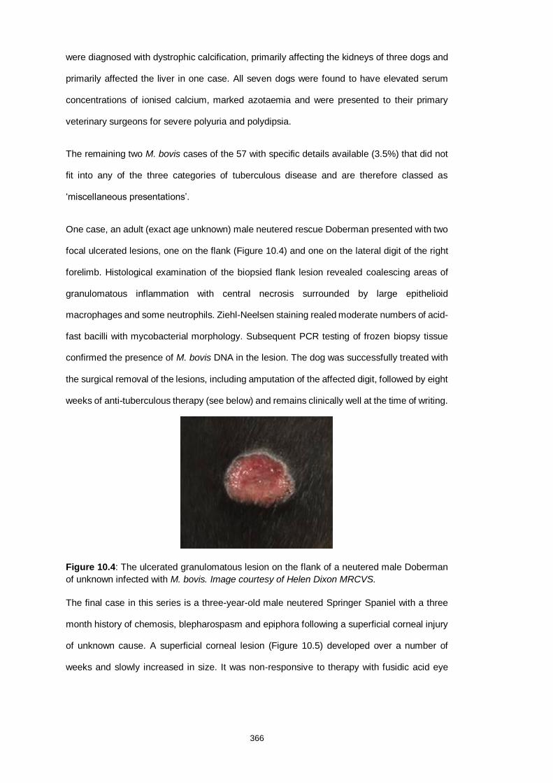
366
were diagnosed with dystrophic calcification, primarily affecting the kidneys of three dogs and
primarily affected the liver in one case. All seven dogs were found to have elevated serum
concentrations of ionised calcium, marked azotaemia and were presented to their primary
veterinary surgeons for severe polyuria and polydipsia.
The remaining two M. bovis cases of the 57 with specific details available (3.5%) that did not
fit into any of the three categories of tuberculous disease and are therefore classed as
‘miscellaneous presentations’.
One case, an adult (exact age unknown) male neutered rescue Doberman presented with two
focal ulcerated lesions, one on the flank (Figure 10.4) and one on the lateral digit of the right
forelimb. Histological examination of the biopsied flank lesion revealed coalescing areas of
granulomatous inflammation with central necrosis surrounded by large epithelioid
macrophages and some neutrophils. Ziehl-Neelsen staining realed moderate numbers of acid-
fast bacilli with mycobacterial morphology. Subsequent PCR testing of frozen biopsy tissue
confirmed the presence of M. bovis DNA in the lesion. The dog was successfully treated with
the surgical removal of the lesions, including amputation of the affected digit, followed by eight
weeks of anti-tuberculous therapy (see below) and remains clinically well at the time of writing.
Figure 10.4: The ulcerated granulomatous lesion on the flank of a neutered male Doberman
of unknown infected with M. bovis. Image courtesy of Helen Dixon MRCVS.
The final case in this series is a three-year-old male neutered Springer Spaniel with a three
month history of chemosis, blepharospasm and epiphora following a superficial corneal injury
of unknown cause. A superficial corneal lesion (Figure 10.5) developed over a number of
weeks and slowly increased in size. It was non-responsive to therapy with fusidic acid eye

367
drops (FuscithalmicTM [1% fusidic acid]; LEO Pharma, Denmark). Cytological examination of
a fine needle aspirate of the lesion revealed granulomatous inflammation with non-staining
“ghost” bacilli. Additional aspirates were found to contain acid-fast bacilli and one was PCR
positive for M. bovis DNA. During this time the dog developed three dermal lesions (one on
the lower lip and two on the cranial portion of the dorsum). Systemic treatment was instigated
with anti-tuberculous therapy (see below) and topical therapy for the ocular lesion with
moxifloxacin drops (VigamoxTM; Alcon, UK). The lesions completely resolved within six weeks
and the dog remains well at the time of writing.
Figure 10.5: The corneal granuloma of a thee-year-old male neutered Springer Spaniel dog
infected with M. bovis. Image courtesy of Jess Florey MRCVS (Dick White Referrals).
10.3.3 Diagnostic Tests
The diagnosis of mycobacterial infections, and MTBC infections in particular, are notoriously
challenging to make as all of the available diagnostic tests have limitations of sensitivity and/or
specificity.
For the majority of cases, evaluation of routine haematology and serum biochemistry were
used as a first-line test when clinicians were presented with an unwell dog, however, the
findings were typically unrewarding. The results, in the vast majority of cases for which they
are available, were found to be within reference intervals. Occasionally, generally as the

368
severity of the disease advanced, non-specific changes occured, such as a stress leukogram
(mature neutrophilia, monocytosis, lymphopenia and eopsinopenia). The mature neutrophilia
seen has occasionally been described as marked or severe and in rare cases there is a band
neutrophilia (a ‘left-shift’). A number of tuberculous dogs have been found to have a non-
regenerative anaemia; which may result from chronic inflammation. Organ specific changes,
for example elevations in liver enzyme activity, were typically only found to occur when the
relevant organ system was implicated in the disease process.
Diagnostic imaging (computed tomography, radiography and ultrasonography) were used in a
number of cases, particularly those diagnosed in recent years. The findings were useful for
establishing the extent of the pathology associated with infections and changes in diagnostic
imaging was reported in a number of cases as an aid to monitoring response to therapy and
as an indicator of when treatment could be discontinued.
Accessible lesions were often sampled by fine needle aspiration and cytological evaluation
has additionally been conducted on bronchial alveolar lavage fluid (N.B. this procedure should
only be undertaken in cases where tuberculosis is suspected after careful consideration and
with appropriate protective equipment). Cytological findings from these cases represent the
typical mammalian inflammatory response to mycobacteria; active macrophages dominate,
variable numbers of neutrophils and lymphocytes and fewer plasma cells. Routinely used
Romanovsky-type stains (e.g. DiffQuickTM; Richard Allen Scientific, UK) may highlight intra-
and extracellular mycobacteria as non-staining, so called “ghost” bacilli as their lipid walls
precent the uptake of stain. Acid-alcohol protocols (e.g. Ziehl-Neelsen) are needed to actively
stain mycobacteria.
A similar inflammatory infiltrate to cytological findings was ubiquitously reported in histological
evaluations of lesioned tissue sections taken from tuberculous dogs but in addition the
granulomatous structure was also visible. Canine granulomas of various structure have been
described; the most commonly identified were well structured granulomas comprising large,
often circular or ovoid, encapsulated areas of densely packed epithelioid macrophages,
Langerhan’s cells and multinucleated giant cells. The peripheral macrophages were frequently

369
reported as admixed with lymphocytes that were occasionally seen to form small aggregates.
In the central regions, irregular areas of necrosis containing cell debris, intact and degenerate
neutrophils were variably recorded. The areas of central necrosis were notably missing from
all three cases of M. microti infection where the histological assessment of tissue was
available. In a small number of cases, the (pyo)granulomatous inflammation was found to
occur outwith a typical granulomatous structure but instead macrophages and neutrophils
simply infiltrated the affected organs in large numbers and only formed small aggregates. The
histological findings in cases that were highly suggestive of mycobacterial infections were
examined by specialist stains to look for aetiological agents. The Ziehl-Neelsen protocol
identified variable numbers of acid-fast bacilli in the sections were this was reported, in line
with findings reported for mycobacterial infections in other species (Brougham et al. 2013. In
many of the cases reviewed in this study, the detailed findings of acid-fast staining were
frequently omitted from case reports (i.e. they were often simply referred to as either positive
or negative) and so a more detailed analysis was not possible.
A number of the examined reports noted that these granulomatous structures and/or
histological findings may also arise in the absence of an infectious agent but due to foreign
body, canine histiocytic disorders, canine sterile granuloma syndrome or neoplasia (e.g.
histiocytoma).
In the UK and the USA, the only diagnostic test which can lead to official confirmation of a
tuberculous infection in a dog is specialist mycobacterial culture conducted by the Animal and
Plant Health Agency, Weybridge or the United States Department of Agriculture, Animal and
Plant Health Inspection Service respectively (USDA, 2017; Middlemiss and Clark, 2018).
Despite this, no sensitivity data are publically available for this technique with respect to canine
submissions. Logically, culture tests are assumed to have perfect (100%) specificity for all
submissions processed at these laboratories.
In human medicine, tuberculosis is increasingly being diagnosed with the use of molecular
assays; either PCR or quantitative PCR (qPCR). This has, in large part, been driven to
decrease the time taken to make a diagnosis compared to culture which can take 2-4 weeks
370
for M. tuberculosis, 6-8 weeks for M. bovis and up to 12-16 weeks for M. microti (Reuter et al.
2006; Emmanuel et al. 2007). In the UK, Mycobacterial Reference Laboratories, such as the
one at Leeds University Teaching Hospital, are offering veterinary clinicians the most robust
PCR assays for mycobacterial infections. This laboratory utilises the GenoType MTBC assay
(Hain LifeScience, Germany) which allows the discrimination between all members of the
complex in one rapid (five hours) assay (Hain LifeScience, 2019). However, this assay is
validated for the assessment of liquid starting material (typically liquid cultures or expectorated
sputum samples) where most veterinary submissions are cytological aspirates or tissue
biopsies and this may have an, as yet, effect on the test sensitivity. In addition, veterinary
submissions may also be formalin-fixed tissues being tested following routine histological
examination and whilst this process is known to fragment extracted DNA, the effect that this
may have on test sensitivity is as yet unknown. Despite this, positive results have been
obtained from formalin-fixed canine tissue, even without visible acid-fast bacilli (Sharp et al.
2019).
Parsons et al. (2012) were the first to use a commercial interferon gamma release assay
(IGRA) to test dogs for M. tuberculosis infections. This test, the QuantiFERONTM TB Gold
assay is used to diagnose latent tuberculosis infections (LTBI) in people (Tilahun et al. 2019).
The IGRA assays incubate whole blood or isolated cells (peripheral blood mononuclear cells
[PBMC]) with mycobacterial antigens; if patients are infected with mycobacteria that posess
those same antigens then antigen-specific T-cells will recognise those antigens, produce and
secrete interferon-gamma (Wood and Jones, 2001). This can then be quantified by ELISA and
compared to unstimulated samples from the same patient; a significant difference indicates to
infection (Wood and Jones, 2001). In humans, the QuantiFERONTM TB Gold assay is reported
to have a sensitivity of 82.3% (95% confidence interval; 77.7% to 85.9%) and specificity of
82.6% (95% confidence interval; 78.6% to 86.1%) for LTBI (Takwoingi et al. 2019). The study
by Parsons et al. (2012) screened high risk dogs (resident with humans that had active M.
tuberculosis infections) and showed that a number generated positive results to the
QuantiFERONTM TB Gold assay.

371
The QuantiFERONTM TB Gold test contains three combined MTBC antigens; 6kDa early-
secreted antigenic target (ESAT-6), 10kDa culture filtrate protein (CFP-10) and Tb7.7 (Bernitz
et al. 2019). Only the latter of these is specific to M. tuberculosis, the former two are both
common to MTBC organisms which encode the RD-1 region of the MTBC genome (i.e. all
MTBC organisms excluding M. bovis-BCG and M. microti) (Leem et al. 2018; Bernitz et al.
2019). Therfore a positive response of a dog to these antigens likely indicates infection with a
RD-1-positive MTBC mycobacteria rather than just M. tuberculosis as implied by Parson et al.
(2012), for example buffalo infected with M. bovis have been shown to give positive results to
the same test (Bernitz et al. 2019). No further studies have expanded on the use of the
QuantiFERONTM TB Gold test for dogs and, to the author’s knowledge, it is not currently
available to veterinary clinicians.
Another IGRA, based on the protocol used for the feline assay, was developed during an
outbreak of M. bovis in a kennel of Foxhounds (Chapter 5; O’Halloran et al. 2018). This assay
uses three separated antigen pools; purified protein derived (PPD) from M. bovis (PPDB), PPD
from M. avium (PPDA) as well as a combination of ESAT-6 and CFP-10. This separation of
antigens theoretically allows for the discrimination between canine infections with M.
tuberculosis and M. bovis (both of which would give rise to the same pattern of response) and
M. mircoti. However, no dogs infected with M. microti have been tested with this assay to date.
Additionally, as with cats (Rhodes et al. 2008), a number of the Foxhounds tested that were
infected with M. bovis did not give a significant interferon gamma response to the ESAT-6 and
CFP-10, as would be expected from animals infected with M. microti. Therefore, whilst this
test would appear helpful in diagnosing MTBC infections in dogs, it may not always
discriminate between the different species. This IGRA test is now available to clinicians at
Biobest Laboratories, Edinburgh.
Serological assays are for the identification of tuberculosis infections in humans and other
animal species (Waters et al. 2017; Bezos et al. 2018; Chen et al. 2018; Infantes-Lorenzo et
al. 2019). The advantages of these assays is that they are comparatively inexpensive and
rapid in relation to other tests (Waters et al. 2017; Bezos et al. 2018). In the context of cattle

372
and wildlife species, the acquisition of the small volume of serum required to run the assay is
practically easier than conducting tests such as the tuberculin skin test which requires the
same individual to be handled twice, usually 72 hours apart (Infantes-Lorenzo et al. 2019). Of
the available commercial assays, the Chembio Dual Path Platform VetTB Assay for Cervids
has been used in dogs but using a test cut-off point that maintains a high specificify, the
sensitivity was found to be poor (O’Halloran et al. 2018). Therefore it is likely that, for the
forseeable future, this and similar serological tests currently only used in research, will only be
useful for screening large groups of at-risk animals rather than being a reliable tests for
individual dogs.
10.3.4 Treatment and Outcome
The treatment of canine tuberculosis is contentious as all of the MTBC organisms isolated
from the cases considered in this review can cause human disease and the inappropriate
treatment of pets may inadvertently lead to drug resistant isolates, further risking public health.
Indeed, the treatment of M. tuberculosis infected pets has been strongly discouraged on public
health grounds and owners of such animals should be referred to the appropriate public or
general health professionals (O’Halloran and Gunn-Moore, 2017).
By contrast, M. microti has only ever been identified in six dogs and although a small number
of people have developed disease due to this organism, no cases have been linked to
transmission from a dog (Emmanuel, 2007).
The public health risk from infections with M. bovis may be considered to sit somewhere
between that of M. bovis and M. tuberculosis. Unlike M. tuberculosis which represents the
major cause of human disease, M. bovis now causes only approximately 1% of human
tuberculosis cases in the UK (Glaziou et al. 2018). However, zoonotic cases due to dogs have
been confirmed or strongly suspected in recent years (Ellis et al. 2006; O’Halloran et al. 2018).
Despite this, Public Health England deem the risk of zoonotic tuberculosis due to pets to be
“very low” (Public Health England, 2014).

373
Treatment details for tuberculosis was reported in 22 cases recorded cases in this study;
including the 19 cases were management was supervised by the author and nine previously
published reports. In 20 cases the dogs were infected with M. bovis and one dog was infected
with M. microti. All except one M. tuberculosis infected dog euthanased at the time the
diagnosis was confirmed.
The treatment protocol for the 19 dogs supervised by the author was based on a triple
antimicrobial combination of drugs, adapted to dogs from the published guidance for cats
(O’Halloran and Gunn-Moore, 2017). This outlines combination therapy comprising rifampicin,
a macrolide and a quinolone administered daily. This class combination has been chosen for
several reasons. Firstly, streptomycin should be kept as a last‐resort antibiotic for human
patients and should not be used in dogs (Engelmann et al. 2014). Secondly, isoniazid can
cause severe adverse neurological effects that has led to euthanasia of a dog (Sykes et al.
2007). Thirdly, pyrazinamide is useful ineffective for the treatment of M. bovis infections (de
Jong et al. 2005). And quinolones, which have documented anti-mycobacterial activity are well
tolerated by dogs (Govendir et al. 2011).
All 19 dogs received rifampicin (generic), an essential component of anti-tuberculous therapy,
at a dose rate of 10-15mg/kg by mouth (PO) every (q) 24 hours (Munir et al. 2019). Three
dogs (15.8%) subsequently developed pruritic skin eruptions (Figure 10.6), putatively due to
the rifampicin as a similar reaction has been reported for cats (O’Halloran and Gunn-Moore,
2017), though these cases appeared more severe than has been documented in cats, and the
lesions healed subsequent to the withdrawal of the drug. In these cases, rifampicin was
replaced with doxycycline (RonaxanTM; Boehringer Ingelheim, Germany) at a rate of 10mg/kg
PO q 24 hours for the remaining duration of therapy.

374
Figure 10.6: A skin eruption on the back of a 6-year-old neutered male Bealge dog with an M.
microti infection five weeks after starting therapy with rifampicin at 11.5mg/kg po q24 hours.
The lesion extends craniodorsally (top to bottom of the picture) approximately 5cm by 2cm
laterally. Image courtesy of Alex McInroy MRCVS.
One dog receiving rifampicin became inappetant and evaluation of serum biochemistry
revealed a serum alkaline phosphatase (ALP) to ten-times the upper reference interval.
Rifampicin therapy was discontinued immediately, doxycycline was started at previously
stated doses and the dog’s appetite returned within 24 hours. Serum biochemistry was not
repeated but the dog made a full clinical recovery.
All 19 dogs either received a macrolide alongside the rifampicin; ten dogs (52.6%) were treated
with the drug azithromycin (ZithromaxTM; Pfizer, UK) at a dose rate of 10-15mg/kg po q24
hours and the remaining nine dogs (47.4%) were treated with clarithromycin (generic) at a
dose rate of 7.5-15mg/kg po q12 hours. Azithromycin was chosen for smaller sized dogs
(~15kg or less) as this drug is available as a reconsitited suspension allowing the amount
administered to be accurately measured. By comparison, clarithromycin is available as 250mg
or 500mg tablets (NHS, 2019), and the former size is an optimal size for a ~16kg dog.
In cases with respiratory tuberculosis, azithromycin has been argued for as the optimal
macrolide drug choice. This drug characteristically concentrates in lung tissue at 100-fold
greater levels than in the circulation (Hand et al. 2001). A further interesting characteristic is
the ability of azithromycin to concentrate within inflammatory cells, particularly neutrophils and
monocytes within the bloodstream, and tissue macrophages (Mandell et al. 2001). The
ongoing inflammatory process in the lung of these dogs may recruit azithromycin-laden

375
neutrophils from the circulation which might then unload the drug in the presence of
mycobacteria within inflammatory sites (Hand et al. 2001). Inflammatory cells can therefore
act as “Trojan horses” delivering local concentrations of azithromycin that are several orders
of magnitude greater than that in plasma (Blumer, 2005).
The final drug in the triple combination is a quinolone. Pradofloxacin is recommended in cats
due to its increased safety profile in this species (Sykes and Blondeau, 2014). Pradofloxacin
has proven efficacy against mycobacteria and also has an excellent safety profile in dogs and
so was used to treat seven of these 19 cases (Govendir et al. 2011; Boothe et al. 2018). The
dose rates ranged from the UK licensed dose of 3-5mg/kg PO q 24hrs up to include the USA
licensed dose of 7.5mg/kg PO q24hrs (Anon, 2018; Boothe et al. 2018). One dog, a four-year-
old male neutered Greyhound developed neutropenia after being on the higher dose of
pradofloxacin and was transitioned onto marbofloxacin (Marbocare PTM; Animalcare, UK) at a
dose of 2mg/kg PO q24 hours and the neutropenia resolved. Five of the dogs had been given
empricial marbofloxacin therapy (at the previously stated rate) before the diagnosis of
tuberculosis was made and so this was continued. Six dogs were treated with enrofloxacin
(BaytrilTM; Bayer, Germany) at 5-10mg/kg PO q24hrs. One of the treated dogs was a one-
year-old Staffordhire Bull terrier and there were concerns about using quinolones in a dog of
this age due to the potential for damage to developing cartilage, especially given the
anticipated duration of therapy (Sebastian, 2007). For this dog, doxycycline was subsitituted
for the quinolone component of the triple antibiosis (RonaxanTM; Boehringer Ingelheim,
Germany) at a rate of 10mg/kg PO q 24 hours.
Three dogs with pulmonary disease also presented with productive coughs. Due to concerns
that this might pose a zoonotic risk to their owners, anti-tussive therapy was instigated. The
dogs received 0.5-1mg/kg codeine phosphate PO q6-8 hours.
These 19 cases were treated for a minimum of 12 weeks and for at least eight weeks following
complete resolution of clinical signs. Where internal pathology had been noted then the
treatment was extended until this had either resolved or had remained unchanged for at least
six weeks i.e. any changes were no longer actively improving or deteriorating. Treatment

376
duration ranged from 14 weeks to 38 weeks (median 20 weeks). All dogs responded
favourably to therapy and to the authors’ knowledge are still alive at the time of writing.
The treatment protocols for three dogs are presented in the literature. One dog with abdominal
tuberculosis was treated with rifampicin (generic) at 10 mg/kg administered PO q24h,
clarithromycin (generic) at 12 mg/kg PO q12h, and enrofloxacin (BaytrilTM; Bayer, Germany) 5
mg/kg administered PO q12h for three months when the enrofloxacin was discontinued and
the remaining two former drugs were continued for a further three months (Englemann et al.
2014). The dog fully responded to therapy and the authors report it was well 31 months after
the initial diagnosis (Englemann et al. 2014).
The second dog also presented with abdominal tuberculosis (due to M. bovis infection) but
was only treated with enrofloxacin monotherapy (dose rate unknown) (Shrikrishana et al.
2009). The dog only partially responded to therapy and was euthanased shortly after therapy
was instigated.
The final dog was resident in Brazil and it was initially based on the presumptive diagnosis of
Ehrlichiosis and so was treated with chloramphenicol (50 mg/kg PO q12 hours),
dexamethasone (0.30 mg/kg PO q24 hours), and fluids (Rocha et al. 2017). However, the
treatment was ineffective and the dog progressed to death; M. bovis was confirmed at post
mortem examination.
10.4 Discussion
This study, the largest review of canine tuberculosis, evaluated 1012 cases and markedly
improves the current knowledge of this disease.
Infecting Mycobacteria
Historical reviews have suggested a 3:1 ratio of infections caused by M. tuberculosis and M.
bovis (Birn et al. 1965; Clerx et al. 1992; Liu, 1980). However in this study found a decrease
in this ratio to just 2:1. If only cases diagnosed in pet dogs (i.e. excluding the single large

377
outbreak of M. bovis in working Foxhounds in the UK) within the last ten years (2009-2019)
are examined, then there is a clear inversion of this trend; there are twice the number of M.
bovis cases compared to M. tuberculosis. Additionally, all six M. microti cases were diagnosed
in this same ten year period. This likely reflects the continuing decline in human M. tuberculosis
cases in the countries/regions from which these cases originated (mostly Europe, North
America and New Zealand). The most common risk factor for a dog to present with an active
M. tuberculosis infection in this study was still an exposure to a human with active tuberculous
disease. The increase in M. bovis and M. microti cases may also reflect the increasing
prevalence of these infections in countries such as the UK and/or increased diagnostic
capacity (e.g. PCR) to separate out infections within the MTBC.
Signalment
This analysis found that young to middle aged adult dogs formed the majority of those infected
with MTBC organisms with a range from six months to nine years. There was no significant
difference between the age distributions of the dogs infected with M. tuberculosis, M. bovis or
M. microti. There was no sex predeliction for infection in the group of cases where this
information was available, though this was only a minority of cases. This distribution, with a
complete absence of older and geriatric dogs, may suggest that dogs may develop a degree
of immunity as they age, potentially through exposure to environmental mycobacteria.
Although the breed was only recorded in a very small group of cases (<3%), it is notable that
most of the pedigree breeds reported were hound or terrier types. This may indicate that these
hound and terrier type breeds have an increased susceptibility to MTBC infection, or it may
reflect an increase in exposure e.g. to wildlife reservoirs due to the natural behaviours of these
breeds such as scavenging and/or hunting small mammals. However, the very small number
of cases where breed information is available makes it difficult to draw firm conclusions.
Interestingly, there was no representation of breeds previously reported as susceptible non-
tuberculous mycobacterial infections such as Miniature Schnauzer’s or Boxers (Malik et al.
1998; Sharp et al. 2019). Identification of the underlying genetics of these susceptibilities

378
would allow a greater understanding of the pathogenesis of mycobacterial infections and could
lead to improvements in diagnosis and the development of new treatments.
Clinical Signs
For the first time, the clinical presentations of canine tuberculosis were grouped into four
different clinical presentations which could be helpful as clinicians can detail the zoonotic
implications e.g. pulmonary cases, which were found to be the most common are likely to pose
the greatest risk of zoonotic transmission. The classification can also help with the prognosis
e.g. all cases presenting with disseminated disease were fatal. The miscellaenous
presentations reported here are critically important as this form of disease is almost certainly
the most likely to be missed by clinicians who may not be suspicious of cases that do not
present with the more commonly expected pulmonary form. Delays in making a diagnosis can
increase the risk to owners by extending the period of exposure to an infectious dog but also
might mean that the opportunity to successfully intervene is lost.
Diagnostics
Specialist mycobacterial culture remains the gold standard diagnostic test but it not always
practically possible and action (such as the instigation of therapy or euthanasia of the dog)
may need to be taken prior to the availability of the results. Further work to validate PCR and
IGRA methods would be helpful so that all stakeholders (owners, clinicians, public health
officials and veterinary health officials) can have a clearer understanding of the test
performance under normal field conditions and so interpret results appropriately. Although
serological assays remain an attractive option for future development, it appears likely that
they will remain too insensitive for use in individual patients.
Treatment
The very small percentage of cases (2.0%) were treatment was attempted implies that canine
tuberculosis may be seen as carrying a poor prognosis but this analysis found that it is in
reality a readily treatable infection; a good outcome (i.e. long term remisison of clinical disease)

379
was achieved in 86% of cases and 100% of cases were triple combination therapy was utilised.
However, treatment was often of long duration and onwers must be aware of this before
therapy is instigated. Also of significance, all cases of disseminated disease were euthanased
on welfare grounds due to the severity of clinical signs and treatment would likely be
unsuccessful in these dogs.
Although there is a report of the successful treatment of a case of M. tuberculosis infection
with triple combination antimicrobial therapy, this should be discoraged based on the risk to
public health from these dogs.
Side effects were reported with rifampicin therapy in a minority of cases which were severe
including skin eruptions and both inappetance and induction of ALP activity. One dog was also
reported as neutropenic, possibly due to chronic high dose pradofloxacin administration, but
as this was a Greyhound, this may simply have been an incidental finding (Zaldívar-López et
al. 2011). Dogs receiving anti-tuberculous therapy require careful monitoring and some
clinicians may consider repeating haematological and serumbiochemical evaluation at regular
interval (e.g. monthly) or if the dogs appear to become unwell whilst on treatment.
Conclusion
This analysis over over 1000 canine tuberculosis cases found that all cases were caused by
infections with one of three MTBC organisms, M. tuberculosis, M. bovis or M. mircroti. This
study found that in recent years the pattern of infections has changed with a relative decline
in the significance of M. tuberculosis as compared with M. bovis and the first documented
canine infections with M. microti. All cases conformed to one of four clinical groups based on
the presenting signs; primary pulmonary, primary abdominal, disseminated or miscellaneous.
For cases other than dogs with disseminated disease, treatment with triple combination
antimicrobial therapy lead to good clinical outcomes but the treatment of dogs with M.
tuberculosis remains against advice due to the public health risk.

380
10.5 References
(1988) Tuberculosis in five Basset hounds Journal of the American Veterinary Medical
Association, 192 (11), 1563-1568.
(2019) American Journal of Veterinary Research, 80 (5), p. 504
Adams, W.E., Vokwald, A.J. (1932) Effect of Bronchial Stenosis on Pulmonary Tuberculosis
in Dogs Proceedings of the Society for Experimental Biology and Medicine, 29 (4), 392-393.
Alexander, K.A., Laver, P.N., Michel, A.L., Williams, M., van Helden, P.D., Warren, R.M., van
Pittius, N.C.G. (2010) Novel mycobacterium tuberculosis complex pathogen, M. Mungi
Emerging Infectious Diseases, 16 (8),1296-1299
Anon (2018) Correction: Pharmacokinetics and pharmacodynamics of oral pradofloxacin
administration in dogs (Am J Vet Res, (2018) 79, 12 (1268-1276), 10.2460/ajvr.79.12.1268)
Barber N.B., O’Neill, E., Sheahan, B.J., Cassidy, J., McAllistair, H. (2004) Calcospherite-like
bodies and caseous necrosis in tracheal mucus from a dog with tuberculosis. Veterinary
Clinical Microbiology, 34, 2734-2740.
Bauer, N.B., O'Neill, E., Sheahan, B.J., Cassidy, J., McAllister, H. (2004) Calcospherite-like
bodies and caseous necrosis in tracheal mucus from a dog with tuberculosis Veterinary
Clinical Pathology, 33 (3), 168-172.
Beretta, C. (1891) On the use of dog's serum in the treatment of tuberculosis British Medical
Journal, 2 (1611), 1041-1043.
Bernitz, N., Kerr, T.J., Goosen, W.J., Higgitt, R.L., de Waal, C., Clarke, C., Cooper, D.V.,
Warren, R.M., van Helden, P.D., Parsons, S.D.C., Miller, M.A. (2019) Impact of
Mycobacterium bovis-induced pathology on interpretation of QuantiFERON®-TB Gold assay
results in African buffaloes (Syncerus caffer) Veterinary Immunology and Immunopathology,
217, art. no. 109923.
Bezos, J., Roy, A., Infantes-Lorenzo, J.A., Gonzalez, I., Venteo, A., Romero, B., Grau, A.,
Minguez, O., Dominguez, L., de Juan, L. (2018) The use of serological tests in combination
with the intradermal tuberculin test maximizes the detection of tuberculosis infected goats.
Veterinary Immunology and Immunopathology, 199, 43–52.
Birn, K.J., Clegg, F.G., Mason, J.A. (1965) Canine tuberculosis.The Veterinary record, 77 (45),
1341-1342.

381
Blumer, J.L. (2005) Evolution of a new drug formulation: the rationale for high-dose, short-
course therapy with azithromycin. International Journal of Antimicrobial Agents. 26, Suppl 3
S143-147.
Boothe, D.M., Bush, K.M., Boothe, H.W., Davis, H.A. (2018) Pharmacokinetics and
pharmacodynamics of oral pradofloxacin administration in dogs. American Journal of
Veterinary Research, 79 (12), 1268-1276.
Broughan, J.M., Downs, S.H., Crawshaw, T.R., Upton, P.A., Brewer, J., Clifton-Hadley, R.S.
(2013) Mycobacterium bovis infections in domesticated non-bovine mammalian species. Part
1: Review of epidemiology and laboratory submissions in Great Britain 2004-2010 Veterinary
Journal, 198 (2), 339-345.
Carpenter, J.L., Myers, A.M., Conner, M.W., Schelling, S.H., Kennedy, F.A., Reimann, K.A.
Chen, Y., Cao, S., Liu, Y., Zhang, X., Wang, W., Li, C. (2018) Potential role for Rv2026c-
and Rv2421c- specific antibody responses in diagnosing active tuberculosis. Clinica Chimica
Acta, 487, 369-376.
Clercx, C., Coignoul, F., Jakovljevic, S., Balligand, M., Mainil, J., Henroteaux, M.,
Kaeckenbeeck, A. (1992) Tuberculosis in dogs: A case report and review of the literature
Journal of the American Animal Hospital Association, 28 (3), 207-211.
Cordes, D.O., Robinson, R.A., Bailey, B.H. (1963) A case of tuberculosis (Human Strain) in a
dog. New Zealand Veterinary Journal, 11 (5), 128-130.
Cousins, DV; Bastida, R; Cataldi, A; Quse, V; Redrobe, S; Dow, S; Duignan, P; Murray, A
(2003). Tuberculosis in seals caused by a novel member of the Mycobacterium tuberculosis
complex: Mycobacterium pinnipedii sp. Nov International Journal of Systematic and
Evolutionary Microbiology 53 (Pt 5): 1305–14
de Jong BC, Onipede A, Pym AS, (2005) Does resistance to pyrazinamide accurately indicate
the presence of Mycobacterium bovis? Journal of Clinical Microbiology 43, 3530–3532.
Deforges L, Boulouis HJ, Thibaud JL, Boulouha L, Sougakoff W, Blot S, Hewinson G, Truffot-
Pernot C, Haddad N. (2004) First isolation of Mycobacterium microti (llama-type) from a dog.
Veterinary Microbiology 103:249–253
DeLisle, G.W. (1992) Mycobacterial infections in cats and dogs. Surveillance, 20, 20-26.
Deppenmeier, S., Schieszler, A., Nolte, I., Moser, I., Hewicker-Trautwein, M. (2007)
Pulmonary tuberculosis with evidence of Mycobacterium tuberculosis in a Golden Retriever
382
[Lungentuberkulose mit Nachweis von Mycobacterium tuberculosis bei einem Golden
Retriever] Tierarztliche Praxis Ausgabe K: Kleintiere - Heimtiere, 35 (2), 111-115.
Dodd, D. C. (1952) New Zealand Veterinary Journal, 1: 17.
Ellis, M.D., Davies, S., McCandlish, I.A.P., Monies, R., Jahans, K., De la Rua-Domenech, R.
(2006) Mycobacterium bovis infection in a dog. Veterinary Record, 159 (2), 46-48.
Emmanuel, F.X., Seagar, A.-L., Doig, C., Rayner, A., Claxton, P., Laurenson, I. (2007)
Human and animal infections with Mycobacterium microti, Scotland. Emerging Infectious
Diseases, 13 (12), 1924-1927.
Engelmann, N. , Ondreka, N. , Michalik, J. and Neiger, R. (2014), Intra‐abdominal
Mycobacterium tuberculosis Infection in a Dog. Journal of Veterinary Internal Medicine, 28,
934-938.
Erwin, P.C., Bemis, D.A., Mawby, D.I., McCombs, S.B., Sheeler, L.L., Himelright, I.M., Halford,
S.K., Diem, L., Metchock, B., Jones, T.F., Schilling, M.G., Thomsen, B.V. (2004)
Mycobacterium tuberculosis transmission from human to canine Emerging Infectious
Diseases, 10 (12), 2258-2260.
Ferber, J.A., Dilts, C.E., Scherzo, C.S. (1983) Tuberculosis in a dog. Journal of the American
Veterinary Medical Association, 183 (1), 117.
Foster, E.S., Scavelli, T.D., Greenlee, P.G., Gilbertson, S.R. (1986) Cutaneous lesion caused
by Mycobacterium tuberculosis in a dog. Journal of the American Veterinary Medical
Association, 188 (10), 1188-1190.
Gay G, Burbidge HM, Bennett P, Fenwick SG, Dupont C, Murray A, Alley MR (2000):
Pulmonary Mycobacterium bovis infection in a dog. New Zealand Veterinary Journal 48, 78–
81.
Govendir, M., Norris, J., Hansen, T. (2011) Susceptibility of rapidly growing mycobacteria and
Nocardia isolates from cats and dogs to pradofloxacin. Veterinary Microbiolology 153, 240–
245.
Gradmann, C. (2006) Robert Koch and the white death: From tuberculosis to tuberculin
Microbes and Infection, 8 (1), 294-301.
Hackendahl, N.C., Mawby, D.I., Bemis, D.A., Beazley, S.L. (2004) Putative transmission of
Mycobacterium tuberculosis infection from a human to a dog Journal of the American
Veterinary Medical Association, 225 (10), 1573-1577.

383
Hain LifeScience (2019) The GenoType MTBC Assay Available at https://www.hain-
lifescience.de/en/products/microbiology/mycobacteria/tuberculosis/genotype-mtbc.html
Accessed 19 Sept. 2019.
Hand, W.L. (2001) Characteristics and mechanisms of azithromycin accumulation and efflux
in human polymorphonuclear leukocytes. International Journal of Antimicrobial Agents. 18(5),
419-425.
Hawthorne, V.M., Jarrett, W.F.H., Lauder, I., Martin, W.B., Roberts, G.B.S. (1957)
Tuberculosis in man, dog, and cat British Medical Journal, 2 (5046), 675-678.
Hjarre, A. (1939) Dogs and cats as sources of tuberculosis in man. Acta Tuberculosis
Scandinavia, 13, 103.
Horard-Herbin, M.-P., Tresset, A., Vigne, J.-D. (2014) Domestication and uses of the dog in
Western Europe from the Paleolithic to the Iron Age. Animal Frontiers, 4 (3), 23-31.
Infantes-Lorenzo, J.A., Moreno, I., Roy, A., Risalde, M.A., Balseiro, A., De Juan, L., Romero,
B., Bezos, J., Puentes, E., Åkerstedt, J., Tessema, G.T., Gortázar, C., Domínguez, L.,
Domínguez, M. (2019) Specificity of serological test for detection of tuberculosis in cattle,
goats, sheep and pigs under different epidemiological situations. BMC Veterinary Research,
15 (1), art. no. 70.
Innis, J.R.M. (1940) Tuberculosis in the dog and cat. The Veterinary Journal. 96, 96.
Leem, A.Y., Song, J.H., Lee, E.H., Lee, H., Sim, B., Kim, S.Y., Chung, K.S., Kim, E.Y., Jung,
J.Y., Park, M.S., Kim, Y.S., Chang, J., Kang, Y.A. (2018) Changes in cytokine responses to
TB antigens ESAT-6, CFP-10 and TB 7.7 and inflammatory markers in peripheral blood
during therapy Scientific Reports, 8 (1), art. no. 1159.
Lovell, R., and White, E.G. (1940) Naturally occurring tuberculosis in dogs and some other
species of animals, British Journal of Tuberculosis, 34, 117-133.
Malik, R., Love, D.N., Wigney, D.I., Martin, P. (1998) Mycobacterial nodular granulomas
affecting the subcutis and skin of dogs (canine leproid granuloma syndrome). Australian
Veterinary Journal, 76 (6), 403-407.
Mandell, G.L., Coleman, E. (2001) Uptake, transport, and delivery of antimicrobial agents by
human polymorphonuclear neutrophils. Antimicrobial Agents Chemotherapy 45(6), 1794-
1798.

384
Manwaring, W.H. (1913) The effects of subdural injections of leucocytes on the development
and course of experimental tuberculous meningitis. Journal of Experimental Medicine, 17 (1),
1-13.
McFadyean, J. (1891) Dog's serum in the treatment of tuberculosis. British Medical Journal,
2 (1613), 1173.
Michelet, L., Cruz, K.D., Zanella, G., Aaziz, R., Bulach, T., Karoui, C., Hénault, S., Joncour,
G., Boschiroli, M.L. (2015) Infection with Mycobacterium microti in animals in France Journal
of Clinical Microbiology, 53 (3), 981-985.
Middlemiss, C., Clark, J. (2018) Mycobacterium in pets. Veterinary Record, 183(18), 571.
Moore, J. (1928) The Old and the New of Veterinary Science and Practice Journal of the
Royal Society of Medicine, 22 (1), 47-53
Moravkova, M., Slany, M., Trcka, I., Havelkova, M., Svobodova, J., Skoric, M., Heinigeova, B.,
Pavlik, I. (2011) Human-to-human and human-to-dog Mycobacterium tuberculosis
transmission studied by IS6110 RFLP analysis: A case report Veterinarni Medicina, 56 (6),
314-317.
Munir, A., Kumar, N., Ramalingam, S.B., Tamilzhalagan, S., Shanmugam, S.K., Palaniappan,
A.N., Nair, D., Priyadarshini, P., Natarajan, M., Tripathy, S., Ranganathan, U.D., Peacock,
S.J., Parkhill, J., Blundell, T.L., Malhotra, S. (2019) Identification and Characterization of
Genetic Determinants of Isoniazid and Rifampicin Resistance in Mycobacterium tuberculosis
in Southern India. Scientific Reports, 9 (1), art. no. 10283.
NHS (2019) Medicines: Clarithromycin. Available at:
https://www.nhs.uk/medicines/clarithromycin/ Accessed 19 Sept. 2019.
O'Halloran, C., Hope, J.C., Dobromylskyj, M., Burr, P., McDonald, K., Rhodes, S., Roberts, T.,
Dampney, R., De la Rua-Domenech, R., Robinson, N., Gunn-Moore, D.A. (2018) An outbreak
of tuberculosis due to Mycobacterium bovis infection in a pack of English Foxhounds (2016–
2017) Transboundary and Emerging Diseases, 65 (6), 1872-1884.
Paltrinieri, S. (1930) Tuberculosis in the dog and cat. Nuova Vet. 6, 7.
Park, H.A., Lim, J.H., Kwon, L.H., Bae, J.H., Park, H.M. (2016) Pulmonary Mycobacterium
tuberculosis infection with giant tubercle formation in a dog: a case report Veterinarni
Medicina, 61, 102–109.,

385
Parsons, S.D.C., Gous, T.A., Warren, R.M., van Helden, P.D. (2008) Pulmonary
Mycobacterium tuberculosis (Beijing strain) infection in a stray dog Journal of the South African
Veterinary Association, 79 (2), 95-98.
Parsons, S.D.C., Warren, R.M., Ottenhoff, T.H.M., Gey van Pittius, N.C., van Helden, P.D.
(2012) Detection of Mycobacterium tuberculosis infection in dogs in a high-risk setting
Research in Veterinary Science, 92 (3), 414-419.
Pavlik, I., Dvorska, L., Matlova, L., Svastova, P., Parmova, I., Bazant, J., Veleba, J. (2002)
Mycobacterial infections in cattle in the Czech Republic during 1990-1999 Veterinarni
Medicina, 47 (9), 241-250.
Pavlík, I., Macháčková, M., Trčka, I., Roubal, P. (2004) Occurrence of bovine tuberculosis in
domestic animals in Europe: Current state and perspectives [Výskyt bovinní tuberkulózy u
domácích zvířat v Evropě: Současný stav a perspektivy] Prakticky Lekar, 84 (8), 441-444.
Pavlik, I., Trcka, I., Parmova, I., Svobodova, J., Melicharek, I., Nagy, G., Cvetnic, Z., Ocepek,
M., Pate, M., Lipiec, M. (2005) Detection of bovine and human tuberculosis in cattle and other
animals in six Central European countries during the years 2000-2004. Veterinarni Medicina,
50 (7), 291-299.
Pinsky, B.A., Banaei, N. (2008) Multiplex real-time PCR assay for rapid identification of
Mycobacterium tuberculosis complex members to the species level. Journal of Clinical
Microbiology, 46 (7), 2241-2246.
Posthaus, H., Bodmer, T., Alves, L., Oevermann, A., Schiller, I., Rhodes, S.G., Zimmerli, S.
(2011) Accidental infection of veterinary personnel with Mycobacterium tuberculosis at
necropsy: A case study Veterinary Microbiology, 149 (3-4), 374-380.
Public Health England (2014) Cases of TB in domestic cats and cat-to-human transmission:
risk to public very low. Available at: https://www.gov.uk/government/news/cases-of-tb-in-
domestic-cats-and-cat-to-human-transmission-risk-to-public-very-low Accessed 26 Aug.
2019.
Reuter, H., Burgess, L., van Vuuren, W., Doubell, A. (2006) Diagnosing tuberculous
pericarditis. QJM, 99 (12), 827-839.
Rodríguez E, Sánchez LP, Pérez S, (2009). Human tuberculosis due to Mycobacterium bovis
and M. caprae in Spain, 2004–2007. Int. J. Tuberc. Lung Dis. 13 (12): 1536–41.
S. Liu (1980) Canine tuberculosis. Journal of the American Veterinary Medical Association,
177(2), 164-167.

386
Saxegaard, F. and Svenkerud, R. (1982) A case of canine tuberculosis caused by
Mycobacterium tuberculosis. Acta veterinaria Scandinavica, 23 (2), 309-311.
Sharp, E., Taylor, S., O'halloran, C. (2019) Unusual presentation of canine Mycobacterium
avium infection. Veterinary Record, 184 (26), 800.
Snider (1971a) Tuberculosis in Canine and Feline Populations. American Review of
Respiratory Disease, 104, 877-887.
Snider, W., Cohen, D., Reif, J.S., Stein, S.C., Prier, J.E. (1971b) Tuberculosis in Canine and
Feline Populations: Study of High Risk Populations in Pennsylvania, 1966-1968. American
Review of Respiratory Disease, 104, 866-876.
Stableforth, W.P. (1914) Bovine Tuberculosis: Its Diagnosis. The Journal of the Royal Society
for the Promotion of Health, 35 (1), 50-59.
Steinbach, M.M., Deskowitz, M. (1933) Susceptibility of Diabetic Dogs to Tuberculosis
Proceedings of the Society for Experimental Biology and Medicine, 30 (8), 1053-1055.
Sykes, J.E., Blondeau, J.M. (2014) Pradofloxacin: A novel veterinary fluoroquinolone for
treatment of bacterial infections in cats. Veterinary Journal, 201 (2), 207-214.
Sykes, J.E., Cannon A.B., Norris, A.J., Byrne, B.A., Affolter, T., O’Malley, M.A., Wisner, E.R.
(2007) Mycobacterium tuberculosis Complex Infection in a Dog. Journal of Veterinary Internal
Medicine, 21(5), 1108-1112.
Takwoingi, Y., Whitworth, H., Rees-Roberts, M., Badhan, A., Partlett, C., Green, N., Boakye,
A., Lambie, H., Marongiu, L., Jit, M., White, P., Deeks, J.J., Kon, O.M., Lalvani, A. (2019)
Interferon gamma release assays for diagnostic evaluation of active tuberculosis (IDEA):
Test accuracy study and economic evaluation. Health Technology Assessment, 23 (23), 1-
152.
Thacker, T.C., Robbe-Austerman, S., Harris, B., Palmer, M.V., Waters, W.R. (2013) Isolation
of mycobacteria from clinical samples collected in the United States from 2004 to 2011 BMC
Veterinary Research, 9, art. no. 100
Thayer, J. et al. (1930) Gastro-Intestinal Port of Entry of B. Tuberculosis in Dogs
Proceedings of the Society for Experimental Biology and Medicine, 27 (5), 367-368.
Tilahun, M., Shibabaw, A., Kiflie, A., Bewket, G., Abate, E., Gelaw, B. (2019) Latent
tuberculosis infection and associated risk factors among people living with HIV and

387
apparently healthy blood donors at the University of Gondar referral hospital, Northwest
Ethiopia. BMC Research Notes, 12 (1), art. no. 515.
USDA (2017) Tuberculosis Available at:
https://www.aphis.usda.gov/aphis/ourfocus/animalhealth/nvap/NVAP-Reference-
Guide/Control-and-Eradication/Tuberculosis Accessed 19 Sept 19.
Warner, D.F., Mizrahi, V. (2013) Complex genetics of drug resistance in Mycobacterium
tuberculosis Nature Genetics, 45 (10), 1107-1108.
Waters, W.R., Vordermeier, H.M., Rhodes, S., Khatri, B., Palmer, M.V., Maggioli, M.F.,
Thacker, T.C., Nelson, J.T., Thomsen, B.V., Robbe-Austerman, S. (2017) Potential for rapid
antibody detection to identify tuberculous cattle with non-reactive tuberculin skin test results.
BMC Veterinary Research, 13 (1), 164.
Wood, P.R., Jones, S.L. (2001) BOVIGAM™: An in vitro cellular diagnostic test for bovine
tuberculosis. Tuberculosis, 81, 147-155.
Zaldívar-López, S., Marín, L.M., Iazbik, M.C., Westendorf-Stingle, N., Hensley, S., Couto, C.G.
(2011) Clinical pathology of Greyhounds and other sighthounds. Veterinary Clinical Pathology,
40 (4), 414-425.

388
Chapter 11: Investigating the Early Response of
Canine Bone Marrow Derived Macrophages to
Mycobacteria.
Acknowledgments
Additional funding for this project was provided by PetSavers Student Research Project
Grant: O’Halloran C. Investigating the Canine Macrophage Response to Mycobacteria. This
work was undertaken by Rachel Coley (BVM&S Undergraduate Student, University of
Edinburgh) and contributed to sections 11.3.6 and 11.3.7.
Abstract
Murine bone marrow derived macrophages (BMDM) and human monocyte derived
macrophages grown in colony stimulating factor 1 (CSF-1) have been widely used to study
macrophage responses to infectious agents as well as Toll-like receptor (TLR) agonists. It was
hypothesised that recombinant CSF-1 could be used to reliably generate canine BMDM in
order to characterise them, examine the effect of TLR agonism and investigate the early (initial
24 hours) response of these cells to mycobacterial infection. The culture of canine bone
marrow cells for 10 days in recombinant human, porcine or canine CSF-1 at appropriate
concentrations produced a pure population of BMDM that were adherent, morphologically
resembled macrophages and expressed surface proteins typical of macrophages (as
described in other species: the CSF-1 receptor, CSF-1, ADGRE-1, CD172a, CD163, CD16
and CD14). Canine BMDM could be generated from bone marrow cells that had been
cryopreserved so that multiple experiments could be conducted on cells from a relatively small
number of animals. The canine BMDM produced were phagocytic and therefore permissive to
mycobacterial infection whereupon they produced the pro-inflammatory cytokines TNF-α and
IL-6 but not nitrite. The BMDM were also found to display mycobactericidal activity during the
early phase of infection. Gene expression and cytokine production of canine BMDM in

389
response to the TLR4 agonist lipopolysaccharide, attenuated M. bovis bacille Calmette-Guérin
and virulent M. bovis showed significant differences to the canine macrophage-like DH82 cell
line indicating that the latter is not a good resource for the study of canine mycobacterial
infections.
11.1 Introduction
Cells of the monocyte-macrophage lineage (MML) and their response to mycobacterial
infection is considered in Section 1.6. These MMLcells include bone marrow progenitor cells,
circulating cells of the MML and tissue resident macrophages. The process of differentiation
and maturation of macrophages from their progenitors is driven and regulated by the
haemopoietic growth factors colony stimulating factor 1 (CSF-1) and interleukin (IL)-34 which
share a common receptor (Hume, 2008; Fairbairn et al. 2011).
Cells of the MML are frequently the first cells of the innate immune system to interact with any
potential pathogen and their early response can have profound effects on the outcome of
infection including pathogen killing and clearance, or persistence and the development of
clinical disease (Burkey et al. 2009; Szatmary, 2012; Pahari et al. 2018). Macrophages
represent the immunological niche for the persistence of mycobacteria, to this end they have
evolved a number of mechanisms for the subversion of mycobacterial killing; some of which
are considered in Section 1.6.5, making tem one of the most critical central cells to the host-
pathogen interaction and pivotal in the aetiopathogenesis of mycobacterial infections.
Microbial pattern-recognition receptors (PRRs) are an essential early component of innate
immunity, they detect and recognise conserved pathogen-associated molecular patterns
(PAMPS), resulting in the activation of stimulated MML cells (and neutrophils) as part of the
host response to eradicate invading pathogens whilst maintaining immunological tolerance by
recognition of self (Burkey et al. 2009; Szatmary, 2012; Pahari et al. 2018). An important class
of mammalian PRRs is the Toll-like receptor (TLR) family which recognise a wide range of
microbial pathogens and pathogen-related products (Schneberger et al. 2011).

390
A number of studies which investigated the response of macrophages to mycobacteria in
humans and mice demonstrated that stimulation of TLR2 and TLR4 receptors, as well as TLR1
and TLR6 by mycobacteria-derived ligands results in downstream activation of the nuclear
transcription factor NF-κβ which in turn leads to the up-regulation of genes encoding pro-
inflammatory cytokines such as TNF-α, IL-1β and IL-6 (Underhill et al. 1999; Quesniaux et al.
2004; Bulut et al. 2005).
While methods exist for the isolation and/or culture of MML cells from bone marrow across
several species, this requires post mortem material which is not always readily accessible
(Kapetenovic et al. 2012). Therefore, utilisation of a cell line that accurately mimics the
response of ex vivo MML would be beneficial. The DH82 cell line was established from the
neoplastic progenitor cells of canine malignant histiocytosis and was characterised as
histiocytic in origin based on light microscopic and ultrastructural morphology (Wellman et al.
1988). Since then it has been used as a model to investigate the canine response to
intracellular pathogens including Leishmania spp., canine distemper virus and extracellular
pathogens such as Mycoplasma canis (Puff et al. 2009; Michaels et al. 2016; Soto et al. 2019)
The aim of this study was to utilise recombinant CSF-1 to generate canine macrophages from
bone marrow (BMDM) in order to characterise them, examine the effect of TLR agonism and
investigate the early (initial 24 hours) response of these cells to mycobacterial infection whilst
comparing them to the established DH82 cell line in order to evaluate the usefulness of both
systems as models for studying canine mycobacterial infections.

391
11.2 Materials and Methods
11.2.1 Isolation of Bone Marrow Cells
Bone marrow was acquired from dogs and cats that were euthanased at the Royal (Dick)
School of Veterinary Studies for reasons unrelated to this project. All animals came from the
Edinburgh Dog and Cat Home and were euthanased because of a history of human-directed
aggression or severe animal-directed aggression which precluded the rehoming of these
animals to members of the public. No animals showed clinical signs of infectious disease,
including mycobacterial infection, on veterinary examination prior to euthanasia.
All euthanasias were performed by intravenous injection of pentobarbitone at a dose of
≥1mg/kg administered by the author (a UK qualified and registered veterinary surgeon),
following prior administration of oral sedation (acepromazine, ACP, 0.05mg/kg [ACP; Elanco,
UK], phenobarbitone 3mg/kg [Epiphen, Vetoquinol, UK] and diazepam 1mg/kg [generic]) by
the attending handlers, or by intramuscular injection of butorphanol 0.2mg/kg [Torbugesic;
Zoetis, USA], 0.05mg/kg ACP and 0.05mg/kg medetomidine [Sedator; Dechra, UK]
administered by the author.
The sedation and euthanasia procedures as well as the use of cadavers for research purposes
had prospective ethical approval from the University of Edinburgh Veterinary Ethics Research
Committee and was adhered to throughout.
The long bones; femur, humerus and ribs, were removed post-mortem from euthanased
animals within one hour of euthanasia; the fascia and connective tissues were removed with
a size 22 scalpel blade and the bones were then stored in PBS on ice until further processing
(within four hours of euthanasia). The bones were surface decontaminated by immersion in
70% ethanol for five minutes which was then removed by rinsing the bones in sterile PBS. A
hand saw with a sterile blade attached was used to trisect the bones by incising through the
diaphysis twice in order to expose the medullary cavity.
392
Bone marrow cells were removed by flushing the central cavity with warmed (37°C) RPMI
1640 media supplemented with anticoagulant; sterilised sodium citrate, added to a final
concentration of 3.2% by volume, through a 20mL syringe and 22 gauge needle. The flushed,
cell-containing media was collected into 50mL sterile plastic tubes which were centrifuged at
600 x g for five minutes before the supernatant was removed and discarded. The cell pellet in
each tube was re-suspended in approximately 10mL of an ammonium chloride erythrocyte
lysis buffer (composed of 8.26 g ammonium chloride, 1.0 g potassium bicarbonate, and
0.037 g EDTA in 1dL of water), the suspension was transferred to a clean tube and incubated
in the dark for seven minutes before the tube was filled with RPMI 1640 media supplemented
with 20% foetal bovine serum. The suspension was passed through a 40µm Corning cell
strainer (Corning Life Sciences, Germany) to remove any remaining debris and the flow
through was again centrifuged at 600 x g for five minutes before the supernatant was removed.
The cell pellets were re-suspended in a single mL of non-supplemented RPMI 1640 media
and all suspensions were combined into a single clean tube at which time 10µL was removed
and mixed with an equal volume of 2% Trypan blue stain prior to counting the number of live
cells using a haemocytometer.
11.2.2 Cryopreservation and Recovery of Bone Marrow Cells
Cells were centrifuged at 600 x g for five minutes and the supernatant was discarded. Cold
(4oC) cell freezing media comprised of heat inactivated FBS (Gibco, UK) containing 20%
dimethyl sulfoxide (DMSO; Sigma Aldrich, UK) by volume was added dropwise to the cell pellet
with manual agitation of the tube throughout. The volume added was sufficient to give a final
cell concentration of 5x10⁷ cells/mL. This cell suspension was divided into 1mL cryovials
(Thermo Fisher Scientific, USA) which were placed into a pre-chilled (-20oC) Mr FrostyTM cell
cooler containing isopropanol.
The Mr FrostyTM cell coolers containing the cell vials were placed at -80oC for 72 hours after
which time the cell vials were removed and transferred to a liquid nitrogen tank for long term
storage. When needed, each vial was defrosted in a water bath pre-warmed to 37oC. The

393
thawed cells were transferred to a 15mL Falcon and 10mL of warmed complete culture media
was added dropwise with agitation. The cells were centrifuged at 600 x g for five minutes and
the supernatant was removed. Cells were re-suspended in 10mL of complete culture media
(RPMI 1640 containing 100µg/ml L-glutamine, 10% foetal bovine serum, 100µg/ml penicillin,
100U/ml streptomycin, 5x10⁻⁵M 2-mercaptoethanol and non-essential amino acids). Cell
viability counts were performed using Trypan blue staining (Section 11.2.1).
11.2.3 Culture of Bone Marrow Derived Macrophages
When recovered from cryopreservation or removed from long bones, viable bone marrow cells
(as determined by Trypan blue staining; Section 11.2.1) were diluted to a density of 1x10⁶/mL
in complete culture media. The cell suspension was plated into appropriately sized SterilinTM
treated tissue culture plastic dishes (Thermo Fisher Scientific, UK). The growth factor
recombinant colony stimulating factor 1 (r-CSF-1) was added at an appropriate concentration
(see Section 11. 3). An equal volume of fresh complete culture media was added to cells after
three days in culture at 37°C/5% CO2 along with sufficient r-CSF-1 so as to reach the same
final concentration as on Day 0. After six days in culture, all media was removed, discarded
and replaced with the same volume and quantity of r-CSF-1 as at time Day 0.
11.2.4 Determination of Cell-surface Molecule Expression by Single and Dual
Colour Flow Cytometry
At four time points during macrophage differentiation; Day 0, 3, 6 and 10 the media was
removed from the bone marrow cell cultures and transferred into 50mL Falcon tubes. The
adherent cell layer was washed twice with sterile PBS to remove serum remnants. Following
this, 10mL of non-enzymatic cell dissociation solution (Sigma Aldrich, UK) was added to the
flask and incubated at 37oC until the cells visibly detached from the plastic. The cell solution
was also transferred to a 50mL Falcon tube. All tubes were centrifuged at 600 x g for five
minutes at room temperature, the supernatant was discarded and the cell pellets were each
re-suspended in 1mL of complete cell culture media and combined into a single tube. Cell
viability counts were performed using Trypan blue staining (Section 11.2.1). Cells were diluted

394
to a final concentration of 5x10⁶/mL in complete culture media and 200µL (i.e. 1x10⁶ cells) was
added to the required number of wells of a round-bottom CELLSTAR® 96W Microplate (Sigma
Aldrich, UK). The plate was centrifuged at 600 x g for five minutes at room temperature and
the supernatants were discarded. Cells were washed twice in 100µL of sterile PBS and re-
suspended in block buffer (PBS containing 2% heat inactivated normal goat serum [Abcam,
UK]) for half an hour at room temperature. Cells were washed twice in 100µL of sterile PBS to
remove any serum remnants. The fixable cell viability stain Zombie Violet™ (Zombie Violet™
Fixable Viability Kit; Biolegend, USA) was added to all wells, other than the unstained control
well, at a dilution of 1:1000 (as recommended by the manufacturer) in 50µL per well of sterile
PBS without serum. Unstained control cells were re-suspended in 50µL of sterile PBS. The
plate was incubated in the dark for 20 minutes at room temperature.
Cells were washed twice in blocking buffer before primary antibodies including relevant isotype
controls (Table 11.1) were added to the appropriate wells, diluted to the required concentration
in 50µL of sterile PBS without serum. The plate was incubated in the dark for 30 minutes at
room temperature. Cells were washed twice in blocking buffer before isotype- and species-
matched secondary antibody was added to the appropriate wells (Table 11.1), including a well
with just secondary antibody added with no primary antibody, diluted in 50µL of sterile PBS
without serum. The remaining control cells were re-suspended in 50µL of sterile PBS only.
The plate was incubated in the dark for 30 minutes at room temperature and cells were then
washed twice in sterile PBS and re-suspended in 100µL of 2% paraformaldehyde solution and
left for 10 minutes at room temperature. Cells were washed twice in sterile PBS and re-
suspended in 100µL of flow cytometer buffer (sterile PBS containing 2% BSA, 2nM EDTA and
2nM sodium azide). The plate was sealed with an adhesive plate sealant to prevent
evaporation and kept refrigerated at 4oC overnight (approximately 16 hours) wrapped in foil to
prevent exposure to light.

395
Table 11.1 Antibodies used for single and dual-colour flow cytometry
Surface
molecule
Clone Species
specificity
Isotype Conjugated
flurochrome
Concentration
Supplier
CD14 TUK4 Mouse
anti-human
Mab
IgG2a AlexaFluor647 10µg/mL Abcam
CD172a DH59B Mouse
monoclonal
anti-bovine
IgG1 None 2.5µg/mL Kingfisher
CD16 LNK16 Mouse
monoclonal
anti-human
IgG1 None 10µg/mL Invitrogen
CD163 EDHu-1 Mouse
monoclonal
anti-human
IgG1 None 4µg/mL Abcam
CSF-1 ROS
5H11-4
Mouse
monoclonal
anti-
porcine
IgG2a None 10µg/mL A gift from the
Roslin
Institute
Immunological
Toolbox
CSF-1-r ROS
8G11-1
Mouse
monoclonal
anti-
porcine
IgG2a None 10µg/mL A gift from the
Roslin
Institute
Immunological
Toolbox
ADGRE1 ROS
4E12-3E6
Mouse
monoclonal
anti-
porcine
IgG1 None 10µg/mL A gift from the
Roslin
Institute
Immunological
Toolbox
Bu-1 AV20 Mouse
monoclonal
anti-
chicken
IgG1
isotype
control
None 10µg/mL R&D
systems
KLH 20102 Mouse
monoclonal
anti-human
IgG2a
isotype
control
None 10µg/mL R&D
systems
KLH 20102 Mouse
monoclonal
anti-human
IgG2a
isotype
control
AlexaFluor647 10µg/mL R&D
systems
IgG (H+L) Ab150113 Goat
polyclonal
Anti-mouse
Secondary
antibody
AlexaFluor488 5µg/mL Abcam
The method for dual-colour staining was similar to that described above for single-colour
staining, except that two isotype mis-matched primary antibodies were added to each sample
(Table 11.1). Additionally, sequential wells of cells were stained with a single one of each of
the fluorochromes used to act as compensation controls. Analysis of all stained cells was
carried out using the BD Fortessa X20 flow cytometer and FlowJo software; 10,000 events
(live cells) were collected. A representative gating strategy is demonstrated in Figure 11.4.

396
11.2.5 The Comparative Efficacy of Recombinant Canine, Porcine and
Human CSF-1 for Generating Canine Macrophages
A previous study has demonstrated that circulating canine and feline monocytes are capable
of maturation into macrophage-like cells when grown in vitro in culture media supplemented
with 104IU/mL of recombinant human (rh)-CSF-1 (Gow et al., 2013; Heinrich et al. 2017).
Therefore, the same concentration (a gift from Professor David Hume) was initially used here
to determine if macrophage cells could be derived from canine bone marrow. The resulting
cells were assessed as being macrophages based on their morphology by light microscopy,
expression of macrophage-associated cell surface molecules during the differentiation time
course (Section 11.2.4) as well as adherence to treated tissue culture plastic and phagocytic
capacity at the culmination of differentiation.
The biological efficacy of recombinant canine (rc) and recombinant porcine (rp) CSF-1 (gifts
from Dr Lissa Herron, The Roslin Institute) was assessed by testing cell viability via MTT (3-
4,5-dimethylthiazol-2-yl-2,5-diphenyltetrazolium bromide) assay. In this assay the relative
activity of NAD(P)H-dependent cellular oxidoreductase enzymes reflects the number of viable
cells present in the well of a 96-well cell culture plate. These enzymes are capable of reducing
the tetrazolium dye MTT to its insoluble formazan, which has a purple colour.
Bone marrow was recovered from cryopreservation and viable cells (determined by Trypan
blue staining) were diluted to a concentration of 2x10⁷ cells/mL in complete culture media
before 100µL was added to each well of a 96-well cell plate (NuncTM; Thermo Fisher Scientific,
USA) i.e. 2x10⁶ cells were added per well. A serial two-fold dilution series of each r-CSF-1
were made in complete culture media, starting at a concentration of 2000ng/mL and ending at
32ng/mL. A negative control condition was included by adding 100µL of complete culture
media without r-CSF-1 supplementation to triplicate wells of bone marrow cells from each dog.
A positive control condition was included by the addition of rh-CSF-1 diluted to a final
concentration of 104IU/mL in complete culture media to triplicate wells of bone marrow cells

397
from each dog. The remainder of the cells had 100µL of the CSF-1 dilutions added in triplicate
so that the final concentration of cells was 1x10⁶/well.
Plates were incubated at 37oC/5% CO2 for ten days with media changes (as described above
in section xx) and the addition of fresh growth factor at Day 3 and Day 6. At Day 10 the media
was removed and discarded, following this 100µL of RPMI-1640 media containing MTT at a
final concentration of 1mg/mL was added to each well. The plates were returned to the
incubator for three hours. At the end of the incubation period the MTT containing media was
aspirated and discarded. To solubilise the formazan produced, 150µL of 4mM HCl in
isopropanol was added to each well. The plate was covered in aluminium foil to protect the
contents from light and incubated at room temperature on a plate shaker for 30 minutes. At
the end of the 30 minutes the plate was visually checked to ensure that all of the formazan
was in suspension, if not then the plate was returned to the plate shaker and rechecked at 10
minute intervals until this was achieved.
The optical density was quantified by measuring the absorbance at OD570nm; the data was
exported to Microsoft Excel and analysed using GraphPad Prism 7.
11.2.6 Phagocytosis Assay
Following harvest of adherent BMDM cells after 10 days of differentiation in CSF-1
supplemented complete culture media, 1x10⁶ cells/ml were plated per well of a 6-well tissue
culture plate in duplicate and cultured overnight at 37°C/5% CO2. Phagocytosis was initiated
by the addition of fluorescein isothiocyanate (FITC)-conjugated Zymosan bioparticles
(Molecular Probes, USA) at a particle to cell ratio of approximately 5:1, followed by further
incubation for one hour. A negative control well was included where PBS was added to the
complete culture media without Zymozan particles. Phagocytosis was stopped by the addition
of 500 μL/well of ice-cold sterile PBS, followed by two further washes in ice-cold sterile PBS
to remove extracellular particles.

398
All cells were fixed with the addition of 2% paraformaldehyde solution for 30 minutes at room
temperature. The culture wells were washed twice with sterile PBS before 1mL of 0.1% Triton
X100 solution (Sigma Aldrich, UK) in PBS was added for five minutes at room temperature in
order to permeabilise cells. Blocking buffer comprising 3% bovine serum albumin in PBS was
added to the cell monolayer and incubated at room temperature for one hour. After this
incubation, blocking buffer was replaced with a solution of phalloidin Texas RedTM (Molecular
Probes, USA) diluted 1:500 by volume in blocking buffer and incubated in the dark at room
temperature for one hour.
The cell plates were washed twice with PBS. Nuclear material was counterstained with the
addition of 300nM 4',6-diamidino-2-phenylindole dihydrochloride (DAPI; Molecular Probes,
USA) solution for five minutes at room temperature. The wells were washed five times with
1mL deionised water which was not removed after the final wash. Cells were analysed for
Zymosan particle uptake using fluorescence microscopy (Zeiss LSM710).
11.2.7 Culture of Mycobacterial Reference Strains
Reference strains of Mycobacterium (M.) bovis bacile Calmette–Guérin (M. bovis-BCG, strain
1173P2 [Pasteur]) and M. bovis (strain ATCC BAA-935 / AF2122/97) were grown in the
containment level two and three laboratory facilities of The Roslin Institute, University of
Edinburgh, respectively. Single aliquots containing 1mL of mycobacteria suspended in culture
media were added to 9mL of Middlebrook 7H9 media (Sigma Aldrich, UK) supplemented with
Middlebrook ADC growth supplement (Sigma Aldrich, UK) to 10% by volume and 0.5g/500mL
Tween 80 (Sigma Aldrich, UK).
Cultures were incubated at 37oC/5% CO2 with agitation in a shaking incubator for 14 days,
after this time the density was measured daily until an OD600 of 0.6 was reached, indicating
that the mycobacteria had reached the exponential growth phase. At this time, the 10mL
culture was added to a further 90mL of Middlebrook 7H9 ADC media and returned to the
incubator. Again, the cultures were grown 37oC/5% CO2 with agitation for 14 days before daily

399
assessment of the OD value. Once an OD600 of 0.6 had been reached, the mycobacteria were
aliquoted into sterile 1mL screw-topped Eppendorf tubes and stored at -80 oC until further use.
Quantification of mycobacteria was conducted throughout by assessing the colony forming
unit (CFU) count. To achieve this, serial ten-fold dilutions of thawed bacterial stock culture, or
infected macrophage lysate (Section 11.2.8), were carried out in Middlebrook 7H9 ADC to a
dilution factor of 1x10⁻⁶. Once diluted, 10μL of each dilution was pipetted in duplicate onto
Middlebrook 7H11 solid agar plates supplemented with Middlebrook OADC mycobacterial
growth supplement (Sigma Aldrich, UK). Plates were incubated upside down at 37ºC/5% CO2
for 16 days. Colony numbers were counted at the dilution factor which allowed between 15
and 50 colonies to be counted to maintain accuracy. The duplicate values were averaged and
the dilution factor was used to calculate the original CFU/mL in the starting sample.
11.2.8 Infection of Bone Marrow Derived Macrophages with Mycobacteria
After 10 days in CSF-1 supplementes culture the resulting BMDM were detached from tissue
culture plates by removing and discarding the culture media and washing the cell monolayer
twice with PBS to remove serum remnants; following this, 10mL of non-enzymatic cell
dissociation solution (Sigma Aldrich, UK) was added to each flask and incubated at 37oC until
the cells visibly detached from the surface of the plastic as assessed using an inverted light
microscope. The cell suspension was removed from the plates into 50mL Falcon tubes,
centrifuged at 600 x g for five minutes and the supernatant was removed and discarded. Cell
pellets were re-suspended in complete culture media containing r-CSF-1 at concentrations
with equivalent biological activity to 104U/mL of rh-CSF-1 (as established in Section 11.2.5)
and cells were diluted to a concentration of 106 cells/mL. A single mL of cell suspension was
added to each well of a 6-well treated tissue culture plate and incubated at 37oC/5% CO2 for
16-24 hours to allow the cells to re-adhere to the plastic.
Aliquots of mycobacterial strains, frozen at -80ºC, were thawed at room temperature and
diluted in complete culture media to a concentration that would give a defined multiplicity of
infection (MOI) per mL. Media was removed from cell culture monolayers and replaced with
400
1mL of the mycobacterial suspension. Media was removed from uninfected control wells and
replaced with 1mL warmed (37oC) complete culture media.
Plates were centrifuged at 600 x g for five minutes, at room temperature, and then incubated
at 37ºC/ 5% CO2. At each experimental timepoint, mycobacteria-containing media was
removed from each well and macrophage monolayers were washed three times with warmed
sterile PBS (37oC) to remove extra-cellular bacteria. All control wells were treated the same
way. The PBS was replaced with 1mL per well of sterile cell culture media and plates returned
to the incubator for the remainder of the infection time-course and samples were taken for
downstream processing at the appropriate timepoints.
For cells stimulated with LPS (Section 11.2.10) or infected with mycobacteria (Section 11.2.8),
cell culture supernatants were harvested at various time points post-stimulation and frozen at
-80oC before analysis by ELISA (Section 11.2.11) and Griess assay (Section 11.2.12). For
analysis by reverse-transcription quantitative PCR (RT-qPCR; Section 11.2.13) cells were
lysed in RNA buffer (RNA ReliaPrep System; Promega, USA) and stored frozen at -80oC until
further analysis.
11.2.9 Culture of DH82 Cells
The cell line, DH82 (CRL-10389; American Type Culture Collection [ATCC], USA), represents
a macrophage-like immortal cell lineage derived from a spontaneously arising histiocytic
sarcoma of a 10-year old Labrador in 1988 (Wellman, 1988). Upon receipt of the cell line (on
dry ice) from ATCC, the vial was defrosted in a water bath pre-warmed to 37oC. The thawed
cells were transferred to a 15mL Falcon and 10mL of warmed complete culture media was
added dropwise with agitation. The cells were centrifuged at 600 x g for five minutes and the
supernatant was removed. Cells were re-suspended in 20mL of complete culture media,
added to a 75cm2 treated tissue culture flask (NuncTM; Thermo Fisher Scientific, USA) and
incubated at 37oC/5% CO2. After 24 hours the media was removed (along with any non-
adherent cells) and the cell layer was washed twice with warmed (37oC) sterile PBS; 20mL of
fresh complete culture media was added and the flask was returned to the incubator. After

401
three days, when the cells reached approximately 80% confluence, the media was removed
and the cell layer was washed twice with PBS before 10mL 0.25% Trypsin-0.53mM EDTA
solution (Sigma Aldrich, UK) was added to the flask and incubated at 37oC until the cells visibly
detached from the plastic. A count of viable cells was performed by mixing 10µL of cell
suspension with the same volume of 2% sterile filtered Trypan blue and assessed using a
haemocytometer. The cells were centrifuged at 600 x g for five minutes and the supernatant
discarded. The cell pellet was then re-suspended in complete culture media to a density of
1x10⁴ cells/mL and 20mL were added to one or more 75cm2 treated tissue culture flasks
(NuncTM, Thermo Fisher Scientific, USA) as required and returned to the incubator. Remaining
cells were cryopreserved to maintain low passage number cells; all experiments were
performed on cells between passage number two and five. Complete culture media was
replaced every three days between sub-cultures.
11.2.10 Stimulation of Bone Marrow Derived Macrophages with
Lipopolysaccharide
On day 10 of differentiation in r-CSF-1, BMDM were detached from tissue culture plates
(Section 11.2.3) by removing and discarding the culture media, washing the cell monolayer
twice with PBS to remove serum remnants. Following this, 10mL of non-enzymatic cell
dissociation solution was added to each flask and incubated at 37oC until the cells visibly
detached from the surface of the plastic as assessed using an inverted light microscope. The
cell suspension was removed from the plates into 50mL Falcon tubes, centrifuged at 600 x g
for five minutes and the supernatant was removed and discarded. Cell pellets were re-
suspended in complete culture media containing r-CSF-1 at concentrations with equivalent
biological activity to 104U/mL of rh-CSF-1 and cells were diluted to a concentration of
106cells/mL.
A single mL of cell suspension was added to each well of a 6-well treated tissue culture plate
and incubated at 37oC/5% CO2 for 16-24 hours to allow the cells to re-adhere to the plastic.
The cells were treated with LPS from Salmonella enterica serotype Minnesota strain 595 (Re

402
mutant) (Sigma-Aldrich, UK) at a final concentration of 100 ng/ml other than one well in each
plate where the media was replaced with fresh complete culture media to act as an
unstimulated control well. Stimulations were run in duplicate for each condition assessed.
11.2.11 Quantification of Cytokine Secretion by ELISA
The cytokines TNF-α (DY1507) and IL-6 (DY1609) were quantified by commercial canine
ELISA purchased from R&D Systems (United States). For each assay, flat bottomed 96-well
plates (Nunc MaxisorpTM plates; Sigma Aldrich, UK) were coated with the appropriate capture
antibody diluted in carbonate/bicarbonate buffer or PBS at the concentration instructed and
incubated overnight at room temperature.
Wash steps were conducted as follows: plates were washed five times in PBS/ 0.05% Tween-
20, using the Skan Washer 400 plate washer. Coated plates were washed and blocked for
between one and two hours using 100µL per well of Reagent Buffer (R&D Systems; United
States). Plates were washed again, and 50μL of supernatants were added to the plate in
duplicate. Recombinant cytokine was serially diluted 1:2 (in duplicate) to produce a standard
curve, and the plate was incubated at room temperature for one hour. A wash procedure was
carried out, and 50μL of the appropriate biotinylated detection antibody was added to each
well and incubated at room temperature for one hour. Plates were washed and 50μL
streptavidin-HRP (provided with the ELISA kit, diluted 1:200 in reagent diluent as instructed)
was added to each well. Plates were incubated for one hour then washed again. Subsequently,
50μL of TMB substrate solution (1-Step TMB Ultra ELISA Substrate; Thermo Fisher Scientific,
United States) was added to each well and plates were incubated in the dark until colour
appeared in the bottom standard. The reaction was stopped by the addition of 25μL 1M H2SO4
per well. Absorbance was measured at 450nm using the Synergy HT Multi-Mode Microplate
Reader and Gen 5 software. The results were exported to Microsoft Excel and subsequent
analysis was performed in GraphPad Prism 7.

403
11.2.12 Quantification of Nitrite by Griess Assay
Following differentiation, BMDM were stimulated with LPS (as described in Section 11.2.10)
or were infected with mycobacteria (as described in Section 11.2.8). The concentration of
nitrite produced was measured in the supernatant at each time point of stimulation or infection
using the Griess Reagent System (Thermo Fisher Scientific, US). Initially, a seven-point two-
fold serial dilution of nitrite standard diluted in RPMI 1640 culture media was produced ranging
from 100µM to 1.56µM and 50µL of each concentration was plated in triplicate into a 96-well
tissue culture plate (Thermo Fisher Scientific, United States). The sulfanilamide and N-1-
napthylethylenediamine dihydrochloride (NED) solutions were left for 15-30 minutes to
equilibrate to room temperature. During this time 50μL of each experimental sample was
plated in triplicate in the remainder of the plate. Once equilibrated, 50μL of the sulfanilamide
solution was added to all experimental samples and wells containing the dilution series for the
nitrite standard reference curve. The plate was then incubated for between five and ten
minutes at room temperature, protected from light, before 50μL of the NED solution was added
to all wells. The plate was again incubated at room temperature for between five and ten
minutes, protected from light. Once the purple/magenta colour had formed in the wells
containing the lowest concentration of nitrite standard the absorbance was measured at
535nm, using the Synergy HT Multi-Mode Microplate Reader and Gen 5 software.
11.2.13 Quantification of Cytokine Gene Expression by RT-qPCR
Cells challenged with either LPS or mycobacteria were collected in 500μL RNA buffer (Buffer
BL+TG; Promega ReliaPrep™ RNA Cell and Tissue Miniprep System) which was stored
frozen at -80oC until needed. The RNA was extracted by thawing the cell lystate, adding 170μL
isopropanol to each sample and vortexing each before transferring the solution to a Promega
ReliaPrep™ Minicolumn. The columns were centrifuged at room temperature at 6,000 x g for
30 seconds and the flow through was discarded. The RNA was washed sequentially with
500μL of RNA Wash Buffer 1 and 2 (Promega ReliaPrep™ RNA Cell and Tissue Miniprep
System) with the columns being centrifuged at 6,000 x g for 60 seconds between each wash.
The gDNA was removed from the samples by the addition of 30μL of DNase I incubation mix

404
(Promega ReliaPrep™ RNA Cell and Tissue Miniprep System) to each column for 15 minutes
at room temperature. Each column was washed sequentially with 300μL of Column Wash
Buffer and twice with 500μL of RNA Wash Buffer 1; columns were centrifuged at 6,000 x g for
60 seconds at room temperature between washes. The RNA was eluted into 50μL of nuclease-
free water and the quantity of RNA was measured using spectrophotometry (NanodropTM).
For each sample, 1μg of cDNA was synthesised using the GoScript™ Reverse Transcription
System Protocol (Promega, USA) according to the manufacturer’s instructions. For each
sample a reaction was run which did not include the reverse transcriptase enzyme (RT
negative controls). The RT negative controls were evaluated for gDNA contamination by
attempting to amplify the constitutively expressed reference gene (i.e. a housekeeping gene)
HPRT1. For each sample a PCR reaction mixture of 5μL of GoldStar PCR Mix (Eurogentec,
Belgium) was added 0.25μL of 25 pmol/mL oligonucleotide primer mix, 0.15mL of MgCl2, 1μL
of sample as template and 3.6μL nuclease-free H2O was prepared. The PCR protocol
consisted of heating to 95°C for three minutes followed by 25 cycles of heating to 95°C for 30
seconds, cooling to 60°C for 30 seconds and warming to 72°C for 45 seconds before a final
step of holding at 72°C for five minutes. The products were analysed by agarose gel
electrophoresis using a 3% agarose gel and SYBR Safe stain. Only samples free of gDNA
contamination were used for quantitative PCR (qPCR) analysis.
The DNA sequences of candidate reference genes and the genes of interest were obtained
from the NCBI nucleotide database and copied into the input window of the Primer3 website
(http://primer3.ut.ee/). The product size range was set to between 80 and 150 base pairs. The
number of primer pairs to be returned was set to ten and the list was rigorously checked in
order to only select primer pairs that would result in minimal complementarity and therefore
limit primer dimer formation. Where possible, primer pairs were selected which spanned an
exon-exon junction and A BLAST search was run to ensure the primer pairs would not anneal
to any non-target DNA sequences. Two primer pairs were tested per reference gene and
details of all primers used are detailed in Table 11.2

405
Table 11.2: Canine RT-qPCR primers used in this study. All sequences are provided 5’ to 3’;
bp: base pairs
Target
Gene
Forward Primer Reverse Primer Product
Size
(bp)
Reference Genes
GAPDH (1) GTGAACGGGAAGCTCACTGG TCCGATGCCTGCTTCACTAC 131
GAPDH (2) GCTGAACGGGAAGCTCACT CGATGCCTGCTTCACTACCT 130
β-Actin (1) TCCTTCCTGGGCATGGAATC GAGGTCCTTGCGGATGTCAA 87
β-Actin (2) CTTCCTGGGCATGGAATCCT CCAGGGTACATGGTGGTTCC 129
HPRT-1 (1) CTTCTGCAGGAGAACCTCCG TCACTAATCACGACGCTGGG 125
HPRT-1 (2) CTGGCTATCATTGATCAGGTGA AACAAGCTCTGGCCAGTTTTA 112
RPS-7 (1) CAAGATCGTGAAGCCCAACG ACTTCAATCTCCTTGGCCGC 141
RPS-7 (2) TGAACTCCGACCTCAAAGCC CCGCCAACTTCAATCTCCTTG 80
RPL-17 (1) TCTTTCGGGAGGAAATCTCGC ACCTCTCGACTTGCATGATTTTG 98
RPL-17 (2) GATCACGTTCTTTCGGGAGGA CCTCTCGACTTGCATGATTTTGT 102
RPL-30 (1) TCTGCATTGTGGGAGCTCTTT CTCCAGCGACTTTTTCGTCTT 125
RPL-30 (2) GGAAGAGCTCTGCATTGTGG TCCAGCGACTTTTTCGTCTTCT 130
Genes of Interest
MHC-II ACCTTTCTGGCTCCTGTGAC GGACAACTCTGACTCTCAGGC 146
IL-1β CCTAGGAATGGGGCCTTCATC TGCATGCCAGCTACTACACC 145
CYP27B1 CTAACCTGCAACCCTCTCCT GGGATTTCTTGCCTCGGCTC 148
CCL20 GGACATTTGCCATTGGCTCC GGCATAAGGGTCCCAGTGAG 80
IDO CTGTCCAGCAACTCCAGTCC AGAGACAGGTGAGAGTGTCTGA 91
NOS2 CTGGCCCTTCTCAATATGGCT CAAGCACAGATGAAGTGGGTG 117
For each gene, a 7-point standard curve of 10-fold dilutions of cDNA was conducted in order
to calculate the efficiency of the reactions and optimise the primer and cDNA concentrations
used. All qPCR reactions were performed using Takyon™ ROX SYBR 2X Mastermix
(Eurogentec, Belgium). Master mixes were made for each gene transcript as follows; 5.0µL
2X Takyon mix, 1μL 25pmol mixed forward and reverse primer solution and 1.5µL nuclease-
free H2O.
A total of 7.5µL of a single master mix was dispensed into each well of a 96 well plate, on ice,
and 2.5µl of cDNA (diluted to the optimum concentration in nuclease-free H2O) or non-
template control added to appropriate wells. Two reference gene transcripts were evaluated
contemporaneously to the genes of interest. Each sample to be tested was represented in
duplicate. Wells were sealed with Ultra Clear qPCR caps and the plate was centrifuged at 400
x g for two minutes. The reaction was run on a Stratagene Mx3000P qPCR machine under

406
the following conditions; one cycle of heating to 50°C for two minutes and 95°C for two minutes
followed by 45 cycles of heating to 95°C for 15 seconds followed by colling to 62°C for 30
seconds a final single cycle of heating to 95°C for 60 seconds, cooling to 62°C for 30 seconds
and heating to 95°C for 30 seconds whilst the dissociation curve was recorded. The
dissociation curve was included in the protocol to ensure that only a single product was
generated. All RT-qPCR data was analysed using MxPro software and Microsoft Excel.
Relative quantification of the target transcript compared to the reference transcript was
calculated using the 2-ΔΔCt method to determine fold-change in gene expression normalised to
both of the included reference genes.

407
11.3 Results
11.3.1 Bone marrow cells harvested from dogs differentiate into macrophage-
like cells in response to rh-CSF-1
Previous studies have shown that rh-CSF-1 readily differentiates murine femoral bone marrow
cells into mature macrophages and more recently the same has been shown for pigs
(Kapetanovic et al. 2012; Assouvie et al. 2018). In both of these species, bone marrow cells
can be cryopreserved and successfully differentiated upon recovery, for pigs this means that
many cells can be obtained from the same animal and so this reduces the overall number of
animals required for research purposes.
In dogs, a number of culture conditions can induce the maturation of circulating monocytes
into macrophages which are adherent to bacteriological plastic tissue culture dishes (Bueno
et al. 2005). However, the acquisition of monocytes from dogs ante mortem is ethically difficult
as it requires an invasive procedure (venepuncture and blood collection) and the number of
cells obtainable is limited by the size of the dog (and therefore the quantity of blood that it is
safe to draw). In addition, only a relatively small percentage of canine circulating peripheral
blood mononuclear cells (PBMC) are monocytes; typically 2-4%, compared, for example, to
bovine PBMC which are 10-15% monocytes.
The first aim of this study was to develop a protocol for the efficient removal of bone marrow
cells from the long bones of dogs, to maintain cell viability following cryopreservation and
recovery of these cells, and to differentiate bone marrow cells into mature bone marrow
derived macrophages (BMDM).
Bone marrow was isolated from three dogs (Table 11.3) which yielded between 1.4x10⁸ and
1.2x10⁹ cells per animal. Cells were either cryopreserved or plated immediately with the
addition of 104IU/mL rh-CSF-1 (Figure 11.1) as this concentration has previously been shown
to be effective for the generation of porcine BMDM and for maturing canine monocytes into
mature macrophages (Kapetanovic et al. 2012).

408
Table 11.3: Signalment details of the dogs used to obtain bone marrow for the initial BMDM
culture protocol optimisation.
Dog Number Breed Age (years) Sex
Dog 1 Jack Russel Terrier Five Male Neutered
Dog 2 Staffordshire Bull Terrier Two Male Entire
Dog 3 Pitbull Terrier One Male Entire
Figure 11.1: Bone marrow cells from Dog 1 plated at 106 cells/mL on Day 0 in complete culture
media supplemented with 104IU/mL rh-CSF-1. Representative image from one dog.
Cells were assessed regularly until the majority had become adherent to the bacteriological
plate and displayed the approximately round, vacuolated morphology of a mature
macrophage. For all three dogs, this was found to be greatest after ten days in culture as
shown in Figure 11.2.
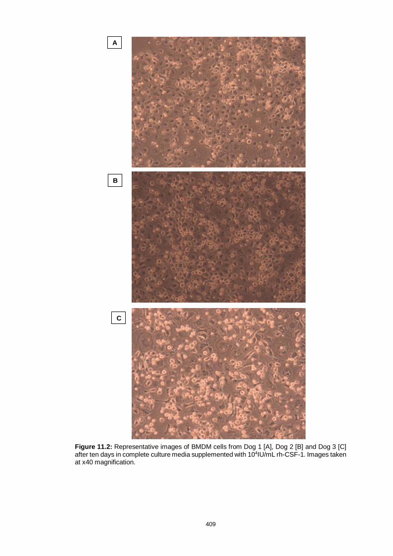
409
Figure 11.2: Representative images of BMDM cells from Dog 1 [A], Dog 2 [B] and Dog 3 [C] after ten days in complete culture media supplemented with 104IU/mL rh-CSF-1. Images taken at x40 magnification.
A
B
C

410
Following cryopreservation, the viable cell recovery rate was between 63% and 80%. Thawed
cells differentiated to produce morphologically identical cells to when bone marrow cells were
plated from fresh (Figre 11.3); therefore future experiments used only cryopreserved cells.
Figure 11.3: Representative image of bone marrow derived macrophage cells from Dog 1. The bone marrow cells were initially cryopreserved, thawed and cultured for ten days in complete culture media supplemented with 104IU/mL rh-CSF-1. Image taken at x40 magnification.
11.3.2 Canine bone marrow cells cultured in rh-CSF-1 acquire cell surface molecules
typical of macrophages within ten days
Human circulating monocytes are frequently subdivided into classical, intermediate and non-
classical phenotypes based on cell surface CD14 and CD16 expression (reviewed by Ziegler-
Heitbrock and Hofer, 2013) whereas pig monocytes can be grouped by CD16 and CD163
expression (Ziegler-Heitbrock, 2014). The surface molecule CD172a (SIRPα) is most
associated with expression on myeloid cells whilst ADGRE-1 (F4/80) is a widely used molecule
specific to murine macrophages (Bastos et al. 2007; Waddell et al. 2018). The aim of this study
was to use flow cytometric analysis of these and other cell surface molecules (CSF-1 and the
CSF-1 receptor [CSF-1r]) to characterise the surface expression profile of canine bone marrow
cells, as well as to examine and monitor changes in their expression profile during macrophage

411
differentiation in rh-CSF-1. The flow cytometry methodology (including antibodies used) is
provided in Section 11.2.4.
Bone marrow from three dogs (Table 11.4) was assessed at four timepoints; Day 0, Day 3,
Day 6 and Day 10 of differentiation and an example gating strategy as applied to all samples
is shown in Figure 11.4. The size and granularity of the cells was assessed by comparing the
side scatter against forward scatter profiles; these cytometry plots from each timepoint for Dog
4 are shown in Figure 11.5 as a representative example of the changes seen for all animals.
The percentage of cell surface molecule-positive cells at each time point for all three animals
tested is shown in Table 11.5 whilst individual plots for the single-colour staining are displayed
in Figure 11.6.
Table 11.4: Signalment details of the dogs used to obtain flow cytometry data during the
differentiation of BMDM cells.
Dog Number Breed Age (years) Sex
Dog 3 Pitbull Terrier One Male Entire
Dog 4 Staffordshire Bull Terrier Three Female Entire
Dog 5 Unknown Cross Four Female Entire

412
Table 11.5: The percentage of cells expressing each of the cell surface molecules tested for
each of the four time points measured. N.B. The anti-CSF-1 receptor antibody tested did not
label any canine cells at any time point.
Cell surface
molecule
Day 0 Day 3 Day 6 Day 10
Dog 3
CSF-1 87.2.0 22.7 53.4 97.0
ADGRE-1 3.64 8.02 11.2 87.5
CD172a 59.9 23.1 58.9 97.2
CD163 11.8 24.2 67.3 94.7
CD16 47.3 6.88 18.8 56.2
CD14 58.7 35.6 57.5 95.7
CD14hi 3.2 6.55 11.5
CD14/CD172a 54.9 23.0 32.7 98.8
CD14/CD163 4.6 25.9 52.1 100
CD14/CD16 28.2 6.6 12.2 50.0
Dog 4
CSF-1 59.4 21.2 25.2 96.1
ADGRE-1 3.1 19.8 24.2 91.8
CD172a 68.5 58.5 46.3 96.3
CD163 3.9 17.4 49.8 96.7
CD16 24.8 4.2 9.89 52.0
CD14 56.5 20.1 42.4 98.4
CD14hi 3.8 10.1 22.7
CD14/CD172a 57.8 22.2 33.5 98.2
CD14/CD163 3.4 20.1 47.8 93.1
CD14/CD16 20.2 4.7 11.0 51.2
Dog 5
CSF-1 56.3 40.2 80.2 96.6
ADGRE-1 5.95 43.5 76.9 95.2
CD172a 52.1 60.7 73.8 99.2
CD163 63.9 39.4 72.7 98.9
CD16 27.4 3.7 41.2 61.4
CD14 47.3 35.8 48.2 98.0
CD14hi 4.06 15.8 67.6
CD14/CD172a 60.3 41.4 44.9 97.2
CD14/CD163 43.2 36.5 48.8 95.3
CD14/CD16 22.0 3.5 7.8 58.4

413
A B
A
C
D
A
A
E F

414
G H I
J K L

415
M N O
P
A
Q R

416
S T U
V W X

417
Figure 11.4: Flow cytometry gating strategy. Representative gating strategy (displaying data from Dog 4) for flow cytometry analysis for
day 0 (A-F), day 3 (G-L), day 6 (M-R) and day 10 (S-X). The forward and side scatter of ungated cells are shown for each day in plots A, G,
M and S respectively. Forward scatter height (FSC-H) vs. forward scatter area (FSC-A) plots (B, G, N and T) were used to reduce the
population to single cells only. Dead cells were excluded by gating only on cells negatively stained with the live-dead marker Zombie VioletTM
(plots C, H, O and U). The remaing cell populations are shown in plots D, I, P and V with gating applied at 160K SSC-A vs. 140 FSC-A to
demonstrate the increase in cellular size and granulation over the 10 days of differentiation in r-CSF-1. Plots E, J, Q and W show the gating
for unstained cells for in the Blue-530-30 channel (FITC [488nm]) and plots F, K, R and X show the same for the Red-670-30 (AlexaFluorTM-
647) channel to show the change in auto-fluorescence over the timecourse.

418
Initially within the total bone marrow cell population there was a large population of mature
granulocytes (as determined by size (Fsc) and granularity (SSc): oval gate; Figure 11.4). The
bone marrow cells expressed CSF-1, CD14, CD172a and CD16 at Day 0 (Figure 11.5). Based
on SSc, the granulocye population expressed CSF-1, CD16 and CD14, and were CD172a
low. At Day 0 only a small percentage (3-6%) of the total viable bone marrow cells from each
dog expressed ADGRE-1 and CD163.
Over the course of differentiation with rhuCSF-1 the large SSc granulocyte population reduced
between Day 0 and Day 3 (Figure 11.4). This was associated with an initial decline in the total
percentage of cells expressing CSF-1, CD14, CD172a and CD16 (Figure 11.5). By contrast,
the putatively monocyte-macrophage population of cells (lower SSc) that expressed ADGRE-
1 and CD163.
By Day 6 the high SSc granulocyte population observed at day 0 was completely lost and the
the total percentage of cells expressing CSF-1, CD14, CD172a and CD16 plateaued or slightly
increased as the maturing monocyte-macrophage cells began to acquire these surface
proteins. There was a concurrent increase in the percentage of cells expressed ADGRE-1,
and CD163.
By Day 10 all cells were adherent, larger (increased FSc), and more granular (increased SSc)
than any of the cells present at Day 0 (as shown in Figure 11.4) and there is expression of
CSF-1, CD14, CD172a, ADGRE-1 and CD163 as would be predicted for a pure population of
mature macrophages (Figure 11.5). For each dog there was a variable proportion of cells
observed at Day 10 which expressed CD16 (48-62%); suggesting that in dogs the divisions
of classical, intermediate and non-classical monocyte subsets may exist, as has been
demonstrated in human bone marrow though further functional assays are needed to confirm
this (Mandl et al. 2014; van Leeuwen-Kerkhoff et al. 2018).

419
1: Surface CSF-1 staining

420
2: Surface ADGRE-1 staining

421
3: Surface CD172a staining

422
4: Surface CD163 staining

423
5: Surface CD16 staining

424
Figure 11.5: Histogram plots showing the changes in cell surface marker expression of canine
bone marrow cells after 0 days (A), 3 days (B), 6 days (C) and 10 days (D) in r-CSF-1. Sets of
plots are shown for each of surface CSF-1 (1), ADGRE-1 (2), CD172a (3), CD163 (4), CD16
(5) and CD14 (6). Stained cell populations are shown in red and isotype controls are shown in
blue.
11.3.3 Both recombinant porcine and recombinant canine CSF-1 can be
used to derive macrophages from canine bone marrow cells, equivalent to rh-
CSF-1
Although rh-CSF-1 has been used previously to generate mature macrophages from
peripherally circulating canine monocytes (Gow et al. 2013), since this finding was published
a recombinant porcine (rp)-CSF-1 had become commercially available and a recombinant
canine (rc)-CSF-1 was under development. The aim of this study was to assess the response
of canine bone marrow cells to rp-CSF-1 and rc-CSF-1 and therefore establish if these growth
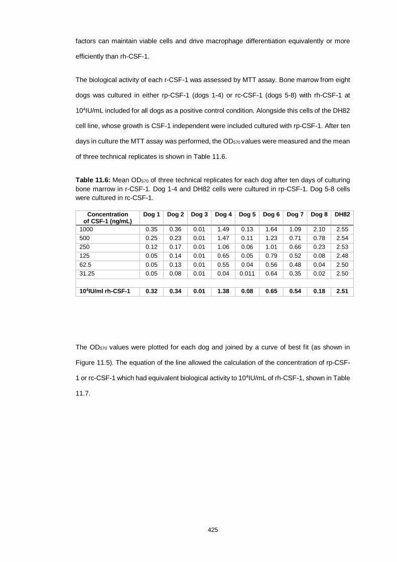
425
factors can maintain viable cells and drive macrophage differentiation equivalently or more
efficiently than rh-CSF-1.
The biological activity of each r-CSF-1 was assessed by MTT assay. Bone marrow from eight
dogs was cultured in either rp-CSF-1 (dogs 1-4) or rc-CSF-1 (dogs 5-8) with rh-CSF-1 at
104IU/mL included for all dogs as a positive control condition. Alongside this cells of the DH82
cell line, whose growth is CSF-1 independent were included cultured with rp-CSF-1. After ten
days in culture the MTT assay was performed, the OD570 values were measured and the mean
of three technical replicates is shown in Table 11.6.
Table 11.6: Mean OD570 of three technical replicates for each dog after ten days of culturing
bone marrow in r-CSF-1. Dog 1-4 and DH82 cells were cultured in rp-CSF-1. Dog 5-8 cells
were cultured in rc-CSF-1.
Concentration of CSF-1 (ng/mL)
Dog 1 Dog 2 Dog 3 Dog 4 Dog 5 Dog 6 Dog 7 Dog 8 DH82
1000 0.35 0.36 0.01 1.49 0.13 1.64 1.09 2.10 2.55
500 0.25 0.23 0.01 1.47 0.11 1.23 0.71 0.78 2.54
250 0.12 0.17 0.01 1.06 0.06 1.01 0.66 0.23 2.53
125 0.05 0.14 0.01 0.65 0.05 0.79 0.52 0.08 2.48
62.5 0.05 0.13 0.01 0.55 0.04 0.56 0.48 0.04 2.50
31.25 0.05 0.08 0.01 0.04 0.011 0.64 0.35 0.02 2.50
104IU/ml rh-CSF-1 0.32 0.34 0.01 1.38 0.08 0.65 0.54 0.18 2.51
The OD570 values were plotted for each dog and joined by a curve of best fit (as shown in
Figure 11.5). The equation of the line allowed the calculation of the concentration of rp-CSF-
1 or rc-CSF-1 which had equivalent biological activity to 104IU/mL of rh-CSF-1, shown in Table
11.7.

426
0 2 0 0 4 0 0 6 0 0 8 0 0 1 0 0 0
0
1
2
3
C o n c e n tra t io n o f r p -C S F -1 (n g /m l)
OD
57
0n
m
D H 8 2 C e ll L in e
D o g 4
D H 8 2 1 04
IU /m L rh -C S F -1
D o g 4 1 04
IU /m L rh -C S F -1
Figure 11.6: MTT assay for BMDM obtained from Dog 4 (■) and DH82 cells (●) cultured in varying concentrations of rp-CSF-1. A standard condition 104IU/mL rh-CSF-1 was used as a control for each cell type; shown by the red line for DH82 cells and the green line for Dog 4 BMDM. Dog 4 was one of four dogs used to evaluate the biological efficacy of rp-CSF-1.
Table 11.7: Concentration of recombinant (porcine/canine) CSF-1 (ng/ml) equivalent in biological activity to 104IU/ml rh-CSF-1 for each dog tested.
Dog 1
Dog 2
Dog 3
Dog 4
Dog 5
Dog 6
Dog 7
Dog 8
DH82
Concentration of rc/rp-CSF-1 (ng/mL) equal to 104IU/ml rh-CSF-1
887 905 ∞ 723 487 36 134 226 ∞
While the MTT assays were used to establish cell viability in each r-CSF-1, the morphology of
the cells generated was assessed after ten days in culture to ensure that the cells present
were comparable to those generated by culture with rh-CSF-1. Both tested recombinant
proteins generated cells with macrophage-like appearance as shown by representative
images of the cells in culture (Figure 11.6).

427
Figure 11.7: Representative images of cells in culture after ten days of differentiation in
complete culture media supplemented with either rc-CSF-1 (Dog 4, left) or rp-CSF-1 (Dog 8,
right).
All three recombinant proteins tested induced macrophage differentiation as determined by
cell morphology and adherence to bacteriological plastic. As might be predicted, the canine
protein was effective at lower final concentrations than the porcine protein; mean of 221ng/mL
compared to 838ng/mL respectively. However, there was notable variation between the dogs
tested. Bone marrow cells from one dog, Dog 3, did not respond at all to the recombinant
porcine protein despite responding to rh-CSF-1 previously. The concentration of rc-CSF-1
equivalent to 104IU/mL of rh-CSF-1 ranged from 36-487ng/mL whilst the results for rp-CSF-1
ranged from 723-905ng/mL. Based on these findings the recombinant proteins were used at
concentrations of; 104IU/mL rh-CSF-1, 850ng/mL rp-CSF-1 or 250ng/mL rc-CSF-1 to generate
canine BMDM for further experiments.
11.3.4 Canine bone marrow dervided macrophages are phagocytic
A key aspect of macrophage function is their phagocytic capacity. To establish the phagocytic
ability of the canine BMDM generated herein, differentiated cells from five dogs were assessed
for their capacity to phagocytose zymosan particles and the resulting images are shown in
Figure 11.7.

428
Figure 11.8: Canine BMDM differentiated from Dog 4 in rh-CSF-1 exposed to either PBS control (left) or FITC fluorescent Zymozan particles (right) showing uptake of particles (green, indicated by white arrows). Cellular filamentous actin was stained with Texas Red phalloidin (red) and nuclear material was counterstained with DAPI (blue). Imaged using a Zeiss LSM710 fluorescent microscope. One representative image of duplicate experiments from three dogs.
11.3.5 Canine BMDM are permissive to Mycobacterium bovis infection and
demonstrate mycobactericidal activity
The main objective of developing a methodology for generating canine BMDM herein was to
establish a model for the study of canine mycobacterial infections, and Mycobacterium (M.)
bovis infections in particular. Previous studies that have examined the interaction of M.
tuberculosis with human monocyte-derived macrophages, or M. bovis with bovine
macrophages (Jensen et al. 2018) have utilised a multiplicity of infection of five (MOI=5). It
was therefore necessary to establish whether this MOI was suitable for the study of canine
BMDM-M. bovis interactions and to assess the number of mycobacteria required to achieve
this in canine BMBM (Nazari et al. 2014; Ruiz et al. 2019).
The reference strain of M. bovis AF-2122/97 was cultured and quantified whilst BMDM were
derived from a single dog in rc-CSF-1 (Dog 3). The M. bovis was diluted in complete culture
media to a range of concentrations/estimated MOI and was then used to challenge 1x10⁶
BMBM/well in vitro as described in Section 11.2.8. Briefly, at the timepoints 0, 0.5, 1, 2 and 24

429
hours post infection the mycobacteria containing media was removed and cells washed
thoroughly to allow the remaining macrophages to be lysed and plated to determine the
number of viable bacteria that had been internalised. The number of CFU of M. bovis in these
lysates was quantifed by culture and the MOI was retrospecitively calcultated. The results are
shown in Table 11.8.
Table 11.8: The multiplicity of infection (MOI) calculated following different durations of
challenge of canine BMDM with various concentrations of M. bovis AF-2122/97, n=1.
Concentration of M. bovis/mL of culture media
Calculated MOI at 0.5 hours post challenge
Calculated MOI at 1 hour post challenge
Calculated MOI at 2 hours post challenge
Calculated MOI at 24 hours post challenge
0 0.00 0.00 0.00 0.00
1x10⁶ 0.97 0.75 0.39 0.33
2x10⁶ 1.95 1.87 0.97 0.43
5x10⁶ 5.03 3.74 1.78 1.57
1x10⁷ 8.82 6.11 3.70 2.86
These data demonstrate that a MOI=5 was best achieved when cells were challenged with
five M. bovis per macrophage for 30 minutes before the media was changed. This allowed
mycobacteria to enter or be phagocytosed by BMDM, but was not long enough for significant
macrophage mediated killing to occur. The data also demonstrate a notable, rapid decline in
the recoverable CFU of M. bovis from the BMBM of this animal within a 24 hour period
indicating potentially potent mycobactericidal activity.
To confirm this finding, the in vitro challenge was repeated with BMDM derived from six
additional animals (Dogs 6-11, Table 11.9) and the CFU of M. bovis recovered at each of four
time points (0, 2, 6 and 24 hours post infection; Figure 11.8). Media containing M. bovis
containing media was added to canine BMDM and left for 30 minutes before being removed,
the cells were then washed to remove any extracellular bacilli and complete culture media
added. The time point ‘0 hours post infection’ was defined as the time that complete culture
media without M. bovis was added to the tissue culture wells.
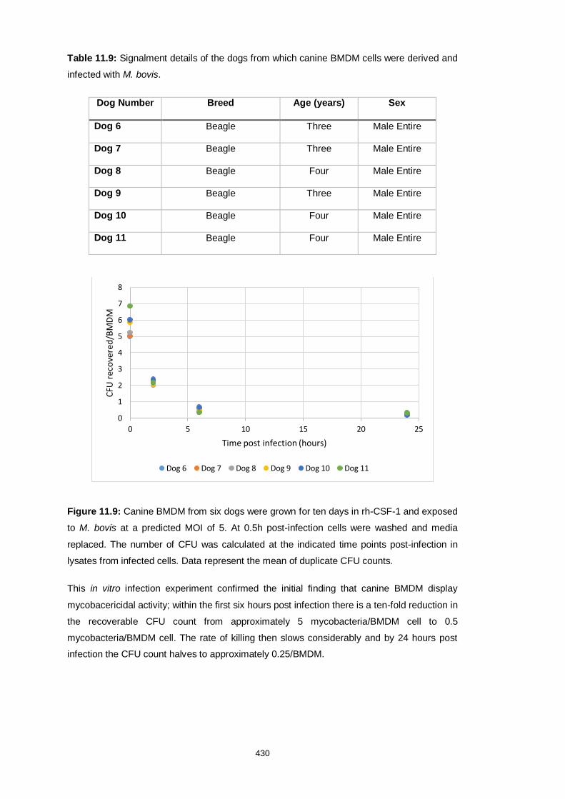
430
Table 11.9: Signalment details of the dogs from which canine BMDM cells were derived and
infected with M. bovis.
Dog Number Breed Age (years) Sex
Dog 6 Beagle Three Male Entire
Dog 7 Beagle Three Male Entire
Dog 8 Beagle Four Male Entire
Dog 9 Beagle Three Male Entire
Dog 10 Beagle Four Male Entire
Dog 11 Beagle Four Male Entire
Figure 11.9: Canine BMDM from six dogs were grown for ten days in rh-CSF-1 and exposed
to M. bovis at a predicted MOI of 5. At 0.5h post-infection cells were washed and media
replaced. The number of CFU was calculated at the indicated time points post-infection in
lysates from infected cells. Data represent the mean of duplicate CFU counts.
This in vitro infection experiment confirmed the initial finding that canine BMDM display
mycobacericidal activity; within the first six hours post infection there is a ten-fold reduction in
the recoverable CFU count from approximately 5 mycobacteria/BMDM cell to 0.5
mycobacteria/BMDM cell. The rate of killing then slows considerably and by 24 hours post
infection the CFU count halves to approximately 0.25/BMDM.
0
1
2
3
4
5
6
7
8
0 5 10 15 20 25
CFU
rec
ove
red
/BM
DM
Time post infection (hours)
Dog 6 Dog 7 Dog 8 Dog 9 Dog 10 Dog 11

431
11.3.6 Canine BMDM and DH82 cells respond to lipopolysaccharide
stimulation and mycobacterial infection by producing pro-inflammatory
cytokines but not nitrite
Based on the key findings of studies in other species, the study presented here examined the
proinflammatory response of canine BMDM and the DH82 cell line by quantifying the
proinflammatory cytokines IL-6 and TNF-α in response Salmonella enterica serotype
Minnesota strain 595 derived LPS, the attenuated M. bovis-BCG and virulent M. bovis. The
BMDM were derived from three dogs (Dog 6, Dog 7 and Dog 8) and DH82 were routinely
cultured. Cells were either stimulated with LPS or infected with mycobacteria. Supernatants
from unstimulated control cells and the challenged cells were taken at 0 hours, 1 hours, 2,
hour, 4 hours, 7 hours and 24 hours post infection. The supernatants were stored at -80°C
before subsequent analysis by ELISA for the presence of IL-6 and TNF-α by ELISA and by
Griess assay to determine the concentration of nitrite present. The cytokine response of the
cells is shown in Figure 11.9; no nitrite was detected in any of the samples tested (data not
shown).

432
0 h
ou
rs
1 h
ou
r
2 h
ou
rs
4 h
ou
rs
7 h
ou
rs
24 h
ou
rs
5 0 0
1 5 0 0
2 5 0 0
3 5 0 0
4 5 0 0
L P S s t im u la te d c e lls
Cy
tok
ine
Co
nc
en
tra
tio
n p
g/m
L
D H 8 2 T N F
B M D M T N F
C o n tro l B M D M T N F
D H 8 2 IL -6
B M D M IL -6
C o n tro l B M D M IL -6
C o n tro l D H 8 2 T N F -
C o n tro l D H 8 2 IL -6
0 h
ou
rs
1 h
ou
r
2 h
ou
rs
4 h
ou
rs
7 h
ou
rs
24 h
ou
rs
0
2 0 0
4 0 0
6 0 0
M . b o v is -B C G in fe c te d c e lls
Cy
tok
ine
Co
nc
en
tra
tio
n p
g/m
L
D H 8 2 T N F
B M D M T N F
C o n tro l B M D M T N F
D H 8 2 IL -6
B M D M IL -6
C o n tro l B M D M IL -6
C o n tro l D H 8 2 T N F
C o n tro l D H 8 2 IL -6
0 h
ou
rs
1 h
ou
r
2 h
ou
rs
4 h
ou
rs
7 h
ou
rs
24 h
ou
rs
0
2 0 0
4 0 0
6 0 0
8 0 0
1 0 0 0
M . b o v is in fe c te d c e lls
Cy
tok
ine
Co
nc
en
tra
tio
n p
g/m
L
D H 8 2 T N F
B M D M T N F
C o n tro l B M D M T N F
D H 8 2 IL -6
B M D M IL -6
C o n tro l B M D M IL -6
C o n tro l D H 8 2 T N F
C o n tro l D H 8 2 T N F
Figure 11.10: The TNFα and IL-6 responses of DH82 cells and canine BMDM to LPS stimulation (A),
M. bovis-BCG infection (B) or M. bovis infection (C) compared to uninfected/unstimulated BMDM (control
BMDM) and DH82 cells (control DH82). Data points are the mean of three biological and two technical
replicates for BMDM and triplicate experiments for DH82 cells. Error bars show the standard deviation.
A
B
C
433
11.3.7 Canine BMDM and DH82 cells show different gene expression profiles
in response to stimulation with LPS and infection with mycobacteria
Due to the limitations of available immunological reagents for domestic dogs, futher
comparison of the canine macrophage response to LPS, M. bovis-BCG and virulent M. bovis
required the development of a RT-qPCR protocol to examine gene expression rather than to
detect protein production and secretion. Therefore, it was first necessary to identify genes in
canine BMDM and the DH82 cell line which retained stable expression levels under all three
challenge conditions for use as reference genes.
An initial panel of six genes was selected as shown in Table 11.2. These primer combinabtions
were assessed by perfroming RT-qPCR on reverse transcriptase positive and negative
samples. The RNA samples tested were collected from each timepoint and for all three stimuli.
The primers that amplify hypoxanthine phosphoribosyltransferase 1 (HPRT-1, primer pair
one), ribosomal protein S7 (RPS7, primer pair one), ribosomal protein L-23 (RPL-23, primer
pair two) were the only three to give a single RT-qPCR product as determined by melt curve
analysis (shown in Figure 11.12), and gave a stable expression level as assessed by the cycle
threshold (Ct) values across all conditions and with both cell types, shown in Table 11.10.

434
Figure 11.11: Dissociation curves for the three primer pairs, HPRT-1 [A], RPS-7 [B] and RPL-
23 [C] that produced single RT-qPCR products and stable Ct values when tested.
A
B
C

435
Table 11.10: The mean of duplicate Ct values for each candidate reference gene analysed at
each timepoint, for both cell types and with all three stimuli.
Primer Pair
Cell Type
Stimulus 0 hours
1 hour
2 hours
4 hours
7 hours
24 hours
HPRT-1 DH82 M. bovis 25.35 25.22 24.81 25.33 25.58 25.04
HPRT-1 DH82 M. bovis BCG
25.15 25.14 25.32 25.18 24.79 24.66
HPRT-1 DH82 LPS 25.12 25.05 25.21 25.73 25.43 25.28
HPRT-1 BMDM M. bovis 24.75 25.14 25.32 25.78 24.79 24.66
HPRT-1 BMDM M. bovis BCG
25.12 25.05 25.21 25.73 25.43 25.28
HPRT-1 BMDM LPS 25.13 25.36 24.40 25.57 25.59 25.55
RPS7 DH82 M. bovis 26.12 26.2 26.23 25.9 26.16 25.98
RPS7 DH82 M. bovis BCG
26.18 26.08 26.42 25.97 26.13 25.85
RPS7 DH82 LPS 26.74 25.96 26.31 25.99 25.89 26.15
RPS7 BMDM M. bovis 26.97 25.84 26.40 26.04 25.79 26.16
RPS7 BMDM M. bovis BCG
27.28 25.72 26.44 26.09 25.66 26.25
RPS7 BMDM LPS 27.19 25.60 26.48 26.13 25.52 26.33
RPL-23 DH82 M. bovis 31.15 31.06 30.83 31.15 30.91 30.40
RPL-23 DH82 M. bovis BCG
30.83 30.91 30.93 31.18 31.21 31.14
RPL-23 DH82 LPS 31.21 30.82 31.09 31.15 30.93 30.40
RPL-23 BMDM DH82 30.94 30.96 31.29 30.92 30.77 30.86
RPL-23 BMDM M. bovis 30.97 30.84 31.42 30.92 30.78 30.86
RPL-23 BMDM LPS 30.95 30.80 31.57 30.85 30.71 30.93
In murine macrophages, nitrite is measurable in the cell supernatant following stimulation with
LPS due to the upregulation of inducible nitric oxide synthase 2 (NOS2) and the metabolism
of arginine to produce nitric oxide, an unstable free radical which rapidly forms nitrite in solution
(Kapetanovic et al. 2012). In porcine BMDM, this process does not occur and neither the nitrite
product nor NOS2 gene transcript are found following exposure to LPS and instead, pig BMDM
alternatively upregulate L-tryptophan metabolism via indoleamine 2,3-dioxygenase 1 (IDO1)
(Kapetanovic et al. 2012). To confirm the lack of nitrite production that suggests both DH82
cells and primary canine BMDM respond similarly to pig BMDM, the expression of NOS2 and

436
IDO1 were quantified in both cell types following challenge with each of LPS, M. bovis-BCG
and M. bovis as per Section 11.3.4. The gene expression of major histocompatibility complex
II (MHC class II), IL-1β, the lymphoattractant chemokine CCL20 (also called MIP3α) and the
vitamin D3-converting enzyme, Cyp27B1 were also measured by RT-qPCR in the same
samples, normalised to HPRT-1 and RPS7. The results are given as a fold-change in gene
expression compared to resting cells (i.e. time 0 hours post infection). Expression changes of
greater than 2-fold from resting were considered to be biologically significant and are shown
in Figure 11.11.

437
DH
82 C
CL
20
BM
DM
CC
L20
DH
82 C
yp
27B
1
BM
DM
Cyp
27B
1
DH
82 IL
-1
BM
DM
IL
-1
DH
82 ID
O-1
BM
DM
ID
O-1
DH
82 M
HC
cla
ss II
BM
DM
MH
C c
lass II
DH
82 N
OS
2
BM
DM
NO
S2
0 .5
1
4
8
1 6
3 2
6 4
M . b o v is -B C G in f e c t e d c e l ls
Fo
ld c
ha
ng
e i
n g
en
e e
xp
re
ss
ion
D H 8 2 C C L 2 0
B M D M C C L 2 0
D H 8 2 C y p 2 7 B 1
B M D M C y p 2 7 B 1
D H 8 2 IL -1
B M D M IL -1
D H 8 2 ID O -1
B M D M ID O -1
D H 8 2 M H C c la s s I I
B M D M M H C c la s s I I
D H 8 2 N O S 2
B M D M N O S 2
DH
82 C
CL
20
BM
DM
CC
L20
DH
82 C
yp
27B
1
BM
DM
Cyp
27B
1
DH
82 IL
-1
BM
DM
IL
-1
DH
82 ID
O-1
BM
DM
ID
O-1
DH
82 M
HC
cla
ss II
BM
DM
MH
C c
lass II
DH
82 N
OS
2
BM
DM
NO
S2
0 .5
1
4
8
1 6
3 2
6 4
1 2 8
M . b o v is in f e c t e d c e l l s
Fo
ld c
ha
ng
e i
n g
en
e e
xp
re
ss
ion
D H 8 2 C C L 2 0
B M D M C C L 2 0
D H 8 2 C y p 2 7 B 1
B M D M C y p 2 7 B 1
D H 8 2 IL -1
B M D M IL -1
D H 8 2 ID O -1
B M D M ID O -1
D H 8 2 M H C c la s s I I
B M D M M H C c la s s I I
D H 8 2 N O S 2
B M D M N O S 2

438
DH
82 C
CL
20
BM
DM
CC
L20
DH
82 C
yp
27B
1
BM
DM
Cyp
27B
1
DH
82 IL
-1
BM
DM
IL
-1
DH
82 ID
O-1
BM
DM
ID
O-1
DH
82 M
HC
cla
ss II
BM
DM
MH
C c
lass II
DH
82 N
OS
2
BM
DM
NO
S2
0 .5
1
4
8
1 6
L P S s t im u la te d c e l ls
Fo
ld c
ha
ng
e i
n g
en
e e
xp
re
ss
ion
D H 8 2 C C L 2 0
B M D M C C L 2 0
D H 8 2 C y p 2 7 B 1
B M D M C y p 2 7 B 1
D H 8 2 IL -1
B M D M IL -1
D H 8 2 ID O -1
B M D M ID O -1
D H 8 2 M H C c la s s I I
B M D M M H C c la s s I I
D H 8 2 N O S 2
B M D M N O S 2
Figure 11.12: The fold-change of gene expression after stimulation with LPS or infection with
M. bovis-BCG or M. bovis for both DH82 and canine BMDM cells. Gene expression was
compared to resting state for the six genes of interest, normalised to averaged HPRT-1 and
RPS7 expression. The six bars represent the timecourse analysed; sequentially left to right
showing 0 hours, 1 hours, 2 hours, 4 hours, 7 hours and 24 hours post infection/stimulation.
Bars show the mean of three biological (Dog 6, 7 and 8) and two technical replicates for
BMDM, and two replicate experiments for DH82 cells. Error bars show the standard deviation.
The dashed line represents a 2-fold change which is considered biologically significant.
The data presented in Figures 11.9 and 11.11 together demonstrate that canine BMDM display
archetypal pro-inflammatory responses to all three stimuli examined; LPS, M. bovis-BCG and
virulent M. bovis. The cytokines IL-6 and TNF-α were both secreted at detectable levels by
four hours following stimulation. Post-stimulation, canine BMDM also significantly increased
their expression of both MHC class II and of IL-β. Together these show conclusively that the
BMDM generated herein are capable of adopting the classically activated phenotype. By
comparison, the frequently studied DH82 cells also produced TNFα and IL-6 and significantly
upregulated MHC class II when stimulated but did not show any change in IL-1β mRNA levels.
There were also other differences between the two cell types in their response to the three
stimuli examined.
439
In general, the BMDM responded more potently to all stimuli compared to DH82 cells. This
difference was most clearly seen in response to M. bovis-BCG infection, which had a nearly
negligible effect on DH82 cells over 24 hours with only small concentrations of IL-6 and TNF-
α secreted and only MHC class II was upregulated across the panel of six genes studied. In
contrast, BMDM upregulated Cyp27B1, IL-1β, IDO-1, MHC class II and NOS2 gene
expression as well as secreting significant amounts of both TNF-α (from two hours post
infection) and IL-6 (from seven hours post infection). In response to M. bovis the concentration
of TNF-α secreted by DH82 into the supernantant peaked at approximately 400pg/mL at four
hours post infection before declining, whereas the concentration produced by BMDM
continued to rise across the 24 hours time course, peaking at approximately 800pg/mL. The
DH82 cells failed to induce IL-1β gene transcription under any of the conditions studies, which
is supported by previous studies showing that the protein is not secreted when the cells are
infected with intracellular parasites such as Leishmania spp (Maia et al. 2007).

440
11.4 Discussion
The aim of the study presented here was to utilise recombinant CSF-1 to generate canine
macrophages from bone marrow in order to chararcterise them and then to examine the effect
of TLR agonism and, critically, to investigate the early response of these cells to mycobacterial
infection. An additional goal was to compare the response of the generated canine BMDM to
the selected stimuli with the response of the frequently used DH82 cell line in order to evaluate
the usefulness of both systems as models for studying canine mycobacterial infections.
The data presented here demonstrate that the methodology developed herein can be used to
generate macrophages from canine bone marrow. In keeping with BMDM from humans and
pigs, the cells were found to be adherent, have typical macrophage morphology, and
phagocytic capacity; all hallmark features of mature cells of the mononuclear phagocyte
system. This study is the first to use bone marrow from dogs to generate macrophages where
previous studies have instead used circulating monocytes. The major advantage of bone
marrow as a starting source of cells is the ability to obtain many more cells from a single
animal; allowing multiple experiments to be conducted with cells from a consistent background
in terms of age, sex and breed that may all influence mature macrophage biology. This
advantage is further conferred by the methodology shown here to cryopreserve bone marrow
cells and still derive macrophages on recovery, meaning that significantly fewer animals may
be needed for studies.
Established culture protocols for the generation of canine monocyte derived macrophages
requires seven days to produce mature cells even though the initial cell population is commited
to the lineage (Heinrich et al. 2017). By comparison, the protocol produced here for BMDM
takes ten days, only three days longer, suggesting that the use of r-CSF-1 in the method
described in this study may act to drive bone marrow progenitors to commit to a macrophage
fate and drives their maturation giving rise to robust differentiation protocol. The use of r-CSF-
1 also adds confidence that the final cell population is comprised purely of macrophages as

441
their survival is maintained in vivo by the action of CSF-1. This study is the first time that the
surface expression of CSF-1 has been demonstrated on canine macrophages (Hume, 2018).
Although the protocol almost always resulted in the generation of canine BMDM, individual
animals varied quite considerably in their responsiveness to r-CSF-1, particualy when different
r-CSF-1 molecules were directly compared. As this study only included a relatively small
number of animals it was not possible to identify any individual reason for this variation in
response; however, it may be a combination of factors such as breed, age and sex of the dog
that the bone marrow cells were obtained from. Studies with a more homogenous population
of dogs would be useful to gain better insight into the effect of these factors on the
differentiation potential of bone marrow cells and their responsiveness to CSF-1 as this may
also contribute to the susceptibility of an individual to infection and/or disease from
mycobacterial challenge.
Previous characterisation of canine monocyte derived macrophages has shown that they
express the lineage associated surface molecules CD14 and CD163 whilst a subset express
CD16 (Heinrich et al. 2017). Examination of histiocyte cells lines, such as DH82 cells, has
additionally shown expression of CD172a (Takada et al. 2018). The data presented here show
that the canine BMDM produced display the same expression pattern of these cell surface
molecules; mature cells were consistently (over 95%) positive for CD14, CD163 and CD172a
whilst just more than half expressed CD16 though this varied slightly from dog to dog.
Numerous studies have divided blood monocytes from many species into subsets according
to their expression of the surface markers CD14 and CD16; classical monocytes highly
express CD14 but do not express CD16 (i.e. they are CD14++CD16−), intermediate cells also
highly express CD14 but also express CD16 (CD14++CD16+) whilst non-classical monocytes
express lower levels of CD14 and have high surface CD16 expression (CD14+CD16++) (Mandl
et al. 2014; Ziegler-Heitbrock et al. 2014). In circulation, the classical phenotype dominates
against lower numbers of the remaining two subtypes, but only one study has evaluated the
presence of these subsets in the bone marrow of humans and has shown that the intermediate
cell phenotype dominates (Mandl et al. 2014). In the present study, CD16 was co-expressed

442
with CD14 on both granuloctyes and monocytes (as defined by their forward and side scatter
profiles) in undifferentiated bone marrow but this increased during the differentiation process
so that by Day 10 a significant population of the macrophages were intermediate or non-
classical in phenotype, which is the first time that this has been shown in dogs. In this study it
was not possible to differentiate CD14+ and CD14++ cells at Day 10 due to all cells being
beyond the visual field; this was done to keep the cytometer parameters identical at all time
points but this could be altered in future studies. Similarly, different subsets of human BMDM
cells have been shown to have different biological functionality such as altered chemokine
receptor expression profiles (Mandl et al. 2014). Therefore future studies of canine BMDM
could separate the cells into phenotypic subsets and further explore their individual response
to stimuli or infection.
The F4/80 antigen, encoded by the Adgre1 locus, has been widely-used as a monocyte-
macrophage marker in mice, but its value as a macrophage marker in other species is unclear,
and has even been questioned (Hume, 2018; Waddell et al. 2018). It has been previously
demonstrated that ADGRE-1 is a myeloid differentiation marker in pigs, absent from
progenitors in bone marrow, highly-expressed in mature granulocytes, monocytes, and tissue
macrophages and induced by CSF-1 treatment in vivo (Hume, 2018; Waddell et al. 2018).
Here, the same expression pattern of ADGRE-1 was demonstrated in canine macrophage
progenitors in bone marrow and BMDM using the same antibody clone as the porcine study,
which showed good cross-reactivity to dog. However, the granulocytes within the canine bone
marrow did not stain positively for ADGRE-1 which was unexpected but was beyond the scope
of this study to investigate further; future characterisation of the immunophenotype of cells
within canine bone marrow would determine which cell types could be derived and studied ex
vivo.
Similarly to the expression of ADGRE-1, canine BMDM may be similar to porcine BMDM in
their pattern of producing TNF-α in response to LPS stimulation but not generating detectable
nitrite. However, RT-qPCR analysis showed that the canine BMDM and DH82 cells did
significantly upregulate NOS2 gene expression from four hours post LPS stimulation and so

443
extending the timecourse of this experiment may have revealed production at a later stage
post stimulation. However, the production of mRNA does not necessarily always correlate with
the induction of functional enzyme activity as a host of post-transcriptional modifications may
occur which prevent translation. Alternatively, other reactive nitrogen species may be
syntesised that are not detected by the Griess assay. The NOS2 enzyme acts to produce nitric
oxide, an unstable radical that cannot be directly assayed and so nitirite is measured as a
proxy. Canine BMDM may have redox mechanisms to safely deal with free radicals which
may, in this case, produce alternative products to nitrite.
The genes for CCL20, Cyp27B1 and IDO-1 were examined by RT-qPCR analysis as they
represent different aspects of the host response to tuberculosis infections. The chemokine
CCL20 attracts lymphocytes and immature dendritic cells to the site of infection and is
expressed by M. tuberculosis infected human monocyte derived macrophages (Rivero et al.
2010). The Cyp27B1 enzyme converts vitamin D3 which is a consistent feature of
granulomatous disease processes, including tuberculosis (Chandra et al. 2004). Tryptophan
metabolism is governed by IDO-1 and M. tuberculosis infection induces marked upregulation
of IDO-1 expression in both human and murine macrophages in vitro (Blumenthal et al. 2012).
In reponse to both mycobacterial infections (M. bovis and M. bovis-BCG) canine BMDM
significantly (2-fold or greater) increased their expression of CCL20 at four, seven and 24
hours post infection while IDO-1 and Cyp27B1 mRNA was present at significant levels from
one hour and then at each subsequent time point of the infection time course examined. These
data suggest that similar to other species, these genes and the metabolic pathways they
belong to, are important factors in mycobacterial immunity and that the canine BMDM model
developed herein is a more biologically appropriate model for the study of canine
mycobacterial infections than the DH82 cell line.
Further studies are needed to confirm and expand on the data shown here; further functional
assessment of the cells such as quantification of phagocytic capacity and their ability to acidify
phaogomes when stimulated/infected would add the the initial evidence that these cells are in
fact macrophage. Future experiments could investigate the production of reactive nitrogen

444
species and nitrite by titrating concentrations of LPS and extending the timecourse of the
challenge study which were logisitically not possible in this study but could be considered for
future experiments.
In conclusion; the data presented here demonstrate that canine BMDM can be generated
using r-CSF-1 supplemented culture media, during culture the resultant cells gain surface
expression of key macrophage associated proteins. After ten days of differentiation, cells are
a pure population of adherent cells which are morphologically macrophage-like and are
phagocytic. Canine BMDM derived in r-CSF-1 comprise both CD16- (classical) and CD16+
(intermidate/non-classical) subsets. These BMDM are permissive to mycobacterial infection
and may represent a more biologically appropriate model for the study of canine tuberculosis
infections than the commonly used DH82 cell line. These results could be expanded on by
futher evaluation of the immunophenotypic subsets of canine BMDM and their potentially
differing functionality as well as comparing different mycobacterial species and strains.
11.5 References
Assouvie, A., Daley-Bauer, L.P. & Rousselet, G. (2018) Growing murine bone marrow-derived
macrophages Methods in Molecular Biology, 1784, 29-33.
Bastos, R.G., Johnson, W.C., Brown, W.C. & Goff, W.L. (2007) Differential response of splenic
monocytes and DC from cattle to microbial stimulation with Mycobacterium bovis BCG and
Babesia bovis merozoites. Veterinary Immunology and Immunopathology, 115 (3-4), 334-345.
Blumenthal, A., Nagalingam, G., Huch, J.H., Walker, L. & Guillemin, G.J. (2012) M.
tuberculosis Induces Potent Activation of IDO-1, but This Is Not Essential for the
Immunological Control of Infection. PLOS ONE 7(5): e37314
Bueno, R., Mello, M.N., Menezes, C.A.S., Dutra, W.O. & Santos, R.L. (2005) Phenotypic,
functional, and quantitative characterization of canine peripheral blood monocyte-derived
macrophages. Memorias do Instituto Oswaldo Cruz, 100 (5), 521-524.
Bulut, Y., Michelsen, K.S., Hayrapetian, L., Naiki, Y., Spallek, R., Singh, M. & Arditi, M. (2005)
Mycobacterium tuberculosis heat shock proteins use diverse toll-like receptor pathways to
activate pro-inflammatory signals. Journal of Biological Chemistry, 280 (22), 20961-20967.

445
Burkey, T.E., Skjolaas, K.A., Dritz, S.S. & Minton, J.E. (2009) Expression of porcine Toll-like
receptor 2, 4 and 9 gene transcripts in the presence of lipopolysaccharide and Salmonella
enterica serovars Typhimurium and Choleraesuis Veterinary Immunology and
Immunopathology, 130 (1-2), 96-101.
Chandra, G., Selvaraj, P., Jawahar, M.S., Banurekha, V.V. & Narayanan, P.R. (2004) Effect
of vitamin D3 on phagocytic potential of macrophages with live Mycobacterium tuberculosis
and lymphoproliferative response in pulmonary tuberculosis. Journal of Clinical Immunology
24(3), 249-257.
Fairbairn, L., Kapetanovic, R., Sester, D.P. & Hume, D.A. (2011) The mononuclear phagocyte
system of the pig as a model for understanding human innate immunity and disease. Journal
of Leukocyte Biology, 89 (6), 855-871.
Gow, D.J., Garceau, V., Pridans, C., Gow, A.G., Simpson, K.E., Gunn-Moore, D. & Hume,
D.A. (2013) Cloning and expression of feline colony stimulating factor receptor (CSF-1R) and
analysis of the species specificity of stimulation by colony stimulating factor-1 (CSF-1) and
interleukin-34 (IL-34) Cytokine, 61 (2), 630-638.
Heinrich, F., Lehmbecker, A., Raddatz, B.B., Kegler, K., Tipold, A., Stein, V.M., Kalkuhl, A.,
Deschl, U., Baumgärtner, W., Ulrich, R. & Spitzbarth, I. (2017) Morphologic, phenotypic, and
transcriptomic characterization of classically and alternatively activated canine blood-derived
macrophages in vitro. PLoS ONE, 12 (8), art. no. e0183572,
Hume, D.A. (2018) Differentiation and heterogeneity in the mononuclear phagocyte system.
Mucosal Immunology, 1 (6), 432-441.
Jensen, K., Gallagher, I.J., Johnston, N., Welsh, M., Skuce, R., Williams, J.L., Glass, E.J.
(2018) Variation in the early host-pathogen interaction of bovine macrophages with divergent
Mycobacterium bovis strains in the United Kingdom. Infection and Immunity, 86 (3), art. no.
e00385-17.
Kapetanovic, R., Fairbairn, L., Beraldi, D., Sester, D.P., Archibald, A.L., Tuggle, C.K., Hume,
D.A. (2012) Pig bone marrow-derived macrophages resemble human macrophages in their
response to bacterial lipopolysaccharide Journal of Immunology, 188 (7), 3382-3394.
Maia, C., Rolão, N., Nunes, M., Gonçalves, L., Campino, L. (200&) Infectivity of five different
types of macrophages by Leishmania infantum. Acta Tropica, 103 (2), 150-155.
Mandl, M., Schmitz, S., Weber, C. & Hristov, M. (2014) Characterization of the cd14++cd16+
monocyte population in human bone marrow PLoS ONE, 9 (11), art. no. e112140

446
Michaels, D.L., Leibowitz, J.A., Azaiza, M.T., Shil, P.K., Shama, S.M., Kutish, G.F., Distelhorst,
S.L., Balish, M.F., May, M.A. & Brown, D.R. Cellular microbiology of Mycoplasma canis (2016)
Infection and Immunity, 84 (6), 1785-1795.
Nazari, H., Karakousis, P.C. & Rao, N.A. (2014) Replication of Mycobacterium tuberculosis
in retinal pigment epithelium. JAMA Ophthalmology, 132 (6), 724-729.
Pahari, S., Kaur, G., Negi, S., Aqdas, M., Das, D.K., Bashir, H., Singh, S., Nagare, M., Khan,
J. & Agrewala, J.N. (2018) Reinforcing the functionality of mononuclear phagocyte system to
control tuberculosis. Frontiers in Immunology, 9, art. no. 193.
Puff, C., Krudewig, C., Imbschweiler, I., Baumgärtner, W. & Alldinger, S. (2009) Influence of
persistent canine distemper virus infection on expression of RECK, matrix-metalloproteinases
and their inhibitors in a canine macrophage/monocytic tumour cell line (DH82) Veterinary
Journal, 182 (1), 100-107.
Quesniaux, V., Fremond, C., Jacobs, M., Parida, S., Nicolle, D., Yeremeev, V., Bihl, F., Erard,
F., Botha, T., Drennan, M., Soler, M.-N., Le Bert, M., Schnyder, B. & Ryffel, B. (2004)Toll-like
receptor pathways in the immune responses to mycobacteria. Microbes and Infection, 6 (10),
946-959.
Rivero-Lezcano, O.M., González-Cortés, C., Reyes-Ruvalcaba, D., Diez-Tascón, C. (2010)
CCL20 is overexpressed in Mycobacterium tuberculosis-infected monocytes and inhibits the
production of reactive oxygen species (ROS). Clinical and Experimental Immunology, 162(2),
289-297.
Ruiz, A., Sarabia, C., Torres, M. & Juárez, E. (2019) Resolvin D1 (RvD1) and maresin 1
(Mar1) contribute to human macrophage control of M. tuberculosis infection while resolving
inflammation. International Immunopharmacology, 74, art. no. 105694.
Schneberger, D., Aharonson-Raz, K. & Singh, B. (2011) Monocyte and macrophage
heterogeneity and Toll-like receptors in the lung. Cell and Tissue Research, 343 (1), 97-106.
Soto, J.M., Mas, A., Rodrigo, J.A., Alieva, T. & Domínguez-Bernal, G. (2019) Label-free
bioanalysis of Leishmania infantum using refractive index tomography with partially coherent
illumination. Journal of Biophotonics, art. no. e201900030.
Szatmary, Z. (2012) Molecular biology of toll-like receptors. General Physiology and
Biophysics, 31 (4), 357-366.

447
Takada, M., Parys, M., Gregory-Bryson, E., Vilar Saavedra, P., Kiupel, M. & Yuzbasiyan-
Gurkan, V. (2018) A novel canine histiocytic sarcoma cell line: Initial characterization and
utilization for drug screening studies. BMC Cancer, 18 (1), art. no. 237
Underhill, D.M., Ozinsky, A., Smith, K.D. & Aderem, A. (1999) Toll-like receptor-2 mediates
mycobacteria-induced proinflammatory signaling in macrophages. Proceedings of the
National Academy of Sciences of the United States of America, 96 (25) 14459-14463.
van Leeuwen-Kerkhoff, N., Lundberg, K., Westers, T.M., Kordasti, S., Bontkes, H.J., Lindstedt,
M., de Gruijl, T.D. & van de Loosdrecht, A.A. (2018) Human bone marrow-derived myeloid
dendritic cells show an immature transcriptional and functional profile compared to their
peripheral blood counterparts and separate from slan+ non-classical monocytes. Frontiers in
Immunology, 9, art. no. 1619
Vijayan, V., Pradhan, P., Braud, L., Fuchs, H.R., Gueler, F., Motterlini, R., Foresti, R. &
Immenschuh, S. (2019) Human and murine macrophages exhibit differential metabolic
responses to lipopolysaccharide - A divergent role for glycolysis Redox Biology, 22, art. no.
101147
Waddell, L.A., Lefevre, L., Bush, S.J., Raper, A., Young, R., Lisowski, Z.M., McCulloch,
M.E.B., Muriuki, C., Sauter, K.A., Clark, E.L., Irvine, K.M., Pridans, C., Hope, J.C. & Hume,
D.A.(2018) ADGRE1 (EMR1, F4/80) is a rapidly-evolving gene expressed in mammalian
monocyte-macrophages. Frontiers in Immunology, 9 art. no. 2246
Wellman, M.L., Krakowka, S., Jacobs, R.M. & Kociba, G.J. (1988) A macrophage-monocyte
cell line from a dog with malignant histiocytosis. In Vitro Cellular & Developmental Biology, 24
(3), 223-229.
Ye, Z.-J., Xu, L.-L., Zhou, Q., Cui, A., Wang, X.-J., Zhai, K., Wang, Z., Tong, Z.-H. & Shi, H.-
Z. (2015) Recruitment of IL-27-Producing CD4 + T Cells and Effect of IL-27 on Pleural
Mesothelial Cells in Tuberculous Pleurisy. Lung, 193 (4), 539-548.
Ziegler-Heitbrock, L. & Hofer, T.P.J. (2013) Toward a refined definition of monocyte subsets.
Frontiers in Immunology, 4, art. no. Article 23
Ziegler-Heitbrock, L. (2014) Monocyte subsets in man and other species. Cellular
Immunology, 289 (1-2), 135-139.

448
Chapter 12: Final Discussion
In the UK, tuberculous mycobacterial infections were historically widely recognised in both
cats and dogs (Anon, 1892; Anon, 1899). In the early 20th century the incidence of tuberculosis
(TB) in the human population of the UK was as high as 300 cases per 100,000 of the
population (Glaziou et al. 2018). Canine TB caused by the human adapted Mycobacterium
(M.) tuberculosis first received significant attention in the UK in the late 1920s and early 1930s
when it was shown that companion dogs can act as both spill-over hosts and as a sentinel
species for human infections (Moore, 1928; Thayer et al. 1930; Adams and Vokwald et al.
1932; Steinbach and Deskowitz, 1933). Similarly, zoonotic TB (caused by M. bovis infection)
was commonplace (Glaziou et al. 2018). Until the end of the Georgian era, most of a city's
milk supply would come from cows living in sheds within city limits, increasing the risk of direct
spread of TB, either by milk or airborne infection, to city dwellers (Wallace and Crouch, 1933;
Tobiesen et al. 1934; Galbrieth et al. 1982). During the 1930’s tuberculin testing was
introduced for cattle in the UK; 40% of all cows were found to be reactors (Glaziou et al. 2018).
With the introduction of pasteurisation, initially to prolong the shelf-life of milk, came the ability
to control the major route of transmission of M. bovis disease to humans (Meara, 1976). At the
same time, forty years of research had focussed on the dog as a model of primary pulmonary
TB caused by both M. bovis and M. tuberculosis, whilst cases of feline TB due to M. bovis
were regularly being recorded almost exclusively with alimentary pathology, presumptively
following the ingestion of contaminated milk (Lemke, 1901; Lewis and Montgomery, 1913;
Manwaring, 1913; Adams and Volkard, 1932; Howe, 1939a; Howe, 1939b). Interestingly, the
apparent resistance of dogs to clinical disease and the lack of pathology in cats caused by M.
tuberculosis had already been noted (Beretta, 1891; McFadyean, 1891).
With the advent of vaccination, the development of antimicrobial agents, improvements in
socio-economic conditions and the formation of the National Health Service, between 1940
and 1990 the incidence of TB in the human population throughout the UK declined by 90%
and the mortality rate fell by 98% (Glaziou et al. 2018). Throughout the same period, reports
of companion animal TB in the literature saw a similarly dramatic decline and began to reflect

449
individual and sporadically occurring canine cases whilst reports of feline mycobacteriosis
became largely limited to incidental findings during post mortem surveys (Dodd, 1952, Cordes
et al. 1963; Birn et al. 1965).
Significant recognition of feline TB as a re-emerging clinical entity in the UK entered the
veterinary literature again in the late 1990’s with a description by Gunn-Moore et al. (1996) of
19 cases of disease due to “an unusual mycobacterial variant” which retrospectively was likely
to be M. microti (the vole bacillus).
This major publication lead on to more in depth investigations into feline mycobacteriosis in
the UK. Following the development of a feline interferon gamma release assay (IGRA) for cats
in 2008 and with state funding made available for the gold standard test of specialist
mycobacterial culture at the Weybridge reference laboratory, it was recognised that these
infections were much more common than had previously been appreciated (Rhodes et al.
2008). In 2011, two seminal papers were published by Gunn-Moore et al. (2011a, 2011b)
describing feline mycobacteriosis in extensive detail. A follow-up study, published in 2013
(Gunn-Moore et al. 2013), used histopathological submissions to estimate the incidence of
feline mycobacterial disease and found that it is diagnosed at a rate of 1% of all routine feline
submissions.
This is the context into which this thesis is presented; four Chapters (2, 3, 7 and 9) present
significant new data which build on the currently accessible diagnostic capacity for companion
animal mycobacterial infections in the UK, evaluating techniques that could be translated into
clinical settings in both the near future but also points to more advanced methods which could
form the next generation of diagnostic tests. These Chapters indicate that molecular
diagnostics using formalin-fixed biopsy tissue is a sensitive method for the identification of TB;
the test methodology developed and presented in Chapter 2 could readily be translated into a
commercially available quantitative test and significantly improve the current diagnostic
capacity for companion animal mycobacterial infections in the UK. Data presented in Chapter
7 also show the first attempt to develop ante mortem canine tests for TB; the development and
implementation of the IGRA assay in real time during an outbreak evidenced its utility and it is

450
consequently already being offered to UK clinicians alongside the feline test at Biobest
Laboratories, Edinburgh (Biobest, 2019).
Studies presented in this thesis are the first to develop and assess diagnostic serological
assays for sub-clinical canine TB infections and in Chapter 8, the accuracy of a subset of the
most promising tests is examined using Bayesian latent class analysis. The findings support
the implementation of these tests and the possibility of using the comparative ELISA as a
screening test for working dogs in order to allay fears that hunting hounds may be inadvertently
contaminating farm land with M. bovis is currently under consideration by the Master of
Foxhounds Association.
Two studies, presented in Chapters 3 and 9, explore the inflammatory processes associated
with TB and other mycobacteria; one in cats and the other in dogs. Whilst the feline study was
conducted on opportunistically collected blood samples, the canine study evaluated cytokine
release into cell culture supernatant by cells stimulated with mycobacterial antigens. Despite
the differences in study methodology, both studies demonstrate a strong cell mediated
immune response to mycobacteria dominate in both of these species. Whilst these assays are
currently too expensive to be considered for diagnostic use, the studies demonstrate
considerable potential for the development of future multiplex cytokine assays which combine
combinations of these inflammatory molecules into accurate and rapid diagnostic tests; in line
with those being developed within human medicine, and for detection of TB in cattle (Clifford
et al. 2019; Coad et al. 2019; Luo et al. 2019) but which are not currently available outside of
a research context.
This thesis presents two unprecedented outbreaks of TB in companion animal species
(Chapters 4 and 5); one canine and the other feline, both caused by the contamination of
feedstuffs with M. bovis genotype 10:a. The canine outbreak, published in O’Halloran et al.
2018, was the first time that the transmission of M. bovis between dogs has been documented
and resulted in nearly 100 dogs being euthanased. The origin of this outbreak was most likely
to have been a contaminated bovine carcass which was fed inadvertently to the Foxhounds
housed at the kennel. The findings of the outbreak investigation prompted a government policy

451
review of the feeding of raw products from fallen stock to hounds, permitted under Article 18
of Commission Regulation (EC) No. 1069/2009. A condition of this regulation is that fallen
stock fed are not killed or have not died as a result of the presence or suspected presence of
a disease communicable to humans or animals. An interpretation of this is that TB reactors,
normally removed by APHA, and inconclusive reactors, should not be fed. The overall
likelihood of at least one foxhound becoming infected with M. bovis within the next five years,
if practices remained unchanged, was considered to be medium. In order to mitigate this risk,
DEFRA has introduced tighter restrictions on the collection and feeding of fallen stock to
hounds in registered kennels. As of 10 October 2017, legislation came into effect as a result
of the findings presented in Chapter 4, to ban the feeding of offal from livestock species to
dogs from recognised kennels or packs of hounds in England (Anon. 2017). Hunt kennel
operators are also now required to carry out additional examinations for TB lesions in fallen
stock originating from high risk premises. Additionally, APHA has developed training materials
and guidance on the identification of TB in carcases of cattle and other livestock, for collectors
and kennels feeding fallen stock to hounds.
The feline outbreak of M. bovis due to contaminated venison being fed raw to cats (Chapter
4) has highlighted a number of wider problems. It was clear from early in the investigations
that were undertaken with the owners of cats affected by this outbreak that the public have
little to no awareness of the microbiological risks presented to them or their pets by feeding
raw food diets; a finding which is supported by the published literature. This is a deficit which
urgently requires future work to address to redress the problem. Unlike the canine TB
outbreak, these cats were affected in geographically disparate areas and countries of the UK
and this meant that it was more challenging to unite different stakeholders around a unified
approach to the outbreak, particularly as APHA do not have a statutory responsibility to
investigate M. bovis infections in pets so initially no culture confirmation was offered pro bono.
However, the conclusions of the epidemiological investigation were robust as they were
evidenced by a number of culture isolates of M. bovis being whole genome sequenced,
proving that they were genetically identical. As noted in the investigation, the withdrawal of UK
Government funding to enable the culture high-risk companion animal samples in recent years

452
has led to owners showing a reluctance to choose this option. Negotiations are ongoing within
the APHA as a result of the data presented within this thesis to potentially reinstate at least a
portion of such funding to aid in the case of future outbreak situations. Owners who opted not
to pay for culture testing were instead using tests viewed as un-validated by APHA and so
future work is needed to increase the confidence in the accuracy of diagnostic tests for
companion animal mycobacterial infections aside from culture.
As well as the devastating impact of both of these unparalleled outbreaks on the animals
affected, significant attendant risks to public health also emerged which were investigated
contemporaneously (Chapters 4 and 6). Jointly, these public health investigations identified
five people with latent TB infections (LTBI); almost equal to the six cases of M. bovis
transmitted from cats to humans in the last 150 years (Public Health England, 2014). Due to
the latent state of these infections it is not possible to definitively prove that they were zoonotic
in origin, but the risk is considered “plausible” and “real” (O’Halloran et al. 2018). The nature
of these investigations are by their nature complex and publications included in this thesis sets
out a robust formal methodology for the approach (Phipps et al. 2019). The protocol was
developed during the canine outbreak and was then successfully utilised when the feline cases
emerged. This highlights the importance of understanding the nature of the pathogen causing
the disease to establish zoonotic risk and inform appropriate action.
Both of the outbreaks referred to above were caused by the same genotype of M. bovis,
namely 10:a, and in both instances a number of cats and dogs were affected by severe and
fulminant clinical disease which resulted in death or euthanasia on welfare grounds. This is
clearly at odds with the conventional dogma in which TB is viewed as a chronic disease with
an insidious onset before more severe clinical signs develop. Interestingly, in both instances
pathological findings occurred which cannot be fully explained by our current knowledge of TB
dynamics and kinetics in these species, such as the severe, necrotising renal lesions seen in
a number of kennel dogs at post mortem examination. Chapter 11 of this thesis begins to
characterise a novel in vitro method for the reliable generation of canine bone marrow derived
macrophages using recombinant CSF-1. It is hoped that this work will now allow for the

453
characterisation of host-pathogen interactions between strains or subtypes M. bovis which
have now been successfully isolated from animals affected by these outbreaks and canine
bone marrow derived macrophages and the macrophage-like DH82 cell line.
Finally, Chapter 10 formally examines our current knowledge of canine TB and brings the
published literature for this species in line with that which is currently the case for feline
mycobacteriosis.
To conclude, this thesis has presented data which significantly add to our current knowledge
with respect to the clinical aspects of TB in both cats and dogs in the UK. In the future, studies
should focus on further understanding of the host-pathogen interaction. This will enable the
development of additional diagnostic tests, or potentially vaccines that will enable the control
of TB in companion animals
12.1 References
Adams, W.E. and Vokwald, A.J. Effect of Bronchial Stenosis on Pulmonary Tuberculosis in Dogs (1932) Proceedings of the Society for Experimental Biology and Medicine, 29 (4), 392-393.
Anon. Authorisation A6 in: Derogations from product controls under Regulation (EC) 1069/2009 and Commission Regulation (EU) 142/2011. Authorisations by the Secretary of State to enable derogations to be used in England. May 2011 (last updated: October 2017). Retrieved from https://www.gov.uk/government/uploads/system/uploads/attachment_data/file/650731/abp-exemptions-201710.pdf
Anon. TUBERCULOSIS IN THE CANINE SPECIES. (1892) The Lancet, 140 (3604), 744-745.
Anon. Tuberculosis of the dog (1899) Journal of the American Medical Association, XXXIII (24), 1499.
Beretta, C. On the use of dog's serum in the treatment of tuberculosis (1891) British Medical Journal, 2 (1611), 1041-1043.
Biobest (2019) Blood Test for Suspect Canine TB Available at: http://www.biobest.co.uk/diagnostics/species/dogs.html Accessed 17 Sept. 2019.
Birn, K.J., Clegg, F.G. and Mason, J.A. Canine tuberculosis. (1965) The Veterinary record, 77 (45), 1341-1342.
Clifford, V., Tebruegge, M., Zufferey, C. et al. Cytokine biomarkers for the diagnosis of tuberculosis infection and disease in adults in a low prevalence setting (2019) Tuberculosis, 114, 91-102.

454
Coad, M., Doyle, M., Steinbach, S. et al. Simultaneous measurement of antigen-induced CXCL10 and IFN-γ enhances test sensitivity for bovine TB detection in cattle (2019) Veterinary Microbiology, 230, 1-6.
Cordes, D.O., Robinson, R.A. and Bailey, B.H. A case of tuberculosis (Human Strain) in a dog (1963) New Zealand Veterinary Journal, 11 (5), 128-130.
Dodd, D.C. A case of miliary bovine tuberculosis in a dog (1952) New Zealand Veterinary Journal, 1 (1), 17-20.
Galbraith, N.S. et al. Communicable disease associated with milk and dairy products in England and Wales 1951-80 (1982) British Medical Journal, 284 (6331), 1761-1765.
Glaziou et al. Trends in Tuberculosis in the UK (2018) Thorax, 702-703.
Gunn-Moore, D.A., Gaunt, C., Shaw, D.J. Incidence of mycobacterial infections in cats in great britain: Estimate from feline tissue samples submitted to diagnostic laboratories (2013) Transboundary and Emerging Diseases, 60 (4), 338-344.
Gunn-Moore, D.A., Jenkins, P.A. and Lucke, V.M. Feline tuberculosis: A literature review and discussion of 19 cases caused by an unusual mycobacterial variant (1996) Veterinary Record, 138 (3), 53-58.
Gunn-Moore, D.A., McFarland, S.E., Brewer, J.I., Crawshaw, T.R., Clifton-Hadley, R.S., Kovalik, M., Shaw, D.J. Mycobacterial disease in cats in Great Britain: I. Culture results, geographical distribution and clinical presentation of 339 cases (2011) Journal of Feline Medicine and Surgery, 13 (12), 934-944.
Gunn-Moore, D.A., McFarland, S.E., Schock, A., Brewer, J.I., Crawshaw, T.R., Clifton-Hadley, R.S., Shaw, D.J. Mycobacterial disease in a population of 339 cats in Great Britain: II. Histopathology of 225 cases, and treatment and outcome of 184 cases (2011) Journal of Feline Medicine and Surgery, 13 (12), 945-952.
Hawthorne, V.M. et al. Tuberculosis in man, dog, and cat (1957) British Medical Journal, 2 (5046), 675-678.
Howe, J.S. Occurrence of Sporadic Bacillemia in Experimental Tuberculosis in Dogs (1939a) Proceedings of the Society for Experimental Biology and Medicine, 40 (4), 509-511.
Howe, J.S. Periodicity of Sporadic Bacillemia in Experimental Tuberculosis in Dogs (1939b) Proceedings of the Society for Experimental Biology and Medicine, 41 (2), 575-577.
Lemke, A.F. Tuberculosis of the lungs treated by compression with nitrogen after the method of murphy: With further remarks on the rationale of the procedure and a record of experiments on dogs (1901) Journal of the American Medical Association, XXXVI (3), 157-168.
Lewis, P.A. and Montgomery, C.M. Experimental pulmonary tuberculosis in the dog: The effect of large amounts of tubercle bacilli of bovine type introduced directly into the lungs by way of the air passages (1913) Journal of Experimental Medicine, 17 (5), 527-534.
Luo, J., Zhang, M., Yan, B., Li, F. et al. Diagnostic performance of plasma cytokine biosignature combination and MCP-1 as individual biomarkers for differentiating stages Mycobacterium tuberculosis infection (2019) Journal of Infection, 78 (4), 281-291.

455
Manwaring, W.H. The effects of subdural injections of leucocytes on the development and course of experimental tuberculous meningitis (1913) Journal of Experimental Medicine, 17 (1), 1-13.
McFadyean, J. Dog's serum in the treatment of tuberculosis (1891) British Medical Journal, 2 (1613), 1173.
Meara, P.J. Veterinary public health implications of U H T processing of milk. (1976) Journal of the South African Veterinary Association, 57 (2), 105-111.
Moore, J. The Old and the New of Veterinary Science and Practice (1928) Journal of the Royal Society of Medicine, 22 (1), 47-53
O'Halloran, C., Hope, J.C., Dobromylskyj, M. et al. An outbreak of tuberculosis due to Mycobacterium bovis infection in a pack of English Foxhounds (2016–2017). (2018) Transboundary and Emerging Diseases, 65 (6), 1872-1884.
Phipps, E., McPhedran, K., Edwards, D. et al. Bovine tuberculosis in working foxhounds: Lessons learned from a complex public health investigation (2019) Epidemiology and Infection, 147, art. no. e24.
Public Health England (2014) Available at https://www.gov.uk/government/news/cases-of-tb-in-domestic-cats-and-cat-to-human-transmission-risk-to-public-very-low Accessed 17 Sept. 19
Rhodes, S.G., Gruffydd-Jones, T., Gunn-Moore, D. et al. Interferon-γ test for feline tuberculosis (2008) Veterinary Record, 162 (14), 453-455.
Steinbach, M.M. and Deskowitz, M. Susceptibility of Diabetic Dogs to Tuberculosis (1933) Proceedings of the Society for Experimental Biology and Medicine, 30 (8), 1053-1055.
Thayer, J. et al. Gastro-Intestinal Port of Entry of B. Tuberculosis in Dogs (1930) Proceedings of the Society for Experimental Biology and Medicine, 27 (5), 367-368.
Tobiesen, Fr. et al. Bovine pulmonary tuberculosis in man-twenty-six cases from Copenhagen (1934) Tubercle, 16 (C), 385-396.
Wallace, G.I. and Crouch, R. Microbiology of Frozen Foods: VI. The Survival of Pathogenic Microorganisms in Ice Cream (1933) Journal of Dairy Science, 16 (4), 315-316.

456
Appendices
Appendix 1.1: IGRA results for the 164 hounds tested
Test number
Negative control
(�̅� + 𝟐𝑺𝑫)
Positive control
(�̅� − 𝟐𝑺𝑫)
PPDA
(�̅� − 𝟐𝑺𝑫) PPDB
(�̅� − 𝟐𝑺𝑫) ESAT-
6/CFP-10
(�̅� − 𝟐𝑺𝑫)
Significant Responses
1 0.05 3.41 -0.09 0.1 0 B only
2 0.09 2.11 0.03 0.05 0.29 EC only
3 0.12 0.65 0.17 0.13 0.13 Fail
4 0.01 3.99 0.06 0.36 0.36 B>A +E
5 0.03 4.11 0.48 1.62 0.96 B>A +E
6 0.09 2.59 0.23 0.69 0 B>A
7 0.01 3.73 0.05 0.3 0.77 B>A +E
8 0.2 3.05 -0.07 0.05 0.03 Negative
9 0.03 4.06 0.15 0.61 1.18 B>A +E
10 0.03 2.69 0.03 0.07 0 Negative
11 0.11 0.25 0.07 0.09 0.07 Fail
12 0.09 2.87 0.02 0 0 Negative
13 0.06 3.88 0.2 0.52 0.63 B>A +E
14 0.07 2.27 0.06 0.16 0 B only
15 0.08 2.27 0.05 0.06 0.08 Negative
16 0.04 0.2 0.01 0 0.05 Fail
17 0.3 3.51 0.45 0.3 0.5 A>B + E
18 0.18 2.8 0.13 0.13 0.23 B>A +E
19 0.01 4.25 0.17 0.44 2.24 B>A +E
20 0.03 4.05 0.06 0.03 0 Negative
21 0.09 2.11 0.03 0.05 0.29 EC only
22 0.13 1.79 0.1 0.1 0.1 Negative
23 0.32 2.8 0 0.1 0.09 Fail
24 0.07 3.16 0.08 0.14 0 B>A
25 0.08 3.34 0.06 0.21 0.03 B only
26 0.08 3.94 0.05 0.45 0.06 B only
27 0.1 2.84 0.5 1.5 0.8 B>A +E
28 0.16 3.35 0.03 0.04 0.07 Negative
29 0.04 3.65 1.4 1.68 1.89 B>A +E
30 0.14 3.5 0.36 0.71 2.56 B>A +E
31 0.05 2.76 0.08 0.14 0.24 B+E
32 0.15 2.18 0 0.12 0 Negative
33 0.3 3.02 0.12 0.04 0 Negative
34 0.25 3.03 0.01 0.04 0 Negative
35 0.12 3.29 0.05 0.81 0.71 B+E

457
36 0.08 1.99 0.01 0 0 Negative
37 0.02 3.05 0 0.25 0.21 B+E
38 0.07 3.33 0.06 0.07 0 Negative
39 0.18 2.8 0.13 0.13 0.23 B>A +E
40 0.07 2.48 0.01 0.03 0.08 EC only
41 0.25 1.34 0.03 0 0.18 Negative
42 0.12 4 0 0.01 0.01 Negative
43 0.04 0.71 0.04 0 0.03 Fail
44 0.08 0.26 0.05 0 0 Fail
45 0.05 4.62 0 0.51 0.21 B+E
46 0.09 2.18 0.06 0.08 0.14 EC only
65 0.06 4.51 0.07 0.04 -0.02 Negative
66 0.01 4.17 0.02 0.01 0.40 EC only
67 0.16 4.32 0.03 -0.18 0.08 Negative
68 0.26 4.47 0.00 0.04 -0.01 Negative
69 0.05 0.79 0.01 -0.20 0.46 EC only
70 0.06 2.00 0.03 -0.05 -0.03 Negative
71 0.08 2.22 0.06 0.02 0.02 Negative
72 0.02 4.60 0.01 -0.03 -0.01 Negative
73
Fail
74 0.13 0.65 0.01 0.04 0.00 Negative
75 -0.01 2.01 -0.01 0.00 -0.01 Negative
76 0.00 2.27 -0.01 -0.01 -0.01 Negative
77 0.01 4.51 0.00 0.08 0.00 Negative
78
Fail
79 0.01 2.44 -0.02 -0.03 -0.02 Negative
80 0.02 4.42 0.01 -0.01 -0.01 Negative
81 0.00 2.52 0.00 0.02 -0.01 Negative
82 0.82 3.91 0.18 0.29 0.07 Negative
83 0.00 3.34 0.00 0.00 -0.25 Negative
84 0.02 4.61 0.01 0.02 0.01 Negative
85 0.06 3.91 0.01 0.10 -0.01 B only
86 0.01 4.10 0.44 1.25 0.26 B>A +E
87 0.01 4.28 0.19 1.05 0.13 B>A +E
88 0.02 4.23 0.01 0.12 0.02 B only
89 0.03 4.24 0.03 0.18 0.00 B only
90 0.02 4.10 0.12 1.32 1.70 B>A +E
91
Fail
92 1.19 3.43 0.20 1.18 1.05 Fail
93 0.03 3.16 0.07 0.29 0.10 B>A +E
94 -0.01 0.86 -0.01 -0.01 -0.01 Negative
95 0.97 3.55 0.00 -0.01 0.00 Negative
96 0.02 3.66 0.03 0.04 0.02 Negative

458
97 0.01 4.73 0.10 0.11 0.00 B>A
98 0.01 4.30 0.01 0.95 0.01 B only
99 0.66 3.98 0.01 0.01 0.01 Negative
100 0.02 4.16 0.02 0.22 0.02 B only
101 0.02 4.15 -0.34 0.02 0.00 Negative
102 -0.06 4.42 -0.07 -0.08 -0.17 Negative
103 0.01 4.19 0.01 -0.13 0.02 Negative
104 0.08 2.73 -0.05 -0.04 -0.05 Negative
105 -0.06 4.44 -0.06 -0.08 -0.07 Negative
106 0.08 4.49 0.14 -0.03 -0.04 A only
107 0.00 4.42 -0.02 -0.05 -0.06 Negative
108 0.09 4.64 -0.01 -0.02 -0.04 Negative
109 0.11 4.51 0.03 -0.03 -0.03 Negative
110 0.04 4.39 0.01 0.01 -0.01 Negative
P1 0.15 1.11 0.09 0.09 0.05 Negative
P2 0.20 2.35 0.08 0.10 0.12 Negative
P3 0.39 3.48 0.16 0.13 0.13 Negative
111 0.26 0.62 0.08 0.31 0.17 Positive
112 0.49 2.05 0.09 0.09 0.07 Negative
113 0.14 0.86 0.10 0.09 0.08 Negative
114 0.34 1.79 0.08 0.11 0.06 Negative
115 0.12 1.85 0.14 0.17 0.11 Positive
116 0.19 2.98 0.13 0.13 0.10 Negative
117 0.14 3.37 0.09 0.12 0.11 Negative
118 0.14 2.97 0.08 0.20 0.15 Positive
119 0.11 3.13 0.11 1.18 0.48 Positive
120 0.18 2.59 0.16 0.71 0.49 Positive
121 0.16 1.17 0.07 0.13 0.06 Negative
122 0.26 4.07 0.11 0.27 0.31 Positive
123 0.17 4.01 0.11 0.19 0.25 Positive
124 0.20 4.00 0.14 0.05 0.14 Negative
125 0.17 3.43 0.14 0.25 0.14 Positive
126 0.26 4.13 0.13 0.16 0.14 Negative
127 0.21 4.42 0.39 1.44 0.41 Positive
128 0.16 3.91 0.20 0.94 1.59 Positive
129 0.16 3.80 0.14 0.28 0.18 Positive
130 0.14 3.68 0.40 0.42 0.21 Positive
131 0.19 4.14 0.22 1.37 0.57 Positive
132 0.20 3.62 0.20 0.10 0.05 Negative
133 0.13 3.19 0.31 1.51 0.33 Positive
134 0.13 2.84 0.09 0.09 0.10 Negative
135 0.13 1.95 0.25 0.16 0.10 Positive
136 0.15 2.56 0.00 0.16 0.11 Positive

459
137 0.24 3.15 0.19 0.16 0.15 Negative
138 0.13 2.54 0.11 0.18 0.13 Positive
139 0.32 2.87 0.12 0.09 0.09 Negative
140 0.32 3.38 0.18 0.17 0.37 Positive
141 0.15 2.41 0.13 0.14 0.09 Negative
142 0.15 2.30 -0.16 0.20 0.37 Positive
143 0.30 3.38 0.19 -0.05 -0.07 Negative
144 0.23 2.98 0.14 0.15 0.12 Negative
145 0.15 3.16 0.17 0.10 0.11 Positive
146 0.12 0.08 0.09 0.07 0.10 Fail
147 0.16 1.74 0.05 0.15 0.12 Negative
148 0.31 1.43 0.12 0.18 0.15 Negative
149 0.17 2.10 0.19 0.27 0.14 Positive
150 0.50 2.45 0.14 0.12 0.14 Negative
151 0.24 3.04 0.19 0.17 0.12 Negative
152 0.16 0.91 0.09 0.12 0.08 Negative
153 0.15 0.57 0.10 0.12 0.09 Negative
154 0.20 1.50 0.06 0.10 0.10 Negative
155 0.14 0.25 0.10 0.08 0.10 Negative
156 0.10 1.07 0.28 0.63 0.13 Positive
157 0.24 4.48 0.13 0.16 0.15 Negative
158 0.66 3.45 0.25 0.70 0.22 Positive
159 0.15 2.61 0.17 0.89 0.22 Positive
160 0.15 2.59 0.34 0.56 0.08 Positive
161 0.37 1.80 0.16 0.19 0.12 Negative
162 0.27 4.50 1.00 2.79 1.58 Positive
163 0.19 1.83 0.18 0.52 0.12 Positive
164 0.39 2.62 0.42 1.06 0.02 Positive
165 0.34 3.60 0.24 0.19 0.21 Negative
166 0.35 4.04 0.22 0.78 0.20 Positive
167 0.60 3.41 0.16 0.16 0.23 Negative
168 0.25 3.91 0.67 1.15 0.26 Positive
169 0.25 1.47 0.27 0.21 0.17 Positive
170 0.27 2.80 0.20 0.74 0.45 Positive
171 0.28 3.15 0.20 0.25 0.08 Negative
172 0.43 0.82 0.09 0.11 0.17 Negative
173 0.44 2.65 0.22 0.31 0.15 Negative
174 0.27 3.56 0.45 0.96 0.53 Positive
175 0.50 3.05 0.15 0.10 0.04 Negative
176 0.22 2.61 0.20 0.28 0.12 Positive
177 0.27 2.89 0.15 0.18 0.18 Negative
178 0.31 2.82 0.28 0.70 0.33 Positive
179 0.44 1.48 0.02 0.12 0.16 Negative

460
180 0.14 0.23 0.08 0.06 0.06 Negative
181 0.08 2.17 0.10 0.13 0.08 Positive
182 0.09 2.74 0.19 0.88 0.14 Positive
183 0.10 3.48 0.11 0.08 0.06 Positive
184 0.22 2.50 0.05 -0.08 0.04 Negative
185 0.27 2.65 0.08 0.12 0.11 Negative
186 0.17 3.49 0.81 0.73 0.25 Positive
187 0.11 2.35 0.10 0.10 0.08 Negative
188 0.09 2.60 -0.03 0.09 0.07 Negative
189 0.07 2.54 0.07 0.09 0.07 Positive
190 0.10 2.66 0.05 0.07 -0.01 Negative
Y1 0.19 3.20 0.12 0.09 0.09 Negative
Y2 0.13 3.95 0.07 0.09 0.10 Negative
Y3 0.37 2.13 0.12 0.14 0.13 Negative
Appendix 1.2: Serology results for the 164 hounds tested
Serology results of ChembioDPP VetTB Assay
Test Number
MPB83 (Visible Assessment)
MPB83 (RLU)
ESAT-6/CFP-10 (Visual Assessment)
ESAT-6/CFP-10 (RLU)
1 Negative 8.01 Negative 0
2 Negative 1.31 Negative 0
3 Negative 2.04 Negative 0
4 Negative 0 Negative 0
5 Negative 3.51 Negative 0
6 Visible positive 127 Negative 0
7 Negative 0 Negative 0
8 Negative 2.43 Negative 0
9 Negative 0 Negative 0
10 Negative 1.22 Negative 0.9
11 Negative 0 Negative 0
12 Negative 0 Visible positive 88.75
13 Negative 0 Negative 0
14 Negative 0 Negative 0
15 Intermediate 73.62 Negative 0
16 Negative 0 Negative 0
17 Negative 0 Negative 0
18 Negative 0 Negative 0
19 Negative 0 Negative 0
20 Negative 0 Negative 0
21 Negative 0 Negative 0
22 Negative 0 Negative 0

461
23 Negative 1.54 Negative 0
24 Negative 15.56 Negative 0.65
25 Negative 0 Intermediate 18.54
26 Negative 0 Negative 0
27 Visible positive 389.21 Negative 0
28 Negative 0 Negative 0
29 Negative 0.89 Negative 0
30 Visible positive 297.47 Negative 3.46
31 Negative 5.79 Negative 0
32 Negative 8.59 Negative 0
33 Negative 0 Negative 0
34 Negative 6.79 Negative 0
35 Negative 0 Negative 0
36 Negative 9.42 Negative 0
37 Negative 0 Negative 0
38 Negative 0 Negative 0
39 Negative 0 Negative 0
40 Visible positive 146.04 Negative 0
41 Negative 0.7 Negative 0
42 Negative 1.23 Negative 0
43 Negative 1.59 Negative 0
44 Negative 11.31 Negative 0
45 Negative 0 Negative 0
46 Negativeative 12.71 Negativeative 0.93
47 Negativeative 0 Negativeative 0
48 Negativeative 0 Negativeative 0
49 Negativeative 17.72 Negativeative 0
50 Negativeative 0.6 Negativeative 0
51 Negativeative 0 Negativeative 3.69
52 Negativeative 0 Negativeative 0
53 Visible positive 124.43 Negativeative 0
54 Negativeative 0 Negativeative 0
55 Negativeative 16.33 Negativeative 0
56 Negativeative 0 Negativeative 0
57 Negativeative 17 Negativeative 0
58 Negativeative 0 Negativeative 0
59 Negativeative 0 Negativeative 0
60 Negativeative 11.73 Negativeative 0
61 Negativeative 0 Negativeative 0
62 Negativeative 21.97 Negativeative 0
63 Negativeative 0 Intermediate 34.66
64 Negativeative 6.61 Negativeative 0
65 Negativeative 9.07 Negativeative 0

462
66 Negativeative 5.04 Negativeative 0
67 Negativeative 0.98 Negativeative 0
68 Negativeative 5.33 Negativeative 0
69 Negativeative 10.51 Negativeative 0
70 Intermediate 58.72 Negativeative 2.32
71 Negativeative 12.14 Negativeative 0
72 Negativeative 7.78 Negativeative 0
73 Negativeative 6.86 Negativeative 0
74 Negativeative 0.71 Negativeative 0
75 Negativeative 0 Negativeative 3.9
76 Negativeative 2.05 Negativeative 0
77 Negativeative 1.16 Negativeative 0
78 Negativeative 9.58 Negativeative 0
79 Negativeative 1.93 Negativeative 0
80 Negativeative 0.9 Negativeative 0
81 Negativeative 0 Negativeative 0
82 Negativeative 0.23 Negativeative 0
83 Negativeative 10.54 Visible positive 113.65
84 Negativeative 13.82 Negativeative 0
85 Negativeative 20.39 Negativeative 0
86 Negativeative 6.13 Negativeative 0
87 Negativeative 21.99 Negativeative 0
88 Negativeative 0 Negativeative 0
89 Negativeative 0 Negativeative 0
90 Negativeative 3.22 Negativeative 0
91 Negativeative 14.56 Negativeative 0
92 Negativeative 0 Negativeative 0
93 Negativeative 0 Negativeative 0
94 Negativeative 0 Negativeative 0
95 Negativeative 4.63 Negativeative 4.07
96 Negativeative 2.15 Intermediate 10.64
97 Negativeative 6.28 Negativeative 0
98 Negativeative 0 Negativeative 0
99 Negativeative 2.68 Negativeative 0
100 Negativeative 0 Negativeative 0
101 Negativeative 20.84 Negativeative 0
102 Negativeative 0 Negativeative 0
103 Negativeative 17.41 Negativeative 0
104 Negativeative 0 Negativeative 0
105 Negativeative 2.16 Intermediate 8.94
106 Negativeative 0 Negativeative 0
107 Visible positive 4169.89 Negativeative 0
108 Negativeative 0 Negativeative 0

463
109 Negativeative 0 Negativeative 0
110 Negativeative 1.66 Negativeative 0
111 Negativeative 0 Intermediate 14.36
112 Negativeative 0 Negativeative 0
113 Negativeative 0 Negativeative 0
114 Negativeative 0 Negativeative 0
115 Negativeative 6.45 Negativeative 0
116 Negativeative 20.97 Negativeative 0
117 Negativeative 8.53 Negativeative 0
118 Negativeative 6.43 Negativeative 0
119 Visible positive 905.4 Negativeative 0
120 Negativeative 0 Negativeative 0
121 Negativeative 0 Negativeative 0
122 Negativeative 0 Negativeative 0
123 Negativeative 0 Intermediate 13.78
124 Negativeative 0 Negativeative 0
125 Negativeative 0 Negativeative 2.83
126 Negativeative 0 Intermediate 27.02
127 Negativeative 0 Negativeative 0
128 Negativeative 2.29 Negativeative 0
129 Negativeative 0 Negativeative 1.53
130 Negativeative 0 Negativeative 0
131 Negativeative 0 Negativeative 0
132 Negativeative 14.72 Negativeative 0
133 Negativeative 0.83 Negativeative 0
134 Negativeative 0 Negativeative 0
135 Negativeative 0 Negativeative 0
136 Negativeative 0 Negativeative 0
137 Negativeative 6.53 Negativeative 0
138 Negativeative 12.78 Negativeative 0
139 Negativeative 4.67 Negativeative 4.39
140 Negativeative 6.93 Negativeative 0
141 Negativeative 7.88 Negativeative 0
142 Negativeative 0 Negativeative 0
143 Negativeative 0 Negativeative 0
144 Negativeative 0 Negativeative 0
145 Negativeative 9.27 Negativeative 0
146 Negativeative 0 Negativeative 0
147 Negativeative 0 Negativeative 0
148 Negativeative 0 Negativeative 0
149 Negativeative 0 Negativeative 0
150 Negativeative 0 Negativeative 0
151 Negativeative 0 Negativeative 0

464
152 Negativeative 0 Negativeative 0
153 Negativeative 0 Negativeative 0
154 Negativeative 0 Negativeative 0
155 Negativeative 0 Negativeative 0
156 Negativeative 0 Negativeative 0
157 Visible positive 115.53 Negativeative 0
158 Negativeative 0 Negativeative 0
159 Visible positive 286.51 Negativeative 2.85
160 Negativeative 0 Negativeative 0
161 Negativeative 8.89 Negativeative 0
162 Negativeative 0 Negativeative 0
163 Negativeative 0.82 Negativeative 0
164 Negativeative 0 Negativeative 0
165 Negativeative 0 Negativeative 0
166 Negativeative 0 Negativeative 0
167 Negativeative 0 Negativeative 0
168 Negativeative 0 Negativeative 0
169 Negativeative 0 Negativeative 0
170 Negativeative 0 Negativeative 0
Appendix 2: WinBUGS syntax of Model One
Model{
X[1:16] ~ dmulti(p[1:16], n)
p[1] <- pi*(1-SeIGRA)*(1-SeCompPPD)*(1-SeDPP)*(1-SeIdexx) + (1-
pi)*SpIGRA*SpCompPPD*SpDPP*SpIdexx
p[2] <- pi*SeIGRA*(1-SeCompPPD)*(1-SeDPP)*(1-SeIdexx)+(1-pi)*(1-
SpIGRA)*SpCompPPD*SpDPP*SpIdexx
p[3] <- pi*SeIGRA*SeCompPPD*(1-SeDPP)*(1-SeIdexx)+(1-pi)*(1-SpIGRA)*(1-
SpCompPPD)*SpDPP*SpIdexx
p[4] <- pi*SeIGRA*SeCompPPD*SeDPP*SeIdexx + (1-pi)*(1-SpIGRA)*(1-SpCompPPD)*(1-
SpDPP)*(1-SpIdexx)
p[5] <- pi*(1-SeIGRA)*SeCompPPD*SeDPP*(1-SeIdexx) + (1-pi)*SpIGRA*(1-
SpCompPPD)*(1-SpDPP)*SpIdexx
p[6] <- pi*(1-SeIGRA)*(1-SeCompPPD)*(1-SeDPP)*SeIdexx + (1-
pi)*SpIGRA*SpCompPPD*SpDPP*(1-SpIdexx)
p[7] <- pi*(1-SeIGRA)*SeCompPPD*(1-SeDPP)*(1-SeIdexx) + (1-pi)*SpIGRA*(1-
SpCompPPD)*SpDPP*SpIdexx
p[8] <- pi*(1-SeIGRA)*(1-SeCompPPD)*SeDPP*(1-SeIdexx) + (1-
pi)*SpIGRA*SpCompPPD*(1-SpDPP)*SpIdexx
p[9] <- pi*SeIGRA*SeCompPPD*SeDPP*(1-SeIdexx) + (1-pi)*(1-SpIGRA)*(1-
SpCompPPD)*(1-SpDPP)*SpIdexx
p[10] <- pi*(1-SeIGRA)*(1-SeCompPPD)*SeDPP*SeIdexx + (1-
pi)*SpIGRA*SpCompPPD*(1-SpDPP)*(1-SpIdexx)
p[11] <- pi*SeIGRA*(1-SeCompPPD)*SeDPP*(1-SeIdexx)+ (1-pi)*(1-
SpIGRA)*SpCompPPD*(1-SpDPP)*SpIdexx

465
p[12] <- pi*SeIGRA*(1-SeCompPPD)*(1-SeDPP)*SeIdexx + (1-pi)*(1-
SpIGRA)*SpCompPPD*SpDPP*(1-SpIdexx)
p[13] <- pi*(1-SeIGRA)*SeCompPPD*SeDPP*SeIdexx + (1-pi)*SpIGRA*(1-
SpCompPPD)*(1-SpDPP)*(1-SpIdexx)
p[14] <- pi*SeIGRA*SeCompPPD*(1-SeDPP)*SeIdexx + (1-pi)*(1-SpIGRA)*(1-
SpCompPPD)*SpDPP*(1-SpIdexx)
p[15] <- pi*(1-SeIGRA)*SeCompPPD*(1-SeDPP)*SeIdexx + (1-pi)*SpIGRA*(1-
SpCompPPD)*SpDPP*(1-SpIdexx)
p[16] <- pi*SeIGRA*(1-SeCompPPD)*SeDPP*SeIdexx + (1-pi)*(1-
SpIGRA)*SpCompPPD*(1-SpDPP)*(1-SpIdexx)
pi ~ dbeta(1.0,1.0)
SeIGRA ~ dbeta (1.0,1.0)
SpIGRA ~ dbeta (1.0,1.0)
SeCompPPD ~ dbeta (1.0,1.0)
SpCompPPD ~ dbeta (1.0,1.0)
SeDPP ~ dbeta (1.0,1.0)
SpDPP ~ dbeta (1.0,1.0)
SeIdexx ~ dbeta (1.0,1.0)
SpIdexx ~ dbeta (1.0,1.0)
ppvIGRA <- pi*SeIGRA/(pi*SeIGRA+(1-pi)*(1-SpIGRA))
ppvCompPPD <- pi*SeCompPPD/(pi*SeCompPPD+(1-pi)*(1-SpCompPPD))
ppvDPP <- pi*SeDPP/(pi*SeDPP+(1-pi)*(1-SpDPP))
ppvIdexx <- pi*SeIdexx/(pi*SeIdexx+(1-pi)*(1-SpIdexx))
npvIGRA <- SpIGRA*(1-pi)/(SpIGRA*(1-pi)+pi*(1-SeIGRA))
npvCompPPD <- SpCompPPD*(1-pi)/(SpCompPPD*(1-pi)+pi*(1-SeCompPPD))
npvDPP <- SpDPP*(1-pi)/(SpDPP*(1-pi)+pi*(1-SeDPP))
npvIdexx <- SpIdexx*(1-pi)/(SpIdexx*(1-pi)+pi*(1-SeIdexx))
}
list(n=164, X=c(66,62,15,4,3,3,2,2,2,1,1,1,1,1,0,0))

466
Project Outcomes
Successful Grant Applications (Chronological order)
1. O’Halloran C. Investigating the Canine Macrophage Response to Mycobacteria.
PetSavers Student Research Project Grant; £1000 March 2019 - September 2019.
2. O’Halloran C. The identification of antigen-specific biomarkers for the diagnosis of
canine tuberculosis caused by Mycobacterium bovis.
American Kennel Club Acorn Grant US$15,000; August 2018 - August 2019.
3. O’Halloran C. and Gunn-Moore, D.A. Validation of diagnostic assays for canine
tuberculosis.
Petplan Charitable Trust; £10,000 November 2017 – December 2018.
Publications (Chronological order)
1. O’Halloran, C., Hope, J.C. and Gunn-Moore, D.A. (2019) Bayesian latent class
estimation of performance parameters of diagnostic tests to identify
Mycobacterium bovis infected domestic dogs (Canis lupus familiaris). In
preparation.
2. Brindley, F., Whitbread, T., and O’Halloran, C. (2019) First confirmed case of
canine leproid granuloma (CLG) in a UK dog. In preparation.
3. O’Halloran, C., Černá, P., Breheny, C., Reed, N., Simpson, K., Cade, S., Jones,
J., Brown, R., Slade, S., Papasouliotis, K. and Gunn-Moore, D.A. (2019)
Investigation of pathological haemorrhage in Maine Coon cats. Veterinary Record.
Under Review.
4. O’Halloran, C., Burr, P., McDonald, K., Rhodes, S., Gunn-Moore, D.A. and
Hope, J.C. (2019) Comparative performance of ante-mortem diagnostic assays
for the identification of Mycobacterium bovis-infected domestic dogs (Canis lupus
familiaris). Scientific Reports Under Review.

467
5. Stavinohova, R., O’Halloran, C., Newton, R., Oliver, J., Scurrell, E., and Gunn-
Moore, D.A. (2019) Histopathological and clinical features of feline ocular
mycobacteriosis in the UK: a multicentre retrospective case series. Veterinary
Pathology. In Press.
6. O’Halloran, C., Ioannidi, O., Reed, N., Murtagh, K., Hope, J., Gale, J., Burr, P.,
Dettemering, E., Van Poucke, S., Vickers, J., Dobromylskyj, M., Mitchell, J.,
Gascoyne-Binzi, D., Howe, R. and Gunn-Moore, D.A. (2019) Tuberculosis due
to Mycobacterium bovis in pet cats associated with feeding a commercial raw
food diet. Journal of Feline Medicine and Surgery In Press.
7. Sharp, E., Taylor, S. and O’Halloran, C. (2019) Unusual presentation of canine
Mycobacterium avium infection Veterinary Record. 184, 800. DOI:
10.1136/vr.105311
8. O'Halloran, C., Gunn-Moore D.A., Reed, N., Vickers, J., Dettmering, E., Ioannidi,
O., and Murtagh, K. (2018) Mycobacterium bovis in pet cats. Veterinary Record.
183(16):510. DOI: 10.1136/vr.k4452.
9. O'Halloran, C., McCulloch, L., Rentoul, L., Alexander, J., Hope, J.C. and Gunn-
Moore, D.A. (2018) Cytokine and Chemokine Concentrations as Biomarkers of
Feline Mycobacteriosis. Scientific Reports, 23;8 (1):17314. DOI:
10.1038/s41598-018-35571-5.
10. O'Halloran, C., Hope, J.C. and Gunn-Moore D.A (2018) Mycobacterium bovis
infection in working foxhounds. Veterinary Record, 22, 183(11):356
DOI:10.1136/vr.k3955.
11. Phipps, E., McPhedran, K., Edwards, D., Russell, K., O'Connor, C.M., Morris, J.,
O'Halloran, C. and Gunn-Moore D.A. (2018) Mycobacterium bovis tuberculosis
in hunting hounds. Veterinary Record, 22, 183(11):356 DOI: 10.1136/vr.k3962
12. Černá, P., O'Halloran, C., SjatkovskaJ, O. and Gunn-Moore, D.A. (2018)
Outbreak of tuberculosis caused by Mycobacterium bovis in a cattery of
Abyssinian cats in Italy Transboundary and Emerging Diseases. In Press.

468
13. Phipps, E., McPhedran, K., Edwards, D., Dampney, R., Russell, K., O’Connor,
C., Gunn-Moore, D.A, O’Halloran, C., Roberts, T. and Morris, J. (2018) Bovine
tuberculosis in working foxhounds: lessons learned from a complex public health
investigation Epidemiology and Infection, 9, 1-6 DOI:
10.1017/S0950268818002753
14. O’Halloran, C., Hope, J.C., Dobromylskyj, M., Burr, P., McDonald, K., Rhodes,
S., Roberts, T., Dampney R, De la Rua-Domenech, R., Robinson, N. and Gunn-
Moore, D.A (2018) An outbreak of tuberculosis due to Mycobacterium bovis
infection in a pack of English Foxhounds (2016-2017). Transboundary and
Emerging Infectious Diseases, 65(6), 1872-1884 DOI:10.1111/tbed.12969
15. O'Halloran, C. and Gunn-Moore, D.A (2017) Mycobacteria in cats: An update In
Practice, 39 (9), 399-406 DOI: 10.1136/inp.j4155
16. Breheny, C.R., Fox, V., Tamborini, A., O'Halloran, C., Robertson, E., Cazzini,
P., Birn-Jeffery, D., Henkin, J., Schwartz, T., Scase, T., Powell, R. and Gunn-
Moore D.A. (2017) Novel characteristics identified in two cases of feline cowpox
virus infection. JFMS Open Reports 11;3(2):2055116917717191. DOI:
10.1177/2055116917717191
17. O’Halloran, C. and Dobromylskyj, M. (2017) Clinical Mycobacterial Diseases of
Companion Animals: Part 2 Companion Animal 22 (11) 2-7.
18. O’Halloran, C., Major, A., Holmes, A., Lalor, S., Littler, R., Spence, S., Schwarz,
T. and Gunn-Moore, D.A. (2017) Use of computed tomography imaging during
long-term follow-up of nine feline tuberculosis cases. Journal of Feline Medicine
and Surgery. DOI:1098612X1769947
19. Gunn-Moore, D.A. and O'Halloran, C. (2017) Hunting dogs and bovine TB.
Veterinary Record. 180, 25: 616
20. Lalor, S. M., Clarke, S., Pink, J., Parry, A., Scurrell, E., Fitzpatrick, N., Watson,
F., O'Halloran, C. and Gunn-Moore, D. (2017) Tuberculosis joint infections in
four domestic cats Journal of Feline Medicine and Surgery Open Reports. 3, 2.
DOI:2055116917719401

469
21. O’Halloran, C. and Gunn-Moore, D.A. (2016) TB in companion animals:
assessing the zoonotic risk Official Vet Magazine 4: 16-19.
22. O’Halloran, C. and Dobromylskyj, M. (2016) Clinical Mycobacterial Diseases of
Companion Animals: Part 1 Companion Animal 22 (6) 325-329.
23. O’Halloran, C., Gunn-Moore, D.A. and Hope, J.C. (2016) Diagnosis of Feline
Mycobacteriosis Veterinary Record 178 (6) 145. DOI: 10.1136/vr.i686.
24. O'Halloran, C., Del-Pozo, J., Breheny, C., Gunn-Moore, D.A., Dobromylskyj, M.,
Papasoulitis, K., Simpson, K. and Henken, J. (2016) Unusual presentations of
feline cowpox. Vet Rec. 29;179(17):442-443. DOI: 10.1136/vr.i5767.
3. Conference Presentations (Oral)
1. Official Veterinarian Conference, Swindon, UK, September 2019. “Raw diets:
Mycobacterium bovis outbreaks in companion animals”
2. Institute of Biomedical Science Congress, Birmingham, UK, September 2019.
“Histopathological insights into feline TB”.
3. International Veterinary Immunology Symposium (IVIS), Seattle, USA, August
2019. “Characterisation of the companion animal immune response to
Mycobacterium bovis infection using cytokine profiling.”
4. International Society for Companion Animal Infectious Diseases (ISCAID),
Portland, USA, October 2018. “Development of a diagnostic PCR assay for feline
mycobacterial disease.”
5. International Society for Companion Animal Infectious Diseases (ISCAID),
Portland, USA, October 2018. “Circulating cytokine and chemokine concentrations
as biomarkers of feline mycobacteriosis”.
6. Feline Mycobacteriosis CPD; Finn Pathlogists, June 2016. “The immunology of
companion animal mycobacterial diseases”.
470
4. Conference Presentations (Abstract Posters)
1. O’Halloran, C., Woods, G., Hope, J.C. and Gunn-Moore, D.A. (2019) Outbreak of
Mycobacterium bovis tuberculosis in UK indoor-only cats associated with raw food
feeding. International Society of Feline Medicine (ISFM) European Congress, Cavtat,
Croatia.
Winner of the Delegates’ Choice Award for best poster.
2. Woods, G., O’Halloran, C. and Gunn-Moore, D.A. (2019) Mycobacterium bovis
tuberculosis in an indoor cat within Scotland associated with commercial raw food.
International Society of Feline Medicine (ISFM) European Congress, Cavtat, Croatia
3. O’Halloran, C., Černá, P., Breheny, C., Reed, N., Simpson, K., Cade, S., Jones, J.,
Brown, R., Slade, S., Papasouliotis, K. and Gunn-Moore, D.A. (2019) Investigation of
pathological haemorrhage in Maine Coon cats.
International Society of Feline Medicine (ISFM) European Congress, Cavtat, Croatia.
4. O’Halloran, C., Hope, J.C.. and Gunn-Moore, D.A (2018) Evaluation of
immunodiagnostic assays for the diagnosis of Mycobacterium bovis infection in dogs.
International Society for Companion Animal Infectious Diseases (ISCAID), Portland,
USA
5. O’Halloran, C., Hope, J.C., Dobromylskyj, M., Burr, P., McDonald, K., Rhodes, S.,
Roberts, T., Dampney, R., De la Rua-Domenech, R., Robinson, N. and Gunn-Moore,
D.A. (2018) Tuberculosis due to Mycobacterium bovis infection in a pack of English
foxhounds – where did it come from?
International Society for Companion Animal Infectious Diseases (ISCAID), Portland,
USA
6. Phipps, E., McPhedran, K., Edwards, D., Dampney, R., Russell, K., O’Connor, C.,
Gunn-Moore, D.A., O’Halloran, C., Roberts, T. and Morris J. (2018) Bovine
tuberculosis in working foxhounds: lessons learned from a complex public health
investigation.

471
International Society for Companion Animal Infectious Diseases (ISCAID), Portland,
USA
7. O’Halloran, C., Hope, J.C., Dobromylskyj, M., Burr, P., McDonald, K., Rhodes, S.,
Roberts, T., Dampney R, De la Rua-Domenech, R., Robinson, N. and Gunn-Moore,
D.A (2018) An outbreak of tuberculosis due to Mycobacterium bovis infection in a pack
of English foxhounds (2016-2017).
International Society for Companion Animal Infectious Diseases (ISCAID), Portland,
USA
8. O’Halloran, C., Hope, J.C.. Dobromylskyj, M. and Gunn-Moore, D.A (2018)
Development of a diagnostic PCR assay for feline mycobacterial disease
International Society for Companion Animal Infectious Diseases (ISCAID), Portland,
USA
9. O’Halloran, C., McCulloch, L., Rentoul, L., Hope, J.C., and Gunn-Moore, D.A. (2018)
Circulating cytokine and chemokine concentrations as biomarkers of feline
mycobacteriosis.
International Society for Companion Animal Infectious Diseases (ISCAID), Portland,
USA
10. Černá, P., O'Halloran, C., SjatkovskaJ, O., Gunn-Moore, D.A. (2018) Outbreak of
tuberculosis caused by Mycobacterium bovis in a cattery of Abyssinian cats in Italy.
International Society of Feline Medicine (ISFM) European Congress, Sorrento, Italy.
11. O’Halloran, C., Hope, J.C.. Dobromylskyj, M. and Gunn-Moore, D.A (2018)
Development of a rapid PCR diagnostic test for feline mycobacterial disease using
formalin-fixed paraffin-embedded tissue biopsies.
International Society of Feline Medicine (ISFM) European Congress, Sorrento, Italy.
12. Mitchell, J.L., Street, S.N., O’Halloran, C., Woolley, C.S., Hope, J.C. and Gunn-
Moore, D.A. (2017) Differences in numbers of observable mycobacteria by Ziehl-
Neelsen staining in skin and lymph node biopsy samples in cases of feline
tuberculosis.
International Society of Feline Medicine (ISFM) European Congress, Sorrento, Italy.

472
13. O'Halloran, C., McCulloch, L., Rentoul, L., Alexander, J., Hope, J.C. and Gunn-
Moore, D.A. (2016) Comparative Analysis of ciculating cytokine and chemokine
concentreations as biomarkers of feline mycobacteriosis.
British Society of Immunology (BSI) Congress, Liverpool, UK.
Shortlisted for best poster presentation prize.
14. O’Halloran, C., Hope, J.C and Gunn-Moore, D.A. (2016) Canine Tuberculosis:
Forgotten but not gone?
International Society for Companion Animal Infectious Diseases (ISCAID)
Symposium, Bristol, UK.