Azithromycin Pharmacodynamics Against Non-Typeable H ...
28
Yale University Yale University EliScholar – A Digital Platform for Scholarly Publishing at Yale EliScholar – A Digital Platform for Scholarly Publishing at Yale Public Health Theses School of Public Health 1-1-2016 Azithromycin Pharmacodynamics Against Non-Typeable H. Azithromycin Pharmacodynamics Against Non-Typeable H. Influenzae Influenzae James Fisher Yale University, james.fi[email protected] Follow this and additional works at: https://elischolar.library.yale.edu/ysphtdl Recommended Citation Recommended Citation Fisher, James, "Azithromycin Pharmacodynamics Against Non-Typeable H. Influenzae" (2016). Public Health Theses. 1089. https://elischolar.library.yale.edu/ysphtdl/1089 This Open Access Thesis is brought to you for free and open access by the School of Public Health at EliScholar – A Digital Platform for Scholarly Publishing at Yale. It has been accepted for inclusion in Public Health Theses by an authorized administrator of EliScholar – A Digital Platform for Scholarly Publishing at Yale. For more information, please contact [email protected].
-
Upload
khangminh22 -
Category
Documents
-
view
0 -
download
0
Transcript of Azithromycin Pharmacodynamics Against Non-Typeable H ...

Yale University Yale University
EliScholar – A Digital Platform for Scholarly Publishing at Yale EliScholar – A Digital Platform for Scholarly Publishing at Yale
Public Health Theses School of Public Health
1-1-2016
Azithromycin Pharmacodynamics Against Non-Typeable H. Azithromycin Pharmacodynamics Against Non-Typeable H.
Influenzae Influenzae
James Fisher Yale University, [email protected]
Follow this and additional works at: https://elischolar.library.yale.edu/ysphtdl
Recommended Citation Recommended Citation Fisher, James, "Azithromycin Pharmacodynamics Against Non-Typeable H. Influenzae" (2016). Public Health Theses. 1089. https://elischolar.library.yale.edu/ysphtdl/1089
This Open Access Thesis is brought to you for free and open access by the School of Public Health at EliScholar – A Digital Platform for Scholarly Publishing at Yale. It has been accepted for inclusion in Public Health Theses by an authorized administrator of EliScholar – A Digital Platform for Scholarly Publishing at Yale. For more information, please contact [email protected].

Azithromycin Pharmacodynamics against Non-typeable H. influenzae
James Fisher1, Raheal Boadi-Yeboah2, Melinda Pettigrew PhD1, Brian Tsuji PharmD2
1 Yale School of Public Health, New Haven, CT
2 State University of New York, University at Buffalo, Buffalo, NY

Abstract
Introduction
Chronic Obstructive Pulmonary Disease (COPD) is the third leading cause of death in
the United States. Significant clinical disease in COPD patients has been tied to non-
typeable Haemophilus influenzae (NTHI) infection. However, reasons why some NTHI
strains persist despite antimicrobial therapy remain unknown.
Objective
The primary objective was to characterize the pharmacodynamics of azithromycin
against a persistent pair of NTHI isolates.
Methods
A persistent pair of NTHI isolates (5P28H1 and 5P54H1) cultured from the sputum of an
adult with COPD and determined to be the same strain by multilocus sequence typing
was carried for 819 days. The Minimum Inhibitory Concentrations (MIC) were
determined according to Clinical and Laboratory Standards Institute guidelines for
5P28H1 (MICazithromycin=2.0) and 5P54H1 (MICazithromycin=16.0). Time-kill experiments
were performed using an array of azithromycin concentrations and samples were
collected over 48h. The log reduction and integrated log ratio area over 48h were
calculated and fit to a Hill-type model. A hollow-fiber infection model (HFIM) simulating
azithromycin concentrations and pharmacokinetics in human serum and alveolar
macrophages was performed over 240h for 5P28H1.
Results
Azithromycin displayed differential killing activity against 5P28H1 and 5P54H1. For
5P28H1, azithromycin concentrations >0.5 mg/L achieved complete killing by 48h. In

contrast, complete bacterial killing was observed by 48h for concentrations >4 mg/L in
5P54H1. Overall, azithromycin demonstrated dose-dependent bactericidal activity
against both isolates. Pharmacodynamic analysis revealed a right shift in the
comparative dose response curves. Model fits were excellent (R2>0.99). Azithromycin
serum concentrations simulated in HFIM did not appreciably reduce 5P28H1 viability by
240h, whereas simulated alveolar macrophage concentrations achieved complete killing
by 26hrs.
Conclusion
Bactericidal activity was achieved for 5P28H1 at lower concentrations compared to
5P54H1, which demonstrated an attenuated killing profile. The differential
pharmacodynamics of azithromycin suggests that antimicrobial pressure plays a role in
counter selection of resistance for NTHI.

Introduction
Chronic Obstructive Pulmonary Disease (COPD) is a leading cause of morbidity
and mortality worldwide (1, 2). COPD is characterized by irreversible airway obstruction,
persistent inflammation, and a multitude of comorbidities (2). It is estimated that 24
million people are affected by COPD in the United States alone, with direct and indirect
health care costs nearing $50 billion per year (3, 4). In contrast to other leading causes
of death like cancer and cardiovascular disease, mortality due to COPD appears to be
on a continuing upward trend (2). While smoking is the primary risk factor, air pollution
and those exposed to dust, fumes, and chemical vapors comprise a large proportion of
those developing COPD (2, 5). However, only about 25% of smokers actually develop
this disease, indicating that other environmental or genetic factors may be involved in its
pathogenesis (6).
Recently, it has become clear that bacteria may chronically colonize the lungs of
COPD patients. Due to host immune responses, persistent bacterial colonization of
damaged airways may lead to additional inflammation, promoting and advancing
disease progression. This understanding led to the creation of the “Two-Hit” hypothesis,
which posits that a combination of environmental and infection-related factors are
crucial to COPD pathogenesis and progression (6). Apart from chronic bacterial
colonization, bacterial infections are thought to play a major role in acute exacerbations
of COPD. Treatment of exacerbations is believed to account for 45%-75% of direct
COPD-related expenditures (7). These acute exacerbations not only incur significant
financial costs, but additional social and indirect economic burdens including lost
wages, absenteeism, and restricted activity (7).

It is estimated that approximately 50% of acute exacerbations have bacterial
etiology (8). The most common species of bacteria isolated from COPD patients’ lungs
during an acute exacerbation is Haemophilus influenzae (1). H. influenzae exclusively
infects humans and may cause pneumonia, bacteremia, or meningitis (9). While a
vaccine exists for one of the six known types of H. influenzae, non-typeable
(unencapsulated) strains predominate incident infections in the US (9). Thus, non-
typeable H. influenzae (NTHI) is posited to play a significant role in progression of
impaired pulmonary function observed in COPD patients (10).
Current guidelines for treating patients with acute COPD exacerbations include
the prescription of an antibiotic and anti-inflammatory steroid agent (3). Azithromycin, a
macrolide with both antibiotic and anti-inflammatory properties, is one of the most
commonly prescribed antimicrobials for exacerbations and is being considered for
prophylactic use in individuals with moderate-to-severe COPD (11, 12). However,
treatment and long-term prophylaxis utilizing azithromycin is not a risk-free endeavor.
Desai and colleagues (2010) noted a clinical association between antibiotic exposure
and occurrence of resistant pneumococcal strains in a cohort of 127 adult COPD
patients (8). In this study, it appeared that over 50% of pneumococcal strains obtained
from patients exposed to macrolides, primarily azithromycin, at some point over a three
and six month period displayed resistance (8). Additionally, individual and community
azithromycin usage is known to increase population-level macrolide resistance in
respiratory pathogens, garnering significant calls for judicious prescription practices
(12).

Azithromycin exhibits antimicrobial activity by binding to the 50S ribosome and
inhibiting protein translation. H. influenzae resistance mechanisms against macrolides
include ribosomal methylase, intrinsic or acquired efflux pumps, and alterations in
ribosomal proteins or RNA (13). While the clinical significance of efflux pumps remains
controversial, strains with mutations in ribosomal proteins L4 or L22 and 23S rRNA tend
to exhibit higher MICs for azithromycin than wild-type strains and may have significant
clinical implication (13).
The pharmacokinetic and pharmacodynamic (PK/PD) profile of azithromycin
against H. influenzae is crucial to the process of determining clinical susceptibility and
improving patient outcomes (13). PK refers to the body’s absorption, distribution,
metabolism, and elimination of an agent (13, 14). Azithromycin is characterized by rapid
absorption, poor bioavailability when taken orally (~37%), and extensive tissue
distribution (14, 15). Peak serum concentrations (Cmax) are estimated to be
approximately 0.4 mg/L, with lung tissue and alveolar macrophage concentrations
reaching up to 700 times that of serum (14, 15). The body does not metabolize
azithromycin and excretion occurs through hepatic, transintestinal, and biliary routes
(14). PD refers to the relationship between antimicrobial agent and pathogen (13). PD
analysis reveals the effect of antimicrobial drug on killing and micro-organism growth
dynamics (13). Azithromycin exhibits a concentration-dependent bactericidal effect on
H. influenzae, in which activity is best determined by the serum concentration-time
curve (AUC):MIC ratio (14). The AUC is the product of two PK parameters: Cmax and the
duration of exposure (14). It is important to note that the duration of exposure for

azithromycin can be quite long, with the terminal half-life estimated to be 68 hours and
sub-inhibitory concentrations extending for up to 30 days (12, 14, 15).
The development of macrolide resistance in H. influenzae presents a major
public health challenge. Since existing literature focuses primarily on pneumococcal
resistance to macrolide antibiotics, there is a dearth of knowledge on the development
of resistance in NTHI. Therefore, the underlying conditions allowing resistance to
develop and optimal therapeutic regimens for treatment of NTHI in the context of COPD
need delineation. In order to understand the driving factors of NTHI resistance to
azithromycin, we characterized the pharmacodynamics of azithromycin against a pair of
clinically persistent, serially obtained NTHI isolates. In addition, we evaluated the drug
resistance profiles of the clinical isolates by simulating an in-vivo infection utilizing
clinically relevant azithromycin concentrations and pharmacokinetic parameters.

Methods
Bacterial Strains
A unique collection of H. influenzae sputum isolates with corresponding whole genome
sequence, epidemiologic, and clinical data were prospectively collected as part of a 20-
year longitudinal study of COPD infection conducted in Buffalo, NY from 1994-2014
(16). A pair of clinically persistent, serially obtained non-typeable H. influenzae strains
from an individual patient in this cohort was obtained. Strains 5P28H1 and 5P54H1
were isolated from the same patient at clinic visit 28 and 54, respectively, and were
carried for 819 days (17). Over this time period, the patient experienced 4 courses of
azithromycin therapy. These strains were of the same multilocus sequence type, with
5P54H1 harboring a mutation in the L22 ribosomal protein (17). MICs were determined
according to CLSI guidelines. 5P28H1 exhibited MICazithromycin of 2 mg/L and 5P54H1
exhibited MICazithromycin of 16 mg/L (17).
Antibiotic and media
Azithromycin analytical grade powder was commercially purchased (Sigma Chemical
Company, St. Louis, MO). Stock solutions of azithromycin in 10% DMSO were prepared
at the start of each experiment. MIC values for 5P28H1 and 5P54H1 were previously
determined by broth microdilution in Mueller-Hinton Broth according to CLSI guidelines
(17). Brain heart infusion (BHI) broth (Difco Laboratories, Detroit, MI) supplemented with
10 μg/mL NAD and 10 μg/mL Hemin was used for all static time kill experiments.
Colonies were enumerated on chocolate agar (Thermo Fisher Scientific, Waltham, MA).

Static Time Kill Experiments
Static time kill experiments were performed over 48 hours as previously described,
using a starting inoculum of 108 CFU/mL (18). In short, fresh bacterial colonies from
overnight growth were added to BHI broth to create a concentrated suspension. From
this suspension, 2 mL were removed and diluted with BHI broth and standard
azithromycin stock solution to achieve a 108 inoculum in a total reaction volume of 20
mL. Each 20 mL culture was incubated in a 37oC water bath with constant shaking for
48 hours. At 0, 1, 2, 4, 8, 24, 26, 28, 32, and 48 hours, .1 mL samples were withdrawn
and serially diluted in sterile saline for CFU/mL enumeration. Colony counts were
determined by plating 50 μL of each diluted sample onto BHI chocolate agar with an
automated spiral dispenser (WASP; Don Whitely Scientific Limited, West Yorkshire,
England) and incubating plates for 24 hours at 35oC with 5% CO2. Time kill experiments
were performed at azithromycin concentrations chosen as a function of the isolates’
MIC: 64, 32, 16, 8, 4, 2, and .5 mg/L. Two growth controls (10% DMSO in BHI and BHI
only) were run in parallel.
Pharmacodynamic Modeling
An integrated PK/PD approach was used to quantify the effect of azithromycin against
the clinical isolates. The area under the CFU curve (AUCFU) for each azithromycin
concentration was normalized by the AUCFU in the absence of drug to obtain the log
ratio area (Equation 1).
Equation 1:
��� ����� ��� = log�����������
������������

Plots of the log ratio area vs. azithromycin concentration were then constructed, and
azithromycin’s activity (E) was modeled by the Hill function (Equation 2), where Emax is
the maximal drug effect, EC50 is the concentration required to obtain half the maximal
effect, C is the concentration, E0 is the effect in the absence of drug, and H is the
sigmoidicity constant (version 12, Systat Software Inc., San Jose, CA). Overall model
fits were analyzed based on coefficients of determination (R2).
Equation 2:
� = �� − �� ! " (�)%
(��&�)% + (�)%
Hollow Fiber Infection Model (HFIM)
HFIM was used to evaluate how clinically relevant azithromycin regimens affect
bacterial burden of lung infection over 240 hours (19, 20). The HFIM utilized cellulosic
cartridge C3008 (FiberCell Systems, Frederick, MD). Essentially, bacteria colonize the
extracapillary space of the cartridge while nutrients and antibiotic are exchanged
through hollow fibers. Apart from growth controls for both clinical isolates, azithromycin
dosing regimens were administered in a two-tiered fashion for 5P28H1 to achieve
identical areas under concentration-time curve (AUC) as those deemed physiologically
relevant. We simulated azithromycin pharmacokinetics in both serum and alveolar
macrophages. A high-burden lung infection was simulated in this model, using a starting
inoculum of 108 CFU/mL.
Serum drug concentrations for hollow fiber modeling were determined by
digitizing data from a 5-day Z-pack pharmacokinetics study in human subjects (21).
Azithromycin protein binding was estimated to be 51% at .02 mg/L and 7% at 2 mg/L via
the FDA package insert. We assumed a linear correlation between these two values
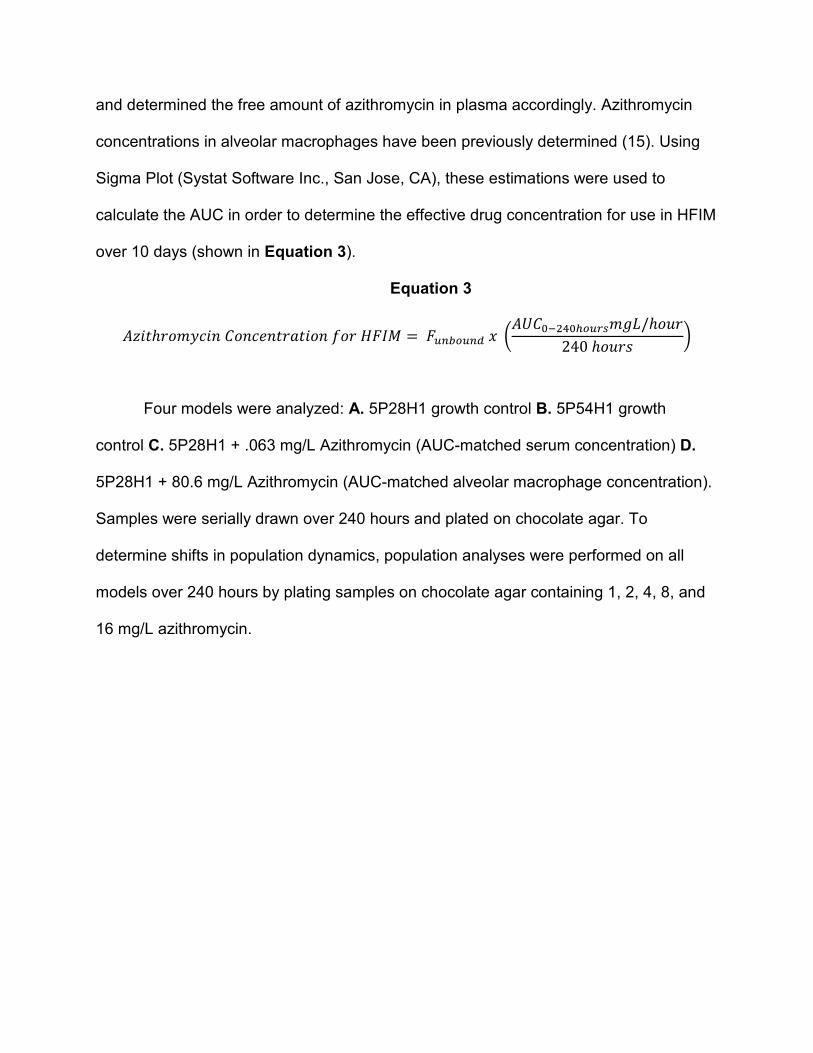
and determined the free amount of azithromycin in plasma accordingly. Azithromycin
concentrations in alveolar macrophages have been previously determined (15). Using
Sigma Plot (Systat Software Inc., San Jose, CA), these estimations were used to
calculate the AUC in order to determine the effective drug concentration for use in HFIM
over 10 days (shown in Equation 3).
Equation 3
�(��ℎ��*+,�- ��-,-������- .�� /�01 = ���2���� " 3����456�7���8*��/ℎ�:�240 ℎ�:�> ?
Four models were analyzed: A. 5P28H1 growth control B. 5P54H1 growth
control C. 5P28H1 + .063 mg/L Azithromycin (AUC-matched serum concentration) D.
5P28H1 + 80.6 mg/L Azithromycin (AUC-matched alveolar macrophage concentration).
Samples were serially drawn over 240 hours and plated on chocolate agar. To
determine shifts in population dynamics, population analyses were performed on all
models over 240 hours by plating samples on chocolate agar containing 1, 2, 4, 8, and
16 mg/L azithromycin.

Results
Static time kill results for 5P28H1 (MICazithromycin: 2 mg/L) are shown in Figure 1.
By 48 hours, all concentrations of azithromycin above .5 mg/L achieved complete killing
for isolate 5P28H1. The three highest concentrations of azithromycin (64, 32, and 16
mg/L) exhibited the most rapid bactericidal effect, completely killing 5P28H1 by 24
hours. At the only azithromycin concentration tested beneath the MIC of 5P28H1 (.5
mg/L), minimal killing was observed. The growth controls for 5P28H1 displayed an
approximate 1-log decrease in CFU/mL by 48 hours. A concentration-dependent effect
was visible, as decreases in azithromycin concentration led to increased time-to-
complete killing. The bactericidal activity of azithromycin against 5P54H1 (MICazithromycin :
16 mg/L) is shown in Figure 2. Concentrations of azithromycin at or above the MIC of
5P54H1 exhibited complete killing by 24 hours. At a concentration of 8 mg/L (half the
MIC of 5P54H1), azithromycin appeared to effectively eliminate viability by 28 hours. At
4, 2, and .5 mg/L of azithromycin, initial reductions in viability were essentially nullified
after 8 hours.
To analyze the pharmacodynamic interaction between azithromycin and our two
strains, we fit a Hill-type function to our time-kill data. The Hill-type function and model
parameters are displayed in Figure 3. The Hill-model displayed excellent model fits, as
the R2 values in both sets were greater than .99. The values for Emax and EC50 were
higher in 5P54H1 (2.38 and 1.47, respectively) than in 5P28H1 (2.25 and .858,
respectively), indicating azithromycin to be both less efficacious and less potent against
5P54H1.
In order to simulate human infection, we performed HFIM on 5P28H1 and
5P54H1. The results of the HFIM are displayed in Figure 4. It is evident that both

5P28H1 and 5P54H1 in the absence of antibiotic were able to grow in the HFIM,
achieving bacterial densities greater than 109 CFU/mL. While 5P28H1 was previously
shown to survive in the conditions of the Hollow Fiber cartridge, this was the first test of
5P54H1 viability in the HFIM. The AUC-matched serum concentration (.063 mg/L) of
azithromycin was virtually unable to produce any killing effect against 5P28H1.
However, at a concentration mimicking that within alveolar macrophages (80.6 mg/L),
5P28H1 exhibited complete loss of viability by 26 hours. A population analysis of
5P28H1 performed throughout the HFIM did not reveal any significant phenotypic
changes in antibiotic susceptibility over 10 days (data not shown).

Discussion
To our knowledge, this is one of the first studies examining azithromycin
pharmacodynamics against resistant NTHI in COPD patients. In this study, we
elucidated the pharmacodynamics of azithromycin against a pair of clinically persistent
NTHI isolates. We determined that azithromycin is effective in killing 5P28H1 and
5P54H1 in a dose-dependent fashion. As the viability of 5P54H1 had not been tested in
the HFIM previously, we provided proof-of-principle for this isolate to be further
examined in the model.
The threat of antibiotic resistance is particularly alarming in the context of COPD.
It has been shown that alveolar macrophage activity and other bacterial lung defense
mechanisms are severely inhibited in COPD patients (22-24). Considering that chronic
bacterial infection may propel lung damage in COPD and that lowered lung defenses of
COPD patients may foster persistent bacterial colonization, a formidable cycle of
disease progression exists. COPD patients often rely on both antibiotic and
corticosteroid treatment to alleviate symptoms. When antibiotic treatments fail, COPD
patients may experience rapid acceleration of disease and significant social, physical,
and financial costs. Understanding the pharmacodynamics of azithromycin against
clinical strains of NTHI may help tailor future treatment regimens and prevent the
development of macrolide resistance.
Multiple challenges exist in correlating antimicrobial susceptibilities with clinical
therapeutic efficacy. Antimicrobial susceptibilities for NTHI, including those for
azithromycin, are derived utilizing microbiological breakpoints (13). These breakpoints
are determined based on drug-effect differences between an azithromycin-naïve

population of H. influenzae (wild-type) and a population exhibiting decreased
azithromycin activity (13, 25). However, microbiological breakpoints may not correlate
with clinical outcome and may not be useful for individual case management (13).
Clinically relevant breakpoints incorporate a number of factors, including: results of
clinical studies, PK information regarding tissues and fluids, dose-effect relationships,
and mathematical modeling (13). The possible disconnect between microbiological
susceptibility breakpoints and clinically relevant breakpoints was evident in our study.
Isolate 5P28H1, though deemed susceptible to azithromycin through microbiological
breakpoints, was not cleared by the patient after four courses of azithromycin therapy.
This disparity may have led to counter-selection and the genetic and phenotypic
development of azithromycin resistance observed in isolate 5P54H1. Thus, our results
highlight the need for clinically relevant breakpoints in H. influenzae susceptibility
testing.
Our observation regarding the inability of simulated serum concentration
exposures in the HFIM to kill 5P28H1 yields insight into a less-defined topic in
azithromycin treatment: utility of serum AUC versus site-specific AUC metrics. It has
been posited that site-specific azithromycin concentration for lower respiratory tract
infections is better correlated with antimicrobial effect than serum or systemic measures
(26, 27). Despite this, site-specific concentrations within the lungs and on epithelial
surfaces remain poorly understood. Lucchi et al. determined the concentration of
immediate and extended release azithromycin in epithelial lining fluid (ELF) in patients
with lung cancer and found values ranging from 0 to 6.81 mg/L (15). In a separate
study, healthy individuals were administered a single 500 mg dose (comparable to the
first dose in a Z-pak regimen) and the amount of azithromycin in ELF was undetectable
(28). Thus, it appears that underlying health conditions as well as individual physiologic
characteristics may affect the achievable azithromycin concentrations in ELF. To our
knowledge, no studies have examined the concentration of azithromycin in ELF of
COPD patients. Additionally, our population analysis of 5P28H1 during the HFIM
revealed no significant changes in antibiotic susceptibility by day 10. Ultimately, this
may implicate that multiple regimens of azithromycin were necessary to initiate the
changes in 5P28H1 to increase its MIC 8-fold.
Utilizing azithromycin for COPD prophylaxis and treatment of a multitude of
respiratory diseases could have far reaching consequences. Since commensal bacteria
are also exposed to azithromycin during treatment, macrolide resistance may develop in
the microbiota. This process may already be occurring, as multiple studies have found
macrolide resistance in nasopharyngeal and oropharyngeal isolates after azithromycin
regimens (12). This may facilitate the transfer of resistance elements between
commensal and pathogenic bacteria, increasing the population burden of macrolide
resistance. Azithromycin prescription has also been linked to the development of
penicillin and multi-drug resistance (12). A Spanish study performed in 2002 found that
macrolide consumption was more important in driving penicillin resistance than
consumption of β-lactams (29). Therefore widespread use of azithromycin could drive
an increase in resistance across drug classes, posing a significant threat to
antimicrobial stewardship efforts and public health. Thus, broad scale azithromycin
prescription practices may need reevaluation.

While this study sheds light on important issues facing the treatment and
management of COPD exacerbations using azithromycin, we recognize a few
limitations to our study. The first limitation is generalizability. The findings from a pair of
isolates obtained from a single patient in Buffalo, NY may not be generalizable to all
individuals chronically infected with NTHI. Additionally, we recognize that a broader
array of physiologically relevant concentrations of azithromycin for time kill experiments
and the hollow-fiber infection model could have been used. Therefore in future studies
we hope to utilize a broader array of concentrations with physiological relevance.

Figures
Figure 1. Static Time Kill results for isolate 5P28H1 (MICazithromycin : 2 mg/L) evaluating
the bactericidal activity of an array of azithromycin concentrations.

Lo
g1
0 C
FU
/mL
5P28H1
Growth Control (GC)
10% DMSO GC
0.5 mg/L
2 mg/L
4 mg/L8 mg/L
16 mg/L
32 mg/L
64 mg/L

Figure 2. Static Time Kill results for 5P54H1 (MICazithromycin : 16 mg/L) evaluating the
bactericidal activity of an array of azithromycin concentrations.

5P54H1
Growth Control (GC)
10% DMSO GC
0.5 mg/L
2 mg/L
4 mg/L8 mg/L
16 mg/L
32 mg/L
64 mg/L
Lo
g1
0 C
FU
/mL

Figure 3. The Hill-type model fit for azithromycin concentrations versus log-ratio area
for 5P28H1 and 5P54H1. Model parameters, including Emax and EC50 are included with
percent standard error.

R2 0.998
Emax (% SE) 2.25 (0.09)
EC50 (% SE) 0.858 (0.09)
R2 0.998
Emax (% SE) 2.38 (0.14)
EC50 (% SE) 1.47 (0.28)
Lo
g R
ati
o A
rea
5P28H1 5P54H1
Concentration (mg/L) Concentration (mg/L)

Figure 4. HFIM model results over 10-days (240 hours). Viable bacteria as determined
by CFU/mL are plotted against time. Simulated concentrations of azithromycin in serum
(.063 mg/L) and alveolar macrophages (80.6 mg/L) were tested against 5P28H1.

Lo
g1
0 C
FU
/mL

Sources:
1. Finney LJ, Ritchie A, Pollard E, Johnston SL, Mallia P. Lower airway colonization and
inflammatory response in COPD: a focus on Haemophilus influenzae. Int J Chron Obstruct
Pulmon Dis. 2014;9:1119-32.
2. Hillas G, Perlikos F, Tsiligianni I, Tzanakis N. Managing comorbidities in COPD. Int J
Chron Obstruct Pulmon Dis. 2015;10:95-109.
3. Wilson R, Sethi S, Anzueto A, Miravitlles M. Antibiotics for treatment and prevention
of exacerbations of chronic obstructive pulmonary disease. J Infect. 2013;67(6):497-515.
4. Disease GIfCOL. Global Strategy for Diagnosis, Management, and Prevention of COPD
- 2016. 2016.
5. WHO. Chronic obstructive pulmonary disease 2015 [Available from:
http://www.who.int/respiratory/copd/en/.
6. Ganesan S, Comstock AT, Kinker B, Mancuso P, Beck JM, Sajjan US. Combined
exposure to cigarette smoke and nontypeable Haemophilus influenzae drives
development of a COPD phenotype in mice. Respiratory Research. 2014;15(11).
7. Patel JG, Nagar SP, Dalal AA. Indirect costs in chronic obstructive pulmonary disease:
a review of the economic burden on employers and individuals in the United States. Int J
Chron Obstruct Pulmon Dis. 2014;9:289-300.
8. Desai H, Richter S, Doern G, Heilmann K, Dohrn C, Johnson A, et al. Antibiotic
Resistance in Sputum Isolates of Streptococcus pneumoniae in Chronic Obstructive
Pulmonary Disease is Related to Antibiotic Exposure. COPD: Journal of Chronic Obstructive
Pulmonary Disease. 2010;7(5):337-44.
9. CDC. H. influenzae Disease 2014 [Available from: http://www.cdc.gov/hi-
disease/index.html
10. Sethi S, Evans N, Brydon JB, Murphy TF. New Strains of Bacteria and Exacerbations
of Chronic Obstructive Pulmonary Disease. New England Journal of Medicine.
2002;347(7):465 - 71.
11. Parameswaran GI, Sethi S. Long-term macrolide therapy in chronic obstructive
pulmonary disease. CMAJ. 2014;186(15):1148-52.
12. Serisier DJ. Risks of population antimicrobial resistance associated with chronic
macrolide use for inflammatory airway diseases. The Lancet Respiratory Medicine.
2013;1(3):262-74.
13. Tristram S, Jacobs MR, Appelbaum PC. Antimicrobial resistance in Haemophilus
influenzae. Clin Microbiol Rev. 2007;20(2):368-89.
14. Nightingale CH. Pharmacokinetics and pharmacodynamics of newer macrolides. The
Pediatric Infectious Disease Journal. 1997;16(4):438-43.
15. Lucchi M, Damle B, Fang A, de Caprariis PJ, Mussi A, Sanchez SP, et al.
Pharmacokinetics of azithromycin in serum, bronchial washings, alveolar macrophages and
lung tissue following a single oral dose of extended or immediate release formulations of
azithromycin. J Antimicrob Chemother. 2008;61(4):884-91.
16. Sethi S, Evans N, Grant BJB, Murphy TF. New Strains of Bacteria and Exacerbations
of Chronic Obstructive Pulmonary Disease. NEJM. 2002;347(7).

17. Pettigrew MM, Tsuji BT, Gent JF, Kong Y, Holden PN, Sethi S, et al. Haemophilus
influenzae in COPD: Effect of fluoroquinolones and macrolides on eradication and
resistance. 2016.
18. Tsuji BT, von Eiff C, Kelchlin PA, Forrest A, Smith PF. Attenuated vancomycin
bactericidal activity against Staphylococcus aureus hemB mutants expressing the small-
colony-variant phenotype. Antimicrob Agents Chemother. 2008;52(4):1533-7.
19. Gumbo T, Louie A, Deziel MR, Parsons LM, Salfinger M, Drusano GL. Selection of a
Moxifloxacin Dose that Suppresses Drug Resistance in Mycobacterium tuberculosis, by Use
of an In Vitro Pharmacodynamic Infection Model and Mathematical Modeling. JID.
2004(190).
20. Lenhard JR, Brown T, Rybak MJ, Meaney CJ, Norgard NB, Bulman ZP, et al. Sequential
Evolution of Vancomycin-Intermediate Resistance Alters Virulence in Staphylococcus
aureus: Pharmacokinetic/Pharmacodynamic Targets for Vancomycin Exposure. Antimicrob
Agents Chemother. 2015;60(3):1584-91.
21. Amsden GW, Nafzinger AN, Foulds G. Pharmacokinetics in Serum and Leukocyte
Exposures of Oral Azithromycin, 1,500 Milligrams, Given over a 3- or 5-Day Period in
Healthy Subjects. AAC. 1999;43(1).
22. Kammerl IE, et al. Impairment of immunoproteasome function by cigarette smoke
and in COPD. AJRCCM. 2016.
23. Kalathil SG, Lugade AA, Pradhan V, Miller A, Parameswaran GI, Sethi S, et al. T-
regulatory cells and programmed death 1+ T cells contribute to effector T-cell dysfunction
in patients with chronic obstructive pulmonary disease. Am J Respir Crit Care Med.
2014;190(1):40-50.
24. Polosukhin VV, Cates JM, Lawson WE, Zaynagetdinov R, Milstone AP, Massion PP, et
al. Bronchial secretory immunoglobulin a deficiency correlates with airway inflammation
and progression of chronic obstructive pulmonary disease. Am J Respir Crit Care Med.
2011;184(3):317-27.
25. Turnidge J, Kahlmeter G, Kronvall G. Statistical characterisation of bacterial wild-
type MIC value distributions and the determination of epidemiological cut-off values. Clin
Microbiol Infect. 2006;12(5):418-25.
26. Firsov AA, Zinner SH, Vostrov SN, Kononenko OV, Portnoy YA, Shustova LV, et al.
Comparative pharmacodynamics of azithromycin and roxithromycin with S. pyogenes and
S. pneumoniae in a model that simulates in vitro pharmacokinetics in human tonsils.
Journal of Antimicrobial Chemotherapy. 2002.
27. Rodvold KA, Gotfried MH, Danziger LH, Servie RJ. Intrapulmonary Stead-State
Concentrations of Clarithromycin and Azithromycin in Healthy Adult Volunteers.
Antimicrob Agents Chemother. 1997.
28. Conte Jr JE, Golden J, Duncan S, McKenna E, Lin E, zurlinden E. Single-Dose
Intrapulmonary Pharmacokinetics of Azithromycin, Clarithromycin, Ciprofloxacin, and
Cefuroxime in Volunteer Subjects. Antimicrob Agents Chemother. 1996.
29. Garcia-Rey C, Aguilar L, Baquero F, Casal J, Dal-Re R. Importance of Local Variations
in Antibiotic Consumption and Geographical Differences of Erythromycin and Penicillin
Resistance in Streptococcus pneumoniae. Journal of Clinical Microbiology. 2002;40(1):159-
64.