Association chiropratique canadienne Canadian Chiropractic ...
82
J Can Chiropr Assoc 2017; 61(1) 1 CANADIAN CHIROPRACTIC ASSOCIATION Chair Dr. David Peeace, BSc, DC JCCA STAFF Editor Kent Stuber, DC, MSc Division of Graduate Education & Research Canadian Memorial Chiropractic College, Calgary, Alberta Editor Emeritus Allan Gotlib, C.M., DC Toronto, Ontario Associate Editors Jeffrey Quon, DC, PhD School of Population & Public Health Faculty of Medicine, University of British Columbia, Vancouver, British Columbia André Bussières, DC, FCCS(C), PhD Faculty of Medicine, McGill University, Montréal, Québec Département chiropratique, Université du Québec à Trois-Rivières, Trois-Rivières, Québec Dana J. Lawrence, DC, MMedEd, MA Palmer College of Chiropractic, Davenport, Iowa Assistant Editors Pierre Côté, DC, PhD University of Ontario Institute of Technology, Oshawa, Ontario B. Kim Humphreys, DC, PhD Head of Chiropractic Medicine, Faculty of Medicine, University of Zurich and University Hospital of Balgrist, Zurich, Switzerland Gregory N. Kawchuk, DC, PhD University of Alberta, Edmonton, Alberta Mohsen Kazemi, RN, DC, MSc, FRCCSS(C), FCCPOR(C) Faculty of Clinical Education, Graduate Studies and Research, Canadian Memorial Chiropractic College, Toronto, Ontario Jill Hayden, DC, PhD Department of Community Health & Epidemiology Faculty of Medicine, Dalhousie University, Halifax, Nova Scotia Production Co-ordinator Tami Ehrlich Advertising Editor, Journal of the Canadian Chiropractic Association 186 Spadina Avenue, Suite 6, Toronto, Ontario M5T 3B2 Tel: 416-585-7902 877-222-9303 Fax: 416-585-2970 Email: Dr. Kent Stuber<[email protected]> Website: www.jcca-online.org TYPESETTING Thistle Printing Limited 35 Mobile Drive, Toronto, Ontario M4A 2P6 Association chiropratique canadienne Canadian Chiropractic Association
-
Upload
khangminh22 -
Category
Documents
-
view
0 -
download
0
Transcript of Association chiropratique canadienne Canadian Chiropractic ...

J Can Chiropr Assoc 2017; 61(1) 1
CANADIAN CHIROPRACTIC ASSOCIATIONChair Dr. David Peeace, BSc, DC
JCCA STAFFEditor Kent Stuber, DC, MSc
Division of Graduate Education & Research Canadian Memorial Chiropractic College, Calgary, Alberta
Editor Emeritus Allan Gotlib, C.M., DC Toronto, Ontario
Associate Editors Jeffrey Quon, DC, PhD School of Population & Public Health Faculty of Medicine, University of British Columbia, Vancouver, British Columbia
André Bussières, DC, FCCS(C), PhD Faculty of Medicine, McGill University, Montréal, Québec Département chiropratique, Université du Québec à Trois-Rivières, Trois-Rivières, Québec
Dana J. Lawrence, DC, MMedEd, MA Palmer College of Chiropractic, Davenport, Iowa
Assistant Editors Pierre Côté, DC, PhD University of Ontario Institute of Technology, Oshawa, Ontario
B. Kim Humphreys, DC, PhD Head of Chiropractic Medicine, Faculty of Medicine, University of Zurich and University Hospital of Balgrist, Zurich, Switzerland
Gregory N. Kawchuk, DC, PhD University of Alberta, Edmonton, Alberta
Mohsen Kazemi, RN, DC, MSc, FRCCSS(C), FCCPOR(C) Faculty of Clinical Education, Graduate Studies and Research, Canadian Memorial Chiropractic College, Toronto, Ontario
Jill Hayden, DC, PhD Department of Community Health & Epidemiology Faculty of Medicine, Dalhousie University, Halifax, Nova Scotia
Production Co-ordinator Tami EhrlichAdvertising Editor, Journal of the Canadian Chiropractic Association
186 Spadina Avenue, Suite 6, Toronto, Ontario M5T 3B2 Tel: 416-585-7902 877-222-9303 Fax: 416-585-2970
Email: Dr. Kent Stuber<[email protected]> Website: www.jcca-online.org
TYPESETTING Thistle Printing Limited
35 Mobile Drive, Toronto, Ontario M4A 2P6
Association chiropratique
canadienne
Canadian Chiropractic Association

2 J Can Chiropr Assoc 2017; 61(1)
JCCA Journal of the Canadian Chiropractic Association
(Formerly the Canadian Chiropractic Association Journal) Copyright Registered © by the Canadian Chiropractic Association 1961
Copyright: The Canadian Chiropractic Association, 2017
All rights reserved. Without limiting the rights under copyright above, no part of this publication may be reproduced, stored in or introduced into any retrieval system, or transmitted in any form
or by any means (electronic, mechanical, photocopying, recording or otherwise), without the prior written permission with the copyright owner and the publisher.
Published by the Canadian Chiropractic Association and issued quarterly
EDITORIAL AND EXECUTIVE OFFICES, 186 SPADINA AVENUE, SUITE 6, TORONTO, CANADA M5T 3B2
General Information: The Journal of the Canadian Chiropractic Association is the official quarterly publication by the Canadian Chiropractic Association. The JCCA is published quarterly by the Canadian Chiropractic Association as a medium of communication be-tween the Association and its members and is a forum for fair comment and discussion of all matters of general interest to the chiropractic profession and the Association. Readers are invited to comment and express their opinions on relevant subjects. Views and opinions in editorials and articles are not to be taken as official expression of the Association’s policy unless so stated. Publication of contributed articles does not necessarily imply endorsement in any way of the opinions expressed therein and the Journal and its publisher does not ac- cept any responsibility for them. Business correspondence should be addressed to: the Edi- tor of JCCA, 186 Spadina Avenue, Suite 6, Toronto, Canada M5T 3B2.
INDEXING SERVICES
JCCA is indexed by PubMed Central, Scopus, CINAHL (Cumulative Index to Nursing and Allied Health Literature), MANTIS (formerly CHIROLARS), AMED, PASCAL, Index to Chiropractic Literature, and selectively by SPORTDiscus.
J Can Chiropr Assoc 2017; 61(1) 3
ContentsJCCA Vol 61 No 1 ISSN 0008-3194 (Print) and ISSN 1715-6181 (Electronic)
Commentary6 Contemporary biopsychosocial exercise prescription for chronic low back pain: questioning
core stability programs and considering context Peter Stilwell, BKin, DC, MSc
Katherine Harman, PT, PhD
Original Articles18 The physical and psychological impact of neurogenic claudication: the patients’ perspectives Carlo Ammendolia, DC, PhD
Michael Schneider, DC, PhD Kelly Williams, MPH Susan Zickmund, PhD Megan Hamm, PhD Kent Stuber, DC, MSc Christy Tomkins-Lane, PhD Y Raja Rampersaud, MD
32 A proposed in vitro model for investigating the mechanisms of ‘joint cracking’: a short report of preliminary techniques and observations
Jerome CJ Fryer, BSc, DC Jeffrey A Quon, DC, MHSc, PhD, FCCS(C) Richard D Vann, PhD
40 Calcium pyrophosphate deposition disease in the ankle joint: a case report Brynne E. Stainsby, BA, DC, FCCS(C)
Nadine Ellul, DC Angelica Dimopoulos, BSc(Hons) Caitlin Lubberdink, BA Kin Vanessa Salerno, BSc HSci Michelle Solomon, BA Kin
45 Chiropractic management of a geriatric patient with idiopathic neuralgic amyotrophy: a case report
Jordan Gliedt, DC Justin M. Goehl, DC, MS Derek P. Smith, DC Clinton J. Daniels, DC, MS
53 Selecting and training opinion leaders and best practice collaborators: experience from the Canadian Chiropractic Guideline Initiative
André E. Bussières, DC, MSc, PhD Michele Maiers, DC, MPH, PhD Diane Grondin, DC, MHK PhD (Student) Simon Brockhusen, MSc (Clin.Biomech), MD (Student)

4 J Can Chiropr Assoc 2017; 61(1)
ContentsJCCA Vol 61 No 1 ISSN 0008-3194 (Print) and ISSN 1715-6181 (Electronic)
Imaging Case Reviews65 Pathological burst fracture in the cervical spine with negative red flags: a 12-year follow-up Jocelyn Cox, DC
Chris DeGrauuw, DC, FRCCSS(C) Erik Klein, DC
68 Grade V acromioclavicular joint separation in a 57-year-old mountain biker Peter C. Emary, DC, MSc
Kylie M. Watkins, BSc John A. Taylor, DC, DACBR
Chiropractic History72 David C. Drum, DC, FCCS(C): an accomplished, multi-faceted individual Douglas M. Brown, DC

J Can Chiropr Assoc 2017; 61(1) 5
Editorial BoardAlan H Adams, DC Texas Chiropractic College Pasadena, Texas
Kelly E. Donkers Ainsworth, DC, MD, FRCPC Staff Pediatric Radiologist McMaster University Hamilton, Ontario
Carlo Ammendolia, DC, PhD University of Toronto
Samuel Bederman, MD, PhD, FRCSC Department of Orthopedic Surgery University of California at Irvine Orange, CA
Paul Bruno, DC, PhD Faculty of Kinesiology and Health Studies University of Regina
Brian Budgell, DC, PhD CMCC
Jason Busse, DC, PhD McMaster University
J David Cassidy, DC, MSc, PhD, FCCS(C), Dr Med Sc University of Southern Denmark
Scott Cheatham, PT, DPT, PhD(C), ATC California State University Dominguez Hills Carson, California.
Raphael K Chow, MD, FRCP(C) University of Toronto
Colin M Crawford, B App Sc (Chiro), FCCS(C), MSc, Grad Dip Neuro, MB BS Perth, Australia
Edward Crowther, DC, EdD International Medical University Kuala Lumpur, Malaysia
Diana De Carvalho, DC, PhD Memorial University St. John’s, Newfoundland
Martin Descarreaux, DC, PhD Université du Québec à Trois-Rivières
John A. Dufton, DC, MSc, MD, FRCPC Staff Radiologist University Hospital of Northern British Columbia Prince George, British Columbia
Peter Emary, BSc, DC, MSc Cambridge, Ontario
Mark Erwin, DC, PhD University of Toronto
Brian Gleberzon, DC, MHSc CMCC
Richard Goldford, BSc, DC, MBA, FRCCSS(C), FCCPOR(C) Toronto, Ontario
Bart Green, DC, MSEd, DACBSP Naval Medical Center, San Diego San Diego, California
David Gryfe, BSc, DC, FRCCSS(C) Etobicoke, Ontario
François Hains, DC, FCCS(C), MSc Dorval, Québec
Scott Haldeman, DC, MD, PhD, FRCP(C) University of California Irvine, California
Jill Hayden, DC, PhD Dalhousie University Halifax, NS
Walter Herzog, PhD University of Calgary
Thomas E Hyde, BA, DC, DACBSP N Miami Beach, Florida
Claire Johnson, DC, MSEd, DACBSP National University of Health Sciences Lombard, Illinois
Mohsen Kazemi, RN, DC, FRCCSS(C), FCCPOR(C), MSc, PhD(c) CMCC
Clark R Konczak, MSc, DC, DABCO, FCCO(C), FRCCSS(C) Victoria, BC
Deborah Kopansky-Giles, DC, FCCS(C), FICC, MSc St. Michael’s Hospital Toronto, Ontario
Doug M Lawson, BA, DC, MSc D’Youville College
Cynthia Long, PhD Palmer Centre for Chiropractic Research Davenport, Iowa
William C Meeker, DC, MPH Palmer Chiropractic University System San Jose, CA
Michelle Mick, DC, DACBR St. Paul, Minnesota
Silvano Mior, DC,FCCS(C), PhD CMCC
Robert D Mootz, DC Associate Medical Director for Chiropractic, State of Washington Department of Labor and Industries Olympia, WA
Bernadette Murphy, DC, PhD University of Ontario Institute of Technology
Martin Normand, DC, PhD UQTR
Jason Pajaczkowski, BSc, BS, DC, FRCCSS(C), FCCPOR(C) CMCC
John Papa, DC, FCCPOR(C) New Hamburg, Ontario
Steven Passmore, DC, PhD Faculty of Medicine University of Manitoba
Stephen Perle, DC, MS University of Bridgeport Bridgeport, CT
Reed B Phillips, DC, PhD, DACBR Southern California University of Health Sciences
Mathieu Piché, DC, PhD UQTR
John J Riva, DC, MSc Department of Family Medicine McMaster University Hamilton, Ontario
Sandy Sajko, DC, MSc, RCCSS(C) Oakville, Ontario
John Z Srbely, DC, PhD University of Guelph
Brynne Stainsby, BA, DC, FCCS(C) CMCC
Igor Steiman, MSc, DC, FCCS(C) CMCC
John S Stites, DC, DACBR Palmer College of Chiropractic Davenport, Iowa
John A M Taylor, DC, DACBR, FCCR(C) D’Youville College Buffalo, NY
Haymo Thiel, DC, MSc (Orth), FCCS(C), Dip Med Ed, PhD Anglo-European College of Chiropractic Bournemouth, England
Gabrielle M van der Velde, BSc, DC, FCCS(C), PhD Toronto Health Economics and Technology Assessment Collaborative University of Toronto
Marja J Verhoef, PhD University of Calgary

6 J Can Chiropr Assoc 2017; 61(1)
Contemporary biopsychosocial exercise prescription for chronic low back pain: questioning core stability programs and considering contextPeter Stilwell, BKin, DC, MSc1 Katherine Harman, PT, PhD1
1 Dalhousie University
Corresponding author:Peter StilwellDalhousie University, 5869 University Ave. PO Box 15000 Halifax, NS B3H 4R2E-mail: [email protected]: 902-817-2280
© JCCA 2017No Funding was received for this work and no conflict of interest declared
This commentary explores the importance of considering the biopsychosocial model and contextual factors when prescribing exercise. Diverse exercise programs for patients with chronic low back pain (CLBP) produce similar outcomes, without one specific exercise protocol demonstrating clear superiority. One clear barrier to positive outcomes is poor exercise adherence. We suggest that there are certain common contextual factors present in all exercise prescription scenarios that may impact adherence and health-related outcomes. While challenging common core stability exercise prescription, we present an argument for enhancing and intentionally shaping the following contextual factors: the therapeutic alliance, patient education, expectations and attributions
Cet article explore l’importance de considérer le modèle biopsychosocial et les facteurs contextuels avant de prescrire des exercices. Divers programmes d’exercices pour les patients qui souffrent de lombalgie chronique produisent des résultats semblables, sans qu’un protocole d’exercices particulier démontre une supériorité claire. Un obstacle évident à l’atteinte de résultats positifs est le fait de ne pas persister à faire les exercices. Nous laissons entendre qu’il existe certains facteurs contextuels communs dans tous les scénarios de prescription d’exercices pouvant avoir des répercussions sur la persistance et les résultats axés sur la santé. Tout en contestant la prescription d’exercices communs de stabilisation du tronc, nous présentons un argument en faveur de l’accroissement et l’élaboration intentionnelle des facteurs contextuels suivants : l’alliance thérapeutique, la sensibilisation du patient, les attentes et les attributions du succès ou de l’échec thérapeutique, ainsi que la maîtrise ou le contrôle cognitif d’un
Commentary

J Can Chiropr Assoc 2017; 61(1) 7
P Stilwell, K Harman
Burden of low back painLow back pain (LBP) is the leading cause of disability worldwide.1 Many individuals with a LBP episode will not be pain-free within a year, despite seeking care from a general practitioner or chiropractor.2 Although many in-dividuals with acute LBP (pain for less than three weeks) see improvements over time; up to 73% will have a re-currence within 12 months.3 Individuals with chronic low back pain (CLBP; pain for greater than three months) also have poor outcomes; 60-80% of those seeking help will continue to have LBP after one year.4 Data shows that dis-ability from back pain has increased since the late 1990’s, despite advances in technology, improved imaging tech-niques, and a plethora of available passive interventions.5 In light of this high burden, it is worthwhile to examine the effectiveness of CLBP treatments; including frequent-ly prescribed exercise programs.
Prescribing exercise for CLBPExercise is one of the few interventions for CLBP that has consistently been demonstrated to reduce pain and improve function.6 Exercise alone or in combination with education is also an effective LBP prevention strategy.7 Although effect sizes for exercise are modest in reducing pain and improving function8,9, it is a desirable part of a treatment program because it is a safe self-management technique that can be performed outside of the clinical environment. As a result, it is possibly the most cost-ef-fective and evidence-informed intervention currently available for CLBP. Unfortunately, while exercise can be effective, only a small percentage of patients with CLBP adhere to a prescribed exercise program, and poor adher-
ence is associated with poor outcomes.10,11 In other words, patients have to do the exercise to reap the benefit. While there are many potential barriers to exercise adherence in patients with CLBP, diagnostic uncertainty and fear of pain or harm are among the most commonly cited.12
Clinicians often prescribe exercise for CLBP with a focus on biomechanics and the musculoskeletal system. This includes a focus on muscle strength, endurance, tim-ing, or mobility. Although targeting the musculoskeletal system can lead to physical changes, current evidence suggests that these changes do not correlate well with meaningful clinical outcomes13,14 and these structured impairment-based programs may not facilitate long-term adherence12. As outlined in the next sections, a contem-porary biopsychosocial approach to exercise prescription with an increased focus on clinician-patient communica-tion and contextual factors surrounding exercise prescrip-tion may improve adherence and patient outcomes.
What type of exercise to prescribe?Despite years of research, the active agent in therapeutic exercise for CLBP is elusive and we also lack high qual-ity evidence to support the long-term effectiveness of one form of exercise over another for non-specific CLBP.8,15-
25 This includes a comparison of programs focused on: general exercise, low back strengthening, increasing flexibility, improving motor control, Pilates, Yoga, and various forms of aerobic exercise.8,15-25 To further com-plicate things, many clinicians, researchers, and patients may be looking in the wrong place for the beneficial ef-fect (i.e., the musculoskeletal system). In a systematic re-view of exercise therapy for non-specific CLBP, Steiger
of therapeutic success or failure, and mastery or cognitive control over a problem. Overall, this commentary argues that to improve exercise adherence and outcomes in the CLBP population, the context in which exercise is delivered and the meaning patients embody need to be considered and shaped by clinicians. (JCCA. 2017;61(1):6-17) k e y w o r d s : chiropractic, low back pain, chronic, exercise, prescription
problème. Dans l’ensemble, cet article soutient qu’afin d’améliorer la persistance à effectuer les exercices et les résultats au sein de la population atteinte de lombalgie chronique, le contexte dans lequel l’exercice est fourni et la signification exprimée par le patient doivent être pris en considération par les cliniciens. (JCCA. 2017;61(1):6-17) m o t s c l é s : chiropratique, lombalgie, chronique, exercice, prescription

8 J Can Chiropr Assoc 2017; 61(1)
Commentary
and colleagues concluded that the treatment effects are not directly attributable to changes in the musculoskel-etal system (e.g., muscle strength, mobility, or muscular endurance).13 Their findings challenge long-held beliefs that exercise programs specifically targeting core stabil-ity/neuromuscular control have a regional structural or biomechanical impact and are key to successful CLBP rehabilitation. Furthermore, a systematic review of stud-ies of transversus abdominis training for LBP patients reported that changes in muscle morphometry or activa-tion were not associated with clinical outcomes.14 They also found that the relationship between clinical improve-ments and changes in lumbar multifidus characteristics were unclear.14 Another study found that even when indi-viduals with LBP were subgrouped and those with motor control impairments were identified, there was no addi-tional benefit to prescribing ‘corrective’ motor control impairment exercises compared to a general exercise pro-gram.19 This finding is consistent with a recent Cochrane systematic review of studies of motor control exercises for non-specific CLBP which reported that no form of ex-ercise is superior to another.21
Rather than only focusing on clinician-identified mus-culoskeletal impairments that have questionable rel-evance, we hypothesize that exercises for CLBP may be better selected and taught using a biopsychosocial ap-proach26; considering patients’ cognitions and self-identi-fied functional goals or meaningful movements that have been avoided due to provoked pain or the expectation of pain. This could be combined with encouraging patients to engage in regular exercise that they expect will help and that they personally enjoy (e.g., walking in nature or yoga with meditation etc.). This is consistent with the World Health Organization (WHO) approach to disabil-ity, where a biopsychosocial approach is recommended, without making the mistake of “...reducing the whole, complex notion of disability to one of its aspects”.27 p.9 Un-fortunately, many exercise programs used in clinical prac-tice have deep-rooted patho-anatomic underpinnings that may be hard for clinicians to change from. The concern is that outdated or unfounded unidimensional tissue-based approaches that appear ubiquitous, ignore the current bio-psychosocial understanding of pain.28 Using the example of core stability exercises for CLBP that are popular with chiropractors and other clinicians, the next section de-scribes how there may be drawbacks to the way they are
widely explained and prescribed. In turn, the benefits of viewing exercise prescription through a contemporary bi-opsychosocial lens and harnessing the therapeutic context may be better appreciated.
Questioning core stability exercise prescriptionPopular core stability exercise programs commonly focus on bracing or activating the trunk muscles that are be-lieved to support the spine. This includes exercises such as: crunches, planks, bird-dogs, or those aimed at spe-cifically targeting the transversus abdominis. While it is agreed that core stability/neuromuscular control are need-ed to perform activities of daily living, only low levels of muscle contraction that occur beyond conscious control are needed to stabilize the spine.29,30 Meanwhile, current biomechanics literature demonstrates that individuals with LBP already have increased levels of abdominal and lumbar muscle activity31, which persist despite symptom improvement32. With this increased muscle activity, it is of little surprise that patients with LBP have increased trunk stiffness33, which is even higher in patients with kinesio-phobia34,35 and catastrophizing36. Although this increased muscle co-contraction and trunk stiffness may provide short-term protection, in the long-term it appears to be maladaptive as it can increase lumbar spine compression and limit movement.33,37-39 Considering this evidence, we must question the value of core stability exercise pro-grams that promote bracing or excessively increasing trunk muscle activation, especially for CLBP patients that are exhibiting fear and guarding to avoid lumbar spine movement. Alternatively, many CLBP patients may be better instructed to perform trunk muscle relaxation tech-niques with movement, rather than trunk muscle activa-tion.40 Indeed, many contemporary approaches to core stability focus on neuromuscular control, where patients are instructed to find a balance between movement and spinal stiffness to optimally perform a task. While this is a positive step away from programs promoting excessive bracing and stiffness; still, the relationships among pain, movement, and injury remain unclear41 and the theories of dysfunctional neuromuscular control in patients with LBP continue to be challenged19,42-45. Furthermore, the way core stability exercises are pre-scribed may be problematic, as it may create rather than reduce negative cognitions about the patient’s back. A sys-tematic review with meta-analysis of stabilization exer-
J Can Chiropr Assoc 2017; 61(1) 9
P Stilwell, K Harman
cises for LBP by Smith and colleagues found that there is strong evidence that core stability exercises are not more effective than any other form of exercise in the long-term (pain or disability) and that the rationale provided for the need of core stability could increase fear-avoidance as compared to other exercises.22 In addition, the Military (POLM) cluster randomized trial (n = 4,147) by George et al.46 found, as compared to traditional lumbar exer-cises, there was no benefit of core stability exercises for preventing the onset of LBP that resulted in healthcare seeking. Instead, a brief psychosocial education program aimed at reducing fear and threat of LBP in combination with either exercise program resulted in lower two-year incidence of healthcare seeking for LBP. These studies suggest that the context of exercise prescription is import-ant. When anatomical explanations or words like spinal ‘weakness’ or ‘instability’ are used to explain why pa-tients get pain or continue to have pain, the meaning pa-tients embody may create and reinforce hyper-vigilance and enduring beliefs that the spine is vulnerable and in need of protection.47-50 As clinicians focus on structural explanations for persistent pain, this presents a dilemma which is nicely summarized by Moseley (2003): “How-ever, there is a vast body of evidence to the contrary; no-ciception is neither sufficient nor necessary to evoke pain and psychosocial factors are more important than physic-al factors in the development of chronic nonspecific pain. The latter finding is reflected in management guidelines for spinal pain throughout the world.”51 p.184 Furthermore, there is research suggesting that patients are actually quite unfamiliar with words such as ‘instability’ and ‘muscle weakness’, leading to misunderstanding.52 This includes believing that their problem is permanent, it will progress, and that their spine can ‘go’ at any time – so they must remain on edge, expecting the worst and unable to relax.52 As highlighted above, these beliefs may unconsciously produce more lumbar spine compression, fear-avoidance, and reduced range of motion. Once again, this demon-strates the importance of exploring the context of exer-cise prescription and considering the complex interplay between biological, psychological, and social factors.
Common contextual factorsIf there are similar effect sizes and long-term outcomes for a large variety of exercise programs, this leads us to consider the context of exercise prescription. The context
of exercise prescription may produce positive or negative effects, in addition to any specific exercise-derived mus-culoskeletal effects. A similar inquiry applied to psycho-therapy interventions led to what is now understood as common contextual factors that are therapeutically valu-able, possibly producing even more potent effects than those derived from specific intended interventions.53,54 Common contextual factors are clearly not limited to just psychotherapy; they are also present in the clinical en-counters that chiropractors55 and physiotherapists56 create with their patients – they are just not commonly appreci-ated or discussed.
Placebo and nocebo effectsWhile exercise behavior change is ultimately the respons-ibility of the patient, clinicians can have a significant im-pact because “...with every utterance, the practitioner has the power to make things better or worse, and influence the outcome.” 57 p.3 The concept of common contextual factors overlap with placebo and nocebo effects. As clin-icians work with patients, the context that is created can have a positive impact beyond the specific efficacy of the treatment intervention or natural fluctuations in pain and function.58 This is commonly known also as the placebo effect. In contrast, clinicians can also promote a negative context and poor outcomes; the lesser-discussed nocebo effect.59,60
Historically, the term placebo has carried negative connotations, viewed as something inert, non-specific, or fake.58 More recently, placebo is not being viewed just as a sugar pill or an inactive ‘sham’ treatment, instead clinicians are being encouraged to embrace the context-ual elements of treatment that can produce positive ef-fects.54,58,61 Indeed, Miller and Kaptchuck have suggested that the term placebo effect should be abandoned, pro-moting a non-stigmatized term such as ‘contextual heal-ing’.58 Häuser and colleagues recently published a concise and all-encompassing description of placebo and nocebo effects, stating that they can be viewed as: “...psychobio-logical phenomena that arise from the therapeutic con-text in its entirety (sham treatments, the patients’ treat-ment expectations and previous experience, verbal and non-verbal communications by the person administering the treatment, and the interaction between that person and the patient).” 62 p.465
While harnessing placebo effects or ‘contextual heal-
10 J Can Chiropr Assoc 2017; 61(1)
Commentary
ing’ is a worthy endeavor, avoiding nocebo effects may be just as, or even more important, because the magnitude of nocebo effects in pain can be large.63 Furthermore, the power of negative communication and nocebo in health care consultations has been suggested to be stronger than positive communication and placebo.64 Studying placebo and nocebo effects in health care is complex, as there are many contextual factors linked to these effects. Further-more, some patients and conditions may be more sus-ceptible to placebo and nocebo than others.60, 65, 66 By ex-ploring common exercise prescription contextual factors and their possible effects, a clinician can see beyond the spine for the positive or negative impacts of their inter-actions and interventions.
Exploring common contextual factorsWe argue that the following contextual factors can sig-nificantly impact prescribed exercise adherence and out-comes: 1. The therapeutic alliance (relationship between
the clinician and the patient), 2. Patient education, 3. Ex-pectations (of therapeutic success or failure), 4. Attribu-tions (of therapeutic success or failure), and 5. Providing an experience of mastery or cognitive control over a prob-lem.54
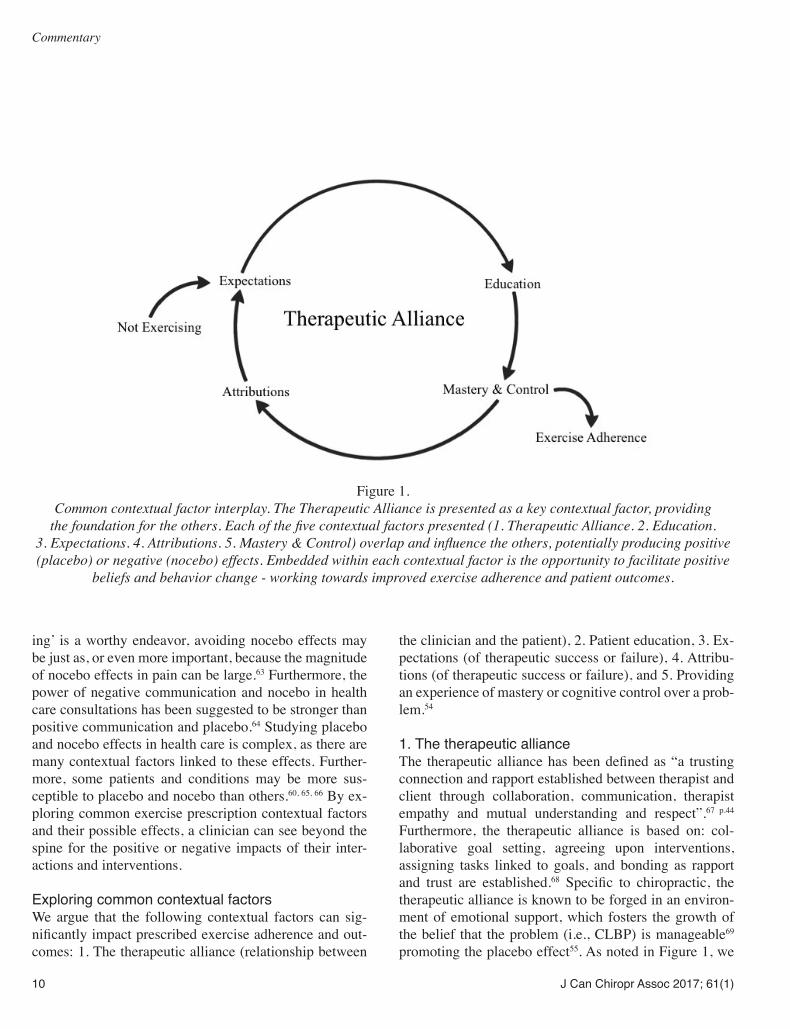
1. The therapeutic allianceThe therapeutic alliance has been defined as “a trusting connection and rapport established between therapist and client through collaboration, communication, therapist empathy and mutual understanding and respect”.67 p.44 Furthermore, the therapeutic alliance is based on: col-laborative goal setting, agreeing upon interventions, assigning tasks linked to goals, and bonding as rapport and trust are established.68 Specific to chiropractic, the therapeutic alliance is known to be forged in an environ-ment of emotional support, which fosters the growth of the belief that the problem (i.e., CLBP) is manageable69 promoting the placebo effect55. As noted in Figure 1, we
Figure 1.
Common contextual factor interplay. The Therapeutic Alliance is presented as a key contextual factor, providing the foundation for the others. Each of the five contextual factors presented (1. Therapeutic Alliance. 2. Education.
3. Expectations. 4. Attributions. 5. Mastery & Control) overlap and influence the others, potentially producing positive (placebo) or negative (nocebo) effects. Embedded within each contextual factor is the opportunity to facilitate positive
beliefs and behavior change - working towards improved exercise adherence and patient outcomes.
J Can Chiropr Assoc 2017; 61(1) 11
P Stilwell, K Harman
are suggesting that the therapeutic alliance provides the central foundation for patients to receive the benefits from other contextual factors and their placebo effects, further improving health-related outcomes and exercise adherence. Research is starting to support this as positive effects on pain and disability have been found when the therapeutic alliance is enhanced during the delivery of LBP interventions.70-73 In contrast, the therapeutic alliance can be weakened by clinician behaviors such as discred-iting and blaming a patient, or being non-supportive.64 We hypothesize that this may stimulate potent nocebo effects, and have a negative impact on self-efficacy, exercise ad-herence, and patient outcomes.
2. EducationNot all education and exercise prescription are equal, as during their delivery there is potential for both placebo and nocebo effects. The following quotations from a qualita-tive study by Slade and colleagues74 p.563 highlight how, in the absence of an easy explanation for CLBP, clinicians may resort to blaming past practitioners and the patient:
“You’ve got to sell it, show them what they do wrong, and that’s the hardest thing” and
“It’s because you’re doing everything wrong, you’ll continue to get your back pain”.74 p.563
Yet, they deliver their own questionable and potentially fear-inducing persistent LBP explanations and exercise advice:
“I generally talk about instability... you’ve got instability at this level and your movement pattern aggravates and it’s because you’re moving through one area too much” and “We see bad movement patterns... it’s all about correcting movement pat-terns”.74 p.563
This type of blaming may not only cause patient confu-sion, the language delivered to certain patients may elicit nocebo effects and reinforce the fear that they are failing to get better because they are incompetent and that they have something seriously wrong with their back. Instead, explaining persistent pain using modern neuroscience ap-proaches may not only open the door to exercise prescrip-tion, it may also improve exercise adherence and patient
outcomes.75 Pain neuroscience education resources such as Explain Pain76 or Therapeutic Neuroscience Educa-tion77 are becoming popular as they can help demystify and unravel the complex and sometimes unpredictable nature of CLBP. These resources provide illustrations and explanations about the neurophysiology of pain and can be used to help patients change their understanding and beliefs about the pain that they are experiencing.78 In essence, education and learning about pain can reduce uncertainty and perceived threat, which can reduce pain.76 We believe that patients with CLBP can then begin to view their backs as sensitized rather than fragile and prone to injury or damage. While research examining pain science education is relatively new, evidence is rapidly building that supports its use with patients experiencing CLBP.79-84
3. Expectations of therapeutic success or failureThere is a large body of literature that demonstrates the strong positive relationship between beliefs and outcomes [for review see Maddux]85 – that is, if a patient expects they will have a positive result from a treatment, there is a strong likelihood that they will experience a positive re-sult from that treatment86-89. There is also evidence that ex-pectations can be modified to produce better intervention outcomes through placebo effects.71,90,91 But we must also consider potential nocebo effects on patients. Individuals in stressful positions are vulnerable to nocebo effects60 and living with CLBP is distressing, accompanied by a sense of loss, lowered self-worth, and fear of the future92. Un-fortunately, as previously discussed, clinicians may create or facilitate negative expectations through poor communi-cation or inappropriate language, which can then lead to poorer patient outcomes.59 It is also possible that messages from the media, family, and friends could facilitate nega-tive expectations about the back and exercise, impacting exercise adherence and health-related outcomes. When clinicians explain pain and the purpose of an exercise, “...it may be healthier to err on the side of optimism...”.60 p.610 This is especially true with the non-specific LBP popula-tion where there is no significant underlying pathology, yet patient fear-avoidance beliefs can be high – already nega-tively affecting outcomes.93 Once again, we argue that evi-dence-based pain neuroscience education should be used to promote positive expectations while avoiding nocebo effects (e.g., pain does not equal damage, the back is inher-ently strong, and the spine/nervous system is adaptable).

12 J Can Chiropr Assoc 2017; 61(1)
Commentary
4. Attributions of therapeutic success or failureAttributions are an individual’s explanation or under-standing of why things have occurred the way they did – it is a way of making sense of past experiences.54 At-tributions help us to create a useful understanding of the world, as far as we can predict or control events.94 When an intervention is judged as a success or failure in the past, it shapes expectations of success/failure for similar interventions in the future.85 Another important impact of attribution is on the strength or stability of treatment out-comes. If a patient believes that their improvement was due to what a clinician did, then any beneficial effect is significantly shorter than if a patient believes that they improved because of their own actions.54 Furthermore, people act on their beliefs,95 if a patient attributes their back pain to the fact that their spine is unstable or weak, and they are educated on how they are failing to do an ex-ercise properly, it should not be a surprise that they would expect to get worse if they engaged in exercise or load their spine. This type of unintended nocebo effect creat-ed by clinicians is clearly demonstrated in the following quote from a study by Darlow and colleagues:47 p.532
“Basically all I’ve kind of been told to do by physios is to work on my core...I’ve been tested by various different physios, and Pilates, and I’m ap-parently ridiculously weak .... I had an abortion because I didn’t think I could have a baby. I didn’t think I could handle it...carrying it, and having ex-tra weight on my stomach.”47 p.532 (Bolding added for emphasis).
This last quote may be an extreme example of how edu-cation can shape attributions and expectations, and how these beliefs can shape behaviors. Still, as highlighted above, the increased use of individualized approaches that facilitate positive beliefs about the back and empow-er patients with CLBP is clearly needed.
5. Mastery or cognitive control over a problemMastery is defined as “control over those circumstances that importantly bear on the life of the individual”.96 p.164 In the context of musculoskeletal rehabilitation, both cognitive and physical control is needed to achieve mastery - which often requires deliberate practice with performance feedback.97-100 Emerging neuroscience re-
search suggests that positive neuroplastic changes appear to be enhanced by slowly increasing the complexity of motor skill tasks, promoting cognitive effort and learn-ing.101 This process is thought of as ‘working through’ the new behavior while paying attention to thoughts and responses to the movement.54 With practice, patients can learn and believe that they are capable of consistently overcoming their challenging movement tasks, which can increase their self-efficacy and result in mastery.95 These ideas are supported by findings in a recent synthe-sis of systematic reviews that identified self-efficacy as one of the most consistent predictors of exercise partici-pation.102 Furthermore, a reciprocal relationship between improved exercise adherence and self-efficacy has been demonstrated. Simply put: participation in exercise tends to increase exercise self-efficacy, which in turn reinfor-ces exercise behavior and continued exercise participa-tion.103 Positive beliefs are a key feature in self-efficacy and mastery, but they can also modulate the placebo ef-fect.104 This suggests that if an exercise is expected by a patient to reduce pain and improve function, the pa-tient is not only more likely to do it, they are also more likely to derive benefit from it. In contrast, the potential for nocebo effects through conditioning and expectation should also be considered. If a patient repeatedly fails when attempting their meaningful movement task(s) and the clinician provides poor education and negative com-ments, such as telling them how they move wrong, their spine is unstable, or how a passive ‘fix’ is the key to suc-cess – the end result can be something like learned help-lessness.105 This occurs when a patient feels that they do not have control over their situation and their pain, and that they only make things worse when they try to help themselves, so they give up. We believe that once the patients’ self-identified move-ment goals are achieved, they should be encouraged to en-gage in regular exercise that they expect will help and that they personally enjoy. Here, patient preferences should be key considerations when prescribing exercise. When a pa-tient can select the exercise they enjoy and/or expect will help, the beneficial effects of the exercise may not only be potentiated through expectations/placebo effects104, but also through improved practice/adherence, leading to improved self-efficacy and mastery106. Research supports this idea, as it has been found that incorporating patient preference and tailoring treatment programs to patients
J Can Chiropr Assoc 2017; 61(1) 13
P Stilwell, K Harman
is associated with improved self-management adherence and health-related outcomes.107,108
The complex positive feedback loop in Figure 1 can now be better appreciated; a strong therapeutic alliance with effective education can promote placebo effects, while avoiding nocebo effects. We argue that positive changes in attribution and expectations can then result in exercise engagement, which can feed forward into in-creased exercise self-efficacy and mastery.
ConclusionEvidence keeps building about the multi-system bene-fits of exercise109; this includes therapeutic exercise for CLBP. As suggested throughout this commentary, a focus on gross biological changes alone (muscle strength, en-durance etc.) has limited value. Instead, more research is needed to examine the interplay between biological, psychological, and social factors - as this may have novel exercise prescription implications for patients with CLBP. This commentary provided an overview of some of the contextual factors that have biopsychosocial implications. It was described how these contextual factors can facili-tate placebo or nocebo effects, impacting patients’ behav-iors and outcomes. The therapeutic alliance was presented as an important foundation, impacting patient education, expectations and attributions of therapeutic success or failure, and the patient’s sense of mastery or control. Cur-rent evidence suggests that a strong therapeutic alliance, pain neuroscience education, and incorporating the func-tional needs and preferences of the patient can positively impact patients’ beliefs and behaviors. Overall, this com-mentary suggests that to improve exercise adherence and health-related outcomes in the CLBP population, the con-text in which exercise is delivered and the meaning pa-tients embody need to be carefully considered and shaped by clinicians. More research is needed to further define and measure the active components within the common contextual factors presented in this commentary, as well as others factors shaping patients’ exercise beliefs and be-haviors.
AcknowledgmentsWe would like to thank and acknowledge Dr. Warren Hef-ford, Selena Glover, and Marsha MacRae for providing feedback on an early draft of this commentary.
References1. Global Burden of Disease Study 2013 Collaborators.
Global, regional, and national incidence, prevalence, and years lived with disability for 301 acute and chronic diseases and injuries in 188 countries, 1990-2013: a systematic analysis for the Global Burden of Disease Study 2013. Lancet. 2015; 386(9995): 743-800.
2. Kongsted A, Kent P, Hestbaek L, et al. Patients with low back pain had distinct clinical course patterns that were typically neither complete recovery nor constant pain. A latent class analysis of longitudinal data. Spine J. 2015; 15(5): 885-894.
3. Pengel LH, Herbert RD, Maher CG, et al. Acute low back pain: systematic review of its prognosis. BMJ. 2003; 327(7410): 323.
4. Hayden JA, Dunn KM, Van der windt DA, et al. What is the prognosis of back pain? Best Pract Res Clin Rheumatol. 2010; 24(2): 167-179.
5. Deyo RA, Mirza SK, Turner JA, et al. Overtreating chronic back pain: time to back off? J Am Board Fam Med. 2009; 22(1): 62-68.
6. Chou R, Huffman LH. Nonpharmacologic therapies for acute and chronic low back pain: a review of the evidence for an American Pain Society/American College of Physicians clinical practice guideline. Ann Intern Med. 2007; 147(7): 492-504.
7. Steffens D, Maher CG, Pereira LS, et al. Prevention of low back pain: a systematic review and meta-analysis. JAMA Intern Med. 2016; 176(2): 1-10.
8. van Middelkoop M, Rubinstein SM, Verhagen AP, et al. Exercise therapy for chronic nonspecific low-back pain. Best Pract Res Clin Rheumatol. 2010; 24(2): 193-204.
9. Keller A, Hayden J, Bombardier C, et al. Effect sizes of non-surgical treatments of non-specific low-back pain. Eur Spine J. 2007; 16(11): 1776-1788.
10. Beinart NA, Goodchild CE, Weinman JA, et al. Individual and intervention-related factors associated with adherence to home exercise in chronic low back pain: a systematic review. Spine J. 2013; 13(12): 1940-1950.
11. Cecchi F, Pasquini G, Paperini A, et al. Predictors of response to exercise therapy for chronic low back pain: result of a prospective study with one year follow-up. Eur J Phys Rehabil Med. 2014; 50(2): 143-151.
12. Slade SC, Patel S, Underwood M, et al. What are patient beliefs and perceptions about exercise for nonspecific chronic low back pain? A systematic review of qualitative studies. Clin J Pain. 2014; 30(11): 995-1005.
13. Steiger F, Wirth B, de Bruin ED, et al. Is a positive clinical outcome after exercise therapy for chronic non-specific low back pain contingent upon a corresponding improvement in the targeted aspect(s) of performance? A systematic review. Eur Spine J. 2012; 21(4): 575-598.
14. Wong AY, Parent EC, Funabashi M, et al. Do changes

14 J Can Chiropr Assoc 2017; 61(1)
Commentary
in transversus abdominis and lumbar multifidus during conservative treatment explain changes in clinical outcomes related to nonspecific low back pain? A systematic review. J Pain. 2014; 15(4): 377.e1-e35.
15. Aleksiev AR. Ten-year follow-up of strengthening versus flexibility exercises with or without abdominal bracing in recurrent low back pain. Spine. 2014; 39(24): 1495-1497.
16. Macedo LG, Maher CG, Latimer J, et al. Motor control exercise for persistent, nonspecific low back pain: a systematic review. Phys Ther. 2009; 89(1): 9-25.
17. Macedo LG, Smeets RJ, Maher CG, et al. Graded activity and graded exposure for persistent nonspecific low back pain: a systematic review. Phys Ther. 2010; 90(6): 860-879.
18. OʼKeeffe M, Nolan D, OʼSullivan P, et al. Re: Aleksiev AR. Ten-year follow-up of strengthening versus flexibility exercises with or without abdominal bracing in recurrent low back pain. Spine. 2014; 39(24): E1495-1497.
19. Saner J, Kool J, Sieben JM, et al. A tailored exercise program versus general exercise for a subgroup of patients with low back pain and movement control impairment: A randomised controlled trial with one-year follow-up. Man Ther. 2015; 20(5): 672-679.
20. Shnayderman I, Katz-leurer M. An aerobic walking programme versus muscle strengthening programme for chronic low back pain: a randomized controlled trial. Clin Rehabil. 2013; 27(3): 207-214.
21. Saragiotto BT, Maher CG, Yamato TP, et al. Motor control exercise for chronic non-specific low-back pain. Cochrane Database Syst Rev. 2016; 1: CD012004.
22. Smith BE, Littlewood C, May S. An update of stabilisation exercises for low back pain: a systematic review with meta-analysis. BMC Musculoskelet Disord. 2014; 15: 416.
23. van der Giessen RN, Speksnijder CM, Helders PJ. The effectiveness of graded activity in patients with non-specific low-back pain: a systematic review. Disabil Rehabil. 2012; 34(13): 1070-1076.
24. Wang XQ, Zheng JJ, Yu ZW, et al. A meta-analysis of core stability exercise versus general exercise for chronic low back pain. PLoS ONE. 2012; 7(12): e52082.
25. Yamato TP, Maher CG, Saragiotto BT, et al. Pilates for low back pain. Cochrane Database Syst Rev. 2015; 2(7): CD010265.
26. O’Sullivan K, Dankaerts W, O’Sullivan L, et al. Cognitive functional therapy for disabling nonspecific chronic low back pain: multiple case-cohort study. Phys Ther. 2015; 95(11): 1478-1488.
27. World Health Organization. International Classification of Functioning, Disability and Health (ICF). 2001; Geneva.
28. Melzack R, Katz J. Pain. Wiley Interdiscip Rev Cogn Sci. 2013; 4(1): 1-15.
29. Cholewicki J, Panjabi MM, Khachatryan A. Stabilizing function of trunk flexor-extensor muscles around a neutral spine posture. Spine. 1997; 22(19): 2207-2212.
30. Lederman E. The myth of core stability. J Bodyw Mov Ther. 2010; 14(1): 84-98.
31. Ghamkhar L, Kahlaee AH. Trunk muscles activation pattern during walking in subjects with and without chronic low back pain: a systematic review. PMR. 2015; 7(5): 519-526.
32. Moreside JM, Quirk DA, Hubley-Kozey CL. Temporal patterns of the trunk muscles remain altered in a low back-injured population despite subjective reports of recovery. Arch Phys Med Rehabil. 2014; 95(4): 686-698.
33. Hodges P, van den Hoorn W, Dawson A, et al. Changes in the mechanical properties of the trunk in low back pain may be associated with recurrence. J Biomech. 2009; 42(1): 61-66.
34. Karayannis NV, Smeets RJ, van den Hoorn W, et al. Fear of movement is related to trunk stiffness in low back pain. PLoS ONE. 2013; 8(6): e67779.
35. Massé-Alarie H, Beaulieu LD, Preuss R, et al. Influence of chronic low back pain and fear of movement on the activation of the transversely oriented abdominal muscles during forward bending. J Electromyogr Kinesiol. 2016; 27: 87-94.
36. Pakzad M, Fung J, Preuss R. Pain catastrophizing and trunk muscle activation during walking in patients with chronic low back pain. Gait Posture. 2016; 49: 73-77.
37. Butler HL, Hubley-Kozey CL, Kozey JW. Changes in electromyographic activity of trunk muscles within the sub-acute phase for individuals deemed recovered from a low back injury. J Electromyogr Kinesiol. 2013; 23(2): 369-377.
38. Geisser ME, Haig AJ, Wallbom AS, et al. Pain-related fear, lumbar flexion, and dynamic EMG among persons with chronic musculoskeletal low back pain. Clin J Pain. 2004; 20(2): 61-69.
39. Marras WS, Ferguson SA, Burr D, et al. Spine loading in patients with low back pain during asymmetric lifting exertions. Spine J. 2004; 4(1): 64-75.
40. Wong AY, Parent EC, Prasad N, et al. Does experimental low back pain change posteroanterior lumbar spinal stiffness and trunk muscle activity? A randomized crossover study. Clin Biomech (Bristol, Avon). 2016; 34: 45-52.
41. Hodges PW, Smeets RJ. Interaction between pain, movement, and physical activity: short-term benefits, long-term consequences, and targets for treatment. Clin J Pain. 2015; 31(2): 97-107.
42. Allison GT, Morris SL. Transversus abdominis and core stability: has the pendulum swung? Br J Sports Med. 2008; 42(11): 930-931.
43. Gubler D, Mannion AF, Schenk P, et al. Ultrasound tissue Doppler imaging reveals no delay in abdominal muscle
J Can Chiropr Assoc 2017; 61(1) 15
P Stilwell, K Harman
feed-forward activity during rapid arm movements in patients with chronic low back pain. Spine. 2010; 35(16): 1506-1513.
44. Laird RA, Kent P, Keating JL. Modifying patterns of movement in people with low back pain -does it help? A systematic review. BMC Musculoskelet Disord. 2012; 13: 169.
45. Mehta R, Cannella M, Henry SM, et al. Trunk postural muscle timing is not compromised in low back pain patients clinically diagnosed with movement coordination impairments. Motor Control. 2015. In Press.
46. George SZ, Childs JD, Teyhen DS, et al. Brief psychosocial education, not core stabilization, reduced incidence of low back pain: results from the Prevention of Low Back Pain in the Military (POLM) cluster randomized trial. BMC Med. 2011; 9: 128.
47. Darlow B, Dowell A, Baxter GD, et al. The enduring impact of what clinicians say to people with low back pain. Ann Fam Med. 2013; 11: 527-534.
48. Darlow B, Dean S, Perry M, et al. Easy to harm, hard to heal. Spine, 2015; 40(11): 842-850.
49. Domenech J, Sánchez-Zuriaga D, Segura-Ortí E, et al. Impact of biomedical and biopsychosocial training sessions on the attitudes, beliefs, and recommendations of health care providers about low back pain: a randomised clinical trial. Pain. 2011; 152(11): 2557-2563.
50. Nijs J, Roussel N, Wilgen C, et al. Thinking beyond muscles and joints: Therapists’ and patients’ attitudes and beliefs regarding chronic musculoskeletal pain are key to applying effective treatment. Man Ther. 2013; 18: 96-102.
51. Moseley L. Unraveling the barriers to reconceptualization of the problem in chronic pain: the actual and perceived ability of patients and health professionals to understand the neurophysiology. J Pain. 2003; 4(4): 184-189.
52. Barker KL, Reid M, Minns Lowe CJ. Divided by a lack of common language? A qualitative study exploring the use of language by health professionals treating back pain. BMC Musculoskelet Disord. 2009; 10(123): 1-10.
53. Rosenzweig, S. Some implicit common factors in diverse methods of psychotherapy. Am J Orthopsych. 1936: 6: 412–415.
54. Weinberger J. Common factors aren’t so common: the common factors dilemma. Clin Psych Sci Pract. 1995; 2(1): 45-69.
55. Jamison JR. Nonspecific intervention in chiropractic care. J Manipulative Physiol Ther. 1998; 21(6): 423-425.
56. Miciak M, Gross DP, Joyce A. A review of the psychotherapeutic ‘common factors’ model and its application in physical therapy: the need to consider general effects in physical therapy practice. Scand J Caring Sci. 2012; 26(2): 394-403.
57. Mason, P, Butler, C. Health Behavior Change: A Guide
for Practitioners. Second edition. Churchill Livingston, Edinburgh, 2010.
58. Miller F, Kaptchuk T. The power of context: reconceptualizing the placebo effect. JRSM. 2008; 101(5): 222-225.
59. Bingel U. Avoiding nocebo effects to optimize treatment outcome. JAMA. 2014; 312(7): 693-694.
60. Hahn R. The nocebo phenomenon: concept, evidence and implications for public health. Prevent Med. 1997; 26: 607-611.
61. Howick J, Friedemann C, Tsakok M, et al. Are treatments more effective than placebos? A systematic review and meta-analysis. PLoS ONE. 2013; 8(5): e62599.
62. Häuser W, Hansen E, Enck P. Nocebo phenomena in medicine: their relevance in everyday clinical practice. Dtsch Arztebl Int. 2012; 109(26): 459-465.
63. Petersen GL, Finnerup NB, Colloca L, et al. The magnitude of nocebo effects in pain: a meta-analysis. Pain. 2014; 155(8): 1426-1434.
64. Greville-Harris M, Dieppe P. Bad is more powerful than good: the nocebo response in medical consultations. Am J Med. 2015; 128(2): 126-129.
65. Hashmi JA, Kong J, Spaeth R, et al. Functional network architecture predicts psychologically mediated analgesia related to treatment in chronic knee pain patients. J Neurosci. 2014; 34(11): 3924-3936.
66. Hashmi JA, Baria AT, Baliki MN, et al. Brain networks predicting placebo analgesia in a clinical trial for chronic back pain. Pain. 2012; 153(12): 2393-2402.
67. Cole MB, McLean V. Therapeutic relationships re-defined. Occ Ther Mental Health. 2003; 19(2): 33-56.
68. Bordin E. The generalizability of the psychoanalytic concept of the working alliance. Psychother. 1979; 16(3): 252–260.
69. Jamison JR. Reflections on chiropractic’s patient-centered care. J Manipulative Physiol Ther. 2001; 24(7): 483-486.
70. Ferreira PH, Ferreira ML, Maher CG, et al. The therapeutic alliance between clinicians and patients predicts outcome in chronic low back pain. Phys Ther. 2013; 93: 470–478.
71. Fuentes J, Armijo-Olivo S, Funabashi M, et al. Enhanced therapeutic alliance modulates pain intensity and muscle pain sensitivity in patients with chronic low back pain: an experimental controlled study. Phys Ther. 2014; 94(4): 477-489.
72. Hall AM, Ferreira PH, Maher CG, et al. The influence of the therapist-patient relationship on treatment outcome in physical rehabilitation: a systematic review. Phys Ther. 2010; 90: 1099–1110.
73. Lewis M, Morley S, van der Windt DA, et al. Measuring practitioner/therapist effects in randomised trials of low back pain and neck pain interventions in primary care settings. Eur J Pain. 2010; 14: 1033–1039.

16 J Can Chiropr Assoc 2017; 61(1)
Commentary
74. Slade SC, Molloy E, Keating JL. The dilemma of diagnostic uncertainty when treating people with chronic low back pain: a qualitative study. Clin rehab. 2012; 26(6): 558-569.
75. Nijs J, Meeus M, Cagnie B, et al. A modern neuroscience approach to chronic spinal pain: combining pain neuroscience education with cognition-targeted motor control training. Phys Ther. 2014; 94(5): 730-738.
76. Butler DS, Moseley G. Explain Pain. Adelaine City West: Noigroup Publications, 2003.
77. Louw A, Puentedura E. Therapeutic neuroscience education: teaching patients about pain: a guide for clinicians. International Spine and Pain Institute, 2013.
78. Moseley GL, Butler DS. Fifteen years of explaining pain: the past, present, and future. J Pain. 2015; 16(9): 807-813.
79. Moseley GL. Combined physiotherapy and education is effective for chronic low back pain. A randomised controlled trial. Aust J Physioth. 2002; 48: 297-302.
80. Moseley GL. Joining forces - combining cognition-targeted motor control training with group or individual pain physiology education: a successful treatment for chronic low back pain. J Man Manip Therap. 2003; 11: 88-94.
81. Moseley GL. Evidence for a direct relationship between cognitive and physical change during an education intervention in people with chronic low back pain. Eur J Pain. 2004; 8: 39-45.
82. Moseley GL, Nicholas MK, Hodges PW. A randomized controlled trial of intensive neurophysiology education in chronic low back pain. Clin J Pain. 2004; 20: 324-330.
83. Pires D, Cruz EB, Caeiro C. Aquatic exercise and pain neurophysiology education versus aquatic exercise alone for patients with chronic low back pain: a randomized controlled trial. Clin Rehabil. 2015; 29(6): 538-547.
84. Ryan CG, Gray HG, Newton M, et al. Pain biology education and exercise classes compared to pain biology education alone for individuals with chronic low back pain: a pilot randomized controlled trial. Man Ther. 2010; 15: 382-387.
85. Maddux J. Expectancies and the social-cognitive perspective: basic principles, processes and variables. In: Kirsch I, editor. How Expectancies Shape Experience.Washington, DC: American Psychological Association, 1999.
86. Iles R, Davidson M, Taylor N, et al. Systematic review of the ability of recovery expectations to predict outcomes in non-chronic non-specific low back pain. J Occup Rehabil. 2009; 19(1): 25-40.
87. Mondloch M, Cole D, Frank J. Does how you do depend on how you think you’ll do? A systematic review of the evidence for a relation between patients’ recovery expectations and health outcomes. CMAJ. 2001; 165(2): 174-179.
88. George S, Robinson M. Preference, expectation, and satisfaction in a clinical trial of behavioral interventions for acute and sub-acute low back pain. J Pain. 2010; 11(11): 1074-1082.
89. Smeets R, Beelen S, Goossens M, et al. Treatment expectancy and credibility are associated with the outcome of both physical and congitive-behavioural treatment in chronic low back pain. Clin J Pain. 2008; 24(4): 305-315.
90. Crum AJ, Langer EJ. Mind-set matters: exercise and the placebo effect. Psychol Sci. 2007; 18(2): 165-171.
91. Wulf G, Chiviacowsky S, Lewthwaite R. Altering mindset can enhance motor learning in older adults. Psychol Aging. 2012; 27(1): 14-21.
92. Snelgrove S, Liossi C. Living with chronic low back pain: a metasynthesis of qualitative research. Chronic Illn. 2013; 9(4): 283-301.
93. Wertli MM, Rasmussen-Barr E, Weiser S, et al. The role of fear avoidance beliefs as a prognostic factor for outcome in patients with nonspecific low back pain: a systematic review. Spine J. 2014; 14(5): 816-836.e4.
94. Anderson C, Krull D, Weiner B. Explanations: Proceeses and Consequences. In: Higgins E, Kruglanski A, editors. Social Psychology: Handbook of Basic Principles. New York: Guilford, 1996.
95. Bandura A. Self-Efficacy: The Exercise of Control. New York: W.H. Freeman, 1997.
96. Pearlin, LI, Nguyen, KB, Schieman, S, et al. The life-course origins of mastery among older people. J Health Soc Behav. 2007; 48, 164–179.
97. Ericsson KA, Krampe RT, Tesch-Römer C. The role of deliberate practice in the acquisition of expert performance. Psych Rev. 1993; 100(3): 363-406.
98. Ericsson KA. Deliberate practice and acquisition of expert performance: a general overview. Acad Emerg Med. 2008; 15(11): 988-994.
99. Kitago T, Krikauer J. Motor learning principles for neurorehabilitation. In: Barnes M, Good D, editors. Neurological Rehabilitation. Elsevier, 2013.
100. Winstein C, Lewthwaite R, Blanton S, et al. Infusing motor learning research into neurorehabilitation practice: a historical perspective with case exemplar from the accelerated skill acquisition program. J Neurol Phys Ther. 2014; 38: 190-200.
101. Boudreau SA, Farina D, Falla D. The role of motor learning and neuroplasticity in designing rehabilitation approaches for musculoskeletal pain disorders. Man Ther. 2010; 15(5): 410-414.
102. Bauman AE, Reis RS, Sallis JF, et al. Correlates of physical activity: Why are some people physically active and others not? Lancet. 2012; 380(9838): 258-271.
103. Weinberg RS, Gould D. Foundations of sport and exercise psychology. 3rd ed. Champaign, IL: Human Kinetics, 2003.

J Can Chiropr Assoc 2017; 61(1) 17
P Stilwell, K Harman
104. Stewart-Williams S, Podd J. The placebo effect: dissolving the expectancy versus conditioning debate. Psychol Bull. 2004; 130(2), 324–340.
105. Roland M, Jenner J. Back Pain: New Approaches to Rehabilitation and Education. Manchester Univ Pr., 1989.
106. Harman K, MacRae M, Vallis M, et al. Working with people to make changes: a behavioural change approach used in chronic low back pain rehabilitation. Physiother Can. 2014; 66(1): 82-90.
107. Lorig K, Holman HR. Self-management education:
History, definition, outcomes, and mechanisms. Ann Behav Med. 2003; 26(1): 1-7.
108. Peek K, Sanson-Fisher R, Carey M, et al. Interventions to aid patient adherence to physiotherapist prescribed self-management strategies: a systematic review. Physiother. 2015; In Press.
109. Pedersen BK, Saltin B. Exercise as medicine - evidence for prescribing exercise as therapy in 26 different chronic diseases. Scand J Med Sci Sports. 2015; 25 Suppl 3: 1-72.
18 J Can Chiropr Assoc 2017; 61(1)
ISSN 0008-3194 (p)/ISSN 1715-6181 (e)/2017/18–31/$2.00/©JCCA 2017
The physical and psychological impact of neurogenic claudication: the patients’ perspectivesCarlo Ammendolia, DC, PhD1 Michael Schneider, DC, PhD2 Kelly Williams, MPH3 Susan Zickmund, PhD4 Megan Hamm, PhD5 Kent Stuber, DC, MSc6 Christy Tomkins-Lane, PhD7 Y Raja Rampersaud, MD8
1 University of Toronto, Institute for Health Policy, Management and Evaluation, Faculty of Medicine2 University of Pittsburgh, Department of Physical Therapy and Clinical and Translational Science Institute3 University of Pittsburgh, Department of Behavioral and Community Health Sciences, Graduate School of Public Health4 Informatics, Decision-Enhancement and Analytic Sciences Center (IDEAS 2.0), Health Services Research and Development Center of
Innovation, 2C21 Building 2, Salt Lake City VA, Salt Lake City, UT5 University of Pittsburgh, Department of Medicine, Center for Research on Health Care6 Canadian Memorial Chiropractic College, Division of Graduate Education & Research,7 Mount Royal University, Department of Health and Physical Education8 University of Toronto, Division of Orthopaedic Surgery, University Health Network-Arthritis Program
Corresponding author:Carlo AmmendoliaMount Sinai Hospital, 60 Murray Street Suite L2-225 Box 7, Toronto Ontario, CanadaPhone: 416 586-4800 ext. 6759E-mail: [email protected]
© JCCA 2017
The authors’ report no related conflict of interest. The following organizations have provided funding for this research: The Canadian Chiropractic Research Foundation (Ammendolia), NCMIC Foundation (Ammendolia and Schneider) and the Veterans Administration Health Services Research and Development Program, award number 5150HX001240 (Zickmund) and The Toronto General and Western Hospital Foundation (Rampersaud).
Background: The patient perspective regarding the impact of neurogenic claudication (NC) has not been well studied. The objectives of this study were to determine what is most bothersome among patients with NC and how it impacts their lives and expectations with surgical and non-surgical treatment. Methods: Semi-structured telephone interviews were
Contexte : Le point de vue du patient concernant l’effet de la claudication neurogène (CN) n’a pas fait l’objet d’études poussées. Les objectifs de cette étude étaient de déterminer ce qui gêne le plus les patients atteints de CN, ainsi que les répercussions sur leur vie et leurs attentes vis-à-vis des traitements chirurgicaux et non chirurgicaux. Méthodologie : Entrevues téléphoniques semi-

J Can Chiropr Assoc 2017; 61(1) 19
C Ammendolia, M Schneider, K Williams, S Zickmund, M Hamm, K Stuber, C Tomkins-Lane, YR Rampersaud
IntroductionNeurogenic claudication (NC) is the clinical syndrome as-sociated with symptomatic lumbar spinal stenosis (LSS). It is characterized by bilateral or unilateral buttock, thigh or calf discomfort, pain, numbness or weakness precipi-tated by walking or prolonged standing and relieved by sitting and lumbar flexion.1,2 Low back pain may or may not be present in individuals with NC. The pathophysiol-ogy is thought to be compression and/or ischemia of the lumbosacral nerve roots due to narrowing of the lateral and central vertebral canals, usually as a consequence of degenerative osteoarthritic changes in the lumbar spine.1,3 Neurogenic claudication due to LSS is one of the most common causes of disability and loss of independence in older adults4 and the most common reason for spine sur-gery in this population5. New cases of NC due to LSS are expected to rise
dramatically over the next 20 years when an estimated 25% of the population in both the U.S. and Canada will be over the age of 65.6 Studies evaluating the effective-ness of both operative and non-operative treatments for NC have used a wide variety of primary and secondary outcome measures.7-10 These outcome measures assess various constructs including bodily pain, bodily func-tion, low back pain disability, back and leg pain, other leg symptoms, walking capacity (distance and time), walking performance, global improvement, quality of life, ranges of motion, treatment satisfaction and medication use. In most studies the outcome measures used are reflect-ive of the bias of the investigator(s) and is often inferred as the desired outcome of the patient. However, rarely has the perspective of the patient regarding the most import-ant outcome been considered. For example, limitation in walking is felt to be the hallmark of NC and is used as a
conducted, audio recorded and transcribed verbatim. A thematic analysis categorized key findings based on relative importance and impact on participants. Results: Twenty-eight individuals participated in this study. Participants were most bothered by the pain of NC, which dramatically impacted their lives. Inability to walk was the dominant functional limitation and this impacted the ability to engage in recreational and social activities. The most surprising finding was how frequently participants reported significant emotional effects of NC. Conclusions: From a patients’ perspective NC has a significant multidimensional effects with pain, limited walking ability and emotional effects being most impactful to their lives. (JCCA. 2017;61(1):18-31) k e y w o r d s : chiropractic, spinal stenosis, neurogenic claudication, outcome measurement, qualitative research
structurées avec enregistrement audio et transcription textuelle. Une analyse thématique a permis de catégoriser les principales conclusions selon l’importance relative et les répercussions sur les participants. Résultats : Vingt-huit personnes ont participé à l’étude. Les participants étaient surtout gênés par la douleur de la CN, qui a d’énormes répercussions sur leur vie. L’incapacité à marcher constituait la limitation fonctionnelle dominante qui avait des conséquences sur la capacité à réaliser des activités récréatives et sociales. La conclusion la plus surprenante était la fréquence à laquelle les participants ont déclaré d’importantes séquelles émotionnelles associées à la CN. Conclusions : Du point de vue des patients, la CN présente d’importants effets multidimensionnels avec la douleur, la capacité de locomotion limitée et les séquelles émotionnelles comme répercussions les plus considérables sur la vie des patients. (JCCA. 2017;61(1):18-31) m o t s c l é s : chiropratique, sténose rachidienne, claudication neurogène, mesure des résultats, recherche qualitative
20 J Can Chiropr Assoc 2017; 61(1)
The physical and psychological impact of neurogenic claudication: the patients’ perspectives
primary outcome measure in clinical trials.7,11,12,28 How-ever, previous systematic reviews by this group7,11,12 have demonstrated that many interventions for NC did not sig-nificantly improve walking performance or capacity. De-spite this, several interventions were still associated with good patient satisfaction and/or pain relief. Given the burden of NC, a lack of understanding of what outcomes are most important to those afflicted with NC represents a significant gap in both clinical and aca-demic knowledge. Clinicians need to know what is most important to a patient in order to recommend effective intervention(s) that address the patient’s concerns. Re-searchers need to know what to measure in order to assess the most relevant patient outcome for a given interven-tion. Moreover, to make valid comparisons across studies and enable the pooling of data, a standardized set of out-come measures unique to this population and most rel-evant to patients is essential. In addition, there may be other constructs beyond those currently measured that may help to explain how this condition impacts people in different ways, and how these other factors can affect the patients experience and outcomes of NC. The objectives of this study were to determine what outcomes matter most among individuals with NC due to LSS and to assess patients’ expectations and their experi-ences with surgical and non-surgical treatment.
Methods
Participant population and settingWe recruited a purposeful sample13 of participants from two university-affiliated hospital surgical and non-sur-gical spine clinics both located in Toronto, Canada. To be eligible to participate, patients had to experience NC with axial imaging-confirmed LSS, and be able to com-municate in English. To gain maximum variation of pa-tient perspectives regarding their condition and success with treatment an attempt was made to select participants along the continuum of care. Specifically, we recruited participants scheduled for non-surgical (early, less severe symptoms) or surgical care (late, more severe symptoms), as well as those who had received surgical and non-sur-gical treatment. We included individuals of varying ages (50-90 years), gender, intensity and type of symptoms, as well as duration of symptoms (months to years). All participants provided written informed consent.
Research Ethics Board (REB) approval was received from the Mount Sinai Hospital REB Registration Number 13-0184-E and University Health Network REB Regis-tration Number 13-6914-BE, as well as the Institution-al Review Board (IRB) at the University of Pittsburgh (PRO13090531).
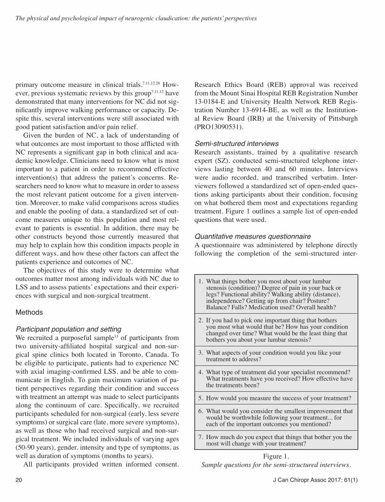
Semi-structured interviewsResearch assistants, trained by a qualitative research expert (SZ), conducted semi-structured telephone inter-views lasting between 40 and 60 minutes. Interviews were audio recorded, and transcribed verbatim. Inter-viewers followed a standardized set of open-ended ques-tions asking participants about their condition, focusing on what bothered them most and expectations regarding treatment. Figure 1 outlines a sample list of open-ended questions that were used.
Quantitative measures questionnaireA questionnaire was administered by telephone directly following the completion of the semi-structured inter-
1. What things bother you most about your lumbar stenosis (condition)? Degree of pain in your back or legs? Functional ability? Walking ability (distance), independence? Getting up from chair? Posture? Balance? Falls? Medication used? Overall health?
2. If you had to pick one important thing that bothers you most what would that be? How has your condition changed over time? What would be the least thing that bothers you about your lumbar stenosis?
3. What aspects of your condition would you like your treatment to address?
4. What type of treatment did your specialist recommend? What treatments have you received? How effective have the treatments been?
5. How would you measure the success of your treatment?
6. What would you consider the smallest improvement that would be worthwhile following your treatment... for each of the important outcomes you mentioned?
7. How much do you expect that things that bother you the most will change with your treatment?
Figure 1. Sample questions for the semi-structured interviews.

J Can Chiropr Assoc 2017; 61(1) 21
C Ammendolia, M Schneider, K Williams, S Zickmund, M Hamm, K Stuber, C Tomkins-Lane, YR Rampersaud
view. The aim of the questionnaire was to characterize the participant sample with respect to demographics, duration of symptoms, pain intensity and functional status and to compare surgical and non-surgical participants. Box 1 below lists the measures included in the questionnaire.
AnalysisDescriptive statistics were used to analyze the question-naires. We compared pain, function and symptom out-comes among and between participants recruited from surgical and non-surgical clinics. For the semi-structured interviews the frequency and types of responses were determined using the Crab-tree and Miller “editing” approach to qualitative data.18 Coding categories were developed through an open, it-erative process that involved reading the interviews with a focus that included physical and emotional effects of NC. From this process, a master code list of categories was developed. These codes were refined with inclusion and exclusion criteria, and then applied to the transcribed interviews. Two analysts [KW and MH], the qualitative expert [SZ], and the study team discussed the coding cat-egories (e.g. coping) and worked to integrate the codes into the larger analysis. Primary coding was completed on all transcripts, and secondary coding was completed on 25% of the tran-scripts. Cohen’s Kappa statistics19 were then calculated
Box 1. Quantitative questionnaire measures
Socio-demographic characteristics
Dominant pain location (back or leg)
Duration of symptoms
Numerical rating scale for back pain with and without activity14
Numerical rating scale for leg pain with and without activity14
Zurich Claudication Questionnaire15
Oswestry Disability index16
Modified Patient Centered Outcome Questionnaire17
Table 1. Characteristics of study participants
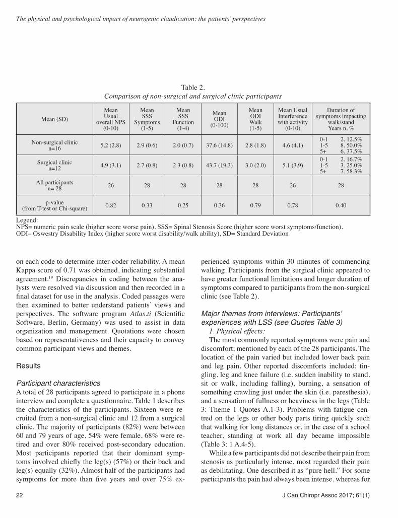
CharacteristicsSummary (N= 28) n= count (%) unless otherwise specified
Age Range (y)50-59 1 (3.6)60-69 13 (46.4)70-79 10 (35.7)80-89 3 (10.7)90-99 1 (3.6)Female 15 (53.6)Married or living with other 16 (67.9)*Education (N=27)< Grade 8 1 (3.7)> Grade 8 but did not graduate from high school 1 (3.7)High school graduate 3 (11.1)Post-Secondary school 10 (37.0)Technical graduate 1 (3.7)University graduate 11 (40.7)*EmploymentFull Time 4 (14.3)Part-time 2 (7.1)Retired 20 (67.9) Disability Leave 2 (7.1)Other 1 (3.6)Dominant PainLegs 16 (57.1)Back, 3 (10.7)Back & Legs 9 (32.1)Duration of symptoms impacting standing/walking (years)0-1 4 (14.3)1- 11 (39.3)5+ 13 (46.4)Usual Mean Numeric Pain Score (SD) (N=26) 5.1 (2. 9)Walking duration before symptoms (minutes) 0-5 6 (22.2) 5-10 6 (22.2)10-30 9 (33.4)30-60 4 (14.8)60+ 2 (7.4)Spinal Stenosis Score (symptoms) (sd) 2.8 (0.7)Spinal Stenosis Score (function) (sd) 2.1 (0.7)Oswestry Disability Index (sd) 40.2 (16.8)Oswestry Disability Index Walk Score (range 0-5) (sd) 2.9 (1.8)
Source of Participants (N=27)Non-surgical clinic: receiving treatment 5 (18.5)Non-surgical clinic: completed treatment 10 (37.0)Surgical clinic – had surgery 7 (25.9)Surgical clinic – scheduled for surgery 2 (7.4)Surgical clinic – not scheduled for surgery 3 (11)
Legend: SD= standard deviation. Variable number of responses due to missing data *characteristics with categories that are not mutually exclusive
22 J Can Chiropr Assoc 2017; 61(1)
The physical and psychological impact of neurogenic claudication: the patients’ perspectives
on each code to determine inter-coder reliability. A mean Kappa score of 0.71 was obtained, indicating substantial agreement.19 Discrepancies in coding between the ana-lysts were resolved via discussion and then recorded in a final dataset for use in the analysis. Coded passages were then examined to better understand patients’ views and perspectives. The software program Atlas.ti (Scientific Software, Berlin, Germany) was used to assist in data organization and management. Quotations were chosen based on representativeness and their capacity to convey common participant views and themes.
Results
Participant characteristicsA total of 28 participants agreed to participate in a phone interview and complete a questionnaire. Table 1 describes the characteristics of the participants. Sixteen were re-cruited from a non-surgical clinic and 12 from a surgical clinic. The majority of participants (82%) were between 60 and 79 years of age, 54% were female, 68% were re-tired and over 80% received post-secondary education. Most participants reported that their dominant symp-toms involved chiefly the leg(s) (57%) or their back and leg(s) equally (32%). Almost half of the participants had symptoms for more than five years and over 75% ex-
perienced symptoms within 30 minutes of commencing walking. Participants from the surgical clinic appeared to have greater functional limitations and longer duration of symptoms compared to participants from the non-surgical clinic (see Table 2).
Major themes from interviews: Participants’ experiences with LSS (see Quotes Table 3) 1. Physical effects: The most commonly reported symptoms were pain and discomfort; mentioned by each of the 28 participants. The location of the pain varied but included lower back pain and leg pain. Other reported discomforts included: tin-gling, leg and knee failure (i.e. sudden inability to stand, sit or walk, including falling), burning, a sensation of something crawling just under the skin (i.e. paresthesia), and a sensation of fullness or heaviness in the legs (Table 3: Theme 1 Quotes A.1-3). Problems with fatigue cen-tred on the legs or other body parts tiring quickly such that walking for long distances or, in the case of a school teacher, standing at work all day became impossible (Table 3: 1 A.4-5). While a few participants did not describe their pain from stenosis as particularly intense, most regarded their pain as debilitating. One described it as “pure hell.” For some participants the pain had always been intense, whereas for
Table 2. Comparison of non-surgical and surgical clinic participants
Mean (SD)Mean Usual
overall NPS (0-10)
Mean SSS
Symptoms (1-5)
Mean SSS
Function (1-4)
Mean ODI
(0-100)
Mean ODI Walk (1-5)
Mean Usual Interference with activity
(0-10)
Duration of symptoms impacting
walk/stand Years n, %
Non-surgical clinic n=16 5.2 (2.8) 2.9 (0.6) 2.0 (0.7) 37.6 (14.8) 2.8 (1.8) 4.6 (4.1)
0-1 2, 12.5% 1-5 8, 50.0% 5+ 6, 37.5%
Surgical clinic n=12 4.9 (3.1) 2.7 (0.8) 2.3 (0.8) 43.7 (19.3) 3.0 (2.0) 5.1 (3.9)
0-1 2, 16.7% 1-5 3, 25.0% 5+ 7, 58.3%
All participants n= 28 26 28 28 28 28 26 28
p-value (from T-test or Chi-square) 0.82 0.33 0.25 0.36 0.79 0.78 0.40
Legend: NPS= numeric pain scale (higher score worse pain), SSS= Spinal Stenosis Score (higher score worst symptoms/function), ODI– Oswestry Disability Index (higher score worst disability/walk ability), SD= Standard Deviation

J Can Chiropr Assoc 2017; 61(1) 23
C Ammendolia, M Schneider, K Williams, S Zickmund, M Hamm, K Stuber, C Tomkins-Lane, YR Rampersaud
Table 3. Example quotes from participant interviews
Theme Example Quotes from Participant Interviews1. Physical
Effects A. Symptoms of Pain, Discomfort:1. “It’s the pain in my leg and also there is times that when I have to go to the washroom; excuse me; that I
have to sit. It’s my knees; like sometimes I can hardly sit down on the toilet and sometimes it’s hard for me to get up. Let’s say, if I sit down on the chair, I’m fine, but then when I have to stand up, I cannot stand up or when I start walking, it’s hard to walk. When I feel better it’s when I’m lying down or sitting down, but then when I’m sitting down and then stand up and starting walking that’s the worse part.”
2. “A burning feeling say from the top of the leg to the knee. A feeling that there was something crawling, as if they were bugs just underneath the surface of the skin crawling up and down and you know, it was pretty disconcerting.”
3. “Like I said it started in 2010 and I had often a lot of back pain, lower back pain. It went down into my left leg, but it was also accompanied by numbness in both legs, so severe at times that I would lose the feeling in both my legs and I would fall and go down.”
4. “Not being able to walk as far as I would like to without having to stop and experience the tingling in the front of my leg and the numbness in my foot and the pain associated with it.”
5. “When I first started the clinic here, I could not walk for than a minute and a half and I had to stop. My legs would swell. My feet would hurt that the sciatic nerve in my back would almost pull me down that it would cripple me. Even to walk, I work at [Name of Location] and to walk here I got to stop 6 or 7 times and this is me, I’m use... to running that distance.”
6. “It started I suspect 5 years ago, as a minor back pain. One usually would think it is just a strain, but over the years it has gradually progressed to the point where now it is very debilitating.”
7. “Now I’ve had that for very many years, but it’s always been bearable. The stenosis has really affected my daily life and it makes me sad at times, such I have to contend with it. At the same time, I must honestly admit that I am grateful for the health I’ve had. I realized that I have been very lucky. So, I can’t have it always, but at the same time its human nature to resent it that I get such pain all the time.”
2. Activity Effects
A. Limitations on Walking/Standing/Sitting:1. So I really, I can walk may be 4 or 5 minutes. It’s really tough on my lifestyle. I used to love to walk. Me
and my wife used to walk for miles... and now I cannot walk a block.”2. “Like I say everything depends on my legs because I walk and I’m one of them simple people; I don’t do
anything. I don’t travel. I have no interest in that.”3. “Well it affected my ability to get around and walk... It affected my exercising, which is walking. I do
quite a bit of walking for exercising and I just love to walk. I get out and I walk all over the place... Now, I go around in the car... I take the dog. Instead of walking about three blocks to take my dog out to run, I take the car there to let her run.”
B. Limitations on Recreational Activities:1. “Usually each weekend in the summer I’m outside... in the canoe or with backpack you know, but now I
am sitting at home.”2. “When it first started what it did curve is my physical activities such as curling and walking long distances
and so on, and eventually it curved all activities that even stairs were very difficult to manage.”3. “...we were in a wedding and I was dancing. It was a slow dance and it seemed that my legs were giving
up on me. I was shocked and I didn’t know what was happening. I had to go and sit down.”4. “Because I wasn’t able to, say, walk any distance, so that pretty well inhibited, so you were sort of left
with sedentary hobbies like reading and crocheting that sort of thing because you’ve found that you’ve gravitated towards that than avoiding the physical.”
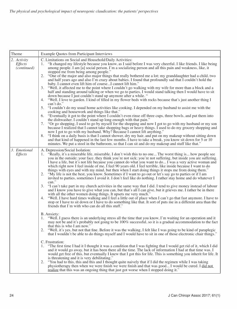
24 J Can Chiropr Assoc 2017; 61(1)
The physical and psychological impact of neurogenic claudication: the patients’ perspectives
Theme Example Quotes from Participant Interviews2. Activity
Effects (continued)
C. Limitations on Social and Household/Daily Activities:1. “It changed my lifestyle because you know, as I said before I was very cheerful. I like friends. I like being
among people. I am [a] social person. I’m a socializing person and all this pain and weakness, like, it stopped me from being among people.”
2. “One of the major and also major things that really bothered me a lot; my granddaughter had a child, two and half years ago and also I’m crazy about babies. I found that profoundly sad that I couldn’t hold the baby. I cannot even lift him of course...I cannot lift him.”
3. “Well, it affected me to the point where I couldn’t go walking with my wife for more than a block and a half and standing around talking or when we go to parties, I would stand talking then I would have to sit down because I just couldn’t stand up anymore after a while. “
4. “Well, I love to garden. I kind of filled in my flower beds with rocks because that’s just another thing I can’t do.”
5. “I couldn’t do my usual home activities like cooking. I depended on my husband to assist me with the cooking and housework and things like that.”
6. “Eventually it got to the point where I couldn’t even rinse off three cups, three bowls, and put them into the dishwasher. I couldn’t stand up long enough with that pain.”
7. “Or go shopping, I used to go by myself for the shopping and now I got to go with my husband or my son because I realized that I cannot take shopping bags or heavy things. I used to do my grocery shopping and now I got to go with my husband. Why? Because I cannot lift anything.”
8. “I think on a daily basis is that I cannot shower, dry my hair, and put on my makeup without sitting down and that kind of happened in the last few months. I have to take a break, you know sit down for 5 or 10 minutes. We put a stool in the bathroom, so that I can sit and do my makeup and stuff like that.”
3. Emotional Effects
A. Depression/Social Isolation:1. “Really, it’s a miserable life, miserable. I don’t wish this to no one... The worst thing is... how people see
you in the outside; your face, they think you’re not sick; you’re not suffering, but inside you are suffering. I have a life, but it’s not life because you cannot do what you want to do... I was a very active woman and which right now I feel inside of me, I feel 90 years old. I feel terrible, like inside because I want to do things with eyes and with my mind, but then when I start doing things it stops me from doing them.”
2. “My life is not the best, you know. Sometimes if I want to go out or let’s say go to parties or if I am invited to parties, sometimes I avoid it. I don’t feel like do nothing. I rather stay home and do whatever I can.”
3. “I can’t take part in my church activities in the same way that I did. I tend to give money instead of labour and I know you have to give what you can, but that’s all I can give, but it grieves me. I rather be in there with all the other women doing things. It upsets me very much.”
4. “Well, I have hard times walking and I feel a little out of place when I can’t go that fast anymore. I have to stop or I have to sit down or I have to do something like that. It sort of puts me in a different area than the friends that I’m with who can do all this stuff.”
B. Anxiety:1. “Well, I guess there is an underlying stress all the time that you know, I’m waiting for an operation and it
may not be and it’s probably not going to be 100% successful, so it is a gradual accommodation to the fact that this is who I am now.”
2. “Well, it’s yes, but not that fine. Before it was the walking, I felt like I was going to be kind of paraplegic that I wouldn’t be able to do things myself and I would have to sit in one of those electronic chair things.”
C. Frustration:1. “The first time I had it I thought it was a condition that I was fighting that I would get rid of it, which I did
and it would go away, but it has been there all the time. The lack of information I had at that time was, I would get free of this, but eventually I knew that I got this for life. This is something you inherit for life. It is threatening and it is very debilitating.”
2. “You had to this, this and this and I thought quite naively that if I did the regimen while I was taking physiotherapy then when we were finish we were finish and that was good... I would be cured. I did not realize that this was an ongoing thing that just got worse when I stopped doing it.”

J Can Chiropr Assoc 2017; 61(1) 25
C Ammendolia, M Schneider, K Williams, S Zickmund, M Hamm, K Stuber, C Tomkins-Lane, YR Rampersaud
Theme Example Quotes from Participant Interviews3. Emotional
Effects (continued)
D. Hurt Pride1. “It affects in so many ways, it’s the whole quality of life, the whole thing. Your wife is dependent on you,
your kids and grandkids are dependent on you for doing these things. Now all of a sudden this person who used to run with me and play with me and can’t even walk down the end of the street with me and it takes a lot of your pride, well at least me it takes a lot of my pride. Even to think of someone having to take care of me, to me it’s just unacceptable.”
2. “I walk kind of awkwardly. I cannot wear heel shoes either, but that really does not bother me that much. All my days of heel shoes are really behind me, so that would be in anyway an older woman wouldn’t be wearing fancy shoe anymore. I am the same as the other old ladies. You know it is awful giving up your autonomy and moving into a different phase. This is one of the reasons why I lie about my age all the time. People tend to put you in category of nature. If you are a certain age, you are just kind of put aside.”
4. Coping Strategies
A. Coping Mechanisms for Physical Effects:1. “I have to... generally stand a few seconds or so before my husband is ready, maybe while he is doing the
cheque... I stand right there waiting for him because if I get up there is no way I can start walking again because of the pain in my thighs. The front of my thighs is screamingly painful. I just stand for a minute or two and get my act together... I do walk strangely to begin with when I’ve been sitting down.”
2. “My condition, you know the pain in my legs increased. I get tingling in my legs, sometimes pain, but I learned not to take painkillers. I don’t take nothing. When I get like this, I just lay down, I rest for a bit and after rest I feel a little bit better.”
5. Treatment Effects
A. Partial Relief from treatments:1. “It just involves may be 2 hours a day of specific exercise and walking and if you don’t do it, you know
you can’t miss 2 days in a row because your symptoms all come back.”2. “...now having done the physiotherapy it’s been a miracle. It is just wonderful. It’s so much better. It will
never go away, but at least I can do things that can help the pain and you know alleviate the different symptoms that occur.”
3. “They helped, but it’s not like they changed my life, either.”
B. Complete Relief from Treatments:1. “I don’t have this excruciating pain. I can walk up the stairs. I can run up the stairs. I can run down the
stairs... since I had the surgery, this surgery, I have improved considerably and I am almost back to normal like a normal person.”
6. Expectations from Treatment
A. Pain Relief/Decreased Pain1. “I try to keep an open mind that the treatment will alleviate the pain. If that happens, so much the better,
but I am not counting on it to eliminate the pain. I will continue with the process and do the exercises and just hope for the best, but I haven’t set a high level of expectations that this is going to cure me.”
2. “I would like to think in doing the treatment that the pain level will be not necessarily gone, but certainly tolerable and not be something that I thought would stop me from doing what I wanted to do.”
B. Pain Elimination1. “Based on my own experience, I would expect it to eliminate the problem. It did the first time and I would
assume that it would the second time.”2. “Well, I would say significantly. It wouldn’t make much sense to have an operation if it was not going to
have much effect on the pain [in reference to surgery].”
C. Increased Physical Activity1. “That I can walk better. Walk with more distance and that I can stand on my own feet and do at least my
housework. Taking care of my family properly. Instead of being in pain, when I’m standing or walking I’m in pain, but pain goes away. To relieve me from pain and suffering.”
2. “Run around with the grandkids a little bit maybe, you know, maybe be able to do some things and not feel like I have to stop because of the pain in my leg”
3. “...consciously plan my route when I wanted to do an activity that I wouldn’t have to very, very specifically, what is it that I need to do to accomplish today and how am I going to do it and not have my back stop me from doing it”
4. “Just to be able to stand around more without the pain and be able to walk farther without stopping because of the pain in my leg.”

26 J Can Chiropr Assoc 2017; 61(1)
The physical and psychological impact of neurogenic claudication: the patients’ perspectives
others it had gradually increased to the point of causing debilitation (Table 3: 1 A.6). In addition to concerns about the severity of their pain, participants described the emo-tional impact of the wearing, grinding nature of having to endure ongoing pain, including feelings of resentment (Table 3: 1 A.7). 2. Activity effects: Participants mentioned a wide range of activities that their NC interfered with including: walking, recreation-al activities (such as sports and exercise), standing, so-cial activities, household activities, controlling comorbid health conditions, working, sleeping and lifting. Each participant mentioned that their NC interfered with their ability to walk. Interference with walking was most frequently mentioned as the “most bothersome” aspect of the condition, identified as such by 17 of the 28 participants. For some participants this was a minor concern or one that had affected them profoundly in the past but with successful treatment was no longer an issue. For many however, the walking limitations caused major disruptions in their lives, from being unable to walk or run for exercise, to being unable to do basic social and daily activities such as grocery shopping, holiday shop-ping, going to the mall with family and friends, or even visiting neighbors (Table 3: 2 A.1-3). Related to walking, many participants described being unable to participate in recreational activities. Recreational activities mentioned included walking itself or hiking, various sports (football, cricket, soccer, golf, badminton, curling, snowshoeing, cross-country skiing, squash), travel (due to the walk-ing that travel entails), dancing, bicycling, and aerobics (Table 3: 2 B. 1-3). Many participants described them-selves as active, outdoorsy people who, as a result of their NC, had become sedentary and were unable to participate in what had been previously seasonal outdoor activities (Table 3: 2 B.4). Inability to participate in recreational ac-tivities was mentioned as the “most bothersome” aspect of having LSS by 11 of the 28 participants. Interference with social activities was mentioned fair-ly often, and was linked to participants’ difficulties with walking, standing, or doing recreational activities. Many described limiting time with family and friends, difficul-ties standing while socializing or being unable to play with grandchildren in the way that they wanted (Table 3: 2 C.1-3). Often the inability to stand or pain upon stand-ing was linked with an inability to do household/daily ac-
tivities, such as cleaning or other chores, in the way that the participants were used to doing. For many there was difficulty with transitioning between positions or activ-ities, describing not being able to walk immediately after standing for example, or having extreme difficulty sit-ting and standing back up. This difficulty in transitioning made a wide range of activities difficult or impossible, from social events to using the toilet. For some participants, the amount of pain experienced while standing meant that they could not wait in lines, go shopping without physical support such as a shopping cart or another person, clean their households, cook, or even stand in the bathroom to do their hair or makeup in the morning (Table 3: 2 C.4-8). This directly impacted individuals’ sense of independence, and for some, elimin-ated activities that they had previously found enjoyable, such as cooking or gardening (Table 3: 2 C.4). 3. Emotional effects: In addition to their physical symptoms, participants re-ported an array of emotional responses to their condition. Twenty-two of the 28 participants reported emotional im-pacts that were associated with their NC. The most fre-quent of these was depressed mood, although not always specified by name. Instead they described their feelings as sadness, loss of interest in activities, or hopelessness (Table 3: 3 A.1). These participants were likely to mention deep feelings of sadness, discouragement, social isolation or loss, as their NC prevented them from doing activities that they enjoyed (i.e., walking or other outdoor activities) or from which they derived meaning (i.e., work, volunteer work, being independent, socializing, lifting and holding grandchildren) (Table 3: 3 A.2-4). Additionally, for some of these participants, the perceived incurable/untreatable nature of NC was described as “depressing” in and of it-self. Eight of the participants mentioned anxiety, express-ing deep worries that family members would have to take care of them, or that their condition would worsen signifi-cantly (Table 3: 3 B.1-2). Seven participants expressed feelings of frustration with their NC (Table 3: 3 C.1-2). For some, this was focused on the physical limitations imposed on them by NC, such as only being able to walk for short distances or the ongoing relationship between exercise and pain relief. Lastly, six of the participants described NC as having hurt their pride. Most of these participants mentioned in

J Can Chiropr Assoc 2017; 61(1) 27
C Ammendolia, M Schneider, K Williams, S Zickmund, M Hamm, K Stuber, C Tomkins-Lane, YR Rampersaud
passing feeling embarrassed at having to rest frequently, or lamented the loss of complete independence (Table 3: 3 D.1). One participant had concerns about loss of in-dependence that were so severe that he spoke favourably of assisted suicide. Similarly, another participant spoke of the embarrassment of dealing with NC in social situa-tions, describing the limitations that always having to sit at parties imposed on her, and describing NC as having pushed her into an older, different phase of life (Table 3: 3 D.2). 4. Treatment effects, non-surgical: Participants had experienced a wide array of treat-ments for their NC. The most common treatments were manual therapy and supervised exercise (rehab therapy), and pain medication (see Table 2). Of the two, rehab ther-apy was spoken of more favourably. Rehab therapy was described as significantly to relieving pain and increasing mobility. For some participants, it provided complete re-lief, although that relief was contingent upon continuing the therapy. Some participants noted that the frequency with which one had to do the therapeutic exercises was sometimes frustrating (Table 3: 5 A.1). For others, how-ever, while rehab therapy did not provide complete relief, it reduced pain or discomfort in ways that were meaning-ful, such as allowing them to sleep at night, or increas-ing the distance they could walk at one time from under 100 metres to one kilometre. Others mentioned that rehab therapy could provide temporary relief, interspersed with some relapses (Table 3: 5 A.2). More participants found it efficacious than those who did not, and some found that it completely relieved their symptoms. 5. Treatment effects, surgical: Seven participants had undergone surgery for their NC, four of whom directly praised the surgery as effective (Table 3: 5 B.1). Two participants found that their sur-geries were initially successful but that over time, their symptoms were beginning to return. Another two partici-pants found their surgeries to be helpful, but had them so recently that they weren’t sure what level of function they would ultimately achieve in the longer term. 6. Expectations from treatments When it came to the relief that participants expected from their treatment, the most frequently mentioned ex-pectations were decreased/eliminated pain and increased physical abilities. Overall, the majority of participants (n=20/28) felt that treatment would have to improve
(rather than simply maintain) their condition in order to be worthwhile. (i) Pain relief. Many participants expected and accepted that they would live with some level of chronic pain. Those who expected to live in pain frequently indicated that simply being in less pain would be adequate for them, and that any amount by which it could be lessened would be bene-ficial. They described themselves as going through life by just dealing with it (Table 3: 6 A.1). For example, one par-ticipant called her desire to be without pain “greedy,” and described pain as her “partner in life.” Two others indi-cated that if 10 were the worst pain possible, they would be willing to live at a constant two. Another participant indicated that a five out of 10 would be acceptable. These participants seemed aware and accepting of the fact that they would never be completely pain free (Table 3: 6 A.2) Rarely, participants expected complete and total relief of their symptoms, and expressed a desire to have no pain whatsoever (Table 3: 6 B.1). It seemed that participants expected more in terms of absolute pain relief from sur-gery than they did from other interventions, such as rehab therapy, although relatively few participants spoke about this issue (Table 3: 6 B.2). (ii) Physical abilities. Relief from pain overwhelmingly appeared to be the single most important thing that could be done for these NC sufferers. However given that decreased pain should lead to increased physical ability, these two outcomes generally go hand-in-hand. When participants spoke about increased mobility, they generally did not expect 100% recovery but rath-er would set an individual benchmark, which generally meant being able to do “more” than they currently could do. Benchmarks included: being able to walk a greater distance, being able to handle household chores alone, be-ing able to transition from sitting to standing without pain, being able to perform activities like playing with grand-children, or being able to “get out of vehicles” (Table 3: 6 C.1-3). Echoing the data discussed earlier about activities impacted by LSS, the most commonly mentioned activity in this area was walking: participants strongly want to be able to walk without pain (Table 3: 6 C.4).
DiscussionIn this study we interviewed 28 individuals with NC due
28 J Can Chiropr Assoc 2017; 61(1)
The physical and psychological impact of neurogenic claudication: the patients’ perspectives
to LSS with the goals of better understanding how this condition impacts their lives and what they expected from non-surgical and surgical treatments. This study uniquely confirms that from the perspectives of patients, NC has a multidimensional impact on individuals with pain, limited walking ability, and depressed mood arising as the most common and significant symptoms. Most participants had undergone multiple treatments for their NC, many of which they found to be ineffective. The most effect-ive treatments were rehab therapy/exercise and surgery. Pain medication was also frequently used but participants generally indicated that they wanted to reduce or elimin-ate use of medications. Patients felt that treatment would have to improve (rather than simply maintain) their condi-tion in order to be worthwhile. Finally, a consistent theme arose amongst participants suggesting that pain, physical abilities, emotional state, and their expectations of treat-ment are strongly inter-related and at times inseparable. By far our participants were most bothered by the pain associated with NC. Described pain ranged from some-what mild and well controlled to absolutely crippling and debilitating. For most participants, the pain from their NC had dramatically impacted their lives; the impact of which cannot be overstated. Beyond the experience of pain itself, participants regularly expressed the desire to re-engage in their regular day-to-day, recreational and social activities. The activity most frequently mentioned, and the source of the most frustration, was the inability to walk and/or stand. In a study using focus groups to assess important outcomes among 33 older patients re-ceiving epidural injections for NC, Edward et al.20 had similar findings. In their study the highest rated prob-lem areas were ‘‘experiencing pain/discomfort’’ (88% of participants), ‘‘problems with physical function’’ (85%), ‘‘difficulty exercising’’ (73%), ‘‘difficulty participating in hobbies and leisure activities’’ (55%), and ‘‘problems with weakness’’ (52%)20. In a recent qualitative study by Lyle et al.21 assessing 15 patients undergoing physiotherapy for NC, pain and the threat of pain was the most prominent feature leading to a loss of engagement in meaningful activities and sense of self. Similarly in our study the majority of patients per-ceived pain as the central cause of their other symptoms, with relief from pain overwhelmingly being the single most important thing that could be done for them. This finding was also prominent in the study by Lyle et al.21,
where most participants wished to get rid of the pain com-pletely as they felt that was key to getting back to their normal activities. While others implied complete relief was unlikely and they would be happy if they could get relief of some of the pain. In our study participants hoped for their pain levels to decrease and walking to return to the levels that they were capable of prior to their symp-toms arising. However, the majority of participants were willing to accept any achievable improvement over their current symptoms. Perhaps the most surprising finding was how frequent-ly participants reported various emotional effects that re-sulted from living with NC. The most common was an expression of depressed mood. In addition to depressed mood, participants mentioned experiencing anxiety (i.e., fear that it would become worse or that they would be debilitated), frustration and hurt pride (i.e., hurt pride at having to be taken care of or being viewed as disabled or unattractive) as a result of their NC. In the study by Lyle et al.21, the authors noted fluctuating and unpredictable symptoms resulted in anxiety and uncertainty, however, they did not report depressed mood as a prevalent theme. Although we did not specifically ask about treatment for psychological illness in our interviews, it is worth noting that patients did not report receiving treatment for their mood alteration. The apparent emotional (psychosocial) impact of NC on patients suggests that these factors should be considered during assessment and management not unlike patients who suffer from chronic pain. Current diagnostic criteria for neurogenic claudication due to de-generative lumbar spinal stenosis do not include psycho-social factors2,29. Our findings strongly suggest that the emotional as-pects of NC, particularly given the high prevalence of de-pressed mood, need to be considered and that treatment for depression and/or anxiety is possibly an unmet need in this population. Specifically, the emotional effects of NC may be important mediators of pain intensity, and/or related to limitations in walking and standing ability, and recreational activity. Emotional effects may also explain why there is a lack of correlation between decreased pain or disability scores and improved walking ability.12 It is also possible that the emotional effects may explain why patients’ symptoms and functional status are variable, as noted in the Lyle et al.21 study and concurs with recent clinical trial data (Ammendolia et al. and Schneider et al.,
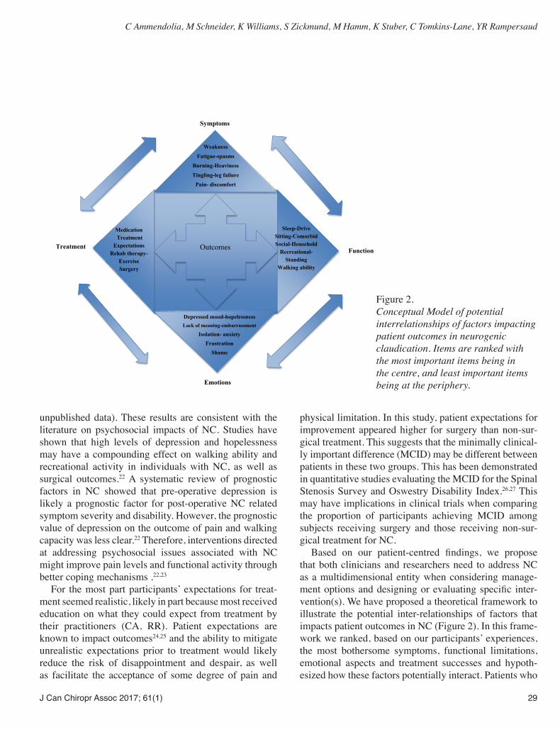
J Can Chiropr Assoc 2017; 61(1) 29
C Ammendolia, M Schneider, K Williams, S Zickmund, M Hamm, K Stuber, C Tomkins-Lane, YR Rampersaud
unpublished data). These results are consistent with the literature on psychosocial impacts of NC. Studies have shown that high levels of depression and hopelessness may have a compounding effect on walking ability and recreational activity in individuals with NC, as well as surgical outcomes.22 A systematic review of prognostic factors in NC showed that pre-operative depression is likely a prognostic factor for post-operative NC related symptom severity and disability. However, the prognostic value of depression on the outcome of pain and walking capacity was less clear.22 Therefore, interventions directed at addressing psychosocial issues associated with NC might improve pain levels and functional activity through better coping mechanisms .22,23
For the most part participants’ expectations for treat-ment seemed realistic, likely in part because most received education on what they could expect from treatment by their practitioners (CA, RR). Patient expectations are known to impact outcomes24,25 and the ability to mitigate unrealistic expectations prior to treatment would likely reduce the risk of disappointment and despair, as well as facilitate the acceptance of some degree of pain and
physical limitation. In this study, patient expectations for improvement appeared higher for surgery than non-sur-gical treatment. This suggests that the minimally clinical-ly important difference (MCID) may be different between patients in these two groups. This has been demonstrated in quantitative studies evaluating the MCID for the Spinal Stenosis Survey and Oswestry Disability Index.26,27 This may have implications in clinical trials when comparing the proportion of participants achieving MCID among subjects receiving surgery and those receiving non-sur-gical treatment for NC. Based on our patient-centred findings, we propose that both clinicians and researchers need to address NC as a multidimensional entity when considering manage-ment options and designing or evaluating specific inter-vention(s). We have proposed a theoretical framework to illustrate the potential inter-relationships of factors that impacts patient outcomes in NC (Figure 2). In this frame-work we ranked, based on our participants’ experiences, the most bothersome symptoms, functional limitations, emotional aspects and treatment successes and hypoth-esized how these factors potentially interact. Patients who
Outcomes
Symptoms
Function
Emotions
Treatment
Weakness
Fatigue-spasms
Burning-Heaviness
Tingling-leg failure
Pain- discomfort
Depressed mood-hopelessness
Lack of meaning-embarrassment Isolation- anxiety
Frustration
Shame
Sleep-Drive Sitting-Comorbid Social-Household
Recreational-Standing
Walking ability
Medication Treatment
Expectations Rehab therapy-
Exercise Surgery
Figure 2. Conceptual Model of potential interrelationships of factors impacting patient outcomes in neurogenic claudication. Items are ranked with the most important items being in the centre, and least important items being at the periphery.

30 J Can Chiropr Assoc 2017; 61(1)
The physical and psychological impact of neurogenic claudication: the patients’ perspectives
are depressed are not likely good surgical candidates, and this is an example of how psychosocial factors can impact treatment decisions. Treatment outcomes can change the direction of future treatment is another potential inter-action. This framework can provide a guide to clinicians to establish how NC individually affects their patients and to inquire about their treatment expectations. This can allow for a more stratified approach to management ranging from rehabilitation therapy, psychosocial support and /or surgery that may lead to better individual patient outcomes. Future research is needed to validate and quan-tify these proposed interactions between pain, physical ability, emotional state and treatment expectations in NC. Future studies should address and measure priority areas including pain, walking and standing ability, recrea-tional and social activity and emotional well being. In a Cochrane review examining non-operative interventions to improve outcomes in symptomatic LSS, none of the 21 studies reviewed directly assessed recreational activ-ity, while only seven of 21 studies assessed psychosocial status, and 12 of 21 studies used an objective walking measure.12 This paper provides valuable insight from a patient’s perspective and this information can influence how we treat LSS patients in future and how we select outcomes for research.
Study limitationsOur study is not without limitations. Our qualitative ap-proach is not designed to be generalizable, but instead to provide depth and insight into patients’ lived experience. For that reason we sought to achieve thematic saturation per group, which can be achieved with 10-12 interviews per group. We were able to recruit additional subjects per group and in our thematic coding we noted saturation, which occurs when key themes such as physical limita-tions are present for all participants. However, it is still possible that if more patients were interviewed different themes may have emerged. Another potential limitation is that our purposeful sam-ple focused on a Canadian sample in a hospital setting. Variations in health care system characteristics and re-lated factors such as access to treatment may impact pa-tients’ outcomes and expectations. Recruiting from a hos-pital setting may result in participants with more severe symptoms and physical limitations. Although an attempt was made to recruit a representative sample of patients
with NC due to LSS, it may be that the participating sam-ple was not a true general reflection of this population. This selection bias may also be reflected in our conceptual model of factors that impact patient outcomes and their potential interactions. These hypothesized interactions should be determined quantitatively using a random rep-resentative sample.
ConclusionsThe results of this qualitative study show that NC should be considered as multidimensional in its impact on pa-tients. We found that pain, and limited walking and stand-ing ability were the most bothersome aspects of NC that significantly impacted important activities of daily living, as well as meaningful recreational and social activities. Additionally, this study is the first to qualitatively identify the significant emotional impact of NC. This is a find-ing that should not be overlooked in clinical practice and future research. A holistic understanding of how psycho-social and other factors impact outcomes in this popula-tion is needed. We present a conceptual model of poten-tial interactions between important outcomes in LSS as a framework for future study.
AcknowledgementsThe following organizations have provided funding for this research: The Canadian Chiropractic Research Foun-dation (Ammendolia), NCMIC Foundation (Ammendolia and Schneider) and The Toronto General and Western Hospital Foundation (Rampersaud). Dr. Zickmund is sup-ported by the VA Salt Lake City Medical Center and the Health Services Research & Development IDEAS 2.0 Center (Award number ISOHX001240).
References1. Katz JN, Harris MB. Clinical practice. Lumbar spinal
stenosis. N Engl J Med. 2008;358: 818-825.2. Suri P, Rainville J, Kalichman L, Katz JN. Does this older
adult with lower extremity pain have the clinical syndrome of lumbar spinal stenosis? JAMA. 2010; 304: 2628-2636.
3. Comer CM, Redmond AC, Bird HA, Conaghan PG. Assessment and management of neurogenic claudication associated with lumbar spinal stenosis in a UK primary care musculoskeletal service: a survey of current practice among physiotherapists. BMC Musculoskelet Disord. 2009;10: 121.
4. Kalichman L, Cole R, Kim DH, Li L, Suri P, Guermazi A, et al. Spinal stenosis prevalence and association with

J Can Chiropr Assoc 2017; 61(1) 31
C Ammendolia, M Schneider, K Williams, S Zickmund, M Hamm, K Stuber, C Tomkins-Lane, YR Rampersaud
symptoms: the Framingham Study. Spine J. 2009; 9:545-550.
5. Deyo RA. Treatment of lumbar spinal stenosis: a balancing act. Spine J. 2010;10:625-627.
6. Government of Canada. Statistics Canada. Statistics Canada . 6-11-2006. Government of Canada, accessed November 2, 2008 at: www.statcan.gc.ca/bsolc/olc-cel/olc-cel?lang=eng&catno=11-008-X
7. Ammendolia C, Stuber K, de Bruin LK, Furlan AD, Kennedy CA, Rampersaud YR, Steenstra IA, Pennick V. Non-operative treatment for lumbar spinal stenosis with neurogenic claudication: a systematic review. Spine. 2012; 37: E609-616.
8. Kovacs FM, Urrútia G, Alarcón JD. Surgery versus conservative treatment for symptomatic lumbar spinal stenosis: a systematic review of randomized controlled trials. Spine 2011;36: E1335-1351.
9. Moojen WA, Arts MP, Bartels RH, Jacobs WC, Peul WC. Effectiveness of interspinous implant surgery in patients with intermittent neurogenic claudication: a systematic review and meta-analysis. Eur Spine J. 2011;20(10): 1596-1606.
10. Zaina F, Tomkins-Lane C, Carragee E, Negrini S. Surgical versus nonsurgical treatment for lumbar spinal stenosis. Spine. 2016;41: E857-868.
11. Ammendolia C, Stuber KJ, Rok E, Rampersaud R, Kennedy CA, Pennick V, Steenstra IA, de Bruin LK, Furlan AD. Nonoperative treatment for lumbar spinal stenosis with neurogenic claudication. Cochrane Database Syst Rev. 2013;8: CD010712.
12. Ammendolia C, Stuber K, Tomkins-Lane C, Schneider M, Rampersaud YR, Furlan AD, Kennedy CA. What interventions improve walking ability in neurogenic claudication with lumbar spinal stenosis? A systematic review. Eur Spine J. 2014;23: 1282-1301.
13. Coyne Imelda T. Sampling in qualitative research. Purposeful and theoretical sampling; merging or clear boundaries? J Advanc Nurs. 1997;26: 623–630.
14. Jensen MP, Karoly P, Braver S. The measurement of clinical pain intensity: a comparison of six methods. Pain. 1986;27: 117-126.
15. Stucki G, Daltroy L, Liang MH, Lipson SJ, Fossel AH, Katz JN. Measurement properties of a self-administered outcome measure in lumbar spinal stenosis. Spine. 1996;21: 796-803.
16. Fairbank JC, Couper J, Davies JB, O’Brien JP. The Oswestry low back pain disability questionnaire. Physiother. 1980;66: 271-273.
17. Ruta DA, Garratt AM, Russell IT. Patient centred assessment of quality of life for patients with four common conditions. Qual Health Care. 1999;8(1):22-29.
18. Crabtree BF, Miller WL (1992). Doing Qualitative Research: Multiple Strategies. Thousand Oaks: Sage Publications.
19. Landis JR, Koch GG. The measurement of observer agreement for categorical data. Biometrics. 1997; 33:159-174.
20. Edwards TC, Lavallee DC, Bauer Z, Comstock BA, Jarvik JG, Patrick DL, Makris UE, Friedly JL. Problem areas identified as important to older adults with lumbar spinal stenosis. Spine J. 2015;15:1636-1644.
21. Lyle S, Williamson E, Darton F, Griffiths F, Lamb SE. A qualitative study of older people’s experience of living with neurogenic claudication to inform the development of a physiotherapy intervention. Disabil Rehabil. 2016; 23:1-9.
22. McKillop AB, Carroll LJ, Battié MC. Depression as a prognostic factor of lumbar spinal stenosis: a systematic review. Spine J. 2014;14: 837-846.
23. Tomkins-Lane CC, Lafave LM, Parnell JA, Rempel J, Moriartey S, Andreas Y, Wilson PM, Hepler C, Ray HA, Hu R. The spinal stenosis pedometer and nutrition lifestyle intervention (SSPANLI): development and pilot. Spine J. 2015;15: 577-586.
24. Saban KL, Penckofer SM. Patient expectations of quality of life following lumbar spinal surgery. J Neurosci Nurs. 2007;39: 180-189.
25. Gepstein R, Arinzon Z, Adunsky A, Folman Y. Decompression surgery for lumbar spinal stenosis in the elderly: preoperative expectations and postoperative satisfaction. Spinal Cord. 2006;44: 427-431.
26. Copay AG, Glassman SD, Subach BR, Berven S, Schuler TC, Carreon LY. Minimum clinically important difference in lumbar spine surgery patients: a choice of methods using the Oswestry Disability Index, Medical Outcomes Study questionnaire Short Form 36, and pain scales. Spine J. 2008;8: 968-974.
27. Cleland JA, Whitman JM, Houser JL, Wainner RS, Childs JD. Psychometric properties of selected tests in patients with lumbar spinal stenosis. Spine J. 2012;12: 921-931.
28. Conway J, Tomkins C, Haig A. Walking assessment in people with lumbar spinal stenosis: capacity, performance, and self-report measures. Spine J. 2011; 11(9): 816-823.
29. Tomkins-Lane C, Melloh M, Lurie J, Smuck M, Battié MC, Freeman B, Samartzis D, Hu R, Barz T, Stuber K, Schneider M, Haig A, Schizas C, Cheung JP, Mannion AF, Staub L, Comer C, Macedo L, Ahn SH, Takahashi K, Sandella D. ISSLS Prize Winner: Consensus on the Clinical Diagnosis of Lumbar Spinal Stenosis: Results of an International Delphi Study. Spine. 2016;41(15):1239-1246.

32 J Can Chiropr Assoc 2017; 61(1)
ISSN 0008-3194 (p)/ISSN 1715-6181 (e)/2017/32–39/$2.00/©JCCA 2017
A proposed in vitro model for investigating the mechanisms of ‘joint cracking’: a short report of preliminary techniques and observationsJerome CJ Fryer, BSc, DC1 Jeffrey A Quon, DC, MHSc, PhD, FCCS(C)2-5 Richard D Vann, PhD6
1 Private Practice, Nanaimo BC2 School of Population and Public Health, Faculty of Medicine, University of British Columbia, Vancouver, Canada3 International Collaboration on Repair Discoveries (ICORD), Vancouver Coastal Health Research Institute4 Department of Orthopaedics, Spine Division, Faculty of Medicine, University of British Columbia5 The Cambie Chiropractic Centre, Vancouver, Canada6 Department of Anesthesiology and Center for Hyperbaric Medicine and Environmental Physiology, Duke University Medical Center
Corresponding author:Jerome Fryer201-1621 Dufferin Crescent Nanaimo BC, Canada V9S 5T4E-mail: [email protected]© JCCA 2017No financial support was provided for preparation of this manuscriptDisclaimer: Full disclosure Jerome Fryer is President and Chief Innovations Officer of Dynamic Disc Designs Corp.
Joint “cracking” is common but not a clearly understood audible phenomenon. In this brief report we propose an in-vitro model to potentially assist in revealing a mechanism for, and therefore source of, this phenomenon. Using a suction cup under tension and de-nucleated fluid to simulate synovial fluid, an audible release with intra-articular cavity formation was elicited. This was followed by a refractory period during which no audible crack could be elicited until the observed cavity had slowly reabsorbed back into the joint fluid. Conversely, if regular fluid containing pre-existing nuclei was used, a cavity formation occurred but with neither an audible release nor subsequent refractory period. With this simple in-vitro model, we were able to reproduce the characteristic audible release, cavity formation and related refractory period typically observed in related
Le « craquement » des articulations est un phénomène sonore commun, mais mal compris. Dans ce court rapport, nous proposons un modèle in vitro pouvant aider à révéler un mécanisme, et par conséquent une source, pour ce phénomène. À l’aide d’une ventouse sous tension et d’un fluide énucléé ayant pour but de simuler la synovie, on a entendu un son provenant de la cavité intraarticulaire, suivi d’une période réfractaire au cours de laquelle on n’a pas obtenu de craquement sonore jusqu’à ce que la cavité observée se soit réabsorbée lentement dans le liquide articulaire. À l’inverse, lorsqu’on utilisait le liquide régulier contenant les noyaux préexistants, il se produisait une perforation de la cavité, mais sans son ni période réfractaire. Ce modèle in vitro simple a permis de reproduire le son, la cavité et la période réfractaire connexe caractéristiques qu’on observe en général lors d’expériences connexes sur des

J Can Chiropr Assoc 2017; 61(1) 33
JCJ Fryer, J A Quon, RD Vann
Introduction“Cracking” or “popping” is common in many joints, par-ticularly the knuckles, however the mechanisms respon-sible for the sound remains unclear. In 1938, Nordheim et al. used x-rays to investigate joint cracking and observed the presence of intra-articular radiolucencies after joints were moved beyond their normal range of active motion.1 These lucencies were believed to be due to gas forma-tion, however, no elaborate in vitro model was offered at the time to help explain this phenomenon. Instead, a simple analogy based on a water-filled syringe was pro-posed. (see Figure 1) In that most basic model, gas forma-tion is reproduced within the syringe by simply pulling on the plunger while the needle end remains sealed. This action generates a bubble that expands in proportion to the increase in volume and corresponding negative pres-sure created as more tension is applied to the syringe. In accordance with Henry’s law of solubility, dissolved gas
comes out of solution as greater tension on the syringe re-duces its partial pressure. Additionally, in accordance with Boyle’s law, the volume of an otherwise fixed amount of undissolved gas increases, again, as its absolute pressure is reduced. Yet gas formation with the syringe model does not generate a cracking sound, and therefore it does not fully explain the events associated with gas formation within joints. In 1947, Roston et al. also detected radiolucencies on x-ray images of cracking joints and hypothesized that they represented bubble formation in the midst of a vacuum created by joint tension or distraction.2 Roston et al. theorized that the bubbles originated from smaller pre-existing gas nuclei (i.e., small spherical bubbles or gas cavities trapped in crevices on hydrophobic surfaces that are ubiquitous in polar fluids such as water), which transitioned to larger visible bubbles at lower vacuum thresholds.3 These gas nuclei can be dissolved and elim-
experiments in human joints. This simple in-vitro model may be of use in helping to discern both the timing and precise nature of other yet to be discerned mechanisms related to joint cracking. (JCCA. 2017;61(1):32-39) k e y w o r d s : chiropractic, joint, cavitation, mechanism
articulations humaines. Ce modèle in vitro simple peut aussi servir à discerner à la fois le moment et la nature précise d’autres mécanismes qu’on n’a pas encore perçus concernant le craquement des articulations. (JCCA. 2017;61(1):32-39) m o t s c l é s : chiropratique, articulation, cavitation, mécanisme
Figure 1. Water filled syringe under tension.

34 J Can Chiropr Assoc 2017; 61(1)
A proposed in vitro model for investigating the mechanisms of ‘joint cracking’: a short report of preliminary techniques and observations
inated by either hydrostatic compression, boiling at sea level pressure at 100oC, or boiling under high vacuum at room temperature. Although this theoretical framework of denucleation was introduced, no subsequent physical in vitro model evolved from that study. By 1971 Unsworth et al. introduced the ‘cavitometre’, which was the first in vitro synovial joint model that was developed specifically to study joint cracking.4 This mod-el was constructed out of nylon and plexiglass with sur-faces similar in contour to a metacarpophalangeal joint, only at double the normal size, and separated by synovial fluid. When tension across the system was applied quick-ly to simulate a joint crack, a cavity appeared and then disappeared. Furthermore, a sound was generated, which the authors concluded was due to bubble collapse rather than bubble formation (the latter of which Roston et al. had hypothesized in an earlier study). However, unlike a real synovial joint, the model was open to ambient air, from which gas nuclei could be introduced. Furthermore, the model could be cracked repeatedly without hindrance by any refractory period between sequential iterations. Irrespective of the model’s limitations, the collapsing bubble hypothesis of Unsworth et al. remained the most popular explanation for joint cracking sounds for over four decades. However in 2015, Kawchuck et al. used magnetic resonance imaging (MRI) to reveal the occur-rence of a hypointense area coincidental to the time of a metacarpophalangeal crack.5 This led to renewed interest in Roston’s original theory of bubble formation as a vi-able alternative to Unsworth’s theory of bubble collapse. In further support of Roston et al.’s work, Kawchuk et al. showed that traction of the finger caused the hypointense area to remain present in the field of view, similar to what is observed in the basic syringe model. But controversy would still exist between the two competing hypotheses as the time window between MRI frames in the study by Kawchuk et al. was 0.3 sec, whereas that of the video imaging in the cavitometre study by Unsworth et al. was within 0.01 sec. Kawchuk et al. further concluded that the process of cavity formation was likely due to tribonucleation, which by definition is a process of bubble formation from the relative motion of two solid structures under liquid ten-sion.6,7 However, other studies on tribonucleation do not indicate that this phenomenon is associated with the gen-eration of any notable cracking sounds.7,8
To assist in resolving this ambiguity, we describe an in vitro model that possesses three important properties: 1) It is a closed system that mimics the sealed environment of the synovial joint in vivo; 2) It contains de-nucleated fluid and 3) It reproduces both a cracking phenomenon and sub-sequent refractory period identical to that which is seen in real synovial joints. We believe that this new in vitro model could be a basis for further advancement of previous re-search on the mechanisms of joint cracking. This improved model simulates much of the natural anatomy and geom-etry of a real metacarpophalangeal joint. The objective of the current paper is to describe this new-and-improved joint cracking model and to also present some qualitative observations from preliminary tests involving this model.
Materials and MethodsThe development of our latest model began with con-struction of a very basic dry joint model initially (Model A), followed by the sequential construction of 5 addi-tional models (Models B to final Model F). Each model represented a minor modification of the one temporally preceding it.
Model A: Basic dry jointThis model consisted of an elastic suction cup (diameter 10mm) adhered to a polished flat glass plate with an air-filled space simulating a joint cavity. In this model, the suction cup was pressed up against the glass plate, and subsequent traction was applied in two different ways: 1) with just enough tension force to cause partial detachment of the cup (i.e., the centre of the cup) without breaking the seal around its perimeter; and subsequently 2) with greater and sufficient tension to cause the cup to detach fully (i.e., both centre and peripheral margin of the cup) from the glass surface.
Model B: A wet jointThis model was identical to Model A, except that the suc-tion cup was immersed in a beaker of distilled water so that the simulated joint space was filled with fluid. In this model, the suction cup was pressed up against the beaker wall to create a suction adherence, but this time, traction was applied with just enough tension force to cause par-tial detachment of the cup (i.e. the centre of the cup) with-out breaking the seal around its perimeter, similar to the first way in Model A.

J Can Chiropr Assoc 2017; 61(1) 35
JCJ Fryer, J A Quon, RD Vann
Model C: A “de-nucleated” wet jointThis model was identical to Model B, except that the suc-tion cup was immersed in “de-nucleated” distilled water. As mentioned previously, nucleated water refers to nor-mal distilled water, which contains visible bubbles that form through the coalescence of pre-existing gas cavities (i.e., gas “nuclei”) within the fluid.4 These pre-existing nuclei are either smaller unattached spherical bubbles or larger gas volumes that are attached to hydrophobic crevices on solid particles.9 Both types of nuclei can be partially removed by boiling the fluid for 30 minutes with the suction cup immersed in the fluid and letting cool. An-other approach to de-nucleate is by over pressurization6 and a third approach is by vacuum, which we performed by using -110 kPa for 20 minutes while the suction cup was fully immersed in the fluid. After denucleating the fluid, the suction cup was pressed up against the glass beaker wall to generate a suction adherence. We then pulled on the suction cup with just enough manual force to cause only the centre of the cup to detach from the wall, without compromising the seal around the perimeter of the suction cup.
Model D: A Ringer’s solution-filled wet jointThis model was identical to model C, but to more close-ly simulate the actual fluid in a synovial joint, a Ringer’s solution, manufactured as per Casentini et al.10, was used and de-nucleated as we did with distilled water for model
C. Again, after pressing the suction cup to the beaker wall to create a suction adherence, we pulled on the suction cup with just enough manual force to cause only the centre of the cup to detach from the glass beaker wall without com-promising the seal around the perimeter of the suction cup.
Model E: Wet joint model with realistic surface geometryTo determine if the cracking event was influenced by the geometry of the suction cup surfaces employed for models A through D, the suction cup was replaced by a polyurethane metacarpal bone. This was achieved by cre-ating a mold of a cadaveric metacarpal bone and pour-ing an identically shaped polyurethane copy. To roughly simulate the presence of a synovial fold and capsule, an elastic ring was attached to the polyurethane metacarpal head with an adhesive, exposing the central surface of the metacarpal head. This structure was then immersed in a glass beaker with Ringer’s solution and de-nucleated under vacuum conditions. After 30 minutes of -110 Kpa the polyurethane metacarpal head was pressed up against the inside of a glass beaker wall while completely im-mersed in denucleated fluid. (see Figure 2) During testing of this model, we pulled on the polyurethane metacarpal base with just enough manual force to cause only the cen-tre aspect of the metacarpal head to release from the beak-er wall, but again without breaking the seal around the perimeter being maintained by the elastic ring.
Figure 2. Model E

36 J Can Chiropr Assoc 2017; 61(1)
A proposed in vitro model for investigating the mechanisms of ‘joint cracking’: a short report of preliminary techniques and observations
Model F (Final Model): Compressible wet joint with realistic surface geometryTo test whether changes in joint tension affected the dur-ation required for the model to return to its baseline state (of cavity dissolution) after simulated cracking, a plexi-glass apparatus with a fulcrum mechanism was used to apply slight compression to the joint represented by the previous model (Model E). Again, de-nucleated Ringer’s solution was used to simulate the presence of synovial fluid. During our tests, we first pulled on the polyurethane metacarpal base with just enough manual force to cause the centre aspect of the metacarpal head to detach from the beaker inside wall below the fluid line, again with-out compromising the seal around the perimeter of the elastic ring. Following detachment of the metacarpal head and corresponding cavity formation within the simulated joint, we applied compression to the joint model with a clamp (i.e. a fulcrum mechanism), which is depicted in Figure 3. The amount of compression force was not meas-ured at the time, but was subsequently estimated to be between 15 and 30 N.
Results of preliminary testingWith the Model A dry joint, pulling of the suction cup to detach only the centre aspect of the suction cup was not associated with an audible event. Only with further trac-tion and detachment of the cup perimeter did a cracking sound occur.
With the Model B wet joint, as the suction cup was slowly pulled from the inside of the beaker wall in its closed state, a gas cavity formed silently as the centre of the suction cup detached without breaking the seal around its perimeter. Furthermore, when tension was subsequent-ly reduced in order to allow the suction cup to return to its previous neutral position, the cavity disappeared (as
Figure 3. Compression tension apparatus
Figure 4.
Model B. Within a simulated wet joint space (without de-nucleated fluid) in a sealed condition (1), decompression and volume expansion results in expanding bubble formation (2-4) while return to initial tension and volume normalization
results in bubble disappearance (5-6).

J Can Chiropr Assoc 2017; 61(1) 37
JCJ Fryer, J A Quon, RD Vann
was expected under Boyle’s Law, which describes the in-verse relationship between pressure on the one hand and volume of an otherwise fixed amount of undissolved gas on the other hand). This sequence of events is depicted in Figure 4. With Model C in which distilled water within the simu-lated joint space was replaced by de-nucleated water, nei-ther central cup detachment nor gas cavity formation was observed when the suction cup was pulled with the same initial force as was used in Model B. In other words, with initial tension the centre of the suction cup did not release from the beaker wall but instead remained completely ad-herent to it. It is noteworthy that in the previous model employing nucleated fluid (Model B), bubble formation was observed immediately during traction, whereas in Model C employing denucleated fluid, gas bubble forma-tion was no longer evident early on. Only later with Mod-el C, when the suction cup was pulled with greater force did its centre detach from the surface. Moreover, upon detachment, a stable gas cavity formed in association with an audible crack. Also with Model C, when the suction cup was released and allowed to return to its baseline position, a visible cavity remained, and then disappeared only gradually over approximately 30 minutes. Finally, so
long as a bubble or cavity remained visible to the naked eye, no further cracking sound could be elicited during re-pulling of the suction cup. On the other hand, once the bubble was no longer visible to the naked eye, an audible crack could again be elicited from the model (Figure 5). With Model D, we observed the same findings that we observed during experiments with Model C. More specif-ically, the experiment with de-nucleated Ringer’s solution resulted in joint cracking and a corresponding refractory period that was identical to that of a model employing de-nucleated distilled water. With Model E, regardless of whether the joint was im-mersed in nucleated distilled water or nucleated Ringer’s solution, the observed events were identical to those that were observed in Model B. In contrast, when either the distilled water or Ringer’s solution was de-nucleated, our observations were identical to those obtained with Model C. Under Model E, we also were able to generate the crack-ing sound when the model was pulled off-axis, which we did in order to simulate cracking of a real knuckle joint in a partially flexed or non-neutral position (https://youtu.be/ TzC7PkgbHGA). With our final model, Model F, the application of com-
Figure 5.
Model C Within a simulated wet joint space (with de-nucleated fluid) in a sealed condition (1), decompression results in suction cup stretch without lift and without cavity (2). Suddenly, when sufficient tension is applied a cavity and sound spontaneously forms (3) and further decompression leads to increasing cavity volume formation. Relaxation of tension
leads to enduring cavity (4) and over time, cavity size shrinks (5-6). The sequence 1-6 can then be repeated.

38 J Can Chiropr Assoc 2017; 61(1)
A proposed in vitro model for investigating the mechanisms of ‘joint cracking’: a short report of preliminary techniques and observations
pression through the joint (subsequent to cavity formation and an audible release) resulted in a reduction in the time required before cavity formation and joint cracking could be repeated. In this regard, the so-called refractory period without compression was previously 30 minutes, whereas the refractory period with joint compression was only 12 minutes. Qualitatively, we observed that the greater the amount of compression that was applied to the model, the shorter was the refractory time.
DiscussionWe have introduced a series of in vitro models and a final model that will serve as a basis for our future investiga-tions into the mechanisms of synovial joint cracking. Our observations at this time are too anecdotal to warrant a full report, however they are presented here for the pur-pose of soliciting immediate comments and criticisms from the broader research community. In the meantime, a key preliminary finding is that the events we observed after replacing regular fluid with denucleated fluid (in Models C, D and E) are completely consistent with the same cavity formation and refractory period phenomena that is associated with the cracking of real synovial joints. Specifically, within a closed system employing denucle-ated fluid, as long as a bubble or cavity formation is vis-ible within the simulated joint space, a crack can not be repeated. In contrast, once the cavity disappears, a crack can again be elicited from the model. This behaviour of gas within liquid is typically inter-preted to reflect the dissolution of a spherical bubble by the forces of surface tension. Until its dissolution, a bub-ble acts as a gas nucleus (as observed in Model B) which permits formation of a larger visible gas cavity during de-compression of liquid within a closed system, but in the absence of generating an audible crack. Epstein and Ples-set derived equations that describe the time to dissolution of a bubble in relation to absolute pressure, dissolved gas tension, and surface tension.11 Accordingly, in Model F, a reduced refractory period was both expected and ob-served, and ultimately reflected faster re-solution time in response to increased local absolute pressure. Admittedly, the precise timing of the crack in relation to the timing of bubble formation was not discernible from these preliminary experiments. At this time, it re-mains a mystery whether sound generation occurs before, after, or simultaneously to the time of cavity formation.
The timing of the crack in relation to bubble formation will be the focus of our future work. Additionally, we plan to quantify corresponding forces, tensions, bubble sizes, and dissolution times through the use of multiple imaging methods (i.e., cinematography, ultrasound, and MRI).
ConclusionIn this brief report, an in-vitro model has been developed and proposed to investigate the origins of the cracking sound within synovial joints. So far, we have observed that when a de-nucleated fluid is introduced, decompres-sion of a sealed joint elicits both cavity formation and an audible event, similar to what occurs in human syn-ovial joints. Immediately afterward, a refractory period occurs during which an additional audible event cannot be elicited regardless of how much joint decompression or tension is applied. In contrast, if the fluid used in the model is nucleated, decompression of the simulated joint elicits cavity formation in the absence of an audible event. Additional measurement techniques will be developed and applied to this new model with the intent of better clarifying the mechanisms of in vivo joint cracking.
AcknowledgmentsWe would like to sincerely thank Sander Wildeman for his assistance in the preliminary testing of the suction cup in water as well as Norm Boulet in the construction of the Figure 3 apparatus.
References1. Nordheim, Y. Einer neue Methode den Gelenkknorpel
besonders die Kniegelenkmenisken röntgenologisch darzustellen. Fortschr Röntgenstr 1938;57:479.
2. Roston JB, Haines RW. Cracking in the metacarpo-phalangeal joint. J Anat. 1947; 81: 165–173.
3. Harvey EN, et al. Bubble formation in animals. I. Physical factors. J Cellular Comp Physiol. 1944; 24(1):1-22.
4. Unsworth A, Dowson D, Wright V. ‘Cracking joints’ A bioengineering study of cavitation in the metacarpophalangeal joint. Ann. Rheum. Dis. 1971; 348–358.
5. Kawchuk GN, Fryer J, Jaremko JL, Zeng H, Rowe L, Thompson R. Real-time visualization of oint cavitation. PLOS ONE. 2015; 10(4): e0119470.
6. Hayward AT. J- Tribonucleation of bubbles. Brit J Appl Phys. 1967;18:641-644.
7. Wildeman S, Lhuissier H, Sun C, Lohse D, Prosperetti A. (2014). Tribonucleation of bubbles. Proc Natl Acac Sci USA. 2014;111(28):10089–10094.

J Can Chiropr Assoc 2017; 61(1) 39
JCJ Fryer, J A Quon, RD Vann
8. Ikels KG. Production of gas bubbles in fluids by tribonucleation. J Appl Physiol. 1970; 28(4): 524–527.
9. Marschall HB, Mørch KA, Keller AP, Kjeldsen M. Cavitation inception by almost spherical solid particles in water. Phys Fluids. 2003;15(2): 545.
10. Casentini G, Di Paola L, Marrelli L, Palma F. Rheological characterization of an artificial synovial fluid. Int J Artif Organs. 2005;28(7):711-717.
11. Epstein PS, Plesset MS. On the stability of gas bubbles in liquid-gas solutions. J Chem Phys. 1950; November 1.
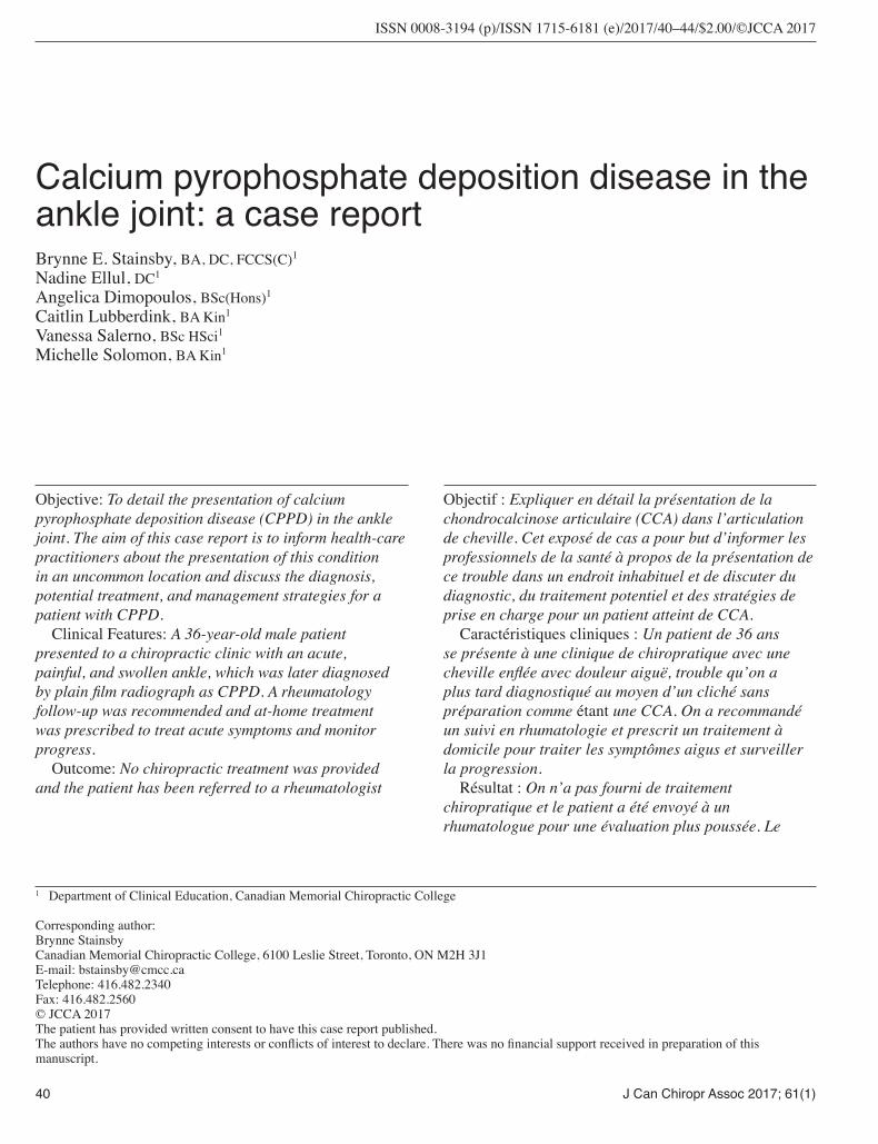
40 J Can Chiropr Assoc 2017; 61(1)
ISSN 0008-3194 (p)/ISSN 1715-6181 (e)/2017/40–44/$2.00/©JCCA 2017
Calcium pyrophosphate deposition disease in the ankle joint: a case reportBrynne E. Stainsby, BA, DC, FCCS(C)1 Nadine Ellul, DC1 Angelica Dimopoulos, BSc(Hons)1 Caitlin Lubberdink, BA Kin1 Vanessa Salerno, BSc HSci1 Michelle Solomon, BA Kin1
1 Department of Clinical Education, Canadian Memorial Chiropractic College
Corresponding author:Brynne StainsbyCanadian Memorial Chiropractic College, 6100 Leslie Street, Toronto, ON M2H 3J1E-mail: [email protected]: 416.482.2340Fax: 416.482.2560© JCCA 2017The patient has provided written consent to have this case report published.The authors have no competing interests or conflicts of interest to declare. There was no financial support received in preparation of this manuscript.
Objective: To detail the presentation of calcium pyrophosphate deposition disease (CPPD) in the ankle joint. The aim of this case report is to inform health-care practitioners about the presentation of this condition in an uncommon location and discuss the diagnosis, potential treatment, and management strategies for a patient with CPPD. Clinical Features: A 36-year-old male patient presented to a chiropractic clinic with an acute, painful, and swollen ankle, which was later diagnosed by plain film radiograph as CPPD. A rheumatology follow-up was recommended and at-home treatment was prescribed to treat acute symptoms and monitor progress. Outcome: No chiropractic treatment was provided and the patient has been referred to a rheumatologist
Objectif : Expliquer en détail la présentation de la chondrocalcinose articulaire (CCA) dans l’articulation de cheville. Cet exposé de cas a pour but d’informer les professionnels de la santé à propos de la présentation de ce trouble dans un endroit inhabituel et de discuter du diagnostic, du traitement potentiel et des stratégies de prise en charge pour un patient atteint de CCA. Caractéristiques cliniques : Un patient de 36 ans se présente à une clinique de chiropratique avec une cheville enflée avec douleur aiguë, trouble qu’on a plus tard diagnostiqué au moyen d’un cliché sans préparation comme étant une CCA. On a recommandé un suivi en rhumatologie et prescrit un traitement à domicile pour traiter les symptômes aigus et surveiller la progression. Résultat : On n’a pas fourni de traitement chiropratique et le patient a été envoyé à un rhumatologue pour une évaluation plus poussée. Le

J Can Chiropr Assoc 2017; 61(1) 41
BE Stainsby, N Ellul, A Dimopoulos, C Lubberdink, V Salerno, M Solomon
IntroductionCalcium pyrophosphate deposition disease (CPPD) is a condition that is characterized by the deposit of pyrophos-phate crystals into tendons, ligaments, cartilage and syn-ovium.1,2 CPPD may be associated with elevated levels of calcium, pyrophosphate or local cartilage matrix chan-ges.3 Typically, CPPD is found in older aged individuals, with an onset of 30 years of age and a peak at 60 years2, CPPD is often associated with primary and secondary osteoarthritis, which results in decreased joint congru-ency and degeneration due to the aging process1. Both the cause of CPPD and the mechanism of onset of crystal deposition remain largely unknown and vastly debated throughout literature.1,2 The formation of calcium pyrophosphate crystals is extracellular, however, pyro-phosphate is a by-product of several intracellular reac-tions thus unable to diffuse passively across the cell mem-brane.1 It is unclear whether crystal deposition occurs as a result of extracellular pyrophosphate synthesis by plasma membrane-bound plasma cell glycoprotein 1 or whether pyrophosphate is transported across cell membranes by ankylosis human protein.1 Causes of CPPD can be clas-sified into the following categories: idiopathic, metabol-ic, hereditary and post-traumatic.1 CPPD can be linked to underlying metabolic disorders such as hemochro-matosis, hyperparathyroidism, hypophosphataemia,
hypomagnesaemia and hypothyroidism, all of which in-crease the risk for calcium pyrophosphate deposition.3 Numerous cases of CPPD have shown familial links in the ANKH gene, which functions to upregulate protein.4 When the ANKH gene becomes mutated, protein activity is enhanced and thus extracellular pyrophosphate levels increase and promote the formation of pyrophosphate crystals leading to CPPD.1,3,4 Despite attempts to deter-mine etiology, the majority of CPPD cases remain idio-pathic.3,4
This condition has not yet shown an association with gender, obesity, or lifestyle characteristics, although it is slightly more common in Caucasian individuals.1,2 CPPD has been documented most commonly in the knees, wrists, symphysis pubis and hips.1 This condition is a common rheumatologic disease in elderly individuals, and is noted most often in knees and pelvis.1 CPPD has been found to appear in one of three forms: asymptomatic, acute or chronic.1 Asymptomatic patients most often dis-cover the deposit of pyrophosphate crystals through plain film radiographs for an alternative reason.1 Acute CPPD, a presentation that is highly suggestive of acute crystal inflammation, is associated with the rapid development of joint pain, swelling, tenderness, warmth and restrict-ed movement, often reaching its maximum within 6-24 hours of symptom onset.1 Chronic CPPD presents simi-
for further assessment. The diagnosis of CPPD was confirmed and he was advised to take an anti-inflammatory if symptoms recurred and booked for further follow-up in six months. Summary: Although the presentation is less common, CPPD can present in the ankle joint and mimic other inflammatory disorders. Conservative treatment can be applied to treat acute symptoms and referral to a rheumatologist is suggested to monitor progress of this condition. (JCCA. 2017;61(1):40-44) k e y w o r d s : chiropractic, ankle, calcium pyrophosphate deposition disease
diagnostic de CCA a été confirmé; on lui a conseillé de prendre un anti-inflammatoire si les symptômes réapparaissaient et on a planifié un suivi six mois plus tard. Résumé : Bien que la présentation soit moins commune, la CCA peut se présenter dans l’articulation de cheville et imiter d’autres affections inflammatoires. Un traitement conservateur peut permettre de soigner les symptômes aigus et on recommande d’envoyer le patient voir un rhumatologue pour surveiller la progression du trouble. (JCCA. 2017;61(1):40-44) m o t s c l é s : chiropratique, cheville, chondrocalcinose articulaire

42 J Can Chiropr Assoc 2017; 61(1)
Calcium pyrophosphate deposition disease in the ankle joint: a case report
larly to osteoarthritis with progressive joint pain, chronic synovitis, crepitus and warmth at the joint line.2
Traditionally, patients presenting with suspected CPPD can be screened for chondrocalcinosis using plain-film radiographs, however, microscopic analysis of synovial fluid provides a more definitive and accurate diagnosis due to the ability to detect calcium pyrophosphate crystals.5 Chondrocalcinosis, calcification in hyaline or fibrocarti-lage, is considered the key identifying feature of CPPD on radiographs, however this diagnostic method is neither highly sensitive nor specific.5 Synovial fluid analysis can be used to identify weakly positively birefringent crystals using polarized light microscopy.5 More recently, ultra-sonography of articular and fibrocartilage has been used to indicate the presence of CPPD crystals.6 Ultrasound is used to detect hyperechoic bands in hyaline cartilage and hyperechoic spots in fibrous cartilage.6 Further studies are required to compare the diagnostic value of ultrasound with existing diagnostic methods, however ultrasound re-mains a promising tool for diagnosis of CPPD.6
Strategies for treatment and management of CPPD vary depending on the symptom severity, stage and clinic-al manifestation of CPPD. Staging for CPPD presentation includes the following categories: asymptomatic, acute, chronic, presenting with OA or as a pseudo-arthritide.1,5 Common treatments that provide symptomatic relief in-clude conventional anti-inflammatory medications, such as non-steroidal anti-inflammatories (NSAIDs) and cor-ticosteroids.5 Although the progression of this condition may vary between individuals, most will experience re-current symptomatic episodes.7 During acute attacks, patients often experience severe pain.7 Optimal and safe treatment for acute pain includes ice, temporary rest, intra-articular injections and joint aspiration.7 Despite the incurable nature of this condition, the prognosis is good, as long as symptoms are controlled and patients are mon-itored for pre-disposing conditions that may be treatable and preventable.8
The objective of this case report is to highlight a unique incidence of CPPD in the ankle, a less common location for this condition with minimal documentation in current literature. Additionally, this case report aims to provide health-care practitioners with a detailed case presenta-tion, radiographic findings, and potential treatment and management options for future patients with this case presentation.
Case Presentation
History and Physical Examination FindingsA 36-year-old male initially presented to the Canadian Memorial Chiropractic College (CMCC) campus clinic with a primary complaint of acute mechanical low back pain. He reported no comorbidities, medical history or medication use. At a subsequent visit, the patient re-ported a new complaint of an acutely swollen left ankle, with an onset of pain six days prior to his appointment. At this time, his back pain was resolved. There was no history of trauma to the ankle. Twenty-four hours after the onset of his ankle symptoms, he was woken up in the middle of the night by severe pain that resulted in the in-ability to fall back asleep. Over the course of the week-end, prior to his appointment, the ankle became swollen. The patient was self-medicating with ibuprofen, which helped to relieve but did not eliminate the pain. Follow-ing observation of the joint and orthopaedic testing of the ankle, all tests created pain due to palpation of the joint but no tests were positive for ligament injury or trauma. The patient was sent for diagnostic imaging in consideration of differential diagnoses of gout, pseudo-gout, osteoarthritis, rheumatoid arthritis and septic arth-ritis.
ImagingA plain-film radiographic series for the left ankle (Fig-ures 1 and 2) was taken in addition to a bilateral AP knee radiograph (Figure 3). There was adequate bone density with no osteochondral defects detected at the tibial pla-fond and talar dome. All bony joints appeared unremark-able, however blurring of Kager’s fat pad was visualized. Radiographic findings included apparent chondrocalcin-osis with joint effusion in the talotibial joint, which is highly suggestive of pseudogout associated with CPPD. As well, there was subtle chondrocalcinosis of the me-nisci bilaterally visualized on the bilateral AP knee radio-graphs (Figure 3).
Diagnosis, Treatment and Referral The patient was diagnosed, by plain-film radiography, with CPPD in the left ankle. At the time of writing, the patient did not receive any chiropractic treatment and was referred to a rheumatologist for further assessment. The patient declined conservative care, and was advised to
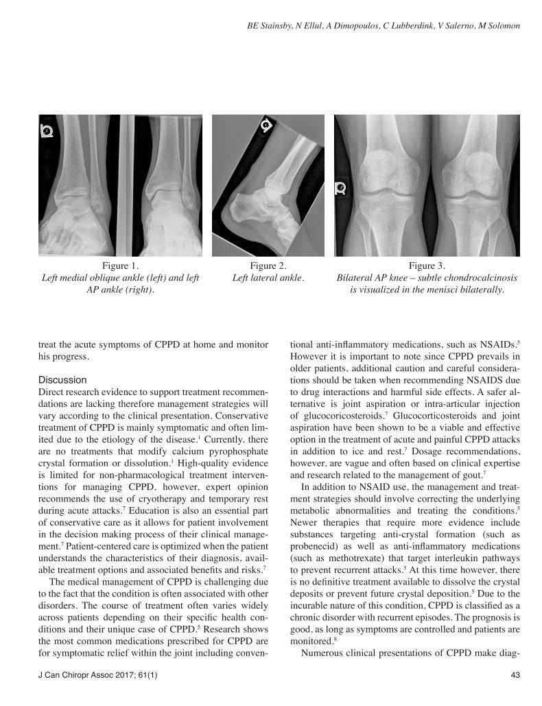
J Can Chiropr Assoc 2017; 61(1) 43
BE Stainsby, N Ellul, A Dimopoulos, C Lubberdink, V Salerno, M Solomon
treat the acute symptoms of CPPD at home and monitor his progress.
DiscussionDirect research evidence to support treatment recommen-dations are lacking therefore management strategies will vary according to the clinical presentation. Conservative treatment of CPPD is mainly symptomatic and often lim-ited due to the etiology of the disease.1 Currently, there are no treatments that modify calcium pyrophosphate crystal formation or dissolution.1 High-quality evidence is limited for non-pharmacological treatment interven-tions for managing CPPD, however, expert opinion recommends the use of cryotherapy and temporary rest during acute attacks.7 Education is also an essential part of conservative care as it allows for patient involvement in the decision making process of their clinical manage-ment.7 Patient-centered care is optimized when the patient understands the characteristics of their diagnosis, avail-able treatment options and associated benefits and risks.7
The medical management of CPPD is challenging due to the fact that the condition is often associated with other disorders. The course of treatment often varies widely across patients depending on their specific health con-ditions and their unique case of CPPD.5 Research shows the most common medications prescribed for CPPD are for symptomatic relief within the joint including conven-
tional anti-inflammatory medications, such as NSAIDs.5 However it is important to note since CPPD prevails in older patients, additional caution and careful considera-tions should be taken when recommending NSAIDS due to drug interactions and harmful side effects. A safer al-ternative is joint aspiration or intra-articular injection of glucocoricosteroids.7 Glucocorticosteroids and joint aspiration have been shown to be a viable and effective option in the treatment of acute and painful CPPD attacks in addition to ice and rest.7 Dosage recommendations, however, are vague and often based on clinical expertise and research related to the management of gout.7
In addition to NSAID use, the management and treat-ment strategies should involve correcting the underlying metabolic abnormalities and treating the conditions.5 Newer therapies that require more evidence include substances targeting anti-crystal formation (such as probenecid) as well as anti-inflammatory medications (such as methotrexate) that target interleukin pathways to prevent recurrent attacks.5 At this time however, there is no definitive treatment available to dissolve the crystal deposits or prevent future crystal deposition.5 Due to the incurable nature of this condition, CPPD is classified as a chronic disorder with recurrent episodes. The prognosis is good, as long as symptoms are controlled and patients are monitored.8
Numerous clinical presentations of CPPD make diag-
Figure 1.
Left medial oblique ankle (left) and left AP ankle (right).
Figure 2.
Left lateral ankle.
Figure 3.
Bilateral AP knee – subtle chondrocalcinosis is visualized in the menisci bilaterally.

44 J Can Chiropr Assoc 2017; 61(1)
Calcium pyrophosphate deposition disease in the ankle joint: a case report
nosing and treating this condition challenging. In this case specifically, CPPD targeted the menisci of the knees and the talotibial joint. A common location for CPPD includes, but is not limited to, the menisci of the knee and the patellofemoral joint, only one of which was seen in this case. It is apparent that there is irregular joint in-volvement, seeing that CPPD was also present in the an-kle, which is a less common location for this condition. Although CPPD is often associated with osteoarthritis, it is important to understand the difference in clinical pres-entation between the two conditions. A large proportion of patients with CPPD follow a progressive course of articu-lar degeneration in an irregular distribution pattern.5 Typ-ically, around 50% of these individuals will experience acute attacks of pseudogout superimposed on their under-lying osteoarthritis, whereas the remaining present with classical osteoarthritis.5 Features that distinguish CPPD from osteoarthritis include atypical joint distribution, presence of contractures and valgus knee deformities.5 Clinically, CPPD has the potential to mimic several forms of inflammatory arthritic conditions resulting in a wide array of clinical manifestations.5 These presentations in-clude asymptomatic or lanthanic chondrocalcinosis, acute pseudogout, pseudo osteoarthritis (with or without acute attacks), pseudo rheumatoid arthritis, pseudo-polymyal-gia rheumatica and pseudo-neuropathic arthropathy.5 It is possible for CPPD to co-exist with other arthritic con-ditions, further complicating diagnosis and management strategies. Further research on diagnosing, managing and reducing calcium pyrophosphate crystal deposition is es-sential seeing that CPPD and the associated CPPD-related arthropathies are likely to increase in prevalence due to the current aging population.5
SummaryThis case report highlights a 36-year-old male patient who presented to a chiropractic clinic with an uncommon presentation of a common arthritic condition. Clinicians
should take note of this unique presentation, as CPPD is prevalent in older, Caucasian populations and can often present concurrently with osteoarthritis.1,7 It is imperative to understand which joints can be targeted by CPPD and what treatment options are available. As well, it is clinic-ally important to note the patient in this case report also had subtle findings of chondrocalcinosis in the menisci of the knees. Thus, inferring that if chondrocalcinosis is found in one joint in the body, there may be other joints in the body targeted as well. Further research is necessary to investigate the benefits of chiropractic care for the treat-ment and management of patients presenting with acute and chronic forms of CPPD, specifically with respect to providing pain relief and improving joint mobility.
References1. Abhishek A. Calcium pyrophosphate deposition. Br J Hosp
Med. 2014;75(4):C61-C64.2. Yochum T, Rowe L. 1996. Essentials of skeletal radiology.
Baltimore: Williams & Wilkins.3. Lindenmeyer C, Sobel A, Nazarian L, Mandel S, Raikin
S. A case study of pseudo-neuropathic pseudogout. Med Forum. 2013; 14(26).
4. Netter P, Bardin T, Bianchi A, Richette P, Loeuille D. The ANKH gene and familial calcium pyrophosphate dihydrate deposition disease. J Bone Spine. 2004;71(5):365-368.
5. MacMullan P, McCarthy G. Treatment and management of pseudogout: insights for the clinician. Ther Adv Musculoskeletal Dis. 2012;4(2):121-131.
6. Gamon E, Mouterde G, Barnetche T, Combe B. SAT0319 Diagnostic value of ultrasound in calcium pyrophosphate deposition disease: a systematic review and meta-analysis. Ann Rheum Dis. 2015;74(Suppl2):774.2-774.
7. Zhang W, Doherty M, Pascual E, Barskova V, Guerne P, Jansen T, et al. EULAR recommendations for calcium pyrophosphate deposition. Part II: Management. Ann Rheum Dis. 2011;70(4):571-575.
8. Schumacher H, Hasselbacher P. CPPD Deposition Disease [Internet]. UW Orthopaedics and Sports Medicine, Seattle. 2012 [cited 2 January 2016]. Available from: http://www.orthop.washington.edu/?q=patient-care/articles/arthritis/cppd-deposition-disease.html

J Can Chiropr Assoc 2017; 61(1) 45
ISSN 0008-3194 (p)/ISSN 1715-6181 (e)/2017/45–52/$2.00/©JCCA 2017
Chiropractic management of a geriatric patient with idiopathic neuralgic amyotrophy: a case reportJordan Gliedt, DC1,2 Justin M. Goehl, DC, MS3 Derek P. Smith, DC4 Clinton J. Daniels, DC, MS5
1 Phoenix Veterans Affairs Health Care System2 Logan University College of Chiropractic3 Veterans Affairs Greater Los Angeles Health Care System4 Private Practice; Back in Motion Chiropractic, Pocahontas, AR, USA5 Veterans Affairs Puget Sound Health Care System
Corresponding author: Jordan Gliedt650 E. Indian School Road, Phoenix, AZ 85012 Tel: 602-277-5551 E-mail: [email protected]© JCCA 2017
The authors declare that they have no competing interests and nothing to disclaim. This project was unfunded and there are no sources of funding to disclose. The patient has provided written consent for this case report to be published.
Objective: The purpose of this paper is to describe chiropractic management of a patient with neuralgic amyotrophy (NA) and to provide discussion regarding presentation, differential diagnosis, management and prognosis of idiopathic NA. Case presentation: An 85 year old Caucasian male presented to a chiropractic clinic with right periscapular and lateral rib cage pain. The patient had previously sought evaluation and treatment from multiple health care providers and underwent multiple interventions without relief. Intervention and outcome: The patient was managed with a course of chiropractic care and an ongoing home exercise program was carried out. The patient reported
Objectif : Ce document a pour objectif de décrire la prise en charge chiropratique d’un patient atteint d’amyotrophie névralgique (AN), ainsi que de discuter de la présentation, du diagnostic différentiel, de la prise en charge et du pronostic d’AN idiopathique. Exposé de cas : Un homme blanc de 85 ans se présente à une clinique de chiropratique en se plaignant de douleur périscapulaire droite et latérale à la cage thoracique. Le patient s’était déjà fait évalué et traité par nombre de fournisseurs de soins de santé et avait subi de nombreuses interventions, sans soulagement. Intervention et résultat : Le patient a reçu des soins chiropratiques et on lui a créé un programme d’exercices à domicile. Le patient a déclaré une

46 J Can Chiropr Assoc 2017; 61(1)
Chiropractic management of a geriatric patient with idiopathic neuralgic amyotrophy: a case report
IntroductionNeuralgic amyotrophy (NA) is a marked sudden onset dis-order which may present in otherwise healthy individuals characterized by abrupt symptoms of severe neuropathic pain of the upper extremity and subsequent neuromuscu-loskeletal dysfunction of the shoulder girdle.1,2 Though NA is also known as Parsonage-Turner syndrome, it may additionally be referred to as acute brachial neuritis, acute brachial plexitis, acute brachial neuropathy, Kiloh-Nevin syndrome, brachial plexus neuropathy, idiopathic brachial plexopathy, idiopathic brachial neuritis, localized neuritis of the shoulder girdle, multiple neuritis of the shoulder girdle, paralytic brachial neuritis, serum neuritis, shoulder girdle neuritis, or shoulder girdle syndrome.3 The hall-mark symptom of NA is sudden onset of intense shoul-der girdle pain with no precipitating traumatic event. The initial onset of pain may last up to several weeks3 and subsequently transition to varying presentations of local paresis, sensory deficit, progressive weakness, or atrophy of the shoulder girdle and upper extremity musculature.2 The variety of interchangeable symptoms reported with NA which may overlap with more commonly known dis-orders, multiple specialists often consulted by patients suffering from NA, and the lack of recognition of NA amongst health care providers can lead to lengthy dif-ferential diagnosis, delayed diagnosis, misdiagnosis and mismanagement.3
This syndrome was first reported in the 1880s by Dre-schfeld with many subsequent cases described over the next half century.4 Parsonage and Turner introduced a de-
scription of clinical characteristics associated with neural-gic amyotrophy in a series of 136 cases in 1948.4,5 Though Parsonage and Turner referred to this disorder as neural-gic amyotrophy, it has become commonly referred to as Parsonage-Turner syndrome as well. The exact etiology of NA is unknown and may occur as a sporadic condition, though it has also been shown to present as an autosomal dominant hereditary trait known as hereditary neuralgic amyotrophy (HNA).6 Though infectious and malignant causes have been recently discredited, there is some dis-cussion of possible autoimmune origin.7 It is possible that NA may be a clinical entity which manifests itself as a collection of multiple underlying mechanisms, pheno-types, and prognoses, and not as a single disorder.6 A lum-bosacral variant of NA exists and is known as lumbosac-ral radiculoplexus neuropathy which is more commonly known to occur in patients with mild type 2 diabetes.7 Traditionally the prevalence of NA has been thought to be 2-4 cases per 100,000; though recent reports suggest an incidence rate of 1 per 1,000 per year.7 NA is found to be more common in males than females, and most often presents in the second, third, and seventh decade of life.8,9 While it seems NA is mostly known in the adult popula-tion, incidence of NA presenting in children and infants have been reported.7 Recurrences are not uncommon and may appear in either the same extremity or an entirely new region. Manifestation of symptoms and symptom patterns may even emerge differently in reoccurrences as well.7 Diagnosis of NA can be difficult due to the varying degree of patient presentation including severity and lo-
spontaneous resolution of pain approximately 14 months post onset. Summary: NA is a poorly known clinical entity amongst health care providers and poses challenges in timely and proper diagnosis. Recognition of NA is important for patients to be best managed and for more optimal patient outcomes to be achieved. (JCCA. 2017;61(1):45-52) k e y w o r d s : chiropractic, neuralgic amoytraophy, Parsonage-Turner syndrome, brachial neuritis
disparition spontanée de la douleur environ quatorze mois après l’apparition des symptômes. Résumé : L’AN est une entité clinique mal connue des fournisseurs de soins de santé, ce qui complique le fait de parvenir à un diagnostic exact avec rapidité. Il est important de reconnaître l’AN pour assurer une prise en charge optimale des patients et obtenir des résultats optimaux. (JCCA. 2017;61(1):45-52) m o t s c l é s : chiropratique, amyotrophie névralgique, syndrome de Parsonage-Turner, névrite brachiale

J Can Chiropr Assoc 2017; 61(1) 47
J Gliedt, JM Goehl, DP Smith, CJ Daniels
cation of pain, paresis, and atrophy. The differential diag-nosis requires clinicians to distinguish NA from periph-eral neuritis, radiculopathy, shoulder pathology, complex regional pain syndrome, rotator cuff injury, acute calcific tendonitis, adhesive capsulitis, cervical spine disorders, peripheral nerve compression, tumor, acute poliomyel-itis, amyotrophic lateral sclerosis, posterior interosseous nerve palsy10,11 and costovertebral/costotransverse joint irritation. The goal of treatment largely focuses on pain management with additional attention paid to maintaining functionality of the affected limb, most commonly the shoulder and arm. As reported above, the prevalence of NA is quite low; furthermore the number of documented cases pre-senting to the chiropractic clinician is even smaller. To our knowledge, there are only two preceding reports of NA published in the chiropractic literature.9,12 Both cases involved male patients in their thirties with an idiopathic acute onset of symptoms and electrodiagnostic evidence of neurologic involvement. Rix and colleagues described a patient that was improving with a combination of re-assurance, range of motion and strength exercises, though this patient was lost to follow-up.12 Charles described a patient that had failed surgical pronator teres release and was subsequently treated with cervical manipulation, deep tissue therapy, and hand/finger exercise.9 This latter case resulted in full functional and pain recovery after 12 treatment sessions. An improved ability for health care providers to recognize the distinct clinical manifestations of NA, formulate differential diagnosis, appreciate best management strategies, and understand prognoses asso-ciated with NA is needed. The objective of this article is to describe a patient with NA presenting to a private chiropractic practice and further discuss clinical presenta-tion, differential diagnosis, management, and prognosis of idiopathic NA.
Case presentationAn 85-year-old Caucasian male presented to a private chiropractic clinic with a chief complaint of right peri-scapular and lateral rib cage pain. The area of greatest pain was localized about the right inferolateral scapular border. Excluding this condition, he appeared to be in good health, ambulatory, and maintained an active life-style. The patient denied family history of related mus-culoskeletal disease or symptoms. The patient reported
an unremarkable health history including no history of smoking, cardiovascular disease and a healthy weight throughout life. Past medical history was unremarkable. The patient’s pain began with sudden onset of severe pain while lying in bed prior to waking in the early mor-ning period. His pain gradually decreased over the next few hours to a tolerable level. The following night the pain returned to a severe level and persisted for approximately 2 days. The patient presented to the local Emergency De-partment and was evaluated by electrocardiogram (EKG) which was unable to establish a cardiovascular causation. The patient was discharged with instructions to follow up with his primary care provider. Upon primary care evalu-ation, further diagnostic studies were performed, includ-ing a second EKG and computed tomography (CT) of the chest which the patient reported revealed no abnormal findings. Subsequent referral to a cardiologist was given. Upon cardiology evaluation, the patient was determined to have vascular blockage, despite having no typical as-sociated symptomatology, and a coronary artery bypass graft (CABG) surgery was recommended. CABG surgery was completed 4 months after initial complaint. The pa-tient began cardiac rehabilitation with a local physical therapist following surgery. At this time the patient re-ported that his chief complaint of right periscapular and lateral rib cage pain remained the same. The patient was referred to interventional pain medicine and spinal mag-netic resonance imaging (MRI) of unknown regions was performed. These investigations revealed findings con-sistent with the patient’s age and no definitive causation of pain was determined. The patient underwent two thor-acic nerve block procedures which yielded no substantial benefit with relief lasting only a few days. The patient was prescribed opioid medication which provided appre-ciable relief; however, the patient did not like taking this medication and wished to avoid taking them long term. The patient was subsequently referred for chiropractic care following a cardiac rehabilitation session in which the physical therapist noticed prominent right scapular winging. The patient presented for initial chiropractic evalua-tion 8 months following initial onset. Pain was reported to fluctuate between 5-8/10 on a numerical pain rating scale. The patient reported pain constant in nature which intensified in the evening. Provocative activities included raising the right arm and applying direct pressure to the
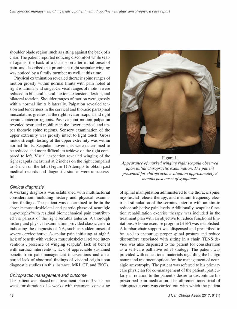
48 J Can Chiropr Assoc 2017; 61(1)
Chiropractic management of a geriatric patient with idiopathic neuralgic amyotrophy: a case report
shoulder blade region, such as sitting against the back of a chair. The patient reported noticing discomfort while seat-ed against the back of a chair soon after initial onset of pain, and described that prominent right scapular winging was noticed by a family member as well at this time. Physical examination revealed thoracic spine ranges of motion grossly within normal limits with pain noted at right rotational end range. Cervical ranges of motion were reduced in bilateral lateral flexion, extension, flexion, and bilateral rotation. Shoulder ranges of motion were grossly within normal limits bilaterally. Palpation revealed ten-sion and tenderness in the cervical and thoracic paraspinal musculature, greatest at the right levator scapula and right serratus anterior regions. Passive joint motion palpation revealed restricted mobility in the lower cervical and up-per thoracic spine regions. Sensory examination of the upper extremity was grossly intact to light touch. Gross motor strength testing of the upper extremity was within normal limits. Scapular movements were determined to be reduced and more difficult to achieve on the right com-pared to left. Visual inspection revealed winging of the right scapula measured at 2 inches on the right compared to ½ inch on the left. (Figure 1) Attempts to obtain past medical records and diagnostic studies were unsuccess-ful.
Clinical diagnosisA working diagnosis was established with multifactorial consideration, including history and physical examin-ation findings. The patient was determined to be in the chronic musculoskleletal and paretic phase of neuralgic amyotrophy1with residual biomechanical pain contribut-ed via paresis of the right serratus anterior. A thorough history and physical examination provided classic criteria indicating the diagnosis of NA, such as sudden onset of severe cervicothoracic/scapular pain initiating at night1, lack of benefit with various musculoskeletal related inter-ventions1, presence of winging scapula1, lack of benefit with cardiac intervention, lack of appreciable sustained benefit from pain management interventions and a re-ported lack of abnormal findings of visceral origin upon diagnostic studies (in this instance, MRI, CT, and EKG).
Chiropractic management and outcomeThe patient was placed on a treatment plan of 3 visits per week for duration of 4 weeks with treatment consisting
of spinal manipulation administered to the thoracic spine, myofascial release therapy, and medium frequency elec-trical stimulation of the serratus anterior with an aim to reduce subjective pain levels. Additionally, scapular func-tion rehabilitation exercise therapy was included in the treatment plan with an objective to reduce functional lim-itations. A home exercise program (HEP) was established. A lumbar chair support was dispensed and prescribed to be used to encourage proper spinal posture and reduce discomfort associated with sitting in a chair. TENS de-vice was also dispensed to the patient for consideration as a self-care palliative relief strategy. The patient was provided with educational materials regarding the benign nature and treatment options for the management of neur-algic amyotrophy. The patient was referred to his primary care physician for co-management of the patient, particu-larly in relation to the patient’s desire to discontinue his prescribed pain medication. The aforementioned trial of chiropractic care was carried out with which the patient
Figure 1.
Appearance of marked winging right scapula observed upon initial chiropractic examination. The patient
presented for chiropractic evaluation approximately 8 months post onset of symptoms.
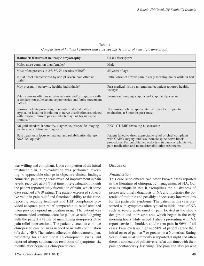
J Can Chiropr Assoc 2017; 61(1) 49
J Gliedt, JM Goehl, DP Smith, CJ Daniels
was willing and compliant. Upon completion of the initial treatment plan, a re-evaluation was performed reveal-ing no appreciable change in objective clinical findings. Numerical pain rating scale revealed improvement in pain levels, recorded at 0-1/10 at time of re-evaluation; though the patient reported daily fluctuation of pain, which some days reached a 7/10 rating. The patient expressed subject-ive value in pain relief and functional ability at this time, reporting ongoing treatment and HEP compliance pro-vided adequate pain relief comparable to relief obtained from previous opioid medication usage. The patient was recommended continued care for palliative relief aligning with the patient’s values of maintaining non-prescriptive pain relief interventions. The patient elected to continue chiropractic care on an as needed basis with continuation of a daily HEP. The patient adhered to this treatment plan, presenting for an additional 18 chiropractic visits, and reported abrupt spontaneous resolution of symptoms six months after beginning chiropractic care.
Discussion
PresentationThis case supplements two other known cases reported in the literature of chiropractic management of NA. Our case is unique in that it exemplifies the elusiveness of proper and timely diagnosis of NA and illustrates the po-tential of multiple and possibly unnecessary interventions for this particular syndrome. The patient in this case pre-sented with symptoms often typical to initial onset of NA such as severe acute onset of pain located in the shoul-der girdle and thorax/rib area which began in the early morning hours while in bed. Patients presenting with NA report cervical, shoulder, and/or arm pain in 96% of all cases. Pain levels are high and 90% of patients grade their initial onset of pain at 7 or greater on a Numerical Rating Scale.1 Pain most commonly is reported at night and often there is no means of palliative relief at this time, with their pain spontaneously lessening. The pain can also present
Table 1. Comparison of hallmark features and case specific features of neuralgic amyotrophy
Hallmark features of neuralgic amyotrophy Case Descriptors
Males more common than females8 Male
Most often presents in 2nd, 3rd, 7th decades of life8,9 85 years of age
Initial onset characterized by abrupt severe pain often at night1,2
Initial onset of severe pain in early morning hours while in bed
May present in otherwise healthy individuals2 Past medical history unremarkable; patient reported healthy lifestyle
Patchy paresis often in serratus anterior and/or trapezius with secondary musculoskeletal asymmetries and faulty movement patterns1
Prominent winging scapula and scapular dyskinesia
Sensory deficits presenting in non-dermatomal pattern atypical in location in relation to nerve distribution associated with involved muscle paresis which may last for weeks to months.1,7
No sensory deficits appreciated at time of chiropractic evaluation at 8 months post-onset
No gold standard laboratory, diagnostic, or specific imaging test to give a definitive diagnosis7
EKG, CT, MRI revealing no causation
Best treatments focus on manual and rehabilitation therapy, NSAIDs, opioids7
Patient failed to show appreciable relief of chief complaint with CABG surgery and two thoracic spine nerve block procedures; Patient obtained reduction in pain complaints with pain medication and manual/rehabilitation treatments

50 J Can Chiropr Assoc 2017; 61(1)
Chiropractic management of a geriatric patient with idiopathic neuralgic amyotrophy: a case report
with an intermittent onset and can take up to 3 weeks to transition out of the acute onset phase.1,7 During the acute phase, pain fails to respond well to traditional palliative treatments.7
Once the acute onset stage is over, paresis and sub-sequent local musculoskeletal dysfunction and possible atrophy most often will occur.1 While NA patients often have several symptoms including pain, paresis, shoulder dysfunction, and atrophy, they are not all necessarily con-sistently present at the same juncture1 or periods of time in any given presentation. When present, shoulder girdle dysfunction results from scapular instability due to the functional loss of the serratus anterior, rhomboid, or the trapezius muscles.1 The majority of patients will experi-ence additional periods of pain once the initial onset is over.1 These subsequent pain experiences are thought to be the result of two consequences of the NA syndrome process. First, the irritated or injured nerves in the plexus can result in amplified mechanical sensitivity, producing neuropathic pain in the affected nerves’ regions from in-creased strain on them with extension, abduction or ele-vation of the arm. This may last from a few weeks up to months in any given case.1 Secondly, due to the dys-function associated with local neuromsuculoskeletal tis-sue such as local shoulder girdle muscular paresis, proper biomechanics may be strained and place excessive stress on compensating musculature resulting in ongoing pain throughout the progression of NA.1
Differential diagnosisA thorough physical exam should be specific and com-plete to help distinguish between the multiple conditions which may present with similar features as NA. One of the hallmark signs of NA presenting in the upper extremity is substantial scapular winging and scapular dyskinesia. Though serratus anterior paresis is commonly thought to be the main offended musculature resulting in musculo-skeletal asymmetry/abnormalities, other muscles of the shoulder girdle may be affected resulting in presentations. For example, trapezius paresis may cause an appearance of a depressed shoulder and scapular sagging.1 Many pa-tients will subsequently develop musculoskeletal related pain in the region of the compensating musculature, often including periscapular region.1
Sensory examination of NA patients may reveal a non-specific pattern that is not consistent with localiza-
tion of pain or regular dermatomal distribution related to the paresis of affected musculature7, though could show pain with palpation of the affected peripheral nerve. How-ever it is important to recognize that sensory deficits and hypersensitivity may dissipate in weeks to months post-onset.1
NA is a clinical diagnosis and does not have a gold standard laboratory, diagnostic, or specific imaging test to give a definitive diagnosis7 though some studies sug-gest utilization of electroneuromyography and magnetic resonance imaging (MRI) may be helpful in confirming diagnosis.4 Previous studies have reported appreciable change in electroneuromyography studies identified as early as three weeks post onset of symptoms.4,13 These alterations typically entail acute denervation and sugges-tion of situational axonal degeneration.4 Though these studies are sensitive for identifying denervation, assum-ing NA will lead to denervation might only be partially accurate as sampling error leading to a negative study is possible.7
Sensory nerve conduction studies have also been shown to be a poor study in assessing for the presence of NA. These studies have been reported to fail to show abnormalities in 80% of patients with NA, even when the affected nerves are examined.7 Thus, a normal sensory nerve conduction study should not eliminate the inclusion of NA in the differential diagnosis.7 MRI findings asso-ciated with NA include intramuscular edema and muscle atrophy, which may show associated fatty infiltration.4,14,15 It is important, though, to emphasize the notion that diag-nostic studies indicating these findings should be viewed as only one piece of the entire clinical picture. These diag-nostic studies results should not be used to rule out the clinical diagnosis of NA, but can be used as additional rationale for the confirmation of the diagnosis.16
In this case, the patient reported prior diagnostic stud-ies, though could only recall the use of electrocardiogram, computed tomography, and magnetic resonance studies. It is not known if an electroneuromyography study was administered. It appears that diagnostic studies utilized in this case provided ability to aid in confirmation of NA diagnosis. In spite of the many conditions that may be considered part of the differential diagnosis, a meticulous history and physical exam should help to distinguish NA from other considerations.7 Nonetheless, NA and its diagnosis seem
J Can Chiropr Assoc 2017; 61(1) 51
J Gliedt, JM Goehl, DP Smith, CJ Daniels
to be relatively foreign to many health care providers7 and delayed diagnosis, misdiagnosis and mismanagement are not uncommon. In this case, the patient completed mul-tiple treatment options for several months with minimal relief. The patient in this case identified as presenting with common features associated with NA and responded well to a subsequent course of chiropractic treatments and self-care treatments for ongoing management of symptoms.
Management and PrognosisFailure to properly identify neuralgic amyotrophy, par-ticularly in the acute phase, can lead to substantial mis-management and potential for suffering for several weeks. Approaches to care may focus primarily on conservative treatment options including manual and rehabilitation therapy, as well as traditional medical treatment options including long acting non-steroidal anti-inflammatory medications, opioids, and corticosteroid injection.7,17,18 During the initial unrelenting pain presentation there is only pharmaceutical options and corticosteroid injection that have been shown to potentially manage symptoms and hasten recovery.1,7,17,18
Manual and rehabilitation therapy may be indicated due to the biomechanical changes that ensue in the latter phase of NA. The objective of these treatments is to sta-bilize the musculoskeletal system about the area of insult. Often NA presents in the lower neck and shoulder region and thus typical therapy involves stabilizing the scapula by strengthening the various muscles around the shoulder girdle.1 The serratus anterior muscle is frequently a site of local paresis and presents a hurdle for therapy creating an inability to produce and maintain proper shoulder mech-anics and muscular endurance. Thus, a graded progres-sion must be utilized to allow for strengthening while not fatiguing the muscles to the point that they contribute to further shoulder and scapular dysfunction. Improving sta-bilization about the shoulder girdle and reducing the pres-ence of scapular winging is important in the long-term success of patient suffering from NA. Potential goals of treatments aim to restore faulty movement patterns and reduce biomechanical imbalances that may have resulted from adaptive alterations from NA. Various other treatment options have shown some indi-cation for continued study to further assess for their ability to aid in the management of NA patients. There is some thought of autoimmune influence in the manifestation of
NA and thus some treatment efforts have been focused on associated management with immunomodulants such as prednisone, intravenous immunoglobulins and other pro-phylactic immunotherapies.7 However, few studies have been conducted in this area of NA management and to date there is no conclusive evidence to recommend this mode of intervention. Surgical intervention for NA pa-tients suffering from substantial nerve injury, consisting of hourglass constriction (shown by intraoperative visual-ization or nerve ultrasound), has shown some prelimin-ary success.7,19,20 Future studies are still needed, though, to determine if surgical intervention is a viable routine treatment option.7
Patients generally regain strength in affected paretic muscles, though roughly one-third of NA patients still re-port some form of dysfunction or symptom even six years post initial presentation.1,7 Previous studies have shown no well-defined link between neurological considerations and functional outcomes or a specific natural course; though studies have identified a strong association be-tween persistent pain, fatigue and the presence of scapular instability.7,21,22
LimitationsThis is a case report which only describes the findings of this individual case. The findings of this study cannot be generalized to other patients or the general public. Reso-lution of symptoms in this case may not have been related to chiropractic care and may have been due to the natural progression of NA. In this case there was no availability of past medical records or diagnostic studies for review. This case relied heavily on communication with the pa-tient and obtaining a thorough patient history, which is subject to potential recall bias.
SummaryThis case features the importance of timely recognition and proper diagnosis of NA. Recognition of NA can be difficult due to the nature of NA as a clinical diagnosis which does not have a gold standard laboratory, diagnos-tic or specific imaging test to establish a definitive diag-nosis, and the fact that it mimics so many other etiologies. Prompt recognition of common features associated with NA and differentiation from other diagnostic consider-ations can be appreciated with a thorough history and examination, which identifies the classic criteria for a

52 J Can Chiropr Assoc 2017; 61(1)
Chiropractic management of a geriatric patient with idiopathic neuralgic amyotrophy: a case report
diagnosis of NA. Prognosis is commonly characterized by fragmented recovery from symptoms associated with NA, with a potential for an extended length of time for resolution and recurrence and continued reports of some degree of residual dysfunction are common even up to several years after initial onset.
References1. van Alfen, N. The neuralgic amyotrophy consultation.
J Neurol. 2007; 254(6): 695-704.2. Feinberg JH, Radecki J. Parsonage-turner syndrome.
HSS J. 2010;6(2):199-205.3. Tjoumakaris FP, Anakwenze OA, Pulos N. Neuralgic
amyotrophy (Parsonage-Turner syndrome). J Am Acad Orthop Surg. 2012; 20(7): 443-449.
4. Monteiro Dos Santos RB, Dos Santos SM, Carneiro Leal FJ, Lins OG, Magalhaes C, Mertens Fittipaldi RB. Parsonage-Turner Syndrome. Rev Bras Ortop. 2015;50(3):336-341.
5. Parsonage MJ, Turner JW. Neuralgic amyotrophy: the shoulder-girdle syndrome. Lancet. 1948;1(6513):973-978.
6. van Alfen N, van Engelen BG. The clinical spectrum of neuralgic amyotrophy in 246 cases. Brain. 2006; 129(Pt 2):438-450.
7. van Eijk JJ, Groothuis JT, van Alfen N. Neuralgic amyotrophy: an update on diagnosis, pathophysiology and treatment. Muscle Nerve. 2016 Dec 10. [Epub ahead of print].
8. Beghi E, Kurland LT, Mulder DW, Nicolosi A. Brachial plexus neuropathy in the population of Rochester, Minnesota, 1970–1981. Ann Neurol. 1985; 18(3): 320-323.
9. Charles E. Chiropractic management of a 30-year-old patient with Parsonage-Turner syndrome. J Chiropr Med. 2011;10(4): 301-305.
10. Misamore GW, Lehman DE. Parsonage-Turner syndrome (acute brachial neuritis). J Bone Joint Surg Am. 1996; 78(9): 1406-1408.
11. Yang JS, Cho YJ, Kang SH, Choi EH. Neuralgic
amyotrophy manifesting as mimicking posterior interosseous nerve palsy. J Korean Neurosurg Soc. 2015;58(5):491-493.
12. Rix GD, Rothman EH, Robinson AW. Idiopathic neuralgic amyotrophy: an illustrative case report. J Manipulative Physiol Ther. 2006; 29(1): 52-59.
13. Rubin DR. Neuralgic amyotrophy: clinical features and diagnostic evaluation. Neurologist. 2001;7(6):350-356.
14. Scalf RE, Wenger DE, Frick MA, Mandrekar JN, Adkins MC. MRI findings of 26 patients with Parsonage-Turner syndrome. Am J Roentgenol. 2007;189(1):W39-44.
15. Gaskin CM, Helms CA. Parsonage-Turner syndrome: MR imaging findings and clinical information of 27 patients. Radiology. 2006;24D(2):501-507.
16. van Eijk J. van Alfen N. Neuralgic amyotrophy. Am J Roentgenol. 2011;196(6):W858; author reply W859.
17. Smith DP, Elliott JA, Helzberg JH. Intravenous corticosteroid therapy for bilateral parsonage-turner syndrome: a case report and review of the literature. Reg. Anesth Pain Med. 2014;39(3):243-247.
18. Smith CC, Bevelagua AC. Challenging pain syndrome: Parsonage-Turner syndrome. Phys Med Rehabil Clin N Am. 2014;25(2):265-277.
19. Pan Y, Wang S, Zheng D, Tian W, Tian G, Ho PC, Cheng HS, Zhong Y. Hourglass-like constrictions of peripheral nerve in the upper extremity: a clinical review and pathological study. Neurosurgery. 2014;75(1):10-22.
20. Pan YW, Wang S, Tian G, Li C, Tian W, Tian M. Typical brachial neuritis (Parsonage-Turner syndrome) with hourglass-like constrictions in the affected nerves. J Hand Surg Am. 2011;36(7):1197-1203.
21. Cup EH, Ijspeert J, Janssen RJ, Bussemaker-Beumer C; Jacobs J, Pieterse AJ, van der Linde H, van Alfen N. Residual complaints after neuralgic amyotrophy. Arch Phys Med Rehabil. 2013;94(1):67-73.
22. van Alfen N, van der Werf SP, van Engelen BG. Long-term pain, fatigue, and impairment in neuralgic amyotrophy. Arch Phys Med Rehabil. 2009;90(3):435-439.
J Can Chiropr Assoc 2017; 61(1) 53
ISSN 0008-3194 (p)/ISSN 1715-6181 (e)/2017/53–64/$2.00/©JCCA 2017
Selecting and training opinion leaders and best practice collaborators: experience from the Canadian Chiropractic Guideline InitiativeAndré E. Bussières, DC, MSc, PhD1,2 Michele Maiers, DC, MPH, PhD3 Diane Grondin, DC, MHK PhD (Student)4,5 Simon Brockhusen, MSc (Clin.Biomech), MD (Student)6,7
1 Assistant Professor, School of Physical and Occupational Therapy, McGill University2 Professor, Département Chiropratique, Université du Québec à Trois-Rivières3 Executive Director of Research and Innovation, Northwestern Health Sciences University4 Assistant Professor, Canadian Memorial Chiropractic College5 PhD student, Institute of Health Policy, Management and Evaluation, University of Toronto6 Research assistant, Nordic Institute of Chiropractic and Clinical Biomechanics7 MD (Student), University of Southern Denmark
Correspondence to:Andre BussièresSchool of Physical and Occupational Therapy, Faculty of Medicine, McGill University, 3630 Promenade Sir-William-Osler, Hosmer House, Room 205, Montreal, Quebec, Canada H3G 1Y5E-mail: [email protected]: Dr. Bussières is the CGI Project Lead of the Canadian Chiropractic Guideline Initiative and holds the Canadian Chiropractic Research Foundation Professorship in Rehabilitation Epidemiology.© JCCA 2017
Objectives: To describe the process for selecting and training chiropractic opinion leaders (OLs) and best practice collaborators (BPCs) to increase the uptake of best practice. Methods: In Phase 1, OLs were identified using a cross-sectional survey among Canadian chiropractic stakeholders. A 10-member committee ranked nominees. Top-ranked nominees were invited to a training workshop. In Phase 2, a national e-survey was administered to 7200 Canadian chiropractors to identify
Objectifs : Décrire le processus permettant de choisir et former les leaders d’opinion (LO) et collaborateurs des pratiques d’exemplaire (CPE) en chiropratique dans le but de favoriser l’adoption des pratiques d’excellence. Méthodologie : Lors de la première phase, on a désigné les LO au moyen d’une enquête transversale parmi les intervenants canadiens de la chiropratique. Un comité composé de dix membres a classé les candidats. Les candidats les mieux classés ont été invités à un atelier de formation. Lors de la deuxième phase, 7 200 chiropraticiens canadiens se sont soumis à une enquête nationale en ligne visant à désigner d’autres LO et CPE. Les noms recommandés ont été présélectionnés par les LO et le choix final s’est fait

54 J Can Chiropr Assoc 2017; 61(1)
Selecting and training opinion leaders and best practice collaborators: experience from the Canadian Chiropractic Guideline Initiative
BackgroundClinical practice guidelines (CPGs) are widely recog-nized as a foundational tool to inform clinical decision making.1 When followed, CPGs have the potential to im-prove health outcomes, as well as the efficiency of the health care system.2,3 Low adherence to CPGs for the management of musculoskeletal disorders and spine care in particular contributes to a wide variation in services seen across several health disciplines including chiro-practic.4-7 Implementing guidelines typically requires change at multiple levels, including attitudinal and behav-ioural change by clinicians and promoting use of CPGs by stakeholders (e.g., governing bodies and professional associations).8
The Canadian Chiropractic Guideline Initiative (CCGI) was launched over a decade ago by the national chiropractic professional organizations in Canada in order to develop CPGs to improve quality care9 and decrease the burden of musculoskeletal disorders10. The mission of the CCGI is to develop, disseminate and help implement CPGs among Canadian chiropractors.11
Prior work explored the determinants of guideline ad-herence among health care professionals using the Theor-etical Domain Framework (TDF)12, a framework previ-ously applied in a wide range of disciplines, settings and
contexts13. Interviews of chiropractors and professional leaders in Canada14,15 suggested that guideline adherence is potentially influenced by the theoretical domains of social influence and social/professional role and identity. In other words, the set of behaviours and personal qual-ities displayed in social or work settings (social influence) and the processes existing between people that have the potential to influence thoughts, feelings or behaviours (social/professional role and identity) can lead to behav-iour change.16 Mapping of Behaviour Change Techniques (BCTs)17 on to these theoretical domains suggested that social processes of encouragement, pressure, support, and modeling/demonstration of behaviour are important techniques for changing professional behaviour15. Change agents (people who, by the nature of their position or abilities are particularly capable of promoting change) are well suited to accomplish these BCTs. This is partly because peer pressure to conform to social norms affects behavioural intention, an important predictor of individ-uals’ behaviour.18
Implementation research supports the use of change agents, including practitioners, to deliver evidence-based recommendations and programs to improve the quality of care.19,20 Opinion leaders (OLs) are individuals who have formal or informal influence on the attitudes, beliefs and
additional OLs and BPCs. Recommended names were screened by OLs and final selection made by consensus. Webinars were utilized to train BPCs to engage peers in best practices, and facilitate guideline dissemination. Results: In Phase 1, 21 OLs were selected from 80 nominees. Sixteen attended a training workshop. In Phase 2, 486 chiropractors recommended 1126 potential BPCs, of which 133 were invited to participate and 112 accepted. Conclusions: OLs and BPCs were identified across Canada to enhance the uptake of research among chiropractors. (JCCA. 2017;61(1):53-64) k e y w o r d s : chiropractic; change agents; opinion leaders; survey; selection; training
d’un commun accord. On s’est servi de webinaires pour former les LO à encourager leurs pairs à adopter des pratiques d’excellence et faciliter la diffusion des lignes directrices. Résultats : Lors de la première phase, on a choisi 21 LO parmi 80 candidats. Seize d’entre eux ont assisté à un atelier de formation. Lors de la deuxième phase, 486 chiropraticiens ont recommandé 1 126 LO potentiels, parmi lesquels 133 ont été invités à participer et 112 ont accepté. Conclusions : On a désigné des LO et CPE à l’échelle du pays pour favoriser l’adoption de la recherche parmi les chiropraticiens. (JCCA. 2017;61(1):53-64) m o t s c l é s : chiropratique, agents de changement, leaders d’opinion, enquête, choix, formation

J Can Chiropr Assoc 2017; 61(1) 55
AE Bussières, M Maiers, D Grondin, S Brockhusen
behaviours of their colleagues and stakeholder organiz-ations.21,22 Generally viewed by their peer group as like-able, trustworthy and influential, OLs play an important role in promoting health care initiatives, in leveraging established relationships to create access points within a target community, in communicating key messages23, and in demonstrating and reinforcing desired behaviour24. As respected members of a community, their spheres of in-fluence provide an opportunity for long-term subsistence of an initiative, long after active intervention has conclud-ed.25 Reviews on the effectiveness of OL interventions suggest that a 12% absolute increase in compliance with an intervention may be expected when using OLs to pro-mote evidence-based practice.26
By extension, best practice collaborators (BPCs) can also be seen as important change agents. Here BPCs are defined as individuals who are also seen as caring, know-ledgeable and good educators and who assist the OLs in their role. Recognizing the use of change agents as an important strategy to influence professional behaviour change, the current paper outlines the strategy used by the CCGI to identify and train OLs and BPCs to promote the use of best practice within the Canadian chiroprac-tic profession broadly (i.e., practicing chiropractors and leaders of national and provincial associations, regula-tory boards, liability insurance carriers, and other stake-holders).
Methods
Phase 1: Opinion LeadersThe aim of Phase 1 was to appoint OLs to assist the CCGI with the dissemination and implementation of CPGs among Canadian chiropractors and support professional leaders engaged in the process of guideline implementa-tion.
Study designA cross-sectional research design was employed to iden-tify OLs in the chiropractic profession in all 10 provinces across Canada.
Participants and recruitmentCCGI stakeholders, professional leaders of national and provincial chiropractic associations and regulatory boards in Canada (n=50) who attended an annual meeting in
February 2014 were asked to complete a survey question-naire.
Survey questionnaireThe questionnaire asked participants to provide up to three names of Canadian chiropractors they felt to be suit-able to serve as OLs as well as their reasons to nominate. Nominations were based on the chiropractors’ skills and attributes, specifically, whether they were educationally influential, knowledgeable and humanistic, along with their sphere of influence (i.e., clinicians and/or profes-sional leaders/decision makers)(Appendix 1).27
Data collectionFor each nominee, general demographic and practice data were collected from internet searches on publicly avail-able search engines such as Google and social media, the practitioner’s clinic website and provincial/regulatory websites. Data gathered from these searches were com-plemented by information provided by the members of the selection committee based on knowledge of the can-didate through prior work (described below). A candidate profile was established in an Excel spreadsheet for each nominee, including their gender, the number of years in practice, their participation (or not) in continuing educa-tion activities, presentations given to their peers, academ-ic and publication record, and the presence/absence of prior liability issues or regulatory complaints.
Data analysisA 10-member selection committee of CCGI stakeholders was established to review the candidate profiles. To ob-tain a wide range of views, perspectives and professional experience, the committee was composed of a purpos-ive sample from diverse geographical settings including members of a chiropractic specialty college (n=2), a field practitioner (n=1), academics (n=2), leaders from prov-incial associations and regulatory boards (n=3), and re-searchers (n=2). To guide the selection process, committee mem-bers considered pre-established criteria adapted from Rycroft-Malone27 to help inform their selection, including geographical location, past and current level of engage-ment with the profession, availability, attitudes and be-liefs toward evidence-based practice and CPGs, and prior teaching experience. Other desirable attributes included

56 J Can Chiropr Assoc 2017; 61(1)
Selecting and training opinion leaders and best practice collaborators: experience from the Canadian Chiropractic Guideline Initiative
Self-reporting method (CCGI stakeholders propose
themselves)
Scre
enin
g In
vite
d El
igib
ility
Id
entif
ying
Peer Nomination Method
(CCGI stakeholders propose up to 3 names of potential candidates)
Potential CCGI Opinion Leaders (n = 80)
Characteristics extracted Licensed in Canada Gender Years in practice Participation in continuing
education activities Lecturing to peers Academic and publication record Presence or absence of a history of
liability issues or regulatory complaints
Criteria for consideration Rounds 1-3
Attitudes and beliefs toward evidence-based practice and guidelines
Past and current level of engagement with the profession
Prior teaching experience Perceived credibility across different
professional groups Authority and presence Communication skills Ability to convince colleagues about
issues related to clinical practice Ability to treat all colleagues with
respect Geographical location Availability
CCGI stakeholders invited to complete the survey questionnaire (n=50)
Round 1: Candidates (n=48) ranked for Practitioners (n=21), Decision Makers (n=9), Both (n=18)
Candidates selected and initially invited to participate (n=21), Declined/withdrew (n=9), additional candidates invited (n=5)
Rank
ing
Candidate profiles to ascertain eligibility as Opinion Leaders (n = 79)
Round 2: Candidates with a mean score >5.5/7 ranked as 1st, 2nd & 3rd choices (n=35); Practitioners
(n=12), Decision Makers (n=7), Both (n=16)
CCGI Opinion Leaders appointed April 2015 (n=17), additional appointments after Nov 2015 (n=5). OLs in May 2016 (n=22)
Round 3: Consensus reached over top 2-3 candidates in each province (n=21)
Figure 1.
Flowchart illustrating the systematic process used to identify Canadian chiropractic OLs.

J Can Chiropr Assoc 2017; 61(1) 57
AE Bussières, M Maiers, D Grondin, S Brockhusen
being perceived as being credible and/or as an authority figure by their peers, having good communication skills, treating all colleagues with respect, and being capable of influencing colleagues about issues related to clinic-al practice (Figure 1). Potential OLs needed to have ex-perience dealing with a range of practicing chiropractors (e.g. working in solo vs. multidisciplinary practices, those with an evidence-based vs. alternative philosophy, and non-specialist vs. specialist chiropractors) and different professional groups. The committee formally met online on three occasions between June and August 2014 to undertake a two-round consensus process to identify nominees that would be invited to become OLs. In round 1, members of the se-lection committee considered the above criteria to rank candidates using a Likert scale from 1-7 (“1” = strongly disagree and “7” = strongly agree) on their likelihood of being influential toward practitioners and/or stakeholders. Mean scores were calculated and candidates with scores over 5.5 out of 7 were considered in the next round. In round 2, names retained were grouped by province and ranked by committee members as first, second and third choices. In round 3, committee members reached consen-sus over the top 2-3 candidates. These were then invited by e-mail to serve as OLs. A follow-up call was made by the project lead (AB) to provide detail about the OL program and answer any questions they had about the program. Other nominated individuals were kept in the retained list.
ResultsCCGI stakeholder survey response rate was 38% (19/50), providing names of 80 potential OLs (Figure 1). One can-didate was from the US and was excluded. In round 1, median scores across identified potential OLs were 5.19 (0.73) for ‘Practitioners’ and 5.44 (0.75) for ‘Leaders/de-cision makers’. To retain 40% of potential OLs, candi-dates with scores of 5.5 or above (n=48) were considered in the next round, including OLs for ‘Practitioners’ (n=21), ‘Leaders/decision makers’ (n=9) or nominated in both categories (n=18). In round 2, 35 candidates ranked as either first, second and third choices were retained for the following round, including potential OLs for ‘Prac-titioners’ (n=12), ‘Leaders/decision makers’ (n=7) and 16 for both ‘Practitioners’ and ‘Leaders/decision makers’ roles. In round 3, the selection committee made final rec-
ommendations for 21 OLs, including two in each prov-ince with the exception of a larger province (three in On-tario and Quebec) and a smaller province (one in Prince Edward Island). Of the 21 clinicians and professional leaders/decision makers in the profession who received an invitation letter to become a CCGI OL, 5 declined due to time constraints or lack of interest. Five alternate can-didates were invited from the retained list and, of these, 4 withdrew for similar reasons, leaving 17 OLs. Because some new OLs admitted to having limited availability for this project, additional appointments were made from the retained list in November 2015 for Nova Scotia (n=1), and in March 2016 for Saskatchewan (n=1) and Alberta (n=3), bringing the total number of OLs in place to 22 by May 2016. The names of OLs in each province are avail-able at: http://www.chiropractic.ca/guidelines-best-prac-tice/about-us/meet-the-team/opinion-leaders/.
Training workshop for Opinion LeadersSixteen OLs were available to attend a one-day workshop in Toronto, Ontario in February 2015. While participation in the OL program is voluntary, traveling expenses were reimbursed. The session was co-developed and delivered by a Certified Executive Leadership Coach with the as-sistance of three academic researchers with clinical train-ing in chiropractic (AB, MM, DG) and one chiropractor and medical student (SB) and a research manager. Con-tent of the workshop was informed by the literature and addressed five essential activities of the implementation process: engaging, planning, reflecting, executing and evaluating.28 The workshop objectives and agenda can be found in Appendix 2. Two weeks prior to the workshop participants received an online invitation to complete the Strength Deployment Inventory (SDI®), a tool aimed at assessing self-aware-ness, conflict resolution and team functioning strategies.29 During the workshop, the Certified Executive Leadership Coach presented the results of the self-assessments, aim-ing to raise the self-awareness of participants and form-ing the basis for enhancing their ability to communicate more effectively, handle conflict more productively, and improve relationships. This was further explored in the context of the roles and personal qualities of effective OLs. Participants were asked to identify stakeholders who they could directly or indirectly influence. Participants

58 J Can Chiropr Assoc 2017; 61(1)
Selecting and training opinion leaders and best practice collaborators: experience from the Canadian Chiropractic Guideline Initiative
were then divided into three groups to identify potential audiences they had access to regionally (e.g. patients, de-cision-makers, clinicians, multidisciplinary teams) and determine those upon which they had the most/least influ-ence. After a brief overview of the content of the CCGI website (www.chiroguidelines.org), participants were given time to explore its resources and tools. Comments received in the end-of workshop feedback questionnaire suggested that this activity successfully addressed an im-portant need of OLs to become familiar with CCGI tools and resources on CPGs and EIP. To provide insight about the OLs’ expectations about their participation in the workshop, attendees were asked to do a ‘3-2-1 exercise’, listing three important things they had learned during the workshop, two questions they felt remained unanswered, and one suggestion for next steps. Results from the exercise indicated a need for addi-tional resources to help with their tasks, such as summar-ies of key guideline recommendations, PowerPoint pres-entations for board meetings or continuing educational events, a Question and Answer sheet to address potential questions by clinicians, and patient handouts and post-ers. Participants were invited to draft an ‘Opinion Leader Action Plan’ to identify upcoming opportunities of influ-ence and to outline the perceived needs for resources from the CCGI. A detailed summary of the workshop was forwarded to the participants with an invitation to attend a follow-up teleconference call. During periodic follow-up teleconfer-ence calls, OLs provided updates on their progress and their plans for dissemination in their respective provinces.
Phase 2: Best Practice CollaboratorsThe aim of phase 2 was to confirm the additional OL se-lection and appoint additional OLs and BPCs to comple-ment and assist trained OLs.
Study design A descriptive cross-sectional survey was conducted on-line 20 months after phase 1 (REB Approval: 1507X01, Canadian Memorial Chiropractic College).
Participants and recruitmentAn invitation to complete a 10-minute national online survey was sent by the Canadian Chiropractic Associ-
ation (CCA) by e-mail to all members with a valid e-mail address (n=7200). It included a description of the study and a link to access the survey. Those who returned their completed survey were eligible to win one of two $100 Indigo gift cards.
Survey questionnaireThe survey was adapted from Hiss’ sociometric question-naire administered to care providers30,31 and was divided into four sections (Appendix 3). Section I contained eight questions to find out how respondents give/receive infor-mation from colleagues (e.g. In general, do you talk to other chiropractors about clinical or professional issues in chiropractic practice?). They had to indicate, on a 5-point Likert scale, the answer that best represented their be-haviour (“1” = Never and “5” = Very often). In section II, respondents were asked to provide the names of three chiropractors in their province who best fit the following roles: educator, knowledgeable practitioner, and caring professional. In section III, the survey asked respondents to suggest up to 18 names of colleagues they would turn toward for: 1) their ability to give good advice, 2) discuss-ing challenging cases, 3) information on referrals resour-ces, and 4) socializing. Section IV asked 10 demographic questions about participants and their practice.
Data collectionData collection took place over a three-week period in the fall of 2015. The survey was available in English and in French and delivered on FluidSurveys (www.fluidsur-veys.com). Data was submitted by respondents electron-ically immediately after completion and a list of all the proposed names of CCGI collaborators were entered into an Excel spreadsheet by province.
Data analysisTo further refine the list of names proposed by clinicians, OLs selected in phase 1 were asked to indicate if they con-sidered the individuals nominated as potential BPCs had the skills and attitudes required to help accomplish their tasks to promote EIP, guidelines and best practice. They were asked to classify the nominees in one of three cat-egories: 1) In my opinion, this candidate carries influence over their peers in the area of evidence-informed practice; 2) In my opinion, this candidate does not carry influence over their peers in the area of evidence-informed practice;
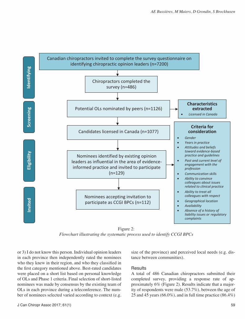
J Can Chiropr Assoc 2017; 61(1) 59
AE Bussières, M Maiers, D Grondin, S Brockhusen
or 3) I do not know this person. Individual opinion leaders in each province then independently rated the nominees who they knew in their region, and who they classified in the first category mentioned above. Best-rated candidates were placed on a short list based on personal knowledge of OLs and Phase 1 criteria. Final selection of short-listed nominees was made by consensus by the existing team of OLs in each province during a teleconference. The num-ber of nominees selected varied according to context (e.g.
size of the province) and perceived local needs (e.g. dis-tance between communities).
ResultsA total of 486 Canadian chiropractors submitted their completed survey, providing a response rate of ap-proximately 6% (Figure 2). Results indicate that a major-ity of respondents were male (53.7%), between the age of 25 and 45 years (66.0%), and in full time practice (86.4%)
Canadian chiropractors invited to complete the survey questionnaire on identifying chiropractic opinion leaders (n=7200)
Iden
tifyi
ng
Chiropractors completed the survey (n=486)
Potential OLs nominated by peers (n=1126)
Scre
enin
g Criteria for consideration
Gender Years in practice Attitudes and beliefs
toward evidence-based practice and guidelines
Past and current level of engagement with the profession
Communication skills Ability to convince
colleagues about issues related to clinical practice
Ability to treat all colleagues with respect
Geographical location Availability Absence of a history of
liability issues or regulatory complaints
Candidates licensed in Canada (n=1077)
Nominees identified by existing opinion leaders as influential in the area of evidence-informed practice and invited to participate
(n=129)
Elig
ibili
ty
Characteristics extracted
Licensed in Canada
Nominees accepting invitation to participate as CCGI BPCs (n=112)
Invi
ted
Figure 2:
Flowchart illustrating the systematic process used to identify CCGI BPCs

60 J Can Chiropr Assoc 2017; 61(1)
Selecting and training opinion leaders and best practice collaborators: experience from the Canadian Chiropractic Guideline Initiative
(Table 1). The largest percentage of participants was from Ontario (44.3%). Survey respondents volunteered names of 1077 colleagues from Canada, 47 individuals from the US and 2 from the UK. Among these, 338 were nomin-ated between 2-3 times, 132 between 4-7 times and 36 people over 8 times. All but one of the OLs selected in phase 1 were nominated within the top five choices in their province. OLs in place helped reduce the list to 129 names they believed had favourable attitudes and skills to assist them with their tasks as CCGI OLs. The potential BPCs received a letter informing them of the results of the survey and were asked if they were interested in collabor-ating with OLs in their respective province. A total of 112 chiropractors accepted the invitation to become BPCs. The list of names is available at: https://www.chiroprac-tic.ca/guidelines-best-practice/about-us/meet-the-team/best-practice-collaborators/.
Training of Best Practice CollaboratorsBPC nominees in each province were invited to attend a 45-minute webinar describing the mission and relat-ed strategies of the CCGI, the current role of OLs, and their potential contribution as BPCs (Table 2). BPCs were asked to help OLs promote the use of best practices using their own network of colleagues. OLs were encouraged to engage with new BPCs to discuss respective views about the CCGI mission and strategies, possible roles and con-tribution of collaborators and their availability for future guideline dissemination projects, as well as to ensure that they have access to and become familiar with the CCGI bi-lingual website, available tools (e.g., online learning mod-ules on EIP and guidelines) and resources. Participation in these various activities aims to provide BPCs with a base-line understanding of the work of the CCGI and to help them perform their new role. A Competency Development Program was developed for OLs and BPCs along with as-sociated performance indicators, including topics such as demonstrating adequate knowledge, effective communi-cation and ability to develop networks with colleagues to help disseminate best practice (Appendix 4).
DiscussionChiropractors across Canada occupying key positions within provincial, national and international organizations and academic institutions were identified and trained to serve as OLs or BPCs. In addition to facilitating the up-take and application of guidelines to improve chiropractic patient care within their networks, these individuals will help raise the credibility and visibility of the CCGI. Engaging team members tasked with implementing best practice and guidelines is often-overlooked.32 It is vital that members be carefully and thoughtfully selected or allowed to rise naturally, especially those considered to be ‘implementation leaders’ and ‘champions’.22 The decision about who to invite as OLs and BPCs for this project was informed by the scientific literature27,30,31 and group consensus. There are different conceptualizations of OLs21,33, one of which proposes that there are two types of OLs: experts and peers. Expert OLs exert influence through their authority and status, while peer OLs exert influence through their representativeness and credibil-ity.34 CCGI OLs and BPCs include a mixture of these types, and results from the stakeholder survey undertaken in phase II affirmed the committee’s selection.
Table 1: Profile of survey participants (November 2015)
Characteristics Proportion n (percentage)
Gender (n=339)Male: 182 (53.7%)Female: 157 (46.3%)Work Status (n=339)Part-time (< 12 hrs/wk): 46 (13.6%)Full-time: 292 (86.4%)Age (n=339)25-45: 224 (66.0%)46-65: 115 (18.9%)Practice location/Province (n=347)Ontario: 154 (44.3%)Quebec: 59 (16.7%)British Columbia: 46 (13.3%)Alberta: 46 (13.3%)Manitoba: 17 (5.0%)Saskatchewan: 9 (2.6%)Nova-Scotia: 9 (2.6%)New-Brunswick: 5 (1.4%)Newfoundland/Labrador: 2 (0.5%)

J Can Chiropr Assoc 2017; 61(1) 61
AE Bussières, M Maiers, D Grondin, S Brockhusen
Table 2: Roles and activities of CCGI Best Practice Collaborators
Roles and activities of CCGI Best Practice Collaborators
Education
Discuss key guideline recommendations with clinicians;
Support the implementation of best practice among the profession in their own area, in collaboration with existing local CCGI opinion leaders;
Participate in or lead activities, groups or events. (e.g. make presentations or assist with workshops on EIP at continuing education events or conferences in partnership with the local CCGI OL/BPC team);
Have a presence on social media (e.g., LinkedIn, CCGI YouTube) to encourage awareness of available CCGI resources;
If teaching, introduce learners to EIP by including it in coursework and highlighting the importance of EIP for clinicians in practice.
Relationship building Build relationships through regular communication with various stakeholders (e.g.: clinicians,
fellow BPCs/OLs); Create networks to encourage clinicians and stakeholders to follow CCGI; Share your own experiences with other BPCs and OLs regarding knowledge translation (KT)
strategies used within respective context and setting; Encourage clinicians to learn about and discuss EIP principles and guideline recommendations; Encourage clinicians to use CCGI tools and resources; Inform on how/where to access reliable information on CPGs or EIP principles; Address concerns clinicians express about EIP principles and guideline recommendations.
Advancing the profession
Raise awareness of the most recent research and encourage clinicians to use it in practice.
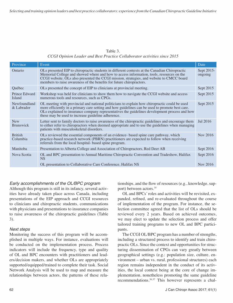
62 J Can Chiropr Assoc 2017; 61(1)
Selecting and training opinion leaders and best practice collaborators: experience from the Canadian Chiropractic Guideline Initiative
Early accomplishments of the OL/BPC programAlthough this program is still in its infancy, several activ-ities have already taken place across Canada, including presentations of the EIP approach and CCGI resources to clinicians and chiropractic students, communications with physicians and presentations to insurance companies to raise awareness of the chiropractic guidelines (Table 3).
Next stepsMonitoring the success of this program will be accom-plished in multiple ways. For instance, evaluations will be conducted on the implementation process. Process indicators will include the frequency, type and quality of OL and BPC encounters with practitioners and lead-ers/decision makers, and whether OLs are appropriately supported/equipped/trained to complete their task. Social Network Analysis will be used to map and measure the relationships between actors, the patterns of these rela-
tionships, and the flow of resources (e.g., knowledge, sup-port) between actors.35 OL and BPCs’ roles and activities will be revisited, ex-panded, refined, and re-evaluated throughout the course of implementation of the program. For instance, the se-lection committee agreed that the list of OLs should be reviewed every 2 years. Based on achieved outcomes, we may elect to update the selection process and offer tailored training programs to new OL and BPC partici-pants. The CCGI OL/BPC program has a number of strengths, including a structured process to identify and train chiro-practic OLs. Since the context and opportunities for struc-turing dissemination of CPGs can vary greatly between geographical settings (e.g.: population size, culture, en-vironment – urban vs. rural, professional structures) each region remains independent in the conduct of its activ-ities, the local context being at the core of change im-plementation, nonetheless promoting the same guideline recommendations.36,37 This however represents a chal-
Table 3. CCGI Opinion Leader and Best Practice Collaborator activities since 2015
Province Event DateOntario OLs presented EIP to chiropractic students in different contexts at the Canadian Chiropractic
Memorial College and showed where and how to access information, tools, resources on the CCGI website. OLs also presented the CCGI mission, strategies, and website to CMCC board members to raise awareness of the benefits for future chiropractors.
Sept 2015- ongoing
Québec OLs presented the concept of EIP to clinicians at provincial meeting. Sept 2015Prince Edward Island
Workshop was held for clinicians to show them how to navigate the CCGI website and access numerous tools and resources, such as CPGs.
Sept 2015
Newfoundland & Labrador
OL meeting with provincial and national politicians to explain how chiropractic could be used more efficiently in a primary care setting and how guidelines can be used to promote best care. OLs explained to insurance company representatives the guidelines development process and how these may be used to increase guideline adherence.
Sept 2015
New Brunswick
Letter sent to family doctors to raise awareness of the chiropractic guidelines and encourage them to either refer to chiropractors when deemed appropriate and to use the guidelines when managing patients with musculoskeletal disorders.
Jul 2016
British Columbia
OLs reviewed the essential components of an evidence- based spine care pathway, which practice-based research network (PBRN) practitioners are expected to follow when receiving referrals from the local hospital- based spine program.
Nov 2016
Manitoba Presentation to Alberta College and Association of Chiropractors, Red Deer AB Sept 2016Nova Scotia OL and BPC presentation to Annual Maritime Chiropractic Convention and Tradeshow, Halifax
NSSept 2016
OL presentation to Collaborative Care Conference, Halifax NS Nov 2016

J Can Chiropr Assoc 2017; 61(1) 63
AE Bussières, M Maiers, D Grondin, S Brockhusen
lenge in terms of systematic assessment of success. While every attempt was made to maximize the response rate in the survey administered to chiropractors, we are unable to determine the generalizability of our findings to the total population of Canadian chiropractors; this is partly because our sample was a convenience sample of mem-bers of the CCA limited to those with e-mail addresses who did not previously opt-out from receiving these. Im-portantly, it remains to be shown whether this long-term investment strategy will significantly increase uptake of EIP and CPGs in chiropractic and improve chiropractic care and patient health.
ConclusionThe CCGI identified OLs and BPCs across Canada as a strategy to enhance the uptake of best practice and guide-line recommendations among clinicians, decision-mak-ers/professional leaders and patients. Respected and in-fluential individuals have been tasked to leverage their spheres of influence to reach out to field clinicians and other stakeholders. This initiative stands to improve the use of research evidence in practice by chiropractors and key leaders in Canada and, ultimately, patient care.
AcknowledgementsWe wish to acknowledge stakeholders of the CCGI as well as the OLs and BPCs for their dedication and valu-able time devoted to improve patient care in Canada. We also wish to give a special thanks to Alison Dantas, MA, CEO of the CCA, for her valuable contribution to the training workshop, Darquise Lafrenière, PhD, CCGI Knowledge Broker, for co-leading the OL and BPC pro-gram, and Heather Owens, MSc, research manager, for all her help in coordinating the activities of this program and for revising this manuscript.
References1. Graham G, Mancher M, Miller Wolman D, Greenfield S,
SteinbergE . Editors. Clinical Practice Guidelines We Can Trust. Institute of Medicine, Shaping the Future for Health. Washington, DC: The National Academies Press. 2011.
2. Bishop PB, Quon JA, Fisher CG, Dvorak MFS. The Chiropractic Hospital-based Interventions Research Outcomes (CHIRO) Study: a randomized controlled trial on the effectiveness of clinical practice guidelines in the medical and chiropractic management of patients with acute mechanical low back pain. Spine J. 2010;10(12):1055-1064.
3. Grimshaw J, Eccles M, Thomas R, MacLennan G, Ramsay C, Fraser C, et al. Toward evidence-based quality improvement. Evidence (and its limitations) of the effectiveness of guideline dissemination and implementation strategies 1966-1998. J Gen Intern Med. 2006;21(Suppl 2):S14-S20.
4. Ivanova J, Birnbaum H, Schiller M, Kantor E, Johnstone B, Swindle R. Real-world practice patterns, health-care utilization, and costs in patients with low back pain: the long road to guideline-concordant care. Spine J. 2011;11:622-632.
5. Walker B, French S, Page M, O’Connor D, McKenzie J, Beringer K, et al. Management of people with acute low-back pain: a survey of Australian chiropractors. Chiropr Man Ther. 2011;19(1):29.
6. Bussieres AE, Laurencelle L, Peterson C. Diagnostic imaging guidelines implementation study for spinal disorders: A randomized trial with postal follow-ups. J Chiropr Educ. 2010;24(1):2-18.
7. Williams C, Maher C, Hancock M, McAuley J, McLachlan A, Britt H, et al. Low back pain and best practice care: a survey of general practice physicians. Arch Intern Med. 2010;170:271-277.
8. Straus S, Tetroe J, Graham I. Knowledge Translation In Health Care: Moving from Evidence to Practice, 2nd Ed. Chichester, West Sussex, UK: John Wiley & Sons; 2013. 2013.
9. CCA-CFCRB. The Canadian Chiropractic Association and the Canadian Federation of Chiropractic Regulatory Boards Clinical Practice Guidelines Development Initiative (The CCA/CFCRB-CPG) development, dissemination, implementation, evaluation, and revision (DevDIER) plan. J Can Chiropr Assoc. 2004;48(1):56-72.
10. Bussieres A, Stuber K. The Clinical Practice Guideline Initiative: a joint collaboration designed to improve the quality of care delivered by doctors of chiropractic. J Can Chiropr Assoc. 2013;57:279-284.
11. Bussières A. The Canadian Chiropractic Guideline Initiative: Progress to date. J Can Chiropr Assoc. 2014;58(3):215-219.
12. Michie S, Johnston M, Abraham C, Lawton R, Parker D, Walker A. Making psychological theory useful for implementing evidence based practice: a consensus approach. Qual Safety Health Care. 2005;14(1):26-33.
13. Francis J, O’Connor D, Curran J. Theories of behaviour change synthesised into a set of theoretical groupings: introducing a thematic series on the theoretical domains framework. Implement Sci. 2012;7(1):35.
14. Bussieres A, Patey A, Francis J, Sales A, Grimshaw J. Identifying factors likely to influence compliance with diagnostic imaging guideline recommendations for spine disorders among chiropractors in North America: a focus group study using the Theoretical Domains Framework. Implement Sci. 2012;7:82.

64 J Can Chiropr Assoc 2017; 61(1)
Selecting and training opinion leaders and best practice collaborators: experience from the Canadian Chiropractic Guideline Initiative
15. Bussières A, Al Zoubi F, Quon J, Ahmed S, Thomas A, Stuber K, et al. Fast tracking the design of theory-based KT interventions through a consensus process. Implementation Sci. 2015;10(1):18.
16. Cane J, O’Connor D, Michie S. Validation of the theoretical domains framework for use in behaviour change and implementation research. Implement Sci. 2012;7(1):37.
17. Abraham C, Michie S. A taxonomy of behavior change techniques used in interventions. Health Psychol. 2008;27(3):379-387.
18. Godin G, Belanger-Gravel A, Eccles M, Grimshaw J. Healthcare professionals’ intentions and behaviours: A systematic review of studies based on social cognitive theories. Implement Sci. 2008;3:36.
19. Knox L, Taylor E, Geonnotti K, Machta R, Kim J, Nysenbaum J, et al. Developing and running a primary care practice facilitation program: a how-to-guide. (Prepared by Mathematica Policy Research under Contract No. HHSA290200900019I TO 5.). Rockville, MD; 2011. Report No.: AHRQ Publication No. 12-0011. 2011.
20. Stetler C, Legro M, Rycroft-Malone J, Bowman C, Curran G, Guihan M, et al. Role of “external facilitation” in implementation of research findings: a qualitative evaluation of facilitation experiences in the Veterans Health Administration. Implement Sci. 2006;1:23.
21. Andrews V, Tonkin E, Lancastle D, Kirk M. Identifying the characteristics of nurse opinion leaders to aid the integration of genetics in nursing practice. J Adv Nurs. 2014;70(11):2598-2611.
22. Damschroder L, Aron D, Keith R, Kirsh S, Alexander J, Lowery J. Fostering implementation of health services research findings into practice: a consolidated framework for advancing implementation science. Implement Sci. 2009;4:50.
23. Carpenter C, Sherbino J. How does an “opinion leader” influence my practice? CJEM. 2010;12(5):431-434.
24. Baskerville NB, Liddy C, Hogg W. Systematic review and meta-analysis of practice facilitation within primary care settings. Ann Fam Med. 2012;10(1):63-74.
25. Valente TW, Pumpuang P. Identifying opinion leaders to promote behavior change. Health Educ Behav. 2007;34(6):881-896.
26. Flodgren G, Parmelli E, Doumit G, Gattellari M,
O’Brien M, Grimshaw J, et al. Local opinion leaders: effects on professional practice and health care outcomes. Cochrane Database Syst Rev. 2011(8):CD000125. 2011.
27. Rycroft-Malone J, Seers K, Crichton N, Chandler J, Hawkes C, Allen C, et al. A pragmatic cluster randomised trial evaluating three implementation interventions. Implement Sci. 2012;7(1):80.
28. Curran G, Thrush C, Smith J, Owen R, Ritchie M, Chadwick D. Implementing research findings into practice using clinical opinion leaders: barriers and lessons learned. Jt Comm J Qual Patient Saf. 2005;31(12):700-701.
29. SDI®. The Strength Deployment Inventory (SDI) http://www.strengthdeployment.com/sdi/about-sdi-an-overview/ (Accessed 04 July 2015). 1973.
30. Doumit G, Gattellari M, Grimshaw J, O’Brien M. Local opinion leaders: effects on professional practice and health care outcomes. Cochrane Database Syst Rev. 2007(1):CD000125.
31. Hiss R, Macdonald R, Davis W. Identification of physician educational influentials in small community hospitals. Proceedings 17th Annual Conference on Research in Medical Education. 1978: 283-288.
32. Pronovost PJ, Berenholtz SM, Needham DM. Translating evidence into practice: a model for large scale knowledge translation. Vol. 3372008.
33. Stephanie Soo WB, Baker GR. Role of champions in the implementation of patient safety practice change. Healthcare Quarter. 2009;12(Sp):123-128.
34. Greenhalgh T, Robert G, Macfarlane F, Bate P, Kyriakidou O. Diffusion of innovations in service organizations: systematic review and recommendations. Milbank Q. 2004;82:581-629.
35. Robeson P. Networking in Public Health: Exploring the value of networks to the National Collaborating Centres for Public Health. Hamilton, ON. National Collaborating Center for Method and Tools. http://www.nccmt.ca/publications/6/view-eng.html. 2009.
36. Battilana J, Gilmartin M, Sengul M, Pache A, Alexander J. Leadership competencies for implementing planned organizational change. Leadership Quarterly. 2010;21:422-38.
37. Weingart SN. Implementing practice guidelines: easier said than done. Israel Journal of Health Policy Research. 2014;3:20-.

J Can Chiropr Assoc 2017; 61(1) 65
Pathological burst fracture in the cervical spine with negative red flags: a 12-year follow-upJocelyn Cox, DC1 Chris DeGrauuw, DC, FRCCSS(C)2 Erik Klein, DC3
1 Department of Graduate Education and Research Program, Canadian Memorial Chiropractic College2 Department of Undergraduate Education, Canadian Memorial Chiropractic College3 Private practice
Corresponding author:Jocelyn CoxDepartment of Graduate Education and Research Program, Canadian Memorial Chiropractic College, 6100 Leslie Street, North York, ON, M2H 3J1 Email: [email protected]© JCCA 2017The authors have no competing interests to declare. There was no financial support received in preparation of this manuscript.Informed consent was obtained from the patient for the publication of this manuscript. All efforts to ensure anonymity have been attempted.
In 2004, a 61-year-old male presented to a chiropractic clinic complaining of neck pain after hearing a ‘crunch’ when getting out of bed that morning. The initial history intake and physical examination identified no red flags or indications for the patient’s pain, with the exception of traction being pain-provoking. Conventional radiographs were ordered, which identified a pathological burst fracture of the fourth cervical vertebra. This Imaging Case Review (ICR) is to provide clinicians with a follow-up to the patient’s care and current state. (JCCA. 2017;61(1):65-67) k e y w o r d s : chiropractic, red flags, multiple myeloma
En 2004, un homme de 61 ans se présente à une clinique de chiropratique en se plaignant de douleur cervicale après avoir entendu un craquement en se levant du lit le matin même. Au début, les antécédents et l’examen physique n’ont pas permis de déceler de signal d’alerte ou d’indication expliquant la douleur du patient, sauf pour la traction qui provoquait de la douleur. On a procédé à des radiographies traditionnelles qui ont décelé une fracture-éclatement pathologique de la quatrième vertèbre cervicale. Cet examen de cas d’imagerie a pour but de fournir aux cliniciens un suivi des soins prodigués au patient et de son état actuel. (JCCA. 2017;61(1):65-67) m o t s c l é s : chiropratique, signal d’alarme, myélome multiple
Imaging Case Review

66 J Can Chiropr Assoc 2017; 61(1)
Pathological burst fracture in the cervical spine with negative red flags: a 12-year follow-up
Case PresentationThis case is a follow up on the care the patient received after being sent to hospital by the chiropractor in January 2004 (Figures 1-3).1 The patient was referred to the local hospital where he underwent a computerized tomography (CT) scan that afternoon. The report identified extensive osteolytic destruction of the vertebral body with exten-sion into the pedicles and superior articular processes. The fracture appeared chronic and included retropulsion of the bone and 50% spinal canal compromise. The radiologist suggested multifocal osteolytic lesions with a chronic pathologic fracture of C4. On the same day, an abdomen and pelvis CT was ordered which showed no intra-ab-dominal metastasis. For a week following admission to the hospital, the patient received chest radiographs, which identified lobar atelectasis. After two weeks in hospital, his liver enzymes increased, though no cause was found on abdominal ultrasound. By February 4th 2004, he was stable and able to begin chemotherapy through a periph-erally inserted central catheter line, which was inserted in the cavoatrial region. On the same day, the patient under-went a cervical and thoracic spine CT to assess for fur-ther bony destruction, which revealed that the pathologic fracture had not yet healed. The severe spinal canal sten-osis had progressed to 9mm width at its narrowest2. The
osteolytic lesions had expanded into the ribs and there was greater than 50% bone destruction at C6, and near complete replacement of bone in T5 with erosion through the posterior cortex into the spinal canal. Although the T5 vertebral body height was preserved, the radiologist warned of imminent pathological fracture at that level. In March 2004, a cervical spine CT with contrast was ordered. The C4 pathological fracture had further col-lapsed with increased retrolisthesis of C3 on C4. Mild cord compression was noted. By April 2004, these findings had stabilized, and no further progression of findings was noted on monthly monitoring follow-ups. In late August 2004, a cervical spine CT without contrast revealed min-
Figure 2. Anterior-posterior cervical spine: decreased vertebral body height of C4 (arrow), moderate degenerative joint disease of the Lushka and facet joints at C4-5, C5-6, and generalized osteopenia, deviation of the tracheal air shadow to the right.
Figure 1. Anterior-poster open mouth plain film image: read as osteopenic, otherwise normal.
Figure 3. Lateral cervical spine plain film image: severe pathologic compression fracture of C4 vertebral body, increase in the AP dimension (arrow) with focal anterior displacement of the retropharyngeal soft tissue, posterior displacement of the posterior wall of the vertebral body compromising the spinal canal, moderate to severe generalized osteopenia, with a decrease in cervical lordosis.

J Can Chiropr Assoc 2017; 61(1) 67
J Cox, C DeGrauuw, E Klein
or healing had occurred. In September 2004, the patient elected to undergo an internal reduction-fixation with par-tial corpectomy. The surgery was performed with a metal plate and screws bridging C3-C5. On post-surgical follow up imaging, there remained mild retrolisthesis of C3 on C4 but with marked improvement in alignment and sten-osis. Final radiographs performed in late October 2004 confirmed that the fusion was stable without subluxation on flexion-extension views. The case report on this patient was published in March 2016.1 The patient is currently receiving follow up as-sessments every six months from his oncologist and has ceased chiropractic management. The most recent radio-graphs are shown in Figure 4. The key imaging features and aetiologies for pathological burst fractures are listed in Table 1.
As discussed previously1 this case serves to emphasize that it is pertinent to recognize the limited accuracy of many orthopaedic tests. In this case, clinical examination failed to reveal a pathologic fracture of the cervical spine that was ultimately identified radiographically. This case further illustrates the need for practitioners to be diligent in their clinical assessment of patients, to be aware of subtle signs of disease processes, and to utilize diagnos-tic imaging when appropriate in ruling out possible sinis-ter differential diagnoses. These rare cases do present in chiropractic offices and with diligent and attentive care, the likelihood of a positive outcome increases.
Key Messages• Diligence is essential in the clinical assessment of
subtle signs of disease processes• Although rare, cases of pathologic spinal fractures
do present to chiropractic offices• Radiographic intervention is the diagnostic tool of
choice to confirm a pathological fracture
References1. Cox J, DeGraauw C, Klein E. Pathological burst fracture
in the cervical spine with negative red flags: a case report. J Can Chiropr Assoc. 2016;60(1):81-87.
2. Yochum TR, Rowe LJ. Essentials of skeletal radiology. 3rd.
ed. Philadelphia, Pa: Lippincott Williams & Wilkins; 2005.
Figure 4. Stable fusion C3-5, mild degenerative joint disease C5-6, moderate facet arthrosis C2-3, mild facet arthrosis C5-T1, mild osteopenia.
Table 1. Key imaging features and aetiologies of pathological
burst fractures.2
Key imaging features:
• Loss of vertebral height on AP and lateral views involving both anterior and posterior vertebral body wall
• Interpediculate widening• Possible retropulsion of vertebral body into spinal canal
Aetiologies: metastatic carcinoma, multiple myeloma, Langerhan histiocytosis

68 J Can Chiropr Assoc 2017; 61(1)
Grade V acromioclavicular joint separation in a 57-year-old mountain bikerPeter C. Emary, DC, MSc1 Kylie M. Watkins, BSc2 John A. Taylor, DC, DACBR3
1 Private Practice, Cambridge, ON2 Division of Undergraduate Studies, Canadian Memorial Chiropractic College3 Chiropractic Department, D’Youville College, Buffalo, NY
Corresponding author:Peter EmaryPrivate Practice, 201C Preston Parkway, Cambridge, ON N3H 5E8, CanadaE-mail: [email protected]
© JCCA 2017The patient has given written consent to have his personal health information, including radiographs, published.The authors have no competing interests to declare. There was no financial support received in preparation of this manuscript.
Patients rarely present to a chiropractic office setting during the acute stage of a high-grade (i.e. Rockwood types IV-VI) separation of the acromioclavicular (AC) joint. Moreover, such cases are non-existent in the peer-reviewed chiropractic literature. Some controversy exists over the optimal (surgical vs. non-surgical) treatment of severe AC joint injuries. Published reports of nonoperative management for grade V injuries of the AC joint are also scarce. This case review highlights the plain film imaging and conservative management of a 57-year-old patient diagnosed with an acute Rockwood type V AC joint separation. Radiographs with nine years of follow-up are presented. (JCCA. 2017;61(1):68-71) k e y w o r d s : acromioclavicular joint, sprain, dislocation, chiropractic
Les patients se présentent rarement à une clinique de chiropratique pendant la phase aiguë d’une séparation de haut grade (c.-à-d., types IV à VI de Rockwood) de l’articulation acromio-claviculaire(AC). En outre, ces cas n’existent pas dans la littérature sur la chiropratique examinée par les pairs. Le traitement optimal (chirurgical vs non chirurgical) des lésions graves de l’articulation AC ne fait pas l’unanimité. Les rapports publiés sur la prise en charge non chirurgicale des lésions de grade V de l’articulation AC sont également peu abondants. Cet examen de cas met en lumière l’imagerie par radiographie et la prise en charge conservatrice d’un patient de 57 ans souffrant de séparation aiguë de l’articulation AC de type V de Rockwood. Les radiographies avec neuf ans de suivi sont présentées. (JCCA. 2017;61(1):68-71) m o t s c l é s : articulation acromio-claviculaire, entorse, dislocation, chiropratique
Imaging Case Review

J Can Chiropr Assoc 2017; 61(1) 69
PC Emary, KM Watkins, JA Taylor
Case PresentationA 57-year-old male presented with acute pain, swelling, and noticeable “clunking” in his left shoulder two days after crashing from his mountain bike while cross-country trail riding. The injury occurred when he landed awkward-ly from a jump and somersaulted over the handlebars of his bike, jamming his left shoulder hard into the ground. He felt immediate excruciating pain, but did not seek medical attention. He applied ice to his shoulder multiple times at home over the next two days before presenting to the chiropractic clinic. The pain severity at the time of presentation was graded as a nine out of a possible 10. On examination, there was notable swelling and deformity of the left acromioclavicular (AC) joint with elevation of the left clavicle. Manual palpation revealed extreme laxity along with complete separation of the distal clavicle from the acromion process. Upper limb neurological and vas-cular examination was normal. Left shoulder joint radio-graphs, including an anteroposterior view of the left AC joint, revealed widening of the AC joint and an increased coracoclavicular (CC) space (measuring 32 mm), along with marked elevation of the clavicle (Figure 1). The pa-tient was diagnosed with an acute grade V separation of the left AC joint. According to the Rockwood classification1, there are six types of AC joint injuries (Table 1). Types I and II are typically treated conservatively while types IV to VI are often treated surgically.1-3 The optimal (i.e. surgical versus non-surgical) management of Rockwood types III and V AC injuries nevertheless remains controversial.2-5
For instance several studies have shown equally good clinical outcomes in patients treated non-surgically, ver-sus surgically, for these types of AC joint dislocations.2,4-6 However, radiographic and/or cosmetic outcomes tend to be better in such patients with surgical intervention.2-4,7
Given the potential for risks and complications with sur-gery2-7, some authors continue to advocate for a ‘conserv-ative-first’ approach to managing severe AC joint injur-ies8. For the clinician, patients with these types of injuries are advised to have both surgical and non-surgical con-sultations. In each individual case one has to consider the benefits and risks associated with surgical and non-sur-gical conservative management. Presently the outcomes with both plans of management are highly variable and require further study. The patient in this case was referred to his family phys-
ician to evaluate the need for orthopedic surgical consul-tation. Based on the patient’s preference as well as the absence of ‘red flags’, such as neurovascular or pulmon-ary compromise, non-surgical conservative therapy was recommended. The patient subsequently returned to the chiropractor and was treated as follows: ultrasound ther-apy (3.0 MHz, 1.0 W/cm2, 50% pulse, eight minutes) was
Figure 1.
Initial anteroposterior radiograph reveals superior elevation of the left clavicle (arrow) and marked
widening of both the AC joint space (22 mm) and CC (coracoclavicular) space (32 mm).
Table 1. Rockwood classification of AC joint injuries1
Type DescriptionI AC ligament sprain; AC joint intact; CC ligaments intact;
Deltoid, trapezius intact
II AC joint disruption; Slight vertical separation of AC joint; CC ligament sprain; CC distance is widened; Deltoid, trapezius intact
III AC ligament disruption; AC joint dislocated; CC ligaments torn; CC distance is 25-100% > than normal side; Deltoid, trapezius may be detached
IV AC ligament disruption; AC joint dislocated; Clavicle displaced posteriorly into trapezius; CC ligaments completely torn; Deltoid, trapezius detached from distal clavicle
V AC ligament disruption; AC joint dislocated; CC ligaments completely torn; CC distance is 100-300% > than normal side; Deltoid, trapezius detached from distal half of clavicle
VI AC ligament disruption; AC joint dislocated; CC ligaments completely torn; Clavicle in subcoracoid position; Deltoid, trapezius detached from distal half of clavicle
Legend: AC = acromioclavicular, CC = coracoclavicular

70 J Can Chiropr Assoc 2017; 61(1)
Grade V acromioclavicular joint separation in a 57-year-old mountain biker
applied to the left AC joint, home-based rotator cuff iso-metric and Thera-BandTM isotonic strengthening exercises were prescribed (i.e. internal and external shoulder rota-tion and triceps extensions, performed to tolerance; three sets of 10 reps, 2-3 times/day), home ice therapy was rec-ommended (i.e. 15-20 minutes, every 1-2 hours for the first 2-3 days, then as needed), and the patient was ad-vised to purchase an AC joint shoulder splint (to be worn 24 hours/day, seven days/week, except for showering and icing). After 12 visits (over eight weeks), the patient’s left clavicle was still superiorly displaced but his shoulder range of motion was normal and his AC joint was pain-free. With the inherent limitations of a single case study,
it is unknown if these clinical outcomes were as a result of the treatment or the natural course of the injury. Radiographs taken nine years later (at age 66) revealed that there was still moderate elevation of the left clavicle and widening of the AC and CC joints (Figure 2). Despite these findings, the shoulder range of motion remained full and pain-free and the patient had continued to participate in competitive cross-country trail riding with no limita-tions. These results are consistent with those found in a recent randomized controlled trial comparing operative versus nonoperative treatment of grades III and V AC joint dislocations.4 Although patients treated nonopera-tively ended up with more prominent or unstable and radiographically wider AC joints, clinical outcomes were equally good between the operative and nonoperative groups at long-term (18- to 20-year) follow-up.4 Good functional outcomes in non-surgically (versus surgically) treated patients with severe AC joint injuries have also been shown by others.6,7 As in the current case this sug-gests that in the absence of clinical ‘red flags’, nonopera-tive treatment may be a viable option in managing some patients with Rockwood type V AC joint dislocations. However, larger studies are still needed.4 For more infor-mation and additional examples of AC joint injuries, visit Radiopaedia.org.9
Key Messages• Based on the Rockwood classification, AC joint
injuries are divided into six categories• Rockwood types I and II typically respond well to
conservative therapy, whereas surgery is usually recommended for types IV to VI
• Patients treated non-surgically for types III and V AC joint separations may achieve a good clinical outcome despite a poorer radiographic and/or cosmetic outcome
• Reports of nonoperative management with long-term follow-up for type V injuries of the AC joint nevertheless remain scarce
• Patients with grades III and V separations are advised to have both surgical and non-surgical consultations; however at present the outcomes with both plans of management are highly variable and need further study
Figure 2.
Follow-up radiographs of the left shoulder without (A) and with (B) weights, obtained nine years later at age 66 years, reveal persistent widening of the AC joint, but no appreciable increase with weights. A radiograph of the
normal right side (C) is provided for comparison.

J Can Chiropr Assoc 2017; 61(1) 71
PC Emary, KM Watkins, JA Taylor
References1. Rockwood CA, Williams GR, Young DC.
Acromioclavicular injuries. In: Rockwood CA, Green DP, Bucholz RW, Heckman JD, editors. Fractures in Adults. 4th ed. Vol I. Philadelphia, PA: Lippincott-Raven; 1996. pp. 1341-1413.
2. Smith TO, Chester R, Pearse EO, Hing CB. Operative versus non-operative management following Rockwood grade III acromioclavicular separation: a meta-analysis of the current evidence base. J Orthop Traumatol. 2011;12(1):19-27.
3. Korsten K, Gunning AC, Leenan LPH. Operative or conservative treatment in patients with Rockwood type III acromioclavicular dislocation: a systematic review and update of current literature. Int Orthop. 2014;38(4):831-838.
4. Joukainen A, Kröger H, Niemitukia L, Mäkelä EA, Väätäinen U. Results of operative and nonoperative treatment of Rockwood types III and V acromioclavicular joint dislocation: a prospective, randomized trial with an 18- to 20-year follow-up. Orthop J Sports Med. 2014;2(12):2325967114560130.
5. Tamaoki MJ, Belloti JC, Lenza M, Matsumoto MH, Gomes Dos Santos JB, Faloppa F. Surgical versus conservative interventions for treating acromioclavicular dislocation of the shoulder in adults. Cochrane Database Syst Rev. 2010;(8):CD007429.
6. Larsen E, Bjerg-Nielsen A, Christensen P. Conservative or surgical treatment of acromioclavicular dislocation. A prospective, controlled, randomized study. J Bone Joint Surg Am. 1986;68(4):552-555.
7. Bannister GC, Wallace WA, Stableforth PG, Hutson MA. The management of acute acromioclavicular dislocation. A randomised prospective controlled trial. J Bone Joint Surg Br. 1989;71(5):848-850.
8. Rasmont Q, Delloye C, Bigare E, Van Isacker T. Is conservative treatment still defensible in grade III acromioclavicular dislocation? Are there predictive factors of poor outcome? Acta Orthop Belg. 2015;81(1):107-114.
9. Radiopaedia.org. Acromioclavicular injury. Available from: https://radiopaedia.org/articles/acromioclavicular-injury [Accessed 22 September 2016].
72 J Can Chiropr Assoc 2017; 61(1)
ISSN 0008-3194 (p)/ISSN 1715-6181 (e)/2017/72–82/$2.00/©JCCA 2017
David C. Drum, DC, FCCS(C): an accomplished, multi-faceted individual Douglas M. Brown, DC1
1 Toronto, ON
Corresponding author:Douglas Brown281 Ridgewood Road, Toronto, ON M1C 2X3Tel: 416-819-5356E-mail: [email protected]
© JCCA 2017
Pablo Picasso (1881-1973) is quoted as saying, “The meaning of life is to find your gift. The purpose of life is to give it away.” With these few words Picasso captures the essence of David Drum’s existence. From his birth in Moncton, NB, through his formal education in Toronto, ON, to his so-called “retirement” in Crystal Beach, ON, this paper spans 72 years, unveiling the broad range of opportunities for growth David Drum has encountered in his life’s journey and documenting his acceptance of the obligations for proud philanthropy that accompany them. Dr. Drum’s many accomplishments, gifts, and contributions to the profession and world of art are discussed. (JCCA. 2017;61(1):72-82) k e y w o r d s : chiropractic, history
Pablo Picasso (1881-1973) aurait déclaré : « Le sens de la vie est de trouver son don. Le but de la vie est de le partager. » Par ces quelques mots, Picasso saisit l’essence de la vie de David Drum. De sa naissance à Moncton, au Nouveau-Brunswick en passant par ses études à Toronto, en Ontario jusqu’à sa prétendue retraite à Crystal Beach, en Ontario, ce document couvre 72 ans et dévoile la vaste gamme d’occasions de croissance vécues par David Drum pendant son périple de vie, en plus d’indiquer sa fière acceptation des obligations de philanthropie qui les accompagnent. Le grand nombre de réalisations, dons et contributions du Dr Drum envers la profession et le monde artistique sont discutés. (JCCA. 2017;61(1):72-82) m o t s c l é s : chiropratique, histoire
Chiropractic History

J Can Chiropr Assoc 2017; 61(1) 73
DM Brown
Growing painsDavid Clayton Drum was born 20 minutes after his twin sister Lana, in Moncton, NB, August 9, 1944. This sur-prised his 20 year old mother, Barbara Phyllis, who was expecting just one bundle of joy, while his father, Wilfred Clayton, a pilot in the Royal Canadian Air Force (RCAF) Coastal Command, was overseas and would not see his babies for the first 2 years of their lives. Coming home in 1946, Wilfred rejected a job with Air Canada to become sales manager of a printing and lithography business in downtown Toronto. “Dad was a fine artist, superb athlete, loved jazz and played the piano. In 1954 he built me a studio and taught me how to use oil paints. When I was nine, my mother left the Anglican Church to become a Je-hovah’s Witness. She enrolled me in their public speaking school and hoped I would become a boy evangelist. I did my best but my heart was never in it and I resigned af-ter 10 years of growing dissatisfaction, to attend anatomy and human dissection courses, to improve my drawing of the human figure.” (Drum CD, old unpublished CV) David painted every day in grades 10 through 13 under the guidance of Alex Fraser, a wonderful teacher at Gor-don Graydon Secondary School. Summers were spent painting landscapes at his uncle’s Gatineau cottage, in the Québec Laurentians and in 1961, at age 17, he won the Canadian National Exhibition (CNE) mural competi-tion and was hired by several companies to illustrate their catalogues.
The Canadian Memorial Chiropractic College (CMCC)Graduating from high school as class valedictorian in 1963, David intended to embark on a career in art, how-ever a misplaced application to the Ontario College of Art dashed his chances for a scholarship that year so he enrolled at CMCC, where he had a full bursary to attend anatomy and human dissection courses. Moving into the original college at 252 Bloor Street West, David was given free reign of the anatomy-pathology museum and library. Lyman C. Johnston, CMCC’s Director of Re-search, commandeered him on the first day.1 Soon David was illustrating Dr. Johnston’s and other professors’ pa-pers, along with the text of “Segmental Neuropathy.”2 As well, he illustrated every undergraduate course for the student note service and served as coordinator of special occasions such as seminars, dances and “Practichiro,” a
minstrel show, named and created by Dr. Rich Luck, dur-ing his college years (CMCC 1950-54). After winning the J.M. Wallace Award for the highest marks in the freshman year, David decided to stay at the College. “I was just having too much fun.” That summer he prepared sets of histological and pathological slides under instructor Diet-er Reschka. In his third year, David met Diane Strickland, a ballet dancer who took him to see “Swan Lake” on their first date. He had never experienced a production that beautiful and fell deeply in love with Diane. “Marriage followed swiftly in 1966.” In his final year, David de-veloped a course in chiropractic biomechanics and wrote and illustrated the text of, “An Introduction to the Study of Spinal and Postural Mechanics.”3 David termed the study an introduction because he believed chiropractors had just begun to understand the part we have “to play in the restoration of the human spirit and movement, by correcting faulty body mechanics.” Some of the chapter headings are: Gravity the Constant Stressor; A Brief His-tory and Review of Principles of Kinesiology; The Pyr-amidal System of Three-Dimensional Postural Analysis; Radiographic Investigation of Functional Spinal Scolio-sis; Studies of Abnormal A/P Spinal Curvatures and The Compression Subluxation, which David co-authored with Scott Haldeman, DC, PhD, MD.4 This chapter is divid-ed into two sections. Section A, The Patho-Physiology of Peripheral Nerve Compression, summarizes Dr. Hal-deman’s original research, as recorded in his master’s thesis, on the microscopic effects of nerve compression and describes its six main sequelae. Section B, covers the various ways nerve roots can be compressed at the spinal level and details nine ways this can occur. David passed his Ontario and American board exams prior to graduating in 1967 with intern of the year hon-ours, which he shared with Edward Demchuk (CMCC 1967). His large practice was mainly composed of dan-cers referred to the school clinic by his wife. In 1968, Dr. Drum joined the faculty, teaching Histology and Princi-ples of Practice and published his first paper in the Jour-nal of the Canadian Chiropractic Association (JCCA): “The Posterior Gravity Line Syndrome.”5 This article was co-authored by Dr. Johnston, with whom he partnered for 20 years, clarifying ideas, designing equipment, writing papers and travelling widely to hold post-graduate sem-inars. Drum observed that Johnston was an outstanding educator and inventor who “conceived theories, con-
74 J Can Chiropr Assoc 2017; 61(1)
David C. Drum, DC, FCCS(C): an accomplished, multi-faceted individual
ducted research, built prototypes of his creations, then marketed and sold them.” In 1968, David was privileged to meet Glenn Gould, one of the best-known and most celebrated classical pian-ists of the 20th century. David was then living and prac-tising in a home on Bathurst Street. Gould heard that David owned Liberace’s Heintzman concert grand piano #100,000 and wanting to try it out, arrived there unan-nounced one auspicious day. David did not hesitate to abandon his patients in the waiting room, for the private concert of a lifetime and was not disappointed. No one dreamed that Canada’s foremost musician would expire prematurely from a cerebral hemorrhage at the age of 50, just 14 years later. In December 1969, Part 1 of Drum’s 2 part treatise “Disc Regeneration: The Rationale for a Positive Thera-peutic Approach,” appeared in the JCCA.6 The author as-serts his material is limited to intervertebral disc degen-eration of the lumbar spine, NOT frank herniation or pro-lapse of a nucleus pulposus. Under the first main heading, Variable Disc Stressors, Drum identifies posture, physical fitness, occupation, obesity and pregnancy, as possible sources of lumbar disc degeneration. Fortunately, many of these factors are amenable to change by the patient or doctor. Under Anatomical-Physiological Disc Stressors, Drum discusses seven factors that contribute to the inher-ent instability of the low back region and asserts: “While it behooves us to be aware of their existence... there is little practical therapeutic significance in this section.” That is because there is nothing we can do to alter them. In April 1970, Part 2 of Drum’s document on Disc Re-generation was published in the JCCA.7 Drum declares one of the by-products of injured disc repair to be fibro-blastic production of tough, collagenous scar tissue. Al-though enzymes involved in a homeostatic system of col-lagen birth and destruction had been identified in some mammalians, they had yet to be found in human beings. Regardless, Drum lists four other factors that govern re-pair’s effectiveness. 1. Physiological Condition of the Host: Young patients have a far greater capacity for re-pair than the aged. 2. Nutritional Status: Protein deple-tion prejudices tissue repair. Patients require nourishing diets rich in Vitamin C, which is essential for collagen formation. 3. Endocrine Function: Endocrine secretions may modify the reparative process. Adrenal steroids such as cortisone depress the rate and adequacy of repair and
should be avoided. 4. Blood Supply: Adequate circulation is crucial to the healing process. Most orthopedic author-ities agree that exercise is valuable. What TYPE of exer-cise is left to the reader’s imagination. Under Therapy, Drum puts “Prevention” of recurring traumatic episodes to the lumbar spine at the top of his list, including physical and emotional factors among the offenders. Because his prescription for regular athletics is seldom obeyed, Drum advises Johnston’s “Posturiz-er,” which exerts a pumping action on the lumbar discs. Even here, Drum warns against its use in the presence of acute inflammatory exacerbations such as rheumatoid arthritis or nucleus pulposus herniation. Drum concurs with James Cyriax when he states: “The first measure to be considered in all disc lesions is manipulation.”8 The form of manipulation Drum refers to is a “chiropractic adjustment” and because that has many connotations he details the rationale and protocol for handling: Fibrocar-tilage Hinge; Disc Bulge; Disc Thinning; Hypermobile Subluxations; Root Compression without Herniation and Posterior Facet Abnormalities. As for corsets and belts, Drum champions Johnston’s “Spine Power Belt,” which is fitted over the patient’s greater trochanters and limits motion at the L4-L5 and L5-S1 motor units. One year Drum utilized an intermittent, motorized traction table and found the results disappointing in treating disc bulge. On the other hand, practically the entire weight of the ab-dominal cavity impacts on the spine and its discs, so the loss of 10 to 15 pounds can contribute substantially to the management of these problems. Drum affirms our profes-sion is in the best position to provide conservative therapy for injured and degenerating discs, “now that we possess a credible rationale behind our therapeutic approach.” With the cooperation of its editor, Donald C. Sutherland (CMCC 1950), Dr. Drum prepared four “series of inves-tigative case histories that will contribute to the formula-tion of a comprehensive concept of the pathogenesis and conservative treatment of the injured and diseased lum-bar disc.”9 Cases are chosen for their instructional value, not because they make our profession “look good.” In the First Series, Case 3 reveals the value of the “health-team concept.” The team consisted of a chiropractor, gynecolo-gist, neurologist and psychiatrist; all of whom were neces-sary for this patient’s return to normal living. The Second Series was released in March 1971.10 Case 7 was a patient Drum suspected of having ankylosing spondylitis. X-rays

J Can Chiropr Assoc 2017; 61(1) 75
DM Brown
confirmed this diagnosis and Drum referred his patient to a neurologist who sent him to a rheumatologist where he received Phenylbutazone. One week later he was pain free and able to resume his job. In the early stages, it is diffi-cult to differentiate ankylosing spondylitis from lumbar disc disease. Drum quotes DePalma and Rothman in their text, “The Intervertebral Disc,” as stating that “many such errors have been made and many patients with ankylosing spondylitis have been subjected to disc surgery and fusion operations on the lower lumbar spine.” The Third Series came out in July 1971.11 On Page 13, Drum provides a handy “Summary of Lumbar Disc Herniation: Its Diag-nosis & Treatment.” Acute herniation is usually “soft” and associated with trauma. Chronic herniation is “hard,” more amenable to manipulative reduction and accompan-ied by multiple motor unit degenerations. Warning: Pro-tracted presence of contralateral leg pain, bladder, bowel or sexual dysfunction, or signs of cauda equina syndrome, require consultation with a neurosurgeon. Drum says one of the most frustrating factors in dealing with disc lesions is their proneness to relapse and tells his patients this in advance, to avoid their loss of confidence. “To my know-ledge, no long range studies have been conducted on this problem within our profession.” February 2-4, 1975, the National Institute of Neuro-logical Diseases and Stroke (NINDS) held a workshop in Bethesda, Maryland, to discuss “The Research Status of Spinal Manipulative Therapy.” Participants included 16 chiropractors, 24 medical doctors, 7 osteopaths and 11 basic scientists.12 This marked the beginning of inter-pro-fessional dialogue on the “neutral” issues of science and research. Inter-professional rivalries were deemed un-acceptable bases for scientific exchange.13
David Drum was invited to attend this conference and present his thoughts. The subject he chose was “The Vertebral Motor Unit and Intervertebral Foramen.” The concept of a vertebral motor unit had been created by Prof. H. Junghanns, in 1950. “It gave manipulating spinal therapists a functional concept of subluxations by stress-ing the relationship between the morphological features of the motor unit and its dynamics.” It also hastened the evolution of clinical procedures to discover altered mech-anics before they became visible on x-rays and redirected attention from the osseous confines of the intervertebral foramen to the function of the structures passing through it.
Drum describes the intervertebral foramen as “an el-lipsoid aperture that gives exit to the segmental spinal nerves and entrance to the vessels and nerve branches that supply the bone and soft tissues of the vertebral canal.” Although its neurovascular components may be influ-enced by spinal discs, the foramina themselves remain anatomically unchanged. Related areas Drum covers are: The Intervertebral Disc and Other Vertebral Motor Unit Ligaments; Vertebral Motor Unit Innervation; The Pos-terior Spinal Articulations; The Occipito-Atlanto-Axial Atypical Vertebral Motor Units; The Vertebral Motor Unit with Congenital Variations and The Pelvic Motor Unit. Dr. Drum buttresses his thesis with 209 specific refer-ences and closes with these remarks on the “Limitations of the Motor Unit Concept – Areas for Additional Re-search.”
A limitation of the vertebral motor unit concept is its emphasis on segmental analysis and yet, Jung-hanns certainly never intended his model to be autonomous. In the introduction to his text he re-fers to the ‘organ system spine’ as appearing ‘no longer, as in the past, as an isolated, segmental, skeletal organ. Today the spine appears in the light of many mutual relationships with the total body; with its equilibrium it exerts influences and also receives forces all of which are interwoven with the far-reaching chain of motion. In addition, the spine is able to exercise considerable influence upon neighbouring structures as well as upon remote organs by its action upon nerves and blood ves-sels. ‘To fully exploit Junghanns’ and other ana-tomical-mechanical concepts in clinical practice, will require the continuation of interdisciplinary dialogue enhanced greatly by this conference.
Expanding horizonsIn 1971, David Drum opened a clinic specializing in dance-related injuries in Toronto at 2 Bloor Street East, where he was joined by his favourite student, Jean A. Moss (CMCC 1970). Dr. Moss stayed for three years, be-fore leaving to get her MBA and rise through the ranks to assume the College presidency (1990-2014). In 1974 she was replaced by Dr. Victor Celeste, who graduated from the New York Institute of Chiropractic in 1967. He came to live with David to avoid the Vietnam war draft
76 J Can Chiropr Assoc 2017; 61(1)
David C. Drum, DC, FCCS(C): an accomplished, multi-faceted individual
and study for his Canadian board exams. “He was the best diagnostician I ever met, possessing a sixth sense that could discern clinical problems. Tragically, he in-herited a rare neurological disorder that carried him off after dealing with it magnificently for 20 years.” John Beal (CMCC 1972) and his wife Veronica, a registered nurse, soon became team members, as did James Mat-thews (CMCC 1961). In 1973, Drum began supervising interns in the CMCC outpatient clinic, teaching principles of practice to all four years and teaming with the insti-tute’s postgraduate faculty to conduct seminars in Canada and the United States. This year, Drum moved his facili-ties to 100 Wellesley Street, between the Wellesley and Orthopaedic and Arthritic Hospitals. “Dr. Brian Nelson (National CC 1964) moved in while his new office was being renovated and we combined our talents as art deal-ers in 19th Century European paintings.” John Colwell, MD, a renowned orthopaedic surgeon, invited David to observe his operations and administer spinal manipula-tive therapy to his patients. “We were able to introduce Dr. Lyman Johnston’s concepts into the Department of Biomedical Engineering at the Hospital for Sick Children with Jan Koreska, PhD.” Dr. Koreska developed John-ston’s models in a paper on orthogonal projection, where the spine could be visualized looking down from above. In February 1975, Dr. Drum had travelled to the NINDS conference accompanied by Dr. Charles Godfrey, as head of physical medicine at Wellesley Hospital, along with the director of epidemiology at the University of Toronto and the Editor of the Canadian Medical Journal. Togeth-er, they obtained a substantial grant from NINDS for a paper they coauthored on protocols and therapeutic goals of spinal manipulation. The year ended with CMCC con-ferring fellowships in the newly formed College of Clin-ical Sciences on Drs. Drum, Johnston, Vear, Haldeman, Ron Gitelman (CMCC 1961) and Adrian Grice (CMCC 1959). By then, Dr. Vear was administrative dean. He asked Drum to liaise between CMCC and the medical profession so David was making frequent trips to address department chairs in hospitals throughout North America. Vear also encouraged him to organize interdisciplinary postgraduate seminars for Canadian chiropractors, at our alma mater. Constantly on the go and rapidly running out of space, in 1974 Dr. Drum purchased two adjoining townhouses, in the heart of Cabbagetown, at 115-117 Winchester Street
and immediately began renovations that provided 10,000 sq. ft. of prime real estate for his clinic/art gallery/resi-dence. “Finally, with the space we needed, Lyman John-ston brought his Posture Research Foundation on board and was a constant presence with his ‘Tension Release Therapy,’ training and certifying chiropractors, three of whom stayed on staff.” Howard Marcus, a retired Nation-al Ballet Principle Dancer, moved in as coach and exem-plar of the Alexander Technique and Feldenkrais Method. Although the systems differ, they are considered compar-able, because they train those in the limelight to perform with greater ease and less effort. Ada Mueller, a saintly German medical doctor who had struggled with women in the prison system for decades, came out of retirement and was joined by Karen Kain’s sister Sandra, who was a registered nurse, a Tai Chi master, four massage therapists and a kinesiologist. “We were swamped!” When Karen Kain, Prima Ballerina of the National Ballet of Canada, followed by purchasing the townhouse next door, “the whole Canadian ballet world seemed to tag along.” In 1974, Victor Celeste’s brother Anthony, a massage therapist, was employed exclusively by David to direct the operation of his co-joined clinic for treating dance injur-ies and art gallery, at 55th St. and 3rd Ave., New York, in the heart of Manhattan. “Art was always displayed on the clinic walls and available for purchase and we held several exhibits a year in the evening after clinic hours, with wine, food and live music.” Every second Thursday night, David flew out of the Toronto Island Airport into Newark, New Jersey and returned Monday morning. On alternate week-ends, he soared to San Francisco to interact with the San Francisco California Dance Theatre and the Marin Civic Ballet, with the inspiring Jodie White. “Her son David, a National Ballet dancer, was living in my home helping me comprehend the demands on male ballet dancers and Na-dia Potts spent hours demonstrating the rigours of pas de deux lifts.” Drum’s wife Diane, cooked for entire ballet companies such as the Royal Winnipeg and Les Grands Ballets Canadiens, when they were in town and attending the clinic. Dance Canada asked for lectures as did George Brown College, Ryerson and York Universities in Toronto and Michigan State, Texas Christian, Pacific Lutheran and Brown College in the USA. “Our clinic was on standby for all Toronto live theatre, thereby we met lots of legends including famous actors, comedians, sports celebrities, musicians, conductors, composers and impresarios.”

J Can Chiropr Assoc 2017; 61(1) 77
DM Brown
As mentioned, 1970 ushered in a decade of rapid ex-pansion for Dr. Drum, who demonstrated financial acu-men equal to his clinical and artistic skills. His first step was to apply for money from the Canada Council for the Arts, to study the prevention and treatment of dance- re-lated injuries. “I was subjected to both written and oral examinations and awarded a $5,000 grant, which was a lot of cash back then.” His next move was to approach the National Endowment for the Arts in the United States. “This independent federal agency graciously funded my lecturing and research in the departments of dance at Brown, Penn State, Texas Christian and Pacific Lutheran Universities, for an entire season.” These contacts gave Drum access to the Life Bliss Foundation, a worldwide movement for meditation and healing, that sponsored him to work with the Joffrey Ballet, a professional touring dance company, founded in 1956 and based at that time in New York City. Dr. Drum’s Manhattan clinic cared for the health care and physical training needs of the Joffrey Ballet and in turn, the Ballet covered most of the clinic’s operating costs. Dancers paid the usual fees to licensed therapists when in New York but in Toronto, OHIP (On-tario Hospital Insurance Plan) paid for our Ontario dan-cers and there was a surcharge of $10. Drum’s expenses were covered when he was in Montréal and Winnipeg by the respective companies but he did not charge the dancers nor those visiting from other countries. Instead, they gave Drum tickets to their concerts and shared their expertise with his staff. “Basically we broke even but I kept re-mortgaging my Cabbagetown properties when we ran short and was fortunate to have purchased them in a depressed market. Without the continuing escalation in downtown Toronto property values and the lowering of interest rates, I could not have sustained the clinic.” David enjoyed teaching. “I would have stayed my whole life in academia, but my political relationship to the college was becoming strained by my attitude toward adhering to the party line.” David often crossed swords with Lyman Johnston. “Lyman would be furious with me when I went off topic during my post graduate lectures. He had his own agenda and wanted me to follow it strict-ly. I started having similar difficulties in faculty meetings when I began advocating for interdisciplinary seminars and lectures.” Without approval, Drum brought in bio-engineers, physical medicine specialists, orthopaedic and neurosurgeons at the under- and post-graduate levels. The
students found this enlightening and his classes enlarged with all four years attending, to the dismay of other staff who were missing students. “I never took attendance nor failed a single student. If they were having problems I gave them after hours tutoring in my clinic and did not request remuneration at the undergraduate level.” Hard times hit in the early 1980s, with the onslaught of the AIDS epidemic, which killed Anthony Celeste and decimated their dancer clientele. “Too many funerals and too many creative geniuses lost too young. I retreated to Toronto and welcomed a retired National Ballet dancer, Julie Houle (who would graduate from CMCC in 1998) and her husband, Donald Dawson, also of the National Ballet, onto the staff.” Dr. Barbara Pike (CMCC 1982) joined in 1985 and the Cabbagetown clinic busied itself with dancers, musicians, actors and athletes. David and Diane toiled in tandem for most of their marriage, how-ever in 1974, when David began flying to New York and San Francisco on alternate weekends, Diane found her-self spending more time in the Canadian dance milieu. Although David cancelled these junkets in 1986, this was the traumatic year Diane’s career was cut tragically short, when she succumbed to malignant melanoma.
Moving onIn 1987, David met Barbara Vance, a registered nurse with an extensive background in oncology clinical trials. One of the trials she was monitoring involved a medical doctor in private practice. “He was being paid by a phar-maceutical company for his participation but was forging the results to avoid the time and effort involved in col-lecting data.” David recalls that, “Barbara became exas-perated with Lyman Johnston’s attitude toward the proto-col of proper clinical trials. Discussions carried on into the night, instilling the discipline that helped me avoid premature decisions.” At this juncture David’s career took off because Barbara did everything for him, so he could practice part time and paint in his off days (4 days a week). After taking a six-month course in custom framing Barbara bought the framing business. Then, in 2006, she picked up the brushes and with no fear of painting, quick-ly developed a personal style with a daring use of colour and freshness that appeals to young and old. “Now we paint our large format abstracts at the same time, without fighting.” David divides his painting techniques into three per-

78 J Can Chiropr Assoc 2017; 61(1)
David C. Drum, DC, FCCS(C): an accomplished, multi-faceted individual
iods. The first is realism, an artistic movement that began in France, after the 1848 Revolution. David uses this method to draw medical illustrations such as those he pro-duced by the dozen, during his early tenure at the school. The second is impressionism, which he employs for land-scapes. In the 1980s, David became immersed in the tech-niques of Claude Monet. Barbara took him to France to experience the famous Monet gardens at Giverny and tour the Monet Museum in Marmottan, Paris. “In those days I sported a straw hat and large beard. Japanese tourists thought I had been hired to impersonate Monet. I spent so long in the Monet Museum that the curator opened a case and let me try on his glasses. The man could hardly see when he was creating those monumental water lily panels!” The third period is abstract expressionism. This post World War II art movement was spawned in New York City in the 1940s. David never blew glass but in 2011 created a series of 20 paintings from exploded
shards of glass from a friend’s studio. “There was danger in just brushing against finished works but this experi-ment led to reverse glass paintings, where luminosity can be maintained without sharp protrusions and they can be placed in humid atmospheres. Because the pigments are sealed within layers of glass, the colours do not oxidize and remain fresh.” Drum informs us that the Chinese had figured this out 2.000 years ago and a number of their antique reverse glass paintings still survive in museums. Unlike Picasso, Drum “never abandons one era for an-other but rotates between all three as my mood and my client’s wishes dictate.” In 1986 David downsized, selling the Cabbagetown properties and moving to 226 Carlton Street. In January that year, Ian Coulter, PhD, CMCC’s President (1983-90) formed an Alumni Affairs Committee associated with the College Governors’ Club.14 Its objectives were to unite our graduates and friends by providing them with an en-tertaining evening and to raise money for the institution. From 1986 through 1994, Barbara and David collabor-ated in the production of nine gala’s, in a variety of lo-cations. The most memorable event was the last; held in Toronto’s elegantly appointed Granite Club. On this beautiful evening, 295 revellers devoured a delicious din-ner, played games of chance, danced to the Murray Al-ter Orchestra and headed home in the wee hours of the morning, bearing 400 door prizes and 300 gifts obtained at auction. Net proceeds of these nine dinners to CMCC were $153,000. Their success was assured by Barbara and David who, with their patrons and friends, donated hundreds of art objects with an assessed retail value of $250,000. In addition, they trucked a load of equipment to each venue and set the paintings up for easy access and visibility. Of course, David was always the accomplished and irrepressible auctioneer, even in 1992 and 1993 when he had to compete with the Toronto Blue Jays, who could be seen on large-screen TV, winning the World Series twice in succession.
Crystal BeachIn January 2006, the Drums “moved to the outskirts of town, where somebody ain’t always hangin’ around,” purchasing an abode at 293 Oxford Avenue, in Crystal Beach, ON. Now a stable community of 3,800 on the northeast shore of Lake Erie, across from Buffalo, NY, from 1888 to 1989, it held an immense amusement park
Figure 1.
Barbara Vance and Dr. David Drum.
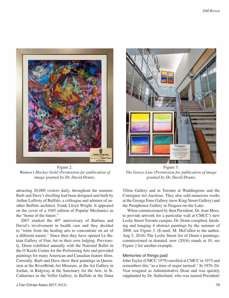
J Can Chiropr Assoc 2017; 61(1) 79
DM Brown
attracting 20,000 visitors daily, throughout the summer. Barb and Dave’s dwelling had been designed and built by Arthur Lafferty of Buffalo, a colleague and admirer of an-other Buffalo architect, Frank Lloyd Wright. It appeared on the cover of a 1945 edition of Popular Mechanics as the “home of the future.” 2007 marked the 40th anniversary of Barbara and David’s involvement in health care and they decided to “retire from the healing arts to concentrate on art of a different nature.” Since then they have opened Le Ba-teau Gallery of Fine Art in their own lodging. Previous-ly, Drum exhibited annually with the National Ballet in the O’Keefe Centre for the Performing Arts and provided paintings for many American and Canadian feature films. Currently, Barb and Dave show their paintings in Queen-ston at the RiverBrink Art Museum, at the Art Gallery in Jordan, in Ridgway at the Sanctuary for the Arts, in St. Catharines in the Veffer Gallery, in Buffalo at the Dana
Tillou Gallery and in Toronto at Waddingtons and the Consignor Art Auctions. They also sold numerous works at the George Enns Gallery (now King Street Gallery) and the Pumphouse Gallery in Niagara-on-the-Lake. When commissioned by then President, Dr. Jean Moss, to provide artwork for a particular wall at CMCC’s new Leslie Street Toronto campus, Dr. Drum complied, finish-ing and hanging 4 abstract paintings by the summer of 2008, see Figure 3. (E-mail, M. McCallen to the author, Aug 5, 2016) The Leslie Street list of Drum’s paintings, commissioned or donated, now (2016) stands at 10, see Figure 2 for another example.
Memories of things pastJohn Taylor (CMCC 1979) enrolled at CMCC in 1975 and remembers this “as a time of major turmoil.” In 1976, Dr. Vear resigned as Administrative Dean and was quickly supplanted by Dr. Sutherland, who was named President/
Figure 2.
Women’s Hockey Gold (Permission for publication of image granted by Dr. David Drum).
Figure 3.
The Gonzo Line (Permission for publication of image granted by Dr. David Drum).

80 J Can Chiropr Assoc 2017; 61(1)
David C. Drum, DC, FCCS(C): an accomplished, multi-faceted individual
CEO. Almost immediately, Sutherland fired his Academic Dean, Thomas Maxwell (CMCC 1955). This move rever-berated throughout the school yet Drum, who was teach-ing all 4 years, appears to have been untouched, watching calmly from the sidelines. Perhaps his extraneous sources of income left him with no desire to scramble up the exec-utive ladder. Later it was disclosed that David was quiet-ly donating his salary back to the college. “David was a breath of fresh air and very popular among the classes. Comparatively young yet experienced and savvy in chiro-practic, he knew the subject well, had cooperated with others in developing chiropractic scientific theories and could link the art, science and philosophy of chiroprac-tic in a way that was understandable to students with a science background.” Dr. Taylor confirms that David warmed his students with the glow of enthusiasm, at a time when they desperately needed it. (Email J. Taylor to the author, Aug 15, 2016) Upon graduation, Dr. Taylor established a practice in North Vancouver, BC. In 1991, Taylor became a Diplo-mate of the American Chiropractic Board of Radiology (DACBR) through the Los Angeles College of Chiroprac-tic, completed a postdoctoral research fellowship at the University of California at San Diego in 1993 and was hired by the Western States Chiropractic College, Port-land, OR, as an associate professor of radiology. John stayed there until 2000, when he became a professor at the New York Chiropractic College, in Seneca Falls and in 2008, was offered his current position as a professor of radiology and coordinator of diagnostic imaging, at D’Youville College in Buffalo, NY. Perched on Canada’s West coast, John heard little about David for decades. “Fast forward about 35 years and I discover that David is retired in Crystal Beach, ON. In 2013 and now living just across the Peace Bridge in Buf-falo, I made a point of visiting David and his lovely wife Barbara Vance. Since then, we have become good friends and I take great pleasure from this relationship.” February 2014, Taylor coaxed Drum into becoming the first speak-er in the D’Youville chiropractic students’ “Mentors Ser-ies” program. David calls it “a 90 minute presentation on the good the bad and the ugly... highlights from 40 years of clinical experience.” John says David’s theme was to make the students feel good about themselves and their futures, emphasizing, “he had a great career because he made his own opportunities rather than waiting for oppor-
tunities to come to him.” David addressed his interdisci-plinary relationship with doctors at the Orthopaedic and Arthritic Hospital in Toronto, talked about how he treated dancers from the Canadian National Ballet and how he kept his clinic open all night to attend to the visiting Bol-shoi company from the USSR. He also spoke at length about his friend and mentor Lyman Johnston and their achievements. “All the students left feeling much better about their careers.” John Taylor is nothing if not persistent. Sunday, May 22, 2016, he cajoled David Drum into delivering the key-note address at the Hooding and Pinning Ceremony, for the Class of 2016 Chiropractic Program at the D’Youville College. Held in the Bauer Family Academic Center, it was attended by 17 graduates and 150 faculty and guests. The following excerpts are taken from Dr, Drum’s re-marks. (E-mails D. Drum to the author May 25, 2016)
I discovered chiropractic through illustrating Dr. Lyman Johnston’s papers at the Canadian Me-morial Chiropractic College. He was director of research and his favourite expression was, ‘If you can’t measure it, it does not exist.’ I wondered where this left intuition, inspiration, hope and love. Lyman told us the profession was too vitalis-tic, needing mechanical and electrical instruments to measure what we were doing with results that could be quantified. He said this was science. But when we are truly sick, we are not wholly ourselves. We require a healer, a relationship we can trust, not just a provider of goods and services. Every new patient represents a case history of one. The case history is the patient’s story. Listen care-fully for they are telling you what is wrong. The doctor-patient relationship is further enriched by the intimate hands-on nature of our therapy. You are not alone but part of a system rigged toward health and greater complexity. So even though you are skillfully performing closed surgery by altering joint topography and restoring motion or decom-pressing tissues, the procedures are not as difficult as you may think. Dr. Johnston often hinted about introducing a ‘perturbation’ into a biological system that needed a kick start to a higher level of functioning. I recall a patient on the way to my clinic with chronic neck
J Can Chiropr Assoc 2017; 61(1) 81
DM Brown
pain I was ineffectually treating, phoning to say her cab had dropped into an excavation which threw her against the ceiling. She arrived with a full range of painless neck motion. That was a perturb-ation but difficult to reproduce. Get your patients into that wonderful, continuous passive motion ma-chine – a rocking chair. Use an antigravity device – the swimming pool. Employ a stationary bike with hands dangling, for spinal mobilization. Embrace the unexpected and never lose a holy curiosity in your clinical interactions. When you are free of self-doubt you will still fail but in more useful ways. Dr. Johnston personified the motto ‘carpe diem’ and taught me to live life where the action is right NOW! One morning a call came in from the Orthopaedic Hospital half a block away. Could I come immediately to help a visiting surgeon who was teaching a new spinal procedure and found himself in extremis on his hands and knees half under the operating table? Thinking perhaps pro-longed flexion may have triggered a rib subluxa-tion, I was ushered into the surgical theatre. The gods were with me, the standing anterior thoracic dorsal lift adjustment worked and he was able to complete the surgery. The following day I was back in surgery ob-serving a patient having his frozen sacroiliac joint manipulated after copious injections of cortisone. It seemed appropriate to demonstrate what every chiropractor does daily without the danger of gen-eral anaesthesia and the expense of a full surgical team. Not to be left out, the hospital physiother-apists asked for a course in sacroiliac adjusting. This lead to the head of physical medicine in an as-sociated hospital sharing his techniques. His fath-er had been a ‘bonesetter’ in Scotland and taught him well. One has no idea where staying connected to your community health practitioners will lead. We are all in this together! At this point you are relatively inexperienced and have not seen many of the conditions you will be treating. But do not fret. You are embarking on an awe-inspiring open-end-ed adventure. The experience will be bigger and more exciting than you can imagine. How could it be otherwise when we are all made of stardust? Welcome to the profession, Class of 2016!
February 17, 2016, it was announced that four large, framed artworks had been donated to D’Youville Col-lege and would soon grace the Chiropractic Department area in the Bauer Family Academic Center. After many years decorating the walls of CMCC with energetic and colourful paintings, Barbara Vance and David Drum de-cided to fill the bare walls of the chiropractic facility in the same spirit as their donations to the Canadian college. “We hope the spirit and energy that so impressed us in the faculty and student body, is reflected in our paintings.” As of August 30, 2016, 15 of their original paintings had been hung, beautifying the halls and eliciting compli-mentary remarks. A number of the works are multimedia abstracts incorporating themes that range from “The Hand of God,” to respectful tributes to Muhammed Ali and David Bowie. “Moreover, the presence of this art in our little area has visibly improved the entire mood of the campus community.” Barbara and David’s commitment to and support of the chiropractic profession is both boundless and borderless.
References1. Brown DM. Lyman C. Johnston, DC. FICC, FCCS(C):
Canadian chiropractic’s postural research pioneer and inventive entrepreneur. JCCA. 2001;45(1):42-52.
2. Peterson AR, et al. Segmental neuropathy: the first evidence of developing pathology. CMCC Library WB 905 6 S454 1965 c. 1.
3. Drum DC. An introduction to the study of posture and spinal mechanics. CMCC Library WB 905 D795i 1970 c. 2.
4. Haldeman S, Drum DC. The compression subluxation. J Clin Chiropr. 1971;Arch.1:10-21.
5. Johnston LC, Drum DC. The posterior gravity line syndrome. JCCA. 1968; 12(4): 5-7.
6. Drum DC. Disc regeneration: the rationale for a positive therapeutic approach (Part 1). JCCA. 1969;13(4):18-23.
7. Drum DC. Disc regeneration: the rationale for a positive therapeutic approach (Part 2). JCCA. 1970;14(2):9-15.
8. Cyriax, J. “Disc Lesions.” Cassel and Co. Ltd., London, 1956.
9. Drum CD. First of a series... Conservative management of lumbar disc degenerations. JCCA. 1970;14(4):8-11.
10. Drum CD. Second of a series... Conservative management of lumbar disc degenerations. JCCA. 1971;15(1):18-21.
11. Drum CD. Third of a series... Conservative management of lumbar disc degenerations. JCCA. 1971;15(2):12-15.
12. Schmorl G, Junghanns H. The human spine in health and disease. 2nd Amer. Ed. (translated by E.F. Besemann) Grune & Stratton, New York. 1971.

82 J Can Chiropr Assoc 2017; 61(1)
David C. Drum, DC, FCCS(C): an accomplished, multi-faceted individual
13. The research status of spinal manipulative therapy: a workshop held at the National Institute of Health. Feb 2-4 1975. CMCC Library Call Number: WB 905.2 W926r 1975.
14. Brown DM. Ian Douglass Coulter, PhD: CMCC’s adventurous president. JCCA. 2004; 48(1):36-55.