Incorporating Data Governance Frameworks in the Financial ...
Upload
khangminh22Category
view
2download
0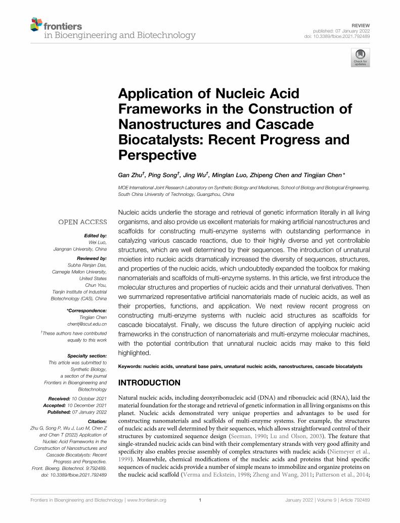
Application of Nucleic AcidFrameworks in the Construction ofNanostructures and CascadeBiocatalysts: Recent Progress andPerspectiveGan Zhu†, Ping Song†, Jing Wu†, Minglan Luo, Zhipeng Chen and Tingjian Chen*
MOE International Joint Research Laboratory on Synthetic Biology and Medicines, School of Biology and Biological Engineering,South China University of Technology, Guangzhou, China
Nucleic acids underlie the storage and retrieval of genetic information literally in all livingorganisms, and also provide us excellent materials for making artificial nanostructures andscaffolds for constructing multi-enzyme systems with outstanding performance incatalyzing various cascade reactions, due to their highly diverse and yet controllablestructures, which are well determined by their sequences. The introduction of unnaturalmoieties into nucleic acids dramatically increased the diversity of sequences, structures,and properties of the nucleic acids, which undoubtedly expanded the toolbox for makingnanomaterials and scaffolds of multi-enzyme systems. In this article, we first introduce themolecular structures and properties of nucleic acids and their unnatural derivatives. Thenwe summarized representative artificial nanomaterials made of nucleic acids, as well astheir properties, functions, and application. We next review recent progress onconstructing multi-enzyme systems with nucleic acid structures as scaffolds forcascade biocatalyst. Finally, we discuss the future direction of applying nucleic acidframeworks in the construction of nanomaterials and multi-enzyme molecular machines,with the potential contribution that unnatural nucleic acids may make to this fieldhighlighted.
Keywords: nucleic acids, unnatural base pairs, unnatural nucleic acids, nanostructures, cascade biocatalysts
INTRODUCTION
Natural nucleic acids, including deoxyribonucleic acid (DNA) and ribonucleic acid (RNA), laid thematerial foundation for the storage and retrieval of genetic information in all living organisms on thisplanet. Nucleic acids demonstrated very unique properties and advantages to be used forconstructing nanomaterials and scaffolds of multi-enzyme systems. For example, the structuresof nucleic acids are well determined by their sequences, which allows straightforward control of theirstructures by customized sequence design (Seeman, 1990; Lu and Olson, 2003). The feature thatsingle-stranded nucleic acids can bind with their complementary strands with very good affinity andspecificity also enables precise assembly of complex structures with nucleic acids (Niemeyer et al.,1999). Meanwhile, chemical modifications of the nucleic acids and proteins that bind specificsequences of nucleic acids provide a number of simple means to immobilize and organize proteins onthe nucleic acid scaffold (Verma and Eckstein, 1998; Zheng and Wang, 2011; Patterson et al., 2014;
Edited by:Wei Luo,
Jiangnan University, China
Reviewed by:Subha Ranjan Das,
Carnegie Mellon University,United States
Chun You,Tianjin Institute of IndustrialBiotechnology (CAS), China
*Correspondence:Tingjian Chen
†These authors have contributedequally to this work
Specialty section:This article was submitted to
Synthetic Biology,a section of the journal
Frontiers in Bioengineering andBiotechnology
Received: 10 October 2021Accepted: 10 December 2021Published: 07 January 2022
Citation:Zhu G, Song P, Wu J, Luo M, Chen Z
and Chen T (2022) Application ofNucleic Acid Frameworks in the
Construction of Nanostructures andCascade Biocatalysts: Recent
Progress and Perspective.Front. Bioeng. Biotechnol. 9:792489.
doi: 10.3389/fbioe.2021.792489
Frontiers in Bioengineering and Biotechnology | www.frontiersin.org January 2022 | Volume 9 | Article 7924891
REVIEWpublished: 07 January 2022
doi: 10.3389/fbioe.2021.792489
Spicer and Davis, 2014). Furthermore, good biocompatibility ofnucleic acids and the fact that nucleic acids can be efficientlyproduced inside the cells by living organisms make the nucleicacid scaffolds not only enzyme-friendly frameworks with variousapplications in vitro, but also powerful tools for in vivo assemblyof nanostructures and multi-enzyme molecular machines(Wilner et al., 2009; Simmel, 2012; Schoffelen and van Hest,2013; Shi et al., 2018a). In recent years, modified or unnaturalmoieties, including unnatural nucleobases and unnatural sugar/phosphate skeletons, have been designed, synthesized, andintroduced into nucleic acids, which significantly expanded thesequence space, properties, functions, and applications of nucleicacids, and some of them have already been employed in theconstruction of nucleic acid frameworks with novel properties(Ochoa and Milam, 2020). Hopefully, these unnatural moietieswill make nucleic acid frameworks more competitive scaffolds forthe construction of nanostructures and multi-enzyme systems inthe future.
In this review article, we start from a brief introduction of themolecular structures and properties of natural and unnaturalnucleic acids. Then the approaches and recent progresses onfabricating and applying complex nanostructures and multi-enzyme systems with nucleic acid scaffolds in vitro and in vivoare summarized and discussed. A perspective on futuredevelopment and application of novel nucleic acid frameworksin building nanostructures and multi-enzyme systems is providedin the end, with the potential contribution of unnatural nucleicacids highlighted.
NUCLEIC ACIDS AND THEIR UNNATURALDERIVATIVES
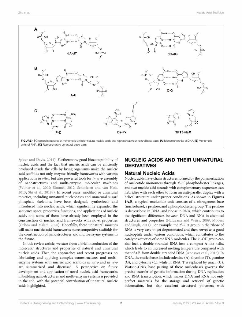
Natural Nucleic AcidsNucleic acids have chain structures formed by the polymerizationof nucleotide monomers through 3′-5′ phosphodiester linkages,and two nucleic acid strands with complementary sequences canhybridize with each other to form an anti-parallel duplex with ahelical structure under proper conditions. As shown in Figures1A,B, a typical nucleotide unit consists of a nitrogenous base(nucleobase), a pentose, and a phosphodiester group. The pentoseis deoxyribose in DNA, and ribose in RNA, which contributes tothe significant differences between DNA and RNA in chemicalstructures and properties (Narayana and Weiss, 2009; Mooersand Singh, 2011). For example, the 2′-OH group in the ribose ofRNA is very easy to get deprotonated and then serves as a goodnucleophile under various conditions, which contributes to thecatalytic activities of some RNA molecules. The 2′-OH group canalso lock a double-stranded RNA into a compact A-like helix,which leads to an increased melting temperature compared withthat of a B-form double-stranded DNA (Anosova et al., 2016). InDNA, the nucleobases include adenine (A), thymine (T), guanine(G), and cytosine (C), while in RNA, T is replaced by uracil (U).Watson-Crick base pairing of these nucleobases governs theprecise transfer of genetic information during DNA replicationand RNA transcription, which makes DNA and RNA not onlyperfect materials for the storage and retrieval of geneticinformation, but also excellent structural polymers with
FIGURE 1 | Chemical structures of monomeric units for natural nucleic acids and representative unnatural base pairs. (A)Monomeric units of DNA. (B)Monomericunits of RNA. (C) Representative unnatural base pairs.
Frontiers in Bioengineering and Biotechnology | www.frontiersin.org January 2022 | Volume 9 | Article 7924892
Zhu et al. Nucleic Acid Scaffolds
amplifiable sequences for the construction of artificialarchitectures. The highly specific hybridization of a DNA orRNA sequence with its complementary sequence also allowsprecise assembly of DNA or RNA strands into variousmicroarchitectures, ranging from simple linear structures tofairly sophisticated three-dimensional structures, some ofwhich have been used as scaffolds to build multi-enzymesystems (Fu et al., 2014; Sachdeva et al., 2014; Ngo et al.,2016). Moreover, the development of phosphoramiditechemistry for automated de novo synthesis of DNA and theinvention of polymerase chain reaction (PCR) technology forexponential amplification of DNA have already laid the technicalfoundations for large-scale preparation of DNA, which is theprerequisite to extensively use DNA as a structural material(Caruthers, 1985; Saiki et al., 1988).
Unnatural Base PairsRecently, the development of unnatural base pairs (UBPs) hasgreatly expanded the genetic alphabet (Kimoto and Hirao, 2020).Introduction of UBPs pairing orthogonally to natural base pairsinto DNA obviously increased the possible sequences, structures,properties, and functions of DNA, and optimization of them fornatural-like efficiencies of replication, transcription andtranslation further expanded their applications. Figure 1Cdemonstrates the chemical structures of three mostpredominant UBPs. The pairing of UBP P-Z, which wasdeveloped by Benner group, is based on hydrogen bondingwith a different pattern from those of natural base pairs, whilethe pairing of UBPs TPT3-NaM and Ds-Px, which weredeveloped by Romesberg group and Hirao group respectively,is based on hydrophobic and packing forces (Kimoto et al., 2008;Li et al., 2014). These UBPs have been shown to be very efficientfor PCR amplification and in vitro transcription, and thussignificantly increased the storage capacities of DNA andRNA for amplifiable and retrievable information (Li et al.,2014). Moreover, these UBPs have already been successfullyused in various in vitro applications, ranging from site-specificlabeling of nucleic acids to selection for affinity reagents(Lavergne et al., 2016; Futami et al., 2019; Kimoto et al.,2019; Zhang et al., 2020; Matsunaga et al., 2021; Miao et al.,2021; Niu et al., 2021).
Most remarkably, UBP TPT3-NaM have been used in theconstruction of the first six-letter semi-synthetic organism, andproven to be very efficient for in vivo replication, transcription,and even translation (Malyshev et al., 2014; Zhang et al., 2017a;Fischer et al., 2020). These exciting discoveries immediatelyenabled countless potential in vivo applications of UBPs,including in vivo site-specific labeling of nucleic acids tosimultaneous introduction of multiple different unnaturalamino acids into proteins. More detailed information aboutdevelopment and application of unnatural base pairs has beensummarized in other specific reviews (Feldman and Romesberg,2018; Kimoto and Hirao, 2020).
Nucleic Acids With Unnatural SugarsWhile the incorporation of UBPs increases the sequencediversity and sequence-related functionality of nucleic acids,
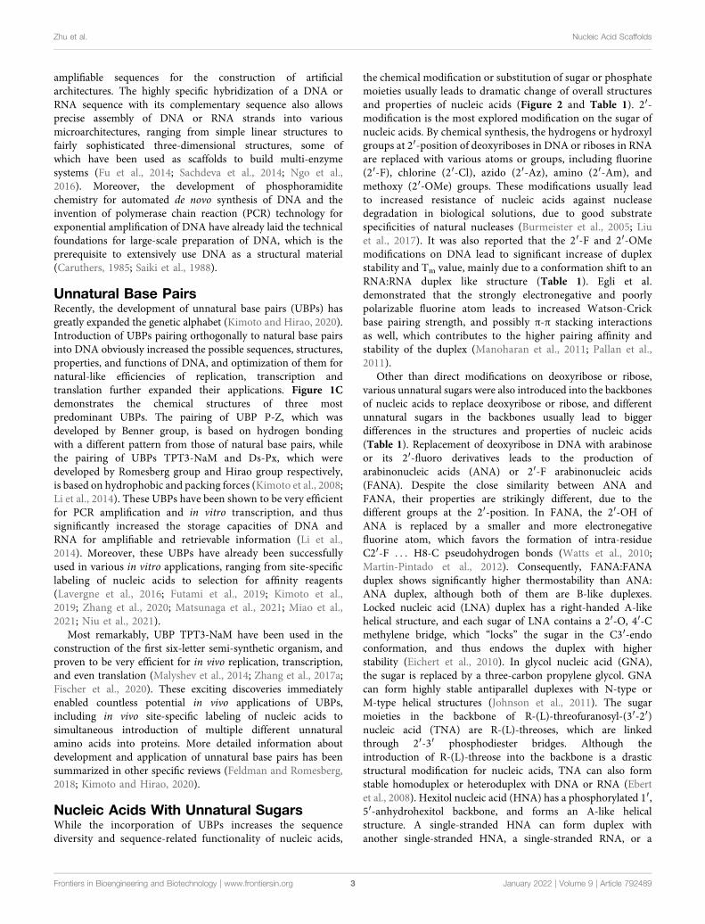
the chemical modification or substitution of sugar or phosphatemoieties usually leads to dramatic change of overall structuresand properties of nucleic acids (Figure 2 and Table 1). 2′-modification is the most explored modification on the sugar ofnucleic acids. By chemical synthesis, the hydrogens or hydroxylgroups at 2′-position of deoxyriboses in DNA or riboses in RNAare replaced with various atoms or groups, including fluorine(2′-F), chlorine (2′-Cl), azido (2′-Az), amino (2′-Am), andmethoxy (2′-OMe) groups. These modifications usually leadto increased resistance of nucleic acids against nucleasedegradation in biological solutions, due to good substratespecificities of natural nucleases (Burmeister et al., 2005; Liuet al., 2017). It was also reported that the 2′-F and 2′-OMemodifications on DNA lead to significant increase of duplexstability and Tm value, mainly due to a conformation shift to anRNA:RNA duplex like structure (Table 1). Egli et al.demonstrated that the strongly electronegative and poorlypolarizable fluorine atom leads to increased Watson-Crickbase pairing strength, and possibly π-π stacking interactionsas well, which contributes to the higher pairing affinity andstability of the duplex (Manoharan et al., 2011; Pallan et al.,2011).
Other than direct modifications on deoxyribose or ribose,various unnatural sugars were also introduced into the backbonesof nucleic acids to replace deoxyribose or ribose, and differentunnatural sugars in the backbones usually lead to biggerdifferences in the structures and properties of nucleic acids(Table 1). Replacement of deoxyribose in DNA with arabinoseor its 2′-fluoro derivatives leads to the production ofarabinonucleic acids (ANA) or 2′-F arabinonucleic acids(FANA). Despite the close similarity between ANA andFANA, their properties are strikingly different, due to thedifferent groups at the 2′-position. In FANA, the 2′-OH ofANA is replaced by a smaller and more electronegativefluorine atom, which favors the formation of intra-residueC2′-F . . . H8-C pseudohydrogen bonds (Watts et al., 2010;Martin-Pintado et al., 2012). Consequently, FANA:FANAduplex shows significantly higher thermostability than ANA:ANA duplex, although both of them are B-like duplexes.Locked nucleic acid (LNA) duplex has a right-handed A-likehelical structure, and each sugar of LNA contains a 2′-O, 4′-Cmethylene bridge, which “locks” the sugar in the C3′-endoconformation, and thus endows the duplex with higherstability (Eichert et al., 2010). In glycol nucleic acid (GNA),the sugar is replaced by a three-carbon propylene glycol. GNAcan form highly stable antiparallel duplexes with N-type orM-type helical structures (Johnson et al., 2011). The sugarmoieties in the backbone of R-(L)-threofuranosyl-(3′-2′)nucleic acid (TNA) are R-(L)-threoses, which are linkedthrough 2′-3′ phosphodiester bridges. Although theintroduction of R-(L)-threose into the backbone is a drasticstructural modification for nucleic acids, TNA can also formstable homoduplex or heteroduplex with DNA or RNA (Ebertet al., 2008). Hexitol nucleic acid (HNA) has a phosphorylated 1′,5′-anhydrohexitol backbone, and forms an A-like helicalstructure. A single-stranded HNA can form duplex withanother single-stranded HNA, a single-stranded RNA, or a
Frontiers in Bioengineering and Biotechnology | www.frontiersin.org January 2022 | Volume 9 | Article 7924893
Zhu et al. Nucleic Acid Scaffolds
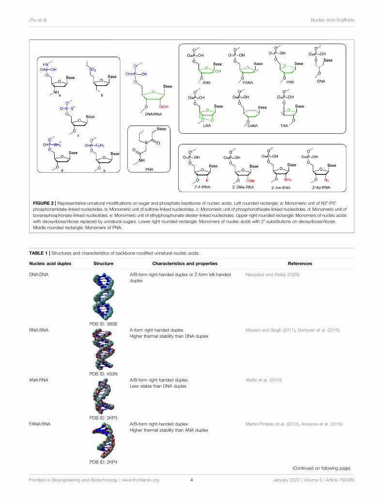
FIGURE 2 | Representative unnatural modifications on sugar and phosphate backbone of nucleic acids. Left rounded rectangle: a: Monomeric unit of N3′-P5′phosphoramidate-linked nucleotides. b: Monomeric unit of sulfone-linked nucleotides. c: Monomeric unit of phosphorothioate-linked nucleotides. d: Monomeric unit ofboranephosphonate-linked nucleotides. e: Monomeric unit of ethylphosphonate diester-linked nucleotides. Upper right rounded rectangle: Monomers of nucleic acidswith deoxyribose/ribose replaced by unnatural sugars. Lower right rounded rectangle: Monomers of nucleic acids with 2′-substitutions on deoxyribose/ribose.Middle rounded rectangle: Monomers of PNA.
TABLE 1 | Structures and characteristics of backbone-modified unnatural nucleic acids.
Nucleic acid duplex Structure Characteristics and properties References
DNA:DNA
PDB ID: 3BSE
A/B-form right-handed duplex or Z-form left-handedduplex
Narayana and Weiss (2009)
RNA:RNA
PDB ID: 4S3N
A-form right-handed duplex Mooers and Singh (2011), Donovan et al. (2015)Higher thermal stability than DNA duplex
ANA:RNA
PDB ID: 2KP3
A/B-form right-handed duplex Watts et al. (2010)Less stable than DNA duplex
FANA:RNA
PDB ID: 2KP4
A/B-form right-handed duplex Martin-Pintado et al. (2012), Anosova et al. (2016)Higher thermal stability than ANA duplex
(Continued on following page)
Frontiers in Bioengineering and Biotechnology | www.frontiersin.org January 2022 | Volume 9 | Article 7924894
Zhu et al. Nucleic Acid Scaffolds
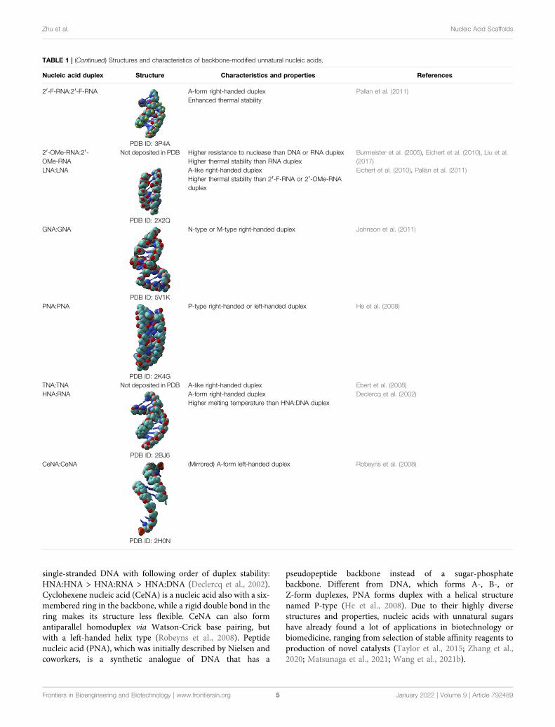
single-stranded DNA with following order of duplex stability:HNA:HNA > HNA:RNA > HNA:DNA (Declercq et al., 2002).Cyclohexene nucleic acid (CeNA) is a nucleic acid also with a six-membered ring in the backbone, while a rigid double bond in thering makes its structure less flexible. CeNA can also formantiparallel homoduplex via Watson-Crick base pairing, butwith a left-handed helix type (Robeyns et al., 2008). Peptidenucleic acid (PNA), which was initially described by Nielsen andcoworkers, is a synthetic analogue of DNA that has a
pseudopeptide backbone instead of a sugar-phosphatebackbone. Different from DNA, which forms A-, B-, orZ-form duplexes, PNA forms duplex with a helical structurenamed P-type (He et al., 2008). Due to their highly diversestructures and properties, nucleic acids with unnatural sugarshave already found a lot of applications in biotechnology orbiomedicine, ranging from selection of stable affinity reagents toproduction of novel catalysts (Taylor et al., 2015; Zhang et al.,2020; Matsunaga et al., 2021; Wang et al., 2021b).
TABLE 1 | (Continued) Structures and characteristics of backbone-modified unnatural nucleic acids.
Nucleic acid duplex Structure Characteristics and properties References
2′-F-RNA:2′-F-RNA
PDB ID: 3P4A
A-form right-handed duplex Pallan et al. (2011)Enhanced thermal stability
2′-OMe-RNA:2′-OMe-RNA
Not deposited in PDB Higher resistance to nuclease than DNA or RNA duplex Burmeister et al. (2005), Eichert et al. (2010), Liu et al.(2017)Higher thermal stability than RNA duplex
LNA:LNA
PDB ID: 2X2Q
A-like right-handed duplex Eichert et al. (2010), Pallan et al. (2011)Higher thermal stability than 2′-F-RNA or 2′-OMe-RNAduplex
GNA:GNA
PDB ID: 5V1K
N-type or M-type right-handed duplex Johnson et al. (2011)
PNA:PNA
PDB ID: 2K4G
P-type right-handed or left-handed duplex He et al. (2008)
TNA:TNA Not deposited in PDB A-like right-handed duplex Ebert et al. (2008)HNA:RNA
PDB ID: 2BJ6
A-form right-handed duplex Declercq et al. (2002)Higher melting temperature than HNA:DNA duplex
CeNA:CeNA
PDB ID: 2H0N
(Mirrored) A-form left-handed duplex Robeyns et al. (2008)
Frontiers in Bioengineering and Biotechnology | www.frontiersin.org January 2022 | Volume 9 | Article 7924895
Zhu et al. Nucleic Acid Scaffolds
Nucleic Acids With Unnatural PhosphatesThe phosphate group in the backbone of nucleic acids has alsobeen chemically modified or substituted with various anionic,cationic, or electroneutral groups for altered properties(Arangundy-Franklin et al., 2019). For example, a series ofmodified oligonucleotides were developed by replacing non-bridging oxygen atoms in the phosphate group with otheratoms or groups, such as S− and BH3
−, to introduce propertiesfor enhanced biological activities (Eckstein, 2014; Kumar andCaruthers, 2020). Recently, Holliger and coworkers replaced oneof the non-bridging oxygen atoms in the phosphate with an alkylgroup, which led to the production of P-alkyl phosphonatenucleic acid (phNA) with an uncharged backbone(Arangundy-Franklin et al., 2019). The enzymatic synthesisand evolution of these nucleic acids were also accomplished withengineered polymerases. More thorough changes have also beenmade to the whole bridges between sugars in the backbone. Forexample, Gryaznov and co-workers developed oligonucleotide N3′-P5′ phosphoramidate, in which the 3′-oxygen of each nucleoside wasreplaced with a 3′-nitrogen (Chen et al., 1995). This nucleic acid wasfound to have great nuclease resistance and superior thermodynamicstability, and thus has great potential for therapeutic and otherapplications. In another early case, Benner and co-workerssynthesized nonionic sulfone-linked RNA analogs, rSNAs, byreplacing the phosphodiester moieties of RNA with dimethylene
sulfone units, and found that rSNAs have many interestingcharacteristics (Richert et al., 1996).
Wild Type or Engineered Polymerases forthe Recognition and Synthesis of UnnaturalNucleic AcidsHaving efficient polymerases is the prerequisite for enzymaticsynthesis, amplification, and even evolution of unnatural nucleicacids for different applications. During the design and synthesisof unnatural base pairs, the polymerase recognition of them hasalready been taken into consideration very well, so thepredominant unnatural base pairs, such as TPT3-NaM, P-Z,Ds-Px, can be efficiently replicated and transcribed by many ofthe natural DNA and RNA polymerases (Table 2). Since naturalpolymerases usually have poor recognition and synthesis activities forbackbone-modified nucleic acids, a lot of efforts have been made todevelop engineered polymerases for efficient synthesis of theseunnatural nucleic acids. In these efforts, thermophilic single-subunit DNA polymerases were usually employed as startingpoint for polymerase engineering. Romesberg and co-workershave developed a modified phage display platform for directedpolymerase evolution, and successfully applied it to evolve theStoffel fragment of Taq DNA polymerase for efficienttranscription, reverse transcription, and amplification of DNAs
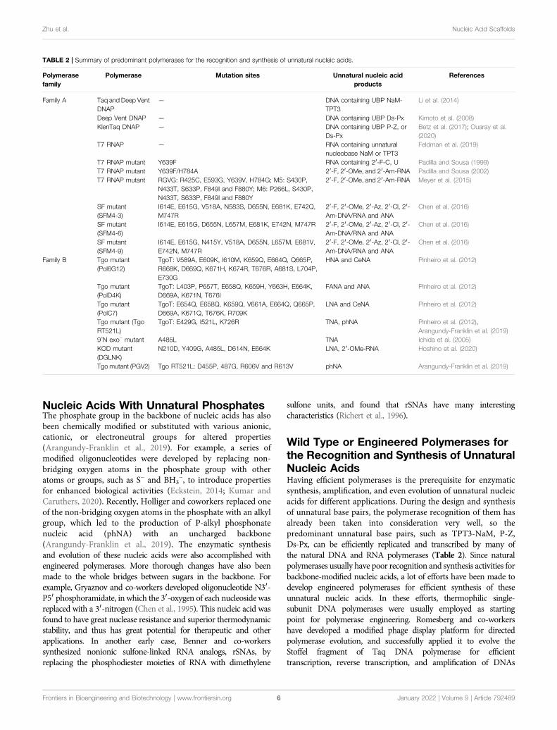
TABLE 2 | Summary of predominant polymerases for the recognition and synthesis of unnatural nucleic acids.
Polymerasefamily
Polymerase Mutation sites Unnatural nucleic acidproducts
References
Family A Taq and Deep VentDNAP
— DNA containing UBP NaM-TPT3
Li et al. (2014)
Deep Vent DNAP — DNA containing UBP Ds-Px Kimoto et al. (2008)KlenTaq DNAP — DNA containing UBP P-Z, or
Ds-PxBetz et al. (2017); Ouaray et al.(2020)
T7 RNAP — RNA containing unnaturalnucleobase NaM or TPT3
Feldman et al. (2019)
T7 RNAP mutant Y639F RNA containing 2′-F-C, U Padilla and Sousa (1999)T7 RNAP mutant Y639F/H784A 2′-F, 2′-OMe, and 2′-Am-RNA Padilla and Sousa (2002)T7 RNAP mutant RGVG: R425C, E593G, Y639V, H784G; M5: S430P,
N433T, S633P, F849I and F880Y; M6: P266L, S430P,N433T, S633P, F849I and F880Y
2′-F, 2′-OMe, and 2′-Am-RNA Meyer et al. (2015)
SF mutant(SFM4-3)
I614E, E615G, V518A, N583S, D655N, E681K, E742Q,M747R
2′-F, 2′-OMe, 2′-Az, 2′-Cl, 2′-Am-DNA/RNA and ANA
Chen et al. (2016)
SF mutant(SFM4-6)
I614E, E615G, D655N, L657M, E681K, E742N, M747R 2′-F, 2′-OMe, 2′-Az, 2′-Cl, 2′-Am-DNA/RNA and ANA
Chen et al. (2016)
SF mutant(SFM4-9)
I614E, E615G, N415Y, V518A, D655N, L657M, E681V,E742N, M747R
2′-F, 2′-OMe, 2′-Az, 2′-Cl, 2′-Am-DNA/RNA and ANA
Chen et al. (2016)
Family B Tgo mutant(Pol6G12)
TgoT: V589A, E609K, I610M, K659Q, E664Q, Q665P,R668K, D669Q, K671H, K674R, T676R, A681S, L704P,E730G
HNA and CeNA Pinheiro et al. (2012)
Tgo mutant(PolD4K)
TgoT: L403P, P657T, E658Q, K659H, Y663H, E664K,D669A, K671N, T676I
FANA and ANA Pinheiro et al. (2012)
Tgo mutant(PolC7)
TgoT: E654Q, E658Q, K659Q, V661A, E664Q, Q665P,D669A, K671Q, T676K, R709K
LNA and CeNA Pinheiro et al. (2012)
Tgo mutant (TgoRT521L)
TgoT: E429G, I521L, K726R TNA, phNA Pinheiro et al. (2012),Arangundy-Franklin et al. (2019)
9°N exo− mutant A485L TNA Ichida et al. (2005)KOD mutant(DGLNK)
N210D, Y409G, A485L, D614N, E664K LNA, 2′-OMe-RNA Hoshino et al. (2020)
Tgo mutant (PGV2) Tgo RT521L: D455P, 487G, R606V and R613V phNA Arangundy-Franklin et al. (2019)
Frontiers in Bioengineering and Biotechnology | www.frontiersin.org January 2022 | Volume 9 | Article 7924896
Zhu et al. Nucleic Acid Scaffolds

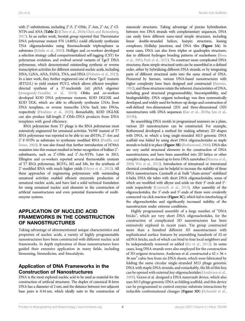
FIGURE 3 | Examples of nanostructures constructed with natural and unnatural nucleic acids. (A)Basic units of DNA nanostructures. From left to right: DNA doublehelix, hairpin, Holliday junction, rectangular DNA tile, Y-shaped DNA tile. (B) Two-dimensional DNA origami shapes. From left to right: square, star, smiling face. (C) DNAcatenane. Four selected neighboring strands of a six-helix bundle were modified with 3′-alkyne and 5′-azide, and intramolecularly cyclized via click reaction to form thetopologically interlocked structure. (D) Enzyme-loaded DNA nanovault in the open and closed states. (E) Square nut-shaped DNA origami. (F) Rhombic lattice andhexagonal lattice constructed with integration of scaffolded DNA origami and scaffold-free LEGO methods. Representative vertexes are magnified in dotted frames.Staples with or without spacers (shown in green) of different lengths were crucial to modulate the lattice pattern. (G) Unnatural nucleic acid tetrahedron. (H) FANAoctahedron. (I) Nucleic acid hydrogel constructed with 2′-azido modified DNA backbone.
Frontiers in Bioengineering and Biotechnology | www.frontiersin.org January 2022 | Volume 9 | Article 7924897
Zhu et al. Nucleic Acid Scaffolds
with 2′-substitutions, including 2′-F, 2′-OMe, 2′-Am, 2′-Az, 2′-Cl-NTPs and ANA (Table 2) (Chen et al., 2016; Chen and Romesberg,2017). In an earlier work, Szostak group reported that TherminatorDNA polymerase mutant 9°N (A485L) could efficiently synthesizeTNA oligonucleotides using threonucleoside triphosphates assubstrates (Ichida et al., 2005). Holliger and co-workers developeda selection strategy called compartmentalized self-tagging (CST) forpolymerase evolution, and evolved several variants of TgoT DNApolymerase, which demonstrated outstanding synthesis or reversetranscription activities for different unnatural nucleic acids, includingHNA, CeNA, ANA, FANA, TNA, and HNA (Pinheiro et al., 2012).In a later work, they further engineered one of these TgoT mutants(RT521L) to yield mutant PGV2, which allows efficient template-directed synthesis of a 57-nucleotide (nt) phNA oligomer(Arangundy-Franklin et al., 2019). Obika and co-workersdeveloped KOD DNA polymerase mutants, KOD DGLNK andKOD DLK, which are able to efficiently synthesize LNAs fromDNA templates, or reverse transcribe LNAs back into DNAs,respectively (Hoshino et al., 2020). Remarkably, KOD DGLNKcan also produce full-length 2′-OMe-DNA products from DNAtemplates with good efficiency.
RNA polymerase from T7 phage is the RNA polymerase mostextensively engineered for unnatural activities. Y639F mutant of T7RNA polymerase was reported to be able to use dNTPs, 2′-Am and2′-F-NTPs as substrates to synthesize modified RNA (Padilla andSousa, 2002). It was also found that further introduction of H784Amutation into this mutant resulted in better recognition of bulkier 2′-substituents, such as 2′-OMe and 2′-Az-NTPs. Later in 2015,Ellington and co-workers reported several thermostable mutantsof T7 RNA polymerase, RGVG, M5 and M6, for the synthesis of2′-modified RNA with much higher yields (Meyer et al., 2015). Allthese approaches of engineering polymerases with outstandingunnatural activities enabled efficient enzymatic production ofunnatural nucleic acids, and have laid a solid technical foundationfor using unnatural nucleic acid elements in the construction ofartificial nanostructures and even potential frameworks of multi-enzyme systems.
APPLICATION OF NUCLEIC ACIDFRAMEWORKS IN THE CONSTRUCTIONOF NANOSTRUCTURESTaking advantage of aforementioned unique characteristics andproperties of nucleic acids, a variety of highly programmablenanostructures have been constructed with different nucleic acidframeworks. In depth exploration of these nanostructures haveguided their extensive application in many fields, includingbiosensing, biomedicine, and biocatalysis.
Application of DNA Frameworks in theConstruction of NanostructuresDNA is the most explored nucleic acid to be used as material for theconstruction of artificial structures. The duplex of canonical B-formDNA has a diameter of 2 nm, and the distance between two adjacentbase pairs is 0.34 nm, which ideally suits to the construction of
nanoscale structures. Taking advantage of precise hybridizationbetween two DNA strands with complementary sequences, DNAcan easily form different nano-sized simple structures, includinglinear double-stranded helices, hairpin structures, kissingcomplexes, Holliday junctions, and DNA tiles (Figure 3A). Insome cases, DNA can also form triplex or quadruplex structures,due to different hydrogen bonding patterns of nucleobases (Rosuet al., 2002; Park et al., 2021). To construct more complicated DNAstructures, these simple structural units can be assembled in a definedorder, either by hybridizing different DNA strands, or by integratingparts of different structural units into the same strand of DNA.Pioneered by Seeman, various DNA-based nanostructures withhigher complexity have been designed and constructed (Seeman,1982), and these structures retain the inherent characteristics of DNA,including good structural programmability, biocompatibility, andbiodegradability. DNA origami technology was then systematicallydeveloped, and widely used for bottom-up design and construction ofwell-defined two-dimensional (2D) and three-dimensional (3D)nanostructures with DNA sequences (Jun et al., 2019a; Jun et al.,2019b).
By assembling DNA motifs in programmed manners on a plain,various 2D nanostructures can be constructed. For example,Rothemund developed a method for making arbitrary 2D shapeswith DNA, in which a long single-stranded M13 genomic DNAscaffold was folded by using short DNA oligonucleotides as staplestrands to hold it in place (Figure 3B) (Rothemund, 2006). DNA tilesare very useful structural elements in the construction of DNAnanostructures, and have been assembled into 2D lattice or othercomplex shapes, or closed up to formDNAnanotubes (Sharma et al.,2009; Wei et al., 2012). Introduction of intrastrand or interstrandchemical crosslinking can be helpful to increase the stability of theseDNA nanostructures. Cassinelli et al. built “chain-armor”-stabilized6-helix DNA tile tubes with short DNA oligonucleotides, some ofwhich are modified with alkyne and azide on their 3′-ends and 5′-ends respectively (Cassinelli et al., 2015). After assembly of theoligonucleotides, the 3′-ends and 5′-ends of them were covalentlyconnected via click reaction (Figure 3C), which led to interlocking ofthe oligonucleotides and significantly increased stability of thenanostructure under extreme conditions.
Highly programmed assembly of a large number of “DNAbricks”, which are very short DNA oligonucleotides, for theconstruction of complicated 3D nanostructures has beenextensively explored in recent years. Yin group constructedmore than a hundred different 3D nanostructures withsophisticated surface features by assembling hundreds of 32-ntssDNA bricks, each of which can bind to four local neighbors andbe independently removed or added (Ke et al., 2012). In somecases, long DNA strands were also employed for the constructionof 3D origami structures. Andersen et al. constructed a 42 × 36 ×36 nm3 cubic box from six DNA sheets, which were fabricated byfolding the same circular single-stranded M13 phage genomicDNAwith staple DNA strands, and remarkably, the lib of this boxcan be opened with external key oligonucleotides (Andersen et al.,2009). Gorssi et al. designed a DNA nanovault device, which alsouses M13 phage genomic DNA as folding scaffold, and this devicecan be programmed to control enzyme-substrate interactions byinducible conformational changes (Figure 3D) (Schoenit et al.,
Frontiers in Bioengineering and Biotechnology | www.frontiersin.org January 2022 | Volume 9 | Article 7924898
Zhu et al. Nucleic Acid Scaffolds
2021). Shih group applied stapled long ssDNA derived fromM13phage genome to build a series of honeycomb-pleated 3Dnanostructures with precisely controlled dimensions andvarious complex shapes, including monolith, railed bridge,stacked cross, slotted cross, genie bottle and square nut-likeshapes (Figure 3E) (Douglas et al., 2009). Shih group alsodemonstrated that the complex shapes of DNA nanostructurescould be twisted or curved by deleting or inserting base pairs attargeted positions, which is very useful in the construction ofshapes with curvatures, such as ball-like structures (Dietz et al.,2009; Douglas et al., 2009). Recently, Cui et al. integrated bothscaffolded DNA origami method and scaffold-free LEGOmethodto build 2D and 3D wireframe structures (Cui et al., 2021). Bysimply changing the DNA staples, they transformed the rhombiclattice made of zigzagged scaffold into larger and more flexiblehexagonal lattice with adjustable hexagon units (Figure 3F). Theyalso fabricated hybrid diamond cubic lattices that are able toaccommodate AuNPs, and their strategy allows the adjustment ofpore volumes according to the size of guest molecules.
While DNA origami nanostructures were constructed withvarious strategies, Dietz group further assembled them toconstruct dynamic DNA devices and giant programmableDNA assemblies (Gerling et al., 2015; Wagenbauer et al.,2017). In another work, they demonstrated rapid productionof 3D DNA nanostructures by folding the DNA strands atconstant temperatures, and the yields could be as high as100% (Sobczak et al., 2012). They also designed a strategy formass production of DNA origami components, in which theprecursor DNA of the origami components and interspacedZn2+-dependent DNAzyme sequences were incorporated intoM13 phage genome, and amplified during the phagepropagation (Praetorius et al., 2017). After extraction of thephage genomic DNA, the DNA origami components wereself-excised off by the DNAzymes. All these efforts woulddirectly contribute to the more robust and easier constructionof versatile DNA nanostructures with lower costs, and enabletheir large-scale applications in various fields.
Due to their unique properties, DNA nanostructures have greatpotential to be broadly applied in biotechnology and biomedicine.For example, some DNA nanostructures can penetrate cellmembranes without transfection agents for intracellulardetection or cargo transport, which provides alternativemethods for molecular diagnosis and drug delivery (Jiang et al.,2021). Fan group demonstrated that DNA tetrahedron modifiedwith unmethylated CpG motifs can noninvasively entermacrophage-like cells without transfection agents and inducethe secretion of cytokines including tumor necrosis factor-α,interleukin-6 and interleukin-12 (Li et al., 2011). Li et al.designed a DNA origami-based nanorobot, which couldprecisely respond to the tumor associated molecular trigger, andrelease drug payload in a controllable manner (Li S. et al., 2018). Inthis work, a DNA origami sheet was constructed with M13genomic DNA scaffold and DNA staples, and loaded withthrombin, which could activate coagulation cascade. The DNAorigami sheet was then wrapped up and lockedwith DNA fastenersto form a DNA nanorobot. The fasteners integrated nucleolinaptamer sequences, and could be triggered to open the DNA
origami sheet and expose the encapsulated thrombin upon thebinding of nucleolin aptamer with nucleolin, which is specificallyexpressed on the surface of tumor-associated endothelial cells. ThisDNA nanorobot proved very safe and effective in inhibiting tumorgrowth in model animals, presumably due to goodbiocompatibility and precise drug delivery. Krishnan and co-workers constructed artificial myosin filaments using DNAnanotubes as scaffolds, which enabled precise spatialorganization of myosin motors and allowed actin filaments toglide along the scaffolds (Hariadi et al., 2015). Based onfluorescence resonance energy transfer (FRET) and pH-sensitiveconformation change of designed DNA nanomachines, Krishnangroup developed several DNA nanodevices to map pH changes inthe cells. In 2009, they reported a DNA nanomachine, I-switch,which could be used to map spatial and temporal pH changesinside living cells (Modi et al., 2009). I-switch consisted of threeDNA oligonucleotides, two of which adjacently hybridized to thethird one, leaving one nucleotide gap between them and twocytosine-rich overhangs modified with fluorophoresunhybridized. Low pH led to the formation of an i-motifbetween the two overhangs, and resulted in the shift of theI-switch conformation from open state to closed state, whichsubsequently allowed FRET between the two fluorophores. ThisI-switch was shown to be very efficient for mapping thespatiotemporal pH changes related to endosome maturation. Inanother work, they successfully used this I-switch to map the pHchanges involved in endocytosis in Caenorhabditis elegans (Suranaet al., 2011). Later, they simultaneously mapped the pH change indifferent organelles within the same cell by introducing targetingmoieties to direct the DNA nanomachines to enter cells viadifferent pathways (Modi et al., 2013). Besides DNAnanodevices that were able to map the pH changes in cells,Krishnan group also developed DNA based fluorescent probesfor mapping enzymatic activities with subcellular resolution. Theseprobes contained three functional modules, including an analytessensing dye, an internal reference dye for ratiometric quantitation,and a targeting moiety to localize the reporter to specific organelle,such as a DNA duplex, a DNA aptamer, or a cholesterol group.Hybridization of complementary DNA strands allowed thestoichiometric assembly of these three functionalities onto thesame DNA scaffold. By using distinct sensing dyes that respondto different molecules and specific targeting moieties, they havesuccessfully mapped organellar disulfide exchange, the activity ofnitric oxide synthase 3 (NOS3) at the plasma membrane or thetrans-Golgi network, and the activity of nitric oxide synthase 2(NOS2) in phagosomes and endosomes (Dan et al., 2019; Jani et al.,2020; Veetil et al., 2020). Ion concentrations and membranepotential could also be mapped with similar DNA probesincorporating different functional groups and dyes. In 2019,they reported a DNA scaffold-based reporter that is sensitive toboth pH andCa2+ concentration, CalipHluor, for themeasurementof Ca2+ concentrations in specific organelles of the endolysosomalpathway (Narayanaswamy et al., 2019). Recently, they reported aDNA scaffold-based voltmeter that can report the membranepotential of organelles, Voltair (Saminathan et al., 2021). DNAwalkers, which can locomote along programed tracks through theprocesses including DNA strand hybridization, enzymatic cleavage
Frontiers in Bioengineering and Biotechnology | www.frontiersin.org January 2022 | Volume 9 | Article 7924899
Zhu et al. Nucleic Acid Scaffolds
of DNA strands, and DNA strand displacement, are among themost studied dynamic DNA nanodevices, and have been broadlyused in various practical applications, such as biosensing, in recentyears (Valero and Skugor, 2020; Cha et al., 2015). Dong et al.developed a highly sensitive electrochemical method for thedetection of tumor exosomes (Dong et al., 2018). The bindingof the aptamers immobilized on the magnetic beads and theexosomes led to the release of three kinds of messenger DNAs,which then hybridized with the DNA probes immobilized on agold electrode, allowing subsequent exo III digestion of the DNAprobes and a decrease in electrochemical signal. Once a probeDNA was digested, the messenger DNA would be released, andhybridize with next adjacent probe DNA. This process went onuntil no probe DNA left. In this system, the signal was amplifiedfirst by the release of multiple messenger DNAs and then by ExoIII-assisted probe DNA digestion and messenger DNA recycling,which led to much lower detection limit compared with othermethods frequently used. Zhao et al. developed a similar but morecomprehensive method for the detection of MCF-7 cell-secretedexosome, in which the employed DNA walker is propelled byDNAzyme-mediated DNA cleavage (Zhao et al., 2019). CD63aptamers and DNA substrates for a DNAzyme were co-immobilized on magnetic beads, and an EpCAM aptamerlinked with the DNAzyme through a swing arm was also addedinto the system. Simultaneous binding of the CD63 aptamer andthe EpCAM aptamer with the target exosome initiated the cleavageof the DNA substrates, releasing product DNA strands that couldtrigger Exo III-mediated cleavage of probe DNAs immobilized ongold electrodes and an electrochemical signal change similar asprevious example. After cleaving a DNA substrate, the DNAzymewent on to bind and cleavage another DNA substrate, whichrepeated until all the adjacent DNA substrates were cleaved. Inanother example, Feng et al. developed a sensitiveelectrochemiluminescence method for the detection of tumorexosomes (Feng et al., 2020). In this method, anchor DNAscontaining a nicking recognition sequence and a swing armpartially hybridizing with a CD63 aptamer were co-assembledon a RuSi NP modified electrode. In the presence of exosomes,binding of the aptamer and the exosome led to release of the swingarm and allowed its hybridization with one of the anchor DNAs,which led to the formation of a dsDNA cleavage site for anendonuclease. After the cleavage of an anchor DNA by theendonuclease, the liberated swing arm would continuouslylocomote to another anchor DNA. The ssDNA productsremaining on the electrode then hybridized withcomplementary ssDNA stands modified with GOx, whichcatalyzed the oxidation of H2O2 and eventually led to thedecrease of electrochemiluminescence signal. Dynamic platformsor systems combining DNA origami and other components havealso found broad practical application recently. For example, Sunet al. developed a DNA origami raft-based platform for real-timeimaging of the protein trajectories on 2D fluidic surfaces (Sun et al.,2017). Cholesterol modified double-stranded DNA and DNAorigami raft were used for tethering fluorophore-labeledenzymes on the supported lipid bilayer, and both dynamic andstatic tethering could be achieved with different tethering modes.By using a total internal reflection fluorescence microscope
(TIRFM), motions of the enzymes could be monitored in realtime, which enabled the imaging of dynamic interactions ofproteins. Summary specifically about DNA nanodevices andtheir applications could be found elsewhere (Simmel, 2007;Krishnan and Simmel, 2011; Kim et al., 2018; Jahanban-Esfahlan et al., 2019; DeLuca et al., 2020; Li et al., 2021a).
More extensive assembly or crosslinking of DNAnanostructures has led to the production of variousmacroscopic DNA hydrogels. For example, in 2006, Luo groupconstructed several biocompatible and biodegradable DNAhydrogels by assembling branched DNA monomers, includingX, Y, or T-shaped DNA, which was simply done by DNAhybridization and T4 ligation under physiological conditions(Um et al., 2006). Later, they expanded this work by using arestriction enzyme-digested plasmid as a crosslinker to crosslinkthe X-shaped DNA, which led to the construction of a hydrogelfor highly efficient cell-free expression of proteins from thecrosslinker plasmid (Park et al., 2009). Much more functionalDNAmaterials, including DNA hydrogels, have been constructedwith branched DNA in recent years. For more informationspecifically about functional DNA materials made frombranched DNA and their application in diagnostics, proteinengineering, drug and gene delivery, therapeutics, and cellengineering, please refer to a review from Yang and co-workers (Dong et al., 2020). In another approach, Luo groupsuccessfully constructed a DNA hydrogel metamaterial withunusual mechanical properties using ϕ29 DNA polymerase-mediated DNA rolling circle amplification (RCA) products(Lee et al., 2012). This work pioneered a series of efforts ofconstructing functional DNA materials or devices with RCAproducts. For example, Yang group synthesized a super-softand super-elastic DNA robot based on magnetic DNAhydrogel fabricated with RCA products (Tang et al., 2020). Inthis DNA robot, short ssDNA modified magnetic nanoparticles(MNPs) provided permanent crosslinking points for ultra-longssDNAs obtained from RCA reaction. The hybridization betweenthe short ssDNAs and the ultra-long ssDNAs triggered thesecondary amplification and the entanglement among theultra-long ssDNAs, which led to a dynamic crosslinking. ThisDNA robot was able to navigate continuously under magneticforce, and was successfully used to deliver cells in a confinedspace. For more information specifically about DNA materialsand devices made from RCA products and their applications,please refer to reviews by Ali et al. (2014), Xu et al. (2021), Yanget al. (2014). Other than branched DNA or RCA products, othersimple DNA components can also be used to assemble functionalDNA hydrogels. For example, Guo et al. reported theconstruction of a micro-sized organelle-like hydrogel, whichcan regulate cellular behaviors, by assembling PCR productsproduced with chemically crosslinked primers harboringcytosine-rich sequences, which could then bind with eachother via the formation of i-motifs (Guo et al., 2020). Thehydrogel could form in vivo in lysosomal acidicmicroenvironment, which induced the formation of thei-motifs and the transformation of DNA nanoparticles toorganelle-like hydrogel. The regulation of this organelle-likehydrogel on cellular behaviors was also demonstrated.
Frontiers in Bioengineering and Biotechnology | www.frontiersin.org January 2022 | Volume 9 | Article 79248910
Zhu et al. Nucleic Acid Scaffolds
Recently, the same group reported a DNA network assembledfrom ssDNA components for capturing T lymphocytes with highpurity and viability (Yao et al., 2021). The DNA networkintegrated three kinds of functional moieties, includingaptamers of programmed death-1 (PD-1), CpGoligonucleotides, and complementary sequences, which areresponsible for capturing T-cells, enhancing immunotherapy,and enabling the formation of DNA network andinflammatory environment responsive release of T cells andCpG oligonucleotides, respectively. More informationspecifically about functional DNA hydrogels and theirapplications can be found elsewhere (Li et al., 2019; Gacaninet al., 2020; Mo et al., 2021).
In vivo production of DNA nanostructures may also lead tonumerous applications, including scaffold construction for invivo organization of enzymes. However, all the complex DNAnanostructures or materials summarized above were constructedby folding and assembling ssDNA components in vitro, while theexamples for direct in vivo construction of DNA nanostructuresare very rare, since in vivo production of ssDNA components isnot easy and straightforward. In 2008, Yan group reported the invivo amplification and assembly of a simple ssDNAnanostructure, immobile Holliday junction or paranemiccross-over DNA, by integrating its sequence into a phagemid,producing the ssDNA of the phagemid via helper phage-mediated rolling circle replication, and crosslinking the self-folded nanostructure with psoralen (Lin et al., 2008). Thiswork indicated that DNA nanotechnology could be welladapted to applications in living cells (Paukstelis andEllington, 2008). Later, Voigt and coworkers designed astrategy for producing genetic encoded ssDNA and assemblingthem into DNA nanostructures in living bacteria cells, in whicheach oligo gene was first transcribed into an RNA containing a 3′-hairpin (HIV Terminator-Binding Site, HTBS), and the ssDNAwas then reverse transcribed from the RNA by HIV reversetranscriptase, which was recruited by HTBS (Elbaz et al.,2016). After the RNA regions were degraded by RNases, thessDNA products self-assembled into nanostructures. In anotherwork of them, a long ssDNA was produced with the samestrategy, and then self-cleaved by DNAzymes harbored in itssequence to produce ssDNA components for the assembly ofDNA nanoscaffolds in the cells (Alon et al., 2020).
In another effort toward the goal of producing DNAnanoscaffolds in vivo, Dietz and coworkers demonstrated thatdsDNA could be folded into different nanoscale shapes with theassistance of double transcription activator like (TAL) effectorstaple proteins (Praetorius and Dietz, 2017). Since both dsDNAsand proteins can be easily and massively produced in living cells,this strategy immediately enabled the in vivo construction andapplication of DNA-protein hybrid nanoscaffolds.
Application of RNA Frameworks in theConstruction of NanostructuresRapid development of nucleic acid nanotechnology enabled thedesign and construction of not only DNA nanostructures, butalso RNA nanostructures. Although RNA is less stable than DNA
in physiological conditions, it still has unique advantages to beused as a nanostructure material. Different from DNA, single-stranded RNAs can be easily produced by direct transcriptionfrom DNA templates in vivo, and then spontaneously fold intofunctional structures, which is very useful for in vivoconstruction, assembly, and application of nucleic acidnanostructures (Li M. et al., 2018). Same as DNA origamitechnology, RNA origami technology has also been developedto fold and assemble RNA strands into complicatednanostructures (Guo, 2010; Stewart et al., 2017).
Similar as DNA, RNA is able to form various typical secondaryand tertiary structural motifs, including three-way junction(3WJ), four-way junction (4WJ), kink-turn motif, hairpins,pseudoknot, C-loops, right-angle motifs, tetraloop-receptors,paranemic motifs, and kissing loops, which can be directlyemployed in the design and construction of RNAnanostructures (Haque et al., 2018). Early efforts focused onconstructing RNA nanostructures with short RNAoligonucleotides. For example, Afonin et al. designed in silicoand assembled RNA cubic nanostructures with several short RNAoligonucleotides, and found these RNA nanostructures can self-assemble very well in isothermal conditions at 37°C, whichimmediately suggested a lot of potential applications inbiomedicine (Afonin et al., 2010). Mao and co-workersconstructed triangular prism and tetragonal prism RNAnanocages using engineered packaging RNAs (pRNAs) of ϕ29bacteriophage, in which stick ends were added to enable the self-assembly (Hao et al., 2014). Hermann and co-workers fabricatedRNA triangles from short oligonucleotides with the guidance ofcrystal structure (Boerneke et al., 2016). Since the structuralmotifs of the RNA triangles were designed based on ligand-responsive RNA switches, the assembly process could becontrolled by the ligand. Controllable release of the payloads,including functional RNA molecules integrated as structuralcomponents, from RNA nanostructures is crucial for practicalapplications, especially delivery applications, of the RNAnanostructures. Dicer, which is an endoribonuclease crucial inRNA interference process, is responsible for processing double-stranded RNA (dsRNA) precursors to generate functional smallRNAs (Park et al., 2011). With in-depth understanding of Dicerprocessing mechanism, researchers have broadly employed it forthe controllable release of functional short RNAs from RNAnanostructure. For example, Ito group designed and constructedbranched RNA nanostructures with three- or four-way junctionfor RNA interference, and Dicer was recruited to transform theassemblies into siRNAs (Nakashima et al., 2011). In anotherexample, Lee group enzymatically synthesized a Y-shaped RNAnanostructure containing Dicer substrates through isothermalrolling circle transcription (RCT) of a circular DNA template, andalso successfully demonstrated its application for programmablesilencing of multiple genes (Jang et al., 2018).
Construction of RNA origami nanostructures with longerand less RNA strands is attractive, since it will make thetranscription process simpler and the in vivo constructionand application of nanostructures more feasible. Geary et al.designed RNA tiles fabricated with a single-stranded RNA, andthese RNA tiles can self-assemble into hexagonal lattices
Frontiers in Bioengineering and Biotechnology | www.frontiersin.org January 2022 | Volume 9 | Article 79248911
Zhu et al. Nucleic Acid Scaffolds
(Geary et al., 2014). Remarkably, these RNA nanostructurescan form by a co-transcriptional folding manner. Recently,they developed a tool called RNA Origami Automated Design(ROAD) for constructing RNA origami with expandedstructural and functional diversity (Geary et al., 2021).Simmel and co-workers developed a similar tile-based RNAnanostructure extending in three dimensions, which could notonly again assemble into a hexagonal plane via interactionbetween kissing loops, but also incorporate out-of-planefunctionalization (Chopra et al., 2019). By incorporating anRNA motif containing a 90° bend, which allowsperpendicularly positioning of other RNA modules,including aptamers, this work provided potential strategiesto spatially organize proteins and even construct artificialmultienzyme complexes. In 2017, Han et al. reported aunimolecular strategy that could be used to design bothsingle-stranded DNA origami and single-stranded RNAorigami, and they successfully constructed an RNA origamiwith a single-stranded RNA as long as ∼6,000 nts with thisstrategy (Han et al., 2017). Later, Andersen group constructeda unique octahedron RNA nanodevice embedded with siRNAprecursors by stapling short strands to the target mRNAscaffold (Hoiberg et al., 2019). The intrinsic recognitionsites for Dicer were incorporated into the structure, andfacilitated the release of multiple functional siRNAs. Thisstrategy could be extended to produce different target RNAsequences while embedded with different precursors. Thedesign also provided multiple potential sites for thecoupling of targeting agents in the structure, which ishelpful to improve the specificity of RNA delivery. Torelliet al. also reported an one-pot strategy for the in vitroconstruction of RNA origami, in which the RNA scaffoldsand staples are co-transcriptionally folded into nanoribbonscontaining split broccoli aptamers that can be lit up uponbinding with specific dye at 37°C (Torelli et al., 2020).Sugiyamaand coworkers designed and constructed 7-helixbundled planar (7HB-tile) and 6-helix bundled tubular(6HB-tube) RNA origami structures using a 720 nucleotidessingle-stranded RNA as scaffold (Endo et al., 2014). Shortstaple RNA strands were used for folding the RNAnanostructures, and chemical modifications were introducedinto the nanostructures by applying nucleobase-modifiedribonucleoside triphosphates during transcription of theRNA scaffold. Including sugar ring modifications into thescaffolds of RNA nanostructures is helpful to increase thestability of RNA nanostructures under physiologicalconditions. For example, LaBean and co-workers reportedsemi-modified RNA origami structures with increasednuclease resistance and stability during storage, as well asenhanced anticoagulant activity (Krissanaprasit et al., 2019).The RNA scaffolds for the origami structures was modified byincorporating 2′-fluoro-modified cytosine and uridine duringtranscription with a T7 RNA polymerase mutant (Y639F). The2-helix RNA origami framework was designed to provide fourtethering sites for aptamers, and origami nanostructures withmultiple thrombin aptamers attached to different sites wereconstructed and characterized. Compared with free aptamers,
the RNA origami nanostructures showed significantly higheranticoagulant activity. Later, they successfully expanded thiswork to the construction of 3- and 4-helix RNA origaminanostructures with six and eight potential tethering sites todisplay more RNA aptamers (Krissanaprasit et al., 2021).
Similar as the case of DNA, extensive assembly of RNAstructural units have led to the production of moremacroscopical RNA materials with various practicalapplications, including functional RNA hydrogels. Forexample, Zhang and coworkers constructed an RNA-triple-helix hydrogel incorporating CXCR4 siRNA duplexes, thescaffold of which was produced by rolling circletranscription (RCT), for the treatment of triple negativebreast cancers (TNBCs) (Ding et al., 2020). Alternatively,hydrogels with functional RNAs may also be constructed bycombining traditional polymer hydrogel materials and RNAcomponents, including RNA nanoparticles. For example, Shiet al. built a thermosensitive PLGA-PEG-PLGA hydrogelharboring RNA polygon nanoparticles, which was designedfor potential ocular drug delivery, and demonstrated that theemployment of the hydrogel significantly increased theretention of the RNA nanoparticles in the eye (Shi et al.,2018b). Rinaldi and co-workers loaded differentprogrammable RNA nanostructures, including rings andcubes, functionalized with dicer substrates ontopolyethylenimine coated magnetic nanoparticles, anddemonstrated that RNA nanostructures were more efficientfor transfection than RNA duplexes, and the magneticnanoparticles were efficient for protecting and deliveringthe functional RNA nanostructures into cells (Cruz-Acunaet al., 2018). Yourston et al. demonstrated the application ofprogrammable RNA nanorings for regulating the formationand properties of silver nanoclusters (AgNCs), which wasachieved by chelating AgNCs on cytosine-rich DNAfragments embedded in RNA rings (Yourston et al., 2020).The applications of other nucleic acid nanostructures forguiding the production and assembly of inorganicnanomaterials have also been extensively reported in recentyears (Liu et al., 2018; Jing et al., 2019; Shang et al., 2020; Wanget al., 2021a). For more information specifically about thistopic, please refer to Heuer-Jungemann and Linko (Heuer-Jungemann and Linko, 2021).
Efforts have also been made to fabricate RNA and DNAstructural units together to produce novel RNA-DNA hybridmaterials that possess characteristics, properties, and functions ofboth RNA and DNA. For example, Hermann and co-workersreported a self-assembled RNA-DNA hybrid polygonalnanostructure, the formation and stabilization of which werehighly dependent on the binding of the DNA aptamer integratedin the nanostructure with its ligand AMP (Chen and Hermann,2020). Afonin group fabricated RNA-DNA hybridnanostructures, which could simultaneously release DS RNAsfor siRNA generation and DNA decoy of NF-ĸB, for controllableactivation of RNAi and regulation of NF-ĸB transcription inhuman cells (Ke et al., 2019). Remarkably, by changing theorientation of DNA-DNA interaction parts, the shape of thehybrid nanostructures could be easily changed from fiber to
Frontiers in Bioengineering and Biotechnology | www.frontiersin.org January 2022 | Volume 9 | Article 79248912
Zhu et al. Nucleic Acid Scaffolds
triangle with different physiochemical and immunologicalproperties. Lee et al. constructed an ultrasoft DNA-RNAhybrid hydrogel with the products of rolling circleamplification (RCA) and rolling circle transcription (RCT) forthe delivery of siRNA-aptamer complex (SAC) (Han et al., 2020).The sequence responsible for the production of the DNA aptamertargeting nucleolin was included in the template of RCA, whilethe sequence responsible for the production of the siRNA wasincluded in the template of RCT. The fabrication of this hydrogelwas achieved by hybridization of the complementary spacersequences in the RCA product and the RCT product. Thisnovel hydrogel demonstrated good efficiencies for targetingnucleolin overexpressing tumor cells and controllably releasingSAC to regulate gene expression.
While progresses in efficient construction of RNAnanostructures enabled their broad applications inbiotechnology and biomedicine, in vivo production of RNAnanostructures via co-transcriptional assembly has also beendemonstrated by several groups, which suggested the potentialuse of RNA as a scaffold material for organizing functionalmolecules in living cells. For example, Delebecque et al.assembled programmed RNA scaffolds in vivo, and appliedthem to guide the spatial organization of proteins (Delebecqueet al., 2011). More information about RNA nanotechnology canbe found in some other more specific reviews (Grabow andJaeger, 2014; Ohno et al., 2019).
Application of Unnatural Nucleic AcidFrameworks in the Construction ofNanostructuresIn recent years, along with the fast progress in the design andchemical synthesis of unnatural nucleic acids, as well aspolymerase engineering for the enzymatic synthesis andamplification of these molecules, construction of
nanostructures with novel structures, properties, andfunctionalities using unnatural nucleic acids becomes feasible.As mentioned above, some unnatural sugar componentsintroduce not only altered structure into nucleic acids, but alsosignificantly increased helix stability. In 2016, Holliger and co-workers reported the construction of tetrahedron structuresentirely composed of FANA, 2′-F-RNA, HNA, or CeNA, andalso an octahedron structure composed of FANA (Figures 3G,H)(Taylor et al., 2016). As an example for characterizing theproperties of these nanostructures, they examined thedegradation of HNA tetrahedron in serum-containing cellculture, and the results clearly demonstrated the superiorstability of this nanostructure in biological solutions. Recently,Wang et al. constructed FANA double crossover nanostructures,and demonstrated that these nanostructures had increasedstability and resistance to nuclease degradation and acidicenvironment, and thus could serve as ideal carriers for thedelivery of small molecules into the cells (Wang Q. et al.,2020). Li et al. reported the construction of dodecahedroncages mimicking the genome of Pariacoto virus with bothRNA and 2′-F-modified RNA, and these structures alsodemonstrated great potential to be employed as nanocarriers(Li et al., 2021b). Chandrasekaran et al. introduced 2′-5′ linkagesinto DNA/RNA hybrid nanostructures, and found thismodification led to improved resistance to nucleasedegradation (Chandrasekaran et al., 2020).
Other than altered structures and properties, the introductionof unnatural components may also lead to an expansion of thefunctionality for the nucleic acids, and brand new nucleic acidarchitectures. For example, we constructed bottlebrush likestructures by attaching alkyne-modified single-stranded DNAto 2′-Az modified DNA backbones, and applied it for theconstruction of nanoparticle arrays and a novel nucleic acidhydrogel, which proved to be a good carrier for proteins(Figure 3I) (Chen and Romesberg, 2017).
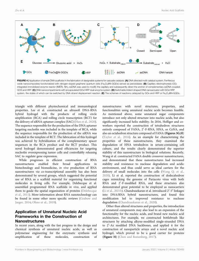
FIGURE 4 | Application of simple DNA scaffolds in the fabrication of designable systems for cascade catalysis. (A) DNA directed multi-catalyst system. Ferriferousoxide nanocomposites functionalized with nitrogen-doped graphene quantum dots (Fe3O4@N-GQDs) served as peroxidases. (B) Capillary electrophoresis (CE)-integrated immobilized enzyme reactor (IMER). NH2-ssDNA was used to modify the capillary and subsequently direct the anchor of complementary ssDNA-coupledGOX and HRP. (C) DNA nanocompartments with encapsulated GOx/HRP dual-enzyme system. (D) Switchable trident-shaped DNA nanoactuator with GOx/HRPsystem, the states of which can be switched by DNA strand displacement reaction. (E) The schemes of reactions catalyzed by GOx and HRP or Fe3O4@N-GQDs.
Frontiers in Bioengineering and Biotechnology | www.frontiersin.org January 2022 | Volume 9 | Article 79248913
Zhu et al. Nucleic Acid Scaffolds
MULTI-ENZYME SYSTEMSCONSTRUCTED IN VITRO BASED ONNUCLEIC ACID FRAMEWORKS AND THEIRAPPLICATIONS
In living organisms, biochemical processes consist of variousenzymatic reactions, including countless cascade reactions, whichare often catalyzed by multi-enzyme systems. A multi-enzymesystem is composed of a series of enzymes that are well organizedin a certain spatial order, which facilitates the transfer of reactionintermediates, and thus significantly enhances the overallcatalytic activities (Hwang and Lee, 2019). Inspired by the
outstanding catalytic performance of natural multi-enzymesystems, numerous artificial multi-enzyme molecular machineshave been designed and applied in a variety of scenarios, rangingfrom bio-catalysis to biopharmaceutical applications (Zhanget al., 2018; Bergquist et al., 2020). In these artificial multi-enzyme systems, the scaffolds that were used to immobilizeand organize the enzymes were prepared from many differentmaterials, including inorganic materials, organic molecules,proteins, and nucleic acids (Jia et al., 2014; Xu K. et al., 2020).
One of the most explored applications of artificial structurescomposed of nucleic acids is building scaffolds to direct theorganization of molecules, including proteins. The outstandingprogrammability, high diversity, and good biocompatibility ofnucleic acid structures make them perfect scaffold materials forelaborate construction of multi-enzyme systems with abundantstrategies. While the mature technologies for solid-phase orenzymatic synthesis of oligonucleotides and efficientamplification of genetically encoded DNA or RNA in livingcells enable rapid production of massive nucleic acid strandswith various lengths, the increasing abundance of methods forprecise assembly of these strands and coupling them to proteinsmakes the spatial organization of enzymes with the guidance ofnucleic acid scaffolds manageable and attractive.
Multi-Enzyme Systems Constructed Basedon Simple DNA ScaffoldsThe simplest way of applying DNA scaffolds for multi-enzymesystem construction is to use an ssDNA template-containingscaffold to guide the immobilization and organization of multipleenzymes that are coupled to short DNA oligonucleotides, which arecomplementary to, and can hybrid specifically with the ssDNAtemplates in the same scaffold (Niemeyer et al., 1999). Variouschemical reactions have been developed for efficient production ofDNA-protein conjugates, which promoted the broad application ofthis strategy. For example, through DNA-directed immobilization,Yang group successfully fabricated a novel multi-catalyst systemwith glucose oxidase (GOx) immobilized on ferriferous oxidenanocomposites functionalized with nitrogen-doped graphenequantum dots (Fe3O4@N-GQD magnetic NPs), which mimickedperoxidase (Shen et al., 2019a). GOx was conjugated with a 5′-thiol-modified single-stranded DNA, which is complementary to anothersingle-stranded DNA immobilized on Fe3O4@N-GQD magneticNPs through glutaraldehyde. The nanozyme-enzyme system wasthen obtained through the hybridization of the two complementaryssDNAs (Figure 4A,E).
Later, they described another strategy for the construction ofnanozyme-enzyme system, in which the GOx was first immobilizedonto magnetic nanoparticles with similar DNA scaffold, and theproduct nanoparticles were then encapsulated with a layer ofnucleotide coordinated polymer (NCP), which was composed ofAMP and Ce3+ (Shen et al., 2019b). The NCP shield not onlyprevented the denaturation and detachment of GOx, but also servedas a peroxidase. In another work, they demonstrated the DNA-directed assembly of a multi-enzyme system with highlycontrollable enzyme ratio, in which the regulation of enzymeratio was achieved by adjusting the ratio of functional groups
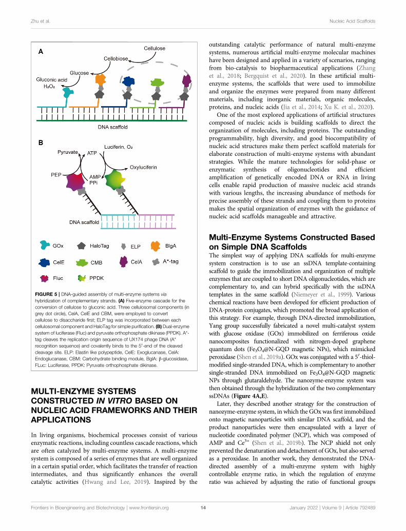
FIGURE 5 | DNA-guided assembly of multi-enzyme systems viahybridization of complementary strands. (A) Five-enzyme cascade for theconversion of cellulose to gluconic acid. Three cellulosomal components (ingrey dot circle), CelA, CelE and CBM, were employed to convertcellulose to disaccharide first; ELP tag was incorporated between eachcellulosomal component and HaloTag for simple purification. (B)Dual-enzymesystem of luciferase (Fluc) and pyruvate orthophosphate dikinase (PPDK). A*-tag cleaves the replication origin sequence of UX174 phage DNA (A*recognition sequence) and covalently binds to the 5′-end of the cleavedcleavage site. ELP: Elastin like polypeptide, CelE: Exoglucanase, CelA:Endoglucanase, CBM: Carbohydrate binding module, BglA: β-glucosidase,FLuc: Luciferase, PPDK: Pyruvate orthophosphate dikinase.
Frontiers in Bioengineering and Biotechnology | www.frontiersin.org January 2022 | Volume 9 | Article 79248914
Zhu et al. Nucleic Acid Scaffolds
used for coupling different guide DNA strands on the nanoparticles(Yang et al., 2017). Other than spherical nanoparticles, surfaces ofother shapesmay also be used for immobilizingDNA-guidedmulti-enzyme systems to adapt different application scenarios. Forexample, Li et al. built a capillary electrophoresis-integratedimmobilized enzyme reactor (CE-IMER), in which the ssDNAstrands for directing the immobilization of complementaryssDNA-coupled GOx and HRP were fixed on the inner surfaceof a capillary (Figure 4B,E) (Li et al., 2020).
In addition to linear DNA strands, Y-shaped DNA structure wasalso used as scaffold to construct multi-enzyme system. Forexample, Song et al. constructed a GOx/HRP multi-enzymebiocatalyst using a Y-shaped DNA scaffold (Song et al., 2019).In this system, The GOx and HRP proteins were immobilized ontothe ends of two arms of the Y-shaped DNA scaffold, and thecomplex was then encapsulated into zeolite imidazolate framework-8. Y-shaped DNA can also be used as a crosslinker to build giantstructures, including those with immobilizedmulti-enzyme systemson them. A DNA nanocompartment with encapsulated GOx/HRPdual-enzyme system was constructed by crosslinking Y-shapedDNA scaffolds with ssDNA-coated magnetic nanoparticles(Figure 4C,E) (Song J. et al., 2020). Each arm of the Y-shapedDNA scaffolds contains a pair of immobilized GOx and HRP, aswell as an ssDNA tail in the end, which is complementary to thessDNA immobilized on the magnetic nanoparticles. Remarkably,the kcat of immobilized enzymes was found to be 16.6-folds higherthan that of free enzymes.
Multi-enzyme systems with dynamic behaviors are attractivedue to their capability of regulating catalytic performance inresponse to environmental changes (Sweetlove and Fernie, 2018).Programmed hybridization of DNA strands not only enables theconstruction of diverse nanostructures, but also is increasinglyapplied for the construction of dynamic devices mainly byintroducing DNA strand-displacement reactions (Zhang andSeelig, 2011). Xing et al. reported a self-assembled dynamic andreconfigurable trident-shaped DNA (TS DNA) nanoactuator, inwhich enzymes were tethered to the arms of the trident DNAscaffold (Figure 4D,E) (Xing et al., 2018). The cascade catalyticefficiency of the dual-enzyme system could be regulated by theswitch of opened, semi-opened, and closed states of the TS DNAnanoactuator, which corresponded to different distances between
the enzymes. Reversible allosteric motion of the arms was achievedby adding the “thermodynamic driver”DNA strands, which led to ashift between stem-loop structure and linear duplex of the DNAspacers between the arms via strand displacement reaction.
Benefiting from the good orthogonality and high affinity, theorthogonal binding of protein tags and their ligands has beensuccessfully applied in one pot assembly of multiple enzyme-tagfusion proteins on ligand-modified DNA scaffolds. Chen et al.reported a five-component enzyme fuel cell constructed with theguidance of a DNA scaffold for direct conversion of cellulose togluconic acid and H2O2 (Chen et al., 2017). All proteincomponents, except for GOx, were genetically fused to aHaloTag, which was then coupled with a chlorohexane groupattached to the ssDNA strand for later site-specific hybridizationwith an ssDNA template (Figure 5A). With similar HaloTag-mediated DNA hybridization strategy, they also reported theconstruction of an artificial cellulosome by assembling fourprotein components on a long ssDNA template produced byrolling circle amplification, which led to 2-fold improvement ofcellulose hydrolysis (Sun and Chen, 2016).
Multi-Enzyme Systems Constructed Basedon the Binding of DNA Scaffolds and DNABinding ProteinsDNA binding proteins recognize and bind to specific sequenceson single-stranded or double-stranded DNAs (Zheng and Wang,2011). The fusion of an enzyme to a DNA-binding protein allowsconvenient immobilization of this enzyme onto a DNA scaffoldwithout any chemical modification, and a multi-enzyme systemcan be simply constructed with a DNA scaffold harboringmultiple recognition sequences of DNA binding proteins. Forexample, Mashimo et al. co-localized firefly luciferase (Fluc) andpyruvate orthophosphate dikinase (PPDK), each of which wasgenetically fused with an ssDNA binding protein, A*-tag, on aDNA scaffold constructed through strand hybridization.Enhanced light emission resulted from cascade bioluminescentreaction suggested that the efficiency of the reaction wasimproved by assembling Fluc and PPDK on the same DNAscaffold (Figure 5B) (Mashimo et al., 2018). Proteins or protein-RNA complexes that can bind with specific dsDNA sequences,
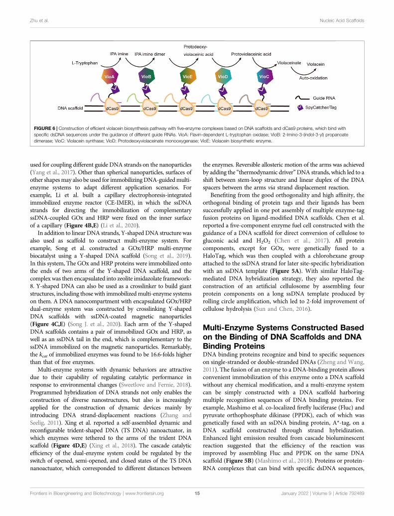
FIGURE 6 | Construction of efficient violacein biosynthesis pathway with five-enzyme complexes based on DNA scaffolds and dCas9 proteins, which bind withspecific dsDNA sequences under the guidance of different guide RNAs. VioA: Flavin-dependent L-tryptophan oxidase; VioB: 2-Imino-3-(indol-3-yl) propanoatedimerase; VioC: Violacein synthase; VioD: Protodeoxyviolaceinate monooxygenase; VioE: Violacein biosynthetic enzyme.
Frontiers in Bioengineering and Biotechnology | www.frontiersin.org January 2022 | Volume 9 | Article 79248915
Zhu et al. Nucleic Acid Scaffolds
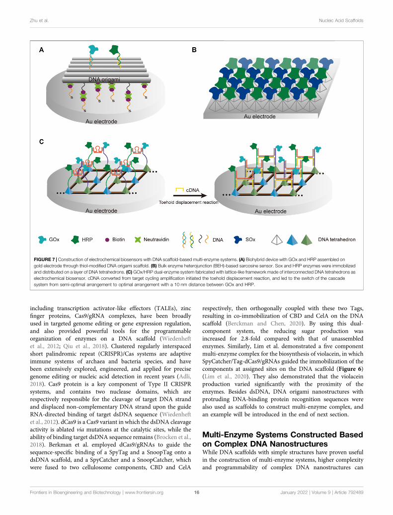
including transcription activator-like effectors (TALEs), zincfinger proteins, Cas9/gRNA complexes, have been broadlyused in targeted genome editing or gene expression regulation,and also provided powerful tools for the programmableorganization of enzymes on a DNA scaffold (Wiedenheftet al., 2012; Qiu et al., 2018). Clustered regularly interspacedshort palindromic repeat (CRISPR)/Cas systems are adaptiveimmune systems of archaea and bacteria species, and havebeen extensively explored, engineered, and applied for precisegenome editing or nucleic acid detection in recent years (Adli,2018). Cas9 protein is a key component of Type II CRISPRsystems, and contains two nuclease domains, which arerespectively responsible for the cleavage of target DNA strandand displaced non-complementary DNA strand upon the guideRNA-directed binding of target dsDNA sequence (Wiedenheftet al., 2012). dCas9 is a Cas9 variant in which the dsDNA cleavageactivity is ablated via mutations at the catalytic sites, while theability of binding target dsDNA sequence remains (Brocken et al.,2018). Berkman et al. employed dCas9/gRNAs to guide thesequence-specific binding of a SpyTag and a SnoopTag onto adsDNA scaffold, and a SpyCatcher and a SnoopCatcher, whichwere fused to two cellulosome components, CBD and CelA
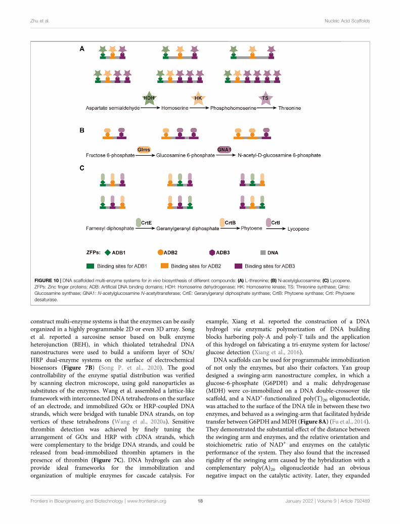
respectively, then orthogonally coupled with these two Tags,resulting in co-immobilization of CBD and CelA on the DNAscaffold (Berckman and Chen, 2020). By using this dual-component system, the reducing sugar production wasincreased for 2.8-fold compared with that of unassembledenzymes. Similarly, Lim et al. demonstrated a five componentmulti-enzyme complex for the biosynthesis of violacein, in whichSpyCatcher/Tag-dCas9/gRNAs guided the immobilization of thecomponents at assigned sites on the DNA scaffold (Figure 6)(Lim et al., 2020). They also demonstrated that the violaceinproduction varied significantly with the proximity of theenzymes. Besides dsDNA, DNA origami nanostructures withprotruding DNA-binding protein recognition sequences werealso used as scaffolds to construct multi-enzyme complex, andan example will be introduced in the end of next section.
Multi-Enzyme Systems Constructed Basedon Complex DNA NanostructuresWhile DNA scaffolds with simple structures have proven usefulin the construction of multi-enzyme systems, higher complexityand programmability of complex DNA nanostructures can
FIGURE 7 | Construction of electrochemical biosensors with DNA scaffold-based multi-enzyme systems. (A) Biohybrid device with GOx and HRP assembled ongold electrode through thiol-modified DNA origami scaffold. (B) Bulk enzyme heterojunction (BEH)-based sarcosine sensor. Sox and HRP enzymes were immobilizedand distributed on a layer of DNA tetrahedrons. (C) GOx/HRP dual-enzyme system fabricated with lattice-like framework made of interconnected DNA tetrahedrons aselectrochemical biosensor. cDNA converted from target cycling amplification initiated the toehold displacement reaction, and led to the switch of the cascadesystem from semi-optimal arrangement to optimal arrangement with a 10 nm distance between GOx and HRP.
Frontiers in Bioengineering and Biotechnology | www.frontiersin.org January 2022 | Volume 9 | Article 79248916
Zhu et al. Nucleic Acid Scaffolds
benefit the building of more sophisticated and controllable multi-enzyme machineries. Fan group created a biomimetic device, inwhich a thiol-modified rectangular DNA origami was used toguide the assembly of GOx/HRP enzymatic cascade on goldelectrodes (Figure 7A) (Ge et al., 2019). In this device, the
enzymatic reaction was translated into a current signal thatcould be directly recorded, and the use of a carefully designedDNA scaffold allowed programmable adjustment of the precisedistance between the enzymes. An irreplaceable advantage ofusing complex DNA nanostructures or their assemblies to
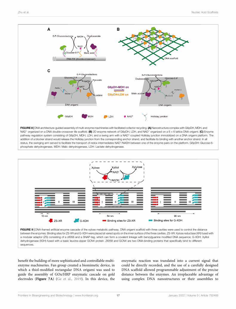
FIGURE 8 |DNA architecture-guided assembly of multi-enzymemachineries with facilitated cofactor recycling. (A)Nanostructure complex with G6pDH, MDH, andNAD+ organized on a DNA double-crossover tile scaffold. (B) 2D enzyme network of G6pDH, LDH, and NAD+ organized on a 6 × 6 lattice DNA origami. (C) Enzymepathway regulation system consisting of G6pDH, MDH, LDH, and a swing arm with a NAD+-coupled Holliday junction immobilized on a DNA origami platform. Theaddition of a blocker strand would release the Holliday junction from the corresponding anchor strand, and facilitate its binding with another anchor strand. In allstatus, the swinging arm served to facilitate the transport of redox intermediates NAD+/NADH between one of the enzyme pairs on the platform. G6pDH: Glucose-6-phosphate dehydrogenase, MDH: Malic dehydrogenase, LDH: Lactate dehydrogenase.
FIGURE 9 | DNA-framed artificial enzyme cascade of the xylose metabolic pathway. DNA origami scaffold with three cavities were used to control the distancebetween the enzymes. Binding sites for ZS-XR andG-XDHwere placed at varied spots on the inner surface of the three cavities. ZS-XR: Xylose reductase (XR) fused witha modular adaptor (ZS) consisting of a zif268 and a SNAP-tag, which can form a covalent linkage with benzylguanine modified DNA sequence; G-XDH: Xylitoldehydrogenase (XDH) fused with a basic leucine-zipper GCN4 protein. Zif268 and GCN4 are two DNA-binding proteins that specifically bind to differentsequences.
Frontiers in Bioengineering and Biotechnology | www.frontiersin.org January 2022 | Volume 9 | Article 79248917
Zhu et al. Nucleic Acid Scaffolds
construct multi-enzyme systems is that the enzymes can be easilyorganized in a highly programmable 2D or even 3D array. Songet al. reported a sarcosine sensor based on bulk enzymeheterojunction (BEH), in which thiolated tetrahedral DNAnanostructures were used to build a uniform layer of SOx/HRP dual-enzyme systems on the surface of electrochemicalbiosensors (Figure 7B) (Song P. et al., 2020). The goodcontrollability of the enzyme spatial distribution was verifiedby scanning electron microscope, using gold nanoparticles assubstitutes of the enzymes. Wang et al. assembled a lattice-likeframework with interconnected DNA tetrahedrons on the surfaceof an electrode, and immobilized GOx or HRP-coupled DNAstrands, which were bridged with tunable DNA strands, on topvertices of these tetrahedrons (Wang et al., 2020a). Sensitivethrombin detection was achieved by finely tuning thearrangement of GOx and HRP with cDNA strands, whichwere complementary to the bridge DNA strands, and could bereleased from bead-immobilized thrombin aptamers in thepresence of thrombin (Figure 7C). DNA hydrogels can alsoprovide ideal frameworks for the immobilization andorganization of multiple enzymes for cascade catalysis. For
example, Xiang et al. reported the construction of a DNAhydrogel via enzymatic polymerization of DNA buildingblocks harboring poly-A and poly-T tails and the applicationof this hydrogel on fabricating a tri-enzyme system for lactose/glucose detection (Xiang et al., 2016).
DNA scaffolds can be used for programmable immobilizationof not only the enzymes, but also their cofactors. Yan groupdesigned a swinging-arm nanostructure complex, in which aglucose-6-phosphate (G6PDH) and a malic dehydrogenase(MDH) were co-immobilized on a DNA double-crossover tilescaffold, and a NAD+-functionalized poly(T)20 oligonucleotide,was attached to the surface of the DNA tile in between these twoenzymes, and behaved as a swinging-arm that facilitated hydridetransfer between G6PDH andMDH (Figure 8A) (Fu et al., 2014).They demonstrated the substantial effect of the distance betweenthe swinging arm and enzymes, and the relative orientation andstoichiometric ratio of NAD+ and enzymes on the catalyticperformance of the system. They also found that the increasedrigidity of the swinging arm caused by the hybridization with acomplementary poly(A)20 oligonucleotide had an obviousnegative impact on the catalytic activity. Later, they expanded
FIGURE 10 | DNA scaffolded multi-enzyme systems for in vivo biosynthesis of different compounds: (A) L-threonine; (B) N-acetylglucosamine; (C) Lycopene.ZFPs: Zinc finger proteins; ADB: Artificial DNA binding domains; HDH: Homoserine dehydrogenase; HK: Homoserine kinase; TS: Threonine synthase; Glms:Glucosamine synthase; GNA1: N-acetylglucosamine N-acetyltransferase; CrtE: Geranylgeranyl diphosphate synthase; CrtB: Phytoene synthase; CrtI: Phytoenedesaturase.
Frontiers in Bioengineering and Biotechnology | www.frontiersin.org January 2022 | Volume 9 | Article 79248918
Zhu et al. Nucleic Acid Scaffolds
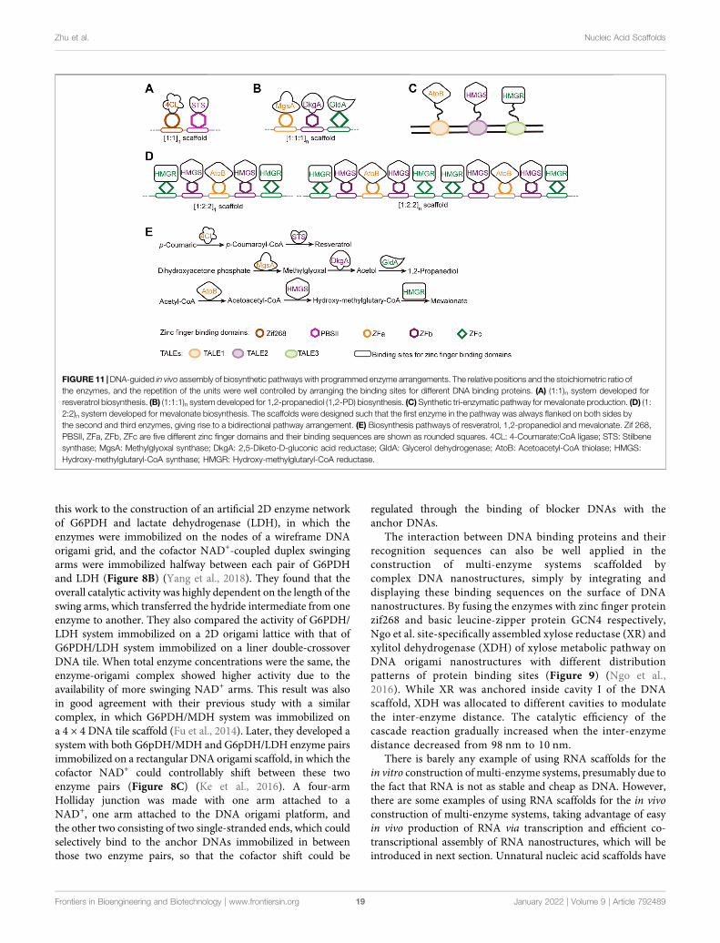
this work to the construction of an artificial 2D enzyme networkof G6PDH and lactate dehydrogenase (LDH), in which theenzymes were immobilized on the nodes of a wireframe DNAorigami grid, and the cofactor NAD+-coupled duplex swingingarms were immobilized halfway between each pair of G6PDHand LDH (Figure 8B) (Yang et al., 2018). They found that theoverall catalytic activity was highly dependent on the length of theswing arms, which transferred the hydride intermediate from oneenzyme to another. They also compared the activity of G6PDH/LDH system immobilized on a 2D origami lattice with that ofG6PDH/LDH system immobilized on a liner double-crossoverDNA tile. When total enzyme concentrations were the same, theenzyme-origami complex showed higher activity due to theavailability of more swinging NAD+ arms. This result was alsoin good agreement with their previous study with a similarcomplex, in which G6PDH/MDH system was immobilized ona 4 × 4 DNA tile scaffold (Fu et al., 2014). Later, they developed asystem with both G6pDH/MDH and G6pDH/LDH enzyme pairsimmobilized on a rectangular DNA origami scaffold, in which thecofactor NAD+ could controllably shift between these twoenzyme pairs (Figure 8C) (Ke et al., 2016). A four-armHolliday junction was made with one arm attached to aNAD+, one arm attached to the DNA origami platform, andthe other two consisting of two single-stranded ends, which couldselectively bind to the anchor DNAs immobilized in betweenthose two enzyme pairs, so that the cofactor shift could be
regulated through the binding of blocker DNAs with theanchor DNAs.
The interaction between DNA binding proteins and theirrecognition sequences can also be well applied in theconstruction of multi-enzyme systems scaffolded bycomplex DNA nanostructures, simply by integrating anddisplaying these binding sequences on the surface of DNAnanostructures. By fusing the enzymes with zinc finger proteinzif268 and basic leucine-zipper protein GCN4 respectively,Ngo et al. site-specifically assembled xylose reductase (XR) andxylitol dehydrogenase (XDH) of xylose metabolic pathway onDNA origami nanostructures with different distributionpatterns of protein binding sites (Figure 9) (Ngo et al.,2016). While XR was anchored inside cavity I of the DNAscaffold, XDH was allocated to different cavities to modulatethe inter-enzyme distance. The catalytic efficiency of thecascade reaction gradually increased when the inter-enzymedistance decreased from 98 nm to 10 nm.
There is barely any example of using RNA scaffolds for thein vitro construction of multi-enzyme systems, presumably due tothe fact that RNA is not as stable and cheap as DNA. However,there are some examples of using RNA scaffolds for the in vivoconstruction of multi-enzyme systems, taking advantage of easyin vivo production of RNA via transcription and efficient co-transcriptional assembly of RNA nanostructures, which will beintroduced in next section. Unnatural nucleic acid scaffolds have
FIGURE 11 |DNA-guided in vivo assembly of biosynthetic pathways with programmed enzyme arrangements. The relative positions and the stoichiometric ratio ofthe enzymes, and the repetition of the units were well controlled by arranging the binding sites for different DNA binding proteins. (A) (1:1)n system developed forresveratrol biosynthesis. (B) (1:1:1)n system developed for 1,2-propanediol (1,2-PD) biosynthesis. (C) Synthetic tri-enzymatic pathway for mevalonate production. (D) (1:2:2)n system developed for mevalonate biosynthesis. The scaffolds were designed such that the first enzyme in the pathway was always flanked on both sides bythe second and third enzymes, giving rise to a bidirectional pathway arrangement. (E) Biosynthesis pathways of resveratrol, 1,2-propanediol and mevalonate. Zif 268,PBSII, ZFa, ZFb, ZFc are five different zinc finger domains and their binding sequences are shown as rounded squares. 4CL: 4-Coumarate:CoA ligase; STS: Stilbenesynthase; MgsA: Methylglyoxal synthase; DkgA: 2,5-Diketo-D-gluconic acid reductase; GldA: Glycerol dehydrogenase; AtoB: Acetoacetyl-CoA thiolase; HMGS:Hydroxy-methylglutaryl-CoA synthase; HMGR: Hydroxy-methylglutaryl-CoA reductase.
Frontiers in Bioengineering and Biotechnology | www.frontiersin.org January 2022 | Volume 9 | Article 79248919
Zhu et al. Nucleic Acid Scaffolds
also not been used for the construction of multi-enzyme systemsyet, since the efforts on the development of efficient tools toproduce, amplify, and manipulate them, including unnaturalnucleic acid polymerases, although already fruitful, are still inprogress. The great potential of this field will be discussed in theend of this article.
IN VIVO CONSTRUCTION OFMULTI-ENZYME SYSTEMS WITH NUCLEICACID FRAMEWORKS FOR EFFICIENTCASCADE BIOCATALYSIS
Whole cell biocatalysts have been broadly used in bioindustry forthe production of valuable chemicals, and their catalytic efficiencycan be significantly improved via in vivo construction of multi-enzyme systems with key enzymes in the metabolic pathways(Dueber et al., 2009; Conrado et al., 2012; Qu et al., 2019).Biomacromolecules that can be synthesized in living cells andgenetically programmed for predefined structures and assembliesare obviously the best options of scaffold materials for theconstruction of intracellular multi-enzyme systems. Besides thein vitro applications, DNA scaffolds are also extensively used astemplates to guide the in vivo assembly of multi-enzyme systems,in which the enzymes are genetically fused to DNA bindingproteins with varied recognition sequences. Zinc finger proteins,which are among the most abundant proteins in eukaryoticorganism and involved in various cellular activities, consist ofrepeated zinc binding motifs containing cysteine and histidineligands (Laity et al., 2001). Many of zinc finger proteins recognizeand bind to dsDNAs with specific sequences to function astranscription factors, which provides perfect tools forprogrammable organization of proteins on dsDNA scaffolds.Lee et al. constructed and optimized a zinc finger protein/DNA scaffold-based multi-enzyme system for the productionof L-threonine (Figure 10A) (Lee et al., 2013). Plasmidcontaining specific binding sequences of zinc finger proteinsand plasmid harboring the genes of zinc finger protein-fusedenzymes, which are involved in L-threonine biosynthesis, wereconstructed and co-transformed into Escherichia coli (E. coli)cells. After being expressed in the cells, the zinc finger protein-fused enzymes spontaneously assembled onto the scaffoldplasmid. They then optimized stoichiometric ratios of theenzymes and intervals between the enzymes for higherL-threonine production. With the optimal system, theproduction time of L-threonine was reduced by over 50%, andthe growth rate of the host cells was also enhanced, presumablydue to reduced accumulation of toxic intermediates. Chen andcoworkers reported the application of similar strategy on spatialmodulation of pathway enzymes for improved production ofN-acetylglucosamine in Bacillus subtilis (Figure 10B) (Liu et al.,2014). The zinc finger protein-directed intracellular assembly ofmulti-enzyme systems also proved effective for modulatingvarious other metabolic pathways and improving theproduction of many other compounds, including lycopene,resveratrol, 1,2-propanediol and mevalonate. (Figure 10C and
Figure 11) (Conrado et al., 2012; Xu X. et al., 2020). TALEs aretranscription factors which contain highly conserved tandemrepeats of 34-aa sequences that are essential for sequence-specific DNA recognition and binding, and have beenengineered and broadly applied in targeted genome editingand regulation of gene expression (Sanjana et al., 2012).Taking advantages of the precise binding between TALEproteins and DNA sequences, a DNA-guided multi-enzymesystem can be assembled in vitro or in vivo by simply fusingenzymes with TALE proteins and immobilizing them onto aDNA scaffold with multiple TALE binding sequences. Zhu et al.reported the in vivo construction of TALE-based multi-enzymesystem for the synthesis of indole-3-acetic acid (Zhu et al., 2016).In this system, tryptophan-2-mono-oxygenase (IAAM) andindole-3-acetimide hydrolase (IAAH) were genetically fusedwith rationally designed TALEs, and site-specificallyimmobilized on a plasmid DNA scaffold containing TALErecognition sequences with different intervals. Later, theyreported the construction of a synthetic tri-enzymatic pathwayin E. coli (Xie et al., 2019). After optimization of the scaffold andthe plasmid copy number, this synthetic tri-enzyme pathwaydemonstrated significantly increased productivity of mevalonatefrom acetyl CoA.
Up to now, not many examples of applying complex nucleicacid nanostructures for in vivo construction of multi-enzymesystems have been reported yet, mainly due to the lack of efficientand user-friendly methods for in vivo production and assembly ofsingles-stranded nucleic acid components, as well as methods forin vivo immobilization of enzymes of interest site-specificallyonto these nanostructures. Sliver et al. pioneered an effort toconstruct multi-enzyme systems in vivo based on RNAnanostructures (Delebecque et al., 2011). RNAs withprogrammed sequences were transcribed and assembled intodiscrete, 1D, and 2D nanostructures in living cells, and theenzymes fused with protein adaptors were immobilized onspecific sites through the binding of these adaptors with theiraptamers, which were prefabricated into those RNAnanostructures. By organizing the proteins in hydrogensynthetic pathway with this strategy, they successfullyoptimized the production of hydrogen. Later, with similarstrategy, they demonstrated the application of various RNAbinding domain (RBD)-aptamer sets in localizing enzymes onin vivo constructed 2D RNA scaffolds for enhanced pentadecaneand succinate production (Sachdeva et al., 2014).
SUMMARY AND PERSPECTIVES
In the past decades, a variety of elegant artificial structuresbased on nucleic acids have been designed and constructed, andsome of them have found broad application in the constructionof elegant nanomaterials or as highly programmable scaffoldsfor multi-enzyme systems. In this article, we started from theintroduction of basic characteristics and properties of naturaland unnatural nucleic acids, which demonstrated theuniqueness and superiority of nucleic acids to be used asstructural materials. The progress in the development of
Frontiers in Bioengineering and Biotechnology | www.frontiersin.org January 2022 | Volume 9 | Article 79248920
Zhu et al. Nucleic Acid Scaffolds
polymerases for the efficient synthesis of nucleic acids withunnatural moieties, which are essential for broader applicationof unnatural nucleic acids, was also discussed. We thensummarized representative approaches in the application ofDNA, RNA, and unnatural nucleic acids for the construction ofvarious artificial nanostructures, ranging from simple one ortwo-dimensional structures to complicated 3D origaminanostructure assemblies. In the end, we reviewed theconstruction of multi-enzyme systems using nucleic acidstructures as scaffolds, as well as the application of thesemulti-enzyme systems for efficient catalysis of cascadereactions, both in vitro and in vivo.
Rapid development of DNA and RNA nanotechnology hasenabled the construction and broad application of various nucleicacid nanostructures. However, more efforts still need to be madeto make the procedures and the computational programs for thedesign and construction of complicated nanostructures morerobust and user-friendly (Andronescu et al., 2003; Jabbariet al., 2015; Jun et al., 2019). For broader practical and evenindustrial applications of the nucleic acid nanostructures, thetechniques for massive and cost-effective production of DNA andRNA building blocks also need to be developed and optimized(Chandler et al., 2020; Geary et al., 2021). Recently, biomass DNAdirectly extracted from living organisms was successfullyconverted into biodegradable materials, ranging from gels toplastics, at large scales with very low costs (Wang et al., 2020;Han et al., 2021). While these approaches demonstrated thefeasibility of acquiring and applying massive amount of DNAraw materials from nature for the construction of functionalmaterials, development of novel technologies for efficientsynthesis of longer and more accurate oligonucleotides, suchas enzymatic de novo synthesis technology, will facilitate theacquirement of more and better oligonucleotide sequences to beused as components for the construction of nucleic acidnanostructures in the future (Jensen and Davis, 2018; Perkel,2019; Eisenstein, 2020).
In order to immobilize and organize two or more enzymes forefficient catalysis of cascade reactions, multi-enzyme systemshave employed various materials to fabricate the scaffolds,including inorganic materials, organic frameworks, andbiological materials. Among those, nucleic acid has proven oneof the most promising scaffold materials. Numerous artificialstructures have been constructed with nucleic acids, and many ofthem are already well suited for the construction of multi-enzymesystems for efficient catalysis of cascade reactions in vitro or invivo. However, these systems are still far from perfect, and a lot ofefforts need to be made to further improve their performance. Forinstance, new chemistry may be developed to achieve efficientsite-specific immobilization of the enzymes onto the scaffolds andprecise control of the orientation of the enzymes, which isobviously very important for the accessibility of substrates orintermediates to the active sites. Smarter nucleic acidnanostructures may also be designed for better regulation ofthe stoichiometric ratio and relative positions of the enzymesbased on their kinetic parameters to achieve optimal overallcatalytic performance. Development of better computationaltools to assist the design, assembly, and behavior simulation of
versatile nucleic acid nanostructures will also greatly promotethese efforts (de Llano et al., 2020; Poppleton et al., 2020). Despitebeing constructed with very high complexity and applied forguiding the assembly of highly programmable multi-enzymesystems in vitro, complex DNA origami nanostructures arerarely used as scaffolds for in vivo construction of multi-enzyme systems. Most of the nucleic acid scaffold-based multi-enzyme system constructed in vivo are still simply based on theimmobilization of DNA-binding protein elements on lineardouble strands of DNA. Further exploitation of methods forconstructing complicated nucleic acid nanostructures in vivo andcombining these nanostructures with well-designed nucleic acid-binding proteins may lead to much better scaffolds for in vivoassembly of highly programmable multi-enzyme systems, andfacilitate the engineering of cellular metabolic pathways.
Although natural nucleic acid frameworks have already beenbroadly used in the construction of nanomaterials or as thescaffolds of multi-enzyme systems, there still remain manychallenges in the construction and application of better nucleicacid scaffolds that fulfill the requirements of more programmableand robust nanostructures or multi-enzyme systems. Forexample, the number of nucleobases in natural DNA andRNA is quite limited compared to the number of amino acidsin protein, which results in limited sequences, structures, anddesignability of DNA and RNA. Moreover, natural nucleic acidshave low tolerance against nuclease degradation and reducedstability under various chemical or physical conditions, whichrestricts the application of nucleic acid nanostructures or multi-enzyme systems with nucleic acid scaffolds in biological solutionsor harsh environments. The fast development of unnaturalcomponents has provided a generous toolbox for thestructural and functional expansion and augmentation ofnucleic acids, and thus offered a great potential for the designand construction of better nucleic acid-based nanostructures andmulti-enzyme scaffolds. As mentioned above, newly developedunnatural base pairs are already able to pair, replicate andtranscribe as efficiently as natural base pairs (Betz et al., 2017;Feldman et al., 2019; Ouaray et al., 2020). The introduction ofthese unnatural base pairs into nucleic acids will undoubtedlyincrease the possible sequences, structures, properties, andfunctionalities of nucleic acids, and thus provide more optionsfor components of nanostructures and scaffolds of multi-enzymesystems. The site-specifically incorporated unnatural base pairsmay also be chemically modified with various functional groups,side chains, or linkers, and thus provide much more controllablesites or handles for the immobilization, organization, andregulation of biomacromolecules, including enzymes. Theintroduction of unnatural sugar-phosphate backbones will notonly expand the possible structures and functionalities of nucleicacids, but can also significantly increase the stability of the nucleicacid scaffolds under different chemical, physical, or biologicalconditions, and thus lead to broader application scenarios for thenanostructures and multi-enzyme systems. While chemicalsynthesis and polymerase engineering underlies thedevelopment and in vitro application of unnatural nucleicacids, the efforts on construction of semi-synthetic organismshave opened the gate for in vivo application of unnatural nucleic
Frontiers in Bioengineering and Biotechnology | www.frontiersin.org January 2022 | Volume 9 | Article 79248921
Zhu et al. Nucleic Acid Scaffolds
acids (Zhang et al., 2017a; Zhang et al., 2017b; Fischer et al., 2020;Hashimoto et al., 2021; Manandhar et al., 2021), so we canconfidently expect the broad application of unnaturalcomponents in future construction of nucleic acid scaffolds forbuilding nanostructures and multi-enzyme machineries, bothin vitro and in vivo.
AUTHOR CONTRIBUTIONS
GZ, PS, JW, ML, and ZC wrote and revised the manuscript underthe guidance of TC. GZ and JW helped to draw the figures. All
authors contributed to the article and approved the submittedversion.
ACKNOWLEDGMENTS
We thank National Natural Science Foundation of China (No.21978100), National Key R&D Program of China (No.2019YFA0904102), Guangdong Provincial Pearl River TalentsProgram (No. 2019QN01Y228), and the Program for GuangdongIntroducing Innovative and Entrepreneurial Teams (No.2019ZT08Y318) for the financial support of this work.
REFERENCES
Adli, M. (2018). The CRISPR Tool Kit for Genome Editing and Beyond. Nat.Commun. 9, 1911. doi:10.1038/s41467-018-04252-2
Afonin, K. A., Bindewald, E., Yaghoubian, A. J., Voss, N., Jacovetty, E., Shapiro, B.A., et al. (2010). In Vitro assembly of Cubic RNA-Based Scaffolds Designed InSilico. Nat. Nanotech 5, 676–682. doi:10.1038/nnano.2010.160
Ali, M.M., Li, F., Zhang, Z., Zhang, K., Kang, D.-K., Ankrum, J. A., et al. (2014). RollingCircle Amplification: A Versatile Tool for Chemical Biology, Materials Science andMedicine. Chem. Soc. Rev. 43 (10), 3324–3341. doi:10.1039/c3cs60439j
Alon, D. M., Voigt, C. A., and Elbaz, J. (2020). Engineering a DNAzyme-BasedOperon System for the Production of DNA Nanoscaffolds in Living Bacteria.ACS Synth. Biol. 9, 236–240. doi:10.1021/acssynbio.9b00415
Andersen, E. S., Dong, M., Nielsen, M. M., Jahn, K., Subramani, R., Mamdouh, W.,et al. (2009). Self-assembly of a Nanoscale DNA Box with a Controllable Lid.Nature 459, 73–76. doi:10.1038/nature07971
Andronescu, M., Aguirre-Hernandez, R., Condon, A., and Hoos, H. H.(2003). RNAsoft: A Suite of RNA Secondary Structure Prediction andDesign Software Tools. Nucleic Acids Res. 31 (13), 3416–3422.doi:10.1093/nar/gkg612
Anosova, I., Kowal, E. A., Dunn, M. R., Chaput, J. C., Van Horn, W. D., and Egli,M. (2016). The Structural Diversity of Artificial Genetic Polymers.Nucleic AcidsRes. 44, 1007–1021. doi:10.1093/nar/gkv1472
Arangundy-Franklin, S., Taylor, A. I., Porebski, B. T., Genna, V., Peak-Chew, S.,Vaisman, A., et al. (2019). A Synthetic Genetic Polymer with an UnchargedBackbone Chemistry Based on Alkyl Phosphonate Nucleic Acids. Nat. Chem.11, 533–542. doi:10.1038/s41557-019-0255-4
Berckman, E. A., and Chen, W. (2020). A Modular Approach for dCas9-MediatedEnzyme Cascading via Orthogonal Bioconjugation. Chem. Commun. 56,11426–11428. doi:10.1039/d0cc04196c
Bergquist, P. L., Siddiqui, S., and Sunna, A. (2020). Cell-Free Biocatalysis for theProduction of Platform Chemicals. Front. Energ. Res. 8, 22. doi:10.3389/fenrg.2020.00193
Betz, K., Kimoto, M., Diederichs, K., Hirao, I., andMarx, A. (2017). Structural Basisfor Expansion of the Genetic Alphabet with an Artificial Nucleobase Pair.Angew. Chem. Int. Ed. 56, 12000–12003. doi:10.1002/anie.201704190
Boerneke, M. A., Dibrov, S. M., and Hermann, T. (2016). Crystal-Structure-GuidedDesign of Self-Assembling RNA Nanotriangles. Angew. Chem. Int. Ed. 55 (12),4097–4100. doi:10.1002/anie.201600233
Brocken, D. J. W., Tark-Dame, M., and Dame, R. T. (2018). dCas9: A Versatile Toolfor Epigenome Editing. Curr. Issues Mol. Biol. 26, 15–32. doi:10.21775/cimb.026.015
Burmeister, P. E., Lewis, S. D., Silva, R. F., Preiss, J. R., Horwitz, L. R., Pendergrast,P. S., et al. (2005). Direct In Vitro Selection of a 2′-O-Methyl Aptamer to VEGF.Chem. Biol. 12, 25–33. doi:10.1016/j.chembiol.2004.10.017
Caruthers, M. H. (1985). Gene Synthesis Machines: DNA Chemistry and its Uses.Science 230, 281–285. doi:10.1126/science.3863253
Cassinelli, V., Oberleitner, B., Sobotta, J., Nickels, P., Grossi, G., Kempter, S., et al. (2015).One-step Formation of "Chain-Armor"-Stabilized DNA Nanostructures. Angew.Chem. Int. Ed. 54, 7795–7798. doi:10.1002/anie.201500561
Cha, T.-G., Pan, J., Chen, H., Robinson, H. N., Li, X., Mao, C., et al. (2015). DesignPrinciples of DNA Enzyme-Based Walkers: Translocation Kinetics andPhotoregulation. J. Am. Chem. Soc. 137 (29), 9429–9437. doi:10.1021/jacs.5b05522
Chandler, M., Panigaj, M., Rolband, L. A., and Afonin, K. A. (2020). Challenges inOptimizing RNA Nanostructures for Large-Scale Production and ControlledTherapeutic Properties. Nanomedicine 15 (13), 1331–1340. doi:10.2217/nnm-2020-0034
Chandrasekaran, A. R., Mathivanan, J., Ebrahimi, P., Vilcapoma, J., Chen, A. A.,Halvorsen, K., et al. (2020). Hybrid DNA/RNA Nanostructures with 2′-5′Linkages. Nanoscale 12, 21583–21590. doi:10.1039/d0nr05846g
Chen, T., and Romesberg, F. E. (2017). Enzymatic Synthesis, Amplification, andApplication of DNAwith a Functionalized Backbone.Angew. Chem. Int. Ed. 56,14046–14051. doi:10.1002/anie.201707367
Chen, J.-K., Schultz, R. G., Lioyd, D. H., and Gryaznov, S. M. (1995). Synthesis ofOligodeoxyribonucleotide N3′→P5′ Phosphoramidates. Nucleic Acids Res. 23,2661–2668. doi:10.1093/nar/23.14.2661
Chen, T., Hongdilokkul, N., Liu, Z., Adhikary, R., Tsuen, S. S., and Romesberg, F. E.(2016). Evolution of Thermophilic DNA Polymerases for the Recognition andAmplification of C2ʹ-Modified DNA. Nat. Chem. 8, 556–562. doi:10.1038/nchem.2493
Chen, Q., Yu, S., Myung, N., and Chen, W. (2017). DNA-Guided Assembly of aFive-Component Enzyme cascade for Enhanced Conversion of Cellulose toGluconic Acid and H2O2. J. Biotechnol. 263, 30–35. doi:10.1016/j.jbiotec.2017.10.006
Chen, S., and Hermann, T. (2020). RNA-DNA Hybrid Nanoshapes That Self-Assemble Dependent on Ligand Binding. Nanoscale 12 (5), 3302–3307.doi:10.1039/c9nr09706f
Chopra, A., Sagredo, S., Grossi, G., Andersen, E., and Simmel, F. (2019). Out-of-Plane Aptamer Functionalization of RNA Three-Helix Tiles. Nanomaterials 9(4), 507. doi:10.3390/nano9040507
Conrado, R. J., Wu, G. C., Boock, J. T., Xu, H., Chen, S. Y., Lebar, T., et al. (2012).DNA-Guided Assembly of Biosynthetic Pathways Promotes ImprovedCatalytic Efficiency. Nucleic Acids Res. 40, 1879–1889. doi:10.1093/nar/gkr888
Cruz-Acuña, M., Halman, J. R., Afonin, K. A., Dobson, J., and Rinaldi, C. (2018).Magnetic Nanoparticles Loaded with Functional RNA Nanoparticles.Nanoscale 10 (37), 17761–17770. doi:10.1039/c8nr04254c
Cui, Y., Yan, J., and Wei, B. (2021). Hybrid Wireframe DNA Nanostructures withScaffolded and Scaffold-Free Modules. Angew. Chem. Int. Ed. 60, 9345–9350.doi:10.1002/anie.202015564
Dan, K., Veetil, A. T., Chakraborty, K., and Krishnan, Y. (2019). DNANanodevicesMap Enzymatic Activity in Organelles. Nat. Nanotechnol. 14 (3), 252–259.doi:10.1038/s41565-019-0365-6
de Llano, E., Miao, H., Ahmadi, Y., Wilson, A. J., Beeby, M., Viola, I., et al. (2020).Adenita: Interactive 3D Modelling and Visualization of DNA Nanostructures.Nucleic Acids Res. 48, 8269–8275. doi:10.1093/nar/gkaa593
Declercq, R., Aerschot, A. V., Read, R. J., Herdewijn, P., and Meervelt, L. V. (2002).Crystal Structure of Double Helical Hexitol Nucleic Acids. J. Am. Chem. Soc.124, 928–933. doi:10.1021/ja016570w
Delebecque, C. J., Lindner, A. B., Silver, P. A., and Aldaye, F. A. (2011).Organization of Intracellular Reactions with Rationally Designed RNAAssemblies. Science 333, 470–474. doi:10.1126/science.1206938
Frontiers in Bioengineering and Biotechnology | www.frontiersin.org January 2022 | Volume 9 | Article 79248922
Zhu et al. Nucleic Acid Scaffolds
DeLuca, M., Shi, Z., Castro, C. E., and Arya, G. (2020). Dynamic DNANanotechnology: Toward Functional Nanoscale Devices. Nanoscale Horiz. 5(2), 182–201. doi:10.1039/c9nh00529c
Dietz, H., Douglas, S. M., and Shih, W. M. (2009). Folding DNA into Twisted andCurved Nanoscale Shapes. Science 325, 725–730. doi:10.1126/science.1174251
Ding, L., Li, J., Wu, C., Yan, F., Li, X., and Zhang, S. (2020). A Self-AssembledRNA-Triple Helix Hydrogel Drug Delivery System Targeting Triple-Negative Breast Cancer. J. Mater. Chem. B 8 (16), 3527–3533.doi:10.1039/c9tb01610d
Dong, H., Chen, H., Jiang, J., Zhang, H., Cai, C., and Shen, Q. (2018). HighlySensitive Electrochemical Detection of Tumor Exosomes Based on AptamerRecognition-Induced Multi-DNA Release and Cyclic Enzymatic Amplification.Anal. Chem. 90 (7), 4507–4513. doi:10.1021/acs.analchem.7b04863
Dong, Y., Yao, C., Zhu, Y., Yang, L., Luo, D., and Yang, D. (2020). DNA FunctionalMaterials Assembled from Branched DNA: Design, Synthesis, andApplications. Chem. Rev. 120 (17), 9420–9481. doi:10.1021/acs.chemrev.0c00294
Donovan, J., Whitney, G., Rath, S., and Korennykh, A. (2015). StructuralMechanism of Sensing Long dsRNA via a Noncatalytic Domain in HumanOligoadenylate Synthetase 3. Proc. Natl. Acad. Sci. U.S.A. 112, 3949–3954.doi:10.1073/pnas.1419409112
Douglas, S. M., Dietz, H., Liedl, T., Högberg, B., Graf, F., and Shih, W. M. (2009).Self-Assembly of DNA into Nanoscale Three-Dimensional Shapes. Nature 459,414–418. doi:10.1038/nature08016
Dueber, J. E., Wu, G. C., Malmirchegini, G. R., Moon, T. S., Petzold, C. J., Ullal, A.V., et al. (2009). Synthetic Protein Scaffolds Provide Modular Control overMetabolic Flux. Nat. Biotechnol. 27, 753–759. doi:10.1038/nbt.1557
Ebert, M.-O., Mang, C., Krishnamurthy, R., Eschenmoser, A., and Jaun, B. (2008).The Structure of a TNA−TNA Complex in Solution: NMR Study of theOctamer Duplex Derived from α-(l)-Threofuranosyl-(3′-2′)-CGAATTCG.J. Am. Chem. Soc. 130, 15105–15115. doi:10.1021/ja8041959
Eckstein, F. (2014). Phosphorothioates, Essential Components of TherapeuticOligonucleotides. Nucleic Acid Ther. 24 (6), 374–387. doi:10.1089/nat.2014.0506
Eichert, A., Behling, K., Betzel, C., Erdmann, V. A., Fürste, J. P., and Förster, C.(2010). The crystal Structure of an ’All Locked’ Nucleic Acid Duplex. NucleicAcids Res. 38 (19), 6729–6736. doi:10.1093/nar/gkq505
Eisenstein, M. (2020). Enzymatic DNA Synthesis Enters New Phase. Nat.Biotechnol. 38 (10), 1113–1115. doi:10.1038/s41587-020-0695-9
Elbaz, J., Yin, P., and Voigt, C. A. (2016). Genetic Encoding of DNANanostructures and Their Self-Assembly in Living Bacteria. Nat. Commun.7, 11179. doi:10.1038/ncomms11179
Endo, M., Takeuchi, Y., Emura, T., Hidaka, K., and Sugiyama, H. (2014).Preparation of Chemically Modified RNA Origami Nanostructures. Chem.Eur. J. 20 (47), 15330–15333. doi:10.1002/chem.201404084
Feldman, A. W., and Romesberg, F. E. (2018). Expansion of the Genetic Alphabet:A Chemist’s Approach to Synthetic Biology. Acc. Chem. Res. 51 (2), 394–403.doi:10.1021/acs.accounts.7b00403
Feldman, A. W., Dien, V. T., Karadeema, R. J., Fischer, E. C., You, Y., Anderson, B.A., et al. (2019). Optimization of Replication, Transcription, and Translation ina Semi-synthetic Organism. J. Am. Chem. Soc. 141, 10644–10653. doi:10.1021/jacs.9b02075
Feng, Q.-M., Ma, P., Cao, Q.-H., Guo, Y.-H., and Xu, J.-J. (2020). An Aptamer-Binding DNA Walking Machine for Sensitive ElectrochemiluminescenceDetection of Tumor Exosomes. Chem. Commun. 56 (2), 269–272.doi:10.1039/c9cc08051a
Fischer, E. C., Hashimoto, K., Zhang, Y., Feldman, A. W., Dien, V. T., Karadeema,R. J., et al. (2020). New Codons for Efficient Production of Unnatural Proteinsin a Semisynthetic Organism. Nat. Chem. Biol. 16, 570–576. doi:10.1038/s41589-020-0507-z
Fu, J., Yang, Y. R., Johnson-Buck, A., Liu, M., Liu, Y., Walter, N. G., et al. (2014).Multi-enzyme Complexes on DNA Scaffolds Capable of Substrate Channellingwith an Artificial Swinging Arm. Nat. Nanotechnol. 9, 531–536. doi:10.1038/nnano.2014.100
Futami, K., Kimoto, M., Lim, Y. W. S., and Hirao, I. (2019). Genetic AlphabetExpansion Provides Versatile Specificities and Activities of Unnatural-BaseDNA Aptamers Targeting Cancer Cells.Mol. Ther. - Nucleic Acids 14, 158–170.doi:10.1016/j.omtn.2018.11.011
Gačanin, J., Synatschke, C. V., and Weil, T. (2020). Biomedical Applications ofDNA-Based Hydrogels. Adv. Funct. Mater. 30 (4), 1906253. doi:10.1002/adfm.201906253
Ge, Z., Fu, J., Liu, M., Jiang, S., Andreoni, A., Zuo, X., et al. (2019). ConstructingSubmonolayer DNA Origami Scaffold on Gold Electrode for Wiring of RedoxEnzymatic Cascade Pathways. ACS Appl. Mater. Inter. 11, 13881–13887.doi:10.1021/acsami.8b12374
Geary, C., Rothemund, P. W. K., and Andersen, E. S. (2014). A Single-StrandedArchitecture for Cotranscriptional Folding of RNA Nanostructures. Science345, 799–804. doi:10.1126/science.1253920
Geary, C., Grossi, G., McRae, E. K. S., Rothemund, P. W. K., and Andersen, E. S.(2021). RNA Origami Design Tools Enable Cotranscriptional Folding ofKilobase-Sized Nanoscaffolds. Nat. Chem. 13, 549–558. doi:10.1038/s41557-021-00679-1
Gerling, T., Wagenbauer, K. F., Neuner, A. M., and Dietz, H. (2015). DynamicDNA Devices and Assemblies Formed by Shape-Complementary, Non-basePairing 3D Components. Science 347, 1446–1452. doi:10.1126/science.aaa5372
Grabow, W. W., and Jaeger, L. (2014). RNA Self-Assembly and RNANanotechnology. Acc. Chem. Res. 47 (6), 1871–1880. doi:10.1021/ar500076k
Guo, X., Li, F., Liu, C., Zhu, Y., Xiao, N., Gu, Z., et al. (2020). Construction ofOrganelle-Like Architecture by Dynamic DNA Assembly in Living Cells.Angew. Chem. Int. Ed. 59 (46), 20651–20658. doi:10.1002/anie.202009387
Guo, P. (2010). The Emerging Field of RNA Nanotechnology. Nat. Nanotechnol. 5,833–842. doi:10.1038/nnano.2010.231
Han, D., Qi, X., Myhrvold, C., Wang, B., Dai, M., Jiang, S., et al. (2017). Single-Stranded DNA and RNAOrigami. Science 358, 10. doi:10.1126/science.aao2648
Han, S., Park, Y., Kim, H., Nam, H., Ko, O., and Lee, J. B. (2020). Double ControlledRelease of Therapeutic RNA Modules through Injectable DNA-RNA HybridHydrogel.ACSAppl.Mater. Inter. 12 (50), 55554–55563. doi:10.1021/acsami.0c12506
Han, J., Guo, Y., Wang, H., Zhang, K., and Yang, D. (2021). Sustainable BioplasticMade from Biomass DNA and Ionomers. J. Am. Chem. Soc. 143 (46),19486–19497. doi:10.1021/jacs.1c08888
Hao, C., Li, X., Tian, C., Jiang, W., Wang, G., and Mao, C. (2014). Construction ofRNA Nanocages by Re-engineering the Packaging RNA of Phi29Bacteriophage. Nat. Commun. 5, 7. doi:10.1038/ncomms4890
Haque, F., Pi, F., Zhao, Z., Gu, S., Hu, H., Yu, H., et al. (2018). RNA Versatility,Flexibility, and Thermostability for Practice in RNA Nanotechnology andBiomedical Applications. WIREs RNA 9 (1), 17. doi:10.1002/wrna.1452
Hariadi, R. F., Sommese, R. F., Adhikari, A. S., Taylor, R. E., Sutton, S., Spudich,J. A., et al. (2015). Mechanical Coordination in Motor Ensembles RevealedUsing Engineered Artificial Myosin Filaments. Nat. Nanotech 10 (8), 696–700.doi:10.1038/nnano.2015.132
Hashimoto, K., Fischer, E. C., and Romesberg, F. E. (2021). Efforts toward FurtherIntegration of an Unnatural Base Pair into the Biology of a SemisyntheticOrganism. J. Am. Chem. Soc. 143, 8603–8607. doi:10.1021/jacs.1c03860
He, W., Hatcher, E., Balaeff, A., Beratan, D. N., Gil, R. R., Madrid, M., et al. (2008).Solution Structure of a Peptide Nucleic Acid Duplex from NMR Data: Featuresand Limitations. J. Am. Chem. Soc. 130, 13264–13273. doi:10.1021/ja800652h
Heuer-Jungemann, A., and Linko, V. (2021). Engineering Inorganic Materials withDNA Nanostructures. ACS Cent. Sci. doi:10.1021/acscentsci.1c01272
Høiberg, H. C., Sparvath, S. M., Andersen, V. L., Kjems, J., and Andersen, E. S.(2019). An RNA Origami Octahedron with Intrinsic siRNAs for Potent GeneKnockdown. Biotechnol. J. 14 (1), 1700634. doi:10.1002/biot.201700634
Hoshino, H., Kasahara, Y., Kuwahara, M., and Obika, S. (2020). DNA PolymeraseVariants with High Processivity and Accuracy for Encoding and DecodingLocked Nucleic Acid Sequences. J. Am. Chem. Soc. 142, 21530–21537.doi:10.1021/jacs.0c10902
Hwang, E. T., and Lee, S. (2019). Multienzymatic Cascade Reactions via EnzymeComplex by Immobilization. ACS Catal. 9, 4402–4425. doi:10.1021/acscatal.8b04921
Ichida, J. K., Horhota, A., Zou, K. Y., McLaughlin, L. W., and Szostak, J. W. (2005).High Fidelity TNA Synthesis by Therminator Polymerase.Nucleic Acids Res. 33,5219–5225. doi:10.1093/nar/gki840
Jabbari, H., Aminpour, M., and Montemagno, C. (2015). ComputationalApproaches to Nucleic Acid Origami. ACS Comb. Sci. 17 (10), 535–547.doi:10.1021/acscombsci.5b00079
Jahanban-Esfahlan, A., Seidi, K., Jaymand, M., Schmidt, T. L., Majdi, H., Javaheri,T., et al. (2019). Dynamic DNA Nanostructures in Biomedicine: Beauty, Utility
Frontiers in Bioengineering and Biotechnology | www.frontiersin.org January 2022 | Volume 9 | Article 79248923
Zhu et al. Nucleic Acid Scaffolds
and Limits. J. Controlled Release 315, 166–185. doi:10.1016/j.jconrel.2019.10.003
Jang, B., Kim, B., Kim, H., Kwon, H., Kim, M., Seo, Y., et al. (2018). EnzymaticSynthesis of Self-Assembled Dicer Substrate RNA Nanostructures forProgrammable Gene Silencing. Nano Lett. 18 (7), 4279–4284. doi:10.1021/acs.nanolett.8b01267
Jani, M. S., Zou, J., Veetil, A. T., and Krishnan, Y. (2020). A DNA-BasedFluorescent Probe Maps NOS3 Activity with Subcellular Spatial Resolution.Nat. Chem. Biol. 16 (6), 660–666. doi:10.1038/s41589-020-0491-3
Jensen, M. A., and Davis, R. W. (2018). Template-Independent EnzymaticOligonucleotide Synthesis (TiEOS): Its History, Prospects, and Challenges.Biochemistry 57 (12), 1821–1832. doi:10.1021/acs.biochem.7b00937
Jia, F., Narasimhan, B., and Mallapragada, S. (2014). Materials-based Strategies forMulti-Enzyme Immobilization and Co-Localization: A Review. Biotechnol.Bioeng. 111, 209–222. doi:10.1002/bit.25136
Jiang, S., Ge, Z., Mou, S., Yan, H., and Fan, C. (2021). Designer DNANanostructures forTherapeutics. Chem 7, 1156–1179. doi:10.1016/j.chempr.2020.10.025
Jing, X., Zhang, F., Pan, M., Dai, X., Li, J., Wang, L., et al. (2019). SolidifyingFramework Nucleic Acids with Silica. Nat. Protoc. 14 (8), 2416–2436.doi:10.1038/s41596-019-0184-0
Johnson, A. T., Schlegel, M. K., Meggers, E., Essen, L.-O., and Wiest, O. (2011). Onthe Structure and Dynamics of Duplex GNA. J. Org. Chem. 76, 7964–7974.doi:10.1021/jo201469b
Jun, H., Shepherd, T. R., Zhang, K., Bricker, W. P., Li, S., Chiu, W., et al. (2019a).Automated Sequence Design of 3D Polyhedral Wireframe DNA Origami withHoneycomb Edges. ACS Nano 13, 2083–2093. doi:10.1021/acsnano.8b08671
Jun, H., Wang, X., Bricker, W. P., and Bathe, M. (2019b). Automated SequenceDesign of 2D Wireframe DNA Origami with Honeycomb Edges. Nat.Commun. 10, 9. doi:10.1038/s41467-019-13457-y
Ke, Y., Ong, L. L., Shih, W. M., and Yin, P. (2012). Three-Dimensional StructuresSelf-Assembled from DNA Bricks. Science 338, 1177–1183. doi:10.1126/science.1227268
Ke, G., Liu, M., Jiang, S., Qi, X., Yang, Y. R., Wootten, S., et al. (2016). DirectionalRegulation of Enzyme Pathways through the Control of Substrate Channelingon a DNAOrigami Scaffold. Angew. Chem. Int. Ed. 55, 7483–7486. doi:10.1002/anie.201603183
Ke,W., Hong, E., Saito, R. F., Rangel, M. C., Wang, J., Viard, M., et al. (2019). RNA-DNA Fibers and Polygons with Controlled Immunorecognition Activate RNAi,FRET and Transcriptional Regulation of NF-Κb in Human Cells. Nucleic AcidsRes. 47 (3), 1350–1361. doi:10.1093/nar/gky1215
Kim, J., Jang, D., Park, H., Jung, S., Kim, D. H., and Kim, W. J. (2018). Functional-DNA-Driven Dynamic Nanoconstructs for Biomolecule Capture and DrugDelivery. Adv. Mater. 30 (45), 1707351. doi:10.1002/adma.201707351
Kimoto, M., and Hirao, I. (2020). Genetic Alphabet Expansion Technology byCreating Unnatural Base Pairs. Chem. Soc. Rev. 49, 7602–7626. doi:10.1039/d0cs00457j
Kimoto, M., Kawai, R., Mitsui, T., Yokoyama, S., and Hirao, I. (2008). An UnnaturalBase Pair System for Efficient PCR Amplification and Functionalization of DNAMolecules. Nucleic Acids Res. 37, e14. doi:10.1093/nar/gkn956
Kimoto, M., Shermane Lim, Y. W., and Hirao, I. (2019). Molecular Affinity Rulers:Systematic Evaluation of DNA Aptamers for Their Applicabilities in ELISA.Nucleic Acids Res. 47, 8362–8374. doi:10.1093/nar/gkz688
Krissanaprasit, A., Key, C., Fergione, M., Froehlich, K., Pontula, S., Hart, M.,et al. (2019). Genetically Encoded, Functional Single-Strand RNAOrigami: Anticoagulant. Adv. Mater. 31 (21), 1808262. doi:10.1002/adma.201808262
Krissanaprasit, A., Key, C. M., Froehlich, K., Pontula, S., Mihalko, E., Dupont, D. M.,et al. (2021). Multivalent Aptamer-Functionalized Single-Strand RNAOrigami asEffective, Target-Specific Anticoagulants with Corresponding Reversal Agents.Adv. Healthc. Mater. 10 (11), 2001826. doi:10.1002/adhm.202001826
Krishnan, Y., and Simmel, F. C. (2011). Nucleic Acid Based Molecular Devices.Angew. Chem. Int. Ed. Engl. 50 (14), 3124–3156. doi:10.1002/anie.200907223
Kumar, P., and Caruthers, M. H. (2020). DNA Analogues Modified at theNonlinking Positions of Phosphorus. Acc. Chem. Res. 53, 2152–2166.doi:10.1021/acs.accounts.0c00078
Laity, J. H., Lee, B. M., and Wright, P. E. (2001). Zinc finger Proteins: New Insightsinto Structural and Functional Diversity. Curr. Opin. Struct. Biol. 11, 39–46.doi:10.1016/S0959-440X(00)00167-6
Lavergne, T., Lamichhane, R., Malyshev, D. A., Li, Z., Li, L., Sperling, E., et al.(2016). FRET Characterization of Complex Conformational Changes in a Large16S Ribosomal RNA Fragment Site-Specifically Labeled Using Unnatural BasePairs. ACS Chem. Biol. 11 (5), 1347–1353. doi:10.1021/acschembio.5b00952
Lee, J. B., Peng, S., Yang, D., Roh, Y. H., Funabashi, H., Park, N., et al. (2012). AMechanical Metamaterial Made from a DNA Hydrogel. Nat. Nanotechnol. 7(12), 816–820. doi:10.1038/nnano.2012.211
Lee, J. H., Jung, S.-C., Bui, L. M., Kang, K. H., Song, J.-J., and Kim, S. C. (2013).Improved Production of L -Threonine in Escherichia C by Use of a DNAScaffold System. Appl. Environ. Microbiol. 79, 774–782. doi:10.1128/aem.02578-12
Li, J., Pei, H., Zhu, B., Liang, L., Wei, M., He, Y., et al. (2011). Self-AssembledMultivalent DNA Nanostructures for Noninvasive Intracellular Delivery ofImmunostimulatory CpG Oligonucleotides. ACS Nano 5, 8783–8789.doi:10.1021/nn202774x
Li, L., Degardin, M., Lavergne, T., Malyshev, D. A., Dhami, K., Ordoukhanian, P.,et al. (2014). Natural-Like Replication of an Unnatural Base Pair for theExpansion of the Genetic Alphabet and Biotechnology Applications. J. Am.Chem. Soc. 136, 826–829. doi:10.1021/ja408814g
Li, M., Zheng, M., Wu, S., Tian, C., Liu, D., Weizmann, Y., et al. (2018). In VivoProduction of RNA Nanostructures via Programmed Folding of Single-Stranded RNAs. Nat. Commun. 9, 2196. doi:10.1038/s41467-018-04652-4
Li, S., Jiang, Q., Liu, S., Zhang, Y., Tian, Y., Song, C., et al. (2018). A DNANanorobot Functions as a Cancer Therapeutic in Response to a MolecularTrigger In Vivo. Nat. Biotechnol. 36, 258–264. doi:10.1038/nbt.4071
Li, F., Tang, J., Geng, J., Luo, D., and Yang, D. (2019). Polymeric DNA Hydrogel:Design, Synthesis and Applications. Prog. Polym. Sci. 98, 101163. doi:10.1016/j.progpolymsci.2019.101163
Li, M., Shen, H., Zhou, Z., He, W., Su, P., Song, J., et al. (2020). Controllable andHigh-Performance Immobilized Enzyme Reactor: DNA-DirectedImmobilization of Multienzyme in Polyamidoamine Dendrimer-Functionalized Capillaries. Electrophoresis 41, 335–344. doi:10.1002/elps.201900428
Li, F., Li, J., Dong, B., Wang, F., Fan, C., and Zuo, X. (2021). DNANanotechnology-Empowered Nanoscopic Imaging of Biomolecules. Chem. Soc. Rev. 50 (9),5650–5667. doi:10.1039/d0cs01281e
Li, H., Zhang, K., Binzel, D. W., Shlyakhtenko, L. S., Lyubchenko, Y. L., Chiu, W.,et al. (2021). RNA Nanotechnology to Build a Dodecahedral Genome of Single-Stranded RNA Virus. RNA Biol. 18, 2390–2400. doi:10.1080/15476286.2021.1915620
Lim, S., Kim, J., Kim, Y., Xu, D., and Clark, D. S. (2020). CRISPR/Cas-DirectedProgrammable Assembly of Multi-Enzyme Complexes. Chem. Commun. 56,4950–4953. doi:10.1039/d0cc01174f
Lin, C., Rinker, S., Wang, X., Liu, Y., Seeman, N. C., and Yan, H. (2008). In VivoCloning of Artificial DNA Nanostructures. Proc. Natl. Acad. Sci. U.S.A. 105,17626–17631. doi:10.1073/pnas.0805416105
Liu, Y., Zhu, Y., Ma, W., Shin, H.-D., Li, J., Liu, L., et al. (2014). Spatial Modulationof Key Pathway Enzymes by DNA-Guided Scaffold System and RespirationChain Engineering for Improved N-Acetylglucosamine Production by BacillusSubtilis. Metab. Eng. 24, 61–69. doi:10.1016/j.ymben.2014.04.004
Liu, Z., Chen, T., and Romesberg, F. E. (2017). Evolved Polymerases FacilitateSelection of Fully 2′-OMe-Modified Aptamers. Chem. Sci. 8, 8179–8182.doi:10.1039/c7sc03747c
Liu, X., Zhang, F., Jing, X., Pan, M., Liu, P., Li, W., et al. (2018). Complex SilicaComposite Nanomaterials Templated with DNA Origami. Nature 559 (7715),593–598. doi:10.1038/s41586-018-0332-7
Lu, X.-J., and Olson, W. K. (2003). 3DNA: A Software Package for the Analysis,Rebuilding and Visualization of Three-Dimensional Nucleic Acid Structures.Nucleic Acids Res. 31, 5108–5121. doi:10.1093/nar/gkg680
Malyshev, D. A., Dhami, K., Lavergne, T., Chen, T., Dai, N., Foster, J. M., et al.(2014). A Semi-Synthetic Organism with an Expanded Genetic Alphabet.Nature 509, 385–388. doi:10.1038/nature13314
Manandhar, M., Chun, E., and Romesberg, F. E. (2021). Genetic Code Expansion:Inception, Development, Commercialization. J. Am. Chem. Soc. 143,4859–4878. doi:10.1021/jacs.0c11938
Manoharan, M., Akinc, A., Pandey, R. K., Qin, J., Hadwiger, P., John, M., et al.(2011). Unique Gene-Silencing and Structural Properties of 2′-Fluoro-ModifiedsiRNAs. Angew. Chem. Int. Ed. 50, 2284–2288. doi:10.1002/anie.201006519
Frontiers in Bioengineering and Biotechnology | www.frontiersin.org January 2022 | Volume 9 | Article 79248924
Zhu et al. Nucleic Acid Scaffolds
Martín-Pintado, N., Yahyaee-Anzahaee, M., Campos-Olivas, R., Noronha, A. M.,Wilds, C. J., Damha, M. J., et al. (2012). The Solution Structure of DoubleHelical Arabino Nucleic Acids (ANA and 2′F-ANA): Effect of Arabinoses inDuplex-Hairpin Interconversion. Nucleic Acids Res. 40, 9329–9339.doi:10.1093/nar/gks672
Mashimo, Y., Mie, M., and Kobatake, E. (2018). A DNA-Scaffold PlatformEnhances a Multi-Enzymatic Cycling Reaction. Biotechnol. Lett. 40 (4),667–672. doi:10.1007/s10529-018-2517-4
Matsunaga, K.-I., Kimoto, M., Lim, V. W., Tan, H. P., Wong, Y. Q., Sun, W., et al.(2021). High-Affinity Five/six-Letter DNA Aptamers with Superior SpecificityEnabling the Detection of Dengue NS1 Protein Variants beyond the SerotypeIdentification. Nucleic Acids Res. 49, 11407–11424. doi:10.1093/nar/gkab515
Meyer, A. J., Garry, D. J., Hall, B., Byrom, M. M., McDonald, H. G., Yang, X., et al.(2015). Transcription Yield of Fully 2′-Modified RNA Can Be Increased by theAddition of Thermostabilizing Mutations to T7 RNA Polymerase Mutants.Nucleic Acids Res. 43, 7480–7488. doi:10.1093/nar/gkv734
Miao, S., Liang, Y., Rundell, S., Bhunia, D., Devari, S., Munyaradzi, O., et al. (2021).Unnatural Bases for Recognition of Noncoding Nucleic Acid Interfaces.Biopolymers 112 (1), 16. doi:10.1002/bip.23399
Mo, F., Jiang, K., Zhao, D., Wang, Y., Song, J., and Tan, W. (2021). DNAHydrogel-Based Gene Editing and Drug Delivery Systems. Adv. Drug Deliv. Rev. 168,79–98. doi:10.1016/j.addr.2020.07.018
Modi, S., M. G., S., Goswami, D., Gupta, G. D., Mayor, S., and Krishnan, Y. (2009).A DNA Nanomachine that Maps Spatial and Temporal pH Changes insideLiving Cells. Nat. Nanotechnol. 4 (5), 325–330. doi:10.1038/nnano.2009.83
Modi, S., Nizak, C., Surana, S., Halder, S., and Krishnan, Y. (2013). Two DNANanomachines Map pH Changes along Intersecting Endocytic Pathways Insidethe Same Cell. Nat. Nanotechnol. 8 (6), 459–467. doi:10.1038/nnano.2013.92
Mooers, B. H. M., and Singh, A. (2011). The Crystal Structure of an Oligo(U):pre-mRNA Duplex from a Trypanosome RNA Editing Substrate. RNA 17 (10),1870–1883. doi:10.1261/rna.2880311
Nakashima, Y., Abe, H., Abe, N., Aikawa, K., and Ito, Y. (2011). Branched RNANanostructures for RNA Interference. Chem. Commun. 47 (29), 8367–8369.doi:10.1039/c1cc11780g
Narayana, N., and Weiss, M. A. (2009). Crystallographic Analysis of a Sex-SpecificEnhancer Element: Sequence-Dependent DNA Structure, Hydration, andDynamics. J. Mol. Biol. 385, 469–490. doi:10.1016/j.jmb.2008.10.041
Narayanaswamy, N., Chakraborty, K., Saminathan, A., Zeichner, E., Leung, K.,Devany, J., et al. (2019). A pH-Correctable, DNA-Based Fluorescent Reporterfor Organellar Calcium. Nat. Methods 16 (1), 95–102. doi:10.1038/s41592-018-0232-7
Ngo, T. A., Nakata, E., Saimura, M., and Morii, T. (2016). Spatially OrganizedEnzymes Drive Cofactor-Coupled Cascade Reactions. J. Am. Chem. Soc. 138,3012–3021. doi:10.1021/jacs.5b10198
Niemeyer, C. M., Boldt, L., Ceyhan, B., and Blohm, D. (1999). DNA-DirectedImmobilization: Efficient, Reversible, and Site-Selective Surface Binding ofProteins by Means of Covalent DNA-Streptavidin Conjugates. Anal.Biochem. 268, 54–63. doi:10.1006/abio.1998.3017
Niu, X., Sun, R., Chen, Z., Yao, Y., Zuo, X., Chen, C., et al. (2021). PseudoknotLength Modulates the Folding, Conformational Dynamics, and Robustness ofXrn1 Resistance of Flaviviral xrRNAs. Nat. Commun. 12 (1), 14. doi:10.1038/s41467-021-26616-x
Ochoa, S., and Milam, V. T. (2020). Modified Nucleic Acids: Expanding theCapabilities of Functional Oligonucleotides. Molecules 25, 4659. doi:10.3390/molecules25204659
Ohno, H., Akamine, S., and Saito, H. (2019). RNANanostructures and Scaffolds forBiotechnology Applications. Curr. Opin. Biotechnol. 58, 53–61. doi:10.1016/j.copbio.2018.11.006
Ouaray, Z., Singh, I., Georgiadis, M. M., and Richards, N. G. J. (2020). BuildingBetter Enzymes: Molecular Basis of Improved Non-Natural NucleobaseIncorporation by an Evolved DNA Polymerase. Protein Sci. 29, 455–468.doi:10.1002/pro.3762
Padilla, R., and Sousa, R. (1999). Efficient Synthesis of Nucleic Acids HeavilyModified with Non- Canonical Ribose 2’-groups Using a mutantT7 RNAPolymerase (RNAP).Nucleic Acids Res. 27, 1561–1563. doi:10.1093/nar/27.6.1561
Padilla, R., and Sousa, R. (2002). A Y639F/H784A T7 RNA Polymerase DoubleMutant Displays superior Properties for Synthesizing RNAs with Non-Canonical NTPs. Nucleic Acids Res. 30, e138. doi:10.1093/nar/gnf138
Pallan, P. S., Greene, E. M., Jicman, P. A., Pandey, R. K., Manoharan, M., Rozners,E., et al. (2011). Unexpected Origins of the Enhanced Pairing Affinity of 2′-Fluoro-Modified RNA. Nucleic Acids Res. 39, 3482–3495. doi:10.1093/nar/gkq1270
Park, N., Um, S. H., Funabashi, H., Xu, J. F., and Luo, D. (2009). A Cell-FreeProtein-Producing Gel. Nat. Mater. 8 (5), 432–437. doi:10.1038/nmat2419
Park, J.-E., Heo, I., Tian, Y., Simanshu, D. K., Chang, H., Jee, D., et al. (2011). DicerRecognizes the 5′ End of RNA for Efficient and Accurate Processing. Nature475 (7355), 201–205. doi:10.1038/nature10198
Park, G., Kang, B., Park, S. V., Lee, D., and Oh, S. S. (2021). A UnifiedComputational View of DNA Duplex, Triplex, Quadruplex and TheirDonor-Acceptor Interactions. Nucleic Acids Res. 49, 4919–4933. doi:10.1093/nar/gkab285
Patterson, D. M., Nazarova, L. A., and Prescher, J. A. (2014). Finding the Right(Bioorthogonal) Chemistry. ACS Chem. Biol. 9, 592–605. doi:10.1021/cb400828a
Paukstelis, P. J., and Ellington, A. D. (2008). Rolling Out DNA Nanostructures InVivo. Proc. Natl. Acad. Sci. U.S.A. 105, 17593–17594. doi:10.1073/pnas.0810029105
Perkel, J. M. (2019). The Race for Enzymatic DNA Synthesis Heats up. Nature 566(7745), 565. doi:10.1038/d41586-019-00682-0
Pinheiro, V. B., Taylor, A. I., Cozens, C., Abramov, M., Renders, M., Zhang, S., et al.(2012). Synthetic Genetic Polymers Capable of Heredity and Evolution. Science336, 341–344. doi:10.1126/science.1217622
Poppleton, E., Bohlin, J., Matthies, M., Sharma, S., Zhang, F., and Šulc, P. (2020).Design, Optimization and Analysis of Large DNA and RNA Nanostructuresthrough Interactive Visualization, Editing and Molecular Simulation. NucleicAcids Res. 48, e72. doi:10.1093/nar/gkaa417
Praetorius, F., and Dietz, H. (2017). Self-Assembly of Genetically Encoded DNA-Protein Hybrid Nanoscale Shapes. Science 355 (6331), 8. doi:10.1126/science.aam5488
Praetorius, F., Kick, B., Behler, K. L., Honemann, M. N., Weuster-Botz, D., andDietz, H. (2017). Biotechnological Mass Production of DNA Origami. Nature552, 84–87. doi:10.1038/nature24650
Qiu, X.-y., Xie, S.-S., Min, L., Wu, X.-M., Zhu, L.-Y., and Zhu, L. (2018). SpatialOrganization of Enzymes to Enhance Synthetic Pathways in Microbial Chassis:A Systematic Review. Microb. Cel. Fact. 17, 7. doi:10.1186/s12934-018-0965-0
Qu, J., Cao, S., Wei, Q., Zhang, H., Wang, R., Kang, W., et al. (2019). SyntheticMultienzyme Complexes, Catalytic Nanomachineries for cascade BiosynthesisIn Vivo. ACS Nano 13, 9895–9906. doi:10.1021/acsnano.9b03631
Richert, C., Roughton, A. L., and Benner, S. A. (1996). Nonionic Analogs of RNAwith Dimethylene Sulfone Bridges. J. Am. Chem. Soc. 118, 4518–4531.doi:10.1021/ja952322m
Robeyns, K., Herdewijn, P., and Van Meervelt, L. (2008). Structure of the FullyModified Left-Handed Cyclohexene Nucleic Acid Sequence GTGTACAC.J. Am. Chem. Soc. 130, 1979–1984. doi:10.1021/ja077313f
Rosu, F. d. r., Gabelica, V. r., Houssier, C., Colson, P., and Pauw, E. D. (2002).Triplex and Quadruplex DNA Structures Studied by Electrospray MassSpectrometry. Rapid Commun. Mass. Spectrom. 16, 1729–1736. doi:10.1002/rcm.778
Rothemund, P. W. K. (2006). Folding DNA to Create Nanoscale Shapes andPatterns. Nature 440, 297–302. doi:10.1038/nature04586
Sachdeva, G., Garg, A., Godding, D., Way, J. C., and Silver, P. A. (2014). In Vivoco-Localization of Enzymes on RNA Scaffolds Increases Metabolic Production in aGeometrically Dependent Manner. Nucleic Acids Res. 42, 9493–9503.doi:10.1093/nar/gku617
Saiki, R., Gelfand, D., Stoffel, S., Scharf, S., Higuchi, R., Horn, G., et al. (1988).Primer-Directed Enzymatic Amplification of DNA with a Thermostable DNAPolymerase. Science 239, 487–491. doi:10.1126/science.2448875
Saminathan, A., Devany, J., Veetil, A. T., Suresh, B., Pillai, K. S., Schwake, M., et al.(2021). A DNA-Based Voltmeter for Organelles. Nat. Nanotechnol. 16 (1),96–103. doi:10.1038/s41565-020-00784-1
Sanjana, N. E., Cong, L., Zhou, Y., Cunniff, M.M., Feng, G., and Zhang, F. (2012). ATranscription Activator-Like Effector Toolbox for Genome Engineering. Nat.Protoc. 7, 171–192. doi:10.1038/nprot.2011.431
Schoenit, A., Cavalcanti-Adam, E. A., and Göpfrich, K. (2021). Functionalization ofCellular Membranes with DNA Nanotechnology. Trends Biotechnol. 39,1208–1220. doi:10.1016/j.tibtech.2021.02.002
Frontiers in Bioengineering and Biotechnology | www.frontiersin.org January 2022 | Volume 9 | Article 79248925
Zhu et al. Nucleic Acid Scaffolds
Schoffelen, S., and van Hest, J. C. (2013). Chemical Approaches for theConstruction of Multi-Enzyme Reaction Systems. Curr. Opin. Struct. Biol.23, 613–621. doi:10.1016/j.sbi.2013.06.010
Seeman, N. C. (1982). Nucleic Acid Junctions and Lattices. J. Theor. Biol. 99,237–247. doi:10.1016/0022-5193(82)90002-9
Seeman, N. C. (1990). De Novo design of Sequences for Nucleic Acid StructuralEngineering. J. Biomol. Struct. Dyn. 8 (3), 573–581. doi:10.1080/07391102.1990.10507829
Shang, Y., Li, N., Liu, S., Wang, L.,Wang, Z. G., Zhang, Z., et al. (2020). Site-SpecificSynthesis of Silica Nanostructures on DNA Origami Templates. Adv. Mater. 32(21), 2000294. doi:10.1002/adma.202000294
Sharma, J., Chhabra, R., Cheng, A., Brownell, J., Liu, Y., and Yan, H. (2009).Control of Self-Assembly of DNA Tubules through Integration of GoldNanoparticles. Science 323, 112–116. doi:10.1126/science.1165831
Shen, H., Song, J., Yang, Y., Su, P., and Yang, Y. (2019a). DNA-Directed EnzymeImmobilization on Fe3O4 Modified with Nitrogen-Doped Graphene QuantumDots as a Highly Efficient and Stable Multi-Catalyst System. J. Mater. Sci. 54,2535–2551. doi:10.1007/s10853-018-2992-y
Shen, H., Song, J., Zhou, Z., Li, M., Zhang, R., Su, P., et al. (2019b). DNA-DirectedImmobilized Enzymes on Recoverable Magnetic Nanoparticles Shielded inNucleotide Coordinated Polymers. Ind. Eng. Chem. Res. 58, 8585–8596.doi:10.1021/acs.iecr.9b01341
Shi, J., Wu, Y., Zhang, S., Tian, Y., Yang, D., and Jiang, Z. (2018). BioinspiredConstruction of Multi-Enzyme Catalytic Systems. Chem. Soc. Rev. 47,4295–4313. doi:10.1039/c7cS00914c
Shi, Z., Li, S. K., Charoenputtakun, P., Liu, C.-Y., Jasinski, D., and Guo, P. (2018).RNA Nanoparticle Distribution and Clearance in the Eye after SubconjunctivalInjection with and without Thermosensitive Hydrogels. J. Controlled Release270, 14–22. doi:10.1016/j.jconrel.2017.11.028
Simmel, F. C. (2007). Towards Biomedical Applications for Nucleic AcidNanodevices. Nanomedicine 2 (6), 817–830. doi:10.2217/17435889.2.6.817
Simmel, F. C. (2012). DNA-Based Assembly Lines and Nanofactories. Curr. Opin.Biotechnol. 23, 516–521. doi:10.1016/j.copbio.2011.12.024
Sobczak, J.-P. J., Martin, T. G., Gerling, T., and Dietz, H. (2012). Rapid Folding ofDNA into Nanoscale Shapes at Constant Temperature. Science 338, 1458–1461.doi:10.1126/science.1229919
Song, J., He, W., Shen, H., Zhou, Z., Li, M., Su, P., et al. (2019). Construction ofMultiple Enzyme Metal-Organic Frameworks Biocatalyst via DNA Scaffold: APromising Strategy for Enzyme Encapsulation. Chem. Eng. J. 363, 174–182.doi:10.1016/j.cej.2019.01.138
Song, J., He,W., Shen, H., Zhou, Z., Li, M., Su, P., et al. (2020). Exquisitely DesignedMagnetic DNA Nanocompartment for Enzyme Immobilization withAdjustable Catalytic Activity and Improved Enzymatic Assay Performance.Chem. Eng. J. 390, 124488. doi:10.1016/j.cej.2020.124488
Song, P., Shen, J., Ye, D., Dong, B., Wang, F., Pei, H., et al. (2020). ProgrammingBulk Enzyme Heterojunctions for Biosensor Development with TetrahedralDNA Framework. Nat. Commun. 11, 10. doi:10.1038/s41467-020-14664-8
Spicer, C. D., and Davis, B. G. (2014). Selective Chemical Protein Modification.Nat. Commun. 5, 14. doi:10.1038/ncomms5740
Stewart, J. M., Subramanian, H. K. K., and Franco, E. (2017). Self-Assembly ofMulti-Stranded RNAMotifs into Lattices and Tubular Structures. Nucleic AcidsRes. 45, 5449–5457. doi:10.1093/nar/gkx063
Sun, Q., and Chen, W. (2016). HaloTag Mediated Artificial Cellulosome Assemblyon a Rolling Circle Amplification DNA Template for Efficient CelluloseHydrolysis. Chem. Commun. 52, 6701–6704. doi:10.1039/c6cc02035f
Sun, L., Gao, Y., Xu, Y., Chao, J., Liu, H., Wang, L., et al. (2017). Real-Time Imagingof Single-Molecule Enzyme Cascade Using a DNA Origami Raft. J. Am. Chem.Soc. 139 (48), 17525–17532. doi:10.1021/jacs.7b09323
Surana, S., Bhat, J. M., Koushika, S. P., and Krishnan, Y. (2011). An AutonomousDNA Nanomachine Maps Spatiotemporal pH Changes in a MulticellularLiving Organism. Nat. Commun. 2, 7. doi:10.1038/ncomms1340
Sweetlove, L. J., and Fernie, A. R. (2018). The Role of Dynamic Enzyme Assembliesand Substrate Channelling in Metabolic Regulation. Nat. Commun. 9, 12.doi:10.1038/s41467-018-04543-8
Tang, J., Yao, C., Gu, Z., Jung, S., Luo, D., and Yang, D. (2020). Super-Soft andSuper-Elastic DNA Robot with Magnetically Driven Navigational Locomotionfor Cell Delivery in Confined Space. Angew. Chem. Int. Ed. 59 (6), 2490–2495.doi:10.1002/anie.201913549
Taylor, A. I., Pinheiro, V. B., Smola, M. J., Morgunov, A. S., Peak-Chew, S., Cozens,C., et al. (2015). Catalysts from Synthetic Genetic Polymers. Nature 518,427–430. doi:10.1038/nature13982
Taylor, A. I., Beuron, F., Peak-Chew, S.-Y., Morris, E. P., Herdewijn, P., andHolliger, P. (2016). Nanostructures from Synthetic Genetic Polymers.ChemBioChem 17, 1107–1110. doi:10.1002/cbic.201600136
Torelli, E., Kozyra, J., Shirt-Ediss, B., Piantanida, L., Voïtchovsky, K., andKrasnogor, N. (2020). Cotranscriptional Folding of a Bio-OrthogonalFluorescent Scaffolded RNA Origami. ACS Synth. Biol. 9, 1682–1692.doi:10.1021/acssynbio.0c00009
Um, S. H., Lee, J. B., Park, N., Kwon, S. Y., Umbach, C. C., and Luo, D. (2006).Enzyme-Catalysed Assembly of DNA Hydrogel. Nat. Mater. 5 (10), 797–801.doi:10.1038/nmat1741
Valero, J., and Skugor, M. (2020). Mechanisms, Methods of Tracking andApplications of DNA Walkers: A Review. Chemphyschem 21 (17),1971–1988. doi:10.1002/cphc.202000235
Veetil, A. T., Zou, J., Henderson, K. W., Jani, M. S., Shaik, S. M., Sisodia, S. S., et al.(2020). DNA-Based Fluorescent Probes of NOS2 Activity in Live Brains. Proc.Natl. Acad. Sci. U.S.A. 117 (26), 14694–14702. doi:10.1073/pnas.2003034117
Verma, S., and Eckstein, F. (1998). Modified Oligonucleotides: Synthesis and Strategy forUsers. Annu. Rev. Biochem. 67, 99–134. doi:10.1146/annurev.biochem.67.1.99
Wagenbauer, K. F., Sigl, C., and Dietz, H. (2017). Gigadalton-Scale Shape-Programmable DNA Assemblies. Nature 552, 78–83. doi:10.1038/nature24651
Wang, D., Cui, J., Gan, M., Xue, Z., Wang, J., Liu, P., et al. (2020). Transformationof Biomass DNA into Biodegradable Materials from Gels to Plastics forReducing Petrochemical Consumption. J. Am. Chem. Soc. 142 (22),10114–10124. doi:10.1021/jacs.0c02438
Wang, D., Chai, Y., Yuan, Y., and Yuan, R. (2020). Lattice-Like DNA TetrahedronNanostructure as Scaffold to Locate GOx and HRP Enzymes for HighlyEfficient Enzyme cascade Reaction. ACS Appl. Mater. Inter. 12, 2871–2877.doi:10.1021/acsami.9b18702
Wang, Q., Chen, X., Li, X., Song, D., Yang, J., Yu, H., et al. (2020). 2′-Fluoroarabinonucleic Acid Nanostructures as Stable Carriers for CellularDelivery in the Strongly Acidic Environment. ACS Appl. Mater. Inter. 12,53592–53597. doi:10.1021/acsami.0c11684
Wang, Y., Dai, L., Ding, Z., Ji, M., Liu, J., Xing, H., et al. (2021a). DNA OrigamiSingle Crystals with Wulff Shapes. Nat. Commun. 12 (1), 8. doi:10.1038/s41467-021-23332-4
Wang, Y., Nguyen, K., Spitale, R. C., and Chaput, J. C. (2021b). A BiologicallyStable DNAzyme that Efficiently Silences Gene Expression in Cells. Nat. Chem.13, 319–326. doi:10.1038/s41557-021-00645-x
Watts, J. K., Martín-Pintado, N., Gómez-Pinto, I., Schwartzentruber, J., Portella, G.,Orozco, M., et al. (2010). Differential Stability of 2′F-ANARNA and ANARNAHybrid Duplexes: Roles of Structure, Pseudohydrogen Bonding, Hydration, IonUptake and Flexibility. Nucleic Acids Res. 38, 2498–2511. doi:10.1093/nar/gkp1225
Wei, B., Dai, M., and Yin, P. (2012). Complex Shapes Self-Assembled from Single-Stranded DNA Tiles. Nature 485 (7400), 623–626. doi:10.1038/nature11075
Wiedenheft, B., Sternberg, S. H., and Doudna, J. A. (2012). RNA-Guided GeneticSilencing Systems in Bacteria and Archaea. Nature 482 (7385), 331–338.doi:10.1038/nature10886
Wilner, O. I., Weizmann, Y., Gill, R., Lioubashevski, O., Freeman, R., andWillner, I. (2009). Enzyme Cascades Activated on TopologicallyProgrammed DNA Scaffolds. Nat. Nanotechnol. 4, 249–254. doi:10.1038/nnano.2009.50
Xiang, B., He, K., Zhu, R., Liu, Z., Zeng, S., Huang, Y., et al. (2016). Self-AssembledDNA Hydrogel Based on Enzymatically Polymerized DNA for ProteinEncapsulation and Enzyme/DNAzyme Hybrid Cascade Reaction. ACS Appl.Mater. Inter. 8 (35), 22801–22807. doi:10.1021/acsami.6b03572
Xie, S. S., Qiu, X. Y., Zhu, L. Y., Zhu, C. S., Liu, C. Y., Wu, X. M., et al. (2019).Assembly of TALE-Based DNA Scaffold for the Enhancement of ExogenousMulti-Enzymatic Pathway. J. Biotechnol. 296, 69–74. doi:10.1016/j.jbiotec.2019.03.008
Xing, C., Huang, Y., Dai, J., Zhong, L., Wang, H., Lin, Y., et al. (2018). Spatial Regulationof Biomolecular Interactions with a Switchable Trident-Shaped DNA Nanoactuator.ACS Appl. Mater. Inter. 10, 32579–32587. doi:10.1021/acsami.8b10761
Xu, K., Chen, X., Zheng, R., and Zheng, Y. (2020). Immobilization of Multi-Enzymes on Support Materials for Efficient Biocatalysis. Front. Bioeng.Biotechnol. 8, 17. doi:10.3389/fbioe.2020.00660
Frontiers in Bioengineering and Biotechnology | www.frontiersin.org January 2022 | Volume 9 | Article 79248926
Zhu et al. Nucleic Acid Scaffolds
Xu, X., Tian, L., Tang, S., Xie, C., Xu, J., and Jiang, L. (2020). Design and Tailoringof an Artificial DNA Scaffolding System for Efficient Lycopene Synthesis Usingzinc-Finger-Guided Assembly. J. Ind. Microbiol. Biotechnol. 47, 209–222.doi:10.1007/s10295-019-02255-6
Xu, L., Duan, J., Chen, J., Ding, S., and Cheng, W. (2021). Recent Advances inRolling Circle Amplification-Based Biosensing Strategies-A Review. AnalyticaChim. Acta 1148, 238187. doi:10.1016/j.aca.2020.12.062
Yang, D., Hartman, M. R., Derrien, T. L., Hamada, S., An, D., Yancey, K. G., et al.(2014). DNA Materials: Bridging Nanotechnology and Biotechnology. Acc.Chem. Res. 47 (6), 1902–1911. doi:10.1021/ar5001082
Yang, Y., Zhang, R., Zhou, B., Song, J., Su, P., and Yang, Y. (2017). High Activityand Convenient Ratio Control: DNA-Directed Coimmobilization of MultipleEnzymes on Multifunctionalized Magnetic Nanoparticles. ACS Appl. Mater.Inter. 9, 37254–37263. doi:10.1021/acsami.7b08553
Yang, Y. R., Fu, J., Wootten, S., Qi, X., Liu, M., Yan, H., et al. (2018). 2D EnzymeCascade Network with Efficient Substrate Channeling by Swinging Arms.ChemBioChem 19, 212–216. doi:10.1002/cbic.201700613
Yao, C., Zhu, C., Tang, J., Ou, J., Zhang, R., and Yang, D. (2021). T Lymphocyte-Captured DNA Network for Localized Immunotherapy. J. Am. Chem. Soc. 143(46), 19330–19340. doi:10.1021/jacs.1c07036
Yourston, L., Rolband, L., West, C., Lushnikov, A., Afonin, K. A., andKrasnoslobodtsev, A. V. (2020). Tuning Properties of Silver Nanoclusterswith RNA Nanoring Assemblies. Nanoscale 12 (30), 16189–16200.doi:10.1039/d0nr03589k
Zhang, D. Y., and Seelig, G. (2011). Dynamic DNA Nanotechnology Using Strand-Displacement Reactions. Nat. Chem 3, 103–113. doi:10.1038/nchem.957
Zhang, Y., Ptacin, J. L., Fischer, E. C., Aerni, H. R., Caffaro, C. E., San Jose, K., et al.(2017a). A Semi-Synthetic Organism that Stores and Retrieves IncreasedGenetic Information. Nature 551, 644–647. doi:10.1038/nature24659
Zhang, Y., Lamb, B. M., Feldman, A. W., Zhou, A. X., Lavergne, T., Li, L., et al.(2017b). A Semisynthetic Organism Engineered for the Stable Expansion of theGenetic Alphabet. Proc. Natl. Acad. Sci. U.S.A. 114, 1317–1322. doi:10.1073/pnas.1616443114
Zhang, G., Quin, M. B., and Schmidt-Dannert, C. (2018). Self-Assembling ProteinScaffold System for Easy In Vitro Coimmobilization of Biocatalytic cascadeEnzymes. ACS Catal. 8, 5611–5620. doi:10.1021/acscatal.8b00986
Zhang, L., Wang, S., Yang, Z., Hoshika, S., Xie, S., Li, J., et al. (2020). An Aptamer-Nanotrain Assembled from Six-Letter DNA Delivers Doxorubicin Selectively toLiver Cancer Cells. Angew. Chem. Int. Ed. 59, 663–668. doi:10.1002/anie.201909691
Zhao, L., Sun, R., He, P., and Zhang, X. (2019). Ultrasensitive Detection ofExosomes by Target-Triggered Three-Dimensional DNA Walking Machineand Exonuclease III-Assisted Electrochemical Ratiometric Biosensing. Anal.Chem. 91 (22), 14773–14779. doi:10.1021/acs.analchem.9b04282
Zheng, Z., and Wang, Y. (2011). DNA Binding Proteins: Outline of FunctionalClassification. Biomol. Concepts. 2, 293–303. doi:10.1515/bmc.2011.023
Zhu, L.-Y., Qiu, X.-Y., Zhu, L.-Y., Wu, X.-M., Zhang, Y., Zhu, Q.-H., et al. (2016).Spatial Organization of Heterologous Metabolic System In Vivo Based onTALE. Sci. Rep. 6, 26065. doi:10.1038/srep26065
Conflict of Interest: The authors declare that the research was conducted in theabsence of any commercial or financial relationships that could be construed as apotential conflict of interest.
Publisher’s Note: All claims expressed in this article are solely those of the authorsand do not necessarily represent those of their affiliated organizations, or those ofthe publisher, the editors and the reviewers. Any product that may be evaluated inthis article, or claim that may be made by its manufacturer, is not guaranteed orendorsed by the publisher.
Copyright © 2022 Zhu, Song,Wu, Luo, Chen and Chen. This is an open-access articledistributed under the terms of the Creative Commons Attribution License (CC BY).The use, distribution or reproduction in other forums is permitted, provided theoriginal author(s) and the copyright owner(s) are credited and that the originalpublication in this journal is cited, in accordance with accepted academic practice.No use, distribution or reproduction is permitted which does not comply withthese terms.
Frontiers in Bioengineering and Biotechnology | www.frontiersin.org January 2022 | Volume 9 | Article 79248927
Zhu et al. Nucleic Acid Scaffolds
Copyright © 2022 FDOKUMEN