ANNEX B SEC (2006) 634 FINAL [SANCO/10264R5/2006 ...
204
EN EN ANNEX B SEC (2006) 634 FINAL [SANCO/10264R5/2006 part 4 Biological Standards]
-
Upload
khangminh22 -
Category
Documents
-
view
0 -
download
0
Transcript of ANNEX B SEC (2006) 634 FINAL [SANCO/10264R5/2006 ...

EN EN
ANNEX B
SEC (2006) 634 FINAL [SANCO/10264R5/2006 part 4 Biological Standards]

EN EN
Community comments:
The Commumity has the following comments on the list of veterinary critical important antimicrobials:
As provided in its answer to the OIE questionnaire, the Community noted that substances whose use in food producing animals is expressly prohibited in the Community for reasons related to the toxicity of their residues (e.g. nitrofurans, nitroimidazoles, chloramphenicol) or their potential to provoke antimicrobial resistance if used as growth promoters (e.g avoparcin, vancomycin) should not be listed as critically important. They should not appear in the list at Appendix VII of the ad-hoc report.
The Community finds it difficult to support a list of “critically important” antimicrobials containing these substances. The Community endorses many of the general conclusions of the Ad-hoc group. We particulary approve the call for the establishment of regulatory authorities that are responsible for the granting of marketing authorisations in those countries where this has not yet happened. An authorisation procedure preceding the use of antimicrobials is one of the cornerstones of prudent use.
We reiterate the concerns expressed in our answer to question N°2 (“Aim of the List of VCIA"). No clarification has been provided to support the views that "the consolidated international list of VCIA would help veterinarians in their therapeutic choice" and "on the usefulness of such list for the risk assessment of antimicrobials in veterinary medicine".
We cannot recognise what are the criteria evidenced by the responses to the questionnaire that have been endorsed and implemented. These criteria would have to be identified and spelled out in detail in the conclusions and recommendations before we could decide whether we can agree to them.
In consequence we find it also that the title of the current list is misleading. A list referring to “critically important” antimicrobials should not contain more or less all antimicrobial available somewhere. So either the title needs to be modified to e.g. “list of available antimicrobials for the treatment of animals” or the list to be further distilled to the really critically important compounds by consequent application of criteria.
One criterion would be that only those antimicrobials should be specified as critically important that can be used in line the requirements of the OIE Terrestrial Animals Health Code Appendix 3.9.3. It would be unfortunate if this prudent use guide and a VCIA list would be contradicting each other. This, however, seems to be currently the case. It is reiterated that according to Article 3.9.3.2 of the OIE Terrestrial Animals Health Code one of the objectives of prudent use include to “protect consumer health by ensuring the safety of food of animal origin with respect to residues of antimicrobial drugs, and the ability to transfer antimicrobial drug resistant micro-organisms to humans”. It is therefore evident that substances generally considered as unsafe such as chloramphenicol should not appear on a list of critically important antimicrobials. The Community would like to highlight again those member countries of OIE, CODEX and WHO should implement and enforce prudent use guidelines of antimicrobials in all concerned areas.
Further development Consequently the Community is questioning the composition of the current list and believes that before agreement can be achieved on the current list (let lone its regular updating), it is necessary to identify more clearly what is the purpose of the list and what are the criteria that have to be fulfilled before antimicrobials appears on it. The current document is a good starting point for this discussion.

EN EN
The Community has difficulties to foresee the positive outcome of a joint meeting between FAO, WHO and OIE, in particular considering the great overlapping of the lists developed by OIE on one side and by WHO on the other side (Appendix 3 of Appendix VII), if the document and the list are not further developed in the direction described above before such joint meeting takes place.

EN EN
Community comments on the first, second and third mailings of certain draft Chapters of the OIE Diagnostic Manual.
ADVICE FOR MEMBER COUNTRY COMMENTS
Chapter Title and Number: 2.2.2. Aujeszky’s disease
Country making the comments: European Community
Date 10/3/2006
General Comments It could be useful if the OIE Reference Laboratory would indicate a suitable protocol in order to performing PCR reaction for identification of aetiological agent. If this is considered not possible at this moment, we believe at least necessary to give more references on the primers used and different available protocols. Indeed a critical evaluation on these methods by the Reference Laboratory could be welcome. Although in the scientific field the terms Aujeszky’s disease and pseudorabies are interchangeable, it would be best for conformity in the chapter if one or the other was chosen and used (but including a reference to the other) consistently throughout the text. The same principle and choice should be applied to the virus name. Specific Comments (add continuation sheets if required YES ) See additional modifications in Chapter attached and the following lines:
9 & 35 At least two countries, Britain and Sweden, have eradicated AD by removal of infected animals in the absence of vaccination – hence the suggestion of “and/or”. 50-51 ADV in the latent form is non-infective. 85-87 –and Real-time PCR protocols have been established for more than 5 years – the OIE manual should be up to date. 89-93 – As above, it is difficult to include the phrase “most modern equipment without including Taqman® and Real-time PCR performance, where amplification is accompanied simultaneously by confirmation by the use of labelled probes. 97-98 Rephrased sentence to remove clumsy English 133-134 Isotonic buffer required for cell cultures 161 – remove, redundant 240 – hyphen 266 As above, the OIE manual should be up to date, DIVA vaccines for AD have been used for more than 13 years 306 Improved English 340 “Subunit” is a more appropriate term 356-357 Improved English 366 Improved English 369-370 Improved English 414 Improved English 441 Redundant

EN EN
C H A P T E R 2 . 2 . 2 .
AUJESZKY’S DISEASE
SUMMARY
Aujeszky’s disease, also known as pseudorabies, is caused by an alphaherpesvirus that infects the central nervous system and other organs, such as the respiratory tract, in virtually all mammals except humans and the tailless apes. It is associated primarily with pigs, the natural host, which remain latently infected following clinical recovery : not required). The disease is controlled by containment of infected herds and by the use of vaccines and/or removal of latently infected animals.
A diagnosis of Aujeszky’s disease is established by detecting the agent (virus isolation, polymerase chain reaction [PCR]), as well as by detecting a serological response in the live animal.
Identification of the agent: Isolation of Aujeszky’s disease virus can be made by inoculating a tissue homogenate, for example of brain and tonsil or material collected from the nose/throat, into a susceptible cell line such as porcine kidney (PK-15) or SK6, or primary or secondary kidney cells. The specificity of the cytopathic effect is verified by immunofluorescence, immunoperoxidase or neutralisation with specific antiserum. The virus can also be identified using PCR, but this technique is still quite new.
Serological tests: Aujeszky’s disease antibodies are demonstrated by virus neutralisation, latex agglutination or enzyme-linked immunosorbent assay (ELISA). A number of ELISA kits are commercially available world-wide. An OIE international standard serum defines the lower limit of sensitivity for routine testing by laboratories that undertake the serological diagnosis of Aujeszky’s disease.
Since about 1990, it has become possible to distinguish between antibodies resulting from natural infection and those from vaccination by use of gene-deleted vaccines.
Requirements for vaccines and diagnostic biologicals: Vaccines, either modified live virus or inactivated virus antigens, should prevent or at least limit the excretion of virus from the infected pigs. More recently, these conventional vaccines have been supplemented by rDNA-derived gene-deleted or naturally deleted live pseudorabies virus vaccines. The virus used in these new vaccines, sometimes referred to as marker vaccines, lacks a specific glycoprotein (gG, gE, or gC).
A. INTRODUCTION
Aujeszky’s disease, also known as pseudorabies, is caused by an alphaherpesvirus, a member of the family Herpesviridae. The virus infects the central nervous system and other organs, such as the respiratory tract, in virtually all mammals except humans and the tailless apes. It is associated primarily with pigs, the natural host, which remain latently infected following clinical recovery :not required. The disease is controlled by containment of infected herds and by the use of vaccines and removal of latently infected animals.
Whereas isolation of the pseudorabies virus (PRV) will assist in a provisional diagnosis in the case of lethal forms of Aujeszky’s disease or clinical disease in pigs, other techniques and serological tests are required for diagnosis of latent infections. Many affected animals, however, except pigs, do not live long enough to produce any marked serological response.

EN EN
B. DIAGNOSTIC TECHNIQUES
1. Identification of the agent
a) Virus isolation The diagnosis of Aujeszky’s disease can be confirmed by isolating PRV from the oro-pharyngeal fluid, nasal fluid (swabs) or tonsil biopsies of living pigs, or from samples from dead pigs or following the presentation of clinical signs such as encephalitis in herbivores or carnivores. For post-mortem isolation of PRV, samples of brain and tonsil are the preferred specimens. In cattle, infection is usually characterised by a pruritus, in which case a sample of the corresponding section of the spinal cord may be required in order to isolate the virus. In latently infected pigs, the trigeminal ganglion is the most consistent site for virus isolation, although latent virus is usually usually non-infective unless reactivated, making it difficult to recover in culture..
The samples are homogenised in normal saline or cell culture medium with antibiotics and the resulting suspension is clarified by low speed centrifugation at 900 g for 10 minutes. The supernatant fluid is used to inoculate any sensitive cell culture system. Numerous types of cell line or primary cell cultures are sensitive to PRV, but a porcine kidney cell line (PK-15) is generally employed. The overlay medium for the cultures should contain antibiotics (such as: 200 IU/ml penicillin; 100 µg/ml streptomycin; 100 µg/ml polymyxin; and 3 µg/ml fungizone).
PRV induces a cytopathic effect (CPE) that usually appears within 24–72 hours, but cell cultures may be incubated for 5–6 days. The monolayer develops accumulations of birefringent cells, followed by complete detachment of the cell sheet. Syncytia also develop, the appearance and size of which are variable. In the absence of any obvious CPE, it is advisable to make one blind passage into further cultures. Additional evidence may be obtained by staining infected cover-slip cultures with haematoxylin and eosin to demonstrate the characteristic herpesviral acidophilic intranuclear inclusions with margination of the chromatin. The virus identity should be confirmed by immunofluorescence, immunoperoxidase, or neutralisation using specific antiserum.
The isolation of PRV makes it possible to confirm Aujeszky’s disease, but failure to isolate does not guarantee freedom from infection.
b) Identification of virus by the polymerase chain reaction The polymerase chain reaction (PCR) can be used to identify PRV genomes in secretions or organ samples. As this technique is still quite new, it is not yet possible to specify a standard procedure. Only some general information can be given.
PCR is based on the selective amplification of a specific part of the genome using two primers located at each end of the selected sequence. In a first step, the complete DNA may be isolated using standard procedures (e.g. proteinase K digestion and phenol–chloroform extraction). Using cycles of DNA denaturation to give single-stranded DNA templates, hybridisation of the primers, and synthesis of complementary sequences using a thermostable DNA polymerase, the target sequence can be amplified up to 106-fold. The primers must be designed to amplify a sequence conserved among PRV strains, for example parts of the gB or gD genes, which code for essential glycoproteins, have been used.
The amplified product may be identified from its molecular weight as determined by migration in agarose gel, with further confirmation where possible by Southern hybridisation using a complementary probe. More recent techniques involve liquid hybridisation using enzyme-labelled probes, which give a colour reaction after incubation with the appropriate substrate. Nowadays, new PCR techniques have become available. For example Light Cycler PCR ‘s based on a primer, probe combinations an a fluorochrome as indicator, allow for real time diagnosis and suffer less form contamination problems as the system is closed Or . More recently the use of fluorescent probes linked to an exonuclease action and real-time monitoring of the evolution of product, has enabled simultaneous amplification and confirmation of the template DNA, thus increasing the rapidity and specificity of the PCR assays.
In all cases, the main advantage of PCR when compared with conventional virus isolation techniques is its rapidity, as preliminary identification can be completed within one day with confirmation of the PCR product on the second day. With the most modern equipment the whole process can be completed in one day. However, because of the nature of the test, many precautions need to be taken to avoid contamination of samples with extraneous DNA from previous tests or from general environmental contamination in the laboratory (see Chapter 1.1.5 Tests for sterility and freedom

EN EN
from contamination of biological materials). This may limit the value of the test for many laboratories unless care is taken to avoid DNA carry-over contamination Or
In all cases, the main advantage of PCR when compared with conventional virus isolation techniques is its rapidity, as with the most modern equipment the whole process of identification and confirmation can be completed within one day.. However, because of the nature of the test, many precautions need to be taken to avoid contamination of samples with extraneous DNA from previous tests or from general environmental contamination in the laboratory (see Chapter 1.1.5 Tests for sterility and freedom from contamination of biological materials). This may limit the value of the test for many laboratories, and therefore this technique cannot be fully recommended for routine diagnosis, in the absence of anticontamination or alternative confirmatory measures (e.g. dUTP-UNG system [d-uracil triphosphate/uracil-N-glycosylase]). Many diagnostic laboratories would restrict the use of PCR to the detection of latent infection.

EN EN
2. Serological tests
Any serological technique used should be sufficiently sensitive to give a positive result with the OIE International Standard Reference Serum. This serum is obtainable from the OIE Reference Laboratory for Aujeszky’s Disease in France (see Table given in Part 3 of this Terrestrial Manual) and should be reconstituted before use according to the data sheet instructions. For international trade purposes, the test should be sensitive enough to detect the standard serum diluted 1/2.
Virus neutralisation (VN) has been recognised as the reference method for serology (4, 27), but for general diagnostic purposes it has been widely replaced by the enzyme-linked immunosorbent assay (ELISA) because of its suitability for large-scale testing (2, 11, 15, 17). The tests can be performed on a variety of matrices ( e.g. serum, whole blood, milk ,meat juice) but the preferred matrix is serum.
A latex agglutination test has also been developed and can be used for screening for antibodies. Kits for the test are commercially available.
a) Virus neutralisation (a prescribed test for international trade) VN in cell culture can be performed in several ways, which vary according to the length of incubation of the virus/serum mixtures (e.g. 1 hour at 37°C or 24 hours at 4°C), and the presence or absence of complement. Most laboratories use a reaction period of 1 hour at 37°C in the absence of complement, because this is easy and rapid. However, the sensitivity can be improved by increasing the incubation period to 24 hours at 4°C, which facilitates the detection of antibody levels 10–15 times lower than in the 1-hour method. For international trade purposes, the test method should be validated as being sensitive enough to detect the OIE Standard Reference Serum diluted 1/2.
VN cannot be used to differentiate antibodies of vaccinal origin from those caused by natural infection. It is one of the two tests available to comply with the requirement in the OIE Terrestrial Animal Health Code chapter when it refers to ‘a diagnostic test to the whole virus’.
Cells: Cells susceptible to infection with PRV are used; they may be cell lines (e.g. PK-15, SK6), or primary or secondary cell cultures.
Cell culture medium: The medium depends on the type of cells. For example, the medium for PK-15 cells is Eagle’s minimal essential medium (MEM) + 10% fetal bovine serum and antibiotics (100 IU/ml penicillin and 100 µg/ml streptomycin, or alternatively, 50 µg/ml gentamycin).
Maintenance of the cells: The cells are cultured in cell culture vessels of, for example, 75 cm2. They are trypsinised once or twice per week. For weekly trypsinisation, the cells are cultured in 50 ml of medium, with a multiplication rate of 5. For two trypsinisations a week, the cells are cultured in 30 ml of medium, with a multiplication rate of 3.
For trypsinisation, the growth medium is removed once the cell sheet is complete. The cell sheet is washed with about 5 ml of recently thawed trypsin/ethylene diamine tetra-acetic acid (EDTA) (0.25%) in an isotonic buffer.. The washing fluid is discarded and the preparation is washed again, retaining only a few drops of trypsin. The container is placed in an incubator at 37°C for 5–10 minutes until the cells have become detached. Once the sheet is detached and the cells are well separated, they are suspended in 90 ml of growth medium, and this suspension is distributed into three 75 cm2 cell culture bottles.
Virus: A suitable strain of PRV, such as the Kojnok strain, or NIA-3 strain, is stored at a temperature of –70°C or below, or in freeze-dried form at 4°C.
Preparation of stock virus suspension: The culture fluid is removed from a cell culture bottle containing a complete cell sheet. About 1 ml of stock virus suspension of known titre (about 107 TCID50/ml [50% tissue culture infective dose]) is added, and the bottle is incubated at 37°C for 1 hour. Then, 30 ml of culture medium is added and the bottle is again incubated at 37°C. The bottle is examined frequently until there is about 75% cell destruction (after about 36–48 hours). It is then frozen at a temperature of –20°C or lower in order to disrupt the cells.
The bottle is then thawed and shaken vigorously. Medium is collected and centrifuged at 1500 g for 15 minutes. The supernatant fluid is divided into portions (of about 0.5 ml) in small tubes that are labelled (date and virus reference) before being stored at a temperature of –70°C or lower until required.

EN EN
Titration of the stock virus suspension: Titration of the stock suspension is performed by the method of Reed & Muench or that of Kärber, and the titre is expressed per 50 µl and per ml.
The VN test requires an internal quality control serum with a known titre of neutralising antibody to PRV (it must be calibrated against an international standard serum or a secondary standard prepared from that serum), and a negative control serum (from a specific antibody free pig, e.g. from an official Aujeszky’s disease free herd). The test sera themselves should be of good quality. Serum should be separated from the coagulum without delay, thereby preventing toxicity.
There are qualitative and quantitative procedures for VN, both of which are described below.
• Qualitative technique
i) Complement in the serum samples is destroyed by heating in a water bath at 56°C for 30 minutes.
ii) Each undiluted serum is placed in three wells, at 50 µl per well, of a 96-well cell-culture grade microtitre plate.
iii) 50 µl of virus suspension containing 100 TCID50 (or 2 × 10³ TCID50/ml), obtained by diluting stock virus suspension of known titre with MEM, is added to each well.
iv) The plate is shaken and placed in an incubator for 1 hour at 37°C (CO2 optional).
v) 150 µl of cell suspension containing about 150,000 cells/ml is added to each well.
vi) The plate is covered (for incubation in CO2), or a plastic sheet is sealed carefully around the edges of the plate (for incubation in air). The plate is shaken lightly to obtain an even distribution of cells at the bottom of the wells, and placed in the incubator at 37°C (CO2 optional).
vii) Controls: Each set of plates must include the following controls:
Virus control: This is to verify the amount of virus actually used for the test. The virus dose used for virus neutralisation (target titre 100 TCID50/50 µl) is diluted with MEM at 1/10, 1/100 and 1/1000. Of each dilution, 50 µl is placed in at least eight wells, to which 50 µl of medium is added before the wells are incubated for 1 hour at 37°C. The cell suspension is added in the same way as for the sera under test.
Cell control: 150 µl cell suspension and 100 µl MEM are placed in each of at least two wells.
Positive serum control: A serum of known PRV neutralising antibody titre is used. Five dilutions are prepared in the same way as for the sera under test: a dilution corresponding to the serum titre, twofold and four-fold dilutions, and 1/2 and 1/4 dilutions (equivalent to T, T/2, T/4, 2T and 4T, where T is the serum titre, i.e. undiluted serum for the qualitative test). To 50 µl of positive control sample dilutions, add 50 µl of virus suspension containing 100 TCID50/50 µl. The cells are incubated and the cell suspension is added in the same way as for the sera under test.
Serum control: This is to verify the absence of a toxic effect of the sera on the cells. Wells containing 50 µl of each serum are incubated for 1 hour at 37°C in the presence of 50 µl of medium. Then, 150 µl of cell suspension is added in the same way as for the sera under test.
Negative serum control: This is done in the same way as for sera under test.
viii) Reading the results: An inverted-image microscope (×100) is used to examine the wells for toxic effects and CPEs after 48 and 72 hours. The controls must give the following results if the tests are to be considered valid:
Virus control: The titre of the viral suspension should be between 30 and 300 TCID50/50 µl.
Cell control: The cell sheet must be intact.
Positive serum control: The titre obtained must be equal to the predicted titre, within one dilution.
Serum control: Examination for a CPE should take into account a possible toxic effect on cells.
Negative serum control: A CPE should be present.
ix) For the sera under test, the following results may be seen: presence of a CPE in three wells = negative result; absence of a CPE in three wells on day 3 = positive result; presence of a CPE in one well but not in the others = doubtful result, test must be repeated; small plaques indicating a CPE on day 3 = doubtful result, test must be repeated; toxicity in serum control and test wells = unreadable result, test must be repeated (NB replacement of

EN EN
medium with fresh medium after 16 hours’ incubation will reduce the toxicity without affecting the titre of specific antibody).
x) Interpretation of the results: This test is capable of detecting the presence or absence of neutralising antibody to PRV. It is incapable of distinguishing vaccinated animals from infected animals.
The technique described (VN for 1 hour at 37°C) can give false-negative and false-positive results. The sensitivity can be increased (leading to fewer false negatives) by adopting a method based on neutralisation involving 24 hours of contact between virus and serum at 4°C, before the addition of cells.
A qualitative technique such as this one, which employs undiluted serum (1/2 final dilution), can give a false-positive result in certain cases due to nonspecific neutralisation of the virus. This problem can be addressed by carrying out a confirmatory test using the quantitative technique (see below).
• Quantitative technique
This is similar to the qualitative procedure, but each serum is used both undiluted and in a series of dilutions. Depending on the desired precision, the purpose of testing and the expected titre, one or more wells are used for each dilution of serum, and a greater or smaller range of dilutions. Ideally, the procedure may be described for a range of dilutions reaching an initial maximum of 1/256, with three wells for each dilution.
i) Complement in the serum samples is destroyed by heating in a water bath at 56°C for 30 minutes.
ii) 50 µl of MEM is added to wells A3 to A6 of a 96-well cell-culture grade microtitre plate.
iii) 50 µl of undiluted serum is added to wells A1 to A3, and continued for wells in rows B, C, etc., with other serum samples.
iv) Using a multichannel pipette, the contents of wells in row 3 are mixed, then 50 µl is transferred to row 4, and so on to row 6 or further to a predetermined row, using the same nozzles. The 50 µl portions remaining after the last row is discarded.
v) Controls are set up as described for the qualitative technique.
vi) 50 µl of MEM is added to row 1 instead of virus: this is a control row of sera. Viral suspension is deposited in the wells of the other rows. Subsequent manipulations are the same as described for the qualitative technique.
vii) Reading the results: The neutralising titre of a serum is expressed by the denominator of the highest initial dilution that brings about complete neutralisation of the CPE of the virus in 50% of the wells. Neutralisation at any dilution (even undiluted, equivalent to a final dilution of 1/2) is considered to be positive. If the serum shows neutralisation only when undiluted (with growth of virus and CPE at the 1/2 and subsequent dilutions), it would be advisable to apply alternative tests (ELISA or latex agglutination) to provide confirmation of the result, or to request another sampling of the animal, at least 8 days after the first.
b) Enzyme-linked immunosorbent assay (a prescribed test for international trade) The sensitivity of the ELISA is generally superior to that of the VN test using 1-hour neutralisation without complement. Some weak positive sera are more readily detected by VN tests using 24-hour neutralisation, while others are more readily detectable by ELISA.
ELISA kits, which are available commercially, use indirect or competitive techniques for measuring antibody levels. They differ in their mode of preparation of antigen, conjugate, or substrate, in the period of incubation and in the interpretation of the results. Their general advantage is that they enable the rapid processing of large numbers of samples. This can also be automated and the results analysed by computer. Some of these kits make it possible to differentiate between vaccinated and naturally infected animals when used with a ‘matching’ vaccine (6, 20, 21). Alternatively, noncommercial ELISA protocols may be adopted (2, 17) provided they are shown to detect the OIE International Standard Reference Serum as positive at a dilution of 1/2 (the minimum sensitivity for international trade purposes). It is recommended to use a kit or in-house assay that has been validated to this standard by external quality control tests by an independent laboratory.
The test must be fully validated using known positive and negative sera, and calibrated against the OIE International Standard Reference Serum. All tests must include positive and negative internal controls, including a weak positive that, when diluted at the appropriate dilution for the test, has equivalent activity to a 1/2 dilution of the OIE International Standard Reference Serum. For further details see reference 17 and Chapter 1.1.3 Principles of validation of diagnostic assays for infectious diseases. Commercial ELISA kits also have to be validated in the setting in which they are going to be used.

EN EN
As well as testing sera, the ELISA can be adapted to test filter paper disks that have been moistened with a small quantity of blood obtained by puncturing a superficial vein. This technique makes it convenient to collect blood samples from large numbers of pigs (3, 18). The disks are air-dried before shipment to the laboratory.
Requirements for the detection of gE antibodies by ELISA in pigs destined for slaughter, that are to be introduced into zones free from Aujeszky’s disease, have been defined by several control authorities. The OIE International Animal Health Code specifies circumstances in which gE-specific tests may be used. The gE ELISAs can also be adapted to test blood on filter paper disks.
. I would lijke to keep this in.C. REQUIREMENTS FOR VACCINES AND DIAGNOSTIC BIOLOGICALS
Aujeszky’s disease may be controlled by the use of vaccines containing either modified live or inactivated virus antigens., Additionally, these conventional vaccines have been supplemented by recombinant DNA-derived gene-deleted or naturally deleted live PRV vaccines. These new vaccines, sometimes referred to as marker vaccines, are made with a virus that lacks a specific glycoprotein (most commonly gE-, although gG- or gC-deleted vaccines have also been described). At least one commercially available vaccine has dual deletions. These gene-deleted marker vaccines have the advantage over conventional whole virus vaccines that it is possible to distinguish noninfected vaccinated animals from those with field infection. This is done by testing for the antibodies directed against the protein coded for by the deleted gene, which will be absent in noninfected marker-vaccinated pigs but present in field-infected pigs. Therefore, in countries with infected pigs, where the eradication of Aujeszky’s disease is planned, these marker vaccines are the vaccines of choice. Standards applicable to the manufacture of live and inactivated virus vaccines are described. For marker vaccines, the tests should include demonstrable absence of a serological response in vaccinated pigs to the protein coded for by the deleted gene, and in addition a demonstrable response to the same protein in vaccinated pigs that become infected by field virus.
Guidelines for the production of veterinary vaccines are given in Chapter 1.1.7 Principles of veterinary vaccine production. The guidelines given here and in Chapter 1.1.7 are intended to be general in nature and may be supplemented by national and regional requirements.
1. Seed management
a) Characteristics of the seed Vaccines are made using a seed-lot system in which a master seed virus (MSV) is prepared from a suitable strain of Aujeszky’s disease virus. A number of strains are used for vaccine manufacture. The antigen in an inactivated vaccine can be one of a number of wild-type strains, or the naturally deleted Bucharest virus, or rDNA-derived gene-deleted virus. Modified live conventional vaccines and rDNA-engineered vaccines use numerous strains, such as Bartha (8–10, 14, 16, 22, 26), or are derived from Aujeszky’s original isolate or from other field isolates, such as the NIA-3 strain.
It is recommended that for differentiating between infected and vaccinated animals, deleted strains should be used.
A virus identity test (using either a fluorescent antibody test, neutralisation test, [constant serum/decreasing virus method], or any other suitable identity test) must be conducted on the MSV.
The MSV must be shown to be free from mycoplasma, bacteria, fungi, cytopathogenic or haemadsorbing viruses, porcine parvovirus, cytopathic and noncytopathic ovine and bovine pestiviruses and other extraneous agents, such as circovirus, as determined by culturing and by fluorescent antibody procedures or others, such as PCR.
b) Method of culture
Most of the cell lines used to propagate PRV are continuous lines, such as the PK-15 line. A master cell stock (MCS) is established at a specified passage level. The MCS and the highest passage level (MCS × n) intended for use in the preparation of a biological product is specified in an Outline of Production. Both MCS and MCS × n are monitored by a variety of procedures to characterise the cell line and to ensure freedom from adventitious agents. The extraneous agents to be detected are generally defined in monographs and/or guidelines (e.g. European Pharmacopoeia, US Code of Federal Regulations, EU guidelines, etc.). In general, the type of agents to be looked for is founded on a risk analysis depending on the history of the viral strain and cells on which the vaccinal strain was isolated and on which it is cultivated. The MCS must be monitored for species of origin. A minimum of 50 mitotic cells should be examined at both the MCS and MCS × n passage levels. The modal number in the MCS × n must not exceed 15% of the modal number of the MCS. Any marker chromosomes in the MCS must also be present in the highest cell passage.

EN EN
If there is evidence that the cell line may induce malignancies in the species for which the product is intended, the cell line is tested for tumorgenicity and oncogenicity.
c) Validation as a vaccine
i) Purity
The MSV must be shown to be free from mycoplasma, bacteria, fungi, cytopathogenic or haemadsorbing viruses, porcine parvovirus, cytopathic and noncytopathic ovine and bovine pestiviruses and other extraneous agents, such as circovirus, as determined by culturing and by fluorescent antibody procedures or others, such as PCR.
ii) Stability tests
Tests have to be carried out to verify the shelf life proposed by the manufacturer. These tests must always be real-time studies; they must be carried out on a sufficient number of batches (at least three) produced according to the described production process and on products stored in the final container, and normally include biological and physicochemical stability tests. The manufacturer has to provide the results of analyses that support the proposed shelf life under all proposed storage conditions. Usually, the proposed shelf life corresponds to the period for which the product is considered to be stable minus 3 months.
iii) Safety tests
Local and general reactions must be examined. When a live vaccine is used, it is necessary to differentiate the exact safety properties of the vaccinal strain from those of the finished product if this includes an adjuvant.
Objective and quantifiable criteria to detect and to measure adverse reactions should be used; these would include temperature changes, weight gain, litter size, reproductive performance, etc., of vaccinated and control groups. The tests must be performed by administering the vaccine to the pigs in the recommended dose and by each recommended route of administration.
In general, safety is tested initially under experimental conditions. When the results of these preliminary tests are known, it is necessary to increase the number of animals vaccinated in order to evaluate the safety of the vaccine under practical conditions.
• Laboratory testing
All tests must be carried out on pigs that do not have antibodies against Aujeszky’s disease virus or against a subunit of the virus.
a. General effects
1. Live vaccines
Intranasal tests and vaccination of 3–5-day-old piglets are very useful for ascertaining the degree of safety of a strain. At least five piglets should be used.
It is also essential to assess the properties of a vaccine, especially live ones, in the target animals under normal conditions of use and at the youngest age intended for vaccination, e.g. fattening pigs, which are generally vaccinated when they are between 9 and 12 weeks old, and pregnant sows when this use of the vaccine is claimed by the manufacturer and is authorised. No clinical signs, including significant thermal reactions (data have to be recorded before vaccination and 6h, 24h and 48h later, then on a daily basis during the observation period) , should be observed after vaccination. These assays have to be performed on at least ten vaccinated pigs, with unvaccinated pigs as controls.
Reversion to virulence following serial passage must be examined. Primary vaccination is done by the intranasal route. Series of at least five passages in piglets are made. No fewer than two fully susceptible animals must be used for each passage.
The object of these assays is to test the genetic stability of live vaccine strains. The tests appear to be less necessary when a genetically modified live strain is concerned, especially if it is produced by gene deletion.
It is recommended to test for possible excretion of the vaccine strain. For this purpose, no fewer than 14 piglets, 3–4 weeks old each receive one dose of vaccine by the recommended route and at the recommended site. Four unvaccinated piglets are kept as contact controls. Suitably sensitive tests for the virus are carried out individually on the nasal and oral secretions as follows: nasal and oral swabs are collected daily from 1 day before vaccination to 10 days after vaccination. Vaccine strains that are isolated from the nasal/oral secretion collected from pigs in which the vaccine was administered by the parenteral route are not recommended.

EN EN
The ability of the Aujeszky’s disease vaccine strain to spread from a vaccinated pig to unvaccinated ones (lateral spread) must be tested by using the recommended route of administration that presents the greatest risk of spreading. A repetition of the assays (four times) is necessary as this phenomenon is difficult to detect. Four piglets should be used each time for vaccination and placed in contact, 1 day later, with two unvaccinated piglets. It may also be necessary to examine the spread of the strain to non target species that may be susceptible to the vaccine strain.
Live attenuated vaccine strains are tested with regard to their general effects by administering to 5–10-day-old piglets ten times the field dose. This administration of an overdose makes it possible to detect reactions not produced under normal conditions of use. Such reactions may be produced inadvertently when large numbers of animals are vaccinated.
2. Inactivated vaccines
It is essential to test inactivated vaccines in the target animals under normal conditions of use for fattening pigs and for sows when this use is claimed by the manufacturer and authorised (25). As described previously, it is fundamental to use objective and quantifiable criteria to detect and to measure adverse reactions, such as temperature changes, weight performance, litter size, reproductive performance, etc., on vaccinated and control groups. The tests must be performed by administering the vaccine in the recommended dose and at each recommended route of administration to the pigs for which it is intended.
Pigs or sows are usually kept under observation and submitted to examinations until any reaction has disappeared. The period of observation must not be fewer than 14 days from the day of administration. This period has to be extended when, for example, the vaccine is used in pregnant sows and it is necessary to assess the possible effects of the vaccine on reproductive performance. In this case, the period of observation lasts the full duration of the pregnancy.
Control authorities generally request vaccination with a double dose so that adverse reactions, which may be at the limit of detection when a single dose is administered, are more likely to be detected.
b. Local reactions
Local reactions are often associated with the use of inactivated vaccines, as these side-effects can be induced by adjuvants, particularly oil adjuvants (23). However, some Aujeszky’s disease live vaccines are mixed with different adjuvants, which modify what has been observed in the past.
Local reactions are mainly inflammatory and can be more or less complicated (necrotic or suppurative), depending on the nature of the adjuvants used and the aseptic conditions of the vaccination. Oil adjuvants can induce a variety of effects including muscular degeneration, granuloma, fibrosis and abscessation. In addition to the nature of the oil used (the intensity of the reaction is reduced when metabolisable oils are used in the vaccine), the type of emulsion used (water/oil, oil/water, water/oil/water) induces these reactions to a greater or lesser extent. In consequence, it is necessary to observe the site of injection not only from the outside, but also by dissection after slaughter, especially for growing and finishing pigs.
• Field testing
Field trials are necessary to assess the safety of an Aujeszky’s disease vaccine in a large number of pigs or sows. In Europe (7), tests must be carried out in each category of animals for which the vaccine is intended (sows, fattening pigs). At least three groups of no fewer than 20 animals each are used with corresponding groups of no fewer than 10 controls. The rectal temperature of each animal is measured at the time of vaccination, and 24 and 48 hours later. At slaughter, the injection site must be examined for local reactions. If the vaccine is intended to be used in sows, reproductive performances have to be recorded. Field trials are supplemented by laboratory studies of efficacy correlated to vaccine potency.
iv) Efficacy tests
• Laboratory trials
All tests must be carried out on pigs that do not have antibodies against Aujeszky’s disease virus or against a subunit of the virus, except that some tests may be done using maternally immune animals.
a. Assessment of passive immunity
To test the efficacy of vaccines, it is important to mimic the natural infection conditions (1). PRV infection gives rise to important losses of young piglets from nonimmune sows. Thus, when vaccinating sows, the main goal is to

EN EN
protect the young piglets through passive immunity conferred by the colostrum ingested immediately after birth, with the secondary objective of preventing abortion.
To measure this passive immunity and the protection induced by vaccinating the sows, experimental models have been established. The sows are vaccinated according to the vaccinal protocol during pregnancy. When the piglets are, for example, 6–10 days old they are given an intranasal challenge exposure with a virulent PRV strain. It is preferable to use a strain titrated in median lethal doses (LD50). Pigs should be inoculated by the nasal route, 102 pig LD50 per pig in 1 ml. The efficacy of the vaccine is assessed by comparing clinical signs, but also and more importantly, mortality in piglets from unvaccinated dams with that observed in piglets from vaccinated sows.
Piglets from vaccinated sows can be found to have 80% protection against mortality compared with those from the control sows. In order for the results to be significant, it is recommended that eight vaccinated sows and four control sows be used (subject to satisfactory numbers of piglets from each sow).
b. Assessment of active immunity 1. Clinical protection
Several criteria can be considered when measuring active immunity induced by vaccinating pigs. Generally, pigs are vaccinated at the beginning of the growing period, i.e. when they are between 9 and 12 weeks old. Laboratory trials are performed by challenging pigs at the end of the finishing period, when they weigh between 80 and 90 kg.
In general, at least three criteria, such as rectal temperatures, weight losses and clinical signs, along with mortality, are used to measure the clinical protection of pigs after vaccination and challenge (5). The antibody titres have little predictive value for the efficacy of the vaccines. Weight loss compared between the vaccinated and control groups is the most reproducible and reliable parameter when the challenge conditions are well standardised. The measure of the difference in weight gain or loss between the two groups of pigs and, in the interval of time between challenge (day 0 and day 7), has a very good predictive value for the efficacy of the vaccines (13). Significant results can be obtained when weight performances are compared between one group of at least eight vaccinated pigs and another group of eight unvaccinated control pigs
For challenge, it is usually preferable to use a high titre of a virulent strain, as this makes it possible to obtain a more marked difference between vaccinated and control pigs. On the basis of previous work, a challenge dose with at least 106 TCID50/ml virulent strain having undergone not more that three passages on primary cells can be sufficient, but a higher titre (107.5 TCID50/ml) is recommended. The oro-nasal route should be used to challenge the pigs by introducing the virulent strain in an appropriately high volume (≥4 ml).
This method of evaluating the efficacy of PRV vaccines is now well tested and has made it possible to establish an objective index for determining the efficacy of a vaccine. This index, which compares the relative weight losses between vaccinated and control pigs, can also be used for potency testing batches before release and for batch efficacy testing. However, the value of the cut-off index will be different as the conditions of the assay will not be identical. The influence of passively acquired, maternally derived antibodies on the efficacy of a vaccine must be evaluated adequately.
2. Virulent virus excretion
Additionally, it is desirable that vaccines should prevent or at least limit viral excretion from infected pigs (12, 19, 24). When a control programme against Aujeszky’s disease is based on large-scale vaccination, it is essential to choose the vaccines or the vaccinal scheme that best limits the replication of virulent virus in infected pigs. Several assays have been performed to compare vaccines on that basis.
Generally, the pigs are vaccinated and challenged at different periods. It is better, but more time-consuming, to infect pigs at the end of the finishing period. To measure the virus excretion, nasal swabs (taken at 10 cm depth in the nostrils) are taken daily from each pig from the day before challenge to at least 12 days after challenge. The swabs can be weighed before the sampling and immediately after to calculate the exact weight of collected mucus. Medium is then added to each tube containing a swab. The virus is titrated from the frozen and thawed medium.
Different indexes can be used to express the quantity of virulent virus excreted by pigs, taking into consideration the duration and the level of viral excretion, and the number of pigs excreting virulent virus.
3. Duration of immunity
It is recommended that any claims regarding the onset and duration of immunity should be supported by data from trials. Assessment of duration of immunity can be based on challenge trials or, as far as it is possible, on immunological and serological tests.
EN EN
• Field trials In general terms, it is extremely difficult to assess vaccine efficacy in animal populations. In order to do this, it would be necessary to vaccinate the animals in the absence of the pathogen that the vaccine protects against, then to await the moment of infection and to compare the effects of infection in vaccinated animals (or the offspring of vaccinated dams) with the effects in the unvaccinated animals of the same age, in the same building and in the same batch as the vaccinated animals (or those protected passively). As all these conditions are difficult to achieve in the field, field trials are certainly more appropriate to safety testing than to efficacy testing.
2. Method of manufacture
Only MSV that has been established as pure, safe and immunogenic may be used as seed for a vaccine product. Cells from the MCS are propagated in a variety of growth media. All batches of vaccine must be from the first to the twentieth passage of MCS.
3. In-process control
It is necessary to carry out tests at each critical step of the manufacturing process. The control tests are also carried out on intermediate products with a view to verifying the consistency of the production process and the final product.
4. Batch control
It is essential to differentiate the tests that are carried out on a routine basis to release batches of final product from those that are performed to define the biological properties of a vaccine. The trials carried out for batch release are not the same as the ones carried out once only to determine the safety and efficacy of a vaccine. The batch release controls are always short-term trials, as inexpensive as possible, and not always carried out in pigs. Their purpose is mainly to attest to the reproducibility of the quality of the finished product, which has to be in compliance with the quality initially defined in the application for marketing authorisation.
a) Sterility and purity tests Tests must be carried out for sterility and freedom from contamination (see Chapter 1.1.5).
Each batch of PRV vaccines must be tested for freedom from extraneous viruses. Using a minimum amount of a monospecific antiserum, the live vaccinal strain is neutralised and inoculated into cell cultures known to be sensitive to viruses pathogenic for pigs. No CPE and no haemadsorbing agents should be detected. The vaccines have to be free from pestiviruses.
b) Inactivation For inactivated vaccines, inactivation must be checked using two passages in the same type of cell culture as used in the production of the vaccine. Tests can be carried out by vaccinating susceptible animals such as rabbits.
c) Identity Where necessary, a specific test for virus identification should be carried out.
d) Safety Safety of live vaccines is tested by administering ten doses of the reconstituted vaccine by the route stated on the leaflet to each of at least two piglets of the minimum age recommended for vaccination that are free from PRV antibodies. Two piglets of the same origin and age are kept as controls. No abnormal local or systemic reaction should occur. The weight curve of the vaccinated piglets must not differ significantly from that of the controls.
For inactivated vaccines, safety is tested by injecting two doses into piglets under the same conditions as described previously.
e) Potency The potency of the vaccine must be demonstrated using a suitable method, the results of which have to be correlated with the efficacy tests described previously.

EN EN
In this kind of test, the most difficult point is to determine an acceptability threshold for using or rejecting the batch according to the results that are obtained.
Virus content tests should be carried out using each of at least three containers. The virus titre of the vaccine must be determined and must normally not be higher than 1/10 of the dose at which the vaccine has been shown to be safe, and not lower than the minimum release titre.
f) Preservatives If no preservative is included in the final product, the manufacturer must demonstrate that the product remains acceptable for its recommended period of use after opening the vial.
The efficacy of preservatives in multidose containers must be demonstrated. The concentration of the preservative in the final filled vaccine and its persistence throughout shelf life must be checked.
g) Precautions (hazards) All information about possible adverse reactions induced by the vaccine must be indicated. Any putative risk for human health if the user is accidentally given a small quantity of the product has to be indicated. The manufacturer should indicate all the conditions of use of the vaccine: mixing, reconstitution, storage, asepsis, length of needle, route of administration and health status of the vaccinated animals.
5. Tests on the final product
a) Safety Every batch of vaccine must be tested for safety, as described in Section C.4.d.
b) Potency Every batch of vaccine must be tested for potency, as described in Section C.4.e.
REFERENCES
1. ANDRIES K., PENSAERT M. B. & VANDEPUTTE J. (1978). Effect of experimental infection with pseudorabies (Aujeszky’s disease) virus on pigs with maternal immunity from vaccinated sows. Am. J. Vet. Res., 39, 1282–1285.
2. BANKS M. (1983). Rapid ELISA for Aujeszky’s disease eradication. Vet. Rec., 113, 94–95.
3. BANKS M. (1985). Detection of antibodies to Aujeszky’s disease virus in whole blood by ELISA-disc. J. Virol. Methods, 12, 41–45.
4. BITSCH V. & EKILDSEN M. (1982). Complement-dependent neutralization of Aujeszky’s disease virus by antibody. In: Current Topics in Veterinary Medicine and Animal Science 17. Aujeszky’s Disease, Wittmann G. & Hall S.A., eds. Martinus Nijhoff, The Hague, The Netherlands, 41.
5. DE LEEUW P.W. & VAN OIRSCHOT J.T. (1985). Vaccines against Aujeszky’s disease: evaluation of their efficacy under standardized laboratory conditions. Vet. Q., 7, 780–786.
6. ELOIT M., FARGEAUD D., VANNIER P. & TOMA B. (1989). Development of an ELISA to differentiate between animals either vaccinated with or infected by Aujeszky’s disease virus. Vet. Rec., 124, 91–94.
7. EUROPEAN PHARMACOPOEIA, THIRD EDITION (1997). Monograph 0744: Vaccinum morbi Aujeszky ad suem inactivatum (inactivated Aujeszky’s disease vaccine for pigs). Monograph 0745: Vaccinum morbi Aujeszky ad suem vivum cryodesiccatum ad usum parenterale (Freeze-dried Aujeszky disease live vaccine for pigs for parenteral administration). Council of Europe, Strasbourg, France.
8. KIT S. (1988). Safety and efficacy of genetically engineered Aujeszky’s disease vaccines. In: Vaccination and Control of Aujeszky’s Disease, Van Oirschot J.T., ed. Kluwer Academic Publishers, The Netherlands, 45–55.
EN EN
9. MARCHIOLI C.C., YANCEY R.J., WARDLEY R.C., THOMSEN D.R. & POST L.E. (1987). A vaccine strain of pseudorabies virus with deletions in the thymidine kinase and glycoprotein genes. Am. J. Vet. Res., 48, 1577–1583.
10. MCFERRAN J.B. & DOW C. (1975). Studies on immunisation of pigs with the Bartha strain of Aujeszky’s disease virus. Res. Vet. Sci., 19, 17–22.
11. MOENNING V., WOLDESENBERT P., FEY H.R., LIESS B., DOPOTKA H.D. & BEHRENS F. (1982). Comparative evaluation of ELISA and neutralization test for the diagnosis of Aujeszky’s disease. In: Current Topics in Veterinary Medicine and Animal Science 17. Aujeszky’s Disease, Wittmann G. & Hall S.A., eds. Martinus Nijhoff, The Hague, The Netherlands, 51.
12. PENSAERT M.B., DE SMET K. & DE WAELE K. (1990). Extent and duration of virulent virus excretion upon challenge of pigs vaccinated with different glycoprotein-deleted Aujeszky’s disease vaccines. Vet. Microbiol., 22, 107–117.
13. STELLMANN C., VANNIER P., CHAPPUIS G., BRUN A., DAUVERGNE M., FARGEAUD D., BUGAUD M. & COLSON X. (1989). The potency testing of pseudorabies vaccines in pigs. A proposal for a quantitative criterion and a minimum requirement. J. Biol. Stand., 17, 17–27.
14. TOMA B. (1979). Obtention et caracterisation d’une souche thermosensible de virus de la maladie d’Aujeszky (souche ALFORT 26). Rec. Med. Vet., 155, 131–137.
15. TOMA B. (1982). Serological diagnosis of Aujeszky’s disease using enzyme-linked immunosorbent assay (ELISA). In: Current Topics in Veterinary Medicine and Animal Science 17. Aujesky’s Disease, Wittmann G. & Hall S.A., eds. Martinus Nijhoff, The Hague, The Netherlands, 65.
16. TOMA B., BRUN, A., CHAPPUIS G. & TERRE J. (1979). Propriétés biologiques d’une souche thermosensible (ALFORT 26) de virus de la maladie d’Aujeszky. Rec. Med. Vet., 155, 245–252.
17. TOMA B. & ELOIT M. (1986). Pseudorabies virus antibodies (Aujeszky’s disease). In: Methods of Enzymatic Analysis X, Antigens and Antibodies 1, Bergmeyer V.C.H., ed. D-6940 Weinheim, Germany.
18. TOMA B., ELOIT M. & TILMANT P. (1986). Sérodiagnostic de la maladie d’Aujeszky: utilisation de prélèvements de sang sur papier filtre. Rec. Med. Vet., 162, 1111–1117.
19. VAN OIRSCHOT J.T. & GIELKENS A.L.J. (1984). Intranasal vaccination of pigs against pseudorabies: absence of vaccinal virus latency and failure to prevent latency of virulent virus. Am. J. Vet. Res., 45, 2099–2103.
20. VAN OIRSCHOT J.T. & OEI H.L. (1989). Comparison of two ELISAs for detecting antibodies to glycoprotein I of Aujeszky’s disease virus. Vet. Rec., 125, 63–64.
21. VAN OIRSCHOT J.T., RZIHA M.J., MOONEN, P.J.L.M., POL J.M. & VAN ZAANE D. (1986). Differentiation of serum antibodies from pigs vaccinated or infected with Aujeszky’s disease virus by a competitive immunoassay. J. Gen. Virol., 67, 1179–1182.
22. VAN OIRSCHOT J.T., TERPSTRA C., MOORMANN R.J.M., BERNS A.J.M. & GIELKENS A.L.J. (1990). Safety of an Aujeszky’s disease vaccine based on deletion mutant strain 783 which does not express thymidine kinase and glycoprotein I. Vet. Rec., 127, 443–446.
23. VANNIER P. (1986). Immunisation de porcs charcutiers contre la maladie d’Aujeszky avec deux vaccins à adjuvants huileux. Etude des réactions locales. Rec. Med. Vet., 162, 37–44.
24. VANNIER P., HUTET E., BOURGUEIL E. & CARIOLET R. (1991). Level of virulent virus excreted by infected pigs previously vaccinated with different glycoprotein deleted Aujeszky’s disease vaccines. Vet. Microbiol., 29, 213–223.
25. VANNIER P., TILLON J.P., TOMA B., DELAGNEAU J.F., LOQUERIE R. & PRUNET P. (1976). Protection conférée au porc par un nouveau vaccin huileux à virus inactivé contre la maladie d’Aujeszky. Conséquences pratiques. J. Rech. Porc Fr., 281–290.

EN EN
26. VISSER N. & LUTTICKEN D. (1988). Experiences with a gI-/TK-modified live pseudorabies virus vaccine: strain Begonia. In: Vaccination and Control of Aujeszky’s Disease, Van Oirschot J.T., ed. Kluwer Academic Publishers, The Netherlands, 37–44.
27. WITTMANN G. & LEITZKE I.I. (1985). Die Beeinflussung des Aujeszkyvirus-neutralizationstests durch verschiedene Testbedingungen. Dtsch. Tierarztl. Wochenschr., 92, 262–266.
* * *
NB: There are OIE Reference Laboratories for Aujeszky’s disease (see Table in Part 3 of this Terrestrial Manual or consult the OIE Web site for the most up-to-date list: www.oie.int).

EN EN
ADVICE FOR MEMBER COUNTRY COMMENTS
Chapter Title and Number: 2.2.5. Rabies
Country making the comments: Euorpean Community
Date: 10/3/2006
General Comments Both the FAVN and RFFIT procedure have been described in detail on pages 5-12 of chapter 2.2.5. However, limited information is given on how to calculate the titre in IU/ml. For standardisation and harmonisation purposes it is of importance to give detailed information how to calculate the LD50 values and how to use the LD 50 values of the test sample and of the OIE reference sample (0.5 IU/ml) in order to calculate the IU/ml of the test sample. Specific Comments
line:
None

EN EN
ADVICE FOR MEMBER COUNTRY COMMENTS
Chapter Title and Number: 2.2.11. Vesicular stomatitis
Country making the comments: European Community
Date: 10/3/2006
General Comments: None
Specific Comments
line: None

EN EN
ADVICE FOR MEMBER COUNTRY COMMENTS
Chapter Title and Number: 2.2.15. Japanese encephalitis
Country making the comments: European Community
Date: 10/3/2006
General Comments: No mention is made of the distinct geographical distribution of Japanese Encephalitis, principally Asia and Australia (Mackenzie JS Emerging zoonotic encephalitis viruses: lessons from Southeast Asia and Australia [2005] J Neurovirol 11, 434-440. This could be included in the introduction section. The problem of serological cross-reactivity with other viruses of the JE serogroup is alluded to in the Serological Tests section. This is especially relevant for West Nile Virus within equine species. All of the tests included within this chapter are serological with limited specificity for JEV. This argues for the addition of a validated JE specific nucleic acid detection test within the manual. Such tests are mentioned in the chapter and have been available for over ten years (see references 13 and 18). Specific Comments:
line: 36-38. All the phylogenetic analyses cited are based on the JEV envelope gene, not its amino acid sequence. Recommend changing this sentence to:
“To date, five genotypes have now been described for JEV based on phylogenetic analysis of the viral E gene (17, 19, 20).”
Line: 355-358. Reference 11 (Hale & Lee) should precede reference 10 (Hasegawa et al) in the reference list to maintain alphabetical order. No change is required to the text.

EN EN
ADVICE FOR MEMBER COUNTRY COMMENTS
Chapter Title and Number: 2.2.17. Q fever
Country making the comments: European Community
Date: 10/3/2006
General Comments:
Good general coverage, but a little dated in some areas, particularly currently available molecular typing methods. Specific Comments
Line 23: include dogs with the list of domestic animals that are susceptible to infection (this will give continuity with the text later in this chapter – line 95).
27: Where test material is decomposed, examination of teeth or bone marrow may be of use.
96: Suggest replacement of “shedders involved in human contamination” with “implicated in human disease/infection”.
100: Insert “traditionally has been” before “made on the basis of”
113: Replace “Q fever-free livestock” with “Coxiella-free livestock” – They may be infected, but will not necessarily be diseased!
Section B 1. a Staining: Would it not be useful to give details of some of the alternative staining methods mentioned?
137: (Staining) refers to 2% basic fuchsin solution. This is too strong and wouldn't decolourise. It should probably be either 0.2% basic fuchsin or a 2% solution of a ZN carbol fuchsin.
145: add “spp” after Brucella
162: Insert after “primers derived” “against various targets such as the multicopy insertion sequence (Acession number M80806) (3, 17)”.
163-4: Delete sentence starting “Several copies…..”
177: Spelling of gentamicin.
211: High resolution molecular typing methods are now being developed and evaluated offering promise of mapping progression of clones and contact traceback studies. Studies using multi-spacer

EN EN
typing [1] and variable number tandem repeat typing [2] have demonstrated high levels of resolution among C. burnetii isolates.
251 & 269: Why are two different % ages of formaldehyde being used as preservative? This is confusing – is a range OK or which should be used?
256 & 272: Sodium azide or thiomersal as a preservative, or either?
301: Magnification of 400 not 40?
320: “many” used twice in same sentence.
335: Need to expand why antigen from various strains or indigenous strains should be preferred and add references, otherwise delete.
381: Also 492nm filters (OPD-HRP).
399: Substrate incubation times are usually critical – include time range for substrate incubation.
1. Glazunova, O., V. Roux, O. Freylikman, Z. Sekeyova, G. Fournous, J. Tyczka, N. Tokarevich, E. Kovacava, T. Marrie, and D. Raoult, Coxiella burnetii genotyping. Emerg Infect Dis, 2005. 11: p. 1211-7. 2. Svraka, S., R. Toman, L. Skultety, K. Slaba, and W.L. Homan, Establishment of a genotyping scheme for Coxiella burnetii. FEMS Microbiology Letters, 2006. 254: p. 268-274.
EN EN
ADVICE FOR MEMBER COUNTRY COMMENTS
Chapter Title and Number: 2.2.20. Leishmaniosis
Country making the comments: European Community
Date: 10/3/2006 General Comments: Minor comments only - having explained that "chagasi" was the same as "infantum", the former came up several more times in the text. "sand fly" is preferred to "sandfly" because the former convention distinguishes true flies (Diptera) from insects of other orders (e.g. dragonflies, butterflies), reducing confusion for non-English speakers. Finally, since Lutzomia appears in the test as well as Leishmania, it may be preferable to abbreviate these as "Lu." and "Le." - this is perfectly acceptable with the ICZN. Again it reduces confusion although in this case "Leishmania" is mentioned a lot more in the text than "Lutzomyia".
Specific Comments YES
SEE TRACKED CHANGES IN CHAPTER.

EN EN
ADVICE FOR MEMBER COUNTRY COMMENTS
Chapter Title and Number: 2.3.1. Bovine brucellosis
Country making the comments: European Community
Date: 31/5/2006
General Comments The Community endorses the comments suggested by the National Reference Laboratory for brucellosis – OIE Reference Laboratory for brucellosis (Istituto Zooprofilattico Sperimentale dell’Abruzzo e del Molise).
Specific Comments
line: 93:
We would prefer that table 1 and 2 were placed at page 2 after line 93, instead of at page 34 and 35.
Line 361: Please, include the following sentence into the text body (line 361) This test also known as multiple locus variable number tandem repeats analysis (MLVA) could be a complement of classical biotyping methods in accordance to the established taxonomy (reference Le Fleche et al., 2006).
and add this publication to the reference list: Le Fleche P., Jacques I., Grayon M., Al Dahouk S., Bouchon P., Denoeud F., Nockler K., Neubauer H., Guilloteau L.A.,
Vergnaud G. (2006). Evaluation and selection of tandem repeat loci for a Brucella MLVA typing assay. BMC Microbiol. 6, 9 [Epub ahead of print]

OIE Terrestrial Manual 2008 1
S E C T I O N 2 . 3 . 1
BOVINE DISEASES IN L IST B 2
C H A P T E R 2 . 3 . 1 . 3
BOVINE BRUCELLOSIS 4
SUMMARY 5
Bovine brucellosis is usually caused by Brucella abortus, less frequently by B. melitensis, and by 6 B. suis. Infection is widespread globally. Several countries in Northern and Central Europe, 7 Canada, Japan, Australia and New Zealand are believed to be free from the agent. 8 Clinically, the disease is characterised by one or more of the following signs: abortion, retained 9 placenta, orchitis, epididymitis and, rarely, arthritis, with excretion of the organisms in uterine 10 discharges and in milk. Diagnosis depends on the isolation of Brucella from abortion material, udder 11 secretions or from tissues removed at post-mortem. Presumptive diagnosis can be made by 12 assessing specific cell-mediated or serological responses to Brucella antigens. 13 Brucella abortus, B. melitensis and B. suis are highly pathogenic for humans, and all infected 14 tissues, cultures and potentially contaminated materials must be handled under appropriate 15 containment conditions. 16 Identification of the agent: Presumptive evidence of Brucella is provided by the demonstration, by 17 modified acid-fast staining of organisms, of Brucella morphology in abortion material or vaginal 18 discharge, especially if supported by serological tests. The recently developed polymerase chain 19 reaction methods provide additional means of detection. Whenever possible, Brucella spp. should 20 be isolated using plain or selective media by culture from uterine discharges, aborted fetuses, 21 udder secretions or selected tissues, such as lymph nodes and male and female reproductive 22 organs. Species and biovars should be identified by phage lysis, and by cultural, biochemical and 23 serological criteria. Polymerase chain reaction (PCR) has recently been introduced as a 24 complementary biotyping method based on specific genomic sequences. Serological and allergic 25 skin tests: The buffered Brucella antigen tests, i.e. rose bengal test and buffered plate 26 agglutination test, the complement fixation test, the enzyme-linked immunosorbent assay (ELISA) 27 or the fluorescence polarisation assay, are suitable tests for screening herds and individual 28 animals. However, no single serological test is appropriate in each and all epidemiological 29 situations. Therefore, the reactivity of samples that are positive in screening tests should be 30 confirmed using an established confirmatory strategy. The indirect ELISA or milk ring test 31 performed on bulk milk samples are effective for screening and monitoring dairy cattle for 32 brucellosis, but the milk ring test is less reliable in large herds. Another immunological test is the 33 brucellin skin test, which can be used as a screening or as a confirmatory herd test when positive 34 serological reactors occur in the absence of obvious risk factors in unvaccinated herds. 35 Immunoprecipitation tests using native hapten antigen and indirect ELISA using cytosolic protein 36 antigens have shown promise in differentiating brucellosis from exposure to cross-reacting 37 microorganisms. 38 Requirements for vaccines and diagnostic biologicals: Brucella abortus strain 19 remains the 39 reference vaccine to which any other vaccines are compared. It should be prepared from US-40 derived seed cultures, and each batch must conform to minimum standards for viability, 41 smoothness, residual virulence and ability to immunise mice against challenge with a virulent strain 42 of B. abortus. Brucella abortus strain RB51 vaccine was produced from a laboratory-derived rough 43

OIE Terrestrial Manual 2008 2
mutant of smooth B. abortus strain 2308. However, its efficiency in cattle and its innocuousness 44 remain controversial. Brucellin preparations for the intradermal test must be free of smooth 45 lipopolysaccharide and must not produce nonspecific inflammatory reactions or interfere with 46 serological tests. Diagnostic antigens must be prepared from smooth strains of B. abortus, strain 47 1119-3 or strain 99 and comply with minimum standards for purity, sensitivity and specificity. 48
A. INTRODUCTION 49
Brucellosis in cattle is usually caused by biovars of Brucella abortus. In some countries, particularly in southern 50 Europe and western Asia, where cattle are kept in close association with sheep or goats, infection can also be 51 caused by B. melitensis. Occasionally, B. suis may cause a chronic infection in the mammary gland of cattle, but 52 it has not been reported to cause abortion or spread to other animals (23, 24). The disease is usually 53 asymptomatic in nonpregnant females. Following infection with B. abortus or B. melitensis, pregnant adult 54 females develop a placentitis usually resulting in abortion between the fifth and ninth month of pregnancy. Even in 55 the absence of abortion, profuse excretion of the organism occurs in the placenta, fetal fluids and vaginal 56 discharges. The mammary gland and associated lymph nodes may also be infected, and organisms may be 57 excreted in the milk. Subsequent pregnancies are usually carried to term, but uterine and mammary infection 58 recurs, with reduced numbers of organisms in cyetic products and milk. In acute infections, the organism is 59 present in most major body lymph nodes. Adult male cattle may develop orchitis and brucellosis may be a cause 60 of infertility in both sexes. Hygromas, usually involving leg joints, are a common manifestation of brucellosis in 61 some tropical countries and may be the only obvious indicator of infection; the hygroma fluid is often infected with 62 Brucella. 63
Brucellosis has been reported in the one-humped camel (Camelus dromedarius), in the two-humped camel (C. 64 bactrianus) and in the South American camelids, llama (Lama glama), alpaca (Lama pacos), guanaco (Lama 65 guanicoe), and vicuna (Vicugna vicugna) related to contact with large and small ruminants infected with 66 B. abortus or B. melitensis. In addition, brucellosis has been observed in the domestic buffalo (Bubalus bubalus), 67 American and European bison (Bison bison, Bison bonasus), yak (Bos grunniens), elk/wapiti (Cervus elaphus) 68 and also occurs in the African buffalo (Syncerus caffer) and various African antelope species. The manifestations 69 of brucellosis in these animals are similar to those in cattle. 70
The World Health Organization (WHO) laboratory biosafety manual classifies Brucella in Risk group III. 71 Brucellosis is readily transmissible to humans, causing acute febrile illness – undulant fever – which may progress 72 to a more chronic form and can also produce serious complications affecting the musculo–skeletal, 73 cardiovascular, and central nervous systems. Infection is often due to occupational exposure and is essentially 74 acquired by the oral, respiratory, or conjunctival routes, but ingestion of dairy products constitutes the main risk to 75 the general public. There is an occupational risk to veterinarians and farmers who handle infected animals and 76 aborted foetuses or placentas. Brucellosis is one of the most easily acquired laboratory infections, and strict 77 safety precautions should be observed when handling cultures and heavily infected samples, such as products of 78 abortion. Specific recommendations have been made for the safety precautions to be observed with Brucella-79 infected materials (for further details see refs 2, 36, 83 and Chapter 1.1.6 Human safety in the veterinary 80 microbiology laboratory). Laboratory manipulation of live cultures or contaminated material from infected animals 81 is hazardous and must be done under containment level 3 or higher, as outlined in Chapter 1.1.6, to minimise 82 occupational exposure. At least containment level 3 is also recommended for handling large volumes of Brucella. 83
Genetic and immunological evidence indicates that all members of the Brucella genus are closely related. 84 Nevertheless, there are real differences in host preference and epidemiology displayed by the major variants, as 85 well as, molecular evidence of genomic variation. The classification of Brucella is divided into six classical 86 nomenspecies: Brucella abortus, B. melitensis, B. suis, B. neotomae, B. ovis and B. canis. The first three of these 87 are subdivided into biovars based on cultural and serological properties (see Tables 1 and 2 at the end of this 88 chapter). Strains of Brucella have been isolated in the last decade from marine mammals that cannot be ascribed 89 to any of the above-recognised species (23). Investigations are continuing to establish their correct position in the 90 taxonomy of the genus and it is proposed that they could be classified into two new species, B. cetaceae and 91 B. pinnipediae (13, 26, 35). Finally, Brucella shows close genetic relatedness to some plant pathogens and 92 symbionts of the genera Agrobacterium and Rhizobium, as well as, animal pathogens (Bartonella) and 93 opportunistic or soil bacteria (Ochrobactrum). 94
Genetic and immunological evidence indicates that all members of the Brucella genus are closely related 95 Nevertheless, based on relevant differences in host preference and epidemiology displayed by the major 96 variants, as well as molecular evidence of genomic variation, the International Committee on Systematics 97 of Prokaryotes, Subcommittee on the Taxonomy of Brucella has taken a clear position in 2005 on a return 98 to pre-1986 Brucella taxonomic opinion; the consequences of this statement imply the re-approval of the 99 six Brucella nomenspecies with recognized biovars. The classical names related to the six Brucella 100

OIE Terrestrial Manual 2008 3
nomenspecies are validly published in the Approved Lists of Bacterial Names, 1980, and the designated 101 type strains are attached to these validly published names: Brucella abortus, B. melitensis, B. suis, 102 B. neotomae, B. ovis and B. canis (http://www.the-icsp.org/subcoms/Brucella.htm ). The first three of 103 these are subdivided into biovars based on cultural and serological properties (see Tables 1 and 2 at the 104 end of this Chapter). Strains of Brucella have been isolated in the last decade from marine mammals that 105 cannot be ascribed to any of the above-recognised species (23). Investigations are continuing to 106 establish their correct position in the taxonomy of the genus and it is proposed that they could be 107 classified into two new species, B. cetaceae and B. pinnipediae (13, 26, 35). Finally, Brucella shows close 108 genetic relatedness to some plant pathogens and symbionts of the genera Agrobacterium and 109 Rhizobium, as well as animal pathogens (Bartonella) and opportunistic or soil bacteria (Ochrobactrum). 110
B. DIAGNOSTIC TECHNIQUES 111
All abortions in cattle should be treated as suspected brucellosis and should be investigated. The clinical picture 112 is not pathognomonic, although the herd history may be helpful. Unequivocal diagnosis of Brucella infections can 113 be made only by the isolation and identification of Brucella, but in situations where bacteriological examination is 114 not practicable, diagnosis must be based on serological methods. There is no single test by which a bacterium 115 can be identified as Brucella. A combination of growth characteristics, serological, bacteriological and/or 116 molecular methods is usually needed. 117
1. Identification of the agent (2, 37) 118
a) Staining methods 119 Brucella are coccobacilli or short rods measuring from 0.6 to 1.5 µm long and from 0.5 to 0.7 µm wide. They 120 are usually arranged singly, and less frequently in pairs or small groups. The morphology of Brucella is fairly 121 constant, except in old cultures where pleomorphic forms may be evident. Brucella are nonmotile. They do 122 not form spores, and flagella, pili, or true capsules are not produced. Brucella are Gram negative and usually 123 do not show bipolar staining. They are not truly acid-fast, but are resistant to decolorisation by weak acids 124 and thus stain red by the Stamp’s modification of the Ziehl–Neelsen method. This is the usual procedure for 125 the examination of smears of organs or biological fluids that have been previously fixed with heat or ethanol, 126 and by this method, Brucella organisms stain red against a blue background. A fluorochrome or peroxidase-127 labelled antibody conjugate based technique could also be used (68). The presence of intracellular, weakly 128 acid-fast organisms of Brucella morphology or immuno-specifically stained organisms is presumptive 129 evidence of brucellosis. However, these methods have a low sensitivity in milk and dairy products where 130 Brucella are often present in small numbers, and interpretation is frequently impeded by the presence of fat 131 globules. Care must be taken as well in the interpretation of positive results in the Stamps’s method because 132 other organisms that cause abortions, e.g. Chlamydophila abortus (formerly Chlamydia psittaci) or Coxiella 133 burnetii, are difficult to differentiate from Brucella organisms. The results, whether positive or negative, 134 should be confirmed by culture. 135
DNA probes or polymerase chain reaction (PCR) methods can be used to demonstrate the agent in various 136 biological samples (9). 137
b) Culture 138 i) Basal media 139
Direct isolation and culture of Brucella are usually performed on solid media. This is generally the most 140 satisfactory method as it enables the developing colonies to be isolated and recognised clearly. Such 141 media also limit the establishment of nonsmooth mutants and excessive development of contaminants. 142 However, the use of liquid media may be recommended for voluminous samples or for enrichment 143 purpose. A wide range of commercial dehydrated basal media is available, e.g. Brucella medium base, 144 trypcase (or tryptone)–soy agar (TSA). The addition of 2–5% bovine or equine serum is necessary for 145 the growth of strains such as B. abortus biovar 2, and many laboratories systematically add serum to 146 basal media, such as blood agar base (Oxoid) or Columbia agar (BioMérieux), with excellent results. 147 Other satisfactory media, such as serum–dextrose agar (SDA) or glycerol dextrose agar, can be used 148 (2). SDA is usually preferred for observation of colonial morphology. A nonselective, biphasic medium, 149 known as Castañeda’s medium, is recommended for the isolation of Brucella from blood and other 150 body fluids or milk, where enrichment culture is usually advised. Castañeda’s medium is used because 151 brucellae tend to dissociate in broth medium, and this interferes with biotyping by conventional 152 bacteriological techniques. 153
ii) Selective media 154

OIE Terrestrial Manual 2008 4
All the basal media mentioned above can be used for the preparation of selective media. Appropriate 155 antibiotics are added to suppress the growth of organisms other than Brucella. The most widely used 156 selective medium is the Farrell’s medium (25), which is prepared by the addition of six antibiotics to a 157 basal medium. The following quantities are added to 1 litre of agar: polymyxin B sulphate (5000 units = 158 5 mg); bacitracin (25,000 units = 25 mg); natamycin (50 mg); nalidixic acid (5 mg); nystatin 159 (100,000 units); vancomycin (20 mg). 160 A freeze-dried antibiotic supplement is available commercially (Oxoid). However, nalidixic acid and 161 bacitracin, at the concentration used in Farrell’s medium, have inhibitory effects on some B. abortus 162 and B. melitensis strains (44). Therefore the sensitivity for B. melitensis isolation increases significantly 163 by the simultaneous use of both Farrell’s and the modified Thayer–Martin medium. Briefly, the modified 164 Thayer–Martin’s medium can be prepared with GC medium base (38 g/litre; Biolife Laboratories, Milan, 165 Italy) supplemented with haemoglobin (10 g/litre; Difco) and colistin methanesulphonate (7.5 mg/litre), 166 vancomycin (3 mg/litre), nitrofurantoin (10 mg/litre), nystatin (100,000 International Units [IU]/litre = 167 17.7 mg) and amphotericin B (2.5 mg/litre) (all products from Sigma Chemical, St Louis, United States 168 of America [USA]) (44). Contrary to several biovars of B. abortus, growth of B melitensis is not 169 dependent on an atmosphere of 5–10% CO2 (Table 2). 170 As the number of Brucella organisms is likely to be lower in milk, colostrum and some tissue samples 171 than in abortion material, enrichment is advisable. In the case of milk, results are also improved by 172 centrifugation and culture from the cream and the pellet. Enrichment can be carried out in liquid 173 medium consisting of serum–dextrose broth, trypcase (or tryptone)–soy broth or Brucella broth 174 supplemented with an antibiotic mixture of at least amphotericin B (1 µg/ml), and vancomycin 175 (20 µg/ml) (all final concentrations). The enrichment medium should be incubated at 37°C in air 176 supplemented with 5–10% (v/v) CO2 for up to 6 weeks, with weekly subcultures on to solid selective 177 medium. If preferred, a biphasic system of solid and liquid selective medium in the same bottle 178 (Castañeda technique) may be used to minimise subculture. A selective biphasic medium composed of 179 the basal Castañeda’s medium with the addition of the following antibiotics to the liquid phase, is 180 sometimes recommended for isolation of Brucella in milk (quantities are per litre of medium): polymyxin 181 B (sulphate) (6000 units = 6 mg); bacitracin (25,000 units = 25 mg); natamycin (50 mg); nalidixic acid 182 (5 mg); amphotericin B (1 mg); vancomycin (20 mg); D-cycloserine (100 mg). 183 All culture media should be subject to quality control and must support the growth of fastidious strains, 184 such as B. abortus biovar 2, from small inocula. 185 On suitable solid media, Brucella colonies are visible after a 2–3-day incubation period. After 4 days’ 186 incubation, Brucella colonies are round, 1–2 mm in diameter, with smooth margins. They are 187 translucent and a pale honey colour when plates are viewed in the daylight through a transparent 188 medium. When viewed from above, colonies appear convex and pearly white. Later, colonies become 189 larger and slightly darker. 190 Smooth (S) Brucella cultures have a tendency to undergo variation during growth, especially with 191 subcultures, and to dissociate to rough (R) forms. Colonies are then much less transparent, have a 192 more granular, dull surface, and range in colour from matt white to brown in reflected or transmitted 193 light. Checking for dissociation is easily tested by crystal violet staining: rough colonies stain red and 194 smooth colonies stain pale yellow. If the colonies are smooth, they should be checked against 195 antiserum to smooth B. abortus, or preferably antisera monospecific for the A and M surface antigens. 196 In the case of nonsmooth colonies, isolates should be checked with antiserum to Brucella R antigen. 197 Changes in the colonial morphology are generally associated with changes in virulence, serological 198 properties and/or phage sensitivity. Positive agglutination with a Brucella antiserum provides 199 presumptive identification of the isolate as Brucella. Subsequent full identification is best performed by 200 a reference laboratory. 201
iii) Collection and culture of samples 202 For the diagnosis of animal brucellosis by cultural examination, the choice of samples usually depends 203 on the clinical signs observed. The most valuable samples include aborted foetuses (stomach 204 contents, spleen and lung), foetal membranes, vaginal secretions (swabs), milk, semen and arthritis or 205 hygroma fluids. From animal carcasses, the preferred tissues for culture are those of the reticulo-206 endothelial system (i.e. head, mammary and genital lymph nodes and spleen), the late pregnant or 207 early post-parturient uterus, and the udder. Growth normally appears after 3–4 days, but cultures 208 should not be discarded as negative until 8–10 days have elapsed. 209 Tissues: Samples are removed aseptically with sterile instruments. The tissue samples are prepared 210 by removal of extraneous material (e.g. fat), and cut into small pieces, and macerated using a 211 ‘Stomacher’ or tissue grinder with a small amount of sterile phosphate buffered saline (PBS), before 212 being inoculated on to solid media. 213 Vaginal discharge: A vaginal swab taken after abortion or parturition is an excellent source for the 214 recovery of Brucella and far less risky for the personnel than abortion material. The swab is then 215 streaked on to solid media. 216 Milk: Samples of milk must be collected cleanly after washing and drying the whole udder and 217 disinfecting the teats. It is essential that samples should contain milk from all quarters, and 10–20 ml of 218

OIE Terrestrial Manual 2008 5
milk should be taken from each teat. The first streams are discarded and the sample is milked directly 219 into a sterile vessel. Care must be taken to avoid contact between the milk and the milker’s hands. The 220 milk is centrifuged at 6000–7000 g for 15 minutes in sealed tubes (to avoid the risk of aerosol 221 contamination of personnel), and the cream and deposit are spread on solid selective medium, either 222 separately or mixed. If brucellae are present in bulk milk samples, their numbers are usually low, and 223 isolation from such samples is very unlikely. 224 Dairy products: Dairy products, such as cheeses, should be cultured on the media described above. As 225 these materials are likely to contain small numbers of organisms, enrichment culture is advised. 226 Samples need to be carefully homogenised before culture, after they have been ground in a tissue 227 grinder or macerated and pounded in a ‘Stomacher’ or an electric blender with an appropriate volume 228 of sterile PBS. Superficial strata (rind and underlying parts) and the core of the product should be 229 cultured. As brucellae grow, survive or disappear quite rapidly, their distribution throughout the different 230 parts of the product varies according to the local physico-chemical conditions linked to specific process 231 technologies. 232 All samples should be cooled immediately after they are taken, and transported to the laboratory in the 233 most rapid way. On arrival at the laboratory, milk and tissue samples should be frozen if they are not to 234 be cultured immediately. 235 Use of laboratory animals should be avoided unless absolutely necessary, but may sometimes provide 236 the only means of detecting the presence of Brucella, especially when samples have been shown to be 237 heavily contaminated or likely to contain a low number of Brucella organisms. Animal inoculation may 238 be either subcutaneously or through abraded skin in guinea-pigs or, preferably, intravenously in mice. 239 This work must be carried out under appropriate biosecurity conditions as outlined in Chapter 1.1.6. 240 The spleens of mice are cultured 7 days after inoculation and, for guinea-pigs, a serum sample is 241 subjected to specific tests 3 and 6 weeks after inoculation, then the spleens are cultured. 242
c) Identification and typing 243 Any colonies of Brucella morphology should be checked using a Gram-stained (or a Stamp-stained) smear. 244 As the serological properties, dyes and phage sensitivity are usually altered in the nonsmooth phases, 245 attention to the colonial morphology is essential in the typing tests described below. The recommended 246 methods for observing colonial morphology are Henry’s method by obliquely reflected light, the acriflavine 247 test described by Braun & Bonestell, or White & Wilson’s crystal violet method of staining colonies (2). 248
Identification of Brucella organisms can be carried out by a combination of the following tests: organism 249 morphology and Gram or Stamp’s staining, colonial morphology, growth characteristics, urease, oxidase and 250 catalase tests, and the slide agglutination test with an anti-Brucella polyclona 251
l serum. Species and biovar identification requires elaborate tests (such as phage lysis and agglutination 252 with A-, M- or R-specific antisera), the performance of which is left to reference laboratories with expertise in 253 these methods. The simultaneous use of several phages e.g. Tbilissi (Tb), Weybridge (Wb), Izatnagar (Iz) 254 and R/C provides a phage-typing system that, in experienced hands, allows a practical identification of 255 smooth and rough species of Brucella. However, several characteristics, for example added CO2 256 requirement for growth, production of H2S (detected by lead acetate papers), and growth in the presence of 257 basic fuchsin and thionin at final concentrations of 20 µg/ml, are revealed by routine tests that can be 258 performed in moderately equipped nonspecialised laboratories (see Tables 1 and 2 at the end of this 259 chapter). 260
. 261
When sending Brucella strains to a reference laboratory for typing, it is essential that smooth colonies be 262 selected. Cultures should be lyophilised and sealed in ampoules packed in screw-capped canisters or 263 subcultured on to appropriate nutrient agar slopes contained in screw-capped bottles. The strains could also 264 be sent suspended in transport media (e.g. Amies), but this could provide an opportunity for the 265 establishment of rough mutants. 266
i) Brucella organisms are among the most dangerous bacteria with which to work in terms of the risk of 267 producing laboratory-acquired infections. For transporting Brucella cultures, the caps of the bottles or 268 canisters should be screwed tightly down and sealed with PVC tapes. Bottles should be wrapped in 269 absorbent paper or cotton wool, sealed in polyethylene bags and packed into a rigid container in 270 accordance with the requirements of the International Air Transport Association (IATA) for shipping 271 dangerous goods (33). These regulations are summarised in Chapter 1.1.1 Sampling methods, and 272 they must be followed. As Brucella cultures are infectious agents, they are designated UN2814 and a 273 Declaration of Dangerous Goods must be completed. There are also restrictions on submitting samples 274 from suspected cases of brucellosis and the IATA regulations should be reviewed before sending 275 samples (33). Other international and national guidelines should also be followed (84). 276

OIE Terrestrial Manual 2008 6
ii) Before dispatching cultures or diagnostic samples for culture, the receiving laboratory should be 277 contacted to determine if a special permit is needed and if the laboratory has the capability to do the 278 testing requested. If samples are to be sent across national boundaries, an import licence will probably 279 be needed and should be obtained before the samples are dispatched (see Appendix 1.1.6.1 of 280 Chapter 1.1.6 International transfer and laboratory containment of animal pathogens). 281
d) Nucleic acid recognition methods 282 The recently developed PCR provides an additional means of detection and identification of Brucella sp. (9). 283 Despite the high degree of DNA homology within the genus Brucella, several molecular methods, including 284 PCR, PCR restriction fragment length polymorphism (RFLP) and Southern blot, have been developed that 285 allow, to a certain extent, differentiation between Brucella species and some of their biovars (for a review 286 see refs 9 and 46). Pulse-field gel electrophoresis has been developed that allows the differentiation of 287 several Brucella species (36, 45). Brucella biotyping and distinguishing vaccine strains by PCR can be 288 accomplished satisfactorily but none of the methods has been validated for primary diagnosis. 289
The AMOS PCR and BaSS PCR are both single-tube multiplex PCR assays. The AMOS PCR differentiates 290 B. abortus, B. melitensis, B. ovis, and B. suis to the species level. The BaSS PCR differentiates field strains 291 of B. abortus from the vaccine strains (strain 19 and strain RB51). The procedures for the two tests are the 292 same with the only difference the primers in the master mix (21, 22). 293
• BaSS PCR procedure is provided as an example (see reference 22 for detailed instructions). 294
• Bacterial preparation 295
i) Prepare bacteria from agar plates (primary or secondary) for PCR, use a sterile inoculating loop to 296 transfer bacteria from several colonies to 500 µl of saline. Adjust the concentration of bacteria to a 297 density of 1.5 to 2.0 units of Absorbance at 600 nm with saline. Immediately before use, re-mix the 298 culture suspension and dilute an aliquot 1/10 in PCR-grade water (e.g. 5 µl suspension in 45 µl water). 299 Mix gently but thoroughly. The diluted material should be appropriately discarded after use. 300
• PCR primer sequences and stock concentrations 301
Primer Nucleotide sequence 5’ to 3’ Concentration of 100× stock
IS711-specific TGC-CGA-TCA-CTT-AAG-GGC-CTT-CAT-TGC-CAG 1.90 µg/µl
Abortus-specific GAC-GAA-CGG-AAT-TTT-TCC-AAT-CCC 1.55 µg/µl
16S-universal-F GTG-CCA-GCA-GCC-GCC-GTA-ATA-C 1.40 µg/µl
16S-universal-R TGG-TGT-GAC-GGG-CGG-TGT-GTA-CAA-G 1.60 µg/µl
eri-F GCG-CCG-CGA-AGA-ACT-TAT-CAA 1.35 µg/µl
eri-R CGC-CAT-GTT-AGC-GGC-GGT-GA 1.30 µg/µl
RB51-3 GCC-AAC-CAA-CCC-AAA-TGC-TCA-CAA 1.55 µg/µl
• PCR amplification 302
• Preparation of the master mix (100 assays) 303
i) Synthetic oligonucleotides should be dissolved in TE buffer to a concentration of 100× (see Table). The 304 100× stock is stable at 4°C for at least 2 years as long as care is taken not to contaminate the solution. 305
ii) Prepare the primer cocktail by dispensing the following 100× concentrates into a 1.5-ml microfuge tube: 306 233 µl PCR-grade water 307 2.5 µl IS711-specific primer 308 2.5 µl B. abortus specific primer 309 2.5 µl 16S universal primer-F 310 2.5 µl 16S universal primer-R 311 2.5 µl eri primer-F 312 2.5 µl eri primer-R 313 2.5 µl RB51-primer 314
iii) Prepare the master mix by dispensing the following into a 3 or 5 ml disposable tube: 315 1130 µl PCR-grade water 316

OIE Terrestrial Manual 2008 7
250 µl 10× reaction buffer without MgCl2 (see Note 6) 317 150 µl 25 mM MgCl2 318 200 µl 10 mM dNTP mix 319 250 µl Primer Cocktail from Step 2 320 500 µl GC Rich Enhancer. If an enhancer is not used, then 500 µl PCR-grade water should be 321
substituted. 322 20 µl FastStart™ Taq DNA Polymerase 323
iv) Mix the solution thoroughly but gently by pipetting up and down. 324 v) Aliquot the master mix in 25 µl quantities into 0.2 µl thin-walled PCR tubes (or alternatively a PCR-325
certified 96-well plate). Store the assay tubes at –20°C ±2°C. 326 vi) Prior to use, thaw enough master mix tubes for unknowns and controls, and mix thoroughly but gently 327
by finger tapping. 328
• Amplification of products by PCR 329
i) Add between 1.0 and 2.5 µl of unknown sample or control to each assay tube. Be sure to mix each 330 sample thoroughly just before removing the aliquot since Brucella tends to settle out quickly. 331
ii) Amplify the PCR products by using the following parameters: 332 95°C 5.0 minutes 1 cycle 333
95°C 15 seconds 334
52°C 30 seconds 40 cycles 335
72°C 90 seconds 336
4°C indefinitely 337
The choice of ramp-time does not appear to be critical. 338
iii) After amplification, the unopened samples can be stored indefinitely at 4°C until ready for detection. 339
• Detection of amplified products 340
i) Prepare a 5 mm thick, 2.0% agarose gel (in 0.5× TBE) with an appropriate number of wells. 341 ii) Combine 1 µl of 6× loading dye with 8 µl amplified sample and mix well before loading into the gel well. 342 iii) Run the gel in 0.5× TBE until the bromophenol blue marker is at least 5 cm from the well to achieve 343
good separation of the bands. For the equipment described here, we use 80–85 V for 2.5 hours to 344 maximise resolution without significant diffusion of the bands but adjustments for other equipment may 345 be needed. 346
iv) Stain the gel for 45 minutes in ethidium bromide solution (250 μg/500 ml of 0.5× TBE). Alternatively, 347 the gel can be stained before electrophoresis or during electrophoresis by adding ethidium bromide to 348 the running buffer. CAUTION: ethidium bromide is a mutagen and potential carcinogen. 349
• Interpretation of data 350
Identification is based on the number and the sizes of the products amplified by PCR (see Figures A & B). 351 All samples except the negative controls should amplify at least 1 product, the 800-bp 16S sequence. If this 352 band is not present then the sample may contain PCR inhibitors, the DNA was degraded, or the sample was 353 not dispensed into the master mix. It may be necessary to dilute the original sample to decrease the level of 354 inhibitors in the reaction, repeat the assay with a fresh sample, or simply repeat the assay with the original 355 sample. 356
All Brucella abortus (biovar 1, 2, and 4) isolates including the vaccine strains will also amplify a 500-bp 357 product from the IS711 alkB locus. Other Brucella species or bacteria will not amplify this product. Only 358 B. abortus vaccine strain RB51 will amplify a 300-bp product from the IS711 wboA locus. All Brucella 359 species and strains except B. abortus vaccine strain S19 will amplify the 180-bp product from the eri gene, 360 but other bacteria will not. Sample results are shown in section B of figure. 361
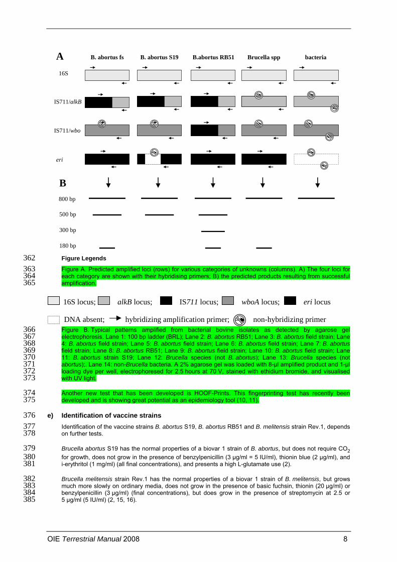
OIE Terrestrial Manual 2008 8
Figure Legends 362 Figure A. Predicted amplified loci (rows) for various categories of unknowns (columns). A) The four loci for 363 each category are shown with their hybridising primers; B) the predicted products resulting from successful 364 amplification. 365
Figure B. Typical patterns amplified from bacterial bovine isolates as detected by agarose gel 366 electrophoresis. Lane 1: 100 bp ladder (BRL); Lane 2: B. abortus RB51; Lane 3: B. abortus field strain; Lane 367 4: B. abortus field strain; Lane 5: B. abortus field strain; Lane 6: B. abortus field strain; Lane 7: B. abortus 368 field strain; Lane 8: B. abortus RB51; Lane 9: B. abortus field strain; Lane 10: B. abortus field strain; Lane 369 11: B. abortus strain S19; Lane 12: Brucella species (not B. abortus); Lane 13: Brucella species (not 370 abortus);. Lane 14: non-Brucella bacteria. A 2% agarose gel was loaded with 8-µl amplified product and 1-µl 371 loading dye per well, electrophoresed for 2.5 hours at 70 V, stained with ethidium bromide, and visualised 372 with UV light. 373
Another new test that has been developed is HOOF-Prints. This fingerprinting test has recently been 374 developed and is showing great potential as an epidemiology tool (10, 11). 375
e) Identification of vaccine strains 376 Identification of the vaccine strains B. abortus S19, B. abortus RB51 and B. melitensis strain Rev.1, depends 377 on further tests. 378
Brucella abortus S19 has the normal properties of a biovar 1 strain of B. abortus, but does not require CO2 379 for growth, does not grow in the presence of benzylpenicillin (3 µg/ml = 5 IU/ml), thionin blue (2 µg/ml), and 380 i-erythritol (1 mg/ml) (all final concentrations), and presents a high L-glutamate use (2). 381
Brucella melitensis strain Rev.1 has the normal properties of a biovar 1 strain of B. melitensis, but grows 382 much more slowly on ordinary media, does not grow in the presence of basic fuchsin, thionin (20 µg/ml) or 383 benzylpenicillin (3 µg/ml) (final concentrations), but does grow in the presence of streptomycin at 2.5 or 384 5 µg/ml (5 IU/ml) (2, 15, 16). 385
B. abortus fs B. abortus S19 B.abortus RB51 Brucella spp bacteriaA16S
IS711/alkB
IS711/wbo
eri
B800 bp
500 bp
300 bp
180 bp
16S locus; alkB locus; IS711 locus; wboA locus;
hybridizing amplification primer; non-hybridizing primer
eri locus
DNA absent;

OIE Terrestrial Manual 2008 9
Brucella abortus strain RB51 is identified by several characteristics; these are: rough morphology, growth in 386 the presence of rifampicin (250 µg per ml of media), and inability to produce O-polysaccharide (OPS) (71). 387 The inability to produce OPS can be demonstrated by reacting RB51 colonies with OPS-specific monoclonal 388 antibodies (MAbs), in dot-blot assays or Western blots (68, 71). An indirect way of demonstrating lack of 389 OPS is by injecting 4 × 10⁸ viable RB51 organisms into BALB/c mice and testing for the induction of OPS-390 antibodies; the serology will be negative (71). 391
Vaccine strains S19, Rev.1 and RB51 may be identified using specific PCRs (9, 12, 70, 78). 392
2. Serological tests 393
No single serological test is appropriate in each and all epidemiological situations, all have limitations especially 394 when it comes to screening individual animals (31, 59). Consideration should be given to all factors that impact on 395 the relevance of the test method and test results to a specific diagnostic interpretation or application. For the 396 purposes of this chapter, the serological methods described represent standardised and validated methods with 397 suitable performance characteristics to be designated as either prescribed tests for international trade. This does 398 not preclude the use of modified or similar test methods or the use of different biological reagents. However, the 399 methods and reagents described in this chapter represent a standard of comparison with respect to expected 400 diagnostic performance. 401
It should be stressed that the serum agglutination test (SAT) is generally regarded as being unsatisfactory for the 402 purposes of international trade. The complement fixation test (CFT) is diagnostically more specific than the SAT, 403 and also has a standardised system of unitage. The diagnostic performance characteristics of some enzyme-404 linked immunosorbent assays (ELISAs) and the fluorescence polarisation assay (FPA) are comparable with or 405 better than that of the CFT, and as they are technically simpler to perform and more robust, their use may be 406 preferred (55, 86). The performances of several of these tests have been compared. 407
For the control of brucellosis at the national or local level, the buffered Brucella antigen tests (BBATs), i.e. the 408 rose bengal test (RBT) and the buffered plate agglutination test (BPAT), as well as the ELISA and the FPA, are 409 suitable screening tests. Positive reactions should be retested using a suitable confirmatory strategy. 410
In other species, for example, buffaloes (Bubalus bubalus), American and European bison (Bison bison, Bison 411 bonasus), yak (Bos grunniens), elk/wapiti (Cervus elaphus), and camels (Camelus bactrianus and 412 C. dromedarius) and South-American camelids, Brucella sp. infection follows a course similar to that in cattle. The 413 same serological procedures may be used for these animals (51), but each test should be validated in the animal 414 under study (28, 29). 415
• Reference sera 416 Primary bovine reference standards are those against which all other standards are compared and 417 calibrated. These reference standards are all available to national reference laboratories and should be used 418 to establish secondary or national standards against which working standards can be prepared and used in 419 the diagnostic laboratory for daily routine use. 420
These sera have been developed and designated by the OIE as International Standard Sera1. The use of 421 these promotes international harmonisation of diagnostic testing and antigen standardisation (86): 422
• For RBT and CFT, the OIE International Standard Serum (OIEISS, previously the WHO Second 423 International anti-Brucella abortus Serum) that contains 1000 IU and ICFTU (international complement 424 fixation test units) is used. 425
• In addition, three OIE ELISA Standard Sera are available for use. These are also of bovine origin and 426 consist of a strong positive (OIEELISASPSS), a weak positive (OIEELISAWPSS) and a negative 427 (OIEELISANSS) standard. 428
• Production of cells 429 Brucella abortus strain 99 (Weybridge) (S99) (see footnote 1 for address) or B. abortus strain 1119-3 430 (USDA) (S1119-3)2 should always be used for diagnostic antigen production. It should be emphasised that 431 antigen made with one of these B. abortus strains is also used to test for B. melitensis or B. suis infection. 432 The strains must be completely smooth and should not autoagglutinate in saline and 0.1% (w/v) acriflavine. 433 They must be pure cultures and conform to the characteristics of CO2-independent strains of B. abortus 434
1 Obtainable from the OIE Reference Laboratory for Brucellosis at Veterinary Laboratories Agency (VLA) Weybridge, New
Haw, Addlestone, Surrey KT15 3NB, United Kingdom. 2 Obtainable from the United States Department of Agriculture (USDA), National Veterinary Services Laboratories (NVSL),
1800 Dayton Road, Ames, Iowa 50010, United States of America.

OIE Terrestrial Manual 2008 10
biovar 1. The original seed cultures should be propagated to produce a seed lot that must conform to the 435 properties of these strains, and should be preserved by lyophilisation or by freezing in liquid nitrogen. 436
For antigen production, the seed culture is used to inoculate a number of potato-infusion agar slopes that 437 are then incubated at 37°C for 48 hours. SDA and TSA, to which 5% equine or newborn calf serum and/or 438 0.1% yeast extract may be added, are satisfactory solid media provided a suitable seed is used as 439 recommended above. The growth is checked for purity, resuspended in sterile PBS, pH 6.4, and used to 440 seed layers of potato-infusion agar or glycerol–dextrose agar in Roux flasks. These are then incubated at 441 37°C for 72 hours with the inoculated surface facing down. Each flask is checked for purity by Gram staining 442 samples of the growth, and the organisms are harvested by adding 50–60 ml of sterile phenol saline (0.5% 443 phenol in 0.85% sodium chloride solution) to each flask. The flasks are gently agitated, the suspension is 444 decanted, and the organisms are killed by heating at 80°C for 90 minutes. Following a viability check, the 445 antigen is stored at 4°C. 446
Alternatively, the cells may be produced by batch or continuous culture in a fermenter (33), using a liquid 447 medium containing (per litre of distilled water) D-glucose (30 g), a high-grade peptone (30 g), yeast extract 448 (Difco) (10 g), sodium dihydrogen phosphate (9 g) and disodium hydrogen phosphate (3.3 g). The initial pH 449 is 6.6, but this tends to rise to pH 7.2–7.4 during the growth cycle. Care should be taken to check batches of 450 peptone and yeast extract for capacity to produce good growth without formation of abnormal or dissociated 451 cells. Vigorous aeration and stirring is required during growth, and adjustment to pH 7.2–7.4 by the addition 452 of sterile 0.1 M HCl may be necessary. The seed inoculum is prepared as described above. The culture is 453 incubated at 37°C for 48 hours. Continuous culture runs can be operated for much longer periods, but more 454 skill is required to maintain them. In-process checks should be made on the growth from either solid or liquid 455 medium to ensure purity, an adequate viable count and freedom from dissociation to rough forms. Cells for 456 use in the preparation of all antigens should be checked for purity and smoothness at the harvesting stage. 457
The culture is harvested by centrifugation to deposit the organisms, which are resuspended in sterile phenol 458 saline (0.5%). The organisms are killed by heating at 80°C for 90 minutes and are stored at 4°C. They must 459 form stable suspensions in physiological saline solutions and show no evidence of autoagglutination. A 460 viability check must be performed on the suspensions and no growth must be evident after 10 days’ 461 incubation at 37°C. The packed cell volume (PCV) of the killed suspensions can be determined by 462 centrifuging 1 ml volumes in Wintrobe tubes at 3000 g for 75 minutes. 463
a) Buffered Brucella antigen tests (prescribed tests for international trade) 464 • Rose bengal test 465
This test is a simple spot agglutination test using antigen stained with Rose Bengal and buffered to a low pH, 466 usually 3.65 ± 0.05 (47). 467
• Antigen production 468 Antigen for the RBT is prepared by depositing killed B. abortus S99 or S1119-3 cells by centrifugation at 469 23,000 g for 10 minutes at 4°C, and uniformly resuspending in sterile phenol saline (0.5%) at the rate of 1 g 470 to 22.5 ml. (Note: if sodium carboxymethyl cellulose is used as the sedimenting agent during preparation of 471 the cell concentrate, insoluble residues must be removed by filtering the suspension through an AMF-CUNO 472 Zeta-plus prefilter [Type CPR 01A] before staining.) To every 35 ml of this suspension, 1 ml of 1% (w/v) rose 473 bengal (Cl No. 45440) in sterile distilled water is added, and the mixture is stirred for 2 hours at room 474 temperature. The mixture is filtered through sterile cotton wool, and centrifuged at 10,000 g to deposit the 475 stained cells, which are then uniformly resuspended at the rate of 1 g cells to 7 ml of diluent (21.1 g of 476 sodium hydroxide dissolved in 353 ml of sterile phenol saline, followed by 95 ml of lactic acid, and adjusted 477 to 1056 ml with sterile phenol saline). The colour of this suspension should be an intense pink and the 478 supernatant of a centrifuged sample should be free of stain; the pH should be 3.65 ± 0.05. After filtration 479 through cotton wool, the suspension is filtered twice through a Sartorius No. 13430 glass fibre prefilter, 480 adjusted to a PCV of approximately 8%, pending final standardisation against serum calibrated against the 481 OIEISS, and stored at 4°C in the dark. The antigen should not be frozen. 482
483
When used in the standard test procedure, the RBT antigen should give a clearly positive reaction with 1/45 484 dilution, but not 1/55 dilution, of the OIEISS diluted in 0.5% phenol saline. It may also be advisable to 485 compare the reactivity of new and previously standardised batches of antigen using a panel of defined sera. 486
• Test procedure 487 i) Bring the serum samples and antigen to room temperature (22 ± 4°C); only sufficient antigen for the 488
day’s tests should be removed from the refrigerator. 489

OIE Terrestrial Manual 2008 11
ii) Place 25–30 µl of each serum sample on a white tile, enamel or plastic plate, or in a WHO 490 haemagglutination plate. 491
iii) Shake the antigen bottle well, but gently, and place an equal volume of antigen near each serum spot. 492 iv) Immediately after the last drop of antigen has been added to the plate, mix the serum and antigen 493
thoroughly (using a clean glass or plastic rod for each test) to produce a circular or oval zone 494 approximately 2 cm in diameter. 495
v) The mixture is agitated gently for 4 minutes at ambient temperature on a rocker or three-directional 496 agitator (if the reaction zone is oval or round, respectively). 497
vi) Read for agglutination immediately after the 4-minute period is completed. Any visible reaction is 498 considered to be positive. A control serum that gives a minimum positive reaction should be tested 499 before each day’s tests are begun to verify the sensitivity of test conditions. 500
The RBT is very sensitive. However, like all other serological tests, it could sometimes give a positive result 501 due to S19 vaccination or due to false-positive serological reactions (FPSR). Therefore positive reactions 502 should be investigated using suitable confirmatory strategies (including the performance of other tests and 503 epidemiological investigation). False-negative reactions occur rarely, mostly due to prozoning and can 504 sometimes be detected by diluting the serum sample or retesting after. Nevertheless RBT appears to be 505 adequate as a screening test for detecting infected herds or to guarantee the absence of infection in 506 brucellosis-free herds. 507
• Buffered plate agglutination test 508 • Antigen production 509 Antigen for the BPAT is prepared from B. abortus S1119-3 according to the procedure described by Angus & 510 Barton (3). 511
Two staining solutions are required: brilliant green (2 g/100 ml) and crystal violet (1 g/100 ml) both certified 512 stains dissolved in distilled water. Once prepared, the two solutions should be stored separately for a period 513 of 24 hours, and then mixed together in equal volumes in a dark bottle and stored in a refrigerator for a 514 period of not less than 6 months before use. The mixed stain may only be used between 6 and 12 months 515 after initial preparation. 516
Buffered diluent is prepared by slowly dissolving sodium hydroxide (150 g) in 3–4 litres of sterile phenol 517 saline (0.5%). Lactic acid (675 ml) is added to this solution, and the final volume is adjusted to 6 litres by 518 adding sterile phenol saline. The pH of the solution should be between 3.63 and 3.67. 519
Brucella abortus S1119-3 packed cells are diluted to a concentration of 250 g/litre in sterile phenol saline; 520 6 ml of stain is added per litre of cell suspension, and the mixture is shaken thoroughly before being filtered 521 through sterile absorbent cotton. The cells are centrifuged at 10,000 g at 4°C, and the packed cells are then 522 resuspended at a concentration of 50 g/100 ml in buffered diluent (as described above). This mixture is 523 shaken thoroughly for 2 hours, and is then further diluted by the addition of 300 ml of buffered diluent per 524 100 ml of suspended cells (i.e. final concentration of 50 g packed cells/400 ml buffered diluent). The mixture 525 is stirred at room temperature for 20–24 hours before the cell concentration is adjusted to 11% (w/v) in 526 buffered diluent. This suspension is stirred overnight before testing. Pending final quality control tests, the 527 antigen is stored at 4°C until required for use. The antigen has a shelf life of 1 year and should not be 528 frozen. 529
The pH of the buffered plate antigen should be 3.70 ± 0.03 and the pH of a serum:antigen mixture at a ratio 530 of 8:3 should be 4.02 ± 0.04. The 11% stained-cell suspension should appear blue-green. Each batch of 531 buffered plate antigen should be checked by testing at least 10 weakly reactive sera and comparing the 532 results with one or more previous batches of antigen. If possible, the antigen batches should be compared 533 with the standard antigen prepared by the NVSL, USDA (see footnote 2 for address). There is, however, no 534 international standardisation procedure established for use with the OIEISS. 535
• Test procedure 536 Bring the serum samples and antigen to room temperature (22 ± 4°C); only sufficient antigen for the day’s 537
tests should be removed from the refrigerator. 538 ii) Shake the sample well. Place 80 µl of each serum sample on a glass plate marked in 4 × 4 cm 539
squares 540 iii) Shake the antigen bottle well, but gently, at least 5-10 min and place 30 µl of antigen near each serum 541
spot. 542

OIE Terrestrial Manual 2008 12
iv) Immediately after the last drop of antigen has been added to the plate, mix the serum and antigen 543 thoroughly (using a clean glass or plastic rod for each test) to produce a circular zone 544 approximately 3 cm in diameter. 545
v) After the initial mixing, the plate should be rotated three times in a tilting motion to ensure even 546 dispersion of the reagents, and then incubated for 4 minutes in a humid chamber at ambient 547 temperature 548
vi) The plate should be removed and rotated as above, and then returned for a second 4-minute 549 incubation 550
Read for agglutination immediately after the 8-minute period is completed. Any visible reaction is 551 considered to be positive. A control serum that gives a minimum positive reaction should be tested 552 before each day’s tests are begun to verify the sensitivity of test conditions. 553
vii) 554 . Like the RBT, the test is very sensitive, especially for detection of vaccine-induced antibody, and positive 555 samples should be retested using a confirmatory test(s). False-negative reactions may occur, usually due to 556 prozoning, which may be overcome by diluting the serum or retesting after a given time. 557
b) Complement fixation test (a prescribed test for international trade) 558 The CFT is a widely used and accepted confirmatory test although it is complex to perform, requiring good 559 laboratory facilities and adequately trained staff to accurately titrate and maintain the reagents. There are 560 numerous variations of the CFT in use, but this test is most conveniently carried out in a microtitre format. 561 Either warm or cold fixation may be used for the incubation of serum, antigen and complement: either 37°C 562 for 30 minutes or 4°C for 14–18 hours. A number of factors affect the choice of the method: anti-563 complementary activity in serum samples of poor quality is more evident with cold fixation, while fixation at 564 37°C increases the frequency and intensity of prozones, and a number of dilutions must be tested for each 565 sample. 566
Several methods have been proposed for the CFT using different concentrations of fresh or preserved 567 sheep red blood cells (SRBCs) (a 2.5% or 3% suspension is usually recommended) sensitised with an equal 568 volume of rabbit anti-SRBC serum diluted to contain several times (usually from two to five times) the 569 minimum concentration required to produce 100% lysis of SRBCs in the presence of a titrated solution of 570 guinea-pig complement. The latter is independently titrated (in the presence or absence of antigen according 571 to the method) to determine the amount of complement required to produce either 50% or 100% lysis of 572 sensitised SRBCs in a unit volume of a standardised suspension; these are defined as the 50% or 100% 573 haemolytic unit of complement/minimum haemolytic dose (C’H or MHD50 or C’H or MHD100), respectively. It 574 is generally recommended to titrate the complement before each set of tests, a macromethod being 575 preferred for an optimal determination of C’H50. Usually, 1.25–2 C’H100 or 5–6 C’H50 are used in the test. 576
Barbital (veronal) buffered saline is the standard diluent for the CFT. This is prepared from tablets available 577 commercially; otherwise it may be prepared from a stock solution of sodium chloride (42.5 g), barbituric acid 578 (2.875 g), sodium diethyl barbiturate (1.875 g), magnesium sulphate (1.018 g), and calcium chloride 579 (0.147 g) in 1 litre of distilled water and diluted by the addition of four volumes of 0.04% gelatin solution 580 before use. 581
• Antigen production 582 Numerous variations of the test exist but, whichever procedure is selected, the test must use an antigen that 583 has been prepared from an approved smooth strain of B. abortus, such as S99 or S1119-3, and 584 standardised against the OIEISS. Antigen for the CFT can be prepared by special procedures (2, 33) or a 585 whole cell antigen can be used after diluting the stock suspension such that the PCV of the concentrated 586 antigen suspension for CFT should be approximately 2% before standardisation against the OIEISS. The 587 antigen should be standardised to give 50% fixation at a dilution of 1/200 of the OIEISS and must also show 588 complete fixation at the lower serum dilutions, because too weak (or too strong) a concentration of antigen 589 may not produce 100% fixation at the lower dilutions of serum. When two dilutions of antigen are suitable, 590 the more concentrated antigen suspension must be chosen in order to avoid prozone occurrence. 591
The appearance of the antigen when diluted 1/10 must be that of a uniform, dense, white suspension with no 592 visible aggregation or deposit after incubation at 37°C for 18 hours. It must not produce anti-complementary 593 effects at the working strength for the test. The antigen is stored at 4°C and should not be frozen. 594
• Test procedure (example) 595 The undiluted test sera and appropriate working standards should be inactivated for 30 minutes in a water 596 bath at 60°C ± 2°C. If previously diluted with an equal volume of veronal buffered saline these sera could be 597
OIE Terrestrial Manual 2008 13
inactivated at 58°C ± 2°C for 50 minutes. Usually, only one serum dilution is tested routinely (generally 1/4 598 or 1/5 depending on the CF procedure chosen), but serial dilutions are recommended for trade purposes in 599 order to detect prozone. 600
Using standard 96-well microtitre plates with round (U) bottoms, the technique is usually performed as 601 follows: 602
i) Volumes of 25 µl of diluted inactivated test serum are placed in the well of the first, second and third 603 rows. The first row is an anti-complementary control for each serum. Volumes of 25 µl of CFT buffer 604 are added to the wells of the first row (anti-complementary controls) to compensate for lack of antigen. 605 Volumes of 25 µl of CFT buffer are added to all other wells except those of the second row. Serial 606 doubling dilutions are then made by transferring 25 µl volumes of serum from the third row onwards. 25 607 µl of the resulting mixture in the last row are discarded. 608
ii) Volumes of 25 µl of antigen, diluted to working strength, are added to each well except in the first 609 row. 610
iii) Volumes of 25 µl of complement, diluted to the number of units required, are added to each well. 611 iv) Control wells containing diluent only, complement + diluent, antigen + complement + diluent, are 612
set up to contain 75 µl total volume in each case. A control serum that gives a minimum positive 613 reaction should be tested in each set of tests to verify the sensitivity of test conditions. 614
v) The plates are incubated at 37°C for 30 minutes or at 4°C overnight, and a volume (25 or 50 µl 615 according to the technique) of sensitised SRBCs is added to each well. The plates are re-incubated 616 at 37°C for 30 minutes. 617
vi) The results are read after the plates have been centrifuged at 1000 g for 10 minutes at 4°C or left 618 to stand at 4°C for 2–3 hours to allow unlysed cells to settle. The degree of haemolysis is 619 compared with standards corresponding to 0, 25, 50, 75 and 100% lysis. The absence of anti-620 complementary activity is checked for each serum in the first row. 621
vii) Standardisation of results of the CFT: 622 There is a unit system that is based on the OIEISS. This serum contains 1000 ICFTU (international 623 complement fixation test units) per ml. If this serum is tested in a given method and gives a titre of, for 624 example 200 (50% haemolysis), then the factor for an unknown serum tested by that method can be 625 found from the formula: 1000 × 1/200 × titre of test serum = number of ICFTU of antibody in the test 626 serum per ml. The OIEISS contains specific IgG; national standard sera should also depend on this 627 isotype for their specific complement-fixing activity. Difficulties in standardisation arise because 628 different techniques selectively favour CF by different immunoglobulin isotypes. It is recommended that 629 any country using the CFT on a national scale should obtain agreement among the different 630 laboratories performing the test to use the same method in order to obtain the same level of sensitivity. 631 To facilitate comparison between countries, results should always be expressed in ICFTUs, calculated 632 in relation to those obtained in a parallel titration with a standard serum, which in turn may be 633 calibrated against the OIEISS. 634
vii) Interpretation of the results: Sera giving a titre equivalent to 20 ICFTU/ml or more are considered to be 635 positive. 636
This procedure is an example, other volumes and quantities of reagents could be chosen provided that the 637 test is standardised against the OIEISS as described above and the results expressed in ICFTU/ml. 638
The CFT is very specific. However, like all other serological tests, it could sometimes give a positive result 639 due to S19 vaccination or due to FPSR. Therefore positive reactions should be investigated using suitable 640 confirmatory strategies. Females that have been vaccinated with Brucella abortus S19 between 3 and 641 6 months are usually considered to be positive if the sera give positive fixation at a titre of 30 or greater 642 ICFTU/ml when the animals are tested at an age of 18 months or older. 643
c) Enzyme-linked immunosorbent assays (prescribed tests for international trade) 644 • Indirect ELISA 645 Numerous variations of the indirect ELISA (I-ELISA) have been described employing different antigen 646 preparations, antiglobulin-enzyme conjugates, and substrate/chromogens. Several commercial I-ELISAs are 647 available that have been validated in extensive field trials and are in wide use. In the interests of 648 international harmonisation, the three OIE ELISA Standard Sera should be used by national reference 649 laboratories to check or calibrate the particular test method in question. 650

OIE Terrestrial Manual 2008 14
The assay should be calibrated such that the optical density (OD) of the strong positive OIE ELISA Standard 651 Serum should represent a point on the linear portion of a typical dose–response curve just below the 652 plateau. The weak positive OIE ELISA Standard Serum should consistently give a positive reaction that lies 653 on the linear portion of the same dose–response curve just above the positive/negative threshold. The 654 negative serum and the buffer control should give reactions that are always less than the positive/negative 655 threshold (84). The threshold should be established in the test population using appropriate validation 656 techniques (see Chapter 1.1.3 Principles of validation of diagnostic assays for infectious diseases). 657
The I-ELISA is a highly sensitive test but it is sometimes not capable of differentiating between antibody 658 resulting from S19 vaccination or other FPSR problems and that induced by pathogenic Brucella strains. The 659 I-ELISA should therefore be considered more as a screening test than a confirmatory test in the testing of 660 vaccinated cattle or herds affected by FPSR problems. 661
For the I-ELISA, preparations rich in smooth lipopolysaccharide (sLPS) should be used as the antigen. 662 There are several protocols for preparing a suitable antigen. 663
Monoclonal, polyclonal antiglobulin or protein A/G enzyme conjugates may be used depending on 664 availability and performance requirements. An MAb specific for the heavy chain of bovine IgG1 may provide 665 some improvement in specificity at the possible cost of some loss of sensitivity while a protein A/G enzyme 666 conjugate may provide a reagent useful for testing a variety of mammalian species(58). 667
The test method described below is an example of a test that has been internationally validated and has 668 been used extensively in internationally sponsored, technical cooperation and research collaboration 669 projects world-wide. 670
The antigen-coating buffer is 0.05 M carbonate/bicarbonate buffer, pH 9.6, composed of sodium hydrogen 671 carbonate (2.93 g) and sodium carbonate (1.59 g) (sodium azide [0.2 g/litre] is optional) in 1 litre of distilled 672 water. The conjugate and test sera diluent buffer is 0.01 M PBS, pH 7.2, composed of disodium hydrogen 673 orthophosphate (1.4 g), potassium dihydrogen phosphate (0.20 g), sodium chloride (8.50 g) and 0.05% 674 Tween 20 dissolved in 1 litre of distilled water (PBST). This buffer is also used as wash buffer. 675
The conjugate used in this example is an MAb specific for the heavy chain of bovine IgG1 and conjugated to 676 horseradish peroxidase (HRPO). The substrate stock solution is 3% hydrogen peroxide. The chromogen 677 stock solution is 0.16 M 2,2’-azino-bis-(3-ethylbenzthiazoline-6-sulphonic acid) (ABTS) in distilled water. 678 Substrate buffer is citrate buffer, pH 4.5, composed of trisodium citrate dihydrate (7.6 g) and citric acid 679 (4.6 g) dissolved in 1 litre of distilled water. The enzymatic reaction-stopping solution is 4% sodium dodecyl 680 sulphate (SDS). 681
• Antigen production (example) 682 sLPS from B. abortus S1119-3 or S99 is prepared by heating 5 g dry weight (or 50 g wet weight) of cells 683 suspended in 170 ml distilled water to 66°C followed by the addition of 190 ml of 90% (v/v) phenol at 66°C. 684 The mixture is stirred continuously at 66°C for 15 minutes, cooled and centrifuged at 10,000 g for 685 15 minutes at 4°C. The brownish phenol in the bottom layer is removed with a long cannula and large cell 686 debris may be removed by filtration (using a Whatman No. 1 filter) if necessary. 687
The sLPS is precipitated by the addition of 500 ml cold methanol containing 5 ml methanol saturated with 688 sodium acetate. After 2 hours’ incubation at 4°C, the precipitate is removed by centrifugation at 10,000 g for 689 10 minutes. The precipitate is stirred with 80 ml of distilled water for 18 hours and centrifuged at 10,000 g for 690 10 minutes. The supernatant solution is kept at 4°C. The precipitate is resuspended in 80 ml distilled water 691 and stirred for an additional 2 hours at 4°C. The supernatant solution is recovered by centrifugation as 692 above and pooled with the previously recovered supernatant. 693
Next, 8 g of trichloroacetic acid is added to the 160 ml of crude LPS. After stirring for 10 minutes, the 694 precipitate is removed by centrifugation and the translucent supernatant solution is dialised against distilled 695 water (two changes of at least 4000 ml each) and then freeze dried. 696
The freeze-dried LPS is weighed and reconstituted to 1 mg/ml in 0.05 M carbonate buffer, pH 9.6, and 697 sonicated in an ice bath using approximately 6 watts three times for 1 minute each. The LPS is then freeze 698 dried in 1 ml amounts and stored at room temperature. 699
• Test procedure (example) 700 i) The freeze-dried sLPS is reconstituted to 1 ml with distilled water and is further diluted 1/1000 (or to a 701
dilution predetermined by titration against the OIE ELISA Standard Sera) in 0.05 M carbonate buffer, 702 pH 9.6. To coat the microplates, 100 µl volumes of the diluted sLPS solution are added to all wells, and 703 the plates are covered and incubated for 18 hours at 4°C. After incubation, the plates may be used or 704

OIE Terrestrial Manual 2008 15
sealed, frozen and stored at –20°C for up to a year. Frozen plates are thawed for 30–45 minutes at 705 37°C before use. 706
ii) Unbound antigen is removed by washing all microplate wells with PBST four times. Volumes (100 µl) of 707 serum diluted in the range of 1/50 to 1/200 in PBST, pH 6.3, containing 7.5 mM each of ethylene 708 diamino tetra-acetic acid (EDTA) and ethylene glycol tetra-acetic acid (EGTA) (PBST/EDTA) are added 709 to specified wells and incubated at ambient temperature for 30 minutes. 710
iii) Test sera are added to the plates and may be tested singly or in duplicate. The controls, calibrated 711 against the OIE ELISA Standard Sera, are set up in duplicate wells and include a strong positive, a 712 weak positive, a negative control serum, and a buffer control. 713
iv) Unbound serum is removed by washing four times with PBST (PBST containing EDTA/EGTA must not 714 be used with HRPO as it inactivates the enzyme). Volumes (100 µl) of conjugate (MAb M23) specific 715 for a heavy chain epitope of bovine IgG1 conjugated with HRPO and diluted in PBST ( predetermined 716 by titration) are added to each well and the plates are incubated at ambient temperature for 717 30 minutes. 718
v) Unbound conjugate is removed by four washing steps. Volumes (100 µl) of substrate/chromogen 719 (1.0 mM H2O2 [100 µl/20 ml citrate buffer] and 4 mM ABTS [500 µl/20 ml citrate buffer]) are added to 720 each well, the plate is shaken for 10 minutes and colour development is assessed in a 721 spectrophotometer at 414 or 405 nm. If required, 100 µl volumes of 4% SDS may be added directly to 722 all wells as a stopping reagent. 723
vi) The control wells containing the strong positive serum are considered to be 100% positive and all data 724 are calculated from these absorbance readings (between 1.000 and 1.800) using the equation: 725 Per cent positivity (%P) = absorbance (test sample)/absorbance (strong positive control) × 100 726
The sLPS antigen, small amounts of the MAb specific for the heavy chain of bovine IgG1, software for 727 generation of data using particular spectrophotometers and a standard test protocol for the I-ELISA are 728 available for research and standardisation purposes
3. 729
Using this or another similar I-ELISA protocol calibrated against the OIE ELISA Standard Sera described 730 above, the diagnostic sensitivity should be equal to or greater than the BBATs in the testing of infected 731 cattle, and the diagnostic specificity should be equivalent to the CFT in the testing of unvaccinated cattle 732 (31). It can be expected that the diagnostic specificity in the testing of S19 vaccinated cattle or in the case of 733 FPSR will be significantly lower than for the CFT depending on where the I-ELISA positive/negative 734 threshold is set. 735
• Competitive ELISA 736 The competitive ELISA (C-ELISA) using an MAb specific for one of the epitopes of the Brucella sp. OPS has 737 been shown to have higher specificity than the I-ELISA (43, 56, 73). This is accomplished by selecting an 738 MAb that has higher affinity than cross-reacting antibody. However, it has been shown that the C-ELISA 739 eliminates some but not all reactions (FPSR) due to cross-reacting bacteria (52, 81).The C-ELISA is also 740 capable of eliminating most reactions due to residual antibody produced in response to vaccination with S19. 741 The choice of MAb and its unique specificity and affinity will have a distinct influence on the diagnostic 742 performance characteristics of the assay. As with any MAb-based assay, the universal availability of the 743 MAb or the hybridoma must also be considered with respect to international acceptance and widespread 744 use. 745
Several variations of the C-ELISA have been described. In the interests of international harmonisation, the 746 three OIE ELISA Standard Sera should be used by national reference laboratories to check or calibrate the 747 test method in question. 748
The assay should be calibrated such that the OD of the strong positive OIE ELISA Standard Serum should 749 represent a point on the linear portion of a typical dose–response curve just above the plateau (i.e. close to 750 maximal inhibition). The weak positive OIE ELISA Standard Serum should give a reaction that lies on the 751 linear portion of the same dose–response curve just above the positive/negative threshold (i.e. moderate 752 inhibition). The negative serum and the buffer/MAb control should give reactions that are always less than 753 the positive/negative threshold (i.e. minimal inhibition). 754
The test method described below is an example of a test, which has been internationally validated and has 755 been used extensively in internationally sponsored, technical cooperation and research collaboration 756 projects world-wide. 757
The buffer systems are the same as those described for the I-ELISA. 758
3 Obtainable from the OIE Reference Laboratory for Brucellosis at the Animal Diseases Research Institute, 3851 Fallowfield
Road, Nepean, Ontario K2H 8P9, Canada.
OIE Terrestrial Manual 2008 16
• Antigen production (example) 759 sLPS from B. abortus S1119-3 is prepared and used as for the I-ELISA. 760
• Test procedure 761 i) The freeze-dried sLPS is reconstituted to 1 ml with distilled water and further diluted 1/1000 with 762
0.05 M carbonate buffer, pH 9.6. To coat the microplates, 100 µl volumes of LPS solution are added to 763 all wells and the plates are covered and incubated for 18 hours at 4°C. After incubation, the plates may 764 be used or sealed, frozen and stored at –20°C for up to 1 year. Frozen plates are thawed for 30–765 45 minutes at 37°C before use. 766
ii) Unbound antigen is removed by washing all microplate wells four times with PBST. Volumes (50 µl) of 767 MAb (M84 in this example) diluted appropriately in PBST/EDTA are added to each well, followed 768 immediately by 50 µl volumes of serum diluted 1/10 in PBST/EDTA. Plates are incubated for 769 30 minutes at ambient temperature with shaking for at least the initial 3 minutes. 770
Iii) Test sera are added to the plates and may be tested as singly or in duplicate. The controls, calibrated 771 against the OIE ELISA Standard Sera, are set up in duplicate wells and include a strong positive, a 772 weak positive, a negative control serum, and a buffer control. 773
iv) Unbound serum and MAb are removed by washing the microplate four times with PBST. Volumes 774 (100 µl) of commercial goat anti-mouse IgG (H and L chain) HRPO conjugate diluted in PBST 775 (predetermined by titration) are added to each well and the plates are incubated at ambient 776 temperature for 30 minutes. 777
v) Unbound conjugate is removed by four washing steps. Volumes (100 µl) of substrate/chromogen 778 (1.0 mM H2O2 and 4 mM ABTS) are added to each well, the plates are shaken for 10 minutes and 779 colour development is assessed in a spectrophotometer at 414 or 405 nm. If required, 100 µl volumes 780 of 4% SDS may be added directly as a stopping reagent. 781
vi) The control wells containing MAb and buffer (no serum) are considered to give 0% inhibition and all 782 data are calculated from these absorbance readings (between 1.000 and 1.800) using the equation: 783 Per cent inhibition (%I) = 100 – (absorbance [test sample]/absorbance [buffer control] × 100) 784
The sLPS antigen, small amounts of the MAb, software for generation of data using particular 785 spectrophotometers and a standard operating procedure for the C-ELISA are available for research and 786 standardisation (see footnote 3 for address). 787
Using this or a similar C-ELISA protocol calibrated against the OIE ELISA Standard Sera, the diagnostic 788 sensitivity should be equivalent to the BBATs and the I-ELISAs in the testing of infected cattle (31, 54–56). 789 However, this tests has been proven less sensitive that both tests and also the CFT (49). Moreover, the C-790 ELISA does not solve the problem of serological interferences due to S19 vaccination or to false positive 791 serological reactions due to Y. enterocolitica O:9 (49). 792
d) Fluorescence polarisation assay (a alternative test for international trade) 793 The FPA is a simple technique for measuring antigen/antibody interaction and may be performed in a 794 laboratory setting or in the field. It is a homogeneous assay in which analytes are not separated and it is 795 therefore very rapid. 796
The mechanism of the assay is based on random rotation of molecules in solution. Molecular size is the 797 main factor influencing the rate of rotation, which is inversely related. Thus a small molecule rotates faster 798 than a large molecule. If a molecule is labelled with a fluorochrome, the time of rotation through an angle of 799 68.5° can be determined by measuring polarised light intensity in vertical and horizontal planes. A large 800 molecule emits more light in a single plane (more polarised) than a small molecule rotating faster and 801 emitting more depolarised light. 802
For most FPAs, an antigen of small molecular weight, less than 50 kD, is labelled with a fluorochrome and 803 added to serum or other fluid to be tested for the presence of antibody. If antibody is present, attachment to 804 the labelled antigen will cause its rotational rate to decrease and this decrease can be measured. 805
For the diagnosis of brucellosis, a small molecular weight fragment (average 22 kD) of the OPS of 806 B. abortus strain 1119-3 sLPS is labelled with fluorescein isothiocyanate (FITC) and used as the antigen. 807 This antigen is added to diluted serum or whole blood and a measure of the antibody content is obtained in 808 about 2 minutes (for serum) or 15 seconds (for blood) using a fluorescence polarisation analyser (54). 809
The FPA can be performed in glass tubes or a 96-well plate format. The bovine serum is diluted 1/100 or, if 810 EDTA-treated blood is used, 1/50 (heparin-treated blood tends to increase assay variability). The diluent 811 used is 0.01 M Tris (1.21 g), containing 0.15 M sodium chloride (8.5 g), 0.05% Igepal CA630 (500 µl) 812 (formerly NP40), 10 mM EDTA (3.73 g) per litre of distilled water, pH 7.2 (Tris buffer). An initial reading to 813
OIE Terrestrial Manual 2008 17
assess light scatter is obtained with the fluorescence polarisation analyser (FPM) after mixing. Suitably 814 labelled titrated antigen (usually giving an intensity of 250,000–300,000) is added, mixed and a second 815 reading is obtained in the FPM about 2 minutes later for serum and 15 seconds for blood. A reading (in 816 millipolarisation units, mP) over the established threshold level is indicative of a positive reaction. A typical 817 threshold level is 90–100 mP units, however, the test should be calibrated locally against International 818 Standard reference sera. Control sera of strong positive, weak positive and negative, as well as S19 819 vaccinate serum, should be included. 820
• Antigen production (example) 821 OPS from 5 g dry weight (or 50 g wet weight) of B. abortus S1119-3 is prepared by adding 400 ml of 2% 822 (v/v) acetic acid, autoclaving the suspension for 15 minutes at 121°C and removing the cellular debris by 823 centrifugation at 10,000 g for 10 minutes at 4°C. The supernatant solution is then treated with 20 g of 824 trichloroacetic acid to precipitate any proteins and nucleic acids. The precipitate is again removed by 825 centrifugation at 10,000 g for 10 minutes at 4°C. The supernatant fluid is dialised against at least 826 100 volumes of distilled water and freeze dried. 827
3 mg of OPS are dissolved in 0.6 ml of 0.1 M sodium hydroxide (4 g NaOH/litre) and incubated at 37°C for 828 1 hour, followed by the addition of 0.3 ml of FITC isomer 1 at a concentration of 100 mg/ml in dimethyl 829 sulphoxide and a further incubation at 37°C for 1 hour. The conjugated OPS is applied to a 1 × 10 cm 830 column packed with DEAE (diethylaminoethyl) Sephadex A 25 equilibrated in 0.01 M phosphate buffer, 831 pH 7.4. The first fraction (after 10–15 ml of buffer) is bright green, after which the buffer is switched to 0.1 M 832 phosphate, pH 7.4. This results in the elution of 10–15 ml of buffer followed by 25–40 ml of green fluorescent 833 material. The latter material is the antigen used in the FPA. Antigen preparation may be scaled up 834 proportionally. 835
The amount of antigen used per test is determined by diluting the material derived above until a total 836 fluorescence intensity of 250,000–300,000 is achieved using the FPM. 837
The antigen can be stored as a liquid for several years at 4°C in a dark bottle or it may be freeze dried in 838 dark bottles. 839
Small quantities of labelled antigen for research and standardisation purposes and standard operating 840 procedures for antigen preparation and the FPA may be obtained (see footnote 3 for address). 841
• Test procedure 842 i) 1 ml of Tris buffer is added to a 10 × 75 mm borosilicate glass tube followed by 10 µl of serum or 20 µl 843
of EDTA-treated blood. It is important to mix well. A reading is obtained on the FPM to determine light 844 scatter. 845
ii) A volume of antigen, which results in a total fluorescence intensity of 250–300 × 103, is added to the 846 tube and mixed well. This volume will vary from batch to batch, but is generally in the range of about 847 10 µl. A second reading is obtained on the FPM after incubation at ambient temperature for 848 approximately 2 minutes for serum and 15 seconds for EDTA-treated blood. 849
iii) A reading above the predetermined threshold is indicative of a positive reaction. 850 iv) The following are included in each batch of tests: a strong positive, a weak positive, a negative working 851
standard serum (calibrated against the OIE ELISA Standard Sera). 852
The diagnostic sensitivity and specificity of the FPA for bovine brucellosis are almost identical to those of the 853 C-ELISA. The diagnostic specificity for cattle recently vaccinated with S19 is over 99% (28, 29, 57). However 854 the specificity of FPA in FPSR conditions is currently unknown. The FPA should be standardised such that 855 the OIE strong positive and weak positive sera consistently give positive results. 856
3. Other tests 857
a) Brucellin skin test 858 An alternative immunological test is the brucellin skin test, which can be used for screening unvaccinated 859 herds, provided that a purified (free of sLPS) and standardised antigen preparation (e.g. brucellin INRA) is 860 used. 861
The brucellin skin test has a very high specificity, such that serologically negative unvaccinated animals that 862 are positive reactors to the brucellin test should be regarded as infected animals (20, 65). Also, results of 863 this test may aid the interpretation of serological reactions thought to be FPSR due to infection with cross-864 reacting bacteria, especially in brucellosis-free areas (65, 69). 865
Not all infected animals react, therefore this test alone cannot be recommended as the sole diagnostic test 866 or for the purposes of international trade. 867

OIE Terrestrial Manual 2008 18
It is essential to use a standardised, defined brucellin preparation that does not contain sLPS antigen, as this 868 may provoke nonspecific inflammatory reactions or interfere with subsequent serological tests. One such 869 preparation is brucellin INRA prepared from a rough strain of B. melitensis that is commercially available
4. 870
• Test procedure 871 i) A volume of 0.1 ml of brucellin is injected intradermally into the caudal fold, the skin of the flank, or the 872
side of the neck. 873 ii) The test is read after 48–72 hours. 874 iii) The skin thickness at the injection site is measured with vernier callipers before injection and at re-875
examination. 876 iv) A strong positive reaction is easily recognised by local swelling and induration. However, borderline 877
reactions require careful interpretation. Skin thickening of 1.5–2 mm would be considered as a positive 878 reaction. 879
Although the brucellin intradermal test is one of the most specific tests in brucellosis (in unvaccinated 880 animals), diagnosis should not be made solely on the basis of positive intradermal reactions given by a few 881 animals in the herd, but should be supported by a reliable serological test. The intradermal inoculation of 882 brucellin might induce a temporary anergy in the cellular immune response. Therefore an interval of 6 weeks 883 is generally recommended between two tests on the same animal. 884
b) Serum agglutination test 885 While not recognised as a prescribed or alternative test, the SAT has been used with success for many 886 years in surveillance and control programmes for bovine brucellosis. Its specificity is significantly improved 887 with the addition of EDTA to the antigen (30, 42, 60). 888
The antigen represents a bacterial suspension in phenol saline (NaCl 0.85% [w/v] and phenol at 0.5% [v/v]). 889 Formaldehyde must not be used. Antigens may be delivered in the concentrated state provided the dilution 890 factor to be used is indicated on the bottle label. EDTA may be added to the antigen suspension to 5 mM 891 final test dilution to reduce the level of false-positive results. Subsequently the pH of 7.2 must be readjusted 892 in the antigen suspension. 893
The OIEISS contains 1000 IUs of agglutination. The antigen should be prepared without reference to the cell 894 concentration, but its sensitivity must be standardised in relation to the OIEISS in such a way that the 895 antigen produces either 50% agglutination with a final serum dilution of 1/600 to 1/1000 or 75% agglutination 896 with a final serum dilution of 1/500 to 1/750. It may also be advisable to compare the reactivity of new and 897 previously standardised batches of antigen using a panel of defined sera. 898
The test is performed either in tubes or in microplates. The mixture of antigen and serum dilutions should be 899 incubated for 16–24 hours at 37°C. If the test is carried out in microplates, the incubation time can be 900 shortened to 6 hours. At least three dilutions must be prepared for each serum in order to refute prozone 901 negative responders. Dilutions of suspect serum must be made in such a way that the reading of the 902 reaction at the positivity limit is made in the median tube (or well for the microplate method). 903
Interpretation of results: The degree of Brucella agglutination in a serum must be expressed in IU per ml. A 904 serum containing 30 or more IU per ml is considered to be positive. 905
c) Native hapten and polyB tests 906 Native hapten and polyB tests are confirmatory tests5 that have been used successfully in an eradication 907 programme in combination with the RBT as a screening test (4). The optimal sensitivity is obtained in a 908 reverse radial immunodiffusion (RID) system in which the serum diffuses into a hypertonic gel containing the 909 polysaccharide (18). However, the double gel diffusion procedure is also useful (40, 41). Calves vaccinated 910 subcutaneously with the standard dose of S19 at 3–6 months of age are negative 2 months after 911 vaccination, and adult cattle vaccinated subcutaneously 4–5 months previously with the reduced dose of 912 S19 do not give positive reactions unless the animals become infected and shed the vaccine in their milk 913 (38). The conjunctival vaccination (both in young and adults) reduces the time to obtain a negative response 914 in native hapten and polyB tests. A remarkable characteristic of the RID test is that a positive result 915 correlates with Brucella shedding as shown in experimentally infected cattle (38) and in naturally infected 916 cattle undergoing antibiotic treatment (36). Precipitin tests using native hapten antigen or cytosol proteins 917
4 Brucellergène OCB®, Synbiotics Europe, 2 rue Alexander Fleming, 69007 Lyon, France. 5 The detailed procedure could be obtained from the Brucellosis Laboratory, Centro de Investigacion y Tecnologia
Agroalimentaria/Gobierno de Aragon, Apartado 727, 50080 Zaragoza, Spain.

OIE Terrestrial Manual 2008 19
have also been shown to eliminate most FPSR reactions caused by Yersinia enterocolitica O:9 and FPSR of 918 unknown origin (49). 919
d) Milk tests 920 An efficient means of screening dairy herds is by testing milk from the bulk tank. Milk from these sources can 921 be obtained cheaply and more frequently than blood samples and is often available centrally at dairies. 922 When a positive test result is obtained, all cows contributing milk should be blood tested. The milk I-ELISA is 923 a sensitive and specific test, and is particularly valuable for testing large herds. The milk ring test (MRT) is a 924 suitable alternative if the ELISA is not available. 925
• Milk I-ELISA 926 As with the serum I-ELISA numerous variations of the milk I-ELISA are in use. Several commercial I-ELISAs 927 are available that have been validated in extensive field trials and are in wide use. In the interests of 928 international harmonisation, the three OIE ELISA Standard Sera should be used by national reference 929 laboratories to check or calibrate the particular test method in question. The I-ELISA should be standardised 930 such that the OIE ELISA strong positive standard when diluted 1/125 in negative serum and further diluted 931 1/10 in negative milk consistently tests positive. Bulk milk samples are generally tested at much lower 932 dilutions than sera, i.e. undiluted to 1/2 to 1/10 in diluent buffer, with the remainder of the assay being similar 933 to that described for serum. The C-ELISA should not be used to test whole milk but may be used with whey 934 samples. 935
• Milk ring test 936 In lactating animals, the MRT can be used for screening herds for brucellosis. In large herds (>100 lactating 937 cows), the sensitivity of the test becomes less reliable. The MRT may be adjusted to compensate for the 938 dilution factor from bulk milk samples from large herds. The samples are adjusted according to the following 939 formula: herd size < 150 animals use 1 ml bulk milk, 150–450 use 2 ml milk sample, 451–700 use 3 ml milk 940 sample. False-positive reactions may occur in cattle vaccinated less than 4 months prior to testing, in 941 samples containing abnormal milk (such as colostrum) or in cases of mastitis. Therefore, it is not 942 recommended to use this test in very small farms where these problems have a greater impact on the test 943 results. 944
• Antigen production 945 MRT antigen is prepared from concentrated, killed B. abortus S99 or S1119-3 cell suspension, grown as 946 described previously. It is centrifuged at, for example, 23,000 g for 10 minutes at 4°C, followed by 947 resuspension in haematoxylin-staining solution. Various satisfactory methods are in use; one example is as 948 follows: 100 ml of 4% (w/v) haematoxylin (Cl No. 75290) dissolved in 95% ethanol is added to a solution of 949 ammonium aluminium sulphate (5 g) in 100 ml of distilled water and 48 ml of glycerol. 2 ml of freshly 950 prepared 10% (w/v) sodium iodate is added to the solution. After standing for 30 minutes at room 951 temperature, the deep purple solution is added to 940 ml of 10% (w/v) ammonium aluminium sulphate in 952 distilled water. The pH of this mixture is adjusted to 3.1, and the solution must be aged by storage at room 953 temperature in the dark for 45–90 days. 954
Before use, the staining solution is shaken and filtered through cotton wool. The packed cells are suspended 955 in the staining solution at the rate of 1 g per 30 ml stain, and held at room temperature for 48 hours (some 956 laboratories prefer to heat at 80°C for 10 minutes instead). The stained cells are then deposited by 957 centrifugation, and washed three times in a solution of sodium chloride (6.4 g), 85% lactic acid (1.5 ml) and 958 10% sodium hydroxide (4.4 ml) in 1.6 litres of distilled water, final pH 3.0. The washed cells are resuspended 959 at the rate of 1 g in 27 ml of a diluent consisting of 0.5% phenol saline, adjusted to pH 4.0 by the addition of 960 0.1 M citric acid (approximately 2.5 ml) and 0.5 M disodium hydrogen phosphate (approximately 1 ml) and 961 maintained at 4°C for 24 hours. The mixture is filtered through cotton wool, the pH is checked, and the PCV 962 is determined and adjusted to approximately 4%. 963
The sensitivity of the new batch should be compared with a previously standardised batch using a panel of 964 samples of varying degrees of reaction prepared by diluting a positive serum in milk. The antigen should be 965 standardised against the OIE ISS so that a 1/500 dilution is positive and 1/1000 dilution is negative. The 966 antigen must be stored at 4°C and not frozen. 967
The pH of the antigen should be between 3.3 and 3.7 and its colour should be dark blue. A little free stain in 968 the supernatant of a centrifuged sample is permissible. When diluted in milk from a brucellosis-free animal, 969 the antigen must produce a uniform coloration of the milk layer with no deposit and no coloration of the 970 cream layer. 971
• Test procedure 972

OIE Terrestrial Manual 2008 20
The test is performed on bulk tank milk samples. If necessary, samples could be pretreated with 973 preservative (0.1% formalin or 0.02% bronopol) for 2–3 days at 4°C prior to use. 974
Bring the milk samples and antigen to room temperature (20 ± 3°C); only sufficient antigen for the day’s tests 975 should be removed from the refrigerator. 976
Shake the antigen bottle well, but gently, 977 The test is performed by adding 30–50 µl of antigen to a 1–2 ml volume of whole milk (the volume of milk 978 may be increased for bulk samples from larger herds – see above). 979
The height of the milk column in the tube must be at least 25 mm. The milk samples must not have been 980 frozen, heated, subjected to violent shaking or stored for more that 72 hours. 981
The milk/antigen mixtures are normally incubated at 37°C for 1 hour, together with positive and negative 982 working standards. However, overnight incubation at 4°C increases the sensitivity of the test and allows for 983 easier reading. 984
A strongly positive reaction is indicated by formation of a dark blue ring above a white milk column. Any blue 985 layer at the interface of milk and cream should be considered to be positive as it might be significant, 986 especially in large herds. 987
The test is considered to be negative if the colour of the underlying milk exceeds that of the cream layer. 988
When the MRT is adjusted for large herd sizes (2 or 3 ml of milk used), 0.1 ml of pooled negative cream is 989 added to the test tube and is followed by 30-50 μl of the ring test antigen. After mixing, the test is incubated 990 and read in the same manner as the unadjusted MRT. The negative pooled cream is collected from the 991 separation of composite, unpasteurised milk from a brucellosis negative herd of 25 or more cows. 992
f) Detection of antibody to rough Brucella 993 The use of rough Brucella (RB51) as a vaccine and in some cases of atypical Brucella infection has led to 994 the need for serological tests for detection of antibody against the core of sLPS. Although B. abortus RB51 995 cells has been shown to contain small amounts of OPS (14), generally the antibody response to OPS is 996 negligible and rLPS is a suitable antigen. This rLPS is readily extracted from B. abortus RB51 by the method 997 of Galanos et al. (27). This antigen may then be used in IELISA (53, 58). The complement fixation test has 998 also been shown to detect antibody to rLPS using a whole cell antigen (1). 999
C. REQUIREMENTS FOR VACCINES AND DIAGNOSTIC BIOLOGICALS 1000
Brucellosis is one of the most easily acquired laboratory infections, and strict safety precautions should be 1001 observed. Laboratory manipulation of live cultures of Brucella, including vaccine strains, is hazardous and must 1002 be done under containment level 3 or higher, as outlined in Chapter 1.1.6, to minimise occupational exposure. 1003
C1. Brucellin 1004
Brucellin–INRA is an LPS-free extract from rough B. melitensis B115. This preparation does not provoke 1005 formation of antibodies reactive in BBAT, CFT or ELISA. 1006
1. Seed management 1007
a) Characteristics of the seed 1008 Production of brucellin-INRA is based on a seed-lot system as described for antigens and vaccines. The 1009 original seed B. melitensis strain B115 for brucellin production
6 should be propagated to produce a seed lot, 1010
which should be preserved by lyophilisation or freezing at liquid nitrogen temperature. It should conform to 1011 the properties of a pure culture of a rough strain of B. melitensis and must not produce smooth Brucella LPS. 1012 It should produce reasonable yields of a mixture of protein antigens reactive with antisera to smooth and 1013 rough Brucella strains. 1014
b) Method of culture (2) 1015
6 Obtainable from Institut National de la Recherche Agronomique (INRA), Laboratoire de Pathologie Infectieuse et
Immunologie, 37380 Nouzilly, France.

OIE Terrestrial Manual 2008 21
Brucella melitensis strain B115 is best grown in the liquid medium described above for fermenter culture. It 1016 may be grown by the batch or continuous method in a fermenter or in flasks agitated on a shaker. Purity 1017 checks should be made on each single harvest, and the organisms must be in the rough phase. 1018
c) Validation as an in-vivo diagnostic reagent 1019 Laboratory and field studies in France have confirmed that brucellin-INRA is safe, non-toxic and specific in 1020 action. The preparation contains 50–75% proteins, mainly of low molecular weight and 15–30% 1021 carbohydrate. It does not contain LPS antigens. Brucellin-INRA does not provoke inflammatory responses in 1022 unsensitised animals, and it is not in itself a sensitising agent. It does not provoke antibodies reactive in the 1023 standard serological tests for brucellosis. More than 90% of small ruminants infected with B. melitensis 1024 manifest delayed hypersensitivity to brucellin-NRA at some stage. The preparation is not recommended as a 1025 diagnostic agent for individual animals, but can be useful when used for screening herds. It is given to small 1026 ruminants in 100-µg doses by the intradermal route, and provokes a local delayed hypersensitivity reaction 1027 visible at 48–72 hours in sensitised animals. Positive reactions can be given by vaccinated as well as by 1028 infected animals. 1029
2. Method of manufacture (2) 1030
Brucella melitensis B115 cells are killed after culture by raising the temperature to 70°C for 90 minutes, cooled to 1031 4°C, and harvested by centrifugation at 9000 g for 15 minutes at 4°C. The cells are washed in cold sterile distilled 1032 water and dehydrated by precipitating with three volumes of acetone at –20°C, and then allowed to stand at –1033 20°C for 24–48 hours. After repeated washing in cold acetone, followed by a final rinse in diethyl ether, the cells 1034 are dried over calcium chloride and held at 4°C. The dried cells are subjected to a viability check. They are 1035 resuspended in sterile 2.5% sodium chloride to a final concentration of 5% (w/v) and agitated for 3 days at 4°C. 1036 Bacterial cells are removed by centrifugation as above, and the supernatant is concentrated to one-fourth the 1037 volume by ultrafiltration on a Diaflo PM10 membrane (Amicon) and precipitated by the addition of three volumes 1038 of ice-cold ethanol. The mixture is held at 4°C for 24 hours and the precipitate is recovered by centrifugation, 1039 redissolved in sterile water, and dialysed to remove ethanol. After centrifugation at 105,000 g for 6-hours at 4°C, 1040 the supernatant material, comprising the unstandardised brucellin, is subjected to assays for protein and 1041 carbohydrate. It may be freeze-dried either as bulk material or after it has been dispensed into its final containers. 1042
3. In-process control 1043
The crude brucellin extract should be checked for sterility after acetone extraction, to ensure killing of Brucella 1044 cells, and again at the end of the process to check possible contamination. The pH and protein concentration 1045 should be determined, and identity tests should be performed on the bulk material before filling the final 1046 containers. 1047
4. Batch control 1048
a) Sterility 1049 Allergen preparations should be checked for sterility as described in Chapter 1.1.5. 1050
b) Safety 1051 Samples of brucellin from the final containers should be subjected to the standard sterility test. Brucellin 1052 preparations should also be checked for abnormal toxicity. Doses equivalent to 20 cattle doses (2 ml) should 1053 be injected intraperitoneally into a pair of normal guinea-pigs that have not been exposed previously to 1054 Brucella organisms or their antigens. Five normal mice are also inoculated subcutaneously with 0.5 ml of the 1055 brucellin to be examined. Animals are observed for 7 days, and there should be no local or generalised 1056 reaction to the injection. 1057
Dermo-necrotic capacity is examined by intradermal inoculation of 0.1 ml of the product to be examined into 1058 the previously shaved and disinfected flank of three normal albino guinea-pigs that have not been exposed 1059 previously to Brucella organisms or their antigens. No cutaneous reaction should be observed. Absence of 1060 allergic and serological sensitisation is checked by intradermal inoculation of three normal albino guinea-1061 pigs, three times every 5 days, with 0.1 ml of a 1/10 dilution of the preparation to be examined. A fourth 1062 similar injection is given, 15 days later, to the same three animals and to a control lot of three guinea-pigs of 1063 the same weight that have not been injected previously. The animals should not become seropositive to the 1064 standard tests for brucellosis (RBT, CFT) when sampled 24 hours after the last injection, and should not 1065 develop delayed hypersensitivity responses. 1066
c) Potency 1067

OIE Terrestrial Manual 2008 22
The potency of brucellin preparations is determined by intradermal injection of graded doses of brucellin into 1068 guinea-pigs that have been sensitised by subcutaneous inoculation of 0.5 ml of reference brucellin
7 in 1069
Freund’s complete adjuvant from 1 to 6 months previously. The erythematous reactions are read and 1070 measured at 24 hours and the titre is calculated by comparison with a reference brucellin
8. This method is 1071
only valid for comparing brucellin preparations made according to the same protocol as the sensitising 1072 allergen. Initial standardisation of a batch of allergen and the sensitisation and titration in ruminants is 1073 described (1). 1074
d) Duration of sensitivity 1075 Duration of sensitivity is uncertain. Individual animals vary considerably in the degree of hypersensitivity 1076 manifested to brucellin. Animals in the very early stages of infection, or with long-standing infection, may not 1077 manifest hypersensitivity to intradermal injection. 1078
7 A national French reference brucellin has been produced by INRA-PII (F-37380 Nouzilly, France) and is obtainable from
the OIE Reference Laboratory for Brucellosis, AFSSA, 23 avenue du Général-de-Gaulle, 94706 Maisons-Alfort Cedex, France.
8 The statistical procedure can be obtained from the OIE Reference Laboratory for Brucellosis, AFSSA, BP67, 94703 Maisons-Alfort Cedex, France.

OIE Terrestrial Manual 2008 23
e) Stability 1079 The freeze-dried preparation retains full potency for several years. The liquid commercial preparation should 1080 retain potency for the recommended shelf-life. 1081
f) Preservatives 1082 The use of preservatives is not recommended when the preparation is freeze-dried. In the liquid form, 1083 sodium merthiolate (at most 0.1 mg/ml) may be used as a preservative. If freeze-dried, the preparation 1084 should not be reconstituted until immediately before use. 1085
g) Precautions (hazards) 1086 Brucellin is not toxic. Nevertheless it may provoke severe hypersensitivity reactions in sensitised individuals 1087 who are accidentally exposed to it. Care should be taken to avoid accidental injection or mucosal 1088 contamination. Used containers and injection equipment should be carefully decontaminated or disposed of 1089 by incineration in a suitable disposable container. 1090
5. Tests on final product 1091
a) Safety 1092 A sterility test should be performed by the recommended method. The in-vivo safety tests are as those 1093 described for batch control (see Section C1.4.b). These tests on the batch may be omitted if the full test is 1094 performed on the final filling lots. 1095
b) Potency 1096 This is performed by injection of a single dose into guinea-pigs using the procedure described in Section 1097 C1.4.c. 1098
C2. Vaccines 1099
Brucella abortus strain 19 vaccine 1100
The most widely used vaccine for the prevention of brucellosis in cattle is the Brucella abortus S19 vaccine, which 1101 remains the reference vaccine to which any other vaccines are compared. It is used as a live vaccine and is 1102 normally given to female calves aged between 3 and 6 months as a single subcutaneous dose of 5–8 × 1103 1010 viable organisms. A reduced dose of from 3 × 10⁸ to 3 × 10⁹ organisms can be administered subcutaneously 1104 to adult cattle, but some animals will develop persistent antibody titres and may abort and excrete the vaccine 1105 strain in the milk. Alternatively, it can be administered to cattle of any age as two doses of 5–10 × 109 viable 1106 organisms, given by the conjunctival route; this produces protection without a persistent antibody response and 1107 reduces the risks of abortion and excretion in milk. 1108
Brucella abortus S19 vaccine induces good immunity to moderate challenge by virulent organisms. The vaccine 1109 must be prepared from USDA-derived seed (see footnote 2 for address) and each batch must be checked for 1110 purity (absence of extraneous microorganisms), viability (live bacteria per dose) and smoothness (determination 1111 of dissociation phase). Seed lots for S19 vaccine production should be regularly tested for residual virulence and 1112 immunogenicity in mice. 1113
Control procedures for this vaccine follow. 1114
Brucella abortus strain RB51 vaccine 1115
Since 1996, B. abortus strain RB51 has become the official vaccine for prevention of brucellosis in cattle in 1116 several countries. However its efficiency compared with S19 and its innocuousness remain controversial (48, 49, 1117 72). Each country uses slightly different methods to administer the vaccine. In the USA, calves are vaccinated 1118 subcutaneously between the ages of 4 and 12 months with 1–3.4 × 10¹⁰ viable strain RB51 organisms. 1119 Vaccination of cattle over 12 months of age is carried out only under authorisation from the State or Federal 1120 Animal Health Officials, and the recommended dose is 1 × 10⁹ viable strain RB51 organisms. In other countries, it 1121 is recommended to vaccinate cattle as calves (4–12 months of age) with a 1–3.4 × 10¹⁰ dose, with revaccination 1122 from 12 months of age onwards with a similar dose to elicit a booster effect and increase immunity. 1123
It has been reported that full doses of RB51 when administered intravenously in cattle induce severe placentitis 1124 and placental infection in most vaccinated cattle (63), and that there is excretion in milk in a relevant number of 1125 vaccinated animals. Field experience also indicates that it can induce abortion in some cases if applied to 1126 pregnant cattle. Due to these observations, vaccination of pregnant cattle should be avoided. One way to reduce 1127

OIE Terrestrial Manual 2008 24
the side effects of RB51 is to reduce the dose. When using the reduced dose of this vaccine (1 × 10⁹ colony-1128 forming units [CFU]), on late pregnant cattle, no abortions or placentitis lesions are produced in subcutaneously 1129 vaccinated cattle (64), but the vaccine strain can be shed by a significant proportion of vaccinated animals (76). 1130 However, this reduced dose does not protect against B. abortus when used as a calfhood vaccination (61), but 1131 does protect when used as an adult vaccine (62). 1132
It should be emphasised that, as well as S19, RB51 could infect humans (82). The RB51 strain is highly resistant 1133 to rifampicin, one of the antibiotics of choice for treating human brucellosis. In addition, the diagnosis of the 1134 infection produced by RB51 requires special tests not available in most hospitals. The Centers for Disease 1135 Control, Department of Health and Human Services, Atlanta, Georgia, USA (CDC) established passive 1136 surveillance for accidental inoculation with the RB51 vaccine in the USA to determine if this vaccine is associated 1137 with human disease. This study included 26 participants that had been exposed to the vaccine during animal 1138 vaccination. Accidental human exposure resulted in both local and systemic adverse events; however, it remains 1139 undetermined if strain RB51 vaccine can cause systemic brucellosis in humans. The number of reported adverse 1140 event case-patients in this study (twenty-six) is small compared to the number of vaccination events (several 1141 million calves vaccinated), and estimated inadvertent RB51 inoculations predicted (8 per 11,000). The report 1142 indicated that appropriate antibiotic use should protect against infection, but it remains undetermined to what 1143 degree the organism versus other vaccine components contribute to the adverse events (75). This is in contrast to 1144 Strain 19 where development of undulant fever caused by accidental exposure is well documented to occur 1145 without preventive treatment. 1146
Control procedures for this vaccine follow. 1147
Brucella melitensis strain Rev.1 vaccine 1148
It is not infrequent to isolate B. melitensis in cattle in countries with a high prevalence of this infection in small 1149 ruminants (79). There has been some debate on the protective efficacy of S19 against B. melitensis infection in 1150 cattle and it has been hypothesised that Rev.1 should be a more effective vaccine in these conditions, however 1151 there is only one report related to this issue that demonstrated that S19 is able to control B. melitensis at the field 1152 level (36). By contrast, no experiments have been conducted showing the efficacy of Rev.1 against B. melitensis 1153 infection in cattle. Moreover, the safety of this vaccine is practically unknown in cattle (77). 1154
Until safety of Rev.1 in cattle of different physiological status and efficacy studies against B. melitensis under 1155 strictly controlled conditions are performed, this vaccine should not be recommended for cattle. 1156
1. Seed management 1157
a) Characteristics of the seed 1158 Brucella abortus S19 original seed for vaccine production must be obtained from the USDA (see footnote 2 1159 for address), and used to produce a seed lot that is preserved by lyophilisation or by freezing at liquid 1160 nitrogen temperature. The properties of this seed lot must conform to those of a pure culture of a CO2-1161 independent B. abortus biovar 1 that is also sensitive to benzylpenicillin, thionin blue and i-erythritol at 1162 recommended concentrations, and that displays minimal pathogenicity for guinea-pigs. 1163
Brucella abortus RB51 original seed for vaccine production is available commercially9. These companies 1164
have legal rights to the vaccine. 1165
b) Method of culture 1166 Brucella abortus S19 for vaccine production is grown on medium free from serum or other animal products, 1167 under conditions similar to those described above for B. abortus S99 or S1119-3 (2). 1168
Brucella abortus strain RB51 follows similar culture methods. 1169
c) Validation as a vaccine 1170 Numerous independent studies have confirmed the value of S19 as a vaccine for protecting cattle from 1171 brucellosis. The organism behaves as an attenuated strain when given to sexually immature cattle. In rare 1172 cases, it may produce localised infection in the genital tract. Antibody responses persisting for 6 months or 1173 longer are likely to occur in a substantial proportion of cattle that have been vaccinated subcutaneously with 1174 the standard dose as adults. Some of the cattle vaccinated as calves may later develop arthropathy, 1175 particularly of the femoro-tibial joints (8, 17). The vaccine is safe for most animals if administered to calves 1176 between 3 and 6 months of age. It may also be used in adult animals at a reduced dose. It produces lasting 1177
9 Colorado Serum Company, 4950 York Street, P.O. Box 16428, Denver, Colorado 80216-0428, USA; or Veterinary
Technologies Corporation, 1872 Pratt Drive, Suite 1100B, Blacksburg, Virginia 24060, USA.

OIE Terrestrial Manual 2008 25
immunity to moderate challenge with virulent B. abortus strains, but the precise duration of this is unknown. 1178 The length of protection against B. melitensis is unknown. The vaccine strain is stable and reversion to 1179 virulence is extremely rare. It has been associated with the emergence of i-erythritol-using strains when 1180 inadvertently administered to pregnant animals. The organism behaves as an attenuated strain in mice, and 1181 even large inocula are rapidly cleared from the tissues. 1182
Reports from both experimental challenge studies and field studies concluded the value of B. abortus strain 1183 RB51 in protecting cattle from brucellosis. The organism is attenuated in calves and adults. As B. abortus 1184 strain RB51 contains minimally expressed sLPS and there is no serological conversion against sLPS in 1185 vaccinated animals. In addition, RB51 does not induce detectable antibodies, using current testing 1186 procedures, to the OPS antigen (74). It produces immunity to moderate challenge with virulent strains, but 1187 the precise duration of this is unknown. The vaccine is very stable and no reversion to smoothness has been 1188 described in vivo or in vitro. The organism behaves as an attenuated strain in a variety of animals including 1189 mice where it is rapidly cleared from the tissues. 1190
S19 and RB51 vaccines have some virulence for humans, and infections may follow accidental inoculation 1191 with the vaccine. Care should be taken in its preparation and handling, and a hazard warning should be 1192 included on the label of the final containers. In any case, accidental inoculations should be treated with 1193 appropriate antibiotics (see Section C2.4.g). 1194
2. Method of manufacture 1195
For production of S19 vaccine, the procedures described above can be used, except that the cells are collected in 1196 PBS, pH 6.3, and deposited by centrifugation or by the addition of sodium carboxymethyl cellulose at a final 1197 concentration of 1.5 g/litre. The yield from one fermenter run or the pooled cells from a batch of Roux flask 1198 cultures that have been inoculated at the same time from the same seed lot constitutes a single harvest. More 1199 than one single harvest may be pooled to form a final bulk, which is used to fill the final containers of a batch of 1200 vaccine. Before pooling, each single harvest must be checked for purity, cell concentration, dissociation and 1201 identity. A similar range of tests must be done on the final bulk, which should have a viable count of between 8 1202 and 24 × 109 CFU/ml. Adjustments in concentration are made by the addition of PBS for vaccine to be dispensed 1203 in liquid form, or by the addition of stabiliser for lyophilised vaccine. If stabiliser is to be used, loss of viability on 1204 lyophilisation should be taken into account, and should not be in excess of 50%. The final dried product should 1205 not be exposed to a temperature exceeding 35°C during drying, and the residual moisture content should be 1–1206 2%. The contents must be sealed under vacuum or dry nitrogen immediately after drying, and stored at 4°C. 1207
The production process for B. abortus strain RB51 is very similar to the one used for S19. 1208
3. In-process control 1209
Brucella abortus S19 vaccine should be checked for purity and smoothness during preparation of the single 1210 harvests. The cell concentration of the bulks should also be checked. This can be done by opacity measurement, 1211 but a viable count must be performed on the final filling lots. The identity of these should also be checked by 1212 agglutination tests with antiserum to Brucella A antigen. The viable count of the final containers should not be less 1213 than 50 × 109 per standard dose after lyophilisation, if this is to be done, and at least 95% of the cells must be in 1214 the smooth phase. 1215
Brucella abortus strain RB51 vaccine should be checked for purity and roughness during preparation of the single 1216 harvests. The cell concentration of the bulks should also be checked. A viable count must be performed on the 1217 final filling lots. The viable count of the final containers should be 1–3.4 × 10¹⁰ viable CFU of RB51 per dose 1218 (dose of 2 ml to be applied subcutaneously) and 100% of the cells must be in the rough phase. All colonies 1219 should be negative on dot-blot assays with MAbs specific for the OPS antigen. 1220
4. Batch control 1221
a) Sterility 1222 Tests for sterility and freedom from contamination of biological materials may be found in Chapter 1.1.5. 1223
b) Safety 1224 The S19 vaccine is a virulent product per se, and it should keep a minimal virulence to be efficient (see 1225 Section C2.4.c). However a safety test is not routinely done. If desired, when a new manufacturing process 1226 is started and when a modification in the innocuousness of the vaccine preparation is expected, it may be 1227 performed on cattle. This control should be done as follows: the test uses 12 female calves, aged 4–1228 6 months. Six young females are injected with one or three recommended doses. Each lot of six young 1229
OIE Terrestrial Manual 2008 26
females are kept separately. All animals are observed for 21 days. No significant local or systemic reaction 1230 should occur. If, for a given dose and route of administration, this test gives good results on a representative 1231 batch of the vaccine, it does not have to be repeated routinely on seed lots or vaccine lots prepared with the 1232 same original seed and with the same manufacturing process. A safety test on S19 vaccine may also be 1233 performed in guinea-pigs. Groups of at least ten animals are given intramuscular injections of doses of 1234 vaccine diluted in PBS, pH 7.2, to contain 5 × 10⁹ viable organisms. The animals should show no obvious 1235 adverse effects and there must be no mortality. 1236
A safety test on B. abortus strain RB51 vaccine is not routinely done. If desired, 8–10-week-old female 1237 Balb/c mice can be injected intraperitoneally with 1 × 10⁸ CFUs and the spleens cultured at 6 weeks post-1238 inoculation. Spleens should be free from RB51 and the mice should not develop anti-OPS antibodies. 1239
c) Potency 1240 • S19 vaccine 1241
An S19 vaccine is efficient if it possesses the characteristics of the S19 original strain, i.e. if it is satisfactory 1242 with respect to identity, smoothness, immunogenicity and residual virulence (7). Batches should also be 1243 checked for the number of viable organisms. 1244
• Identity 1245 The reconstituted S19 vaccine should not contain extraneous microorganisms. Brucella abortus 1246 present in the vaccine is identified by suitable morphological, serological and biochemical tests and by 1247 culture: Brucella abortus S19 has the normal properties of a biovar 1 strain of B. abortus, but does not 1248 require CO2 for growth, does not grow in the presence of benzylpenicillin (3 µg/ml = 5 IU/ml), thionin 1249 blue (2 µg/ml), and i-erythritol (1 mg/ml) (all final concentrations). 1250
• Smoothness (determination of dissociation phase) 1251 The S19 vaccine reconstituted in distilled water is streaked across six agar plates (serum–dextrose 1252 agar or trypticase–soy agar (TSA) with added serum 5% [v/v] or yeast extract 0.1 % [w/v) in such a 1253 manner that the colonies will be close together in certain areas, while semi-separated and separated in 1254 others. Slight differences in appearance are more obvious in adjacent than widely separated colonies. 1255 Plates are incubated at 37°C for 5 days and examined by obliquely reflected light (Henry’s method) 1256 before and after staining (three plates) with crystal violet (White & Wilson’s staining method). 1257 Appearance of colonies before staining: S colonies appear round, glistening and blue to blue-green in 1258 colour. R colonies have a dry, granular appearance and are dull yellowish-white in colour. Mucoid 1259 colonies (M) are transparent and greyish in colour and can be distinguished by their slimy consistency 1260 when touched with a loop. Intermediate colonies (I), which are the most difficult to classify, have an 1261 appearance intermediate between S and R forms: they are slightly opaque and more granular than S 1262 colonies. 1263 Appearance of colonies after staining with crystal violet: S colonies do not take up the dye. Dissociated 1264 colonies (I, M, or R) are stained various shades of red and purple and the surface may show radial 1265 cracks. Sometimes a stained surface film slips off a dissociated colony and is seen adjacent to it. 1266 The colony phase can be confirmed by the acriflavine agglutination test (2). S colonies remain in 1267 suspension, whereas R colonies are agglutinated immediately and, if mucoid, will form threads. 1268 Intermediate colonies may remain in suspension or a very fine agglutination may occur. 1269
• Enumeration of live bacteria 1270 Inoculate each of at least five plates of tryptose, serum–dextrose or other suitable agar medium with 1271 0.1 ml of adequate dilutions of the vaccine spread with a sterile glass, wire or plastic spreader. CFU 1272 per vaccine volume unit are enumerated. 1273
• Residual virulence (50% persistence time or 50% recovery time) (7, 32) 1274 i) Prepare adequate suspensions of both the B. abortus S19 seed lot or batch to be tested (test vaccine) 1275
and the S19 original seed culture (as a reference strain). For this, harvest a 24–48 hours growth of 1276 each strain in sterile buffered saline solution (BSS: NaCl 8.5 g; KH2PO4 1.0 g; K2HPO4 2.0 g; distilled 1277 water 1000 ml; pH 6.8) and adjust the suspension in BSS to 109 CFU/ml using a spectrophotometer 1278 (0.170 OD when read at 600 nm). The exact number of CFU/ml should be checked afterwards by 1279 plating serial tenfold dilutions on to adequate culture medium (blood agar base or TSA are 1280 recommended). 1281
ii) Inject subcutaneously 0.1 ml (108 CFU/mouse) of the suspension containing the test vaccine into each 1282 of 32 female CD1 mice, aged 5–6 weeks. Carry out, in parallel, a similar inoculation in another 32 mice 1283

OIE Terrestrial Manual 2008 27
using the suspension containing the S19 reference strain. The original seed S19 strain, which has 1284 been shown satisfactory with respect to immunogenicity and/or residual virulence, can be obtained 1285 from USDA (see footnote 2 for address). 1286
iii) Kill the mice by cervical dislocation, in groups of eight selected at random 3, 6, 9 and 12 weeks later. 1287 iv) Remove the spleens and homogenise individually and aseptically with a glass grinder (or in adequate 1288
sterile bags with the Stomacher) in 1 ml of sterile BSS. 1289 v) Spread each whole spleen suspension in toto on to several plates containing a suitable culture medium 1290
and incubate in standard Brucella conditions for 5–7 days (lower limit of detection: 1 bacterium per 1291 spleen). An animal is considered infected when at least 1 CFU is isolated from the spleen. 1292
vi) Calculate the 50% persistence time or 50% recovery time (RT50) by the SAS® statistical method 1293 specifically developed for RT50 calculations (to obtain the specific SAS® file see footnote 5 for 1294 address) (31). For this, determine the number of cured mice (no colonies isolated in the spleen) at 1295 each slaughtering point time (eight mice per point) and calculate the percentage of cured accumulated 1296 mice over time, by the Reed and Muench method (described in ref. 5). The function of distribution of 1297 this percentage describes a sigmoid curve, which must be linearised for calculating the RT50 values, 1298 using the computerised PROBIT procedure of the SAS® statistical package. 1299
vii) Compare statistically the parallelism (intercept and slope) between the distribution lines obtained for 1300 both tested and reference S19 strains using the SAS® file specifically designed for this purpose. Two 1301 RT50 values can be statistically compared exclusively when they come from parallel distribution lines. If 1302 parallelism does not exist, the residual virulence of the tested strain should be considered inadequate, 1303 and discarded for vaccine production. 1304
viii) If the parallelism is confirmed, compare statistically the RT50 values obtained for both tested and 1305 reference S19 strains using a SAS® file specifically designed for this purpose. To be accepted for 1306 vaccine production, the RT50 obtained with the tested strain should not differ significantly from that 1307 obtained with the reference S19 strain (RT50 and confidence limits are usually around 7.0 ± 1.3 weeks). 1308
The underlying basis of the statistical procedure for performing the above residual virulence calculations 1309 have been recently described in detail (66). Alternatively, the statistical calculations described in steps vi) to 1310 viii) can be avoided by an easy-to-use specific HTML-JAVA script program (Rev2) recently developed (66) 1311 and available free at: http://www.afssa.fr/interne/Rev2.html. 1312
If this test has been done with good results on a representative seed lot or batch of the test vaccine, it does 1313 not have to be repeated routinely on other vaccine lots prepared from the same seed lot and using the same 1314 manufacturing process. 1315
• Immunogenicity in mice (6, 7) 1316 This test uses three groups of six female CD1 mice, aged 5–7 weeks, that have been selected at random. 1317
i) Prepare and adjust spectrophotometrically the vaccine suspensions as indicated above. 1318 ii) Inject subcutaneously a suspension containing 105 CFU (in a volume of 0.1 ml/mouse) of the vaccine 1319
to be examined (test vaccine) into each of six mice of the first group. 1320 iii) Inject subcutaneously a suspension containing 105 CFU of live bacteria of a reference S19 vaccine into 1321
each of six mice of the second group. The third group will serve as the unvaccinated control group and 1322 should be inoculated subcutaneously with 0.1 ml of BSS. 1323
iv) The exact number of CFU inoculated should be checked afterwards by plating serial tenfold dilutions 1324 on to adequate culture medium (blood agar base or TSA are recommended). 1325
v) All the mice are challenged 30 days after vaccination (and immediately following 16 hours’ starvation), 1326 intraperitoneally with a suspension (0.1 ml/mouse) containing 2 × 10⁵ CFU of B. abortus strain 544 1327 (CO2-dependent), prepared, adjusted and retrospectively checked as above. 1328
vi) Kill the mice by cervical dislocation 15 days later. 1329 vii) Each spleen is excised aseptically, the fat is removed, and the spleen is weighed and homogenised. 1330
Alternatively, the spleens can be frozen and kept at –20°C for from 24 hours to 7 weeks. 1331 viii) Each spleen is homogenised aseptically with a glass grinder (or in adequate sterile bags in Stomacher) 1332
in nine times its weight of BSS, pH 6.8 and three serial tenfold dilutions (1/10, 1/100 and 1/1000) of 1333 each homogenate made in the same diluent. Spread 0.2 ml of each dilution by quadruplicate in agar 1334

OIE Terrestrial Manual 2008 28
plates and incubate two of the plates in a 10% CO2 atmosphere (allows the growth of both vaccine and 1335 challenge strains) and the other two plates in air (inhibits the growth of the B. abortus 544 CO2-1336 dependent challenge strain), both at 37°C for 5 days. 1337
ix) Colonies of Brucella should be enumerated on the dilutions corresponding to plates showing fewer 1338 than 300 CFU. When no colony is seen in the plates corresponding to the 1/10 dilution, the spleen is 1339 considered to be infected with five bacteria. These numbers of Brucella per spleen are first recorded as 1340 X and expressed as Y, after the following transformation: Y = log (X/log X). Mean and standard 1341 deviation, which are the response of each group of six mice, are then calculated. 1342
x) The conditions of the control experiment are satisfactory when: i) the response of unvaccinated mice 1343 (mean of Y) is at least of 4.5; ii) the response of mice vaccinated with the reference S19 vaccine is 1344 lower than 2.5; and iii) the standard deviation calculated on each lot of six mice is lower than 0.8. 1345
xi) Carry out the statistical comparisons (the least significant differences [LSD] test is recommended) of 1346 the immunogenicity values obtained in mice vaccinated with the S19 strain to be tested with respect to 1347 those obtained in mice vaccinated with the reference vaccine and in the unvaccinated control group. 1348 The test vaccine would be satisfactory if the immunogenicity value obtained in mice vaccinated with 1349 this vaccine is significantly lower than that obtained in the unvaccinated controls and, moreover, does 1350 not differ significantly from that obtained in mice vaccinated with the reference vaccine. (For detailed 1351 information on this procedure, see footnote 5 for contact address.) 1352
If this test has been done with good results on a representative batch of the test vaccine, it does not have to 1353 be repeated routinely on other vaccine lots prepared from the same seed lot and with the same 1354 manufacturing process. 1355
• RB51 vaccine 1356 There is no standardised potency test on B. abortus strain RB51 vaccine and such a test is not routinely 1357 carried out. A test in Balb/c female mice using 1 × 10⁴ B. abortus strain 2308 organisms as the challenge 1358 strain has been proposed, but the usefulness of this test in predicting protection in cattle has not been 1359 completely determined. In the USA plate counts of viable organisms have been approved and used. 1360
d) Duration of immunity 1361 Vaccinating calves with a full dose of S19 vaccine is considered to give long-lasting immunity, and 1362 subsequent doses are not recommended. However, there is no proven evidence for this and revaccination 1363 could be advisable in endemic areas. 1364
Vaccination of calves with B. abortus strain RB51 is believed to stimulate an immunity that will last for a 1365 period similar to the one induced with S19, although there are no specific studies to demonstrate this. 1366
e) Stability 1367 Brucella abortus S19 vaccine prepared from seed stock from appropriate sources is stable in characteristics, 1368 provided that the in-process and batch control requirements described above are fulfilled, and shows no 1369 tendency to reversion to virulence. The lyophilised vaccine shows a gradual loss of viable count, but should 1370 retain its potency for the recommended shelf life. Allowance for this phenomenon is normally made by 1371 ensuring that the viable count immediately following lyophilisation is well in excess of the minimum 1372 requirement. Maintenance of a cold chain during distribution of the vaccine will ensure its viability. 1373
Brucella abortus strain RB51 has shown no tendency to revert to virulent smooth organisms after many 1374 passages in vitro or in vivo. This is probably due to the nature and place of the mutations found in this strain. 1375 Brucella abortus strain RB51 has its wboA gene disrupted by an IS711 element impeding synthesis of OPS. 1376 Unpublished data indicate that it also contains a second mutation affecting the export of OPS to the bacterial 1377 surface or the coupling of OPS to the core of the LPS, or both. 1378
f) Preservatives 1379 Antimicrobial preservatives must not be used in live S19 or B. abortus strain RB51 vaccines. For preparation 1380 of the lyophilised vaccine, a stabiliser containing 2.5% casein digest, e.g. Tryptone (Oxoid), 5% sucrose and 1381 1% sodium glutamate, dissolved in distilled water and sterilised by filtration is recommended. 1382
g) Precautions (hazards) 1383 Brucella abortus S19 and RB51, although attenuated strains, are still capable of causing disease in humans. 1384 The cell cultures and suspensions must be handled under appropriate conditions of biohazard containment. 1385 Reconstitution and subsequent handling of the reconstituted vaccine should be done with care to avoid 1386 accidental injection or eye or skin contamination. Vaccine residues and injection equipment should be 1387 decontaminated with a suitable disinfectant (phenolic, iodophor or aldehyde formulation) at recommended 1388

OIE Terrestrial Manual 2008 29
concentration. Medical advice should be sought in the event of accidental exposure. The efficacy of the 1389 antibiotic treatment of infections caused by S19 and RB51 in humans has not been adequately established; 1390 however, the CDC will provide treatment recommendations. If S19 contamination occurs, a combined 1391 treatment with doxicycline plus rifampicin could be recommended. In the case of contamination with RB51 (a 1392 rifampicin-resistant strain), the treatment with rifampicin should be avoided. 1393
5. Tests of the final product 1394
a) Safety 1395 See Section C2.4.b. 1396
b) Potency 1397 For the lyophilised vaccine, potency must be determined on the final product. The procedure is as described 1398 in Section C2.4.c. 1399
REFERENCES 1400
1. ADONE R. & CIUGHINI F. (1999). Complement fixation test to assess humoral immunity in cattle and sheep 1401 vaccinated with Brucella abortus RB51. Clin. Diagn. LaB. Immunol., 56, 787–790. 1402
2. ALTON G.G., JONES L.M., ANGUS R.D. & VERGER J.M. (1988). Techniques for the Brucellosis Laboratory. 1403 Institut National de la Recherche Agronomique, Paris, France. 1404
3. ANGUS R.D. & BARTON C.E. (1984). The production and e valuation of a buffered plate antigen for use in a 1405 presumptive test for brucellosis. Dev. Biol. Stand., 56, 349–356. 1406
4. ASARTA A. (1989). Erradicación de la brucelosis en el ganado vacuno de Navarra. In: Actas del XII Congreso 1407 Nacional de Microbiología.Sociedad Española de Microbiología (Ed.), SEM, Pamplona, 371–371. 1408
5. BONET-MAURY P., JUDE A. & SERVANT P. (1954). La mesure statistique de la virulence et l’immunité. 1409 Application á l’etude de la virulence du bacille typhique et á la mesure du pouvoir immunisant des vaccins 1410 antityphoidiques. Rev. d'Immun. Th. Antimic., 18, 21–49. 1411
6. BOSSERAY N. (1992). Le vaccin Rev.1: dérive des caractères d’immunogénicité et de virulence indépendante 1412 des marqueurs classiques. In: Prevention of Brucellosis in the Mediterranean Countries, Plommet M., ed. 1413 Pudoc Scientific, Wageningen, The Netherlands, 182–186. 1414
7. BOSSERAY N. (1993). Control methods and thresholds of acceptability for anti-Brucella vaccines. Dev. Biol. 1415 Stand., 79, 121–128. 1416
8. BRACEWELL C.D. & CORBEL M.J. (1980). An association between arthritis and persistent serological reactions 1417 to B. abortus in cattle from apparently brucellosis-free herds. Vet. Rec., 106, 99. 1418
9. BRICKER B.J. (2002). PCR as a diagnostic tool for brucellosis, Vet. Microbiol., 90, 435–446. 1419
10. BRICKER B.J. EWALT D.R. (2005). Evaluation of HOOF-Print assay for typing Brucella abortus strains isolated 1420 from cattle in the United States: results with four performance criteria. BMC Microbiol., 23June, 5, 37. 1421
11. BRICKER B.J., EWALT D.R. & HALLING S.M. (2003). Brucella ‘HOOF-Prints’ strain typing by multi-locus analysis 1422 of variable number tandem repeats (VNTRs). BMC Microbiol., 11 June, 3, 15. 1423
12. CLOECKAERT A., GRAYON M. & GREPINET O. (2002). Identificiation of Brucella melitensis vaccine strain Rev.1 1424 by PCR-RFLP based on a mutation in the rpsL gene, Vaccine, 7, 19–20. 1425
13. CLOECKAERT A., VERGER J.M., GRAYON M., PAQUET J.Y., GARIN-BASTUJI B., FOSTER G. & GODFROID J. (2001). 1426 Classification of Brucella spp. isolated from marine mammals by DNA polymorphism at the omp2 locus. 1427 Microbes Infect., 3, 29–38. 1428

OIE Terrestrial Manual 2008 30
14. CLOECKAERT A., ZYGMUNT M. & GUILLOTEAU L. (2002). Brucella abortus vaccine strain RB51 produces low 1429 levels of M-lik O-antigen. Vaccine, 20, 1820–1822. 1430
15. CORBEL M.J., BRACEWELL C.D., THOMAS E.L. & GILL K.P.W. (1979). Techniques in the identification of Brucella 1431 species. In: Identification Methods for Microbiologists, Second Edition. Skinner F.A. & Lovelock D.W., eds. 1432 Academic Press, London, UK and New York, USA, 86–89. 1433
16. CORBEL M.J. & HENDRY D.M.F.D. (1983). Methods for the identification of Brucella. Booklet 2085. Ministry of 1434 Agriculture, Fisheries and Food, Lion House, Alnwick, Northumberland, UK. 1435
17. CORBEL M.J., STUART F.A., BREWER R.A., JEFFREY M. & BRADLEY R. (1989). Arthropathy associated with 1436 Brucella abortus Strain 19 vaccination in cattle. 1. Examination of field cases. Br. Vet. J., 145, 337. 1437
18. DIAZ R., GARATEA P., JONES L.M. & MORIYON I. (1979). Radial immunodiffusion test with a Brucella 1438 polysaccharide antigen for differentiating infected from vaccinated cattle. J. Clin. Microbiol., 10, 37–41. 1439
19. EUROPEAN COMMISSION (1999). The modification of Technical Annexes of Council Directive 64/432/EEC to 1440 take account of Scientific Developments regarding Tuberculosis, Brucellosis and Enzootic Bovine Leucosis. 1441 Report of the Scientific Committee on Animal Health and Animal Welfare adopted 11 October 1999. 1442 Sanco/B3/R10/1999. 1443
20. EWALT D.R. & BRICKER B.J. (2000). Validation of the abbreviated AMOS PCR as a rapid screening method 1444 for differentiation of Brucella abortus field strain isolates and the vaccine strains, 19 and RB51. J. Clin. 1445 Microbiol., 38, 3085–3086. 1446
21. EWALT D.R. & BRICKER B.J. (2003). Identification and differentiation of Brucella abortus field and vaccine 1447 strains by BaSS-PCR. In: Methods in Molecular Biology, Volume 216: PCR Detection of Microbial 1448 Pathogens: Methods and Protocols, Saches K. & Frey J., eds. Humana Press, Totowa, NJ, USA, 97–108. 1449
22. EWALT D.R., PAYEUR J.B., RHYAN J.C. & GEER P.L. (1997). Brucella suis biovar 1 in naturally infected cattle: a 1450 bacteriological, serological, and histological study. J. Vet. Diagn. Invest., 10, 417–420. 1451
23. EWALT D.R., PAYEUR J.B., MARTIN B.M. CUMMINS D.R. MILLER W.G. (1994). Characteristics of a Brucella 1452 species from a bottlenose dolphin (Tursiops truncates). J. Vet. Diagn. Invest., 6, 448–452. 1453
24. FARRELL I.D. (1974). The development of new selective medium for the Isolation of Brucella abortus from 1454 Contaminated Sources. Res. Vet. Sci., 16, 280–286. 1455
25. FOSTER G., MACMILLAN A.P., GODFROID J., HOWIE F., ROSS H.M., CLOECKAERT A., REID R.J., BREW S. & 1456 PATTERSON I.A.P. (2002). A review of Brucella sp. infection of sea mammals with particular emphasis on 1457 isolates from Scotland. Vet. Microbiol., 90, 563–580. 1458
26. GALANOS C, LUDERITZ O, WETSPHAL O. (1969). A new method for the extraction of R lipopolysaccharide. Eur. 1459 J. Biochem., 9, 245–249. 1460
27. GALL D., NIELSEN K., FORBES L., COOK W., LECLAIR D., BALSEVICIUS S., KELLY L., SMITH P. & MALLORY M. (2001). 1461 Evaluation of the fluorescence polarization assay and comparison to other serological assays for detection 1462 of brucellosis in cervids. J. Wildl. Dis., 37, 110–118. 1463
28. GALL D., NIELSEN K., FORBES L., DAVIS D., ELZER P., OLSEN S., BALSEVICIUS S., KELLY L., SMITH P., TAN S. & 1464 JOLY D. (2000). Validation of the fluorescence polarization assay and comparison to other serological tests 1465 for detection of serum antibody to Brucella abortus in bison. J. Wildl. Dis., 36, 469–476. 1466
29. GARIN B., TRAP D. & GAUMONT R. (1985). Assessment of the EDTA seroagglutination test for the diagnosis of 1467 bovine brucellosis. Vet. Rec., 117, 444–445. 1468
30. GODFROID J., SAEGERMAN C., WELLEMANS V., WALRAVENS K., LETESSON J.J., TIBOR A., MCMILLAN A., SPENCER 1469 S., SANAA M., BAKKER D., POUILLOT R. & GARIN-BASTUJI B. (2002). How to substantiate eradication of bovine 1470 brucellosis when aspecific serological reactions occur in the course of brucellosis testing. Vet. Microbiol., 90, 1471 461–477. 1472

OIE Terrestrial Manual 2008 31
31. GRILLO M.J., BOSSERAY N. & BLASCO J.M. (2000). In vitro markers and biological activity in mice of seed lot 1473 strains and commercial Brucella melitensis Rev.1 and Brucella abortus B19 vaccines. Biologicals, 28, 119–1474 127. 1475
32. HENDRY D.M.F.D., CORBEL M.J., BELL R.A. & STACK J.A. (1985). Brucella antigen production and 1476 standardisation. Booklet 2499. Ministry of Agriculture, Fisheries and Food, Lion House, Alnwick, 1477 Northumberland, UK. 1478
33. INTERNATIONAL AIR TRANSPORT ASSOCIATION (2006). Dangerous Goods Regulations, 44th Edition. 1479 International Air Transport Association, 800 Place Victoria, P.O. Box 113; Montreal, Quebec H4Z 1M1, 1480 Canada, 815 pp. 1481
34. JENSEN A.E., CHEVILLE N.F., THOEN C.O., MACMILLAN A.P. & MILLER W.G (1999). Genomic fingerprinting and 1482 development of a dendrogram for Brucella spp. isolated from seals, porpoises, and dolphins. J. Vet. Diagn. 1483 Invest., 11, 152–157. 1484
35. JIMENEZ de BAGUES M.P., MARIN C. & BLASCO J.M. (1991). Effect of antibiotic therapy and strain 19 1485 vaccination on the spread of Brucella melitensis within an infected dairy herd. Prev. Vet. Med., 11, 17–24. 1486
36. JOINT FOOD AND AGRICULTURE ORGANIZATION OF THE UNITED NATIONS/WORLD HEALTH ORGANIZATION EXPERT 1487 COMMITTEE ON BRUCELLOSIS (1986). Technical Report Series 740, Sixth Report. WHO, Geneva, Switzerland. 1488
37. JONES L.M., BERMAN D.T., MORENO E., DEYOE B.L., GILSDORF M.J., HUBER J.D. & NICOLETTI P.L. (1980). 1489 Evaluation of a radial immunodiffusion test with polysaccharide B antigen for diagnosis of bovine brucellosis. 1490 J. Clin. Microbiol., 12, 753–760. 1491
38. KITTELBERGER R., REICHEL M., JOYCE M. & STAAK C. (1997). Serological crossreactivity between Brucella 1492 abortus and Yersinia enterocolitica O:9:III. Specificity of the in vitro antigen-specific gamma interferon test 1493 for bovine brucellosis diagnosis in experimentally Yersinia enterocolitica O:9 infected cattle. Vet. Microbiol., 1494 57, 361–371. 1495
39. LORD V.R. & CHERWONOGRODZKY J.W. (1992). Evaluation of polysaccharide, lipopolysaccharide, and beta-1496 glucan antigens in gel immunodiffusion tests for brucellosis in cattle. Am. J. Vet. Res., 53, 389–391. 1497
40. LORD V.R., ROLO M.R. & CHERWONOGRODZKY J.W. (1989). Evaluation of humoral immunity to Brucella sp in 1498 cattle by use of an agar-gel immunodiffusion test containing a polysaccharide antigen. Am. J. Vet. Res., 50, 1499 1813–1816. 1500
41. MACMILLAN A.P. & COCKREM D.S. (1985). Reduction of non-specific reactions to the Brucella abortus serum 1501 agglutination test by the addition of EDTA. Res. Vet. Sci., 38, 288–291. 1502
42. MACMILLAN A.P., GREISER-WILKE I., MOENNIG V. & MATHIAS L.A. (1990). A competition enzyme immunoassay 1503 for brucellosis diagnosis. Dtsch Tierarztl. Wochenschr., 97, 83–85. 1504
43. MARIN C.M., ALABART J.L. & BLASCO J.M. (1996). Effect of antibiotics contained in two Brucella selective 1505 media on growth of B. abortus, B. melitensis and B. ovis. J. Clin. Microbiol., 34, 426–428. 1506
44. MICHAUX-CHARACHON S., BOURG G., JUMAS-BILAK E., GUIGUE-TALET P., ALLARDET-SERVENT A., O’CALLAHAN D. & 1507 RAMUZ M. (1997). Genome structure and phylogeny in genus Brucella. J. Bacteriol., 179, 3244–3249. 1508
45. MORENO E., CLOECKAERT A. & MORIYON I. (2002). Brucella evolution and taxonomy. Vet. Microbiol., 90, 209–1509 227. 1510
46. MORGAN W.J.B., MACKINNON D.J., LAWSON J.R. & CULLEN G.A. (1969). The rose bengal plate agglutination 1511 test in the diagnosis of brucellosis. Vet. Rec., 85, 636–641. 1512
47. MORIYON I. (2002). Rough vaccines in animal brucellosis. In: Proceedings of the CIHEAM Advanced 1513 Seminar – Human and Animal Brucellosis, Pamplona, Spain, 16–20 September 2002. 1514

OIE Terrestrial Manual 2008 32
48. MORIYON I., GRILLO M.J, MONREAL D., GONZALEZ D., MARIN C.M., LOPEZ-GONI I., MAINAR-JAIME R.C., MORENO E. 1515 & BLASCO J.M. (2004). Rough vaccines in animal brucellosis: structural and genetic basis and present status. 1516 Vet. Res., 35, 1–38. 1517
49. MUNOZ P., MARIN C., MONREAL D., GONZALES D., GARIN-BASTUJI B., DIAZ R., MAINAR-JAIME R., MORIYON I. & 1518 BLASCO J. (2005). Efficacy of several serological tests and antigens for the diagnosis of bovine brucellosis in 1519 the presence of false positive serological results due to Yersinia enterocolitica O:9. Clin. Diagn. 1520 LaB. Immunol., 12, 141–151. 1521
50. NICOLETTI P. (1992). An evaluation of serologic tests used to diagnose brucellosis in buffaloes (Bubalus 1522 bubalis). Trop. Anim. Health Prod., 24, 40–44. 1523
51. NIELSEN K. (1990). The serological response of cattle immunized with Yersinia enterocolitica O:9 or O:16 to 1524 Yersinia and Brucella abortus antigens in enzyme immunoassays. Vet. Immunol. Immunopathol., 24, 373–1525 382. 1526
52. NIELSEN K. (2002). Diagnosis of brucellosis by serology. Vet. Microbiol., 90, 447–459. 1527
53. NIELSEN K., GALL D., JOLLEY M., LEISHMAN G., BALSEVICIUS S., SMITH P., NICOLETTI P. & THOMAS F. (1996). A 1528 homogenous fluorescence polarisation assay for detection of antibody to Brucella abortus. J. Immunol. 1529 Methods, 195, 161–168. 1530
54. NIELSEN K., KELLY L., GALL D., BALSEVICIUS S., BOSSE J., NICOLETTI P. & KELLY W. (1996). Comparison of 1531 enzyme immunoassays for the diagnosis of bovine brucellosis. Prev. Vet. Med., 26, 17–32. 1532
55. NIELSEN K., KELLY L., GALL D., NICOLETTI P. & KELLY W. (1995). Improved competitive enzyme immunoassay 1533 for the diagnosis of bovine brucellosis. Vet. Immunol. Immunopathol., 46, 285–291. 1534
56. NIELSEN K., SAMAGH B.S., SPECKMANN G. & STEMSHORN B. (1979). The bovine immune response to Brucella 1535 abortus. II. Elimination of some sporadic serological reactions by chelation of divalent cations. Can. J. Comp. 1536 Med., 43, 420–425. 1537
57. NIELSEN K., SMITH P., YU W., NICOLETTI P., ELZER P., ROBLES C., BERMUDEZ R., RENTERIA T., MORENO F., RUIZ 1538 A., MASSENGILL C., MUENKS Q., JURGERSEN G., TOLLERSRUD T., SAMARTINO L., CONDE S., FORBES L., PEREZ B., 1539 ROJAS X. & MINOS A. (in press). Towards a single screening test for brucellosis. Rev. sci. tech. Off. int. Epiz., 1540
58. NIELSEN K., SMITH P., YU W., NICOLETTI P., ELZER P., VIGLIOCCO A., SILVA P., BERMUDEZ R., RENTERIA T., 1541 MORENO F., RUIZ A., MASSENGILL C., MUENKS Q., KENNY K., TOLLERSRUD T., SAMARTINO L., CONDE S., DRAGHI DE 1542 BENITEZ G., GALL D., PEREZ B. & ROJAS X. (in press). Enzyme immunoassay for the diagnosis of bovine 1543 brucellosis: chimeric protein A-protein G as a common enzyme labelled detection reagent for sera of 1544 different animal species. 1545
59. NIELSEN K., SMITH P., YU W., NICOLETTI P., JURGERSEN G., STACK J. & GODFROID J. (in press). Serological 1546 discrimination by indirect enzyme immunoassay between the antibody response to Brucella sp. and Yersinia 1547 enterocolitica O:9 in cattle and pigs. Vet. Immunol. Immunopathol., 1548
60. OLSEN S.C. (2000). Immune responses and efficacy after administration of commercial Brucella abortus 1549 strain RB51 vaccine to cattle. Vet. Therapeutics, 3, 183–191. 1550
61. OLSEN S.C. (2002). Responses of adult cattle to vaccination with a reduced dose of Brucella abortus Strain 1551 RB51. Res. Vet. Sci., 59, 135–140. 1552
62. PALMER M, CHEVILLE N & JENSEN A (1996). Experimental infection of pregnant cattle with vaccine candidate 1553 Brucella abortus strain RB51: Pathologic, bacteriologic and serologic findings. Vet. Pathol., 33, 682–691. 1554
63. PALMER M.V., OLSEN S.C. & CHEVILLE N.F. (1997). Safety and immunogenicity of Brucella abortus strain 1555 RB51 vaccine in pregnant cattle. Am. J. Vet. Res., 58, 472–477. 1556
64. POUILLOT R., GARIN-BASTUJI B., GERBIER G., COCHE Y., CAU C., DUFOUR B. & MOUTOU F. (1997). The brucellin 1557 skin test as a tool to differentiate false positive serological reactions in bovine brucellosis. Vet. Res., 28, 1558 365–374. 1559
OIE Terrestrial Manual 2008 33
65. POUILLOT R., GRILLO M.J., ALABART J.L., GARIN-BASTUJI B. & BLASCO J.M. (2004). Statistical procedures for 1560 calculating the residual virulence of Brucella abortus strain 19 (S19) and Brucella melitensis strain Rev.1 1561 vaccines in mice: theoretical basis and practical applications. Rev. sci. tech. Off. int. Epiz., 22 (3), 1051–1562 1063. 1563
67. ROOP II, R.M., PRESTON-MOORE D., BAGCHI T. & SCHURIG G.G. (1987). Rapid Identification of smooth Brucella 1564 species with a monoclonal antibody. J. Clin. Microbiol., 25, 2090–2093. 1565
68. SAERGERMAN C., VO T.-K.O., DE WAELE L., GILSON D., BASTIN A., DUBRAY G., FLANAGAN P., LIMET J.N., 1566 LETESSON J.J. & GODFROID J. (1999). Diagnosis of bovine brucellosis by skin test: conditions for the test and 1567 evaluation of its performance. Vet. Rec., 145, 214–218. 1568
69. SANGARI F.J., GARCIA-LOBO J.M. & AGUERO J. (1994). The Brucella abortus vaccine strain B19 carries a 1569 deletion in the erythritol catabolic genes. FEMS Microbiol. Lett., 121, 337–342. 1570
70. SCHURIG G.G., ROOP R.M., BUHRMAN D., BOYLE S., BAGCHI T. & SRIRANGANATHAN N. (1991). Biological 1571 Properties of RB5l, a stable, O-chain deficient mutant of Brucella abortus. Vet. Microbiol., 28, 171–188. 1572
71. SCHURIG G.G., SRIRANGANATHAN N. & CORBEL M.J. (2002). Brucellosis vaccines: past, present and future, 1573 Veterinary microbiology, 90, 479–496. 1574
72. STACK J.A., PERRETT L.L., BREW S.D., MACMILLAN A.P. (1999). C-ELISA for bovine brucellosis suitable for 1575 testing poor quality samples. Vet. Record, 145, 735–736. 1576
73. STEVENS M.G., HENNAGER S.G., OLSEN S.C. & CHEVILLE N.F. (1994). Serologic responses in diagnostic tests 1577 for brucellosis in cattle vaccinated with Brucella abortus 19 or RB51. J. Clin. Microbiol., 32, 1065–1066. 1578
74. UNITED STATES DEPARTMENT OF AGRICULTURE (USDA), ANIMAL AND PLANT HEALTH INSPECTION SERVICES (APHIS) 1579 (2003). Availability of an Environmental Assessment for Licensing of Brucella abortus Vaccine, Strain RB-51, 1580 Live Culture. Federal Register, 18 Feb 2003, 68 (32), 7761. 1581
75. UZAL F., SAMARTINO L., SCHURIG G., CARRASCO A., NIELSEN K., CABRERA R. & TADDEO H. (2000). Effect of 1582 vaccination with Brucella abortus strain RB51 on heifers and pregnant cattle. Vet. Res. Comm., 24, 143–1583 151. 1584
76. VAN DRIMMELEN G. & HORWELL F. (1964). Preliminary findings with the use of Brucella melitensis strain Rev 1 1585 as a vaccine against brucellosis in cattle. OIE Bull., 62, 987. 1586
77. VEMULAPALLI R., MCQUISTON J.R., SCHURIG G.G., SRIRANGANATHAN N., HALLING S.M. & BOYLE S.M. (1999). 1587 Identification of an IS711 element interrupting the wboA gene of Brucella abortus vaccine strain RB51 and a 1588 PCR assay to distinguish strain RB51 from other Brucella species and strains. Clin. Diagn. LaB. Immunol., 6, 1589 760–764. 1590
78. VERGER J.M. (1985). B. melitensis infection in cattle. In: Brucella melitensis, Plommet & Verger, eds. 1591 Martinus Nijhoff Publ., Dordrecht-Boston-Lancaster. 197–203. 1592
79. VILLARROEL M., GRELL M. & SAENZ. R. (2000). Reporte de primer caso humano de ailsamiento y tipificación 1593 de Brucella abortus RB51. Arch. Med. Vet., 32, 89–91. 1594
80. WEYNANTS V., GILSON D., CLOECKAERT A., TIBOR A., DENOEL P.A., GODFROID F., LIMET J.N. & LETESSON J.J. 1595 (1997). Characterization of smooth-lipopolysaccharide and O polysaccharides of Brucella species by 1596 competition binding assays with monoclonal antibodies. Infect. Immun., 65, 1939–1943. 1597
81. WEYNANTS V., GODFROID J., LIMBOURG B., SAEGERMAN C. & LETESSON J. (1995). Specific bovine brucellosis 1598 diagnosis based on in vitro specific gamma interferon production. J. Clin. Microbiol., 33, 706–712. 1599
82. WEYNANTS V., WALRAVENS K., DIDEMBURGH C., FLANAGAN P., GODFROID J. & LETESSON J. (1998). Quantative 1600 assessment by flow cytometry of T-lymphocytes producing antigen-specific gamma interferon in Brucella 1601 immune cattle. Vet. Immunol. Immunopathol., 66, 309–320. 1602

OIE Terrestrial Manual 2008 34
83. WORLD HEALTH ORGANIZATION (1993). WHO Laboratory Biosafety Manual, Second Edition. WHO, Geneva, 1603 Switzerland. 1604
84. WORLD HEALTH ORGANIZATION (1997). (2005). Guidance on regulations for the Transport of Infectious 1605 Substances., WHO, Geneva, Switzerland, 1606
http://www.who.int/csr/resources/publications/biosafety/WHO_CDS_CSR_LYO_2005_22r%20.pdf 1607
85. WRIGHT P.F., NILSSON E., VAN ROOIJ E.M.A., LELENTA M. & JEGGO M.H. (1993). Standardisation and validation 1608 of enzyme-linked immunosorbent assay techniques for the detection of antibody in infectious disease 1609 diagnosis. Rev. sci. tech. Off. int. Epiz., 12, 435–450. 1610
86. WRIGHT P.F., TOUNKARA K., LELENTA M. & JEGGO M.H. (1997). International reference Standards: antibody 1611 standards for the indirect enzyme-linked immunosorbent assay. Rev. Sci. Tech., 3, 824–832. 1612
* 1613 * * 1614
NB: There are OIE Reference Laboratories for Bovine brucellosis (see Table in Part 3 of this Terrestrial Manual or 1615 consult the OIE Web site for the most up-to-date list: www.oie.int). 1616

OIE Terrestrial Manual 2008 35
Table 1. Differential characteristics of species of the genus Brucella 1617 1618
Lysis by phagesa
Tb Wb Iz1 R/C
Species
Col
ony
mor
phol
ogyb
Ser
um
requ
irem
ent
RTD
c
104 R
TD
RTD
RTD
RTD
Oxi
dase
Ure
ase
activ
ity
Preferred host
B. abortus S –d + + + + – +e +f Cattle and other Bovidae
Biovar 1: swine
Biovar 2: swine, hare
B. suis S – – + +g +g – + +h Biovar 3: swine
Biovar 4: reindeer
Biovar 5: wild rodents
B. melitensis S – – – –i + – + +j Sheep and goats
B. neotomae S – –k + + + – – +h Desert wood ratl
B. ovis R + – – – – + – – Rams
B. canis R – – – – – + + +h Dogs
From refs 2, 36. 1619
a Phages: Tbilisi (Tb), Weybridge (Wb), Izatnagar1(Iz1) and R/C 1620 b Normally occurring phase: S: smooth, R: rough 1621 c RTD: routine test dilution 1622 d Brucella abortus biovar 2 generally requires serum for growth on primary isolation 1623 e Some African isolates of B. abortus biovar 3 are negative 1624 f Intermediate rate, except strain 544 and some field strains that are negative 1625 g Some isolates of B. suis biovar 2 are not or partially lysed by phage Wb or Iz1 1626 h Rapid rate 1627 i Some isolates are lysed by phage Wb 1628 j Slow rate, except some strains that are rapid 1629 k Minute plaques 1630 l Neotoma lepida 1631

OIE Terrestrial Manual 2008 36
Table 2. Differential characteristics of the biovars of Brucella species 1632
Growth on dyesa Agglutination with monospecific sera
Species
Bio
var
CO
2 re
quire
men
t
H2S
pro
duct
ion
Thio
nin
Bas
ic
fuch
sin
A M R
1 – – + + b – + –
B. melitensis 2 – – + + + – –
3 – – + + b + + –
1 +c + – + + – –
2 +c + – – + – –
3 +c + + + + – –
B. abortus 4 +c + – +d – + –
5 – – + + – + –
6 – – + + + – –
9 + or – + + + – + –
1 – + + –e + – –
2 – – + – + – –
B. suis 3 – – + + + – –
4 – – + –f + + –
5 – – – – + –
B. neotomae – – + –g – + – –
B. ovis – + – + –f – – +
B. canis – – – + –f – – +
From refs 2, 36. 1633 a Dye concentration in serum dextrose medium: 20 µg/ml 1634 b Some basic fuchsin sensitive strains have been isolated 1635 c Usually positive on primary isolation 1636 d Some strains are inhibited by dyes 1637 e Some basic fuchsin resistant strains have been isolated 1638 f Negative for most strains 1639 g Growth at a concentration of 10 µg/ml thionin 1640 1641
1642
OIE Terrestrial Manual 2008 37
ADVICE FOR MEMBER COUNTRY COMMENTS
Chapter Title and Number: 2.3.5. Infectious bovine rhinotracheitis/infectious pustular vulvovaginitis
Country making the comments European Community
Date: 10/3/2006
General Comments: It could be useful if the OIE Reference Laboratory would indicate a detailed protocol (step by step in the case of the other methods described in the chapter) in order to performing PCR reaction for identification of aetiological agent. Specific Comments
line: Page 1, line 16: “The virus can be isolated from nasal swabs...” - We recommend to add: “and in cases of vulvovaginitis or balanoposthitis from genital swabs.”

OIE Terrestrial Manual 2008 38
ADVICE FOR MEMBER COUNTRY COMMENTS
Chapter Title and Number: 2.3.6. Trichomonosis
Country making the comments: European Community
Date: 10/3/2006
General Comments: None
Specific Comments:
line: None
OIE Terrestrial Manual 2008 39
ADVICE FOR MEMBER COUNTRY COMMENTS
Chapter Title and Number: 2.3.8. Bovine babesiosis
Country making the comments: European Community
Date: 10/3/2006
General Comments: None
Specific Comments:
line: None

OIE Terrestrial Manual 2008 40
ADVICE FOR MEMBER COUNTRY COMMENTS
Chapter Title and Number: 2.3.14. Lumpy skin disease
Country making the comments: European Community
Date: 10/3/2006
General comments:
This chapter was obviously updated a few times. Nevertheless a thorough review for the 2008 edition seems a good idea.
Specific Comments
Line 80: There should be a paragraph on the virus including reference to ref. 25.
112: Ficoll is certainly an alternate method requiring reference to basic protocol (or more detailed description). Erythrolysis works but either leaves some red blood cells or damages a proportion of white ones. 198: It is not clear if this ELISA is available commercially or via OIE reference labs. 204: The method of choice for strain characterization is PCR combined with sequence analysis and phylogenetic analysis. Not a method for every lab, but certainly for all reference labs. 208: The PCR requires further description, such as in other chapters. 255-258: If AGID is not recommended why not delete this paragraph altogether? 303/4: This test lacks information on availability of reagents.

OIE Terrestrial Manual 2008 41
ADVICE FOR MEMBER COUNTRY COMMENTS
Chapter Title and Number: 2.3.15. Contagious bovine pleuropneumonia
Country making the comments: European Community
Date: 10/3/2006
General Comments
There have only been minor changes to the chapter since 2004 but our expert comments that many changes suggested last time were included. He has made a lot of editorial changes and deleted loosely written and ambiguous sentences. Included more relevant references and deleted irrelevant ones. Specific Comments (add continuation sheets if required YES )
PLEASE REFER TO TRACKED CHANGES IN THE CHAPTER ATTACHED and the following points:
line: 13 The meaning and the interest of the line 13 in the summary is not clear. In the complete text, one finds no element referring to this remark. Propose that this line is simply removed line: 31-34 important statement as if the vaccine contains too low a dose of mycoplasma then exacerbation can take place.
49: reference needed for this.
63-64: Included a reference to some recent work on antibiotic usage.
158-160: Boiling is a less sensitive method than chemical extraction so should not be encouraged.
291-292 more accurate reference.
303 cELISA-this chapter leaves out references which show that cELISA is less sensitive than CFT under some conditions eg Marobela-Raborokwge et al 2003
OIE Terrestrial Manual 2008 42
ADVICE FOR MEMBER COUNTRY COMMENTS
Chapter Title and Number: 2.4.10. Sheep pox and goat pox
Country making the comments: European Community
Date: 10/3/2006
General Comments None
Specific Comments
line: None
OIE Terrestrial Manual 2008 43
ADVICE FOR MEMBER COUNTRY COMMENTS
Chapter Title and Number: 2.5.5. Equine influenza
Country making the comments: European Community
Date: 10/3/2006
General Comments: The whole chapter looks very out of date, both in content and by the fact that there are only two references newer than 1999 and nothing less than five years old. Considering at least two excellent reviews that have recently been produced (3, 10), including one by the Ref Expert, this could be updated quite easily. Introduction – extremely brief, with many important facts missing No mention made of: a) the ability of equine influenza to infect dogs (2, 4, 5, 6) b) occasional reports of human infection by H7N7 (1) c) Neurovirulence (7, 8) Suggest a more comprehensive review of global distribution. Diagnostic techniques – only passing mention made of PCR in diagnosis. Considering it’s been in use for at least 12 years, it should now be included. Also consider mention of real-time (ie 9). Specific Comments
line: P. 4, line 166-167: It is well that it is mentioned that paired serum samples should be tested together at the same
time. P. 4, line 202-203: Determination of the HA-titre is different that in other HA-tests: "The HA titre is taken as the
last virus dilution giving partial HA". We would prefer a definition that is clearer: "The titration should be read to the highest dilution giving complete HA (no streaming); this represents 1 HA unit (HAU)". This is the definition from the chapter on highly pathogenic avian influenza (HPAI).
P. 5, line 217-218: Reading of HI-titre is also different than in other HI-tests: "No agglutination is recorded as a
positive result". We would prefer (as for HPAI): The HI titre is the highest dilution of serum causing complete inhibition of 4 HAU of antigen.
P. 7, line 331-315: It is interesting that it is now recommended to omit the H7N7 component from the vaccines. P. 9, line 445-446: "Due to poor repeatability and reproducibility of the HI test, no HI titre could be assigned to
these sera." Why is it that the HI test could not be used for this purpose? On page 4, line 164-165 it says about HI-test and SRH-test: "each equally efficient and widely used".
In addition following refrences should be included in a total update of the chapter: References: Alexander, D. J. and Brown, I. H. (2000). Recent zoonoses caused by influenza A viruses. Rev Sci Tech 19, 197-225
OIE Terrestrial Manual 2008 44
Crawford, P. C., Dubovi, E. J., Castleman, W. L., Stephenson, I., Gibbs, E. P., Chen, L., Smith, C., Hill, R. C., Ferro, P., Pompey, J., Bright, R. A., Medina, M. J., Johnson, C. M., Olsen, C. W., Cox, N. J., Klimov, A. I., Katz, J. M., and Donis, R. O. (2005). Transmission of equine influenza virus to dogs. Science 310, 482-5. Daly, J. M., Newton, J. R., and Mumford, J. A. (2004). Current perspectives on control of equine influenza. Vet Res 35, 411-23. Daly, J. M. (2006). Equine influenza in dogs: Too late to bolt the stable door? Vet J 171, 7-8. Enserink, M. (2005). Epidemiology. Horse flu virus jumps to dogs. Science 309, 2147. Hampton, T. (2005). Equine influenza jumps to canines. JAMA 294, 2015. Romero, J. R. and Newland, J. G. (2003). Viral meningitis and encephalitis: traditional and emerging viral agents. Semin Pediatr Infect Dis 14, 72-82. Shinya, K., Suto, A., Kawakami, M., Sakamoto, H., Umemura, T., Kawaoka, Y., Kasai, N., and Ito, T. (2005). Neurovirulence of H7N7 influenza A virus: brain stem encephalitis accompanied with aspiration pneumonia in mice. Arch Virol 150, 1653-60.

OIE Terrestrial Manual 2008 45
ADVICE FOR MEMBER COUNTRY COMMENTS
Chapter Title and Number: 2.5.7. Equine rhinopneumonitis
Country making the comments: European Community
Date: 10/3/2006
General Comments The term “equine rhinopneumonitis” describes a clinico-pathological condition that may have many different causes. OIE should consider the adoption of a term that relates specifically to the herpesviral causes described in this chapter. Specific Comments (add continuation sheets if required) line: P. 1, line 29-31: It is good that it is mentioned that the demonstration of specific antibody in the serum from a single blood sample is not sufficient for a positive diagnosis of recent, active ER. The same is mentioned at page 6 line 280-281. Lines 23/4: PCR methods are now widely used and the text should be more positive than this on their application. 26/28: The pathological approach cannot provide a specific diagnosis and its application may be questioned here. 34/5: what is the scientific basis for this statement about CF tests? It is likely there are better tests available now. Has CF been evaluated in ring trials? 77/8: Expert thought this is so far an unpublished result and doubts that it would be widely accepted by the scientific community. At the very least it should be validated by full peer review. P. 3, line 103-106: Collection of blood in EDTA for virus isolation must be a mistake as the EDTA can destroy the tissue culture. In line 151 only citrate and heparin and not EDTA is mentioned and it is more correct.
Line 108: Sampling of these tissues may, however, be valuable for PCR.
151-156: This method is NOT state of the art. Here is an alternative, using Ficoll: “Buffy coats may be prepared from unclotted blood by centrifugation at 600 g for 15 minutes, and the buffy coat is taken after the plasma has been carefully removed. The buffy coat is then layered onto Ficoll 1,090, centrifuged at 400g for 20min and the leukocyte rich interface is then layered onto Ficoll 1.077 and centrifuged in the same way. The PBMC interface (without most granulocytes) is washed twice in PBS (300 g for 10 minutes) and resuspended in 1 ml of MEM containing 2% FCS. Then, 0.5 ml of the rinsed cell suspension………”
162: add “The best results are achieved if the monolayers are just subconfluent.” 225-264: The PCR system described is comparatively old but so far as we know has not been part of an international ring trial or similar. PCR technology has certainly evolved during the past 15 years. As a consequence it may hardly be necessary to use a nested PCR at all, which has its own technical problems. If such specific primers are published they will be considered OIE reference standards, which requires a certain proof of evaluation/validation. 235: It should be made clear that these primers are modified from those described in Ref 4 P. 6, line 275-278: It is well to mention that demonstration of a negative antibody titre to EHV-1/4 by serological testing is not part of veterinary regulations in export connexion to prevent international spread of the disease. 223/4: The author points to a problem that requires further validation. However, the reference provided is not unanimously agreed in scientific community and PCR from PBMC or in other brain tissues is certainly a valid

OIE Terrestrial Manual 2008 46
contribution to the diagnostic tools. It should be stated that especially trigeminal ganglia should be avoided in seeking a diagnosis on a horse with neurologic symptoms.
283: suggest “2-4 weeks later” 283/4: Sampling csf is a risky procedure of questionable value in this context. Suggest deletion of this sentence. p6, line 284-286: It is well to mention that "acute phase" sera from mares after abortion may already contain maximal titres of EHV-1 antibody with no increase in titres detectable in sera collected at later dates. 290: This comment on fetuses should be related to the more effective virus detection approach offered by PCR. 292-4: This statement is not supported by reference and highly doubtful. The CF test is most likely also influenced by vaccination and the VN test in the method described also drops to extremely low levels. While single sample serology may be undertaken in practice, it should not be encouraged in a standard text such as this.

OIE Terrestrial Manual 2008 47
ADVICE FOR MEMBER COUNTRY COMMENTS Chapter Title and Number: 2.6.1. Atrophic rhinitis of swine
Country making the comments: European Community
Date: 10/3/2006
General Comments: None
Specific Comments (add continuation sheets if required)
line: line:69 replace ‘second premolar’ with ‘first/second premolar’
line 73 after ‘…may not be apparent by radiography.’ Insert ‘However, from a practicality point, both of these techniques are of limited use’

OIE Terrestrial Manual 2008 48
ADVICE FOR MEMBER COUNTRY COMMENTS
Chapter Title and Number: 2.7.2. Marek’s disease
Country making the comments: European Community
Date: 10/3/2006
General Comments: On page 2 of the chapter, under the clinical and pathological signs of Marek’s disease, reference is made to the acute and classical forms of the disease but not to early mortality syndrome/ acute cytolytic disease or to the transient paralysis form of the disease. Although these two forms of Marek’s are less common than the acute and classical forms, they are nevertheless documented in recent avian disease textbooks (for example ‘Diseases of Poultry’, Eds Saif and others, 2003, page 425) and perhaps could be mentioned briefly at the end of the paragraph at the top of page 2. Specific Comments
None

OIE Terrestrial Manual 2008 49
ADVICE FOR MEMBER COUNTRY COMMENTS
Chapter Title and Number: 2.7.4. Avian chlamydiosis
Country making the comments: European Community
Date: 31/5/2006
General Comments None Specific Comments (add continuation sheets if required YES) line 29: include the word "Chlamydia" in the sentence "Enzyme-linked immunosorbent assays (ELISAs) developed for detecting Chlamydia trachomatis antigen ..." line 82: It can be expected that, given the possibilities opened up by new diagnostic techniques, other chlamydiae than Cp. psittaci will certainly be found in birds, too. This is why the sentence should be extended by this introductory remark: According to present knowledge, "the avian strains all belong to the species Chlamydophila psittaci." line 152: replace "Chlamydia" with "Chlamydiae" See attched amendments in the Chapter attached and the following: P.1. line 23: Under ”Identification of the agent” we miss a mentioning of the commonly used histochemical staining of impression smears from heart, liver and spleen. line 29: include the word "Chlamydia" in the sentence "Enzyme-linked immunosorbent assays (ELISAs) developed for detecting Chlamydia trachomatis antigen ..." line 82: It can be expected that, given the possibilities opened up by new diagnostic techniques, other chlamydiae than Cp. psittaci will certainly be found in birds, too. This is why the sentence should be extended by this introductory remark: According to present knowledge, "the avian strains all belong to the species Chlamydophila psittaci." line 152: replace "Chlamydia" with "Chlamydiae" P. 10. line 457: According to the European Commissions scientific committee of animal health and animal welfare (2002), fluoroquinolone treatment should be further evaluated before recommended.

OIE Terrestrial Manual 2008 50
C H A P T E R 2 . 7 . 4 .
AVIAN CHLAMYDIOSIS
SUMMARY
Avian chlamydiosis (AC) is caused by the bacterium Chlamydophila psittaci. AC occurring in humans and all birds was originally called psittacosis, but later the term ornithosis was introduced to identify the disease contracted from or occurring in domestic and wildfowl, the name of the disease contracted from or occurring in psittacine birds remained psittacosis. These diseases are similar when contracted by humans. The genus Chlamydia was recently divided into two genera, Chlamydia and Chlamydophila. All known avian strains are now in the species Chlamydophila psittaci. Chlamydiosis is still the term used for diseases produced by both genera. The avian strains include at least six serotypes that correlate with the avian species from which they are usually isolated. Chlamydiosis as it occurs naturally in mammalian species and not contracted from avian species, is caused by distinctly different strains of the organism.
Depending on the chlamydial serovar and the avian host, chlamydiae cause pericarditis, conjunctivitis, sinusitis, airsacculitis, pneumonia, lateral nasal adenitis, peritonitis, hepatitis, and splenitis. Generalised infections result in fever, anorexia, lethargy, diarrhoea, and occasionally shock and death. Special laboratory handling is recommended because avian chlamydial strains can cause serious illness and possibly death in humans. The disease in ducks and turkeys is of particular concern as transmission to humans is common during handling and slaughter of the birds. The diagnosis of AC requires the isolation and identification of the organism, the demonstration of chlamydiae in tissues, or the demonstration of a four-fold increase in specific humoral antibody, as well as typical clinical signs. A tentative diagnosis can be made in a flock that includes birds with clinical signs of AC as well as a high incidence of birds with high antibody levels.
Identification of the agent: Isolation of chlamydiae requires the inoculation of embryonated eggs, laboratory animals, or cell cultures, and testing for chlamydiae by cytochemical stains or immunohistochemical methods. The direct inoculation of samples into cell cultures of buffalo green monkey (BGM), African green monkey kidney (Vero), McCoy, or L cells is preferable. Cell cultures are as sensitive for the isolation of most avian strains of chlamydiae as are chicken embryos. To enhance the infectivity of samples, a preferred method is the centrifugation of the inoculum on to monolayers and addition of cell-division inhibitors, such as cycloheximide. The cell cultures are then stained by direct immunofluorescence or by other appropriate stains at appropriate times to demonstrate the presence of inclusions.
Enzyme-linked immunosorbent assays (ELISAs) developed for detecting trachomatis antigen in humans have been used for diagnosing chlamydiae in birds. Many of the earlier tests were developed using monoclonal or polyclonal antisera against lipopolysaccharide epitopes, some of which were shared with other Gram-negative bacteria. Their use when screening individual birds is questionable, as their sensitivity and specificity for use with avian samples has not been proven. Their main value is in confirming chlamydiosis in a bird showing signs of disease. However, these tests have not been approved and licensed for use in testing birds.
Molecular tools (Polymerase chain reaction restriction length polymorphism (PCR RFLP), DNA microarray based detection and identification systems, sequencing...) and immunohistochemical staining of histological sections are new techniques showing much promise for the future. All of them are rapid and do not require the live agent. The current PCR tests target the MOMP gene or the ribosomal RNA genes (16S–23S), and will amplify all chlamydial strains and allow identification at the level of the chlamydial species. There has been an increase in the use of immunohistochemical staining of histological sections because of the recent development and availability of automated staining equipment. It has the advantage that most diagnostic laboratories routinely collect materials for haematoxylin and eosin (H&E)-stained sections, and extra sections can easily be cut at the time or retrospectively be cut for immunohistochemical staining.

OIE Terrestrial Manual 2008 51
Serological tests: The standard serological test for chlamydial antibodies is the complement fixation (CF) test. The antigen is a group-reactive lipopolysaccharide antigen present in all strains. The occurrence of high CF titres in the majority of individuals in a flock with clinical signs is presumptive evidence of active infection. The demonstration of a four-fold increase in titre in an individual bird is considered to be diagnostic of a current infection.
Other serological tests, such as the ELISA, latex agglutination, elementary body agglutination, micro-immunofluorescence, and the agar gel immunodiffusion tests are available. These tests are of value in specific cases and may replace the CF test; however, comparisons of reliability and reproducibility are not yet available.
Requirements for vaccines and diagnostic biologicals: There are no commercial vaccines available for chlamydiosis control in poultry. Antibiotics are the only current means of control. Chlamydophila psittaci is susceptible to a number of antibiotics. The drug of choice varies from country to country.
A. INTRODUCTION
Avian chlamydiosis (AC) is caused by the bacterium Chlamydophila psittaci. The disease in birds was originally called psittacosis, but the term ornithosis was introduced later to differentiate the disease in domestic and wild fowl from the disease in psittacine birds. The two syndromes are currently considered to be the same (5). Their earlier separation was based on the assumption that in humans ornithosis was a milder disease than psittacosis. However, it should be noted that the disease in humans contracted from turkeys and ducks is often as severe as that contracted from psittacine birds.
Chlamydophila psittaci produces a systemic and occasionally fatal disease in birds. The clinical signs vary greatly in severity and depend on the species and age of the bird and the strain of chlamydia. AC can produce lethargy, hyperthermia, abnormal excretions, nasal and eye discharges, and reduced egg production. Mortality rates will vary greatly. In pet birds the most frequent clinical signs are anorexia and weight loss, diarrhoea, yellowish droppings, sinusitis, conjunctivitis, biliverdinuria, nasal discharge, sneezing, lacrimation and respiratory distress (27). Many birds, especially older psittacine birds, may show no clinical signs; nevertheless, they may often shed the agent for extended periods. Necropsy of affected birds will often reveal spleen and liver enlargement, fibrinous airsacculitis, pericarditis and peritonitis (5, 40). Histological lesions are non-pathognomonic unless there are identifiable chlamydiae present.
The severity of disease in turkeys depends on the chlamydial strain and the presence of other diseases. Serovar D strains are usually the most severe and are particularly hazardous for poultry workers. At the peak of disease in a flock infected with serovar D strains, 50–80% of the birds can show clinical signs and mortality is often 10–30% (5). In broiler turkeys, mortality rates as high as 80% have been reported (41). Strains of other serovars, such as serovars B and E, often have morbidity rates of 5–20% and mortality rates of under 5%.
Clinical signs and necropsy lesions in turkeys vary greatly. Turkeys infected with the highly virulent strains show cachexia, anorexia, and elevated temperatures. The birds will excrete yellow-green gelatinous droppings. In laying hens, egg production will drop rapidly and remain low until recovery is complete. In broiler turkeys, a respiratory syndrome having the characteristics of rhinotracheitis has been reported (41). Signs included are conjunctivitis, swelling of the infra-orbital sinuses and sneezing. In turkeys infected with strains of low virulence, the disease signs are milder and usually include anorexia and, in some birds, loose green droppings. Leg problems (arthritis) have also been associated with Chlamydia psittaci infection of turkeys. Characteristic lesions on necropsy of birds infected with the virulent strain include enlarged spleen and liver, and a fibrinous to fibropurulent exudate on respiratory, peritoneal, and pericardial surfaces. The lesions can include sinusitis, tracheitis, airsacculitis, pneumonia, and enteritis. Pneumonia is usually seen only in birds that die of the infection. The lesions in birds infected with the low virulence strains are similar, but not as extensive or severe.
Chlamydiosis in ducks is important both economically and as a public health hazard in a number of parts of the world. The disease is usually severe with morbidity as high as 80% and mortality ranging from 0 to 40% depending on the age of the ducks and the presence of concurrent infections (5). Clinical signs include head tremors, unsteady gait, conjunctivitis, and serous to purulent nasal discharge, depression and death. On necropsy, enlarged spleens, focal necrosis of the liver, fibrinous polyserositis, and pneumonia are common. In recent years, a mild form has been recognised in which disease signs are minimal or absent, and death is associated only with stress of handling or with other disease. Human infections have been known to occur following handling or slaughter of both clinically and inapparently infected birds.
Chlamydiosis in ostriches and rheas has been reported in many parts of the world. The only isolates that have been serotyped were serovar E, which has been isolated from pigeons, ducks and humans. Its reservoir is thought to be in wild

OIE Terrestrial Manual 2008 52
pigeons or other wild birds. Ratites are usually housed outdoors where they are exposed to wild birds. Chlamydiosis usually occurs in younger birds, but can occur in adults. It is usually very acute with a high mortality; however, studies have not been reported giving the per cent of the infected birds showing clinical signs. Because of the widespread occurrence of the disease in ratites and the potential of transmission to humans, clinically sick birds should be handled with caution.
The family Chlamydiaceae was recently reclassified into two genera and nine species based on sequence analysis of its 16S and 23S rRNA genes (15). The two new genera, Chlamydia and Chlamydophila, correlate with the former species Chlamydia trachomatis and C. psittaci. The genus Chlamydia includes C. trachomatis (human), C. suis (swine), and C. muridarum (mouse, hamster). The genus Chlamydophila includes C. psittaci (avian), C. felis (cats), C. abortus (sheep, goats, cattle), C. caviae (guinea pigs), and the former species C. pecorum (sheep, cattle) and C. pneumonia (human).
The two genera and nine species have merit both molecularly and for classification of host range and clinical disease. The species show a high degree of correlation with host range, disease syndrome, and virulence, and thus provide an understanding of the epidemiology of the various species and serovars affecting livestock and birds. The terms ‘chlamydiosis’ and ‘chlamydia(e)’ are used as generic terms to refer to members of either and both genera. However, the new scientific names are used when referring to a specific chlamydial species.
The avian strains all belong to the species Chlamydophila psittaci. This species includes six known avian serovars, and two mammalian serovars, M56 from muskrats and WC from cattle (15). M56 and WC were each isolated from a single outbreak. The six avian serovars are labelled A through F and each shows host specificity. The hosts that each serovar has been associated with are: A, psittacine birds; B, pigeons; C, ducks and geese; D, turkeys; E, pigeons and ratites; and F is a single isolate from a psittacine bird. What is not known is how many of these birds and mammals are the natural hosts of the serovars.
The strains of avian chlamydiae can infect humans and should be handled carefully under conditions of biocontainment (12, 13). Most infections occur through inhalation of infectious aerosols. Post-mortem examinations of infected birds and handling of cultures should be done in laminar flow hoods or with proper protective equipment. Human infection can result from transient exposures. The incubation period is usually 5–14 days; however, longer incubation periods are known. Human infections vary from unapparent to severe systemic disease with interstitial pneumonia and encephalitis. The disease is rarely fatal in properly treated patients; therefore, awareness of the danger and early diagnosis are important. Infected humans typically develop headache, chills, malaise and myalgia, with or without signs of respiratory involvement. Pulmonary involvement is common; auscultatory findings, however, may appear to be normal or to underestimate the extent of involvement. Diagnosis can be difficult and is usually established through testing paired sera for antibodies to chlamydia by the complement fixation (CF) test. In humans, tetracycline, doxycycline, or azithromycin are usually the drugs of choice unless contraindicated. The length of treatment will vary with the drug, but should be continued for at least 14 days for tetracycline.
B. DIAGNOSTIC TECHNIQUES
1. Identification of the agent
The preferred method for the identification of AC is the isolation and identification of the organism. Because of the time involved, the need for high quality samples, and the hazard to laboratory personnel, other techniques are often used. These include histochemical staining of smears of exudate and faeces, and impression smears of tissues, immunohistochemical staining of cytological and histological preparations, antigen-capture enzyme-linked immunosorbent assays (ELISA), polymerase chain reaction (PCR), PCR-RFLP (restriction fragment length polymorphism), DNA microarray or sequencing.
a) Collection and treatment of samples The samples to be collected will depend on the disease signs in evidence. They must be taken aseptically. Contaminant bacteria may interfere with the isolation of the chlamydiae. Specimens from acute cases should include inflammatory or fibrinous exudate in or around organs that display lesions, ocular and nasal exudates, whole blood and tissue samples from kidney, lung, pericardium, spleen, and liver. In cases with diarrhoea, colon contents or excrement should be cultured. In live birds, the preferred samples are pharyngeal and nasal swabs (2). Intestinal excrement, cloacal swabs, conjunctival scrapings, and peritoneal exudate can also be taken.
Proper handling of clinical samples is necessary to prevent loss of infectivity of chlamydiae during shipping and storage. A special medium consisting of sucrose/phosphate/glutamate (SPG) was developed for rickettsiae and has proven to be satisfactory for transport of chlamydial field samples. The medium as recommended for chlamydiae (36) consists of SPG buffer: sucrose (74.6 g/litre); KH2PO4 (0.512 g/litre); K2HPO4 (1.237 g/litre); and L-glutamic acid (0.721 g/litre), which can be sterilised by autoclaving or filtering. Added to this are fetal calf serum (10%), vancomycin and

OIE Terrestrial Manual 2008 53
streptomycin (200–500 µg/ml), nystatin and gentamicin (50 µg/ml). The addition of antibiotics reduces the effect of contamination, even when samples are shipped at ambient temperatures. This medium can also be used as a laboratory diluent and for freezing of chlamydiae.
Contaminated samples must be pretreated before being used to inoculate animals or cell cultures. There are three basic methods: treatment with antibiotics (7, 8), treatment with antibiotics together with low-speed centrifugation (4, 5), and treatment with antibiotics with filtration (4, 7, 8, 11). A number of antibiotics that do not inhibit chlamydia can be used. Samples are homogenised in phosphate buffered saline (PBS), pH 7.2, containing a maximum of the following: streptomycin (1 mg/ml), vancomycin (1 mg/ml), and kanamycin (1 mg/ml). Gentamicin (200 µg/ml) can be used. Amphotericin B (50 µg/ml) can be added to control yeast and fungal growth. Other antibiotic solutions are often used. Penicillin, tetracycline and chloramphenicol should be avoided as these inhibit the growth of chlamydiae.
When contamination is light, samples should be homogenised in an antibiotic solution prior to inoculation into chicken embryos or tissue cultures. Samples are often left to stand in the antibiotic solution for 24 hours at 5°C before inoculation. Heavily contaminated samples, such as faecal samples, should be homogenised in antibiotics and then centrifuged at 500 g for 20 minutes. The surface layer and the bottom layer are discarded. The supernatant fluid is collected and recentrifuged. The final supernatant fluid is used for inoculation. Samples should be passed through a filter of 450–800 µm average pore size if contamination persists.
b) Isolation in cell culture Cell cultures are the most convenient method for the isolation of C. psittaci. Cell lines are satisfactory, the more common ones being buffalo green monkey (BGM), McCoy, HeLa, African green monkey kidney (Vero), and L cells (39). The cells are grown as monolayers using standard tissue culture media containing 5–10% fetal calf serum and antibiotics that are not inhibitory to chlamydia (as described previously).
When selecting cell culture equipment, it is important to remember that:
i) Chlamydiae can be identified by direct or indirect immunofluorescence or some other appropriate staining technique;
ii) The inoculum is usually centrifuged on to the monolayer to enhance its infectivity;
iii) The sample may need to be blind passaged at 5–6 days to increase sensitivity of isolation;
iv) The sample will need to be examined from two to three times during any one passage; and
v) Chlamydia can be infectious to humans.
Small flat-bottomed vials, such as 1-dram (3.7 ml, 15 × 45 mm) shell vials or bottles containing cover-slips that are 12 mm in diameter, will meet these requirements (7, 8, 11). A number of vials, often four to six, are inoculated with each sample to permit fixing and staining at various intervals, and to permit repassaging of apparently negative samples 6 days after inoculation. When testing multiple samples, 96-well dishes can also be used as they have a labour-saving advantage. However, it should be noted that cross-contamination between samples can be a problem.
Chlamydiae can be isolated from cells that are replicating normally, but the use of nonreplicating cells is preferable as these may provide increased nutrients for the growth of chlamydiae. Suppressed cells can also be observed for longer periods. Host cell division can be suppressed either by irradiation or, more commonly, by cytotoxic chemicals. The latter include 5-iodo-2-deoxyuridine, cyto-cholasin B, cycloheximide, and emetine hydrochloride (32). Cycloheximide is the most commonly used and can be added to the medium at the rate of 0.5–2.0 µg/ml at the time of inoculation of the monolayer. Emetine is removed after treatment and replaced by medium (4, 5, 7, 8). The monolayer is first treated for 5 minutes with emetine (0.5 µg/ml), after which the emetine is removed and replaced with culture medium; the monolayer is then ready for use. The growth of most chlamydial strains will be enhanced by the treatment of the monolayer by one of these drugs; the treatment will have no effect on the growth of other strains.
Attachment of chlamydia to cells is increased by centrifuging the inoculum on to the monolayer at 500–1500 g for 30–90 minutes at 37°C. The inoculum is removed and replaced with tissue culture medium containing a cell-division inhibitor, and then incubated at 37–39°C. Cultures must be examined for chlamydiae at regular intervals using an appropriate staining method. This is usually done on day 2 or 3, as well as on day 5 or 6. Cultures that appear to be negative at the sixth day are harvested and repassaged. When repassaging chlamydiae, cells and culture media should be passaged without freezing as freeze–thawing should not be used to disrupt cells as this may destroy the chlamydiae.
Before staining the cultures, the medium is first removed, the cultures are washed with PBS and fixed with acetone for 2–10 minutes. The fixation time will depend on the tissue culture vessel used. As acetone will soften most plastics, the use of a mixture of 50% acetone and 50% methyl alcohol may be preferable. A number of staining methods can be employed to demonstrate chlamydial inclusions. The preferred method is direct immunofluorescence (4, 7, 28). A
OIE Terrestrial Manual 2008 54
chlamydial fluorescein-conjugated antiserum is applied to the infected cells and incubated in a humid chamber for 30 minutes at 37°C. The cover-slips are then washed three times with PBS, air-dried, mounted, and examined. Chlamydial inclusions fluoresce a green colour. Commercial conjugate preparations using monoclonal antibodies (MAbs) are available and are highly specific. Conjugates may also be prepared from polyclonal sera, but it is important to obtain specific, high-titred antisera. Polyclonal antisera can be prepared in rabbits, guinea-pigs, sheep or goats. Sheep and goats are excellent sources because of the volume and high titres that are readily obtained following infection. Conjugates are then prepared using standard techniques (4, 5, 7).
Chlamydial inclusions can also be demonstrated by indirect fluorescent antibody and immunoperoxidase techniques (4, 6, 28). Direct staining can be done with Gimenez, Giemsa, Ziehl–Neelsen, or Macchiavello’s stains. Except for immunofluorescence, all these stains have the advantage that standard light microscopes can be used.
c) Isolation in eggs Chicken embryos are still used for the primary isolation of chlamydiae. The standard procedure is to inject up to 0.5 ml of inoculum into the yolk sac of a specific pathogen free 6–7-day-old embryo (4, 5). The eggs are then incubated in a humid atmosphere at 39°C, rather than at 37°C, as multiplication of chlamydia is greatly increased at the higher temperature. Replication of the organism usually causes the death of the embryo within 3–10 days. If no deaths occur, two additional blind passages are usually made before designating any sample as negative. Chlamydial infections will give rise to a typical vascular congestion of the yolk sac membranes. These are harvested and homogenised as a 20% (w/v) suspension in SPG buffer, and can be frozen to preserve the strain, or inoculated into eggs or on to cell cultures.
The organism can be identified by preparing an antigen from an infected yolk sac and testing it by direct staining of smears using appropriate stains or by using the antigen in a serological test. Cell culture monolayers can be inoculated with the yolk sac suspension and examined by direct immunofluorescence 48–72 hours later for the presence of chlamydial inclusions. Typical inclusions are intracytoplasmic, round, or hat-shaped bodies. With some virulent strains, the inclusions rapidly break up and the chlamydial antigen is dispersed throughout the cytoplasm.
d) Differentiating among species/strains All avian isolates are in the Chlamydophila psittaci group, as discussed earlier (15). The avian strains can be differentiated from other chlamydiae by PCR-RFLP of either the MOMP gene or the 16S–23S rDNA operon (14). A DNA microarray technology was recently developed in order to differentiate chlamydiae strains (Sachse et al., 2005)
The avian strains of C. psittaci contain at least six serotypes determined by serovar-specific MAbs (1, 3, 6). The syndromes caused by the various strains are quite specific; the natural host range of a particular strain may also be fairly specific. Serotypes are labelled A through F. The hosts from which they are mainly isolated are: serotype A, psittacine birds; serotype B, pigeons; serotype C, ducks; serotype D, turkeys; serotype E, pigeons and ratites; and one isolate of serotype F from a psittacine bird. Serotyping is relatively easy to perform and laboratories that need it can easily set up the procedures.
Avian strains could be differentiated by molecular tools like PCR-RFLP (3, 35, 38). Serotyping and PCR-RFLP have been compared (Sudler et al. 2004, Van Loock et al., 2003, 38) and sometimes incongruent results were observed. The recently identification of a new genotype, named E/B, after sequencing clearly showed that new genotypes cannot always be discovered by PCR-RFLP or serotyping.
e) Histochemical staining Giemsa, Gimenez, Ziehl–Neelsen and Macchiavello’s stains are commonly used to detect chlamydiae in impression smears of liver and spleen. The following modified Giminez technique is used by several laboratories (4).
• Modified Gimenez technique or (Pierce-van der Kamp) stain
• Reagents:
Solution 1: Distilled H2O (450.0 ml) and phenol (5.0 ml) added to basic fuchsin (2.5 g) and 95% ethanol (50.0 ml). Incubate at 37°C for 48 hours. Filter and store in the dark at room temperature.
Solution 2: Na2HPO4 (11.65 g); Na2HPO4.H2O (2.47 g); distilled H2O, pH 7.5 (to 1.0 litre).
Solution 3: Solution 1 (20.0 ml); and solution 2 (25.0 ml). Let stand for 10 minutes, filter and use.
Solution 4: 0.5% citric acid.
Solution 5: Fast green (0.2 g); distilled H2O (100.0 ml); and glacial acetic acid (0.2 ml).
Solution 6: Solution 5 (20.0 ml); and distilled H2O (50.0 ml).

OIE Terrestrial Manual 2008 55
• Procedure for smears is as follows:
i) Fix in methanol for 5 minutes.
ii) Stain in Solution 3 for 10 minutes and rinse in tap water.
iii) Counterstain in Solution 6 for 2 minutes.
iv) Rinse in tap water and air-dry.
• Procedure for paraffin sections is as follows:
i) Deparaffinise and hydrate with distilled H2O.
ii) Stain in Solution 3 for 10 minutes and rinse in tap water.
iii) Dip in Solution 4 until no more red runs out of the section. Rinse in tap water.
iv) Counterstain in Solution 6 for 20 dips.
v) Dip in two changes of 95% alcohol, for five dips each. Dehydrate, clear, and mount.
Chlamydiae will appear red against a green background.
f) Immunohistochemical staining Immunohistochemical staining can be used to detect chlamydiae in cytological and histological preparations. The technique is more sensitive than histochemical staining, but some experience is necessary as cross-reactions with some bacteria and fungi require that morphology must be considered.
Most widely used immunohistochemical staining procedures can be adapted to give satisfactory results. The selection of the primary antibody is very important. Both polyclonal and monoclonal antibodies have been used. Because formalin affects chlamydial antigens, it is recommended that polyclonal antibodies be made to purified formalin-inactivated chlamydiae. The chlamydial strain used is not important as the antibodies will be mainly to the group-reactive antigens. MAbs should also be selected for reactions to formalin-fixed chlamydia. A pool of group-reactive MAbs can be used.
g) Enzyme-linked immunosorbent assays The ELISA is a relatively new technique that has been extensively promoted as kits for use in the diagnosis of human chlamydiosis. These test kits detect the lipopolysaccharide (LPS) antigen (group reactive) and will detect all species of chlamydiae. A number of these kits have been tested for use in detecting chlamydia in birds (42), but none of the kits has been licensed for detection of C. psittaci. One problem with some of these tests is that the chlamydial LPS shares some epitopes with other Gram-negative bacteria, and these epitopes can cross-react, resulting in a high number of false-positive results. This problem has been reduced or eliminated in more recently developed kits by careful selection of the MAbs used. These kits, however, still lack sensitivity because a few hundred organisms are still needed to give a positive reaction. Most diagnosticians believe that a diagnosis of AC can be made when a strong positive ELISA reaction is obtained from birds with signs of psittacosis. Because of the number of false-positive results, a positive in an individual bird without signs of disease is not considered to be significant, but indicates the need for further testing using different methods.
h) Polymerase chain reaction and PCR-based systems PCR techniques have been reported for the detection of chlamydiae in animals. Current PCR tests for detection of C. psittaci target the MOMP gene or the 16S–23S rRNA gene (16, 22, 26, 37). The sensitivity and specificity varies on sample preparation and the PCR test. Sensitivity is increased by targeting a relatively short DNA segment, using a nested procedure or using the new rapid-cycle real-time PCR techniques. The nested procedure increases the risk of contamination. The real-time PCR requires a labelled probe and special equipment, which increases costs. Targeting the 16S–23S gene also increases sensitivity as multiple copies are usually present in the organism; however, cross-reactions with other bacteria can be a problem. Sequencing of the product will allow comparison with the sequences of reference avian chlamydia isolates and the sequence can be used in phylogenic analysis for classification and epidemiological purposes. DNA sample preparation has improved with the availability of DNA extraction kits that work on most clinical samples.
A DNA microarray technology was recently developed in order to detect chlamydiae (Sachse et al., 2005) The assay was also proved suitable for unambiguous species identification of chlamydial cell cultures and showed a potential for direct detection of these bacteria from clinical tissues.

OIE Terrestrial Manual 2008 56
2. Serological tests
a) Modified direct complement fixation test for Chlamydia The following is a widely used modified direct CF test for the detection of antibody. The reagents are relatively easy to prepare and standardise. There are other CF tests; each has advantages. The modified direct CF test is performed in 96-well round-bottom dishes. Incubation steps are usually done by floating the plates in a 37°C water bath. The chlamydial antigen can be prepared from either infected yolk sacs or cell culture preparations. The modified direct CF test differs from the direct CF test in that normal, unheated chicken serum from chickens without chlamydial antibody is added to the complement dilution. The normal serum increases the sensitivity of the CF procedure so that it can be used to test sera from avian species whose antibodies do not normally fix guinea-pig complement.
• Test procedure
i) Dilution of sera
Figure 1 gives a suggested pattern for performing the test in round-bottom, 96-well dishes. All sera must be heat-inactivated at 60°C for 30 minutes prior to use. The sera are diluted in Veronal (barbiturate) buffer saline (VBS) as shown in Figure 1. The dilutions are made in the dish by adding 100 µl of VBS to each well of rows A and E, and then adding 25 µl of the undiluted sera, positive serum, or negative serum to each of three wells. This gives a starting dilution of 1/5. Then, 25 µl of VBS is added to each well in row B through to D and row F through to H. Twofold dilutions are made, using a 25 µl micropipette, from row A through to D and row E through to H. Appropriate volumes are discarded from the starting and finishing rows to give 25 µl per well. Diluters are rinsed twice in distilled H2O and once in VBS between each serum.
A
B
C
D
E
F
G
H
1 2 3 4 5 6 7 8 9 10 11 12
+Ag VBS -Ag +Ag VBS -Ag +Ag VBS -Ag +Ag VBS -AgAntigens:Serum #1 Serum #2 Serum #3 Serum #4
Serum #5 Serum #6 + Control – Control
1/5
1/10
1/20
1/401/5
1/10
1/20
1/40
/
Fig. 1. Suggested test pattern for the modified direct complement fixation test when using 96-well dishes.
ii) Addition of antigen
To each well in columns 1, 4, 7, and 10, add 25 µl of positive chlamydial antigen. In columns 2, 5, 8, and 11, add 25 µl of VBS (anticomplementary control wells), and in columns 3, 6, 9, and 12, add 25 µl of negative antigen (normal yolk sac or cell culture prepared the same as the chlamydial antigen). The chlamydial antigens are stored undiluted at 4°C and diluted to proper concentration in VBS prior to use.
iii) Addition of complement
Complement (C’) is stored at –70°C and should be thawed and diluted in VBS prior to the addition of the antigen. Fresh chicken serum is added before diluting the C’ to give a 5% concentration in the complement. Dilutions of C’ are made as in previous tests or from titration. C’ should be allowed to stand in an ice bath to stabilise for 15 minutes. The diluted C’ should be stored at 4°C following stabilisation and should be used within 2 hours: 50 µl of the C’ is added to each well immediately following the addition of the antigens. The plates are incubated uncovered in a 37°C water bath for 2 hours.

OIE Terrestrial Manual 2008 57
iv) Addition of sheep red blood cells
Mix 4% standardised sheep red blood cells (SRBCs) with an equal volume of VBS. To this add an equal volume of haemolysin diluted in VBS. The final dilution is incubated in a 37°C water bath for 15 minutes to sensitise the SRBCs. To each well add 50 µl of sensitised SRBCs. The plates are then incubated for 1 hour in a 37°C water bath. The plates can be centrifuged at 400 g for 5 minutes before reading or they can be refrigerated at 4°C overnight prior to reading.
v) Interpretation of the results
The wells are often scored 1+, 2+, 3+, or 4+ corresponding to reduction of haemolysis of 25, 50, 75, or 100%. A positive reaction is 2+ or higher, which is equivalent to 50% or less lysis of the SRBCs. This indicates that the C’ was fixed by antibody prior to the addition of the SRBCs. Negative wells are indicated by the complete lysis of the cells: the C’ remains unbound and reacts with the SRBCs and the haemolysin to produce lysis of the SRBCs.
Invalid tests occur when the serum is anticomplementary and a positive reaction occurs in the dilution with VBS as the antigen. Non-specific serum reactions give positive reactions in both the positive and negative wells.
• Reagents
i) Antigen preparation
The simplest methods start with the growth of chlamydiae in cell culture. The two methods described below produce antigens that can be used in the micro-CF test. The procedures are quite similar: both include the growth of chlamydiae in cell culture, the inactivation of the chlamydiae, partial purification of the antigen, mechanical disruption, and dilution into the appropriate buffer. The method selected will depend on the equipment available.
The first procedure (17, 19) starts with the chlamydiae and cell culture debris harvested when cytopathic effects are noted. The culture is inactivated by the addition of phenol to a final concentration of 1.0%, incubated for 24 hours at 37°C, and concentrated by centrifugation at 10,000 g for 1 hour. The sediment is reconstituted to 10% of the original volume using VBS, pH 7.2, containing 1.0% phenol and 1.0% glycerol.
The sediment is then homogenised in an omnimixer at top speed for three 1-minute periods while cooled in ice water. The homogenate is centrifuged for 15 minutes at 100 g to remove debris. Some procedures suggest heating the antigen for 30 minutes in a boiling water bath at this time. The supernatant is saved and diluted to the desired concentration.
In the second procedure for the production of antigen for the CF test (9, 10), antigen is prepared from L cells infected with a psittacine strain. The cell culture medium is discarded, and the cells are heated for 40 minutes at 56°C. The cells are lysed in distilled water, the chlamydiae are disrupted by ultrasonication and then made isotonic in VBS. The antigen is tested against a standard sheep convalescent serum and used at 2 units in the micro-CF test.
There are a number of procedures for preparing the antigen from infected yolk sacs, some of which are quite elaborate. However, with the following procedure it is relatively easy to prepare a crude infected yolk sac antigen that works well in the modified direct CF test. An egg-adapted strain of chlamydia is used to inoculate 6–7-day-old embryonated chicken eggs via the yolk sac. The yolk sacs are harvested from embryos that die between 3 and 7 days post-inoculation. The yolk-sac harvest is diluted 1/3 in PBS, Tris buffer, or cell culture medium, and then autoclaved for 20 minutes. The suspension is cooled and then homogenised thoroughly. The use of a high-speed tissue homogeniser for 3–5 minutes is recommended. After homogenisation, phenol is added to make a final concentration of 0.5% phenol (prepare a 5% phenol stock solution and add 1 ml for every 9 ml of antigen). The antigen preparation is prepared, held for 3 days, and then used after centrifugation for 20 minutes at 1000 g. The antigen can be stored for long periods of time at 4°C.
ii) Preparation of sensitised SRBCs
Defibrinated SRBCs are preserved by mixing in an equal volume of Alsever’s solution. These can be stored at 4°C for up to 4 weeks. Wash 25 ml of the stock SRBCs with 25 ml of VBS. Centrifuge at 400 g for 10 minutes. Aspirate off the VBS and resuspend in 50 ml of VBS. Repeat the wash a total of three times. Following the final wash, dilute the SRBCs at a ratio of 2.2 ml of packed SRBCs to 98 ml of VBS. The SRBCs can then be standardised by optical density: mix 1 ml of the diluted, washed SRBCs with 14 ml distilled H2O, determine the absorbance using a spectrophotometer, and standardise to 0.25 at a wave length of 550 mm. The reading obtained can be used in the following formula to determine the dilution needed:

OIE Terrestrial Manual 2008 58
absorbance desired 0.25
volume) current x reading e(absorbanc= SRBCs of volume Final
The SRBCs are sensitised by rapidly adding an equal volume of VBS containing the appropriate dilution of haemolysin (dilution determined by titration). Incubate at 37°C for 15 minutes prior to use.
iii) Veronal buffer saline
VBS is prepared as a 5 × stock solution and diluted 1/5 with distilled H2O prior to use. The following formula makes 4 litres. To distilled water add sodium barbital (7.5 g); barbital H2O (dissolve in boiling H2O) (11.5 g); MgSO4.7 H2O (4.056 g); NaCl (170.0 g); and CaCl2 (0.078 g). Add distilled H2O to make to 4 litres.
iv) Complement titration
Complement (C’) is unstable and will deteriorate if improperly handled. Normally it should be kept frozen at –70°C in aliquots that are used at one time to eliminate refreezing. To obtain the desired working concentration (2 units per test well) first add 5% normal chicken serum for the modification to enhance sensitivity as described earlier. Then estimate a starting point based on previous lots. A good starting point is a dilution of 1/30 after the chicken serum has been added. Set up a series of tubes with various amounts of complement in VBS. The VBS should contain the antigen to be used in the reaction and take into account any anticomplementary properties of the antigen. A common method is to dilute 0.10 ml C’ + 0.90 ml VBS; 0.12 ml complement + 0.88 ml VBS, etc. through 0.25 ml C’ + 0.75 ml VBS. Incubate the tubes for 2 hours in a 37°C water bath. Add 0.5 ml of sensitised SRBCs to each tube. Incubate for 1 additional hour in the 37°C water bath. The highest dilution giving complete haemolysis equals 1 unit. Twice that amount equals 2 units. The following formula can be used to obtain 2 units/0.05 ml:
x = (di) (v)/2dh
where:
x = reciprocal of C’ dilution desired to yield 2 units C’/well di = reciprocal of C’ initial dilution used in titration (1/30)
v = volume of diluted C’ to be added dh = twice the volume of C’ giving complete haemolysis in titration
v) Titration of haemolysin
Haemolysin can be obtained from commercial sources. It must be standardised by titration. The following procedure is recommended:
Prepare a 1/100 dilution of the stock haemolysin in VBS. From this, prepare 1/300, 1/400, and 1/500 dilutions in tubes. From each of these dilutions, make 0.5 ml of twofold dilutions in VBS for a block titration.
To determine haemolysin concentration, add the following to 0.5 ml of each dilution: 0.5 ml of C’ at 1/30 dilution, 0.5 ml of unsensitised SRBCs at 0.25 optical density, and 1.5 ml of VBS. Incubate for 1 hour at 37°C, and then centrifuge at 400 g for 5 minutes. One unit of the haemolysin is the dilution that gives complete lysis of the SRBCs. The haemolysin solution is prepared in VBS at the dilution containing 2 units of haemolysin. This is then added to an equal volume of SRBCs at the proper concentration.
vi) Titration of antigen and positive control serum
In order to standardise the CF test, it is also necessary to have titres of both the antigen and the positive control serum. If the titre is known for either the positive serum or antigen, the titre of the other component can be determined by performing the CF test using dilutions of the component being titrated. If titres of both the positive serum and antigen are unknown, a block titration (chequerboard) can be used to determine the limiting dilutions of both the antigen and the antibody where haemolysis starts. It is very critical to obtain these titres accurately.
For both the antigen and the positive control serum, 4 units are used. A unit is the highest dilution that will give a positive test. That is, if a dilution of 1/160 gives a positive test, then a 1/40 dilution has 4 units and is used for the test.

OIE Terrestrial Manual 2008 59
Complement-fixing antibodies usually appear within 7–10 days of infection. For a positive diagnosis, a four-fold rise in CF antibody titre is required. A presumptive diagnosis by serological tests on a flock can only be made if typical clinical signs are present and a majority of the birds have antibody titres of >1/64.
b) Other tests Other serological tests have been developed, but their specificity has not yet been sufficiently evaluated. The ELISA for group-specific chlamydial antibodies is more rapid and sensitive than the CF test; it can be automated. Evaluations of ELISA for the detection of antibodies to both C. trachomatis and C. psittaci (15, 24, 33, 34) indicate that it can be substituted for the CF test in most cases. However, it has yet to be tested extensively, standards for its use have not been established, and conjugates are not commercially available for all species of birds.
Other tests include the agar gel immunodiffusion test (29), the latex agglutination (LA) test, the elementary body agglutination (EBA) test (18, 21) and the micro-immunofluorescence test (MIFT). Immunodiffusion is less sensitive than the CF test. The LA test will detect antibodies to C. psittaci, and is easy and rapid to perform (20). Latex beads are coated with purified chlamydial antigen, mixed thoroughly with the test serum on a glass plate, and rotated for 2 minutes to enhance agglutination. The test is read against a dark background. Sera giving positive reactions should be retested with uncoated beads to eliminate possible non-specific agglutination. The LA and direct CF tests correlate in 72.5% of tests with paired sera. The LA test has a sensitivity of 39.1% and a specificity of 98.8% relative to the direct CF test (20). The test detects both IgM and IgG, but it is best at detecting IgM. It has been suggested for use in detecting recent or active infections. The EBA test detects only IgM, and it is indicative of a current infection. The MIFT is rapid and easy to perform; however, fluorescence-conjugated anti-species sera are not always available.
C. REQUIREMENTS FOR VACCINES AND DIAGNOSTIC BIOLOGICALS
There are no commercial vaccines available for chlamydiosis in poultry. Attempts to produce a vaccine have met with limited success, and most have been based on bacterins produced by formalin inactivation of concentrated suspensions of chlamydiae. There is evidence that immunity involves cell-mediated immune responses (30, 31), but vaccine manufacture has not been directed towards reactions of this type.
Antibiotics are the only current means of control. Chlamydophila psittaci is susceptible to a number of antibiotics: the drug of choice varies from country to country. Chlortetracycline, doxycycline, and other tetracyclines are the most commonly used. Fluoroquinolone antibiotics have also proven their value (25). Treatment needs to be maintained for extended periods of time. For pet birds, 45 days is often recommended (23, 40).
REFERENCES
1. ANDERSEN A.A. (1991). Serotyping of Chlamydia psittaci isolates using serovar-specific monoclonal antibodies with the micro-immunofluorescence test. J. Clin. Microbiol., 29, 707–711.
2. ANDERSEN A.A. (1996). Comparison of pharyngeal, fecal, and cloacal samples for the isolation of Chlamydia psittaci from experimentally infected cockatiels and turkeys. J. Vet. Diagn. Invest., 8, 448–450.
3. ANDERSEN A.A. (1997). Two new serovars of Chlamydia psittaci from North American birds. J. Vet. Diagn. Invest., 9, 159–164.
4. ANDERSEN A.A. (1998). Chlamydiosis. In: A Laboratory Manual for the Isolation and Identification of Avian Pathogens, Fourth Edition. Kendall/Hunt Publishing, Dubuque, Iowa, USA.
5. ANDERSEN A.A., GRIMES J.E. & WYRICK P.B. (1997). Chlamydiosis (Psittacosis, Ornithosis). In: Diseases of Poultry, Tenth Edition. Iowa State University Press, Ames, Iowa, USA, 333–349.
6. ANDERSEN A.A. & VAN DUSEN R.A. (1988). Production and partial characterization of monoclonal antibodies to four Chlamydia psittaci isolates. Infect. Immun., 56, 2075–2079.
7. BEVAN B.J. & BRACEWELL C.D. (1986). Chlamydiosis in birds in Great Britain. 2. Isolations of Chlamydia psittaci from birds sampled between 1976 and 1984. J. Hyg. (Camb.), 96, 453–458.
OIE Terrestrial Manual 2008 60
8. BEVAN B.J., CULLEN G.A. & READ W.M.F. (1978). Isolation of Chlamydia psittaci from avian sources using growth in cell culture. Avian Pathol., 7, 203–211.
9. BRACEWELL C.D. & BEVAN B.J. (1982). Chlamydia infections in ducks: Preliminary communication. J. R. Soc. Med., 75, 249–252.
10. BRACEWELL C.D. & BEVAN B.J. (1986). Chlamydiosis in birds in Great Britain. 1. Serological reactions to chlamydia in birds sampled between 1974 and 1983. J. Hyg. (Camb.), 96, 447–451.
11. BROWN S. & WHITE J. (1982). Diagnosis of avian psittacosis using cell culture techniques. Proc. Am. Assoc. Vet. Lab. Diagnosticians, 25, 151–158.
12. CENTERS FOR DISEASE CONTROL AND PREVENTION (2000). Compendium of measures to control Chlamydia psittaci infection among humans (psittacosis) and pet birds (avian chlamydiosis), 2000. Morb. Mortal. Weekly Rep., 49 (Rr-8), 3–17.
13. EVANS R.T., CHALMERS W.S.K., WOOLCOCK P.R., FARMER H. & TAYLOR ROBINSON D. (1983). An enzyme-linked immunosorbent assay (ELISA) for detection of chlamydial antibody in duck sera. Avian Pathol., 12, 117–124.
14. EVERETT K.D.E. & ANDERSEN A.A. (1999). Identification of nine species of the Chlamydiaceae using PCR-RFLP. Int. J. Syst. Bacteriol., 49, 803–813.
15. EVERETT K.D.E., BUSH R.M. & ANDERSEN A.A. (1999). Emended description of the order Chlamydiales, proposal of Parachlamydiaceae fam. nov. and Simkaniaceae fam. nov., each containing one monotypic genus, revised taxonomy of the family Chlamydiaceae, including a new genus and five new species, and standards for the identification of organisms. Int. J. Syst. Bacteriol., 49, 415–440.
16. EVERETT K.D.E., HORNUNG L.J. & ANDERSEN A.A. (1999). Rapid detection of the Chlamydiaceae and other families in the order Chlamydiales: three PCR tests. J. Clin. Microbiol., 37, 575–580.
17. GRIMES J.E. (1985). Direct complement fixation and isolation attempts for detecting Chlamydia psittaci infection of psittacine birds. Avian Dis., 29, 837–877.
18. GRIMES J.E. & ARIZMENDI F. (1996). Usefulness and limitations of three serologic methods for diagnosing or excluding chlamydiosis in birds. J. Am. Vet. Med. Assoc., 209, 747–750.
19. GRIMES J.E., GRUMBLES L.C. & MOORE R.W. (1970). Complement fixation and haemagglutination antigens from a chlamydial (ornithosis) agent grown in cell cultures. Can. J. Comp. Med., 34, 256–260.
20. GRIMES J.E., PHALEN D.N. & ARIZMENDI F. (1993). Chlamydia latex agglutination antigen and protocol improvement and psittacine bird anti-chlamydial immunoglobulin reactivity. Avian Dis., 37, 817–824.
21. GRIMES J.E., TULLY T.N. JR, ARIZMENDI F. & PHALEN D.N. (1994). Elementary body agglutination for rapidly demonstrating chlamydial agglutinins in avian serum with emphasis on testing cockatiels. Avian Dis., 38, 822–831.
22. HEWINSON R.G., GRIFFITHS P.C., BEVAN B.J., KIRWAN S.E.S., FIELD M.E., WOODWARD M.J. & DAWSON M. (1997). Detection of Chlamydia psittaci DNA in avian clinical samples by polymerase chain reaction. Vet. Microbiol., 54, 155–166.
23. JOHNSTON W.B., EDISON M., SMITH K.A. & STOBIERSKI M.G. (1999). Compendium of chlamydiosis (psittacosis) control, 1999. J. Vet. Med. Assoc., 214, 640–646.
24. LANG G.H. (1984). La chlamydiose du dindon: Diagnostic serologique par la methode ELISA. Rec. Med. Vet., 160, 763–771.
25. LINDESTRUTH H. & FROST J.W. (1993). Enrofloxacin (Baytril) – eine Alternative in der Psittakoseprophylaxe und -therapie beim importierten Psittacidien. Dtsch Tierarztl. Wochenschr., 100, 364–368.
26. MESSMER T.O., SKELTON S.K., MORONEY J.F., DAUGHARTY H. & FIELDS B.S. (1997). Application of a nested, multiplex PCR to psittacosis outbreaks. J. Clin. Microbiol., 35, 2043–2046.

OIE Terrestrial Manual 2008 61
27. MOHAN R. (1984). Epidemiologic and laboratory observations of Chlamydia psittaci infection in pet birds. J. Am. Vet. Med. Assoc., 184, 1372–1374.
28. MOORE F.M. & PETRAK M.L. (1985). Chlamydia immuno reactivity in birds with psittacosis: Localization of chlamydiae by the peroxidase antiperoxidase method. Avian Dis., 29, 1036–1042.
29. PAGE L.A. (1974). Application of an agar gel precipitin test to the serodiagnosis of avian chlamydiosis. Proc. Am. Assoc. Vet. Lab. Diagnosticians, 17, 51–61.
30. PAGE L.A. (1975). Studies on immunity to chlamydiosis in birds, with particular reference to turkeys. Am. J. Vet. Res., 36, 597–600.
31. PAGE L.A. (1978). Stimulation of cell-mediated immunity to chlamydiosis in turkeys by inoculation of chlamydial bacterin. Am. J. Vet. Res., 39, 473–480.
32. PAUL I.D. (1982). The growth of chlamydia in McCoy cells treated with emetine. Med. Lab. Sci., 39, 15–32.
33. PEPIN M., BAILLY L., SOURIAU A. & RODOLAKIS A. (1985). An enzyme-linked immunosorbent assay (ELISA) for the detection of chlamydial antibodies in caprine sera. Ann. Rech. Vet., 16, 393–398.
34. RUPPANER R., BEHYMER D.E., DELONG W.J. III, FRANTI C.E. & SCHULTZ T.E. (1984). Enzyme immunoassay of chlamydia in birds. Avian Dis., 28, 608–615.
35. SAYADA C., ANDERSEN A.A., STOREY C., MILON A., EB F., HASHIMOTO O.N., HIRAI K., ELION J. & DENAMUR E. (1995). Usefulness of omp1 restriction mapping for avian Chlamydia psittaci isolate differentiation. Res. Microbiol., 146, 155–165.
36. SPENCER W.N. & JOHNSON F.W.A. (1983). Simple transport medium for the isolation of Chlamydia psittaci from clinical material. Vet. Rec., 113, 535–536.
37. TAKASHIMA I., IMAI Y., KARIWA H. & HASHIMOTO N. (1996). Polymerase chain-reaction for the detection of Chlamydia psittaci in the feces of budgerigars. Microbiol. Immunol., 40, 21–26.
38. VANROMPAY D., BUTAYE P., SAYADA C., DUCATELLE R. & HAESEBROUCK F. (1997). Characterization of avian Chlamydia psittaci strains using omp1 restriction mapping and serovar-specific monoclonal antibodies. Res. Microbiol., 148, 327–333.
39. VANROMPAY D., DUCATELLE R. & HAESEBROUCK F. (1992). Diagnosis of avian chlamydiosis; specificity of the modified Gimenez staining on smears and comparison of the sensitivity of isolation in eggs and three different cell cultures. J. Vet. Med. [B], 39, 105–112.
40. VANROMPAY D., DUCATELLE R. & HAESEBROUCK F. (1995). Chlamydia psittaci infections: a review with emphasis on avian chlamydiosis. Vet. Microbiol., 45, 93–119.
41. VANROMPAY D., DUCATELLE R., HAESEBROUCK F. & HENDRICKX W. (1993). Primary pathogenicity of an European isolate of Chlamydia psittaci from turkey poults. Vet. Microbiol., 38, 103–113.
42. VANROMPAY D., VAN NEROM A., DUCATELLE R. & HAESEBROUCK F. (1994). Evaluation of five immunoassays for detection of Chlamydia psittaci in cloacal and conjunctival specimens from turkeys. J. Clin. Microbiol., 32, 1470–1474.
SUDLER C., HOELZLE L., SCHILLER I. & HOOP R. (2004). Molecular characterisation of chlamydial isolates from birds. Vet. Microbiol., 98, 235–241
VAN LOOCK M., VANROMPAY D., HERRMANN B., VANDER S., VOLCKAERT G., GODDEERIS B. & EVERETT K. (2003). Missing llinks in the divergence of Chlamydophila abortus from Chlamydophila psittaci. Int. J. Syst. Bacteriol., 53, 761-770
SACHSE K., HOTZEL H., SLICKERS P., ELLINGER T. & EHRICHT R. (2005). DNA microarray-based detection and identification of Chlamydia and Chlamydophila. Mol Cell Probes. 19, 41-50

OIE Terrestrial Manual 2008 62
ADVICE FOR MEMBER COUNTRY COMMENTS
Chapter Title and Number: 2.7.5. Fowl typhoid and Pullorum disease
Country making the comments: European Community
Date: 10/3/2006
General Comments: General Comments On page 6, under section 2, lines 252 to 255, it is stated that ‘normally at least 20’ samples should be taken from a flock to determine infection. It would be preferable to advocate a statistical sampling procedure: for example, to be 95% confident in detecting infection if present in 10% of the flock, then 29 samples would be required in flocks over 1000 birds. The number of samples required depends principally on the level of confidence and the percentage of infected birds in the flock, as well as on the flock size. Specific Comments
line: Line 44: “However, Salmonella Gallinarum has recently recurred in some European countries.” This statement
is not justified by the reference given (or any other reference?): Hitchner 2004 Avian dis 48 1-8. P.2. line 59: I suggest an addition of the characteristic alteration of follicles: “ …or shrunken ovaries with
follicles attached by pedunculated fibrous stalks.” Line 181: ‘transluscent’ should be translucent.
Line 259: ‘posess’ should be possess.
Line 327: ‘aglutination’ should be agglutination

OIE Terrestrial Manual 2008 63
ADVICE FOR MEMBER COUNTRY COMMENTS
Chapter Title and Number: 2.7.9. Duck virus hepatitis
Country making the comments: European Community
Date: 10/3/2006
General Comments: None
Specific Comments
line: None

OIE Terrestrial Manual 2008 64
ADVICE FOR MEMBER COUNTRY COMMENTS
Chapter Title and Number: 2.8.2. Rabbit haemorrhagic disease
Country making the comments: European Community
Date: 10/3/2006
General Comments None
Specific Comments:
line:
Paragraphe B.1.f Nucleic acid recognition methods : il manque la mention de l'existence de RT-PCR permettant la détection du virus de l'EBHS. On pourrait modifier la phrase à la ligne 321 par "...of RHDV- and EBHSV- specific nucleic acid..." et modifier légèrement les phrases suivantes pour intégrer cette notion, ou faire état de cette technique dans un paragraphe à suivre, comme cela a été fait pour le virus RCV (lignes 328-330). I l pourrait être rajouté les références "Nowotny N., Ros Bascunana C., Ballagi-Pordany A., Gavier-Widen D., Uhlen M. and Belak S. (1997). Phylogenetic analysis of rabbit haemorrhagic disease and European brown hare syndrome viruses by comparison of sequences from the capsid protein gene. Archives of Virol., 142, 657-673." et "Le Gall-Reculé G., Zwingelstein F., Portejoie Y. and Le Gall G. (2001). Immunocapture-RT-PCR assay for the detection and molecular epidemiology studies of Rabbit haemorrhagic disease and European brown hare syndrome viruses. J. Virol. Methods, 97, 49-57" qui décrivent les amorces utilisées. ligne 427 Paragraphe B.2.b,: ne s'agit-il pas de "0.05% tween 20" au lieu de " 0,05% tween 80" ? ligne 502 Paragraphe C,: il manque probablement "or" dans la phrase "...in the baculovirus system or by using recombinant vaccinia..." car les stratégies d'expression de protéines vaccinantes mentionnées sont différentes. NB : le mot "or" figurait dans la version 2000 sans le "by".
line:720 typographical error ‘croos-reactive’ should read ‘cross-reactive’

OIE Terrestrial Manual 2008 65
ADVICE FOR MEMBER COUNTRY COMMENTS
Chapter Title and Number: 2.10.3. Salmonellosis
Country making the comments: European Community
Date: 10/3/2006
General Comments None
Specific Comments
line:
Line 4: A new Salmonella species has been identified and there are today three species of Salmonella
(Ref. Evgenya S. Shelobolina, Sara A. Sullivan, Kathleen R. O'Neill, Kelly P. Nevin, and Derek R. Lovley (2004) “Isolation, Characterization, and U(VI)-Reducing Potential of a Facultatively Anaerobic, Acid-Resistant Bacterium from Low-pH, Nitrate- and U(VI)-Contaminated Subsurface Sediment and Description of Salmonella subterranea sp. nov.”, Applied and Environmental Microbiology, May 2004, Vol. 70, No. 5, p. 2959-2965) Line 9: or as a consequence of latent infection in food animals. 4-23, The summary may be enhanced by greater clarity regarding systemic disease and gastro-intestinal (enteric) disease and especially the host limitation of certain serotypes and promiscuity of others. For example, S. Typhi, which is regarded by many as a water borne disease, is limited to man and causes both systemic and gastro-intestinal disease whereas S. Typhimurium is promiscuous and causes largely gastro-intestinal disease in many species. 10, It is debated whether Salmonella is the ‘most common’ food borne disease; importance is accorded because it has a higher potential to cause life threatening unlike other food borne pathogens. 45-46, Some Salmonella vaccines do indeed induce circulating antibody but this is highly variable and dependent on the type of vaccine. As such, vaccines may indeed confound serological testing. 57: Suggest deleting this sentence: The classification of salmonellae has been controversial for many years. For those in the field this is known whereas those not in the field only need to know the current accepted standard.

OIE Terrestrial Manual 2008 66
95-96, Could some examples of host restricted serovars that occasionally cause human infection be cited here (e.g. S. Dublin)? 101-ff, It may be of value to state that rapid molecular epidemiological tools are taking precedence over phenotypic methods such as biochemical testing, antibiotic resistance and phagetyping largely because the newer molecular methods are more discriminatory and more rapid. However, the technology is not necessarily available in all laboratories and some molecular tools when used on some serotypes may give no more discrimination that existing phenotypic methods. 109-ff. It may be useful to cite some of the newer methods such as AFLP, RAPD, SNP etc as the readers will be aware of their increasing use in certain detailed investigations. 128, Would it be helpful to cite the relevant section of the chapter otherwise this sections begs the question what serological methods are used (e.g. whole antigen vs subunit antigen and serum vs meat juice etc) 135, typographical error ‘feedstuffsshould’ should read ‘feedstuffs should’, 134: Introduction of latent infected animals by eg. trade is expected to be the most common source of new animal infection (feed is a secondary source) 134-137, Would a comment need to be made that many of the Salmonellas contaminating feed belong to ‘environmental’ and less pathogenic groups that are not necessarily going to cause disease in animals or continue to cycle in the animal population. Also, as feed is milled from material of mixed origin, ‘exotic’ Salmonellas may well be in the feed. 155, The following statement ‘Vaccines are increasingly used to reduce Salmonella in poultry (53)’ is made but remains unqualified in the context of the previous sentence. Is the implication that there is a need for routine testing for the presence of live vaccines from vaccinated animal sources? This reviewer suggests this should be done as all vaccines should NOT get through to the food chain to man and this requires verification.
160-160, Some samples are not informed by the clinical picture (e.g. feedstuffs etc) and this sentence needs clarifying.
162, The statement ‘collected as aseptically as possible and before any antibiotic treatment has commenced’ is not sound. Samples should be collected aseptically and the reference to antibiotic only applies to the clinical situation. Perhaps this section should focus on detection in the clinical scenario and another section could deal with the non-clinical scenario.
164, The text focuses on environmental sampling of poultry but comments regarding other species (intensively reared or otherwise; e.g. organic production) might be pertinent also.
Line 182-3: In recent years a standard method for detection of Salmonella from the primary animal production has been developed and evaluated, and an ISO- method is now nearly adopted (Mooijman, 2004). The core of the standard method is pre-enrichment in BPW, enrichment on MSRV and isolation on XLD and an additional media free of choice. (Mooijman, K.A. 2004. The use of semi-solid media for the detection of Salmonella spp. in poultry faeces and other matrices, Working document ISO/TC34 SC9 N681 – annex 1, 17.12.204.) 184-209, Earlier in the text, it suggests the importance of screening for live Salmonella vaccines. A comment needs to be made that some vaccines are very fastidious and isolation is dependent upon very specific recovery procedures using specific media validated by the vaccine manufacturer. This should be commented upon.

OIE Terrestrial Manual 2008 67
Page 6. Line 203: The reference number 30 should be changes to reference 32. The text refers to the Danish study. 242-245, Given the preceding comments on vaccines, one vaccine is marked by antibiotic resistance and can be differentiated from wild type using specific media recommended by the vaccine manufacturer. This should be commented upon.
330-332, There is some confusion in this section as the immune naivety of poultry is 2-3 weeks and cattle 10-12 weeks. This is not clearly teased out. Can similar figures be given for other food producing animal, especially the pig.
340, Vaccinal responses are highly variable and are dependent on the type of vaccine used. Whilst most current live vaccines tend to induce low responses and not all animals in a herd/flock are positive, there are invariably rare outliers that give unusually high titres. This needs to be explained. A comment that future vaccine development should focus on serologically marking of live vaccines is worthy of consideration.
373-ff, A comment on the QC of seed stock of master strains for serum production is taken as read but is certainly worth mentioning as the methods described here are circular (i.e. the bacterium is tested against the previous serum prior to making the serum and then testing after the serum is made). Open to major disaster without an independent (genetic?) test to verify serotype.
484, Vaccine 9R is removed from use in the UK. Whether this needs to be mentioned is open to debate.
480-607, There is no comment on vaccine administration as either a risk to the operator or contamination of the environment; whether this needs to be added is open to debate but the move to in-line drinking water , spray and in-ovo administration may be of importance to detection.
609-612, Very light weight; to what extent does probiotic/competitive exclusion processes influence detection. Perhaps a comment is needed.

OIE Terrestrial Manual 2008 68
ADVICE FOR MEMBER COUNTRY COMMENTS
Chapter Title and Number: 2.10.4. Mange
Country making the comments: : European Community
Date: 10/3/2006
General Comments: None
Specific Comments
line: None

OIE Terrestrial Manual 2008 69
ADVICE FOR MEMBER COUNTRY COMMENTS
Chapter Title and Number: 2.10.10. Swine influenza
Country making the comments: : European Community
Date: 10/3/2006
General Comments: None
Specific Comments (add continuation sheets if required YES )
line: Please also see comments in text of Chapter attached.
In addition: 4-23, The summary may be enhanced by greater clarity regarding systemic disease and gastro-intestinal (enteric) disease and especially the host limitation of certain serotypes and promiscuity of others. For example, S. Typhi, which is regarded by many as a water borne disease, is limited to man and causes both systemic and gastro-intestinal disease whereas S. Typhimurium is promiscuous and causes largely gastro-intestinal disease in many species. 10, It is debated whether Salmonella is the ‘most common’ food borne disease; importance is accorded because it has a higher potential to cause life threatening unlike other food borne pathogens. 45-46, Some Salmonella vaccines do indeed induce circulating antibody but this is highly variable and dependent on the type of vaccine. As such, vaccines may indeed confound serological testing. 57: Suggest deleting this sentence: The classification of salmonellae has been controversial for many years. For those in the field this is known whereas those not in the field only need to know the current accepted standard. 95-96, Could some examples of host restricted serovars that occasionally cause human infection be cited here (e.g. S. Dublin)? 101-ff, It may be of value to state that rapid molecular epidemiological tools are taking precedence over phenotypic methods such as biochemical testing, antibiotic resistance and phagetyping largely because the newer molecular methods are more discriminatory and more rapid. However, the technology is not necessarily available in all laboratories and some molecular tools when used on some serotypes may give no more discrimination that existing phenotypic methods.

OIE Terrestrial Manual 2008 70
Line 107: The typing of the virus by RT-PCR is described here but should be included under the Typing paragraph as stated above. 109-ff. It may be useful to cite some of the newer methods such as AFLP, RAPD, SNP etc as the readers will be aware of their increasing use in certain detailed investigations. 128, Would it be helpful to cite the relevant section of the chapter otherwise this sections begs the question what serological methods are used (e.g. whole antigen vs subunit antigen and serum vs meat juice etc) 135, typographical error ‘feedstuffsshould’ should read ‘feedstuffs should’, 134-137, Would a comment need to be made that many of the Salmonellas contaminating feed belong to ‘environmental’ and less pathogenic groups that are not necessarily going to cause disease in animals or continue to cycle in the animal population. Also, as feed is milled from material of mixed origin, ‘exotic’ Salmonellas may well be in the feed. Line 149: The RT-PCR are mentioned for typing in the summary but not under the paragraph: “b) Typing of isolates” – this should be mentioned that both the H and N type may be typed either by RT-PCR (type specific primers) and/or by RT-PCR followed by sequencing. It is not clear which of those that can be used. The paragraph “b” should be moved to the end of the chapter 1. 155, The following statement ‘Vaccines are increasingly used to reduce Salmonella in poultry (53)’ is made but remains unqualified in the context of the previous sentence. Is the implication that there is a need for routine testing for the presence of live vaccines from vaccinated animal sources? This reviewer suggests this should be done as all vaccines should NOT get through to the food chain to man and this requires verification.
160-160, Some samples are not informed by the clinical picture (e.g. feedstuffs etc) and this sentence needs clarifying.
162, The statement ‘collected as aseptically as possible and before any antibiotic treatment has commenced’ is not sound. Samples should be collected aseptically and the reference to antibiotic only applies to the clinical situation. Perhaps this section should focus on detection in the clinical scenario and another section could deal with the non-clinical scenario.
164, The text focuses on environmental sampling of poultry but comments regarding other species (intensively reared or otherwise; e.g. organic production) might be pertinent also.
184-209, Earlier in the text, it suggests the importance of screening for live Salmonella vaccines. A comment needs to be made that some vaccines are very fastidious and isolation is dependent upon very specific recovery procedures using specific media validated by the vaccine manufacturer. This should be commented upon.
242-245, Given the preceding comments on vaccines, one vaccine is marked by antibiotic resistance and can be differentiated from wild type using specific media recommended by the vaccine manufacturer. This should be commented upon.
252: The use of kaolin should be included (add: “, kaolin” after RDE )

OIE Terrestrial Manual 2008 71
330-332, There is some confusion in this section as the immune naivety of poultry is 2-3 weeks and cattle 10-12 weeks. This is not clearly teased out. Can similar figures be given for other food producing animal, especially the pig.
340, Vaccinal responses are highly variable and are dependent on the type of vaccine used. Whilst most current live vaccines tend to induce low responses and not all animals in a herd/flock are positive, there are invariably rare outliers that give unusually high titres. This needs to be explained. A comment that future vaccine development should focus on serologically marking of live vaccines is worthy of consideration.
373-ff, A comment on the QC of seed stock of master strains for serum production is taken as read but is certainly worth mentioning as the methods described here are circular (i.e. the bacterium is tested against the previous serum prior to making the serum and then testing after the serum is made). Open to major disaster without an independent (genetic?) test to verify serotype.
484, Vaccine 9R is removed from use in the UK. Whether this needs to be mentioned is open to debate.
480-607, There is no comment on vaccine administration as either a risk to the operator or contamination of the environment; whether this needs to be added is open to debate but the move to in-line drinking water , spray and in-ovo administration may be of importance to detection.
609-612, Very light weight; to what extent does probiotic/competitive exclusion processes influence detection. Perhaps a comment is needed.
OIE Terrestrial Manual 2008 1
C H A P T E R 2 . 1 0 . 1 0 . 1
SWINE INFLUENZA 2
SUMMARY 3
Swine influenza is a highly contagious viral infection of pigs. Swine influenza virus (SIV) infections 4 cause respiratory disease characterised by coughing, sneezing, nasal discharge, elevated rectal 5 temperatures, lethargy, difficulty breathing, and depressed appetite. In some instances, SIV 6 infections are associated with reproductive disorders such as abortion. Clinical signs and nasal 7 shedding of virus can occur within 24 hours of infection. Morbidity rates can reach 100% with SIV 8 infections, while mortality rates are generally low. Secondary bacterial infections can exacerbate 9 the clinical signs following infection with SIV. Transmission is through contact with SIV-containing 10 secretions such as aerosols created by coughing or sneezing, and nasal discharges. 11 Identification of the agent: Virus identification is best accomplished by collection of samples 12 within 24–48 hours after development of clinical signs. The pig of choice is an untreated, acutely ill 13 pig with an elevated rectal temperature. Virus can readily be detected in lung tissue and nasal 14 swabs. Virus isolation can be conducted in embryonated chicken eggs and on continuous cell lines. 15 Isolated viruses can be subtyped using the haemagglutination inhibition (HI) and the neuraminidase 16 inhibition tests, or by reverse transcription-polymerase chain reaction assays. 17 Immunohistochemistry can be conducted on formalin-fixed tissue and a fluorescent antibody test 18 can be conducted on fresh tissue. Polymerase chain reaction tests are also available. Enzyme-19 linked immunosorbent assays (ELISA) are commercially available for detection of type A influenza 20 viruses. 21 Serological tests: The primary serological test for detection of SIV antibodies is the HI test 22 conducted on paired sera. The HI test is subtype specific. The sera are generally collected 10–23 21 days apart. A four-fold or greater increase in titre between the first and second sample is 24 suggestive of a recent SIV infection. Additional serological tests that have been described are the 25 agar gel immunodiffusion test, indirect fluorescent antibody test, virus neutralisation, and ELISA. 26 Requirements for vaccines and diagnostic biologicals: Inactivated, adjuvanted SIV vaccines 27 are commercially available. Vaccines may be in the form of a single SIV subtype or may contain 28 multiple SIV subtypes. Vaccines should reflect the current antigenic profile of field viruses 29 containing subtypes and strains that are changed as needed, to assure protection. The finished 30 vaccine must be shown to be pure, safe, potent, and efficacious. 31
A. INTRODUCTION 32
Swine influenza is a highly contagious viral infection of pigs that can have significant economic impact on an 33 affected herd (26, 31). The swine influenza virus (SIV) is a type A orthomyxovirus with a segmented RNA 34 genome. The type A swine influenza viruses are further subdivided based on their haemagglutinin and 35 neuraminidase proteins. Subtypes of SIV that are most frequently identified in pigs include classical and avian-like 36 H1N1, reassortant (r) H3N2, and rH1N2 (1, 3, 4, 15, 21, 28). Other subtypes that have been identified in pigs 37 include rH1N7, rH3N1, avian (av) H4N6, avH3N3, and avH9N2 (2, 7, 13, 16, 27). The H1N1, H1N2 and H3N2 38 viruses found in Europe are antigenically and genetically different than those found in the United States of 39 Northern America (14, 22, 23, 30, 36, 37, 38). Pigs have receptors in their respiratory tract that will bind swine, 40 human, and avian influenza viruses. Consequently, pigs have been called ‘mixing vessels’ for the development of 41 new influenza viruses when swine, avian, and/or human influenza viruses undergo genetic reassortment in pigs 42 (12). SIV infections are described as causing respiratory disease characterised by coughing, sneezing, nasal 43 discharge, elevated rectal temperatures, lethargy, difficulty breathing and depressed appetite. Other agents that 44 may cause respiratory disease in pigs include porcine reproductive and respiratory syndrome virus, Aujeszky’s 45 disease (pseudorabies) virus, porcine respiratory coronavirus, Actinobacillus pleuropneumoniae and other 46 bacterial agents, and Mycoplasma hyopneumoniae. However, most of these have other signs that do not 47

OIE Terrestrial Manual 2008 2
resemble swine influenza (20). Actinobacillus pleuropneumoniae, in the acute form of the infection, has clinical 48 signs most similar to swine influenza, such as dyspnoea, tachypnoea, abdominal breathing, coughing, fever, 49 depression and anorexia. Clinical signs and nasal shedding of virus can occur within 24 hours of infection, and 50 shedding typically ceases by day 7–10 after infection. Two forms of the disease occur in swine, epidemic or 51 endemic. In the epidemic form, the virus quickly moves through all phases of a swine unit with rapid recovery 52 provided there are not complicating factors such as secondary bacterial infections. In the endemic form, clinical 53 signs may be less obvious and not all pigs may demonstrate traditional clinical signs of infection. Morbidity rates 54 can reach 100% with SIV infections, while mortality rates are generally low. The primary economic impact is 55 related to weight loss resulting in an increase in the number of days to reach market weight. Transmission is 56 through contact with SIV containing secretions such as aerosols created by coughing or sneezing, and nasal 57 discharges. Human infections with SIV can occur and a limited number of deaths have been reported (9, 10, 24). 58 Precautions should be taken to prevent human infection as described in Chapter 1.1.6 Human safety in the 59 veterinary microbiology laboratory. Conversely, human influenza viruses can be transmitted from animal 60 caretakers to pigs. Similarly, influenza virus transmission can also occur from poultry to pigs, and from pigs to 61 poultry (5, 25) . 62
B. DIAGNOSTIC TECHNIQUES 63
1. Identification of the agent 64
Because SIV is a potential human pathogen, all work with infectious tissues, swabs, embryonated eggs, and cell 65 cultures should be done in a class II biological safety cabinet. 66
a) Culture 67 • Sample processing 68 Lung tissue can be processed for virus isolation in a variety of ways, for example with a mortar and pestle, 69 stomacher, homogeniser, or mincing with a scalpel blade or scissors. Processing of the tissue is done in cell 70 culture medium with antibiotic supplement (e.g. 10 × working strength), at a final concentration of 10–20% 71 weight to volume. Nasal swabs should be collected in cell culture medium or phosphate buffered saline 72 (PBS), supplemented with antibiotics and bovine serum albumin (5 mg/ml). Fetal bovine serum should not 73 be included. Samples should ideally be shipped to a diagnostic laboratory overnight on wet ice, not frozen. 74 Upon receipt at the laboratory, the nasal swabs are vigorously agitated by hand or on a vortex mixer. The 75 nasal swab and lung materials are centrifuged at 1500–1900 g for 15–30 minutes at 4°C. The supernatant is 76 collected and maintained at 4°C until inoculated. If supernatant is to be held for longer than 24 hours before 77 inoculation, it should be stored at –70°C. Lung supernatant is inoculated without further dilution. Nasal swab 78 supernatant can also be inoculated without dilution or diluted 1/3 in cell culture medium. Antibiotics are 79 added to the cell culture medium used for processing and/or the supernatant can be filtered to reduce 80 bacterial contamination, but this may decrease virus titre. 81
• Cell culture virus isolation 82 i) Virus isolation can be conducted in cell lines and primary cells susceptible to SIV infection. Madin–83
Darby canine kidney (MDCK) is the preferred cell line, but primary swine kidney, swine testicle, or 84 swine lung or swine tracheal epithelial cells can be used. 85
ii) Wash confluent cell monolayers (48–72 hours post-seeding) three times with cell culture medium 86 containing a final concentration of 1 µg/ml of TPCK
10-treated trypsin; however, the concentration will 87
depend on the type of trypsin and the cells used (0.3–10 µg/ml may be used). The cell culture medium 88 can be supplemented with antibiotics, but is not supplemented with fetal bovine serum. 89
iii) Inoculate cell cultures with an appropriate amount of tissue suspension or swab supernatant. Note: The 90 volume of inoculum will vary with the size of the cell culture container. In general, 100–200 µl are 91 inoculated in each well of a 24-well culture plate, 1 ml in each Leighton tube, and 1–2 ml into a 25 cm2 92 flask. 93
iv) Incubate inoculated cell cultures for 1–2 hours at 37°C with occasional rocking. When using cell culture 94 containers that are open to the environment, such as culture plates, incubation should be done in a 95 humidified incubator with 5% CO2. 96
v) Remove the inoculum and wash the cell monolayer three times with the cell culture medium containing 97 trypsin. 98
vi) Add an appropriate volume of the cell culture maintenance medium to all containers and incubate at 99 37°C for 5–7 days with periodic examination for cytopathic effect (CPE). If CPE is not observed at the 100 end of the incubation period, the cell culture container can be frozen at –70°C, thawed, and blind 101
10 TPCK: tosylphenylalanylchloromethane

OIE Terrestrial Manual 2008 3
passaged as described above (step iii). If CPE is observed, an aliquot of the cell culture medium can 102 be tested for haemagglutinating viruses and can be collected and used as inoculum for confirmation by 103 the fluorescent antibody technique (see Section B.1.e below). Cover-slips (Leighton tube, 24-well cell 104 culture plate) or chamber slides with MDCK (or other appropriate cell) monolayer can be inoculated for 105 this purpose. The isolation procedure is as described above (step iii). In some instances, it may be 106 necessary to make tenfold dilutions of the cell culture virus in order to have appropriate CPE on the 107 cover-slip. Influenza subtypes can be determined by the haemagglutination inhibition (HI) and 108 neuraminidase inhibition (NI) tests, or by reverse transcription-polymerase chain reaction (RT-PCR) 109 with primers validated for sensitive and specific amplification of individual HA and NA genes. 110
• Egg inoculation (29) 111 i) Use 10–11-day-old embryonated chicken eggs. 112 ii) Inoculate 0.1–0.3 ml of inoculum into the allantoic cavity and amniotic sac; many laboratories only 113
inoculate via the allantoic route with similar sensitivity. Generally, 3–4 eggs are inoculated per sample. 114 iii) Incubate eggs at 35–37°C for 3–4 days and candle daily. Eggs with embryos that have died within 115
24 hours of inoculation are discarded. 116 iv) Refrigerate eggs with embryos that have died later than 24 hours after inoculation. Harvest amniotic 117
and allantoic fluids from eggs with dead embryos and from eggs with viable embryos at the end of the 118 incubation period. All egg materials should be considered to be potentially infectious and should be 119 treated accordingly to prevent SIV exposure to the laboratory worker. 120
v) Centrifuge fluids at 1500–1900 g for 10–20 minutes at 4°C. Transfer the supernatant to another tube 121 for testing. 122
vi) Fluids are evaluated for the presence of SIV with the haemagglutination (HA) test (see below). 123 vii) Repass fluids negative for haemagglutinating activity (negative for SIV) in eggs or on cell lines as 124
described above. Isolation may be improved by making tenfold dilutions of the fluid in cell culture 125 medium. Antibiotics may be added to the cell culture fluid. 126
• Haemagglutination test 127 i) Prepare a 0.5% erythrocyte suspension from male turkey or chicken blood. Certain strains agglutinate 128
chicken rather than turkey erythrocytes to greater or lesser degrees. Therefore, it may be necessary to 129 choose the species of erythrocytes based on the strains circulating in a given area. Washed 130 erythrocytes and 0.5% suspensions of erythrocytes can be stored at 4°C for up to 1 week. Discard if 131 haemolysis is observed. 132
ii) Dispense 50 µl PBS in a row of 8–12 wells on a 96-well V- or U-bottom microtitre plate for each 133 unknown virus. One additional row of wells should be included for a positive control. 134
iii) Add 50 µl of undiluted isolate to the first well of each corresponding row. 135 iv) Serially dilute the isolate with a micropipette set to deliver 50 µl. The resulting dilutions will range from 136
1/2 (well 1) to 1/2048 (well 11). Well 12 contains PBS only and serves as a cell control. 137 v) Add 50 µl of 0.5% erythrocyte suspension to each well and agitate the plate to mix thoroughly. Note: 138
keep erythrocytes thoroughly suspended during the dispensing process. 139 vi) Cover the plate with sealing tape and incubate at room temperature until a distinct button has formed 140
(30–60 minutes) in the control well. 141 vii) Wells with complete haemagglutination (positive HA, SIV present) will have erythrocytes spread 142
throughout the well in a ‘mat’ type appearance. Wells with a distinct button of erythrocytes at the 143 bottom of the well are negative for haemagglutinating activity (negative for SIV). Incomplete HA activity 144 is demonstrated by partial buttons characterised by fuzzy margins or ‘donut-like’ appearance. When 145 interpretation between complete and incomplete inhibition is doubtful, tilt the microtitre plate to about a 146 45-degree angle for 20–30 seconds and look for streaming, which produces a tear-drop appearance 147 and translucency around the cells in wells with complete inhibition. Wells with partial inhibition will not 148 produce a tear drop. 149
b) Typing SIV isolates 150 • Haemagglutination inhibition test 151 i) Dilute reference HA antigens (H1, H3, etc.) to a concentration of 8 HA units (HAU) per 50 µl 152
(4 HAU/25 µl) in 0.01 M PBS, pH 7. 153 ii) Standardise unknown influenza A viruses to contain 8 HAU in 50 µl. 154

OIE Terrestrial Manual 2008 4
iii) Conduct a back titration (HA test) for all unknown isolates and the H subtype antigens to assure that 155 the correct HAUs are present. The back titration is performed as described in the HA procedures 156 except that six well dilutions are used instead of eleven. 157
iv) Treat each reference serum (specific for an individual HA subtype) with RDE (receptor-destroying 158 enzyme); add 50 µl serum to 200 µl RDE (1/10 dilution in calcium saline solution equalling 100 units 159 per ml). Incubate overnight (12–18 hours) in a 37°C water bath. Add 150 µl 2.5% sodium citrate 160 solution and heat inactivate at 56°C for 30 minutes. Combine 200 µl treated sample and 25 µl PBS. 161 Note: RDE treatment is recommended as it will reduce nonspecific reactions and will enhance the 162 identification of H1N2 and H3N2 isolates. 163
v) Remove natural serum agglutinins from the sera by treating diluted serum with 0.1 ml packed, washed 164 erythrocytes per 1 ml diluted serum. Incubate for 30 minutes at room temperature with occasional 165 mixing to keep the erythrocytes suspended. Centrifuge the treated serum at 800 g for 10 minutes and 166 then retain the serum. 167
vi) Dispense 25 µl of standardised antigen (unknown isolate or positive control antigen) into three wells of 168 a 96-well V- or U-bottom microtitre plate. Add 50 µl of PBS to several wells to serve as an erythrocyte 169 cell control. Note: 25 µl of PBS can be used in place of the 25 µl of standardised antigen. 170
vii) Add 25 µl of the appropriate standardised antiserum to the first well of the H subtype being evaluated. 171 Serially dilute the antiserum in 25 µl volumes in the antigen wells with a pipette set to deliver 25 µl. 172 Repeat this procedure for each H subtype being evaluated. Note: If 25 µl of PBS was used in place of 173 the 25 µl of standardised antigen in step vi, add 25 µl of standardised antigen to each well containing 174 the standardised antiserum. 175
viii) Cover plate(s) and incubate at room temperature for 10–30 minutes. 176 ix) Add 50 µl 0.5% erythrocyte suspension to each well and shake/agitate the plate(s) to mix thoroughly. 177
Keep the erythrocytes thoroughly suspended during the dispensing process. 178 x) Cover the plate(s) with sealing tape and incubate at room temperature until a distinct button has 179
formed in the positive control wells (usually 30–60 minutes). Observe the plates after about 20 minutes’ 180 incubation for evidence of haemagglutination as some isolates may begin to elute (detach from 181 erythrocytes) in 30 minutes. 182
xi) Read test results as described above for the HA test. When interpretation between complete and 183 incomplete inhibition is doubtful, tilt the microtitre plate to about a 45-degree angle for 20–30 seconds 184 and look for streaming, which produces a tear-drop appearance and translucency around the cells in 185 wells with complete inhibition. Wells with partial inhibition will not produce a tear drop. A sample is 186 considered positive for a specific H subtype if haemagglutination is inhibited. The test is considered 187 valid if the positive reference antigen and its homologous antiserum demonstrate the expected HI titre 188 and the back titration of each antigen (unknown and positive control) is 8 HAUs. If these conditions are 189 not met, the test should be repeated. 190
xii) If erythrocytes in the cell control wells do not settle into a well-defined button, check the following as 191 possible causes: incorrect formulation of PBS, excessive evaporation from the plates, erythrocytes too 192 old, or incorrect concentration of erythrocytes. 193
• Neuraminidase inhibition test 194 Subtype identification based on the NI test is beyond the scope of many laboratories. Reference laboratories 195 should be consulted for N typing of isolates. 196
c) Fluorescent antibody test 197 i) This technique can be used for tissue sections or cover-slips/slides of infected cell monolayers. 198
Positive and negative controls should be included with all staining procedures. 199 ii) Inoculated cells are incubated for an appropriate length of time to allow 10–25% of the cells to become 200
productively infected with virus. Rinse the cover-slip or slide once in PBS, place in 100% acetone for 201 5–10 minutes and air-dry. Acetone should be used in a vented hood. 202
iii) Prepare frozen tissue sections on glass slides. Fix the glass slides in acetone for 5–10 minutes and air-203 dry. 204
iv) Apply conjugate (fluorescein-labelled swine influenza antibody) and incubate in a humid chamber at 205 37°C for 30 minutes. 206
v) Rinse in PBS, pH 7.2, soak for 5–10 minutes in fresh PBS, rinse in distilled water, and air-dry. 207 vi) Place cover-slips on glass slides, cell side down, with mounting fluid. Remove the rubber gasket from 208
chamber slides and add mounting fluid followed by a glass cover-slip. Mounting fluid followed by a 209 glass cover-slip is also placed over tissue sections on the slide. 210

OIE Terrestrial Manual 2008 5
vii) Observe stained slides in a darkened room with the use of an ultraviolet microscope. Cells infected 211 with SIV are identified by the presence of bright apple-green fluorescence. It is recommended that the 212 person examining the slides receive training in reading fluorescein-labelled slides as they can be 213 difficult to interpret. It is also important to use an antibody that recognises all possible viruses 214 circulating in the area (e.g. a pan-anti-influenza A nucleoprotein antibody). 215
d) Immunohistochemistry (35) 216 i) Slice formalin-fixed, paraffin-embedded lung in 4-µm thick sections and place on poly-L-lysine-coated 217
slides. Positive and negative control tissues should be included with all tests. 218 ii) Heat slides at 60°C for 15 minutes, deparaffinise, and rehydrate through immersions in decreasing 219
concentrations of ethanol and then in distilled water. 220 iii) Treat samples with 3% hydrogen peroxide for 10 minutes and rinse twice in distilled water. 221 iv) Digest samples with 0.05% protease for 2 minutes and rinse twice for 2 minutes in 0.1 M Tris/PBS 222
buffer, pH 7.2, at room temperature. 223 v) Apply primary mouse anti-SIV monoclonal antibody (directed against the viral nucleoprotein) to each 224
slide and incubate at room temperature for 1 hour or overnight at 4°C. Rinse slides with Tris/PBS 225 buffer. 226
vi) Apply secondary antibody (biotinylated goat anti-mouse antibody) for 10 minutes at room temperature. 227 Rinse with Tris/PBS buffer. 228
vii) Apply tertiary antibody (peroxidase-conjugated streptavidin) for 10 minutes at room temperature. Rinse 229 with Tris/PBS buffer. 230
viii) Apply diaminobenzidine tetrahydrochloride solution for 5 minutes at room temperature. Rinse twice in 231 distilled water. 232
ix) Counterstain slides in Gill’s haematoxylin for 10–30 seconds, wash in water for 2 minutes, dehydrate, 233 clear, and add cover-slips. 234
x) SIV-infected tissues are identified by the presence of brown staining in bronchiolar epithelium and 235 pneumocytes. 236
e) Antigen-capture enzyme-linked immunosorbent assays 237 Type A antigen-capture enzyme-linked immunosorbent assays (ELISAs) are commercially available for 238 detection of human influenza viruses. These types of assays have been used for detection of SIV in lung 239 tissue and nasal swabs (19, 32). The assays are generally available through human health care companies. 240
f) Polymerase chain reaction 241 RT-PCR tests have been developed for the diagnosis of swine influenza (8, 17). Population-wide validation 242 data for these tests are not currently available. 243
2. Serological tests 244
The primary serological test for detection of SIV antibodies is the HI test and it is subtype specific. It should be 245 conducted on paired sera collected 10–21 days apart. A four-fold or greater increase in titre between the first and 246 second sample is suggestive of a recent SIV infection. Additional serological tests that have been described but 247 not commonly used are the virus neutralisation, agar gel immunodiffusion test, and indirect fluorescent antibody 248 test. ELISA technology for detection of SIV antibodies has been described in the literature and commercial kits 249 have been marketed. The validation of the ELISA kits is ongoing. 250
• Haemagglutination inhibition test 251 i) Dilute reference HA antigens (H1, H3, etc.) to a concentration of 4–8 HAU/25 µl in 0.01 M PBS, pH 7.2. 252 ii) H1N1 test: Heat inactivate sera for 30 minutes at 56°C. Dilute 1/10 in PBS. Add 0.1 ml packed, washed 253
erythrocytes to 1 ml of heat-inactivated, diluted serum and mix. Incubate at room temperature for 254 30 minutes with periodic shaking every 10–15 minutes. Centrifuge at 800 g for 10 minutes at 4°C. 255 Note: Sera can be treated with RDE and erythrocytes as described below in step iii as an alternative to 256 heat inactivation and treating with packed erythrocytes. 257
iii) H1N2 and H3N2 test: Add 50 µl serum to 200 µl RDE (1/10 dilution in calcium saline solution equaling 258 100 units per ml). Incubate overnight (12–18 hours) in a 37°C water bath. Add 150 µl 2.5% sodium 259 citrate solution and heat inactivate at 56°C for 30 minutes. Combine 200 µl treated sample and 25 µl 260 PBS. Add 50 µl of 50% erythrocytes. Shake and incubate for 30 minutes at room temperature or 261 overnight at 4°C. Centrifuge at 800 g for 10 minutes at 4°C. 262

OIE Terrestrial Manual 2008 6
iv) Dispense 50 µl treated serum into two wells of a V- or U-bottom 96-well plate. Dispense 25 µl of 263 treated serum into two wells to be used as a serum control. Positive and negative control sera are 264 treated in the same way as the unknown sera. 265
v) Dispense 25 µl PBS in the serum control wells and all empty wells except two wells identified as the 266 cell control wells. Add 50 µl PBS in the cell control wells. 267
vi) Make serial twofold dilutions of the serum in 25 µl volumes in the plate and then add 25 µl of 268 appropriate antigen to all test wells except the serum control wells and the cell control wells. 269
vii) Incubate covered plates at room temperature for 30–60 minutes. 270 viii) Add 50 µl of 0.5% erythrocyte suspension to each well, shake, and incubate at room temperature for 271
20–30 minutes until a distinct button forms at the bottom of the cell control wells. Keep erythrocytes 272 thoroughly suspended during the dispensing process. 273
ix) Conduct a HA test using the HI test antigens prior to and simultaneously to conducting the HI test to 274 verify that antigen concentrations are appropriate. 275
x) For the test to be valid, there should be no haemagglutination in the serum control well, no inhibition of 276 haemagglutination with the negative serum, the positive serum should have its anticipated HI titre and 277 the HA back titration should indicate 4–8 HAU per 25 µl. 278
b) Enzyme-linked immunosorbent assay (18) 279 ELISA technology for detection of SIV antibodies has been described in the literature and ELISAs are 280 available as commercially produced kits. 281
C. REQUIREMENT FOR VACCINES AND DIAGNOSTIC BIOLOGICALS 282
Guidelines for the production of veterinary vaccines are given in Chapter 1.1.7 Principles of veterinary vaccine 283 production. The guidelines given here and in Chapter 1.1.7 are intended to be general in nature and may be 284 supplemented by national and regional requirements. 285
1. Seed management 286
a) Characteristics of the seed 287 Identity of the seed should be well documented, including the source and passage history of the organism. 288 All defining characteristics such as haemagglutinin and neuramindase subtype should be established. 289 Haemagglutination inhibition and neuraminidase inhibition by subtype-specific antisera or RT-PCR and 290 sequencing can be used to establish the H and N subtypes. Also, aliquots of the master seed virus (MSV) 291 can be neutralised with specific antiserum, e.g. antiserum produced against H1N1 or H3N2 SIV, then 292 inoculated into the allantoic sac of 10-day old embryonated chicken eggs or on to susceptible cell lines such 293 as the MDCK cell line. Allantoic fluid or cell culture supernatant is harvested 72–96 hours post-inoculation 294 and tested for HA activity. Identity is demonstrated by the lack of HA activity in the neutralised seed, and the 295 presence of HA activity in the non-neutralised seed. Significant antigenic differences present in a given 296 strain that set it apart from other members of its subtype, and that purportedly have a beneficial impact on its 297 use as a vaccine, should be confirmed. 298
b) Method of culture 299 SIV seed can be grown in eggs or in cell culture. Selection of a culture method is dependent on the degree 300 of virus adaptation, growth in medium, rate of mutation, and viral yield in the specific culture system. SIV 301 vaccine products should be limited to five passages from the MSV to avoid genetic/antigenic variation. 302
c) Validation of culture 303 The purity of the seed and cells to be used for vaccine production must be demonstrated. The MSV should 304 be free from adventitious agents, bacteria, or Mycoplasma, using tests known to be sensitive for detection of 305 these microorganisms. The test aliquot should be representative of a titre adequate for vaccine production, 306 but not such a high titre that hyperimmune antisera are unable to neutralise seed virus during purity testing. 307 Seed virus is neutralised with monospecific antiserum or monoclonal antibody against SIV and the 308 virus/antibody mixture is cultured on several types of cell line monolayers. Cultures are subpassaged at 7-309 day intervals for a total of at least 14 days, then tested for cytopathogenic and haemadsorbing agents. Cells 310 are also examined for adventitious viruses that may have infected the cells or seed during previous 311 passages. Potential contaminants include bovine viral diarrhoea virus, reovirus, rabies virus, Aujeszky’s 312 disease (pseudorabies) virus, transmissible gastroenteritis virus, porcine respiratory coronavirus, porcine 313

OIE Terrestrial Manual 2008 7
parvovirus, porcine adenovirus, haemagglutinating encephalomyelitis virus, porcine rotavirus, porcine 314 circovirus, and porcine reproductive and respiratory syndrome virus. Cell lines on which the seed is tested 315 include: an African green monkey kidney (Vero) cell line (rabies and reoviruses), a porcine cell line, a cell 316 line of the species of cells used to propagate the seed, if not of porcine origin, and cell lines for any other 317 species through which the seed has been passaged. Additionally, a cell line highly permissive for bovine 318 viral diarrhoea virus, types 1 and 2, is recommended. Bovine viral diarrhoea virus is a potential contaminant 319 introduced through the use of fetal bovine serum in cell culture systems. 320
Factors that may contribute to instability during production, such as replication on an unusual cell line, should 321 be investigated. If production is approved for five passages from the master seed, then sequencing of the 322 genes for H and N at the maximum passage may be warranted to confirm the stability of the viral seed. 323
d) Validation as a vaccine 324 Vaccine candidates should be shown to be pure, safe, potent, and efficacious. 325
Strains used in vaccine production should be antigenically relevant to SIV strains circulating in the field (6, 326 11, 33, 34). Haemagglutination inhibition and neutralisation tests demonstrating cross-reactivity between 327 antisera from animals vaccinated with the candidate vaccine strain and current field isolates can be used for 328 the selection. A vaccination/challenge study in swine, using homologous and heterologous challenge strains, 329 will indicate the degree of protection afforded by the vaccine. Swine used in vaccination/challenge studies 330 should be free of antibodies against SIV. Vaccination/challenge studies should be conducted using virus 331 produced by the intended production method, at the maximum viral passage permitted, and using swine of 332 the minimum recommended age listed on the label. Initially, lots are formulated to contain varying amounts 333 of viral antigen. The test lot containing the least amount of antigen that demonstrates protection becomes 334 the standard against which future production lots are measured. The most valuable criterion for blind trial 335 evaluations of treatment groups is a statistically significant reduction of virus (titres and duration of shedding) 336 in the respiratory tract of vaccinated pigs. Differences in clinical observations and lung lesions are also 337 among the criteria used in evaluation of a successful trial. If in-vivo or in-vitro test methods are to be used to 338 determine the potency of each production lot of vaccine, those assays should be conducted concurrent with 339 the minimum antigen studies in order to establish the release criteria. Combination vaccines containing more 340 than one strain of SIV are available in some countries. The efficacy of the different components of these 341 vaccines must each be established independently and then as a combination in case interference between 342 different antigens exists. 343
2. Method of manufacture 344
Once the vaccine is shown to be efficacious, and the proposed conditions for production are acceptable to 345 regulatory authorities, approval may be granted to manufacture vaccine. Generally, large-scale monolayer or 346 suspension cell systems are operated under strict temperature-controlled, aseptic conditions and defined 347 production methods, to assure lot-to-lot consistency. When the virus has reached its maximum titre, as 348 determined by HA, CPE, fluorescent antibody assay, or other approved technique, the virus is clarified, filtered, 349 and inactivated. Several inactivating agents have been used successfully, including formalin or binary 350 ethylenimine. An inactivation kinetics study should be conducted using the approved inactivating agent on a viral 351 lot with a titre greater than the maximum production titre and grown using the approved production method. This 352 study should demonstrate that the inactivation method is adequate to assure complete inactivation of virus. 353 Samples taken at regular timed intervals during inactivation, then inoculated on to a susceptible cell line or into 354 the allantoic sac of embyonated eggs, should indicate a linear and complete loss of titre by the end of the 355 inactivation process. This is represented as less than one infectious particle per 104 litres of fluids following 356 inactivation. Typically, adjuvant is added to enhance the immune response. 357
3. In-process control 358
Cell cultures should be checked macroscopically for abnormalities or signs of contamination and discarded if 359 unsatisfactory. A lot is ready to harvest when viral CPE has reached 80–100%. Virus concentration can be 360 assessed using antigenic mass or infectivity assays. 361
4. Batch control 362
a) Sterility 363 During production, tests for bacteria, Mycoplasma, and fungal contamination should be conducted on both 364 inactivated and live vaccine harvest lots and confirmed on the completed product (see Chapter 1.1.5). 365
b) Safety 366

OIE Terrestrial Manual 2008 8
Mice or guinea-pigs can be used to evaluate the safety of an inactivated product. In one model, eight mice 367 are inoculated intraperitoneally or subcutaneously with 0.5 ml and observed for 7 days. The mouse safety 368 test may not be applicable when certain adjuvants are used, especially saponin-based products. In the other 369 model, two guinea-pigs are each injected with a 2-ml dose either intramuscularly or subcutaneously and 370 observed for 7 days. Adverse clinical signs or mortality attributable to the vaccine is indicative of a lot that is 371 not acceptable for use. The completeness of viral inactivation in a killed product can be determined by 372 multiple passes in cell culture or eggs of the post-inactivation, pre-adjuvant production fluids, followed by HA 373 testing for the presence of virus. 374
Final product may be evaluated in the host animal using two animals of the minimum age recommended for 375 use, according to the instructions given on the label; the animals are observed for 21 days. Field safety studies 376 conducted on vaccinates, in at least three divergent geographical areas, with at least 300 animals per area, are 377 also recommended. If the vaccine is to be used in swine destined for market and intended for human 378 consumption, a withdrawal time consistent with the adjuvant used (generally 21 days) should be established by 379 such means as histopathological examination submitted to the appropriate food safety regulatory authorities. 380
c) Potency 381 During production, antigen content is measured to establish that minimum bulk titres have been achieved. 382 Antigen content is generally measured before inactivation and prior to further processing. Relative potency 383 ELISA, HA, and HI are among the assays that can be used to determine antigen content in final product. It is 384 necessary to confirm the sensitivity, specificity, reproducibility, and ruggedness of such assays. 385
d) Duration of immunity 386 The duration of immunity and recommended frequency of vaccination of a vaccine should be determined 387 before a product is approved. Initially, such information is acquired directly using host animal 388 vaccination/challenge studies. The period of demonstrated protection, as measured by the ability of 389 vaccinates to withstand challenge in a valid test, can be incorporated into claims found on the vaccine label. 390 Once a suitable potency assay has been identified, should antigenic drift require replacement of strains 391 within the vaccine, strains of the same subtype can be evaluated in either the host animal or a correlated 392 laboratory animal model. However, circulating strains may show significant antigenic differences from the 393 vaccine strain, but the vaccine strain may still provide protection (33). Also, the vaccine may not protect 394 against a new strain that appears to be antigenically similar to the vaccine. Other factors that play a role 395 include the adjuvant and the antigenic dose. Consequently, it would appear that the efficacy of a vaccine will 396 always have to be evaluated in swine. 397
e) Stability 398 Vaccines should be stored at 4°C ± 2°C, with minimal exposure to light. The shelf life should be determined 399 by use of the approved potency test (Section C.5.b) over the proposed period of viability. 400
f) Preservatives 401 The most common preservative is thimerosol, at a final concentration not to exceed 0.01% (1/10,000). 402 Addition of thimerosol or other mercury-based compounds should be avoided if possible. Antibiotics may be 403 used as preservatives in SIV vaccines but are limited as to kinds and amounts. Also restricted are residual 404 antibiotics from cell culture media that may be present in the final product. For example, the total amount of 405 preservative and residual gentamicin is not to exceed 30 mcg per ml of vaccine. 406
g) Precautions 407 Inactivated SIV vaccines present no special danger to the user, although accidental inoculation may result in 408 an adverse reaction due to the adjuvant and secondary components of the vaccine. Generally, healthy pigs 409 of weaning age or older and pregnant sows at any stage of gestation may be safely vaccinated with 410 inactivated SIV vaccines. 411
5. Tests on the final product 412
a) Safety 413 Final container samples of completed product from inactivated vaccines should be tested in young mice as 414 described above in Section C.4.b. 415
b) Potency 416 The potency assay established at the time of the minimum antigen protection study should be used to evaluate 417 new lots for release. The assay needs to be specific and reproducible. It must reliably detect vaccines that are 418 not sufficiently potent. If laboratory animal serology is used instead of swine serology, it should first be 419

OIE Terrestrial Manual 2008 9
demonstrated that vaccination of the laboratory animal induces a specific, sensitive, dose-dependent response 420 as measured in the potency assay and is correlated to protection in swine (Section C.1.d). 421
REFERENCES 422
1. BROWN I.H., HARRIS P.A., MCCAULEY J.M. & ALEXANDER D.J. (1998). Multiple genetic reassortment of avian 423 and human influenza A viruses in European pigs, resulting in the emergence of the H1N2 virus of novel 424 genotype. J. Gen. Virol., 79, 2947–2955. 425
2. BROWN I. H., HILL M. L., HARRIS P. A., ALEXANDER D. J. AND MCCAULEY J. W. (1997). Genetic characterization 426 of an influenza A virus of unusual subtype (H1N7) isolated from pigs in England. Arch. Virol., 142, 1045-427 1050. 428
3. CASTRUCCI M.R., DONATELLI I., SIDOLI L., BARIGAZZI G., KAWAOKA Y. & WEBSTER R.G. (1993). Genetic 429 reassortment between avian and human influenza A viruses in Italian pigs. Virology, 193, 503–506. 430
4. CHOI Y.K., GOYAL S.M., FARNHAM M.W. & JOO H.S. (2002). Phylogenetic analysis of H1N2 isolates of 431 influenza A virus from pigs in the United States. Virus Res., 87, 173–179. 432
5. CHOI Y. K., LEE J. H., ERICKSON G., GOYAL S. M., JOO H. S. WEBSTER R. G. AND WEBBY R. J. (2004) H3N2 433 influenza virus transmission from swine to turkeys, United states. Emerging and Infectious Dis. 10, 2156-434 2160. 435
6. DE JONG J.C., VAN NIEUWSTADT A.P., KIMMAN T.G., LOEFFEN W.L., BESTEBROER T.M., BIJLSMA K., VERWEIJ C., 436 OSTERHAUS A.D. & CLASS E.C. (1999). Antigenic drift in swine influenza H3 haemagglutinins with implications 437 for vaccination policy. Vaccine, 17, 1321–1328. 438
7. DONE S.H. & BROWN I.H. (1997). Swine influenza in the United Kingdom, past and present. Large Anim. 439 Pract., 2, 20–28. 440
8. FOUCHIER R.A., BESTEBROER T.M., HERFST S., VAN DER KEMP L., RIMMELZWAAN G.F. & OSTERHAUS A.D. (2000). 441 Detection of influenza A viruses from different species by PCR amplification of conserved sequences in the 442 matrix gene. J. Clin. Microbiol., 38, 4096–4101. 443
9. GREGORY V., LIM W., CAMERON K., BENNETT M., MAROZIN S., KLIMOV A., HALL H., COX N., HAY A. AND LIN Y. P. 444 (2001) Infection of a child in Hong-Kong by an influenza A H3N2 virus closely related to viruses circulating in 445 European pigs. J. Gen. Virol., 82, 1397-1406. 446
10. GREGORY V., BENNETT M., THOMAS Y., KAISER L., WUNDERLI W., MATTER H., HAY A. AND LIN Y. P. (2003) Human 447 infection by a swine influenza A (H1N1) virus in Switzerland. Arch. Virol., 148, 793-802. 448
11. HEINEN P.P., VAN NIEUWSTADT A.P., DE BOER-LUIJTZE E.A. & BIANCHI A.T.J. (2001). Analysis of the quality of 449 protection induced by a porcine influenza A vaccine to challenge with an H3N2 virus. Vet. Immunol. 450 Immunopathol., 82, 39–56. 451
12. ITO T., COUCEIRO J.N.S.S., KELM S., BAUM L.G., KRAUSS S., CASTRUCCI M.R., DONATELLI I., KIDA H., PAULSON 452 J.C., WEBSTER R.G. & KAWAOKA Y. (1998). Molecular basis for the generation in pigs of influenza A viruses 453 with pandemic potential. J. Virol., 72, 7367–7373. 454
13. KARASIN A.I., BROWN I.H., CARMAN S. & OLSEN C.W. (2000). Isolation and characterization of H4N6 avian 455 influenza viruses from pigs with pneumonia in Canada. J. Virol., 74, 9322–9327. 456
14. KARASIN A.I., SCHUTTEN M.M., COOPER L.A., SMITH C.B., SUBBARAO K., ANDERSON G.A., CARMAN S. & OLSEN 457 C.W. (2000). Genetic characterization of H3N2 influenza viruses isolated from pigs in North America, 1977-458 1999: evidence for wholly human and reassortant virus genotypes. Virus Res., 68, 71–85. 459
15. KARASIN A.I., LANDGRAF J., SWENSON S., ERICKSON G., GOYAL S., WOODRUFF M., SCHERBA G., ANDERSON G. & 460 OLSEN C.W. (2002). Genetic characterization of H1N2 influenza A viruses isolated from pigs throughout the 461 United States. J. Clin. Microbiol., 40, 1073–1079. 462
16. 1KARASIN A.I., WEST K., CARMAN S. & OLSEN C.W. (2004) Characterization of avian H3N3 and H1N1 influenza 463 A viruses isolated from pigs in Canada. J. Clin. Microbiol., 42, 4349–4354. 464
17. LANDOLT G., KARASIN A.I. HOFER C., MAHANEY J., SVAREN J. & OLSEN C.W. (2004). Use of real-time reverse 465 transcriptase polymerase chain reaction assay and cell culture methods for detection of swine influenza A 466 viruses. Am. J. Vet. Res., 66, 119–124. 467

OIE Terrestrial Manual 2008 10
18. LEE B.W., BEY R.F., BAARSCH M.J. & EMERY D.A. (1993). Subtype specific ELISA for the detection of 468 antibodies against influenza A H1N1 and H3N2 in swine. J. Virol. Methods, 45, 121–136. 469
19. LEE B.W., BEY R.F., BAARSCH M.J. & SIMONSON R.R. (1993). ELISA method for detection of influenza A 470 infection in swine. J. Vet. Diagn. Invest., 5, 510–515. 471
20. LOEFFEN W.L., KAMP E.M., STOCKHOFE-ZURWIEDEN N., VAN NIEUWSTADT A.P., BONGERS J.H., HUNNEMAN W.A., 472 ELBERS A.R., BAARS J., NELL T. & VAN ZIJDERVELD F.G. (1999). Survey of infectious agents involved in acute 473 respiratory disease in finishing pigs. Vet. Rec., 145, 123–129. 474
21. MAROZIN S., GREGORY V., CAMERON K., VALETTE M., AYMARD M., FONI E., BARIGAZZI G., LIN Y. AND HAY A. (2002) 475 Antigenic and genetic diversity among swine influenza A H1N1 and H1N2 viruses in Europe. J. Gen. Virol. 476 83, 735-745. 477
22. NOBLE S., MCGREGOR M.S., WENTWORTH D.E. & HINSHAW V.S. (1993). Antigenic and genetic conservation of 478 the haemagglutinin in H1N1 swine influenza viruses. J. Gen. Virol., 74, 8243–8251. 479
23. OLSEN C.W. (2002). Emergence of novel strains of swine influenza virus in North America, In: Trends in 480 Emerging Viral Infections of Swine, Morilla A., Yoon K.J. & Zimmerman J.J., eds. Iowa State University 481 Press, Iowa, USA, 37–43. 482
24. OLSEN C. W., BRAMMER L., EASTERDAY B. C., ARDEN N., BELAY E., BAKER I. AND COX N. J. (2002). Serologic 483 evidence of H1 swine influenza virus infection in swine farm residents and employees. Emerging Infectious 484 Diseases, 8, 814-815. 485
25. OLSEN C. W., KARASIN A. AND ERICKSON G. (2003) Characterization of a swine-like reassortant H1N2 influenza 486 virus isolated from a wild duck in the United States. Virus Res., 93, 115-121. 487
26. OLSEN C.W., BROWN I., EASTERDAY B.C. & VAN REETH K. (2005). Swine Influenza. In: Diseases of Swine, 488 Straw B., D’Allaire S., Zimmerman J. & Taylor D., eds. Iowa State University Press, Iowa, USA, 469–482. 489
27. PEIRIS J. S. M., GUAN Y., MARKWELL D., GHOSE P., WEBSTER R. G. AND SHORTRIDGE K. F. (2001) Cocirculation 490 of avian H9N2 and contemporary “human” H3N2 influenza A viruses in pigs in southeastern China: potential 491 for genetic reassortment. J. Virol., 75, 9679-9686. 492
28. SCHRADER C. AND SÜSS J. (2004) Molecular epidemiology of porcine H3N2 influenza viruses isolated in 493 Germany between 1982 and 2001. Intervirolgy, 47, 72-77. 494
29. SENNE D.A. (1998). Virus propagation in embryonating eggs. In: A Laboratory Manual for the Isolation and 495 Identification of Avian Pathogens, Fourth Edition, Swayne D.E., Glisson J.R., Jackwood M.W., Pearson J.E. 496 & Reed W.M., eds. American Association of Avian Pathologists, Kennett Square, Pennsylvania, USA, 235–497 240. 498
30. SHEERAR M.G., EASTERDAY B.C. & HINSHAW V.S. (1989). Antigenic conservation of H1N1 swine influenza 499 viruses. J. Gen. Virol., 70, 3297–3303. 500
31. SHOPE R.E. (1931). Swine influenza. III. Filtration experiments and etiology. J. Exp. Med., 54, 373–380. 501
32. SWENSON S.L., VINCENT L.L, LUTE B.M., JANKE B.H., LECHTENBERG K.E., LANDGRAF J.G., SCHMITT B.J., KINKER 502 D.R. & MCMILLEN J.K. (2001). A comparison of diagnostic assays for the detection of type A swine influenza 503 virus from nasal swabs and lungs. J. Vet. Diagn. Invest., 13, 36–42. 504
33. VAN REETH K., LABARQUE G., DE CLERCQ S. & PENSAERT M. (2001). Efficacy of vaccination of pigs with 505 different H1N1 swine influenza viruses using a recent challenge strain and different parameters of 506 protection. Vaccine, 19, 4479–4486. 507
34. VAN REETH K., VAN GUCHT S. & PENSAERT M. (2003). Investigations of the efficacy of European H1N1- and 508 H3N2-based swine influenza vaccines against the novel H1N2 subtype. Vet. Rec., 153, 9–13. 509
35. VINCENT L.L., JANKE B.H., PAUL P.S. & HALBUR P.G. (1997). A monoclonal-antibody-based 510 immunohistochemical method for the detection of swine influenza virus in formalin-fixed, paraffin-embedded 511 tissues. J. Vet. Diagn. Invest., 9, 191–195. 512
36. WEBBY R.J., SWENSON S.L., KRAUSS S.L., GERRISH P.J., GOYAL S.M. & WEBSTER R.G. (2000). Evolution of 513 swine H3N2 influenza viruses in the United States. J. Virol., 74, 8243–8251. 514

OIE Terrestrial Manual 2008 11
37. WEBBY R.J., ROSSOW K., ERICKSON G., SIMS Y. AND WEBSTER R. (2004). Multiple lineages of antigenically and 515 genetically diverse influenza A virus co-circulate in the United States swine population. Virus Res., 103, 67-516 73. 517
38. ZHOU N.N., SENNE D.A., LANDGRAF J.S., SWENSON S.L., ERICKSON G., ROSSOW K., LIU L., YOON K.J., KRAUSS S. 518 & WEBSTER R.G. (1999). Genetic reassortment of avian, swine, and human influenza A viruses in American 519 pigs. J. Virol., 73, 8851–8856. 520
* 521 * 522

OIE Terrestrial Manual 2008 12

OIE Terrestrial Manual 2008 13
CHAPTER 2.1.9.
BLUETONGUE
SUMMARY
Bluetongue (BT) is an infectious, noncontagious, insect-borne viral disease of sheep and domestic and wild ruminants, such as goats, cattle deer, bighorn sheep, most species of African antelope and various other Artiodactyla. The outcome of infection ranges from inapparent in the vast majority of infected animals to fatal in a proportion of infected sheep, goats, deer and wild ruminants. Clinical signs of disease, when they appear in domestic and wild ruminants, include a febrile response characterised by inflammation and congestion, facial oedema and haemorrhages, and ulceration of the mucous membranes. In severe cases the tongue may show intense hyperaemia, and become oedematous and protrude from the mouth. Hyperaemia may extend to other parts of the body particularly the groin, axilla and perineum. There is often severe muscle degeneration. Dermatitis may cause wool breaks. Sheep may become lame as a result of coronitis, inflammation of the coronary band of the hoof, or skeletal myopathy. A similar severe disease of wild ruminants is caused by epizootic haemorrhagic disease virus (EHDV), which, like BT virus (BTV), is a member of the Orbivirus genus, but is classified in a separate serogroup. EHD may occasionally cause clinical signs in cattle that appear to be similar to bluetongue. Identification of the agent: BTV is a member of the Orbivirus genus of the family Reoviridae. Within the genus there are 14 serogroups. The BT serogroup contains 24 serotypes. The former are differentiated by immunological tests that detect viral proteins that are conserved within each serogroup. Most serogroups appear to be immunologically distinct, but there is considerable cross-reaction between members of the BT and EHD serogroups. The serotype of individual viruses in each serogroup is identified on the basis of neutralisation tests. Complete BTV particles are double-shelled and the outer layer contains two proteins, one of which is the major determinant of serotype specificity. The inner icosahedral core contains two major and three minor proteins and ten species of double-stranded RNA. VP7 is a major core protein possessing the serogroup-specific antigens. Virus identification traditionally requires isolation and amplification of the virus in tissue culture, and the subsequent application of serogroup- and serotype-specific tests. Recently, the application of polymerase chain reaction (PCR) technology has permitted very rapid amplification of BTV RNA in clinical samples, and PCR-based procedures are now available to provide information on virus serogroup and serotype. Serological tests: Serological responses in ruminants appear some 7–14 days after BTV infection and are generally long-lasting. Until recently, tests such as agar gel immunodiffusion and indirect enzyme-linked immunosorbent assay (ELISA) were used to detect BT serogroup-specific antibody, although these tests had the major drawback of being unable to consistently distinguish between antibodies to viruses in the BT and EHD serogroups. A monoclonal antibody-based competitive ELISA has solved this problem and competitive ELISAs to specifically detect anti-BTV antibodies are recommended. Current procedures to determine the serotype-specificity of antibodies in sera are cumbersome because they require determination of the capacity of test sera to inhibit the infectivity of panels of known virus serotypes in time-consuming neutralisation tests. Requirements for vaccines and diagnostic biologicals: Live, attenuated vaccines that are serotype-specific are used in several countries of the world, such as South Africa, where nonvaccination may lead to outbreaks of disease. Also, inactivated vaccines have been developed and they have been succesfully used in some countries in the mediterranean area (France, Italy, Portugal and Spain) Since 2005, inactivated vaccines are available in the market (Merial laboratories). They are, mono and bivalent vaccines against several serotypes of the BT virus, and have been used in BT affected countries succesfully (France, Italy, Portugal and Spain)

OIE Terrestrial Manual 2008 14
Attenuated viruses are prepared by serial passage of field virus in embryonated chicken eggs or cultured cells. Following serial passage, virulence is attenuated and, concomitantly, viruses replicate to lower titres in sheep. Attenuated virus vaccines are teratogenic and should not be administered to pregnant sheep during the first half of pregnancy as this may cause fetal death and abnormalities. In determining an appropriate degree of attenuation for vaccine purposes, a compromise is sought between a level of replication sufficient to reduce virulence but stimulate protective immunity in sheep, and a need to reduce the titre of virus in the blood in an attempt to prevent infection of feeding insects. Procedures to test vaccine efficacy and teratogenic potential in sheep are easily performed. In contrast, few studies have been carried out to determine whether or not attenuated virus can be transmitted by insects from vaccinated sheep to other animals. The fact that attenuated viruses are teratogenic makes determination of transmissibility very important especially if live virus vaccines are used in countries for the first time.
A. INTRODUCTION
Midges of the genus Culicoides transmit bluetongue virus (BTV) to and from susceptible animals, having become infected by feeding on viraemic vertebrates. After a replication period of 6–8 days, and following its appearance in the salivary gland, the virus can be transmitted to a vertebrate host during a blood meal. Infected midges remain infective for life. The central role of the insect in BT epidemiology ensures that prevalence of the disease is governed by ecological factors, such as high rainfall, temperature, humidity and soil characteristics, which favour insect survival (6). In many parts of the world therefore, the disease has a seasonal occurrence (13). BT is an infectious, noncontagious disease of sheep and other domestic and wild ruminants, such as goats, cattle, deer, bighorn sheep, most species of African antelope and other Artiodactyla. The outcome of infection ranges from inapparent in the vast majority of infected animals to fatal in a proportion of infected sheep, deer and some wild ruminants. Although the frequency of BTV infection of cattle is generally higher than in sheep, overt disease in cattle is rare and the signs, when they occur, are much milder than those observed in sheep. In nondomestic ruminants, the disease can vary from an acute haemorrhagic disease with high mortality, as observed in white-tailed deer (Odocoilus virginianus), to an inapparent disease as seen in the North American elk (Cervus canadensis). Epizootic haemorrhagic disease virus (EHDV) can produce a disease in wild ruminants with clinical manifestations identical to those observed in response to BTV infection. Clinical signs of disease in domestic and wild ruminants range from subclinical in the vast majority of cases to an acute febrile response characterised by inflammation and congestion, leading to oedema of the face, eyelids and ears, and haemorrhages and ulceration of the mucous membranes. Extensive erosions can develop in the cheeks and on the tongue opposite molar teeth. The tongue may show intense hyperaemia and become oedematous, protrude from the mouth and, in severe cases become cyanotic. Hyperaemia may extend to other parts of the body particularly the groin, axilla and perineum. There is often severe muscle degeneration. Dermatitis may cause wool breaks. Coronitis with haemorrhage of the coronary band of the hoof is common and may cause lameness. When sheep die as a result of acute BT disease, the lungs may show interalveolar hyperaemia, severe alveolar oedema and the bronchial tree may be filled with froth. The thoracic cavity may contain several litres of plasma-like fluid and the pericardial sac may show many petechial haemorrhages. Most cases show a distinctive haemorrhage near the base of the pulmonary artery (11). BTV is a member of the Orbivirus genus, currently one of nine genera classified in the family Reoviridae. Within the Orbivirus genus, 14 groups are differentiated on serological grounds. The best-studied Orbiviruses are in the BT, EHD and African horse sickness (AHS) serogroups. Within the serogroups, individual members are differentiated on the basis of neutralisation tests, and 24 serotypes of BTV have been described to date. There is significant immunological cross-reactivity between members of the BT and EHD serogroups (16). Details of EHD-specific tests will not be provided in this chapter. BTV particles are composed of three protein layers. The outer layer contains two proteins, VP2 and VP5. VP2 is the major neutralising antigen and determinant of

OIE Terrestrial Manual 2008 15
serotype specificity. It is also responsible for haemagglutination and the binding of BTV to mammalian cells. The ability of a MAb to VP5 to neutralise AHS virus (AHSV) and react with the equivalent protein of BTV and EHDV confirms a role for VP5 in neutralisation of Orbiviruses and highlights the extent of immunological cross-reactivity between members of the different Orbivirus serogroups (24). Removal of the outer VP2/VP5 layer leaves a bi-layered icosahedral core particle that is composed of two major proteins, VP7 and VP3, three minor proteins and the ten species of double-stranded RNA. VP7 is a major determinant of serogroup specificity and the site of epitopes used in competitive enzyme-linked immunosorbent assay (C-ELISA) to detect anti-BTV antibody. VP7 can also mediate attachment of BTV to insect cells (39). VP7 subunits consist of two domains.
B. DIAGNOSTIC TECHNIQUES
1. Identification of the agent (a prescribed test for international trade) a) Virus isolation The same diagnostic procedures are used for domestic and wild ruminants. A number of virus isolation systems are in common use, but two of the most efficient are embryonated chicken eggs (ECE) and sheep. Identification of BTV following inoculation of sheep may be a useful approach if the titre of virus in the sample blood is very low, as may be the case several weeks after virus infection. Attempts to isolate virus in cultured cells in vitro may be more convenient, but the success rate is frequently much lower than that achieved with in-vivo systems. Within a virus population not all BTV particles are identical at the genetic and amino acid level and only a small, perhaps minute, proportion of viruses present in the blood of infected animals may have appropriate amino acid sequences in key viral proteins to bind to and replicate in cells in culture. This may be the reason why direct inoculation on to cultured cells of viraemic blood that contains a relatively small number of virus particles is an inefficient way to isolate BTV. A high-titre virus preparation, and one more likely to contain virus that has the ability to replicate in tissue culture, is most readily generated by one or at most two passages in ECE. Cell culture has proven to be a more sensitive technique for isolation of EHDV. • Isolation in embryonated chicken eggs i) Blood is collected from febrile animals into an anticoagulant such as heparin; EDTA (ethylamine diamine tetra-acetic acid) or sodium citrate, and the blood cells are washed three times with sterile phosphate buffered saline (PBS). Washed cells are resuspended in PBS or isotonic sodium chloride and either stored at 4°C or used immediately for attempted virus isolation. ii) For long-term storage where refrigeration is not possible blood samples are collected in oxalate–phenol–glycerin (10). If samples can be frozen, they should be collected in buffered lactose peptone or 10% dimethyl sulphoxide (36) and stored at –70°C or colder. The virus is not stable for long periods at –20°C. iii) In fatal cases, spleen and lymph nodes are the preferred organs for virus isolation attempts. Organs and tissues should be kept and transported at 4°C to a laboratory where they are homogenised in PBS or isotonic saline, and used as described below, for blood cells. iv) Washed blood cells are resuspended in distilled water or sonicated in PBS and 0.1 ml amounts inoculated intravascularly into 6–12 ECE that are 9–12 days old. This procedure is difficult to perform and requires practise. Details are provided by Clavijo et al. (8). v) The eggs are incubated in a humid chamber at 33.5°C and candled daily. Any embryo deaths within the first 24 hours post-inoculation are regarded as nonspecific. vi) Embryos that die between days 2 and 7 are retained at 4°C and embryos remaining alive at 7 days are killed. Infected embryos often have a haemorrhagic appearance. Dead embryos and those that live to 7 days are homogenised as two separate pools. Whole embryos, after removal of their heads, or specific organs such as the liver, are homogenised and the debris is removed by

OIE Terrestrial Manual 2008 16
centrifugation. vii) Virus in the supernatant may be identified either directly by antigen-capture ELISA (18), or indirectly by antigen-detection methods, such as immunofluorescence or immunoperoxidase, after further amplification in cell culture, as described in the next section. viii) If no embryos are killed following inoculation of sample material, an inoculum made from the first egg passage material may be repassaged in ECE or in cell culture. • Isolation in cell culture Virus may also be added to mouse L, baby hamster kidney (BHK)-21, African green monkey kidney (Vero) or Aedes albopictus (AA) cells in culture. The efficiency of isolation is often significantly lower following direct addition to cultured cells compared with that achieved in ECE. Greatest efficiency of isolation in cell culture is achieved by first passaging ECE homogenates in AA cells, followed by either antigen detection procedures or additional passages in mammalian cell lines, such as BHK-21 or Vero. A cytopathic effect (CPE) is not necessarily observed in AA cells. Cell monolayers are monitored for the appearance of a CPE for 5 days at 37°C in 5% CO2 with humidity. If no CPE appears, a second passage is made in cell culture. The identity of BTV in the culture medium of cells manifesting a CPE may be confirmed by a number of serological methods described below, including antigen-capture ELISA, immunofluorescence, immunoperoxidase, or virus neutralisation (VN) tests. • Isolation in sheep i) Sheep are inoculated with washed cells from 10 ml up to approximately 500 ml of blood, or 10–50 ml tissue suspension. Inocula are administered subcutaneously in 10–20 ml aliquots. Large volumes may aid in the virus isolation attempts and should be administered intravenously. ii) The sheep are held for 28 days and checked for antibody using the agar immunodiffusion (1) test or C-ELISA as described below. b) Immunological methods • Serogrouping of viruses Orbivirus isolates are typically serogrouped on the basis of their reactivity with specific standard antisera that detect proteins, such as VP7 that are conserved within each serogroup. The cross-reactivity between members of the BT and EHD serogroups raises the possibility that an isolate of EHDV could be mistaken for BTV on the basis of a weak immunofluorescence reaction with a polyclonal anti-BTV antiserum. For this reason, a BT serogroup-specific MAb can be used. A number of laboratories have generated such serogroup-specific reagents (3, 22). In contrast to serogrouping, the usual method of serotyping is by VN testing using methods described later. Commonly used methods for the identification of viruses to serogroup level are as follows. i) Immunofluorescence Monolayers of BHK or Vero cells on glass cover-slips are infected with either tissue culture-adapted virus or virus in ECE lysates. After 24–48 hours at 37°C, or after the appearance of a mild CPE, infected cells are fixed with agents such as paraformaldehyde, acetone or methanol, dried and viral antigen detected using anti-BTV antiserum and standard immunofluorescent procedures. ii) Antigen capture enzyme-linked immunosorbent assay (27) Virus in ECE lysates, culture medium and infected insects may be detected directly. In this technique, virus and/or core particles are captured by antibody adsorbed to an ELISA plate and bound virus is detected using a second

OIE Terrestrial Manual 2008 17
antibody. The capture antibody may be polyclonal or a serogroup-specific MAb. Serogroup-specific MAbs and polyconal antibody raised to baculovirus-expressed core particles have been used successfully to detect captured virus (18). iii) Immunospot test (14) Small volumes (2 µl) of infected cell culture supernatant or lysed or sonicated infected cells are adsorbed to nitrocellulose and air-dried. Nonspecific binding sites are blocked by incubation in a solution containing skim milk protein. After incubation with a BT serogroup-reactive MAb, bound antibody is detected using horseradish peroxidase-conjugated anti-mouse IgG. • Serotyping by virus neutralisation Neutralisation tests are type specific for the currently recognised 24 BTV serotypes and can be used to serotype a virus isolate, or can be modified to determine the specificity of antibody in sera. In the case of an untyped isolate, the characteristic regional localisation of BTV serotypes should generally obviate the need to attempt neutralisation by all 24 antisera, particularly when endemic serotypes have been identified. There is a variety of tissue culture-based methods available to detect the presence of neutralising anti-BTV antibody. Cell lines commonly used are BHK, Vero and L929. Four methods to serotype BTV are outlined briefly below. BTV serotype-specific antisera generated in guinea-pigs or rabbits have been reported to have less serotype cross-reactivity than those made in cattle or sheep. It is important that antiserum controls be included to ensure that an effective level of reference antiserum is used against comparable and standardised titres of reference and untyped virus. i) Plaque reduction The virus to be serotyped is diluted to contain approximately 100 plaque-forming units (PFU), and incubated with either no antiserum or with individual standard antisera to a panel of BTV serotypes. Virus/antiserum mixtures are added to monolayers of cells and the virus titre is determined by plaque assay. The unidentified virus is considered serologically identical to a standard serotype if the latter is run in parallel with the untyped virus in the test, and is similarly neutralised. ii) Plaque inhibition Tests are performed in 90 mm diameter Petri dishes containing confluent cell monolayers that are infected with approximately 5 x 10⁴ PFU standard or untyped virus. After adsorption and removal of inoculum, monolayers are overlaid with agarose. Standard anti-BTV antisera are added to individual filter paper discs and placed on the agarose surface. Dishes are incubated for at least 4 days. A zone of virus neutralisation, with concomitant survival of the cell monolayer, will surround the disc containing the homologous antiserum. iii) Microtitre neutralisation Approximately 100 TCID50 (50% tissue culture infective dose) of the standard or untyped virus is added in 50 µl volumes to test wells of a flat-bottomed microtitre plate and mixed with an equal volume of standard antiserum diluted in tissue culture medium. Approximately 104 cells are added per well in a volume of 100 µl, and after incubation for 4–6 days, the test is read using an inverted microscope. Wells are scored for the degree of CPE observed. Those wells that contain cells only or cells and antiserum, should show no CPE. In contrast, wells containing cells and virus should show convincing CPE. The unidentified virus is considered to be serologically identical to a standard BTV serotype if both are neutralised in the test to a similar extent. iv) Fluorescence inhibition test (5) This rapid and simple neutralisation assay requires varying concentrations of an unknown virus and standard concentrations of reference antisera. Virus isolates grown in cell culture are serially diluted starting and mixed
OIE Terrestrial Manual 2008 18
with individual reference antisera in the wells of a Lab-Tek slide for 1 hour prior to addition of cells. After incubation for 16 hours, cells are fixed and probed by an immunofluorescent procedure using a BT serogroup-specific MAb. The serotype of the virus is indicated by the specificity of the antiserum causing the largest reduction in the number of fluorescent cells. c) Polymerase chain reaction (a prescribed test for international trade) Primer-directed amplification of viral nucleic acid has revolutionised BT diagnosis (8, 26, 37). Polymerase chain reaction (PCR) techniques have allowed the rapid identification of BT viral nucleic acid in blood and other tissues of infected animals. Regarding international trade, PCR has allowed the identification of BT antibody-positive animals that are negative for viral nucleic acid, permitting their importation. With this remark, it is possible to conclude that antibody seropositive animals (whether due to contact with the field virus or with the vaccine virus), once checked that they are negative for viral ARN, are safe animals, and its movement should be allowed to free zones without altering the health status of the importing zone. PCR can also be used to ‘serogroup’ Orbiviruses and may ultimately be used to ‘serotype’ BTV within a few days of receipt of a clinical sample, such as infected sheep blood. Traditional approaches, which rely on virus isolation followed by virus identification serologically, may require at least 3–4 weeks to generate information on serogroup and serotype. Oligonucleotide primers used so far have been derived from RNA 7 (VP7 gene) (37), RNA 6 (NS1 gene) (9), RNA 3 (VP3 gene) (30), RNA 10 (NS3 gene) (4) and RNA 2 (VP2 gene) (26). The size of the amplified transcripts is usually small – in the order of several hundred nucleotides – but can also be a full-length gene. In the procedure described in detail below, a 101-nucleotide stretch of RNA 6 is amplified. Primers derived from the more highly conserved genes, such as VP3, VP7 and NS1, may be used for serogrouping (i.e. will react with all members of the BT serogroup), while primers for which the sequence was determined from VP2 gene sequences provide information on virus serotype. A multiplex PCR assay that depends on the size of the amplified products has been used to identify the five North American BTV serotypes, both alone and in mixtures, in a single reaction (19). The nucleic acid sequence of cognate BTV genes may differ with the geographical area of virus isolation (17). This has provided a unique opportunity to complement studies of BTV epidemiology by providing information on the potential geographical origin of virus isolates, a process termed genotyping or topotyping. Thus, determination of the nucleic acid sequence of portions of RNA 3 and RNA 6 may provide information on whether the virus came form Australia, North America or South Africa. It appears likely that sequencing of BTV isolates from other parts of the world may permit finer discrimination of geographical origin. However, the relationship between sequence and geographical origin may not be straightforward. Genotypes specific to geographical l cations were not as clearly defined by PCR analyses of RNA genome segment 7 (38) as they appeared to be using RNA genome segment 3 (17). The development of topotyping as an epidemiological tool thus depends on the acquisition of sequence data for BTV isolates from many and diverse regions of the world and availability of the data in readily accessible data banks. In principle, given a large enough RNA 2 sequence database, it should ultimately be possible to determine rapidly virus serotype by PCR amplification of RNA 2. To facilitate this process new sequence data derived from both characterised and uncharacterised BTV isolates should be made widely available by submitting the data to web sites such as: http://www.iah.bbsrc.ac.uk/dsRNA_virus_proteins/ and http://www.iah.bbsrc.ac.uk/dsRNA_virus_proteins/btv_sequences.htm. The web site http://www.iah.bbsrc.ac.uk/dsRNA_virus_proteins/btv2-segment-2-tree.htm provides phylo-genetic tree analyses of BTV isolates based on the sequence of RNA2. These compiled data will provide a resource for epidemiological studies, the identification of new isolates and the design of new primers for further reverse transcription (RT) PCR and possibly serotype-specific assays for BTV.

OIE Terrestrial Manual 2008 19
It has been observed that BTV nucleic acid can be detected by PCR from the blood of infected calves and sheep at least 30 days, and sometimes over 90 days, after the virus can be isolated. When blood that was positive for virus isolation (infectious) and blood that was negative for virus isolation but positive by PCR (PCR-detectable only) were inoculated into or fed to the vector, Culicoides sonorensis, it was shown that the virus was amplified and transmitted only by vectors exposed to infectious blood. Vectors exposed to PCR-detectable only blood did not amplify or transmit the BTV (23). Because of this, PCR-based diagnostics should be interpreted with caution. The PCR procedure will detect virus-specific nucleic acid, but this does not necessarily indicate the presence of infectious virus. The capacity of PCR assays to detect very small numbers of nucleic acid molecules means that such tests are exquisitely sensitive to contamination by extraneous nucleic acids. The latter may include any primers in use in the laboratory or previously amplified polynucleotides. It is critical therefore to have a ‘clean’ area containing all equipment necessary for reagent and test preparation and a separate area with its own equipment for amplification. Latex gloves should be worn and changed frequently at all stages of the procedure, particularly after working with sample RNA or amplified DNA. This will help protect reagents and samples from contamination by ubiquitous RNases and other agents and from cross-contamination by DNA. The possibility of false positives, due to sample contamination, highlights the importance of sequencing PCR products to determine, for example, if the amplified sequence is identical to or different from that of the positive control. False negatives, due for example to poor sample quality or inappropriate primers, may be identified following the failure to amplify both BTV and a host gene, such as globin, from extracts of infected cells. The PCR assay described here involves three separate procedures. In the first, BTV RNA is extracted from blood using a chaotropic agent such as guanidine thiocyanate (GuSCN) to denature protein and release viral RNA. A number of commercial kits are available for this purpose and the protocol below describes the use of one such kit, IsoQuick (Orca Research, Bothell, Washington, United States of America [USA]). The reagents provided with the kit are numbered and their use is indicated in the protocol below. Other kits are available and one, TRIZOL (Life Technologies, Grand Island, New York, USA), is particularly useful for the extraction of viral nucleic acid from spleen or blood clots. Operators should follow the procedures specified in each kit and use reagent solutions either provided or recommended for the kit of their choice. The second procedure is the denaturation of viral double-stranded RNA and reverse transcription to generate cDNA, which is amplified by PCR. In the procedure described below, the SuperscriptTM Preamplification System (Life Technologies) is used to transcribe viral RNA, and reagents from Perkin-Elmer are used for the PCR. Equivalent kits and reagents are available from other sources. The final step of the process is the analysis of the PCR product by electrophoresis. Procedures used to determine the sequence of the amplified product are not described here. • Extraction of viral RNA i) Whole blood is collected from test and uninfected control animals in EDTA tubes and centrifuged at 800–1000 g for 10 minutes. The plasma is aspirated and the red blood cells (RBCs) are gently resuspended in sterile PBS. RBCs are pelleted by centrifugation at 1000 g for 10 minutes and the supernatant is removed. ii) Next, 400 µl of test RBCs is added to each of four 1.7 ml microcentrifuge tubes, and 400 µl of control RBCs is added to each of two microcentrifuge tubes. An equal volume of RNase-free water is added to each tube and the tubes are vortexed briefly to mix and lyse the cells. Two tubes containing test RBCs are frozen at –70°C for repository purposes and the extraction is continued in duplicate. iii) Lysed test and control RBCs are centrifuged at 12,000–16,000 g for 10 minutes and the supernatant is discarded. Next, 800 µl RNase-free water is added and the tubes are vortexed and centrifuged again at the same speed for 10 minutes. The supernatant is removed and the RBC pellet is drained. iv) A small volume of BTV (e.g. 5 µl containing from 103 to 107 PFU) is added to one of two control RBC pellets. This is the positive control. The other

OIE Terrestrial Manual 2008 20
control RBC pellet remains as the negative control. v) Next, 75 µl of sample buffer (IsoQuick reagent A) is added to each pellet, and the pellets are then vortexed vigorously, followed by the addition of 125 µl of the GuSCN-containing lysis solution (IsoQuick reagent 1). The mixture is vortexed vigorously for 30 seconds. vi) Before use the extraction matrix provided with the kit (IsoQuick reagent 2 plus dye 2A) is shaken vigorously and 500 µl is added to the sample lysates. Then, 400 µl extraction buffer (IsoQuick reagent 3) is added and the tubes are vortexed for 10 seconds. vii) The tubes are incubated at 65°C for 10 minutes, vortexed briefly after 5 minutes and centrifuged at 12,000 g for 5 minutes. viii) The aqueous phase (500 µl) is transferred to a new microcentrifuge tube and an equal volume of extraction matrix (IsoQuick reagent 2) is added. The tubes are vortexed for 10 seconds and centrifuged at 12,000 g for 5 minutes. ix) The aqueous phase (330 µl) is transferred to a new microcentrifuge tube and a 10% volume (33 µl) of sodium acetate (IsoQuick reagent 4) and 365 µl isopropanol are added. After gentle mixing, the tubes are placed at –20°C for from 20 minutes to 1 hour. x) The RNA is pelleted by centrifugation at 12,000 g for 10 minutes. The supernatant is decanted and 1.0 ml 70% ethanol is added and mixed gently. After centrifugation at 12,000 g for 5 minutes, the supernatant is decanted and 1.0 ml 100% ethanol is added. The tubes are stored at –70°C until ready for use in the RT-PCR. • Reverse-transcription polymerase chain reaction i) RNA in ethanol is centrifuged at 12,000 g for 5 minutes. The ethanol is decanted and the tubes are inverted and allowed to drain. The pellet, which may not be obvious, must not be allowed to dry out because this makes resuspension difficult. A dry pellet is also likely to fall out of the inverted tube. ii) Next, 12 µl RNase-free water is added to each tube, mixed and heated at 65°C for 5–10 minutes. The samples are placed in ice. iii) In a ‘clean’ biohazard hood, stock solutions containing 200 pmol/µl of primers A, B, C and D are prepared in RNase-free water and stored at –70°C. First stage PCR primers (to amplify RNA 6 from nucleotide 11 to 284) Primer A: 5’-GTT-CTC-TAG-TTG-GCA-ACC-ACC-3’ Primer B: 5’-AAG-CCA-GAC-TGT-TTC-CCG-AT-3’ Nested PCR primers (to amplify RNA 6 from nucleotide 170 to 270) Primer C: 5’-GCA-GCA-TTT-TGA-GAG-AGC-GA-3’ Primer D: 5’-CCC-GAT-CAT-ACA-TTG-CTT-CCT-3’ iv) Primer stock solutions are diluted to a concentration of 15–20 pmol/µl. Primers for the first stage PCR reaction are prepared by mixing equal volumes of A and B. Primers for the nested PCR reaction are prepared by mixing equal volumes of C and D. Small aliquots of pooled primer mixes are frozen at –20°C. v) PCR reaction tubes are labelled and, for first stage synthesis, 4.0 µl of primer (A + B) mix is added to each tube. The tubes are held on ice. vi) In a ‘clean’ fume hood methylmercuric hydroxide is diluted to 50 mM (1/20 dilution) and 2-mercaptoethanol is diluted to 350 mM (1/40 dilution) in RNase-free water. Methylmercuric hydroxide and 2-mercaptoethanol are considered to be extremely and highly toxic, respectively. Use both chemicals with extreme care and dispose of them and pipette tips as required by safety regulations.

OIE Terrestrial Manual 2008 21
vii) Next, 4 µl of test and positive and negative control RNA samples (step ii) are added to 4 µl of the primer mix in PCR tubes (38). viii) To each PCR tube 2.0 µl of the 1/20 dilution of methylmercuric hydroxide is added with gentle mixing and allowed to sit at room temperature for 10 minutes prior to adding 2.0 µl of the 1/40 dilution of 2-mercaptoethanol. For safety reasons, some laboratories use formamide instead of methylmercuric hydroxide for double-stranded RNA denaturation. However, for optimum sensitivity, methylmercuric hydroxide is preferred. ix) In a ‘clean’ hood a cDNA mix is prepared containing the following reagents in sufficient volume for the number of samples being tested. The amount given is per sample and the reagents are contained in the SuperscriptTM Preamplification System (Life Technologies). 10 x SuperscriptTM buffer (200 mM Tris/HCl, pH 8.4, and 500 mM KCl) 2.0 µl MgCl2 (25 mM) dNTP mix (10 mM each dATP, dCTP, dGTP, dTTP) Dithiothreitol (DTT) (0.1 M) Reverse transcriptase (200 units/µl) x) Then, 8.0 µl of the mix is added to each PCR tube to a final volume of 20.0 µl. xi) The PCR tubes are placed in a thermal cycler, such as GeneAmpTM PCR System 9600, which is programmed for reverse transcription as follows: Hold 44°C Hold 4°C xii) The tubes are removed from the thermal cycler and 1.0 µl RNase H and a wax bead are added to each tube. The cycler is programmed as follows: Hold 37°C Hold 98°C Hold 4°C xiii) In a ‘clean’ hood a first stage amplification mix is prepared containing the following reagents and in a volume sufficient for the number of samples being tested. All these reagents except water are available from Perkin-Elmer. The amount given is per sample. RNase-free water 10 x PCR Perkin-Elmer buffer (100 mM Tris/HCl, pH 8.3, and 500 mM KCl) 7.0 µl MgCl2 (25 mM) dNTP mix (2.5 mM each dATP, dCTP, dGTP, dTTP) Taq DNA polmerase (5 units/µl) xiv) The first stage mix is removed from the ‘clean’ area to the thermal cycling area and 80 µl is overlaid in each sample tube. The wax layer must not be pierced. Each tube should now contain 101 µl. xv) The tubes are placed in the thermal cycler, which is programmed as follows (correct for GeneAmp PCR System 9600 – programmes for other thermal cyclers would need to be determined) for first stage amplification: One cycle: Hold 95°C 3 minutes Hold 58°C 20 seconds Hold 72°C 30 seconds

OIE Terrestrial Manual 2008 22
40 cycles: Hold 95°C 20 seconds Hold 58°C 20 seconds Hold 72°C 20 seconds One cycle: Hold 95°C 20 seconds Hold 58°C 20 seconds Hold 72°C 5 minutes Hold 4°C Forever xvi) PCR reaction tubes are prepared for the nested reaction in a ‘clean’ hood 15 minutes before cycling is complete, and held on ice: Rnase-free water 17 µl per tube Nested primer mix (C+D) 4.0 µl per tube Wax bead xvii) When first stage amplification is complete, the tubes are removed from the thermal cycler and placed in a biological safety cabinet (not the ‘clean’ hood). Then, 1.5 µl of the first stage product is transferred to the corresponding nested PCR tube containing primer, water and a wax bead. xviii) The tubes are placed in the thermal cycler, which is programmed as follows for wax layer formation: Hold 98°C 4 minutes Hold 4°C Forever xix) In a ‘clean’ hood the nested mix of the following reagents is prepared in sufficient volume for the number of samples being tested. The reagents used are the same as in the first stage (step xii). The amount given is per sample. RNase-free water 17.0 µl 10 x PCR buffer 5.0 µl MgCl2 3.5 µl dNTP mix 4.5 µl Taq DNA polymerase 0.5 µl xx) The nested mix is removed from the ‘clean’ hood to the thermal cycler and 30 µl is overlaid into each sample tube. Each tube should now contain 52 µl. xxi) The tubes are placed in the thermal cycler, which is programmed as follows for nested amplification. After completion, the tubes are held at 4°C or at –20°C until electrophoresis: One cycle: Hold 95°C 3 minutes Hold 58°C 20 seconds Hold 72°C 30 seconds 40 cycles: Hold 95°C 20 seconds Hold 58°C 20 seconds Hold 72°C 20 seconds One cycle: Hold 95°C 20 seconds
OIE Terrestrial Manual 2008 23
Hold 58°C 20 seconds Hold 72°C 5 minutes Hold 4°C Forever • Electrophoretic analysis of PCR product i) First, 1 x TBE buffer (0.045 mM Tris/borate, pH 8.6, and 1.5 mM EDTA) is prepared from a x10 stock solution. For the Bio-Rad Wide Mini-Sub cell system, 700 ml buffer is prepared (100 ml for the gel and 600 ml for the tank buffer). ii) A 3% solution of NuSieve 3/1 agarose (FMC Bioproducts, Rockland, Maine, USA) or an equivalent is prepared in TBE buffer. The solution is boiled until the agarose is completely dissolved, and then allowed to cool to 40°C. Ethidium bromide is added to a concentration of 0.5 µg/ml to both the agarose and the tank buffer. Ethidium bromide is a mutagen and is toxic. Gloves, protective clothing, and eye-wear must always be worn. iii) The ends of the electrophoresis tray are taped and the agarose solution is poured. The comb is inserted and the agarose is allowed to solidify on a level surface for 30–60 minutes. The comb and the tape are gently removed from the electrophoresis tray. iv) Pour the tank buffer into the electrophoresis apparatus and insert the tray with the agarose so that the buffer covers the agarose. v) Test and positive and negative control samples are prepared for electrophoresis in 0.65 ml microcentrifuge tubes as follows: Gel-loading solution (Cat. G-2526, Sigma, St Louis, Missouri, USA) Amplified DNA from each of the PCR tubes and an extra tube is set up for a DNA ladder: Gel-loading solution (Cat. G-2526, Sigma, St Louis, Missouri, USA) 100 base-pair ladder (Cat. 15268-019, Life Technologies, Grand Island, New York, USA) vi) Samples are loaded into the appropriate wells in the gel and run at 65–75 volts for 1–1.5 hours or until the dye has travelled about half the length of the gel. The gel is transferred to a transilluminator and photographed for a permanent record. Use protective eye-wear to visualise the gel bands. vii) BT-positive samples will have a band of 101 base pairs. For the test to be valid, the positive control must show a band of the correct size, and the negative and ‘no RNA’ controls show no band. Samples are considered to be positive if there is a band of the same size as the positive control. Duplicate samples should show the same reaction. If there is disparity, the test should be repeated. viii) A destaining bag (Ameresco, Solon, Ohio, USA) is placed in the tank buffer overnight to remove the ethidium bromide. The buffer can then be poured down the drain and the destaining bag, after reuse 10–15 times, should be placed in a properly identified ethidium bromide waste container and ultimately incinerated. Kits and reagents for two prescribed serological tests – the agar gel immunodiffusion (AGID) test and the C-ELISA – are available from three licensed manufacturers in the USA (VMRD, P.O. Box 502, Pullman, Washington 99163, USA; or Veterinary Diagnostic Technology, 4980 Van Gordon Street, Suite 101, Wheat Ridge, Colorado 80033, USA; or Diagxotics, 27 Cannon Road, Wilton, Connecticut 06897, USA). The C-ELISA reagents are available from the European Union ‘Community Reference Laboratory’ for BTV (Pirbright Laboratory, Ash Road, Pirbright, Woking GU24 0NF, United Kingdom). 2. Serological tests Anti-BTV antibody generated in infected animals can be detected in a variety of

OIE Terrestrial Manual 2008 24
ways that depend on the sensitivity and type of test used. Both serogroup-specific and serotype-specific antibodies are elicited and if the animal was not previously exposed to BTV, the neutralising antibodies generated are specific for the infecting virus. Multiple infections with different BTV serotypes lead to the production of antibodies capable of neutralising serotypes to which the animal has not been exposed. There are two explanations for this phenomenon. First, several serotypes share monoclonal MAb-defined neutralisation epitopes. Secondly, serotypes also share a large number of epitopes that are present in a neutralising conformation in one serotype, but in non-neutralising conformations in other serotypes. a) Complement fixation A complement fixation (7) test to detect BTV antibodies was widely used until 1982, when it was largely replaced by the AGID test although the CF test is still used in some countries. b) Agar gel immunodiffusion (a prescribed test for international trade) The AGID test to detect anti-BTV antibodies is simple to perform and the antigen used in the assay relatively easy to generate. Since 1982, the test has been the standard testing procedure for international movement of ruminants. However, one of the disadvantages of the AGID used for BT is its lack of specificity in that it can detect antibodies to other Orbiviruses, particularly those in the EHD serogroup. Thus AGID positive sera may have to be retested using a BT serogroup-specific assay. The lack of specificity and the subjectivity exercised in reading the results have encouraged the development of ELISA-based procedures for the specific detection of anti-BTV antibodies. The preferred format, a C-ELISA is described in the Section B.2.c. • Test procedure i) A 2.8 mm thick layer of 0.9% agarose in 0.85% NaCl is prepared and circular wells, 4.0 mm in diameter and 2.4 mm apart, are cut out with six wells arranged around a central well. ii) Viral antigen is prepared by generating a crude soluble preparation from BHK or Vero cells infected with a single BTV serotype 24–48 hours previously. Antigen can be concentrated by precipitation or ultrafiltration. iii) Three positive and three test sera are placed in alternate wells surrounding antigen in the central well and the plates are incubated at 20–25°C in a humid environment for 24 hours. iv) A series of precipitin lines form between the antigen and known positive sera and lines generated by strong positive test sera will join up with those of the positive controls. With weak positive samples the control lines bend toward the antigen and away from the test sample well, but may not form a continuous line between the control test wells. With negative samples, the precipitin lines will continue into the sample wells without bending toward the antigen. v) All weak positive samples and other samples that produce questionable results should be repeated using wells that are 5.3 mm in diameter placed 2.4 mm apart or retested using the C-ELISA as described below. c) Competitive enzyme-linked immunosorbent assay (a prescribed test for international trade) The BT competitive or blocking ELISA was developed to measure BTV-specific antibody without detecting cross-reacting antibody to other Orbiviruses (1, 3, 22, 28, 31). The specificity is the result of using one of a number of BT serogroup-reactive MAbs, such as MAb 3-17-A3 (3) or MAb 20E9 (22) or MAb 20E9 (21). The antibodies were derived in a number of laboratories, and although different, all appear to bind to the amino-terminal region of the major core protein VP7. In the C-ELISA, antibodies in test sera compete with the MAbs for binding to antigen. The following procedure for the C-ELISA has been standardised after comparative studies in a number of international laboratories.
OIE Terrestrial Manual 2008 25
• Test procedure i) First, 96-well microtitre plates are coated at 4°C overnight or 37°C for 1 hour with 50–100 µl of either tissue culture-derived sonicated cell culture antigen (3) of the major core antigen VP7 expressed in either baculovirus (29) or yeast (25) and diluted in 0.05 M carbonate buffer, pH 9.6. ii) The plates are washed five times with PBST (0.01 M PBS containing 0.05% or 0.1% Tween 20, pH 7.2). iii) Next, 50 µl of test sera is added in duplicate at a single dilution, either 1/5 (1) or 1/10 (22) in PBST containing 3% bovine serum albumin (BSA). iv) Immediately, 50 µl of a predetermined dilution of MAb diluted in PBST containing 3% BSA is added to each well. MAb control wells contain diluent buffer in place of test serum. v) Plates are incubated for 1 hour at 37°C or 3 hours at 25°C, with continuous shaking. vi) After washing as described above, wells are filled with 100 µl of an appropriate dilution of horseradish peroxidase-labelled rabbit anti-mouse IgG (H+L) in PBST containing 2% normal bovine serum. vii) Following incubation for 1 hour at 37°C, the conjugate solution is discarded and plates are washed five times using PBS or PBST. Wells are filled with 100 µl substrate solution containing 1.0 mM ABTS (2,2’-azino-bis-[3-ethylbenzothiazoline-6-sulphonic acid]), 4 mM H2O2 in 50 mM sodium citrate, pH 4.0, and the plates are shaken at 25°C for 30 minutes. (Other substrates may be used and the reaction continued with shaking for an appropriate length of time to permit colour development.) viii) The reaction is stopped by addition of a stopping reagent, such as sodium azide. ix) After blanking the ELISA reader on wells containing substrate and stopping reagent, the absorbance values are measured at 414 nm. Results are expressed as per cent inhibition and are derived from the mean absorbance values for each sample by the following formula. % inhibition = 100 – [(Mean absorbance test sample)/(Mean absorbance MAb control)] x 100. NB: Some laboratories prefer to use a negative control serum that has previously been shown to have a percentage inhibition of zero as an alternative to the MAb control. x) Percentage inhibition values >50% are considered to be positive. Inhibition between 40% and 50% is considered to be suspicious. The results of the duplicates of test sera can vary as long as they do not lie either side of the chosen inhibition value. xi) Strong and weak positive sera and a negative serum should be included on each plate. The weak positive should give 60–80% inhibition and the negative should give less than 40% inhibition.
C. REQUIREMENTS FOR VACCINES AND DIAGNOSTIC BIOLOGICALS
Of several vaccine options available, namely live attenuated, killed or recombinant, only attenuated virus vaccines are in current use in several countries. Since 2005, inactivated vaccines are available in the market (Merial laboratories). They are, mono and bivalent vaccines against several serotypes of the BT virus, and have been used in BT affected countries succesfully (France, Italy, Portugal and Spain) In South Africa, for example, they have been used for over 40 years and are known to induce an effective and lasting immunity (11). Although the efficacy of inactivated virus vaccines has been investigated in some laboratory studies (15), they do not
OIE Terrestrial Manual 2008 26
appear to have been used in the field. Since 2005, inactivated vaccines are available in the market (Merial laboratories). They are, mono and bivalent vaccines against several serotypes of the BT virus, and have been used in BT affected countries succesfully (France, Italy, Portugal and Spain) There are several options for the development of recombinant BTV vaccines, including live virus delivery of BTV neutralisation antigens and the virus-like particles (VLP) generated in infected insect cells by recombinant baculoviruses expressing the four major BTV coat proteins VP2, 3, 5 and 7. Only the latter has shown significant promise (33). However there is still much to determine, such as the longevity of the neutralising response generated to VLP, the need for multiple VLPs for different serotypes and the commercial scale up of VLP production in a cost effective and efficient process. The following description applies to attenuated virus vaccines. 1. Seed management a) Characteristics of seed The master or primary virus seed is prepared from a single plaque of serially passaged, attenuated BTV. Secondary seed lots, which are used as inocula for vaccine production, are usually not more than three passages beyond the primary seed lot. Primary seed virus must be free of contaminating bacteria, viruses, fungi and mycoplasmas, particularly pestivirus contamination, and must be shown to have the desired serotype specificity. Each primary seed virus lot should also be tested for transmissibility and reversion to virulence prior to vaccine manufacture. Samples of vaccine prepared from secondary seed virus at the maximum permitted passage level should be tested in sheep for avirulence, safety and immunogenicity. b) Method of culture The first BT vaccines were propagated in ECE (2). More recently, several different cells have been used for tissue culture adaptation and serial passage. These include primary bovine embryo, lamb and fetal lamb kidney cells, and the continuous BHK cells. Cells used for attenuation must be thoroughly checked for the presence of contaminating viruses. Not only may continuous cell lines harbour oncogenic viruses, but primary cells may also contain a number of inapparent or latent virus infections, such as pestivirus contamination. For the latter, particular attention should be paid to the fetal bovine serum used in cell cultures, as it may be contaminated. Vaccine viruses have been attenuated by either passage in ECE, tissue culture cells or a combination of both. c) Validation as a vaccine Attenuated BT vaccines must be safe and efficacious, and a brief description of appropriate tests for these parameters is given below. In addition, attenuated viruses should not revert to virulence during replication in vaccinated animals or be transmitted from such animals by insect vectors. The latter criterion is very important because insect-mediated transmission of attenuated virus from vaccinated to nonimmune animals, with the subsequent replicative steps in each host species, increases the possibility of reversion to virulence. Although tests for reversion to virulence and transmissibility are rarely, if ever performed, a brief description of what may be necessary is outlined. i) Avirulence A number of sheep, seronegative by BT C-ELISA, are inoculated with either the primary seed stock or an equal volume of tissue culture medium. Temperatures are noted twice daily. The animals are monitored at regular intervals over a period of 28 days for clinical signs and any local or systemic reactions to ensure avirulence and innocuity. Blood samples removed at regular intervals can be used to measure viraemia and antibody responses. The test shall be valid if all of the sheep inoculated with vaccine show evidence of virus growth and do not display signs of disease other than mild transient illness. In South Africa, a clinical reaction index (33) is calculated for each animal between days 4 and 14 and must be below a specific standard value. ii) Safety

OIE Terrestrial Manual 2008 27
Safety tests for attenuated vaccines do not address the issue of their teratogenicity (34). Attenuated virus vaccines are teratogenic and should not be administered to pregnant sheep during the first half of pregnancy as this may cause fetal abnormalities and death (20). iii) Efficacy Vaccinated and unvaccinated sheep are challenged with virulent virus of the same serotype and animals are monitored for clinical signs of BT. Rectal temperatures are taken twice daily. Unvaccinated control sheep should show clinical signs of BT. However, it is difficult to be certain of the appearance of clinical disease following inoculation of sheep with certain BTV serotypes and isolates, and consequently, evidence of infection of unvaccinated control sheep may rest on the appearance of a temperature rise of at least 1.7°C over the prechallenge mean. Pre- and post-vaccination sera are checked for the presence of neutralising antibody. iv) Transmissibility Procedures to determine if attenuated virus can be transmitted by insects that feed on vaccinated, viraemic sheep are difficult to perform and analyse statistically, and consequently, this criterion of vaccine validation is rarely sought. Laboratory data indicate that laboratory-adapted viruses can be transmitted by insect vectors (35). A suitable procedure to determine attenuated virus transmissibility requires that sheep be vaccinated and, during viraemia, that they be exposed to competent, uninfected Culicoides, which are then permitted to feed on uninfected animals that are monitored for the presence of BTV and anti-BTV antibody. Due to the fact that the titre of attenuated virus in the blood of vaccinated sheep is low, very large numbers of Culicoides would be needed and only a small proportion of these would become infected and live long enough to feed on and potentially transmit the virus to other uninfected sheep. It is difficult to design a laboratory experiment that takes account of the large numbers of vaccinated sheep and insects that would be present in field situations. However, in South Africa it is estimated that the minimum titre of virus circulating in the bloodstream of an animal must be at least 103 before feeding Culicoides become infected, although it has also been suggested that a lower titre may sometimes be infective. To select a suitable attenuated virus strain, whole blood is collected between days 4 and 14 after vaccination, and the virus titre is determined. Only attenuated viruses that generate titres under 103 are deemed to be acceptable as vaccines. Current data indicate that during viraemia and in contrast to wild-type virus, laboratory-adapted strains of BTV may be found in the semen of bulls and rams (32). The implications of these observations for virus transmissibility are unclear. v) Reversion to virulence Validation studies confirm that attenuated viruses do not revert to virulence in vaccinated sheep. Consequently, if insects do not transmit attenuated viruses from vaccinated to unvaccinated animals, reversion to virulence becomes a theoretical possibility only. However, if attenuated viruses can be transmitted from vaccinated animals, reversion to virulence during a number of sheep-insect replication cycles becomes a distinct prospect. The only appropriate way to monitor for reversion to virulence under these circumstances is to compare the virulence of the vaccine virus with that which had been subject to several sheep–insect replication cycles as described above. As indicated, this is difficult to achieve. Consequently, the effect of a number of sheep–insect passages on the virulence of attenuated viruses has not been determined. In South Africa, it is accepted that if blood from vaccinated animals during the viraemic stages is serially passaged three times in sheep without reversion to virulence, the chances of reversion in the field will be infinitely small. 2. Method of manufacture Attenuation of field isolates of BTV was first achieved by serial passage in ECE. More recently, it is clear that passage in cultured cells will also result in attenuation of virulence. No studies have been done to precisely relate passage number and extent of attenuation for individual virus isolates or serotypes. To prepare attenuated

OIE Terrestrial Manual 2008 28
virus, field isolates are adapted to cell culture and passaged in vitro up to 40 times or more. Ideally, a number of plaque-purified viruses are picked at this stage and each is examined to determine the level of viraemia they generate and their ability to elicit a protective immune response in vaccinated sheep. The most suitable virus is one that replicates to low titre but generates a protective immune response, and this may represent the source of vaccine primary seed stock virus. 3. In-process control All ingredients of animal origin, including serum and cells must be checked for the presence of viable bacteria, viruses, fungi or mycoplasmas. 4. Batch control a) Sterility Every batch of vaccine should be tested for the presence of viable bacteria, extraneous viruses, fungi or mycoplasmas, particularly pestivirus contamination. For example, in South Africa a pool of ten randomly selected ampoules are inoculated into soya broth and thioglycollate broth, and incubated at room temperature and 37°C, respectively, for 14 days. If contaminated, the batch is disqualified. b) Safety Every batch is safety tested in newborn and adult mice, guinea-pigs and sheep. If any adverse reactions or significant signs are noted, the test is repeated. Any increase in the body temperature of the target animal that is above the level expected for the particular strain of attenuated virus under test should be regarded as symptomatic. If the results are unsatisfactory, the batch is disqualified. c) Potency Each batch is tested by inoculation of susceptible sheep. Prevaccination, and 21- and 28-day post-vaccination sera are tested by VN assay to determine neutralising antibody levels. In order to be passed, the antibody titre must be equal to or higher than a set standard based on international vaccine standards. d) Duration of immunity Studies with live attenuated BTV vaccine have shown that antibodies in sheep may appear before day 10 post-vaccination, reach a maximum approximately 4 weeks later and persist for well over a year. There is a temporal relationship between the increase in neutralising antibody titre and clearance of virus from the peripheral circulation. Live attenuated BTV vaccines have been in use for over 40 years and are known to induce an effective and lasting immunity (13). Many serotypes of BTV are present in endemic areas of South Africa, and polyvalent vaccines are used. The inclusion of 15 serotypes in the vaccine means that an effective immune response is not generated to all serotypes, presumably because of the antigenic mass of individual serotype-specific antigens is small. In an attempt to broaden the response, vaccination is repeated annually (12). e) Stability Stability should be tested over a period of 2 years. Vaccines in liquid and lyophilised forms are deemed to have shelf lives of 1 and 2 years, respectively. Each batch of vaccine is subjected to an accelerated shelf-life test by storing it at 37°C for 7 days. It is then titrated and evaluated according to a set standard, as determined in the initial testing of the vaccine. f) Precautions (hazards) The polyvalent vaccine is safe except if used in ewes during the first half of pregnancy. Lambs possessing colostral immunity cannot be effectively vaccinated before 6 months of age. 5. Tests on the final product

OIE Terrestrial Manual 2008 29
a) Safety See C.4.b. b) Potency See C.4.c.
ACKNOWLEDGEMENT
I wish to thank Dr Peter Mertens for helpful correspondence pertaining to the future of PCR diagnosis, Dr H. Aitchison for providing information relating to BT vaccine production and testing in South Africa, National Veterinary Services Laboratories, Ames, Iowa USA for providing the detailed bluetongue PCR protocol and all the reviewers of the previous chapter published in Terrestrial Manual in 2000. 1. Afshar A., Thomas F.C., Wright P.F., Shapiro J.L. & Anderson J. (1989). Comparison of competitive ELISA, indirect ELISA and standard AGID tests for detecting bluetongue virus antibodies in cattle and sheep. Vet. Rec., 124, 136–141. 2. Alexander R.A. & Haig D.A. (1951). The use of egg attenuated bluetongue in the production of a polyvalent vaccine for sheep. A. Propagation of the virus in sheep. Onderstepoort J. Vet. Res., 25, 3–15. 3. Anderson J. (1984). Use of monoclonal antibody in a blocking ELISA to detect group specific antibodies to bluetongue virus. J. Immunol. Methods, 74, 139–149. 4. Billinis C., Koumbati M., Spyrou V., Nomikou K., Mangana O., Panagiotidis C.A. & Papadopoulos O. (2001). Bluetongue virus diagnosis of clinical cases by a duplex reverse transcription–PCR: a comparison with conventional methods. J. Virol. Methods, 98, 77–89. 5. Blacksell S.D. & Lunt R.A (1996). A simplified fluorescence inhibition test for the serotype determination of Australian bluetongue viruses. Aust. Vet. J., 73, 33–34. 6. Calistri P., Goffredo M., Caporale V. & Meiswinkel R. (2003). The distribution of Culicoides imicola in Italy. Application and evaluation of current Mediterranean models based on climate. J. Vet. Med. B., 40, 132–138. 7. Boulanger P. & Frand F.J. (1975). Serological methods in the diagnosis of bluetongue. Aust. Vet. J., 51, 185–189. 8. Clavijo A., Heckert R.A., Dulac G.C. & Afshar A. (2000). Isolation and identification of bluetongue virus. J. Virol. Methods, 87, 13–23. 9. Dangler C.A., De Mattos C.A., De Mattos C.C. & Osburn B.I. (1990). Identifying bluetongue virus ribonucleic acid sequences by the polymerase chain reaction. J. Virol. Methods, 28, 281–292. 10. Edington A. (1900). South African horse-sickness: its pathology and methods of protective inoculation .J. Comp. Pathol. Therap., 13, 200–231. 11. Erasmus B.J. (1975). Bluetongue in sheep and goats. Aust. Vet. J., 51, 165–170. 12. Erasmus B.J. (1975). The control of bluetongue in an enzootic situation. Aust. Vet. J., 51, 209–210. 13. Erasmus B.J. (1990). Bluetongue virus. In: Virus Infections of Ruminants, Dinter Z. & Morein B., eds. Elsevier, New York, USA, 227–237. 14. Gard G.P. & Kirkland P.D. (1993). Bluetongue virology and serology. In: Australian Standard Diagnostic Techniques for Animal Diseases, Corner L.A. & Bagust T.J., eds. CSIRO Information Services, P.O. Box 89, East Melbourne, Victoria 3002, Australia.

OIE Terrestrial Manual 2008 30
15. Ghalib H.W., Cherrington J.M. & Osburn B.I. (1984). Virological, clinical and serological responses of sheep infected with tissue culture adapted bluetongue virus serotypes 10, 11, 13 and 17.Vet. Microbiol., 10, 179–188. 16. Gouet P., Diprose J.M., Grimes J.M., Malby R., Burroughs J.N., Zientara S., Stuart D.I. & Mertens P.P. (1999). The highly ordered double-stranded RNA genome of bluetongue virus revealed by crystallography. Cell, 97, 481–490. 17. Gould A.R. (1987). The complete nucleotide sequence of bluetongue virus serotype 1 RNA3 and a comparison with other geographic serotypes from Australia, South Africa and the United States of America, and with other orbivirus isolates. Virus Res., 7, 169–183. 18. Hawkes R.A., Kirkland P.D., Sanders D.A., Zhang F., Li Z., Davis R.J. & Zhang N. (2000). Laboratory and field studies of an antigen capture ELISA for bluetongue virus. J. Virol. Methods, 85, 137–149. 19. Johnson D.J., Wilson W.C. & Paul P.S. (2000). Validation of a reverse transcriptase multiplex PCR test for the serotype determination of U.S. isolates of bluetongue virus. Vet. Microbiol., 76, 105–115. 20. Johnson S.J., Polkinghorne I.G., Flanagan M., & Townsend W.L. (1992). The Australian experience: results of a bluetongue vaccination program. In: Bluetongue, African Horsesickness and Related Orbiviruses, Walton T.E. & Osburn B., eds. CRC Press, Boca Raton, Florida, USA, 868–873. 21. Lelli R., Portanti O., Langella V., Luciani M., Di Emidio B. & Conte A.M. (2003). Production of a competitive ELISA kit for the serological disgnosis of bluetongue disease. Veterinaria Italiana, 39, 47. 22. Lunt R.A., White J.R. & Blacksell S.D. (1988). Evaluation of a monoclonal antibody blocking ELISA for the detection of group-specific antibodies to bluetongue virus in experimental and field sera. J. Gen. Virol., 69, 2729–2740. 23. MacLachlan N.J., Nunamaker R.A., Katz J.B., Sawyer M.M., Akita G.Y., Osburn B.I. & Taberchnick W.J. (1994). Detection of bluetongue virus in the blood of inoculated calves: comparison of virus isolation, PCR assay, and in vitro feeding of Culicoides variipennis. Arch. Virol., 136, 1–8. 24. Martinez-Torrecuadrada J.L., Langeveld J.P., Venteo A., Sanz A., Dalsgaard K., Hamilton W.D., Meloen R.H. & Casal J.I. (1999). Antigenic profile of African horse sickness virus serotype 4 VP5 and identification of a neutralizing epitope shared with bluetongue virus and epizootic hemorrhagic disease virus. Virology, 257, 449–459. 25. Martyn C.J., Gould A.R. & Eaton B.T. (1990). High level expression of the major core protein VP7 and the non-structural protein NS3 of bluetongue virus in yeast: use of expressed VP7 as a diagnostic, group-reactive antigen in a blocking ELISA. Virus Res., 18, 165–178. 26. McColl K.A. & Gould A.R. (1991). Detection and characterisation of bluetongue virus using the polymerase chain reaction. Virus Res., 21, 19–34. 27. Mecham J.O., Dean V.C., Wigington J.G. & Nunamaker R.A. (1990). Detection of bluetongue virus in Culicoides variipennis (Diptera: Ceratopognidae) by an antigen capture enzyme-linked immunosorbent assay. J. Med. Entomol., 27, 602–606. 28. Naresh A. & Prasad G. (1995). Relative superiority of c-ELISA for detection of bluetongue virus antibodies. Indian J. Exp. Biol., 33, 880–882. 29. Oldfield S., Adachi A., Urakawa T., Hirasawa T. & Roy P. (1990). Purification and characterization of the major group-specific core antigen VP7 of bluetongue virus synthesized by a recombinant baculovirus. J. Gen. Virol., 71, 2649–2656. 30. Pritchard L.I., Gould A.R., Wilson W.C., Thompson L., Mertens P.P. & Wade-

OIE Terrestrial Manual 2008 31
Evans A.M. (1995). Complete nucleotide sequence of RNA segment 3 of bluetongue virus serotype 2 (Ona-A). Phylogenetic analyses reveal the probable origin and relationship with other orbiviruses. Virus Res., 35, 247–261. 31. Reddington J.J., Reddington G.M. & MacLachlan N.J. (1991). A competitive ELISA for detection of antibodies to the group antigen of bluetongue virus. J. Vet. Diagn. Invest., 3, 144–147. 32. Roberts D.H., Lucus M.H. & Bell R.A. (1992). Animal and animal product importation and assessment of risk. In: Bluetongue, African Horsesickness and Related Orbiviruses, Walton T.E. & Osburn B.I., eds. CRC Press, Boca Raton, Florida, USA, 916–923. 33. Roy P., Bishop D.H.L., LeBlois H. & Erasmus B.J. (1994). Long-lasting protection of sheep against bluetongue challenge after vaccination with virus-like particles: evidence for homologous and paretial heterologous protection. Vaccine, 12, 805–811. 34. Shultz G. & Delay P.D. 1955). Losses in newborn lambs associated with bluetongue vaccination of pregnant ewes. J. Am. Vet. Med. Assoc., 128, 224–226. 35. Standfast H.A., Dyce A.L. & Muller M.J. (1985). Vectors of BT in Australia. In: Bluetongue and Related Orbiviruses, Barber T.L. & Jochim M.M., eds. Alan R. Liss, New York, USA, 177–186. 36. Thomas F.C. (1984). Comparison of some storage and isolation methods to recover bluetongue virus from bovine blood. Can. J. Comp. Med., 48, 108–110. 37. Wade-Evans A.M., Mertens P.P.C. & Bostock C.J. (1990). Development of the polymerase chain reaction for the detection of bluetongue virus in tissue samples. J. Virol. Methods, 30, 15–24. 38. Wilson W.C., Ma, H.C., Venter E.H., Van Djik A.A., Seal B.S. & Mecham J.O. (2000). Phylogenetic relationships of bluetongue viruses based on gene S7. Virus Res., 67, 141–151. 39. Xu G., Wilson W., Mecham J., Murphy K., Zhou E.M. & Tabachnick W. (1997). VP7: an attachment protein of bluetongue virus for cellular receptors in Culicoides variipennis. J. Gen. Virol., 78, 1617–1623.
* * *
NB: There are OIE Reference Laboratories for Bluetongue (please consult the OIE Web site at: http://www.oie.int/eng/OIE/organisation/en_LR.htm).
Summary | »»
---------------------------------------------
[top]
Contact : mailto:[email protected]

OIE Terrestrial Manual 2008 32
ADVICE FOR MEMBER COUNTRY COMMENTS
Chapter Title and Number: 1.1.2. Quality management in veterinary testing laboratories
Country making the comments: European Community
Date: 31 /5/2006
General Comments: NONE
Specific Comments (add continuation sheets if required)
line: NONE
OIE Terrestrial Manual 2008 33
ADVICE FOR MEMBER COUNTRY COMMENTS
Chapter Title and Number: 1.1.8. Biotechnology in the diagnosis of infectious diseases and vaccine development
Country making the comments: European Community
Date: 31/5/2006
General Comments: NONE
Specific Comments (add continuation sheets if required)
line: See amendments in Chapter attached and the following:
P.1, line 15-16: Point 1 and 2 should be exchanged as PCR is a prerequisit to RFLP (as mentioned on page 2 line52-53)
P. 3, line 128-130: .More commonly,…….. should be changed to: Since the advent of automated cycle sequencing techniques, identification via direct sequencing of the PCR product is possible.
P. 4, line 181: The following should be mentioned: Often a confirmation of the amplicon can be obtained through the melting curve.
Besides this a description on the RT-PCR step for DNA viruses is missed.
P. 7, line 377: We do not agree that the sensitivity of C-ELISA is generally improved by detecting antibody directly conjugated with an enzyme

OIE Terrestrial Manual 2008 34
C H A P T E R 1 . 1 . 8 .
BIOTECHNOLOGY IN THE DIAGNOSIS OF INFECTIOUS DISEASES
AND VACCINE DEVELOPMENT
INTRODUCTION
Molecular biological methods have become increasingly applicable to the diagnosis of infectious diseases and vaccine development. To become widely used the methods need to be easy, safe, sensitive, reproducible and eventually automated to facilitate the evaluation of large numbers of samples.
The purpose of this chapter is to provide general background information for the nonspecialist. Two issues of the OIE Scientific and Technical Review are concerned with biotechnology and the diagnosis of animal diseases, and may be consulted for a more detailed review (103, 104). The following is an outline of the topics briefly reviewed in this chapter.
A. Detection of nucleic acids 1. Diagnosis by restriction fragment length polymorphisms and related DNA-based
approaches 2. Polymerase chain reaction (PCR) and real-time PCR 3. Diagnosis by DNA probes and DNA microarray technology
B. Detection of protein 1. Immunohistochemistry 2. Immunoblotting 3. Antigen-capture enzyme-linked immunosorbent assay (ELISA) 4. Proteomics
C. Antibody detection 1. Competitive ELISA (C-ELISA) 2. Production of antigens by recombinant DNA technology
D. Vaccines 1. Gene deletion vaccines – bacteria 2. Marker vaccines and companion diagnostic tests 3. Virus-vectored vaccines 4. DNA vaccines 5. Other developments in vaccine technology
A. DETECTION OF NUCLEIC ACIDS
1. Diagnosis by restriction fragment length polymorphisms and related DNA-based approaches
Serological tests that are commonly used to identify microorganisms may be insufficiently discriminatory to distinguish between isolates of closely related pathogens, whether they be viruses, bacteria, fungi or parasites. A DNA-based procedure will offer the better discrimination that is often required and an appropriate starting point may be analyses for restriction fragment length polymorphisms (RFLP).

OIE Terrestrial Manual 2008 35
The RFLP approach is based on the fact that the genomes of even closely related pathogens are defined by variation in sequence. Thus, instances where the linear order of adjacent nucleotides comprising the recognition sequence of a specific restriction enzyme in one genome may be absent in the genome of a closely related strain or isolate.
In practice the RFLP procedure consists of isolating the target pathogen, extracting DNA or RNA (with subsequent reverse transcription to DNA) and then digesting the nucleic acid with one of a panel of restriction enzymes. The individual fragments within the digested DNA are then separated within a gel by electrophoresis and visualised by staining with ethidium bromide. Ideally each strain will reveal a unique pattern, or fingerprint. Many different restriction enzymes may be considered at the outset of a new piece of work, so that analyses of many molecular fingerprints from digestions with several individual restriction enzymes may be undertaken and combination of the best set of results will allow a comprehensive differentiation between strains or isolates. A good example of the application of this technique is the differentiation between rabies virus biotypes from dog or vampire bats origin in Latin America (83).
Of greater utility to the study of pathogens is a modification to the basic RFLP technique whereby the polymerase chain reaction (PCR) is incorporated as a preliminary step. The PCR method (described in more detail in Section 2 below) is used to amplify a specific region of the genome (known by the investigator to be variable in sequence between pathogens), which then serves as the template DNA for the RFLP technique. This new combination (PCR-RFLP) offers a much greater sensitivity for the identification of pathogens and is especially useful when the pathogen occurs in small numbers or is difficult to culture, two features that characterise the intestinal protozoan parasite Cryptosporidium spp. Both RFLP and, more especially, PCR-RFLP are immensely useful for the genotyping of strains of Cryptosporidium as they can identify sources of human infection and provide a commentary on their epidemiology and occurrence (14, 118, 145). The involvement of specific strains or types in a disease outbreak can be thus defined and the epidemiological tracing of isolates within a country or between countries should be possible.
There are many other examples in which the RFLP/PCR-RFLP techniques are proving useful for discriminating between genotypes; for example, the fungus Candida (32, 33), the porcine reproductive and respiratory syndrome virus (160) and the bacterium Helicobacter pylori (52).
The human pathogen Candida krusei provides a good illustration of the general application of a range of molecular techniques. Dassanayake et al. (32) investigated the genetic diversity of eleven oral isolates of C. krusei and identified five different genotypes by pulsed field gel electrophoresis (PFGE), nine genotypes by RFLP using the enzyme HinfI, while DNA fingerprinting by the randomly amplified polymorphic DNA approach (RAPD-PCR) revealed three, eight or eleven genotypes depending on the primers used.
The incorporation of PFGE facilitates the separation of large (up to megabase size) fragments of DNA and can be a useful adjunct to the basic RFLP analysis. Jager et al. (62) used a combination of the rare-cutting restriction enzyme NotI and PFGE to characterise 80 isolates of Coxiella burnetii derived from animals and humans in Europe, USA, Africa and Asia. They distinguished 20 different restriction patterns and phylogenetic analysis of the different RFLP patterns revealed evolutionary relationships among groups that corresponded to the geographical origin of the isolates. No correlation between restriction group and the virulence of an isolate was detected in this study, but similar approaches on some other pathogens have made such a connection. Grigg and Boothroyd (51), for example, identified three restriction sites within the 35-fold-repetitive B1 locus that were capable of discriminating type I (mouse-virulent) from type II or III (mouse-avirulent) strains of Toxoplasma gondii.
RFLPs have clear value for use in epidemiological studies but more critical interpretation of RFLP data involves the construction of databases to determine whether the RFLP profiles are linked to factors such as virulence, host range and clinical significance. In practice, it is usual not to rely on one restriction site but to use sites from several locations within the genome to classify the isolate. A continuing issue for veterinary diagnosticians is the correct assessment of any molecular differences found between isolates of a pathogen as the loss or acquisition of restriction endonuclease site(s) may not be associated with differences in the ability of the pathogen to cause disease, i.e. an RFLP difference may not be functionally significant, except as a distinguishing feature.
The technique of RAPD-PCR exploits the ability of short primer sequences to reproducibly amplify random fragments of DNA from a template of genomic DNA in a PCR with a low annealing temperature. The amplified fragments are again separated by electrophoresis and visualised by staining with ethidium bromide. Thus genomes from closely related pathogens yield DNA fingerprints that are more similar to each other than the fingerprints from the genomes of unrelated pathogens. Keil & Fenwick (69), for example, evaluated the degree of genetic diversity in 26 strains of the respiratory tract pathogen of canines, Bordetella bronchiseptica, and with one 10-nucleotide primer were able to identify four distinct fingerprint patterns. The technique has also proved useful for studies on Cryptosporidium (e.g. 93, 139).

OIE Terrestrial Manual 2008 36
Polymorphic RAPD markers that define individual strains, etc. may be sequenced and thence used as a sequence-confirmed amplified region (SCAR). Thus conversion of an anonymous polymorphic marker to a SCAR means that a single PCR may be done to more simply identify a specific genome. Lewin et al. (75) used the approach to identify 19 unique multilocus genotypes among 29 strains of the protozoan, Leishmania donovani.
The techniques by which DNA from a pathogen may be detected and characterised continue to improve and evolve. The ultimate discriminatory procedure is that of genome sequencing, but this is practicable at present only for significant pathogens with small genomes, such as viruses. For example, the outbreak of severe acute respiratory syndrome (SARS) and the sequencing of the 29,751-base genome of the associated coronavirus (87) usefully revealed that the virus was only moderately related to other known coronaviruses, including two human coronaviruses and did not closely resemble any of the three previously known groups of coronaviruses. This degree of interrogation at the level of nucleic acid will not be available to studies of the majority of pathogens for many years hence. Thus techniques such as RFLP, PCR-RFLP, RAPD-PCR and SCAR analyses will continue to play a central role in the identification of, and discrimination between, isolates of most pathogens.
2. Polymerase chain reaction (PCR) and real-time PCR
The PCR exploits natural DNA replication mechanisms and results in the in-vitro production of large quantities of a desired sequence of DNA from a complex mixture of heterogeneous sequences (44, 133). PCR can amplify a selected region of 50 to several thousand base pairs into billions of copies. A detailed discussion on the methodology and applications of PCR is given in Mullis et al. (96).
The amplification of DNA by the PCR is accomplished via a cyclic succession of incubation steps at different temperatures. The target DNA is first heat-denatured to separate the two complementary strands to provide a single-stranded template. Specific primers (short synthetic molecules of DNA complementary to both strands and flanking the target sequences) are then annealed to the single-stranded template at low temperature and extended with DNA polymerase at an intermediate temperature. Once the polymerase has synthesised a new strand of DNA, the product is separated from the template by heating to a higher temperature. These steps, referred to as cycles, are repeated 20–40 times, resulting in amplification of target DNA sequences. The key to the geometric amplification of target DNA sequences by the PCR is the selection of paired primers that, when extended, will create additional reciprocal primer-annealing sites for primer extension in subsequent cycles. To detect RNA (e.g. RNA viruses), a cDNA copy of the RNA must first be made using reverse transcriptase (RT). The cDNA then acts as the template for amplification by the PCR. This technique is referred to as RT-PCR.
Any PCR product generated has, by definition, a characteristic size. Its identity is generally confirmed using agaroase-gelelectrophoresis (length), DNA probes (specificity of amplified sequence) (see below), or restriction digests, which can be used to provide RFLPs (see above). More commonly, since the advent of automated cycle sequencing techniques, identification is via direct sequencing of the PCR product. For example, sequencing has been used in the virulence typing of avian influenza A virus, in which virulence-associated structural motifs at the haemagglutinin gene cleavage site are reliable indicators of high pathogenicity in chickens (59). The sensitivity of a PCR may be enhanced by the use of a second set of primers to amplify a sub-fragment from the PCR product of the first reaction. This technique is commonly referred to as ‘nested PCR’ and has been used to detect low levels of Anaplasma marginale in persistently infected cattle (157). However, the use of nested PCR can increase the rate of false-positive results. In general, the sensitivity of ‘nested PCR’ is comparable to that of real-time PCR (see below).
PCR is a highly sensitive procedure for detecting infectious agents in host tissues and vectors, even when only a small number of host cells are infected. PCR can target and amplify a gene sequence that has become integrated into the DNA of infected host cells. It can also target and amplify unintegrated viral gene sequences. It is clear that PCR has a role in the testing of vaccines to detect contamination. However, it does not differentiate between viable and nonviable organisms or incomplete pieces of genomic DNA, and this may complicate interpretation of results and affect the applicability of PCR in this role.
PCR may prove to be very useful in the diagnosis of chronic-persistent infections, such as those caused by retroviruses (bovine leukaemia virus, caprine arthritis/encephalitis virus, etc.). These diseases present serious problems in terms of diagnosis and prevention since infected animals are a constant potential source for transmission.
When PCR is used for diagnosis, a great deal of care is required to avoid contamination of the samples because the exquisite sensitivity of the technique can easily lead to false-positive results. Multicentre studies have shown that positive samples are detected consistently between laboratories, but that false positives are frequently obtained with known negative samples, indicating the continuing presence of

OIE Terrestrial Manual 2008 37
contamination problems (137). Systems have been developed to deal with this problem, for example the dUTP-UNG system (d-uracil triphosphate and uracil-N-glycosylase). This system uses an enzymatic reaction to specifically degrade PCR products from previous PCR amplification (in which dUTP has been incorporated) without degrading native nucleic acid templates (24). This, of course, does not exclude contamination of the sample with extraneous virus. A new generation of robotic workstations is now available where PCRs may be set up with only a single tube open at any one time, which reduces the risk exposure by extraneous virus or previously amplified material. During and after real time PCR the tubes are never reopened. Therefore, the risk release of previously amplified material is also strongly reduced. These new developments greatly reduces the risk on false positive results. It is also important to control for potential ‘negative’ results caused by the presence of interfering substances in the PCR mixture or patient’s sample. For very variable types of samples such as faeces, inclusion of a template known to produce a PCR product will help to reduce potential ‘negative’ results (24). Use of all these precautions allows the PCR to become a realistic option for the diagnostician.
To expand its utility in veterinary diagnostics and pathogen identifications, PCR has been extensively modified in the past years. PCR using broadly conserved primers is designed for identification of classes of pathogens. The best example is the use of sequences of the 16s rRNA gene, an evolutionarily conserved gene in bacterial species (50). Using PCR primers that are complementary to these conserved sequence regions, one can determine the presence of any bacteria of a desired class from the sample. It must be noted that a positive PCR result needs to be further characterised by hybridisation with species-specific probes, analysis by restriction enzyme digestion, or by sequencing. Similarly, consensus PCR is designed to use degenerate primers targeting conserved sequence regions or motifs of a group of related pathogens (168). Use of degenerate primers targeting the sequence regions of the herpesviral DNA polymerase gene has led to identification of many previously unrecognised herpesviruses in various animal species (42, 76). On the other hand, multiplex PCR is designed to use two or more primer pairs directed at pathogen-specific unique sequences within a single reaction for simultaneous detection of multiple pathogens that are of interest (43).
Classical PCR methods for diagnosis of pathogens, both bacterial and viral, are now being complemented and in some cases replaced with real-time PCR assays. Real-time PCR monitors the accumulation of PCR product during the amplification reaction, thus enabling identification of the cycles during which near-logarithmic PCR product generation occurs. In other words, the assay can be used to reliably quantify the DNA or RNA content in a given sample. In contrast to conventional PCR, real-time PCR requires less manipulation, is more rapid than conventional PCR techniques, has a closed-tube format therefore decreasing risk of cross-contamination, is highly sensitive and specific, thus retaining qualitative efficiency, and provides quantitative information. In many cases, the real-time PCR assays have proved to be more sensitive than existing reference methods (41, 57, 84, 88, 174). The recent development of portable real-time PCR machines and assays (56, 128) raises the exciting prospect of these techniques being used for rapid (less than 2 hours) diagnosis of disease outbreaks in the field.
Validation of PCR techniques is covered in Chapter 1.1.4 Validation and quality control of polymerase chain reaction methods used for the diagnosis of infectious diseases.
3. Diagnosis by DNA probes and DNA microarray technology
Conventional DNA probing and microarray analysis are two sides of the same coin. Fundamental to both processes is the binding (hybridisation) of DNA, derived from a sample suspected of containing a pathogen (the ‘unknown’), with highly characterised DNA derived in advance from a pathogen of interest (the ‘known’ DNA).
In conventional DNA probing the unknown DNA (or RNA), the target, is immobilised on a solid surface e.g. a filter. The known DNA, made into a probe by labelling or tagging it in some way, is in the liquid phase and is applied to the target. In microarray diagnosis it is the known DNA (large oligonucleotides or complementary DNA) that is the target, immobilised on a glass slide, and the unknown DNA, in the liquid phase, that is labelled to make a probe.
In conventional DNA probing the target can be nucleic acids extracted from clinical material or cultured cells and either (a) added to filters (a dot or slot blot) or (b), less conveniently in a diagnostic context, transferred to a filter after gel electrophoresis. The amount of pathogen in a clinical sample might be too low for detection. Consequently one might amplify the nucleic acid by PCR or reverse transcription PCR (RT-PCR), the PCR product being applied to a filter. In order to visualise a probe bound to its target, the probe can be labelled with a radioactive nuclide or, more commonly and safely, ‘tagged’ non-radioactively. For example, biotin or psoralen–biotin may be incorporated into the probe, bound probe being detected by addition of streptavidin linked to an enzyme for subsequent generation of colour or light (chemiluminescence).

OIE Terrestrial Manual 2008 38
A microarray is so-called because it can comprise 20,000 or more different known DNAs, each DNA being spotted onto glass slides, to form the array. Each spot is only around 10 μm in diameter. DNAs complementary to parts of selected genes of pathogens can be used to make the arrays (15). However, if large numbers of pathogens are to be investigated then it would be logistically easier to use large oligonucleotides. The microarray that was used to identify the SARS virus as being a coronavirus had oligonucleotides comprising 70 nucleotides (70-mer) (173). In microarray probing it is the sample from which a probe is made. Essentially nucleic acid is extracted from a sample and an (RT-) PCR performed using random oligonucleotide primers. In this way part of all the nucleic acids in the sample – both of host and pathogen origin – are amplified. These PCR products, representative of every nucleic acid in the sample, are labelled with a fluorescent dye and applied to the microarray. Under optimised conditions only the DNA derived from the pathogen will bind to the DNA on the glass slide. If one is interested in detecting only a particular pathogen or group of related pathogens then pathogen-specific oligonucleotides can be used to amplify these within the sample for probe production.
Microarrays for detecting pathogens can be designed for several levels of differentiation. In the case of oligonucleotide target DNAs one might initially design oligonucleotides to be able to detect and differentiate pathogens at the genus level. One would choose a number, perhaps 10 or so, of oligonucleotides with a high degree of sequence conservation (consensus oligonucleotides) within a given genus, such that a probe made from a field sample containing a member of that genus would be likely to hybridise to at least some of the oligonucleotides, whilst not hybridising (or hybridising to a lesser degree) to those corresponding to related genera, e.g. to differentiate Apthovirus (foot and mouth disease, FMDV) isolates from Enterovirus strains in the Picornaviridae family. One could then select other sets of oligonucleotides, placed on the same array slide, able to characterise a pathogen more specifically, e.g. to differentiate the seven types of FMDV, and potentially to even further refinement at subtype level.
In conventional DNA probing the detection of a pathogen is limited by the number of probes used, whereas in microarray analysis one is limited only by the number of target DNAs on the array. If a microarray has 1000 different oligonucleotides, then to achieve the same resolving power by conventional probing would require 1000 probes, 1000 separate probing reactions. The great advantage of microarray analysis in searching for pathogens is thus that hundreds of pathogens can be looked for simultaneously when probing a single microarray slide. Clearly microarray analysis has great potential when one is investigating diseases of unknown aetiology, diseases where more than one pathogen might be present, and when subtyping is required. To enhance sensitivity in pathogen detection, microarrays can be coupled with PCR amplifications. These PCRs are usually designed to amplify one or more conserved genes, or multiple sequences, such as PCR using broadly conserved primers, consensus PCR and multiplex PCR as mentioned in the above section. When one has a particular pathogen in mind, then the use of a microarray would be less justifiable, since the production and hybridisation of slides is relatively expensive. Instead, for these more simple cases, one might use pathogen/subtype specific PCRs, followed by sequencing or restriction fragment analysis for confirmation.
If previous experience of biotechnology is indicative of the future, then one would expect microarray equipment and reagents to become less expensive, leading to greater application of this technology in animal disease diagnosis. It will make the search for hitherto undiscovered viruses very much easier, allowing a much broader picture of the prevalence of infection in animal species.
B. DETECTION OF PROTEIN
1. Immunohistochemistry
As an adjunct to the isolation of causative organisms from tissue, immunohistochemistry is rapidly becoming a standard tool in diagnostic laboratories for the identification of antigens associated with viral, bacterial and protozoal microorganisms (125). The detection of antigens in fixed tissues offers a number of advantages over other diagnostic techniques. These advantages are: (a) convenience of sample submission; (b) safe handling of potential human pathogens; (c) retrospective studies of stored specimens; (d) rapidity; and (e) the detection of nonviable organisms (53). Immunohistochemistry is also used for the detection of abnormal prion protein (PrPSc) in brain tissue to confirm scrapie, bovine spongiform encephalopathy and other transmissible spongiform encephalopathies, and has proved to be more sensitive than the standard histopathological examination for diagnosis of these diseases (155). Demonstration of PrPSc in lymphoid tissue biopsies, e.g. nictitating membrane, can also be used for the preclinical diagnosis of scrapie (101). As the number of monoclonal antibodies (MAbs) to defined antigens increases, the use of immunohistochemistry for the identification of organisms and other specific markers for autoimmunity and neoplasia will increase. The limiting step in the process of immunohistochemistry is identifying a MAb/antigen combination that will bind in formalin-fixed tissues. This may be overcome by

OIE Terrestrial Manual 2008 39
using frozen sections or employing antigen retrieval techniques (e.g. proteolytic enzyme digestion, microwaving) before immunostaining.
2. Immunoblotting
Immunoblotting combines the high resolution of gel electrophoresis with the specificity of immunochemical detection and offers a means of identifying immunodominant epitopes recognised by antibodies from infected animals. The immunoblotting procedure can be divided into six steps:
a) Preparation of the antigen
b) Resolution of the antigen by gel electrophoresis
c) Transfer of the separated polypeptides to a membrane support (nitro-cellulose membrane)
d) Blocking nonspecific binding sites on the membrane
e) Addition of detecting antibody
f) Detection of bound antibody
The choice of detecting antibody is critical. Polyclonal sera are composed of a range of antibodies reflecting the full repertoire of the immune response to a particular complex antigen. They will therefore detect a number of distinct polypeptides giving a characteristic ‘profile’ of reactivity. MAbs bind to only one epitope, therefore they are useful in identifying highly specific polypeptides. After incubation with the detecting antibody, any antibodies bound to specific protein bands are visualised using enzyme-labelled conjugated anti-species antisera and a suitable substrate/chromogen.
Immunoblotting is performed chiefly in diagnostic laboratories to identify and/or characterise infectious agents based on antigen specificity or to use known antigens to look for a specific serological response. False-positive and false-negative results in other diagnostic assays can often be resolved by immunoblotting (92). Immunoblotting is also often used to determine the specificity of individual MAbs. Individual purified polypeptides (or recombinant expressed proteins) may also be transferred to nitrocellulose membranes by immunoblotting to examine the reactivity of test sera to individual proteins. This characteristic profile of reactivity may be used to help distinguish between animals that have been vaccinated or infected (8). The major factor affecting the success of an immunoblotting technique is the nature of the epitopes recognised by the antibodies. Most high resolution gel techniques involve some form of denaturation of the antigen. This destroys conformational determinants and allows only the detection of linear or non-conformational epitopes. Most polyclonal antisera contain antibodies to both linear and conformational epitopes, but MAbs often are directed at conformational epitopes.
3. Antigen-capture enzyme-linked immunosorbent assay (ELISA)
The antigen-capture enzyme-linked immunosorbent assay (ELISA) facilitates detection of antigen from pathogens directly from an animal prior to or during clinical disease. The ELISA commonly follows a sandwich assay format using capture and detecting antibodies (either specific MAbs or polyclonal antibodies). Antigen from the test sample is first captured by a specific MAb or polyclonal antibody bound to a solid-phase support and its presence is detected through use of a second MAb or polyclonal antibody, which may either be radio- or more generally, enzyme-labelled (conjugated). If the detecting antibody is not conjugated then an anti-species conjugate (reactive to the detector antibody) is used. The capture antibody selects the target antigen from other competing protein in sample suspensions and ensures that it is semi-concentrated to increase the chances of its detection. The desired characteristics of the capture MAb are strong binding to the pathogen, recognition of a conserved epitope highly specific for the target agent, and the ability to attach to an ELISA plate without loss of reactivity. In addition, a second MAb recognising an epitope other than that recognised by the capture MAb that is bound to the ELISA plate is often used as part of the indicator system. However, it may be difficult to identify MAbs of comprehensive intra-typic reactivity and polyclonal antisera may be preferred to increase the likelihood of reaction against all antigenic variants. Examples of antigen-capture ELISAs are the system for detection of Anaplasma marginale in the blood of preclinical cattle (158), the use of antigen-capture ELISA on cattle blood samples for the detection of bovine viral diarrhoea virus (90, 91, 135), and the rapid detection of rinderpest and peste des petits ruminants virus antigens in clinical samples (79). Respiratory syncytial antigen in nasal secretions was captured using ELISA with MAbs directed against epitopes of the viral capsid (102). Related antigen-capture methods using immunomagnetic beads are now important and well accepted methods for detecting certain bacterial infections, including Listeria, Salmonella and Escherichia coli.
Immunocapture PCR is a promising method for detection of antigen that combines the principles of ELISA diagnostics with the amplification power of PCR and has been reported for the detection of plant and human viruses (63, 100). The principles involve antigen capture by antibody coated to a solid phase and

OIE Terrestrial Manual 2008 40
subsequent DNA (or RNA) extraction, (reverse transcription in the case of RNA) and PCR amplification. Several studies have shown this procedure to increase the sensitivity of ELISA techniques significantly, by as much as tenfold in the case of bovine herpesvirus 1 (BHV-1) (97).
Validation of tests to detect antibody is addressed in Chapter 1.1.3 Principals of validation of diagnostic assays for infectious disease.
4. Proteomics
The proteome is the total complement of proteins expressed within a cell, a tissue or an organism and proteomics is the study of proteins, including their expression level, post-translational modification and interaction with other proteins, on a large scale. Since not all proteins are expressed at all times, but are dependent on physiological and environmental factors, proteomics can provide an excellent global view of disease processes at the protein level. Because the application of proteomics to novel drug discovery promises huge economic returns, companies all over the world have rapidly poured resources into this new research field (21).
Many methods used in proteomics, including two-dimensional gel electrophoresis (2DGE) and mass spectrometry (MS) were established years ago. However recent advances in MS techniques, together with whole genome sequencing and the development of powerful bioinformatics and robotics platforms, have revolutionised protein identification. The general principle of proteomics is that proteins are separated, usually by 2DGE on polyacrylamide gels, then protein spots are excised, digested with trypsin, and the resultant peptides analysed by MS. The masses of these peptides are then compared to the predicted masses of peptides derived by computational analyses of genome databases, resulting in gene identification. MS can also be used to deduce the amino acid sequence of peptides and to characterise post-translational modifications such as glycosylation or phosphorylation. 2DGE shows some drawbacks, particularly for the separation of hydrophobic proteins, and other separation techniques based on liquid chromatography are now finding favour for some applications. Nevertheless, 2DGE is the method of choice for creating quantitative maps of protein expression and many thousands of proteins can be analysed in a short space of time.
Alterations in the proteome of body tissues or of fluids such as serum, urine or cerebro-spinal fluid can be measured directly so changes that occur in a disease state can be accurately pinpointed. As well as identifying molecules that may be targets for novel therapies, this approach is a very powerful tool for early-stage diagnosis of disease. The best-established clinical applications of proteomics are so far in the identification of markers for the early diagnosis of cancers, such as bladder cancers in urine (127). However, considerable research efforts are also ongoing on other areas such as heart disease (65), Alzheimer’s disease (26) and insulin-dependent diabetes (1).
The use of proteomics for the diagnosis of infectious disease is in its infancy but may prove to be of considerable importance. For example, definitive diagnosis of chronic hepatitis B virus (HBV) infection still relies on liver biopsy, but proteomic analysis of serum samples shows that the expression of at least seven serum proteins is changed significantly in chronic HBV patients (55). Similarly, the ante-mortem differential diagnosis of Creutzfeldt-Jakob disease (CJD) may be aided by proteomics since preliminary data show that seven proteins in cerebro-spinal fluid (CSF) are differentially expressed between patients with variant or sporadic CJD (27).
An extremely useful application of proteomics to the diagnosis of infectious disease is in the identification of novel diagnostic antigens by screening serum from infected and uninfected individuals against immunoblotted, 2DGE mapped proteomes of infectious agents. Using this type of approach with human sera, nine new potential immunodiagnostic antigens were identified in Helicobacter pylori (49), over 80 antigens in Borrelia burgdorferi that could potentially differentiate between patients with early or late symptoms of lyme disease (65) and seven antigens of Toxoplasma gondii that could potentially differentiate between acute and latent toxoplasmosis (65).
Within the veterinary field, proteomics-based research projects are now underway and these will undoubtedly yield novel diagnostic tools for the future. Proteome maps are being derived for a range of veterinary pathogens including bacteria such as Brucella melitensis (95) and Streptococcus agalactiae (61), protozoa such as Toxoplasma gondii (28), Eimeria tenella (20) and Trypanosoma brucei (130) and nematodes such as Haemonchus contortus (179).
C. ANTIBODY DETECTION
1. Competitive enzyme-linked immunosorbent assay (C-ELISA)

OIE Terrestrial Manual 2008 41
The competitive ELISA (C-ELISA) has largely replaced the indirect ELISA for large-scale screening and sero-surveillance. The C-ELISA offers significant advantages over the indirect assay since samples from many species may be tested without the need for species-specific enzyme-labelled conjugates for each species under test. Many antigens are extremely difficult or time consuming to purify. If used in an indirect assay, they would result in high background values due to nonspecific binding. However, relatively crude antigens may be used in the C-ELISA provided the ‘detecting antibody” has the desired specificity. The principle of a competitive assay for the detection of antibodies is competition between the test serum and the detecting antibody. Specific binding of the detecting antibody is detected using an appropriate anti-species conjugate. A reduction in the expected colour obtained is due to binding of antibodies in the test serum, which prevent binding of the detecting antibody.
The detecting antibody may be polyclonal or monoclonal depending on the required specificity. MAbs directed against highly conserved epitopes will give broadly reactive assays whereas those directed against highly specific epitopes will result in a highly specific test. One of the early reports on the use of the C-ELISA was its use in detecting anti-bluetongue virus antibody (3). This used an MAb against a highly conserved epitope on bluetongue virus (BTV) P7 and allowed detection of antibodies to all 24 serotypes of BTV. The epitope was not shared in any of the other closely related Orbivirus serogroups, therefore the test was also BTV-specific. The specificity of the assay can therefore be tailored depending on the specificity of the detecting antibody. Sensitivity of C-ELISA is improved using detecting antibody directly conjugated with an enzyme (77).
The C-ELISA format has been successfully used in the screening of large numbers of pig sera for classical swine fever antibodies (176), the detection of antibody to malignant catarrhal fever virus in inapparently infected sheep, deer and bison (78) and antibodies to Babesia equi and B. caballi in persistently infected horses (68, 71). More recently, a solid-phase C-ELISA was used for the large-scale serological surveillance during the UK FMD outbreak in 2001 (107). This facilitated the testing of some 3 million sera over a period of less than one year.
2. Production of antigens by recombinant DNA technology
Advances in molecular biology and genetics in the 1970s initiated the development of recombinant DNA technology. Since then the impact of this technology is such that it plays a vital role in scientific research as well as in the diagnosis and treatment of disease. Recombinant DNA technology simply refers to the transfer of a gene from one organism into another – literally, the recombination of DNA from different sources. The objectives of recombinant DNA technology include identifying genes, isolating genes, modifying genes, and re-expressing genes in other hosts or organisms. These steps permit scientists and clinicians to identify new genes and the proteins they encode, to correct endogenous genetic defects, and to manufacture large quantities of specific gene products such as hormones, vaccines, and other biological agents of interest. Of particular importance is the degree of specificity in diagnostic tests attainable by the use of recombinant protein. One example is the use of ESAT-6, (early secretory target antigen 6) present in virulent Mycbacterium bovis and M. tuberculosis but not in aviruent BCG or most environmental mycobacteria, for the diagnosis of tuberculosis in cattle and humans (23, 159). This has the potential for providing a degree of specificity in diagnosis not achievable with purified protein derivative (PPD), the bacterial extract currently used.
Natural proteins are perhaps the ideal antigens, providing sequence-specific and surface structural epitopes. Many current diagnostic tests require test antigens that need to be continuously produced from cell culture or harvested from an infected animal. These antigen preparations are expensive and often have a short shelf-life, with each new batch of antigen requiring standardisation. Natural proteins are rarely available in a completely pure form, and antibodies often develop against contaminating polypeptides that can lead to false-positive results. Recombinant DNA technology produces antigens that offer many advantages over antigens isolated from other biological sources. These advantages include a high purity, high specific activity and since the protein is synthesised in genetically modified laboratory-grown cells, each preparation of the protein product is identical to the previous preparation, ensuring batch-to-batch consistency. When recombinant antigens are used in combination with the C-ELISA format, purification of the recombinant antigen from the lysate may not be necessary as the specificity of the C-ELISA resides mainly in the MAb used. An example of the procedure is the cloning of the envelope genes of caprine arthritis/encephalitis lentivirus in a vaccinia expression vector (80).
An outline of the procedure for the production of an antigen by recombinant DNA technology is as follows. The identification of an antigen of potential diagnostic or scientific significance is achieved through the study of the antibody response of the host to the proteins of the organism in question. Immunodominant antigens, defined proteins of the organism against which the host responds with the highest potential diagnostic titre, are of particular interest as they are major stimulants of cellular and humoral immunity against the disease of interest. Antigen discovery studies are widely used to identify biologically relevant, immunodominant antigens for use in generating MAbs as well as in vaccine development. Once a protein of interest has been identified, the gene encoding the protein is generated using messenger RNA (mRNA)

OIE Terrestrial Manual 2008 42
from the organism as a template for making cDNA. This method of cloning the gene encoding the protein of interest requires a prior knowledge about the gene sequence, either directly from the organism of interest or through the use of gene sequences from closely related species. An alternative method, when gene sequence data is not available, is the generation of recombinant libraries from the genomic DNA of the organism or from cDNA synthesised from mRNA. Fragments of the recombinant libraries can be cloned into an expression system, which may be prokaryotic or eukaryotic, and the gene library screened for expression of the protein.
There is a wide choice of expression systems. Protein may be expressed in bacteria, usually E. coli (122), yeast (25), insect cells using baculovirus (146), or in eukaryotic cells by infection with appropriate viral vectors (143) or by permanent transfection. Differences in glycosylation when prepared in bacterial, insect or mammalian cell cultures can modify protein structure and its reactivity with antibody. Antigen may need extracting from the cell or be secreted. Purification is often, but not always, necessary. An upcoming trend in the production of antigens for use in assays is in the development of synthetic peptide antigens. This allows antigens to be tested as diagnostic reagents from knowledge of the gene sequence, without expression of the whole protein being necessary, thus curtailing the process. An example is the production of peptide antigens to two immunodominant antigens, reported to be promising candidates as diagnostic reagents for the detection of M. bovis infection in cattle (171).
D. VACCINES
1. Gene deletion vaccines – bacteria
Live attenuated bacterial vaccines confer better protection against challenge than killed vaccines (60). The reasons for this improved protection are not yet clear, but one could be that live vaccines are able to express antigens in vivo necessary for protection that killed vaccines preparations do not contain. Another reason could be that live vaccines are able to stimulate antigen presenting cells (APC) in a manner in which killed vaccine preparations are unable to. Most likely it is a combination of both, novel antigen expression and interaction with APC. Generation of live attenuated bacterial vaccines relied mainly on the generation of mutants by prolonged culture in vitro, changes in temperature growth or chemical modification, which resulted in undefined attenuations. In some cases, for unknown reasons, these mutants reverted to wild-type phenotype and therefore could not be used as vaccines (144). In 1981 Hosieth & Stocker (58), using transposon technology, developed Salmonella typhimurium strains with defined genetic mutations auxotrophic for aromatic aminoacids (Aro) that were unable to survive in the immunocompetent host. These strains were able to confer protection against virulent challenge in the murine model of salmonellosis and in several domestic species, although for unknown reasons, not all the mutants were able to confer protection in the domestic species (140, 141). In 1992, Jones et al. (64) developed a live attenuated salmonella mutant using precise genomic excision of two genes involved in the aromatic amino acid pathway, which resulted in an even lower probability of the strain reverting to wild-type phenotype. This mutant proved to be a vaccine with relatively mild clinical secondary effects and able to confer protection in cattle against virulent challenge at the age in which the host is more susceptible. This vaccine has also been used as a delivery vector for guest antigens, which brings closer to reality the ideal single dose multivaccine (169). Developments in molecular biology and a greater understanding of the host pathogen interaction will permit the rational design of safer and more efficient vaccines with markers that will allow the distinction between vaccinated and infected hosts. Although most of the developments described in here focus on salmonella, similar technologies are being applied to other bacterial pathogens.
2. Marker vaccines and companion diagnostic tests
In animal health, one can either vaccinate animals in order to prevent a disease or try to eliminate the infection through strict application of sanitary measures such as slaughtering of infected and in-contact animals. For certain diseases for which no vaccine exists (e.g. African swine fever) and particularly for zoonotic infections (e.g. Nipah virus infection of pigs), the systematic slaughtering of infected animals is the only available solution. Diagnosis of infection is of paramount importance whatever the measures taken to fight the disease. Diagnosis can be direct, through the detection of the infectious agent using immunological or molecular technologies, or indirect, based upon the detection of specific antibodies against the suspected infectious agent. The latter methods have a major drawback in that one must wait until antibodies are synthetised by the animal after infection and generally they do not allow distinction between a humoral immune response resulting from an infection or a vaccination.
This problem can be overcome by adopting new approaches to vaccine development (105) using molecular technologies that allow the production of marker vaccines associated with companion diagnostic tests. There are currently two types, either based on the detection of a serological response against a

OIE Terrestrial Manual 2008 43
protein whose gene has been deleted in the vaccine strain (either used as a replicating vaccine or as an inactivated vaccine derived from such a deleted virus vaccine), or on the detection of the serological response to virus nonstructural proteins (purified inactivated vaccines). In the case of the deletion vaccines the gene coding for a non-essential protein, the marker characteristic, is always linked with the detection test while in the case of subunit vaccines (e.g. protein E2 of classical swine fever virus expressed in baculovirus) the choice of the marker test assay may be linked to several other of the virus proteins. For harmonisation purposes, an agreed protein should be chosen for the test (e.g. protein gE of pseudorabies virus). In the first type of marker vaccines, the marker must always be negative since a positive marker, for instance provided through the insertion of a gene coding for a foreign protein, is not suitable; such a vaccine will only show if the animal has been vaccinated but will not indicate if the animal was also infected with the wild virus. Marker vaccine used with the intention of distinguishing a serological response resulting from either vaccination or infection must always be associated with a companion diagnostic test that can be used during a prophylactic campaign with the aim of eliminating the infectious agent. Previous veterinary vaccines were mainly designed to prevent clinical signs in animals following an infection without taking too much account of the epidemiological impact of vaccination on the excretion of wild virus following infection and on its dissemination/circulation. If marker vaccines are used with the aim of eliminating a virus they must have a clear impact on the epidemiology of the infection.
There can be problems with this approach, for example if wild virus multiplication is inhibited to the point that it does not induce the synthesis of specific antibodies in all animals. Therefore, most of the available marker vaccines can only be used for herd certification and not for individual animal certification.
a) Marker vaccines with one gene deletion: the examples of pseudorabies and infectious bovine rhinotracheitis Pseudorabies in pigs and infectious bovine rhinotracheitis are two infections caused by herpesviruses that become latent in an animal, even when it has already been vaccinated (111, 115, 116). The first marker vaccine became available to prevent pseudorabies infection in pigs (162) following the development of an attenuated strain of pseudorabies virus by Bartha in Hungary (6) that had a spontaneous deletion in the gE glycoprotein. Analogous vaccines were later developed for infectious bovine rhinotracheitis.
As mentioned above, the herpesvirus responsible for infectious bovine rhinotracheitis becomes latent after infection, whether or not the animal has been vaccinated. It does not matter if the vaccine is an inactivated or an attenuated one, either way the animal becomes a latent carrier after infection with a wild virus. Moreover, all the attenuated vaccine strains establish latency after vaccination, including gE deleted strains. It should be borne in mind that attenuated vaccines produced with identical strains, deleted or not, are generally more efficacious than their inactivated counterparts (16, 66, 67).
In an area where vaccination is prohibited, all animals serologically positive with regard to infectious bovine rhinotracheitis virus must be considered as potentially infected and latent carriers of a wild virus. Similarly, in an area where animals are vaccinated with a conventional (non-deleted) vaccine, either attenuated or inactivated, it is impossible to distinguish between vaccinated and infected cattle and so if an elimination programme is in place, all the seropositive animals must also be eliminated from the herd.
A solution may come from the use of a marked/deleted vaccine. The deleted protein in the vaccine strain must have the following characteristics:
1) Be a structural protein, in order to be able to produce inactivated vaccines;
2) Be non-essential in order to be able to produce the vaccine;
3) Not be an essential protective immunogen in order to still have an efficacious vaccine;
4) Induce a significant and long lasting humoral immune response when present in order to be used (when deleted) as a marker;
5) Be present in all the wild virus strain;
6) Induce a humoral immune response by wild virus in already vaccinated animals.
If such a marker vaccine is used, whenever an animal is seropositive towards the deleted protein, it must be seen as infected and eliminated. The gD protein of herpesviruses, being a major protective immunogen, cannot be deleted but contrarily may be used to develop subunit vaccines. The main problem encountered with the use of marker vaccines against infectious bovine rhinotracheitis is their inability to completely prevent wild virus circulation when used within the framework of an elimination programme.
OIE Terrestrial Manual 2008 44
No available vaccine is able to induce sterile immunity. As a consequence the vaccination schedule must be more stringent than a conventional one designed merely to protect against clinical signs in the herd. Vaccination must be repeated according to a strict schedule to reduce the possibility of wild virus excretion and must, in addition, be associated with strict sanitary measures (79). Within the framework of a coordinated virus elimination campaign, vaccination must prevent the excretion of wild virus by naïve animals and prevent re-excretion by latently infected ones.
The efficacy of a repeated vaccination using an inactivated gE negative vaccine administered intramuscularly has been investigated under field conditions in the Netherlands. This study showed a significantly reduced incidence of seroconversion against wild virus in the vaccinated group compared with the placebo injected control animals. In addition, wild virus circulation, while not completely restricted, was nevertheless significantly reduced (16) and in some circumstances even prevented (163).
b) Vaccination against classical swine fever with subunit vaccines An elimination programme for classical swine fever has been set up within the European Union. Vaccination using conventional vaccines is now prohibited and a slaughter policy is in place. This policy is challenged by the existence of a strong antigenic relationship with other pestiviruses, such as the virus responsible for bovine viral diarrhoea (BVD/MD), that impede serological diagnosis, the insidious circulation of hypovirulent strains (11) and, last but not least, the presence of a wild reservoir in wild boar (Sus scrofa) in continental Europe (4).
The classical, conventional, vaccines had a well proven efficacy (124) and even prevented the emergence of asymptomatic carriers when they were of sufficient potency (12, 74). Live attenuated vaccines were more efficacious than their inactivated counterparts in this respect (31) and they contributed greatly to the elimination of the disease. Their one disadvantage was the creation of a population of serologically positive animals, which is not acceptable if a slaughter policy is in place. The solution for countries that prohibit vaccination but which are still facing recurrent episodes of classical swine fever may come from the use of subunit vaccines that act as ‘marker vaccines’.
Subunit vaccines have been developed by expressing the E2 protein (or formerly named E1), a major immunogen of classical swine fever virus, either in a baculovirus system (Hulst, M.M., Westra, D.F., Wensvoort, G., and Moormann. R.J.M. (1993) Glycoprotein E1 of hog cholera virus expressed in insect cells protects swine from hog cholera. J Virol. 67(9): 5435–5442. 72, 164) or in vaccinia or pseudorabies viruses (132, 166). The baculovirus expressed E2 protein vaccine allows distinction between infected or vaccinated animals when used with reliable companion diagnostic tests to detect the presence of specific antibodies directed against other major immunogens of classical swine fever virus not present in the subunit vaccine, such as Erns or NS3 protein. Unfortunately, inactivated vaccines are not sufficiently efficacious from an epidemiological standpoint (38) when compared to the former conventional vaccines (36, 161).. This is worrying since it would be difficult to completely eliminate classical swine fever from Europe without vaccination (167).
c) Vaccination against foot and mouth disease using highly purified vaccines Elimination of foot and mouth disease in continental Europe was achieved by mass vaccination of cattle and pigs (85), notwithstanding the difficulties encountered with all such vaccination campaigns (35). Preventive vaccination has been prohibited in the European Union since 1991. This prohibition ended a 30-year period of vaccination and consequently completely naïve cattle herds now exist in Europe (148). This situation is particularly detrimental when the disease is accidentally reintroduced (40). The contingency plan that has evolved to deal with unexpected outbreaks is mainly based on information and training of the concerned partners in the European Union. In order to overcome the risks associated with the complete susceptibility of European livestock, concentrated, highly purified virus antigen vaccine banks have been established (82, 134) and there is the possibility of using these as marker vaccines in case of an emergency outbreak (35).
This is possible since, when highly purified vaccines are used, whenever an animal is found that is seropositive to the nonstructural proteins (NSP) coded by the virus using an ELISA diagnostic test kit (34), it must have been infected by a wild virus. The NSP are only produced when virus multiplication occurs and are not present in the extracellular virions used to produce purified inactivated vaccines. The NSP are synthesised at the same level as the structural proteins during infection and so produce a good humoral immune response. In order to completely remove contaminating NSP, the vaccines must be submitted to a special purification procedure in order to ensure that they only contain structural proteins before formulation. Unfortunately, the companion diagnostic tests currently available only permit certification of freedom from foot and mouth disease at a herd level and not the individual animal level.

OIE Terrestrial Manual 2008 45
d) Equine influenza as a special case A similar approach as that used for foot and mouth disease has been applied, in a different context, to equine influenza (110). When carrying out studies on the duration of protective immunity with equine influenza inactivated vaccines, it is useful to have a diagnostic tool that allows the exclusion of antibodies due to intercurrent infection of the experimental animals by a wild influenza virus. A diagnostic test has been developed (10) based on the serological response to a nonstructural protein coded by the virus.
3. Virus-vectored vaccines
Many virus species, including adenoviruses, herpesviruses and poxviruses, have been used as delivery systems (vectors) for foreign antigens. The virus can be used simply as a vector, for example the vaccinia-rabies recombinant virus, or as both a vector and a vaccine against the infection by the wild vector itself. An example of a virus acting both as a vector and a self vaccine is the recombinant capripox virus expressing a peste des petits ruminants virus antigen (9). A vector virus may undergo full multiplication cycle leading to the production of progeny virus or abortive multiplication cycle without the production of progeny virus, such as in the case of the avipoxvirus vector in mammalian species.
The most commonly used vectors are poxviruses and this chapter will therefore focus on the use of poxviruses as vaccine vectors (117).
A number of features make poxvirus recombinants suitable as vaccines:
i) the stability of freeze-dried vaccine (27), its low cost, ease of manufacture and administration;
ii) the vaccine can be administrated by several routes (46) and in the case of vaccinia virus it has even been shown that the virus can be administrated per os (this feature has been used for vaccinating wildlife) (113);
iii) the ability to induce both antibody and cytotoxic T cell responses against the foreign antigen with long lasting immunity after a single inoculation (142, 143, 178);
iv) the packing flexibility of the genome, which allows large amounts of the genome to be lost or deleted and foreign DNA to be inserted in its place (at least 25 kb), thus enabling multivalent vaccines to be created (119, 120, 142, 143);
v) the use of recombinant poxviruses as vaccines allows discrimination between naturally infected versus vaccinated animal since the recombinant vaccine displays a defined subset of the antigens of the pathogens concerned.
Within each genus of the Poxviridae family the members are antigenically related (94). This antigenic relationship has raised an important question concerning the use of poxvirus-derived vectors as live vaccines, as pre-existing immunity against the vector could reduce the success of a subsequent vaccination performed with a homologous poxvirus vector (29, 30, 73). To circumvent this problem, the use of different combinations of vectors and/or routes of immunisation has been implemented (45, 126).
a) Vaccinia virus as a vector The first recombinant vaccinia to be used in the field is the recombinant vaccinia-rabies vaccine (VRG) used for oral vaccination of foxes against rabies. This was developed using the Copenhagen strain and tested in many potential target species under laboratory conditions (13, 18, 70, 112, 154) before it was eventually used under field conditions in 1987 (114) and proved to be safe and efficacious (18). It has been used on a large-scale in several European countries that were, as a consequence, freed from rabies (17) as well as in North America.
The safety of vaccinia virus can be enhanced by multiple gene deletions. This has been demonstrated by the engineering of the NYVAC strain of vaccinia virus (151). For this the Copenhagen strain of vaccinia virus was chosen as the vaccine substrate and based on the entire DNA sequence (48), on extensive knowledge of virulence-related genes and on genes determining host range replication competency, unwanted genetic information was deleted from the viral genome in a very precise manner. The resulting virus, named NYVAC, has 18 open reading frames deleted compared with the parental strain. NYVAC is highly attenuated as demonstrated in many animal studies. Intracranial inoculation of newborn and young adult mice demonstrated a very favourable dose range compared with either the parental or other vaccinia strains. Most significantly there is no dissemination of the virus in immunocompromised hosts. NYVAC has dramatically reduced the ability to replicate in a variety of human tissue culture cells and is unable to produce infectious particles in

OIE Terrestrial Manual 2008 46
humans. Several animal and human trials have demonstrated the safety of the NYVAC strain-derived vectors (19, 108, 109, 150, 175).
b) Avipoxvirus vectors When considering the development of avipox-derived vectors for the production of vaccines for birds, the use of attenuated strains is recommended in order to reduce the safety risk and the potential consequences arising from environmental spread to other avian species. Attenuated derivatives of fowlpox virus, like TROVAC, and canarypox virus, like ALVAC, have been extensively tested and their safety demonstrated in a variety of species, including immunocompromised animals and human volunteers. These viruses can be used under laboratory safety conditions level 1, the lowest category for recombinant organisms (109).
Despite the fact that their multiplication is restricted to avian species, attenuated strains of avipoxviruses have been demonstrated to be efficacious and extremely safe vectors for mammals. Inoculation of avipox based recombinants in mammalian cells results in expression of the foreign gene and inoculation into mammalian species induces protective immunity without producing progeny viruses (152, 153). This observation demonstrates that they have a significant safety advantage for human and animal use. Since immunisation can be achieved in the absence of productive replication it eliminates the potential for dissemination of the vector within the vaccinates and, therefore, the spread of the vector to non-vaccinated contacts or to the general environment. Moreover, the use of this vector in species that are not a reservoir of avipoxviruses renders the likelihood of recombination in vivo nil. Additionally, these vectors can be used for vaccination of individuals with pre-existing immunity to vaccinia virus.
In the past decade, a great number of recombinant viruses have been produced using the attenuated canarypox ALVAC strain as the parental strain. An impressive number of trials, both in humans and animals, have demonstrated the safety and protective efficacy of vaccines using this vector.
4. DNA vaccines
DNA vaccination is the direct introduction into host cells of a bacterial plasmid DNA that expresses an antigenic protein under the control of a eukaryotic cell promoter (129). As a consequence, the foreign antigen is expressed within the host cell and can stimulate the induction of both humoral and cell-mediated immune responses. This approach to vaccination has been effective against a wide-range of viruses, bacteria and parasites and not only has many of the benefits of live vaccines but also has several advantages over more conventional approaches to vaccination. For example, DNA vaccines encoding foreign genes are inexpensive and easy to produce; they obviate the need for complex carrier organisms; the risks associated with live vaccines are absent; and the impact of pre-existing immunity to the organism or vector on vaccine efficacy is circumvented. However, a disadvantage of DNA vaccination is that, as the plasmid persists for a long time, there is a potential for chromosomal integration with resulting cell transformation.
The immune response of DNA vaccines can be further improved by simultaneous inoculation of immunostimulators, such as CpG motif sequences (123), plasmids expressing cytokines (177), plasmids expressing co-stimulatory molecules (86), or even conventional adjuvants (165). Immunogenicity can also be improved by first priming with a plasmid DNA vaccine expressing an immunogenic protein followed by subsequent boosting with the protein or with a recombinant virus vector expressing the protein, the so-called ‘prime-boost’ approach (170).
Several DNA vaccines for veterinary use are currently being developed in cattle, pigs and poultry (99, 106, 165). Delivery of the DNA is either by intramuscular, intradermal or intranasal inoculation, particle-mediated intradermal delivery using a gene gun, in which the DNA is precipitated onto gold microspheres (81), or it can be accomplished using attenuated intracellular bacteria, such as Shigella flexneri or Salmonella typhimurium (39). Whilst this latter approach has the advantage of targeting the DNA to a large number of antigen-presenting cells, and could be delivered via the oral route, there are a number of safety issues that need to be addressed before this method of delivery is accepted.
Another DNA vaccine strategy is based on the use of a DNA vector consisting of recombinant Semliki Forest virus (SFV) cDNA under the control of a eukaryotic promoter and expressing a foreign gene (7). Unlike conventional DNA vectors, the promoter is not directly driving the expression of the foreign antigen, but directs the synthesis of a recombinant SFV replicon RNA transcript. Translation of this RNA molecule produces a SFV replicase complex that allows replication of the RNA in the cell cytoplasm and results in high-level production of the mRNA for the encoded foreign antigen. Since expression mediated by the SFV vector is transient and lytic, there is less risk from possible chromosomal integration.

OIE Terrestrial Manual 2008 47
Applications of bacterial artificial chromosome (BAC) technology have opened new avenues for manipulation of large DNA virus genomes, such as herpesviruses (2, 22). The use of BAC clones of herpesviruses is not only a powerful tool for studying viral gene functions and pathogenesis (172), but also has great potential in herpesviral vaccine development (98). Experimental studies using BAC clones as vaccines for herpesvirus infections have delivered on their promise (121, 149, 156). This technology for the generation of novel herpesvirus vaccines will have significant impact and application in veterinary medicine.
5. Other developments in vaccine technology
Subunit vaccines, which contain purified protein or glycoprotein components of a pathogen that have been identified as carrying critical epitopes involved in inducing a protective immune response (5) have distinct safety advantages and recent improvements in their production using recombinant DNA technology may facilitate their more widespread use (37). Synthetic peptide vaccines have also been engineered (89), however, thus far they have not been shown to be very effective in inducing protection against infectious diseases. There may be many reasons why synthetic peptides may not induce protective immunity. For example, even so-called linear peptides exhibit a degree of conformational flexibility so that they adopt a different structure from that of the parent molecule and therefore induce antibodies of low avidity for the pathogen in question. A potential disadvantage of using peptides that represent single antigenic sites to stimulate a protective antibody response is the possibility of selecting for antigenic mutations in the pathogen.
A number of strategies have been developed for inducing cytotoxic T cell (CTL) responses using peptides, such as coupling CTL epitopes to toxins that are able to invade eukaryotic cells or constructing virus-like particles carrying foreign CTL epitopes (136, 138). However, the utility of this approach in outbred populations is limited by the polymorphism of the major histocompatability complex molecules. Other virus-like particle vaccines that involve self-assembling proteins that can be used to carry foreign antigens have been made from particles produced from the TYA gene of the yeast retrotransposon Ty (47). A vaccine composed of empty virus-like particles produced by expressing the four main structural proteins of bluetongue virus in baculovirus has been shown to protect against challenge with bluetongue virus (131).
Reverse genetics, regeneration of viruses with RNA as genome by construction of a full length cDNA, has been developed for many pathogens. This breakthrough has opened, together with modern recombinant DNA technology, new possibilities in (marker) vaccine development for these RNA-viruses. Promising possibilities are marker vaccines based on chimeric viruses (van Gennip et al., 2000), and nonspreading (marker)vaccines (Widjojoatmodjo et al., 2000, van Gennip et al., 2002). In chimeric viruses a major immunogenic protein has been replaced by an immunogenically different but functionally similar protein. In nonspreading (marker)vaccines the genetic code for an essential immunogenic protein is deleted (marker or serological differentation). Nonspreading marker vaccine can infect animals, but are unable to reproduce infectious vaccine (safety). The vaccine is produced on cell lines delivering the essential viral protein (transcomplementation).
Widjojoatmodjo, M.N., van Gennip, H,G.P., Bouma, A., van Rijn, P.A., and Moormann, R.J.M. (2000). Classical Swine Fever Virus Erns Deletion Mutants: trans-Complementation and Potential Use as Non-Transmissible Modified Live-Attenuated Marker Vaccines. J. Virol. 74, 2973-2980. van Gennip, H.G.P., van Rijn, P.A., Widjojoatmodjo, M.N., de Smit, A.J., and Moormann, R.J.M. (2000). Chimeric Classical Swine Fever Viruses Containing Envelope Protein ERNS or E2 of Bovine Viral Diarrhea Virus Protect Pigs against Challenge with CSFV and Induce a Distinguishable Antibody Response. Vaccine 19, 447-459. van Gennip, H.G.P., Bouma, A., van Rijn, P.A., Widjojoatmodjo, M.N., and Moormann, R.J.M. (2002). Experimental non-transmissible marker vaccines for classical swine fever (CSF) by trans-complementation of ERNS or E2 of CSFV. Vaccine 20, 1544-1556.
Another interesting approach is the development of ‘edible vaccines‘. Plants can be engineered to express a number of foreign proteins and can express multiple transgenes at one time (147). The oral delivery of subunit vaccines expressed in plants would be particularly suited to protect against intestinal pathogens. A disadvantage would be that antigens delivered orally would be susceptible to proteolytic degradation. Moreover, oral delivery of antigens tends to induce tolerance rather than active immunity. However, tolerance can be circumvented by expression of a fusion protein composed of the antigen with the B subunit of the heat labile enterotoxin (LT-B) of E. coli (54).
REFERENCES

OIE Terrestrial Manual 2008 48
1. AANSTOOT H.J., KANG S.M., KIM J., LINDSAY L.A., ROLL U., KNIP M., ATKINSON M., MOSE-LARSEN P., FEY S., LUDVIGSSON J., ET AL. (1996). Identification and characterization of glima 38, a glycosylated islet cell membrane antigen, which together with gad65 and ia2 marks the early phases of autoimmune response in type 1 diabetes. J. Clin. Invest., 97, 2772–2783.
2. ADLER H., MESSERLE M. & KOSZINOWSKI U.H. (2003). Cloning of herpesviral genomes as bacterial artificial chromosomes. Rev. Med. Virol., 13, 111–121.
3. ANDERSON J. (1984). Use of monoclonal antibody in a blocking ELISA to detect group specific antibodies to bluetongue virus. J. Immunol. Methods, 74, 139–149.
4. AUBERT M., PICARD M., FOUQUET E., CONDÉ J., CRUCIÈRE C., FERRY R., ALBINA E., BARRAT J. & VEDEAU F. (1994). La peste porcine classique du sanglier en europe. Ann. Méd. Vét., 138, 239–247.
5. BABIUK l.A. (1997). Subunit vaccines. In: Veterinary Vaccinology, Pastoret P.-P., Blancou J., Vannier P. & Verschueren C., eds. Elsevier Science, Amsterdam, the Netherlands, 262–264.
6. BARTHA A. (1961). Experiments to reduce the virulence of aujeszky’s virus. Magy. Allartov. Lapja., 16, 42–45.
7. BERGLUND P., SMERDOU C., FLEETON M.N., TUBULEKAS I. & LILJESTROM P. (1998). Enhancing immune responses using suicidal DNA vaccines. Nature Biotechnology, 16, 562–565.
8. BERGMANN I.E., DE MELLO P.A., NEITZERT E. & GOMEZ I. (1993). Diagnosis of persistent aphthovirus infection and its differentiation from vaccination response in cattle by use of the enzyme-linked immunoelectrotransfer blot analysis with bio-engineered non-structural viral antigens. Am. J. Vet. Res., 54, 825–832.
9. BERHE G., MINET C., LE GOFF C., BARRETT T., NGANGNOU A., GRILLET C., LIBEAU G., FLEMING M., BLACK D.N. & DIALLO A. (2003). Development of a dual recombinant vaccine to protect small ruminants against peste-des-petits-ruminants virus and capripoxvirus infections. J. Virol., 77, 1571–1577.
10. BIRCH-MACHIN I., ROWAN A., PICK J., MUMFORD J.A. & BINNS M.M. (1997). Expression of the non structural protein NS1 of equine influenza a virus: detection of anti-NS1 antibody in post infection equine sera. J. Virol. Methods, 65, 255–263.
11. BIRONT P., LEUNEN J., DEPIERREUX R., VANDEVELDE A., PASTORET P.P. & DEWAELE A. (1983). La peste porcine classique : diagnostic, transmission et prophylaxie. Ann. Méd. Vét., 127, 547–563.
12. BIRONT P., LEUNEN J. & VANDEPUTTE J. (1987). Inhibition of virus replication in the tonsils of pigs previously vaccinated with a Chinese strain vaccine and challenged oronasally with a virulent strain of classical swine fever virus. Vet. Microbiol., 14, 105–113.
13. BLANCOU J., KIENY M.P., LATHE R., LECOCQ J.P., PASTORET P.P., SOULEBOT J.P. & DESMETTRE P. (1986). Oral vaccination of the fox against rabies using a live recombinant vaccinia virus. Nature, 322, 373–375.
14. BONNIN A., FOURMAUX M.N., DUBREMETZ J.F., NELSON R.G., GOBET P., HARLY G., BUISSON M., PUYGAUTHIER-TOUBAS D., GABRIEL-POSPISIL G., NACIRI M. & CAMERLYNCK P. (1996). Genotyping human and bovine isolates of Cryptosporidium parvum by polymerase chain reaction-restriction fragment length polymorphism analysis of a repetitive DNA sequence. FEMS Microbiol. Lett., 137, 207–211.
15. BOONHAM N., WALSH K., SMITH P., MADAGAN K., GRAHAM I & PARKER I. (2003). Detection of potato viruses using microarray technology: towards a generic method for plant viral disease diagnosis. J. Virol. Methods, 108, 181–187.
16. BOSCH J.C., KAASHOEK M.J., KROESE A.H. & VAN OIRSCHOT J.T. (1996). An attenuated bovine herpesvirus 1 marker vaccine induces a better protection than two inactivated marker vaccines. Vet. Microbiol., 52, 223–234.
17. BROCHIER B., DECHAMPS P., COSTY F., HALLET L., LEURIS J., VILLERS M., PEHARPRE D., MOSSELMANS F., BEIER R., LECOMTE L., MULLIER P., ROLAND H., BAUDUIN B., KERVYN T., RENDERS C., ESCUTENNAIRE S. & PASTORET P.P. (2001). Elimination de la rage en belgique par la vaccination du renard roux (Vulpes vulpes). Ann. Méd. Vét., 145, 293–305.

OIE Terrestrial Manual 2008 49
18. BROCHIER B., KIENY M.P., COSTY F., COPPENS P., BAUDUIN B., LECOCQ J.P., LANGUET B., CHAPPUIS G., DESMETTRE P., AFIADEMANYO K., LIBOIS R. & PASTORET P.P. (1991). Large-scale eradication of rabies with a recombinant vaccinia-rabies virus. Nature, 354, 520–522.
19. BROCKMEIER S.L., LAGER K.M., TARTAGLIA J., RIVIERE M., PAOLETTI E. & MENGELING W.L. (1993). Vaccination of pigs against pseudorabies with highly attenuated vaccinia (nyvac) recombinant viruses. Vet. Microbiol., 38, 41–58.
20. BROMLEY E., LEEDS N., CLARK J., MCGREGOR E., WARD M., DUNN M.J. & TOMLEY F. (2003). Defining the protein repertoire of microneme secretory organelles in the apicomplexan parasite Eimeria tenella. Proteomics, 3, 1553–1561.
21. BROWER V. (2001). Proteomics: biology in the post-genomic era. EMBO Reports, 2, 558–560.
22. BRUNE W., MESSERLE M. & KOSZINOWSKI U.H (2000). Forward with BACs: new tools for herpesvirus genomics. Trends Genet., 16, 254–259.
23. BUDDLE B.M., PARLANE N.A., KEEN D.L., ALDWELL F.E., POLLOCK J.M., LIGHTBODY K. & ANDERSEN P. (1999). Differentiation between Mycobacterium bovis BCG-vaccinated and M. bovis-infected cattle by using recombinant mycobacterial antigens. Clin Diagn. Lab. Immunol., 6, 1–5.
24. BURKHARDT H.J. (2000). Standardisation and quality control of PCR analyses. Clin. Chem. Lab. Med., 38, 87–91.
25. CARTER B.L.A., IRANI M. & MACKAY V.L. (1987). Expression and secretion of foreign genes in yeast. In: DNA Cloning, Volume iii. Glover D.M., ed. Oxford University Press, Oxford, UK, 141–162.
26. CHOE L.H., DUTT M.J., RELKIN N. & LEE K.H. (2002). Studies of potential cerebrospinal fluid molecular markers for Alzheimer’s disease. Electrophoresis, 23, 2257–2251.
27. CHOE L.H., GREEN A., KNIGHT R.S., THOMPSON E.J. & LEE K.H. (2002). Apolipoprotein E and other cerebrospinal fluid proteins differentiate ante mortem variant Creutzfeldt-Jakob disease from ante mortem sporadic Creutzfeldt-Jakob disease. Electrophoresis, 23, 2242–2246.
28. COHEN A.M., RUMPEL K., COOMBS G.H. & WASTLING J.M. (2002). Characterisation of global protein expression by two-dimensional electrophoresis and mass spectrometry: proteomics of Toxoplasma gondii. Int. J. Parasitol., 32, 39–51.
29. COONEY E.L., COLLIER A.C., GREENBERG P.D., COOMBS R.W., ZARLING J., ARDITTI E.E., HOFFMAN M.C., HU S.L. & COREY L. (1991). Safety of and immunological response to a recombinant vaccinia virus vaccine expressing HIV envelope glycoprotein. Lancet, 337, 567–572.
30. COONEY E.L., MCELRATH M.J., COREY., HU S.L., COLLIER A.C., ARDITTI D., HOFFMAN M., COOMBS R.W., SMITH G.E. & GREENBERG P.D. (1993). Enhanced immunity to human immunodeficiency virus (HIV) envelope elicited by a combined vaccine regimen consisting of priming with a vaccinia recombinant expressing HIV envelope and boosting with gp 160 protein. Proc. Natl Acad. Sci., USA, 90, 1882–1886.
31. CORTHIER G., GALICHER C. & GELFI J. (1975). Peste porcine : étude comparée du pouvoir immunogène des vaccins à virus vivant et à virus inactivé par la cinétique des anticorps neutralisants dans le sérum. Ann. Rech. Vét., 6, 93–101.
32. DASSANAYAKE R.S. & SAMARANAYAKE L.P. (2003). Amplification-based nucleic acid scanning techniques to assess genetic polymorphism in Candida. Crit. Rev. Microbiol., 29, 1–24.
33. DASSANAYAKE R.S., SAMARANAYAKE Y.H. & SAMARANAYAKE L.P. (2000). Genomic diversity of oral Candida krusei isolates as revealed by DNA fingerprinting and electrophoretic karyotyping. Apmis., 108, 697–704.
34. DE DIEGO M., BROCCHI E., MACKAY D. & DE SIMONE F. (1997). The non-structural polyprotein 3 ABC of foot-and-mouth disease virus as a diagnostic antigen ELISA to differentiate infected from vaccinated cattle. Arch. Virol., 142, 2021–2033.

OIE Terrestrial Manual 2008 50
35. DECLERCQ C. (2002). La vaccination comme outil de lutte contre la fièvre aphteuse. Ann. Méd. Vét., 146, 155–160.
36. DEPNER K.R., BOUMA A., KOENEN F., KLINKENBERG D., LANGE E., DE SMIT H. & VANDERHALLEN H. (2001). Classical swine fever (CSF) marker vaccine. Trial ii. Challenge study in pregnant sows. Vet. Microbiol., 83, 107–120.
37. DESMETTRE P. (1999). Diagnosis and prevention of equine infectious diseases: present states, potential and challenges for the future. Adv. Vet. Med., 41, 359–377.
38. DEWULF J., LAEVENS H., KOENEN F., MINTIENS K. & DE KRUIF A. (2001). An E2 sub-unit marker vaccine does not prevent horizontal or vertical transmission of classical swine fever virus. Vaccine, 20, 86–91.
39. DIETRICH G., GENTSCHEV I., HESS J., ULMER J.B., KAUFMANN S.H.E. & GOEBEL W. (1999). Delivery of DNA vaccines by attenuated intracellular bacteria. Immunology Today, 20, 251–253.
40. DONALDSON A.J. & DOEL T.R. (1994). La fièvre aphteuse : le risque pour la grande-bretagne après 1992. Ann. Méd. Vét., 138, 283–293.
41. EGLI C., THUR B., LIU L. & HOFMANN M.A. (2001). Quantitative TaqMan RT-PCR for the detection and differentiation of European and North American strains of porcine reproductive and respiratory syndrome virus. J. Virol. Methods, 98, 63–75.
42. EHLERS B., ULRICH S. & GOLTZ M. (1999). Detection of two novel porcine herpesviruses with high similarity to gammaherpesviruses. J. Gen. Virol., 80, 971–978.
43. ELNIFRO E.M., ASHSHI A.M., COOPER R.J. & KLAPPER P.E. (2000). Multiplex PCR: optimization and application in diagnostic virology. Clin. Microbiol. Rev., 13, 559–570.
44. ERLICH H.A., GELFAND D.H. & SAIKI R.S. (1988). Specific DNA amplification. Nature, 331, 461–462.
45. FLEXNER C., MURPHY B.R., ROONEY J.F., WOHLENBERG C., YUFEROV V., NOTKINS A.L. & MOSS B. (1988). Successful vaccination with a polyvalent live vector despite existing immunity to an expressed antigen. Nature, 335, 259–262.
46. GHERARDI M.M. & ESTEBAN M. (1999). Mucosal and systemic immune responses induced after oral delivery of vaccinia virus recombinants. Vaccine, 17, 1074–1083.
47. GILBERT S.C. (2001). Virus-like particles as vaccine adjuvants. Molecular Biotechnology, 19, 169–177.
48. GOEBEL S.J., JOHNSON G.P., PERKUS M.E., DAVIS S.W., WINSLOW J.P. & PAOLETTI E. (1990). The complete DNA sequence of vaccinia virus. Virology, 179, 247–266.
49. GOEBEL U.B., VOGT K., ROZNOWSKI A.B., WIEDENMANN B.J., MEYER T.F., AEIBISCHER T. & JUNGBLUT P.R. (2002). Immunoproteomics of Helicobacter pylori infection and relation to gastric disease. Proteomics, 2, 313–324.
50. GREISEN K., LOEFFELHOLZ M., PUROHIT A., LEONG D. (1994). PCR primers and probes for the 16S rRNA gene of most species of pathogenic bacteria, including bacteria found in cerebrospinal fluid. J. Clin. Microbiol., 32, 335–351.
51. GRIGG M.E. & BOOTHROYD J.C. (2001). Rapid identification of virulent type I strains of the protozoan pathogen Toxoplasma gondii by PCR-restriction fragment length polymorphism analysis at the B1 gene. J. Clin. Microbiol., 39, 398–400.
52. GZYL A., AUGUSTYNOWICZ E., DZIERZANOWSKA D., ROZYNEK E., DURA W., CELINSKA-CEDRO D. & BERG D.E. (1999). Genotypes of Helicobacter pylori in polish population. Acta Microbiol. Pol., 48, 261–275.
53. HAINES D.M. & CLARK E.G. (1991). Enzyme immunohistochemical staining of formalin-fixed tissues for diagnosis in veterinary pathology. Can. Vet. J., 32, 295–302.
54. HAQ T.A., MASON H.S., CLEMENTS J.D. & ARNTZEN C.J. (1995). Oral immunization with a recombinant bacterial antigen produced in transgenic plants. Science, 268, 714–716.

OIE Terrestrial Manual 2008 51
55. HE Q.Y., LAU G.K., ZHOU Y., YUEN S.T., LIN M.C., KUNG H.F. & CHIU J.F. (2003). Serum biomarkers of hepatitis B virus infected liver inflammation: A proteomic study. Proteomics, 3, 666–674.
56. HEARPS A., ZHANG Z. & ALEXANDERSEN S. (2002). Evaluation of the portable Cepheid SmartCycler real-time PCR machine for the rapid diagnosis of foot-and-mouth disease. Vet. Rec., 150, 625–628.
57. HEIM A., EBNET C., HARSTE G. & PRING-AKERBLOM P. (2003). Rapid and quantitative detection of human adenovirus DNA by real-time PCR. J. Med. Virol., 70, 228–239.
58. HOISETH S.K. & STOCKER B.A. (1981). Aromatic-dependent Salmonella typhimurium are non-virulent and effective as live vaccines. Nature, 291, 238–239.
59. HORIMOTO T. & KAWAOKA Y. (1995). Direct reverse transcriptase PCR to determine virulence potential of influenza a virus in birds. J. Clin. Microbiol., 33, 748–751.
60. HORMAECHE C.E., KHAN C.M.A., MASTROENI P., VILLARREAL B., DOUGAN G., ROBERTS M. & CHATFIELD S.N. (1995). Salmonella vaccines: mechanisms of immunity and their use as carriers of recombinant antigens. In: Molecular and Clinical Aspects of Bacterial Vaccine Development, Ala’Aldeen D.A.A. & Hormaeche C.E., eds. John Wiley and Sons Ltd, Guildford, UK, 119–153.
61. HUGHES M.J., MOORE J.C., LANE J.D., WILSON R., PRIBUL P.K., YOUNES Z.N., DOBSON R.J., EVEREST P., REASON A.J., REDFERN J.M., GREER F.M., PAXTON T., PANICO M., MORRIS H.R., FELDMAN R.G. & SANTANGELO J.D. (2002). Identification of major outer surface proteins of Streptococus agalactiae. Infect. Immun., 70, 1254–1259.
62. JAGER C., WILLEMS H., THIELE D. & BALJER G. (1998). Molecular characterization of Coxiella burnetii isolates. Epidemiol Infect., 120, 157–164.
63. JENSEN R.W., SIEGL G. & LEMON L.S. (1990). Molecular epidemiology of human hepatitis A virus defined by an antigen-capture polymerase chain reaction method. Proc. Natl Acad. Sci. USA, 87, 2867–2871.
64. JONES P.W., DOUGAN G., HAYWARD C., MACKENSIE N., COLLINS P. & CHATFIELD S.N. (1991). Oral vaccination of calves against experimental salmonellosis using a double aro mutant of Salmonella typhimurium. Vaccine, 9, 29–34.
65. JUNGBLUT P.E., SIMNY-ARNDT U., ZEINDL-EBERHART E., STULIK J., KOUPILOVA K., PLEISSNER K.P., OTTO A., MULLER E.C., SOKOLOWSKA-KOHLER W., GRABHER G. & STOFFLER G. (1999). Proteomics in human disease: cancer, heart and infectious diseases. Electrophoresis, 20, 2100–2110.
66. KAASHOEK M.J., MOERMAN A., MADIC J., RIJSEWIJK F.A., QUAK J., GIELKENS A.L. & VAN OIRSCHOT J.T. (1994). A conventionally attenuated glycoprotein E-negative strain of bovine herpesvirus type 1 is an efficacious and safe vaccine. Vaccine, 12, 439–444.
67. KAASHOEK M.J., MOERMAN A., MADIC J., WEERDMEESTER K., MARIS-VELDHUIS M., RIJSEWIJK F.A. & VAN OIRSCHOT J.T. (1995). An inactivated vaccine based on a glycoprotein E-negative strain of bovine herpesvirus 1 induces protective immunity and allows serological differentiation. Vaccine, 13, 342–346.
68. KAPPMEYER L.S., PERRYMAN L.E., HINES S.A., BASZLER T.V., KATZ J.B., HENNAGER S.G. & KNOWLES D.P. (1999). Detection of equine antibodies to Babesia caballi by recombinant B. Caballi rhoptry-associated protein 1 in a competitive inhibition enzyme-linked immunosorbent assay. J. Clin. Microbiol., 37, 2285–2290.
69. KEIL D.J. & FENWICK B. (1999). Evaluation of canine Bordetella bronchiseptica isolates using randomly amplified polymorphic DNA fingerprinting and ribotyping. Vet. Microbiol., 66, 41–51.
70. KIENY M.P., LATHE R., DRILLIEN R., SPEHNER D., SKORY S., SCHMITT D., WIKTOR T., KOPROWSKI H. & LECOCQ J.P. (1984). Expression of rabies virus glycoprotein from a recombinant vaccinia virus. Nature, 312, 163–166.
71. KNOWLES D.P., KAPPMEYER L.S., STILLER D., HENNAGER S.G. & PERRYMAN L.E. (1992). Antibody to a recombinant merozoite protein epitope identifies horses infected with Babesia equi. J. Clin. Microbiol., 30, 3122–3126.

OIE Terrestrial Manual 2008 52
72. KONIG M., LENGSFELD T., PAULY T., STARK R. & THIEL H.J. (1995). Classical swine fever virus: independent induction of protective immunity by two structural glycoproteins. J. Virol., 69, 6479–6486.
73. KUNDIG T.M., KALBERER C.P., HENGARTNER H. & ZINKERNAGEL R.M. (1993). Vaccination with two different vaccinia recombinant viruses: long-term inhibition of secondary vaccination. Vaccine, 11, 1154–1158.
74. LEUNEN J. & STROBBE R. (1977). Capacity of attenuated swine fever vaccines to prevent virus carriers in the vaccinated pigs, after contact with field virus. Arch. Exp. Vet. Med., 31, 533–536.
75. LEWIN S., SCHONIAN G., EL TAI N., OSKAM L., BASTIEN P. & PRESBER W. (2002). Strain typing in Leishmania donovani by using sequence-confirmed amplified region analysis. Int. J. Parasitol., 32, 1267–1276.
76. LI H., DYER N., KELLER J. & CRAWFORD T.B. (2000). Newly recognized herpesvirus causing malignant catarrhal fever in white-tailed deer (Odocoileus virginianus). J. Clin. Microbiol., 38, 1313–1318.
77. LI H., MCGUIRE T.C., MULLER-DOBLIES U.U. & CRAWFORD T.B. (2001). A simpler, more sensitive competitive inhibition ELISA for detection of antibody to malignant catarrhal fever viruses. J. Vet. Diagn. Invest., 13, 361–364.
78. LI H., SHEN D.T., KNOWLES D.P., GORHAM J.R. & CRAWFORD T.B. (1994). Competitive inhibition enzyme-linked immunosorbent assay for antibody in sheep and other ruminants to a conserved epitope of malignant catarrhal fever virus. J. Clin. Microbiol., 32, 1674–1679.
79. LIBEAU G., DIALLO A., COLAS F. & GUERRE L. (1994). Rapid differential diagnosis of rinderpest and pestes des petits ruminants using an immunocapture ELISA. Vet. Rec., 134, 300–304.
80. LICHTENSTEIGER C.A., KNOWLES D.P., MCGUIRE T.C. & CHEEVERS W.P. (1991). Recombinant gp135 envelope glycoproteins of caprine arthritis-encephalitis lentivirus variants inhibit homologous and heterologous variant-specific neutralizing antibodies. Virology, 185, 2–9.
81. LOEHR B.I., WILLSON P., BABIUK L.A. & VAN DRUNEN LITTLE-VAN DEN HURK S. (2000). Gene gun-mediated DNA immunization primes development of mucosal immunity against bovine herpesvirus 1 in cattle. J. Virol., 74, 6077–6086.
82. LOMBARD M. (1992). Constitution d’une banque d’antigènes congelés et préparation d’urgence des vaccins antiaphteux. Ann. Méd. Vét., 136, 523–524.
83. LOZA-RUBIO E., AGUILAR-SETIÉN A., BAHLOUL CH., BROCHIER B., PASTORET P.P. & TORDO N. (1999). Discrimination between epidemiological cycles of rabies in Mexico. Arch. Med. Res., 30, 144–149.
84. MAHLIM C.E., HAUGERUD S., SHIVERS J.L., ROSSOW K.D., GOYAL S.M., COLLINS J.E. & FAABERG K.S. (2002). Detection of bovine viral diarrhea virus by TaqMan reverse transcription polymerase chain reaction. J. Vet. Diagn. Invest., 14, 120–125.
85. MAMMERICKX M. (1990). Historique de la fièvre aphteuse du bétail en europe avant un changement important des méthodes prophylactiques. Ann. Méd. Vét., 134, 277–286.
86. MANOJ S., GRIEBEL P.J., BABIUK L.A. & VAN DRUNEN LITTLE-VAN DEN HURK S. (2003). Targeting with bovine CD154 enhances humoral immune responses induced by a DNA vaccine in sheep. J. Immunol., 170, 989–996.
87. MARRA M.A., JONES S. J., ASTELL C.R., ET AL. (2003). The genome sequence of the SARS-associated coronavirus. Science, 300, 1399–1404.
88. MCGOLDRICK A., LOWINGS J.P., IBATA G., SANDS J.J., BELAK S. & PATON D.J. (1998). A novel approach to the detection of classical swine fever virus by RT-PCR with a fluorogenic probe (TaqMan). J. Virol. Methods, 72, 125–135.
89. MELOEN R.H. (1997). Synthetic peptide vaccines. In: Veterinary Vaccinology, Pastoret P.-P., Blancou J., Vannier P. & Verschueren C., eds. Elsevier Science BV, Amsterdam, the Netherlands, 272–276.

OIE Terrestrial Manual 2008 53
90. MIGNON B., DUBUISSON J., BARANOWSKI E., KOROMYSLOV I., ERNST E., BOULANGER D., WAXWEILER S. & PASTORET P.P. (1991). A monoclonal ELISA for bovine viral diarrhoea pestivirus antigen detection in persistently infected cattle. J. Virol. Methods, 35, 177–188.
91. MIGNON B., WAXWEILER S., THIRY E., BOULANGER D., DUBUISSON J. & PASTORET P.P. (1992). Epidemiological evaluation of a monoclonal elisa detecting bovine diarrhoea pestivirus antigen in field blood samples of persistently infected cattle. J. Virol. Methods, 40, 85–94.
92. MOLINA CABALLERO J.M., ANGUIANO A., FERRER O., SERRANO E. & UCEDA A. (1993). Use of an enzyme-linked immunosorbent assay for serodiagnosis of clinical paratuberculosis in goats. Study by western blotting of false-positive reactions. Rev. sci. tech. Off. int. Epiz., 12, 629–638.
93. MORGAN U.M., CONSTANTINE C.C., O’DONOGHUE P., MELONI B.P., O’BRIEN P.A. & THOMPSON R.C.A. (1995). Molecular characterization of Cryptosporidium isolates from humans and other animals using random amplified polymorphic DNA analysis. Am. J. Trop. Med. Hyg., 52, 559–564.
94. MOSS B. (1996). Poxviridae: the viruses and their replication. In: Fields Virology, Fields B., Knipe D.M. & Howley P.M., editors. Lippinoctt Raven Press, New York, USA, 2637–2671.
95. MUJER V.C., WAGNER M.A., ESCHENBRENNER M., HORN T., KRAYCER J.A., REDKAR R., HAGIOUS S., ELZER P. & DELVECCHIO V.G. (2002). Global analysis of Brucella melitensis proteomes. Ann. NY Acad. Sci., 969, 97–101.
96. MULLIS K.B., FERRE F. & GIBBS R.A., EDS (1994). PCR: The Polymerase Chain Reaction. Birkhäuser Boston, Cambridge, USA.
97. MWEENE A.S., ITO T., OKAZAKI K., ONO E., SHIMIZU Y. & KIDA H. (1996). Development of immuno-PCR for diagnosis of bovine herpesvirus 1 infection. J. Clin. Microbiol., 34, 745–750.
98. NAIR V. (2004). Successful control of Marek's disease by vaccination. Dev. Biol. (Basel), 119, 147–154.
99. NOBIRON I., THOMPSON I., BROWNLIE J. & COLLINS M.E. (2003). DNA vaccination against bovine viral diarrhoea virus induces humoral and cellular responses in cattle with evidence for protection against viral challenge. Vaccine, 21, 2091–2101.
100. NOLASCO G., DE BLAS C., TORRES V. & PONZ F. (1993). A method combining immunocapture and PCR amplification in a microtitre plate for the detection of plant viruses and subviral pathogens. J. Virol. Methods, 45, 201–218.
101. O’ROURKE K.I., BASZLER T.V., PARISH S.M. & KNOWLES D.P. (1998). Preclinical detection of PrPSc in nictitating membrane lymphoid tissue of sheep. Vet. Rec., 142, 489–491.
102. OBERT G. & BEYER C. (1988). An enzyme-linked immunosorbent assay using monoclonal antibodies for the detection of respiratory syncytial virus in clinical specimens. Arch. Virol., 100, 37–49.
103. OFFICE INTERNATIONAL DES EPIZOOTIES (1993). Biotechnology applied to the diagnosis of animal diseases. Rev. sci. tech. Off. int. Epiz., 12 (2), 313–672.
104. OFFICE INTERNATIONAL DES EPIZOOTIES (1990). Biotechnology and veterinary science. Rev. sci. tech. Off. int. Epiz., 9 (3), 611–916.
105. OHISHI K., INUI K., BARRETT T. & YAMANOUCHI K. (2000). Long-term protective immunity to rinderpest in cattle following a single vaccination with a recombinant vaccinia virus expressing the virus haemagglutinin protein. J. Gen. Virol., 81, 1439–1446.
106. OSHOP G.L, ELANKUMARAN S. & HECKERT R.A. (2002). DNA vaccination in the avian. Vet. Immunol. Immunopath., 89, 1–12.
107. PAIBA G.A., ANDERSON J., PATON D.J., SOLDAN A.W., ALEXANDERSEN S., CORTEYN M., WILSDEN G., HAMBLIN P., MACKAY D.K.J. & DONALDSON A.I. (2004). Validation of a foot and mouth disease antibody screening solid-phase competition ELISA (SPCE). J. Virol. Methods, 115, 145–158.

OIE Terrestrial Manual 2008 54
108. PAOLETTI E., TARTAGLIA J. & TAYLOR J. (1994). Safe and effective poxvirus vectors – NYVAC and ALVAC. Dev. Biol. Stand., 82, 65–69.
109. PAOLETTI E., TAYLOR J., MEIGNIER B., MERIC C. & TARTAGLIA J. (1995). Highly attenuated poxvirus vectors: NYVAC, ALVAC and TROVAC. Dev. Biol. Stand., 84, 159–163.
110. PASTORET P.P. (2001). International harmonisation of vaccine standards (equine influenza vaccines, inactivated). In: Proceedings, Quality Control of Equine Influenza Viruses, 10–11 December 2001, Budapest, Hungary, 69–78.
111. PASTORET P.P., BABIUK L.A., MISRA V. & GRIEBEL P. (1980). Reactivation of temperature-sensitive and non-temperature-sensitive infectious bovine rhinotracheitis vaccine virus with dexamethasone. Infect. Immun., 29, 483–488.
112. PASTORET P.P., BROCHIER B., BLANCOU J., ARTOIS M., AUBERT M., KIENY M.P., LECOCQ J.P., LANGUET P., CHAPPUIS G. & DESMETTRE P. (1992). Development and deliberate release of a vaccinia-rabies recombinant virus for the oral vaccination of foxes against rabies. In: Recombinant Poxviruses, Binns M.M., Smith G.L., eds. CRC Press, Boca Raton, USA.
113. PASTORET P.P. & BROCHIER B. (1996). The development and use of vaccinia-rabies recombinant oral vaccine for the control of wildlife rabies: a link between Jenner and Pasteur. Epidemiol. Infect., 116, 235–240.
114. PASTORET P.P., BROCHIER B., LANGUET B., THOMAS I., PAQUOT A., BAUDUIN B., KIENY M.P., LECOCQ J.P., DEBRUYN J., COSTY F., ANTOINE H. & DESMETTRE P. (1988). First field trial of fox vaccination against rabies with a vaccinia-rabies recombinant virus. Vet. Rec., 123, 481–483.
115. PASTORET P.P., THIRY E., BROCHIER B., DERBOVEN G. & VINDEVOGEL H. (1984). The role of latency in the epizootiology of infectious bovine rhinotracheitis. In: Latent Herpesvirus Infections in Veterinary Medicine, Wittmann G., Gaskell R.M., Rziha H.J., eds. Martinus Nijhoff Publishers for the Commission of the European Communities.
116. PASTORET P.P., THIRY E. & THOMAS R. (1986). Logical description of bovine herpesvirus 1 latent infection. J. Gen. Virol., 67, 885–897.
117. PASTORET P.P. & VANDERPLASSCHEN A. (2003). Poxviruses as vaccine vectors. Comp. Immunol. Microbiol. Infect. Dis., 26, 343–355.
118. PEDRAZA-DIAZ S., AMAR C.F., MCLAUCHLIN J., NICHOLS G.L., COTTON K.M., GODWIN P., IVERSEN A.M., MILNE L., MULLA J.R., NYE K., PANIGRAHL H., VENN S.R., WIGGINS R., WILLIAMS M. & YOUNGS E.R. (2001). Cryptosporidium meleagridis from humans: molecular analysis and description of affected patients. J. Infect., 42, 243–250.
119. PERKUS M.E., GOEBEL S.J., DAVIS S.W., JOHNSON G.P., NORTON E.K. & PAOLETTI E. (1991). Deletion of 55 open reading frames from the termini of vaccinia virus. Virology, 180, 406–410.
120. PERKUS M.E., PICCINI A., LIPINSKAS B.R. & PAOLETTI E. (1985). Recombinant vaccinia virus: immunization against multiple pathogens. Science, 229, 981–984.
121. PETHERBRIDGE L., HOWES K., BAIGENT S.J., SACCO M.A., EVANS S., OSTERRIEDER N. & NAIR V. (2003). Replication-competent bacterial artificial chromosomes of Marek’s disease virus: novel tools for generation of molecularly defined herpesvirus vaccines. J. Virol., 77, 8712–8718.
122. PINES O. & INOUYE M. (1999). Expression and secretion of proteins in E. coli. Mol. Biotechnol., 12, 25–34.
123. PONTAROLLO R.A., BABIUK L.A., HECKER R. & VAN DRUNEN LITTLE-VAN DEN HURK S. (2002). Augmentation of cellular immune responses to bovine herpesvirus-1 glycoprotein D by vaccination with CpG-enhanced plasmid vectors. J. Gen. Virol., 83, 2973–2981.
124. PRECAUSTA P., BRUN A., KATO F., TERRE J. & MARCON C. (1975). Peste porcine classique. Etude d’un vaccin préparé à partir de la souche chinoise CL adaptée à la culture cellulaire. Revue méd. Vét., 126, 969–981.

OIE Terrestrial Manual 2008 55
125. PROCOP G.W. & WILSON M. (2001). Infectious disease pathology. Clin. Infect. Dis., 32, 1589–1601.
126. RAMIREZ J.C., GHERARDI M.M., RODRIGUEZ D. & ESTEBAN M. (2000). Attenuated modified vaccinia virus ankara can be used as an immunizing agent under conditions of pre-existing immunity to the vector. J. Virol., 74, 7651–7655.
127. RASMUSSEN H.H., .JI H., WOLF H. & CELIS J.E. (1996). Towards a comprehensive database of proteins from the urine of patients with bladder cancer. J. Urology, 155, 2113–2119.
128. RISATTI G.R., CALLAHAN J.D., NELSON W.M. & BORCA M.V. (2003). Rapid detection of classical swine fever virus by a portable real-time reverse transcriptase PCR assay. J. Clin. Microbiol., 41, 500–505.
129. ROBINSON H.L. (1997). Nucleic acid vaccines: an overview. Vaccine, 15, 785–787.
130. ROUT M.P. & FIELD M.C. (2001). Isolation and characterization of subnuclear compartments from Trypanosoma brucei. Identification of a major repetitive nuclear lamina component. J. Biol. Chem., 276, 38261–38271.
131. ROY P., FRENCH T.J.R. & ERASMUS B.J. (1992). Protective efficacy of virus-like particles for bluetongue disease. Vaccine, 10, 28–32.
132. RUMENAF T., STARK R., MEYERS G. & THIEL H.J. (1991). Structural proteins of hog cholera virus expressed by vaccinia virus: further characterization and induction of protective immunity. J. Virol., 65, 589–597.
133. SAIKI R., GEFLAND D., STOFFEL S., SCHANF S., HIGUCHI R., HORN G., MULLIS K. & ERLICH H. (1988). Primer-directed enzymatic amplification of DNA with thermostable DNA polymerase. Science, 293, 487–491.
134. SALT J.S. (1997). Vaccination against foot and mouth disease. In: Veterinary Vaccinology, P.P. Pastoret, J. Blancou, P. Vannier, C. Verschueren, eds. Elsevier Science, Amsterdam, The Netherlands, 641–652.
135. SANDVIK T. & KROGSRUD J. (1995). Evaluation of an antigen-capture ELISA for detection of bovine viral diarrhea virus in cattle blood samples. J. Vet. Diagn. Invest., 7, 65–71.
136. SARON M.F., FAYOLE C., SEBO P., LADANT D., ULLMANN A. & LECLERC C. (1997). Anti-viral protection conferred by recombinant adenylate cyclase toxins from Bordetella pertussis carrying a CD8+ T cell eopitope from lymphocytic choriomeningitis virus. Proc. Natl. Acad. Sci., USA, 94, 3314–3319.
137. SCHWEIGER B., PAULI G., ZEICHHARDT H. & KUCHERER C. (1997). A multicentre quality assessment study to monitor the performance of HIV-1 PCR. J. Virol. Methods, 67, 45–55.
138. SEDLIK C., SARON M., SARRASECA J., CASAL I. & LECLERC C. (1997). Recombinant parvovirus-like particles as an antigen carrier: a novel nonreplicative exogenous antigen to elicit protective antiviral cytotoxic T cells. Proc. Natl Acad. Sci., USA, 94, 7503–7508.
139. SHIANNA K.V., RYTTER R. & SPANIER J.G. (1998). Randomly amplified polymorphic DNA PCR analysis of bovine Cryptosporidium parvum strains isolated from the watershed of the Red Rver of the North. Appl. Environ. Microbiol., 64, 2262–2265.
140. SMITH B.P., REINA-GUERRA M., HOISETH S.K., STOCKER B.A.D., HABASHA F., JOHNSON E. & MERRIT F. (1984). Aromatic-dependent Salmonella typhimurium as modified live vaccines for calves. Am. J. Vet. Res., 45, 59–66.
141. SMITH B.P., REINA-GUERRA M., STOCKER B.A., HOISETH S.K. & JOHNSON E. (1984). Aromatic-dependent Salmonella dublin as a parenteral modified live vaccine for calves. Am. J. Vet. Res., 45, 2231–2235.
142. SMITH G.L., MACKETT M. & MOSS B. (1983). Infectious vaccinia virus recombinants that express hepatitis b virus surface antigen. Nature, 302, 490–495.
143. SMITH G.L., MURPHY B.R. & MOSS B. (1983). Construction and characterisation of an infectious vaccinia virus recombinant that expresses the influenza hemagglutinin gene and induces resistance to influenza virus infection in hamsters. Proc. Natl Acad. Sci., USA, 80, 7155–7159.

OIE Terrestrial Manual 2008 56
144. SMITH H.W. (1965). The immunization of mice, calves and pigs against Salmonella dublin and Salmonella cholera-suis infections. J. Hyg. Cam., 63, 117–135.
145. SPANO F., PUTIGNANI L., MCLAUCHLIN J., CASEMORE D.P. & CRISANTI A. (1997). PCR-RFLP analysis of the Cryptosporidium oocyst wall protein (COWP) gene discriminates between C. wrairi and C. parvum, and between C. parvum isolates of human and animal origin. FEMS Microbiol. Lett., 150, 209–217.
146. STEWART L.M.D. & POSSEE R.D. (1993). Baculovirus expression vectors. In: Molecular Virology, Davison A.J. & Elliot R.M., eds. Oxford University Press, UK, 227–254.
147. STREATFIELD S.J. & HOWARD J.A. (2003). Plant-based vaccines. Int. J. Parasitol., 33, 479–493.
148. STROBBE R. (1992). Diminution de l’immunité du cheptel bovin belge après l’arrêt de la vaccination antiaphteuse et conditions d’une vaccination d’urgence. Ann. Méd. Vét., 136, 513–519.
149. SUTER M., LEW A.M., GROB P., ADEMA G.J., ACKERMANN M., SHORTMAN K. & FRAEFEL C. (1999). BAC-VAC, a novel generation of (DNA) vaccines: A bacterial artificial chromosome (BAC) containing a replication-competent, packaging-defective virus genome induces p rotective immunity against herpes simplex virus 1. Proc. Natl Acad. Sci. USA, 96, 12697–12702.
150. TARTAGLIA J., COX W.I., PINCUS S. & PAOLETTI E. (1994). Safety and immunogenicity of recombinants based on the genetically-engineered vaccinia strains, NYVAC. Dev. Biol. Stand., 82, 125–129.
151. TARTAGLIA J., PERKUS M.E., TAYLOR J., NORTON E.K., AUDONNET J.C., COX W.I., DAVIS S.W., VAN DER HOEVEN J., MEIGNIER B., RIVIERE M. ET AL. (1992). NYVAC: a highly attenuated strain of vaccinia virus. Virology, 188, 217–232.
152. TAYLOR J. & PAOLETTI E. (1988). Fowlpox virus as a vector in non-avian species. Vaccine, 6, 466–468.
153. TAYLOR J., TRIMARCHI C., WEINBERG R., LANGUET B., GUILLEMIN F., DESMETTRE P. & PAOLETTI E. (1991). Efficacy studies on a canarypox-rabies recombinant virus. Vaccine, 9, 190–193.
154. THOMAS I., BROCHIER B., LANGUET B., BLANCOU J., PEHARPRE D., KIENY M.P., DESMETTRE P., CHAPPUIS G. & PASTORET P.P. (1990). Primary multiplication site of the vaccinia-rabies glycoprotein recombinant virus administered to foxes by the oral route. J. Gen. Virol., 71, 37.
155. THORGEIRSDOTTIR S., GEORGSSON G., REYNISSON E., SIGURDARSON S. & PALSDOTTIR (2002). A search for healthy carriers of scrapie: an assessment of subclinical infection in an Icelandic scrapie flock by three diagnostic methods and correlation with PRP genotypes. Arch. Virol., 147, 709–722.
156. TIBBETTS S.A., MCCLELLAN J.S., GANGAPPA S., SPECK S.H. & VIRGIN H.W. (2003). Effective vaccination against long-term gammaherpesvirus latency. J. Virol., 77, 2522–2529.
157. TORIONI DE E.S., KNOWLES D.P., MCGUIRE T.C., PALMER G.H., SUAREZ C.E. & MCELWAIN T.F. (1998). Detection of cattle naturally infected with Anaplasma marginale in a region of endemicity by nested PCR and a competitive enzyme-linked immunosorbent assay using recombinant major surface protein 5. J. Clin. Microbiol., 36, 777–782.
158. TRUEBLOOD E.S., MCGUIRE T.C. & PALMER G.H (1991). Detection of anaplasma marginale rickettsemia prior to onset of clinical signs by using an antigen capture enzyme-linked immunosorbent assay. J. Clin. Microbiol., 29, 1542–1544.
159. ULRICHS T., MUNK M.E., MOLLENKOPF H., ET AL. (1998). Differential T cell response to Mycobacterium tuberculosis ESAT6 in tuberculosis patients and healthy donors. Eur. J. Immunol., 28, 3949–3958.
160. UMTHUN A.R. & MENGELING W.L. (1999). Restriction fragment length polymorphism analysis of strains of porcine reproductive and respiratory syndrome virus by use of a nested-set reverse transcriptase-polymerase chain reaction. Am. J. Vet. Res., 60, 802–806.
161. UTTENTHAL A., LEPOTIER M.F., ROMERO L., DEMIA G.M. & FLOEGEL-NIESMANN G. (2001). Classical swine fever (CSF) marker vaccine. Trial I. Challenge studies in weaner pigs. Vet. Microbiol., 83, 85–106.
OIE Terrestrial Manual 2008 57
162. VAN OIRSCHOT J.T., GIELKENS A.L.J., MOORMANN R.J.M. & BERNS A.J.M. (1990). Marker vaccines, virus protein-specific antibody assays and the control of Aujeszky’s disease. Vet. Microbiol., 23, 85–101.
163. VAN OIRSCHOT J.T., KAASHOEK M.J. & RIJSEWIJK F.A.M. (1996). Advances in the development and evaluation of bovine herpesvirus 1 vaccines. Vet. Microbiol., 53, 43–54.
164. VAN RIJN P.A., VAN GENNIP H.G. & MOORMANN R.J. (1999). An experimental marker vaccine and accompanying serological diagnostic test both based on envelope glycoprotein E2 of classical swine fever virus (CSFV). Vaccine, 17, 433–440.
165. VAN ROOIJ E.M., GLANSBEEK H.L., HILGERS L.A., TE LINTELO E.G., DE VISSER Y.E., BOERSMA W.J., HAAGMANS B.L. & BIANCHI A.T. (2002). Protective antiviral immune responses to pseudorabies virus induced by DNA vaccination using dimethyldioctadecylammonium bromide as an adjuvant. J. Virol., 76, 10540–10545.
166. VAN ZIJL M., WENSVOORT G., DE KLUYVER E., HULST M., VANDER G.H., GIELKENS A., BERNS A. & MOORMANN R. (1991). Live attenuated psudorabies virus expressing envelope glycoprotein E1 of hog cholera virus protects swine against both pseudorabies and hog cholera. J. Virol., 65, 2761–2765.
167. VANDEPUTTE J. & CHAPPUIS G. (1999). Classical swine fever: the European experience and a guide for infected areas. Rev. sci. tech. Off. int. Epiz., 18, 638–647.
168. VANDEVANTER D.R., WARRENER P., BENNETT L., SCHULTZ E.R., COULTER S., GARBER R.L. & ROSE T.M. (1996). Detection and analysis of diverse herpesviral species by consensus primer PCR. J. Clin. Microbiol., 34, 1666–1671.
169. VILLARREAL-RAMOS B., MANSER J., COLLINS R.A., DOUGAN G., CHATFIELD S.N. & HOWARD C. (1998). Immune responses in calves immunised orally or subcutaneously with a live Salmonella typhimurium aro vaccine. Vaccine, 16, 45–54.
170. VORDERMEIER H.M., LOWRIE D.B. & HEWISON R.G. (2003). Improved immunogenicity of DNA vaccination with mycobacterial HSP65 against bovine tuberculosis by protein boosting. Vet. Microbiol., 93, 349–359.
171. VORDERMEIER H.M., WHELAN A., COCKLE P.J., FARRANT L., PALMER N. & HEWINSON R.G. (2001). Use of synthetic peptides derived from the antigens ESAT-6 and CFP-10 for differential diagnosis of bovine tuberculosis in cattle. Clin. Diagn. Lab. Immunol., 8, 571–578.
172. WAGNER M., RUZSICS Z. & KOSZINOWSKI U.H. (2002). Herpesvirus genetics has come of age. Trends Microbiol., 10, 318–324.
173. WANG D., COSCOY L., ZYLBERBERG M., AVILA P.C., BOUSHEY H.A., GANEM D & DERISI J.L. (2002). Microarray-based detection and genotyping of viral pathogens. Proc. Natl Acad. Sci., USA, 99, 15687–15692.
174. WEIDMANN M., MEYER-KONIG U. & HUFERT F.T. (2003). Rapid detection of herpes simplex virus and varicella-zoster virus infections by real-time pcr. J. Clin. Microbiol., 41, 1565–1568.
175. WELTER J., TAYLOR J., TARTAGLIA J., PAOLETTI E. & STEPHENSEN C.B. (2000). Vaccination against canine distemper virus infection in infant ferrets with and without maternal antibody protection, using recombinant attenuated poxvirus vaccines. J. Virol., 74, 6358–6367.
176. WENSVOORT G., BLOEMRAAD M. & TERPSTRA C. (1988). An enzyme immunoassay employing monoclonal antibodies and detecting specifically antibodies to classical swine fever virus. Vet. Microbiol., 17, 129–140.
177. XIANG Z. & ERTL C.J. (1995). Manipulation of the immune response to a plasmid-encoded viral antigen by coinoculation with plasmids expressing cytokines. Immunity, 2, 129–135.
178. YAMANOUCHI K., BARRETT T. & KAI C. (1998). New approaches to the development of virus vaccines for veterinary use. Rev. sci. tech. Off. int. Epiz., 17, 641–653.

OIE Terrestrial Manual 2008 58
179. YATSUDA A.P., KRIJGSVELD J., CORNELISSEN A.W., HECK A.J. & DE VRIES E. (2003) comprehensive analysis of the secreted proteins of the parasite Haemonchus contortus reveals extensive sequence variation and differential immune recognition. J. Biol. Chem., 278, 16941–16951.
* * *

OIE Terrestrial Manual 2008 59
ADVICE FOR MEMBER COUNTRY COMMENTS
Chapter Title and Number: 1.1.10. Laboratory methodologies for bacterial antimicrobial susceptibility testing
Country making the comments: European Community
Date: 31/5/2006
General Comments: Considering the purpose of the OIE guideline, this chapter provides an overview of all aspects dealing with ‘harmonised’ susceptibility testing and reporting. However the guidance provided is so general that true harmonisation needs a lot of additional efforts. Like defining most relevant antibiotics by bacterial species and interpretive criteria used. One of the most important sources of variation in susceptibility test result data is the sampling strategy used for the isolates to include. This is missing in this document. E-test is missing. Although this seems to be an expensive alternative for quantitative testing, it is very reliable and convenient for individual antibiotics or for confirmation. The description of broth dilution does not reflect its wide spread use in surveillance programmes. A chapter on advantages (a lot) and disadvantages (a few) should be considered to include. Similar as done for agar dilution. Reporting zone diameters in a quantitative way in a centralised database is only meaningfull if all would use the same method (inoculum concentration and media). Specific Comments (add continuation sheets if required):
line: See comments in the Chapter below.
C H A P T E R 1 . 1 . 1 0
LABORATORY METHODOLOGIES FOR BACTERIAL ANTIMICROBIAL
SUSCEPTIBIL ITY TESTING
SUMMARY
Historically, medical practitioners and veterinarians selected antimicrobials to treat bacterial infectious diseases based primarily on past clinical experiences. However, with the increase in bacterial resistance to traditionally used antimicrobials, it has become more difficult for clinicians to empirically select an appropriate antimicrobial agent (14). As a result, in vitro antimicrobial susceptibility testing (AST) of the relevant bacterial pathogens, from properly

OIE Terrestrial Manual 2008 60
collected specimens, is now standard procedure. Thus, AST is an important component of prudent antimicrobial use guidelines in animal husbandry worldwide and the veterinarian should have these data available (1).
Although a variety of methods exist, the goal of in vitro antimicrobial susceptibility testing is the same: to provide a reliable predictor of how an organism is likely to respond to antimicrobial therapy in the infected host. This type of information aids the clinician in selecting the appropriate antimicrobial agent, provides data for surveillance, and aids in developing antimicrobial use policy. Susceptibility tests are also important in studies of new antimicrobial agents. AST is also the basis of the epidemiological surveillance of bacterial pathogens in animals and humans. Such epidemiological surveillance provides a base to choose properly empirical treatment (first line therapy) and to detect the emergence and/or the dissemination of resistant bacterial strains or resistance determinants in different bacterial species. The selection of a particular AST method is based on many factors such as practicality, flexibility, automation, cost, reproducibility, accuracy, and individual preference.
The use of genotypic approaches for detection of antimicrobial resistance genes has also been promoted as a way to increase the rapidity and accuracy of susceptibility testing. Numerous DNA-based assays are being developed to detect bacterial antibiotic resistance at the genetic level. These methods offer the promise of increased sensitivity, specificity, and speed in the detection of specific known resistance genes when used in tandem with traditional laboratory AST methods.
INTRODUCTION
The spread of multiple antimicrobial-resistant pathogenic bacteria has been recognised by the OIE, FAO and the World Health Organization as a serious global animal and human health problem. The development of bacterial antimicrobial resistance is neither an unexpected nor a new phenomenon. It is, however, an increasingly troublesome situation due to the frequency with which new emerging resistance phenotypes are occurring among many bacterial pathogens.
Historically, many infections could be treated successfully based on the clinician’s past clinical experience (i.e. empirical therapy). However, this is becoming more the exception than the rule. Resistance has been observed to essentially all of the antimicrobial agents currently approved for use in human and veterinary clinical medicine. This, combined with the variety of antimicrobial agents currently available, makes the selection of an appropriate agent an increasingly more challenging task. This situation has made clinicians more dependent on data from in vitro antimicrobial susceptibility testing, and highlights the importance of the diagnostic laboratory in clinical practice.
There are a number of AST methods available to determine bacterial susceptibility to antimicrobials. The selection of a method is based on many factors such as practicality, flexibility, automation, cost, reproducibility, accuracy, and individual preference. Standardisation and harmonisation of AST methodologies, used in epidemiological surveillance of antimicrobial drug resistance, are critical if data are to be compared among national or international surveillance/monitoring programmes of OIE Member Countries. It is essential that AST methods provide reproducible results in day-to-day laboratory use and that the data be comparable with those results obtained by an acknowledged ‘gold standard’ reference method. In the absence of standardised methods or reference procedures, susceptibility results from different laboratories cannot be reliably compared.
As the science of AST has progressed, a greater understanding of the multiple factors that could affect the overall outcome of susceptibility testing has become clearer. This chapter provides guidelines and standardisation for AST methodologies, and includes procedures to standardise and harmonise interpretation of antimicrobial susceptibility test results.
1. Test requirements

OIE Terrestrial Manual 2008 61
In order to achieve standardisation of AST methods and comparability of AST results, the following requirements apply:
i) the use of standardised AST methods and the harmonisation of AST data (including choice of antimicrobial agents and subsequent interpretive criteria) are essential,
ii) standardised AST methods and equivalent interpretive criteria should be accepted and used by all participating laboratories,
iii) all AST methods should generate accurate and reproducible data,
iv) all data should be reported quantitatively,
v) establishment of national or regional designated laboratories is essential for the coordination of AST methodologies, interpretations and quality controls,
vi) microbiological laboratories should conduct their work within an internal quality assurance system,
vii) laboratories should become accredited, where applicable, and participate in external proficiency testing programmes,
viii) specific bacterial reference/quality control strains are essential for determining intra- and inter-laboratory quality control, quality assurance and proficiency testing.
2. Selection of antimicrobials for testing and reporting
SELECTING THE APPROPRIATE ANTIMICROBIALS FOR SUSCEPTIBILITY TESTING CAN BE DIFFICULT GIVEN THE VAST NUMBERS OF AGENTS AVAILABLE. THE FOLLOWING
GUIDELINES SHOULD BE NOTED:
i) the FAO/OIE/WHO expert workshop on non-human antimicrobial usage and antimicrobial resistance recommends creating a list of veterinary and human critically important antimicrobials for susceptibility testing and reporting,
ii) selection of the most appropriate antimicrobials is a decision best made by each OIE Member Country in consultation with the appropriate bodies and organisations,
iii) antimicrobials in the same class may have similar in-vitro activities against select bacterial pathogens. In these cases, a representative antimicrobial should be selected that predicts susceptibility to other members of the same class,
iv) microorganisms can be intrinsically resistant to particular antimicrobial classes, therefore it is unnecessary and misleading to test certain agents for activity in vitro,
v) some bacterial pathogens remain predictably susceptible to certain antimicrobial agents (e.g. continued penicillin susceptibility of Arcanobacterium pyogenes), thus AST is seldom performed,
v) the number of antimicrobials to be tested should be limited as to ensure the relevance and practicality of AST.
3. Antimicrobial susceptibility testing methodologies
The following requirements should be respected:
i) bacteria subjected to AST must be isolated in pure culture from the submitted sample,
ii) the bacterial identification procedure for that particular bacterium should be standardised so that the subject bacteria are consistently and correctly identified to the genus and/or species level,
iii) when possible, bacterial isolates that are considered to be the most important and a sampling of other isolates should be stored for future analysis (either lyophilisation or cryogenic preservation at –70°C to –80°C).
The following factors influencing AST methods should be standardised:

OIE Terrestrial Manual 2008 62
i) once the bacterium has been isolated in pure culture, the concentration of the inoculum must be standardised to obtain accurate susceptibility results and should be from a fresh culture,
ii) the composition of the agar and broth media used (pH, cations, thymidine or thymine, use of supplemented media),
iii) the content of antimicrobial in the carrier (disk, strip, tablet),
iv) composition of solvents and diluents for preparation of antimicrobial stock solutions,
v) growth and incubation conditions (time, temperature, atmosphere e.g. CO2),
vi) agar depth,
vii) number of concentrations tested for broth and agar dilution,
viii) the subsequent interpretive criteria.
For these reasons, special emphasis has to be placed on reference procedures and standardised methods, as sufficient reproducibility can be attained only through the use of standardised methodology.
4. Selection of antimicrobial susceptibility testing methodology
The selection of an AST methodology may be based on the following factors:
i) ease of performance,
ii) flexibility,
iii) adaptability to automated or semi-automated systems,
iv) cost,
v) reproducibility,
vi) reliability,
vii) accuracy,
viii) national and international preference.
5. Antimicrobial susceptibility testing methods
The following three methods are the only ones that consistently provide reproducible and repeatable results:
i) disk diffusion,
ii) broth dilution,
iii) agar dilution.
a) Disk diffusion method Disk diffusion refers to the diffusion of an antimicrobial agent of a specified concentration from disks, tablets or strips, into the solid culture media, which has been seeded with a standardised bacterial inoculum from a pure culture. Disk diffusion is based on the determination of an inhibition zone proportional to the bacterial susceptibility to the antimicrobial present in the disk.
The diffusion of the antimicrobial agent into the seeded culture media results in a gradient of the antimicrobial. When the concentration of the antimicrobial becomes so diluted that it can no longer inhibit the growth of the test bacterium, the zone of inhibition is demarcated. In theory, the edge of this zone of inhibition correlates with the minimum inhibitory concentration (MIC) for that particular bacterium/ antimicrobial combination. In other words, the zone of inhibition correlates inversely with the MIC of the test bacterium. Generally, the larger the zone of inhibition, the lower the concentration of antimicrobial required to inhibit the growth of the organisms. However, this depends on the concentration of antibiotic in the disk and its diffusibility.
Note: Disk diffusion tests based solely on the presence or absence of a zone of inhibition without regard to the size of the zone of inhibition are not acceptable AST methodology.

OIE Terrestrial Manual 2008 63
• Considerations for the use of the disk diffusion methodology
Disk diffusion is straightforward to perform, reproducible, and does not require expensive equipment. Its main advantages are:
i) low cost,
ii) ease in modifying test antimicrobial disks when required.
Manual measurement of zones of inhibition may be time-consuming. Automated zone-reading devices are available that can be integrated with laboratory reporting and data-handling systems. The disks should be distributed evenly so that the zones of inhibition around antimicrobial discs in the disc diffusion test should not overlap to such a degree that the zone of inhibition cannot be determined. Generally this can be accomplished if the discs are no closer than 24 mm from centre to centre.
b) Broth and agar dilution methods The aim of the broth and agar dilution methods is to determine the lowest concentration of the assayed antimicrobial that inhibits the growth of the bacterium being tested (MIC, usually expressed in mcg/ml or mg/litre). However, the MIC does not always represent an absolute value. The ‘true’ MIC is a point between the lowest test concentration that inhibits the growth of the bacterium and the next lower test concentration.
Antimicrobial ranges should encompass both the interpretive criteria (susceptible, intermediate and resistant) for a specific bacteria/antibiotic combination and appropriate quality control reference organisms.
Antimicrobial susceptibility dilution methods appear to be more reproducible and quantitative than agar disk diffusion. However, antibiotics are usually tested in doubling dilutions, which can produce inexact MIC data.
Any laboratory that intends to use a dilution method and set up its own reagents and antibiotic dilutions should have the ability to obtain, prepare and maintain appropriate stock solutions of reagent-grade antimicrobials and to generate working dilutions on a regular basis. It is then essential that such laboratories use quality control organisms (see below) to assure accuracy and standardisation of their procedures.
• Broth dilution
Broth dilution is a technique in which a standardised suspension of bacteria is tested against varying concentrations of an antimicrobial agent (usually using serial twofold dilutions) in a standardised liquid medium. The broth dilution method can be performed either in tubes containing a minimum volume of 2 ml (macrodilution) or in smaller volumes using microtitration plates (microdilution). Numerous microtitre plates containing prediluted antibiotics within the wells are commercially available. The use of identical lots of microdilution plates may minimise variation that may arise due to the preparation and dilution of the antimicrobials. The use of these plates with a standardised protocol, including appropriate quality control reference strains, will facilitate harmonisation.
Due to the fact that most broth microdilution antimicrobial test panels are prepared commercially, they can be considered to be less flexible than agar dilution or disk diffusion in adjusting to the changing needs of the surveillance/monitoring programme.
Because the purchase of the equipment and antimicrobial panels may be costly, this methodology may not be the choice for laboratories with limited budgets.
• Agar dilution
Agar dilution involves the incorporation of varying concentrations of antimicrobial agent into an agar medium, usually using serial twofold dilutions, followed by the application of a defined bacterial inoculum to the agar surface of the plate. These results are often considered as the gold standard for the determination of an MIC for the test bacterium/antimicrobial combination.
The advantages of agar dilution methods include:
i) a greater control of the purity of the test bacterium,
ii) the ability to test multiple bacteria on the same set of agar plates at the same time,

OIE Terrestrial Manual 2008 64
iii) the potential to improve the identification of MIC endpoints and extend the antibiotic concentration range,
iv) the possibility to semi-automate the method using an inoculum-replicating apparatus. Commercially produced inoculum replicators are available and these can transfer between 32 and 36 different bacterial inocula to each agar plate.
Agar dilution methods also have certain disadvantages, for example:
i) they are very laborious and require substantial economic and technical resources,
ii) once the plates have been prepared, they have to used within a week,
iii) the endpoints are by no means always easy to read nor is the purity of the inoculum easy to verify.
Agar dilution is often recommended as a standardised AST method for fastidious organisms (8), such as anaerobes and Helicobacter species.
c) Other bacterial AST and specific antimicrobial resistance tests Bacterial antimicrobial MICs can be obtained from commercially available gradient strips that diffuse a pre-formed antibiotic concentration. However, the use of gradient strips can be very expensive and MIC discrepancies can be found when testing certain bacteria/antimicrobial combinations compared with agar dilution results (2, 5).
Regardless of the AST method used, the procedures should be standardised to ensure accurate and reproducible results, and appropriate quality control reference organisms should always be tested every time AST is performed in order to ensure accuracy and validity of the data.
The appropriate AST choice will ultimately depend on the growth characteristics of the bacterium in question. In special circumstances, novel test methods and assays may be more appropriate for detection of particular resistance phenotypes. For example, chromogenic cephalosporin-based tests (8) (e.g. nitrocefin) or equivalent methods may provide more reliable and rapid results for beta-lactamase determination in certain bacteria, whereas inducible clindamycin resistance in Staphylococcus spp. may be detected using a disk diffusion method employing standard erythromycin and clindamycin disks in adjacent positions and measuring the resultant zones of inhibition (e.g. D-zone) (16).
Similarly, extended-spectrum beta-lactamase (ESBL) (8) activity in certain bacteria can also be detected by using standard disk diffusion susceptibility test methods using specific cephalosporins (cefotaxime and ceftazidime) in combination with a beta-lactamase inhibitor (clavulanic acid) and measuring the resulting zones of inhibition. Additionally, chloramphenicol resistance attributed to production of chloramphenicol acetyl transferase can be detected in some bacteria via rapid tube or filter paper tests within 1–2 hours (8).
d) Future directions in antimicrobial susceptibility/resistance detection The use of genotypic approaches for detection of antimicrobial resistance genes has been promoted as a way to increase the rapidity and accuracy of susceptibility testing (3). Numerous DNA-based assays are being developed to detect bacterial antibiotic resistance at the genetic level. The newest and perhaps most state-of-the-art approach is to predict antimicrobial resistance phenotypes via identification and characterisation of the known genes that encode specific resistance mechanisms. Methods that employ the use of comparative genomics, genetic probes, microarrays, nucleic acid amplification techniques (e.g. polymerase chain reaction [PCR]), and DNA sequencing offer the promise of increased sensitivity, specificity, and speed in the detection of specific known resistance genes (17;36;37).

OIE Terrestrial Manual 2008 65
Genotypic methods have been successfully applied to supplement traditional AST phenotypic methods for other organisms including methicillin-resistant staphylococci, vancomycin-resistant enterococci, and detection of fluoroquinolone resistance mutations (17;39-41). PCR methods have also been described for beta-lactamases, aminoglycoside inactivating enzymes, and tetracycline efflux genes, to name a few (42-44). Technological innovations in DNA-based diagnostics should allow for the detection of multiple resistance genes and/or variants during the same test. The development of rapid diagnostic identification methods and genotypic resistance testing should help reduce the emergence of antimicrobial resistance, by enabling the use of the most appropriate antimicrobial when therapy is initiated.
Additionally, new technological advances may facilitate the ability to probe bacterial species for large numbers of antimicrobial resistance genes quickly and cheaply, thereby providing additional relevant data into surveillance and monitoring programmes. However, despite the new influx of genotypic tests, standardised phenotypic AST methods will still be required in the near future to detect emerging resistance mechanisms among bacterial pathogens.
6. Antimicrobial susceptibility breakpoints and zone of inhibition criteria
The objective of in-vitro AST is to predict how a bacterial pathogen may respond to the antimicrobial agent in vivo. The results generated by bacterial in-vitro antimicrobial susceptibility tests, regardless of whether disk diffusion or dilution methods are used, are generally reported as resistant, susceptible or intermediate to the action of a particular antimicrobial. No precise formula for selection of optimal breakpoints has been established. Instead, the process involves a review of existing data and is influenced by the individuals tasked with selecting the appropriate breakpoints.
Generally, antimicrobial susceptibility breakpoints are established by national standards organisations, professional societies or regulatory agencies. The relevant documents should be consulted. However, there can be notable differences in breakpoints for the same antimicrobial agent within countries due to differences between standards setting organisations and regulatory agencies and among countries because of regional or national decisions on dosing regimens (6).
As mentioned previously, antimicrobial susceptibility testing results should be recorded quantitatively:
i) as distribution of MICs in milligrams per litre or mcg/ml,
ii) or as inhibition zone diameters in millimetres.
The following two primary factors enable a bacterium to be interpreted as susceptible or resistant to an antimicrobial agent:
i) the development and establishment of quality control ranges (8), using diffusion when possible and dilution testing, for quality control reference microorganisms.
This is essential for validating the specific AST method used. The quality control ranges for the quality control reference microorganisms should be established prior to determining breakpoints for susceptibility or resistance.
ii) the determination of the appropriate interpretive criteria (8).
This involves the generation of three distinct types of data:
• MIC population distributions of the relevant microorganisms,
• pharmacokinetic parameters and pharmacodynamic indices of the antimicrobial agent,
• results of clinical trials and experience.
The interpretation of the data involves creating a scattergram from the bacterial population distribution (representative bacterial isolates), by plotting the zone of inhibition against the logarithm to the base 2 of

OIE Terrestrial Manual 2008 66
the MIC for each bacterial pathogen. The selection of breakpoints is then based on multiple factors, including regression line analysis that correlates MICs and zone diameters of inhibition, bacterial population distributions, error rate bounding, pharmacokinetics, and ultimately, clinical verification.
The development of a concept known as ‘microbiological breakpoints’, which is based on the population distributions of the specific bacterial species tested, may be more appropriate for some antimicrobial surveillance programmes. In this case, bacterial isolates that deviate from the normal wild-type susceptible population would be designated as resistant, and shifts in susceptibility to the specific antimicrobial/bacterium combination could be monitored (12).
7. Antimicrobial susceptibility testing guidelines
A number of standards and guidelines are currently available for antimicrobial susceptibility testing and subsequent interpretive criteria throughout the world (6). Amongst others, these include standards and guidelines published by:
Clinical Laboratory and Standards Institute (CLSI/NCCLS, USA), British Society for Antimicrobial Chemotherapy (BSAC, UK), Comité de l’Antibiogramme de la Société française de Microbiologie (CASFM, France), Swedish Reference Group for Antibiotics (SIR, Sweden), Deutsches Institut für Normung (DIN, Germany), Japanese Society for Chemotherapy (JSC, Japan), Commissie richtlijnen gevoeligheidsbepalingen (CRG, the Netherlands).
AT THIS TIME, ONLY THE CLSI/NCCLS HAS DEVELOPED PROTOCOLS FOR SUSCEPTIBILITY TESTING OF BACTERIA OF ANIMAL ORIGIN AND
DETERMINATION OF INTERPRETIVE CRITERIA (8). HOWEVER, PROTOCOLS AND GUIDELINES ARE AVAILABLE FROM A NUMBER OF
STANDARDS ORGANISATIONS AND PROFESSIONAL SOCIETIES FOR SUSCEPTIBILITY TESTING FOR SIMILAR BACTERIAL SPECIES THAT
CAUSE INFECTIONS IN HUMANS. IT IS POSSIBLE THAT SUCH GUIDELINES CAN BE ADOPTED FOR SUSCEPTIBILITY TESTING FOR
BACTERIA OF ANIMAL ORIGIN, BUT EACH COUNTRY MUST EVALUATE ITS OWN AST STANDARDS AND GUIDELINES. ADDITIONALLY, EFFORTS
FOCUSING ON HARMONISATION OF SUSCEPTIBILITY BREAKPOINTS ON AN INTERNATIONAL SCALE ARE PROGRESSING. THESE EFFORTS HAVE PRIMARILY FOCUSED ON THE ADOPTION OF THE STANDARDS
AND GUIDELINES OF THE CLSI/NCCLS, WHICH PROVIDE LABORATORIES WITH STANDARDISED METHODS AND QUALITY
CONTROL VALUES ENABLING COMPARISONS OF AST METHODS AND GENERATED DATA (8, 15). FOR THOSE OIE MEMBER COUNTRIES THAT
DO NOT HAVE STANDARDISED AST METHODS IN PLACE, THE ADOPTION OF CLSI/NCCLS STANDARDS WOULD BE AN APPROPRIATE
INITIAL STEP.
AS A FIRST STEP TOWARDS COMPARABILITY OF MONITORING AND SURVEILLANCE DATA, MEMBER COUNTRIES SHOULD BE
ENCOURAGED TO STRIVE FOR HARMONISED AND STANDARDISED PROGRAMME DESIGN (13). DATA FROM COUNTRIES USING DIFFERENT
METHODS AND STUDY DESIGN MAY OTHERWISE NOT BE DIRECTLY COMPARABLE (7, 13). NOTWITHSTANDING THIS, DATA COLLECTED
OVER TIME IN A GIVEN COUNTRY MAY AT LEAST ALLOW THE DETECTION OF EMERGENCE OF ANTIMICROBIAL RESISTANCE OR
TRENDS IN PREVALENCE OF SUSCEPTIBILITY/RESISTANCE IN THAT
OIE Terrestrial Manual 2008 67
PARTICULAR COUNTRY (11). HOWEVER, IF RESULTS ACHIEVED WITH DIFFERENT AST METHODS ARE TO BE PRESENTED SIDE BY SIDE, THEN
COMPARABILITY OF RESULTS MUST BE DEMONSTRATED AND CONSENSUS ON INTERPRETATION ACHIEVED.
NOTE: THIS WILL BE BEST ACCOMPLISHED BY THE USE OF ACCURATE AND RELIABLE STANDARDISED AST METHODS IN
CONJUNCTION WITH MONITORING OF AST PERFORMANCE WITH DEFINED QUALITY CONTROL BACTERIAL STRAINS AMONG
PARTICIPATING LABORATORIES.
8. Comparability of results
To determine the comparability of results originating from different surveillance systems, results should be reported quantitatively including information on the methods, quality control organisms and antimicrobial concentration ranges tested and interpretive criteria used.
AST data, consisting of cumulative and ongoing summary of susceptibility patterns (antibiograms) among clinically important and surveillance microorganisms should be created, recorded and analysed periodically at regular intervals (9). Data must also be presented in a clear and consistent manner so that both new patterns of resistance can be identified and atypical findings confirmed or refuted.
Cumulative AST data will be useful in monitoring susceptibility/resistance trends in a region over time and assessing the effects of interventions to reduce antimicrobial resistance.
9. Quality control and quality assurance
Adequate quality control/quality assurance systems should be established in AST performing laboratories.
The following components should be monitored:
i) precision of the AST procedure,
ii) accuracy of the AST procedure,
iii) qualifications of the laboratory personnel,
iv) performance of the appropriate reagents.
The following requirements should be respected:
i) Strict adherence to standardised techniques in conjunction with quality control of media and reagents.
ii) Record keeping of:
• lot numbers of all appropriate materials and reagents,
• expiration dates of all appropriate materials and reagents.
iii) The appropriate quality control reference bacteria should always be tested to ensure standardisation regardless of the AST method used.
iv) Reference bacterial strains should be catalogued and characterised with stable defined antimicrobial susceptibility phenotypes. These quality control strains should also encompass resistant and susceptible ranges of the antimicrobials to be assayed.
v) Laboratories involved in AST should use the appropriate quality control reference strains.
vi) Reference strains should be kept as stock cultures from which working cultures are derived and should be obtained from national or international culture collections. Reference bacterial strains should be stored at designated centralised or regional laboratories.

OIE Terrestrial Manual 2008 68
vii) The preferred method for analysing the overall performance of each laboratory is to test the appropriate quality control bacterial strains on each day that susceptibility tests are performed.
Because this may not always be practical or economic, the frequency of such quality control tests may be reduced if the laboratory can demonstrate that the susceptibility testing procedures are reproducible. If a laboratory can document the reproducibility of the susceptibility testing methods used, testing may be performed on a weekly basis. If quality control errors emerge, the laboratory has a responsibility to determine the cause(s) and repeat the tests. If the laboratory cannot determine the source of error(s), then quality control testing should be re-initiated on a daily basis.
viii) Recognised quality control strains should be tested each time a new batch of medium or plate lot is used and on a regular basis in parallel with the bacterial strains to be assayed.
ix) Appropriate biosecurity issues should be addressed in obtaining and dispersing quality control reference strains to participating laboratories. The use of such strains will allow for comparison of antimicrobial susceptibility data.
10. External proficiency testing
To ensure that reported antimicrobial susceptibility data is accurate, OIE Member Countries should initiate external proficiency testing (e.g. third party testing). External proficiency testing can be carried out on a national basis. Laboratories in Member Countries are also encouraged to participate in international inter-laboratory comparisons (e.g. Enter-Net) (6). All bacterial species subjected to AST should be included.
Countries should appoint or establish designated national laboratories that are responsible for:
i) monitoring the quality assurance programmes of laboratories participating in surveillance and monitoring of antimicrobial resistance,
ii) supplying to those laboratories a set of reference strains,
ii) creating a central database available on the internet (e.g. EARSS) that contains the different susceptibility/resistance profiles for each bacterial species.
REFERENCES
1. ANTHONY F., ACAR J., FRANKLIN A., GUPTA R., NICHOLLS T., TAMURA Y., THOMPSON S., THRELLFALL E.J., VOSE D., VAN VUUREN M. & WHITE D.G. (2001). Antimicrobial resistance: responsible and prudent use of antimicrobial agents in veterinary medicine. Rev. sci. tech. Off. int. Epiz., 20, 829–839.
2. BROWN D.F. & BROWN L. (1991). Evaluation of the E-test, a novel method of quantifying antimicrobial activity. J. Antimicrob. Chemother., 26, 185–190.
3. CAI H.Y., ARCHAMBAULT M., GYLES C.L. & PRESCOTT J.F. (2003). Molecular genetic methods in the veterinary clinical bacteriology laboratory: current usage and future applications. Anim. Health Rev., 4, 73–93.
4. CHEN S., ZHAO S., MCDERMOTT P.F., SCHROEDER C.M., WHITE D.G. & MENG J. (2005). A DNA microarray for identification of virulence and antimicrobial resistance genes in Salmonella serovars and Escherichia coli. Mol. Cell. Probes, 19, 195–201.
5. GE B., BODEIS S., WALKER R.D., WHITE D.G., ZHAO S., MCDERMOTT P.F. & MENG J. (2002). Comparison of Etest and agar dilution for in vitro antimicrobial susceptibility testing of Campylobacter. J. Antimicrob. Chemother., 50, 487–494.
6. KAHLMETER G., BROWN D.F.J., GOLDSTEIN F.W., MACGOWAN A.P., MOUTON J.W., OSTERLUND A., RODLOFF A., STEINBAKK M., URBASKOVA P. & VATOPOULOS A. (2003). European harmonization of MIC breakpoints for antimicrobial susceptibility testing of bacteria. J. Antimicrob. Chemother., 52, 145–148.
7. LEEGARD T.M., CAUGANT D.A., FROHOLM L.O. & HOIB, E.A. (2000). Apparent differences in antimicrobial susceptibility as a consequence of national guidelines. Clin. Microbiol. Inf. Dis., 6, 290–293.
8. NATIONAL COMMITTEE FOR CLINICAL LABORATORY STANDARDS (NCCLS) (2002). Document M31-A2. Performance Standards for Antimicrobial Disk and Dilution Susceptibility Tests for Bacteria Isolated

OIE Terrestrial Manual 2008 69
from Animals, Approved Standard, Second Edition. NCCLS document M31-A2. NCCLS, 940 West Valley Road, Suite 1400, Wayne, Pennsylvania 19087-1898, USA, 80 pp.
9. NATIONAL COMMITTEE FOR CLINICAL LABORATORY STANDARDS (NCCLS) (2002). Document M39-A. Analysis and Presentation of Cumulative Antimicrobial Susceptibility Test Data; Approved Guideline. NCCLS document M39-A. NCCLS, 940 West Valley Road, Suite 1400, Wayne, Pennsylvania 19087-1898, USA, 30 pp.
10. PERRETEN V., VORLET-FAWER L., SLICKERS P., EHRICHT R., KUHNERT P. & FREY J. (2005). Microarray-based detection of 90 antibiotic resistance genes of gram-positive bacteria. J. Clin. Microbiol., 43, 2291–2302.
11. PETERSEN A., AARESTRUP F.M., HOFSHAGEN M., SIPILA H., FRANKLIN A. & GUNNARSSON E. (2003). Harmonization of antimicrobial susceptibility testing among veterinary diagnostic laboratories in five Nordic countries. Microb. Drug Resist., 9, 381–388.
12. RINGERTZ S., OLSSON-LILJEQUIST B., KAHLMETER G. & KRONVALL G. (1997). Antimicrobial susceptibility testing in Sweden II. Species-related zone diameter breakpoints to avoid interpretive errors and guard against unrecognised evolution of resistance. Scand. J. Infect. Dis. (Suppl.), 105, 8–12.
13. THRELFALL E.J., FISHER I.S.T., WARD L., TSCHAPE H. & GERNER-SMIDT P. (1999). Harmonization of antibiotic susceptibility testing for Salmonella: Results of a study by 18 national reference laboratories within the European Union-funded Enter-Net group. Microb. Drug Resist., 5, 195–199.
14. WALKER R.D. (2000). Antimicrobial susceptibility testing and interpretation of results. In: Antimicrobial Therapy in Veterinary Medicine, Prescott J.F., Baggot J.D., Walker R.D., eds. Ames, IA, Iowa State University Press, 12–26.
15. WHITE D.G., ACAR J., ANTHONY F., FRANKLIN A., GUPTA R., NICHOLLS T., TAMURA Y., THOMPSON S., THRELFALL J.E., VOSE D., VAN VUUREN M., WEGENER H., & COSTARRICA L. (2001). Standardisation and harmonisation of laboratory methodologies for the detection and quantification of antimicrobial resistance. Rev. Sci. Tech. Off. int. Epiz., 20, 849–858.
16. ZELAZNY A.M., FERRARO M.J., GLENNEN A., HINDLER J.F., MANN L.M., MUNRO S., MURRAY P.R., RELLER L.B., TENOVER F.C. & JORGENSEN J.H. (2005). Selection of strains for quality assessment of the disk induction method for detection of inducible clindamycin resistance in staphylococci: a CLSI collaborative study. J. Clin. Microbiol., 43, 2613–2615.
* * *
NB: There is an OIE Reference Laboratory for Antimicrobial resistance (see Table in Part 3 of this Terrestrial Manual or consult the OIE Web site for the most up-to-date list: www.oie.int).

OIE Terrestrial Manual 2008 70
ADVICE FOR MEMBER COUNTRY COMMENTS
Chapter Title and Number: 2.2.9. Trichinellosis
Country making the comments: European Community
Date: 31/5/2006
General Comments: NONE
Specific Comments (add continuation sheets if required)
line: Page 2 Introduction line 2: The short-lived adult_ worms (delete s from adults)
Page 2 Introduction line 7: the viviparous females (delete ovo-)
Page 3 point ix) line 3: whirl without splashing (replace o with i)
Page 4 Other tests point i) line 2: “approved by the EU for export use” should be changed to: approved by the EU for use at export authorised slaughterhouses.
Page 4 Other tests point i) line 4: horsemeat (one word)
Page 5 point ii): The mechanically assisted pooled sample digestion method/sedimentation technique (Method 4: 84/319/EEC): Should be changed to : …. (Equivalent method I: 2075/2005/EEC). Also change reference 3 (Page 7), which is outdated and should be replaced by: COMMISSION REGULATION (EC) No 2075/2005. of 5 December 2005 http://europa.eu.int/eur-lex/lex/LexUriServ/site/en/oj/2005/l_338/l_33820051222en00600082.pdf
Page 5 point ii) (continued): Add a point iii) The mechanically assisted pooled sample digestion method/on filter isolation technique (Equivalent method II: 2075/2005/EEC): This method uses a heated Stomacher blender for the digestion phase, and a filter holder with filter discs for the isolation of larvae (ref. As above)
Page 5 line 11: ii) Polymerase … Change to iv) Polymerase
Page 5 Point:Direct detection methods not recommended for routine meat inspection (add routine)
Page 5 same point, i): change to: Trichinoscopic examination (2075/2005/EEC)
Page 5 same point, ii) change to: Trichomatic 35 (Equivalent method III: 2075/2005/EEC)
Page 7 References: Replace reference 2 + 3 with: COMMISSION REGULATION (EC) No 2075/2005. of 5 December 2005 http://europa.eu.int/eur-lex/lex/LexUriServ/site/en/oj/2005/l_338/l_33820051222en00600082.pdf

OIE Terrestrial Manual 2008 71
ADVICE FOR MEMBER COUNTRY COMMENTS
Chapter Title and Number: 2.3.4. Enzootic bovine leukosis
Country making the comments: European Community
Date: 31/5/2006
General Comments NONE
Specific Comments (add continuation sheets if required)
line: NONE

OIE Terrestrial Manual 2008 72
ADVICE FOR MEMBER COUNTRY COMMENTS
Chapter Title and Number: 2.3.7. Bovine anaplasmosis
Country making the comments: European Community
Date: 31/5/2006
General Comments: NONE
Specific Comments (add continuation sheets if required)
line: NONE

OIE Terrestrial Manual 2008 73
ADVICE FOR MEMBER COUNTRY COMMENTS
Chapter Title and Number: 2.3.11. Theileriosis
Country making the comments: European Community
Date: 31/5/2006
General Comments: NONE Specific Comments (add continuation sheets if required)
line: Page 5 i) Piroplasm antigen slides line 4: inoculation of blood drawn from cattle (replace o with a)
Page 11 Stabilate preparation paragraph 5 line 4: sporozoites (replace s with z)

OIE Terrestrial Manual 2008 74
ADVICE FOR MEMBER COUNTRY COMMENTS
Chapter Title and Number: 2.3.17. Trypanosomosis (tsetse-transmitted)
Country making the comments: European Community
Date: 31/5/2006
General Comments: NONE
Specific Comments (add continuation sheets if required)
line: NONE

OIE Terrestrial Manual 2008 75
ADVICE FOR MEMBER COUNTRY COMMENTS
Chapter Title and Number: 2.4.3. Contagious agalactia
Country making the comments: European Community
Date: 31/5/2006
General Comments: Te proposed new classification within the group “Mycoplasma mycoides” that merges both subspecies M. mycoides subsp. mycoides LC and M. mycoides subsp. capri in a single species M. mycoides subsp. capri has not yet been definitively accepted ; - it is also necessary to keep consistency on this point between the three chapters on
CBPP, CCPP and Contagious agalactiae.
Specific Comments (add continuation sheets if required)
Lines7 and 37: Using abruptly the new named M. mycoides subsp. capri may be very confusing as formally the name indicated a subspecies rarely isolated and now will indicate a common subspecies widely known under the name of M. mycoides subsp. mycoides LC. Prefer “In recent years, M. mycoides subsp. mycoides LC, M. mycoides subsp. capri (two subspecies now merged into a single subsp. mycoides capri), M. capricolum subsp. capricolum, have also been isolated in many countries

OIE Terrestrial Manual 2008 76
ADVICE FOR MEMBER COUNTRY COMMENTS
Chapter Title and Number: 2.4.6. Contagious caprine pleuropneumonia
Country making the comments: European Community
Date: 31/5/2006
General Comments : The taxonomy of M. mycoides group proposed in the different chapters of the Manual should be the same. It especially concerned the chapters CCPP and Contagious agalactia : in this last chapter the new proposition for taxonomy in M. mycoides group have been included while it is not the case in this chapter on CCPP. Specific Comments (add continuation sheets if required)
line: NONE

OIE Terrestrial Manual 2008 77
ADVICE FOR MEMBER COUNTRY COMMENTS
Chapter Title and Number: 2.5.6. Equine piroplasmosis
Country making the comments: European Community
Date: 31/5/2006
General Comments: NONE Specific Comments (add continuation sheets if required): line: Page 1 Serological tests line 7these animals may_be (insert space)
Page 3 Serological tests 3rd paragraph line 3: immunochoromatographic (delete o)
Page 4 point b 2nd paragraph line 2-3: further validations of these assays are (add s)
Page 5 test procedure iv)line 2: sera (50ul/well) are added

OIE Terrestrial Manual 2008 78
ADVICE FOR MEMBER COUNTRY COMMENTS
Chapter Title and Number: 2.5.10. Equine viral arteritis
Country making the comments: European Community
Date: 31/5/2006
General Comments: NONE
Specific Comments (add continuation sheets if required)
line:
P. 2 line 84-90: It is written that heparin can inhibit the growth of EAV in RK-13 cells and therefore is contraindicated to use as anticoagulant. It would be useful to mention which anticoagulant that should be preferred and not only write " unclotted blood samples" (in line 85). Normally EDTA will destroy cell culture so I don`t expect this to be the choice.
P. 6 line 283-284: Is it possible to find antibodies to EAV in fetal heart blood from aborted fetuses? I would expect fetuses to be aborted before any antibodies have been developed.
OIE Terrestrial Manual 2008 79
ADVICE FOR MEMBER COUNTRY COMMENTS
Chapter Title and Number: 2.5.14. African horse sickness
Country making the comments: European Community
Date: 31/5/2006
General Comments: NONE
Specific Comments (add continuation sheets if required)
line:
NONE

OIE Terrestrial Manual 2008 80
ADVICE FOR MEMBER COUNTRY COMMENTS
Chapter Title and Number: 2.6.5. Swine vesicular disease
Country making the comments: European Community
Date: 31/5/2006
General Comments: NONE
Specific Comments (add continuation sheets if required)
line:
In several stages of the examination the RT-PCR is missing, line 14-15 should read: " .... or inconclusive, isolation of virus or RT-PCR may be carried out. Isolation of virus is carried out by the inoculation of porcine ......." Line 71 should read: "...... calf kidney cells, or tested by an FMD and SVD specific RT-PCR. Recently real-time RT-PCR techniques have been developed. At least one of these tests should be mentioned in the references in line 139, e.g. van Rijn, P. A., G. J. Wellenberg, R. Hakze Van der Honing, L. Jacobs, P. Moonen and H. Feitsma (2004). "Detection of economically important viruses in boar semen by quantitative RealTime PCR (TM) technology." J. Virol. Methods 120(2): 151-160. Transport as described in line 52 is essential for transport over long distances, but in most European countries samples can arrive at the laboratory within a few hours and cooling alone should be sufficient, this way the sample will not be diluted by the buffer. Therefore I would suggest that line 52 should read: "".... contained FMD virus and must be transported to the laboratory as quickly as possible, it transport takes more than a few hours the sample should be transported in phosphate ........" Laboratories could decide to test the sample first by real-time RT-PCR, which takes almost as much time as ELISA. This way the diagnosis is quicker. Therefore I would suggest that line 68 should read: "Investigation can start with the ..." (so can in stead of should). In our laboratory we validated the ELISA very extensively. To avoid false positive reactions we decided to take the average reading of the background and the standard deviation we found in samples we tested for approximately 2 - 3 years. When looking at 99% specificity we needed a value of 3 times the standard deviation, resulting in approximately a cut-off of an absorbance of 0.2 above the background. So in our laboratory we call absorbance readings between 0.1 - 0.2 above the background suspicious and only absorbance readings of more than 0.2 positive. As mentioned before this is based on an extensive validation for our ISO 17025. If a specific cut-off is mentioned in the OIE manual I would like to have information (publications) in which this cut-off is supported. I think the cut-off mentioned is approximately two standard deviations above background resulting in 95% specificity, and duplicate testing will

OIE Terrestrial Manual 2008 81
results in over 99% specificity, but in a laboratory where washing conditions are poor it may result in false positive results. Perhaps the author could reconsider the mentioning of the cut-off Nested RT-PCR systems a prone to contamination and false positive results. In most diagnostic tests these techniques have been replaced by more modern techniques like real-time RT-PCR. I would suggest to delete on line 135 - 138 the part "Were subclinical ........ passage on tissue culture" Although the 5B7 MAC-ELISA is a very good test, it is not commercially available. Similar commercial available tests have been developed, which are much easier for laboratories to implement. A reference could be made to these commercial available ELISA techniques, e.g. Chenard, G., M. Bloemraad, J. A. Kramps, C. Terpstra and A. Dekker (1998). "Validation of a monoclonal antibody based ELISA to detect antibodies directed against swine vesicular disease virus." J. Virol. Methods 75(1): 105-112.

OIE Terrestrial Manual 2008 82
ADVICE FOR MEMBER COUNTRY COMMENTS
Chapter Title and Number: 2.6.6. African swine fever
Country making the comments: European Community
Date: 31/5/2006
General Comments: The different diagnosis methods for Asf are well described in this chapter. It has been updated with all the last assays published for viral genome detection by PCR, that are now often used for first screening. Specific Comments (add continuation sheets if required)
Pease see comments in the Chapter below and in addition:
line:
P. 1, line 15 Laboratory diagnosis must be directed towards isolation of the virus by simultaneously carrying out the inoculation of pig leukocyte or bone marrow cultures : it should also be specified that virus isolation is easy to perform on porcine alveolar macrophages and these cells can be stored for a long time in liquid nitrogen or at –70°C.
P. 1, line36: The following comment should be added: Often enlargement of the spleen is observed.
P. 2, line 63: when pig inoculation is not recommended is should maybe not be mentioned anymore?
P. 3, line105-106: …”in the field or, those inoculated in the laboratory” …should be deleted
P. 4, line 154-155: why should antibiotics be added before PCR?
P. 4, line 164-200: This is a description according to the manufacturer of a commercially available product and is not relevant here as the description follow the product.
P. 4 and P. 7.: Different primer pairs are mentioned, references on the primers should be mentioned.
P. 4, line221: Overlay the mixture with oil if necessary. Why/when should this be done.
P. 4, line 237: Add: Photos should be taken for documentation.
P. 4, line 241-242: If multiplex PCR is used a cDNA step is necessary as CSF is an RNA virus.
OIE Terrestrial Manual 2008 83
P. 6, line 290-316: This is a description according to the manufacturer of a commercially available product and is not relevant here as the description follow the product.
P. 7, line 331: tubes could be used in stead of plates, which makes it cheaper if only few samples are examined
P. 7, 341-343: Why use TaqMan for gel based PCR
.
C H A P T E R 2 . 6 . 6 .
AFRICAN SWINE FEVER
SUMMARY
African swine fever (ASF) is an infectious disease of domestic and wild pigs that affects animals of all breeds and ages, and which is caused by a virus that produces a range of syndromes. Acute disease is characterised by high fever, haemorrhages in the reticuloendothelial system, and a high mortality rate. Infectious virus can survive for several months in fresh and salted dried-meat products.
ASF virus is the only member of the Asfarviridae family.
Laboratory diagnostic procedures for ASF fall into two groups: the first contains the tests for virus isolation and the detection of virus antigens and genomic DNA, while the second contains the tests for antibody detection. The selection of the tests to be carried out depends on the disease situation in the area or country.
Identification of the agent: Laboratory diagnosis must be directed towards isolation of the virus by simultaneously carrying out the inoculation of pig leukocyte or bone marrow cultures, the detection of antigen in smears or cryostat sections of tissues by fluorescent antibody test (FAT) and, where possible, the detection of genomic DNA by the polymerase chain reaction (PCR). The PCR is especially useful if the tissues are unsuitable for virus isolation and antigen detection.
In doubtful cases, the material is passaged and the procedures described above are repeated.
Serological tests: Where the disease is endemic, or where a primary outbreak is caused by a strain of low virulence, the investigation of new outbreaks should include the detection, using the enzyme-linked immunosorbent assay, of specific antibodies in serum or extracts of the tissues submitted.
Requirements for vaccines and diagnostic biologicals: At present, there is no vaccine for ASF.
A. INTRODUCTION

OIE Terrestrial Manual 2008 84
African swine fever virus (ASFV) was originally classified as a member of the family Iridoviridae, but the structure of the genome and the replication strategy of the virus have been shown to have many features in common with members of the Poxviridae (17). This virus is currently classified as the only member of a family called Asfarviridae.
ASF viruses produce a range of syndromes varying from peracute to chronic disease, and apparently healthy virus carriers. The more virulent strains produce peracute or acute haemorrhagic disease characterised by high fever, loss of appetite, haemorrhages in the skin and internal organs, and death in 3–10 days. Mortality rates may be as high as 100%. Less virulent strains produce mild clinical signs – slight fever, reduced appetite and depression – which can be readily confused with many other conditions in pigs and may not lead to suspicion of ASF. In some countries, avirulent, nonhaemadsorbing strains produce mainly subclinical nonhaemorrhagic infection and seroconversion, but some animals may develop discrete lesions in the lungs or on the skin in areas over bony protrusions and other areas subject to trauma.
ASF cannot be differentiated from classical swine fever (hog cholera) by either clinical or post-mortem examination, and both diseases should be considered in the differential diagnosis of any acute febrile haemorrhagic syndrome of pigs. Bacterial septicaemias may also be confused with ASF and classical swine fever (CSF). Laboratory tests are essential to distinguish between these diseases.
In countries free from ASF but suspecting its presence, the laboratory diagnosis must be directed towards isolation of the virus by simultaneously carrying out the inoculation of pig leukocyte or bone marrow cultures, the detection of antigen in smears or cryostat sections of tissues by the fluorescent antibody test (FAT) and, where possible, the detection of genomic DNA by the polymerase chain reaction (PCR), which is particularly useful if samples submitted are unsuitable for virus isolation and antigen detection because they have undergone putrefaction. The detection of antibodies in serum or tissue fluids by the enzyme-linked immunosorbent assay (ELISA), immunoblotting or indirect fluorescent antibody (IFA) test should also be carried out at the same time in order to avoid a delay in detecting infection by an unexpected virus of low virulence. Serology can be an invaluable tool for helping to confirm an outbreak as antibody can often be detected in animals that die of acute disease.
B. DIAGNOSTIC TECHNIQUES
1. Identification of the agent
Where ASF is suspected, the following samples should be sent to the laboratory: blood in anticoagulant (heparin or ethylene diamine tetra-acetic acid [EDTA]), spleen, tonsil, kidney, lymph nodes. These should be kept as cold as possible, without freezing, during transit. After the samples arrive at the laboratory, they should be stored at –70°C if processing is going to be delayed. As maintaining a cold chain is not always possible, samples can be submitted in glycerosaline; this may slightly decrease the likelihood of virus identification, but it may facilitate the submission of samples to the laboratory so that an outbreak can be confirmed.
• Sample preparation for haemadsorption and pig inoculation i) Prepare suspensions of tissues by grinding small pieces with a pestle and mortar containing
sterile sand, then add 5–10 ml of a buffered salt solution or tissue culture medium containing antibiotics.
ii) Clarify the suspensions by centrifugation at 1000 g for 5 minutes.
Use the supernatant for haemadsorption (Section B.1.a below) and pig inoculation (Section B.1.d below), although pig inoculation is not recommended.
a) Haemadsorption test The haemadsorption (HAD) test (9) is definitive for ASF and depends on the fact that pig erythrocytes will adhere to the surface of pig monocyte or macrophage cells infected with ASFV, and that most virus isolates produce this phenomenon of haemadsorption. A very small number of ‘nonhaemadsorbing’ viruses have been isolated, most of which are avirulent, but some do produce typical acute ASF. The test is carried out by inoculating blood or tissue suspensions from suspect pigs into primary leukocyte cultures (Procedure 1 below) or by preparing leukocyte cultures from the blood of pigs inoculated at the laboratory or from the blood of suspect pigs collected in the field (Procedure 2 below). Up to 300 cultures can be prepared from each 100 ml of defibrinated or heparinised blood collected. It is essential to carry out all procedures in such a way as to prevent contamination of the cultures.

OIE Terrestrial Manual 2008 85
• Procedure 1: Haemadsorption test in primary leukocyte cultures
i) Collect the required volume of fresh pig blood in heparin (100 International Units [IU]/ml blood).
ii) Centrifuge at 700 g for 30 minutes, remove the buffy coat cells and wash in medium.
iii) Resuspend the cells at a concentration of 107 cells/ml in tissue culture medium containing 10–30% pig serum and antibiotics. In order to prevent nonspecific haemadsorption, the medium should contain serum or plasma from the same pig from which the leukocytes were obtained. If a large volume of samples is to be tested, the homologues serum can be replaced by serum that has been identified by pre-screening as capable of preventing the nonspecific auto-rosette formation.
iv) Dispense the cell suspension in aliquots of 1.5 ml in 160 × 16 mm tubes and incubate in a sloping position (5–10° from the horizontal) at 37°C.
Note: For routine diagnosis, only 2–4-day-old cultures are sufficiently sensitive.
v) Inoculate three tubes of cells by adding 0.2 ml of prepared samples of tissue per tube. It is advisable to inoculate ten-fold and hundred-fold dilutions into cultures, and this is especially important when the field material submitted is in poor condition.
vi) Inoculate positive control cultures with haemadsorbing virus. Uninoculated negative controls are essential to monitor the possibility of nonspecific haemadsorption.
vii) After 3 days, add 0.2 ml of a fresh preparation of 1% pig erythrocytes in buffered saline to each tube.
viii) Examine the cultures daily for 7–10 days under a microscope for cytopathic effect (CPE) and haemadsorption.
ix) Reading the results: Haemadsorption consists of the attachment of large numbers of pig erythrocytes to the surface of infected cells. A CPE consisting of a reduction in the number of adherent cells in the absence of haemadsorption may be due to the cytotoxicity of the inoculum, Aujeszky’s disease virus or nonhaemadsorbing ASFV, which can be detected by the FAT on the cell sediment or by use of PCR (see below). If no change is observed, or if the results of the immunofluorescence and PCR tests are negative, subinoculate the supernatant into fresh leukocyte cultures.
• Procedure 2: Haemadsorption ‘autorosette’ test with peripheral blood leukocytes from infected pigs
This procedure is quicker than the preparation and inoculation of primary pig leukocyte cultures (described in Procedure 1 above) and will give more rapid results in positive cases. It can be performed in laboratories that are not equipped for routine virological examinations; the minimum requirements are slides and cover-slips, a microscope and sterile medium, tubes or bottles and pipettes. Blood from suspect pigs in the field, or those inoculated in the laboratory, is collected in heparin and leukocyte cultures are prepared for direct examination for haemadsorption. However, the results of the test are difficult to evaluate and it is now being replaced by the PCR.
i) Collect 20 ml of whole blood in a syringe containing 2000 IU heparin in 2 ml of saline, mix and transfer to a glass tube or narrow bottle.
ii) Place the tube/bottle vertically in an incubator or water bath at 37°C, and allow the cells to settle. Sedimentation is improved by the addition of 2 ml of a plasma volume expander, such as ‘Dextravan 150’ which is a solution of Dextran 150 in 0.9% NaCl for injection (Fisons, United Kingdom).
iii) Incubate the cultures for 6–8 hours at 37°C, and then examine the cultures at 2–3-hour intervals by transferring small aliquots of the white-cell-rich supernatant, together with some erythrocytes, on to a glass slide and identify haemadsorbing cells under a microscope.
b) Antigen detection by fluorescent antibody test The FAT (4) can be used as an additional method to detect antigen in tissues of suspect pigs in the field or those inoculated at the laboratory. By itself, it is not enough for ASF diagnosis and should be used in conjunction with another test. It can also be used to detect ASFV antigen in leukocyte cultures in which no HAD is observed and can thus identify nonhaemadsorbing strains of virus. It also distinguishes between the CPE produced by ASFV and that produced by other viruses, such as Aujeszky’s disease virus or a cytotoxic inoculum.
• Test procedure

OIE Terrestrial Manual 2008 86
i) Prepare cryostat sections or impression smears of test tissues, or spreads of cell sediment from inoculated leukocyte cultures on slides, air dry and fix with acetone for 10 minutes at room temperature.
ii) Stain with fluorescein isothiocyanate (FITC)-conjugated anti-ASFV immunoglobulin at the recommended or pretitrated dilution for 1 hour at 37°C in a humid chamber.
iii) Fix and stain positive and negative control preparations similarly.
iv) Wash in phosphate buffered saline (PBS), mount stained tissues in PBS/glycerol, and examine under an ultraviolet light microscope with suitable barrier and exciter filters.
v) Reading the results: Tissues are positive if specific granular cytoplasmic fluorescence is observed in paracortical tissue of lymphoid organs or in fixed macrophages in other organs.
c) Detection of virus genome by the polymerase chain reaction PCR techniques have been developed, using primers from a highly conserved region of the genome, to detect and identify a wide range of isolates belonging to all the known virus genotypes, including both nonhaemadsorbing viruses and isolates of low virulence. The PCR techniques are particularly useful for identifying virus DNA in pig tissues that are unsuitable for virus isolation or antigen detection because they have undergone putrefaction, or when there is good reason to believe that virus may have been inactivated before samples are received in the laboratory. PCR procedures are described and consist of a sample preparation procedure (depending on the type of sample), an isolation procedure for genetic material, followed by the test procedure.
• PCR method
The procedures described serve as a general guideline and a starting point for the PCR protocol. Optimal reaction conditions (incubation times and temperatures, models and suppliers of equipment, concentrations of assay reagents such as the primers and dNTPs) may vary so the described conditions should be evaluated first. Details of PCR validation and procedures to help ensure test validity are given in Chapter I.1.4 of this Terrestrial Manual. Alterations can then easily be made to any aspect of the protocol to achieve better performance.
• Sample preparation procedure 1
This sample preparation procedure is simple and inexpensive, but may produce false-negative results due to the presence of PCR inhibitors (7).
i) Prepare suspensions of tissue by grinding up small pieces of tissue with a pestle and mortar containing sterile sand, and make a 1/10 dilution by adding 5–10 ml of PBS containing 1% ox serum and antibiotics.
ii) Centrifuge at 500 g for 5 minutes.
iii) Extraction for control samples: 1/10 tissue homogenates (same tissue as the samples to be analysed): (a) a negative control: use 500 µl of a homogenate of ASFV-negative tissue; (b) a positive control: use 500 µl of a homogenate of ASFV-positive tissue.
iv) Transfer 500 µl to a screw-capped Eppendorf tube and boil for 10 minutes.
v) Centrifuge at 13,000 g in a microfuge for 5 minutes.
The resultant tissue supernatant can be used directly in the PCR assay.
• Sample preparation procedure 2
An alternative more expensive but more sensitive extraction procedure using the commercial High Pure PCR Template Preparation Kit (Roche Diagnostics) is described below. A number of other DNA extraction kits are commercially available for the preparation of template suitable for PCR depending on the sample submitted for analysis and may be appropriate for use. Different samples can be used in this procedure such as cell culture supernatants, EDTA-blood, serum and tissue homogenates, even if the latter have been kept in warm conditions and undergone a degree of putrefaction. This procedure has the advantage in that it can be used for both the extraction of ASFV DNA and CSFV RNA which enables the simultaneous detection of both viruses in a multiplex PCR assay (2).
The High Pure PCR Template Preparation Kit (Roche Diagnostics) includes the following reagents: Binding Buffer, Proteinase K, Inhibitor Removal Buffer, Wash Buffer, and High Pure Filter Tubes and collection tubes.
OIE Terrestrial Manual 2008 87
For organ and tissue samples, first prepare a 1/10 homogenate of the material in PBS, then centrifuge to clarify at 12,000 g for 5 minutes. Extract DNA/RNA from the resultant supernatant fluid. Sometimes it is recommended to process a 1/10 dilution of the supernatant in parallel.
Extraction for control samples: 1/10 tissue homogenates (same tissue as samples to be analysed): (a) a negative control: use 200 µl of a homogenate of ASFV-negative tissue; (b) a positive control: use 200 µl of a homogenate of ASFV-positive tissue. Process both controls together with the test samples.
i) Pipette 200 µl of sample into a 1.5 ml microcentrifuge tube.
ii) Add 200 µl of binding buffer and 40 µl of proteinase K. Mix immediately. Incubate for 10 minutes at 72°C.
iii) Briefly centrifuge the 1.5 ml microcentrifuge tube to remove drops from the inside of the lid.
iv) Add 100 µl of isopropanol to the sample tube.
v) Place the High Pure filter tube in a collection tube and pipette the sample in the upper reservoir. Centrifuge for 1 minute at 8000 rpm. (With blood samples, repeat the centrifugation step if sample remains in the filter tube.)
v) Discard the collection tube and place the filter tube into a clean collection tube.
vii) Add 500 µl of Inhibitor Removal Buffer to the upper reservoir and centrifuge for 1 minute at 8000 rpm.
viii) Discard the collection tube and place the filter tube into a clean collection tube.
ix) Add 450 µl of wash buffer to the upper reservoir and centrifuge for 1 minute at 8000 rpm.
x) Discard the collection tube and repeat the washing step.
xi) Discard the collection tube and place the filter tube into a clean collection tube. Centrifuge for 10 seconds at 13000 rpm to remove residual wash buffer.
xii) Discard the collection tube and place the filter tube in a clean 1.5 ml microcentrifuge tube.
xiii) For the elution of nucleic acids, add 50 µl of prewarmed (70°C) sterile water to the upper reservoir (be careful not to use the Elution Buffer included in the kit for CSFV RNA). Centrifuge for 1 minute at 8000 rpm.
xiv) Use immediately or store at –20°C for future use.
• PCR Procedure 1 (validated for use with sample preparation procedure 1 and 2) • Stock solutions
i) Taq DNA polymerase and PCR amplification buffer (10×) are commercially available.
ii) Stock 1.25 mM dNTP: Prepare 50 mM stock solutions of each of the following nucleotides: dATP, dCTP, dGTP and dTTP. Add 10 µl of each of these stock solutions to 360 µl sterile distilled water.
iii) Primers at a concentration of 20 pmol/µl: Primer 1 sequence 5’-ATGGA-TACCG-AGGGA-ATAGC-3’ (positive strand); Primer 2 sequence 5’-CTTAC-CGATG-AAAAT-GATAC-3’ (negative strand).
iv) Loading buffer: 0.25% Orange G in an aqueous solution of 30% glycerol.
v) TAE buffer (50×) for agarose gel: Tris base (242 g); glacial acetic acid (57.1 ml); 0.5 M EDTA, pH 8.0 (100 ml); distilled water (to 1 litre).
vi) Marker DNA: 100 base-pair ladder is commercially available.
• PCR amplification assay
i) Add the following reagents to the required number of 0.75 ml polypropyl Eppendorf tubes:
Sterile distilled water (24.5 µl); (10× conc.) PCR amplification buffer (5 µl); magnesium chloride 25 mM (4 µl); 1.25 mM stock dNTP solution (8 µl); primer 1, 20 µM (1 µl); primer 2 (1 µl); Taq DNA polymerase 5 U/µl (0.25 µl), tissue supernatant (10 µl)
ii) Control tubes contain no tissue supernatant.
Negative control (no DNA): Add 10 µl of distilled water.
Positive control: Add 2 µl of ASFV DNA and 8 µl of distilled water.

OIE Terrestrial Manual 2008 88
iii) Overlay the mixture with 60 µl of mineral oil if necessary.
iv) Place all the tubes in an automated DNA thermal cycler and run the following programme:
One cycle at 94°C for 5 minutes.
Thirty-five cycles at 94°C for 1 minute, 50°C for 1 minute and 72°C for 1 minute.
One cycle at 72°C for 10 minutes.
Hold at 4°C.
v) At the end of the programme, carefully remove 20 µl of each reaction mixture from below the mineral oil, transfer to a clean tube and add 2 µl of loading buffer.
vi) Load all the samples in a 2% agarose gel in TAE buffer containing ethidium bromide at a final concentration of 0.5 µg/ml.
Add marker DNA to one lane on each side of the gel.
vii) Run the gel at a constant voltage of 150 volts for 2 hours.
viii) Reading the results: Examine the gel over a UV light source. In a positive sample, a discrete band will be present that should co-migrate with the PCR product of the positive control. Calculate the size of the PCR products in the test samples and the positive control by reference to the standard markers. The PCR product of the positive control has a size of 278 base pairs. No bands should be seen in the negative control.
• PCR Procedure 2 (validated for use with sample preparation procedure 2) In order to gain maximum levels of sensitivity this PCR method described in reference 1 is recommended for use with sample preparation procedure 2. The ASFV primer set described in this procedure can be combined with a specific primer set for CSFV in a multiplex RT-PCR method that allows the simultaneous and differential detection of both virus genomes in a single reaction (2).
• Stock solutions
Nuclease-free sterile water.
Taq Gold DNA polymerase, 10× PCR Buffer II, and magnesium chloride are commercially available from Applied Biosystems.
PCR nucleotide mix containing 10 mM of each dNTP is commecially available from Roche Diagnostics.
Primers at a concentration of 20 pmol/µl: Primer 1 sequence 5’-AGT-TAT-GGG-AAA-CCC-GAC-CC-3’ (forward primer); primer 2 sequence 5’-CCC-TGA-ATC-GGA-GCA-TCC-T-3’ (reverse primer).
10× Loading buffer: 0.2% xylene cyanol, 0.2% bromophenol blue, 30% glycerol.
TAE buffer (50×) for agarose gel: Tris base (242 g); glacial acetic acid (57.1 ml); 0.5 M EDTA, pH 8.0 (100 ml); distilled water (to 1 litre).
Marker DNA: 100 base-pair ladder is commercially available.
• PCR amplification assay
i) In a sterile 1.5 ml microcentrifuge tube prepare the PCR reaction mixture described below for each sample. Prepare the reaction mixture in bulk for the number of samples to be assayed allowing for one extra sample.
ii) Nuclease-free or sterile distilled water (17.375 µl), 10 × PCR Buffer II (2.5 µl), magnesium chloride 25 mM (2 µl), dNTP mix 10mM (0.5 µl), primer 1, 20 pmol/µl (0.25 µl), primer 2, 20 pmol/µl (0.25 µl), Taq Gold DNA polymerase 5 U/µl (0.125 µl).
iii) Add 23 µl of the PCR reaction mix to the required number of 0.2 ml PCR tubes.
iv) Add 2µl of extracted sample template to each PCR tube. Include a positive reaction control (2 µl of ASFV DNA) and a negative reaction control (2 µl of distilled water) for each PCR run.
v) Place all the tubes in an automated thermal cycler and run the following programme:
One cycle at 95°C for 10 minutes.
40 cycles at 95°C for 15 seconds, 62°C for 30 seconds and 72°C for 30 seconds.
One cycle at 72°C for 7 minutes.
Hold at 4°C.

OIE Terrestrial Manual 2008 89
vi) At the end of the programme, remove PCR tubes and add 2.5 µl of 10× loading buffer to each tube.
vii) Load all the samples in a 2% agarose gel in TAE buffer containing ethidium bromide at a final concentration of 0.5 µg/ml.
viii) Add marker DNA to one lane on each side of the gel.
ix) Run the gel at a constant voltage of 150-200 volts for about 30 minutes.
x) Reading the results: Examine the gel over a UV light source. In a positive sample, a discrete band will be present that should co-migrate with the PCR product of the positive control. Calculate the size of the PCR products in the test samples and the positive control by reference to the standard markers. The PCR product of the positive control has a size of 257 base pairs. No bands should be seen in the negative control.
xi) Optional: An additional confirmatory assay could be performed by BsmA I restriction endonuclease digestion of the amplified products. For this assay, incubate for 2.5 hours at 55ºC a total of 5 µl of amplified DNA product in a final volume of 20 µl digestion mix: 2 µl of 10× buffer, 1 µl of BsmA I (5 U/µl) and 12 µl of sterile distilled water. Then, run the samples in a 3% agarose gel as described above. The restriction pattern should include two fragments of 173–177 and 84–80 base pairs in the positive samples.
• PCR Procedure 3: TaqMan® PCR protocol (8) • Sample preparation
This PCR method is described in reference 8. A number of other DNA extraction kits are commercially available for the preparation of template suitable for PCR depending on the sample submitted for analysis and may be appropriate for use in Reference Laboratories.
The QIAamp® Viral RNA Mini Kit (QIAGEN) procedure (spin protocol) is described below. This kit can be used for blood from suspected swine fever animals. In these cases, detection of ASFV can be performed in parallel to classical swine fever virus (see Chapter 2.1.13 for CSFV molecular detection methods).
i) Pipette 560 µl of the supplied buffer AVL into a 1.5 ml microcentrifuge tube.
ii) Add 140 µl of test or control sample and mix by pulse-vortexing for about 15 seconds. Negative ASF control samples consisting of spleen homogenates from uninfected pigs and uninfected porcine bone marrow (PBM) and peripheral blood mononuclear (PBL) cells should be processed alongside the test samples. Additional extraction negative controls can also be prepared for each test sample and uninfected negative control by running parallel extractions of nuclease-free water (all controls should subsequently be assayed by the PCR procedure along with the test samples).
iii) Incubate at room temperature for at least 10 minutes.
iv) Briefly centrifuge the 1.5 ml microcentrifuge tube to remove drops from the inside of the lid.
v) Add 560 µl ethanol to the sample, pulse-vortex for approximately 15 seconds and briefly centrifuge to remove drops from the inside of the lid.
vi) Add 630 µl of the solution from v) to a QIAamp spin column (in a 2-ml collection tube) without wetting the rim. Close the cap and centrifuge for 1 minute at 6000 g. Place the spin column into a clean 2 ml collection tube and discard the tube containing the filtrate.
vii) Carefully open the QIAamp spin column and repeat step vi.
viii) Carefully open the QIAamp spin column and add 500 µl of Buffer AW1. Close the cap and centrifuge for 1 minute at 6000 g. Place the spin column into a clean 2-ml collection tube and discard the tube containing the filtrate.
ix) Carefully open the QIAamp spin column and add 500 µl of Buffer AW2. Close the cap and centrifuge for 3 minutes at 20,000 g.
x) Place the QIAamp spin column in a new 1.5 ml microcentrifuge tube. Discard the old collection tube containing the filtrate. Carefully open the QIAamp spin column and add 60 µl of Buffer AVE. Close the cap and incubate at room temperature for 1 minute. Centrifuge for 1 minute at 6000 g.
xi) Discard the QIAamp spin column. Store the extracted DNA (60 µl) at –20°C until required for PCR amplification procedure.

OIE Terrestrial Manual 2008 90
• Stock solutions
i) Nuclease-free or another appropriate sterile water and TaqMan® PCR reaction master mix (2×).
ii) Primers at a concentration of 50 pmol/µl: Primer 1 sequence 5’-CTGCT-CATGG-TATCA-ATCTT-ATCGA-3’ (positive strand); Primer 2 sequence 5’-GATAC-CACAA-GATC(AG)-GCCGT-3’ (negative strand).
iii) TaqMan® probe at a concentration of 5 pmol/µl: (5’-[6-carboxy-fluorescein (FAM)]-CCACG-GGAGG-AATAC-CAACC-CAGTG-3’-[6-carboxy-tetramethyl-rhodamine (TAMRA)]).
• PCR amplification by TaqMan® assay (8)
i) In a sterile 1.5 ml microcentrifuge tube prepare the PCR reaction mixture described below for each sample. Prepare the reaction mixture in bulk for the number of samples to be assayed but allowing for one extra sample.
Nuclease-free or sterile water (7.5 µl); (2× conc.) TaqMan® PCR reaction master mix (12.5 µl); primer 1, 50 pmol (0.5 µl); primer 2, 50 pmol (0.5 µl); TaqMan® probe, 5 pmol (1 µl).
ii) Add 22 µl PCR reaction mix to a well of a MicroAmp® optical reaction plate for each sample to be assayed.
iii) Add 3 µl of extracted sample template or blank extraction control and securely cover each well with a cap.
iv) Spin the plate for 1 minute in a suitable centrifuge to mix the contents of each well.
v) Place the plate in a TaqMan® Sequence Detection System for PCR amplification and run the following programme:
One cycle at 50°C for 2 minutes.
One cycle at 95°C for 10 minutes.
Forty cycles at 95°C for 15 seconds, 58°C for 1 minute.
Note: If a TaqMan® thermal cycler is not available, an ordinary thermal cycler can be used and the PCR products analysed by end-point fluorescence readers or alternatively by electrophoresis on a 1.5% agarose gel.
vi) Reading the results: Assign a threshold cycle (CT) value to each PCR reaction from a scan of all amplification plots (a plot of the fluorescence signal versus cycle number). Negative test samples, uninfected negative or extraction blank controls should have a CT value >40.0. Positive test samples and controls should have a CT value < 40.0 (strongly positive samples have a CT value <30.0).
d) Pig inoculation In the past, pig inoculation has been used to differentiate between classical and African swine fever, as these diseases produce indistinguishable clinical signs. This test is now unlikely to be necessary because there are alternative laboratory tests that give reliable results for both ASF and CSF. The pig inoculation test is slow, expensive and difficult to perform and results in acute distress for the animals involved, which raises serious animal welfare concerns. It is therefore no longer recommended for use.
2. Serological tests
Antibodies persist in recovered pigs for long periods after infection, sometimes for life, and a number of tests are available for detecting these antibodies, although only a few of them have been developed for routine use in diagnostic laboratories (3, 5, 10, 12, 15). The most commonly used is the ELISA (16, 18), which is suitable for examining either serum or fluid from the tissues. Confirmatory testing of ELISA-positive samples should be carried out in critical cases using an alternative test, such as the IFA test (14), immunoperoxidase staining or immunoblotting (5, 13). Antibody is usually not detected in pigs infected with virulent ASFV as they die before it is produced. Antibodies are produced in pigs infected with low or moderately virulent ASF viruses, but these are not fully neutralising antibodies.
Where ASF is endemic, confirmation of suspected cases of disease is best done using a standard serological test (ELISA), combined with an alternative serological test (IFA) or an antigen-detection test. In some countries, over 95% of positive cases have been identified using a combination of IFA tests and FAT (15).
OIE Terrestrial Manual 2008 91
It should be noted that when pigs have been infected with avirulent isolates or those of low virulence, serological tests may be the only way of detecting infected animals.
Both the counter immunoelectrophoresis test and ELISA can be used for the large-scale screening of sera, although the ELISA is more sensitive for detecting individual positive sera and has been used extensively as part of eradication programmes.
The method used depends on the staff and facilities available.
a) Enzyme-linked immunosorbent assay (the prescribed test for international trade) The ELISA (3, 12) is a direct test that can detect antibodies to ASFV in pigs that have been infected by viruses of low or moderate virulence. Commercial ELISA kits are available that show high levels of specificity and sensitivity. The most commonly used of these kits is the competition ELISA Ingezim PPA Compac (11.PPA K3) kit (Ingenesa, Madrid, Spain) for the detection of African swine fever (ASF) specific antibodies. A cheaper alternative to prepare antigen for use in an ELISA is described below.
The ELISA is not 100% sensitive or specific so it is important to use a second confirmatory test such as the indirect fluorescent antibody test for sera from areas that are free from ASF and are positive in the ELISA and for sera from endemic areas that give an inconclusive result in the ELISA.
• Antigen preparation for ELISA
The ELISA antigen is prepared from infected cells grown in the presence of pig serum (6).
i) Infect MS (monkey stable) cells at multiplicity of infection of 10 with adapted virus, and incubate in medium containing 2% pig serum.
ii) Harvest the cells at 36–48 hours post-infection, when the CPE is extensive. Wash in PBS, sediment at 650 g for 5 minutes, wash the cell pellet in 0.34 M sucrose in 5 mM Tris/HCl, pH 8.0, and centrifuge to pellet cells.
Carry out steps (iii) to (v) on ice:
iii) Resuspend the cell pellet in 67 mM sucrose in 5 mM Tris/HCl, pH 8.0 (1.8 ml per 175 cm2 flask), and leave for 10 minutes with agitation after 5 minutes.
iv) Add nonionic detergent Nonidet P-40 to a final concentration of 1% (w/v), and leave for 10 minutes (with agitation after 5 minutes) to lyse the cells.
v) Add sucrose to a final concentration of 64% (w/w) in 0.4 M Tris/HCl, pH 8.0, and centrifuge at 1000 g for 10 minutes to pellet nuclei.
vi) Collect the supernatant and add EDTA (2 mM final concentration), beta-mercaptoethanol (50 mM final concentration) and NaCl (0.5 M final concentration) in 0.25 mM Tris/HCl, pH 8.0, and incubate for 15 minutes at 25°C.
vii) Centrifuge at 100,000 g for 1 hour at 4°C over a layer of 20% (w/w) sucrose in 50 mM Tris/HCl, pH 8.0.
Remove the band immediately above the sucrose layer and use as the ELISA antigen. Store at –20°C.
• Test procedure (13)
i) Coat ELISA microtitre plate(s) with antigen by adding 100 µl of the recommended or pretitrated dilution of antigen in 0.05 M carbonate/bicarbonate buffer, pH 9.6, to each well.
ii) Incubate at 4°C for 16 hours (overnight) and then wash five times with 0.05% Tween 20 in PBS, pH 7.2.
iii) Dilute the test sera and positive and negative control sera 1/30 in 0.05% Tween 20 in PBS, pH 7.2, and add 100 µl of each diluted serum to duplicate wells of the antigen-coated plate(s).
If four pairs of each positive and negative control serum are added to wells in different parts of the plate, 40 sera can be tested in duplicate on one plate, as shown on the plate plan below.
1 2 3 4 5 6 7 8 9 10 11 12

OIE Terrestrial Manual 2008 92
A + –
B + –
C + –
D + –
E – +
F – +
G – +
H – +
iv) Incubate plates at 37°C for 1 hour (optionally on a plate shaker), and then wash five times with 0.05% Tween 20 in PBS.
v) To each well add 100 µl of protein-A/horseradish-peroxidase conjugate at the recommended or pretitrated dilution in 0.05% Tween 20 in PBS.
vi) Incubate the plates at 37°C for 1 hour, and then wash five times with 0.05% Tween 20 in PBS.
vii) Add hydrogen peroxide to the substrate solution (0.04% orthophenylenediamine in phosphate/citrate buffer, pH 5.0) at the rate of 10 µl/25 ml, and add 100 µl of substrate to each well.
viii) Incubate at room temperature for approximately 10 minutes; the time necessary for the colour to develop will depend on both the temperature of the substrate when added to the wells, and the room temperature.
ix) Stop the reaction by adding 100 µl of 1.25 M sulphuric acid to each well.
x) Reading the results: Positive sera have a clear yellow colour and can be read by eye, but to ensure that all positive sera are identified, it is necessary to read the absorbance in each well spectrophoto-metrically, at 492 nm, in an ELISA reader. Any serum is considered to be positive if it has an absorbance value of more than twice the mean absorbance value of the control negative sera on that plate.
b) Indirect fluorescent antibody test This test (11) should be used as a confirmatory test for sera from areas that are free from ASF and are positive in the ELISA, and for sera from endemic areas that give an inconclusive result in the ELISA.
• Test procedure
i) Prepare a suspension of ASFV-infected pig kidney or monkey cells at a concentration of 5 × 10⁵ cells/ml, spread small drops on glass slides, air dry and fix with acetone at room temperature for 10 minutes. Note that slides can be stored at –20°C until ready for use.
ii) Heat inactivate test sera at 56°C for 30 minutes.
iii) Add appropriate dilutions of test sera and positive and negative control sera in buffered saline to slides of both infected and uninfected control cells, and incubate for 1 hour at 37°C in a humid chamber.
iv) Wash the slides with PBS and then distilled water.
v) Add predetermined or recommended dilutions of anti-pig immunoglobulin/FITC or protein-A/FITC conjugate to all slides, and incubate for 1 hour at 37°C in a humid chamber.
vi) Wash the slides with PBS and then distilled water, mount in PBS/glycerol, and examine under an ultraviolet light microscope with suitable barrier and exciter filters.
vii) Reading the results: The control positive serum on infected cells must be positive and all other controls must be negative before the test can be read. Sera are positive if infected cultures show specific fluorescence.
c) Immunoblotting test This test should be used as an alternative to the IFA test to confirm equivocal results with individual sera. The immunoblot test is very specific, but its sensitivity may be less than the IFA.

OIE Terrestrial Manual 2008 93
• Preparation of antigen strips
i) Prepare cytoplasmic soluble virus proteins as described for the preparation of ELISA antigen in Section B.2.a.
ii) Electrophorese through 17% acryl-amide/N,N’-diallyltartardiamide (DATD) gels with appropriate molecular weight standards.
iii) Transfer the proteins on to a 14 × 14 cm2 nitrocellulose membrane by electrophoresis at a constant current of 5 mA/cm in transfer buffer (20% methanol in 196 mM glycine, 25 mM Tris/HCl, pH 8.3).
iv) Dry the membrane and label the side on to which the proteins were electrophoresed.
v) Cut one strip from the edge of the filter and carry out the immunoblotting procedure described below. Identify the region containing proteins of 23–35 kDa by comparison with the molecular weight standards run in parallel, and cut this region into 0.5 cm wide strips. Label each strip on the side on to which the proteins were electrophoresed.
These strips (approximately 4 cm long) constitute the antigen strips used for immunoblotting and contain proteins with which antibodies in both acute and convalescent pig sera will react. These antibodies persist for life in some pigs.
• Preparation of chloranaphthol substrate solution
This solution must be prepared immediately before use.
i) Dissolve 6 mg of 4-chloro-1-naphthol in 2 ml of methanol and add this solution slowly to 10 ml of PBS while it is being stirred.
ii) Remove the white precipitate that is formed by filtration through Whatman No.1 filter paper (optional).
iii) Add 4 µl of 30% hydrogen peroxide.
• Test procedure
The antigen strips must be kept with the labelled side uppermost during the immunoreaction procedure.
i) Incubate the antigen strips in blocking buffer (2% nonfat dried milk in PBS) at 37°C for 30 minutes with continuous agitation.
ii) Prepare 1/40 dilutions of test sera and positive and negative control sera in blocking buffer.
iii) Incubate the antigen strips in the appropriate serum at 37°C for 45 minutes with continuous agitation. Incubate one antigen strip in positive control serum and one in negative control serum. These two strips are controls. Wash four times in blocking buffer; the final wash should be for 5 minutes with continuous agitation.
iv) Add protein-A/horseradish-peroxidase conjugate at the recommended or pretitrated dilution in blocking buffer to all antigen strips. Incubate at 37°C for 45 minutes with continuous agitation. Wash four times in blocking buffer; the final wash should be for 5 minutes with continuous agitation.
v) Prepare the substrate solution, add to the antigen strips, and incubate at room temperature for 5–15 minutes with continuous agitation.
vi) Stop the reaction with distilled water when the protein bands are suitably dark.
vii) Reading the results: Positive sera react with more than one virus protein in the antigen strip; they must give a similar protein pattern and have the same intensity of colour as the antigen strips stained with positive control serum.
d) Counter immunoelectrophoresis (immunoelectro-osmophoresis) test This test (7) can be carried out rapidly and specific antibody can be detected in some sera 30 minutes after the test is set up. It requires the use of electrophoresis equipment (electrophoresis chamber, slide frames, gel cutter) and a 500-volt constant-current power supply. Due to its low sensitivity, this test is recommended for screening groups of pigs, but not individual animals.
• Test procedure
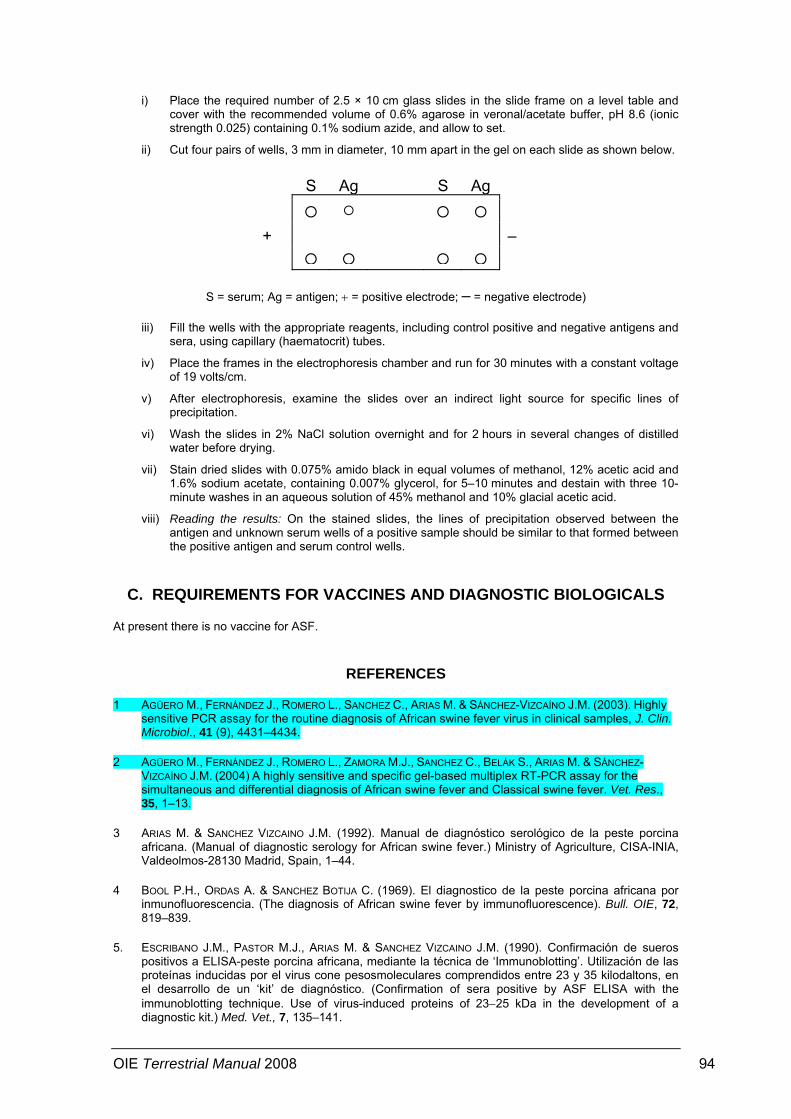
OIE Terrestrial Manual 2008 94
i) Place the required number of 2.5 × 10 cm glass slides in the slide frame on a level table and cover with the recommended volume of 0.6% agarose in veronal/acetate buffer, pH 8.6 (ionic strength 0.025) containing 0.1% sodium azide, and allow to set.
ii) Cut four pairs of wells, 3 mm in diameter, 10 mm apart in the gel on each slide as shown below.
S Ag S Ag
+ –
S = serum; Ag = antigen; + = positive electrode; – = negative electrode)
iii) Fill the wells with the appropriate reagents, including control positive and negative antigens and sera, using capillary (haematocrit) tubes.
iv) Place the frames in the electrophoresis chamber and run for 30 minutes with a constant voltage of 19 volts/cm.
v) After electrophoresis, examine the slides over an indirect light source for specific lines of precipitation.
vi) Wash the slides in 2% NaCl solution overnight and for 2 hours in several changes of distilled water before drying.
vii) Stain dried slides with 0.075% amido black in equal volumes of methanol, 12% acetic acid and 1.6% sodium acetate, containing 0.007% glycerol, for 5–10 minutes and destain with three 10-minute washes in an aqueous solution of 45% methanol and 10% glacial acetic acid.
viii) Reading the results: On the stained slides, the lines of precipitation observed between the antigen and unknown serum wells of a positive sample should be similar to that formed between the positive antigen and serum control wells.
C. REQUIREMENTS FOR VACCINES AND DIAGNOSTIC BIOLOGICALS
At present there is no vaccine for ASF.
REFERENCES
1 AGÜERO M., FERNÁNDEZ J., ROMERO L., SANCHEZ C., ARIAS M. & SÁNCHEZ-VIZCAÍNO J.M. (2003). Highly sensitive PCR assay for the routine diagnosis of African swine fever virus in clinical samples, J. Clin. Microbiol., 41 (9), 4431–4434.
2 AGÜERO M., FERNÁNDEZ J., ROMERO L., ZAMORA M.J., SANCHEZ C., BELÁK S., ARIAS M. & SÁNCHEZ-VIZCAÍNO J.M. (2004) A highly sensitive and specific gel-based multiplex RT-PCR assay for the simultaneous and differential diagnosis of African swine fever and Classical swine fever. Vet. Res., 35, 1–13.
3 ARIAS M. & SANCHEZ VIZCAINO J.M. (1992). Manual de diagnóstico serológico de la peste porcina africana. (Manual of diagnostic serology for African swine fever.) Ministry of Agriculture, CISA-INIA, Valdeolmos-28130 Madrid, Spain, 1–44.
4 BOOL P.H., ORDAS A. & SANCHEZ BOTIJA C. (1969). El diagnostico de la peste porcina africana por inmunofluorescencia. (The diagnosis of African swine fever by immunofluorescence). Bull. OIE, 72, 819–839.
5. ESCRIBANO J.M., PASTOR M.J., ARIAS M. & SANCHEZ VIZCAINO J.M. (1990). Confirmación de sueros positivos a ELISA-peste porcina africana, mediante la técnica de ‘Immunoblotting’. Utilización de las proteínas inducidas por el virus cone pesosmoleculares comprendidos entre 23 y 35 kilodaltons, en el desarrollo de un ‘kit’ de diagnóstico. (Confirmation of sera positive by ASF ELISA with the immunoblotting technique. Use of virus-induced proteins of 23−25 kDa in the development of a diagnostic kit.) Med. Vet., 7, 135–141.

OIE Terrestrial Manual 2008 95
6. ESCRIBANO J.M., PASTOR M.J. & SANCHEZ VIZCAINO J.M. (1989). Antibodies to bovine serum albumin in swine sera: implications for false positive reactions in the serodiagnosis of African swine fever. Am. J. Vet. Res., 50, 1118–1122.
7. GONZAGUE M., PLIN C., BAKKALI-KASSIMI L., BOUTROUILLE A. & CRUCIERE C. (2002). Development of an internal control for the detection of the African swine fever virus by PCR. Mol. Cell. Probes, 16, 237–242.
8. KING D.P., REID S.M., HUTCHINGS G.H., GRIERSON S.S., WILKINSON P.J., DIXON L.K., BASTOS A.D.S. & DREW T.W. (2003). Development of a TaqMan® PCR assay with internal amplification control for the detection of African swine fever virus. J. Virol. Methods, 107, 53–61.
9. MALMQUIST W.A. & HAY D. (1960). Haemadsorption and cytopathic effect produced by African swine fever virus in swine bone marrow and buffy coat cultures. Am. J. Vet. Res., 21, 104–108.
10. PAN I.C., DE BOER C.J. & HESS W.R. (1972). African swine fever: application of immuno-electro-osmophoresis for the detection of antibody. Can. J. Comp. Med., 36, 309–316.
11 PAN I.C., TRAUTMAN R., HESS W.R., DE BOER C.J., TESSLER J., ORDAS A., SANCHEZ BOTIJA C., OVEJERO J. & SANCHEZ M.C. (1974). African swine fever: comparison of four serotests on porcine serums in Spain. Am. J. Vet. Res., 35, 787–790.
12. PASTOR M.J., ARIAS M. & ESCRIBANO J.M. (1990). Comparison of two antigens for use in an enzyme-linked immunosorbent assay to detect African swine fever antibody. Am. J. Vet. Res., 51, 1540–1543.
13. PASTOR M.J., LAVIADA M.D., SANCHEZ VIZCAINO J.M. & ESCRIBANO J.M. (1989). Detection of African swine fever virus antibodies by immunoblotting assay. Can. J. Vet. Res., 53, 105–107.
14 SANCHEZ BOTIJA C., ORDAS A. & GONZALES J.G. (1970). La immunofluorescencia indirecta aplicada a la investigación de anticuerpos de la peste porcina africana. Su valor para el diagnóstico. (Indirect immunofluorescence for the investigation of African swine fever antibodies. Its value for diagnosis). Rev. Patron. Biol. Anim., 14, 159–180.
15. SANCHEZ VIZCAINO J.M. (1987). African swine fever diagnosis. In: African Swine Fever, Becker Y., ed. Martinus Nijhoff, Boston, USA, 63–71.
16. SANCHEZ VIZCAINO J.M., CROWTHER J.R. & WARDLEY R.C. (1983). A collaborative study on the use of the ELISA in the diagnosis of African swine fever. In: African Swine Fever. (CEC/FAO Research Seminar, Sardinia, Sept. 1981). Wilkinson P.J., ed. Commission of the European Communities Publication EUR 8466 EN, 297–325.
17. VINUELA E. (1985). African swine fever. Curr. Top. Microbiol. Immunol., 116, 151–170.
18. WARDLEY R.C., ABU ELZEIN E.M.E., CROWTHER J.R. & WILKINSON P.J. (1979). A solid-phase enzyme-linked immunosorbent assay for the detection of African swine fever antigen and antibody. J. Hyg., 83, 363–369.
* * *
NB: There are OIE Reference Laboratories for African swine fever (see Table in Part 3 of this Terrestrial Manual or consult the OIE Web site for the most up-to-date list: www.oie.int).

OIE Terrestrial Manual 2008 96
ADVICE FOR MEMBER COUNTRY COMMENTS
Chapter Title and Number: 2.7.6. Avian infectious bronchitis
Country making the comments: European Community
Date: 31/5/2006
General Comments: NONE Specific Comments (add continuation sheets if required)
line:
P. 6 line 276: For HA and HI tests, procedures are carried out at 4 degrees. Does this include all steps in the test? If yes, it would be more useful to write this in line 295 and line 297 too.

OIE Terrestrial Manual 2008 97
ADVICE FOR MEMBER COUNTRY COMMENTS
Chapter Title and Number: 2.7.10. Duck virus enteritis
Country making the comments: European Community
Date: 31/5/2006
General Comments: NONE
Specific Comments (add continuation sheets if required)
line: NONE

OIE Terrestrial Manual 2008 98
ADVICE FOR MEMBER COUNTRY COMMENTS
Chapter Title and Number: 2.7.11. Fowl cholera
Country making the comments: European Community
Date: 31/5/2006
General Comments: NONE
Specific Comments (add continuation sheets if required)
line:
NONE

OIE Terrestrial Manual 2008 99
ADVICE FOR MEMBER COUNTRY COMMENTS
Chapter Title and Number: 2.7.13. Newcastle disease
Country making the comments: European Community
Date: 31/5/2006
General Comments: NONE
Specific Comments (add continuation sheets if required)
line:
NONE

OIE Terrestrial Manual 2008 100
ADVICE FOR MEMBER COUNTRY COMMENTS
Chapter Title and Number: 2.7.16. Fowl pox
Country making the comments: European Community
Date: 31/5/2006
General Comments: NONE
Specific Comments (add continuation sheets if required)
line:
NONE

OIE Terrestrial Manual 2008 101
ADVICE FOR MEMBER COUNTRY COMMENTS
Chapter Title and Number: 2.9. Bee note
Country making the comments: European Community
Date: 31/5/2006
General Comments: NONE
Specific Comments (add continuation sheets if required)
line:
NONE

OIE Terrestrial Manual 2008 102
ADVICE FOR MEMBER COUNTRY COMMENTS
Chapter Title and Number: 2.9.1. Acarapisosis of honey bees
Country making the comments: European Community
Date: 31/5/2006
Gneral Comments: NONE
Specific Comments (add continuation sheets if required)
line: NONE

OIE Terrestrial Manual 2008 103
ADVICE FOR MEMBER COUNTRY COMMENTS
Chapter Title and Number: 2.9.2. American foulbrood of honey bees
Country making the comments: European Community
Date: 31/5/2006
General Comments: NONE
Specific Comments (add continuation sheets if required)
line: NONE

OIE Terrestrial Manual 2008 104
ADVICE FOR MEMBER COUNTRY COMMENTS
Chapter Title and Number: 2.9.4. Varroosis of honey bees
Country making the comments: European Community
Date: 31/5/2006
General Comments: NONE
Specific Comments (add continuation sheets if required)
line: NONE

OIE Terrestrial Manual 2008 105
ADVICE FOR MEMBER COUNTRY COMMENTS
Chapter Title and Number: 2.10.14. Listeria monocytogenes
Country making the comments: European Community
Date: 31/5/2006
General Comments: NONE
Specific Comments (add continuation sheets if required)
line: NONE
OIE Terrestrial Manual 2008 106
ADVICE FOR MEMBER COUNTRY COMMENTS
Chapter Title and Number: 1.1.3. Principles of validation of diagnostic assays for infectious diseases
Country making the comments: European Community
Date: 31 May 2006
General Comments NONE
Specific Comments (add continuation sheets if required)
line: NONE
OIE Terrestrial Manual 2008 107
ADVICE FOR MEMBER COUNTRY COMMENTS
Chapter Title and Number: 1.1.7. Principles of veterinary vaccine production
Country making the comments: Eurpean Community
Date: 31 May 2006
General Comments: NONE
Specific Comments (add continuation sheets if required)
line: NONE

OIE Terrestrial Manual 2008 108
ADVICE FOR MEMBER COUNTRY COMMENTS
Chapter Title and Number: 2.2.10. Foot and mouth disease
Country making the comments: Eurpean Community
Date: 31 May 2006
General Comments: NONE
Specific Comments (add continuation sheets if required)
line: NONE