Anatomical and ecological observations in succulent (articulated) halophytes from Chenopodiaceae
6
19 ANATOMICAL AND ECOLOGICAL OBSERVATIONS IN SUCCULENT (ARTICULATED) HALOPHYTES FROM CHENOPODIACEAE OBSERVAłII ANATOMO-ECOLOGICE LA SPECII DE HALOFITE SUCULENTE (ARTICULATE) DIN FAMILIA CHENOPODIACEAE GRIGORE M.N. 1 , TOMA C. 1 , ZAMFIRACHE Maria-Magdalena 1 , IVĂNESCU Lăcrămioara, DARABAN Iulia 2 e-mail: [email protected] Abstract. Several succulent halophytes, with articulated segments have been anatomically investigated: Sarcocornia fruticosa (L.) A. J. Scott, Arthrocnemum macrostachyum (Moric.) Moris in Moris & Delponte, Salicornia ramosissima Woods (Chenopodiaceae). These species have been collected from Spain, in 2010. The nature of articulated segments is still disputed from anatomical point of view, a caulinar or foliar origin being suggested during time. We also evidenced several special structures, such as stereids (‘spicular cells’) and tracheoidioblasts, whose functions played within these segments, are still incompletely elucidated. These structures, as well the succulence are discussed as adaptations of halophytes to environmental conditions. Key words: halophytes, anatomy, ecology. Rezumat. În lucrarea de faŃă, am supus investigaŃiei anatomice următoarele specii de halofite suculente, cu segmente articulate, din familia Chenopodiaceae: Sarcocornia fruticosa (L.) A. J. Scott, Arthrocnemum macrostachyum (Moric.) Moris in Moris & Delponte, Salicornia ramosissima Woods, colectate din Spania, în 2010. Natura segmentelor articulate este încă disputată din punct de vedere anatomic, ridicându-se problema dacă acestea au origine caulinară sau foliară. Am evidenŃiat şi alte structuri speciale, cum ar fi stereidele (celule „spiculiforme”) şi traheoidioblastele, ale căror funcŃii în cadrul acestor segmente suculente sunt încă incomplet elucidate. Aceste structuri, precum şi prezenŃa suculenŃei, au fost interpretate în sensul adaptărilor halofitelor la condiŃiile complexe de mediu. Cuvinte cheie: halofite, anatomie, ecologie. INTRODUCTION Halophytes are plants that naturally vegetate in saline habitats (Grigore, 2008). They are included in a very heterogeneous ecological group; for this reason, plants present very different and complex adaptive features, formed most likely during evolution, as a result of continuous influence of environmental factors (Grigore and Toma, 2010). In the present paper, we continue the anatomical and ecological research 1 "Alexandru Ioan Cuza" University of Iasi, Romania 2 Institut of Life Sciences "Vasile Goldiş", University of Arad, Romania
Transcript of Anatomical and ecological observations in succulent (articulated) halophytes from Chenopodiaceae

19
ANATOMICAL AND ECOLOGICAL OBSERVATIONS IN
SUCCULENT (ARTICULATED) HALOPHYTES FROM
CHENOPODIACEAE
OBSERVAłII ANATOMO-ECOLOGICE LA SPECII DE HALOFITE SUCULENTE (ARTICULATE) DIN FAMILIA CHENOPODIACEAE
GRIGORE M.N.1, TOMA C.1, ZAMFIRACHE Maria-Magdalena1,
IVĂNESCU Lăcrămioara, DARABAN Iulia2 e-mail: [email protected]
Abstract. Several succulent halophytes, with articulated segments have been anatomically investigated: Sarcocornia fruticosa (L.) A. J. Scott, Arthrocnemum macrostachyum (Moric.) Moris in Moris & Delponte, Salicornia ramosissima Woods (Chenopodiaceae). These species have been collected from Spain, in 2010. The nature of articulated segments is still disputed from anatomical point of view, a caulinar or foliar origin being suggested during time. We also evidenced several special structures, such as stereids (‘spicular cells’) and tracheoidioblasts, whose functions played within these segments, are still incompletely elucidated. These structures, as well the succulence are discussed as adaptations of halophytes to environmental conditions. Key words: halophytes, anatomy, ecology. Rezumat. În lucrarea de faŃă, am supus investigaŃiei anatomice următoarele specii de halofite suculente, cu segmente articulate, din familia Chenopodiaceae: Sarcocornia fruticosa (L.) A. J. Scott, Arthrocnemum macrostachyum (Moric.) Moris in Moris & Delponte, Salicornia ramosissima Woods, colectate din Spania, în 2010. Natura segmentelor articulate este încă disputată din punct de vedere anatomic, ridicându-se problema dacă acestea au origine caulinară sau foliară. Am evidenŃiat şi alte structuri speciale, cum ar fi stereidele (celule „spiculiforme”) şi traheoidioblastele, ale căror funcŃii în cadrul acestor segmente suculente sunt încă incomplet elucidate. Aceste structuri, precum şi prezenŃa suculenŃei, au fost interpretate în sensul adaptărilor halofitelor la condiŃiile complexe de mediu. Cuvinte cheie: halofite, anatomie, ecologie.
INTRODUCTION
Halophytes are plants that naturally vegetate in saline habitats (Grigore,
2008). They are included in a very heterogeneous ecological group; for this
reason, plants present very different and complex adaptive features, formed most
likely during evolution, as a result of continuous influence of environmental
factors (Grigore and Toma, 2010).
In the present paper, we continue the anatomical and ecological research
1 "Alexandru Ioan Cuza" University of Iasi, Romania 2 Institut of Life Sciences "Vasile Goldiş", University of Arad, Romania

20
regarding halophytes from Mediterranean climate, a work included in a large
series (Grigore, Toma, Boşcaiu, 2011; Grigore, Toma, Ivănescu, 2011).
In the Mediterranean region, the halophytic communities represent two
categories – those that belong to the maritime salt marshes and those that belong to
the salt deserts (Chapman, 1974). Moreover, as already stated, Mediterranean salt
marshes provide special ecological conditions, controlling the spatial distribution of
vegetation; this is related to the predominance of several environmental factors and
to adaptive set of halophytes (Grigore, Toma, Boşcaiu, 2011).
MATERIAL AND METHOD
In this study, three species of halophytes from Chenopodiaceae (sometimes included in Amaranthaceae) have been anatomically investigated: Sarcocornia fruticosa (L.) A. J. Scott, Arthrocnemum macrostachyum (Moric.) Moris in Moris & Delponte, and Salicornia ramosissima Woods. These have been collected in July of 2010, from a coastal salt marsh from Alicante (Spania).
Anatomical investigations were conducted following the method standardized by our group from Faculty of Biology, Iasi (for an extended description of this method, see: Grigore, Toma and Boşcaiu, 2010).
RESULTS AND DISCUSSIONS
Following the anatomical investigations, several observations can be
delineated. In Arthrocnemum macrostachyum, in the external cortex of
unarticulated stem long or very long brachysclereids are dispersed; these are
perpendicular on the epidermis, simple or branched (especially at the ends), with a
thick and lignified wall. At the limit between the external cortex and the middle
one there are located very small vascular bundles, with spiral xylem vessels
disposed on a circle.
There follows a special type of cork area (Fig. 1): 2-3 layers of rectangular
cells, slightly tall with relatively thick and suberified walls. The phelloderm forms
a thick area with cells disposed in radial rows having the tangential walls
moderately thickened.
The stele comprises 3-4 rings of vascular bundles (Figs. 1, 2) embedded
into the fundamental sclerenchyma mass, all resulting from the activity of the
supernumerary cambia. The phloem appears like cellulosic isles surrounded by
sclerenchyma and the xylem of the conducting vessels.
On the internal face of the first ring, that is, close to the medulla, there are
six vascular bundles (Fig. 2) larger than the ones resulting from the activity of the
supernumerary cambia, having the xylem with little libriform fibers and being
separated by wide medullary rays, made of parenchyma cells with moderately
thickened and lignified wall.
In the cortex of articulated (succulent) segment, there are many stereides
(Fig. 3), perpendicular on the epidermis, partially embedded in the water-storage
parenchyma. These stereides have been also evidenced by De Fraine, (1912), who
called them “spicular cells”. This author considers the stereides and
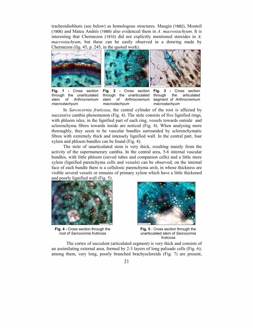
21
tracheoidioblasts (see below) as homologous structures. Mangin (1882), Monteil
(1906) and Mateu Andrés (1989) also evidenced them in A. macrostachyum. It is
interesting that Chermezon (1910) did not explicitly mentioned stereides in A. macrostachyum, but these can be easily observed in a drawing made by
Chermezon (fig. 45, p. 245, in the quoted work).
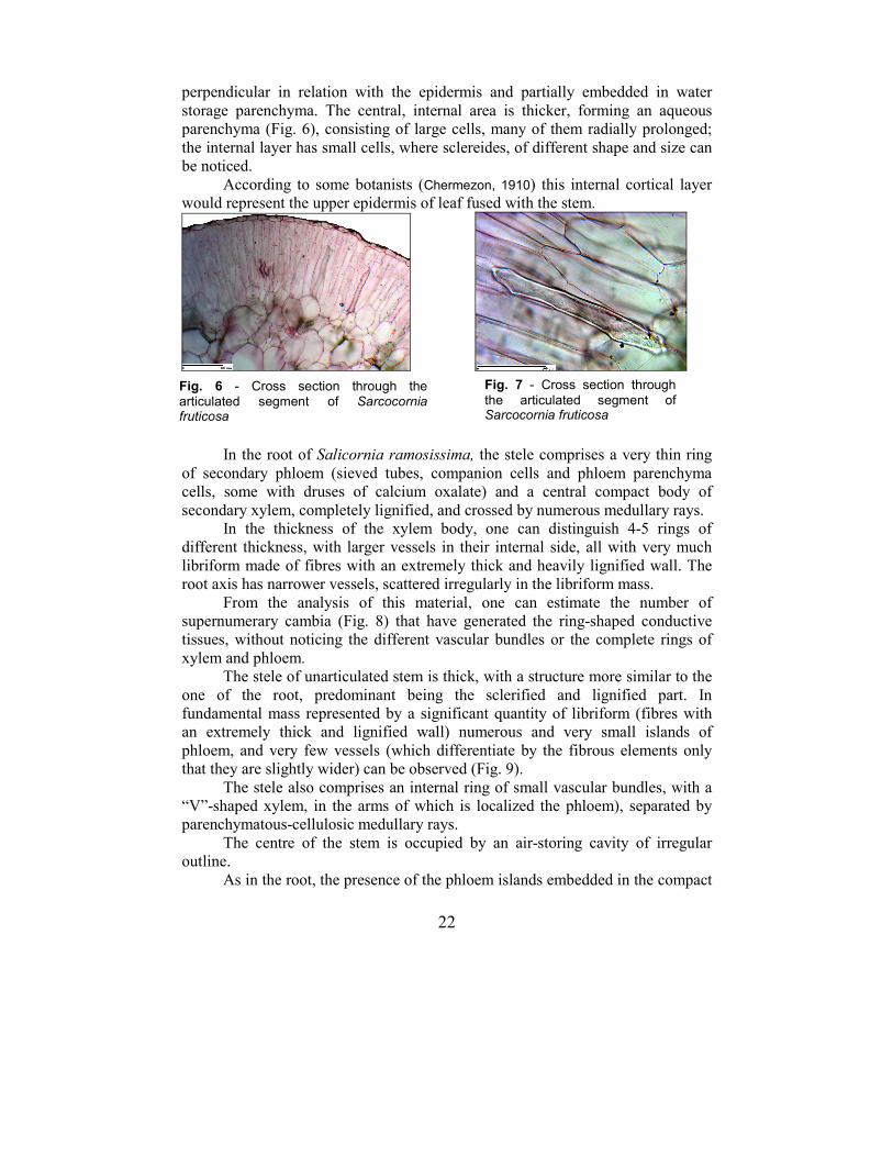
In Sarcocornia fruticosa, the central cylinder of the root is affected by
successive cambia phenomenon (Fig. 4). The stele consists of five lignified rings,
with phloem isles; in the lignified part of each ring, vessels towards outside and
sclerenchyma fibres towards inside are noticed (Fig. 4). When analysing more
thoroughly, they seem to be vascular bundles surrounded by sclerenchymatic
fibres with extremely thick and intensely lignified wall. In the central part, four
xylem and phloem bundles can be found (Fig. 4).
The stele of unarticulated stem is very thick, resulting mainly from the
activity of the supernumerary cambia. In the central area, 5-6 internal vascular
bundles, with little phloem (sieved tubes and companion cells) and a little more
xylem (lignified parenchyma cells and vessels) can be observed; on the internal
face of each bundle there is a cellulosic parenchyma arch, in whose thickness are
visible several vessels or remains of primary xylem which have a little thickened
and poorly lignified wall (Fig. 5).
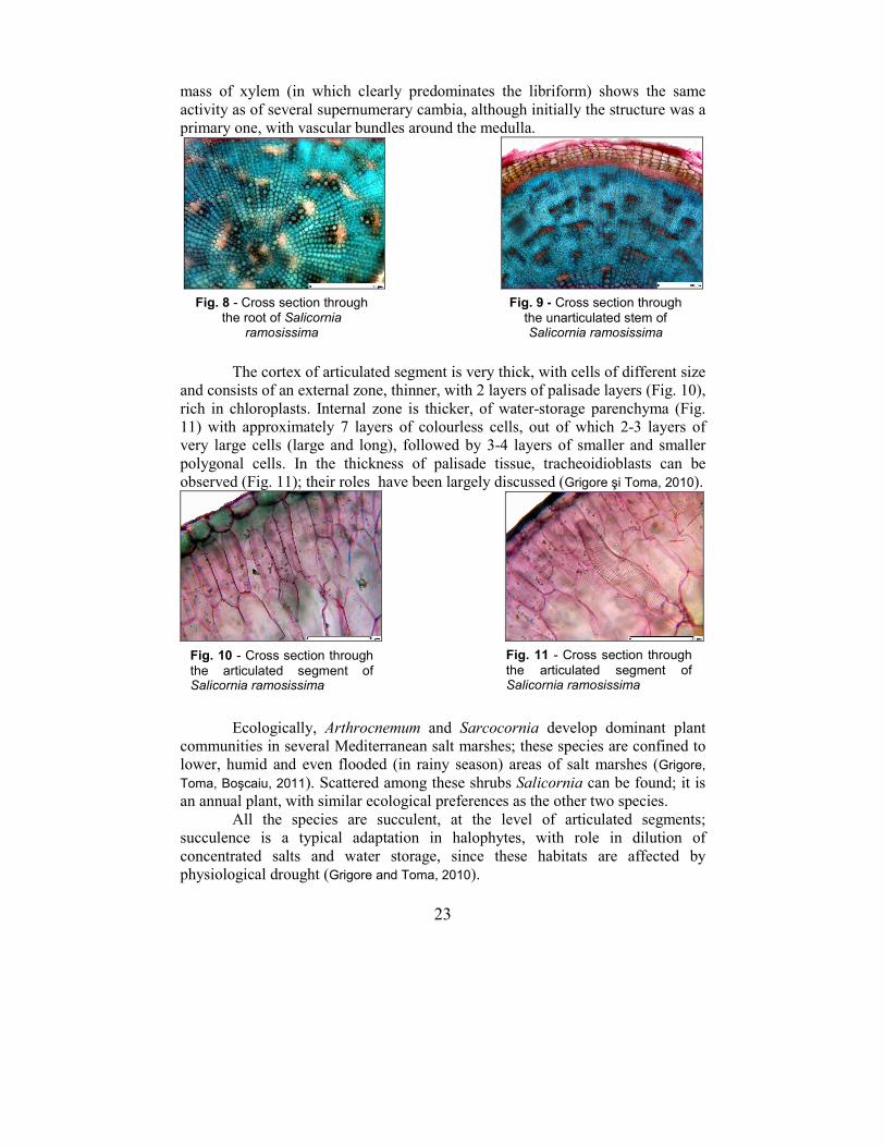
The cortex of succulent (articulated segment) is very thick and consists of
an assimilating external area, formed by 2-3 layers of long palisade cells (Fig. 6);
among them, very long, poorly branched brachysclereids (Fig. 7) are present,
Fig. 1 - Cross section
through the unarticulated stem of Arthrocnemum macrostachyum
Fig. 2 - Cross section
through the unarticulated stem of Arthrocnemum macrostachyum
Fig. 3 - Cross section
through the articulated segment of Arthrocnemum macrostachyum
Fig. 4 - Cross section through the root of Sarcocornia fruticosa
Fig. 5 - Cross section through the unarticulated stem of Sarcocornia
fruticosa
22
perpendicular in relation with the epidermis and partially embedded in water
storage parenchyma. The central, internal area is thicker, forming an aqueous
parenchyma (Fig. 6), consisting of large cells, many of them radially prolonged;
the internal layer has small cells, where sclereides, of different shape and size can
be noticed.
According to some botanists (Chermezon, 1910) this internal cortical layer
would represent the upper epidermis of leaf fused with the stem.
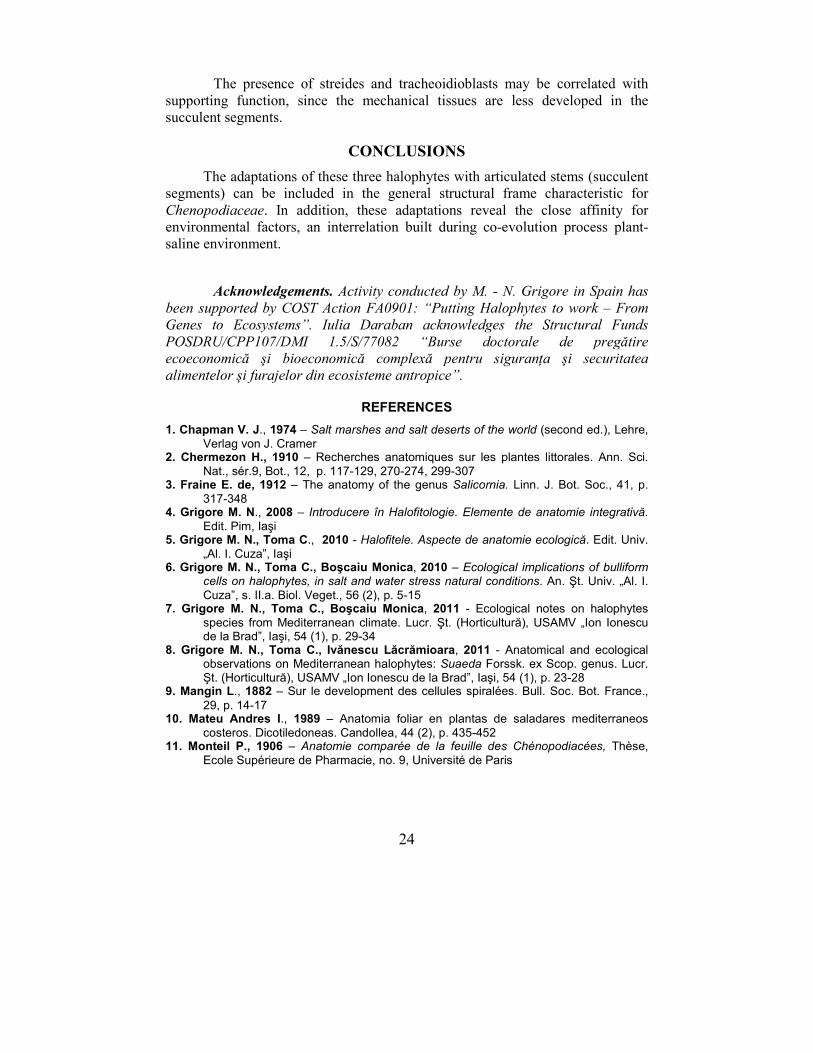
In the root of Salicornia ramosissima, the stele comprises a very thin ring
of secondary phloem (sieved tubes, companion cells and phloem parenchyma
cells, some with druses of calcium oxalate) and a central compact body of
secondary xylem, completely lignified, and crossed by numerous medullary rays.
In the thickness of the xylem body, one can distinguish 4-5 rings of
different thickness, with larger vessels in their internal side, all with very much
libriform made of fibres with an extremely thick and heavily lignified wall. The
root axis has narrower vessels, scattered irregularly in the libriform mass.
From the analysis of this material, one can estimate the number of
supernumerary cambia (Fig. 8) that have generated the ring-shaped conductive
tissues, without noticing the different vascular bundles or the complete rings of
xylem and phloem.
The stele of unarticulated stem is thick, with a structure more similar to the
one of the root, predominant being the sclerified and lignified part. In
fundamental mass represented by a significant quantity of libriform (fibres with
an extremely thick and lignified wall) numerous and very small islands of
phloem, and very few vessels (which differentiate by the fibrous elements only
that they are slightly wider) can be observed (Fig. 9).
The stele also comprises an internal ring of small vascular bundles, with a
“V”-shaped xylem, in the arms of which is localized the phloem), separated by
parenchymatous-cellulosic medullary rays.
The centre of the stem is occupied by an air-storing cavity of irregular
outline.
As in the root, the presence of the phloem islands embedded in the compact
Fig. 6 - Cross section through the articulated segment of Sarcocornia fruticosa
Fig. 7 - Cross section through
the articulated segment of Sarcocornia fruticosa
23
mass of xylem (in which clearly predominates the libriform) shows the same
activity as of several supernumerary cambia, although initially the structure was a
primary one, with vascular bundles around the medulla.
The cortex of articulated segment is very thick, with cells of different size
and consists of an external zone, thinner, with 2 layers of palisade layers (Fig. 10),
rich in chloroplasts. Internal zone is thicker, of water-storage parenchyma (Fig.
11) with approximately 7 layers of colourless cells, out of which 2-3 layers of
very large cells (large and long), followed by 3-4 layers of smaller and smaller
polygonal cells. In the thickness of palisade tissue, tracheoidioblasts can be
observed (Fig. 11); their roles have been largely discussed (Grigore şi Toma, 2010).
Ecologically, Arthrocnemum and Sarcocornia develop dominant plant
communities in several Mediterranean salt marshes; these species are confined to
lower, humid and even flooded (in rainy season) areas of salt marshes (Grigore,
Toma, Boşcaiu, 2011). Scattered among these shrubs Salicornia can be found; it is
an annual plant, with similar ecological preferences as the other two species.
All the species are succulent, at the level of articulated segments;
succulence is a typical adaptation in halophytes, with role in dilution of
concentrated salts and water storage, since these habitats are affected by
physiological drought (Grigore and Toma, 2010).
Fig. 8 - Cross section through the root of Salicornia
ramosissima
Fig. 9 - Cross section through
the unarticulated stem of Salicornia ramosissima
Fig. 10 - Cross section through
the articulated segment of Salicornia ramosissima
Fig. 11 - Cross section through
the articulated segment of Salicornia ramosissima
24
The presence of streides and tracheoidioblasts may be correlated with
supporting function, since the mechanical tissues are less developed in the
succulent segments.
CONCLUSIONS
The adaptations of these three halophytes with articulated stems (succulent
segments) can be included in the general structural frame characteristic for
Chenopodiaceae. In addition, these adaptations reveal the close affinity for
environmental factors, an interrelation built during co-evolution process plant-
saline environment.
Acknowledgements. Activity conducted by M. - N. Grigore in Spain has been supported by COST Action FA0901: “Putting Halophytes to work – From Genes to Ecosystems”. Iulia Daraban acknowledges the Structural Funds POSDRU/CPP107/DMI 1.5/S/77082 “Burse doctorale de pregătire ecoeconomică şi bioeconomică complexă pentru siguranŃa şi securitatea alimentelor şi furajelor din ecosisteme antropice”.
REFERENCES
1. Chapman V. J., 1974 – Salt marshes and salt deserts of the world (second ed.), Lehre,
Verlag von J. Cramer 2. Chermezon H., 1910 – Recherches anatomiques sur les plantes littorales. Ann. Sci.
Nat., sér.9, Bot., 12, p. 117-129, 270-274, 299-307 3. Fraine E. de, 1912 – The anatomy of the genus Salicornia. Linn. J. Bot. Soc., 41, p.
317-348 4. Grigore M. N., 2008 – Introducere în Halofitologie. Elemente de anatomie integrativă.
Edit. Pim, Iaşi 5. Grigore M. N., Toma C., 2010 - Halofitele. Aspecte de anatomie ecologică. Edit. Univ.
„Al. I. Cuza”, Iaşi 6. Grigore M. N., Toma C., Boşcaiu Monica, 2010 – Ecological implications of bulliform
cells on halophytes, in salt and water stress natural conditions. An. Şt. Univ. „Al. I.
Cuza”, s. II.a. Biol. Veget., 56 (2), p. 5-15 7. Grigore M. N., Toma C., Boşcaiu Monica, 2011 - Ecological notes on halophytes
species from Mediterranean climate. Lucr. Şt. (Horticultură), USAMV „Ion Ionescu de la Brad”, Iaşi, 54 (1), p. 29-34
8. Grigore M. N., Toma C., Ivănescu Lăcrămioara, 2011 - Anatomical and ecological observations on Mediterranean halophytes: Suaeda Forssk. ex Scop. genus. Lucr. Şt. (Horticultură), USAMV „Ion Ionescu de la Brad”, Iaşi, 54 (1), p. 23-28
9. Mangin L., 1882 – Sur le development des cellules spiralées. Bull. Soc. Bot. France.,
29, p. 14-17 10. Mateu Andres I., 1989 – Anatomia foliar en plantas de saladares mediterraneos
costeros. Dicotiledoneas. Candollea, 44 (2), p. 435-452 11. Monteil P., 1906 – Anatomie comparée de la feuille des Chénopodiacées, Thèse,
Ecole Supérieure de Pharmacie, no. 9, Université de Paris