An iflavirus found in stink bugs (Hemiptera_ ... - Ainfo
8
Contents lists available at ScienceDirect Virology journal homepage: www.elsevier.com/locate/virology An iflavirus found in stink bugs (Hemiptera: Pentatomidae) of four different species Ethiane R. dos Santos a,1 , Luana B. Trentin a,1 , Assis Ecker a , Leonardo A. Silva b , Miguel Borges c , Joseph D. Mowery d , Bergmann M. Ribeiro b , Robert L. Harrison e , Daniel M.P. Ardisson-Araújo a,∗ a Laboratory of Insect Virology, Department of Biochemistry and Molecular Biology, Federal University of Santa Maria, Santa Maria, RS, 97105-900, Brazil b Laboratory of Baculovirus, Cell Biology Department, University of Brasilia, Brasilia, DF, 70910-900, Brazil c Embrapa Recursos Genéticos e Biotecnologia, Brasília, DF, 70.770-917, Brazil d Electron and Confocal Microscopy Unit, Beltsville Agricultural Research Center, USDA Agricultural Research Service, Beltsville, MD, 20705, USA e Invasive Insect Biocontrol and Behavior Laboratory, Beltsville Agricultural Research Center, USDA Agricultural Research Service, Beltsville, MD, 20705, USA ARTICLE INFO Keywords: Iflavirus Iflaviridae Covert infection Stink bugs Pentatomidae Chinavia ubica Dichelops melachantus Euschistus heros Halyomorpha halys ABSTRACT An analysis of transcriptomes from the antennae of the three South American stink bugs (Euschistus heros, Chinavia ubica, and Dichelops melacanthus) revealed the presence of picorna-like virus genome-length RNAs with high sequence identity to the genome of Halyomorpha halys virus (HhV), originally discovered in the tran- scriptome of the brown marmorated stink bug, Halyomorpha halys. Features of the genome, phylogenetic re- lationships to other viruses, and the appearances of virus-like particles isolated from host stink bugs all confirm that these viruses are iflaviruses and isolates of an undescribed species. Iflavirus RNAs were present at high levels (40%–90% of transcriptome reads) in the stink bug antennal transcriptomes. In whole-insect transcriptomes of H. halys, HhV reads were > 500-fold more abundant in adults than in nymphs. We identified from field popu- lation a subject of species E. heros infected by this iflavirus. The results of the analysis suggest that these ifla- viruses are able to produce large quantities of their RNAs without causing any obvious pathology to their hosts. 1. Introduction Iflaviruses belong to the picorna-like virus family Iflaviridae (order Picornavirales) and are classified within a single genus, Iflavirus (Valles et al., 2017). Members of Iflavirus infect invertebrates of phylum Ar- thropoda, including Lepidoptera, Hymenoptera, Diptera, Coleoptera, Acari, Araneae, Mesostigmata, and Decapoda (Aizawa et al., 1964; Liu et al., 2017). They are characterized by a positive ssRNA genome or- ganized into a single ORF that codes for one polyprotein consisting of structural and non-structural peptides. The ORF is flanked by a long IRES-containing 5′-UTR and a short, conserved poly-A tail-containing 3′-UTR (Lanzi et al., 2006), which regulate both virus replication and translation (Belsham, 2009). A stable 30-nm non-enveloped ichosahe- dral capsid packages and shields the RNA genome (van Oers, 2010). The iflavirus infection process is still not well understood and seems to differ depending on the host type. In some iflavirus hosts, such as the silkworm Bombyx mori (Himeno et al., 1974), the oil palm tree pest Opsiphanes invirae (Silva et al., 2015), and the honey bee Apis mellifera (Yue et al., 2007), signs of infection are quite evident depending on the virus species and host condition. For example, in silkworm larvae, the pathology of infectious flacherie virus (the type species of iflavirus) infection includes a brown color or other discoloration of the posterior and middle parts of the insect body and flaccidity (Inoue and Ayuzawa, 1972). In honeybees, iflaviruses may cause foreleg paralysis, wing de- formities, a shortened abdomen and discoloration of the adult bees, as well as mortality (Bailey and Woods, 1974; Bailey and Ball, 1991). These viruses are usually associated and transmitted by the parasite mite Varroa destructor (Santillán-Galicia et al., 2010), and may be di- rectly linked to colony collapse disorder (Bailey and Ball, 1991). However, the majority of iflavirus infections do not result in visible signs of infection or any apparent disease (van Oers, 2010). The first picorna-like virus found infecting a stink bug was reported from the southern green stink bug, Nezara viridula, by Williamson and von Wechmar (1992, 1995). This virus (Nezara viridula virus 1; NVV1) https://doi.org/10.1016/j.virol.2019.06.002 Received 25 March 2019; Received in revised form 5 June 2019; Accepted 5 June 2019 ∗ Corresponding author. E-mail addresses: [email protected] (E.R. dos Santos), [email protected] (L.B. Trentin), [email protected] (A. Ecker), [email protected] (L.A. Silva), [email protected] (M. Borges), [email protected] (J.D. Mowery), [email protected] (B.M. Ribeiro), [email protected] (R.L. Harrison), [email protected], [email protected] (D.M.P. Ardisson-Araújo). 1 Equal contribution. Virology 534 (2019) 72–79 Available online 06 June 2019 0042-6822/ © 2019 Elsevier Inc. All rights reserved. T
-
Upload
khangminh22 -
Category
Documents
-
view
2 -
download
0
Transcript of An iflavirus found in stink bugs (Hemiptera_ ... - Ainfo
Contents lists available at ScienceDirect
Virology
journal homepage: www.elsevier.com/locate/virology
An iflavirus found in stink bugs (Hemiptera: Pentatomidae) of four differentspecies
Ethiane R. dos Santosa,1, Luana B. Trentina,1, Assis Eckera, Leonardo A. Silvab, Miguel Borgesc,Joseph D. Moweryd, Bergmann M. Ribeirob, Robert L. Harrisone, Daniel M.P. Ardisson-Araújoa,∗
a Laboratory of Insect Virology, Department of Biochemistry and Molecular Biology, Federal University of Santa Maria, Santa Maria, RS, 97105-900, Brazilb Laboratory of Baculovirus, Cell Biology Department, University of Brasilia, Brasilia, DF, 70910-900, Brazilc Embrapa Recursos Genéticos e Biotecnologia, Brasília, DF, 70.770-917, Brazild Electron and Confocal Microscopy Unit, Beltsville Agricultural Research Center, USDA Agricultural Research Service, Beltsville, MD, 20705, USAe Invasive Insect Biocontrol and Behavior Laboratory, Beltsville Agricultural Research Center, USDA Agricultural Research Service, Beltsville, MD, 20705, USA
A R T I C L E I N F O
Keywords:IflavirusIflaviridaeCovert infectionStink bugsPentatomidaeChinavia ubicaDichelops melachantusEuschistus herosHalyomorpha halys
A B S T R A C T
An analysis of transcriptomes from the antennae of the three South American stink bugs (Euschistus heros,Chinavia ubica, and Dichelops melacanthus) revealed the presence of picorna-like virus genome-length RNAs withhigh sequence identity to the genome of Halyomorpha halys virus (HhV), originally discovered in the tran-scriptome of the brown marmorated stink bug, Halyomorpha halys. Features of the genome, phylogenetic re-lationships to other viruses, and the appearances of virus-like particles isolated from host stink bugs all confirmthat these viruses are iflaviruses and isolates of an undescribed species. Iflavirus RNAs were present at high levels(40%–90% of transcriptome reads) in the stink bug antennal transcriptomes. In whole-insect transcriptomes ofH. halys, HhV reads were> 500-fold more abundant in adults than in nymphs. We identified from field popu-lation a subject of species E. heros infected by this iflavirus. The results of the analysis suggest that these ifla-viruses are able to produce large quantities of their RNAs without causing any obvious pathology to their hosts.
1. Introduction
Iflaviruses belong to the picorna-like virus family Iflaviridae (orderPicornavirales) and are classified within a single genus, Iflavirus (Valleset al., 2017). Members of Iflavirus infect invertebrates of phylum Ar-thropoda, including Lepidoptera, Hymenoptera, Diptera, Coleoptera,Acari, Araneae, Mesostigmata, and Decapoda (Aizawa et al., 1964; Liuet al., 2017). They are characterized by a positive ssRNA genome or-ganized into a single ORF that codes for one polyprotein consisting ofstructural and non-structural peptides. The ORF is flanked by a longIRES-containing 5′-UTR and a short, conserved poly-A tail-containing3′-UTR (Lanzi et al., 2006), which regulate both virus replication andtranslation (Belsham, 2009). A stable 30-nm non-enveloped ichosahe-dral capsid packages and shields the RNA genome (van Oers, 2010).
The iflavirus infection process is still not well understood and seemsto differ depending on the host type. In some iflavirus hosts, such as thesilkworm Bombyx mori (Himeno et al., 1974), the oil palm tree pest
Opsiphanes invirae (Silva et al., 2015), and the honey bee Apis mellifera(Yue et al., 2007), signs of infection are quite evident depending on thevirus species and host condition. For example, in silkworm larvae, thepathology of infectious flacherie virus (the type species of iflavirus)infection includes a brown color or other discoloration of the posteriorand middle parts of the insect body and flaccidity (Inoue and Ayuzawa,1972). In honeybees, iflaviruses may cause foreleg paralysis, wing de-formities, a shortened abdomen and discoloration of the adult bees, aswell as mortality (Bailey and Woods, 1974; Bailey and Ball, 1991).These viruses are usually associated and transmitted by the parasitemite Varroa destructor (Santillán-Galicia et al., 2010), and may be di-rectly linked to colony collapse disorder (Bailey and Ball, 1991).However, the majority of iflavirus infections do not result in visiblesigns of infection or any apparent disease (van Oers, 2010).
The first picorna-like virus found infecting a stink bug was reportedfrom the southern green stink bug, Nezara viridula, by Williamson andvon Wechmar (1992, 1995). This virus (Nezara viridula virus 1; NVV1)
https://doi.org/10.1016/j.virol.2019.06.002Received 25 March 2019; Received in revised form 5 June 2019; Accepted 5 June 2019
∗ Corresponding author.E-mail addresses: [email protected] (E.R. dos Santos), [email protected] (L.B. Trentin), [email protected] (A. Ecker),
[email protected] (L.A. Silva), [email protected] (M. Borges), [email protected] (J.D. Mowery), [email protected] (B.M. Ribeiro),[email protected] (R.L. Harrison), [email protected], [email protected] (D.M.P. Ardisson-Araújo).
1 Equal contribution.
Virology 534 (2019) 72–79
Available online 06 June 20190042-6822/ © 2019 Elsevier Inc. All rights reserved.
T
caused reduced insect longevity when associated with a toti-like viruswith a monopartite dsRNA genome (NVV2). Another picorna-like virus(Plautia stali intestine virus, PSIV) causing disease in a stink bug wasisolated from the brown-winged green bug, Plautia stali. PSIV was alsoshown to infect the brown marmorated stink bug, Halyomorpha halys(Nakashima et al., 1998). A covert picorna-like virus infection of a stinkbug host was detected when a picorna-like virus genome was identifiedin a whole body transcriptome developed from a USDA colony of Ha-lyomorph halys (Sparks et al., 2013). Analysis of the genome sequenceindicated that this virus (termed Halymorpha halys iflavirus, or HhV)should be classified as an iflavirus. No further studies on the route ofinfection, pathology, or biological control potential of HhV have beenpublished.
In this study, we report the discovery of new isolates of HhV cov-ertly infecting three other pentatomid hosts, including Euschistus heros(Fabricius, 1794), Chinavia ubica (Rolston, 1983), and Dichelops mela-canthus (Dallas, 1851). E. heros is considered the major pest of soybeancultures in Neotropical regions (Panizzi et al., 2000). C. ubica and D.melacanthus are soybean secondary pests less abundant than E. herosand damage soybean crops as well (Panizzi et al., 2000, 2007). Ingeneral, stink bugs feed by inserting their needle-like stylets into stems,leaves, blooms, fruit or seeds (Velikova et al., 2010). By sucking onplant fluids and injecting toxic saliva into the host tissues, these or-ganisms cause delayed maturation and deformities in seeds, which af-fects grain quality and production (Velikova et al., 2010). Severalstrategies to control the spread of stinkbug populations have beenadopted, including the applications of broad-spectrum insecticides, therelease of natural predators such as the Neotropical egg parasitoidTelenomus podisi (Ashmead, 1893) (Hymenoptera: Scelionidae), and theuse of entomopathogenic biopesticides (Gouli et al., 2012) or pher-omone lures (Weber et al., 2014). However, the intensive and indis-criminate use of chemical insecticides has resulted in the elimination ofnatural enemies and the selection of resistant insect populations (Bauret al., 2010). Because several hemipterans of family Pentatomidae maybe invasive pests, the impact of covertly-infecting viruses on insect pestbiology and ecology is of importance for the control of insect popula-tions.
2. Materials and methods
2.1. SRA data, trimming, de novo assembly, and BLAST search
We obtained the raw transcriptome data from Sequence ReadArchive (SRA) experiments (Table S1) assigned to the BioProjectPRJNA246320 using the software CLC Genomics Workbench 11 (CLCbio, Aarhus, Denmark). The transcriptomes were generated from threepools containing 50 antenna pairs from males and females (1:1) of threeBrazilian stink bug species E. heros, C. ubica, and D. melacanthus (formore details, see Farias et al., 2015) from a technical duplicate. The sixraw SRAs were trimmed and de novo assembled using the same soft-ware. The generated contig lists were used as queries to search for si-milar viral sequences in a non-redundant local Viral protein Refseqdatabase by means of the BLAST implemented in Geneious R10 (Kearseet al., 2012).
2.2. Viral genome assembly, annotation, and viral RNA quantification
Several contigs were found to have significant sequence similarity tothe genome sequence of Halyomorpha halys virus (HhV), previouslyidentified in the transcriptome of the brown marmorated stink bugHalyomorpha halys (Hemiptera: Pentatomidae; Sparks et al., 2013). Inorder to assemble complete viral genome sequences, we used the HhVgenome (GenBank accession no. NC_022611) as a reference sequenceand mapped individually trimmed reads from the six libraries thatmatched the HhV sequence with a pairwise identity of ≥80% in Gen-eious R10 (Kearse et al., 2012). After that, we extracted the consensus
sequence for each mapped read and mapped back again the trimmedreads exhibiting a pairwise identity of ≥99% to confirm the results ofthe first mapping and the correct assembly of the viral genomes.Moreover, to quantify the viral RNA sequence reads in different tran-scriptomes, we mapped the total trimmed reads for each transcriptomeagainst the HhV polyprotein CDS sequence. As an internal control, weused the CDS sequence of the highly expressed actin gene. The totalnumber of mapped reads were divided by the total number of reads andthe query size (i.e. the virus polyprotein CDS or the actin CDS) for eachreplicate and the mean ± standard deviation were plotted in graphbars as arbitrary values. In the case of H. halys, one single replicate wasperformed for transcriptomes generated from second and fourth instarnymphs, adult male, and adult female (Sparks et al., 2014). Imature andadults are graphed together with mean ± standard deviation.
2.3. Search for single nucleotide variants (SNVs) among isolates of HhV inthe polyprotein CDS
To search for SNVs among the new virus isolate genomes, we con-ducted a MAFFT alignment between the HhV genome and each of thenew identified genomes. After that, we set the HhV genome sequence asa reference and searched for variants within the polyprotein CDS.Effects of polymorphisms on conceptual translation were analyzedusing the software Geneious R10 (Kearse et al., 2012).
2.4. Genome annotation and virus phylogeny
Iflaviruses encode a single polyprotein within a single ORF.Therefore, we used Geneious to identify a similar CDS occurring in eachof the three genomes of the newly discovered HhV isolates. Moreover,we performed BLASTP analysis of the predicted polyprotein amino acidsequences to search in the non-redundant Genbank database for relatediflaviruses and identify conserved regions (Altschul et al., 1997).Afterward, we carried out a phylogenetic analysis using the predictedamino acid sequence of the RNA-dependent RNA polymerase (RdRp)(Pfam entry, cd01699) domain found in several inflaviruses and otherrelated picornaviruses, including dicistroviruses and secoviruses. TheGenBank accession numbers of the taxa used are available in Table S1.The sequences were aligned by the MAFFT method (Katoh et al., 2002)and used to search for the best evolutionary model using the softwareMEGA 7 (Kumar et al., 2016). We inferred phylogenetic relationshipsamong the RdRp sequences by maximum likelihood as implemented inPhyML (Guindon et al., 2010) using the model LG + G (0.81). The treewas rooted against the clade containing members of the Secoviridae.
2.5. Insects and iflavirus RNA prospection in field population
Stinkbugs of E. heros, C. ubica, and D. melacanthus (Hemiptera:Pentatomidae) used in this study were obtained from laboratory co-lonies previously established in the Semiochemicals Laboratory ofEMBRAPA Genetic Resources and Biotechnology, Brasília, DF, Brazil(15° 47´S and 47° 55´ W). The insects were kept in a climatized room, at26 ± 1 °C, 65 ± 10% RH and a 14:10 (day:night) photoperiod.Individual stink bugs were reared in 8 L plastic cages separated byspecies. Insects were fed with a natural diet containing sunflower seed(Heliantus annus L. [Asteraceae]), peanut (Arachis hypogaea L.[Fabaceae]) and soybean (Glycine max L. [Fabaceae]), fresh bean pods(Phaseolus vulgaris L. [Fabaceae]) and water. Individuals of the brownmarmorated stink bug (H. halys) were obtained from a colony initiatedfrom adults collected in Pennsylvania, USA, maintained at the USDA-ARS Beltsville Agricultural Research Center in Beltsville, Maryland,USA, and supplemented with individuals collected locally in Beltsville(Sparks et al., 2014). Individuals of species E. heros were collected fromsoybean (G. max) fields in the Midwest Region of Brazil (Federal Dis-trict). The whole body of ten field-collected adult individuals (pickedrandomly) of species E. heros and three pools (with five adult subjects
E.R. dos Santos, et al. Virology 534 (2019) 72–79
73
each) of laboratory-obtained individuals of species D. melachantus, C.ubica, and E. heros were macerated with Trizol™ Reagent (ThermoFisher Scientific) and used for RNA extraction according to manufac-turer's instructions. The extracted total RNAs were subjected to cDNAfirst-strand synthesis using GoScript™ Reverse Transcriptase (Promega)according to manufacturer's instructions. After cDNA synthesis, a di-lution of 1:50 was individually used as template for PCR with 0.4 μM ofthe primers Penta-Iflavirus F (GAC CGA ACT CTT AAC GGA GGA GGAGC) and Penta-Iflavirus R (CTA TAA GAC ATA CCA GCA AAT TCT AT),300 μM of dNTP mix (Fermentas), 1 U of GoTaq (Promega), and 1x ofthe supplied reaction buffer. The reactions were subjected to the fol-lowing program: 95 °C/2min, 35 cycles of 95 °C/30s, 53 °C/30 s and72 °C/1min with a final extension of 5min at 72 °C.
2.6. Semi-purified virus negative staining
For extraction of virus particles from E. heros, C. ubica, and D.melacanthus, a total of 10 whole stink bug males and females from eachspecies were homogenized with 25mL of Phosphate Buffered Saline 1X(PBS, 137.0 mM NaCl, 2.7mM KCl, 10.0mM Na2HPO4, 2.0mMKH2PO4, pH 7.4), and centrifuged at 4000×g for 10min for clarifica-tion. The supernatants were filtered through a syringe plugged withcotton, loaded in polyallomer ultracentrifuge tubes (Beckman) andcentrifuged at 63,000×g for 75min. The pellets containing the virusparticles were resuspended in PBS-1X and prepared for TransmissionElectron Microscopy (TEM) by negative staining as described elsewhere(Brenner and Horne, 1959) and observed in a TEM Jeol 1011 at 100 kV.
For extraction of virus particles from H. halys, 20 adults werehomogenized in 15mL NT buffer (100mM Tris-HCl, pH7.6, and 10mMNaCl) with an Ultra-Turrax T-25. The homogenate was filtered throughthree layers of cheesecloth in a funnel with a steel strainer and furtherclarified by low-speed centrifugation (1467×g) for 10min. The su-pernatant was transferred to polyallomer tubes, underlaid with a 20%(w/v) sucrose pad, and centrifuged at 103,680×g for 4 h. The pelletcontaining virus particles was resuspended in PBS. Virus particles werenegatively stained with 2.5% phosphotungstic acid (w/v) for 5 s. Afterdrying, grids with stained particles were viewed and imaged at 80 kVwith a Hitachi HT-7700 transmission electron microscope (Hitachi HighTechnologies America, Inc., Dallas, TX, USA).
3. Results
3.1. Three virus isolates related to Halyomorpha halys virus (HhV) werefound in the antenna transcriptomes of three Brazilian pentatomid species
We discovered complete viral genomes with a high degree of se-quence identity to the iflavirus Halyomorpha halys virus (HhV) in theantennal transcriptomes of the stink bugs E. heros, C. ubica, and D.melacanthus (Hemiptera: Pentatomidae). Contigs of viral genomes wereassembled from these transcriptomes with a high degree of sequencingcoverage, ranging from 180,000 to 480,000 X. In Table 1, the char-acteristics of the viral genomes are compared to each other and to thepreviously sequenced HhV genome sequence. The viral isolates werenamed according to the host species: Euschistus heros iflavirus (EhIV),
Chinavia ubica iflavirus (CuIV), and Dichelops melacanthus iflavirus(DmIV). DmIV was the isolate with lowest pairwise nucleotide identitywhen compared to the other isolates with about 83% identity, whileEhIV and CuIV were almost identical to each other and exhibited about98% nucleotide sequence identity with HhV.
A search of seven pentatomid transcriptomes from Nezara viridula,Plautia stali, Dolycoris baccarum, Murgantia histrionica, Acrosternum hi-lare, Chalcocoris rutilans, and Erthesina fullo (Table S1) failed to identifyreads mapping to the HhV genome sequence or any other isolate de-scribed in this work. We also failed to find HhV-related sequences in thegenomic DNA sequence data of several pentatomids, including E. heros,C. ubica, D. melacanthus, H. halys, Chinavia impicticornis, Stiretrus an-chorago, and Piezodorus guildinii (Table S1).
3.2. Phylogeny of Halyomorpha halys virus and the three new virus isolates
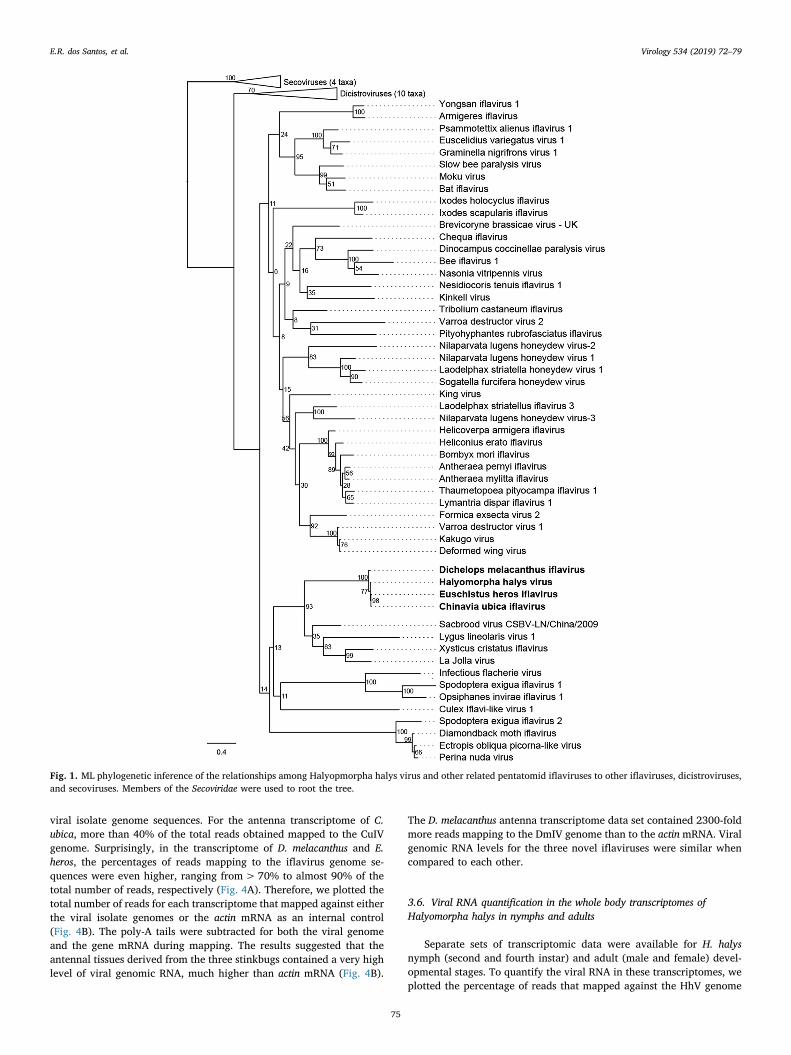
Features of the sequences of the three new isolates suggest theybelong to the virus family Iflaviridae. This family is classified underorder Picornavirales and is related to families Dicistroviridae andSecoviridae (Le Gall et al., 2008). In order to understand the relationshipof the new isolates and HhV to other iflaviruses, we constructed aphylogenetic tree based on rdrp sequences retrieved from the non-re-dundant Genbank database. The four viruses clustered together,forming a highly supported monophyletic clade within a larger cladecontaining other iflaviruses. The most genetically distant isolate, DmIV,was the most basal taxon in the clade (Fig. 1).
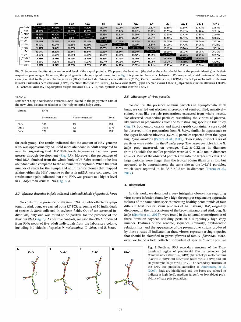
The species demarcation criterion for members of genus Iflavirus isbased on amino acid sequence identity in the capsid region of thepolyprotein, with iflaviruses exhibiting> 90% capsid amino acid se-quence identity with each other considered to belong to the samespecies (Valles et al., 2017). Amino acid sequence identities among thecapsid sequences of HhV, CuIV, DmIV, and EhIV are all> 94%, in-dicating that they are isolates of the same species of genus Iflavirus(Fig. 2). Identities with capsid sequences of other iflaviruses are<28.3%, suggesting that HhV and the stink bug viruses identified in thisstudy represent a distinct iflavirus species.
3.3. Genetic variation among stink bug iflaviruses
Upon pairwise comparison of the HhV polyprotein CDS with thoseof the other stink bug iflaviruses (Table 2), DmIV was found to containthe most single nucleotide variants (SNV), with a total of 1175 SNVs. 82of these SNVs constituted non-synonymous substitutions. In contrast,CuIV and EhIV each exhibited 199 SNVs with 20 and 19 non-synon-ymous substitutions, respectively. These sequence differences corre-sponded to polyprotein CDS nucleotide pairwise alignment identitieswas 83.7% for DmIV and 97.8% for CuIV and EhIV compared to HhV.
3.4. 5′- and 3′-Untranslated region analysis
Pairwise alignment of the 5′- and 3′-untranslated regions (UTRs) ofthe novel pentatomid iflavirus isolates and HhV revealed global se-quence identities of 95.3 and 90.9%, respectively. Yet again, DmIV wasfound to possess the most divergent sequences for these two regions. Aspreviously observed with the CDS sequence, the 5′-UTR sequences ofCuIV and EhIV were identical to each other. CuIV and EhIV differed in aU-rich region of the 3′-UTR with a pairwise identity of 97.1%. AlthoughDmIV was the most genetically different in sequence identity, the sec-ondary structures of the 5′-UTRs predicted at 25 °C (Andronescu et al.,2007) were more closely related to each other than to the HhV 5′-UTRsecondary structure (Fig. 3).
3.5. Viral RNA quantification in the antenna transcriptome
In order to quantify the viral RNA in the transcriptomes, we plottedthe percentage of reads in the transcriptome dataset that mapped to the
Table 1Genome characteristics of the pentatomid iflavirus isolates with their respectivenucleotide identity (nt ID) percentages.
Virus Host species Genome Size (nt) nt ID (%)
HhV EhIV CuIV DmIV
HhV Halyomorpha halys 9271 – 97.74 97.76 83.88EhIV Euschistus heros 9356 97.74 – 99.98 83.94CuIV Chinavia ubica 9351 97.75 99.98 – 83.96DmIV Dichelops melacanthus 9359 83.88 83.96 83.96 –
E.R. dos Santos, et al. Virology 534 (2019) 72–79
74
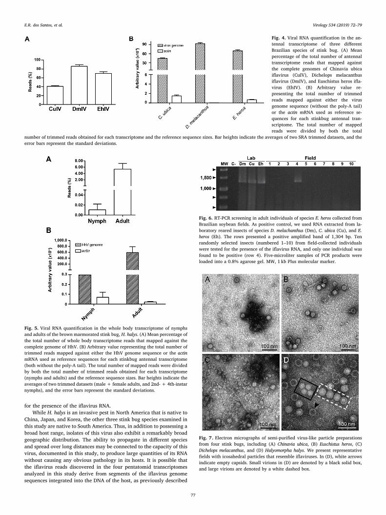
viral isolate genome sequences. For the antenna transcriptome of C.ubica, more than 40% of the total reads obtained mapped to the CuIVgenome. Surprisingly, in the transcriptome of D. melacanthus and E.heros, the percentages of reads mapping to the iflavirus genome se-quences were even higher, ranging from>70% to almost 90% of thetotal number of reads, respectively (Fig. 4A). Therefore, we plotted thetotal number of reads for each transcriptome that mapped against eitherthe viral isolate genomes or the actin mRNA as an internal control(Fig. 4B). The poly-A tails were subtracted for both the viral genomeand the gene mRNA during mapping. The results suggested that theantennal tissues derived from the three stinkbugs contained a very highlevel of viral genomic RNA, much higher than actin mRNA (Fig. 4B).
The D. melacanthus antenna transcriptome data set contained 2300-foldmore reads mapping to the DmIV genome than to the actin mRNA. Viralgenomic RNA levels for the three novel iflaviruses were similar whencompared to each other.
3.6. Viral RNA quantification in the whole body transcriptomes ofHalyomorpha halys in nymphs and adults
Separate sets of transcriptomic data were available for H. halysnymph (second and fourth instar) and adult (male and female) devel-opmental stages. To quantify the viral RNA in these transcriptomes, weplotted the percentage of reads that mapped against the HhV genome
Fig. 1. ML phylogenetic inference of the relationships among Halyopmorpha halys virus and other related pentatomid iflaviruses to other iflaviruses, dicistroviruses,and secoviruses. Members of the Secoviridae were used to root the tree.
E.R. dos Santos, et al. Virology 534 (2019) 72–79
75
for each group. The results indicated that the amount of HhV genomeRNA was approximately 510-fold more abundant in adult compared tonymphs, suggesting that HhV RNA levels increase as the insect pro-gresses through development (Fig. 5A). Moreover, the percentage ofviral RNA obtained from the whole body of H. halys seemed to be lessabundant when compared to the antenna transcriptome. When the totalnumber of reads for the nymph and adult transcriptomes that mappedagainst either the HhV genome or the actin mRNA were compared, theresults once again indicated that viral RNA was present at a higher levelin H. halys than actin mRNA (Fig. 5B).
3.7. Iflavirus detection in field-collected adult individuals of species E. heros
To confirm the presence of iflavirus RNA in field-collected asymp-tomatic stink bugs, we carried out a RT-PCR screening of 10 individualsof species E. heros collected in soybean fields. Out of ten screened in-dividuals, only one was found to be positive for the presence of theiflavirus RNA (Fig. 6). As positive controls, we used the cDNA producedfrom RNA pools of five adult individuals from the laboratory colony,including individuals of species D. melacanthus, C. ubica, and E. heros.
3.8. Microscopy of virus particles
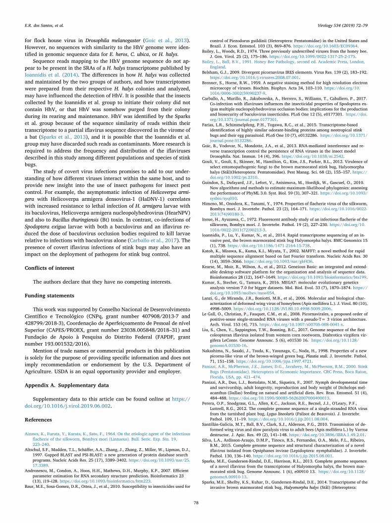
To confirm the presence of virus particles in asymptomatic stinkbugs, we carried out electron microscopy of semi-purified, negatively-stained virus-like particle preparations extracted from whole insects.We observed icosahedral particles resembling the virions of picorna-like viruses in preparations from the four stink bug species in this study(Fig. 7). Both empty capsids and intact capsids containing a core couldbe observed in the preparation from H. halys, similar in appearance tothe Lygus lineolaris iflavirus (LyLV-1) particles reported from the lygusbug, Lygus lineolaris (Perera et al., 2012). Two visibly distinct sizes ofparticles were evident in the H. halys prep. The larger particles in the H.halys prep measured, on average, 41.2 ± 0.32 nm in diameter(n= 15), while the smaller particles were 31.9 ± 0.34 nm in diameter(n= 7). Most of the observed particles fell into the larger size class. Thelarge particles were bigger than the typical 30-nm iflavirus virion, butappeared to be approximately the same size as the LyLV-1 particles,which were reported to be 38.7–40.2 nm in diameter (Perera et al.,2012).
4. Discussion
In this work, we described a very intriguing observation regardingvirus covert infection found by a high throughput sequencing approach:isolates of the same virus species infecting healthy pentatomids of fourdifferent host species. Virus genomes of an iflavirus, HhV, originallydiscovered in the transcriptome of the brown marmorated stink bug, H.halys (Sparks et al., 2013), were found in the antennal transcriptomes ofthree Brazilian soybean stinkbug pests in a surprisingly high copynumber. Features of the genome, sequence similarity, phylogeneticrelationships, and the appearance of the presumptive virions producedby these viruses all indicate that these viruses represent a single speciesthat should be classified in genus Iflavirus of family Iflaviridae. More-over, we found a field collected individual of species E. heros positive
Fig. 2. Sequence identity at the amino acid level of the capsid proteins. We present the heat map (the darker the color, the higher is the protein identity) with theirrespective percentages. Moreover, the phylogenetic relationship addressed in the Fig. 1 is presented here as a cladogram. We compared capsid proteins of iflavirusclosely related to Halyomorpha halys virus (HhV) that include Chinavia ubica iflavirus (CuIV), Culex Iflavi-like virus 1 (CIV-1), Dichelops melacanthus iflavirus(DmIV), Euschistus heros iflavirus (EhIV), Infectious flacherie virus (IfIV), La Jolla virus (LJV), Lygus lineolaris virus 1 (LlV-1), Opsiphanes invirae iflavirus 1 (OiIV-1), Sacbrood virus (SV), Spodoptera exigua iflavirus 1 (SeIV-1), and Xysticus cristatus iflavirus (XcIV).
Table 2Number of Single Nucleotide Variants (SNVs) found in the polyprotein CDS ofthe new virus isolates in relation to the Halyomorpha halys virus.
Viruses SNVs
Synonymous Non-synonymous Total
EhIV 180 19 199DmIV 1093 82 1175CuIV 179 20 199
Fig. 3. Predicted RNA secondary structure of the 5′-un-translated region of pentatomid iflavirus genomes. (A)Chinavia ubica iflavirus (CuIV); (B) Dichelops melachanthusiflavirus (DmIV); (C) Euschistus heros virus (EhIV); and (D)Halyomorpha halys virus (HhV). The secondary structure ofthe RNA was predicted according to Andronescu et al.(2007). Ends are highlighted and the bases are colored toindicate a high (red), medium (green), or low (blue) prob-ability of base pair formation.
E.R. dos Santos, et al. Virology 534 (2019) 72–79
76
for the presence of the iflavirus RNA.While H. halys is an invasive pest in North America that is native to
China, Japan, and Korea, the other three stink bug species examined inthis study are native to South America. Thus, in addition to possessing abroad host range, isolates of this virus also exhibit a remarkably broadgeographic distribution. The ability to propagate in different speciesand spread over long distances may be connected to the capacity of thisvirus, documented in this study, to produce large quantities of its RNAwithout causing any obvious pathology in its hosts. It is possible thatthe iflavirus reads discovered in the four pentatomid transcriptomesanalyzed in this study derive from segments of the iflavirus genomesequences integrated into the DNA of the host, as previously described
Fig. 4. Viral RNA quantification in the an-tennal transcriptome of three differentBrazilian species of stink bug. (A) Meanpercentage of the total number of antennaltranscriptome reads that mapped againstthe complete genomes of Chinavia ubicaiflavirus (CuIV), Dichelops melacanthusiflavirus (DmIV), and Euschistus heros ifla-virus (EhIV). (B) Arbitrary value re-presenting the total number of trimmedreads mapped against either the virusgenome sequence (without the poly-A tail)or the actin mRNA used as reference se-quences for each stinkbug antennal tran-scriptome. The total number of mappedreads were divided by both the total
number of trimmed reads obtained for each transcriptome and the reference sequence sizes. Bar heights indicate the averages of two SRA trimmed datasets, and theerror bars represent the standard deviations.
Fig. 5. Viral RNA quantification in the whole body transcriptome of nymphsand adults of the brown marmorated stink bug, H. halys. (A) Mean percentage ofthe total number of whole body transcriptome reads that mapped against thecomplete genome of HhV. (B) Arbitrary value representing the total number oftrimmed reads mapped against either the HhV genome sequence or the actinmRNA used as reference sequences for each stinkbug antennal transcriptome(both without the poly-A tail). The total number of mapped reads were dividedby both the total number of trimmed reads obtained for each transcriptome(nymphs and adults) and the reference sequence sizes. Bar heights indicate theaverages of two trimmed datasets (male + female adults, and 2nd- + 4th-instarnymphs), and the error bars represent the standard deviations.
Fig. 6. RT-PCR screening in adult individuals of species E. heros collected fromBrazilian soybean fields. As positive control, we used RNA extracted from la-boratory reared insects of species D. melachanthus (Dm), C. ubica (Cu), and E.heros (Eh). The rows presented a positive amplified band of 1,304 bp. Tenrandomly selected insects (numbered 1–10) from field-collected individualswere tested for the presence of the iflavirus RNA, and only one individual wasfound to be positive (row 4). Five-microliter samples of PCR products wereloaded into a 0.8% agarose gel. MW, 1 kb Plus molecular marker.
Fig. 7. Electron micrographs of semi-purified virus-like particle preparationsfrom four stink bugs, including (A) Chinavia ubica, (B) Euschistus heros, (C)Dichelops melacanthus, and (D) Halyomorpha halys. We present representativefields with icosahedral particles that resemble iflaviruses. In (D), white arrowsindicate empty capsids. Small virions in (D) are denoted by a black solid box,and large virions are denoted by a white dashed box.
E.R. dos Santos, et al. Virology 534 (2019) 72–79
77
for flock house virus in Drosophila melanogaster (Goic et al., 2013).However, no sequences with similarity to the HhV genome were iden-tified in genomic sequence data for E. heros, C. ubica, or H. halys.
Sequence reads mapping to the HhV genome sequence do not ap-pear to be present in the SRAs of a H. halys transcriptome published byIoannidis et al. (2014). The differences in how H. halys was collectedand maintained by the two groups of authors, and how transcriptomeswere prepared from their respective H. halys colonies and analyzed,may have influenced the detection of HhV. It is possible that the insectscollected by the Ioannidis et al. group to initiate their colony did notcontain HhV, or that HhV was somehow purged from their colonyduring its rearing and maintenance. HhV was identified by the Sparkset al. group because of the sequence similarity of reads within theirtranscriptome to a partial iflavirus sequence discovered in the virome ofa bat (Sparks et al., 2013), and it is possible that the Ioannidis et al.group may have discarded such reads as contaminants. More research isrequired to address the frequency and distribution of the iflavirusesdescribed in this study among different populations and species of stinkbugs.
The study of covert virus infections promises to add to our under-standing of how different viruses interact within the same host, and toprovide new insight into the use of insect pathogens for insect pestcontrol. For example, the asymptomatic infection of Helicoverpa armi-gera with Helicoverpa armigera densovirus-1 (HaDNV-1) correlateswith increased resistance to lethal infection of H. armigera larvae withits baculovirus, Helicoverpa armigera nucleopolyhedrovirus (HearNPV)and also to Bacillus thuringiensis (Bt) toxin. In contrast, co-infections ofSpodoptera exigua larvae with both a baculovirus and an iflavirus re-duced the dose of baculovirus occlusion bodies required to kill larvaerelative to infections with baculovirus alone (Carballo et al., 2017). Thepresence of covert iflavirus infections of stink bugs may also have animpact on the deployment of pathogens for stink bug control.
Conflicts of interest
The authors declare that they have no competing interests.
Funding statements
This work was supported by Conselho Nacional de DesenvolvimentoCientífico e Tecnológico (CNPq, grant number 407908/2013-7 and428799/2018-3), Coordenação de Aperfeiçoamento de Pessoal de nívelSuperior (CAPES/PROEX, grant number 23038.005848/2018–31) andFundação de Apoio à Pesquisa do Distrito Federal (FAPDF, grantnumber 193.001532/2016).
Mention of trade names or commercial products in this publicationis solely for the purpose of providing specific information and does notimply recommendation or endorsement by the U.S. Department ofAgriculture. USDA is an equal opportunity provider and employer.
Appendix A. Supplementary data
Supplementary data to this article can be found online at https://doi.org/10.1016/j.virol.2019.06.002.
References
Aizawa, K., Furuta, Y., Kurata, K., Sato, F., 1964. On the etiologic agent of the infectiousflacherie of the silkworm, Bombyx mori (Linnaeus). Bull. Seric. Exp. Stn. 19,223–240.
Altschul, S.F., Madden, T.L., Schäffer, A.A., Zhang, J., Zhang, Z., Miller, W., Lipman, D.J.,1997. Gapped BLAST and PSI-BLAST: a new generation of protein database searchprograms. Nucleic Acids Res. 25 (17), 3389–3402. https://doi.org/10.1093/nar/25.17.3389.
Andronescu, M., Condon, A., Hoos, H.H., Mathews, D.H., Murphy, K.P., 2007. Efficientparameter estimation for RNA secondary structure prediction. Bioinformatics 23(13), i19–i28. https://doi.org/10.1093/bioinformatics/btm223.
Baur, M.E., Sosa-Gomez, D.R., Ottea, J., et al., 2010. Susceptibility to insecticides used for
control of Piezodorus guildinii (Heteroptera: Pentatomidae) in the United States andBrazil. J. Econ. Entomol. 103 (3), 869–876. https://doi.org/10.1603/EC09364.
Bailey, L., Woods, R.D., 1974. Three previously undescribed viruses from the honey bee.J. Gen. Virol. 25 (2), 175–186. https://doi.org/10.1099/0022-1317-25-2-175.
Bailey, L., Ball, B.V., 1991. Honey Bee Pathology, second ed. Academic Press, London,England.
Belsham, G.J., 2009. Divergent picornavirus IRES elements. Virus Res. 139 (2), 183–192.https://doi.org/10.1016/j.virusres.2008.07.001.
Brenner, S., Horne, R.W., 1959. A negative staining method for high resolution electronmicroscopy of viruses. Biochim. Biophys. Acta 34, 103–110. https://doi.org/10.1016/0006-3002(59)90237-9.
Carballo, A., Murillo, R., Jakubowska, A., Herrero, S., Williams, T., Caballero, P., 2017.Co-infection with iflaviruses influences the insecticidal properties of Spodoptera ex-igua multiple nucleopolyhedrovirus occlusion bodies: implications for the productionand biosecurity of baculovirus insecticides. PLoS One 12 (5), e0177301. https://doi.org/10.1371/journal.pone.0177301.
Farias, L.R., Schimmelpfeng, P.H., Togawa, R.C., et al., 2015. Transcriptome-basedidentification of highly similar odorant-binding proteins among neotropical stinkbugs and their egg parasitoid. PLoS One 10 (7), e0132286. https://doi.org/10.1371/journal.pone.0132286.
Goic, B., Vodovar, N., Mondotte, J.A., et al., 2013. RNA-mediated interference and re-verse transcription control the persistence of RNA viruses in the insect modelDrosophila. Nat. Immun. 14 (4), 396. https://doi.org/10.1038/ni.2542.
Gouli, V., Gouli, S., Skinner, M., Hamilton, G., Kim, J.S., Parker, B.L., 2012. Virulence ofselect entomopathogenic fungi to the brown marmorated stink bug, Halyomorphahalys (Stål)(Heteroptera: Pentatomidae). Pest Manag. Sci. 68 (2), 155–157. https://doi.org/10.1002/ps.2310.
Guindon, S., Dufayard, J.F., Lefort, V., Anisimova, M., Hordijk, W., Gascuel, O., 2010.New algorithms and methods to estimate maximum-likelihood phylogenies: assessingthe performance of PhyML 3.0. Syst. Biol. 59 (3), 307–321. https://doi.org/10.1093/sysbio/syq010.
Himeno, M., Onodera, K., Tanami, Y., 1974. Properties of flacherie virus of the silkworm,Bombyx mori. J. Invertebr. Pathol. 23 (2), 164–171. https://doi.org/10.1016/0022-2011(74)90180-3.
Inoue, H., Ayuzawa, C., 1972. Fluorescent antibody study of an infectious flacherie of thesilkworm, Bombyx mori. J. Invertebr. Pathol. 19 (2), 227–230. https://doi.org/10.1016/0022-2011(72)90213-3.
Ioannidis, P., Lu, Y., Kumar, N., et al., 2014. Rapid transcriptome sequencing of an in-vasive pest, the brown marmorated stink bug Halyomorpha halys. BMC Genomics 15(1), 738. https://doi.org/10.1186/1471-2164-15-738.
Katoh, K., Misawa, K., Kuma, K.I., Miyata, T., 2002. MAFFT: a novel method for rapidmultiple sequence alignment based on fast Fourier transform. Nucleic Acids Res. 30(14), 3059–3066. https://doi.org/10.1093/nar/gkf436.
Kearse, M., Moir, R., Wilson, A., et al., 2012. Geneious Basic: an integrated and extend-able desktop software platform for the organization and analysis of sequence data.Bioinformatics 28 (12), 1647–1649. https://doi.org/10.1093/bioinformatics/bts199.
Kumar, S., Stecher, G., Tamura, K., 2016. MEGA7: molecular evolutionary geneticsanalysis version 7.0 for bigger datasets. Mol. Biol. Evol. 33 (7), 1870–1874. https://doi.org/10.1093/molbev/msw054.
Lanzi, G., de Miranda, J.R., Boniotti, M.B., et al., 2006. Molecular and biological char-acterization of deformed wing virus of honeybees (Apis mellifera L.). J. Virol. 80 (10),4998–5009. https://doi.org/10.1128/JVI.80.10.4998-5009.2006.
Le Gall, O., Christian, P., Fauquet, C.M., et al., 2008. Picornavirales, a proposed order ofpositive-sense single-stranded RNA viruses with a pseudo-T= 3 virion architecture.Arch. Virol. 153 (4), 715. https://doi.org/10.1007/s00705-008-0041-x.
Liu, S., Chen, Y., Sappington, T.W., Bonning, B.C., 2017. Genome sequence of the firstcoleopteran iflavirus isolated from western corn rootworm, Diabrotica virgifera vir-gifera LeConte. Genome Announc. 5 (6), e01530 16. https://doi.org/10.1128/genomeA.01530-16.
Nakashima, N., Sasaki, J., Tsuda, K., Yasunaga, C., Noda, H., 1998. Properties of a newpicorna-like virus of the brown-winged green bug, Plautia stali. J. Invertebr. Pathol.71, 151–158. https://doi.org/10.1006/jipa.1997.4721.
Panizzi, A.R., McPherson, J.E., James, D.G., Javahery, M., McPherson, R.M., 2000. StinkBugs (Pentatomidae). Heteroptera of Economic Importance. CRC Press, Boca Raton,Florida, USA, pp. 421–474.
Panizzi, A.R., Duo, L.J., Bortolato, N.M., Siqueira, F., 2007. Nymph developmental timeand survivorship, adult longevity, reproduction and body weight of Dichelops mel-acanthus (Dallas) feeding on natural and artificial diets. Rev. Bras. Entomol. 51 (4),484–488. https://doi.org/10.1590/S0085-56262007000400013.
Perera, O.P., Snodgrass, G.L., Allen, K.C., Jackson, R.E., Becnel, J.J., O'Leary, P.F.,Luttrell, R.G., 2012. The complete genome sequence of a single-stranded RNA virusfrom the tarnished plant bug, Lygus lineolaris (Palisot de Beauvois). J. Invertebr.Pathol. 109, 11–19. https://doi.org/10.1016/j.jip.2011.08.004.
Santillán-Galicia, M.T., Ball, B.V., Clark, S.J., Alderson, P.G., 2010. Transmission of de-formed wing virus and slow paralysis virus to adult bees (Apis mellifera L.) by Varroadestructor. J. Apic. Res. 49 (2), 141–148. https://doi.org/10.3896/IBRA.1.49.2.01.
Silva, L.A., Ardisson-Araujo, D.M.P., Tinoco, R.S., Fernandes, O.A., Melo, F.L., Ribeiro,B.M., 2015. Complete genome sequence and structural characterization of a noveliflavirus isolated from Opsiphanes invirae (Lepidoptera: nymphalidae). J. Invertebr.Pathol. 130, 136–140. https://doi.org/10.1016/j.jip.2015.08.001.
Sparks, M.E., Gundersen-Rindal, D.E., Harrison, R.L., 2013. Complete genome sequenceof a novel iflavirus from the transcriptome of Halyomorpha halys, the brown mar-morated stink bug. Genome Announc. 1 (6), e00910 13. https://doi.org/10.1128/genomeA.00910-13.
Sparks, M.E., Shelby, K.S., Kuhar, D., Gundersen-Rindal, D.E., 2014. Transcriptome of theinvasive brown marmorated stink bug, Halyomorpha halys (Stål) (Heteroptera:
E.R. dos Santos, et al. Virology 534 (2019) 72–79
78

Pentatomidae). PLoS One 9 (11), e111646. https://doi.org/10.1371/journal.pone.0111646.
van Oers, M.M., 2010. Genomics and biology of iflaviruses. In: Insect Virology. CaisterAcademic Press, Norfolk, pp. 231–250.
Valles, S.M., Chen, Y., Firth, A.E., et al., 2017. ICTV virus taxonomy profile: Iflaviridae. J.Gen. Virol. 98 (4), 527–528. https://doi.org/10.1099/jgv.0.000757.
Velikova, V., Salerno, G., Frati, F., Peri, E., Conti, E., Colazza, S., Loreto, F., 2010.Influence of feeding and oviposition by phytophagous pentatomids on photosynthesisof herbaceous plants. J. Chem. Ecol. 36 (6), 629–641. https://doi.org/10.1007/s10886-010-9801-7.
Weber, D.C., Leskey, T.C., Walsh, G.C., Khrimian, A., 2014. Synergy of aggregationpheromone with methyl (E, E, Z)-2, 4, 6-decatrienoate in attraction of Halyomorpha
halys (Hemiptera: Pentatomidae). J. Econ. Entomol. 107 (3), 1061–1068. https://doi.org/10.1603/EC13502.
Williamson, C., von Wechmar, M.B., 1992. Two novel viruses associated with severedisease symptoms of the green stinkbug Nezara viridula. J. Gen. Virol. 73, 2467–2471.https://doi.org/10.1099/0022-1317-73-9-2467.
Williamson, C., von Wechmar, M.B., 1995. The effect of two viruses on the metamor-phosis, fecundity, and longevity of the green stinkbug, Nezara viridula. J. Invertebr.Pathol. 65, 174–178. https://doi.org/10.1006/jipa.1995.1025.
Yue, C., Schröder, M., Gisder, S., Genersch, E., 2007. Vertical-transmission routes fordeformed wing virus of honeybees (Apis mellifera). J. Gen. Virol. 88 (8), 2329–2336.https://doi.org/10.1099/vir.0.83101-0.
E.R. dos Santos, et al. Virology 534 (2019) 72–79
79