agr207 course title: anatomy and physiology of farm animals ...
164
NATIONAL OPEN UNIVERSITY OF NIGERIA FACULTY OF AGRICUTURAL SCIENCES COURSE CODE: AGR207 COURSE TITLE: ANATOMY AND PHYSIOLOGY OF FARM ANIMALS COURSE MATERIAL WRITER/DEVELOPER: DR. C. E. ISIDAHOMEN Department of Animal Science Faculty of Agriculture Ambrose Alli University Ekpoma
-
Upload
khangminh22 -
Category
Documents
-
view
6 -
download
0
Transcript of agr207 course title: anatomy and physiology of farm animals ...

NATIONAL OPEN UNIVERSITY OF NIGERIA
FACULTY OF AGRICUTURAL SCIENCES
COURSE CODE: AGR207
COURSE TITLE: ANATOMY AND PHYSIOLOGY OF FARM ANIMALS
COURSE MATERIAL WRITER/DEVELOPER: DR. C. E. ISIDAHOMEN
Department of Animal Science
Faculty of Agriculture
Ambrose Alli University Ekpoma

Introduction
The course AGR207 (anatomy and physiology of farm animals), is a two (2) credit unit course
designed for 200 level undergraduate students pursuing a degree in Agricultural Science. The
course is expected to provide a good knowledge base for the future manpower for animal
towards a sustainable production of livestock. It explains the rudimentary of Anatomy and
physiological traits, Parts of the beef and dairy cattle, sheep, goats, pigs, rabbits and poultry,
fundamentals of cell biology, anatomy and physiology of the cell, cell types. anatomy and
physiology of animal tissues, nervous system, skeletal system, muscle, bone, circulatory system,
reproductive, digestive, special senses and other systems of farm animals. Physiological
functions of animals – homeostatic, nutrition and digestion, respiration. Temperature regulation,
excretion and reproduction, endocrinology, the blood and circulation, lactation, milk let down
and egg production, water balance. The course will provide a basic foundation for students
intending to take up Animal science as a Career in the future. The course is divided into four (4)
modules with unit one to eleven units. Each unit begins with a clear introduction and statement
of objectives followed by the main content. The conclusion, summary and references (for further
reading) were also provided for each unit. Tutor marked assignments were provided for each unit
to enable you attempt some questions on the topics treated for onward submission to your tutor.
The Course Guide provides you with access to brief information and overview of the course
content, course duration, what you are expected to know in each unit, what course material you
need to use and how you can systematically go through the course materials.. Thus, we intend to
achieve the above through the following broad aim and other specific objectives.
Course Aim
Anatomy and Physiology of Farm Animals is designed to provide you with the knowledge of
different types of farm animals, various physiological functions and structures. It also enlighten
the students on the different types of animals and their adaptive features.
Course Objectives
On successful completion of the course, you should be able to:
Explain the different parts of livestock andfundamentals of cell biology
Explain the different anatomy andphysiologyof the cell, cell types

Explain anatomy and physiology of animal tissues, nervous system, skeletal system, muscle,
bone, circulatory system, reproductive, digestive, special senses and other systems of farm
animals.
Mention the different Physiological functions of farm animal.
Working through this Course
You are expected to study and understand the content of this course. Each unit must be properly
studied for good comprehension of the contents. By the end of each unit, you are expected to
answer the questions therein and submit as appropriate when directed by the administration of
the University. These questions are like continuous assessment. You are expected to sit for an
examination on completion of the course. The course duration shall take about 17 weeks of
learning. Therefore, you must be able to organize your time to achieve this successfully. Tutorial
session will be available and it is advisable for you to attend in order to be able to assess and
compared yourself with your peers and clarify any area that you do not properly understand.
The Course Material
Major components of the course material are:
The Course Guide
Study Units
The References/Further Reading, that will be provided at the end of each unit are
necessary supplements to the course material.
The term anatomy has come to refer to the science that deals with the form and structure of all
organisms. Literally, the word means to cut apart; it was used by early anatomists when speaking
of complete dissection of a cadaver. In contrast to anatomy, which deals primarily with structure,
physiology is the study of the integrated functions of the body and the functions of all its parts
(systems, organs, tissues, cells, and cell components), including biophysical and biochemical
processes. When anatomy and physiology courses are taught separately, the approach to the
laboratory portion of each course is considerably different. Study in a typical gross anatomy

laboratory is based primarily on dissection of animal cadavers. These usually have been
preserved by embalming, and one or more parts of the vascular system have been injected with a
colored material to facilitate identification of the vessels. Careful dissection coupled with close
observation gives the student a concept of the shape, texture, location, and relations of structures
visible to the unaided eye that can be gained in no other way. Similarly, the use of the micro-
scope with properly prepared tissue sections on slides is essential for understanding structures
that are so small they cannot be seen without optical or electron microscopic assistance. In the
physiology laboratory, the student studies the response of whole animals, isolated Descriptive
Terms Useful in the Study of Anatomy Microscopic Anatomy: Animal Cells and Tissues
Epithelial Tissues Connective Tissues Muscle Tissue Nervous Tissue the General Plan of the
Animal Body
MODULE 1: Different parts of livestock
Unit 1: Different parts of livestock
1.1 Parts of cattle(beef and dairy)
1.2 Parts of sheep
1.3 Parts of goats
1.4Parts of rabbits
1.5 Parts of poultry
MODULE 2: Fundamentals of cell biology,Anatomy and physiology of cellandcell types.
Unit 1: Types cell
Unit 2: Cell theory
Unit 3: Function of Cell
Unit 4: Characteristic of a cell
Unit 5: Cell structure
Unit 6: Cell membrane
Unit 7: Cytoplasm
Unit 8: Cell division
Unit 9:Cell as a factor

MODULE 3: Anatomy and physiology of farm animals
Unit 1: Animal tissue
Unit 2: Nervous system
Unit 3: Skeletal systems
Unit 4: Muscles
Unit 5: Circulatory systems
Unit 6: Reproductive systems
Unit 7: Digestive systems
MODULE 4: Physiology functions of farm animals
Unit 1: Homeostasis
Unit 2: Nutrition and digestion
Unit 3: Respiratoryand thermoregulation
Unit 4: Physiology and temperature regulation
Unit 5: Excretion and reproduction
Unit 6: Endocrinology
Unit 7: The blood and circulation
Unit 8: Lactation
Unit 9: Milk let down and egg production
Unit 10: Physiology of egg production
Unit 11: Water balance
Assessment
The assessment of the course shall be in two parts. The Tutor-Marked Assignments (TMAs) will
take a part while the end of course written examination takes the second part. As a result, you
must do the TMAs applying the knowledge and techniques learnt in each unit. The assignment
must be submitted to your tutor/facilitator for assessment in accordance with the set time in the

presentation schedule. The TMAs assessment will constitute 30% while the written examination
account for 70% of the total mark for the course.
Tutor-Marked Assignment
The TMA is a continuous assessment component of your course. It carries 30% of the total score.
You will be given four TMAs to answer. Three of these must be answered before you are
allowed to sit for the end of the course examination. The TMA would be given to you by your
facilitator and you should submit after you have done the assignment.
End of Course Examination
The examination concludes the assessment for this course. It constitutes 70% of the mark for the
whole course. You will be informed of the time for the examination.
Summary
Feed formulation is a course that gives you a good understanding of the different kind of
feedstuffs for livestock, raw materials for processing, different methods for compounding feeds.
It teaches the skills of handling, processing storing feeds and also feeds availability for livestock.
Other information inclusive are ant nutritional factors in livestock feeds, law governing
establishing of feed mill
Best wishes.
The study of the bones that make up the skeleton, or framework of the body, is osteology. The
skeleton gives a basis for the external structure and appearance of most vertebrate animals as we
know them. All mammals share a basic body plan with striking similarities in skeletal structure.
Differences reflect adaptations to specific lifestyles .The skeleton of a living animal is made up
of bones that are themselves living structures. They have blood vessels, lymphatic vessels, and
nerves; they are subject to disease; they can undergo repair; and they adjust to changes in stress.
The functions of bones include providing protection, giving rigidity and form to the body, acting
as levers, storing minerals, and forming the cellular elements of blood.
Functions of Bones

Protection of vital organs is one of the important functions of bones. The central nervous system
is protected by the skull and vertebral column; the heart and lungs, by the rib cage; and internal
parts of the urogenital system, by the pelvis. In the vertebrates, locomotion, defense, offense,
grasping, and other activities of this type depend largely upon the action of muscles that attach to
levers. Almost without exception, these levers are made of bone and are integral parts of the
skeleton.
MODULE 1: DIFFERENT PARTS OF LIVESTOCK
UNIT 1: PARTS OF LIVESTOCK
CONTENTS
1.0 Introduction
2.0 Objectives
3.0 Main contents
3.1 Parts of the beef and dairy cattle
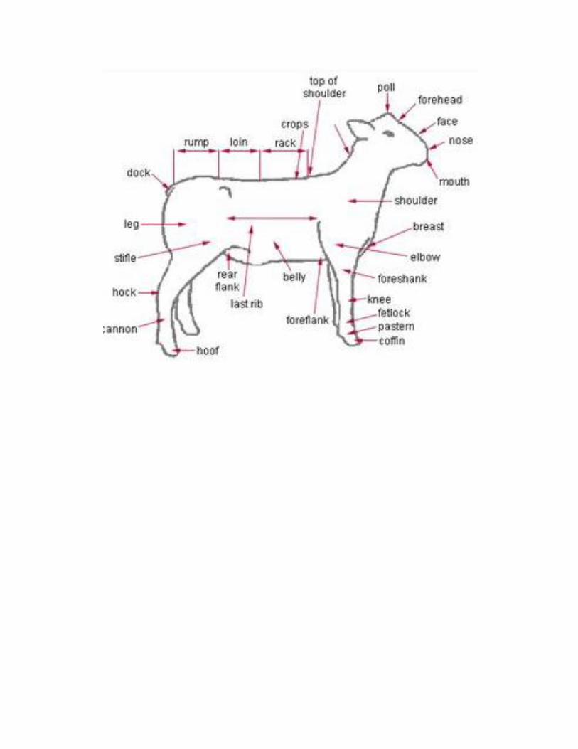
3.2 Parts of the sheep
3.3 Parts of the goats
3.4 Parts of the pigs
3.5 Parts of the rabbits
3.6 Parts of the poultry
4.0 Conclusion
5.0 Summary
6.0 Tutor-Marked Assignment
7.0 References/Further Reading
2.0 OBJECTIVES
By the end of this unit, you should be able to:
Know the different parts of livestock
Identify the function of the skeletal system
Know the different types of bones and functions
3.0 MAIN CONTENT
3.1 Parts of the beef and dairy cattle
3.1.1 Parts of the sheep
3.1.2 Parts of the goats

3.1.1.3 Parts of the pigs
3.1.4 Parts of the rabbits
3.1.5 Parts of the poultry
INTRODUCTION
The skeleton act as the protection of vital organs is one of the important functions of bones. The
central nervous system is protected by the skull and vertebral column; the heart and lungs, by the
rib cage; and internal parts of the urogenital system, by the pelvis. In the vertebrates, locomotion,
defense, offense, grasping, and other activities of this type depend largely upon the action of
muscles that attach to levers. Almost without exception, these levers are made of bone and are
integral parts of the skeleton.
Unit 1:Parts of the beef and dairy cattle, sheep, goats, pigs, rabbits and poultry
Parts of the Beef and Dairy Cattle,
3.1 Bovine Skeletal System


3.1.1Parts of Sheep

3.1.3: Parts of goats

3.1.4: Parts of Pigs

3.1.5: Parts of Rabbits

3.1.6: Parts of Poultry
3.1: Skeleton
The study of the bones that make up the skeleton, or framework of the body, is osteology. The
skeleton gives a basis for the external structure and appearance of most vertebrate animals as we
know them. All mammals share a basic body plan with striking similarities in skeletal structure.
Differences reflect adaptations to specific lifestyles .The skeleton of a living animal is made up
of bones that are themselves living structures. They have blood vessels, lymphatic vessels, and
nerves; they are subject to disease; they can undergo repair; and they adjust to changes in stress.
The functions of bones include providing protection, giving rigidity and form to the body, acting
as levers, storing minerals, and forming the cellular elements of blood.
3.2: Functions of Bones
Protection of vital organs is one of the important functions of bones. The central nervous system
is protected by the skull and vertebral column; the heart and lungs, by the rib cage; and internal
parts of the urogenital system, by the pelvis. In the vertebrates, locomotion, defense, offense,
grasping, and other activities of this type depend largely upon the action of muscles that attach to
levers. Almost without exception, these levers are made of bone and are integral parts of the
skeleton.
4.0: Conclusion
Decades of documented evidence demonstrates that Protection of vital organs is one of the
important functions of bones.
5.0: Summary
The skeleton gives a basis for the external structure and appearance of most vertebrate animals as
we know them. All mammals share a basic body plan with striking similarities in skeletal
structure. Differences reflect adaptations to specific lifestyles .The skeleton of a living animal is
made up of bones that are themselves living structures.
6.0: Tutor marked assignment
Define skeletal systems and give example of how it is being used
What are the functions of bones
1.7 References/ Further Reading

Babayemi,Olaniyi J, Abu,Okhiomah, A and Opakunbi, Ayotunde(2014). INTEGRATED ANIMAL
HUSBANDRY for schools and colleges.ISBN978-978-52033-3-3-2
MODULE 2: FUNDAMENTALS OF CELL BIOLOGY.
CONTENTS
1.0: Introduction
2.0: Objectives
3.0: Main contents
3.1: Types cell
3.1.2: Cell theory
3.1.3: Function of Cell
3.1.4 Characteristic of a cell
3.1.5:Cell structure
3.1.6:Cell membrane
3.1.7: Cytoplasm
3.1.8: Cell division
3.1.9:Cell as a factor
4.0 Conclusion
5.0 Summary
6.0 Tutor-Marked Assignment
7.0 References/Further Reading
2.0 OBJECTIVES
By the end of this unit, you should be able to:
Know the different parts of livestock
Identify the function of the skeletal system
Know the different types of bones and functions

3.0 MAIN CONTENT
3.1: Types of Cells
Cells are similar to small factories with different labourers and departments that work all the time
to make life possible. Various types of cells perform different functions.
There are two major kinds of living organisms based on their cellular structure namely:
Prokaryotes
Eukaryotes
Prokaryotes
1. Prokaryotes are made up of cells with no nucleus.
2. They all are single-celled microorganisms including archaea, bacteria and photosynthetic
blue-green algae known as cyan bacteria.
3. The cell size ranges from 0.1 to 0.5 µm in diameter.
4. The hereditary material DNA is found in the nucleoid present in the central part of the
cell.
5. They reproduce by binary fission.
Eukaryotes
1. Eukaryotes are made up of cells consisting of a true nucleus.
2. This large category involves all plants, fungi (such as moulds, yeast, and mushrooms),
protozoa (Plasmodium falciparum and parasite that cause malaria) and animals.
3. The plasma membrane is responsible for monitoring the transport of nutrients and
electrolytes in and out of the cell and also responsible for cell to cell communication.
4. Cellular life is entirely dependent on the various chemical process for survival. These
chemical reactions mainly occur in a watery solution within the cell known as cytoplasm.
5. They reproduce sexually as well as asexually.

6. There are some contrasting features between plant and animal cell. For eg., the plant cell
contains chloroplast, central vacuoles, and other plastids, whereas the animal cell does
not.
3.1.1: Cell Theory
Cell Theory was proposed by the German scientists, Theodor Schwann, Matthias Schleiden, and
Rudolf Virchow. The cell theory states that:
1. All living species on Earth are composed of cells.
2. A cell is the basic unit of life.
3. All cells arise from pre-existing cells.
A modern version of the cell theory was eventually formulated and it contains the following
postulates:
1. Energy flows within the cells.
2. Hereditary information is passed on from one cell to the other.
3. The chemical composition of all the cells is the same.
3.1.2: Functions of a Cell
A cell performs six major functions essential for the growth and development of an organism.
The functions of a cell include:
Provides Support and Structure
All the organisms are made up of cells. They form the structural basis of all the organisms. The
cell wall and the cell membrane are the main components that function to provide support and
structure to the organism. For eg., the skin is made up of a large number of cells. Xylem present
in the vascular plants is made of cells that provide structural support to the plants.
Facilitate Growth Mitosis

In the process of mitosis, the parent cell divides into the daughter cells. Thus, the cells multiply
and facilitate the growth in an organism.
Allows Transport of Substances
Various nutrients are imported by the cells to carry out various chemical processes going on
inside the cells. The waste produced by the chemical processes is eliminated from the cells by
active and passive transport.
Small molecules such as oxygen, carbon dioxide, and ethanol diffuse across the cell membrane
along the concentration gradient. This is known as passive transport.
The larger molecules diffuse across the cell membrane through active transport where the cells
require a lot of energy to transport the substances.
Energy Production
Cells require energy to carry out various chemical processes. This energy is produced by the cells
through a process called photosynthesis in plants and respiration in animals.
Aids in Reproduction
A cell aids in reproduction through the processes called mitosis and meiosis. Mitosis is termed as
the asexual reproduction where the parent cell divides to form daughter cells. Meiosis causes the
daughter cells to be genetically different from the parent cells.
Thus, we see why cells are known as the structural and functional unit of life. This is because
they are responsible for providing structure to the organisms and performs several functions
necessary for carrying out life‘s processes.
Important Questions about Cells
1.Definecell
A cell is defined as the basic, structural and functional unit of all life.

2. State the characteristics of cells
Cells provide the necessary structural support for an organism.
The genetic information necessary for reproduction is present within the nucleus.
Structurally, the cell has cell organelles which are suspended in the cytoplasm.
Mitochondria is the organelle responsible for fulfilling the cell‘s energy requirements
Lysosomes digest metabolic wastes and foreign particles in the cell.
Endoplasmic reticulum synthesizes selective molecules and processes them, eventually
directing them to their appropriate locations.
3. State the cellular components.
Cell membrane
Cell wall
Cell organelles
o Nucleolus
o Nuclear membrane
o Endoplasmic reticulum
o Golgi Bodies
o Ribosome
o Mitochondria
o Lysosomes
o Chloroplast
o Vacuoles
4. State the types of cells
Cells are primarily classified into two types, namely
Prokaryotic cells
Eukaryotic cells

5. ElaborateonCellTheory
Cell Theory was proposed by Matthias Schleiden, Theodor Schwann, and Rudolf Virchow, who
were German scientists. The cell theory states that:
All living species on Earth are composed of cells.
A cell is the basic unit of life.
All cells arise from pre-existing cells.
Cell Theory: All known living things are made up of cells. All cells come from preexisting cells
by division. The cell is structural and functional unit of all living things.
Cell Structural Overview: The major parts of a cell are the nucleus, cytoplasm, and cell
membrane.
Nucleus:
The nucleus contains a nucleolus and is separated from the cytoplasm by the nuclear
envelope.
The nucleus contains the cell‘s DNA, a type of nucleic acid.
The nucleolus is like a ―tiny nucleus‖ inside the actual nucleus. It contains RNA, a type
of nucleic acid.
The nucleus communicates through holes in the envelope called nuclear pores.
The nucleus decides what the cell needs and uses DNA to print out instructions for the
rest of the cell to produce that need.
Chromosomes:
Hold the cell‘s DNA in the nucleus.
The nucleus contains genetic information in the form of DNA (the universal genetic
code).
The DNA does not hang around loosely in the nucleus. The DNA is packaged with
proteins and wound up.
Recall that the role of nucleic acids is to carry genetic information, which is inherited by

an organism‘s offspring.
These wound up DNAprotein structures are called chromosomes.
Cytoplasmic Organelles: Are compartmentalized structures that perform a specialized function
within a cell.
Golgi apparatus: ships packages around the cell.
The golgi is made up of flattened, folded sacs.
Packages (e.g. containing proteins) are carried to the golgi in vesicles.
The golgi receives an incoming vesicle, tags the package, and sends the vesicle to its final
destination.
Lysosome: destroy waste to clean up the cell.
Lysosomes contain an environment made to destroy waste.
Vesicles carry the waste (bacteria, old organelles, etc.) into the lysosome.
Once inside, the waste is destroyed and its parts recycled.
Smooth endoplasmic reticulum: The two types of ER make different building blocks for the
cell.
Smooth ER is NOT attached to the nucleus and DOES NOT have attached ribosomes
(thus smooth).
Smooth ER synthesizes carbohydrates (sugars) and lipids (fats).
Mitochondria: produce energy to power the cell.
The mitochondria convert carbohydrates (sugar) taken from food into ATP.
The mitochondria are unique in that it has two protective shells.
Ribosomes: make proteins for the cell.

The ribosome reads the DNA strand instructions to make proteins for the cell to use in its
normal activities.
The units clasp around a strand of nucleic acid instructions from the nucleus.
Each ribosome is made of two protein subunits.
Rough endoplasmic reticulum: The two types of ER make different building blocks for the cell.
Rough ER is found attached to the outside of the nucleus. It appears rough because of the
ribosomes on its surface.
Rough ER helps the attached ribosomes in finishing protein synthesis.
3.1.6: Cell membrane: A selectively permeable structure that envelops the cell and protects the
cell‘s internal environment.
Plasma Membrane, the cell‘s membrane is made of phospholipids, which have
carbohydrate heads and lipid tails.
Embedded proteins are anchored to the cell membrane.
Exterior of the plasma membrane touches water; polar heads touch water on the inside of
the cell and water on the outside of the cell.
Interior Blocks Passage However, water and other molecules cannot pass through to
either side because of the nonpolar tails.
Provides a stabilized environment, which protects and maintains the cell‘s internal
environment, separate from the environment outside.
Proteins embedded into the membrane send and receive signals to communicate with
other cells.
Transport across the cell membrane: The cell exchanges materials through the cell membrane
using passive and active transport.
Three types of passive transport are osmosis, diffusion, and facilitated diffusion. Osmosis is the
natural movement of water from a high concentration of water to a lower concentration of water.
Diffusion is the natural movement of molecules from a higher concentration to a lower

concentration. Facilitated Diffusion is the natural movement of molecules from a higher
concentration to a lower concentration with the help of a transporter protein embedded on the cell
membrane.
Active transport requires energy to occur. Active transport is ―forced‖ movement of molecules
from a lower concentration to a higher concentration. The most common type of active transport
is a pump. Pumps are proteins embedded in the cell membrane, which use ATP energy to work.
Different Cell Types: Prokaryote and Eukaryote.
Prokaryotic: Bacteria and other microscopic organisms are made up of prokaryotic cells.
Prokaryotic cells do not have any complex organelles (not even a nucleus). However,
prokaryotes do have ribosomes.
Eukaryotic: Two types of eukaryotic cells are plant and animal cells.
3.1.5: The Cell structure
Diagram 3.1: A variety of animal cells
The cell is the basic building block of living organisms. Bacteria and the parasite that causes
malaria consist of single cells, while plants and animals are made up of trillions of cells. Most
cells are spherical or cube shaped but some are a range of different shapes (see diagram 3.1).
Most cells are so small that a microscope is needed to see them, although a few cells, e.g. the
ostrich‘s egg, are so large that they could make a meal for several people.
A normal cell is about 0.02 of a millimetre (0.02mm) in diameter. (Small distances like this are
normally expressed in micrometres or microns (μm). Note there are 1000 μms in every mm).
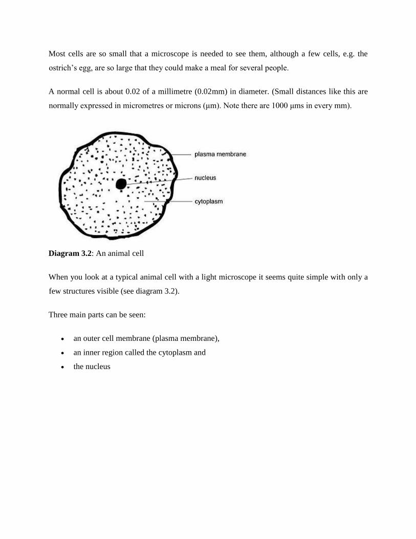
Diagram 3.2: An animal cell
When you look at a typical animal cell with a light microscope it seems quite simple with only a
few structures visible (see diagram 3.2).
Three main parts can be seen:
an outer cell membrane (plasma membrane),
an inner region called the cytoplasm and
the nucleus

Diagram 3.3: An animal cell as seen with an electron microscope
However, when you use an electron microscope to increase the magnification many thousands of
times you see that these seemingly simple structures are incredibly complex, each with its own
specialized function. For example the plasma membrane is seen to be a double layer and the
cytoplasm contains many special structures called organelles (meaning little organs) which are
described below. A drawing of the cell as seen with an electron microscope is shown in diagram
3.3.
The Plasma Membrane
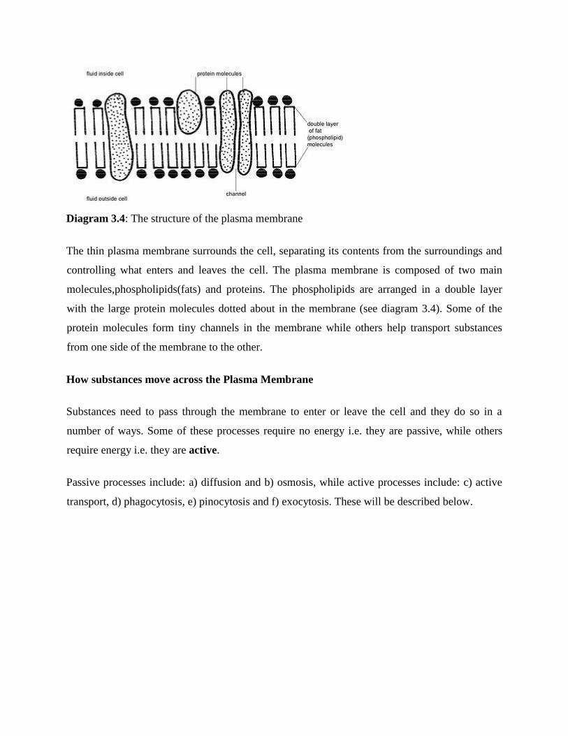
Diagram 3.4: The structure of the plasma membrane
The thin plasma membrane surrounds the cell, separating its contents from the surroundings and
controlling what enters and leaves the cell. The plasma membrane is composed of two main
molecules,phospholipids(fats) and proteins. The phospholipids are arranged in a double layer
with the large protein molecules dotted about in the membrane (see diagram 3.4). Some of the
protein molecules form tiny channels in the membrane while others help transport substances
from one side of the membrane to the other.
How substances move across the Plasma Membrane
Substances need to pass through the membrane to enter or leave the cell and they do so in a
number of ways. Some of these processes require no energy i.e. they are passive, while others
require energy i.e. they are active.
Passive processes include: a) diffusion and b) osmosis, while active processes include: c) active
transport, d) phagocytosis, e) pinocytosis and f) exocytosis. These will be described below.

Diagram 3.5: Diffusion in a liquid
a) Diffusion
Although you may not know it, you are already familiar with the process of diffusion. It is
diffusion that causes a smell (expensive perfume or smelly socks) in one part of the room to
gradually move through the room so it can be smelt on the other side. Diffusion occurs in the air
and in liquids.
Diagram 3.5 shows what happens when a few crystals of a dark purple dye called potassium
permanganate are dropped into a beaker of water. The dye molecules diffuse into the water
moving from high to low concentrations so they become evenly distributed throughout the
beaker.
In the body, diffusion causes molecules that are in a high concentration on one side of the cell
membrane to move across the membrane until they are present in equal concentrations on both
sides. It takes place because all molecules have an in-built vibration that causes them to move
and collide until they are evenly distributed. It is an absolutely natural process that requires no
added energy.
Small molecules like oxygen, carbon dioxide, water and ammonia as well as fats, diffuse directly
through the double fat layer of the membrane. The small molecules named above as well as a
variety of charged particles (ions) also diffuse through the protein-lined channels. Larger

molecules like glucose attach to a carrier molecule that aids their diffusion through the
membrane. This is called facilitated diffusion.
In the animal‘s body diffusion is important for moving oxygen and carbon dioxide between the
lungs and the blood, for moving digested food molecules from the gut into the blood and for the
removal of waste products from the cell.
Diagram 3.6: Osmosis
b) Osmosis
Although the word may be unfamiliar, you are almost certainly acquainted with the effects of
osmosis. It is osmosis that plumps out dried fruit when you soak it before making a fruit cake or
makes that wizened old carrot look almost like new when you soak it in water. Osmosis is in fact
the diffusion of water across a membrane that allows water across but not larger molecules. This
kind of membrane is called a semi-permeable membrane.
Take a look at side A of diagram 3.6. It shows a container divided into two parts by an artificial
semi-permeable membrane. Water is poured into one part while a solution containing salt is
poured into the other part. Water can cross the membrane but the salt cannot. The water crosses
the semi-permeable membrane by diffusion until there is an equal amount of water on both sides
of the membrane. The effect of this would be to make the salt solution more diluted and cause
the level of the liquid in the right-hand side of the container to rise so it looked like side B of

diagram 3.6. This movement of water across the semi-permeable membrane is called osmosis. It
is a completely natural process that requires no outside energy.
Although it would be difficult to do in practice, imagine that you could now take a plunger and
push down on the fluid in the right-hand side of container B so that it flowed back across the
semi-permeable membrane until the level of fluid on both sides was equal again. If you could
measure the pressure required to do this, this would be equal to the osmotic pressure of the salt
solution. (This is a rather advanced concept at this stage but you will meet this term again when
you study fluid balance later in the course).
Diagram 3.7: Osmosis in red cells placed in a hypotonic solution
The plasma membrane of cells acts as a semi-permeable membrane. If red blood cells, for
example, are placed in water, the water crosses the membrane to make the amount of water on
both sides of it equal (see diagram 3.7). This means that the water moves into the cell causing it
to swell. This can occur to such an extent that the cell actually bursts to release its contents. This
bursting of red blood cells is called haemolysis. In a situation such as this when the solution on
one side of a semi-permeable membrane has a lower concentration than that on the other side, the
first solution is said to be hypotonic to the second.

Diagram 3.8: Osmosis in red cells placed in a hypertonic solution
Now think what would happen if red blood cells were placed in a salt solution that has a higher
salt concentration than the solution within the cells (see diagram 3.8). Such a bathing solution is
called a hypertonic solution. In this situation the ―concentration‖ of water within the cells would
be higher than that outside the cells. Osmosis (diffusion of water) would then occur from the
inside of the cells to the outside solution, causing the cells to shrink.
Diagram 3.9: Red cells placed in an isotonic solution

A solution that contains 0.9% salt has the same concentration as body fluids and the solution
within red cells. Cells placed in such a solution would neither swell nor shrink (see diagram 3.9).
This solution is called an isotonic solution. This strength of salt solution is often called normal
saline and is used when replacing an animal‘s body fluids or when cells like red blood cells have
to be suspended in fluid.
Remember - osmosis is a special kind of diffusion. It is the diffusion of water molecules across a
semi-permeable membrane. It is a completely passive process and requires no energy.
Sometimes it is difficult to remember which way the water molecules move. Although it is not
strictly true in a biological sense, many students use the phrase “SALT SUCKS” to help them
remember which way water moves across the membrane when there are two solutions of
different salt concentrations on either side.
As we have seen water moves in and out of the cell by osmosis. All water movement from the
intestine into the blood system and between the blood capillaries and the fluid around the cells
(tissue or extra cellular fluid) takes place by osmosis. Osmosis is also important in the production
of concentrated urine by the kidney.
c) Active transport
When a substance is transported from a low concentration to a high concentration i.e. uphill
against the concentration gradient, energy has to be used. This is called active transport.
Active transport is important in maintaining different concentrations of the ions sodium and
potassium on either side of the nerve cell membrane. It is also important for removing valuable
molecules such as glucose, amino acids and sodium ions from the urine.

Diagram 3.10: Phagocytosis
d) Phagocytosis
Phagocytosis is sometimes called ―cell eating‖. It is a process that requires energy and is used by
cells to move solid particles like bacteria across the plasma membrane. Finger-like projections
from the plasma membrane surround the bacteria and engulf them as shown in diagram 3.10.
Once within the cell, enzymes produced by the lysosomes of the cell (described later) destroy the
bacteria.
The destruction of bacteria and other foreign substance by white blood cells by the process of
phagocytosis is a vital part of the defense mechanisms of the body.
e) Pinocytosis
Pinocytosis or ―cell drinking‖ is a very similar process to phagocytosis but is used by cells to
move fluids across the plasma membrane. Most cells carry out pinocytosis (note the pinocytotic
vesicle in diagram 3.3).
f) Exocytosis

Exocytosis is the process by means of which substances formed in the cell are moved through the
plasma membrane into the fluid outside the cell (or extra-cellular fluid). It occurs in all cells but
is most important in secretory cells (e.g. cells that produce digestive enzymes) and nerve cells.
3.1.7: The Cytoplasm
Within the plasma membrane is the cytoplasm. It consists of a clear jelly-like fluid called the a)
cytosol or intracellular fluid in which b) cell inclusions, c) organelles and d) microfilaments
and microtubules are found.
a) Cytosol
The cytosol consists mainly of water in which various molecules are dissolved or suspended.
These molecules include proteins, fats and carbohydrates as well as sodium, potassium, calcium
and chloride ions. Many of the reactions that take place in the cell occur in the cytosol.
b) Cell inclusions
These are large particles of fat, glycogen and melanin that have been produced by the cell. They
are often large enough to be seen with the light microscope. For example the cells of adipose
tissue (as in the insulating fat layer under the skin) contain fat that takes up most of the cell.
c) Organelles
Organelles are the ―little organs‖ of the cell - like the heart, kidney and liver are the organs of
the body. They are structures with characteristic appearances and specific ―jobs‖ in the cell. Most
can not be seen with the light microscope and so it was only when the electron microscope was
developed that they were discovered. The main organelles in the cell are the ribosomes,
endoplasmic reticulum, mitochondrion, Golgi complex and lysosomes. A cell containing
these organelles as seen with the electron microscope is shown in diagram 3.3.
Ribosomes

Diagram 3.11: Rough endoplasmic reticulum
Ribosomes are tiny spherical organelles that make proteins by joining amino acids together.
Many ribosomes are found free in the cytosol, while others are attached to the rough endoplasmic
reticulum.
Endoplasmic reticulum
The endoplasmic reticulum (ER) is a network of membranes that form channels throughout the
cytoplasm from the nucleus to the plasma membrane. Various molecules are made in the ER and
transported around the cell in its channels. There are two types of ER: smooth ER and rough ER.
Smooth ER is where the fats in the cell are made and in some cells, where chemicals like
alcohol, pesticides and carcinogenic molecules are inactivated.
The Rough ER has ribosomes attached to its surface. The function of the Rough ER is
therefore to make proteins that are modified stored and transported by the ER (Diagram
3.11).
Mitochondria
Diagram 3.12: A mitochondrion
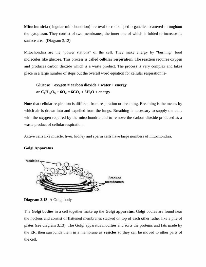
Mitochondria (singular mitochondrion) are oval or rod shaped organelles scattered throughout
the cytoplasm. They consist of two membranes, the inner one of which is folded to increase its
surface area. (Diagram 3.12)
Mitochondria are the ―power stations‖ of the cell. They make energy by ―burning‖ food
molecules like glucose. This process is called cellular respiration. The reaction requires oxygen
and produces carbon dioxide which is a waste product. The process is very complex and takes
place in a large number of steps but the overall word equation for cellular respiration is-
Glucose + oxygen = carbon dioxide + water + energy
or C6H12O6 + 6O2 = 6CO2 + 6H2O + energy
Note that cellular respiration is different from respiration or breathing. Breathing is the means by
which air is drawn into and expelled from the lungs. Breathing is necessary to supply the cells
with the oxygen required by the mitochondria and to remove the carbon dioxide produced as a
waste product of cellular respiration.
Active cells like muscle, liver, kidney and sperm cells have large numbers of mitochondria.
Golgi Apparatus
Diagram 3.13: A Golgi body
The Golgi bodies in a cell together make up the Golgi apparatus. Golgi bodies are found near
the nucleus and consist of flattened membranes stacked on top of each other rather like a pile of
plates (see diagram 3.13). The Golgi apparatus modifies and sorts the proteins and fats made by
the ER, then surrounds them in a membrane as vesicles so they can be moved to other parts of
the cell.

Lysosomes
Lysosomes are large vesicles that contain digestive enzymes. These break down bacteria and
other substances that are brought into the cell by phagocytosis or pinocytosis. They also digest
worn-out or damaged organelles, the components of which can then be recycled by the cell to
make new structures.
d) Microfilaments and Microtubules
Some cells can move and change shape and organelles and chemicals are moved around the cell.
Threadlike structures called microfilaments and microtubules that can contract are responsible
for this movement.
These structures also form the projections from the plasma membrane known as flagella
(singular flagellum) as in the sperm tail, and cilia found lining the respiratory tract and used to
remove mucus that has trapped dust particles (see chapter 4).
Microtubules also form the pair of cylindrical structures called centrioles found near the nucleus.
These help organise the spindle used in cell division.
The Nucleus
Diagram 3.14: A cell with an enlarged chromosome

Diagram 3.15: A full set of human chromosomes
The nucleus is the largest structure in a cell and can be seen with the light microscope. It is a
spherical or oval body that contains the chromosomes. The nucleus controls the development
and activity of the cell. Most cells contain a nucleus although mature red blood cells have lost
theirs during development and some muscle cells have several nuclei.
A double membrane similar in structure to the plasma membrane surrounds the nucleus (now
called the nuclear envelope). Pores in this nuclear membrane allow communication between the
nucleus and the cytoplasm.
Within the nucleus one or more spherical bodies of darker material can be seen, even with the
light microscope. These are called nucleoli and are made of RNA. Their role is to make new
ribosomes.
Chromosomes
Inside the nucleus are the chromosomes which:
contain DNA;
control the activity of the cell;
are transmitted from cell to cell when cells divide;
are passed to a new individual when sex cells fuse together in sexual reproduction.

In cells that are not dividing the chromosomes are very long and thin and appear as dark grainy
material. They become visible just before a cell divides when they shorten and thicken and can
then be counted (see diagram 3.14).
The number of chromosomes in the cells of different species varies but is constant in the cells of
any one species (e.g. horses have 64 chromosomes, cats have 38 and humans 46). Chromosomes
occur in pairs (i.e. 32 pairs in the horse nucleus and 19 in that of the cat). Members of each pair
are identical in length and shape and if you look carefully at diagram 3.15, you may be able to
see some of the pairs in the human set of chromosomes.
3.1.8: Cell Division
Diagram 3.16: Division by mitosis results in 2 new cells identical to each other and to parent cell

Diagram 3.17: Division by meiosis results in 4 new cells that are genetically different to each
other
Cells divide when an animal grows, when its body repairs an injury and when it produces sperm
and eggs (or ova). There are two types of cell division: Mitosis and meiosis.
Mitosis. This is the cell division that occurs when an animal grows and when tissues are repaired
or replaced. It produces two new cells (daughter cells) each with a full set of chromosomes that
are identical to each other and to the parent cell. All the cells of an animal‘s body therefore
contain identical DNA.
Meiosis. This is the cell division that produces the ova and sperm necessary for sexual
reproduction. It only occurs in the ovary and testis.
The most important function of meiosis it to half the number of chromosomes so that when the
sperm fertilises the ovum the normal number is regained. Body cells with the full set of
chromosomes are called diploid cells, while gametes (sperm and ova) with half the
chromosomes are called haploid cells.
Meiosis is a more complex process than mitosis as it involves two divisions one after the other
and the four cells produced are all genetically different from each other and from the parent cell.
This fact that the cells formed by meiosis are all genetically different from each other and from
the parent cell can be seen in litters of kittens where all the members of the litter are different
from each other as well as being different from the parents although they display characteristics
of both.
3.1.9: The Cell asa Factory
To make the function of the parts of the cell easier to understand and remember you can compare
them to a factory. For example:
The nucleus (1) is the managing director of the factory consulting the blueprint (the
chromosomes) (2);

The mitochondria (3) supply the power
The ribosomes (4) make the products;
The chloroplasts of plant cells (5) supply the fuel (food)
The Golgi apparatus (6) packages the products ready for dispatch;
The ER (7) modifies, stores and transports the products around the factory;
The plasma membrane is the factory wall and the gates (8);
The lysosomes dispose of the waste and worn-out machinery.
The cell compared to a factory
Summary
Cells consist of three parts: the plasma membrane, cytoplasm and nucleus.
Substances pass through the plasma membrane by diffusion (gases, lipids), osmosis
(water), active transport (glucose, ions), phagocytosis (particles), pinocytosis (fluids)
and exocytosis (particles and fluids).
Osmosis is the diffusion of water through a semipermeable membrane. Water diffuses
from high water "concentration" to low water "concentration".
The cytoplasm consists of cytosol in which are suspended cell inclusions and organelles.
organelles include ribosomes, endoplasmic reticulum, mitochondria, Golgi bodies and
lysosomes.
The nucleus controls the activity of the cell. It contains the chromosomes that are
composed of DNA.
The cell divides by mitosis and meiosis
1.6 Tutor marked assignment
Define cell and it composition
Define genetic and cell
Define genes and comment on its importance
Reference/ further reading
Extreme skewing of X chromosome inactivation in mothers of homosexual men. Human
Genetics118:6 (691) 2006.

[(1) Department of Human Genetics, University of California, Los Angeles, CA, USA; (2)
Department of Biostatistics, University of California, Los Angeles, CA, USA;
(3)Laboratory of Biochemistry, National Cancer Institute, Bethesda, MD, USA;
(4)Gonda 5524, 695 Charles Young Drive South, Los Angeles, CA90095-7088, USA
97 mothers of homosexual men; 103 age-matched control women without gay sons.
MODULE 3: Anatomy and physiology of farm animals
INTRODUCTION
The term tissue is used to describe a group of cells found together in the body. The cells within a
tissue share a common embryonic origin. Microscopic observation reveals that the cells in a
tissue share morphological features and are arranged in an orderly pattern that achieves the
tissue‘s functions. From the evolutionary perspective, tissues appear in more complex organisms.
For example, multicellular protists, ancient eukaryotes, do not have cells organized into tissues.
Although there are many types of cells in the human body, they are organized into four broad
categories of tissues: epithelial, connective, muscle, and nervous. Each of these categories is
characterized by specific functions that contribute to the overall health and maintenance of the
body. A disruption of the structure is a sign of injury or disease. Such changes can be detected
through histology, the microscopic study of tissue appearance, organization, and function.
2.0: Learning Objectives
By the end of this section, you will be able to:
Identify the four main tissue types
Discuss the functions of each tissue type
Relate the structure of each tissue type to their function
Discuss the embryonic origin of tissue
Identify the three major germ layers
Identify the main types of tissue membranes

3.0 MAIN CONTENT
3.1: Animal tissue
3.2: Nervous system
3.3: Skeletal systems
3.4: Muscles
3.5: Circulatory systems
3.6: Reproductive systems
3. 7: Digestive systems
3.1: Animal Tissues-The Four Types of Tissues
Types of Tissues
Epithelial tissue, also referred to as epithelium, refers to the sheets of cells that cover exterior
surfaces of the body, lines internal cavities and passageways, and forms certain glands.
Connective tissue, as its name implies, binds the cells and organs of the body together and
functions in the protection, support, and integration of all parts of the body. Muscle tissue is
excitable, responding to stimulation and contracting to provide movement, and occurs as three
major types: skeletal (voluntary) muscle, smooth muscle, and cardiac muscle in the heart.
Nervous tissue is also excitable, allowing the propagation of electrochemical signals in the form
of nerve impulses that communicate between different regions of the body .
The next level of organization is the organ, where several types of tissues come together to form
a working unit. Just as knowing the structure and function of cells helps you in your study of
tissues, knowledge of tissues will help you understand how organs function. The epithelial and
connective tissues are discussed in detail in this chapter. Muscle and nervous tissues will be
discussed only briefly in this chapter.
Embryonic Origin of Tissues
The zygote, or fertilized egg, is a single cell formed by the fusion of an egg and sperm. After
fertilization the zygote gives rise to rapid mitotic cycles, generating many cells to form the
embryo. The first embryonic cells generated have the ability to differentiate into any type of cell
in the body and, as such, are called totipotent, meaning each has the capacity to divide,
differentiate, and develop into a new organism. As cell proliferation progresses, three major cell
lineages are established within the embryo. Each of these lineages of embryonic cells forms the
distinct germ layers from which all the tissues and organs of the human body eventually form.
Each germ layer is identified by its relative position: ectoderm (ecto- = ―outer‖), mesoderm
(meso- = ―middle‖), and endoderm (endo- = ―inner‖).shows the types of tissues and organs
associated with the each of the three germ layers. Note that epithelial tissue originates in all three
layers, whereas nervous tissue derives primarily from the ectoderm and muscle tissue from
mesoderm.
Tissue Membranes
A tissue membrane is a thin layer or sheet of cells that covers the outside of the body (for
example, skin), the organs (for example, pericardium), internal passageways that lead to the
exterior of the body (for example, abdominal mesenteries), and the lining of the moveable joint
cavities. There are two basic types of tissue membranes: connective tissue and epithelial
membranes. The two broad categories of tissue membranes in the body are (1) connective tissue
membranes, which include synovial membranes, and (2) epithelial membranes, which include
mucous membranes, serous membranes, and the cutaneous membrane, in other words, the skin.
Connective Tissue Membranes
The connective tissue membrane is formed solely from connective tissue. These membranes
encapsulate organs, such as the kidneys, and line our movable joints. A synovial membrane is a
type of connective tissue membrane that lines the cavity of a freely movable joint. For example,
synovial membranes surround the joints of the shoulder, elbow, and knee. Fibroblasts in the
inner layer of the synovial membrane release hyaluronan into the joint cavity. The hyaluronan
effectively traps available water to form the synovial fluid, a natural lubricant that enables the
bones of a joint to move freely against one another without much friction. This synovial fluid
readily exchanges water and nutrients with blood, as do all body fluids.
Epithelial Membranes

The epithelial membrane is composed of epithelium attached to a layer of connective tissue, for
example, your skin. The mucous membrane is also a composite of connective and epithelial
tissues. Sometimes called mucosae, these epithelial membranes line the body cavities and hollow
passageways that open to the external environment, and include the digestive, respiratory,
excretory, and reproductive tracts. Mucous, produced by the epithelial exocrine glands, covers
the epithelial layer. The underlying connective tissue, called the lamina propria (literally ―own
layer‖), help support the fragile epithelial layer.
A serous membrane is an epithelial membrane composed of mesodermally derived epithelium
called the mesothelium that is supported by connective tissue. These membranes line the
coelomic cavities of the body, that is, those cavities that do not open to the outside, and they
cover the organs located within those cavities. They are essentially membranous bags, with
mesothelium lining the inside and connective tissue on the outside. Serous fluid secreted by the
cells of the thin squamous mesothelium lubricates the membrane and reduces abrasion and
friction between organs. Serous membranes are identified according locations. Three serous
membranes line the thoracic cavity; the two pleura that cover the lungs and the pericardium that
covers the heart. A fourth, the peritoneum, is the serous membrane in the abdominal cavity that
covers abdominal organs and forms double sheets of mesenteries that suspend many of the
digestive organs.
The skin is an epithelial membrane also called the cutaneous membrane. It is a stratified
squamous epithelial membrane resting on top of connective tissue. The apical surface of this
membrane is exposed to the external environment and is covered with dead, keratinized cells that
help protect the body from desiccation and pathogens.

3.1.2: Nervous System
The nervous system is the part of the body that coordinates voluntary and involuntary actions
and transmits signals to and from different parts of its body. It detects and responds to changes
inside and outside the body. Along with the endocrine system, the nervous system controls the
vital functions of the body and maintains homeostasis. The response of the nervous system is
much faster than that of the endocrine system.


The nervous system consists of:
Brain
Spinal cord
Peripheral nerves
The nervous system can be divided into:
The central nervous system (CNS): Consisting of brain and spinal cord
The peripheral nervous system (PNS): Consisting all the nerves outside brain and
spinal cord
The central nervous system receives sensory information through afferent nerves. It then
processes this information and responds appropriately by sending impulses through motor nerves
to the effector organs. For example, responses to changes in the internal environment regulate
important functions such as respiration and blood pressure. Similarly, responses to changes in the
external environment result in voluntary actions such as change in posture or other activities.
The peripheral nervous system (PNS) consists of paired cranial and sacral nerves. Some of these
nerves are sensory (afferent) i.e. transmit impulses to the CNS. Some are motor nerves (efferent)
i.e. transmit impulses from the CNS. Some others are mixed.
The PNS can be divided into two functional parts:
Sensory division
Motor division
The motor division has two parts:
Somatic nervous system: Controls voluntary movements of skeletal muscles
Autonomic nervous system: Controls involuntary functions such as heart rate, digestion,
respiratory rate, pupillary response, urination, and sexual arousal
The autonomic nervous system has two divisions:
Sympathetic: Stimulates the body‘s ―fight-or-flight‖ response
Parasympathetic: Stimulates ―rest-and-digest‖ or ―feed and breed‖ activities that occur
when the body is at rest
Topics in this Section
Neurons
o Properties of neurons
o Cell bodies
o Axons and dendrites
o The nerve impulse (action potential)
o Types of nerves
o The synapse and neurotransmitters
Central nervous system
o Neuroglia
o Membranes covering the brain and spinal cord (the meninges)
o Ventricles of the brain and the cerebrospinal fluid
Brain
o Blood supply to the brain
o Cerebrum
o Brain stem
o Cerebellum
Spinal cord
o Grey matter
o White matter
Peripheral nervous system
o Spinal nerves
o Thoracic nerves
o Cranial nerves
Autonomic nervous system
o Sympathetic nervous system
o Parasympathetic nervous system

o Functions of the autonomic nervous system
Effects of autonomic stimulation
Afferent impulses from viscera
Response of nervous tissue to injury
o Neuron damage
o Neuron regeneration
o Neuroglia damage
o Effects of poisons on the central nervous system
Disorders of the brain
o Increased intracranial pressure
Effects of increased ICP
Cerebral edema
Hydrocephalus
o Head injuries
Blow to the head
Acceleration-deceleration injuries
Complications of head injury
o Circulatory disturbances affecting the brain
Cerebral hypoxia
Stroke (cerebrovascular disease)
o Dementia
o Parkinson‘s disease
Infections of the central nervous system
o Pyogenic infection
o Viral infections
o Creutzfeldt-Jakob disease
o Myalgic encephalitis (ME)
Demyelinating diseases
o Multiple (disseminated) sclerosis (MS)
o Acute disseminating encephalomyelitis
Phenylketonuria

Diseases of the spinal cord
o Motor neurons
o Sensory neurons
o Mixed motor and sensory conditions
Diseases of peripheral nerves
o Neuropathies
o Neuritis
Developmental abnormalities of the nervous system
o Spina bifida
o Hydrocephalus
Tumors of the nervous system
3.1.3:The Functions of the Skeletal System
2.0. Learning Objectives
By the end of this section, you will be able to:
Define bone, cartilage, and the skeletal system
List and describe the functions of the skeletal system
Bone, or osseous tissue, is a hard, dense connective tissue that forms most of the adult skeleton,
the support structure of the body. In the areas of the skeleton where bones move (for example,
the ribcage and joints), cartilage, a semi-rigid form of connective tissue, provides flexibility and
smooth surfaces for movement. The skeletal system is the body system composed of bones and
cartilage and performs the following critical functions for the human body:
supports the body
facilitates movement
protects internal organs
produces blood cells
stores and releases minerals and fat

Support, Movement, and Protection
The most apparent functions of the skeletal system are the gross functions—those visible by
observation. Simply by looking at a person, you can see how the bones support, facilitate
movement, and protect the human body.
Just as the steel beams of a building provide a scaffold to support its weight, the bones and
cartilage of your skeletal system compose the scaffold that supports the rest of your body.
Without the skeletal system, you would be a limp mass of organs, muscle, and skin.
Bones also facilitate movement by serving as points of attachment for your muscles. While some
bones only serve as a support for the muscles, others also transmit the forces produced when
your muscles contract. From a mechanical point of view, bones act as levers and joints serve as
fulcrums . Unless a muscle spans a joint and contracts, a bone is not going to move. For
information on the interaction of the skeletal and muscular systems, that is, the musculoskeletal
system, seek additional contentBones also protect internal organs from injury by covering or
surrounding them. For example, your ribs protect your lungs and heart, the bones of your
vertebral column (spine) protect your spinal cord, and the bones of your cranium (skull) protect
your brain.
Orthopedist
An orthopedist is a doctor who specializes in diagnosing and treating disorders and injuries
related to the musculoskeletal system. Some orthopedic problems can be treated with
medications, exercises, braces, and other devices, but others may be best treated with surgery
Orthopedists commonly treat bone and joint injuries but they also treat other bone conditions
including curvature of the spine. Lateral curvatures (scoliosis) can be severe enough to slip under
the shoulder blade (scapula) forcing it up as a hump. Spinal curvatures can also be excessive
dorsoventrally (kyphosis) causing a hunch back and thoracic compression. These curvatures
often appear in preteens as the result of poor posture, abnormal growth, or indeterminate causes.
Mostly, they are readily treated by orthopedists. As people age, accumulated spinal column
injuries and diseases like osteoporosis can also lead to curvatures of the spine, hence the stooping
you sometimes see in the elderly.

Some orthopedists sub-specialize in sports medicine, which addresses both simple injuries, such
as a sprained ankle, and complex injuries, such as a torn rotator cuff in the shoulder. Treatment
can range from exercise to surgery.
Mineral Storage, Energy Storage, and Hematopoiesis
On a metabolic level, bone tissue performs several critical functions. For one, the bone matrix
acts as a reservoir for a number of minerals important to the functioning of the body, especially
calcium, and phosphorus. These minerals, incorporated into bone tissue, can be released back
into the bloodstream to maintain levels needed to support physiological processes. Calcium ions,
for example, are essential for muscle contractions and controlling the flow of other ions involved
in the transmission of nerve impulses.
Bone also serves as a site for fat storage and blood cell production. The softer connective tissue
that fills the interior of most bone is referred to as bone marrow . There are two types of bone
marrow: yellow marrow and red marrow. Yellow marrow contains adipose tissue; the
triglycerides stored in the adipocytes of the tissue can serve as a source of energy. Red marrow is
where hematopoiesis—the production of blood cells—takes place. Red blood cells, white blood
cells, and platelets are all produced in the red marrow.
The major functions of the bones are body support, facilitation of movement, protection of
internal organs, storage of minerals and fat, and hematopoiesis. Together, the muscular system
and skeletal system are known as the musculoskeletal system.
Critical Thinking Questions
The skeletal system is composed of bone and cartilage and has many functions. Choose three of
these functions and discuss what features of the skeletal system allow it to accomplish these
functions.
Glossary
bone
hard, dense connective tissue that forms the structural elements of the skeleton
cartilage
semi-rigid connective tissue found on the skeleton in areas where flexibility and smooth
surfaces support movement
hematopoiesis
production of blood cells, which occurs in the red marrow of the bones
orthopedist
doctor who specializes in diagnosing and treating musculoskeletal disorders and injuries
osseous tissue
bone tissue; a hard, dense connective tissue that forms the structural elements of the
skeleton
red marrow
connective tissue in the interior cavity of a bone where hematopoiesis takes place
skeletal system
organ system composed of bones and cartilage that provides for movement, support, and
protection
yellow marrow
connective tissue in the interior cavity of a bone where fat is stored
3.1.4: Muscles
2.0. Objectives
After completing this section, you should know:
The structure of smooth, cardiac and skeletal muscle and where they are found.
What the insertion and origin of a muscle is.
What flexion and extension of a muscle means.
That muscles usually operate as antagonistic pairs.
What tendons attach muscles to bones.
Muscles
Muscles make up the bulk of an animal‘s body and account for about half its weight. The meat
on the chop or roast is muscle and is composed mainly of protein. The cells that make up muscle
tissue are elongated and able to contract to a half or even a third of their length when at rest.
There are three different kinds of muscle based on appearance and function: smooth, cardiac and
skeletal muscle.
Types of Muscle
Smooth muscle
Smooth or Involuntary muscle carries out the unconscious routine tasks of the body such as
moving food down the digestive system, keeping the eyes in focus and adjusting the diameter of
blood vessels. The individual cells are spindle-shaped, being fatter in the middle and tapering off
towards the ends with a nucleus in the centre of the cell. They are usually found in sheets and are
stimulated by the non-conscious or autonomic nervous system as well as by hormones (see
Chapter 3).
Cardiac muscle
Cardiac muscle is only found in the wall of the heart. It is composed of branching fibres that
form a three-dimensional network. When examined under the microscope, a central nucleus and
faint stripes or striations can be seen in the cells. Cardiac muscle cells contract spontaneously
and rhythmically without outside stimulation, but the sinoatrial node (natural pacemaker)
coordinates the heart beat. Nerves and hormones modify this rhythm (see Chapter 3).
Skeletal muscle
Skeletal muscle is the muscle that is attached to and moves the skeleton, and is under voluntary
control. It is composed of elongated cells or fibres lying parallel to each other. Each cell is
unusual in that it has several nuclei and when examined under the microscope appears striped or
striated. This appearance gives the muscle its names of striped or striated muscle. Each cell of
striated muscle contains hundreds, or even thousands, of microscopic fibres each one with its

own striped appearance. The stripes are formed by two different sorts of protein that slide over
each other making the cell contract (see diagram 7.1).
Diagram 7.1 - A striped muscle cell
Muscle contraction
Muscle contraction requires energy and muscle cells have numerous mitochondria. However,
only about 15% of the energy released by the mitochondria is used to fuel muscle contraction.
The rest is released as heat. This is why exercise increases body temperature and makes animals
sweat or pant to rid themselves of this heat.
What we refer to as a muscle is made up of groups of muscle fibers surrounded by connective
tissue. The connective tissue sheaths join together at the ends of the muscle to form tough white
bands of fiber called tendons. These attach the muscles to the bones. Tendons are similar in
structure to the ligaments that attach bones together across a joint (see diagrams 7.2a and b).

Diagram 7.2 a and b - The structure of a muscle
Remember:
Tendons Tie muscles to bones
and
Ligaments Link bones at joints
Structure of a muscle
A single muscle is fat in the middle and tapers towards the ends. The middle part, which gets
fatter when the muscle contracts, is called the belly of the muscle. If you contract your biceps

muscle in your upper arm you may feel it getting fatter in the middle. You may also notice that
the biceps is attached at its top end to bones in your shoulder while at the bottom it is attached to
bones in your lower arm. Notice that the bones at only one end move when you contract the
biceps. This end of the muscle is called the insertion. The other end of the muscle, the origin, is
attached to the bone that moves the least (see diagram 7.3).
Diagram 7.3 - Antagonistic muscles, flexion and extension
Antagonistic muscles
Skeletal muscles usually work in pairs. When one contracts the other relaxes and vice versa.
Pairs of muscles that work like this are called antagonistic muscles. For example the muscles in
the upper forearm are the biceps and triceps (see diagram 7.3). Together they bend the elbow.

When the biceps contracts (and the triceps relaxes) the lower forearm is raised and the angle of
the joint is reduced. This kind of movement is called flexion. When the triceps is contracted (and
the biceps relaxes), the angle of the elbow increases. The term for this movement is extension.
When you or animals contract skeletal muscle it is a voluntary action. For example, you make a
conscious decision to walk across the room, raise the spoon to your mouth, or smile. There is
however, another way in which contraction of muscles attached to the skeleton happens that is
not under voluntary control. This is during a reflex action, such as jerking your hand away from
the hot stove you have touched by accident. This is called a reflex arc and will be described in
detail in chapter 14-15.
Summary
There are three different kinds of muscle tissue: smooth muscle in the walls of the gut
and blood
vessels; cardiac muscle in the heart and skeletal muscle attached to the skeleton.
Tendons attach skeletal muscles to the skeleton.
Ligaments link bones together at a joint.
Skeletal muscles work in pairs known as antagonistic pairs. As one contracts the other
in the
pair relaxes.
Flexion is the movement that reduces the angle of a joint. Extension increases the angle
of a joint.
Test Yourself
1. What kind of muscle tissue:
a) moves bones
b) makes the heart pump blood:

c) pushes food along the intestine:
d) makes your mouth form a smile:
e) makes the hair stand up when cold:
f) makes the diaphragm contract for breathing in:
2. What structure connects a muscle to a bone?
3. What is the insertion of a muscle?
4. Which muscle is antagonistic to the biceps?
5. Name 3 other antagonistic pairs and tell what they do.
6. When you bend your knee what movement are you making?
7. When you straighten your ankle joint what movement happens?
8. What organelles provide the energy that muscles need?
9. State the difference between a tendon and a ligament.
10.In the section "Skeletal Muscle" there are 2 proteins mentioned. Name these proteins, state
their size difference, and tell what they actually do to help produce movement.
Website
http://health.howstuffworks.com/muscle.htm How muscles work
Description of the three types of muscles and how skeletal muscles work.
3.1.5: The Circulatory System
You might remember that blood is a form of connective tissue (widely spaced cells in a matrix,
in this case a fluid matrix). In this section, you will start to understand how blood might better be

called the connective tissue. Most people grow up thinking of blood as part of the ―circulatory‖
system, but as you shall see, there are in fact two systems involved in circulation: the
cardiovascular system and the lymphatic system. In terms of transport, the cardiovascular system
takes the top spot, but in terms of defending against bacteria and viruses, the lymphatic system
gets top billing. In this section, I explore the connection between these two parallel systems.
Blood is the giver of life, the provider of food, water, and air, the waste remover (with the help
of the kidneys; see The Excretory System), but it can also be the harbinger of death, if an
infection makes its way into the blood and we become septic. Adult females have an average of
four to five liters of blood, and adult males average about five to six liters. How the blood
manages to get from place to place is the subject of The Heart and Cardiovascular and
Lymphatic Circulation. Here I discuss what blood actually does.
Function Junction
With apologies to Tina Turner, what's blood got to do with it? Blood does many wondrous
things:
Blood (plus vessels and the heart) is the primary transportation system for materials (the
lymphatic system, as you will see, is the secondary transportation system): nutrients,
water, wastes, O2, and CO2.
Blood, which is slightly alkaline (7.35 to 7.45), regulates pH levels by absorbing acids
from the interstitial fluid and neutralizing them.
Blood regulates body temperature by transferring (via the plasma) heat generated by
muscles (hemopoiesis; see The Muscles) to tissues throughout the body; heat can also be
retained or lost through the constriction and dilation of vessels in the skin. As such, the
average temperature of blood is higher than body temperature (38ºC, or 100.4ºF).
Blood protects from fluid loss by clotting at the site of injuries.
Blood protects from toxins and pathogens, through the action of white blood cells and
antibodies.

3.1.6:Reproductive System
In biological terms sexual reproduction involves the union of gametes - the sperm and the ovum
- produced by two parents. Each gamete is formed by meiosis (see Chapter 3). This means each
contains only half the chromosomes of the body cells (haploid). Fertilization results in the
joining of the male and female gametes to form a zygote which contains the full number of
chromosomes (diploid). The zygote then starts to divide by mitosis (see Chapter 3) to form a
new animal with all its body cells containing chromosomes that are identical to those of the
original zygote
Sexual reproduction
The offspring formed by sexual reproduction contain genes from both parents and show
considerable variation. For example, kittens in a litter are all different although they (usually)
have the same mother and father. In the wild this variation is important because it means that
when the environment changes some individuals may be better adapted to survive than others.
These survivors pass their ―superior‖ genes on to their offspring. In this way the characteristics
of a group of animals can gradually change over time to keep pace with the changing
environment. This ―survival of the fittest‖ or ―natural selection‖ is the mechanism behind the
theory of evolution.
Fertilization
In most fish and amphibia (frogs and toads) fertilisation of the egg cells takes place outside the
body. The female lays the eggs and then the male deposits his sperm on or at least near them.
In reptiles and birds, eggs are fertilized inside the body when the male deposits the sperm inside
the egg duct of the female. The egg is then surrounded by a resistant shell, ―laid‖ by the female
and the embryo completes its development inside the egg.
In mammals the sperm are placed in the body of the female and the eggs are fertilized internally.
They then develop to quite an advanced stage inside the body of the female.
Sexual Reproduction In Mammals

The reproductive organs of mammals produce the gametes (sperm and egg cells), help them
fertilize and then support the developing embryo.
The Male Reproductive System
The male reproductive system consists of a pair of testes that produce sperm (or spermatozoa),
ducts that transport the sperm to the penis and glands that add secretions to the sperm to make
semen (see diagram 13.2).
The various parts of the male reproductive system with a summary of their functions are shown
in diagram 13.3.

Diagram 13.2. The reproductive organs of a male dog
File:Anatomy and physiology of animals Diagram summarizing the functions of the male
reproductive organs.jpg
Diagram 13.3 - Diagram summarizing the functions of the male reproductive organs
The Testes
Sperm need temperatures between 2 to 10 degrees Centigrade lower and then the body
temperature to develop. This is the reason why the testes are located in a bag of skin called the

scrotal sacs (or scrotum) that hangs below the body and where the evaporation of secretions
from special glands can further reduce the temperature. In many animals (including humans) the
testes descend into the scrotal sacs at birth but in some animals they do not descend until sexual
maturity and in others they only descend temporarily during the breeding season. A mature
animal in which one or both testes have not descended is called a cryptorchid and is usually
infertile.
The problem of keeping sperm at a low enough temperature is even greater in birds that have a
higher body temperature than mammals. For this reason bird‘s sperm are usually produced at
night when the body temperature is lower and the sperm themselves are more resistant to heat.
The testes consist of a mass of coiled tubes (the seminiferous or sperm producing tubules) in
which the sperm are formed by meiosis (see diagram 13.4). Cells lying between the seminiferous
tubules produce the male sex hormone testosterone.
When the sperm are mature they accumulate in the collecting ducts and then pass to the
epididymisbefore moving to the sperm duct or vas deferens. The two sperm ducts join the
urethra just below the bladder, which passes through the penis and transports both sperm and
urine.
Ejaculation discharges the semen from the erect penis. It is brought about by the contraction of
the epididymis, vas deferens, prostate gland and urethra.

Diagram 13.4 - The testis and a magnified seminiferous tubule
Semen
Semen consists of 10% sperm and 90% fluid and as sperm pass down the ducts from testis to
penis, (accessory) glands add various secretion...
Accessory Glands
Three different glands may be involved in producing the secretions in which sperm are
suspended, although the number and type of glands varies from species to species.

Seminal vesicles are important in rats, bulls, boars and stallions but are absent in cats and dogs.
When present they produce secretions that make up much of the volume of the semen, and
transport and provide nutrients for the sperm.
The prostate gland is important in dogs and humans. It produces an alkaline secretion that
neutralizes the acidity of the male urethra and female vagina.
Cowper’s glands (bulbourethral glands) have various functions in different species. The
secretions may lubricate, flush out urine or form a gelatinous plug that traps the semen in the
female reproductive system after copulation and prevents other males of the same species
fertilizing an already mated female. Cowper‘s glands are absent in bears and aquatic mammals.
The Penis
The penis consists of connective tissue with numerous small blood spaces in it. These fill with
blood during sexual excitement causing erection.
Penis Form And Shape
Dogs, bears, seals, bats and rodents have a special bone in the penis which helps maintain the
erection (see diagram 13.2). In some animals (e.g. the bull, ram and boar) the penis has an ―S‖
shaped bend that allows it to fold up when not in use. In many animals the shape of the penis is
adapted to match that of the vagina. For example, the boar has a corkscrew shaped penis, there is
a pronounced twist in bulls‘ and it is forked in marsupials like the opossum. Some have spines,
warts or hooks on them to help keep them in the vagina and copulation may be extended to help
retain the semen in the female system. Mating can last up to three hours in minks, and dogs may
―knot‖ or ―tie‖ during mating and can not separate until the erection has subsided.
Sperm
Sperm are made up of three parts: a head consisting mainly of the nucleus, a midpiece
containing many mitochondria to provide the energy and a tail that provides propulsion (see
diagram 13.5).

Diagram 13.5 - A sperm
A single ejaculation may contain 2-3 hundred million sperm but even in normal semen as many
as 10% of these sperm may be abnormal and infertile. Some may be dead while others are
inactive or deformed with double, giant or small heads or tails that are coiled or absent
altogether.
When there are too many abnormal sperm or when the sperm concentration is low, the semen
may not be able to fertilize an egg and the animal is infertile. Make sure you don‘t confuse
infertility with impotence, which is the inability to copulate successfully.
Sperm do not live forever. They have a definite life span that varies from species to species.
They survive for between 20 days (guinea pig) to 60 days (bull) in the epididymis but once
ejaculated into the female tract they only live from 12 to 48 hours. When semen is used for
artificial insemination, storage under the right conditions can extend the life span of some
species.
Artificial Insemination
In many species the male can be artificially stimulated to ejaculate and the semen collected. It
can then be diluted, stored and used to inseminate females. For example bull semen can be
diluted and stored for up to 3 weeks at room temperature. If mixed with an antifreeze solution
and stored in ―straws‖ in liquid nitrogen at minus 79oC it will keep for much longer.
Unfortunately the semen of chickens, stallions and boars can only be stored for up to 2 days.

Dilution of the semen means that one male can be used to fertilise many more females than
would occur under natural conditions. There are also advantages in the male and female not
having to make physical contact. It means that owners of females do not have to buy expensive
males and the possibility of transmitting sexually transmitted diseases is reduced. Routine
examination of the semen for sperm concentration, quality and activity allows only the highest
quality semen to be used so a high success rate is ensured.
Since the lifespan of sperm in the female tract is so short and ova only survive from 8 to 10 hours
the timing of the artificial insemination is critical. Successful conception depends upon detecting
the time that the animal is ―on heat‖ and when ovulation occurs.
The Female Reproductive Organs
The female reproductive system consists of a pair of ovaries that produce egg cells or ova and
fallopian tubes where fertilisation occurs and which carry the fertilised ovum to the uterus.
Growth of the foetus takes place here. The cervix separates the uterus from the vagina or birth
canal, where the sperm are deposited (see diagram 13.6).
Diagram 13.6. - The reproductive system of a female rabbit

Note that primates like humans have a uterus with a single compartment but in most mammals
the uterus is divided into two separate parts or horns as shown in diagram 13.6.
The Ovaries
Ovaries are small oval organs situated in the abdominal cavity just ventral to the kidneys. Most
animals have a pair of ovaries but in birds only the left one is functional to reduce weight (see
below).
The ovary consists of an inner region (medulla) and an outer region (cortex) containing egg
cells or ova. These are formed in large numbers around the time of birth and start to develop
after the animal becomes sexually mature. A cluster of cells called the follicle surrounds and
nourishes each ovum.
The Ovarian Cycle
The ovarian cycle refers to the series of changes in the ovary during which the follicle matures,
the ovum is shed and the corpus luteum develops (see diagram 13.7).
Numerous undeveloped ovarian follicles are present at birth but they start to mature after sexual
maturity. In animals that normally have only one baby at a time only one ovum will mature at
once but in litter animals several will. The mature follicle consists of outer cells that provide
nourishment. Inside this is a fluid-filled space that contains the ovum.
A mature follicle can be quite large, ranging from a few millimetres in small mammals to the
size of a golf ball in large animals. It bulges out from the surface of the ovary before eventually
rupturing to release the ovum into the abdominal cavity. Once the ovum has been shed, a blood
clot forms in the empty follicle. This develops into a tissue called the corpus luteum that
produces the hormone progesterone (see diagram 13.9). If the animal becomes pregnant the
corpus luteum persists, but if there is no pregnancy it degenerates and a new ovarian cycle
usually.
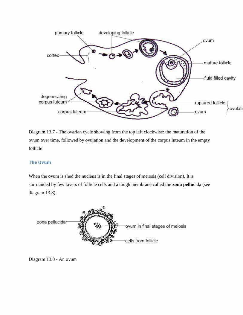
Diagram 13.7 - The ovarian cycle showing from the top left clockwise: the maturation of the
ovum over time, followed by ovulation and the development of the corpus luteum in the empty
follicle
The Ovum
When the ovum is shed the nucleus is in the final stages of meiosis (cell division). It is
surrounded by few layers of follicle cells and a tough membrane called the zona pellucida (see
diagram 13.8).
Diagram 13.8 - An ovum

The Oestrous Cycle
The oestrous cycle is the sequence of hormonal changes that occurs through the ovarian cycle.
These changes influence the behaviour and body changes of the female (see diagram 13.9).
Diagram 13.9 - The oestrous cycle
The first hormone involved in the oestrous cycle is follicle stimulating hormone (F.S.H.),
secreted by the anterior pituitary gland (see chapter 16). It stimulates the follicle to develop.
As the follicle matures the outer cells begin to secrete the hormone oestrogen and this stimulates
the mammary glands to develop. It also prepares the lining of the uterus to receive a fertilised
egg. Ovulation is initiated by a surge of another hormone from the anterior pituitary, luteinising
hormone (L.H.). This hormone also influences the development of the corpus luteum, which
produces progesterone, a hormone that prepares the lining of the uterus for the fertilised ovum
and readies the mammary glands for milk production. If no pregnancy takes place the corpus
luteum shrinks and the production of progesterone decreases. This causes FSH to be produced
again and a new oestrous cycle begins.

For fertilisation of the ovum by the sperm to occur, the female must be receptive to the male at
around the time of ovulation. This is when the hormones turn on the signs of ―heat‖, and she is
―in season‖ or ―in oestrous‖. These signs are turned off again at the end of the oestrous cycle.
During the oestrous cycle the lining of the uterus (endometrium) thickens ready for the fertilised
ovum to be implanted. If no pregnancy occurs this thickened tissue is absorbed and the next
cycle starts. In humans and other higher primates, however, the endometrium is shed as a flow of
blood and instead of an oestrous cycle there is a menstrual cycle.
The length of the oestrous cycle varies from species to species. In rats the cycle only lasts 4–5
days and they are sexually receptive for about 14 hours. Dogs have a cycle that lasts 60–70 days
and heat lasts 7–9 days and horses have a 21-day cycle and heat lasts an average of 6 days.
Ovulation is spontaneous in most animals but in some, e.g. the cat, and the rabbit, ovulation is
stimulated by mating. This is called induced ovulation.
Signs of Oestrous or Heat
When on heat a bitch has a blood stained discharge from the vulva that changes a
little later to a straw coloured one that attracts all the dogs in the neighbourhood.
Female cats ―call‖ at night, roll and tread the carpet and are generally restless but
will ―stand‖ firm when pressure is placed on the pelvic region (this is the lordosis
response).
A female rat shows the lordosis response when on heat. It will ―mount‖ other
females and be more active than normal.
A cow mounts other cows (bulling), bellows, is restless and has a discharge from
the vulva.
Breeding Seasons and Breeding Cycles
Only a few animals breed throughout the year. This includes the higher primates (humans,
gorillas and chimpanzees etc.), pigs, mice and rabbits. These are known as continuous breeders.

Most other animals restrict reproduction to one or two seasons in the year-seasonal breeders
(see diagram 13.10). There are several reasons for this. It means the young can be born at the
time (usually spring) when feed is most abundant and temperatures are favourable. It is also
sensible to restrict the breeding season because courtship, mating, gestation and the rearing of
young can exhaust the energy resources of an animal as well as make them more vulnerable to
predators.
Diagram 13.10 - Breeding cycles
The timing of the breeding cycle is often determined by day length. For example the shortening
day length in autumn will bring sheep and cows into season so the foetus can gestate through the
winter and be born in spring. In cats the increasing day length after the winter solstice (shortest
day) stimulates breeding. The number of times an animal comes into season during the year
varies, as does the number of oestrous cycles during each season. For example a dog usually has
2-3 seasons per year, each usually consisting of just one oestrous cycle. In contrast ewes usually
restrict breeding to one season and can continue to cycle as many as 20 times if they fail to
become pregnant.
Fertilisation and Implantation

Fertilisation
The opening of the fallopian tube lies close to the ovary and after ovulation the ovum is swept
into its funnel-like opening and is moved along it by the action of cilia and wave-like
contractions of the wall.
Copulation deposits several hundred million sperm in the vagina. They swim through the cervix
and uterus to the fallopian tubes moved along by whip-like movements of their tails and
contractions of the uterus. During this journey the sperm undergo their final phase of maturation
so they are ready to fertilise the ovum by the time they reach it in the upper fallopian tube.
High mortality means only a small proportion of those deposited actually reach the ovum. The
sperm attach to the outer zona pellucida and enzymes secreted from a gland in the head of the
sperm dissolve this membrane so it can enter. Once one sperm has entered, changes in the zona
pellucida prevent further sperm from penetrating. The sperm loses its tail and the two nuclei fuse
to form a zygote with the full set of paired chromosomes restored.
Development Of The Morula And Blastocyst
As the fertilised egg travels down the fallopian tube it starts to divide by mitosis. First two cells
are formed and then four, eight, sixteen, etc. until there is a solid ball of cells. This is called a
morula. As division continues a hollow ball of cells develops. This is a blastocyst (see diagram
13.11).
Implantation
Implantation involves the blastocyst attaching to, and in some species, completely sinking into
the wall of the uterus.
Pregnancy

The Placenta And Foetal Membranes
As the embryo increases in size, the placenta, umbilical cord and foetal membranes (often
known collectively as the placenta) develop to provide it with nutrients and remove waste
products (see diagram 13.12). In later stages of development the embryo becomes known as a
foetus.
The placenta is the organ that attaches the foetus to the wall of the uterus. In it the blood of the
foetus and mother flow close to each other but never mix (see diagram 13.13). The closeness of
the maternal and foetal blood systems allows diffusion between them. Oxygen and nutrients
diffuse from the mother‘s blood into that of the foetus and carbon dioxide and excretory products
diffuse in the other direction. Most maternal hormones (except adrenaline), antibodies, almost all
drugs (including alcohol), lead and DDT also pass across the placenta. However, it protects the
foetus from infection with bacteria and most viruses.
Diagram 13.11 - Development and implantation of the embryo

Diagram 13.12. The foetus and placenta
The foetus is attached to the placenta by the umbilical cord. It contains arteries that carry blood
to the placenta and a vein that returns blood to the foetus. The developing foetus becomes
surrounded by membranes. These enclose the amniotic fluid that protects the foetus from knocks
and other trauma (see diagram 13.12).

Diagram 13.13 - Maternal and foetal blood flow in the placenta
Hormones during Pregnancy
The corpus luteum continues to secrete progesterone and oestrogen during pregnancy. These
maintain the lining of the uterus and prepare the mammary glands for milk secretion. Later in the
pregnancy the placenta itself takes over the secretion of these hormones.
Chorionic gonadotrophin is another hormone secreted by the placenta and placental
membranes. It prevents uterine contractions before labour and prepares the mammary glands for
lactation. Towards the end of pregnancy the placenta and ovaries secrete relaxin, a hormone that
eases the joint between the two parts of the pelvis and helps dilate the cervix ready for birth.
Pregnancy Testing
The easiest method of pregnancy detection is ultrasound which is noninvasive and very reliable
Later in gestation pregnancy can be detected by taking x-rays.
In dogs and cats a blood test can be used to detect the hormone relaxin.
In mares and cows palpation of the uterus via the rectum is the classic way to determine
pregnancy. It can also be done by detecting the hormones progesterone or equine chorionic
gonagotrophin (eCG) in the urine. A new sensitive test measures the amount of the hormone,
oestrone sulphate, present in a sample of faeces. The hormone is produced by the foal and
placenta, and is only present when there is a living foal.
In most animals, once pregnancy is advanced, there is a window of time during which an
experienced veterinarian can determine pregnancy by feeling the abdomen.
Gestation Period
The young of many animals (e.g. pigs, horses and elephants) are born at an advanced state of
development, able to stand and even run to escape predators soon after they are born. These

animals have a relatively long gestation period that varies with their size e.g. from 114 days in
the pig to 640 days in the elephant.
In contrast, cats, dogs, mice, rabbits and higher primates are relatively immature when born and
totally dependent on their parents for survival. Their gestation period is shorter and varies from
25 days in the mouse to 31 days in rabbits and 258 days in the gorilla.
The babies of marsupials are born at an extremely immature stage and migrate to the pouch
where they attach to a teat to complete their development. Kangaroo joeys, for example, are born
33 days after conception and opossums after only 8 days.
Birth
Signs Of Imminent Birth
As the pregnancy continues, the mammary glands enlarge and may secrete a milky substance a
few days before birth occurs. The vulva may swell and produce thick mucus and there is
sometimes a visible change in the position of the foetus. Just before birth the mother often
becomes restless, lying down and getting up frequently. Many animals seek a secluded place
where they may build a nest in which to give birth.
Labour
Labour involves waves of uterine contractions that press the foetus against the cervix causing it
to dilate. The foetus is then pushed through the cervix and along the vagina before being
delivered. In the final stage of labour the placenta or ―afterbirth‖ is expelled.
Adaptations Of The Foetus To Life Outside The Uterus
The foetus grows in the watery, protected environment of the uterus where the mother supplies
oxygen and nutrients, and waste products pass to her blood circulation for excretion. Once the
baby animal is born it must start to breathe for itself, digest food and excrete its own waste. To
allow these functions to occur blood is re-routed to the lungs and the glands associated with the

gut start to secrete. Note that newborn animals can not control their own body temperature. They
need to be kept warm by the mother, littermates and insulating nest materials.
Milk Production
Cows, manatees and primates have two mammary glands but animals like pigs that give birth to
large litters may have as many as 12 pairs. Ducts from the gland lead to a nipple or teat and there
may be a sinus where the milk collects before being suckled (see diagram 13.14).
Diagram 13.14 - A mammary gland
The hormones oestrogen and progesterone stimulate the mammary glands to develop and
prolactin promotes the secretion of the milk. Oxytocin from the pituitary gland releases the milk
when the baby suckles. The first milk is called colostrum. It is a rich in nutrients and contains
protective antibodies from the mother. Milk contains fat, protein and milk sugar as well as
vitamins and most minerals although it contains little iron. Its actual composition varies widely
from species to species. For example whale‘s and seal‘s milk has twelve times more fat and four
times more protein than cow‘s milk. Cow‘s milk has far less protein in it than cat‘s or dog‘s
milk. This is why orphan kittens and puppies cannot be fed cow‘s milk.
Reproduction In Birds
Male birds have testes and sperm ducts and male swans, ducks, geese and ostriches have a penis.
However, most birds make do with a small amount of erectile tissue known as a papilla. To

reduce weight for flight most female birds only have one ovary - usually the left, which produces
extremely yolky eggs. The eggs are fertilised in the upper part of the oviduct (equivalent to the
fallopian tube and uterus of mammals) and as they pass down it albumin (the white of the egg),
the membrane beneath the shell and the shell are laid down over the yolk. Finally the egg is
covered in a layer of mucus to help the bird lay it (see diagram 13.15).
Most birds lay their eggs in a nest and the hen sits on them until they hatch. Ducklings and
chicks are relatively well developed when they hatch and able to forage for their own food. Most
other nestlings need their parents to keep them warm, clean and fed. Young birds grow rapidly
and have voracious appetites that may involve the parents making up to 1000 trips a day to
supply their need for food.
Diagram 13.15 - Female reproductive organs of a bird
Summary
Haploid gametes (sperm and ova) are produced by meiosis in the gonads (testes and
ovaries).
Fertilisation involves the fusing of the gametes to form a diploid zygote.

The male reproductive system consists of a pair of testes that produce sperm (or
spermatozoa), ducts that transport the sperm to the penis and glands that add secretions
to the sperm to make semen.
Sperm are produced in the seminiferous tubules, are stored in the epididymis and travel
via the vas deferens or sperm duct to the junction of the bladder and the urethra where
various accessory glands add secretions. The fluid is now called semen and is ejaculated
into the female system down the urethra that runs down the centre of the penis.
Sperm consist of a head, a midpiece and a tail.
Infertility is the inability of sperm to fertilize an egg while impotence is the inability to
copulate successfully.
The female reproductive system consists of a pair of ovaries that produce ova and
fallopian tubes where fertilisation occurs and which carry the fertilised ovum to the
uterus. Growth of the foetus takes place here. The cervix separates the uterus from the
vagina, the birth canal and where the sperm are deposited.
The ovarian cycle refers to the series of changes in the ovary during which the follicle
matures, the ovum is shed and the corpus luteum develops.
The oestrous cycle is the sequence of hormonal changes that occurs through the ovarian
cycle. It is initiated by the secretion of follicle stimulating hormone (F.S.H.), by the
anterior pituitary gland which stimulates the follicle to develop. The follicle secretes
oestrogen which stimulates mammary gland development. luteinising hormone (L.H.)
from the anterior pituitary initiates ovulation and stimulates the corpus luteum to
develop. The corpus luteum produces progesterone that prepares the lining of the uterus
for the fertilised ovum.
Signs of oestrous or heat differ. A bitch has a blood stained discharge, female cats and
rats are restless and show the lordosis response, while cows mount other cows, bellow
and have a discharge from the vulva.
After fertilisation in the fallopian tube the zygote divides over and over by mitosis to
become a ball of cells called a morula. Division continues to form a hollow ball of cells
called the blastocyst. This is the stage that implants in the uterus.
The placenta, umbilical cord and foetal membranes (known as the placenta) protect
and provide the developing foetus with nutrients and remove waste products.

Websites
http://www.anatomicaltravel.com/CB_site/Conception_to_birth3.htm Anatomical travel.
Images of fertilisation and the development of the (human) embryo through to birth.
http://www.uchsc.edu/ltc/fert.swf Fertilisation. A great animation of fertilisation,
formation of the zygote and first mitotic division. A bit advanced but still worth
watching.
http://www.uclan.ac.uk/facs/health/nursing/sonic/scenarios/salfordanim/heart.swf Sonic.
An animation showing the foetal blood circulation through the placenta to the changes
allowing circulation through the lungs after birth.
http://en.wikipedia.org/wiki/Estrus Wikipedia. As always, good interesting information
although some terms and concepts are beyond the requirements of this level.
3.1.7: Digestion
2.0: Objectives
After completing this section, you should know:
what is meant by the terms: ingestion, digestion, absorption, assimilation, egestion, peristalsis and
chyme
the characteristics, advantages and disadvantages of a herbivorous, carnivorous and omnivorous
diet
the 4 main functions of the gut
the parts of the gut in the order in which the food passes down
The Gut and Digestion
Plant cells are made of organic molecules using energy from the sun. This process is called
photosynthesis. Animals rely on these ready-made organic molecules to supply them with their food.
Some animals (herbivores) eat plants; some (carnivores) eat the herbivores.

Herbivores
Herbivores eat plant material. While no animal produces the digestive enzymes to break down the large
cellulose molecules in the plant cell walls, micro-organisms' like bacteria, on the other hand, can break
them down. Therefore herbivores employ micro-organisms to do the job for them.
There are two types of herbivore:
The first, ruminants like cattle, sheep and goats, house these bacteria in a special compartment in
the enlarged stomach called the rumen.
The second group has an enlarged large intestine and caecum, called a functional caecum,
occupied by cellulose digesting micro-organisms. These non-ruminant herbivores include the
horse, rabbit and rat.
Plants are a primary pure and good source of nutrients, however they aren't digested very easily and
therefore herbivores have to eat large quantities of food to obtain all they require. Herbivores like cows,
horses and rabbits typically spend much of their day feeding. To give the micro-organisms access to the
cellulose molecules, the plant cell walls need to be broken down. This is why herbivores have teeth that
are adapted to crush and grind. Their guts also tend to be lengthy and the food takes a long time to pass
through it.
Eating plants have other advantages. Plants are immobile so herbivores normally have to spend little
energy collecting them. This contrasts with another main group of animals - the carnivores that often have
to chase their prey.
Carnivores
Carnivorous animals like those in the cat and dog families, polar bears, seals, crocodiles and birds of
prey catch and eat other animals. They often have to use large amounts of energy finding, stalking,
catching and killing their prey. However, they are rewarded by the fact that meat provides a very
concentrated source of nutrients. Carnivores in the wild therefore tend to eat distinct meals often with
long and irregular intervals between them. Time after feeding is spent digesting and absorbing the food.
The guts of carnivores are usually shorter and less complex than those of herbivores because meat is
easier to digest than plant material. Carnivores usually have teeth that are specialised for dealing with

flesh, gristle and bone. They have sleek bodies, strong, sharp claws and keen senses of smell, hearing and
sight. They are also often cunning, alert and have an aggressive nature.
Omnivores
Many animals feed on both animal and vegetable material – they are omnivorous. There are currently
two similar definitions of omnivorism:
1. Having the ability to derive energy from plant and animal material.
2. Having characteristics which are optimized for acquiring and eating both plants and animals.
Some animals fit both definitions of omnivorism, including bears, raccoons, dogs, and hedgehogs. Their
food is diverse, ranging from plant material to animals they have either killed themselves or scavenged
from other carnivores. They are well equipped to hunt and tear flesh (claws, sharp teeth, and a strong,
non-rotational jaw hinge), but they also have slightly longer intestines than carnivores, which has been
found to facilitate plant digestion. The examples also retain an ability to taste amino acids, making
unseasoned flesh palatable to most members of the species.
Classically, humans and chimpanzees are classified as omnivores. However, further research has shown
chimpanzees typically consume 95% plant matter (the remaining mass is largely termites), and their teeth,
jaw hinge, stomach pH, and intestinal length closely matches herbivores, which many suggest classified
them as herbivores. Humans, conversely, have chosen to eat meat for much of the archaeological record,
although their teeth, jaw hinge, and stomach pH, and intestinal lengths also closely match other
herbivores.
The dispute of human/ chimps classifications is caused by two things. First, there is research that both
plant-only and some-animal diets promote health (longevity and freedom from disease) in humans.
Second, well-off humans have often chosen to eat meat and dairy products throughout written history,
which some argue shows that we prefer meat and dairy by latent instinct.
Per the classical definition, omnivores lack the specialized teeth and guts of carnivores and herbivores but
are often highly intelligent and adaptable reflecting their varied diet.
Treatment of Food
Whether an animal eats plants or flesh, the carbohydrates, fats and proteins in the food it eats are
generally giant molecules (see chapter 1). These need to be split up into smaller ones before they can pass
into the blood and enter the cells to be used for energy or to make new cell constituents.
For example:
Carbohydrates like cellulose, starch, and glycogen need to be split into glucose and other
monosaccharides;
Proteins need to be split into amino acids;
Fats or lipids need to be split into fatty acids and glycerol.
The Gut
The digestive tract, alimentary canal or gut is a hollow tube stretching from the mouth to the anus. It is
the organ system concerned with the treatment of foods.
At the mouth the large food molecules are taken into the gut - this is called ingestion. They must then be
broken down into smaller ones by digestive enzymes - digestion, before they can be taken from the gut
into the blood stream - absorption. The cells of the body can then use these small molecules -
assimilation. The indigestible waste products are eliminated from the body by the act of egestion (see
diagram 11.1).

Diagram 11.1 - From ingestion to egestion
The 4 major functions of the gut are:
1. Transporting the food;
2. Processing the food physically by breaking it up (chewing), mixing, adding fluid etc.
3. Processing the food chemically by adding digestive enzymes to split large food molecules into
smaller ones.
4. Absorbing these small molecules into the blood stream so the body can use them.
The regions of a typical mammals gut (for example a cat or dog) are shown in diagram 11.2.

Diagram 11.2 - A typical mammalian gut
The food that enters the mouth passes to the oesophagus, then to the stomach, small intestine, cecum,
large intestine, rectum and finally undigested material exits at the anus. The liver and pancreas
produce secretions that aid digestion and the gall bladder stores bile. Herbivores have an appendix which
they use for the digestion of cellulose. A carnivore has an appendix but is not of any function anymore
due to the fact that their diet is not based on cellulose anymore.
Mouth

The mouth takes food into the body. The lips hold the food inside the mouth during chewing and allow
the baby animal to suck on its mother‘s teat. In elephants the lips (and nose) have developed into the trunk
which is the main food collecting tool. Some mammals, e.g. hamsters, have stretchy cheek pouches that
they use to carry food or material to make their nests.
The sight or smell of food and its presence in the mouth stimulates the salivary glands to secrete saliva.
There are four pairs of these glands in cats and dogs (see diagram 11.3). The fluid they produce moistens
and softens the food making it easier to swallow. It also contains the enzyme, salivary amylase, which
starts the digestion of starch.
The tongue moves food around the mouth and rolls it into a ball called a bolus for swallowing. Taste
buds are located on the tongue and in dogs and cats it is covered with spiny projections used for
grooming and lapping. The cow‘s tongue is prehensile and wraps around grass to graze it.
Swallowing is a complex reflex involving 25 different muscles. It pushes food into the oesophagus and at
the same time a small flap of tissue called the epiglottis closes off the windpipe so food doesn‘t enter the
trachea and choke the animal (see diagram 11.4).
Diagram 11.3 - Salivary glands
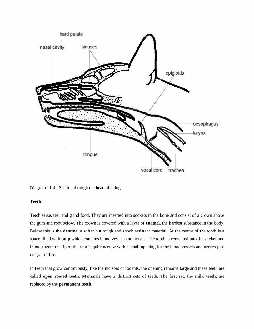
Diagram 11.4 - Section through the head of a dog
Teeth
Teeth seize, tear and grind food. They are inserted into sockets in the bone and consist of a crown above
the gum and root below. The crown is covered with a layer of enamel, the hardest substance in the body.
Below this is the dentine, a softer but tough and shock resistant material. At the centre of the tooth is a
space filled with pulp which contains blood vessels and nerves. The tooth is cemented into the socket and
in most teeth the tip of the root is quite narrow with a small opening for the blood vessels and nerves (see
diagram 11.5).
In teeth that grow continuously, like the incisors of rodents, the opening remains large and these teeth are
called open rooted teeth. Mammals have 2 distinct sets of teeth. The first set, the milk teeth, are
replaced by the permanent teeth.

Diagram 11.5 - Structure of a tooth
Types Of Teeth
All the teeth of fish and reptiles are similar but mammals usually have four different types of teeth.
The incisors are the chisel-shaped ‗biting off‘ teeth at the front of the mouth. In rodents and rabbits the
incisors never stop growing (open-rooted teeth). They must be worn or ground down continuously by
gnawing. They have hard enamel on one surface only so they wear unevenly and maintain their sharp
cutting edge.
The largest incisors in the animal kingdom are found in elephants, for tusks are actually giant incisors.
Sloths have no incisors at all, and sheep have no incisors in the upper jaw (see diagram 11.6). Instead
there is a horny pad against which the bottom incisors cut.
The canines or ‗wolf-teeth‘ are long, cone-shaped teeth situated just behind the incisors. They are
particularly well developed in the dog and cat families where they are used to hold, stab and kill the prey
(see diagram 11.7).

The tusks of boars and walruses are large canines while rodents and herbivores like sheep have no (or
reduced) canines. In these animals the space where the canines would normally be is called the diastema.
In rodents like the rat and beaver it allows the debris from gnawing to be expelled easily.
The cheek teeth or premolars and molars crush and grind the food. They are particularly well developed
in herbivores where they have complex ridges that form broad grinding surfaces (see diagram 11.6).
These are created from alternating bands of hard enamel and softer dentine that wear at different rates.
In carnivores the premolars and molars slice against each other like scissors and are called carnassial
teeth see diagram 11.7). They are used for shearing flesh and bone.
Dental Formula
The numbers of the different kinds of teeth can be expressed in a dental formula. This gives the numbers
of incisors, canines, premolars and molars in one half of the mouth. The numbers of these four types of
teeth in the left or right half of the upper jaw are written above a horizontal line and the four types of
teeth in the right or left half of the lower jaw are written below it.
Thus the dental formula for the sheep is:
0.0.3.3
3.1.3.3
It indicates that in the upper right (or left) half of the jaw there are no incisors or canines (i.e. there is a
diastema), three premolars and three molars. In the lower right (or left) half of the jaws are three incisors,
one canine, three premolars and three molars (see diagram 11.6).

Diagram 11.6 - A sheep‘s skull
The dental formula for a dog is:
3.1.4.2
3.1.4.3
The formula indicates that in the right (or left) half of the upper jaw there are three incisors, one canine,
four premolars and two molars. In the right (or left) half of the lower jaw there are three incisors, one
canine, four premolars and three molars (see diagram 11.7).

Diagram 11.7 - A dog‘s skull
Oesophagus
The oesophagus transports food to the stomach. Food is moved along the oesophagus, as it is along the
small and large intestines, by contraction of the smooth muscles in the walls that push the food along
rather like toothpaste along a tube. This movement is called peristalsis (see diagram 11.8).
Diagram 11.8 - Peristalsis
Stomach

The stomach stores and mixes the food. Glands in the wall secrete gastric juice that contains enzymes to
digest protein and fats as well as hydrochloric acid to make the contents very acidic. The walls of the
stomach are very muscular and churn and mix the food with the gastric juice to form a watery mixture
called chyme (pronounced kime). Rings of muscle called sphincters at the entrance and exit to the
stomach control the movement of food into and out of it (see diagram 11.9).
Diagram 11.9 - The stomach
Small Intestine
Most of the breakdown of the large food molecules and absorption of the smaller molecules take place in
the long and narrow small intestine. The total length varies but it is about 6.5 metres in humans, 21 metres
in the horse, 40 metres in the ox and over 150 metres in the blue whale.
It is divided into 3 sections: the duodenum (after the stomach), jejunum and ileum. The duodenum
receives 3 different secretions:
1) Bile from the liver;
2) Pancreatic juice from the pancreas and
3) Intestinal juice from glands in the intestinal wall.
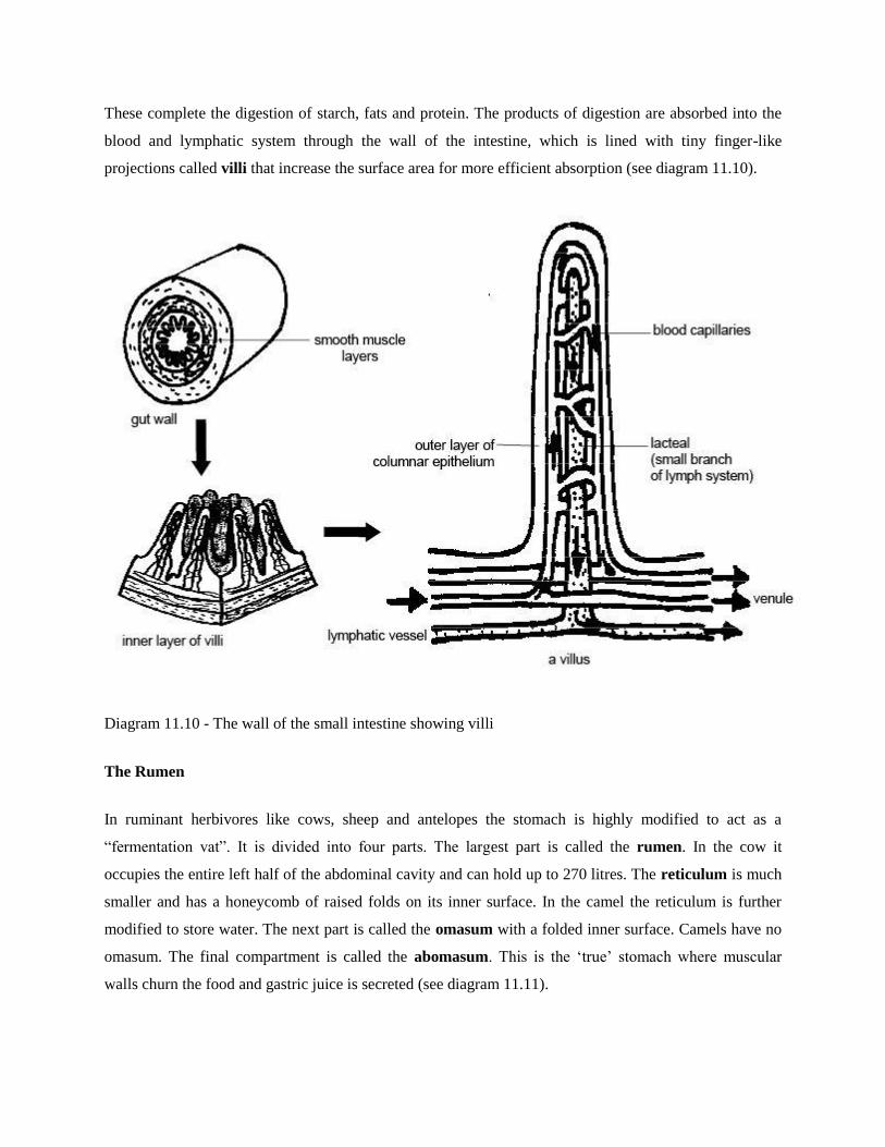
These complete the digestion of starch, fats and protein. The products of digestion are absorbed into the
blood and lymphatic system through the wall of the intestine, which is lined with tiny finger-like
projections called villi that increase the surface area for more efficient absorption (see diagram 11.10).
Diagram 11.10 - The wall of the small intestine showing villi
The Rumen
In ruminant herbivores like cows, sheep and antelopes the stomach is highly modified to act as a
―fermentation vat‖. It is divided into four parts. The largest part is called the rumen. In the cow it
occupies the entire left half of the abdominal cavity and can hold up to 270 litres. The reticulum is much
smaller and has a honeycomb of raised folds on its inner surface. In the camel the reticulum is further
modified to store water. The next part is called the omasum with a folded inner surface. Camels have no
omasum. The final compartment is called the abomasum. This is the ‗true‘ stomach where muscular
walls churn the food and gastric juice is secreted (see diagram 11.11).

Diagram 11.11 - The rumen
Ruminants swallow the grass they graze almost without chewing and it passes down the oesophagus to
the rumen and reticulum. Here liquid is added and the muscular walls churn the food. These chambers
provide the main fermentation vat of the ruminant stomach. Here bacteria and single-celled animals start
to act on the cellulose plant cell walls. These organisms break down the cellulose to smaller molecules
that are absorbed to provide the cow or sheep with energy. In the process, the gases methane and carbon
dioxide are produced. These causes the ―burps‖ you may hear cows and sheep making.
Not only do the micro-organisms break down the cellulose but they also produce the vitamins E, B and
K for use by the animal. Their digested bodies provide the ruminant with the majority of its protein
requirements.
In the wild grazing is a dangerous activity as it exposes the herbivore to predators. They crop the grass as
quickly as possible and then when the animal is in a safer place the food in the rumen can be regurgitated
to be chewed at the animal‘s leisure. This is ‗chewing the cud‘ or rumination. The finely ground food
may be returned to the rumen for further work by the microorganisms or, if the particles are small enough,
it will pass down a special groove in the wall of the oesophagus straight into the omasum. Here the
contents are kneaded and water is absorbed before they pass to the abomasum. The abomasum acts as a
―proper‖ stomach and gastric juice is secreted to digest the protein.

Large Intestine
The large intestine consists of the caecum, colon and rectum. The chyme from the small intestine that
enters the colon consists mainly of water and undigested material such as cellulose (fibre or roughage). In
omnivores like the pig and humans the main function of the colon is absorption of water to give solid
faeces. Bacteria in this part of the gut produce vitamins B and K.
The caecum, which forms a dead-end pouch where the small intestine joins the large intestine, is small in
pigs and humans and helps water absorption. However, in rabbits, rodents and horses, the caecum is very
large and called the functional caecum. It is here that cellulose is digested by micro-organisms. The
appendix, a narrow dead end tube at the end of the caecum, is particularly large in primates but seems to
have no digestive function.
Functional Caecum
The caecum in the rabbit, rat and guinea pig is greatly enlarged to provide a ―fermentation vat‖ for micro-
organisms to break down the cellulose plant cell walls. This is called a functional caecum (see diagram
11.12). In the horse both the caecum and the colon are enlarged. As in the rumen, the large cellulose
molecules are broken down to smaller molecules that can be absorbed. However, the position of the
functional caecum after the main areas of digestion and absorption, means it is potentially less effective
than the rumen. This means that the small molecules that are produced there can not be absorbed by the
gut but pass out in the faeces. The rabbit and rodents (and foals) solve this problem by eating their own
faeces so that they pass through the gut a second time and the products of cellulose digestion can be
absorbed in the small intestine. Rabbits produce two kinds of faeces. Softer night-time faeces are eaten
directly from the anus and the harder pellets you are probably familiar with, that have passed through the
gut twice.

Diagram 11.12 - The gut of a rabbit
The Gut of Birds
Birds‘ guts have important differences from mammals‘ guts. Most obviously, birds have a beak instead of
teeth. Beaks are much lighter than teeth and are an adaptation for flight. Imagine a bird trying to take off
and fly with a whole set of teeth in its head! At the base of the oesophagus birds have a bag-like structure
called a crop. In many birds the crop stores food before it enters the stomach, while in pigeons and doves
glands in the crop secretes a special fluid called crop-milk which parent birds regurgitate to feed their
young. The stomach is also modified and consists of two compartments. The first is the true stomach with
muscular walls and enzyme secreting glands. The second compartment is the gizzard. In seed eating birds
this has very muscular walls and contains pebbles swallowed by the bird to help grind the food. This is
the reason why you must always supply a caged bird with grit. In birds of prey like the falcon the walls of
the gizzard are much thinner and expand to accommodate large meals (see diagram 11.13).

Diagram 11.13 - The stomach and small intestine of a hen
Digestion
During digestion the large food molecules are broken down into smaller molecules by enzymes. The three
most important groups of enzymes secreted into the gut are:
1. Amylases that split carbohydrates like starch and glycogen into monosaccharides like
glucose.
2. Proteases that split proteins into amino acids.
3. Lipases that split lipids or fats into fatty acids and glycerol.
Glands produce various secretions which mix with the food as it passes along the gut.
These secretions include:
1. Saliva secreted into the mouth from several pairs of salivary glands (see diagram 11.3).
Saliva consists mainly of water but contains salts, mucous and salivary amylase. The
function of saliva is to lubricate food as it is chewed and swallowed and salivary amylase
begins the digestion of starch.
2. Gastric juice secreted into the stomach from glands in its walls. Gastric juice contains
pepsin that breaks down protein and hydrochloric acid to produce the acidic conditions

under which this enzyme works best. In baby animals rennin to digest milk is also
produced in the stomach.
3. Bile produced by the liver. It is stored in the gall bladder and secreted into the
duodenum via the bile duct (see diagram 11.14). (Note that the horse, deer, parrot and rat
have no gall bladder). Bile is not a digestive enzyme. Its function is to break up large
globules of fat into smaller ones so the fat splitting enzymes can gain access the fat
molecules.
Diagram 11.14 - The liver, gall bladder and pancreas
Pancreatic juice
The pancreas is a gland located near the beginning of the duodenum (see diagram 11.14). In most
animals it is large and easily seen but in rodents and rabbits it lies within the membrane linking the loops
of the intestine (the mesentery) and is quite difficult to find. Pancreatic juice is produced in the
pancreas. It flows into the duodenum and contains amylase for digesting starch, lipase for digesting fats
and protease for digesting proteins.
Intestinal juice
Intestinal juice is produced by glands in the lining of the small intestine. It contains enzymes for
digesting disaccharides and proteins as well as mucus and salts to make the contents of the small intestine
more alkaline so the enzymes can work.

Absorption
The small molecules produced by digestion are absorbed into the villi of the wall of the small intestine.
The tiny finger-like projections of the villi increase the surface area for absorption. Glucose and amino
acids pass directly through the wall into the blood stream by diffusion or active transport. Fatty acids and
glycerol enter vessels of the lymphatic system (lacteals) that run up the centre of each villus.
The Liver
The liver is situated in the abdominal cavity adjacent to the diaphragm (see diagrams 2 and 14). It is the
largest single organ of the body and has over 100 known functions. Its most important digestive functions
are:
1. the production of bile to help the digestion of fats (described above) and
2. the control of blood sugar levels
Glucose is absorbed into the capillaries of the villi of the intestine. The blood stream takes it directly to
the liver via a blood vessel known as the hepatic portal vessel or vein (see diagram 11.15).
The liver converts this glucose into glycogen which it stores. When glucose levels are low the liver can
convert the glycogen back into glucose. It releases this back into the blood to keep the level of glucose
constant. The hormone insulin, produced by special cells in the pancreas, controls this process.

Diagram 11.15 - The control of blood glucose by the liver
Other functions of the liver include:
3. making vitamin A,
4. making the proteins that are found in the blood plasma (albumin, globulin and fibrinogen),
5. storing iron,
6. removing toxic substances like alcohol and poisons from the blood and converting them to
safer substances,
7. producing heat to help maintain the temperature of the body.

Diagram 11.16 - Summary of the main functions of the different regions of the gut
Summary
The gut breaks down plant and animal materials into nutrients that can be used by animals‘
bodies.
Plant material is more difficult to break down than animal tissue. The gut of herbivores is
therefore longer and more complex than that of carnivores. Herbivores usually have a
compartment (the rumen or functional caecum) housing micro-organisms to break down the
cellulose wall of plants.

Chewing by the teeth begins the food processing. There are 4 main types of teeth: incisors,
canines, premolars and molars. In dogs and cats the premolars and molars are adapted to slice
against each other and are called carnassial teeth.
Saliva is secreted in the mouth. It lubricates the food for swallowing and contains an enzyme to
break down starch.
Chewed food is swallowed and passes down the oesophagus by waves of contraction of the wall
called peristalsis. The food passes to the stomach where it is churned and mixed with acidic
gastric juice that begins the digestion of protein.
The resulting chyme passes down the small intestine where enzymes that digest fats, proteins and
carbohydrates are secreted. Bile produced by the liver is also secreted here. It helps in the
breakdown of fats. Villi provide the large surface area necessary for the absorption of the
products of digestion.
In the colon and caecum water is absorbed and micro organisms produce some vitamin B and
K. In rabbits, horses and rodents the caecum is enlarged as a functional caecum and micro-
organisms break down cellulose cell walls to simpler carbohydrates. Waste products exit the body
via the rectum and anus.
The pancreas produces pancreatic juice that contains many of the enzymes secreted into the
small intestine.
In addition to producing bile the liver regulates blood sugar levels by converting glucose
absorbed by the villi into glycogen and storing it. The liver also removes toxic substances from
the blood, stores iron, makes vitamin A and produces heat.
Worksheet
Use the Digestive System Worksheet to help you learn the different parts of the digestive system and their
functions.
Test Yourself
Then work through the Test Yourself below to see if you have understood and remembered what you
learned.
1. Name the four different kinds of teeth
2. Give 2 facts about how the teeth of cats and dogs are adapted for a carnivorous diet:

1.
2.
3. What does saliva do to the food?
4. What is peristalsis?
5. What happens to the food in the stomach?
6. What is chyme?
7. Where does the chyme go after leaving the stomach?
8. What are villi and what do they do?
9. What happens in the small intestine?
10. Where is the pancreas and what does it do?
11. How does the caecum of rabbits differ from that of cats?
12. How does the liver help control the glucose levels in the blood?
13. Give 2 other functions of the liver:
1.
2.
Test Yourself Answers
Websites
http://www.second-opinions.co.uk/carn_herb_comparison.html Second opinion. A good
comparison of the guts of carnivores and herbivores
http://www.chu.cam.ac.uk/~ALRF/giintro.htm The gastrointestinal system. A good comparison
of the guts of carnivores and herbivores with more advanced information than in the previous site.
http://www.westga.edu/~lkral/peristalsis/index.html Peristalsis animation.

http://en.wikipedia.org/wiki/Digestion Wikipedia on digestion with links to further information
on most aspects. Like most websites this is mainly about human digestion but much is applicable
to animals.
MODULE 4: Physiological functions of farm animals
CONTENTS
1.0: Introduction
2.0: Objectives
3.0: Main contents
3. 1: Homeostasis
3.1.2: Nutrition and digestion
3.1.3: Respiratory and thermoregulation
3.1.4: Physiology and temperature regulation
3.1.5: Excretion and reproduction
3.1.6: Endocrinology
3.1.7: The blood and circulation
3.1.8: Lactation
3.1.9: Milk let down and egg production
3.1.10: Physiology of egg production
3.1.11: Water balance
4.0 Conclusion
5.0 Summary
6.0 Tutor-Marked Assignment
7.0 References/Further Reading
Physiological functions of animals
1.0 INTRODUCTION
There are many types of physiological functions that help in the wellbeing of farm animals
among are,homeostatic, nutrition and digestion, respiration. Temperature regulation, excretion
and reproduction, endocrinology, the blood and circulation, lactation, milk let down and egg
production, water balance which are essential and also important in all activities of the body.

2.0 OBJECTIVES
By the end of this unit, you should be able to:
Know the different physiological functions
Identify the different type of digestion
Know the different types of excretion and reproductions
3.0 MAIN CONTENT
3.1: Homeostasis. This is a physiological process of a body to maintain constant internal
environment in response to fluctuations in outer external environment. Internal environment
refers the interstitial fluids surrounding individual cells while external environment refers to the
environment in which organisms live. Maintaining a dynamic equilibrium between body‘s
internal environment and changing external environment requires constant monitoring and
adjustments. This adjusting of physiological systems within the body is called homeostatic
regulation. The liver, the kidneys and the brain (hypothalamus, the autonomic nervous system
and the endocrine system help maintain homeostasis. An inability to maintain homeostasis may
lead to death or a disease, a condition known as homeostatic imbalance. Other diseases which
result from a homeostatic imbalance include diabetes, dehydration, hypoglycemia,
hyperglycemia, gout, hypothermia etc. Medical intervention can help restore homeostasis and
possibly prevent permanent damage to the organs.
3.1b: The control system for regulation of homeostasis is an open system, which involves
stimulus as input and response as output.
Control system or homeostasis regulation involves five basic components:
1. Stimulus: any physical, chemical or environmental factors or disturbance that causes
deviation of normal body‘s environment
2. Receptor or detector: The receptor receives the stimulus and forward to the control
center. Eg. neurons
3. Control center or regulator: The control center or coordinator center receives and
processes information from the receptor. It set the normal reference point or setup point
for any physiological processes. Eg. Hypothalamus or brain

4. Effector: The control center commands the effector to respond to the stimulus. Eg.
Glands, muscles
5. Response: It is the reaction to the stimulus. It is a corrective measure toward the
disturbance. It can oppose or enhance the stimulus. All the physiological process have
their own reference point or setup point. Any deviation from reference point activates the
control system. The control system after optimizing the physiological process to its
reference point is always feed backed. The control system is known as feed-back
mechanism. There are two form of feed-back mechanism
1. Negative feed-back mechanism
2. Positive feed-back mechanism
The Internal Environment | Back to Top There are two types of extracellular fluids in animals:
the extracellular fluid that surrounds and bathes cellsplasma, the liquid component of the blood.
Internal components of homeostasis:
1. Concentration of oxygen and carbon dioxide
2. pH of the internal environment
3. Concentration of nutrients and waste products
4. Concentration of salt and other electrolytes
5. Volume and pressure of extracellular fluid Control Systems
. Closed Systems has two components: a sensor and an effect or, such as a thermostat (sensor)
and furnace (effect or). Most physiological systems in the body use feedback to maintain the
body's internal environment. Extrinsic Most homeostatic systems are extrinsic: they are
controlled from outside the body. Endocrine and nervous systems are the major control systems
in higher animals.
The nervous system depends on sensors in the skin or sensory organs to receive stimuli and
transmit a message to the spinal cord or brain. Sensory input is processed and a signal is sent to
an effector system, such as muscles or glands, whichaffects the response to the stimulus. The
endocrine system is the second type of extrinsic control, and involves a chemical component to
the reflex. Sensors detect a change within the body and send a message to anendocrine effector
(parathyroid), which makes PTH. PTH is released into the blood when blood calcium levels are
low. PTH causes bone to release calcium into the bloodstream, raising the blood calcium levels

and shutting down the production of PTH. Some reflexes have a combination of nervous and
endocrine response. The thyroid gland secretes thyroxin (which controls the metabolic rate) into
the bloodstream. Falling levels of thyroxin stimulate receptors in the brain to signal the
hypothalamus to release a hormone that acts on the pituitary gland to release thyroid-stimulating
hormone (TSH) into the blood. TSH acts on the thyroid, causing it to increase production of
thyroxin. Intrinsic Local, or intrinsic, controls usually involve only one organ or tissue. When
muscles use more oxygen, and also produce more carbon dioxide, intrinsic controls cause
dilation of the blood vessels allowing more blood into those active areas of the muscles.
Eventually the vessels will return to "normal".Feedback Systems in Homeostasis | Negative
feedback control mechanisms (used by most of the body's systems) are called negative because
the information caused by the feedback causes a reverse of the response.TSH is an example:
blood levels of TSH serve as feedback for production of TSH.
Positive feedback control is used in some cases. Input increases or accelerates the response.
During uterine contractions, oxytocin is produced. Oxytocin causes an increase infrequency and
strength of uterine contractions. This in turn causes further production of oxytocin, etc.
Homeostasis depends on the action and interaction of a number of body systems to maintain a
range of conditions within which the body can best operate. Body Systems and Homeostasis |
Eleven major organ systems are present within animals, although some animals lack one or more
of them. The vertebrate body has two cavities: the thoracic, which contains the heart and lungs;
and the abdominal, which contains digestive organs. The head, or cephalic region, contains four
of the five senses as well as a brain encased in the bony skull. These organ systems can be
grouped according to their functions.
The integumentary, skeletal, and muscular systems. Image from Purves et al., Life:
The Science of Biology, 4th Edition, by Sinauer Associates (www.sinauer.com) andWH Freeman
(www.whfreeman.com), used with permission.
Muscular System facilitates movement and locomotion. The muscular system produces body
movements, body heat, maintains posture, and supports the body. Muscle fibers are the main cell
type. Action of this system is closely tied to that of the skeletal system. Skeletal System provides
support and protection, and attachment points for muscles. The skeletal system provides rigid
framework for movement. It supports and protects the body and body parts, produces blood cells,
and stores minerals. Skin or Integument is the outermost protective layer. It prevents water loss

from and invasion of foreign microorganisms and viruses into the body. There are three layers of
the skin. The epidermis is the outer, thinner layer of skin. Basal cells continually undergo
mitosis. Skin is waterproof because keratin, a protein is produced. The next layer is the dermis a
layer of fibrous connective tissue. Within the dermis many structures are located, such as sweat
glands, hair follicles and oil glands. The subcutaneous layer is composed of loose connective
tissue. Adipose tissue occurs here, serving primarily for insulation. Nerve cells run through this
region, as doarteries and veins.
3.1.2:Animal Nutrition and Digestion
Animal Nutrition and Digestion in today‘s society, one can‘t turn on the TV or read a newspaper
without seeing or reading information about health issues. What is the major factor discussed in
these issues? Nutrition. Along with nutrition, genetics and environment are all factors that
influence your health. Just like in humans, agricultural animals are influenced by these same
factors. If animals don‘t have proper nutrition, their growth rate, reproduction rate, immunity,
and well-being are all affected. Just as proper nutrition is vital for your good health and proper
function, it is just as vital for the health and function of agricultural animals.
3.1.2a: Importance of Nutrition and Digestion
Nutrition is important for a variety of reasons. Animals need the proper nutrition for growth and
maintenance, and to provide energy for work and vital functions. Maintenance is the nutrition
required for an animal to maintain its current weight. Energy is the ability of the body to perform
functions. Proper nutrition is also needed to maintain body temperature, produce milk, reproduce,
and develop proper bone structures. Without proper nutrition, animals can develop health
problems, which could result in treatment costs or even fatality. Good nutrition is essential for all
of the systems of an animal to function and work together properly. Animals that do not receive
the proper nutrition are more likely to develop health and reproductive problems, and be less
productive and marketable. Nutrition is important to have healthy animals, and, in turn, healthy
animals help ensure profitability in agricultural operations. Producers must understand not only
what nutrition an animal needs but also how to supply that needed nutrition. Producers must
know what ration is appropriate for a specific animal and how to provide a balanced ration. A
ration is the feed an animal receives over a 24-hour period. A variety of feedstuffs, or basic
ingredients, are used in rations, and producers must choose those that best suit their needs. A
ration with all the nutrients an animal needs is a balanced ration. Digestion is the process of

breaking down feeds into simple substances that can be absorbed into the bloodstream and used
by the body‘s cells. It is important for producers to understand the digestive process because it
helps in selecting the proper feed for the animal‘s type of digestive system. The digestive system
is the system in which food is acted upon through physical (chewing) and chemical (stomach
acids) means. It includes all parts from the mouth to the anus. Understanding the digestive
system can help the producer become more efficient in feeding, resulting in a more cost-effective
livestock enterprise. It also helps the producer have a better understanding of animal health and
problems that may occur. Understanding the chemical and physical changes that take place after
an animal eats is important in noticing health problems related to digestion. Jupiter Images Some
animals need just enough nutrients to maintain current body weight.
3.1.2c: Animal Nutrition and Digestion
● Discuss the importance of nutrition and digestion for animal growth.
● Explore the basics of animal physiology.
● Determine how nutrients are used by various livestock species.
● Identify classes and sources of nutrients.
● Identify symptoms of nutrient deficiencies.
●Explain the role of feed additives in livestock nutrition.
●Formulate a feed ration using the Pearson square.
●Compare and contrast animal digestive systems.
3.1.3: The digestive and respiratory systems. Respiratory System moves oxygen from the
external environment into the internal environment; also removes carbon dioxide. The respiratory
system exchanges gas between lungs(gills in fish) and the outside environment. It also maintains
pH of the blood and facilitates exchange of carbon dioxide and oxygen. Digestive System digests
and absorbs food into nutrient molecules by chemical and mechanical breakdown; eliminates
solid wastes into the environment. Digestion isaccomplished by mechanical and chemical
means,breaking food into particles small enough to pass into bloodstream. Absorption of food
molecules occurs in the smallintestine and sends them into circulatory system. The digestive
system also recycles water and reclaims vitamins from food in the large intestine. transports
oxygen, carbon dioxide, nutrients, waste products, immune components, and hormones. Major
organs include the heart, capillaries,arteries, and veins. The lymphatic system also transports

excess fluids to and from circulatory system and transports fat to the heart.Immune System
(Lymphatic system, Figure 3) defends the internal environment from invading microorganisms
and viruses, as well as cancerous cell growth. The immunesystem provides cells that aid in
protection of the body from disease via the antigen/antibody response. A variety of general
responses are also part of this system.
3.1.3.2: Respiratory system and thermoregulation
Respiratory system and thermoregulation Air sacs and lungs the respiratory system is one of the
major systems of the body. It has a number of very important functions including the provision
of oxygen, the removal of carbon dioxide, the removal of excess heat (thermoregulation) and
vocal communication. The respiratory system is a complex one and while there are some
similarities with that of mammals, there are a number of quite significance differences
Nasal cavity the openings to the nasal cavity, the nares, lie at the point of the base of the comb on
the top beak or mandible. The nasal cavity occupies a triangular shaped space between the nares
and the margin of the eye and within the beak. Between the integument and the nasal cavity lie
the lacrimal sinuses that empty into the cavity through the lateral wall. The lateral wall of the
cavity has three conchae, or projections, into the cavity:
i. Anterior – of squamous epithelium – a single layer of attended cells
ii. ii. Medial – ciliated columnar epithelium – special cube shaped cells with cilia or
hairs that trap foreign material
iii. Posterior – olfactory membrane – that gives the sense of smell
Smell, protection and reduction of electrolyte the odour or smell of any material or object is a
result of minute quantities of special chemicals that are detected by the olfactory membrane that
in turn sends a signal via the olfactory nerve to the brain where it will be recognized for what it
is. The epithelium of the nasal cavity is well endowed with mucosal a gland which produce
mucous that helps to keep foreign material from gaining entry to the body through the respiratory
system. The nasal glands are small and located on either side. Their action is to supplement the
action of the kidneys by reducing the electrolyte content of body ids, especially when the level is
higher than the kidneys can handle (electrolyte – common chemical salts e.g. sodium chloride).
Oropharynx (mouth and pharynx)The oropharynx consists of the mouth and the pharynx that is
located immediately behind it.The palate is part hard and part soft. The choanal opening (from

the nasal cavity) is the cleft in the palate. The pharynx begins between the choanal opening and
the common opening for the auditory tubes and extends to the rear to include that section of the
oral cavity carrying the base of the tongue, the tip of which is located in the mouth. Behind the
base of the tongue is found the rima glottidis or opening into the larynx, which is sometimes
called the cranial larynx. This opening is located in a conspicuous mound called the laryngeal
prominence. The opening into the larynx is a median slit that is supported one ach side by the
arytenoid cartilages. These are special cartilages with a shape resembling the mouth of a jug or
pitcher. It varies in length from approximately 8.5 mm in the female to 11mm in the male.
During gasping while the bird is in a state of respiratory distress it can open to a width of 7 to 9
mm. There are no vocal cords, epiglottis and thyroid cartilages that are normally found in
mammals.
Trachea:This organ is a long tube with the function of moving the respiratory gases from the
upper respiratory system to the organs of respiration – the lungs and air sacs or from the air sacs
and lungs to the upper respiratory organs. The trachea in medium sized adults measures
between15 and 18 centimetres. It is held open permanently by 108 to 125 cartilaginous rings
each one complete and lapping its neighbour. This arrangement prevents the trachea from
collapsing or compressing but allows elongation and The syrinx is the vocal organ of the fowl. It
is located at the caudal end of the trachea and issus pended within the clavicular air sac. At rest, it
is compressed laterally (at the sides). It consists of the pessulus, a wedge shaped cartilage located
where the trachea divides into two to form the two bronchi plus the last four specialised
cartilaginous rings at the bottom of the trachea. Below this section there are 4-7 thin,
On entering the lungs, the primary bronchi divide to form four series of secondary bronchi and
these, in turn, divide again to form numerous anastomising tertiary bronchi orpara bronchi. The
secondary and tertiary bronchi are lined with squamous epithelium and not the ciliated
epithelium of the primary bronchi. The tertiary bronchi are arranged in layers. Theyare not blind
ending, but join others that in turn lead back to the secondary and primarybronchi. Ultimately,
the bronchial system is continuous.
Gas exchange Leading off from the bronchi in the lungs are a large number of extremely small
air capillaries(ducts) that are interlocked with the capillaries of the lung circulatory system.
These interlocked capillaries are the lungs‘ gas exchange system and are very thin which
accommodates gaseous exchange. The blood/gas barrier in this area consists of whatever cell

layers separate the two systems – the blood circulatory system and the air supply system of the
lungs. These layers are:
. Single cell epithelial wall of the air capillary
ii. Base membrane one cell thickiii. Single cell epithelial wall of the blood capillary
Lungs
3.3: Respiration is the movement of oxygen from the outside environment to the cells within
tissues, and the transport of carbon dioxidein the opposite direction.
The physiological definition of respiration differs from the biochemical definition, which refers
to a metabolic process by which an organism obtains energy (in the form of ATP) by oxidizing
nutrients and releasing waste products. Although physiologic respiration is necessary to sustain
cellular respiration and thus life in animals, the processes are distinct: cellular respiration takes
place in individual cells of the organism, while physiologic respiration concerns the diffusion and
transport of metabolites between the organism and the external environment.
In animals with lungs, physiological respiration involves respiratory cycles of inhaled and
exhaledbreaths. Inhalation (breathing in) is usually an active movement. The contractions of the
diaphragm muscle cause a pressure variation, which is equal to the pressures caused by elastic,
resistive and inertial components of the respiratory system. In contrast, exhalation (breathing out)
is usually a passive process. Breathing in, brings air into the lungs where the process of gas
exchange takes place between the air in thealveoliand thebloodin the pulmonarycapillaries
The process of breathing does not fill the alveoli with atmospheric air during each inhalation
(about 350 ml per breath), but the inhaled air is carefully diluted and thoroughly mixed with a
large volume of gas (about 2.5 liters in adult humans) known as the functional residual capacity
which remains in the lungs after each exhalation, and whosegaseous composition differs
markedly from that of the ambient air. Physiological respiration involves the mechanisms that
ensure that the composition of the functional residual capacity iskept constant, and equilibrates
with the gases dissolved in the pulmonary capillary blood, and thus throughout the body. Thus, in
preciseusage, the words breathing and ventilation are hyponyms, not synonyms, of respiration;
but this prescription is not consistently followed, even by most health care providers, because the

term respiratory rate(RR) is a well-established term in health care, even though it would need to
be consistently replaced with ventilation rate if the precise usage were to be followed.
3.1.4: Physiology, Temperature Regulation
(Introduction)
Thermoregulation, by definition, is a mechanism by which mammals maintain body temperature
by tightly controlled self-regulation, no matter the temperature of their surroundings.
Temperature regulation is a type of homeostasis, which is a process that biological systems use
to preserve a stable internal state to survive. Ectotherms are animals that depend on their external
environment for their body heat, and endotherms are animals that use thermoregulation to
maintain a somewhat consistent internal body temperature to survive, even when their external
environment changes. Humans and other mammals and birds are endotherms. Human beings
have a normal core, or internal, temperature of around 37 degrees Celsius, which is equivalent to
around 98.6 degrees Fahrenheit. Core temperature is most accurately measured via rectal probe
thermometer. This is the temperature at which the human body‘s systems work together at their
optimum, which is the reason the body has such tightly regulated mechanisms. Thermoregulation
is crucial to human life. Without thermoregulation, the human body would not be able to
adequately function and, inevitably, will expire.
Issues of Concern
When the body‘s ability to thermoregulation becomes hindered and is left untreated, organ
failure is imminent. Blood flow will be reduced, leading to ischemia, and, ultimately, multiple
organ failures.
Cellular
Viral illness or another infectious disease can cause a person to develop a fever, and the body no
longer has that same core temperature of 37 degrees Celsius. This is because when the body
experiences an infection from invading pathogens, it tries to fight back by releasing pyrogens

such as cytokines, prostaglandins, and thromboxane—all of which increase the body‘s
temperature. By releasing these pyrogens, the foreign pathogens are not able to breed. This
allows for antibodies to develop and enzymes to be activated to fight the infection further.
Development
The brain, or more specifically the hypothalamus, controls thermoregulation. If the
hypothalamus senses external temperatures growing too hot or too cold, it will automatically
send signals to the skin, glands, muscles, and organs. For example, when the body is in a very
hot external environment, or simply undergoing high activity levels such as exercise, it‘s
temperature will rise, causing the hypothalamus to send signals to the cells of the skin that
produce sweat. Sweating is the body's approach to cooling itself down. As the body‘s
temperature rises, sweat is expelled, the muscles relax, and body hair lies flat against the skin.
These are all ways to release heat and therefore lower the temperature of the body. In contrast,
when the body experiences a cold environment, the skeletal muscles tense up leading to the
shivering reflex, and the arrector pili muscles, a type of smooth muscle, raise the bodily hair
follicles where they are attached. These processes, in turn, create warmth and trap heat,
respectively.
Organ Systems Involved
Multiple organs and body systems are affected when thermoregulation is not working correctly.
During heat illness as a result of improper thermoregulation, the following organs and systems
are impaired. Notice that many of these issues cause or are influenced by the other issues.
The heart experiences an increased burden due to elevations in heart rate and increased
cardiac output.
The circulatory system sustains intravascular dehydration.
The brain suffers from cerebral ischemia and/or cerebral edema.
The gastrointestinal tract is vulnerable to hemorrhage and also sepsis because the mucosa
of the intestines increases its permeability.
The lungs suffer when ARDS results from the increase of hyperventilation, hyperpnea,
and pulmonary vasodilation.

Acute renal failure can commence because of the dehydration and impairment in
circulation.
Liver cells suffer because of the fever, ischemia, and cytokines increase in the intestinal
tract.
Various organs can result in DIC and microthrombi.
Electrolyte abnormalities are likely as well as hypoglycemia, metabolic acidosis, and
respiratory alkalosis.
When body temperatures severely decrease in hypothermia, the body‘s systems are negatively
affected. The cardiovascular system experiences dysrhythmias such as the Osborn, or J, wave on
EKG. The central nervous system's (CNS) electrical activity is noticeably diminished,
noncardiogenic pulmonary edema will occur, as well as cold diuresis, and decreases in
glomerular filtration rate (GFR) and renal blood flow (RBF). Basically, hypothermia causes pre-
glomerular vasoconstriction, which leads to decreased GFR and RBF.
Function
Slight changes in core body temperature occur every day, depending upon variables such as
circadian rhythm and menses; but otherwise, the temperature is tightly controlled. When a person
is unable to regulate his or her body temperature, various pathologies may occur. The human
body has four different methods for keeping itself at its core temperature: vaporization, radiation,
convection, and conduction. To keep the body functioning, it must be at its ideal temperature,
and for this to happen, physical factors must be sufficient. This includes having enough
intravascular volume and cardiovascular function; the body must be able to transport the rising
internal heat to its surface for release. The reason that elderly people are at higher risk for
disorders of thermoregulation is that they, as a whole, have less intravascular volume and
decreased cardiac function.
Mechanism
Thermoregulation has three mechanisms: afferent sensing, central control, and efferent
responses. There are receptors for both heat and cold throughout the human body. Afferent
sensing works through these receptors to determine if the body is experiencing either too hot or

too cold of a stimulus. Next, the hypothalamus is the central controller of thermoregulation.
Lastly, efferent responses are carried out primarily by the body‘s behavioral reactions to
fluctuations in body temperature. For example, if a person is feeling too warm, the normal
response is to remove an outer article of clothing. If a person is feeling too cold, they choose to
wear more layers of clothing. Efferent responses also consist of automatic responses by the body
to protect itself from extreme changes in temperature, such as sweating, vasodilation,
vasoconstriction, and shivering.
Related Testing
The thermoregulatory sweat test (TST) is a specific clinical test that is used to diagnose certain
conditions that cause abnormal temperature regulation and defects in sweat production in the
body. It measures a patient‘s ability to produce sweat in a controlled, heated and humidified
environment and assesses the patient's central and autonomic nervous systems to determine if the
thermoregulatory centers are working correctly.To perform the thermoregulatory sweat test, the
patient is placed in a chamber that slowly rises in temperature. Before the chamber is heated, the
patient is coated with a special kind of indicator powder that will change in color when sweat is
produced. This powder, when changing color, will be useful in visualizing which skin is
sweating versus not sweating. Results of the patient‘s sweat pattern will be documented by
digital photography, and abnormal TST patterns can indicate if there is dysfunction in the
autonomic nervous system. Certain differentials can be made depending on the type of sweat
pattern found from the TST (along with history and clinical presentation) including
hyperhidrosis, small fiber and autonomic neuropathies, multiple system atrophy, Parkinson
disease with autonomic dysfunction, and pure autonomic failure.
Pathophysiology
When external environments are exceedingly warm, or a person is engaging in an excessive
amount of physical activity, the heat is produced inside his or her body is typically transported to
the blood. The blood then carries the heat through numerous capillaries that are located directly
under the skin. Because the blood is near the surface, it can cool the person down. This cooled
blood can then be transported back through the body to prevent the body temperature from

becoming too high. Sweat is also a means by which the body cools itself down; it is created by
glands to carry out evaporation at the topmost skin layer, the epidermis, to release heat. This
describes vaporization, one of the four mechanisms used to maintain core body temperature.
Radiation is when the heat that is released from the body‘s surface is moved into the surrounding
air; convection occurs when cooler air surrounds the body‘s surface, and conduction comes into
play when a person is either immersed in cold water or uses an ice pack—their internally
generated heat is transferred to the cold water or the ice pack. This is another reason why it is
very important to stay hydrated in the heat or during physical activity—not only to maintain
adequate intravascular fluid volume, but also to aid in conduction processes that cool the body
down. When cold fluids are ingested, the heat is released into the fluid and excreted out of the
body as sweat or urine.While infection can cause the body‘s temperature to rise internally, a
number of mechanisms can cause body temperature to rise externally. As previously discussed, a
number of diseases with dysfunctional thermoregulatory mechanisms prove to be clinically
significant, including small fiber and autonomic neuropathies, radiculopathies, and central
autonomic disorders such as multiple system atrophy, Parkinson disease with autonomic
dysfunction, and pure autonomic failure. Decreased cardiac function is also a notable risk factor
for abnormal thermoregulatory function. Patients who are in externally hot environments do not
have the ability to use their heart as efficiently to pump blood to their bodies‘ surface. With the
loss of contractility, the blood is unable to be cooled, and therefore they are at risk of having
heat-related illnesses. Even patients with the diagnosis of hypertension have an increased risk of
these types of illnesses because some medications used to treat hypertension decrease the
pumping ability of the heart, thereby decreasing the heated blood‘s means of getting cool at the
body surface.
When patients are dehydrated, they contain a decreased amount of intravascular volume. This in
itself is another risk factor for dysfunctional thermoregulation. If there are not enough fluids in
the body, there is not enough blood. When there is not enough blood, the body cannot transport
the internally made heat to the body‘s surface to be cooled. Heat will remain retained, blood will
become thicker, and the heart becomes strained.In contrast, hypothermia is defined as low
internal body temperature, or a temperature less than 35 degrees Celsius (95 degrees Fahrenheit).
It is usually caused by too much heat loss from cold weather exposure or cold water immersion.

During cold water immersion, there is a reflex known as the diving reflex, which causes
vasoconstriction in the visceral muscles. The body does this as a protective mechanism to keep a
person‘s essential organs, like their heart and brain, supplied with blood. The body also does this
to prevent the brain from hypoxia, since metabolic demand will have decreased.There are two
different types of hypothermia: primary and secondary. Primary hypothermia is when the cold
environment is the direct pathology, and secondary hypothermia is when a patient‘s illness is
what causes his hypothermia. Conduction, convection, and radiation also come into play with
hypothermia; this is how the rate of heat loss is determined. Hypothermia decelerates all
physiologic roles include metabolic rate, mental awareness, nerve conduction, neuromuscular
reaction times, and both the cardiovascular and respiratory systems. As previously mentioned,
the vasoconstriction caused by hypothermia induces renal dysfunction and, eventually, cold
diuresis due to the decreased levels of ADH. These decreased levels of antidiuretic
hormone mean that the patient‘s urine will be very dilute, the definition of cold diuresis and
another outcome of hypothermia. Because of the vasoconstriction, the patient may not know he
is hypovolemic, which may lead to cardiac arrest or abrupt shock as the patient‘s vasculature is
dilated while being rewarmed, a condition referred to as the rewarming collapse.
Clinical Significance
When a person who cannot sustain his or her core body temperature presents with heat illness,
multiple organ support is indicated. It is imperative to be comprehensive in their care and utilize
any and every cooling method available to sustain their body. Support must be provided to their
respiratory, hepatic, renal, and circulatory systems. Giving fresh frozen plasma and blood
platelets as needed is also recommended. Previous animal studies have also shown that
administration of thrombomodulin can be helpful because it demonstrates anti-DIC and anti-
inflammatory mechanisms.
Malignant hyperthermia is a rare type of event, but one that is life-threatening. It can occur when
a susceptible patient is given certain anesthetic agents, notably halogenated ones or ones that are
agents that depolarize neuromuscularly. Predisposed patients who are given such anesthetics
either have abnormal ryanodine receptors (RYR-1) or have other myopathies such as Duchenne
muscular dystrophy, central core disease, neuroleptic malignant syndrome, and King-

Denborough syndrome. Ryanodine receptors are located in skeletal muscles, which typically
contain calcium channels and sarcoplasmic reticulum; these are what control the release of
calcium. However, in malignant hyperthermia, calcium release is not regulated, and this leads to
a continued release, which leads to an increased frequency of muscle metabolism. The clinical
features first would present as muscle rigidity and an overwhelmingly high heart rate and end-
tidal carbon dioxide. The person‘s temperature will increasingly elevate, and soon after, muscle
tightening and shortening will occur as well as metabolic acidosis.
The rapid treatment of malignant hyperthermia is of the utmost importance to prevent fatality.
Once malignant hyperthermia is suspected, dantrolene must be given intravenously. This will
prevent that uncontrolled release of calcium as described above. It is also important to rapidly
cool the patient, give 100% oxygen, and regulate metabolic acidosis.Serotonin syndrome and
neuroleptic malignant syndrome are other disorders of temperature regulation. They both are due
to adverse drug reactions, and they both present with clinical hyperthermia. It is important to
distinguish them from one another. Neuroleptic malignant syndrome (NMS) continues over
many days and is depicted by slow movements or the complete loss of voluntary movements.
Serotonin syndrome is much more rapid, happening over a timespan of hours and presenting
with fast movements such as tremor, muscular spasms, and hyperactive reflexes. These two
reactions must be managed rapidly to avoid organ failure, especially if the patient‘s temperature
goes past 40.5 degrees Celsius. There are both physical and pharmaceutical ways to cool the
affected patients. Physical methods include providing cool towels, ice packs, cooling blanket
systems, and intravenous infusion of fluids. Pharmaceuticals include sedatives and antipyretics
such as paracetamol and NSAIDs. (Note that in critically ill patients, it is important to consider
individual patient‘s renal function and gastric function before administering NSAIDs.)
Hyperthyroidism is another disorder of thermoregulation. In this endocrinological disease, the
core temperature is raised in the body because the basal metabolic rate is raised. Essentially, with
this disorder, all of the body‘s metabolic pathways are accelerated. Heat production is increased
as a result, and both oxygen consumption and ATP turnover are increased. The thyroid is
overactive and causes a person to feel too warm.Certain patients who have endured traumatic
brain injuries (TBI) or suffered from cardiac arrests may do well with therapeutic hypothermia.
In these patients with TBI and post-cardiac arrest, it is very dangerous for them to have an

elevation of their core temperatures. High temperatures in these patients are associated with
higher mortality rates and slower recovery, especially neurologically. Thus, hypothermia in post-
cardiac arrest patients and perinatal hypoxic patients is used to prevent neuronal injury. This is
because therapeutic hypothermia (TH) lowers the body‘s demand for metabolic oxygen. With
this mechanism, neurons can be protected before they are injured.
When TH is recommended, it should proceed very rapidly. Ice packs and intravenous
administration of cold fluids are imperative to keep the patient cool. Cooling blanket systems or
cooling suits may be utilized as well as cooling helmets and caps. For post-cardiac arrest
patients, intranasal cooling using nasal probes also may be utilized.
In contrast, some diseases that can cause decreased heat production, or hypothermia include
endocrinological diseases such as diabetes, hypothyroidism, hypoadrenalism, and
hypopituitarism. Those who are most at risk for hypothermia are elderly patients, trauma
patients, those who are mentally ill, those who are abusing alcohol, drugs, or on other types of
medication, and, lastly, those with low socio-economic status. Ordinarily, people who get
hypothermia have an underlying issue—either from a disease or surgery.
When a patient with one of these diseases presents with hypothermia, it is imperative to treat the
underlying disease to effectively treat the hypothermia. This includes treatments with
pharmaceuticals including triiodothyronine and steroids. Other causes of hypothermia from
decreased metabolic rates are people who are very malnourished, those who suffer from burns,
and also those with hypoglycemia.
Hypothermia, like hyperthermia, affects all of the human body‘s systems. When the core
temperature drops below 30 degrees Celsius, the heart responds with arrhythmias. The body may
be hypovolemic, hypokalemic, and hypomagnesemic as a result of their hypothermia;
therefore, it is important to keep the patient hydrated and to manage their electrolyte
disturbances.
The reason that patients with traumatic brain injury are likely to have impaired thermoregulation
is that the hypothalamus regulates the core body temperature. When this essential body part is
injured, the body is unable to control how it regulates the body‘s heat. Other problems in the

CNS that affect thermoregulation by the same mechanism can include tumors in the CNS, spinal
cord injuries, intracranial hemorrhage, and diseases such as Parkinson, Wernicke
encephalopathy, and multiple sclerosis.
Patients who are on the extreme spectrums of age (such as infants and elderly persons) are at
higher risk for disorders of thermoregulation and exhibit these features more readily when sick.
The very young and the very old cannot increase their metabolic rates, and this can provoke them
into hypothermia since they do not have the shivering reflux or much muscle mass. Changes that
occur as one ages include those affecting vasomotor sweating function, skeletal muscle response,
and temperature perception. Elderly persons have lower than normal internal body temperatures
and decreased immunity, so when they have an infection, they may not get the normal pyretic
response. Instead, they may present with hypothermia caused by a septic infection. In fact,
studies have shown that the core temperatures of elderly patients with sepsis within their first 24
hours of presentation are a huge predictor of their mortality. This is the reason that elderly
patients who are septic have a higher mortality rate than younger patients who are septic.
References/ Further reading
1.
Lim CL, Byrne C, Lee JK. Human thermoregulation and measurement of body
temperature in exercise and clinical settings. Ann. Acad. Med. Singap. 2008
Apr;37(4):347-53. [PubMed]
2.
Charkoudian N. Mechanisms and modifiers of reflex induced cutaneous vasodilation and
vasoconstriction in humans. J. Appl. Physiol. 2010 Oct;109(4):1221-8. [PMC free article]
[PubMed]
3.
Kurz A. Physiology of thermoregulation. Best Pract Res Clin Anaesthesiol. 2008
Dec;22(4):627-44. [PubMed]
4.

Díaz M, Becker DE. Thermoregulation: physiological and clinical considerations during
sedation and general anesthesia. Anesth Prog. 2010 Spring;57(1):25-32; quiz 33-4. [PMC
free article] [PubMed]
5.
Cheshire WP. Thermoregulatory disorders and illness related to heat and cold stress.
Auton Neurosci. 2016 Apr;196:91-104. [PubMed]
6.
Schieber AM, Ayres JS. Thermoregulation as a disease tolerance defense strategy. Pathog
Dis. 2016 Dec;74(9) [PMC free article] [PubMed]
7.
Boulant JA. Hypothalamic mechanisms in thermoregulation. Fed. Proc. 1981
Dec;40(14):2843-50. [PubMed]
8.
Romanovsky AA. Skin temperature: its role in thermoregulation. Acta Physiol (Oxf).
2014 Mar;210(3):498-507. [PMC free article] [PubMed]
9.
Zhao ZD, Yang WZ, Gao C, Fu X, Zhang W, Zhou Q, Chen W, Ni X, Lin JK, Yang J,
Xu XH, Shen WL. A hypothalamic circuit that controls body temperature. Proc. Natl.
Acad. Sci. U.S.A. 2017 Feb 21;114(8):2042-2047. [PMC free article] [PubMed]
10.
Boulant JA. Role of the preoptic-anterior hypothalamus in thermoregulation and fever.
Clin. Infect. Dis. 2000 Oct;31 Suppl 5:S157-61. [PubMed]
11.
Coon EA, Fealey RD, Sletten DM, Mandrekar JN, Benarroch EE, Sandroni P, Low PA,
Singer W. Anhidrosis in multiple system atrophy involves pre- and postganglionic
sudomotor dysfunction. Mov. Disord. 2017 Mar;32(3):397-404. [PMC free article]
[PubMed]
12.

Illigens BM, Gibbons CH. Sweat testing to evaluate autonomic function. Clin. Auton.
Res. 2009 Apr;19(2):79-87. [PMC free article] [PubMed]
Copyright © 2019, StatPearls Publishing LLC.
3.1. 5: Excretion and Reproduction
Excretory System regulates volume of internal body fluids as well as eliminates metabolic wastes
from the internal environment. The excretory system removes organic wastes from the blood,
accumulating wastes as urea in the kidneys. These wastes are then removed as urine. This system
is also responsible for maintaining fluid levels. The nervous and endocrine system coordinates
and controls actions of internal organs and body systems. Memory, learning, and conscious
thought are a few aspects of the functions of the nervous system. Maintaining autonomic
functions such as heartbeat, breathing, control of involuntary muscle actions are performed by
some of the parts of this system. Reproductive System, is mostly controlled by the endocrine
system, and is responsible for survival and perpetuation of the species. Elements of the
reproductive system produce hormones (from endocrine control) that control and aid in sexual
development. Organs of this system produce gametes that combine in the female system to
produce the next generation
3.1.5.1: Excretory and Reproductive Systems
Excretory and reproductive systems each have important but different functions in animals. The
excretory system maintains water, ion, and nitrogen balance within the body and eliminates
wastes. The reproductive system creates new individuals of a species. Both the excretory and
reproductive systems are under endocrine control but are also influenced by the external
environment.
3.1.5.2: The Excretory System
Vertebrates have a closedcirculatory system, which means that they have arteries and veins that
transport blood. Cellular metabolism produces waste and uses nutrients and water. The
circulatory system carries water and nutrients to cells and carries waste products away from
cells. Water and nutrients come from ingestion and digestion. Waste removal is critical to
maintaining internal homeostasis.

Nitrogen is the most toxic byproduct of cellular metabolism. The body must excrete nitrogen.
Nitrogen is commonly produced as ammonia from cells. Many animals will convert ammonia,
which is extremely toxic, into a less toxic form of nitrogenous waste. These include urea and uric
acid.
The habitat of an animal determines which kind of nitrogenous waste it produces. Most aquatic
animals, both invertebrates and freshwater fish, excrete ammonia. Ammonia dissolves in water
and is easy to transport outside of the body by diffusion when an animal is surrounded by water.
This happens primarily through the skin in invertebrates and through the gills of fishes.
Ammonia must be diluted by a great deal of water to be nontoxic. There is not enough water in
land animals to dilute ammonia enough, so ammonia is turned into either urea oruric acid.
Which of these an animal produces is linked to where the offspring develop. Animals
characterized by internal development of offspring such as mammals make urea. So do animals
that have eggs which develop in water, such as amphibians, sharks, and some fish that live in
saltwater.
Urea is not very toxic and can be concentrated by the kidney to conserve water. Urea is dissolved
in water, so transport of waste out of a developing egg that is sitting in freshwater is done by
diffusion. Uric acid is not water soluble and is excreted in a paste. Birds, reptiles, and insects all
produce uric acid. These animals develop in an egg on land, and waste must accumulate in the
egg as the individual grows and develops. Because uric acid is not water soluble, it can sit in the
egg and not be reabsorbed by the developing zygote.
Water balance in the body is maintained through a process called osmoregulation. The vertebrate
kidney is a specialized organ that both concentrates urea or uric acid and maintains water
balance, mostly by filtering the blood. When blood passes through the kidney, water is
reabsorbed and reused by the body. Additionally, almost all sugar, salts, and other nutrients are
reabsorbed by the body from the kidney. Waste products are taken out and eliminated, the main
waste product being nitrogenous waste. The kidney contains about 20 percent of the blood
volume at any one time.

In invertebrates, a variety of specialized structures exist for waste removal. Flame bulb
protonephridia in planaria are the most primitive specialized osmoregulation structure in
invertebrates. Annelids have an excretory system called the metanephridium, and it functions for
both osmoregulation and nitrogenous waste removal. Insects use Malpighian tubules for both
osmoregulation and filtering the hemolymph.
References/Further studies
Campbell, Neil A., Jane B. Reece, and Lawrence G. Mitchell. Biology, 5th ed. Menlo Park, CA:
Addison Wesley Longman, 1999.
Crews, David. "Animal Sexuality." Scientific American (January), 1994.
Marieb, E. N. Human Anatomy and Physiology, 4th ed. Menlo Park, CA: Benjamin/Cummings,
1998.
Schmidt-Nielsen, K. Animal Physiology. Cambridge, U.K.: Cambridge University Press, 1997.
3.1.5.3. The Reproductive System
The reproductive system functions to produce more individuals of a species. The reproductive
system is regulated by the endocrine system and is affected by environmental conditions outside
of the animal and internal conditions inside the animal. Reproductive cycles are influenced by
the time of year, amount of daylight and rainfall, and temperature, in addition to nutrition and
general health of the animal.
The main organs in the reproductive system are the gonads, which include the ovary and testis.
The ovaries produce eggs and the testes produce sperm. The egg and the sperm are gametes that
combine to form a zygote in a process called fertilization. The zygote will grow and develop into
a new individual of the species.
The egg and sperm both carry DNA, the material that contains the genetic instructions for the
growth and development of a new individual. Ga-metes are produced through meiosis and
contain only one-half of the genetic information of the parent. When the gametes come together

and form a zygote, the zygote will have half of its genetic information from the egg and half
from the sperm.
In vertebrates such as humans, egg and sperm come separately from different individuals.
Humans and most other animals (both vertebrates and invertebrates) have different sexes
commonly called males and females. In the case of animals without separate sexes, one
individual produces both eggs and sperm and is called a hermaphrodite. Most hermaphrodites,
including many annelids, pair with another individual and exchange eggs and sperm rather than
fertilizing themselves. Some platyhelminthes self fertilize. Other animals switch between being
male and female, a condition called sequential hermaphroditism.
In addition to sexual reproduction, the fusion of the egg and sperm, some animals can reproduce
asexually. In asexual reproduction, genetic material is not combined. Therefore, it produces an
offspring that is genetically identical to the parent. Types of asexual reproduction include
budding, fission, and parthenogenesis. Parthenogenesis occurs in rotifers, some bees, wasps,
ants, and several species of fish, lizards, and amphibians. Some animals switch between sexual
and asexual reproduction, and some reproduce only asexually.
Fertilization can happen internally or externally. External fertilization occurs in most sea-living
creatures as well as freshwater fish and amphibians. In external fertilization both eggs and sperm
are shed into water and fertilization, development, and growth of the zygote all take place
outside the body. Internal fertilization occurs when egg and sperm are joined inside of the body.
The zygote can develop inside of the reproductive tract, as in mammals. Birds and reptiles have
internal fertilization but lay eggs. The zygote develops inside the egg but outside of the body.
Invertebrates that have internal fertilization also lay eggs.
3.1. 6: Endocrinology
Endocrine System, function as to control the activity internal organs as well as coordinating long-
range response to external stimuli. The endocrine system secretes hormones that regulate body
metabolism, growth, and reproduction. These organs are not in contact with each other, although
they communicate by chemical messages dumped into the circulatory system.

.
Endocrinology
Illustration depicting the primary endocrine organs of
a female
System Endocrine
Significant
diseases
Diabetes, Thyroid disease,
Androgen excess
Significant tests Thyroid function tests, Blood
sugar levels
Specialist Endocrinologist
Endocrinology (fromendocrine + -ology) is a branch of biology and medicine dealing with the
endocrine system, its diseases, and its specific secretions known as hormones. It is also
concerned with the integration of developmental events proliferation, growth, and differentiation,
and the psychological or behavioral activities of metabolism, growth and development, tissue
function, sleep, digestion, respiration, excretion, mood, stress, lactation, movement,
reproduction, and sensory perception caused by hormones. Specializations include behavioral
endocrinology and comparative endocrinology.
The endocrine system consists of several glands, all in different parts of the body that secrete
hormones directly into the blood rather than into a duct system. Therefore, endocrine glands are
regarded as ductless glands. Hormones have many different functions and modes of action; one

hormone may have several effects on different target organs, and, conversely, one target organ
may be affected by more than one hormone.
Endocrinology is the study of the endocrine system in the human body. This is a system of
glands which secrete hormones. Hormones are chemicals which affect the actions of different
organ systems in the body. Examples include thyroid hormone, growth hormone, and insulin.
The endocrine system involves a number of feedback mechanisms, so that often one hormone
(such as thyroid stimulating hormone) will control the action or release of another secondary
hormone (such as thyroid hormone). If there is too much of the secondary hormone, it may
provide negative feedback to the primary hormone, maintaining homeostasis.
In the original 1902 definition by Bayliss and Starling (see below), they specified that, to be
classified as a hormone, a chemical must be produced by an organ, be released (in small
amounts) into the blood, and be transported by the blood to a distant organ to exert its specific
function. This definition holds for most "classical" hormones, but there are also paracrine
mechanisms (chemical communication between cells within a tissue or organ), autocrine signals
(a chemical that acts on the same cell), and intracrine signals (a chemical that acts within the
same cell).[4]
A neuroendocrine signal is a "classical" hormone that is released into the blood by a
neurosecretory neuron (see article on neuroendocrinology).
Hormones
Griffin and Ojeda identify three different classes of hormones based on their chemical
composition:[5]
Amines
Norepinephrine

Triiodothyronine
Examples of amine hormones
Amines, such as norepinephrine, epinephrine, and dopamine (catecholamines), are derived from
single amino acids, in this case tyrosine. Thyroid hormones such as 3,5,3‘-triiodothyronine (T3)
and 3,5,3‘,5‘-tetraiodothyronine (thyroxine, T4) make up a subset of this class because they
derive from the combination of two iodinated tyrosine amino acid residues.
Peptide and protein
Peptide hormones and protein hormones consist of three (in the case of thyrotropin-releasing
hormone) to more than 200 (in the case of follicle-stimulating hormone) amino acid residues and
can have a molecular mass as large as 31,000 grams per mole. All hormones secreted by the
pituitary gland are peptide hormones, as are leptin from adipocytes, ghrelin from the stomach,
and insulin from the pancreas.
Steroid
Cortisol
Vitamin D3
Examples of steroid hormones
Steroid hormones are converted from their parent compound, cholesterol. Mammalian steroid
hormones can be grouped into five groups by the receptors to which they bind: glucocorticoids,
mineralocorticoids, androgens, estrogens, and progestogens. Some forms of vitamin D, such as
calcitriol, are steroid-like and bind to homologous receptors, but lack the characteristic fused ring
structure of true steroids.
Work
The medical specialty of endocrinology involves the diagnostic evaluation of a wide variety of
symptoms and variations and the long-term management of disorders of deficiency or excess of
one or more hormones.
The diagnosis and treatment of endocrine diseases are guided by laboratory tests to a greater
extent than for most specialties. Many diseases are investigated through excitation/stimulation or
inhibition/suppression testing. This might involve injection with a stimulating agent to test the
function of an endocrine organ. Blood is then sampled to assess the changes of the relevant
hormones or metabolites. An endocrinologist needs extensive knowledge of clinical chemistry
and biochemistry to understand the uses and limitations of the investigations.
A second important aspect of the practice of endocrinology is distinguishing human variation
from disease. Atypical patterns of physical development and abnormal test results must be
assessed as indicative of disease or not. Diagnostic imaging of endocrine organs may reveal
incidental findings called incidentalomas, which may or may not represent disease.
Endocrinology involves caring for the person as well as the disease. Most endocrine disorders
are chronic diseases that need lifelong care. Some of the most common endocrine diseases
include diabetes mellitus, hypothyroidism and the metabolic syndrome. Care of diabetes, obesity
and other chronic diseases necessitates understanding the patient at the personal and social level
as well as the molecular, and the physician–patient relationship can be an important therapeutic
process.
Apart from treating patients, many endocrinologists are involved in clinical science and medical
research, teaching, and hospital management.

Training
Endocrinologists are specialists of internal medicine or pediatrics. Reproductive endocrinologists
deal primarily with problems of fertility and menstrual function—often training first in
obstetrics. Most qualify as an internist, pediatrician, or gynecologist for a few years before
specializing, depending on the local training system. In the U.S. and Canada, training for board
certification in internal medicine, pediatrics, or gynecology after medical school is called
residency. Further formal training to subspecialize in adult, pediatric, or reproductive
endocrinology is called a fellowship. Typical training for a North American endocrinologist
involves 4 years of college, 4 years of medical school, 3 years of residency, and 2 years of
fellowship. In the US, adult endocrinologists are board certified by the American Board of
Internal Medicine (ABIM) or the American Osteopathic Board of Internal Medicine (AOBIM) in
Endocrinology, Diabetes and Metabolism.
References
Nelson, R. J.( 2005). An Introduction to Behavioral Endocrinology, Fourth Edition. Sinauer
Associates, Sunderland, MA.
Unit 7: Blood and circulation
2.0. Objectives | Blood Circulation
After completing this section, you should know:
that the circulatory system is double consisting of pulmonary and systemic circuits with
the blood passing through the heart twice;
the differences in the structure and function of arteries, capillaries and veins;
how the pulse is produced and where it can be felt;
what tissue fluid and lymph are and how they are formed;
the names of the main arteries and veins.
3.1.7: Blood Circulation

The circulatory system is the continuous system of tubes through which the blood is pumped
around the body. It supplies the tissues with their requirements and removes waste products. In
mammals and birds the blood circulates through two separate systems - the first from the heart to
the lungs and back to the heart again (the pulmonary circulation) and the second from the heart
to the head and body and back again (the systemic circulation) (see diagram 8.12).
Diagram 8.12 - The mammalian circulatory system
The tubes through which the blood flows are the arteries, capillaries and veins. The heart
pumps blood into arteries that carry it away from the heart. The arteries divide into very thin
vessels called capillaries that form a network between the cells of the body. The capillaries then
join up again to make veins that return the blood to the heart.
Arteries

Arteries carry blood away from the heart. They have thick elastic walls that stretch and can
withstand the surges of high pressure blood caused by the heartbeat (the pulse, see later). The
arteries divide into smaller vessels called arterioles. The hole down the centre of the artery is
called the lumen. There are three layers of tissue in the walls of an artery. It is lined with
squamous epithelial cells. The middle layer is the thickest layer. It made of elastic fibres and
smooth muscle to make it stretchy. The outer fibrous layer protects the artery (see diagram 8.13).
The pulse is only felt in arteries.
Diagram 8.13 - Cross section of an artery
The Pulse
The pulse is the spurt of high pressure blood that passes along the aorta and arteries when the left
ventricle contracts. As the pulse of blood passes along an artery the elastic walls stretch. When
the pulse has passed the walls contract and this helps push the blood along. The pulse is easily
felt at certain places where an artery passes near the surface of the body. It is strongest near the
heart and becomes weaker as it travels away from the heart. The pulse disappears altogether in
the capillaries.
Capillaries
Arterioles divide repeatedly to form a network penetrating between the cells of all tissues of the
body. These small vessels are called capillaries. The walls are only one cell thick and some
capillaries are so narrow that red blood cells have to fold up to pass through them. Capillaries
form networks in tissues called capillary beds. The capillary networks in capillary beds are so
dense that no living cell is far from its supply of oxygen and food (see diagram 8.14).

Note: All arteries carry oxygenated blood except for the pulmonary artery that carries
deoxygenated blood to the lungs.
Diagram 8.14 - A capillary bed
The Formation Of Tissue Fluid And Lymph
The thin walls of capillaries allow water, some white blood cells and many dissolved substances
to diffuse through them. These form a clear fluid called tissue fluid (or extracellular fluid or
interstitial fluid) that surrounds the cells of the tissues. The tissue fluid allows oxygen and
nutrients to pass from the blood to the cells and carbon dioxide and other waste products to be
removed from the tissues (see diagram 8.15).

Diagram 8.15 - The formation of tissue fluid and lymph from blood
Some tissue fluid finds its way back into the capillaries and some of it flows into the blind-ended
lymphatic vessels that form a network in the tissues. Once the tissue fluid has entered the
lymphatics it is called lymph although its composition remains the same. The lymph vessels
have walls that are even thinner than the capillaries. This means that molecules and particles that
are larger than those that can pass into the blood stream e.g. cancer cells and bacteria can enter
the lymphatic system. These are then filtered out as the lymph passes through lymph nodes. (See
chapter 10 for more information on the lymphatic system).
Veins
Capillaries unite to form larger vessels called venules that join to form veins. Veins return blood
to the heart and since blood that flows in veins has already passed through the fine capillaries, it
flows slowly with no pulse and at low pressure. For this reason veins have thinner walls than
arteries although there have the same three layers in them as arteries (see diagram 8.16). As there
is no pulse in veins, the blood is squeezed along them by the contraction of the skeletal muscles
that lay alongside them.
Veins also have valves in them that prevent blood flowing backwards (see diagram 8.17a and b).

Diagram 8.16 - Cross section of a vein
Diagram 8.17 a) and b) Valves in a vein
Note: Most veins carry deoxygenated blood. The pulmonary vein that carries oxygenated blood
from the lungs to the left atrium of the heart is an exception.
Regulation Of Blood Flow
The flow of blood along arteries, arterioles and capillaries is not constant but can be controlled
depending upon the requirements of the body. For example more blood is directed to the skeletal
muscles, brain or digestive system when they are active. Regulation of the blood flow to the
arterioles of the skin is also important in controlling body temperature. The size of the vessels is
adjusted by the contraction or relaxation of smooth muscle fibres in their walls.
Oedema And Fluid Loss
Oedema is the swelling of the tissues due to the accumulation of tissue fluid. This may occur
because the tissue fluid is prevented from returning to the bloodstream and accumulates in the
tissues. This may be caused by physical inactivity (e.g. long car or plane trips in humans) or

because of imbalances in the proteins in the blood. This is what causes the ―pot-belly‖ of the
malnourished child or worm-infested puppy.
Loss of body fluid can be caused not only by drinking insufficient liquid but also through
diarrhea and vomiting or sudden loss of blood due to haemorrhage. The effect is to reduce the
volume of the blood which decreases the blood pressure. This could be dangerous because the
supply of adequate blood to the brain depends upon maintaining the blood pressure at a constant
level.
To compensate for the loss of fluid various mechanisms come into play. First of all the blood
vessels contract in order to try and maintain the pressure. Then, since the loss of fluid tends to
make the blood more concentrated and increases its osmotic pressure, fluid is drawn into the
blood from the tissues by osmosis.
The Spleen
The spleen is situated near the stomach. It has a rich blood supply and acts as a reservoir of red
blood cells. When there is a sudden loss of blood, as happens when a haemorrhage occurs, the
spleen contracts to release large numbers of red blood cells into the circulation. The spleen also
destroys old red cells and makes new lymphocytes but it is not an essential organ because its
removal in adult life seems to cause few problems.
3.1.7:.2 Important Blood Vessels Of The Systemic (Body) Circulation
Blood is pumped out into the body via the main artery, the aorta. This takes the blood to the
head, the limbs and all the body organs. After passing through a network of fine capillaries, the
blood is returned to the heart in the largest vein, the vena cava (see diagrams 8.8, 8.12, 8.18 and
8.19).
Arteries and veins to and from many organs often run alongside each other and have the same
name e.g. the renal artery and vein serve the kidney, the femoral artery and vein serve the
hind limbs and the subclavian artery and vein serve the forelimbs. However, blood to the head
passes along the carotid artery and returns to the cranial vena cava via the jugular vein.

One variation on this arrangement is found in the blood vessels that serve the digestive tract. A
variety of arteries take blood from the aorta to the intestines but blood from the intestines is
carried by the hepatic portal vein to the liver where the digested food can be processed (see
diagram 8.12). This vessel is unlike others in that it transports blood from one organ to another
rather precious Betine Diagram 8.18 - The main arteries and veins of the horse
3.1.7.3.Blood Pressure
The blood pressure is the pressure of the blood against the walls of the main arteries. The
pressure is highest as the pulse produced by the contraction of the left ventricle passes along the
artery. This is known as the systolic pressure. Pressure is much lower between pulses. This is
known as the diastolic pressure. Blood pressure is measured in millimetres of mercury. A blood
pressure that is higher than expected is known as hypertension while a pressure lower than

expected is known as hypotension.
Diagram 8.19 - The main arteries of the body
Summary
The circulatory system is double with the blood passing through the heart twice.
Arteries carry blood away from the heart. They have thick elastic walls that stretch and
can withstand the high pressure of the pulse.
Capillaries are small, thin walled vessels that form a network between the cells of the
tissues.
Veins return low pressure blood to the heart. They have thinner walls than arteries.

The pulse is the spurt of high pressure blood that passes along the arteries when the left
ventricle contracts. It can be felt where arteries pass close to the body surface.
Tissue fluid is the clear fluid that leaks from the capillaries and surrounds the cells of the
tissues. Lymph forms when tissue fluid enters lymphatics.
Important blood vessels include the vena cava, aorta, pulmonary artery, carotid artery,
jugular vein, renal artery and vein and hepatic portal vessel.
Test Yourself
1. Give 2 differences between arteries and veins
2. What is systole?
3. Blood flows by this route through the blood vessels of the body?.
4. Name the vessel that carries bloodfrom the heart to the main organs of the body:
Importance of peripubertal mammary development as a foundation for subsequent mammary
growth and milk production was discussed. Morphological differences in peripubertal mammary
growth in rodents and ruminants were described. The relevance of tissue interactions and
association with hormones and growth factors in mammary development were delineated. Data
from specific studies with ruminant mammary parenchyma were outlined for comparison with
rodent studies. It is concluded that the wholesale extrapolation of data from rodent studies to
explain udder development is inappropriate. Lastly, recent data from experiments with culture of
mammary explants from bulls is described. Pragmatically, these data suggest that responses of
mammary tissue from bulls might provide a means for early selection of superior sires or provide
a unique model to study tissue interactions in udder development.
2.0 Learning Objectives
By the end of this section, you will be able to:
Describe the structure of the lactating breast
Summarize the process of lactation

Explain how the composition of breast milk changes during the first days of lactation and
in the course of a single feeding
3.1.8: Lactation
Lactation is the process by which milk is synthesized and secreted from the mammary glands of
the postpartum female breast in response to an infant sucking at the nipple. Breast milk provides
ideal nutrition and passive immunity for the infant, encourages mild uterine contractions to
return the uterus to its pre-pregnancy size (i.e., involution), and induces a substantial metabolic
increase in the mother, consuming the fat reserves stored during pregnancy.
3.1.8.1: Structure of the Lactating Breast
Mammary glands are modified sweat glands. The non-pregnant and non-lactating female breast
is composed primarily of adipose and collagenous tissue, with mammary glands making up a
very minor proportion of breast volume. The mammary gland is composed of milk-transporting
lactiferous ducts, which expand and branch extensively during pregnancy in response to
estrogen, growth hormone, cortisol, and prolactin. Moreover, in response to progesterone,
clusters of breast alveoli bud from the ducts and expand outward toward the chest wall. Breast
alveoli are balloon-like structures lined with milk-secreting cuboidal cells, or lactocytes, that are
surrounded by a net of contractile myoepithelial cells. Milk is secreted from the lactocytes, fills
the alveoli, and is squeezed into the ducts. Clusters of alveoli that drain to a common duct are
called lobules; the lactating female has 12–20 lobules organized radially around the nipple. Milk
drains from lactiferous ducts into lactiferous sinuses that meet at 4 to 18 perforations in the
nipple, called nipple pores. The small bumps of the areola (the darkened skin around the nipple)
are called Montgomery glands. They secrete oil to cleanse the nipple opening and prevent
chapping and cracking of the nipple during breastfeeding.
3.1. 8.2: The Process of Lactation
The pituitary hormone prolactin is instrumental in the establishment and maintenance of breast
milk supply. It also is important for the mobilization of maternal micronutrients for breast milk.

Near the fifth week of pregnancy, the level of circulating prolactin begins to increase, eventually
rising to approximately 10–20 times the pre-pregnancy concentration. We noted earlier that,
during pregnancy, prolactin and other hormones prepare the breasts anatomically for the
secretion of milk. The level of prolactin plateaus in late pregnancy, at a level high enough to
initiate milk production. However, estrogen, progesterone, and other placental hormones inhibit
prolactin-mediated milk synthesis during pregnancy. It is not until the placenta is expelled that
this inhibition is lifted and milk production commences.
After childbirth, the baseline prolactin level drops sharply, but it is restored for a 1-hour spike
during each feeding to stimulate the production of milk for the next feeding. With each prolactin
spike, estrogen and progesterone also increase slightly.
When the infant suckles, sensory nerve fibers in the areola trigger a neuroendocrine reflex that
results in milk secretion from lactocytes into the alveoli. The posterior pituitary releases
oxytocin, which stimulates myoepithelial cells to squeeze milk from the alveoli so it can drain
into the lactiferous ducts, collect in the lactiferous sinuses, and discharge through the nipple
pores. It takes less than 1 minute from the time when an infant begins suckling (the latent period)
until milk is secreted (the let-down).
Figure 1.
Let-Down Reflex. A positive feedback loop ensures continued milk production as long as the
infant continues to breastfeed.
The prolactin-mediated synthesis of milk changes with time. Frequent milk removal by
breastfeeding (or pumping) will maintain high circulating prolactin levels for several months.
However, even with continued breastfeeding, baseline prolactin will decrease over time to its
pre-pregnancy level. In addition to prolactin and oxytocin, growth hormone, cortisol, parathyroid
hormone, and insulin contribute to lactation, in part by facilitating the transport of maternal
amino acids, fatty acids, glucose, and calcium to breast milk.

3.1: 8.3.Changes in the Composition of Breast Milk
In the final weeks of pregnancy, the alveoli swell with colostrum, a thick, yellowish substance
that is high in protein but contains less fat and glucose than mature breast milk. Before
childbirth, some women experience leakage of colostrum from the nipples. In contrast, mature
breast milk does not leak during pregnancy and is not secreted until several days after childbirth.
*Cow‘s milk should never be given to an infant. Its composition is not suitable and its proteins
are difficult for the infant to digest.
Compositions of Human Colostrum, Mature Breast Milk, and Cow’s Milk (g/L) (Table 3)
Human colostrums Human breast milk Cow’s milk*
Total protein 23 11 31
Immunoglobulins 19 0.1 1
Fat 30 45 38
Lactose 57 71 47
Calcium 0.5 0.3 1.4
Phosphorus 0.16 0.14 0.90
Sodium 0.50 0.15 0.41
Colostrum is secreted during the first 48–72 hours postpartum. Only a small volume of
colostrum is produced—approximately 3 ounces in a 24-hour period—but it is sufficient for the
newborn in the first few days of life. Colostrum is rich with immunoglobulins, which confer
gastrointestinal, and also likely systemic, immunity as the newborn adjusts to a nonsterile
environment.
After about the third postpartum day, the mother secretes transitional milk that represents an
intermediate between mature milk and colostrum. This is followed by mature milk from
approximately postpartum day 10. As you can see in the accompanying table, cow‘s milk is not a
substitute for breast milk. It contains less lactose, less fat, and more protein and minerals.
Moreover, the proteins in cow‘s milk are difficult for an infant‘s immature digestive system to
metabolize and absorb.
The first few weeks of breastfeeding may involve leakage, soreness, and periods of milk
engorgement as the relationship between milk supply and infant demand becomes established.
Once this period is complete, the mother will produce approximately 1.5 liters of milk per day
for a single infant, and more if she has twins or triplets. As the infant goes through growth spurts,
the milk supply constantly adjusts to accommodate changes in demand. A woman can continue
to lactate for years, but once breastfeeding is stopped for approximately 1 week, any remaining
milk will be reabsorbed; in most cases, no more will be produced, even if suckling or pumping is
resumed.
Mature milk changes from the beginning to the end of a feeding. The early milk, called
foremilk, is watery, translucent, and rich in lactose and protein. Its purpose is to quench the
infant‘s thirst. Hindmilk is delivered toward the end of a feeding. It is opaque, creamy, and rich
in fat, and serves to satisfy the infant‘s appetite.
During the first days of a newborn‘s life, it is important for meconium to be cleared from the
intestines and for bilirubin to be kept low in the circulation. Recall that bilirubin, a product of
erythrocyte breakdown, is processed by the liver and secreted in bile. It enters the gastrointestinal
tract and exits the body in the stool. Breast milk has laxative properties that help expel
meconium from the intestines and clear bilirubin through the excretion of bile. A high
concentration of bilirubin in the blood causes jaundice. Some degree of jaundice is normal in
newborns, but a high level of bilirubin—which is neurotoxic—can cause brain damage.
Newborns, who do not yet have a fully functional blood–brain barrier, are highly vulnerable to
the bilirubin circulating in the blood. Indeed, hyperbilirubinemia, a high level of circulating
bilirubin, is the most common condition requiring medical attention in newborns. Newborns with
hyperbilirubinemia are treated with phototherapy because UV light helps to break down the
bilirubin quickly.

Chapter Review
The lactating mother supplies all the hydration and nutrients that a growing infant needs for the
first 4–6 months of life. During pregnancy, the body prepares for lactation by stimulating the
growth and development of branching lactiferous ducts and alveoli lined with milk-secreting
lactocytes, and by creating colostrum. These functions are attributable to the actions of several
hormones, including prolactin. Following childbirth, suckling triggers oxytocin release, which
stimulates myoepithelial cells to squeeze milk from alveoli. Breast milk then drains toward the
nipple pores to be consumed by the infant. Colostrum, the milk produced in the first postpartum
days, provides immunoglobulins that increase the newborn‘s immune defenses. Colostrum,
transitional milk, and mature breast milk are ideally suited to each stage of the newborn‘s
development, and breastfeeding helps the newborn‘s digestive system expel meconium and clear
bilirubin. Mature milk changes from the beginning to the end of a feeding. Foremilk quenches
the infant‘s thirst, whereas hindmilk satisfies the infant‘s appetite.
Review Questions
1. Alveoli are connected to the lactiferous sinuses by ________.
2. How is colostrum most important to a newborn?
3. Mature breast milk ________.
Critical Thinking Questions
1. Describe the transit of breast milk from lactocytes to nipple pores.
2. A woman who stopped breastfeeding suddenly is experiencing breast engorgement and
leakage, just like she did in the first few weeks of breastfeeding. Why?
Glossary
colostrum

thick, yellowish substance secreted from a mother‘s breasts in the first postpartum days;
rich in immunoglobulins
foremilk
watery, translucent breast milk that is secreted first during a feeding and is rich in lactose
and protein; quenches the infant‘s thirst
hindmilk
opaque, creamy breast milk delivered toward the end of a feeding; rich in fat; satisfies the
infant‘s appetite
lactation
process by which milk is synthesized and secreted from the mammary glands of the
postpartum female breast in response to sucking at the nipple
let-down reflex
release of milk from the alveoli triggered by infant suckling
prolactin
pituitary hormone that establishes and maintains the supply of breast milk; also important
for the mobilization of maternal micronutrients for breast milk
3.1.9: Milk letdown and Egg Production
Answers for Critical Thinking Questions
1. Milk is secreted by lactocytes into alveoli. Suckling stimulates the contraction of
myoepithelial cells that squeeze milk into lactiferous ducts. It then collects in lactiferous
sinuses and is secreted through the nipple pores.
2. It takes time to establish a balance between milk supply and milk demand. When
breastfeeding stops abruptly, it takes time for the supply to fall. Excessive milk supply
creates breast engorgement and leakage.
Milk Let-down - an efficient routine
3.1.:9.2.Milk let-down and 'lag time'

The process of milk 'let-down' in the cow is of particular interest as the timing of let-down can be
used to form an efficient routine to milk cows as quickly and efficiently as possible while
minimising any teat damage that can be caused by 'overmilking' - when there is a high vacuum
but little milk flow - and also by acknowledging that the time immediately following milking is
crucial to controlling bacterial entry into the teat as the teat sphincter takes time to close post-
milking.
Milk let-down is controlled by unconditioned factors, most notably the response to tactile stimuli
provided by a calf rubbing the udder or teat when suckling, or a similar stimulus provided by the
milker when foremilking the quarter or otherwise preparing it for being milked. Other,
conditioned factors, such as the psychological stimuli provided by the sounds, smells and routine
the cow experiences at or around milking time also contribute to milk let-down.
These stimuli result in the release of the hormone oxytocin from the cow's pituitary gland in the
brain into the bloodstream, where it travels to the udder and causes several important processes
to occur.
Oxytocin release causes the mass of interconnecting blood vessels at the base of theteatto fill
with blood, making the teat more erect and allowing milk to enter it from higher in the udder and
pass through the teat. Oxytocin also encourages muscles throughout the udder to act to release
milk. Most importantly, the muscle cells around the milk-producing alveoli deep in the udder
contract and force the milk into the various ducts in theudder, down into the udder cistern and
then into the teat cistern, ready for the milk to be removed by the suckling calf or the milking
equipment.
This is why during milking, for efficient let-down cows should be subjected to minimal stress, as
this can cause the release of the hormone adrenalin (as a response to stress) which can counter
the effect of oxytocin.
The average time between beginning to prepare the cow for milking and the resultant let-down of
milk is in the order of 60 to 90 seconds. During the period between milkings, an amount of milk
will have already collected in the udder and teat cisterns, and will be released almost

immediately upon attachment of the milking equipment. There then follows a period known as
lag time, whereupon the oxytocin released into the bloodstream causes the release of milk deep
in the udder. If the time between the first stimulus of the udder by foremilking or wiping the teats
occurs approximately 60 seconds after beginning the process, the release of milk from higher in
the udder will be practically continuous with the first release of milk stored in the teat and udder
cisterns.
Where a longer or shorter lag time occurs, the milk flow can become bimodal; there is
effectively a gap where overmilking can occur, even at this early stage of the milking process.
Here, the high vacuum from the milking machine combined with a low or nonexistent flow of
milk can cause significant damage to the teat end, making the cow more susceptible to mastitis,
and likely also to lengthen the milking time significantly.
During milking, the teat lengthens while the teat canal opens up and becomes shorter, to allow
faster removal of the milk from the cistern structures above it. Following milking, the overall teat
length shortens, the teat canal lengthens and the teat sphincter begins to close, as the folds of skin
around the opening close around one another, creating a tight seal, and the lipidised film around
the sphincter stops a column of milk forming through which bacterial entry could occur. A waxy
keratin seal begins to form in the teat canal to protect against bacterial entry after milking.
However, the sphincter muscle can take in the order of 20 to 30 minutes to close, and it is during
this time that the risk of bacterial entry is greatly increased. This is why post-dip treatments play
an important role, and also why cows should not be permitted to lie down for a 30 minute period
Post milking
3.1. 10: Reproduction in Birds (Physiology of Egg Production)
It is apparent even to the casual observer that there are dramatic reproductive differences between
mammals and birds. Chickens (Gallus domesticus) are familiar as domesticated birds in the
poultry industry. These birds are believed to be domesticated from Red Jungle fowl found in
south-eastern Asia. Humans take advantage of the prodigious egg production of these animals to
provide breakfast staple around the world and meat for our tables. Before World War II, most

egg production came from flocks of only a few hundred hens. Beginning in the 1960s, changing
technology and creation specialized equipments shifted production from these small farm flocks
to larger, vertically integrated (direct association between growers and producers) larger
enterprises including several thousands of birds.
Avian reproduction is best understood in domestic chicken and turkeys. In fact, the modern
poultry industry depends on a well-developed understanding of the reproductive cycle and how
best to manipulate and control it for maximum egg production. Birds lay eggs in groups or
clutches of one or more eggs. This is followed by a rest period and then another cycle. Clutch
size, as well as the numbers of clutches laid in a breeding season, varies with species, but the
principle is the same. Domestic hens usually lay five or more eggs in a clutch, with a day‘s break
between clutches.
Hence ovulate in the morning and almost never after 3.00 p.m. under a normal photoperiod. The
final stages to prepare the terminal egg for laying takes from 25 to 26h. This period includes
approximately 3.5 h to add the layers of albumen (egg white) around the yolk, 1,5 h for shell
membranes, and 20 h needed for shell formation. Ovulation of the next egg in sequence (part of
the clutch) begins within an hour of laying the previous egg. This means that the hen starts to
slowly get behind as each day presses. After several day she gets so far behind that she would
have to ovulate after 3:00 p.m. Since this does not happen, the next ovulation is delayed and the
clutch cycle is broken. After a couple of days the sequence begins again with a new clutch of
eggs.
Sometimes a hen will stop laying additional eggs and begin to focus on the incubation of eggs.
This is called ―broodiness‖ or ―going broody, A broody chicken will doggedly sit on her nest and
protest or peck if distributed. While brooding, the hen maintains constant temperature and
humidity, and also turns the eggs regularly. At the end of the21-day incubation period, if the eggs
are fertilized they will hatch and the broody hen will take care of her chicks. Since individual
eggs do not all hatched at the same time (the hen lays only one egg approximately every 25 h),
the hen will usually stay on the nest for about 2 days after the first egg hatches. During this time,
newly hatched chicks live off the egg yolk they absorb just before hatching. The hen can sense
the chicks peeping inside the eggs and will gently cluck to stimulate them to break out of their

shells. If the eggs are not fertilized and do not hatch, the hen will eventually lose interest and
leave the nest.
3.1. 10.2 Female reproductive systems
Among female birds, both right and left ovaries are present embryologically, but chickens,
turkey, and domestic geese the right organs regress early in development and only the left side
develops (both sides persist and develop in ducks). The ovaries are located cranioventrally to the
corresponding kidney, and the size varies with the reproductive status of the bird. Prior to the
onset of the first laying period, the small ovary is smooth; as puberty is approached, it begins to
develop a granular, then cobblestone appearance as follicles enlarge in preparation for ovulation.
Just prior to ―coming into lay‖, the ovary will resemble a bunches of grapes.
The mature ovum is more-or-less equivalent to the yolk of the egg; it will be released at
ovulation and received into the expanded terminus of the oviduct, the infundibulum (Fig. 13. 1.).
Figure 1.1.: Structures of female chicken reproductive tract (Akers and Denbow, 2008).
The ovary with multiple oocytes in various stages of development is apparent. After
ovulation, the egg progresses through segments of the oviduct, infundibulum, magnum, and
isthmus. In this illustration, a mature egg after albumin deposition (soft egg), but before
shell formation, is shown. The mature egg is subsequently laid after passage into the cloaca

It is in the infundibulum that fertilization will take place if the bird is allowed to breed. The
oviduct is variable in size, being maximally expanded and lengthened during the lay (60-70 cm
long in the hen) and regress in size during broodiness and the moult. It possesses grossly
identifiable sections (the magnum, isthmus, uterus, and vagina) that correlate with the important
tasks of production the albumen and the laying down of the egg shell. The oviduct terminates in a
slit-like aperture in the urodeum, next to the opening of the ureter.
3.1.11: Water Balance
2.0 Learning Objectives
By the end of this section, you will be able to:
Explain how water levels in the body influence the thirst cycle
Identify the main route by which water leaves the body
Describe the role of ADH and its effect on body water levels
Define dehydration and identify common causes of dehydration

On a typical day, the average adult will take in about 2500 mL (almost 3 quarts) of aqueous
fluids. Although most of the intake comes through the digestive tract, about 230 mL (8 ounces)
per day is generated metabolically, in the last steps of aerobic respiration. Additionally, each day
about the same volume (2500 mL) of water leaves the body by different routes; most of this lost
water is removed as urine. The kidneys also can adjust blood volume though mechanisms that
draw water out of the filtrate and urine. The kidneys can regulate water levels in the body; they
conserve water if you are dehydrated, and they can make urine more dilute to expel excess water
if necessary. Water is lost through the skin through evaporation from the skin surface without
overt sweating and from air expelled from the lungs. This type of water loss is called insensible
water loss because a person is usually unaware of it.
3.1. 11.2.Regulation of Water Intake
Osmolality is the ratio of solutes in a solution to a volume of solvent in a solution. Plasma
osmolality is thus the ratio of solutes to water in blood plasma. A person‘s plasma osmolality
value reflects his or her state of hydration. A healthy body maintains plasma osmolality within a
narrow range, by employing several mechanisms that regulate both water intake and output.
Drinking water is considered voluntary. So how is water intake regulated by the body? Consider
someone who is experiencing dehydration, a net loss of water that results in insufficient water
in blood and other tissues. The water that leaves the body, as exhaled air, sweat, or urine, is
ultimately extracted from blood plasma. As the blood becomes more concentrated, the thirst
response—a sequence of physiological processes—is triggered. Osmoreceptors are sensory
receptors in the thirst center in the hypothalamus that monitor the concentration of solutes
(osmolality) of the blood. If blood osmolality increases above its ideal value, the hypothalamus
transmits signals that result in a conscious awareness of thirst. The person should (and normally
does) respond by drinking water. The hypothalamus of a dehydrated person also releases
antidiuretic hormone (ADH) through the posterior pituitary gland. ADH signals the kidneys to
recover water from urine, effectively diluting the blood plasma. To conserve water, the
hypothalamus of a dehydrated person also sends signals via the sympathetic nervous system to
the salivary glands in the mouth. The signals result in a decrease in watery, serous output (and an

increase in stickier, thicker mucus output). These changes in secretions result in a ―dry mouth‖
and the sensation of thirst.
Thirst Response. The thirst response begins when osmoreceptors detect a decrease in water
levels in the blood.
Decreased blood volume resulting from water loss has two additional effects. First,
baroreceptors, blood-pressure receptors in the arch of the aorta and the carotid arteries in the
neck, detect a decrease in blood pressure that results from decreased blood volume. The heart is
ultimately signaled to increase its rate and/or strength of contractions to compensate for the
lowered blood pressure.
Second, the kidneys have a renin-angiotensin hormonal system that increases the production of
the active form of the hormone angiotensin II, which helps stimulate thirst, but also stimulates
the release of the hormone aldosterone from the adrenal glands. Aldosterone increases the
reabsorption of sodium in the distal tubules of the nephrons in the kidneys, and water follows
this reabsorbed sodium back into the blood.
If adequate fluids are not consumed, dehydration results and a person‘s body contains too little
water to function correctly. A person who repeatedly vomits or who has diarrhea may become
dehydrated, and infants, because their body mass is so low, can become dangerously dehydrated
very quickly. Endurance athletes such as distance runners often become dehydrated during long
races. Dehydration can be a medical emergency, and a dehydrated person may lose
consciousness, become comatose, or die, if his or her body is not rehydrated quickly.
3.1: 11.2.Regulation of Water Output
Water loss from the body occurs predominantly through the renal system. A person produces an
average of 1.5 liters (1.6 quarts) of urine per day. Although the volume of urine varies in
response to hydration levels, there is a minimum volume of urine production required for proper
bodily functions. The kidney excretes 100 to 1200 milliosmoles of solutes per day to rid the body
of a variety of excess salts and other water-soluble chemical wastes, most notably creatinine,
urea, and uric acid. Failure to produce the minimum volume of urine means that metabolic

wastes cannot be effectively removed from the body, a situation that can impair organ function.
The minimum level of urine production necessary to maintain normal function is about 0.47
liters (0.5 quarts) per day.
The kidneys also must make adjustments in the event of ingestion of too much fluid. Diuresis,
which is the production of urine in excess of normal levels, begins about 30 minutes after
drinking a large quantity of fluid. Diuresis reaches a peak after about 1 hour, and normal urine
production is reestablished after about 3 hours.
Role of ADH
Antidiuretic hormone (ADH), also known as vasopressin, controls the amount of water
reabsorbed from the collecting ducts and tubules in the kidney. This hormone is produced in the
hypothalamus and is delivered to the posterior pituitary for storage and release. When the
osmoreceptors in the hypothalamus detect an increase in the concentration of blood plasma, the
hypothalamus signals the release of ADH from the posterior pituitary into the blood.
Antidiuretic Hormone (ADH). ADH is produced in the hypothalamus and released by the
posterior pituitary gland. It causes the kidneys to retain water, constricts arterioles in the
peripheral circulation, and affects some social behaviors in mammals.
ADH has two major effects. It constricts the arterioles in the peripheral circulation, which
reduces the flow of blood to the extremities and thereby increases the blood supply to the core of
the body. ADH also causes the epithelial cells that line the renal collecting tubules to move water
channel proteins, called aquaporins, from the interior of the cells to the apical surface, where
these proteins are inserted into the cell membrane. The result is an increase in the water
permeability of these cells and, thus, a large increase in water passage from the urine through the
walls of the collecting tubules, leading to more reabsorption of water into the bloodstream. When
the blood plasma becomes less concentrated and the level of ADH decreases, aquaporins are
removed from collecting tubule cell membranes, and the passage of water out of urine and into
Aquaporins. The binding of ADH to receptors on the cells of the collecting tubule results in

aquaporins being inserted into the plasma membrane, shown in the lower cell. This dramatically
increases the flow of water out of the tubule and into the bloodstream.
A diuretic is a compound that increases urine output and therefore decreases water conservation
by the body. Diuretics are used to treat hypertension, congestive heart failure, and fluid retention
associated with menstruation. Alcohol acts as a diuretic by inhibiting the release of ADH.
Additionally, caffeine, when consumed in high concentrations, acts as a diuretic.
Chapter Review
Homeostasis requires that water intake and output be balanced. Most water intake comes through
the digestive tract via liquids and food, but roughly 10 percent of water available to the body is
generated at the end of aerobic respiration during cellular metabolism. Urine produced by the
kidneys accounts for the largest amount of water leaving the body. The kidneys can adjust the
concentration of the urine to reflect the body‘s water needs, conserving water if the body is
dehydrated or making urine more dilute to expel excess water when necessary. ADH is a
hormone that helps the body to retain water by increasing water reabsorption by the kidneys.
Review Questions
1. The largest amount of water comes into the body via ________.
2. The largest amount of water leaves the body via ________.
3. Insensible water loss is water lost via ________.
4. How soon after drinking a large glass of water will a person start increasing their urine output?
Critical Thinking Questions
1. Describe the effect of ADH on renal collecting tubules.
2. Why is it important for the amount of water intake to equal the amount of water output?
Glossary

antidiuretic hormone (ADH)also known as vasopressin, a hormone that increases the volume of
water reabsorbed from the collecting tubules of the kidney
dehydrationstate of containing insufficient water in blood and other tissues
diuresisexcess production of urine
plasma osmolalityratio of solutes to a volume of solvent in the plasma; plasma osmolality reflects
a person‘s state of hydration
Answers for Critical Thinking Questions
1. ADH constricts the arterioles in the peripheral circulation, limiting blood to the
extremities and increasing the blood supply to the core of the body. ADH also causes the
epithelial cells lining the renal collecting tubules to move water channel proteins called
aquaporins from the sides of the cells to the apical surface. This greatly increases the
passage of water from the renal filtrate through the wall of the collecting tubule as well as
the reabsorption of water into the bloodstream.
2. Any imbalance of water entering or leaving the body will create an osmotic imbalance
that will adversely affect cell and tissue function.
Unit 11: Water balance
Introduction
It is best that the fluid surrounding the cells remains fairly constant. If volume is varied
dramatically then it has major knock on effects on the blood pressure and perfusion of the organs
and tissue.
The Importance of Maintaining Adequate Levels of Water
Too Little Water
Inadequate perfusion due to low blood volume
A build up of waste products due to insufficient urine volume

Increased solute concentrations affect cell function
Inability to sweat and control body temperature
Death
Too Much Water
Oedema
Increased blood volume and therefore pressure could damage the CVS
A decrease in solute concentrations disrupt normal cell function
Inter-Animal Differences
There are differences in water content between the many tissues which make up the body.
Tissues which have low metabolic activity such as adipose tissue and bone have relatively lower
water content. This leads to a variation in water between individuals for example a very fat cow
may only have a 40% body water content compared to 70% in a lean cow. There is also an age
difference. Young animals tend to have a high body water content where as older well nourished
animal have a lower.
Due to the differences in relative tissue distribution there is a sex related difference also. Males
which tend to have more muscle have a higher water content where as female which tend to have
more adipose tissue have a lower.
Water Locations
Although 65% of water is found intracellulary there is a significant quantity in the digestive and
urinary tracts. In ruminants this could equal the volume of the interstial fluid. This water is not
easy to mobilise but can be if it is required to stabilise blood volume.
Water Gain and Loss
Water volume varies very little from day to day suggesting intake is equal to loss. Water is
gained from three sources - drinking, food and metabolic water. In the periods between water
intake ADH is secreted because the osmolarity of the plasma increases. Once the osmolarity

reaches a certain threshold the thirst centre makes the animal drink. However intake is not under
precise control. Metabolic water is derived from the oxidation of hydrogen within cells and
represents about 8-12% of an animal‘s water supply. However this can be quite extreme. For
example the kangaroo rat survives almost fully on this metabolic water.
If water gain and loss is to be balanced then the loss needs to be adjusted to match the variable
intake. Water is lost from, 4/5 main areas - the body surface, the airways, faeces, urine and
lactation (if applicable).
Insensible Water Loss
Insensible water loss is water loss which cannot be sensed by the individual and tends to be a
response to thermoregulation rather than being a regulated process of water homeostasis. It
includes evaporation from the skin and epithelial surfaces of the airways and varies with
environmental temperature and humidity as well as air movement. The main losses being from
sweating and increased respiration.
Water Loss Through the Faeces
This is minimal in omnivores and carnivores but of major importance to herbivorous species. It is
not really under physiological control with regards to water balance and as such is not a viable
means of adjusting the output to match the intake.
Renal Regulation of Water Loss
Only the kidneys are capable of precise adjustment of water loss to compensate for the variation
in water intake. However a minimum amount must always be excreted to remove the waste. For
example a 70kg human produces 600mosmol of solutes every 24 hours even without food. This
is mainly Urea, Uric acid, phosphate, creatine, creatinine other waste products and ions. In
humans the maximum urine osmolarity is 1200mosmol/l therefore there is a minimum water loss
of 500ml even when no food or water is available. When this is added to another 500ml in the
exhaled air and 400ml evaporated from the body surfaces a person loses 1400ml of water per

24 hours. This is the inevitable water loss.300ml of metabolic water is produced per 24 hours
in this person leaving an 1100ml deficit to be compensated for by drinking.
Water Intake and Lactation
Water intake during lactation increases significantly even over and above the requirement for
milk. To produce 31l of milk and cow needs to take in an extra 57l of water. This is thought to be
because of an increased loss from the airways, skin and faeces.
5.5 Summary
Importance of water
Too Little Water
Inadequate perfusion due to low blood volume
A build up of waste products due to insufficient urine volume
Increased solute concentrations affect cell function
Inability to sweat and control body temperature
Death
Too Much Water
Oedema
Increased blood volume and therefore pressure could damage the CVS
A decrease in solute concentrations disrupt normal cell function
Water loss
Water balance
5.6 Tutor marked assignment
What do you understand by water balance?
Using specific examples, differentiate between water gain and water loss
Why is it necessary to water?

5.7 References/ Further Reading
FAO. (2015). the Second Report on the State of the World's Animal Genetic Resources for Food
and Agriculture. Section A, p 5. Rome
Osinowo, O.A (2006).Introduction to Animal Reproduction. Sophie Academic ISBN 978-37674-
0-2