
Activity Report 2016 - CHESS (Cornell)
78
CHESS/Wilson Lab | Cornell University | 161 Synchrotron Drive | Ithaca, NY 14853 | 607-255-7163 Activity Report 2016 The Ammassian Group, studying thin-films at CHESS
-
Upload
khangminh22 -
Category
Documents
-
view
1 -
download
0
Transcript of Activity Report 2016 - CHESS (Cornell)

CHESS/Wilson Lab | Cornell University | 161 Synchrotron Drive | Ithaca, NY 14853 | 607-255-7163
Activity Report 2016The Ammassian Group, studying
thin-films at CHESS

From the DirectorYear at a GlanceLab Management Leadership & Key Personnel .................................................................................. pg 18 External Advisory Committee ................................................................................ pg 20 Users Executive Committee ................................................................................... pg 21 Diversity Report ..................................................................................................... pg 22 Strategic Plan ......................................................................................................... pg 24 Safety ..................................................................................................................... pg 28 Data Management Plan ......................................................................................... pg 30
User Facility Proposal Submission and Review Process ............................................................. pg 36 InSitu Group at CHESS .......................................................................................... pg 38 MacCHESS Group .................................................................................................. pg 39 Current Status, Beamline A1 .................................................................................. pg 41 Current Status, Beamline A2 .................................................................................. pg 43 Current Status, Beamline B1 .................................................................................. pg 45 Current Status, Beamline C1 .................................................................................. pg 48 Current Status, Beamline D1 .................................................................................. pg 50 Current Status, Beamline F1 .................................................................................. pg 52 Current Status, Beamline F2 .................................................................................. pg 54 Current Status, Beamline F3 .................................................................................. pg 57 Current Status, Beamline G1 ................................................................................. pg 59 Current Status, Beamline G2 ................................................................................. pg 61 Current Status, Beamline G3 ................................................................................. pg 63
Outreach & Education In and Around the Lab ........................................................................................... pg 69 REU & Summer Student Programs ......................................................................... pg 82 User Meeting, Workshops and Conferences ......................................................... pg 94
In-House Research ................................................................................................... pg 107
Selected User Research X-Ray Imaging ...........................................................................................pg 121 Energy & Structural Materials ...................................................................pg 124 Macromolecules and BioChemistry ..........................................................pg 128 Computationally-Enabled Total Scattering Studies ..................................pg 134 Rapidly Evolving Systems .........................................................................pg 136 Spectroscopic Studies ...............................................................................pg 140 Designer Solids .........................................................................................pg 144

2016 was an incredible year at CHESS. While building on the successful operation of undulators, CHESS is establishing itself as a world-leading 3rd-generation, high-energy, high-flux x-ray synchrotron source.
The combination of CHESS’s unique facilities and culture of supporting novel experiments and developing novel technologies enable our international user community to perform experiments that simply cannot be performed elsewhere.
Examples of unique capabilities developed in the past year at CHESS include: time-resolved, simultaneous imaging and diffraction studies of all the crystallites in a macroscopic sample of structural steel during dynamic loading, the technique is now being applied to organic thin-film devices; simultaneous two element x-ray emission spectroscopy of biological catalysts to explore the role of multiple metallic sites in enzymes; high dynamic range diffraction studies of the subtle decay of ordering in correlated electron systems due to radiation damage; time-resolved structural studies of thin-film growth and processing; and, three-dimensional chemical mapping of macroscopic samples with 1-μm spatial resolution to probe the distribution of metallic toxins in commercially obtained
Director’s Messagesea-food or in the exhumed bones of 18th century sailors. Each of these capabilities represents a finely tuned system that is only getting stronger at CHESS.
CHESS-U UpgradeOverall, the laboratory is being optimized to deliver ultra-high-flux, high energy x-ray beams for future experiments. During the 2016 summer shutdown, the space was cleared for the new accelerator and beamline upgrades by removing the CLEO particle physics detector, ending an era of high energy physics, but paving a way for x-ray enhancement.
The CHESS-U project has many facets which will benefit the users of the facility. The Cornell Electron Storage Ring (CESR) accelerator will be upgraded with a multi-bend achromat magnet technology, and will be converted to running only a single particle beam which enhances the energy from 5.3 to 6.0 GeV and 200 milliAmperes.
With a single type of charged particle in the machine, half of the x-ray beamlines will be turned around and rebuilt to handle the heat load, and deliver the much higher photon flux, of individually tunable undulator sources.
Driving the upgrade process are new science capabilities that CHESS wants to provide to the national user community. Many of you participated in helping to identify future science needs during various 2016 summer science workshops.
Following the workshops, our user community helped put together a summary document “New Science Made Possible by CHESS-U,” which highlights exciting new opportunities.
In collaboration with our External Advisory Committee (EAC) and Users’ Executive Committee (UEC), CHESS organized six workshops on specific scientific topics. Teams of local and external organizers developed speaker lists. In total, the workshops involved 21 organizers, 60 speakers, and 351 participants (in person or virtually). Each workshop generated one or more white papers. The workshop organizers, CHESS scientists, EAC, and UEC identified seven themes from the white papers: structural materials, high-precision plant phenotyping, nanocrystal superlattices, in-situ processing of organic semiconductors, atomically thin films and interfaces, catalysts, and disordered materials. The science case, available on the CHESS website, describes each scientific theme. In each theme, CHESS-U plays an indispensable and powerful role in enabling scientific progress.
Educating Future Synchrotron ScientistsEducation is one of CHESS’s most important missions and our broadest impact. CHESS strives to be inclusive and train a diverse set of communities. First and foremost, CHESS trains graduate students. To date, well over 1,500 students have received Ph.D. degrees using data taken at CHESS and CESR. CHESS is a national incubator for synchrotron radiation leaders and beamline scientists.
The 2016 summer research programs available at CHESS were taken up by students who worked directly on research projects related to x-ray and accelerator technologies, working side-by-side with CHESS staff, faculty and graduate student mentors to conduct studies on such topics
as photocathode sources, electron beams measurements, and laser optics for use in the future accelerator, x-ray optics and x-ray detectors. These students are an invaluable resource to CHESS and to the future of science for years to come.
With this Activity Report we have highlighted the CHESS accomplishments of 2016 as well as the unique research of our users, hoping to inspire those who are looking for an excellent facility with a world-class staff to come and do research at CHESS.
Bring us your toughest experiments.
Joel BrockCHESS Director
May, 2017

76
BIOLOGY
ENGINEERING
MATERIALS
Scientists study mineral distribution and molecular environment, genetic and environmentally-induced structural changes, and the interplay between structural changes and mineral distribution, all at organ, tissue, and cellular scales.
Scientists use the high energy synchrotron x-rays to pass through bulk thicknesses of most engineering alloys and interrogate every crystal within a multi-grain sample.
Scientists use techniques such as high-energy X-ray diffraction, diffuse scattering, and computed tomography to examine the positions of atoms in macroscopic materials under real engineering conditions.
CHESS is a high-intensity X-ray source supported by the National Science Foundation which provides our users state-of-the-art synchrotron radiation facilities for research in Physics, Chemistry, Biology, and Environmental and Materials Sciences.
Scientists use the x-rays at CHESS to explore everything from annealing of titanium alloys to how climate change affects a plant’s ability to survive subzero temperatures. A special NIH Research Resource, called MacCHESS, supports special facilities for macromolecular crystallography and BioSAXS.
The InSitµ group provides user support for structural materials with a scientific and engineering staff that is dedicated to providing state-of-the-art specimen handling and in-hutch instrumentation for high-energy x-ray beams. InSitµ’s data collection software, computational tools for analysis, visualization and interpretation have provided insight into the residual stress and characterization of materials.
Engaging research communities
3,600hours of x-ray operations
ONE big upgrade
243experimental projects

98
1,067Users
National Labs: 9%
Private Companies: 7%Government Labs: 2%Universities: 82%
29%New Users
Where do CHESS Users Come From?
...and from 22 different countries around the world.
A Community of International Researchers With a Worldwide Impact on Discovery
In 2016, 1,067 CHESS users traveled from 38 states nationwide...
Cornell High Energy Synchrotron Source
-Ithaca, NY
Supporting the Scientistsof tomorrow!
1110
37% of students,
and 28% of Postdocs are female “53
Undergraduates used CHESS for research.
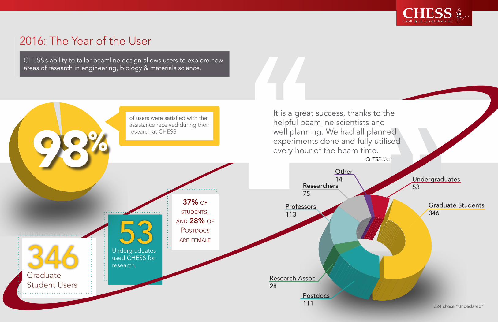
Graduate Students346
Undergraduates53
Professors113
Researchers75
324 chose “Undeclared”
Other14
Postdocs111
Research Assoc.28
346Graduate Student Users
2016: The Year of the User
CHESS’s ability to tailor beamline design allows users to explore new areas of research in engineering, biology & materials science.
98of users were satisfied with the assistance received during their research at CHESS “It is a great success, thanks to the
helpful beamline scientists and well planning. We had all planned experiments done and fully utilised every hour of the beam time.
-CHESS User

1312
40
75%
83minutes minutes
Xraise has doubled the amount of time spent on science instruction in targeted classrooms in Ithaca City School District
of the original 6th grade Xraise Summer Science Snapshot
students have declared STEM majors as first-year undergraduates.
Impacting the STEM Pipeline
There were 2,812 uses of the Xraise Lending Library investigations and equipment by high school students across the U.S., 45% of which were females.
17,778 minutes of videos have been watched by Xraise fans from around the world
2,007 people took a guided tour of Wilson Lab and CHESS experimental hutches. July was the busiest month with 317 visitors!
Training a Community of Synchrotron Scientists
Xraise is the outreach group at CHESS which engages minds by facilitating direct interaction with physical phenomena and encouraging careful observation of the world. Xraise stimulates thinking and helps develop the next generation of scientists.
people have interacted with Xraise Junk Genies exhibits showcasing synchrotron science phenomena
families have used bottles, egg cartons, and other recycled materials to engineer motorized contraptions as part of the Moto-Inventions program
Miles the Junk Genies exhibits have traveled as part of Xraise’s mobile science exhibition - nearly the same distance as the circumference of the Earth!
11,781 900+ 23,068Converting Junk into Learning Experiences

1514
13’ Height of the Cleo Super-conducting
Magnet
2,038,000Pounds of material moved by the
CLEO disassembly crew (1019 tons)
CHESS-U: A brighter CHESS
In 2016, CHESS began a $15M upgrade that will extend its capabilities for cutting-edge, innovative science and technology. The CHESS-U upgrade will provide the critical leverage to position CHESS as a world leader in synchrotron science. In addition, it will attract the research of more U.S. manufacturers and drive local high-tech businesses. CHESS-U has already dispensed over $4 million in the economy, spurring job growth in the aerospace, electronics, and manufacturing sectors.
The improvements of CHESS-U will be achieved by replacing one-sixth of the CESR storage ring with modern multibend achromats and by replacing or upgrading the x-ray beamlines to take greatest advantage of the new undulator sources. At the completion of CHESS-U in 2018, CHESS will be the premier synchrotron source in the US for high-energy, high-flux x-ray studies.

2.0 Lab Management

1918
LEADERSHIP Joel Brock ..................... Director, CHESSRichard Cerione ............ PI, MacCHESSKathy Dedrick ............... Director, CHESS User Program OfficeErnie Fontes ................. Assoc. Director, CHESSSol Gruner .................... Co-PI MacCHESS, CHESSLora Hine ...................... Dir. of Educ. Programs at CLASSEMatt Miller .................... Assoc. Director, CHESSMarian Szebenyi ........... Director, MacCHESSDon Bilderback ............. Emeritus Assoc. Director, CHESS
STAFF SCIENTISTSAaron Finke .................. Staff Scientist, MacCHESSKen Finkelstein ............. Staff Scientist, CHESSRichard Gillilan ............. Staff Scientist, MacCHESSQingqiu Huang ............. Staff Scientist, MacCHESSRong Huang .................. Staff Scientist, CHESSPeter Ko ........................ Staff Scientist, CHESSHyun Hwi Lee ............... Visiting Scientist, CHESSDarren Pagan ................ Staff Scientist, CHESSPeter Revesz ................ Staff Scientist, CHESSJacob Ruff ..................... Staff Scientist, CHESSDavid Schuller .............. Staff Scientist, MacCHESSDetlef Smilgies ............. Staff Scientist, CHESSStanislav Stoupin .......... Staff Scientist, CHESSZhongwu Wang ............ Staff Scientist, CHESSArthur Woll ................... Staff Scientist, CHESS
RESEARCH STAFFCarter Bagnell .............. Operator, CHESSElisabeth Bodnaruk ...... Research Support SpecialistZach Brown ................... Operator, CHESSDarol Chamberlain ....... Research Support SpecialistAustin Cao .................... Research Support SpecialistMelissa Cole ................. Operator, CHESSChris Conolly ................ Operations Manager, CHESSJohn Conrad ................. Operator, CHESSMike Cook .................... Research Support Specialist, MacCHESS
2.1Leadership and Key Personnel
Eric Edwards ................. Research Support Specialist, CHESSLee Geiger .................... Operator, CHESSJerry Houghton ............ Technician, CHESSAimee Kellicutt ............. Research Support Specialist, CHESSJohn Kopsa ................... Machinist, CHESSTom Krawczyk ............... Operator, CHESSIrina Kriksunov .............. Research Support Specialist, MacCHESSAaron Lyndaker ............ Research Support Specialist, CHESSKurt McDonald ............. Research Support Specialist, CHESSGregg McElwee ............ Research Support Specialist, CHESSBill Miller ....................... Research Support Specialist, MacCHESSKatie Moring ................ Operations Manager, CHESSAlan Pauling .................. Research Support Specialist, CHESSDana Richter ................. Research Support Specialist, CHESSSaramoira Shields ......... Operator, CHESSLee Shelp ...................... Operator, CHESSScott Smith ................... Research Support Specialist, MacCHESSPhil Sorensen ................ Software Developer, CHESSChris Whiting ................ Operator, CHESSShijie Yang .................... Comp Hardware, CHESS
POST DOCJulian Becker ................ Post Doc, CHESSTK Chua ........................ Post Doc, MacCHESSJesse Hopkins .............. Post Doc, MacCHESSJooseop Lee ................. Post Doc, MacCHESSJeney Wierman ............ Post Doc, MacCHESS
ADMIN STAFFErik Herman .................. Educ. & Outreach Coordinator at CLASSEBarbara Herrman .......... Data Analyst, CHESS User Program OfficeEva Luna ........................ Teaching Support Specialist & Lending Librarian, CIPTRick Ryan ...................... Science Communicator, CLASSE
STUDENTSDavid Agyeman-Budu .. Graduate Student, CHESSNaigeng Chen .............. Graduate Student, CHESSMarissa D’Amelio ......... Undergraduate Student, CHESSRohit Garg .................... Graduate Student, CHESSGabrielle Illava.............. Student-Agriculture and Life Sciences/ClarkHoward Joress .............. Graduate Student, G-lineMatt Kasemer ............... Graduate Student, CHESSJames Pastore .............. Graduate Student, CHESSVeronica Pillar ............... Graduate Student, MacCHESS

2120
External Advisory CommitteeG. Brian Stephenson (Chair)Senior PhysicistMaterials Science DivisionArgonne National Laboratory
Mavis Agbandje-McKenna Dept. of Biochemistry and Molecular BiologyUniversity of Florida
Paul G. Evans Dept. of Materials Science and EngineeringUniversity of Wisconsin-Madison
Robert (Bob) HettelDeputy Director, Accelerator DirectorateSLAC National Accelerator Laboratory
Jean Jordan-SweetIBM Research DivisionNational Synchrotron Light SourceBrookhaven National Laboratory
Janos KirzScientific Advisor, Advanced Light SourceErnest Orlando Lawrence Berkeley National LaboratoryDistinguished Professor Emeritus at Stony Brook UniversityAdvanced Light SourceLawrence Berkeley National Laboratory
Lee MakowskiProfessor Bioengineering, Electrical and Computer EngineeringNational Synchrotron Light SourceNortheastern University
2.2Users Executive CommitteeKyle Lancaster (Chair, 2018)Dept. of Chemistry and Chemical BiologyCornell University
John Smedley (Vice Chair, 2019)Instrumentation DivisionBrookhaven National Lab
Stephen Meisburger (2018)Dept. of ChemistryPrinceton University
Meredith Silberstein (Past-Chair, 2017)Mechanical and Aerospace EngineeringCornell University
John Twilley (2018)Materials Science and EngineeringSUNY Stony Brook University
Marian Szebenyi (Ex Officio, non-voting)Director, MacCHESSCornell University
Matthew Miller (Ex Officio, non-voting)Director, In-SitµCornell University
2.3

2322
open position, but rather assists the supervi-sor in defining the process and executing a professional and proper search.The Hiring Advisory Committee would be chosen by and charged by the CLASSE Di-rector. The HAC would not “do all the leg work” for each and every hiring process in CLASSE. Instead, the HAC will act as a point of reference aimed at providing consistency across CLASSE hiring practices.
Actions TakenJuly 2015 – June 2016: A Hiring Advisory Committee (HAC) has been established: Ka-tie Jacoby, Executive Director of Adminis-tration, and Monica Wesley, Executive Staff Assistant. The HAC meets with each hiring manager to review the needs of the position, review hiring process and procedure, estab-lish the Staff Position Description (SPD), and monitor/provide reference throughout the entire search process, from conception of position through final offer to candidate. Recommendation #2: a Search Plan should be composed for all hiring positionsA necessary part of trying to improve hiring processes and diversity efforts in CLASSE is to plan, track progress, provide over-sight, and analyze and learn from successes and shortcomings. It would greatly benefit CLASSE to create a standard set of proce-dures, expectations, and accountability cri-teria that each and every hiring team could follow. We therefore recommend that each hiring process be guided by a Search Plan. The Search Plan should attempt to cover all aspects of filling a position, including but not limited to: defining the “staff position description”, creating written materials for posting, deciding where/when to post, de-fining the screening process, phone and/or on-site interviews, interview teams and
Diversity ReportThree topics in which the CLASSE Diversity Team strived for in 2016 were 1) job search practices, 2) recent hiring decisions, and 3) utilization of University resources. The com-mittee utilizes background data to analyze and comment on; including detailed organi-zational charts, demographics data on exist-ing staff, copies of administrative resources currently in force in CLASSE (policies, train-ing documents, etc.), as well as a detailed summary report on filled job searches from 2009 to present.
Summary Recommendations to CLASSE Management
Recommendation #1: CLASSE should for-mulate a Hiring Advisory CommitteeA significant amount of discussion focused on improving diversity, and drew forth ex-amples of successes achieved by other technical organizations across campus, am-plifying the need for organizations to pre-pare and train decision makers, understand issues of effective and compliant hiring, re-cruitment and pipeline nurturing, and pro-vide oversight of the entire hiring process (from job description to onboarding). Cur-rently CLASSE has a highly decentralized hiring system insomuch as supervisors at all levels of the organization can be put in charge of a hiring process. Although many of the staff have ample subject matter ex-pertise to be supervisors, most have little or no training in hiring procedures, process or diversity issues. While training any and all the staff would be ideal, in practical terms CLASSE would be better and more efficient-ly served by assembling a Hiring Advisory Committee (HAC) who could oversee pro-cesses and training. The HAC does not re-place the supervisors in charge of filling an
questions, decision making process, and follow up. The Search Plan should be pre-approved by the Hiring Advisory Com-mittee, and the HAC should be charged with following up on the performance of the plan. The Search Plan will need final approval and go-ahead granted by the CLASSE Director and/or Steering Com-mittee, as deemed appropriate.
Actions TakenJuly 2015 – June 2016: In collaboration with the Office of the Vice Provost for Re-search’s Human Resources department, the HAC along with the hiring manager meet as the first step in the hiring process to establish a Search Plan/Posting Tem-plate. Emphasis is placed on identifying and advertising to underrepresented mi-nority locations.
Recommendation #3: CLASSE should create a formal Mentoring and Reten-tion programCLASSE has done “quite a good job” in hiring (and promotion) of women into technical positions over the past two years. The difficulties in hiring subject matter ex-perts with significant career experience argues for investing to train younger tal-ent (formal and informal training, job rota-tion, etc.) and focusing on long-term ad-vancement and retention. In other words, because of the highly specific technical expertise CLASSE needs, it should be ac-tively planning to produce senior experi-enced staff.Recommendations for how to formulate such a program were not prescriptive, but included mention of job rotations, iden-tifying a “pool of mentors” and rotating mentees on a few month basis (mentee-mentors should not be immediate super-visors), and learning from other campus programs (i.e. Advance for academics, etc.).
Actions TakenJuly 2015 – June 2016: CLASSE completed a formal review of all Research and Senior Research Associates identifying the top per-formers and established a retention pro-gram. Scientific accomplishments were re-viewed along with salaries. Eight individuals were identified and received in-year merit in-creases (PhD Year: 1997, 2000, 2004, 2006, 2009, 2010, 2010, 2013). Salaries were also evaluated against comparable PhD Year of colleagues at other facilities.
Recommendation #4: Training for hiring managers, supervisors, and search com-mittee membersSupervisors and others are often thrown into the hiring process when someone an-nounces they are leaving a position. We are recommending a training program that will promote more awareness of unbiased deci-sions throughout the search/hiring process, which will in turn increase the diversity of our interview pools and overall hires.It Depends on the Lens, a two to three hour program developed by the Cornell Inter-active Theatre Ensemble and the Cornell University Recruitment and Employment Center, is designed for hiring managers, su-pervisors, and search committee members. It combines interactive theatre and guided discussion with research on unconscious bias. The session concludes with a discus-sion of best practices for combating uncon-scious bias in searches.Group discussion will center on:• the behaviors, perspectives, emo-tions, assumptions and biases of the mem-bers of the staff search committee as they evaluate applicants during a discussion of materials submitted for review• unconscious bias which undermines fairness in the search process because of the tendency to evaluate applicants in a way that puts minorities at a disadvantage• the onus of responsibility for recog-
2.4

2524
nizing racial, gender and other forms of bias in the evaluation of applicants and for chal-lenging our implicit hypotheses about appli-cantsThere may be money available through the Recruitment and Employment Center to help fund this program, but if not we are recommending that funding be support-ed through CLASSE. The cost is currently $1,500.
Actions TakenJuly 2015 – June 2016: All hiring managers and supervisors were expected to attend It Depends on the Lens. Search committee members who are not the hiring manager or supervisors are given on-site training by either a member of the HAC or the hiring manager prior to meeting with applicants.
Charge to the Diversity CommitteeThe Diversity Committee (DC) will work to develop recommendation for a strategic plan with a goal to increase diversity of the CLASSE organization through leadership implementation. The DC will consider facil-ity leadership, governing committees, staff, undergraduates, graduate students, and postdoctoral associates. Committee mem-bership will include representation from staff, students and faculty within CLASSE as well as members invited from outside. On an annual basis the committee will evaluate the effectiveness of the previous year’s plan (or parts therein), identifying opportunities and developing plans for improvements. The DC will prepare a report to CLASSE leadership; parts of that report might be excerpted by programs within CLASSE (i.e. CHESS) for in-clusion in the annual reports sent to funding
agencies.
Strategic Plan Mission of the FacilityThe overall mission of the Cornell High En-ergy Synchrotron Source (CHESS) is to pro-vide a na-tional hard X-ray synchrotron radi-ation facility. This includes four submissions: (1) operation as a synchrotron user facility; (2) research and development of new syn-chrotron radiation technology and upgrad-ing of the facility; (3) integration of research and education in the training of the person-nel who use and operate synchrotron radia-tion facilities; and, (4) educational outreach to expose K-12 students and the public to synchrotron x-ray science and its application to materials research in age and experience appropriate forms. The synchrotron facility is used by investigators from a wide range of science and engineering disciplines in academia, industry, government, non-profit, and international institutions. They conduct studies encompassing, but not limited to, the atomic and nanoscale structure, prop-erties, in operando, and time-resolved be-havior of electronic, structural, polymeric and biological materials, pro-tein and virus crystallography, environmental science, ra-diography of solids and fluids, and micro-elemental analysis, and other technologies for X-ray science.
Description of Physical InfrastructureFive stories underground, ½-mile in circum-ference, currently running at 5.3 GeV and 200 mA each of positrons and electrons, and one of only two high energy rings in the United States, the Cornell Electron Storage Ring (CESR) powers the magnetic structures that produce hard (10-100 keV) synchrotron X-ray beams used by 11 experimental sta-tions. The focusing magnets are individually configurable, making CESR unusually flexi-ble and adaptable. The Wilson Synchrotron
2.5 beam characterization. All goals require best stewardship of the accelerator and X-ray infrastructure; towards that end, CHESS supports the staff skill set needed to keep the existing infra-structure running with min-imal down-time and to improve it over time to address future research needs.
Educational ProgramsEducational goals include providing re-search and engineering experiences for community college, undergraduate, gradu-ate students and post-doctoral researchers. We will support 8 GRAs and 3 post-doctoral research associates each year. We will ex-pand the Master’s of Engineering degree pi-lot program (MEng) to 5, including students in Electrical and Computer Engineering (ECE), Computer Science (CS), Materials Sci-ence and Engineering (MSE), and Applied and Engineering Physics (AEP).
K-12 Outreach: CHESS will continue an education and outreach program providing age-appropriate activities that teach science knowledge, hands-on skill, and apprecia-tion, and career modeling for STEM fields.
ManagementCLASSE is chartered as a Cornell University Center, which means that it is an interdisci-plinary organization of faculty and staff to fa-cilitate and promote research and education in the branches of science concerned with the development and uses of accelerators. Faculty members are from many Cornell de-partments, including Physics, Chemistry and Chemical Biology, Applied and Engineering Physics, Mate-rials Science and Engineer-ing, Molecular Medicine, etc., to facilitate student (undergraduate, graduate and post-doctoral) involvement for education, training and research opportunities and to involve the intellectual resources of the wider uni-versity community. The CLASSE Directorate is a mixture of faculty and senior profession-als, whose purpose is to integrate research
Laboratory that houses CHESS and CESR is also home to world-leading accelerator physics research programs. X-ray users receive beam time via a propos-al-based, peer-reviewed process. CHESS’ organizational structure encourages novel, high-risk experiments that require new tech-nology, heavy investments of staff scientist effort, and/or require personnel, equipment or capabilities available at Cornell but hard to find elsewhere. CHESS scientists have considerable latitude to make programmat-ic decisions for their beamlines and pursue exciting new ideas.
Scientific Initiatives (Programs)As a user facility, CHESS supports a wide range of research activities. Improvements to X-ray beam-lines and experimental end stations are guided by seven scientific ini-tiatives: (1) Computationally-Enabled “Total Scattering” Studies of Complex Materials, (2) “Designer Solids” – Structure, Process-ing, and Performance, (3) Rapidly Evolving Systems, (4) X-ray Imaging: Scanning Probe and Full-Field, (5) Spectroscopic Studies, (6) Energy and Structural Materials – In Oper-ando Studies, and (7) Macromole-cules and Biochemistry. Details are on these scientific initiatives are given in section D.4 of the re-newal proposal.
Technology R&D ProgramsThe scientific research initiatives are enabled by an active X-ray and accelerator R&D pro-gram. R&D projects include: (a) continued optimization and improvement of CESR for X-ray production, (b) development of novel insertion devices, (c) development of spe-cialized X-ray optics, (d) advanced X-ray detec-tor development, and (e) an indepen-dently funded accelerator physics program. This program leverages expertise of CHESS and CESR staff members who are world leaders in storage rings, compact insertion devices, single crystal and lithographically fabricated X-ray optics, and synchrotron

2726
and education activities (e.g., accelerator R&D, X-ray Science, EPP) with technical functions requiring full-time operations staff (e.g., Technical Operations, Project Man-agement, Safety, Administration).
Management of Accelerator SystemsThe accelerator physics/operations group has designed/built/maintained and operat-ed the synchrotron injector and CESR stor-age ring at Wilson laboratory for 4 decades. It has the expertise and technical skill sets to keep large projects operating smoothly, and also all the administrative infrastructure to coordinate development of new groups and directions. The facilities under manage-ment and development include an up to 12 GeV synchrotron, an up to 8 GeV electron/positron storage ring, 5 MW transformer pad, klystron gallery, closed circuit helium refrigerator and recovery plant, cooling tow-ers, instrumentation and computer controls group, ERL injector test facility, etc. The PI connects to CLASSE’s technical infrastructure and operating capability via CLASSE Direc-torate Chair Ritchie Patterson, the Technical Director, Dave Rice, and co-PI Dave Rubin. Rubin, a well-known accelerator physicist, is presently PI for the CESR-TA project that is using CESR for low-emittance experiments. This is a highly experienced team.Because of CHESS’ dual mission, educa-tion and research are both equally impor-tant goals. Students are heavily involved in operating the facility. For example, gradu-ate students in CLASSE routinely build and maintain accelerator and upstream portions of X-ray beamlines, working and learning behind the primary shielding walls. In this way, CLASSE serves a very important syn-ergy with the national laboratories by train-ing accelerator physicists and X-ray beam-line scientists who go on to work at other national facilities.
Management StructureCHESS is a unit within the Cornell Labora-tory for Accelerator-based ScienceS and Education (CLASSE). The CHESS Direc-tor is appointed by Sr. Vice-Provost for Re-search R.A. Buhrman. J. Brock is CHESS Direc-tor and PI of this award. The CHESS Director reports jointly to Buhrman and the CLASSE Director, J.R. Patterson. D. Rice is CESR Technical Operations Director. L. Hine is Education and Outreach Director. B. Heltsley is CLASSE Safety Director. The CHESS Director is also the Associate Direc-tor of CLASSE. CHESS Associate Director E. Fontes is responsible for X-ray technical operations. Brock is responsible for X-ray scientific staff. CHESS Associate Director M. Miller is responsible for educational pro-grams and the struc-tural materials initiative. R. Cerione is MacCHESS PI. M. Szebenyi is MacCHESS Director. K. Dedrick is CHESS’s User Office Director. Scientific staff oversee X-ray end stations. Detailed organizational charts are included as appendices.
Management ChallengesThe matrix management structure of CLASSE both gives CHESS access to skill sets and resources that it cannot afford on its own and generates couplings between the various projects in CLASSE that must be managed. A SWOT analysis of CLASSE’s matrix management structure appears in the table below.
Strengths1. Provides CHESS with access to world-class ac-celerator physics faculty, research programs, and infrastructure.2. Provides sophisticated technical skill sets that CHESS needs critically, but not full time.3. Shared use gives CHESS access to state of the art storage ring built by other programs.
Opportunities1. Transform a particle physics facility into a state of the art photon science facility.2. Cost effective, sequential up-grades to CESR, insertion devices, x-ray optics, and x-ray detectors can be invented, developed, and executed.3. Take advantage of latest advances in accelera-tor technology to provide high brightness x-ray beams.
Weaknesses1. Financial support for matrixed staff is also ma-trixed. If other major programs in CLASSE lose their funding, CHESS may need to preserve criti-cal skill sets.2. Various projects in CLASSE may have conflicting needs for space and personnel.
Threats1. Re-organization, turn over in man-agement, and natural evolution of priorities at funding agen-cies constantly alters the funding situation.
Roles and Meeting Dates of Internal and External CommitteesThe External Advisory Committee (EAC) meets annually to advise the Director on the utilization and development of CHESS facili-ties. The Vice Provost for Research selects EAC members from the scientific and engi-neering community. The EAC reports to the Vice Provost for Research. The EAC over-sees and re-ports on the on-line Proposal Review Process annually.The EAC has chosen to have its on-site meeting in January or early February in or-der to be involved in the initial stages of the development of the annual operating plan. The EAC will meet again by a web-based conference in late June or early July to re-view the final version of the annual operat-ing plan.The CHESS Users Executive Committee
(UEC) is elected by the users of CHESS and meets annually to advise the Director on the utilization and development of CHESS facili-ties. Candidates for the UEC are nominated by the CHESS users. UEC members serve for two years. The UEC elects a vice chair each year. The vice chair succeeds the chair. The chair then serves an additional year on the UEC as past chair.The UEC will hold a web-based meeting in January and will have an in person meet-ing in June during the annual CHESS Users’ Meeting.The annual meeting of the Diversity com-mittee is in February.
BudgetThe appendices include an Annual Budget request (Form 1030) and budget justifica-tion with a Table showing the breakdown of expenditure by facility component and ma-jor activity. Also include is a 5% budget re-duction scenario from above request (Form 1030) and budget justification with a break-down Table.
Transformative Opportunity: Installing a multi-bend achromat lattice in the southern arc of CESR that utilizes dual function mag-net technology, will both in-crease the brightness of the x-ray beams ten-fold and create additional straight sec-tions that can house six (6) more undulators. This upgrade is the basis of CHESS’s pro-posal to NSF’s Major Research Instrumenta-tion (MRI) program. A white paper describ-ing the benefit to CHESS and an abridged version of the MRI proposal are included as appendices. If the MRI proposal is successful, CHESS will immediately begin seeking funds to take advantage of the new accelerator configu-ration by instrumenting six (6) new undulator beam lines.

2928
Safety Guiding Principles of Safety at CHESSCHESS holds no higher priorities than ensur-ing the health and safety of all personnel and facility visitors. These values have been wo-ven into the fabric of laboratory administra-tion and operation. Synergistic relationships with CHESS, CLASSE and Cornell University provide important policy guidance, institu-tional support, and oversight. CHESS faces unique challenges in addressing the some-times-disparate needs of staff, students, visitors, and a diverse user community that has a (largely) transient onsite presence. The CHESS approach to safety is built around three overlapping commitments: to continu-ously provide a safe laboratory environment, to engender an abiding culture of safety in all personnel, and to address and anticipate safety challenges with proactive safety man-agement.
Safe Laboratory EnvironmentThe first line of defense against potential hazards is a safe laboratory environment. Exterior doors to Wilson Lab are locked out-side of business hours; entry at off-hours is by keycard access or explicit permission of a staff member. The Wilson Laboratory fire alarm and detection system is a part of the centralized University system. State fire offi-cials conduct inspections of the entire labo-ratory on an annual basis. Designated staff members are trained for specific roles in emergency situations. Only trained and/or licensed personnel operate industrial equip-ment, such as cranes, forklifts, and large vehicles. Machine tools are periodically in-spected for correct operation and presence of appropriate guards. A spill control plan is in place for oil-filled transformers. An arc-flash hazard study of laboratory high-voltage AC distribution panels was completed and
appropriate hazard labels posted on electri-cal panels, but some recommended hard-ware changes at the 13.2 kV breaker panels were deferred for lack of funding. A lock/tag/verify (lock-out/tag-out) program is in place to cover work near equipment with remote power control, as is a policy govern-ing hot work and welding. Personal protec-tive equipment and safety training specific to their tasks is made available to workers who need it. Fume hoods for handling small quantities of chemical and biological sam-ples (BSL2 or below) are available. Safety Data Sheets are stored in notebooks near where the hazardous substances are used and appropriate safeguards are in place at the point of usageSources of ionizing radiation in Wilson Lab are primarily from the accelerators. Addi-tional sources of ionizing radiation include RF processing stations, CHESS x-ray beam-lines, portable x-ray sources, and the occa-sional use of a sealed radioactive source. Mitigation of radiation hazards from radia-tion-producing equipment (RPE) is dealt with largely via engineering controls. Permanent shielding, generally consisting of concrete, lead, and/or iron, surrounds all RPE so as to restrict potential exposure outside its shield-ing to below 2 mrem in one hour or 100 mrem in one year, per New York State regu-lations. Locations just outside the shielding where radiation dose rates are expected to be below those listed above but which are considered potentially vulnerable to higher levels, are designated as “controlled areas”, in accordance with Cornell University policy and NYS regulations. Access to controlled areas is restricted to authorized personnel wearing radiation dosimeters (in “badge” form) or those accompanying a CLASSE host with a real-time readout dosimeter. Entranc-es to controlled areas are clearly signed. Exclusion areas, inside which personnel should not be present during RPE opera-tion, are protected by more sophisticated access controls: all entryways are equipped
2.6 with interlocked gates and/or light beams that, if tripped during RPE operation, cut power to the RPE and cause audible and visible alarms. Radiation detectors monitor the radiation in controlled areas, and trip off the accelerators if conservative levels are exceeded. Exclusion area interlocks cannot be set until a full in-person search has been conducted; the integrity of interlock opera-tion is verified by periodic operational tests of the interlock components. Sealed radio-active sources are securely stored and ac-cessible for temporary use only by trained and authorized personnel.CHESS hutches, where x-ray experiments must be accessible when CESR beams are circulating, are located in controlled areas and shielded so as to contain the x-rays. A hutch must be searched and secured in order for the beam-stop for that hutch to be lifted; the beam-stop drops to block the x-ray beam if the door is unlocked. The integrity of the search is enhanced by the hardwired requirement that one or two but-tons located in remote corner(s) of the hutch must be pressed prior to setting the inter-lock. Stand-alone x-ray sources can be used for photon science or calibration and each is operated inside an interlocked hutch and powered via a fail-safe interlock system.
Culture of SafetyCHESS and CLASSE seek to establish and maintain a culture of safety, which entails much more than compliance with a set of rules. A culture of safety is embodied by each staff member taking responsibility for safety of the staff, users and visitors of the facility; safety being valued on par with sci-entific achievement and/or task completion; safety concerns always being taken seriously and promptly addressed; safety challenges being approached with intellectual rigor; new activities being planned from the start with safety in mind; new participants receiv-ing relevant safety training immediately; and always striving for improved safety.
Such practices are self-reinforcing, but can be undermined by even occasional lapses, so considerable vigilance on the part of su-pervisory personnel is required.
Proactive Safety ManagementProactive safety management ensures that: specific safety responsibilities of each staff member, student, user, or visitor are clearly delineated and communicated; appropriate training and resources are provided to those who need it, including staff, students, and users; mechanisms are in place to maintain accountability and establish and publicize appropriate safety-related policies; compli-ance with relevant University and govern-mental safety and environmental regulations and ordinances is attained; and intra-univer-sity resources are leveraged when helpful. CLASSE has an online Safety Handbook, with links to specific CHESS safety sections. A central safety document database has been implemented and is home to proce-dures, radiation permit applications, meet-ing minutes, internal incident reports, and more. An in-house training database exists to track training history for each worker. Con-version of this database to the cloud-based Learning Management System acquired by Cornell University for all personnel and units is in progress. The new system, which will hold all not-for-credit online learning con-tent and tracking information for the entire University, is expected to become active in November 2016.Clear lines of accountability for performance related to safety have been shown to be crucial to superior safety achievement, es-pecially in academic research settings. The CLASSE Safety Committee and Safety Di-rector, which set, communicate, and imple-ment laboratory safety policy, speak and act with the imprimatur of the CLASSE Labo-ratory Director, who appoints both. Each staff member is accountable to a supervisor. CHESS users are accountable to the CHESS User Safety Committee, their assigned Cog-

3130
nizant Safety Officer, and the CHESS Safe-ty Officer, all of whom are accountable to CHESS management. CHESS users have an immediate interface to the CHESS Operator (who is always on duty during user sched-uled beamtime) and the CHESS scientist who is their primary contact at the assigned experimental station.
Independent Safety Advisory PanelIn order to obtain independent feedback on the CHESS and CLASSE Safety Programs, a Safety Advisory Panel consisting of three non-Cornell-affiliated safety professionals was tasked with providing such feedback and advice. The Panel visited CHESS and CLASSE on March 5, 2013, attended a se-ries of presentations, and conducted brief tours of the facilities. Their report includes the Panel composition, its charge, a sum-mary of documents reviewed and presenta-tions made to it, and its advice to CLASSE for safety improvements. Excerpting from the Executive Summary:“Considering the complexity of CLASSE facilities and their wide spectrum of safe-ty challenges, CLASSE appears to have a strong, well-functioning Safety Program and a well-developed safety culture shared by employees and management.“The Panel advice was structured in the form of four categories of increasing importance: Noteworthy Practices, Opportunities for Improvement, Observations, and Essential Changes. A response to the Panel Report, consisting of a series of specific action items and associated actual or target completion dates, was sent to NSF. All of the action items have been completed.
Data ManagementTypes of Data The majority of research data produced at CHESS is in the form of electronic scans (i.e. ASCII data files) or electronic images (binary files) that record x-ray interactions with spec-imens. Full data sets often include intrinsic and extrinsic conditions of the specimens (preparation conditions, chemical compo-sition, orientation angles, motor positions, temperatures, pressure, voltages, etc.) that are oftentimes recorded as part of the elec-tronic files but are sometimes recorded only in hardcopy notebooks or secondary com-puter text files. Raw data are often pro-cessed utilizing standard software packages (MATLAB, etc.) or using custom visualization or analysis code. Raw and processed data are sometimes used as inputs to models and computer simulations, whose algorithms, computer codes, and outputs are also con-sidered parts of complete data sets along with the raw and processed data. Research data is created by staff scientists as well as facility visitors.In addition to research data, “machine” and “people” data are also produced by the CHESS technical and administrative staff. During machine operations, the per-formance of facility equipment (the storage ring, x-ray beamlines, etc.) is recorded; pro-cedures to operate equipment are docu-mented, annotated, and refined over time; and the performance of a large number of devices is monitored continuously. The man-agement of people and the user programs also create data. Scheduling and recording of researcher access to CHESS is recorded electronically. Submission of proposals, re-view, equipment needs, scheduling of vis-its, recording of hours received, and user feedback are mostly entered electronically, stored, and archived.
Data StandardsX-ray data are recorded using a wide variety of computer software packages, some com-mercially produced and some created by CHESS staff or individual research groups. Commercial software uses file formats that are considered standards (i.e. SPEC, HKL, HDF), and many commercial and custom-built x-ray area detectors produce digital image formats that are standards (i.e. TIFF, JPEG). Processing software packages use standard ASCII or binary formats (using typi-cal MATLAB I/O standards and functions), although individual researchers can write custom code (or macros) that introduce new, non-standard data structures. Most standard file formats include some “meta-data” descriptive information, though uni-form standards covering all x-ray data do not exist. For example, SPEC data files in-corporate motor positions and other config-uration information in each individual scan “headers.” Two-dimensional x-ray images often encapsulate exposure times, image dimensions, etc., inside binary file headers. In addition, researchers often utilize hard-copy notebooks (or electronic equivalents) to tie together metadata needed for index-ing data files, specimen handling, and other pertinent conditions. In these cases, meta-data standards are created by investigators as needed to unambiguously associate data with specimens and preparation procedures, instruments used, dates and times, process-ing and analysis techniques, and any other information deemed necessary to fully un-derstand the data analysis flow and results. No external or community-wide standards exist for individual note taking.CHESS has adopted the standards-based EPICS software for most of its machine con-trol and monitoring. EPICS is an open-source environment for developing machine con-trol systems and is used by many synchro-tron light sources. People data and some machine data are created and recorded by custom software in the form of interactive
2.7 databases and control systems, respectively. In these cases, even if the backend database uses a standard query language (e.g. SQL), the control languages, data structures, and database schema have been custom-built to address equipment and community needs. In these cases, internal standards are well defined and consistently applied.
Access to Data and Data Sharing Practic-es and PoliciesGeneral policies: CHESS provides a data collection facility where all activities of re-searchers and their data are kept secure and private until they are processed, analyzed, and results are selectively released by the owners of those data. Over time, the most significant results will be published in peer-reviewed journals, after which time CHESS asks permission from researchers to high-light and release information about results. Given permission, CHESS also releases in-formation about other activities utilizing the facility such as seminars, student projects, and educational programs. Standard out-put channels include facility and community-wide topical web sites, reports in university publications, reports to review committees and NSF, etc. These output channels have full open access and allow internet search engine indexing services.CHESS stores and archives all research data that is electronically produced on CHESS equipment by its staff and by visiting re-searchers. This data is stored redundantly on disk arrays for approximately six months, and then it is copied to magnetic tape for long-term archival storage. An index of the research data (metadata) is kept persistently on disk. Every dataset is archived twice to tape, and one of these copies is moved to an off-site storage facility. Experimental data that has been archived can be restored to disk upon request.Staff Scientist research data: CHESS consid-ers itself the owner for all data produced by staff supported by the NSF grant, and

3332
CHESS stores and archives staff data in all varieties. In addition to the above general policies, hardcopy notebooks are main-tained to document installation and use of project equipment and experimental pro-cedures developed to operate the facility. Hardcopy notebooks with be entitled and indexed according with accepted standards in the lab. Notebooks are kept in circulation at the facility or moved to a dry storage facil-ity and kept indefinitely.Visiting Researcher data: Data produced by visiting researchers is not owned by CHESS; it is owned by the individual investigator group or group principal investigator. As a convenience, CHESS stores and archives data produced by visiting researchers on their behalf. However, CHESS does not dis-tribute visiting researcher data without per-mission of the owner. Similarly, CHESS does not explicity seek to comply with the data access policies of its visiting researchers’ funding agencies; compliance is the respon-sibility of the visiting researcher.In some cases, visiting researchers bring their own personal computers in order to control their own experiments and to cap-ture, process, analyze and produce data in their own software environments (avoiding the time-consuming process of reconfigur-ing CHESS computers). In these cases, the data of visiting researchers is never in con-tact with CHESS facility equipment. Machine and people data: Machine opera-tions and people data (proposal, beamtime requests, etc.) are always kept private and secure (no external access to raw data). The only exceptions to this are web-based kiosk displays that indicate to researchers that x-ray beams are available for data collection purposes. Machine and facility use informa-tion is summarized periodically for official reporting purposes only (i.e. NSF review committees, GPRA and NSF annual reports). All machine and people data is stored and archived indefinitely in both on-site and off-site locations.
Policies for Re-Use, Re-distributionCHESS will honor routine requests for staff (and facility) owned data that may be used to clarify published results. To date, such re-quests are very rare and usually involve col-legial peer-to-peer telephone or email con-versations. Materials placed on web sites for communications and publicity purposes have an implicit expectation that source at-tribution to CHESS will be included for any re-use.
Data StewardshipThe storage and archival of research data is described above. All other categories of data owned by CHESS, including processed data, computer codes, and machine and people information, are maintained on re-dundant-hardware computers and copied to on-site and off-site time-stamped back-up devices. Data backups can run as often as hourly for synchronization of machine configuration files to daily incremental and monthly full backups for most data sources. Hardcopy notebooks are securely archived. Many types of raw, processed and analyzed data are also copied to external storage me-dia (i.e. CD/DVD storage media) for redun-dancy and long-term storage.

3.0 User Facility

3736
Step Three: How is my proposal reviewed?
A peer review is conducted on your propos-al by outside reviewers (2-3) and an average final score will be assigned to the proposal upon completion of the review(s). Your av-erage score will be on a scale of 1-4, 1 be-ing excellent and 4 being poor. The areas in which your proposal will be scored are:• Scientific and or Technical Merit• Need for CHESS Capabilities• Experimental Plan Details• Expertise of Group (in both x-ray meth-
ods and science subject areas)
Below is a snapshot of the reviewer score sheet:Scientific and/or Technical Merit• Excellent - Results will be considered im-
pactful and important - ambitious and in-novative
• Very Good - Will advance scientific knowledge, methods, and/or address critical questions
• Good - Research contributes to scientific and/or technical knowledge base
• Poor - Proposed research has no clear importance or originality
Need for CHESS Capabilities• Excellent - CHESS facilities and capabili-
ties essential to obtain experimental re-sults
• Very Good - Well documented need for existing facilities and capabilities
• Good - Appropriate use of existing facili-ties and capabilities
• Poor - Routine use of existing facilities and methods or poorly demonstrated need
Experimental Plan Details• Thorough - Uses established facilities/
methods or addresses all phases of a successful experiment (preparation, data collection/analysis, theory/calculations, etc.)
• Detailed - Provides a detailed descrip-tion of most aspects of the experiment
• Adequate - Reasonable outline of exper-imental needs provided
• Insufficient - Too little detail to evaluate needs and/or predict successful comple-tion
Expertise of Group (in both x-ray methods and science subject area)• Extensive - Very experienced group with
extensive history of successful outcomes• Experienced - Group with proven track
record of successes• Gaining - Group has experience and
demonstrated competence• Novice - Group lacks experience or did
not provide evidence of outcomes
Proposal Submission and Review ProcessStep One: Understanding Beamline Capa-bilitiesLook at the Beamline capabilities webpage, and please contact a Staff Scientist to dis-cuss your research (the CHESS User Office will also get you in touch with the appropri-ate Staff Scientist):
• What is the research problem?• Which station(s) are appropriate?• How mature is the research project (risk,
size)? Has this been tried on a home source?
• What is the material - sample composi-tion, form, size, availability?
• What are the experimental conditions (temperature, pressure, etc.)?
• What will be measured?• Probability of success? Impact? Signifi-
cance?• How will results be presented and to
whom?• What is the timeline?
Step Two: The Proposal QuestionsTitle: Pick a good Title, specific and to the point is better than vague.• Schedule: The Standard proposal is
good for 2 years so you should estimate the number of 8 hours shifts your group will need over the course of a 2 year pe-riod.
• Investigators: Show your collaborators that will be recognized in future publica-tions especially if your group is less expe-rienced but you are working with a more experienced collaborator. This will add to the experience level the reviewer will award.
• Funding Sources: How well is your group
funded? If you put nothing here and you are a new group that is expected, but please keep in mind to keep this infor-mation updated with each new propos-al. Funding comes with expertise of the group.
• Specimens and Materials: Very impor-tant to the CHESS Safety Committee. Be complete. The act of being vague on specimens and materials wastes hours of safety committee members time and de-lays proposals in review.
• Material Declaration: Answer all ques-tions in their entirety. CHESS is a national laboratory, what may seem to be nor-mal use in your lab is not here. Keep in mind that samples that can be prepared off site and brought to CHESS in sealed containers are much easier to process by the CHESS Safety Committee. If you will be doing sample prep at CHESS and you are working with solvents or other hazards it is important that you provide safety procedures. Have a conversation with the Staff Scientist involved about samples and sample prep.
• Scientific Justification: Briefly explain the background and significance of why your experiment is interesting and im-portant (scientifically technologically or educationally). The reviewer(s) are not necessarily an expert in your subject. List the specific aims and particular ques-tions you want to answer. Avoid broad discussions in general terms.
• Experimental Plan: Clearly state what you want to measure and how, so that the technical feasibility of this experi-ment can be evaluated by the Staff Sci-entist and reviewer. If you have previous results from other experiments include them. Provide a plan, with a series of ex-periments planned out, this proposal is good for 2 years. The reviewer needs to judge if the experiment is feasible and justified at CHESS.
3.1

3938
InSitµ Group at CHESS
• Beams of high energy x-rays (50-80keV) transmit through metallic samples
• Monochromatic and white beam diffrac-tion and tomography capabilities
• Sophisticated In situ loading and heating capabilities
• Support for both x-ray experiments and material behavior simulations
• Under ONR funding, enhanced support for industrial users
MissionTo provide user support for structural mate-rials with a scientific and engineering staff dedicated to providing state-of-the-art spec-imen handling, in-hutch instrumentation for high-energy x-ray beams, data collection software, and computational tools for analy-sis, visualization and interpretation. Why Choose InSitµ?The team at InSitµ provides enhanced sup-port for a new generation of industrial users, strengthening your experience during the experiment and simulation.
We are material modelers. We work with mechanical civil, and structural engineers to create mathematical modeling, build a com-
MacCHESS
The National Institutes of Health (NIH), through its National Institute of General Medical Sciences (NIGMS), funds MacCHESS for two purposes: core research as motivat-ed by the important biomedical problems and support to all structural biologists mak-ing use of the CHESS facility for crystallo-graphic and small-angle X-ray scattering ex-periments, as well as for novel experiments requiring special equipment and staff assis-tance not readily available at other synchro-tron sources. Macromolecular Diffraction at the Cornell High Energy Synchrotron Source (MacCHESS) provides a facility for develop-ing new technology and for advancing the research goals of structural biologists as well as the broader biological research commu-nity. MacCHESS has a strong commitment
putational prototype, and then validate that modeling design to measure, understand and account for stress. Capabilities of InSitµ• Polychromatic “white” beam diffrac-
tion: The white beam capability of the new CHESS-U sector 1 will enable de-tailed maps of stress gradients at an en-gineering sized scale, up to several cen-timeters of depth within an engineering component.
• Monochromatic experiments: Using the rotating crystal method, diffraction experiments are conducted on polycrys-talline metallic samples. In-situ loading and heating stages enable collection of data “during” elastic-plastic deforma-tion. Both polycrystalline grain maps and the mechanical response at the crystal and aggregate scale can be determined using the software infrastructure resident at the beamline.
• Real time processes: X-ray pixel array detectors suitable for use with InSitμ’s very hard x-rays are being developed by the detector group. These include the Keck-PAD, a burst-rate imager suitable for processes on the microsecond time scale and the MM-PAD, a wide dynamic imager for millisecond time scale pro-cesses and total scattering. Both CdTe and GaAs x-ray converters will be uti-lized, enabling real-time understanding of processes such as high speed impact, stress relaxation, solidification and phase transformations.
• Model support: Much of the utility and potential of the high energy x-ray diffrac-tion data is using them in conjunction with sophisticated multi-scale material models. The enhanced support given by our engineers at InSitµ extends to these models as well.
to training future leaders, who will be able to translate advances in synchrotron science and structural biology into valuable biomed-ical applications. Guidance in determining MacCHESS’s major emphases is provided by the MacCHESS Advisory Committee.
MacCHESS is currently able to provide mi-crobeams through the use of focusing glass capillaries. The capillaries are produced us-ing a custom puller, and can be drawn to a wide range of specifications. With the wig-gler source, a 20 µm beam with 2 mrad di-vergence is readily obtained, and beams as small as 5 µm diameter can be produced, although the larger divergence in the lat-ter case limits the useful crystal-detector distance. With the less-divergent undulator source, this limitation should be reduced, and we will develop capillaries optimized for the new source.
Experimentals
Microbeam is available at A1 and F1 MacCHESS beamlines. Capillaries are mounted in a holder which also contains a collimator, and recent improvements to the holder have made switching optics quick and easy. The current - most popular - mod-el of capillary provides a beam of < 20 mi-crons at the sample.
3.2 3.3

4140
A1 Endstation: Biological PX and High Energy Diffraction for Materials Science
Instrument overview, capabilities, and re-sourcesBetween 2014-2016, A1 was upgraded to an undulator source with a water-cooled side-bounce diamond crystal monochroma-tor. The takeoff angle is constrained to be close to 17.7 degrees. The optics consist of a pair of thin diamond crystals with dif-ferent orientations, held beside each other in a cooled copper mount. By translating the mount horizontally, we can select which crystal intercepts the beam, and thus trans-mit either 19.6 keV or 32 keV beams into the A1 hutch using the <111> and <220> reflections respectively. Primarily, A1 has served a steady quveue of monochromat-ic oscillation protein crystallography users as a MacCHESS beamline, using 19.6 keV beams. However, due to rising demand from materials science users seeking high
flux, high-energy beams, CHESS has begun running these types of experiments at A1 part time.In MacCHESS configuration, A1 is well equipped for monochromatic macromo-lecular crystallography; features include a single-axis goniostat, Oxford Cryosystems cryocooler, high-resolution crystal viewing system with autocentering as well as click-to-center software, and an ADSC Quan-tum-210 CCD detector. Standard beam siz-es are 100 µm (using a collimator) and 20 µm (using a focusing capillary); custom capillar-ies producing smaller beams are available. Conveniences include computer-controlled crystal annealing and special illumination to enhance a sample’s visibility, using its intrin-sic fluorescence. The ADX data collection GUI provides experimental control. In CHESS configuration, the beamline has operated using a pair of Dexela flat-panel detectors for powder diffraction and PDF measurements; some experiments use the Q-210 detector. The station has also run using the rapid Eiger 1M detector, which allows kHz framerates for studying rapidly evolving systems. Other CHESS experi-ments at A1 have used diamond anvil cells to study microcrystals under high pressure. On rare occasions, the Pilatus 6M detector is available for CHESS or MacCHESS experi-ments at A1, although its primary home is the F1 MacCHESS beamline.
Unique aspects and opportunitiesDue to the extremely short beamline length, CHESS Compact Undulator source, and sin-gle-bounce optics, A1 offers high flux, nar-row bandpass, and low divergence beams which are well suited for all manner of dif-fraction experiments. In MacCHESS con-figuration, the beamline offers a reliable
3.4
Members of MacCHESS with Summer Student Shina Okunoye, second from left.
A1 1.5m CHESS Compact Undulator
Monochromatic macromolecular crystallography; high-energy powder diffraction (PDF);
19.3 or 32 keV ADSC Quantum-210, other area detectors as needed
A2 1.5m CHESS Compact Undulator
Resonant & non-resonant scattering; Single crystals & thin films; High-energy powder diffraction and PDF; Reciprocal space mapping; low temperatures and custom sample environments
5-70 keV Pilatus (100K,300K,6M); PiXirad-1; GE Amorphous Si panel; Dexela; XFlash, Si and Ge energy-dispersive detectors; Cyberstar scintillation detector
B1 hard-bend magnet
High pressure Angle dispersive x-ray diffraction using diamond anvil-cell and resistive heating. In-situ SAXS/WAXS measurements in extreme environments
Mono-chromatic beam with tunable x-ray energy
Large area Mar345 detectors; Several detectors sharing at CHESS available upon early request
C1 hard-bend magnet
Very flexible: 4 circle diffractometer; Resonant scattering including ASAXS; SAXS; IXS; Polarimetry: Topography
5-35 keV Energy-dispersive detectors,XFlash; Gruner and FLI 1kx1k CCD, NaI
D1 hard-bend magnet
Grazing-Incidence X-ray Scattering
8-15 keV CCD cameras and Pixel Array detectors
F1 24 pole wiggler Monochromatic macromolecular crystallography; Se SAD; Microcrystallography
12.7 keV Dectris Pilatus 6M
F2 200 mA e+, 24 pole wiggler
High Energy x-ray experiments, near-field & far-field diffraction and tomography
42-81 keV GE Detector 2048x2048, 200 µm pixels, Dexela,Retiga 4000DC, LuAG:Ce scintillator
F3 hard-bend magnet
Scanning micro-XRF microscope; Transmission X-ray imaging and tomography; XAFS; X-ray diffraction; oscillation crystallography
6-30 keV Scintillator-coupled Andor camera, Vortex SSD, Maia 384 element detector, Pilatus, Quantum 4, ion chambers
G1 1.5m CHESS Compact Undulator
SAXS, GISAXS, WAXS, BioSAXS
8-13 keV Low noise CCD, 2 Pilatus 100K detectors, shared detector pool.
G2 1.5m CHESS Compact Undulator
High resolution Grazing Incidence Diffraction, X-ray reflectivity, off-specular CTR
9-15 keV 640-element linear diode array (BNL). 1024-element “Mythen” linear diode array (Dectris)
G3 1.5m CHESS Compact Undulator
In situ thin film growth & surface manipulation, x-ray microscopy.
9-15 keV 2 Pilatus 100K detectors, 1 energy-dispersive detector (XRF)
Beamline Status

4342
A2 Endstation: High Energy Diffraction, Resonant Scattering, & Diffuse ScatteringInstrument overview, capabilities, and re-sourcesIn 2014 A2 was upgraded to a CHESS Com-pact Undulator (CCU) source with a water-cooled diamond double-crystal monochro-mator. During the first part of 2015 the optics were modified to allow an alternative mode of operation, where the high-energy tail of the filtered white beam (called “blue beam”) is passed directly into the endsta-tion. With diamond optics the energy range of photons into the hutch covers 5-70 keV. These monochromatic beams are particu-larly suited to high-resolution diffraction, resonant scattering, and diffuse scattering studies of single crystals and thin films. The uniquely wide range of accessible energies covers K-edges of ~ 52 different elements (and L edges of ~ 40 different elements) for resonant scattering and absorption mea-surements, spanning the scientifically and technologically important transition metals,
lanthanides, and actinides. Alternatively, the highly intense broadband (“blue beam”) mode of operation is ideal for structural ma-terials research and ultrafast Laue diffraction. A2 shares an optics room with A1, known collectivey as the “A-Cave”. In addition to the diamond monochromator, A2 has a re-movable, high-heat-load white-beam mirror (0.8 m long, vertically collimating/focusing, Rh-plated). The mirror is primarily used for harmonic rejection for monochromatic mea-surements below 30 keV, or as an optional low-pass filter in blue beam mode. The A2 hutch typically accomodates several dis-tinct configurations in a given run, but the most common setup uses a four-circle dif-fractometer and a variety of sample envirn-ments (low temperatures, high tempertures, thermal gradients, electrochemical cells) and area detectors. A special exhaust gas handling system in the A2 experimental hutch allows toxic and flammable gas han-dling. This was critical to supporting vacuum chambers for the first layer-by-layer growth studies at CHESS.
High energy single crystal scattering measurements at A2 (56 keV monochromatic beam, Pilatus6M detector) can generate in excess of 5 TB of data / day, and allow interrogation of disordered local structures in materials. In this case, the polar nanodomains in a lead-containing relaxor ferroelectric (PMN-PT) generate “butterfly” and “octahe-dron” patterns of diffuse scattering. (Image courtesy M. Krogstad, D. Phelan, S. Rosenkranz, R. Osborn.)
platform for crystallographic experiments, particularly for crystals with moderately sized unit cells that diffract to high resolu-tion;, mail-in service is available. In CHESS configuration, A1 operates as one of only 3 CHESS beamlines currently capable of de-livering high energy photons (E > 30 keV), a capability which has been chronically over-subscribed at A2 and F2.
Developments / enhancements (sample environments, software, etc.)New conveniences include computer-con-trolled crystal annealing and special illumi-nation to enhance a sample’s visibility, us-ing its intrinsic fluorescence. We have also commissioned diamond anvil cell and elec-trochemical cell experiments at A1 over the past year. As always, a dedicated cryostream system is available at A1 which allows tem-perature control between 100K - 400K.
Plans / Directions for coming year• We are planning on adding a third dia-
mond crystal to the selectable mono-chromator mount, enabling the addi-tional option of 45 keV beams using the diamond <400> reflection.
• The possibility of doing work with pro-tein specimens under pressure in a diamond-anvil cell is being explored. Preliminary work in 2016 produced inter-esting results, but more work is needed to understand the processes occurring in the crystals as the pressure is varied.
Macromolecular crystallography setup in A1

4544
B1 Endstation: High-pressure SciencesInstrument overview, capabilities, and re-sourcesB1 station has upgraded capabilities for x-ray scattering measurements of samples under extreme conditions of pressure and temperature from only the wide angle to both wide and small angle regions. The expanded hutch space over 4 meter long further enables development of new in-situ techniques using a series of solid and por-table instruments. The improved research capabilities include a rotation stage for side x-ray diffraction, laser-excited Raman and photoluminescence spectroscopy (Figures 1 and 2), Uv-vis-NIR absorption and reflection spectroscopy, two large-area Mar345 detec-tor control system, high temperature and high pressure tuning systems, in-situ optical imaging system, three meter long optical table with different setups, one newly built two circle rotation diffractometer etc. The on-site and off-site High Pressure and High Temperature Facilities exist to provide experimental support for high pressure ex-periments with diamond anvil cells (DACs) that are also implemented at high tempera-ture conditions. In these cells with variable configurations, pressures from around 0.1
GPa (1 kbar) to well over 300 GPa (3 Mbar) can be generated. With electrical resistance heating and inert gas flow, one high tem-perature DAC allows heating the sample into DAC to the temperature of 1100 K. The facility has stages for mounting DACs per-manently installed in the B1 hutch. Upon re-quest, a micro-optical system for in situ pres-sure, temperature, and Raman spectroscopy can be installed in the hutch before conduct-ing synchrotron-based measurements. Several off-site instruments enable load-ing of gases and samples into DAC cells for performing high pressure studies at hydrostatic and quasi-hydrostatic condi-tions. A large room at 175 serves users for the sample preparation and off-site works. These instruments include: 1) one micro-drill press and microscope for precise drilling of the DAC gasket; 2) one EDM for drilling of high strength metal gaskets; 3) one micros-copy with light source for loading sample into DAC; 4) one photographable and ste-reomicroscopy for in-situ visualization and imaging of pressurizing sample; 5) one high temperature oven for the sample heating treatment to 300°; 6) one Uv-vis-NIR spec-troscopy for absorption and reflection mea-surements; 7) miniature solid state laser (532 nm) source for illuminating the ruby or the sample for spectroscopy measurements. In addition, a glove box can be accessed for
Figure 1: Micro-Laser Raman system Figure 2: Schematic of the diffraction and optical system
Unique aspects and opportunitiesHigh-energy (HE) x-ray diffraction (energies 30 keV and above) have long been a main-stay at the A2 station at CHESS. HE beam-lines are oversubscribed in the US and de-mand is growing. CHESS currently offers HE capabilities at A2 and F2, with complemen-tary capabilities. F2 is optimized for maxi-mum monochromatic flux in a relatively larg-er beam, while A2 is maximized for narrow energy bandpass in a smaller beam, or alter-natively highest possible flux in a broadband beam. A2 is the only high energy beamline in the US that currently offers a 6 Megapixel Pilatus detector for user experiments with incident photon energy as high as 70 keV.
Developments / enhancements (sample environments, software, etc.)Over the past year, A2 interrogated mate-rials under various extreme sample condi-tions, including temperatures as low as 3 Kelvin and as high as 600 K, uniaxial loads sufficient to plastically deform materials, ap-plied thermal gradients, and rapid thermal anneals. Measurements were perfomed un-der operando conditions inside battery coin cells and photocatalytic reaction vessels. Large scale computing infrastructure was brought online for live parallel processing of huge diffuse scatteing datasets (in excess of 5 TB / day), in collaboration with high en-ergy physicists at CLASSE and computer sci-entists from Argonne National Lab.
Plans / Directions for coming yearA2 will continue to focus ~80% of efforts and resources on high-energy, high-thorughput, high-dynamic range diffraction studies of crystals and thin films, supplemented by ~20% broadband high-energy (blue beam) diffraction studies of structural materials and rapidly evolving systems. These two core capabilities are unique at CHESS and in the US, and both oversubscribed.
A single (564 nanosecond duration) Laue diffraction pat-tern from a Gadolinium Gallium Garnet crystal. Collected using blue beams at A2 and a one-of-a-king CdTe-based KeckPAD prototype detector. Average photon energy is >100 keV. This image was generated using one period of the CHESS orbit, which contains 48 bunches of electrons. This setup allows sub-microsecond time-resolved studies of crystal structure dynamics. (Image courtesy of J. Becker, M. Tate, H. Philip, and S. Gruner)

4746
crystallographic sites are determined ac-cordingly. These new capabilities allow users 1) discov-er surprising and useful new properties by modifying the structures of known materials without altering their composition; 2) build-ing useful structure-property correlations of NPs as a function of inter-NP distance and superlattice phase transitions [1]; 3) moni-toring the pathway of NP nucleation and growth; 4) reconstruct the structural details of NP-assembled supercrystal at an unprec-edented level; 5) rationalize the processing approach to create advanced materials with desired property [2]. A common example of a useful HP synthesis is diamond, pro-duced at high pressure and temperature, which retains its technologically useful hard-ness when brought to ambient conditions. A counterexample is sulphur, a superconduc-tor at HP but not when pressure is released. Recently, a team of researchers from Univer-sity of Florida, Chinese Academy of Science and Cornell demonstrated for the first time that the use of NP assembly and pressure processing makes a strain-but-interconnect-ed NP-based architecture that retains a HP metastable structure of PbS to ambient con-ditions [3].
Plans / Directions for coming yearWe plan to 1) replace the MAR345 detec-tor (e.g. long readout time) by a large area and fast-readout detector to develop the time-related measurements; 2) purchase a spare spectrometer for the Ruby and Raman measurements; 3) purchase a spectrometer with extension of wavelength to far near red range for measurements.
1. Nagaoka, Y et al., Nanocube superlattices of cesium lead perovskites and pressure in-duced phase transformations at atomic and mesoscale level, Advanced Materials, 2017, DOI: 10.1002/adma.2016066666.
Figure 3: Configuration of Diamond anvil cell.
Figure 4: Demonstration of experimental setups for Nanocrystal-assembled supercrystals through in-situ SAXS and WAXS.
2. Li, R. et al., An obtuse rhombohedral superlattice assembled by Pt nanocubes, Nano Letters, 2015, 15 (9) 6254-6260.
3. Wang, T. et al., Pressure processing of nanocube assemblies toward harvesting of a metastable PbS phase, Advanced Materi-als, 2015, 27 (31), 4544-4549.
users to work on nanocrystal samples under inert gas environments.Unique aspects and opportunitiesTraditionally, high-pressure (HP) research uses diamond-anvil cells (DACs) to create extreme environments to study materials properties at high temperature and pres-sure with wide applications to geophysics and industrial processes. Over the past few years, staff scientist Zhongwu Wang has ex-panded the capabilities at CHESS to serve growing interests in nanoparticles (NP), NP-assembled supercrystals, and nanoma-terials characterization, synthesis, phase transitions, and tuning of materials proper-ties. These systems are unusual in that their properties can be dependent upon multiple length scales, from the properties of the atomic lattice inside individual NPs, up to the collective properties of newly created (one-, two and three-dimensional) ordered arrays of NP building blocks. The interest in NP systems is growing out of desire to engi-neer new materials and devices with custom properties as well as to understand a host of naturally-occurring ordering phenomena that remain mysteries.Developments / enhancements (sample environments, software, etc.)This work requires developing techniques combining small and wide-angle x-ray scat-tering (SAXS, WAXS) with specimen condi-tions of pressure and temperature inside either normal or radial DAC configurations (Figure 3). With newly installed spectrom-eters, end-station users at CHESS can now explore superlattice pressure-temperature phase diagrams and mechanical and optical properties of NP-assembled supercrystals and better understand structure-proper-ties correlations at atomic and mesoscopic scales. DACs have been engineered to ac-cess a wide angular range of x-ray scatter and accommodate energy tuning from 15-
30 keV. Transparent gasket materials of be-ryllium and amorphous boron are now used routinely for radial diffraction (through the plane of the gasket); the scattering geom-etry now allows angles up 120 degrees for x-ray scattering or fluorescence detection. The CHESS B1 experimental end-station has two translation stages: one mounted to the ceiling provides 4-axis motion control to a MAR-345 large-area detector at distances ranging from 0 to 1600 millimeters for WAXS measurements; and another one mounted on the long table provides control of anoth-er Mar345 detector at distances from 500 to 4000 mm for SAXS measurements. Two detectors are synchronized together and tuned at different distance to the sample, enabling simultaneous collection of SAXS and WAXS from same spot of the samples (NP size up to 100 nm). Microscope align-ment stages and in-situ and ex-situ Raman, UV-vis-NIR absorption, luminescence and photoluminescence are also implemented. The diamonds are heated by resistive wires to control specimen temperatures and gra-dients. The synchrotron-based supercrystallogra-phy was recently invented at B1 endstation. In conjunction with the newly developed protocol for the large supercrystal growth at CHESS, both SAXS and WAXS images of the obtained single supercrystal upon ro-tation from 0 to 180° in 1° increments can be simultaneously collected from the same volume at each angle (Figure 4). Using the automatic indexing of the SAXS spots by a procedure common in protein crystallogra-phy, several superlattice symmetry candi-dates are initially identified to best fit the images. Then simulated superlattices with those symmetries are used to compare with experimental data to define the final super-lattice symmetry. Combining with WAXS images, the orientations of NCs at various

4948
sets is available for different emission spec-tra. The 5-analyzer array is being upgraded for faster change over between sets and higher spectral resolution. The 2-color collection capability was imple-ments because nature has evolved many proteins utilizing more than one metal ac-tive site to effect difficult chemical reactions. These include the class Ic ribonucleotide reductase (RNR) using Mn/Fe to produce building blocks of DNA, hydrogenases (Ni/Fe), nitrogenases (V/Fe), and photosystem II (Mn/Ca). The new design also enables a doubling in signal collection capability, im-proved geometry for helium flight paths, enhanced flexibility in designing new ex-periments, and optimizing data collection methods. The design was introduced at 12th International SRI Conference (2015), has now been followed by a number of sci-ence papers.
Plans / Directions for coming yearThe beamline and station will continue to al-ternate between offering beamtime for high resolution spectroscopy and for diffraction and scattering.
We are designing a new CHESS-U undulator beamline (Sector 2) optimized for spectros-copy available to users in 2019.
X-ray emission spectrometer DAVES uses indepen-dent control of arrays of spherical analyzer crystals, and detectors (not shown) for simultaneous collect-ing spectra from a sample (represented by red dot on the symmetry plane) with more than one active emission site. The incident x-ray beam is represented by a black arrow.ll.
View of DAVES “fully loaded” for collecting iron and manganese Kß emission simultaneously. Each ana-lyzer array paired with a Pilatus 100K detector moves along its own Rowland circle during emission energy scanning.
Imaging Polarimeter, installed on HUBER 6-circle dif-fractometer, collects elastic scattering over a solid angle several degrees wide. The demagnified, po-larization filtered image is recorded on a Pilatus 100K detector. The system collects scatter over an extend-ed region of reciprocal space with each exposure. Collecting resonant magnetic diffuse scattering is a principle application area for this new capability.
C1 Endstation: X-ray Spectroscopies, Low energy x-raysInstrument overview, capabilities, and re-sourcesThis beamline supports spectroscopy in many forms: (absorption(XAS), resonant and non-resonant emission(XES), and reso-nant inelastic scattering(RIXS) at 1eV resolu-tion), resonant scattering (anomalous-SAXS, single crystal diffraction, polarization stud-ies), single crystal topography (at 5 micron spatial resolution), and x-ray optics devel-opment. The large hutch accommodates a variety of instruments including unique equipment developed at CHESS. With only a single beryllium vacuum window we can deliver monochromatic flux from approxi-mately 3 to 30KeV using a helium flight path extending to the sample. The choice of x-ray optics includes: vertical focusing mirror equidistant from source and sample, sev-eral silicon and wide bandpass multilayer double crystal monochrometers, sagittal (horizontal) focusing, capillary optics, and polarization measuring devices. Standard experimental capabilities include: up to 9-circle diffractometer, a multi-analyzer x-ray emission Rowland-circle spectrometer for energy scanning, dispersive (simultaneous full spectrum) emission collection, optical tables, closed cycle helium cryostats and a wide selection of x-ray detectors.
Unique aspects and opportunitiesX-rays stimulate emission of fluorescence and emission line shape is a rich source of information about local spin moments and electronic structure of chemical bonds. For chemists, materials scientists, and physicists hoping to exploit such information, staff scientist Ken Finkelstein designs new capa-
bilities to carry out high-resolution XAS and XES for the bend-magnet station. XES of Kß and valence-to-core electron transitions for most 1st row transition elements can be col-lected at 1 eV resolution. Using the ionized atom as a “detector,” it is possible to study electronic states, on either side of the Fer-mi energy in transition metal and rare-earth compounds. A unique x-ray topography capability of-fered at C1 combines highly parallel, mono-chromatic beams with a custom built imag-ing camera for high throughput and spatial resolution imaging on Bragg spot at a time. The method is important for technology de-velopment, and has attracted groups doing diamond growth, nuclear physics, x-ray de-tector development, and materials science. This setup is also used for microscopic ex-amination of structures within living plant structures (e.g. grapevine buds) by phase-contrast tomography.A new capability combining diffraction im-aging and polarimetery is coming on-line in early 2017 (see photo below). Initial appli-cations are aimed at collection of resonant magnetic DIFFUSE scattering associated to phase transitions in strongly correlated elec-tron single crystal samples.
Developments / enhancements (sample environments, software, etc.)CHESS scientist Ken Finkelstein and engi-neers Aaron Lyndaker and Tom Krawczyk built a novel high resolution spectrometer for x-ray fluorescence studies. DAVES – Dual Array Valence Emission Spectrometer - scans up to 10 crystal analyzers, 5 on each of two Rowland circles to simultaneously measuring two emission lines (2-colors) from the sample. Each Rowland circle is a HUBER Θ-2Θ rotation stage with independent verti-cal/horizontal translation as illustrated in the figure below. A growing “library” of analyzer

5150
References:
[1] Mark C. Weidman, Detlef-M. Smilgies and William A. Tisdale: “Kinetics of the self-assembly of nanocrystal superlattices mea-sured by real-time in situ X-ray scattering”, Nature Materials 15, 775-781 (2016).
[2] Jing Wan, Yang Li, Jeffrey G. Ulbrandt, Detlef-M. Smilgies, Jonathan Hollin, Adam C. Whalley, and Randall L. Headrick: “Tran-sient phases during fast crystallization of or-ganic thin films from solution”, APL Mater. 4, 016103 (2016).
[3] M. R. Niazi, R. Li, M. Abdelsamie, K. Zhao, D. H. Anjum, M. M. Payne, J. Anthony, D.-M. Smilgies, A. Amassian: “Contact-Induced Nucleation in High-Performance Bottom-Contact Organic Thin Film Transistors Man-ufactured by Large-Area Compatible Solu-tion Processing”, Adv. Funct. Mater. 2016, 26, 2371–2378.
[4] Yibei Gu, Rachel M. Dorin, Kwan W. Tan, Detlef-M. Smilgies, and Ulrich Wiesner, In Situ Study of Evaporation-Induced Surface Structure Evolution in Asymmetric Triblock Terpolymer Membranes, Macromolecules 2016, 49, 4195−4201.
[5] Burhannudin Sutisna, Georgios Polyme-ropoulos, Valentina Musteata, Klaus-Viktor Peinemann, Apostolos Avgeropoulos, De-tlef-M. Smilgies, Nikos Hadjichristidis and Suzana P. Nunes: “Design of block copoly-mer membranes using segregation strength trend lines”, Mol. Syst. Des. Eng., 2016,1, 278-289 .
Figure 1. D1 in-situ knife coating set-up
D1 Endstation: High-pressure SciencesDevelopment/enhancement Sample stages: The beamline miniature knife coater has been routinely available to all users. The solvent vapor chambers have been further developed to provide dry and wet gas mixing to access the broadest vapor concentration range. All vapor chambers are compatible with an optical film thickness monitor. Alternatively, one of the cells per-mits users to do UV/VIS spectroscopy dur-ing vapor treatment.X-ray detectors: Lately the Pilatus 200k de-tector has been used dominantly at D1. At times it has been combined with a Pilatus 100k detector for simultaneous small- and wide_angle scattering experiments. Data are directly moved to a terabyte server and are accessible throughout the lab. Beamline software: the “Rock’n’Roll” macro package for automatic sample line-up and data acquisition has been extended to new data collection modes. A Scilab-based pro-gram “captureGIXS” automatically displays the most recent detector image. In addition calibrated horizontal or vertical cuts can be obtained on-line to permit the user to di-rectly follow the sample structure evolution during sample processing.
Major research activities These continue to be organic electronics (structure and kinetics of conjugated mole-cules and polymers), nanoparticles (structure and kinetics of self-assembled superlattices and designer solids), and nanostructures (block copolymers and nanoporous mem-branes). Structural tools are mainly real-time GISAXS and GIWAXS studies combined with a variety of in-situ set-ups for solvent vapor annealing, controlled drying, as well as in-situ deposition of electronic materi-
als, nanoparticles, and block copolymers via knife coating. Spatially resolved stud-ies using scanning microGIWAXS are being developed for the general user community. Our successful solvent annealing program is getting further upgrades.
Recent discoveries In-situ real-time studies of nanoparticle self-assembly during controlled drying and us-ing simultaneous GISAXS and GIWAXS [1]. Previously unknown transient phases were discovered during coating in organic semi-conductors [2,3] and in membrane forming triblock terpolymers [4,5]. In-situ spin-coat-ing [6] as well as thermal and solvent vapor processing [7,8] of hybrid organic-inorganic perovskites for photovoltaics revealed previ-ously unknown details of the dependence of structure formation on solvent and precur-sor phases.
Plans / Directions for coming yearIn the course of the CHESS Upgrade, D-line will move to a new and much more spa-cious hutch in the new Sector 3b which will be supplied with photons by a Cornell Com-pact Undulator and a diamond side-bounce monochromator. In the course of the up-grade the beamline will be extended to reach 300 nm resolution and routine switch-ing between microbeam and regular beam modes. The additional space will enable fur-ther development of sample environments for in-situ materials processing in the hutch.
The Sample Environment Laboratory, the chemistry room and the glovebox provide users with new means of sample handling and testing in-situ set-ups. In addition a to-tal of ten Masters of Engineering projects in Chemical Engineering have been supported by the sample environment lab. These proj-ects are typically geared toward develop-ment and improvements of sample environ-ments.

5352
Configuration for macromolecular crystallography. Beam enters at upper right, Pilatus 6M detector at left, BAM-2 automounter at lower right.
Configuration for BioSAXS. This photo was taken during development of the system; the current version is better supported and the rope is no longer necessary.
F1 Endstation: Biological Macromolecules and BiochemistryInstrument overview, capabilities, and re-sourcesF1 is usually equipped for monochromatic macromolecular crystallography (MX), but can be reconfigured for small-angle scatter-ing (BioSAXS). Crystallographic equipment includes a single-axis goniostat, Oxford Cryosystems cryocooler, high-resolution crystal viewing system with autocentering as well as click-to-center software, ALS-style BAM-2 crystal automounter which accepts multiple types of puck, and a Dectris Pilatus 6M detector, with an ADSC Quantum-270 CCD as backup. Beam sizes of 100 µm and 20 µm are routinely available. Crystal-detec-tor distances of about 75-1500 mm. are pos-sible. The X-ray energy of 12.69 keV (0.977 Å) allows collection of good SAD data from crystals containing Selenium. Special illu-mination which enhances sample visibility by using intrinsic protein fluorescence is in-stalled. The ADX GUI controls all aspects of data collection, including crystal anneal-ing. An innovative confocal optical system for viewing crystals in 3D is under develop-ment, and available to selected users. A special feature available on request is a UV laser for performing UV-RIP experiments, in which the UV irradiation is used to remove Se from SeMet residues; datasets collected before and after irradiation show differences which can be used for phasing. When needed, the BioSAXS setup includes an evacuated flight path, dual Pilatus 100-K detectors for recording SAXS/WAXS data, a computerized flow control system including custom sample cells, the Robocon GUI for experiment control, and RAW software for
data analysis. A q range of 0.007 to 0.7 Å-1 is accessible.BSL-2 biohazards, such as some viruses, are accepted. Mail-in and remote data collec-tion (for MX) modes are available, and a high level of support is provided for all users.
Unique aspects and opportunitiesF1 is in high demand by the macromolecu-lar structure community. Doubly focused, its high intensity beam has made it an ideal station in which to optimize protein and vi-rus crystallography. It incorporates a BSL2 biohazard safety facility so that certain class-es of sensitive and potentially dangerous viruses can be safely handled. At present there is only one other facility in the U.S. for biohazard work. The fixed energy of the F1 station is set just above the selenium edge to enable single wavelength anomalous dif-fraction (SAD) phasing from crystals contain-ing Se.
Developments / enhancements (sample environments, software, etc.)The F1 station has been used almost exclu-sively for macromolecular crystallography (MX); recently an alternative BioSAXS con-figuration has been implemented. Hardware and software improvements to support MX experiments include:• development of support for the Pilatus
6M detector. • installation of a new optical table and the
goniometer mounted on it. This provides improved stability (especially important when microcrystals are used) and is bet-ter able to accommodate additions such as confocal microscopy equipment.
Plans / Directions for coming yearNo change in plans for the overall use and utilization of this beamline endstation. Plan-ning is underway for an undulator and/or optics upgrades in the future.

5554
chronization pulse triggers data to be read from various subsystems has been working tremendously well. Building upon this suc-cess, a similar system was fully implemented for other motors (e.g. rotation stages on other load frames). In the past, users had to invest significant amount of effort in manu-ally coordinating timing between various components of the experimental station. With this new capability, users can essen-tially start their experiments as soon as their experimental setup is in place. 3) Record-ing load and digital image correlation (DIC) metadata: When DIC images are collected, load from the sample and metadata of the corresponding images are automatically logged in a data file for users, regardless of which load frame is used. This development enhances user experience twofold: First, it eliminates the need for manually recording relevant information, and second, the data files can be used for fast plotting and analy-sis at the beamline, speeding up on-the-fly analysis and decision-making. And 4) Com-munication between the BOSE load frame and data collection system: The BOSE load frame, which has maximum load of 3 kN and is capable of cycling loading, has been in service at CHESS for a few years. A new capability of triggering between the load frame and data collection system at F2 has been implemented, allowing for previously labor-intensive experiments to be unattend-ed for several hours. 5) On-site high-energy diffraction microscopy: data reduction capa-bility improvements in the HEXRD software package now allow for initial data process-
ing (orientation maps and grain average lat-tice strains) as an experiment is being per-formed. The ability to quickly process data lets users diagnose experimental problems quickly and adjust experimental design as necessary in order to maximize experiment success.
Major research activities and Discoveries over prior yearHigh-energy diffraction microscopy (HEDM) is a powerful tool for understanding the me-chanical response of engineering materials at the microscale. The near field variant can be used to reconstruct three dimensional maps of grain orientation while the far field variant can be determine the full elastic strain (and stress) tensors of individual de-forming grains. Together, they can be com-bined to study how grain-to-grain interac-tions influence the local intragranular stress state. F2 users, Oddershede et al. set out to study these complex elastic interactions by focusing specifically on annealing twin
Full 3D grain map of (a) the illuminated 0.7 × 0.7 × 0.5mm3
section of the sample, (b) a selected twin pair (enclosing box 0.15 × 0.15 × 0.15mm3) and (c) a representative pair of neighboring grains (enclosing box 0.15 × 0.15 × 0.15mm3) color coded according to the orientation of the tensile axis. The tensile stress is applied along the z-direction.)
F2 Endstation: High-Energy X-ray Diffraction – Structural and Energy MaterialsInstrument overview, capabilities, and re-sourcesThe F2 beamline has been extensively up-graded and recommissioned as a dedicated high-energy facility to meet increasing de-mand for high-energy x rays. F2 utilizes a double-bounce Laue monochromator with bent crystals to provide sagittal horizontal focusing and optimize the bandpass, yield-ing the highest flux of high-energy X-rays obtainable at CHESS. The first monochro-mator crystal is cryogenically cooled to liq-uid nitrogen temperatures to accommodate the intense power produced by the CHESS East Wiggler, and all of the monochroma-tor adjustments are motorized to allow fine tuning at cryogenic temperatures during x-ray experiments. As a result, the flux avail-able at F2 is more than an order of magni-tude greater than what could previously be achieved at CHESS.The F2 hutch has been completely reinstru-mented for high-energy x-ray experiments. A new large-format GE area detector opti-mized for high-energy x-rays is mounted on a new remotely-controlled stage providing more than 1 meter of adjustment in sample-detector distance, plus fine adjustments in both transverse directions and pitch, yaw, and roll. The beam stop is also mounted on an independent stage, decoupled from adjustments to detector or sample stages. The new incident flightpath includes a fast x-ray shutter with ~1 ms transit time, and an array of thin metal foils to monitor po-tential drift in the x-ray energy (which has so far been observed to be well under the
~0.25% bandpass of the monochromator.) The station has been equipped with a new oscilloscope, function and delay generators for general use. A small Huber 4-circle dif-fractometer is available.
Unique aspects and opportunitiesHigh-energy (HE) x-ray diffraction (ener-gies 30 keV and above) have long been a mainstay at CHESS, supporting many user groups on a wide variety of in-house devel-oped techniques. HE synchrotron x-rays are well suited to in-situ and operando studies of energy and engineering materials, due to their short wavelength and narrow band-pass, adjustable energy and beam size, high flux and ability to interrogate samples in complex environments by penetrating elec-trochemical cells or bulk polycrystalline sam-ples up to centimeters in thickness. Pressure to grow the HE program is now coming from research groups in engineering – who need to study stress, strain, fatigue and crack for-mation – and chemists and materials sci-entists, needing to learn more about novel energy materials comprising catalysts, bat-teries and fuel cells, to name a few. Inves-tigations of these important materials and the challenges of such systems resulted in oversubscribed HE beamlines in the US and demand is continuing to grow.
Developments / enhancements (sample environments, software, etc.)The following developments were intro-duced in the last year. 1) In situ furnace for RAMS2 load frame: An in situ furnace, which can heat samples up to 1000 °C has been developed and installed, allowing for thermo-mechanical loading of engineering materials. 2) On-the-fly data acquisition for all instruments: On-the-fly data acquisition system for RAMS2 load frame, where its ro-tation stage moves continuously and a syn-

5756
F3 Endstation: X-ray Fluorescence Microscopy, X-ray Imaging, TomographyInstrument overview, capabilities, and re-sourcesThe F3 station is a versatile bend-magnet station that provides a variety of experi-mental capabilities. The radiation from the 5.3 GeV positrons, 21.6 m upstream from sample, has critical energy of 10.3 keV. The standard monochromator is double-crystal Si(111), tunable from 6 to 31 keV. A sagittal focusing second crystal is available, which focuses about 3 mrad of bending magnet radiation. White-beam and monochromatic mirrors may be used to provide a doubly-focused beam. Alternatively, multilayers with bandpass around 0.6%, 0.22%, 5% and 10% can be used, providing higher flux than Si-111 over an energy range of 5-20 keV. Si(220) crystals can also be used for better energy resolution. CHESS single-bounce mono-capillaries are used for microbeam focusing. With in-house-fabricated tapered-glass cap-illary focusing, scanning XRF microscope is approaching below 10 micrometer spatial resolution. By expending X-ray beam in ver-tical direction with asymmetric Si(111) crys-tal, TXM and X-ray tomography has been implemented for large samples with field of view up to 9 mm by 9 mm. X-ray diffraction, X-ray protein crystallography with 0.22% energy bandwidth multilayers are also avail-able. EXAFS and XANES have been imple-mented in transmission, fluorescence and grazing incident fluorescence mode at F3. X-ray imaging with phase-contrast enhance-ment has been implemented for in situ ex-periments. X-ray fluorescence tomography
for biological samples has been performed with self-absorption corrected.Various detectors and software packages are available for F3 experiments, such as scintil-lator coupled Andor camera, Quantum-4, Pilatus 100K, 200K and 300K, 4-element Vortex SDD and Maia 384 element detector. Praxes using PyMca libraries and GeoPixe are available for XRF data processing; Athe-na is available for EXAFS and XANES data processing, Octopus and TomoPy are used for tomography reconstruction.
Unique aspects and opportunitiesXRF microprobe provides non-destructive spatial mapping of atomic composition of samples on length scales defined by the x-ray spot size. Researchers interested in this type of measurement come from diverse fields ranging from biologists studying the chemical history of a lake by measuring the trace element distributions in the ear stones of fish, to plant scientists studying both plant nutritional and toxic element absorp-tions affected by mutants and controlled environments, and to geologists studying gold deposition in ore forming process, to classicists correlating volcanic events with tree rings, to archeologists reading inscrip-tions on pottery shards or stone engravings. Many XRF microprobe experiments have been combined with XANES technique for speciation information of studied elements. The capability of X-ray fluorescence tomog-raphy with absorption correction developed recently at F3 is useful for on-going plant sci-ence studies and biology studies in CHESS. Related to this, X-ray phase-contrast imag-ing and tomography has been used for non-woven composite fibers, in situ studies of morphology changes on sulfur cathode of Li-S battery during its charge and discharge cycling, and ex-situ studies of battery cath-ode 3D structure at different stages of bat-tery cycling.
boundaries produced by recrystallization in austenitic steel under macroscopic uniaxial tension. Using the combination of near-field and far-field diffractions, the non-deformed state of the sample was characterized (figure below). (See Summary of Elastic interaction between twins during tensile deformation of austenitic stainless steel (Scripta Materialia 120 (2016) 1-4, Juul et al.)Another example of work done at F2 is syn-thetic control of the structure and electro-chemical properties of the electrode materi-als. One of the goals of these studies is to seek a better understanding the structure chemistry during the high temperature syn-thesis process. F2 now supports in situ stud-ies of reactions in the synthesis of electrode materials of various elemental compositions to gain insights into synthetic control of the structural ordering and electrochemical properties of these materials.
Plans / Directions for coming yearIn January 2018, F2 will be renamed to Sec-tor 1A2 and joined by a beam pipe to a new end-station Sector 1A3. Sector 1A3 will pro-vide a significant increase in floor space (>5x) on which users can bring and operate new large scale loading apparatus. In addition, the new, two end-station, set-up will pro-vide the necessary sample-to-detector dis-tances for high-energy small angle scatter-ing experiments (HE-SAXS) and a new wide array of information characterizing a mate-rial’s state during in situ deformation. Lastly, with the reconfiguration of the end-stations, Sector 1A3 will become polychromatic X-ray beam capable (50-150keV), enabling energy dispersive diffraction strain measurements for characterization of residual stress in full-size engineering component.
Work is ongoing to extend the capabilities of the F2 instrumentation and software. The RAMS2 control system is being extended
to support a force-control mode and piezo-driven cyclical loading. The general-pur-pose tomography stages will be upgraded with an air bearing rotation stage to improve precision. We are working on installing the analysis software on the CLASSE computing cluster to make those resources available from offsite and aiding users
Electrochemical experiment in F2 station. (Provided by J. Bai and F. Wang)

5958
G1 Endstation: Time-resolved small- and wide-angle X-ray scatteringInstrument overview, capabilities, and re-sourcesG1 receives the radiation produced by posi-trons passing through one of the two West undulators (commissioned Fall 2014). In conjunction with wide-bandpass multilayer optics, upgraded in January 2016 (http://news.chess.cornell.edu/articles/2016/Woll160512.html), this beamline line is one of the highest-flux lines available anywhere. The beamline delivers a focused flux of up-wards of 5x10¹³ x-rays/second in a 1 mm x 1 mm spot FWHM with a divergence of ap-proximately 5 mr (H) x 0.4 mr (V) with a 1% energy bandpass at 10 keV. For small angle scattering measurements, slits 4 meters upstream of the hutch are used to reduce the horizontal divergence to approximately match the vertical divergence. In this con-figuration, G1 delivers 1.1x10¹² photons/second into a 0.25 mm x 0.25 mm spot with vertical and horizontal divergences of 0.5 mr or less. The G1 experimental hutch was de-signed for small-angle X-ray scattering mea-surements so it is especially long at 5 meters, the longest hutch at CHESS. The optical
table can support a specimen-to-detector distance of up to 4 meters, allowing resolu-tion of diffraction from length scales up to 300 nm. The systems studied range from liquid crystals used in computer displays to synthetic silk and other biopolymers. The G1 station is used for general high-flux small and wide angle monochromatic x-ray scattering that is tolerant of 1% energy bandwidth.
Current upgrades include• inline size-exclusion chromatography
(SEC-SAXS) for separating protein mix-tures
• disposable low-scattering plastic mi-crofluidic cells for SEC-SAXS and high-throughput SAXS
• inline* multi-angle and dynamic light scattering (MALS & DLS)
• rennovation of beamline control area (new furniture and partitions) to improve utilization of space and accommodate larger user groups.
• Organic solvent compatible temperature control SAXS cell
• GISAXS/GIWAXS custom-built furnace• * “inline” refers in situ data collection
where liquid sample streams flow simul-taneously to both x-ray sample cells and additional complementary instrumenta-tion.. Terms like “SEC-SAXS” are also sometimes referred to as hyphenated methods.
Developments / enhancements (sample environments, software, etc.)The X-ray imaging enhanced by phase-con-trast at F3 for in situ studies is now sensitive enough to study changes of low Z material of Lithium in Li-S batteries, thanks to the gradual improvement of imaging optics.The XRF microprobe, including scanning XRF mapping and CT, has been improved by the implement of Maia 384 element de-tector and Xspress 3 digital processing sys-tem. The implementation of Maia 384 ele-ment detector significantly enhanced the efficiency of scanning XRF microscope, ex-tended the capability for large samples with fine spatial resolutions. The implement of Xspress 3 also improves XRF counting capa-bility but without compromising the flexibil-ity of positioning SDD to minimize scatter-ing signals. Both Maia detector and Xspress 3 also enhanced the capability of XAFS in fluorescence mode, and is very useful for fluorescence tomography.To implement Xspress 3 for XRF with Vortex 4 element detector, the XRF data processing software developed at CHESS, Praxes, was upgraded to be compatible with Xspress 3 file format. Fly-scan for Xspress 3 was also developed, which significantly reduced data collection time and improved experiment efficiency when combined with Xspress 3 high XRF counting rate capability. A special feature of so-called “spectrum enhance-ment” was recently developed within Praxes to improve signal to noise ratio during data processing, which is very useful for very low concentration trace element 2D mapping. To implement Maia detector at F3, special data collection networking connection has been set up, computing and data process-ing connections at F3 have also been test-ed, and testing sample data collection has been done for using Maia at F3. The tomography setup at F3 started with
a specific application about non-woven fi-ber material studies, and with commercial reconstruction software Octopus. This pro-gram has been expanded to include X-ray absorption CT with phase-contract, and quantitative X-ray fluorescence tomography with self-absorption corrections. System im-provements include using airbearing rota-tion stage from Aerotech for better rotation concentricity, and the option of open source software TomoPy for the flexibility of data reconstruction.
Plans / Directions for coming yearThe implementation of Xspress 3 with Vortex SDD still has room for further improvement, mostly on software part for convenience and efficiency. This includes real-time data pro-cess while the XRF data collection is in pro-cess, publishing upgraded Praxes on web-site for user to download and to install in their home PC, to name a few.
Enhancing XRF microscopy towards high-er spatial resolution partly depends on the progress on the development of capillary optics. We would like to see gradual im-provement in this direction in future. Capil-laries with focal size about 3 um, based on preliminary data, have been developed. We would like also to develop 5-10 um focal size capillaries and start to utilize them for smaller beam size XRF applications.

6160
G2 Endstation: In-situ, Time-resolved and High-resolution (kappa geometry)Instrument overview, capabilities, and re-sourcesG2 is one of five CHESS hutches fed by an undulator beam. The front-end G-line op-tics consist of an internally water-cooled collimating mirror, two pairs of synthetic multilayer monochromators, and two addi-tional, monochromatic mirrors. The beam is separated by the upstream multilayers of each pair, one bounce up, the other bounce down, into upper and lower branches. The upper branch is delivered to G1, whereas the lower branch is used by both G2 and G3. A major upgrade was completed in Jan-uary 2016 (http://news.chess.cornell.edu/articles/2016/Woll160512.html), in which the upstream optic of each multilayer mono-chromator pair was replaced by multilayers deposited onto internal water-cooled sub-strates, resulting in substantial gains in sta-bility and improved focusing. The G2 beam is split from the G3 beam by a secondary, side-bounce monochromator operating in transmission geometry, comprised of a thin section of a Beryllium single crystal. The ge-ometry of the G2 monochromator generally results in a horizontally diverting beam, but the vertical focusing is retained. As a result, G2 delivers a beam that is naturally wide horizontally but vertically narrow. The natural G2 beam shape is well suited to working at grazing incidence, and there-fore for the study thin films. The G2 beam characteristics are complemented by cus-tom, in-hutch instrumentation, especially a Kappa-style, six-circle diffractometer. This diffractometer is ideally suited for thin film
work, since it allows access to a large range of reciprocal space while keeping both the incident beam angle and foot-print on the sample constant. Compared to many GID setups, which utilize 2D detectors, G2 com-bines a set of soller slits with a tall 1D diode array, which eliminates degradation of in-plane scattering angle resolution due from the large beam foot-print arising from graz-ing incidence geometry (Figure 2).
Unique aspects and opportunitiesG2 houses a Kappa-style, psi geometry dif-fractometer, and is specifically optimized for grazing incidence diffraction, high resolution diffraction and and x-ray reflectivity. The improved beam stability and reflectivity of the new monochromators described below have resulted in a substantial gain in inten-sity at G2, up to as high as 5 x 10¹¹ photons/second in a 2 mm (HZ) x 0.2 mm (VT) beam at 11.3 keV. A 640 element linear diode ar-ray from NSLS is the principle detector in use at G2. More recently, a 1024-element Mythen detector by Dectris, has also been made available. The Mythen detector acts as a drop-in substitute for the original diode array, but features greater dynamic range. A set of soller slits with 0.1-degree resolution, which is well-matched to the mosaic spread of the transmission Laue monochromator, allows efficient data collection over a large fraction of reciprocal space. G2’s distinctive characteristic is the combination of a setup optimized for thin film characterization, with its ease of use. A vacuum sample chuck and wall-mounted alignment laser allow fast sample exchange. A helium shroud is avail-able both to reduce background and for samples, such as very thin organic films, that are attacked by ozone. A vacuum furnace is also available.
Unique aspects and opportunitiesG1 is highly optimized for operation as a high flux, easy-to-use end station for SAXS, GISAXS, and low-resolution WAXS, but also serves as one of two undulator-fed, multi-layer monochromator stations at CHESS for other high flux applications. Motorized slits can be used by the user to customize beam size, and a set of slits 4 m upstream of the hutch allows precise control of beam diver-gence. The open geometry near the sam-ple position allows flexible sample environ-ments; recent experiments include the use of a vapor annealing cell for organic elec-tronic thin films, and a quartz tube furnace allowing in situ examination of organic elec-tronic thin films. Additionally, monocapillary optics, fabricated at CHESS, can be inserted in front of the sample for micro-probe work – such as scanning SAXS and scanning XRF. A vacuum flight path is located downstream of the sample, and can be modified by the user from 30 cm to 3 m in length. Two Pi-latus 100K detectors are permanently as-sociated with G1 and avaialable for simul-taneous SAXS/WAXS studies. .. In addition, special specimen handling allows:• Combining inline size exclusion SAXS
data with dynamic and static light scat-tering
• Cryogenic SAXS to prolong sample life-time and trap intermediates
• Microfluidic dialysis of biological sam-ples in situ on the beamline
• Time-resolved BioSAXS
Developments / enhancements (sample environments, software, etc.)Macromolecules and Biochemistry Initiative: Fall 2013 saw the commissioning of G1 line as the new home for biological small-angle x-ray scattering (BioSAXS) at CHESS. Beam stability was rock solid and we were able to routinely collect data below q = 0.006 A-1 at a faster pace than ever. The G1 beamline control area has been renovated and reor-
ganized to make more efficient use of space and to accommodate larger groups. The G-line chemistry room, which has a dedicated BioSAXS sample preparation area, now has high-quality ultrapure water on tap and all the equipment you need to prepare a wide variety of buffers. Inline Size Exclusion Chro-matography and Static and Dynamic Light Scattering came online in 2014. CHESS and MacCHESS hosted a training workshop at the annual ACA meeting to bring new user groups up to speed on advanced tech-niques in the field of complex biomolecular systems.
Size exclusion chromatograph (left) connected inline with BioSAXS sample cell (right) on G1 beamline. Di-rect connection allows unstable samples to be puri-fied and analyzed rapidly before they aggregate.

6362
G3 Endstation: Real-time in-situ crystal growth studiesInstrument overview, capabilities, and re-sourcesG3 is one of five CHESS hutches fed by an undulator beam. The front-end G-line optics consist of an internally water-cooled colli-mating mirror, two pairs of synthetic multi-layer monochromators, and two additional, monochromatic mirrors. The beam is sepa-rated by the upstream multilayers of each pair into upper and lower branches. The up-per branch is delivered to G1, whereas the lower branch is used by both G2 and G3. A major upgrade was completed in Janu-ary 2016 (http://news.chess.cornell.edu/ar-ticles/2016/Woll160512.html), in which the upstream optic of each multilayer pair was replaced by a multilayer deposited onto in-ternal a water-cooled substrate, resulting in substantial gains in stability and improved focusing. At the same time, the fixed radius, sagittal 2nd multilayer was replaced with a flat substrate. The function of horizontal focus-ing for G2/3 is now performed by a new, tor-roidal mirror which is usually used in place of the prior, flat mirror. As a result of this change, horizontal focusing at G3 is now achromat-ic, resulting in orders-of-magnitude gains in flux at the upper end of the G3 energy range. At 11.5 keV, G3 delivers upwards of 1014 photons/second in a spot just 0.6 x 0.6 mm2. By changing the multilayer d-spacing and coating from W/B4C to RuB4C, both the bandwidth and energy range at G3 has been improved. The G3 bandwidth, ΔE/E, is 0.6%. An in-hutch, channel-cut Si(111) monochromator may be inserted on-the-fly for measurements demanding narrower bandwidth. The monochromatic flux at G3 is currently the largest available at CHESS in
this energy regime, making G3 a superlative station for flux-limited experiments as well as feasibility studies involving novel optics and detectors. In particular, G3 science has focused on surface-senstive x-ray scattering at time scales relevant to thin film growth.
Unique aspects and opportunitiesA key and nearly unique feature of G3 is the presence of in-hutch gas cabinets to allow for the use of hazardous gases often employed for thin film growth and process-ing. G3 also houses a 348-nm excimer laser for pulsed laser deposition. G3 plays a cen-tral role in world-leading thin film research in several areas: alkali antimonide photo-cathods (Smedley group, BNL, Figure 1), III-V nitride heterostructures growth (Eddy group, NRL) and WSe2 thin films, one of a class of 2D transition metal dichalcogenides (Engstrom & Xing groups, Cornell). Differ-ent process chambers are exchanged onto a heavy duty 3-axis optical table (Figure 2, ADC, Lansing, IL) that is used to align the chamber to the beam and provides a verti-cal axis of rotation for the chamber. Optics upgrades, described above (Figure 2), make G3 one of the highest, monochromatic flux stations worldwide, with over 1014 photons/second delivered in a sub-mm beam, over an energy range from 9-16 keV. A The high flux of G3 makes it increasingly in demand for exceptionally high-flux experiments, in-cluding serial crystalography and the study of shock wave propagation.
Developments / enhancements (sample environments, software, etc.)A host of small scale improvements have been made to the station over the last sev-eral years, including a vacuum chuck for thin films, removable scatter shields, a He-lium sample environment for the reduction of sample background and beam-induced damage to organic thin films. The upstream flight path has been reworked to allow fast change-over between different diffractome-ter configurations. Motorized upstream slits allow quick adjustment to incident beam size. Continued improvements to the up-stream optics have greatly stabilized beam and improved performance at G2. New stepper motor drives were installed bringing the station up to current CHESS standards. Most recently, software has been developed that automates the acquisition of x-ray re-flectivity data. Specifically, it automates the insertion of different attenuators into the beam when the intensity falls out of its lin-ear range. The changing attenuation factor required to properly noramlize the data is automatically measured and saved along-side the data. Obtaining XRR data at G2 has never been easier.
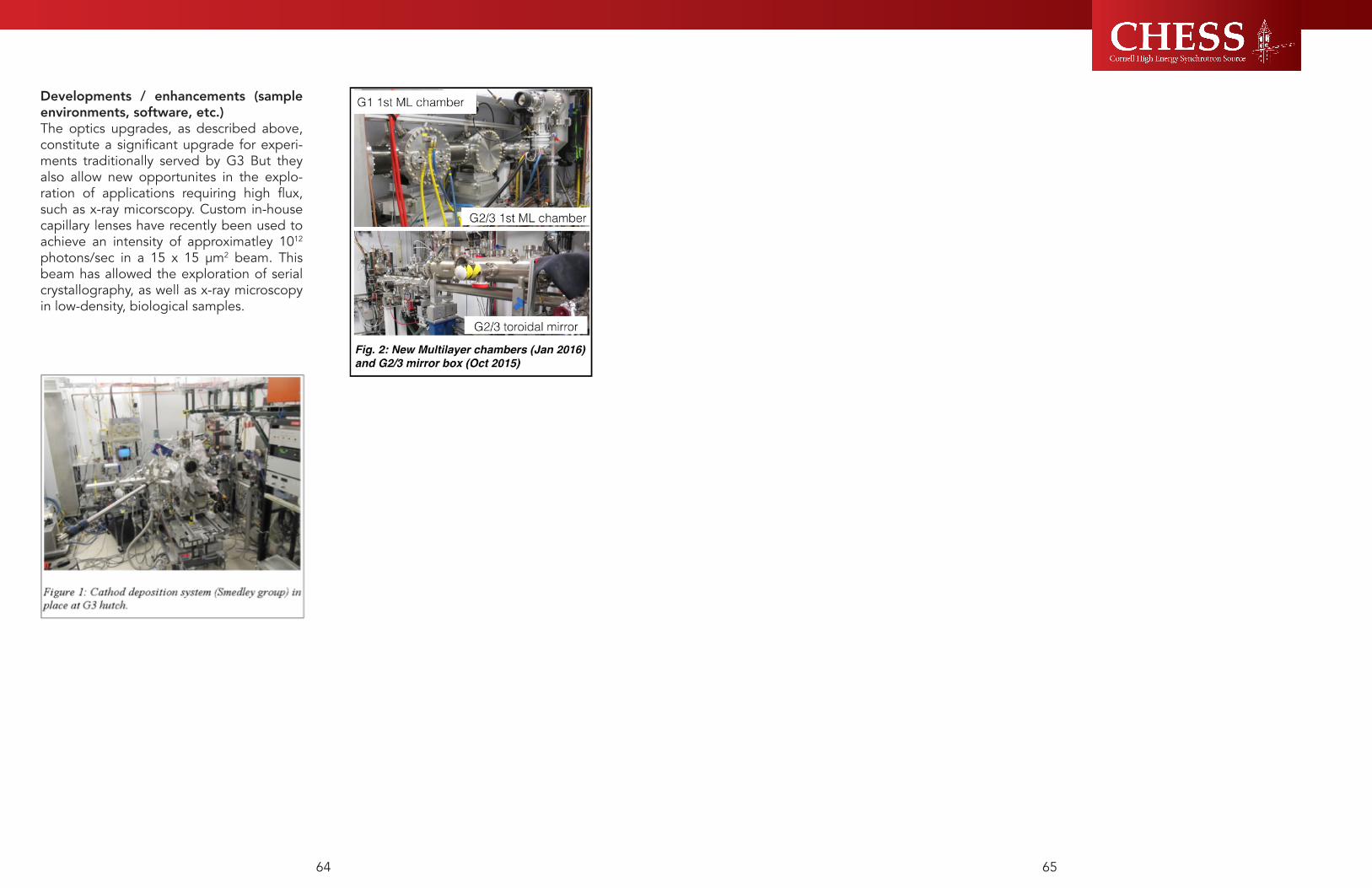
6564
Developments / enhancements (sample environments, software, etc.)The optics upgrades, as described above, constitute a significant upgrade for experi-ments traditionally served by G3 But they also allow new opportunites in the explo-ration of applications requiring high flux, such as x-ray micorscopy. Custom in-house capillary lenses have recently been used to achieve an intensity of approximatley 1012 photons/sec in a 15 x 15 μm2 beam. This beam has allowed the exploration of serial crystallography, as well as x-ray microscopy in low-density, biological samples.

4.0 Outreach & Education

6968
Education and outreach are ways that CHESS strives to broaden participation in sciences, expand and broaden our user community, teach a national audience about the value and excitement of x-ray science, and strengthen the role science plays in our society. Below find descriptions of educational programs covering many ages and levels – K-12, undergraduates (2- and 4-year colleges and universities), graduate students, post-doctoral associates, and up to and including facility users. Outreach efforts at the laboratory address leaks in the STEM pipeline by providing meaningful enrichment activities and research experiences that draw people of all ages – ‘pre-K through gray’, and hooking them on the beauty and wonder of science. First we describe the team of educators and communicators that organize and carry out most aspects of our programs.
Xraise: the education and outreach team for CHESSXraise, the education and outreach team for CHESS, is committed to cultivating a world where all people actively embrace science and engineering in their everyday lives. How we achieve this is by providing people--especially those with limited access-the opportunity to directly observe and interact with the physical world. Our work is especially relevant and powerful within the context of a user facility--what a user facility offers to science professionals, we offer an experience tailored to K-12. Toward the proliferation of our methods beyond our immediate reach, we also serve as a replicable model for other institutions.We achieve our mission through five
approaches that work in concert: building interest, enthusiasm, and engagement; cultivating agency and identity; implementing research, evaluation, and best practices; facilitating college and career readiness; and public outreach. All of these approaches contain these four core values: an explicit connection to the science of our laboratory; engineering as a vehicle for engagement and growth in science; equity of access to STEM across socioeconomic barriers; and last but not least the human component--relevance, having fun, and collaboration. Our work continues to impact the broader national and international community through: online resources, workshops and presentations, and last but not least the multiplier effect--participants in our programs become promoters of science and engineering.A user facility like CHESS exemplifies a true culture of science and engineering and, as such, serves as a powerful and indispensable model for informing the field of broader STEM education. Beyond giving tours of our facility and providing face-to-face interactions with our scientists, Xraise distills core elements of our laboratory, its people, and their process, and offers these elements directly to students, educators, and the broader community. Many of the programs we offer are done on-site in our dedicated makerspace, the eXploration station, other programs are done on other parts of campus and through field trips to other science hotspots, others at area schools and community centers, and still others are offered through shipping and science-resources-on-wheels.During the past 12 months, the Xraise team
4.1In and Around the Lab

7170
policy makers. Ryan reports directly to Katie Jacoby.
K-12: GOALS AND ACTIVITIESGoal #1: Xraise provokes interest, enthusiasm, and engagement in science, technology, engineering and math (STEM) by infusing engineering practices into the science classroom and community to establish a lifelong passion for science learning and understanding. To meet this goal, Xraise has employed two mobile science exhibitions, the Tinker Cart and the Physics Bus; developed two school-based initiatives aimed at redesigning elementary school science; Community of Practice Workshops and the Support for Engineering Education (SEE) program; and nurtured the development of Maker activities and spaces through the MotoInventions program. The strategies used for each program and evaluative indicators of programmatic success are described in the following section.
Tinker CartFeedback from local elementary school teachers during the past year has indicated that more educators would facilitate open-ended engineering projects in their classrooms if they were supplied with the materials needed to conduct such activities.
In response to teachers’ requests, the new Next Generation Science Standards that promote engineering design, and as part of Xraise’s growing mobile science program, the Xraise team developed and launched a prototype traveling Tinker Cart during the spring of 2016. The Cart holds supplies for one class of elementary school students to participate in the science and engineering activities. Tinker Carts will be donated to local schools and will be managed by the school librarian, who will check out the cart to teachers and will be responsible for restocking accessible materials such as recyclable bottles, containers, caps and paper products. During the first year of the pilot, Xraise will donate items that are harder to find or more expensive, such as hobby motors and batteries, until school librarians are able to secure future funding through small grants to local foundations to stock the Tinker Cart. Fall Creek Elementary School, test site for the first Xraise Tinker Cart prototype, provided Xraise with feedback on the design, layout, components, accessibility, ergonomics, aesthetics, branding and other aspects of the Tinker Cart to insure its use and continued success. Preliminary feedback from the school librarian, Alex Spencer and from Chris Bell, first grade teacher, is overwhelmingly positive. After the first day of using the Tinker Cart in his classroom this June, Bell reported “When the kids came back from music today, I had the Tinker Cart parked on my carpet in the center of the room. The first kids in the room turned to the rest of the line of kids, jubilantly exclaiming, ‘The Tinker cart! The Tinker cart!’ Thank you Xraise for rolling it our way.” The enthusiasm for the Tinker Cart has spread beyond Fall Creek Elementary School and has travelled throughout the Ithaca City School District. Librarians from three different elementary schools in the district, Caroline Elementary School, South Hill Elementary School and Enfield Elementary School have already
Tinker Cart Tinkerer.
hosted thousands of visitors as part of our education and public outreach program. The eXploration station serves as: part science museum, showcasing student built exhibits and physics demonstration materials; part makerspace, outfitted with tools, equipment and materials need to design and build creative contraptions; part conference room, greeting and orienting people prior to their guided tour of our research facility; and part classroom, outfitted with educational resources, lending library materials, manipulatives, and curricular material for effective pedagogy with K-12 audiences. The ability of Xraise to accommodate such a broad spectrum of visitors puts CHESS on the map as not only a world-class research laboratory for scientific users, but as a top-notch educational research facility for educational users. See section L.2 for summarized summary of the audiences, demographics composition, and numbers reached by the Xraise program.
Xraise Staff2.5 full time staff are devoted to managing and running the multiple Xraise programs; Lora Hine is the Director of the Education and Outreach program, overseeing the multiple initiatives and serving as the primary coordinator for the formal school-based and summer programs for teachers, youth and undergraduate students. Hine has a Master of Science degree and is currently pursuing her doctoral degree in Science Education. She has expanded her role as a novice science education researcher, working with school district administrators and educators to answer questions about science and engineering education at the elementary school level. Hine is largely responsible for overseeing the Xraise budget, including travel, large materials purchases, approval of participant support, and staffing concerns. Hine reports directly to Ernie Fontes, Associate Director of CHESS. Erik Herman is our Science Education Specialist,
spearheading our informal science education efforts by running the Junk Genies of Science, serving as liaison and exhibit expert to the Ithaca Physics Bus, generating hours of Do-It-Yourself videos, and providing extensive guidance to schools and organizations starting their own Makerspaces. With a Master of Arts in Science Education and several years of experience as a physics teacher, Herman provides solid content and pedagogical knowledge to the team and reports directly to Hine. Eva Luna works part-time for Xraise and serves as the team’s Teaching Support Specialist, managing the Lending Library, running formal workshops for training high school science teachers, and mentoring high school students participating in apprenticeships. Luna holds a Master of Environmental Engineering degree, playing a key role in improving existing high school science investigations by integrating engineering practices. Luna oversaw efforts to update our new Xraise website and improve the appearance and functionality of the eXploration station. Luna reports directly to Hine. Rick Ryan serves as the newest member of the team, running the public outreach and science communication arm of the Xraise program. Ryan has contributed to a growing collection of resources about the laboratory available to the general public, CHESS Users, and
Erik Herman, Lora Hine, and Eva Luna make up the Xraise team for CHESS.

7372
practices or design processes and were clearly connected to Full Option Science Sequence (FOSS) kits used by the majority of the elementary teachers in ICSD to teach the adopted science curriculum. The relevant NGSS Disciplinary Core Ideas for each grade level were studied, science and engineering practices were modeled, and teachers discovered that engineering provides elementary students with a real-world context for learning science. In addition, educators experienced for themselves how design-based challenges develop problem-solving skills, improve communication, and enhance collaboration skills. Jen Wilkie independently convened a small focus group comprised of the elementary teachers who attended the Community of Practice workshop to reflect upon Xraise’s role in their professional development and growth. Feedback, later shared with the Xraise team, was overwhelmingly positive. One teacher wrote, “Xraise provided us with a framework for how to make engineering design challenges work in the classroom. I feel empowered to do more engineering.” Another teacher stated, “Xraise provided me with the background knowledge and science language for me to use in the classroom. I know better now how to explain how things work.” A few days following the workshops, Jen Wilkie added, “I just wanted to say thank you for all for the work that you did to make the Community of Practice Workshops exciting, fun, provocative and informative. I’m so excited for what this group can do this year, and appreciate all the heavy lifting that Xraise did to get it rolling.”
Support for Engineering Education (SEE) Students of every age are drawn towards designing and building. Educators can capitalize on children’s natural inclination to engineer by weaving engineering design challenges into their science curriculum. Xraise has been working closely with teachers throughout the school year to integrate
these challenges into the classrooms and has experience with planning, implementing and assessing these challenges. Framing a design challenge is an important step in the process - creating a problem for student’s to solve that is interesting, relevant and fun established buy-in and make the lesson meaningful for the students. Recent efforts included introducing a design challenge to third graders where they were asked to create a wind-o-meter that could accurately measure 7-9 mph wind speeds generated by a small fan located 12 inches away from their device. Using recycled materials, students drew and discussed their plans, prototyped, worked as a team, tested, and redesigned a variety of wind-o-meters. Other efforts include working with teams of fourth grade teachers in the Ithaca City School District to implement the Squirrel Challenge, where students are asked to design a device that will lift a certain amount of weight (nuts) a certain height (up a tree) to store for the winter months. Using this storyline to establish buy-in and enhance motivation, students worked for hours on their projects, asking to stay behind during recess to enhance their design. Journaling, interviews and conversations with students revealed that students connected the concepts presented during the fourth grade unit on Simple Machines, successfully integrating science and engineering through the context of design challenges.Testimonials from educators describe the benefits of integrating engineering and science into their daily curriculum. Kelsey Delany, fourth grade teacher at Beverly J. Martin Elementary School in Ithaca City School District wrote to the Xraise team; “Thank you so much for being such a supportive resource for my classroom the past few years. I have learned so much about integrating engineering into my curriculum, and have developed a love for teaching science that I had not felt prior to attending your summer workshops and working with
contacted Xraise and have expressed interest in serving as the next pilot site for a Tinker Cart.
Physics BusThree years ago Xraise began partnering with a small local not-for-profit to increase the audience of our JunkGenies exhibition, a project further explained below. The Physics Bus is a mobile platform whose mission includes offering direct interactions with physics phenomena. The Physics Bus is a locally run entity, a renovated school bus that sparks interest and creativity in audiences of all ages. Synchrotron Science themed exhibits have been secured to tabletops inside the bus where they can then be taken to schools, community centers, parks, reservations, and other diverse locations throughout the United States. Covered in aluminum foil and decorated
with window stickers advertising CHESS and the National Science Foundation, these alluring and engaging displays take visitors on an educational journey like no other.The collaboration between the Physics Bus and Xraise has been mutually beneficial to both organizations, with the Physics Bus benefitting from a display of sparks, vortices, fog, beams of light, and invisible forces, and Xraise benefiting by having our exhibition reach nearly 10,000 people per year. The exhibits have traveled cross country twice and have been on display in such locations
as San Francisco, Gainesville, Phoenix, even going as far as China to be showcased as part of the Beijing Science Festival. Two other mobile science programs have been inspired by the collaboration between the Ithaca-based Physics Bus and CHESS: the Gainesville Physics Bus (Gainesville, FL) and Physics Bus West (Tucson, AZ). In addition, there have been four requests for detailed advice on starting mobile programs in other locations throughout the U.S., including Community Science Workshops in California and Seton Hall University in New Jersey. Brian Jones, founder and director of the Little Shop of Physics (LSoP), an NSF funded and award-winning outreach program out of Colorado State University, has expressed interest in adding a mobile platform for LSoP exhibits.
Community of Practice WorkshopsThe Next Generation Science Standards (NGSS) have raised the level of interest in and importance of integrating engineering into elementary science curriculum, making engineering-based learning activities more commonplace than ever before in the primary grades. Engineering-based projects have been shown to enhance student understanding of science and, for many, their interest in science (LaChappelle and Cunningham, 2014). The Xraise team has been collaborating with the Ithaca City School District (ICSD) K-6 Science Coordinator Jen Wilkie, along with elementary school teachers throughout ICSD, to leverage engineering design challenges as a way to allow young students to test their developing scientific knowledge and apply it to practical problems. As part of this ReDesigning Science initiative, Xraise sponsored eighteen ICSD educators from grades 2nd-4th (six teachers per grade level) to participate in daylong professional growth workshops hosted by Xrasie staff. Learning activities developed by Xraise for the ICSD teachers integrate engineering

7574
careers. Over 30 IAUPR faculty attended the presentation Empowering Minds through Dynamic Design, introducing Xraise’s innovative approach to education and highlighting specific programs that were the most relevant to retention in engineering at the university. Xraise has designed numerous pre-college learning activities based upon the groundswell of research in education and empirical evidence gathered through fieldwork espousing the benefits of both play and making. These engineering experiences are goal-driven, design-based, student-centered, hands-on, and collaborative in nature -- emphasizing 21st Century skills needed in the workforce and mirrored in the activity of experts at the CHESS research facility. The IAUPR Bayamon workshop also provided faculty with an opportunity to participate in tabletop activities illustrating such physics concepts as rotational inertia, eccentric motion, and mechanical resonance, sparking creativity and lively discussion.
University of Puerto Rico students and faculty showcase their projects to Xraise during the November visit to the Bayamon campus. MotoInventions The Maker-based program is co-sponsored by Xraise and the Sciencenter, taking place every Sunday afternoon in downtown Ithaca.
The materials for the program include donated recyclables as well as hobby motors, batteries, LED’s, electrical tape and other miscellaneous items provided by Xraise. The guests are free to explore different materials and build whatever they would like, with the activity being open-ended and entirely creative. Cornell graduate students volunteer and help run the program. Collectively, volunteers observe learning process taking place during the MotoInvention building sessions as visitors build, test and iterate their designs based on their successes and failure. An important aspect of the program is its ability to draw adults, in addition to children, into exploring how motors and electricity work. According to Stephanie Hiltz, a graduate student who has volunteered with the program for three years, learning occurs as a result of visitors participating in the program. “There were adults who would tape batteries onto their inventions without connecting them to the motors and be initially frustrated that their motor was not turning. They would then figure out how the wires need to be connected to each end of the battery for the electricity to flow into, and thus power, the motor. There was an enormous satisfaction that lit up the faces of both adults and children alike when they finally figured it out and got their invention to work.” The program has been able to connect kids with their adults in an unthreatening, safe and supportive environment where the adult is no longer the “expert”, but is a novice to making -- discovering concepts and techniques alongside their child. Loiusa Smeiska, a post-doctoral fellow at CHESS and MotoInventions volunteer for the past year, reflected on her experience with families, “It is really rewarding to see adults and kids working together and building confidence in new skills.” Sciencenter Director of Program, Michelle Kortenaar, commented, “People love the MotoInventions program. I know of families that make the trip to the
Xraise during the school year. Your support of classrooms is invaluable, and please do not hesitate if you ever need anything in the future!” Xraise continues to receive requests from teachers in the district for the upcoming year who would like assistance in tailoring engineering activities for their classrooms. At this time, Xraise has plans to work with six teams of elementary educators throughout the 2016-17 school year to assist them in integrating physical science core ideas with engineering practices. Students participating in these types of learning activities display perseverance, grit and follow through, spending hours on a given project or challenge. Erin Hammes, fourth grade teacher at Cayuga Heights Elementary School, was transitioning her students from a morning of building structures with the Xraise team to a mid-morning recess break. Her students, eager to improve their designs, asked to continue working during their recess. Mrs. Hammes
had to tell her students, “It is time for recess. No, you may not continue working on your design” before they would reluctantly clean up. Students are eager to improve their designs and show off their final projects to their peers and family. Lily Talcott, principal of Northeast Elementary School provided the Xraise team with a quote from one of her students following the engineering challenge activity that was presented in a fourth-grade classroom: “Mrs. Talcott, I liked working together and building things
to solve a problem, just like an engineer!” - Lucy Walker, age 9.Universities across the country are investigating various ways to successfully recruit and retain minority students in an effort to train a more diverse pool of scholars prepared for careers in science and engineering. Furthermore, many engineering departments have committed themselves to developing educational programs that deepen students’ understanding of fundamental concepts, enhance students’ active participation in learning, and establish engineering’s role in meeting the needs of a global society. The U.S. Department of Education supports such worthy initiatives through the Minority Science and Engineering Program (MSEIP), advancing America’s capacity to build and create by strengthening education programs in science, technology, engineering and math (STEM) and by supporting a diverse pool of talent in the STEM fields.In 2012, the Inter-American University of Puerto Rico at Bayamon was the recipient of the MSEIP award for the engineering project, “Increasing Retention and Graduation Rates of Hispanic Engineering Students by Active Learning Activities and Undergraduate Research Experiences”. This November, IAUPR engineering faculty members Eduardo Perez and Omar Castillo extended an invitation to Xraise to speak at the annual MSEIP faculty workshop to share their expertise in provoking interest and engagement in science and engineering. Professors Perez and Omar recognize that the key to retaining freshman in the IAUPR’s Mechanical Engineering program is to provide meaningful activities, demonstrations, and team projects that act as the “hook” to sustained interest and meaningful learning.Once on IAUPR’s Bayamon campus, Xraise met with mechanical engineering undergraduate students and high school seniors interested in pursuing STEM-related
“I liked working together and building things to solve a problem, just like an engineer!” -Lucy Walker, age 9

7776
Over the course of four years, participation in the JunkGenies program has been requested by multiple partner organizations: the Greater Ithaca Activities Center, the Sciencenter, Ithaca High School Tech Program, Research Experience for Undergraduates, Ithaca Generator, Lehman Alternative Community School, and New Roots High School. There is more demand and higher enrollment every year. Aside from increased requests from local organizations to directly participate in our JunkGenies program, there have also been inquiries by organizers from abroad. For instance, Kenneth Monjero and Antony Kinyili from the burgeoning Pioneer Science Centre in Nairobi, Kenya are working with us through Skype to create JunkGenies exhibits. Evidence that we are achieving our goal of improving agency and identity in STEM include increasing demand of our Junk Genies programming from area schools. Pride, identity, and ownership are expressed directly from teens through social media posts--some of them even using our program as the context of their profile picture. Several local high school students who have built exhibits as part of the Junk Genies program during the past few years have remained in contact with Xraise, volunteering to help
showcase the exhibits aboard the Physics Bus at the annual World Maker Faire in New York and California. One past intern, now a fourth year engineering student at Cornell, has even elected to do his senior design project in collaboration with us.Xraise has amassed an arsenal of these exhibits. A subset of these exhibits remain on display at the eXploration station, while the majority are showcased on the Physics Bus.
3. Goal #3: Xraise shares best practices with other educators through online resources, professional conferences, and publications, contributing to a growing body of science education research. Xraise adjusts future educational efforts through evaluation efforts integrated throughout our programming.To meet this goal, Xraise conducted a study, called The E in STEM: Making Engineering Evident in Elementary School, to examine urban elementary school teachers’ ability to integrate engineering design activities as part of their science curriculum. We have longitudinally tracked our Summer Science Snapshot participants to see what academic and career trajectories they have taken. In addition, we have hired an external evaluator to consult with Xraise around formative, summative and utilization-focused evaluation efforts.
Making Engineering Evident in ElementaryDuring the past year, the Making Engineering Evident in Elementary School program has provided a team of urban elementary educators with high quality professional growth and development experiences along with access to the resources and materials necessary to teach engineering design. Teachers were provided with the opportunity to enhance content knowledge and skills necessary to successfully integrate science and engineering practices into their classrooms. This study examined
Sciencenter specifically to participate!” After facilitating over one hundred MotoInvention sessions, Hiltz declared, “MotoInventions has given me, as a volunteer, many gifts. I am a much stronger teacher and scientific communicator because of the program. It has also given me many wonderful memories and a chance to give back in a meaningful way to my community.”
Goal #2: Xraise cultivates a sense of agency and identity in STEM, focusing on the needs of traditionally marginalized populations.To meet this goal, Xraise has developed an exceptional program that provides students with meaningful learning opportunities and side-by-side interactions with staff and students, The Junk Genies of Science is highlighted in the following paragraphs, along with strategies used and evaluative indicators of programmatic success.
Junk Genies of Science The JunkGenies project transforms the boundaries of participation in STEM by recasting familiar and accessible artifacts as objects of science exploration and creativity. This new approach not only changes kids’ relationship to the things around them, it creates a grassroots community of STEM experts who are co-designers in their own STEM trajectory. Leveraging our education efforts for outreach purposes, middle and high school students dive deeply into a physics concept to create an object, device or display that illustrates a physics phenomenon. Over the course of a semester, participating students use up-cycled materials, ingenuity and a lot of trial and error to create interactive physics exhibits.During the school year, middle and high school students come to the eXploration station weekly to construct--out of salvaged materials--exhibits that showcase unusual physics phenomena. Examples include a
CRT television whose deflector coil has been intercepted by an audio signal, a microwave that makes neon gas glow, and a series of bug zappers that accelerate a charged ping pong ball around a salad bowl. These projects, constructed largely of unwanted/discarded electromechanical artifacts safely and carefully intercepted from the e-waste stream, allow students to immerse themselves in a project-based authentic learning experience that has a meaningful outcome. Once completed, these refreshingly relatable exhibits travel on display for thousands of kids per year aboard a mobile platform. Aside from the obvious cost benefit, sourcing our materials from the waste stream has many other advantages which include: familiarity, eliminating fear of breaking equipment, environmental conservation, and a sense of ownership. Our program is particularly good at leveling the playing field for students who don’t necessarily excel in a traditional classroom setting, valuing who these students are and what they bring to the STEM table.
Students working on JunkGenies construction projects.

7978
coming back to the eXploration station two or three summers in a row. A few exceptional students, who were no longer eligible for the program as they entered into high school, returned in the role of Junior Leader or Snapshot Intern. Of the original 21 Snapshot students, 13 have graduate from high school and have been accepted into a post-secondary program at a college or university. The majority of the students (10 out of 13) have declared STEM majors. The remaining eight students are completing their senior year of high school during 2016-17. (see Table 1).During the summer of 2016, the Summer Science Snapshot teachers and youth embarked on a multi-day journey to learn about water accessibility and sustainability. Students entering fifth and sixth grade from Tully and West Genesee School Districts participated in a series of hands-on activities and field trips related to water and the environment, including a visit to Onondaga Lake and the Tully mudboils to learn about the impact of industry on water quality, native vegetation and animal life and how remediation efforts are helping to clean the pollution. Students spent time team-building, discovering how science is linked to technology, and working through the engineering design process. Groups conducted hands-on activities such as experimenting with water filters using various earth materials, building water pumps, and crafting three-dimensional models of local watersheds. A visit to the Cornell Water Filtration Plant provided students with the opportunity to talk with experts and witness how filtration systems work in the real world. Students displayed their final projects to the public during a showcase event on the last day of the program, communicating their findings and demonstrating how humans impact their environment.
Snapshot students build water pumps and create three-dimensional watershed models during the summer program for upper-elementary and middle school students.
Xraise recently added a new teaching-tool, the Augmented Reality Sandbox, to our arsenal of teaching resources. Ithaca area middle school students now have an exciting new way--3D, kinesthetic, and visual--to experience topography and isolines in their science classrooms, thanks to our outreach team at Xraise. The idea for our own AR Sandbox arose when teachers participating in our Summer Science Snapshot program were exploring ways to integrate technology into their 5th grade place-based watershed curriculum while still keeping it “hands-on” and exciting. The project culminated in a perfect storm of: what was needed, what hardware was readily available, what was possible with the experts on-hand, and in its relevance to the science of our laboratory.
elementary school educators’ perceptions of the use of engineering design challenges as a method for teaching science and math concepts and skills. Engineers and scientists worked alongside staff to provide teachers with the background and essential skills for integrating science and math content into engineering design activities. Using local resources, accessible materials and researched-based curriculum, we studied educators’ comfort level, familiarity and attitude changes toward using engineering as a vehicle for teaching science and math. A subset of educators will help plan and then facilitate an engineering based experience for urban elementary students. During the program, elementary students worked collaboratively to generate the best solution to an engineering problem, prototyped, tested solutions, and modify designs based upon results. Students presented their projects and share their experience with peers and adults. This study examined elementary school students’ engagement in certain learning activities that involve engineering-based design challenges. We studied student comfort level, familiarity and attitudes changed toward engineering and science as a result of the continued classroom support during the academic year. From this work, a network of elementary educators and university staff worked collaboratively, shared best practices, and developed a model for sustainable impact. Research methods, analysis and study results have been submitted for publication in the Terra Journal of Urban Education, fall 2016.
4. Goal #4: Xraise enhances career and college readiness by offering programs that encourage young students to envision themselves in STEM professions, providing apprenticeships and research opportunities for older students, and supporting precollege educators with training and resources for effectively teaching science.To meet this goal, Xraise offered enrichment programs for precollege students and teachers. The Summer Science Snapshot for Youth, Lending Library for High School Educators, Internships for High School Students and Summer Undergraduate Research Programs are featured in the below text as effective mechanisms for enhancing STEM career and college readiness.
Summer Science Snapshot for YouthMiddle-school is a formative time in a young person’s life; a student’s career interest when entering high school is the strongest predictor of their career interest when leaving high school (Sadler, Sonnert and Tai, 2012). The Summer Science Snapshot for Youth is one example of how Xraise capitalizes on this knowledge, offering experiential learning platforms for students in the middle grades and the opportunity to interact directly with scientists and engineers. There are many entertainment and recreation programs available for children in the summer months – but Snapshot is a learning program designed to provide children with opportunities to explore and envision their future in STEM. For the past five years the Summer Science Snapshot program has been oversubscribed -- more students apply that there are spaces available, sometimes within days of advertising the program on the Xraise web site. In early June, Xrasie staff met with a cohort of the 2010 Summer Science Snapshot alumns, the first group of 21 middle school students to participate in the program. Many of the students had participated in multiple Snapshot programs, Students building a windmill

8180
participating in (your) workshops and using these labs is the primary reason I received the award. Students love the labs and learn from them. Their Regents scores show the effectiveness of these labs. Thank you!” The Lending Library program does an excellent job at retaining its current audience and is diligent about spreading the program to a broader national audience of educators. We have obtained the appropriate permits for two portable X-ray fluorescence (XRF) machines to host a training workshop for teachers on how to use this equipment and make it applicable to their science classes. We continue to work with local high school science educators in updating existing laboratory investigations to reflect the Next Generation Science Standards and to modernize the equipment to more current technologies.
In addition to our annual workshops, Xraise staff travels locally providing training to science teachers and educators who are interested in borrowing the equipment and instructions. During summer 2016 we presented at the TC3 College Now! Conference (a professional development event for physics teachers who teach college level courses at their respective high schools), we introduced the lab activities of “Arecibo’s Giant Mirror” — a lab that studies radio telescopes and focal points and “Discovering Ohm’s Law” where Ohm’s Law is derived empirically — to physics and astronomy teachers who find these resources very useful for their high level courses. This summer we also presented the lab activity “Notebook Circuits” to a group of New York State Cooperative Extension 4H agents during our visit to Camp Shankitunk, who were also very grateful and excited to connect youth to these resources provided by Xraise. During fall 2016, our staff along with an engineering graduate student presented at the Science Teacher Association of New York State (STANYS)
conference held in Rochester. We introduced a new addition to our Lending Library program called “SolCycle” — a hands-on lessons on renewable energy based on a mini 3D printed solar-powered bicycle— to middle school teachers who find these resources very useful. Many of the teachers commented on how 3D printers often go unused in their schools since there are no lesson plans that justify their use and that sometimes the printers are complicated to use for both students and teachers. Teachers were appreciative of getting access to the CAD files for the activities and instructions on how to use TinkerCAD, a free online design program.
The Lending Library program also collaborates with the Learning Web, a local youth development agency that pairs youth with local job opportunities. Apprentices are assigned to the Lending Library to help with the fixing and troubleshooting of the equipment that gets broken though shipping and usage throughout the academic year. During summer 2016 an Xraise staff member worked with a middle school student whose job it was to get familiar with the Lending Library equipment and troubleshoot many of the broken items we have accumulated. The student worked on tasks as simple as replacing multimeter batteries and fuses all the way to soldering circuits that are not working correctly, as well as a personal project coding and working with Arduinos. At the end of his apprenticeship he put together a simple pulsating Arduino LED cube that provided a lot of soldering practice, figuring out how to make the code on his computer interact with the Arduino board and using multiplexing to power the 64 LEDS on the cube.
Only four months into our pilot period, this resource has been successfully used with 5th, 6th, and 8th graders across three districts. Stationed at DeWitt Middle School, teachers have rewritten their lesson plans to embrace the AR Sandbox’s permanent integration into their Physical Science and Earth Science classes. Collaborating DeWitt Physical Science teacher, Laurie VanVleet, has been developing curriculum for the sandbox that forces students to think, visualize, and apply their learning. Initially without the aid of the projector and software, students in small groups design their own landscapes--predicting where the contour lines will be; then the projector is turned on so they can compare their predictions. Sixth grade science teacher Liz Quadrozzi offers, “Teaching topography and isolines used to be either flat or messy. Not with this!”Whether we’re characterizing thin films or the
ordered lattice of a crystal, measuring and interpreting features of surfaces is a strong area of research at CHESS--to say nothing of the interest we have in assuring that our beam optics are as close to perfection as possible. The AR Sandbox offers, at an impressionable age, an immersive, enriching, and positive experience in topography. This timely and relevant connection to students’ existing curriculum has allowed us to meet these future scientists where they are in their trajectory--serving as a gateway to the science of our laboratory.
Lending LibraryThe Xraise Lending Library offers educational resources for educators. The Library has over 30 laboratory investigations, providing instructional material as well as equipment for a variety of topics in physics, biology, chemistry and engineering. Xraise hosts workshops to provide training of these investigations. Once an educator has received training, it has access to borrow them for use in its own classroom, free-of-charge.Educators that attend the workshops and utilize the lending library materials are grateful for the access to the equipment that is not readily available to them through their school districts. The annual workshops also offer opportunities for professional development where, in addition to be trained to use the lending library equipment, current university research and facility tours are presented by faculty, researchers and graduate students.
Ruth Wahl from Allegany-Limestone High School sent the following comment to Xraise after borrowing the Resonance in Transverse Waves equipment during the 15-16 academic year: “This is always a great lab. Students can actually “see” resonance. On another note, the Western New York Education Research Council recently awarded me their excellence in teaching award.. I believe that
Undergraduate physics and chemistry students from throughout the country have come to CHESS to conduct cutting-edge research this summer. In addition to working with staff scientists to understand complex x-ray phenomena, these students work with outreach staff to deliver educational programming to area youth. At left, Michelle Kortenaar, Director of Education of the Ithaca Sciencenter, dry runs an activity exploring iridescence and light waves with a group of summer research students and an 8-year old volunteer.

8382
2016 Student Summer ProjectsLSAMP - Dynamic Light Scattering Inspired by Contemporary X-Ray TechniquesStudent: Carter, JevarAffiliation: Onondaga Community CollegeMentor: Carl Franck, Professor of Physics, CornellDynamic light scattering (DLS) also known as photon correlation spectroscopy, is a common technique used to determine the size of proteins and colloidal particles down to 1 nm. Information acquired from DLS experiments can tell researchers the dynamics of the particles within a system. Inspired by the use of array detectors in X-ray experiments to determine scattering patterns in substances, a DLS experiment was constructed to study colloidal dynamics. The apparatus consisted of a laser (photon source), diluted suspension of .81 micron diameter polystyrene particles (test sample) in water and a lens less webcam (array detector). We used two diffusion equations to interpret our results
SUNRiSE -Synchrotron X-ray Diffraction Experiments to Understand the Performance of Structural MaterialsStudent: Hanley, CooperAffiliation: Fort Lewis College, ColoradoMentor: Matt Miller, Chris Budrow (Mechanical and Aerospace Engineering, Cornell)
White beam x-ray diffraction experiments use a polychromatic beam with an energy spectrum of 40-100 keV. These high energies allow the x-rays to penetrate through real sized samples. The ability to watch a materials strain reactions to loading on an atomic level aids engineers in the design process of large scale projects. When looking at large samples using x-ray diffraction, it is important to have the ability to control what region of the sample you’re looking at. Upstream and downstream slitting allows you to control the volume of diffracted x-rays seen by the detector.
SRCCS - High Sensitivity Temperature Mapping for Superconducting RF CavitiesStudent: Allen, MelissaAffiliation: Tompkins Cortland Community CollegeMentor: Pete Koufalis, Mattias Liepe (Physics, Cornell)Superconducting radio-frequency (SRF) cavities are used in particle accelerators to propel charged particles. Physical mechanisms that cause SRF cavities to exceed their critical temperature and become normal-conducting, also called quench, limit achievable accelerating fields. A temperature mapping system is used to pinpoint “hotspots” on the cavity walls to further investigate the causes of the quench.
Jevar Carter, left, works closely with Professor Carl Franck during his 10-week research project at CHESS.
Cooper Hanley, right, helps to adjust the slits used in the beamline at the A2 Endstation.
Undergraduate Research Experiences: SRCCS, SUNRiSE, REU, LSAMPAn important part of the mission of CHESS as a national user facility is its role training the next generation of students to fill roles in the varied fields in science, technology, engineering and math – the so-called STEM fields. The current CHESS award supports undergraduate students through both formal and informal programs. Summer Research for Community College Students (SRCCS), Summer Undergraduate Research in Science and Engineering (SUNRiSE), Louis Stokes Alliance for Minority Participation (LSAMP) and the Research Experience for Undergraduates (REU) are top-notch summer research programs where students contribute to the growing body of scientific knowledge generated by the staff. Students worked directly on research projects related to x-ray and accelerator technologies, working side-by-side their staff, faculty and graduate student mentors to conduct studies on such topics as photocathode sources, electron beams measurements and laser optics for use in the future accelerator, x-ray optics and x-ray detectors. In addition to conducting research, students developed numerous technical skills, including familiarity with programming languages and techniques (Unix/Linux, LaTeX, Matlab, Python and Fortran90) and technical skills (spectroscopy, chemical-
bathing, use of digital oscilloscopes, etc.). On a weekly basis, students participated in formal lectures, informal seminars, tours of research facilities, social and recreational events, as well as outreach activities. A forum at the end of each summer provided participants with the opportunity to present their research results to their peers, mentors and staff scientists. Successful recruitment efforts have increased the numbers of underrepresented minorities, females, and traditionally underserved populations who have limited access to research opportunities. In addition to providing one-on-one or co-mentoring arrangements for our summer research participants, visiting students participate in tours of laboratory facilities, presentations from experts in the field, and participate in informal sessions aimed at addressing potential obstacles to entering graduate school. Summer interns attend special training sessions, such as workshops for how to give a scientific talk or how to prepare a scientific poster. In the future, we plan to add new training workshops, such as how to present research to interested stakeholders who reside outside of the scientific community; skills such as conducting interviews in front of the camera, explaining the fundamentals of research in terms understandable to a layperson, etc. In a typical year, our participation goals are to support 5 SRCCS students, up to 5 SUNRiSE students, 1-2 LSAMPS students (supported by the College of Engineering at Cornell), and 2-4 REU students (supported by a separate NSF award). All students are mentored by CHESS staff or closely allied researchers affiliated with Cornell science departments.
4.2
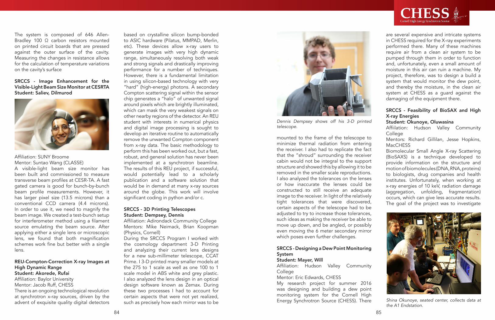
8584
mounted to the frame of the telescope to minimize thermal radiation from entering the receiver. I also had to replicate the fact that the “shroud” surrounding the receiver cabin would not be integral to the support structure and showed this by allowing it to be removed in the smaller scale reproductions. I also analyzed the tolerances on the lenses or how inaccurate the lenses could be constructed to still receive an adequate image to the receiver. In light of the relatively tight tolerances that were discovered, certain aspects of the telescope had to be adjusted to try to increase those tolerances, such ideas as making the receiver be able to move up down, and be angled, or possibly even moving the 6 meter secondary mirror which poses even further challenges.
SRCCS - Designing a Dew Point Monitoring SystemStudent: Mayer, WillAffiliation: Hudson Valley Community CollegeMentor: Eric Edwards, CHESSMy research project for summer 2016 was designing and building a dew point monitoring system for the Cornell High Energy Synchrotron Source (CHESS). There
are several expensive and intricate systems in CHESS required for the X-ray experiments performed there. Many of these machines require air from a clean air system to be pumped through them in order to function and, unfortunately, even a small amount of moisture in this air can ruin a machine. My project, therefore, was to design a build a system that would monitor the dew point, and thereby the moisture, in the clean air system at CHESS as a guard against the damaging of the equipment there.
SRCCS - Feasibility of BioSAX and High X-ray EnergiesStudent: Okunoye, OluwasinaAffiilation: Hudson Valley Community CollegeMentors: Richard Gillilan, Jesse Hopkins, MacCHESSBiomolecular Small Angle X-ray Scattering (BioSAXS) is a technique developed to provide information on the structure and motion of biomolecules (DNA, RNA, proteins) to biologists, drug companies and health institutes. Unfortunately, when working at x-ray energies of 10 keV, radiation damage (aggregation, unfolding, fragmentation) occurs, which can give less accurate results. The goal of the project was to investigate
Dennis Dempsey shows off his 3-D printed telescope.
Shina Okunoye, seated center, collects data at the A1 Endstation.
The system is composed of 646 Allen-Bradley 100 Ω carbon resistors mounted on printed circuit boards that are pressed against the outer surface of the cavity. Measuring the changes in resistance allows for the calculation of temperature variations on the cavity’s surface
SRCCS - Image Enhancement for the Visible-Light Beam Size Monitor at CESRTAStudent: Saliev, Dilmurod
Affiliation: SUNY BroomeMentor: Suntao Wang (CLASSE)A visible-light beam size monitor has been built and commissioned to measure transverse beam profiles at CESR-TA. A fast gated camera is good for bunch-by-bunch beam profile measurements. However, it has larger pixel size (13.5 microns) than a conventional CCD camera (4.4 microns). In order to use it, we need to magnify the beam image. We created a test-bunch setup for interferometer method using a filament source emulating the beam source. After applying either a single lens or microscopic lens, we found that both magnification schemes work fine but better with a single lens.
REU-Compton-Correction X-ray Images at High Dynamic RangeStudent: Akorede, RufaiAffiliation: Baylor UniversityMentor: Jacob Ruff, CHESSThere is an ongoing technological revolution at synchrotron x-ray sources, driven by the advent of exquisite quality digital detectors
based on crystalline silicon bump-bonded to ASIC hardware (Pilatus, MMPAD, Merlin, etc). These devices allow x-ray users to generate images with very high dynamic range, simultaneously resolving both weak and strong signals and drastically improving performance for a number of techniques. However, there is a fundamental limitation in using silicon-based technology with very “hard” (high-energy) photons. A secondary Compton scattering signal within the sensor chip generates a “halo” of unwanted signal around pixels which are brightly illuminated, which can mask the very weakest signals on other nearby regions of the detector. An REU student with interests in numerical physics and digital image processing is sought to develop an iterative routine to automatically remove the unwanted Compton component from x-ray data. The basic methodology to perform this has been worked out, but a fast, robust, and general solution has never been implemented at a synchrotron beamline. The results of this REU project, if successful, would potentially lead to a scholarly publication and a software solution that would be in demand at many x-ray sources around the globe. This work will involve significant coding in python and/or c.
SRCCS - 3D Printing TelescopesStudent: Dempsey, DennisAffiliation: Adirondack Community CollegeMentors: Mike Neimack, Brian Koopman (Physics, Cornell)During the SRCCS Program I worked with the cosmology department 3-D Printing and analyzing their current lens designs for a new sub-millimeter telescope, CCAT Prime. I 3-D printed many smaller models at the 275 to 1 scale as well as one 100 to 1 scale model in ABS white and grey plastic. I also analyzed the lens design in an optical design software known as Zemax. During these two processes I had to account for certain aspects that were not yet realized, such as precisely how each mirror was to be

8786
sample. The setup used is a 15 KeV, 7 × 7 mm monochromatic beam. The sample is placed on a small goniometer mounted on a Huber 4-circle diffractometer. As the beam passes through the sample, the wavefront is slightly distorted by sample density variations. Wavefront angular deviations interfere with the unperturbed beam and become intensity variations recorded on 2D images with spatial resolution better than 5 μm. With the use of appropriate software (e.g. Octopus Reconstruction), images were reconstructed from parallel beam data. 3D renderings were built using OsiriX for better visualization of the intricate structure of the bud. An initial data collection demonstrated that the 3D reconstruction can be used to identify structures within a bud.
Mark Obstalecki - Cornell University: “Understanding Microplasticity Processes Related to Fatigue Damage Using High Energy X-rays and Crystal-based Modeling”
High energy x-ray diffraction presents a unique opportunity to study material evolution associated with the onset of microcrack nucleation within ductile polycrystals during low cycle fatigue conditions. Similar to microcrack initiation that accompanies persistent slip bands within deforming single crystals aligned for single slip, we search for regions of heterogeneous
cyclic slip within polycrystalline aggregates. The plastic strain amplitude initially being carried by the entire aggregate is eventually focused into regions within individual grains. We have created a combined experiment/ simulation methodology for tracking heterogeneous cyclic plasticity at the size scale of individual grains. The high energy x-ray diffraction experiments supply us with the average stress state and misorientation heterogeneity of each grain; however, they lack spatial information other than the center of mass position of each grain. Details of the subgrain response such as the stress, lattice orientation, and plastic strain rate distributions are extracted from the simulation.
Doug Nevers - Cornell University: “Probing the Structural Origins of Quantum Dots: in-situ Characterization and Isolation”Despite many years of impressive advances in the synthesis of colloidal quantum dots (QDs) with precisely programmable size, shape and composition, it may be surprising that several fundamental questions about the basic nucleation and growth of QDs persist. For example, the detailed structure and composition of the ‘critical nucleus’ that defines the transition from molecular precursors to colloidal crystals is poorly understood. Moreover, the active growth species (i.e. monomer) have not been investigated. This knowledge gap derives in part from the difficulty in isolating and characterizing these small short-lived reaction intermediates. Nevertheless, controlling these early-stage species is crucial to minimizing QDs size dispersion, tailoring QD size, and reliability producing large-batches of identical QDs. Total X-ray scattering methods (PDF) provide a unique, powerful, and in-situ analytical tool to characterize these crucial, but fleeting initial structures and their subsequent growth.We have investigated two model systems, PbSe and CdS magic-sized clusters.
Mark Obstalecki shows his load frame that was designed at CHESS.
whether increasing the x-ray energy would change the rate of damage. We believed, by collecting data at high x-ray energy (19.8 keV and 32.4 keV), the radiation damage could become a less important obstacle for BioSAXS
Graduate and Post-Graduate Educational ProgramsGraduate students and post-doctoral associates are typically the most active participants in research programs utilizing synchrotron facilities like CHESS. Year after year CHESS notices and reports statistics showing that half the research visitors to the laboratory are in these training phases of their scientific careers (see section L.6). This section highlights some of the special programs targeting this audience; the next section on professional development discusses other more general training opportunities.
Graduate Student SymposiumOrganized by CHESS scientist Jacob Ruff and held at CHESS, these evening events are planned to provide important opportunities for graduate students to share progress, challenges, and results related to x-ray-based research carried out at CHESS. Pizza is provided, and the symposia typically draw 30-50 attendees comprised of faculty and CHESS staff in addition to graduate students and post-docs. The symposium series started several years ago as a forum for Cornell students working at the CHESS G1, G2 and G3 stations. However, it proved so popular that it was then expanded to include students who worked at other stations. Starting in the spring of 2011, the seminar series was expanded to include students from outside Cornell, typically coinciding with their beamtime. The speakers are usually grad students or post-docs, but on occasions undergraduates have presented their work.
Al Kovaleski - Cornell University: “X-ray phase contrast imaging of grapevine buds”Low temperatures are the greatest limiting factor governing plant distribution on Earth. Plants located in higher latitudes experience below freezing temperatures and may be subject to freezing of their tissues, but overwintering in these areas likely led to the development of freezing tolerance mechanisms. Water freezes in different tissue compartments at separate times in some plants, grapevines (Vitis spp.) included. However, direct observation of the freezing is difficult considering the opacity of the structures. Previous studies have used x-ray phase-contrast imaging to visualize ice formation in insects. Initial data on plants were acquired at the Cornell High Energy Synchrotron Source using a highly parallel 15 KeV monochromatic beam. However, the structures in grapevine buds were found to be much more intricate and difficult to image, compared to those of insect larvae, making it difficult to visualize ice in a time-lapse 2D fashion. Therefore, using 3-dimensional reconstruction of the bud is proposed. The objective is to visualize in 3D the structures within grapevine buds. Buds of different Vitis species were imaged to evaluate structural differences between species. The images obtained are parallel images of a rotating
Al Kovaleski harvest grape buds that will later be placed in the C1 beamline at CHESS.

8988
scientists. These lectures and workshops provide Cornell students and young researchers the first-hand knowledge and experience in x-ray physics, synchrotron radiation, and experimental instrumentation. Meetings scheduled during reporting period: 2016.08.26 - Julian Becker “Detecting x-rays with CdTe sensors. The good, the bad and the ugly.”2016.04.14 - Aaron Lyndaker “The Basics of Undulators and Bending Magnets”2016.02.11 - Margaret Koker “Grain Mapping at CHESS using High Energy X-Ray Diffraction”
PhDs Resulting from the CHESS FacilitySince its inception, over 1500 Ph.D. degrees have been awarded based on x-ray research done at CHESS and more than 500 additional Ph.D.s have been awarded from associated accelerator and high-energy physics activities at Wilson Lab. CHESS users are polled annually in mid-year to provide numbers of new PhD theses.
PROFESSIONAL DEVELOPMENT
Conferences, Workshops and MinicoursesProfessional Development for K-12 instructors is discussed in the goals and activities sections just above. See section D.7 below for goals and activities aimed at professional development of all levels of scientists and with a national audience. 5. CHESS Outreach ProgramThe outreach program bridges the scientific community to the general public through the distribution of scientific and educational materials, the sponsoring of various synchrotron science related events, and by providing opportunities for both CHESS scientific and educational users to access the rich resources available through a NSF funded National User Facility.
Web Presence and Social MediaIn January of 2016, Xraise launched a dynamic and interactive new website. The site has already attracted nearly 2000 new visitors of all ages to explore the pages and empower their minds with science. Working on a new web site gave the Xraise team a chance to focus on the mission and vision of the CHESS outreach program and how those goals can be conveyed to visitors in a meaningful way. Collectively, the team decided how to present their vast array of educational initiatives to the public. Priority was placed on conveying enthusiasm for the work the Xraise team does, peppering the site with photos of staff, students and participants engaged in hands-on investigations. At the same time, the team was committed to providing visitors with a glimpse of the unique research conducted by scientists at the laboratory. We asked some of our educational users if they had some suggestions for improving the pages. These suggestions included providing direct links to previous newsletter articles and clear instructions for viewers visiting the “Science Stuff” pages. The Xraise team considers the site to be a work in progress, celebrating the simple editing capabilities of Perch, the site’s content management system. Xraise looks forward to getting feedback from viewers and connecting with the public via online social media links and other communication tools.Connecting with people has never been easier, but it still takes a dedicated effort to create content that people will want to see on their social media feeds. Through the use of Facebook, Twitter, and YouTube - both Xraise and CHESS have been able to keep followers up to date on the exciting education and research that CHESS brings to light. Publications from our users are highlighted and shared amongst the science community, giving them more exposure and favorability. Social media also gives us an
The former presents an advantageous experimental platform, as an air-stable and slow growing reaction, for in-situ characterization of QD intermediates. Notably, MSC QDs grow through discrete transitions rather than continuous shifts in size, thus reducing the number of possible structural products. Yet, traditional MSC syntheses still yield a mixture of MSC sizes, complicating the unambiguous identification of intermediate structures. The second model system (CdS MSC) emerged from parallel research efforts to isolate high-purity and high-selectivity MSC families (or discrete states for CdS QDs). Access to high purity samples has enabled the precise structural characterization of individual MSC building blocks. Further, we show that reversible transformation between two related MSC families in response to changes in the solvent environment. We hypothesize that the change between MSC ‘states’ relates to changes in the ligand binding mode and MSC surface reconstruction. We show that 1) PDF analysis captures small structural reorganizations in MSC caused by changes in solution environment, 2) that MSC structures deviate substantially from bulk crystal phases, yet still have a reproducible and distinct atomic structure, and 3) structural identification via PDF requires accounting for all potential sources of scatter. Ultimately, in-situ structural analysis, high-purity MSC standards, and computational techniques enable characterization of crucial QD building blocks as they structurally evolve along the synthetic path from raw reagents to pristine MSC and QDs.
Matt Krogstad - Northern Illinois University: “Diffuse Scattering from Relaxor PMN-xPT”Relaxor ferroelectrics possess intriguing electromechanical and dielectric properties, the microscopic physics of which is widely regarded to be related to local, correlated atomic displacements from long-range
symmetry. However, despite numerous studies over the last few decades, the details of how short range correlations and disorder drive the relaxor behavior remain unresolved. Single crystal diffuse scattering provides a powerful probe of such deviations from an average structure correlated over varying length scales, and over the last few years, techniques and instruments for measuring diffuse scattering with both x-rays and neutrons have seen a dramatic improvement, allowing for large volumes of reciprocal space to be measured in little time. We present our recent complementary neutron and x-ray measurements on solid solutions of PMN-xPT which revealed new structure to the diffuse scattering of relaxors close to the morphotropic phase boundary.
“The More You Know” - Young Investigator Seminar Series“The More You Know” seminar series is a peer-led survey seminar and forum for CHESS graduate students and post-docs to learn about synchrotrons and general x-ray methods. This seminar was established by post-doctoral associate (at the time) Margaret Koker based on a general interest in learning more about CHESS, how a synchrotron works, and basic information about many x-ray methods. Members intend to both expand upon personal knowledge of x-ray methods and incorporate new techniques into their respective research projects.Seminar subjects (and peer speakers) are decided upon by seminar members. Each session consists of an informal talk prepared by the speaker, with plenty of time for questions and discussion about the topics being covered. Speakers are asked to keep talks at an introductory level, remembering the seminar members come from a wide variety of fields (including biology, material science, chemistry, physics, and mechanical engineering).CHESS co-sponsors weekly seminars with both local and visiting

9190
students at Cornell, many of whom are from groups traditionally underrepresented in higher education.Summarizing the data in tables L.2, during the 2015-2016 recruitment cycle reflective of Fall 2016 admissions, the Graduate School’s Recruitment Office participated in a total of 28 recruitment events as outlined in the tables below. A total of 5351 prospective graduate students were met during the 2015-16 recruitment cycle. Of the 5351 students met 389 applied for Spring 2015 or Fall 2016 graduate admission. 201 applicants are women and 224 are underrepresented minorities. Of the 389 students who applied, 129 were admitted (33%). Of the 129 students offered admission 66 (51%) are female and 87 (67%) are underrepresented minority. 45 (35%) admitted students matriculated. Of the 45 applicants who matriculated 27 (60%) are female and 31 (69%) are underrepresented minorities. This shows that while there is a higher than average admission rate (average is 22%), there is a lower matriculation rate (based on Fall 2015 overall matriculation rate of 46%). See section L.2 for more details.
Evaluation The Xraise team take the evaluation of our work seriously, recognizing that acquiring honest and constructive input from our stakeholders is essential to the overall success our programming. The previous sections of this report describe indicators of success -- features, feedback, anecdotes, observations and testimonials, provided directly from collaborators, colleagues and participants.Measuring changes in behaviors, attitude, skills, interests and knowledge done through a variety of mechanisms. Facilitated learning activities are reviewed by outreach staff and participating educators to reflect upon student engagement, discuss strategies for improving student participation, and to refine and improve existing lessons for future
implementation. Analysis of specific student artifacts are conducted to reveal information about student interest and investment in the learning activity. Motivational shifts, such as a student’s willingness to describe and execute the steps of the engineering design process, or a child’s increased involvement in group work and open collaboration on design projects, and a student’s self-initiated redesign based on preliminary data collection, are all indicators of attitudinal changes. Video recordings of students document instances of engagement and are coded for certain behaviors and instances of learning (persistence, concentration, attention, asking questions, and contributing to discussions). Surveys are administered to subsets of students, teachers and even parents before and after programming to document changes in aptitude, interests, attitudes and aspirations. The Xraise group has been working with Dr. Stephen Tai’s Science Education research group from the University of Virginia to administer and analyze surveys to measure students perceptions of science and engineering before and after a sequence of learning activities. We continue to work with Dr. Lyn English from Queensland University of Technology to modify her Engineering in Middle School Teacher Survey, an assessment of teacher attitudes toward and comfort level in implementing engineering-based activities. The adapted version, Engineering in Elementary School Teacher Survey, has not yet been validated, but has been administered to elementary teachers participating in some of our teacher training workshops in an effort to contribute to a better understanding of how primary grade teachers respond to the integration of engineering into their classrooms. In June of 2016, Xraise hired Just Consulting, LLC to help the team maximize their formal science education efforts and impact. As part of a six month pilot, Just Consulting will
avenue to recruit talent for job postings and other vacancies.
Video Production with Summer Research Students
CHESS sets itself apart by our accessibility and training for students, especially undergraduates and graduates. Capitalizing on this distinction, CHESS is increasing the visibility of its summer research students, particularly the Research Experience for Undergraduate (REU), Summer Research program for upstate New York Community College Students (SRCCS), and the Summer Undergraduate Research in Science and Engineering (SUNRiSE) programs. This years’ participating students arrived at CHESS on June 6th, and were initiated through a series of video interviews, photograph sessions, and article submissions. The testimonials provided by students contribute to the collection of materials that CHESS posts on its website and through social media. Cooper Hanley, a SUNRiSE student from Fort Lewis College in Colorado, worked for 10 weeks with the InSitu group of CHESS helping design a component for use in white beam x-ray diffraction experiments. The design and research of such components have profound effects on both the student and the future of CHESS. “This is a device that is going to get used the next time I have beamtime for my experiment”, states Cooper’s Mentor, Chris Budrow, “this is a real product that will get built”. The promotion of these programs can be
viewed by prospective students to gain an understanding of the type of research that is conducted at CHESS. These videos and communication events also achieve more than simply endorsing the programs, they give the participating students an opportunity to practice their communication efforts in preparation for future endeavors; of the six students that were interviewed, five of them stated that they had never been in front of a camera in an official capacity, showing the need for programs that develop their communication skills.
Diversity Recruiting of Graduate and Undergraduate StudentsCHESS doesn’t accept or appoint undergraduates or graduate students. However, because the diversity of undergraduate and graduate student participation in CHESS research and development projects depends strongly on the diversity of the pool of students at Cornell, CHESS participates in Cornell recruitment programs and supports a part-time diversity recruiter to network and promote CHESS programs. During 2015-2016, CHESS participated in a variety of diversity-focused recruitment events throughout the United States – see attached list in section L.2. One of the key components of the CHESS recruitment strategy continues to be the inclusion of Cornell faculty and current graduate students. A Graduate Student Ambassador Program was started as a pilot in 2014. CHESS has furthered its partnerships with the Office of Inclusion and Professional Development at the Cornell Graduate School, the Office of Academic Diversity Initiatives and the Diversity Programs in Engineering Office by hosting various groups of students participating in diversity programs like McNair, LSAMP and POSSE. Through our Director of Recruitment, CHESS also hosts the summer graduate school panels for all summer REU

9392
PresentationsThe Xraise team continues to expand the scope of educational presentation offerings available to science educators and enthusiasts across the country. Xraise hosted two workshops for high school science teachers this year, attracting secondary teachers from New York, Pennsylvania, North Carolina, Massachusetts and Ohio to participate in the eight-hour professional development workshop. The Fall 2015 Xraise High School workshop had participating educators exploring light, lenses, and mirrors to help inform practices and address in the Next Generation Science Standard (NGSS) HS-PS4-1, Waves and Their Applications in Technologies for Information Transfer. The Spring 2016 Xraise High School workshop was co-sponsored by the New York Section of the American Association of Physics Teachers. This workshop addressed a different disciplinary core idea of NGSS HS-PS4-1, helping teachers experiment with waves, electromagnetic radiation, and measuring the speed of light. The Lending Library investigations presented at these two workshops have been shipped out and used in high school classrooms in California, Michigan, and Tennessee. Collectively, 62 educators and 3,250 students throughout the United State have used Lending Library kits during the 2015-2016 school year. During the past year, Xraise staff shared best practices with science educators and enthusiasts through a variety of formal presentation venues. Erik Herman presented an interactive and instructional presentation highlighting Synchrotron Science as part of the Little Shop of Physics Open House in Fort Collins, Colorado. The Open House, the largest event on the Colorado State University campus, attracted thousands of visitors of all ages from throughout Colorado and surrounding states (Wyoming, Nebraska) for a day of hands-on science fun. Herman also presented at the World Maker Faire in
the San Mateo, CA. A Maker Faire audience eager to explore ways science can be experienced as approachable, imaginative and fun attended his presentation, entitled “The Physics Bus - Science Education, Play, Art or All Three?” Herman shared how both the beauty and understanding of certain synchrotron science concepts, such as polarization, can be simultaneously taught to visitors who interact with exhibits aboard the Bus. Lora Hine provided attendees of the St. Lawrence Section of the American Society for Engineering Education with a presentation entitled “The Role of Play in Engineering”, presenting research supporting the idea that playfulness is embedded in engineering through concepts of invention and design. Faculty from throughout the northeastern United States attended the conference along with Nedim Vardar, Engineering professor at the Interamerican University of Puerto Rico who expressed interest in adopting some of the suggested playful learning strategies for his freshman engineering students.
conduct a process and preliminary impact evaluation of the Xraise ReDesigning Science Education initiative. Just Consulting, LLC will provide project management advice, a review of existing monitoring and evaluation processes, tools and data. Just Consulting will support preliminary quantitative and qualitative data collection via observations, focus groups, surveys and validated scales. They will advise the Xraise team on data management, cleaning and analysis. Frequent face to face meetings will help the team interpret data and provide unbiased feedback for improving future programming and evaluation processes.
NATIONAL IMPACTXraise WebsiteIn January of 2016 Xraise launched a dynamic and modern new website which has already attracted thousands of visitors. The new website, aside from expanding our reach, has provided us a platform for sharing our mission and vision to the public while also clearly profiling our broader impacts role for CHESS. Beyond the purpose it is already serving our audience, creating an electronic portfolio of our vast array of educational initiatives was a valuable metacognitive exercise for our team--serving to further articulate and convey our core values. Leveraging the simple editing capabilities of Perch (the site’s content management system) the process included a beta rollout period for gathering input from a sample of users. Suggestions included providing direct links to previous newsletter articles and clear instructions for viewers visiting the “Science Stuff” pages. Beyond the value this dialogue brought to refining the website, it also informed our practice. We continue to welcome feedback from users and fully intend and expect the website to continue evolving to meet the needs of our audience.The website’s target audience includes educators, outreach and education professionals at other national user facilities,
and last but not least, the public. The resources we provide through this new medium include high school lesson plans and highlights, best-practices for outreach and education, and an intriguing glimpse of the unique research that is conducted by scientists here at our laboratory. With colorful photos of staff, students, and participants engaged in hands-on investigations, the website successfully conveys the enthusiasm of our Xraise team.In addition to our new website, Xraise has continued to develop its online clout and subscribership with ongoing up-to-date posts to social media, including Pinterest, Twitter, Facebook, and YouTube. We continue to strategically leverage the “high-profile” activities we do in order to draw viewers and subscribers toward our overall science-content-rich programming, resources, and the laboratory itself. Highlights include these posts: TinkerCart rollout at Fall Creek Elementary (1852 Twitter impressions), Synchro-Science physics demonstration show at Beverly J. Martin Elementary (2090 Facebook impressions), Garbage to Gadgets with DeWitt Middle School (4068 Twitter impressions), DIY Science! (6774 YouTube views), and our JunkGenies exhibitions aboard the Physics Bus reached over 10k views and impressions across multiple social media platforms and accounts during our southwest tour.While many of our subscribers are unknown to us, we are pleased to see recognized science and education institutions, science education professionals, and past and current students, interns, and mentors among them. Beyond what we offer to our Xraise social media followers, our monthly contribution to the CHESS newsletter demonstrates our value to the need for fresh content in cultivating and maintaining the engagement of online audiences.
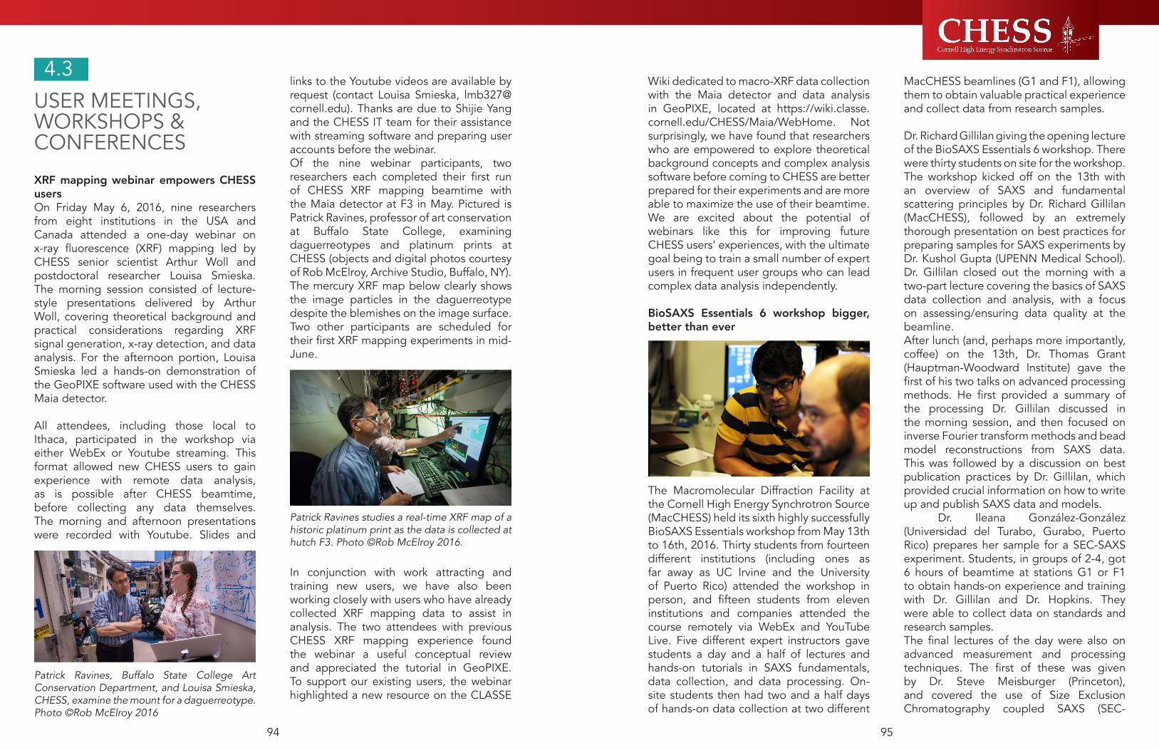
9594
Wiki dedicated to macro-XRF data collection with the Maia detector and data analysis in GeoPIXE, located at https://wiki.classe.cornell.edu/CHESS/Maia/WebHome. Not surprisingly, we have found that researchers who are empowered to explore theoretical background concepts and complex analysis software before coming to CHESS are better prepared for their experiments and are more able to maximize the use of their beamtime. We are excited about the potential of webinars like this for improving future CHESS users’ experiences, with the ultimate goal being to train a small number of expert users in frequent user groups who can lead complex data analysis independently.
BioSAXS Essentials 6 workshop bigger, better than ever
The Macromolecular Diffraction Facility at the Cornell High Energy Synchrotron Source (MacCHESS) held its sixth highly successfully BioSAXS Essentials workshop from May 13th to 16th, 2016. Thirty students from fourteen different institutions (including ones as far away as UC Irvine and the University of Puerto Rico) attended the workshop in person, and fifteen students from eleven institutions and companies attended the course remotely via WebEx and YouTube Live. Five different expert instructors gave students a day and a half of lectures and hands-on tutorials in SAXS fundamentals, data collection, and data processing. On-site students then had two and a half days of hands-on data collection at two different
MacCHESS beamlines (G1 and F1), allowing them to obtain valuable practical experience and collect data from research samples. Dr. Richard Gillilan giving the opening lecture of the BioSAXS Essentials 6 workshop. There were thirty students on site for the workshop.The workshop kicked off on the 13th with an overview of SAXS and fundamental scattering principles by Dr. Richard Gillilan (MacCHESS), followed by an extremely thorough presentation on best practices for preparing samples for SAXS experiments by Dr. Kushol Gupta (UPENN Medical School). Dr. Gillilan closed out the morning with a two-part lecture covering the basics of SAXS data collection and analysis, with a focus on assessing/ensuring data quality at the beamline.After lunch (and, perhaps more importantly, coffee) on the 13th, Dr. Thomas Grant (Hauptman-Woodward Institute) gave the first of his two talks on advanced processing methods. He first provided a summary of the processing Dr. Gillilan discussed in the morning session, and then focused on inverse Fourier transform methods and bead model reconstructions from SAXS data. This was followed by a discussion on best publication practices by Dr. Gillilan, which provided crucial information on how to write up and publish SAXS data and models. Dr. Ileana González-González (Universidad del Turabo, Gurabo, Puerto Rico) prepares her sample for a SEC-SAXS experiment. Students, in groups of 2-4, got 6 hours of beamtime at stations G1 or F1 to obtain hands-on experience and training with Dr. Gillilan and Dr. Hopkins. They were able to collect data on standards and research samples.The final lectures of the day were also on advanced measurement and processing techniques. The first of these was given by Dr. Steve Meisburger (Princeton), and covered the use of Size Exclusion Chromatography coupled SAXS (SEC-
USER MEETINGS, WORKSHOPS & CONFERENCES
XRF mapping webinar empowers CHESS usersOn Friday May 6, 2016, nine researchers from eight institutions in the USA and Canada attended a one-day webinar on x-ray fluorescence (XRF) mapping led by CHESS senior scientist Arthur Woll and postdoctoral researcher Louisa Smieska. The morning session consisted of lecture-style presentations delivered by Arthur Woll, covering theoretical background and practical considerations regarding XRF signal generation, x-ray detection, and data analysis. For the afternoon portion, Louisa Smieska led a hands-on demonstration of the GeoPIXE software used with the CHESS Maia detector. All attendees, including those local to Ithaca, participated in the workshop via either WebEx or Youtube streaming. This format allowed new CHESS users to gain experience with remote data analysis, as is possible after CHESS beamtime, before collecting any data themselves. The morning and afternoon presentations were recorded with Youtube. Slides and
links to the Youtube videos are available by request (contact Louisa Smieska, [email protected]). Thanks are due to Shijie Yang and the CHESS IT team for their assistance with streaming software and preparing user accounts before the webinar.Of the nine webinar participants, two researchers each completed their first run of CHESS XRF mapping beamtime with the Maia detector at F3 in May. Pictured is Patrick Ravines, professor of art conservation at Buffalo State College, examining daguerreotypes and platinum prints at CHESS (objects and digital photos courtesy of Rob McElroy, Archive Studio, Buffalo, NY). The mercury XRF map below clearly shows the image particles in the daguerreotype despite the blemishes on the image surface. Two other participants are scheduled for their first XRF mapping experiments in mid-June.
In conjunction with work attracting and training new users, we have also been working closely with users who have already collected XRF mapping data to assist in analysis. The two attendees with previous CHESS XRF mapping experience found the webinar a useful conceptual review and appreciated the tutorial in GeoPIXE. To support our existing users, the webinar highlighted a new resource on the CLASSE
4.3
Patrick Ravines, Buffalo State College Art Conservation Department, and Louisa Smieska, CHESS, examine the mount for a daguerreotype.Photo ©Rob McElroy 2016
Patrick Ravines studies a real-time XRF map of a historic platinum print as the data is collected at hutch F3. Photo ©Rob McElroy 2016.
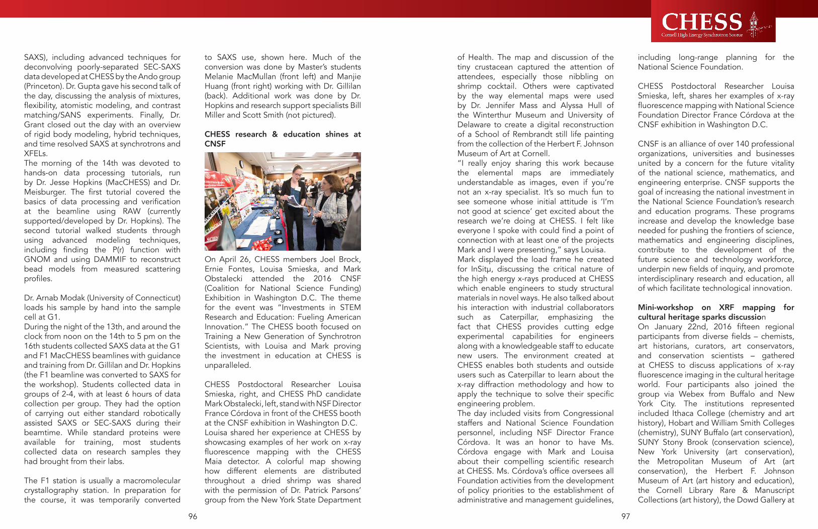
9796
of Health. The map and discussion of the tiny crustacean captured the attention of attendees, especially those nibbling on shrimp cocktail. Others were captivated by the way elemental maps were used by Dr. Jennifer Mass and Alyssa Hull of the Winterthur Museum and University of Delaware to create a digital reconstruction of a School of Rembrandt still life painting from the collection of the Herbert F. Johnson Museum of Art at Cornell.“I really enjoy sharing this work because the elemental maps are immediately understandable as images, even if you’re not an x-ray specialist. It’s so much fun to see someone whose initial attitude is ‘I’m not good at science’ get excited about the research we’re doing at CHESS. I felt like everyone I spoke with could find a point of connection with at least one of the projects Mark and I were presenting,” says Louisa.Mark displayed the load frame he created for InSitµ, discussing the critical nature of the high energy x-rays produced at CHESS which enable engineers to study structural materials in novel ways. He also talked about his interaction with industrial collaborators such as Caterpillar, emphasizing the fact that CHESS provides cutting edge experimental capabilities for engineers along with a knowledgeable staff to educate new users. The environment created at CHESS enables both students and outside users such as Caterpillar to learn about the x-ray diffraction methodology and how to apply the technique to solve their specific engineering problem.The day included visits from Congressional staffers and National Science Foundation personnel, including NSF Director France Córdova. It was an honor to have Ms. Córdova engage with Mark and Louisa about their compelling scientific research at CHESS. Ms. Córdova’s office oversees all Foundation activities from the development of policy priorities to the establishment of administrative and management guidelines,
including long-range planning for the National Science Foundation. CHESS Postdoctoral Researcher Louisa Smieska, left, shares her examples of x-ray fluorescence mapping with National Science Foundation Director France Córdova at the CNSF exhibition in Washington D.C.
CNSF is an alliance of over 140 professional organizations, universities and businesses united by a concern for the future vitality of the national science, mathematics, and engineering enterprise. CNSF supports the goal of increasing the national investment in the National Science Foundation’s research and education programs. These programs increase and develop the knowledge base needed for pushing the frontiers of science, mathematics and engineering disciplines, contribute to the development of the future science and technology workforce, underpin new fields of inquiry, and promote interdisciplinary research and education, all of which facilitate technological innovation.
Mini-workshop on XRF mapping for cultural heritage sparks discussionOn January 22nd, 2016 fifteen regional participants from diverse fields – chemists, art historians, curators, art conservators, and conservation scientists – gathered at CHESS to discuss applications of x-ray fluorescence imaging in the cultural heritage world. Four participants also joined the group via Webex from Buffalo and New York City. The institutions represented included Ithaca College (chemistry and art history), Hobart and William Smith Colleges (chemistry), SUNY Buffalo (art conservation), SUNY Stony Brook (conservation science), New York University (art conservation), the Metropolitan Museum of Art (art conservation), the Herbert F. Johnson Museum of Art (art history and education), the Cornell Library Rare & Manuscript Collections (art history), the Dowd Gallery at
SAXS), including advanced techniques for deconvolving poorly-separated SEC-SAXS data developed at CHESS by the Ando group (Princeton). Dr. Gupta gave his second talk of the day, discussing the analysis of mixtures, flexibility, atomistic modeling, and contrast matching/SANS experiments. Finally, Dr. Grant closed out the day with an overview of rigid body modeling, hybrid techniques, and time resolved SAXS at synchrotrons and XFELs.The morning of the 14th was devoted to hands-on data processing tutorials, run by Dr. Jesse Hopkins (MacCHESS) and Dr. Meisburger. The first tutorial covered the basics of data processing and verification at the beamline using RAW (currently supported/developed by Dr. Hopkins). The second tutorial walked students through using advanced modeling techniques, including finding the P(r) function with GNOM and using DAMMIF to reconstruct bead models from measured scattering profiles. Dr. Arnab Modak (University of Connecticut) loads his sample by hand into the sample cell at G1.During the night of the 13th, and around the clock from noon on the 14th to 5 pm on the 16th students collected SAXS data at the G1 and F1 MacCHESS beamlines with guidance and training from Dr. Gillilan and Dr. Hopkins (the F1 beamline was converted to SAXS for the workshop). Students collected data in groups of 2-4, with at least 6 hours of data collection per group. They had the option of carrying out either standard robotically assisted SAXS or SEC-SAXS during their beamtime. While standard proteins were available for training, most students collected data on research samples they had brought from their labs. The F1 station is usually a macromolecular crystallography station. In preparation for the course, it was temporarily converted
to SAXS use, shown here. Much of the conversion was done by Master’s students Melanie MacMullan (front left) and Manjie Huang (front right) working with Dr. Gillilan (back). Additional work was done by Dr. Hopkins and research support specialists Bill Miller and Scott Smith (not pictured).
CHESS research & education shines at CNSF
On April 26, CHESS members Joel Brock, Ernie Fontes, Louisa Smieska, and Mark Obstalecki attended the 2016 CNSF (Coalition for National Science Funding) Exhibition in Washington D.C. The theme for the event was “Investments in STEM Research and Education: Fueling American Innovation.” The CHESS booth focused on Training a New Generation of Synchrotron Scientists, with Louisa and Mark proving the investment in education at CHESS is unparalleled. CHESS Postdoctoral Researcher Louisa Smieska, right, and CHESS PhD candidate Mark Obstalecki, left, stand with NSF Director France Córdova in front of the CHESS booth at the CNSF exhibition in Washington D.C.Louisa shared her experience at CHESS by showcasing examples of her work on x-ray fluorescence mapping with the CHESS Maia detector. A colorful map showing how different elements are distributed throughout a dried shrimp was shared with the permission of Dr. Patrick Parsons’ group from the New York State Department

9998
In keeping with the theme, though, the big news was hearing that CHESS is planning an upgrade project, called “CHESS-U”, to reconfigure the accelerator and beamlines to use only a single source of particles and relocate and rebuild five experimental stations on the A, B, C, and D beamlines. Shanks showed how a single particle beam in the storage ring will reduce x-ray source sizes and give all experimental stations independently-tunable undulators. Today’s bend-magnet stations will see intensity gains over a factor of 1000. Fontes showed a conceptual plan for six new experimental stations, an extension of the three existing F-line stations, and discussed how new beamlines will not be simply a relocation of equipment and capabilities from old beamlines. Echoing an earlier message from Brock’s introduction, the project called CHESS-U will follow identi-fying the most pressing and important scientific needs for a future CHESS, illustrating the exact reason the six science workshops being held through June. In the second session, Director Brock presented a long-term vision for CHESS as a high intensity, high-energy x-ray source with unique capabilities and special types of organization and user-support. Users were especially interested in hearing from Dr. Linda Sapochak, the Deputy Director of the Division of Materials Research at the National Science Foundation (and current acting Division Director) who spoke to the Foundation’s mission, funding, and process seeking “Big Ideas” for future science that will be most impactful and relevant to society. We expect that the energy and enthusiasm of her presentation will keep CHESS and the user-community buzzing about “big ideas” throughout this month’s workshops and beyond.Users enjoyed hearing presentations from student paper prize winner Geoffrey Purdum (the Loo group at Princeton), as well as from Neil Banerjee (Univ. of Western Ontario),
Erik Muller (SUNY Stony Brook), Moutse Ranaivoson (Univ. of Medicine & Dentistry of New Jersey), Sin Urban (Johns Hopkins University), and Randall Headrick (Univ. of Vermont). The afternoon poster session showed a record 50 posters to an enthusiastic mulling crowd. CHESS awards two poster prizes for categories of best technology and best science. Advanced Design Consulting Inc. sponsored the award for the best technology poster, which went to graduate student Prafull Purohit and his group of Sol. M. Gruner (Cornell) for their poster “High-Speed, High Dynamic Range Pixel Array Detector for Scanning Transmission Electron Microscopy”. Xcelera Inc. sponsored the prize for the best science poster, which went to graduate student Jessica M. Stromberg and her group with Neil R. Banerjee (Western) for their poster “Mine to Micron Characterization of Gold and Arsenic in the Dome Mine Ankerite Veins: Synchrotron X-ray Spectroscopy of Whole Rock Samples”. The business side of the meeting saw the user-community nominate two new representatives to the Users’ Executive Committee. Congratulations to new members Steven Meisburger (Princeton) and John Twilley (SUNY Stony Brook). After-dinner speaker Juan Hinestroza (Cornell, Fiber Science and Apparel Design) discusses creating and using advanced fibers and fabrics supplemented with nanoparticles.7. International Summer Science WorkshopsThe day after the Users’ Meeting launched the six summer science workshops. A total of 78 speakers came from across the US and beyond for a total of 220 registered participants and, new this year, 131 viewers engaged remotely, asking questions and participating in the discussions through an online YouTube video forum. Detailed
SUNY Cortland (art history), the Binghamton University Art Museum (art history), and West Lake Conservators (art conservation).The mini-workshop consisted of two short talks. Post-doctoral researcher Louisa Smieska introduced the concepts behind x-ray fluorescence mapping, and provided examples of macro-XRF applied to the study of paintings, illuminated manuscripts, ceramics, and stained glass. Senior scientist Arthur Woll discussed typical project sequences and possible outcomes for cultural heritage research at CHESS, and illustrated the process with the case study of Picasso’s Blue Room, scanned in 2012. Lively discussions explored the assumptions that must be made to create digital reconstructions of faded images, strategies for XRF depth resolution, and further details on the technical capabilities at CHESS. The workshop concluded with a tour of CHESS led by Dr. Woll (pictured).The workshop sparked several project ideas that are now being actively pursued and some attendees are seeking collaborators with complementary expertise at their home institutions. Others who joined this workshop with substantial previous experience in XRF are planning to become CHESS users, and will attend the technical workshop on XRF mapping later this spring to get hands-on experience. Time will reveal the full impact of new collaborations established at the workshop, but this low-barrier-to-entry format seems to be a promising way to develop new CHESS user communities. Left to right: Abbott Nixon (West Lake Conservators); Louisa Smieska (CHESS); Patrick Ravines (Director of Art Conservation Dept., SUNY Buffalo State); Alana Ryder (Andrew W. Mellon Curatorial Coordinator for Academic Programs, Herbert F. Johnson Museum of Art); Arthur Woll (CHESS); Raphael Shea (West Lake Conservators); Walter Bowyer (Dept. of Chemistry, Hobart and William Smith Colleges); Chiara Kuhns (West Lake Conservators); Michael Haaf
(Dept. of Chemistry, Ithaca College). [Photo: Andy Weislogel]
2016 CHESS Users’ Meeting
It’s hard to capture in print the amount of pent-up excitement circulating at CHESS in preparation for the month of June. Never in our past had we planned to hold in one month the Annual Users’ Meeting as well as six separate, individually-organized science workshops. Adding to the challenge of inviting and hosting hundreds of visitors, for the first time we streamed live to the web (with two-way chat) all the presentations and discussion so that those who couldn’t travel to Ithaca, NY might still participate.The theme of this year’s CHESS Users’ Meeting, held June 7th at the Physical Sciences Building on the Ithaca campus of Cornell University, was “Creating a Brighter Future for CHESS, Users and Science”. As in past years, the user community heard updates on status, vision and new capabilities by CHESS Director Joel Brock and Associate Director Ernie Fontes, followed by Marian Szebenyi speaking for MacCHESS and Matthew Miller for the InSitμ program. Accelerator physicist James Shanks talked about successful conclusions to upgrade projects he mentioned last year towards reducing x-ray beam sizes at the undulator sources, reducing injection times, and increasing currents in the storage ring. Poster session in the hallway of the Physical Science Building

101100
can be applied to SAXS/WAXS and diffuse scattering experiments to provide more complete information than we can get from single-wavelength experiments. Solution SAXS/WAXS, time-resolved crystallography, microfluidic mixers, and laser and thermal pumping systems synchronized to the storage-ring x-ray pulses are all parts of the experimental arsenal needed to make striking new discoveries in biomolecular systems in fields as diverse as plants, animals and human disease and health.Workshop 3: Synchrotron Resources for Future Investigations of Thin-Film Growth, Processing, and Characterization explored the tremendous promise and particular challenges of ultra-thin film science and engineering for future applications ranging from photovoltaics to new technologies for never-before-seen electronic devices. Kyle Shen (Cornell) opened the workshop noting that, in recent years, the study of heterointerfaces between electronic materials has emerged as a new frontier in materials research. In particular, significant effort has been invested in studying emergent electronic states at the interface between oxides, including heterostructures of 2D van der Waals materials, such as graphene, BN, and MoS2. Over the past 2 years, one particular 2D / oxide heterostructure has been investigated intensively for the possibility of enhanced interfacial superconductivity: monolayer FeSe grown on SrTiO3, where superconductivity has been detected above 100 K. This startling discovery has radically altered our notions of what is achievable at monolayer interfaces. However, the atomic structure of the FeSe / SrTiO3 interface, and indeed nearly all atomically thin materials, is largely unknown.Other topics of the workshop included the need to capture and understand in-situ growth and processing, in-situ photocathode life-cycle studies, challenges of hard x-ray spectroscopies during in-situ growth, and metrology of devices with features below
the 10 nanometer technology node. Discussions also centered on the launch in 2015 of the NSF Materials Innovation Platform program, and how recently funded centers at Cornell and Penn State will be engineering new materials and heterostructures, and how each program-site could use capabilities at CHESS to ascertain the crystal structure of atomically thin materials and interfaces.Workshop 4: Materials Design and Processing from Nano to Mesoscale considered the strengths of CHESS to develop unique methods for studying materials design and processing from the nano to mesoscale. The theme of in-situ processing of functional soft materials was introduced by speakers Kevin Yager (BNL and Joshua Choi (UVa), who discussed photo-thermal assembly of block copolymers and self-assembly of metal halide perovskite thin films, respectively. Future high-performance applications will require sophisticated functional soft materials, including conjugated molecules and polymers, nanoparticle composites, and block copolymers. With the ability to self-assemble, these materials have direct functionality — chemical, electronic, optical, or magnetic —relevant to areas of organic electronics (OLED, OFET, OPV) and nanotechnology (e.g. directed self-assembly, ultrafiltration and desalination membranes). Many industrial processing methods can be studied in-situ with x-ray scattering to help understand and fine-tune the quality of the devices, including thermal annealing, solvent vapor annealing, blade coating and laser annealing. A second theme revolved around in-situ discovery and processing of designer materials. Speakers Ou Chen (Brown) and Yugang Sun (Temple) introduced self-assembly of multicomponent nanocrystal building blocks. Recent advance of wet synthetic chemistry enable exquisite
agendas, abstracts and organizer list can be found on the CHESS website.Workshop 1: New Industrial and Scientific Opportunities for Structural Materials invited academic and industrial partners to ponder how high-energy x-rays can help characterize residual stress as well as capture kinetic processes during life-cycle testing and additive manufacturing (AM). Residual stress in manufactured components was a particular focus of participants from American industry, including Caterpillar, Alcoa, and General Electric, who provided insight into how CHESS can be utilized to strengthen (and lighten!) the products they produce. Speakers from NASA and Air Force Research Laboratory seek to understand structural integrity in components used in high strain, high stress situations. One example came from Douglas Wells (NASA), who discussed in-situ strain measurements to understand damage tolerance at the microstructural scale, showing how solid state welding employed by NASA produces a tremendous residual stress in materials.Continuing that theme, evaluating welds using high-energy x-rays could greatly improve understanding complex processes and make a stronger and more durable component. Justin Mach (Caterpillar) gave examples of successes his company has had collaborating with CHESS to study Caterpillar’s manufacturing processes and product performance. Other speakers promoted the theme that permeated throughout all of the speakers: the importance of industrial and academic interaction for a successful Materials Genome Initiative (MGI). The higher energy and higher flux resulting from the CHESS-U upgrade could enable these measurements to become a standard by which industrial processes and products are validated and improved.Additive manufacturing, sometimes referred to as 3D printing, was a second major theme. James Williams (Ohio State)
presented the appeal of AM while also exposing the technical and economic barriers to its success. High energy x-rays have the necessary penetrating power to determine the inherent strain caused by the AM process, concluding that high fidelity modeling, consistent with the federal MGI program, is needed to validate this new manufacturing process. Participants in Workshop #1: New Industrial and Scientific Opportunities for Structural Materials.
Workshop 2: Biomolecules in Motion, attracted participants from countries as far away as Germany, interested in motion of biomolecules (proteins, nucleic acids, complexes) as they perform functions. “Motion” includes conformational changes, ranging from large domain “hinging” motions to relatively small loop motions (e.g. to allow and deny access to an active site); oligomerization changes; interaction with partner molecules.Lee Makowski (Northeastern University) introduced how protein function depends on motion and that studying structures in motion is a lot more complex than the investigation of static ones. Measurements on many time and length scales will be needed, along with a strong computational framework, to understand this difficult topic. Biomolecules are “molecular machines”. In order to understand how molecular machines work, we need to capture their dynamics, i.e. how the parts move and interact when the “machine” is working. Workshop attendees discussed how the most promising methods for obtaining dynamic information include time-resolved SAXS/WAXS (small-angle/wide-angle X-ray scattering from solution), time-resolved serial crystallography using a large number of small crystals, and diffuse scattering from crystals. The use of anomalous scattering, obtained through measurements at multiple wavelengths,

103102
essential for - the properties of a wide-range of complex functional materials. Solving fundamental questions in disordered materials physics would have extensive technological ramifications. Understanding what appear to be exotic correlations, such as the topologically phases of strongly spin-orbit-coupled materials, could have far-reaching consequences for applications in fields of energy materials or the next-generation of quantum computers. Other areas of opportunity for x-rays include nanoengineering and studying superconductivity, where, for example, pinning defects and phase competition control the technologically relevant parameters such as critical currents, structurally robustness, to how high in temperature the supercurrents can be maintained. Only now are high dynamic range high-energy x-ray detectors making it practical to record comprehensive maps of the diffuse scattering that can begin to answer such subtle questions. Following the workshops, CHESS scientists, organizers and participants joined forces to summarize notes and compose “white papers,” capturing the scientific need and opportunities for innovative work using an upgraded CHESS source. The CHESS staff is enormously grateful to members of the user community and beyond who’ve helped shape the future of CHESS. Stay tuned to the CHESS-U website for further news about the construction and timeline.
control of nanocrystals (NCs) not only with great monodispersity, size, shape and composition, but also with desired molecular decorations on NC surfaces. As it is increasingly clear, these tunable and fully-customizable NCs building blocks can act as “super-atoms” having ability to form either random or periodically ordered superlattices. Examples given included using DNA-mediated Au supercrystal to mimick bio-inspired structures, or using magnetic/luminescence to image and sense biological cell activities in a controlled way. Also mentioned were using in-situ processing with pressure, temperature or solvent extraction to develop mechanically stable architectures with size-dependent and collective properties for device purpose, such as creating solar cells by printing 3D porous nanostructured catalysts or 2D semiconductor nanowire arrays.Workshop 5: Hard X-ray Spectroscopies and Imaging held two parallel sessions covering high-resolution x-ray spectroscopy and hard x-ray microscopy. The first discussed how advanced x-ray spectroscopic techniques, such as x-ray resonant and non-resonant emission, inelastic, and Raman scattering, are powerful tools that go beyond elemental analysis to give precise information on electronic structure and excitations in solids and liquids. A major theme for discussion was the need to improve x-ray spectroscopic capabilities for mechanistic insights into catalysts under working conditions. One large outstanding goal is to understand fundamental processes of energy storage and conversion. Namely, how does one efficiently and reversibly store and release energy in chemical bonds? The answers to this question will inform knowledge-based catalytic design and enable scientists and engineers to design global energy solutions based on scientific evidence. The parallel session on hard x-ray microscopy showcased advanced x-ray microscopies and applications in a broad array of fields,
including biology, geology, and cultural heritage. Plant scientist Olena Vatamaniuk (Cornell) introduced the enormous worldwide need for safe, plant-based, high mineral density food crops. So-called biofortification, using both traditional and genetic methods, has enormous potential for nutritional enhancement of crops but is severely limited in pace and effectiveness by insufficient understanding of mineral localization and speciation in plants and of how such nutrients are regulated and transported. Synchrotron-based techniques, such as x-ray fluorescence and x-ray absorption spectrometry, have emerged as critical tools in the study of plant mineral nutrition, that bridge the gap between gene function and total mineral accumulation with mineral localization, speciation and ligand environment. Especially impactful will be enhanced instrumentation – particularly high speed, energy dispersive detectors allowing rapid collection of large and/or high resolution scan-probe imaging. Workshop 6, Defects, Distortions and Dynamics in Complex Materials addressed emerging opportunities in high-energy, high-dynamic range diffraction using next-generation pixel array detectors as a probe of lattice-coupled effects in quantum materials. Utilizing both diffuse scattering and weak-peak crystallography, participants discussed studying phase transitions, low-dimensional correlations, short-range order, superstructures, charge-density-waves, phonons, frustration, quasicrystals, and strain.This workshop articulated a need to develop a quantitative, robust, intuitive understanding of disordered states of matter. Materials researchers are trained to think of solids as perfect repeating patterns, and defects as extrinsic “nuisances” which only act to obscure the underlying physics. This picture is fundamentally incorrect. All materials are disordered on some level, and correlated disorder is intrinsic to - and

5.0 Selected In-House Research

107106
When CESR first came online in 1979, the accelerator was optimized for high-energy physics (HEP), with a design based on the “FODO” concept: one unit cell was comprised of a horizontally-focusing quadrupole, a dipole, a vertically-focusing quadrupole, and another dipole. This unit cell was repeated roughly 50 times around the CESR tunnel. A FODO-based accelerator is very flexible and the design allowed for many modes of running over the following decades, up to and including the present-day “arc pretzel” CHESS operations.
Fast-forward 30 years, and now we find the days of HEP with the CLEO detector have come to a close. Although CESR now primarily operates for CHESS, the original CESR design decisions for HEP continue to have an impact. In particular, CHESS was established to run parasitically during two-beam HEP collisions, which is why there are beam lines from both electron and positron sources, and why we continue to run with counter-rotating beams today. However, two-beam operation limits the beam quality (emittance), stability, and beam current; it also poses unique operational challenges compared to a single-beam light source such as APS or NSLS-II.
For these reasons, CHESS is exploring options for upgrading to single-beam operation. To allow for additional insertion devices one-sixth of the storage ring magnets, through the L0 / CHESS user area, must be replaced. This turns out to be quite convenient from an accelerator design perspective. In order to make room for the CLEO particle detector in the original CESR design, several stronger dipoles (the so-
called “hard bends”) were placed on either side of the L0 experimental hall, creating a long straight section. Several of these hard bends now act as source points for CHESS beam lines. Not coincidentally, the hard bend magnets are by far the largest contribution to the horizontal emittance. So, get rid of the hard bends, the emittance drops by a factor of four, and everybody wins.
The only remaining question is, what should the new sector of the storage ring look like? There are many constraints to consider beyond the ideal “this design reduces the emittance the most” or “that design makes room for the most beam lines.” All components must fit into the existing space, global properties such as the circumference of the accelerator must be preserved, ID straights must point in a direction where a beam line could exist, and so forth. After many iterations between the accelerator design group and CHESS, a design was chosen.
5.1
CHESS-U accelerator design: From two beams to one
Artistic rendering of the new CHESS-U double-bend achromat (DBA). One cell is comprised of two vertically-focusing dipole magnets, two pairs of quadrupoles, and a variety of corrector magnets, instrumentation, etc. Not shown are the pair of canted insertion devices that will occupy the straights on either side of each cell. Each straight will house two IDs.

109108
Particle physics detector makes way for upgrade
On September 6th, the solenoid from the CLEO detector was removed from the area known as L-Zero at Wilson Synchrotron Laboratory, the former interaction point of electron and positron colliding beams. The migration of this 26-ton superconducting magnet marks the last major component to be removed from the detector as the lab prepares for its next major upgrade, CHESS-U. After months of disassembly of the struc-tural steel, iron rings, calorimeter and inter-leaved muon chambers, the CLEO solenoid will soon be transported to its new home at Jefferson Lab in Virginia, where it will come out of retirement for a whole new set of ex-periments.
For nearly thirty years CLEO operated as a high-energy physics detector, from 1979 to 2008, recording the particles produced in electron-positron collisions at the Cor-nell Electron-positron Storage Ring (CESR). CLEO carried out a broad physics program of studying the production and decay of bottom and charm quarks, as well as tau leptons, and to search for new phenome-na beyond the Standard Model of particle physics. To meet the evolving needs of this wide-ranging physics program, CLEO un-derwent five major upgrades, each incor-
porating new technologies in particle detection and identification, as well as implementing advances in computing and software.
March 3rd, 2008 marked the last run where CLEO took in data. The high-energy physics team had previously branched out to studying high energy proton-proton collisions using the Com-pact Muon Solenoid (CMS) at the Large Hadron Collider (LHC) and muon decay with the g-2 experiment at Fermilab . Meanwhile, the partial skeleton of the CLEO detector remained at Wilson Lab, encircling the CESR beam pipe, where electron and positron bunches no lon-ger collided, but continued to travel smoothly through its center.
With the removal of CLEO as the first step in CHESS-U, the storage ring will soon be able to operate with a single beam of positrons instead of counter-rotating electron and positron beams, eliminating many of the performance limitations associated with the present two-beam operation. This will also en-able all CHESS beam lines to be aligned to a single beam orbit, enhancing the x-ray beam quality for research in physics, chemistry, biology, and environmental and materials sciences.
As the pieces of CLEO were carefully set in place decades ago, they also had to be carefully extracted. During the re-moval of the calorimeter, the engineers had to raise the remaining structure of CLEO, weighing over 180 tons, to ensure that the calorimeter would not graze the steel floor as it slid horizontally from in-side the solenoid coil. Existing jacks that
The final design uses a unit cell called a “double-bend achromat” (DBA), which is widely utilized in third-generation light sources. DBAs more or less do what they say on the tin: one cell is comprised of two bends, and provide an insertion device straight with zero-dispersion (achromatic). In our design the dipoles will have a vertically-focusing gradient, and two pairs of quadrupoles will provide horizontal focusing. The CHESS-U upgrade calls for six of these DBA cells to fit between the East and West RF cavities, spanning the region from the present-day F-line source to the A/G-line undulators.
There are numerous advantages to this design. The unit cell is compact, allowing for a maximal number of beam lines with insertion devices. Its periodicity means the source parameters at each beam line are identical in the baseline mode of operation, yet the individually-powered magnets allow some flexibility in designing custom source parameters without interfering with adjacent sectors. And by getting rid of the hard bend magnets, the horizontal emittance is significantly reduced. With single-beam
operations, vertical emittance is also reduced, so the source size is significantly smaller than present-day CHESS.
In June 2016 CHESS held an external review of the accelerator design, where experts responsible for the NSLS-II and APS-U designs (among many others) had the opportunity to critique the CHESS-U accelerator design and offer suggestions for improvement. Our design was well-received, and after small adjustments to reflect the committee’s reports, the accelerator design was frozen in order to enable prototyping of magnets, the first of which will be completed in January 2017.
With the accelerator design frozen, now CHESS can also begin mapping out beam lines and designing end stations to enable the diverse science program put forth in the recent science case workshops. And in a short 18 months, CESR will turn off for the last time in its present configuration, to awaken in the latter half of 2018 as a fully-fledged third-generation light source.
Artistic rendering of the new CHESS-U double-bend achromat (DBA). One cell is comprised of two vertically-focusing dipole magnets, two pairs of quadrupoles, and a variety of corrector magnets, instrumentation, etc. Not shown are the pair of canted insertion devices that will occupy the straights on either side of each cell. Each straight will house two IDs.
The 26-ton solenoid being removed from L-Zero.

111110
The answer was a resounding “yes,” fol-lowed by careful thought before committing to design and commission a new type of x-ray monochromator for the undulator beam on A1. Intercepting the undulator beam only 15 meters from the source, the only crys-tal optics that could survive very high heat loads are semi-transparent and high thermal conductivity diamond. Previously, A1 had a single (111) diamond crystal using a hori-zontal deflection geometry to provide 19.5 keV photons. To deliver higher energies into the hutch, our engineering staff, with Tom Krawczyk as the project lead, designed a water-cooled crystal mount to hold multiple crystals.. The (111) crystal diffracts a beam with center energy of 19.5 keV, and a sepa-rate (220) crystal diffracts a beam with cen-ter energy of 32 keV. In practice, switching energies requires only moving the copper block horizontally. The next phase of devel-opment will add a third crystal to produce a 45 keV beam using the (004) reflection.
To test the capabilities of A1 in a variety of experimental modes, the layout of optical tables and ancillary equipment inside the A1 hutch was reconfigured by Chris Conol-ly and the operations group. (The changes were made so that the protein crystallogra-phy setup of the MacCHESS group can be quickly returned to use.) Scientist Jacob Ruff helped commission the two new diamond crystals, resulting in a command-line switch from 19.5 to 32 keV in 5 seconds. He tried using two Dexela high-energy x-ray area de-tectors to record powder diffraction data, and succeeded in recording calibration im-ages at both energies using only 50 milli-second exposures. Later, Zhongwu Wang, Ruipeng Li and Qingqui Huang successfully recorded diffraction data from a protein crys-tal under pressure inside a diamond-anvil cell. And Xin Huang, graduate student with Joel Brock, recorded diffraction data from sulfur compounds inside a battery during charge-discharge cycling. In all these test
cases, the intensity, stability and geometry of the x-ray beams in the A1 hutch were well suited to the measurements at hand.
Upcoming feasibility work includes Richard Gillilan and Jesse Hopkins testing BioSAXS data quality and harmonic rejection at 19.5 and 32 keV, tests of X-ray Fluorescence im-aging at the higher energy, and combined phase contrast imaging with simultaneous x-ray diffraction at both energies. Users of any of the x-ray techniques mentioned above are invited to speak with staff members and encouraged to submit proposals and beam-time requests to try experiments at the new-ly upgraded A1.
had not been put to use in over eighteen years were turned on to elevate the detector a mere two inches. These jacks were able to give the team the amount of space needed to pull the calorimeter along the steel beam placed inside. Inch by inch, the crew manu-ally removed the cesium iodide calorimeter that was pressure-fitted onto the same large mandrel with which it was installed in 1988.
The team at Wilson Lab is now focusing on the reassembly and commissioning of the storage ring, where users will start experi-ments on October 26th. CHESS welcomes over 1,200 scientists and scientists-in-train-ing annually to collect data that comprises all or part of their research programs at the facility. It is supported by grants from the Di-vision of Materials Research of the National Science Foundation.
Developing new high-energy x-ray capabilities at the A1 station
The user communities for high energy x-ray capabilities at the A2 and F2 stations have grown considerably in the past few years, easily saturating the beamtime available for powder diffraction, high-dynamic range and high resolution studies at A2 and multiple load-frame structural materials experiments at F2. In addition, this past year the A2 sta-tion was upgraded to deliver filtered white beam from its undulator; early feasibility work doing fast time-resolved Laue and en-ergy-dispersive diffraction was very success-ful. With additional demand on A2, the time came to ask if the A1 station could be re-configured to deliver x-rays energies above 19.5 keV.
Schematic of the new x-ray monochromator for the A1 station. At right, the A2 undulator beam passes through the water-cooled copper block while the A1 undulator beam strikes one of two diamond crystals. Moving the block horizontally brings either the (220) or (111) diamond crystal into the A1 undulator beam to provide 32.0 keV or 19.5 keV x-rays to the A1 hutch, respectively. Though different in design, the idea for a movable water-cooled block with multiple crystals follows a successful stacked optic at beamline ID15C at the European Synchrotron Radiation Facility.
Powder diffraction scans of cerium oxide collected using the two-color monochromators at the A1 station at 19.5 and 32 keV. Both spectra taken with 50 millisecond exposures using Dexela area detectors.
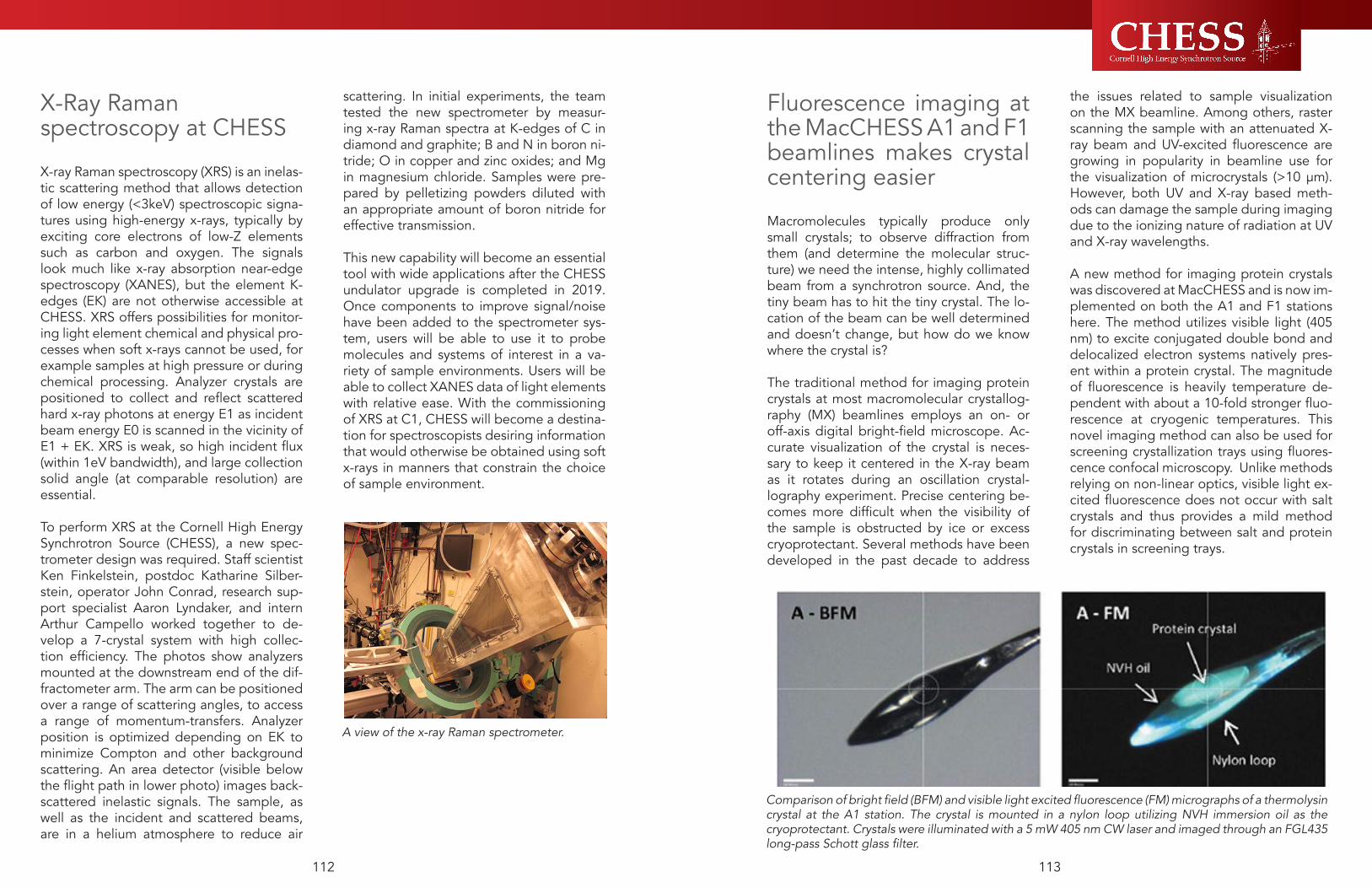
113112
Fluorescence imaging at the MacCHESS A1 and F1 beamlines makes crystal centering easier
Macromolecules typically produce only small crystals; to observe diffraction from them (and determine the molecular struc-ture) we need the intense, highly collimated beam from a synchrotron source. And, the tiny beam has to hit the tiny crystal. The lo-cation of the beam can be well determined and doesn’t change, but how do we know where the crystal is?
The traditional method for imaging protein crystals at most macromolecular crystallog-raphy (MX) beamlines employs an on- or off-axis digital bright-field microscope. Ac-curate visualization of the crystal is neces-sary to keep it centered in the X-ray beam as it rotates during an oscillation crystal-lography experiment. Precise centering be-comes more difficult when the visibility of the sample is obstructed by ice or excess cryoprotectant. Several methods have been developed in the past decade to address
the issues related to sample visualization on the MX beamline. Among others, raster scanning the sample with an attenuated X-ray beam and UV-excited fluorescence are growing in popularity in beamline use for the visualization of microcrystals (>10 μm). However, both UV and X-ray based meth-ods can damage the sample during imaging due to the ionizing nature of radiation at UV and X-ray wavelengths.
A new method for imaging protein crystals was discovered at MacCHESS and is now im-plemented on both the A1 and F1 stations here. The method utilizes visible light (405 nm) to excite conjugated double bond and delocalized electron systems natively pres-ent within a protein crystal. The magnitude of fluorescence is heavily temperature de-pendent with about a 10-fold stronger fluo-rescence at cryogenic temperatures. This novel imaging method can also be used for screening crystallization trays using fluores-cence confocal microscopy. Unlike methods relying on non-linear optics, visible light ex-cited fluorescence does not occur with salt crystals and thus provides a mild method for discriminating between salt and protein crystals in screening trays.
X-Ray Raman spectroscopy at CHESS
X-ray Raman spectroscopy (XRS) is an inelas-tic scattering method that allows detection of low energy (<3keV) spectroscopic signa-tures using high-energy x-rays, typically by exciting core electrons of low-Z elements such as carbon and oxygen. The signals look much like x-ray absorption near-edge spectroscopy (XANES), but the element K-edges (EK) are not otherwise accessible at CHESS. XRS offers possibilities for monitor-ing light element chemical and physical pro-cesses when soft x-rays cannot be used, for example samples at high pressure or during chemical processing. Analyzer crystals are positioned to collect and reflect scattered hard x-ray photons at energy E1 as incident beam energy E0 is scanned in the vicinity of E1 + EK. XRS is weak, so high incident flux (within 1eV bandwidth), and large collection solid angle (at comparable resolution) are essential.
To perform XRS at the Cornell High Energy Synchrotron Source (CHESS), a new spec-trometer design was required. Staff scientist Ken Finkelstein, postdoc Katharine Silber-stein, operator John Conrad, research sup-port specialist Aaron Lyndaker, and intern Arthur Campello worked together to de-velop a 7-crystal system with high collec-tion efficiency. The photos show analyzers mounted at the downstream end of the dif-fractometer arm. The arm can be positioned over a range of scattering angles, to access a range of momentum-transfers. Analyzer position is optimized depending on EK to minimize Compton and other background scattering. An area detector (visible below the flight path in lower photo) images back-scattered inelastic signals. The sample, as well as the incident and scattered beams, are in a helium atmosphere to reduce air
scattering. In initial experiments, the team tested the new spectrometer by measur-ing x-ray Raman spectra at K-edges of C in diamond and graphite; B and N in boron ni-tride; O in copper and zinc oxides; and Mg in magnesium chloride. Samples were pre-pared by pelletizing powders diluted with an appropriate amount of boron nitride for effective transmission.
This new capability will become an essential tool with wide applications after the CHESS undulator upgrade is completed in 2019. Once components to improve signal/noise have been added to the spectrometer sys-tem, users will be able to use it to probe molecules and systems of interest in a va-riety of sample environments. Users will be able to collect XANES data of light elements with relative ease. With the commissioning of XRS at C1, CHESS will become a destina-tion for spectroscopists desiring information that would otherwise be obtained using soft x-rays in manners that constrain the choice of sample environment.
A view of the x-ray Raman spectrometer.
Comparison of bright field (BFM) and visible light excited fluorescence (FM) micrographs of a thermolysin crystal at the A1 station. The crystal is mounted in a nylon loop utilizing NVH immersion oil as the cryoprotectant. Crystals were illuminated with a 5 mW 405 nm CW laser and imaged through an FGL435 long-pass Schott glass filter.

115114
as a result of the wider spread of the helium luminescence, the signals from F1 and F2 were difficult to separate.The new edge-on diamond VBPM was in-stalled upstream of the aperture. The dia-mond plates have been clamped to a Cu holder that was fixed to the aperture provid-ing cooling by conduction. This VBPM has to deal with two beams: F2, the direct white beam from the F-line wiggler (south blade), and, the F1 beam, the mirror reflected beam (north beam blade). The horizontal separa-tion between the north (F1) and the south blades (F2) was 7 mm.
After six months of use, the new VBPM system at F-line has shown small amounts of contamination build-up on the blades. The build-up of this contamination on the blades’ surface is a very slow process and it does not have a measurable impact on the VBPM’s performance.
The New Edge-on VBPM at C-line
We have replaced the old PE-type position monitor with and edge-on type diamond VBPM.
The diamond blade is clamped to a water cooled Cu holder near the center of the beam pipe. In the image of VBPM the X-ray beam is shown as a red band intercepting the edge-on diamond blade. The diamond luminescence is viewed through a viewport from the side through the mirror mount and a Currera (by XIMEA) camera. The camera has an embedded XP operational system that carries out all image acquisition and processing and data transmission functions.
The screen dump from the C-line VBPM is shown above. Here the beam (coming from left-to-right) hits the diamond blade edge resulting intensive luminescence. The beam position is determined as the pixel intensity centroid from the luminescence map inside of the ROI (shown as a red rectangle). The insert on the left shows the vertical profile of the beam; on the left, the beam position trace is shown.
Upgrade of VBPMs at C-, F- and G-line
At CHESS, the X-ray position monitoring and position stabilization feedback by CESR is fully realized using CHESS’ video beam position monitors (VBPMs). Throughout the years, we have developed five different types of VBPMs. All VBPMs provide position information with submicron precision, in ad-dition to visual information about the X-ray cross-sectional shape. The data delivered from the VBPMs are normally updated in each second. This frequency of data deliv-ery is appropriate for the position feedback system and our signal-archiving timing inter-vals.
VBPMs based on helium gas luminescence are used on beam lines where white beam passes through helium-filled sections. The helium luminescence is observed through a viewport on the side of the beam pipe. One downside of this method is that the intensity helium luminescence strongly depends on the purity of the helium.
Hard bend radiation passing through a thin CVD diamond sheet results in intense lumi-nescence of the diamond as the footprint of the beam passes though the diamond. Best used in vacuum conditions - over time, con-tamination may be deposited on the dia-mond surface in helium-filled systems. (F-HB and D-line)
A variation of the previous VBPM is the split diamond VBPM. In this version, the center core of the HB radiation is unobstructed and the X-rays pass through between two diamond plates. The advantage of this de-sign is that beam is mostly unaffected by the VBPM system. The position information is derived from the difference-over-sum of the top and bottom diamond luminescence. This type is used at C-line.
Slit or pinhole VBPM design: when white X-rays pass through a graphite filter or a Be window, X-rays will forward scatter from the footprint of the beam. These are mostly high energy X-rays that easily pass through a viewport. The X-rays are then detected with a pin-hole arrangement as the scat-tered X-rays create an image on a fluores-cent screen. Used at G and F-lines.The newest design is the edge-on diamond VBPM. Here the diamond plate(s) are insert-ed parallel to the beam’s propagation. In this configuration the heat load on the diamond is very low due to the small cross section of the diamond facing the X-ray beam. The di-amond plates can be placed at both edges of an undulator beam. This VBPM will pro-vide vertical position information from the centroid and horizontal position information from the difference-over-the- sum of the two plate’s luminescence. This design has been implemented at A1 and A2 undulator beam lines, and a new installation of this type is now found in the C-line upstream VBPM (which replaces the old C-line PE monitor), and at F2/F1 wiggler beam lines, replacing the helium-based VBPM.
F1/F2 Edge-on VBPM
The old F1/F2 VBPM used helium fluores-cence. The VBPM had two problems: low in-tensity due to dependence on gas humidity and purity, and degradation of fluorescence due to helium contamination. Additionally,
F1/F2 mono box with edge-on diamond VBPM.
C-line Edge-on VBPM.
Screen dump from the edge-on diamond C-line VBPM.

117116
G-line Pinhole camera VBPM
At the original F-line VBPM we used a hori-zontal heavy-met slit placed at the viewport. The forward scattered high energy X-rays easily penetrate through the viewport. The slit created an image in form of a line of the beam on the fluorescent screen coming from the beam’s footprint on the Be window. Having two undulator beams we realized that using a pinhole instead of a slit we can obtain the images of the footprint for both (G1 and G2) beams allowing us to indepen-dently measure both vertical and horizontal beam positions for G1 and G2 beams. Note the fluorescent screen is parallel to the Be window therefore the pinhole demagnifi-cation is constant along the image for any scattered X-rays. The demagnification factor of the pinhole was measured by introduc-ing a small (100um) vertical displacement of the slit and measuring the resulting change of the vertical position of the image. The demagnification factor basically is the ratio of the distances Be-window-to-Pinhole and Pinhole-to-fluorescent screen.
The screen dump of the G-line pinhole VBPM is shown below. The vertical (red) and horizontal (green) profiles shown in the left insert (for G1) and the vertical (red) and horizontal (green) beam position traces are shown in the right. To avoid clutter in these displays profiles and traces only one beam is shown as selected by the operator (the se-lection is shown with thicker ROI, below, G1 is the selected beam. The centroid program reports to CHESS’s and CESR signal data control systems for both beams regardless of the display selection.
Concept of the G-line pinhole VBPM.
Screen dump from the G-line VBPM showing the G1 and G2 beams.

6.0 Selected User Research

121120
X-RAY IMAGING: Scanning Probe & Full-Field
Relationship between maternal Se exposures in Sacramento spittail and
incidence of spinal deformaties in juveniles
Johnson RC, et. al1NOAA Fisheries,
Selenium (Se) is an essential nutrient required for oxi-dative and enzymatic processes, but at elevated levels it can disrupt protein synthesis resulting in deformities in developing offspring of fish and birds. Incidences of individuals with deformities consistent with Se tox-icity (e.g., S-shaped spines) have been observed in Sacramento splittail (splittail) Pogonichthys macro-lepidotus, a cyprinid endemic to the San Francisco Estuary and its watershed. Juvenile splittail can be exposed to elevated Se through direct ingestion of prey or through maternally-derived yolk. Here, we use scanning X-ray fluorescence microscopy (SXFM) at Cornell’s High Energy Synchrotron Source to detect Se and quantify the chronology of Se in otoliths of wild-caught juvenile splittail that display spinal defor-mities. We evaluate the spatio-temporal distribution of Se in the otoliths and compare the core (mater-nal) and edge (environmental) to test the pathway of Se exposure. Results of this study indicate that all juveniles exhibited elevated levels of Se >10 days post-hatch, outside of the influence of maternal yolk indicating direct exposure to high levels of Se in the foodweb (Fig. 1a, 1b). The majority of juveniles were exposed to elevated Se between 20-60 days after they began ingesting prey from the environment and were exposed to these elevated levels for 10-50 days (Fig. 2). However, significant variation was detected among juveniles within the otolith core suggest-ing additional research is needed to understand the role of maternal exposure of Se in influencing early development and transfer of Se to juveniles (Fig. 3). Further, this study demonstrates the utility of otolith tools in ecotoxicology to differentiate among multiple human-mediated sources of elevated Se in the eco-system that can influence native fishes.
Fig. 1a.
Fig. 1b.
Fig. 2.
Fig. 3.

123122
X-RAY IMAGING: Scanning Probe & Full-Field
Second, we compared the role of OPT7, a rice orthologue of A. thaliana OPT3, in Fe and other minerals distribution in grains. We found that grains of the opt7 mutant contain less Fe and that Fe is distribution is altered because of the loss of OPT7. We also profiledgrains from 15 rice genotypes and established the genetic variability in accumulation and distribution of different minerals among different rice varieties (see Fig 2 attached). This is very exciting findings because it shows that XRF can be used as a phenotyping tool for subsequent molecular breeding efforts. However, to be able to pinpoint changes in Fe and possibly other elements distribution to specific structures in the grain, we again need to increase the resolution.
Third, and perhaps one of the most exciting findings during this year was finding the relationship between Cu and pollen fertility. It has been known for decades that deficiency for the micronutrient Cu compromises plant fertility and reduces grain yield. Which reproductive structures require Cu and which transcriptional regulatory networks coordinate copper homeostasis and plant reproduction are still poorly understood. Using XRF we showed that Cu localizes mainly to anthers (the sites of pollen development) in Arabidopsis flowers and that the delivery of Cu to anthers requires the function of two transcription factors, CIT1 and SPL7. The A. thaliana mutant lacking CIT1 and SPL7 (cit1 spl7) is infertile and fails to deliver Cu to anthers (Fig. 3).
Relationship between maternal Se exposures in Sacramento spittail and
incidence of spinal deformaties in juveniles
Vatamaniunk, O. et al.
Cornell University
We have been very successful in using XRF for the analyses of the special distribution of transition metals in plant leaves, flowers and grains. We used several plant species: a model plant Arabidopsis thaliana, and crops, rice and wheat. First, we have established conditions to image live plant tissues. We then used the A. thaliana opt3-3 mutant lacking an Fe transporter that loads Fe into the phloem (1) and the ysl1ysl3 double mutant, lacking a transporter that moves across plasma membrane Fe associated with the Fe ligand nicotianamine (2). We also used the nas4x mutant lacking enzymes responsible for nicotianamine synthesis (3). We found that Fe has been trapped within the vasculature of leaves in all mutants unlike wild type were Fe was overall evenly distributed within leaf tissues (see Fig. 1 attached). We also noted that the pattern of Fe distribution was different these mutants suggesting different roles of transporters and nicotianamine in Fe transport (see Fig. 1).
Our colleague from the University of Massachusetts, Dr. Walker, expressed interest in collaborating on analyses of YSL1 and YSL3 function. XRF comparison of Fe and other elements distribution in the ysl1ysl3 double mutant vs wild type plants have led to novel hypothesis of YSL1 and YSL3 function in Fe homeostasis. These data have been submitted for publication. One of the reviewer’s comments was that we have to increase the resolution of our analyses and so, our next goal is to increase the resolution of our analyses from 20 µm to less than 10 microns.
These exiting findings are now in the preparation for publication and the NSF-IOS proposal to continue this work is pending. Given that global food security urges the development of high-yield grain crops, we believe that these findings will aid the development of high-yield crops. Therefore, we now extended our studies from A. thaliana to a global crop, wheat. We used 2D-XRF to initiate comparisons of spatial distribution of Cu in flowers of different wheat genotypes generated in the Sorrells lab (Cornell). We used two parental varieties, Opata 85 (from here on Opata) and W7984 synthetic, and two of their offspring, 913 and 963 (4). Consistent with our previous results in A. thaliana (Fig. 4), the majority of Cu was associated with anthers and ovules in wheat as well (Fig. 5B).
We found significant variability in Cu localization and concentration between different wheat genotypes. For example, for the two parental lines, Opata had a higher concentration of Cu in anthers and ovules than W7984. Given the essential role of Cu in plant development and reproduction, we hypothesize that these four genotypes differ in their growth, development and fertility under Cu limited conditions. These preliminary data will be used for the submission of a grant proposal to the NSF Plant Genome Program. The CRDF-Global has also funded our studies to improve wheat grain yield and nutritional quality (the URL is below). Analysis of the spatial distribution of minerals in flowers and grains of different wheat varieties is a significant component of this funded work.https://blogs.cornel l .edu/s ips/2017/02/25/v a t a m a n i u k - r e c e i v e s - f u n d i n g - t o - b o o s t -micronutrients-in-wheat/
REFERENCES
1. Z. Zhai et al., OPT3 Is a Phloem-Specific Iron Transporter That Is Essential for Systemic Iron Signaling and Redistribution of Iron and Cadmium in Arabidopsis. The Plant Cell 26, 2249-2264 (2014).
2. H. H. Chu et al., Successful reproduction requires the function of Arabidopsis Yellow Stripe-Like1 and Yellow Stripe-Like3 metal-nicotianamine transporters in both vegetative and reproductive structures. Plant Physiol 154, 197-210 (2010).
3. M. Schuler, R. Rellán-Álvarez, C. Fink-Straube, J. Abadía, P. Bauer, Nicotianamine Functions in the Phloem-Based Transport of Iron to Sink Organs, in Pollen Development and Pollen Tube Growth in Arabidopsis. The Plant Cell 24, 2380-2400 (2012).
4. M. E. Sorrells et al., Reconstruction of the Synthetic W7984 x Opata M85 wheat reference population. Genome 54, 875-882 (2011).

ENERGY & STRUCTURAL MATERIALS: In Operando Studies
125124
The effects of loading direction and strain rate on the texture evolution of a fine-grained Mg alloy AMX602
Meredith, C, et. al
Army Research Laboratory
The objective of this work is to experimentally characterize and identify fundamental microstructural features that govern flow and localization in lightweight metals, such as magnesium, by using novel in-situ and post mortem dynamic and ballistic tests in conjunction with microscale modeling. Based on this comprehensive understanding, we will construct a microstructure-sensitive, macroscale constitutive model specialized for loading conditions experienced during penetration. The ARL project started at the beginning of FY17 and runs to the end of FY19. The project initially focused on quasi-static and dynamic compression of Mg alloy AMX602, and we utilized CHESS to measure the change in texture due the compressive load applied in different loading directions. In the future, we will be conducting dynamic tension and shear dominated loading. Since ARL is a founding member of the Dynamic Compression Sector at Argonne National Lab, in the future we will be using that x-ray synchrotron to measure in-situ what we measured at CHESS on recovered samples. Using CHESS allowed us to accomplish our initial goals at the beginning of our ARL project.
The reduced-order crystal plasticity model recently developed at ARL [1,2] was successfully used to model the quasi-static and dynamic mechanical behavior of the same AMX602 measured using CHESS [3]. Utilizing the data obtained from CHESS, we published a Society of Experimental Mechanics (SEM) Annual Conference paper titled “Texture Evolution of a Fine-grained Mg Alloy at Dynamic Strain Rates” [4]. The experimental texture measurements from CHESS were compared to those predicted by the model (Figure 1-4).
Figure 1 As received (a) experimentally measured texture and (b) randomly sampled orientations used as input for the model. Both pole figures have the same orientation
Figure 2 Experimentally measured (top) and model predicted (bottom) texture of AMX602 subjected to (a) ~3%, (b) ~10% (c) ~15% dynamic compression in the ED. All pole figures have the same orientation and scale
Figure 3 Experimentally measured (top) and model predicted (bottom) texture of AMX602 subjected to (a) ~3%, (b) ~10% (c) ~15% compression in the TD. All pole figures have the same orientation and scale
Figure 4 Experimentally measured (top) and model predicted (bottom) texture of AMX602 subjected to (a) ~3%, (b) ~10% (c) ~15% compression in the ND. All pole figures have the same orientation
References1. Becker, R. and Lloyd, J.T., An efficient crystal model for
HCP metals: Application to Mg. Mechanics of Materials, 2016. 98: p. 98-110.
2. Lloyd, J.T. and Becker, R., Stress-based crystal analysis of yielding in rolled Mg AZ31B. Philosophical Magazine, 2016. 96(4): p. 1-17.
3. Meredith, C.S. and Lloyd, J.T., The quasi-static and dynamic response of fine-grained Mg alloy AMX602: An experimental and computational study. Materials Science and Engineering A, 2016. 673: p. 73-82.
4. Meredith, C.S. and Lloyd, J.T., Texture Evolution of a Fine-grained Mg Alloy at Dynamic Strain Rates. 2017. Dynamic Behavior of Materials, Vol. 1, D. Casem et al. (eds.), Ch. 35: p. 263-270.

ENERGY & STRUCTURAL MATERIALS: In Operando Studies
127126
Studying Microcrack Initiation Using Finite Element Simulation and High
Energy X-ray Diffraction
Miller, M.Cornell UniversityThe project focuses on developing new insights into microcrack initiation in ductile metals through combined high energy x-ray diffraction experiments and finite element simulation. Crack initiation will occur within a region which combines detrimental levels of heterogeneous deformation and a multiaxial stress state. The x-ray diffraction data provides us with grain averaged information about the structural heterogeneity resulting from deformation, however, the data is not invertible to extract spatial distributions of quantities such as stress and misorientation. The finite element simulation bridges the gap, supplementing the x-ray data with approximations of spatial deformation fields. Previous methods to approximate the microstructure utilized grain averaged positions and Voronoi tessellation to approximate the topology of the polycrystalline aggregate. However, the exact spatial topology of grain boundaries can strongly influence the deformation heterogeneity within a grain. The newly developed capability at CHESS to measure the grain topology prior to deformation enables us to compare experimental and simulation results directly.During the 2016 award period, near-field and far-field experiments were conducted on samples of commercially pure copper and Ti-7Al titanium. Our March experiment focused on gathering near-field grain orientation maps from copper samples and performing an experiment on a Ti-7Al sample provided by the Air Force Research Laboratory (AFRL). The titanium sample was deformed cyclically until failure with pauses for x-ray measurements.
The grain orientations within two copper samples with different grain sizes were measured using the near-field experimental geometry. These same two samples were later mechanically cycled for over 100 cycles in May while gathering far-field measurements. We interrogated the same volumes of material during both the March and May experiments.
The March experiment was our first attempt to measure and reconstruct grain orientation maps, so a considerable amount of time was spent incorporating near-field data reduction capabilities into our Matlab data reduction code, APEX-RD. Figure 1 highlights the reconstructed microstructure of one copper sample. The grain orientation map measurement is completely nondestructive – a unique and desirable capability. The nondestructive nature of the measurement allowed us to subsequently mechanically load the sample during our May experiment.
Figure 1:(Left) Measured grain orientations using the box beam technique at CHESS and (Right) the entire volume after stitching grains together
The next step of the project involves correlating the spatial grain maps with far-field experimental data collected during the May experiment. This data set is the first of its kind, providing in situ measurements of spatially correlated stress and structural heterogeneity across an aggregate from a cyclically deformed sample. At the same time our modeling collaborator, Dr. Paul Dawson, will focus on transforming the 3-D orientation map into a finite element mesh. The complete data set enables direct comparison between experiments and simulation where the grain topology is accurately represented within the simulations. The data will be used to both advance the simulation’s capability to capture the intragranular structural heterogeneity observed in the HEXD experiments and obtain insight into the detrimental conditions leading to microcrack initiation.
Our AFRL collaborators have been reducing the titanium data in tandem. X-ray diffraction data were collected during the first and 96th cycle as the sample was mechanically loaded between tension and compression at a fixed strain amplitude of 1% strain. Figure 2 shows the stress strain response of the sample along with a plot of misorientation within many grains. Ongoing work focuses on correlating the structural heterogeneity with quantities such as stress to build models better capable of predicting failure within ductile metals.
Figure 2: (Left) Stress-strain response of the Ti-7Al for cycle numbers 1 and 96. The sample was mechani-cally deformed at 1% macroscopic strain amplitude. (Right) Plot of the misorientation within each grain at the compressive loop tip of cycle 1. The misorienta-tion data is plotted at the corresponding centroid po-sitions of each grain extracted from the far-field data.
Measurement of Residual Stress in Welded Steel Components
Beaudoin, A and Mach, JCornell University and Caterpillar
A direct and timely path to fuel-efficiency in both land vehicles and aircraft is through light-weighting of structural components. This particular work addresses residual stresses in welded steel structures: without knowledge of residual stresses, a component must be (over-) sized to address the possibility of fatigue failure. In this project, we mapped the residual stress distribution about a weld using a reflection geometry. The mapping was performed at several energies, at stations A2 and F2 from 2014-2015, to establish the effect of penetration depth on the measurement of residual stress and provide a connection between synchrotron practice and the usual lab sources applied by industry. Working with Caterpillar, as part of the inSitu@CHESS project, we have developed welded steel specimens that are unique for addressing the interplay of residual stress and fatigue loading: the samples are of a dimensional scale that is representative of real-world application, tractable for synchrotron study, and exhibit large gradients in residual stress (actually spanning the range from the compressive to tensile yield stress, a stress gradient of roughly 600 MPa). The sample is paired
with a simulation of the welding process. Initial trial measurements, conducted at F2, provided validation of the locations of maximum compressive and tensile stress – as indicated by the simulation. In the present work, we have performed mapping of the stress state about the weld. A stage has been developed by CHESS personnel that provides for positioning of (these relatively large) samples in the usual four-circle goniometer setup. This experiment was performed at A2 and F2, using three energies (6.791 keV, 15.85 keV and 42 keV). Additionally we performed Energy Dispersive Diffraction (EDD) at APS 1-BM-B to provide complementary verification of the modeling with a technique that allowed us to penetrate further into the sample and perform detailed mapping of the near-surface strain state (as shown in the figure below). Furthermore, the experiments allowed for understanding of surface phenomenon related to lower penetration from lower energy lab-based sources, which may skew surface stress results.

Macromolecules and BioChemistry
129128
Dynamic Metalloenzymes
Ando, N. et. al.
Princeton University
Major activities. This proposal focuses on experimen-tal characterization of domain motion in multi-domain metalloenzymes, toward understanding how these motions are correlated with each other during differ-ent stages of the catalytic cycle. One such dynamic multi-domain metalloenzyme is the complex between the corrinoid Fe-S protein and its methyltransferase (CFeSP/MeTr), which is involved in aceto-genic car-bon fixation. Correlated motions occurring in crystals of these enzymes give rise to X-ray dif-fuse scatter-ing. Although diffuse scattering measurements are commonly used to characterize disorder in crystalline materials, the technique is relatively unknown in a bio-logical context. One challenge of measuring diffuse scattering from biological samples is the fact that the diffuse signal is weak compared with the Bragg peaks.
We proposed to apply new technologies in order to more accurately measure the weak signal. During the reporting period, significant progress was made by taking advantage of the unique properties of pixel array detectors (PADs) available at CHESS, and this work has been de-scribed in Accounts of Chemical Research [Meisburger 2017].
Specific aim 1: Diffuse scattering is present in the background of diffraction images from crystals, be-tween and underneath the Bragg peaks. While the motions of atoms in a crystal results in a loss of reso-lution in the Bragg data, these photons are not lost, but are instead scattered between the Bragg peaks, with a complex pattern that depends on how the atomic motions are correlated with one another. No other technique is capable of measuring correlated atomic motions over long distances. The central chal-lenge of measuring diffuse scattering from proteins is that protein crystals have large unit cells, which means that Bragg peaks are relatively close together on the detector. In addition, the Bragg peaks are many orders of magnitude more intense than the dif-fuse scattering. As a consequence, the detector must have a very narrow point-spread function so that the peaks do not bleed into the background and corrupt the diffuse signal. Until recently, commonly available X-ray detectors were not suited to the task. That has changed with the introduction of PADs for protein crystallography, such as the Pilatus 6M available at CHESS. These detectors provide a narrow point-spread function and low noise (Figure 1A). As a con-sequence, it is possible to build up a detailed recon-struction of reciprocal space by accumulat-ing the weak signals from many images as the crystal rotates.
During this reporting period, we undertook a sys-tematic study of diffuse scattering from lyso-zyme crystals at room temperature in collaboration with Sol Gruner (Cornell). Crystals of either tetrago-nal or orthorhombic lysozyme were looped and kept hydrated by placing the loops within plastic capil-lar-ies. Complete datasets were acquired at beam-line F1 using the Pilatus 6M in fine-slicing mode. Images were processed using custom software and merged in 3D. Despite the very weak background in each frame (averaging ~1 photon per pixel), the 3D merged data has excellent signal-to-noise, revealing the cloudy diffuse features between the Bragg peaks (Figure 1B).
Specific aim 2: Acetogenic bacteria produce acetic
acid from atmospheric CO2 and are important play-ers in the global carbon cycle. During this reporting period, we examined CFeSP/MeTr in collaboration with Steve Ragsdale (U. Michigan). CFeSP/MeTr cata-lyzes a key step in acetogenic carbon fixation, remov-ing a methyl group from the substrate CH3-H4folate and transferring it to carbon monoxide dehy-droge-nase/acetylCoA synthase (CODH/ACS). To accom-plish this challenging reaction, CFeSP/MeTr uses a vitamin B12 derivative as a supernucleophile. The B12-binding domain domain in CFeSP (Figure 2A) is known to move by an astounding 18 Å during the catalytic cycle, and this can occur even within crystals. However, the allosteric mechanisms that regulate the motion of the B12 domain are not under-stood.
In order to determine how domain motions are cor-related at each catalytic step, we obtained crystals of CFeSP/MeTr and acquired complete diffuse scatter-ing datasets. In order to prevent radiation damage and oxidation, these crystals were looped in an anaer-obic chamber and frozen prior to data col-lection. In the diffraction images, we observe strong, textured diffuse scattering in the background that extends to the edge of the detector, 2.0 Å, well beyond the resolution of the Bragg diffraction, 3.0 - 3.5 Å (Figure 2B). In 3D reciprocal space reconstructions, the sym-metry of the diffuse pattern is apparent (Figure 2C). The cloudy features are a tell-tale signature of large-scale correlations within the unit cell, consistent with domain motion.
Although the data shown in Figure 2 were collected at 100K, where protein motion is arrested, the pres-ence of diffuse scattering suggests that the ensem-ble of conformations is “locked in” during freezing. However, it is also possible that freezing process somehow causes the disorder, which produces the dif-fuse scattering. We have taken the first steps toward confirming that the diffuse scattering we ob-serve at 100K is due to conformational ensembles present at room temperature. Measuring diffuse scat-tering from CFeSP/MeTr at room temperature is a signifi-cant challenge because the crystals must be kept in
an oxygen-free environment without introducing any window materials that would scatter X-rays and over-whelm the weak diffuse signal. To achieve this, we have placed CFeSP/MeTr crystals in gra-phene-win-dowed chips, developed in collaboration with Sarah Perry (U. Mass. Amherst) and collected diffuse scat-tering from multiple small crystals in series to build a complete dataset.
Publications:Meisburger SP & Ando N. (2017) Correlated Motions from Crystal-lography beyond Diffraction. Acc. Chem. Res. 50, 580–583.
Figure 2. (A) Crystal structure of CFeSP (dark blue) and MeTr (light blue) bound to B12 (magenta) from PDB 4DJD. (B) X-ray diffraction image of CFeSP/MeTr obtained at CHESS. (C) Surface of constant |S| through the 3D reciprocal space reconstruction, beyond the Bragg peaks. Symmetry of the diffuse pattern is clearly visible.
Figure 1. Diffuse scattering from Lyso-zyme measured using a PAD. (A) Weak background signal is recorded alongside intense peaks [Meisburger 2017]. (B) Re-ciprocal space reconstruction re-veals the cloudy diffuse pattern (0kl is shown).

Macromolecules and BioChemistry
131130
Material Moduli in Lipid Bilayers
Nagle J1, Dupuy F2, Petrache H3, Tristram-Nagle S1
1Carnegie Mellon University, 2Universidad Nacional de Tucuman (Argentina), 3Indiana University–Purdue University Indianapolis
In 2014 my group reported the first experimental value for the tilt modulus (first reference), which is now recognized as important in biology. However, we had only studied one lipid, DOPC. In 2016 dif-fuse scattering data for many lipids from 2004 to the present were re-analyzed with the new, superior, tilt-dependent model and a paper is being written to publish the results. Another paper is being planned to focus on the interesting temperature dependence of the tilt modulus of DMPC just discovered by me in 2016. Employing a tilt-dependent model also allows us to obtain better values of the bending modulus, which has been recognized as a central quantity in the biophysics of membranes.
It was important to take the time to do this exten-sive analysis for many lipids using the data already acquired during the last 12 years in order to deter-mine which samples to take to CHESS on our next trip, now scheduled for March 2017. This accounts for our not requesting beam time in 2016.
The two papers published in 2016, also listed in refer-ences, continued to sharpen our analysis and experi-mental methodology.
In 2016, using a new method of analysis1,2 of diffuse x-ray scattering data taken at CHESS, a dramatic soft-ening was revealed in the molecular tilt modulus K� of the DMPC proto bio membrane as the temperature is lowered towards the main phase transition tempera-ture TM. As spontaneous tilt occurs below TM, this sug-gests that tilt is a symmetry breaking order parameter. The bending modulus KC also softens close to TM, but unlike the tilt modulus, KC has a maximum 3 degrees above TM, which also marks the limit of the well-known anomalous swelling range.3 Tilt adds a new perspec-tive to our understanding of the main phase transition in DMPC bilayers, previously viewed only as a poly-meric conformational order/disorder transition with an order parameter depending upon area per lipid.4
Fig. 1. Tilt modulus KΘ versus temperature for DMPC bilayers with phase transition temperature TM.
[1] Jablin MS, Ph.D. thesis http://lipid.phys.cmu.edu (2015).[2] Jablin MS, Phys. Rev. Letts. 113, 248102 (2014). [3] Chu N et al., Phys. Rev. E. 71, 041904 (2005).[4] Nagle JF, Faraday Discussion Chem. Soc. 81, 151-162 (1986).[5] Nagle JF et al., Soft Matter 12, 1884-1891 (2016).[6] Nagle JF et al., Chem. Phys. Lipids 196, 76-80 (2016).
Interactions of the M binding domain from HIV MA with membranes
Tristram-Nagle S1, Andenoro K1, Carroll L1, Dell Z1, Heinrich F1, Jablin M1, Langer L1, Losche M1, Nagle J1, O’Neil L1, Pagano B1, Perera D2, Treece B1
1Carnegie Mellon University, 2Rutgers University
This project was part of a funded NIH proposal that began in 2010. It was the third of three specific aims in the NIH grant. The work took three years to com-plete, since our preliminary data indicated that many concentrations of Matrix binding peptide should be combined with various mimics of the T-cell plasma membrane in order to obtain a complete picture of the peptide/membrane interactions. The work involved three trips to CHESS in 2013, 2014 and 2015. Both graduate and undergraduate students accompanied our lab to CHESS, as well as other col-laborators, such as postdocs, technicians and faculty members. Another reason that this project took so much time was that in the final stages, we added neu-tron scattering results through a collaboration with Drs. Mathias Loesche and Frank Heinrich, who col-lected the data at NIST. The final data collection and analysis were completed in the spring of 2016, and the paper was submitted to Biochimica et Biophysica Acta. The paper was published in fall, 2016 (see refer-ences). This was an important work, since it revealed that different membrane mimics respond differently to the same HIV-1 matrix binding peptide, and that the reason for the location of the special lipid, PIP2, in the inner leaflet of the plasma membrane, may be more than just a stereochemical requirement. Indeed, we found that MA31 softens a membrane containing PIP2, while it stiffens a membrane containing PS; this softening may be required for the lipid structural rear-rangements during budding of new HIV-1 virions. The work shows the importance of lipids in virology. The neutron scattering confirmed my earlier model of MA penetration that was published online in the CHESS newsletter; i.e., there is quite deep penetration of the MA31 binding peptide into the hydrocarbon interior, which was unexpected since MA31 is highly positively charged. Following are three figures from the BBA publication.
Future Directions: While the study of HIV-1 peptide/lipid interactions was interesting and fruitful, resulting in eight publications, I have launched a new direction to study the interaction of antimicrobial peptides with membranes. This change of direction results from an urgent, worldwide problem of bacterial resistance to all common antibiotics. New antibiotics, antimicrobial peptides, are being developed, and biophysical stud-ies using diffuse X-ray scattering are required to char-
acterize the membrane interactions at the molecular level. My new proposal focuses on antimicrobial pep-tides.
Fig. 1. LAXS of A. POPC, B. POPC/0.02 MA, C. POPC/0.02MAmyr. D. Intensity vs lateral pixels through 2nd and 3rd lobes in A, B. and C.
Fig. 2. Structural results: Area – A, PC/PS, C, PC/PIP2, Hydrocarbon thickness – B, PC/PS, D, PC/PIP2.
Fig. 3. Cartoon of MAmyr insertion into a negatively charged membrane, based on neutron reflectivity results.

Macromolecules and BioChemistry
133132
Structural studies of pannexins and P2X channels
Kawate T, et al
Cornell University
The pannexin projects described in this proposal have been transferred to our another project “Structural studies of pannexin channels” and I am reporting the progress on pannexin channels for that proposal. For the P2X channel projects, we have successfully solved the first crystal structures of a mammalian P2X7 receptor in complex with five structurally dis-tinct antagonists (Fig. 1). Remarkably, we discovered that all these antagonists bind to an allosteric site dis-tinct from the ATP-binding pocket in a groove formed between two neighboring subunits (Fig. 2). In com-bination with functional studies, we established that these compounds allosterically prevent narrowing of the drug-binding pocket and the turret-like architec-ture during channel opening, which is consistent with a site of action distal to the ATP-binding pocket. We anticipate these novel mechanistic insights will facili-tate the development of P2X7-specific drugs for treat-ing human diseases. We have published this study in eLife in 2016.
Fig. 1. Drug-binding pocket of the P2X7 Receptor. (A) Cartoon representation of a “dolphin-like” single subunit of the apo pdP2X7 structure. Fourteen beta strands are labeled as β1-14. Each domain is colored consistent with the previous studies for better comparison (Kawate et al., 2009; Hattori and Gouaux, 2012). (B) Cartoon representation of the trimeric pdP2X7 structure viewed from the side. The black box indicates an approximate location of the upper body domains shown in (C) and (D). (C) Side view of the upper body domains exhibiting A804598 binding sites with respect to the ATP-binding pockets (orange dashed lines). A804598 is shown as spheres. (D) Top view of the apo pdP2X7 structure with respect to the ATP-binding pockets (orange dashed lines) and one of the drug-binding pockets (green dashed line).
Fig. 2. Electron density around the P2X7 specific antagonists. Fo-Fc map (contoured at Θ=3.0) around the P2X7 specific antagonists. Each drug is depicted as a stick representation.
Mechanism and regulation of c-di-GMP signaling in bacterial biofilm
formation
Sondermann H, et al. Cornell University
The Sondermann group has been studying a broadly conserved tripartite biofilm signaling system in, which consists of the transmembrane c-di-GMP-receptor LapD, the periplasmic protease LapG and an extra-cellular adhesion system that is controlled by LapD, LapG and c-di-GMP. LapD belongs to the group of proteins that contain enzymatically inactive GGDEF and EAL domain. The signaling switch in this system is achieved through the binding of c-di-GMP to LapD’s enzymatically inactive GGDEF-EAL-domain mod-ule, triggering a conformational change that is sent through the receptor’s HAMP and transmembrane domains to its periplasmic output domain. The result-ing conformation exposes a binding site for the peri-plasmic protease LapG, sequestering it away from its substrate, often large adhesin proteins that are impor-tant for cell attachment during biofilm formation. Stabilization of the adhesin at the cell surface in turn is a critical event early in biofilm formation, helping cells to commit to a sessile life-style upon receiving a nutri-tional signal. This signaling mechanism was termed “inside-out” signaling with regard to its directionality, important for sensing cytosolic c-di-GMP levels and translating them into a physiological response.
In 2016, the Sondermann group continued studies on LapD with a particular focus on the full-length, transmembrane protein, reconstituted in detergent micelles. In particular, we optimized crystals that now diffract X-rays to 7-8 Å. Further optimization is ongo-ing. In addition, we have undertaken an extensive solution SAXS study, capitalizing on G lines SEC-SAXS setup. This work provided fundamental new insight into the signaling switch of LapD, which has been published recently.
In related work, we initiated a new project on the major enzyme that is responsible for clearing the c-di-GMP breakdown products pGpG, converting it to GMP. This enzyme is conserved in many bacteria. Co-crystal structures bound to substrates are starting to reveal the structural basis for apparent substrate preferences of the enzyme. These studies will be con-tinued in 2017.
G protein transducin (αT) studies and Glutaminase studies
Cerione R, et al
Cornell University
G protein transducin (αT) studies: Insights into how the alpha subunit of the G protein transducin (αT) binds to its biological effector, the cyclic GMP phosphodi-esterase (PDE), have come from structural and muta-genesis studies, with the Switch II domain of αT being implicated as a primary binding site for the PDEγ sub-unit of PDE. However, we discovered the importance of an additional PDE-interaction site on αT. We are able to demonstrate the complete restoration of PDE-binding and stimulatory activity to a partially active chimeric transducin alpha subunit (αT*), comprised mainly of residues from αT and two segments from αi1, by introducing three αG/α4 loop residues from αT into αT* (αT*-SFD). These three residues constitute a key-binding site for holo-PDE that is required for maximal stimulation of effector activity.
A crystal structure of αT*-SFD would provide an image of the necessary contacts and local conformational changes needed to fully stimulate PDE. The αT*-SFD protein expresses well in bacteria and can be purified to near homogeneity with high yields making this pro-tein suitable for crystallographic studies. Screening various salts and buffer conditions nice large crys-tals of this protein were obtained (Fig. 1A). Using the F1 station, the best crystals of αT*-SFD diffracted to approximately 2.8 Å and when scaled generated a complete dataset to 3.0 Å (Fig. 1B). The structure was solved using molecular replacement, however, a higher resolution structure is needed to provide the greater details we seek. The next step in the project besides obtaining a higher resolution dataset would be to solve the structure of αT*-SFD bound to GTP or one of its analogues. αT*-SFD is in its activated state when bound to GTP and inactive when bound to GDP. The dataset mentioned above is αT*-SFD bound to GDP.
Glutaminase Studies: Glutamine-derived carbon becomes available for anabolic biosynthesis in can-cer cells via the hydrolysis of glutamine to glutamate, as catalyzed by GAC, a splice variant of kidney-type glutaminase (GLS). Thus, there is significant interest in understanding the regulation of GAC activity, with the suggestion being that higher order oligomeriza-tion is required for its activation. We used x-ray crys-tallography, together with site-directed mutagenesis, to determine the minimal enzymatic unit capable of robust catalytic activity (Fig. 2). Mutagenesis of the helical interface between the two pairs of dimers comprising a GAC tetramer yielded a non-active, GAC dimer whose x-ray structure displays a station-
ary loop (“activation loop”) essential for coupling the binding of allosteric activators like inorganic phos-phate to catalytic activity. Further mutagenesis that removed constraints on the activation loop yielded a constitutively active dimer, providing clues regarding how the activation loop communicates with the active site, as well as with a peptide segment that serves as a “lid” to close off the active site following substrate binding. Our studies show that the formation of large GAC oligomers is not a pre-requisite for full enzy-matic activity. They also offer a mechanism by which the binding of activators like inorganic phosphate enables the activation loop to communicate with the active site to ensure maximal rates of catalysis, and promotes the opening of the lid to achieve optimal product release. Moreover, these findings provide new insights into how other regulatory events might induce GAC activation within cancer cells.
Fig. 1: A, Crystals of αT*-SFD; B, Diffraction pattern of αT*-SFD.
Fig. 2: A, Proximal relationship of the activation loop and the substrate accessibility lid when either glutamate or glutamine is bound (PDB: 3SS5); B, Ligand free depiction of GAC where the dashed line segments designate unresolved regions in the crystal structure (PDB: 3SS3).
[1] Li Y et al., Journal of Biological Chemistry 291, 20900-10 (2016).

COMPUTATIONALLY-ENABLED TOTAL SCATTERING STUDIES
135134
Structural phase transition study on heavy fermion system
Lee J1, Granroth G2, Mydosh J3, Prokes K4, Ruff J1
1Cornell University, 2Oak Ridge National Laboratory, 3Leiden University (Netherlands), 4Helmholtz-Zentrum Berlin (Germany)
It has been long argued that disorder can have a strong interplay with electronic correlation especially in heavy fermions near magnetic instability [1]. In UPt2Si2 system, a crystallographic disorder has been reported which was supported by large in-plane displacement parameters in Pt(2) and Si(2) sites [2]. The origin and the nature of the atomic disorder in this heavy fermion system was, however, left open to questions. Comparative studies on the structure and transport of an annealed UPt2Si2 sample shows that the disorder is indeed intrinsic, not depending on synthesis conditions [3]. With specific heat anom-aly observed around Tanomaly ~ 310 K [4], it has been suspected that there is a structural phase transition coming from the competition between two different structures. However, x-ray diffraction measurement on a polycrystalline sample were not able to find any indication of symmetry change across the room tem-perature.
To explain the heat capacity anomaly, synchrotoron x-ray diffraction measurement on the single crystalline UPt2Si2 has been performed at the CHESS beamline A2. There was no obvious change in the Bragg peaks across Tanomaly. However, to our surprise, we observed the appearance of the superlattice peak of Q~(0.4 0 0) around Tc as shown in Fig. 1(a). Fig. 1(b) shows that the intensity of this superlattice peak increase gradu-ally below Tanomaly. There is a change of slope around 200K and full temperature dependence should be the studied more in detail by a follow-up low tempera-ture measurement. More detailed analysis also found another superstructure peak of (0.2 0 0) with about an order of magnitude smaller intensity, but showing similar temperature dependence.
From the single crystal diffraction measurement, we were able to explain the origin of heat capacity anom-aly by the emergence of superstructure below Tanomaly. It is also likely that the system is actually not disor-dered, but the correct structure should be explained with a 5 times larger unit cell. It is interesting to note that (0.4 0 0) is the Fermi surface nesting vector sug-gested in closely related material of URu2Si2. Finding out the nature of this superstructure reflections will shed light on our understanding of heavy fermion physics such as unconventional superconductivity or hidden order.
Structural Transition and Valence Bond Order in the Honeycomb
Lattice Compound Li2RuO3
Clancy, P., et al
Trent University
Our group received 6 days of beamtime to follow up on our preliminary measurements on this mate-rial. Our measurements focused on investigating the critical behavior of Li2RuO3 in the vicinity of the structural transition at Tc, and tracking the evolution of the diffuse scattering associated with short-range dimer correlations in the high temperature phase. These measurements suggest that the nature of the structural transition in Li2RuO3 is quite complex, and exhibits significant sensitivity to disorder and sample stoichiometry. We observe clear evidence of diffuse scattering in the high temperature phase of Li2RuO3 (as shown in Figure 1), indicating that short range Ru-Ru dimer correlations persist well into the pro-posed valence bond liquid state. The somewhat rod-like nature of this scattering indicates the presence of anisotropic correlation lengths, which are as short as ~5 to 10 Angstroms along the crystallographic b-axis. The detailed temperature dependence of this scatter-ing (shown in Figure 2) points towards the existence of multiple transitions. This would appear to support recent claims [4] that Li2RuO3 may undergo distinct magnetic and structural transitions (i.e. a high temper-ature structural distortion which then drives the for-mation of a spin-singlet state). However, this behavior is not consistent with the exotic Maier-Saupe critical behavior previously proposed for this compound [3]. In addition, the transition temperature observed for this sample (Tc ~ 515 K) is substantially lower than the nominal value of Tc ~ 540 K, suggesting the presence of significant sample disorder [5].
Early results from this experiment were presented at the CHESS-U science workshop on Defects, Distortions, and Dynamics in Complex Materials in June, 2016. On the basis of this experiment, future measurements are being planned to extend the tem-perature range of this study (currently limited to ~560 K and below) and to explore the impact of sample stoichiometry and disorder effects in greater detail.
Fig. 1(a) Reciprocal map around (H, K,4) showing the superlattice peak of (0.4 0 0) or (0 0.4 0). (b) Temperature dependence of the superlattice peak intensity from 400K down to 150K.
[1] Castro Neto AH et al., Phys. Rev. Lett. 81, 3531 (1998). [2] Sullow et al., J. Phys. Soc. Jpn. 77, 024708 (2008). [3] Bleckmann M et al., J. Magn. Magn. Mater. 322, 2447 (2010).
Fig. 1
Fig. 2

Rapidly Evolving Systems
137136
In Situ Study of the Growth Kinetics of Thin Films Deposited by Plasma-Enhanced Atomic Layer Deposition
Eddy C1, Anderson V1, Johnson S1, Kozen A1, Ludwig K2, Nepal N1, Robinson Z3, Rosenberg S1, Wagenbach C2
1U.S. Naval Research Laboratory, 2Boston University, 3SUNY Brockport
The NRL-led Eddy team, collaborating with the Ludwig group from Boston University, made two vis-its to G3 and G2 in 2016 one in March/April for two weeks and one in October for one week. In these vis-its we continued to employ GISAXS measurements to study the atomic layer epitaxial growth of group III nitride semiconductors (namely indium nitride (InN) and aluminum nitride (AlN)) on either sapphire or gallium nitride (GaN) bulk and epiwafer substrates. This year’s efforts focused on two thrusts: 1) evaluat-ing the effect of plasma conditions during the plasma pulse portion of the ALEp growth cycle that provides nitrogen to the surface; and 2) the use of atomic layer processing (ALP) of the starting bulk GaN substrates and epiwafers (GaN substrates with MOCVD grown epilayers on top). In the former effort, two aspects of the plasma were evaluated – the pulse duration and the pulse chemistry (N2/Ar ratio). The pulse duration had a significant effect on evolution of surface fea-tures and growth mode as shown in Figure 1 below. For short plasma pulse times, the correlated features on the surface had a bimodal distribution while for longer times the distribution was single mode. For the longest pulse times, 30 seconds, there was evidence of etching with a delayed development in surface features. The single mode growth films had superior electrical properties when measured post growth. Plasma chemistry was also examined to determine the relative contribution of atomic vs. molecular nitro-gen from the plasma (based on NRL-experiments using optical emission spectroscopy). These studies were conducted for both InN growth on sapphire and AlN growth on sapphire and the results are still being assessed. Our efforts to investigate the use of atomic layer processing to prepare GaN surfaces (bulk sub-strate and epiwafer) for ALEp epitaxy of InN were studied in our second visit. We discovered that as-polished GaN substrates behave quite differently in surface morphological evolution compared to epiwa-fers (Fig. 2). We believe this is due to subsurface dam-age due to polishing. GISAXS was dominated by the morphology developed after hydrogen atomic layer processing for growth immediately on said surfaces with ALEp InN and neither develops the correlated peaks shown in Figure 1 for growth on sapphire. G2 is used to measure samples grown in G3 for thickness
(XRR) and epitaxial growth (in-plane diffraction peaks) as well as other samples grown previously at NRL.
Fig. 1. Effect of plasma pulse duration on surface morphological evolution during InN growth by ALEp on sapphire. Correlated features are bimodal for short pulse times and single mode for longer times.
Fig. 2. Comparison of GISAXS scattering from bulk GaN (top) and epiwafer GaN (bottom) surfaces after atomic layer processing with hydrogen plasma.
Operando X-ray reflectivity studies of SrTiO3 surface during
photo-catalytsis
Brock, J. et. al.Cornell University
Wide band gap semiconductors, such as TiO2 and SrTiO3, have triggered strong research interests, because of their photocatalytic properties to split the water into hydrogen and oxygen under UV illumination[1]. Among them, SrTiO3 has shown very attractive properties: has high stability in base; high quantum efficiency for the electro-oxidation of water under UV illumination, and performs the light-driven water splitting reaction even at open circuit condition[2]. However, the reaction mechanism happening in the electrode/electrolyte interface still remains unclear. In order to understand the relationship between the surface structure and photocatalytic properties, we proposed and conducted two experiments in CHESS:(1) We conducted in situ surface x-ray diffraction (SXRD) on n-doped SrTiO3 (100) surface in alkaline media (0.1M NaOH), under the photo-assisted water splitting condition. The experimental setup was shown in Figure (a). The experiment was conducted with a home-built electrochemical cell at A-2 station in CHESS. An electrochemical training process, through biasing the sample, was discovered which can both change the surface structure of the sample and enhance its photocatalytic performance. As shown in Figure (b), the form factor of SrTiO3 from SXRD before and after electrochemical training has shown significant difference especially around the anti-Bragg positions, indicating the reordering of the surface structure of SrTiO3 happens due to the training. Figure (c) shows the measurement of oxygen generation rate from the water splitting before training (blue) and after training (red), while chopping the UV light. The generation rate is increased by 260%, after training, implying that under operando conditions the electrochemical training of n-doped SrTiO3 (001) in basic media induces an irreversible surface reordering that enhances its activity for photocatalytic water splitting. With the help of JDFT (by Letchworth-Weaver, K.) to interpret the surface structure from the form factor, we found that the surface reordering drove the surface from double layer TiO2 termination to anatase like termination. The detailed results from this work have been published in the Journal of the American Chemical Society, titled “Structure of the photo-catalytically active surface of SrTiO3”[3].(2) Realizing the fact that the surface structure has played an important role in increasing the photocatalytic efficiency and can be changed by electrochemical training from double layer TiO2
termination to anatase like termination, we designed a followed in situ experiment to understand how surface reordering of SrTiO3 (100) is evolved to anatase like surface by the training. The experiment was conducted with SrTiO3 that contains a “marker layer”: building one unit cell layer of BaTiO3 inside SrTiO3 (100) as “marker layer” (grown by Dawley N. using molecular beam epitaxy). The form factor of SrTiO3 with and without marker layers was shown in Figure (d). The kiessig fringes from the marker-layered sample could be clearly observed, which corresponds to 10 unit cell of SrTiO3 above the BaTiO3 marker layer. Because the periodicity of Kiessig frings is related with thickness of the SrTiO3 above the marker layer, from the change in fringes, we calculate the number of atomic layer that is involved in the surface reordering during the training. Except the kiessig fringes in the marker layered sample, the form factor from the two samples have the same lineshape, indicating they have the same surface termination, and the samples have shown sharp interface between SrTiO3 and BaTiO3 and smooth surface. However, the challenge in this work is that the photocatalytic properties have been shown to be directly related with the growth condition of marker layer. The samples with marker layer do not show reproducible photocatalytic performance, which becomes the obstacle for us to conduct the in situ experiment.
Figure caption:Figure (a) Schematic of the experimental set-up for operando surface x-ray diffraction of SrTiO3 during photoassisted (electro)chemistry. (b) 00L structure factor of SrTiO3 in air (blue), in 0.1 M NaOH at open circuit before (black) and after (red) training. (b) SECM collection with UV light on/off: (blue) 0.1 M NaOH before training, (red) after training (biasing to 0.8 V vs. Ag/AgCl for 40 min). The oxygen generation rate is proportional to the current, 50 pA ~ 100mol h-1m-2 (c)00L structure factor of SrTiO3 without marker layer (blue) and with marker layer (red).

Rapidly Evolving Systems
139138
For Project: In-situ x-ray diffraction study of organic semiconductor thin
film fabrication by the hollow capillary pen-writer method
Headrick, R. University of Vermont
In this project, we completed an initial round of studies for two different organic semiconductor thin film systems. We performed real-time X-ray analysis during the crystallization of organic semiconductor thin films from solution. An X-ray microbeam was utilized at the CHESS D1 station in order to study films being produced on substrates that were moving at a speed of 0.8 to 8.0 mm/s. Note that a combination of both spatial and temporal resolution ( less than 50 ms) was required for these measurements.
The first system is 6,13-Bis(triisopropylsilylethynyl)pentacene (TIPS-Pentacene). We report two important results: (i) The results from real-time X-ray scattering strongly suggest that there is an intermediate phase between the as-deposited liquid solution phase and the final crystalline film. (ii) We also studied the formation of metastable strained films at 135°C, which become strained when the substrate temperature is lowered to room temperature. The results in Fig. 1 show that the X-ray pattern is substantially unchanged when cooling from the deposition temperature (b) to room temperature (c). This confirms that the high-temperature phase is stabilized. These results were also correlated with optical spectroscopy results, as shown in Fig 1(a). One publication has resulted from this work (Ref. 1) and a second publication is in preparation.
The second system is 2,7-Dioctyl[1]benzothieno[3,2-b][1]benzothiophene (C8-BTBT). We clearly observed the presence of intermediate phases. In C8-BTBT, the crystallization proceeds by a transformation from the liquid solution state in to a metastable liquid-crystalline state after the solvent has completely evaporated within the sensitivity of the measurement (determined by monitoring the intensity of the liquid ring in the X-ray pattern). This liquid crystalline state then transforms into the stable crystalline state via a transient intermediate phase. Fig. 2 shows some of the key data from this study. This unexpectedly rich sequence of transformations was explained in terms of the Ostwald rule of stages, which is based on the empirical observation that there is a general tendency for thermodynamically unstable phases to form before the stable phase during crystallization from solution. One publication resulted from this work (Ref. 2).
Figure 1. (a) In-situ reflection spectroscopy data for constrained TIPS-pentacene thin films during cooling and heating cycles. The upper data set is for a Form II film with a thickness of about 26 nm, and the lower data set is for a 32 nm Form I film. These two films are listed as B and A respectively in Table 2. Microscopy images of the Form I and Form II sample after heating and cooling are given in Supplementary Figs S5 and S6 respectively.(b,c) In-situ μGIWAXS of a Form II TIPS-pentacene thin film made at 135 °C, on a Si/SiO2 substrate. This sample is referred to as D in Tables 2 and 3. (b) X-ray scattering pattern at the deposition temperature of 135 °C. (c) X-ray scattering pattern of the same film after cooling to 25 °C.
FIG. 2. Integrated Bragg peaks intensities during the film writing with a speed of 25 mm/s and substrate temperatures of (a) 85 �C and (e) 90 �C for a solution of 1 wt.% in toluene. The film thickness is ≈59 nm in both cases. The dashed lines indicate different stages of the crystallization process. At 0.63 s in (a) and 0.7 s in (e), the pen passes the X-ray beam, and thus the X-ray scattering pattern corresponds to the moment that the film is deposited. From t = 0.63 s to 1.8 s in (a) and from 0.7 to 6.4 s in (e), (001) develops without any in-plane Bragg peaks, consistent with the LC phase. The second dashed in each case marks the beginning of the development of the in-plane structure. (b)-(d) are GIWAXS images of the corresponding Bragg peaks intensities in (a). Boxes indicate the chosen areas for the integrated intensities. The Q� positions of the (11L) reflections are observed to shift from 1.28 .−1 for Cr1 t
to 1.31 .−1 for Cr2. (g) shows a similar shift in Q� at 90 �C and also shows that the (111) shifts in Qz from 0.38 .−1 to 0.25 .−1. The image in (b) is at t = 2.5 s. The times for (c) and (d) are 1.9 s and 2.4 s, respectively, while (f) and (g) are at t = 6.6 s. The complete sequences of images are shown in supplementary movies 2 (85 �C) and 3 (90 �C).18 (Multimedia view) [URL: http://dx.doi.org/10.1063/1.4939464.3]
References
1. “Nucleation and strain stabilization during organic semiconductor thin film deposition,” Y. Li, J. Wang, D.-M. Smilgies, N. Bouffard, R. Sun, and R.L. Headrick, Sci. Rep. 6, 32620 (2016).
2. “Transient phases during fast crystallization of organic thin films from solution,” J. Wan, Y. Li, J.G. Ulbrandt, D.-M. Smilgies, J. Hollin, A.C. Whalley, and R.L. Head-rick, APL Mater. 4, 016103 (2016).
Investigating the nucleation and growth of 2D semiconductors using in situ synchrotron x-ray techniques
Engstrom, J. et. al.Cornell University Transition metal dichalcogenides (TMDs) have shown tremendous promise for their application in the next generation of semiconductors. This is mostly due to the properties of these TMDs, such as their direct bandgap and well-defined monolayer structure, which make them potential materials for use in FETs, transparent and flexible optoelectronics, as well as photovoltaic applications and sensitive photodetectors. However, as the thickness of TMDs increase from one monolayer to multiple layers, the bandgap transitions from direct to indirect. This means that controlling the thickness of these materials is extremely important for device integration. Apart from precise thickness control, fabrication of transistors requires the formation of thin films of high crystallographic quality. Many of the current methods used for producing TMDs, such as mechanical exfoliation, do not meet either of these requirements. Alternative growth methods need to be employed and an in-depth study of the factors affecting nucleation should to be performed in order to meet the needs of fabrication and integration. The research proposed will improve our understanding of the fundamental mechanisms involved in the nucleation and growth of TMDs.
We proposed to investigate the growth of thin films of TMDs using metal organic molecular beam epitaxy (MOMBE). In our case, we began with the study of simple carbonyl compounds for the metal containing precursors [W(CO)6], and we evaluated a solid source for the chalcogen species (Se). The experiments proposed here were conducted in a custom-built UHV chamber used in G3. This system has been designed for in situ measurements of thin film growth using synchrotron X-ray radiation. In previous work, we made great use of this system to study the deposition of ultrathin crystalline films [1]. This system enables the monitoring of growth using techniques such as: • X-ray reflectivity, to determine if growth is layer-by-layer or 3D, and determine thickness, growth rate, and coverage • Grazing incidence X-ray diffraction, to monitor in-plane orientation; • X-ray fluorescence, to measure the amount of deposited material.
We investigated the growth of WSe2 by MOMBE on HOPG using the sources described above while utilizing XRF. More specifically we looked at the role the precursors played in the growth and we could determine that the W precursors was responsible for stripping Se from the film in a ligand exchange reaction which created SeCO. We also determined that the presence of this W precursor was necessary for the growth of WSe2 thin films. We also observed that on HOPG growth was confined to the step edges which is very different from some of the further experiments we have performed.
We also investigated the growth of WSe2 of sapphire substrates by performing similar experiments to those described above. However, on this substrate we were able to utilize additional x-ray techniques such as XRR. We investigated the effect of substrate temperature, flux and kinetic energy on the growth of these thin films. We have demonstrated using XRR that these films do indeed grow layer by layer, something that has not been shown before. We also demonstrated that this reaction is dependent on kinetic energy for growth to occur. Most recently we have been investigating increasing the grain size of our films by lowering the growth rates. The experiments we have performed so far have set the stage for the experiments to come which include looking at of TMDs, alloying and doping of different TMDs as well as the growth of multi-TMD heterostructures.

SPECTROSCOPIC STUDIES
141140
Crystal structure determination of poly(ethylene furanoate)(PEF) using
X-ray fiber diffraction
Krebbs, K. et. al
Penn
Our proposal focuses on using x-ray emission spec-troscopy (XES) to study protonation state changes occurring during the activation of the class Ic ribo-nucleotide reductase from the human pathogen Chlamydia trachomatis (Ct). This system begins as a Mn(II)Fe(II) state, which reacts with O2 to generate a Mn(IV)Fe(IV) intermediate that subsequently under-goes one electron reduction to the catalytically active Mn(IV)Fe(III) state (denoted 2/2, 4/4, 4/3, respectively, Scheme 1).
MnII FeII MnIV FeIV MnIV FeIII
O
O
O
OH
These steps are associated with an unknown number of proton transfers, which are critical to understand-ing—and eventually therapeutically targeting—the activation of this enzyme. We seek to establish when during the activation these proton transfers occur. Moreover, beyond understanding the mechanism of this particular reaction, we also hope to develop spec-troscopic methodologies capable of tracking proton movement during reactions, as such methods would be enormously important throughout biochemistry.
During 2016, our proposal received two allotments of beamtime at C-line. Our objectives were twofold: 1) to establish the experimental conditions necessary for collecting two-color XES spectra from dilute biologi-cal samples using the new dual analyzer valence emis-sion spectrometer (DAVES) at C-line, and 2) to obtain mainline and valence-to-core (VtC) spectra on CtR2 in three oxidation states in order to track the movement of protons during enzyme activation.
As the first two-color XES measurements using DAVES, the first goal of the project was to optimize the spectrometer for collection of data on dilute (~2.5 mM metal) biological samples. We initially dis-covered that high background signal and drift of the analyzer reflections during a scan severely limited the achievable S/N, so modifications were made to DAVES to overcome these obstacles. After installing horizontal steering motors for the crystal analyzers and extensive detector shielding—these experiments were a success: The background counts in each of the detectors could be reduced to <1 count / second
and sufficiently high S/N could be obtained so as to allow VtC data collection on dilute protein samples. The experimental setup thus assembled is suitable for studies on any dilute metal-containing samples.
With DAVES optimized, we collected two-color Kβ mainline XES data on all three oxidation states of the enzyme and VtC data on the 4/4 and 2/2 states. From both the Fe and Mn data, a clear increase in VtC intensity was observed, consistent with the known increase in metal oxidation states. Moreover, for the Mn data, a clear low energy feature consistent with O 2s ligation can be seen. Unfortunately, due to time constraints—each protein sample requires >30 hours of collection—and a series of hardware and software failures, we were unable to collect VtC data on the 4/3 sample. The lack of this sample precludes any con-clusions to be drawn about the protonation changes during activation, though the success of the measure-ments on 4/4 is a strong indication that, with addi-tional beamtime, these experiments will ultimately succeed.
Mapping structural distortions across phase transitions in strongly
correlated thin films
Brahlek M1, Engel-Herbert R1, Huon A2, Lapano J1, May S2, Ruff J3, Zhang L1
1Pennsylvania State University - University Park, 2Drexel University, 3Cornell University
The goal of this project was to use the hard X-ray facil-ities at the Cornell High Energy Synchrotron Source to measure select half-order (IE [H/2, K/2, L/2]) diffrac-tion peaks from various ABO3 perovskite thin films and determine the structural trajectory as a function of temperature with emphasis placed on observing large changes across phase transitions. Measuring particular diffraction peaks enables the determination of the all atomic positions [1], and, thus, extraction of bond angles, which are critical to understanding changes to the underling electronic structure. Due to the small diffraction volume and naturally weak inten-sity of the necessary half order peaks, this experiment is extremely challenging, and has yet to be under-taken.
During our first beamtime (Nov. 2nd-8th 2016) we have performed half-order diffraction and analysis on the following films and the initial observations which will be expanded and substantiated in the next beam-time:
1) LaVO3 grown on NdGaO3 (001) and (110):
a. LaVO3 undergoes an orthorhombic to monoclinic phase transition at 140 K, which is concomitant with the emergence of orbital and spin order. As shown in the figure on the right, we have found that when com-pressively strained on NdGaO3 (001) that a surprising stripe order appears below 140 K. We are currently analyzing this data to determine the full structural change and planning future experiments to deter-mine the orbital and spin ordering and anisotropic changes to the underlying electronic phase.
b. In contrast to previous understanding of the epi-taxial relation between thin film perovskites and sub-strates, we have found the LaVO3 prefers to orient the (110) crystal direction parallel to the NdGaO3’s (001) direction. To do this requires the structure to rearrange within a few unit cells of the interface: This observation is being confirmed and explored via scanning transition electron microscopy here at Penn State, and contrasted to the result of LaVO3 grown on NdGaO3 (110), in which LaVO3 aligns its (110) crystal direction with NdGaO3’s (110) direction.
2) Compressively strained Ca2RuO4: we have mea-sure selected diffractions peaks of this Ruddlesden-Popper phase at room temperature. This will provide
initial data to help with the first structural determina-tion of this material as a strained thin film and will lead the way to future temperature dependent measure-ments across its metal to insulator phase transition.
3) EuFeO3/LaFeO3 superlattices: We performed room temperature structure analysis of (EuFeO3)n/(LaFeO3)n for n = 3 and n = 5 unit cells periodic mod-ulation. The aim of this study was to determine the how the A-site positions modulate as a function of n.

SPECTROSCOPIC STUDIES
143142
XAS and XES Characterization of NiFe Hydrogenase Active Sites
Debeer, S et. alMax-Planck-Institut fur Bioanorganische Chemie
Our previous beamtimes under this proposal were utilized to study nitrogenase related model complexes, to complete a vanadium XES/XAS calibration study and to initiate XES studies of FeNO complexes. This work resulted in the three publications [1-3] provided in the reference list.
In the vanadium calibration study, a series of vanadium compounds were examined by Vanadium K-edge X-ray absorption (XAS) and K-Beta X-ray emission spectroscopies (XES). All XES data were obtained at the C-line at CHESS. These data highlight the relative strengths and weaknesses of XAS and XES. The complementary nature of the chemical information argues strongly for applying both XAS and XES in order to highlight the chemical information content of these spectra. All results were correlated to time-dependent density functional theory calculations. As such, this study provides an important reference for future studies of complex catalytic systems. Figure 1 highlights a comparison between the experimental and calculations valence-to-core XES spectra for a series of vanadium oxides.
Figure 1. Experimental (lectf) and calculated (right) valence-to-core XES spectra for a series of vanadium oxides.
Our vanadium studies were then extended to studies on Vanadium (and Molybdenum) nitrogenases. Or CHESS measurements included both non-resonant and resonant Fe K-Beta XES in order to elucidate the role of the heterometal on the iron electronic structure. This is of particular interest in the enzyme nitrogenase, as substitution of Mo by V results in a three order of magnitude increase in the
proteins ability to form hydrocarbons. This opens up the possibility of using nitrogenases to produce fuels. In the present study, we focused on how the V vs Mo modulates the Fe electronic structure. We were able to show that the Vanadium incorporated proteins and model complexes support a larger fraction of reduced iron, enabling CO to interact as a substrate at an early stage in the catalytic cycle. This work was published in Dalton and featured as a back cover article (as shown in Figure 2).
Figure 2. Back cover of reference 2, featuring work in part from the CHESS C-Line.
1) Rees, J.A., Wandzilak, A., Maganas, D., Wurster, N.I.C., Hugenbruch, S., Kowalska, J.K., Pollock, C.J., Lima, F.A., Finkelstein, K.D., DeBeer, S. (2016) Experimental and theoretical correlations between vanadium K-edge X-ray absorption and Kβ emission spectra, J. Biol. Inorg. Chem. (Special Issue in honor of Prof. Edward I. Solomon), 21, 793-806.2) Kupper, C., Rees, J.A., Dechert, S., DeBeer, S., Meyer, F. (2016) Complete Series of {FeNO} 8,{FeNO} 7 and {FeNO} 6 Complexes Stabilized by a Tetracarbene Macrocycle, J. Am. Chem. Soc., 138, 7888–7898.3) Rees, J.A., Bjornsson, R., Kowalska, J.K., Lima, F. A., Schlesier, J., Sippel , D., Weyhermüller, T. Einsle, O., Kovacs, J.A., DeBeer, S. (2017) Dalton Transactions 46, 2445-2455.
Probing evolution of Yb valence in YbAl3 thin films and superlattices by
HERFD XANES – Progress Report
Shen, KCornell University
We have successfully performed Resonant x-ray emission spectroscopy at the Yb LIII edge for a series of YbAl3 thin films, both bulk thin films and superlattices, where a few atomic layers of YbAl3 have been interrupted by LuAl3 layers. Thin films were synthesized at Cornell University using Molecular-beam epitaxy (MBE)[1]. Using the DAVES[2] spectrometer at the C1 beamline at CHESS, we were able to map out the two-dimen-sional x-ray emission spectra of Yb in our thin films as a function of energy transfer (excitation energy – emission energy) and excitation energy as shown in Fig. 1 a). Such measurements were performed both at room temperature (300 K) and at the base tem-perature of the cryostat (45 K). Because ground states with two different Yb valence configurations (Yb2+ and Yb3+) undergoes slightly different transitions, line cuts along the Yb Lα1 emission energy (Fig. 1 b)) shows distinct contributions corresponding to Yb2+ and Yb3+ valence states separated by an energy ~ 8 eV [3]. By measuring the intensity of the correspond-ing spectral peaks we can quantitatively estimate the change in Yb valence as a function of temperature, as shown in Fig. 1 d).
By combining our RXES measurements at CHESS with in situ angle-resolved photoemission spectroscopy (ARPES) and x-ray photoemission spectroscopy (XPS) performed in our lab in Cornell we have uncovered, for
the first time, a precise one-to-one correspondence between the local change in the average Yb valence in YbAl3 and the change in its band structure and Fermi surface topology that results in a Lifshitz transition of a small electron pocket. We believe such a phenomenon should be generic to any mixed-valence system [4].Furthermore, dimensionality of a material system is known to strongly influence the effective Kondo interaction [5], responsible for the mixed-valence character in YbAl3. By being able to measure RXES from our YbAl3 superlattice thin films we were able to investigate how the average Yb valence in these films as the YbAl3 atomic layers were made increasingly thinner i.e more two-dimensional. We observed that as the system became more two-dimensional average Yb valence at a particular temperature progressively decreased indicating dilution of the Kondo interaction strength. We are currently further analyzing these results from the superlattice films.
While at the C1 end-station we were only able to collect data at two different temperatures (at 300 K and 45 K, the base temperature of the cryostat) as the heater attached to the cryostat was not powerful enough to stabilize the cold finger at any other tem-perature. In future, it would be immensely useful if we can perform temperature dependent RXES measurements with more than two data points. Ability to go to a lower base temperature to a few degrees below the coherence temperature, 37 K in YbAl3, will also help us understand how the average Yb valence changes across it.
References:1. S. Chatterjee et al., Epitaxial growth and electronic prop-
erties of mixed valence YbAl3 thin films, J. Appl. Phys., 120, 035105 (2016)
2. K. D. Finkelstein et al., Dual-Array Valence Emission Spectrometer (DAVES): a New Approach for Hard X-ray Photon-in Photon-out Spectroscopies, AIP Conf. Proc., 1741, 030009 (2016)
3. K. Kummer et al., Intermediate valence in Yb compounds probed by 4 f photoemission and resonant inelastic x-ray scattering, Phys. Rev. B, 84, 245114 (2011)
4. S. Chatterjee et al., Lifshitz transition from Valence fluc-tuations in a mixed-valence system, under review, Nature Communications
5. Piers Coleman, Heavy fermions: Dimensions are critical, Nat. Mater., 11, 185–187 (2012)
Figure 1: RXES on YbAl3 thin films a, Two-dimensional intensity map of the photon yield around the Yb Lα1 (7.416 KeV) emission energy plotted as a function of incident energy and energy transfer into the sample (Ein - Eout). The line cut shown in green corre-sponds to the emission energy L Lα1 b, Intensity variation of the emission spectra with excitation energy at Yb Lα1emission energy with the corresponding fit showing contributions from Yb2+ and Yb3+ components and respective arc-tan like contributions captur-ing the edge jumps. The absorption feature corresponding to Yb3+ has a double peak structure, which is ascribed to the crystal field splitting of the Yb 5d band. c, Identical cut as in panel b at two dif-ferent temperatures. Spectra are normalized to their corresponding maximum in intensity to highlight the enhanced contribution from Yb2+ at the lower temperature. d, Estimated Yb valence as a func-tion of temperature as obtained from RXES.
Figure 2: Correspondence between r - space and k - space electronic structure in YbAl3 The temperature dependence of the change in Luttinger volume, estimated from the change in occupation of the electron-like pocket centered at (0, 0, 0) and, in Yb valence, revealing a precise one-to-one correspondence between them
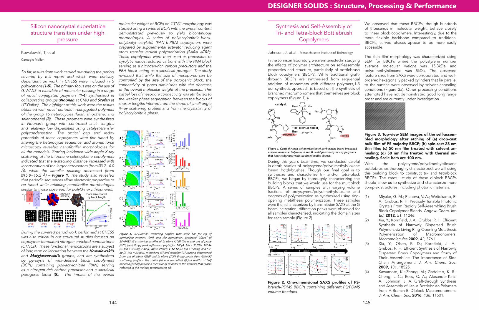
DESIGNER SOLIDS : Structure, Processing & Performance
145144
Silicon nanocrystal superlattice structure transition under high
pressure
Kowalewski, T, et al
Carnegie Mellon
So far, results from work carried out during the period covered by this report and which were critically dependent on work in CHESS were included in 5 publications (1-5). The primary focus was on the use of GIWAXS to elucidate of molecular packing in a range of novel conjugated polymers (1-4) synthesized in collaborating groups (Noonan at CMU and Stefan at UT-Dallas). The highlight of this work were the results obtained with novel periodic π-conjugated polymers of the group 16 heterocycles (furan, thiophene, and selenophene) (3). These polymers were synthesized in Noonan’s group with controlled chain lengths and relatively low dispersities using catalyst-transfer polycondensation. The optical gap and redox potentials of these copolymers were fine-tuned by altering the heterocycle sequence, and atomic force microscopy revealed nanofibrillar morphologies for all the materials. Grazing incidence wide-angle X-ray scattering of the thiophene-selenophene copolymers indicated that the π-stacking distance increased with incorporation of the larger heteroatom (from �3.7−4.0 Å), while the lamellar spacing decreased (from �15.8−15.2 Å) – Figure 1. The study also revealed that periodic sequences allow electronic properties to be tuned while retaining nanofibrillar morphologies similar to those observed for poly(3-hexylthiophene).
During the covered period work performed at CHESS was also critical in our structural studies focused on copolymer-templated nitrogen enriched nanocarbons (CTNCs). These functional nanocarbons are a subject of long term collaboration between the Kowalewski’s and Matyjaszewski’s groups, and are synthesized by pyrolysis of well-defined block copolymers (BCPs) containing polyacrylonitrile (PAN) serving as a nitrogen-rich carbon precursor and a sacrificial porogenic block (5). The impact of the overall
molecular weight of BCPs on CTNC morphology was studied using a series of BCPs with the overall content demonstrated previously to yield bicontinuous morphologies. A series of polyacrylonitrile-block-poly(butyl acrylate) (PAN-b-PBA) copolymers were prepared by supplemental activator reducing agent atom transfer radical polymerization (SARA ATRP). These copolymers were then used as precursors to pyrolytic nanostructured carbons with the PAN block serving as a nitrogen-rich carbon precursors and the PBA block acting as a sacrificial porogen. The study revealed that while the size of mesopores can be controlled by the size of the porogenic block, the connectivity of pores diminishes with the decrease of the overall molecular weight of the precursor. This partial loss of mesopore connectivity was attributed to the weaker phase segregation between the blocks of shorter lengths inferred from the shape of small-angle X-ray scattering profiles and from the crystallinity of polyacrylonitrile phase.
Figure 1. 2D-GIWAXS scattering profiles with scale bar for log of normalized intensity (left), and the azimuthally averaged “slices” of 2D-GIWAXS scattering profiles of in plane (100) (blue) and out of plane (020) (red) Bragg peak reflections (right) for T-T (A, Mn = 36100), T-T-Se (B, Mn = 32100), T-Se (C, Mn = 39800), T−Se-Se (D, Mn = 39000), and F-T-Se (E, Mn = 23100). π-stacking (F) and lamellar (G) spacing determined from out of plane (020) and in plane (100) Bragg peaks from GIWAXS scattering profiles. The radial (H) and azimuthal (I) full widths at half maxima (fwhm) provide a measure of disorder in the samples that is also reflected in the melting temperatures (J).
Synthesis and Self-Assembly of Tri- and Tetra-block Bottlebrush
Copolymers
Johnson, J, et al - Massachusetts Institute of Technology
In the Johnson laboratory, we are interested in studying the effects of polymer architecture on self-assembly properties and structure, particularly of bottlebrush block copolymers (BBCPs). While traditional graft-through BBCPs are synthesized from sequential addition of monomers with different polymers,1-3 our synthetic approach is based on the synthesis of branched macromonomers that themselves are block copolymers (Figure 1).4
Figure 1. Graft-through polymerization of norbornene-based branched macromonomers. Polymers A and B could potentially be any polymers that have endgroups with the functionality shown.
During this year’s beamtime, we conducted careful in-depth studies of polystyrene/polydimethylsiloxane based bottlebrushes. Though our final goal is to synthesize and characterize tri- and/or tetra-block BBCPs, we began by thoroughly characterizing the building blocks that we would use for the multiblock BBCPs. A series of samples with varying volume fractions of polystyrene/polydimethylsiloxane and degrees of polymerization as synthesized using ring-opening metathesis polymerization. These samples were then characterized by transmission SAXS at the G beamline station; diffraction peaks were observed for all samples characterized, indicating the domain sizes for each sample (Figure 2).
Figure 2. One-dimensional SAXS profiles of PS-branch-PDMS BBCPs containing different PS/PDMS volume fractions.
We observed that these BBCPs, though hundreds of thousands in molecular weight, behave closely to linear block copolymers. Interestingly, due to the more flexible backbone compared to traditional BBCPs, curved phases appear to be more easily accessible.
The thin film morphology was characterized using SEM for BBCPs where the polystyrene number average molecular weight was 15.3kDa and polydimethylsiloxane was 5kDa. The observed feature sizes from SAXS were corroborated and well-ordered hexagonally packed cylinders that lie parallel to the surface were observed by solvent annealing conditions (Figure 3a). Other processing conditions attempted have not demonstrated good long range order and are currently under investigation.
100 nm
a b
c d
Figure 3. Top-view SEM images of the self-assem-bled morphology after etching of (a) drop-cast bulk film of PS majority BBCP; (b) spin-cast 28 nm thin film; (c) 50 nm film treated with solvent an-nealing; (d) 50 nm film treated with thermal an-nealing. Scale bars are 100 nm.With the polystyrene/polydimethylsiloxane bottlebrushes thoroughly characterized, we will using this building block to construct tri- and tetrablock BBCPs. The careful study of these diblock BBCPs should allow us to synthesize and characterize more complex structures, including photonic materials.
(1) Miyake, G. M.; Piunova, V. A.; Weitekamp, R. A.; Grubbs, R. H. Precisely Tunable Photonic Crystals From Rapidly Self-Assembling Brush Block Copolymer Blends. Angew. Chem. Int. Ed. 2012, 51, 11246.
(2) Xia, Y.; Kornfield, J. A.; Grubbs, R. H. Efficient Synthesis of Narrowly Dispersed Brush Polymers via Living Ring-Opening Metathesis Polymerization of Macromonomers. Macromolecules 2009, 42, 3761.
(3) Xia, Y.; Olsen, B. D.; Kornfield, J. A.; Grubbs, R. H. Efficient Synthesis of Narrowly Dispersed Brush Copolymers and Study of Their Assemblies: The Importance of Side Chain Arrangement. J. Am. Chem. Soc. 2009, 131, 18525.
(4) Kawamoto, K.; Zhong, M.; Gadelrab, K. R.; Cheng, L.-C.; Ross, C. A.; Alexander-Katz, A.; Johnson, J. A. Graft-through Synthesis and Assembly of Janus Bottlebrush Polymers from A-Branch-B Diblock Macromonomers. J. Am. Chem. Soc. 2016, 138, 11501.
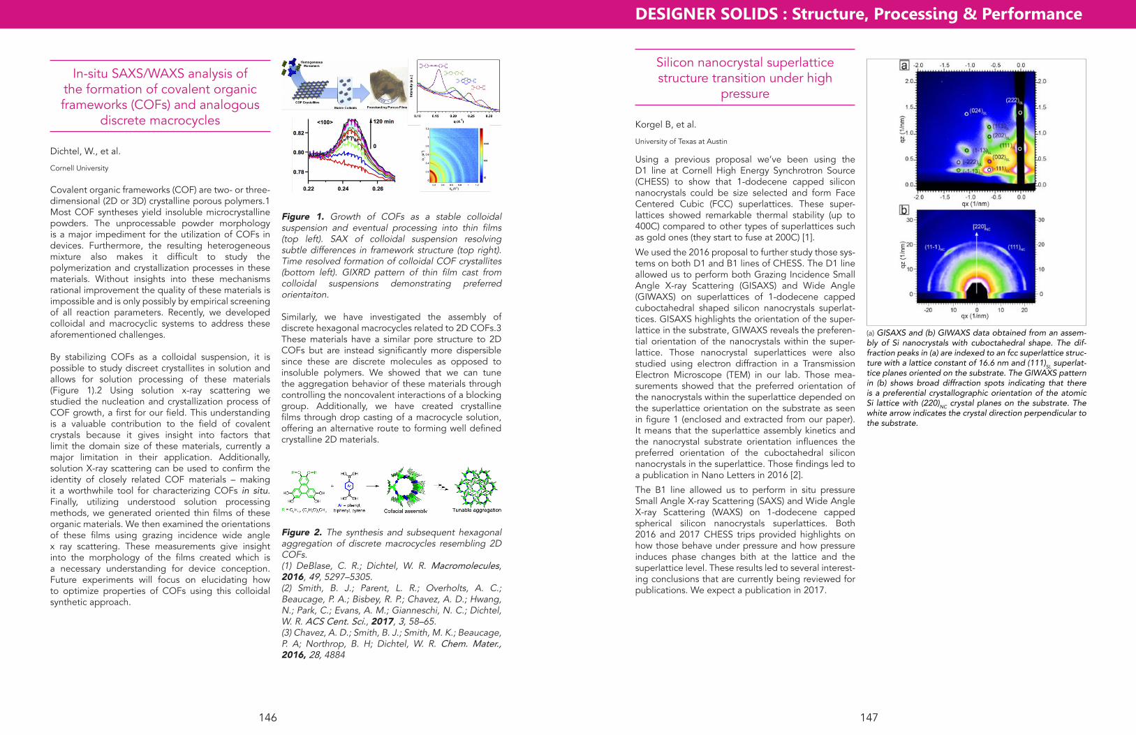
DESIGNER SOLIDS : Structure, Processing & Performance
147146
Silicon nanocrystal superlattice structure transition under high
pressure
Korgel B, et al.
University of Texas at Austin
Using a previous proposal we’ve been using the D1 line at Cornell High Energy Synchrotron Source (CHESS) to show that 1-dodecene capped silicon nanocrystals could be size selected and form Face Centered Cubic (FCC) superlattices. These super-lattices showed remarkable thermal stability (up to 400C) compared to other types of superlattices such as gold ones (they start to fuse at 200C) [1].
We used the 2016 proposal to further study those sys-tems on both D1 and B1 lines of CHESS. The D1 line allowed us to perform both Grazing Incidence Small Angle X-ray Scattering (GISAXS) and Wide Angle (GIWAXS) on superlattices of 1-dodecene capped cuboctahedral shaped silicon nanocrystals superlat-tices. GISAXS highlights the orientation of the super-lattice in the substrate, GIWAXS reveals the preferen-tial orientation of the nanocrystals within the super-lattice. Those nanocrystal superlattices were also studied using electron diffraction in a Transmission Electron Microscope (TEM) in our lab. Those mea-surements showed that the preferred orientation of the nanocrystals within the superlattice depended on the superlattice orientation on the substrate as seen in figure 1 (enclosed and extracted from our paper). It means that the superlattice assembly kinetics and the nanocrystal substrate orientation influences the preferred orientation of the cuboctahedral silicon nanocrystals in the superlattice. Those findings led to a publication in Nano Letters in 2016 [2].
The B1 line allowed us to perform in situ pressure Small Angle X-ray Scattering (SAXS) and Wide Angle X-ray Scattering (WAXS) on 1-dodecene capped spherical silicon nanocrystals superlattices. Both 2016 and 2017 CHESS trips provided highlights on how those behave under pressure and how pressure induces phase changes bith at the lattice and the superlattice level. These results led to several interest-ing conclusions that are currently being reviewed for publications. We expect a publication in 2017.
(a) GISAXS and (b) GIWAXS data obtained from an assem-bly of Si nanocrystals with cuboctahedral shape. The dif-fraction peaks in (a) are indexed to an fcc superlattice struc-ture with a lattice constant of 16.6 nm and (111)SL superlat-tice planes oriented on the substrate. The GIWAXS pattern in (b) shows broad diffraction spots indicating that there is a preferential crystallographic orientation of the atomic Si lattice with (220)NC crystal planes on the substrate. The white arrow indicates the crystal direction perpendicular to the substrate.
In-situ SAXS/WAXS analysis of the formation of covalent organic frameworks (COFs) and analogous
discrete macrocycles
Dichtel, W., et al.
Cornell University
Covalent organic frameworks (COF) are two- or three- dimensional (2D or 3D) crystalline porous polymers.1 Most COF syntheses yield insoluble microcrystalline powders. The unprocessable powder morphology is a major impediment for the utilization of COFs in devices. Furthermore, the resulting heterogeneous mixture also makes it difficult to study the polymerization and crystallization processes in these materials. Without insights into these mechanisms rational improvement the quality of these materials is impossible and is only possibly by empirical screening of all reaction parameters. Recently, we developed colloidal and macrocyclic systems to address these aforementioned challenges.
By stabilizing COFs as a colloidal suspension, it is possible to study discreet crystallites in solution and allows for solution processing of these materials (Figure 1).2 Using solution x-ray scattering we studied the nucleation and crystallization process of COF growth, a first for our field. This understanding is a valuable contribution to the field of covalent crystals because it gives insight into factors that limit the domain size of these materials, currently a major limitation in their application. Additionally, solution X-ray scattering can be used to confirm the identity of closely related COF materials – making it a worthwhile tool for characterizing COFs in situ. Finally, utilizing understood solution processing methods, we generated oriented thin films of these organic materials. We then examined the orientations of these films using grazing incidence wide angle x ray scattering. These measurements give insight into the morphology of the films created which is a necessary understanding for device conception. Future experiments will focus on elucidating how to optimize properties of COFs using this colloidal synthetic approach.
Figure 1. Growth of COFs as a stable colloidal suspension and eventual processing into thin films (top left). SAX of colloidal suspension resolving subtle differences in framework structure (top right). Time resolved formation of colloidal COF crystallites (bottom left). GIXRD pattern of thin film cast from colloidal suspensions demonstrating preferred orientaiton.
Similarly, we have investigated the assembly of discrete hexagonal macrocycles related to 2D COFs.3 These materials have a similar pore structure to 2D COFs but are instead significantly more dispersible since these are discrete molecules as opposed to insoluble polymers. We showed that we can tune the aggregation behavior of these materials through controlling the noncovalent interactions of a blocking group. Additionally, we have created crystalline films through drop casting of a macrocycle solution, offering an alternative route to forming well defined crystalline 2D materials.
Figure 2. The synthesis and subsequent hexagonal aggregation of discrete macrocycles resembling 2D COFs.(1) DeBlase, C. R.; Dichtel, W. R. Macromolecules, 2016, 49, 5297–5305.(2) Smith, B. J.; Parent, L. R.; Overholts, A. C.; Beaucage, P. A.; Bisbey, R. P.; Chavez, A. D.; Hwang, N.; Park, C.; Evans, A. M.; Gianneschi, N. C.; Dichtel, W. R. ACS Cent. Sci., 2017, 3, 58–65.(3) Chavez, A. D.; Smith, B. J.; Smith, M. K.; Beaucage, P. A; Northrop, B. H; Dichtel, W. R. Chem. Mater., 2016, 28, 4884

DESIGNER SOLIDS : Structure, Processing & Performance
149148
Reversible structural transition study of thiolate-coated sub 2 nm gold
nanocrystal superlattices
Korgel B. et al.
University of Texas at Austin,
Thiol coated sub 2 nm gold nanocrystals show inter-esting properties due to their relatively high ligand to core ratio. In the past, using this proposal, we’ve been showing that dodecanethiol capped gold nano-crystals could preferentially lead to Body Centered Cubic (BCC) superlattices which are less dense than the expected Face Centered Cubic (FCC) ones. This phenomenon was explained by ligand packing frus-tration and lead to a publication in 2015 in the Journal of Physical Chemistry Letters [1]. Our experiments on the D1 line of Cornell High Energy Synchrotron Source (CHESS) also demonstrated that a disordered assembly of octadecanethiol-capped gold nanocrys-tals could order when heated from room tempera-ture to 60oC and this transformation was shown to be reversible. This heat induced ordering was shown to be related to the melting and solidification of the octadecanethiol capping ligands. In fact, capping ligands compensate for the gold core polydispersity when heated above the melting point of octadecane-thiol which lead to BCC structures above this point. These results were published in 2015 in Faraday Discussions[2].
Recently, we used our proposal to run in situ Grazing Incidence Small Angle X-ray Scattering (GISAXS) at the D1 line of CHESS to further confirm the gener-ality of this “inverse melting” transition phenome-non. Gold nanocrystals with shorter capping ligands (dodecanethiol, and pentanethiol) that already form superlattices at room temperature and that have a ligand melting-solidification below room tempera-ture were tested. A cold nitrogen stream nozzle was placed right above the sample stage to perform in situ cooling. When cooled below 260 K (the melting solidification temperature of dodecanethiol), super-lattices of dodecanethiol-capped gold nanocrystals clearly disorder. When heated back up to room tem-perature they don’t fully recover their ordering. That’s only after getting rid of the moisture formed by previ-ous cooling by blowing some room T nitrogen gas on the sample that they fully recover their ordering. Fig. 1 (enclosed and extracted from our paper) illus-trates that order to disorder transition. When cooled down below 100K (the lowest accessible temperature with our set up), superlattices of pentanethiol gold nanocrystals do not undergo the disorder transition. Dodecanethiol cooling experiments confirmed our inverse melting theory. For pentanethiol ones there
was just no ligand melting solidification transitions in the accessible temperature range. These results lead to a publication in 2016 in The Journal of Physical ChemistryC [3].
Further work was started in both 2016 and 2017 CHESS trips to test this reversible transition effect in more original superlattices membranes structures. Thin air filled bubble membranes of octadecanethiol capped gold nanocrystals superlattices were tested under in situ heating GISAXS and lead to the same behavior. This work is currently in the process of being reviewed and published.
Fig. 1.
In Situ SAXS Studies of Thin Film Polyurethane-Peptide Hybrids
Korley L, et al
Case Western Reserve University
Project Status/studies completed: The study to identify morphological structures in linear segmented thermoplastic polyurethanes (TPUs) with incorporated peptide units was achieved. However, in-situ studies were not possible; therefor, native films and annealed films at temperatures associated with the shape mem-ory thermal cycle for proposed in-situ study were executed (80 °C, 10 mins.). As can be seen in Fig. 1, the control TPU with no peptide content showed little change after annealing. However, TPUs contain-ing either β-sheet (Fig. 2) or α-helical (Fig. 3) fold-ing peptides expressed more horizontal broadening in GI-SAXS spectra after annealing. Thus, it can be concluded that peptide content allows for rearrange-ment in phase behavior throughout the shape mem-ory cycle. In addition to this work, crosslinked elasto-mers were also tested as native and annealed films. As expected the covalent network within the cross-linked elastomers did not exhibit any GI-SAXS spec-tral changes post annealing for any samples, control or peptide containing (Fig. 4). This work resulted in a better understanding of how thermal annealing affects phase behavior and gives insight in to future experimental design for shape memory peptide con-taining elastomers.
Future work: In the future we would like to expand our material choice (alterations in elastomer block choice to better achieve desired phase behavior) and redo the annealing study in hopes to optimize the design of shape memory polymers. Additionally we would like to expand the annealing study to include multiple temperatures and time intervals. Ideally, we will conduct in-situ SAXS experiments with the opti-mized films.
Fig. 1. Control TPU (left native film, right annealed film).
Fig. 2. β-sheet TPU (left native film, right annealed film).
Fig. 3. α-helix TPU (left native film, right annealed film).
Fig. 4. Example of a covalently crosslinked peptidic elastomer.

DESIGNER SOLIDS : Structure, Processing & Performance
151150
Diffuse X-ray Scattering Study of Defects in Radiation-Damaged Ni-Based Alloys and Hydrogen-
Corroded Pipeline Steel
Connolly M1, Olsen R2, Zhang F2
1National Institute of Standards and Technology, 2Oak Ridge National Laboratory
Results from the diffuse X-ray scattering experiment conducted at CHESS are shown in the attached fig-ures for 5 samples (Ni, NiCo, NiFe, NiCoCr, and NiFeCoCr) all at the same dose of 0.1 DPA (displace-ments per atom). This is a dose at which we do not expect the damage to be quite fully saturated, but nearby cascades are interacting significantly. We have used the methods in our previous JNM paper to ana-lyze the data.
The results shown in Fig. 1 are quite unexpected, as they show that the total amount of diffuse scatter-ing, which is nominally proportional to the number of point defects in clusters, increases with alloy complex-ity. This is unexpected because it seemingly disagrees with results found by other members of our research group using TEM. However, TEM is most effective at detecting large interstitial clusters >4 nm. We show in Fig. 2 that within the region of diffuse X-ray scatter-ing corresponding to these larger interstitial clusters, the diffuse scattering actually decreases with alloy complexity, thus these results in fact agree quite well with the TEM results. A future experiment will investi-gate these samples at a wider range of doses, so that we can see how the size distribution changes as the
dose increases.Fig. 1. Diffuse X-ray scattering around the 004 Bragg peak from damage in samples irradiated to 0.1 DPA. q is the distance from the 004 peak Bragg peak in the L direction. Labels indicate the defect cluster type (vacancy or interstitial) and the size trends.
Fig. 2. Diffuse X-ray scattering from damage in samples irradiated to 0.1 DPA, zoomed to look at the low q region where the largest defect clusters are seen.
Structural characterization of supercrystals of ZnTe Magic Sized
Nanoclusters
Zhang J
China University of Petroleum (East China)
The project was going smoothly in 2016 and we have obtained valuable data from high pressure studies at CHESS. Two families of ZnTe magic sizes nanoclusters (MSNCs), namely ZnTe F323 and F398), were synthe-sized according to a method that we have developed before.1 UV-Visible absorption spectrum confirmed they were MSNCs instead of normal ultra-small nano-crystals. TEM images showed that both ZnTe F323 and F398 MSNCs self-assembled into layers which are separated by double layers of oleylamine molecular. The size of F323 and F398 were determined as 2.1nm and 1.4nm respectively. In order to study the structural stability of assemblies of ZnTe MSNCs, we have col-lected both small angle X-ray scatterings (SAXS) and wide angle X-ray scatterings (WAXS) of assemblies of F323 and F398 under various pressures using a dia-mond anvil cell. The analysis of SAXS and WAXS pat-terns at ambient pressure revealed that ZnTe MSNCs possessed a wurtzite structure and orientationally assembled into layered superstructures. Size depen-dent structural evolution of self-assemblies of F323 and F398 under pressure have been observed.(Fig. 1) For the sample of F398 assemblies, we observed the first order phase transition from wurzite to metallic CmCm at 14.2 GPa. When the pressure was recov-ered to ambient, the diffraction pattern shows a vary week and board peaks, indicating the appearance of zinc blende phase. However, ZnTe F323 as the small-est species in our experiment, showed even narrower peaks that those of F398, due to a higher degree of orientation of nanoclusters within layered assemblies. To the country of the results for ZnTe F398, neither phase transition nor amorphorization occurred dur-ing the whole process of increasing the pressure upto 20Gpa. All the diffractions peaks of wurtzite struc-ture shift to higher two theta positions with gradu-ally broadening of peaks and decreased intensities. After the pressure was released to ambient, metasta-ble wurtzite structure was completely recovered. The results of our experiments have provided important insight into the size dependence of structural stability of MSCN assemblies and may benefit rational design of nanocluster assembled materials with desirable properties. Our next step is to determine the struc-ture of 3D supercrystals composed of ZnTe MSNC F323 prepaired in octylamine and prepare a manu-script based on the results we have obtained.
Fig. 1. High-pressure X-ray-diffraction patterns of assem-blies of two families of ZnTe MSNCs. (A) F398 and (B) F323.
[1] Zhang J, Rajh T et al., J. Am. Chem. Soc., 137(2), 742-9 (2015).

DESIGNER SOLIDS : Structure, Processing & Performance
153152
In-situ SAXS/WAXS analysis of the formation of covalent organic frameworks (COFs) and analogous
discrete macrocycles
Dichtel, W., et al.
Cornell University
Covalent organic frameworks (COF) are two- or three- dimensional (2D or 3D) crystalline porous polymers.1 Most COF syntheses yield insoluble microcrystalline powders. The unprocessable powder morphology is a major impediment for the utilization of COFs in devices. Furthermore, the resulting heterogeneous mixture also makes it difficult to study the polymerization and crystallization processes in these materials. Without insights into these mechanisms rational improvement the quality of these materials is impossible and is only possibly by empirical screening of all reaction parameters. Recently, we developed colloidal and macrocyclic systems to address these aforementioned challenges.
By stabilizing COFs as a colloidal suspension, it is possible to study discreet crystallites in solution and allows for solution processing of these materials (Figure 1).2 Using solution x-ray scattering we studied the nucleation and crystallization process of COF growth, a first for our field. This understanding is a valuable contribution to the field of covalent crystals because it gives insight into factors that limit the domain size of these materials, currently a major limitation in their application. Additionally, solution X-ray scattering can be used to confirm the identity of closely related COF materials – making it a worthwhile tool for characterizing COFs in situ. Finally, utilizing understood solution processing methods, we generated oriented thin films of these organic materials. We then examined the orientations of these films using grazing incidence wide angle x ray scattering. These measurements give insight into the morphology of the films created which is a necessary understanding for device conception. Future experiments will focus on elucidating how to optimize properties of COFs using this colloidal synthetic approach.
Figure 1. Growth of COFs as a stable colloidal suspension and eventual processing into thin films (top left). SAX of colloidal suspension resolving subtle differences in framework structure (top right). Time resolved formation of colloidal COF crystallites (bottom left). GIXRD pattern of thin film cast from colloidal suspensions demonstrating preferred orientaiton.
Similarly, we have investigated the assembly of discrete hexagonal macrocycles related to 2D COFs.3 These materials have a similar pore structure to 2D COFs but are instead significantly more dispersible since these are discrete molecules as opposed to insoluble polymers. We showed that we can tune the aggregation behavior of these materials through controlling the noncovalent interactions of a blocking group. Additionally, we have created crystalline films through drop casting of a macrocycle solution, offering an alternative route to forming well defined crystalline 2D materials.
Figure 2. The synthesis and subsequent hexagonal aggregation of discrete macrocycles resembling 2D COFs.(1) DeBlase, C. R.; Dichtel, W. R. Macromolecules, 2016, 49, 5297–5305.(2) Smith, B. J.; Parent, L. R.; Overholts, A. C.; Beaucage, P. A.; Bisbey, R. P.; Chavez, A. D.; Hwang, N.; Park, C.; Evans, A. M.; Gianneschi, N. C.; Dichtel, W. R. ACS Cent. Sci., 2017, 3, 58–65.(3) Chavez, A. D.; Smith, B. J.; Smith, M. K.; Beaucage, P. A; Northrop, B. H; Dichtel, W. R. Chem. Mater., 2016, 28, 4884
bly mechanism discovered here may provide a flexi-ble material synthesis route because morphology and architecture can be readily tuned to produce desired properties independent of crystal facet orientation.
Current goal of the project is to understand the pres-sure induced assembly science of nanoparticle arrays. Future work will focus on investigation of optical property, pressure induced optical coupling, and its correlation with nanoparticle superlattice structures.
Fig. 1. TEM images of CdSe nanowires synthesized by pressure induced assembly.
Fig. 2. Pressure induced structural evolution of CdSe nanocrystal mesophase during compression and release.
Pressure Induced Formation of Luminescent CdSe Nanowires
Fan H, et. al.
Sandia National Laboratories
Oriented attachment (OA) of synthetic nanocrystals is emerging to be an effective means to fabricate low dimensional, nanoscale materials. However, nano-structures synthesized through OA are generally lim-ited to a specific crystal facet in their final morphology. We discovered that high-pressure compression can induce consolidation of spherical CdSe nanocrystal arrays, leading to unexpected one-dimensional semi-conductor nanowires (Fig. 1) that do not exhibit the typical crystal facet. In particular, in situ high-pressure synchrotron x-ray scattering, optical spectroscopy, and high-resolution transmission electron micros-copy characterizations indicate that by manipulating the coupling between nanocrystals through external pressure, a reversible change in nanocrystal assem-blies and properties can be achieved at modest pres-sure. When pressure is increased above a threshold, these nanocrystals begin to contact and consolidate, irreversibly forming one-dimensional nanowires.
The CdSe nanoparticles are surface-stabilized by organic monolayers, which initially prevent nanopar-ticles from aggregation or contacting. At ambient pressure, ordered fcc nanoparticle arrays are formed through balanced nanocrystal interactions such as van der Waals forces, hydrophobic attraction, and charge-charge interaction, leading to alkane chain interdigitation that locks in the final fcc nanocrystal mesophase1. During compression at low pressure, pressure is not sufficient to break the force balance between adjacent nanoparticles. Hence the spheri-cal nanoparticle arrays remain in the fcc mesophase while shrinking and reversibly springing back when pressure is released, as verified by the SAXS results (Fig. 2). However, when the pressure is higher, con-tinuous compression increasingly breaks the force balance between adjacent nanoparticles, and drives the nanoparticles to contact and ultimately coalesce, forming nanowires. In the fcc mesophase, the clos-est interparticle spacings are between {220} planes. Consequently, particles touch and coalesce along the close packing <110> direction first, forming one-dimensional nanowires. During this process, partial dissociation of ligands at the contact points of the nanocrystal surfaces likely occurs to allow particle connection2. Because the starting CdSe nanocrystal arrays are in a solid state phase that doesn’t allow fac-ile re-arrangement of nanocrystals, this coalescence occurs in a random pattern within adjacent nano-crystals, leading to unusually twisted, polycrystalline nanowires. The pressure-induced nanocrystal assem-

CHESS/Wilson Lab | Cornell University | 161 Synchrotron Drive | Ithaca, NY 14853 | 607-255-7163