Acetoxy drug: protein transacetylase of buffalo liver—characterization and mass spectrometry of...
12
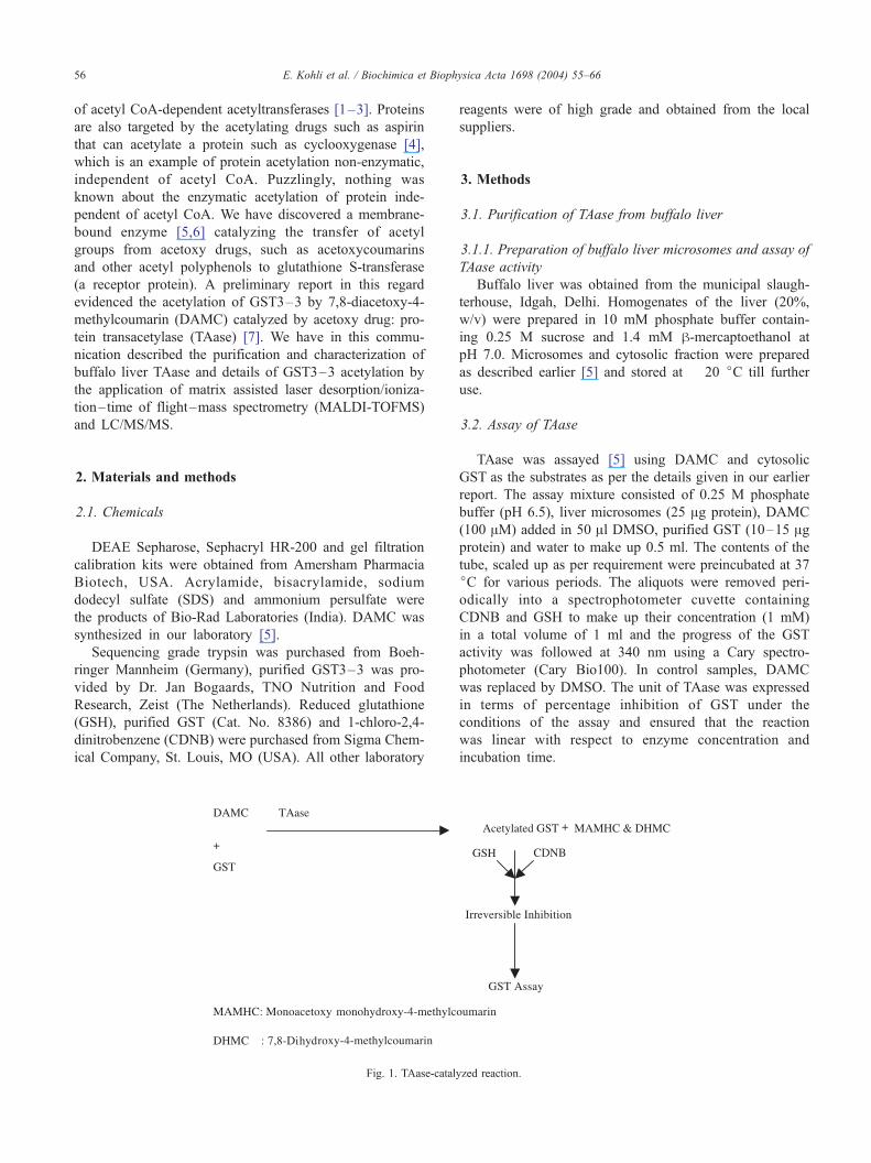
Acetoxy drug: protein transacetylase of buffalo liver—characterization and mass spectrometry of the acetylated protein product Ekta Kohli a , Marco Gaspari b , Hanumantharao G. Raj a, * , Virinder S. Parmar c,d , Sunil K. Sharma c,d , Jan van der Greef b,e , Ranju Kumari a , Garima Gupta a , Seema a , Pulkit Khurana a , Yogesh K. Tyagi a , Arthur C. Watterson d , Carl E. Olsen f a Department of Biochemistry, V.P. Chest Institute, University of Delhi, Delhi 110 007, India b TNO Nutrition and Food Research, Analytical Sciences Department, Zeist, The Netherlands c Bioorganic Laboratory, Department of Chemistry, University of Delhi, Delhi 110 007, India d INS ET, Department of Chemistry, University of Massachusetts, Lowell, MA 01854, USA e Division of Analytical Biosciences, Leiden/Amsterdam Center for Drug Research, Leiden University, Leiden, The Netherlands f Chemistry Department, Royal Veterinary and Agricultural University, DK-1871 Frederiksberg C, Copenhagen, Denmark Received 4 June 2003; received in revised form 29 September 2003; accepted 14 October 2003 Abstract The purification and characterization of the buffalo liver microsomal transacetylase (TAase) catalyzing the transfer of acetyl groups from a model acetoxy drug: 7,8-diacetoxy-4-methylcoumarin (DAMC) to GST3 – 3 has been described here. The enzyme was routinely assayed using DAMC and cytosolic GST as the substrates and was partially purified from microsomes of the buffalo liver. The enzyme was found to have approximate molecular weight of 65 kDa. The action of TAase and DAMC on liver cytosolic GST resulted in the formation of monoacetoxymonohydroxy-4-methylcoumarin (MAMHC) and 7,8-dihydroxy-4-methylcoumarin (DHMC), although the former was the major metabolite. The buffalo liver microsomal TAase exhibited hyperbolic kinetics and yielded K m (1667 AM) and V max (192 units) when the concentration of DAMC was varied keeping the concentration of GST constant. After having characterized the nature of the substrates and a product of the TAase-catalyzed reaction, we set out to identify the acetylated protein which is another product of the reaction. GST3 –3 was used as a model protein substrate for the action of TAase using DAMC as the acetyl donor. The subunit of control and modified GST3–3 were separated by SDS-polyacrylamide gel electrophoresis (PAGE) and digested with trypsin. The tryptic peptides were extracted from the gel pieces and analyzed by matrix assisted laser desorption/ionization – time of flight – mass spectrometry (MALDI-TOFMS). The data search for calibrated and labeled mass peaks of peptides was performed on the Matrix Science Server using the search engine Mascot. The peptide maps so obtained covered 97% of the GST3 – 3 sequence. On comparison of MALDI peptide maps of modified and control GST, seven new peaks were recognized corresponding to the potentially acetylated peptides in peptide map. The mass value of each of them was 42 Da higher than the theoretical mass of a non-modified GST3 – 3 tryptic peptide, strongly suggesting acetylation. By examining the fragmentation patterns and by comparing experimental and predicted values for MS/MS daughter ions, the identity of the seven acetylated GST tryptic peptides could be confirmed by the application of LC/MS/MS. In the modified GST, N-terminal proline and six lysines (Lys 51 , Lys 82 , Lys 123 , Lsy 181 , Lys 191 and Lys 210 ) were found to be acetylated. The structure of acetylated GST revealed that the lysines that underwent acetylation were peripheral in positions. D 2003 Elsevier B.V. All rights reserved. Keywords: Transacetylase; Acetoxy drug; Protein acetylation; MALDI-TOF; Protein mass spectrometry 1. Introduction The current knowledge on biological protein acetylation is largely confined to acetyl CoA-dependent acetylation of proteins catalyzed by specific acetyltransferases. Histones, the tumor-specific protein, p53, acyl carrier protein are some examples of proteins that are acetylated by the action 1570-9639/$ - see front matter D 2003 Elsevier B.V. All rights reserved. doi:10.1016/j.bbapap.2003.10.004 * Corresponding author. Tel.: +91-11-2766-7497; fax: +91-11-2766- 7420. E-mail address: [email protected] (H.G. Raj). www.bba-direct.com Biochimica et Biophysica Acta 1698 (2004) 55 – 66
-
Upload
independent -
Category
Documents
-
view
0 -
download
0
Transcript of Acetoxy drug: protein transacetylase of buffalo liver—characterization and mass spectrometry of...
www.bba-direct.com
Biochimica et Biophysica Acta 1698 (2004) 55–66
Acetoxy drug: protein transacetylase of buffalo liver—characterization
and mass spectrometry of the acetylated protein product
Ekta Kohlia, Marco Gasparib, Hanumantharao G. Raja,*, Virinder S. Parmarc,d,Sunil K. Sharmac,d, Jan van der Greef b,e, Ranju Kumaria, Garima Guptaa, Seemaa,
Pulkit Khuranaa, Yogesh K. Tyagia, Arthur C. Wattersond, Carl E. Olsenf
aDepartment of Biochemistry, V.P. Chest Institute, University of Delhi, Delhi 110 007, IndiabTNO Nutrition and Food Research, Analytical Sciences Department, Zeist, The NetherlandscBioorganic Laboratory, Department of Chemistry, University of Delhi, Delhi 110 007, Indiad INS ET, Department of Chemistry, University of Massachusetts, Lowell, MA 01854, USA
eDivision of Analytical Biosciences, Leiden/Amsterdam Center for Drug Research, Leiden University, Leiden, The NetherlandsfChemistry Department, Royal Veterinary and Agricultural University, DK-1871 Frederiksberg C, Copenhagen, Denmark
Received 4 June 2003; received in revised form 29 September 2003; accepted 14 October 2003
Abstract
The purification and characterization of the buffalo liver microsomal transacetylase (TAase) catalyzing the transfer of acetyl groups
from a model acetoxy drug: 7,8-diacetoxy-4-methylcoumarin (DAMC) to GST3–3 has been described here. The enzyme was routinely
assayed using DAMC and cytosolic GST as the substrates and was partially purified from microsomes of the buffalo liver. The enzyme
was found to have approximate molecular weight of 65 kDa. The action of TAase and DAMC on liver cytosolic GST resulted in the
formation of monoacetoxymonohydroxy-4-methylcoumarin (MAMHC) and 7,8-dihydroxy-4-methylcoumarin (DHMC), although the
former was the major metabolite. The buffalo liver microsomal TAase exhibited hyperbolic kinetics and yielded Km (1667 AM) and
Vmax (192 units) when the concentration of DAMC was varied keeping the concentration of GST constant. After having characterized
the nature of the substrates and a product of the TAase-catalyzed reaction, we set out to identify the acetylated protein which is
another product of the reaction. GST3–3 was used as a model protein substrate for the action of TAase using DAMC as the acetyl
donor. The subunit of control and modified GST3–3 were separated by SDS-polyacrylamide gel electrophoresis (PAGE) and digested
with trypsin. The tryptic peptides were extracted from the gel pieces and analyzed by matrix assisted laser desorption/ionization– time
of flight–mass spectrometry (MALDI-TOFMS). The data search for calibrated and labeled mass peaks of peptides was performed on
the Matrix Science Server using the search engine Mascot. The peptide maps so obtained covered 97% of the GST3–3 sequence. On
comparison of MALDI peptide maps of modified and control GST, seven new peaks were recognized corresponding to the potentially
acetylated peptides in peptide map. The mass value of each of them was 42 Da higher than the theoretical mass of a non-modified
GST3–3 tryptic peptide, strongly suggesting acetylation. By examining the fragmentation patterns and by comparing experimental and
predicted values for MS/MS daughter ions, the identity of the seven acetylated GST tryptic peptides could be confirmed by the
application of LC/MS/MS. In the modified GST, N-terminal proline and six lysines (Lys51, Lys82, Lys123, Lsy181, Lys191 and Lys210)
were found to be acetylated. The structure of acetylated GST revealed that the lysines that underwent acetylation were peripheral in
positions.
D 2003 Elsevier B.V. All rights reserved.
Keywords: Transacetylase; Acetoxy drug; Protein acetylation; MALDI-TOF; Protein mass spectrometry
1570-9639/$ - see front matter D 2003 Elsevier B.V. All rights reserved.
doi:10.1016/j.bbapap.2003.10.004
* Corresponding author. Tel.: +91-11-2766-7497; fax: +91-11-2766-
7420.
E-mail address: [email protected] (H.G. Raj).
1. Introduction
The current knowledge on biological protein acetylation
is largely confined to acetyl CoA-dependent acetylation of
proteins catalyzed by specific acetyltransferases. Histones,
the tumor-specific protein, p53, acyl carrier protein are
some examples of proteins that are acetylated by the action
E. Kohli et al. / Biochimica et Biophysica Acta 1698 (2004) 55–6656
of acetyl CoA-dependent acetyltransferases [1–3]. Proteins
are also targeted by the acetylating drugs such as aspirin
that can acetylate a protein such as cyclooxygenase [4],
which is an example of protein acetylation non-enzymatic,
independent of acetyl CoA. Puzzlingly, nothing was
known about the enzymatic acetylation of protein inde-
pendent of acetyl CoA. We have discovered a membrane-
bound enzyme [5,6] catalyzing the transfer of acetyl
groups from acetoxy drugs, such as acetoxycoumarins
and other acetyl polyphenols to glutathione S-transferase
(a receptor protein). A preliminary report in this regard
evidenced the acetylation of GST3–3 by 7,8-diacetoxy-4-
methylcoumarin (DAMC) catalyzed by acetoxy drug: pro-
tein transacetylase (TAase) [7]. We have in this commu-
nication described the purification and characterization of
buffalo liver TAase and details of GST3–3 acetylation by
the application of matrix assisted laser desorption/ioniza-
tion–time of flight–mass spectrometry (MALDI-TOFMS)
and LC/MS/MS.
2. Materials and methods
2.1. Chemicals
DEAE Sepharose, Sephacryl HR-200 and gel filtration
calibration kits were obtained from Amersham Pharmacia
Biotech, USA. Acrylamide, bisacrylamide, sodium
dodecyl sulfate (SDS) and ammonium persulfate were
the products of Bio-Rad Laboratories (India). DAMC was
synthesized in our laboratory [5].
Sequencing grade trypsin was purchased from Boeh-
ringer Mannheim (Germany), purified GST3–3 was pro-
vided by Dr. Jan Bogaards, TNO Nutrition and Food
Research, Zeist (The Netherlands). Reduced glutathione
(GSH), purified GST (Cat. No. 8386) and 1-chloro-2,4-
dinitrobenzene (CDNB) were purchased from Sigma Chem-
ical Company, St. Louis, MO (USA). All other laboratory
Fig. 1. TAase-catal
reagents were of high grade and obtained from the local
suppliers.
3. Methods
3.1. Purification of TAase from buffalo liver
3.1.1. Preparation of buffalo liver microsomes and assay of
TAase activity
Buffalo liver was obtained from the municipal slaugh-
terhouse, Idgah, Delhi. Homogenates of the liver (20%,
w/v) were prepared in 10 mM phosphate buffer contain-
ing 0.25 M sucrose and 1.4 mM h-mercaptoethanol at
pH 7.0. Microsomes and cytosolic fraction were prepared
as described earlier [5] and stored at � 20 jC till further
use.
3.2. Assay of TAase
TAase was assayed [5] using DAMC and cytosolic
GST as the substrates as per the details given in our earlier
report. The assay mixture consisted of 0.25 M phosphate
buffer (pH 6.5), liver microsomes (25 Ag protein), DAMC
(100 AM) added in 50 Al DMSO, purified GST (10–15 Agprotein) and water to make up 0.5 ml. The contents of the
tube, scaled up as per requirement were preincubated at 37
jC for various periods. The aliquots were removed peri-
odically into a spectrophotometer cuvette containing
CDNB and GSH to make up their concentration (1 mM)
in a total volume of 1 ml and the progress of the GST
activity was followed at 340 nm using a Cary spectro-
photometer (Cary Bio100). In control samples, DAMC
was replaced by DMSO. The unit of TAase was expressed
in terms of percentage inhibition of GST under the
conditions of the assay and ensured that the reaction
was linear with respect to enzyme concentration and
incubation time.
yzed reaction.
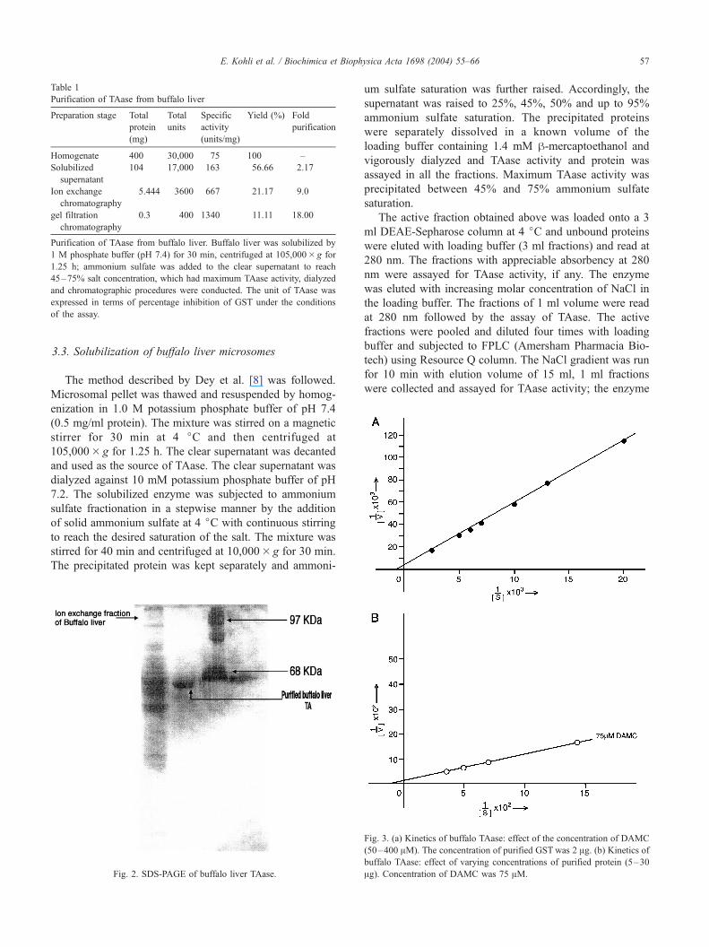
Table 1
Purification of TAase from buffalo liver
Preparation stage Total
protein
(mg)
Total
units
Specific
activity
(units/mg)
Yield (%) Fold
purification
Homogenate 400 30,000 75 100 –
Solubilized
supernatant
104 17,000 163 56.66 2.17
Ion exchange
chromatography
5.444 3600 667 21.17 9.0
gel filtration
chromatography
0.3 400 1340 11.11 18.00
Purification of TAase from buffalo liver. Buffalo liver was solubilized by
1 M phosphate buffer (pH 7.4) for 30 min, centrifuged at 105,000� g for
1.25 h; ammonium sulfate was added to the clear supernatant to reach
45–75% salt concentration, which had maximum TAase activity, dialyzed
and chromatographic procedures were conducted. The unit of TAase was
expressed in terms of percentage inhibition of GST under the conditions
of the assay.
E. Kohli et al. / Biochimica et Biophysica Acta 1698 (2004) 55–66 57
3.3. Solubilization of buffalo liver microsomes
The method described by Dey et al. [8] was followed.
Microsomal pellet was thawed and resuspended by homog-
enization in 1.0 M potassium phosphate buffer of pH 7.4
(0.5 mg/ml protein). The mixture was stirred on a magnetic
stirrer for 30 min at 4 jC and then centrifuged at
105,000� g for 1.25 h. The clear supernatant was decanted
and used as the source of TAase. The clear supernatant was
dialyzed against 10 mM potassium phosphate buffer of pH
7.2. The solubilized enzyme was subjected to ammonium
sulfate fractionation in a stepwise manner by the addition
of solid ammonium sulfate at 4 jC with continuous stirring
to reach the desired saturation of the salt. The mixture was
stirred for 40 min and centrifuged at 10,000� g for 30 min.
The precipitated protein was kept separately and ammoni-
Fig. 2. SDS-PAGE of buffalo liver TAase.
um sulfate saturation was further raised. Accordingly, the
supernatant was raised to 25%, 45%, 50% and up to 95%
ammonium sulfate saturation. The precipitated proteins
were separately dissolved in a known volume of the
loading buffer containing 1.4 mM h-mercaptoethanol and
vigorously dialyzed and TAase activity and protein was
assayed in all the fractions. Maximum TAase activity was
precipitated between 45% and 75% ammonium sulfate
saturation.
The active fraction obtained above was loaded onto a 3
ml DEAE-Sepharose column at 4 jC and unbound proteins
were eluted with loading buffer (3 ml fractions) and read at
280 nm. The fractions with appreciable absorbency at 280
nm were assayed for TAase activity, if any. The enzyme
was eluted with increasing molar concentration of NaCl in
the loading buffer. The fractions of 1 ml volume were read
at 280 nm followed by the assay of TAase. The active
fractions were pooled and diluted four times with loading
buffer and subjected to FPLC (Amersham Pharmacia Bio-
tech) using Resource Q column. The NaCl gradient was run
for 10 min with elution volume of 15 ml, 1 ml fractions
were collected and assayed for TAase activity; the enzyme
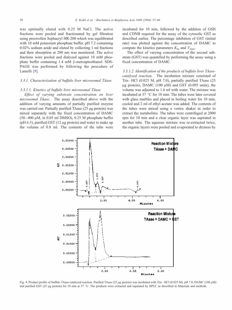
Fig. 3. (a) Kinetics of buffalo TAase: effect of the concentration of DAMC
(50–400 AM). The concentration of purified GST was 2 Ag. (b) Kinetics ofbuffalo TAase: effect of varying concentrations of purified protein (5–30
Ag). Concentration of DAMC was 75 AM.
E. Kohli et al. / Biochimica et Biophysica Acta 1698 (2004) 55–6658
was optimally eluted with 0.25 M NaCl. The active
fractions were pooled and fractionated by gel filtration
using preswollen Sephacryl HR-200 which was equilibrated
with 10 mM potassium phosphate buffer, pH 7.2 containing
0.02% sodium azide and eluted by collecting 1 ml fractions
and their absorption at 280 nm was monitored. The active
fractions were pooled and dialyzed against 10 mM phos-
phate buffer containing 1.4 mM h-mercaptoethanol. SDS-
PAGE was performed by following the procedure of
Lamelli [9].
3.3.1. Characterization of buffalo liver microsomal TAase
3.3.1.1. Kinetics of buffalo liver microsomal TAase
Effect of varying substrate concentration on liver
microsomal TAase. The assay described above with the
addition of varying amounts of partially purified enzyme
was carried out. Partially purified TAase (25 Ag protein) wasmixed separately with the fixed concentration of DAMC
(50–400 AM, in 0.05 ml DMSO), 0.25 M phosphate buffer
(pH 6.5), purified GST (12 Ag protein) and water to make up
the volume of 0.8 ml. The contents of the tube were
Fig. 4. Product profile of buffalo TAase-catalyzed reaction. Purified TAase (25 Agand purified GST (25 Ag protein) for 10 min at 37 jC. The products were extrac
incubated for 10 min, followed by the addition of GSH
and CDNB required for the assay of the cytosolic GST as
described earlier. The percentage inhibition of GST (initial
rate) was plotted against the concentration of DAMC to
compute the kinetics parameters Km and Vmax.
The effect of varying concentration of the second sub-
strate (GST) was quantified by performing the assay using a
fixed concentration of DAMC.
3.3.1.2. Identification of the products of buffalo liver TAase-
catalyzed reaction. The incubation mixture consisted of
Tris–HCl (0.025 M, pH 7.0), partially purified TAase (25
Ag protein), DAMC (100 AM) and GST (0.095 units), the
volume was adjusted to 1.6 ml with water. The mixture was
incubated at 37 jC for 10 min. The tubes were later covered
with glass marbles and placed in boiling water for 10 min,
cooled and 2 ml of ethyl acetate was added. The contents of
the tubes were mixed using a vortex shaker in order to
extract the metabolites. The tubes were centrifuged at 2000
rpm for 10 min and a clear organic layer was aspirated in
another tube. The aqueous mixture was re-extracted twice,
the organic layers were pooled and evaporated to dryness by
protein) was incubated with Tris–HCl (0.025 M), pH 7.0, DAMC (100 AM)
ted and separated by HPLC as described in Materials and methods.
Table 2
Product profile of buffalo liver TAase-catalyzed reaction
Reaction mixture Product formed (%)
DHMC MAMHC DAMC
(1) Enz +DAMC 77.30 7.60 13.17
(2) Enz +DAMC+GST 39.95 61.05 –
E. Kohli et al. / Biochimica et Biophysica Acta 1698 (2004) 55–66 59
blowing N2 gas. The products were dissolved in 1 ml
methanol, and the metabolites were separated by HPLC
using Waters 996 Chromatograph fitted with C-18 column.
Methanol extract (20 Al) prepared as described earlier was
injected and isocratically eluted with methanol/water (60:40
Fig. 5. (a) MALDI-TOFMS spectrum of intact control GST3–3
v/v) using a diode array detector. Authentic samples of
DAMC and DHMC were also chromatographed to identify
them in the sample (Fig. 4).
3.4. Demonstration of acetylation of GST by MALDI-
TOFMS and LC/MS/MS
The reaction mixture containing partially purified buffalo
liver TAase (50 Ag), recombinant GST3–3 isoform (50 Ag),and DAMC (200 AM) in 0.25 M phosphate buffer (pH 6.5)
was incubated for 30 min at 37 jC, and the reaction mixture
was subjected to SDS-PAGE in order to separate modified
. (b) MALDI-TOFMS spectra of intact modified GST3–3.
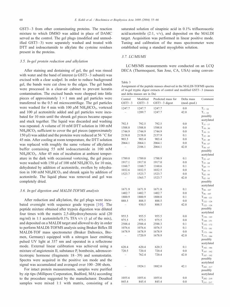
Table 3
Assignment of the peptide masses observed in the MALDI-TOFMS spectra
of in-gel tryptic digest extracts of control and modified GST3–3 (masses
and delta masses are in Da)
Control
GST3–3
Modified
GST3–3
Predicted mass for
GST3–3 digest
Delta mass
(mod.-pred.)
Comment
1247.7 1247.7 1247.7 0.0 T1– 10
– 1289.7 1247.7 42.0 T1– 10
possibly
acetylated
792.5 792.5 792.5 0.0 T11 – 17
1588.8 1588.8 1588.8 0.0 T18 – 30
1744.9 1744.9 1744.9 0.0 T18 – 31
2158.0 2158.0 2157.9 0.1 T32 – 49
903.5 903.4 903.4 0.0 T43 – 49
2064.1 2064.1 2064.1 0.0 T50 – 67
– 2106.1 2064.1 42.0 T50 – 67
possibly
acetylated
1789.0 1789.0 1788.9 0.1 T52 – 67
1917.1 1917.0 1917.0 0.0 T52 – 68
1160.7 1160.7 1160.6 0.1 T68 – 77
1032.6 1032.5 1032.5 0.0 T69 – 77
1523.7 1523.7 1523.7 0.0 T82 – 93
– 1565.7 1523.7 42.0 T82 – 93
possibly
acetylated
1671.9 1671.9 1671.8 0.1 T94 – 107
1402.7 1402.7 1402.7 0.0 T96 – 107
1800.9 1800.9 1800.8 0.1 T108 – 121
888.5 888.5 888.5 0.0 T122 – 128
– 930.5 888.5 42.0 T122 – 128
possibly
acetylated
955.5 955.5 955.5 0.0 T136 – 143
975.5 975.5 975.5 0.0 T144 – 151
2588.4 2588.4 2588.3 0.1 T152 – 172
1076.6 1076.6 1076.5 0.1 T173 – 181
1678.9 1678.9 1678.9 0.0 T173 – 186
– 1720.9 1678.9 42.0 T173 – 186
possibly
acetylated
620.4 620.4 620.3 0.1 T182 – 186
720.5 720.4 720.4 0.0 T187 – 192
– 762.4 720.4 42.0 T187 – 192
possibly
acetylated
– 1924.1 1882.0 42.1 T202 – 217
possibly
acetylated
1055.6 1055.6 1055.6 0.0 T202 – 210
845.4 845.4 845.4 0.0 T211 – 217
E. Kohli et al. / Biochimica et Biophysica Acta 1698 (2004) 55–6660
GST3–3 from other contaminating proteins. The reaction
mixture to which DMSO was added in place of DAMC
served as the control. The gel plugs (modified and unmod-
ified GST3–3) were separately washed and treated with
DTT and iodoacetamide to alkylate the cysteine residues
present in the proteins.
3.5. In-gel protein reduction and alkylation
After staining and destaining of gel, the gel was rinsed
with water and the band of interest (a GST3–3 subunit) was
excised with a clear scalpel. In order to reduce background
gel, the bands were cut close to the edges. The gel bands
were processed in a clear-air cabinet to prevent keratin
contamination. The excised bands were chopped into little
pieces of approximately 1�1 mm and gel particles were
transferred to the 0.5 ml microcentrifuge. The gel particles
were washed for 4 min with 100 AM NH4HCO3, vortexed
and 100 Al acetonitrile added and gel particles were incu-
bated for 10 min until the shrunk gel pieces became opaque
and stuck together. The liquid was discarded and washing
was repeated. A volume of 10 mM DTT solution in 100 mM
NH4HCO3 sufficient to cover the gel pieces (approximately
150 Al) was added and the proteins were reduced at 56 jC for
45 min. After cooling at room temperature, the DTT solution
was replaced with roughly the same volume of alkylation
buffer containing 55 mM iodoacetamide in 100 mM
NH4HCO3. After 45 min of incubation at ambient temper-
ature in the dark with occasional vortexing, the gel pieces
were washed with 150 Al of 100 mM NH4HCO3 for 10 min,
dehydrated by addition of acetonitrile, swollen by rehydra-
tion in 100 mM NH4HCO3 and shrunk again by addition of
acetonitrile. The liquid phase was removed and gel was
completely dried.
3.6. In-gel digestion and MALDI-TOFMS analysis
After reduction and alkylation, the gel plugs were incu-
bated overnight with sequence grade trypsin [10]. The
peptide mixture obtained after trypsin digestion was diluted
four times with the matrix 2,5-dihydroxybenzoic acid (20
mg/ml) in 1:1 acetonitrile/0.1% TFA v/v (1 Al of the mix),
and deposited on a MALDI target and allowed to dry in order
to perform MALDI-TOFMS analysis using Bruker Biflex III
MALDI-TOF mass spectrometer (Bruker Daltonics, Bre-
men, Germany) equipped with a nitrogen laser emitting
pulsed UV light at 337 nm and operated in a reflectrone
mode. External linear calibration was achieved using a
mixture of angiotensin II, substance P, bombesin, adrenocor-
ticotropic hormone (fragments 18–39) and somatostatin.
Spectra were acquired in the positive ion mode and the
signal was accumulated and averaged over 100–200 shots.
For intact protein measurements, samples were purified
by zip tips (Millipore Corporation, Bedford, MA) according
to the procedure suggested by the manufacturer. Desalted
samples were mixed 1:1 with matrix, consisting of a
saturated solution of sinapinic acid in 0.1% trifluoroacetic
acid/acetonitrile (2:1, v/v), and deposited on the MALDI
target. Acquisition was performed in linear positive mode.
Tuning and calibration of the mass spectrometer were
established using a standard myoglobin solution.
3.7. LC/MS/MS
LC/MS/MS measurements were conducted on an LCQ
DECA (Thermoquest, San Jose, CA, USA) using conven-
E. Kohli et al. / Biochimica et Biophysica Acta 1698 (2004) 55–66 61
tional ESI source in positive ion mode detection. The spray
voltage was set at 3.8 kV and the heated capillary temper-
ature at 300 jC. The eluent flow of 25 Al/min was provided
by an Eldex micro-LC (separations). Analysis was con-
ducted on a 15 cm 800 Am i.d. column packed with 5 Amspherisorb C18 reverse phase material (LC Packings,
Amsterdam, The Netherlands). The injection volume was
10 Al. Gradient elution was performed by using the
following mobile phases: (A) 10 mM ammonium acetate
in 0.1% HCOOH (v/v); (B) 10 mM ammonium acetate in
0.15% HCOOH (v/v) and 80% acetonitrile (v/v). Gradient:
from 5% to 30% B in 25 min, from 30% to 60% B in 10
min, from 60% to 100% B in 5 min, down to 5% B again
in 2 min. Two full-scan (m/z range 200–2000) LC/MS runs
were initially acquired for modified and non-modified GST,
respectively. Potentially acetylated peptides previously
detected by MALDI-TOF experiments were found back
in LC/MS total ion current (TIC) of the modified GST as
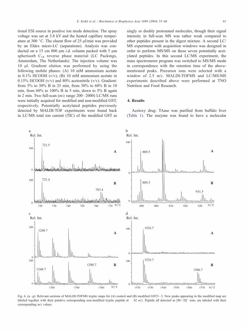
Fig. 6. (a–g): Relevant sections of MALDI-TOFMS tryptic maps for (A) control
labeled together with their putative corresponding non-modified tryptic peptide
corresponding m/z values.
singly or doubly protonated molecules, though their signal
intensity in full-scan MS was rather weak compared to
other peptides present in the digest mixture. A second LC/
MS experiment with acquisition windows was designed in
order to perform MS/MS on these seven potentially acet-
ylated peptides. In this second LC/MS experiment, the
mass spectrometer program was switched to MS/MS mode
in correspondence with the retention time of the above-
mentioned peaks. Precursor ions were selected with a
window of 2.5 m/z. MALDI-TOFMS and LC/MS/MS
experiments described above were performed at TNO
Nutrition and Food Research.
4. Results
Acetoxy drug: TAase was purified from buffalo liver
(Table 1). The enzyme was found to have a molecular
and (B) modified GST3–3. New peaks appearing in the modified map are
at � 42 m/z. Peptide all detected as [M+H]+ ions, are labeled with their
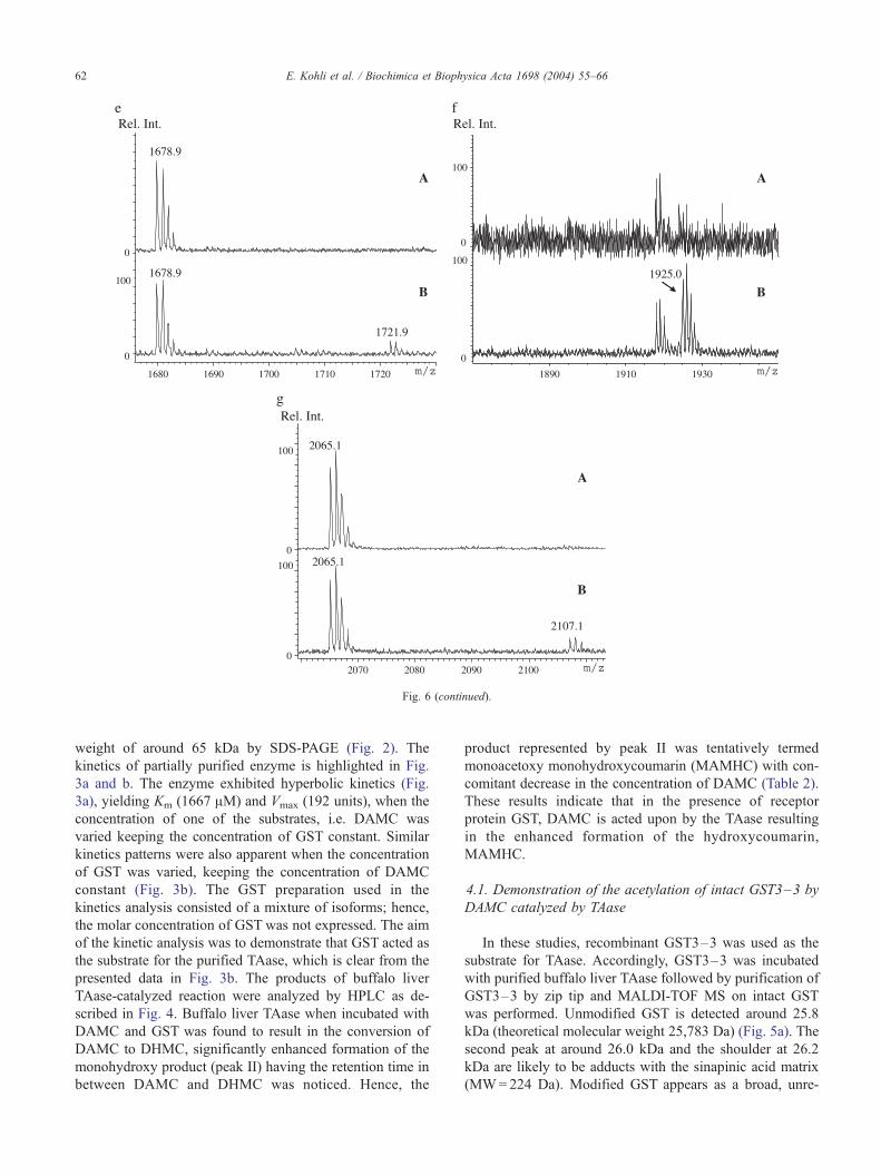
Fig. 6 (continued).
E. Kohli et al. / Biochimica et Biophysica Acta 1698 (2004) 55–6662
weight of around 65 kDa by SDS-PAGE (Fig. 2). The
kinetics of partially purified enzyme is highlighted in Fig.
3a and b. The enzyme exhibited hyperbolic kinetics (Fig.
3a), yielding Km (1667 AM) and Vmax (192 units), when the
concentration of one of the substrates, i.e. DAMC was
varied keeping the concentration of GST constant. Similar
kinetics patterns were also apparent when the concentration
of GST was varied, keeping the concentration of DAMC
constant (Fig. 3b). The GST preparation used in the
kinetics analysis consisted of a mixture of isoforms; hence,
the molar concentration of GST was not expressed. The aim
of the kinetic analysis was to demonstrate that GST acted as
the substrate for the purified TAase, which is clear from the
presented data in Fig. 3b. The products of buffalo liver
TAase-catalyzed reaction were analyzed by HPLC as de-
scribed in Fig. 4. Buffalo liver TAase when incubated with
DAMC and GST was found to result in the conversion of
DAMC to DHMC, significantly enhanced formation of the
monohydroxy product (peak II) having the retention time in
between DAMC and DHMC was noticed. Hence, the
product represented by peak II was tentatively termed
monoacetoxy monohydroxycoumarin (MAMHC) with con-
comitant decrease in the concentration of DAMC (Table 2).
These results indicate that in the presence of receptor
protein GST, DAMC is acted upon by the TAase resulting
in the enhanced formation of the hydroxycoumarin,
MAMHC.
4.1. Demonstration of the acetylation of intact GST3–3 by
DAMC catalyzed by TAase
In these studies, recombinant GST3–3 was used as the
substrate for TAase. Accordingly, GST3–3 was incubated
with purified buffalo liver TAase followed by purification of
GST3–3 by zip tip and MALDI-TOF MS on intact GST
was performed. Unmodified GST is detected around 25.8
kDa (theoretical molecular weight 25,783 Da) (Fig. 5a). The
second peak at around 26.0 kDa and the shoulder at 26.2
kDa are likely to be adducts with the sinapinic acid matrix
(MW=224 Da). Modified GST appears as a broad, unre-
E. Kohli et al. / Biochimica et Biophysica Acta 1698 (2004) 55–66 63
solved peak composed of several species with a different
degree of acetylation (Fig. 5b). The maximum is at 26.0
kDa, accounting for multiple acetylation. No matrix adducts
are observed due to overlapping of peaks and insufficient
resolution.
4.2. Identification of TAase-catalyzed acetylation of amino
acid residues of GST3–3 by DAMC
The GST3–3 incubated with DAMC and purified TAase
was separated on SDS-PAGE. The GST3–3 subunit was
trypsinized as described under Materials and methods. The
tryptic peptides were extracted from the gel pieces and
analyzed by MALDI-TOFMS. The amino acid sequence
of GST3–3 and the sequence coverage of GST3–3 was
obtained by tryptic mapping using MALDI-TOFMS. The
predicted peptides were identified using Mascot Search
Engine www.matrixscience.com). The peptide map so
obtained matched to GTM1-RAT: glutathione S-transferase
YB1, taxonomy Rattus norvegicus, nominal mass of protein
is 25,937 Da.
Table 4
Summary of MS/MS results for confirmation of acetylation in peptides of interes
Peptide m/z Peptide
mass
Peptide sequence Comments
T187 – 192 763.4
(charge + 1)
762.4 FEGLK(Ac)K Lys191 ace
confirmed
and b5 ion
T122 – 128 466.4
(charge + 2)
930.8 QK(Ac)PEFLK Lys123 ace
is confirme
and y6 ion
T1– 10 1290.6
(charge + 1)
1289.6 P(Ac)MILGYWNVR N-terminal
is confirme
and y9 ion
T82 – 93 784.2
(charge + 2)
1566.4 K(Ac)HHLCGETEEER y10 and b2the acetyla
on Lys82 o
The acetyl
acid is like
T173 – 186 861.7
(charge + 2)
1721.4 CLDAFPNL K(Ac)DFLAR b9 and y6Lys181 ace
T202 – 217 963.4
(charge + 2)
1924.8 YLSTPIFSK(Ac)LAQWSNK y8 and b9Lys210 ace
T50 – 67 1054.2
(charge + 2)
2106.4 FK(Ac)LGLDFPNLPYLIDGSR b2 ion poin
acetylation
Phe50 or o
The acetyl
acid is like
In the last two columns, m/z values for the corresponding ions are reported in pa
Peptide map of GST3–3 (matched peptides shown in
boldface)
1 PMILGYWNVR GLTHPIRILLL EYTDSSYEEK
RYAMGDAPDY DRSQWLNEKF
51 KLGLDFPNLP YLIDGSRKIT QSNAIIMRYLA
RKHHLCGETE EERIRADIVE
101 NQVMDNRMQL IMLCYNPDFE KQKPEFKTI
PEKMKLYSEF LGKRPWFAGD
151 KVTYDFLAY DILDQYHIFE PKCLDAFPNL
KDFLARFEGL KKISAYMKSS
201 RYLSTPIFSK LAQWSNK
MALDI-TOFMS peptide maps represented an excellent
starting point in order to address possible covalent mod-
ifications in the amino acid sequence of GST3–3 incu-
bated with DAMC and purified TAase. These
modifications could be individuated by the appearance
on new peaks in the modified GST3–3 tryptic map, not
actually present in the control tryptic map. Overlying
MALDI peptide maps of modified and control GST
t
N-terminal fragments
(y ions)
C-terminal fragments
(b and a ions)
tylation is
by y2s
[M+H]+ (763.3); y4(487.3); y2 (317.2)
b5 (617.2); b5j (599.2);
a5 (589.2); b4 (447.1)
tylation
d by b2s
[M+2H-H2O]2 + (457.5);
y6 (803.6); y5 (633.3);
y3 (407.2)
b5j (654.2); b2 (299.2);
b2j (282.1)
acetylation
d by b3s
[M+H]+ (1290.6);
[M+H-42]+ (1248.3);
y9 (1151.4); y8 (1020.5);
y7 (907.4); y6 (794.4)
b9 (1116.3); b7 (903.3);
b6 (717.1); b5 (554.1);
b4 (497.1); b3 (384.0)
ion point
tion either
r His83.
ated amino
ly to be Lys82
y10 (1259.4); y9 (1122.3);
y8 (1009.3); y7 (849.3);
y6 (792.4); y5 (663.3);
y3 (433.1); y2 (304.1)
b11 (1392.3); b10 (1263.3);
b9 (1134.4); b7 (904.4);
b6 (774.9); b5 (718.3);
b4 (558.2); b3 (445.2);
b2 (308.2)
confirm
tylation
y12 (1448.6); y11 (1333.5);
y10 (1262.4); y9 (1115.5);
y8 (1018.4); y7 (904.4);
y6 (791.3); y5 (621.3);
y4 (506.2)
b13 (1547.3); b10 (1216.3);
b9 (1102.2); b8 (931.2);
b6 (704.2); b5 (607.1);
b4 (460.0); b3 (389.1);
b2 (274.0)
confirm
tylation
y14 (1648.5); y13 (1561.4);
y12 (1460.5); y11 (1363.4);
y10 (1250.4); y9 (1103.4);
y8 (1016.4); y7 (846.3);
y6 (733.2); y5 (662.2);
y4 (534.2); y3 (348.1)
b15 (1778.4); b14 (1664.6);
b13 (1577.5); b12 (1391.4);
b11 (1263.3); b10 (1192.2);
b9 (1079.3); b7 (822.1);
b6 (675.2); b4 (465.0);
b3 (363.9); b2 (276.9)
ts the
either on
n Lys51.
ated amino
ly to be Lys51
y15 (1676.5); y14 (1619.5);
y13 (1506.5); y12 (1391.5);
y11 (1244.5); y10 (1147.4);
y9 (1033.4); y8 (920.4);
y7 (823.5); y6 (660.4);
y5 (547.3); y4 (434.2);
y3 (319.1)
b15 (1788.5); b14 (1673.6);
b13 (1560.4); b12 (1448.1);
b10 (1187.4); b9 (1074.4);
b8 (959.7); b7 (863.3);
b6 (716.3); b5 (601.2);
b4 (488.1); b3 (431.1);
b2 (318.0)
rentheses. A circle above a ‘‘b’’ ion indicates loss of water.
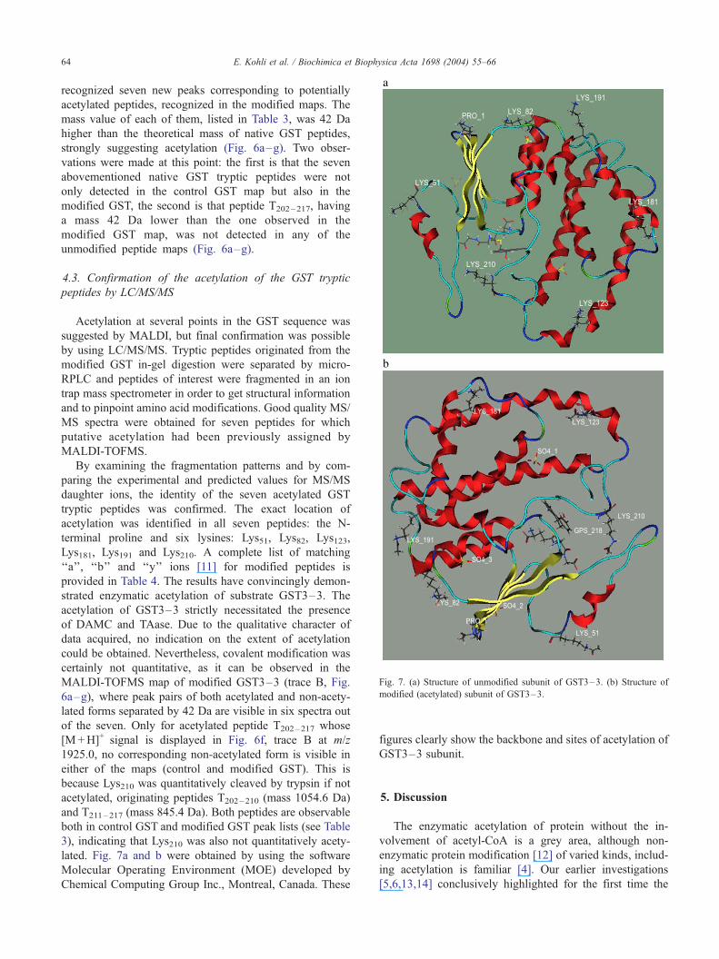
Fig. 7. (a) Structure of unmodified subunit of GST3–3. (b) Structure of
modified (acetylated) subunit of GST3–3.
E. Kohli et al. / Biochimica et Biophysica Acta 1698 (2004) 55–6664
recognized seven new peaks corresponding to potentially
acetylated peptides, recognized in the modified maps. The
mass value of each of them, listed in Table 3, was 42 Da
higher than the theoretical mass of native GST peptides,
strongly suggesting acetylation (Fig. 6a–g). Two obser-
vations were made at this point: the first is that the seven
abovementioned native GST tryptic peptides were not
only detected in the control GST map but also in the
modified GST, the second is that peptide T202–217, having
a mass 42 Da lower than the one observed in the
modified GST map, was not detected in any of the
unmodified peptide maps (Fig. 6a–g).
4.3. Confirmation of the acetylation of the GST tryptic
peptides by LC/MS/MS
Acetylation at several points in the GST sequence was
suggested by MALDI, but final confirmation was possible
by using LC/MS/MS. Tryptic peptides originated from the
modified GST in-gel digestion were separated by micro-
RPLC and peptides of interest were fragmented in an ion
trap mass spectrometer in order to get structural information
and to pinpoint amino acid modifications. Good quality MS/
MS spectra were obtained for seven peptides for which
putative acetylation had been previously assigned by
MALDI-TOFMS.
By examining the fragmentation patterns and by com-
paring the experimental and predicted values for MS/MS
daughter ions, the identity of the seven acetylated GST
tryptic peptides was confirmed. The exact location of
acetylation was identified in all seven peptides: the N-
terminal proline and six lysines: Lys51, Lys82, Lys123,
Lys181, Lys191 and Lys210. A complete list of matching
‘‘a’’, ‘‘b’’ and ‘‘y’’ ions [11] for modified peptides is
provided in Table 4. The results have convincingly demon-
strated enzymatic acetylation of substrate GST3–3. The
acetylation of GST3–3 strictly necessitated the presence
of DAMC and TAase. Due to the qualitative character of
data acquired, no indication on the extent of acetylation
could be obtained. Nevertheless, covalent modification was
certainly not quantitative, as it can be observed in the
MALDI-TOFMS map of modified GST3–3 (trace B, Fig.
6a–g), where peak pairs of both acetylated and non-acety-
lated forms separated by 42 Da are visible in six spectra out
of the seven. Only for acetylated peptide T202–217 whose
[M+H]+ signal is displayed in Fig. 6f, trace B at m/z
1925.0, no corresponding non-acetylated form is visible in
either of the maps (control and modified GST). This is
because Lys210 was quantitatively cleaved by trypsin if not
acetylated, originating peptides T202–210 (mass 1054.6 Da)
and T211–217 (mass 845.4 Da). Both peptides are observable
both in control GST and modified GST peak lists (see Table
3), indicating that Lys210 was also not quantitatively acety-
lated. Fig. 7a and b were obtained by using the software
Molecular Operating Environment (MOE) developed by
Chemical Computing Group Inc., Montreal, Canada. These
figures clearly show the backbone and sites of acetylation of
GST3–3 subunit.
5. Discussion
The enzymatic acetylation of protein without the in-
volvement of acetyl-CoA is a grey area, although non-
enzymatic protein modification [12] of varied kinds, includ-
ing acetylation is familiar [4]. Our earlier investigations
[5,6,13,14] conclusively highlighted for the first time the
E. Kohli et al. / Biochimica et Biophysica Acta 1698 (2004) 55–66 65
presence of an unique enzyme in liver and other tissues
catalyzing the transfer of acetyl groups from a model
acetoxy drug DAMC and other acetyl polyphenols to
receptor proteins such as GST, cytochrome P-450 linked
mixed function oxidases and NADPH cytochrome c reduc-
tase resulting in the modulation of their catalytic activities.
The enzyme was termed acetoxy drug (DAMC): protein
transacetylase (TAase). In order to understand the nature of
the TAase-catalyzed reaction, efforts were made to purify
TAase from buffalo liver microsomes. The partially purified
TAase exhibited molecular weight of 65 kDa according to
SDS-PAGE. Similar results were also obtained from size
exclusion gel chromatography. The initial rate of TAase-
catalyzed reaction was found to vary with the concentration
of the substrates DAMC/cytosol. These results confirm that
cytosolic GST is an effective substrate for the buffalo liver
TAase. TAase-catalyzed transfer of acetyl group from
DAMC to the receptor protein resulted in the formation
of hydroxycoumarins (Figs. 1 and 4). We have earlier
reported the presence of a membrane-bound deacetylase
in liver microsomes catalyzing the deacetylation of DAMC
yielding hydroxycoumarin [15]. It is conceivable that TAase
preparation has the contaminating presence of deacetylase,
which acts on DAMC forming dihydroxycoumarin
(DHMC). But the addition of GST to the TAase reaction
mixture resulted in the drastic enhancement (60%) in the
formation of MAMHC. These results convey the following
conclusions: (a) the action of deacetylase on DAMC yields
both MAMHC and DHMC, although the latter was in larger
amounts [5]; (b) the nature of TAase action on DAMC is
distinct from the deacetylase in that acetyl groups are
preferentially transferred from a particular acetoxy group
to the receptor protein leading to the accumulation of
MAMHC; and (c) it substantiates the TAase-catalyzed
reaction (Fig. 1). Also, DAMC-deacetylase was strongly
inhibited by the protease inhibitor, phenylmethanesulfonyl-
fluoride (PMSF), while the TAase-catalyzed reaction was
unaffected by the inhibitor (unpublished data). The afore-
mentioned results highlighted the distinct nature of TAase
compared to the deacetylase. After having characterized the
TAase-catalyzed reaction in terms of the nature of the
substrates and products of the reaction, efforts were made
to identify acetylated protein, which is another product of
the reaction by the application of MALDI-TOFMS and LC/
MS/MS. To identify the TAase-catalyzed acetylation of
GST with DAMC as the acetyl donor, the GST3–3 was
used as a model protein substrate. Acetylation of GST in the
first place was confirmed by performing MALDI-TOFMS
on the tryptic peptide isolated from the gel pieces. The
trypsinized modified GST exhibited acetylation of peptides.
The modified amino acid residues were determined by LC/
MS/MS. In the modified GST, N-terminal proline and six
lysines (Lys51, Lys82, Lys123, Lys181, Lys191 and Lys210)
were found to be acetylated. The flat ribbon structure of
acetylated GST showed that the amino acids acetylated are
peripheral in position (Fig. 7a,b). The fact of the matter is
that the acetylation of GST does result in the inhibition of
catalytic activity. Although a definitive explanation is
difficult from the present data, it is possible that the lysines
acetylated distinct from the active site could play a role in
causing the inhibition of GST activity. There exists several
types of transacetylases such as platelet-activating factor
(PAF): sphingosine transacetylase, PAF: lysoplasmalogen
transacetylase, N-arylamine acetyltransferase and histone
acetyltransferase [1,16,17]. TAase appears to be distinct
from such transacetylases in terms of the molecular weights
and as well as the substrate specificity. Several enzymes,
deacetylases, mixed function oxidases, and carboxyl
esterases metabolize acetoxycoumarins. Our studies sub-
stantiated the role of TAase in the metabolism of acetox-
ycoumarins in particular and acetyl polyphenols in general.
Moreover, TAase appears promising in the modification of
functional proteins by way of acetylation utilizing the
appropriate acetoxy drugs as the acetyl donors which can
lead to altered physiological/pharmacological effects.
Acknowledgements
The financial assistance of the Department of Biotech-
nology (DBT, Govt. of India) and the Danish International
Development Agency (DANIDA) are gratefully acknowl-
edged. R.K. was awarded CSIR NET Fellowship of the
Govt. of India. Dr. Sonja Jespersen offered helpful
discussions.
References
[1] C.A. Mizzen, J.E. Brownell, R.G. Cook, C.D. Allis, Histone acetyl
transferase: preparation of substrates and assay procedure, Methods
Enzymol. 304 (1999) 675–704.
[2] W. Gu, R.G. Roeder, Activation of p53 sequence-specific DNA bind-
ing by acetylation of the p53 C-terminal domain, Cell 90 (1997)
595–606.
[3] S.J. Wakil, Fatty acid synthase, a proficient multifunctional enzyme,
J. Biochem. 45 (28(11)) (1981) 23–30.
[4] J.R. Vane, Inhibition of prostaglandin synthesis as a mechanism of
action for aspirin-like drugs, Nature (New Biol.) 231 (1971) 232–234.
[5] H.G. Raj, V.S. Parmar, S.C. Jain, E. Kohli, N. Ahmad, S. Goel, Y.K.
Tyagi, S.K. Sharma, J. Wengel, C.E. Olsen, Mechanism of biochem-
ical action of substituted 4-mehtylbenzopyran-2-ones. Part 7: assay
and characterization from rat liver microsomes based on the irrever-
sible inhibition of cytosolic glutathione S-transferase, Bioorg. Med.
Chem. 8 (2000) 1707–1712.
[6] I. Singh, E. Kohli, H.G. Raj, K. Gyanda, S.K. Jain, Y.K. Tyagi, G.
Gupta, R. Kumari, A. Kumar, G. Pal, A.K. Prasad, R.C. Rastogi, C.E.
Olsen, S.C. Jain, V.S. Parmar, Mechanism of Biochemical Action of
substituted 4-methylbenzopyran-2-ones. Part 9: comparison of acetoxy
4-methylcoumarins and other polyphenolacetates reveal the specificity
to acetoxy drug: protein transacetylase for pyrancarbonyl group in
proximity to the oxygen heteroatom, Bioorg. Med. Chem. 10 (2002)
4103–4111.
[7] E. Kohli, G. Gaspari, H.G. Raj, V.S. Parmar, J. Vander Greef, G.
Gupta, R. Kumari, A.K. Prasad, S. Goel, G. Pal, Y.K. Tyagi, S.C.
Jain, N. Ahmad, A.C. Watterson, C.E. Olsen, Establishment of the
E. Kohli et al. / Biochimica et Biophysica Acta 1698 (2004) 55–6666
enzymatic protein acetylation independent of acetyl CoA: recombinant
glutathione S-transferase 3–3 is acetylated by a novel membrane
bound transacetylase using 7,8-diacetoxy-4-methylcoumarin as the
acetyl donor, FEBS Lett. 530 (2002) 139–142.
[8] A.C. Dey, S. Rahal, R.L. Rimsay, I.R. Senciall, A simple procedure
for the solubilization of NADH cytochrome b5 reductase, Anal. Bio-
chem. 110 (1981) 373.
[9] U.K. Lamelli, Cleavage of structural proteins during the assembly of
the head of bacteriophage T4, Nature (Lond.) 227 (1970) 680–685.
[10] A. Shevchenko, M. Wilm, O. Vorm, M. Mann, Mass spectrometry
sequencing of proteins silver stained polyacrylamide, Anal. Chem. 68
(1996) 850–858.
[11] P. Roepstroff, J. Fohlman, Proposal for a common nomenclature for
sequence ions in mass spectra of peptide 11, Biomed. Mass Spectrom.
(1984) 601.
[12] E. Gianazza, Isoelectric focussing as a tool for the investigation of
post translational processing and chemical modifications of proteins,
J. Chromatogr. 705 (1995) 67–87.
[13] H.G. Raj, V.S. Parmar, E. Kohli, Y.K. Tyagi, C.E. Olsen, Discovery of
new player in drug metabolism: acetoxy drug: protein transacetylase,
FASEB J. 14 (2000) A1445.
[14] E. Kohli, R. Kumari, G. Gupta, H.G. Raj, V.S. Parmar, N. Ahmad,
S.C. Jain, G. Pal, C.E. Olsen, Acetoxy drug: protein transacetylase
mediates acetyl CoA independent protein acetylation, Drug Metab.
Rev. 33 (Suppl. 1) (2001) 39.
[15] H.G. Raj, V.S. Parmar, S.C. Jain, S. Goel, Y.K. Tyagi, S.K. Sharma,
C.E. Olsen, J. Wengel, Mechanism of biochemical action of substi-
tuted 4-methylbenzopyran-2-ones. Part 6: hydrolysis of 7,8-diace-
toxy-4-methylcoumarin by a novel deacetylase in the rat liver
microsomes—a simple method for assay and characterization, Bioorg.
Med. Chem. 8 (2000) 2333–2336.
[16] Y. Uemura, T.C. Lee, F. Synder, A coenzyme A transacylase is linked
to the formation of platelet activating factor (PAF) by generating the
LysoPAF intermediate in the remodeling pathways, J. Biol. Chem.
266 (1991) 8268–8272.
[17] W.W. Weber, D.W. Hein, N-acetylation pharmacogenetics, Pharma-
col. Rev. 37 (1985) 25–79.