A State-of-the-Art Review - Donald School Journal of ...
11
Donald School Journal of Ultrasound in Obstetrics and Gynecology, April-June 2017;11(2):115-125 115 DSJUOG Effectiveness of Ultrasound Simulation in Obstetrics and Gynecology Education: A State-of-the-Art Review 1 Sushila Arya, 2 Alok Dwivedi, 3 Zuber D Mulla, 4 Sanja Kupesic Plavsic ABSTRACT Introduction: The benefits and uses of ultrasound (US) are well documented for procedural and diagnostic purposes. A number of studies have evaluated the utility of simulation-based US training in achieving competency and improving safety. To the best of our knowledge, no previous studies have attempted to synthesize the effectiveness of US simulation in Obstetrics and Gynecology (OB GYN) education using a systematic method. This review article summarizes the effect of US simulation on learning outcomes in OB GYN with three objectives: (1) To review and summarize the available evidence on the effective- ness of US simulation in OB GYN; (2) determine the validity and usefulness of US simulation in OB GYN training; and (3) describe advantages and disadvantages of various US simula- tors available in OB GYN as of 2016. Materials and methods: We performed a literature search using different search engines, such as Medline PubMed and EMBACE using appropriate keywords. The data were extracted from all published eligible studies. A meta-analysis was conducted in order to obtain a pooled estimate of effect of US simulation in OB GYN education based on the availability of data on common outcomes. Results: The majority of the included studies supported the usefulness or validity of simulation training in OB GYN for the enhancement of US skills. The US simulation significantly improved the skills necessary to measure crown-rump length and nuchal translucency accurately. Conclusion: Despite the cost, integration of US simulators in medical education appears to have a positive impact on the scanning and interpretation skills of trainees. This study may REVIEW ARTICLE 1 Assistant Professor, 2 Assistant Professor and Director 3 Assistant Dean and Professor, 4 Associate Dean, Professor and Director 1 Department of Obstetrics and Gynecology, Paul L. Foster School of Medicine, Texas Tech University Health Sciences El Paso, El Paso, Texas, USA 2 Department of Obstetrics and Gynecology, Paul L. Foster School of Medicine; Biostatistics and Epidemiology Consulting Lab, Texas Tech University Health Sciences El Paso, El Paso Texas, USA 3,4 Department of Faculty Development; Department of Obstetrics and Gynecology; Center for Advanced Teaching and Assessment in Clinical Simulation, Paul L. Foster School of Medicine, Texas Tech University Health Sciences El Paso, El Paso, Texas, USA Corresponding Author: Sanja Kupesic Plavsic, Associate Dean for Faculty Development; Professor, Department of Obstetrics and Gynecology; Director, Center for Advanced Teaching and Assessment in Clinical Simulation Center for Advanced Teaching and Assessment in Clinical Simulation, Paul L. Foster School of Medicine, Texas Tech University Health Sciences El Paso, El Paso, Texas, USA, e-mail: [email protected] 10.5005/jp-journals-10009-1512 assist in preparing a dedicated curriculum for OB GYN US education via the inclusion of US simulation. Keywords: Clinical skills, Gynecology, Obstetrics, Training, Ultrasound education, Ultrasound simulation How to cite this article: Arya S, Dwivedi A, Mulla ZD, Kupesic Plavsic S. Effectiveness of Ultrasound Simulation in Obstetrics and Gynecology Education: A State-of-the-Art Review. Donald School J Ultrasound Obstet Gynecol 2017;11(2):115-125. Source of support: Nil Conflict of interest: None INTRODUCTION Ultrasound (US) is the most commonly used diagnostic tool for prenatal assessment and evaluation of various gynecological pathologies. While US is continuously enhancing the practice of obstetrics and gynecology (OB GYN), there is lack of standard curriculum and perfor- mance assessment tools to monitor trainees’ improve- ment. There is a wide range of US skills among trainees and practitioners. Training standards and assessment of competency are not standardized among residency programs. In today’s OB GYN training programs, US skills are primarily gained through clinical exposure at the cost of patient discomfort and safety. Training in US is highlighted as a top deficiency by residents. 1 Due to insuf- ficient competency at the basic level, there is a concern over safety and efficiency of US examination performed by resident physician novices. Patient encounters with novices who do not have appropriate training can lead to compromised patient care, unnecessary intervention, and additional testing. 2,3 In a recent survey of 70 OB GYN residents, 50% of them failed to achieve US competencies required for the stage of training and reported limited exposure to dedicated US sessions, while 73% of them considered US simulation to be an essential component of their residency training which may improve their clinical and interpretation skills. 4 The US is operator-dependent, requires manual dexterity and eye-hand coordination, as well as a thorough understanding of anatomy, physiol- ogy, and pathophysiology. Also, US training is time- consuming and requires extensive exposure to various normal and abnormal clinical scenarios. 5 The American Institute of Ultrasound in Medicine (AIUM) has provided guidelines and standards for US training. They require 3 months of US training or a
-
Upload
khangminh22 -
Category
Documents
-
view
4 -
download
0
Transcript of A State-of-the-Art Review - Donald School Journal of ...
Effectiveness of Ultrasound Simulation in Obstetrics and Gynecology Education: A State-of-the-Art Review
Donald School Journal of Ultrasound in Obstetrics and Gynecology, April-June 2017;11(2):115-125 115
DSJUOGDSJUOG
Effectiveness of Ultrasound Simulation in Obstetrics and Gynecology Education: A State-of-the-Art Review 1 Sushila Arya, 2 Alok Dwivedi, 3 Zuber D Mulla, 4 Sanja Kupesic Plavsic
ABSTRACT Introduction: The benefi ts and uses of ultrasound (US) are well documented for procedural and diagnostic purposes. A number of studies have evaluated the utility of simulation-based US training in achieving competency and improving safety. To the best of our knowledge, no previous studies have attempted to synthesize the effectiveness of US simulation in Obstetrics and Gynecology (OB GYN) education using a systematic method. This review article summarizes the effect of US simulation on learning outcomes in OB GYN with three objectives: (1) To review and summarize the available evidence on the effective-ness of US simulation in OB GYN; (2) determine the validity and usefulness of US simulation in OB GYN training; and (3) describe advantages and disadvantages of various US simula-tors available in OB GYN as of 2016.
Materials and methods: We performed a literature search using different search engines, such as Medline PubMed and EMBACE using appropriate keywords. The data were extracted from all published eligible studies. A meta-analysis was conducted in order to obtain a pooled estimate of effect of US simulation in OB GYN education based on the availability of data on common outcomes.
Results: The majority of the included studies supported the usefulness or validity of simulation training in OB GYN for the enhancement of US skills. The US simulation signifi cantly improved the skills necessary to measure crown-rump length and nuchal translucency accurately.
Conclusion: Despite the cost, integration of US simulators in medical education appears to have a positive impact on the scanning and interpretation skills of trainees. This study may
REVIEW ARTICLE
1 Assistant Professor, 2 Assistant Professor and Director 3 Assistant Dean and Professor, 4Associate Dean, Professor and Director 1 Department of Obstetrics and Gynecology, Paul L. Foster School of Medicine, Texas Tech University Health Sciences El Paso, El Paso, Texas, USA 2Department of Obstetrics and Gynecology, Paul L. Foster School of Medicine; Biostatistics and Epidemiology Consulting Lab, Texas Tech University Health Sciences El Paso, El Paso Texas, USA3,4Department of Faculty Development; Department of Obstetrics and Gynecology; Center for Advanced Teaching and Assessment in Clinical Simulation, Paul L. Foster School of Medicine, Texas Tech University Health Sciences El Paso, El Paso, Texas, USA
Corresponding Author: Sanja Kupesic Plavsic, Associate Dean for Faculty Development; Professor, Department of Obstetrics and Gynecology; Director, Center for Advanced Teaching and Assessment in Clinical Simulation Center for Advanced Teaching and Assessment in Clinical Simulation, Paul L. Foster School of Medicine, Texas Tech University Health Sciences El Paso, El Paso, Texas, USA, e-mail: [email protected]
10.5005/jp-journals-10009-1512
assist in preparing a dedicated curriculum for OB GYN US education via the inclusion of US simulation.
Keywords: Clinical skills, Gynecology, Obstetrics, Training, Ultrasound education, Ultrasound simulation
How to cite this article: Arya S, Dwivedi A, Mulla ZD, Kupesic Plavsic S. Effectiveness of Ultrasound Simulation in Obstetrics and Gynecology Education: A State-of-the-Art Review. Donald School J Ultrasound Obstet Gynecol 2017;11(2):115-125.
Source of support: Nil
Confl ict of interest: None
INTRODUCTION
Ultrasound (US) is the most commonly used diagnostic tool for prenatal assessment and evaluation of various gynecological pathologies. While US is continuously enhancing the practice of obstetrics and gynecology (OB GYN), there is lack of standard curriculum and perfor-mance assessment tools to monitor trainees’ improve-ment. There is a wide range of US skills among trainees and practitioners. Training standards and assessment of competency are not standardized among residency programs. In today’s OB GYN training programs, US skills are primarily gained through clinical exposure at the cost of patient discomfort and safety. Training in US is highlighted as a top defi ciency by residents. 1 Due to insuf-fi cient competency at the basic level, there is a concern over safety and effi ciency of US examination performed by resident physician novices. Patient encounters with novices who do not have appropriate training can lead to compromised patient care, unnecessary intervention, and additional testing. 2 , 3 In a recent survey of 70 OB GYN residents, 50% of them failed to achieve US competencies required for the stage of training and reported limited exposure to dedicated US sessions, while 73% of them considered US simulation to be an essential component of their residency training which may improve their clinical and interpretation skills. 4 The US is operator-dependent, requires manual dexterity and eye-hand coordination, as well as a thorough understanding of anatomy, physiol-ogy, and pathophysiology. Also, US training is time-consuming and requires extensive exposure to various normal and abnormal clinical scenarios. 5
The American Institute of Ultrasound in Medicine (AIUM) has provided guidelines and standards for US training. They require 3 months of US training or a

Sushila Arya et al
116
minimum of 300 US examinations as a part of a residency or fellowship before independently performing and interpreting female pelvic US. 6 The International Society of Ultrasound in Obstetrics and Gynecology (ISUOG) has published guidelines for basic US training for residents and suggested a minimum of 200 OB scans for residents in OB GYN. 7 , 8 The US skills correlate with number of scans or procedures performed, and may be infl uenced by duty hour restrictions of trainees and reduced expo-sure time to US training. 9 , 10 In the current era, educators focus on achieving suffi cient competence to deliver safe and effective patient care in a nontraditional method like simulated environment. Residents, fellows, and sonog-raphy students should be exposed to simulation-based training to maximize learning within few duty hours, achieve the highest possible performance level before US encounters with real patients, and improve patient safety.
A recent narrative review describes the US simulators used in OB GYN. 11 However, this study does not provide information on the effectiveness of US simulation for US training and its validity. Until now there have been no attempts to analyze the overall evidence of the educa-tional and competence benefi ts of US simulation and the transferability of simulation skills to the clinical OB GYN using a systematic review. We intend to summarize the effectiveness of US simulators in improving the perfor-mance of US skills in OB GYN with the following specifi c aims: (1) To review and summarize the available evidence on the effectiveness of US simulation in OB GYN; (2) determine the validity and usefulness of US simulation; and (3) describe advantages and disadvantages of various US simulators in OB GYN available as of 2016.
MATERIALS AND METHODS
Data Analysis
A literature search was performed within the electronic databases MEDLINE, PubMed, and EMBASE ® . A total of 128 articles were obtained initially using the combination of search terms “US simulator OR US simulation” AND “Obstetrics and Gynecology” AND “US education or education” AND “clinical performance OR clinical skills or learning outcomes” AND/OR “validity”. Any studies, which evaluated the impact of US simulation education on at least one learning outcome in OB GYN US, such as accuracy in measuring biometry, were included in this review. Review articles, non-English articles, and abstracts were excluded from the study.
A manual review of titles and abstracts produced 78 articles, which met the inclusion criteria. Further exami-nation of the full articles and the identifi cation of dupli-cates revealed that 63 articles did not meet the inclusion criteria. Sixteen articles met the inclusion criteria and
were included in this systematic review. The outcomes and conclusion of each study were summarized. A meta-analysis was carried out using a fi xed effect models to obtain a pooled estimate for the satisfaction proportion and crown-rump length (CRL) outcome.
RESULTS
With rare exception, all of the studies on the usefulness or validity of simulation training in OB GYN reported an enhancement in US skills after the use of simulation. 1
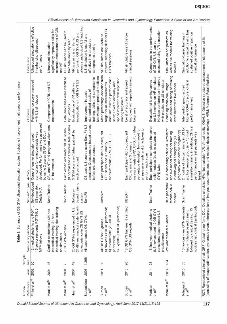
Table 1 summarizes the studies assessing the characteris-tics and outcomes of OB GYN US simulation. 11 - 18 Over a span of 13 years, we identifi ed 10 articles which evaluated the impact of the use of US simulation training on differ-ent outcomes in OB GYN. The majority of these studies evaluated the effect of US simulation through varying study designs, such as a prepost experimental study, 11 , 14
nonrandomized interventional study, 13 , 16 observational study, 12 , 15 and randomized clinical trial. 17 Of these studies, most of them (n = 9) were based on small sample sizes (≤50). The two randomized studies produced contradic-tory fi ndings; Skupski et al 17 refl ected that simulation-based training showed inferiority compared to live model in regards to the primary outcomes (rating of training, scanning technique, and image acquisition), while Tolsgaard et al 12 demonstrated that simulation-based US training improved the performance compared to clinical training only. The latter study performed a randomized trial using a control, clinical training only group.
First Trimester Screening
Of the total studies included in this review, two assessed the impact of US simulation training on CRL and nuchal translucency (NT) measurements. 13 , 14 These two studies found that US simulation signifi cantly improves the skills required to measure CRL and NT accurately and may reduce false results.
Anatomy Scan
US is used to evaluate fetal anatomy and detect fetal structural abnormalities. 15 The incidence of fetal anoma-lies is 2% for major, and 5% for minor anomalies. 16 - 18 , 26
Compared with other diagnostic tools, the sensitivity of US in detecting anomalies is far less than perfect since a lot depends on the operator. Some multicenter studies from the early 1990s demonstrated no reduction in peri-natal morbidity or mortality since the introduction of US. 27 , 28 We may argue that this outcome is a consequence of the current training style, which involves theoretical knowledge gained by means of lectures and textbooks, and practical knowledge gained by exposure to as many patients as possible. The currently available high-fi delity
Effectiveness of Ultrasound Simulation in Obstetrics and Gynecology Education: A State-of-the-Art Review
Donald School Journal of Ultrasound in Obstetrics and Gynecology, April-June 2017;11(2):115-125 117
DSJUOG
Tabl
e 1:
Sum
mar
y of
OB
GY
N u
ltras
ound
sim
ulat
ion
stud
ies
illus
tratin
g im
prov
emen
t in
ultra
soun
d pe
rform
ance
Aut
hor/
Ref
eren
ceYe
arS
ampl
e si
zeS
ampl
e po
pula
tion
Sim
ulat
or ty
peA
ctiv
ityO
utco
me
Con
clus
ion
Pitt
ini e
t al19
2002
3012
med
ical
stu
dent
s an
d P
GY
1,
15 s
enio
r res
iden
ts P
GY
3-5,
3
MFM
fello
ws
Self-
deve
lope
d U
S si
mul
ator
; D
etai
ls N
/A
Com
preh
ensi
ve s
imul
ator
bas
ed
curr
icul
um fo
r am
nioc
ente
sis
was
de
velo
ped;
Per
form
ance
was
ass
esse
d by
usi
ng 2
sca
les
(GR
F an
d D
CL)
Per
form
ance
sco
re im
prov
ed
with
US
sim
ulat
ion
Ultr
asou
nd s
imul
ator
is e
ffect
ive
in e
nhan
cing
ultr
asou
nd
perfo
rman
ce
Mau
l et a
l1320
0445
Cer
tified
obs
tetri
cian
s (2
4 ha
d th
eore
tical
trai
ning
; 21
had
theo
retic
al tr
aini
ng p
lus
train
ing
on s
imul
ator
)
Son
o Tr
aine
rC
RL
and
NT
on 9
pre
gnan
t vol
unte
ers
in 1
st tr
imes
ter
Com
paris
on o
f CR
L an
d N
T m
easu
rem
ents
Ultr
asou
nd s
imul
atio
n si
gnifi
cant
ly im
prov
es s
kills
for
accu
rate
mea
sure
men
ts o
f CR
L an
d N
TM
aul e
t al13
2004
77
OB
GY
N e
xper
tsS
ono
Trai
ner
Eac
h ex
pert
eval
uate
d 10
US
sca
ns
(8 n
orm
al a
nd 2
abn
orm
al fe
tal s
cans
)Fe
tal a
nom
alie
s w
ere
iden
tified
by
5 e
xper
tsU
S s
imul
atio
n ca
n be
use
d to
tra
in fe
tal a
nom
aly
scan
Hee
r et a
l2020
0449
25 O
B G
YNs
expe
rienc
ed in
US;
24
4th
yea
r med
ical
stu
dent
s w
ith
no e
xper
ienc
e in
OB
GYN
US
Sof
twar
e-ba
sed
train
ing
syst
em
3 G
YN
sca
ns o
n “v
irtua
l pat
ient
” by
each
par
ticip
ant
Equ
ival
ence
of V
R w
ith li
ve
inve
stig
atio
ns in
OB
GY
N U
SVR
sca
nnin
g is
sim
ilar t
o pe
rform
ing
OB
GYN
US
and
allo
ws
stan
dard
ized
US
teac
hing
Sta
boul
idou
et
al21
2006
1,26
6A
ll ex
perie
nced
OB
GY
Ns
Son
oFit
OB
bas
ic U
S c
ours
e us
ing
Son
oFit
US
si
mul
ator
; Que
stio
nnai
re b
ased
sur
vey
befo
re a
nd a
fter c
ours
es
Impr
ovem
ent i
n ba
sic
stan
dard
ized
qua
lity
of
train
ing,
ski
lls a
nd s
onog
raph
ic
know
ledg
e in
pre
nata
l med
icin
e
US
sim
ulat
ion
is u
sefu
l and
ef
fect
ive
in in
stru
ctur
ed
sono
grap
hic
train
ing
Bur
den
et
al22
2011
30A
ll O
B G
YN
s; 3
gro
ups
a) N
ovic
es (<
10 U
S p
erfo
rmed
;b)
Inte
rmed
iate
(20–
50 U
S
perfo
rmed
);c)
Exp
erts
(>10
0 U
S p
erfo
rmed
)
Ultr
aSim
Eac
h pa
rtici
pant
did
5 s
ubse
quen
t C
RL
scan
s an
d 3
biom
etry
m
easu
rem
ents
(BP
D, O
FD, F
L)
Mea
n pe
rcen
tage
dev
iatio
n fro
m
targ
et fo
r all
mea
sure
men
ts
and
time
take
n to
per
form
eac
h sc
an; L
evel
of a
ccur
acy
and
spee
d im
prov
ed w
ith re
petit
ion
amon
g be
ginn
ers
VR
sim
ulat
ors
are
usef
ul to
im
prov
e sc
anni
ng s
kills
for O
B
GY
N tr
aine
es
Bur
den
et
al23
2013
2618
OB
GY
N tr
aine
es; 8
cer
tified
O
B G
YN
US
exp
erts
Ultr
aSim
Eac
h pa
rtici
pant
did
5 s
ubse
quen
t C
RL
scan
s an
d 3
biom
etry
m
easu
rem
ents
(BP
D, O
FD, F
L); M
ean
perc
enta
ge d
evia
tion
from
targ
et fo
r al
l mea
sure
men
ts a
nd ti
me
take
n to
pe
rform
eac
h sc
an
Leve
l of a
ccur
acy
and
spee
d im
prov
ed w
ith re
petit
ion
amon
g be
ginn
ers
VR
sim
ulat
ors
usef
ul b
efor
e cl
inic
al s
essi
ons
Mad
sen
et
al24
2014
2816
fina
l yea
r med
ical
stu
dent
s (U
S n
ovic
es) a
nd 1
2 O
B G
YN
co
nsul
tant
s (e
xper
ienc
ed U
S
prac
titio
ners
)
Sca
n Tr
aine
rE
ach
parti
cipa
nt c
ompl
eted
the
seve
n va
lid m
odul
es (b
asic
to a
dvan
ced
GY
N) t
wic
e
Eva
luat
ion
of le
arni
ng c
urve
s fo
r US
nov
ices
on
VR
sim
ulat
or;
Nov
ices
per
form
ance
impr
oved
w
ith p
ract
ice
on U
S s
imul
ator
Com
pete
nce
in th
e pe
rform
ance
of
OB
GY
N U
S c
an b
e as
sess
ed u
sing
VR
sim
ulat
ion
Moa
k et
al25
2014
134
3rd
year
med
ical
stu
dent
sB
lue
phan
tom
/fe
mal
e pe
lvic
m
odel
s
RC
T co
mpa
ring
pelv
ic U
S s
imul
ator
an
d liv
e m
odel
trai
ning
for t
rain
ing
in e
ndov
agin
al s
onog
raph
y (IU
pr
egna
ncy
and
ecto
pic
preg
nanc
y)
Sca
nnin
g te
chni
que,
imag
e ac
quis
ition
and
ratin
g of
trai
ning
w
ere
bette
r with
live
mod
el
Sim
ulat
ors
do n
ot p
erfo
rm a
s w
ell a
s liv
e m
odel
s fo
r tra
inin
g no
vice
s
Tols
gaar
d
et a
l1220
1533
18 n
ovic
es (n
ew G
YN
resi
dent
s)
had
sim
ulat
ion
base
d tra
inin
g fo
llow
ed b
y cl
inic
al tr
aini
ng; 1
5 no
vice
s ha
d cl
inic
al tr
aini
ng o
nly
Sca
n Tr
aine
r2
mon
ths
of c
linic
al tr
aini
ng in
US
for
all;
Inte
rven
tion
grou
p (n
= 1
8) h
ad
sim
ulat
ion
train
ing
in a
dditi
on; C
linic
al
perfo
rman
ce o
n re
al p
atie
nts
was
te
sted
Inte
rven
tion
grou
p sc
ored
hi
gher
OS
AU
S s
core
, clin
ical
pe
rform
ance
test
Sim
ulat
ion-
base
d tra
inin
g in
ad
ditio
n to
clin
ical
trai
ning
has
su
stai
ned
posi
tive
impa
ct o
n cl
inic
al p
erfo
rman
ce
RC
T: R
ando
miz
ed c
linic
al tr
ial;
GR
F: G
loba
l rat
ing
form
; DC
L: D
etai
led
chec
klis
t; N
/A: N
ot a
vaila
ble;
VR
: Virt
ual r
ealit
y; O
SA
US
: Obj
ectiv
e st
ruct
ured
ass
essm
ent o
f ultr
asou
nd s
kills
(c
onsi
stin
g of
imag
e op
timiz
atio
n, s
yste
mat
ic e
xam
, int
erpr
etat
ion
of im
ages
, doc
umen
tatio
n, a
nd m
edic
al d
ecis
ion-
mak
ing)
; MFM
: Mat
erna
l Fet
al M
edic
ine

Sushila Arya et al
118
US simulators can simulate almost every imaginable US examination and may ultimately reduce the need of hired models and patients for early learning. The systematic use of US simulation may improve the detection rate of congenital fetal anomalies; improve the learning curve, self-assessment, and objective evaluation of the learner’s competency. 13 The training agenda for individual trainees can be modifi ed depending on the desired pace of the acquisition of the required skills.
Biometry
SonoTrainer was used by experts and demonstrated improvement in accurate CRL and NT measurements and supported the idea of introducing simulation-based training into clinical learning. 13 The majority of participants reported good image quality and excellent training effect with the use of SonoTrainer US simulator. Using UltraSim, Burden et al 23 reported improvement in effi ciency in obtaining biometry measurements, the accuracy (mean deviation in the measurements of fetal biometry from target values), and placental localization. They also reported that the simulator was easy to be used by novices as well as experienced operators and noticed quick adaptation to the simulator. 29 Akoma et al 30 evaluated the role of a fetal pig simulator in OB US train-ing in 24 participants who were randomized to two groups with 12 learners in each group. Only hands-on scanning on pregnant patients was used for the fi rst group (patients between 16 and 28 weeks gestation), and hands-on scan-ning plus fetal pig simulation for the second group. No difference in biometric scan between the two groups was observed, but the intervention group (hands-on scanning plus fetal pig simulation) obtained improvement in scan-ning time and the acquisition of adequate images. The conclusion of this study was that the addition of a fetal pig US task trainer improved US scan effi ciency.
Prenatal Procedures
Since the focus in health care is shifting to better pre-natal outcomes per center, in-training physicians should achieve the best skills possible in order to perform critical procedures that can improve fetal survival. Teaching and training can be challenging especially for “not so com-monly performed procedures” like fetal surgeries. The rate of invasive procedures has dropped signifi cantly in the past 6 years due to the increased use of improved screening tests. 31 The use of noninvasive cell-free fetal DNA testing is likely to continue to cause a decline in the use of invasive testing. In order to increase the expertise of fellows within the limitation of a declining number of invasive procedures, the role of simulation should be explored and the American Congress of Obstetricians
and Gynecologists (ACOG) should consider guidelines to maximize trainees’ experience. 32 Rose et al 33 indicated that simulation-based training may help preserve and improve those procedural skills.
Amniocentesis, chorionic villi sampling (CVS), in-utero stent placement, percutaneous umbilical cord blood sampling, and cervical cerclage are the areas where simu-lation has great potential benefi t. 34 It is anticipated that more fetal surgeries will be performed in the future due to the increasing incidence of multiple gestation (increasing use of assisted reproductive technologies and advanced maternal age), economic growth, and heightened aware-ness of fetal surgeries. McWeeney et al 35 reported that maternal-fetal medicine (MFM) fellowship programs are not able to provide suffi cient training in CVS. Therefore, they developed a novel training model using a porcine heart, piglet, and freezer bag with US gel to simulate abdominal wall and use of transabdominal sonogram to guide CVS performance. All MFM faculty and fellows agreed that the model was useful. 35 Nitsche et al 36 created a novel in-utero stent placement training model using a gravid pig uterus. This kind of low-cost task trainer was utilized to enhance skills in a nonclinical environment. Zubair et al 37 developed a novel amniocentesis model by using formalin-preserved gravid pig uterus and a freezer bag fi lled with US gel placed on the top of the uterus to simulate the abdomen. Changing the fetal position and amniotic fl uid and gel thickness simulated realistic sce-narios. Simulation-based curriculum examples helped trainees learn amniocentesis early in their training with no discomfort to patients from practice trials. 38 Peeters et al 39 reported improved performance of fetoscopic laser surgery in twin-to-twin transfusion syndrome with the use of advanced high-fi delity simulator. Experts as well as novices reported the usefulness of simulators and felt that the use of simulation improved their performance score and reduced procedure time. Similarly, the use of a task trainer for simulation of ultrasound-guided second trimester uterine evacuation improved profi ciency and confi dence with dilatation and evacuation procedures among residents and other trainees. 40
Pelvic Ultrasound
Madsen et al 24 reported that the virtual reality (VR) simulator is a reliable and valid tool to improve pelvic US examination performance. In their study various advanced pelvic modules were used and authors observed improvement in novices’ performance, which plateaued after 4 hours of simulation training. Girzadas et al 41 demonstrated improvement in knowledge, diag-nostic skills, and management of a ruptured ectopic preg-nancy using a hybrid simulator compared to a standard high-fi delity simulator. The hybrid simulator consisted
Effectiveness of Ultrasound Simulation in Obstetrics and Gynecology Education: A State-of-the-Art Review
Donald School Journal of Ultrasound in Obstetrics and Gynecology, April-June 2017;11(2):115-125 119
DSJUOG
of a transvaginal US task trainer combined with a high-fidelity US mannequin. Vallabh-Patel et al4 reported improvement in clinical knowledge and interpretation of images skills in a clinical setting with the use of low- and high-fidelity transabdominal and transvaginal pelvic US simulators. Monsky et al also reported improved knowledge and scanning ability following early pelvic US simulation for residents.42
Ultrasound Learning for Trainees
Steps should be taken to develop a standard curriculum, dedicated and effective training for OB GYN residents, fellows and practicing physicians to improve and preserve their US scanning and interpretation skills. Credible performance standards should be reached before encounters with actual patients. Based on our systematic review, there is a benefit of including simulation courses and dedicated curricula for different level of trainees using different modules. A standard US curriculum, similar to what was developed for MFM fellowships, which incorporates the introduction of US simulation at an early stage for the novices in OB GYN training is of paramount importance.43 For improvement in US educa-tion among OB GYN residents and its subspecialties, clear educational goals and objectives, and valid performance rating should be established.44,45
Three major competencies in US are as follows: (1) Technical aspect of performance, (2) image percep-tion, and (3) interpretation – medical decision-making skills.2,45,46 For the objective assessment of US skills, international multispecialty consensus suggested seven elements: (1) Indication for the examination; (2) applied knowledge of US equipment; (3) image optimization; (4) systematic examination; (5) interpretation of images; (6) documentation of examination; and (7) medical decision-making.47
Validity of Various Simulators
Limitations of the first generation of VR simulators are static images of the fetus, lack of heart activity, and no blood flow. Additionally, there is no adiposity effect and given the increasing prevalence of obesity the addition of this feature in upcoming advanced simulators will be very helpful. These real-time properties of US simulation were improved in recent models. Most studies variably demon-strated acquisition of knowledge and skills and generated findings, suggesting a correlation with simulation training and improved performance in the simulated environ-ment.12,19-25,13 This finding may be acceptable provided the simulator is appropriately validated (in many reports, i.e., debatable). Some studies examined the question of validity concurrently or in isolation, so there is limited evidence on
construct validity of simulators.11,14,29,48-50 Though litera-ture on the use of US simulation is sparse, it consistently showed its usefulness in US education in OB GYN.
Burden et al demonstrated construct validity of the UltraSim simulator in performance of CRL and growth scan measurements, and stated that this high-fidelity simulator has the potential to improve the scanning skills of OB GYN trainees.51 Newey et al14 have demonstrated the validity of VirUS for NT measurement. More recently, Patel et al51 explored the OB GYN trainees’ perspective on the use of VR US simulation in the United Kingdom. Of 140 trainees, 70 (50%) responded to the survey; 73% of respondents considered US simulation to be an essential component of training; 69% agreed that it helps improving their clinical skills; 77% would like to have US simulation integrated into OB GYN training. Table 2 reviews the studies evaluating the validity of OB GYN US simulators.17,22,24-26,33,51
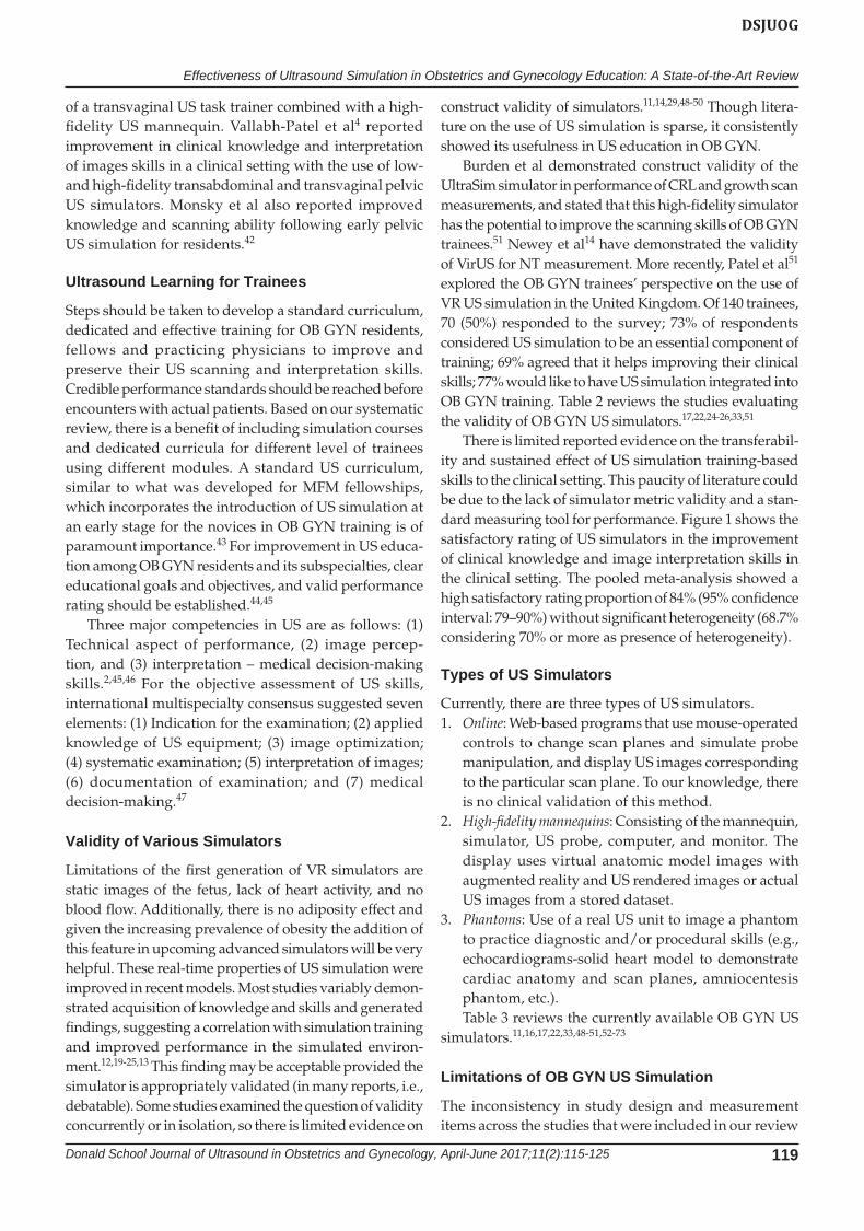
There is limited reported evidence on the transferabil-ity and sustained effect of US simulation training-based skills to the clinical setting. This paucity of literature could be due to the lack of simulator metric validity and a stan-dard measuring tool for performance. Figure 1 shows the satisfactory rating of US simulators in the improvement of clinical knowledge and image interpretation skills in the clinical setting. The pooled meta-analysis showed a high satisfactory rating proportion of 84% (95% confidence interval: 79–90%) without significant heterogeneity (68.7% considering 70% or more as presence of heterogeneity).
Types of US Simulators
Currently, there are three types of US simulators.1. Online: Web-based programs that use mouse-operated
controls to change scan planes and simulate probe manipulation, and display US images corresponding to the particular scan plane. To our knowledge, there is no clinical validation of this method.
2. High-fidelity mannequins: Consisting of the mannequin, simulator, US probe, computer, and monitor. The display uses virtual anatomic model images with augmented reality and US rendered images or actual US images from a stored dataset.
3. Phantoms: Use of a real US unit to image a phantom to practice diagnostic and/or procedural skills (e.g., echocardiograms-solid heart model to demonstrate cardiac anatomy and scan planes, amniocentesis phantom, etc.).Table 3 reviews the currently available OB GYN US
simulators.11,16,17,22,33,48-51,52-73
Limitations of OB GYN US Simulation
The inconsistency in study design and measurement items across the studies that were included in our review

Sushila Arya et al
120
Tabl
e 2:
Rev
iew
of s
tudi
es e
valu
atin
g th
e va
lidity
of O
B G
YN
US
sim
ulat
ors
Aut
hor,
year
an
d re
fere
nce
Sam
ple
size
Sam
ple
popu
latio
nS
imul
ator
ty
peA
ctiv
ityO
utco
me
Con
clus
ion
New
ey e
t al14
13E
xper
ienc
ed s
onog
raph
ers
VirU
SN
T re
peat
abili
ty, i
nter
- and
in
traob
serv
er m
easu
rem
ent
Sig
nifi c
ant c
orre
latio
n be
twee
n re
peat
abili
tyP
oten
tial u
se o
f sim
ulat
or in
ope
rato
r tra
inin
gB
urde
n et
al29
2618
OB
US
trai
nees
and
8 c
ertifi
ed
expe
rtsU
ltraS
imE
ach
parti
cipa
nt d
id fi
ve s
ubse
quen
t C
RL
scan
s an
d th
ree
biom
etry
m
easu
rem
ents
(BP
D, O
FD, F
L)
Mea
n pe
rcen
tage
dev
iatio
n fro
m
targ
et fo
r all
mea
sure
men
ts a
nd
time
take
n to
per
form
eac
h sc
an;
Trai
nees
had
gre
ater
var
iatio
n of
m
easu
rem
ents
on
sim
ulat
or a
nd
took
long
er ti
me
to s
can
Ultr
aSim
has
con
stru
ct v
alid
ity
Als
alam
ah
et a
l4836
25 e
xper
ienc
ed a
nd 1
1 in
depe
nden
t pra
ctiti
oner
s; b
oth
grou
ps w
ere
new
for t
he s
imul
ator
Sca
nTra
iner
Tran
svag
inal
US
trai
ning
10 p
oint
Vis
ual A
nalo
gue
Sca
leS
can
train
er h
as fa
ce a
nd c
onte
nt
valid
ity; R
epor
ted
as b
enefi
cia
l too
l fo
r tea
chin
g U
S s
kills
Al-M
emar
et
al49
24Th
ree
grou
ps, e
ach
had
8 pa
rtici
pant
s:(1
) Nov
ice
train
ees;
(2) I
nter
med
iate
leve
l tra
inee
s;(3
) Exp
erts
Sca
nTra
iner
;E
ach
parti
cipa
nt c
ompl
eted
two
mod
ules
(GY
N a
nd e
arly
pre
gnan
cy
mod
ules
)
Que
stio
nnai
re u
sing
fi ve
-poi
nt
Like
rt sc
ale
Face
and
con
tent
and
con
stru
ct
valid
ity; R
epor
ted
as a
val
id
train
ing
tool
for G
YN
US
trai
ning
; P
artic
ipan
ts a
gree
d th
at s
imul
ator
pl
ayed
a p
ositi
ve ro
le in
US
trai
ning
Cha
louh
i et
al17
29O
B G
YN
and
radi
olog
y co
nsul
tant
s (n
= 1
2); M
idw
ives
(n
= 1
3); P
hysi
cian
s (n
= 1
4);
VIM
ED
IXE
ach
cand
idat
e sc
anne
d vo
lunt
eer
preg
nant
pat
ient
s an
d us
ed O
B G
YN
si
mul
ator
; Eac
h ca
ndid
ate
obta
ined
ni
ne b
iom
etric
and
mor
phol
ogic
al
plan
es o
n pa
tient
s an
d si
mul
ator
; S
cans
wer
e sc
ored
and
com
pare
d by
tw
o re
view
ers
Mea
n de
xter
ity s
core
in
sim
ulat
ion
and
real
US
ex
amin
atio
n w
ere
com
para
ble
OB
GY
N U
S s
imul
ator
is a
goo
d m
etho
d to
eva
luat
e sk
ills
of tr
aine
es;
It is
com
para
ble
to e
valu
atio
n in
pr
egna
nt v
olun
teer
s
Pre
shaw
et
al50
25Tr
aine
es a
nd e
xper
ienc
e so
nogr
aphe
rs (d
etai
ls N
/A)
Sca
nTra
iner
Ano
nym
ous
surv
ey o
n fu
nctio
nalit
y,
real
ism
and
role
of S
canT
rain
er in
de
velo
ping
US
ski
lls
Sca
nTra
iner
brid
ges
the
gap
and
help
s no
vice
s to
lear
n pr
actic
al
skill
s an
d pr
inci
ples
of U
S
scan
ning
Nov
ices
sho
uld
deve
lop
initi
al U
S
skill
s us
ing
sim
ulat
ion-
base
d tra
inin
g
Pat
el e
t al51
140
OB
GY
N tr
aine
esN
onsp
ecifi
ed
VR
US
si
mul
ator
s
Ano
nym
ous
surv
ey o
n U
S s
imul
atio
n tra
inin
g73
% o
f res
pond
ents
con
side
red
US
sim
ulat
ion
to b
e an
ess
entia
l co
mpo
nent
of t
rain
ing;
69%
ag
reed
that
it h
elps
impr
ovin
g th
eir c
linic
al s
kills
; 77%
wou
ld
like
to h
ave
US
sim
ulat
ion
inte
grat
ed in
to O
B G
YN
trai
ning
US
sim
ulat
ion
has
the
pote
ntia
l to
impr
ove
the
use
of c
urre
ntly
av
aila
ble
reso
urce
s in
clin
ical
US
ed
ucat
ion
and
may
ena
ble
train
ees
to a
chie
ve m
anda
tory
US
ski
lls
BP
D: B
ipar
ieta
l dia
met
er; F
L: F
emur
leng
th; A
C: A
bdom
inal
circ
umfe
renc
e; O
FD: O
ccip
itofro
ntal
dia
met
er; N
T: N
ucha
l tra
nslu
cenc
y; U
S: U
ltras
ound
Effectiveness of Ultrasound Simulation in Obstetrics and Gynecology Education: A State-of-the-Art Review
Donald School Journal of Ultrasound in Obstetrics and Gynecology, April-June 2017;11(2):115-125 121
DSJUOG
Fig. 1: Rating of ultrasound training using simulators
precluded a broader meta-analysis of the effect of US simulation on OB GYN education. In most studies the scoring system used to measure improvement was not standardized and pre- and posttest analysis was not done consistently. In some studies a control group was lacking, and the US experience of comparison groups was not clear. Our review did not include the results of unpub-lished research studies and non-English language studies, and a more comprehensive review of the “gray” literature was not performed. Despite the limitations stated above, nearly all of the included studies reported substantial improvement in clinical knowledge, skills, and confidence following the use of OB GYN US simulation.
CONCLUSION
In surgical fields simulation-based training has already been incorporated, with proven benefit in procedural skills.52-54 Simulators are not perceived as a replace-ment of clinical training but rather as an aid to speed up the basic, as well as advanced skills learning curve. A simulator is an educational tool, which imitates real-life scenarios, closely approximates patient encounters to develop knowledge and skills that can be transferred to the clinical setting to improve patient safety and efficiency. The goal of simulation is to help the learners become more confident and competent when caring for their patients.55 Additional benefit of simulation is the reduction of patient discomfort. Simulation also provides an opportunity for independent learning and limits the need of supervision. The US simulation is expected to improve efficiency and diagnostic skills resulting in the decreased need of expensive imaging tools, such as computed tomography and magnetic resonance imaging. Simulation-based training is gaining more popularity in all medical specialties, and following the introduction
of simulation improved outcomes have been widely reported.44,56 The US simulation is a safe, effective, and learner-centered educational approach which improves image optimization and probe orientation, provides the opportunity for unlimited practice without pressure, and facilitates a systematic approach to sonography prior to the patient encounter (Fig. 2).11,29,33,47,57-59
Patient discomfort and the intimate nature of endo-vaginal sonography encourage the need of simulation-based learning. Our systematic review reports significant improvement in clinical knowledge, skills and behaviors; and moderate effects for patient-related outcomes with the use of US simulation in training.11,15,24,67 However, the present studies failed to demonstrate a compelling body of evidence to support widespread adoption of US simulation-based OB GYN education to improve US performance skills.
Trainees with varied exposure to simulation found US simulation to be useful. Trainees also expressed a desire for more substantial incorporation of US simulation in their training.51 There is limited but supportive litera-ture on the usefulness of OB GYN US simulation, which reveals that it not only improves the scanning skills of trainees and detection rates of abnormal findings but also helps providers preserve their skills. It is not surprising to see transferability of US skills to the clinical area, though not many studies investigated this effect. Despite the cost, integration of US simulators in medical education seems to have a positive implication on the scanning and interpretation skills of trainees.
We hope that this review will encourage various train-ing programs to include US simulation in the education of their trainees with the ultimate goal of improving patient safety. More extensive clinical trials are needed to assess the long-term impact of US simulation on clinical

Sushila Arya et al
122
Tabl
e 3:
Rev
iew
of O
B G
YN
US
sim
ulat
ors
Ref
eren
ce (y
ear)
Sim
ulat
orVe
ndor
Mod
ules
for O
B G
YN
App
licat
ion
Per
form
ance
as
sess
men
t cap
abili
tyP
rope
rties
Dat
a ac
quis
ition
Caw
thor
n et
al60
; P
ark
et a
l61V
IME
DIX
OB
G
YN
CA
E H
ealth
ca
re, S
aras
ota,
U
SA
and
Ville
St
Laur
ent,
Que
bec,
C
AN
Eig
ht w
eeks
fetu
s (a
llow
s TV
sc
anni
ng),
fetu
s at
20
wee
ks
(mor
e th
an 2
0 bi
rth d
efec
ts
incl
uded
) GY
N c
ases
OB
GY
N (T
A, T
V p
robe
), ec
hoca
rdio
grap
hy a
nd
TTE
Met
rics
to a
sses
s co
mpe
tenc
y; v
isua
l cl
ue to
indi
cate
pai
n w
ith d
eep
endo
vagi
nal
prob
e in
serti
on
Rea
listic
with
a d
umm
y,
high
imag
e qu
ality
, ec
hoca
rdio
grap
hy w
ith
hear
t mov
emen
t
Sof
twar
e ge
nera
ted,
vi
rtual
pat
ient
s ba
sed
on re
al
patie
nt s
can
***T
olsg
aard
et a
l12; M
adse
n et
al24
; Car
olan
-Ree
s an
d R
ay62
Sca
n Tr
aine
r P
rofe
ssio
nal
Med
aPho
r Lt
d, C
ardi
ff M
edic
entre
Wal
es,
UK
Feta
l 1st
/2nd
trim
este
r with
no
rmal
and
var
ious
feta
l an
omal
ies,
GY
N c
ases
with
ut
erin
e pa
thol
ogie
s an
d pe
lvic
m
asse
s
OB
GY
N (T
A, T
V p
robe
, B
-mod
e, c
olor
and
spe
ctra
l D
oppl
er);
IM a
nd E
M
mod
ules
Cur
ricul
um b
ased
te
achi
ng, r
eal-t
ime
assi
sted
gui
danc
e,
hapt
ic fe
edba
ck
devi
ce, m
etric
-bas
ed
asse
ssm
ent
Rea
listic
with
a d
umm
y,
high
imag
e qu
ality
3D d
ata
from
re
al s
cans
Ehr
icke
63S
onoS
imS
onoS
im, I
nc.
San
ta M
onic
a,
CA
, US
A
1,00
0 ac
tual
pat
ient
cas
esP
oint
of c
are
US
lear
ning
m
odul
es fo
r var
ious
sp
ecia
lties
No
asse
ssm
ent t
ool
avai
labl
eP
C b
ased
virt
ual U
S
scan
ner;
Gra
phic
al
inte
ract
ive
sim
ulat
ion
3D d
ata
from
re
al p
atie
nts
Bur
den
et a
l 33; M
elle
r et a
l64;
Mel
ler65
; Knu
dson
and
S
isle
y66; H
enric
hs e
t al67
; K
aufm
ann
and
Liu68
; Sch
wid
et
al69
; Zuv
ekas
et a
l16,7
0
Ultr
aSim
Med
Sim
Inc.
La
uder
dale
, FL,
U
SA
Ove
r 120
cas
es o
f fet
us in
all
trim
este
r, no
rmal
and
var
ious
pa
thol
ogie
s in
OB
GY
N
OB
GYN
(TA,
TV
prob
e,
B-m
ode,
col
or a
nd s
pect
ral
Dop
pler
), ab
dom
en, b
reas
t, va
scul
ar, n
eck
and
EM
Pro
vide
s m
easu
res
to
mon
itor p
erfo
rman
ceR
ealis
tic w
ith a
dum
my,
In
tera
ctiv
e; In
trodu
ced
in 1
995;
Pio
neer
in U
S
sim
ulat
ion
3D d
ata
from
re
al p
atie
nts,
co
nfi g
ured
in
to re
leva
nt
mod
ules
Non
eS
onoM
omTM
SIM
ULA
BS
US
A S
eattl
e, W
A, U
SA
1st t
rimes
ter c
ompl
icat
ions
(1
3 ca
ses)
OB
(TA
, TV
pro
be),
EM
N/A
Rea
listic
with
a d
umm
y,
high
imag
e qu
ality
, rea
l tim
e
3D d
ata
from
re
al p
atie
nts
Non
eU
S M
ento
r3D
Sys
tem
s H
ealth
car
e,
Littl
eton
, CO
, U
SA
Feta
l 1st
tri (
viab
ility,
GA
asse
ssm
ent,
NT,
cho
rioni
city
an
d am
nion
icity
); 2n
d tri
mes
ter
mod
ules
offe
r mov
ing
fetu
s w
ith n
orm
al a
nd m
alfo
rmed
ca
ses;
bas
ic a
nd d
iffi c
ult 4
G
YN m
odul
es, 2
4 ca
ses
OB
GY
N, c
ardi
olog
y,
IM a
nd E
M; b
asic
an
d ad
vanc
ed im
age
enha
ncem
ents
, arti
fact
s,
colo
r Dop
pler
, CW
, PW
, M
-mod
e, T
A an
d TV
pr
obes
Met
ric a
sses
smen
t to
ol fo
r all
mod
ules
Com
preh
ensi
ve a
nd
real
istic
mod
ules
with
a
dum
my,
hig
h im
age
qual
ity, r
eal p
atie
nt-
base
d ca
ses
VR
, com
pute
r re
nder
ing
from
re
al p
atie
nt-
base
d im
ages
Mau
l et a
l13; B
aier
et a
l71;
Wus
tem
ann
et a
l72; T
erka
mp
et a
l73
Son
oTra
iner
Son
ofi t
Gm
bH,
Sta
deck
en-
Els
heim
, G
erm
any
OB
mod
ules
(1st
and
2n
d tri
mes
ter,
maj
or a
nd
min
or a
nom
alie
s, fe
tal
echo
card
iogr
aphy
), G
YN
m
odul
es (T
A an
d TV
)
OB
GY
N c
ases
(TA
, TV
pr
obe)
, bre
ast,
IM a
nd E
M,
card
iolo
gy, T
TE, u
rolo
gy
N/A
Rea
listic
with
a
dum
my,
high
imag
e qu
ality
, rea
l tim
e, fe
tal
echo
card
iogr
aphy
in
cludi
ng h
eart
mov
emen
ts
3D fr
om re
al
scan
s
Non
eS
pace
Fan
S
TK
yoto
Kag
aku,
To
kyo
and
Nag
ya,
Japa
n
23- w
eeks
fetu
s m
odel
(b
iom
etry
, pla
cent
al
loca
lizat
ion
and
amni
otic
fl ui
d an
atom
y as
sess
men
t)
OB
, bre
ast a
nd lu
ng e
xam
, E
MN
/AO
val-s
hape
d ph
anto
m
abdo
men
, med
ium
im
age
qual
ity, n
o he
art
mov
emen
ts
VR
bas
ed o
n re
al p
atie
nt
scan
Non
eS
chal
lwar
e U
S s
imul
ator
Sch
allw
are
Gm
bH, B
erlin
, G
erm
any
100
case
s, n
orm
al a
nd
abno
rmal
obs
tetri
cs a
nd
gyne
colo
gy c
ases
OB
GY
N, c
ardi
olog
y, IM
an
d E
M; B
- and
M-m
ode,
co
lor D
oppl
er
N/A
Rea
listic
with
a d
umm
y,
high
imag
e qu
ality
, ec
hoca
rdio
grap
hy w
ith
hear
t mov
emen
ts
3D fr
om re
al
patie
nt s
cans
N/A
: Not
ava
ilabl
e; 3
D: T
hree
-dim
ensi
onal
; TA:
Tra
nsab
dom
inal
; TV:
Tra
nsva
gina
l; AFI
: Am
niot
ic fl
uid
inde
x; G
A: G
esta
tiona
l age
; NT:
Nuc
hal t
rans
luce
ncy;
IM: I
nter
nal m
edic
ine;
EM
: Em
erge
ncy
med
icin
e; T
TE: T
rans
thor
acic
ech
ocar
diog
raph
y; C
W: C
ontin
uous
wav
e D
oppl
er; P
W: P
ulse
d w
ave;
M-m
ode:
Mot
ion
mod
e; B
-mod
e: B
right
ness
mod
e

Effectiveness of Ultrasound Simulation in Obstetrics and Gynecology Education: A State-of-the-Art Review
Donald School Journal of Ultrasound in Obstetrics and Gynecology, April-June 2017;11(2):115-125 123
DSJUOG
performance with the use of a comprehensive curriculum including advanced simulators. Given that there has been only one randomized trial to date which tested the impact of US education incorporating a US simulator, additional randomized controlled trials are called for.11 Further studies are needed to specify the number of ses-sions required to acquire and retain US skills, perform cost analysis, and assess validity and feasibility of the most recent US simulators.
REFERENCES
1. Royal College of Obstetricians and Gynaecologists. Trainees Survey; London: RCOG; 2010.
2. Tolsgaard MG, Rasmussen MB, Tappert C, Sundler M, Sorensen JL, Ottesen B, Ringsted C, Tabor A. Which factors are associated with trainees’ confidence in performing obstetric and gynecological ultrasound examinations? Ultrasound Obstet Gynecol 2014 Apr;43(4):444-451.
3. Moore CL, Copel JA. Point-of-care ultrasonography. N Engl J Med [Internet]. 2011 Feb;364(8):749-757. Available from: http://www.nejm.org/doi/full/10.1056/NEJMra0909487/ninternal-pdf://1587/Moore and Copel - 2011 - Point-of-Care Ultrasonography.html.
4. Vallabh-Patel V, Mendez M, Kupesic Plavsic S. The importance of multimodality pelvic ultrasound simulation in teaching of obstetrics and gynecology residents. Donald Sch J Ultrasound Obs Gynecol 2014;8(1):1-5.
5. Jang TB, Ruggeri W, Dyne P, Kaji AH. Learning curve of emergency physicians using emergency bedside sonography for symptomatic first-trimester pregnancy. J Ultrasound Med 2010 Oct;29(10):1423-1428.
6. American Institute of Ultrasound in Medicine. AIUM practice guideline for the performance of obstetric ultrasound exami-nations. J Ultrasound Med 2013 Jun;32(6):1083-1101.
7. ISUOG Education Committee recommendations for basic training in obstetric and gynecological ultrasound. Ultrasound Obstet Gynecol 2014 Jan;43(1):113-116.
8. ISUOG Education Committee. Update on proposed minimum standards for ultrasound training for residents in Ob/Gyn. Ultrasound Obstet Gynecol 1996;8:363-366.
9. Hertzberg BS, Kliewer MA, Bowie JD, Carroll BA, DeLong DH, Gray L, Nelson RC. Physician training requirements in
sonography: how many cases are needed for competence? AJR Am J Roentgenol 2000 May;174(5):1221-1227.
10. Gracias VH, Frankel H, Gupta R, Reilly PM, Gracias F, Klein W, Nisenbaum H, Schwab CW. The role of positive examinations in training for the focused assessment sonogram in trauma (FAST) examination. Am Surg 2002 Nov;68(11):1008-1011.
11. Chalouhi GE, Bernardi V, Ville Y. Ultrasound simulators in obstetrics and gynecology: state of the art. Ultrasound Obstet Gynecol 2015 Sep;46(3):255-263.
12. Tolsgaard MG, Ringsted C, Dreisler E, Nørgaard LN, Petersen JH, Madsen ME, Freiesleben NL, Sørensen JL, Tabor A. Sustained effect of simulation-based ultrasound training on clinical performance: a randomized trial. Ultrasound Obstet Gynecol 2015 Sep;46(3):312318.
13. Maul H, Scharf A, Baier P, Wüstemann M, Günter HH, Gebauer G, Sohn C. Ultrasound simulators: experience with the SonoTrainer and comparative review of other training systems. Ultrasound Obstet Gynecol 2004 Oct;24(5):581-585.
14. Newey VR, Nassiri DK, Bhide A, Thilaganathan B. Nuchal translucency thickness measurement: repeatability using a virtual ultrasound scanner. Ultrasound Obstet Gynecol 2003 Jun;21(6):596-601.
15. Ewigman BG, Crane JP, Frigoletto FD, LeFevre ML, Bain RP, McNellis D. Effect of prenatal ultrasound screening on peri-natal outcome. RADIUS Study Group. N Engl J Med 1993 Sep;329(12):821-827.
16. Lee K, Kim SY, Choi SM, Kim JS, Lee BS, Seo K, Lee YH, Kim DK. Effectiveness of prenatal ultrasonography in detect-ing fetal anomalies and perinatal outcome of anomalous fetuses. Yonsei Med J 1998 Aug;39(4):372-382.
17. Chalouhi GE, Bernardi V, Ville Y. Ultrasound simulators in obstetrics and gynecology: State of the art. Ultrasound Obstet Gynecol. 2015;46(3):255-263. .
18. Eurenius K, Axelsson O, Cnattingius S, Eriksson L, Norsted T. Second trimester ultrasound screening performed by mid-wives; sensitivity for detection of fetal anomalies. Acta Obstet Gynecol Scand [Internet]. 1999 Feb;78(2):98-104. Available from: http://www.ncbi.nlm.nih.gov/pubmed/10023870.
19. Pittini R, Oepkes D, Macrury K, Reznick R, Beyene J, Windrim R. Teaching invasive perinatal procedures: assessment of a high fidelity simulator-based curriculum. Ultrasound Obstet Gynecol 2002 May;19(5):478-483.
20. Heer IM, Middendorf K, Müller-Egloff S, Dugas M, Strauss A. Ultrasound training: the virtual patient. Ultrasound Obstet Gynecol 2004 Sep;24(4):440-444.
21. Staboulidou I, Freitag U, Marquardt R, Wüstemann M, Hillemanns P, Scharf A. [Quality assured ultrasound simula-tion training for the detection of fetal malformations – can a training benefit be evidenced?]. Z Geburtshilfe Neonatol [Internet]. 2006 Aug;210(4):135-140. (Ger). Available from: http://www.ncbi.nlm.nih.gov/pubmed/16941306.
22. Burden CA, Preshaw J, Grant S. Virtual reality simulation ultrasound training in obstetrics and gynaecology. Arch Dis Child Fetal Neonata 2011 Jun;96 (Suppl 1):Fa56.
23. Burden C, Preshaw J, White P, Draycott TJ, Grant S, Fox R. Usability of virtual-reality simulation training in obstetric ultrasonography: a prospective cohort study. Ultrasound Obstet Gynecol 2013 Aug;42(2):213-217.
24. Madsen ME, Konge L, Nørgaard LN, Tabor A, Ringsted C, Klemmensen AK, Ottesen B, Tolsgaard MG. Assessment of performance measures and learning curves for use of a virtual-reality ultrasound simulator in transvaginal
Fig. 2: Simulation of ultrasound-guided procedures

Sushila Arya et al
124
ultrasound examination. Ultrasound Obstet Gynecol 2014 Dec; 44 (6): 693- 699.
25. Moak JH, Larese SR, Riordan JP, Sudhir A, Yan G. Training in transvaginal sonography using pelvic ultrasound simulators versus live models: a randomized controlled trial. Acad Med [Internet]. 2014 Jul; 89 (7): 1063- 1068. Available from: http://content.wkhealth.com/linkback/openurl?sid=WKPTLP:landingpage&an=00001888-201407000-00032 .
26. Levi S, Schaaps JP, De Havay P, Coulon R, Defoort P. End-result of routine ultrasound screening for congenital anomalies: the Belgian Multicentric Study 1984-92. Ultrasound Obstet Gynecol [Internet]. 1995 Jun; 5 (6): 366- 371. Available from: http://www.ncbi.nlm.nih.gov/pubmed/7552796 .
27. Bofi ll JA, Sharp GH. Obstetric sonography. Who to scan, when to scan, and by whom. Obstet Gynecol Clin North Am 1998 Sep; 25 (3): 465- 478.
28. Crane JP, LeFevre ML, Winborn RC, Evans JK, Ewigman BG, Bain RP, Frigoletto FD, McNellis D. A randomized trial of prenatal ultrasonographic screening: impact on the detec-tion, management, and outcome of anomalous fetuses. The RADIUS Study Group. Am J Obstet Gynecol 1994 Aug; 171 (2): 392- 399.
29. Burden C, Preshaw J, White P, Draycott TJ, Grant S, Fox R. Validation of virtual reality simulation for obstetric ultraso-nography: a prospective cross-sectional study. Simul Healthc [Internet]. 2012 Oct; 7 (5): 269- 273. Available from: http://www.ncbi.nlm.nih.gov/pubmed/22878584 .
30. Akoma UN, Shumard KM, Street L, Brost BC, Nitsche JF. Impact of an inexpensive anatomy-based fetal pig simula-tor on obstetric ultrasound training. J Ultrasound Med 2015 Oct; 34 (10): 1793- 1799.
31. Biggio JR Jr. Prenatal screening for fetal aneuploidy: time to examine where we are and where we are going. Am J Obstet Gynecol 2016 Jun; 214 (6): 673- 675.
32. Khurshid N, Heiser T, Trampe B, Grady M, Stewart K, Shah D, Iruretagoyena J. Current trends in ultrasound guided invasive procedures and impact on MFM fellowship training. Am J Obstet Gynecol 2014 Jan; 210 (1 Suppl): S80- S81.
33. Burden C, Preshaw J, White P, Draycott TJ, Grant S, Fox R. Usability of virtual-reality simulation training in obstetric ultrasonography: A prospective cohort study. Ultrasound Obstet Gynecol. 2013;42(2):213-217.
34. Nitsche JF, Brost BC. The use of simulation in maternal-fetal medicine procedure training. Semin Perinatol [Internet]. 2013 Jun; 37 (3): 189- 198. Available from: http://dx.doi.org/10.1053/j.semperi.2013.02.011 .
35. McWeeney DT, Schwendemann WD, Nitsche JF, Rose CH, Davies NP, Watson WJ, Brost BC. Transabdominal and tran-scervical chorionic villus sampling models to teach mater-nal-fetal medicine fellows. Am J Perinatol 2012 Aug; 29 (7): 497- 502.
36. Nitsche JF, McWeeney DT, Schwendemann WD, Rose CH, Davies NP, Watson W, Brost BC. In-utero stenting: develop-ment of a low-cost high-fi delity task trainer. Ultrasound Obstet Gynecol 2009 Dec; 34 (6): 720- 723.
37. Zubair I, Marcotte MP, Weinstein L, Brost BC. A novel amnio-centesis model for learning stereotactic skills. Am J Obstet Gynecol 2006 Mar; 194 (3): 846- 848.
38. Khurshid N, Trampe B, Heiser T, Birkeland L, Duris E, Stewart K, Shah D, Iruretagoyena J. 420: impact of an amniocentesis simulation curriculum for training in MFM fellowship program. Am J Obstet Gynecol [Internet]. 2014
Jan; 210 (1): S212- S213. Available from: http://linkinghub.elsevier.com/retrieve/pii/S0002937813015184 .
39. Peeters SH, Akkermans J, Slaghekke F, Bustraan J, Lopriore E, Haak MC, Middeldorp JM, Klumper FJ, Lewi L, Devlieger R, et al. Simulator training in fetoscopic laser surgery for twin-twin transfusion syndrome: a pilot randomized con-trolled trial. Ultrasound Obstet Gynecol [Internet]. 2015 Sep; 46 (3): 319- 326. Available from: http://ovidsp.ovid.com/ovidweb.cgi?T=JS&PAGE=reference&D=prem&NEWS=N&AN=26036333 .
40. Shumard KM, Akoma UN, Street LM, Brost BC, Nitsche JF. Development of a novel task trainer for second trimester ultrasound-guided uterine evacuation. Simul Healthc 2015 Feb; 10 (1): 49- 53.
41. Girzadas DV Jr, Antonis MS, Zerth H, Lambert M, Clay L, Bose S, Harwood R. Hybrid simulation combining a high fi delity scenario with a pelvic ultrasound task trainer enhances the training and evaluation of endovaginal ultrasound skills. Acad Emerg Med 2009 May; 16 (5): 429- 435.
42. Monsky WL, Levine D, Mehta TS, et al. Using a sonographic simulator to assess residents before overnight call. Am J Roentgenol. 2002;178(1):35-39.
43. Khurshid N, Trampe B, Iruretagoyena J, Shah D, Stewart K. 419: so you think you can scan? A competency based ultra-sound curriculum for MFM fellows. Am J Obstet Gynecol 2014 Jan; 210 (1): S212.
44. Issenberg SB, McGaghie WC, Petrusa ER, Lee Gordon D, Scalese RJ. Features and uses of high-fi delity medical simula-tions that lead to effective learning: a BEME systematic review. Med Teach 2005 Jan; 27 (1): 10- 28.
45. van der Gijp A, van der Schaaf MF, van der Schaaf IC, Huige JC, Ravesloot CJ, van Schaik JP, Ten Cate TJ. Interpretation of radiological images: towards a framework of knowl-edge and skills. Adv Health Sci Educ Theory Pract 2014 Oct; 19 (4): 565- 580.
46. Krupinski EA. Current perspectives in medical image percep-tion. Atten Percept Psychophys [Internet]. 2010 Jul; 72 (5): 1205- 1217. Available from: http://www.pubmedcentral.nih.gov/articlerender.fcgi?artid=3881280&tool=pmcentrez&rendertype=abstract .
47. Tolsgaard MG, Todsen T, Sorensen JL, Ringsted C, Lorentzen T, Ottesen B, Tabor A. International multispecialty consensus on how to evaluate ultrasound competence: a Delphi consensus survey. PLoS One 2013; 8 (2): e57687.
48. Alsalamah A., Campo R., Hood K., Amso N. Face and content validity of the scantrainer ultrasound simulator [Internet]. Gynecol Surg 2014;11(1)(Suppl 1): 107- 108. Available from: http://ovidsp.ovid.com/ovidweb.cgi?T=JS&PAGE=reference&D=emed12&NEWS=N&AN=71644179
49. Al-Memar M, Saso S, Bobdiwala S, Ameye L, Stalder C, Joash K, Timmerman D, Bourne T. Validation of a virtual reality simu-lator for the use of transvaginal ultrasound in gynaecology and early pregnancy. BJOG An Int J Obstet Gynaecol 2016 Jun; 123 (Suppl): 212.
50. Preshaw J, Ficquet J, Burden C, Overton C, Grant S. Would it be best practice for trainees to learn ultrasound scanning in a simulated setting prior to a clinical setting. Int J Gynecol Obstet 2012 Oct; 119 (53): S457.
51. Patel H, Chandrasekaran D, Myriokefalitaki E, Gebeh A, Jones K, Jeve YB; Midlands Research Collaborative in Obstetrics & Gynecology. The role of ultrasound simulation in

Effectiveness of Ultrasound Simulation in Obstetrics and Gynecology Education: A State-of-the-Art Review
Donald School Journal of Ultrasound in Obstetrics and Gynecology, April-June 2017;11(2):115-125 125
DSJUOG
Obstetrics and Gynecology Training: a UK trainees’ perspec-tive. Simul Healthc 2016 Oct;11(5):340-344.
52. Palter VN, Grantcharov T, Harvey A, Macrae HM. Ex vivo technical skills training transfers to the operating room and enhances cognitive learning: a randomized controlled trial. Ann Surg 2011 May;253(5):886-889.
53. Seymour NE, Gallagher AG, Roman SA, O’Brien MK, Bansal VK, Andersen DK, Satava RM. Virtual reality training improves operating room performance: results of a randomized, double-blinded study. Ann Surg [Internet]. 2002 Oct;236(4):458-463. Available from: http://www.ncbi.nlm.nih.gov/pmc/articles/PMC1422600/pdf/20021000s00008p458.pdf.
54. Hiemstra E, Kolkman W, van de Put MA, Jansen FW. Retention of basic laparoscopic skills after a structured training program. Gynecol Surg 2009 Sep;6(3):229-235.
55. Sarmiento J, Stewart K, Aguila J, Bagherpour A, Plavsic SK. Pelvic ultrasound simulation training models and case sce-narios. Vol. 8, Donald School J Ultrasound Obstet Gynecol 2014 Jan-Mar;8(1):22-30.
56. Cook DA, Hatala R, Brydges R, Zendejas B, Szostek JH, Wang AT, Erwin PJ, Hamstra SJ. Technology-enhanced simulation for health professions education: a systematic review and meta-analysis. JAMA [Internet]. 2011 Sep;306(9): 978-988. Available from: http://jama.ama-assn.org/content/ 306/9/978.abstract\nhttp://jama.ama-assn.org/content/ 306/9/978.full.pdf.
57. Salvesen KA, Lees C, Tutschek B. Basic European ultrasound training in obstetrics and gynecology: where are we and where do we go from here? Ultrasound Obstet Gynecol [Internet]. 2010 Nov [cited 2016 Sep 25];36(5):525-529. Available from: http://www.ncbi.nlm.nih.gov/pubmed/20981718.
58. Nitsche JF, Brost BC. Obstetric ultrasound simulation. Semin Perinatol 2013 Jun;37(3):199-204.
59. McGaghie WC, Issenberg SB, Petrusa ER, Scalese RJ. A criti-cal review of simulation-based medical education research: 2003-2009. Med Educ 2010 Jan;44(1):50-63.
60. Cawthorn TR, Nickel C, O’Reilly M, Kafka H, Tam JW, Jackson LC, Sanfilippo AJ, Johri AM. Development and evaluation of methodologies for teaching focused cardiac ultrasound skills to medical students. J Am Soc Echocardiogr 2014 Mar;27(3):302-309.
61. Parks AR, Atkinson P, Verheul G, Leblanc-Duchin D. Can medical learners achieve point-of-care ultrasound competency using a high-fidelity ultrasound simulator?: a pilot study. Crit Ultrasound J 2013 Nov;5(1):9.
62. Carolan-Rees G, Ray AF. The ScanTrainer obstetrics and gynaecology ultrasound virtual reality training simulator: a cost model to determine the cost viability of replacing
clinical training with simulation training. Ultrasound 2015 May;23(2):110-115.
63. Ehricke HH. SONOSim3D: a multimedia system for sonog-raphy simulation and education with an extensible case database. Eur J Ultrasound 1998 Aug;7(3):225-300.
64. Meller G, Tepper R, Bergman M, Anderhub B. The trad-eoffs of successful simulation. Stud Health Technol Inform 1997;39:565-571.
65. Meller G. A typology of simulators for medical education. J Digit Imaging 1997 Aug;10(3 Suppl 1):194-196.
66. Knudson MM, Sisley AC. Training residents using simulation technology: experience with ultrasound for trauma. J Trauma 2000 Apr;48(4):659-665.
67. Henrichs B, Rule A, Grady M, Ellis W. Nurse anesthesia students’ perceptions of the anesthesia patient simulator: a qualitative study. AANA J 2002 Jun;70(3):219-225.
68. Kaufmann C, Liu A. Trauma training: virtual reality applica-tions. Stud Health Technol Inform [Internet]. 2001;81:236-241. Available from: http://www.ncbi.nlm.nih.gov/pubmed/ 11317747
69. Schwid HA, Rooke GA, Carline J, Steadman RH, Murray WB, Olympio M, Tarver S, Steckner K, Wetstone S; Anesthesia Simulator Research Consortium. Evaluation of anesthesia residents using mannequin-based simulation: a multiinstitu-tional study. Anesthesiology [Internet]. 2002 Dec;97(6):1434-1444. Available from: http://ovidsp.ovid.com/ovidweb.cgi?T=JS&CSC=Y&NEWS=N&PAGE=fulltext&D=yrovftf&AN=00000542-200212000-00015\nhttp://www.ovid.com/site/resources/expert_search/healthexp.html.
70. Zuvekas SH, Banthin JS, Selden TM. How would mental health parity affect the marginal price of care? Health Serv Res [Internet]. 2001 Feb;35(6):1207-1227. Available from: http://www.pubmedcentral.nih.gov/articlerender.fcgi?artid=1089187&tool=pmcentrez&rendertype=abstract
71. Baier P, Scharf A, Sohn C. Der Echtzeit Ultraschallsimulator. Eine neue Methode zum Training in der Ultraschalldiagnostik [New ultrasound simulation system: a method for training and improved quality management in ultrasound examina-tion]. Z Geburtshilfe Neonatol. 2001 Nov-Dec;205(6):213-217.
72. Wustemann M, Scharf A, Maul H, Baier P, Sohn C. New ultrasound simulation system: an effective training method to improve the examiner’s experience for the first trimester screening for chromosomal abnormalities using sonography. Geburtshilfe Frauenheilkd 2002 Jan;62(12):1183-1187.
73. Terkamp C, Kirchner G, Wedemeyer J, Dettmer A, Kielstein J, Reindell H, Bleck J, Manns M, Gebel M. Simulation of abdomen sonography. Evaluation of a new ultrasound simulator. Ultraschall Med 2003 Aug;24(4):239-244.