
A novel low-volume two-chamber microfabricated platform for evaluating drug metabolism and toxicity
8
1 TECHNOLOGY l VOLUME 3 • NUMBER 2 • JUNE 2015 © World Scientific Publishing Co./Imperial College Press TECHNOLOGY COMMUNICATION A novel low-volume two-chamber microfabricated platform for evaluating drug metabolism and toxicity Shyam Sundhar Bale 1 , Gautham Vivek Sridharan 1 , Inna Golberg 1 , Ljupcho Prodanov 1 , William J. McCarty 1 , Osman Berk Usta 1 , Rohit Jindal 1, * & Martin L. Yarmush 1,2, * To evaluate drug and metabolite efficacy on a target organ, it is essential to include metabolic function of hepatocytes, and to evalu- ate metabolite influence on both hepatocytes and the target of interest. Herein, we have developed a two-chamber microfabricated device separated by a membrane enabling communication between hepatocytes and cancer cells. The microscale environment created enables cell co-culture in a low media-to-cell ratio leading to higher metabolite formation and rapid accumulation, which is lost in traditional plate cultures or other interconnected models due to higher culture volumes. We demonstrate the efficacy of this system by metabolism of tegafur by hepatocytes resulting in cancer cell toxicity. INNOVATION This work demonstrates the fabrication and use of a two-chamber microdevice for inter-tissue drug toxicity testing and evaluating me- tabolite effects. e microscale environment created enables cell culture in a low media-to-cell ratio leading to higher metabolite formation and rapid accumulation, which is lost in traditional plate cultures or other interconnected models due to higher culture volumes. By building a two-chamber microfabricated device that allows for direct interaction through a permeable membrane, we have eliminated the need for any fluidic flow — simplifying the model to a great extent. In this report, we demonstrate the applicability of this device using a chemotherapeutic pro-drug tegafur-uracil, which when metabolized by primary hepatocytes produces 5-Fluorouracil (5-FU), a metabolite toxic to cancer cells. e metabolite conversion and its resultant toxicity are measureable in the microscale model within 24 hours. Overall, the two-chamber device provides a novel, easy-to-use platform to evaluate drug metabolism, toxicity and interactions between multi-tissue systems. NARRATIVE e liver is the primary organ where drugs are metabolized. While drug induced liver injury remains an important reason for withdrawal of drugs, metabolites generated by the liver can also be responsible for toxicity in other organs and tissues 1,2 . Significant progress has been made in the development of platforms for evaluating drug hepatotoxicity 3–5 . ey include incorporation of hepatic cell lines or primary hepatocytes in various monoculture and co-culture configuration both in flow and static conditions 4 . However, development of systems that facilitate evaluation of drug toxicity in other organ or tissues as a result of metabolite(s) generated by liver has lagged somewhat. Typically, these systems are comprised of interconnected chambers in which each chamber is seeded with a tissue or organ model and communication is achieved by fluid flowing across and in between the chambers 6–8 . In many cases, the liver chamber is seeded with a hepatocyte cell line 6,7 , which generally lack metabolic function in comparison to primary hepatocytes 9 . While these models may provide an initial assessment for drug toxicity and multi-cell interactions, most of these in vitro models incorporate high media volumes (including tubing and reservoir volumes) creating an artificially high media-to-cell ratio, leading to loss of sensitivity in detecting product interactions, particularly, in case of highly unstable metabolites 6,7 . ere is a critical need for the development of simpler, more effective, multi-tissue systems that can facilitate screening of drug toxicity as a result of primary hepatocytes communicating with the cells in other organs through the metabolic conversion of drugs. In the context of drug metabolism, hepatocytes communicate with the cells in other organ via secreted metabolites. Traditionally, co-culture interactions mediated by secreted factors have been evaluated in transwell systems where different cell types are separated by porous membrane that enables soluble factor communication. One limitation of these systems is the dilution of secreted factors due to exposure of the cells to high media volume 10,11 . is becomes especially limiting in the settings where the secreted factor such as metabolite is cleared by other mechanisms, preventing it from reaching effective or toxic levels. While plate cultures have inherently high media-to-cell ratio, micro- fluidic interconnected chamber systems rely on fluidic connections and reservoirs which eventually result in higher media-to-cell ratio. For drug screening and toxicity related studies, it is essential to develop in vitro models that incorporate both hepatocytes and target-organ of interest, while reducing the culture volume to increase metabolite production and interrogate resulting metabolite toxicity. Herein, we have developed a simple microscale device with a two-chamber design separated by a tissue-culture membrane allowing the culture of two different cells within the same device, while addressing cells within each chamber (Fig. 1). is device design is similar to our recently published work 1 Center for Engineering in Medicine, Massachusetts General Hospital, Harvard Medical School and Shriners Hospital for Children, Boston, MA 02114, USA. 2 Department of Biomedical Engineering, Rutgers University, Piscataway, NJ 08854, USA. Correspondence should be addressed to R.J. ([email protected]) or M.L.Y. ([email protected]). Received 8 October 2014; accepted 3 March 2015; Published online 21 April 2015; doi:10.1142/S2339547815200034 Technology Downloaded from www.worldscientific.com by Dr Shyam Sundhar Bale on 04/25/15. For personal use only.
-
Upload
independent -
Category
Documents
-
view
0 -
download
0
Transcript of A novel low-volume two-chamber microfabricated platform for evaluating drug metabolism and toxicity

1TECHNOLOGY l VOLUME 3 • NUMBER 2 • JUNE 2015© World Scientific Publishing Co./Imperial College Press
TECHNOLOGYCOMMUNICATION
A novel low-volume two-chamber microfabricated platform for evaluating drug metabolism and toxicityShyam Sundhar Bale1, Gautham Vivek Sridharan1, Inna Golberg1, Ljupcho Prodanov1, William J. McCarty1, Osman Berk Usta1, Rohit Jindal1,* & Martin L. Yarmush1,2,*
To evaluate drug and metabolite effi cacy on a target organ, it is essential to include metabolic function of hepatocytes, and to evalu-ate metabolite infl uence on both hepatocytes and the target of interest. Herein, we have developed a two-chamber microfabricated device separated by a membrane enabling communication between hepatocytes and cancer cells. The microscale environment created enables cell co-culture in a low media-to-cell ratio leading to higher metabolite formation and rapid accumulation, which is lost in traditional plate cultures or other interconnected models due to higher culture volumes. We demonstrate the effi cacy of this system by metabolism of tegafur by hepatocytes resulting in cancer cell toxicity.
INNOVATIONThis work demonstrates the fabrication and use of a two-chamber microdevice for inter-tissue drug toxicity testing and evaluating me-tabolite eff ects. Th e microscale environment created enables cell culture in a low media-to-cell ratio leading to higher metabolite formation and rapid accumulation, which is lost in traditional plate cultures or other interconnected models due to higher culture volumes. By building a two-chamber microfabricated device that allows for direct interaction through a permeable membrane, we have eliminated the need for any fl uidic fl ow — simplifying the model to a great extent. In this report, we demonstrate the applicability of this device using a chemotherapeutic pro-drug tegafur-uracil, which when metabolized by primary hepatocytes produces 5-Fluorouracil (5-FU), a metabolite toxic to cancer cells. Th e metabolite conversion and its resultant toxicity are measureable in the microscale model within 24 hours. Overall, the two-chamber device provides a novel, easy-to-use platform to evaluate drug metabolism, toxicity and interactions between multi-tissue systems.
NARRATIVETh e liver is the primary organ where drugs are metabolized. While drug induced liver injury remains an important reason for withdrawal of drugs, metabolites generated by the liver can also be responsible for toxicity in other organs and tissues1,2. Signifi cant progress has been made in the development of platforms for evaluating drug hepatotoxicity3–5. Th ey include incorporation of hepatic cell lines or primary hepatocytes in various monoculture and co-culture confi guration both in fl ow and static conditions4. However, development of systems that facilitate evaluation of drug toxicity in other organ or tissues as a result of metabolite(s) generated by liver has lagged somewhat. Typically, these systems are comprised of interconnected chambers in which each chamber is seeded with a tissue or organ model and communication is achieved by fl uid fl owing across and
in between the chambers6–8. In many cases, the liver chamber is seeded with a hepatocyte cell line6,7, which generally lack metabolic function in comparison to primary hepatocytes9. While these models may provide an initial assessment for drug toxicity and multi-cell interactions, most of these in vitro models incorporate high media volumes (including tubing and reservoir volumes) creating an artifi cially high media-to-cell ratio, leading to loss of sensitivity in detecting product interactions, particularly, in case of highly unstable metabolites6,7. Th ere is a critical need for the development of simpler, more eff ective, multi-tissue systems that can facilitate screening of drug toxicity as a result of primary hepatocytes communicating with the cells in other organs through the metabolic conversion of drugs.
In the context of drug metabolism, hepatocytes communicate with the cells in other organ via secreted metabolites. Traditionally, co-culture interactions mediated by secreted factors have been evaluated in transwell systems where diff erent cell types are separated by porous membrane that enables soluble factor communication. One limitation of these systems is the dilution of secreted factors due to exposure of the cells to high media volume10,11. Th is becomes especially limiting in the settings where the secreted factor such as metabolite is cleared by other mechanisms, preventing it from reaching eff ective or toxic levels.
While plate cultures have inherently high media-to-cell ratio, micro-fl uidic interconnected chamber systems rely on fl uidic connections and reservoirs which eventually result in higher media-to-cell ratio. For drug screening and toxicity related studies, it is essential to develop in vitro models that incorporate both hepatocytes and target-organ of interest, while reducing the culture volume to increase metabolite production and interrogate resulting metabolite toxicity. Herein, we have developed a simple microscale device with a two-chamber design separated by a tissue-culture membrane allowing the culture of two diff erent cells within the same device, while addressing cells within each chamber (Fig. 1). Th is device design is similar to our recently published work
1Center for Engineering in Medicine, Massachusetts General Hospital, Harvard Medical School and Shriners Hospital for Children, Boston, MA 02114, USA. 2Department of Biomedical Engineering, Rutgers University, Piscataway, NJ 08854, USA. Correspondence should be addressed to R.J. ([email protected]) or M.L.Y. ([email protected]).
Received 8 October 2014; accepted 3 March 2015; Published online 21 April 2015; doi:10.1142/S2339547815200034
1520003.indd 11520003.indd 1 4/21/2015 12:08:30 PM4/21/2015 12:08:30 PM
Tech
nolo
gy D
ownl
oade
d fro
m w
ww
.wor
ldsc
ient
ific.
com
by D
r Shy
am S
undh
ar B
ale
on 0
4/25
/15.
For
per
sona
l use
onl
y.

2 TECHNOLOGY l VOLUME 3 • NUMBER 2 • JUNE 2015© World Scientific Publishing Co./Imperial College Press
COMMUNICATION
wherein rat primary hepatocytes were cultured in a collagen gel for 14 days under fl ow conditions12. Th e use of a two chamber device allows for the culture of primary hepatocytes in collagen gel (in the bottom chamber), while media circulates in the top chamber. In this work, we have created a hepatocyte — cancer co-culture model by culturing hepatocytes (liver) and cancer cells (MCF-7, breast cancer) in the bottom and top chamber, respectively. We have tested the metabolic conversion of tegafur, a pro-drug which is converted into 5-FU by hepatocytes, and is then taken up by dividing tumor cells (MCF-7), leading to their cell death. Th e two-chamber device is fabricated with a) top PDMS chamber (100 µm height), b) a tissue culture membrane, c) bottom chamber cut from a PDMS sheet (250 µm height), and d) a glass slide. Th e top and bottom chambers accommodate ~10,000 cells each (cultured on tissue culture membrane and glass slide respectively, 10 mm2 area), which are seeded separately from ports for each chamber. In addition, single chamber devices were prepared by bonding a PDMS chamber with similar dimensions of the top chamber (100 µm height, 10 mm2 area, 10,000 cells) onto a glass slide. Media volumes and other pertinent parameter for the well and device confi gurations are given in Table 1. Within the
human body, there are ~60 hepatocytes per 1 nL blood13, and in the case of our microfabricated co-culture model, we are able to accommodate ~3 hepatocytes per 1 nL of media. While the media-to-cell ratio is an order of magnitude higher than the in vivo conditions, microfabrication technologies enable the culture of cells in much lesser media volumes when compared with traditional plate cultures (~3 hepatocytes vs. 0.3 hepatocytes per 1 nL media).
Our hypothesis is that the media-to-cell volume ratios in culture is a key parameter in metabolite synthesis and will aff ect reaction dynamics within the volume, enabling higher concentrations of secreted factor or drug metabolite to be achieved in micro-scale dimensions in comparison to macroscale plate cultures. To confi rm this hypothesis, we evaluated the enzyme kinetics within single chamber microfabricated devices and compared with a 12-well plate assay using primary rat hepatocytes. While in a single chamber microfabricated device, hepatocytes are cultured in 0.1 nL/hepatocyte volume, similar culture in a 12-well plate requires 1 nL media (Table 1). We have evaluated the kinetics of CYP1A1/2 and CYP3A4, and the reaction rate (normalised to volume) for microfabri-cated device and 12-well plate is shown in Fig. 2. Our results indicate a
Figure 1 Schematic showing the preparation of a two-chamber microfabricated device. (a) Device components include a top chamber (PDMS cast), a laser cut tissue-culture membrane, a bottom chamber (PDMS sheet) and a glass slide. The top chamber has ports for access to both top and bottom chambers. (b) The tissue culture membrane is attached to the top chamber (spin-coated PDMS, 10 µm) and the bottom chamber is bonded by plasma treatment onto the glass slide. (c) The top chamber with membrane and bottom chamber on glass slide are aligned and bonded using plasma treatment. (d) Top view of the assembled two-chamber device showing inlet and outlet ports for top chamber and bottom chamber. (e) Cross-section of the two-chamber device showing MCF-7 cells seeded in the top chamber (100 µm height) and hepatocytes seeded in the bottom chamber (250 µm height).
Table 1 Table listing culture parameters of device and plate set-up used in this study.
Single chamber device Two-chamber device 12-well plate 12-well plate with transwell
Top Bottom Transwell Well
Culture area 10 mm2 10 mm2 10 mm2 3.8 cm2 1.12 cm2 3.8 cm2
# Cells 10,000 10,000 10,000 500,000 150,000 500,000Volume 1 μL 1 μL 2.5 μL 500 μL 500 μL 1000 μLMedia volume/hepatocyte 0.1 nL 0.35 nL 1 nL 3 nL
1520003.indd 21520003.indd 2 4/21/2015 12:08:32 PM4/21/2015 12:08:32 PM
Tech
nolo
gy D
ownl
oade
d fro
m w
ww
.wor
ldsc
ient
ific.
com
by D
r Shy
am S
undh
ar B
ale
on 0
4/25
/15.
For
per
sona
l use
onl
y.

3TECHNOLOGY l VOLUME 3 • NUMBER 2 • JUNE 2015© World Scientific Publishing Co./Imperial College Press
COMMUNICATION
3–4 times higher product accumulation within devices when compared with plate cultures. Th e reduction in the media volume leads to an increase in product accumulation within the device, which in comparison gets diluted in a higher volume for well cultures.
We examined the co-culture of rat primary hepatocytes and human breast cancer cells (MCF-7) to create a hepatocyte-cancer model for drug screening. In our two-chamber device, intercellular communication between hepatocytes and cancer cells is achieved without any fl ow within the devices6,7. In the two-chamber device, both cells were independently seeded into their respective chambers in the device using inlet ports on fi bronectin-coated devices (see Materials and Methods). Rat primary hepatocytes and MCF-7 cells were seeded into the bottom and top cham-
ber respectively. Control monocultures were seeded in a single chamber device. Th e co-culture model created provides a microenvironment with cells cultured in close proximity and relatively small volume. We compared co-culture of hepatocytes and MCF-7 cells in 12-well plates against a two-chamber device. A two-chamber device co-culture requires 0.35 nL/hepatocyte volume, while similar culture in a 12-well plate requires 3 nL/hepatocyte (Table 1).
Tegafur-uracil is a pro-drug that is widely used in chemotherapeutic applications for colorectal and breast cancer14. Briefl y, orally administered tegafur is metabo-lized in the liver to form 5-Fluorouracil (5-FU), which gets incorporated into fast-dividing cells and cancerous cells. 5-FU is an analogue of uracil, which is an essential component during cell division and integrates into cellular DNA, inhibiting cell division14. However, 5-FU in the human body is degraded by a) conversion into secondary and tertiary metabolites and b) degradation by dihydropyrimidine dehydrogenase (DPD)14,15. Due to these processes, the half-life of 5-FU in circulation
in human body is very short (~ 30 minutes), while DPD activity clears ~80% of 5-FU produced14,16. Uracil is added in combination with tegafur to reduce pyrimidine catabolism and increase the longevity of 5-FU circulation. Th e low bioavailability of 5-FU and faster clearance makes understanding tegafur metabolism in vitro very challenging.
We tested metabolism of tegafur to 5-FU and the resulting toxicity, in both 12-well transwell and two-chamber microfabricated device with co-cultures exposed to 100 μM tegafur + 100 μM uracil (Fig. 3a,b). Th e dose of 100 μM for tegafur was chosen based on the concentra-tion that is achieved by oral uptake of tegafur-uracil (UFT), Cmax = 31.159 µM, Area Under Curve (AUC) = 121 µM × min15. Furthermore, IC-50 values (Table 2, Supplementary Fig. 1) indicate that at the
Figure 2 Comparison of enzyme activity and product accumulation in 12-well plate and single chamber microfabricated device. Rate of product formation of (a) Resorufi n (CYP1A1/2) and (b) Luciferin (CYP 3A4) in both culture formats.
Figure 3 Tegafur metabolism and 5-Fluorouracil (5-FU) toxicity comparison in 12-well plate and microfabricated devices. (a) Metabolic conversion of tegafur into 5-FU by CYP present in hepatocytes. Tegafur is a non-toxic pro-drug, whereas 5-FU is toxic to dividing, cancerous cells. (b) Cell placement within the two-chamber device. Hepatocytes in the lower chamber convert tegafur into 5-FU, which is uptaken by MCF-7 cells in the top chamber resulting in cell death and is measured by LDH release. (c) Comparison of LDH release of hepatocyte, MCF-7 and co-culture exposure to 100 µM tegafur + 100 µM uracil in 12-well plate and microfabricated device formats. Single cell controls were performed in single chamber devices.
1520003.indd 31520003.indd 3 4/21/2015 12:08:33 PM4/21/2015 12:08:33 PM
Tech
nolo
gy D
ownl
oade
d fro
m w
ww
.wor
ldsc
ient
ific.
com
by D
r Shy
am S
undh
ar B
ale
on 0
4/25
/15.
For
per
sona
l use
onl
y.

4 TECHNOLOGY l VOLUME 3 • NUMBER 2 • JUNE 2015© World Scientific Publishing Co./Imperial College Press
COMMUNICATION
concentration of 100 μM tegafur is not toxic to either hepatocytes or MCF-7 cells. Cells in both co-culture formats were incubated with tegafur-uracil for 24 hours and media was collected and evaluated for LDH content. Single cell controls were prepared with single chamber device or 12-well plate. LDH release from drug-exposed samples was normalized with respective confi gurations exposed to vehicle only controls (see “Material and Methods”). Our results indicate that there is an increase in the LDH release (~3.5 times) in the case of microfl uidic co-cultures, while there was no such increase in case of plate cultures or single cell controls (Fig. 3c). Th e images in Supplementary Fig. 2 indicate that in microfabricated co-culture MCF-7 cells show somewhat reduced cell density for tegafur treatment in comparison to control, while hepatocytes monolayer is similar in both conditions. Th is is not surprising given the dramatically higher susceptibility of MCF-7 cells to 5-FU in comparison to hepatocytes (Supplementary Fig. 1). Th us, the increase in LDH release for tegafur treatment of microfabricated co-culture was primarily due to toxicity of 5-FU towards MCF-7 cells. Th is is a clear demonstration of the advantages of a microscale cell culture model, which overcomes the challenges (dilution eff ects) of traditional plate techniques.
To further understand tegafur metabolism and kinetics, we used mass spectrometry to measure the concentrations of tegafur, uracil and 5-FU within the media in the device. First, we measured the kinetics of tegafur, uracil metabolism; 5-FU production in a single chamber device with hepatocytes alone. Tegafur and uracil concentrations within the media shows a decrease ~75% over a period of 24 hours and 5-FU shows an increase in production upto 8 hours and a subsequent decrease (Fig. 4a,b). While tegafur and uracil are actively metabolized by hepatocytes, 5-FU is actively produced and subsequently converted by hepatocytes into secondary metabolites14. Further, the bioavailability of 5-FU is signifi cantly lower (Cmax = 0.847 µM) when compared with tegafur (Cmax = 31.159 µM) due to its rapid clearance14,15. In co-culture drug exposure experiments within two-chamber microfabricated device, meas-urements were made at the end of incubation (24 hours), and are shown in Supplementary Fig. 3. A similar trend is noticed in these cultures, showing a decrease in tegafur and uracil in hepatocyte and co-culture
samples (Supplementary Fig. 3). Within the co-culture microfabricated device there is ~0.53 ± 0.11 µM 5-FU (at t = 8 hours) produced, which is similar to the concentrations observed in vivo (Cmax = 0.847 µM, Area Under Curve (AUC) = 0.79 µM × min), albeit the diff erences in the systems15. Further, absorption studies with empty devices exposed to these drugs showed no appreciable decrease upto 24 hours, excluding any artifacts due to PDMS (data not shown).
Th e temporal profi le (Fig. 4b) of 5-FU suggest that relatively quick turn over rate of 5-FU may prevent it from reaching toxic level in macroscale cultures with high media volume. Th is is supported by our results whereby MCF-7 toxicity was observed in microscale cultures but not in macroscale cultures. Furthermore, while the process of metabolism of 5-FU into subsequent secondary and tertiary metabo-lites is kinetic, the use of microfabricated models provides a unique opportunity to understand the mechanisms of the process, which has not been shown so far.
In summary, we have developed a simple and versatile two-chamber microfabricated device that captures metabolic functions of hepatocytes, suggesting the importance of media-to-cell ratio in drug metabolism studies. Th e proposed device demonstrates that microscale architecture recapitulates the metabolism of hepatocytes for drug screening. While several studies have evaluated the use of microfl uidic devices for drug metabolism and toxicity applications, our model provides a simple alternative for pro-drug metabolism studies where microscale technol-ogy is utilized for creating static co-cultures in low media-to-cell ratio microenvironment.
MATERIALS AND METHODS
MaterialsFibronectin (Cat No. F1141), thiazolyl blue tetrazolium bromide (MTT reagent, Cat No. M5655), tegafur (Cat No. T7205), 5-fl uorouracil (5-FU, Cat No. F6627), and uracil (Cat No. U1128) were purchased from Sigma. DMEM (Cat No. 31600083), 0.05% trypsin-EDTA (Cat No. 25300062), Williams E media (Cat No. A1217601), epidermal growth factor (EGF, Cat No. E3476), penicillin-streptomycin (Cat No. 15140122), and glutamine (Cat No. 21051040) were purchased from Life Technologies. LDH assay kit was purchased from Promega (Cat No. G1780). Fetal bovine serum (FBS, Hyclone Cat No. SH30071.03), glucagon (Bedford Laboratories, Cat No. 55390-004-01), hydrocortisone (SOLU-CORTEF® hydrocortisone sodium succinate for injection, Pharmacia Corporation), and insulin (Eli Lily, Cat No. HI-213) were purchased and used as per manufacturer’s directions. All other chemical reagents were purchased from Sigma.
Media formulations1. MCF-7 cell culture media was
prepared with high glucose (4.5 g/L) DMEM supplemented with 10% FBS, 2 mM glutamine, and 2% penicillin-streptomycin.
2. Hepatocyte maintenance media was prepared with high glucose (4.5 g/L) DMEM supplemented with 10% FBS, 20 µg/L EGF, 14.28 µg/L glucagon, 7.5 mg/L hydrocortisone, 500 U/L insu-lin, 2 mM glutamine, and 2% penicillin-streptomycin.
3. Williams E medium was sup-plemented with, 20 µg/L EGF, 14.28 µg/L glucagon, 7.5 mg/L hydrocortisone, 0.05 U/L insulin, and 2% penicillin-streptomycin.
Figure 4 Mass spectrometry analysis of tegafur and uracil consumption, and 5-FU production in microfabri-cated devices. Kinetics showing of (a) tegafur, uracil consumption and (b) 5-FU production in single chamber device seeded with rat primary hepatocytes.
Table 2 IC-50 values of tegafur and (5-Fluorouracil + uracil).
Cell type Tegafur 5-Fluorouracil + uracil
Rat hepatocytes > 1200 µM > 200 µMMCF-7 340 µM 13.5 ± 0.5 µM
1520003.indd 41520003.indd 4 4/21/2015 12:08:34 PM4/21/2015 12:08:34 PM
Tech
nolo
gy D
ownl
oade
d fro
m w
ww
.wor
ldsc
ient
ific.
com
by D
r Shy
am S
undh
ar B
ale
on 0
4/25
/15.
For
per
sona
l use
onl
y.

5TECHNOLOGY l VOLUME 3 • NUMBER 2 • JUNE 2015© World Scientific Publishing Co./Imperial College Press
COMMUNICATION
Rat hepatocyte isolationHepatocytes were obtained from female lewis rat using two-step col-lagenase protocol. Two to three month old female Lewis rats (Charles River Laboratories, Wilmington, MA) weighing 180 to 200 g were used as a hepatocyte source and were maintained in accordance with National Research Council guidelines. Experimental protocols were approved by the Subcommittee on Research Animal Care, Massachusetts General Hospital. Using a modifi cation on the two-step collagenase perfusion method17,18, which involves purifi cation of cell suspension by means of centrifugation over percoll, we routinely isolated approximately 200 million hepatocytes per rat liver with viability between 85% and 98% as evaluated by trypan blue exclusion.
MCF-7 cell cultureMCF-7 cells were maintained in DMEM at 37 °C, 5% CO2. Cells were grown till 80% confl uency and trypsinized using trypsin-EDTA and passaged at 1:10 dilution.
Toxicity experimentsToxicity experiments were performed in 96-well plates. Briefl y, 96-well plates were coated with 50 µg/mL fi bronectin for 1 hour at 37 °C. Freshly isolated rat hepatocytes and MCF-7 cells were seeded at 50,000 cells/well in 100 µL of media and incubated overnight at 37 °C, 10% CO2. Hepatocytes were seeded in hepatocyte maintenance media while MCF-7 cells were seeded in DMEM. Media was replaced and cells were exposed to tegafur or 5-FU + uracil in Williams E media at 37 °C, 10% CO2. Uracil concentration was maintained at 100 µM, while 5-FU concentration varied. Aft er 24 hour exposure, media was removed and cells were incubated with 0.5 mg/mL MTT reagent for 2 hours. Media was removed from the wells and 100 µL DMSO was added to each well and mixed on a shaker for 10 minutes. Th e absorbance was measured at 570 nm and IC-50 values were obtained using Sigmaplot soft ware with a sigmoidal 4-parameter fi t.
Microfl uidic device fabrication and cell culture
Fabrication of microfl uidic deviceA two-chamber, membrane-based microfl uidic device was fabricated at Massachusetts General Hospital’s BioMEMS Research facility and assembled in the lab. Briefl y, silicon-wafer templates served as negative molds to generate the top layer of the device in poly(dimethyl)siloxane (PDMS, Sylgard 184, Dow Corning), using standard soft -lithography protocols19. Appropriate dimension of channel for the bottom layer was laser-cut on a thin PDMS sheet (250 μm, HT-6240-.010, Rogers Corporation) and bonded to a 50 × 22 mm glass slide using oxygen plasma treatment followed by incubation at 70 °C for 10 minutes. A 3.0 µm pore sized PET-based transwell membrane insert (FisherSci, Cat No. 07-200-171) was cut to dimensions using a laser cutter. Th e top chamber of the device was fi rst bonded to the membrane. Briefl y, a 10 μm layer of PDMS pre-polymer was spin-coated onto a clean glass coverslip and a clean top layer was placed onto it for the PDMS to spread on the surface around the channel. A clean laser-cut membrane was then applied to the PDMS pre-polymer coated surface and bonded carefully while ensuring the channels remained free of PDMS pre-polymer and covered the ports for the top chamber only. Th e top layer with the membrane was cured at RT for 48 hours until the PDMS cured and held the membrane tightly. Once the top and bottom layers are assembled individually, they are then bonded to each other using oxygen plasma treatment, with the membrane being in the center of the device. Th e assembled fi nal device is then heated at 70 °C overnight to strengthen the bonds and stored in a dry, dark place till use.
Seeding cells into the microfl uidic device cultureMicrofl uidic devices were wiped clean with 70% isopropanol and steri-lized under UV in a hood for 20–30 minutes. Both the top and bottom chambers of the device are then fi lled with 50 µg/mL fi bronectin and
incubated for at least 1 hour at 37 °C. In the bottom chamber, 10 µL of primary rat hepatocytes (5 million/mL), and in the top chamber 10 µL of MCF-7 (10 million/mL) were introduced and incubated at 37 °C, 10% CO2 overnight. Media in the device was replaced with Williams E media for toxicity experiments.
Transwell culture systemsTranswell experiments were performed in 12-well transwell culture systems with a 3.0 µm pore size. Briefl y, well and transwell were coated with 50 µg/mL fi bronectin and incubated for 1 hour at 37 °C. To the well, 0.5 M freshly isolated rat hepatocytes were added and to the transwell, 0.15 M MCF-7 cells were added and incubated overnight at 37 °C, 10% CO2. Media was replaced with Williams E media for toxicity experiments.
CYP 450 assayCYP450 1A1/2 activity was evaluated using 7-ethoxyresorufin. For hepatocytes in wells, 500 µL of substrate (10 µM 7-ethoxyresorufi n + 80 µM dicumarol) in Williams E media was added and incubated at 37 °C; 100 µL of the reagent was withdrawn at 15, 30, 45 and 60 minute intervals. For hepatocytes in devices, multiple devices (n = 2 per time point) were used and reagent was collected at 15, 30, 45 and 60 minute intervals in 20 µL Williams E media, and diluted to 100 µL. Rate of resorufi n produc-tion was calculated by diluting resorufi n standard in Williams E media. Fluorescence from the collected sample was measured at λex = 525 ± 10 nm and λem = 580 ± 10 nm.
CYP450 3A4 activity was evaluated using CYP3A4 kit from Promega (Cat No. V9001) with setup similar to CYP450 1A1/2 assay. Hepatocytes in both wells and transwells were exposed to substrate solution (3 µM Luciferin-IPA) and media collected at 15, 30, 45 and 60 minute intervals. Media in the devices was collected in 20 µL Williams E media, and diluted to 100 µL. To 50 µL sample, 50 µL detection reagent was added and luminescence from the sample was measured with a 1 second integration time. Rate of luciferin production in the samples was calculated using beetle luciferin (Promega, Cat No. E1601) as standards.
Data normalization and statisticsConcentration of product formed in microfl uidic devices was normal-ized to 1 µL of culture media within the device. Data was averaged from n = 2 experiments with n = 2 wells, n = 8 devices per experiment.
Lactose dehydrogenase (LDH) assayLDH in the supernatant was evaluated using CytoTox 96 Non-Radioactive Cytotoxicity Assay (Promega Cat No. G1780). For plate assays, 50 µL of media was mixed with 50 µL reagent and incubated for 30 minutes at RT in dark. For microfl uidic devices, media in the device was collected in 20 µL of fresh Williams E media. To 5 µL of the media, 45 µL of Williams E media was added and mixed with 50 µL reagent and incubated for 30 minutes at RT in dark. At the end of incubation, 50 µL stop solution was added and absorbance measured at 490 nm.
Data normalization and statisticsLDH from culture supernatant is normalized as LDH from cultures exposed to 100 µM tegafur + 100 µM uracil to LDH from cultures exposed to vehicle control. Data and is averaged from n = 2 experiments for plate with n = 3 samples per experiment, and n = 5 experiments for devices with n = 2 or 3 per experiment. Data is plotted as fold change over controls.
Mass spectrometry Tegafur, 5-FU, and uracil concentrations in media were quantifi ed using an LC/MS-MS 3200 QTRAP Hybrid Triple Quadrupole Linear Ion Trap mass spectrometer (AB SCIEX, Foster City, CA) coupled to a 1200 Series Binary LC System (Agilent Technologies, Santa Clara, CA). A standard solution of each analyte was directly infused into the mass spectrometer, which was operating under negative mode with the following settings: Curtain gas (CUR) at 30.0, collision gas (CAD) at 5, IonSpray Voltage (IS) at –4500.0,
1520003.indd 51520003.indd 5 4/21/2015 12:08:35 PM4/21/2015 12:08:35 PM
Tech
nolo
gy D
ownl
oade
d fro
m w
ww
.wor
ldsc
ient
ific.
com
by D
r Shy
am S
undh
ar B
ale
on 0
4/25
/15.
For
per
sona
l use
onl
y.
6 TECHNOLOGY l VOLUME 3 • NUMBER 2 • JUNE 2015© World Scientific Publishing Co./Imperial College Press
COMMUNICATION
the temperature (TEM) of the turbo gas in the TurboIonSpray at 400 °C and both Ion Source Gases (GS1 and GS2) at 60.0. Using Analyst soft ware (Version 1.5, AB Sciex), a ‘Compound Optimization’ routine was performed to identify multiple reaction monitoring (MRM) precursor/product ion transition pairs for each analyte that maximizes peak intensity. Th e tunable MS-specifi c parameters for the optimization were the declustering potential (DP), the entrance potential (EP), the collision energy (CE), as well as the collision cell exit potential (CXP). Th e optimized parameters for each MRM transition are summarized in Supplementary Table 1.
MS data acquisition for each sample was achieved by injecting 10 μL of media through a liquid chromatographic (LC) separation phase followed by simultaneous detection of all three MRM transitions, each with a dwell time of 500 ms. Th e LC method utilized a Synergy Hydro-RP (reverse phase) column (150 mm × 2 mm inner 4 µm 80 Å particles; Phenomenex, Torrance, CA), which was kept at ambient temperature. Th e aqueous mobile phase A was HPLC grade water with 0.1% formic acid and the organic phase B was HPLC grade methanol with 0.1% formic acid. Th e elution gradient was set as: 0 minute — 3% B, 3 minute — 3% B, 12 minute — 95% B, 15 minute — 95% B, 18 minute — 3% B, 25 minute — 3% B. A sample chromatogram for all three analytes is shown in Supplementary Fig. 4.
Data normalization and statisticsData was averaged from n = 3 samples per time point. Th e concentration of each analyte in the media was determined by comparing the Area Under Curve (AUC) for each peak with the corresponding standard curve obtained from serial dilutions of each pure chemical dissolved in methanol. Tegafur, uracil and 5-FU was measured from alteast n = 3 samples and averaged.
ACKNOWLEDGEMENTSThis work was supported by grants from the National Institutes of Health NIH-5UH2TR000503, NIH-F32DK098905 for WJM and NIH-F32DK103500 for GS. We acknowledge support from the MGH BioMEMS resource center in fabricating microdevices.
AUTHOR CONTRIBUTIONSSSB, RJ, OBU and MLY designed the experiments, analyzed the data, and co-wrote the manuscript. SSB and IG performed the experiments. GS performed mass spectrometry and analysed the data. LP carried out laser-cutting operations and contributed to manufacture of the devices. All authors participated in discussions and commented on the manuscript.
COMPETING INTERESTS STATEMENTTh e authors declare competing interests statement. Invention Disclosure fi led “Methods and devices to study metabolism” — Application No. 62067239.
REFERENCES 1. Kaplowitz, N. Drug-induced liver injury: Introduction and overview. In Drug Induced
Liver Disease — Third Edition (eds.) Kaplowitz, N. & DeLeve, L.D. (Elsevier, 2013).
2. Schuster, D., Laggner, C. & Langer, T. Why drugs fail —- A study on side effects in new chemical entities. Curr. Pharm. Des. 11(27), 3545–3559 (2005).
3. Bale, S.S., Vernetti, L., Senutovitch, N., Jindal, R., Hegde, M., Gough, A., McCarty, W.J., Bakan, A., Bhushan, A., Shun, T.Y., Golberg, I., Debiasio, R., Usta, B.O., Taylor, D.L. & Yarmush, M.L. In vitro platforms for evaluating liver toxicity. Exp. Biol. Med. (Maywood) 239(9), 1180–1191 (2014).
4. Godoy, P., Hewitt, N., Albrecht, U., Andersen, M., Ansari, N., Bhattacharya, S., Bode, J., Bolleyn, J., Borner, C., Böttger, J., Braeuning, A., Budinsky, R., Burkhardt, B., Cameron, N., Camussi, G., Cho, C.-S., Choi, Y.-J., Craig Rowlands, J., Dahmen, U., Damm, G., Dirsch, O., Donato, M., Dong, J., Dooley, S., Drasdo, D., Eakins, R., Ferreira, K., Fonsato, V., Fraczek, J., Gebhardt, R., Gibson, A., Glanemann, M., Goldring, C.P., Gómez-Lechón, M., Groothuis, G.M., Gustavsson, L., Guyot, C., Hallifax, D., Hammad, S., Hayward, A., Häussinger, D., Hellerbrand, C., Hewitt, P., Hoehme, S., Holzhütter, H.-G., Houston, J.B., Hrach, J., Ito, K., Jaeschke, H., Keitel, V., Kelm, J., Kevin Park, B., Kordes, C., Kullak-Ublick, G., LeCluyse, E., Lu, P., Luebke-Wheeler, J., Lutz, A., Maltman, D., Matz-Soja, M., McMullen, P., Merfort, I., Messner, S., Meyer, C., Mwinyi, J., Naisbitt, D., Nussler, A., Olinga, P., Pampaloni, F., Pi, J., Pluta, L., Przyborski, S., Ramachandran, A., Rogiers, V., Rowe, C., Schelcher, C., Schmich, K., Schwarz, M., Singh, B., Stelzer, E.K., Stieger, B., Stöber, R., Sugiyama, Y., Tetta, C., Thasler, W., Vanhaecke, T., Vinken, M., Weiss, T., Widera, A., Woods, C., Xu, J., Yarborough, K. & Hengstler, J. Recent advances in 2D and 3D in vitro systems using primary hepatocytes, alternative hepatocyte sources and non-parenchymal liver cells and their use in investigating mechanisms of hepatotoxicity, cell signaling and ADME. Arch. Toxicol. 87(8), 1315–1530 (2013).
5. Dash, A., Inman, W., Hoffmaster, K., Sevidal, S., Kelly, J., Obach, R.S., Griffi th, L.G. & Tannenbaum, S.R. Liver tissue engineering in the evaluation of drug safety. Expert
Opin. Drug Metab. Toxicol. 5, 1159–1174 (2009). 6. Sung, J.H. & Shuler, M.L. A micro cell culture analog (MicroCCA) with 3-D hydrogel
culture of multiple cell lines to assess metabolism-dependent cytotoxicity of anti-cancer drugs. Lab Chip 9, 1385–1394 (2009).
7. Ma, L., Barker, J., Zhou, C., Li, W., Zhang, J., Lin, B., Foltz, G., Kublbeck, J. & Honkakoski, P. Towards personalized medicine with a three-dimensional micro-scale perfusion-based two-chamber tissue model system. Biomaterials 33(17), 4353–4361 (2012).
8. Chao, P., Maguire, T., Novik, E., Cheng, K.-C. & Yarmush, M.L. Evaluation of a micro-fl uidic based cell culture platform with primary human hepatocytes for the prediction of hepatic clearance in human. Biochem. Pharmacol. 78, 625–632 (2009).
9. Szabo, M., Veres, Z., Baranyai, Z., Jakab, F. & Jemnitz, K. Comparison of human hepatoma HepaRG cells with human and rat hepatocytes in uptake transport assays in order to predict a risk of drug induced hepatotoxicity. PLoS One 8(3), e59432 (2013).
10. Wikswo, J.P. The relevance and potential roles of microphysiological systems in biology and medicine. Exp. Biol Med. 239(9), 1061–1072 (2014).
11. Mehling, M. & Tay, S. Microfl uidic cell culture. Curr. Opin. Biotechnol. 25, 95–102 (2014). 12. Hegde, M., Jindal, R., Bhushan, A., Bale, S.S., McCarty, W.J., Golberg, I., Usta, O.B. &
Yarmush, M.L. Dynamic interplay of fl ow and collagen stabilizes primary hepatocytes culture in a microfl uidic platform. Lab Chip 14(12), 2033–2039 (2014).
13. Wikswo, J.P., Curtis, E.L., Eagleton, Z.E., Evans, B.C., Kole, A., Hofmeister, L.H. & Matloff, W.J. Scaling and systems biology for integrating multiple organs-on-a-chip. Lab Chip
13(18), 3496–3511 (2013). 14. Longley, D.B., Harkin, D.P. & Johnston, P.G. 5-Fluorouracil: Mechanisms of action and
clinical strategies. Nat. Rev. Cancer 3(5), 330–338 (2003). 15. Meropol, N.J., Sonnichsen, D.S., Birkhofer, M.J., Ferreira, I. & Noel, D. Bioavailability
and Phase Ii Study of Oral Uft Plus Leucovorin in Patients with Relapsed or Refractory Colorectal Cancer. Cancer Chemotherapy and Pharmacology 43(3), 221–6 (1999).
16. Boisdron-Celle, M., Remaud, G., Traore, S., Poirier, A.L., Gamelin, L., Morel, A. & Gamelin, E. 5-Fluorouracil-Related Severe Toxicity: A Comparison of Different Methods for the Pretherapeutic Detection of Dihydropyrimidine Dehydrogenase Defi ciency. Cancer Letters 249(2), 271–82 (2007).
17. Seglen, P.O. Preparation of isolated rat liver cells. Methods Cell Biol. 13, 29–83 (1976). 18. Dunn, J.C., Yarmush, M.L., Koebe, H.G. & Tompkins, R.G. Hepatocyte function and
extracellular matrix geometry: Long-term culture in a sandwich confi guration. FASEB
J. 3(2), 174–177 (1989). 19. McDonald, J.C., Chabinyc, M.L., Metallo, S.J., Anderson, J.R., Stroock, A.D. &
Whitesides, G.M. Prototyping of microfl uidic devices in poly(dimethylsiloxane) using solid-object printing. Anal. Chem. 74(7), 1537–1545 (2002).
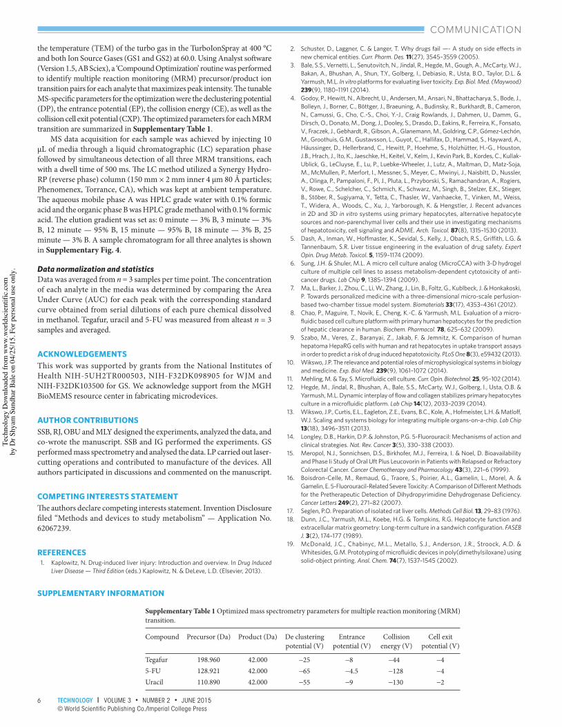
Supplementary Table 1 Optimized mass spectrometry parameters for multiple reaction monitoring (MRM) transition.
Compound Precursor (Da) Product (Da) De clustering potential (V)
Entrance potential (V)
Collision energy (V)
Cell exit potential (V)
Tegafur 198.960 42.000 −25 −8 −44 −45-FU 128.921 42.000 −65 −4.5 −128 −4Uracil 110.890 42.000 −55 −9 −130 −2
SUPPLEMENTARY INFORMATION
1520003.indd 61520003.indd 6 4/21/2015 12:08:35 PM4/21/2015 12:08:35 PM
Tech
nolo
gy D
ownl
oade
d fro
m w
ww
.wor
ldsc
ient
ific.
com
by D
r Shy
am S
undh
ar B
ale
on 0
4/25
/15.
For
per
sona
l use
onl
y.

7TECHNOLOGY l VOLUME 3 • NUMBER 2 • JUNE 2015© World Scientific Publishing Co./Imperial College Press
COMMUNICATION
Supplementary Figure 1 Toxicity plots showing IC-50 of hepatocytes exposed to (a) tegafur, (b) 5-FU, and MCF-7 cells exposed to (c) tegafur and (d) 5-FU. 5-FU samples contained 100 µM uracil, while 5-FU concentration varied. Cell viability was measured using MTT assay and calculated IC-50 values are reported in Table 2.
Supplementary Figure 2 Phase images showing hepatocyte and MCF-7 cell density in control cultures (a,b) and after 24-hour exposure to 100 µM tegafur + 100 µM uracil (c,d) in two-chamber devices. Scale bar = 50 µm.
1520003.indd 71520003.indd 7 4/21/2015 12:08:36 PM4/21/2015 12:08:36 PM
Tech
nolo
gy D
ownl
oade
d fro
m w
ww
.wor
ldsc
ient
ific.
com
by D
r Shy
am S
undh
ar B
ale
on 0
4/25
/15.
For
per
sona
l use
onl
y.

8 TECHNOLOGY l VOLUME 3 • NUMBER 2 • JUNE 2015© World Scientific Publishing Co./Imperial College Press
COMMUNICATION
Supplementary Figure 3 Mass spectrometry analysis of tegafur and uracil consumption, in single chamber (control) and two-chamber (co-culture) microfabricated devices after 24-hour incubation.
Supplementary Figure 4 Sample chromatogram showing elution peaks of tegafur, 5-FU and uracil.
1520003.indd 81520003.indd 8 4/21/2015 12:08:37 PM4/21/2015 12:08:37 PM
Tech
nolo
gy D
ownl
oade
d fro
m w
ww
.wor
ldsc
ient
ific.
com
by D
r Shy
am S
undh
ar B
ale
on 0
4/25
/15.
For
per
sona
l use
onl
y.