A new link between translation termination and NMD complexes
171
HAL Id: tel-01784960 https://tel.archives-ouvertes.fr/tel-01784960 Submitted on 4 May 2018 HAL is a multi-disciplinary open access archive for the deposit and dissemination of sci- entific research documents, whether they are pub- lished or not. The documents may come from teaching and research institutions in France or abroad, or from public or private research centers. L’archive ouverte pluridisciplinaire HAL, est destinée au dépôt et à la diffusion de documents scientifiques de niveau recherche, publiés ou non, émanant des établissements d’enseignement et de recherche français ou étrangers, des laboratoires publics ou privés. A new link between translation termination and NMD complexes Etienne Raimondeau To cite this version: Etienne Raimondeau. A new link between translation termination and NMD complexes. Biochemistry, Molecular Biology. Université Grenoble Alpes, 2016. English. NNT: 2016GREAV048. tel-01784960
-
Upload
khangminh22 -
Category
Documents
-
view
3 -
download
0
Transcript of A new link between translation termination and NMD complexes

HAL Id: tel-01784960https://tel.archives-ouvertes.fr/tel-01784960
Submitted on 4 May 2018
HAL is a multi-disciplinary open accessarchive for the deposit and dissemination of sci-entific research documents, whether they are pub-lished or not. The documents may come fromteaching and research institutions in France orabroad, or from public or private research centers.
L’archive ouverte pluridisciplinaire HAL, estdestinée au dépôt et à la diffusion de documentsscientifiques de niveau recherche, publiés ou non,émanant des établissements d’enseignement et derecherche français ou étrangers, des laboratoirespublics ou privés.
A new link between translation termination and NMDcomplexes
Etienne Raimondeau
To cite this version:Etienne Raimondeau. A new link between translation termination and NMD complexes. Biochemistry,Molecular Biology. Université Grenoble Alpes, 2016. English. �NNT : 2016GREAV048�. �tel-01784960�

THESIS / THÈSE
To obtain the title of / Pour obtenir le grade de
DOCTEUR DE LA COMMUNAUTE UNIVERSITÉ GRENOBLE ALPES
Discipline / Spécialité : Biologie Structurale et Nanobiologie
Arrêté ministériel : 7 août 2006
Presented by / Présentée par
Etienne Raimondeau
Thesis supervisor / Thèse dirigée par Prof. Christiane Schaffitzel
Thesis prepared at / Thèse préparée au sein du European Molecular Biology Laboratory (EMBL), Grenoble Outstation in / dans l'École Doctorale de Chimie et Sciences du Vivant
A new link between translation termination and NMD complexes Un nouveau lien entre les complexes de terminaison de la traduction et de la NMD
Public defense on / Thèse soutenue publiquement le 03.11.2016 Jury members / Devant le jury composé de :
Prof. Winfried Weissenhorn President/Président Professor, University of Grenoble Alpes, France.
Dr. Niels Gehring Reviewer / Rapporteur Group Leader, University of Cologne, Germany.
Prof. Sven Danckwardt Reviewer / Rapporteur University Professor, University of Mainz, Germany.
Prof. Christiane Schaffitzel Thesis Supervisor / Directrice de Thèse Team Leader, EMBL-Grenoble, France.


À mes parents


Contents
Contents v
List of Figures vii
List of Tables ix
1 Preface 1
2 Introduction 32.1 The mRNA life cycle . . . . . . . . . . . . . . . . . . . . . . . . . . . . 42.2 mRNA quality control . . . . . . . . . . . . . . . . . . . . . . . . . . . 92.3 Nonsense mediated mRNA decay . . . . . . . . . . . . . . . . . . . . . 122.4 Scope of this work . . . . . . . . . . . . . . . . . . . . . . . . . . . . . 30
3 Structural studies of yeast termination complexes 313.1 Introduction . . . . . . . . . . . . . . . . . . . . . . . . . . . . . . . . . 323.2 Results . . . . . . . . . . . . . . . . . . . . . . . . . . . . . . . . . . . . 333.3 Discussion . . . . . . . . . . . . . . . . . . . . . . . . . . . . . . . . . . 483.4 Methods . . . . . . . . . . . . . . . . . . . . . . . . . . . . . . . . . . . 523.5 Supplemental information . . . . . . . . . . . . . . . . . . . . . . . . . 62
4 Double-tasking of UPF3B in early and late phases of translation ter-mination 654.1 Introduction . . . . . . . . . . . . . . . . . . . . . . . . . . . . . . . . . 674.2 Results . . . . . . . . . . . . . . . . . . . . . . . . . . . . . . . . . . . . 704.3 Discussion . . . . . . . . . . . . . . . . . . . . . . . . . . . . . . . . . . 864.4 Methods . . . . . . . . . . . . . . . . . . . . . . . . . . . . . . . . . . . 904.5 Supplemental information . . . . . . . . . . . . . . . . . . . . . . . . . 964.6 Additional results . . . . . . . . . . . . . . . . . . . . . . . . . . . . . . 101
5 Discussion 1095.1 Binding of NMD and termination factors at a PTC . . . . . . . . . . . 110

vi Contents
5.2 NMD factors control the recycling and initiation of ribosomes . . . . . 1135.3 mRNA and nascent chain degradation . . . . . . . . . . . . . . . . . . 1165.4 Regulation of NMD and outlook . . . . . . . . . . . . . . . . . . . . . . 1185.5 Concluding remarks . . . . . . . . . . . . . . . . . . . . . . . . . . . . 119
References 121
Acknowledgements 139
A Primer and plasmid list 141
B Second Appendix 151B.1 Secondary structure prediction of human UPF3B . . . . . . . . . . . . 151B.2 Multispecies alignment of UPF3 . . . . . . . . . . . . . . . . . . . . . . 154B.3 Alignment and prediction of UPF3A and UPF3B . . . . . . . . . . . . 157
List of Figures
2.1 The life of mRNA . . . . . . . . . . . . . . . . . . . . . . . . . . . . . 52.2 Overview of translation termination and recycling . . . . . . . . . . . . 82.3 mRNA quality control pathways in the cell . . . . . . . . . . . . . . . . 112.4 mRNAs triggering the NMD pathway . . . . . . . . . . . . . . . . . . . 132.5 Schematic representation of human core NMD factors . . . . . . . . . . 162.6 Schematic representation of human termination factors . . . . . . . . . 172.7 Current general model for NMD activation . . . . . . . . . . . . . . . . 222.8 Model for mRNA decay by NMD in mammals . . . . . . . . . . . . . . 252.9 NMD can modulate the phenotypic outcome of nonsense mutations . . 27
3.1 In vitro ribosome-nascent chain complex preparation scheme . . . . . . 343.2 Optimization of the in vitro translation reaction . . . . . . . . . . . . . 353.3 Optimization of the RNC purification . . . . . . . . . . . . . . . . . . . 383.4 eRF1AGQ, eRF3, Upf1p and Pab1p purification . . . . . . . . . . . . . 403.5 Upf1p-eRF3 and Pab1p-eRF3 purification and complex formation with
eRF1AGQ . . . . . . . . . . . . . . . . . . . . . . . . . . . . . . . . . . 413.6 Reconstitution of yeast termination complexes . . . . . . . . . . . . . . 433.7 Single particle analysis of RNC:eRF1AGQ:eRF3-Pab1p . . . . . . . . . 443.8 Further optimization of RNC purification . . . . . . . . . . . . . . . . . 463.9 Binding assays between yeast Upf1p, Pab1p and the release factors . . 473.10 Vector modification using self-SLIC . . . . . . . . . . . . . . . . . . . . 533.11 mRNA or eRF1 depletion of the yeast extract for in vitro translation . 623.12 yNC mRNA construct optimization . . . . . . . . . . . . . . . . . . . . 633.13 Upf2p and Up3p Purification. . . . . . . . . . . . . . . . . . . . . . . . 64
4.1 UPF3B delays translation termination in vitro . . . . . . . . . . . . . . 734.2 In vivo interaction between release factors and UPF proteins . . . . . 764.3 UPF3B forms a complex with eRF3a and eRF1. . . . . . . . . . . . . . 784.4 UPF3B can directly interact with UPF1. . . . . . . . . . . . . . . . . . 794.5 UPF3B interacts with the eRF3a N-terminus . . . . . . . . . . . . . . . 824.6 UPF3B dissociates postTCs. . . . . . . . . . . . . . . . . . . . . . . . . 85

viii List of Figures
4.7 Model for early and late UPF3B function in translation termination . . 894.8 Supplemental Figure related to Figure 4.1 . . . . . . . . . . . . . . . . 964.9 Supplemental Figure related to Figure 4.1B . . . . . . . . . . . . . . . 974.10 Supplemental Figure related to Figure 4.3 . . . . . . . . . . . . . . . . 984.11 Supplemental Figure related to Figure 4.5 . . . . . . . . . . . . . . . . 994.12 Supplemental Figure related to Figure 4.6 . . . . . . . . . . . . . . . . 1004.13 UPF2 interacts with eRF3a . . . . . . . . . . . . . . . . . . . . . . . . 1024.14 UPF3B interacts with UPF1 CH domain . . . . . . . . . . . . . . . . . 1034.15 UPF3B phosphorylation does not impair eRF3a binding . . . . . . . . 1044.16 UPF3B bridge UPF2 to the 80S ribosome . . . . . . . . . . . . . . . . 107
5.1 eRF3a and the ribosome integrate signals for mRNA quality control . . 1135.2 mRNA surveillance mechanisms and recycling and initiation control. . 1155.3 mRNA surveillance mechanisms and protein and mRNA decay control . 1175.4 mRNA translation and decay regulation by helicases . . . . . . . . . . 119

List of Tables
A.1 Oligonucleotides used in Chapter 3 . . . . . . . . . . . . . . . . . . . . 142A.2 Oligonucleotides used in Chapter 4 . . . . . . . . . . . . . . . . . . . . 145A.3 Plasmids used in this study . . . . . . . . . . . . . . . . . . . . . . . . 148


Nomenclature
3’ UTR 3’ untranslated region
40S Eukaryotic small ribosomal subunit
60S Eukaryotic large ribosomal subunit
80S Eukaryotic ribosome
ATP Adenosine-5’-triphosphate
ATPase ATP hydrolase
CMV Cytomegalovirus
Cryo-EM Cryo-electron microscopy
DNA Deoxyribonucleic acid
EBM Exon junction complex-binding motif
EJC Exon junction complex
GTP Guanosine-5’-triphosphate
GTPase GTP hydrolase
LB Lysogeny broth
MIF4G Middle domain of eukaryotic initiation factor 4G (eIF4G)
MVHC-STOP Model mRNA used in Chapter 4
NGD No-go decay
NMD Nonsense mediated mRNA decay
NSD No-stop decay
NTC Normal termination codon

xii Nomenclature
ORF Open reading frame
PABPC Human cytoplasmic poly(A)-binding protein
PABPN Human nuclear poly(A)-binding protein
PTC Premature termination codon
Pab1p Yeast poly(A)-binding protein
PostTC Post-terminating ribosome
PreTC Pre-terminating ribosome
RNA Ribonucleic acid
RNC Ribosome-nascent chain complex
Toeprinting Primer extension inhibition
UPF Upstream frameshifting genes products
VCE Vaccinia capping enzyme
eEF Eukaryotic elongation factor
eIF Eukaryotic initation factor
eRF Release factor
mRNA Messenger RNA
mRNP Messenger ribonucleoprotein
rpm Rotations per minute
sup35 Yeast eukaryotic release factor 3 (eRF3)
sup45 Yeast eukaryotic release factor 1 (eRF1)
tRNA Transfer RNA
yNC Yeast nascent chain

1. Preface
My doctorial work was focusing on termination of translation and nonsense mediatedmRNA decay in yeast and human. The main work of my thesis has been performedwith proteins of human origin and has led to the submission of a manuscript. Thisthesis is thus written in a cumulative style. The second chapter contains a generalintroduction on termination of translation and nonsense mediated mRNA decay. Thethird and fourth chapter detail the work undertaken, focusing on yeast and humanrespectively. Both chapters are formatted as independent manuscripts and contain theclassical “Results” and “Material and methods” sections. The fifth and final chapterdiscuss the main conclusions of both manuscripts.

2 Preface

2. Introduction
Resumé en français
Depuis la formulation de la théorie fondamentale de la biologie moléculaire par FrancisCrick, en 1958, notre compréhension de l’expression génétique a beaucoup progressé.L’expression des gènes est un processus hautement complexe, impliquant de nombreuxcomplexes cellulaires travaillant de concert. Les cellules ont développé di�érents mécan-ismes de contrôle qualité pour chaque étape de l’expression génétique afin de maintenirl’intégrité de l’information. La dégradation des ARNm non-sens (Nonsense-mediateddecay ou NMD) a été mise en évidence il y a plus de 30 ans. Au cours de la termi-naison de la traduction, la NMD discrimine les codons stop prématurés (PTC) descodons stop physiologiques et déclenche la dégradation des ARNm aberrants. Cettevoie représente un des principaux mécanismes de contrôle qualité de la cellule et est legardien de l’expression génétique. La NMD est d’une importance éminente du pointde vue médical bien qu’elle ait une activité à double tranchant. Des mécanismes dereconnaissance ainsi que des ARN substrats pour la NMD ont été mis à jour.
Les facteurs principaux de la NMD : UPF1, UPF2 et UPF3 sont conservés de lalevure à l’homme (Upf1p, Upf2p et Upf3p chez la levure), ces protéines permettentla reconnaissance des PTCs. Ces dernières interagissent avec la machinerie de la tra-duction comportant les ribosomes, les facteurs de terminaison eRF1 et eRF3 et unevariété de facteurs additionnels parmi lesquels le Exon Junction Complexe (EJC) etla protéine poly(A)-binding (PABPC1 ou Pab1p chez la levure) qui sont positionnéssur l’extrémité 3’ non-traduite de l’ARNm (3’UTR). La reconnaissance d’un PTC dé-clenche la phosphorylation de UPF1 chez les mammifères et aboutit à la dégradationde l’ARNm défectueux.
Le but de notre étude est d’apporter de nouvelles connaissances sur les interactionsentre facteurs de terminaison et facteurs NMD. Plus particulièrement, ce travail seconcentre sur la terminaison de la traduction chez la levure en présence de Pab1p etde Upf1p ainsi que sur l’impact des facteurs NMD sur la terminaison de la traductionchez l’homme.

4 Introduction
2.1 The mRNA life cycle
2.1.1 Transcription and mRNA processing in the nucleus
Transcription is the first step in gene expression and enables the conversion of a definedsegment of DNA into RNA. RNA biogenesis takes place in a highly structured anddynamic nuclear environment in eukaryotes (Venkatesh & Workman, 2015). RNApolymerase II (Pol II) is responsible for the synthesis of mRNAs and a few non-codingRNAs (Sainsbury et al., 2015; Cramer et al., 2001). To promote e�cient and accurateprocessing of pre-mRNAs into mature mRNAs, multiple processing factors, such asthe capping enzymes, and splicing factors amongst others, interact with the carboxy-terminal domain (CTD) of the large subunit of the RNA Pol II in a phosphorylation-dependent manner (Moore & Proudfoot, 2009; Bentley, 2014; Venkatesh & Workman,2015). During each step of the processing, factors remain associated with mRNAs andlead to the assembly of the messenger ribonucleoprotein (mRNP).
The first step in the maturation process requires the addition of a 7-methylguanosinecap (m7GpppN) to the pre-mRNA 5� end. In mammals, capping is performed bytwo enzymes: the mRNA capping enzyme (a triphosphatase with guanylyltransferaseactivity), and a guanine-7-methyltransferase (Bentley, 2014). The 5’ end cap is thenrecognized by the cap-binding complex (CBC), formed by CBP20 and CBP80 (Mazza etal., 2001), and remains bound to the mRNA until it is exported from the nucleus (Figure2.1). Correct capping is crucial for the protection of mRNAs from degradation by 5’-3’exonucleases, which are present in the nucleus as well as the cytoplasm (Schoenberg &Maquat, 2012) and for translation initiation.
During the early stages of transcription, the genetic information on pre-mRNAsis discontinuous and subdivided into exons and introns. While exons harbor protein-encoding sequences, introns play a regulatory role (for example in alternative splicing(Chen & Manley, 2009) and in nonsense-mediated decay (NMD) (Alexandrov et al.,2012; Ottens & Gehring, 2016)). Recognition and excision of introns involves a vast va-riety of factors assembled into the spliceosome. The spliceosome complexes are formedstepwise, by the addition or removal of small nuclear ribonucleoproteins (snRNPs fromU1 to U6) and a broad collection of additional proteins. Most notably, the spliceosomeallows the deposition of Serine/Arginine-rich (SR) proteins and of the exon junctioncomplex (EJC) which are critical for post-transcriptional processes (Figure 2.1)(Hocineet al., 2010; Huang & Steitz, 2005; Hir et al., 2015). Splicing is a major step in theregulation of gene expression and splicing-defective transcripts are subjected to degra-dation (Doma & Parker, 2007)(see section 2.2.1). As such, mis-regulation of splicingis associated with multiple diseases in humans such as leukemia (Cazzola et al., 2013).
The last step of the transcription is characterized by 3’ end processing of mR-

2.1 The mRNA life cycle 5
AAAAAAAA
m7G
Stop
Start
EJC
m7G
P
P
P
P
P
AP
Start
Pol II
Spliceosome
CBC
SR protein
processing proteins
Pol II CTD
Histones
PABPN
PAP
Poly(A) complex
NXF1/TAP
TREX/THO
UPF3B
EJC
intron
exon
eIF4F
PABPC XRN1
Exosome
Ribosomal subunits
Initiation factors
Cytoplasm
Nucleus
7. Degradation
6. Translation
5. Export
3. Splicing
4. Polyadenylation
2. Capping
1. Transcription
E P AE P A
AAAAAAA
m7G
Stop
P
eIF4F
eIF4G
eIF4A
eIF4E
Figure 2.1: The life of mRNA. 1. Pre-mRNAs are synthetized by Pol II in the nucleusfrom the DNA sequence bound to histones. Phosphorylation of residues (yellow P) in the PolII C-terminal domain allows the binding of various processing factors such as transcription,capping, splicing or polyadenylation factors. 2. The 5’end cap (m7G) is recognized by the CapBinding Complex (CBC) during the early stage of pre-mRNA processing. 3. The assemblyof the spliceosome (grey) occurs co-transcriptionally. mRNA exons are represented as a thickblack line and the introns as a thin black line. The excision of introns by the spliceosomeleads to the deposition of the EJC, UPF3B and phosphorylated Serine/Arginine-rich (SR)proteins on the spliced mRNA. 4. 3’ end cleavage is performed by the poly(A) complexand the poly-adenosine sequence is added by poly(A) polymerase (PAP) and recognized bythe poly(A) binding protein in the nucleus (PABPN). 5. Mature mRNPs are bound by theTREX/THO complex and by hypophosphorylated SR proteins, which allow the binding to thenuclear pore proteins NXF1/TAP. 6. In the cytoplasm, CBC is exchanged against the eIF4Fcap-binding complex (formed by eIF4A, eIF4E and eIF4G) and PABPN is replaced by poly(A)binding protein in the cytoplasm (PABPC). Translation initiation factors and the ribosomalsubunits assemble to translate the mRNA. Phosphorylated SR proteins can be reimported intothe nucleus after dissociation from the mRNA. 7. At a later stage, mRNAs are degraded byXRN1 and the exosome Adapted from (Moore & Proudfoot, 2009; Hir et al., 2015)

6 Introduction
NAs. The cleavage and polyadenylation of mammalian pre-mRNAs is carried out by amultisubunit complex (called poly(A) complex in Figure 2.1) (Porrua & Libri, 2015).Subsequently, the poly(A) tail is coated by nuclear poly(A)-binding proteins (PABPN).These are essential for mRNA protection from 3’ end degradation, and for translationin addition to nuclear export (Gross et al., 2007; Xie et al., 2014). It has been reportedthat ~50% of human genes can be alternatively polyadenylated, generating isoformswith a di�erent 3’ UTR (Tian et al., 2005). Di�erently polyadenylated transcriptsshow di�erent levels of mRNA stability or cellular localization which could also haveimplications for health and diseases (Subtelny et al., 2014; Danckwardt et al., 2008).
In metazoans, mature mRNPs are recognized by NXF1/TAP-p15 receptors for ex-port through the nuclear pore complex (NPC) (Köhler & Hurt, 2007). The transcrip-tion export complex (TREX) and SR proteins are the main adaptors linking mRNAsto the export receptors. The phosphorylation levels of the SR proteins have beensuggested to signal mRNAs ready for export (Reed & Cheng, 2005; Katahira, 2012).Export competent mRNP particles can then dock to the NPC for translocation throughthe pore.
2.1.2 Translation
In the cytoplasm, CBC is replaced by the eukaryotic initiation factor 4F complex(eIF4F) and PABPN is exchanged against a cytoplasmic PABP (PABPC1). The inter-action between eIF4F subunit, eIF4G, and PABPC1 facilitates circularization of themRNA and thereby initiates translation and e�cient protein synthesis (Wells et al.,1998).
The general mechanism of translation has been recognized for 40 years, but recentadvances in biochemistry and cryo-electron microscopy have enabled further research togain a more detailed, molecular understanding of the process. More detailed initiationand elongation mechanisms have been reviewed in detail, for example in (Hinnebusch,2014; Jackson et al., 2010; Dever & Green, 2012). The factors involved in translationtermination are described more precisely in section 2.3.3.
2.1.2.1 Initiation
Translation initiation is the process of the assembly of the translation components toform a translation-ready 80S ribosome on the start codon of the mRNA. Firstly, the40S small subunit of the ribosome associates with Met-tRNAMet and initiation factorseIF1A, 1, 3 and 5 to form a 43S preinitiation complex. The attachment of the 43Scomplex to the mRNA is mediated by the capping complex eIF4F and initiation factoreIF3 (Jackson et al., 2010). At this stage, the 43S complex is found in an ‘open’conformation, which allows translocation along the mRNA in the 5’ to 3’ direction for

2.1 The mRNA life cycle 7
the methionine initiation codon (des Georges et al., 2015). Start codon recognitionleads to conformational changes, which switch the complex to a ’closed’ conformation,so called the 48S initiation complex, locking it onto the mRNA (Hussain et al., 2014).GTP hydrolysis by initiation factor eIF2 results in the displacement of initiation factorseIF1, eIF3 and eIF5 and recruitment of the 60S large ribosomal subunit. The 80Sribosome initiation complex is then ’committed’ to protein synthesis (Hinnebusch, 2014;Jackson et al., 2010).
2.1.2.2 Elongation
After initiation, elongation factor eEF1A directs aminoacylated tRNAs in a GTP-dependent manner to the A-Site. Codon recognition triggers tRNA stabilization inthe A-site and GTP hydrolysis by eEF1A. This enables the release of eEF1A fromthe ribosome and the tRNA to reach the evolutionary conserved peptidyl transferasecenter in the 60S subunit, a process called accommodation. The 60S catalyzes peptidebond formation leading to deacetylation of the P-site tRNA and the transfer of thenascent peptide chain to the A-site tRNA. Translocation of the ribosome along themRNA is supported by elongation factor eEF2 and requires GTP. After translocation,GTP hydrolysis and release of eEF2-GDP, the ribosome is ready for a new cycle ofelongation. (Dever & Green, 2012).
2.1.2.3 Termination
Termination of translation occurs when a stop codon (UAA, UGA, and UAG) reachesthe A-site of the translating ribosome (called the Pre-Terminating Ribosome, PreTC)(Figure 2.2). The recognition of the stop codon is achieved by the synergy of the twoeukaryotic release factors: eRF1 (sup45 in yeast) and eRF3 (sup35 in yeast) (Figure2.6). The structure of a human ribosomal termination complex was solved by singleparticle cryo-electron microscopy and allowed the generation of an atomic model ofeRF1/eRF3a bound to the ribosome (Des Georges et al., 2014; Brown et al., 2015).eRF1 recognizes the stop codon sequence in the A-site of 40S (the decoding center) andtriggers peptidyl-tRNA hydrolysis (Dever & Green, 2012; Jackson et al., 2012). eRF3is a ribosome-dependent GTPase which stimulates eRF1-mediated peptide release andreciprocally, eRF1 stimulates the GTP binding to eRF3 (Pisareva et al., 2006). Uponbinding to eRF3a-GTP, eRF1 adopts a shape mimicking the tRNA structure. Thisallows the N-domain of eRF1 to reach deep into the decoding center of the small ribo-somal subunit. Stop codon recognition in the decoding center leads to a rearrangementof both ribosomal subunits and yields the formation of a post-termination complex(PostTC). This was initially shown by a forward toeprint shift (of approximately 2nucleotides) in a reconstituted in vitro system (Alkalaeva et al., 2006) (Figure 2.2).

8 Introduction
E P A
E P AStop E P AStop
AAAAAA AAAAAAE P AStop
AAAAAA
E P AStop
E P AE P AE P AE P A
Stop
Stop
E P AE P AE P AE P AE P AE P A
nascent chain
ABCE1 + 2ATP
eRF1+eRF3a+GTP
eIf3jeiF1
60SeiF3
eRF1
eiF1A
tRNA
tRNA
PABPC1
Nascent chain
80s at NTCPreTC
Binding of eRF1:eRF3a
GTP hydrolysiseRF3a release
Ribosome shift (2 nt)PostTC
ABCE1 bindingATP hydrolysis
Ribosome dissociation
P-site tRNA dissociation
ribosome dossociation
mRNA dossociation
eRF3+GDP + Pi
2ADP+ 2 PI
A
B
eRF160S
? ?
Figure 2.2: Overview of translation termination and recycling. Translation iscarried out by the 80S ribosome consisting of the small 40S subunit and the large 60S subunit.The stop codon in the A-site of pre-termination complex (PreTC) is recognized by eRF1 boundto eRF3a-GTP. Stop codon recognition and eRF1 accommodation in the ribosome result ina 2 nucleotide shit (forming post-termination complex - PostTC). Following GTP hydrolysisby eRF3a, eRF1 triggers the release of the nascent chain. These steps may be stimulatedby the poly(A)-binding protein (PABPC1). Subunit dissociation and recycling are promotedby ABCE1 ATPase activity (A pathway). Alternatively eIF1A and eIF3 association withthe postTC lead to ribosomal dissociation (B pathway). Subsequently, eIF1 and eIF3j allowthe removal of the P-site tRNA and the mRNA from the 40S subunit. After recycling, thecomponents are ready for a new round of translation. Adapted from (Alkalaeva et al., 2006;Pisarev et al., 2007, 2010; Schweingruber et al., 2013)

2.2 mRNA quality control 9
Upon GTP hydrolysis, the eRF1 GGQ motif can be accommodated in the peptidyltransferase center of the large ribosomal subunit (60S) and triggers peptidyl-tRNAhydrolysis (Brown et al., 2015).
2.1.2.4 Recycling
Termination complexes need to be disassembled for the components to be reused for anew round of translation. In the current model of recycling of postTCs, eRF3a mostlikely dissociates from the ribosome following GTP hydrolysis while eRF1 remainsbound. A highly conserved ATPase ABCE1 has been shown to mediate recycling ofpostTCs by interacting with ribosome-bound eRF1 (Figure 2.2A) (Pisarev et al., 2010;Franckenberg et al., 2012). Alternatively, using fully reconstituted in vitro translation,Pisarev and colleagues showed that eIF3, eIF1, eIF1A and eIF3j could mediate therecycling of postTCs under low Mg2+ conditions (Figure 2.2B). eIF3 promotes thesplitting of ribosomal subunits. Subsequently, eIF1 and eIF3j can the release thetRNA from the P-site and mRNA from the 40S subunit, respectively (Pisarev et al.,2007).
2.1.3 mRNA degradation
mRNA decay a�ects not only aberrant mRNAs, but also normal mRNA transcriptswhich lead to fully functional proteins. Overall protein synthesis depends not only onthe e�ciency of transcription and translation but also on the rate of mRNA decay.
In eukaryotes, the main bulk of mRNAs in the cell are degraded via shortening ofthe poly(A) tail by a deadenylase protein complex such as CCR4-NOT or Poly(A)-specific nuclease (PARN)(Schoenberg & Maquat, 2012). Subsequently, either the 5’end cap is removed in a process called decapping, enabling decay from the 5’ to the3’ direction by the exonuclease XRN1; or the unprotected 3’ end can be degraded inthe 3’ to the 5’ direction by a protein complex called the exosome. Alternatively topoly (A) tail shortening, mRNAs can be subjected to endonuclease cleavage followedby decay by XRN1 or the exosome. (Garneau et al., 2007). Components involved in5’ to 3’ mRNA decay are localized in cytoplasmic loci called P-bodies. P bodies havebeen suggested to be important loci dedicated to mRNA storage and decay (Garneauet al., 2007). Interestingly, the main NMD factors UPF1, UPF2 and UPF3 have beenfound in these cellular sites (Eulalio et al., 2007) (section 2.3.2).
2.2 mRNA quality control
Every step of mRNA biogenesis is associated with mRNA quality control, which de-tects errors in the transcript. Mutations at the DNA or RNA level, or errors during

10 Introduction
transcription or mRNA processing can cause aberrant mRNAs. Grossly mutated mR-NAs are retained and degraded in the nucleus, whereas mRNAs containing more subtleerrors are likely to be exported in the cytoplasm and engaged in translation. Such mR-NAs encode potentially harmful proteins and thus need to be eliminated. RNA qualitycontrol pathways have been proposed to act along a unified principle (Doma & Parker,2007). They can be viewed as a series of checkpoints where control and degradationproteins compete in a kinetic manner against the normal translation factors (Figure2.3). Decades of research have highlighted the importance of mRNA surveillance as amechanism not only to degrade aberrant mRNAs but also as an important means tofine-tune the expression of genes (Schoenberg & Maquat, 2012).
2.2.1 Nuclear RNA quality control
Grossly aberrant or improperly processed mRNAs are retained in the nucleus for degra-dation (Doma & Parker, 2007). Mechanisms have evolved to prevent the export ofmisprocessed transcripts: studies in yeast and mammals have revealed the existenceof capping quality control mechanisms which allow for the specific removal of an un-methylated cap and which subsequently targets the pre-mRNA for degradation (Jiaoet al., 2013; Doma & Parker, 2007).
The spliceosome cycle requires successive discrete conformation steps facilitated byATPase/helicases (Villa et al., 2008; Bourgeois et al., 2016). E�ective splicing is indirect competition with degradation, therefore mRNA features which slow down ATPhydrolysis by helicases, and thus spliceosome cycle, will subject these pre-mRNAs toquality control and degradation (Doma & Parker, 2007). Additional controls involvenuclear retention of unspliced transcripts (Hocine et al., 2010). 3’ end processing ofmRNAs is also rigorously controlled. In yeast, Rrp6p has been shown to be a criticalfactor for 3’ end processing quality control. In deletion strains (rrp6D), hypoadenylatedmRNAs, which would normally be targeted for nuclear degradation, were stabilizedand exported. Similarly, mRNAs containing an aberrant cleavage or transcriptiontermination site were stabilized (Allmang et al., 1999; Burkard & Butler, 2000).
Export into the cytoplasm is the last step of nuclear quality control, in whichincorrectly processed mRNA can be detected and degraded. Misprocessed mRNAscannot assemble into mRNP particles containing appropriate export factors, for ex-ample TREX or SR, and therefore are retained in the nucleus (Reed & Cheng, 2005;Katahira, 2012).
Aberrant mRNA retained in the nucleus can be degraded from 3’ to 5’ by thenuclear exosome, or from 5’ to 3’ by nuclear endonucleases. Dependent upon thedefects present, mRNAs are degraded by either one of the mechanisms with di�eringe�ciency. For example, defects in polyadenylation will be more e�ciently degraded by

2.2 mRNA quality control 11
AAA
Translation defects triggering mRNA decay
Premature translation termination (NMD) Elongation stall (NGD) No termination codon (NGD)
AAA
EJC
m7G Start
Pol II
PABPN
Processing defectsNuclear degradation
Degradation
Export forcytoplasm degradation
Releasefrom gene
Capping, splicing and polyadenylation
mRNPexport
Spliceosome
CBC
Retention
Cytoplasm
Nucleus
m7G
AAAAAAAA
m7G
Stop
eIF4F PABPC
AAAAAAAA
Stop
m7G
80s
PTC
UPF1-2-3BEJC
AAAAAAAA
Stop
m7G
Pelota/Dom34
Hbs1AAAAAAAA
Stop
m7G
80s
Ski7
PelotaHbs1
AAAAAAAA
eRF1-3a
Figure 2.3: mRNA quality control pathways in the cell. Defects can occur during pre-mRNA processing (capping, splicing and polyadenylation) in the nucleus. These can lead toretention of the pre-mRNA in the nucleus and nuclear degradation or export to the cytoplasmfor degradation. During translation in the cytoplasm, errors in the mRNA can be detectedby the mRNA surveillance pathways. A premature termination codon (PTC in blue box) isdetected by the nonsense-mediated mRNA decay (NMD) pathway involving the eRF1, eRF3aand UPF1-UPF2-UPF3B proteins and triggered by the presence of an exon junction complex(EJC) in the 3’UTR. Stalled elongating ribosomes are recognized by the Pelota/Dom34-Hbs1complex in no-go decay (NGD). The absence of a stop codon can be detected by the Ski7complex or the Pelota/Dom34-Hbs1 complex in the non-stop decay (NSD). Adapted from(Doma & Parker, 2007; Schweingruber et al., 2013)
the nuclear exosome (Garneau et al., 2007; Hocine et al., 2010).
2.2.2 Cytoplasmic mRNA quality control
Cytoplasmic mRNA surveillance pathways are classified by the type of mRNA targeted.Quality control depends on translation: nonsense mediated mRNA decay (NMD) tar-gets mRNA with a premature termination codon (PTC); non-stop decay (NSD) specif-ically targets mRNA lacking a stop codon and no-go decay (NGD) targets mRNA withstalling secondary structures or sequences (Shoemaker & Green, 2012)(Figure 2.3).
Stalling of protein synthesis can result from secondary structures such as stem loopsor certain sequences comprising rare codons or damaged RNA bases. In eukaryotes, theA-site of stalled ribosomes is recognized by NGD factors Pelota (Dom34 in yeast) and

12 Introduction
Hbs1 (Shoemaker et al., 2010). Interestingly, Dom34 and Hsb1 share homology withrelease factors eRF1 and eRF3, suggesting that NGD involves a modified terminationevent (Shoemaker & Green, 2012). Following Hbs1 dissociation, ABCE1 associateswith the Dom34 to promote recycling of the stalled ribosome (Pisareva et al., 2011;Shoemaker & Green, 2011; Young et al., 2015). The N-terminal domain of Hbs1 extendsto the head of the 40S subunit and was proposed to act as an mRNA sensor and tostimulate the recycling of the NGD stalled ribosomes (Becker et al., 2011; Franckenberget al., 2012).
NSD recognizes mRNAs which lack a proper termination context: for example,3’ end truncated mRNA which lacks a stop codon and a poly(A) tail or an mRNAmissing only the stop codon. According to the current model in yeast, Ski7, anothereRF3 paralogue, binds to the stalled ribosome and promotes mRNA degradation byinteraction with the exosome (Shoemaker & Green, 2012). Moreover, it is postulatedthat the translation of the poly(A) tail into polylysine by the ribosome would lead totranslation stalling due to the positively charged peptide in the exit tunnel. In sucha context similar to NGD, Dom34 and Hsb1 are recruited to the stalled translationmachinery (Ito-Harashima et al., 2007).
mRNAs containing PTCs are recognized and degraded via the NMD pathway whichis discussed in the next section.
It is worth noting that the recognition of NGD targets and the subsequent recyclingof stalled ribosomes is currently the best understood, compared to the other surveillancepathways.
2.3 Nonsense mediated mRNA decay
Nonsense mRNA decay was initially discovered in Saccharomyces cerevisiae and Caenorhab-ditis elegans over 30 years ago (Losson & Lacroute, 1979; Maquat et al., 1981). Thecore NMD machinery is conserved from yeast to human (Culbertson & Leeds, 2003).NMD specifically recognizes and degrades transcripts containing a premature termi-nation codon (PTC). Mutations in the core NMD factor genes are linked to neurode-velopmental disorders, mental disorders and are associated with specific tumor types(Ottens & Gehring, 2016). Beyond its clinical significance, NMD serves as a posttran-scriptional regulator of gene expression and plays a role in important cellular processessuch as development and di�erentiation (Weischenfeldt et al., 2008; Avery et al., 2011;T. Li et al., 2015; Lou et al., 2014), cellular stress response (Gardner, 2010; Sakakiet al., 2012), neural activity (Giorgi et al., 2007; Colak et al., 2013) and immunity(Gloggnitzer et al., 2014) (reviewed by (S. Lykke-Andersen & Jensen, 2015; Ottens &Gehring, 2016)).

2.3 Nonsense mediated mRNA decay 13
Start
m7G AAAAAAAAE P A
PTCStop
StopStart
m7G AAAAAAAAE P A
PTC
StopStart Start
m7G AAAAAAAAE P A
Start
m7G AAAAAAAAE P A
Stop
Start
m7GE P A
AAAAAAAA
Stop
A. PTC ininternal exon
B. PTC interminal exon
C. uORF
D. NTC and3’UTR intron
E. NTC andlong 3’UTR
50-55 nt
Figure 2.4: mRNAs triggering the NMD pathway. A. PTCs in an internal exon arethe classical example for an NMD substrate in mammals. Such PTC would elicit NMD ifit is located ~50-55 nt upstream from an EJC which remain bound downstream of the PTC(see section 2.3.4). B. PTCs in the terminal exon are typically found in yeast, although itcan also be found in higher eukaryotes. C. Upstream ORFs (uORFs) located 5’ of the mainORF are known to trigger NMD. In this case, EJCs are bound to the downstream mRNA andthere is an unusually long 3’ UTR. D. 3’UTR introns will result in the deposition of an EJCdownstream of the normal termination codon (NTC) and trigger NMD. E. Long 3’ UTRsare known to activate the NMD pathways in multiple organisms. mRNAs are represented byblack lines: translated exons are shown as white boxes and untranslated exons as grey boxes.5’ and 3’ UTRs are represented by black lines. The start codon is illustrated as a green arrow,the stop codon as a red sign and PTC as a blue square. The 5’ cap (m7G) is bound to eIF4Fcomplex (yellow) and the poly(A) tail is complexed with PABP (orange). The translatingribosomes are shown in yellow and the EJCs in green. Adapted from (Schweingruber et al.,2013)

14 Introduction
2.3.1 NMD Substrate
NMD was originally thought to trigger the degradation only of PTC-containing mR-NAs. PTCs can arise at the DNA level by mutations or at the RNA level due totranscription or mRNA processing errors, which occur mainly during splicing. 75%of human pre-mRNAs are alternatively spliced (Johnson, 2003) and 45% of those cangenerate at least one isoform, which will be degraded by NMD (Lewis et al., 2003).Transcriptome-wide experiments have shown that NMD is also triggered by 5-15 % ofnormal transcripts, whereby the termination codon is interpreted by the cell as “prema-ture” (Figure 2.4) (He et al., 2003; Guan et al., 2006; Mendell et al., 2004; Tani et al.,2012). This applies to the termination codons of some upstream open reading frames(uORFs), termination codons upstream of 3’ UTR introns or unusually long 3’ UTRs.This can also include termination codons introduced into a transcript by somatic re-arrangements during early stages of T and B cell maturation, ribosomal frameshifting,or mRNA editing (Rehwinkel et al., 2006; Schweingruber et al., 2013; Fatscher et al.,2015).
Translation is indispensable for the recognition of a PTC, and NMD is triggeredby an aberrant termination event (Shoemaker & Green, 2012; Kervestin & Jacobson,2012). Decades of research have unveiled common features possessed by NMD sub-strates (Figure 2.4). During the translation of NMD targets, termination often occursin an unusual position, either too far from the poly(A) tail and downstream RNPs,or with an EJC located in the 3’UTR downstream of the termination codon. NMDactivation is discussed in section 2.3.6.
2.3.2 Core UPF proteins
Key factors for NMD were identified in early genetic screens in Saccharomyces cerevisiaeas the Upstream frameshifting genes products: Upf1p, Upf2p and Upf3p (known inhuman as UPF1, UPF2 and UPF3 respectively)(Leeds et al., 1992). These NMDcore proteins are conserved from yeast to human (Figure 2.5). UPF1 and UPF2 arecytoplasmic proteins although UPF2 appears to have a perinuclear localization. UPF3shuttles between the nucleus and the cytoplasm (J. Lykke-Andersen et al., 2000). UPFproteins interact with each other and are associated with mRNAs, the translationmachinery and numerous other factors (He et al., 1997; Atkin et al., 1997; Kashima etal., 2006; Melero et al., 2012).
2.3.2.1 UPF1
UPF1 is a superfamily I 5’ to 3’ RNA helicase and RNA-dependent ATPase (Bhattacharyaet al., 2000). UPF1 is the most conserved NMD factor (48.5 % identity between yeast

2.3 Nonsense mediated mRNA decay 15
and human) and appears to be the central e�ector in the pathway (Culbertson &Leeds, 2003). UPF1 contains a C-terminal domain, a helicase domain formed by anATPase motif surrounded by two RecA-like domains and an N-terminal cysteine- andhistidine- (CH) rich region (Figure 2.5). In the absence of UPF2, the CH domaininteracts with the helicase domain thereby preventing untimely helicase and ATPaseactivities of UPF1 (Clerici et al., 2009; Chakrabarti et al., 2011; Chamieh et al., 2008).Furthermore, in yeast, the CH domain has been reported to bind to the ribosomalprotein Rps26 (Min et al., 2013), to recruit the decapping enzyme Dcp2 (He & Ja-cobson, 2015a). Phosphorylation and dephosphorylation cycles of UPF1 at N- andC-terminal S/T-Q sites are essential for NMD and seem to be a prerequisite for the re-cruitment of the decay inducing enzymes SMG6 and SMG5/7 (see section 2.8)(Durandet al., 2016). The discrimination between early termination-related and late mRNPremodeling/degradation-related functions of UPF1 is supported by the timing of thephosphorylation by SMG1 kinase and the accessory factors SMG8 and SMG9 (see2.3.6).
2.3.2.2 UPF2
UPF2 is described as a ring-like sca�old protein and acts by bridging UPF1 at theterminating ribosome and UPF3B, which is bound to a downstream EJC (Melero etal., 2012). 20.8 % of the UPF2 protein sequence is conserved between human andyeast protein (Culbertson & Leeds, 2003). It is composed of three ‘conserved middledomain of eIF4G domains’ (MIF4G) (Figure 2.5). MIF4G is a common domain foundin mRNA-associated proteins such as CBC80 and eIF4G. While the function of the firsttwo MIF4G domains in UPF2 remains enigmatic, the third MIF4G domain is knownto interact with a region in the RNA-Recognition Motif-like (RRM-like) domain ofUPF3B (Kadlec et al., 2004; Clerici et al., 2014). UPF2 C-terminal domain interactswith UPF1 CH domain and activates the helicase and ATPase function of UPF1 (Clericiet al., 2009; Chakrabarti et al., 2011).
Initially, UPF2 was thought to be a platform for the recruitment of SMG1-8-9 and topromote UPF1 phosphorylation (Melero et al., 2014). However, recent findings indicatethat the presence of UPF2, rather than activating the kinase, destabilizes SMG-1–8–9-UPF1 leading to release of UPF1 from the complex (Deniaud et al., 2015). Recently,human UPF2 has been shown to directly interact with eRF3a (but not with eRF1)through a protein region known as the UPF3B interaction domain, which overlapswith MIF4G domain 3 (López-Perrote et al., 2015). UPF2 binds strongly to UPF3Band, consequently the addition of UPF3B could displace eRF3a from UPF2.

16 Introduction
UPF3B 1
49 279 421 434
470RRM-like
EBM
UPF3A 1
66 292 434 447
476RRM-like
EBM
UPF2 1
121 429 457 757 768 1015 1105 1207
1272MIF4G MIF4G MIF4G U1B
UPF1 1
115 272 914 967 1019700
1118CH 1B
Helicase
1C RecA2RecA1 SQ
CH Domain
Helicase Domain
PDB 2WJV
PDB 4CEM PDB 4CEK PDB 1UW4
PDB 2WJV
PDB 1UW4
PDB 2XB2
Figure 2.5: Schematic representation of human core NMD factors UPF1, UPF2,UPFB and UPF3A. Known crystal structures are included with their respective PDB IDand mapping regions. SQ: Serine Glutamine rich domain; CH: Cysteine Histidine rich do-main, Helicase domain : RecA1 and RecA2 translocate the mRNA, 1B and 1C are involved inRNA binding and ATP hydrolysis ; MIF4G: middle domain of eIF4G; U1B: UPF1-binding do-main; RRM-like: RNA Recognition Motif like domain; EBM: Exon junction complex-bindingmotif. Numbers indicated the amino acid boundaries of the di�erent domains. Adapted from(Clerici, 2009)
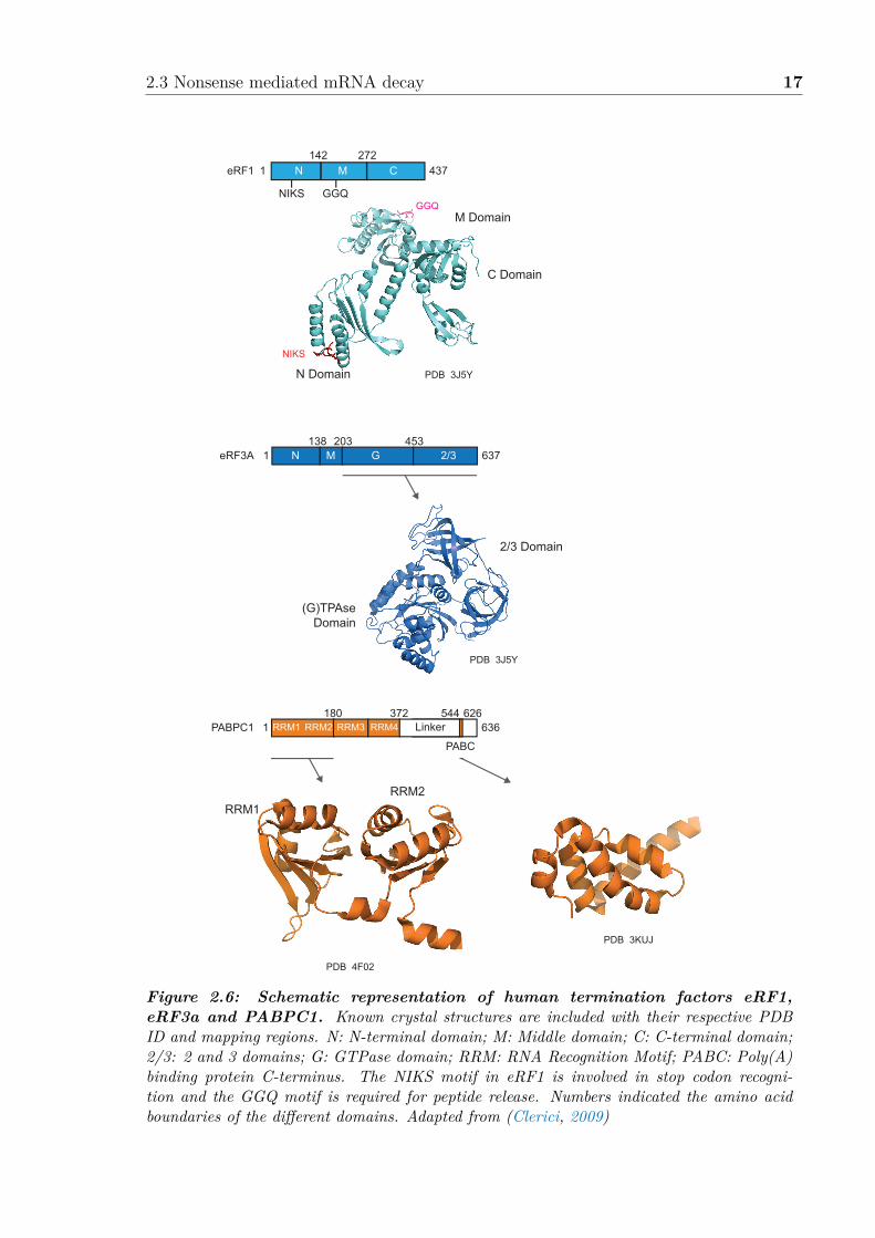
2.3 Nonsense mediated mRNA decay 17
eRF3A G 2/31
138 203
637N M
453
eRF1 C1
142 272
N M 437
PABPC1 1
180
636RRM1 RRM3 RRM4
PABC
372 544RRM2
626
GGQNIKS
2/3 Domain
N Domain
M Domain
C Domain
(G)TPAseDomain
RRM1
RRM2
PDB 3J5Y
NIKS
GGQ
PDB 3J5Y
PDB 4F02
PDB 3KUJ
Linker
Figure 2.6: Schematic representation of human termination factors eRF1,eRF3a and PABPC1. Known crystal structures are included with their respective PDBID and mapping regions. N: N-terminal domain; M: Middle domain; C: C-terminal domain;2/3: 2 and 3 domains; G: GTPase domain; RRM: RNA Recognition Motif; PABC: Poly(A)binding protein C-terminus. The NIKS motif in eRF1 is involved in stop codon recogni-tion and the GGQ motif is required for peptide release. Numbers indicated the amino acidboundaries of the di�erent domains. Adapted from (Clerici, 2009)
18 Introduction
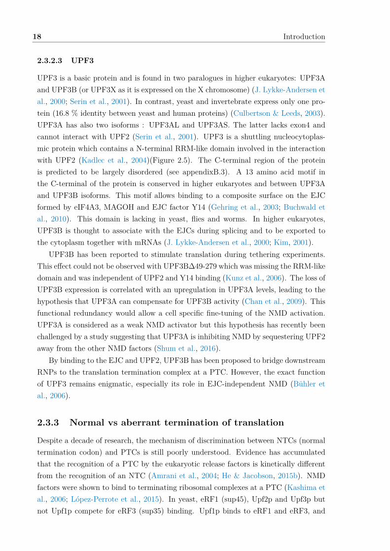
2.3.2.3 UPF3
UPF3 is a basic protein and is found in two paralogues in higher eukaryotes: UPF3Aand UPF3B (or UPF3X as it is expressed on the X chromosome) (J. Lykke-Andersen etal., 2000; Serin et al., 2001). In contrast, yeast and invertebrate express only one pro-tein (16.8 % identity between yeast and human proteins) (Culbertson & Leeds, 2003).UPF3A has also two isoforms : UPF3AL and UPF3AS. The latter lacks exon4 andcannot interact with UPF2 (Serin et al., 2001). UPF3 is a shuttling nucleocytoplas-mic protein which contains a N-terminal RRM-like domain involved in the interactionwith UPF2 (Kadlec et al., 2004)(Figure 2.5). The C-terminal region of the proteinis predicted to be largely disordered (see appendixB.3). A 13 amino acid motif inthe C-terminal of the protein is conserved in higher eukaryotes and between UPF3Aand UPF3B isoforms. This motif allows binding to a composite surface on the EJCformed by eIF4A3, MAGOH and EJC factor Y14 (Gehring et al., 2003; Buchwald etal., 2010). This domain is lacking in yeast, flies and worms. In higher eukaryotes,UPF3B is thought to associate with the EJCs during splicing and to be exported tothe cytoplasm together with mRNAs (J. Lykke-Andersen et al., 2000; Kim, 2001).
UPF3B has been reported to stimulate translation during tethering experiments.This e�ect could not be observed with UPF3BD49-279 which was missing the RRM-likedomain and was independent of UPF2 and Y14 binding (Kunz et al., 2006). The loss ofUPF3B expression is correlated with an upregulation in UPF3A levels, leading to thehypothesis that UPF3A can compensate for UPF3B activity (Chan et al., 2009). Thisfunctional redundancy would allow a cell specific fine-tuning of the NMD activation.UPF3A is considered as a weak NMD activator but this hypothesis has recently beenchallenged by a study suggesting that UPF3A is inhibiting NMD by sequestering UPF2away from the other NMD factors (Shum et al., 2016).
By binding to the EJC and UPF2, UPF3B has been proposed to bridge downstreamRNPs to the translation termination complex at a PTC. However, the exact functionof UPF3 remains enigmatic, especially its role in EJC-independent NMD (Bühler etal., 2006).
2.3.3 Normal vs aberrant termination of translation
Despite a decade of research, the mechanism of discrimination between NTCs (normaltermination codon) and PTCs is still poorly understood. Evidence has accumulatedthat the recognition of a PTC by the eukaryotic release factors is kinetically di�erentfrom the recognition of an NTC (Amrani et al., 2004; He & Jacobson, 2015b). NMDfactors were shown to bind to terminating ribosomal complexes at a PTC (Kashima etal., 2006; López-Perrote et al., 2015). In yeast, eRF1 (sup45), Upf2p and Upf3p butnot Upf1p compete for eRF3 (sup35) binding. Upf1p binds to eRF1 and eRF3, and

2.3 Nonsense mediated mRNA decay 19
the interaction inhibits the ATPase activity of Upf1p (Wang et al., 2001).Eukaryotic eRF1 mimics the L-shape of tRNA. It is composed of 3 domains: N-
terminal (N), middle (M), and C-terminal (C) domain (Figure 2.6). The N domainis involved in the recognition of the stop codon through the highly conserved NIKSand YxCxxxF motifs (Kryuchkova et al., 2013). The M domain of eRF1 containsa universally conserved Gly-Gly-Gln (GGQ) motif that reaches into the ribosomalpeptidyl transferase center to trigger the peptidyl-tRNA hydrolysis. The mutation ofG183 (G180 in yeast) to an alanine residue in the GGQ motif (AGQ mutant) has beenshown to stall termination. This mutant can still bind to the ribosomes and eRF3,but it cannot trigger the release of the nascent chain (Frolova et al., 1999). eRF1 andeRF3 interact through their respective C-terminal region (Cheng et al., 2009). Yeastexpresses one eRF3 protein, but two isoforms exist in higher eukaryotes: eRF3a andeRF3b. eRF3a has been shown to be the main e�ector in termination (Chauvin et al.,2005), and consists of 3 domains: N-terminal (N) domain, GTPase (G) domain andbeta barrels 2/3 domains (Figure 2.6). The GTPase activity of eRF3a is stimulatedby eRF1 and the ribosome. The N domain was initially thought to be dispensablefor termination, but has since been shown to be required for the interaction withthe Poly(A)-Binding Protein (PABPC1 and Pab1p in human and yeast respectively)(Kozlov & Gehring, 2010).
PABPs consist of an N-terminal region comprising 4 RNA-Recognition motifs (RRMs)and a C-terminal region, called MLLE (or PABC) (Figure 2.6). The MMLE domainmediates the binding to the human eRF3a N-terminal, PABP-binding motif 2 (PAM2)(Kozlov & Gehring, 2010). In yeast, despite of the lack of a PAM2 motif, eRF3 hassome ability to bind to Pab1p as well (Kervestin et al., 2012; Xie et al., 2014; Roqueet al., 2014).
The normal termination process is suggested to be fast and e�cient (as describedin section 2.1.2.3 and Figure 2.2), in contrast to termination at a PTC. An abnormalpausing at a PTC, attributed to the absence of downstream termination-promotingfactors such as PABPC1, would allow enough time for the core NMD factors to assembleon the terminating ribosome (Amrani et al., 2004; Singh et al., 2008). UPF1 andPABPC1 are proposed to compete for eRF3 binding on the terminating ribosome (Singhet al., 2008). This is suggested to be the basis for discrimination between a PTC and anNTC (Amrani et al., 2004; P. V. Ivanov et al., 2008). This model is supported by thefact that PABPC1 can bind to eRF3a and that tethering of PABPC1 downstream ofPTCs as well as shortening the 3’ UTR interferes with NMD and stabilizes the mRNA(Kozlov & Gehring, 2010; Joncourt et al., 2014; Fatscher et al., 2014). However,PABPC1 interaction with eRF3a N-terminal may be important for post-terminationand reinitiation but not for mRNA decay (Joncourt et al., 2014; Durand & Lykke-Andersen, 2013; Roque et al., 2014). Moreover, UPF1 was reported to bind to eRF1

20 Introduction
and eRF3 and to inhibit termination of translation(Wang et al., 2001; P. V. Ivanov etal., 2008).
More recently, PABPC1 has been proposed to modulate UPF1 ATPase activity topromote the release of UPF1 from non-target mRNA (S. R. Lee et al., 2015). However,this model cannot explain how mRNAs without poly(A) tails are subjected to NMDand how transcripts with a long 3’UTR evade NMD (Singh et al., 2008).
2.3.4 NMD links splicing and translation
The EJC core is composed of the proteins eIF4A3 (eukaryotic initiation factor 4A3),MAGOH, Y14 (also known as RNA-binding motif 8A) and MLN51 (metastatic lymphnode 51). EJCs are deposited 24 nt upstream of an exon-exon junction during splicing(Le Hir et al., 2001). Multiple observations have led to propose the EJC as an NMDactivator (Hir et al., 2015). Mammalian NMD is enhanced if a PTC is located atleast 50-55 nt upstream of an exon junction (Nagy & Maquat, 1998; Thermann et al.,1998). NMD factors UPF3B, and possibly UPF2, are associated with the EJC corecomponents (Kim, 2001; J. Lykke-Andersen et al., 2001; Buchwald et al., 2010). Inaddition, RNA degradation can be artificially enhanced by tethering EJC componentsdownstream of an NTC (J. Lykke-Andersen et al., 2001; Gehring et al., 2003, 2005).Vice versa, PTC-containing mRNAs can be stabilized by the elimination of the EJCcore components (Shibuya et al., 2004; Palacios et al., 2004). A model was thusproposed in which EJCs deposited at exon-exon junctions would be displaced fromthe mRNA by elongating ribosomes during the pioneer round of translation(Gehringet al., 2009). EJCs found downstream of a PTC cannot be removed from the mRNA,explaining the ‘50-55 nt boundary rule’ for NMD activation(Nagy & Maquat, 1998).UPF1, in complex with the release factors at the termination site, is suggested to besubsequently activated by UPF2 and UPF3 bound to the EJC.
However, the EJC cannot be the only criterion for NMD activation: transcriptome-wide studies have shown that 50 % of the EJCs are distributed on non-canonicallocations on the mRNA (Singh et al., 2012; Saulière et al., 2012), that the EJC promotesthe translation of spliced transcripts (Nott et al., 2004; Chazal et al., 2013) and thatthe EJC is absent in lower organisms (such as yeast). In addition, this model failsto explain NMD of intron-less mRNAs (EJC-independent NMD) (Rajavel & Neufeld,2001; Bühler et al., 2006). The EJC is therefore considered as a non-essential enhancerof NMD.
2.3.5 UPF1 phosphorylation triggers NMD
In addition to the core UPF proteins, in higher eukaryotes, a set of additional proteinsis needed for the activation of the NMD pathway. SMG1 is a phosphatidylinositol 3

2.3 Nonsense mediated mRNA decay 21
kinase-related kinase (PIKK) and phosphorylates four S/TQ sites on UPF1 (Yamashitaet al., 2001). This is considered to be a key step in the activation of the NMD pathwayand the point of no return for the degradation of the mRNAs by the NMD machinery.SMG8 and SMG9 are thought to stabilize SMG1, to modulate its kinase activity, andto assist in the selection of phosphorylation sites (Melero et al., 2014; Deniaud etal., 2015). SMG1 can be immunoprecipitated in a complex including UPF1, eRF1and eRF3a forming the so-called SURF complex (Figure 2.7) (Kashima et al., 2006).UPF1 phosphorylation has been proposed to occur in the, still hypothetical, complexnamed DECID which forms when the SURF complex interacts with UPF2 and UPF3bound to the downstream EJC (Kashima et al., 2006). Structural and biochemicalstudies have shown that the UPF1 helicase domain interacts with the SMG1 kinasedomain and that the UPF2 MIF4G-3 domain binds to SMG1. Di�erent binding sitesin the MIF4G-3 domain of UPF2 interact with SMG1 and UPF3B, and thus thereis the possibility of the formation of a DECID complex (Clerici et al., 2014; Meleroet al., 2014). Interestingly, Upf1p and Upf2p phosphorylation were observed in yeast,however there is no direct evidence that phosphorylation cycles have a direct influenceon the activity of yeast NMD factors (Wang et al., 2006).
Numerous additional factors have been linked to human NMD including DHX34,NBAS, RUVBL1, RUVBL2, MOV10, GNL2 and SEC13 (Longman et al., 2013; Izumiet al., 2010; Gregersen et al., 2014; Casadio et al., 2015). Recent evidence indicatesthat DHX34, an RNA helicase of the DEAH box family, could function as a sca�oldprotein to help in the recruitment of UPF1 to SMG1 (Hug & Cáceres, 2014; Melero etal., 2016).
2.3.6 Model for NMD activation
Slow translation termination at a PTC can be attributed either to the delay in recruit-ment of factors promoting fast termination (such as PABPC1) or to the presence ofaberrant mRNPs in the 3’UTR which enhance NMD (specifically EJCs). All currentmodels of NMD consider UPF1 as the main e�ector interacting with the release factorsand the ribosome. However, these models propose di�erent strategies for NMD activa-tion. It is now understood that di�erent branches of NMD exist, depending on contextof the organism, cell and transcript. Di�erent activation strategies for NMD have beenunveiled. Some NMD events are independent of UPF2 (Gehring et al., 2003; Fanour-gakis et al., 2016), of UPF3B (Chan et al., 2007) or of the EJC (Bühler et al., 2006).UPF3B has been proposed to elicit UPF2-dependent and -independent NMD depend-ing on the EJC context (Gehring et al., 2005). Whilst the exact molecular mechanismbehind the di�erent branches of NMD remains enigmatic, there is a consensus model,described below, which can be applied to lower organisms, but also higher organisms

22 Introduction
E P APTC
UPF2
UPF3B
StopE P APTC
m7G
Start
UPF2
eRF1-eRF3a
UPF1
UPF3B
Stop
AAAAAAAA
UPF1
StopPTCE P A
m7G
Start eIF4F
PABP
eIF4G
+Pi
ATPADPRNA clamping
Low ATPaseactivity
RNA unwindingIncreased ATPase
Stimulated by the EJC in mammals?
Delay in termination
Promote release of UPF1?
?
AAAAAAAA
+Pi
ATP
ADP
?
PTC Recognition
Figure 2.7: Current general model for NMD activation. UPF1 associates tran-siently with the elongating ribosome and possibly with the mRNA. Release from the mRNAis modulated by the ATPase activity of UPF1 and possibly PABP bound to the poly(A) tail.Termination is slowed down at a premature termination codon (PTC) due to a delay in therecruitment of the release factors allowing UPF2 and UPF3 to bind to UPF1. This step maybe promoted in mammalian by the presence of the exon junction complex (EJC) downstreamof a PTC. PTC recognition stabilizes UPF1 on the targeted mRNA. UPF2- and UPF3-bindingswitches UPF1 to the RNA-unwinding mode and increases its ATPase activity. Adapted from(He & Jacobson, 2015b)
2.3 Nonsense mediated mRNA decay 23
with an additional layer of complexity.
2.3.6.1 PTC recognition in mammals
Transcriptome-wide studies have remodeled the understanding of UPF1 associationwith mRNA (Hogg & Go�, 2010; Zünd et al., 2013). It has been shown that UPF1preferentially binds to transcripts containing NMD-inducing 3’ UTRs. UPF1 localizesmore abundantly to the 3’ UTR of these transcripts, in a length, but not a sequence-dependent manner. This association is translation-independent, but it has been pro-posed that elongating ribosomes are able to displace UPF1 from the mRNA. Thissuggests that all mRNAs are primed for NMD, but only NMD-targets can activateUPF1 for NMD. In mammals, the EJC is likely to enhance the activation of UPF1by acting as a platform for the recruitment of UPF3B and/or UPF2. The preferentialbinding of UPF1 to the NMD target has been attributed to the ATPase activity ofUPF1 (Kurosaki et al., 2014; S. R. Lee et al., 2015). ATPase deficient mutants havebeen found to associate indi�erently to NTC- or PTC-containing mRNAs compared towild-type (WT) UPF1. In addition, the ability of UPF1 WT to bind PTC-containingtranscripts can be modulated by artificial tethering of PABPC1 to the mRNA, butthe ability of the ATPase mutant cannot be modulated (S. R. Lee et al., 2015). Thisconcept was introduced only few years ago, and it is still debated whether UPF1 as-sociates with mRNA regardless of translation or whether UPF1 associates transientlywith elongating ribosomes (He & Jacobson, 2015b) and whether PABPC1 has a directe�ect on NMD or if this e�ect is mediated by eIF4G (Durand & Lykke-Andersen, 2013;Joncourt et al., 2014).
2.3.6.2 PTC recognition in lower eukaryotes
The conservation of the core NMD factors UPF1, UPF2 and UPF3 from yeast to humanpoints to a fundamental mechanism conserved through evolution. Deletion of the genesfor these factors promotes read-through at PTCs (Maderazo et al., 2000; Wang et al.,2001). Interestingly, NMD factors partially regulate Mg2+ uptake by the cell, which inturn modulates read-through (Johansson & Jacobson, 2010). The regulation of Mg2+
uptake may reflect a function of the core NMD factors at an early stage of aberranttermination, possibly stimulating the recruitment of the release factors or their activity(He & Jacobson, 2015b). The deletion of the core NMD factors is also linked to defectsin reinitiation and ribosome recycling (Ghosh et al., 2010) indicating a pivotal rolein these processes at a later stage of translation termination at a PTC. This dualfunction has been attributed to di�erent levels of ATPase activity of UPF1 and to thepresence of UPF2 and UPF3 at a later stage. During elongation, UPF1 may transientlyassociate with the ribosome or the mRNA (Figure 2.7). Modulation of UPF1 ATPase,

24 Introduction
in clamping conformation, would allow the discrimination between target and non-target mRNAs (S. R. Lee et al., 2015). Recruitment of UPF2 and UPF3 stabilizesUPF1 on the termination complex, because of a delay in termination (He & Jacobson,2015b). In this complex, UPF1 ATPase activity would be increased and its helicasewould switch from a clamping to an unwinding mode (Chakrabarti et al., 2011). In thisconformation UPF1 is likely to promote subsequent steps in termination and recycling(Kervestin & Jacobson, 2012).
2.3.7 NMD degradation of mRNA
NMD-targeted transcripts can follow one out of several routes to degradation. In yeast,mRNAs are mainly degraded via deadenylation-independent decapping by Dcp1/2 and5’ to 3’ digestion by Xrn1(Muhlrad & Parker, 1999; He & Jacobson, 2001). In humans,phosphorylated UPF1 recruits SMG6 and SMG5-7 which then recruit the mRNA degra-dation factors (Figure 2.8)(Durand et al., 2016). SMG5, SMG6 and SMG7 are con-served in metazoans (except SMG7 in Drosophila melanogaster) (Gatfield et al., 2003;Okada-Katsuhata et al., 2012; Chakrabarti et al., 2014). Depending on the presenceof SMG6 or SMG5-7, mRNAs can be subjected either to deadenylation-dependent de-capping or to endonucleolytic cleavage, followed by degradation by the exosome andXrn1 (Lejeune et al., 2003; Huntzinger et al., 2008; Eberle et al., 2009).
In the first decay route, phosphorylated UPF1 recruits the SMG5-7 heterodimer tothe mRNA (Fukuhara et al., 2005; Chakrabarti et al., 2014). SMG7 can promote dead-enylation by interaction with the CCR4-NOT complex. Interestingly, the CCR4-NOTinteraction with PABPC1 could be favored by the presence of a long 3’ UTR as foundin NMD substrates (Kozlov & Gehring, 2010). After deadenylation, decapping is per-formed by Dcp1/2, followed by 5’ to 3’ exonucleolytic digestion by Xrn1 (Unterholzner& Izaurralde, 2004; Loh et al., 2013). Moreover, UPF1 can interact directly with Dcp2to promote decapping (He & Jacobson, 2001).
In the second decay pathway, UPF1 appears to initiate the decay by recruitingSMG6, via phosphorylated and un-phosphorylated protein regions of UPF1 (Okada-Katsuhata et al., 2012; Chakrabarti et al., 2014). UPF1 is able to recruit SMG6 whileit is still associated or near the stalled ribosome (Eberle et al., 2009). SMG6 exhibitsnuclease activity due to an N-terminal PIN (PilT N-terminus) domain (Glavan et al.,2006) and cleaves the target mRNA in the vicinity of the PTC, generating 5’ and 3’fragments which are degraded by the exosome and Xrn1 respectively (Huntzinger etal., 2008; Eberle et al., 2009; Boehm et al., 2014). SMG6 and UPF3B contain an exonjunction complex-binding motif (EBM), and both proteins compete for EJC binding.Due to the fact that UPF3B binds with stronger a�nity than SMG6 to the EJC andthat UPF3B is recruited during splicing, it has been proposed that UPF3B dissociates

2.3 Nonsense mediated mRNA decay 25
Stop
m7G AAAAAAStop
A
A
AA
m7G
StopP
PE P A
PTC
Stop
StopP
PE P A
PTC
StopE P AE P A
eIF1, 1A, 2, 3, 5
60S
P
P
E P
A
PTC
E P APTC
40S
A
StopE P APTC
P
Pm7G AAAAAAAA
StopE P APTC
m7G AAAAAAAA
AAAAAAAAm7G
SMG1-8-9
PTC Recognition
UPF2
SURF Complex
UPF3B
EJC
SMG6-mediatedendonucleolysis
UPF1 phosphorylation and mRNP remodeling
?
SMG6
SMG5-7
5’-3’ decay
SMG5-7-mediateddecay
5’-3’ decayand
3’-5’ decayExosome XRN1 XRN1
CCR4-NOT
Dcp1/21-Deadenylation
2-Decapping
DECID Complex
eRF1-eRF3a-UPF1
80S ribosome
PABPC1
Figure 2.8: Model for mRNA decay by NMD in mammals. Premature terminationcodon (PTC) recognition results in the recruitment of SMG1-8-9 to the terminating ribo-some with the release factors and UPF1 bound, forming the SURF complex. Subsequently,UPF2-UPF3B bound to the exon junction complex (EJC) joins the SURF complex to formthe DECID complex resulting in UPF1 phosphorylation (Yellow P) by SMG1-8-9. UPF1phosphorylation triggers the dissociation of the DECID complex. Subsequently, SMG6 andSMG5-7 are recruited by UPF1 phosphorylation sites. It is unknown whether 40S ribosome,UPF2 and UPF3B are part of this complex. Phosphorylated UPF1 represses initiation oftranslation by inhibiting the formation of 80S ribosomes. Decay can be carried out by tworoutes. SMG6 endonucleolytic activity can cleave the mRNA in the vicinity of the PTC al-lowing the digestion of the mRNA from the 5’ to 3’ end by XRN1 and from the 3’ to 5’ end bythe exosome. Alternatively, SMG7 can promote deadenylation by the CCR4-NOT complex.The decapping complex, Dcp1/2, cleaves the 5’ cap allowing decay 5’ to 3’ end by XRN1.Adapted from (Isken et al., 2008; Schweingruber et al., 2013)

26 Introduction
from the EJC prior to the subsequent binding of SMG6 (Kashima et al., 2010).Recent studies have identified an additional route for mRNA decay. Proline-rich
nuclear receptor coregulatory protein 2 (PNRC2) can interact with UPF1 and DCP1enabling a polyadenylation-independent decapping (Cho et al., 2009). The inactiva-tion of SMG5, SMG6 and SMG7 leads to the accumulation of phosphorylated UPF1indicating that these proteins are involved in the dephosphorylation of UPF1. SMG5-7is thought to be responsible for the recruitment of the phosphatase (Ohnishi et al.,2003). Protein Phosphatase 2A (PP2A) allows UPF1 dephosphorylation which is crit-ical for functional cycles of NMD. Consistently, cell treatment with a PP2A inhibitorsuppresses NMD (P. V. Ivanov et al., 2008).
2.3.8 NMD and human diseases
2.3.8.1 Disorders associated with NMD factor mutations
Several mutations in UPF3B are linked to X-linked intellectual disability (XLID),autism and schizophrenia (Tarpey et al., 2007; Laumonnier et al., 2010; Addingtonet al., 2011; Lynch et al., 2012; Xu et al., 2013). Some mutations result in the intro-duction of a PTC in the UPF3B transcript, triggering degradation by NMD (Tarpeyet al., 2007; Laumonnier et al., 2010). Missense mutations observed in patients withneurodevelopmental disorders are mostly located between amino acids 160 to 366, aUPF3B region with unknown function. For example, tyrosine 160 is conserved be-tween vertebrates and flies in UPF3B, in human UPF3A and in the C.elegans orthologof UPF3. The mutation of this residue in human UPF3B is linked to nonsyndromicXLID and autistic features (Tarpey et al., 2007). In addition, this residue is thoughtto be critical for UPF3B NMD function, especially in neural stem cells, as shown bytethering experiments (Alrahbeni et al., 2015).
It is thought that UPF3B is widely expressed in neurons and dendritic spines andthus NMD is critical for neural development (Laumonnier et al., 2010; Alrahbeni et al.,2015). This idea is also supported by the fact that de novo mutations in UPF2 havebeen identified in patients with schizophrenia (Gulsuner et al., 2013).
Somatic mutations in UPF1 have been identified in pancreatic adenosquamous car-cinoma (ASC) tumors and could not be detected in normal pancreatic cells (Liu et al.,2014). The e�ect of UPF1 mutations in the cell remains unclear, but they are likelyto disturb proper NMD functioning.
2.3.8.2 Clinical relevance of NMD
Mutations leading to a PTC account for approximately 30% of all known disease-associated mutations (reviewed by (Bhuvanagiri et al., 2010)). However, NMD can be

2.3 Nonsense mediated mRNA decay 27
Startm7G
E P A
PTC
PTC
Stop
Stop
AAAAAAAA
Start
Start Start
m7GE P A
Stop
Stop
AAAAAAAA
Heterozygous carrier
Functional allele
Unaffected individual Affected individual PTC-dependent phenotype
PTC carrying allele
Transcription,processing and export
Translation
HaploinsufficiencySufficiency
Normal expression of functional protein
Residual level ofaberrant protein
Normal expressionof abberant protein- Loss of function- Dominant-negative activity- Gain of function- Unaltered function
NMDEvasion from
NMD
Transcription,processing and export
Figure 2.9: NMD can modulate the phenotypic outcome of nonsense mutations.In heterozygous carriers, only one allele is a�ected by a premature termination codon (PTC)(right) while the other allele is fully functional (left). This results in aberrant mRNA andfunctional mRNA, respectively. After translation, the faulty mRNA can trigger NMD yieldingonly small amounts of truncated protein. The total amount of protein is thus reduced whichmay be either su�cient (left) or insu�cient (middle) to retain a normal phenotype. If thefaulty mRNA evades NMD (right panel), this will result in di�erent phenotypes, dependentupon the associated mutation. Adapted from (S. Lykke-Andersen & Jensen, 2015)

28 Introduction
beneficial regarding many of these nonsense mutations, as it degrades the transcriptproduced by the aberrant allele, allowing the unaltered allele to produce su�cientprotein to restore the normal phenotype. In absence of NMD, the mutated transcriptcould result in a C-terminally truncated protein, which has a dominant negative e�ectand thus is harmful to the cell (Figure 2.9). This is exemplified by b-thalassemiawhich is caused by nonsense mutations in the first or second exon of the b-globingene. The mutant transcripts are targeted by NMD resulting in a very low copynumber of aberrant b-globin in heterozygous red blood cells. The individual is thusasymptomatic since the production of normal b-globin can be compensated by thefunctional allele (Thein et al., 1990). In contrast, if a PTC is found in the last exonof the b-globin gene, the resulting transcript is able to evade NMD. The truncated b-globin chains precipitate and this over-activates the proteolytic mechanism of the redblood precursor cells. The resulting clinical phenotype is a form of inherited anemiacalled b-thalassemia intermedia (Hall & Thein, 1994).
However, NMD can also deteriorate the clinical phenotype and cause haplo-insu�ciency.This occurs when the functional allele cannot express enough functional protein (Figure2.9). For example, some nonsense mutations in the dystrophin gene trigger NMD andlead to the degradation of C-terminally truncated, functional proteins. The resultingclinical phenotype is a severe form of Duchenne muscular dystrophy (Kerr et al., 2001).However, if the PTC escapes NMD, it results in the production of a partially functionalprotein, which is associated with a much milder form of the disease called Becker mus-cular dystrophy. The germline mutations of the cadherin-1 (CDH1) gene are anotherexample. 80 % of CDH1 mutations associated with hereditary di�use gastric cancerare nonsense mutations. Some data indicate that transcripts with a PTC which trig-ger NMD and thus the degradation of C-terminally truncated CDH1 are linked to anunfavorable progression of the cancer (Karam et al., 2008). Similar associations havebeen established for other cancer-associated proteins such as BRCA1, TP53 and WT1(Bhuvanagiri et al., 2010).
The central role that NMD plays in disease emphasizes the need for clinical treat-ment strategies to inhibit or enhance NMD e�ciency depending on the missense mu-tations associated with the disease.
2.3.8.3 Clinical approach to NMD-associated diseases
In cases where NMD worsens the clinical phenotype or causes haplo-insu�ciency, po-tential treatments should aim to inhibit NMD and/or translation termination at a PTCin order to maintain an appropriate quantity of functioning full length proteins. Cur-rently, the most promising approach for these conditions is with drugs that increase stopcodon read-through during translation. Current read-through therapies include amino-glycosides (which reduce the codon:anticodon pairing accuracy), Ataluren (a compound

2.3 Nonsense mediated mRNA decay 29
also known as PTC124, promoting stop codon read-through by an unknown mechanism(Martin et al., 2014)) and suppressor tRNAs (which incorporate an amino acid intothe transcript at the stop codon). However, these therapies have major drawbacks interms of delivery and adverse side e�ects (Bhuvanagiri et al., 2010).

30 Introduction
2.4 Scope of this workDespite decades of research and major progress in our understanding of NMD, themolecular interplay between the translation termination apparatus and the NMD fac-tors, which allows the discrimination between a normal and an aberrant stop codon,has remains unclear. Current models are usually based on in vivo data (pulldown andmutational analyses) and limited structural information from sub-complexes. The in-teraction network between purified factors is yet to be established, and the temporalaspect of the multiprotein complex assembly remains to be elucidated. Ultimately, abetter understanding of the molecular mechanism of NMD will facilitate the develop-ment of therapeutic strategies for the treatment of PTC-associated diseases.
This thesis aims to elucidate the mechanism of recognition of a terminating ribo-some stalled on a PTC. This is investigated by producing full length purified yeastand human NMD and translation factors and by studying the interactions betweenthose individual factors and the terminating ribosome. The first section of this workfocuses on solving high resolution structures of the ribosome in normal and aberranttermination contexts. The yeast poly(A) binding protein was proposed to stimulatee�cient translation termination and to antagonize NMD by competing against Upf1pfor eRF3 binding (Amrani et al., 2004; Singh et al., 2008). At the start of this project,only the binding of human PABPC1 to eRF3a was resolved and no structural informa-tion was available for the terminating ribosome bound to the release factors in absenceor presence of Pab1p and Upf1p. Using a yeast cell-free in vitro translation system,ribosome nascent chain complexes were produced and purified in order to reconstitutedi�erent termination complexes for structural studies. The second part of this workaims to elucidate the influence of human NMD core factors on the terminating ribo-some. Based on co-immunoprecipitation experiments, UPF1 was proposed to interactwith the release factors but direct evidence was lacking. This work describes an ap-proach that combines a fully reconstituted mammalian in vitro termination system (asdescribed by (Alkalaeva et al., 2006)) with in vitro and in vivo interaction studies todecipher the UPF-eRF interactome during translation termination.

3. Structural studies of yeast termi-nation complexes
Resumé en français
Le travail présenté dans ce chapitre se concentre sur la terminaison de la traduction chezla levure en présence de Pab1p et de Upf1p. L’objectif était d’obtenir des informationsbiochimiques et structurales sur la fixation des ribosomes et leurs reconnaissances parles facteurs NMD Upf1p et Pab1p permettant la discrimination entre un codon stopprématuré ou physiologique. Afin de répondre à ce problème, nous avons reconstitué invitro la traduction chez la levure et généré des complexes de terminaison. Ces dernierscomprennent des complexes ribosome-nascent chain (RNC) positionnés sur un codonstop, eRF1 mutant, eRF3 ainsi que l’addition de Upf1p ou de Pab1p. La première partiede ma thèse a été la mise en place d’un système de traduction in vitro en utilisant desextraits de levure. Ce système a permis de générer des RNCs que nous avons purifiépar deux étapes : ultracentrifugation sur un coussin de sucrose et purification para�nité. Dans une seconde partie, les facteurs de terminaison et NMD ont été purifiés.Il s’est avéré impossible d’isoler un complexe entre les facteurs de terminaison et Upf1pou Pab1p. Nous avons donc utilisé une approche liant de manière covalente eRF3 etPab1p. Ceci a permis de purifier un complexe entre eRF1 : Pab1p lié à eRF3 quia pu être ensuite ajouté aux RNCs afin de reconstituer un complexe de terminaison.Suite à une analyse par cryo-microscopie électronique, un volume 3D du complexe a étéobtenu. Il se compose d’un ribosome en cours de traduction occupé par un ARNt sur lesite P et une densité pour les facteurs de terminaison sur le site A. Malheureusement,aucune densité n’a pu être observée dans ce volume pour Pab1p.
Ce chapitre met en lumière la complexité de la discrimination des PTC par laNMD et établit une procédure pour la reconstitution de complexes de traduction chezla levure.

32 Structural studies of yeast termination complexes
3.1 IntroductionTranslation termination at PTCs is described as slow and ine�cient (Amrani et al.,2004). The abnormal pausing, attributed to the absence of downstream termination-promoting factors, allows su�cient time for the core NMD factors to assemble on theribosome (Amrani et al., 2004; Singh et al., 2008). In S. cerevisiae, NMD transcriptsare often characterized by the increased distance of the aberrant stop codon fromthe poly(A) tail of the mRNA. It was thus proposed that the potential termination-stimulatory e�ect of Pab1p could be antagonized by Upf1p to activate NMD (Amraniet al., 2004; Singh et al., 2008). Moreover, it has been suggested that both Upf1p andPab1p compete for eRF3 binding (Wang et al., 2001; Cosson et al., 2002a; Kobayashiet al., 2004). However, recent evidence has challenged this view as it was discoveredthat the deletion of the Pab1p interaction domain of eRF3 impacts termination oftranslation, but not nonsense-mediated mRNA decay (Roque et al., 2014). The molec-ular mechanism of PTC recognition remains unknown in yeast and higher eukaryotes,partially due to the lack of structural information for the relevant complexes.
The aim of the work presented in this chapter was to obtain structural and biochem-ical evidence to answer two main questions: How are stalled ribosomes recognized byNMD factors or Pab1p? How do the interactions between each NMD factor and withthe translation machinery influence the discrimination between normal and aberranttermination codons? To address these questions, we focused on using Saccharomycescerevisiae as a model organism. Yeast termination complexes were reconstituted bythe addition of either Upf1p or Pab1p to ribosome-nascent chain complexes (RNC)stalled at a stop codon in presence of eRF1AGQ mutant and eRF3. With the use ofa fusion protein to link eRF3 to Pab1p, eRF1AGQ:Pab1p-eRF3 complexes could thusbe purified and added to the RNCs in order to reconstitute the termination complexes.Following single particle analysis by electron cryo-microscopy (Cryo-EM), a 3D volumewas obtained which was composed of a translating ribosome with a P-site tRNA anddensity for the release factors in the A-site. Unfortunately, no density was observedfor Pab1p.

3.2 Results 33
3.2 Results
3.2.1 Yeast RNC preparation
Firstly, a yeast cell-free translation system was used to prepare RNCs (Waters & Blo-bel, 1986). Purified plasmids encoding for the yeast synthetic nascent chain (yNC)mRNAs were transcribed in vitro using T7 RNA polymerase and purified by lithiumchloride followed by ethanol/sodium acetate precipitations. The in vitro translationof the yNC mRNA was carried out in a reaction mix containing yeast cell extract(RH2585 strain see section 3.4.2) for 45 minutes at 25oC. The reaction was stoppedby addition of cycloheximide, a translocation inhibitor. Ribosomes were separatedfrom the translation mixture by ultracentrifugation through a high salt sucrose cush-ion. Subsequently, non-translating ribosomes were removed by an a�nity purificationstep using an N-terminal tag encoded by the yNC mRNA (Figure 3.1). Secondly,pure eRF1AGQ:eRF3-Pab1p complexes were added to the RNCs for reconstruction ofthe yeast translation termination complex. These complexes were then used for gridpreparation and single particle cryo-EM analysis, as described in section 3.2.3.
The yNC plasmid construct (referred to as SBP-yNC) used to generate ribosomenascent chain complexes, was established for cryo-EM analyzes of translating yeastribosomes.(Beckmann et al., 2001; Halic et al., 2004; Becker et al., 2011). In thisconstruct, the open reading frame (ORF) is preceded by a T7 promoter for in vitrotranscription. The ORF comprises an N-terminal streptavidin binding peptide tag(SBP) for a�nity purification, followed by a coding sequence for the 120 N-terminalamino acids of the type II membrane protein dipeptidyl aminopeptidase B (DPAP-B).The nascent chain is su�ciently long to ensure that the tag exits the ribosomal tunneland is able to bind to the streptavidin a�nity column. In the yNC construct, theDPAP-B sequence is followed by phosphoglycerate kinase 1 (PGK1) 3’ untranslatedregion (3’ UTR), 3 BoxB tags (for a�nity purification via the mRNA if needed) anda 73 nt poly(A) tail. The presence of the poly(A) tail enabled the reconstruction oftermination complexes with Pab1p and the presence of a long 3’ UTR mimicked anNMD substrate which would allow the reconstruction of complexes with Upf1p.
3.2.1.1 Optimization of the yeast cell-free translation reaction
Capping optimizationThe addition of a 5’ cap to the synthetic mRNA is critical for e�cient in vitro transla-tion. Caps were added using the ScriptCap m7G Capping System (CellScript) or usingpurified vaccinia capping enzyme (VCE) (Figure 3.2A). Our purified VCE was testedfree of RNAse contaminants after incubation of the enzyme with an mRNA reporterand subsequent electrophoresis resulted in intact mRNA with no signs of degradation.

34 Structural studies of yeast termination complexes
AAAAAAAA 3’5’ PPPm7G
yNC mRNA
PPPm7G
T7SBP Tag
DPAP-B
PGK1 3’UTRBoxBpoly(A)
pUC19-SBP-yNC-BoxB-pA
TerminationComplex
E P A
eRF1AGQ:eRF3:Pab1p
AAAAAAAA 3’5’ PPPm7G
Empty Ribosome
TranslatingRibosome (RNC)
Streptavidinbeads
E P A
E P A
AAAAAAAA 3’5’ PPPm7G
Translating Ribosome (RNC)
AAAAAAAA(~60) 3’
PPP
AAAAAAAA(~60) 3’
PPP
AA
AA
AA
AA (~60)
3’
PP
P
AAAAAAAA(~60) 3’
PPP
AAAAAAAA(~60) 3’
PPP
AAAAAAAA(~60) 3’
PPP
Reaction Mix
Total Reaction (T)
Pellet (P)
Supernatant (S)
Sucrose Cushion
E P A
AAAAAAAA (~60)
3’
5’
PP
P
m 7G
E
P
A
AAAAAAAA(~60)
3’
5’
PP
P
m7 G
E
P
A
AAAAAAAA (~60)
3’
5’
PPPm 7G
E P A
AAAAAAAA(~60)
3’
5’
PPP
m7 G
E P
A
AA
AA
AA
AA
(~60)
3’
5’
PP
P
m7 G
E
P
A
AAAAAAAA(~60) 3’
PPP
AAAAAAAA(~60) 3’
PPP
AAAAAAAA (~60)
3’
5’
PP
P
m 7G
E
P
A
AAAAAAAA(~60) 3’5’ PPPm7G
E P A
AAAAAAAA(~60)
3’
5’
PP
P
m7 G
E
P
A
AAAAAAAA(~60)
3’
5’
PP
P
m7 G
E
P
A
AAAAAAAA 3’
5’PPP
m7G
AAAAAAAA 3’5’ PPPm7G
In vitro
Transcription
In vitro
Capping
In vitro
Translation
Affinity Purification
55,000 rpm 3h
UltracentrifugationComplex
Reconstitution AAAAAAAA(~60) 3’
PPP
AAAAAAAA(~60) 3’
PPP
AAAAAAAA(~60)
3’
5’
PP
P
m7G
E P
A
AAAAAAAA (~60)
3’
5’
PPPm 7G
E P A
AAAAAAAA(~60)
3’
5’
PP
P
m7 G
E
P
A
Figure 3.1: In vitro ribosome-nascent chain complex preparation scheme. RNCsare produced by in vitro transcription using T7 polymerase followed by in vitro translationusing yeast extract. The DNA constructs used are described in section 3.4.1.1. Followingin vitro translation, ribosomes are separated from the translation mix by ultracentrifugationthrough a sucrose cushion. RNCs are further purified by a�nity chromatography via theN-terminal Streptavidin binding peptide of the nascent chain construct which binds to strep-tavidin beads. Termination complexes are reconstituted by addition of purified release factorseRF1, eRF3 and Pab1p.
Using a defined amount of luciferase mRNA reporter, di�erent amounts of VCE weretested and the respective translation activity was monitored. 1 mg of VCE for 5 mgof mRNA reporter was found to maximize the translation e�ciency in the assay, asindicated by the relative light unit intensity in the luciferase assay (Figure 3.2B). Sinceuncapped mRNA cannot be translated e�ciently by the yeast in vitro translation sys-tem, an excess of VCE was used to ensure capping. 2.5 mg of VCE was used throughoutthis study to cap 5 mg of mRNA. The capping e�ciency was also tested with the yNCmRNA to ensure that a 2:1 ratio of mRNA and VCE allows e�cient translation ofthe yNC mRNA. The translation e�ciency was assessed by SDS-PAGE gel and West-ern blot against the SBP tag. Following western blotting using streptavidin coupledto HRP (horseradish peroxidase), a band at 20kDa indicating yNC translation couldbe detected in the total reaction lane, only if the mRNA was capped before the invitro translation (Figure 3.2C). This band was also present in the pellet fraction, in-dicating that the yNC was still attached to ribosomes and thus could be pelleted byultracentrifugation.
Optimization of the in vitro translation protocol
The protocol used for in vitro translation in this work is based on previously publishedprotocols (Beckmann et al., 2001). The mRNA concentration in the in vitro translationsystem was optimized. We reasoned that optimal translational activity of our system

3.2 Results 35
1
10
100
1000
10000
100000
1000000
10000000
no mRNAControl
+ 0 μg + 0.5 μg + 1 μg
5μg uncapped mRNA + VCE
+ 2 μg + 3 μg + 4 μg CappedmRNA
Rela
tive
Lig
ht
Unit
Luciferase assay: capping optimization
50
37
25
20
15
[Kda] M T T P T P T P T P
no mRNA 0.008 0.015 0.05 0.10
75
100
150250
50
37
25
20
15
[Kda]
Purified VCE
D1
D2
A B
C
50
75
37
25
20
15
10
[Kda] M-+
+-
++
-+
+-
++
Total (T)Reaction Pellet (P)
yNC
Streptavidin-HRP blot Coomassie
VCE
yNC-mRNA
M-+
+-
++
Pellet (P)
D
yNC
M T P T P T P T P T P
no mRNA 0.05 0.25 0.15 0.5 µM mRNA
Figure 3.2: Optimization of the in vitro translation reaction. A. SDS-PAGE anal-ysis of purified vaccinia capping enzyme (VCE) enzyme showing pure subunits (D1 and D2respectively) after size exclusion chromatography (coomassie stain). B. Luciferase assay us-ing the yeast in vitro translation system and varying amounts of VCE to 5 mg reporter mRNA.The luciferase activity was measured without mRNA as a negative control. A capped mRNAprovided by Ottilie von Loe�elholz served as a positive control. C. Coomassie stained SDS-PAGE and Western blot analysis of the in vitro translation reaction starting from capped(+VCE) or uncapped (-VCE) yNC-mRNA. Western blotting was performed after sucrosecushion ultracentrifugation (Pellet - P) with streptavidin-HRP (1/4,000 dilution). D. Dif-ferent concentrations of SBP-yNC mRNA were used in 100 mL of yeast cell-free translationreaction. Western blot analysis was performed using streptavidin-HRP for the total reaction(T) and after sucrose cushion ultracentrifugation (Pellet - P). M: protein molecular weightstandards (kDa) (1/4,000 dilution).

36 Structural studies of yeast termination complexes
would result in a maximal detection of yNC protein after ultracentrifugation in theribosomal pellet (TLA-55 Rotor (Beckmann Coulter), 55,000 rpm for 3 h at 4oC).Tests were carried out with increasing concentration of mRNA (ranging from 8 nMto 0.5 mM) (Figure 3.2D). We found that the yNC presence in the ribosomal pelletwas increasing until 100 nM of mRNA; and decreasing if larger mRNA concentrationswere used, possibly indicating too much competition between mRNAs for ribosomesand for translation factors. 100 nM (30 mg/mL) of SBP-tagged yNC mRNA was thusdetermined to be the optimal concentration and used for the rest of this study exceptindicated otherwise.
Additional optimization tests
It was hypothesized that limiting the competition between exogenous and endogenousmaterial in the in vitro translation would improve the RNC yield. In an e�ort to provethis, endogenous mRNAs were removed by incubating the cell extract with oligo(dT)resin prior to the in vitro translation reaction (Supplemental Figure 3.11A). Using lu-ciferase assays, we compared oligo(dT) purified with unpurified yeast extracts and itwas found that they were minimal di�erence in yNC synthesis between the two ex-tracts (Supplemental Figure 3.11B). It was concluded that the amount of yNC mRNA,optimized in the previous section, outcompeted the remaining endogenous mRNA e�-ciently during in vitro translation. Therefore, it was decided not to deplete endogenousmRNAs from the extracts and to supply 100 nM of yNC mRNA to the in vitro trans-lation reaction instead.
Following the same principle, a yeast strain (TB50) was engineered in collaborationwith the Robbie Loewith laboratory in order to substitute endogenous eRF1 by amutant which could be removed from the extract by TAP purification (IgG coupledbeads followed by TeV cleavage). Following eRF1 depletion (Supplemental Figure3.11C), the translation e�ciency of the di�erent yeast extracts was estimated by aluciferase assay. As shown in Supplemental Figure 3.11D, translation with TB50 wasless e�cient compared to RH2585 and thus we decided not to use TB50. Additionaltests were performed in order to optimize the in vitro translation system and thus theyield of RNC preparation. Since the capping step appeared to be the limiting stepin the system, a capping independent initiation of translation approach was tested byadding a cricket paralysis virus internal ribosome entry site (CrPV-IRES) sequence inthe yNC mRNA (Supplemental Figure 3.12A). This approach was unsuccessful as notranslation activity could be observed.

3.2 Results 37
3.2.1.2 RNC purification and stalling optimization
RNCs were purified in a two-step process (Figure 3.1). In the first step, ribosomesand ribosome-associated proteins were removed from the translation reaction mixtureby ultracentrifugation through a high salt sucrose cushion. In the second step, ana�nity purification step was performed to specifically purify ribosomes translatingthe yNC mRNA. Two di�erent tags were tested in order to purify the RNC by a�nity,strepavidin binding peptide (SBP) and Strep-tagIII (3Strep). We decided to use a yNCwith an SBP tag for the yNC purification as it yielded higher protein concentration(Supplemental Figure 3.12B to E).
Since eRF1AGQ cannot release the nascent chain from the ribosome, the addition ofexogenous release factors to the in vitro translation system should e�ciently stall, andpossibly stabilize, the ribosomes. Thus, the addition of eRF1AGQ could increase thefraction of terminating ribosomes in the reaction. Di�erent concentrations of the releasefactor complex eRF1AGQ:eRF3 were added at a 1:1 ratio to the in vitro translationsystem up to a concentration of 8 mM. It was found that 4 mM of the release factorcomplex greatly enhanced the yNC yield after ultracentrifugation through a high saltsucrose cushion (Figure 3.3A). Di�erent concentrations of Pab1p were added to the invitro translation reactions, but no major changes in RNC yields were observed (Figure3.3B). Thus, 4 mM RF complex, but no Pab1p was added to the translation mixture.
In previous cryo-EM studies of termination complexes “CMV-stalled” ribosomeswere used (Bhushan et al., 2010; Preis et al., 2014; Matheisl et al., 2015). During theearly stage of the cytomegalovirus (CMV) infection, glycoprotein 48 (gp48) expressionis blocked by an upstream open reading frame (uORF2). Ribosomes are stalled on theuORF2 due to interactions between the encoded nascent chain and the ribosomal exittunnel (Bhushan et al., 2010). Based on this knowledge, this stalling motif was addedto the yNC mRNA sequence for two reasons: firstly, the CMV motif is thought toe�ciently stall pre-terminating ribosomes. Secondly, contacts between CMV and theribosome could stabilize the ribosome-nascent chain complex with the SBP tag at theexit of the ribosomal tunnel. The last 30 amino acids of the DPAP-B coding sequencewere thus replaced by the CMV uORF2 (referred to as SBP-yNC-CMV). The stallingactivity of the CMV peptide was determined using the yeast cell-free translation system(Figure 3.3C, D). Compared to SBP-yNC, the translation of SBP-yNC-CMV led tothe appearance of a higher molecular weight band in the Western blot, indicatingthat a large fraction of yNC remained bound to the P-site tRNA (tRNA-yNC). TheCMV stalling motif thus allowed the stabilization of both P-site tRNAs and translatingribosomes. This also resulted in the reduction of co-purification of free yNC peptidestogether with the RNCs. The elution fraction of the RNCs translating SBP-yNC-CMVwas analyzed by 2 % uranyl acetate negative stain electron microscopy (Figure 3.3E).

38 Structural studies of yeast termination complexes
yNC
yNC-tRNA50
37
25
20
15
yNC
50
37
25
20
15
[Kda] M T P T P T P T P T P
0 µM 1 µM 2 µM 3 µM 4 µM Pab1p
100 nM SBP-yNC mRNA + 6 µM RFs in vitro translation
yNC
tRNA-yNC
[Kda]
Sucrose Cushion Strepavidin Purification
100 nM SBP-yNC-CMV mRNAD
50
37
150
250
75
25
20
15
M T S P FT W1 W2 W3 E R
yNC
yNC-tRNA50
37
25
20
15
yNC
50
37
25
20
[Kda] M T P T P T P T P T P
0 µM 2 µM 4 µM 6 µM 8 µM eRF1AGQ:eRF3a
50 nM SBP-yNC mRNA in vitro translation
50
37
150
250
75
25
20
15
[Kda] M T S P FT W1 W2 W3 E R
Sucrose Cushion Strepavidin Purification
100 nM SBP-yNC mRNAC
yNC
tRNA-yNC
50 nm
E
BA
Figure 3.3: Optimization of the RNC purification. A. B. Western blotting wasperformed using streptavidin-HRP (1/4,000 dilution) for the total reaction (T) and aftersucrose cushion ultracentrifugation (Pellet - P) with the addition of release factors or Pab1pas indicated. 100 mL translation reaction. C, D. SDS-PAGE followed by coomassie staining(above) or Western blot analysis (below) of in vitro translation reactions using SBP-yNC (C)or CMV stalling motif-containing SBP-yNC (D). After ultracentrifugation, the pellet fractionwas resuspended and subjected to a�nity purification using streptavidin beads. Western blotswere performed using streptavidin-HRP. Total reaction (T), supernatant (S), pellet (P), flow-through (FT), washes (W1-3), elution (E) fractions and resin (R) are indicated. Two bandscan be detected for the SBP tagged yNC: a lower band at 20kDa indicating free protein (yNC)and a higher one at 40kDa indicating that the yNC is still bound to the P-site tRNA (tRNA-yNC). E. 2% uranyl acetate negative stain EM analysis (50,000 x) of the Elution fraction ofthe purified SBP-yNC-CMV RNCs from (D). M: protein molecular weight standards (kDa).

3.2 Results 39
The micrographs showed a large amount of intact ribosomes and only few polysomes,indicating a good quality sample. Overall, the purification yield was doubled withmRNAs containing the CMV motif. The final RNC concentration was on average 3pmol/mL compared to 1.5 pmol/mL RNCs without the CMV motif.
3.2.2 Purification of termination complexes
3.2.2.1 Protein purification
eRF1AGQ, eRF3 and Pab1p full length proteins were C-terminally tagged with a hexa-histidine tag and were purified from BL21 (DE3*). Upf1p was produced with an N-terminal 6xHis tag using the MultiBac insect cell expression system (Fitzgerald et al.,2006). Proteins were purified by Ni-NTA (Qiagen) a�nity chromatography, followed byan anion exchange chromatography (MonoQ, GE Healthcare). Subsequently, they werefurther purified by size-exclusion chromatography (SEC) via a Superdex 200 column(GE Healthcare). The purity of the proteins was assessed by SDS-PAGE (Figure 3.4),and the activity of the release factors was tested by a luciferase assay. Despite oure�orts, Upf2p could not be expressed in E.coli or insect cells (Supplemental Figure3.13A). Upf3p could be purified from E.coli (Supplemental Figure 3.13B and C).
3.2.2.2 Protein complex purification
In order to reconstitute the yeast termination complexes, release factor complexes hadto be purified before addition to the RNCs. Incubation of equimolar ratios of eRF1AGQand eRF3 resulted in a complex which eluted earlier than the individual proteins,indicating a higher molecular weight (Figure 3.4E). SDS-PAGE analysis showed thateRF1AGQ and eRF3 co-eluted from the gel filtration column in the same fractions, thusconfirming complex formation (Figure 3.4E). When Pab1p or Upf1 were added to therelease factors, no high molecular peak was observed by chromatography. Gel analysisconfirmed that Pab1p or Upf1 eluted in later fractions compared to the eRF1AGQ:eRF3complex (Figure 3.4F, G). This indicated that Pab1p and Upf1p bound with low a�nityto the eRF and the complex possibly dissociates during SEC.
In order to increase the local concentrations of Pab1p and Upf1p and thus pro-mote complex formation, fusion protein constructs between eRF3 and Pab1p or Upf1pwere generated. A similar strategy was previously used in the laboratory to gen-erate a stable complex of a signal recognition particle and its receptor in the pres-ence of translating ribosomes (Estrozi et al., 2011). The proteins were linked by a58 amino acid glycine/serine linker and both possible orientations were tested (eRF3-Pab1p and Pab1p-eRF3 or eRF3-Upf1p or Upf1p-eRF3). Recombinant proteins withan N-terminal hexaHis tag and a C-terminal StrepTagII were expressed in Sf21 insect

40 Structural studies of yeast termination complexes
110
66.09 mL
0
500
1000
1500
2000
50 60 70 80 90 100Elution Volume (mL)
Rlu
eRF1AGQ50
75
37
[Kda] M 1 13FractionsInput
A
0
100
200
300
400
0 20 40 60 80 100
69.97 mL
Elution Volume (mL)R
lu
eRF35037
75100
[Kda] M 1 13FractionsInput
B
0
50
100
150
200
250
300
350
400
450
0 0.5 1 1.5 2 2.5 3
A2
60
(n
AU
)
Elution Volume (mL)
Pab1peRF1AGQ:eRF3eRF1AGQ:eRF3:Pab1p
66
21
0
67
0
29
15
0
44
0
[kDa]
75
50
eRF3
75
50
131Fractions
75
50
M[kD
a]
inp
ut
eRF1AGQ
eRF3
eRF1AGQ
Pab1p
Pab1p
F G
1.391.331.29
0
500
1000
0 20 40 60 80 100
69.62 mL
Elution Volume (mL)
Rlu
Pab1p
50
37
75100
[Kda] M 1 13FractionsInput
C
0
500
1000
1500
2000
0 5 10 15 20
13.01 mL
Elution Volume (mL)
Rlu
Upf1p75
100150250
[Kda] M 1 13FractionsInput
D
0
50
100
150
200
250
300
350
400
0 0.5 1 1.5 2 2.5 3
A2
60
(m
AU
)
Elution Volume (mL)
Upf1p
eRF1AGQ:eRF3
eRF1AGQ:eRF3:Upf1p
66
21
0
67
0
29
15
0
44
0
[kDa]
1.311.281.29
75
50
eRF3
75100
131Fractions
75100
50
M[kD
a]
inp
ut
eRF1AGQ
eRF3
eRF1AGQ
Upf1p
Upf1p
0
20
40
60
80
100
120
0 0.5 1 1.5 2 2.5 3
eRF1AGQeRF3eRF1AGQ:eRF3
66
21
0
67
0
29
15
0
44
0
[kDa]
1.52
1.29 1.38
A2
60
(m
AU
)
Elution Volume (mL)
E
75
50
eRF3
50
131Fractions
M[kD
a]
inp
ut
eRF1AGQ
eRF1AGQ
75 eRF3a
Figure 3.4: eRF1AGQ, eRF3, Upf1p and Pab1p purification. A. B. C. Sizeexclusion chromatography (SEC) via Superdex 200 16/60 (GE Healthcare) with eRF1AGQ,eRF3 or Pab1p. D. Same as (A) with Upf1p and Superdex 200 10/300 (GE Healthcare).SDS-PAGE analysis of the elution fractions is shown under each chromatogram (coomassiestain) . Absorbance at 280 nm and 245 nm (relative light unit - rlu) is displayed in blueand red, respectively E. SEC elution profile of eRF1AGQ (red), eRF3 (dark blue) or both(red) (left). 7 mM of each protein was loaded onto a Superdex 200 column. Calibration ofthe column was performed with globular proteins (shown above). Eluation fractions wereanalyzed by SDS-PAGE (right). F. SEC elution profile as in (E) of eRF1AGQ:eRF3 (red),Pab1p (orange) or all proteins (green). G. SEC elution profile as in (E) of eRF1AGQ:eRF3(red), Upf1p (purple) or all proteins (dark green). The elution volume (in mL) is indicatedfor each experiment; M: protein molecular weight standards (kDa).

3.2 Results 41
2.5
0.0
5.0
10.0
15.0
20.0
25.0
30.0
35.0
40.0
0.0 0.5 1.0 1.5 2.0
1.23 mL
Elution Volume (mL)
Rlu
Upf1p-eRF3a
50
75
100
150
250
[Kda] M 1 13Fractions
Input
A
0.0
10.0
20.0
30.0
40.0
50.0
0.0 0.5 1.0 1.5 2.0 2.5
1.16 mL
Elution Volume (mL)
Rlu
Pab1p-eRF3a
50
75100150250
[Kda] M 1 13Fractions
Input
B
0
20
40
60
80
100
120
140
160
180
0.0 0.5 1.0 1.5 2.0 2.5 3.0
A2
80
(m
AU
)
Elution Volume (mL)
eRF1AGQPab1p-eRF3eRF1AGQ:Pab1p-eRF3
66
21
0
67
0
29
15
0
44
0
[kDa]
1.461.52
1.75
75100150
250
50
37
Pab1p-eRF3
250
75
131Fractions
M[kD
a]
inp
ut
eRF1AGQ
Pab1p-eRF3
50
37
eRF1AGQ
C D
05
101520253035404550
0 0.5 1 1.5 2 2.5 3
A2
80
(m
AU
)
Elution Volume (mL)
eRF1AGQUpf1p-eRF3eRF1AGQ:Upf1p-eRF3
66
21
0
67
0
29
15
0
44
0
[kDa]
75
100
150
250
50
Upf1p-eRF3
250
150
131Fractions
M[kD
a]
inp
ut
eRF1AGQ
Upf1p-eRF3
50
37
eRF1AGQ
1.75
1.49
1.471.73
Figure 3.5: Upf1p-eRF3 and Pab1p-eRF3 purification and complex formationwith eRF1AGQ. A. B. Size exclusion chromatography (SEC) using a Superdex 200 PC3.2 column (GE healthcare) of Pab1p-eRF3 (A) or Upf1p-eRF3 (B). SDS-PAGE analysisof the elution fractions is shown under each chromatograph (coomassie stain). Absorbanceat 280 nm and 245 nm (relative light unit - rlu) is displayed in blue and red, respectively.C. SEC elution profile of eRF1AGQ (red), Pab1p-eRF3 (blue) or both (green) (left). 7mM of each protein was loaded onto a Superdex 200 column. Calibration of the column wasperformed with globular proteins (shown above). Eluation fractions were analyzed by SDS-PAGE (below). D. SEC elution profile as in A. of eRF1AGQ (red), Upf1p-eRF3 (purple) orboth (orange). Elution in mL is indicated for each elution peak. M: protein molecular weightstandards (kDa).
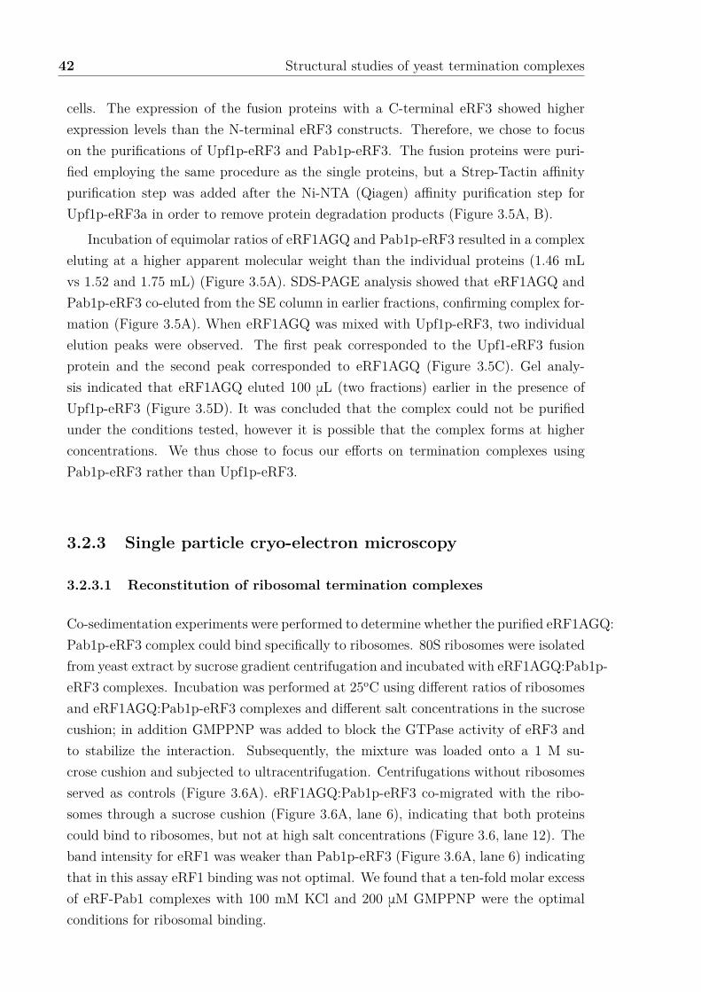
42 Structural studies of yeast termination complexes
cells. The expression of the fusion proteins with a C-terminal eRF3 showed higherexpression levels than the N-terminal eRF3 constructs. Therefore, we chose to focuson the purifications of Upf1p-eRF3 and Pab1p-eRF3. The fusion proteins were puri-fied employing the same procedure as the single proteins, but a Strep-Tactin a�nitypurification step was added after the Ni-NTA (Qiagen) a�nity purification step forUpf1p-eRF3a in order to remove protein degradation products (Figure 3.5A, B).
Incubation of equimolar ratios of eRF1AGQ and Pab1p-eRF3 resulted in a complexeluting at a higher apparent molecular weight than the individual proteins (1.46 mLvs 1.52 and 1.75 mL) (Figure 3.5A). SDS-PAGE analysis showed that eRF1AGQ andPab1p-eRF3 co-eluted from the SE column in earlier fractions, confirming complex for-mation (Figure 3.5A). When eRF1AGQ was mixed with Upf1p-eRF3, two individualelution peaks were observed. The first peak corresponded to the Upf1-eRF3 fusionprotein and the second peak corresponded to eRF1AGQ (Figure 3.5C). Gel analy-sis indicated that eRF1AGQ eluted 100 mL (two fractions) earlier in the presence ofUpf1p-eRF3 (Figure 3.5D). It was concluded that the complex could not be purifiedunder the conditions tested, however it is possible that the complex forms at higherconcentrations. We thus chose to focus our e�orts on termination complexes usingPab1p-eRF3 rather than Upf1p-eRF3.
3.2.3 Single particle cryo-electron microscopy
3.2.3.1 Reconstitution of ribosomal termination complexes
Co-sedimentation experiments were performed to determine whether the purified eRF1AGQ:Pab1p-eRF3 complex could bind specifically to ribosomes. 80S ribosomes were isolatedfrom yeast extract by sucrose gradient centrifugation and incubated with eRF1AGQ:Pab1p-eRF3 complexes. Incubation was performed at 25oC using di�erent ratios of ribosomesand eRF1AGQ:Pab1p-eRF3 complexes and di�erent salt concentrations in the sucrosecushion; in addition GMPPNP was added to block the GTPase activity of eRF3 andto stabilize the interaction. Subsequently, the mixture was loaded onto a 1 M su-crose cushion and subjected to ultracentrifugation. Centrifugations without ribosomesserved as controls (Figure 3.6A). eRF1AGQ:Pab1p-eRF3 co-migrated with the ribo-somes through a sucrose cushion (Figure 3.6A, lane 6), indicating that both proteinscould bind to ribosomes, but not at high salt concentrations (Figure 3.6, lane 12). Theband intensity for eRF1 was weaker than Pab1p-eRF3 (Figure 3.6A, lane 6) indicatingthat in this assay eRF1 binding was not optimal. We found that a ten-fold molar excessof eRF-Pab1 complexes with 100 mM KCl and 200 mM GMPPNP were the optimalconditions for ribosomal binding.

3.2 Results 43
S P S P S P S P S P S P
75
100
150
250
50
37
25
80SeRF1AGQ:Pab1p-eRF3
+-
-+
++
+-
-+
++
Pab1p-eRF3
eRF1AGQ
[kDa]
100 mM KCl 150 mM KCl
75
100
150
250
50
Pab1p-eRF3
eRF1AGQ
Lane 1 2 3 4 5 6 7 8 9 10 11 12
A B
Figure 3.6: Reconstitution of yeast termination complexes. A. Reactions contain-ing 2 pmols of 80S ribosomes and 10 time excess of eRF1AGQ:Pab1p-eRF3 complex werecentrifuged through a sucrose cushion with two di�erent KCl concentrations. Supernatant(S) and pellet (P) fractions were analyzed by SDS-PAGE and stained with SYPRO Rubystain or Western blot using a mouse Anti-6xHis tag antibody (1/5,000 dilution) and a sec-ondary Alexa 532-labeled anti-mouse antibody (1/10,000 dilution). B. Cryo-EM micrographsof frozen, reconstituted RNC-CMV with 10 fold molar excess of eRF1AGQ:eRF3-Pab1p com-plexes collected on the F20 microscope (IBS) at a 50,000x magnification.
3.2.3.2 Single particle analysis of RNC-eRF1AGQ:Pab1p-eRF3
Grids were frozen in the EM facility of the Institut de Biologie Structurale (IBS)using the Vitrobot (FEI) and 2 pmol of RNCs mixed with 20 pmol eRF1AGQ:Pab1p-eRF3. Micrographs were collected on the Polara F30 (FEI) equipped with a 4k x 4kGatan CCD camera (Figure 3.6B) at 50,000 x magnification using low dose conditions.A test dataset was composed of 42,239 ribosomal particles, manually picked usingEMAN boxer (Tang et al., 2007). For unsupervised 3D classification a reconstructionof the S. cerevisiae empty 80S ribosome (EMDB 2275, low-pass filtered at 30 Å) wasused as a template. Broken particles, ribosomes without P-site tRNA (non-translatingribosomes) or ribosomes with missing A-site density (non-terminating ribosomes) weresorted out (Figure 3.7A). 2,480 particles (corresponding to ~5% of the data) formed anew class where densities could be observed in the P-site and A-site of the ribosome.The P-site density was well defined and indicated the presence of peptidyl-tRNA inthe RNC. The A-site density was not well defined, but it was su�cient to confirm thepresence of eRF1 and eRF3 in our reconstruction (Figure 3.7B-E). Despite e�orts tofurther sort the data computationally, density for Pab1p could not be observed in anyof the reconstructions. Sorting was hampered by the small size of the particle poolrepresenting translation termination complexes. Thus, we could not visualize Pab1 inthe termination complexes despite being physically linked to eRF3.
This initial cryo-EM reconstruction helped to formulate hypotheses and draw con-

44 Structural studies of yeast termination complexes
100% 42 239
25.5% ~10 500 20.4% ~8 600 11.0% ~ 4 600 15.2% ~6 400 27.7% 11 700
21.2% ~2 480 25.5% ~1 280 25.5% ~990 37.3% 4 300 21.8% 2 550
Empty Ribosome Empty Ribosome Empty Ribosome P-site tRNABroken particles
A-site occupancy Empty Ribosome tRNA - RibosomeBroken particles Broken particles
Empty ribosome
90o
45o
B C
D E
A
Figure 3.7: Single particle analysis of RNC:eRF1AGQ:eRF3-Pab1p. A. Using areconstruction of an S. cerevisiae 80S empty ribosome (EMDB 2275) as an initial model forthe 3D reconstruction, the entire data set was sorted into 5 classes. 11,700 particles showedP-site tRNA (indicated in a green circle). Subsequently, this class was used for a secondround of 3D classification to sort for density in the A-site (indicated in a red circle). Thefinal volume further refined is shown in a black square. The occupancy of each particle poolis indicated. Images show structures derived from sub-data sets after computational sorting.B. The 40S subunit is colored in yellow, 60S in blue, the release factors in red and the P-sitetRNA in green. The map is shown in a top (B), side (C) and sliced (E) view. For comparison,the map of a human termination complex is shown in panel D (Franks et al., 2010).

3.2 Results 45
clusions about the sample preparation: only 25% of the particles contained P-sitetRNA indicative of translating ribosomes. Strikingly, only 5% of the dataset showeddensity for the release factors (but not for Pab1p). Thus, the sample quality needed beimproved in order to increase ribosome occupancy. Moreover, the binding a�nity be-tween Pab1p and the release factors should be investigated more thoroughly to ensurethat a ternary complex can indeed be formed in the presence of the ribosome.
3.2.4 Concluding experiments
3.2.4.1 RNC purification steps optimization
In order to increase the occupancy of P-site tRNA, we tried to reduce unspecific bindingof non-translating ribosomes to the resin. The streptavidin beads were thus preincu-bated with Bovine serum albumin (BSA) and total yeast RNA overnight. This in-cubation helped to minimize the co-purificaton of ribosomes and yNC peptide versusyNC-tRNA bound to RNCs (Figure 3.8), indicating that the fraction of translatingversus non-translating ribosomes was e�ciently increased.
3.2.4.2 eRF3 complex with Upf1p and Pab1p characterization
In order to investigate the complex formation of the release factors with Upf1p orPab1p, we decided to study the interaction between eRF3 and Upf1p or Pab1p first.Co-incubation of equimolar ratios of eRF3 and Pab1p (20 mM as an alternative to 7 mMin Figure 3.5) did not result in a complex as demonstrated by the lack of an apparenthigher molecular weight peak compared to the individual proteins (Figure 3.9A). SDS-PAGE analysis revealed that a mixture of eRF3 and Pab1p eluted from the gel filtrationcolumn in the same fractions as the single proteins (Figure 3.9A). However, if eRF3was mixed with Upf1p, the complex eluted slightly ahead of the single proteins (1.29mL compared to 1.31 mL and 1.38 mL) (Figure 3.9B). SDS-PAGE analysis suggestedthat the proteins were coeluting in earlier fractions than the proteins alone, indicatingthat a complex can be formed at higher concentrations. An attempt was then madeto mix the three proteins together in order to verify if Upf1p and Pab1p could bindcooperatively to eRF3 (Figure 3.9C). The resulting elution volume was not significantlydi�erent from the elution volume of eRF3 mixed with Upf1. However, the SDS-PAGEanalysis showed that Pab1p co-eluted together with eRF3 and Upf1 (Figure 3.9C)suggesting a weak interaction between Pab1p and these proteins.
In order to further investigate whether a complex is formed between Upf1p andrelease factors in vitro, pulldown assays were performed. Briefly, reaction mixturescontaining oligohistidine (His)-tagged eRF3 with eRF1AGQ or Upf1p or both proteinswere incubated with Ni-NTA beads in the absence of nucleotides (Figure 3.9D), washed

46 Structural studies of yeast termination complexes
yNC
yNC-tRNA50
37
2520
15
[Kda]
Sucrose Cushion
Strepavidin Purification
100 nM SBP-yNC-CMV mRNA
M T S P FT W1 W2 W3 E R
yNC
yNC-tRNA50
37
2520
15
[Kda] M T S P FT W1 W2 W3 E R
Sucrose Cushion
Strepavidin Purification
no mRNAA B
Figure 3.8: Further Optimization of RNC purification. A. B. RNC purificationusing BSA and total yeast RNA treatment of the streptavidin beads with or without SBP-yNCmRNA as indicated. Western blot using streptavidin HRP (1/4,000 dilution). Two bands canbe detected for the SBP tagged yNC: a lower band at 20 kDa indicating free protein (yNC) anda higher band at 40 kDa corresponding to the yNC still attached to P-site tRNA (tRNA-yNC).M: Marker; T: total reaction, S: supernatant; P: pellet Fraction; FT: flow-through; W1, W2,W3: washes; E: elution and R: resin. M: protein molecular weight standards (kDa).
extensively and the bound proteins were eluted with imidazole. Untagged Upf1p wasfound in the elution fractions suggesting unspecific binding. However, Upf1p boundvery slightly to eRF3 alone or to eRF1-eRF3 complexes (Figure 3.9D, lanes 5-7). Thesubsequent question to be answered was whether Pab1p could bind to eRF3 in pulldownassays (Figure 3.9E). However, binding of Pab1p to eRF3 alone or in presence ofUpf1p could not be detected under the conditions tested (Figure 3.9E, lanes 5-7).Our results are thus ambiguous and additional techniques must be used to validate theinteraction between the release factors and Upf1p and Pab1p. Either these interactionsare transient by nature or additional factors are required to stabilize the interactions.

3.2 Results 47
1.381.31
1.29
050
100150200250300350400450500
0 0.5 1 1.5 2 2.5 3
A2
80
(m
AU
)
Elution Volume (mL)
eRF3Upf1eRF3:Upf1p
66
21
0
67
0
29
15
0
44
0
[kDa]
7550
eRF3
75100
131Fractions
75100
50
M[kD
a]
inp
ut
eRF3Upf1p
Upf1p
131Fractions
M[kD
a]
inp
ut
7550
eRF3
75100
7550
eRF3Pab1p
Pab1p
1.37
1.38
1.37
0
20
40
60
80
100
120
140
160
0 0.5 1 1.5 2 2.5 3
A2
80
(m
AU
)
Elution Volume (mL)
eRF3Pab1peRF3:Pab1p
66
21
0
67
0
29
15
0
44
0
[kDa]
50
75
100 Input
50
75
100
Elu
tion
Lane 1 2 3 4 5 6 7
Upf1p
eRF3
eRF1AGQ
Upf1p
eRF3
eRF1AGQ
eRF3eRF1
Upf1p
His6 - eRF3
+--
-+-
--+
++-
+-+
-++
+++ [kDa]
75
100
Input
75
100
Elu
tion
Lane 1 2 3 4 5 6 7
Pab1peRF3
Upf1p
Pab1peRF3
Upf1p
eRF3
Upf1pPab1p
His6 - eRF3
+--
-+-
--+
++-
+-+
-++
+++ [kDa]
131Fractions
M[kD
a]
inp
ut
75100
Upf1p
eRF3
eRF3Pab1p
Pab1p
75100
75100
50 eRF3Upf1p
75100
50 Pab1pUpf1p
0
100
200
300
400
500
600
0 0.5 1 1.5 2 2.5 3
A2
80
(rl
u)
Elution Volume (mL)
Upf1p:Pab1peRF3:Upf1peRF3:Pab1peRF3:Upf1p:Pab1p
66
21
0
67
0
29
15
0
44
0
[kDa]
1.32
1.28 1.29
1.37
D
E
C
A B
Figure 3.9: Binding assays between yeast Upf1p, Pab1p and the release factors.A. Size exclusion chromatography (SEC) elution profile of eRF3 (blue) or Pab1p (orange)or both (light green) (left). 20 mM of each protein was loaded onto a Superdex 200 column.The elution volume (in mL) is indicated for each experiment. Calibration of the column wasperformed with globular proteins (shown above). Eluate fractions were analyzed by SDS-PAGE(below). M: protein molecular weight standards (kDa) B. Same as A. but with eRF3 (blue),Upf1p (purple) and both (green). C. Same as A. but with Upf1:Pab1p (purple), eRF3:Upf1(green), eRF3:Pab1p (light green) and eRF3:Upf1:Pab1p (light orange) D. Pulldown withhexahistidine-tagged eRF3 mixed with eRF1 or Upf1p or both. Protein mixtures loaded (input)onto the beads (above) and after elution (below) were separated by SDS-PAGE followed byCoomassie staining. E. Same as D. but with untagged Pab1p (10 mM protein input).

48 Structural studies of yeast termination complexes
3.3 Discussion
The aim of this study was to gain insights into the mechanism of the terminationof translation by solving the structure of terminating ribosomes bound to the releasefactors and Upf1p or Pab1p. To achieve this, a yeast cell-free lysate system for invitro translation was established in our laboratory. This allowed the purification ofstalled translating ribosomes still with the nascent chain bound in the exit tunnel(RNC) by a two-step procedure. First, ribosomes were separated from associatedfactors by ultracentrifugation through a high salt sucrose cushion. Second, an a�nitypurification step allowed the separation of non-translating from translating ribosomeswhich contain a SBP tag at the N-terminal end of the nascent chain. To reconstitutethe translation termination complexes, it was decided to use a fusion protein approachto promote complex formation by physically linking eRF3 with Upf1p or Pab1p. Thisstrategy allowed the purification of a complex containing eRF1AGQ and eRF3 linkedto Pab1p. The complex was able to bind to purified RNCs and a cryo-EM datasetof the reconstituted complex was collected. However, the test reconstruction showedonly a small fraction of ribosomes containing a P-site tRNA and density for the releasefactors. No density could be observed for the Pab1p despite being linked to eRF3. Thisindicates that Pab1p does not interact with the release factors, ribosomal proteins orrRNA when tethered to the A-site of the ribosome.
3.3.1 Optimization of yeast translational extract
It was found that 5’ end mRNA capping was critical for the cell-free translation sys-tem. In vitro transcribed mRNAs were capped using purified VCE. The optimal ratiobetween capping enzyme and mRNAs was determined to be 1:2 (Figure 3.2A-C). ThemRNA concentration in the system was also optimized and the addition of 100 nMmRNA to the translation reaction resulted in the maximal detection of yNC peptideafter ultracentrifugation. This indicates that the nascent chain remained bound to theribosome through the sucrose cushion and thus is part of a ribosome-nascent chaincomplex (Figure 3.2D). Cap-independent mechanisms using CrPV IRES elements havebeen reported in yeast (Iizuka et al., 1994) and have been successfully used to gener-ate ribosome-nascent chain complexes (Matheisl et al., 2015). Under the conditionstested, IRES-mediated translation did not occur e�ciently (Figure 3.12A). The yNCsequence was verified to be correct and therefore it was concluded that the yeast strainor the cell extract preparation protocol used in this study was not optimal for IRES-mediated translation initiation. In agreement with this statement, it has been shownthat CrPV-IRES functions more e�ciently in mutant yeast strains which result inenhanced phosphorylation of translation initiation factor eIF2a, which is critical for

3.3 Discussion 49
IRES-mediated translation initiation (Thompson et al., 2001).
3.3.2 Preparation of yeast ribosome-nascent chain complexes
The in vitro translation reaction was loaded onto a high salt sucrose cushion andsubjected to ultracentrifugation. This separated the ribosomal complexes from theremaining cell extract components. The presence of an N-terminal tag on the yNCallowed the purification of translating ribosomes. The translation system was optimizedto stall ribosomes using a mutant of eRF1 which cannot trigger the nascent chain releaseduring the in vitro translation reaction (Figure 3.3). Moreover, a CMV stalling motifwas introduced in the coding sequence of the synthetic mRNA in order to e�cientlystall the translating ribosomes and stabilize them during the purification procedure.Subsequently, a larger fraction of tRNA-bound nascent chain in the ribosomal pelletwas detected after ultracentrifugation (Figure 3.3). The possibility cannot be excludedthat tRNA-yNC failed to be detected on Western blots during earlier experiments, dueto sensitivity of our detection technique. In fact, the peptidyl-tRNA is usually detectedradioactively after RNC purification according to published protocols (Beckmann etal., 2001; Brown et al., 2015), which is more sensitive than Western blotting.
3.3.3 Purification of termination complexes
eRF1AGQ, eRF3 and Pab1p full length proteins were purified from E. coli (BL21(DE3)*)using a C-terminal hexahistidine tag. Upf1p full length with an N-terminal hexahis-tidine tag was produced in insect cells (Sf21 ). Whereas eRF1AGQ and eRF3 formeda stable complex, we could not detect a ternary complex after addition of Pab1p orUpf1p under the tested conditions (Figure 3.4). It was thus decided to fuse Pab1por Upf1p to eRF3 in order to promote complex formation and enrich the presence ofUpf1p / Pab1p at the ribosome. The fusion proteins Pab1p-eRF3 and Upf1p-eRF3could be purified (Figure 3.5). However, only Pab1p-eRF3 could form a complex witheRF1AGQ during the SEC experiments (Figure 3.5). It can be speculated that theeRF1:Upf1p-eRF3 interaction is too transient, possibly due to steric problems, andthus dissociates during gel filtration. In addition, we cannot exlude that, despite beinglong and flexible, the linker could interfere witht the protein interactions.
3.3.4 Reconstitution of termination complexes and single par-ticle analysis
Using co-sedimentation experiments, it was revealed that eRF1AGQ:Pab1p-eRF3 com-plexes interacted with 80S ribosomes, and that this interaction was sensitive to high salt

50 Structural studies of yeast termination complexes
concentrations in the bu�er (Figure 3.6). The reconstitution of RNC:eRF1AGQ:Pab1p-eRF3 complexes were conducted in presence of GMPPNP to block eRF3’s GTPaseactivity. These complexes were used for cryo-grid preparation and a test cryo-EM dataset was collected (Figure 3.6). Image processing of a small dataset of 42,239 ribosomalparticles resulted in a structure from a subpool of 2,480 particles (5% of the dataset)that contained a P-site tRNA and density in the A-site corresponding to eRF1AGQand eRF3. However, no density could be observed for Pab1p (Figure 3.7). The numberof particles was insu�cient when compared to the other published ribosomal complexstructures (Becker et al., 2011; Preis et al., 2014), indicating that the in vitro transla-tion protocol required further improvement. Overall, these observations indicated thatthe RNC preparation could be further optimized to increase the fraction of translatingribosomes. Peptide-tRNA is sensible to pH and thus bu�ers at pH7.0 instead of pH7.5should be used for the purification. In addition, the RNCs probably decay over timeand therefore, the RNCs should be prepared freshly before sample freezing. At thispoint, the possibility that the absence of Pab1p density was due to the addition ofrelease factors during the in vitro translation cannot be excluded. It is possible thatthe eRFs could not be e�ciently removing during sucrose cushion centrifugation andthus addition of a ten-fold excess of eRF1AGQ:Pab1p-eRF3 was not su�cient. Theinteraction between and eRF1AGQ and Pab1p-eRF3 may be transient. It is possiblethat this complex is too weak to survive the vitrification procedure, resulting in a com-plex that lacks density for Pab1p in our final reconstruction because this part of thecomplex is flexibly attached to eRF3. In fact, eRF1AGQ is not completely inactiveand retains 5 % activity in peptidyl-tRNA hydrolysis (Alkalaeva et al., 2006). So it ispossible that, in the presence of the ribosome, the basal activity of the release factorscould have been stimulated by Pab1p and resulted in nascent peptide hydrolysis andfactor release. Last but not least, the previously published ribosomal volumes used atleast 20,000 particles for the final reconstruction (Brown et al., 2015; Matheisl et al.,2015). In order to build an atomic model of a terminating ribosome in the presence ofthe release factors and Pab1p a significantly larger cryo dataset must be collected.
3.3.5 Final experiments and eRF3 interaction with Upf1 andPab1p
Our experiments have shown that the purity of RNC could be further improved bythe pre-incubation of the streptavidin resin with yeast total RNA and BSA (Figure3.8). In addition, the interaction between eRF3, Upf1p and Pab1p was investigated(Figure 3.9). The results have hinted toward the formation of a eRF3:Upf1p complexwhen high protein concentrations were used but this result is yet to be confirmed byadditional biochemical techniques such as surface plasmon resonance and isothermal

3.3 Discussion 51
titration calorimetry. eRF3 complexes with Pab1p could not be isolated. Nevertheless,the binding of Upf1p and Pab1p to eRF3 has been reported previously in yeast (Wanget al., 2001; Cosson et al., 2002a; Kobayashi et al., 2004; Cosson et al., 2002b) andhumans (Hoshino et al., 1999; Kashima et al., 2006; P. V. Ivanov et al., 2008; Kozlov &Gehring, 2010). Using yeast two-hybrid and co-immunoprecipitations, the interactionbetween the two proteins could be mapped to the N and the M domains of eRF3 andthe C-terminal part of Pab1p (comprising amino acids 406-577) (Roque et al., 2014).It is worth noting that the N terminal domain of eRF3 is not conserved between yeastand humans. In this thesis work, unlike previously published studies (Kervestin etal., 2012; Roque et al., 2014), no direct in vitro interaction between eRF3 and Pab1pcould be observed. As di�erent experimental settings were tested compared to thesestudies, it cannot be excluded that the binding between eRF3 and Pab1p requiresconditions similar to those in previously published work. Despite using a very highconcentration (40 mM) of purified Pab1p and His
6
-eRF3 for our binding assay (Figure3.9), an interaction was not confirmed. Taken together, these observations indicate thatthe a�nity between yeast Pab1p and eRF3 is very low under the tested conditions.Further work is required to determine whether this interaction could be detected in adi�erent bu�er or if it is mediated by other proteins.
3.3.6 Concluding remarks
During this doctoral research study, a yeast in vitro translation system was establishedin our laboratory and provided a framework for the reconstitution of RNC complexesand their examination by cryo-EM single particle analysis. Biochemical experimentswere also established to study protein-protein interactions which were critical for thesecond part of this work (see Chapter 4).
Furthermore, attempts were made to study the interplay of yeast Upf2p and Upf3pwith the release factors. Despite our e�orts, only high-purity full length Upf3p couldbe obtained (Figure 3.13). Since full length as well as fragments of human UPF3B werealready available in our laboratory, we decided to switch our focus to the human model.The following section details the work done during this doctoral thesis to investigatethe role of human UPF3B in NMD and termination of translation.

52 Structural studies of yeast termination complexes
3.4 Methods
3.4.1 Molecular Biology Techniques
3.4.1.1 Cloning of yeast nascent chain and protein constructs
Sequence and ligation independent cloning (SLIC) and self-SLIC.
Sequence and ligation independent cloning (SLIC) was performed as described previ-ously (M. Z. Li & Elledge, 2007). SLIC reactions without an insert were performedin order to introduce mutations or generate truncations of the sequence of interest.This is referred as to self-SLIC (Figure 3.10). Briefly, the vector was amplified byPCR using primers carrying mutations. 40 U of DpnI enzyme was added to 100 mLof PCR reaction and incubated at 37oC for 1 h. The DNA was purified using via aPCR purification and a DNA extraction kit (Qiagen). Subsequently, 1 mg of plasmidwas treated with 0.5 U of T4 DNA polymerase in T4 ligase bu�er in 20 mL at RT for30 min. The reaction was stopped by adding 1/10 volume of 10mM dCTP and lefton ice. Annealing reactions were set up with 150 ng of linearized plasmid with 1x T4ligase bu�er at a final volume of 10 mL and incubated at 37oC for 30 min. 5 mL ofthe annealed mixture was used to transform chemically competent TOP10 E. coli cells(Thermo Scientific).
yNC mRNA cloning.
The yeast Nascent Chain (yNC) plasmid construct was ordered from Clontech andsub-cloned into pUC19 through the EcoRI and NcoI restriction sites. The boxB andpoly(A) sequence was inserted via the NotI and HindIII restriction sites from pBS-BMS_EF3_3boxBpA73 to generate pUC19-SBP-yNC-BoxB-pA plasmid (Duncan etal., 2006). The 3Strep-tag was amplified by PCR from pUC19-3Strep-FtsQ-SecM(Scha�tzel & Ban, 2007) and subcloned into pUC19-SBP-yNC-BoxB-pA plasmid viaEcoRI and SpeI. A CMV stalling motif was inserted by PCR using the T7 primerand a reverse primer containing the CMV sequence (see primers in Appendix A).The resulting PCR fragment was subcloned into pUC19-SBP-yNC-BoxB-pA usingEcoRI and NcoI restriction enzymes. The IRES motif was amplified from pUC18-CPV-UAA_mRNA (kind gift of Elena Alkalaeva) and sub-cloned into yNC plasmidusing EcoRI and NheI.
yNC plasmid production and linearization.
yNC plasmids were transformed into XL1-Blue competent cells. A Lysogeny broth (LB)medium culture with 200 mg/mL ampicillin were started from 10 colonies. Incubationwas completed overnight in an orbital shaker incubator at 37oC and 150 rotations per
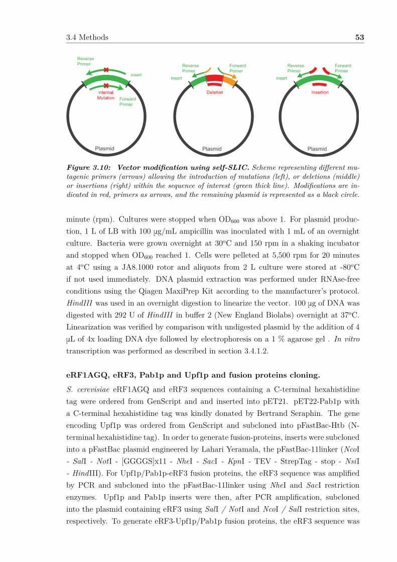
3.4 Methods 53
insert
Plasmid
Deletion
Forward Primer
ReversePrimer
insert
Plasmid
Insertion
Forward Primer
ReversePrimer
insert
Plasmid
internalMutation Forward
Primer
ReversePrimer
Figure 3.10: Vector modification using self-SLIC. Scheme representing di�erent mu-tagenic primers (arrows) allowing the introduction of mutations (left), or deletions (middle)or insertions (right) within the sequence of interest (green thick line). Modifications are in-dicated in red, primers as arrows, and the remaining plasmid is represented as a black circle.
minute (rpm). Cultures were stopped when OD600
was above 1. For plasmid produc-tion, 1 L of LB with 100 mg/mL ampicillin was inoculated with 1 mL of an overnightculture. Bacteria were grown overnight at 30oC and 150 rpm in a shaking incubatorand stopped when OD
600
reached 1. Cells were pelleted at 5,500 rpm for 20 minutesat 4oC using a JA8.1000 rotor and aliquots from 2 L culture were stored at -80oCif not used immediately. DNA plasmid extraction was performed under RNAse-freeconditions using the Qiagen MaxiPrep Kit according to the manufacturer’s protocol.HindIII was used in an overnight digestion to linearize the vector. 100 mg of DNA wasdigested with 292 U of HindIII in bu�er 2 (New England Biolabs) overnight at 37oC.Linearization was verified by comparison with undigested plasmid by the addition of 4mL of 4x loading DNA dye followed by electrophoresis on a 1 % agarose gel . In vitrotranscription was performed as described in section 3.4.1.2.
eRF1AGQ, eRF3, Pab1p and Upf1p and fusion proteins cloning.
S. cerevisiae eRF1AGQ and eRF3 sequences containing a C-terminal hexahistidinetag were ordered from GenScript and and inserted into pET21. pET22-Pab1p witha C-terminal hexahistidine tag was kindly donated by Bertrand Seraphin. The geneencoding Upf1p was ordered from GenScript and subcloned into pFastBac-Htb (N-terminal hexahistidine tag). In order to generate fusion-proteins, inserts were subclonedinto a pFastBac plasmid engineered by Lahari Yeramala, the pFastBac-11linker (NcoI- SalI - NotI - [GGGGS]x11 - NheI - SacI - KpnI - TEV - StrepTag - stop - NsiI- HindIII). For Upf1p/Pab1p-eRF3 fusion proteins, the eRF3 sequence was amplifiedby PCR and subcloned into the pFastBac-11linker using NheI and SacI restrictionenzymes. Upf1p and Pab1p inserts were then, after PCR amplification, subclonedinto the plasmid containing eRF3 using SalI / NotI and NcoI / SalI restriction sites,respectively. To generate eRF3-Upf1p/Pab1p fusion proteins, the eRF3 sequence was

54 Structural studies of yeast termination complexes
amplified by PCR and subcloned into the pFastBac-11linker plasmid using XhoI andNotI restriction enzymes. Upf1p and Pab1p were amplified by PCR and subclonedinto the plasmid containing eRF3 using NheI and SacI restriction sites.
3.4.1.2 RNA Techniques
In vitro transcription with T7 RNA polymerase.
100 mg of linearized plasmid was transcribed in vitro with 37.5 mg of T7 RNA polymerasein 200 mM HEPES/KOH, pH 7.6, 30 mM Mg(OAc)
2
, 2 mM spermidine, 40 nM DTT,8.75 mM dNTPs, 0.4 mg/mL RNAsin (Promega). Reactions were incubated for 4hours at 37oC.
In vitro transcription with T3 RNA polymerase.
Luciferase mRNA was transcribed from the pUC19-CA_fireflyLUC (Promega) plasmidusing the mMESSAGE mMACHINE® High Yield Capped RNA Transcription Kit(Ambion).
RNA purification.
In order to purify mRNA from DNA and proteins, lithium chloride precipitations wereperformed by the addition of 800 mL of 6 M LiCl into a final volume of 1,600 mL.Reactions were vortexed and kept on ice for at least 30 min. After 30 minutes ofcentrifugation at 14,000 x g at 4°C, the pellet was washed with 500 mL of 70 % ethanol.The pellet was resuspended in 100 mL RNAse-free water. Ethanol precipitations werecarried out by the addition of 100 mL 97 % ethanol and 4 mL of 3 M sodium acetate.Reactions and wash steps were performed as for the LiCl precipitation. Finally, thepellet was resuspended in 20 mL of RNAse-free water.
mRNA concentration determination.
mRNA concentration was determined as follows: 1 mL of sample was diluted 1/10 intoRNAse-free water and the absorbance at 260 nm was determined. RNA concentrationwas calculated on the basis that an OD
260
of 1 corresponds to 40 mg/mL RNA.
RNA agarose gel electrophoresis.
Analytic RNA agarose gels were run to assess the mRNA quality. RNA molecules wereseparated on agarose gels containing 1.5 % agarose in freshly prepared RNAse free 1 xTBE. Guanidinium isothiocyanate and ethidium bromide were added to the agarose gelto a final concentration of 20 mM and 0.05 mL/mL respectively. Before loading, sampleswere mixed in a 1:2 [v/v] ratio with RNA loading dye (ThermoFisher Scientific) andincubated for 10 min at 70°C to dissolve secondary structures. Electrophoresis was

3.4 Methods 55
carried out at 70 V for 45 min.
RNAse contamination test.
To test proteins for contaminating RNases, 100 ng of mRNA was incubated alone orwith 2 - 5 pmoles of protein in a final volume of 10 mL at 37 oC for 1h, followed by RNAagarose gel electrophoresis, as described above. The presence of RNases in the proteinsample would result in degradation of the mRNA, as indicated by a diminution of thefull size mRNA band and the appearance of lower molecular weight bands (smear)after electrophoresis.
mRNA capping.
To dissolve potential secondary structures, the uncapped mRNA was denatured at 65oC for 10 minutes and transferred onto ice immediately. VCE and mRNA were mixedin a 1:2 ratio in 1x script capping bu�er (Epicentre Biotechnologies) supplementedwith 1 mM GTP and 0.1 mM S-adenosylmethionine (SAM) (final concentration). Thereactions were incubated for 45 min at 37 oC, frozen in liquid nitrogen and stored at-80oC, if not used immediately.
3.4.2 Cell Culture
Yeast cultures.
Yeast extracts were prepared from the RH2585 strain (MATa, ura3-52, trp1::hisG,his3::hisG - kind gift of Prof. Altmann). A second strain was designed with RobbieLoewith’s laboratory in Geneva where eRF1 (sup45 in yeast) was TAP-tagged in orderto deplete it from the cell extract. This yeast strain background is the following: TB50sup45-TAP: a leu2 ura3 rme1 trp1 his3D GAL+ HMLa. A few mL of the glycerolstock of yeast were streaked out onto yeast extract peptone dextrose (YPD - 2 % [w/v]tryptone, 1 % [w/v] yeast extract, 2 % [w/v] glucose) agar-agar plates and incubatedfor 48h at 30oC. 10 colonies from each plate were used to start 20 mL precultures inYPD media at 30oC and 150 rpm overnight. The overnight pre-cultures were used tostart 5 L cultures by dilution 1:1,000. Cultures were stop when the OD
600
reached 1in order to avoid overgrowth. The cells were pelleted at 5,000 rpm for 5 min at 4oCusing a JA8.1000 rotor.
E. coli expression: eRF1AGQ, eRF3, Pab1p, VCE.
Plasmids were transformed into chemically competent BL21(DE3)* cells. 20 mL cul-tures were grown overnight at 37oC from a single colony in LB medium containing100 mg/ml ampicillin. 1 liter of TB medium containing 100 mg/ml ampicillin wasinoculated with 10 ml of overnight culture and grown until an OD
600
of 0.8 was

56 Structural studies of yeast termination complexes
reached. The expression was induced by addition of 0.5 mM IPTG (Isopropyl b-D-1-thiogalactopyranoside) at 20oC and continued incubation overnight. The cells wereharvested by centrifugation at 5,500 rpm and 4oC for 20 min using a JA8.1000 rotor.
Insect cell culture: Upf1p and fusion proteins expression.
Proteins were expressed in the cell line Spodoptera frugiperda (Sf 21 - Invitrogen) usingthe MultiBac system as previously described (Fitzgerald et al., 2006). Briefly, Sf21insect cells were transfected with a recombinant bacmid isolated from DH10-EMBacYcells and grown in Sf-900 media (ThermoFisher Scientific) (Bieniossek et al., 2008).V0 was produced in a 6-well plate with 3 mL of culture per well. This virus wasused to generate a higher titer V1 by infection of 25 mL Sf21 cell cultures in a shakerflask. The V1 was used for large scale protein production by infection of 400 mLof Hi5 insect cells (ThermoFisher Scientific) in Express-Five medium (ThermoFisherScientific) in 2 L flasks. The cells were harvested 72 - 96 h following the day ofproliferation arrest (DPA) when the expression of the internal expression reporter,yellow fluorescent protein (YFP), plateaued. The cells were harvested by centrifugationat 1,000 x g in a JA8.1000 rotor for 10 min at 4oC.
3.4.3 Protein Biochemistry
3.4.3.1 Protein biochemistry techniques
Western blotting.
The PVDF membrane (Millipore) was activated in 100 % methanol for a few seconds.Proteins from the SDS-PAGE were transferred to the membrane soaked in transferbu�er (25 mM Tris-Base, 192 mM Glycine, 20 % EtOH) at 25 V for 1 h using a Trans-Blot SD Semi-Dry Transfer Cell (Bio-Rad). Non-specific interactions were reduced byblocking bu�er consisting of 3 % BSA in PBS bu�er (137 mM NaCl, 2.7 mM KCl, 1mM Na
2
HPO4
, 1.8 mM KH2
PO4
, pH 7.4) supplemented with 0.05 % [v/v] Tween-20 forat least 1 h at room temperature. The membrane was incubated with the appropriatedilution of antibody or Streptavidin or Strep-Tactin in PBS + 0.05 % Tween-20 with0.75 % BSA. Streptavidin coupled with Horseradish Peroxidase (HRP) (Sigma) andStrep-Tactin coupled with Alkaline Phosphatase (AP) (IBA Lifesciences) were diluted1/4,000 and incubated with the membrane for 1 h. His-tagged proteins were detectedusing 1/10,000 dilution of mouse monoclonal anti-polyHistidine antibody coupled toAP. After washing in PBS + 0.05 % Tween-20 for 3 x 15 minutes, blots with AP weredeveloped using BCIP/NBT photoreaction solution (100 mM Tris, 100 mM NaCl, 5mM MgCl
2
, 1 % [v/v] BCIP/NBT (Roche), pH 8.8). Western blots with HRP weredeveloped using 3,3�-Diaminobenzidine tablets (Sigma) in 1 mL of ultrapure water.

3.4 Methods 57
3.4.3.2 Protein purification
Yeast eRF1AGQ, eRF3, Pab1p and Upf1p purification.
Cell pellets were lysed in bu�er B (20 mM HEPES/KOH pH 7.5, 300 mM KOAc, 10% [v/v] glycerol, 5 mM b-Mercaptoethanol) and 10 mM imidazole by 2 passages in aFrench press cell with a pressure of 20,000 psi. The lysed cells were centrifuged for 30min at 31,000 rpm at 4oC using a Type 70Ti rotor (Beckmann Coulter). The lysatewas applied to 10 ml of 50 % slurry Ni-NTA resin (Qiagen) and washed with bu�er Bplus 1 M KOAc and 50 mM imidazole. The protein was eluted with bu�er B containing200 mM imidazole. If required, the hexahistidine tag was cleaved by the addition ofTeV protease [1/50] during overnight dialysis into bu�er C (20 mM HEPES/KOH pH7.5, 2.5 mM Mg(OAC)
2
, 5 % [v/v] glycerol, 100 mM KOAc) at 4oC supplementedwith 5 mM b-Mercaptoethanol. Subsequently, the sample was incubated with Ni-NTA resin and the flow-through collected. The sample was loaded onto a MonoQ5/50GL column (GE Healthcare), pre-equilibrated with bu�er C plus 2 mM DTT. Theproteins were eluted by a 40 ml gradient from 100 mM – 1 M KOAc. The proteinswere further purified by size-exclusion chromatography using a Superdex 200 column(GE Healthcare) pre-equilibrated with bu�er C plus additional 50 mM KOAc and 2mM DTT.
Purification of vaccinia capping enzyme (VCE).
Cell pellets were lysed in bu�er D (40 mM HEPES/KOH pH 8.0, 200 mM NaCl) and40 mM imidazole by sonication. The lysed cells were centrifuged at 30,000 rpm for20 min at 4oC using a Type 70Ti rotor (Beckmann Coulter). The lysate was appliedto 10 ml Ni-NTA resin (Qiagen) and washed with bu�er D supplemented with 1 MNaCl and 60 mM imidazole. The proteins were eluted with bu�er D, plus 300 mMimidazole. The His tag was cleaved by the addition of 1/100 TeV protease [w/w] at4oC during overnight dialysis in bu�er D supplemented with 3 mM b-Mercaptoethanol.The sample was loaded onto a 5 ml pre-packed Heparin column (GE Healthcare), pre-equilibrated with bu�er D containing 3 mM b-Mercaptoethanol. VCE was eluted bya 20 ml gradient from 200 mM – 1 M NaCl. The protein was further purified by size-exclusion chromatography using a Superdex 200 column (GE Healthcare) equilibratedwith bu�er D and 3 mM DTT (Kyrieleis et al., 2014).
3.4.3.3 Yeast Extract preparation and purification
After harvesting, the yeast cell pellets were washed in DEPC (Diethylpyrocarbonate)water and subsequently with bu�er A (30 mM HEPES/KOH pH7.4, 100 mM KOAc, 2mM Mg(OAc)
2
, 2 mM DTT, 0.1 mM PMSF) supplemented with 8.5 % [v/v] mannitol.The cells were frozen in liquid nitrogen and grinded using a mortar and a pestle until a
58 Structural studies of yeast termination complexes
homogenous paste was obtained. The reactions were pelleted twice at 13,000 rpm for15 min at 4oC using a tabletop centrifuge (Eppendorf). Subsequently, the supernatantwas further purified using a Sephadex G25 column equilibrated with bu�er A. Fractionswith A
260
close to 100 rlu were frozen in liquid nitrogen and stored at -80oC.
3.4.3.4 In vitro translation and RNC preparation
Luciferase assay using the yeast cell-free translation system.
Capped and polyadenylated Renilla luciferase (Rluc) mRNA was used to measure trans-lational activity in an untreated cell extract (exogenously added mRNA will competewith endogenous mRNA for translation). 20 ng Rluc mRNA was translated with 50 %[v/v] yeast extract, 0.04 mM amino acids (-methionine), 25 mM creatine phosphate,0. 5 mg/mL creatine phosphokinase, 1 U RNAsin (Promega) in a final volume of 10mL. Samples were incubated for 45 min in a 25oC water bath. Reactions were stoppedby the addition of 12 mL of 1x passive lysis bu�er (Promega Dual-Luciferase). 20 mLsamples were supplemented with 50 ml luciferase assay reagent (Promega), and lumi-nescence was read out from a 96 well plate on a plate reading luminometer (Mithras,Berthold Technologie). The luminescence was detected 1.6 sec after injection of thesubstrate solution for 10 sec.
in vitro translation of yNC mRNA.
100 nM yNC mRNA was translated in 50 % [v/v] yeast cell extract (~100 OD260
/mL)for 60 minutes at 25oC in 20 mM HEPES/KOH, pH 7.5, 150 mM KOAc, 1.5 mMMg(OAc)
2
, 2 mM DTT, 25 mM creatine phosphate, 0.5 mg/mL creatine phosphoki-nase, 0.75 mM ATP, 0.1 mM GTP, 0.4 mM amino acids (-methionine), 1 U/mL RNAsin(Promega). 4 mM of eRF1AGQ and eRF3 were added to the reaction. The in vitrotranslation reaction was stopped by 0.2 mg/mL cycloheximide, a translocation inhibitor.Subsequently, the ribosomes were pelleted through a 3 mL high salt sucrose cushion(50 mM HEPES/KOH pH7.5, 220 mM KOAc, 25 mM Mg(OAc)
2
, 2 mM DTT, 10mg/mL cycloheximide, 1 M sucrose and protease inhibitors) by ultracentrifugation us-ing a TLA-55 rotor (Beckmann Coulter) at 4oC, 3 h at 55,000 rpm. The ribosomalpellets were resuspended in RNC bu�er (50 mM HEPES/KOH pH7.5, 2 mM DTT, 10mg/mL cycloheximide) supplemented with 50 mM KOAc and 5 mM Mg(OAc)
2
.
RNC a�nity purification.
Subsequently, non-translating ribosomes were removed by an a�nity purification step.Resuspended pellets were added to 500 mL of streptavidin beads (Fisher Scientific)in RNC bu�er supplemented with 50 mM KOAc and 5 mM Mg(OAc)
2
and 0.05 %[v/v] Tween20. The beads were washed with 5 volumes (cv) of RNC bu�er with

3.4 Methods 59
220 mM KOAc and 25 mM Mg(OAc)2
. RNCs were eluted with 1 cv of RNC bu�ersupplemented with 100 mM KOAc and 2.5 mM Mg(OAc)
2
and 2 mM biotin. The finalRNC concentration was measured using A
260
and the purity of the RNCs was evaluatedby coomassie stained SDS-PAGE gel and Western blot using an appropriate antibodyagainst the yNC tag.
Ribosome binding experiments.
For co-sedimentation experiments, 2 pmol of yeast total ribosomes were mixed witha ten-fold molar excess of eRF1AGQ and eRF3 or fusion protein in 20 mL of RNCbu�er with 100 mM KOAc and 2.5 mM Mg(OAc)
2
and incubated for 1 hour on ice.Subsequently, the reaction mixtures were applied on a sucrose cushion (same bu�ercontaining 1 M sucrose) and centrifuged for 3 h at 55,000 rpm at 4°C using a TLA-55 rotor (Beckman). Supernatant and pellet fractions were analyzed by SDS-PAGEfollowed by SYPRO Ruby (Thermo Scientific) staining and Western blot using theappropriate antibody against the yNC tag, or the hexahistidine tag fused to the releasefactors.
In vitro pulldown assays.
All experiments were performed with 20 mM of each protein in a final reaction volumeof 20 mL in bu�er D (20 mM HEPES/KOH pH 7.5, 100 mM KCl, 10 mM imidazole, 0.05% [v/v] Tween20, 5 mM b-Mercaptoethanol, 5 % [v/v] glycerol, 5 mM Mg(OAC)
2
),if not indicated otherwise. The protein mixtures were incubated for 1 hour on ice;subsequently 20 mL of Ni-NTA agarose (Qiagen) was added to the reaction mix andincubated for 1 hour on ice. The reaction mixtures were washed 5 times with 200 mL ofbu�er D, and proteins were eluted using bu�er D supplemented with 200 mM imidazoleby centrifugation at 2,000 rpm for 2 minutes in a tabletop centrifuge (Eppendorf). 8mL of SDS loading dye were mixed with either 2 mL of input reactions or 20 mL of theeluted complexes. 10 mL of the input sample and 10 mL of the elution sample wereloaded onto a 10 or 12 % SDS-PAGE gel.
Size exclusion chromatography.
For SEC, 7 mM of each protein was added to a final reaction volume of 60 mL in bu�erE (20 mM HEPES/KOH pH 7.5, 150 mM KCl, 5 mM Mg(OAc)
2
, 2 mM DTT). Theprotein mixtures were incubated for 1 hour on ice before loading onto a Superdex200 PC3.2/30 column (AEKTA micro system, GE Healthcare). 10 mL of each elutionfraction was loaded onto a 10 % SDS-PAGE gel. The SEC runs in section 3.2.4.2 wereperformed with 40 mM instead of 7 mM.

60 Structural studies of yeast termination complexes
3.4.4 Electron microscopy
Negative staining electron microscopy.
Samples were applied to a carbon film deposited on a 300 mesh copper grid (Quantifoil),followed by staining with 2 % uranyl acetate solution. Grids were dried and theninserted into the microscope. Image acquisition was performed at room temperaturein a JEOL JEM-1200EX II microscope using high magnification (30 k – 50 k) and aGatan ORIUS CCD camera .
Cryo-grid preparation.
2 pmol of RNCs were incubated with 10x molar excess of purified eRF1:eRF3-Pab1pin RNC bu�er supplemented with 100 mM KOAc, 2.5 mM Mg(OAc)
2
and 200 mMGMPPNP. 3.5 mL of this sample were applied to Quantifoil grids R 2/2 type with athin continuous carbon foil. The grids were previously glow discharged (for 1 min at25 mA and at 1 x 10 1 mbar). Subsequently, the grids were vitrified using a VitrobotMark IV (FEI Company) at 5 oC and 100 % relative humidity with a 2 min incubationbefore blotting.
Cryo-electron microscopy and single particle reconstruction.
Data collection was performed by Manikandan Karuppasamy on a Tecnai POLARAF30 (FEI Company) equipped with 4k x 4k CCD camera (Gatan Inc), operated at 300keV using the EPU software (FEI). 1,740 micrographs were collected at a magnificationof 50,000 x and a nominal defocus between -2.5 and -3.5 mm. The pixel size was 1.93Å. 575 micrographs were discarded after contrast transfer function (CTF) correction(bctf, Bsoft Package; (Heymann, 2001)) and 42,239 ribosomal particles were manuallypicked with EMAN2 (Tang et al., 2007). The particles were aligned and refined usingthe RELION autorefine procedure (S. H. W. Scheres, 2012) and an S. cerevisiae 80Sempty ribosome (EMDB 2275 (Bai et al., 2013)) was used as an initial model. 3Dclassification was performed to sort out the particles into 5 classes. From the 5 re-sulting reconstructions, one reconstruction contained a ribosome with P-Site t-RNA.This reconstruction contained 27 % of the dataset (11,700 particles). It was furtherautorefined with RELION and subjected to a new round of classification using thesame parameters. From the five new reconstructions, one class showed P-Site tRNAand density in the A-site, this class contained 2,480 particles (5.8 % of the dataset).
3.4.5 Acknowledgements
We are much obliged to Prof. Karl Altmann for generously introducing us to the recon-stituted in vitro translation system and for providing the RH2585 yeast strain, Robbie

3.4 Methods 61
Loewith for TB50 engineered strain, Bertrand Seraphin for the Pab1p expression plas-mid and Elena Alkalaeva for providing the CrPV construct. This work is supportedby an European Research Council Starting Grant (ComplexNMD, 281331)

62 Structural studies of yeast termination complexes
3.5 Supplemental information
yNC
50
37
2520
15
[Kda] M T P T P T P T P
oligo(dT) purified extract no purification
no mRNA no mRNA3 μg 3 μg
0
500000
1000000
1500000
2000000
2500000
3000000
3500000
4000000
4500000
- eRF1AGQ + - + - +
RH2585 extract TB50-Sup45 extract
Ave
rage
Re
lative
Lig
ht
Un
it
Luciferase Assay : TB50-TAP-sup45 extract vs RH2585 extract
- TAP Purification - +
rRNA
yNC mRNA
I FT W EI FT W EI FT W EI FT W E
no mRNA 3 µg 30 µg
SBP-yNC mRNA
oligo(dT) purification
RH2585Extract
A B
TAP-sup45(eRF1)
50
75
100150250
37
25
20
[Kda] M I FT W E R
TB50-TAP-sup45 TAP purification
C D
Figure 3.11: mRNA depletion of the yeast extract for in vitro translation. A.RNA gel analysis of the endogenous mRNA depletion experiment using oligo(dT) resin, show-ing input (I), flow-through (FT), last wash (W) and elution (E) fractions. SBP-yNC containsa poly(A) tail and was used as a positive control B. Western blot analysis of 100 mL of in vitrotranslation with depleted extract (Oligo(dT) purified) and un-depleted (no purification) using1/4,000-diluted streptavidin-HRP for the total reaction (T) and after sucrose cushion ultra-centrifugation (Pellet - P). eRF1 (TAP-sup45) depletion of the TB50-TAP-sup45 cell extractfor in vitro translation. C. Western blot analysis of eRF1 TAP purification using dynabeads(Invitrogen) coupled with IgG, showing input (I), flow-through (FT), last wash (W), elution(E) fractions and resin (R). D. Luciferase assay using the yeast in vitro translation systemand wildtype yeast extract or TB50-Sup45 yeast extract with or without TAP purification andaddition of exogenous eRF1AGQ. M: protein molecular weight standards (kDa).

3.5 Supplemental information 63
yNC
50
37
25
2015
yNC
50
37
25
2015
[Kda] M T T P T P T P T P
capped SBP-yNC
0 μg mRNA
IRES-SBP-yNC
1.5 μg 3 μg 1.5 μg 3 μg
yNC
50
75
37
2520
15
[Kda] M T S P FT W1 W2 W3 E R
Sucrose Cushion StrepTactin Purification
no mRNA in vitro translationB
A
yNC
50
75
37
2520
15
yNC
50
75
37
25
20
15
[Kda] M T S P FT W1 W2 W3 E R
Sucrose Cushion StrepTactin Purification
100 nM 3Strep-yNC-CMV mRNA in vitro translationC
yNC
50
37
2520
15yNC
50
37
25
20
15
yNC
50
75
37
2520
15
[Kda]
Sucrose Cushion Strepavidin Purification
no mRNA in vitro translation
M T S P FT W1 W2 W3 E R
D
yNC
50
37
25
20
15
[Kda] T M S P FT W1 W2 W3 E R
Sucrose Cushion Strepavidin Purification
100 nM SBP-yNC-CMV mRNA in vitro translationE
75
Figure 3.12: yNC mRNA construct optimization. A. Western blot analysis ofin vitro translation using capped SBP-yNC or IRES-SBP-yNC. Two mRNA concentrationswere used for in vitro translation. Western blot was performed with streptavidin-HRP forthe total reaction (T) and after sucrose cushion ultracentrifugation (Pellet – P). 100 mLtranslation reaction. B. Western blot analysis of RNC purifications without mRNA usingStrep-Tactin resin. C. RNC Purification using 3Strep tagged yNC mRNA using Strep-Tactinresin. D. Same as A. using strepavidin resin. E. Same as B. using SBP tagged yNC andstrepavidin resin. Western blots were performed using streptavidin-HRP or Strep-Tactin-AP. Total reaction (T), Supernatant (S), pellet (P), flow-through (FT), washes (W1-3) andelution (E). M: protein molecular weight standards (kDa).

64 Structural studies of yeast termination complexes
-1000
-500
0
500
1000
2000
mAU
0.0 20.0 40.0 60.0 80.0
Elution Volume (mL)
65.5 mL1000Upf3p50
37
75100
[Kda] 1 8Fractions
25
20
B C %B100
0
Upf2p
50
37
75100150250
[Kda]
AIPTG
+
Figure 3.13: Upf2p and Up3p Purification. A. SDS-PAGE followed by a Western Blot,using 1/4,000-diluted anti-hexahistidine antibody coupled to alkaline phosphatase, analysis ofUpf2p expression in BL21 using di�erent concentrations of IPTG. Human UPF2L (121-1227) was used as a positive control (+). B. SDS-PAGE analysis of Upf3p purification byanion exchange (SP/HP - GE Healthcare). C. Chromatogram of Upf3p purification by anionexchange (SP/HP - GE Healthcare). Absorbance at 280 nm and 245 nm (mAU) is displayed inblue and red, respectively. Elution in mL is indicated for each elution peak. Elution followinga KCl gradient (%B) from 150 mM to 1 M is indicated as a green line and the secondaryaxis.

4. Double-tasking of UPF3B in earlyand late phases of translation ter-mination
Resumé en françaisL’objectif du travail présenté dans ce chapitre était d’obtenir des informations biochim-iques sur les interactions entre les facteurs de terminaison et ceux de la NMD au coursde la terminaison, afin de mettre à l’épreuve le modèle actuel de discrimination desPTCs. Dans cette optique, nous avons utilisé une approche combinant un système detraduction in vitro, établi dans notre laboratoire, ainsi que des analyses d’interactionsin vitro et in vivo. Nous avons montré que UPF3B, et non UPF1 ou UPF2, est leseul facteur capable de se lier à eRF1 et eRF3a. D’après nos résultats, UPF1 ne joueaucun rôle significatif dans la terminaison de la traduction in vitro. De plus, UPF3Bpeut s’associer à l’ARN, au ribosome et à UPF1. UPF3B retarde la reconnaissancedu codon stop si les facteurs de terminaison sont limitants. Dans le cas où les facteursde terminaison sont saturants, UPF3B favorise la dissociation des sous-unités riboso-males une fois que le codon stop est reconnu. UPF1 n’a pas d’influence dans cetteactivité mais la présence de UPF2 abolit le délai de la terminaison et la dissociationdu ribosome par UPF3B.

66 Double-tasking of UPF3B in early and late phases of translation termination
Double-tasking of UPF3B in early and late phases oftranslation termination
Manuscript under review in Molecular Cell
Authors
Gabriele Neu-Yilik* and Etienne Raimondeau*, Boris Eliseev, Lahari Yeramala, BeateAmthor, Aurélien Deniaud, Karine Huard, Kathrin Kerschgens, Matthias Hentze**,Christiane Scha�tzel**, Andreas Kulozik**
* co-first authors** corresponding authors
A�liations
• European Molecular Biology Laboratory, Grenoble Outstation, 71 Avenue desMartyrs, 38042 Grenoble, France
• School of Biochemistry, University of Bristol, Bristol, BS8 1TD, United Kingdom
• Department of Pediatric Oncology, Hematology and Immunology, University ofHeidelberg, Im Neuenheimer Feld 430, 69120 Heidelberg, Germany
• Molecular Medicine Partnership Unit, University of Heidelberg and EuropeanMolecular Biology Laboratory, Im Neuenheimer Feld 350, 69120 Heidelberg,
• Germany European Molecular Biology Laboratory, Meyerhofstr. 1, 69117 Hei-delberg, Germany
Abstract
Nonsense-mediated mRNA decay (NMD) is a cellular surveillance pathway that recog-nizes and degrades mRNAs with premature termination codons (PTCs). The mecha-nisms underlying translation termination are key to the understanding of RNA surveil-lance mechanisms such as NMD and crucial for the development of therapeutic strate-gies for NMD-related diseases. Here, we have used a fully reconstituted in vitro trans-lation system to probe the currently prevailing model of how NMD proteins interactwith the termination apparatus. We discovered that UPF3B (1) delays translationtermination and (2) dissociates post-termination ribosomal complexes that are devoidof the nascent peptide. These previously unknown functions of UPF3B in early andlate phases of translation termination position UPF3B at a central mechanistic stepof RNA surveillance and suggest that it may function as a dedicated NMD ribosomerescue factor.

4.1 Introduction 67
4.1 Introduction
Nonsense-mediated mRNA decay (NMD) is a eukaryotic surveillance mechanism thatcontrols the expression of aberrant mRNAs, degrading transcripts with premature ter-mination codons (PTCs). PTCs can be introduced into mRNAs by mutations, tran-scriptional errors, and aberrant splicing, but are also contained in 5 -15 % of normaltranscripts. By modulating the expression of physiological target mRNAs, NMD servesas a posttranscriptional regulator of gene expression and thus controls important cel-lular and organismal processes in development, cellular stress responses, immunity,and neuronal di�erentiation (Kurosaki & Maquat, 2016; Linder et al., 2015; Ottens& Gehring, 2016). NMD is also of medical importance as it limits the productionof truncated proteins that may otherwise exert dominant negative functions but canalso result in loss of function when mRNAs encoding (partially) functional truncatedproteins are degraded (Bhuvanagiri et al., 2010; Nguyen et al., 2014). Mutations orcopy number variations in NMD factors are linked to genetic diseases, specifically toneurodevelopmental disorders and intellectual disabilities (Linder et al., 2015; Nguyenet al., 2014).
Conceptually, NMD can be divided into a translation termination phase and anmRNA degradation phase. During the past two decades a wealth of information hasaccumulated concerning the interplay between the core NMD factors and decay en-zymes that enable the recognition and degradation of NMD substrates (Schweingruberet al., 2013; Fatscher et al., 2015). However, the mechanism by which translation ter-mination at a PTC is distinguished from termination at a normal termination codon(NTC) is still poorly understood. Two prevailing models, the “downstream markermodel” and the “faux 3’UTR model”, have been proposed to explain the di�erencebetween normal and aberrant termination as well as the interaction between the ter-minating ribosome and the NMD machinery (reviewed in (Bhuvanagiri et al., 2010;He & Jacobson, 2015b)). The “downstream marker model” posits the formation of anaberrant termination complex at a PTC consisting of the terminating ribosome, thecentral NMD e�ector UPF1, the UPF1 kinase complex SMG1-8-9, and the release fac-tors eRF1 and eRF3. This so-called SURF complex (Kashima et al., 2006) is thoughtto delay translation termination and to sense the presence of an mRNP complex on theextended 3’UTR which in mammalian cells is represented by an exon junction complex(EJC) downstream of the PTC. The premature termination complex and the EJC arethought to be bridged by UPF2 that, according to this model, interacts with UPF1at the termination site and EJC-bound UPF3B, leading to the formation of a decayinducing complex that remodels the 3’ mRNP and recruits mRNA decay enzymes.
In contrast, the faux 3’UTR model posits that NMD can be induced by an aberrant3’UTR mRNP, which is characterized by the absence of at least one termination-

68 Double-tasking of UPF3B in early and late phases of translation termination
enhancing factor that is associated with a normal 3’ UTR. Consequently, terminationat a PTC is decelerated and ine�cient. Such an aberrant 3’ UTR mRNP can be definedby the inappropriate distance between the termination codon and the poly(A) tail thuspreventing the termination promoting interaction between eRF3a and poly(A) bindingprotein and instead allowing the recruitment of UPF1.
Both models converge on the central NMD e�ector UPF1 that interacts withthe release factors (eRFs) at the terminating ribosome. For yeast NMD, all threeUPF proteins are essential, whereas in higher eukaryotes, UPF2-independent, UPF3B-independent, and EJC-independent NMD branches have been described (Bühler et al.,2006; Chan et al., 2007; Gehring et al., 2005). UPF1 can be recruited to the termi-nation site by the NMD-stimulating EJC, by an aberrant 3’ UTR mRNP, or by anine�cient termination event. How UPF2 and UPF3 are recruited to the terminationsite in EJC-independent NMD is unknown. UPF2 and UPF3B are thought to stimulatethe phosphorylation of UPF1 and to activate UPF1’s ATPase and helicase functionsthat are necessary to remodel the 3’UTR mRNP and to recruit mRNA degradationenzymes (Chamieh et al., 2008; P. V. Ivanov et al., 2008; Kashima et al., 2006; Fioriniet al., 2015).
Although the necessity of an interaction between the UPF proteins and the trans-lation termination apparatus is generally accepted, the sequence and timing of NMDfactor recruitment to the termination site has not been addressed experimentally. Re-cent studies revealed that UPF1 is bound along the entire length of transcripts and thatthis binding occurs in a translation-independent fashion (Hogg & Go�, 2010; Hurt etal., 2013; Zünd et al., 2013). These findings have challenged the hypothesis, proposedby both the downstream marker model and the faux 3’UTR model, that UPF1 is specif-ically recruited to aberrant termination events as an anchor point for the assembly ofan NMD-mRNP.
Translation termination, whether regular or aberrant, needs to recycle ribosomes toavoid deleterious consequences for the translation apparatus (Graille & Séraphin, 2012;J. Lykke-Andersen & Bennett, 2014). In ribosome recycling of normal and di�erentforms of abnormal termination, the ATPase ABCE1/Rli1 is needed for the ultimatedissociation of post-termination ribosomes from the mRNA (Dever & Green, 2012;Franckenberg et al., 2012; Graille & Séraphin, 2012; Jackson et al., 2012). In yeastand human cells, depletion of the UPF proteins induces readthrough at PTCs in vivoas well as delayed termination in vitro (Amrani et al., 2004; Peixeiro et al., 2012). Arecent attempt to reconcile all available data into a new NMD model posits that UPF1,UPF2, and UPF3 have roles in both, the early and late phases of premature termi-nation (He & Jacobson, 2015b). Accordingly, UPF1’s initially weak association withelongating ribosomes is proposed to be stabilized by UPF2 and UPF3 when a ribosometerminates prematurely. The UPF1-UPF2-UPF3 complex stimulates the initially de-

4.1 Introduction 69
layed termination at a PTC by either recruiting the release factors or by enhancingpeptide release. Subsequently, UPF2 and UPF3 promote ATP hydrolysis by UPF1to fuel the dissociation of post-terminating ribosomal complexes. UPF1, still boundto the 40S ribosomal subunit, then recruits mRNA decay enzymes to initiate mRNAdegradation.
To shed light on these critical aspects of translation, we adopted an approach thatcombines a fully reconstituted cell-free in vitro translation termination system with invitro and in vivo interaction studies to decipher the UPF-eRF interactome in transla-tion termination. We find that UPF3B, rather than UPF1 interacts with eRF3a andforms a trimeric complex with both eRF3a and eRF1. Moreover, UPF3B binds toRNA, the ribosome, and to UPF1. UPF1 plays no discernible functional role in thiscontext, suggesting that it acts downstream to promote NMD. In contrast, UPF3Bdelays translation termination when release factors are limiting and dissolves post-termination complexes after peptidyl-tRNA hydrolysis by the release factors.
70 Double-tasking of UPF3B in early and late phases of translation termination
4.2 Results
Validation of the experimental system
During termination at a PTC, the UPF1-eRF interaction is thought to stall translationtermination (P. V. Ivanov et al., 2008; Kashima et al., 2006). Here, we set out to ana-lyze whether UPF1 alone or together with UPF2 and/or UPF3B a�ects the e�ciencyof mammalian translation termination in vitro.
We produced full-length eRF1, eRF3a, UPF1, and UPF3B and a UPF2 variant(UPF2L) comprising amino acids (aa) 121–1227 (Supplemental Figure 4.8A). Riboso-mal pre-termination complexes (translating ribosomes stalled at a stop codon; preTCs)were assembled on a model mRNA using ribosomal subunits, aminoacylated tRNAs,and purified initiation, elongation and release factors. The model mRNA (MVHC-STOP) contained the b-globin 5’-UTR and a short open reading frame encoding aMVHC tetrapeptide followed by a UAA stop codon and a 3’UTR of ~400 nt (Figure4.1A) (Alkalaeva et al., 2006; Fan-Minogue et al., 2008). The formation of defined ri-bosomal complexes was analyzed by sucrose density gradient (SDG) centrifugation andprimer extension inhibition (toeprinting). PreTCs containing the peptidyl-tRNACys inthe ribosomal P-site and the stop codon in the A-site are characterized by specifictoeprints at position +16 nt 3’ to the U of the UGC (Cys) codon (Figure 4.1B, lane 1,Supplemental Figure 4.8B).
During translation termination, eRF1 in complex with eRF3a binds to the stopcodon (Brown et al., 2015) inducing conformational rearrangements. The formationof such post-termination complexes (postTCs) is manifested by a +1-2 nt forwardshift relative to the preTC toeprint (Figure 4.1B, lane 2; Supplemental Figure 4.8B)(Shirokikh et al., 2009). GTP hydrolysis by eRF3a leads to accommodation of theGGQ motif of eRF1 in the peptidyl transferase centre of the large ribosomal subunit,resulting in rapid peptide release. eRF1 (together with eRF3a or after dissociation ofeRF3a) remains associated with postTCs, thus maintaining the +2 nt shift of theirtoeprint (Alkalaeva et al., 2006).
To confirm the biological activity of purified UPF1, UPF2L, and UPF3B, we per-formed ATP hydrolysis experiments. In previous studies (Chakrabarti et al., 2011;Chamieh et al., 2008). Maximal ATP hydrolysis by UPF1 was achieved at low pH(6-6.5) using N- and C-terminally truncated UPF1 fragments (Supplemental Figure4.8C). UPF1LDCH (aa 295-914) lacks the CH domain and exhibits high ATPase ac-tivity that cannot be further stimulated, whereas UPF1L (aa 115-914) contains theCH domain and exhibits similar ATPase activity but requires stimulation by UPF2and UPF3B (Chamieh et al., 2008). In full-length UPF1 (UPF1) the C-terminus con-tributes to maintaining UPF1 in an inactive state (Fiorini et al., 2013). Using UPF1L

4.2 Results 71
and UPF1DCH as controls, we first tested ATP hydrolysis by UPF1 in end-point ex-periments at pH 6.5 (Supplemental Figure 4.8D). UPF1 exhibited only modest ATPaseactivity at 30°C and pH 6.5 which was doubled in the presence of UPF2L/3B (Supple-mental Figure 4.8D, lanes 2 and 3). In contrast, the ATPase activity of UPF1L wasabout fourfold compared to that of UPF1 and was strongly enhanced by UPF2L/3B(lanes 4 and 5). The ATPase activity of UPF1LDCH was comparable to that ofUPF1L/2L/3B and could not be further stimulated (lanes 6 and 7). Notably, at aphysiological pH 7.5 the activity of all proteins was lower, but followed a similar pat-tern (Supplemental Figure 4.8D, lanes 8-13). We further investigated the ATPaseactivity of UPF1, either alone or in the presence of UPF2L, UPF3B or both, underin vitro translation conditions at pH 7.5 and 37°C (Supplemental Figure 4.8E). Wefound that neither UPF2L (lane 5) nor UPF3B (lane 6) have a significant e�ect onthe ATPase function of UPF1. By contrast, when used in combination the ATPasefunction of UPF1 was slightly enhanced (lane 7). Taken together, these data show thatour UPF proteins are biologically active.
UPF3B delays ine�cient translation termination in a fully reconstitutedtranslation termination system
According to current models, termination at a PTC contrasts with termination at anNTC by being slow and ine�cient, possibly caused by ine�cient recruitment of theeRFs (He & Jacobson, 2015b). To simulate this situation in vitro, we used limitingconcentrations of eRFs for our termination experiments as judged by the retention ofa faint but discernible preTC toeprint in addition to the appearance of postTC signalsafter termination (Figure 4.1B, lane 2).
To test if UPF proteins a�ect the e�ciency of translation termination, preTCswere incubated with excess UPF1, UPF2L, UPF3B or combinations of these proteins(Figure 4.1B, lanes 3-9). As controls, preTCs were either left untreated (lane 1) orthe UPF proteins were replaced by BSA (lane 2). Subsequently, limiting amounts ofeRFs were added to the reactions (except in lane 1), and translation termination wasallowed to proceed for 5 min, followed by toeprinting analysis.
Neither UPF1 nor UPF2L individually (Figure 4.1B, lanes 3, 4) or together (lane6) a�ected the intensity of pre- or postTC bands, compared to the control sample (lane2). These findings indicate that when the eRFs are limiting, neither UPF1 nor UPF2Lhave a direct e�ect on translation termination in vitro.
By contrast, UPF3B (lane 5) substantially reduced the preTC to postTC trans-formation rate to about 40 % of the rate observed in the control reaction (lane 2) asestimated by calculating the ratio between the preTC and postTC signal intensitiesusing a phosphorimager. Notably, the addition of UPF1 to UPF3B resulted in a simi-lar delay of termination (lane 7) and did not have an additive, synergistic or reversing
72 Double-tasking of UPF3B in early and late phases of translation termination
e�ect. Addition of UPF2L abolished the e�ect of UPF3B on translation termination(lane 7, 8), indicating that binding to UPF2L may prevent UPF3B from interferingwith the termination reaction. We observed that the toeprint signals corresponding tothe full length RNA and to the termination complexes as well as to the traces of ini-tiating and elongating ribosomes present in preTC preparations were always strongerin the presence of UPF3B than in reactions without UPF3B. Therefore, we performedtoeprinting of preTCs that had been incubated with UPF3B but without eRFs. Wefound that here, too, all toeprint signals were stronger than in the absence of UPF3B(Figure 4.1B, lanes 1 and 10), which is likely to be caused by a more e�cient recoveryof ribosomal complexes and RNA.
UPF3B reduces the e�ciency of peptidyl-tRNA hydrolysis at low concen-trations of release factors
To investigate if UPF3B delays translation termination by impairing peptidyl-tRNAhydrolysis, preTCs assembled on the MVHC-STOP mRNA using 35S-labeled initiator-tRNA were incubated with or without UPF3B, and with limiting amounts of eRFs.In comparison to the maximal rate of peptidyl-tRNA hydrolysis achieved within theobserved time window, peptide release e�ciency was reduced in the presence of UPF3Bby ~40-50 % (Figure 4.1C). We conclude that UPF3B impairs stop codon recognitionand peptidyl-tRNA hydrolysis by the eRFs and thereby reduces termination e�ciency.
Translation termination in vitro is independent of ATP-binding or the AT-Pase activity of UPF1
ATP binding and hydrolysis by UPF1 are essential for NMD. UPF2 and UPF3B havebeen shown to stimulate this ATPase function (Chakrabarti et al., 2011; Chamieh etal., 2008). Therefore, we repeated the toeprinting experiment described above in thepresence of either ATP (Supplemental Figure 4.9, lanes 2-8) or its non-hydrolyzableanalogue AMPPNP (lanes 9-15). Under these conditions, neither UPF1 nor UPF2Lindividually (lanes 3, 4, 10, 11) or together (lanes 6, 13) a�ected the intensity of pre- orpostTC signals compared to the control samples (lanes 2, 9). By contrast, UPF3B bothalone and following addition of UPF1 reduced termination e�ciency. These findingswere independent of the presence of ATP or AMPPNP, indicating that neither theATP-binding nor the ATPase function of UPF1 influences the transition of preTCs topostTCs.

4.2 Results 73
EMVHC-preTCs
eRF1/eRF3aUPF1
UPF2L
UPF3B++++
+ + + + ++ + + + +
+++
+++
+++
+++
+++
+++
+++
+++
- - - - - - -- -- - - -
- - - - - -
---
--+
-++
-++
-++
TAA
C T A G
1 2 3 4 5 6 7 8 9 10 11 12
SMG1-8-9
full length
preTCspostTCs
D
P-UPF1
P-SMG1
UPF1UPF2L
eRF3aUPF3B
eRF1
UPF2LUPF1
UPF3B
eRF1/3aSMG1-8-9 +
-
+-- - -
+--
+- -
+--
-+ +
+-
+
+- - -
+-
+-+ + +
+- - - - -
+
++ + +
+ +
++ +- -
+
+-
+
+
+-
-
+ +
++ +
+ +
+ +
++ +- -
+ +
++ +
+ +
1 2 3 4 5 6 7 8 9 1310 1411 1512 16
250
250
130
130
10070
55
kDa
CB
1 2 3 4 5 6 7 8 9 10
MVHC-preTCs
eRF1/eRF3a
UPF1
UPF2L
UPF3B + + ++
+ + ++
+
+
+
+
+
+
+
+
+
+-++
++
- +- - - -
- -- - - -
- -
- -
+ +
-+
+
-+
+
-+
+
C T A G
full length
preTCspostTCs
**
TAA
AMVHC-STOP mRNA
5'G (CAA) -(b-globin 5'UTR)-AUG-GUG-CAC-UGC-UAA-3'UTR (~400nt)4
Met Val His Cys
eRF1 +eRF3a
eRF1 +eRF3a +UPF3B
Figure 4.1: UPF3B delays translation termination in vitro. A. Structure of theMVHC-STOP mRNA. B. Toeprinting analysis of ribosomal complexes obtained by incubatingpreTCs assembled on MVHC-STOP mRNA (MVHC-preTCs) with UPF1, UPF2L, UPF3B,or BSA at 1 mM free Mg2+ followed by termination with limiting amounts of eRF1 andeRF3a. The positions of preTCs, postTCs, and full length cDNA are indicated. Asterisksmark initiation and elongation complexes. C. Kinetics of [35S]-peptidyl-tRNA hydrolysisin the presence of eRF1 and eRF3a (black circles) or eRF1, eRF3a, and UPF3B (whitetriangles). A value equal to 1 corresponds to the maximum value for peptide release triggeredby eRF1 and eRF3a. D. UPF1 in vitro phosphorylation by SMG1-8-9 in the presence ofUPF2L and/or UPF3B and in the presence (lanes 7-12) or absence (lanes 1-6) of the eRFs.In lanes 13-16 UPF2L and/or UPF3B were added after UPF1 phosphorylation. Sampleswere analysed by SDS-PAGE, Coomassie-stained to control for equal loading (lower panel)and autoradiographed (upper panel). SMG1 autophosphorylation (P-SMG1) confirms equalSMG1-activity in all samples. UPF1 is represented by the lower and UPF2L by the upperof the two closely migrating bands between 125 and 130 kDa in the Coomassie stained gel.E. Toeprinting analysis of ribosomal complexes obtained by incubating MVHC-preTCs withUPF1, UPF2L, UPF3B, SMG1-8-9, or BSA as indicated followed by translation terminationby eRF1 and eRF3a. See also Supplemtental Figure 4.9.

74 Double-tasking of UPF3B in early and late phases of translation termination
Translation termination is independent of UPF1 phosphorylation and thepresence of the SMG1-8-9 complex
According to current models, UPF1, the eRFs, and the SMG1-8-9 complex form thetermination-stalling SURF complex (Kashima et al., 2006). UPF1 phosphorylationby SMG1 is thought to trigger UPF1’s release from the eRFs (Kashima et al., 2006;Okada-Katsuhata et al., 2012). Here, we explored whether in vitro phosphorylation ofUPF1 in the absence or in the presence of UPF2L and/or UPF3B influences translationtermination. Maximal in vitro phosphorylation of UPF1 by SMG1 or SMG1-8-9 isachieved at pH 9.0, corresponding to the pH-optimum of the kinase (Chakrabarti et al.,2014; Deniaud et al., 2015; Morita et al., 2007). We examined phosphorylation of UPF1by SMG1-8-9 at physiological pH in the absence or presence of UPF2L, UPF3B andthe eRFs (Figure 4.1D). UPF2L only slightly stimulates UPF1 phosphorylation (Figure4.1D, lanes 4, 10) by SMG1-8-9. In contrast, UPF3B alone (lanes 5, 11) moderatelyand together with UPF2L (lanes 6, 12) strongly inhibits UPF1 phosphorylation bySMG1-8-9 irrespective of the presence of equimolar concentrations of the eRFs. Weconfirmed that our UPF2L or UPF3B preparations do not contain a phosphatase byco-incubating the phosphorylated UPF1 (P-UPF1) with the preparations of UPF2Land UPF3B for 15 min at 37°C (lanes 13-16), which did not a�ect the abundance ofthe phosphorylated UPF1.
We next investigated whether UPF1 phosphorylation or the presence of SMG1-8-9per se a�ects in vitro translation termination. UPF1 alone or together with eitherUPF2L, UPF3B, or both was incubated with SMG1-8-9 and ATP for 30 min andsubsequently mixed with preTCs for another 10 min at 37°C (Figure 4.1E, lanes 9-12)followed by termination with eRF1 and eRF3a and toeprint analysis. Reactions withoutSMG1-8-9 served as controls (lanes 3-8). We found that irrespective of the presence ofeither UPF2L or UPF2L and UPF3B, neither UPF1 phosphorylation nor the presenceof SMG1-8-9 have a detectable influence on termination e�ciency as judged by therate of transformation of preTCs to postTCs (compare lanes 3 and 9, lanes 6 and 10,lanes 8 and 12). Importantly, the inhibitory e�ect of UPF3B on this transformationwas independent of the presence of SMG1-8-9 and UPF1 (compare lanes 7 and 11).
UPF1 and UPF3B are part of release factor-containing complexes in vivo
We next analyzed the interaction of the UPF proteins with the termination complexin vivo. Based on co-immunoprecipitation (co-IP) experiments, human UPF1 has beensuggested to interact with both eRF1 and eRF3a, and thereby physically link theNMD apparatus with translation termination (P. V. Ivanov et al., 2008; Kashima etal., 2006; Singh et al., 2008). In yeast, all three Upf proteins were reported to bind toeRF3 (Sup35) (Wang et al., 2001). We thus transiently co-transfected HeLa cells withFLAG-tagged eRF1 or eRF3a and full length versions of V5-tagged UPF1, UPF2, or

4.2 Results 75
UPF3B, and immunoprecipitated on FLAG-antibody beads in the presence of RNaseA. Co-IPs of FLAG-eRF1 with V5-eRF3a and of FLAG-eRF3a with V5-eRF1 servedas positive controls and yielded strong eRF1-eRF3a interactions (Figure 4.2A, B, lanes2). Co-IPs of FLAG-eRFs with the EJC-disassembly factor PYM (Gehring et al., 2009)served as specificity controls (Figure 4.2A, B, lanes 10).
Using FLAG-eRF1 as bait, UPF1 (Figure 2A, lane 3), but not UPF2 (lanes 4, 6,8, 9) or UPF3B (lane 5, 7-9) was co-immunoprecipitated with eRF1. Importantly,co-transfection of UPF3B and UPF1 prevented the formation of a complex containingeRF1 and UPF1 (lane 7) indicating that UPF3B either directly or indirectly competeswith eRF1 for UPF1 binding.
When we co-expressed V5-tagged UPF proteins with FLAG-eRF3a the complexcontained only little UPF2 (Figure 4.2B, lane 4) but considerably more UPF1 andUPF3B (lanes 3 and 5). Interestingly, we found UPF3B in FLAG-eRF3a immuno-precipitates together with UPF1 (lanes 7 and 9), indicating that UPF1 and UPF3Bcan bind both, individually and together, but that these proteins do not compete foreRF3a-binding. Although co-transfection of UPF2 strongly enhanced the ability ofUPF1 to co-immunoprecipitate with eRF3a (compare lane 3 to lane 6), UPF2 was ex-cluded from these complexes as well as from complexes containing eRF3a and UPF3B(lanes 6, 8 and 9). We conclude that UPF2 stimulates the direct or indirect interac-tion between UPF1 and eRF3a, but does not partake in complexes containing eRF3atogether with UPF1, UPF3B, or both.
UPF3B directly interacts with eRF3a in a magnesium-sensitive mannerforming a ternary complex with eRF1
Co-IP experiments do not reveal whether the interactions identified are direct or in-direct. Therefore, we performed in vitro pulldown assays to analyse whether purifiedUPF proteins and release factors interact directly.
We incubated reaction mixtures containing His-tagged UPF1, UPF2L or UPF3Band one or both untagged eRF(s) (Figure 4.3A) with Ni-NTA beads, washed extensivelyand eluted the bound proteins with imidazole. We found that neither eRF1 nor eRF3aindividually, nor the eRF1-eRF3a complex, detectably bound to UPF1 (Figure 4.3B,lanes 5-7), or UPF2L (Figure 4.3C, lanes 5-7). UPF2 has recently been reported todirectly interact with eRF3a. However, UPF2L did not bind to eRF3a, although itcontains the part that has been reported to interact with eRF3a (López-Perrote etal., 2015). In contrast, eRF3a and, to a lesser extent, eRF1 co-eluted with UPF3Bindividually (Figure 4.3D, lanes 5, 6) as well as simultaneously (lane 7) indicating thatUPF3B directly interacts with both release factors.
Reciprocal control experiments using His-eRF3a as a bait for both UPF1 andUPF3B (Supplemental Figure 4.10) corroborated the eRF3a-UPF3B interaction (Sup-

76 Double-tasking of UPF3B in early and late phases of translation termination
A
B
FLAG-eRF1 FLAG-eRF1 FLAG FLAG
V5-UPF1V5-UPF2
V5-UPF3BV5-eRF3a
V5-PYM
TUBB
FLAG-eRF1
input pulldown
1
1
2
2
3
3
4
4
5
5
6
6
7
7
8
8
9
9
10
10
11
11
12
12
13
13
14
14
15
15
FLAG
pulldown
11 12 13 14 15
UPF1eRF3a
UPF2
UPF3B
PYM+ +- -
- -
+-
-
+-
-
- -
+ +
- -
+-
-
+ ++ + - -+
+ ++ + + - --
-
-
-
- -
- -
+ +
- -
- -
- -
- -
- -
- -
-
-
-
- - -- -- --
- - -- -- -
+-
-
-
+
-
+ -+ -
-
-
+
-
-
-
-
-
-
- - -
- - -
+ +- -
- -
+-
-
+-
-
- -
+ +
- -
+-
-
+ ++ + - -+
+ ++ + + - --
-
-
-
- -
- -
+ +
- -
- -
- -
- -
- -
- -
-
-
-
- - -- -- --
- - -- -- -
V5-
FLAG-eRF3a FLAG-eRF3aFLAG
V5-UPF1V5-UPF2
V5-UPF3BV5-eRF1
V5-PYM
ACTB
FLAG-eRF3a
input
1
1
2
2
3
3
4
4
5
5
6
6
7
7
8
8
9
9
10
10
11 12 13 14 15
UPF1eRF1
UPF2
UPF3B
PYM
+ +- -
- -
+-
-
+-
-
- -
+ +
- -
+-
-
+ ++ + - -+
+ ++ + + - --
-
-
-
- -
- -
+ +
- -
- -
- -
- -
- -
- -
-
-
-
- - -- -- --
- - -- -- -
+-
-
+-
-
+-
-
-
+
-
+-
-
+ + + -+
+ + + + --
-
-
-
-
-
+
-
-
-
-
-
-
-
-
-
- - - - -
- - - -
V5-
250
13010070
55
35
kDa
25013010070
55
35
kDa
Figure 4.2: In vivo interaction between release factors and UPF proteins. A.Co-immunoprecipitation from RNase A-treated lysates of Hela cells transfected with FLAG-eRF1 (lanes 1-10) or unfused FLAG (lanes 11-15) and V5-eRF3a, V5-UPF1, V5-UPF2,V5-UPF3B, or V5-PYM. Co-precipitated proteins were detected using an anti-V5 antibody.Lysate used for the immunoprecipitations was loaded in the input lanes (left). Re-probingwith anti-TUBB antibody served as loading control. B. Co-IP experiment as in (A) withFLAG-eRF3a. Re-probing with anti-ACTB served as loading control.

4.2 Results 77
plemental Figure 4.10) and confirmed that UPF1 does not co-elute with eRF3a irre-spective of the presence of eRF1 (Supplemental Figure 4.10A).
Translation is modulated by the Mg2+ concentration both in vivo and in vitro. Inour in vitro translation termination assays (Figure 4.1) we used 1mM free Mg2+ whichcorresponds to the physiological intracellular level of unbound Mg2+ (MacDermott,1990; Veloso et al., 1973). We explored the impact of Mg2+ on the UPF3B-eRFinteraction. At physiological [Mg2+] a substantial amount of UPF3B bound to eRF3a,whereas at >5 mM Mg2+ the interaction between UPF3B and eRF3a was considerablyweaker (Figure 4.3E, lanes 4-6). Notably, UPF1 did not directly interact with the eRFsat all Mg2+ concentrations tested (Supplemental Figure 4.10C).
To assess UPF3B-eRF complex formation independently, we performed size ex-clusion chromatography (SEC). Co-incubation of equimolar amounts of eRF3a andUPF3B resulted in a complex eluting in earlier fractions at a higher apparent molec-ular weight than the individual proteins (Figure 4.3F), corroborating a direct interac-tion between eRF3a and UPF3B. Because UPF3B alone eluted at a higher apparentmolecular weight than expected (Figure 4.3F), indicating possible oligomerisation ora deviation from the globular shape, we subjected UPF3B to SEC-MALLS and foundthat UPF3B is monomeric thus suggesting a non-globular shape of this protein (Sup-plemental Figure 4.10D).
In contrast, after co-incubation of eRF1 and UPF3B the proteins eluted in two peaks(Supplemental Figure 4.10E). The first peak eluted at the same volume as UPF3B whenanalysed individually and thus corresponds to UPF3B. The second peak eluted at ahigher apparent molecular weight than eRF1 alone (1.55 ml vs. 1.50 ml). Accordingly,in the SDS-PAGE analysis a slight shift of the eRF1 containing fractions can be ob-served in the gel analysing co-migration of UPF3B and eRF1 in SEC as compared tothe gel analysing the eRF1-SEC fractions (Supplemental Figure 4.10E) suggesting aweak interaction between eRF1 and UPF3B.
When UPF3B was mixed with both eRF1 and eRF3a, a single peak containingall three proteins eluted at a higher apparent molecular weight than each individualprotein (Figure 4.3G) demonstrating that UPF3B, eRF1, and eRF3a can form a stabletrimeric complex. The complex is likely stabilized by eRF3a, which can bind bothUPF3B and eRF1, indicating that UPF3B does not compete with eRF1 for eRF3a-binding. These findings suggest that the e�ect of UPF3B on translation terminationcan be fully or partially mediated by a direct interaction of UPF3B with either eRF3aor the eRF1-eRF3a complex.
UPF3B and UPF1 interact directly
In EJC-dependent NMD, UPF2 is thought to bridge the termination complex and theEJC by simultaneously binding to UPF1 at the termination site and UPF3B at the EJC

78 Double-tasking of UPF3B in early and late phases of translation termination
eRF3a G 2/31 210 637
N439
eRF1 C 1 142 272
N M437
UPF11 115 272 322 914 1129
CH HelicaseATPase
1227121
MIF4G MIF4G MIF4GUPF2L
A
B
50
75
100
150
Input
50
75
100
150
Elu
tion
Lane 1 2 3 4 5 6 7
UPF1
eRF3a
eRF1
UPF1
eRF3a
eRF1
eRF3aeRF1
UPF1
His6 - UPF1
+--
-+-
--+
++-
+-+
-++
+++
[kDa]
C
50
75
100
150
Inp
ut
Elu
tion
Lane 1 2 3 4 5 6 7
UPF2L
eRF3a
eRF1
50
75
100
150UPF2L
eRF3a
eRF1
eRF3aeRF1
UPF2L
His6 - UPF2L
+--
-+-
--+
++-
+-+
-++
+++
[kDa]
D
75
50
75
50
Inp
ut
Elu
tion
Lane 1 2 3 4 5 6 7
eRF3aeRF1
UPF3B
His6 - UPF3B
+--
-+-
--+
++-
+-+
-++
+++
eRF3a
eRF1
UPF3B
eRF3a
eRF1
UPF3B
[kDa]
E
75
50
100
75
50
100
Inp
ut
Elu
tion
Lane
eRF3a
eRF1
UPF3B
eRF3a
eRF1
UPF3B
eRF3aeRF1
Mg(OAc)2
UPF3B
His6 - eRF3a
+--
-+-
--+
+++
+++
+++ [kDa]
1 2 4 5 63
F
eRF3aUPF3BeRF3a-UPF3B
[kDa]
66
21
0
67
0
29
15
0
44
0
1.42
1.30
1.23
75100
50
UPF3B
75100
50eRF3a
M 131Fractions[k
Da
]
75100
50UPF3BeRF3a
inp
ut
SQ
UPF3B1 49 279 4
21
43
4
470
RRM-like
EBM
G
0
20
40
60
80
100
120
0 0.5 1 1.5 2 2.5 3
A2
80
(m
AU
)
eRF1eRF3aUPF3BeRF1-eRF3a-UPF3B
1.42
1.55
1.30
1.24
[kDa]
66
21
0
67
0
29
15
0
44
0
Elution Volume (mL)
75100
50eRF3a
eRF3a
75
50
131Fractions
M[kD
a]
inp
ut
eRF1
eRF1
75
50
UPF3B
UPF3B75100
50
0
20
40
60
80
100
120
140
160
180
200
0 0.5 1 1.5 2 2.5 3
A2
80
(m
AU
)
Elution Volume (mL)
Figure 4.3: UPF3B forms a complex with eRF3a and eRF1. A. Schematic represen-tation of eRFs, UPF1, UPF2L and UPF3B proteins. Domains of known function and struc-tural motifs are indicated. N, M, C, G, CH stand for N-terminal, middle, C-terminal, GTP-binding- and cysteine-histidine-rich domain, respectively. 2/3: domains 2 and 3. MIF4G:middle fragment of eIF4G, RRM: RNA-recognition motif, EBM: EJC-binding motif. B. Invitro pulldown of eRF1 and/or eRF3a with His-UPF1. Protein mixtures before loading ontothe beads (input) or after elution (eluate) were separated by SDS-PAGE. C. Pulldown as inB, with His-UPF2L as bait. D. Pulldown as in B, with His-UPF3B as bait. E. Pulldownof eRF1, UPF3B, or both with His-eRF3a at 0, 2.5, or 5 mM Mg2+ respectively. F. Left:SEC elution profile of eRF3a (yellow), UPF3B (green), or both (blue). The elution volume(in mL) is indicated for each experiment. Column calibration was performed with globularproteins (shown above). Right: SDS-PAGE analysis of eluate fractions. M: protein molecularweight standards (kDa). G. SEC elution profile and SDS-PAGE analysis as in (F) of eRF1(red), eRF3a, UPF3B or all three (light green). See also Supplemental Figure 4.10.

4.2 Results 79
0
5
10
15
20
25
30
35
0 0.5 1 1.5 2 2.5 3
A2
80
(m
AU
)Elution Volume (mL)
UPF1UPF3BUPF1-UPF3B
B
66
21
0
67
0
29
15
0
44
0
[kDa]
1.301.31
1.1975
100150
50
Minp
ut
131Fractions
[kD
a]
75100
50
150
75100
50
150
UPF3B
UPF1
UPF3B
UPF1
A
75
100
Inp
ut
Elu
tion
Lane 1 2 3 4 5 6 7
UPF1eRF3a
UPF3B
His6 - UPF3B
+--
-+-
--+
++-
+-+
-++
+++
75
100
UPF1
UPF3B
eRF3a
UPF1
UPF3B
eRF3a
[kDa]
Figure 4.4: UPF3B can directly interact with UPF1. A. In vitro pulldown of eRF3a,UPF1, or both with His-UPF3B. Protein mixtures before loading onto the beads (input) orafter elution (eluate) were separated by SDS-PAGE. B. Left: SEC elution profile of UPF1(purple), UPF3B (green) or both (red). Right: SDS-PAGE analysis of eluate fractions.
(Chamieh et al., 2008; Kashima et al., 2006). However, UPF3B but not UPF1 binds toeRF3a in vitro and UPF2, in contrast to UPF1 and UPF3B, is excluded from eRF3a-bound complexes in vivo (Figures 4.2, 4.3). Although earlier in vitro binding studiesusing a truncated UPF1 variant revealed no direct interaction (Chamieh et al., 2008),we tested full length UPF1 binding to UPF3B. We incubated His-UPF3B with UPF1either in the presence or in the absence of eRF3a and found that UPF1 directly interactswith UPF3B (Figure 4.4A, lanes 6). Binding of eRF3a to UPF3B was not a�ected byUPF1, suggesting that the two proteins can bind to UPF3B independently (lanes 5 and7). In SEC analysis, a single peak containing both UPF1 and UPF3B eluted earlierthan UPF1 and UPF3B alone. SDS-PAGE analysis revealed that the elution profileof UPF1 and UPF3B within this peak was not fully symmetric, indicating that theUPF1-UPF3B complex partly dissociates during SEC (Figure 4.4B).
The eRF3a-UPF3B interaction requires the N-terminus of eRF3a in-vitroand in-vivo
The N-terminus of eRF3a is not required for the function of eRF3a in translationtermination (Ter-Avanesyan et al., 1993). We explored whether an eRF3a variantlacking the first 138 aa (eRF3aDN) (Figure 4.5A) can bind UPF3B and found thatin contrast to eRF3a (Figure 4.5B, lanes 4), eRF3aDN was not co-eluted with His-UPF3B (lane 5). In the reciprocal experiment using His-eRF3a or His-eRF3aDN asbait, UPF3B co-eluted with eRF3a (Figure 4.5C, lanes 4) but not with eRF3aDN(lanes 5).
We further characterized the eRF3a-UPF3B interaction and generated truncatedversions of His-UPF3B comprising the RNA recognition motif (RRM) domain (aa 42-

80 Double-tasking of UPF3B in early and late phases of translation termination
217, UPF3B-N) (Kadlec et al., 2004), the middle domain (aa 147-419, UPF3B-M),part of the RRM-like domain (aa 147-256, UPF3B-SM), or the EJC-binding motif(EBM) (aa 380-470, UPF3B-C) (Figure 4.5D). UPF3B-N and UPF3B-C did not bindeRF3a, indicating that neither the RRM domain which binds UPF2 nor the EBM aresu�cient to interact with eRF3a (Figure 4.5E, lanes 8, 11).In contrast, UPF3B-M andUPF3B-SM, comprising the hitherto uncharacterized middle domain of UPF3B, boundto eRF3a, albeit less e�ciently than the full length protein (lanes 9, 10). The protein-protein contact between eRF3a and UPF3B is thus established by binding between theeRF3a N-terminus and the middle domain of UPF3B. Notably, most UPF3B mutationslinked to neurodevelopmental disorders are located in this region (Alrahbeni et al.,2015).
To examine if the interaction between UPF3B and N-terminally truncated eRF3awas also impaired in vivo, we transiently co-transfected HeLa cells with plasmids en-coding a FLAG-tagged version of eRF3a lacking the first 199 aa (FLAG-eRF3aD199)and with V5-eRF1 or V5- UPF1, -UPF2, or -UPF3B, either individually or simultane-ously (Figure 4.5F). We found that UPF1 still co-precipitated with eRF3aD199 whenit was co-transfected individually or with UPF2 (Figure 4.5F, lanes 3, 6). In contrast,only trace amounts of UPF3B were found in FLAG-eRF3aD199 complexes (lanes 5, 7-9), illustrating that the eRF3a N-terminus is necessary for the interaction with UPF3Bin vivo.
Next, we examined if the inability of eRF3aDN to interact with UPF3B a�ects thetermination-delaying function of UPF3B (Figure 4.5G). However, the pre- and postTCtoeprints generated in the presence of UPF3B and eRF3a (lanes 3, 5) or eRF3aDN(lanes 4, 6), respectively, were very similar. We thus examined whether the e�ect ofUPF3B in delaying translation termination may involve direct binding to the ribosome,and whether its potential role in eRF3a recruitment can be bypassed by eRF1 in the invitro translation system. With an isoelectric point of 9.4, UPF3B is positively chargedat physiological pH, and may interact with negatively charged rRNAs or ribosomalproteins. However, previous studies using truncated UPF2 and UPF3B protein frag-ments found that UPF3B’s RRM binds to UPF2 instead of RNA (Kadlec et al., 2004).Here, we used a 24 nt RNA oligomer in SEC to analyse the RNA-binding capacity offull length UPF3B (Supplemental Figure 4.11A). The first of two peaks from the SECcolumn contained UPF3B, eluted earlier than UPF3B alone (1.22 ml vs 1.30 ml), andhad a higher OD
260nm
signal, indicating the presence of nucleic acids. The second peakcontained unbound RNA oligomer. The majority of the RNA oligonucleotide shiftedto the position of the UPF3B peak, demonstrating that full length UPF3B binds RNA,a finding that is consistent with recent iCLIP and RNA interactome data (Sieber etal., 2016).
To explore the ability of UPF3B, UPF1, and UPF2L to interact with ribosomes

4.2 Results 81
we performed co-sedimentation assays. Centrifugation without ribosomes served ascontrols (Supplemental Figure 4.11B). UPF1 and UPF3B individually and simultane-ously co-sedimented with 80S ribosomes (Supplemental Figure 4.11C, lanes 2, 6, 8),indicating that both proteins can bind independently to ribosomes. In contrast, theminor ribosome binding of UPF2L alone (lane 4) was considerably enhanced in thepresence of UPF3B (lane 10). This finding indicates that UPF2L can be recruited tothe ribosome by UPF3B, and that the interaction with UPF2L on the ribosome mayinterfere with the function of UPF3B in termination.
UPF3B triggers disassembly of post-termination complexes
The ability of UPF3B to form complexes with UPF1 and the release factors suggests theexistence of a previously unknown dynamic UPF-eRF protein network. We reasonedthat the influence of the UPF proteins on translation termination might di�er fromwhat we had observed with limiting amounts of the eRFs at equimolar and saturatingamounts of eRFs and UPF proteins, allowing free interplay of all factors.
We thus complemented the experiments described in Figure 4.1B, E and Supple-mental Figure 4.9 with saturating amounts of eRFs (Figure 4.6A, Supplemental Figure4.12A, B). Addition of the eRFs induced the transformation of all preTCs to postTCsas indicated by a shift of the toeprint by +1-2 nt relative to the preTC toeprint (Fig-ure 4.6A, Supplemental Figure 4.12A, B, lanes 2). Irrespective of the presence of ATPor AMPPNP, this pattern was essentially the same when preTCs were pre-incubatedwith either UPF1, UPF2L, both of these proteins (lanes 3, 4, and 6), with UPF2Land UPF3B (lanes 8), or with all three UPF proteins (lanes 9). We also investigatedwhether the phosphorylation of UPF1 either alone (Figure S4.5C, lane 7), in presenceof UPF2L and/or UPF3B (8-10), or before (lanes 11-14) addition of UPF2L and/orUPF3B influenced UPF1 function. We found that irrespective of the phosphorylationstatus, toeprints generated by termination with phosphorylated UPF1 (lanes 7-14)were indistinguishable from those generated with non-phosphorylated UPF1 (lanes3-6) Thus, independent of its functions in ATP-binding or ATP-hydrolysis or of itsphosphorylation status, UPF1 has no impact on translation termination even wheneRFs are present at saturating concentrations.
In termination reactions including UPF3B either alone or together with UPF1 asmall amount of preTCs was retained, suggesting that here, too, UPF3B delayed ter-mination. However, most preTCs were transformed to postTCs in the presence ofeither UPF3B alone or in the presence of both, UPF1 and UPF3B, when eRFs werenot limiting (Figure 4.6A, Supplemental Figure 4.12A, B, lanes 5, 7, 4.12C lane 5, 9,13). Importantly, the postTC toeprints resulting from termination reactions in thepresence of UPF3B (or of UPF1 and UPF3B; Figures 4.6A, S4.12A,B, lanes 5 and 7)were considerably weaker and the readthrough full length signal considerably stronger

82 Double-tasking of UPF3B in early and late phases of translation termination
75
50
Inp
ut
Elu
tion
Lane 1 2 3 4 5
UPF3BeRF3a
His6 - eRF3a
ΔN
-FL
--+
ΔN
+FL
+
UPF3BeRF3aΔN
eRF3a
75
50
UPF3BeRF3aΔN
eRF3a
C
[kDa]
75
[kDa]
50
Inp
ut
Elu
tion
Lane 1 2 3 4 5
UPF3B
eRF3a
His6 - UPF3B
ΔN
-FL
--+
ΔN
+FL
+
UPF3B
eRF3aΔN
eRF3a
75
50
UPF3B
eRF3aΔN
eRF3a
BA
eRF3a G 2/31 210 637
N439
eRF3aΔN637
G 2/3139
eRF3aΔ199 G 2/3200 637
V5-UPF1
V5-UPF3BV5-eRF1
V5-PYM
ACTB
FLAG-eRF3aΔ199
FLAG-eRF3aΔ199 FLAG-eRF3aΔ199
input pulldown
1
1
2
2
3
3
4
4
5
5
6
6
7
7
8
8
9
9
10
10
UPF1eRF1
UPF2UPF3B
PYM+-
-
++
-
+
-
--
+-
-
+
-
++
--
-+
--
-
-
+-
-+-
--
-
--
--
+
--
+--
--
+-
-
--
+-
-
++
-
+
-
--
+-
-
+
-
++
--
-+
--
-
-
+-
-+-
--
-
--
--
+
--
+--
--
+-
-
--
V5-
V5-UPF225013010070
55
35
[kDa]
F UPF3B
1 49 279 42
14
34
470
RRM-likeEBM
UPF3B-N45 217
UPF3B-M147 419
UPF3B-C380 470
UPF3B-SM147 256
D
E
75100
5037
252015
Input
+-
-WT
-N
-M
-SM
-C
+WT
+N
+M
+SM
+C
eRF3aUPF3B
His6 - UPF3B
eRF3aUPF3B
UPF3B-M
UPF3B-NUPF3B-SM
UPF3B-C
75100
5037
252015
Elu
tion
eRF3aUPF3B
UPF3B-M
UPF3B-NUPF3B-SM
UPF3B-C
[kDa]
*
*
Lane 1 2 3 4 5 6 7 8 9 10 11
WT
N M SM
C WT
N M SM
C
Lane
Lane
GMVHC-preTCs
eRF1
eRF3a
eRF3aΔN
UPF3B
preTCs
full length
postTCs
**
+
-
-
-
-
+
+
+
-
+
+
+
-
+
+
+
+
-
+
-
+
+
+
-
-
+
-
-
-
+ C T A G
2 3 4 5 61
TAA
Figure 4.5: UPF3B interacts with the eRF3a N-terminus. A. Schematic represen-tation of eRF3a constructs used for this figure. eRF3aDN, eRF3aDN199: eRF3a variantslacking amino acids 1-100 and 1-199, respectively. B. In vitro pulldown as in Figure 3of eRF3a or eRF3aDN with His-UPF3B. C. Pulldown of UPF3B with His-eRF3a or His-eRF3aDN. D. Co-IP experiment as in Figure 2 with FLAG-eRF3aD199 and V5-UPF1,-UPF2L, -UPF3B, or -PYM. E. Schematic representation of UPF3B constructs. F. Pull-down of eRF3a with His-UPF3B variants. G. Toe-printing analysis of ribosomal complexesobtained by incubating MVHC-preTCs with BSA or with UPF3B as indicated. Terminationwas completed with limiting amounts of eRF1 and either eRF3a (lanes 3,5) or eRF3aDN(lanes 4,6), respectively.

4.2 Results 83
than toeprints resulting from termination reactions without UPF3B (Figures 4.6A,4.12A,B, lanes 2-4, 6, 8, 9; S4.12C, lanes 3, 4, 7, 8, 11, 12). This finding indicatesthat UPF3B induced the release of postTCs from the mRNA. We thus conclude thatUPF3B destabilizes postTCs when release factors are abundant. This activity is in-dependent of UPF1 and is prevented by UPF2L (Figures 4.6A, S4.12A, B, lanes 8, 9;S4.12C, lanes 6, 10, 14).
To explore if UPF3B exerts its postTC-dissociating activity before or after peptiderelease, we interfered with the termination reaction by adding GMPPNP, eRF1AGQ,or the peptide-releasing reagent puromycin to the preTCs, or by omitting eRF3a (Sup-plemental Figure 4.12C, lane 7), in presence of UPF2L and/or UPF3B (8-10) or before(lanes 11-14) addition of UPF2L and/or UPF3B influenced its function in this exper-imental setting. We found that irrespective of the phosphorylation mode, toeprintsgenerated by termination with phosphorylated UPF1 (lanes 7-14) were indistinguish-able from those generated with unphosphorylated UPF1 (lanes 3-6) and that UPF3Bdissociated postTCs both in the presence of phosphorylated and unphosphorylatedUPF1 (lanes 5, 9, 13).
To explore if UPF3B exerts its postTC-dissociating activity before or after peptiderelease, we interfered with the termination reaction by adding GMPPNP, eRF1AGQ, orthe peptide-releasing reagent puromycin to the preTCs, or by omitting eRF3a (Figure4.6B). eRF1AGQ (Figure S4.8A, lane 2) carries a G183A mutation in the GGQ motif.eRF1AGQ is inactive in peptide release, but can bind ribosomes, stimulate the GTPaseactivity of eRF3a, and together with eRF3a can induce the ribosomal rearrangementsreflected by the +1-2 nt shift in the toeprint (Figure 4.6B, lane 4, 5)(Alkalaeva et al.,2006; Frolova et al., 1999). Likewise, eRF3a supports ribosomal rearrangements inthe presence of GMPPNP (lane 3), but impairs peptidyl-tRNA hydrolysis by eRF1.eRF1 alone can induce termination and ribosomal rearrangements as well as peptiderelease, but the reaction is considerably less e�cient than in the presence of eRF3aand GTP (Alkalaeva et al., 2006). UPF3B e�ciently dissociated postTCs generatedin the presence of eRF1, eRF3a and GTP (Figure 4.6B, lane 10) but not ribosomalcomplexes that were deficient in peptide release either due to blocking the activityof eRF3a by GMPPNP (lane 12), the peptide-hydrolysis defective eRF1AGQ (lane13), or the absence of eRF3a (lane 15). When peptide release was enforced by theaddition of puromycin, UPF3B e�ectively dissociated the resulting postTCs (lanes 14,16), which was also reflected by a concomitant increase of the toeprint corresponding tothe ribosome-free full length mRNA. Notably, UPF3B was unable to dissociate preTCsin the absence of eRFs (Figure 4.6B, lane 9) or residual preTCs in reactions that wereincubated with eRF1 or puromycin alone (lanes 15, 16). These data indicate thatUPF3B dissociates postTCs after both GTP and peptidyl-tRNA hydrolysis, but notpreTCs or postTCs before peptide hydrolysis. UPF3B also dissociates postTCs that

84 Double-tasking of UPF3B in early and late phases of translation termination
have been generated in the absence of eRF3a (Figure 4.6B, lane 9) herefore, the eRF3a-UPF3B interaction is not required for the function of UPF3B in ribosome dissociation.
The ability of UPF3B to promote the dissociation of postTC is reminiscent of theenergy-free ribosome recycling activity mediated by eIF3, eIF1, and eIF1A (Pisarev etal., 2007), which is apparent only at low Mg2+ concentrations. Furthermore, ribosomalinter-subunit association is dynamic and more flexible at physiological rather thanat higher Mg2+ concentrations (Shenvi et al., 2005). Therefore, we investigated theability of UPF3B to dissociate postTCs at Mg2+ concentrations higher than 1 mM(Figure 4.6C). Whereas incubation of preTCs with eRFs and UPF3B at 1 mM freeMg2+ resulted in the dissociation of postTCs (Figure 4.6C, lane 7), no dissociationoccurred at 2.5 or 5 mM Mg2+ (lanes 8, 9), respectively. These findings suggest thatUPF3B dissociates postTCs with flexible subunit association, possibly by accessing theinter-subunit surfaces of one or both ribosomal subunits.

4.2 Results 85
B
1 112 12
MVHC-preTCseRF1WT
eRF1AGQeRF3a
UPF3B
+ ++ +- -- -+ ++ +
+ +- -+ +
- -+ ++ +
- -+ ++ +
puromycin - -- -+ +- -+ +- -- -+ +
GTPGMPPNP
+-
+-
+-
+-
+-
+-
--
--
+-
+-
+-
+-
-+
-+
+-
+-
- -- -+ ++ +- -- -- -- -- -+ ++ +
- -+ ++ +
+ +- -+ +
TAA
C T A G
3 134 145 156 167 8 9 10
- +- +- +- +- +- +- +- +
full length
preTCspostTCs
A
TAA
1 2
MVHC-preTCseRF1/eRF3a
UPF1UPF2LUPF3B
ATP
+ + ++ -+ +
+++
+++
+++
-++
- - +- - +-- - +- - ---+
- ++ ++ +
- -+ -+ +
-++
C T A G
3 4 5 6 7 8 9 10
+ + + ++ + + + + +
**
full length
preTCspostTCs
C
1 2
MVHC-preTCseRF1/eRF3a
UPF3BmM Mg2+ 1 2.55 2.5
- +- -+ +
-++
+++
+++
1 2.5 55 12.5 51
--+
- +- ++ +
- ++ -+ +
- ++ -+ +
C T A G
3 4 5 6 7 8 9 10 11 12
full length
preTCspostTCs
**
TAA
Figure 4.6: UPF3B dissociates postTCs. A. Toeprinting analysis of ribosomal com-plexes obtained by incubating MVHC-preTCs with UPF1, UPF2L, UPF3B, or BSA at 1 mMfree Mg2+ and 1 mM ATP followed by termination with saturating amounts of eRF1 andeRF3a. B. Toeprinting analysis of ribosomal complexes obtained by incubating preTCs asin (A) with UPF3B or BSA and combinations of eRF1, eRF1AGQ, eRF3a, and puromycinin the presence of GTP or GMPPNP. Gel on the left was exposed 2x longer than gel on theright. Note that puromycin-treated preTCs are relatively unstable at low Mg2+ (Skabkin etal., 2013). C. Mg2+ sensitivity of postTC dissociation by UPF3B. Toe-printing analysis ofribosomal complexes obtained by incubating MVHC-preTCs with BSA (lanes 1-6) or UPF3B(lanes 7-12) and at the indicated concentrations of free Mg2+ . Termination was completedby adding eRF1 and eRF3a to the samples in lanes 4-9. See also Supplemental Figure 4.12.

86 Double-tasking of UPF3B in early and late phases of translation termination
4.3 Discussion
How translation termination at a premature termination codon di�ers from terminationat a normal termination codon has long been a matter of debate. All prevailing modelsfrom yeast to man ascribe a critical role to UPF1 not only in the mRNA degradationphase, but already in the translation termination phase of NMD. These hypotheses arefounded on the interaction of UPF1 with eRF1 and eRF3a, which were identified inco-IP experiments (P. V. Ivanov et al., 2008; Wang et al., 2001; Singh et al., 2008)(Figure 4.2). UPF1 is thought to recruit the eRFs to ribosomes that are stalled ata PTC in an early phase of termination and to promote ribosome disassembly in alate phase of termination via its ATPase function that is activated by UPF2- andUPF3 binding (reviewed in (Celik et al., 2015; He & Jacobson, 2015b; Brogna et al.,2016)). However, it has not been possible to address the hypothetical functions ofNMD factors in translation termination in cells and organisms, because no adequate invivo termination assay is available to date. Deletion of the UPF genes in yeast leadsto increased stop codon suppression (Keeling et al., 2004; Wang et al., 2001), whereasRNAi-mediated depletion of UPF1 in human cells reduces stop codon readthrough(P. V. Ivanov et al., 2008). Yet, it is unclear, if these manipulations disturb or reflectdirect interactions of NMD proteins with the translation termination machinery.
Here, we tested the functional interactions of key NMD factors in vitro using a fullyreconstituted translation termination system that has been demonstrated to faithfullymirror all phases of eukaryotic translation (Alkalaeva et al., 2006; Pisarev et al., 2007;Pisareva et al., 2008). We discover that neither UPF1 per se nor its biochemical func-tions such as ATP-binding, ATP–hydrolysis or its phosphorylation play a discernablerole in early or late phases of translation termination. We also find that UPF2L andUPF3B do not substantially stimulate the ATPase activity of UPF1 at physiologicalpH, and that UPF3B inhibits UPF1 phosphorylation (Figures 4.1D, 4.8E, F). Further-more, UPF1 does not appear to bind eRF1 and eRF3a directly, and the previouslydescribed interactions between UPF1 and the eRFs, found in co-IP experiments, arelikely to be indirect. Cumulatively, our data demonstrate that UPF1 remains inac-tive or functionally dispensable during translation termination, and that the role ofboth, UPF1 and UPF2, may be restricted to the post-termination phase of NMD. Thisconclusion is supported by findings that show UPF1 phosphorylation and its ATPaseand helicase activities to be important for its functions in 3’UTR mRNP remodelingand the recruitment of mRNA decay factors (Fiorini et al., 2015; Franks et al., 2010;Kurosaki et al., 2014; Okada-Katsuhata et al., 2012).
We discover that UPF3B binds eRF3a in vivo and in vitro, and exerts the bifunc-tional influence on translation termination that has hitherto been attributed to UPF1.When release factors are limiting and translation termination is ine�cient, UPF3B

4.3 Discussion 87
further delays termination and inhibits peptide release. After GTP hydrolysis andrelease of the nascent peptide UPF3B promotes the dissociation of post-terminationribosomal complexes. Both activities are prevented by UPF2L, which is likely causedby either sequestration of UPF3B from the termination reaction or by interference withits function at the termination site. This dual function of UPF3B is in excellent accordwith the observation that termination at PTCs is considerably slower than terminationat NTCs (Amrani et al., 2004; Peixeiro et al., 2012), and that deletion of any of theUPF genes in yeast causes defects in ribosome release both in vitro and in vivo (Ghoshet al., 2010).
Because UPF3B impairs the large structural rearrangement induced by stop codonrecognition by eRF1 complexed with GTP-bound eRF3a, we incorporate the centralrole of UPF3B into a new model for NMD. According to this model, UPF3B binds inthe vicinity of the A site of the ribosome and assists in the recruitment of eRF1-eRF3aduring the initial slow phase of termination at a PTC (Figure 4.7, phase 1). In thelight of recent findings that UPF1 binds along the entire 3’UTR of translated mRNAs(Hogg & Go�, 2010; Hurt et al., 2013; Zünd et al., 2013) UPF1 molecules boundnearby may interact with terminating ribosomes but remain inactive in the proceedingsof termination. As our eRF3a co-IP experiments indicate (Figure 4.2B), UPF2 couldassist with this interaction, but is not itself part of a complex that contains both, UPF1and the terminating ribosome. After both GTP and peptidyl-tRNA hydrolysis (Figure4.7, phase 2). UPF3B, possibly promoted by a conformational or positional change atthe ribosome, contributes to the rescue of ribosomes stalled at a PTC and dissolves thepostTC (Figure 4.6A, B, Figure 4.7, phase 3). This phase is independent of UPF3B-eRF binding and may be promoted by electrostatic interactions of UPF3B with thesolvent-exposed inter-subunit surface of the ribosomal subunits. Because UPF2 is notdetected in complexes that contain both UPF3B and eRF3a (Figure 4.2B) UPF2 isproposed to be recruited to postTCs after the release of eRF3a and dissociation of theribosome by UPF3B. Subsequently UPF1, supported by UPF2 and UPF3B, engages in3’UTR remodeling and the recruitment of decay enzymes triggering the decay phase ofNMD. Since UPF2 does not elicit a strong stimulation of the ATPase function of UPF1or of its phosphorylation at physiological pH, additional factors may well be involved(Figure 4.7, phase 4).
Importantly, UPF3B can neither destabilize preTCs nor postTCs, when either GTPhydrolysis by eRF3a or peptidyl-tRNA hydrolysis are inhibited (Figure 4.6B). Ribo-some recycling after proper or faulty translation termination is crucial for the pro-tein synthesis machinery to avoid sequestration of essential components of the trans-lation apparatus. In normal termination, no-go decay (NGD) and non-stop decay(NSD) postTCs or stalled ribosomes are dissociated by ABCE1 (reviewed in (Graille& Séraphin, 2012; S. Lykke-Andersen & Jensen, 2015)). A specific mechanism for ribo-

88 Double-tasking of UPF3B in early and late phases of translation termination
some rescue in NMD has not yet been identified. UPF1 has been proposed to dissolveribosomes stalled at a PTC, possibly because eRF3a is not able to leave the complexand therefore prevents the interaction of ABCE1 with eRF1 (Celik et al., 2015). Here,we uncover that UPF3B dissociates postTCs and may, therefore, function as a ded-icated NMD ribosome rescue factor. However, since UPF3B is not an ATPase, itsactivity is reminiscent of the energy-free activity of initiation factors that can recyclepost-termination complexes only at a narrow range of low Mg2+ (Pisarev et al., 2007,2010). Dissociation of postTCs by eIF3 alone is relatively ine�cient and is enhancedby eIF1, eIF1A, and eIF3j. Similarly, UPF3B does not dissolve all postTCs (Figure4.6) ) even when in large excess over the preTCs. Hence, future work will determinewhether UPF3B-mediated ribosome dissociation is simply slower than ribosome releasein normal termination as has been suggested (He & Jacobson, 2015b), or if it can bestimulated by other proteins reminiscent of the cooperation of initiation factors andABCE1 (Pisarev et al., 2010).
There still remain to be questions answered concerning the role of UPF3B both inNMD and in NMD-related diseases. Loss-of-function mutations of the UPF3B generesult in X-linked intellectual disability disorders. However, their underlying molecularmechanisms are unknown (Jolly et al., 2013; Tarpey et al., 2007). Most of these muta-tions impair NMD and have been mapped to the functionally uncharacterized middledomain of UPF3B (Alrahbeni et al., 2015). We show here that this domain medi-ates the interaction with eRF3a (Figure 4.5E). This finding will enable investigation ofwhether disruption of the UPF3B-eRF3a interaction leads to the deregulation of genesthat are required for normal neurodevelopment.
Upf3 is essential for NMD in yeast, but both UPF2-independent and UPF3B-independent branches have been reported for human NMD (Chan et al., 2007; Gehringet al., 2005). Our discovery that UPF1 can directly interact with UPF3B contributesto the understanding of the UPF2-independent NMD branch, since UPF2 was assumedto bridge between UPF1 and UPF3B. However, if the major role of UPF3B in NMDis confined to translation termination, it remains to be investigated how ribosomesstalled at a PTC are recognized in the UPF3B-independent branch. Notably, UPF3exists in two paralogs in higher eukaryotes, UPF3A and UPF3B. UPF3A has hithertobeen considered to be a “backup molecule” that can substitute for UPF3B in NMD(Chan et al., 2009). By contrast, UPF3A has recently been shown to also act as anantagonist of UPF3B and to function as a suppressor of NMD (Shum et al., 2016).Therefore, it will be interesting to probe if UPF3A can either substitute or antagonizethe role of UPF3B in translation termination.

4.3 Discussion 89
E P APTC
A
E P APTC
E P AE P APTC
E P AE P APTC
EP
A
?
?
?
UPF2
UPF3B
80S
UPF1
eRF1/3a-GTP
Delayed stop codonrecognition
GTP hydrolysis andpeptide release
Post-TC dissociation
eRF1
other proteins
eRF3a-GDP
Nascent Chain
Figure 4.7: Model for early and late UPF3B function in translation termination.During termination at a PTC ribosome-bound UPF3B recruits the eRF1/eRF3a-GTP com-plex by interacting with the eRF3a N-terminus. UPF1 bound to the 3’UTR and stimulatedby UPF2 contacts the termination complex, but remains inactive. The presence of UPF3Bor lack of a termination-enhancing factor delay stop codon recognition. After GTP hydroly-sis and nascent peptide release UPF3B destabilizes the post-termination ribosomal complex.Subsequently, UPF3B, UPF1, UPF2 and other factors form a complex that activates UPF1ATPase and helicase functions to remodel the 3’ UTR mRNP and attract decay enzymes.

90 Double-tasking of UPF3B in early and late phases of translation termination
4.4 Methods
Plasmids.
The pPROExHtb_eRF1 plasmid was generated by subcloning the PCR-amplified geneencoding from pQE30_eRF1 (Frolova et al., 1999) into pPROExHtb (Life Technolo-gies) using restriction enzymes NcoI and NotI. The plasmid pET21d_UPF2L (121-1227) was generated by subcloning pPROExHtb_UPF2 (121-1227) into pET21d (EMDBiosciences) using restriction enzymes NcoI and NotI. pFastBacHtb_eRF3a was gener-ated by subcloning the NcoI/HindIII fragment encoding full-size wildtype eRF3a frompET15b-eRF3a into pFastBacHtb (Life Technologies). Deletion constructs for pFast-BacHtb_UPF3B or pFastBacHtb_eRF3a were engineered by Self-SLIC, an insert-freeSLIC reaction (M. Z. Li & Elledge, 2007). Plasmids encoding human UPF1, UPF2L,UPF3A and UPF3B were generated by subcloning NcoI/NotI digested fragments fromthe respective pCI Neo / pcDNA vectors (Promega) into pFastBacHtb. PlasmidspCIneo-FLAG-eRF1, -eRF3a, and eRF3a variant plasmids as well as pCIneo-V5-UPF1,-UPF2, and -UPF3B plasmids for eukaryotic expression have been described previsouly(P. V. Ivanov et al., 2008).
Protein production and purification.
UPF1(115-914) and UPF1(295-914) were expressed from plasmids pET28-UPF1(115-914) and pET28a-UPF1(295-914) and purified as described (Chamieh et al., 2008).His-tagged human eRF3a, UPF1, UPF2L and UPF3B were expressed using the Multi-bac expression system (Fitzgerald et al., 2006). His-tagged eRF1 and UPF2L wereexpressed in the E. coli strain BL21-Gold(DE3) (Life Technologies). Cells were lysed inbu�er A (25 mM HEPES-KOH, 10 mM imidazole, 300 mM KCl, 5 mM 2-mercaptoethanol,5% [v/v] glycerol, pH 7.5) supplemented with protease inhibitor cocktail (Roche) and0.1% [v/v] NP40 for the insect cell expressed proteins. Lysed cells were centrifuged at30,000 x g, 30 min. The supernatant was subjected to Ni-NTA a�nity chromatogra-phy (QIAGEN). After removal of the His-tag with Tobacco Etch Virus (TEV) protease,proteins were further purified using a HiTrap QXL column (GE Healthcare); followedby cation exchange chromatography using a HiTrap SP/HP column (GE Healthcare)for UPF3B. UPF1, eRF1 and eRF3a were further purified by size-exclusion chromatog-raphy (SEC) using a Superdex 200 column (GE Healthcare) equilibrated with bu�erB (25 mM HEPES-KOH, 300 mM KCl, 5 mM Dithiothreitol (DTT), 5% [v/v] glyc-erol, pH 7.5). The SMG1-8-9 complex was expressed in HEK-293T cells and purifiedvia its streptavidin-binding tag and SEC as described (Deniaud et al., 2015). For invitro termination, ATPase and in vitro phosphorylation assays all proteins used werediluted to 3 µM in the protein storage bu�er (25 mM Tris, pH 7.5, 150 mM NaCl,
4.4 Methods 91
1mM Mg(OAc)2
, 1 µM ZnSO4, 1mM DTT, 10% [v/v] glycerol). Aliquots were storedat -80°C.
Pulldown assays to probe protein-protein interaction using purified pro-teins.
All experiments were performed with 20 µM of each protein in the final reaction volumeof 20 µL in bu�er C (25 mM HEPES/KOH, 150 mM KCl, 10 mM imidazole, 0.05%[v/v] Tween20, 5 mM DTT, 5% [v/v] glycerol, pH 7.5), if not indicated otherwise.The protein mixtures were incubated for 1 hour on ice, subsequently 20 µL of Ni-NTAagarose (QIAGEN) was added to the mix and incubated for 1 hour on ice. The reactionmixtures were washed 4 times with 200 µL of bu�er C, and proteins were eluted usingbu�er C supplemented with 200 mM imidazole. 8 µL of SDS loading dye were mixedwith either 2 µL of input reactions or 20 µL for the eluted complexes. 5 µL of the inputsample and 10 µL of the elution sample were loaded onto a 10 or 12% SDS-PAGE gel.
Size Exclusion Chromatography.
For SEC, 40 µM of each protein was added in a final reaction volume of 60 µL in bu�erD (25 mM HEPES/KOH, 200 mM KCl, 3 mM Mg(OAc)
2
, 0.05% [v/v]Tween20, 5 mMDTT, 5% [v/v] glycerol, pH 7.5). The protein mixtures were incubated for 1 houron ice before loading onto a Superdex 200 PC3.2/30 column (AEKTA micro system,GE Healthcare). 10 µL of each elution fraction was loaded onto a 10% SDS-PAGEgel. For the UPF3B-RNA interaction experiment, 40 µM of a 24-nucleotide long RNAoligonucleotide (5’-CCCAGGTGCTGCCGTCAGCTCAGGG-3’) was mixed with 40µM UPF3B.
Size Exclusion Chromatography coupled to Multiple Angle Laser LightScattering (SEC-MALLS).
SEC-MALLS of UPF3B was performed with a Superdex-200 increase column (GEHealthcare) equilibrated with 25 mM HEPES-KOH, 300 mM KCl, 5 mM DTT, 5%[v/v] glycerol, pH 7.5. The column was calibrated with globular standard proteins.The experiments were performed at 20°C with a flowrate of 0.5 mL.min-1. A DAWN-HELEOS II detector (Wyatt Technology Corp.) with a laser emitting at 690 nm wasused for detection. The protein concentration was determined on-line by di�erentialrefractive index measurements, using an Optilab T-rEX detector (Wyatt TechnologyCorp.) and a refractive index increment, dn/dc, of 0.185 mL.g-1. The weight-averagedmolar masses were calculated using the ASTRA software (Wyatt Technology Corp.).

92 Double-tasking of UPF3B in early and late phases of translation termination
Ribosome binding experiments.
For co-sedimentation experiments, 5 pmol of rabbit 80S ribosomes were mixed with aten-fold molar excess of UPF1, UPF2L and UPF3B in 20 µL of 25 mM HEPES-KOHpH 7.5, 100 mM KOAc, 2.5 mM Mg(OAc)
2
, 2 mM DTT and incubated for 1 hour onice. Subsequently, the reaction mixtures were applied on a sucrose cushion (same bu�ercontaining 250 mM KOAc and 750 mM sucrose) and spun for 3 h at 55,000 x g at 4°Cusing a TLA-55 rotor (Beckman). Supernatant and pellet fractions were analysed bySDS-PAGE followed by SYPRO Ruby (Thermo Scientific) staining.
ATPase assays.
ATPase assays were performed in a total volume of 20 µL essentially as described(Chamieh et al., 2008). Briefly, 1.5 pmol UPF1 either alone or in presence of 3 pmolUPF2L and/or 3 pmol UPF3B were mixed with 4 µL 5x MES bu�er (250 mM MES pH6.5, 250 mM KOAc, 25 mM Mg(OAc)
2
, 10 mM DTT, 0.5 mg/mL BSA) or 5 x transla-tion bu�er (100 mM Tris pH 7.5, 500 mM KCl, 12.5 mM MgCl
2
, 10 mM DTT, 1.25 mMspermidine), 2 µL Poly(U) RNA (Sigma, 2 mg/mL in H
2
O) and H2
O to a final volumeof 16 µL. When variable protein compositions were tested, the total volume of proteinsadded was adjusted to equal using protein storage bu�er. The reaction was started byadding 3.9 µL 10 mM ATP and 0.1 µL g32P-ATP (Hartmann Analytic, 3000Ci/mmol).Reactions proceeded for 1 h at 30 or 37°C. 1.5 µL per sample were spotted on PEIcellulose TLC plates (Merck) that had been pre-run in water. Plates were developedin 0.4 M LiCl, 0.8 M acetic acid, dried and visualized by autoradiography.
In vitro phosphorylation.
1.2 pmol UPF1 either alone or in presence of various combinations of 2.4 pmol ofUPF2L, UPF3B, eRF1/eRF3a and 40 fmol of SMG1-8-9 were mixed with 4 µL 5 xtranslation bu�er and H
2
O to a final volume of 16 µL. When variable protein com-positions were tested, the total volume of proteins added was adjusted to equal usingprotein storage bu�er. The reaction was started by adding 2.5 µL 10 mM ATP and 1.5µL g32P-ATP and was allowed to proceed for 30 min at 37°C. Samples were separatedby SDS-PAGE and visualized by autoradiography.
Cell culture and transfections.
HeLa cells were grown in DMEM and transfected in 10 cm plates using JetPrimetransfection reagent (Polyplus), 1 – 4.5 µg of the test plasmids, and 0.4 mg of a YFP-plasmid. Empty pGEMG 3z vector (Promega) was used to adjust total amounts oftransfected DNA.

4.4 Methods 93
Co-immunoprecipitation assays from transfected cells.
24 hours after transfection, cells were harvested in 400 µL/10 cm plate of bu�er E(20 mM Tris pH 7.5, 150 mM KCl, 0.1% [v/v] NP40) supplemented with 0.3 mMMgCl
2
and EDTA-free complete (Sigma-Aldrich) and lyzed 30 min on ice. MagneticM2 anti-FLAG beads (Sigma-Aldrich) were used to immunoprecipitate FLAG-taggedcomplexes from RNaseA-treated (30 mg/mL) cell lysates after 1 h incubation with thebeads at 4°C. Beads were washed 8 times with bu�er E supplemented with 0.6 mMMgCl
2
. Tubes were changed before the last wash. FLAG-complexes were eluted with25 µL 0.1 M glycine (pH 3.0) added to 6 µL 5 x loading bu�er and neutralized with 1.5µL 1 M Tris pH 7.5. 6 µL of the samples were loaded for anti-FLAG detection and 20µL for anti-V5 detection, separated by SDS-PAGE and analysed by immunoblottingusing anti-FLAG and anti-V5-antibodies (both Sigma-Aldrich).
Pre-termination complex assembly and purification.
Pre-termination complexes (preTC) were assembled as described (Alkalaeva et al.,2006) with the following modifications. The translation reaction performed in transla-tion bu�er D (20 mM Tris–HCl, pH 7.5, 50 mM KOAc, 1.3 mM MgCl
2
, 2 mM DTT, 0.25mM spermidine) supplemented with 200 U RNase inhibitor (RiboLock, Fermentas), 1mM ATP , 0.2 mM GTP, 35 pmol of MVHC-stop mRNA, 35 pmol initiator-tRNA
t
(acylated with [35S]-methionine for peptide release assays), 75 mg total tRNA (acylatedwith individual amino acids), 50 pmol purified human ribosomal subunits (40S and60S), 100 pmol eIF2, 50 pmol eIF3, 80 pmol eIF4GD, eIF4A, eIF4B, eIF1, eIF1A,eIF5, eIF5BD each, 200 pmol eEF1H and 50 pmol eEF2 in the volume of 500 mL.The reaction mix was incubated for 40 min at 37°C. Subsequently, the reaction mixwas loaded onto a 10–30 % [w/w] linear sucrose density gradient (SDG) prepared inbu�er D with 5 mM MgCl
2
and centrifuged for 115 min at 4°C at 50 000 rpm usinga Beckman SW60 rotor. The fractions corresponding to preTC complexes accordingto optical density and the presence of [35S]-methionine were combined and used fortoe-printing and peptide release assays.
In vitro translation and toe-printing analysis of pre- and post-terminationcomplexes.
In vitro translation termination was performed essentially as described (Alkalaeva etal., 2006). Briefly, SDG-purified preTCs assembled on MVHC-STOP mRNA (0.1 pmol)were incubated for 10 minutes at 37°C with 3 pmol of UPF proteins or BSA in a totalvolume of 35 µL translation bu�er E (10 mM Tris pH 7.5, 100 mM KCl, 1 mM freeMgCl
2
, 2 mM DTT, 0.25 mM spermidine) supplemented with 0.5 mM GTP and 1 mMATP or AMPPNP as indicated. Protein storage bu�er was used to compensate for

94 Double-tasking of UPF3B in early and late phases of translation termination
varying amounts of proteins. The amounts of eRF1 and eRF3a required to limit thetermination rate were determined for each batch of preTCs. eRF1 and eRF3a wereadded to the reaction mix, and termination was allowed to proceed for 5 minutes at37°C. Ribosomal complexes were analyzed by primer extension inhibition using AMVreverse transcriptase and a [32P]-labelled primer (5’-GCAATGAAAATAAATTTCC-3’)complementary to a coding region of b-globin mRNA. For the experiment described inFigure 4.1E 3 pmol of UPF1 were incubated in ATP-supplemented translation bu�ereither alone or the presence of equal amounts of UPF2L and/or UPF3B and withor without 50 fmol SMG1-8-9 as indicated for 30 min at 37°C. After the addition ofpreTCs the mixture was incubated for 10 min at 37°C. The termination reaction wasinitiated by the addition of eRF1 and eRF3a and allowed to proceed for 5 min.
Peptide release assays.
The termination e�ciency was determined as described(Alkalaeva et al., 2006) withthe following modifications. Aliquots containing 0.01 pmol of [35S]-methionine contain-ing preTCs were incubated for 10 minutes at 37°C with 0.3 pmol BSA or UPF3B intranslation bu�er E supplemented with 1 mM ATP and 0.5 mM GTP. The peptiderelease reaction was started by addition of termination-rate limiting amounts eRF1and eRF3a. Subsequently, the reaction was allowed to proceed for 0-60 sec at 37°C.The ribosomal complexes were pelleted with ice-cold 5% [v/v] trichloroacetic acid andcentrifuged for 30 min at 4°C and 14,000 x g. The amount of released [35S]-methionine-containing tetrapeptide was determined by scintillation counting of the supernatantsusing a Wallac 1504 liquid scintillation spectrometer.
Author contributions
GN-Y, CS, AEK, and MWH designed the study and wrote the manuscript with inputfrom ER, BE, and LY; GN-Y, BA, and KK performed toeprinting experiments, ATPaseassays, in vitro phosphorylation, and Co-IPs; ER, BE, LY, AD, and KH producedproteins; ER performed in vitro pulldown, co-sedimentation, and SEC (SEC-MALLS)experiments; BE generated preTCs and performed peptide release experiments.
Acknowledgements
We are much obliged to Tatyana Pestova for generously introducing K.K. to the recon-stituted in vitro translation system and for providing the MVHC-STOP plasmid. Wethank Hervé Le Hir for the UPF1L and UPF1DCH expression plasmids and providingprotocols for their production as well as for the ATPase assay, Ludmila Frolova forproviding the plasmid pET15b-eRF3a, and Stephen Cusack for plasmids pPROEx-Htb_UPF2 (121-1227), pPROExHtb_UPF3B-N, and the RNA oligonucleotide for

4.4 Methods 95
RNA interaction assays. We thank Elena Alkalaeva for providing Met-tRNA and theplasmids for initiation factors used for in vitro translation Alain Miller. AD is sup-ported by an EC-EMBL CoFUNDS EIPOD fellowship, CS is supported by a EuropeanResearch Council Starting Grant (ComplexNMD, 281331). The experimental work ofMWH and AEK is supported by the DFG through the SFB 1036 grant and togetherwith GN-Y through the SPP1935 grant.

96 Double-tasking of UPF3B in early and late phases of translation termination
4.5 Supplemental information
D EpH 6.5 pH 7.5
UP
F1
UP
F1
UP
F1L
UP
F1L
UP
F1L
ΔC
H
UP
F1L
ΔC
H
UP
F1+
2L+3
B
UP
F1+
2L+3
B
UP
F1L
+2L+
3B
UP
F1L
+2L+
3B
UP
F1L
ΔC
H+2
L+3B
UP
F1L
ΔC
H+2
L+3B
-
1 2 3 4 5 6 7 8 9 10 11 12 13
ATP
Pi
0.2 3
0.8
7.5
1.1
5.2
0.6
18
2.0
23.4
3.0
1.6
0.4
2
0.6
2
0.24
4.1
0.2
4.2
0.4
4.1
0.4
22.3
2.8
UPF2LUPF1
UPF1L
UPF1LΔCHUPF3B
BSA
UP
F1
UP
F2L
UP
F3B
UP
F1+
2L+3
B
UP
F1+
2LU
PF1+
3B
-
1 2 3 4 5 6 7
UPF1
UPF2L
UPF3B
0.14
0.02+/- 1.20.041.4 0.01 1.3 1.15
4.8 0.17 0.12 3.9 5.2 6.5
ATP
% ATP hydrolysis
Pi
+/-
% ATP hydrolysis
UPF11 115 272 322 914 1129
CH HelicaseATPase SQ
UPF1L115 914
CH HelicaseATPase
UPFL1ΔCH295 914
HelicaseATPase
C
A Be
RF
1
eR
F1
AG
Q
eR
F3
a
eR
F3
aΔ
N
UP
F1
250
130
100
70
55
[kDa] UP
F2
L
UP
F3
B
SM
G1
-8-9
SMG1
SMG8
SMG9
Figure 4.8: Supplemental Figure related to Figure 4.1. A. SDS-PAGE analysis ofpurified recombinant proteins used in the toe-printing experiments as indicated. B. Toe-printanalysis of translation complexes prepared by the reconstituted in vitro translation system.Above: 80S initiation complex; middle: pre-termination complex (preTC); below: terminationcomplex formed in presence of eRF1, eRF3a and GTP. Peaks at 138 nt indicate the position ofthe 80S initiation complex on mRNA, peaks at 129 nt indicate the position of preTC and peaksat 127 nt correspond to the termination complex (postTC). Rfu – relative fluorescence units.C. Schematic representation of UPF1 variants used in (D). D. Thin layer chromatography(TLC) analysis of the ATPase activity of UPF1 variants in the absence or presence of UPF2Land/or UPF3B at 30 °C in MES bu�er (pH 6.5, lanes 1-7) or translation bu�er (pH 7.5,lanes 8-13), respectively. 1.5 µl of the samples were spotted on the TLC plates and theresidual 18.5 µl were analysed on SDS-PAGE gels for loading control (lower panels). Thepositions of g32P-ATP and g32P-Pi are indicated. % ATP hydrolysis was calculated usinga phosphorimager and displays the means ± SEM of 4 independent experiments. E. ATPhydrolysis experiment as in (D) at 37 °C in translation bu�er.
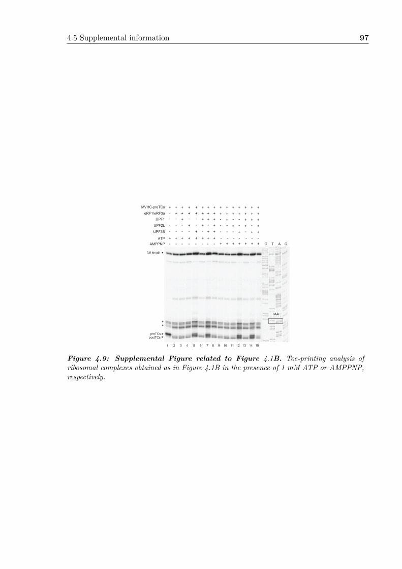
4.5 Supplemental information 97
1 2
MVHC-preTCs
eRF1/eRF3a
UPF1
UPF2L
UPF3B
ATP
AMPPNP
+ ++ ++ +
+ ++ ++ +
+ +
+ +
+ +
+ +
+ +
+ +
+ +
+ +
+ +
+ +
+ +
+ +
- - -- -- -- -
- - -- -- -- -
-
-
+
- -
+ +
+ +
- -
+ +
+ +
- -
+ +
+ +
TAA
C T A G
3 4 5 6 7 8 9 10 11 12 13 14 15
+
+
+
+
+
+
+
+
+ +
+
+
+
+
+-
-
-
-
-
-
-
-
-
-
-
-
-
-
-
-
full length
preTCspostTCs
**
Figure 4.9: Supplemental Figure related to Figure 4.1B. Toe-printing analysis ofribosomal complexes obtained as in Figure 4.1B in the presence of 1 mM ATP or AMPPNP,respectively.

98 Double-tasking of UPF3B in early and late phases of translation termination
0
20
40
60
80
100
120
0 0.5 1 1.5 2 2.5 3
A2
80
(m
AU
)
Elution Volume (mL)
eRF1UPF3BeRF1-UPF3B
1.30
1.55
1.301.50
75
50
UPF3B
75
50 eRF1
131Fractions
75
50
UPF3B
eRF1
66
21
0
67
0
29
15
0
44
0
[kDa]E
M[kD
a]
inp
ut
75
50
Lane 1 2 3 4 5 6 7
eRF3a
eRF1
UPF3B
eRF3aeRF1
UPF3B
His6 - eRF3a
+--
-+-
--+
++-
+-+
-++
+++
75
50
eRF3a
eRF1
UPF3B
B
Inp
ut
Elu
tion
[kDa]
50
75
100
150
Inp
ut
Elu
tion
Lane 1 2 3 4 5 6 7
UPF1
eRF3a
A
eRF1
50
75
100
150UPF1
eRF3a
eRF1
eRF3aeRF1
UPF1
His6 - eRF3a
+--
-+-
--+
++-
+-+
-++
+++ [kDa]
D
75
50
100
150
75
50
100
150
Inp
ut
Elu
tion
Lane 1 2 3 4 5 6
eRF3a
eRF1
UPF1
eRF3a
eRF1
UPF1
eRF3aeRF1
Mg(OAc)2
UPF1
His6 - eRF3a
+--
-+-
--+
+++
+++
+++ [kDa]
C
Figure 4.10: Supplemental Figure related to Figure 4.3. A. In vitro pulldown ofeRF1 and/or UPF1 with His-eRF3a. Protein mixtures before loading onto the beads (input)or after elution (eluate) were separated by SDS-PAGE. B. Pulldown experiment as in (A) witheRF1, UPF3B, and His-eRF3a. C. Pulldown experiment as in (B) with eRF1, UPF1, andHis-eRF3a and at 0, 2.5, and 5 mM of Mg2+ (lanes 4-6), respectively. D. Molecular mass ofUPF3B determined by size-exclusion chromatography using a Superdex 200 column combinedwith detection by multiangle laser light scattering and refractometry (SEC-MALLS-RI). TheSEC elution profiles as monitored by refractometry (RI) are represented for UPF3B. Themolecular mass (MM) of UPF3B calculated from light scattering and refractometry data isindicated. E. SEC elution profile of eRF1 (red), UPF3B (green), or both (blue). The elutionvolume (in mL) is indicated for each experiment. Calibration of the column was performedwith globular proteins (shown above). Lower panel: SDS-PAGE analysis of eluate fractions.M: protein molecular weight standards (kDa).

4.5 Supplemental information 99
66
21
0
67
0
29
15
0
44
0
[kDa]
75
50
UPF3B
MInput
131Fractions[kDa]
75
50
UPF3B
0
50
100
150
200
250
300
0 0.5 1 1.5 2 2.5 3
OD
(m
AU
)
Elution Volume (mL)
UPF3B 280nmUPF3B 260nm
UPF3B:RNA 280nmUPF3B:RNA 260nm
RNA 280nm RNA 260nm
1.30
1.22 1.77
S P S P S P S P S P S P
75
100
150250
50
37
2520
80SUPF1
UPF2LUPF3B
+---
-+--
--+-
---+
-+-+
--++
UPF3B
UPF1UPF2L
[kDa]
A
B
1 112 123 4 5 6 7 8 9 10 1 2 3 4 5 6 7 8 9 10
S P S P S P S P S P
75
100
150250
50
37
2520
80SUPF1
UPF2LUPF3B
++--
+-+-
+--+
++-+
+-++
UPF3B
UPF1UPF2L
[kDa]
C
Figure 4.11: Supplemental Figure related to Figure 4.5. A. Left: SEC elution profileof UPF3B (dotted lines), RNA (dashed lines) and the mix (solid lines). Optical density wasrecorded at 280 nm (blue) and at 260 nm (red). 40 mM of UPF3B or RNA oligonucleotide (24mer) or both were loaded onto the Superdex200 column. The elution volumes are indicatednext to the curves. Right: Eluate fractions of UPF3B alone (upper) and UPF3B-RNA mixes(lower) were analysed by SDS-PAGE. M: protein molecular weight standards (kDa). B.Sucrose cushion co-sedimentation analysis of UPF1, UPF2L, or UPF3B or in combinationsas indicated. After ultracentrifugation, the supernatant (S) and pellet (P) fractions wereanalysed by SDS-PAGE. C. Sucrose cushion co-sedimentation analysis of UPF1, UPF2L, orUPF3B as in (B) but with 80S ribosomes as indicated.

100 Double-tasking of UPF3B in early and late phases of translation termination
1 2
MVHC-preTCs
eRF1/eRF3a
UPF1
UPF2L
UPF3B+ + +
+ -+ +
+
+
+
+
+
+
+
+
+
-+
+
- - +- - +-
- - +- - -
-
-+
- ++ ++ +
- -+ -+ +
-+
+
C T A G
3 4 5 6 7 8 9 10
full length
preTCspostTCs
**
TAA
1 2
MVHC-preTCs
eRF1/eRF3a
UPF1
UPF2L
UPF3B
AMPPNP
+ + +
+ -+ +
+
+
+
+
+
+
+
+
+
-+
+
- - +- - +-
- - +- - -
-
-+
- ++ ++ +
- -+ -+ +
-+
+
TAA
full length
preTCspostTCs
C T A G
3 4 5 6 7 8 9 10
+ + + ++ + + + + +
**
A B
C
1 2
MVHC-preTCseRF1/eRF3a
UPF1UPF2L
SMG1-8-9+ + +
+ -+ -++ ++ ++ +
+ ++ ++ +
+ ++ ++ +
+++
- - - - -- - -+ + +
- - -- +- +- +
--+
- ++ ++ +
+ ++ ++ +
+ ++ ++ +
C T A G
3 4 5 6 7 8 9 10 11 12 13 14
+ + + ++ + + +- -- - - -
full length
UPF3B
preTCspostTCs
**
TAA
Figure 4.12: Supplemental Figure related to Figure 4.6. A. B. Toe-printing analysisof ribosomal complexes as in Figure 4.6A, but in the presence of 1 mM AMPPNP (A) or with-out nucleotide (B). C. Impact of UPF1-phosphorylation on e�cient translation terminationand on ribosome dissociation by UPF3B. Toe-printing analysis of ribosomal complexes ob-tained by incubating preTCs formed on MVHC-STOP mRNA (MVHC-preTCs) with UPF1,UPF2L, UPF3B, or BSA at 1 mM free Mg2+ and 1 mM ATP. In lanes 7-10 UPF1 was in-cubated with SMG1-8-9 and ATP for 30 min at 37 °C either alone (lane 7) or in the presenceof UPF2L, UPF3B, or both (lanes 8-10) before preTCs were added to the mixture and againincubated for 10 min. In lanes 11-14 UPF1 was incubated with ATP and SMG1-8-9 for 30min. Then, UPF2L and /or UPF3B were added for additional 15 min (lanes 12-14). Finally,MVHC-preTCs were added to the mixtures for 10 min followed by translation termination byeRF1 and eRF3a.

4.6 Additional results 101
4.6 Additional results
This section describes additional result, which were generated, on the interplay betweenthe human release factors and UPF proteins, but which were not included in thesubmitted manuscript.
Full length UPF2 interacts with the release factors
In order to further investigate the binding of UPF proteins to the release factors,full length UPF2 was purified from insect cells (Figure 4.13A) and additional bindingassays were performed. Complexes were resolved by incubating the sample with NiNTAa�nity resin (see the method section 4.4). After extensive washing, proteins were elutedwith imidazole and subsequently were visualized on Coomassie stained SDS-PAGE.
Immobilized full-length UPF1, UPF2 and UPF3B were tested against eRF3a (Fig-ure 4.13B). UPF3B co-purified with eRF3a as described previously (figure 4.3, 4.13Blane 7). Surprisingly, UPF2 also co-eluted with eRF3a but to a lesser degree thanUPF3B, suggesting that UPF3B might have a higher a�nity for eRF3a than UPF2(Figure 4.13B, lane 5-7). Using full-length His-tagged UPF2 for binding assays withboth release factors, eRF3a, but not eRF1AGQ alone, co-eluted with UPF2 (Figure4.13C, lane 5-7). However, a ternary complex could be formed when both releasefactors were mixed with UPF2 indicating that eRF1AGQ might be recruited to thecomplex through eRF3a (Figure 4.13C, lane 5-7). This result is consistent with a recentstudy showing that the MIF4G-3 domain of UPF2 forms a stable complex with eRF3abut not with eRF1 (López-Perrote et al., 2015). In our previous binding assays, wedid not isolate a strong complex with UFP2L indicating that the binding site mightbe an overlap between the C-terminal part of UPF2 and the MIF4G-3 (Figure 4.3).Alternatively, UPF2L was purified from E. coli and thus could lack some importantpost-translational modifications promoting the interaction with eRF3a. It was testedif the interaction between UPF2 and the release factors was dependent on the Mg2+
concentration, similarly to UPF3B. It was found that UPF2 interacted with the releasefactors in a Mg2+-independent manner (Figure 4.13).
To further investigate the interaction network, combinations of NMD factors weremixed in the presence of eRF3a. UPF2 interaction with eRF3a appear to be disturbedin presence of UFP1 (Figure 4.13D, lane 5-7). This is consistent with the in vivo pull-downs performed in Figure 4.2 where a complex comprising eRF3a, UPF1 and UPF2could not be isolated. In contrast, UPF2 interaction with eRF3a was not a�ected bythe presence of UPF3B (Figure 4.13E, lane 6-7). However, It is unclear at the mo-ment whether a ternary complex is formed or whether UPF2-UPF3 and UPF2-eRF3acomplexes are coeluted. These results should be confirmed by performing pulldownsexperiments with untagged UPF2 and by alternative methods.

102 Double-tasking of UPF3B in early and late phases of translation termination
75
100
150
250
Inp
ut
Elu
tion
Lane 1 2 3 4 5 6 7
UPF2
eRF3a
UPF3B
His6 - UPF2
+--
-+-
--+
++-
+-+
-++
+++
F.
UPF2
UPF3B
eRF3a
75
100
150
250UPF2
UPF3B
eRF3a
[kDa]
*
*
*
*
75
100
150
Inp
ut
Elu
tion
Lane 1 2 3 4 5 6 7
UPF1
eRF3a
UPF2
His6 - UPF2
+--
-+-
--+
++-
+-+
-++
+++
75100
150
UPF2
eRF3a
UPF1
UPF2
eRF3a
UPF1
[kDa]
E.
75
100
150
Input
UPF1
eRF3a
UPF2UPF3B
His6 - UPF1, UPF2, UPF3B
+---
-+--
--+-
---+
++--
+-+-
+--+
UPF2
eRF3a
UPF1
UPF3B
[kDa]
B.
75
100
150 Elu
tion
UPF2
eRF3a
UPF1
UPF3B
Lane 1 2 3 4 5 6 7
75
50
100
150
75
50
100
150
Inp
ut
Elu
tion
Lane 1 2 3 4 5 6
eRF3a
eRF1AGQ
UPF2
eRF3a
eRF1AGQ
UPF2
eRF3aeRF1AGQ
Mg(OAc)2
UPF2
His6 - UPF2
+--
-+-
--+
+++
+++
+++ [kDa]
D.
50
75
100
150
250
Input
Lane 1 2 3 4 5 6 7
UPF2
eRF3a
eRF1AGQ
50
75
100
150
250
Elu
tion
UPF2
eRF3a
eRF1AGQ
eRF3a
eRF1AGQ
UPF2
His6 - UPF2
+--
-+-
--+
++-
+-+
-++
+++
C.
A.
[kDa]
*
*
*
*
0
10
20
30
40
Rlu
0.0 0.5 1.0 1.5 2.0 2.5 3
1.50 mL
Elution Volume (mL)
UPF2
50
37
75
100
150250
[Kda] M 1 13Fractions
Input
Figure 4.13: UPF2 interacts with eRF3a. A. Full length UPF2 size exclusion profileusing Superdex 6 16/60 (GE Healthcare). B. Pulldown with hexahistidine-tagged UPF1,UPF2 or UPF3B with untagged eRF3a. Protein mixtures loaded (input) onto the beads(above) and after elution (below) were separated by SDS-PAGE followed by Coomassie stain-ing. C. Pulldown with hexahistidine-tagged UPF2 mixed with eRF1 or eRF3a or both. D.Same as C. but with increasing Mg(OAc)2 concentrations from 0 to 2.5 to 5 mM. E. Pull-down as C. but with untagged eRF3a or UPF1. F. Pulldown as C. but with untagged eRF3aor UPF3B.

4.6 Additional results 103
75100
50
37
25
20
150250
Input
UPF1
UPF1UPF1ΔUPF1L
UPF1-CH
UPF3B
UPF3B
His6 - UPF1
75100
50
37
25
20
150250
Elu
tion
UPF1UPF1ΔUPF1L
UPF1-CH
UPF3B
[kDa]WT CH ΔCH L - WT CH ΔCH L- - - - + + + + +
UPF11 115 272 322 914 1129
CH HelicaseATPase SQ
UPF1-CH115
131
287
CH
UPF1ΔCH250 914
HelicaseATPase
UPF1L115 914
CH HelicaseATPase
BA
Lane 1 2 3 4 5 6 7 8 9
Figure 4.14: UPF3B interacts with UPF1 CH domain A. Schematic representa-tion of UPF1 constructs used in B. B. Pulldown with hexahistidine-tagged UPF1, UPF1-CH, UPF1DCH or UPF1L untagged UPF3B. Protein mixtures loaded (input) onto the beads(above) and after elution (below) were separated by SDS-PAGE followed by Coomassie stain-ing.
UPF3B interacts with UPF1 CH domain
Surprisingly, UPF1 and UPF3B co-eluted in the absence of eRF3a (Figure 4.4). Pre-viously, this interaction has been tested with purified human UPF1L (aa115-914) andUPF3B but was not observed (Chamieh et al., 2008). In order to map the UPF1 inter-action domain, di�erent His-tagged UPF1 constructs were generated (Figure 4.14A).UPF1-CH comprises the CH domain of UPF1 (aa115-287) whereas UPF1DCH lacksmost of this domain (aa1-131 and 250-1129); UPF1L is described in section 4.2 andlacks both N- and C-terminal ends (aa115-314). These constructs were used in invitro pulldown assays and it was found that full length UPF1, UPF1L and UPF1-CHfragment co-eluted with a similar amount with UPF3B, indicating that the UPF1 CHdomain might mediate the interaction (Figure 4.14B, lane 6,7,9). However, UPF1DCHcould still interact with UPF3B, albeit less e�ciently, suggesting the possibility of asecond binding site (Figure 4.14B, lane 8).
UPF3B is a phosphorylated protein
Using mass spectrometry, our collaborators identified 4 phosphorylation sites in UPF3B:Y160, T169, S320 and T413 (Figure 4.15A). Interestingly, Y160 has been linked toautistic features when the tyrosine is mutated to an aspartic acid (UPF3B-Asp160)(Tarpey et al., 2007). This residue is conserved between UPF3A, and UPF3B in highereukaryotes (Figure in Appendix B.3). The UPF3B-Asp160 mutant was found to in-crease the expression of NMD targets indicating that it may reduce UPF3B activity(Alrahbeni et al., 2015). In addition, patients with UPF3B-Asp160 have higher levels

104 Double-tasking of UPF3B in early and late phases of translation termination
75
50
100 Inp
ut
Elu
tion
Lane 1 2 3 4 5 6 7
eRF3a
UPF3B T169
His6 - eRF3a
+-
-WT
-A
-E
+WT
+A
+E
75
50
100
UPF3B
eRF3a
UPF3B
eRF3a
75
100 Inp
ut
Elu
tion
Lane 1 2 3 4 5 6 7
eRF3a
UPF3B T320
75
100
His6 - UPF3B
+-
-WT
-A
-E
+WT
+A
+E
UPF3B
eRF3a
UPF3B
eRF3a
UPF2UPF1
eRF3aUPF3A/B
75100
Inp
ut
Elu
tion
Lane 1 2 3 4 5 6 7
eRF3a
UPF3B S413
75
100
His6 - UPF3B
+-
-WT
-A
-E
+WT
+A
+E
UPF3B
eRF3a
UPF3BeRF3a
75
50
100 Inp
ut
Elu
tion
Lane 1 2 3 4 5 6 7
eRF3a
UPF3B Y160
His6 - UPF3B
+-
-WT
-A
-E
+WT
+A
+E
UPF3BeRF3a
75
50
100
UPF3BeRF3a
[kDa] [kDa]
[kDa][kDa]
C eR
F3
a
M UP
F1
UP
F2
UP
F3
A
UP
F3
B
C eR
F3
a
M UP
F1
UP
F2
UP
F3
A
UP
F3
B
[kDa]
150
100
75
50
37
ProQ stainingCoomassie Blue
A
C
E F
B
D
Y160 T320T169 S413
UPF3B1 49 279 4
21
43
4
470
RRM-like
EBM
Figure 4.15: UPF3B phosphorylation does not impair eRF3a binding A. Schematicrepresentation of UPF3B. 4 phosphorylation sites identified by mass spectrometry are mappedto the sequence with their respective amino acids and position. RRM-like: RNA-recognitionmotif-like, EBM: EJC-binding motif. B. SDS-PAGE analysis of purified recombinant pro-teins. Left and right panels were stained with Coomassie blue or ProQ (Thermo scientific)staining respectively. C. Pulldown with hexahistidine-tagged WT or Y160A or Y160E mu-tant UPF3B and eRF3 alone or mixed together. Protein mixtures loaded (input) onto thebeads (above) and after elution (below) were separated by SDS-PAGE followed by Coomassiestaining. D. Pulldown with hexahistidine-tagged eRF3 and untagged WT or T169A or T169Emutant UPF3B alone or mixed together. E. Same as C. but with T320A or T320E UPF3Bmutants. F. Same as C. but with S413A or S413E UPF3B mutants.

4.6 Additional results 105
of UPF3A, similarly to patients lacking UPF3B. The T169 site was found to be a caseinkinase 2 (ck2) substrate motif (TDNE). Interestingly, ck2 has already been reported asa putative kinase for yeast Upf2p (Wang et al., 2006). No information was found forS320 and T413 except that T413 phosphorylation was only observed in the presence ofUPF2. The phosphorylation status of the UPF proteins were assessed by ProQ stain-ing (Thermo Scientific) (Figure 4.15B). Surprisingly, it was found that eRF3a, UPF2,UPF3A and UPF3B all showed strong signals after ProQ staining indicative of phos-phorylation during protein expression in insect cell. However, following purification,UPF1 did not appear to be hyperphosphorylated (Figure 4.15B). In order to assessthe e�ect of the phosphorylation on UPF3B interactions, the amino acids Y160, T169,S320 and T413 were mutated to either glutamic acid (mimicking a phosphorylatedform) or an alanine residue. Thus, 8 UPF3B mutants were generated recombinantlyand probed for binding to eRF3a. Using the same in vitro binding assays as describedin section 4.2, we found that none of these mutations alone could impair the bind-ing of UPF3B to eRF3a (Figure 4.15C-F). However, these binding assays are ratherinsensitive and cannot discriminate between small changes in dissociation constants.It would be interesting to measure the Kds of di�erent UPF3B mutants for eRF3a.Alternatively, these amino acids could be important for another function of UPF3B,for example binding to the ribosome or to another partner.
UPF3B could bridge UPF2 to the ribosome
To explore the ability of UPF3B to interact with ribosomes, co-sedimentation assayswere performed (Figure 4.16). Briefly, the protein was added to the 80S ribosome andcentrifuged through a sucrose cushion. Only the proteins interacting with the ribosomewill sediment to the pellet fraction. Firstly, the bu�er conditions were optimized. Cen-trifugation of UPF proteins without 80S ribosome served as controls to confirm thatthe proteins remained in the supernatant fraction. Two di�erent volume of sucroselayer and two di�erent bu�er combinations were tested (Figure 4.16A). Subsequently,ribosome pelleting assays were performed using 10-fold molar excess of UPF proteins.It was found that UPF3B co-sediments with 80S ribosomes indicating that UPF3B canbind ribosomes (Figure 4.16A). Conditions were further optimized to 400 µL of sucrosecushion in HEPES/KOH bu�er with KOAc as it allowed only a fraction of UPF3Bto bind to 80S, thus minimizing unspecific binding. Magnesium levels have previouslybeen shown to be critical for ribosomal subunits stability,(Shenvi et al., 2005). Inaddition, it was already found that UPF3B interaction with the release factors wasdependent on the Mg2+ level (Figure 4.10). Thus, the binding of UPF3B to the ribo-some was next assessed at di�erent Mg2+ concentration (Figure 4.16B). The bindingof UPF3B to the 80S ribosome was only slightly depending on the Mg2+ concentrationand UPF3B binding could still be observed above 5 mM Mg(OAc)
2
. It was decided
106 Double-tasking of UPF3B in early and late phases of translation termination
to keep 2.5 mM Mg2+ which allowed stable association of UPF3B with 80S ribosomeduring sedimentation. Following these experiments it was investigated whether the in-teraction was depending on the salt concentration (Figure 4.16C). UPF3B binding tothe ribosome was lost at increasing salt concentrations (Figure 4.16C lanes 6 vs 10 vs14). It was decided to use 250 mM KOAc for further experiments as UPF3B could onlypartially bind to the 80S as indicated by UPF3B presence in the supernatant fraction(Figure 4.16C lane 10). Next, the influence of full length UPF2 on UPF3B ribosomalbinding was investigated1. Similarly to UPF2L, full length UPF2 only weakly boundto the 80S ribosome. The ribosomal binding of UPF2 was increased in presence ofUPF3B, indicating that UPF3B may recruit full length UPF2 to the 80S ribosome(Figure 4.16D lane 10 vs 14). Subsequently, the ability of UPF2 to co-sediment withthe ribosome in the presence of UPF1 was assessed. In contrast to UPF3B, UFP1presence did not significantly increase UPF2 co-sedimentation with the 80S ribosome(Figure 4.16D lane 10 vs 14). This result suggests that only UPF3B could bridge UPF2to the ribosome. In future experiments, UPF3B binding to either ribosomal subunitsshould be assessed.
1UPF3B binding to the 80S ribosome in the presence of UPF1 was tested in the manuscript. Pleaserefer to section 4.2
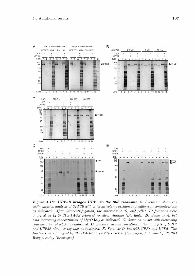
4.6 Additional results 107
75100
50
37
25
1510
M S P S P S P S P S P S P S P
UPF3B
Mg(OAc)2 2.5 mM 5 mM 10 mM
+-
-+
++
-+
++
-+
++
80SUPF3B
[Kda]
Lane 1 2 3 4 5 6 7 8 9 10 11 12 13 14
Lane 1 2 3 4 5 6 7 8 9 10 11 12 13 14
75100150250
50
37
25
1510
M S P S P S P S P S P S P S P
UPF3B
KOAc 150 mM 250 mM 500 mM
+-
-+
++
-+
++
-+
++
80SUPF3B
[Kda]
75100
50
37
25
1510
S P S P S P S P S P S P S P S P M
UPF3B
-+
++
-+
++
80SUPF3B
[Kda]
Lane 1 2 3 4 5 6 7 8
HEPES / KOAc Tris / KCl
-+
++
-+
++
HEPES / KOAc Tris / KCl
9 10 11 12 13 14 15 16
400 µL sucrose cushion 80 µL sucrose cushion
S P S P S P S P S P S P S P
80sUPF2
UPF3B
+--
-+-
--+
-++
++-
+-+
+++
UPF2
UPF3B75
100
150250
50
37
2515
[Kda]
Lane 1 2 3 4 5 6 7 8 9 10 11 12 13 14
S P S P S P S P S P S P S P
80sUPF1UPF2
+--
-+-
--+
-++
++-
+-+
+++
UPF2UPF1
75
100
150250
50
37
2515
[Kda]
Lane 1 2 3 4 5 6 7 8 9 10 11 12 13 14
A B
C
D E
Figure 4.16: UPF3B bridges UPF2 to the 80S ribosome A. Sucrose cushion co-sedimentation analysis of UPF3B with di�erent volume cushion and bu�er/salt concentrationsas indicated. After ultracentrifugation, the supernatant (S) and pellet (P) fractions wereanalyzed by 12 % SDS-PAGE followed by silver staining (Bio-Rad). B. Same as A. butwith increasing concentration of Mg(OAc)2 as indicated. C. Same as A. but with increasingconcentration of KOAc as indicated. D. Sucrose cushion co-sedimentation analysis of UPF2and UPF3B alone or together as indicated. E. Same as D. but with UPF1 and UPF2. Thefractions were analyzed by SDS-PAGE on 4-12 % Bis-Tris (Invitrogen) following by SYPRORuby staining (Invitrogen)


5. Discussion
Resumé en françaisLes travaux e�ectués durant cette thèse ont pour but de fournir des informationsbiochimiques et structurales sur la termination de la traduction lors de la dégrada-tion d’ARNm non-sens (NMD). Ceux-ci se basent sur des systèmes de traduction invitro de levure et d’homme. Nos systèmes nous ont permis de reconstruire des com-plexes de termination de levure ainsi que de tester l’impact de di�érents facteurs NMDsur la termination chez l’homme. De tels systèmes sont essentiels pour l’enrichissementdes connaissances sur le rôle des facteurs NMD dans la termination de la traduction.
Ce travail de thèse apporte pour la première fois l’évidence que UPF3B, et nonUPF1, a un e�et dans la termination et le recyclage de la traduction, contrairementau courant de pensée établi. Nos résultats suggèrent un nouveau mécanisme pour lareconnaissance des PTCs et soulignent aussi une implication directe de la NMD dansle recyclage des ribosomes positionnés au niveau d’un PTC. De nombreuses questionsrestent à élucider notamment sur le fonctionnement de la NMD dans la branche in-dépendante de UPF3B ou sur le rôle de UPF3A dans la termination. Nos travaux sontun point de départ solide pour de futures études fonctionnelles et structurales sur descomplexes de terminations en levure et en humain.
La NMD est un mécanisme de contrôle complexe qui connecte di�érents proces-sus cellulaires dans l’espace et le temps. Sa compréhension représente un challengeformidable en particulier pour le développement de nouvelles stratégies thérapeutiquesdans le traitement des maladies associées au NMD.
110 Discussion
Despite decades of investigation, the interplay between translation and NMD fac-tors allowing the discrimination between NTCs and PTCs remained unclear. Multiplesmodels have been proposed for the mechanism of premature translation terminationand activation of NMD, but all failed to restitute complexity of the pathway. ThisPhD work was aiming at providing biochemical and structural information on thetermination of translation in the presence of NMD factors. In vitro translation sys-tems for yeast and human were established in our laboratory, which permitted thereconstruction of termination complexes and functional probing of the di�erent fac-tors. This work yielded the first evidence that UPF3B and not UPF1 has an activityin termination of translation and dissociation of PTC-stalled ribosomes. In the fol-lowing sections, I discuss the implications of our results in our understanding of thevarious steps from termination to ribosome recycling during NMD. The final sectionwill discuss the repercussion of our understanding of NMD.
5.1 Binding of NMD and termination factors at aPTC
5.1.1 Interplay between eRF1-3a and the NMD factors
In contrast to other surveillance pathways, NMD requires the canonical release factorsand additional factors to terminate translation and rescue ribosomes stalled at a pre-mature termination codon (Figure 5.1). However, toeprint experiments suggest thattermination is slower at a PTC than at a NTC in yeast (Amrani et al., 2004). Thisdelay was attributed to the absence of Pab1p, which would allow the recruitment ofUpf1 by the release factors. In yeast, Upf1p was shown to interact with eRF1 and eRF3(Wang et al., 2001; Kervestin et al., 2012; Roque et al., 2014). Yet we were not able toconfirm a direct in vitro interaction between eRF3a and UPF1 from both human andyeast as our experiments showed ambiguous results (Figure 3.9, 4.3). Human UPF2,in contrast, interacted directly with eRF3a, but not with eRF1 in our pulldown experi-ments, confirming the result from (López-Perrote et al., 2015). However, no complexesbetween the release factors and UPF2 were isolated by in vivo immunoprecipitations,indicating that UPF2 might bridge the release factors with other factors. Yet, it mightnot partake in the resulting interactions and thus it remain unclear which UPF proteincould interact with the release factors both in vivo and in vitro. In our experiments,UPF3B was the only NMD factor, able to bind directly to eRF1 and to eRF3a both invitro and in vivo and thus potentially able to interfere with termination (Figure 4.3).
UPF1 is thought to associate with NMD-target mRNAs and possibly the elongatingribosomes (He & Jacobson, 2015b). This would explain the co-immunoprecipitation of

5.1 Binding of NMD and termination factors at a PTC 111
UPF1 with the release factors in an RNA-independent manner (Figure 4.2). However,it remains unclear whether UPF1 can mark stop codons as aberrant or if additionalfactors are involved in the decision (He & Jacobson, 2015b). In the toeprint assays,we could not detect any e�ect of UPF1 or UPF2 alone on the stop codon recognitionprocess, indicating that they may not be active on their own during termination oftranslation as previously proposed (Figure 4.1) (He & Jacobson, 2015b). Our resultsindicate that UPF3B is the only NMD factor which could, in addition to interactingwith both release factors, impair the stop codon recognition and peptide release. Wepropose that the presence of UPF3B nearby a terminating ribosome could form thebasis of a molecular mechanism signaling aberrant termination to the NMD machinery.Our results challenge the prevailing NMD models, which place UPF1 at center stage,and shed light on previously unknown functions of UPF3B in PTC recognition andribosome rescue after termination at a PTC.
However, in the cell, additional mRNP factors, such as the EJC or PABPC1, arelikely to be involved. The e�ect of UPF3B in stop codon recognition and releasefactors binding remains to be investigated in vivo. Such an endeavor will require thedevelopment and application of new techniques such as real time in vivo imaging (Yanet al., 2016) and innovative transcriptome-wide approaches (Pelechano et al., 2015;Archer et al., 2016). The latter approach has already revolutionized our view of NMDbut remains to be extended to other NMD factors(Zünd et al., 2013; Hogg & Go�,2010; S. R. Lee et al., 2015; Hauer et al., 2016).
5.1.2 eRF3a as a node for the integration of termination sig-nals
5.1.2.1 Possible competition between PABPC1 and UPF3B during termi-nation
The N-terminal part of Hbs1 interacts with the 40S subunit proteins S3 and S30 nearthe mRNA entry site and might have a regulatory activity in no-go decay (NGD) ac-tivation, an RNA surveillance mechanism for mRNAs stalled in translation elongation(Becker et al., 2012). Consistent with this idea, ribosomal subunit dissociation in NGDis sensitive to mRNA length in the presence of Hbs1 (Pisareva et al., 2011). As Hbs1and eRF3 share homology, it has been hypothesized that the eRF3 N-terminal wasable to bind the same ribosomal surface and to help to regulate termination e�ciency(Franckenberg et al., 2012). In support of this idea, the yeast eRF3 N-terminal regionwas proposed to regulate the binding of eRF1 binding site in a competitive manner(Kong et al., 2004). In addition, the Faux 3’ UTR model postulates that Pab1p couldpromote termination and antagonize nonsense mediated mRNA decay by associatingwith the eRF3 N-terminal (Amrani et al., 2004; Kervestin et al., 2012; A. Ivanov et

112 Discussion
al., 2016).However, in our biochemistry and cryo-EM studies of yeast proteins, we could
not reproduce the direct binding of Pab1p to eRF3 observed previously (Figure 3.9)(Kervestin et al., 2012; Roque et al., 2014). Since these interactions have been reported(Wang et al., 2001; Cosson et al., 2002a), only the interaction between human eRF3aand PABPC1 has been solved by x-ray crystallography (Kozlov & Gehring, 2010). Wereasoned that the competition model between Upf1p and Pab1p for the binding of eRF3should be investigated more thoroughly in yeast. In our experiments, a stable bindingof yeast Upf1p or Pab1p to the yeast release factors could not be confirmed. Thismay be explained by a rather transient nature of their interaction. Alternatively, itcannot be excluded that these interactions may be promoted by other mRNP factors inyeast. In mammalian NMD, the interaction between UPF3B and the eRF3a N-terminaldomain raises the possibility of competition between human UPF3B and PABPC1for the binding of eRF3a (Figure 4.3). Competition in vitro binding assays can beperformed to investigate possible competition between UPF3B and PABPC1 for theeRF3a N-terminal domain. Additionally, further mapping of UPF3B interaction witheRF3a N-terminal by peptide spot arrays could be performed to find out whetherthe PABPC1-binding motifs of eRF3a overlap with the UPF3B-interacting region. Incontrast, recent studies have proposed that the PABPC1 interaction with eRF3a N-terminal may be important for post-termination and reinitiation but not for mRNAdecay (Joncourt et al., 2014; Durand & Lykke-Andersen, 2013; Roque et al., 2014).Thus, competition between UPF3B and PABPC1 for the eRF3a N-terminal regionwould be di�cult to reconcile with these recent observations. However, it could alsobe a sign of partially overlapping roles for both proteins in recycling and reinitiationof translation.
5.1.2.2 Possible competition between eI3j and UPF3B following termina-tion
In yeast, the j subunit of eukaryotic initiation factor 3 (eIF3j) has been shown to bindto the ribosomal protein S30 on the small subunit (40S) and eRF3 (Beznosková etal., 2013; Aylett et al., 2015). It was proposed that eIF3j could promote eRF3-GDPdissociation from the ribosome to allow e�cient recycling by ABCE1 in a ‘termination-initiation’ complex (Beznosková et al., 2013). Our work is consistent with the factsthat UPF3B associates with the eRF3a N-terminal part and with the ribosome andslows down peptide release. Interestingly, in tethering experiments, the deletion ofthe UPF3B RRM-like domain and the first predicted coiled coil (aa 49-279) impairedthe activation of translation observed by wild-type UPF3B (Appendix B.3) (Kunz etal., 2006). Based on these observations it is tempting to speculate that UPF3B couldinfluence the eRF3a GTP hydrolysis rate or the dissociation rate from the ribosome

5.2 NMD factors control the recycling and initiation of ribosomes 113
E P A
tRNAieEF1H+GTP
AAAAAA
eIF3jABCE1
PABPC1
UPF3B
eRF1-3a
UPF2
?
?
Re-initiationRecycling
Termination
NMDNMD
NGD
Elongation
80S
S3/30 E P A
ABCE1
PelotaHbs1
Recycling
NGD
80S
S3/30PTC
UPF1
Figure 5.1: eRF3a and the ribosome integrate signals for mRNA quality con-trol. Left panel: during recognition of a premature termination codon (PTC in blue box)by nonsense mediated mRNA decay (NMD), eRF3a N-terminal could integrate stimulatoryand inhibitory signals (green and red arrows respectively) from various factors: poly(A) bind-ing protein PABPC1, recycling factor ABCE1, initiation factor eIF3j and the NMD factorsUPF1, UPF2 and UPF3B; in order to decide if the quality control pathway should be acti-vated. Right panel: similarly, during no-go decay (NGD) activation, NGD factor Pelota com-pete with elongation factors bound to tRNA. Subsequent to translation termination, Pelotarecruits ABCE1 to rescue the stalled ribosome.
through a mechanism reminiscent of eIF3j. However, our toeprints assays suggest thatUPF3B’s activity could be independent of the eRF3a N-terminal domain (Figure 4.5).To shed light on UPF3B’s action in termination and ribosome recycling, the influenceof UPF3B on eRF3a-ribosome association and GTP hydrolysis should be determined(Figure 5.1). Atomic resolution structures of terminating ribosomes in the presenceof full length eRFs and/or UPF3B would yield additional insights into the impact ofUFP3B on translation termination.
In summary, the role of the eRF3a N-terminal domain remains obscure. It could becritical to integrate the di�erent inhibitory or stimulatory signals during the termination-recycling-initiation (Figure 5.1).
5.2 NMD factors control the recycling and initia-tion of ribosomes
5.2.1 Ribosome dissociation following PTC recognition
The importance of proper recycling of the translation components in mRNA surveil-lance was illustrated by Young and colleagues who found that in the absence ofABCE1/Rli1 and Pelota/Dom34, yeast 80S ribosomes accumulated at stop codons and

114 Discussion
in the 3’UTR of mRNAs, possibly due to aberrant reinitiation (Young et al., 2015).eRF1-eRF3a were reported to destabilize the association between the P-site tRNA andthe 60S subunit following peptide release (Shoemaker et al., 2010). When the releasefactors were saturating, our results indicate that UPF3B could dissolve postTCs follow-ing peptide hydrolysis (Figure 4.6B, 5.2). This energy-free mechanism was reminiscentof eIF3, eIF3j, eIF1, and eIF1A mediated ribosome recycling which operates only atlow Mg2+ concentrations (1 mM Mg2+) (Pisarev et al., 2007). We found that at Mg2+
concentration higher than 5 mM, UPF3B could not dissolve postTCs nor interact withthe release factors (Figure 4.3, 4.6) indicating that UPF3B’s role in termination ishighly sensitive to Mg2+ concentrations and possibly providing a molecular reason forthe Mg2+ homeostasis by NMD (Johansson & Jacobson, 2010). Previous toeprint stud-ies have suggested that ribosomes dissociate slower at PTCs than NTCs (Amrani et al.,2004). Concordantly, our data indicate that UPF3B cannot dissociate all the postTCsindicating that the process is likely to be less e�cient than ABCE1-mediated ribosomedissociation (Figure 4.6). Moreover, UPF2 appears to inhibit UPF3B’s capacity to de-lay termination and to dissolve postTCs. At the same time, our sedimentation assaysindicate that UPF2 could associate with the ribosomes more e�ciently in the pres-ence of UPF3B. These two observations appear to be contradicting, but may representdi�erent activities of the UPF proteins at di�erent stages during NMD. One possibleexplanation is that UPF3B could be recruited to the terminating ribosome in an in-active form bound to UPF2 and other mRNPs (Figure 5.2). This idea is consistentwith the fact that UPF3B interaction with UPF2 is not required to activate NMD,but UPF3B is required to dissociate from the UPF1-UPF2-EJC complexes after UPF1phosphorylation and activation of NMD, for subsequent mRNA decay (Gehring et al.,2003; Ohnishi et al., 2003; Kashima et al., 2010). In addition, UPF3B activity couldbe fine-tuned by the competition with UPF3A in mammals (Shum et al., 2016) andMg2+ levels (Johansson & Jacobson, 2010). Peptide release assays and sedimentationon a sucrose gradient with di�erently labeled components of the termination machinerywould yield insight into the UPF3B mechanism of action on the ribosome.
5.2.2 Translation repression and reinitiation
In contrast to eRF1, Pelota cannot trigger peptide release (Brandman & Hegde, 2016).Thus, following ribosome dissociation by ABCE1 bound to Pelota in NGD, the 60Ssubunit is still bound to the peptidyl-tRNA and is then recognized by the ribosomequality control factors NEMF and Listerin (Figure 5.2) (Shao & Hegde, 2014; Shaoet al., 2015). These factors inhibit reassociation of the 60S subunit with the 40Ssubunit and thus prevent reinitiation of translation on faulty mRNAs (Brandman &Hegde, 2016). Similarly, in vitro re-initiation following termination was shown to be less

5.2 NMD factors control the recycling and initiation of ribosomes 115
Ribosomal dissolution
Inhibition ofreinitiation
NMD NGD
PTC other proteins? 40S 60S E P A
poly-Ub
NEMF
Listerin
eIF1
Met-tRNAiMet
eIF1A
UPF3B
UPF1
UPF2
eIF3
E P A ?
E P APTC
? ABCE1
Pelota
eRF1
E P A
eIF5eIF2
Figure 5.2: mRNA surveillance mechanisms and recycling and initiation control.Left panel: following translation termination, UPF3B promotes ribosomal dissociation. Itremains to be unclear whether UPF1 and UPF2 participate in this activity. NMD activationalso triggers inhibition of reinitiation by 40S bound to initiation factors eIF3, eIF1, eIF1A,eIF2 and eIF5 though phosphorylated UPF1 binding to eIF3a subunit (Isken et al., 2008)Right panel: during no-go decay (NGD), ABCE1 bound to Pelota mediates the dissociation ofthe ribosome using ABCE1’s ATPase activity (Red squares). Subsequently, ribosome qualitycontrol factors Listerin and NEMF mediate nascent chain ubiquitination (poly-Ub) resultingin inhibition of 60S association with the 40S subunit. Adapted from (Brandman & Hegde,2016)

116 Discussion
e�cient following termination at a PTC compared to normal termination (Ghosh et al.,2010). Moreover, reinitiation was suggested to inhibit NMD (Zhang & Maquat, 1997).This indicates that NMD factors could directly repress the reinitiation of translation ofPTC-containing mRNAs (Figure 5.2). Protein association during scanning, initiationand the first few rounds of elongation is controlled by the largest initiation factoreIF3, consisting of 13 subunits (eIF3a-m) (Hinnebusch, 2014; des Georges et al., 2015;A. S. Y. Lee et al., 2015). In addition to a role in recycling identified for eIF3g,eIF3i and eIF3j, evidence exists that eIF3 has a critical role in NMD (Pisarev et al.,2010; Beznosková et al., 2013). Phosphorylated UPF1 might prohibit 80S assemblyby interacting with eIF3a (Isken et al., 2008). eIF3f and eIF3h appear to preventNMD while subunit eIF3e appears to be required for NMD and interaction with UPF2(Morris et al., 2007; Peixeiro et al., 2012). It is thus possible that eIF3 promotes thetransition from termination to initiation while the UPF factors control the kineticsof the overall process for each stop codon. Therefore, mRNAs failing to reinitiatee�ciently would trigger the activation of the NMD factors. This is consistent with thehypothesis that all mRNAs are primed for NMD and the fact that UPF1 and UPF3Bare also bound in the 3’UTR of normal mRNAs (Hogg & Go�, 2010; Zünd et al.,2013; S. R. Lee et al., 2015; Hauer et al., 2016). Following this logic, Brogna andcolleagues suggested a new NMD model in which NMD is the result of a prematurerelease of ribosomes from the mRNA rather than a discrimination mechanism betweennormal and aberrant stop codons (Brogna et al., 2016). The model predicts thattranslating ribosomes would passively stabilize transcripts. Premature release of theribosomes and blocking of reinitiation by NMD factors would leave large regions ofthe mRNA exposed to nucleolytic attacks. However, co-translational 5’ to 3’ decayhas been reported and challenges the idea that ribosomes would significantly stabilizeand prevent mRNAs from decay (Pelechano et al., 2015). Thus, the interplay betweeneIF3 and the UPFs proteins during the di�erent initiation complex conformations (seeintroduction 2.1.2.1) and termination remains to be further investigated in order tounderstand its significance (Figure 5.2).
5.3 mRNA and nascent chain degradation
Following ribosomal dissociation during NGD rescue, a mechanism allows NEMF andListen to access the nascent chain by binding to the 60S interface and next to theribosomal tunnel exit (Figure 5.2) (Shao & Hegde, 2014). NEMF and Listerin areresponsible for the ubiquitination of the nascent chain, resulting in its degradation bythe proteasome (Figure 5.2). In contrast, little is known about the degradation ofnascent chains following NMD. UPF1 has been shown to promote the degradation ofnonsense peptides and to act as an E3 Ligase. In fact, the CH domain of UPF1 shares

5.3 mRNA and nascent chain degradation 117
60S
poly-Ub
NEMF
Listerin
?
Proteasome
nascent peptide
nascent peptide
Proteasome
SMG6
SMG5-7
XRN1 Exosome
XRN1 Exosome
UPF1
UPF1UPF3
?
P
P
PTC
Protein decay
mRNA decay
NMD NGD
Figure 5.3: mRNA surveillance mechanisms and protein and mRNA decay con-trol. Left panel: the proteasome is responsible for the degradation of ubiquitinylated proteins.Evidence exists that in nonsense mediated mRNA decay (NMD), UPF3B could activate UPF1E3 ligase activity to promote nascent chain degradation (Takahashi et al., 2008). Decay ofpremature termination codon (PTC) containing mRNAs by XRN1 and the exosome is accel-erated by SMG6 and SMG5-7 which recruit these factors to the mRNA as described in Figure2.8. Right panel: During no-go decay (NGD), Listerin and NEMF are responsible for thepoly-ubiquitination (poly-Ub) and the targeting of the nascent chain to the proteasome (Shaoet al., 2015). The molecular mechanisms of recruitment of mRNA decay factors during NGDremain unclear.
homologies with E3 RING finger domains. Yeast Upf1p has been reported to self-ubiquitylate in vitro in an Upf3p-dependant manner (Kuroha et al., 2009; Takahashiet al., 2008). Supporting this idea, our results indicate that the CH domain of humanUPF1 binds to UPF3B, but additional regions of UPF1 might further stabilize theinteraction (Figure 4.14). The toeprint assays from chapter 4 indicate that UPF1 didnot influence the activity of UPF3B during termination of translation. Hence, thefunctional roles of UPF1 association with UPF3B and the ubiquitination of UPF1remain unclear. UPF1’s ubiquitinylation activity could be assessed using an in vitroreconstituted ribosome-associated ubiquitination system (Shao & Hegde, 2014). Itis tempting to speculate that UPF3B could not only bridge UPF1 to downstreammRNPs, as proposed by the EJC model, but also be involved in the regulation ofUPF1 activity in a later stage of aberrant termination to promote e�cient nascentpeptide degradation.
mRNA decay is well documented for NMD (see introduction section 2.3.7) comparedto NGD (J. Lykke-Andersen & Bennett, 2014) (Figure 5.3). However, this work risesthe question of why and how UPF3B may interfere with UPF1 phosphorylation by theSMG1-8-9 complex (Figure 4.1) which is the critical step during NMD activation andsubsequent mRNA decay. Binding a�nity and phosphorylation kinetic measurementscoupled with mass spectrometry (to identify specific phospho-sites) would allow theprobing of the influence of UPF3B on UPF1 phosphorylation. These experiments

118 Discussion
have already been performed for UPF1 alone (Deniaud et al., 2015). Using massspectrometry, we identified 4 phosphorylation sites on recombinant UPF3B purifiedfrom insect cells (Y160, T169, S320 and T413). The mutation of Y160 has beenpreviously reported to fail to activate NMD in tethering experiments (Alrahbeni et al.,2015). In our assays, it was not possible to observe any impact of such modificationshence the physiological significance remains to be investigated (Figure 4.15).
5.4 Regulation of NMD and outlookHelicases play a critical role in coordinating the various steps during mRNA metabolism,processing and quality control (Bourgeois et al., 2016). As discussed in the introduction(see section 2.2), mRNA processing and degradation are in direct kinetic competition.This is exemplified by the spliceosome assembly, which is a succession of defined con-formations. Transitions between steps are coupled in time with ATP hydrolysis byhelicases (Query & Konarska, 2006). Aberrant mRNPs which are too slow to transi-tion between conformational states within the ATP hydrolysis rate will be targeted fordegradation (Villa et al., 2008). Most likely, the same principle applies to translationtermination and NMD. Over recent years, multiple helicases involved in termination oftranslation and NMD have been identified, of which initiation factor eIF4A and NMDfactor UPF1 are the best characterized (Bourgeois et al., 2016). Recently, several newlydiscovered helicases have been linked to termination and NMD (Figure 5.4). For ex-ample, yeast Dbp5/DDX19B has been shown to mediate eRF1 stop codon recognition(Gross et al., 2007), DHX34 and MOV10 were identified as a helicases contributingto UPF1 activity in NMD (Gregersen et al., 2014; Hug & Cáceres, 2014). HumanDDX5 has been shown to bind to UPF3B in vivo (Geißler et al., 2013). The impactof these helicases on kinetically coordinating mRNP remodeling steps remains to beestablished. Evidence has accumulated on the critical role of ATPase activity of UPF1,but its function, site of action and time of activation in the NMD pathway remains tobe elucidated (He & Jacobson, 2015b).
Lastly, a critical achievement in the field would be to improve the current in vitrotranslation system in order to recapitulate the NMD pathway in vitro. There is cur-rently no reconstituted in vitro NMD assay. Here we performed experiments withterminating ribosomes, release factors, UPFs and SMGs proteins but new techniquesmust be developed to dissect the recruitment of the NMD machinery and its activationto trigger mRNA decay. Moreover, it will now be important to corroborate our findingthat UPF3B has a direct impact on translation termination in vivo, using mutationalstudies (UPF3B variants) and ribosome profiling (Ingolia et al., 2012; Archer et al.,2016), in addition to the classical in vivo NMD and stop codon readthrough assayswith reporter mRNAs.

5.5 Concluding remarks 119
DDX6 SKI2
NMD
DDX5DHX34MOV10UPF1
DDX6DDX3XDDX25 DDX48
DDX9DDX29
DDX33
DDX4DDX19B
E P A
Startm7G
Stop
AAAAAAAAE P A
E P A
Stop
A
eIF4G
eIF4F
eIF4E eIF4A
PABPC1mRNA
mRNA Decay
XRN1 Exosome
Figure 5.4: mRNA translation and decay regulation by helicases. The eIF4F com-plex (eIF4A, E and G) regulates 43S loading onto the mRNA and scanning for the startcodon. This process is regulated and depends on several DEAD box helicases that help to un-wind the 5’UTR. Ribosomal subunits are represented in yellow. DDX4 and DDX19 helicaseshave been shown to impact translation termination. Following translation, mRNA decay iscarried out by exonucleases XRN1 and the exosome; both complexes contain subunits withhelicase function. Nonsense mediated mRNA decay (NMD) has been shown to be regulated byDDX5, DHX34 and MOV10 helicases. All helicases identified in the regulation of the di�er-ent steps from translation to decay are indicated in orange boxes. Adapted from (Bourgeoiset al., 2016)
5.5 Concluding remarks
There has been much e�ort has been made to investigate NMD and the role of UPF1 inaberrant termination. In recent years, several studies have expanded and supersededour view of the NMD pathway mechanism and shed light on its complexity and theimplication of numerous additional factors involved in PTC discrimination and down-stream events. The main achievement of this thesis work was to probe the activityof the NMD factors, alone or in combination, in a reconstituted in vitro translationsystem. The finding that UPF3B has a role in termination in vitro may constitute amajor step forward in our understanding of the NMD pathway. It is the first time thata NMD factor has been shown to directly influence the stop codon recognition andpeptide release during in vitro termination of translation. NMD links multiple stepswithin the mRNA life cycle, from splicing in the nucleus to translation and degradationin the cytoplasm. The pathway thus constitutes a densely meshed interactome net-work which must be tightly regulated in time and space in the cell. Its understandingconstitutes a formidable challenge.
Classic in vitro biochemistry and genome-wide approaches in combination in withinnovative single molecule imaging techniques (Yan et al., 2016) and advanced ribosome

120 Discussion
profiling technologies (Archer et al., 2016) are required to further our understandingof the complex interplay of NMD factors and the translation machinery. Recent ad-vances in cryo-EM regarding direct electron detectors (Veesler et al., 2013) and imageprocessing (S. H. W. Scheres, 2012; S. H. w. Scheres, 2014) have revolutionized thestructural biology field and allowed solving high-resolution structures from sparse andheterogeneous samples. There is no doubt that cryo-EM structures will provide criticalinsights into the interplay between the translational machinery with the NMD factorsonce the relevant ribosomal complexes have been identified.
A mechanistic understanding of the recognition of premature stop codons wouldfacilitate the development of new therapeutic strategies to treat NMD-associated dis-eases. Current treatments rely on stop codon read through approaches. An improvedmechanistic understanding of NMD could allow the targeting of early NMD stepsrather than the translation machinery in general (Martin et al., 2014). The futureof healthcare is moving towards personalized medicine and the identification of partic-ular mutations will determine an appropriate treatment. As CRISPR/Cas9 technologyis improving, understanding of the rules that govern NMD activation will o�er the pos-sibility to amend mutations leading to a PTC by specific replacement of faulty geneswith correct DNA sequences (Popp & Maquat, 2016).
In conclusion, despite many years of study, translation termination at a PTC andNMD activation remain enigmatic. The future expansion of our understanding of NMDholds promises for both technique development to elucidate its mechanism and for thetreatment of NMD-associated diseases.

References
Addington, A. M., Gauthier, J., Piton, A., Hamdan, F. F., Raymond, a., Gogtay, N., . . .Rouleau, G. a. (2011). A novel frameshift mutation in UPF3B identified in brothersa�ected with childhood onset schizophrenia and autism spectrum disorders. MolecularPsychiatry, 16 (3), 238–239.
Alexandrov, A., Colognori, D., Shu, M.-D., & Steitz, J. A. (2012). Human spliceosomalprotein CWC22 plays a role in coupling splicing to exon junction complex depositionand nonsense-mediated decay. Proceedings of the National Academy of Sciences of theUnited States of America, 109 (52), 21313–8.
Alkalaeva, E. Z., Pisarev, A. V., Frolova, L. Y., Kisselev, L. L., & Pestova, T. V. (2006). InVitro Reconstitution of Eukaryotic Translation Reveals Cooperativity between ReleaseFactors eRF1 and eRF3. Cell, 125 (6), 1125–1136.
Allmang, C., Petfalski, E., Podtelejnikov, A., Mann, M., Tollervey, D., & Mitchell, P. (1999).The yeast exosome and human PM-Scl are related complexes of 3’ - 5’ exonucleases.Genes and Development, 13 (16), 2148–2158.
Alrahbeni, T., Sartor, F., Anderson, J., Miedzybrodzka, Z., McCaig, C., & Müller, B. (2015).Full UPF3B function is critical for neuronal di�erentiation of neural stem cells. Molec-ular brain, 8 (1), 33.
Amrani, N., Ganesan, R., Kervestin, S., Mangus, D. A., Ghosh, S., & Jacobson, A. (2004).A faux 3’-UTR promotes aberrant termination and triggers nonsense-mediated mRNAdecay. Nature, 432 (7013), 112–118.
Archer, S. K., Shirokikh, N. E., Beilharz, T. H., & Preiss, T. (2016). Dynamics of ribosomescanning and recycling revealed by translation complex profiling. Nature, 1–17.
Atkin, A. L., Schenkman, L. R., Eastham, M., Dahlseid, J. N., Lelivelt, M. J., & Culbertson,M. R. (1997). Relationship between yeast polyribosomes and Upf proteins required fornonsense mRNA decay. Journal of Biological Chemistry, 272 (35), 22163–22172.
Avery, P., Vicente-Crespo, M., Francis, D., Nashchekina, O., Alonso, C. R., & Palacios,I. M. (2011). Drosophila Upf1 and Upf2 loss of function inhibits cell growth and causesanimal death in a Upf3-independent manner. RNA (New York, N.Y.), 17 (4), 624–638.
Aylett, C. H. S., Boehringer, D., Erzberger, J. P., Schaefer, T., & Ban, N. (2015). Structure ofa yeast 40S-eIF1-eIF1A-eIF3-eIF3j initiation complex. Nature structural & molecularbiology, 22 (3), 269–71.
Bai, X. C., Fernandez, I. S., McMullan, G., & Scheres, S. H. W. (2013). Ribosome structuresto near-atomic resolution from thirty thousand cryo-EM particles. eLife, 2013 (2),

122 References
2–13.Becker, T., Armache, J.-P., Jarasch, A., Anger, A. M., Villa, E., Sieber, H., . . . Beckmann,
R. (2011). Structure of the no-go mRNA decay complex Dom34-Hbs1 bound to astalled 80S ribosome. Nature structural & molecular biology, 18 (6), 715–20.
Becker, T., Franckenberg, S., Wickles, S., Shoemaker, C. J., Anger, A. M., Armache, J.-P.,. . . Beckmann, R. (2012). Structural basis of highly conserved ribosome recycling ineukaryotes and archaea. Nature, 482 (7386), 501–6.
Beckmann, R., Spahn, C. M. T., Eswar, N., Helmers, J., Penczek, P. A., Sali, A., . . . Blo-bel, G. (2001). Architecture of the protein-conducting channel associated with thetranslating 80S ribosome. Cell, 107 (3), 361–372.
Bentley, D. L. (2014). Coupling mRNA processing with transcription in time and space.Nature reviews. Genetics, 15 (3), 163–75.
Beznosková, P., Cuchalová, L., Wagner, S., Shoemaker, C. J., Guniöová, S., von der Haar,T., & Valáöek, L. S. (2013). Translation Initiation Factors eIF3 and HCR1 ControlTranslation Termination and Stop Codon Read-Through in Yeast Cells. PLoS Genetics,9 (11).
Bhattacharya, A., Czaplinski, K., Trifillis, P., He, F., Jacobson, A., & Peltz, S. W. (2000).Characterization of the biochemical properties of the human Upf1 gene product thatis involved in nonsense-mediated mRNA decay. RNA (New York, N.Y.), 6 (9), 1226–1235.
Bhushan, S., Meyer, H., Starosta, A. L., Becker, T., Mielke, T., Berninghausen, O., . . . Beck-mann, R. (2010). Structural basis for translational stalling by human cytomegalovirusand fungal arginine attenuator peptide. Molecular Cell, 40 (1), 138–146.
Bhuvanagiri, M., Schlitter, A. M., Hentze, M. W., & Kulozik, A. E. (2010). NMD: RNAbiology meets human genetic medicine. The Biochemical journal, 430 (3), 365–77.
Bieniossek, C., Richmond, T. J., & Berger, I. (2008). MultiBac: Multigene baculovirus-basedeukaryotic protein complex production. Current Protocols in Protein Science, Chapter5 (SUPPL. 51), Unit 5.20.
Boehm, V., Haberman, N., Ottens, F., Ule, J., & Gehring, N. H. (2014). 3’ UTR lengthand messenger ribonucleoprotein composition determine endocleavage e�ciencies attermination codons. Cell Reports, 9 (2), 555–568.
Bourgeois, C. F., Mortreux, F., & Auboeuf, D. (2016). The multiple functions of RNAhelicases as drivers and regulators of gene expression. Nature Reviews Molecular CellBiology.
Brandman, O., & Hegde, R. S. (2016). Ribosome-associated protein quality control. NatureStructural & Molecular Biology, 23 (1), 7–15.
Brogna, S., McLeod, T., & Petric, M. (2016). The Meaning of NMD: Translate or Perish.Trends in Genetics, xx, 1–13.
Brown, A., Shao, S., Murray, J., Hegde, R. S., & Ramakrishnan, V. (2015). Structural basisfor stop codon recognition in eukaryotes. Nature, 524 (7566), 493–6.
Buchwald, G., Ebert, J., Basquin, C., Sauliere, J., Jayachandran, U., Bono, F., . . . Conti, E.

References 123
(2010). Insights into the recruitment of the NMD machinery from the crystal structureof a core EJC-UPF3b complex. Proceedings of the National Academy of Sciences ofthe United States of America, 107 (22), 10050–10055.
Bühler, M., Steiner, S., Mohn, F., Paillusson, A., & Mühlemann, O. (2006). EJC-independentdegradation of nonsense immunoglobulin-mu mRNA depends on 3’ UTR length. Naturestructural & molecular biology, 13 (5), 462–464.
Burkard, K. T., & Butler, J. S. (2000). A nuclear 3’-5’ exonuclease involved in mRNA degra-dation interacts with Poly(A) polymerase and the hnRNA protein Npl3p. Molecularand cellular biology, 20 (2), 604–16.
Casadio, A., Longman, D., Hug, N., Delavaine, L., Vallejos Baier, R., Alonso, C. R., &Cáceres, J. F. (2015). Identification and characterization of novel factors that actin the nonsense-mediated mRNA decay pathway in nematodes, flies and mammals.EMBO reports, 16 (1), 71–8.
Cazzola, M., Della Porta, M. G., & Malcovati, L. (2013). The genetic basis of myelodysplasiaand its clinical relevance. Blood, 122 (25), 4021–4034.
Celik, A., Kervestin, S., & Jacobson, A. (2015). NMD: At the crossroads between translationtermination and ribosome recycling. Biochimie, 114 , 2–9.
Chakrabarti, S., Bonneau, F., Schüssler, S., Eppinger, E., & Conti, E. (2014). Phospho-dependent and phospho-independent interactions of the helicase UPF1 with the NMDfactors SMG5-SMG7 and SMG6. Nucleic Acids Research, 42 (14), 9447–9460.
Chakrabarti, S., Jayachandran, U., Bonneau, F., Fiorini, F., Basquin, C., Domcke, S., . . .Conti, E. (2011). Molecular Mechanisms for the RNA-Dependent ATPase Activity ofUpf1 and Its Regulation by Upf2. Molecular Cell, 41 (6), 693–703.
Chamieh, H., Ballut, L., Bonneau, F., & Le Hir, H. (2008). NMD factors UPF2 and UPF3bridge UPF1 to the exon junction complex and stimulate its RNA helicase activity.Nature structural & molecular biology, 15 (1), 85–93.
Chan, W.-K., Bhalla, A. D., Le Hir, H., Nguyen, L. S., Huang, L., Gécz, J., & Wilkinson,M. F. (2009). A UPF3-mediated regulatory switch that maintains RNA surveillance.Nature structural & molecular biology, 16 (7), 747–753.
Chan, W.-K., Huang, L., Gudikote, J. P., Chang, Y.-F., Imam, J. S., MacLean, J. a., &Wilkinson, M. F. (2007). An alternative branch of the nonsense-mediated decay path-way. The EMBO journal, 26 (7), 1820–1830.
Chauvin, C., Salhi, S., Le Go�, C., Viranaicken, W., Diop, D., & Jean-Jean, O. (2005).Involvement of human release factors eRF3a and eRF3b in translation terminationand regulation of the termination complex formation. Molecular and cellular biology,25 (14), 5801–11.
Chazal, P.-E., Daguenet, E., Wendling, C., Ulryck, N., Tomasetto, C., Sargueil, B., & Le Hir,H. (2013). EJC core component MLN51 interacts with eIF3 and activates translation.Proceedings of the National Academy of Sciences of the United States of America,110 (15), 5903–8.
Chen, M., & Manley, J. L. (2009). Mechanisms of alternative splicing regulation: insights from

124 References
molecular and genomics approaches. Nature reviews. Molecular cell biology, 10 (11),741–54.
Cheng, Z., Saito, K., Pisarev, A. V., Wada, M., Pisareva, V. P., Pestova, T. V., . . . Song, H.(2009). Structural insights into eRF3 and stop codon recognition by eRF1. Genes andDevelopment, 23 (9), 1106–1118.
Cho, H., Kim, K. M., & Kim, Y. K. (2009). Human Proline-Rich Nuclear Receptor Coregu-latory Protein 2 Mediates an Interaction between mRNA Surveillance Machinery andDecapping Complex. Molecular Cell, 33 (1), 75–86.
Clerici, M. (2009). Etudes structurales du mécanisme d’interaction des protéines UPF1 etUPF2, impliquées dans le "Nonsense Mediated mRNA Decay" (Unpublished doctoraldissertation). Université Joseph Fourier (Grenoble).
Clerici, M., Deniaud, A., Boehm, V., Gehring, N. H., Scha�tzel, C., & Cusack, S. (2014).Structural and functional analysis of the three MIF4G domains of nonsense-mediateddecay factor UPF2. Nucleic Acids Research, 42 (4), 2673–2686.
Clerici, M., Mourão, A., Gutsche, I., Gehring, N. H., Hentze, M. W., Kulozik, A., . . . Cusack,S. (2009). Unusual bipartite mode of interaction between the nonsense-mediated decayfactors, UPF1 and UPF2. The EMBO journal, 28 (15), 2293–2306.
Colak, D., Ji, S.-J., Porse, B. T., & Ja�rey, S. R. (2013). Regulation of axon guidance bycompartmentalized nonsense-mediated mRNA decay. Cell, 153 (6), 1252–65.
Cosson, B., Couturier, A., Chabelskaya, S., Kiktev, D., Inge-vechtomov, S., Philippe, M., &Zhouravleva, G. (2002a). Poly ( A ) -Binding Protein Acts in Translation Terminationvia Eukaryotic Release Factor 3 Interaction and Does Not Influence [ PSI + ] Prop-agation Poly ( A ) -Binding Protein Acts in Translation Termination via EukaryoticRelease Factor 3 Interaction an. Molecular and cellular biology, 22 (10), 3301–3315.
Cosson, B., Couturier, A., Chabelskaya, S., Kiktev, D., Inge-vechtomov, S., Philippe, M., &Zhouravleva, G. (2002b). Poly ( A ) -Binding Protein Acts in Translation Terminationvia Eukaryotic Release Factor 3 Interaction and Does Not Influence [ PSI + ] Prop-agation Poly ( A ) -Binding Protein Acts in Translation Termination via EukaryoticRelease Factor 3 Interaction an. Molecular and cellular biology, 22 (10), 3301–3315.
Cramer, P., Bushnell, D. a., & Kornberg, R. D. (2001). Structural basis of transcription:RNA polymerase II at 2.8 angstrom resolution. Science (New York, N.Y.), 292 (5523),1863–1876.
Culbertson, M. R., & Leeds, P. F. (2003). Looking at mRNA decay pathways through thewindow of molecular evolution. Current Opinion in Genetics and Development, 13 (2),207–214.
Danckwardt, S., Hentze, M. W., & Kulozik, A. E. (2008). 3’ end mRNA processing: molecularmechanisms and implications for health and disease. Embo J , 27 (3), 482–498.
Deniaud, A., Karuppasamy, M., Bock, T., Masiulis, S., Huard, K., Garzoni, F., . . . Scha�tzel,C. (2015). A network of SMG-8, SMG-9 and SMG-1 C-terminal insertion domainregulates UPF1 substrate recruitment and phosphorylation. Nucleic Acids Research,43 (15), 7600–7611.

References 125
Des Georges, A., Hashem, Y., Unbehaun, A., Grassucci, R. A., Taylor, D., Hellen, C. U. T.,. . . Frank, J. (2014). Structure of the mammalian ribosomal pre-termination complexassociated with eRF1.eRF3.GDPNP. Nucleic Acids Research, 42 (5), 3409–3418.
des Georges, A., Dhote, V., Kuhn, L., Hellen, C. U. T., Pestova, T. V., Frank, J., & Hashem,Y. (2015). Structure of mammalian eIF3 in the context of the 43S preinitiation complex.Nature, 525 (7570), 491–5.
Dever, T. E., & Green, R. (2012). The elongation, termination, and recycling phases oftranslation in eukaryotes. Cold Spring Harbor Perspectives in Biology, 4 (7), 1–16.
Doma, M. K., & Parker, R. (2007). RNA Quality Control in Eukaryotes. Cell, 131 (4),660–668.
Duncan, K., Grskovic, M., Strein, C., Beckmann, K., Niggeweg, R., Abaza, I., . . . Hentze,M. W. (2006). Sex-lethal imparts a sex-specific function to UNR by recruiting it to themsl-2 mRNA 3’ UTR: Translational repression for dosage compensation. Genes andDevelopment, 20 (3), 368–379.
Durand, S., Franks, T. M., & Lykke-Andersen, J. (2016). Hyperphosphorylation amplifiesUPF1 activity to resolve stalls in nonsense-mediated mRNA decay. Nature communi-cations, 7 , 12434.
Durand, S., & Lykke-Andersen, J. (2013). Nonsense-mediated mRNA decay occurs duringeIF4F-dependent translation in human cells. Nature structural & molecular biology,20 (6), 702–9.
Eberle, A. B., Lykke-Andersen, S., Mühlemann, O., & Jensen, T. H. (2009). SMG6 promotesendonucleolytic cleavage of nonsense mRNA in human cells. TL - 16. Nature structural& molecular biology, 16 VN - r(1), 49–55.
Estrozi, L. F., Boehringer, D., Shan, S.-O., Ban, N., & Scha�tzel, C. (2011). Cryo-EMstructure of the E. coli translating ribosome in complex with SRP and its receptor.Nature structural & molecular biology, 18 (1), 88–90.
Eulalio, A., Behm-Ansmant, I., & Izaurralde, E. (2007). P bodies: at the crossroads ofpost-transcriptional pathways. Nature Reviews Molecular Cell Biology, 8 (1), 9–22.
Fan-Minogue, H., Du, M., Pisarev, A. V., Kallmeyer, A. K., Salas-Marco, J., Keeling, K. M.,. . . Bedwell, D. M. (2008). Distinct eRF3 Requirements Suggest Alternate eRF1Conformations Mediate Peptide Release during Eukaryotic Translation Termination.Molecular Cell, 30 (5), 599–609.
Fanourgakis, G., Lesche, M., Akpinar, M., Dahl, A., & Jessberger, R. (2016). ChromatoidBody Protein TDRD6 Supports Long 3’ UTR Triggered Nonsense Mediated mRNADecay. PLOS Genetics, 12 (5), e1005857.
Fatscher, T., Boehm, V., & Gehring, N. H. (2015). Mechanism, factors, and physiologicalrole of nonsense-mediated mRNA decay. Cellular and Molecular Life Sciences, 72 (23),4523–4544.
Fatscher, T., Boehm, V., Weiche, B., & Gehring, N. H. (2014). The interaction of cytoplas-mic poly(A)-binding protein with eukaryotic initiation factor 4G suppresses nonsense-mediated mRNA decay. RNA (New York, N.Y.), 20 (10), 1579–92.

126 References
Fiorini, F., Bagchi, D., Le Hir, H., & Croquette, V. (2015). Human Upf1 is a highlyprocessive RNA helicase and translocase with RNP remodelling activities. NatureCommunications, 6 (May 2015), 7581.
Fiorini, F., Boudvillain, M., & Hir, H. L. (2013). Tight intramolecular regulation of thehuman Upf1 helicase by its N- and C-terminal domains. Nucleic Acids Research, 41 (4),2404–2415.
Fitzgerald, D. J., Berger, P., Scha�tzel, C., Yamada, K., Richmond, T. J., & Berger, I.(2006). Protein complex expression by using multigene baculoviral vectors. Naturemethods, 3 (12), 1021–1032.
Franckenberg, S., Becker, T., & Beckmann, R. (2012). Structural view on recycling ofarchaeal and eukaryotic ribosomes after canonical termination and ribosome rescue.Current Opinion in Structural Biology, 22 (6), 786–796.
Franks, T. M., Singh, G., & Lykke-Andersen, J. (2010). Upf1 ATPase-dependent mRNP dis-assembly is required for completion of nonsense-mediated mRNA decay. Cell, 143 (6),938–950.
Frolova, L. Y., Tsivkovskii, R. Y., Sivolobova, G. F., Oparina, N. Y., Serpinsky, O. I., Blinov,V. M., . . . Kisselev, L. L. (1999). Mutations in the highly conserved GGQ motif of class1 polypeptide release factors abolish ability of human eRF1 to trigger peptidyl-tRNAhydrolysis. RNA (New York, N.Y.), 5 (8), 1014–1020.
Fukuhara, N., Ebert, J., Unterholzner, L., Lindner, D., Izaurralde, E., & Conti, E. (2005).SMG7 is a 14-3-3-like adaptor in the nonsense-mediated mRNA decay pathway. Molec-ular Cell, 17 (4), 537–547.
Gardner, L. B. (2010). Nonsense-mediated RNA decay regulation by cellular stress: impli-cations for tumorigenesis. Molecular cancer research : MCR, 8 (3), 295–308.
Garneau, N. L., Wilusz, J., & Wilusz, C. J. (2007). The highways and byways of mRNAdecay. Nature reviews. Molecular cell biology, 8 (2), 113–126.
Gatfield, D., Unterholzner, L., Ciccarelli, F. D., Bork, P., & Izaurralde, E. (2003). Nonsense-mediated mRNA decay in Drosophila: At the intersection of the yeast and mammalianpathways. EMBO Journal, 22 (15), 3960–3970.
Gehring, N. H., Kunz, J. B., Neu-Yilik, G., Breit, S., Viegas, M. H., Hentze, M. W., &Kulozik, A. E. (2005). Exon-junction complex components specify distinct routes ofnonsense-mediated mRNA decay with di�erential cofactor requirements. MolecularCell, 20 (1), 65–75.
Gehring, N. H., Lamprinaki, S., Kulozik, A. E., & Hentze, M. W. (2009). Disassembly ofExon Junction Complexes by PYM. Cell, 137 (3), 536–548.
Gehring, N. H., Neu-Yilik, G., Schell, T., Hentze, M. W., & Kulozik, A. E. (2003). Y14 andhUpf3b form an NMD-activating complex. Molecular Cell, 11 (4), 939–949.
Geißler, V., Altmeyer, S., Stein, B., Uhlmann-Schi�er, H., & Stahl, H. (2013). The RNAhelicase Ddx5/p68 binds to hUpf3 and enhances NMD of Ddx17/p72 and Smg5 mRNA.Nucleic Acids Research, 41 (16), 7875–7888.
Ghosh, S., Ganesan, R., Amrani, N., & Jacobson, A. (2010). Translational competence of
References 127
ribosomes released from a premature termination codon is modulated by NMD factors.RNA (New York, N.Y.), 16 (9), 1832–1847.
Giorgi, C., Yeo, G. W., Stone, M. E., Katz, D. B., Burge, C., Turrigiano, G., & Moore, M. J.(2007). The EJC Factor eIF4AIII Modulates Synaptic Strength and Neuronal ProteinExpression. Cell, 130 (1), 179–191.
Glavan, F., Behm-Ansmant, I., Izaurralde, E., & Conti, E. (2006). Structures of the PINdomains of SMG6 and SMG5 reveal a nuclease within the mRNA surveillance complex.Embo J , 25 (21), 5117–5125.
Gloggnitzer, J., Akimcheva, S., Srinivasan, A., Kusenda, B., Riehs, N., Stampfl, H., . . .Riha, K. (2014). Nonsense-mediated mRNA decay modulates immune receptor levelsto regulate plant antibacterial defense. Cell Host and Microbe, 16 (3), 376–390.
Graille, M., & Séraphin, B. (2012). Surveillance pathways rescuing eukaryotic ribosomes lostin translation. Nature reviews. Molecular cell biology, 13 (11), 727–35.
Gregersen, L. H., Schueler, M., Munschauer, M., Mastrobuoni, G., Chen, W., Kempa, S.,. . . Landthaler, M. (2014). MOV10 Is a 5’ to 3’ RNA Helicase Contributing to UPF1mRNA Target Degradation by Translocation along 3’ UTRs. Molecular Cell, 54 (4),573–585.
Gross, T., Siepmann, A., Sturm, D., Windgassen, M., Scarcelli, J. J., Seedorf, M., . . . Kreb-ber, H. (2007). The DEAD-box RNA helicase Dbp5 functions in translation termina-tion. Science (New York, N.Y.), 315 (5812), 646–9.
Guan, Q., Zheng, W., Tang, S., Liu, X., Zinkel, R. A., Tsui, K. W., . . . Culbertson, M. R.(2006). Impact of nonsense-mediated mRNA decay on the global expression profile ofbudding yeast. PLoS Genetics, 2 (11), 1924–1943.
Gulsuner, S., Walsh, T., Watts, A. C., Lee, M. K., Thornton, A. M., Casadei, S., . . . McClel-lan, J. M. (2013). Spatial and temporal mapping of de novo mutations in schizophreniato a fetal prefrontal cortical network. Cell, 154 (3), 518–529.
Halic, M., Becker, T., Pool, M. R., Spahn, C. M. T., Grassucci, R. A., Frank, J., & Beckmann,R. (2004). Structure of the signal recognition particle interacting with the elongation-arrested ribosome. Nature, 427 (6977), 808–14.
Hall, G. W., & Thein, S. (1994). Nonsense codon mutations in the terminal exon of thebeta-globin gene are not associated with a reduction in beta-mRNA accumulation: amechanism for the phenotype of dominant beta-thalassemia. Blood, 83 (8), 2031–7.
Hauer, C., Sieber, J., Schwarzl, T., Alleaume, A.-m., Hentze, M. W., Kulozik, A. E., . . .Alleaume, A.-m. (2016). Exon Junction Complexes Show a Distributional Bias towardAlternatively Spliced mRNAs and against mRNAs Coding for Ribosomal Proteins Arti-cle Exon Junction Complexes Show a Distributional Bias toward Alternatively SplicedmRNAs and against mRNAs Coding fo. CellReports, 1–16.
He, F., Brown, A. H., & Jacobson, A. (1997). Upf1p, Nmd2p, and Upf3p are interactingcomponents of the yeast nonsense-mediated mRNA decay pathway. Molecular andcellular biology, 17 (3), 1580–94.
He, F., & Jacobson, a. (2001). Upf1p, Nmd2p, and Upf3p regulate the decapping and

128 References
exonucleolytic degradation of both nonsense-containing mRNAs and wild-type mRNAs.Molecular and cellular biology, 21 (5), 1515–1530.
He, F., & Jacobson, A. (2015a). Control of mRNA decapping by positive and negativeregulatory elements in the Dcp2 C-terminal domain. RNA (New York, N.Y.), 1633–1647.
He, F., & Jacobson, A. (2015b). Nonsense-Mediated mRNA Decay: Degradation of DefectiveTranscripts Is Only Part of the Story. Annual Review of Genetics, 49 (1), annurev–genet–112414–054639.
He, F., Li, X., Spatrick, P., Casillo, R., Dong, S., & Jacobson, A. (2003). Genome-WideAnalysis of mRNAs Regulated by the Nonsense-Mediated and 5’ to 3’ mRNA DecayPathways in Yeast. Molecular Cell, 12 (6), 1439–1452.
Heymann, J. B. (2001). Bsoft: image and molecular processing in electron microscopy.Journal of structural biology, 133 (2-3), 156–69.
Hinnebusch, A. G. (2014). The scanning mechanism of eukaryotic translation initiation.Annual review of biochemistry, 83 (1), 779–812.
Hir, H. L., Saulière, J., & Wang, Z. (2015). The exon junction complex as a node of post-transcriptional networks. Nature reviews. Molecular cell biology, 17 (1), 41–54.
Hocine, S., Singer, R. H., & Grünwald, D. (2010). RNA processing and export. Cold SpringHarbor perspectives in biology, 2 (12), 1–20.
Hogg, J. R., & Go�, S. P. (2010). Upf1 senses 3’UTR length to potentiate mRNA decay.Cell, 143 (3), 379–389.
Hoshino, S. I., Imai, M., Kobayashi, T., Uchida, N., & Katada, T. (1999). The eukary-otic polypeptide chain releasing factor (eRF3/GSPT) carrying the translation ter-mination signal to the 3’-poly(A) tail of mRNA. Direct association of eRF3/GSPTwith polyadenylate-binding protein. Journal of Biological Chemistry, 274 (24), 16677–16680.
Huang, Y., & Steitz, J. A. (2005). SRprises along a messenger’s journey. Molecular Cell,17 (5), 613–615.
Hug, N., & Cáceres, J. F. (2014). The RNA Helicase DHX34 Activates NMD by promotinga transition from the surveillance to the decay-inducing complex. Cell Reports, 8 (6),1845–1856.
Huntzinger, E., Kashima, I., Fauser, M., Saulière, J., & Izaurralde, E. (2008). SMG6 is thecatalytic endonuclease that cleaves mRNAs containing nonsense codons in metazoan.RNA (New York, N.Y.), 14 (12), 2609–17.
Hurt, J. A., Robertson, A. D., & Burge, C. B. (2013). Global analyses of UPF1 binding andfunction reveal expanded scope of nonsense-mediated mRNA decay. Genome Research,23 (10), 1636–1650.
Hussain, T., Llácer, J. L., Fernández, I. S., Munoz, A., Martin-Marcos, P., Savva, C. G., . . .Ramakrishnan, V. (2014). Structural changes enable start codon recognition by theeukaryotic translation initiation complex. Cell, 159 (3), 597–607.
Iizuka, N., Najita, L., Franzuso�, a., & Sarnow, P. (1994). Cap-dependent and cap-

References 129
independent translation by internal initiation of mRNAs in cell extracts prepared fromSaccharomyces cerevisiae. Molecular and cellular biology, 14 (11), 7322–7330.
Ingolia, N. T., Brar, G. A., Rouskin, S., McGeachy, A. M., & Weissman, J. S. (2012). Theribosome profiling strategy for monitoring translation in vivo by deep sequencing ofribosome-protected mRNA fragments. Nature protocols, 7 (8), 1534–50.
Isken, O., Kim, Y. K., Hosoda, N., Mayeur, G. L., Hershey, J. W. B., & Maquat, L. E. (2008).Upf1 Phosphorylation Triggers Translational Repression during Nonsense-MediatedmRNA Decay. Cell, 133 (2), 314–327.
Ito-Harashima, S., Kuroha, K., Tatematsu, T., & Inada, T. (2007). Translation of thepoly(A) tail plays crucial roles in nonstop mRNA surveillance via translation repressionand protein destabilization by proteasome in yeast. Genes and Development, 21 (5),519–524.
Ivanov, A., Mikhailova, T., Eliseev, B., Yeramala, L., Sokolova, E., Susorov, D., . . . Alkalaeva,E. (2016). PABP enhances release factor recruitment and stop codon recognition duringtranslation termination. Nucleic Acids Research, gkw635.
Ivanov, P. V., Gehring, N. H., Kunz, J. B., Hentze, M. W., & Kulozik, A. E. (2008).Interactions between UPF1, eRFs, PABP and the exon junction complex suggest anintegrated model for mammalian NMD pathways. The EMBO journal, 27 (5), 736–747.
Izumi, N., Yamashita, A., Iwamatsu, A., Kurata, R., Nakamura, H., Saari, B., . . . Ohno, S.(2010). AAA+ proteins RUVBL1 and RUVBL2 coordinate PIKK activity and functionin nonsense-mediated mRNA decay. Science signaling, 3 (116), ra27.
Jackson, R. J., Hellen, C. U. T., & Pestova, T. V. (2010). The mechanism of eukaryotictranslation initiation and principles of its regulation. Nature reviews. Molecular cellbiology, 11 (2), 113–127.
Jackson, R. J., Hellen, C. U. T., & Pestova, T. V. (2012). Termination and post-terminationevents in eukaryotic translation (1st ed., Vol. 86). Elsevier Inc.
Jiao, X., Chang, J. H., Kilic, T., Tong, L., & Kiledjian, M. (2013). A Mammalian Pre-mRNA5’ End Capping Quality Control Mechanism and an Unexpected Link of Capping toPre-mRNA Processing. Molecular Cell, 50 (1), 104–115.
Johansson, M. J. O., & Jacobson, A. (2010). Nonsense-mediated mRNA decay maintainstranslational fidelity by limiting magnesium uptake. Genes and Development, 24 (14),1491–1495.
Johnson, J. M. (2003). Genome-Wide Survey of Human Alternative Pre-mRNA Splicingwith Exon Junction Microarrays. Science, 302 (5653), 2141–2144.
Jolly, L. A., Homan, C. C., Jacob, R., Barry, S., & Gecz, J. (2013). The UPF3B gene,implicated in intellectual disability, autism, ADHD and childhood onset schizophreniaregulates neural progenitor cell behaviour and neuronal outgrowth. Human MolecularGenetics, 22 (23), 4673–4687.
Joncourt, R., Eberle, A. B., Rufener, S. C., & Mühlemann, O. (2014). Eukaryotic initiationfactor 4G suppresses nonsense-mediated mRNA decay by two genetically separablemechanisms. PloS one, 9 (8), e104391.

130 References
Kadlec, J., Izaurralde, E., & Cusack, S. (2004). The structural basis for the interactionbetween nonsense-mediated mRNA decay factors UPF2 and UPF3. Nature structural& molecular biology, 11 (4), 330–337.
Karam, R., Carvalho, J., Bruno, I., Graziadio, C., Senz, J., Huntsman, D., . . . Oliveira, C.(2008). The NMD mRNA surveillance pathway downregulates aberrant E-cadherintranscripts in gastric cancer cells and in CDH1 mutation carriers. Oncogene, 27 (30),4255–4260.
Kashima, I., Jonas, S., Jayachandran, U., Buchwald, G., Conti, E., Lupas, A. N., & Izaur-ralde, E. (2010). SMG6 interacts with the exon junction complex via two conservedEJC-binding motifs (EBMs) required for nonsense-mediated mRNA decay. Genes andDevelopment, 24 (21), 2440–2450.
Kashima, I., Yamashita, A., Izumi, N., Kataoka, N., Morishita, R., Hoshino, S., . . . Ohno,S. (2006). Binding of a novel SMG-1-Upf1-eRF1-eRF3 complex (SURF) to the exonjunction complex triggers Upf1 phosphorylation and nonsense-mediated mRNA decay.Genes and Development, 20 (3), 355–367.
Katahira, J. (2012). MRNA export and the TREX complex. Biochimica et Biophysica Acta- Gene Regulatory Mechanisms, 1819 (6), 507–513.
Keeling, K., Lanier, J., Du, M., Salas-marco, J. O. E., Gao, L. I. N., Kaenjak-angeletti, A.,& Bedwell, D. M. (2004). Leaky termination at premature stop codons antagonizesnonsense-mediated mRNA decay in S . cerevisiae Leaky termination at premature stopcodons antagonizes nonsense-mediated mRNA decay in S . cerevisiae. Rna, 10 (4),691–703.
Kerr, T. P., Sewry, C. A., Robb, S. A., & Roberts, R. G. (2001). Long mutant dystrophinsand variable phenotypes: Evasion of nonsense-mediated decay? Human Genetics,109 (4), 402–407.
Kervestin, S., & Jacobson, A. (2012). NMD: a multifaceted response to premature transla-tional termination. Nature Reviews Molecular Cell Biology, 13 (11), 700–712.
Kervestin, S., Li, C., Buckingham, R., & Jacobson, A. (2012). Testing the faux-UTR modelfor NMD: Analysis of Upf1p and Pab1p competition for binding to eRF3/Sup35p.Biochimie, 94 (7), 1560–1571.
Kim, V. N. (2001). Role of the Nonsense-Mediated Decay Factor hUpf3 in the Splicing-Dependent Exon-Exon Junction Complex. Science, 293 (5536), 1832–1836.
Kobayashi, T., Funakoshi, Y., Hoshino, S. I., & Katada, T. (2004). The GTP-bindingrelease factor eRF3 as a key mediator coupling translation termination to mRNA decay.Journal of Biological Chemistry, 279 (44), 45693–45700.
Köhler, A., & Hurt, E. (2007). Exporting RNA from the nucleus to the cytoplasm. NatureReviews Molecular Cell Biology, 8 (10), 761–773.
Kong, C., Ito, K., Walsh, M. A., Wada, M., Liu, Y., Kumar, S., . . . Song, H. (2004). Crystalstructure and functional analysis of the eukaryotic class II release factor eRF3 from S.pombe. Molecular Cell, 14 (2), 233–245.
Kozlov, G., & Gehring, K. (2010). Molecular basis of eRF3 recognition by the MLLE domain

References 131
of poly(A)-binding protein. PLoS ONE , 5 (4), e10169.Kryuchkova, P., Grishin, A., Eliseev, B., Karyagina, A., Frolova, L., & Alkalaeva, E. (2013).
Two-step model of stop codon recognition by eukaryotic release factor eRF1. NucleicAcids Research, 41 (8), 4573–4586.
Kunz, J. B., Neu-Yilik, G., Hentze, M. W., Kulozik, A. E., & Gehring, N. H. (2006). Functionsof hUpf3a and hUpf3b in nonsense-mediated mRNA decay and translation. RNA (NewYork, N.Y.), 12 (6), 1015–1022.
Kuroha, K., Tatematsu, T., & Inada, T. (2009). Upf1 stimulates degradation of the productderived from aberrant messenger RNA containing a specific nonsense mutation by theproteasome. EMBO reports, 10 (11), 1265–71.
Kurosaki, T., Li, W., Hoque, M., Popp, M. W. L., Ermolenko, D. N., Tian, B., & Maquat,L. E. (2014). A Post-Translational regulatory switch on UPF1 controls targeted mRNAdegradation. Genes and Development, 28 (17), 1900–1916.
Kurosaki, T., & Maquat, L. E. (2016). Nonsense-mediated mRNA decay in humans at aglance. Journal of cell science(January), 461–467.
Kyrieleis, O. J. P., Chang, J., De La Peña, M., Shuman, S., & Cusack, S. (2014). Crystalstructure of vaccinia virus mRNA capping enzyme provides insights into the mechanismand evolution of the capping apparatus. Structure, 22 (3), 452–465.
Laumonnier, F., Shoubridge, C., Antar, C., Nguyen, L. S., Van Esch, H., Kleefstra, T.,. . . Raynaud, M. (2010). Mutations of the UPF3B gene, which encodes a proteinwidely expressed in neurons, are associated with nonspecific mental retardation withor without autism. Molecular Psychiatry, 15 (7), 767–776.
Le Hir, H., Gatfield, D., Izaurralde, E., & Moore, M. J. (2001). The exon-exon junc-tion complex provides a binding platform for factors involved in mRNA export andnonsense-mediated mRNA decay. EMBO Journal, 20 (17), 4987–4997.
Lee, A. S. Y., Kranzusch, P. J., & Cate, J. H. D. (2015). eIF3 targets cell-proliferationmessenger RNAs for translational activation or repression. Nature, 522 , 111–114.
Lee, S. R., Pratt, G. A., Martinez, F. J., Yeo, G. W., & Lykke-Andersen, J. (2015). TargetDiscrimination in Nonsense-Mediated mRNA Decay Requires Upf1 ATPase Activity.Molecular Cell, 59 (3), 413–425.
Leeds, P., Wood, J. M., Lee, B. S., & Culbertson, M. R. (1992). Gene products that promotemRNA turnover in Saccharomyces cerevisiae. Molecular and cellular biology, 12 (5),2165–77.
Lejeune, F., Li, X., & Maquat, L. E. (2003). Nonsense-mediated mRNA decay in mammaliancells involves decapping, deadenylating, and exonucleolytic activities. Molecular Cell,12 (3), 675–687.
Lewis, B. P., Green, R. E., & Brenner, S. E. (2003). Evidence for the widespread couplingof alternative splicing and nonsense-mediated mRNA decay in humans. Proceedings ofthe National Academy of Sciences of the United States of America, 100 (1), 189–92.
Li, M. Z., & Elledge, S. J. (2007). Harnessing homologous recombination in vitro to generaterecombinant DNA via SLIC. Nature methods, 4 (3), 251–256.

132 References
Li, T., Shi, Y., Wang, P., Guachalla, L. M., Sun, B., Joerss, T., . . . Wang, Z.-Q. (2015).Smg6/Est1 licenses embryonic stem cell di�erentiation via nonsense-mediated mRNAdecay. The EMBO Journal, 34 (12), 1630–1647.
Linder, B., Fischer, U., & Gehring, N. H. (2015). MRNA metabolism and neuronal disease.FEBS Letters, 589 (14), 1598–1606.
Liu, C., Karam, R., Zhou, Y., Su, F., Ji, Y., Li, G., . . . Lu, Y. (2014). The UPF1 RNAsurveillance gene is commonly mutated in pancreatic adenosquamous carcinoma. Na-ture medicine, 20 (6), 596–599.
Loh, B., Jonas, S., & Izaurralde, E. (2013). The SMG5-SMG7 heterodimer directly re-cruits the CCR4-NOT deadenylase complex to mRNAs containing nonsense codonsvia interaction with POP2. Genes and Development, 27 (19), 2125–2138.
Longman, D., Hug, N., Keith, M., Anastasaki, C., Patton, E. E., Grimes, G., & Cáceres, J. F.(2013). DHX34 and NBAS form part of an autoregulatory NMD circuit that regulatesendogenous RNA targets in human cells, zebrafish and Caenorhabditis elegans. NucleicAcids Research, 41 (17), 8319–8331.
López-Perrote, A., Castaño, R., Melero, R., Zamarro, T., Kurosawa, H., Ohnishi, T., . . .Llorca, O. (2015). Human nonsense-mediated mRNA decay factor UPF2 interactsdirectly with eRF3 and the SURF complex. Nucleic Acids Research, 44 (4), 1909–1923.
Losson, R., & Lacroute, F. (1979). Interference of nonsense mutations with eukaryoticmessenger RNA stability. Proceedings of the National Academy of Sciences of theUnited States of America, 76 (10), 5134–5137.
Lou, C. H., Shao, A., Shum, E. Y., Espinoza, J. L., Huang, L., Karam, R., & Wilkinson,M. F. (2014). Posttranscriptional Control of the Stem Cell and Neurogenic Programsby the Nonsense-Mediated RNA Decay Pathway. Cell Reports, 6 (4), 748–764.
Lykke-Andersen, J., & Bennett, E. J. (2014). Protecting the proteome: Eukaryotic cotrans-lational quality control pathways. Journal of Cell Biology, 204 (4), 467–476.
Lykke-Andersen, J., Shu, M. D., & Steitz, J. A. (2000). Human Upf proteins target anmRNA for nonsense-mediated decay when downstream of a termination codon. Cell,103 (7), 1121–1131.
Lykke-Andersen, J., Shu, M. D., & Steitz, J. a. (2001). Communication of the positionof exon-exon junctions to the mRNA surveillance machinery by the protein RNPS1.Science (New York, N.Y.), 293 (5536), 1836–1839.
Lykke-Andersen, S., & Jensen, T. H. (2015). Nonsense-mediated mRNA decay: an intricatemachinery that shapes transcriptomes. Nature reviews. Molecular cell biology, 16 (11),665–677.
Lynch, S. A., Nguyen, L. S., Ng, L. Y., Waldron, M., McDonald, D., & Gecz, J. (2012).Broadening the phenotype associated with mutations in UPF3B: Two further caseswith renal dysplasia and variable developmental delay. European Journal of MedicalGenetics, 55 (8-9), 476–479.
MacDermott, M. (1990). The intracellular concentration of free magnesium in extensor

References 133
digitorum longus muscles of the rat. Experimental physiology, 75 (6), 763–769.Maderazo, A. B., He, F., Mangus, D. A., & Jacobson, A. (2000). Upf1p control of nonsense
mRNA translation is regulated by Nmd2p and Upf3p. Molecular and cellular biology,20 (13), 4591–603.
Maquat, L. E., Kinniburgh, a. J., Rachmilewitz, E. a., & Ross, J. (1981). Unstable beta-globinmRNA in mRNA-deficient beta o thalassemia. Cell, 27 (3 Pt 2), 543–553.
Martin, L., Grigoryan, A., Wang, D., Wang, J., Breda, L., Rivella, S., . . . Gardner, L. B.(2014). Identification and characterization of small molecules that inhibit nonsense-mediated rna decay and suppress nonsense p53 mutations. Cancer Research, 74 (11),3104–3113.
Matheisl, S., Berninghausen, O., Becker, T., & Beckmann, R. (2015). Structure of a humantranslation termination complex. Nucleic acids research, 1 (18), 1–12.
Mazza, C., Ohno, M., Segref, A., Mattaj, I. W., & Cusack, S. (2001). Crystal structure ofthe human nuclear cap binding complex. Molecular Cell, 8 (2), 383–396.
Melero, R., Buchwald, G., Castaño, R., Raabe, M., Gil, D., Lázaro, M., . . . Llorca, O. (2012).The cryo-EM structure of the UPF-EJC complex shows UPF1 poised toward the RNA3’ end. Nature structural & molecular biology, 19 (5), 498–505, S1–2.
Melero, R., Hug, N., López-Perrote, A., Yamashita, A., Cáceres, J. F., & Llorca, O. (2016).The RNA helicase DHX34 functions as a sca�old for SMG1-mediated UPF1 phospho-rylation. Nature communications, 7 , 10585.
Melero, R., Uchiyama, A., Castaño, R., Kataoka, N., Kurosawa, H., Ohno, S., . . . Llorca, O.(2014). Structures of SMG1-UPFs complexes: SMG1 contributes to regulate UPF2-dependent activation of UPF1 in NMD. Structure, 22 (8), 1105–1119.
Mendell, J. T., Sharifi, N. a., Meyers, J. L., Martinez-Murillo, F., & Dietz, H. C. (2004).Nonsense surveillance regulates expression of diverse classes of mammalian transcriptsand mutes genomic noise. Nature genetics, 36 (10), 1073–1078.
Min, E. E., Roy, B., Amrani, N., He, F., & Jacobson, A. (2013). Yeast Upf1 CH domaininteracts with Rps26 of the 40S ribosomal subunit. TL - 19. RNA (New York, N.Y.),19 VN - r(8), 1105–1115.
Moore, M. J., & Proudfoot, N. J. (2009). Pre-mRNA Processing Reaches Back toTranscrip-tion and Ahead to Translation. Cell, 136 (4), 688–700.
Morita, T., Yamashita, A., Kashima, I., Ogata, K., Ishiura, S., & Ohno, S. (2007). DistantN- and C-terminal domains are required for intrinsic kinase activity of SMG-1, a crit-ical component of nonsense-mediated mRNA decay. Journal of Biological Chemistry,282 (11), 7799–7808.
Morris, C., Wittmann, J., Jäck, H.-M., & Jalinot, P. (2007). Human INT6/eIF3e is requiredfor nonsense-mediated mRNA decay. EMBO reports, 8 (6), 596–602.
Muhlrad, D., & Parker, R. (1999). Aberrant mRNAs with extended 3’ UTRs are substratesfor rapid degradation by mRNA surveillance. RNA (New York, N.Y.), 5 , 1299–1307.
Nagy, E., & Maquat, L. E. (1998). A rule for termination-codon position within intron-containing genes: When nonsense a�ects RNA abundance. Trends in Biochemical

134 References
Sciences, 23 (6), 198–199.Nguyen, L. S., Wilkinson, M. F., & Gecz, J. (2014). Nonsense-mediated mRNA decay: Inter-
individual variability and human disease. Neuroscience and Biobehavioral Reviews,46 (P2), 175–186.
Nott, A., Le Hir, H., & Moore, M. J. (2004). Splicing enhances translation in mammaliancells: An additional function of the exon junction complex. Genes and Development,18 (2), 210–222.
Ohnishi, T., Yamashita, A., Kashima, I., Schell, T., Anders, K. R., Grimson, A., . . . Ohno, S.(2003). Phosphorylation of hUPF1 induces formation of mRNA surveillance complexescontaining hSMG-5 and hSMG-7. Molecular Cell, 12 (5), 1187–1200.
Okada-Katsuhata, Y., Yamashita, A., Kutsuzawa, K., Izumi, N., Hirahara, F., & Ohno, S.(2012). N-and C-terminal Upf1 phosphorylations create binding platforms for SMG-6and SMG-5:SMG-7 during NMD. Nucleic Acids Research, 40 (3), 1251–1266.
Ottens, F., & Gehring, N. H. (2016). Physiological and pathophysiological role of nonsense-mediated mRNA decay. Pflügers Archiv - European Journal of Physiology.
Palacios, I. M., Gatfield, D., St Johnston, D., & Izaurralde, E. (2004). An eIF4AIII-containingcomplex required for mRNA localization and nonsense-mediated mRNA decay. Nature,427 (6976), 753–7.
Peixeiro, I., Inácio, Â., Barbosa, C., Silva, A. L., Liebhaber, S. A., & Romão, L. (2012).Interaction of PABPC1 with the translation initiation complex is critical to the NMDresistance of AUG-proximal nonsense mutations. Nucleic Acids Research, 40 (3), 1160–1173.
Pelechano, V., Wei, W., & Steinmetz, L. M. (2015). Widespread co-translational RNA decayreveals ribosome dynamics. Cell, 161 (6), 1400–1412.
Pisarev, A. V., Hellen, C. U. T., & Pestova, T. V. (2007). Recycling of Eukaryotic Postter-mination Ribosomal Complexes. Cell, 131 (2), 286–299.
Pisarev, A. V., Skabkin, M. A., Pisareva, V. P., Skabkina, O. V., Rakotondrafara, A. M.,Hentze, M. W., . . . Pestova, T. V. (2010). The Role of ABCE1 in Eukaryotic Postter-mination Ribosomal Recycling. Molecular Cell, 37 (2), 196–210.
Pisareva, V. P., Pisarev, A. V., Hellen, C. U. T., Rodnina, M. V., & Pestova, T. V. (2006).Kinetic analysis of interaction of eukaryotic release factor 3 with guanine nucleotides.Journal of Biological Chemistry, 281 (52), 40224–40235.
Pisareva, V. P., Pisarev, A. V., Komar, A. A., Hellen, C. U. T., & Pestova, T. V.(2008). Translation Initiation on Mammalian mRNAs with Structured 5’UTRs Re-quires DExH-Box Protein DHX29. Cell, 135 (7), 1237–1250.
Pisareva, V. P., Skabkin, M. A., Hellen, C. U. T., Pestova, T. V., & Pisarev, A. V. (2011).Dissociation by Pelota, Hbs1 and ABCE1 of mammalian vacant 80S ribosomes andstalled elongation complexes. The EMBO journal, 30 (9), 1804–17.
Popp, M., & Maquat, L. (2016). Leveraging Rules of Nonsense-Mediated mRNA Decay forGenome Engineering and Personalized Medicine. Cell, 165 (6), 1319–1322.
Porrua, O., & Libri, D. (2015). Transcription termination and the control of the transcrip-

References 135
tome: why, where and how to stop. Nat Rev Mol Cell Biol, 16 (3), 190–202.Preis, A., Heuer, A., Barrio-Garcia, C., Hauser, A., Eyler, D. E., Berninghausen, O., . . .
Beckmann, R. (2014). Cryoelectron microscopic structures of eukaryotic translationtermination complexes containing eRF1-eRF3 or eRF1-ABCE1. Cell Reports, 8 (1),59–65.
Query, C. C., & Konarska, M. M. (2006). Splicing fidelity revisited. Nature structural &molecular biology, 13 (6), 472–4.
Rajavel, K. S., & Neufeld, E. F. (2001). Nonsense-mediated decay of human HEXA mRNA.Molecular and cellular biology, 21 (16), 5512–5519.
Reed, R., & Cheng, H. (2005). TREX, SR proteins and export of mRNA. Current Opinionin Cell Biology, 17 (3), 269–273.
Rehwinkel, J., Raes, J., & Izaurralde, E. (2006). Nonsense-mediated mRNA decay: tar-get genes and functional diversification of e�ectors. Trends in Biochemical Sciences,31 (11), 639–646.
Roque, S., Cerciat, M., Gaugué, I., Mora, L., Floch, A. G., de Zamaroczy, M., . . . Kervestin,S. (2014). eukaryotic release factor eRF3 regulates translation termination but notmRNA decay in Saccharomyces cerevisiae Interaction between the poly ( A ) -bindingprotein Pab1 and the eukaryotic release factor eRF3 regulates translation terminationbut not mRNA . RNA (New York, N.Y.), 21 (1), 124–134.
Sainsbury, S., Bernecky, C., & Cramer, P. (2015). Structural basis of transcription initiationby RNA polymerase II. Nature reviews. Molecular cell biology, 16 (3), 129–143.
Sakaki, K., Yoshina, S., Shen, X., Han, J., DeSantis, M. R., Xiong, M., . . . Kaufman,R. J. (2012). RNA surveillance is required for endoplasmic reticulum homeostasis.Proceedings of the National Academy of Sciences of the United States of America,109 (21), 8079–84.
Saulière, J., Murigneux, V., Wang, Z., Marquenet, E., Barbosa, I., Le Tonquèze, O., . . .Le Hir, H. (2012). CLIP-seq of eIF4AIII reveals transcriptome-wide mapping of thehuman exon junction complex. TL - 19. Nature structural & molecular biology, 19 VN- r(11), 1124–1131.
Scha�tzel, C., & Ban, N. (2007). Reprint of "Generation of ribosome nascent chain complexesfor structural and functional studies" [J. Struct. Biol. 158 (2007) 463-471]. Journal ofStructural Biology, 159 (2 SPEC. ISS.), 302–310.
Scheres, S. H. W. (2012). RELION: Implementation of a Bayesian approach to cryo-EMstructure determination. Journal of Structural Biology, 180 (3), 519–530.
Scheres, S. H. w. (2014). Beam-induced motion correction for sub-megadalton cryo-EMparticles. eLife, 3 , e03665.
Schoenberg, D. R., & Maquat, L. E. (2012). Regulation of cytoplasmic mRNA decay. Naturereviews. Genetics, 13 (4), 246–59.
Schweingruber, C., Rufener, S. C., Zünd, D., Yamashita, A., & Mühlemann, O. (2013).Nonsense-mediated mRNA decay - Mechanisms of substrate mRNA recognition anddegradation in mammalian cells. Biochimica et Biophysica Acta - Gene Regulatory

136 References
Mechanisms, 1829 (6-7), 612–623.Serin, G., Gersappe, A., Black, J. D., Arono�, R., & Maquat, L. E. (2001). Identification and
characterization of human orthologues to Saccharomyces cerevisiae Upf2 protein andUpf3 protein (Caenorhabditis elegans SMG-4). Molecular and cellular biology, 21 (1),209–23.
Shao, S., Brown, A., Santhanam, B., & Hegde, R. S. (2015). Structure and assembly pathwayof the ribosome quality control complex. Molecular Cell, 57 (3), 433–445.
Shao, S., & Hegde, R. S. (2014). Reconstitution of a Minimal Ribosome-Associated Ubiqui-tination Pathway with Purified Factors. Molecular Cell, 55 (6), 880–890.
Shenvi, C. L., Dong, K. C., Friedman, E. M., Hanson, J. A., & Cate, J. H. D. (2005).Accessibility of 18S rRNA in human 40S subunits and 80S ribosomes at physiologicalmagnesium ion concentrations–implications for the study of ribosome dynamics. RNA(New York, N.Y.), 11 (12), 1898–908.
Shibuya, T., Tange, T. Ø., Sonenberg, N., & Moore, M. J. (2004). eIF4AIII binds splicedmRNA in the exon junction complex and is essential for nonsense-mediated decay.Nature Structural & Molecular Biology, 11 (4), 346–351.
Shirokikh, N. E., Alkalaeva, E. Z., Vassilenko, K. S., Afonina, Z. A., Alekhina, O. M.,Kisselev, L. L., & Spirin, A. S. (2009). Quantitative analysis of ribosome-mRNAcomplexes at di�erent translation stages. Nucleic Acids Research, 38 (3), e15.
Shoemaker, C. J., Eyler, D. E., & Green, R. (2010). Dom34:Hbs1 promotes subunit dissocia-tion and peptidyl-tRNA drop o� to initiate no-go decay. Science, 330 (6002), 369–372.
Shoemaker, C. J., & Green, R. (2011). Kinetic analysis reveals the ordered coupling oftranslation termination and ribosome recycling in yeast. Proceedings of the NationalAcademy of Sciences of the United States of America, 108 (51), E1392–8.
Shoemaker, C. J., & Green, R. (2012). Translation drives mRNA quality control. Naturestructural & molecular biology, 19 (6), 594–601.
Shum, E. Y., Jones, S. H., Shao, A., Dumdie, J., Krause, M. D., Chan, W. K., . . . Wilkinson,M. F. (2016). The Antagonistic Gene Paralogs Upf3a and Upf3b Govern Nonsense-Mediated RNA Decay. Cell, 165 (2), 382–395.
Sieber, J., Hauer, C., Bhuvanagiri, M., Leicht, S., Krijgsveld, J., Neu-Yilik, G., . . . Kulozik,A. E. (2016). Proteomic analysis reveals branch-specific regulation of the unfoldedprotein response by nonsense-mediated mRNA decay. Molecular & cellular proteomics: MCP, 15 (5), 1584–1597.
Singh, G., Kucukural, A., Cenik, C., Leszyk, J. D., Sha�er, S. A., Weng, Z., & Moore, M. J.(2012). The cellular EJC interactome reveals higher-order mRNP structure and anEJC-SR protein nexus. Cell, 151 (4), 750–764.
Singh, G., Rebbapragada, I., & Lykke-Andersen, J. (2008). A competition between stimu-lators and antagonists of Upf complex recruitment governs human nonsense-mediatedmRNA decay. PLoS Biology, 6 (4), 860–871.
Skabkin, M., Skabkina, O., Hellen, C. T., & Pestova, T. (2013). Reinitiation and otherunconventional posttermination events during eukaryotic translation. Molecular Cell,

References 137
51 (2), 249–264.Subtelny, A. O., Eichhorn, S. W., Chen, G. R., Sive, H., & Bartel, D. P. (2014). Poly(A)-
tail profiling reveals an embryonic switch in translational control. Nature, 508 (7494),66–71.
Takahashi, S., Araki, Y., Ohya, Y., Sakuno, T., Hoshino, S.-I., Kontani, K., . . . Katada, T.(2008). Upf1 potentially serves as a RING-related E3 ubiquitin ligase via its associationwith Upf3 in yeast. RNA (New York, N.Y.), 14 (9), 1950–8.
Tang, G., Peng, L., Baldwin, P. R., Mann, D. S., Jiang, W., Rees, I., & Ludtke, S. J. (2007).EMAN2: An extensible image processing suite for electron microscopy. Journal ofStructural Biology, 157 (1), 38–46.
Tani, H., Imamachi, N., Salam, K. A., Mizutani, R., Ijiri, K., Irie, T., . . . Akimitsu, N. (2012).Identification of hundreds of novel UPF1 target transcripts by direct determination ofwhole transcriptome stability. RNA biology, 9 (11), 1370–9.
Tarpey, P. S., Raymond, F. L., Nguyen, L. S., Rodriguez, J., Hackett, A., Vandeleur, L., . . .Gécz, J. (2007). Mutations in UPF3B, a member of the nonsense-mediated mRNA de-cay complex, cause syndromic and nonsyndromic mental retardation. Nature genetics,39 (9), 1127–33.
Ter-Avanesyan, M. D., Kushnirov, V. V., Dagkesamanskaya, a. R., Didichenko, S. a., Cher-no�, Y. O., Inge-Vechtomov, S. G., & Smirnov, V. N. (1993). Deletion analysis ofthe SUP35 gene of the yeast Saccharomyces cerevisiae reveals two non-overlappingfunctional regions in the encoded protein. Molecular microbiology, 7 (5), 683–692.
Thein, S. L., Hesketh, C., Taylor, P., Temperley, I. J., Hutchinson, R. M., Old, J. M., . . .Weatherall, D. J. (1990). Molecular basis for dominantly inherited inclusion body beta-thalassemia. Proceedings of the National Academy of Sciences of the United States ofAmerica, 87 (10), 3924–3928.
Thermann, R., Neu-Yilik, G., Deters, A., Frede, U., Wehr, K., Hagemeier, C., . . . Kulozik,A. E. (1998). Binary specification of nonsense codons by splicing and cytoplasmictranslation. EMBO Journal, 17 (12), 3484–3494.
Thompson, S. R., Gulyas, K. D., & Sarnow, P. (2001). Internal initiation in Saccharomycescerevisiae mediated by an initiator tRNA/eIF2-independent internal ribosome entrysite element. Proceedings of the National Academy of Sciences of the United States ofAmerica, 98 (23), 12972–12977.
Tian, B., Hu, J., Zhang, H., & Lutz, C. S. (2005). A large-scale analysis of mRNA polyadeny-lation of human and mouse genes. Nucleic Acids Research, 33 (1), 201–212.
Unterholzner, L., & Izaurralde, E. (2004). SMG7 acts as a molecular link between mRNAsurveillance and mRNA decay. Molecular Cell, 16 (4), 587–596.
Veesler, D., Campbell, M. G., Cheng, A., yu Fu, C., Murez, Z., Johnson, J. E., . . . Carragher,B. (2013). Maximizing the potential of electron cryomicroscopy data collected usingdirect detectors. Journal of Structural Biology, 184 (2), 193–202.
Veloso, D., Guynn, R., Oskarsson, M., & RL. (1973). The concentrations of free and boundmagnesium in rat tissues. Journal of Biological Chemistry, 248 (13), 4811–4819.

138 References
Venkatesh, S., & Workman, J. L. (2015). Histone exchange, chromatin structure and theregulation of transcription. Nature Reviews. Molecular Cell Biology, 16 (3), 178–189.
Villa, T., Rougemaille, M., & Libri, D. (2008). Nuclear quality control of RNA polymerase IIribonucleoproteins in yeast: Tilting the balance to shape the transcriptome. Biochimicaet Biophysica Acta - Gene Regulatory Mechanisms, 1779 (9), 524–531.
Wang, W., Cajigas, I. J., Peltz, S. W., Wilkinson, M. F., & González, C. I. (2006). Role forUpf2p phosphorylation in Saccharomyces cerevisiae nonsense-mediated mRNA decay.Molecular and cellular biology, 26 (9), 3390–3400.
Wang, W., Czaplinski, K., Rao, Y., & Peltz, S. W. (2001). The role of Upf proteins inmodulating the translation read-through of nonsense-containing transcripts. EMBOJournal, 20 (4), 880–890.
Waters, M. G., & Blobel, G. (1986). Secretory protein translocation in a yeast cell-free systemcan occur posttranslationally and requires ATP hydrolysis. Journal of Cell Biology,102 (5), 1543–1550.
Weischenfeldt, J., Damgaard, I., Bryder, D., Theilgaard-Mönch, K., Thoren, L. A., Nielsen,F. C., . . . Porse, B. T. (2008). NMD is essential for hematopoietic stem and progenitorcells and for eliminating by-products of programmed DNA rearrangements. Genes andDevelopment, 22 (10), 1381–1396.
Wells, S. E., Hillner, P. E., Vale, R. D., & Sachs, a. B. (1998). Circularization of mRNA byeukaryotic translation initiation factors. Molecular cell, 2 (1), 135–140.
Xie, J., Kozlov, G., & Gehring, K. (2014). The "tale" of poly(A) binding protein: TheMLLE domain and PAM2-containing proteins. Biochimica et Biophysica Acta - GeneRegulatory Mechanisms, 1839 (11), 1062–1068.
Xu, X., Zhang, L., Tong, P., Xun, G., Su, W., Xiong, Z., . . . Hu, Z. (2013). Exome sequencingidentifies UPF3B as the causative gene for a Chinese non-syndrome mental retardationpedigree. Clinical Genetics, 83 (6), 560–564.
Yamashita, A., Ohnishi, T., Kashima, I., Taya, Y., & Ohno, S. (2001). Human SMG-1, a novelphosphatidylinositol 3-kinase-related protein kinase, associates with components of themRNA surveillance complex and is involved in the regulation of nonsense-mediatedmRNA decay. Genes and Development, 15 (17), 2215–2228.
Yan, X., Hoek, T., Vale, R., & Tanenbaum, M. (2016). Dynamics of Translation of SinglemRNA Molecules In Vivo. Cell, 165 (4), 976–989.
Young, D. J., Guydosh, N. R., Zhang, F., Hinnebusch, A. G., & Green, R. (2015).Rli1/ABCE1 Recycles Terminating Ribosomes and Controls Translation Reinitiationin 3’UTRs In Vivo. Cell, 162 (4), 872–84.
Zhang, J., & Maquat, L. E. (1997). Evidence that translation reinitiation abrogates nonsense-mediated mRNA decay in mammalian cells. EMBO Journal, 16 (4), 826–833.
Zünd, D., Gruber, A. R., Zavolan, M., & Mühlemann, O. (2013). Translation-dependentdisplacement of UPF1 from coding sequences causes its enrichment in 3’ UTRs. TL -20. Nature structural & molecular biology, 20 VN - r(8), 936–943.

Acknowledgements
All this work would not have been possible without the support, patience of the peoplearound me. Thank you to all of them.
First, I would like to thank my supervisor Prof. Christiane Scha�tzel for givingme the opportunity to work on fascinating problems in a great environment and forthe guidance and helpful discussions during these four years.
I also would like to thank my jury members : Dr. Niels Gehring, Prof. SvenDanckwardt, Dr. André Verdel and Prof. Winfried Wissenhorn for taking their timeto evaluate my thesis.
I am also thankful to the members of my thesis advisory committee : Dr. RameshPillai, Dr. Carsten Sachse and Dr. Guy Schoehn for their input and advice throughoutour annual reports.
I am very grateful to the people involved in the project and more especially ourcollaborators Gabriele Neu-Yilik, Beate Amthor, Matthias Hentze and Andreas Kulozik(MMU, Heidelberg, Germany) for their help and sharing their knowledge about NMD.Gabriele has been essential for pushing the project foward and I am thankful for thehelpful discussions.
I am thankful for the support by EMBL, IBS and PSB core facilities : the EEF,the Biophysical Platform for their great work. I would like to give special thanks tothe past and present members of the Berger and Scha�tzel laboratories for the greatworking environment in the lab. Especially I would like to especially thank Lahari my’sister in arms’, Boris, Mani and Aurelien and Karine without who nothing would havebe possible. A very big thanks to Miriam for reading and giving suggestions to improvethis manuscript. Special thanks for the following people : Sara, Jorge, Matze, Taiana,Simon, Sarah, Kyle, Jelger, Mathieu, Paul, Petra, Kapil, Martin. I am grateful for thepredoc course and all the great people I met there especially these crazy ones: PietroIl magnifico, Simone, Ana Rita, Hernando, Nick, Alex and Florian.

140 References
Merci Georgia (B.S) pour ton aide précieuse, ta patience et pour reussir à mesupporter depuis tout ce temps!
Enfin j’aimerai remercier mes parents sans qui rien n’aurait été possible. Je tiensaussi à les remercier ainsi que Guillaume, Anne, Jojo et Tintin pour leurs enourage-ments et leur soutien! Je souhaiterai remercier mes amis en particulier ceux de lapromo Biotech 2010 de l’ESIL.
Lastly, this is for Jorge : Like a boss!

A. Primer and plasmid list
All the oligonucleotides and plasmids used in this study are listed in the followingtables. For more information please refer to the Materiel and methods section of theappropriate Chapter.

142 Primer and plasmid listT
able
A.1
:O
ligon
ucleo
tides
used
inC
hapt
er3
Na
me
Se
qu
en
ce
(5
’-
3’)
In
se
rt
Ve
cto
rT
ag
Re
str
ic
tio
n
1pU
C19
Stre
p3_
Eco
RI_
For
GC
CA
GT
GA
AT
TC
GA
GC
TC
GG
TA
C3S
trep
for
yNC
pUC
193S
trep
Eco
RI
2pU
C19
Stre
p3_
SpeI
_R
evG
CC
TG
AC
Cca
tgag
AC
TA
GT
TT
TT
TC
G3S
trep
for
yNC
pUC
193S
trep
SpeI
3pU
C19
Stre
p3_
Eco
RI_
For
GC
CA
GT
GA
AT
TC
GA
GC
TC
GG
TA
CC
MV
for
yNC
pUC
19E
coR
I4
pUC
19_
CM
V_
Nco
I_R
evT
TC
AA
TC
CA
TG
GT
TA
AG
GA
GG
AA
TA
TA
TT
-T
GC
AG
GT
CA
GC
AG
GC
TG
CT
CA
GT
TT
TT
TG
-C
AC
TC
AG
CA
CC
AG
CG
GT
TC
AA
TT
TT
AT
TG
-T
CA
CT
AA
TC
CA
T
CM
Vfo
ryN
CpU
C19
Nco
I
5pU
C18
-CP
V_
IRE
S_T
7_E
coR
I_Fo
rG
CC
AG
TG
AA
TT
Cta
atac
gact
cact
atag
caaa
aatg
t-ga
tctt
gcT
7-IR
ES
for
yNC
pUC
19E
coR
I
6pU
C18
-CP
V_
IRE
S_N
heI_
Rev
GC
CA
GT
GC
TA
GC
aggt
aaat
ttct
tagg
tttt
tcga
ctac
ct-
aatc
tgT
7-IR
ES
for
yNC
pUC
19N
heI
7R
F_
Lin
ker_
Xho
I_Fo
rca
tggc
ctcg
aga
AG
CT
CG
GA
TT
CA
AA
CC
AA
GG
CA
AC
eRF
3pF
astB
ac11
Lin
ker
Xho
I
8R
F_
Lin
ker_
Not
I_R
evca
tat
ggc
ggcc
gcC
TC
GA
GC
TC
GG
CA
AT
TT
TA
AC
AA
TT
TT
AC
CeR
F3
pFas
tBac
11L
inke
rN
deI,
Not
I,Sa
cI,
Xho
I
9L
inke
r_R
F_
Nhe
I_Fo
rC
AT
AT
GG
CT
AG
CT
CG
GA
TT
CA
AA
CC
AA
GeR
F3
pFas
tBac
11L
inke
rN
deI,
Nhe
I10
Lin
ker_
RF
_Sa
cI_
Rev
cat
atg
gagc
tcgc
CT
CG
GC
AA
TT
TT
AA
CA
AT
TT
TA
CC
eRF
3pF
astB
ac11
Lin
ker
Nde
I,Sa
cI,
Xho
I
11L
inke
r_U
pf_
Nhe
I_Fo
rca
tat
ggc
tag
cG
TC
GG
TT
CC
GG
TT
CT
CA
CA
CT
CU
pf1p
pFas
tBac
11L
inke
rN
deI,
Nhe
I
12L
inke
r_U
pf_
SacI
_R
evca
tat
gga
gctc
gcT
AT
TC
CC
AA
AT
TG
CT
GA
AG
TC
TT
-T
TG
AC
AA
TT
CA
TG
C
Upf
1ppF
astB
ac11
Lin
ker
SacI
,X
hoI
13U
pf_
Lin
ker_
SalI
_Fo
rca
tggc
gtcg
aca
GT
CG
GT
TC
CG
GT
TC
TC
AC
AC
TC
Upf
1ppF
astB
ac11
Lin
ker
SalI
14U
pf_
Lin
ker_
Not
I_R
evca
tat
ggc
ggcc
gcT
AT
TC
CC
AA
AT
TG
CT
GA
AG
TC
TT
-T
TG
AC
AA
TT
CA
TG
C
Upf
1ppF
astB
ac11
Lin
ker
Nde
I,N
otI,
Xho
I
15P
ab_
Lin
ker_
Nco
Ica
tat
gcc
atgg
aaG
CT
GA
TA
TT
AC
TG
AT
AA
GA
CA
GC
TG
AA
CA
AT
TG
GP
ab1p
pFas
tBac
11L
inke
rN
coI
16P
ab_
Lin
ker_
SalI
_R
evca
tggc
gtcg
aCG
CA
GC
TT
GC
TC
AG
TT
TG
TT
GT
TC
TT
GC
TC
Pab
1ppF
astB
ac11
Lin
ker
SalI
17L
inke
r_P
ab_
Nhe
I_Fo
rca
tat
ggc
tag
cG
CT
GA
TA
TT
AC
TG
AT
AA
GA
CA
GC
TG
AA
CA
AT
TG
GP
ab1p
pFas
tBac
11L
inke
rN
heI
18L
inke
r_P
ab_
SacI
_R
evca
tat
gga
gctC
GC
AG
CT
TG
CT
CA
GT
TT
GT
TG
TT
CT
TG
CT
CP
ab1p
pFas
tBac
11L
inke
rSa
cI
19pF
ast1
1_yu
pf2_
SalI
_Fo
rG
AT
CC
GG
TC
GA
CG
gacg
atgg
acgg
aaaa
aaga
attg
caU
pf2p
pFas
tBac
11L
inke
rSa
lI20
pFas
t11_
yupf
2_Sa
cI_
Rev
GA
TA
TA
GA
GC
TC
CA
gtcg
aaag
aacg
tttt
aaaa
caa-
tctt
ttta
atc
Upf
2ppF
astB
ac11
Lin
ker
SacI
21pP
roE
xH
tb_
yupf
2_N
coI_
For
GA
TC
CG
CC
AT
GG
GC
gacg
atgg
acgg
aaaa
aaga
attg
caU
pf2p
pPR
OE
xH
tbH
is6-
Tag
Nco
I22
pPro
Ex
Htb
_yu
pf2_
Not
I_R
evG
AT
AT
Agc
ggcc
GC
TC
AT
TT
TT
CG
AA
CT
GC
G-
GG
TG
GC
TC
CA
GC
CC
TG
AA
AA
TA
CA
GG
TT
-T
TC
Gag
ctcg
tcga
aaga
acgt
ttta
aaac
aatc
tttt
taat
c
Upf
2ppP
RO
Ex
Htb
Stre
p-T
agN
otI
23pF
ast1
_yu
pf2_
SalI
_Fo
rG
AT
CC
GG
TC
GA
Cat
gTG
GA
GC
CA
CC
CG
CA
G-
TT
CG
AA
AA
AG
AA
AA
CC
TG
TA
TT
TT
CA
GG
-G
CC
AT
AT
Gga
cgat
ggac
ggaa
aaaa
gaat
tgca
Upf
2ppF
astB
acSt
rep-
Tag
SalI

143
Na
me
Se
qu
en
ce
(5
’-
3’)
In
se
rt
Ve
cto
rT
ag
Re
str
ic
tio
n
24pF
ast1
_yu
pf2_
SacI
_R
evG
AT
AT
AG
AG
CT
Ctc
agtg
atgg
tgat
ggtg
atgg
tagt
ac-
gact
gaaa
atac
aggt
tttc
ggtc
gttg
ggat
atcg
taat
cCC
A-
TG
Ggt
cgaa
agaa
cgtt
ttaa
aaca
atct
tttt
aatc
Upf
2ppF
astB
acH
is6-
Tag
SacI
25pE
T21
d_yu
pf2_
Nde
I_Fo
rG
AT
AT
AC
AT
AT
GT
GG
AG
CC
AC
CC
GC
AG
TT
-C
GA
AA
AA
GA
AA
AC
CT
GT
AT
TT
TC
AG
GG
C-
gagc
tCga
cgat
ggac
ggaa
aaaa
gaat
tgca
Upf
2ppE
T21
dSt
rep-
Tag
Nde
I
26pE
T21
d_yu
pf2_
Xho
I_R
evG
AT
AT
AC
TC
GA
Ggt
cgaa
agaa
cgtt
ttaa
aaca
atct
tt-
ttaa
tcU
pf2p
pET
21d
His
6-T
agX
hoI
27pF
ast_
yupf
3_B
amH
I_Fo
rG
AT
CC
GG
GA
TC
Cag
caat
gtgg
ctgg
ggaa
ttg
Upf
3ppF
astB
acH
is6-
Tag
Bam
HI
28pF
ast_
yupf
3_X
hoI_
Rev
GA
TA
TA
CT
CG
AG
CA
ctac
ttct
tggt
atca
tcac
gatg
caU
pf3p
pFas
tBac
His
6-T
agX
hoI
29pE
T21
a_yu
pf3_
Nde
I_Fo
rG
AT
AT
AC
AT
AT
Gag
caat
gtgg
ctgg
ggaa
ttg
Upf
3ppE
T21
aH
is6-
Tag
Nde
I30
pET
21a_
yupf
3_X
hoI_
Rev
GA
TA
TA
CT
CG
AG
cttc
ttgg
tatc
atca
cgat
gca
Upf
3ppE
T21
aH
is6-
Tag
Xho
I31
pPR
O_
U3_
For
ctgt
attt
tcag
ggcg
ccat
ggtg
agca
atgt
ggct
gggg
aatt
gU
pf3p
pPR
OE
xH
tbH
is6-
Tag
SLIC
32pP
RO
_U
3_R
evaa
tttt
gcat
cgtg
atga
tacc
aaga
agtg
agat
ccgg
aatt
caaa
-gg
cct
Upf
3ppP
RO
Ex
Htb
His
6-T
agSL
IC
33pP
RO
_U
3_R
ev_
Pls
mca
attc
ccca
gcca
catt
gctc
acca
tggc
gccc
tgaa
aata
cag
pPR
OE
xH
tbpP
RO
Ex
Htb
His
6-T
agSL
IC34
pPR
O_
U3_
For_
Pls
maa
tttt
gcat
cgtg
atga
tacc
aaga
agtg
agat
ccgg
aatt
caaa
-gg
cct
pPR
OE
xH
tbpP
RO
Ex
Htb
His
6-T
agSL
IC
35pA
CE
Bac
1_U
1_B
amH
I_Fo
rag
atcc
GG
AT
CC
GG
AA
TT
AT
GG
TC
GG
TT
CC
Upf
1ppA
ceB
acB
amH
I36
pAC
EB
ac1_
U1_
Xho
I_R
evga
gctc
CT
CG
AG
TT
AT
AT
TC
CC
AA
AT
TG
CT
G-
AA
GT
CT
TT
TG
Upf
1ppA
ceB
acX
hoI/
SalI
37pA
CE
Bac
1_U
1_SL
IC_
For
tccc
acca
tcgg
gcgc
GG
AT
CC
GG
AA
TT
AT
GG
TC
-G
GT
TC
CU
pf1p
pAce
Bac
SLIC
38pA
CE
Bac
1_U
1_SL
IC_
Pls
m_
RG
GA
AC
CG
AC
CA
TA
AT
TC
CG
GA
TC
Cgc
gccc
g-at
ggtg
gga
pAce
Bac
pAce
Bac
SLIC
39pA
CE
Bac
1_U
1_SL
IC_
Rre
vgc
cgcg
acaa
gtga
gctc
gTC
GA
GT
TA
TA
TT
CC
CA
-A
AT
TG
CT
GA
AG
TC
Upf
1ppA
ceB
acSL
IC
40pA
CE
Bac
1_U
1_SL
IC_
Pls
m_
For
GA
CT
TC
AG
CA
AT
TT
GG
GA
AT
AT
AA
CT
CG
A-
cgag
ctca
cttg
tcgc
ggc
pAce
Bac
pAce
Bac
SLIC
41pI
DC
_H
is_
U2_
Rsr
II_
For
gcct
accg
gtcc
gaaa
ccat
gtcg
tact
acU
pf2p
pID
CH
is6-
Tag
Rsr
II42
pID
C_
U2_
SalI
_Fo
rgc
ctac
GT
CG
AC
gatg
gacg
atgg
acg
Upf
2ppI
DC
SalI
43pI
DC
_U
2_Sa
cI_
Rev
acta
gtG
agct
cTca
gtcg
aaag
aacg
tttt
aaaa
cU
pf2p
pID
CSa
cI44
PID
C_
His
_U
2_SL
IC_
Fore
ccat
cggg
cgcg
gatc
ccgg
tccg
aaac
catg
tcgt
acta
cU
pf2p
pID
CH
is6-
Tag
SLIC
45P
IDC
_H
is_
U2_
SLIC
_P
lsm
_R
evgt
agta
cgac
atgg
tttc
ggac
cggg
atcc
gcgc
ccga
tgg
Upf
2ppI
DC
His
6-T
agSL
IC46
PID
C_
U2_
SLIC
_Fo
rgc
gcgg
aatt
caaa
ggcc
tacG
TC
GA
Cga
tgga
cgat
ggac
ggU
pf2p
pID
CSL
IC47
PID
C_
U2_
SLIC
_P
lsm
_R
evcc
gtcc
atcg
tcca
tcG
TC
GA
Cgt
aggc
cttt
gaat
tccg
cgc
Upf
2ppI
DC
SLIC
48pI
DC
_U
2_SL
IC_
Rev
gaaa
gcgg
ccgc
gact
agtG
agct
cTca
gtcg
aaag
aacg
ttt-
taaa
acU
pf2p
pID
CSL
IC
49P
IDC
_U
2_SL
IC_
Pls
m_
For
gttt
taaa
acgt
tctt
tcga
ctgA
gagc
tCac
tagt
cgcg
gccg
cttt
cU
pf2p
pID
CSL
IC50
PE
T21
a_P
ab1_
Xba
I_F
TA
AT
TC
TC
TA
GA
TA
AG
GA
GG
TG
AC
TA
TG
-A
AT
GG
CT
GA
TA
TT
AC
TG
AT
AA
GA
CA
GC
TG
AA
CP
ab1p
pET
21a
His
6-T
agX
baI
51P
ET
21a_
Pab
1_B
amH
I_R
AT
AG
TC
GG
AT
CC
AG
CT
TG
CT
CA
GT
TT
GT
T-
GT
TC
TT
GP
ab1p
pET
21a
His
6-T
agB
anH
I
52eR
F3_
52C
AG
AA
CG
GT
AA
CC
AA
CA
AC
AA
GeR
F3
53eR
F3_
846
CT
TG
AC
TG
GC
TC
TG
TG
GA
TA
AG
eRF
354
eRF
3_17
08A
CA
TC
GC
CA
AA
GA
AC
CC
TA
TC
eRF
355
Upf
1p_
440
GT
AG
AA
TA
CC
TT
GT
GC
CC
AG
AC
Upf
1p56
Upf
1p_
874
GT
TG
CC
AT
CG
GT
GA
TG
AA
AT
GU
pf1p
57U
pf1p
_12
87C
CA
CC
AG
GC
AC
TG
GT
AA
AA
CU
pf1p

144 Primer and plasmid listN
am
eS
eq
ue
nc
e(5
’-
3’)
In
se
rt
Ve
cto
rT
ag
Re
str
ic
tio
n
58U
pf1p
_20
61G
GT
AG
AG
AG
GA
GA
TT
TC
TG
CT
AU
pf1p
59U
pf1p
_87
4G
TT
GC
CA
TC
GG
TG
AT
GA
AA
TG
Upf
1p60
Upf
1p_
1657
TG
TG
TT
GG
TG
CT
GG
TG
AT
AA
GU
pf1p
61U
pf1p
_23
89G
GT
CT
AA
CC
CG
TG
CC
AA
AT
AU
pf1p
62P
ab1p
_67
8C
TC
CG
AA
AC
TA
CT
GA
CG
AA
CA
AP
ab1p
63P
ab1p
_14
56C
AA
GG
TG
GT
TT
CC
CA
AG
AA
AT
GP
ab1p
64U
pf2p
_74
4G
GC
CA
CA
GA
AC
TG
CA
TA
AG
AU
pf2p
65U
pf2p
_13
56G
GC
TT
CA
GG
AG
TC
AA
TT
AC
AT
Upf
2p66
Upf
2p_
2147
AC
TA
CT
CA
GA
TC
CA
CC
GG
AT
AA
Upf
2p67
Upf
2p_
744
GG
CC
AC
AG
AA
CT
GC
AT
AA
GA
Upf
2p68
Upf
2p_
1356
GG
CT
TC
AG
GA
GT
CA
AT
TA
CA
TU
pf2p
69U
pf2p
_21
47A
CT
AC
TC
AG
AT
CC
AC
CG
GA
TA
AU
pf2p
70P
IDK
_Fo
rca
ctcg
acga
agac
ttga
tpI
DK
71U
pf3p
_53
3cc
tacg
taaa
gaaa
ttca
ctca
aU
pf3p
72P
IDK
_R
evcc
gtgt
ttca
gtta
gcct
pID
K

145
Tab
leA
.2:
Olig
onuc
leotid
esus
edin
Cha
pter
4
Nam
eSe
quen
ce(5
’-
3’)
Inse
rtV
ecto
rT
agR
estr
icti
on72
pPR
O_
hRF
1m_
Nco
I_Fo
rG
AT
CC
GC
CA
TG
GT
AA
TG
GC
GG
AC
GeR
F1A
GQ
pPR
OE
xT
eV-S
ite
Nco
I73
pPR
O_
hRF
1m_
Not
I_R
evG
AT
CC
Ggc
ggcc
gcC
TA
gtag
tcat
caag
gtc
eRF
1AG
QpP
RO
Ex
TeV
-Sit
eN
otI
74P
PR
O_
Stre
p_R
srII
_Fo
rG
AT
AtA
cggt
ccgt
ataa
tctg
tgga
attg
tgag
cgga
taac
a-at
ttca
caca
ggaa
acag
acca
tgtc
gtac
tacT
GG
AG
CC
-A
CC
CG
CA
GT
TC
GA
AA
AA
gatt
acga
tatc
ccaa
cga
-pP
RO
Ex
Stre
p-ta
gR
srII
75P
PR
O_
Stre
p_N
coI_
Rev
cgga
tccc
atgg
cgcc
ctga
aaat
acag
gttt
t-
pPR
OE
xSt
rep-
tag
Nco
I76
PP
RO
_St
rep_
For
gaaa
caga
ccat
gtcg
tact
acT
GG
AG
CC
AC
CC
GC
A-
GT
TC
GA
AA
AA
gatt
acga
tatc
ccaa
cga
eRF
1AG
QpP
RO
Ex
His
6-St
rep-
Tag
Rsr
II
77P
PR
O_
Stre
p_R
evcg
gatc
ccat
ggcg
ccct
gaaa
atac
aggt
ttt
eRF
1AG
QpP
RO
Ex
His
6-St
rep-
Tag
Nco
I78
PP
RO
_St
rep_
Nes
t_R
srII
_Fo
rG
AT
AtA
cggt
ccgt
ataa
tctg
tgga
attg
tgag
cgga
taac
a-at
ttca
caca
ggaa
acag
acca
tgeR
F1A
GQ
pPR
OE
xH
is6-
Stre
p-T
agR
srII
79P
PR
O_
Stre
p_N
est_
Nco
I_R
evcc
tttg
aatt
ccgg
atcc
catg
gcgc
cctg
eRF
1AG
QpP
RO
Ex
His
6-St
rep-
Tag
Nco
I80
PP
RO
_eR
F1_
A18
3G_
For
aaca
cggt
agag
Gag
gtca
gtca
gcc
eRF
1pP
RO
Ex
His
6-T
agSe
lf-S
LIC
81P
PR
O_
eRF
1_A
183G
_R
evgg
ctga
ctga
cctC
ctct
accg
tgtt
eRF
1pP
RO
Ex
His
6-T
agSe
lf-S
LIC
81pF
ast1
1_hR
F3_
FL
_N
coI_
For
GA
TC
CG
ccA
TG
GA
TC
CG
GG
CA
GT
GeR
F3a
pFas
tBac
11lin
ker
His
6-St
rep-
Tag
Nco
I82
pFas
t11_
hRF
3_F
L_
Kpn
I_R
evG
AT
CC
Gag
cttg
gtac
cGT
CT
TT
CT
CT
GG
AA
CC
AG
TeR
F3a
pFas
tBac
11lin
ker
His
6-St
rep-
Tag
Kpn
I83
PE
T23
c_R
F3A
_20
8_Fo
rG
AT
AT
AC
AT
AT
GA
AG
AA
AG
AG
CA
TG
TA
A-
AT
GT
AG
TA
TT
CeR
F3a
�N
M(2
08-6
36)
pET
23c
His
6-T
agN
deI
84P
ET
23c_
RF
3A_
256_
Rev
CA
TG
CT
CT
TT
CT
TC
AT
AT
GT
AT
AT
CT
CC
TT
-C
TT
AA
AG
TT
AA
AC
eRF
3a�
NM
(208
-636
)pE
T23
cH
is6-
Tag
Xho
I
85P
ET
23c_
RF
3A_
439_
For
gata
taC
AT
AT
GA
TC
AG
GC
TG
CC
AA
TT
GT
GG
-A
TA
AG
TA
CA
AG
GA
TA
TG
eRF
3A23
(439
-636
)pE
T23
cH
is6-
Tag
Nde
I
86P
ET
23c_
RF
3A_
636_
Rev
TG
GC
AG
CC
TG
AT
CA
TA
TG
tata
tctc
cttc
ttaa
agtt
aaeR
F3A
23(4
39-6
36)
pET
23c
His
6-T
agX
hoI
87P
ET
23c_
RF
3A_
139_
For
TC
AA
TA
GA
TC
AG
CT
CG
AG
cacc
acca
ccac
ceR
F3a
MG
(139
-434
)pE
T23
cH
is6-
Tag
Nde
I88
PE
T23
c_R
F3A
_43
4_R
evgt
ggtg
CT
CG
AG
CT
GA
TC
TA
TT
GA
AG
TT
CG
G-
CA
AA
TT
AT
eRF
3aM
G(1
39-4
34)
pET
23c
His
6-T
agX
hoI
89P
ET
23c_
RF
3A_
G_
208_
Nde
I_Fo
rG
AT
AT
AC
AT
AT
GA
AG
AA
AG
AG
CA
TG
TA
A-
AT
GT
AG
TA
TT
CA
TT
GG
eRF
3aG
(208
-434
)pE
T23
cH
is6-
Tag
Nde
I
90P
ET
23c_
RF
3A_
G_
434_
Xho
I_R
evG
AT
AT
AC
TC
GA
GT
CA
TG
AT
CT
AT
TG
AA
GT
-T
CG
GC
AeR
F3a
G(2
08-4
34)
pET
23c
His
6-T
agX
hoI
91pP
RO
_3A
_1_
Nco
I_Fo
rca
tcat
ccat
ggca
AT
GG
AT
CC
GG
GC
AG
TG
eRF
3aN
M(1
-203
)pP
RO
Ex
His
6-T
agN
coI
92pP
RO
_3A
_20
3_N
otI_
Rev
catc
atG
CG
GC
CG
Ctc
aCG
GT
GC
AA
CC
AC
AG
A-
CT
TA
GeR
F3a
NM
(1-2
03)
pPR
OE
xH
is6-
Tag
Not
I
93pP
RO
_3A
_1_
Nco
I_Fo
rca
tcat
ccat
ggca
AT
GG
AT
CC
GG
GC
AG
TG
eRF
3AN
MG
(1-4
53)
pPR
OE
xH
is6-
Tag
Nco
I94
pPR
O_
3A_
453_
Not
I_R
evca
tcat
GC
GG
CC
GC
tcaC
AC
AG
TG
CC
CA
TA
TC
C-
TT
GeR
F3A
NM
G(1
-453
)pP
RO
Ex
His
6-T
agN
otI
95pP
RO
_3A
_dN
_1_
For
TG
AA
AG
TT
CC
AT
GG
CG
CC
CT
GA
AA
AT
AC
eRF
3A�
N(�
1-13
8)pP
RO
Ex
His
6-T
agSe
lf-S
LIC
96pP
RO
_3A
_dN
_13
8_R
evC
AG
GG
CG
CC
AT
GG
AA
CT
TT
CA
GA
AC
CT
AT
-T
GT
AG
AeR
F3A
�N
(�1-
138)
pPR
OE
xH
is6-
Tag
Self
-SL
IC
97P
fast
_R
F3a
_1_
For
Aag
cttg
gtac
cTT
AG
CT
AA
CA
GC
TG
AA
TT
TG
A-
AC
CT
TC
eRF
3aN
(1-1
38)
pFas
tBac
His
6-T
agSe
lf-S
LIC
98P
fast
_R
F3a
_13
8_R
evG
CT
GT
TA
GC
TA
Agg
tacc
aagc
ttgt
cgag
aagt
act
eRF
3aN
(1-1
38)
pFas
tBac
His
6-T
agSe
lf-S
LIC
99pE
TM
13_
Stre
p_hU
2_B
31_
Nco
I_Fo
rG
AT
AT
AC
CA
TG
GG
AA
TG
AA
AG
AA
AA
AG
-A
AG
AA
TC
CA
TT
CA
GC
TT
CA
TC
AG
GA
AG
UP
F2-
B31
pET
M13
TeV
-Sit
eN
coI
100
pET
M13
_St
rep_
hU2_
B31
_X
hoI_
Rev
GT
GG
TG
CT
CG
AG
gccc
tgaa
aata
cagg
tttt
cTG
CG
-G
CC
Gct
tctt
gttc
ttg
UP
F2-
B31
pET
M13
TeV
-Sit
eX
hoI
101
Fast
11_
U2_
Nco
I_Fo
rA
GG
GC
GC
CA
TG
GG
GA
TC
CU
PF
2pF
astB
ac11
linke
rH
is6-
Stre
p-T
agN
coI

146 Primer and plasmid listN
ame
Sequ
ence
(5’
-3’
)In
sert
Vec
tor
Tag
Res
tric
tion
102
Fast
11_
U2_
NH
e_R
evT
CG
AA
AG
CT
AG
CA
CG
TC
TC
CT
CC
CA
UP
F2
pFas
tBac
11lin
ker
His
6-St
rep-
Tag
Nhe
I10
3U
2_B
31_
1_N
coI_
For
GA
TA
TA
CC
AT
GG
gaat
gaaa
gaaa
aag
UP
F2
B31
(121
-122
7)pF
astB
ac11
linke
rH
is6-
Stre
p-T
agN
coI
104
U2_
B31
_N
otI_
Rev
TT
TT
CT
GC
GG
CC
GC
TT
AT
TC
TT
UP
F2
B31
(121
-122
7)pF
astB
ac11
linke
rH
is6-
Stre
p-T
agN
otI
105
U2_
1_N
coI_
For
GA
TA
TA
CC
AT
GG
gaA
TG
CC
AG
CT
GA
GC
GT
-A
AA
AA
GC
UP
F2
B70
(1-1
227)
pFas
tBac
11lin
ker
His
6-St
rep-
Tag
Nco
I
106
U2_
1227
_N
otI_
Rev
GA
TA
TA
GC
GG
CC
GC
TC
AT
TC
TT
GT
TC
TT
GC
-C
GT
TC
AT
TG
AU
PF
2B
70(1
-122
7)pF
astB
ac11
linke
rH
is6-
Stre
p-T
agN
otI
107
U2_
B31
_12
1_N
coI_
For
GA
TA
TA
CC
AT
GG
gaA
TG
AA
AG
AA
AA
AG
A-
AG
AA
TC
CA
TT
CA
UP
F2
B71
(121
-127
2)pF
astB
ac11
linke
rH
is6-
Stre
p-T
agN
coI
108
U2_
Not
I_R
evG
AT
AT
AG
CG
GC
CG
CT
GC
TG
GA
TC
AA
UP
F2
B71
(121
-127
2)pF
astB
ac11
linke
rH
is6-
Stre
p-T
agN
otI
109
pFas
t11_
hU3_
FL
_N
coI_
For
GA
TC
CG
CC
AT
GG
AG
AT
GA
AG
GA
AG
AG
UP
F3B
pFas
tBac
11lin
ker
His
6-St
rep-
Tag
Nco
I11
0pF
ast1
1_hU
3_F
L_
Kpn
I_R
evG
AT
CC
Ggg
tacc
CT
CC
TC
TC
CT
CC
TT
CU
PF
3BpF
astB
ac11
linke
rH
is6-
Stre
p-T
agK
pnI
111
Pfa
st_
His
_St
rep_
Rsr
I_Fo
rgg
atct
cggt
ccga
aacc
atgt
cgta
ctac
catc
acca
tcac
cat-
cacg
ctag
cTG
GA
GC
CA
CC
CG
CA
GT
TC
GA
AA
-A
Aga
ttac
gata
tccc
aacg
accg
UP
F3B
pFas
tBac
His
6-St
rep-
Tag
Rsr
II
112
Pfa
st_
Stre
p_N
coI_
Rev
GA
TC
CG
CC
AT
GG
Cgc
cctg
aaaa
taca
ggtt
ttcg
gtc
UP
F3B
pFas
tBac
His
6-St
rep-
Tag
Nco
I11
3P
fast
_St
rep_
Rsr
I_Fo
rgg
atct
cggt
ccga
aacc
atgt
cgta
ctac
TG
GA
GC
CA
CC
-C
GC
AG
TT
CG
AA
AA
Aga
ttac
gata
tccc
aacg
accg
aaa
-pF
astB
acH
is6-
Stre
p-T
agR
srII
114
Pfa
st_
Stre
p_N
coI_
Rev
GA
TC
CG
CC
AT
GG
Cgc
cctg
aaaa
taca
ggtt
ttcg
gtc
-pF
astB
acH
is6-
Stre
p-T
agN
coI
115
hU3B
2_Y
160F
_Fo
rT
CG
AT
GA
TG
AT
CC
AG
AA
TT
TA
GA
AA
GT
TT
-T
TG
GA
AA
GT
TU
PF
3BY
160F
--
Self
-SL
IC
116
hU3B
2_Y
160F
_R
evA
AC
TT
TC
CA
AA
AA
CT
TT
CT
AA
AT
TC
TG
GA
-T
CA
TC
AT
CG
AU
PF
3BY
160F
--
Self
-SL
IC
117
hU3B
2_Y
160A
_Fo
rT
CG
AT
GA
TG
AT
CC
AG
AA
GC
TA
GA
AA
GT
T-
TT
TG
GA
AA
GT
TU
PF
3BY
160A
--
Self
-SL
IC
118
hU3B
2_Y
160A
_R
evA
AC
TT
TC
CA
AA
AA
CT
TT
CT
AG
CT
TC
TG
GA
-T
CA
TC
AT
CG
AU
PF
3BY
160A
--
Self
-SL
IC
119
hU3B
2_Y
160D
_Fo
rT
CG
AT
GA
TG
AT
CC
AG
AA
GA
TA
GA
AA
GT
T-
TT
TG
GA
AA
GT
TU
PF
3BY
160D
--
Self
-SL
IC
120
hU3B
2_Y
160D
_R
evA
AC
TT
TC
CA
AA
AA
CT
TT
CT
AT
CT
TC
TG
GA
T-
CA
TC
AT
CG
AU
PF
3BY
160D
--
Self
-SL
IC
121
hU3B
2_Y
160E
_Fo
rT
CG
AT
GA
TG
AT
CC
AG
AA
GA
AA
GA
AA
GT
T-
TT
TG
GA
AA
GT
TU
PF
3BY
160E
--
Self
-SL
IC
122
hU3B
2_Y
160E
_R
evA
AC
TT
TC
CA
AA
AA
CT
TT
CT
TT
CT
TC
TG
GA
T-
CA
TC
AT
CG
AU
PF
3BY
160E
--
Self
-SL
IC
123
hU3B
2_T
320A
_Fo
rG
CC
AG
TG
GG
CA
AA
GT
TG
TG
CC
TT
GC
CC
AA
-G
CG
TT
CT
GA
TU
PF
3BT
320A
--
Self
-SL
IC
124
hU3B
2_T
320A
_R
evA
TC
AG
AA
CG
CT
TG
GG
CA
AG
GC
AC
AA
CT
TT
-G
CC
CA
CT
GG
CU
PF
3BT
320A
--
Self
-SL
IC
125
hU3B
2_T
320E
_Fo
rG
CC
AG
TG
GG
CA
AA
GT
TG
TG
AA
TT
GC
CC
A-
AG
CG
TT
CT
GA
TU
PF
3BT
320E
--
Self
-SL
IC
126
hU3B
2_T
320E
_R
evA
TC
AG
AA
CG
CT
TG
GG
CA
AT
TC
AC
AA
CT
TT
-G
CC
CA
CT
GG
CU
PF
3BT
320E
--
Self
-SL
IC
127
hU3B
2_S4
12A
_Fo
rA
AG
GC
TG
AA
AG
TA
CA
GA
AG
CC
AT
AG
GC
A-
GC
TC
AG
AA
AA
AU
PF
3BS4
13A
--
Self
-SL
IC
128
hU3B
2_S4
12A
_R
evT
TT
TT
CT
GA
GC
TG
CC
TA
TC
CG
TT
CT
GT
AC
T-
TT
CA
GC
CT
TU
PF
3BS4
13A
--
Self
-SL
IC
129
hU3B
2_S4
12E
_Fo
rA
AG
GC
TG
AA
AG
TA
CA
GA
AG
AA
AT
AG
GC
-A
GC
TC
AG
AA
AA
AU
PF
3BS4
13E
--
Self
-SL
IC

147
Nam
eSe
quen
ce(5
’-
3’)
Inse
rtV
ecto
rT
agR
estr
icti
on13
0hU
3B2_
S412
E_
Rev
TT
TT
TC
TG
AG
CT
GC
CT
AT
TT
CT
TC
TG
TA
CT
-T
TC
AG
CC
TT
UP
F3B
S413
E-
-Se
lf-S
LIC
131
PP
RO
_U
3B_
42_
Nco
I_F
Agg
gcgc
catg
gAA
AA
CA
AG
GA
GA
AG
AA
AG
A-
AG
CG
CU
PF
3B(4
2-x)
pPR
OE
xH
is6-
Tag
Nco
I
132
PP
RO
_U
3B_
256_
Not
I_R
tcga
aagc
ggcc
gcT
CA
AA
TT
CT
GT
CT
AT
CT
TC
T-
TT
AG
CT
TT
TC
TA
TA
TC
TT
TC
CU
PF
3B(x
-256
)pP
RO
Ex
His
6-T
agN
otI
133
PP
RO
_U
3B_
303_
Not
I_R
Tcg
aaag
cggc
cgcT
CA
CC
CA
CT
GG
CT
CT
TT
CA
TC
AU
PF
3B(x
-303
)pP
RO
Ex
His
6-T
agN
otI
134
PP
RO
_U
3B_
402_
Not
I_R
tcga
aagc
ggcc
gcT
CA
GC
TG
CC
TA
TT
GA
TT
CT
G-
TA
CT
TT
CA
GC
UP
F3B
(x-4
02)
pPR
OE
xH
is6-
Tag
Not
I
135
PP
RO
_U
3B_
179_
Nco
I_F
aggg
cgcc
atgg
AA
GA
GA
CA
CT
GC
TA
GA
GG
AA
-A
TA
GA
AG
CA
UP
F3B
(179
-x)
pPR
OE
xH
is6-
Tag
136
PP
RO
_U
3B_
1_N
coI_
Fca
tcat
ccat
ggca
AT
GA
AG
GA
AG
AG
AA
GG
AG
CA
CU
PF
3B(1
-x)
pPR
OE
xH
is6-
Tag
Nco
I13
7P
PR
O_
U3B
_14
3_N
otI_
Rca
tcat
GC
GG
CC
GC
tcaC
TT
CT
TT
TT
TG
CA
GC
T-
TT
TT
GU
PF
3B(x
-143
)pP
RO
Ex
His
6-T
agN
otI
138
Pfa
st_
U3B
_14
7_N
coI_
FA
gggc
gcca
tggA
AA
AA
AG
AG
AT
AC
CA
AA
GT
-C
GG
GA
CT
AT
CG
UP
F3B
(147
-x)
pFas
tBac
His
6-T
agN
coI
139
Pfa
st_
U3B
_41
9_N
otI_
Rtc
gaaa
gcgg
ccgc
TC
AT
CG
AT
CT
CT
CT
TG
AC
CA
-C
TT
CT
TC
TT
UP
F3B
(x-4
19)
pFas
tBac
His
6-T
agN
otI
140
Pfa
st_
U3B
_29
4_N
otI_
Rtc
gaaa
gcgg
ccgc
TC
AC
TC
TT
TG
TC
CA
AT
TT
CT
-T
GG
CT
TT
TT
CT
UP
F3B
(x-2
94)
pFas
tBac
His
6-T
agN
otI
141
Pfa
st_
U3B
_43
9_N
otI_
Rtc
gaaa
gcgg
ccgc
TC
AA
TT
TC
GG
CT
TC
GA
GC
TC
CU
PF
3B(x
-439
)pF
astB
acH
is6-
Tag
Not
I14
2pF
B_
U3B
_38
0_N
coI_
FA
gggc
gcca
tggA
AA
TG
AA
AA
AA
GA
GA
AA
GA
-C
AC
AC
TT
CU
PF
3B(3
80-x
)pF
astB
acH
is6-
Tag
Nco
I
143
pFB
_U
3B_
470_
Not
I_R
tcga
aagc
ggcc
gcT
CA
CT
CC
TC
TC
CT
CC
TT
CT
TT
UP
F3B
(x-4
70)
pFas
tBac
His
6-T
agN
otI
144
hU3B
2_d4
21_
For
TC
GA
TT
TC
GG
CT
GT
TT
CT
TA
TT
CG
AT
CT
CT
-C
TT
GA
CC
UP
F3B
�E
BM
(�42
1-43
4)-
-Se
lf-S
LIC
145
hU3B
2_d4
34_
Rev
CG
AA
TA
AG
AA
AC
AG
CC
GA
AA
TC
GA
CT
CT
GT
CU
PF
3B�
EB
M(�
421-
434)
--
Self
-SL
IC

148 Primer and plasmid listT
able
A.3
:Pl
asm
ids
used
inth
isst
udy
IDN
ame
Spec
ies
Res
ista
nce
Inse
rtC
omm
ent
Tag
Sour
ce1
CA
_fir
eflyL
UC
S.ce
revi
siae
Am
pL
ucif
eras
efir
efly
Luc
ifer
ase
mR
NA
wit
hp
olyA
tail,
T3
prom
oter
Hen
tze/
Kul
ozik
Gro
up
2pB
S-B
MS_
EF
3_3b
oxB
pA73
S.ce
revi
siae
Am
pB
oxB
Box
Ban
d70
ntp
oly(
A)
Box
BH
entz
e/K
uloz
ikG
roup
3pU
C19
_St
rep3
_F
tsQ
_Se
cMS.
cere
visi
aeA
mp
3Str
ep-F
tsQ
-Sec
M3S
trep
Scha
�tz
elG
roup
4pU
C18
-CP
V-U
AA
_m
RN
AS.
cere
visi
aeA
mp
CrP
VC
rPV
-IR
ES,
MV
HL
-UA
Aan
dgl
obin
OR
FA
lkal
aeva
Gro
up5
pUC
19-S
BP
-yN
C-B
oxB
-pA
S.ce
revi
siae
Am
pSB
P-y
NC
SBP
,D
PA
PB
-1,
PG
K1
3’U
TR
,B
oxB
and
70nt
pol
y(A
)N
-SB
PT
his
stud
y
6pU
C19
-3St
rep-
yNC
-Box
B-p
AS.
cere
visi
aeA
mp
3Str
ep-y
NC
3Str
epta
gs,
DP
AP
B-1
,P
GK
13’
UT
R,
Box
Ban
d70
ntp
oly(
A)
N-3
xStr
epT
his
stud
y
7pU
C19
-SB
P-C
MV
-yN
C-B
oxB
-pA
S.ce
revi
siae
Am
pSB
P-y
NC
-CM
VSB
P,
DP
AP
B-1
wit
hC
MV
stal
ling
mot
if,
PG
K1
3’U
TR
,B
oxB
and
70nt
pol
y(A
)N
-SB
PT
his
stud
y
8pU
C19
-IR
ES-
SBP
-yN
C-B
oxB
-pA
S.ce
revi
siae
Am
pIR
ES-
SBP
-yN
CIR
ES,
SBP
,D
PA
PB
-1,
PG
K1
3’U
TR
,B
oxB
and
70nt
pol
y(A
)N
-SB
PT
his
stud
y
9pE
T21
ayR
F1
AG
QH
isS.
cere
visi
aeA
mp
eRF
1AG
Qsu
p35
C-H
is6
Thi
sst
udy
10pE
T21
byR
F3
His
S.ce
revi
siae
Am
peR
F3
sup4
5C
-His
6T
his
stud
y11
pET
22_
PA
B1
/pB
S218
8S.
cere
visi
aeA
mp
Pab
1pC
-His
6Se
raph
inG
roup
12pF
astb
acH
tb11
Lin
ker
S.ce
revi
siae
Am
pN
coI-
SalI
-Not
I-[G
GG
GS]
x11-
Nhe
I-Sa
cI--
Kpn
I-T
EV
-Str
epT
ag-s
top-
Nsi
I-H
indI
II).
N-H
is6,
C-S
trep
Scha
�tz
elG
roup
13pF
astb
acH
tbeR
F3
11L
inke
rS.
cere
visi
aeA
mp
eRF
3-lin
ker-
N-H
is6,
C-S
trep
Thi
sst
udy
14pF
astb
acH
tbU
pf1p
11L
inke
rS.
cere
visi
aeA
mp
Upf
1p-l
inke
r-N
-His
6,C
-Str
epT
his
stud
y15
pFas
tbac
Htb
Pab
1p11
Lin
ker
S.ce
revi
siae
Am
pP
ab1p
-lin
ker-
N-H
is6,
C-S
trep
Thi
sst
udy
16pF
astb
acH
tbeR
F3
11L
inke
rU
pf1p
S.ce
revi
siae
Am
peR
F3-
linke
r-U
pf1p
N-H
is6,
C-S
trep
Thi
sst
udy
17pF
astb
acH
tbeR
F3
11L
inke
rP
ab1p
S.ce
revi
siae
Am
peR
F3-
linke
r-P
ab1p
N-H
is6,
C-S
trep
Thi
sst
udy
18pF
astb
acH
tbU
pf1p
11L
inke
reR
F3
S.ce
revi
siae
Am
pU
pf1p
-lin
ker-
eRF
3N
-His
6,C
-Str
epT
his
stud
y19
pFas
tbac
Htb
Pab
1p11
Lin
ker
eRF
3S.
cere
visi
aeA
mp
Pab
1p-l
inke
r-eR
F3
N-H
is6,
C-S
trep
Thi
sst
udy
20pF
astB
acH
tbyU
pf1p
S.ce
revi
siae
Am
pU
pf1p
Acc
epto
rve
ctor
for
Mul
tiB
acw
ith
yeas
tU
PF
1N
-His
6T
his
stud
y
21pA
CE
Bac
1H
tbyU
pf1p
S.ce
revi
siae
Am
pU
pf1p
Acc
epto
rve
ctor
for
Mul
tiB
acw
ith
yeas
tU
PF
1N
-His
6T
his
stud
y
22pF
astB
ac1
Stre
pyU
pf2p
His
S.ce
revi
siae
Am
pU
pf2p
N-H
is6,
C-S
trep
Thi
sst
udy
23pF
astB
ac1
Htb
yUpf
3pS.
cere
visi
aeA
mp
Upf
3pN
-His
6T
his
stud
y24
pAC
EB
ac1
Htb
yUpf
3S.
cere
visi
aeA
mp
Upf
3pA
ccep
tor
vect
orfo
rM
ulti
Bac
wit
hye
ast
UP
F3
N-H
is6
Thi
sst
udy
25pI
DC
yUP
F2p
S.ce
revi
siae
Am
pU
pf2p
Don
orve
ctor
for
Mul
tiB
acw
ith
yeas
tU
PF
2N
oT
his
stud
y26
pFas
tBac
1H
isyU
pf3
+pI
DC
yUP
F2p
(cre
lox)
S.ce
revi
siae
Am
pC
relo
xH
ista
gon
yeas
tU
PF
3,un
tagg
edU
PF
2N
-His
6T
his
stud
y
27pQ
E30
-eR
F1
AG
QH
.sa
pien
sA
mp
eRF
1AG
QN
-His
6H
entz
e/K
uloz
ikG
roup
28pP
roE
xH
tbeR
F1a
(183
G)
H.
sapi
ens
Am
peR
F1A
GQ
N-H
is6
Thi
sst
udy
29pP
roE
xH
tbeR
F1
WT
H.
sapi
ens
Am
peR
F1
N-H
is6
Thi
sst
udy
30pF
astb
acH
tbeR
F3a
iso2
H.
sapi
ens
Am
peR
F3a
His
atN
-Ter
min
usSc
ha�
tzel
Gro
up31
pFas
tBac
Stre
peF
R3a
isof
orm
2H
.sa
pien
sA
mp
eRF
3aeR
F3a
FL
isof
orm
2N
-His
-Str
epT
his
stud
y32
pFas
tBac
Stb
eFR
3ais
o2H
.sa
pien
sA
mp
eRF
3aeR
F3a
FL
isof
orm
2N
-Str
epT
his
stud
y33
pPro
Ex
Htb
heR
F3a
�N
H.
sapi
ens
Am
peR
F3a
(1-1
38)
eRF
3aFr
agm
ent
mis
sing
aa1-
138
N-H
is6
Thi
sst
udy
34pP
roE
xH
tbhe
RF
3a1-
143
H.
sapi
ens
Am
peR
F3a
(1-1
43)
eRF
3aFr
agm
ent
(1-1
43)
N-H
is6
Thi
sst
udy
35pP
roE
xH
tbhe
RF
3a1-
217
H.
sapi
ens
Am
peR
F3a
(1-2
17)
eRF
3aFr
agm
ent
(1-2
17)
N-H
is6
Thi
sst
udy
36pP
roE
xH
tbhU
PF
3a1-
256
H.
sapi
ens
Am
peR
F3a
(1-2
56)
eRF
3aFr
agm
ent
(1-2
56)
N-H
is6
Thi
sst
udy
37pP
roE
xH
tbhe
RF
3a1-
203
H.
sapi
ens
Am
peR
F3a
(1-2
03)
eRF
3aFr
agm
ent
(1-2
03)
N-H
is6
Thi
sst
udy
38pF
ast
Bac
Htb
eRF
3aN
H.
sapi
ens
Am
peR
F3a
(1-1
38)
eRF
3aFr
agm
ent
(1-1
38)
N-H
is6
Thi
sst
udy
39pF
astB
ac-H
Tc-
UP
F1-
WT
H.
sapi
ens
Am
pU
PF
1U
PF
1is
ofor
m2
FL
N-H
is6
Hen
tze/
Kul
ozik
Gro
up40
Pfa
stB
ac-H
Tc-
UP
F1
delt
aCH
Rdo
mai
nH
.sa
pien
sA
mp
UP
F1
(�13
1-25
0)U
PF
1is
ofor
m2
Frag
men
tm
issi
ngaa
131-
250
N-H
is6
Hen
tze/
Kul
ozik
Gro
up41
Pfa
stB
ac-H
Tc-
UP
F1
stop
at97
5H
.sa
pien
sA
mp
UP
F1
(1-9
86)
UP
F1
isof
orm
2Fr
agm
ent
stop
ataa
986
N-H
is6
Hen
tze/
Kul
ozik
Gro
up42
Pfa
stB
ac-H
Tc-
UP
F1
stop
at10
84H
.sa
pien
sA
mp
UP
F1
(1-1
047)
UP
F1
isof
orm
2Fr
agm
ent
stop
ataa
1084
N-H
is6
Hen
tze/
Kul
ozik
Gro
up43
pFas
tBac
-HT
c-U
PF
1-de
lta
N-t
erH
.sa
pien
sA
mp
UP
F1
(40-
1118
)U
PF
1is
ofor
m2
Frag
men
tm
issi
ngaa
1-40
N-H
is6
Scha
�tz
elG
roup
44pF
astB
acH
TC
_U
PF
2H
.sa
pien
sA
mp
UP
F2
UP
F2
FL
Hen
tze/
Kul
ozik
Gro
up45
pET
M13
_up
f2(1
20-1
227)
H.
sapi
ens
Kan
UP
F2
(120
-122
7)U
PF
2Fr
agm
ent
(120
-122
7)N
-His
6C
usac
kG
roup
46pP
roE
XH
tbU
PF
2(1
20-7
57)
H.
sapi
ens
Am
pU
PF
2(1
20-7
57)
UP
F2
Frag
men
t(1
20-7
57)
N-H
is6
Cus
ack
Gro
up47
pET
21d
UP
F2
B31
TeV
His
H.
sapi
ens
Am
pU
PF
2(1
20-1
227)
UP
F2
Frag
men
t(1
20-1
227)
C-H
is6
Thi
sst
udy

149
IDN
ame
Spec
ies
Res
ista
nce
Inse
rtC
omm
ent
Tag
Sour
ce75
pFas
tBac
hUP
F3B
FL
S413
EH
.sa
pien
sA
mp
UP
F3B
UP
F3B
FL
isof
orm
2S4
13E
mut
ant
N-H
is6
Thi
sst
udy
48pF
astB
ac11
hUP
F2
FL
H.
sapi
ens
Am
pU
PF
2U
PF
2F
LN
-Str
ep,
C-H
isT
his
stud
y49
pFas
tBac
Stc
hUP
F2
FL
H.
sapi
ens
Am
pU
PF
2U
PF
2F
LN
-Str
epT
his
stud
y50
pFas
tBac
Stc
hUP
F2
B70
H.
sapi
ens
Am
pU
PF
2(1
21-1
227)
UP
F2
Frag
men
t(1
21-1
227)
N-S
trep
Thi
sst
udy
51pF
astB
acSt
chU
PF
2B
71H
.sa
pien
sA
mp
UP
F2
(1-1
227)
UP
F2
Frag
men
t(1
-122
7)N
-Str
epT
his
stud
y52
pFas
tBac
Stc
hUP
F2
B31
H.
sapi
ens
Am
pU
PF
2(1
21-1
272)
UP
F2
Frag
men
t(1
21-1
272)
N-S
trep
Thi
sst
udy
53pF
astb
acH
tcU
PF
3BH
.sa
pien
sA
mp
UP
F3B
UP
F3B
FL
isof
orm
2N
-His
6H
entz
e/K
uloz
ikG
roup
54pF
astB
acH
tbhU
PF
3BH
.sa
pien
sA
mp
UP
F3B
UP
F3B
FL
isof
orm
2N
-His
6T
his
stud
y55
pFas
tBac
Htb
Stre
pU
PF
3BH
.sa
pien
sA
mp
UP
F3B
UP
F3B
FL
isof
orm
2N
-His
6-St
rep
Thi
sst
udy
56pF
astB
acSt
cU
PF
3BF
LH
.sa
pien
sA
mp
UP
F3B
UP
F3B
FL
isof
orm
2N
-Str
epT
his
stud
y57
pPro
EX
Htb
UP
F3B
D1
H.
sapi
ens
Am
pU
PF
3B(4
2-14
3)U
PF
3Bfr
agm
ent
(42-
143)
N-H
is6
Cus
ack
Gro
up58
pPro
EX
Htb
UP
F3B
D2
H.
sapi
ens
Am
pU
PF
3B(4
2-14
3)U
PF
3Bfr
agm
ent
(42-
143)
N-H
is6
Cus
ack
Gro
up59
pFas
tB
acH
tbhU
PF
3BD
22H
.sa
pien
sA
mp
UP
F3B
(42-
402)
UP
F3B
frag
men
t(4
2-40
2)N
-His
6T
his
stud
y60
pFas
tBac
Htb
hUP
F3B
D24
H.
sapi
ens
Am
pU
PF
3B(1
79-4
02)
UP
F3B
frag
men
t(1
79-4
02)
N-H
is6
Thi
sst
udy
61pF
astB
acH
tbhU
PF
3BD
25H
.sa
pien
sA
mp
UP
F3B
(1-1
43)
UP
F3B
frag
men
t(1
-143
)N
-His
6T
his
stud
y62
pFas
tBac
Htb
hUP
F3B
D26
H.
sapi
ens
Am
pU
PF
3B(1
-217
)U
PF
3Bfr
agm
ent
(1-2
17)
N-H
is6
Thi
sst
udy
63pF
astB
acH
tbhU
PF
3BD
27H
.sa
pien
sA
mp
UP
F3B
(1-2
56)
UP
F3B
frag
men
t(1
-256
)N
-His
6T
his
stud
y64
pFas
tBac
Htb
hUP
F3B
D28
H.
sapi
ens
Am
pU
PF
3B(1
47-4
19)
UP
F3B
frag
men
t(1
47-4
19)
N-H
is6
Thi
sst
udy
65pF
astB
acH
tbhU
PF
3BD
29H
.sa
pien
sA
mp
UP
F3B
(147
-256
)U
PF
3Bfr
agm
ent
(147
-256
)N
-His
6T
his
stud
y66
pFas
tBac
Htb
hUP
F3B
D30
H.
sapi
ens
Am
pU
PF
3B(1
47-2
84)
UP
F3B
frag
men
t(1
47-2
84)
N-H
is6
Thi
sst
udy
67pF
astB
acH
tbhU
PF
3BD
31H
.sa
pien
sA
mp
UP
F3B
(42-
419)
UP
F3B
frag
men
t(4
2-41
9)N
-His
6T
his
stud
y68
pFas
tBac
Htb
hUP
F3B
D32
H.
sapi
ens
Am
pU
PF
3B(3
80-4
70)
UP
F3B
frag
men
t(3
80-4
70)
N-H
is6
Thi
sst
udy
69pF
astB
achU
PF
3BF
L�
421-
434
H.
sapi
ens
Am
pU
PF
3B(�
421-
434)
UP
F3B
frag
men
tm
issi
ngE
BM
(421
-434
)N
-His
6T
his
stud
y70
pFas
tBac
hUP
F3B
FL
Y16
0AH
.sa
pien
sA
mp
UP
F3B
UP
F3B
FL
isof
orm
2Y
160A
mut
ant
N-H
is6
Thi
sst
udy
71pF
astB
achU
PF
3BF
LY
160E
H.
sapi
ens
Am
pU
PF
3BU
PF
3BF
Lis
ofor
m2
Y16
0Em
utan
tN
-His
6T
his
stud
y72
pFas
tBac
hUP
F3B
FL
T32
0AH
.sa
pien
sA
mp
UP
F3B
UP
F3B
FL
isof
orm
2T
320A
mut
ant
N-H
is6
Thi
sst
udy
73pF
astB
achU
PF
3BF
LT
320E
H.
sapi
ens
Am
pU
PF
3BU
PF
3BF
Lis
ofor
m2
T32
0Em
utan
tN
-His
6T
his
stud
y74
pFas
tBac
hUP
F3B
FL
S413
AH
.sa
pien
sA
mp
UP
F3B
UP
F3B
FL
isof
orm
2S4
13A
mut
ant
N-H
is6
Thi
sst
udy


B. Second Appendix
B.1 Secondary structure prediction of human UPF3B

Conf:
Pred:
CCCCCCCCCCCCCCCCCCCCCCCCCCCCCCCCCCCCCCCCPred:MKEEKEHRPKEKRVTLLTPAGATGSGGGTSGDSSKGEDKQAA:
10 20 30 40
Conf:
Pred:
CCHHHHCCCCCEEEEECCCCCCCHHHHHHHHCCCCCCCEEPred:DRNKEKKEALSKVVIRRLPPTLTKEQLQEHLQPMPEHDYFAA:
50 60 70 80
Conf:
Pred:
EEECCCCCCCCCCCCEEEEEECCHHHHHHHHHHCCCCEEEPred:EFFSNDTSLYPHMYARAYINFKNQEDIILFRDRFDGYVFLAA:
90 100 110 120
Conf:
Pred:
CCCCCCCCEEEEEECCHHHHCCCCCCCCCCCCCCCCCHHHPred:DNKGQEYPAIVEFAPFQKAAKKKTKKRDTKVGTIDDDPEYAA:
130 140 150 160
Conf:
Pred:
HHHHHHHHCCCCCCCCCCHHHHHHHHHHHHHCCCCCCCHHPred:RKFLESYATDNEKMTSTPETLLEEIEAKNRELIAKKTTPLAA:
170 180 190 200
Conf:
Pred:
HHHHHHHHHHHHHHHHHHHHHHHHHHHHHHHHHHHHHHHHPred:LSFLKNKQRMREEKREERRRREIERKRQREEERRKWKEEEAA:
210 220 230 240
Conf:
Pred:
HHHHHHHHHHHCCCCCCCCCCCCCCCHHHHHHHHHHHHHHPred:KRKRKDIEKLKKIDRIPERDKLKDEPKIKVHRFLLQAVNQAA:
250 260 270 280

Conf:
Pred:
HHCCCCCCCCCCHHHHHHHHHHHHHCCCCCCCCCCCCCCCPred:KNLLKKPEKGDEKELDKREKAKKLDKENLSDERASGQSCTAA:
290 300 310 320
Conf:
Pred:
CCCCCCCCCCCCCCCCCCCCCCCHHHHHHHHHHHHHHHHHPred:LPKRSDSELKDEKPKRPEDESGRDYREREREYERDQERILAA:
330 340 350 360
Conf:
Pred:
HHHHHHHHHHHHHHHHHHHHHHHHCCCCHHHHHHHHCCCCPred:RERERLKRQEEERRRQKERYEKEKTFKRKEEEMKKEKDTLAA:
370 380 390 400
Conf:
Pred:
CCCCCCCCCCCCCCCCCCCHHHHHHHHHHCCCCCCCCCCCPred:RDKGKKAESTESIGSSEKTEKKEEVVKRDRIRNKDRPAMQAA:
410 420 430 440
Conf:
Pred:
CCCCCCCCCCCCCCCCCCCCCCCCHHHHCCCCCCCCCCCCPred:LYQPGARSRNRLCPPDDSTKSGDSAAERKQESGISHRKEGAA:
450 460 470 480
Conf:
Pred:
CCCPred:GEEAA:
Legend:
= helix
= strand
= coil
Conf: = confidence of prediction- +
Pred: predicted secondary structure
AA: target sequence

154 Second Appendix
B.2 Multispecies alignment of UPF3

sp|Q9BZI7-2|REN3B_HUMAN 1 10 20 30
sp|Q9BZI7-2|REN3B_HUMAN P .......MKEEK.E..HR ..............KEKR....VTLLTPAGATG..SGGGTS
sp|Q9H1J1|REN3A_HUMAN G .......MRSEKEG..AG LRAAVAARGPSGREKLSA....LEVQFHRDSQQQEAETPPT
tr|Q3ULJ3|UPF3_MOUSE G ........MRSEEG..AG LGAALAARGPSWREKLSS....SETHCRRESPRKESAAPPA
tr|Q7SXF0|UPF3B_DANRE P .......MKEDKEN..TR ..............REKK....VEIKC..............
sp|B0S733|REN3A_DANRE G .......MRSEKEQ..RT S............RERGS....VEIQFRDC...........
tr|Q9W1H3|UPF3_DROME .........................................MAETFA..........EEK
sp|Q10267|UPF3_SCHPO ............................................................
sp|P48412|UPF3_YEAST G MSNVAGELKNSEGKKKGR NRYHNKNRGKSKNETVDPKKNENKVNNATNATHNNSKGRRN
sp|Q9BZI7-2|REN3B_HUMAN 40 50 60 70
sp|Q9BZI7-2|REN3B_HUMAN K LPP EKK A VVIRR TLT E E L PM GDS.....SKGEDKQDRNK E LS K QLQ H Q ...........
sp|Q9H1J1|REN3A_HUMAN K LPP K EKR A VVIRR LT E E L PL SSS.....GCGGGAG PRE T LS G K QLE Q R ...........
tr|Q3ULJ3|UPF3_MOUSE K LPP K EKR A VVLRR LT E E L PL PT......LSESGAG PRE T LS G K QLE Q R ...........
tr|Q7SXF0|UPF3B_DANRE K LPP K EKK A IVIRR TLT E E L PL ........DENDKQE PCK E MT K DLE Q Q ...........
sp|B0S733|REN3A_DANRE K LPP K EKK I VVIRR SLS D E L PL ..Q.....DNAAVNP HKE E FS K QLQ H S ...........
tr|Q9W1H3|UPF3_DROME K LPP K DK IVMRH TMT D V PL PKE.....KSKASRR DKK TNQIV EAQFL Q G ...........
sp|Q10267|UPF3_SCHPO K LPP KK L VLV TL I ..............MAPDIS R PC FN PEQVFLQS NSF...........
sp|P48412|UPF3_YEAST K LPP R LVIR NLT D L NKKRNREYYNYKRKA LGKSTENEGF L A EFFAI RDNNNDDGDKQDIQ
sp|Q9BZI7-2|REN3B_HUMAN 80 90 100 110 120
sp|Q9BZI7-2|REN3B_HUMAN F P DY F D SL MY RAYI F N DII FRDRFDGYV LDNKG EY... EH FE FS..N T YPH A N K ..QE L Q
sp|Q9H1J1|REN3A_HUMAN F P DY F D SL LY RAYI F N DIL FRDRFDGYI LDSKG EY... AH FE FA..A L YPH S N R ..PD L L
tr|Q3ULJ3|UPF3_MOUSE F P DY D SL VY RAYI F N DIL FRDRFDGYI I NKG EY... AH FEVVA..A L YPH S N R ..PD L G L
tr|Q7SXF0|UPF3B_DANRE F P DY F D SL LF RAYL F N DIV FRDRFDGYV IDNRG EY... EL LE FS..S T FPH A N K ..QD L Q
sp|B0S733|REN3A_DANRE F P DY F D SL LF RAYI F N DII FRDRFDGYV IDNKG EY... SF FE FP..A Q YPH S N K ..PE I Q
tr|Q9W1H3|UPF3_DROME F P D Y D SL A RAYI S EVV FRDRFDGYV VD KG EY... EN SYY CK..A W GQE TC DMS KDIG Q H V
sp|Q10267|UPF3_SCHPO F P EW F L AYL F S V F Y G ID K Y..L HV HR SKGKATVGTRSE LSF K Q ..ATA QE FRV Q HT K NNT
sp|P48412|UPF3_YEAST F DW F Y R F N DL IDNK GKLKYS CF EGHYSSKVFKNST S CNFL D ..LS EKCANFIKTCK DNIT
.. sp|Q9BZI7-2|REN3B_HUMAN130 140 150 160 170 180
sp|Q9BZI7-2|REN3B_HUMAN P F AIVEFA FQKA K K KK D K GTIDDDP YR L S D E TPE LL EIP A K T R T V E K E YAT N KMTS T.. E
sp|Q9H1J1|REN3A_HUMAN P F AVVEFA FQKI K K RK D K GSIEDDP YK L T E E NPE LL EMP A K L K A T E K E YCV E KTSA T.. G
tr|Q3ULJ3|UPF3_MOUSE P F AVVEFA FQKI K K KK D K GSIEDDP YK L S E E SPE LL EIP A K L K A T E Q E YSL E KTSA T.. G
tr|Q7SXF0|UPF3B_DANRE P F AIVEFA FQKV K R KK D K GTIDDD YK L D E NPE LL EIP A K S K A S AD K EFYNG E KSPS I.. E
sp|B0S733|REN3A_DANRE P F AVVEFA FQKV K K KK D K GTIEEDP YR L N D E NPE LL EIP S K L K A A E R E YSC E KSMA T.. G
tr|Q9W1H3|UPF3_DROME P F AIVEYA FQ K K R D K TIE EP Y I E E GD I D M CFL N A ND S VN S H QE KRLAQ R EASRM V..K . F
sp|Q10267|UPF3_SCHPO P F AIV A YQKI K K D GSLE DP F Q S D VI R TI PPS V .A SLE Q K QE KV RESYSQ..TA N D.. ...
sp|P48412|UPF3_YEAST P F M Y K KK GTIEED FK M S E S VL LIPD KLS V KFTQTS DAALV EI T N MKQLN NDEY FQDFS KS
...... sp|Q9BZI7-2|REN3B_HUMAN 190 200 210 220 230
sp|Q9BZI7-2|REN3B_HUMAN L E R A KTTPL FL N KREERRRR ERKR REEER AK......N ELI K S K ....KQRMREE EI Q RK
sp|Q9H1J1|REN3A_HUMAN L E R A RTTPL YI N KREERRRR EKKR REEEK AK......T ELI R E K RKLEKQRIREE EL L RR
tr|Q3ULJ3|UPF3_MOUSE L E R A RTTPL YI N KREERRRR EKKR REEEK AK......T ELL R E K RKLEKQRLREE EL L RK
tr|Q7SXF0|UPF3B_DANRE L E K KTTPL FL N KKEERRRR ERKR REEER AK......T ELSSK D K ....KQRIREE EI L RK
sp|B0S733|REN3A_DANRE L E R A RTTPL YI N KREERRRR EKKR REEEK AK......T ELI K E K KKLEKQRIREE EL Q RK
tr|Q9W1H3|UPF3_DROME L R KSTPL YL N RREE RRR EKRK REE K NFE......R TEEKV Q A KKEK....... A NE Q Q ..
sp|Q10267|UPF3_SCHPO L L STPL YL K KK KKK R EK ............EK QT Q AEKKNAVVEKGKS ..PS SVKA L LA PA
sp|P48412|UPF3_YEAST L E A RT V L KKK KKK KEEE KEFSKSIELENKI E ER TE VGTGDKVKNKN.....K NKNA F ASA
. . .sp|Q9BZI7-2|REN3B_HUMAN 240 250 260 270 280
sp|Q9BZI7-2|REN3B_HUMAN K K K KK I LLK E E K K WKEEE. R R DIEKL IDRIPERDK...LKDEPK K KP KG.D KELD RE. A.
sp|Q9H1J1|REN3A_HUMAN R K K KK I LLK E E K K R RREEE. C K ETDKQ IA...........EKEVR K KP KG.E PTTE P E G.
tr|Q3ULJ3|UPF3_MOUSE R K K K I LLK E E K K R RREED. C R EAERH. RA...........EKDVR K KL TG.E VATE P E G.
tr|Q7SXF0|UPF3B_DANRE R K K KK I LLR E D K K K WREED. R R EAEKL VEKASEKDKDQHKDEQPK K KP KA.D GEAE P D P.
sp|B0S733|REN3A_DANRE R K K KK I LLK D D R K K RREEE. Q R EAEKQ LS...........EKEIK K KC RD.D VDSD L D G.
tr|Q9W1H3|UPF3_DROME R A LK E K K .......LL........ LAAQSD............ SK EA GGGGGGDTK PAK D.
sp|Q10267|UPF3_SCHPO K KK A VIK K K SNNSK...AG SSQES SSKAPA.........ESA A EDKVSDRKKSKK P KTP.
sp|P48412|UPF3_YEAST R R K R V II E K K KIPKKK N G KKREN EKSTISK.......TKNSN V EEA..G.K VLKQ.R K ML
.. ..... sp|Q9BZI7-2|REN3B_HUMAN 290 300 310 320 330 340
sp|Q9BZI7-2|REN3B_HUMAN S RD RE ..KKLDKENLSDERASGQSCTLPKRSDSELKDEKPKRPEDE G Y REREYE.....R
sp|Q9H1J1|REN3A_HUMAN S KE RD ..EEIDTGGGKQESCAPGAVVKARPMEG.SLEEPQETSHSG D H VERSQE......
tr|Q3ULJ3|UPF3_MOUSE S KE R ..EAVGAGDEKQEDRLVA....SQPET......AGERLQDT D P .DKRSRE......
tr|Q7SXF0|UPF3B_DANRE KE RD ..KKQDRERQKEDRPSGG..DLKKRQNGEHREEKGGKPEDVGH Y RDGERE.....R
sp|B0S733|REN3A_DANRE N KE RE ..DSGETEKNRWE..KPGGHTKSKD.......SKDNRSQME D Q GHGRR.......
tr|Q9W1H3|UPF3_DROME T RD R ..VKD.......PQSSGS..........QANDAKASRSKRR E Q RREEHEQRKLVKR
sp|Q10267|UPF3_SCHPO S K KE ..VSNSTASQA..........................SENA D KT KKS.........
sp|P48412|UPF3_YEAST KE LQEKLKISNSSQPQSSSAQ.T..QPS.........FQP........ NLFVPRVKILHR
α1

...... ............................sp|Q9BZI7-2|REN3B_HUMAN 350 360
sp|Q9BZI7-2|REN3B_HUMAN K R D......QERILRERERL RQEEER RQKERY............................
sp|Q9H1J1|REN3A_HUMAN R R ............QESEAQ YHVDDG RHRAHH............................
tr|Q3ULJ3|UPF3_MOUSE R R ............KDPETS CHPDTC KHSSHS............................
tr|Q7SXF0|UPF3B_DANRE R R DR..DRERERRQKEKERI RQDEER RRREHQ............................
sp|B0S733|REN3A_DANRE R K ............QRDKDH GRDEER RQRHHY............................
tr|Q9W1H3|UPF3_DROME K K DKKKDDGQGKQDKQDKQD GKPSSG QNKNSKSMDIVILKKANKPESNESLDVPSTSNAA
sp|Q10267|UPF3_SCHPO K .......................SG QKIASKKKDQLTTDNV..................
sp|P48412|UPF3_YEAST DDTKK.......................................................
................ sp|Q9BZI7-2|REN3B_HUMAN 370 380 390 400
sp|Q9BZI7-2|REN3B_HUMAN K K DK ................EKEKTFKRKEEEM KE DTLR GKKAESTE.SIGS.....SEK
sp|Q9H1J1|REN3A_HUMAN R K DR ................EPERLSRRSEDEQ WG GPGQ GKKGSQDS.GAPGEAMERLGR
tr|Q3ULJ3|UPF3_MOUSE K K DR ................EFARFSRRNEDEA WG GFAQ GKKGQDG..SVPMDVAERSGK
tr|Q7SXF0|UPF3B_DANRE K K EK ................DGENTYRKREEEG KD ERVW RKNEHTGE.SSGSARPEKSSR
sp|B0S733|REN3A_DANRE K K DR ................EFDKFMRR.KEET WG GYCQ AKKDQHHGYSYCPETGDKLGK
tr|Q9W1H3|UPF3_DROME R K DK EASKEKPATENTETPEAAVKVFTPAARGG KE RTGA QPAGSAH.........ETAEE
sp|Q10267|UPF3_SCHPO ............................................................
sp|P48412|UPF3_YEAST ............................................................
sp|Q9BZI7-2|REN3B_HUMAN 410 420 430 440 450 460
sp|Q9BZI7-2|REN3B_HUMAN RI K ..TEKKEEVVKRD RN DRPAMQLYQPGARSRNRLCPPDDSTKSGDSAAERKQESGISH
sp|Q9H1J1|REN3A_HUMAN RL K AQRCDDSPAPRKE AN DRPALQLYDPGARFRARECGGNRRIC.............KAE
tr|Q3ULJ3|UPF3_MOUSE RL K EHKEYDSLGPRKD GH AQ........................................
tr|Q7SXF0|UPF3B_DANRE RL K ..DSKKEESARKD RN DRPAIQLYQPGSRNRTRGGGGGGGTGGDGPAAEKRAEREAKK
sp|B0S733|REN3A_DANRE RI K ED..REDMGSRKE RN DRPAMQLYQPGARNRKRMGSGNKTFDFPPISPEHAGEHCYKT
tr|Q9W1H3|UPF3_DROME RI K AIKAAQFRASEER RN DRPSIAIYQPKARIRASDELPQGAGGKDGSDGEASVVEEKTS
sp|Q10267|UPF3_SCHPO ............................................................
sp|P48412|UPF3_YEAST ............................................................
sp|Q9BZI7-2|REN3B_HUMAN 470
sp|Q9BZI7-2|REN3B_HUMAN RKEGGEE...............................
sp|Q9H1J1|REN3A_HUMAN GSGTGPEKREEAE.........................
tr|Q3ULJ3|UPF3_MOUSE ......................................
tr|Q7SXF0|UPF3B_DANRE NQEKGAE...............................
sp|B0S733|REN3A_DANRE VIGTGSEKSADE..........................
tr|Q9W1H3|UPF3_DROME KRNKRPNRRNKPKPKEVKECELETRRFSKSSESSTSVK
sp|Q10267|UPF3_SCHPO ......................................
sp|P48412|UPF3_YEAST ......................................

B.3 Alignment and prediction of UPF3A and UPF3B 157
B.3 Alignment and prediction of UPF3A and UPF3B
UPF3B_HUMAN 40 50 60 70 80 90
Q9BZI7-2|UPF3B_HUMAN EK ALSKVVIRRLPP LTKEQL E L P P HDYFEFF D SLYPH K M MSKGEDKQDRNK E T Q H Q E SN T Q9H1J1|UPF3A_HUMAN EK ALSKVVIRRLPP LTKEQL E L P P HDYFEFF D SLYPH R L L.......KPRE T G E Q R A AA L
UPF3B_HUMAN 100 110 120 130 140 150
Q9BZI7-2|UPF3B_HUMAN Y RAYINF N DI LFRDRFDGY FLD KG EYPA VEFAPFQK AKKK K D K G K E I V N I A K R T A Q Q T T V Q9H1J1|UPF3A_HUMAN Y RAYINF N DI LFRDRFDGY FLD KG EYPA VEFAPFQK AKKK K D K G R D L I S V I R K S S P L L A T
.... UPF3B_HUMAN 160 170 180 190 200
Q9BZI7-2|UPF3B_HUMAN I DDPEY KFLE Y EK PETLL E EAK RELIA TTPLL KN QR D R S D T T I N KK FL K AT N M S E S .... Q9H1J1|UPF3A_HUMAN I DDPEY KFLE Y EK PETLL E EAK RELIA TTPLL KN QR E K T E S N M T RR YI R CV E T A G E KLEK
UPF3B_HUMAN210 220 230 240 250 260
Q9BZI7-2|UPF3B_HUMAN REEKREERRRRE E KR REEE R EEE K K K KKI E IKM I R R K K K R D E L K Q W R I L DRIPERDK KD P Q9H1J1|UPF3A_HUMAN REEKREERRRRE E KR REEE R EEE K K K KKI E IKI L K K R R R K E D A R L R C T Q ........ EK V
UPF3B_HUMAN270 280 290 300 310 320
Q9BZI7-2|UPF3B_HUMAN LLKKPEKG E K E SC P D D L P D D KEL REKAKKLDK N SDERASGQ TL KRSDSELK EKPKR E ..EQ9H1J1|UPF3A_HUMAN LLKKPEKG E K E SC P E E I G E E PTT PKERG.... E DTGGGKQE AP AVVKARPM GSLEE Q TSH
UPF3B_HUMAN330 340 350 360 370 380
Q9BZI7-2|UPF3B_HUMAN SG D R ER QE E R RR E E R E E E K EE K R K K E K R YRE ER Y D RILR RE L RQE R Q E Y K TF K M KE....Q9H1J1|UPF3A_HUMAN SG D R ER QE E R RR E E R E E D H DD R H R R D R S KEH .. V S QES. AQ Y ..V G H A H P LS S Q WGKGPG
UPF3B_HUMAN 390 400 410 420 430
Q9BZI7-2|UPF3B_HUMAN K D G E E G R NKDRPA QLY PGAR R R I K E V KRD I M .... DTLR K KKA ST S SSE T KKEE V R Q S N LQ9H1J1|UPF3A_HUMAN K D G E E G R NKDRPA QLY PGAR R R L R D A RKE L L QDRG KGSQ S APG AM R RAQ C .DSP P A D F A E
UPF3B_HUMAN440 450 460 470
Q9BZI7-2|UPF3B_HUMAN C G R E PP D H DDSTKSG SAAERKQES IS K GGEE Q9H1J1|UPF3A_HUMAN C G R E GG E K NRRICKA GSGT..... PE E AE..
UPF3B_HUMAN 1 10 20 30
Q9BZI7-2|UPF3B_HUMAN E HR P G GGG M T ........................... K EKE PKEKRVTLLT AGA S TSGDSQ9H1J1|UPF3A_HUMAN E HR P G GGG A S MRSEKEGAGGLRAAVAARGPSGREKLS L VQF DSQQQEAETP TSS C AG...
β2β1
β0
α1
α2
α3 α4 α5
α6 α7
α8
α9 α10
α11
α12
β3 β3’ β3’’ β4
A.
B.
0
0.2
0.4
0.6
0.8
1
0 50 100 150 200 250 300 350 400 450 500
Pro
bab
ility
Amino Acids Number
0
0.2
0.4
0.6
0.8
1
0 50 100 150 200 250 300 350 400 450 500
Pro
bab
lity
Amino Acids Number
Window=14Window=21Window=28
Window=14Window=21Window=28
A. Sequence alignment of human UPF3A and UPF3B (Uniprot Q9H1J1 and Q9Z17-2 respectively). Residues that are 100% conserved are in red boxes. Similarities above70% are in red. The secondary structure of UPF3B from the PDB 1UW4 (Kadlec et al.2004) is in light blue and the prediction is in blue: a, alpha helix. Helices larger than 2amino acids were annotated (a3-12). Figure was generated using NPS@ (Combet et al.2000), CLUSTALO (Sievers et al. 2011) and ESPrit (Gouet et al. 2003). (B) Coiledcoil predictions for UPF3B (left panel) and UPF3A (right panel) using COILED COILprediction server on NPS@. The 3 di�erent lengths of amino acids windows used topredict the coiled coil regions are represented in blue, red and green lines.

RésumésEnviron un tiers des maladies humaines, héréditaires ou acquises, sont dues à la généra-tion d’un codon stop prématuré (PTC). Le système de contrôle appelé dégradation desARNm non-sens (NMD) permet de détecter puis de dégrader des ARNm contenantun PTC. Les facteurs principaux de la NMD : UPF1, UPF2 et UPF3 reconnaissentles PTCs en interagissant avec le complexe de terminaison de traduction contenantles ribosomes, les facteurs de terminaison eRF1, eRF3 et la protéine poly(A) binding(Pab1p en levure). Nous avons pu résoudre la structure d’un tel complexe en levurecomprenant un ribosome en cours de traduction en présence d’un ARNt dans le siteP et de facteurs de terminaisons dans le site A. Aucune densité n’a pu être observéepour Pab1p indiquant la flexibilité de l’interaction avec ce complexe. Nous avons aussiévalué l’impact des facteurs de la NMD sur la terminaison dans un système de tra-duction in vitro humain. UPF3B retarde la reconnaissance du codon stop et favorisela dissociation des sous-unités ribosomales. UPF2 abolit l’e�et de UPF3B tandis quel’addition de UPF1 n’a pas d’influence dans la terminaison. Par in vivo et in vitropulldowns, nous avons montré que UPF3B interagit avec eRF3a et UPF1 et pourraitconstituer le lien manquant entre la terminaison et la NMD. Nos résultats illustrent lacomplexité des mécanismes de la terminaison et de la NMD.
Mots clés: dégradation des ARNm non-sens, terminaison de la traduction, UPF3B
Premature termination codons (PTCs) account for approximately one third of in-herited and acquired diseases. A surveillance pathway called nonsense-mediated mRNAdecay (NMD) detects and degrades PTC-containing transcripts. NMD core factorsUPF1, UPF2 and UPF3 mediate the recognition of PTCs by associating with the ter-minating translation machinery composed of the ribosome, the release factors eRF1and eRF3 and the poly(A) binding protein (Pab1p in yeast). Using electron cryo-microscopy, we solved such a complex in yeast and observed the translating ribosome,containing a P-site tRNA and an A-site density for the release factors but not forPab1p indicating that Pab1p is flexibly bound. We also probed the function of NMDfactors in mammalian termination using a reconstituted human in vitro translationsystem. Surprisingly, we found that UPF3B delayed stop codon recognition and pro-moted ribosomal dissociation. The addition of UPF2 could abolish UPF3B’s e�ect ontranslation termination. UPF1 had no influence in the termination process alone or incombination with UPF2. Using in vitro and in vivo pulldowns we found that UPF3Binteracts with eRF3a and UPF1, indicating that UPF3B could be the missing linkbetween termination and NMD. Our results point to a complex interplay between theNMD factors and the termination apparatus.
Keywords: nonsense-mediated mRNA decay, translation termination, UPF3B