Evolution of vertebrate interferon inducible transmembrane proteins
Upload
independentCategory
view
2download
0
Developmental Biology 2010 (2010) 138–152
Contents lists available at ScienceDirect
Developmental Biology
j ourna l homepage: www.e lsev ie r.com/deve lopmenta lb io logy
Evolution of Developmental Control Mechanisms
A conserved mechanism for vertebrate mesoderm specification in urodeleamphibians and mammals
Gemma Swiers a,b,1, Yi-Hsien Chen a,1, Andrew D. Johnson a,⁎, Matthew Loose a,⁎a Institute of Genetics, Queens Medical Centre, University of Nottingham, NG7 2UH, UKb Now at The Weatherall Institute of Molecular Medicine, University of Oxford, John Radcliffe Hospital, Headington, Oxford, OX3 9DS, UK
⁎ Corresponding authors.E-mail addresses: [email protected]
[email protected] (M. Loose).1 These two authors contributed equally to this work
0012-1606/$ – see front matter © 2010 Elsevier Inc. Adoi:10.1016/j.ydbio.2010.04.002
a b s t r a c t
a r t i c l e i n f oArticle history:Received for publication 7 December 2009Revised 2 April 2010Accepted 6 April 2010Available online 13 April 2010
Keywords:Mesoderm inductionGene regulatory networks
Understanding how mesoderm is specified during development is a fundamental issue in biology, and it hasbeen studied intensively in embryos from Xenopus. The gene regulatory network (GRN) for Xenopus issurprisingly complex and is not conserved in vertebrates, including mammals, which have single copies ofthe key genes Nodal and Mix. Why the Xenopus GRN should express multiple copies of Nodal and Mix genesis not known. To understand how these expanded gene families evolved, we investigated mesodermspecification in embryos from axolotls, representing urodele amphibians, since urodele embryology is basalto amphibians and was conserved during the evolution of amniotes, including mammals. We show thatsingle copies of Nodal and Mix are required for mesoderm specification in axolotl embryos, suggesting theancestral vertebrate state. Furthermore, we uncovered a novel genetic interaction in which Mix inducesBrachyury expression, standing in contrast to the relationship of these molecules in Xenopus. However, wedemonstrate that this functional relationship is conserved in mammals by showing that it is involved in theproduction of mesoderm from mouse embryonic stem cells. From our results, we produced an ancestralmesoderm (m)GRN, which we suggest is conserved in vertebrates. The results are discussed within thecontext of a theory in which the evolution of mechanisms governing early somatic development isconstrained by the ancestral germ line–soma relationship, in which germ cells are produced by epigenesis.
.uk (A.D. Johnson),
.
ll rights reserved.
© 2010 Elsevier Inc. All rights reserved.
Introduction
Understanding the sequence of events leading to the specificationof mesoderm is a fundamental issue in biology whose importancecannot be overstated. For example, it is widely acknowledged thatrecapitulating the signalling regimes occurring naturally in develop-ment is an effective route to the in vitro derivation of selected tissuetypes from embryonic stem cells (ESCs), maximizing their utility fortherapeutic purposes (Irion et al., 2008). Thus, understanding thegene regulatory network (GRN) for mammalian mesoderm specifica-tion is essential to the directed derivation of mesodermal cell types invitro. From another perspective, mesoderm was the last of threeprimary metazoan germ layers to evolve. Mesoderm movementsduring gastrulation give shape to the developing embryo (Keller,2002), which can therefore provide morphological diversity, under-scoring the significance of understanding how the mechanismsgoverning mesoderm specification evolved.
Amphibian embryos have been used as model organisms to studyvertebrate development for well over a century. In the last severaldecades, experiments with embryos from Xenopus laevis have laidmuch of the foundation for our understanding of the molecularmechanisms that govern vertebrate mesoderm specification. Howev-er, the gene regulatory network for Xenopus mesoderm (XMN; Looseand Patient, 2004) is surprisingly complex when compared to othervertebrates. Much of this complexity arises from the presence of twolarge gene families, the Nodal TGF-beta signalling molecules and theMix homeobox transcription factors. The Nodal family in Xenopusconsists of six members, alongside the related TGF-beta Derriere; theMix family includes seven members (see Table S1) (Wardle andSmith, 2006). With the exception of Xnr-3, all Xenopus Nodal-relatedmolecules have some role in the specification of the mesoderm andendoderm (Hansen et al., 1997; Jones et al., 1995; Onuma et al., 2002;Osada and Wright, 1999). Moreover, it has been shown that 15distinct copies of Xnr-5 are encoded in the X. laevis genome, and all areexpressed and functional (Takahashi et al., 2006). While theacquisition of tetraploidy likely contributes to some of the geneexpansion, Xenopus tropicalis, a diploid species, also has multiplecopies of each of these genes (D'Souza et al., 2003). Furthermore, thisexpansion is not peculiar to Xenopus or to frogs in general. Multiplecopies of these key genes have been reported in zebrafish, withmultiple Nodal genes shown to be a general feature of teleosts (Fan

139G. Swiers et al. / Developmental Biology 2010 (2010) 138–152
and Dougan, 2007). The presence of these multiple gene copies standin contrast to the single Nodal and Mix orthologs found in mice andhumans (see Fig. S1A and Table S1), and in the case of mice, it hasbeen demonstrated that expression of both Nodal and Mix is essentialto normal mesoderm development (Conlon et al., 1994; Hart et al.,2002). Furthermore, within the amphioxus genome, only a singleNodal gene has been identified to date (Yu et al., 2002); thus, theamplified Nodal genes in Xenopus and teleost fish were probably notpresent in the ancestor to chordates. This raises the possibility that theexpanded mesendoderm (m)GRN in Xenopus, and zebrafish, is aderived trait that evolved within these specific lineages.
The evolutionary history of amphibians is well established.Anurans (frogs) and urodeles (salamanders) diverged from acommon ancestor with urodele-like traits, over 200 million yearsago (Anderson et al., 2008; Rage and Rocek, 1989; Roelants et al.,2007). The fossil record demonstrates that urodeles retained the basicskeletal structure of the tetrapod ancestor (Callier et al., 2009;Niedzwiedzki et al., 2010), while anurans evolved a radical alterationof this, so that the body plan of modern frogs is unique amongvertebrates (Handrigan and Wassersug, 2007; Johnson et al., 2003b).An ancestral urodele-like embryology was conserved during theevolution of amniotes (Bachvarova et al., 2009a). This includesfundamental features, such as a surface origin for mesoderm (Smithand Malacinski, 1983), a dorsally restricted blastopore (Shook andKeller, 2008; Shook et al., 2002), the origin of the notochord (Brun andGarson, 1984), and, importantly, the origin of the primordial germcells (PGCs), which, in amniotes and urodeles, originate by inductionwithin the posterior lateral mesoderm, a trait that is conserved inchordate embryology (Bachvarova et al., 2009a,b). In each of thesespecific respects, anurans have evolved a divergent embryology (forreview, see Shook and Keller, 2008), including the evolution of germplasm and the repositioning of the germ cell precursors to theendoderm (Johnson et al., 2001, 2003b). Together, these resultssuggest that the mGRN expressed in urodele embryos might reflectthe conserved state for amphibians and for vertebrates at large,including mammals. We tested this by investigating the mGRN inembryos from the axolotl, a representative urodele.
Herewe demonstrate thatmesoderm induction in axolotl embryosis mediated by a single Nodal andMix gene, in contrast to themultiplecopies of these genes in Xenopus. Furthermore, we report that inaxolotls Mix functions upstream of Brachyury expression, and itsexpression is necessary to initiate downstream events required for thespecification of mesoderm, again in contrast to its role in Xenopus(Lemaire et al., 1998). We further show that this unexpectedjuxtaposition of Mix and Brachyury is conserved in the pathwaythat leads to induction of mesoderm from mouse embryonic stemcells (ESC), indicating that the simplified mGRN that we haveidentified in axolotls is conserved in mammals. Our results areconsistent with the hypothesis that the evolution of germ plasmliberates developmental constraints on the mechanisms that governsomatic development, which we have proposed before (Johnson et al.,2003b). The results are discussed with respect to the germ line–somarelationship and how the change in this relationship evoked by theevolution of germ plasm is a major contributor to species diversity,manifested by the emergence of novel genetic interactions within themGRN.
Materials and methods
Axolotls
Natural matings were established as previously described (Arm-strong and Malacinski, 1989). One or two cell embryos were placed in1× MBS+4% Ficoll (Sigma) and antibiotics and injected in the animalhemisphere with 2×4 nl injections (one per blastomere in two cellembryos). Embryos were staged according to Bordzilovskaya and
Dettlaff (1979), which are approximately equivalent to Nieuwkoopand Faber's (1994) stages of Xenopus.
Morpholino injection
Morpholino oligonucleotides (GeneTools, LLC, OR)were designed totarget splice junctions. Intron/exon boundaries were predicted byhomology, and sequence was obtained by PCR from Axolotl genomicDNA prepared from reticulocytes as previously described (Unsal andMorgan, 1995). The morpholino sequences used were as follows: MO:AxNodal-1, 5′-TAGACAGGCTGTGGGAAGAGAAGAC-3′ and 5′-TTGAT-GAAAGCATCTTACCTGCATG-3′;MO:AxNodal-2, 5′-AGATTCCATATTTCT-TACCTGCATG-3′ and 5′-AGACTCTGAAGAAGAAAAGGAGAAG-3′; MO:AxMix, 5′-AACCTCCTACTGCAAAAGAAGAGAC-3′ and 5′-GGCCTATC-CACGGGTCTCACCTGGA-3′; and MO:AxBra, 5′-TGATCTGTAGAGAGA-GAAGGACAGT-3′ and 5′-TCCCCCACCACCACTCACCGCTCCT-3′. Anonspecific morpholino was injected in each experiment at equivalentlevels to the specific splice morpholino combinations: MO:Control, 5′-GGATTTCAAGGTTGTTTACCTGCCG-3′. Each morpholino experimentwas repeated at least three times, and the efficacy of the splice mor-pholinos was tested by PCR in each experiment. The primers usedwereas follows: AxNodal-1, FP 5′-AAGCCCCACCTGCTCTTGCGTTCA-3′ and RP5′-GGTGGCGCATCACCACCTCCCCATTCT-3′; AxNodal-2, FP 5′-AGAG-CACCCCGCCGCCAGAGAAGAT-3′ andRP5′-CTCCTCGTGGTGATGAACCA-CAACCTG-3′; AxMix, FP 5′-GGATGAGCAGGATGCCCGCAGACA-3′ and RP5′-GCGGGACTTGGCACGCCTATTCT-3′; andAxBra, FP 5′-TGCACAAGTAT-GAACCCCG-3′ and RP 5′-TCGCCATTATCCAGAACATC-3′.
cDNA library synthesis and screening
To isolate AxMix (GU256640), a stage 10 cDNA library was madeusing a ZAP Express® cDNA Synthesis Kit (Stratagene) and screenedusing a ZAP Express cDNA Gigapack Gold Cloning Kit (Stratagene). Atotal of 500,000 clones were screened using a full-length mouse Mixprobe (Robb et al., 2000). Screening this same library with XenopusMix family sequences did not identify any Mix orthologs.
Degenerate PCR
Degenerate PCR was carried out using stage 10.5 cDNA. Primers:AxNodal, forward primer 5′ TGGATCRTYYACCCVMARMAGTWC 3′ andreverse primer 5′ GGCAVCCRCAYTCBTSBACRAYCA 3′. 5′ and 3′ RACEwas carried out using a BD SMART RACE Kit (Clontech). RACE-specificprimers for 5′ RACE were AxNodal-1 5′ GGTGGCGCATCACCACCTCCC-CATTCT 3′ and AxNodal-2 5′ CTCCTCGTGGTGATGAACCACAACCTG 3′and those for 3′ RACE were AxNodal-1 5′ TACCGCTGTGATGGAAA-GTGTCCCAGC 3′ and AxNodal-2 5′ ATGCTTACAGATGCGAAGGGC-TGTGCC 3′. AxNodal-1 (GU256638) and AxNodal-2 (GU256639).
In situ hybridization
Embryos were fixed in 4% PFA at 4 °C for 1 week, then washedtwice in 100%methanol, and stored at−20 °C. In situ hybridization onhemisectioned embryos was carried out as previously described for X.laevis (Lee et al., 2001). Hemisectioned embryos were stored in 100%methanol at −20 °C until use. DIG-labelled probes were prepared aspreviously described (Sive et al., 2000); see Table S2 for probe details.
Quantitative RT–PCR
qPCR was performed using the ABI 7500 Sequence DetectionSystem (Applied Biosystems) with TaqMan probes and primers asdescribed in Table S3. RNA was isolated from a minimum of 5 wholeembryos or 10 cap explants depending on the experiment. Each assaywas performed in three independent experimental replicates. Datashown are from one representative experiment each time. Gene
140 G. Swiers et al. / Developmental Biology 2010 (2010) 138–152

Fig. 2. Analysis of AxMix and AxBrachyury expression during axolotl early development. (A) qPCR of AxMix and AxBra, normalised to ODC and then stage 12. (B) In situ hybridizationon hemisectioned embryos. Stage 10.5, 10.75, and 12 images are the same embryo: dorsal=top, vegetal=left. The cartoons below represent the combined expression of AxMix(blue) and AxBra (yellow).
141G. Swiers et al. / Developmental Biology 2010 (2010) 138–152
expression during developmental time was measured in twoindependent sets of embryos with at least five embryos at eachstage. Measurement of AxMix in AxMix morphants used primerstargeted specifically to exon 2, all other AxMix qRT–PCR used primerstargeted to 3′ UTR. qPCR data are analyzed in Microsoft Excel by thecomparative CT method (Livak and Schmittgen, 2001). Mousesequences were assayed using the following standardized PCR assaysfrom Applied Biosystems (UK). Mixl1 (Mm00489085_m1), Brachy-ury/T (Mm00436877_m1), and Actin (Mm02619580_g1).
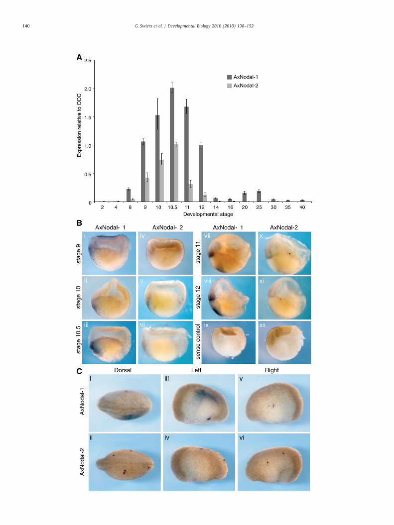
Fig. 1. Analysis of AxNodal-1 and AxNodal-2 expression during axolotl early development. (A1 at stage 12 to compare levels between AxNodal-1 and AxNodal-2. (B) In situ hybridiz(C) Characteristic asymmetrical expression of AxNodal-1, but not AxNodal-2, at stage 20.
Cell culture and manipulation
CGR8 mouse ES cell lines were maintained on gelatin-coateddishes (0.1%) in ESC medium as described in Turksne (2006).Embryoid bodies were generated via the hanging drop method,cultivating 600 cells in a 20-μl drop. ES cells were expanded anddifferentiated as previously described (Tada et al., 2005). Mixl1-specific shRNA sequences were designed as previously described(Izumi et al., 2007).
) qPCR of AxNodal-1 and AxNodal-2 expression, normalised to ODC, and then AxNodal-ation for AxNodal-1 and AxNodal-2 on hemisectioned embryos (dorsal to the left).
142 G. Swiers et al. / Developmental Biology 2010 (2010) 138–152
Southern blots
About 30 µg of genomic DNA was digested for each lane with allpossible combinations of PstI, BsrGI, and MscI (NEB). Primers used togenerate each probe are shown in Table S4. Hybridizations wereperformed according to standard methods (hybridize at 55 °Covernight, two washes in 2× SSC, 0.1% SDS at room temperature,once in 1× SSC, 0.1% SDS at room temperature, and once in 1× SSC,0.1% SDS at 50 °C.
Results
Nodal and Mix orthologs
Based on our hypothesis that the conserved mGRN is expressed inaxolotl embryos, we investigated the Nodal and Mix families, whichare amplified in Xenopus. Using a combination of degenerate PCR andlibrary screening, we identified two orthologs of Nodal, AxNodal-1and AxNodal-2 (Fig. S1), and a single Mix ortholog (Fig. S2), AxMix.Southern blotting to genomic DNA confirms these to be the onlycopies of these genes in the axolotl genome (Figs. S3 and S4).Phylogenetic analysis shows that AxNodal-1 is most closely related toa Nodal gene identified in Cynops, another urodele, and then to theXenopus genes Xnr1, 2, 3, 5, and 6 (Fig. S1). AxNodal-2 clusters withXnr4 from Xenopus (Fig. S1). Interestingly, AxMix is more closelyrelated to Mix orthologs from human or mouse than it is to anyindividual Xenopus gene (Fig. S2). We analyzed expression of thesegenes using a combination of quantitative real-time PCR (qPCR) andwhole-mount in situ hybridization (WISH) to hemisectioned embry-os, a method that allows two different probes to analyzed inequivalent tissue from the same embryo.
Analysis of the Nodal-related genes (Fig. 1) shows that bothcommence expression at the midblastula stage (stage 9), withtranscript levels peaking in early gastrulae (stage 10) (Fig. 1A). Fora direct comparison of expression levels, both time courses arenormalised to AxNodal-1 at stage 9. At all stages, AxNodal-1 isexpressed at least two-fold higher than AxNodal-2, and this isconfirmed by WISH (Fig. 1B). At stage 9, weak expression ofAxNodal-1 and -2 is detected in the animal cap, although the signalis strongest in the marginal zone (Fig. 1B, i). The expression ofAxNodal-1 at this stage is on the future dorsal side of the embryo,confirmed by comparison with Goosecoid in hemisectioned embryos(Fig. S5A). Both AxNodal genes are expressed in the dorsal lip of earlygastrulae (stage 10, Fig. 1B, ii and v), and by stage 12, AxNodal-1 ismainly found in the mesoderm with some weak expression in theendoderm (Fig. 1B, vii). At later stages, AxNodal-1, but not AxNodal-2,is detectable in the left lateral plate mesoderm, consistent with thewell-characterised role for Nodal in the left–right asymmetry (Fig. 1C)(Shen, 2007). Thus, the expression pattern of AxNodal-1 is equivalentto the additive expression profile of Xenopus Xnr1, 2, 5, and 6, with theexception of the pre-MBT expression unique to Xenopus (Yang et al.,2002). AxNodal-2, lacking the later asymmetrical expression, has anexpression pattern similar to Xnr-4, in agreement with the phylogeny(Fig. S1) (Joseph and Melton, 1997). Together, these data suggest thatNodal was duplicated before the divergence of anurans and urodelesfrom their last common ancestor, with subsequent amplification ofthe AxNodal-1 grouping in anurans.
AxMix transcripts are first detected by qPCR at stage 9 (Fig. 2A).Expression persists until tailbud stage (stage 25), contrasting withXenopus embryos, in which the cumulative expression of Mix genes iscomplete by stage 14 (Ecochard et al., 1998; Henry and Melton, 1998;Lemaire et al., 1998; Tada et al., 1998).We compared expression of theaxolotl Brachyury ortholog, AxBra (Johnson et al., 2003a), with AxMix.Interestingly, the relative timing of expression of AxMix and AxBra isaltered compared to their Xenopus orthologs. In Xenopus, Brachyuryand Mix family gene expression commences at the start of
gastrulation (Rosa, 1989; Smith et al., 1991). However, in axolotlembryos, AxMix precedes AxBra expression by several hours, with theinitiation of AxBra expression being delayed until midgastrula stages(Fig. 2A; also see Johnson et al., 2003a).We confirmed the unexpectedrelationship in the timing of these genes' expression using WISH(Fig. 2B). AxMix is first detected in the mesoderm of the blastopore lipas early as stage 10, and this can clearly be seen there by stage 10.5(Fig. 2B, i). By stage 10.75, expression is retained in the involuteddorsal mesoderm and at the leading edge of the involuting mesodermin the blastopore lip (Fig. 2B, iii). At this stage, AxBra first becomesdetectable in presumptive mesoderm on the embryo's surface(Fig. 2B, iv), as well as in a fraction of the dorsal mesoderm previouslymarked by AxMix (Fig. 2B, iii and iv). Lower-level AxBra expression canalso be detected in ventral mesoderm at this stage (Fig. 2B, iv). Bystage 12, AxMix expression is extinguished in the dorsal mesoderm,where AxBra RNA is now abundant, and AxMix is now found in ventralmesoderm, as well as endoderm (Fig. 2B, v and vi). Coexpression ofthe two genes is retained in a fraction of the ventral mesodermcompartment at this stage (Figs. 2B, v and vi, and S5B). By stage 14,ventral AxMix expression is maintained, while AxBra transcripts arefound only in the posterior mesoderm and in the roof of thearchenteron, which contains the notochordal precursors (Fig. 2B, viiand viii). To summarize, AxMix expression precedes that of AxBra,beginning in the dorsal mesoderm, and largely covers the cumulativeexpression domains of all the XenopusMix family members. However,in contrast with Xenopus embryos, AxMix and AxBra are notcoexpressed in cells found at the mesoderm/endoderm boundary,rather AxMix expression precedes that of AxBra in this domain (Figs.2A and B).
Knockdown of Nodal-related genes
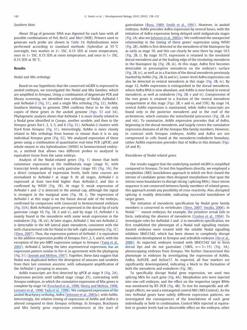
Our results suggest that the underlying axolotl mGRN is simplifiedcompared to Xenopus. To test this hypothesis directly, we employed amorpholino (MO) knockdown approach in which we first cloned theintrons of candidate genes then designed morpholinos that span theintron/exon boundaries to disrupt slicing (splicing MOs). Since intronsequence is not conserved between family members of related genes,this approach avoids any possibility of cross-reactivity. Also, disruptedsplicing is readily detectable, indicating successful knockdown oftarget genes.
The initiation of mesoderm specification by Nodal gene familymembers is conserved in vertebrates (Shen, 2007; Swalla, 2006). InNodal−/− mouse embryos, for example, the primitive streak fails toform, indicating the absence of mesoderm (Conlon et al., 1994). Toinvestigate roles for AxNodal-1 and -2 in mesoderm specification, wefirst established the phenotype of a Nodal null signalling mutant.Axolotl embryos were treated with the soluble Nodal signallinginhibitor SB431542, which has been shown to completely disruptmesoderm development in Xenopus and zebrafish embryos (Ho et al.,2006). As expected, embryos treated with SB431542 fail to formdorsal lips and do not gastrulate (100%, n=3×15) (Fig. 3A),phenocopying embryos from Xenopus. We further characterised thisphenotype in embryos by investigating the expression of AxMix,AxBra, AxFGF8, and AxSox17. As expected, all four markers aresignificantly downregulated, indicating a block to the formation ofboth the mesoderm and endoderm (Fig. 3B).
To specifically disrupt Nodal gene expression, we used twosplicing MOs for each gene (Fig. 4A). Morpholino sets were injectedinto one-cell embryos, and the effect on the respective target RNAswas monitored by RT–PCR (Fig. 4B). To test for nonspecific and off-target effects, we used a mistargeted control MO (MO:Control). As thetwo AxNodal genes have subtly different expression patterns, weinvestigated the consequences of the knockdown of each geneindividually or both in combination. Control MOs injected at equiva-lent or greater levels had no discernible effect on the embryos, other

Fig. 3. Analysis of the effect of SB 431542 Nodal antagonist during early axolotl development. (A) Representative embryos showing the complete block to gastrulation caused by SB431542 treatment, as seen in Xenopus embryos. Panels i, iv, v, vi, vii, ix, and x are vegetal views. Panels vii and xi show the animal view of the embryos in vi and x, respectively.Embryos were treated from the two-cell stage. (B) qRT–PCR analysis of inhibitor treated embryos (five embryos per sample).
143G. Swiers et al. / Developmental Biology 2010 (2010) 138–152
than slowing development. In contrast, knockdown of AxNodal-1results in complete developmental arrest at the onset of gastrulation(88%, n=3×20). These morphant embryos are unable to form adorsal lip (compare Fig. 4C, i–iv with v–viii), phenocopying the effects
of SB431542 treatment (Fig. 4C, v–viii). By stage 14, sibling embryoshave gastrulated normally, while the AxNodal-1morphants are haltedat a pregastrula stage, resembling embryos at stage 9 (Fig. 4C, viii).This phenotype suggests a complete loss of mesoderm. In the same
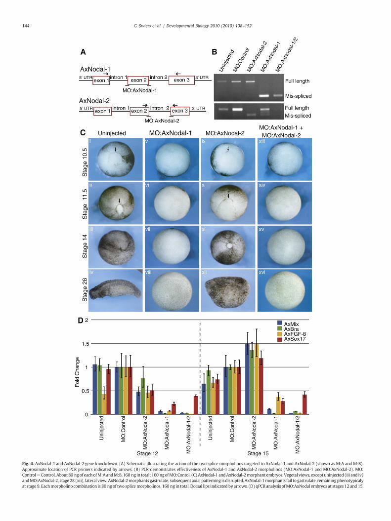
Fig. 4. AxNodal-1 and AxNodal-2 gene knockdown. (A) Schematic illustrating the action of the two splice morpholinos targeted to AxNodal-1 and AxNodal-2 (shown as M:A and M:B).Approximate location of PCR primers indicated by arrows. (B) PCR demonstrates effectiveness of AxNodal-1 and AxNodal-2 morpholinos (MO:AxNodal-1 and MO:AxNodal-2). MO:Control=Control. About80 ngof eachofM:AandM:B, 160 ng in total; 160 ngofMO:Contol. (C)AxNodal-1 andAxNodal-2morphant embryos. Vegetal views, exceptuninjected (iii and iv)andMO:AxNodal-2, stage28(xii), lateral view.AxNodal-2morphants gastrulate, subsequent axial patterning isdisrupted. AxNodal-1morphants fail to gastrulate, remainingphenotypicalyat stage 9. Eachmorpholino combination is 80 ng of two splicemorpholinos, 160 ng in total. Dorsal lips indicated by arrows. (D) qPCR analysis ofMO:AxNodal embryos at stages 12 and 15.
144 G. Swiers et al. / Developmental Biology 2010 (2010) 138–152
145G. Swiers et al. / Developmental Biology 2010 (2010) 138–152
experiments, AxNodal-2 morphants successfully completed gastrula-tion, although they were delayed with respect to uninjected siblings(100%, n=3×20) (compare Fig. 4C, xi with ii–iii). Later, AxNodal-2morphants are disrupted with respect to axial patterning, havingreduced head and tail structures (100%, n=3×20). Nevertheless, theability of these embryos to complete gastrulation indicates thatAxNodal-2 is dispensable formesoderm induction. Coinjection of bothsets of morpholinos has no additional effects over injecting MOstargeted only to AxNodal-1 (n=3×20) (Fig. 4C, xiii–xvi), substanti-ating that only AxNodal-1 is required.
We next examined gene expression in morphants by qPCR. In allcases, gene expression was normalized to embryos injected with thecontrol MO. At stage 12, AxNodal-2 morphants show a mild decreasein expression of AxMix, AxBra, AxFGF-8, and AxSox17 (Fig. 4D), but bystage 15 (neurula), expression levels reach those in controls. Incontrast, AxNodal-1 morphants display an almost complete loss ofexpression from all four genes when assayed at stage 12, andexpression is never recovered. These results are similar to those
Fig. 5. AxMix gene knockdown. (A) Schematic illustrating the action of two splice morpho(B) PCR demonstrates the effectiveness of MO:AxMix (80 ng of each M:A and M:B, 160 ng inormal and mis-spliced transcripts have been used. (C) AxMix morphants are unable to gmorpholinos A and B, 160 ng total. (i, iii, v, and vi) Vegetal view. (ii and iv) Dorsal view. (Dtime point.
obtainedwith SB431542 treatment. Again, the phenotype of AxNodal-2 and -1 MOs combined is equivalent to the AxNodal-1 phenotypealone (Figs. 4C and D). Remarkably, these results indicate that onlyAxNodal-1 is required to initiate mesoderm development, a markedcontrast to Xenopus embryos, in which functional redundanciesoverride a specific requirement for any single Nodal-related gene toproduce mesoderm.
Knockdown of AxMix
A priori, the consequence of the loss of all Mix activity in axolotlembryos is difficult to predict. In Xenopus, Mix.1 and Brachyurynegatively regulate each other's expression to drive the segregation ofendoderm and mesoderm (Lemaire et al., 1998). In contrast, Mixerdepletion results in a mild down-regulation of Brachyury, indicatingthat at least some level of Mix activity is required for normalBrachyury expression (Kofron et al., 2004). However, knockdownof all seven mix/bix family members in X. laevis is technically
linos targeted to AxMix. Approximate location of PCR primers is indicated by arrows.n total). MO:Control=Control, 160 ng. Note that primers designed to amplify both theastrulate. MO:Control=160 ng control morpholino, MO:AxMix=80 ng of Mix spliceand E) qPCR analysis of MO:AxMix embryos, normalised to uninjected controls at each

146 G. Swiers et al. / Developmental Biology 2010 (2010) 138–152
challenging and, to date, has not been achieved. Finally, mouseembryos carrying a targeted deletion of Mixl1 do not express T/Brachyury in the primitive streak (Hart et al., 2002), indicating a directrole in mesoderm specification.
Injection of MOs targeted to the two splice junctions surroundingexon 2 of AxMix (Fig. 5A) completely disrupts splicing (Fig. 5B).Morphants reach early gastrula stage, but then development halts(97%, n=3×20). Dorsal lips fail to form and involution does not occur(Fig. 5C, v and vi), suggesting a defect in mesoderm specification. Asbefore, sibling embryos injected with a control MO gastrulatenormally, although early development is slower than in uninjectedembryos (100%, n=3×20) (Fig. 5C, iii and iv). We next assayedmarker gene expression in morphants. AxMix RNA is barely detectablein these embryos (Fig. 5D). Again, small changes in gene expressionare seen in embryos injected with control MO, which are consistentwith the delay in development. More importantly, we assumed that
Fig. 6. AxBrachyury gene knockdown. (A) Schematic illustrating the action of two splice moNote that AxBra is predicted to have eight exons. Exon 4, likely to be required for DNA beffectiveness of MO:AxBra (80 ng of each M:A and M:B, 160 ng in total). MO:Control=Cmorphants are unable to gastrulate. MO:AxBra=80 ng of both Brachyury splice morpholinouninjected controls at each time point.
AxBra expression would be enhanced in AxMix morphants, based onthe role of Mix activity in Xenopus, but this was not the case; AxBraexpression is completely lost in AxMix morphants (Fig. 5D). UnlikeNodal morphant embryos, however, AxFGF-8 and AxSox17 expressionis maintained and later upregulated (Fig. 5E). Therefore, the loss ofAxBra RNA results from the loss of AxMix and is not a consequence ofthe disruption of a Brachyury/FGF feedback loop (Schulte-Merker andSmith, 1995). This novel relationship between AxMix and AxBra issupported by our earlier observation that AxMix expression precedesthat of AxBra in the mesoderm.
We next asked if the loss of mesoderm in AxMix morphants is adirect consequence of the loss of AxBra. We therefore designedmorpholinos targeted to disrupt splicing in the AxBra gene (Figs. 6Aand B). As with AxNodal and AxMix, AxBra morphants fail togastrulate (100%, n=3×20) (Fig. 6C, v and vi). However, in thiscase, although AxMix expression is initially downregulated, it is
rpholinos targeted to AxBra. Approximate location of PCR primers indicated by arrows.inding was targeted for disruption and is only 62 bp in length. (B) PCR demonstratesontrol, 160 ng. Orange shading indicates predicted DNA binding domain. (C) AxBraA and B, 160 ng in total. (D and E)qPCR analysis of MO:AxBra embryos, normalised to
147G. Swiers et al. / Developmental Biology 2010 (2010) 138–152
ultimately upregulated to higher than normal levels, as would beexpected if AxBra negatively regulates AxMix (Fig. 6D). Moreover, incontrast with AxMix morphants, these embryos show a loss of FGF-8 expression, raising the possibility that AxMix itself is a repressor ofFGF-8 activity, and explaining why FGF-8 is increased in AxMixmorphants (Figs. 5D and E). Lastly, AxSox17 expression is upregulatedin these embryos, suggesting that an increase in endoderm occurs atthe expense of the production of mesoderm (Fig. 6E).
AxMix is required for AxBra expression
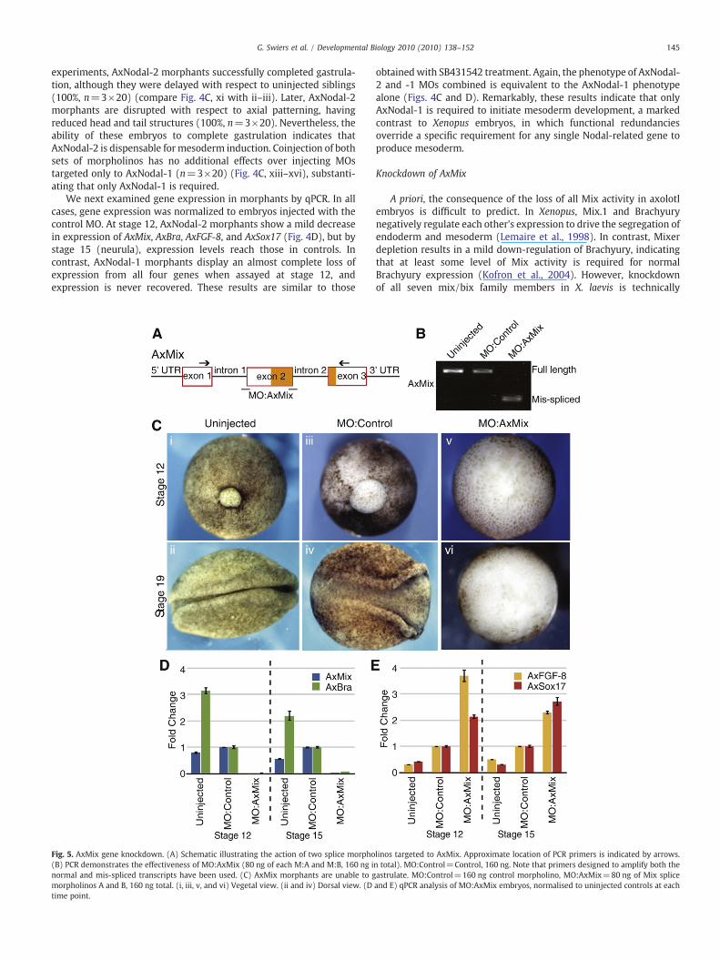
Taken together, our observations demonstrate that the mGRN inaxolotls contains a single Mix gene and two Nodal genes. Surprisingly,Mix acts upstream of Brachyury in the specification of mesoderm, incontrast to the relationship of these genes in Xenopus, suggesting thatinjection of AxMix RNA would expand the AxBra domain. To test thisin axolotl embryos, one of the two ventral blastomeres at the four-cellstage was injected with RNA encoding either AxBra or AxMix, alongwith a lineage tracer (miniruby) to mark the site of RNA injection.Endogenous gene expression was then assayed at stage 12 by WISH.Similar to results with Xenopus (Lemaire et al., 1998), the injection ofRNA for AxBra (200 pg) inhibits Mix expression at the site of injection(Fig. 7), indicating that negative regulation of Mix expression byBrachyury is conserved. However, injection of AxMix RNA (200 pg)induced ectopic expression from the zygotic AxBra gene. This is theopposite of predictions based on Xenopus and supports the position-ing of AxMix in the mGRN upstream of AxBra.
To further elucidate the pathway to mesoderm specification inaxolotls we turned to animal cap assays, a standard experimentalregime for amphibian embryos (Fig. 8A). By titration, we establishedthat 1 pg of RNA encoding activin (mimicking the effects of Nodal)was sufficient to induce elongation (Fig. 8B), an indicator for theinduction of mesoderm (Green et al., 1992). Elongation, and somesoderm induction, in response to activin RNA can be completelyblocked by coinjection of the AxMix morpholino (100%, n=3×10).However, mesoderm induction in the morphants can be rescued byinjection of 20 pg of RNA encoding AxMix. At a higher level of 100 pgAxMix RNA, the caps no longer elongate, rather they appear toproduce endoderm (100%, n=3×10), as expected (Green et al.,1992). We next analyzed gene expression in each group of caps. Inaccord with our previous results, the inclusion of the AxMix MOdramatically reduces Brachyury induction by activin (Fig. 8C).
Fig. 7. The relationship between AxMix and AxBra. Overexpression of 200 pg of either AxBrachyury expression. Conversely, AxBra injected ventrally leads to a down-regulation of M
Brachyury expression is rescued in caps coinjected with a low levelof AxMix mRNA. However, high levels of AxMix upregulate AxSox17(about twofold) and not AxBra, supporting the observation that highlevels of AxMix induce endoderm at the expense of mesoderm.AxFGF-8 expression can be induced in the presence of AxMix MO,indicating a Brachyury-independent pathway, and expression isdecreased by overexpression of AxMix, as expected.
To further investigate the hierarchical relationship between AxMixand AxBra, we rescued the failure of mesoderm induction in AxMixmorphants downstream of activin signalling by overexpressing AxBra.Overexpression of AxBra alone in axolotl animal cap explants induceselongation (100%, n=3×10) typical of mesoderm (Fig. 9A). Meso-derm induction by activin, blocked by the AxMix MO, can be rescued(100%, n=3×10) by overexpression of AxBra (Fig. 9A). At the level ofgene expression, AxBra overexpression reduces AxSox17 levels,suggesting that the loss of Brachyury expression in AxMix morphantsis a significant cause of the loss of mesoderm (Fig. 9B). However, as wesee with the AxMix rescue (Fig. 8C), AxBra overexpression fails torestore normal expression of AxFGF-8, supporting the presence of aBrachyury-independent pathway regulating AxFGF-8 activity(Fig. 9B). Taken together, these data confirm that AxMix is requiredfor the induction of mesoderm, acting downstream of Nodal andupstream of AxBra.
Mixl1 in ES cell differentiation
Our results reveal a role for Mix activity in axolotl embryos that isvery different from its function in Xenopus (Henry and Melton, 1998;Kofron et al., 2004; Lemaire et al., 1998). We sought to determinewhich role for Mix is conserved in mammals, where conflictingconclusions from a variety of studies have not clearly defined a role forMixl1 in the production of mesoderm (Hart et al., 2002; Izumi et al.,2007). We used RNA interference-mediated knockdown to blockMixl1 activity in murine embryonic stem cells, using the Mixl1knockdown 1 sequence described by Izumi et al. (2007). ESC lineswere produced after stable transfection with Mixl1 shRNA, ascrambled Mixl1 control, or the vector alone, and these were usedto produce embryoid bodies (EBs) to test the consequence of Mixl1knockdown on the expression of Brachyury/T. As expected, the Mixl1shRNA leads to a substantial inhibition of Mixl1 compared withnontransfected, scrambled, or vector-only lines (Fig. 10A). However,in agreement with our findings in axolotls, Brachyury/T expression is
Mix or AxBra in whole embryos. AxMix injected dorsally leads to an up-regulation ofix expression.
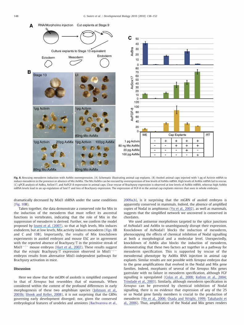
Fig. 8. Rescuing mesoderm induction with AxMix overexpression. (A) Schematic illustrating animal cap explants. (B) Axolotl animal caps injected with 1 pg of Activin mRNA toinduce mesoderm in the presence or absence of Mo:AxMix. TheMo:AxMix can be rescued by overexpression of low levels of AxMix mRNA. High levels of AxMix mRNA fail to rescue.(C) qPCR analysis of AxBra, AxSox17, and AxFGF-8 expression in animal caps. Clear rescue of Brachyury expression is observed at low levels of AxMix mRNA, whereas high AxMixmRNA levels lead to an up-regulation of Sox17 and loss of Brachyury expression. The expression of FGF-8 in the animal cap explants mirrors that seen in whole embryos.
148 G. Swiers et al. / Developmental Biology 2010 (2010) 138–152
dramatically decreased by Mixl1 shRNA under the same conditions(Fig. 10B).
Taken together, the data demonstrate a conserved role for Mix inthe induction of the mesoderm that must reflect its ancestralfunctions in vertebrates, indicating that the role of Mix in thesuppression of mesoderm is derived. Further, we confirm the modelproposed by Izumi et al. (2007), so that at high levels, Mix inducesendoderm, but at low levels, Mix activity induces mesoderm (Figs. 8Band C and 10B). Importantly, the results of Mix knockdownexperiments in axolotl embryos and mouse ESC are in agreementwith the reported absence of Brachyury/T in the primitive streak ofMixl1−/− mouse embryos (Hart et al., 2002). These results suggestthat the ectopic Brachyury/T expression observed in Mixl1−/−
embryos results from alternative Mixl1-independent pathways forBrachyury activation in mice.
Discussion
Here we show that the mGRN of axolotls is simplified comparedto that of Xenopus but resembles that of mammals. Whenconsidered within the context of the profound differences in earlymorphogenesis of these two amphibian species (Johnson et al.,2003b; Shook and Keller, 2008), it is not surprising that the GRNsgoverning early development diverged; nor, given the conservedembryological features of urodeles and amniotes (Bachvarova et al.,
2009a,b), is it surprising that the mGRN of axolotl embryos isapparently conserved in mammals. Indeed, the absence of amplifiedcopies of Nodal in amphioxus (Yu et al., 2002), as well as mammals,suggests that the simplified network we uncovered is conserved inchordates.
We used antisense morpholinos targeted to the splice junctionsof AxNodal1 and AxMix to unambiguously disrupt their expression.Knockdown of AxNodal1 blocks the induction of mesoderm,phenocopying the effects of chemical inhibition of Nodal signallingat both a morphological and a molecular level. Unexpectedly,knockdown of AxMix also blocks the induction of mesoderm,demonstrating that these two factors act together in a pathway formesoderm specification. This is supported by rescue of themesodermal phenotype by AxMix RNA injection in animal capexplants. Similar results are not possible with Xenopus embryos dueto the gene amplifications that evolved in the Nodal and Mix genefamilies. Indeed, morphants of several of the Xenopus Mix genesgastrulate with no failure in mesoderm specification, although FGFsignalling is upregulated (Colas et al., 2008; Kofron et al., 2004;Trindade et al., 2003). Similarly, although mesoderm specification inXenopus can be prevented by chemical inhibition of Nodalsignalling, there is no evidence that expression of any of the 25or so Nodal gene family members is crucial to the production ofmesoderm (Ho et al., 2006; Osada and Wright, 1999; Takahashi etal., 2006). Thus, amplification of the Nodal and Mix genes renders
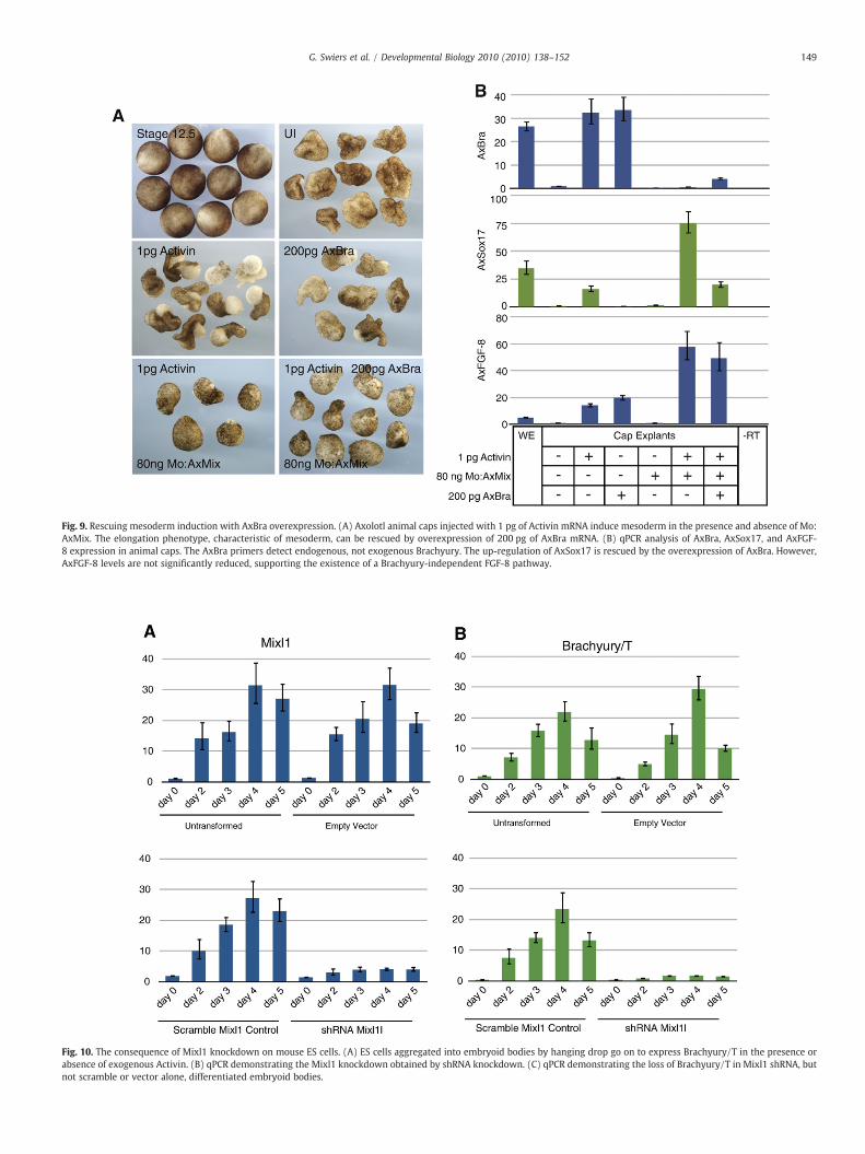
Fig. 9. Rescuing mesoderm induction with AxBra overexpression. (A) Axolotl animal caps injected with 1 pg of Activin mRNA induce mesoderm in the presence and absence of Mo:AxMix. The elongation phenotype, characteristic of mesoderm, can be rescued by overexpression of 200 pg of AxBra mRNA. (B) qPCR analysis of AxBra, AxSox17, and AxFGF-8 expression in animal caps. The AxBra primers detect endogenous, not exogenous Brachyury. The up-regulation of AxSox17 is rescued by the overexpression of AxBra. However,AxFGF-8 levels are not significantly reduced, supporting the existence of a Brachyury-independent FGF-8 pathway.
Fig. 10. The consequence of Mixl1 knockdown on mouse ES cells. (A) ES cells aggregated into embryoid bodies by hanging drop go on to express Brachyury/T in the presence orabsence of exogenous Activin. (B) qPCR demonstrating the Mixl1 knockdown obtained by shRNA knockdown. (C) qPCR demonstrating the loss of Brachyury/T in Mixl1 shRNA, butnot scramble or vector alone, differentiated embryoid bodies.
149G. Swiers et al. / Developmental Biology 2010 (2010) 138–152

150 G. Swiers et al. / Developmental Biology 2010 (2010) 138–152
the mesodermal GRN of Xenopus resistant to perturbations thatwould be lethal in axolotl.
The resistance of the mGRN to genetic perturbation offers amechanistic explanation for the accumulation of amplified Nodal andMix genes in the Xenopus genome, as it fulfils Waddington's conceptof a canalized developmental process. The robustness that resultsfrom canalization is generally considered a selective advantage(Kitano, 2004); however, this can only be true under conditions thatdo not compromise development of the germ line. We suggest thatgene expansion within the mesodermal GRN could not have beentolerated before the evolution of predetermined germ cells in frogs.The induction of PGCs in ventral mesoderm is the ancestral conditionfor amphibians (Bachvarova et al., 2009a) and would likely have beendisrupted as a consequence of Mix and Nodal expansion. Therefore, itis logical to propose that the evolution of germ plasm liberatedconstraints on the mechanisms of mesoderm specification in anurans,in accord with previous hypotheses (Crother et al., 2007; Johnson etal., 2003b). Furthermore, expansion of the Nodal and Mix genes inteleosts (Fan and Dougan, 2007), which also contain germ plasm,suggests this may be a generalized mechanism leading to canalizeddevelopment.
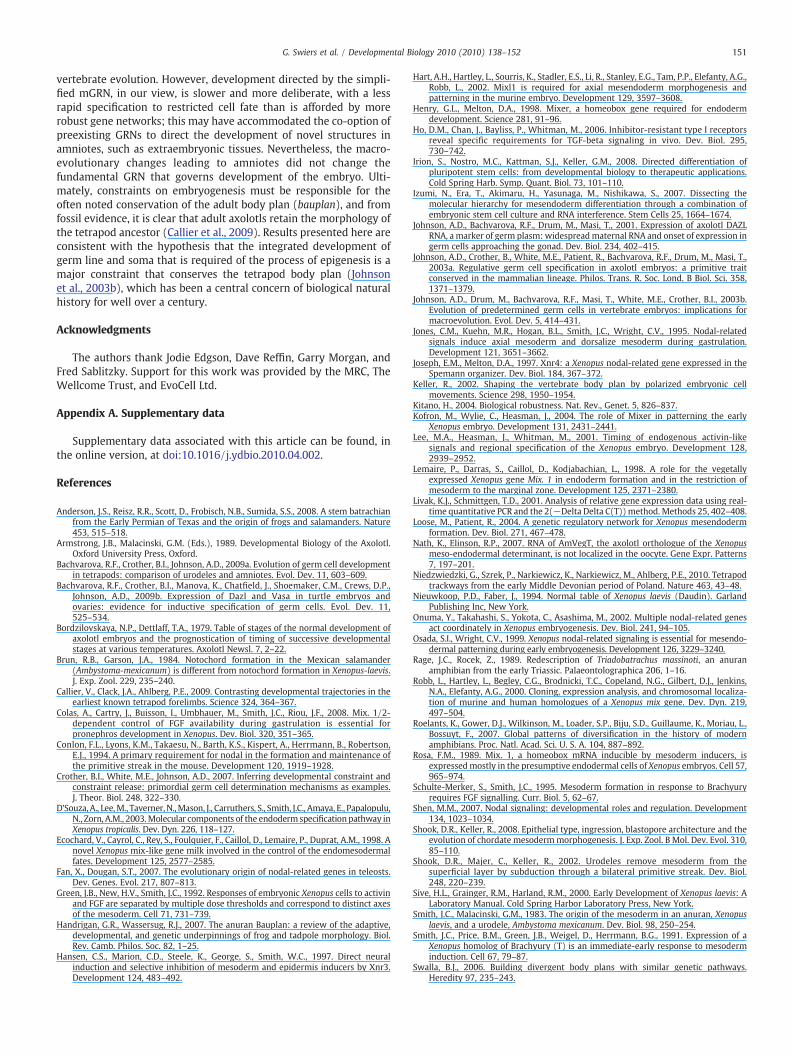
The evolution of gene expansion within a GRN is likely to includesubfunctionalisation of genetic interactions within the network. Here,we have revealed a previously unrecognised role for Mix in the axolotlthat may have been obscured by subfunctionalisation in Xenopus. InXenopus embryos, Nodal signalling induces coexpression of the Mixgenes and Brachyury in the mesendoderm (Lemaire et al., 1998;Wardle and Smith, 2006). The subsequent mutual antagonism ofthese factors causes Brachyury to segregate with the mesoderm andthe Mixes to segregate with endoderm. However, we have detectedlimited coexpression of AxBra and AxMix in axolotl embryos and eventhen only in ventral mesoderm. Furthermore, our results place AxFGF-8, AxSox17, and AxBra downstream of Nodal signalling, but activatedby two independent pathways, with the activation of AxBra beingdependent on AxMix activity. Based on the prevailing interpretation
Fig. 11. The presumptive sGRN for the axolotl. (A) Solid lines indicate experimentally verirelative roles of Nodal, Mix, and Brachyury in Xenopus (B) and axolotl (C) development.
of evidence from Xenopus, we would have expected a Mix morphantto promote mesoderm and suppress endoderm (Lemaire et al., 1998).However, we see the converse, increased AxSox17 expression inAxMix morphants (Fig. 5E), with a loss of mesoderm. This identifies arequirement for AxMix in mesoderm induction before any role in itssuppression, and this is not conserved in Xenopus. We demonstratedthis directly, showing that the AxBra domain is expanded in responseto forced AxMix expression (Fig. 7). Studieswithmouse embryos havelead to conflicting results, with some studies implicating Mix inmesoderm production, and others in its repression. However, weknocked downMix1 in EBs and showed a clear inhibition of Brachyuryexpression. This is consistent with the absence of Brachyuryexpression in the primitive streak (the site of nascent mesodermproduction) of Mixl1−/− mouse embryos, suggesting that the role forMix at the top of a hierarchy or transcription factors leading tomesoderm specification is conserved in vertebrates.
On the basis of these findings, we have constructed a generegulatory network for mesoderm specification in axolotl embryoscontaining a key change from the Xenopus network in which AxMixactivates AxBra and, consequently, the mesoderm (Fig. 11). Thispresumed mesodermal GRN for the axolotl, including dashed lines forlinks not yet confirmed, will probably require alteration in the future,since, for example, the role of the localized determinants that initiatethe mesoderm GRN in Xenopus are uncertain in axolotls (Nath andElinson, 2007). Nevertheless, our observations suggest a two-stepprocess for mesoderm induction in the axolotl. Firstly, Nodal, viaMix, induces a population of mesendodermal cells, the bipotentialprecursors of the mesoderm and endoderm. In the second step,Brachyury expression, triggered by Mix, induces the mesoderm andsuppresses Mix. The loss of mesoderm in the Nodal and Mixmorphants reflects the loss of the bipotential mesendoderm thataccounts for the mesodermal defects we observe.
It is straightforward to understand why canalization of the mGRNwould evolve as an adaptive response to selection, but it is not obviouswhy the less robust simplified mGRN would be conserved through
fied links; dashed lines indicate presumed links from Xenopus. Models comparing the
151G. Swiers et al. / Developmental Biology 2010 (2010) 138–152
vertebrate evolution. However, development directed by the simpli-fied mGRN, in our view, is slower and more deliberate, with a lessrapid specification to restricted cell fate than is afforded by morerobust gene networks; this may have accommodated the co-option ofpreexisting GRNs to direct the development of novel structures inamniotes, such as extraembryonic tissues. Nevertheless, the macro-evolutionary changes leading to amniotes did not change thefundamental GRN that governs development of the embryo. Ulti-mately, constraints on embryogenesis must be responsible for theoften noted conservation of the adult body plan (bauplan), and fromfossil evidence, it is clear that adult axolotls retain the morphology ofthe tetrapod ancestor (Callier et al., 2009). Results presented here areconsistent with the hypothesis that the integrated development ofgerm line and soma that is required of the process of epigenesis is amajor constraint that conserves the tetrapod body plan (Johnsonet al., 2003b), which has been a central concern of biological naturalhistory for well over a century.
Acknowledgments
The authors thank Jodie Edgson, Dave Reffin, Garry Morgan, andFred Sablitzky. Support for this work was provided by the MRC, TheWellcome Trust, and EvoCell Ltd.
Appendix A. Supplementary data
Supplementary data associated with this article can be found, inthe online version, at doi:10.1016/j.ydbio.2010.04.002.
References
Anderson, J.S., Reisz, R.R., Scott, D., Frobisch, N.B., Sumida, S.S., 2008. A stem batrachianfrom the Early Permian of Texas and the origin of frogs and salamanders. Nature453, 515–518.
Armstrong, J.B., Malacinski, G.M. (Eds.), 1989. Developmental Biology of the Axolotl.Oxford University Press, Oxford.
Bachvarova, R.F., Crother, B.I., Johnson, A.D., 2009a. Evolution of germ cell developmentin tetrapods: comparison of urodeles and amniotes. Evol. Dev. 11, 603–609.
Bachvarova, R.F., Crother, B.I., Manova, K., Chatfield, J., Shoemaker, C.M., Crews, D.P.,Johnson, A.D., 2009b. Expression of Dazl and Vasa in turtle embryos andovaries: evidence for inductive specification of germ cells. Evol. Dev. 11,525–534.
Bordzilovskaya, N.P., Dettlaff, T.A., 1979. Table of stages of the normal development ofaxolotl embryos and the prognostication of timing of successive developmentalstages at various temperatures. Axolotl Newsl. 7, 2–22.
Brun, R.B., Garson, J.A., 1984. Notochord formation in the Mexican salamander(Ambystoma-mexicanum) is different from notochord formation in Xenopus-laevis.J. Exp. Zool. 229, 235–240.
Callier, V., Clack, J.A., Ahlberg, P.E., 2009. Contrasting developmental trajectories in theearliest known tetrapod forelimbs. Science 324, 364–367.
Colas, A., Cartry, J., Buisson, I., Umbhauer, M., Smith, J.C., Riou, J.F., 2008. Mix. 1/2-dependent control of FGF availability during gastrulation is essential forpronephros development in Xenopus. Dev. Biol. 320, 351–365.
Conlon, F.L., Lyons, K.M., Takaesu, N., Barth, K.S., Kispert, A., Herrmann, B., Robertson,E.J., 1994. A primary requirement for nodal in the formation and maintenance ofthe primitive streak in the mouse. Development 120, 1919–1928.
Crother, B.I., White, M.E., Johnson, A.D., 2007. Inferring developmental constraint andconstraint release: primordial germ cell determination mechanisms as examples.J. Theor. Biol. 248, 322–330.
D'Souza, A., Lee,M., Taverner, N.,Mason, J., Carruthers, S., Smith, J.C., Amaya, E., Papalopulu,N., Zorn, A.M., 2003.Molecular components of the endoderm specification pathway inXenopus tropicalis. Dev. Dyn. 226, 118–127.
Ecochard, V., Cayrol, C., Rey, S., Foulquier, F., Caillol, D., Lemaire, P., Duprat, A.M., 1998. Anovel Xenopus mix-like gene milk involved in the control of the endomesodermalfates. Development 125, 2577–2585.
Fan, X., Dougan, S.T., 2007. The evolutionary origin of nodal-related genes in teleosts.Dev. Genes. Evol. 217, 807–813.
Green, J.B., New, H.V., Smith, J.C., 1992. Responses of embryonic Xenopus cells to activinand FGF are separated by multiple dose thresholds and correspond to distinct axesof the mesoderm. Cell 71, 731–739.
Handrigan, G.R., Wassersug, R.J., 2007. The anuran Bauplan: a review of the adaptive,developmental, and genetic underpinnings of frog and tadpole morphology. Biol.Rev. Camb. Philos. Soc. 82, 1–25.
Hansen, C.S., Marion, C.D., Steele, K., George, S., Smith, W.C., 1997. Direct neuralinduction and selective inhibition of mesoderm and epidermis inducers by Xnr3.Development 124, 483–492.
Hart, A.H., Hartley, L., Sourris, K., Stadler, E.S., Li, R., Stanley, E.G., Tam, P.P., Elefanty, A.G.,Robb, L., 2002. Mixl1 is required for axial mesendoderm morphogenesis andpatterning in the murine embryo. Development 129, 3597–3608.
Henry, G.L., Melton, D.A., 1998. Mixer, a homeobox gene required for endodermdevelopment. Science 281, 91–96.
Ho, D.M., Chan, J., Bayliss, P., Whitman, M., 2006. Inhibitor-resistant type I receptorsreveal specific requirements for TGF-beta signaling in vivo. Dev. Biol. 295,730–742.
Irion, S., Nostro, M.C., Kattman, S.J., Keller, G.M., 2008. Directed differentiation ofpluripotent stem cells: from developmental biology to therapeutic applications.Cold Spring Harb. Symp. Quant. Biol. 73, 101–110.
Izumi, N., Era, T., Akimaru, H., Yasunaga, M., Nishikawa, S., 2007. Dissecting themolecular hierarchy for mesendoderm differentiation through a combination ofembryonic stem cell culture and RNA interference. Stem Cells 25, 1664–1674.
Johnson, A.D., Bachvarova, R.F., Drum, M., Masi, T., 2001. Expression of axolotl DAZLRNA, a marker of germ plasm: widespreadmaternal RNA and onset of expression ingerm cells approaching the gonad. Dev. Biol. 234, 402–415.
Johnson, A.D., Crother, B., White, M.E., Patient, R., Bachvarova, R.F., Drum, M., Masi, T.,2003a. Regulative germ cell specification in axolotl embryos: a primitive traitconserved in the mammalian lineage. Philos. Trans. R. Soc. Lond. B Biol. Sci. 358,1371–1379.
Johnson, A.D., Drum, M., Bachvarova, R.F., Masi, T., White, M.E., Crother, B.I., 2003b.Evolution of predetermined germ cells in vertebrate embryos: implications formacroevolution. Evol. Dev. 5, 414–431.
Jones, C.M., Kuehn, M.R., Hogan, B.L., Smith, J.C., Wright, C.V., 1995. Nodal-relatedsignals induce axial mesoderm and dorsalize mesoderm during gastrulation.Development 121, 3651–3662.
Joseph, E.M., Melton, D.A., 1997. Xnr4: a Xenopus nodal-related gene expressed in theSpemann organizer. Dev. Biol. 184, 367–372.
Keller, R., 2002. Shaping the vertebrate body plan by polarized embryonic cellmovements. Science 298, 1950–1954.
Kitano, H., 2004. Biological robustness. Nat. Rev., Genet. 5, 826–837.Kofron, M., Wylie, C., Heasman, J., 2004. The role of Mixer in patterning the early
Xenopus embryo. Development 131, 2431–2441.Lee, M.A., Heasman, J., Whitman, M., 2001. Timing of endogenous activin-like
signals and regional specification of the Xenopus embryo. Development 128,2939–2952.
Lemaire, P., Darras, S., Caillol, D., Kodjabachian, L., 1998. A role for the vegetallyexpressed Xenopus gene Mix. 1 in endoderm formation and in the restriction ofmesoderm to the marginal zone. Development 125, 2371–2380.
Livak, K.J., Schmittgen, T.D., 2001. Analysis of relative gene expression data using real-time quantitative PCR and the 2(−Delta Delta C(T))method. Methods 25, 402–408.
Loose, M., Patient, R., 2004. A genetic regulatory network for Xenopus mesendodermformation. Dev. Biol. 271, 467–478.
Nath, K., Elinson, R.P., 2007. RNA of AmVegT, the axolotl orthologue of the Xenopusmeso-endodermal determinant, is not localized in the oocyte. Gene Expr. Patterns7, 197–201.
Niedzwiedzki, G., Szrek, P., Narkiewicz, K., Narkiewicz, M., Ahlberg, P.E., 2010. Tetrapodtrackways from the early Middle Devonian period of Poland. Nature 463, 43–48.
Nieuwkoop, P.D., Faber, J., 1994. Normal table of Xenopus laevis (Daudin). GarlandPublishing Inc, New York.
Onuma, Y., Takahashi, S., Yokota, C., Asashima, M., 2002. Multiple nodal-related genesact coordinately in Xenopus embryogenesis. Dev. Biol. 241, 94–105.
Osada, S.I., Wright, C.V., 1999. Xenopus nodal-related signaling is essential for mesendo-dermal patterning during early embryogenesis. Development 126, 3229–3240.
Rage, J.C., Rocek, Z., 1989. Redescription of Triadobatrachus massinoti, an anuranamphibian from the early Triassic. Palaeontolographica 206, 1–16.
Robb, L., Hartley, L., Begley, C.G., Brodnicki, T.C., Copeland, N.G., Gilbert, D.J., Jenkins,N.A., Elefanty, A.G., 2000. Cloning, expression analysis, and chromosomal localiza-tion of murine and human homologues of a Xenopus mix gene. Dev. Dyn. 219,497–504.
Roelants, K., Gower, D.J., Wilkinson, M., Loader, S.P., Biju, S.D., Guillaume, K., Moriau, L.,Bossuyt, F., 2007. Global patterns of diversification in the history of modernamphibians. Proc. Natl. Acad. Sci. U. S. A. 104, 887–892.
Rosa, F.M., 1989. Mix. 1, a homeobox mRNA inducible by mesoderm inducers, isexpressedmostly in the presumptive endodermal cells of Xenopus embryos. Cell 57,965–974.
Schulte-Merker, S., Smith, J.C., 1995. Mesoderm formation in response to Brachyuryrequires FGF signalling. Curr. Biol. 5, 62–67.
Shen, M.M., 2007. Nodal signaling: developmental roles and regulation. Development134, 1023–1034.
Shook, D.R., Keller, R., 2008. Epithelial type, ingression, blastopore architecture and theevolution of chordate mesodermmorphogenesis. J. Exp. Zool. B Mol. Dev. Evol. 310,85–110.
Shook, D.R., Majer, C., Keller, R., 2002. Urodeles remove mesoderm from thesuperficial layer by subduction through a bilateral primitive streak. Dev. Biol.248, 220–239.
Sive, H.L., Grainger, R.M., Harland, R.M., 2000. Early Development of Xenopus laevis: ALaboratory Manual. Cold Spring Harbor Laboratory Press, New York.
Smith, J.C., Malacinski, G.M., 1983. The origin of the mesoderm in an anuran, Xenopuslaevis, and a urodele, Ambystoma mexicanum. Dev. Biol. 98, 250–254.
Smith, J.C., Price, B.M., Green, J.B., Weigel, D., Herrmann, B.G., 1991. Expression of aXenopus homolog of Brachyury (T) is an immediate-early response to mesoderminduction. Cell 67, 79–87.
Swalla, B.J., 2006. Building divergent body plans with similar genetic pathways.Heredity 97, 235–243.

152 G. Swiers et al. / Developmental Biology 2010 (2010) 138–152
Tada, M., Casey, E.S., Fairclough, L., Smith, J.C., 1998. Bix1, a direct target of Xenopus T-box genes, causes formation of ventral mesoderm and endoderm. Development125, 3997–4006.
Tada, S., Era, T., Furusawa, C., Sakurai, H., Nishikawa, S., Kinoshita, M., Nakao, K., Chiba,T., 2005. Characterization of mesendoderm: a diverging point of the definitiveendoderm and mesoderm in embryonic stem cell differentiation culture.Development 132, 4363–4374.
Takahashi, S., Onuma, Y., Yokota, C., Westmoreland, J.J., Asashima, M., Wright, C.V.,2006. Nodal-related gene Xnr5 is amplified in the Xenopus genome. Genesis 44,309–321.
Trindade, M., Messenger, N., Papin, C., Grimmer, D., Fairclough, L., Tada, M., Smith, J.C.,2003. Regulation of apoptosis in the Xenopus embryo by Bix3. Development 130,4611–4622.
Turksne, K. (Ed.), 2006. Embryonic Stem Cell Protocols Volume 2: DifferentiationModels. Humana Press, Totowa.
Unsal, K., Morgan, G.T., 1995. A novel group of families of short interspersed repetitiveelements (SINEs) in Xenopus: evidence of a specific target site for DNA-mediatedtransposition of inverted-repeat SINEs. J. Mol. Biol. 248, 812–823.
Wardle, F.C., Smith, J.C., 2006. Transcriptional regulation of mesendoderm formation inXenopus. Semin. Cell Dev. Biol. 17, 99–109.
Yang, J., Tan, C., Darken, R.S., Wilson, P.A., Klein, P.S., 2002. beta-Catenin/Tcf-regulatedtranscription prior to the midblastula transition. Development 129, 5743–5752.
Yu, J.K., Holland, L.Z., Holland, N.D., 2002. An amphioxus nodal gene (AmphiNodal)with early symmetrical expression in the organizer and mesoderm and laterasymmetrical expression associated with left-right axis formation. Evol. Dev. 4,418–425.
Copyright © 2022 FDOKUMEN