2019_tese_aevieiraneto.pdf - Universidade Federal do Ceará
57
UNIVERSIDADE FEDERAL DO CEARÁ CENTRO DE CIÊNCIAS DEPARTAMENTO DE BIOQUÍMICA E BIOLOGIA MOLECULAR PROGRAMA DE PÓS-GRADUAÇÃO EM BIOQUÍMICA ANTONIO EUFRÁSIO VIEIRA NETO NEW STRUCTURAL INSIGHTS INTO ANOMERIC CARBOHYDRATE RECOGNITION BY FRUTALIN: AN α-D-GALACTOSE-BINDING LECTIN FROM BREADFRUIT SEEDS FORTALEZA 2019
-
Upload
khangminh22 -
Category
Documents
-
view
1 -
download
0
Transcript of 2019_tese_aevieiraneto.pdf - Universidade Federal do Ceará

UNIVERSIDADE FEDERAL DO CEARÁ
CENTRO DE CIÊNCIAS
DEPARTAMENTO DE BIOQUÍMICA E BIOLOGIA MOLECULAR
PROGRAMA DE PÓS-GRADUAÇÃO EM BIOQUÍMICA
ANTONIO EUFRÁSIO VIEIRA NETO
NEW STRUCTURAL INSIGHTS INTO ANOMERIC CARBOHYDRATE
RECOGNITION BY FRUTALIN: AN α-D-GALACTOSE-BINDING LECTIN FROM
BREADFRUIT SEEDS
FORTALEZA
2019

ANTONIO EUFRÁSIO VIEIRA NETO
NEW STRUCTURAL INSIGHTS INTO ANOMERIC CARBOHYDRATE RECOGNITION
BY FRUTALIN: AN α-D-GALACTOSE-BINDING LECTIN FROM BREADFRUIT SEEDS
Tese apresentada ao Programa de Pós-
Graduação em Bioquímica da Universidade
Federal do Ceará, como requisito parcial à
obtenção do título de doutora em Bioquímica.
Área de concentração: Bioquímica Vegetal.
Orientador: Prof. Dr. Renato de Azevedo
Moreira.
FORTALEZA
2019


ANTONIO EUFRÁSIO VIEIRA NETO
NEW STRUCTURAL INSIGHTS INTO ANOMERIC CARBOHYDRATE RECOGNITION
BY FRUTALIN: AN α-D-GALACTOSE-BINDING LECTIN FROM BREADFRUIT SEEDS
Tese apresentada ao Programa de Pós-
Graduação em Bioquímica da Universidade
Federal do Ceará, como requisito parcial à
obtenção do título de doutora em Bioquímica.
Área de concentração: Bioquímica Vegetal.
Aprovada em: 18 / 03 /2019.
BANCA EXAMINADORA
________________________________________
Prof. Dr. Renato de Azevedo Moreira (Orientador)
Universidade Federal do Ceará (UFC)
_________________________________________
Prof.a Dra. Daniele de Oliveira Bezerra de Sousa
Universidade Federal do Ceará (UFC)
_________________________________________
Prof.a Dra. Ayrles Fernanda Brandão da Silva
Universidade Federal do Ceará (UFC)
_________________________________________
Dra. Carolina de Araújo Viana
Universidade de Fortaleza (Unifor)
_________________________________________
Prof.a Dra. Ana Cristina de Oliveira Monteiro Moreira
Universidade de Fortaleza (Unifor)

A Deus, em sua Santíssima Trindade,
Ao meu pai, Vicente.

AGRADECIMENTOS
Agradeço à Universidade Federal do Ceará – UFC, pelo ensino de excelência que
me proporcionou, à Universidade de Fortaleza – UNIFOR, por toda a infraestrutura, suporte e
acolhimento em todos estes anos de pesquisa, à Coordenação de Aperfeiçoamento de Pessoal
de Nível Superior – CAPES, pela concessão da bolsa de estudos, e aos governos anteriores que
fortaleceram as políticas sociais e proporcionaram a mim e a milhares de jovens a possibilidade
de se inserir na ciência.
Aos meus pais, Vicente (in memoriam) e Célia, por terem dado o melhor de si para
que eu pudesse realizar este grande sonho, e por terem injetado em mim, ao longo dos anos, os
ingredientes fundamentais para a construção de um grande pesquisador: o amor e humildade.
Ao meu orientador, Prof. Dr. Renato de Azevedo Moreira, meu pai científico, por
ter me dado a grande oportunidade de ser seu aluno e seu amigo. Sou eternamente grato por
todo tempo e dedicação que foi dedicada a mim e à minha formação. Neste doutorado, Prof.
Renato preencheu um enorme vazio que senti em meu coração com a perda de meu pai, e este
é o diferencial dele diante de absolutamente todos os outros professores que já passaram na
minha vida. Sua forma de ensinar, amar e se preocupar com todos seus alunos é algo que
permanecerá por muitos anos em mim e certamente serão repassadas aos meus futuros alunos.
À minha co-orientadora: Prof. Dra. Ana Cristina de Oliveira Monteiro Moreira, por
sempre ter acreditado em mim, me incentivando a ir além, a aprender coisas novas, a sair da
zona de conforto, a lecionar, a colaborar e a evoluir. Jamais esquecerei o quanto sua presença
fez a diferença em minha vida profissional, pessoal e até mesmo esportiva. Muito obrigado por
se preocupar com meu trabalho, em meio a vários outros alunos, por ser essencial na execução
dos experimentos, e por ter me passado tanta confiança, autoestima, otimismo e pensamento
positivo nas horas mais difíceis e desafiadores, inclusive no processo de publicação. O laço
criado neste doutorado certamente vai além do científico, e sua amizade será levada pelo resto
da vida.
À minha esposa (Natália Vieira) e meu filho (Guilherme Vieira), por serem a razão
da minha vida, o motivo maior da minha luta, a felicidade plena que sempre busquei, e por
terem trazido paz aos 4 anos mais difíceis da minha vida.
À minha família, em especial à Tia Eliene, que foi minha segunda mãe na minha
infância e realizou brilhantemente a missão de me ensinar a ler aos 4 anos. Aos meus irmãos,
Sarah e Vicente Filho; meu primo e grande amigo, Ricardus Júnior; meus sogros, Márcia e

Egilberto, por terem me recebido tão amorosamente em sua família; e a todos da família Vieira,
por serem grandes incentivadores em todas as etapas dos meus estudos e por terem sido a
melhor família que alguém pode ter.
Aos meus grandes amigos: Victor Ramalho, Agrício Moreira, Ana Karine, Solange
Moreira, Demócrito Ramalho, Christian Neves, Rodrigo Franklin, João Carlos (in memoriam),
Romário Coelho, Rodrigo Coelho, Fabíola Barroso, Edgerson Siebra, Godyan Wesley, Rafael
Pinto, Vinicius Jucá, Leonardo Almeida, Saulo Martins, Luana Anselmo, Thiago Medeiros,
Ubirajara Júnior, Natália Almada, Pablito Augusto, Ruan Bruno, Jamille Magalhães, Roseline
Torres, Júlia Yoshioka, Ana Luisa, por tantos anos de amizade e por incentivo constante durante
todas as etapas da minha formação.
Aos amigos que a vida acadêmica me proporcionou: Wallace, Edson e Ana Luiza,
meus fiéis companheiros de mestrado e doutorado, que desde 2013 fazem parte
substancialmente da minha vida. À professora Adriana Rolim, pelas parcerias e colaborações
nos projetos e pela confiança em meu trabalho, minha sincera gratidão. Aos queridos amigos
do laboratório F66: Rogênio Mendes, Felipe Sousa, Larissa Fiúza, Kueirislene, Thiago Jucá,
Marina Lobo, Hyldecia Lellis, Rosueti Filho, Carol Viana, Fernanda Rodrigues, George e
Ronielly, por terem participado diretamente da minha caminhada durante o doutorado, por
terem tornado os dias mais leves e felizes, e por serem incentivadores do sucesso do projeto.
Agradeço a todos os membros da banca que contribuíram substancialmente para a
melhoria desse trabalho, com suas sugestões e considerações finais na avaliação do manuscrito.

RESUMO
As lectinas são proteínas que se ligam a carboidratos ou glicoconjugados, de forma reversível,
sem alterar a estrutura dos mesmos. Desta forma, podem interagir com a superfície celular e
promover as mais diversas funções biológicas. A Frutalina (FTL) é a lectina mais abundante
das sementes do fruta-pão (Artocarpus incisa), pertencendo à família de lectinas relacionadas
à Jacalina (JRL). FTL reconhece e se liga especificamente a α-D-galactose e tem sido usada
com sucesso em pesquisas imunobiológicas para o reconhecimento de oligossacarídeos
associados ao câncer. Neste trabalho, nós relatamos toda a estrutura 3D da FTL, conforme
determinada por cristalografia de raios-X. Os cristais obtidos foram difratados a 1,81 Å (Apo-
frutalin) e 1,65 Å (FTL–D-Gal) de resolução. A lectina exibe clivagem pós-traducional
produzindo uma cadeia α- (133 aminoácidos) e uma cadeia β (20 aminoácidos), apresentando
um homotetrâmero quando em solução, com um típico β-prisma presente nas JRL. O β-prisma
da FTL é composto de três folhas-β formando três motivos chave grega antiparalelos. O sítio
de ligação à carboidratos (CBS) envolve o N-terminal da cadeia α e é formado por quatro
resíduos chave: Gly25, Tyr146, Trp147 e Asp149. Estes resultados foram usados em
simulações de dinâmica molecular em soluções aquosas para esclarecer as bases molecular da
ligação de glicanos à FTL. As simulações sugerem que a excisão do peptídeo Thr-Ser-Ser-Asn
(TSSN) reduz a rigidez do CBS da FTL, aumentando o número de interacões com ligantes e
resultando em locais de ligação múltipla e reconhecimento anomérico das moléculas de açúcar.
Nossos achados fornecem uma nova perspectiva para elucidar ainda mais a versatilidade do
FTL em muitas atividades biológicas, incluindo o reconhecimento de glicoproteínas do soro
sanguíneo que provocam neoplasias através de sua super-expressão. A interação da FTL com
glicoproteína Complement-C3 foi investigada por simulações de docking molecular e as bases
moleculares deste reconhecimento foram observadas, comprovando o envolvimento do CBS no
reconhecimento do ligante.
Palavras-chave: Fruta-pão. Lectinas. Frutalina. Complement-C3.

ABSTRACT
Lectins are proteins that bind to carbohydrates or glycoconjugates, reversibly, without changing
the structure of them. They can interact with the cell surface and promote the most diverse
biological activities. Frutalin (FTL) is the most abundant lectin in breadfruit seeds (Artocarpus
incisa), belonging to the jacalin-related lectin family (JRL). FTL specifically recognizes α-D-
galactose and has been successfully used in immunobiological research for the recognition of
cancer-associated oligosaccharides. Here, we report the full 3D structure of the FTL, as
determined by X-ray crystallography. The crystals obtained were diffracted at 1.81 Å (Apo-
frutalin) and 1.65 Å (FTL-D-Gal) resolution. The lectin exhibits post-translational cleavage
producing an α- (133 amino acids) chain and a β chain (20 amino acids), exhibiting a
homotetramer when in solution, with a typical β-prism present in the JRLs. The β-prism of the
FTL is composed of three β-sheets forming three antiparallel Greek key motifs. The
carbohydrate binding site (CBS) involves the N-terminus of the α-chain and is formed by four
key residues: Gly25, Tyr146, Trp147 and Asp149. These results were used in simulations of
molecular dynamics in aqueous solutions to clarify the molecular bases of glycan binding to
CBS. The simulations suggest that the excision of the Thr-Ser-Ser-Asn peptide (TSSN) reduces
the CBS rigidity of the FTL, increasing the number of interactions with ligands and resulting
in multiple binding sites and anomeric recognition of the sugar molecules. Our findings provide
a new perspective to further elucidate the FTL's versatility in many biological activities,
including the recognition of blood serum glycoproteins that elicit neoplasms through their
overexpression. The interaction of FTL with Complement-C3 glycoprotein was investigated by
molecular docking simulations and the molecular bases of this recognition were observed,
proving the involvement of CBS in ligand recognition.
Keywords: Breadfruit. Lectins. Frutalin. Complement-C3.

LISTA DE FIGURAS
Figura 1 − Predicted structural features of FTL.................................................................. 24
Figura 2 − FTL carbohydrate-binding site with D-galactose............................................. 26
Figura 3 − FTL CBS anchoring specific sugars………...................................................... 27
Figura 4 − RMSD FTL during MD simulation……........................................................... 30
Figura 5 − PII between FTL residues and water molecules................................................ 32
Figura 6 − Interface between 03B1; and 03B2; chains of FTL connected by hydrogen
bonds…………………………………………………………………………..
.34

LISTA DE ABREVIATURAS E SIGLAS
C1 Carbono 1
C4 Carbono 4
CBS “Carbohydrate binding site”
FTL Frutalina
JRL “Jacalin-related lectin”
MCA “Minimal concentration for agglutination”
MD Dinâmica Molecular (“Molecular dynamics”)
NO Óxido nítrico
PDB “Protein Databank”
RMSD “Root-mean square deviation”
SDS-PAGE Eletroforese em gel de poliacrilamida na presença de SDS

SUMÁRIO
1 INTRODUÇÃO ................................................................................................. 14
2 CAPÍTULO I - NEW STRUCTURAL INSIGHTS INTO ANOMERIC
CARBOHYDRATE RECOGNITION BY FRUTALIN: AN Α-D-
GALACTOSE-BINDING LECTIN FROM BREADFRUIT
SEEDS................................................................................................................. 16
2.1 Introduction........................................................................................................
2.2 Experimental...................................................................................................... 18
2.2.1 Isolation and purification of FTL from Artocarpus incisa
seeds..................................................................................................................... 18
2.2.2 Crystallization, data collection and processing and refinement of FTL
structure............................................................................................................... 19
2.2.3 Molecular docking studies.................................................................................. 19
2.2.4 Molecular dynamics ........................................................................................... 20
2.3 Results and discussion........................................................................................ 20
2.4 Conclusion........................................................................................................... 36
3 CAPÍTULO II - FRUTALIN RECOGNIZES COMPLEMENT C3
OVER-EXPRESSED BY
NEOPLASMS..................................................................................................... 43
3.1 Introduction........................................................................................................ 44
3.1.1 Biomarkers and cancer research………………………………………..………….. 44
3.1.2 Frutalin as biomedical tool……………………………………………...…………… 44
3.2 Material and methods………………………………………………………… 45
3.3 Results and discussion………………………………………………………… 46
3.4 Conclusions……………………………………………………………………. 49
4 CONSIDERAÇÕES FINAIS...................................................………………. 52
REFERÊNCIAS................................................................................................. 54


1 INTRODUÇÃO
As lectinas vegetais, por serem proteínas capazes de se ligar reversivelmente a glicanos
sem alterar a estrutura dos mesmos, possuem uma grande versatilidade em aplicações
biotecnológicas. Desta forma, o trabalho a seguir busca investigar detalhes da estrutura da
Frutalina (FTL), uma lectina abundante nas sementes de Artocarpus incisa, conhecida
popularmente como fruta-pão. Nas sementes de fruta-pão é possível encontrar três lectinas com
reconhecimento distinto a carboidratos: frutalina, frutapina e frutaquina [1]. A Frutalina possui
alta homologia estrutural com outra lectina amplamente estudada, a Jacalina, presente nas
sementes de Jaca (Artocarpus integrifólia), porém as lectinas são capazes de promover
diferentes aplicações biológicas [2].
As sementes de fruta-pão são compostas por um alto teor de água (até 60%) e moderado
teor de proteína (12,25% do peso seco). A FTL é a lectina mais abundante desta espécie,
apresentando propriedades de ligação múltipla em que o mesmo CBS reconhece uma gama de
diferentes ligantes, embora tenha maiores afinidades para os monossacarídeos de α-D-galactose
e carboidratos complexos que contêm glicanas Gα-1-3. [17]. O padrão da FTL em SDS-PAGE
é semelhante a Jacalina: duas bandas entre 20 e 14 KDa que correspondem às frações
glicosilada e não glicosilada, respectivamente, além de apresentar espectro de massa
deconvoluído com diferentes massas dentro de 16,5 kDa, consistente com a presença de glico-
isoformas de monômeros idênticos. Em solução, a frutalina pode formar homotetrâmeros nos
quais cada monômero contém uma cadeia α (16 kDa) e uma cadeia β (2 kDa) [3,4]. Semelhante
às lectinas de Moraceae, o CBS da frutalina está localizado em um domínio próximo ao terminal
N da cadeia α, consistindo de quatro resíduos chave Gly25, Tyr146, Trp147 e Asp149. No
reconhecimento ao ligante, ocorrem cerca de dez interações envolvendo hidroxilas: hidroxila
C1 na Tyr146, hidroxila C3 na Gly25, hidroxila C4 nos resíduos Gly25 e Asp149 e hidroxila
C6 em Tyr146, Trp147 e Asp149 [5], estas interações também foram evidenciadas no capítulo
I deste trabalho, nas simulações in silico. Além disso, há evidências que sugerem que a frutalina
possui estereoespecificidade, capaz de ligar especificamente α-D-galactose, uma vez que foi
previamente isolada em uma coluna galactomanana reticulada, mas não em matrizes
imobilizadas com β-galactosil [3, 6], o que deu origem à modelagem dos fragmentos ligantes
destas matrizes para as simulações computacionais a seguir.
A Frutalina, por ser uma proteína bioativa, é uma lectina de interesse mundial, tendo
sido investigada por muitos anos, com pesquisas multidisciplinares, dentre elas, destacam-se:
levantamento preliminar de lectinas em extratos de fruta-pão [7], isolamento e caracterização

parcial da frutalina [3], desdobramento e redobragem da estrutura da frutalina [8], modulação
das funções dos neutrófilos pela frutalina [9], mecanismos envolvidos no efeito mitogênico da
frutalina nos linfócitos humanos [10], expressão de frutalina na levedura Pichia pastoris [11],
expressão funcional da frutalina em Escherichia coli [12], estudo comparativo da ligação de
frutalina recombinante e nativa aos tecidos da próstata humana [13], especificidade da frutalina
usando modelos de biomembranas [14], efeitos citotóxicos da frutalina nativa e recombinante
em células de câncer cervical Hela [15], potencial gastroprotetor da frutalina contra lesões
gástricas induzidas pelo etanol [16], produção aumentada de frutalina heteróloga em Pichia
pastoris sob pressão de ar aumentada [17], cristalização e estudos preliminares de difração de
raio-X da frutalina [18], redução dos comportamentos nociceptivos agudos e neuropáticos em
modelos de roedores da dor orofacial usando frutalina [19], painel de glicoproteínas como
biomarcadores candidatos para o diagnóstico precoce e avaliação do tratamento da leucemia
linfoblástica aguda de células-β [20], análise proteômica livre de marcadores de plasma de
pacientes com câncer de mama: expressão de proteínas específicas do estágio [21],
caracterização neurofarmacológica da frutalina em camundongos: Evidência de um efeito
semelhante ao antidepressivo mediado pela via do receptor NMDA / NO / cGMP [22],
reconhecimento da proteína Complement-C3 super-expressa em neoplasias pela frutalina [23].
Com base no que foi dito acima, pode-se sugerir que as pequenas diferenças e nuances
conformacionais da Frutalina são fatores diferenciais nas suas funções biológicas e aplicações,
o que impulsionou os objetivos deste trabalho, divididos aqui em dois capítulos. No capítulo I,
o objetivo é a elucidação da estrutura tridimensional e a descrição dos detalhes estruturais da
Frutalina, que apesar de homólogos à uma grande parte da Jacalina, apresenta detalhes que
fazem da frutalina uma lectina versátil e relevante. Já o capítulo II traz como objetivo a
promoção da interação in silico da Frutalina com uma glicoproteína presente no sangue, que se
apresenta super-expressa em pacientes com neoplasias, buscando elucidar as interações
específicas e inespecíficas que fazem da Frutalina uma ferramenta de detecção aplicável e
valiosa para as ciências biomédicas.

2 CAPÍTULO I:
Running title: New structural insights into anomeric carbohydrate recognition by frutalin.
New structural insights into anomeric carbohydrate recognition by frutalin: an α-D-
galactose-binding lectin from breadfruit seeds
Antonio Eufrásio Vieira Neto1,2, Felipe Domingos de Sousa1,2, Humberto D’Muniz Pereira3,
Frederico Bruno Mendes Batista Moreno2, Marcos Roberto Lourenzoni4, Thalles Barbosa
Grangeiro5, Ana Cristina de Oliveira Monteiro Moreira2 and Renato de Azevedo Moreira1,2
1 Department of Biochemistry and Molecular Biology, Federal University of Ceará, Campus do
Pici, Bloco 907, Fortaleza, Ceara 60451 970, Brazil.
2 Center of Experimental Biology (Nubex), University of Fortaleza (UNIFOR), Av. Washington
Soares, 1321, Fortaleza, Ceara 60811-905, Brazil;
3Physics Institute of São Carlos, University of São Paulo, Av. Trabalhador São-Carlense, 400,
Pq. Arnold Schimidt, São Carlos, São Paulo 13566-590, Brazil;
4Fiocruz, Fundação Oswaldo Cruz — Ceará, Drugs and Biopharmaceuticals Development
Group: Evolution, in silico and in vitro of Biomolecules, CEP 60175-047 Fortaleza, Ceará,
Brazil;
5Departmento de Biologia, Bloco 906, Centro de Ciências, Campus do Pici, Universidade
Federal do Ceará, Fortaleza, Ceará 60440-900, Brazil
ABSTRACT
Frutalin (FTL) is a multiple-binding lectin belonging to the jacalin-related lectin (JRL)
family and derived from Artocarpus incisa (breadfruit) seeds. This lectin specifically
recognizes and binds α-D-galactose. FTL has been successfully used in immunobiological
research for the recognition of cancer-associated oligosaccharides. However, the molecular
bases by which FTL promotes these specific activities remain poorly understood. Here, we

report the whole 3D structure of FTL for the first time, as determined by X-ray crystallography.
The obtained crystals diffracted to 1.81 Å (Apo-frutalin) and 1.65 Å (frutalin–D-Gal complex)
of resolution. The lectin exhibits post-translational cleavage yielding an α- (133 amino acids)
and β-chain (20 amino acids), presenting a homotetramer when in solution, with a typical JRL
β-prism. The β-prism was composed of three 4stranded β-sheets forming three antiparallel
Greek key motifs. The carbohydrate-binding site (CBS) involved the N-terminus of the α-chain
and was formed by four key residues: Gly25, Tyr146, Trp147 and Asp149. Together, these
results were used in molecular dynamics simulations in aqueous solutions to shed light on the
molecular basis of FTL ligand binding. The simulations suggest that Thr-Ser-Ser-Asn (TSSN)
peptide excision reduces the rigidity of the FTL CBS, increasing the number of interactions
with ligands and resulting in multiple-binding sites and anomeric recognition of α-D-galactose
sugar moieties. Our findings provide a new perspective to further elucidate the versatility of
FTL in many biological activities.
2.1 INTRODUCTION
Plant lectins are proteins that are capable of interacting specifically and reversibly with
glycans without altering their covalent structure. These proteins can mediate a variety of
biological processes when in contact with glycoconjugates on the cell surface. This
carbohydrate recognition ability also confers inflammatory and anti-inflammatory properties to
plant lectins, as well as immunostimulatory activities [1]. Indeed, plant lectins are generally
considered to be a very heterogeneous group of proteins, given that comparative biochemical
studies indicate distinct biochemical/physicochemical properties, molecular structure,
carbohydrate-binding specificity and biological activities, even among homologous molecules
[2].
The genus Artocarpus (Moraceae) is a group among forest plants comprising more than
1400 species widely used in traditional medicines. Thus, prompting scientific interest in the
secondary metabolites produced by this genus, which possess useful biological activities.
Among these species, jackfruit (Artocarpus integrifolia) and breadfruit (Artocarpus altilis, also
known as Artocarpus incisa) are well-known sources of lectins [3]. Jacalin is the predominant
protein in jackfruit crude seed extract [4,5]. Since 1998, our group has surveyed A. incisa seeds
and found lectins with similar characteristics to those in jackfruit seeds. The most abundant
lectin in breadfruit is frutalin (FTL), first identified by Moreira et al. [6].

FTL belongs to the jacalin-related lectins (JRLs) with a preference for α-D-galactose
moieties. At pH >8.0, FTL appears to be in a tetramer form composed of four protomers and
with an apparent molecular mass of 60 kDa in native electrophoresis. FTL presents bands of 12
and 15 kDa upon SDS–PAGE, which correspond to a glycosylated fraction and a slightly or
non-glycosylated fraction [7]. FTL is expressed in different isoforms, which mainly reflect
differences in its post-translational glycosylation pattern [8]. Monteiro-Moreira et al. observed
several masses ∼16 kDa in the deconvoluted spectra, which is consistent with the presence of
these isoforms.
FTL has become an attractive and versatile biotechnological tool based on its ability to
specifically recognize glycoconjugates. FTL has a multitude of activities, such as in the
identification of prostate cancer tissues [9], as a tool for pivotal cancer biomarkers [10],
neutrophil activation [11], gastroprotection in ethanol-induced lesions [12], and as an inhibitor
of orofacial nociception in acute and chronic pain [13]. Therefore, based on such attractive
properties, we aimed to elucidate the structure–activity relationship of FTL, to provide an
understanding of its protein–carbohydrate interactions, which could help to identify and/or
improve its therapeutic value.
2.2 EXPERIMENTAL
2.2.1 Isolation and purification of FTL from Artocarpus incisa seeds
A. incisa seeds were collected in Maranguape, Ceará, Brazil, and FTL extraction was
performed according to previous studies [6]. Purity was confirmed by SDS–PAGE, and
functional activity was assessed by a routine hemagglutination assay to measure the minimal
concentration for agglutination (MCA). FTL-induced hemagglutination was determined as
described previously [14]. After 1 h incubation at 37°C, duplicate wells were assessed to
determine the MCA, i.e. the lowest lectin concentration that gave visible agglutination. PBS
blanks were used as control [15]. After tryptic digestion, FTL was also submitted to mass
spectrometry using a SYNAPT HDMS mass spectrometry (Waters, Manchester, U.K.)
spectrometer, coupled to a nanoUPLC-ESI system. The sample was diluted to 1 mg/ml in 0.1%
(v/v) formic acid. One microliter of the sample was used to perform reverse-phase
chromatography using a gradient from 3% to 70% (v/v) acetonitrile for 30 min with 0.1%
formic acid (v/v) at a flow rate of 300 nl/min in a BEH C4 column. For intact mass analysis, an
aliquot was diluted in 0.1% (v/v) formic acid and applied directly to the spectrometer without

being submitted to reverse-phase chromatography. The acquired MS data were processed using
a technique that prioritized the maximal entropy (MaxEnt) to obtain a deconvoluted spectrum
[16].
2.2.2 Crystallization, data collection, and processing and refinement of FTL structure
Crystallization and data collection of Apo-frutalin has been previously described [8].
The molecular replacement was performed with the use of PHASER [17], using jacalin co-
ordinates (PDB ID: 3P8S) as the initial model (98% identity with FTL). Co-crystallization
experiments were performed using FTL crystals incubated with 5 mM D-galactose (Sigma–
Aldrich®, purchased as a mixture of α/β-D-galactose) and 10% PEG 20 000, 50% (v/v) PEG
MME 550, 0.1 M bicine/Trizma base (pH 8.5). Data collection was performed at 100 K up to
1.6 5 Å using a Rigaku MicroMax 007 HF equipped with RAXIS IV++ IP, to 1.81 Å (Apo-
frutalin) and 1.65 Å resolution (FTL–D-galactose complex). The diffraction data were indexed,
integrated and scaled using the XDS package [18]. We used jacalin co-ordinates for molecular
replacement, which shares 98% sequence identity with FTL (PDB ID: 3P8S); both structures
were refined using Phenix [19] and Coot [20]. R and Rfree were monitored to evaluate the
validity of the refinement protocol, and the stereochemistry of the model was assessed using
Molprobity [21]. The co-ordinates and structure factors have been deposited in the RCSB
Protein Data Bank with the accession codes 4wog and 5bn6 for the Apo and FTL–D-galactose
complex, respectively. The representative figures for both models were generated, and the two
structures were validated through the validation server of the Protein Data Bank.
2.2.3 Molecular docking studies
AutoDock 4.2 was used to perform the molecular docking analysis [22]. The grid maps
of 40 Å x 40 Å x 40 Å centered on the CBS of FTL (PDB ID: 4WOG) and α-D-Gal-(1 → 6)-
D-Man) and β-D-Gal-(1 → 2)-α-D-Xyl-(1 → 6)-D-Glu residues mimicking, respectively, D-
galactose found in galactomannan and xyloglucan polysaccharides, were calculated with
AutoGrid. These molecules were modeled with Chem3D software and had their energies
minimized to maintain the most stable structure. The saccharide substrates exhibited all of the
torsional bonds with free rotation, while the protein was held rigid. The 10 best structures were
analyzed and ranked according to the predicted binding affinity (expressed in kcal mol−1).
Three-dimensional images of the interactions between ligands and proteins were depicted with
the aid of Pymol [21].
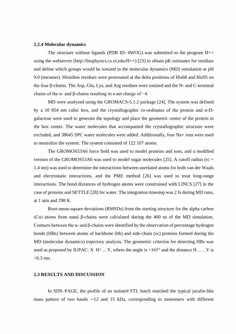
2.2.4 Molecular dynamics
The structure without ligands (PDB ID: 4WOG) was submitted to the program H++
using the webserver (http://biophysics.cs.vt.edu/H++) [23] to obtain pK estimates for residues
and define which groups would be ionized in the molecular dynamics (MD) simulation at pH
9.0 (tetramer). Histidine residues were protonated at the delta positions of His68 and His95 on
the four β-chains. The Asp, Glu, Lys, and Arg residues were ionized and the N- and C-terminal
chains of the α- and β-chains resulting in a net charge of −4.
MD were analyzed using the GROMACS-5.1.2 package [24]. The system was defined
by a 10 854 nm cubic box, and the crystallographic co-ordinates of the protein and α-D-
galactose were used to generate the topology and place the geometric center of the protein in
the box center. The water molecules that accompanied the crystallographic structure were
excluded, and 38645 SPC water molecules were added. Additionally, four Na+ ions were used
to neutralize the system. The system consisted of 122 107 atoms.
The GROMOS53A6 force field was used to model proteins and ions, and a modified
version of the GROMOS53A6 was used to model sugar molecules [25]. A cutoff radius (rc =
1.4 nm) was used to determine the interactions between unrelated atoms for both van der Waals
and electrostatic interactions, and the PME method [26] was used to treat long-range
interactions. The bond distances of hydrogen atoms were constrained with LINCS [27] in the
case of proteins and SETTLE [28] for water. The integration timestep was 2 fs during MD runs,
at 1 atm and 298 K.
Root-mean-square deviations (RMSDs) from the starting structure for the alpha carbon
(Cα) atoms from αand β-chains were calculated during the 400 ns of the MD simulation.
Contacts between the α- and β-chains were identified by the observation of percentage hydrogen
bonds (HBs) between atoms of backbone (bb) and side-chain (sc) proteins formed during the
MD (molecular dynamics) trajectory analysis. The geometric criterion for detecting HBs was
used as proposed by IUPAC: X H^ ... Y, where the angle is >165° and the distance H……Y is
<0.3 nm.
2.3 RESULTS AND DISCUSSION
In SDS–PAGE, the profile of an isolated FTL batch matched the typical jacalin-like
mass pattern of two bands ∼12 and 15 kDa, corresponding to monomers with different

glycosylation levels, as previously reported [6]. Evidence based on FTL mass deconvoluted
spectra suggests that the protein is naturally expressed as a mixture of isoforms [29]. Using a
hemagglutination assay, we found FTL to be in a biologically active form and able to
agglutinate human erythrocytes, which was preliminarily required before submitting the lectin
to crystallization assays (Supplementary Figure S1).
2.3.1 X-ray diffraction
The best solution for both Apo-frutalin and FTL–D-galactose complexes was obtained
in space group I2 with four monomers per asymmetric unit. The data collection statistics are
summarized in Table 1. The final models were refined to an Rfactor of 0.163 and an Rfree of
0.203 for Apo-frutalin, and to an Rfactor of 0.166 and an Rfree of 0.200 for the FTL–D-
galactose complex. In both cases, the asymmetric unit contains for monomers, with 153 residues
in each (Figure 1A,B). The residues (96.78%) are in the most favored regions, and 1.21% are
in the allowed regions of the Ramachandran plot.
Similar to jacalin, FTL is synthesized in vivo as an unusual preproprotein, which
becomes two chains after co-translational and post-translational processing: an α-chain (133
amino acids) and a β-chain (20 amino acids). FTL also shows conserved consensus sequences,
which suggests that three N-glycosylation sites may be present [6,8]. In contrast, mannose-
specific lectins such as frutapin (also found in breadfruit seeds) consist of uncleaved protomers
of ∼150 amino acid residues [15].
Table 1 - X-ray parameters for FTL structures
(Values in parentheses are for the outer resolution shell.)
Apo-frutalin FTL–galactose complex
PDB ID 4WOG 5BN6
X-ray source Rigaku MicroMax 007HF Rigaku MicroMax 007HF
Detector Rigaku Raxis IV++ Rigaku Raxis IV++
Cell
parameters (Å)
a, b, c
76.17, 74.36, 118.98 75.93, 74.60, 119.04
Space group I2 I2
Resolution (Å) 26.74–1.81 (1.85–1.81) 19.65–1.65 (1.65–1.68)
X-ray source Rigaku MicroMax 007 HF Rigaku MicroMax 007 HF

λ (Å) 1.5418 1.5418
Multiplicity 4.0 (3.7) 2.3 (1.5)
Rmerge (%) 10.0 (53.3) 4.9 (44.6)
Rpim (%) 4.9 (27.1) 3.9 (39.5)
CC(1/2) 0. 996 (0.784) 0.998 (0.736)
Completeness (%) 99.8 (97.0) 97.3 (75.6)
Reflections 240 259 (12 493) 174 200 (4489)
Unique reflections 59 769 (3386) 77 162 (2939)
I/σ 10.3 (2.6) 13.7 (1.9)
Reflections used for
refinement
59 762 77 151
Rfactor 0.163 0.166
Rfree 0.203 0.200
No. of protein atoms 4629 4626
No. of ligand atoms 0 48
B (Å2) 14.19 13.83
Co-ordinate error (ML
based) (Å)
0.21 0.15
Phase error (°) 19.33 19.15
Ramachandran plot
Favored (%) 96.59 96.59
Allowed (%) 3.41 3.41
Outliers (%) 0.00 0
All-atom clashscore 1.86 2.38
RMSD from ideal
geometry
r.m.s. bond lengths (Å) 0.007 0.006
r.m.s. bond angles (°) 1.017 1.038
The FTL structure showed a typical symmetric β-prism fold, which is found in jacalin
and other lectins [30]. This β-prism is composed of three 4-stranded β-sheets forming three
antiparallel Greek key motifs, generating an approximate 3-fold symmetry (Figure 1C,D). FTL
shares high structural similarity with jacalin, CGB, and frutapin, which superposed well and

gave RMSDs ranging from 0.31 to 0.43 Å for the superposition of Cα atoms in these
homologous structures (Supplementary Figure S2) [15,30,31].
Another major difference between the mannose-specific JRLs and galactose-specific
JRLs is their biosynthesis, processing, and topogenesis. For example, jacalin is synthesized as
a preproprotein, which undergoes a complex series of processing steps and is presumed to be
located in the vacuolar compartment [32]. In contrast, the mature polypeptides of the mannose-
specific JRLs correspond to the entire open reading frame of the respective lectin genes,
presenting both α- and β-chains linked by a loop, and therefore are synthesized and located in
the cytoplasm [32]. This alteration is believed to expose amino acids that are involved in
carbohydrate recognition by FTL.

Figure 1: Predicted structural features of FTL
.
(A) Sequence alignment of FTL (PDB ID: 5BN6) with jacalin (PDB ID: 1UGW) and
CGB (PDB ID: 4AKB). Alignment was performed using Clustal Omega. The expanded
carbohydrate-binding site for each protein is boxed in blue. (B) Tetrameric structure of FTL
with the α- (magenta) and β-chains (red). Interactions between α- and β-chains can be observed
in α1–β1–α2– β2 chains (set A) and α3–β3–α4–β4 chains (set B). (C) Ribbon diagram of β-
prism, each part of the β-prism, is colored differently to facilitate the understanding of this
structure (red, blue, and yellow). Topology diagram of FTL generated by PDBSUM. (D) The
FTL structure is composed of three Greek keys and could be classified as N- (N- and C-termini)
and C-types.

Indeed, leguminous lectins have considerable conservation in their primary, secondary,
and tertiary structures. Comparisons of these sequences and structures demonstrate that
differences in carbohydrate specificity appear to occur due to changes in carbohydrate-binding
site-adjacent amino acids. The conformation of these loops is determined by the presence of
calcium or transition metal ions in the protein structure, which helps CBS orientation and
affinity for ligands. Although structurally analogous, with some reaching up to 90% similarity,
these lectins present several distinct biological activities [33].
2.3.2 Carbohydrate-binding site and molecular docking studies
Although FTL recognizes a range of ligands, it has great affinity for α-D-galactose
monosaccharides and complex carbohydrates that contain Galα1–3 glycans [6,9]. Galactose
binding is dominated by hydrogen bonding with the sugar hydroxyl groups O1, O3, O5, and
O6 (Figure 2A). Hydrogen bonding is the most dominant interaction in recognition of sugar
molecules by the lectin CBS via carbonyl and hydroxyl groups (Figure 2B) of the backbone
and side chains [34]. The crystal structures of the FTL–D-galactose complex also showed this
pattern. The FTL–D-galactose complex does not present significant structural differences when
compared with Apo-frutalin.
Overall, the FTL-binding site is similar to those in Moraceae lectins and consists of a
domain close to the N-terminus of the α-chain consisting of four key residues: Gly25, Tyr146,
Trp147, and Asp149 [35–37]. In the three-dimensional structure of the jacalin–α-D-galactose
complex (PDB ID: 1KU8), eight hydrogen bonds form directly between CBS amino acids and
the hydroxyls, especially those of C3 and C6 positions in D-galactose. Similarly, in the
galactose-binding lectin from champedak fruit (CGB)–Gal complex, FTL–galactose binding
occurs via many hydrogen bonds between the O atoms on the sugar ring and with side-chain
and main-chain N and O atoms on the α-chain (O3 and Gly1 N, O4 and Gly1 N and Asp125
OD1, O6 and Trp123 O, Trp123 N and Tyr122 N, O5 and Tyr122 N) through O3, O4, and O5
[31]. In contrast, the FTL–D-galactose complex displays 10 interactions through the C1
hydroxyl to residue Tyr146, C3 hydroxyl to residue Gly25, C4 hydroxyl to residues Gly25 and
Asp149, and C6 hydroxyl to residues Tyr146, Trp147, and Asp149 (Figure 3A).
Jacalin is among the most thoroughly studied lectins. Jeyaprakash et al. [38] identified
three components of the jacalin sugar-binding site, a primary binding site and two secondary
sites, named A and B. In this postulation, the primary site is responsible for binding galactose.
The secondary site can bind any α-linked sugar moiety, but cannot tolerate any β-substitutions

[39]. In addition, jacalin and CGB bind carbohydrates at one primary and two secondary
binding sites [31,40]. The large number of interactions is consistent with FTL’s affinity for
galactose. The FTL agglutination of erythrocytes is due to its multi-subunit proteins, which
contain multiple carbohydrate-binding sites that enable them to agglutinate cells [41]. These
carbohydrate-binding domains are identical or very similar and bind a wide range of ligands. A
comparison of the FTL CBS to other Moraceae lectins suggests that the site closely resembles
that of jacalin. There are equivalent residues in both lectins (such as Asp149 in FTL and Asp125
in jacalin), and both form two hydrogen bonds: one with the C3 hydroxyl and another with the
C4 hydroxyl. Based on these interactions, we suggest that FTL has an affinity for D-galactose
epimers such as D-mannose, as the C4 hydroxyl in this sugar is equatorial instead of axial, and
the interaction would also be allowed by Asp149.
Figure 2. FTL carbohytdrate-binding site with D-galactose.
(A) Composite omit map contoured at 1σ for a galactose molecule in the FTL structure. (B)
Galactose-neighboring residues are labeled. Ligplus interaction drawing of galactose
interactions in the FTL structure.

Figure 3. FTL CBS anchoring specific sugars.
(A) Close-up view of the FTL sugar-binding site in the D-galactose complex. Protein and
carbohydrate molecules are depicted as ribbon and stick models, respectively. Amino acid
residues, which interact with carbohydrates, are highlighted in blue. (B) Molecular docking
experiments suggest that the FTL CBS can accommodate the α-D-Gal–D-Man disaccharide
present in galactomannans, but not the β-D-Gal–Xyl–Glu trisaccharide present in xyloglucans
(C), as evidenced through affinity chromatography in those cross-linked polysaccharide
matrices.

Interactions between derivatives of Gal β-(1,3) Gal-α-OMe and jacalin have been
structurally and thermodynamically characterized [39]. It is now known that distortion of the
ligand occurs as a strategy for modulating affinity. β-Substituted methyl derivatives of
disaccharides can also bind to jacalin without changing the pattern of interactions in the
complexes involving the corresponding α-substituted derivatives. This is achieved through
distortions of the ligand molecule at the anomeric carbon and the glycosidic linkage in addition
to a small lateral shift. The higher internal energy caused by the distortion reduces the affinity
of β-substituted b-(1,3)-linked disaccharides to jacalin when compared with the α-substituted
variants [36,39,42]. Gal β-(1,3) Gal and its derivatives preferentially bind to jacalin with the
reducing sugar at the primary binding site, although binding with the non-reducing Gal at the
primary site is possible. α-Methyl substitution further strengthens binding in the first
arrangement. In contrast, β-substitution weakens the binding due to ligand distortion. The β-
substituted disaccharides continue to bind with the reducing Gal at the primary binding site,
thus, indicating that the affinity reduction is not strong enough to overcome the intrinsic
propensity of Gal β-(1,3) Gal to bind to jacalin with the reducing Gal at the primary site. This
propensity is believed to be an important determinant in the biologically relevant interactions
between jacalin and oligosaccharides [39].
Interestingly, native FTL was previously isolated in an affinity chromatography step
using an Adenanthera pavonina cross-linked galactomannan [6]. In this type of polysaccharide,
galactose is naturally α-linked (1-6-D-galactopyranose) to the β-1-4-D-mannopyranose
backbone. On the other hand, in xyloglucan from Tamarindus indica seeds, these galactose
branches are bound by β-(1-2) to xylosyl residues attached to the main glucan backbone. When
using xyloglucan cross-linked matrices, we found FTL to always be easily removable from the
column with PBS buffer during the washing steps. This result suggests that FTL has a lower or
no affinity for β-galactose residues, implying an anomeric recognition. In looking for further
evidence of this anomeric recognition, docking experiments were performed to check whether
the FTL carbohydrate-binding site could accommodate the α-D-Gal-(1 → 6)-D-Man
disaccharide and β-D-Gal-(1 → 2)-α-D-Xyl-(1 → 6)-D-Glu trisaccharide, which mimic the
FTL interaction in galactomannan and xyloglucan cross-linked matrices, respectively. FTL
interactions with those saccharides yielded 10 energetic clusters. Figure 3B,C shows the pivotal
interface between the FTL–galactomannan residue (−4.9 kcal mol−1) and FTL–xyloglucan
residue (−6.5 kcal mol−1), respectively. These results are consistent with affinity
chromatography data in which xyloglucan matrices were inefficient at retaining FTL. In

contrast, accommodation of the α-D-Gal-(1 → 6)-D-Man disaccharide with Ala17 and Val19
residues was observed in FTL–galactomannan docking, in addition to those involved in the
CBS. Thus, FTL has anomeric carbohydrate recognition.
In addition, the tetrapeptide-linker ‘T-S-N-N’ is not a structural component of mature
FTL, as it is excised during lectin processing to separate FTL β- and α-chains, giving rise to
new N- and C-terminal sequences and reducing CBS rigidity, which results in the multiple-
binding abilities of FTL, jacalin, and CGB [37]. The specificity of KM+ (Artocarpin), the
mannose-binding lectin in jackfruit seeds, is attributed to the increased structural rigidity caused
by the presence of the binding peptide GGPGGNGW. This peptide linker is not eliminated by
post-translational processes, and is rich in glycine residues, thereby yielding extremely strong
bonds between the two chains [43]. Therefore, we investigated MD to determine how this
peptide linker may affect carbohydrate recognition by FTL.
2.3.3 Molecular dynamics
Simulations of the FTL structure with four α- and β-chains revealed an increasing
RMSD with a remarkable peak ∼110 ns and 0.5 nm (Figure 4). This observed behavior indicates
structural relaxation, which might occur through hydration after 110 ns and therefore reach
equilibrium. Structural properties were then analyzed considering the trajectory at the same
time point (equilibrium position) for all α- and β-chains (as shown in RMSD profiles by MD
time in Supplementary Figure S3A,B). Table 2 shows the mean RMSD for the interaction of
tertiary FTL structures (α- and β-chains), β-chain moieties (RMSD-M and RMSD-E), and the
sum of the four α- and β-chains. Higher averages were observed for single β-chains when
compared with single α-chains, implying that β-chains are more flexible.
The RMSDs for α-chains varied between 0.14 and 0.16 nm, whereas β-chain RMSDs
varied between 0.16 and 0.41 nm, suggesting that the positions of the α-chains have shorter
distanced than the beginning of the β-chains until reaching the equilibrium position of the
quaternary structure.

Figure 4. RMSD FTL during MD simulation.
RMSD from the α-carbon positions after finding maximal overlap between α-carbons of the
initial structure and structures collected during the MD simulation in all four FTL α- and β-
chains, with its peak within 110 ns and 0.5 nm.

Table 2 - The mean RMSD ± SD obtained in the last 300 ns of the MD simulation
ID RMSD (nm) RMSD-M RMSD-E
β1
0.26 ± 0.05 0.07 ± 0.02 0.32 ± 0.11
β2 0.16 ± 0.05 0.08 ± 0.02 0.39 ± 0.08
β3 0.41 ± 0.04 0.07 ± 0.02 0.38 ± 0.14
β4 0.28 ± 0.04 0.09 ± 0.02 0.60 ± 0.09
β1+ β2 + β3 + β4 0.37 ± 0.02
α1 0.15 ± 0.01
α2 0.16 ± 0.01
α3 0.16 ± 0.03
α4 0.14 ± 0.02
α1 + α2 + α3 + α4 0,36 ± 0.03
All β- and α-chains 0.35 ± 0.05
The RMSD-M includes the superimposing step and calculating the RMSD between the
Cα of residues in the 7–17 range. In the RMSD-E, Cα atoms between residues 7 and 17 are
used for the superimposing step, and the deviation calculation is between the atoms of residues
near the N- and C-termini of the β-chain from 1 to 6 and 18 to 19.
Similarly, the RMSDs were higher for the β-chains (range 0.04–0.05 nm), suggesting
that after reaching the equilibrium position, β-chain positions vary more than α (range 0.01–
0.03). These variations can be viewed and compared between the RMSD profiles in
Supplementary Figure S3A,B. Therefore, even though β-chains move more than α, they are
maintained in the structure, and the chain set stabilizes the octameric structure of FTL. Taking
into account the relative positions of the four α- and β-chains in the quaternary structure, the
RMSDs of (α1 + α2 + α3 + α4) 0.36 ± 0.03 nm and (β1 + β2 + β3 + β4) 0.37 ± 0.02 nm presented
similar variations in the means and SD, suggesting that positions of the eight chains supporting
the quaternary structure are preserved in equilibrium (Table 2). The RMSD-E and standard
deviation were much larger than for the overall side-chain (RMSD). The high RMSD-E values
suggest that the residues placed in these regions do not establish effective interchain contacts

with the α-chains, which remain free to move. However, the RMSD-M for the 7–17 region
happens to be smaller than the RMSD and RMSD-E, which indicates increased rigidity in this
regions.
Figure 5. PII between FTL residues and water molecules.
The distribution of IIP between the residues of the four β-chains and the water molecules
within a 0.5 nm cutoff radius.
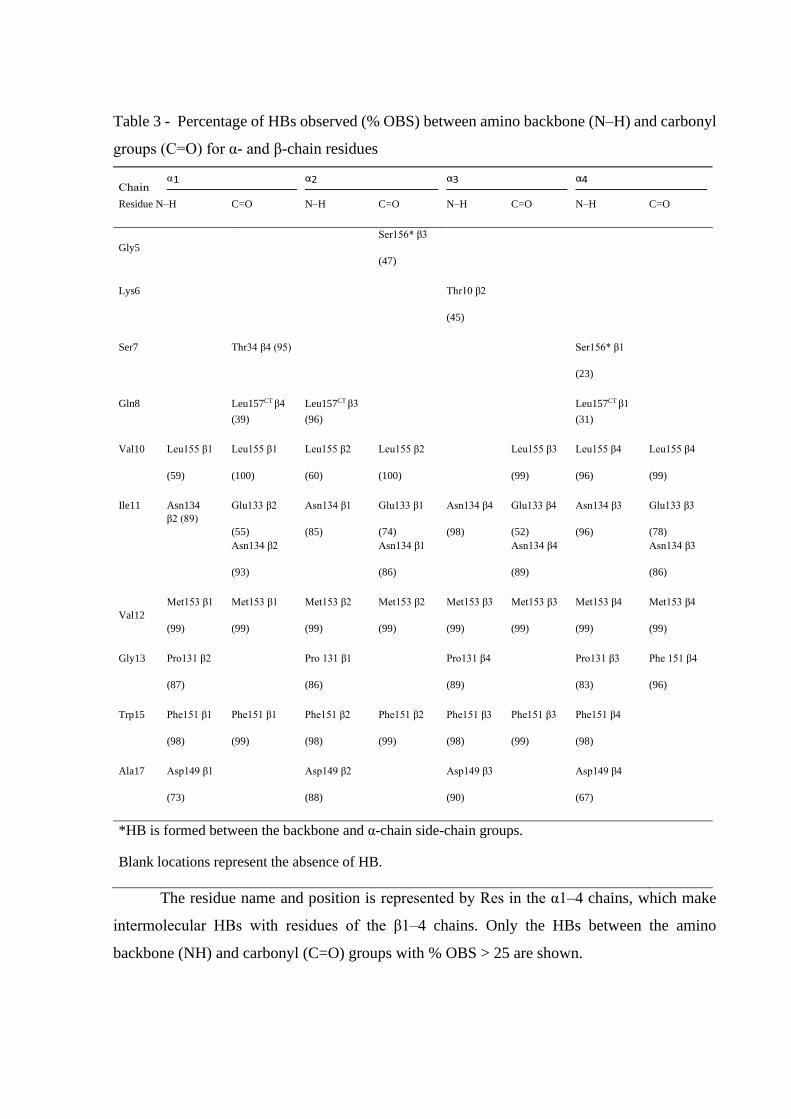
Table 3 - Percentage of HBs observed (% OBS) between amino backbone (N–H) and carbonyl
groups (C=O) for α- and β-chain residues
Residue N–H C=O N–H C=O N–H C=O N–H C=O
Gly5 Ser156* β3
(47)
Lys6 Thr10 β2
(45)
Ser7 Thr34 β4 (95)
Ser156* β1
(23)
Gln8 Leu157CT β4 (39)
Leu157CT β3 (96)
Leu157CT β1 (31)
Val10 Leu155 β1
(59)
Leu155 β1
(100)
Leu155 β2
(60)
Leu155 β2
(100)
Leu155 β3
(99)
Leu155 β4
(96)
Leu155 β4
(99)
Ile11 Asn134
β2 (89) Glu133 β2
(55) Asn134 β2
(93)
Asn134 β1
(85)
Glu133 β1
(74) Asn134 β1
(86)
Asn134 β4
(98)
Glu133 β4
(52) Asn134 β4
(89)
Asn134 β3
(96)
Glu133 β3
(78) Asn134 β3
(86)
Val12 Met153 β1
(99)
Met153 β1
(99)
Met153 β2
(99)
Met153 β2
(99)
Met153 β3
(99)
Met153 β3
(99)
Met153 β4
(99)
Met153 β4
(99)
Gly13 Pro131 β2
(87)
Pro 131 β1
(86)
Pro131 β4
(89)
Pro131 β3
(83)
Phe 151 β4
(96)
Trp15 Phe151 β1
(98)
Phe151 β1
(99)
Phe151 β2
(98)
Phe151 β2
(99)
Phe151 β3
(98)
Phe151 β3
(99)
Phe151 β4
(98)
Ala17 Asp149 β1
(73)
Asp149 β2
(88)
Asp149 β3
(90)
Asp149 β4
(67)
*HB is formed between the backbone and α-chain side-chain groups.
Blank locations represent the absence of HB.
The residue name and position is represented by Res in the α1–4 chains, which make
intermolecular HBs with residues of the β1–4 chains. Only the HBs between the amino
backbone (NH) and carbonyl (C=O) groups with % OBS > 25 are shown.

Figure 5 shows the distribution of the intermolecular interaction potential (IIP) between
the residues, building up the four β-chains and water molecules within a 0.5 nm cutoff radius.
It is evident that regions from 1 to 6 and 18 to 19 are hydrated (energy less than −5 kcal mol−1),
which is in contrast with those from 7 to 17. The IIP results combined with the RMSD-E values
found in the simulations allow us to conclude that the β-chain region from 7 to 17 residues
increases the rigidity of the referred chain by interacting with the α- and β-chain residues. These
interactions reflect the intermolecular HBs between groups of α- and β-chains, as can be seen
in Table 3. We expected to find interactions among these α- and β-chains by the breakdown in
FTL structure, which are mostly by α1–α2 and α3–α4 pairs. However, the majority of pivotal
HBs were negligible. β-Chains appear to be useful in maintaining the structure, and they occur
close to the amino acids from 10 to 18 in α-chains and interact through HB between the
backbone amino and carbonyl groups (C=O……H–N) of both α-chains (Table 3). Figure 6
shows a representation of these connections formed by HBs between pairs of αand β-chains.
The β1 and β2 chains connect the α1 and α2 chains, while the β3 and β4 connect the α1 and α2
chains via intermolecular HBs.
The amino and carbonyl groups from 10, 12, 15, and 17 β-chain residues form HBs with
amino and carbonyl groups of one α-chain, while residues 11 and 13 form HBs with groups of
another.
Figure 6. Interface between 03B1; and 03B2; chains of FTL connected by hydrogen bonds.

Carbonyl and amine group HBs (C=O……H–N) between α1, β1-chains from monomer
1 and the α2-chain of monomer 2 are depicted in the tetrameric FTL structure (magenta). The
interactions occur on two faces of the lectin and involve both α- and β-chains from monomers
1 and 2, resulting in an α2–β2–α1 cluster. Likewise, the connections are prone to occur in α3–
β3–α4 and α4–β4–α3 interchain clusters.
Thus, the HBs establish β1–α1, β2–α2, β3–α3, and β4–α4 pairs; while for residues 11
and 13, the pairs are β1–α2, β2–α1, β3– α4, and β4–α3. Indeed, the α-chain segment from 10
to 19 residues (VIVGPWGAQ), with hydrophobic side chains, helps to maintain these HBs by
keeping the water molecules away from the amino and carbonyl groups.
The stability of the entire FTL structure can be understood when we consider interaction
sets between αand β-chains, which occur as α1–β1–α2–β2 (set A) and α3–β3–α4–β4 chains (set
B) (Figure 1B). Sets A and B form an interface maintained through interactions between the C-
terminal regions of the β-chains (residues 1– 8) and complementary regions of the α-chains in
set B. These interactions result in the formation of HBs between α1–β4, α2–β3, α3–β2, and α4–
β1 (mainly by the carboxylic acid of the C-terminal Leu157 in α-chains and amine of Gln8 in
β-chains) (Table 3).
Additionally, Asp149 in the α-chain is stabilized in the carbohydrate-binding site by
HBs between the amine and carbonyl groups of Ala17 in the β-chain (Figure 1D and Table 3).
The occurrence of HB is greater than 67% in all α–β pair interactions. Furthermore, Ala17 and
Val19 promote a hydrophobic environment by enhancing interchain HBs and preventing the α-
D-Gal-(1 → 6)-D-Man disaccharide hydration, which helps its accommodation into the FTL
CBS. Meanwhile, Ala17 appears to play a structural role, helping to stabilize Asp149, with
Val19 oscillating in its position, as shown by the RMSD-E (Table 2). The MD results validate
the functional importance of the β-chains in maintaining the FTL structure. In addition, Ala17
in the β-chains helps to stabilize the Asp149 position in the α-chains through HB formation,
which corroborates Asp149 functioning as the key residue for interactions and carbohydrate-
binding. Indeed, Asp149 allows entry of α-D-Gal-(1 → 6)-D-Man disaccharide to the CBS site
and its subsequent stabilization.
In regard to this carbohydrate recognition, FTL has been evaluated in several biomedical
applications and found to establish hydrogen bonds with the glycosylated fraction of the TRPV1
ion channel, inhibit the orofacial pain mechanism [13], and specifically recognize the NMDA
receptor in its glycosylated fraction, interfering in a cellular mechanism and producing
antidepressant-like NMDA receptor-mediated activity [44].

2.4 CONCLUSION
Over the years, the therapeutic relevance of FTL has been demonstrated in many
biomedical mechanisms. However, little was known about FTL’s structure. Taken together, the
results in this work provide a new perspective for further elucidation of the functional properties
of this lectin. While FTL is conserved across species, which explains the high similarity to other
lectins in the Moraceae family, some plants have developed unique and specialized mechanisms
to deal with their complex intracellular signaling pathways, mostly triggered by the intricate
relationships between the carbohydrate-binding specificity of lectins. Thus, a better
understanding of FTL binding to sugar moieties provides insights into its functionality and
sheds light on the biological activities of this lectin.
ABBREVIATIONS
CBS, carbohydrate-binding site; CGB, champedak galactose-binding lectin; FTL,
frutalin; HB, hydrogen bond; IIP, intermolecular interaction potential; JRL, jacalin-related
lectins; KM+, Artocarpin; MCA, minimal concentration for agglutination; MD, molecular
dynamics; PME, particle mesh ewald; RMSD, root-mean-square deviation; UPLC-ESI, ultra-
performance liquid chromatography with electrospray ionization.
AUTHOR CONTRIBUTION
A.E.V.N., T.B.G., and F.D.S. were involved in all aspects of purifying the protein, while
H.M.P. and F.B.M.B.M determined the X-ray structure. M.R.L. performed the MD
experiments. A.C.O.M.M. and R.A.M. obtained funding for the study and provided overall
supervision. A.E.V.N and F.D.S. wrote the initial drafts of the paper and all authors contributed
to the final manuscript.
FUNDING
This work was supported by National Council for Scientific and Technological
Development (CNPq), Fundação Cearense de Amparo á Pesquisa (FUNCAP), and Agency for
Financing Studies and Projects (FINEP).
ACKNOWLEDGEMENTS
We acknowledge the Physics Institute of São Carlos (University of São Paulo, USP) for
assistance with crystal testing and data collection. We also thank the Fundação Edson Queiroz

for providing infrastructure at the University of Fortaleza (UNIFOR) and CAPES (Coordination
for the Improvement of Higher Education) for financial support.
COMPETING INTERESTS
The Authors declare that there are no competing interests associated with the manuscript
REFERÊNCIAS
1 Lagarda-Diaz, I., Guzman-Partida, A. and Vazquez-Moreno, L. (2017) Legume
lectins: proteins with diverse applications. Int. J. Mol. Sci. 18, 1242
https://doi.org/10.3390/ijms18061242
2 Jiang, S.-Y., Ma, Z. and Ramachandran, S. (2010) Evolutionary history and stress
regulation of the lectin superfamily in higher plants. BMC Evol. Biol. 10, 79
PMID:20236552
3 Jagtap, U.B. and Bapat, V.A. (2010) Artocarpus: a review of its traditional uses,
phytochemistry and pharmacology. J. Ethnopharmacol. 129, 142–166
https://doi.org/10.1016/j.jep.2010.03.031
4 de Azevedo Moreira, R. and Ainouz, I.L. (1981) Lectins from seeds of jack fruit
(Artocarpus integrifolia L.): isolation and purification of two isolectins from the albumin
fraction. Biol. Plant. 23, 186–192 https://doi.org/10.1007/BF02894883
5 Kabir, S., Aebersold, R. and Daar, A.S. (1993) Identification of a novel 4 kDa
immunoglobulin-A-binding peptide obtained by the limited proteolysis of jacalin. Biochim.
Biophys. Acta 1161, 194–200 PMID:8431469
6 Moreira, R.A., Castelo-Branco, C.C., Monteiro, A.C., Tavares, R.O. and Beltramini,
L.M. (1998) Isolation and partial characterization of a lectin from Artocarpus incisa L. seeds.
Phytochemistry 47, 1183–1188 https://doi.org/10.1016/S0031-9422(97)00753-X
7 Oliveira, C., Felix, W., Moreira, R.A., Teixeira, J.A. and Domingues, L. (2008)
Expression of frutalin, an α-D-galactose-binding jacalin-related lectin, in the yeast Pichia
pastoris. Protein Expr. Purif. 60, 188–193 https://doi.org/10.1016/j.pep.2008.04.008

8 Monteiro-Moreira, A.C.O., D’Muniz Pereira, H., Vieira-Neto, A.E., Moreno,
F.B.M.B., Lobo, M.D.P., Sousa, F.D. et al. (2015) Crystallization and preliminary X-ray
diffraction studies of frutalin, an α-D-galactose-specific lectin from Artocarpus incisa seeds.
Acta Crystallogr. Sect. F Struct. Biol. Commun. F71, 1282–1285 PMID:26457519
9 Oliveira, C., Teixeira, J.A., Schmitt, F. and Domingues, L. (2009) A comparative
study of recombinant and native frutalin binding to human prostate tissues. BMC Biotechnol.
9, 78 https://doi.org/10.1186/1472-6750-9-78
10 Lobo, M.D., Moreno, F.B., Souza, G.H., Verde, S.M., Moreira, R.A. and Monteiro-
Moreira, A.C. (2017) Label-free proteome analysis of plasma from patients with breast
cancer: stage-specific protein expression. Front. Oncol. 7, 14 PMID:28210565
11 Brando-Lima, A.C., Saldanha-Gama, R.F., Henriques, M.D.G.M.O., Monteiro-
Moreira, A.C.O., Moreira, R.A. and Barja-Fidalgo, C. (2005) Frutalin, a galactose-binding
lectin, induces chemotaxis and rearrangement of actin cytoskeleton in human neutrophils:
involvement of tyrosine kinase and phosphoinositide 3-kinase. Toxicol. Appl. Pharmacol.
208, 145–154 https://doi.org/10.1016/j.taap.2005.02.012
12 De Vasconcellos Abdon, A.P., Coelho De Souza, G., Noronha Coelho De Souza, L.,
Prado Vasconcelos, R., Araújo Castro, C., Moreira Guedes, M. et al. (2012) Gastroprotective
potential of frutalin, a D-galactose binding lectin, against ethanol-induced gastric lesions.
Fitoterapia 83, 604–608 https://doi. org/10.1016/j.fitote.2012.01.005
13 Damasceno, M.B.M.V., De Melo Júnior, J.D.M.A., Santos, S.A.A.R., Melo, L.T.M.,
Leite, L.H.I., Vieira-Neto, A.E. et al. (2016) Frutalin reduces acute and neuropathic
nociceptive behaviours in rodent models of orofacial pain. Chem. Biol. Interact. 256, 9–15
https://doi.org/10.1016/j.cbi.2016.06.016
14 Moreira, R.A. and Perrone, J.C. (1977) Purification and partial characterization of a
lectin from Phaseolus vulgaris. Plant Physiol. 59, 783–787
https://doi.org/10.1104/pp.59.5.783
15 de Sousa, F.D., da Silva, B.B., Furtado, G.P., Carneiro, I.S., Lobo, M.D.P., Guan, Y.
et al. (2017) Frutapin, a lectin from Artocarpus incisa (breadfruit): cloning, expression and
molecular insights. Biosci. Rep. 37, BSR20170969 https://doi.org/10.1042/BSR20170969

16 Ferrige, A.G., Seddon, M.J., Jarvis, S., Skilling, J. and Aplin, R. (1991) Maximum
entropy deconvolution in electrospray mass spectrometry. Rapid Commun. Mass Spectrom.
5, 374–377 https://doi.org/10.1002/rcm.1290050810
17 McCoy, A.J., Grosse-Kunstleve, R.W., Adams, P.D., Winn, M.D., Storoni, L.C. and
Read, R.J. (2007) Phaser crystallographic software. J. Appl. Crystallogr. 40(Pt 4), 658–674
PMID:19461840
18 Kabsch, W. (2010) Xds. Acta Crystallogr. Sect. D Biol. Crystallogr. 66, 125–132
https://doi.org/10.1107/S0907444909047337
19 Adams, P.D., Afonine, P.V., Bunkóczi, G., Chen, V.B., Davis, I.W., Echols, N. et al.
(2010) PHENIX: a comprehensive Python-based system for macromolecular structure
solution. Acta Crystallogr. Sect. D Biol. Crystallogr. 66, 213–221
https://doi.org/10.1107/S0907444909052925
20 Emsley, P. and Cowtan, K. (2004) Coot : model-building tools for molecular graphics
research papers. Acta Crystallogr. D Biol. Crystallogr. 60(Pt 12 Pt 1), 2126–2132
PMID:15572765
21 Chen, V.B., Bryan, W., Iii, A., Headd, J.J., Keedy, D.A., Robert, M. et al.(2010)
Molprobity : all-atom structure validation for macromolecular crystallography research
papers. Acta Crystallogr. D Biol. Crystallogr. 66(Pt 1), 12–21 PMID:20057044
22 Trott, O. and Olson, A.J. (2011) NIH public access. J. Comput. Chem. 31, 455–461
PMID:19499576
23 Gordon, J.C., Myers, J.B., Folta, T., Shoja, V., Heath, L.S. and Onufriev, A. (2005)
H++: a server for estimating pKas and adding missing hydrogens to macromolecules. Nucleic
Acids Res. 33, W368–W371 https://doi.org/10.1093/nar/gki464
24 Abraham, M.J., Murtola, T., Schulz, R., Páll, S., Smith, J.C., Hess, B. et al. (2015)
Gromacs: high performance molecular simulations through multi-level parallelism from
laptops to supercomputers. SoftwareX 1–2, 19–25 https://doi.org/10.1016/j.softx.2015.06.001
25 Pol-Fachin, L., Rusu, V.H., Verli, H. and Lins, R.D. (2012) GROMOS 53A6GLYC,
an improved GROMOS force field for hexopyranose-based carbohydrates. J. Chem. Theory
Comput. 8, 4681–4690 https://doi.org/10.1021/ct300479h

26 Darden, T., York, D. and Pedersen, L. (1993) Particle mesh Ewald: an N⋅log(N)
method for Ewald sums in large systems. J. Chem. Phys. 98, 10089
https://doi.org/10.1063/1.464397
27 Hess, B., Bekker, H., Berendsen, H.J.C. and Fraaije, J.G.E.M. (1997) LINCS: a linear
constraint solver for molecular simulations. J. Comput. Chem. 18, 1463–1472
https://doi.org/10.1002/(SICI)1096-987X(199709)18:12<1463::AID-JCC4>3.0.CO;2-H
28 Miyamoto, S. and Kollman, P.A. (1992) Settle: an analytical version of the SHAKE
and RATTLE algorithm for rigid water models. J. Comput. Chem. 13, 952–962
https://doi.org/10.1002/jcc.540130805
29 De Oliveira Monteiro-Moreira, A.C., D’Muniz Pereira, H., Vieira Neto, A.E., Mendes
Batista Moreno, F.B., Duarte Pinto Lobo, M., de Sousa, F.D. et al. (2015) Crystallization and
preliminary X-ray diffraction studies of frutalin, an α-D-galactose-specific lectin from
Artocarpus incisa seeds. Acta Crystallogr. Sect. Struct. Biol. Commun. 71(Pt 10), 1282–
1285 https://doi.org/10.1107/S2053230X15015186
30 Pratap, J.V., Jeyaprakash, A.A., Rani, P.G., Sekar, K., Surolia, A. and Vijayan, M.
(2002) Crystal structures of artocarpin, a Moraceae lectin with mannose specificity, and its
complex with methyl-alpha-D-mannose: implications to the generation of carbohydrate
specificity. J. Mol. Biol. 317, 237–247 https://doi.org/10.1006/jmbi.2001.5432
31 Gabrielsen, M., Abdul-Rahman, P.S., Othman, S., Hashim, O.H. and Cogdell, R.J.
(2014) Structures and binding specificity of galactose- and mannose-binding lectins from
champedak: differences from jackfruit lectins. Acta Crystallogr. Sect. F Struct. Biol.
Commun. 70, 709–716 PMID:24915077
32 Peumans, W.J., Hause, B. and Van Damme, E.J.M. (2000) The galactose-binding and
mannose-binding jacalin-related lectins are located in different sub-cellular compartments.
FEBS Lett. 477, 186–192 https://doi.org/10.1016/S0014-5793(00)01801-9
33 Gadelha, C.A.A., Moreno, F.B.M.B., Santi-Gadelha, T., Cajazeiras, J.B., Rocha,
B.A.M., Assreuy, A.M.S. et al. (2005) Native crystal structure of a nitric oxide-releasing
lectin from the seeds of Canavalia maritima. J. Struct. Biol. 152, 185–194
https://doi.org/10.1016/j.jsb.2005.07.012

34 Thirumalai, D., Reddy, G. and Straub, J.E. (2012) Role of water in protein aggregation
and amyloid polymorphism. Acc. Chem. Res. 45, 83–92 https://doi.org/10.1021/ar2000869
35 Pinedo, M., Orts, F., Carvalho, A.O., Regente, M., Soares, J.R., Gomes, V.M. et al.
(2015) Molecular characterization of Helja, an extracellular jacalin-related protein from
Helianthus annuus: insights into the relationship of this protein with unconventionally
secreted lectins. J. Plant Physiol. 183, 144–153 https://doi.org/10.1016/j.jplph.2015.06.004
36 Abhinav, K.V., Sharma, K., Surolia, A. and Vijayan, M. (2016) Effect of linkage on
the location of reducing and nonreducing sugars bound to jacalin. IUBMB Life 68, 971–979
https://doi.org/10.1002/iub.1572
37 Gabrielsen, M., Abdul-Rahman, P.S., Othman, S., Hashim, O.H. and Cogdell, R.J.
(2014) Structures and binding specificity of galactose- and mannose-binding lectins from
champedak: differences from jackfruit lectins. Acta Crystallogr. Sect. F Struct. Biol.
Commun. 70, 709–716 https://doi. org/10.1107/S2053230X14008966
38 Jeyaprakash, A.A., Katiyar, S., Swaminathan, C.P., Sekar, K., Surolia, A. and Vijayan,
M. (2003) Structural basis of the carbohydrate specificities of jacalin: an X-ray and modeling
study. J. Mol. Biol. 332, 217–228 https://doi.org/10.1016/S0022-2836(03)00901-X
39 Abhinav, K.V., Sharma, K., Surolia, A. and Vijayan, M. (2017) Distortion of the
ligand molecule as a strategy for modulating binding affinity: further studies involving
complexes of jacalin with β-substituted disaccharides. IUBMB Life 69, 72–78
https://doi.org/10.1002/iub.1593
40 Jeyaprakash, A.A., Srivastav, A., Surolia, A. and Vijayan, M. (2004) Structural basis
for the carbohydrate specificities of artocarpin: variation in the length of a loop as a strategy
for generating ligand specificity. J. Mol. Biol. 338, 757–770
https://doi.org/10.1016/j.jmb.2004.03.040
41 Varki, A. (2017) Biological roles of glycans. Glycobiology 27, 3–49
https://doi.org/10.1093/glycob/cww086
42 Abhinav, K.V., Sharma, K., Swaminathan, C.P., Surolia, A. and Vijayan, M. (2015)
Jacalin-carbohydrate interactions: distortion of the ligand molecule as a determinant of

affinity. Acta Crystallogr. Sect. D Biol. Crystallogr. D71, 324–331
https://doi.org/10.1107/S139900471402553X
43 Rosa, J.C., De Oliveira, P.S., Garratt, R., Beltramini, L., Resing, K., Roque-Barreira,
M.C. et al. (1999) KM+, a mannose-binding lectin from Artocarpus integrifolia: amino acid
sequence, predicted tertiary structure, carbohydrate recognition, and analysis of the beta-prism
fold. Protein Sci. 8, 13–24 https://doi.org/10.1110/ps.8.1.13
44 Araújo, J.R.C., Júnior, J.M.A.M., Damasceno, M.B.M.V., Santos, S.A.A.R., Vieira-
Neto, A.E., Lobo, M.D.P. et al. (2018) Neuropharmacological characterization of frutalin in
mice: evidence of an antidepressant-like effect mediated by the NMDA receptor/NO/cGMP
pathway. Int. J. Biol. Macromol. 112, 548–554
https://doi.org/10.1016/j.ijbiomac.2018.01.180

3 CAPÍTULO II
Running title: Frutalin Recognizes Complement C3 Over-Expressed by Neoplasms
Frutalin Recognizes Complement C3 Over-Expressed by Neoplasms
Antonio Eufrásio Vieira Neto1,2, Ana Cristina de Oliveira Monteiro Moreira2 and Renato de
Azevedo Moreira1,2
1 Department of Biochemistry and Molecular Biology, Federal University of Ceará, Campus do
Pici, Bloco 907, Fortaleza, Ceara 60451 970, Brazil.
2 Center of Experimental Biology (Nubex), University of Fortaleza (UNIFOR), Av. Washington
Soares, 1321, Fortaleza, Ceara 60811-905, Brazil;
ABSTRACT
Frutalin (FTL) is an α-D-galactose-binding lectin obtained from the seeds of Artocarpus
incisa L. It has presented several biomedical activities based on the recognition of the
carbohydrate binding site. Bioinformatics techniques have sought to elucidate the molecular
bases of interaction with biological receptors. Previous studies have demonstrated that Frutalin,
in addition to promoting biological activities, is able to recognize differentially expressed
glycoproteins in the blood in patients with neoplasms such as breast cancer and acute
lymphoblastic leukemia (ALL). The aim of this work was to elucidate the molecular bases of
interaction of Frutalin with Complement C3 Protein, a glycoprotein over-expressed in serum of
patients with ALL and breast cancer. The possible interaction between FTL with the surface of
the complement protein C3 was analyzed using molecular docking, with the Hex 8.0.0 platform.
The 10 most energetic clusters were investigated for analysis of interaction energy, binding
specificity, CRS-glycoprotein affinity, the amino acids involved and the attraction/repulsion
forces. Molecular docking demonstrated high affinity to between FTL and C3 glycans, with
high specificity and complexation energy, with 08 hydrogen bonds measuring up to 0.8
angstroms, with high reproducibility. It can be concluded that FTL is a lectin capable of
promoting biological and biomedical activities due to the flexibility of its CRS, which allows

the recognition of complex glycans present in glycoprotein’s involved in biological phenomena,
which makes it a relevant biotechnological tool.
3.1 INTRODUCTION
3.1.1 Biomarkers in cancer research
Biomarkers are increasingly useful tools for cancer research. The search for new,
specific and sensitive biomarkers continually aims to discover improvements in the diagnosis
and confirmation of the neoplasms or that may have practical value in the evaluation of the
treatment and in the behavior of the disease [1].
Acute lymphoblastic leukemia of Precursor Cells B (ALL-B) and Breast Cancer are
examples of diseases that occur in worrying proportions and have to be more studied. Precursor
Cell Lymphoblastic Leukemia-Lymphoma is the most common malignancy in childhood and
breast cancer and the most common cancer among women worldwide after non-melanoma skin
cancer. Featuring clinically and biologically heterogeneous diseases, early detection, reliable
characterization, and accurate diagnosis are critical for reducing mortality [2].
3.1.2 Frutalin as biomedical tool
This work investigates the interaction of Complement C3, which is a differentially-
expressed glycoprotein with changes in its glycosylation patterns in both diseases, with Frutalin
(FTL), that is a plant lectin with affinity for α-D-galactose and α-D-mannose, obtained from
the seeds of Artocarpus incisa L. FTL is a glycoprotein with approximately 66 kDa, which has
a CBS in each monomer of its homotetramer. It is able to agglutinate erythrocytes since it is
oligomerized under physiological conditions and promotes glycans recognition in two or more
CBS simultaneously, agglutinating the red blood cells [3]. The three-dimensional structure of
Frutalin has been elucidated by crystallographic methods [4] and has already shown to be
compatible with other biological components [5,6]. FTL proved to be efficient in detecting the
overexpression of Complement C3, which suggests an association with the development and
characterization of cancer in LLA and breast cancer situations [2,7]. The aim of this study was
elucidating the molecular bases of interaction of FTL with the C3 protein, in the recognition
observed in previous studies.
3.2 MATERIALS AND METHODS
3.2.1 Molecular docking
The interaction between Complement C3 and Frutalin (FTL) was analyzed using
molecular docking. FTL and Complement C3 structures were available in the Protein Data Bank
(codes 4WOG and 2A73, respectively).
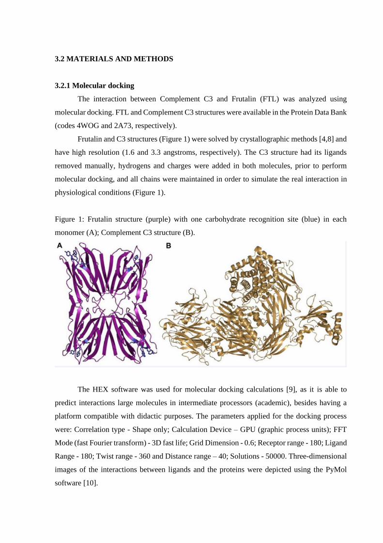
Frutalin and C3 structures (Figure 1) were solved by crystallographic methods [4,8] and
have high resolution (1.6 and 3.3 angstroms, respectively). The C3 structure had its ligands
removed manually, hydrogens and charges were added in both molecules, prior to perform
molecular docking, and all chains were maintained in order to simulate the real interaction in
physiological conditions (Figure 1).
Figure 1: Frutalin structure (purple) with one carbohydrate recognition site (blue) in each
monomer (A); Complement C3 structure (B).
The HEX software was used for molecular docking calculations [9], as it is able to
predict interactions large molecules in intermediate processors (academic), besides having a
platform compatible with didactic purposes. The parameters applied for the docking process
were: Correlation type - Shape only; Calculation Device – GPU (graphic process units); FFT
Mode (fast Fourier transform) - 3D fast life; Grid Dimension - 0.6; Receptor range - 180; Ligand
Range - 180; Twist range - 360 and Distance range – 40; Solutions - 50000. Three-dimensional
images of the interactions between ligands and the proteins were depicted using the PyMol
software [10].

3.3 RESULTS AND DISCUSSION
Molecular docking showed the interaction of FTL (receptor molecule) with
Complement C3, after 50,000 possible fittings. In in vitro proteomic investigations serum
samples from patients with cancer were applied in chromatography column to interact with the
FTL immobilized matrix [2,7].
These results showed that the C3 molecule interacts with matrix, being considered a
ligand while the FTL was considered receptor. The molecular bases of this interaction were
elucidated through computational simulation. The 10 most stable clusters of this interaction
were analyzed and initially presented high specificity, since they were overlapped in the same
place, which indicates good energy stabilization of the formed complex, in that position (Figure
2).
Figure 2: Overlapping of the 3 most energetic clusters (colored) of C3, which suggests specific
affinity for the FTL surface (purple) surrounding the CBS.

A protein has an intrinsic vibratory energy and when it binds with another molecule; its
energy is stabilized by the complexation, which was also evidenced with the connection to
Complement C3. The fit provided an energy stabilization of up to -26767.43 kcal/mol and
proved to be efficient in all 10 most energetic clusters (Table 1). A similar result has been as
have been found in other studies concerning the molecular interactions of the FTL in biomedical
activities [5,6] (Table 1).
Table 1: Total energy of the interactions between FTL (receptor) and Complement C3 (ligand),
after molecular docking simulation.
Complex obtained
by FTL-C3 interaction
Total energy
obtained (kcal/mol)
Cluster 01: -26767.43
Cluster 02: -26556.11
Cluster 03: -25255.56
Cluster 04: -24777.30
Cluster 05: -24430.97
Cluster 06: -23427.78
Cluster 07: -22620.34
Cluster 08: -22402.28
Cluster 09: -22321.76
Cluster 10: -21322.84
The high complexing energies observed in the 10 analyzed clusters indicate that there
is high affinity of CBS for the ligand, and regarding the role of CBS in this recognition, it was
important to investigate the surface of FTL and the location of its CBS in each monomer. In
fact, C3 is a large glycoprotein and interact with FTL with the involvement of two monomers.
In general, plant lectins have their lectin activity measured by the ability to agglutinate
erythrocytes, due to their ability to recognize glycans, agglutinating them. This agglutination
occurs when the lectin binds to two or more erythrocytes, which recruits more than one CBS
[11]. Based on this, FTL recognizes complement C3 with two monomers simultaneously. This
interaction increases the stabilization and neutralizes the repulsions and the hydrophobic effects
of the region that does not belong to the lectin activity, in Frutalin (Figure 3).

Figure 3: Interaction of Complement C3 (gray) with FTL (purple), evidencing lectin recognition
activity, interacting with two tetramer subunits simultaneously.
According to previously information about FTL amino acid residues [6] that promote
lectin activity (Gly25, Tyr146, Trp147 and Asp149), the interaction of these residues with the
amino acids of Complement C3 was measured. As a result, was observed the action of 6 amino
acid residues (please specify these 6 residues), which made bonds of up to 0.8 angstroms and
could stabilize the region and promote the binding between the two molecules (Figure 4). The
results corroborate with in vitro findings [2,7] (Figure 4).
Figure 4: Carbohydrate recognition site of FTL (blue) showing hydrogen bonds with 6 amino
acid residues (yellow) of Complement C3 (0.8 – 3.9 angstroms).

In order to assess the affinity of the FTL for glycoprotein’s, oligosaccharides and
glycans with higher molecular size, it can be suggested that the size and flexibility of the CBS
promotes greater recognition by stabilizing in the middle of so many bonds. Therefore, the
binding energy against a monosaccharide is much lower than that observed in biological
activities already reported [12], in which FTL showed a high recognition activity in silico. It is
known that plant lectins have been studied because they have specificity and affinity to
carbohydrates in several biological processes [13]. Based on this, this work strengthens the
suggestion of investigating plant lectins with high specificity to glycans characteristic of
specific diseases, seeking biotechnological potential.
3.4 CONCLUSIONS
The molecular docking results reflect a specific interaction between Frutalin and
Complement C3 protein over expressed in some neoplasms added to its specificity, stability in
physiological conditions and its affinity to glycans make it a useful tool in research concerning
cancer and biomarkers. FTL is a plant lectin capable of promoting biomedical activities due to
the flexibility of its CBS, which allows the recognition of glycoproteins involved in ALL and
breast cancer.
ABBREVIATIONS
ALL: Acute Lymphoblastic Leukemia; BC: Breast Cancer; CBS: Carbohydrate Binding Site;
FFT: Fast Fourier Transform; FTL: Frutalin
ACKNOWLEDGEMENTS
University of Fortaleza (UNIFOR), Experimental Biology Center (NUBEX), National Council
for Scientific and Technological Development (CNPq), National Council for the Improvement
of Higher Education (CAPES), Fundação Cearense de Amparo á Pesquisa (FUNCAP) and
Agency for Financing Studies and Projects (FINEP), for all support.

REFERÊNCIAS
1. Siegel RL, Miller KD, Jemal A. Cancer statistics. CA Cancer J Clin 2016; 66: 7-30.
2. Cavalcante M de S, Torres-Romero JC, Lobo MDP, Moreno FBMB, Bezerra LP, Lima
DS, et al. A panel of glycoproteins as candidate biomarkers for early diagnosis and
treatment evaluation of B-cell acute lymphoblastic leukemia. Biomark Res. 2016; 4: 1.
3. Moreira RA, Castelo-Branco CC, Monteiro ACO, Tavares RO, Beltramini LM. Isolation
and partial characterization of a lectin from Artocarpus incisa L. seeds. Phytochemistry.
1998; 47: 1183-1188.
4. Monteiro-Moreira AC de O, D’Muniz Pereira H, Vieira Neto AE, Mendes Batista
Moreno FB, Duarte Pinto Lobo M, Sousa FD De, et al. Crystallization and preliminary
X-ray diffraction studies of frutalin, an α-D-galactose-specific lectin from Artocarpus
incisa seeds. Acta Crystallogr Sect F Struct Biol Commun. 2015; 71: 1282-1285.
5. Damasceno MBM V, De Melo Júnior JDMA, Santos SAAR, Melo LTM, Leite LHI,
Vieira-Neto AE, et al. Frutalin reduces acute and neuropathic nociceptive behaviours in
rodent models of orofacial pain. Chem Biol Interact. 2016; 256: 9-15.
6. Araújo JRC, Júnior J de MA de M, Damasceno M de BMV, Santos SAAR, Vieira-Neto
AE, Lobo MDP, et al. Neuropharmacological antidepressant like Biol Macromol. 2018;
112: 548-54.
7. Lobo MDP, Moreno FBMB, Souza GHMF, Verde SMML, Moreira R de A, Monteiro-
Moreira AC de O. Label-Free Proteome Analysis of Plasma from Patients with Breast
Cancer: Stage-Specific Protein Expression. Front Oncol. 2017; 7: 14.
8. Janssen BJ, Huizinga EG, Raaijmakers HC, Roos A, Daha MR, NilssonEkdahl K, et al.
Structures of complement component C3 provide insights into the function and evolution
of immunity. Nature. 2005; 437: 505-511.
9. Macindoe G, Mavridis L, Venkatraman V, Devignes MD, Ritchie DW. HexServer: an
FFT-based protein docking server powered by graphics processors. Nucleic Acids Res.
2010; 38: 445-449.
10. DeLano WL. The PyMOL Molecular Graphics System, Version 1.8. Schrödinger LLC
2014.
11. Rao UJSP, Ramasarma PR, Rao DR, Prasad KVS. Detection of lectin activity on western
blots using erythrocytes. Electrophoresis 1994; 15: 907-910.
12. De Vasconcellos Abdon AP, Coelho De Souza G, Noronha Coelho De Souza L, Prado
Vasconcelos R, Araújo Castro C, Moreira Guedes M, et al. Gastroprotective potential of
frutalin, a d-galactose binding lectin, against ethanol-induced gastric lesions. Fitoterapia.
2012; 83: 604608.

13. Estrada-Martínez LE, Moreno-Celis U, Cervantes-Jiménez R, FerrizMartínez RA,
Blanco-Labra A, García-Gasca T. Plant lectins as medical tools against digestive system
cancers. Int J Mol Sci. 2017; 18.

4. CONSIDERAÇÕES FINAIS
O aprofundamento no estudo estrutural da Frutalina em associação ao uso dos recursos
de simulação computacional foi fundamental para a determinação dos detalhes e “insights”
estruturais deste trabalho. No capítulo I os dados foram esclarecedores em relação às interações
que promovem estabilidade à estrutura, estabilizando-a, além da ação do CBS no
reconhecimento anomérico específico e na flexibilidade do sítio, permitindo as ligações
químicas observadas no docking molecular. As simulações foram eficientes na elucidação das
bases moleculares de interação da Frutalina com os ligantes, responsáveis pelas suas inúmeras
atividade biotecnológicas e isto também se deu devido à resolução da estrutura. Os cristais de
Frutalina foram de excelente qualidade, alcançando padrões de difração com alta resolução
(~1,6 Å), originando dois depósitos de coordenadas moleculares no Protein Data Bank (PDB):
4WOG e 5BN6, referentes a Apo-FTL e o complexo FTL–D-Galactose, respectivamente. Esta
melhor compreensão das ligações entre FTL e ligantes específicos fornece insights sobre sua
funcionalidade e lança um melhor esclarecimento sobre as atividades biológicas desta lectina.
No Capítulo II o conhecimento prévio da estrutura permitiu a aplicação da FTL em uma
interação in silico com base num fenômeno observado por outros autores: a capacidade da FTL
se ligar a glicoproteínas específicas (contendo glicanos de α-D-galactose) presentes no soro
sanguíneo em pacientes com neoplasias (leucemia linfoide aguda e câncer-de-mama). Usando
a proteína Complement-C3 como objeto de estudo, devido sua incidência e super-expressão em
mais de uma doença, foi evidenciada a afinidade da FTL por ligantes maiores, que promovem
algumas interações inespecíficas e mesmo assim se ligam ao CBS da Frutalina, recrutando todos
os aminoácidos do sítio com ligações químicas eficientes.
Os dados aqui coletados também fomentaram outras pesquisas no âmbito da
farmacologia, com abordagem semelhante ao capítulo II, porém com viés farmacológico.
Destas pesquisas, foram gerados dois artigos complementares:
• Frutalin reduces acute and neuropathic nociceptive behaviours in rodent models of
orofacial pain. Chemico-Biological Interactions (DAMASCENO, et al., 2016).
Neste trabalho, nosso grupo conseguiu promover e elucidar a interação da Frutalina com
o canal proteico TRPV1 (PDB ID: 3J5P), responsável pela passagem de íons pela membrana
celular, uma proteína que faz parte do mecanismo bioquímico associado à dor. Nestes ensaios
in vivo e in silico, a FTL bloqueou a passagem de íons, ao se associar à glicanos do canal

TRPV1, e essa interação se deu, principalmente, pela ação dos resíduos de aminoácidos do CBS
da FTL.
• Neuropharmacological characterization of frutalin in mice: Evidence of an
antidepressant-like effect mediated by the NMDA receptor/NO/cGMP pathway.
International Journal of Biological Macromolecules (ARAÚJO, et al., 2018).
Neste segundo artigo, o grupo conseguiu mostrar o envolvimento da FTL com duas
glicoproteínas presentes no efeito depressivo em roedores: o receptor proteico NMDA (PDB
ID: 5I57) e a enzima NOS (1ZVI). Ao observar o encaixe in silico, foi demonstrado que a ação
dos resíduos de aminoácido do CBS da FTL foi fundamental na estabilização estrutural destas
duas proteínas, promovendo efeito antidepressivo nos animais.
Os artigos complementares a este trabalho e que foram publicados, foram anexados ao
material suplementar desta Tese, a fim de promover ao leitor uma visão complementar e
multidisciplinar dos dados bioquímicos gerados neste trabalho, inserindo a molécula vegetal
(Frutalina) ao contexto farmacológico, o que amplia ainda mais o potencial biotecnológico da
frutalina.

REFERÊNCIAS
ARAÚJO J.R.C.; MELO-JÚNIOR, J.M.; DAMASCENO, M.B.M.V.;, SANTOS, S.A.A.
VIEIRA-NETO, A.E.; LOBO, M.D.P.; MONTEIRO-MOREIRA, A.C.O.; CAMPOS, A.R.
Neuropharmacological characterization of frutalin in mice: Evidence of an antidepressant-like
effect mediated by the NMDA receptor/NO/cGMP pathway. Int. J. Biol. Macromolecules,
[s.I.], 112, 548–554, 2018.
BRANDO-LIMA, A.C.; SALDANHA-GAMA, R.F.; HENRIQUES, M.D.G.M.O.;
MONTEIRO-MOREIRA, A.C.O.; MOREIRA, R.A.; BARJA-FIDALGO, C. Frutalin, a
galactose-binding lectin, induces chemotaxis and rearrangement of actin cytoskeleton in
human neutrophils: Involvement of tyrosine kinase and phosphoinositide 3-kinase. Toxicol.
Appl. Pharmacology, [s.I.], 208, 145–154, 2005.
BRANDO-LIMA, A.C.; SALDANHA-GAMA, R.F.; PEREIRA, C.R.; VILLELA, C.G.;
SAMPAIO, A.L.F.; MONTEIRO-MOREIRA, A.C.O.; MOREIRA, R.A. Involvement of
phosphatidylinositol-3 kinase-Akt and nuclear factor kappa-B pathways in the effect of
frutalin on human lymphocyte. Int. Immunopharmacology, [s.I.], 6, 465–472, 2006.
CAMPANA, P.T.; MORAES, D.I.; MONTEIRO-MOREIRA, A.C.O.; BELTRAMINI, L.M;.
Unfolding and refolding studies of frutalin, a tetrameric D-galactose binding lectin. Eur. J.
Biochemistry, [s.I.], 269, 753–758, 2002.
CAVALCANTE, M.S.; TORRES-ROMERO, J.C.; LOBO, M.D.P.; MORENO, F.B.M.B.;
BEZERRA, L.P.; LIMA, D.S.; MONTEIRO-MOREIRA, A.C.O.; MOREIRA, R.A. A panel
of glycoproteins as candidate biomarkers for early diagnosis and treatment evaluation of B-
cell acute lymphoblastic leukemia. Biomarkers Research, [s.I.], 2016.
DAMASCENO, M.B.M.V.; MELO-JÚNIOR, J.D.M.; SANTOS, S.A.A.R.; MELO, L.T.M.;
LEITE, L.H.I.; VIEIRA-NETO, A.E.; MONTEIRO-MOREIRA, A.C.O.; MOREIRA, R.A.
Frutalin reduces acute and neuropathic nociceptive behaviours in rodent models of orofacial
pain. Chem. Biol. Interaction, [s.I.], 256, 9–15, 2016.
DE VASCONCELLOS ABDON AP, COELHO DE SOUZA G, NORONHA COELHO DE
SOUZA L, PRADO VASCONCELOS R, ARAÚJO CASTRO C, MOREIRA GUEDES M,
LIMA-JUNIOR, R.C.P.; MOREIRA, R.A.; MONTEIRO-MOREIRA, A.C.O.; CAMPOS,
A.R. Gastroprotective potential of frutalin, a d-galactose binding lectin, against ethanol-
induced gastric lesions. Fitoterapia, [s.I.], 83, 604–608, 2012.
DE-SIMONE, S.G.; NETTO, C.C.; SILVA, F.P. Simple affinity chromatographic procedure
to purify β-galactoside binding lectins. J. Chromatogr. B Anal. Technol. Biomed. Life
Sciences, [s.I.], 838, 135–138, 2006.
LOBO, M.D.P.; MORENO, F.B.M.B.; SOUZA, G.H.M.F.; VERDE, S.M.M.L.; MOREIRA,
R.A.; MONTEIRO-MOREIRA, A.C.O. Label-Free Proteome Analysis of Plasma from
Patients with Breast Cancer: Stage-Specific Protein Expression. Front. Oncology, [s.I.], 7, 1–
12, 2017.

LOPES, M.; OLIVEIRA, C.; DOMINGUES, L.; MOTA, M.; BELO, I. Enhanced
heterologous protein production in Pichia pastoris under increased air pressure. Biotechnol.
Prog, [s.I.], 30, 1040–1047, 2014.
MONTEIRO-MOREIRA, A.C.O.; PEREIRA, H.M.; VIEIRA-NETO, A.E.; MORENO,
F.B.M.B.; LOBO, M.D.P.; SOUSA, F.D.; MOREIRA, R.A. Crystallization and preliminary
X-ray diffraction studies of frutalin, an α-D-galactose-specific lectin from Artocarpus incisa
seeds. Acta Crystallogr. Sect. F Struct. Biol. Communications, [s.I.], F71, 1282–1285,
2015.
MOREIRA, R.A.; CASTELO-BRANCO, C.C.; MONTEIRO-MOREIRA, A.C.O.;
TAVARES, R.O.; BELTRAMINI, L.M. Isolation and partial characterization of a lectin from
Artocarpus incisa L. seeds. Phytochemistry, [s.I.], 47, 1183–1188, 1998.
MOREIRA, R.A.; MONTEIRO-MOREIRA, A.C.O. Comparative studies of seed proteins of
the genus Artocarpus with respect to lectins. Biol. Plant, [s.I.], 25, 336–342, 1983.
MOREIRA, R.A.; MONTEIRO-MOREIRA, A.C.O. Lectins from the genus Artocarpus. Biol.
Plant, [s.I.], 25, 343–348, 1983.
NOBRE, T.M.; PAVINATTO, F.J.; COMINETTI, M.R.; SELISTRE, H.S.A.; ZANIQUELLI,
M.E.D.; BELTRAMINI, L.M. The specificity of frutalin lectin using biomembrane models.
Biochim. Biophys. Acta - Biomembranes, [s.I.], 1547–1555, 2010.
OLIVEIRA, C.; COSTA, S.; TEIXEIRA, J.A.; DOMINGUES, L. cDNA cloning and
functional expression of the alpha-D-galactose-binding lectin frutalin in Escherichia coli.
Mol. Biotechnology, [s.I.], 43, 212–20, 2009.
OLIVEIRA, C.; FELIX, W.; MOREIRA, R.A.; TEIXEIRA, J.A.; DOMINGUES, L.
Expression of frutalin, an α-d-galactose-binding jacalin-related lectin, in the yeast Pichia
pastoris. Protein Expr. Purification, [s.I.], 60, 188–193, 2008.
OLIVEIRA, C.; NICOLAU, A.; TEIXEIRA, J.A.; DOMINGUES L. Cytotoxic effects of
native and recombinant frutalin, a plant galactose-binding lectin, on Hela cervical cancer
cells. J. Biomed. Biotechnology, [s.I.], 2011.
OLIVEIRA, C.; TEIXEIRA, J.A.; SCHMITT, F.; DOMINGUES, L. A comparative study of
recombinant and native frutalin binding to human prostate tissues. BMC Biotechnology,
[s.I.], 2009.
PEUMANS, W.J.; VAN-DAMME, E.J. Lectins as plant defense proteins. Plant Physiology,
[s.I.], 109, 347–352, 1995.
VIEIRA-NETO AE. Caracterização estrutural da frutalina, uma lectina α-D-galactose
ligante de sementes de artocarpus incisa e análise das suas bases moleculares de ligação
à D-galactose. Universidade Federal do Ceará (Dissertação de Mestrado), 2015.
VIEIRA-NETO, A. E.; MONTEIRO-MOREIRA, A.C.O.; MOREIRA, R.A. Frutalin
Recognizes Complement C3 Over-Expressed by Neoplasms. J Bioinform, Genomics,
Proteomics, [s.I.], 3, 2–5, 2018.

ANEXO A – ARTIGO COMPLEMENTAR EM EXPERIMENTAÇÃO ANIMAL

ANEXO B – ARTIGO COMPLEMENTAR EM FARMACOLOGIA


![1 ] UNIVERSIDADE FEDERAL DO CEARÁ FACULDADE DE ...](https://static.fdokumen.com/doc/165x107/63376e49c06e50574602f91c/1-universidade-federal-do-ceara-faculdade-de-.jpg)