2017-AHA-Guidelines.pdf - Kawasaki Disease Foundation
73
CLINICAL STATEMENTS AND GUIDELINES e927 Circulation. 2017;135:e927–e999. DOI: 10.1161/CIR.0000000000000484 April 25, 2017 BACKGROUND: Kawasaki disease is an acute vasculitis of childhood that leads to coronary artery aneurysms in ≈25% of untreated cases. It has been reported worldwide and is the leading cause of acquired heart disease in children in developed countries. METHODS AND RESULTS: To revise the previous American Heart Association guidelines, a multidisciplinary writing group of experts was convened to review and appraise available evidence and practice-based opinion, as well as to provide updated recommendations for diagnosis, treatment of the acute illness, and long-term management. Although the cause remains unknown, discussion sections highlight new insights into the epidemiology, genetics, pathogenesis, pathology, natural history, and long- term outcomes. Prompt diagnosis is essential, and an updated algorithm defines supplemental information to be used to assist the diagnosis when classic clinical criteria are incomplete. Although intravenous immune globulin is the mainstay of initial treatment, the role for additional primary therapy in selected patients is discussed. Approximately 10% to 20% of patients do not respond to initial intravenous immune globulin, and recommendations for additional therapies are provided. Careful initial management of evolving coronary artery abnormalities is essential, necessitating an increased frequency of assessments and escalation of thromboprophylaxis. Risk stratification for long-term management is based primarily on maximal coronary artery luminal dimensions, normalized as Z scores, and is calibrated to both past and current involvement. Patients with aneurysms require life-long and uninterrupted cardiology follow-up. CONCLUSIONS: These recommendations provide updated and best evidence-based guidance to healthcare providers who diagnose and manage Kawasaki disease, but clinical decision making should be individualized to specific patient circumstances. Brian W. McCrindle, MD, MPH, FAHA, Chair Anne H. Rowley, MD Jane W. Newburger, MD, MPH, FAHA Jane C. Burns, MD Anne F. Bolger, MD, FAHA Michael Gewitz, MD, FAHA Annette L. Baker, MSN, RN, CPNP Mary Anne Jackson, MD Masato Takahashi, MD, FAHA Pinak B. Shah, MD Tohru Kobayashi, MD, PhD Mei-Hwan Wu, MD, PhD Tsutomu T. Saji, MD, FAHA Elfriede Pahl, MD, FAHA, Co-Chair On behalf of the Ameri- can Heart Association Rheumatic Fever, Endo- carditis, and Kawasaki Disease Committee of the Council on Cardio- vascular Disease in the Young; Council on Car- diovascular and Stroke Nursing; Council on Car- diovascular Surgery and Anesthesia; and Council on Epidemiology and Prevention Diagnosis, Treatment, and Long-Term Management of Kawasaki Disease A Scientific Statement for Health Professionals From the American Heart Association © 2017 American Heart Association, Inc. Key Words: AHA Scientific Statements ◼ aneurysm ◼ arteritis ◼ coronary vessels ◼ immunoglobulins, intravenous ◼ Kawasaki syndrome ◼ thrombosis ◼ vasculitis AHA SCIENTIFIC STATEMENT Downloaded from http://ahajournals.org by on July 9, 2019
-
Upload
khangminh22 -
Category
Documents
-
view
0 -
download
0
Transcript of 2017-AHA-Guidelines.pdf - Kawasaki Disease Foundation

CLINICAL STATEMENTS
AND GUIDELINES
e927Circulation. 2017;135:e927–e999. DOI: 10.1161/CIR.0000000000000484 April 25, 2017
BACKGROUND: Kawasaki disease is an acute vasculitis of childhood that leads to coronary artery aneurysms in ≈25% of untreated cases. It has been reported worldwide and is the leading cause of acquired heart disease in children in developed countries.
METHODS AND RESULTS: To revise the previous American Heart Association guidelines, a multidisciplinary writing group of experts was convened to review and appraise available evidence and practice-based opinion, as well as to provide updated recommendations for diagnosis, treatment of the acute illness, and long-term management. Although the cause remains unknown, discussion sections highlight new insights into the epidemiology, genetics, pathogenesis, pathology, natural history, and long-term outcomes. Prompt diagnosis is essential, and an updated algorithm defines supplemental information to be used to assist the diagnosis when classic clinical criteria are incomplete. Although intravenous immune globulin is the mainstay of initial treatment, the role for additional primary therapy in selected patients is discussed. Approximately 10% to 20% of patients do not respond to initial intravenous immune globulin, and recommendations for additional therapies are provided. Careful initial management of evolving coronary artery abnormalities is essential, necessitating an increased frequency of assessments and escalation of thromboprophylaxis. Risk stratification for long-term management is based primarily on maximal coronary artery luminal dimensions, normalized as Z scores, and is calibrated to both past and current involvement. Patients with aneurysms require life-long and uninterrupted cardiology follow-up.
CONCLUSIONS: These recommendations provide updated and best evidence-based guidance to healthcare providers who diagnose and manage Kawasaki disease, but clinical decision making should be individualized to specific patient circumstances.
Brian W. McCrindle, MD, MPH, FAHA, Chair
Anne H. Rowley, MDJane W. Newburger, MD,
MPH, FAHAJane C. Burns, MDAnne F. Bolger, MD, FAHAMichael Gewitz, MD, FAHAAnnette L. Baker, MSN,
RN, CPNPMary Anne Jackson, MDMasato Takahashi, MD,
FAHAPinak B. Shah, MDTohru Kobayashi, MD, PhDMei-Hwan Wu, MD, PhDTsutomu T. Saji, MD, FAHAElfriede Pahl, MD, FAHA,
Co-ChairOn behalf of the Ameri-
can Heart Association Rheumatic Fever, Endo-carditis, and Kawasaki Disease Committee of the Council on Cardio-vascular Disease in the Young; Council on Car-diovascular and Stroke Nursing; Council on Car-diovascular Surgery and Anesthesia; and Council on Epidemiology and Prevention
Diagnosis, Treatment, and Long-Term Management of Kawasaki DiseaseA Scientific Statement for Health Professionals From the American Heart Association
© 2017 American Heart Association, Inc.
Key Words: AHA Scientific Statements ◼ aneurysm ◼ arteritis ◼ coronary vessels ◼ immunoglobulins, intravenous ◼ Kawasaki syndrome ◼ thrombosis ◼ vasculitis
AHA SCIENTIFIC STATEMENT
Dow
nloaded from http://ahajournals.org by on July 9, 2019
McCrindle et al
e928 April 25, 2017 Circulation. 2017;135:e927–e999. DOI: 10.1161/CIR.0000000000000484
K awasaki disease (KD) is an acute, self-limited fe-brile illness of unknown cause that predominantly affects children <5 years of age. When initially
described, the potential for coronary artery complica-tions was not appreciated. KD is now the most common cause of acquired heart disease in children in developed countries. In the absence of pathognomonic tests, the diagnosis continues to rest on the identification of prin-cipal clinical findings and the exclusion of other clinically similar entities with known causes. Timely initiation of treatment with intravenous immunoglobulin (IVIG) has reduced the incidence of coronary artery aneurysms defined from absolute luminal dimensions from 25% to ≈4%. Ongoing studies with additional therapies have not substantially reduced this residual risk. The long-term prognosis is determined by the initial and current level of coronary artery involvement. Certain subsets of pa-tients are at risk for myocardial ischemia from coronary artery thrombosis and stenoses. Medical management of such patients hinges on judicious use of thrombopro-phylaxis and vigilance to identify evolving stenoses. Inva-sive revascularization procedures might be required for selected patients.
In 2004, the American Heart Association (AHA) pub-lished guidelines for the diagnosis, treatment, and long-term management of KD.1 The current scientific state-ment incorporates new evidence regarding underlying pathological processes, an algorithm to ensure capture of incomplete KD during the effective window of therapy, improved management of the acute illness that includes the use of additional therapies for IVIG-refractory pa-tients, greater use of Z scores for classifying coronary artery involvement, greater specification of long-term management based on both initial and current coronary artery involvement, and acknowledgment of the care needs of a growing population of adults with a previous history of KD and coronary artery aneurysms. The cur-rent scientific statement incorporates recommendation statements that reflect the associated grade and level of evidence.
The writing group included content experts from all disciplines related to KD (pediatric and adult cardiolo-gy, infectious disease, pathology, rheumatology, immu-nology, and nursing). The group also included experts from Taiwan and Japan, where the incidence of KD is 3- to 15-fold higher than in North America. All poten-tial conflicts of interest were reported, vetted, tracked, and recorded and updated throughout the guideline de-velopment, review, and publication process. After draft-ing a detailed outline and performing a careful review of the 2004 AHA scientific statement, as well as existing guidelines, assigned writing group members carefully reviewed published literature, focusing on reports pub-lished since the last guidelines. Background sections were drafted to provide context for recommendations. The methodology outlined in Methodologies and Policies
from the American College of Cardiology/AHA Task Force on Practice Guidelines was followed.2,3 Recom-mendations were generated as stand-alone statements and graded by the class of the recommendation and the level of evidence as outlined in Table 1. This clas-sification determined the wording of recommendation statements. All recommendation statements were re-viewed by the entire writing group and approved be-fore submission for peer review and again before final publication.
EPIDEMIOLOGYIn the past, the illness may have masqueraded in various guises, and old reports on infantile polyarteritis nodosa in Western countries describe pathological findings iden-tical to those of fatal KD.4–8 First described in Japan, KD has now been described worldwide.9–17 However, the disease is markedly more prevalent in children in Japan, where the annual incidence was 243.1 per 100 000 chil-dren <5 years of age in 2011 and 264.8 per 100 000 in 2012. The greater susceptibility of children of Japanese ancestry to KD is also evidenced by epidemiological data from Hawaii, where children of Japanese descent had the highest incidence (210.5 per 100 000 children <5 years of age); white children had the lowest incidence (13.7 per 100 000 children <5 years of age).18 In the continental United States, the incidence of KD has been best estimated from hospital discharge data at ≈25 per 100 000 children <5 years of age.19–21 An estimated 5523 hospitalizations associated with KD occurred in the United States in 2006, at a mean age of 3 years, for an annual incidence of 20.8 per 100 000 children <5 years of age.21 The incidence was highest among Asians and Pacific Islanders (30.3 per 100 000 children <5 years of age) and in boys versus girls (24.2 versus 16.8, respectively).21 Epidemiological comparisons be-tween countries and regions should be viewed in light of often differing methods and completeness of case ascertainment and reporting.
Rates of recurrence and familial occurrence of KD are best documented in literature from Japan; recurrence rates could be lower in other races and ethnicities. In Japan, the recurrence rate of KD has been reported to be ≈3% in one study,22 and in a review of 4560 patients, it was noted to be 5.21 episodes per 1000 patient-years of follow-up, highest in the first 2 years after the index episode.23 From the nationwide surveys in Japan, the recurrence rate was reported to be 6.89 episodes per 1000 patient-years of follow-up.24 A comparison of sur-veillance data from the United States (1984–2008) and Japan (2001–2002) showed a rate of 1.7% in the United States, which increased to 3.5% in Asians and Pacific Is-landers, which was similar to the rate of 3.5% in Japan.25 In Canada, a review of 1010 patients showed a recur-rence rate of 2.9 episodes per 1000 patient-years of
Dow
nloaded from http://ahajournals.org by on July 9, 2019

Diagnosis, Treatment, and Management of Kawasaki Disease
e929
CLINICAL STATEMENTS
AND GUIDELINES
Circulation. 2017;135:e927–e999. DOI: 10.1161/CIR.0000000000000484 April 25, 2017
follow-up, with recurrences occurring at a median of 1.5 years after the index episode and with similar features and outcomes.26 However, Nakamura et al27 reported a higher risk of developing coronary artery sequelae with the recurrent episode, regardless of the sequelae devel-oped with the index episode. The proportion of cases with a positive family history is ≈1%.22,24 Within 1 year after the onset of the first case in a family, the rate in a sibling is 2.1%, a relative risk of ≈10-fold compared with the Japanese population in general; approximately half of the second cases develop within 10 days of the
first case.28 The risk of concordance in identical twins is ≈13%.28–30 Higher rates of KD in siblings of index cases and twins are consistent with a genetic predisposition that interacts with exposure to the pathogenic agent or agents in the environment.28,29,31,32 The reported occur-rence of KD in children of parents who themselves had the illness in childhood also supports the contribution of genetic factors.33–36
In the continental United States, KD is more common during the winter and early spring, boys with the disease outnumber girls by ≈1.5–1.7:1, and 76% of affected chil-
Table 1. Applying Classification of Recommendations and Level of Evidence
A recommendation with Level of Evidence B or C does not imply that the recommendation is weak. Many important clinical questions addressed in the guidelines do not lend themselves to clinical trials. Although randomized trials are unavailable, there may be a very clear clinical consensus that a particular test or therapy is useful or effective.
*Data available from clinical trials or registries about the usefulness/efficacy in different subpopulations such as sex, age, history of diabetes mellitus, history of prior myocardial infarction, history of heart failure, and prior aspirin use.
†For comparative effectiveness recommendations (Class I and IIa; Level of Evidence A and B only), studies that support the use of comparator verbs should involve direct comparisons of the treatments or strategies being evaluated.
Dow
nloaded from http://ahajournals.org by on July 9, 2019

McCrindle et al
e930 April 25, 2017 Circulation. 2017;135:e927–e999. DOI: 10.1161/CIR.0000000000000484
dren are <5 years of age.19,20,25 From a global perspec-tive, regions in the extratropical northern hemisphere have seasonal peaks in the winter, with low numbers of cases in the late summer and fall.37 A lack of a seasonal cycle has been noted in the tropics and the extratropical southern hemisphere.
Epidemiological studies demonstrating that KD is as-sociated with antecedent respiratory illness and expo-sure to carpet cleaning have not been consistently con-firmed.38–44 Other factors reportedly associated with KD include eczema,45 humidifier use,44 and residence near a standing body of water.46 Recent epidemiological stud-ies have pointed to some potential environmental risk factors for KD. Although the findings have not been repli-cated, a study in the state of Washington suggested that the risk for KD might be linked to perinatal exposures, including older maternal age, maternal group B strepto-coccal colonization, and hospitalization in early infancy for a bacterial illness, which was associated with a 2.8-fold higher risk.47 Epidemiological analyses have corre-lated the incidence of KD cases in Japan, Hawaii, and San Diego with tropospheric wind currents originating in northeastern China, which suggests that a wind-borne agent could trigger the illness.48,49
The case fatality rate in KD in Japan is 0.015% (4 deaths in 26 691 patients from 2011 to 2012).22,50 The standardized mortality ratio (SMR; the observed number of deaths divided by the expected number of deaths based on vital statistics in Japan) in patients diagnosed between 1982 and 1992 was higher than normal only for males with coronary artery aneurysms (SMR, 2.55; 95% confidence interval, 1.23–4.70).51 A more recent study from Japan showed that the SMR beyond the acute illness was elevated for all patients with cardiac sequel-ae (SMR, 1.86; 95% confidence interval, 1.02–3.13), thus, stressing the importance of long-term surveillance for this subgroup of patients.52 Patients without cardiac sequelae after the acute phase had a lower mortality relative to the general population (SMR, 0.65; 95% con-fidence interval, 0.41–0.96). In the continental United States, using administrative data that could include re-admissions for coronary disease, the in-hospital mortal-ity rate is ≈0.17%.53 Virtually all deaths in patients with KD result from its cardiac sequelae.54 The peak mortal-ity occurs 15 to 45 days after onset of fever, during which time well-established coronary artery vasculitis oc-curs concomitantly with marked elevation of the platelet count and a hypercoagulable state.55 However, sudden death of myocardial infarction (MI) can occur many years later in children and adults with coronary artery aneu-rysms and stenoses. Many cases of fatal and nonfatal MI in young adults have now been attributed to “missed” KD in childhood.56 Indeed, among adults <40 years of age with suspected myocardial ischemia who underwent cor-onary angiography in San Diego, CA, ≈5% had lesions consistent with late sequelae of KD.57
Key Points: Epidemiology• The cause is unknown.• The estimated incidence in North America is ≈25
cases per 100 000 children <5 years of age per year.
• The highest relative risk is in Asian children, espe-cially of Japanese ancestry.
• The ratio of males to females is ≈1.5:1.• KD affects predominantly, but not exclusively,
young children.• It is most common in winter and early spring in
North America.• Predisposing factors have been reported
inconsistently.• Nonspecific symptoms are common in the 10
days before diagnosis.• In Japan, the recurrence rate is ≈3%, and the rela-
tive risk in siblings is 10-fold higher.• The case fatality rate is <0.1% in Japan.• Coronary artery aneurysms from KD account for
5% of acute coronary syndromes (ACS) in adults <40 years of age.
GENETICSEvidence for a genetic component to KD susceptibility in-cludes the observation of an increased incidence among Japanese children and among children of Japanese de-scent residing outside of Japan, the increased incidence of a history of KD among the parents of a KD patient, and the increased incidence among siblings and extended family members of an index case.18,35,58–60 Family linkage studies and genome-wide association studies with sub-sequent validation studies have implicated single-nucleo-tide polymorphisms in 6 genes or gene regions: FcγR2a, caspase 3 (CASP3), human leukocyte antigen class II, B-cell lymphoid kinase (BLK), inositol 1,4,5-trisphosphate kinase-C (ITPKC), and CD40 (Table 2). Variants in genes in the transforming growth factor (TGF)-β signaling path-way (TGFβ2, TGFβR2, and SMAD3) were associated with increased risk of aneurysm formation in patients of Eu-ropean descent by use of a case-control study design and the transmission disequilibrium test, which assesses transmission of candidate risk alleles from heterozygous parents to their affected offspring.64,65 A genome-wide association study in Japan identified a human leukocyte antigen determinant that influenced susceptibility among Japanese and Taiwanese children but not children of Eu-ropean descent.59 Taken together, these results suggest that KD susceptibility and disease outcome, including an-eurysm formation and response to IVIG, are influenced by variants in several different genes and signaling path-ways. These polymorphisms likely vary across popula-tions, and when the sum total of genetic influences for KD are eventually described, it is predicted that there
Dow
nloaded from http://ahajournals.org by on July 9, 2019

Diagnosis, Treatment, and Management of Kawasaki Disease
e931
CLINICAL STATEMENTS
AND GUIDELINES
Circulation. 2017;135:e927–e999. DOI: 10.1161/CIR.0000000000000484 April 25, 2017
will be important differences in allele frequency that will explain the increased incidence of disease among Asian populations. The preliminary understanding of genetic influences on disease susceptibility have already led to clinical trials of cyclosporine to interrupt the calcineurin-NFAT (nuclear factor of activated T cells) pathway and to trials of statins to block downstream effects of the TGF-β signaling pathway on myofibroblast formation and matrix metalloproteinase secretion.
CAUSES AND PATHOGENESISDespite 4 decades of investigation, the cause of KD re-mains unknown. Current understanding of the immune response suggests response to a classic antigen that is protective against future exposure in most patients.66 An impressive list of candidate pathogens has been tested and discarded. One line of investigation suggests infec-tion with a novel RNA virus that enters through the upper respiratory tract.67,68 Intracytoplasmic inclusion bodies in bronchial epithelial cells and multiple other cell types throughout the body appear to contain RNA and could be linked to the KD agent. Efforts to characterize the molecular details of these inclusion bodies have been hampered by the paucity of autopsy tissues available for study. The study of relevant tissues (eg, coronary arter-ies) in surviving patients treated for KD is not feasible except in the case of cardiac explantation at transplanta-tion, and polyclonal B-cell activation makes serological studies challenging. Another line of evidence links the seasonality of KD to tropospheric wind patterns, which
suggests the transport of an agent that, when inhaled by genetically susceptible children, triggers the immuno-logic cascade of KD.
Although early studies provided evidence for an im-mune response triggered by a superantigen, subsequent studies favored a canonical response to a conventional antigen. Activation of the innate immune system is an early event, with high numbers of activated, circulating neutrophils and evidence for activation of the interleukin (IL) 1, IL-6, and tumor necrosis factor (TNF) signaling pathways.69 Study of the adaptive immune response demonstrated that both proinflammatory and regulatory T cells can be found in the circulation in the first week after fever onset.66 Expansion of the regulatory T-cell population after IVIG administration is associated with cessation of fever and clinical improvement.70 The self-limited nature of the disease coupled with a low rate of recurrence suggests emergence of T- and B-cell memory that is protective against future encounters with the KD agent.
PATHOLOGYAlthough inflammation of the coronary arteries results in the most important clinical outcomes, KD is character-ized by systemic inflammation in all the medium-sized arteries and in multiple organs and tissues during the acute febrile phase,71 leading to associated clinical find-ings: liver (hepatitis), lung (interstitial pneumonitis), gas-trointestinal tract (abdominal pain, vomiting, diarrhea, gallbladder hydrops), meninges (aseptic meningitis, ir-
Table 2. Genes Implicated in Susceptibility to KD With Replication in Independent Cohorts
GeneChromosome
Location Genetic Methods Validation Populations Potential SignificanceReference and
Year
FCGR2A 1q23 GWAS European descent, Taiwanese, Koreans, Han Chinese
Low-affinity receptor for Fc fragment of IgG; risk allele has lower binding affinity
Khor et al61 2011
CASP3 4q34-35 Linkage analysisCandidate gene study
Japanese, Taiwanese, Koreans, Chinese, Euro-Americans
Mediates apoptosis in immune cells and cardiomyocytesRisk allele decreases gene transcription
Onouchi et al62 2010
HLA class II 6p21.3 GWAS Japanese, Taiwanese, Koreans
Activation marker for immune cells; antigen presentation
Onouchi et al63 2012
BLK 8p23-22 GWAS Japanese, Taiwanese, Koreans
B-cell receptor signal transduction
Onouchi et al63 2012
IPTKC 19q13.2 Linkage analysisTDT
Japanese, Taiwanese, Koreans, Chinese, Euro- Americans
Negative regulator of calcineurin-NFAT signaling pathway; risk allele increases signaling
Onouchi et al64 2008
CD40 20q12-13.2 GWAS Japanese, Taiwanese, Koreans
Risk alleles associated with increased translation
Onouchi et al63 2012
BLK indicates B-cell lymphoid kinase; CASP3, caspase 3; FCGR, Fcγ receptor; GWAS, genome-wide association study; HLA, human leukocyte antigen; IgG, immunoglobulin G; ITPKC, inositol 1,4,5-trisphosphate kinase-C; KD, Kawasaki disease; NFAT, nuclear factor of activated T cells; and TDT, transmission disequilibrium test.
Dow
nloaded from http://ahajournals.org by on July 9, 2019

McCrindle et al
e932 April 25, 2017 Circulation. 2017;135:e927–e999. DOI: 10.1161/CIR.0000000000000484
ritability), heart (myocarditis, pericarditis, valvulitis), uri-nary tract (pyuria), pancreas (pancreatitis), and lymph nodes (lymphadenopathy). Unfortunately, lymph node pathology is nonspecific and nondiagnostic. Intracyto-plasmic inclusion bodies are commonly observed in cili-ated bronchial epithelial cells in autopsied cases.67,68,72
A recently proposed model of KD arteriopathy identi-fied 3 pathological processes. The first is a necrotizing arteritis that consists of a synchronized neutrophilic pro-cess complete within 2 weeks after fever onset. It is the only self-limited process and progressively destroys the arterial wall into the adventitia, causing aneurysms. The second process is a subacute/chronic vasculitis charac-terized by an asynchronous infiltration of lymphocytes, plasma cells, and eosinophils with fewer macrophages that begins in the first 2 weeks after fever onset but can continue for months to years in a small subset of patients and is closely linked to the third process. The third process is luminal myofibroblastic proliferation (LMP), which is characterized by a unique medial smooth muscle cell–derived myofibroblastic process that begins in the first 2 weeks and persists for months to years, with the potential to cause progressive arterial steno-sis. LMP is composed of myofibroblasts and their matrix products accompanied by subacute or chronic inflam-matory cells (Figure 1).73 This model, based on careful study of arterial tissues from 41 KD patients, includes pathological features described in prior reports.54,74–77 The model goes further to demonstrate the distinct na-tures of neutrophilic necrotizing arteritis and subacute/chronic arteritis, the persistence of subacute/chronic ar-teritis for months to years after onset in a small subset of patients, and the electron microscopic evidence that LMP is an active proliferative process (rather than scar) that begins in the first few weeks after onset and is itself a key component of KD arteritis, with the potential to
cause progressive arterial stenosis in KD patients with coronary artery abnormalities.
Pathological outcomes of coronary artery damage de-pend on the severity of the lesions. Very mildly dilated and inflamed arteries may be able to return to normal. Large saccular aneurysms have lost their intima, media, and elastica, which cannot be regenerated. The rim of remaining adventitia can rupture or undergo sequential thrombosis that can organize, recanalize, and calcify. Fusiform aneurysms with partially preserved media can thrombose or develop progressive stenosis from LMP. Large aneurysms can appear to “resolve” when the lumen size decreases because of layered mural thrombi or LMP. The largest aneurysms (“giant aneurysms”) have generally lost virtually all of the media, with only a rim of adventitia remaining. These aneurysms develop successive layers of thrombi, with organization and calcification of the old-est thrombi closest to the remaining adventitia. Giant an-eurysms can rupture in the first 2 to 3 weeks after fever onset but rarely do so thereafter. MI can occur from acute or progressive thrombosis or from stenosis caused by LMP.73 A recent study of pediatric vasculitis fatalities over the past 50 years from Japan indicated that the vast ma-jority of such deaths were the result of KD and that fatality rates markedly decreased around the time IVIG therapy was introduced, in the mid to late 1980s.78
Key Points: Pathology• KD vasculopathy primarily involves muscular arter-
ies and is characterized by 3 linked processes: (1) necrotizing arteritis; (2) subacute/chronic vasculi-tis; and (3) LMP.
• Large or giant coronary artery aneurysms ≥8 mm in diameter or with a Z score ≥10 do not “resolve,” “regress,” or “remodel.” They rarely rupture and
Figure 1. Epicardial coronary artery (right) and epicardial vein (left) from a 19-month-old child who died 10 months after Kawasaki disease onset. The epicardial vein contains blood and shows mild thickening of the wall, while the coronary artery shows almost complete occlusion by luminal myofibroblastic proliferation with a fine slit-like lumen.
Dow
nloaded from http://ahajournals.org by on July 9, 2019

Diagnosis, Treatment, and Management of Kawasaki Disease
e933
CLINICAL STATEMENTS
AND GUIDELINES
Circulation. 2017;135:e927–e999. DOI: 10.1161/CIR.0000000000000484 April 25, 2017
virtually always contain thrombi (the oldest of which may calcify) that can become occlusive.
• Aneurysms with markedly damaged but partially preserved media may develop decreases in lumen diameter over time as the result of LMP or thrombi and can become progressively stenotic.
• Atherosclerotic features are not characteristic of KD vasculopathy even in late deaths or transplants.
• Pericarditis and myocarditis result from subacute/chronic inflammation, which is usually concen-trated around coronary arteries.
DIAGNOSISClinical criteria are used to diagnose KD.1,79 Table 3 de-scribes the clinical features that constitute the epidemiolog-ical case definition, as well as other clinical and laboratory findings. Patients who meet the case definition based on principal clinical findings are said to have complete KD (also sometimes referred to as typical or classic KD). Patients who do not have sufficient principal clinical findings may be diagnosed with incomplete KD (also sometimes referred to as atypical KD). In the absence of a specific diagnostic test, other clinical, laboratory, and echocardiographic findings can support the diagnosis of incomplete KD in a patient whose clinical presentation suggests KD but whose clinical features do not meet the epidemiological case definition.
Principal Clinical FindingsThe diagnosis of classic KD is based on the presence of ≥5 days of fever (first calendar day of fever is illness day 1) and the presence of ≥4 of the 5 principal clinical fea-tures (Table 3, Figure 2).1 In the presence of >4 principal clinical criteria, particularly when redness and swelling of the hands and feet are present, the diagnosis may be made with only 4 days of fever. Similarly, experienced clinicians who have treated many KD patients may make the diagnosis in rare instances with only 3 days of fever in the presence of a classic clinical presentation. Typical-ly the clinical features are not all present at a single point in time, and it is generally not possible to establish the diagnosis very early in the course. Similarly, some clini-cal features may have abated in patients who present after 1 to 2 weeks of fever, and a careful review of prior signs and symptoms can help establish the diagnosis.
Fever Fever is typically high spiking (>39°C to 40°C) and remit-tent. In the absence of appropriate therapy, fever con-tinues for 1 to 3 weeks. The spontaneous resolution of fever after 7 days should not be regarded as evidence that the diagnosis of KD has been excluded. Fever usu-ally resolves within 36 hours after IVIG infusion has been completed; if not, the patient is considered to have resis-tance to IVIG, and further therapy is required.
Extremity ChangesChanges in the extremities are distinctive. Erythema of the palms and soles and firm and sometimes painful in-duration of the hands or feet often occur in the acute phase. Desquamation of the fingers and toes usually be-gins in the periungual region within 2 to 3 weeks after the onset of fever and may extend to involve the palms and soles. At 1 to 2 months after fever onset, deep transverse grooves across the nails (Beau’s lines) may be noted.
RashAn erythematous rash usually appears within 5 days of fever onset. Most commonly, this is a diffuse maculo-papular eruption. Scarlatiniform erythroderma and ery-thema multiforme-like rashes are also common. Less commonly, urticarial or fine micropustular eruptions are observed. The rash is usually extensive, primarily involv-ing the trunk and extremities, and accentuation in the groin with early desquamation is a characteristic fea-ture. An unusually severe form of psoriasis with plaques and pustular features can rarely occur during or after the acute KD illness.80 Patients may also experience a flare of new-onset atopic dermatitis during the subacute phase. Bullous, vesicular, and petechial rashes are not consistent with KD and should prompt a search for an alternative diagnosis.
Conjunctivitis Bilateral bulbar nonexudative conjunctival injection usu-ally begins shortly after fever onset and often spares the limbus, an avascular zone around the iris. Anterior uveitis is often observed by slit-lamp examination during the first week of fever.81,82 Subconjunctival hemorrhage and punctate keratitis are occasionally observed.82,83
Oral ChangesChanges of the lips and oral cavity include (1) erythema, dryness, fissuring, peeling, cracking, and bleeding of the lips; (2) a “strawberry tongue,” with erythema and prominent fungiform papillae; and (3) diffuse erythema of the oropharyngeal mucosa. Oral ulcers and pharyngeal exudates are not consistent with KD.
Cervical LymphadenopathyCervical lymphadenopathy is the least common of the principal clinical features. Lymph node swelling is usually unilateral, ≥1.5 cm in diameter, and confined to the ante-rior cervical triangle. In a small subset of patients, lymph node findings may be the most notable and sometimes only initial clinical finding, prompting a clinical diagno-sis of bacterial lymphadenitis and significantly delaying KD diagnosis.84 In such cases, fever persists, and other typical KD features, such as rash and conjunctival injec-tion, will follow. Imaging studies including ultrasound and computed tomography (CT) can be helpful in differentiat-ing KD lymphadenopathy from bacterial lymphadenitis.
Dow
nloaded from http://ahajournals.org by on July 9, 2019

McCrindle et al
e934 April 25, 2017 Circulation. 2017;135:e927–e999. DOI: 10.1161/CIR.0000000000000484
In KD, multiple lymph nodes are enlarged, and retropha-ryngeal edema or phlegmon is common. In contrast, bacterial lymphadenitis is most frequently associated with a single node with a hypoechoic core.84 It has been increasingly recognized that cervical lymphadenopathy can be associated with deep neck inflammation leading to parapharyngeal and retropharyngeal edema and non-suppurative phlegmon.84,85
Other Illnesses With Similar FeaturesOther illnesses with similar clinical features (Table 3) should be considered before the diagnosis of KD is made, because the principal clinical findings that fulfill the diagnostic criteria are not specific. The presence of exudative conjunctivitis, exudative pharyngitis, oral ulcerations, splenomegaly, and vesiculobullous or pe-techial rashes should prompt consideration of another diagnosis.46 Measles shares many clinical features with KD and should be considered in the differential diag-nosis in any unimmunized infant or child. KD occurs more commonly in the winter and spring in nontemper-ate climates, when many respiratory viruses circulate,
Nervous system
Extreme irritability
Aseptic meningitis (pleocytosis of cerebrospinal fluid)
Facial nerve palsy
Sensorineural hearing loss
Genitourinary
Urethritis/meatitis, hydrocele
Other
Desquamating rash in groin
Retropharyngeal phlegmon
Anterior uveitis by slit lamp examination
Erythema and induration at BCG inoculation site
The differential diagnosis includes other infectious and noninfectious conditions, including the following:
Measles
Other viral infections (eg, adenovirus, enterovirus)
Staphylococcal and streptococcal toxin-mediated diseases (eg, scarlet fever and toxic shock syndrome)
Drug hypersensitivity reactions, including Stevens Johnson syndrome
Systemic onset juvenile idiopathic arthritis
With epidemiologic risk factors:
Rocky Mountain spotted fever or other rickettsial infections
Leptospirosis
BCG indicates bacillus Calmette-Guérin; CXR, chest radiography; and KD, Kawasaki disease.
Table 3. ContinuedTable 3. Diagnosis of Classic KD
Classic KD is diagnosed in the presence of fever for at least 5 d (the day of fever onset is taken to be the first day of fever) together with at least 4 of the 5 following principal clinical features. In the presence of ≥4 principal clinical features, particularly when redness and swelling of the hands and feet are present, the diagnosis of KD can be made with 4 d of fever, although experienced clinicians who have treated many patients with KD may establish the diagnosis with 3 d of fever in rare cases (Figure 2):
1. Erythema and cracking of lips, strawberry tongue, and/or erythema of oral and pharyngeal mucosa
2. Bilateral bulbar conjunctival injection without exudate
3. Rash: maculopapular, diffuse erythroderma, or erythema multiforme-like
4. Erythema and edema of the hands and feet in acute phase and/or periungual desquamation in subacute phase
5. Cervical lymphadenopathy (≥1.5 cm diameter), usually unilateral
A careful history may reveal that ≥1 principal clinical features were present during the illness but resolved by the time of presentation.
Patients who lack full clinical features of classic KD are often evaluated for incomplete KD (Figure 3). If coronary artery abnormalities are detected, the diagnosis of KD is considered confirmed in most cases.
Laboratory tests typically reveal normal or elevated white blood cell count with neutrophil predominance and elevated acute phase reactants such as C-reactive protein and erythrocyte sedimentation rate during the acute phase. Low serum sodium and albumin levels, elevated serum liver enzymes, and sterile pyuria can be present. In the second week after fever onset, thrombocytosis is common.
Other clinical findings may include the following:
Cardiovascular
Myocarditis, pericarditis, valvular regurgitation, shock
Coronary artery abnormalities
Aneurysms of medium-sized noncoronary arteries
Peripheral gangrene
Aortic root enlargement
Respiratory
Peribronchial and interstitial infiltrates on CXR
Pulmonary nodules
Musculoskeletal
Arthritis, arthralgia (pleocytosis of synovial fluid)
Gastrointestinal
Diarrhea, vomiting, abdominal pain
Hepatitis, jaundice
Gallbladder hydrops
Pancreatitis
(Continued )
Dow
nloaded from http://ahajournals.org by on July 9, 2019

Diagnosis, Treatment, and Management of Kawasaki Disease
e935
CLINICAL STATEMENTS
AND GUIDELINES
Circulation. 2017;135:e927–e999. DOI: 10.1161/CIR.0000000000000484 April 25, 2017
Figure 2. Clinical features of classic Kawasaki disease. A, Rash: Maculopapular, diffuse erythroderma, or erythema multiforme-like. B, Conjunctivitis: Bulbar conjunctival injection without exudate; bilateral. C, Oral changes: Erythema and cracking of lips (cheilitis); strawberry tongue; erythema of oral and pharyngeal mucosa. D and E, Palmar and plantar erythema: Usually accompanied by swelling; resolves with subsequent periun-gual desquamation in the subacute phase. F, Cervical adenopathy: Usually unilateral, node ≥1.5 cm in diameter. (Continued )
Dow
nloaded from http://ahajournals.org by on July 9, 2019

McCrindle et al
e936 April 25, 2017 Circulation. 2017;135:e927–e999. DOI: 10.1161/CIR.0000000000000484
and a child with KD may have concurrent infection with a respiratory viral pathogen. In a child with clinical find-ings compatible with classic KD, the detection of respi-ratory viruses such as respiratory syncytial virus, meta-pneumovirus, coronaviruses, parainfluenza viruses, or influenza viruses does not exclude the diagnosis of KD.86–88 The detection of adenovirus in a nasopharyn-geal sample from a patient with suspected KD poses a particular challenge, because the illnesses have some similar clinical features.89 Adenoviruses (particularly species C) can persist in tonsil or adenoid tissue, po-tentially confusing diagnosis of a subsequent febrile ill-ness.90 In a patient with fever, exudative pharyngitis, exudative conjunctivitis, and a nasopharyngeal sample positive for adenovirus by respiratory polymerase chain reaction assay, KD is extremely unlikely; however, the diagnosis of KD should still be considered if adenovi-rus is detected in a patient with nonexudative pharyn-gitis. Other diagnostic features of KD not commonly observed in adenovirus infection include erythema and swelling of the hands and feet, strawberry tongue, and a desquamating groin rash.91 In children with some clini-cal features of KD and a positive rapid test or culture for group A streptococcus who do not improve after 24 to 48 hours of effective antibiotic therapy (strepto-coccal carriers), the diagnosis of KD should be again considered.
Incomplete (Atypical) KDAlthough the presence of fever for ≥4 days with 4 of the 5 other principal clinical findings establishes the diagnosis of complete KD, these criteria unfortunately do not identify all children with the illness. KD should be considered in the differential diagnosis of prolonged un-explained fever in childhood associated with any of the principal clinical features of the disease, and the diag-nosis can be considered confirmed when coronary ar-tery aneurysms are identified in such patients by echo-cardiography. However, coronary artery dilatation is generally not detected by echocardiography until after the first week of illness, and a normal echocardiogram in the first week of illness does not rule out the diag-nosis of KD. Patients with incomplete KD, particularly those <6 months of age and those lacking eye or oral mucosal changes, may experience significant delays in diagnosis.92 Studies evaluating the incomplete KD diagnostic algorithm first proposed in the 2004 guide-lines1 suggest its usefulness in identifying patients who require treatment and in preventing coronary artery
aneurysms.93,94 Incomplete KD occurs most commonly in infants, who are at substantial risk of developing coronary artery abnormalities and who may have pro-longed fever as the sole clinical finding or have subtle or fleeting clinical signs in addition to fever. Laboratory findings and cardiovascular sequelae in incomplete and complete cases appear the same. Although there are no pathognomonic laboratory findings, the presence of certain laboratory features may raise the clinical sus-picion of KD. The finding of coronary artery Z scores (based on body surface area [BSA]) of ≥2.5 for the left anterior descending (LAD) or right coronary artery (RCA) branches lacks sensitivity but has a very high specificity for the diagnosis.95,96
Diagnosis of Incomplete KDThe diagnosis of incomplete (sometimes referred to as atypical) KD should be considered in any infant or child with prolonged unexplained fever, fewer than 4 of the principal clinical findings, and compatible laboratory or echocardiographic findings (Figure 3).
Common Pitfalls in DiagnosisA high index of suspicion for the diagnosis is particu-larly important in certain clinical situations. In the infant <6 months of age, prolonged fever and irritability may be the only clinical manifestations of KD, and these children are at high risk of developing coronary artery abnormalities. Delayed diagnosis is common in older children and adolescents with KD, and they appear to have a high prevalence of coronary artery abnormali-ties.97 The presence of fever and pyuria in an infant or young child can be mistakenly attributed to a urinary tract infection, and subsequent development of rash, red eyes, and red lips to an antibiotic reaction. Like-wise, irritability and a culture-negative pleocytosis of the cerebrospinal fluid in an infant with prolonged fever suggestive of aseptic meningitis (or if antibiotics have been given, partially treated meningitis) may cause a diagnosis of KD to be overlooked. Patients with cervi-cal lymphadenitis as the primary clinical manifestation can be misdiagnosed as having bacterial adenitis, and many such patients will have concurrent retropharyn-geal phlegmon that is attributed to bacterial infection.84 Patients with prominent gastrointestinal symptoms are sometimes admitted to a surgical service, and other physical findings of KD can be overlooked. Patients who present with shock may be misdiagnosed as hav-
Figure 2 Continued. G, Coronary artery aneurysms: Magnetic resonance image of the left ventricular outflow tract showing a giant right coronary artery (RCA) aneurysm with nonocclusive thrombus (yellow arrow) and a giant left main coronary artery (LMCA) aneurysm. Ao indicates aorta; AoV, aortic valve; LV, left ventricle; and RV, right ventricle. H, Peripheral artery aneurysms: Magnetic resonance image showing aneurysms in the axillary and subclavian arteries and the iliac and femoral arteries (yellow arrows). Patient photographs used with permission from the Kawasaki Disease Foundation, Inc.
Dow
nloaded from http://ahajournals.org by on July 9, 2019

Diagnosis, Treatment, and Management of Kawasaki Disease
e937
CLINICAL STATEMENTS
AND GUIDELINES
Circulation. 2017;135:e927–e999. DOI: 10.1161/CIR.0000000000000484 April 25, 2017
ing bacterial sepsis or staphylococcal or streptococ-cal toxic shock syndrome. In these clinical scenarios, consultation with an expert in the diagnosis of KD can be useful.
Key Points: Consider KD in the Differential Diagnosis of Certain Infants or Children
• Infants <6 months old with prolonged fever and irritability
• Infants with prolonged fever and unexplained asep-tic meningitis
• Infants or children with prolonged fever and unex-plained or culture-negative shock
• Infants or children with prolonged fever and cer-vical lymphadenitis unresponsive to antibiotic therapy
• Infants or children with prolonged fever and ret-ropharyngeal or parapharyngeal phlegmon unre-sponsive to antibiotic therapy
Other Clinical and Laboratory FindingsOther Clinical FindingsAlthough important long-term sequelae are confined to the arterial tree (in particular, the coronary arteries), multiple other organs and tissues are inflamed during the acute illness and cause clinical symptoms. Common neurological findings include extreme irritability exceed-ing that observed in other febrile illnesses and aseptic meningitis in those children who undergo lumbar punc-ture.98 Transient unilateral, and rarely bilateral, peripheral facial nerve palsy has been noted in rare case reports.99 Profound sensorineural hearing loss is a rare but serious complication.100,101 Common gastrointestinal findings include hepatitis, diarrhea, vomiting, abdominal pain, and gallbladder hydrops; pancreatitis and jaundice are less common. Genitourinary findings include urethritis, which is common, and hydrocele and phimosis, which are less common. Musculoskeletal findings include ar-thralgia and arthritis, involving multiple small interphalan-geal joints and large weight-bearing joints during the first
Figure 3. Evaluation of suspected incomplete Kawasaki disease. (1) In the absence of a “gold standard” for diagnosis, this algorithm cannot be evidence based but rather represents the informed opinion of the expert committee. Consultation with an expert should be sought any time assistance is needed. (2) Clinical findings of Kawasaki disease are listed in Table 3. Characteristics suggesting that another diagnosis should be consid-ered include exudative conjunctivitis, exudative pharyngitis, ulcerative intraoral lesions, bullous or vesicular rash, generalized adenopathy, or splenomegaly. (3) Infants ≤6 months of age are the most likely to develop prolonged fever without other clinical criteria for Kawasaki disease; these infants are at particularly high risk of developing coronary artery abnormalities. (4) Echocar-diography is considered positive for purposes of this algorithm if any of 3 conditions are met: Z score of left anterior descending coronary artery or right coronary artery ≥2.5; coronary artery aneurysm is observed; or ≥3 other suggestive features exist, including decreased left ventricular function, mitral regurgitation, pericardial effusion, or Z scores in left anterior descending coronary artery or right coronary artery of 2 to 2.5. (5) If the echocardiogram is positive, treatment should be given within 10 days of fever onset or after the tenth day of fever in the presence of clinical and laboratory signs (C-reactive protein [CRP], eryth-rocyte sedimentation rate [ESR]) of ongoing inflammation. (6) Typical peeling begins under the nail beds of fingers and toes. ALT indicates alanine transaminase; and WBC, white blood cells.
Dow
nloaded from http://ahajournals.org by on July 9, 2019

McCrindle et al
e938 April 25, 2017 Circulation. 2017;135:e927–e999. DOI: 10.1161/CIR.0000000000000484
week of illness and predominantly large weight-bearing joints, especially the knees and ankles, in the second to third week of illness.102,103 Respiratory findings include peribronchial and interstitial infiltrates on chest radiog-raphy; nodular infiltrates occur rarely. Erythema and in-duration at the site of a previous vaccination with bacille Calmette-Guérin is common in children with KD born in countries where it is used widely.104 Macrophage activa-tion syndrome occurs rarely and is often associated with IVIG resistance.105
Laboratory Findings Laboratory tests, although nonspecific, provide support for a diagnosis of KD in patients with nonclassic but sug-gestive clinical features. Clinical experience suggests that KD is unlikely if the erythrocyte sedimentation rate (ESR), C-reactive protein (CRP), and platelet count are normal after day 7 of illness. In addition, low white blood cell count and lymphocyte predominance suggest an al-ternative diagnosis.
The evolution of the laboratory findings during and af-ter the acute KD illness was summarized recently.106 Leu-kocytosis is typical during the acute stage of illness, with a predominance of immature and mature granulocytes. Leukopenia and lymphocyte predominance suggest an alternative diagnosis. Anemia occurs commonly, is nor-mochromic and normocytic, and resolves with resolution of inflammation. Elevation of acute-phase reactants such as ESR and CRP is nearly universal; the degree of el-evation of ESR and CRP may be discrepant. The CRP normalizes more quickly than the ESR during resolution of inflammation. Moreover, the ESR is elevated by IVIG therapy, and therefore, a decreased ESR during follow-up should not be used to assess response to treatment with IVIG. The CRP is more useful as a marker of inflam-mation after treatment of the acute illness. Finding of a minimally elevated ESR in the setting of severe clinical disease should prompt investigation for disseminated in-travascular coagulation.55
Thrombocytosis is a characteristic feature of KD but generally does not occur until the second week, peaking in the third week (mean ≈700 000 per mm3) and normal-izing by 4 to 6 weeks after onset in most cases. Throm-bocytopenia is rare but may occur in the first 1 to 2 weeks of illness. Thrombocytopenia can be a sign of dis-seminated intravascular coagulation and is a risk factor for the development of coronary artery abnormalities. In patients with arthritis, arthrocentesis typically yields purulent-appearing fluid with a white blood cell count of 125 000 to 300 000 per mm3, a normal glucose level, and negative Gram stain and cultures.
Mild to moderate elevations in serum transaminas-es or gammaglutamyl transpeptidase occur in 40% to 60% of patients, and mild hyperbilirubinemia occurs in ≈10%.106,107 Hypoalbuminemia is common and associat-ed with more severe and more prolonged acute disease.
Urinalysis may show pyuria in up to 80% of children, al-though this finding lacks specificity for KD.108 In children who undergo lumbar puncture, ≈30% demonstrate pleo-cytosis with a mononuclear cell predominance, normal glucose levels, and generally normal protein levels.98
In the absence of a diagnostic test, identification of serum or urine biomarkers of KD is an active area of re-search, but no biomarkers presently available have been demonstrated to be superior to elevated CRP or ESR. N-terminal moiety of B-type natriuretic peptide (NT-proB-NP), likely indicative of myocardial involvement, may be elevated in some patients with KD, but this biomarker may not have sufficient discriminative ability to differenti-ate KD, and cut-point values for a positive result have not been clearly defined.109,110
Cardiovascular FindingsCardiovascular manifestations and complications repre-sent the major contributors to morbidity and mortality related to KD, both during the acute illness and in the long-term. Prompt and accurate recognition and man-agement are essential.
Clinical FindingsCardiovascular manifestations can be prominent during the acute KD episode and are the leading cause of long-term morbidity and mortality. The pericardium, myocar-dium, endocardium including valves, and the coronary arteries all may be inflamed. Clinical findings during the acute illness may include a hyperdynamic precordium and tachycardia. Innocent systolic flow murmurs may be accentuated, and a gallop rhythm suggesting decreased compliance (diastolic dysfunction) of the ventricle sec-ondary to myocardial inflammation and edema may be present. The presence of a pericardial rub, or clinical signs of pericardial tamponade, is very rare, although echocardiographic findings of small pericardial effu-sions are common. Valvar dysfunction occurs in ≈25% of patients regardless of coronary artery involvement and most often involves the mitral valve.111 Children with clinically important mitral regurgitation (MR) may have a pansystolic murmur heard best between the low left ster-nal border and the apex. A diastolic murmur associated with important aortic regurgitation (AR) is rare.
Electrocardiographic ChangesDuring the acute illness, electrocardiography may show arrhythmia, including sinus node and atrioventricular node functional abnormalities, with prolonged PR inter-val and nonspecific ST and T-wave changes or low volt-age if there is myocardial or pericardial involvement.112 Increased QT dispersion, abnormalities of ventricular re-polarization, and electrocardiographic signs suggestive of left ventricular (LV) dilation have been reported.113,114 Rarely, malignant ventricular arrhythmias may be seen in the setting of myocarditis or myocardial ischemia.115,116
Dow
nloaded from http://ahajournals.org by on July 9, 2019

Diagnosis, Treatment, and Management of Kawasaki Disease
e939
CLINICAL STATEMENTS
AND GUIDELINES
Circulation. 2017;135:e927–e999. DOI: 10.1161/CIR.0000000000000484 April 25, 2017
Cardiovascular CollapseApproximately 5% of children with KD in the continental United States present with cardiovascular collapse and hypotension requiring the initiation of volume expanders, the infusion of vasoactive agents, or transfer to the inten-sive care unit. The presence of thrombocytopenia and coagulopathy in such cases is notable, and a diagnosis of bacterial sepsis is frequently suspected at the outset. In such cases, when bacterial cultures are negative and fever persists, the diagnosis of KD should be considered. Children with shock presentation appear to be at higher risk of IVIG resistance, coronary artery abnormalities, MR, and prolonged myocardial dysfunction.117–119
Myocardial DysfunctionMyocarditis occurs frequently in acute KD. Reports of myocardial biopsies performed early in the disease course suggested a nearly universal incidence.120 More recent data indicate that myocardial inflammation can be documented in 50% to 70% of patients using gallium ci-trate Ga 67 scans and technetium Tc 99m–labeled white blood cell scans.121 Recently, it has been demonstrated that myocardial inflammatory changes in KD occur be-fore coronary artery abnormalities and that without con-current ischemic damage, there is myocardial edema but little associated permanent cellular disruption or cell loss.122 Thus, most often, myocarditis in KD develops early, and acute LV dysfunction is generally transient and responds readily to anti-inflammatory treatment.111 The rapid improvement in LV function differs from that observed in other causes of myocarditis. Myocarditis in KD likely improves rapidly as the inflammatory process subsides because it results from interstitial edema and inflammation and only rarely from myocardial cell necro-sis.73,122 Infrequently, acute myocardial inflammation is associated with overt ventricular ectopy, although re-cent information indicates more common repolarization impact than may be clinically apparent (see Long-Term Management, Arrhythmias). The exception to the more typical short-term impact of mild myocarditis in KD is the KD shock syndrome.
Valvular and Aortic AbnormalitiesEarly studies in KD found wide variability in the incidence of MR depending on techniques of diagnosis and vari-ability of inclusion and exclusion criteria.123,124 However, other clinical studies, including a contemporary mul-ticenter US study,111 have demonstrated a more con-sistent incidence of MR of 23% to 27% acutely. When detected early, the preponderance of MR as assessed with echocardiography is in the mild to moderate range of severity and does not appear to persist on follow-up. MR has been correlated with other laboratory markers of inflammation early in the course of KD, and it has been postulated to result from a pancarditis, or a “shared in-flammatory mechanism” with other KD changes during the acute illness.
AR is much less common at presentation (1% of pa-tients).111 AR in KD is usually associated with aortic root dilation and becomes apparent early in the course of the disease. It is associated with coronary artery dila-tion as well.111,125 Aortic root dilation (as indicated by an increased ascending aortic Z-score measurement) has been reported in ≈10% of patients during the acute illness.111
Coronary Artery AbnormalitiesThe pathophysiology and pathology of coronary artery abnormalities are described in previous sections. Clini-cally, coronary artery abnormalities have been detected and defined based on luminal dimensions, as assessed with echocardiography or angiography. The presence of coronary artery abnormalities is considered a specific criterion supportive of the diagnosis of KD, particularly for those patients who do not meet the full clinical crite-ria for a diagnosis of complete KD. The coronary artery abnormalities associated with KD can be differentiated from lesser degrees of dilation that may be rarely pres-ent with other febrile illnesses.95 The prevalence of coro-nary artery abnormalities in a clinical trial of initial treat-ment was 23% at 4 weeks after enrollment, reduced to 8% with 4 infusions of low-dose IVIG.126 In a subsequent trial of single high-dose IVIG, this was further reduced to ≈4%.127 These trials used absolute luminal dimensions and Japanese Ministry of Health cut points to define ab-normalities and did not exclude patients with abnormali-ties at baseline.
Coronary artery abnormalities during the acute illness range from dilation only to aneurysms of various num-bers, sizes, and characteristics, with the involvement occurring first in proximal segments and then extending distally. It is very rare to have distal involvement without some abnormalities being evident in proximal segments. In up to 80% of those patients who have significant dila-tion or aneurysms as noted on later echocardiograms, some abnormality is evident on the initial baseline echo-cardiogram obtained in the first 10 days of illness.128 The largest proportion of patients with coronary artery abnormalities will have dilation only, characterized by lu-minal measurements outside the normal range but with a maximal Z score of <2.5. Dilation resolves within 4 to 8 weeks in the majority. Some patients will have coronary artery dimensions always within the normal range but with serial measurements will demonstrate reductions in luminal dimensions suggestive of dilation, using the patient as his or her own control.129,130 The prevalence of these patients may range from 32% to 50%, which may indicate that coronary artery dilation may be more common than previously thought. However, it is unclear whether such reductions in dimensions represent reso-lution of inflammatory changes in the arterial walls or hemodynamic or functional factors related to fever and circulating inflammatory mediators.95,96
Dow
nloaded from http://ahajournals.org by on July 9, 2019

McCrindle et al
e940 April 25, 2017 Circulation. 2017;135:e927–e999. DOI: 10.1161/CIR.0000000000000484
Patients with severe coronary artery involvement (ex-tensive or large/giant aneurysms) do not have cardiac symptoms unless myocardial ischemia develops sec-ondary to severe coronary artery flow disturbances or thromboses. Symptoms and signs of myocardial isch-emia/infarction may be atypical and nonspecific, par-ticularly in infants. There have been rare case reports of rupture of a coronary artery aneurysm with subsequent myocardial ischemia and pericardial tamponade. This usually occurs during the acute illness, when aneurysms may be rapidly enlarging.
Other Arterial AbnormalitiesPatients with severe coronary artery involvement may also develop aneurysms of other medium-sized arteries, with rare occurrences of thromboses or rupture at these sites.73,131 Common sites include the axillary, subclavian, brachial, femoral, iliac, splanchnic, and mesenteric ar-teries, usually near or at branching points. These may present clinically as pulsatile masses and bruits. The pathology is probably similar to that of coronary artery involvement, with a similar natural history that can lead to thromboses and stenoses, although often not associ-ated with clinical symptoms, signs, or sequelae during childhood, because collateralization is common. Another rare but important complication is peripheral gangrene, often with resulting loss of digits.132,133
Evaluation for Cardiovascular Abnormalities
EchocardiographyEchocardiography is the primary imaging modality for cardiac assessment because it is noninvasive and has a high sensitivity and specificity for the detection of ab-normalities of the proximal coronary artery segments.134 The initial echocardiogram should be performed as soon as the diagnosis is suspected, but initiation of treatment should not be delayed by the timing of the study. Be-cause detailed echocardiographic imaging is compro-mised if a child is uncooperative, sedation is frequently needed for those <3 years of age and may also be re-quired in older, irritable children.135 If a poor-quality initial echocardiogram is obtained because sedation was not administered, a sedated study should be repeated as soon as possible within the 48 hours after diagnosis and initial treatment. This initial study establishes a baseline for longitudinal follow-up monitoring of coronary artery morphology, LV wall motion, valvular regurgitation, and pericardial effusion. An initial echocardiogram in the first week of illness is typically normal and does not rule out the diagnosis.
Imaging Standards Echocardiography should be performed with equipment with appropriate transducers and should be supervised by an experienced pediatric echocardiographer. The 2-di-mensional (2D) imaging should be performed with the
highest-frequency transducer possible, even for older children, because these probes allow for high-resolution detailed evaluation of the coronary arteries. Studies should be recorded in a dynamic video or digital cine format that enables future review and comparison with subsequent studies. In addition to standard anatomic and physiological imaging from parasternal, apical, sub-costal, and suprasternal notch windows, 2D echocardio-graphic evaluation of patients with suspected KD should focus on imaging the left main coronary artery (LMCA), LAD, left circumflex, RCA (proximal, middle, and distal segments), and posterior descending coronary arteries. Multiple imaging planes and transducer positions are re-quired for the optimal visualization of all major coronary segments (Table 4).136 Maximal efforts should be made to visualize all major coronary artery segments. In order of highest to lowest frequency of occurrence, typical sites of coronary artery aneurysms include the proximal LAD and proximal RCA, followed by the LMCA, left circumflex, distal RCA and, least often, the junction between the RCA and posterior descending coronary artery.
Table 4. Echocardiographic Views of Coronary Arteries in Patients With KD
LMCA
Precordial short axis at level of aortic valve; precordial long axis of left ventricle (superior tangential); subcostal ventricular long axis
LAD coronary artery
Precordial short axis at level of aortic valve; precordial superior tangential long axis of left ventricle; precordial short axis of left ventricle
Left circumflex branch
Precordial short axis at level of aortic valve; apical 4-chamber
RCA, proximal segment
Precordial short axis at level of aortic valve; precordial long axis (inferior tangential) of left ventricle; subcostal coronal projection of right ventricular outflow tract; subcostal short axis at level of atrioventricular groove
RCA, middle segment
Precordial long axis of left ventricle (inferior tangential); apical 4-chamber; subcostal left ventricular long axis; subcostal short axis at level of atrioventricular groove; RCA proximal (#1) and mid (#2) are observed in the atrioventricular groove from the third intercostal space at the left and right sternal border
RCA, distal segment
Apical 4-chamber (inferior); subcostal atrial long axis (inferior)
Posterior descending coronary artery
Apical 4-chamber (inferior); subcostal atrial long axis (inferior); precordial long axis (inferior tangential) imaging; posterior interventricular groove
KD indicates Kawasaki disease; LAD, left anterior descending; LMCA, left main coronary artery; and RCA, right coronary artery.
Dow
nloaded from http://ahajournals.org by on July 9, 2019

Diagnosis, Treatment, and Management of Kawasaki Disease
e941
CLINICAL STATEMENTS
AND GUIDELINES
Circulation. 2017;135:e927–e999. DOI: 10.1161/CIR.0000000000000484 April 25, 2017
Qualitative and Quantitative AssessmentEchocardiographic evaluation of the coronary arteries should include quantitative assessment of the internal vessel diameters. Measurements should be made from inner edge to inner edge and should exclude points of branching, which may have normal focal dilation. Excep-tion should be made for some patients who develop a small aneurysm at the bifurcation or trifurcation of the LMCA, which may cause blunting of the sharp angula-tions that are usually found between the LAD, left cir-cumflex, and sometimes a diagonal branch (so-called webbing). The number and location of aneurysms and the presence or absence of intraluminal thrombi and ste-notic lesions should also be assessed, although thrombi and stenotic lesions may not be fully elucidated by stan-dard transthoracic echocardiography.
If the patient has risk factors for intracoronary throm-bosis (ie, giant aneurysms), part of the examination should be performed with a wider gray scale to capture freshly formed thrombus. Aneurysms are classified as saccular if axial and lateral diameters are nearly equal or as fusiform if symmetrical dilation with gradual proximal and distal tapering is seen. Sometimes aneurysms oc-cur in series with interposing narrow segments. When a coronary artery is dilated without a segmental aneu-rysm, the vessel is considered ectatic. Care must be taken in making the diagnosis of ectasia because of con-siderable normal variation in coronary artery distribution and dominance. Enlargement of the LMCA caused by KD does not involve the orifice and rarely occurs without associated dilation either of the LAD, the left circumflex, or both arteries.
Quantitative assessment of luminal dimensions al-lows for more accurate classification of coronary artery abnormalities. The Japanese guidelines classify coro-nary arteries by absolute or relative internal lumen di-ameter.137 Dilation or small aneurysms are defined as a localized dilation of the internal lumen diameter but <4 mm, or if the child is ≥5 years of age, dilation but with an internal diameter of a segment measuring ≤1.5 times that of an adjacent segment. Medium aneurysms are de-
fined as an internal lumen diameter >4 mm but ≤8 mm, or if the child is ≥5 years of age, an internal diameter of a segment measuring 1.5 to 4 times that of an adja-cent segment. Large or giant aneurysms are defined as those with an internal lumen diameter >8 mm, or if the child is >5 years of age, an internal diameter of a seg-ment measuring >4 times that of an adjacent segment. These criteria do not account for patient size, which can substantially affect normal coronary artery dimensions, potentially leading to underdiagnosis and underestima-tion of the true prevalence of coronary artery dilation.138
Normalization of dimensions for BSA as Z scores (stan-dard deviation units from the mean) based on regression equations allows for standardization as a continuous mea-sure,139 as well as within a classification scheme,140 and allows for comparisons across time and populations.141 Several different formulas for calculating Z scores have been derived (Table 5).138,139,142–146 These systems differ regarding the number, age range, and race of the nor-mal subjects, the formula used to calculate BSA, and the regression method used for analysis. The previous AHA guidelines provided nomograms for generating Z scores but did not specify the source of the normative data, the method of calculating BSA, and the regression method used for analysis.1 The most rigorous systems, based on larger populations and with careful statistical modeling, are those reported for Japanese subjects by Kobayashi et al145 using a lambda-mu-sigma method for regression analysis of BSA and those reported for Canadian subjects by Dallaire et al146 using a square root function of BSA. Both systems used the Du Bois147 and Haycock148 formu-las for estimating BSA, although the report by Dallaire et al146 further employed the Mosteller149 formula. These systems also have the advantage of providing norma-tive data for the left circumflex branch. These 2 systems were shown to perform equally well when the Canadian system was applied to a Japanese population and when the Japanese system was applied to the US population, with the Canadian system defining a higher proportion of abnormalities.141 In addition to the use of these available regression equations and tables, online calculators are
Table 5. Z-Score Methods for Normalizing Coronary Artery Luminal Dimensions From Echocardiography
De Zorzi et al138
Kurotobi et al142
Tan et al143*
McCrindle et al139
Olivieri et al144
Kobayashi et al145
Dallaire et al146
Year of publication 1998 2002 2003 2007 2009 2009 2011
Number of subjects 89 71 390 221 432 5344 1036
Country USA Japan Singapore USA USA Japan Canada
Regression method for model fitting of BSA
Linear Linear Linear Exponential Logarithmic LMS Square root
BSA calculation method NS NS NS NS Dubois Dubois Haycock
Values for left circumflex No No No No No Yes Yes
BSA indicates body surface area; LMS, lambda-mu-sigma; NS, not stated; and USA, United States of America.*Age range limited to 2 months to 8 years; also provided for age, sex, and to the aortic annulus.
Dow
nloaded from http://ahajournals.org by on July 9, 2019

McCrindle et al
e942 April 25, 2017 Circulation. 2017;135:e927–e999. DOI: 10.1161/CIR.0000000000000484
available. The use of different Z-score systems can yield variation in Z scores for a given luminal dimension and BSA, with the differences being greater with larger aneu-rysm dimensions.150
Definition of AbnormalityAs a mathematical construct, a Z score ≥2.5 in 1 coro-nary artery branch would be expected to occur in ≈0.6% of the normal afebrile population, and a Z score ≥3.0 in ≈0.1%. Having a coronary artery Z score ≥2.5 in both the proximal RCA and LAD branches would be very un-common in the general population. Anatomic variations are frequent in the LMCA, where the Z score must be interpreted with caution. Other anatomic variations oc-cur, such as a dominant left or right coronary artery sys-tem, which is not associated with luminal irregularities and usually becomes evident when serial measurements do not show a decrease in luminal diameter over several months. Another limitation of normal values is that they are not uniformly provided for the left circumflex branch in different Z-score systems. Z-score measurements also only reflect normal values for proximal segments. Addi-tional use of a criterion of a dimension >1.5 times the surrounding segments could be useful for defining abnor-malities for distal segments. It might also be useful for defining involvement in other noncoronary arterial beds.
Impact of FeverNormative measurements from which coronary artery Z scores are derived are based on assessment of popu-lations of healthy afebrile children. Of note, coronary artery enlargement has been reported in patients with other inflammatory, genetic, and infectious diseases.151 Recently, 2 studies have more systematically assessed coronary dimensions in children with febrile illnesses other than KD. Muniz et al95 reported that coronary artery dimensions in patients with febrile illnesses other than KD were significantly larger than in the afebrile normative population but smaller than in KD patients. Two of 43 patients had coronary artery Z scores >2.0. One of these patients had osteomyelitis with an LAD Z score of 2.8, which resolved over time. Of note, febrile non-KD patients had lower white blood cell counts and ESR than KD pa-tients. No febrile patients reported by Bratincsak et al96 had a coronary artery Z score >2.5, but their duration of fever and degree of systemic inflammation were not de-scribed. Taken together, these studies suggest that cut points between 2.0 and 2.5 might reliably differentiate coronary artery involvement secondary to KD, with a Z score ≥2.5 differentiating KD with a 98% specificity.
Classification of Coronary Artery AbnormalitiesThe previous 2004 AHA scientific statement1 used a Z-score cut point of ≥2.5 to define abnormality but clas-sified aneurysms on the basis of absolute dimensions, similar to the 2008 guidelines from Japan.137 In long-term follow-up studies, this classification did have a relation-
ship with thromboses, stenoses, and cardiovascular events and presumably reflects the more severe vascu-lar pathology underlying an increasing size of the lumen. However, this classification fails to account for body size. For example, a 5-mm aneurysm in a 3-month-old patient represents much greater severity and a higher risk of thrombosis than a 5-mm aneurysm in a 14-year-old pa-tient. The use of Z scores better allows for evaluation of the severity of coronary artery dilation by correcting for BSA. Manlhiot et al proposed a classification scheme based solely on Z scores using the formulas provided in the study from the National Heart, Lung, and Blood Institute Pediatric Heart Network.139,140 One potential limitation of this study is that regression formulas for the LAD were used to derive Z scores for the left circumflex branch (normal values for the circumflex are not available with the Z-score system that was used). A classification scheme based solely on Z scores was proposed, which has been adapted and recommended in these guidelines:
Z-Score Classification1. No involvement: Always <22. Dilation only: 2 to <2.5; or if initially <2, a
decrease in Z score during follow-up ≥13. Small aneurysm: ≥2.5 to <54. Medium aneurysm: ≥5 to <10, and absolute
dimension <8 mm5. Large or giant aneurysm: ≥10, or absolute
dimension ≥8 mmOne caveat to be considered when using Z scores
is that a small error in measurement of the coronary artery diameter can translate into a larger difference in Z scores, such that the patient’s risk category might change. In addition, accurate measurement of weight and particularly height is important to enable calcula-tion of an accurate BSA. For irritable young infants and toddlers, measurement of height might need to be re-checked if it was initially obtained under less than ideal circumstances.
Limitations of Echocardiography for Coronary Artery AssessmentIt is important to recognize the limitations of echocar-diography in the evaluation and follow-up of patients with KD. Although echocardiographic detection of thrombi and coronary artery stenosis has been reported, the sen-sitivity and specificity of echocardiography for identifying these abnormalities is unclear. In addition, the visualiza-tion of coronary arteries becomes progressively more difficult as a child grows and body size increases. This also impacts visualization of more distal segments. For assessment of aneurysms in the long term, dystrophic calcification in the coronary arterial walls can also hinder clear visualization of the lumen. It is reasonable to ob-tain advanced imaging studies such as computed tomo-graphic angiography (CTA), cardiac magnetic resonance imaging (CMRI), or invasive angiography on patients with
Dow
nloaded from http://ahajournals.org by on July 9, 2019
Diagnosis, Treatment, and Management of Kawasaki Disease
e943
CLINICAL STATEMENTS
AND GUIDELINES
Circulation. 2017;135:e927–e999. DOI: 10.1161/CIR.0000000000000484 April 25, 2017
severe proximal coronary artery abnormalities in the acute phase when management decisions depend on vi-sualization of distal segments that are not well seen by echocardiography. Of note, cardiac catheterization in the acute phase of KD has been associated with a greater in-cidence of adverse vascular events at the site of an arte-rial access vessel potentially affected by KD vasculitis.152
Assessment of Ventricular Form and FunctionAlthough the echocardiographic examination of patients with KD is focused on the coronary arteries, other informa-tion must also be obtained. Myocardial involvement with LV dysfunction is present in 20% of patients at diagnosis and is associated with coronary artery dilation.111 Therefore, assessment of ventricular systolic and diastolic function should be a part of the echocardiographic evaluation of all patients with suspected KD. LV end-diastolic and end-systolic dimensions and a shortening fraction should be measured, usually from standard M-mode tracings. Addi-tional apical imaging allows estimation of LV end-diastolic and end-systolic volumes and ejection fraction. Evaluation of regional wall motion can also be useful, especially in children with coronary artery abnormalities.
Assessment of the Aortic RootThe aortic root also should be imaged, measured, and compared with references for BSA. Aortic root Z scores >2 have been reported for 10% of KD patients.111
Assessment of Pericardial EffusionPericarditis can be associated with the vasculitis and myocarditis seen in patients with KD, and the presence and severity of a pericardial effusion should be noted. Hemodynamically important pericardial effusions are very rare.
Valvular RegurgitationStandard pulsed and color flow Doppler interrogation should be performed to assess the presence and de-gree of valvular regurgitation (in particular for mitral and aortic valves). Color flow Doppler with a low Nyquist limit setting should be used to demonstrate coronary artery flow in the proximal right and left coronary artery lumens.
Other Cardiovascular Imaging ModalitiesTransesophageal echocardiography, invasive angiogra-phy, CMRI, and CTA can be of value in the assessment of selected patients but are not routinely indicated for diagno-sis and management of the acute illness. Invasive angiog-raphy is rarely performed during the acute illness. Trans-esophageal echocardiography, CTA, and CMRI can be useful for the evaluation of older children and adolescents in whom visualization of the coronary arteries with trans-thoracic echocardiography is inadequate.153,154 Evaluation of potential aneurysmal involvement in other arterial beds can be assessed with CMRI, CTA, and, rarely, invasive angi-ography, but such assessment is best performed after re-covery from the acute illness, and usually for patients with
severe coronary artery involvement or symptoms or signs, such as the presence of a pulsatile axillary mass.155,156
Recommendations for Cardiovascular Assessment for Diagnosis and Monitoring During the Acute Illness
1. Echocardiography should be performed when the diagnosis of KD is considered, but unavailability or technical limitations should not delay treatment (Class I; Level of Evidence B).
2. Coronary arteries should be imaged, and quantitative assessment of luminal dimen-sions, normalized as Z scores adjusted for body surface, should be performed (Class I; Level of Evidence B).
3. For uncomplicated patients, echocardiog-raphy should be repeated both within 1 to 2 weeks and 4 to 6 weeks after treatment (Class I; Level of Evidence B).
4. For patients with important and evolving coronary artery abnormalities (Z score >2.5) detected during the acute illness, more fre-quent echocardiography (at least twice per week) should be performed until luminal dimensions have stopped progressing to determine the risk for and presence of throm-bosis (Class I; Level of Evidence B).
5. To detect coronary artery thrombosis, it may be reasonable to perform echocardiography for patients with expanding large or giant aneurysms twice per week while dimensions are expanding rapidly and at least once weekly in the first 45 days of illness, and then monthly until the third month after illness onset, because the failure to escalate throm-boprophylaxis in time with the rapid expansion of aneurysms is a primary cause of morbidity and mortality (Class IIa; Level of Evidence C).
TREATMENT OF THE ACUTE ILLNESSInitial Treatment of KDThe goal of therapy in the acute phase is to reduce in-flammation and arterial damage and to prevent throm-bosis in those with coronary artery abnormalities. The original guidelines for diagnosis of KD were created by a committee appointed by the Japanese Ministry of Health in 1970, at which time the coronary artery com-plications of KD were not yet appreciated and there was neither effective treatment nor a noninvasive method of assessing coronary artery abnormalities. The case definition was created, therefore, for epidemiological surveillance and has evolved over time. The mainstay
Dow
nloaded from http://ahajournals.org by on July 9, 2019

McCrindle et al
e944 April 25, 2017 Circulation. 2017;135:e927–e999. DOI: 10.1161/CIR.0000000000000484
of initial treatment for both complete and incomplete KD is a single high dose of IVIG together with acetyl-salicylic acid (ASA), which is supported by clinical trial evidence.127,157
This section covers treatment from onset of the acute illness until resolution of acute systemic inflammation and when coronary artery luminal dimensions have stabi-lized and are no longer expanding.
Patient Selection for TreatmentAll patients meeting the AHA diagnostic criteria for KD (Table 3) should be treated as soon in the course of ill-ness as the diagnosis can be established.
Although the current case definition provides a spe-cific tool for epidemiological surveillance, it might not be optimal for aiding clinicians in the recognition of children with a systemic vasculitis that requires prompt treat-ment. An algorithm to aid the clinician in deciding which patients with fever and fewer than 4 classic criteria, that is, suspected incomplete KD, should be treated is sum-marized in Figure 3. Use of the algorithm appears to be largely successful in ensuring that children at highest risk are treated with IVIG.93,94 In view of the low risks associated with IVIG administration and the high risks of coronary artery aneurysms among children who do not receive timely treatment, the current algorithm should be applied to the child with suspected incomplete KD un-til an evidence-based algorithm or a specific diagnostic test for KD becomes available.
IVIG should be instituted as early as possible within the first 10 days of illness onset of fever, that is, as soon as the diagnosis can be established. Patients with a delayed diagnosis of KD (ie, later than day 10 of fever) may still be candidates for treatment. IVIG should also be administered to children presenting after the tenth day of illness (ie, in whom the diagnosis was missed ear-lier) if they have ongoing systemic inflammation as man-ifested by elevation of ESR or CRP (CRP >3.0 mg/dL) together with either persistent fever without other expla-nation or coronary artery aneurysms (luminal dimension Z score >2.5). Those in whom fever has resolved and laboratory values have normalized and whose echocar-diograms are normal do not require IVIG treatment. Pa-tients with recurrent KD, defined as a repeat episode of complete or incomplete KD after complete resolution of the previous episode, should receive standard therapy with IVIG and ASA.
Primary TreatmentIntravenous ImmunoglobulinThe efficacy of IVIG administered in the acute phase of KD is well established to reduce the prevalence of cor-onary artery abnormalities.126,158,159 Meta-analyses of IVIG compared with placebo have shown a conclusive
decrease in new coronary artery abnormalities among IVIG-treated patients.157,160 The mechanism of action of IVIG in treatment of KD is unknown. IVIG appears to have a generalized anti-inflammatory effect. Possible mechanisms of action include modulation of cytokine production, neutralization of toxins or other pathogenic agents, augmentation of regulatory T-cell activity, sup-pression of antibody synthesis, and provision of anti-idiotypic antibodies.161
Patients should be treated with IVIG 2 g/kg as a single infusion, usually given over 10 to 12 hours, together with ASA.160 A variety of dose regimens have been used in Japan and the United States in the past. Two meta-anal-yses have demonstrated a dose-response effect, with higher doses given in a single infusion having the great-est efficacy.126,158,159,162 Furthermore, peak adjusted se-rum immunoglobulin G levels are lower among patients who subsequently develop coronary artery abnormalities and are inversely related to fever duration and labora-tory indices of acute inflammation.126,163 The association of lower peak immunoglobulin G levels with worse out-comes lends further support to the concept of a relation-ship between serum immunoglobulin G concentration and therapeutic effectiveness.
IVIG is a biological product made from pooled donor plasma, and potentially important product manufacturing differences exist. Perhaps for this reason, adverse ef-fects appear to vary considerably among products.164–166 Recently, Coombs-positive hemolytic anemia complicat-ing IVIG administration has been reported, especially in individuals with AB blood type.167–169 Aseptic meningitis can occur as a result of IVIG infusion, but it resolves quickly without neurological sequelae.170 Clinical stud-ies comparing the efficacy of different immune globulin products have been conflicting.171–173
Measles, mumps, and varicella immunizations should be deferred for 11 months after receiving high-dose IVIG.174 However, children in whom risk of expo-sure to measles is high may receive vaccination earlier and then be re-immunized at least 11 months after IVIG administration if they have an inadequate serological response.
Even when treated with high-dose IVIG regimens within the first 10 days of illness, 20% of children will develop transient coronary artery dilation in the proximal LAD or proximal RCA by Z-score criteria, 5% will develop coronary artery aneurysms (Z >2.5), and 1% will develop giant aneurysms according to the Japanese Ministry of Health Criteria.158,159,175 An even greater percentage of patients (30%) will be classified as having coronary ar-tery dilation when a Z-score cut point of 2.0 is used to define dilation.111,140,176
Additional therapies of potential benefit are discussed below, but optimal treatment awaits delineation of the specific agent(s)/trigger(s) and pathogenesis of KD.
Dow
nloaded from http://ahajournals.org by on July 9, 2019

Diagnosis, Treatment, and Management of Kawasaki Disease
e945
CLINICAL STATEMENTS
AND GUIDELINES
Circulation. 2017;135:e927–e999. DOI: 10.1161/CIR.0000000000000484 April 25, 2017
Acetylsalicylic AcidASA has been used in treatment of KD for many years. Although ASA has important anti-inflammatory activity (at high doses) and antiplatelet activity (at low doses), it does not appear to lower the frequency of development of cor-onary abnormalities.177 During the acute phase of illness, ASA is administered every 6 hours, with a total daily dose of 80 to 100 mg·kg−1·d−1 in the United States and 30 to 50 mg·kg−1·d−1 in Japan and Western Europe (see Reye Syndrome). There are no data to suggest that either dose of ASA is superior. Practices regarding duration of high-dose ASA administration vary across institutions, with many centers reducing the ASA dose after the child has been afebrile for 48 to 72 hours. Other clinicians contin-ue high-dose ASA until the 14th day of illness and at least 48 to 72 hours after cessation of fever. When high-dose ASA is discontinued, low-dose ASA (3 to 5 mg·kg−1·d−1) is begun and continued until the patient has no evidence of coronary changes by 6 to 8 weeks after onset of ill-ness. For children who develop coronary abnormalities, ASA may be continued indefinitely. Of note, concomitant use of ibuprofen antagonizes the irreversible platelet inhi-bition induced by ASA178; thus, ibuprofen generally should be avoided in children with coronary artery aneurysms taking ASA for its antiplatelet effects.
Reye SyndromeReye syndrome is a risk in children who receive salicy-lates while they are experiencing active infection with varicella or influenza and has also been reported in pa-tients taking high-dose ASA for a prolonged period of time after KD.179–181 The low-dose therapy used for an-tiplatelet effect has not been associated with the devel-opment of Reye syndrome. In the patient who presents with influenza and KD, administration of high-dose IVIG without aspirin and use of alternative antipyretic drugs (ie, acetaminophen) as needed for fever should be con-sidered. An alternative antiplatelet agent should be con-sidered for a minimum of 2 weeks.
All children ≥6 months should receive a seasonal in-fluenza vaccine, as should their family members. Only inactivated vaccine should be administered to children on aspirin therapy. Children with acute KD during influ-enza season who have not yet been immunized should receive the inactivated influenza vaccine before leaving the hospital, as should any family members who have not yet been vaccinated for the season. Those who are taking chronic ASA therapy should receive an annual inactivated influenza vaccine.174 Although the vaccine manufacturer recommends that salicylates be avoided for 6 weeks after administration of varicella vaccine, physicians need to weigh the theoretical risks associ-ated with varicella vaccine against the known risks of wild-type varicella in children receiving long-term salicy-late therapy. Some physicians substitute another anti-platelet medication for ASA during the 6-week period.
Parents of children receiving chronic ASA therapy should be instructed to contact their child’s physician promptly if the child develops symptoms of or is exposed to either influenza or varicella.
Recommendations for Initial Treatment With IVIG and ASA
1. Patients with complete KD criteria and those who meet the algorithm criteria for incom-plete KD should be treated with high-dose IVIG (2 g/kg given as a single intravenous infusion) within 10 days of illness onset but as soon as possible after diagnosis (Class I; Level of Evidence A).
2. It is reasonable to administer IVIG to children presenting after the 10th day of illness (ie, in whom the diagnosis was missed earlier) if they have either persistent fever without other explanation or coronary artery abnormalities together with ongoing systemic inflamma-tion, as manifested by elevation of ESR or CRP (CRP >3.0 mg/dL) (Class IIa; Level of Evidence B).
3. Administration of moderate- (30–50 mg·kg−1·d−1) to high-dose (80–100 mg·kg−1·d−1) ASA is reasonable until the patient is afebrile, although there is no evidence that it reduces coronary artery aneurysms (Class IIa; Level of Evidence C).
4. IVIG generally should not be administered to patients beyond the tenth day of illness in the absence of fever, significant elevation of inflammatory markers, or coronary artery abnormalities (Class III; Level of Evidence C).
5. The ESR is accelerated by IVIG therapy and therefore should not be used to assess response to IVIG therapy. A persistently high ESR alone should not be interpreted as a sign of IVIG resis-tance (Class III; Level of Evidence C).
Adjunctive Therapies for Primary TreatmentPatients believed to be at high risk for development of coronary artery aneurysms may benefit from primary ad-junctive therapy.
CorticosteroidsAlthough corticosteroids are the treatment of choice in other forms of vasculitis, their use has been controver-sial for children with KD.182 Corticosteroids were used as the initial therapy for KD long before the first report of IVIG efficacy by Furusho et al in 1984,159 and studies have shown either no ill effects or possible benefit. In a randomized trial of high-dose intravenous methylpred-nisolone plus heparin compared with heparin alone, Ki-
Dow
nloaded from http://ahajournals.org by on July 9, 2019
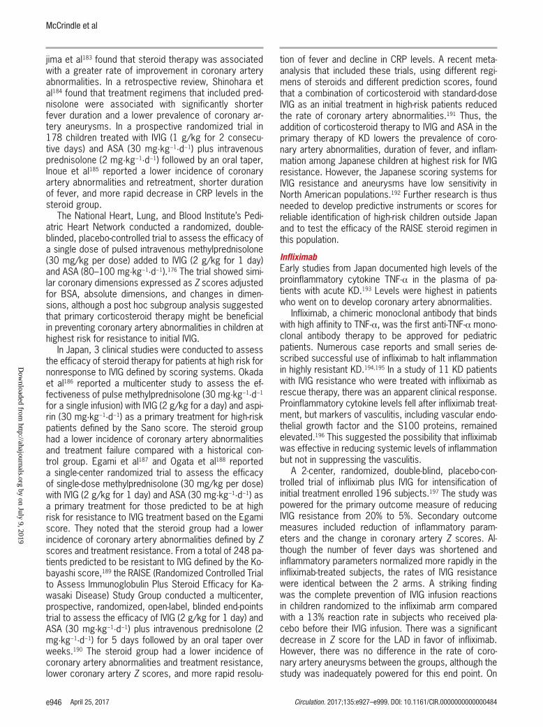
McCrindle et al
e946 April 25, 2017 Circulation. 2017;135:e927–e999. DOI: 10.1161/CIR.0000000000000484
jima et al183 found that steroid therapy was associated with a greater rate of improvement in coronary artery abnormalities. In a retrospective review, Shinohara et al184 found that treatment regimens that included pred-nisolone were associated with significantly shorter fever duration and a lower prevalence of coronary ar-tery aneurysms. In a prospective randomized trial in 178 children treated with IVIG (1 g/kg for 2 consecu-tive days) and ASA (30 mg·kg−1·d−1) plus intravenous prednisolone (2 mg·kg−1·d−1) followed by an oral taper, Inoue et al185 reported a lower incidence of coronary artery abnormalities and retreatment, shorter duration of fever, and more rapid decrease in CRP levels in the steroid group.
The National Heart, Lung, and Blood Institute’s Pedi-atric Heart Network conducted a randomized, double-blinded, placebo-controlled trial to assess the efficacy of a single dose of pulsed intravenous methylprednisolone (30 mg/kg per dose) added to IVIG (2 g/kg for 1 day) and ASA (80–100 mg·kg−1·d−1).176 The trial showed simi-lar coronary dimensions expressed as Z scores adjusted for BSA, absolute dimensions, and changes in dimen-sions, although a post hoc subgroup analysis suggested that primary corticosteroid therapy might be beneficial in preventing coronary artery abnormalities in children at highest risk for resistance to initial IVIG.
In Japan, 3 clinical studies were conducted to assess the efficacy of steroid therapy for patients at high risk for nonresponse to IVIG defined by scoring systems. Okada et al186 reported a multicenter study to assess the ef-fectiveness of pulse methylprednisolone (30 mg·kg−1·d−1 for a single infusion) with IVIG (2 g/kg for a day) and aspi-rin (30 mg·kg−1·d−1) as a primary treatment for high-risk patients defined by the Sano score. The steroid group had a lower incidence of coronary artery abnormalities and treatment failure compared with a historical con-trol group. Egami et al187 and Ogata et al188 reported a single-center randomized trial to assess the efficacy of single-dose methylprednisolone (30 mg/kg per dose) with IVIG (2 g/kg for 1 day) and ASA (30 mg·kg−1·d−1) as a primary treatment for those predicted to be at high risk for resistance to IVIG treatment based on the Egami score. They noted that the steroid group had a lower incidence of coronary artery abnormalities defined by Z scores and treatment resistance. From a total of 248 pa-tients predicted to be resistant to IVIG defined by the Ko-bayashi score,189 the RAISE (Randomized Controlled Trial to Assess Immunoglobulin Plus Steroid Efficacy for Ka-wasaki Disease) Study Group conducted a multicenter, prospective, randomized, open-label, blinded end-points trial to assess the efficacy of IVIG (2 g/kg for 1 day) and ASA (30 mg·kg−1·d−1) plus intravenous prednisolone (2 mg·kg−1·d−1) for 5 days followed by an oral taper over weeks.190 The steroid group had a lower incidence of coronary artery abnormalities and treatment resistance, lower coronary artery Z scores, and more rapid resolu-
tion of fever and decline in CRP levels. A recent meta-analysis that included these trials, using different regi-mens of steroids and different prediction scores, found that a combination of corticosteroid with standard-dose IVIG as an initial treatment in high-risk patients reduced the rate of coronary artery abnormalities.191 Thus, the addition of corticosteroid therapy to IVIG and ASA in the primary therapy of KD lowers the prevalence of coro-nary artery abnormalities, duration of fever, and inflam-mation among Japanese children at highest risk for IVIG resistance. However, the Japanese scoring systems for IVIG resistance and aneurysms have low sensitivity in North American populations.192 Further research is thus needed to develop predictive instruments or scores for reliable identification of high-risk children outside Japan and to test the efficacy of the RAISE steroid regimen in this population.
InfliximabEarly studies from Japan documented high levels of the proinflammatory cytokine TNF-α in the plasma of pa-tients with acute KD.193 Levels were highest in patients who went on to develop coronary artery abnormalities.
Infliximab, a chimeric monoclonal antibody that binds with high affinity to TNF-α, was the first anti-TNF-α mono-clonal antibody therapy to be approved for pediatric patients. Numerous case reports and small series de-scribed successful use of infliximab to halt inflammation in highly resistant KD.194,195 In a study of 11 KD patients with IVIG resistance who were treated with infliximab as rescue therapy, there was an apparent clinical response. Proinflammatory cytokine levels fell after infliximab treat-ment, but markers of vasculitis, including vascular endo-thelial growth factor and the S100 proteins, remained elevated.196 This suggested the possibility that infliximab was effective in reducing systemic levels of inflammation but not in suppressing the vasculitis.
A 2-center, randomized, double-blind, placebo-con-trolled trial of infliximab plus IVIG for intensification of initial treatment enrolled 196 subjects.197 The study was powered for the primary outcome measure of reducing IVIG resistance from 20% to 5%. Secondary outcome measures included reduction of inflammatory param-eters and the change in coronary artery Z scores. Al-though the number of fever days was shortened and inflammatory parameters normalized more rapidly in the infliximab-treated subjects, the rates of IVIG resistance were identical between the 2 arms. A striking finding was the complete prevention of IVIG infusion reactions in children randomized to the infliximab arm compared with a 13% reaction rate in subjects who received pla-cebo before their IVIG infusion. There was a significant decrease in Z score for the LAD in favor of infliximab. However, there was no difference in the rate of coro-nary artery aneurysms between the groups, although the study was inadequately powered for this end point. On
Dow
nloaded from http://ahajournals.org by on July 9, 2019

Diagnosis, Treatment, and Management of Kawasaki Disease
e947
CLINICAL STATEMENTS
AND GUIDELINES
Circulation. 2017;135:e927–e999. DOI: 10.1161/CIR.0000000000000484 April 25, 2017
the basis of current information, addition of infliximab to initial therapy with IVIG is safe but does not prevent recrudescent fever.
EtanerceptA more limited experience with etanercept (soluble TNF receptor) plus IVIG for intensification of initial therapy was reported recently.198 In this prospective, open-la-bel study of 15 patients, etanercept was administered subcutaneously after IVIG infusion and again at 1 and 2 weeks later. Most patients received 0.8 mg/kg per dose. The pharmacokinetics were similar to those re-ported in older children, and there were no adverse re-actions attributable to etanercept. On the basis of these results, a phase III randomized, placebo-controlled trial was initiated and is still enrolling subjects.199 Recom-mendations for primary adjunctive treatment with etan-ercept await publication of the results of this clinical trial. The potential advantage of etanercept might be the shorter half-life if secondary infections are of con-cern. However, the soluble receptor only binds to circu-lating and not cell-bound TNF-α, which could reduce the anti-inflammatory effect.
Recommendations for Adjunctive Therapies for Primary Treatment
1. Single-dose pulse methylprednisolone should not be administered with IVIG as routine pri-mary therapy for patients with KD (Class III; Level of Evidence B).
2. Administration of a longer course of corti-costeroids (eg, tapering over 2–3 weeks), together with IVIG 2 g/kg and ASA, may be considered for treatment of high-risk patients with acute KD, when such high risk can be identified in patients before initiation of treatment (Class IIb; Level of Evidence B).
IVIG ResistanceApproximately 10% to 20% of patients with KD develop recrudescent or persistent fever at least 36 hours after the end of their IVIG infusion and are termed IVIG resis-tant.176,200,201 The immunologic basis of IVIG resistance is unknown, in part because the mechanism of action of IVIG is poorly understood. It is likely that host genetic factors, such as polymorphisms in the Fc gamma recep-tors, play a role in both the response and resistance to IVIG.61,202,203
Risk Scores for Predicting Nonresponse to IVIGApproximately 10% to 20% of patients with KD have per-sistent or recurrent fever after primary therapy with IVIG plus ASA.204,205 Many studies have shown that patients who are resistant to initial IVIG are at increased risk of developing coronary artery abnormalities.171,206,207 Thus, scoring systems have been constructed to identify pa-tients likely to be resistant to IVIG and who may benefit from more aggressive initial therapy. In 2006, 3 Japa-nese groups devised scoring systems to predict resis-tance to IVIG.187,189,208,209 However, currently available
Table 6. Treatment Options for IVIG-Resistant KD Patients
Agent Description Dose References
Most frequently administered
IVIG: Second infusion Pooled polyclonal IG 2 g/kg IV 211
IVIG + prednisolone IVIG + steroid IVIG: 2 g/kg IV + prednisolone 2 mg·kg−1·d−1 IV divided every 8 h until afebrile, then prednisone orally until CRP normalized, then taper over 2–3 wk
212
Infliximab Monoclonal antibody against TNF-α Single infusion: 5 mg/kg IV given over 2 h 194, 213, 214
Alternative treatments
Cyclosporine Inhibitor of calcineurin-NFAT pathway IV: 3 mg·kg−1·d−1 divided every 12 hPO: 4–8 mg·kg−1·d−1 divided every 12 hAdjust dose to achieve trough 50–150 ng/mL; 2-h peak level 300–600 ng/mL
215, 216
Anakinra Recombinant IL-1β receptor antagonist 2–6 mg·kg−1·d−1 given by subcutaneous injection 217, 218
Cyclophosphamide Alkylating agent blocks DNA replication 2 mg·kg−1·d−1 IV 219
Plasma exchange Replaces plasma with albumin Not applicable 220
IVIG resistance is defined as persistent or recrudescent fever at least 36 hours and <7 days after completion of first IVIG infusion. The top 3 treatments have been most commonly used, although no comparative effectiveness trial has been performed. Pulsed high-dose steroid treatment is not recommended. The alternative treatments have been used in a limited number of patients with KD.
CRP indicates C-reactive protein; IG, immunoglobulin; IL, interleukin; IV, intravenous; IVIG, intravenous immunoglobulin; KD, Kawasaki disease; NFAT, nuclear factor of activated T cells; PO, by mouth; and TNF, tumor necrosis factor.
Dow
nloaded from http://ahajournals.org by on July 9, 2019

McCrindle et al
e948 April 25, 2017 Circulation. 2017;135:e927–e999. DOI: 10.1161/CIR.0000000000000484
risk prediction models for Asian populations are insuffi-ciently accurate to be clinically useful in North American patients in predicting response to initial treatment with IVIG.192,210 Better predictive models, perhaps incorporat-ing biomarkers or genetic variants, will need to be devel-oped for use outside Japan.
Treatment of IVIG ResistanceThere are no robust data from clinical trials to guide the clinician in the choice of therapeutic agents for the child with IVIG resistance. We summarize below the ex-perience with different therapeutic approaches (Table 6). Cost-effectiveness comparisons between different ap-proaches have not been reported.
IVIG RetreatmentMany experts recommend retreatment with IVIG 2 g/kg. The putative dose-response effect of IVIG forms the theoretical basis for this approach. Retrospective se-ries have suggested efficacy, but IVIG retreatment has never been tested in an adequately powered random-ized trial.171,211
CorticosteroidsCorticosteroids have also been used to treat patients who have failed to respond to initial therapy for KD.182 Several small case series and observational studies have described children with KD with recrudescent or persistent fever despite IVIG treatment in whom the ad-ministration of steroid therapy was associated with an improvement in symptoms and the absence of an im-portant progression in coronary artery abnormalities or adverse effects.219,221–225 In a study of first-line rescue therapy, Furukawa et al226 reported 63 IVIG-resistant pa-tients, of whom 44 were then given intravenous methyl-prednisolone (30 mg·kg−1·d−1 for 3 consecutive days) followed by oral methylprednisolone tapered over 7 days; 19 patients were given a second infusion of IVIG. The incidence of coronary artery abnormalities and treat-ment failure was similar between the 2 treatment groups, although power was limited by the small sample size. Fever tended to recur more frequently in the group treat-ed with pulse steroids. Ogata et al227 compared the ef-fectiveness of treatment strategies for 27 IVIG-resistant patients: 14 patients were treated with additional IVIG (2 g/kg for 1 day), and 13 were treated with pulse intrave-nous methylprednisolone (30 mg/kg for 3 consecutive days). Patients in the steroid group had shorter duration of fever and lower medical costs. Three patients (21%) treated with IVIG and no patients treated with steroids had coronary artery abnormalities. Again, the study was underpowered to show a significant difference between the groups. Teraguchi et al228 studied 41 IVIG-resistant patients and reported that intravenous methylpredniso-lone (30 mg·kg−1·d−1 for 3 consecutive days) followed by prednisolone (1 mg·kg−1·d−1) for 7 days did not improve
clinical or coronary artery outcomes compared with IVIG retreatment. Kobayashi et al212 reported a retrospective study to assess the efficacy of intravenous prednisolone followed by an oral taper (2 mg·kg−1·d−1 tapered over 2 weeks after CRP normalized) using a database of 359 consecutive IVIG-resistant patients. Patients treated with IVIG plus prednisolone had significantly lower rates of persistent or recrudescent fever and coronary artery abnormalities than the group that received IVIG mono-therapy. It is hypothesized that the improved outcomes associated with the longer steroid course in the Japa-nese studies might be attributed to suppression of per-sistent vascular inflammation, although there have been no clinical trials comparing different steroid regimens for patients who do not respond to an initial or a second dose of IVIG. The optimal steroid regimen is therefore not known, and both pulsed and longer-term steroid therapy remain options.
InfliximabA phase I multicenter, randomized, open clinical trial of infliximab (5 mg/kg intravenously over 2 hours) versus a second infusion of IVIG (2 g/kg) was performed to deter-mine the safety, tolerability, and pharmacokinetics of in-fliximab for rescue therapy for patients who had fever at least 36 hours after the end of the initial IVIG infusion.195 The study enrolled 24 subjects with IVIG-resistant KD and determined that infliximab was well tolerated in infants and children with KD and that the pharmacokinetics were similar to adults, with circulating levels of the monoclo-nal antibody detected out to 10 weeks. In the Japanese trial, 20 KD patients resistant to 2 consecutive IVIG infu-sions (2 g/kg each) were treated with infliximab (5 mg/kg), and an apparent clinical response was achieved in 18 (90%).229 The 2 unresponsive patients were treated with plasma exchange with resolution of their inflam-mation. The coronary artery abnormalities detected by echocardiogram all subsequently resolved. There were no adverse reactions attributed to infliximab among the study subjects.
A retrospective review of 2 centers that consistently administered either a second dose of IVIG or infliximab to IVIG-resistant patients suggested that patients receiving infliximab had shorter hospitalization and fewer days of fever, but coronary artery outcomes and adverse events were similar.213 On the basis of these retrospective data, infliximab can be considered as an alternative to a sec-ond infusion of IVIG for resistant patients.
Other TreatmentsHighly inflamed patients who fail to respond to either a second infusion of IVIG, steroids, or infliximab require additional therapy to control inflammation.
CyclosporineInterest in the calcineurin inhibitor cyclosporine for KD patients grew out of 2 observations. First, immunohis-
Dow
nloaded from http://ahajournals.org by on July 9, 2019

Diagnosis, Treatment, and Management of Kawasaki Disease
e949
CLINICAL STATEMENTS
AND GUIDELINES
Circulation. 2017;135:e927–e999. DOI: 10.1161/CIR.0000000000000484 April 25, 2017
tochemical studies of coronary arteries from autopsies suggested that CD8+ T cells contribute to the inflamma-tory process in the arterial wall.230 Second, genetic stud-ies in children of Japanese or European descent have implicated activation of the NFAT-calcineurin calcium signaling pathway as a contributor to both disease sus-ceptibility and coronary artery aneurysm formation.231,232 Cyclosporine is a specific inhibitor of calcineurin, and a protocol for its administration and monitoring has been used successfully in a small number of highly re-sistant patients.216 Dosing is provided in Table 6; levels are monitored to arrive at the appropriate dose but not monitored thereafter. Once the patient is afebrile and clinically improving and the CRP is ≤1.0 mg/dL, or after 2 weeks of therapy, the dose can be tapered by 10% of the initial dose every 3 days and discontinued when the dose has reached 1 mg·kg−1·d−1. A small, open, single-arm pilot trial in Japan studied cyclosporine treatment in 28 children who remained febrile after administration of 2 doses of IVIG.215 After receiving an oral dose of 4 to 6 mg·kg−1·d−1, 18 patients (64%) were afebrile after 3 days of therapy. Overall, 78% of patients responded. Nine patients developed hyperkalemia, but none had se-rious adverse effects; however, cyclosporine levels were not monitored. Four patients had coronary artery aneu-rysms, 2 before cyclosporine had been administered. In a separate report, the levels of soluble IL-6 and IL-2 receptors dropped to control levels by 7 days after initia-tion of cyclosporine.169 Randomized trials are needed to determine whether calcineurin inhibitors such as cyclo-sporine or tacrolimus reduce the rate of coronary artery aneurysms; however, these small studies suggest that cyclosporine has few serious adverse events and is a well-tolerated option for treatment of highly refractory patients, although further study is needed.
Other Monoclonal Antibody TherapyTwo case reports describe the successful use of anakinra, a recombinant, nonglycosylated form of the human IL-1 receptor antagonist, for treatment of high-ly refractory KD.217,218 Clinical trials are in progress to evaluate the efficacy of IL-1 blockade in children with acute KD.
Plasma ExchangePlasma exchange has been reported in uncontrolled clin-ical trials to be an effective therapy in patients who are refractory to IVIG and to lower the incidence of coronary artery aneurysms.220,233 Because of its risks, plasma exchange should be reserved for patients in whom all reasonable medical therapies have failed.
Cytotoxic AgentsCytotoxic agents such as cyclophosphamide, in conjunc-tion with oral steroids, have been used for exceptional patients with particularly refractory acute KD.219 This therapy is used widely to treat other severe vasculitides.
There are insufficient studies of cyclophosphamide in KD to formulate recommendations for its use, but the risks of cytotoxic agents are such that its use should only be considered in the most severe cases, which are resis-tant to other agents.
Recommendations for Additional Therapy in the IVIG-Resistant Patient
1. It is reasonable to administer a second dose of IVIG (2 g/kg) to patients with persistent or recrudescent fever at least 36 hours after the end of the first IVIG infusion (Class IIa; Level of Evidence B).
2. Administration of high-dose pulse steroids (usually methylprednisolone 20–30 mg/kg intravenously for 3 days, with or without a subsequent course and taper of oral predni-sone) may be considered as an alternative to a second infusion of IVIG or for retreatment of patients with KD who have had recurrent or recrudescent fever after additional IVIG (Class IIb; Level of Evidence B).
3. Administration of a longer (eg, 2–3 weeks) tapering course of prednisolone or predni-sone, together with IVIG 2 g/kg and ASA, may be considered in the retreatment of patients with KD who have had recurrent or recrudes-cent fever after initial IVIG treatment (Class IIb; Level of Evidence B).
4. Administration of infliximab (5 mg/kg) may be considered as an alternative to a second infu-sion of IVIG or corticosteroids for IVIG-resistant patients (Class IIb, Evidence Level C).
5. Administration of cyclosporine may be con-sidered in patients with refractory KD in whom a second IVIG infusion, infliximab, or a course of steroids has failed (Class IIb; Level of Evidence C).
6. Administration of immunomodulatory mono-clonal antibody therapy (except TNF-α block-ers), cytotoxic agents, or (rarely) plasma exchange may be considered in highly refrac-tory patients who have failed to respond to a second infusion of IVIG, an extended course of steroids, or infliximab (Class IIb; Level of Evidence C).
Treatment of Acute Myocardial Dysfunction/Cardiovascular CollapseAs noted previously, myocarditis that consists of myo-cardial interstitial edema, cellular infiltration (mainly monocytes, as well as neutrophils/macrophages), and (rarely) degeneration and necrosis of myocytes can oc-cur during the acute stage of KD, even earlier than the
Dow
nloaded from http://ahajournals.org by on July 9, 2019

McCrindle et al
e950 April 25, 2017 Circulation. 2017;135:e927–e999. DOI: 10.1161/CIR.0000000000000484
occurrence of coronary arteritis.73,120,122,234 Because the myocardial cells are preserved in most of the patients, LV function usually normalizes promptly with IVIG thera-py.111,235,236 However, severe myocarditis can still occur, can manifest as hemodynamic instability, and rarely can be a cause of death in the acute phase of KD.117–119,237 In most cases, the development of a shock syndrome more often reflects decreased peripheral vascular resistance, with a smaller contribution from decreased LV contractil-ity. The incidence of KD shock syndrome (KDSS) is esti-mated to be ≈7%.118,119 KDSS can be defined as the pres-ence of hypotension and shock requiring the initiation of volume expanders, the infusion of vasoactive agents, or transfer to intensive care units. Shock in KDSS is often moderate, with low lactate values and the need for treat-ment with inotropic and vasopressor agents.117 Although hemodynamic instability generally improves quickly once therapy with diuretic agents and vasopressor agents is initiated, a mild degree of ventricular diastolic dysfunc-tion can persist after acute management.118 The causes of KDSS may involve the release of endogenous mol-ecules that mediate a decrease in peripheral vascular re-sistance, myocardial dysfunction from myocarditis with or without myocardial ischemia, and capillary leakage, but the exact underlying mechanisms remain unclear. KDSS is often associated with more severe laboratory markers of inflammation and higher risk of coronary ar-terial dilation.111,118 Such cases are also more likely to be resistant to IVIG therapy and to require additional anti-inflammatory treatment.111,118
Because KDSS can manifest before the diagnosis of KD becomes clear, it is critical to recognize the early signs of KD so that IVIG therapy can be initiated prompt-ly. Treatment of the underlying inflammation is important in the resolution of KDSS. The shock in patients with KDSS can be cardiogenic, distributive, or mixed. The dis-tributive component of shock might result from high lev-els of circulating inflammatory cytokines. Of note, there are no clinical trials focusing on KDSS patients. In most of the published case series, management followed the guidelines for pediatric septic shock.238,239 The underly-ing pathophysiology of KDSS appears to be similar to septic shock with pathological vasodilation, relative and absolute hypovolemia, myocardial dysfunction, and al-tered distribution of blood volume. Because these pa-tients have high circulating levels of vascular endothelial growth factor, they are susceptible to capillary leak, and vigorous volume replacement without concomitant anti-inflammatory therapy can result in complications from interstitial fluid accumulation. Therefore, an important aspect of hemodynamic stabilization in these patients re-mains administration of IVIG along with fluid and inotropic and vasoactive agents as necessary to support blood pressure. The inotropic support reported in case series has included dobutamine, epinephrine, norepinephrine, and dopamine.117–119,237,240
The administration of IVIG after the diagnosis of KD has been shown to improve LV function, because it improves inflammation and systemic manifestations.111,235,236
Prevention and Treatment of Thrombosis in Patients With Coronary Artery AneurysmsOther than rupture of a coronary artery aneurysm, which is rare, the most serious complication during the acute illness is thrombotic occlusion of a coronary artery an-eurysm precipitating MI or sudden death. Contributing factors to thrombosis include the presence of thrombo-cytosis and increased platelet adhesion, inflammation, and endothelial dysfunction, together with abnormal flow conditions through areas of severe dilation. For patients with evolving coronary artery aneurysms, follow-up as-sessments with echocardiography should be performed frequently to monitor for increases in luminal dimensions and hence increasing thrombotic risk, as well as for the presence of thrombosis or signs of ventricular dysfunc-tion. Failure to increase the intensity of antithrombotic therapy in the presence of rapidly expanding aneurysms is the most important contributor to sudden cardiovas-cular events during the acute illness. MIs in young chil-dren and infants are either silent or associated with non-specific symptoms, such as unusual fussiness, vomiting, or shock. Sudden worsening in ventricular function or change in electrocardiographic findings should heighten suspicion for coronary artery thrombosis.
Prevention of Coronary Artery ThrombosisTo date, no randomized clinical trials have evaluated the safety and efficacy of antithrombotic regimens for pro-phylaxis of coronary thrombosis in KD, in part because the power for such trials is limited by small patient num-bers and few thrombotic events. For this reason, recom-mendations for use of antithrombotic agents have relied on reasoning from first principles, retrospective studies, practices in atherosclerotic coronary artery disease (CAD), and expert consensus.
Antiplatelet agents are considered to be standard of care in the therapeutic armamentarium for patients with coronary artery aneurysms. For patients with small coro-nary artery aneurysms, monotherapy with low-dose ASA therapy is sufficient for prophylaxis of thrombosis. In pa-tients with moderate but not large or giant aneurysms, ASA therapy may be combined with a thienopyridine (eg, clopidogrel) to antagonize ADP-mediated platelet activa-tion, a practice supported by the superior efficacy of such a regimen, compared with ASA alone, among adults with coronary artery or cerebrovascular disease.241–244 Finally, ibuprofen and other nonsteroidal anti-inflamma-tory drugs with known or potential involvement of the cyclooxygenase pathway interfere with the antiplatelet effect of ASA to prevent thrombosis. If nonsteroidal anti-inflammatory drugs are needed for treatment of arthritis
Dow
nloaded from http://ahajournals.org by on July 9, 2019

Diagnosis, Treatment, and Management of Kawasaki Disease
e951
CLINICAL STATEMENTS
AND GUIDELINES
Circulation. 2017;135:e927–e999. DOI: 10.1161/CIR.0000000000000484 April 25, 2017
in patients with coronary artery aneurysms who are tak-ing ASA, alternative antiplatelet therapies (eg, thienopyri-dines) should be considered.
Patients with large or giant aneurysms, that is, with an internal luminal diameter Z score ≥10 or absolute dimension ≥8 mm, are at particularly high risk for coro-nary artery thrombosis. In affected arterial segments, coronary artery thrombosis is promoted by markedly abnormal flow conditions, with low wall sheer stress and stasis, together with activation of platelets, clotting fac-tors, and the endothelium. Over time, stenoses often develop, causing activation of platelets and endothelial dysfunction from turbulence of flow when located proxi-mally and occluding flow and worsening stasis when located distal to an aneurysm. Indeed, most giant an-eurysms seen at postmortem examination are lined by chronic thrombus.73 Because both platelets and humoral clotting factors promote thrombus formation within giant aneurysms, patients are treated with a combination of antiplatelet and anticoagulant therapy, most commonly low-dose ASA together with either warfarin, maintaining an international normalized ratio of 2.0. to 3.0, or low-molecular-weight heparin (LMWH).245
Anticoagulation with therapeutic doses of LMWH is generally substituted for warfarin in infants and is also occasionally used in the older child in whom the interna-tional normalized ratio cannot be adequately controlled. Transition from LMWH to warfarin may be considered once aneurysms have stopped expanding and the patient is stable. During the acute phase, the anti-inflammatory actions of the LMWH could be an added advantage. Transient low levels of antithrombin occur in more than half of KD patients during the acute illness, are related to increased antithrombin consumption, and can affect the antithrombotic action of LMWH.55 If patients fail to achieve the desired activated factor Xa level (0.5–1.0) on an appropriate dose (Table 7), then antithrombin levels should be measured. If deficient, fresh-frozen plasma or antithrombin supplementation may be given.
More aggressive regimens may be used in patients with exceptionally high risk for coronary artery thrombo-sis. Infants and children who recently required thromboly-sis for coronary artery thrombosis may be maintained for a limited time on 3 agents, that is, ASA, a thienopyri-dine, and an anticoagulant. Because the risk of bleeding is greater with such a regimen, clinicians must consider its risk/benefit ratio on an individual basis. Newer oral di-rect factor Xa inhibitors or direct thrombin inhibitors are not yet approved for use in pediatrics but could supplant warfarin and LMWH in the future.
Recommendations for Prevention of Thrombosis During the Acute Illness
1. Low-dose ASA (3–5 mg·kg−1·d−1) should be administered to patients without evidence
of coronary artery changes until 4 to 6 weeks after onset of illness (Class I; Level of Evidence C).
2. For patients with rapidly expanding coro-nary artery aneurysms or a maximum Z score of ≥10, systemic anticoagulation with LMWH or warfarin (international normal-ized ratio target 2.0–3.0) in addition to low-dose ASA is reasonable (Class IIa; Level of Evidence B).
3. For patients at increased risk of thrombosis, for example, with large or giant aneurysms (≥8 mm or Z score ≥10) and a recent his-tory of coronary artery thrombosis, “triple therapy” with ASA, a second antiplatelet agent, and anticoagulation with warfarin or LMWH may be considered (Class IIb; Level of Evidence C).
4. Ibuprofen and other nonsteroidal anti-inflammatory drugs with known or potential involvement of cyclooxygenase pathway may be harmful in patients taking ASA for its antiplatelet effects (Class III; Level of Evidence B).
Treatment of Coronary Artery ThrombosisBecause of its rarity, treatment of acute coronary throm-bosis in KD patients has not been tested in randomized controlled trials. Rather, recommendations for therapy are based on guidelines in adults with ACS and small pe-diatric case series, with goals of reestablishing coronary artery patency and flow, salvaging myocardium, and improving survival.247–249 Compared with coronary artery thrombosis in the adult with atherosclerotic CAD, throm-bus mass in the KD patients is typically much greater. Furthermore, coronary artery thrombosis in the adult with CAD is most often caused by plaque rupture or in-flammation, with exposure of lipids and extracellular ma-trix to the coagulation system, whereas highly abnormal flow characteristics form the basis for coronary artery thrombosis in KD patients. Methods used to reestablish coronary artery perfusion can vary with the size of the patient and expertise of caregivers; the optimal treat-ment is that which re-establishes blood flow most rapid-ly. Coronary artery thrombosis with actual or impending occlusion of the arterial lumen should be treated with thrombolytic therapy or, in patients of sufficient size, by mechanical restoration of coronary artery blood flow at cardiac catheterization.
Thrombolytic therapy with tissue-type plasminogen activator (tPA) is the most commonly administered therapeutic regimen for occlusive or near-occlusive coronary artery thrombosis in infants and children (Table 7). A common regimen of tPA is 0.5 mg·kg−1·h−1 over 6 hours.250 Of note, an alternative regimen of tPA, used in adult coronary artery thrombosis, is 0.2 mg/kg
Dow
nloaded from http://ahajournals.org by on July 9, 2019

McCrindle et al
e952 April 25, 2017 Circulation. 2017;135:e927–e999. DOI: 10.1161/CIR.0000000000000484
Table 7. Antithrombotic and Fibrinolytic Therapy in KD246
Drug Mechanism of Action Indications Dose Target Range Monitoring Adverse Effects
Anticoagulants
UFH Potentiates the inhibition of factors XIIa, Xia, Xa, IXa, IIa by antithrombin
Hepatic and renal clearance; reversible with protamine sulfate
Treatment of acute coronary artery thrombosis, usually in conjunction with tPA; bridging for patients undergoing invasive procedures requiring reversal of anticoagulation
Age-dependent dosing:
<12 mo of age: 28 U·kg−1·h−1
≥12 mo of age: 20 U·kg−1·h−1
Low dose: commonly 10–15 U·kg−1·h−1
Titrate to PTT target range
Given as continuous parenteral infusion
Anti–factor Xa 0.35–0.70 U/mL
PTT 1.5–3 times baseline PTT, depending on local laboratory values
Every 24 h at minimum
Bleeding, heparin-induced thrombocytopenia, association with bone mineral loss
LMWH Same as UFH but greater inhibition of factor Xa
Renal clearance; not fully reversible; hold for 24 h before invasive procedures
Chronic thromboprophylaxis option for patients with large or giant coronary artery aneurysms or previous thrombosis, particularly for young infants or those with expanding aneurysms early in the course of their illness; bridging for patients between UFH and warfarin
Age- and agent-dependent dosing Anti–factor Xa level 0.5–1.0 U/mL
Every month at minimum
Bleeding, bruising at injection sites
Enoxaparin Given every 12 h subcutaneously:
<2 mo of age: 1.5 mg/kg per dose
>2 mo of age: 1.0 mg/kg per dose
Higher doses may be needed in neonates; titrate to anti–factor Xa target range
Obtain levels 4–6 h after dose
Tinzaparin Given every 24 h subcutaneously:
0–2 mo: 275 U/kg per dose
2–12 mo: 250 U/kg per dose
1–5 y: 240 U/kg per dose
5–10 y: 200 U/kg per dose
>10 y: 175 U/kg per dose
Titrate to anti–factor Xa target range
Age-dependent monitoring:
<5 y: 2 h after dose
≥5 y: 4 h after dose
Warfarin Inhibits the gamma-carboxylation of the vitamin K–dependent factors II, VII, IX, and X and protein C, S, and Z
Hepatic metabolism; oral administration; many food, drug, and illness interactions; reversible with vitamin K administration
Long-term thromboprophylaxis for patients with large or giant coronary artery aneurysms or previous thrombosis
Load with 0.2 mg·kg−1·d−1, then 0.1 mg·kg−1·d−1; titrate dose to INR target level
INR level 2–3 INR daily until in target range, thereafter minimum monthly testing; test INR with illness, medication, or diet change
Bleeding, tracheal calcification, hair loss, decreased bone mineral density
Antiplatelet therapy
ASA Inhibition of COX-1 and COX-2 activity
Irreversible platelet inhibition; some patients are ASA resistant; discontinue 7 d before invasive procedures; precaution for concomitant use of ibuprofen or other nonsteroidal anti-inflammatory agents (usually given to treat arthritis/arthralgias); risk of Reye syndrome with varicella and influenza infections (see Reye Syndrome section in Treatment of the Acute Illness), given orally
Thromboprophylaxis for all patients from the acute illness until 4–6 wk; continue long term for patients with ongoing coronary artery involvement (see Thromboprophylaxis section in Long-Term Management)
3–5 mg·kg−1·d−1, maximum 81–325 mg/d
Bruising, confusion, vertigo, nausea, vomiting, tinnitus, abdominal pain, cramping, burning, fatigue, bleeding
(Continued )
Dow
nloaded from http://ahajournals.org by on July 9, 2019

Diagnosis, Treatment, and Management of Kawasaki Disease
e953
CLINICAL STATEMENTS
AND GUIDELINES
Circulation. 2017;135:e927–e999. DOI: 10.1161/CIR.0000000000000484 April 25, 2017
Clopidogrel Inhibition of ADP-induced platelet aggregation, no effects on arachidonic acid metabolism
Renal clearance, hepatic metabolism; some patients fail to respond; given orally
Thromboprophylaxis together with ASA and anticoagulation (triple therapy) for selected patients with very severe/complex coronary artery aneurysms at high risk of thrombosis or with evidence of previous thrombosis
Thromboprophylaxis together with ASA (dual antiplatelet therapy) for patients with moderate coronary artery aneurysms or large or giant aneurysms that have reduced to moderate size
Can be used in place of ASA for patients who are ASA resistant or allergic to ASA
0.2–1.0 mg·kg−1·d−1 Fatigue, vertigo, stomach upset or pain, bruising, bleeding, diarrhea
Dipyridamole Inhibits adenosine reuptake, increasing cAMP, and inhibits platelets
Usually used in place of ASA for patients who are taking ibuprofen, resistant or allergic to ASA, or at risk of Reye syndrome; not generally used for long-term thromboprophylaxis
1–5 mg·kg−1·d−1 given orally Chest pain, angina pectoris, headache, abnormalities of ECG
Abciximab Inhibits glycoprotein IIb–IIIa, prevents binding of fibrinogen to von Willebrand factor, inhibiting platelet aggregation
Monoclonal antibody, renal excretion
Limited use, usually reserved for patients with coronary artery aneurysms who develop thrombosis (both occlusive and nonocclusive) and given as a single course
0.25 mg/kg bolus, then 0.125 μg·kg−1·min−1 every 12 h
Bleeding, hypertension, nausea, vomiting, vertigo, irritation at injection site
Thrombolytic therapy
Alteplase tPA converts plasminogen into plasmin; plasmin degrades fibrin (crosslinked) and fibrinogen
Reversible with aminocaproic acid; contraindicated if active bleeding or recent surgery or trauma
Reserved for patients with coronary artery aneurysm thrombosis, particularly if occlusive
For coronary artery thrombosis, data are lacking
Dosing regimens that have been used include:
0.1–0.6 (commonly 0.5) mg·kg−1·h−1 IV for 6 h
As per adult guidelines, 0.2 mg/kg IV bolus (maximum 15 mg), then 0.75 mg/kg over 30 min (maximum 50 mg), then 0.5 mg/kg over 60 min (maximum 35 mg), for a maximum total dose of 100 mg
Low-dose alteplase combined with abciximab
Administration: give fresh-frozen plasma 10–20 mL/kg infusion before using alteplase as a plasminogen source; keep fibrinogen >100 mg/dL and platelets >50 000/mm3; continue UFH at an age-appropriate dose during administration of alteplase; alteplase may be given as an intermittent (recommended) or continuous usual or low-dose systemic infusion or directed locally toward the thrombus by catheters at a lower dose
Reassess thrombus with imaging at completion of the infusion; retreatment may be indicated once hematologic parameters are acceptable; careful patient observation is required, with prompt investigation if there is any suspicion of internal bleeding
Major and minor bleeding
ASA indicates acetylsalicylic acid; COX, cyclooxygenase; INR, international normalized ratio; IV, intravenous; KD, Kawasaki disease; LMWH, low-molecular-weight heparin; PTT, partial thromboplastin time; tPA, tissue plasminogen activator; and UFH, unfractionated heparin.
Modified from Giglia et al.246 Copyright © 2013, American Heart Association, Inc.
Table 7. ContinuedDrug Mechanism of Action Indications Dose Target Range Monitoring Adverse Effects
Dow
nloaded from http://ahajournals.org by on July 9, 2019

McCrindle et al
e954 April 25, 2017 Circulation. 2017;135:e927–e999. DOI: 10.1161/CIR.0000000000000484
intravenously (maximum 15 mg), then 0.75 mg/kg over 30 minutes (maximum 50 mg) followed by 0.5 mg/kg over 60 minutes (maximum 35 mg).137 It should be ad-ministered together with low-dose ASA and low-dose in-travenous heparin (10 U·kg−1·h−1) with careful monitoring of coagulation parameters to prevent bleeding, maintain-ing the fibrinogen level >100 mg/dL and platelet count >50 000/mm3.251 After completion of tPA, heparin dos-age is increased as appropriate for age. The coronary artery thrombus should be reassessed with echocardio-graphic imaging after completion of the tPA infusion.
The large thrombus burden in the KD patient with coronary artery thrombosis, as well as the tendency for rebound thrombosis in such patients, has led some clini-cians to use reduced-dose thrombolytic therapy togeth-er with a glycoprotein IIb/IIIa inhibitor, such as the mono-clonal antibody abciximab (0.25 mg/kg bolus over 30 minutes, followed by an infusion of 0.125 μg·kg−1·min−1 for 12 hours).252,253 In adults with ACS, inhibition of this receptor has been shown to improve outcomes, both with and without the use of thrombolytic drugs.254–256 It may be reasonable to treat coronary artery thrombosis with substantial thrombus burden and high risk of oc-clusion with a reduced-dose thrombolytic therapy and abciximab. When echocardiographic surveillance in the first weeks of the illness reveals a small mural coronary artery thrombus that does not pose an urgent threat of occlusion, it may be reasonable to use abciximab rather than tPA to prevent clot extension.
Recommendations for Treatment of Coronary Artery Thrombosis*
1. Coronary artery thrombosis with actual or impending occlusion of the arterial lumen should be treated with thrombolytic therapy or, in patients of sufficient size, by mechanical res-toration of coronary artery blood flow at cardiac catheterization (Class I; Level of Evidence C).
2. Thrombolytic agents should be administered together with low-dose ASA and low-dose heparin, with careful monitoring for bleeding (Class I; Level of Evidence C).
3. Treatment of coronary artery thrombosis with substantial thrombus burden and high risk of occlusion with a combination of reduced-dose thrombolytic therapy and abciximab may be considered (Class IIb; Level of Evidence C).
LONG-TERM MANAGEMENTLong-term management begins at the end of the acute illness, usually at 4 to 6 weeks after fever onset, when
symptoms and signs have resolved and the coronary artery involvement has reached its maximal extent and luminal dimensions. The goals of long-term management are to prevent thrombosis and myocardial ischemia while maintaining optimal cardiovascular health. There are no specific treatments that target the pathological process-es of ongoing subacute/chronic vasculitis and LMP in those patients with coronary artery aneurysms, although there may be a potential role for statins in this setting.257 Thromboprophylaxis and careful surveillance for coronary artery stenoses/obstructions and myocardial ischemia are the cornerstones of management. Selected patients with myocardial ischemia may be candidates for revascu-larization with catheter interventions or coronary artery bypass surgery or, rarely, cardiac transplantation. Surviv-al with optimal functional, psychosocial, and reproductive outcomes into adulthood will require the development of effective and collaborative programs between pediatric and adult cardiology providers to facilitate the transition process into adult-oriented care systems.
Long-Term OutcomesLong-term outcomes are primarily driven by the conse-quences of resolving and ongoing cardiovascular pathol-ogy that contribute to morbidity, cardiovascular events, and mortality. Arterial pathology in noncoronary peripher-al artery beds can cause ongoing morbidity in a small sub-set of patients and is somewhat correlated with the extent of coronary artery pathology.131 However, the main culprit is the coronary arteries. The pathological process of nec-rotizing arteritis during the acute illness, which results in destruction and weakening of the arterial wall leading to aneurysms, may be accompanied by subacute or chronic inflammation and luminal myofibroblastic proliferation. Su-perimposed on these processes are the effects of acute and organized thrombus. The degree to which convention-al atherosclerosis may contribute to chronic pathological changes is not known. These processes may lead to both acute (usually precipitated by thrombus or arrhythmia) and chronic (usually precipitated by occlusions or steno-ses) cardiovascular morbidity and events. Although the chronic processes may result in normalization of the in-ternal luminal dimension, the arterial wall architecture and function remain abnormal and may progress to stenosis or occlusion over time.73 Nonetheless, normalization of the luminal dimension does reduce abnormal flow charac-teristics (low sheer stress, stasis) and hence thrombosis risk. The terms regression and resolution have been used to describe normalization of luminal dimensions; however, important abnormalities may remain, particularly with large or giant aneurysms, and these terms may give the false impression that the coronary arteries have healed and are normal. The natural history of coronary artery ab-normalities is shown in Figure 4. These processes are the primary determinants of prognosis.
*See Diagnosis section for recommendations regarding echocardio-graphic monitoring for thrombosis.
Dow
nloaded from http://ahajournals.org by on July 9, 2019
Diagnosis, Treatment, and Management of Kawasaki Disease
e955
CLINICAL STATEMENTS
AND GUIDELINES
Circulation. 2017;135:e927–e999. DOI: 10.1161/CIR.0000000000000484 April 25, 2017
Coronary Artery AbnormalitiesPrevalenceThe original descriptions of KD did not recognize the presence of coronary artery abnormalities until it was observed that 1% to 2% of patients died suddenly of cardiac complications.259 An angiographic study of 1100 patients showed coronary artery lesions in 24%, with aneurysms in 8% and a number of patients with stenoses and occlusions.260 The early reports of the prevalence of abnormalities vary widely given the lack of uniformity in the timing of angiography and the definition of abnormalities, and they predate echocardiography and treatment with IVIG. The clinical trial of 4-day IVIG treatment with strict entry criteria (classic KD present-ing within 10 days of fever onset) and using the 1984 Japanese Ministry of Health criteria (based on absolute luminal dimensions) noted a prevalence of coronary ar-tery abnormalities of 23% in the ASA-only group versus 8% in the IVIG-plus-ASA group at 2 weeks, with a lower prevalence at 7 weeks.176 A similar subsequent trial of a
single high dose of IVIG showed coronary artery abnor-malities, again using Japanese Ministry of Health crite-ria, at 2 weeks in 9.1% of those treated with 4-day IVIG treatment versus 4.6% in those treated with high-dose IVIG (further reduced to 2.4% when those with coronary artery abnormalities at presentation were excluded).127 Using a cut point of a Z score of 2.5 together with the Japanese Ministry of Health criteria, a further trial of single-dose methylprednisolone in addition to high-dose IVIG showed a prevalence of coronary artery abnormali-ties of 30% in both groups at 1 week.176 The incorpo-ration of dilation into the definition results in a higher prevalence of coronary artery abnormalities. The prev-alence of dilation is further increased if one includes those patients whose coronary artery Z scores were below the cut point for abnormality (<2) but decreased significantly during follow-up.129,130
These studies define the prevalence of coronary ar-tery abnormalities in homogenous populations; however, some patients in clinical populations may have incom-
Figure 4. Natural history of coronary artery abnormalities. Modified from Kato258 with permission from Elsevier. Copyright © 2004, Elsevier.
Dow
nloaded from http://ahajournals.org by on July 9, 2019

McCrindle et al
e956 April 25, 2017 Circulation. 2017;135:e927–e999. DOI: 10.1161/CIR.0000000000000484
plete criteria, present late, have a missed diagnosis, or have not received appropriate treatment, all of which are risk factors for more severe coronary artery involve-ment. In addition, some patients presenting within 10 days of fever onset may have coronary artery abnormali-ties at the time of initial echocardiogram and before or at the time that IVIG is given.261 Michihata et al262 showed that patient treatment that followed existing guidelines was associated with a 4.9% prevalence of aneurysms versus 9.9% if it did not. A classification system based on Z scores alone in a clinical population of 1356 pa-tients with serial echocardiograms has been proposed and showed overlap in Z scores for classifications based on absolute dimensions only.140 An analysis of 1082 pa-tients from centers in the United States and Japan used 2 different sources of normal values for calculation of Z scores.141 Using the Z-score equations of Kobayashi et al,145 26% of US subjects and 39% of Japanese sub-jects had a maximal Z score ≥2.5, with 4.1% and 3.1%, respectively, having a Z score ≥5. Using the Z-score equations of Dallaire et al,146 30% of US subjects and 44% of Japanese subjects had a maximal Z score ≥2.5, with 5.8% and 6.2%, respectively, having a Z score ≥5. The higher Z scores in Japanese patients remained sig-nificant after adjustment for younger age, male sex, late treatment, and failure to respond to initial IVIG. Thus, many factors need to be considered when determining and comparing the prevalence of coronary artery abnor-malities, particularly the population and definitions.
Natural History and Cardiovascular Disease EventsInitial definition of the natural history of coronary artery abnormalities was determined from tracking luminal di-mensions from serial angiography together with clinical follow-up for events. Kato et al263 reported outcomes in 598 patients diagnosed from 1973 to 1983 and followed up for up to 10 to 21 years. Aneurysms were diagnosed in 25%, with 49% of these having reduced to a normal lu-minal dimension 6 to 18 months later, increasing to 55% with ongoing follow-up. All aneurysms that reduced in size to a normal luminal dimension were originally small or moderate in size. Stenoses developed in 28 patients and showed a more constant risk over time. Coronary artery bypass surgery was performed in 7 patients, 2 had interventional catheter procedures, and 16 required thrombolytic treatment. MI occurred in 11 patients (8 with giant aneurysms), all with severe stenotic lesions in 2 or 3 branches. Patients without coronary artery ab-normalities had no symptoms or events during follow-up. Akagi et al264 reported similar outcomes in a cohort of 583 patients and showed that normalization of luminal dimensions occurred more frequently and more rapidly in those with smaller aneurysms and did not occur for gi-ant aneurysms, with MI occurring only in those with giant aneurysms. This remained true in a subsequent study of 1356 patients diagnosed from 1990 to 2007 and fol-
lowed up with serial echocardiograms for up to 15.7 years.140 Coronary artery events (thrombosis, stenosis, intervention, MI, death) occurred in 1% of those with an aneurysm Z score <10 and an absolute dimension <8 mm, in 29% of those with a Z score ≥10 but an absolute dimension <8 mm, and in 48% of those with both a Z score ≥10 and an absolute dimension ≥8 mm. Longitudi-nal studies of outcomes based on Z-score classifications alone have yet to be performed.
Late development or increases in size of aneurysms have been reported in case reports.265–267 Tsuda et al268 in an angiography study of 562 patients noted new di-lated or expanding lesions in 15 patients. The new an-eurysms occurred at sites where previous aneurysms had diminished in size but were all associated with a localized stenosis, although none were associated with cardiac events.
MI as a result of thrombotic occlusion of an aneurysm or the development of critical stenosis attributable to LMP occurs mainly in those patients with more severe coronary artery abnormalities and can cause sudden death. In young patients, it can be clinically silent or pres-ent with atypical symptoms.263 The gradual progression of LMP and laminar thrombosis to obstructive lesions may be accompanied by the development of collateral vessels, particularly in the presence of segmental ste-nosis and in younger patients, regardless of the occur-rence of MI.269 Prompt and effective management of acute MI can improve outcomes, although the reported experience is limited.270 Myocardial dysfunction present shortly after MI can improve, although some patients will develop adverse ventricular remodeling and ventricular aneurysms if the damage is extensive.271 Patients who have had an MI are at further risk of subsequent MI, although the risk is reduced with effective revascular-ization. Tsuda et al272 reported long-term outcomes up to 33 years after surviving MI in 60 patients. The 30-year survival rate was 63%, and the 25-year ventricular tachycardia–free survival rate was 29%. Low post-MI LV ejection fraction was predictive of poor outcomes.
Subclinical Vascular OutcomesLong-term changes in coronary artery structure and function precede clinical events and reflect chronic path-ological vascular processes in areas that were acutely involved by KD. Arterial wall structure can be imaged noninvasively and has provided insights into the natural history. Intravascular ultrasound (IVUS) has been used to demonstrate symmetrical and asymmetrical wall thick-ening in aneurysms, particularly in those aneurysmal segments that have progressed toward normal luminal dimensions.273,274 IVUS has also been used to charac-terize wall elements, noting areas of fibrofatty changes, necrotic core, and dense calcification.275 More recently, these changes have been assessed with optical coher-ence tomography (OCT).276 Studies have been more
Dow
nloaded from http://ahajournals.org by on July 9, 2019

Diagnosis, Treatment, and Management of Kawasaki Disease
e957
CLINICAL STATEMENTS
AND GUIDELINES
Circulation. 2017;135:e927–e999. DOI: 10.1161/CIR.0000000000000484 April 25, 2017
equivocal as to whether or not wall thickening may be evident in coronary artery segments without previous dilation or aneurysm.
These structural findings are associated with functional abnormalities, with impaired dilation in response to nitro-glycerin277 or adenosine,278 resulting in impaired coronary artery flow reserve. Iemura et al274 studied KD patients with various degrees of coronary artery involvement who had all progressed to a normal luminal dimension on angiogra-phy. They noted that those segments that previously had large aneurysms showed paradoxical vasoconstriction in response to acetylcholine and diminished vasodilation in response to nitroglycerin. On positron emission tomogra-phy (PET) imaging, sites of previous aneurysms showed both impaired vasodilation coupled with reduced myocar-dial blood flow and flow reserve, particularly in segments with stenosis.279 In contrast, a small case-control study of KD patients with no history of coronary artery abnormali-ties showed reduced myocardial flow reserve on PET.280 PET has also been used to demonstrate persistent inflam-mation in patients with aneurysms, which may be reduced with statin therapy.281 It would appear that long-term structural and functional coronary artery abnormalities are evident and associated with both maximal and current coronary artery status, with the greatest abnormalities in those with a history of large or giant aneurysms that have diminished to normal luminal dimensions. Whether or not long-term abnormalities occur in those patients who had no luminal abnormalities or only transient dilation remains a subject of debate, although the majority of evidence, including the absence of cardiovascular disease (CVD) events and lack of calcification on CT, suggests a normal long-term prognosis.
KD is a systemic vasculitis, and hence, the potential presence of generalized long-term abnormalities of sys-temic arterial structure and function has been a subject of controversy. Abnormal brachial artery reactivity or abnormal flow-mediated dilation, reflective of endothelial dysfunction, after KD was initially reported by Dhillon et al282 in a case-control study. Since then, there have been many reports comparing noninvasive surrogate vascular markers, some additionally including measures of arte-rial stiffness and intima-media thickness. A systematic review and meta-analysis of 30 studies showed impor-tant heterogeneity between studies.283 Similar to stud-ies of coronary artery structure and function, evidence for systemic arterial endothelial dysfunction was most commonly reported for KD patients in general but more consistently for those with aneurysms, whereas results for those without a history of coronary artery abnor-malities were not consistent. Similar findings were noted for arterial stiffness, whereas increased mean carotid intima-media thickness was not noted for KD patients, although studies of maximal carotid intima-media thick-ness were conflicting. The authors concluded that sur-rogate vascular markers were abnormal only in KD pa-
tients with aneurysms. Serological evidence of ongoing systemic inflammation has been noted in those patients with persistent aneurysms, with higher serum amyloid A and IL-6284 and CRP,285 and imaging evidence has been suggested on PET scanning.286 Nonetheless, the clinical impact of these abnormalities has not been defined.
Valvular RegurgitationWhen detected early, the preponderance of MR as as-sessed with echocardiography is in the mild to moder-ate range of severity and does not appear to persist on follow-up. MR can occur after the acute stage from myocardial ischemia. Late-onset valvulitis of the mitral and aortic valves can occur very rarely and may require valve replacement.263,287 MR that becomes severe or that persists into late phases of KD appears to occur with lower frequency and can result from persistent ischemia or, rarely, from more resistant inflammatory processes that result in fixed structural abnormalities of the valve apparatus.287,288 This circumstance indicates more serious valvular pathology and may require surgi-cal intervention.
AR in KD is usually associated with aortic root dilation (as indicated by an increased aortic sinus Z score) and becomes apparent early in the course of the disease. AR is of lesser severity and appears to persist more consistently, and it is less reliably associated with other inflammatory markers. Nevertheless, progressive aortic valve dysfunction associated with severe AR has been re-ported in a patient with recurrent KD,289 and aortic valve replacement was required.
Aortic AbnormalitiesPatients after KD have been shown to have functional and anatomic abnormalities of the aorta. Assessment of aortic distensibility has been used in adults in the early screening of atherosclerosis. In a study that compared 40 KD patients without coronary artery aneurysms to 168 healthy children, aortic diameter was measured at both minimum diastolic pressure and maximum systolic pressure by 2D echocardiography; this study found that aortic distensibility varies with age in normal children.290 It was low in infants, increased gradually to peak during the ages 10 to 15 years, and decreased with age there-after. For KD patients, aortic stiffness was increased during the acute illness and was normal during conva-lescence. In a more recent study, Oyamada et al291 dem-onstrated that the aortas in 75 patients with a history of KD had altered elastic properties compared with 57 control subjects. Specifically, LV mass index and aortic stiffness were significantly higher, whereas aortic disten-sibility and strain were lower at 5 to 10 years of follow-up in KD patients. Another study confirmed that in a KD cohort, aortic stiffness and elasticity were increased in 57 patients with KD >1 year after onset of illness, of whom 12 had coronary artery sequelae.292 Both groups had altered elasticity and stiffness of the aorta.
Dow
nloaded from http://ahajournals.org by on July 9, 2019

McCrindle et al
e958 April 25, 2017 Circulation. 2017;135:e927–e999. DOI: 10.1161/CIR.0000000000000484
An unusual finding during the acute KD illness is the presence of retrograde holodiastolic flow in the abdomi-nal aorta in the absence of AR. Mori et al293 described this finding in 15 of 21 children at the time of their acute KD episode, which resolved by 1 month. The abnormal flow pattern may be attributable to an increase in the distensibility of the descending aorta, which is associ-ated with the acute inflammation. Additionally, it was proposed that this pattern may represent diastolic run-off into dilated and dysfunctional peripheral arteries af-fected by the vasculitis.
An aspect of the vasculitis/inflammation noted dur-ing KD may lead to aortic root dilation. Ravekes et al125 evaluated 100 children with history of KD from 1993 to 1997 and noted that patients had a greater normal-ized aortic root dimension that persisted at least to 1-year follow-up. In addition, 4% of these patients also had AR, although this was not evident on auscultation in any patient. A study of 198 children with acute KD noted that aortic root dilatation was present in 8%, remained constant at 1 and 5 weeks after diagnosis, and was associated with larger coronary artery size at diagnosis111; however, aortic root size had little asso-ciation with inflammatory markers. The long-term impli-cations of these alterations in aortic size and properties are unknown.
Myocardial AbnormalitiesAlthough myocarditis is common during the acute illness, complete resolution is expected. Early biopsy studies suggested the presence of myocardial abnormalities, but the relationship to the acute illness and coronary ar-tery abnormalities was unclear.294–296 In a more recent study of 16 patients with giant aneurysms, initial biopsy samples showed myocyte degeneration, hypertrophy, and inflammatory cell infiltration, whereas follow-up bi-opsy specimens showed myocyte disarray, interstitial fi-brosis, and ongoing inflammatory cell infiltration.297 The pathogenesis of the abnormalities was not clear, but the sites of the aneurysms were not related to the biopsy findings. Subsequent studies of myocardial characteriza-tion and function have reported variable findings.298–300 Long-term myocardial dysfunction, resulting from prima-ry myocardial insult at the time of acute KD and which is independent of long-term coronary artery abnormalities, may very rarely occur, although evidence-based reports are few. Whether the myocarditis that occurs with the acute illness leads to long-term myocardial pathology, such as fibrosis and myocyte dropout, independent of coronary artery abnormalities is not clear. Clinical expe-rience indicates that myocardial function is normal, ex-cept among patients with ischemic heart disease from coronary artery stenoses.153,236
ArrhythmiasGenerally, the development of important rhythm changes in KD has been documented primarily in those patients
manifesting more severe forms of myocardial dysfunc-tion, including those patients with overt myocardial isch-emia or infarction. Premature ventricular contractions and ventricular tachycardia have been taken as clinical markers of underlying myocardial damage and as poten-tial predictors of long-term consequences, including late sudden death.272,301 In particular, after MI, the incidence of ventricular tachycardia can be increased. Patients who have sustained severe myocardial injury or infarction may benefit from extended rhythm surveillance (Holter or other long-term electrocardiographic monitoring) to best assess the need for specific antiarrhythmic thera-py. However, other recent studies have demonstrated an electrophysiological impact from KD even in the absence of important ventricular dysfunction or of any coronary artery abnormalities. Ghelani et al302 found increased QT interval dispersion, indicating inhomogenous ventricular repolarization, in a group of KD patients from North In-dia. Kuriki et al303 similarly described elevations in the QT variability index during acute KD that was correlated with serum inflammatory markers and that normalized as the disease regressed.
Although conduction abnormalities have not been characteristic of KD, sinus node and atrioventricular node dysfunction have been demonstrated in patients with moderate to severe coronary artery abnormalities, although only 1 patient among the 40 studied developed evidence of atrioventricular block.112
Risk StratificationClinical experience with KD has taught us that it is rea-sonable to stratify patients according to their relative risk of myocardial ischemia, either related to coronary artery thrombosis or stenoses/occlusions. This stratifi-cation allows for patient long-term management to be individualized regarding the frequency of clinical follow-up and diagnostic testing, cardiovascular risk factor assessment and management, medical therapy, throm-boprophylaxis, physical activity, and reproductive coun-seling. With careful clinical follow-up 10 to 20 years after the onset of KD, patients with no coronary artery luminal changes at any stage of the illness appear to demon-strate a risk for clinical cardiac events that is similar to that in the population without KD.263 For long-term prog-nostication and management, the severity of coronary artery luminal abnormalities defines the risk category. The extent of maximal involvement, together with its evo-lution over time, determines the risk of myocardial isch-emia related to thrombosis and stenosis. The long-term management algorithm is applied after acute manage-ment is completed, and generally when coronary artery luminal Z scores are stable and no longer enlarging. If the patient’s Z scores are still increasing after the end of the convalescent phase, then recommendations for assessment and follow-up for evolving coronary artery
Dow
nloaded from http://ahajournals.org by on July 9, 2019

Diagnosis, Treatment, and Management of Kawasaki Disease
e959
CLINICAL STATEMENTS
AND GUIDELINES
Circulation. 2017;135:e927–e999. DOI: 10.1161/CIR.0000000000000484 April 25, 2017
involvement should be followed, as outlined at the end of the Diagnosis section.
Echocardiography is the primary modality used to assess coronary artery luminal dimensions, which are converted to Z scores adjusted for BSA as outlined in the Diagnosis section. The risk stratification first rests on the patient’s maximal Z score at any time point and in any branch. The risk stratification is further modified by the maximal Z score in any branch at the time of current assessment (Table 8). This allows clinicians to incorpo-rate different risk levels based on the past and current coronary artery involvement, with changes in the risk of thrombosis and stenosis. Coronary artery involvement based on Z scores from echocardiographic assessment of luminal dimensions is classified into 5 categories as outlined in the section Diagnosis, Echocardiography, Classification of Coronary Artery Abnormalities. The cur-rent guidelines diverge from previous guidelines, which primarily classified coronary artery involvement based on absolute dimensions, with little to no adjustment for body size.
Although the risk stratification scheme primarily rests on maximal and current coronary artery Z scores derived from echocardiography, other features of the coronary arteries and other noncoronary artery cardiac complica-tions could also influence decisions regarding risk speci-fication (Table 9). These additional features may further be derived from other imaging modalities.
Recommendations for Risk Stratification of Coronary Artery Abnormalities
1. It is reasonable to use echocardiographic coronary artery luminal dimensions converted to BSA-adjusted Z scores to determine risk stratification (Class IIa; Level of Evidence B).
2. It is reasonable to incorporate both maximal and current coronary artery involvement in risk stratification (as per Table 8) (Class IIa; Level of Evidence C).
3. It is reasonable to incorporate the presence of additional features other than coronary artery luminal dimensions into decisions regarding risk stratification (as per Table 9) (Class IIa; Level of Evidence C).
Long-Term Management of Coronary Artery AbnormalitiesOn the basis of the risk stratification scheme, specific recommendations are made regarding surveillance, car-diovascular risk factor assessment and management, medical therapy, thromboprophylaxis, physical activity, and reproductive counseling for each category of past and current coronary artery involvement. The algorithm is depicted in Tables 10 and 11. The rationale for recom-mendations in the algorithm is provided in the sections after the recommendation statements.
Risk-Stratified Recommendations for Long-Term Evaluation and ManagementNote: Long-term status is taken to be when the patient is stable after the acute illness and the coronary artery luminal dimensions are not increasing, usually at 4 to 6
Table 8. Risk Classification of Coronary Artery Abnormalities During Follow-up
Classification Description
1 No involvement at any timepoint (Z score always <2)
2 Dilation only (Z score 2 to <2.5)
3 Small aneurysm (Z score ≥2.5 to <5)
3.1 Current or persistent
3.2 Decreased to dilation only or normal luminal dimension
4 Medium aneurysm (Z score ≥5 to <10, and absolute dimension <8 mm)
4.1 Current or persistent
4.2 Decreased to small aneurysm
4.3 Decreased to dilation only or normal luminal dimension
5 Large and giant aneurysm (Z score ≥10, or absolute dimension ≥8 mm)
5.1 Current or persistent
5.2 Decreased to medium aneurysm
5.3 Decreased to small aneurysm
5.4 Decreased to dilation only or normal luminal dimension
Table 9. Additional Clinical Features That May Increase the Long-Term Risk of Myocardial Ischemia
Greater length and distal location of aneurysms that increase the risk of flow stasis
Greater total number of aneurysms
Greater number of branches affected
Presence of luminal irregularities
Abnormal characterization of the vessel wall (calcification, luminal myofibroblastic proliferation)
Presence of functional abnormalities (impaired vasodilation, impaired flow reserve)
Absence or poor quality of collateral vessels
Previous revascularization performed
Previous coronary artery thrombosis
Previous myocardial infarction
Presence of ventricular dysfunction
Dow
nloaded from http://ahajournals.org by on July 9, 2019

McCrindle et al
e960 April 25, 2017 Circulation. 2017;135:e927–e999. DOI: 10.1161/CIR.0000000000000484
Table 10. Long-Term Assessment and Counseling Algorithm
Risk Level
Frequency of Cardiology
Assessment*
Assessment for Inducible Myocardial Ischemia†
Type and Frequency
of Additional Cardiology
Assessment
Cardiovascular Risk Factor
Assessment and Management‡
Physical Activity Counseling§
Reproductive Counseling
1: No involvement May discharge between 4 wk and
12 mo
None None Assess at 1 y Promotion counseling at every visit
Age-appropriate counseling without modification
2: Dilation only May discharge after 1 y if normal;
assess every 2–5 y if persists
None None Assess at 1 y Promotion counseling at every visit
Age-appropriate counseling without modification
3.1: Small aneurysm, current or persistent
Assess at 6 mo, then yearly
Assess every 2–3 y
May consider every 3–5 y
Assess at 1 y Promotion counseling at every visit; restrict contact
Precautions for contraception and pregnancy
3.2: Small aneurysm, regressed to normal or dilation only
Assess every 1–3 y (may omit
echocardiography)
Assess every 3–5 y
May consider if there is inducible
ischemia
Assess at 1 y, then every 2 y
Promotion counseling at every visit
Age-appropriate counseling without modification
4.1: Medium aneurysm, current or persistent
Assess at 3, 6, and 12 mo, then yearly
Assess every 1–3 y
May consider every 2–5 y
Assess at 1 y Promotion counseling at every visit; restrict contact; self-limit
Precautions for contraception and pregnancy
4.2: Medium aneurysm, regressed to small aneurysm
Assess yearly Assess every 2–3 y
May consider every 3–5 y
Assess yearly Promotion counseling at every visit; restrict contact; self-limit
Precautions for contraception and pregnancy
4.3: Medium aneurysm, regressed to normal or dilation only
Assess every 1–2 y (may omit
echocardiography)
Assess every 2–4 y
May consider if there is inducible
ischemia
Assess every 2 y Promotion counseling at every visit; restrict contact; self-limit
Precautions for contraception and pregnancy
5.1: Large or giant aneurysm, current or persistent
Assess at 3, 6, 9, and 12 mo, then every
3–6 mo
Assess every 6–12 mo
Baseline within 2–6 mo; may consider every
1–5 y
Assess every 6–12 mo
Promotion counseling at every visit; restrict contact; self-limit
Precautions for contraception and pregnancy
5.2: Large or giant aneurysms, regressed to medium aneurysm
Assess every 6–12 mo
Assess yearly
May consider every 2–5 y
Assess yearly Promotion counseling at every visit; restrict contact; self-limit
Precautions for contraception and pregnancy
5.3: Large or giant aneurysm, regressed to small aneurysm
Assess every 6–12 mo
Assess every 1–2 y
May consider every 2–5 y
Assess yearly Promotion counseling at every visit; restrict contact; self-limit
Precautions for contraception and pregnancy
5.4: Large or giant aneurysm, regressed to normal or dilation only
Assess every 1–2 y (may omit
echocardiography)
Assess every 2–3 y
May consider every 2–5 y
Assess every 2 y Promotion counseling at every visit; restrict contact; self-limit
Precautions for contraception and pregnancy
Yellow indicates a Class IIa recommendation (it is reasonable to perform); orange indicates a Class IIb recommendation (may be considered).*To include history and physical examination, echocardiography, and electrocardiography.†May include stress echocardiography, stress electrocardiography, stress with magnetic resonance perfusion imaging, and stress with nuclear medicine
perfusion imaging.‡General healthy lifestyle counseling should be provided at every visit (may be performed by primary care provider).§Restrictions for contact apply to patients on anticoagulation or dual antiplatelet therapy; self-limit refers to allowing patients to participate to their
reasonable abilities without coercion or pressure to perform or overexert (self, parents, coaches).
Dow
nloaded from http://ahajournals.org by on July 9, 2019

Diagnosis, Treatment, and Management of Kawasaki Disease
e961
CLINICAL STATEMENTS
AND GUIDELINES
Circulation. 2017;135:e927–e999. DOI: 10.1161/CIR.0000000000000484 April 25, 2017
weeks after the onset of fever. Until this point, patients should be managed in accordance with the recommen-dations in the Acute Treatment section.
No Involvement (Z Score Always <2)Frequency of cardiology assessment (to include history and physical examination, echocardiography, electrocar-diography):
1. It is reasonable to discharge patients from cardiology care at 4 to 6 weeks after KD onset, although ongoing follow-up to 12 months may be considered. Ongoing car-diology follow-up is not indicated. Patients and families should be advised to remember that having had KD is part of the patient’s
permanent medical history (Class IIa; Level of Evidence C).
Type and frequency of additional cardiology assessment (other cardiology testing):
1. It is reasonable that no additional cardiology assessment be performed (Class IIa; Level of Evidence C).
Cardiovascular risk factor assessment and management:1. It is reasonable to provide general counseling
regarding healthy lifestyle and activity pro-motion at every visit; this may be provided by the primary care provider (Class IIa; Level of Evidence C).
2. It is reasonable to assess blood pressure, fast-ing lipid profile, body mass index (and plot),
Table 11. Long-Term Thromboprophylaxis and Medical Therapy Algorithm
Risk Level Low-Dose ASAAnticoagulation
(Warfarin or LMWH)
Dual Antiplatelet Therapy
(ASA+Clopidogrel) β-Blocker Statin
1: No involvement 6–8 wk then discontinue
Not indicated Not indicated Not indicated Not indicated
2: Dilation only Continuation after 6–8 wk is
reasonable
Not indicated Not indicated Not indicated Not indicated
3.1: Small aneurysm, current or persistent
Continue May be considered May be considered as an alternative to
anticoagulation
Not indicated Empirical therapy may be considered
3.2: Small aneurysm, regressed to normal or dilation only
Continue, but discontinuation
may also be considered
Not indicated Not indicated Not indicated Empirical therapy may be considered
4.1: Medium aneurysm, current or persistent
Continue May be considered May be considered as an alternative to
anticoagulation
Not indicated Empirical therapy may be considered
4.2: Medium aneurysm, regressed to small aneurysm
Continue Not indicated May be considered Not indicated Empirical therapy may be considered
4.3: Medium aneurysm, regressed to normal or dilation only
Continue Not indicated May be considered Not indicated Empirical therapy may be considered
5.1: Large and giant aneurysm, current or persistent
Continue Reasonably indicated May be considered in addition to
anticoagulation
May be considered
Empirical therapy may be considered
5.2: Large or giant aneurysm, regressed to medium aneurysm
Continue Reasonably indicated May be considered as an alternative to
anticoagulation
May be considered
Empirical therapy may be considered
5.3: Large or giant aneurysm, regressed to small aneurysm
Continue May be considered May be considered as an alternative to
anticoagulation
May be considered
Empirical therapy may be considered
5.4: Large or giant aneurysm, regressed to normal or dilation only
Continue Not indicated May be considered as an alternative to
anticoagulation
Not indicated Empirical therapy may be considered
ASA indicates acetylsalicylic acid or aspirin; and LMWH, low-molecular-weight heparin. Green indicates a Class I recommendation (should be performed); yellow indicates a Class IIa recommendation (it is reasonable to perform); orange indicates a Class IIb recommendation (may be considered); and red indicates a Class III recommendation (should not be performed).
Dow
nloaded from http://ahajournals.org by on July 9, 2019

McCrindle et al
e962 April 25, 2017 Circulation. 2017;135:e927–e999. DOI: 10.1161/CIR.0000000000000484
waist circumference, dietary and activity assessment, and smoking at least once and ideally at least 1 year from the episode of acute KD; this may be performed by the primary care provider (Class IIa; Level of Evidence C).
Medical therapy (β-blockers, angiotensin-converting enzyme inhibitor [ACEI], statin):
1. No additional medical therapy should be given (Class III; Level of Evidence C).
Thromboprophylaxis:1. It is reasonable to give low-dose ASA for up
to 4 to 6 weeks after the episode of acute KD, which should be discontinued thereafter (Class IIa; Level of Evidence C).
Physical activity:1. It is reasonable to provide physical activity
counseling at every visit with no restrictions or precautions at any time (Class IIa; Level of Evidence B).
Reproductive counseling:1. It is reasonable to provide age-appropriate
counseling regarding contraception and pregnancy without modification (Class IIa; Level of Evidence B).
Dilation Only (Z Score ≥2 but <2.5, or a Decrease in Z Score During Follow-up ≥1)†Frequency of cardiology assessment (to include history and physical examination, echocardiography, electrocar-diography):
1. If luminal dimensions have returned to normal by 4 to 6 weeks after KD onset, it is reason-able to discharge the patient from cardiol-ogy care, although ongoing follow-up to 12 months may be considered (Class IIa; Level of Evidence C).
2. If dilation remains present at 4 to 6 weeks after KD onset, then it is reasonable to con-tinue follow-up to 12 months. If the luminal dimensions return to normal before then, it is reasonable to discharge the patient from ongoing cardiology care (Class IIa; Level of Evidence C).
3. Resolution is expected within 1 year. If dila-tion persists at 1 year, consider whether this represents a dominant branch. If this is a probable explanation, then it is reasonable to discharge the patient from ongoing cardiol-ogy care, although ongoing follow-up every 2 to 5 years may be considered. Patients and families should be advised to remember that having had KD is part of the patient’s
permanent medical history (Class IIa; Level of Evidence C).
Type and frequency of additional cardiology assessment (other cardiology testing):
1. It is reasonable that no additional cardiology assessment be performed (Class IIa; Level of Evidence C).
Cardiovascular risk factor assessment and manage-ment:
1. It is reasonable to provide general counseling regarding healthy lifestyle and activity pro-motion at every visit; this may be provided by the primary care provider (Class IIa; Level of Evidence C).
2. It is reasonable to assess blood pressure, fasting lipid profile, body mass index (and plot), waist circumference, dietary and activ-ity assessment, and smoking at least once and ideally at least 1 year from the episode of acute KD; this may be performed by the primary care provider (Class IIa; Level of Evidence C).
Medical therapy (β-blockers, ACEI, statin):1. No additional medical therapy should be
given (Class III; Level of Evidence C).Thromboprophylaxis:
1. It is reasonable to give low-dose ASA until 4 to 6 weeks after the acute episode, which should be discontinued thereafter (Class IIa; Level of Evidence C).
Physical activity:1. It is reasonable to provide physical activity
counseling at every visit with no restrictions or precautions at any time (Class IIa; Level of Evidence B).
Reproductive counseling:1. It is reasonable to provide age-appropriate
counseling regarding contraception and preg-nancy without modification (Class IIa; Level of Evidence B).
Small Aneurysms (Z Score ≥2.5 to <5)Current or Persistent Small AneurysmsFrequency of cardiology assessment (to include history and physical examination, echocardiography, electrocar-diography):
1. Patients should be seen at 4 to 6 weeks after the acute KD episode, then it is reasonable to assess after 6 months and 1 year. Ongoing follow-up assessment every year thereafter is reasonable (Class IIa; Level of Evidence B).
Type and frequency of additional cardiology assessment (other cardiology testing):
1. It is reasonable to assess for inducible myo-cardial ischemia (stress echocardiography, stress with magnetic resonance imaging [MRI],
†Dilation has also been defined as an increased absolute luminal dimension up to 1.5 times the dimension of an adjacent segment and in those with a Z score <2 but who during follow-up demonstrate a decrease in Z score of ≥1.
Dow
nloaded from http://ahajournals.org by on July 9, 2019

Diagnosis, Treatment, and Management of Kawasaki Disease
e963
CLINICAL STATEMENTS
AND GUIDELINES
Circulation. 2017;135:e927–e999. DOI: 10.1161/CIR.0000000000000484 April 25, 2017
stress nuclear medicine [NM], positron emis-sion tomography [PET]) every 2 to 3 years or if the patient has symptoms suggestive of ischemia or signs suggestive of ventricular dysfunction (Class IIa; Level of Evidence B).
2. Further imaging with angiography (CT, MRI, invasive) may be considered for periodic sur-veillance every 3 to 5 years (Class IIb; Level of Evidence C).
Cardiovascular risk factor assessment and management:1. It is reasonable to provide general counseling
regarding healthy lifestyle and activity pro-motion at every visit; this may additionally be provided by the primary care provider (Class IIa; Level of Evidence C).
2. It is reasonable to assess blood pressure, fast-ing lipid profile, body mass index (and plot), waist circumference, dietary and activity assessment, and smoking at least once and ideally at least 1 year from the episode of acute KD; this may be performed by the primary care provider. It is reasonable to obtain a follow-up fasting lipid profile as per the Expert Panel guidelines304 (Class IIa; Level of Evidence C).
Medical therapy (β-blockers, ACEI, statin):1. Empirical statin therapy for non–lipid-lower-
ing (pleiotropic) effects may be considered (Class IIb; Level of Evidence C).
2. Empirical treatment with β-blockers is not indicated (Class III; Level of Evidence C).
Thromboprophylaxis:1. Patients should be treated with low-dose ASA
(Class I; Level of Evidence C).2. Use of an alternative antiplatelet agent (eg,
a thienopyridine such as clopidogrel) instead of ASA is reasonable if the patient is intol-erant or resistant to ASA (Class IIa; Level of Evidence C).
3. Anticoagulation or treatment with dual-anti-platelet therapy is not indicated (Class III; Level of Evidence C).
Physical activity:1. It is reasonable to provide physical activity
counseling at every visit with no restrictions or precautions at any time (Class IIa; Level of Evidence C).
Reproductive counseling:1. It is reasonable to provide age-appropriate
counseling regarding contraception and pregnancy without modification (Class IIa; Level of Evidence B).
Regression to Normal Z Score or Dilation OnlyFrequency of cardiology assessment (to include history and physical examination, echocardiography, electrocar-diography):
1. It is reasonable to assess every 1 to 3 years. It is reasonable not to perform echocardiogra-phy unless there is evidence for inducible myo-cardial ischemia or the patient has symptoms suggestive of ischemia or signs suggestive of ventricular dysfunction (Class IIa; Level of Evidence B).
Type and frequency of additional cardiology assessment (other cardiology testing):
1. It is reasonable to assess for inducible myo-cardial ischemia (stress echocardiography, stress with MRI, stress NM perfusion imaging, PET) every 3 to 5 years or if the patient has symptoms suggestive of ischemia or signs suggestive of ventricular dysfunction (Class IIa; Level of Evidence B).
2. Further imaging with angiography (CT, MRI, invasive) may be considered only if there is evidence for inducible myocardial ischemia or ventricular dysfunction (Class IIb; Level of Evidence C).
Cardiovascular risk factor assessment and manage-ment:
1. It is reasonable to provide general counseling regarding healthy lifestyle and activity pro-motion at every visit; this may additionally be provided by the primary care provider (Class IIa; Level of Evidence C).
2. It is reasonable to assess blood pressure, fasting lipid profile, body mass index (and plot), waist circumference, dietary and activ-ity assessment, and smoking every 2 years; this may be performed by the primary care provider. It is reasonable to obtain a follow-up fasting lipid profile (Class IIa; Level of Evidence C).
Medical therapy (β-blockers, ACEI, statin):1. Empirical statin therapy for non–lipid-lower-
ing (pleiotropic) effects may be considered (Class IIb; Level of Evidence C).
2. Empirical treatment with β-blockers is not indicated (Class III; Level of Evidence C).
Thromboprophylaxis:1. Ongoing treatment with low-dose ASA may
be considered, although it is reasonable to discontinue (Class IIb; Level of Evidence C).
2. Use of an alternative antiplatelet agent (eg, a thienopyridine such as clopidogrel) instead of ASA is reasonable if the patient is intol-erant or resistant to ASA (Class IIa; Level of Evidence C).
Physical activity:1. It is reasonable to provide physical activity
counseling at every visit with no restrictions or precautions at any time (Class IIa; Level of Evidence C).
Dow
nloaded from http://ahajournals.org by on July 9, 2019

McCrindle et al
e964 April 25, 2017 Circulation. 2017;135:e927–e999. DOI: 10.1161/CIR.0000000000000484
Reproductive counseling:1. It is reasonable to provide age-appropriate
counseling regarding contraception and pregnancy without modification (Class IIa; Level of Evidence B).
Medium Aneurysms (Z Score ≥5 to <10, With an Absolute Luminal Dimension <8 mm)Current or Persistent Medium AneurysmsFrequency of cardiology assessment (to include history and physical examination, echocardiography, electrocar-diography):
1. Patients should be seen at 4 to 6 weeks after the acute KD episode; then it is reasonable to assess after 3 months, 6 months, and 1 year. Ongoing follow-up assessment every 6 to 12 months thereafter is reasonable (Class IIa; Level of Evidence B).
Type and frequency of additional cardiology assessment (other cardiology testing):
1. It is reasonable to assess for inducible myo-cardial ischemia (stress echocardiography, stress with MRI, stress NM perfusion imaging, PET) every 1 to 3 years or if the patient has symptoms suggestive of ischemia or signs suggestive of ventricular dysfunction (Class IIa; Level of Evidence B).
2. Further imaging with angiography (CT, MRI, invasive) may be considered for periodic sur-veillance every 2 to 5 years (Class IIb; Level of Evidence C).
Cardiovascular risk factor assessment and manage-ment:
1. It is reasonable to provide general counseling regarding healthy lifestyle and activity pro-motion at every visit; this may additionally be provided by the primary care provider (Class IIa; Level of Evidence C).
2. It is reasonable to assess blood pressure, fasting lipid profile, body mass index (and plot), waist circumference, dietary and activ-ity assessment, and smoking at least once and ideally at least 1 year from the episode of acute KD; this may be performed by the primary care provider. It is reasonable to obtain a follow-up fasting lipid profile (Class IIa; Level of Evidence C).
Medical therapy (β-blockers, ACEI, statin):1. Empirical statin therapy for non–lipid-lower-
ing (pleiotropic) effects may be considered (Class IIb; Level of Evidence C).
2. Empirical treatment with β-blockers is not indicated (Class III; Level of Evidence C).
Thromboprophylaxis:1. Patients should be treated with low-dose ASA
(Class I; Level of Evidence C).
2. Use of an alternative antiplatelet agent (eg, a thienopyridine such as clopidogrel) instead of ASA is reasonable if the patient is intoler-ant or resistant to ASA (Class IIa; Level of Evidence C).
3. Additional patient and coronary artery char-acteristics (Table 9) may be considered in decision making regarding intensification of thromboprophylaxis (Class IIb; Level of Evidence C).
4. Dual-antiplatelet therapy with an additional antiplatelet agent (eg, a thienopyridine such as clopidogrel) may be considered (Class IIb; Level of Evidence C).
5. Use of anticoagulation (warfarin, LMWH) is not indicated (Class III; Level of Evidence C).
Physical activity:1. It is reasonable to provide physical activity
counseling at every visit without restrictions or precautions. Participation in competitive sports or high-intensity activities should be guided by results from testing for inducible myocardial ischemia or exercise-induced arrhythmias (Class IIa; Level of Evidence C).
2. For patients taking dual-antiplatelet therapy, activities involving a risk of bodily contact, trauma, or injury should be restricted or modified (Class I; Level of Evidence B).
Reproductive counseling:1. It is reasonable to discourage use of oral
contraceptive drugs that increase thrombosis risk, to recommend that pregnancy be super-vised by a multidisciplinary team including a cardiologist, and to alter thromboprophylaxis management during pregnancy and delivery (Class IIa; Level of Evidence B).
Regression to Small AneurysmsFrequency of cardiology assessment (to include history and physical examination, echocardiography, electrocar-diography):
1. Ongoing follow-up assessment every year is reasonable (Class IIa; Level of Evidence B).
Type and frequency of additional cardiology assessment (other cardiology testing):
1. It is reasonable to assess for inducible myo-cardial ischemia (stress echocardiography, stress with MRI, stress NM perfusion imaging, PET) every 2 to 3 years or if the patient has symptoms suggestive of ischemia or signs suggestive of ventricular dysfunction (Class IIa; Level of Evidence B).
2. Further imaging with angiography (CT, MRI, invasive) may be considered for periodic surveillance every 3 to 5 years (Class IIb; Level of Evidence C).
Dow
nloaded from http://ahajournals.org by on July 9, 2019

Diagnosis, Treatment, and Management of Kawasaki Disease
e965
CLINICAL STATEMENTS
AND GUIDELINES
Circulation. 2017;135:e927–e999. DOI: 10.1161/CIR.0000000000000484 April 25, 2017
Cardiovascular risk factor assessment and manage-ment:
1. It is reasonable to provide general counseling regarding healthy lifestyle and activity pro-motion at every visit; this may additionally be provided by the primary care provider (Class IIa; Level of Evidence C).
2. It is reasonable to assess blood pressure, fasting lipid profile, body mass index (and plot), waist circumference, dietary and activ-ity assessment, and smoking every year; this may be performed by the primary care provider. It is reasonable to obtain a follow-up fasting lipid profile (Class IIa; Level of Evidence C).
Medical therapy (β-blockers, ACEI, statin):1. Empirical statin therapy for non–lipid-lower-
ing (pleiotropic) effects may be considered (Class IIb; Level of Evidence C).
2. Empirical treatment with β-blockers is not indicated (Class III; Level of Evidence C).
Thromboprophylaxis:1. Patients should be treated with low-dose ASA
(Class I; Level of Evidence C).2. Use of an alternative antiplatelet agent (eg,
a thienopyridine such as clopidogrel) instead of ASA is reasonable if the patient is intol-erant or resistant to ASA (Class IIa; Level of Evidence C).
3. Dual-antiplatelet therapy with an additional antiplatelet agent (eg, a thienopyridine such as clopidogrel) may be considered (Class IIb; Level of Evidence C).
4. Use of anticoagulation is not indicated (Class III; Level of Evidence C).
5. Additional patient and coronary artery char-acteristics (Table 9) may be considered in decision making regarding intensification or discontinuation of thromboprophylaxis (Class IIb; Level of Evidence C).
Physical activity:1. It is reasonable to provide physical activity
counseling at every visit without restrictions or precautions. Participation in competitive sports or high-intensity activities should be guided by results from testing for inducible myocardial ischemia or exercise-induced arrhythmias (Class IIa; Level of Evidence C).
2. For patients taking dual-antiplatelet therapy, activities involving a risk of bodily contact, trauma, or injury should be restricted or modified (Class I; Level of Evidence B).
Reproductive counseling:1. It is reasonable to discourage use of oral
contraceptive drugs that increase thrombo-sis risk, to recommend that pregnancy be
supervised by a multidisciplinary team includ-ing a cardiologist, and to alter thrombopro-phylaxis management during pregnancy and delivery (Class IIa; Level of Evidence B).
Regression to Normal Z Score or Dilation OnlyFrequency of cardiology assessment (to include history and physical examination, echocardiography, electrocar-diography):
1. Ongoing follow-up assessment every 1 to 2 years is reasonable. Not performing routine 2D echocardiography may be considered unless there is evidence for inducible myo-cardial ischemia or the patient has symptoms suggestive of ischemia or signs suggestive of ventricular dysfunction (Class IIb; Level of Evidence B).
Type and frequency of additional cardiology assessment (other cardiology testing):
1. It is reasonable to assess for inducible myo-cardial ischemia (stress echocardiography, stress with MRI, stress NM perfusion imag-ing, PET) every 2 to 4 years or if the patient has symptoms suggestive of ischemia or signs suggestive of ventricular dysfunction (Class IIa; Level of Evidence B).
2. It is reasonable to perform no further imag-ing with angiography (CT, MRI, invasive) in the absence of evidence of inducible myocardial ischemia (Class IIa; Level of Evidence C).
Cardiovascular risk factor assessment and management:1. It is reasonable to provide general counseling
regarding healthy lifestyle and activity pro-motion at every visit; this may additionally be provided by the primary care provider (Class IIa; Level of Evidence C).
2. It is reasonable to assess blood pressure, fasting lipid profile, body mass index (and plot), waist circumference, dietary and activ-ity assessment, and smoking every 2 years; this may be performed by the primary care provider. It is reasonable to obtain a follow-up fasting lipid profile (Class IIa; Level of Evidence C).
Medical therapy (β-blockers, statin):1. Empirical statin therapy for non–lipid-lower-
ing (pleiotropic) effects may be considered (Class IIb; Level of Evidence C).
2. Empirical treatment with β-blockers is not indicated (Class III; Level of Evidence C).
Thromboprophylaxis:1. It is reasonable to continue treatment with
low-dose ASA (Class IIa; Level of Evidence C).2. Use of an alternative antiplatelet agent (eg,
a thienopyridine such as clopidogrel) instead
Dow
nloaded from http://ahajournals.org by on July 9, 2019

McCrindle et al
e966 April 25, 2017 Circulation. 2017;135:e927–e999. DOI: 10.1161/CIR.0000000000000484
of ASA is reasonable if the patient is intol-erant or resistant to ASA (Class IIa; Level of Evidence C).
3. Use of anticoagulation (warfarin/LMWH) is not indicated (Class III; Level of Evidence C).
4. Use of an additional antiplatelet agent (eg, a thienopyridine such as clopidogrel) is not rec-ommended except in the presence of induc-ible myocardial ischemia (Class IIb; Level of Evidence C).
5. Additional patient and coronary artery char-acteristics (Table 9) may be considered in decision making regarding intensification or discontinuation of thromboprophylaxis (Class IIb; Level of Evidence C).
Physical activity:1. It is reasonable to provide physical activity
counseling at every visit without restrictions or precautions. Participation in competitive sports or high-intensity activities should be guided by results from testing for inducible myocardial ischemia or exercise-induced arrhythmias (Class IIa; Level of Evidence C).
Reproductive counseling:1. It is reasonable to provide age-appropriate
counseling regarding contraception and pregnancy without modification (Class IIa; Level of Evidence B).
Large and Giant Aneurysms (Z Score ≥10 or Absolute Dimension ≥8 mm)Note: Long-term status is taken to be when the patient is stable after the acute illness and the coronary artery lu-minal dimensions are not increasing or progressing (usu-ally within 15 to 45 days). Until this point, patients should be managed in accordance with the recommendations in the Acute Treatment section. Failure to follow up closely and to escalate thromboprophylaxis with progressing coronary artery aneurysms is a major contributor to un-expected morbidity and mortality.
Current or Persistent Large and Giant AneurysmsFrequency of cardiology assessment (to include history and physical examination, echocardiography, electrocar-diography):
1. It is reasonable to assess patients at 1, 2, 3, 6, 9, and 12 months after the episode of acute KD in the first year and every 3 to 6 months thereafter (Class IIa; Level of Evidence C).
Type and frequency of additional cardiology assess-ment (other cardiology testing):
1. It is reasonable to assess for inducible myo-cardial ischemia (stress echocardiography, stress with MRI, stress NM perfusion imaging, PET) every 6 to 12 months or if the patient has symptoms suggestive of ischemia or
signs suggestive of ventricular dysfunction (Class IIa; Level of Evidence B).
2. Further imaging with angiography (CT, MRI, invasive) may be considered for diagnostic and prognostic purposes during the first year and may be considered for periodic surveil-lance every 1 to 5 years thereafter (Class IIb; Level of Evidence C).
Cardiovascular risk factor assessment and manage-ment:
1. It is reasonable to provide general counseling regarding healthy lifestyle and activity pro-motion at every visit; this may additionally be provided by the primary care provider (Class IIa; Level of Evidence C).
2. It is reasonable to assess blood pressure, body mass index (and plot), waist circumference, dietary and activity assessment, and smoking every 6 to 12 months; this may be performed by the primary care provider. It is reasonable to obtain a fasting lipid profile during follow-up (Class IIa; Level of Evidence C).
Medical therapy (β-blockers, ACEI, statin):1. Empirical statin therapy for non–lipid-lower-
ing (pleiotropic) effects may be considered (Class IIb; Level of Evidence C).
2. Empirical treatment with β-blockers may be considered (Class IIb; Level of Evidence C).
Thromboprophylaxis:1. Patients should be treated with low-dose ASA
(Class I; Level of Evidence C).2. Use of an alternative antiplatelet agent (eg,
a thienopyridine such as clopidogrel) instead of ASA is reasonable if the patient is intol-erant or resistant to ASA (Class IIa; Level of Evidence C).
3. Use of warfarin to achieve a target interna-tional normalized ratio of 2 to 3 is reasonable (Class IIa; Level of Evidence B).
4. Use of LMWH to achieve target anti-factor Xa levels of 0.5 to 1.0 U/mL is reasonable as an alternative to warfarin (Class IIa; Level of Evidence C).
5. Use of an additional antiplatelet agent (eg, a thienopyridine such as clopidogrel) may be considered together with ASA and warfarin/LMWH (triple therapy) for thromboprophy-laxis in the setting of very extensive or distal coronary artery aneurysms, or if there is a history of coronary artery thrombosis (Table 9) (Class IIb; Level of Evidence C).
6. Additional patient and coronary artery char-acteristics (Table 9) may be considered in decision making regarding adjustments to strategy for thromboprophylaxis (Class IIb; Level of Evidence C).
Dow
nloaded from http://ahajournals.org by on July 9, 2019

Diagnosis, Treatment, and Management of Kawasaki Disease
e967
CLINICAL STATEMENTS
AND GUIDELINES
Circulation. 2017;135:e927–e999. DOI: 10.1161/CIR.0000000000000484 April 25, 2017
Physical activity:1. It is reasonable to provide physical activity
counseling at every visit without restrictions or precautions. Participation in competitive sports or high-intensity activities should be guided by results from testing for induc-ible myocardial ischemia or exercise-induced arrhythmias (Class IIa; Level of Evidence C).
2. Activities involving a risk of bodily contact, trauma, or injury should be restricted or modified if the patient is on dual-antiplatelet or anticoagulation therapy (Class I; Level of Evidence B).
Reproductive counseling:1. It is reasonable to discourage use of oral
contraceptive drugs that increase thrombosis risk, to recommend that pregnancy be super-vised by a multidisciplinary team including a cardiologist, and to alter thromboprophylaxis management during pregnancy and delivery (Class IIa; Level of Evidence B).
Regression to Medium AneurysmsFrequency of cardiology assessment (to include history and physical examination, echocardiography, electrocar-diography):
1. It is reasonable to assess the patient every 6 to 12 months (Class IIa; Level of Evidence C).
Type and frequency of additional cardiology assessment (other cardiology testing):
1. It is reasonable to assess for inducible myo-cardial ischemia (stress echocardiography, stress with MRI, stress NM perfusion imaging, PET) every year or if the patient has symp-toms suggestive of ischemia or signs sug-gestive of ventricular dysfunction (Class IIa; Level of Evidence B).
2. Further imaging with angiography (CT, MRI, invasive) may be considered for periodic sur-veillance every 2 to 5 years (Class IIb; Level of Evidence C).
Cardiovascular risk factor assessment and manage-ment:
1. It is reasonable to provide general counseling regarding healthy lifestyle and activity pro-motion at every visit; this may additionally be provided by the primary care provider (Class IIa; Level of Evidence C).
2. It is reasonable to assess blood pressure, body mass index (and plot), waist circumfer-ence, dietary and activity assessment, and smoking every year; this may be performed by the primary care provider. It is reason-able to obtain a follow-up fasting lipid profile (Class IIa; Level of Evidence C).
Medical therapy (β-blockers, ACEI, statin):1. Empirical statin therapy for non–lipid-lower-
ing (pleiotropic) effects may be considered (Class IIb; Level of Evidence C).
2. Empirical treatment with β-blockers may be considered (Class IIb; Level of Evidence C).
Thromboprophylaxis:1. Patients should be treated with low-dose ASA
(Class I; Level of Evidence C).2. Use of an alternative antiplatelet agent (eg,
a thienopyridine such as clopidogrel) instead of ASA is reasonable if the patient is intol-erant or resistant to ASA (Class IIa; Level of Evidence C).
3. Use of anticoagulation (warfarin, LMWH) is not indicated (Class III; Level of Evidence C).
4. Discontinuation of anticoagulation (warfarin/LMWH) and substitution with an additional antiplatelet agent (eg, a thienopyridine such as clopidogrel) is reasonable (Class IIa; Level of Evidence C).
5. Additional patient and coronary artery char-acteristics (Table 9) may be considered in decision making regarding adjustments to strategy for thromboprophylaxis (Class IIb; Level of Evidence C).
Physical activity:1. It is reasonable to provide physical activity
counseling at every visit without restrictions or precautions. Participation in competitive sports or high-intensity activities should be guided by results from testing for inducible myocardial ischemia or exercise-induced arrhythmias (Class IIa; Level of Evidence C).
2. Activities involving a risk of bodily contact, trauma, or injury should be restricted or modified for patients on dual-antiplatelet or anticoagulation therapy (Class I; Level of Evidence B).
Reproductive counseling:1. It is reasonable to discourage use of oral
contraceptive drugs that increase thrombosis risk, to recommend that pregnancy be super-vised by a multidisciplinary team including a cardiologist, and to alter thromboprophylaxis management during pregnancy and delivery (Class IIa; Level of Evidence B).
Regression to Small AneurysmsFrequency of cardiology assessment (to include history and physical examination, echocardiography, electrocar-diography):
1. It is reasonable to assess the patient every 6 to 12 months (Class IIa; Level of Evidence C).
Type and frequency of additional cardiology assessment (other cardiology testing):
Dow
nloaded from http://ahajournals.org by on July 9, 2019

McCrindle et al
e968 April 25, 2017 Circulation. 2017;135:e927–e999. DOI: 10.1161/CIR.0000000000000484
1. It is reasonable to assess for inducible myo-cardial ischemia (stress echocardiography, stress with MRI, stress NM perfusion imag-ing, PET) every 1 to 2 years or if the patient has symptoms suggestive of ischemia or signs suggestive of ventricular dysfunction (Class IIa; Level of Evidence B).
2. Further imaging with angiography (CT, MRI, invasive) may be considered for periodic sur-veillance every 2 to 5 years (Class IIb; Level of Evidence C).
Cardiovascular risk factor assessment and management:1. It is reasonable to provide general counseling
regarding healthy lifestyle and activity pro-motion at every visit; this may additionally be provided by the primary care provider (Class IIa; Level of Evidence C).
2. It is reasonable to assess blood pressure, body mass index (and plot), waist circumfer-ence, dietary and activity assessment, and smoking every year; this may be performed by the primary care provider. It is reason-able to obtain a follow-up fasting lipid profile (Class IIa; Level of Evidence C).
Medical therapy (β-blockers, ACEI, statin):1. Empirical statin therapy for non–lipid-lower-
ing (pleiotropic) effects may be considered (Class IIb; Level of Evidence C).
2. Empirical treatment with β-blockers may be considered (Class IIb; Level of Evidence C).
3. Discontinuation of additional medical ther-apy may be considered (Class IIb; Level of Evidence C).
Thromboprophylaxis:1. Patients should be treated with low-dose ASA
(Class I; Level of Evidence C).2. Use of an alternative antiplatelet agent (eg,
a thienopyridine such as clopidogrel) instead of ASA is reasonable if the patient is intol-erant or resistant to ASA (Class IIa; Level of Evidence C).
3. Anticoagulation or dual-antiplatelet therapy is not indicated (Class III; Level of Evidence C).
4. Additional patient and coronary artery char-acteristics (Table 9) may be considered in decision making regarding adjustments to strategy for thromboprophylaxis (Class IIb; Level of Evidence C).
Physical activity:1. It is reasonable to provide physical activity
counseling at every visit without restrictions or precautions. Participation in competitive sports or high-intensity activities should be guided by results from testing for inducible myocardial ischemia or exercise-induced arrhythmias (Class IIa; Level of Evidence C).
2. For patients on anticoagulation or dual-anti-platelet therapy, activities involving a risk of bodily contact, trauma, or injury should be restricted or modified (Class I; Level of Evidence B).
Reproductive counseling:1. It is reasonable to provide age-appropri-
ate counseling regarding contraception. It is reasonable to recommend that preg-nancy be supervised by a multidisciplinary team including a cardiologist and to alter thromboprophylaxis management during pregnancy and delivery (Class IIa; Level of Evidence B).
Regression to Normal Z Score or Dilation OnlyFrequency of cardiology assessment (to include history and physical examination, echocardiography, electrocar-diography):
1. It is reasonable to assess the patient every 1 to 2 years. Not performing routine 2D echocardiography may be considered unless there is evidence for inducible myocardial ischemia or the patient has symptoms sug-gestive of ischemia or signs suggestive of ventricular dysfunction (Class IIa; Level of Evidence C).
Type and frequency of additional cardiology assessment (other cardiology testing)”
1. It is reasonable to assess for inducible myo-cardial ischemia (stress echocardiography, stress with MRI, stress NM perfusion imaging, PET) every 2 to 5 years or if the patient has symptoms suggestive of ischemia or signs suggestive of ventricular dysfunction (Class IIa; Level of Evidence B).
Cardiovascular risk factor assessment and management:1. It is reasonable to provide general counseling
regarding healthy lifestyle and activity pro-motion at every visit; this may additionally be provided by the primary care provider (Class IIa; Level of Evidence C).
2. It is reasonable to assess blood pressure, body mass index (and plot), waist circumfer-ence, dietary and activity assessment, and smoking every 2 years; this may be per-formed by the primary care provider. It is reasonable to obtain a follow-up fasting lipid profile as per the Expert Panel guidelines.304 Evaluation and management of identified abnormalities should follow the Expert Panel guidelines304 (Class IIa; Level of Evidence C).
Medical therapy (β-blockers, ACEI, statin):1. Empirical statin therapy for non–lipid-lower-
ing (pleiotropic) effects may be considered (Class IIb; Level of Evidence C).
Dow
nloaded from http://ahajournals.org by on July 9, 2019

Diagnosis, Treatment, and Management of Kawasaki Disease
e969
CLINICAL STATEMENTS
AND GUIDELINES
Circulation. 2017;135:e927–e999. DOI: 10.1161/CIR.0000000000000484 April 25, 2017
2. Empirical treatment with β-blockers is not indicated (Class III; Level of Evidence C).
Thromboprophylaxis:1. It is reasonable to continue treatment with
low-dose ASA (Class IIa; Level of Evidence C).2. Use of an alternative antiplatelet agent (eg,
a thienopyridine such as clopidogrel) instead of ASA is reasonable if the patient is intol-erant or resistant to ASA (Class IIa; Level of Evidence C).
3. Use of anticoagulation (warfarin/LMWH) or dual-antiplatelet therapy is not indicated (Class III; Level of Evidence C).
4. Additional patient and coronary artery char-acteristics (Table 9) may be considered in decision making regarding intensification or discontinuation of thromboprophylaxis (Class IIb; Level of Evidence C).
Physical activity:1. It is reasonable to provide physical activity
counseling at every visit without restrictions or precautions. Participation in competitive sports or high-intensity activities should be guided by results from testing for inducible myocardial ischemia or exercise-induced arrhythmias (Class IIa; Level of Evidence C).
2. For patients on anticoagulation or dual-anti-platelet therapy, activities involving a risk of bodily contact, trauma, or injury should be restricted or modified (Class I; Level of Evidence B).
Reproductive counseling:1. It is reasonable to provide age-appropriate
counseling regarding contraception. It is reasonable to recommend that pregnancy be supervised by a multidisciplinary team including a cardiologist and to alter thrombo-prophylaxis management during pregnancy and delivery (Class IIa; Level of Evidence B).
SurveillanceLong-term management of sequelae of KD requires an approach to surveillance calibrated to the presence and severity of past and current coronary artery in-volvement. Surveillance is aimed at defining changes in coronary artery involvement that either increase or decrease the risk of thrombosis, stenoses/obstruc-tions, and myocardial ischemia, particularly those that require changes in surveillance or therapy. Surveillance also aims to detect and define changes in valvular func-tion and myocardial abnormalities, particularly function, perfusion, and scar/fibrosis.
Frequency of AssessmentStandard assessment includes history and physical ex-amination, electrocardiography, and echocardiography
as previously outlined. For patients whose coronary arteries have consistently remained with a maximal Z score <2 and who are therefore defined as having no in-volvement, discharge from cardiology care is reasonable at between 4 weeks and 12 months, provided that the recommended echocardiograms have been obtained at diagnosis and 1 and 4 to 6 weeks after acute treat-ment.305 Previous studies have shown that if there are no echocardiographic abnormalities at a 4- to 6-week assessment, further follow-up is not cost-effective, and these patients are not at risk for new onset of abnormali-ties.306–308 Likewise, it is reasonable to discharge patients classified as having dilation only with Z score <2.5 if the Z score is documented to have decreased to <2 by the 4- to 6-week assessment. Otherwise, a further follow-up assessment is reasonable after 6 months to 1 year, or until the measurements are normal or an alternative ex-planation (dominant coronary artery branch) is evident. Although systematic long-term data are not available, ev-idence suggests that these patients are not at increased risk of late mortality attributable to CVD compared with the general population.309–313 The association with late events is limited to rare case reports.314,315
For patients defined as having an aneurysm of any size noted at any assessment, ongoing cardiology fol-low-up is recommended. The frequency depends on the degree of maximal and current involvement, which can be modified by other characteristics (Table 9). For pa-tients with small or medium aneurysms, the pathological progression toward normal luminal dimensions occurs most quickly during the first year after acute treatment, whereas for patients with large or giant aneurysms, this occurs at a slower and constant rate over a longer period of time, with far fewer patients ever achieving a normal luminal dimension. The frequency of follow-up is informed by the rate of change and risk. It is also noted that the initial degree of involvement influences the frequency of follow-up based on current involvement. For example, a patient with a persistent small aneurysm would be consid-ered at lower risk of myocardial ischemia than a patient with a large or giant aneurysm that evolved to the size of a small aneurysm, and this is reflected in the recom-mended frequency of follow-up. Ranges of recommended follow-up frequency represent the need to individualize follow-up, taking into account other factors as previously outlined that would increase risk.
For patients with aneurysms whose coronary artery luminal dimensions have reduced to normal or dilation only, it is reasonable to omit imaging of the coronary arteries with 2D echocardiography, although ongoing assessments for inducible myocardial ischemia are valuable.
Assessment for Inducible IschemiaFor patients with coronary artery aneurysms, the patho-logical progression toward normal luminal dimension
Dow
nloaded from http://ahajournals.org by on July 9, 2019

McCrindle et al
e970 April 25, 2017 Circulation. 2017;135:e927–e999. DOI: 10.1161/CIR.0000000000000484
(thrombosis, luminal myofibroblastic proliferation) in-creases the risk of stenoses and obstructions. Hence, periodic surveillance for inducible myocardial ischemia is recommended, with timing of the first assessment and the subsequent testing frequency calibrated to the severity of maximal and current coronary artery abnor-malities. In addition, patients with symptoms suggestive of myocardial ischemia should be evaluated for inducible ischemia in a timely manner. If inducible ischemia is pres-ent, further imaging is suggested, usually with invasive angiography, to determine the presence of coronary ar-tery stenoses and occlusions. Historically, patients with significant residual coronary artery abnormalities were followed by myocardial perfusion imaging (MPI) and serial coronary angiography; however, angiography is invasive, and both modalities expose the patient to re-peated radiation, which is an important issue in children. Although MPI is useful, limitations include modest speci-ficity, lengthy acquisition time, and necessity for seda-tion in young children, and other modalities have gained preference. The selection of modality for surveillance for inducible myocardial ischemia should take into ac-count the expertise of the institution, with preference for physiological stress with exercise over pharmacological stress, and minimizing the cumulative radiation dose and risks to the patient.
NM Scintigraphic Stress ImagingMPI is used to detect myocardial ischemia in KD pa-tients, particularly in those with abnormal coronary ar-tery morphology.316,317 The presence of reversible perfu-sion defects on dipyridamole-stress MPI has been shown to be a powerful predictor for cardiac events in patients using thallium 201–based and technetium (99mTc)-based radiopharmaceuticals.318 Kashyap et al319 reported a lon-gitudinal study of 84 children with KD who underwent MPI, 20 of whom had coronary artery aneurysms or dilatation on echocardiography. There were 12 patients with abnormal MPI; however, only 2 of these patients had coronary artery abnormalities. These investigators concluded that perfusion deficits in patients with normal-appearing coronary arteries might be attributable to endothelial dysfunction. Because of the challenges in performing treadmill exercise testing in small children, pharmacological stress (coronary vasodilators and car-diac inotropic agents) is preferred by some clinicians to assess the presence of myocardial ischemia during stress MPI. However, exercise stress may be more suit-able for evaluation of older patients. With MPI, there may be a high positive rate in patients who have anatomically normal coronary arteries.
Positron Emission Tomography PET can detect the attenuation of myocardial flow re-serve and endothelial function and is therefore another potential tool to detect myocardial ischemia in KD pa-tients.279 In the late follow-up of patients with a history
of KD, myocardial flow reserve and endothelial function might still be impaired in regressed aneurysmal regions, despite coronary angiography demonstrating smooth, normal-appearing arteries.279,320 Although no perfusion deficits were found in patients with normal coronary ar-teries and a history of KD 4 to 15 years before PET study, myocardial flow reserve was decreased and coro-nary resistance was increased compared with a normal control group, which confirmed that these patients had abnormal coronary flow reserve.280 A recent study that used this tool to monitor treatment demonstrated that statins reduced persistent coronary arterial inflamma-tion as evaluated by serial fluorodeoxyglucose (18F) PET imaging long after KD.281
Stress EchocardiographyBoth dobutamine and exercise stress echocardiography have been used in children diagnosed with coronary ar-tery abnormalities secondary to KD.321–324 Pahl et al321 performed treadmill exercise stress echocardiographic studies in 28 children aged 6 to 16 years with a history of KD 1 to 10 years before the study and coronary ar-tery abnormalities. They concluded that exercise stress echocardiography is a safe, noninvasive procedure and may identify children with myocardial ischemia that was not detected with exercise stress electrocardiographic testing alone. However, a major limitation to exercise stress echocardiography is rapid return of heart rates to normal in children, thus necessitating rapid imaging. Also, young children cannot perform on treadmills; thus, alternatives such as dobutamine stress echocardiogra-phy (DSE) may be used instead.
Zilberman et al322 studied 47 patients after KD and found that DSE was useful to distinguish high-risk pa-tients from other lower-risk categories. They concluded that DSE might have more sensitivity for perfusion abnor-mality detection than standard exercise stress electro-cardiographic testing, and they found positive wall-motion abnormalities in 2 of 4 patients with coronary artery ste-noses; all others were negative. In an early large experi-ence from Japan, Noto et al323 reported DSE using doses limited to 30 μg·kg−1·min−1 in 50 patients, of whom 26 had residual coronary artery abnormalities. Although only 40% of patients reached an ideal rate pressure product of 20 000, they found new wall-motion abnormalities in 19 of 21 patients and did not find wall-motion abnormalities in the 24 patients with normal coronary arteries; the sen-sitivity and specificity were 90% and 100%, respectively. This same group recently reported a long-term study of DSE with 15-year follow-up of these patients in which they found that DSE provided independent prognostic informa-tion in adolescent KD survivors.324
Cardiac MRIIn adults, adenosine stress CMRI has been used in pa-tients with coronary artery stenosis for risk stratification of major cardiac events. This tool could have potential
Dow
nloaded from http://ahajournals.org by on July 9, 2019

Diagnosis, Treatment, and Management of Kawasaki Disease
e971
CLINICAL STATEMENTS
AND GUIDELINES
Circulation. 2017;135:e927–e999. DOI: 10.1161/CIR.0000000000000484 April 25, 2017
applications for patients with a history of KD; however, data are limited. In a case series of 14 asymptomatic patients with a history of KD and coronary involvement (5 residual coronary artery abnormalities), subjects un-derwent adenosine-stress CMRI, with gadolinium also used to assess for fibrosis.325 Notably, 8 patients had anesthesia for this study. The authors demonstrated an inducible perfusion defect in 1 patient and a myocardial scar in another. Notably, the mean myocardial perfusion reserve was impaired in all patients compared with his-torical control subjects, which suggests the presence of microvascular dysfunction. A second study in the Nether-lands, which used comprehensive MRI both for anatomic imaging and stress, applied CMRI during follow-up of 63 patients with KD using adenosine.326 They identified 23 aneurysms in 15 patients, ischemia in 4, and scar in 5. In 6 of the 15 patients with aneurysms, the coronary artery abnormalities were not visualized with echocar-diography, which suggests that comprehensive CMRI is a superior, noninvasive, and radiation-free imaging mo-dality for long-term surveillance of these patients.
In summary, patients with a history of KD and coro-nary artery abnormalities warrant risk stratification and surveillance for inducible ischemia and long-term follow-up. Testing should include anatomic imaging of the coro-nary arteries, as well as functional testing with exercise or pharmacological stress testing with echocardiogra-phy or MPI, depending on institutional expertise and age of the child. Treadmill stress electrocardiographic test-ing alone is not adequate to assess for inducible isch-emia. PET has also been used with less conclusive data to detect perfusion defects, and myocardial stress CMRI is a promising new technique with limited published data in the KD cohort.
Recommendations for Testing for Inducible Ischemia
1. It is reasonable to use stress echocardiogra-phy or CMRI, NM MPI, or PET for assessment of inducible myocardial ischemia (Class IIa; Level of Evidence B). Note: The general prin-ciple is to minimize risk to the patient, particu-larly cumulative radiation dose, and this should guide selection of testing modality based on patient and institutional characteristics.
2. Exercise treadmill electrocardiographic test-ing alone should not be used for assessment for inducible myocardial ischemia (Class III; Level of Evidence C).
Recommendation for Assessment of Patients With Inducible Myocardial Ischemia
1. Patients with evidence of inducible myo-cardial ischemia on testing should undergo
invasive coronary angiography (Class I; Level of Evidence B).
The management of patients with evidence of inducible ischemia on testing who are noted to have important coronary artery stenoses or occlusions on advanced im-aging is outlined in the Catheter and Surgical Coronary Artery Interventions sections.
Role of Advanced Cardiovascular Imaging and Functional AssessmentThe long-term cardiovascular impact of KD may mani-fest not only in distortion of coronary artery luminal geometry but also in changes in the structure and function of the arterial endothelium and wall, as well as the myocardium. Advanced imaging methods can be applied to characterize vascular remodeling, flow reserve, endothelial dysfunction, and myocardial fibro-sis, any of which can influence the prognosis and risks of selected patients with important coronary artery in-volvement. In the convalescent KD patient with coro-nary artery aneurysms, long-term specialized follow-up is recommended.311
Invasive AngiographyThe “gold standard” for coronary artery assessment, particularly in the adult patient, is invasive angiography. It provides a detailed image of the coronary artery lu-men and is very useful in defining regional flow-limiting stenoses and assessing them for potential intervention. Fractional flow reserve, measured during angiography, is a common method for determining the ischemia-caus-ing potential of atherosclerotic stenoses. Discrete coro-nary artery stenosis in KD can also be assessed, with similar cut points as in adults with atherosclerosis.327 An additional insight from fractional flow reserve (FFR) in KD relates to the impact of coronary artery aneurysms on the arterial pressure. Turbulence-related pressure loss at dilated segments may create a drop in pressure along the artery, but FFR assessed in a small series of KD-associated aneurysms documented pressure drops that were smaller than threshold values used to predict pathophysiological importance.328
Intravascular AssessmentA more detailed assessment of the KD-related arterial wall abnormalities can be obtained with intravascular im-aging. IVUS has been used to demonstrate vascular pa-thology at the sites where coronary artery abnormalities were documented during the acute phase of KD. Ana-tomic and functional vessel wall changes can be identi-fied in patients with both current and regressed coronary artery aneurysms. Using IVUS-based “virtual histology” in convalescent KD patients, Mitani et al275 showed dense calcium, necrotic core, and fibrofatty areas at sites of important coronary artery stenosis compared with nor-mal regions or sites of regressed aneurysms. Iemura et al274 found ongoing functional abnormalities in cases of
Dow
nloaded from http://ahajournals.org by on July 9, 2019

McCrindle et al
e972 April 25, 2017 Circulation. 2017;135:e927–e999. DOI: 10.1161/CIR.0000000000000484
regressed coronary artery aneurysms using IVUS plus acetylcholine infusion.
OCT is an invasive angiographic modality that uses light rather than ultrasound to provide high-resolution in-travascular imaging of arterial wall abnormalities, which have been detected even when the coronary artery lu-men is not distorted. The spatial resolution of OCT is higher than IVUS because of the shorter wavelengths, but the depth of penetration into the wall is less (2–3 mm versus 4–8 mm with IVUS). OCT has mostly been applied in adults. In a small series of children with a history of KD and angiographically normal luminal dimensions after regression of aneurysms, OCT was able to demonstrate important arterial wall abnormalities in all, including inti-mal thickening, distortion of wall layers, thrombus, cal-cification, and neovascularization with destruction of the internal elastic lamina.329
These invasive intravascular assessments can define the extent of coronary artery thrombus, calcification, and eccentricity; however, their utility for serial follow-up of KD patients is currently limited by their invasive nature. Likewise, the routine use of invasive angiography is additionally limited by patient exposure to contrast agents and radiation.
Noninvasive ModalitiesLess invasive approaches to visualization of the coro-nary arteries have proven useful in the follow-up of KD patients. CMRI or magnetic resonance angiography stud-ies, multislice spiral CT, and rapid CTA have become es-tablished as preferred methodologies for surveillance.
CMRI is useful for the assessment of many aspects of KD patients in the long term and has the advantage of avoiding radiation exposure.330 Compared with CMRI angiography for aneurysm detection, CTA may be more sensitive to abnormalities in distal vessels and to the presence of thrombus.331 These differences may be min-imized as increasing field strength and spatial resolution continue to improve coronary artery visualization with magnetic resonance angiography. CMRI also affords as-sessment of ventricular function, myocardial perfusion, and scarring. CMRI of 60 patients at an average inter-val of 11.6 years after acute KD did not demonstrate differences in right ventricular or LV sizes or function compared with control subjects.153 Delayed gadolinium enhancement, in a pattern consistent with MI, was iden-tified in only 2 patients with persistent giant coronary artery aneurysms. Quantitative myocardial perfusion with CMRI identified abnormal perfusion reserve in KD convalescent patients that was independent of coronary artery status. This could make it a tool for identification of coronary microvascular dysfunction in KD patients.325 Finally, CMRI can be used to detect myocardial edema with quantitative T2 mapping, scarring with delayed gad-olinium enhancement, and fibrosis with T1 mapping.332 Whether these multimodal features in isolation or com-
bination will correlate with outcomes or modify therapy remains to be determined.
CTA can provide 3-dimensional visualization of the coronary arterial tree and may identify regions of ste-noses more optimally than current cardiac magnetic resonance techniques; however, the radiation involved, when serial studies are likely, could limit its use. Newer systems with lower levels of radiation exposure could increase the utility and safety of this modality.
Low-dose, noncontrast CT calcium scoring also has been demonstrated to be useful in KD patients to guide selection for further evaluation with coronary angiogra-phy. In a series of patients with a history of KD (average time from acute illness, 14 years), coronary artery cal-cification was not identified in convalescent KD patients who had never had coronary artery abnormalities. In contrast, coronary artery calcium was demonstrated in most subjects with a persistent aneurysm.333 This could be useful in guiding further evaluation of adults with prior KD when information about prior coronary artery abnor-malities cannot be obtained.
CT performed in combination with PET can identify the presence of ongoing inflammation of the coronary artery, but insufficient data are available to define a role for this approach at present.334 In addition, this is associ-ated with important radiation exposure.
Lifestyle and Cardiovascular Risk FactorsThere continues to be debate about whether the long-term pathological vascular process in the arteries of patients after KD represents a distinct vasculopathy or has com-mon features of atherosclerosis.335 Pathology suggests a distinct process characterized by thrombosis, chronic inflammation, and luminal myofibroblastic proliferation. However, studies in patients have variably noted the pres-ence of endothelial dysfunction, increased intima-media thickness, and arterial stiffness. It is unknown whether atherosclerosis and atherosclerosis risk factors could in-fluence the chronic processes of KD vasculopathy.
Nonetheless, KD patients have been classified as be-ing at risk for CVD and targeted for evaluation and man-agement of atherosclerotic CVD risk factors. In an AHA scientific statement on “Cardiovascular Risk Reduction in High-Risk Pediatric Patients” published in 2006, KD was classified as a risk condition, with patients having current coronary artery aneurysms believed to be at high risk, those with regressed aneurysms at moderate risk, and those without detected coronary artery involvement at low risk.336 This classification was incorporated into the 2011 National Heart, Lung, and Blood Institute–commis-sioned Expert Panel Integrated “Guidelines for Cardio-vascular Health and Risk Reduction in Children and Ado-lescents,” with patients having current aneurysms being designated as having a high-risk condition and those with regressed aneurysms as having a moderate-risk condi-
Dow
nloaded from http://ahajournals.org by on July 9, 2019
Diagnosis, Treatment, and Management of Kawasaki Disease
e973
CLINICAL STATEMENTS
AND GUIDELINES
Circulation. 2017;135:e927–e999. DOI: 10.1161/CIR.0000000000000484 April 25, 2017
tion.304 The reader is referred to these guidelines for de-tailed recommendations regarding lifestyle management and detection and management of specific CVD risk fac-tors applicable to both the general population and those after KD. Given that these patients already have CAD, it is important that CVD risk factors are assessed and managed.
Risk Factors in KD PatientsKD patients might have a different pattern or suscep-tibility to CVD risk factors than the general population. Several reports have documented that high-density lipo-protein (HDL) cholesterol levels are decreased acutely after KD, sometimes together with increases in triglyc-erides and reduced levels of apolipoproteins AI and AII, with a variable relation to the extent of coronary artery involvement or inflammatory markers.337–340 Qualitative changes in HDL particles have also been noted and thought to be related to acute levels of serum amyloid A.341 Although they are improved after convalescence, lower HDL cholesterol levels can persist, particularly in those patients with severe and ongoing coronary artery aneurysms.338,342,343 HDL and inflammation are known to be interrelated, and chronic changes may reflect this relationship. Nuclear magnetic resonance lipoprotein particle analysis suggests that pediatric and adult pa-tients with KD, regardless of their aneurysm status, are no more likely than age-similar, healthy control subjects to have lipid patterns associated with increased risk of atherosclerosis.344 Differences in blood pressure have been equivocal, with one study that used ambulatory blood pressure reporting reduced nighttime dipping345 and others reporting no alterations in blood pressure regulation.342,346–348 KD patients may be predisposed to adiposity, most likely related to lifestyle factors, particu-larly reduced levels of physical activity.349–351 A single study noted elevation of glycosylated hemoglobin rela-tive to normal control subjects.345 It would appear that KD patients do not differ significantly from the general population with regard to CVD risk factors; however, their increased risk based on the presence of CAD mer-its careful attention and more aggressive management of lifestyle and CVD risk factors.
Medical TherapyInfective Endocarditis ProphylaxisGuidelines from the AHA have provided recommenda-tions for prevention of infective endocarditis.352 Patients with KD do not require antibiotic prophylaxis, regardless of the degree of past or current coronary artery involve-ment, coronary artery revascularization including stent placement, or the presence of valvular regurgitation.
Medical Therapy for Myocardial ProtectionPatients with coronary artery aneurysms after KD may merit medical therapy to minimize the risk for and the
degree of myocardial ischemia. Myocardial ischemia in patients after KD may result from structural abnormali-ties, including coronary artery stenoses or obstructions, and extreme sluggishness of flow through capacious aneurysms, particularly those in distal segments.328 It may also arise from functional abnormalities, including vasospasm, endothelial dysfunction, and impaired myo-cardial flow reserve. KD patients with CAD may be as-ymptomatic or may have stable symptoms of ischemic heart disease, such as exertional chest pain or dyspnea. In addition to compromised coronary artery perfusion, some KD patients may also have ventricular dysfunction resulting from prior MI, which increases their potential for myocardial ischemia, symptoms, and progression and can modify the choices of therapy.
The evidence base specific to KD patients is sparse, but data acquired from extensive experience with ath-erosclerotic disease in adults identifies several effective approaches to myocardial protection in the setting of coronary obstruction. These interventions have been ad-dressed in recent adult guidelines for the management of stable ischemic heart disease.353 Medical interven-tions can be considered in 2 roles: first, of avoiding MI and death, and second, of controlling chest pain and other ischemic symptoms.
β-Blockersβ-Blockade decreases the risk of MI and death by re-ducing myocardial oxygen demand. For atherosclerotic disease, β-blockers are a critical part of management, and their effects should extend to the pathophysiology of KD coronary disease as well. Use of β-blockade is a Class I indication for all adult patients who have had MI or ACS for the 3 years after the event, irrespective of LV function (Level of Evidence B), and indefinitely in patients with LV systolic dysfunction (LV ejection fraction <40%) with heart failure or prior MI, unless a contraindi-cation exists (Level of Evidence A). β-Blockers may be considered for KD patients of all ages, particularly those at high risk of myocardial ischemia because of large or giant coronary artery aneurysms. Carvedilol, metoprolol succinate, or bisoprolol are the β-blocking agents that have been shown to reduce risk of death. The consid-eration of β-blocking agents has been incorporated into the long-term management algorithm for KD patients with large or giant aneurysms that persist.
Angiotensin-Converting Enzyme InhibitorsRenin-angiotensin-aldosterone blocker therapy has also been shown to be protective against MI and death in atherosclerotic CVD, and similar protection may be an-ticipated in KD patients with reduced ventricular func-tion, although this has not been proven. ACEIs are rec-ommended in all adult patients with atherosclerosis and stable ischemic heart disease who have the incremental risks of hypertension, diabetes mellitus, LV ejection frac-tion ≤40%, or chronic kidney disease. In patients who
Dow
nloaded from http://ahajournals.org by on July 9, 2019

McCrindle et al
e974 April 25, 2017 Circulation. 2017;135:e927–e999. DOI: 10.1161/CIR.0000000000000484
are intolerant of ACEIs, angiotensin receptor blockers are recommended.
Medical Therapy for Symptoms of IschemiaFor relief of symptoms of ischemia, β-blockers should be used as initial therapy (Class I, Level of Evidence B for atherosclerotic patients); if these are inadequate for symptom control, calcium channel blockers or long-acting nitrates should be added or used instead of β-blockers in intolerant patients. Sublingual nitroglycerine or nitroglycerine spray is recommended for immediate control of angina.
Empirical Use of StatinsHydroxymethylglutaryl coenzyme-A reductase inhibitors (statins) are a cornerstone of therapy for the primary and secondary prevention of atherosclerotic cardiovascular events in adults.354 In addition to lowering low-density li-poprotein cholesterol, statins have potentially beneficial pleiotropic effects on inflammation, endothelial function, oxidative stress, platelet aggregation, coagulation, and fibrinolysis. Although controversy continues concerning whether the vascular pathology of KD may have fea-tures of atherosclerosis, statins could have a role in the long-term management. KD patients have been variably shown to have chronic inflammation and reduced HDL-cholesterol levels. In addition, endothelial dysfunction, increased vascular stiffness, and intima-media thicken-ing have been noted in both affected coronary arteries and in systemic arteries. In the setting of familial hy-percholesterolemia, children and adolescents treated with statins showed normalized endothelial function and regression of carotid intima-media thickening.355,356 To date, these studies have included patients as young as 6 years. Short-term small studies in KD patients with aneurysms treated with statins have shown reductions in high-sensitivity CRP and improved endothelial func-tion.357–359 A review of empirical statin use in 20 KD pa-tients as young as 8 months with aneurysms who were treated for a median of 2.5 years showed only transient laboratory abnormalities and no effect on growth.360 Given this discussion, empirical treatment with low-dose statin may be considered for KD patients with past or current aneurysms, regardless of age or sex.
ThromboprophylaxisPatients with important coronary artery aneurysms re-main at chronic risk of thrombosis. Nonocclusive orga-nized thrombus and recanalized occlusive thrombus both contribute to chronic pathological changes in the arterial wall and may be superimposed on chronic inflammation and luminal myofibroblastic proliferation. These changes may contribute not only to a reduction or normalization of luminal dimensions but also to the development of stenoses. The chronic risk of thrombosis is greatest in those with giant aneurysms and is attributable to re-
duced shear stress and flow disturbances (stagnation) as noted in rheological studies361,362 and flow simula-tions.363,364 Other patient and aneurysm characteristics can also increase thrombosis risk (Table 9). The degree to which both local and systemic endothelial dysfunction and inflammation contribute to thrombosis risk is not completely known. Albisetti et al365 showed that patients with aneurysms had a decreased fibrinolytic response to venous occlusion as a marker of systemic endothelial dysfunction.
Prevention of thrombosis is therefore an important component of long-term management. Given that arte-rial thrombi are believed to initiate with platelet activa-tion, antiplatelet therapy is the mainstay of initial therapy. However, for patients with large or giant aneurysms, in which flow stasis is a prominent feature, activation of the clotting system may be an initiating factor, and hence, anticoagulation is added. For some patients with medi-um aneurysms, or giant aneurysms that have reduced in size, dual-antiplatelet therapy may be considered as an alternative to the addition of an anticoagulant. The risk is largely driven by the size of the aneurysm and is high-est in those with giant aneurysms. Also, flow stasis may increase in more distal aneurysms, particularly those distal to large proximal aneurysms. The relationship with luminal dimension in the chronic phase is largely driven by the current degree of involvement, although the pres-ence of previous thrombosis also increases the risk. Hence, an approach to thromboprophylaxis must take into account both maximal and current luminal dimen-sions, as well as other factors that could increase the risk of thrombosis.
For antiplatelet effects, low-dose ASA remains the mainstay. For those with resistance to ASA or ASA in-tolerance or allergy, an alternative antiplatelet agent is used. For anticoagulation, warfarin continues to be the drug of choice in most circumstances. The use of antico-agulation has been shown to reduce MI in those with giant aneurysms, to 1 of 19 patients treated with warfarin and ASA versus 16 of 49 patients treated with ASA alone (with 7 sudden deaths in the ASA-only group).245 A further mul-ticenter study of 83 patients with giant aneurysms, most of whom were treated with ASA and warfarin, showed a 10-year freedom from cardiac events of 91%, or 2.9% per patient-year of follow-up, with a rate of hemorrhagic complications of 1.7% per patient-year.366 However, par-ticularly for patients in whom dosing and maintenance of warfarin are problematic and achievement of a stable level of anticoagulation is essential, such as in young patients and in those early in the course of their disease, LMWH may be a useful alternative.367 LMWH has been shown to provide a similar freedom from thrombosis, with more minor but fewer major bleeding complications than with warfarin. In addition, a greater time spent in the therapeutic target range has been noted, with some evi-dence of increased normalization of luminal dimensions.
Dow
nloaded from http://ahajournals.org by on July 9, 2019

Diagnosis, Treatment, and Management of Kawasaki Disease
e975
CLINICAL STATEMENTS
AND GUIDELINES
Circulation. 2017;135:e927–e999. DOI: 10.1161/CIR.0000000000000484 April 25, 2017
The use of direct oral anticoagulant drugs has not been studied in children, or in patients with KD, although they do hold promise.
Physical ActivityRegular physical activity is important for healthy physical and psychosocial development for children and adoles-cents. Conversely, there are important health risks asso-ciated with inactivity. However, little is known of the activ-ity levels of patients after KD, and there is no evidence to support aggressive activity restrictions. KD patients, re-gardless of the extent of coronary artery abnormalities, have been shown to be <50% as active as their healthy peers, and this was associated with lower self-efficacy for physical activity and lower physical functioning.351 Exercise capacity has been shown to be normal regard-less of the extent of coronary artery involvement, even in the presence of inducible myocardial ischemia.368–370 KD has been recognized as a high-risk cardiovascular condition, and physical activity should be promoted for everyone within the parameters defined by the risk of myocardial ischemia, arrhythmia, and bleeding associ-ated with thromboprophylaxis.336
Guidelines exist regarding physical activity and exer-cise for patients with congenital heart disease and can be adapted for the KD patient.371 In addition, the 36th Bethesda Conference regarding competitive athletes with cardiovascular abnormalities provides recommen-dations specific to KD.372 These recommendations re-garding participation in competitive sports emphasize the need for guidance based on testing for inducible myocardial ischemia or arrhythmia for those with past or current aneurysms, with a preference for lower-intensity competitive sports for those with persistent aneurysms.
Physical activity should be discussed and encouraged at every visit. If a precaution is indicated, the reason for the precaution should be discussed in detail and provided in writing to the patient and the patient’s providers. Fail-ure to do so has been shown to result in uncertainties for patients and families, which leads to lack of participa-tion and inactivity.373 Patients taking thromboprophylaxis that includes dual-antiplatelet or anticoagulation therapy are restricted from activities involving a risk of bodily contact, trauma, or injury. If the risk can be effectively mitigated, such as with appropriate supervision and the use of a helmet and protective gear, participation may be considered. Patients at risk for myocardial ischemia or exercised-induced arrhythmia are restricted from ac-tivities with a high dynamic or static component, and decisions should be guided by stress echocardiography or MPI, as well as the presence of exercise-induced ar-rhythmias or symptoms. Patients should be instructed regarding symptoms and signs of myocardial ischemia and guidance for safe participation (lack of coercion, ad-equate supervision, permission to self-limit, safe environ-
ment, and availability of a defibrillator and people capable of performing cardiopulmonary resuscitation). A 2013 AHA scientific statement provides healthcare providers with best practices regarding physical activity promo-tion.374 Additionally, the 2010 KD guidelines from Japan provide a school activity management table that gives clear direction to patients and schools regarding specific recommended activities and participation levels.137
Some KD patients at risk for myocardial ischemia or who have exercise intolerance and deconditioning could benefit from participation in a rehabilitation program. Rehabilitation programs are recognized as an important part of the care for adults with CVD375–377 and are begin-ning to be conceptualized and adapted for children.378 Most emerging programs have focused on exercise training interventions for patients with congenital heart disease that require important resources. Home-based activity programs might be a better option for children and families. The benefits of such programs have yet to be broadly studied. A small study of KD patients with an occluded coronary artery and stress-induced myocardial ischemia showed improved perfusion to collateral-de-pendent areas after a 10-day exercise training program with heparin pretreatment.379 This further supports the concept that all KD patients should be allowed to benefit from physical activity within the context of any restric-tions.
Reproductive CounselingFor female patients, reproductive counseling in terms of contraception and risks of pregnancy are part of long-term management. Although specific recommendations for patients with KD are not available, guidelines are avail-able for adults with congenital heart disease.380 Counsel-ing should be age appropriate and begin at approximate-ly the age of 10 years, and they should be incorporated into general health counseling. For those patients with important CAD, the issue of increasing thrombosis risk with certain types of oral contraceptive agents should be considered. In such circumstances, low-estrogen or pro-gesterone-only oral contraceptives would be preferred. Appropriate referral or consultation with a specialist might be needed. For patients considering pregnancy or who have become pregnant, appropriate assessment of their current cardiac status is essential, including the risk of ischemia, deterioration in functional status, heart failure, arrhythmia, and thrombosis. Ideally, at-risk wom-en who are considering pregnancy should be referred to a high-risk obstetric service for appropriate counseling before pregnancy. Comanagement of pregnancy with a high-risk obstetric service, including a maternal-fetal medicine specialist, and an adult cardiologist is needed. Thromboprophylaxis strategy might need to be adjust-ed during pregnancy (warfarin should be discontinued; heparin or dual-antiplatelet therapy may be a suitable al-
Dow
nloaded from http://ahajournals.org by on July 9, 2019
McCrindle et al
e976 April 25, 2017 Circulation. 2017;135:e927–e999. DOI: 10.1161/CIR.0000000000000484
ternative) and delivery. Warfarin must be avoided during the first trimester because of its teratogenic effect. In addition, statins and ACEIs should be discontinued.
Outcomes of pregnancy have been reported in KD pa-tients. Two patients with giant aneurysms had successful pregnancy and delivery after switching their warfarin to low-dose ASA and unfractionated heparin.381 A series of 21 pregnancies in 10 women (6 normal, 4 with coronary artery aneurysms) showed no cardiovascular complica-tions (although 2 of the 21 progeny subsequently devel-oped KD).382 A study in Japan of 46 pregnancies in 30 women also showed no cardiac events.383,384
Catheter and Surgical Coronary Artery InterventionsThe recommendations outlined in this section are based on limited data and mostly reflect available observational data and consensus opinion from experts in the field. Decisions regarding the need for revascularization and the optimal mode of revascularization are often difficult and tailored to the patient’s clinical status, candidacy for different forms of revascularization, and preference. It is important to consult an adult interventional cardiolo-gist and adult cardiothoracic surgeon with experience in revascularization of patients with KD when revasculariza-tion is considered.
Acute Coronary SyndromesACS includes ST-segment elevation MI (STEMI), non–STEMI, and unstable angina. Patients with KD may pres-ent with STEMI in the setting of complete thrombosis of an aneurysm during the acute/subacute phase of the illness, thrombosis of a residual giant aneurysm later in the illness, or rupture of an atherosclerotic plaque that may have formed independently in an adult with a remote history of KD. STEMI is a medical emergency and re-quires an attempt at prompt restoration of anterograde flow through the vessel.
In young patients in the acute/subacute phase of the illness, the optimal means of achieving restoration of cor-onary flow is not known. In this setting, there is little ex-perience with mechanical revascularization techniques, either with catheter-based techniques or with coronary artery bypass grafting (CABG) surgery. Systemic throm-bolytic or intravenous antiplatelet therapy (ie, abciximab) may be the best option for these patients in potentially reducing thrombus burden and allowing for rapid recana-lization of the acutely occluded vessel. Systemic and intracoronary thrombolytic therapy has been used suc-cessfully to reduce aneurysmal thrombus burden in pa-tients with more stable presentation.385,386 CABG should not be considered because of inherent delays in restor-ing anterograde flow into the occluded vessel. Percuta-neous coronary interventions (PCIs) can be considered, although there may be difficulties in successfully passing
a coronary guidewire through an acutely occluded aneu-rysm. If PCI is pursued, consideration should be given to the use of thrombectomy catheters to remove thrombus burden. Balloon angioplasty may not yield a durable re-sult, and it is unlikely that a stent could be deployed in a stable fashion within an acutely occluded aneurysm with thrombus.
The adult with a known remote history of KD present-ing with STEMI should be referred for emergency coro-nary angiography and determination of the best mode of revascularization. Unlike the patient in the acute/subacute phase of KD presenting with STEMI, the adult presenting with STEMI may have typical atherosclerotic disease as the cause of their STEMI, and standard PCI techniques may be appropriate. If the patient is found to have an acutely thrombosed aneurysm, then a judg-ment decision will need to be made by the interventional cardiologist as to whether PCI should be attempted or a pharmacological strategy should be used.
Management of ACS in adult patients with remote KD can be particularly complicated when the initial di-agnosis was missed in childhood or was not followed up after the transition to adult care. Given the high inci-dence of ACS in the general population, such patients can surprise the adult interventional cardiologist and should be recognized as a clinical challenge unique from conventional atherosclerotic disease, and suspi-cion of prior KD, particularly in young adults presenting with ACS and in the setting of unanticipated aneurysmal changes, should be maintained. A recent series in a US-based population underscores the particular challenges of acute percutaneous interventions in this population, relating to the presence of coronary calcification and the potential for underestimation of true luminal dimen-sions and the potential to miss underlying aneurysmal distortion.387 These factors emphasize the importance of IVUS to demonstrate true luminal dimensions, improve stent deployment, and inform potential modifications to postprocedural anticoagulation. Clinical follow-up strate-gies in the patient after an ACS episode will include the previously listed recommendations for surveillance and management of CAD in adult KD patients.
Urgency for revascularization is less for patients with other forms of ACS (non-STEMI and unstable angina) pro-vided the patient is stable from an ischemic and hemody-namic standpoint. KD patients with non-STEMI/unstable angina may present because of nonocclusive thrombo-sis of coronary aneurysms with distal embolization or progression of calcified stenoses later in the disease. Coronary CTA or CMRI may be helpful to understand the pathophysiology of the presentation and determine the appropriate next steps. For patients who present be-cause of thrombosis of an aneurysm, consideration may be given to thrombolytic therapy and institution of long-term anticoagulation. Mechanical revascularization is not likely to be of marginal benefit in these patients. For pa-
Dow
nloaded from http://ahajournals.org by on July 9, 2019

Diagnosis, Treatment, and Management of Kawasaki Disease
e977
CLINICAL STATEMENTS
AND GUIDELINES
Circulation. 2017;135:e927–e999. DOI: 10.1161/CIR.0000000000000484 April 25, 2017
tients presenting with progression of calcified stenoses as the cause of their presentation, cardiac catheteriza-tion should be considered, and revascularization can be considered as discussed below.
KD patients with stable angina typically present well after the initial presentation with KD and often will pres-ent in early adulthood. They generally have predict-able angina with exertion. Symptoms tend to develop gradually, not suddenly as is typically seen with ACS. Stable angina is usually attributable to demand isch-emia caused by progressive stenoses located at the inlet or outflow of coronary artery aneurysms that have mostly regressed and stabilized. Unlike typical athero-sclerotic plaques seen in CAD, these stenoses tend to be circumferential rather than eccentric and are often heavily calcified.
KD patients with stable angina should undergo re-vascularization for left main coronary artery involve-ment, lifestyle-limiting angina despite maximal medical therapy, or high-risk features on noninvasive ischemia assessment. This practice would be in keeping with guidelines for adult patients with typical atherosclerotic CAD.388
Left main CAD, or disease that is equivalent to left main disease (ie, ostial/proximal LAD involvement and left circumflex involvement), represents anatomy that confers a high ischemic burden with activity. According to revascularization guidelines in adults with CAD, a left main stenosis of ≥50% should be considered for revas-cularization in a patient with symptoms and documenta-tion of ischemia. CABG is the mode of revascularization of choice, although PCI can be considered in selected patients.
There is growing evidence that patients with angina and without high-risk coronary artery anatomy can be safely managed with medical therapy without confer-ring an increased risk of long-term mortality or MI. Although data are limited, this likely is true for KD pa-tients with stable angina attributable to fixed obstruc-tions within the coronary arteries. Therapies should be aimed at relieving angina, and this can be achieved with β-blockers, calcium channel blockers, and ni-trates. However, if the angina cannot be successfully managed to the point of being acceptable for the pa-tient, or the side effects of the antianginal medications cannot be tolerated, consideration should be given to revascularization.
Symptomatic KD patients with high-risk features on noninvasive imaging should also be considered for re-vascularization. High-risk features include an early posi-tive testing for inducible myocardial ischemia or exer-cise-induced arrhythmias, or poor exercise tolerance (<3 MET [metabolic equivalent of task] units) because of symptoms (angina and dyspnea). The COURAGE (Clini-cal Outcomes Utilizing Revascularization and Aggressive Drug Evaluation) study randomized adult patients with
stable atherosclerotic coronary disease to receive op-timal medical therapy versus revascularization with PCI and optimal medical therapy.389 Patients in the revas-cularization group fared no better than patients in the optimal medical therapy group in terms of death and MI at follow-up. However, the nuclear substudy suggested a potential mortality benefit in patients who underwent revascularization for CAD that resulted in ≥10% of the myocardial muscle mass becoming ischemic.390 There-fore, in symptomatic KD patients with this threshold of ischemic muscle mass, revascularization may be rea-sonable.
Prior reports have suggested that symptomatic KD patients with coronary stenoses estimated to be ≥70% on coronary angiography should be considered for re-vascularization independent of physiological assess-ment of the lesion. However, revascularization based on lesion severity alone (the “oculostenotic reflex”) in stable patients has not proven to be of benefit. It would be reasonable to consider revascularization in patients if ischemia testing demonstrates ischemia in the myocar-dial territory subtended by the lesion. In patients referred for cardiac catheterization without prior noninvasive test-ing, measurement of FFR in the catheterization labora-tory should be considered as a risk-stratification tool to determine need for revascularization. Patients with FFR >0.80 can safely be managed medically without an in-creased risk of death, MI, or delayed need for target-vessel revascularization. However, based on the results of the recent FAME II (Fractional Flow Reserve versus Angiography for Multivessel Evaluation 2) study, patients with FFR ≤0.80 may be at increased risk for urgent re-admission for unstable angina and need for target-vessel revascularization.391 These patients may be considered for revascularization after FFR assessment.
Patients with silent ischemia (documented isch-emia on noninvasive testing or FFR assessment in the absence of symptoms) represent a difficult subset of patients to manage because the optimal management of these patients is not certain. This holds true for pa-tients with KD as well. Patients with silent ischemia are believed to have an altered warning system and do not sense angina in a typical fashion. In stable patients with obstructive coronary lesions, angina relief is the major benefit of coronary revascularization; however, whether revascularization positively affects outcomes of patients with silent ischemia is not well understood at this time. Revascularization should certainly be considered in KD patients with silent ischemia who have left main coronary artery involvement (or left main equivalent involvement) or who have high-risk features on noninvasive assess-ments for ischemia. It would be reasonable to consider revascularization in KD patients with silent ischemia who have ≥10% of myocardial muscle mass that is ischemic on MPI. It would also be reasonable to consider revascu-larization in patients with FFR ≤0.80.
Dow
nloaded from http://ahajournals.org by on July 9, 2019
McCrindle et al
e978 April 25, 2017 Circulation. 2017;135:e927–e999. DOI: 10.1161/CIR.0000000000000484
Recommendations for Indications for Mechanical Revascularization
1. Revascularization should be avoided in KD patients in the acute/subacute phase of the illness with STEMI attributable to acute thrombotic occlusion of an aneurysm (Class III; Level of Evidence C).
2. Adult patients with remote history of KD presenting with STEMI should be referred emergently for coronary angiography for determination of best means of flow resto-ration in the culprit artery (Class I; Level of Evidence C).
3. Revascularization should be performed in KD patients with stable angina and high-risk coro-nary anatomy including left main CAD, multi-vessel coronary disease with reduction in LV function, multivessel coronary disease with diabetes mellitus, or high-risk noninvasive ischemia testing (Class I; Level of Evidence C).
4. Revascularization should be performed for patients with non–ST-segment elevation and coronary anatomy amenable to revascular-ization on coronary angiography (Class I; Level of Evidence C).
5. Revascularization for patients with stable angina and symptoms refractory to maxi-mal medical therapy is reasonable (Class IIa; Level of Evidence C).
6. Revascularization for KD patients with silent ischemia and ischemia involving >10% of LV mass may be considered (Class IIb; Level of Evidence C).
Coronary Artery Bypass Grafting Once the decision to proceed with revascularization is made, the decision between CABG and PCI can often be difficult, and the risks and benefits of both procedures have to be weighed carefully before a route is selected.
There are several factors favoring CABG surgery over PCI. Patients with left main coronary involvement or mul-tivessel coronary artery involvement will be better treat-ed with CABG. It is likely that more complete revascular-ization can be achieved with CABG, particularly if there is the presence of ≥1 chronic total occlusions. Patients with multivessel coronary artery involvement and reduced LV function (because of either prior MI or chronic ischemia) may also benefit more from CABG, again because of the greater likelihood for complete revascularization. A third subset of patients who may fare better with CABG are diabetic patients. The recently published FREEDOM trial (Future Revascularization Evaluation in Patients With Diabetes Mellitus: Optimal Management of Multivessel Disease) showed a survival benefit for diabetic patients treated with CABG versus multivessel PCI.392 Although
these were all patients with typical atherosclerotic CAD, whether these findings would apply in a clinical trial of KD patients with diabetes mellitus is unknown. It stands to reason, however, that the conclusions would be similar. Patients with single-vessel disease requiring revascular-ization but for whom prior PCI attempts have failed or for whom PCI is deemed unfeasible should also be consid-ered for CABG. Finally, age plays an important role in the decision for CABG versus PCI. CABG is favored in older children and younger adults, although children in the first decade of life have been treated with CABG.
If CABG is deemed the optimal revascularization strat-egy, every effort should be made to use both mammary arteries for conduits. Unlike saphenous vein grafts, the length and diameter of mammary artery grafts continues to grow as children grow. Furthermore, the patency of mammary artery grafts over the long-term is superior to that of saphenous vein grafts. In a Japanese survey of KD patients who underwent CABG since 1975 (the largest published series to date of KD patients who have undergone surgery), the patency rates for mammary ar-tery grafts at 1, 5, and 15 years were 95%, 91%, and 91%, respectively, when the operation was performed at >12 years of age.393 In patients for whom the opera-tive age was ≤12 years, the patency rates were less at 93%, 73%, and 65% at the same respective time points. The overall patency of saphenous vein grafts was 65%, 53%, and 48%, respectively, at 5, 10, and 15 years after CABG in all patients. There are no published data regard-ing the patency of radial artery or gastroepiploic artery grafts in patients with KD.
Although graft failure of mammary artery grafts is uncommon, particularly in older children, it is important to ensure that a mammary artery is used as a bypass conduit only for arteries with physiological stenoses. Mammary artery grafts for angiographically borderline lesions may fail to mature because of significant com-petitive flow from the native circulation. If prior ischemia testing has failed to show ischemia in the territory sup-plied by the lesion, strong consideration should be given to FFR assessment in the catheterization laboratory before grafting of the vessel with a mammary artery is considered.
Complications from CABG for KD are no different than for CABG for patients with CAD and include an aggregate risk of 1% to 2% for major complications, including death, MI, major bleeding, stroke, and renal failure. Patients with KD tend to be much younger than patients with CAD and do not have the diffuse athero-sclerosis that contributes to major complications from CABG in adults.
The long-term clinical outcome of KD patients treat-ed with CABG appears to be favorable. In the 244 patients who were included in the survey, there were 15 deaths (1 operative death, 12 late deaths, and 2 noncardiac deaths).393 Fourteen patients required re-
Dow
nloaded from http://ahajournals.org by on July 9, 2019

Diagnosis, Treatment, and Management of Kawasaki Disease
e979
CLINICAL STATEMENTS
AND GUIDELINES
Circulation. 2017;135:e927–e999. DOI: 10.1161/CIR.0000000000000484 April 25, 2017
peat CABG operations, and another 17 patients re-quired PCI for graft stenoses. As would be expected, patients with normal LV function experienced better long-term survival.
Percutaneous Coronary Intervention The factors that tend to favor PCI as the optimal revascu-larization strategy include single-vessel disease, multives-sel disease with focal and easily treated lesions, normal LV function, and absence of diabetes mellitus. Patients with coronary artery anatomy severe enough to warrant CABG but with significant medical comorbidities that make CABG too high of a risk can also be considered for PCI provided the lesions are technically amenable to PCI and the risk of PCI is acceptable. Finally, patients who refuse CABG can be considered for PCI as well, provided the risks and benefits of PCI compared with CABG are care-fully discussed and the patients are aware of the potential long-term consequences of their decisions. Patients may wish to take the less invasive route to minimize the re-covery time required, and in most cases, this would not preclude CABG in the future should it become necessary.
Most of the experience with PCI has been accumu-lated in Japan, and at this time, there are very few large-scale data to evaluate the long-term efficacy of PCI in pa-tients with KD. PCI techniques that can be used to treat stenotic lesions in patients with KD include balloon angio-plasty, with or without coronary stenting, and rotational atherectomy (RA) with or without coronary stenting.
Balloon angioplasty is a poor stand-alone technique for the treatment of stenotic lesions in KD. In the years after the acute illness, these lesions become heavily fi-brotic and calcified, which renders them extremely dif-ficult to expand with balloon angioplasty alone. If the lesion can be expanded, there is often significant recoil that limits the acute result. Furthermore, the high pres-sures required to expand these lesions have been as-sociated with the development of neoaneurysms at the site of dilation. There are no data or reports of the use of plaque-incising balloons in calcified stenotic lesions in KD, although their use can be considered.
For patients in whom moderate balloon inflations fail to expand the lesion, or in whom there is clear evi-dence of heavy calcification at the lesion, consideration should be given to RA to debride the calcium and in-crease the compliance of the lesion. RA has been used successfully to treat calcified lesions in KD394; however, the short-term and long-term outcomes have not been studied in a systematic fashion. RA also poses unique considerations in KD patients. For instance, it is likely the RA burr will need to traverse an aneurysm to ad-dress the stenotic lesion. It is conceivable that high-speed rotation of the burr in an aneurysm that is not completely thrombosed could lead to liberation and embolization of thrombotic material, although this com-plication has not been reported.
Regardless of whether balloon angioplasty or RA is used for lesion modification, coronary stents should be used. Stenting after balloon angioplasty will reduce the impact of recoil on restenosis, a major limitation in densely fibrotic lesions. The largest routinely used RA burr is 2 mm in diameter. Although a burr this size would be sufficient to favorably alter the compliance of the le-sion, the residual lumen of 2 mm may still be too small to allow for relief of ischemia under demand conditions. Coronary artery stenting provides a means of restoring lumen dimensions that are congruent with the native ves-sel. RA followed by stenting has a success rate of >90% in a published Japanese series.395
When a stent is chosen for PCI, the choice of a bare-metal stent versus a drug-eluting stent (DES) is an impor-tant consideration. In patients with atherosclerotic CAD, numerous studies have shown that bare-metal stents are limited by a higher risk of in-stent restenosis than DESs; however, DESs may require longer to achieve complete endothelial coverage, and therefore, the time period of risk for stent thrombosis may be longer. At the present time, guidelines suggest that patients receiving bare-metal stents remain on dual-antiplatelet therapy (ASA and a thi-enopyridine agent) for a minimum of 30 days, whereas patients receiving DESs should remain on dual-antiplate-let therapy for a minimum of 1 year. The choice of stent will be highly individualized on the basis of the patient’s ability to take multiple antiplatelet/antithrombin agents if they require warfarin for prophylaxis in the setting of giant coronary aneurysm. The bleeding risk of 1 year of “triple therapy” may be excessive for some patients; however, if an anticoagulant is not required, a DES may be the better choice. At the present time, there are no reports regarding the long-term performance of DESs in patients with KD.
Concurrent use of intravascular imaging may be help-ful in planning PCI procedures in patients with KD. IVUS is the imaging modality of choice and will provide qualita-tive information regarding the extent of calcification of the lesion, as well as potentially providing information regarding the composition of any aneurysms. Quantita-tive information that can be obtained includes reference vessel diameter, which would be helpful in the selection of appropriate stent sizes. There have been a few re-ports describing the use of OCT in KD patients, although this technology may be limited in patients with coronary artery aneurysms, and there are difficulties in adequately displacing the blood pool during imaging.
Recommendations for Modes of Revascularization
1. CABG is preferred to PCI in KD patients with left main CAD, multivessel CAD with reduced LV function, multivessel CAD with lesions not amenable to PCI, and multivessel CAD in dia-betic patients (Class I; Level of Evidence B).
Dow
nloaded from http://ahajournals.org by on July 9, 2019

McCrindle et al
e980 April 25, 2017 Circulation. 2017;135:e927–e999. DOI: 10.1161/CIR.0000000000000484
2. CABG is preferred to PCI in older children and adults with KD and multivessel involve-ment (Class I; Level of Evidence C).
3. CABG should be performed with bilateral internal thoracic arterial grafts where pos-sible (Class I; Level of Evidence B).
4. PCI is preferred in patients with single-vessel or focal multivessel disease amenable to PCI (Class I; Level of Evidence C).
5. RA and stents should be used in PCI of calci-fied lesions (Class I; Level of Evidence C).
6. The use of multivessel PCI is reasonable for KD patients with focal lesions amenable to PCI (Class IIa; Level of Evidence C).
7. The use of DESs during PCI is reasonable for KD patients who do not require long-term anticoagulation (Class IIa; Level of Evidence C).
8. The use of IVUS is reasonably indicated dur-ing PCI in KD patients to ensure adequate stent sizing and deployment (Class IIa; Level of Evidence C).
9. Multivessel PCI may be considered for patients who are acceptable CABG candi-dates but prefer to avoid CABG, provided the risks and benefits of both approaches are discussed with and understood by the patient (Class IIb; Level of Evidence C).
10. The use of DESs during PCI may be con-sidered for KD patients who require anti-coagulation, provided the bleeding risk of the patient is acceptable (Class IIb; Level of Evidence C).
11. Stand-alone balloon angioplasty should not be used for PCI in KD patients with coronary obstructions (Class III; Level of Evidence C).
Cardiac TransplantationA small number of pediatric and adult patients with KD have undergone cardiac transplantation for severe myo-cardial dysfunction, severe ventricular arrhythmias, or severe coronary arterial lesions for which interventional catheterization or coronary artery bypass procedures were not feasible. The timing of transplantation after acute KD has ranged from a few weeks to as long as 19 years, and it has been performed in pediatric as well as adult pa-tients. A review performed in 1997 from transplant regis-tries and KD investigators documented that almost half of transplant patients had undergone previous bypass graft-ing procedures.396 Reported cases include patients who developed severe heart failure after extensive MI from thrombosis of aneurysms but also from in-stent thrombo-sis after percutaneous coronary stenting.397,398 Individual case reports of transplantation outcomes are insufficient to determine whether posttransplantation vasculopathy
or rejection risk is higher in transplant patients with KD. Advanced pretransplantation care with ventricular assist devices used as a bridge to subsequent cardiac trans-plantation has been successfully achieved in a child.399
Recommendation for Cardiac Transplantation1. It is reasonable to consider cardiac trans-
plantation for patients with severe, irrevers-ible myocardial dysfunction and coronary artery lesions for which interventional cath-eterization procedures or CABG are not fea-sible (Class IIa; Level of Evidence C).
Psychosocial IssuesAfter KD, nearly all children return to their usual baseline state of functional health. Reports of overall psychoso-cial well-being provide reassurance that KD does not af-fect long-term health-related quality of life in the majority of patients. Similarly, KD has not been shown to have long-term effects on cognitive development or academic performance.400,401 Patients with a history of KD have similar or better scores on physical and psychosocial health when questionnaires are completed by their par-ents.402,403 The association of KD with behavior problems is controversial, with some studies describing increased behavioral concerns400,404,405 and others finding no evi-dence of heightened behavior problems.401,405 Important-ly, several studies have suggested that parents continue to worry long-term about their child’s health after KD re-gardless of their child’s coronary artery status.402,406,407
Healthcare providers should provide accurate educa-tion to families throughout the illness course. Patients and families face similar stressors during the acute ill-ness, including hospitalization, medical procedures, and uncertainty about long-term outcome.406 Children who have coronary artery aneurysms face the challenges of adapting to a chronic, potentially important health condi-tion that requires continued medical testing and medica-tions and, for those on anticoagulation or with myocardial ischemia or arrhythmias, precautions about physical ac-tivity. Additionally, both the patient and the patient’s family may have challenges coping with the uncertainty of the long-term prognosis.406 Caregivers should determine on an individual basis whether a patient or family would ben-efit from the support of a psychologist or social worker.
Transition to Adult CareThe earliest patients with KD are now in their middle adult years, and many more patients reach adulthood every year. The ultimate goal of transition is to prevent lapses in care during and after transfer, which for KD patients with aneurysms can put them at increased risk of morbidity and mortality. Transition programs should be in place to
Dow
nloaded from http://ahajournals.org by on July 9, 2019

Diagnosis, Treatment, and Management of Kawasaki Disease
e981
CLINICAL STATEMENTS
AND GUIDELINES
Circulation. 2017;135:e927–e999. DOI: 10.1161/CIR.0000000000000484 April 25, 2017
prepare these patients for transfer of care to adult cardi-ology teams with expertise in the unique issues related to KD. Best practices have been developed regarding education, assessment of readiness for transition, and skills development for effective communication, self-ad-vocacy, decision making, and self-care for patients with congenital and acquired heart disease.408,409 Therefore, transition encompasses more than the transfer of care to an appropriate and knowledgeable adult cardiology provider. Transition should be purposeful and planned, with the needs of the adolescent or young adult at the center while taking into account the perspectives of the families and care providers (Figure 5).
For patients who have never had coronary artery an-eurysms, long-term cardiology care is not recommend-ed, and hence, transition is not required. These patients can continue to receive health maintenance from their primary care providers. However, for patients with an-eurysms, either persistent or decreased to a normal lu-minal dimension, lifetime cardiology follow-up is recom-mended. Adult KD patients would best be served by joint programs consisting of a selected group of adult cardi-ology providers who are experts in CAD in consultation with pediatric cardiac teams who have expertise in the unique issues related to KD. At some centers, experts in adult congenital heart disease may have the appropriate combined training to follow these patients. Transfer from pediatric to adult cardiology care is recommended, with a flexible age of 18 to 21 years, and mechanisms should be in place to ensure uninterrupted financial coverage of care together with collaboration with an adult cardiology care team knowledgeable in KD. Flexibility in the age of transfer may reflect the fact that some patients may not be ready for or in a situation to facilitate transition. Some
patients in this age group may be in living situations that are less permanent (attending college).
Because KD is an illness that typically occurs in young childhood, most of the early education is aimed at par-ents. However, as the child becomes older, it is impor-tant to educate him or her about the particular coronary artery or cardiac issues, starting as early as age 12 years. Knowledge goals should include the following408:
• Specifics of KD history and complications, including cardiac events and procedures with dates or ages
• Importance of uninterrupted life-long cardiology care• Names, doses, and reasons for taking all medica-
tions; requirements for monitoring• Names and reasons for tests performed• Specific symptoms or signs that warrant immedi-
ate medical attention• Recommendations regarding physical activity• Considerations regarding contraception, preg-
nancy, and recurrence of KD in offspring• Expectations regarding long-term prognosis and
health• Importance of and strategies to achieve healthy
lifestyle behaviorsRisk behaviors, such as the use of alcohol and its
potential interactions with medications such as warfarin, should be discussed beginning in adolescent years. The use of illicit drugs should also be assessed at each visit, because certain drugs, such as cocaine, can be particu-larly dangerous for patients with CAD.
In addition to education, it is important that the transi-tioning patient assume increasing responsibility for their decision making and management in accordance with their readiness for transition. There are several tools and algorithms designed to assist in the assessment
Figure 5. Stakeholder perspectives regarding transition from pediatric to adult cardiac care. Reprinted from Kovacs and McCrindle408 by permission from Macmillan Publishers Ltd. Copyright © 2013, Macmillan Publishers Limited.
Dow
nloaded from http://ahajournals.org by on July 9, 2019
McCrindle et al
e982 April 25, 2017 Circulation. 2017;135:e927–e999. DOI: 10.1161/CIR.0000000000000484
of readiness of a particular patient that can be used to guide skills development.408,410 Self-management skills that should be evident at the time of transfer include the following:
• Ability to contact healthcare providers; scheduling and attending appointments and tests; knowing when and how to access emergency care; under-standing when and how to access mental health services
• Creating and using a portable health summary; maintaining health records
• Adhering to a medication regimen, including requesting prescription refills
• Communicating independently and effectively with healthcare providers
Recognizing the requirements for education and skills development in addition to the need for effective pro-cesses for achieving and tracking transfer of care, both the pediatric and the adult cardiology program should collaborate to create an effective transition program. Programs should have a designated transition champion who partners with each patient and creates a planned and individualized process. The transition process and documented plan should also be shared with the pa-tient’s primary care provider. The program should ben-efit from shared best practices and resources with other centers. The prevention and prompt detection of lapses in care will ensure that the possibility exists to optimize long-term outcomes for these vulnerable patients.
SUMMARYThis statement provides updated discussion and recom-mendations for the diagnosis, acute treatment, and long-term management of KD. The ultimate goal is the preven-tion of important coronary artery abnormalities. However, young adult patients continue to present with CAD or sud-den death presumed to be secondary to complications of a remote episode of KD during childhood. Although the development of this statement relied on best-available evi-dence and expert opinion, important evidence gaps were identified. Until the cause and pathogenesis are defined, an exact diagnostic test remains elusive, and acute treat-ment remains somewhat empirical. In addition, we have no means to prevent KD. Despite best available empirical therapy, to which some patients do not respond, a small percentage of patients either present with or develop coronary artery aneurysms. The distinctive nature of KD-related vasculopathy is beginning to be understood, but it has yet to completely inform strategies aimed at risk stratification and prevention of thromboses, occlusions,
and stenoses, as well as their effective long-term manage-ment, in young patients. Given the young age at the acute illness and the long-term and unpredictable consequences for those with aneurysms across the life span, effective care strategies to address psychosocial concerns and to ensure transition to uninterrupted expert adult cardiology care are essential to optimize health-related quality of life. It is hoped that the evidence gaps can be addressed in future iterations of this statement.
FOOTNOTESThe American Heart Association makes every effort to avoid any actual or potential conflicts of interest that may arise as a result of an outside relationship or a personal, professional, or business interest of a member of the writing panel. Specifically, all members of the writing group are required to complete and submit a Disclosure Questionnaire showing all such relationships that might be perceived as real or potential conflicts of interest.
This statement was approved by the American Heart As-sociation Science Advisory and Coordinating Committee on September 8, 2016, and the American Heart Association Executive Committee on September 23, 2016. A copy of the document is available at http://professional.heart.org/statements by using either “Search for Guidelines & State-ments” or the “Browse by Topic” area. To purchase addi-tional reprints, call 843-216-2533 or e-mail [email protected].
The American Heart Association requests that this docu-ment be cited as follows: McCrindle BW, Rowley AH, Newburg-er JW, Burns JC, Bolger AF, Gewitz M, Baker AL, Jackson MA, Takahashi M, Shah PB, Kobayashi T, Wu M-H, Saji TT, Pahl E; on behalf of the American Heart Association Rheumatic Fever, Endocarditis, and Kawasaki Disease Committee of the Council on Cardiovascular Disease in the Young; Council on Cardiovas-cular and Stroke Nursing; Council on Cardiovascular Surgery and Anesthesia; and Council on Epidemiology and Prevention. Diagnosis, treatment, and long-term management of Kawasaki disease: a scientific statement for health professionals from the American Heart Association. Circulation. 2017;135:e927–e999. doi: 10.1161/CIR.0000000000000484.
Expert peer review of AHA Scientific Statements is con-ducted by the AHA Office of Science Operations. For more on AHA statements and guidelines development, visit http://professional.heart.org/statements. Select the “Guidelines & Statements” drop-down menu, then click “Publication Devel-opment.”
Permissions: Multiple copies, modification, alteration, enhance-ment, and/or distribution of this document are not permitted with-out the express permission of the American Heart Association. Instructions for obtaining permission are located at http://www.heart.org/HEARTORG/General/Copyright-Permission-Guidelines_UCM_300404_Article.jsp. A link to the “Copyright Permissions Request Form” appears on the right side of the page.
Circulation is available at http://circ.ahajournals.org.
Dow
nloaded from http://ahajournals.org by on July 9, 2019

Diagnosis, Treatment, and Management of Kawasaki Disease
e983
CLINICAL STATEMENTS
AND GUIDELINES
Circulation. 2017;135:e927–e999. DOI: 10.1161/CIR.0000000000000484 April 25, 2017
Writing Group Disclosures
Writing Group Member Employment
Research Grant
Other Research Support
Speakers’ Bureau/
Honoraria Expert WitnessOwnership
Interest
Consultant/Advisory
Board Other
Brian W. McCrindle
The Hospital for Sick Children
NIH† None None None None Janssen*; Aegerion*;
Daichii Sankyo*
The Hospital for Sick
Children†
Elfriede Pahl
Ann and Robert Lurie Children’s
Hospital
None None None None None None None
Annette L. Baker
Boston Children’s Hospital
None None None None None None None
Ann F. Bolger
University of California, San
Francisco
None None None None None None None
Jane C. Burns
University of California, San
Diego, School of Medicine
Novartis*; Bristol-Myers
Squibb*
None None Represented plaintiff in case of death from
Kawasaki disease*
None None None
Michael Gewitz
New York Medical College, Maria
Fareri Children’s Hospital
None None None Primary Children’s Hospital*
None None None
Mary Anne Jackson
Children’s Mercy Hospital and
Clinics
NIH†; CDC†; Theravance*
None None None None None AAP*; Society
of Urgent Care*;
Washington University Pediatric Update*
Tohru Kobayashi
National Center for Child Health and
Development
None None Tohru Kobayashi* None None None None
Jane W. Newburger
Boston Children’s Hospital and Dept.
of Pediatrics, Harvard Medical
School
None None None Expert reviewer for 2 cases—
neither has had deposition—just opinion for the
defense*
None Bristol-Myers Squibb*; Merck*
Dept. of Defense†; National Institutes
of Health†; Bristol-Myers
Squibb†; Pfizer/
Inventive*; Novartis*
Anne H. Rowley
Northwestern University, The
Feinberg School of Medicine
NIH/NAID† None None None None UpToDate* None
Tsutomu T. Saji
Toho University Faculty of Medicine
None None None None None None None
DISCLOSURES
(Continued )
Dow
nloaded from http://ahajournals.org by on July 9, 2019
McCrindle et al
e984 April 25, 2017 Circulation. 2017;135:e927–e999. DOI: 10.1161/CIR.0000000000000484
Reviewer Disclosures
Reviewer Employment Research Grant
Other Research Support
Speakers’ Bureau/
HonorariaExpert
WitnessOwnership
Interest
Consultant/ Advisory
Board Other
Mary Glode Children’s Hospital Colorado
NIH (phase I/2 trial of
atorvastatin in children >2 years old with Kawasaki
disease and CAA)*
None Kawasaki disease talk at Milwaukee
Children’s Hospital*
None None None None
Gerard R. Martin
Children’s National Medical Center
None None None None None None None
Stanford T. Shulman
Lurie Children’s Hospital, Children’s Memorial Hospital,
Northwestern University Medical
School
None None None None None None None
Kei Takahashi Toho University Ohashi Medical Center (Japan)
None None None Expert witness for plaintiff*
None None None
This table represents the relationships of reviewers that may be perceived as actual or reasonably perceived conflicts of interest as reported on the Disclosure Questionnaire, which all reviewers are required to complete and submit. A relationship is considered to be “significant” if (a) the person receives $10 000 or more during any 12-month period, or 5% or more of the person’s gross income; or (b) the person owns 5% or more of the voting stock or share of the entity, or owns $10 000 or more of the fair market value of the entity. A relationship is considered to be “modest” if it is less than “significant” under the preceding definition.
*Modest.
Pinak B. Shah
Brigham and Women’s
Cardiovascular Division
None None None None None None None
Masato Takahashi
Seattle Children’s Hospital Division of
Cardiology
None None None Expert witness for plaintiff*
None None None
Mei-Hwan Wu
National Taiwan University
Children’s Hospital
None None None None None None None
This table represents the relationships of writing group members that may be perceived as actual or reasonably perceived conflicts of interest as reported on the Disclosure Questionnaire, which all members of the writing group are required to complete and submit. A relationship is considered to be “significant” if (a) the person receives $10 000 or more during any 12-month period, or 5% or more of the person’s gross income; or (b) the person owns 5% or more of the voting stock or share of the entity, or owns $10 000 or more of the fair market value of the entity. A relationship is considered to be “modest” if it is less than “significant” under the preceding definition.
*Modest.†Significant.
Writing Group Disclosures Continued
Writing Group Member Employment
Research Grant
Other Research Support
Speakers’ Bureau/
Honoraria Expert WitnessOwnership
Interest
Consultant/Advisory
Board Other
Dow
nloaded from http://ahajournals.org by on July 9, 2019

Diagnosis, Treatment, and Management of Kawasaki Disease
e985
CLINICAL STATEMENTS
AND GUIDELINES
Circulation. 2017;135:e927–e999. DOI: 10.1161/CIR.0000000000000484 April 25, 2017
REFERENCES 1. Newburger JW, Takahashi M, Gerber MA, Gewitz MH, Tani LY,
Burns JC, Shulman ST, Bolger AF, Ferrieri P, Baltimore RS, Wilson WR, Baddour LM, Levison ME, Pallasch TJ, Falace DA, Taubert KA. Diagnosis, treatment, and long-term management of Kawasaki disease: a statement for health professionals from the Committee on Rheumatic Fever, Endocarditis and Kawasaki Disease, Council on Cardiovascular Disease in the Young, American Heart Asso-ciation. Circulation. 2004;110:2747–2771. doi: 10.1161/01.CIR.0000145143.19711.78.
2. Gibbons RJ, Smith S, Antman E. American College of Cardiology/American Heart Association clinical practice guidelines: part I: where do they come from? Circulation. 2003;107:2979–2986. doi: 10.1161/01.CIR.0000063682.20730.A5.
3. Gibbons RJ, Smith SC Jr, Antman E. American College of Car-diology/American Heart Association clinical practice guidelines: part II: evolutionary changes in a continuous quality improvement project. Circulation. 2003;107:3101–3107. doi: 10.1161/01.CIR.0000079017.53579.9C.
4. Kushner HI, Bastian JF, Turner CL, Burns JC. The two emergencies of Kawasaki syndrome and the implications for the developing world. Pediatr Infect Dis J. 2008;27:377–383. doi: 10.1097/INF.0b013e318166d795.
5. Burns JC, Kushner HI, Bastian JF, Shike H, Shimizu C, Matsub-ara T, Turner CL. Kawasaki disease: a brief history. Pediatrics. 2000;106:E27.
6. Gee SJ. Cases of morbid anatomy: aneurysms of coronary arter-ies in a boy. St Bartholomews Hosp Rep. 1871;7:141–148.
7. Landing BH, Larson EJ. Are infantile periarteritis nodosa with coro-nary artery involvement and fatal mucocutaneous lymph node syn-drome the same? Comparison of 20 patients from North America with patients from Hawaii and Japan. Pediatrics. 1977;59:651–662.
8. Yanagawa H, Yashiro M, Nakamura Y, Kawasaki T, Kato H. Results of 12 nationwide epidemiological incidence surveys of Kawasaki disease in Japan. Arch Pediatr Adolesc Med. 1995;149:779–783.
9. Al-Ammouri I, Al-Wahsh S, Khuri-Bulos N. Kawasaki disease in Jor-dan: demographics, presentation, and outcome. Cardiol Young. 2012;22:390–395. doi: 10.1017/S1047951111001818.
10. Alexopoulos A, Vekiou A, Lycopoulou L, Tavena A, Lagona E, Kakourou T. Kawasaki disease in Greek children: a retrospective study. J Eur Acad Dermatol Venereol. 2013;27:580–588. doi: 10.1111/j.1468-3083.2012.04488.x.
11. Arkachaisri T. Pediatric rheumatology in Southeast Asia: in-sights from the Singapore experience. Curr Rheumatol Rep. 2011;13:117–122. doi: 10.1007/s11926-010-0159-1.
12. Bar-Meir M, Haklai Z, Dor M. Kawasaki disease in Israel. Pediatr Infect Dis J. 2011;30:589–592. doi: 10.1097/INF.0b013e31820e3849.
13. Davaalkham D, Nakamura Y, Baigalmaa D, Davaa G, Chimedsuren O, Sumberzul N, Lkhagvasuren T, Uehara R, Yanagawa H, Kawasa-ki T. Kawasaki disease in Mongolia: results from 2 nationwide ret-rospective surveys, 1996-2008. J Epidemiol. 2011;21:293–298.
14. Huang SK, Lin MT, Chen HC, Huang SC, Wu MH. Epidemiology of Kawasaki disease: prevalence from national database and fu-ture trends projection by system dynamics modeling. J Pediatr. 2013;163:126–131.e1. doi: 10.1016/j.jpeds.2012.12.011.
15. Singh S, Aulakh R, Bhalla AK, Suri D, Manojkumar R, Narula N, Burns JC. Is Kawasaki disease incidence rising in Chandigarh, North India? Arch Dis Child. 2011;96:137–140. doi: 10.1136/adc.2010.194001.
16. Uehara R, Belay ED. Epidemiology of Kawasaki disease in Asia, Europe, and the United States. J Epidemiol. 2012;22:79–85.
17. Salo E, Griffiths EP, Farstad T, Schiller B, Nakamura Y, Yashiro M, Uehara R, Best BM, Burns JC. Incidence of Kawasaki disease in
northern European countries. Pediatr Int. 2012;54:770–772. doi: 10.1111/j.1442-200X.2012.03692.x.
18. Holman RC, Christensen KY, Belay ED, Steiner CA, Effler PV, Mi-yamura J, Forbes S, Schonberger LB, Melish M. Racial/ethnic dif-ferences in the incidence of Kawasaki syndrome among children in Hawaii. Hawaii Med J. 2010;69:194–197.
19. Chang RK. The incidence of Kawasaki disease in the United States did not increase between 1988 and 1997. Pediatrics. 2003;111(pt 1):1124–1125.
20. Holman RC, Curns AT, Belay ED, Steiner CA, Schonberger LB. Kawasaki syndrome hospitalizations in the United States, 1997 and 2000. Pediatrics. 2003;112(pt 1):495–501.
21. Holman RC, Belay ED, Christensen KY, Folkema AM, Steiner CA, Schonberger LB. Hospitalizations for Kawasaki syn-drome among children in the United States, 1997–2007. Pediatr Infect Dis J. 2010;29:483–488. doi: 10.1097/INF. 0b013e3181cf8705.
22. Yanagawa H, Nakamura Y, Yashiro M, Ojima T, Tanihara S, Oki I, Zhang T. Results of the nationwide epidemiologic survey of Kawasaki disease in 1995 and 1996 in Japan. Pediatrics. 1998;102:E65.
23. Nakamura Y, Hirose K, Yanagawa H, Kato H, Kawasaki T. Inci-dence rate of recurrent Kawasaki disease in Japan. Acta Paediatr. 1994;83:1061–1064.
24. Hirata S, Nakamura Y, Yanagawa H. Incidence rate of recurrent Kawasaki disease and related risk factors: from the results of nationwide surveys of Kawasaki disease in Japan. Acta Paediatr. 2001;90:40–44.
25. Maddox RA, Holman RC, Uehara R, Callinan LS, Guest JL, Schon-berger LB, Nakamura Y, Yashiro M, Belay ED. Recurrent Kawasaki disease: USA and Japan. Pediatr Int. 2015;57:1116–1120. doi: 10.1111/ped.12733.
26. Chahal N, Somji Z, Manlhiot C, Clarizia NA, Ashley J, Yeung RS, McCrindle BW. Rate, associated factors and outcomes of re-currence of Kawasaki disease in Ontario, Canada. Pediatr Int. 2012;54:383–387. doi: 10.1111/j.1442-200X.2012.03628.x.
27. Nakamura Y, Oki I, Tanihara S, Ojima T, Yanagawa H. Cardiac sequelae in recurrent cases of Kawasaki disease: a comparison between the initial episode of the disease and a recurrence in the same patients. Pediatrics. 1998;102:E66.
28. Fujita Y, Nakamura Y, Sakata K, Hara N, Kobayashi M, Nagai M, Yanagawa H, Kawasaki T. Kawasaki disease in families. Pediat-rics. 1989;84:666–669.
29. Harada F, Sada M, Kamiya T, Yanase Y, Kawasaki T, Sasazuki T. Genetic analysis of Kawasaki syndrome. Am J Hum Genet. 1986;39:537–539.
30. Kottek A, Shimizu C, Burns JC. Kawasaki disease in monozygotic twins. Pediatr Infect Dis J. 2011;30:1114–1116. doi: 10.1097/INF.0b013e31822ac4ff.
31. Quasney MW, Bronstein DE, Cantor RM, Zhang Q, Stroupe C, Shike H, Bastian JF, Matsubara T, Fujiwara M, Akimoto K, Newburger JW, Burns JC. Increased frequency of alleles associated with elevated tumor necrosis factor-alpha levels in children with Kawasaki dis-ease. Pediatr Res. 2001;49:686–690. doi: 10.1203/00006450-200105000-00013.
32. Yanagawa H, Yashiro M, Nakamura Y, Kawasaki T, Kato H. Epi-demiologic pictures of Kawasaki disease in Japan: from the nationwide incidence survey in 1991 and 1992. Pediatrics. 1995;95:475–479.
33. Kaneko K, Obinata K, Katsumata K, Tawa T, Hosaka A, Yamashi-ro Y. Kawasaki disease in a father and daughter. Acta Paediatr. 1999;88:791–792.
34. Newburger JW, Taubert KA, Shulman ST, Rowley AH, Gewitz MH, Takahashi M, McCrindle BW. Summary and abstracts of the Seventh International Kawasaki Disease Symposium: December 4-7, 2001, Hakone, Japan. Pediatr Res. 2003;53:153–157. doi: 10.1203/00006450-200301000-00026.
Dow
nloaded from http://ahajournals.org by on July 9, 2019

McCrindle et al
e986 April 25, 2017 Circulation. 2017;135:e927–e999. DOI: 10.1161/CIR.0000000000000484
35. Uehara R, Yashiro M, Nakamura Y, Yanagawa H. Kawasaki disease in parents and children. Acta Paediatr. 2003;92:694–697.
36. Bruckheimer E, Bulbul Z, McCarthy P, Madri JA, Friedman AH, Hellenbrand WE. Images in cardiovascular medicine: Kawasaki disease: coronary aneurysms in mother and son. Circulation. 1998;97:410–411.
37. Burns JC, Herzog L, Fabri O, Tremoulet AH, Rodó X, Uehara R, Burgner D, Bainto E, Pierce D, Tyree M, Cayan D; Kawasaki Disease Global Climate Consortium. Seasonality of Kawasaki disease: a global perspective. PLoS One. 2013;8:e74529. doi: 10.1371/journal.pone.0074529.
38. Patriarca PA, Rogers MF, Morens DM, Schonberger LB, Kaminski RM, Burns JC, Glode MP. Kawasaki syndrome: association with the application of rug shampoo. Lancet. 1982;2:578–580.
39. Bell DM, Brink EW, Nitzkin JL, Hall CB, Wulff H, Berkowitz ID, Feorino PM, Holman RC, Huntley CL, Meade RH 3rd, Anderson LJ, Cheeseman SH, Fiumara NJ, Gilfillan RF, Keim DE, Modlin JF. Kawasaki syndrome: description of two outbreaks in the United States. N Engl J Med. 1981;304:1568–1575. doi: 10.1056/NEJM198106253042603.
40. Bronstein DE, Dille AN, Austin JP, Williams CM, Palinkas LA, Burns JC. Relationship of climate, ethnicity and socioeconomic status to Kawasaki disease in San Diego County, 1994 through 1998. Pediatr Infect Dis J. 2000;19:1087–1091.
41. Davis RL, Waller PL, Mueller BA, Dykewicz CA, Schonberger LB. Kawasaki syndrome in Washington State: race-specific incidence rates and residential proximity to water. Arch Pediatr Adolesc Med. 1995;149:66–69.
42. Rauch AM. Kawasaki syndrome: review of new epidemiologic and laboratory developments. Pediatr Infect Dis J. 1987;6:1016–1021.
43. Rauch AM, Glode MP, Wiggins JW Jr, Rodriguez JG, Hopkins RS, Hurwitz ES, Schonberger LB. Outbreak of Kawasaki syndrome in Denver, Colorado: association with rug and carpet cleaning. Pedi-atrics. 1991;87:663–669.
44. Treadwell TA, Maddox RA, Holman RC, Belay ED, Shahriari A, An-derson MS, Burns J, Glodé MP, Hoffman RE, Schonberger LB. Investigation of Kawasaki syndrome risk factors in Colorado. Pe-diatr Infect Dis J. 2002;21:976–978.
45. Brosius CL, Newburger JW, Burns JC, Hojnowski-Diaz P, Zierler S, Leung DY. Increased prevalence of atopic dermatitis in Kawasaki disease. Pediatr Infect Dis J. 1988;7:863–866.
46. Burns JC, Mason WH, Glode MP, Shulman ST, Melish ME, Meissner C, Bastian J, Beiser AS, Meyerson HM, Newburger JW. Clinical and epidemiologic characteristics of patients referred for evaluation of possible Kawasaki disease: United States Multicenter Kawasaki Disease Study Group. J Pediatr. 1991;118:680–686.
47. Hayward K, Wallace CA, Koepsell T. Perinatal exposures and Ka-wasaki disease in Washington State: a population-based, case-control study. Pediatr Infect Dis J. 2012;31:1027–1031. doi: 10.1097/INF.0b013e31825eaed0.
48. Rodó X, Ballester J, Cayan D, Melish ME, Nakamura Y, Uehara R, Burns JC. Association of Kawasaki disease with tropospheric wind patterns. Sci Rep. 2011;1:152. doi: 10.1038/srep00152.
49. Rodo X, Curcoll R, Robinson M, Ballester J, Burns JC, Cayan DR, Lipkin WI, Williams BL, Couto-Rodriguez M, Nakamura Y, Uehara R, Tanimoto H, Morguí JA. Tropospheric winds from northeastern Chi-na carry the etiologic agent of Kawasaki disease from its source to Japan. Proc Natl Acad Sci U S A. 2014;111:7952–7957. doi: 10.1073/pnas.1400380111.
50. Makino N, Nakamura Y, Yashiro M, Ae R, Tsuboi S, Aoyama Y, Kojo T, Uehara R, Kotani K, Yanagawa H. Descriptive epidemiology of Kawasaki disease in Japan, 2011-2012: from the results of the 22nd nationwide survey. J Epidemiol. 2015;25:239–245. doi: 10.2188/jea.JE20140089.
51. Nakamura Y, Aso E, Yashiro M, Uehara R, Watanabe M, Oki I, Yanagawa H. Mortality among persons with a history of Kawasaki
disease in Japan: mortality among males with cardiac sequelae is significantly higher than that of the general population. Circ J. 2008;72:134–138.
52. Nakamura Y, Aso E, Yashiro M, Tsuboi S, Kojo T, Aoyama Y, Kotani K, Uehara R, Yanagawa H. Mortality among Japanese with a his-tory of Kawasaki disease: results at the end of 2009. J Epidemiol. 2013;23:429–434.
53. Chang RK. Hospitalizations for Kawasaki disease among children in the United States, 1988-1997. Pediatrics. 2002;109:e87.
54. Fujiwara H, Hamashima Y. Pathology of the heart in Kawasaki dis-ease. Pediatrics. 1978;61:100–107.
55. Burns JC, Glode MP, Clarke SH, Wiggins J Jr, Hathaway WE. Co-agulopathy and platelet activation in Kawasaki syndrome: identifi-cation of patients at high risk for development of coronary artery aneurysms. J Pediatr. 1984;105:206–211.
56. Burns JC, Shike H, Gordon JB, Malhotra A, Schoenwetter M, Ka-wasaki T. Sequelae of Kawasaki disease in adolescents and young adults. J Am Coll Cardiol. 1996;28:253–257.
57. Daniels LB, Tjajadi MS, Walford HH, Jimenez-Fernandez S, Trofi-menko V, Fick DB Jr, Phan HA, Linz PE, Nayak K, Kahn AM, Burns JC, Gordon JB. Prevalence of Kawasaki disease in young adults with suspected myocardial ischemia. Circulation. 2012;125:2447–2453. doi: 10.1161/CIRCULATIONAHA.111.082107.
58. Dergun M, Kao A, Hauger SB, Newburger JW, Burns JC. Famil-ial occurrence of Kawasaki syndrome in North America. Arch Pediatr Adolesc Med. 2005;159:876–881. doi: 10.1001/ archpedi.159.9.876.
59. Onouchi Y. Genetics of Kawasaki disease: what we know and don’t know. Circ J. 2012;76:1581–1586.
60. Uehara R, Yashiro M, Nakamura Y, Yanagawa H. Clinical features of patients with Kawasaki disease whose parents had the same disease. Arch Pediatr Adolesc Med. 2004;158:1166–1169. doi: 10.1001/archpedi.158.12.1166.
61. Khor CC, Davila S, Shimizu C, Sheng S, Matsubara T, Suzuki Y, Newburger JW, Baker A, Burgner D, Breunis W, Kuijpers T, Wright VJ, Levin M, Hibberd ML, Burns JC; US and International Kawa-saki Disease Genetics Consortia. Genome-wide linkage and as-sociation mapping identify susceptibility alleles in ABCC4 for Ka-wasaki disease. J Med Genet. 2011;48:467–472. doi: 10.1136/jmg.2010.086611.
62. Onouchi Y, Ozaki K, Buns JC, Shimizu C, Hamada H, Honda T, Terai M, Honda A, Takeuchi T, Shibuta S, Suenaga T, Suzuki H, Higashi K, Yasukawa K, Suzuki Y, Sasago K, Kemmotsu Y, Takatsuki S, Saji T, Yoshikawa T, Nagai T, Hamamoto K, Kishi F, Ouchi K, Sato Y, Newburger JW, Baker AL, Shulman ST, Rowley AH, Yashiro M, Nakamura Y, Wakui K, Fukushima Y, Fujino A, Tsunoda T, Kawasaki T, Hata A, Nakamura Y, Tanaka T. Common variants in CASP3 confer susceptibility to Kawasaki disease. Hum Mol Genet. 2010;19:2898–2906. doi: 10.1093/hmg/ddq176.
63. Onouchi Y, Ozaki K, Burns JC, Shimizu C, Terai M, Hamada H, Honda T, Suzuki H, Suenaga T, Takeuchi T, Yoshikawa N, Suzuki Y, Yasukawa K, Ebata R, Higashi K, Saji T, Kemmotsu Y, Takatsuki S, Ouchi K, Kishi F, Yoshikawa T, Nagai T, Hamamoto K, Sato Y, Honda A, Kobayashi H, Sato J, Shibuta S, Miyawaki M, Oishi K, Ya-maga H, Aoyagi N, Iwahashi S, Miyashita R, Murata Y, Sasago K, Takahashi A, Kamatani N, Kubo M, Tsunoda T, Hata A, Nakamura Y, Tanaka T; Japan Kawasaki Disease Genome Consortium; US Kawasaki Disease Genetics Consortium. A genome-wide associa-tion study identifies three new risk loci for Kawasaki disease. Nat Genet. 2012;44:517–521. doi: 10.1038/ng.2220.
64. Onouchi Y, Gunji T, Burns JC, Shimizu C, Newburger JW, Yashiro M, Nakamura Y, Yanagawa H, Wakui K, Fukushima Y, Kishi F, Ham-amoto K, Terai M, Sato Y, Ouchi K, Saji T, Nariai A, Kaburagi Y, Yoshikawa T, Suzuki K, Tanaka T, Nagai T, Cho H, Fujino A, Sekine A, Nakamichi R, Tsunoda T, Kawasaki T, Nakamura Y, Hata A. IT-PKC functional polymorphism associated with Kawasaki disease
Dow
nloaded from http://ahajournals.org by on July 9, 2019

Diagnosis, Treatment, and Management of Kawasaki Disease
e987
CLINICAL STATEMENTS
AND GUIDELINES
Circulation. 2017;135:e927–e999. DOI: 10.1161/CIR.0000000000000484 April 25, 2017
susceptibility and formation of coronary artery aneurysms. Nat Genet. 2008;40:35–42. doi: 10.1038/ng.2007.59.
65. Shimizu C, Jain S, Davila S, Hibberd ML, Lin KO, Molkara D, Frazer JR, Sun S, Baker AL, Newburger JW, Rowley AH, Shul-man ST, Burgner D, Breunis WB, Kuijpers TW, Wright VJ, Levin M, Eleftherohorinou H, Coin L, Popper SJ, Relman DA, Fury W, Lin C, Mellis S, Tremoulet AH, Burns JC. Transforming growth factor-be-ta signaling pathway in patients with Kawasaki disease. Circ Car-diovasc Genet. 2011;4:16–25. doi: 10.1161/CIRCGENETICS. 110.940858.
66. Franco A, Shimizu C, Tremoulet AH, Burns JC. Memory T-cells and characterization of peripheral T-cell clones in acute Kawasaki disease. Autoimmunity. 2010;43:317–324. doi: 10.3109/08916930903405891.
67. Rowley AH, Baker SC, Shulman ST, Garcia FL, Fox LM, Kos IM, Crawford SE, Russo PA, Hammadeh R, Takahashi K, Oren-stein JM. RNA-containing cytoplasmic inclusion bodies in cili-ated bronchial epithelium months to years after acute Kawa-saki disease. PLoS One. 2008;3:e1582. doi: 10.1371/journal.pone.0001582.
68. Rowley AH, Baker SC, Shulman ST, Rand KH, Tretiakova MS, Perl-man EJ, Garcia FL, Tajuddin NF, Fox LM, Huang JH, Ralphe JC, Takahashi K, Flatow J, Lin S, Kalelkar MB, Soriano B, Orenstein JM. Ultrastructural, immunofluorescence, and RNA evidence sup-port the hypothesis of a “new” virus associated with Kawasaki dis-ease. J Infect Dis. 2011;203:1021–1030. doi: 10.1093/infdis/jiq136.
69. Matsubara T, Ichiyama T, Furukawa S. Immunological profile of peripheral blood lymphocytes and monocytes/macrophages in Kawasaki disease. Clin Exp Immunol. 2005;141:381–387. doi: 10.1111/j.1365-2249.2005.02821.x.
70. Franco A, Touma R, Song Y, Shimizu C, Tremoulet AH, Kanegaye JT, Burns JC. Specificity of regulatory T cells that modulate vascular inflammation. Autoimmunity. 2014;47:95–104. doi: 10.3109/08916934.2013.860524.
71. Amano S, Hazama F, Kubagawa H, Tasaka K, Haebara H, Hamashi-ma Y. General pathology of Kawasaki disease: on the morphologi-cal alterations corresponding to the clinical manifestations. Acta Pathol Jpn. 1980;30:681–694.
72. Rowley AH, Baker SC, Shulman ST, Fox LM, Takahashi K, Gar-cia FL, Crawford SE, Chou P, Orenstein JM. Cytoplasmic inclu-sion bodies are detected by synthetic antibody in ciliated bron-chial epithelium during acute Kawasaki disease. J Infect Dis. 2005;192:1757–1766. doi: 10.1086/497171.
73. Orenstein JM, Shulman ST, Fox LM, Baker SC, Takahashi M, Bhatti TR, Russo PA, Mierau GW, de Chadarévian JP, Perlman EJ, Trev-enen C, Rotta AT, Kalelkar MB, Rowley AH. Three linked vasculo-pathic processes characterize Kawasaki disease: a light and trans-mission electron microscopic study. PLoS One. 2012;7:e38998. doi: 10.1371/journal.pone.0038998.
74. Amano S, Hazama F, Hamashima Y. Pathology of Kawasaki dis-ease: I: pathology and morphogenesis of the vascular changes. Jpn Circ J. 1979;43:633–643.
75. Naoe S, Takahashi K, Masuda H, Tanaka N. Kawasaki disease: with particular emphasis on arterial lesions. Acta Pathol Jpn. 1991;41:785–797.
76. Landing BH, Larson EJ. Pathological features of Kawasaki disease (mucocutaneous lymph node syndrome). Am J Cardiovasc Pathol. 1987;1:218–229.
77. Sasaguri Y, Kato H. Regression of aneurysms in Kawasaki dis-ease: a pathological study. J Pediatr. 1982;100:225–231.
78. Takahashi K, Oharaseki T, Yokouchi Y, Yamada H, Shibuya K, Naoe S. A half-century of autopsy results–incidence of pediat-ric vasculitis syndromes, especially Kawasaki disease. Circ J. 2012;76:964–970.
79. Council on Cardiovascular Disease in the Young; Committee on Rheumatic Fever, Endocarditis, and Kawasaki Disease; American
Heart Association. Diagnostic guidelines for Kawasaki disease. Circulation. 2001;103:335–336.
80. Eberhard BA, Sundel RP, Newburger JW, Baker A, Fuhlbrigge RC, Burns JC, Gellis SE. Psoriatic eruption in Kawasaki disease. J Pe-diatr. 2000;137:578–580. doi: 10.1067/mpd.2000.107840.
81. Smith LB, Newburger JW, Burns JC. Kawasaki syndrome and the eye. Pediatr Infect Dis J. 1989;8:116–118.
82. Burns JC, Joffe L, Sargent RA, Glode MP. Anterior uveitis associat-ed with Kawasaki syndrome. Pediatr Infect Dis. 1985;4:258–261.
83. Ohno S, Miyajima T, Higuchi M, Yoshida A, Matsuda H, Saheki Y, Nagamatsu I, Togashi T, Matsumoto S. Ocular manifestations of Kawasaki’s disease (mucocutaneous lymph node syndrome). Am J Ophthalmol. 1982;93:713–717.
84. Kanegaye JT, Van Cott E, Tremoulet AH, Salgado A, Shimizu C, Kruk P, Hauschildt J, Sun X, Jain S, Burns JC. Lymph-node-first presentation of Kawasaki disease compared with bacterial cervical adenitis and typical Kawasaki dis-ease. J Pediatr. 2013;162:1259–1263.e2. doi: 10.1016/j.jpeds.2012.11.064.
85. Kato H, Kanematsu M, Kato Z, Teramoto T, Kondo N, Hoshi H. Computed tomographic findings of Kawasaki disease with cervi-cal lymphadenopathy. J Comput Assist Tomogr. 2012;36:138–142. doi: 10.1097/RCT.0b013e31823b4497.
86. Kim JH, Yu JJ, Lee J, Kim MN, Ko HK, Choi HS, Kim YH, Ko JK. Detection rate and clinical impact of respiratory viruses in children with Kawasaki disease. Korean J Pediatr. 2012;55:470–473. doi: 10.3345/kjp.2012.55.12.470.
87. Benseler SM, McCrindle BW, Silverman ED, Tyrrell PN, Wong J, Yeung RS. Infections and Kawasaki disease: implications for coro-nary artery outcome. Pediatrics. 2005;116:e760–e766.
88. Turnier JL, Anderson MS, Heizer HR, Jone PN, Glodé MP, Dominguez SR. Concurrent respiratory viruses and Kawasaki disease. Pediatrics. 2015;136:e609–e614. doi: 10.1542/peds.2015-0950.
89. Jaggi P, Kajon AE, Mejias A, Ramilo O, Leber A. Human adenovi-rus infection in Kawasaki disease: a confounding bystander? Clin Infect Dis. 2013;56:58–64. doi: 10.1093/cid/cis807.
90. Song E, Kajon AE, Wang H, Salamon D, Texter K, Ramilo O, Leber A, Jaggi P. Clinical and virologic characteristics may aid distinc-tion of acute adenovirus disease from Kawasaki disease with inci-dental adenovirus detection. J Pediatr. 2016;170:325–330. doi: 10.1016/j.jpeds.2015.11.021.
91. Rowley AH, Shulman ST. Editorial commentary: missing the for-est for the trees: respiratory viral assays in patients with Kawa-saki disease. Clin Infect Dis. 2013;56:65–66. doi: 10.1093/cid/cis811.
92. Minich LL, Sleeper LA, Atz AM, McCrindle BW, Lu M, Colan SD, Printz BF, Klein GL, Sundel RP, Takahashi M, Li JS, Vetter VL, New-burger JW; Pediatric Heart Network Investigators. Delayed diag-nosis of Kawasaki disease: what are the risk factors? Pediatrics. 2007;120:e1434–e1440. doi: 10.1542/peds.2007-0815.
93. Yellen ES, Gauvreau K, Takahashi M, Burns JC, Shulman S, Baker AL, Innocentini N, Zambetti C, Pancheri JM, Ostrow A, Frazer JR, Sundel RP, Fulton DR, Newburger JW. Performance of 2004 Amer-ican Heart Association recommendations for treatment of Kawa-saki disease. Pediatrics. 2010;125:e234–e241. doi: 10.1542/peds.2009-0606.
94. Heuclin T, Dubos F, Hue V, Godart F, Francart C, Vincent P,Martinot A; Hospital Network for Evaluating the Management of Common Childhood Diseases. Increased detection rate of Kawasaki dis-ease using new diagnostic algorithm, including early use of echo-cardiography. J Pediatr. 2009;155:695–699.e1. doi: 10.1016/j.jpeds.2009.04.058.
95. Muniz JC, Dummer K, Gauvreau K, Colan SD, Fulton DR, New-burger JW. Coronary artery dimensions in febrile children without Kawasaki disease. Circ Cardiovasc Imaging. 2013;6:239–244. doi: 10.1161/CIRCIMAGING.112.000159.
Dow
nloaded from http://ahajournals.org by on July 9, 2019

McCrindle et al
e988 April 25, 2017 Circulation. 2017;135:e927–e999. DOI: 10.1161/CIR.0000000000000484
96. Bratincsak A, Reddy VD, Purohit PJ, Tremoulet AH, Molkara DP, Frazer JR, Dyar D, Bush RA, Sim JY, Sang N, Burns JC, Melish MA. Coronary artery dilation in acute Kawasaki disease and acute illnesses associated with fever. Pediatr Infect Dis J. 2012;31:924–926. doi: 10.1097/INF.0b013e31826252b3.
97. Cai Z, Zuo R, Liu Y. Characteristics of Kawasaki disease in older children. Clin Pediatr (Phila). 2011;50:952–956. doi: 10.1177/0009922811409027.
98. Dengler LD, Capparelli EV, Bastian JF, Bradley DJ, Glode MP, Santa S, Newburger JW, Baker AL, Matsubara T, Burns JC. Cerebrospinal fluid profile in patients with acute Kawasaki dis-ease. Pediatr Infect Dis J. 1998;17:478–481.
99. Poon LK, Lun KS, Ng YM. Facial nerve palsy and Kawasaki dis-ease. Hong Kong Med J. 2000;6:224–226.
100. Knott PD, Orloff LA, Harris JP, Novak RE, Burns JC; Kawasaki Disease Multicenter Hearing Loss Study Group. Sensorineural hearing loss and Kawasaki disease: a prospective study. Am J Otolaryngol. 2001;22:343–348.
101. Alves NR, Magalhães CM, Almeida RdFR, Santos RC, Gandolfi L, Pratesi R. Prospective study of Kawasaki disease compli-cations: review of 115 cases. Rev Assoc Med Bras (1992). 2011;57:295–300.
102. Gong GW, McCrindle BW, Ching JC, Yeung RS. Arthritis pre-senting during the acute phase of Kawasaki disease. J Pediatr. 2006;148:800–805. doi: 10.1016/j.jpeds.2006.01.039.
103. Baker AL, Lu M, Minich LL, Atz AM, Klein GL, Korsin R, Lambert L, Li JS, Mason W, Radojewski E, Vetter VL, Newburger JW; Pediatric Heart Network Investigators. Associated symptoms in the ten days before diagnosis of Kawasaki disease. J Pediatr. 2009;154:592–595.e2. doi: 10.1016/j.jpeds.2008.10.006.
104. Uehara R, Igarashi H, Yashiro M, Nakamura Y, Yanagawa H. Kawasaki disease patients with redness or crust formation at the Bacille Calmette-Guérin inoculation site. Pediatr Infect Dis J. 2010;29:430–433. doi: 10.1097/INF.0b013e3181cacede.
105. Latino GA, Manlhiot C, Yeung RS, Chahal N, McCrindle BW. Macrophage activation syndrome in the acute phase of Kawasaki disease. J Pediatr Hematol Oncol. 2010;32:527–531. doi: 10.1097/MPH.0b013e3181dccbf4.
106. Tremoulet AH, Jain S, Chandrasekar D, Sun X, Sato Y, Burns JC. Evolution of laboratory values in patients with Kawasaki dis-ease. Pediatr Infect Dis J. 2011;30:1022–1026. doi: 10.1097/INF.0b013e31822d4f56.
107. Eladawy M, Dominguez SR, Anderson MS, Glodé MP. Abnormal liver panel in acute Kawasaki disease. Pediatr Infect Dis J. 2011;30:141–144. doi: 10.1097/INF.0b013e3181f6fe2a.
108. Shike H, Kanegaye JT, Best BM, Pancheri J, Burns JC. Pyuria associated with acute Kawasaki disease and fever from other causes. Pediatr Infect Dis J. 2009;28:440–443. doi: 10.1097/INF.0b013e318193ec8e.
109. Dahdah N, Siles A, Fournier A, Cousineau J, Delvin E, Saint-Cyr C, Spiegelblatt L, Bonny Y, Vartian M, Montigny M. Natriuretic peptide as an adjunctive diagnostic test in the acute phase of Kawasaki disease. Pediatr Cardiol. 2009;30:810–817. doi: 10.1007/s00246-009-9441-2.
110. Lin KH, Chang SS, Yu CW, Lin SC, Liu SC, Chao HY, Lee MT, Wu JY, Lee CC. Usefulness of natriuretic peptide for the diagnosis of Kawasaki disease: a systematic review and meta-analysis. BMJ Open. 2015;5:e006703. doi: 10.1136/bmjopen-2014-006703.
111. Printz BF, Sleeper LA, Newburger JW, Minich LL, Bradley T, Cohen MS, Frank D, Li JS, Margossian R, Shirali G, Takahashi M, Colan SD; Pediatric Heart Network Investigators. Noncoronary cardiac abnormalities are associated with coronary artery dila-tion and with laboratory inflammatory markers in acute Kawasaki disease. J Am Coll Cardiol. 2011;57:86–92. doi: 10.1016/j.jacc.2010.08.619.
112. Sumitomo N, Karasawa K, Taniguchi K, Ichikawa R, Fukuhara J, Abe O, Miyashita M, Kanamaru H, Ayusawa M, Harada K.
Association of sinus node dysfunction, atrioventricular node conduction abnormality and ventricular arrhythmia in patients with Kawasaki disease and coronary involvement. Circ J. 2008;72:274–280.
113. Crystal MA, Syan SK, Yeung RS, Dipchand AI, McCrindle BW. Echocardiographic and electrocardiographic trends in children with acute Kawasaki disease. Can J Cardiol. 2008;24:776–780.
114. Fujino M, Hata T, Kuriki M, Horio K, Uchida H, Eryu Y, Boda H, Miyata M, Yoshikawa T. Inflammation aggravates heterogene-ity of ventricular repolarization in children with Kawasaki dis-ease. Pediatr Cardiol. 2014;35:1268–1272. doi: 10.1007/s00246-014-0926-2.
115. Haney I, Beghetti M, McCrindle BW, Gow RM. Ventricular ar-rhythmia complicating Kawasaki disease. Can J Cardiol. 1995;11:931–933.
116. Yagi S, Tsuda E, Shimizu W, Kurita T, Seguchi O, Nonogi H, Kamakura S. Two adults requiring implantable defibrilla-tors because of ventricular tachycardia and left ventricular dysfunction caused by presumed Kawasaki disease. Circ J. 2005;69:870–874.
117. Gatterre P, Oualha M, Dupic L, Iserin F, Bodemer C, Lesage F, Hubert P. Kawasaki disease: an unexpected etiology of shock and multiple organ dysfunction syndrome. Intensive Care Med. 2012;38:872–878. doi: 10.1007/s00134-012-2473-8.
118. Kanegaye JT, Wilder MS, Molkara D, Frazer JR, Pancheri J, Tremoulet AH, Watson VE, Best BM, Burns JC. Recognition of a Kawasaki disease shock syndrome. Pediatrics. 2009;123:e783–e789. doi: 10.1542/peds.2008-1871.
119. Dominguez SR, Friedman K, Seewald R, Anderson MS, Willis L, Glodé MP. Kawasaki disease in a pediatric intensive care unit: a case-control study. Pediatrics. 2008;122:e786–e790. doi: 10.1542/peds.2008-1275.
120. Yutani C, Go S, Kamiya T, Hirose O, Misawa H, Maeda H, Kozuka T, Onishi S. Cardiac biopsy of Kawasaki disease. Arch Pathol Lab Med. 1981;105:470–473.
121. Kao CH, Hsieh KS, Wang YL, Wang SJ, Yeh SH. The detection of ventricular dysfunction and carditis in children with Kawasaki disease using equilibrium multigated blood pooling ventriculog-raphy and 99Tcm-HMPAO-labelled WBC heart scans. Nucl Med Commun. 1993;14:539–543.
122. Harada M, Yokouchi Y, Oharaseki T, Matsui K, Tobayama H, Tanaka N, Akimoto K, Takahashi K, Kishiro M, Shimizu T, Takahashi K. Histopathological characteristics of myocarditis in acute-phase Kawasaki disease. Histopathology. 2012;61:1156–1167. doi: 10.1111/j.1365-2559.2012.04332.x.
123. Akagi T, Kato H, Inoue O, Sato N, Imamura K. Valvular heart dis-ease in Kawasaki syndrome: incidence and natural history. Am Heart J. 1990;120:366–372.
124. Suzuki A, Kamiya T, Tsuchiya K, Sato I, Arakaki Y, Kohata T, Ono Y. Tricuspid and mitral regurgitation detected by color flow Doppler in the acute phase of Kawasaki disease. Am J Cardiol. 1988;61:386–390.
125. Ravekes WJ, Colan SD, Gauvreau K, Baker AL, Sundel RP, van der Velde ME, Fulton DR, Newburger JW. Aortic root dilation in Kawasaki disease. Am J Cardiol. 2001;87:919–922.
126. Newburger JW, Takahashi M, Burns JC, Beiser AS, Chung KJ, Duffy CE, Glode MP, Mason WH, Reddy V, Sanders SP. The treatment of Kawasaki syndrome with intravenous gamma globulin. N Engl J Med. 1986;315:341–347. doi: 10.1056/NEJM198608073150601.
127. Newburger JW, Takahashi M, Beiser AS, Burns JC, Bastian J, Chung KJ, Colan SD, Duffy CE, Fulton DR, Glode MP. A single intravenous infusion of gamma globulin as compared with four infusions in the treatment of acute Kawasaki syn-drome. N Engl J Med. 1991;324:1633–1639. doi: 10.1056/NEJM199106063242305.
Dow
nloaded from http://ahajournals.org by on July 9, 2019

Diagnosis, Treatment, and Management of Kawasaki Disease
e989
CLINICAL STATEMENTS
AND GUIDELINES
Circulation. 2017;135:e927–e999. DOI: 10.1161/CIR.0000000000000484 April 25, 2017
128. Dominguez SR, Anderson MS, El-Adawy M, Glodé MP. Preventing coronary artery abnormalities: a need for earlier diagno-sis and treatment of Kawasaki disease. Pediatr Infect Dis J. 2012;31:1217–1220. doi: 10.1097/INF.0b013e318266bcf9.
129. Crystal MA, Manlhiot C, Yeung RS, Smallhorn JF, McCrindle BW. Coronary artery dilation after Kawasaki disease for children within the normal range. Int J Cardiol. 2009;136:27–32. doi: 10.1016/j.ijcard.2008.04.019.
130. Dallaire F, Fournier A, Breton J, Nguyen TD, Spigelblatt L, Dahdah N. Marked variations in serial coronary artery diameter measures in Kawasaki disease: a new indicator of coronary involvement. J Am Soc Echocardiogr. 2012;25:859–865. doi: 10.1016/j.echo.2012.05.019.
131. Hoshino S, Tsuda E, Yamada O. Characteristics and fate of systemic artery aneurysm after Kawasaki disease. J Pediatr. 2015;167:108–112.e2. doi: 10.1016/j.jpeds.2015.04.036.
132. Tomita S, Chung K, Mas M, Gidding S, Shulman ST. Peripheral gangrene associated with Kawasaki disease. Clin Infect Dis. 1992;14:121–126.
133. Gomez-Moyano E, Vera Casaño A, Camacho J, Sanz Trelles A, Crespo-Erchiga V. Kawasaki disease complicated by cutane-ous vasculitis and peripheral gangrene. J Am Acad Dermatol. 2011;64:e74–e75. doi: 10.1016/j.jaad.2010.04.029.
134. Capannari TE, Daniels SR, Meyer RA, Schwartz DC, Kaplan S. Sensitivity, specificity and predictive value of two-dimen-sional echocardiography in detecting coronary artery aneu-rysms in patients with Kawasaki disease. J Am Coll Cardiol. 1986;7:355–360.
135. Margossian R, Lu M, Minich LL, Bradley TJ, Cohen MS, Li JS, Printz BF, Shirali GS, Sleeper LA, Newburger JW, Colan SD; Pediatric Heart Network Investigators. Predictors of coronary artery visualization in Kawasaki disease. J Am Soc Echocardiogr. 2011;24:53–59. doi: 10.1016/j.echo.2010.10.015.
136. Fuse S, Kobayashi T, Arakaki Y, Ogawa S, Katoh H, Sakamoto N, Hamaoka K, Saji T. Standard method for ultrasound imaging of coronary artery in children. Pediatr Int. 2010;52:876–882. doi: 10.1111/j.1442-200X.2010.03252.x.
137. JCS Joint Working Group. Guidelines for diagnosis and man-agement of cardiovascular sequelae in Kawasaki disease (JCS 2008): digest version. Circ J. 2010;74:1989–2020.
138. de Zorzi A, Colan SD, Gauvreau K, Baker AL, Sundel RP, Newburger JW. Coronary artery dimensions may be misclassified as normal in Kawasaki disease. J Pediatr. 1998;133:254–258.
139. McCrindle BW, Li JS, Minich LL, Colan SD, Atz AM, Takahashi M, Vetter VL, Gersony WM, Mitchell PD, Newburger JW; Pediatric Heart Network Investigators. Coronary artery involvement in children with Kawasaki disease: risk factors from analysis of se-rial normalized measurements. Circulation. 2007;116:174–179. doi: 10.1161/CIRCULATIONAHA.107.690875.
140. Manlhiot C, Millar K, Golding F, McCrindle BW. Improved clas-sification of coronary artery abnormalities based only on coro-nary artery z-scores after Kawasaki disease. Pediatr Cardiol. 2010;31:242–249. doi: 10.1007/s00246-009-9599-7.
141. Ogata S, Tremoulet AH, Sato Y, Ueda K, Shimizu C, Sun X, Jain S, Silverstein L, Baker AL, Tanaka N, Ogihara Y, Ikehara S, Takatsuki S, Sakamoto N, Kobayashi T, Fuse S, Matsubara T, Ishii M, Saji T, Newburger JW, Burns JC. Coronary artery outcomes among children with Kawasaki disease in the United States and Japan. Int J Cardiol. 2013;168:3825–3828. doi: 10.1016/j.ijcard.2013.06.027.
142. Kurotobi S, Nagai T, Kawakami N, Sano T. Coronary diameter in normal infants, children and patients with Kawasaki disease. Pediatr Int. 2002;44:1–4.
143. Tan TH, Wong KY, Cheng TK, Heng JT. Coronary normograms and the coronary-aorta index: objective determinants of coro-nary artery dilatation. Pediatr Cardiol. 2003;24:328–335. doi: 10.1007/s00246-002-0300-7.
144. Olivieri L, Arling B, Friberg M, Sable C. Coronary artery Z score regression equations and calculators derived from a large hetero-geneous population of children undergoing echocardiography. J Am Soc Echocardiogr. 2009;22:159–164. doi: 10.1016/j.echo.2008.11.003.
145. Kobayashi T, Fuse S, Sakamoto N, Mikami M, Ogawa S, Hamaoka K, Arakaki Y, Nakamura T, Nagasawa H, Kato T, Jibiki T, Iwashima S, Yamakawa M, Ohkubo T, Shimoyama S, Aso K, Sato S, Saji T; Z Score Project Investigators. A new Z score curve of the coronary arterial internal diameter us-ing the lambda-mu-sigma method in a pediatric population. J Am Soc Echocardiogr. 2016;29:794–801. doi: 10.1016/j.echo.2016.03.017.
146. Dallaire F, Dahdah N. New equations and a critical appraisal of cor-onary artery Z scores in healthy children. J Am Soc Echocardiogr. 2011;24:60–74. doi: 10.1016/j.echo.2010.10.004.
147. Du Bois D, Du Bois EF. A formula to estimate the approximate surface area if height and weight be known: 1916. Nutrition. 1989;5:303–311.
148. Haycock GB, Schwartz GJ, Wisotsky DH. Geometric method for measuring body surface area: a height-weight formula validated in infants, children, and adults. J Pediatr. 1978;93:62–66.
149. Mosteller RD. Simplified calculation of body-surface area. N Engl J Med. 1987;317:1098. doi: 10.1056/NEJM198710223171717.
150. Ronai C, Hamaoka-Okamoto A, Baker AL, de Ferranti SD, Colan SD, Newburger JW, Friedman KG. Coronary artery aneurysm measurement and Z score variability in Kawasaki disease. J Am Soc Echocardiogr. 2016;29:150–157. doi: 10.1016/j.echo.2015.08.013.
151. Binstadt BA, Levine JC, Nigrovic PA, Gauvreau K, Dedeoglu F, Fuhlbrigge RC, Weindling SN, Newburger JW, Sundel RP. Coronary artery dilation among patients presenting with systemic-onset ju-venile idiopathic arthritis. Pediatrics. 2005;116:e89–e93. doi: 10.1542/peds.2004-2190.
152. Gurofsky RC, Sabharwal T, Manlhiot C, Redington AN, Benson LN, Chahal N, McCrindle BW. Arterial complications associated with cardiac catheterization in pediatric patients with a previ-ous history of Kawasaki disease. Catheter Cardiovasc Interv. 2009;73:809–813. doi: 10.1002/ccd.21892.
153. Tacke CE, Romeih S, Kuipers IM, Spijkerboer AM, Groenink M, Kuijpers TW. Evaluation of cardiac function by magnetic reso-nance imaging during the follow-up of patients with Kawasaki dis-ease. Circ Cardiovasc Imaging. 2013;6:67–73. doi: 10.1161/CIRCIMAGING.112.976969.
154. Carbone I, Cannata D, Algeri E, Galea N, Napoli A, De Zorzi A, Bosco G, D’Agostino R, Menezes L, Catalano C, Passariello R, Francone M. Adolescent Kawasaki disease: usefulness of 64-slice CT coronary angiography for follow-up investiga-tion. Pediatr Radiol. 2011;41:1165–1173. doi: 10.1007/s00247-011-2141-0.
155. Chu WC, Mok GC, Lam WW, Yam MC, Sung RY. Assessment of coronary artery aneurysms in paediatric patients with Kawasaki disease by multidetector row CT angiography: feasibil-ity and comparison with 2D echocardiography. Pediatr Radiol. 2006;36:1148–1153. doi: 10.1007/s00247-006-0281-4.
156. Yu Y, Sun K, Wang R, Li Y, Xue H, Yu L, Chen S, Xi L. Comparison study of echocardiography and dual-source CT in diagnosis of coronary artery aneurysm due to Kawasaki disease: coronary artery disease. Echocardiography. 2011;28:1025–1034. doi: 10.1111/j.1540-8175.2011.01486.x.
157. Mori M, Miyamae T, Imagawa T, Katakura S, Kimura K, Yokota S. Meta-analysis of the results of intravenous gamma globu-lin treatment of coronary artery lesions in Kawasaki dis-ease. Mod Rheumatol. 2004;14:361–366. doi: 10.1007/s10165-004-0324-3.
158. Terai M, Shulman ST. Prevalence of coronary artery abnor-malities in Kawasaki disease is highly dependent on gamma
Dow
nloaded from http://ahajournals.org by on July 9, 2019

McCrindle et al
e990 April 25, 2017 Circulation. 2017;135:e927–e999. DOI: 10.1161/CIR.0000000000000484
globulin dose but independent of salicylate dose. J Pediatr. 1997;131:888–893.
159. Furusho K, Kamiya T, Nakano H, Kiyosawa N, Shinomiya K, Hayashidera T, Tamura T, Hirose O, Manabe Y, Yokoyama T. High-dose intravenous gammaglobulin for Kawasaki disease. Lancet. 1984;2:1055–1058.
160. Oates-Whitehead RM, Baumer JH, Haines L, Love S, Maconochie IK, Gupta A, Roman K, Dua JS, Flynn I. Intravenous immunoglobu-lin for the treatment of Kawasaki disease in children. Cochrane Database Syst Rev. 2003;(4):CD004000.
161. Burns JC, Franco A. The immunomodulatory effects of intrave-nous immunoglobulin therapy in Kawasaki disease. Expert Rev Clin Immunol. 2015;11:819–825. doi: 10.1586/1744666X. 2015.1044980.
162. Durongpisitkul K, Gururaj VJ, Park JM, Martin CF. The preven-tion of coronary artery aneurysm in Kawasaki disease: a meta-analysis on the efficacy of aspirin and immunoglobulin treatment. Pediatrics. 1995;96:1057–1061.
163. Sawaji Y, Haneda N, Yamaguchi S, Kajino Y, Kishida K, Seto S, Konishi N, Waki K, Baba K, Arisawa K, Kamiya T, Mori C. Coronary risk factors in acute Kawasaki disease: correlation of serum im-munoglobulin levels with coronary complications. Acta Paediatr Jpn. 1998;40:218–225.
164. Rosenfeld EA, Shulman ST, Corydon KE, Mason W, Takahashi M, Kuroda C. Comparative safety and efficacy of two im-mune globulin products in Kawasaki disease. J Pediatr. 1995;126:1000–1003.
165. Centers for Disease Control and Prevention (CDC). Outbreak of hepatitis C associated with intravenous immunoglobulin adminis-tration: United States, October 1993-June 1994. MMWR Morb Mortal Wkly Rep. 1994;43:505–509.
166. Abolhassani H, Asgardoon MH, Rezaei N, Hammarstrom L, Aghamohammadi A. Different brands of intravenous immu-noglobulin for primary immunodeficiencies: how to choose the best option for the patient? Expert Rev Clin Immunol. 2015;11:1229–1243.
167. Luban NL, Wong EC, Henrich Lobo R, Pary P, Duke S. Intravenous immunoglobulin-related hemolysis in patients treated for Kawasaki disease. Transfusion. 2015;55(suppl 2):S90–S94. doi: 10.1111/trf.13089.
168. Padmore R. Possible mechanisms for intravenous immunoglob-ulin-associated hemolysis: clues obtained from review of clinical case reports. Transfusion. 2015;55(suppl 2):S59–S64. doi: 10.1111/trf.13090.
169. Hamada H, Suzuki H, Abe J, Suzuki Y, Suenaga T, Takeuchi T, Yoshikawa N, Shibuta S, Miyawaki M, Oishi K, Yamaga H, Aoyagi N, Iwahashi S, Miyashita R, Honda T, Onouchi Y, Terai M, Hata A. Inflammatory cytokine profiles during cyclosporin treat-ment for immunoglobulin-resistant Kawasaki disease. Cytokine. 2012;60:681–685.
170. Kemmotsu Y, Nakayama T, Matsuura H, Saji T. Clinical character-istics of aseptic meningitis induced by intravenous immunoglobu-lin in patients with Kawasaki disease. Pediatr Rheumatol Online J. 2011;9:28. doi: 10.1186/1546-0096-9-28.
171. Burns JC, Capparelli EV, Brown JA, Newburger JW, Glode MP. Intravenous gamma-globulin treatment and retreatment in Kawasaki disease: US/Canadian Kawasaki Syndrome Study Group. Pediatr Infect Dis J. 1998;17:1144–1148.
172. Tsai MH, Huang YC, Yen MH, Li CC, Chiu CH, Lin PY, Lin TY, Chang LY. Clinical responses of patients with Kawasaki disease to different brands of intravenous immunoglobulin. J Pediatr. 2006;148:38–43. doi: 10.1016/j.jpeds.2005.08.024.
173. Silverman ED, Huang C, Rose V, Boutin C, Smallhorn J, McCrindle BW, Laxer RM. IVGG treatment of Kawasaki disease: are all brands equal? In: Kato H, ed. Kawasaki Disease: Proceedings of the 5th International Kawasaki Disease Symposium, Fukuoka, Japan, 22–25 May 1995. New York, NY: Elsevier; 1995:301–304.
174. Kawasaki disease. In: Red Book. 2015 Report of the Committee on Infectious Diseases. Elk Grove, IL: American Academy of Pediatrics; 2015:494–500.
175. Dajani AS, Taubert KA, Takahashi M, Bierman FZ, Freed MD, Ferrieri P, Gerber M, Shulman ST, Karchmer AW, Wilson W, Peter G, Durack DT, Rahimtoola SH. Guidelines for long-term management of patients with Kawasaki disease: report from the Committee on Rheumatic Fever, Endocarditis, and Kawasaki Disease, Council on Cardiovascular Disease in the Young, American Heart Association. Circulation. 1994;89:916–922.
176. Newburger JW, Sleeper LA, McCrindle BW, Minich LL, Gersony W, Vetter VL, Atz AM, Li JS, Takahashi M, Baker AL, Colan SD, Mitchell PD, Klein GL, Sundel RP; Pediatric Heart Network Investigators. Randomized trial of pulsed corticosteroid thera-py for primary treatment of Kawasaki disease. N Engl J Med. 2007;356:663–675. doi: 10.1056/NEJMoa061235.
177. Baumer JH, Love SJ, Gupta A, Haines LC, Maconochie I, Dua JS. Salicylate for the treatment of Kawasaki disease in children. Cochrane Database Syst Rev. 2006;(4):CD004175.
178. Catella-Lawson F, Reilly MP, Kapoor SC, Cucchiara AJ, DeMarco S, Tournier B, Vyas SN, FitzGerald GA. Cyclooxygenase in-hibitors and the antiplatelet effects of aspirin. N Engl J Med. 2001;345:1809–1817. doi: 10.1056/NEJMoa003199.
179. Lee JH, Hung HY, Huang FY. Kawasaki disease with Reye syn-drome: report of one case. Zhonghua Min Guo Xiao Er Ke Yi Xue Hui Za Zhi. 1992;33:67–71.
180. Takahashi M, Mason W. Kawasaki syndrome, Reye syndrome, and aspirin. Pediatrics. 1986;77:616–617.
181. Takahashi M, Mason W, Thomas D, Sinatra F. Reye syndrome following Kawasaki syndrome confirmed by liver histopathol-ogy. In: Kato H, ed. Kawasaki Disease: Proceedings of the 5th International Kawasaki Disease Symposium, Fukuoka, Japan, 22–25 May 1995. New York, NY: Elsevier; 1995:436–444.
182. Shulman ST. Is there a role for corticosteroids in Kawasaki disease? J Pediatr. 2003;142:601–603. doi: 10.1067/mpd. 2003.258.
183. Kijima Y, Kamiya T, Suzuki A, Hirose O, Manabe H. A trial pro-cedure to prevent aneurysm formation of the coronary arter-ies by steroid pulse therapy in Kawasaki disease. Jpn Circ J. 1982;46:1239–1242.
184. Shinohara M, Sone K, Tomomasa T, Morikawa A. Corticosteroids in the treatment of the acute phase of Kawasaki disease. J Pediatr. 1999;135:465–469.
185. Inoue Y, Okada Y, Shinohara M, Kobayashi T, Kobayashi T, Tomomasa T, Takeuchi K, Morikawa A. A multicenter prospective randomized trial of corticosteroids in primary therapy for Kawasaki disease: clinical course and coronary artery outcome. J Pediatr. 2006;149:336–341. doi: 10.1016/j.jpeds.2006.05.025.
186. Okada K, Hara J, Maki I, Miki K, Matsuzaki K, Matsuoka T, Yamamoto T, Nishigaki T, Kurotobi S, Sano T; Osaka Kawasaki Disease Study Group. Pulse methylprednisolone with gamma-globulin as an initial treatment for acute Kawasaki disease. Eur J Pediatr. 2009;168:181–185. doi: 10.1007/s00431- 008-0727-9.
187. Egami K, Muta H, Ishii M, Suda K, Sugahara Y, Iemura M, Matsuishi T. Prediction of resistance to intravenous immuno-globulin treatment in patients with Kawasaki disease. J Pediatr. 2006;149:237–240. doi: 10.1016/j.jpeds.2006.03.050.
188. Ogata S, Ogihara Y, Honda T, Kon S, Akiyama K, Ishii M. Corticosteroid pulse combination therapy for refractory Kawasaki disease: a randomized trial. Pediatrics. 2012;129:e17–e23. doi: 10.1542/peds.2011-0148.
189. Kobayashi T, Inoue Y, Takeuchi K, Okada Y, Tamura K, Tomomasa T, Kobayashi T, Morikawa A. Prediction of intravenous im-munoglobulin unresponsiveness in patients with Kawasaki disease. Circulation. 2006;113:2606–2612. doi: 10.1161/CIRCULATIONAHA.105.592865.
Dow
nloaded from http://ahajournals.org by on July 9, 2019

Diagnosis, Treatment, and Management of Kawasaki Disease
e991
CLINICAL STATEMENTS
AND GUIDELINES
Circulation. 2017;135:e927–e999. DOI: 10.1161/CIR.0000000000000484 April 25, 2017
190. Kobayashi T, Saji T, Otani T, Takeuchi K, Nakamura T, Arakawa H, Kato T, Hara T, Hamaoka K, Ogawa S, Miura M, Nomura Y, Fuse S, Ichida F, Seki M, Fukazawa R, Ogawa C, Furuno K, Tokunaga H, Takatsuki S, Hara S, Morikawa A; RAISE study group investigators. Efficacy of immunoglobulin plus prednisolone for prevention of coronary artery abnormalities in severe Kawasaki disease (RAISE study): a randomised, open-label, blinded-endpoints trial. Lancet. 2012;379:1613–1620. doi: 10.1016/S0140-6736(11)61930-2.
191. Chen S, Dong Y, Yin Y, Krucoff MW. Intravenous immunoglobulin plus corticosteroid to prevent coronary artery abnormalities in Kawasaki disease: a meta-analysis. Heart. 2013;99:76–82. doi: 10.1136/heartjnl-2012-302126.
192. Sleeper LA, Minich LL, McCrindle BM, Li JS, Mason W, Colan SD, Atz AM, Printz BF, Baker A, Vetter VL, Newburger JW; Pediatric Heart Network Investigators. Evaluation of Kawasaki disease risk-scoring systems for intravenous immunoglobulin resistance. J Pediatr. 2011;158:831–835.e3. doi: 10.1016/j.jpeds.2010.10.031.
193. Matsubara T, Furukawa S, Yabuta K. Serum levels of tumor ne-crosis factor, interleukin 2 receptor, and interferon-gamma in Kawasaki disease involved coronary-artery lesions. Clin Immunol Immunopathol. 1990;56:29–36.
194. Weiss JE, Eberhard BA, Chowdhury D, Gottlieb BS. Infliximab as a novel therapy for refractory Kawasaki disease. J Rheumatol. 2004;31:808–810.
195. Burns JC, Best BM, Mejias A, Mahony L, Fixler DE, Jafri HS, Melish ME, Jackson MA, Asmar BI, Lang DJ, Connor JD, Capparelli EV, Keen ML, Mamun K, Keenan GF, Ramilo O. Infliximab treatment of intravenous immunoglobulin-resistant Kawasaki disease. J Pediatr. 2008;153:833–838. doi: 10.1016/j.jpeds. 2008.06.011.
196. Hirono K, Kemmotsu Y, Wittkowski H, Foell D, Saito K, Ibuki K, Watanabe K, Watanabe S, Uese K, Kanegane H, Origasa H, Ichida F, Roth J, Miyawaki T, Saji T. Infliximab reduces the cytokine-mediated inflammation but does not suppress cel-lular infiltration of the vessel wall in refractory Kawasaki dis-ease. Pediatr Res. 2009;65:696–701. doi: 10.1203/PDR. 0b013e31819ed68d.
197. Tremoulet AH, Jain S, Jaggi P, Jimenez-Fernandez S, Pancheri JM, Sun X, Kanegaye JT, Kovalchin JP, Printz BF, Ramilo O, Burns JC. Infliximab for intensification of primary therapy for Kawasaki disease: a phase 3 randomised, double-blind, placebo-controlled trial. Lancet. 2014;383:1731–1738. doi: 10.1016/S0140-6736(13)62298-9.
198. Choueiter NF, Olson AK, Shen DD, Portman MA. Prospective open-label trial of etanercept as adjunctive therapy for Kawasaki disease. J Pediatr. 2010;157:960–966.e1. doi: 10.1016/j.jpeds.2010.06.014.
199. Portman MA, Olson A, Soriano B, Dahdah N, Williams R, Kirkpatrick E. Etanercept as adjunctive treatment for acute Kawasaki disease: study design and rationale. Am Heart J. 2011;161:494–499. doi: 10.1016/j.ahj.2010.12.003.
200. Tremoulet AH, Best BM, Song S, Wang S, Corinaldesi E, Eichenfield JR, Martin DD, Newburger JW, Burns JC. Resistance to intravenous immunoglobulin in children with Kawasaki dis-ease. J Pediatr. 2008;153:117–121. doi: 10.1016/j.jpeds. 2007.12.021.
201. Bar-Meir M, Kalisky I, Schwartz A, Somekh E, Tasher D; Israeli Kawasaki Group. Prediction of resistance to intravenous immuno-globulin in children with Kawasaki disease. J Pediatric Infect Dis Soc. 2017. pii: piw075. doi: 10.1093/jpids/piw075.
202. Shendre A, Wiener HW, Zhi D, Vazquez AI, Portman MA, Shrestha S. High-density genotyping of immune loci in Kawasaki disease and IVIG treatment response in European-American case-parent trio study. Genes Immun. 2014;15:534–542. doi: 10.1038/gene.2014.47.
203. Shrestha S, Wiener HW, Olson AK, Edberg JC, Bowles NE, Patel H, Portman MA. Functional FCGR2B gene variants influence in-travenous immunoglobulin response in patients with Kawasaki disease. J Allergy Clin Immunol. 2011;128:677–680. doi: 10.1016/j.jaci.2011.04.027.
204. Nakamura Y, Yashiro M, Uehara R, Oki I, Watanabe M, Yanagawa H. Epidemiologic features of Kawasaki disease in Japan: re-sults from the nationwide survey in 2005-2006. J Epidemiol. 2008;18:167–172.
205. Son MB, Gauvreau K, Ma L, Baker AL, Sundel RP, Fulton DR, Newburger JW. Treatment of Kawasaki disease: analysis of 27 US pediatric hospitals from 2001 to 2006. Pediatrics. 2009;124:1–8. doi: 10.1542/peds.2008-0730.
206. Durongpisitkul K, Soongswang J, Laohaprasitiporn D, Nana A, Prachuabmoh C, Kangkagate C. Immunoglobulin failure and re-treatment in Kawasaki disease. Pediatr Cardiol. 2003;24:145–148. doi: 10.1007/s00246-002-0216-2.
207. Uehara R, Belay ED, Maddox RA, Holman RC, Nakamura Y, Yashiro M, Oki I, Ogino H, Schonberger LB, Yanagawa H. Analysis of potential risk factors associated with nonresponse to initial intravenous immunoglobulin treatment among Kawasaki disease patients in Japan. Pediatr Infect Dis J. 2008;27:155–160.
208. Seki M, Kobayashi T, Kobayashi T, Morikawa A, Otani T, Takeuchi K, Ayusawa M, Tsuchiya K, Yasuda K, Suzuki T, Shimoyama S, Ikeda K, Ishii Y, Arakawa H. External validation of a risk score to predict intravenous immunoglobulin resistance in patients with Kawasaki disease. Pediatr Infect Dis J. 2011;30:145–147. doi: 10.1097/INF.0b013e3181f386db.
209. Sano T, Kurotobi S, Matsuzaki K, Yamamoto T, Maki I, Miki K, Kogaki S, Hara J. Prediction of non-responsiveness to stan-dard high-dose gamma-globulin therapy in patients with acute Kawasaki disease before starting initial treatment. Eur J Pediatr. 2007;166:131–137. doi: 10.1007/s00431-006-0223-z.
210. Fu PP, Du ZD, Pan YS. Novel predictors of intravenous immu-noglobulin resistance in Chinese children with Kawasaki dis-ease. Pediatr Infect Dis J. 2013;32:e319–e323. doi: 10.1097/INF.0b013e31828e887f.
211. Sundel RP, Burns JC, Baker A, Beiser AS, Newburger JW. Gamma globulin re-treatment in Kawasaki disease. J Pediatr. 1993;123:657–659.
212. Kobayashi T, Kobayashi T, Morikawa A, Ikeda K, Seki M, Shimoyama S, Ishii Y, Suzuki T, Nakajima K, Sakamoto N, Arakawa H. Efficacy of intravenous immunoglobulin combined with prednisolone following resistance to initial intravenous im-munoglobulin treatment of acute Kawasaki disease. J Pediatr. 2013;163:521–526. doi: 10.1016/j.jpeds.2013.01.022.
213. Son MB, Gauvreau K, Burns JC, Corinaldesi E, Tremoulet AH, Watson VE, Baker A, Fulton DR, Sundel RP, Newburger JW. Infliximab for intravenous immunoglobulin resistance in Kawasaki disease: a retrospective study. J Pediatr. 2011;158:644–649.e1. doi: 10.1016/j.jpeds.2010.10.012.
214. Burns JC, Mason WH, Hauger SB, Janai H, Bastian JF, Wohrley JD, Balfour I, Shen CA, Michel ED, Shulman ST, Melish ME. Infliximab treatment for refractory Kawasaki syndrome. J Pediatr. 2005;146:662–667.
215. Suzuki H, Terai M, Hamada H, Honda T, Suenaga T, Takeuchi T, Yoshikawa N, Shibuta S, Miyawaki M, Oishi K, Yamaga H, Aoyagi N, Iwahashi S, Miyashita R, Onouchi Y, Sasago K, Suzuki Y, Hata A. Cyclosporin A treatment for Kawasaki disease re-fractory to initial and additional intravenous immunoglobu-lin. Pediatr Infect Dis J. 2011;30:871–876. doi: 10.1097/INF.0b013e318220c3cf.
216. Tremoulet AH, Pancoast P, Franco A, Bujold M, Shimizu C, Onouchi Y, Tamamoto A, Erdem G, Dodd D, Burns JC. Calcineurin inhibitor treatment of intravenous immunoglobulin-resistant Kawasaki disease. J Pediatr. 2012;161:506–512.e1. doi: 10.1016/j.jpeds.2012.02.048.
Dow
nloaded from http://ahajournals.org by on July 9, 2019

McCrindle et al
e992 April 25, 2017 Circulation. 2017;135:e927–e999. DOI: 10.1161/CIR.0000000000000484
217. Shafferman A, Birmingham JD, Cron RQ. High dose Anakinra for treatment of severe neonatal Kawasaki disease: a case report. Pediatr Rheumatol Online J. 2014;12:26. doi: 10.1186/1546- 0096-12-26.
218. Cohen S, Tacke CE, Straver B, Meijer N, Kuipers IM, Kuijpers TW. A child with severe relapsing Kawasaki disease rescued by IL-1 receptor blockade and extracorporeal membrane oxygen-ation. Ann Rheum Dis. 2012;71:2059–2061. doi: 10.1136/annrheumdis-2012-201658.
219. Wallace CA, French JW, Kahn SJ, Sherry DD. Initial intravenous gammaglobulin treatment failure in Kawasaki disease. Pediatrics. 2000;105:E78.
220. Hokosaki T, Mori M, Nishizawa T, Nakamura T, Imagawa T, Iwamoto M, Yokota S. Long-term efficacy of plasma exchange treatment for refractory Kawasaki disease. Pediatr Int. 2012;54:99–103. doi: 10.1111/j.1442-200X.2011.03487.x.
221. Wright DA, Newburger JW, Baker A, Sundel RP. Treatment of im-mune globulin-resistant Kawasaki disease with pulsed doses of corticosteroids. J Pediatr. 1996;128:146–149.
222. Dale RC, Saleem MA, Daw S, Dillon MJ. Treatment of se-vere complicated Kawasaki disease with oral prednisolone and aspirin. J Pediatr. 2000;137:723–726. doi: 10.1067/mpd.2000.108444.
223. Lang BA, Yeung RS, Oen KG, Malleson PN, Huber AM, Riley M, Ebbeson R, Ramsey SE, Laxer RM, Silverman ED, McCrindle BW, Ratnapalan S, Feldman BM. Corticosteroid treatment of refrac-tory Kawasaki disease. J Rheumatol. 2006;33:803–809.
224. Miura M, Tamame T, Naganuma T, Chinen S, Matsuoka M, Ohki H. Steroid pulse therapy for Kawasaki disease unresponsive to additional immunoglobulin therapy. Paediatr Child Health. 2011;16:479–484.
225. Han RK, Sinclair B, Newman A, Silverman ED, Taylor GW, Walsh P, McCrindle BW. Recognition and management of Kawasaki dis-ease. CMAJ. 2000;162:807–812.
226. Furukawa T, Kishiro M, Akimoto K, Nagata S, Shimizu T, Yamashiro Y. Effects of steroid pulse therapy on immunoglobulin-resistant Kawasaki disease. Arch Dis Child. 2008;93:142–146. doi: 10.1136/adc.2007.126144.
227. Ogata S, Bando Y, Kimura S, Ando H, Nakahata Y, Ogihara Y, Kaneko T, Minoura K, Kaida M, Yokota Y, Furukawa S, Ishii M. The strategy of immune globulin resistant Kawasaki disease: a compar-ative study of additional immune globulin and steroid pulse therapy. J Cardiol. 2009;53:15–19. doi: 10.1016/j.jjcc.2008.08.002.
228. Teraguchi M, Ogino H, Yoshimura K, Taniuchi S, Kino M, Okazaki H, Kaneko K. Steroid pulse therapy for children with intravenous immunoglobulin therapy-resistant Kawasaki disease: a prospec-tive study. Pediatr Cardiol. 2013;34:959–963. doi: 10.1007/s00246-012-0589-9.
229. Sonoda K, Mori M, Hokosaki T, Yokota S. Infliximab plus plas-ma exchange rescue therapy in Kawasaki disease. J Pediatr. 2014;164:1128–1132.e1. doi: 10.1016/j.jpeds.2014.01.020.
230. Brown TJ, Crawford SE, Cornwall ML, Garcia F, Shulman ST, Rowley AH. CD8 T lymphocytes and macrophages infiltrate coro-nary artery aneurysms in acute Kawasaki disease. J Infect Dis. 2001;184:940–943. doi: 10.1086/323155.
231. Khor CC, Davila S, Breunis WB, Lee YC, Shimizu C, Wright VJ, Yeung RS, Tan DE, Sim KS, Wang JJ, Wong TY, Pang J, Mitchell P, Cimaz R, Dahdah N, Cheung YF, Huang GY, Yang W, Park IS, Lee JK, Wu JY, Levin M, Burns JC, Burgner D, Kuijpers TW, Hibberd ML; Hong Kong–Shanghai Kawasaki Disease Genetics Consortium; Korean Kawasaki Disease Genetics Consortium; Taiwan Kawasaki Disease Genetics Consortium; International Kawasaki Disease Genetics Consortium; US Kawasaki Disease Genetics Consortium; Blue Mountains Eye Study. Genome-wide association study identifies FCGR2A as a susceptibility locus for Kawasaki disease. Nat Genet. 2011;43:1241–1246. doi: 10.1038/ng.981.
232. Shimizu C, Eleftherohorinou H, Wright VJ, Kim J, Alphonse MP, Perry JC, Cimaz R, Burgner D, Dahdah N, Hoang LT, Khor CC, Salgado A, Tremoulet AH, Davila S, Kuijpers TW, Hibberd ML, Johnson TA, Takahashi A, Tsunoda T, Kubo M, Tanaka T, Onouchi Y, Yeung RS, Coin LJ, Levin M, Burns JC; International Kawasaki Disease Genetics Consortium. Genetic variation in the SLC8A1 calcium signaling pathway is associated with susceptibility to Kawasaki disease and coronary artery abnormalities. Circ Cardiovasc Genet. 2016;9:559–568.
233. Imagawa T, Mori M, Miyamae T, Ito S, Nakamura T, Yasui K, Kimura H, Yokota S. Plasma exchange for refractory Kawasaki disease. Eur J Pediatr. 2004;163:263–264. doi: 10.1007/s00431-003-1267-y.
234. Yonesaka S, Nakada T, Sunagawa Y, Tomimoto K, Naka S, Takahashi T, Matubara T, Sekigami I. Endomyocardial bi-opsy in children with Kawasaki disease. Acta Paediatr Jpn. 1989;31:706–711.
235. Newburger JW, Sanders SP, Burns JC, Parness IA, Beiser AS, Colan SD. Left ventricular contractility and function in Kawasaki syndrome: effect of intravenous gamma-globulin. Circulation. 1989;79:1237–1246.
236. Moran AM, Newburger JW, Sanders SP, Parness IA, Spevak PJ, Burns JC, Colan SD. Abnormal myocardial mechanics in Kawasaki disease: rapid response to gamma-globulin. Am Heart J. 2000;139(pt 1):217–223.
237. Natterer J, Perez MH, Di Bernardo S. Capillary leak lead-ing to shock in Kawasaki disease without myocardial dys-function. Cardiol Young. 2012;22:349–352. doi: 10.1017/S1047951111001314.
238. Kissoon N, Orr RA, Carcillo JA. Updated American College of Critical Care Medicine–pediatric advanced life sup-port guidelines for management of pediatric and neonatal septic shock: relevance to the emergency care clinician. Pediatr Emerg Care. 2010;26:867–869. doi: 10.1097/PEC. 0b013e3181fb0dc0.
239. Dellinger RP. Steroid therapy of septic shock: the decision is in the eye of the beholder. Crit Care Med. 2008;36:1987–1989. doi: 10.1097/CCM.0b013e31817d7ee4.
240. Thabet F, Bafaqih H, Al-Mohaimeed S, Al-Hilali M, Al-Sewairi W, Chehab M. Shock: an unusual presentation of Kawasaki disease. Eur J Pediatr. 2011;170:941–943. doi: 10.1007/s00431-011-1426-5.
241. Mehta SR, Yusuf S; Clopidogrel in Unstable angina to prevent Recurrent Events (CURE) Study Investigators. The Clopidogrel in Unstable angina to prevent Recurrent Events (CURE) trial programme: rationale, design and baseline characteristics in-cluding a meta-analysis of the effects of thienopyridines in vas-cular disease. Eur Heart J. 2000;21:2033–2041. doi: 10.1053/euhj.2000.2474.
242. Albers GW, Amarenco P. Combination therapy with clopidogrel and aspirin: can the CURE results be extrapolated to cerebrovas-cular patients? Stroke. 2001;32:2948–2949.
243. Gerschutz GP, Bhatt DL; Clopidogrel in Unstable Angina to Prevent Recurrent Events study. The Clopidogrel in Unstable Angina to Prevent Recurrent Events (CURE) study: to what extent should the results be generalizable? Am Heart J. 2003;145:595–601. doi: 10.1067/mhj.2003.180.
244. Peters RJ, Mehta SR, Fox KA, Zhao F, Lewis BS, Kopecky SL, Diaz R, Commerford PJ, Valentin V, Yusuf S; Clopidogrel in Unstable angina to prevent Recurrent Events (CURE) Trial Investigators. Effects of aspirin dose when used alone or in combination with clopidogrel in patients with acute coronary syndromes: ob-servations from the Clopidogrel in Unstable angina to prevent Recurrent Events (CURE) study. Circulation. 2003;108:1682–1687. doi: 10.1161/01.CIR.0000091201.39590.CB.
245. Sugahara Y, Ishii M, Muta H, Iemura M, Matsuishi T, Kato H. Warfarin therapy for giant aneurysm prevents myocardial infarction
Dow
nloaded from http://ahajournals.org by on July 9, 2019

Diagnosis, Treatment, and Management of Kawasaki Disease
e993
CLINICAL STATEMENTS
AND GUIDELINES
Circulation. 2017;135:e927–e999. DOI: 10.1161/CIR.0000000000000484 April 25, 2017
in Kawasaki disease. Pediatr Cardiol. 2008;29:398–401. doi: 10.1007/s00246-007-9132-9.
246. Giglia TM, Massicotte MP, Tweddell JS, Barst RJ, Bauman M, Erickson CC, Feltes TF, Foster E, Hinoki K, Ichord RN, Kreutzer J, McCrindle BW, Newburger JW, Tabbutt S, Todd JL, Webb CL; on behalf of the American Heart Association Congenital Heart Defects Committee of the Council on Cardiovascular Disease in the Young, Council on Cardiovascular and Stroke Nursing, Council on Epidemiology and Prevention, and Stroke Council. Prevention and treatment of throm-bosis in pediatric and congenital heart disease: a scientific statement from the American Heart Association [published correction appears in Circulation. 2014;129:e23]. Circulation. 2013;128:2622–2703. doi: 10.1161/01.cir.0000436140.77832.7a.
247. O’Gara PT, Kushner FG, Ascheim DD, Casey DE Jr, Chung MK, de Lemos JA, Ettinger SM, Fang JC, Fesmire FM, Franklin BA, Granger CB, Krumholz HM, Linderbaum JA, Morrow DA, Newby LK, Ornato JP, Ou N, Radford MJ, Tamis-Holland JE, Tommaso CL, Tracy CM, Woo YJ, Zhao DX. 2013 ACCF/AHA guideline for the management of ST-elevation myocardial infarction: a report of the American College of Cardiology Foundation/American Heart Association Task Force on Practice Guidelines [published correction appears in Circulation. 2013;128:e481]. Circulation. 2013;127:e362–e425. doi: 10.1161/CIR.0b013e3182742cf6.
248. Lange RA, Hillis LD. Reperfusion therapy in acute myocardial infarction. N Engl J Med. 2002;346:954–955. doi: 10.1056/NEJM200203283461302.
249. Task Force on the Management of ST-Segment Elevation Acute Myocardial Infarction of the European Society of Cardiology (ESC); Steg PG, James SK, Atar D, Badano LP, Blomstrom-Lundqvist C, Borger MA, Di Mario C, Dickstein K, Ducrocq G, Fernandez-Aviles F, Gershlick AH, Giannuzzi P, Halvorsen S, Huber K, Juni P, Kastrati A, Knuuti J, Lenzen MJ, Mahaffey KW, Valgimigli M, van ‘t Hof A, Widimsky P, Zahger D. ESC guidelines for the manage-ment of acute myocardial infarction in patients presenting with ST-segment elevation. Eur Heart J. 2012;33:2569–2619. doi: 10.1093/eurheartj/ehs215.
250. Monagle P, Chan AK, Goldenberg NA, Ichord RN, Journeycake JM, Nowak-Göttl U, Vesely SK; American College of Chest Physicians. Antithrombotic therapy in neonates and children: Antithrombotic Therapy and Prevention of Thrombosis, 9th ed: American College of Chest Physicians Evidence-Based Clinical Practice Guidelines [published corrections appear in Chest. 2014;146:1422 and Chest. 2014;146:1694]. Chest. 2012;141(suppl):e737S–801S. doi: 10.1378/chest.11-2308.
251. Gupta AA, Leaker M, Andrew M, Massicotte P, Liu L, Benson LN, McCrindle BW. Safety and outcomes of thrombolysis with tissue plasminogen activator for treatment of intravascular thrombo-sis in children. J Pediatr. 2001;139:682–688. doi: 10.1067/mpd.2001.118428.
252. Williams RV, Wilke VM, Tani LY, Minich LL. Does abciximab en-hance regression of coronary aneurysms resulting from Kawasaki disease? Pediatrics. 2002;109:E4.
253. McCandless RT, Minich LL, Tani LY, Williams RV. Does abciximab promote coronary artery remodeling in patients with Kawasaki disease? Am J Cardiol. 2010;105:1625–1628. doi: 10.1016/j.amjcard.2010.01.332.
254. Hellstrom HR. Platelet glycoprotein IIb/IIIa inhibitors. Circulation. 2003;107:E39–E39.
255. Ibbotson T, McGavin JK, Goa KL. Abciximab: an updated review of its therapeutic use in patients with ischaemic heart disease undergoing percutaneous coronary revascularisation. Drugs. 2003;63:1121–1163.
256. Westerhout CM, Boersma E. Risk-benefit analysis of platelet gly-coprotein IIb/IIIa inhibitors in acute coronary syndromes. Expert Opin Drug Saf. 2003;2:49–58.
257. Blankier S, McCrindle BW, Ito S, Yeung RS. The role of atorvastatin in regulating the immune response leading to vascular damage in
a model of Kawasaki disease. Clin Exp Immunol. 2011;164:193–201. doi: 10.1111/j.1365-2249.2011.04331.x.
258. Kato H. Cardiovascular complications in Kawasaki disease: coro-nary artery lumen and long-term consequences. Prog Pediatr Cardiol. 2004;19:137–145.
259. Kato H, Koike S, Yamamoto M, Ito Y, Yano E. Coronary aneu-rysms in infants and young children with acute febrile mucocuta-neous lymph node syndrome. J Pediatr. 1975;86:892–898.
260. Suzuki A, Kamiya T, Kuwahara N, Ono Y, Kohata T, Takahashi O, Kimura K, Takamiya M. Coronary arterial lesions of Kawasaki disease: cardiac catheterization findings of 1100 cases. Pediatr Cardiol. 1986;7:3–9. doi: 10.1007/BF02315475.
261. Baer AZ, Rubin LG, Shapiro CA, Sood SK, Rajan S, Shapir Y, Romano A, Bierman FZ. Prevalence of coronary artery lesions on the initial echocardiogram in Kawasaki syndrome. Arch Pediatr Adolesc Med. 2006;160:686–690. doi: 10.1001/archpedi.160.7.686.
262. Michihata N, Matsui H, Fushimi K, Yasunaga H. Guideline-concordant treatment of Kawasaki disease with immunoglobulin and aspirin and the incidence of coronary artery aneurysm. Clin Pediatr (Phila). 2015;54:1076–1080. doi: 10.1177/0009922814566932.
263. Kato H, Sugimura T, Akagi T, Sato N, Hashino K, Maeno Y, Kazue T, Eto G, Yamakawa R. Long-term consequences of Kawasaki disease: a 10- to 21-year follow-up study of 594 patients. Circulation. 1996;94:1379–1385.
264. Akagi T, Rose V, Benson LN, Newman A, Freedom RM. Outcome of coronary artery aneurysms after Kawasaki disease. J Pediatr. 1992;121(pt 1):689–694.
265. Kobayashi T, Sone K, Shinohara M, Kosuda T, Kobayashi T. Images in cardiovascular medicine: giant coronary aneurysm of Kawasaki disease developing during postacute phase. Circulation. 1998;98:92–93.
266. Ozawa J, Suzuki H, Hasegawa S, Numano F, Haniu H, Watanabe K, Uchiyama M, Saitoh A. Two cases of new coronary aneu-rysms that developed in the late period after Kawasaki dis-ease. Pediatr Cardiol. 2013;34:1992–1995. doi: 10.1007/s00246-012-0543-x.
267. Toyono M, Shimada S, Aoki-Okazaki M, Kubota H, Oyamada J, Tamura M, Takahashi T. Expanding coronary aneurysm in the late phase of Kawasaki disease. Pediatr Int. 2012;54:155–158. doi: 10.1111/j.1442-200X.2011.03403.x.
268. Tsuda E, Kamiya T, Ono Y, Kimura K, Echigo S. Dilated coronary arterial lesions in the late period after Kawasaki disease. Heart. 2005;91:177–182. doi: 10.1136/hrt.2003.025338.
269. Onouchi Z, Hamaoka K, Kamiya Y, Hayashi S, Ohmochi Y, Sakata K, Shiraishi I, Hayano T, Fukumochi H. Transformation of coro-nary artery aneurysm to obstructive lesion and the role of col-lateral vessels in myocardial perfusion in patients with Kawasaki disease. J Am Coll Cardiol. 1993;21:158–162.
270. Paredes N, Mondal T, Brandão LR, Chan AK. Management of myocardial infarction in children with Kawasaki disease. Blood Coagul Fibrinolysis. 2010;21:620–631. doi: 10.1097/MBC.0b013e32833d6ec2.
271. Suzuki A, Kamiya T, Ono Y, Kohata T, Okuno M. Myocardial isch-emia in Kawasaki disease: follow-up study by cardiac catheteriza-tion and coronary angiography. Pediatr Cardiol. 1988;9:1–5. doi: 10.1007/BF02279876.
272. Tsuda E, Hirata T, Matsuo O, Abe T, Sugiyama H, Yamada O. The 30-year outcome for patients after myocardial infarction due to coronary artery lesions caused by Kawasaki disease. Pediatr Cardiol. 2011;32:176–182. doi: 10.1007/s00246-010-9838-y.
273. Sugimura T, Kato H, Inoue O, Fukuda T, Sato N, Ishii M, Takagi J, Akagi T, Maeno Y, Kawano T. Intravascular ultrasound of coronary arteries in children: assessment of the wall morphology and the lumen after Kawasaki disease. Circulation. 1994;89:258–265.
274. Iemura M, Ishii M, Sugimura T, Akagi T, Kato H. Long term con-sequences of regressed coronary aneurysms after Kawasaki
Dow
nloaded from http://ahajournals.org by on July 9, 2019

McCrindle et al
e994 April 25, 2017 Circulation. 2017;135:e927–e999. DOI: 10.1161/CIR.0000000000000484
disease: vascular wall morphology and function. Heart. 2000;83:307–311.
275. Mitani Y, Ohashi H, Sawada H, Ikeyama Y, Hayakawa H, Takabayashi S, Maruyama K, Shimpo H, Komada Y. In vivo plaque composi-tion and morphology in coronary artery lesions in adolescents and young adults long after Kawasaki disease: a virtual histology-intravascular ultrasound study. Circulation. 2009;119:2829–2836. doi: 10.1161/CIRCULATIONAHA.108.818609.
276. Kakimoto N, Suzuki H, Kubo T, Suenaga T, Takeuchi T, Shibuta S, Ino Y, Akasaka T, Yoshikawa N. Evaluation of coronary arterial lesions due to Kawasaki disease using optical coherence tomog-raphy. Can J Cardiol. 2014;30:956.e7–956.e9. doi: 10.1016/j.cjca.2014.04.028.
277. Suzuki A, Yamagishi M, Kimura K, Sugiyama H, Arakaki Y, Kamiya T, Miyatake K. Functional behavior and morphology of the coronary artery wall in patients with Kawasaki disease assessed by intravascular ultrasound. J Am Coll Cardiol. 1996;27:291–296.
278. Noto N, Karasawa K, Kanamaru H, Ayusawa M, Sumitomo N, Okada T, Harada K. Non-invasive measurement of coro-nary flow reserve in children with Kawasaki disease. Heart. 2002;87:559–565.
279. Furuyama H, Odagawa Y, Katoh C, Iwado Y, Yoshinaga K, Ito Y, Noriyasu K, Mabuchi M, Kuge Y, Kobayashi K, Tamaki N. Assessment of coronary function in children with a history of Kawasaki disease using 15O-water positron emission tomogra-phy. Circulation. 2002;105:2878–2884.
280. Muzik O, Paridon SM, Singh TP, Morrow WR, Dayanikli F, Di Carli MF. Quantification of myocardial blood flow and flow reserve in children with a history of Kawasaki disease and normal coronary arteries using positron emission tomography. J Am Coll Cardiol. 1996;28:757–762.
281. Suda K, Tahara N, Honda A, Yoshimoto H, Kishimoto S, Kudo Y, Kaida H, Abe T, Ueno T, Fukumoto Y. Statin reduces persis-tent coronary arterial inflammation evaluated by serial 18fluo-rodeoxyglucose positron emission tomography imaging long after Kawasaki disease. Int J Cardiol. 2015;179:61–62. doi: 10.1016/j.ijcard.2014.10.057.
282. Dhillon R, Clarkson P, Donald AE, Powe AJ, Nash M, Novelli V, Dillon MJ, Deanfield JE. Endothelial dysfunction late after Kawasaki disease. Circulation. 1996;94:2103–2106.
283. Dietz SM, Tacke CE, Hutten BA, Kuijpers TW. Peripheral endothe-lial (dys)function, arterial stiffness and carotid intima-media thick-ness in patients after Kawasaki disease: a systematic review and meta-analyses. PLoS One. 2015;10:e0130913. doi: 10.1371/journal.pone.0130913.
284. Mitani Y, Sawada H, Hayakawa H, Aoki K, Ohashi H, Matsumura M, Kuroe K, Shimpo H, Nakano M, Komada Y. Elevated levels of high-sensitivity C-reactive protein and se-rum amyloid-A late after Kawasaki disease: association be-tween inflammation and late coronary sequelae in Kawasaki disease. Circulation. 2005;111:38–43. doi: 10.1161/01.CIR.0000151311.38708.29.
285. Cheung YF, Ho MH, Tam SC, Yung TC. Increased high sensitiv-ity C reactive protein concentrations and increased arterial stiffness in children with a history of Kawasaki disease. Heart. 2004;90:1281–1285. doi: 10.1136/hrt.2003.018507.
286. Suda K, Tahara N, Honda A, Iemura M, Yoshimoto H, Kudo Y, Kaida H, Abe T, Sawada K, Akashi H, Tanaka H, Fukumoto Y. Persistent peripheral arteritis long after Kawasaki dis-ease: another documentation of ongoing vascular inflamma-tion. Int J Cardiol. 2015;180:88–90. doi: 10.1016/j.ijcard. 2014.11.205.
287. Gidding SS, Shulman ST, Ilbawi M, Crussi F, Duffy CE. Mucocutaneous lymph node syndrome (Kawasaki disease): de-layed aortic and mitral insufficiency secondary to active valvulitis. J Am Coll Cardiol. 1986;7:894–897.
288. Mishima A, Asano M, Saito T, Yamamoto S, Ukai T, Yoshitomi H, Mastumoto K, Manabe T. Mitral regurgitation caused by ruptured chordae tendineae in Kawasaki disease. J Thorac Cardiovasc Surg. 1996;111:895–896.
289. Fuse S, Tomita H, Ohara T, Iida K, Takamuro M. Severely damaged aortic valve and cardiogenic shock in an infant with Kawasaki dis-ease. Pediatr Int. 2003;45:110–113.
290. Okubo M, Ino T, Takahashi K, Kishiro M, Akimoto K, Yamashiro Y. Age dependency of stiffness of the abdominal aorta and the mechanical properties of the aorta in Kawasaki disease in children. Pediatr Cardiol. 2001;22:198–203. doi: 10.1007/s002460010203.
291. Oyamada J, Toyono M, Shimada S, Aoki-Okazaki M, Takahashi T. Altered central aortic elastic properties in Kawasaki dis-ease are related to changes in left ventricular geometry and coronary artery aneurysm formation. J Am Soc Echocardiogr. 2012;25:690–696. doi: 10.1016/j.echo.2012.03.003.
292. Vaujois L, Dallaire F, Maurice RL, Fournier A, Houde C, Thérien J, Cartwright D, Dahdah N. The biophysical properties of the aorta are altered following Kawasaki disease. J Am Soc Echocardiogr. 2013;26:1388–1396. doi: 10.1016/j.echo.2013.08.022.
293. Mori K, Hayabuchi Y, Kuroda Y, Nii M, Yuasa Y, Taguchi Y. Retrograde holodiastolic flow in the abdominal aorta detected by pulsed Doppler echocardiography in patients with Kawasaki disease. Eur J Pediatr. 2000;159:509–514.
294. Yutani C, Okano K, Kamiya T, Oguchi K, Kozuka T, Ota M, Onishi S. Histopathological study on right endomyocardial biopsy of Kawasaki disease. Br Heart J. 1980;43:589–592.
295. Yonesaka S, Takahashi T, Matubara T, Nakada T, Furukawa H, Tomimoto K, Oura H. Histopathological study on Kawasaki dis-ease with special reference to the relation between the myocar-dial sequelae and regional wall motion abnormalities of the left ventricle. Jpn Circ J. 1992;56:352–358.
296. Haneda N, Mori C. Histopathologic and coronary angiographic as-sessment of effectiveness of aspirin or aspirin-and-gammaglobu-lin in Kawasaki disease. Acta Paediatr Jpn. 1993;35:294–297.
297. Yonesaka S, Takahashi T, Eto S, Sato T, Otani K, Ueda T, Sato A, Kitagawa Y, Konno Y, Kinjo M. Biopsy-proven myocardial sequels in Kawasaki disease with giant coronary aneurysms. Cardiol Young. 2010;20:602–609. doi: 10.1017/S1047951109991132.
298. Leonardi B, Giglio V, Sanders SP, Pasceri V, De Zorzi A. Ultrasound tissue characterization of the myocardium in patients after Kawasaki disease. Pediatr Cardiol. 2010;31:766–772. doi: 10.1007/s00246-010-9694-9.
299. Takeuchi D, Saji T, Takatsuki S, Fujiwara M. Abnormal tissue Doppler images are associated with elevated plasma brain natri-uretic peptide and increased oxidative stress in acute Kawasaki disease. Circ J. 2007;71:357–362.
300. Nagasawa H, Arakaki Y, Yamada O, Nakajima T, Kamiya T. Longitudinal observations of left ventricular end-diastolic di-mension in children using echocardiography. Pediatr Cardiol. 1996;17:169–174. doi: 10.1007/BF02505207.
301. Tsuda E, Arakaki Y, Shimizu T, Sakaguchi H, Yoshimura S, Yazaki S, Echigo S. Changes in causes of sudden deaths by decade in patients with coronary arterial lesions due to Kawasaki dis-ease. Cardiol Young. 2005;15:481–488. doi: 10.1017/S1047 951105001344.
302. Ghelani SJ, Singh S, Manojkumar R. QT interval dispersion in North Indian children with Kawasaki disease without overt coro-nary artery abnormalities. Rheumatol Int. 2011;31:301–305. doi: 10.1007/s00296-009-1252-5.
303. Kuriki M, Fujino M, Tanaka K, Horio K, Kusuki H, Hosoi M, Eryu Y, Kato T, Yamazaki T, Hata T. Ventricular repolarization lability in children with Kawasaki disease. Pediatr Cardiol. 2011;32:487–491. doi: 10.1007/s00246-011-9908-9.
304. Expert Panel on Integrated Guidelines for Cardiovascular Health and Risk Reduction in Children and Adolescents; National Heart,
Dow
nloaded from http://ahajournals.org by on July 9, 2019

Diagnosis, Treatment, and Management of Kawasaki Disease
e995
CLINICAL STATEMENTS
AND GUIDELINES
Circulation. 2017;135:e927–e999. DOI: 10.1161/CIR.0000000000000484 April 25, 2017
Lung, and Blood Institute. Expert panel on integrated guidelines for cardiovascular health and risk reduction in children and adolescents: summary report. Pediatrics. 2011;128 (suppl 5): S213–S256.
305. Lowry AW, Knudson JD, Myones BL, Moodie DS, Han YS. Variability in delivery of care and echocardiogram surveillance of Kawasaki disease. Congenit Heart Dis. 2012;7:336–343. doi: 10.1111/j.1747-0803.2012.00670.x.
306. Scott JS, Ettedgui JA, Neches WH. Cost-effective use of echo-cardiography in children with Kawasaki disease. Pediatrics. 1999;104:e57.
307. Lee MH, Dai ZK, Lee MS, Hsu JH, Chuang HY, Wu JR. The rec-ommended frequency of echocardiography in follow-up evalua-tion of patients with Kawasaki disease. Acta Paediatr Taiwan. 2005;46:346–351.
308. McMorrow Tuohy AM, Tani LY, Cetta F, Lewin MB, Eidem BW, Van Buren P, Williams RV, Shaddy RE, Tuohy RP, Minich LL. How many echocardiograms are necessary for follow-up evaluation of pa-tients with Kawasaki disease? Am J Cardiol. 2001;88:328–330.
309. Senzaki H. Long-term outcome of Kawasaki disease. Circulation. 2008;118:2763–2772. doi: 10.1161/CIRCULATIONAHA.107. 749515.
310. McCrindle BW. Kawasaki disease: a childhood disease with im-portant consequences into adulthood. Circulation. 2009;120:6–8. doi: 10.1161/CIRCULATIONAHA.109.874800.
311. Manlhiot C, Niedra E, McCrindle BW. Long-term manage-ment of Kawasaki disease: implications for the adult patient. Pediatr Neonatol. 2013;54:12–21. doi: 10.1016/j.pedneo. 2012.12.013.
312. Gersony WM. The adult after Kawasaki disease: the risks for late coronary events. J Am Coll Cardiol. 2009;54:1921–1923. doi: 10.1016/j.jacc.2009.06.057.
313. Holve TJ, Patel A, Chau Q, Marks AR, Meadows A, Zaroff JG. Long-term cardiovascular outcomes in survivors of Kawasaki disease. Pediatrics. 2014;133:e305–e311. doi: 10.1542/peds.2013-1638.
314. Suzuki A, Miyagawa-Tomita S, Komatsu K, Nakazawa M, Fukaya T, Baba K, Yutani C. Immunohistochemical study of apparently intact coronary artery in a child after Kawasaki disease. Pediatr Int. 2004;46:590–596. doi: 10.1111/j.1442-200x.2004. 01943.x.
315. Gordon JB, Kahn AM, Burns JC. When children with Kawasaki disease grow up: myocardial and vascular complications in adult-hood. J Am Coll Cardiol. 2009;54:1911–1920. doi: 10.1016/j.jacc.2009.04.102.
316. Ogawa S, Fukazawa R, Ohkubo T, Zhang J, Takechi N, Kuramochi Y, Hino Y, Jimbo O, Katsube Y, Kamisago M, Genma Y, Yamamoto M. Silent myocardial ischemia in Kawasaki disease: evaluation of percutaneous transluminal coronary angioplasty by dobutamine stress testing. Circulation. 1997;96:3384–3389.
317. Zanon G, Zucchetta P, Varnier M, Vittadello F, Milanesi O, Zulian F. Do Kawasaki disease patients without coronary artery abnormali-ties need a long-term follow-up? A myocardial single-photon emis-sion computed tomography pilot study. J Paediatr Child Health. 2009;45:419–424. doi: 10.1111/j.1440-1754.2009.01531.x.
318. Miyagawa M, Mochizuki T, Murase K, Tanada S, Ikezoe J, Sekiya M, Hamamoto K, Matsumoto S, Niino M. Prognostic value of dipyridamole-thallium myocardial scintigraphy in patients with Kawasaki disease. Circulation. 1998;98:990–996.
319. Kashyap R, Mittal BR, Bhattacharya A, Manojkumar R, Singh S. Exercise myocardial perfusion imaging to evaluate inducible ischaemia in children with Kawasaki disease. Nucl Med Commun. 2011;32:137–141. doi: 10.1097/MNM.0b013e3283411c67.
320. Hauser M, Bengel F, Kuehn A, Nekolla S, Kaemmerer H, Schwaiger M, Hess J. Myocardial blood flow and coronary flow reserve in children with “normal” epicardial coronary arteries af-ter the onset of Kawasaki disease assessed by positron emission
tomography. Pediatr Cardiol. 2004;25:108–112. doi: 10.1007/s00246-003-0472-9.
321. Pahl E, Sehgal R, Chrystof D, Neches WH, Webb CL, Duffy CE, Shulman ST, Chaudhry FA. Feasibility of exercise stress echocar-diography for the follow-up of children with coronary involvement secondary to Kawasaki disease. Circulation. 1995;91:122–128.
322. Zilberman MV, Goya G, Witt SA, Glascock B, Kimball TR. Dobutamine stress echocardiography in the evaluation of young patients with Kawasaki disease. Pediatr Cardiol. 2003;24:338–343. doi: 10.1007/s00246-002-0327-9.
323. Noto N, Ayusawa M, Karasawa K, Yamaguchi H, Sumitomo N, Okada T, Harada K. Dobutamine stress echocardiogra-phy for detection of coronary artery stenosis in children with Kawasaki disease. J Am Coll Cardiol. 1996;27:1251–1256. doi: 10.1016/0735-1097(95)00570-6.
324. Noto N, Kamiyama H, Karasawa K, Ayusawa M, Sumitomo N, Okada T, Takahashi S. Long-term prognostic impact of dobuta-mine stress echocardiography in patients with Kawasaki disease and coronary artery lesions: a 15-year follow-up study. J Am Coll Cardiol. 2014;63:337–344. doi: 10.1016/j.jacc.2013.09.021.
325. Bratis K, Chiribiri A, Hussain T, Krasemann T, Henningsson M, Phinikaridou A, Mavrogeni S, Botnar R, Nagel E, Razavi R, Greil G. Abnormal myocardial perfusion in Kawasaki disease conva-lescence. JACC Cardiovasc Imaging. 2015;8:106–108. doi: 10.1016/j.jcmg.2014.05.017.
326. Tacke CE, Kuipers IM, Groenink M, Spijkerboer AM, Kuijpers TW. Cardiac magnetic resonance imaging for noninvasive as-sessment of cardiovascular disease during the follow-up of patients with Kawasaki disease [published correction appears in Circ Cardiovasc Imaging. 2012;5:e10]. Circ Cardiovasc Imaging. 2011;4:712–720. doi: 10.1161/CIRCIMAGING.111. 965996.
327. Ogawa S, Ohkubo T, Fukazawa R, Kamisago M, Kuramochi Y, Uchikoba Y, Ikegami E, Watanabe M, Katsube Y. Estimation of myocardial hemodynamics before and after intervention in chil-dren with Kawasaki disease. J Am Coll Cardiol. 2004;43:653–661. doi: 10.1016/j.jacc.2003.10.032.
328. Murakami T, Tanaka N. The physiological significance of coronary aneurysms in Kawasaki disease. EuroIntervention. 2011;7:944–947. doi: 10.4244/EIJV7I8A149.
329. Harris KC, Manouzi A, Fung AY, De Souza A, Bezerra HG, Potts JE, Hosking MC. Feasibility of optical coherence tomography in children with Kawasaki disease and pediatric heart transplant recipients. Circ Cardiovasc Imaging. 2014;7:671–678. doi: 10.1161/CIRCIMAGING.113.001764.
330. Prsa M, Hussain T, McCrindle BW, Grosse-Wortmann L. Comprehensive evaluation of a patient with Kawasaki disease and giant coronary aneurysms with cardiac magnetic reso-nance. Congenit Heart Dis. 2014;9:E195–E198. doi: 10.1111/chd.12131.
331. Cantin L, Chartrand-Lefebvre C, Marcotte F, Pressacco J, Ducharme A, Lapierre C. Coronary artery noninvasive imaging in adult Kawasaki disease. Clin Imaging. 2009;33:181–187. doi: 10.1016/j.clinimag.2008.09.008.
332. Mavrogeni S, Papadopoulos G, Hussain T, Chiribiri A, Botnar R, Greil GF. The emerging role of cardiovascular magnetic resonance in the evaluation of Kawasaki disease. Int J Cardiovasc Imaging. 2013;29:1787–1798. doi: 10.1007/s10554-013-0276-9.
333. Kahn AM, Budoff MJ, Daniels LB, Jimenez-Fernandez S, Cox AS, Gordon JB, Burns JC. Calcium scoring in patients with a history of Kawasaki disease. JACC Cardiovasc Imaging. 2012;5:264–272. doi: 10.1016/j.jcmg.2011.12.010.
334. Suda K, Tahara N, Kudo Y, Yoshimoto H, Iemura M, Ueno T, Kaida H, Ishibashi M, Imaizumi T. Persistent coronary arte-rial inflammation in a patient long after the onset of Kawasaki disease. Int J Cardiol. 2012;154:193–194. doi: 10.1016/j.ijcard.2011.10.078.
Dow
nloaded from http://ahajournals.org by on July 9, 2019

McCrindle et al
e996 April 25, 2017 Circulation. 2017;135:e927–e999. DOI: 10.1161/CIR.0000000000000484
335. Fukazawa R. Long-term prognosis of Kawasaki disease: in-creased cardiovascular risk? Curr Opin Pediatr. 2010;22:587–592. doi: 10.1097/MOP.0b013e32833e12f7.
336. Kavey RE, Allada V, Daniels SR, Hayman LL, McCrindle BW, Newburger JW, Parekh RS, Steinberger J. Cardiovascular risk reduction in high-risk pediatric patients: a scientific statement from the American Heart Association Expert Panel on Population and Prevention Science; the Councils on Cardiovascular Disease in the Young, Epidemiology and Prevention, Nutrition, Physical Activity and Metabolism, High Blood Pressure Research, Cardiovascular Nursing, and the Kidney in Heart Disease; and the Interdisciplinary Working Group on Quality of Care and Outcomes Research. Circulation. 2006;114:2710–2738. doi: 10.1161/CIRCULATIONAHA.106.179568.
337. Okada T, Harada K, Okuni M. Serum HDL-cholesterol and lipopro-tein fraction in Kawasaki disease (acute mucocutaneous lymph node syndrome). Jpn Circ J. 1982;46:1039–1044.
338. Newburger JW, Burns JC, Beiser AS, Loscalzo J. Altered lipid pro-file after Kawasaki syndrome. Circulation. 1991;84:625–631.
339. Salo E, Pesonen E, Viikari J. Serum cholesterol levels during and after Kawasaki disease. J Pediatr. 1991;119:557–561.
340. Kim H, Yamaguchi H, Inamo K, Okada T, Harada K. Changes in apolipoproteins during the acute phase of Kawasaki disease. Acta Paediatr Jpn. 1995;37:672–676.
341. Cabana VG, Gidding SS, Getz GS, Chapman J, Shulman ST. Serum amyloid A and high density lipoprotein participate in the acute phase response of Kawasaki disease. Pediatr Res. 1997;42:651–655. doi: 10.1203/00006450-199711000-00017.
342. Cheung YF, Yung TC, Tam SC, Ho MH, Chau AK. Novel and tra-ditional cardiovascular risk factors in children after Kawasaki disease: implications for premature atherosclerosis. J Am Coll Cardiol. 2004;43:120–124.
343. Ou CY, Tseng YF, Lee CL, Chiou YH, Hsieh KS. Significant re-lationship between serum high-sensitivity C-reactive protein, high-density lipoprotein cholesterol levels and children with Kawasaki disease and coronary artery lesions. J Formos Med Assoc. 2009;108:719–724. doi: 10.1016/S0929- 6646(09)60395-8.
344. Lin J, Jain S, Sun X, Liu V, Sato YZ, Jimenez-Fernandez S, Newfield RS, Pourfarzib R, Tremoulet AH, Gordon JB, Daniels LB, Burns JC. Lipoprotein particle concentrations in children and adults fol-lowing Kawasaki disease. J Pediatr. 2014;165:727–731. doi: 10.1016/j.jpeds.2014.06.017.
345. McCrindle BW, McIntyre S, Kim C, Lin T, Adeli K. Are patients after Kawasaki disease at increased risk for accelerated athero-sclerosis? J Pediatr. 2007;151:244–8, 248.e1. doi: 10.1016/j.jpeds.2007.03.056.
346. Dalla Pozza R, Bechtold S, Urschel S, Kozlik-Feldmann R, Netz H. Subclinical atherosclerosis, but normal autonomic function after Kawasaki disease. J Pediatr. 2007;151:239–243. doi: 10.1016/j.jpeds.2007.03.057.
347. Noto N, Okada T, Yamasuge M, Taniguchi K, Karasawa K, Ayusawa M, Sumitomo N, Harada K. Noninvasive assessment of the early progression of atherosclerosis in adolescents with Kawasaki disease and coronary artery lesions. Pediatrics. 2001;107:1095–1099.
348. Gupta-Malhotra M, Gruber D, Abraham SS, Roman MJ, Zabriskie JB, Hudgins LC, Flynn PA, Levine DM, Okorie U, Baday A, Schiller MS, Maturi J, Meehan D, Dyme J, Parker TS, Wittkowski KM, Gersony WM, Cooper RS. Atherosclerosis in sur-vivors of Kawasaki disease. J Pediatr. 2009;155:572–577. doi: 10.1016/j.jpeds.2009.04.054.
349. Silva AA, Maeno Y, Hashmi A, Smallhorn JF, Silverman ED, McCrindle BW. Cardiovascular risk factors after Kawasaki dis-ease: a case-control study. J Pediatr. 2001;138:400–405.
350. Suthar R, Singh S, Bhalla AK, Attri SV. Pattern of subcutane-ous fat during follow-up of a cohort of North Indian children
with Kawasaki disease: a preliminary study. Int J Rheum Dis. 2014;17:304–312. doi: 10.1111/1756-185X.12296.
351. Banks L, Lin YT, Chahal N, Manlhiot C, Yeung RS, McCrindle BW. Factors associated with low moderate-to-vigorous physi-cal activity levels in pediatric patients with Kawasaki dis-ease. Clin Pediatr (Phila). 2012;51:828–834. doi: 10.1177/ 0009922812441664.
352. Wilson W, Taubert KA, Gewitz M, Lockhart PB, Baddour LM, Levison M, Bolger A, Cabell CH, Takahashi M, Baltimore RS, Newburger JW, Strom BL, Tani LY, Gerber M, Bonow RO, Pallasch T, Shulman ST, Rowley AH, Burns JC, Ferrieri P, Gardner T, Goff D, Durack DT. Prevention of infective endocarditis: guidelines from the American Heart Association: a guideline from the American Heart Association Rheumatic Fever, Endocarditis, and Kawasaki Disease Committee, Council on Cardiovascular Disease in the Young, and the Council on Clinical Cardiology, Council on Cardiovascular Surgery and Anesthesia, and the Quality of Care and Outcomes Research Interdisciplinary Working Group [published correction appears in Circulation. 2007;116:e376–e377]. Circulation. 2007;116:1736–1754. doi: 10.1161/CIRCULATIONAHA.106.183095.
353. Fihn SD, Gardin JM, Abrams J, Berra K, Blankenship JC, Dallas AP, Douglas PS, Foody JM, Gerber TC, Hinderliter AL, King SB 3rd, Kligfield PD, Krumholz HM, Kwong RY, Lim MJ, Linderbaum JA, Mack MJ, Munger MA, Prager RL, Sabik JF, Shaw LJ, Sikkema JD, Smith CR Jr, Smith SC Jr, Spertus JA, Williams SV. 2012 ACCF/AHA/ACP/AATS/PCNA/SCAI/STS guideline for the diagnosis and management of patients with stable ischemic heart disease: a re-port of the American College of Cardiology Foundation/American Heart Association task force on practice guidelines, and the American College of Physicians, American Association for Thoracic Surgery, Preventive Cardiovascular Nurses Association, Society for Cardiovascular Angiography and Interventions, and Society of Thoracic Surgeons [published correction appears in Circulation. 2014;129:e463]. Circulation. 2012;126:e354–e471. doi: 10.1161/CIR.0b013e318277d6a0.
354. Stone NJ, Robinson JG, Lichtenstein AH, Bairey Merz CN, Blum CB, Eckel RH, Goldberg AC, Gordon D, Levy D, Lloyd-Jones DM, McBride P, Schwartz JS, Shero ST, Smith SC Jr, Watson K, Wilson PW. 2013 ACC/AHA guideline on the treatment of blood cholesterol to reduce atherosclerotic cardiovascular risk in adults: a report of the American College of Cardiology/American Heart Association Task Force on Practice Guidelines [published corrections appear in Circulation. 2014;129(suppl 2):S46–S48 and Circulation. 2015;132:e396]. Circulation. 2014;129(suppl 2):S1–45. doi: 10.1161/01.cir.0000437738. 63853.7a.
355. de Jongh S, Lilien MR, op’t Roodt J, Stroes ES, Bakker HD, Kastelein JJ. Early statin therapy restores endothelial function in children with familial hypercholesterolemia. J Am Coll Cardiol. 2002;40:2117–2121.
356. Wiegman A, Hutten BA, de Groot E, Rodenburg J, Bakker HD, Büller HR, Sijbrands EJ, Kastelein JJ. Efficacy and safety of statin therapy in children with familial hypercholesterolemia: a random-ized controlled trial. JAMA. 2004;292:331–337. doi: 10.1001/jama.292.3.331.
357. Huang SM, Weng KP, Chang JS, Lee WY, Huang SH, Hsieh KS. Effects of statin therapy in children complicated with coronary arterial abnormality late after Kawasaki disease: a pilot study. Circ J. 2008;72:1583–1587.
358. Hamaoka A, Hamaoka K, Yahata T, Fujii M, Ozawa S, Toiyama K, Nishida M, Itoi T. Effects of HMG-CoA reductase inhibitors on continuous post-inflammatory vascular remodeling late after Kawasaki disease. J Cardiol. 2010;56:245–253. doi: 10.1016/j.jjcc.2010.06.006.
359. Duan C, Du ZD, Wang Y, Jia LQ. Effect of pravastatin on endothelial dysfunction in children with medium to giant coronary aneurysms
Dow
nloaded from http://ahajournals.org by on July 9, 2019

Diagnosis, Treatment, and Management of Kawasaki Disease
e997
CLINICAL STATEMENTS
AND GUIDELINES
Circulation. 2017;135:e927–e999. DOI: 10.1161/CIR.0000000000000484 April 25, 2017
due to Kawasaki disease. World J Pediatr. 2014;10:232–237. doi: 10.1007/s12519-014-0498-5.
360. Niedra E, Chahal N, Manlhiot C, Yeung RS, McCrindle BW. Atorvastatin safety in Kawasaki disease patients with coro-nary artery aneurysms. Pediatr Cardiol. 2014;35:89–92. doi: 10.1007/s00246-013-0746-9.
361. Kuramochi Y, Ohkubo T, Takechi N, Fukumi D, Uchikoba Y, Ogawa S. Hemodynamic factors of thrombus formation in coro-nary aneurysms associated with Kawasaki disease. Pediatr Int. 2000;42:470–475.
362. Ohkubo T, Fukazawa R, Ikegami E, Ogawa S. Reduced shear stress and disturbed flow may lead to coronary aneurysm and thrombus formations. Pediatr Int. 2007;49:1–7. doi: 10.1111/j.1442-200X.2007.02312.x.
363. Sengupta D, Kahn AM, Burns JC, Sankaran S, Shadden SC, Marsden AL. Image-based modeling of hemodynamics in coro-nary artery aneurysms caused by Kawasaki disease. Biomech Model Mechanobiol. 2012;11:915–932. doi: 10.1007/s10237-011-0361-8.
364. Sengupta D, Kahn AM, Kung E, Esmaily Moghadam M, Shirinsky O, Lyskina GA, Burns JC, Marsden AL. Thrombotic risk strati-fication using computational modeling in patients with coro-nary artery aneurysms following Kawasaki disease. Biomech Model Mechanobiol. 2014;13:1261–1276. doi: 10.1007/s10237-014-0570-z.
365. Albisetti M, Chan AK, McCrindle BW, Wong D, Vegh P, Adams M, Dinyari M, Monagle P, Andrew M. Fibrinolytic response to ve-nous occlusion is decreased in patients after Kawasaki disease. Blood Coagul Fibrinolysis. 2003;14:181–186. doi: 10.1097/01.mbc.0000046185.72384.65.
366. Suda K, Kudo Y, Higaki T, Nomura Y, Miura M, Matsumura M, Ayusawa M, Ogawa S, Matsuishi T. Multicenter and retrospec-tive case study of warfarin and aspirin combination therapy in patients with giant coronary aneurysms caused by Kawasaki dis-ease. Circ J. 2009;73:1319–1323.
367. Manlhiot C, Brandão LR, Somji Z, Chesney AL, MacDonald C, Gurofsky RC, Sabharwal T, Chahal N, McCrindle BW. Long-term anticoagulation in Kawasaki disease: Initial use of low molecular weight heparin is a viable option for patients with severe coro-nary artery abnormalities. Pediatr Cardiol. 2010;31:834–842. doi: 10.1007/s00246-010-9715-8.
368. Rhodes J, Hijazi ZM, Marx GR, Fulton DR. Aerobic exercise func-tion of patients with persistent coronary artery aneurysms sec-ondary to Kawasaki disease. Pediatr Cardiol. 1996;17:226–230. doi: 10.1007/BF02524798.
369. Paridon SM, Galioto FM, Vincent JA, Tomassoni TL, Sullivan NM, Bricker JT. Exercise capacity and incidence of myocardial perfu-sion defects after Kawasaki disease in children and adolescents. J Am Coll Cardiol. 1995;25:1420–1424.
370. Allen SW, Shaffer EM, Harrigan LA, Wolfe RR, Glode MP, Wiggins JW. Maximal voluntary work and cardiorespiratory fit-ness in patients who have had Kawasaki syndrome. J Pediatr. 1992;121:221–225.
371. Takken T, Giardini A, Reybrouck T, Gewillig M, Hövels-Gürich HH, Longmuir PE, McCrindle BW, Paridon SM, Hager A. Recommendations for physical activity, recreation sport, and exercise training in paediatric patients with con-genital heart disease: a report from the Exercise, Basic & Translational Research Section of the European Association of Cardiovascular Prevention and Rehabilitation, the European Congenital Heart and Lung Exercise Group, and the Association for European Paediatric Cardiology. Eur J Prev Cardiol. 2012;19: 1034–1065.
372. Thompson PD, Myerburg RJ, Levine BD, Udelson JE, Kovacs RJ; on behalf of the American Heart Association Electrocardiography and Arrhythmias Committee of the Council on Clinical Cardiology, Council on Cardiovascular Disease in Young, Council on
Cardiovascular and Stroke Nursing, Council on Functional Genomics and Translational Biology, and American College of Cardiology. Eligibility and disqualification recommendations for competitive athletes with cardiovascular abnormalities: Task Force 8: Coronary Artery Disease: a scientific statement from the American Heart Association and American College of Cardiology. Circulation. 2015;132:e310–e314. doi: 10.1161/CIR.0000000000000244.
373. Longmuir PE, McCrindle BW. Physical activity restrictions for children after the Fontan operation: disagreement between parent, cardiologist, and medical record reports. Am Heart J. 2009;157:853–859. doi: 10.1016/j.ahj.2009.02.014.
374. Longmuir PE, Brothers JA, de Ferranti SD, Hayman LL, Van Hare GF, Matherne GP, Davis CK, Joy EA, McCrindle BW; on behalf of the American Heart Association Atherosclerosis, Hypertension and Obesity in Youth Committee of the Council on Cardiovascular Disease in the Young. Promotion of physical activity for children and adults with congenital heart disease: a scientific statement from the American Heart Association. Circulation. 2013;127:2147–2159. doi: 10.1161/CIR.0b013e318293688f.
375. Leon AS, Franklin BA, Costa F, Balady GJ, Berra KA, Stewart KJ, Thompson PD, Williams MA, Lauer MS. Cardiac rehabilitation and secondary prevention of coronary heart disease: an American Heart Association scientific statement from the Council on Clinical Cardiology (Subcommittee on Exercise, Cardiac Rehabilitation, and Prevention) and the Council on Nutrition, Physical Activity, and Metabolism (Subcommittee on Physical Activity), in collabo-ration with the American Association of Cardiovascular and Pulmonary Rehabilitation [published correction appears in Circulation. 2005;111:1717]. Circulation. 2005;111:369–376. doi: 10.1161/01.CIR.0000151788.08740.5C.
376. Balady GJ, Williams MA, Ades PA, Bittner V, Comoss P, Foody JM, Franklin B, Sanderson B, Southard D. Core components of cardi-ac rehabilitation/secondary prevention programs: 2007 update: a scientific statement from the American Heart Association Exercise, Cardiac Rehabilitation, and Prevention Committee, the Council on Clinical Cardiology; the Councils on Cardiovascular Nursing, Epidemiology and Prevention, and Nutrition, Physical Activity, and Metabolism; and the American Association of Cardiovascular and Pulmonary Rehabilitation. Circulation. 2007;115:2675–2682. doi: 10.1161/CIRCULATIONAHA.106.180945.
377. Stone JA, Arthur HM, Canadian Association of Cardiac Rehabilitation Guidelines Writing Group. Canadian guidelines for cardiac rehabilitation and cardiovascular disease preven-tion, second edition, 2004: executive summary. Can J Cardiol. 2005;21(suppl D):3D–19D.
378. Pieles GE, Horn R, Williams CA, Stuart AG. Paediatric ex-ercise training in prevention and treatment. Arch Dis Child. 2014;99:380–385. doi: 10.1136/archdischild-2013-303826.
379. Tateno S, Terai M, Niwa K, Jibiki T, Hamada H, Yasukawa K, Honda T, Oana S, Kohno Y. Alleviation of myocardial ischemia after Kawasaki disease by heparin and exercise therapy. Circulation. 2001;103:2591–2597.
380. Warnes CA, Williams RG, Bashore TM, Child JS, Connolly HM, Dearani JA, del Nido P, Fasules JW, Graham TP Jr, Hijazi ZM, Hunt SA, King ME, Landzberg MJ, Miner PD, Radford MJ, Walsh EP, Webb GD. ACC/AHA 2008 guidelines for the manage-ment of adults with congenital heart disease: a report of the American College of Cardiology/American Heart Association Task Force on Practice Guidelines (Writing Committee to Develop Guidelines on the Management of Adults With Congenital Heart Disease). Circulation. 2008;118:e714–e833. doi: 10.1161/CIRCULATIONAHA.108.190690.
381. Arakawa K, Akita T, Nishizawa K, Kurita A, Nakamura H, Yoshida T, Kuroda K, Nagata I. Anticoagulant therapy during successful pregnancy and delivery in a Kawasaki disease patient with coro-nary aneurysm: a case report. Jpn Circ J. 1997;61:197–200.
Dow
nloaded from http://ahajournals.org by on July 9, 2019

McCrindle et al
e998 April 25, 2017 Circulation. 2017;135:e927–e999. DOI: 10.1161/CIR.0000000000000484
382. Gordon CT, Jimenez-Fernandez S, Daniels LB, Kahn AM, Tarsa M, Matsubara T, Shimizu C, Burns JC, Gordon JB. Pregnancy in women with a history of Kawasaki disease: management and outcomes. BJOG. 2014;121:1431–1438. doi: 10.1111/ 1471-0528.12685.
383. Tsuda E, Kawamata K, Neki R, Echigo S, Chiba Y. Nationwide survey of pregnancy and delivery in patients with coronary arte-rial lesions caused by Kawasaki disease in Japan. Cardiol Young. 2006;16:173–178. doi: 10.1017/S1047951106000126.
384. Taniguchi K, Ono H, Sato A, Kinomoto S, Tagawa N, Umehara N, Kato H, Sago H. Strict management of a pregnant patient with gi-ant coronary artery aneurysm due to Kawasaki disease. Pediatr Int. 2015;57:990–992. doi: 10.1111/ped.12679.
385. Ichinose E, Kato H, Inoue O, Hirata K, Eto Y, Yoshioka F. Intracoronary thrombolytic therapy in Kawasaki disease and the usefulness of two-dimensional echocardiography in detecting in-tracoronary thrombi [in Japanese]. J Cardiogr. 1985;15:79–87.
386. Tsubata S, Ichida F, Hamamichi Y, Miyazaki A, Hashimoto I, Okada T. Successful thrombolytic therapy using tissue-type plasminogen activator in Kawasaki disease. Pediatr Cardiol. 1995;16:186–189. doi: 10.1007/BF00794192.
387. Gordon JB, Daniels LB, Kahn AM, Jiminez-Fernandez S, Vejar M, Numato F and Burns JC. The spectrum of cardiovascular lesions requiring intervention in adults after Kawasaki disease. JACC Cardiovasc Interv. 2016;9:687–696. doi: 10.1016/j.jcin.2015.12.011.
388. Levine GN, Bates ER, Blankenship JC, Bailey SR, Bittl JA, Cercek B, Chambers CE, Ellis SG, Guyton RA, Hollenberg SM, Khot UN, Lange RA, Mauri L, Mehran R, Moussa ID, Mukherjee D, Nallamothu BK, Ting HH. 2011 ACCF/AHA/SCAI guideline for percutane-ous coronary intervention: a report of the American College of Cardiology Foundation/American Heart Association Task Force on Practice Guidelines and the Society for Cardiovascular Angiography and Interventions. Circulation. 2011;124:e574–e651. doi: 10.1161/CIR.0b013e31823ba622.
389. Boden WE, O’Rourke RA, Teo KK, Hartigan PM, Maron DJ, Kostuk WJ, Knudtson M, Dada M, Casperson P, Harris CL, Chaitman BR, Shaw L, Gosselin G, Nawaz S, Title LM, Gau G, Blaustein AS, Booth DC, Bates ER, Spertus JA, Berman DS, Mancini GB, Weintraub WS; COURAGE Trial Research Group. Optimal medical therapy with or without PCI for stable coronary disease. N Engl J Med. 2007;356:1503–1516. doi: 10.1056/NEJMoa070829.
390. Shaw LJ, Berman DS, Maron DJ, Mancini GB, Hayes SW, Hartigan PM, Weintraub WS, O’Rourke RA, Dada M, Spertus JA, Chaitman BR, Friedman J, Slomka P, Heller GV, Germano G, Gosselin G, Berger P, Kostuk WJ, Schwartz RG, Knudtson M, Veledar E, Bates ER, McCallister B, Teo KK, Boden WE; COURAGE Investigators. Optimal medical therapy with or without percutaneous coronary intervention to reduce ischemic burden: results from the Clinical Outcomes Utilizing Revascularization and Aggressive Drug Evaluation (COURAGE) trial nuclear substudy. Circulation. 2008;117:1283–1291. doi: 10.1161/CIRCULATIONAHA.107.743963.
391. De Bruyne B, Pijls NH, Kalesan B, Barbato E, Tonino PA, Piroth Z, Jagic N, Möbius-Winkler S, Mobius-Winckler S, Rioufol G, Witt N, Kala P, MacCarthy P, Engström T, Oldroyd KG, Mavromatis K, Manoharan G, Verlee P, Frobert O, Curzen N, Johnson JB, Jüni P, Fearon WF; FAME 2 Trial Investigators. Fractional flow reserve-guided PCI versus medical therapy in stable coronary disease [published correction appears in N Engl J Med. 2012;367:1768]. N Engl J Med. 2012;367:991–1001. doi: 10.1056/NEJMoa1205361.
392. Farkouh ME, Domanski M, Sleeper LA, Siami FS, Dangas G, Mack M, Yang M, Cohen DJ, Rosenberg Y, Solomon SD, Desai AS, Gersh BJ, Magnuson EA, Lansky A, Boineau R, Weinberger J, Ramanathan K, Sousa JE, Rankin J, Bhargava B, Buse J, Hueb W, Smith CR, Muratov V, Bansilal S, King S 3rd, Bertrand M,
Fuster V; FREEDOM Trial Investigators. Strategies for multives-sel revascularization in patients with diabetes. N Engl J Med. 2012;367:2375–2384. doi: 10.1056/NEJMoa1211585.
393. Tsuda E, Kitamura S; Cooperative Study Group of Japan. National survey of coronary artery bypass grafting for coronary stenosis caused by Kawasaki disease in Japan. Circulation. 2004;110(sup-pl 1):II61–II66. doi: 10.1161/01.CIR.0000138194.61225.10.
394. Akagi T, Ogawa S, Ino T, Echigo S, Kishida K, Baba K, Matsushima M, Hamaoka K, Tomita H, Ishii M, Kato H. Catheter intervention-al treatment in Kawasaki disease: a report from the Japanese Pediatric Interventional Cardiology Investigation group. J Pediatr. 2000;137:181–186.
395. Ishii M, Ueno T, Ikeda H, Iemura M, Sugimura T, Furui J, Sugahara Y, Muta H, Akagi T, Nomura Y, Homma T, Yokoi H, Nobuyoshi M, Matsuishi T, Kato H. Sequential follow-up results of catheter intervention for coronary artery lesions after Kawasaki disease: quantitative coronary artery angiography and intravascular ultra-sound imaging study. Circulation. 2002;105:3004–3010.
396. Checchia PA, Pahl E, Shaddy RE, Shulman ST. Cardiac transplan-tation for Kawasaki disease. Pediatrics. 1997;100:695–699.
397. McNeal-Davidson A, Fournier A, Scuccimarri R, Dancea A, Houde C, Bellavance M, Dahdah N. The fate and observed management of giant coronary artery aneurysms secondary to Kawasaki disease in the Province of Quebec: the complete series since 1976. Pediatr Cardiol. 2013;34:170–178. doi: 10.1007/s00246-012-0409-2.
398. Suda K, Iemura M, Nishiono H, Teramachi Y, Koteda Y, Kishimoto S, Kudo Y, Itoh S, Ishii H, Ueno T, Tashiro T, Nobuyoshi M, Kato H, Matsuishi T. Long-term prognosis of patients with Kawasaki disease complicated by giant coronary aneurysms: a single-institution experience. Circulation. 2011;123:1836–1842. doi: 10.1161/CIRCULATIONAHA.110.978213.
399. Schmitz C, Welz A, Dewald O, Kozlik-Feldmann R, Netz H, Reichart B. Switch from a BIVAD to a LVAD in a boy with Kawasaki disease. Ann Thorac Surg. 2000;69:1270–1271.
400. King WJ, Schlieper A, Birdi N, Cappelli M, Korneluk Y, Rowe PC. The effect of Kawasaki disease on cognition and behavior. Arch Pediatr Adolesc Med. 2000;154:463–468.
401. Nishad P, Singh S, Sidhu M, Malhi P. Cognitive and behaviour assessment following Kawasaki disease: a study from North India. Rheumatol Int. 2010;30:851–854. doi: 10.1007/s00296-009-1078-1.
402. Baker AL, Gauvreau K, Newburger JW, Sundel RP, Fulton DR, Jenkins KJ. Physical and psychosocial health in children who have had Kawasaki disease. Pediatrics. 2003;111:579–583.
403. Muta H, Ishii M, Iemura M, Matsuishi T. Health-related quality of life in adolescents and young adults with a history of Kawasaki disease. J Pediatr. 2010;156:439–443. doi: 10.1016/j.jpeds.2009.09.041.
404. Carlton-Conway D, Ahluwalia R, Henry L, Michie C, Wood L, Tulloh R. Behaviour sequelae following acute Kawasaki disease. BMC Pediatr. 2005;5:14. doi: 10.1186/1471-2431-5-14.
405. Tacke CE, Haverman L, Berk BM, van Rossum MA, Kuipers IM, Grootenhuis MA, Kuijpers TW. Quality of life and behavioral functioning in Dutch children with a history of Kawasaki dis-ease. J Pediatr. 2012;161:314–9.e1. doi: 10.1016/j.jpeds. 2012.01.071.
406. Chahal N, Clarizia NA, McCrindle BW, Boydell KM, Obadia M, Manlhiot C, Dillenburg R, Yeung RS. Parental anxiety associ-ated with Kawasaki disease in previously healthy children. J Pediatr Health Care. 2010;24:250–257. doi: 10.1016/j.pedhc.2009.07.002.
407. Ishikawa S, Inaba Y, Okuyama K, Asakura T, Kim YS, Sasa S. A study of psycho-social effects of chronic MCLS upon mothers. Prog Clin Biol Res. 1987;250:531–533.
408. Kovacs AH, McCrindle BW. So hard to say goodbye: transi-tion from paediatric to adult cardiology care. Nat Rev Cardiol. 2014;11:51–62. doi: 10.1038/nrcardio.2013.172.
Dow
nloaded from http://ahajournals.org by on July 9, 2019

Diagnosis, Treatment, and Management of Kawasaki Disease
e999
CLINICAL STATEMENTS
AND GUIDELINES
Circulation. 2017;135:e927–e999. DOI: 10.1161/CIR.0000000000000484 April 25, 2017
409. Sable C, Foster E, Uzark K, Bjornsen K, Canobbio MM, Connolly HM, Graham TP, Gurvitz MZ, Kovacs A, Meadows AK, Reid GJ, Reiss JG, Rosenbaum KN, Sagerman PJ, Saidi A, Schonberg R, Shah S, Tong E, Williams RG; on behalf of the American Heart Association Congenital Heart Defects Committee of the Council on Cardiovascular Disease in the Young, Council on Cardiovascular Nursing, Council on Clinical Cardiology, and Council on Peripheral Vascular Disease. Best practices in man-aging transition to adulthood for adolescents with congenital
heart disease: the transition process and medical and psycho-social issues: a scientific statement from the American Heart Association. Circulation. 2011;123:1454–1485. doi: 10.1161/CIR.0b013e3182107c56.
410. Sawicki GS, Lukens-Bull K, Yin X, Demars N, Huang IC, Livingood W, Reiss J, Wood D. Measuring the transition readiness of youth with special healthcare needs: validation of the TRAQ–Transition Readiness Assessment Questionnaire. J Pediatr Psychol. 2011;36:160–171. doi: 10.1093/jpepsy/jsp128.
Dow
nloaded from http://ahajournals.org by on July 9, 2019