1997 ANNUAL MEETING PROGRAM - NET
135
1997 ANNUAL MEETING PROGRAM AMERICAN ASSOCIATION FOR THORACIC SURGERY 1996-1997 President David B. Skinner, New York, NY Vice-President Floyd D. Loop, Cleveland, OH Secretary James L. Cox, St. Louis, MO Treasurer Andrew S. Wechsler, Richmond, VA Editor John A. Waldhausen, Hershey, PA Councilors Mortimer J. Buckley (1997), Boston, MA Fred A. Crawford, Jr. (2000), Charleston, SC Tirone E. David (1998) Toronto, ON, Canada Douglas J. Mathisen (1997), Boston, MA D. Glenn Pennington (1999), Winston-Salem, NC Historian J. Gordon Scannell, Boston, MA Membership Committee Irving L. Kron, Chairman (1997), Charlottesville, VA John C. Baldwin (1997), Houston, TX Douglas J. Mathisen (1997), Boston, MA Constantine Mavroudis (1997), Chicago, IL Joseph I. Miller, Jr. (1997), Atlanta, GA Vaughn A. Starnes (1997), Los Angeles, CA Richard D. Weisel (1997), Toronto, ON, Canada Association Representatives, The American Board of Thoracic Surgery Fred A. Crawford, Jr. (1997), Charleston, SC Floyd D. Loop (1999), Cleveland, OH Douglas J. Mathisen (2002), Boston, MA J. Kent Trinkle (1998), San Antonio, TX Board of Governors, American College of Surgeons Floyd D. Loop (1997), Cleveland, OH Alex G. Little (1999), Las Vegas, NV
-
Upload
khangminh22 -
Category
Documents
-
view
3 -
download
0
Transcript of 1997 ANNUAL MEETING PROGRAM - NET

1997 ANNUAL MEETING PROGRAM
AMERICAN ASSOCIATION
FOR THORACIC SURGERY
1996-1997
President David B. Skinner, New York, NY Vice-President Floyd D. Loop, Cleveland, OH Secretary James L. Cox, St. Louis, MO Treasurer Andrew S. Wechsler, Richmond, VA Editor John A. Waldhausen, Hershey, PA Councilors Mortimer J. Buckley (1997), Boston, MA Fred A. Crawford, Jr. (2000), Charleston, SC Tirone E. David (1998) Toronto, ON, Canada Douglas J. Mathisen (1997), Boston, MA D. Glenn Pennington (1999), Winston-Salem, NC Historian J. Gordon Scannell, Boston, MA Membership Committee Irving L. Kron, Chairman (1997), Charlottesville, VA John C. Baldwin (1997), Houston, TX Douglas J. Mathisen (1997), Boston, MA Constantine Mavroudis (1997), Chicago, IL Joseph I. Miller, Jr. (1997), Atlanta, GA Vaughn A. Starnes (1997), Los Angeles, CA Richard D. Weisel (1997), Toronto, ON, Canada Association Representatives, The American Board of Thoracic Surgery Fred A. Crawford, Jr. (1997), Charleston, SC Floyd D. Loop (1999), Cleveland, OH Douglas J. Mathisen (2002), Boston, MA J. Kent Trinkle (1998), San Antonio, TX Board of Governors, American College of Surgeons Floyd D. Loop (1997), Cleveland, OH Alex G. Little (1999), Las Vegas, NV
AMERICAN ASSOCIATION FOR THORACIC SURGERY
1997 Annual Meeting COMMITTEES
LOCAL ARRANGEMENTS Richard A. Hopkins, Chairman
Nelson A. Burton Mario N. Gomes
Robert B. Wallace
SPOUSES' HOSPITALITY COMMITTEE Jenny Hopkins, Chair
Amy Burton Belinda Gomes Betty Wallace
PROGRAM COMMITTEE David B. Skinner (1997), Chairman............................................. New York, New York Floyd D. Loop (1997)............................................................................. Cleveland, Ohio James L. Cox (1997)........................................................................... St. Louis, Missouri John A. Waldhausen (1997)..................................................... Hershey, Pennsylvania Andre C.H. Duranceau (1998)............................................ Montreal, Quebec, Canada Mark K. Ferguson (1999)...................................................................... Chicago, Illinois Frank L. Hanley (1999)........................................................... San Francisco, California Robert L. Hardesry (1997)..................................................... Pittsburgh, Pennsylvania Richard A. Hopkins (1997)................................................... Providence, Rhode Island Karl H. Krieger (1999).................................................................... New York, New York Thomas L. Spray (1998)...................................................... Philadelphia, Pennsylvania Victor F. Trastek (1998)............................................................... Rochester, Minnesota Edward D. Verrier (1997)................................................................ Seattle, Washington CARDIOTHORACIC RESIDENTS COMMITTEE Fred A. Crawford, Jr. (1997), Chairman........................... Charleston, South Carolina John C. Baldwin (1997)........................................................................... Houston, Texas Lawrence H. Cohn (1997)........................................................ Boston, Massachusetts Timothy J. Gardner (1997).................................................. Philadelphia, Pennsylvania Victor F. Trastek (1997)............................................................... Rochester, Minnesota EVARTS A. GRAHAM MEMORIAL TRAVELING FELLOWSHIP COMMITTEE John C. Baldwin (1997), Chairman...................................................... Houston, Texas James K. Kirklin (2000)................................................................ Birmingham, Alabama Hillel Laks (1998)....................................................................... Los Angeles, California John E. Mayer (1999)............................................................... Boston, Massachusetts David B. Skinner (1997)................................................................. New York, New York James L. Cox (1997)........................................................................... St. Louis, Missouri Andrew S. Wechsler (1997)............................................................. Richmond, Virginia ETHICS COMMITTEE Frank C. Spencer (1997), Chairman............................................ New York, New York Harvey W. Bender, Jr. (1997)...................................................... Nashville, Tennessee Paul A. Ebert (1997)............................................................................... Chicago, Illinois James V. Maloney, Jr. (1997)................................................... Los Angeles, California Robert M. Sade (1997)........................................................ Charleston, South Carolina

GOVERNMENT RELATIONS COMMITTEE (Joint Committee of AATS/STS) Timothy J. Gardner, Chairman (1997).............................. Philadelphia, Pennsylvania Robert W. Anderson (1998), Vice-Chairman...................... Durham, North Carolina Safuh Attar (1998), Vice-Chairman............................................. Baltimore, Maryland Charles C. Canver (1999).............................................................. Madison, Wisconsin John S. Chaffin (1999)........................................................... Oklahoma City, Oklahoma Theodore L. Folkerth (1999)........................................................ Oceanside, California William A. Gay, Jr. (1997)................................................................. St. Louis, Missouri J. Donald Hill (1997)................................................................ San Francisco, California Robert W. Jamplis (1998)............................................................... Palo Alto, California Sidney Levitsky (1999)............................................................ Boston, Massachusetts Christopher Maloney (1998)................................................. Bedford, New Hampshire John E. Mayer (1997)............................................................... Boston, Massachusetts Robert M. Sade (1997)........................................................ Charleston, South Carolina Richard J. Shemin (1998).......................................................... Boston, Massachusetts Alan M. Speir (1998)............................................................................... Fairfax, Virginia Victor F. Trastek (1997)............................................................... Rochester, Minnesota Robert M. Vanecko (1997).................................................................... Chicago, Illinois William A. Baumgartner (1997)..................................................... Baltimore, Maryland Arthur C. Beall, Jr. (1997)....................................................................... Houston, Texas Marvin Pomerantz (1997).................................................................... Denver, Colorado Hugh E. Scully (1997)............................................................ Toronto, Ontario, Canada George C. Kaiser (Ex-Officio)........................................................... St. Louis, Missouri Jack M. Matloff (Ex-Officio).................................................... Los Angeles, California George E. Miller, Jr. (Emeritus)............................................... Pebble Beach, California JOINT COUNCIL ON THORACIC SURGERY EDUCATION (Joint Committee of AATS/STS/TSDA/ABTS/RRC) Andrew S. Wechsler (AATS), Chairman..................................... Richmond, Virginia Lawrence H. Cohn (AATS)..................................................... Boston, Massachusetts Robert J. Ginsberg (AATS).......................................................... New York, New York William A. Gay, Jr. (ABTS).............................................................. St. Louis, Missouri Gordon N. Olinger (ABTS)....................................................... Milwaukee, Wisconsin James L. Cox (RRC)........................................................................... St. Louis, Missouri Fred A. Crawford, Jr. (RRC)............................................... Charleston, South Carolina Peter C. Pairolero (STS).............................................................. Rochester, Minnesota Benson R. Wilcox (STS)..................................................... Chapel Hill, North Carolina Mark B. Orringer (TSDA)............................................................ Ann Arbor, Michigan Edward D. Verrier (TSDA)............................................................. Seattle, Washington NOMENCLATURE AND CODING (Joint Committee of AATS/STS) Sidney Levitsky, Chairman (1997)........................................ Boston, Massachusetts David R. Clarke (1998)......................................................................... Denver, Colorado Ross L.Guarino (1997)........................................................................ Buffalo, New York Steven R. Gundry (1999)............................................................ Loma Linda, California Kent W. Jones (1999)...................................................................... Salt Lake City, Utah George J. Magovern, Jr. (1999)............................................. Pittsburgh, Pennsylvania John E. Mayer, Jr. (1997)......................................................... Boston, Massachusetts Keith S. Naunheim (1999)................................................................. St. Louis, Missouri Peter C. Pairolero (1998).............................................................. Rochester, Minnesota Marvin Pomerantz (1998).................................................................... Denver, Colorado William H. Warren (1999)...................................................................... Chicago, Illinois James M. Levett (AATS) (1997)...................................................... Park Ridge, Illinois John C. Alexander, Jr. (ACCP) (1997)............................................... Evanston, Illinois

THORACIC SURGICAL WORKFORCE (Joint Committee of AATS/STS) Lawrence H. Cohn, Chairman................................................ Boston, Massachusetts Joseph N. Cunningham, Jr. ........................................................... Brooklyn, New York Richard G. Fosburg........................................................................... Del Mar, California Hillel Laks................................................................................... Los Angeles, California Floyd D. Loop......................................................................................... Cleveland, Ohio YOUNG MEMBERS ADVISORY GROUP Douglas J. Mathisen (1997), Chairman................................ Boston, Massachusetts Thomas M. Daniel (1998)......................................................... Charlottesville, Virginia Davis C. Drinkwater (1999)...................................................... Los Angeles, California John A. Elefteriades (2000)................................................... New Haven, Connecticut Jeffrey P. Gold (1998)............................................................................ Bronx, New York Richard A. Hopkins (1998)................................................... Providence, Rhode Island Thomas J. Kirby (2000).......................................................................... Cleveland, Ohio Rodney J. Landreneau (1999)............................................... Pittsburgh, Pennsylvania Harvey I. Pass (1997)...................................................................... Bethesda, Maryland Sara J. Shumway (1999).......................................................... Minneapolis, Minnesota William D. Spotnitz (1997)........................................................ Charlottesville, Virginia Vaughn A. Starnes (1997)........................................................ Los Angeles, California David J. Sugarbaker (2000)...................................................... Boston, Massachusetts AMERICAN COLLEGE OF SURGEONS ADVISORY COUNCIL FOR CARDIOTHORACIC SURGERY
Douglas J. Mathisen, Boston Massachusetts (1997) John A. Waldhausen, Hershey, Pennsylvania (1997)
AMERICAN MEDICAL ASSOCIATION HOUSE OF DELEGATES Robert L. Replogle, Chicago, Illinois (1997)
AMERICAN MEDICAL ASSOCIATION CPT-4 ADVISORY COMMITTEE
James M. Levett, Park Ridge, Illinois (1997)
ASSOCIATION OF AMERICAN MEDICAL COLLEGES COUNCIL OF ACADEMIC SOCIETIES
Stanton P. Nolan, Charlottesville, Virginia (1997) Richard J. Shemin, Boston, Massachusetts (1997)
COMMITTEE FOR COORDINATING CONTINUING EDUCATION IN THORACIC SURGERY
Douglas M. Behrendt, Iowa City, Iowa (1999) David B. Campbell, Hershey, Pennsylvania (2002) Robert A. Guyton, Atlanta, Georgia (1997)
EXTRACORPOREAL PERFUSION AFFAIRS (AmSECT, ABCPT AND CAHEA)
Hendrick B. Barner, St. Louis, Missouri (1997) Stanton P. Nolan, Charlottesville, Virginia (1997)
NATIONAL ASSOCIATION FOR BIOMEDICAL RESEARCH Williams A. Baumgartner, Baltimore, Maryland (1997)
AMERICAN ASSOCIATION OF BLOOD BANKS Robert L. Thurer, Boston, Massachusetts (1997)

THE JOURNAL OF THORACIC AND CARDIOVASCULAR SURGERY John A. Waldhausen, Editor................................................... Hershey, Pennsylvania L. Henry Edmunds, Jr., Associate Editor........................ Philadelphia, Pennsylvania Richard A. Jonas, Associate Editor...................................... Boston, Massachusetts Nicholas T. Kouchoukos, Associate Editor................................. St. Louis, Missouri G. Alexander Patterson, Associate Editor..................................... St. Louis, Missouri Steven Piantadosi, Associate Editor, Biostatistician.............. Baltimore, Maryland David B. Campbell, Consultant, Electronic Media............. Hershey, Pennsylvania Cynthia A. Miller, Editorial Administrator.......................... Hershey, Pennsylvania Lois R. Lindley, Senior Manuscript Editor.................................. St. Louis, Missouri ADVISORY EDITORIAL BOARD Gary W. Akins.......................................................................... Boston, Massachusetts John C. Baldwin...................................................................................... Houston, Texas William A. Baumgartner................................................................. Baltimore, Maryland R. Morton Bolman, III............................................................. Minneapolis, Minnesota Edward L. Bove............................................................................ Ann Arbor, Michigan Gerald D. Buckberg................................................................... Los Angeles, California Fred A. Crawford, Jr............................................................ Charleston, South Carolina Ralph J. Damiano, Jr.................................................................. Hershey, Pennsylvania Tom R. DeMeester.................................................................... Los Angeles, California Jean Deslauriers.................................................................. Saint-Foy, Quebec, Canada William Jeffrey Dreyer............................................................................ Houston, Texas L. Penfield Faber..................................................................................... Chicago, Illinois Frank L. Hanley....................................................................... San Francisco, California Alden H. Harken.................................................................................. Denver, Colorado Michel N. Ilbawi.................................................................................. Oak Lawn, Illinois Gerald M. Lawrie..................................................................................... Houston, Texas Sidney Levitsky........................................................................ Boston, Massachusetts Bruce W.Lytle......................................................................................... Cleveland, Ohio Robert M. Mentzer, Jr................................................................... Lexington, Kentucky Lynda L. Mickleborough...................................................... Toronto, Ontario, Canada Joseph I. Miller, Jr.................................................................................. Atlanta, Georgia John L. Ochsner........................................................................ New Orleans, Louisiana Eric A. Rose.................................................................................... New York, New York Jack A. Roth............................................................................................ Houston, Texas Valerie W. Rusch............................................................................ New York, New York Norman A. Silverman.......................................................................... Detroit, Michigan Thomas L. Spray................................................................. Philadelphia, Pennsylvania Edward D. Verrier............................................................................ Seattle, Washington Andrew S. Wechsler......................................................................... Richmond, Virginia Richard D. Weisel.................................................................. Toronto, Ontario, Canada

DEVELOPING THE ACADEMIC SURGEON SYMPOSIUM
1997
AATS
Postgraduate
Course
Congenital Heart
Disease Sunday, May 4, 1997
8:00 a.m. - 4:30 p.m.
Washington Ballroom Sheraton Washington Hotel
Washington, DC
Objectives The 1997 Postgraduate Course in Congenital Heart Disease
will address the following topics: Complete common
atrioventricular (AV) canal defects including management
of the common atrioventricular valve, the problem of the
small left ventricle in complete and partia AV canal, the
decision-making process regarding single ventricle or two-
ventricle repair in unbalanced AV canal and long-term
outcome and reoperation rates. In addition, various topics
related to postoperative care, including management
strategies for single ventricle palliation in hypoplastic left
heart syndrome, the management of postoperative
arrhythmias, and strategies for improved postoperative care
including early extubation and use of care paths in
congenital heart surgery will be covered. Finally, a
comprehensive discussion of approaches to the
management of pulmonary atresia with ventricular septal
defect and aortopulmonary colaterals will include video
presentations of surgical techniques. The current status of
knowledge of the molecular basis of congenital heart
disease will be presented by a national expert. The course
will provide attendees the opportunity to interact with
recognized experts involved in research and development of
new techniques and procedures in congenital heart disease.
The format of the course will include lectures and videos of
current issues within each of the topics areas with ample
time provided during each session for discussion of specific
questions from the audience. At the completion of the
course, participants should have an enhanced knowledge of
the diagnosis and management options in patients with
complex congenital heart anomalies.

Registration The registration fee is $100 per person and includes the
course, coffee breaks and lunch.
Accreditation The American Association for Thoracic Surgery is accredited by the Accreditation Council for Continuing Medical Education to sponsor continuing medical education for physicians. The American Association for Thoracic Surgery designates this continuing education activity for 6 credit hours in Category 1 of the Physicians Recognition Award of the American Medical Association.
Postgraduate Course on Congenital Heart Disease
Sheraton Washington Hotel - Washington Ballroom
7:00 a.m. REGISTRATION AND CONTINENTAL BREAKFAST
8:00 a.m. INTRODUCTION
Thomas L. Spray, M.D., Course Chairman
Session I ATRIOVENTRICULAR CANAL DEFECTS
Moderators: Thomas L. Spray, M.D.
Marc R. de Leval, M.D.
8:05 a.m. Repair of CAVC in Infancy - Current Results
Thomas L. Spray, M.D., Philadelphia, PA
8:25 a.m. Partial AVC and the Problem of the Small LV
Peter Manning, M.D., Cincinnati, OH
8:45 a.m. "Unbalanced" AVC - Repair vs. Fontan
Marc R. de Leval, M.D., London, UK
9:05 a.m. Reoperation after Repair of AVC
Francisco J. Puga, M.D., Rochester, MN
9:25 a.m. Discussion
9:45 a.m. Refreshment Break
Session II POSTOPERATIVE MANAGEMENT ISSUES
Moderator: Erie H. Austin, III, M.D.
10:30 a.m. Critical Care Pathways for Congenital Heart Surgery
John E. Mayer, Jr., M.D., Boston, MA
10:50 a.m. Early Extubation After Cardiac Surgery in Neonates and Small Infants
Lawrence S. Fox, M.D., Fort Worth, TX
11:10 a.m. Modified Ultrafiltration - Effects on Morbidity

J. William Gaynor, M.D., Philadelphia, PA
11:30 a.m. Postoperative Management Strategies after Norwood Operation
Erie H. Austin, III, M.D., Louisville, KY
11:50 a.m. Current Management of Postoperative Arrhythmias - JET
Larry Rhodes, M.D., Philadelphia, PA
12:10 p.m. Discussion
12:30 p.m. Luncheon
Session III MANAGEMENT OF TETRALOGY OF FALLOT WITH PULMONARY ATRESIA Moderators: Frank L. Hanley, M.D.
Thomas L. Spray, M.D.
1:45 p.m. Initial Iterventions (Video)
Roger B.B. Mee, M.D., Cleveland, OH 2:05 p.m. Staged Unifocalization (Video)
Hillel Laks, M.D., Los Angeles, CA
2:25 p.m. One-Stage Repair (Video)
Frank L. Hanley, M.D., San Francisco, CA
2:50 p.m. Homograft Reconstruction with Catheter Interventions
Richard A. Jonas, M.D., Boston, MA 3:10 p.m. Discussion and Case Presentations
Session IV BASIC SCIENCE LECTURE 3:40 p.m. Towards a Molecular Understanding of Congenital Heart Disease
Arnold W. Strauss, M.D., St. Louis, MO.
5:00 p.m. RECEPTION IN EXHIBIT HHALL
1997
AATS
General Thoracic Surgery
Symposium Sponsored in
cooperation with The General
Thoracic Surgical Club
Objective The 1997 General Thoracic Surgery Symposium will begin with a discussion of complex esophageal problems The management of T3N1 esophageal cancer will include two presentations: surgery alone versus preoperative adjuvant therapy prior to resection and chemoradiation alone. A panel discussion with audience participation will follow. The Palliation of unresectable esophageal cancer will then be discussed. Management of esophagea perforation will complete the program on esophageal problems. The morning session will conclude with a review of surgery for metastatic disease to the lung.
Session II will be devoted to pleural space problems: following infection (empyema), post-lobectomy, and post-pneumonectomy empyema. Again, a panel discussion will facilitate audience participation.

Sunday, May 4, 1997
8:00 a.m. - 5:00 p.m.
North Sheraton Ballroom,
Sheraton Washington
Hotel
Washington, DC
Session III will cover controversies in pulmonary surgery: postoperative adjuvant therapy for lung cancer, chest trauma, and thoracoscopic lobectomy.
Session IV will feature issues for the thoracic surgeon in the managed care environment.
This symposium is designed for the practicing thoracic surgeon and provides attendees the opportunity to interact with individuals experienced in the management of difficult problems, some of which are infrequently seen. Topics will be illustrated by using clinical cases, and management issues will be stressed. Finally, an appraisal of our common preoperative, hospital, and postoperative practices will be reviewed from the managed care perspective.
At the completion of the symposium, participants should have an enhanced knowledge of the diagnosis and management of complex esophageal problems, a better understanding of the intricacies of pleural space problems, and current information on several areas of controversy in thoracic surgery. In addition, participants will be better able to practice their specialty in the current as well as forthcoming managed care environment.
Registration
The registration fee is $100 per person and includes the symposium, coffee breaks and lunch.
Accreditation
The American Association for Thoracic Surgery is accredited by the Accreditation Council for Continuing Medical Education to sponsor continuing medical education for physicians. The American Association for Thoracic Surgery designates this continuing education activity for 6.5 credit hours in Category 1 of the Physicians Recognition Award of the American Medical Association.
General Thoracic Surgery Symposium
Sheraton Washington Hotel - North Sheraton Ballroom
7:00 a.m. REGISTRATION AND CONTINENTAL BREAKFAST
8:00 a.m. INTRODUCTION AND WELCOME
Larry R. Kaiser, M.D., Co-Chair
Carolyn E. Reed, M.D., Co-Chair
Session I MANAGEMENT OF T3N1 ESOPHAGEAL CANCER
Moderator: Carolyn E. Reed, M.D.
8:05 a.m. Surgery vs. Adjuvant Therapy/Surgery
Richard F. Heitmiller, M.D., Baltimore, MD 8:25 a.m. Combined Modality Therapy of Esophageal Cancer
David J. Sugarbaker, M.D., Boston, MA

8:45 a.m. PANEL DISCUSSION
9:00 a.m. Palliation of Unresectable Esophageal Cancer
Darroch W.O. Moores, M.D., Albany, NY
9:30 a.m. Managing the Patient with Esophageal Perforation
Alex G. Little, M.D., Las Vegas, NV
10:00 a.m. COFFEE BREAK
10:30 a.m. Surgery for Metastatic Disease to the Lung
Joe B. Putnam, Jr., M.D., Houston, TX
Session II SPACE PROBLEMS
Moderator: Larry R. Kaiser, M.D.
11:00 a.m. Management of Empyema
Douglas E. Wood, M.D., Seattle, WA
11:20 p.m. Prolong Air Leak and Post-Lobectomy Space Problems
Jean DesLauriers, M.D., Sainte-Foy, Quebec, Canad
11:40 p.m. Post-Pneumonectomy Empyema
Willard A. Fry, III, M.D., Evanston, IL
12:00 noon PANEL DISCUSSION
12:30 p.m. LUNCHEON
Session III Moderator: Larry R. Kaiser, M.D.
2:00 p.m. Adjuvant Therapy of N1 and N2 Lung Cancer Following Resection
Steven M. Keller, M.D., New York, NY 2:30 p.m. Chest Trauma/Flail Chest/Airway Disruption/Lung Contusion
Kenneth L. Mattox, M.D., Houston, TX
3:00 p.m. Thoracoscopic Lobectomy vs. Muscle-Sparing Lobectomy
Malcolm DeCamp, Jr., M.D., Boston, MA
3:30 p.m. REFRESHMENT BREAK
Session IV THORACIC SURGERY IN THE MANAGED CARE ENVIRONMENT: COST EFFECTIVENESS & OUTCOMES
Moderator: Carolyn E. Reed, M.D.
4:00 p.m. Pre-Operative Staging of Lung Cancer: What Tests are Necessary?
Gerard A. Silvestri, M.D., Charleston, SC

4:20 p.m. Providing Quality Care in a Cost-Constrained Environment
J. Sanford Schwartz, M.D., Philadelphia, PA
4:40 p.m. Thoracic Surgeryin the Managed Care Environment: Follow-up Management
Leslie J. Kohman, M.D., Syracuse, NY
5:00 p.m. RECEPTION IN EXHIBIT HALL
1997 AATS Adult Cardiac
Surgery Symposium
Frontiers in Cardiac Surgery
Sunday, May 4, 1997
8:00 a.m. - 5:00 p.m.
South Sheraton Ballroom Sheraton
Washington
Hotel
Washington, DC
Objective The 1997 Adult Cardiac Surgery Symposium will feature the evolving frontiers in cardiac surgery covering the following specific topics: minimally invasive aortic valve and mitral valve procedures as well as minimally invasive coronary revascularization with and without cardiopulmonary bypass.
In addition, the afternoon session will cover the specific topics of arterial revascularization with emphasis on utilization of the radial artery, gastroepiploic artery, and strategies for total arterial revascularization in patients undergoing reoperation (including interior epigastric artery utilization). Finally, techniques in ventricular remodeling for chronic left ventricular failure will be presented.
This symposium is designed for the practicing cardiac surgeon. At the completion of this symposium, participants should have an enhanced knowledge of the procedures using state-of-the-art techniques for minimally invasive aortic valve and mitral valve procedures, minimally invasive coronary revascularization with and without cardiopulmonary bypass, arterial revascularization with and without coronary bypass, arterial revascularization with emphasis on utilization of the radial artery, gastroepiploic artery, total arterial revascularization in patients undergoing reoperation (including interior epigastric artery utilization), and ventricular remodeling for chronic left ventricular failure.
Registration
The registration fee is $100 per person and includes the symposium, coffee breaks and lunch.
Accreditation
The American Association for Thoracic Surgery is accredited by the Accreditation Council for Continuing Medical Education to sponsor continuing medical education for physicians. The American Association for Thoracic Surgery designates this continuing education activity for 6.5 credit hours in Category 1 of the Physicians Recognition Award of the American Medical Association.

Adult Cardiac Surgery Symposium
Sheraton Washington Hotel - South Sheraton Ballroom
7:00 a.m. REGISTRATION AND CONTINENTAL BREAKFAST
Session I MINIMALLY INVASIVE AORTIC VALVE PROCEDURE
8:00 a.m. Right Paramedian Approach
Lawrence H. Cohn, M.D., Boston, MA
8:15 a.m. Mini Sternotomy Approach to Aortic Valve Repair/ Replacement
Steven R. Gundry, M.D., Loma Linda, CA
8:30 a.m. Critique: Limitations and Pitfalls
Frank C. Spencer, M.D., New York, NY
Session II MINIMALLY INVASIVE MITRAL VALVE PROCEDURE
8:45 a.m. Minithoracotomy
Delos M. Cosgrove, M.D., Cleveland, OH 9:00 a.m. Minithoracotomy with Port Access
Steven B. Colvin, M.D., New York, NY
9:15 a.m. Critique: Limitations and Pitfalls
Albert Starr, Portland, OR
9:30 a.m. PANEL DISCUSSION - QUESTIONS AND ANSWERS Minimally Invasive Valve Procedures
10:00 a.m. REFRESHMENT BREAK
Session III INITIAL MINIMALLY INVASIVE CORONARY REVASCULARIZATION 10:30 a.m. Thoractomy Without Pump
Valavanur A. Subramanian, M.D., New York, NY
10:45 a.m. Ministernotomy Without Pump
Kit V. Arom, M.D., Minneapolis, MN
11:00 a.m. Revascularization With Port Access
Thomas A. Burdon, M.D., Stanford, CA
11:15 a.m. Critique: Limitations and Pitfalls
Denton A. Cooky, M.D., Houston, TX
11:30 a.m. PANEL DISCUSSION - QUESTIONS AND ANSWERS
Minimally Invasive Coronary Revascularization
12:00 noon LUNCHEON

Session IV SURGERY FOR ISCHEMIC HEART DISEASE
1:00 p.m. Rationale for Total or Partial Arterial Revascularization Including Internal Mammary
Noel L. Mills, M.D., New Orleans, LA
1:15 p.m. Radial Artery - Harvesting, Preparation and Use
Hendrick B. Earner, M.D., St. Louis, MO 1:30 p.m. Gastroepiploic Artery - Harvesting, Preparation and Use
John Pym, M.B., F.R.C.S., Kingston, Ontario, Canada 1:45 p.m. Strategies for Total Arterial Revascularization Including Inferior Epigastric in
Patients Undergoing Reoperation for Coronary Disease
Bruce W. Lytle, M.D., Cleveland, OH
2:00 p.m. Critique: Limitations and Pitfalls
O. Wayne horn, M.D., New York, NY
2:15 p.m. PANEL DISCUSSION - QUESTIONS AND ANSWERS
Surgery for Ischemic Heart Disease
2:45 p.m. REFRESHMENT BREAK
3:15 p.m. Ventricular Remodeling for Left Ventricular Failure
Randes J. Batista, M.D., Curitiba, Brazil 3:45 p.m. Critique: Limitations and Pitfalls of Remodeling Including Alternative Surgical
Procedures
Craig R. Smith, M.D., New York, NY
4:15 p.m. PANEL DISCUSSION - QUESTIONS AND ANSWERS
5:00 p.m. RECEPTION IN EXHIBIT HALL
The American Association For Thoracic Surgery
77TH ANNUAL MEETING May 4-7, 1997
Sheraton Washington Hotel Washington, DC
MONDAY, MAY 5, 1997
8:00 a.m. BUSINESS SESSION (Limited to Members) Sheraton Ballroom 8:15 a.m. PLENARY SCIENTIFIC SESSION
Sheraton Ballroom

Moderators: David B. Skinner, M.D.
James L. Cox, M.D.
1. OUTFLOW OBSTRUCTION AFTER THE ARTERIAL SWITCH OPERATION: A
MULTI-INSTITUTIONAL STUDY.
William G. Williams, M.D., Jan M. Quaegebeur, M.D., John W. Kirklin, M.D. and Eugene H. Blackstone, M.D.
Toronto, Ontario, Canada; New York, New York and Birmingham, Alabama
Discussant: Frank L. Hanley, M.D.
Whether or not right-sided and left-sided outflow obstruction immutably accompanies the arterialswitch operation to some degree is unknown, as are factors that may decrease its prevalence. Thiswas studied in 514 neonates undergoing an arterial switch operation for simple transposition or transposition with ventricular septal defect entering 23 institutions before 15 days of age betweenJanuary 1, 1985 and March 1, 1989. Each patient has been followed yearly. The time-related freedom from percutaneous or surgical intervention for obstruction across time is shown in the first figure. The results of a multi-variable analysis of right-sided events is shown in the table. The influence of the coronary pattern and the improvement with date of operation areshown in the second figure. The "base prevalence" predicted at the end of the experience in the bestsubset of patients is contrasted with those receiving coronary excision away from the transectionsite in the third figure.
Inferences (derived assumptions): 1) There is a "base valence" (5%-10%) of the need for reintervention for right-sided obstruction, which is predominately late postoperatively. 2) When the enlargement of the base of the pulmonary trunk (PT) effected by the operation is less (for example,when the coronary explant is away from the transection site or when the left coronary artery comesfrom sinus 2), the prevalence is increased. 3) Apparently mild and often overlooked congenitalvariability of the right ventricular outflow tract and "outflow valve" may occasionally yield amorphology which increases the prevalence (albeit more proximal). 4) Apparently mild and often overlooked variability in the "LeCompte maneuver" (performed in all but 20 patients in this study,with 2 right-sided events) may increase the prevalence (variability less when the LeCompte is notdone), albeit more distal in the pulmonary artery. 5) Inexperience and operator variability mayresult in a "less than optimal" PT reconstruction which increases this prevalence (therefore, a dateof operation and institution variables in the analysis). 6) These same types of variability probablyaffect the aortic root, but its native characteristics plus higher distending pressure make the basicprevalence considerably less than that for the "right side".
Incremental Risk Factors
for Intervention
Right-Sided Obstruction
Hazard phase
Early Late
Left coronary artery arising from sinus 2
P = .002
Coronary explant away from transection site
P =.01
Institution X P = .0003
Institution Y P = .0002

Institution Z
P = .01
Earlier date of arterial switch P = .01
*By invitation
§2. VALVE REPAIR VERSUS REPLACEMENT FOR MITRAL INSUFFICIENCY: WHEN IS A MECHANICAL VALVE STILL INDICATED?
Eugene A. Grossi, M.D.*, Aubrey C. Galloway, M.D., Greg H. Ribakove, M.D.*, Alfred T. Culliford, M.D., Rick Esposito, M.D.*, Julie Delianides, M.A.*, Patricia Buttenheim, M.A.*, F. Gregory Baumann, Ph.D.*, Frank C. Spencer, M.D. and Stephen B. Colvin, M.D.*
New York, New York
Discussant: Gary W. Akins, M.D.
While many advantages of mitral valve reconstruction are well known, the specific subgroup ofpatients in which mechanical valve replacement offers superior long term results remains uncertain.This study addressed this issue by examining the long term results of mitral valve surgery in patients(pts) with mitral insufficiency who received either a St. Jude valve (SJV)(n = 516) or a mitral reconstruction with ring annuloplasty (MVR) (n = 725) between 1980 and 1995. Overall operative mortality was 7.2% in the SJV pts and 5.4% in the MVR pts (NS); isolated mortality was 2.5% inthe SJV pts and 2.2% in the MVR pts (NS). Three hundred and forty pts had a follow-up interval > 5 yrs; 51 pts had a follow-up interval > 10 yrs (mean follow-up = 39.8 months; 98.5% complete).Actuarial analysis of freedom from late cardiac death (LCD), reoperation (REOP), and all valve-related complications (AVC) is shown below:
Freedom from: 5 Years (%) 8 Years (%) p
SJV MVR SJV MVR
All Patients
LCD 87.8 90.1 86.8 84.1 NS
LCD & REOP 84.2 82.9 81.5 73.1 NS
LCD, REOP, & AVC 84.0 74.4 79.9 64.8 NS
Isolated NonRheumatic MVR
LCD 91.7 98.1 91.7 95.4 <.05
LCD & REOP 85.4 93.6 85.4 88.3 .08
LCD, REOP, & AVC 85.0 87.5 82.2 82.1 NS
Isolated Rheumatic MVR
LCD 91.7 98.1 91.7 98.1 .13
LCD & REOP 85.4 84.0 85.4 84.0 NS
LCD, REOP, & AVC 85.0 71.5 82.2 71.5 .04
Concomitant Valve
LCD 89.1 80.0 89.1 60.5 NS
LCD & REOP 88.2 72.2 88.2 51.0 <.01

Cox multivariate
analysis in isolated, non-rheumatic mitral valve pts revealed that MVR was independently associated with increased survival from late cardiac death (p = .04) irrespective of preoperativeNYHA class. In rheumatic and multiple valve pts SJV offered better freedom from late cardiacdeath, reoperation and all valve-related complications. MVR is preferred for isolated, non-rheumatic pts, whereas SJV gives improved late results in rheumatic and multiple valve pts.
§Authors have a relationship with Baxter, St. Jude Medical and Medtronics
*By invitation
3. REOPERATIVE TRACHEAL RESECTION AND RECONSTRUCTION FOR FAILED REPAIR OF POSTINTUBATION STENOSIS.
Dean M. Donahue, M.D.*, Hermes C. Grillo, M.D., John C. Wain, M.D.*, Cameron D. Wright, M.D.* and Douglas J. Mathisen, M.D.
Boston, Massachusetts
Discussant: F. Griffith Pearson, M.D.
Primary tracheal resection and reconstruction for postintubation stenosis restores airway continuityand avoids life-long tracheostomy. Success can be expected in over 90% (408/450) of cases with alow incidence of morbidity (15.1%, 68/450) and mortality (2.2%, 10/450). When primary resectionfails, there is still an opportunity for restoration of the airway, but the operation is of much greater complexity with the potential for added morbidity and mortality. Timing of surgery, airwaymanagement, and attention to technical detail are critical to successful reoperation.
We have had experience with 69 patients undergoing reoperation for tracheal stenosis followingfailed primary repair. Temporary airway management was accomplished with T-tubes in 18 and tracheostomy in 14. The amount of trachea removed at the initial operation was 3.5 cm (range 1.0 to 5.5). The average amount of trachea resected at the reoperation was 3.4 cm (range 1.0 to 6.0). Arelease maneuver (laryngeal = 17, hilar = 1) was employed in 18 patients (26.1%), compared to7.1% (32/450) in our series of intial repairs. There were 14 major complications (20%) and 15minor complications (22%). The major complications were restenosis (N = 3), anastomoticgranulations (N = 3), sternal infection (N = 3), dehiscence (N = 2), pneumonia (N = 2), andtemporary vocal cord paralysis (N=1). There were four failures (5.8%) requiring permanent T-tube or tracheostomy. Two patients required a second reoperation for restenosis (2.8%), both with goodlong term results. There were two deaths in the series (2.8%). Successful reconstruction of the airway was achieved in 88.4% (good = 51, satisfactory = 10) at a mean follow-up of over 3 years. Reconstruction following failed repair for tracheal stenosis is possible by adhering to certainprinciples and attention to the technical details of the operation. *By invitation
LCD, REOP, & AVC 88.0 67.2 83.7 50.8 <.001

4. SUSTAINED RELIEF TO THE LEFT VENTRICLE IN
HYPERTROPHIC OBSTRUCTIVE CARDIOMYOPATHY BY EXTENDED MYECTOMY AND RECONSTRUCTION OF THE SUBVALVULAR MITRAL APPARATUS.
Friedrich A. Schoendube, M.D., M.S.*, Heinrich G. Klues, M.D.*, Sebastian Reith, M.D.*, Andreas Franke, M.D.*, Frank A. Flachskampf, M.D.*, Peter Hanrath, M.D.* and Bruno J. Messmer, M.D.
Aachen, Germany
Discussant: Robert B. Wallace, M.D.
Background: Classical myotomy-myectomy for patients with severely symptomatic hypertrophic obstructive cardiomyopathy (HOCM) represents inherent risks of ventricular septal defect orincomplete relief. A modified surgical technique with extended myectomy and reconstruction ofthe subvalvular apparatus was developed to allow safe and lasting relief of the left ventricularoutflow tract obstruction.
Material and Methods: Between 1979 and 1996, 74 patients (45 male), age 49 (15-73) years were operated upon HOCM. All patients were severely symptomatic despite adequate medication. Pre-operative echocardiographic studies showed moderate mitral regurgitation in 27% and severe mitralregurgitation in 5% of the patients. All patients had a significant SAM (3-4+). Concommittant surgical procedures were performed in 17 patients: CABG 11/74 (15%), MV-Repair: 2/74 (3%), ICD: 1/74 (1.5%), Ao. asc. aneurysm: 1/74 (1.5%), RV-myectomy: 2/74 (3%).
Results: Perioperative mortality (30 days) was 0%, hospital mortality concerned one patient withseptic multi-organ failure (1.3%). Peri-operative non-fatal complications included one transientcerebral attack with full recovery, three patients required permanent pacemaker therapy. Noventricular septal defect occurred in the whole series. Long-term follow-up (73/74 patients / 98%) comprised a total of 434 patient-years (py) (mean 84, 1-204 months). Linear mortality was 1.4%/py(7/73), ten year survival was 86 ± 7% (5-year 94%). No sudden cardiac death occurred duringfollow-up. Echoes were performed for 65/66 patients at latest follow-up (1996). Normal LVEDD (46 ± 6 mm) and LVESD (32 ± 8 mm) were shown, septal thickness was particularly small (13.4 ±4 mm) for HOCM patients (pre-op: 25 ± 5 mm, p < 0.05). None of the patients showed at follow-up relevant systolic anterior movement of the mitral valve (SAM) and no significant mitralregurgitation was observed. Eleven patients had pre- and post-operative 3-D reconstruction from multiplane transesophageal echoes and showed an increase of the minimal cross-sectional area of the LVOT from 1.1 ± 1.0 Cm2 pre-operatively to 4.4 ± 2.7cm2 postoperatively (p < 0.05). Maximaldeviation of the mitral leaflets fell from 15 + 7 mm pre- to 7 ± 8 mm postoperatively (p < 0.05) asconsequence of subvalvular reconstruction. Functional capacity of the patients at long-term follow-up is still excellent for the majority of patients (65%) in NYHA class I or II. Patients being in classIII (NYHA) are all in their 8th decade of life and nobody was in class IV.
Conclusion: Transaortic extended myectomy and reconstruction of the subvalvular mitral apparatus has proven to be a highly effective therapy for patients with severely symptomatichypertrophic obstructive cardiomyopathy because: 1) obstruction to left ventricular outflow tract isreliably eliminated, 2) long-term results show an excellent functional and hemodynamic status ofthe patients, 3) annual mortality rate is low and no sudden cardiac death occurred during follow-up.
9:40 a.m. EVARTS A. GRAHAM MEMORIAL TRAVELING FELLOW PRESENTATION Monica Robotin-Johnson, M.D., Sydney, Australia

9:45 a.m. INTERMISSION - VISIT EXHIBITS
*By invitation
10:30 a.m. PLENARY SCIENTIFIC SESSION
Sheraton Ballroom Moderators: Floyd D. Loop, M.D.
James L. Cox, M.D.
5. LATE RESULTS OF 151 AORTIC VALVE PRESERVING OPERATIONS IN PATIENTS WITH ANEURYSMS OF THE ASCENDING AORTA AND ROOT.
Petra J. Gehle, M.D.*. Rosemary C. Radley-Smith, F.R.C.P., M.B.B.S.* and Magdi H. Yacoub, F.R.C.S., Ph.D.
London, England
Discussant: Tirone E. David, M.D.
A technique of excising aneurysms of the ascending aorta and root with resuspension of the aorticvalve and implantation of the coronary ostia into a dacron graft was devised in 1979 and used byone surgeon whenever possible thereafter. The aortic sinuses are excised to within one millimeterof the aortic anulus. A dacron tube of the appropriate size is fashioned to have three tongue-shaped processes to match the three reconstituted sinuses. To date 151 patients (70% of all patientsundergoing resection of aneurysm of the ascending aorta) were operated on using this technique.Their ages ranged from 2 to 77 years (mean 43); 46 patients were female, 105 male; 64 patientshad skeletal manifestations of Marfan syndrome. Nearly one third (49 patients) presented with acute or chronic type A dissection. Emergency surgery was required in 32 patients. Additional proceduressuch as partial or complete arch replacement, coronary artery revascularisation or mitral valverepair were performed in 53 patients. In all there were eight (5.3%) early deaths (1.3% for electiveand 12.2% for emergency operations and dissections), and 13 late deaths during a follow-up period varying from 1 to 209 months (mean 79). There were no early deaths in the 109 electively operatedpatients since 1986. The actuarial survival at 5, 10 and 15 years was 92.4%, 87.2% and 70.4%,respectively. Nine patients (7 of whom are Marfan patients) required re-operation (aortic valve replacement or re-do repair) 4 months to 12 years (mean 5.4) after operation. There were no early deaths in this group. The probability of freedom from re-operation at 5 and 10 years was 95.7%and 90.7%, respectively.
There were no instances of infective endocarditis or thromboembolic complications. Noanticoagulants were used. Echocardiography showed reduction in left ventricular end systolic andend diastolic dimensions which was maintained throughout the period of follow-up except in those patients who required re-operation. Mild or no aortic regurgitation was demonstrated in 93%, moderate in 5.1% and moderate to severe in one patient who is currently awaiting re-operation.
It is concluded that valve preserving operations are possible in a large proportion of patients withaneurysms of the ascending aorta and that the medium and "long" term results are encouraging. *By invitation

6. IS RETURN OF ANGINA AFTER CABG IMMUTABLE ?
Paul T. Sergeant, M.D., Eugene H. Blackstone, M.D., Bart Meyns, M.D.*
Leuven, Belgium and Birmingham, Alabama
Discussant: Bruce W. Lytle, M.D.
Since today survival after either surgery or angioplasty seem similar for a wide spectrum ofcoronary patients, the efficacy of surgery in long term relief of angina assumes higher priority.Therefore time-related return of angina, without infarct or death the same day, was studied in amultivariable parametric analysis of a consecutive series of 9600 patients after primary isolatedCABG (Jan./71-Jan./92). The common closing date (Jan./93) follow-up was 99.9 % complete. Extensive arterial revascularisation was used since 1972 with different prevalence over time.
The 1-yr., 5-yr., 10-yr., 15-yr. and 20-yr. freedom from angina was 94%, 82%, 61%, 38% and21% respectively. A two-phase hazard function was identified. Early return of angina, rapidlydeclining after two months, was influenced by demographic variables, preoperative anginalstatus, distribution of coronary disease, vascular comorbidity, but more strongly by procedural(e.g. extensive arterial revascularisation) and institutional variables. Late return of angina, rising after two years and for the whole extent of the follow-up, was influenced by demographicvariables, anginal status, left ventricular function, distribution of coronary disease, verystrongly by coexisting cardiac and non-cardiac comorbidity (such as obesity, diabetes andpreoperative lipid levels), but, in contrast with early return of angina, moderately by proceduralvariables. The 2-yr freedom from angina for a median patient with 4 distals was 94.7%, 95.5%,96.0% and 96.3% with 0, 1, 2 and 3 arterial anastomoses. The 15-yr freedom from angina for a median patient with 4 distals was 38.1%, 42.0%, 45.7% and 49.2% with 0, 1, 2 and 3 arterialanastomoses. Thus early return of angina is minimized by use of procedural techniques such as arterial grafts, but reduction of late angina return requires control of non-cardiac comorbidity.
11:15 a.m. PRESIDENTIAL ADDRESS
Shaping the Revolution: Thoracic Surgeons and Something More.
David B. Skinner, M.D., New York, New York.
12:00 p.m. ADJOURN FOR LUNCH - VISIT EXHIBITS
*By invitation
MONDAY AFTERNOON, MAY 5, 1997
1:30 p.m. PLENARY SCIENTIFIC SESSION
Sheraton Ballroom
Moderators: Mortimer J. Buckley, M.D.
Andrew S. Wechsler, M.D.

7. PEDIATRIC AND ADULT LUNG TRANSPLANTATION FOR CYSTIC FIBROSIS. Eric N. Mendeloff, M.D.*, Charles B. Huddleston, M.D.*, George B. Mallory, M.D.*, Alan Cohen, M.D.*, Stewart Sweet, M.D.*, Burt P. Trulock, M.D.*, Sudhir Sundaresan, M.D.*, Joel D. Cooper, M.D. and G. Alec Patterson, M.D.
St. Louis, Missouri
Discussant: Frank C. Detterbeck, M.D.
It has been suggested that lung transplantation for cystic fibrosis is fraught with increased morbidityand mortality. Since 1989, we have performed 103 bilateral sequential lung transplants in patientswith cystic fibrosis (46 pediatric, 48 adult) including 9 redo transplants. Mean age at transplantationfor the entire population was 21 ± 10 years (13 ± 3 years in the pediatric population and 29 ± 8years in the adult group) and mean weight was 43 ± 15 Kg (32 ± 10 Kg in the pediatric and 52 ±12 in the adult group). Average waiting periods from time of listing to time of transplantation were231 ± 175 days and 362 ± 210 days in the pediatric and adult populations, respectively. Alltransplants except one in the pediatric age group were performed using cardiopulmonary bypass while this modality was employed selectively in the adults (23%). Hospital mortality for the entirepopulation was 2.1%, with both early deaths occurring in the adult age group. Bronchialanastomotic complications requiring dilation, stent placement or surgery took place with equalfrequency in the pediatric and the adult population occurring in 15 of 206 (7.3%) anastomoses atrisk. At an average length of follow-up of 2.1 ± 1.6 years. The one and three year actuarial survivalfor the entire group were 80% and 59%, with no significant difference between the pediatric andadult age groups. Mean forced expiratory volume in 1 second at the time of listing for transplantwas 25 ± 9% for the entire population, while average values at 1 month and 1 year post-transplantation improved to 54 ± 17% and 79 ± 35%, respectively. There was an average of 1episode of acute rejection per patient-year, with the majority occurring in the first 6 months post-transplant. Actuarial freedom from bronchiolitis obliterans (biopsy proven or by clinical criteria)was 63% at 3 years. The subset who underwent redo transplantation consisted entirely of patientsin the pediatric age range (<18 years old), and the mean time from the initial transplant to re-transplantation was 56 ± 44 weeks. The combined early and late mortality in this group was 44%.Eight living related donor transplants have been performed (4 as primary transplants and 4 as redotransplants) with an early survival of 87.5%. Lung transplantation in patients with end stage cystic fibrosis can be performed with low peri-operative mortality and with complication rates similar tothose seen in pulmonary transplantation for other disease entities. *By invitation
8. MID-TERM RESULTS AFTER MINIMALLY INVASIVE CORONARY SURGERY (LAST OPERATION).
Antonio M. Calafiore, M.D.*, Giovanni Teodori, M.D.*, Gabriele Di Giammarco, M.D.*, Giuseppe Vitolla, M.D.*, Angela Iacó, M.D.*, Teresa lovino, M.D.* and Sergio Cirmeni, M.D.*
Chieti, Italy
Sponsored by: Tomas Antonio Salerno, M.D., Buffalo, New York
Discussant: Steven R. Gundry, M.D.
Background. Left internal mammary artery (LIMA) to left anterior descending (LAD) artery via aleft anterior small thoracotomy (LAST) is a recently proposed procedure specially designed to be effective, reproducible and to increase patients' comfort. We reviewed our experience to evaluateif these goals could be considered achieved.

Methods. From November 1994 to October 1996 366 patients (pts) underwent LIMA to LADgrafting via a LAST. One hundred eighty-two pts (49.8%) had a single LAD disease, in 184 LADlesion was part of multiple vessel disease. High risk factors for, cardiopulmonary bypass werepresent in 51 pts (13.9%). Intravenous diltiazem was infused during the operation. LIMA was harvested for the length enough to reach the LAD.
Results. One hundred forty-seven pts (40.1%) were extubated in the OR or in the 1st hour. MeanICU stay was 4.2 h; mean postoperative in hospital stay was 53 h; 30 day mortality was 0.8% (3pts); late mortality was 1.1% (4 pts). All pts who died but one had a patent anastomosis. Eighteenpts were reoperated on early (< 30 d) and 7 late (> 30 d) due to conduit or anastomotic malfunction;4 were reoperated on with patent anastomosis for progression of disease (3) or pericarditis (1). Fourpts had angina, 3 due to anastomotic stenosis (spontaneously reversed in 2) and 1 due to progressionof disease. A 5th pt had dispnea; LIMA was patent but apical dyskinesia, was present. A patent andwell functioning anastomosis, checked by angiography or stress doppler flow assessment, wasobtained in 340 pts (92.9%). Twenty-three months after surgery, actuarial survival was 98.0%(100% in 1-v disease and 96.4% in 2/3-v disease, p = ns); event free was 87.9% (90.9% in 1-v disease and 85.8 in 2/3-v disease, p = 0.006). In the last 130 pts (from April 1, 1996), with increasedexperience and better instruments, a patent well functioning anastomosis was obtained in 128 pts(98.5%); 7 month survival was 99.1% (100% in 1-v disease and 97.9% in 2/3-v disease, p = ns) and event free survival was 93.5% (95.1% in 1-v disease and 91.5% in 2/3-v disease, p = ns).
Comment. LAST Operation is a safe operation that gives good midterm results. The great majorityof events happened in the first four months, and are, at our opinion, due to technical factors orselection of the patients. However for single LAD lesion our experience compares favourably withstent PTCA procedures on LAD.
*By invitation
9. SURVIVAL AFTER PHOTODYNAMIC THERAPY FOR ENDOBRONCHIAL MALIGNANCY: A 14 YEAR STUDY.
James S. McCaughan, Jr., M.D.* and Thomas E. Williams, M.D. Columbus, Ohio
Discussant: Douglas E. Wood, M.D.
Background: After being injected intravenously, the photosensitizer dihematoporphyrin ether isselectively retained in the tumor cells. The photosensitizer absorbs 630 nanometer wavelength light(red) energy delivered from a laser and produces a singlet oxygen which destroys the tumor. Alimiting factor in the effectiveness of PDT is the fact that the light only penetrates 5 to 10 mm. Thebronchi, however, have a maximum diameter of 9 mm and therefore photodynamic therapy isideally suited to relieving obstruction due to endobronchial tumors. Photodynamic therapy (PDT)was performed using 630 nm light generated by an argon dye laser system delivered throughcylinder diffusing tip quartz fibers passed through the biopsy channel of a flexible endoscope.
Objectives: Determine factors affecting survival rates, benefits and complications of patients withendobronchial cancer treated with photodynamic therapy.
Methods: All patients had already received, refused, or were ineligible for other modalities; nonewas refused PDT because of a low performance status; and some were on a respirator when firsttreated. All signed informed consents approved by the Institutional Review Board. From 1982 toMay, 1996 photo-dynamic therapy was performed on 175 patients with endobronchial and endo-tracheal tumors (158 squamous, 17 adeno). All were clinically staged at the time of PDT. Sixteen

were Stage I, 9 Stage II, 42 Stage IIIA, 64 Stage IIIB, and 44 Stage IV. All patients were followeduntil death or November, 1996.
Results: Multivariate analysis of survival using a model of the effects of age, sex, race, histology,Karnofsky Performance Status (KPS) and clinical stage showed the clinical stage (p<.0001) wasthe only statistically significant factor. Sixteen Stage I patients had a 93% five-year disease related estimated survival. Median (months) survivals after PDT were: Stage I = not reached; Stage II =22.5; Stage IIIA = 5.7; Stage IIIB = 5.5; Stage IV = 5.0. KPS does become significant when itreaches 50 but is not significant for Stages I or II. Within Stages III and IV a KarnofskyPerformance Status (KPS) ‰¥ 50 had a significant effect. For Stage IIIA the median survival was8.2 months when the KPS was ‰¥ 50 and 2.0 for a KPS < 50 . For Stage IIIB the median survivalwas 7.2 months when the KPS was ‰¥ 50 and 4.0 for a KPS < 50. For Stage IV survival was 6.5months when the KPS was ‰¥ 50 and 2.6 for a KPS < 50.
Conclusions: Photodynamic therapy may be considered as an alternative treatment for patientsunder consideration for surgery for Stage I carcinoma who are high surgical risks. The length ofpalliation for "non-curative" patients was equal to or better than that reported historically for mostother treatment regimens.
2:30 p.m. BASIC SCIENCE LECTURE
Implications for Gene Therapy in Treating Coronary Artery Disease and Lung Cancer. Ronald G. Crystal, M.D., New York, New York
3:15 p.m. INTERMISSION - VISIT EXHIBITS
*By invitation
4:00 p.m. PLENARY SCIENTIFIC SESSION
Sheraton Ballroom
Moderators: Mortimer J. Buckley, M.D.
Andrew S. Wechsler, M.D.
10. LONG-TERM RESULTS, OVER 10 YEARS, OF CONSERVATIVE SURGERY OF CONGENITAL MITRAL VALVE INSUFFICIENCY.
Sylvain Chauvaud, M.D.*, Jean-Francois Fuzellier, M.D.*, Remi Houel, M.D.*, Alain Berrebi, M.D.*, Serban Mihaileanu, M.D.* and Alain F. Carpentier, M.D., Ph.D.
Paris, France
Discussant: Richard A. Hopkins, M.D.
Background: Previous publications from various authors have stressed the benefits of mitral valverepair over mitral valve replacement in children. Very few communications have reported the long-term results and none with follow-up over 10 years. This paper reports our results in a series of 141patients (pts) operated on for congenital mitral valve insufficiency (MVI) using the same technique(Carpentier technique) in the same center.
Patients and Methods: Between 1970 and 1995, 141 patients (pts) younger than 12 years underwent surgery for congenital MVI. Mean age was 5.8 ± 3.1 years ranging from 0.5 to 12 years.According to Carpentier classification, 30 pts mitral dysfunction were classified type I (normalleaflet motion), 77 classified type II (leaflet prolapse), 34 classified type III (restricted leafletmotion), 14 with normal papillary muscle and 20 with abnormal papillary muscle (hammock orparachute valve). Associated lesions were present in 38 pts (27%). Conservative surgery waspossible in 134 pts (95%). Among them, 66 pts required a prosthetic annuloplasty and 10 valveextension with patch. Valve replacement was necessary in 7 pts (5%).
Results: In-hospital mortality was 5.6% (8 pts). No early death was observed in the group of ptswho underwent valvular replacement. In-hospital mortality in type I was 10%, in type II, 3.9%, intype III, 5.9% (p : NS). Only 2 of these pts had an associated lesion (ventricular septal defect).Early reoperation was required in 3 pts for recurrent MVI.
Mean follow up was 7.2 ± 6 years (0.4 to 25 years), available in 129 pts (97%). There were 5 latedeaths. Actuarial survival was 89.2% ± 6.4% at 15 years and respectively 90.8% and 68.6% in ptswho underwent mitral valve repair and in pts who underwent mitral replacement. Late reoperation was required in 6% (8 pts) : 5.5% (7 pts) in pts who had undergone mitral repair and 14.3% (1 pt)in pts with valve replacement. Causes of reoperation were recurrent MVI (6 pts), mitral stenosis (1pt) and bioprosthesis degenerescence (1 pt). Actuarial freedom from reoperation was 85.9 ± 11.4%at 15 years and a linearized rate of pts exposed to reoperation was 0.9% pts-year. No thromboembolic event was observed. Conclusion: Congenital MVI can be repaired in infancy with low mortality. Conservative surgery using Carpentier techniques is feasible in the majority of cases of congenital MVI. This technique offers stable long-terms results with a low rate of reoperation.
*By invitation
11. EFFICACY OF ENDOVENTRICULAR PATCH PLASTY REPAIR IN LARGE POST-INFARCTION AKINETIC SCAR AND SEVERE LV DYSFUNCTION. COMPARISON WITH A SERIES OF LARGE DYSKINETIC SCAR.
Vincent Dor, M.D., Marisa Di Donate, M.D.*, Michel Sabatier, M.D.*, Anna Toso, M.D.*, Fran9oise Montiglio, M.D.* and Mauro Maioli, M.D.*
Monte Carlo, Monaco and Florence, Italy
Discussant: Michael K. Pasque, M.D.
In previous studies we have demonstrated that endoventricular circular patch plasty repair(EVCCP) for post-infarction anterior LV aneurysm improves early and late clinical andhaemodynamic status. Since 1984 more than 700 pts with different degree and type of post-infarction asynergies have been proposed in our Center for EVCPP, associated coronarygrafting and cryotherapy (when indicated for ventricular arrhythmias). Large akinetic scars, associated with severely depressed pump function, are more difficult to treat by surgery as thelimit between scar and sound tissue is not as clear as in pure dyskinetic aneurysm. Thereforethe technique of the patch anchorage slightly differs, the site of the patch inside the leftventricle depending on the size of the chamber that is « worthy » to leave. The present reportconcerns 49 pts (60 ± 8 yrs) with large akinetic scar and compares to 40 pts (61 ± 9 yrs) withlarge dyskinetic scar proposed for EVCPP and coronary grafting. Pts were selected if EF ‰¤30% and A% (A is the extent of left ventricular perimeter involved by the asynergy) ‰¥ 60%.Regional wall motion was quantitatively evaluated with the centerline method before and aftersurgery. All pts have had an anterior myocardial infarction (Ml), groups were comparable for

symptoms, indication for surgery, delay from MI and other clinical variables. Heart failure wasthe major indication for surgery in both groups, 72% of pts were in NYHA class III/IV, 13 pts were operated on emergency. Ventricular tachycardia (VT) was inducible in 43% (group 1)and 48% (group 2).
AKINETIC (1) DYSKINETIC (2)
Pre-op Post-op Pre-op Post-op
EF% 23 ± 6 38 ± 11* 23 ± 4 42 ± 8*
Contractile EF% 33 ± 9
38 ± 10
EDVI (ml/m2) 248 ± 79 107 ± 47*# 211 ± 79 79 ± 22*
CWP (mmHg) 19 ± 9 12 ± 7* 15 ± 8 12 ± 5*
PAP (mmHg) 28 ± 10 21 ± 9* 23 ± 12 18 ± 6*
CI(ml/min/m ) 2, 6 ± .7 2, 6 ± .8 2, 5 ± .6 2, 6 ± .5
*p<= 0.001 vs basal. # vs dyskinetic.
The mortality rate was 10, 2% (5/49) in akinetic and 17% in dyskinetic (7/40) -ns-. Associated procedures were mitral repair or replacement in 10 pts (5 in group 1 and 5 in group 2) andcryotherapy in 22/49 (group 1) and 20/40 (group 2). Coronary grafting was performed in 98% of pts (LIMA in 89%); the mean number of bypass was 1, 9 ± 0, 4. VT was still inducible in 7/33 ptsof group 1; no pt in group 2 had inducible VT after surgery. Results show that the surgical outcomeof EVCPP does not depend on the presence or absence of dyskinesia pts with large akinetic scarhave even worse pre-operative hemodynamics nevertheless, they benefit from a surgical techniquepreviously reserved only for dyskinetic aneurysms. The reduction of wall tension and oxygendemand due to the marked decrease in volume and the increase in oxygen supply due to completemyocardial revascularization play the major role in improving pump function in either akinetic ordyskinetic post-infarction scars with severely depressed pump function. Therefore, EVCPP can be considered as an alternative to heart transplantation in pts with end stage ischemic cardiomyopathyand predominant akinesia. *By invitation
12. FIRST RESULTS WITH VIDEO-ASSISTED MINIMALLY INVASIVE MITRAL VALVE REPAIR USING THE PORT-ACCESS-SYSTEM.
Friedrich W. Mohr, M.D., Ph.D.*, Volkmar Falk, M.D.*, Anno Diegeler, M.D.*, Thomas Walther, Ph.D.*, Jaques A.M. van Son, M.D., Ph.D.* and Rudiger Autschbach, M.D., Ph.D.*
Leipzip, Germany
Sponsored by: Hans G. Borst, M.D., Hannover, Germany
Discussant: W. Randolph Chitwood, Jr., M.D.
Background: This study was performed to evaluate the application of the Port-Access-System (Heartport, Redwood, CA) for video assisted minimally invasive mitral valve repair. As yet this isthe largest series using this technique worldwide.
Patients and Methods: After approval by the local ethical committee 18 consecutive patients(mean age 65.1 ± 9.4 years, 16 female, 2 male, LVEF 58 ± 12%) were included in the study. Mitralinsufficiency (MI) grade III-IV° was present in 14 patients while 4 had predominant mitral stenosis

(MS). Patients (pts) were placed on femoro-femoral bypass and an aortic endoclamp (Heartport)was inflated in the ascending aorta under flouroscopy and TEE control. A minithoracotomy wasperformed in the 5th intercostal space (length of incision 3.8 to 6.5 cm). Cardiac arrest was inducedby antegrade crystalloid cardioplegia via the distal lumen of the endoclamp. The left atrium wasopened and a stereoscope inserted through a separate port. The heart was vented through atransvenously placed endopulmonary vent (Heartport).
Results: With videoscopic assistance quadrangular resection and ring implantation was performedin 7 pts. In 4 pts. commissurotomy alone (n = 2) or in combination with a ring annuloplasty (n = 2) and additional chordal replacement (n = 2) was performed. In 5 pts. partial or complete ringannuloplasty was performed. One pt. had persistent MI 11° after quadrangular resection, chordalreplacement, and ring implantation and consequently underwent mitral valve replacement using thesame approach. Mean duration of operation, cardiopulmonary bypass, and crossclamp time were180 ± 33, 114 ± 21, and 71 ± 16 min, respectively. Intubation time was 25.6 hours (range 5 to 123hours). Postoperative pain index (day 2) was low averaging 1.1 ± 0.8 on a 0-10 scale. Duration of ICU treatment and hospital stay were 2 days (1-11 days) and 12 days (10-20 days), respectively. There was one non cardiac related postoperative death. Two pts. required reexploration for bleeding (intercostal artery n = 1, port site n = 1). In 2 pts. transient psychosis most likely due to incompletedeairing was noted. At a mean follow up of 10 ± 6 weeks all patients are in a NYHA class I or II.Echocardiography revealed excellent results of mitral valve repair in all patients with only trivialregurgigation (equal or less MI I°) in 5 pts.
Conclusion: Using the Port-Access-System even complex mitral valve repairs can be performedminimally invasively with good results. The stereoscope allows visualization of all valvularstructures in great detail and facilitates repair. With gaining experience operation time decreased to less than 2.5 hours making this new approach a valuable alternative to conventional mitral valverepair.
*By invitation
§13. PARTIAL LIQUID VENTILATION MINIMIZES PULMONARY PARENCHYMAL AND VASCULAR INJURY AND IMPROVES CARDIAC OUTPUT IN A NEONATAL SWINE MODEL OF CARDIOPULMONARY BYPASS.
Ira M. Chaifetz, M.D.*, Michael L. Cannon, M.D.*, Damian M. Craig, B.S.*, George Quick*, Ross M. Ungerleider, M.D., Peter K. Smith, M.D. and Jon N. Meliones, M.D.*
Durham, North Carolina
Discussant: John E. Mayer, M.D.
During cardiopulmonary bypass (CPB) organ protective strategies are traditionally directed at themyocardium and brain. Without a strategy for lung protection, the pulmonary parenchyma andvasculature may suffer severe injury after CPB, especially in neonates. CPB may result in ahypoxic/ ischemic injury, reperfusion injury, surfactant dysfunction, and immunesystem/complement activation. These processes often result in decreased pulmonary compliance,increased pulmonary vascular resistance, and, potentially, decreased cardiac output (CO). Partialliquid ventilation (PLV) has been shown to be beneficial in both clinical and animal evaluations ofacute lung injury. The beneficial effects of PLV result from its oxygen carrying capability, surfactant function, alveolar distending properties, and anti-inflammatory properties. Thus, wehypothesized that PLV might minimize the pulmonary parenchymal and vascular injuries seen inneonates after CPB.

Methods: Twenty neonatal swine (2.0-3.4 kg) were randomized to receive CPB with (n = 9) orwithout (n = 11) PLV. In the liquid ventilated group, a single dose of perflubron (LiquiVent,Alliance Pharmaceutical Corp.) was administered to functional residual capacity prior to CPB. Thecontrol group (CTL) was ventilated conventionally. Each animal was placed on non-pulsatile CPB at 125 mL/kg/min and cooled to a nasopharyngeal temperature of 18°C over 20 minutes. Low-flow CPB (35 mL/kg/min) was then performed for 60 minutes. The flow rate was returned to 125 mL/kg/min, and the animals warmed to 37°C. The animals were removed from CPB, and data wereobtained at 30, 60, and 90 minutes after CPB.
Results: The pre-CPB data (mean + sem) for each group were compared to the data 30 min. afterCPB by paired t-tests (# p<0.05 vs. pre-CPB). The post-CPB data (30, 60, and 90 min.) werecompared between the 2 groups using a linear regression model of analysis of variance withrepeated measures (*p<0.05vs. CTL).
Time Group CO
(mL/min)
Rin
(d-s/cm5)
Zo
(d-s/cm5)
Cstat (mL/mcH, O/kg)
pre-CPB CTL 229 ± 29 4434 ± 527 794 ± 78 1.16 ± 0.09
PLV 223 ± 22 4294 ± 537 801 ± 113 1.36 ± 0.15
30 min. CTL 140 ± 18# 22, 522 ± 4713# 1339 ± 219# 0.88 ± 0.06#
PLV 215 ± 18* 1 0, 850 ± 805#* 913 ± 59* 1.11 ± 0.11#*
60 min. CTL 153 ± 19 17, 865 ± 2517 1228 ± 113 0.83 ± 0.07
PLV 190 ± 21* 12, 045 ± 1896* 744 ± 52 * 1.10 ± 0.11*
90 min. CTL 146 ± 15 18, 738 ± 2790 1114 ± 125 0.78 ± 0.05
PLV 170 ± 12* 13, 793 ± 1852* 922 ± 136* 1.04 ± 0.11*
Rin, input pulmonary vascular resistance; Zo, characteristic impedance; Cstat, statis pulmonary compliance.
Conclusions: The lung protection strategy of partial liquid ventilation minimized the pulmonaryparenchymal and vascular injuries associated with neonatal CPB while increasing cardiac output.PLV may become an important technique for protecting the lungs from the deleterious effects ofCPB. The morbidity associated with CPB as well as the cost of post-operative care may be significantly reduced if the pulmonary sequelae of CPB can be diminished.
§Authors have a relationship with Alliance Pharmaceutical Corp. *By invitation
TUESDAY MORNING, MAY 6, 1997
7:00 a.m. FORUM SESSION I - CARDIAC
Sheraton Ballroom Moderators: Edward D. Verrier, M.D.
Pedro J. Del Nido, M.D.

F1. CREATION OF VIABLE PULMONARY ARTERY AUTOGRAFTS THROUGH TISSUE ENGINEERING.
John E. Mayer, Jr., M.D., Toshiharu Shinoka, M.D.*, Dominique Shum-Tim, M.D.*, Peter X. Ma, Ph.D.*, Ronn E. Tanel, M.D.*, Noritaka Isogai, M.D.*, Robert Langer, M.D.* and Joseph P. Vacanti, M.D.*
Boston and Cambridge, Massachusetts
Background. "Repair" of many congenital cardiac defects requires use of conduits to establish rightventricle to pulmonary artery continuity. Currently available homografts or prosthetic conduits lack growth potential and can become obstructed by tissue ingrowth or calcification leading to multipleconduit replacements. Tissue engineering (TE) is an approach where cells are grown in vitro ontobiodegradable polymers to create "tissues" for implantation. A TE approach has recently been usedto construct cardiac valve leaflets from autologous cells. This study assessed the feasibility of a TEapproach to constructing pulmonary artery conduits.
Materials and Methods: Ovine artery (Grp A, N = 4) or vein (Grp V, N = 3) segments wereharvested, separated into individual cells, expanded in tissue culture, and seeded onto syntheticbiodegradable (polyglactin/polyglycolic acid) tubular scaffolds (20 mm long x 15 mm diameter).After 7 days in vitro culture the autologous cell/polymer vascular constructs were used to replacea 2 cm segment of main pulmonary artery in lambs (age = 68.4 ± 15.5d, weight = 18.7 ± 2.0 kg).One other animal received an acellular polymer tube sealed with fibrin glue. Animals were sacrificed at intervals of 11 to 23 weeks (mean follow-up = 125.4 30.8 days, mean weight 38.6 ±13.0 kg) after echocardiographic and angiographic studies. Explanted TE conduits were assayedfor collagen (4-hydroxyprolene) and calcium content, and a tissue DNA assay (bis-benzimide dye) was used to estimate number of cell nuclei. Mechanical tensile strength was evaluated with avitrodyne V-1000 device.
Results: The acellular control (polymer only) graft developed progressive obstruction andthrombosis, but all 7 TE grafts were patent and demonstrated increase in diameter (Grp A = 18.3 ±1.3 mm = 95.3% of native PA. Grp V = 17.1 ± 1.2 mm = 86.8% of native PA). None of thebiodegradable polymer scaffold remained in any TE graft histologically. Collagen content in TE graft was 73.9 ± 8.0% of adjacent native PA. Tensile strength was 1.115 MPa (native PA = 0.583MPa). Histologically elastin fiberswere present in the TE vessel wall and Factor VIII (specific forendothelium) was present on the luminal surface. DNA assay showed decreasing numbers of cellnuclei leftover 11 and 23 weeks suggesting an ongoing tissue remodeling. TE grafts calciumcontent was elevated (A= 7.95 ± 5.09, V= 13.2 ±5.48, native PA= 1.2 ±0.8 mg/g dry wt), but nomacroscopic calcification was found.
Conclusion: In growing lambs vascular grafts engineered from autologous cells and biodegradablepolymers functioned well in the pulmonary circulation and demonstrated increase in diameter anddevelopment of an extracellular matrix and an endothelial lining.
This tissue engineering approach may ultimately allow the development of viable vascular graftsfor clinical use.
*By invitation

§F2. COMPARISON OF SURGICAL AND CATHETER-BASED TECHNIQUES OF VEGF DELIVERY ON MYOCARDIAL PERFUSION AND ENDOTHELIUM-DEPENDENT RELAXATION.
Frank W. Sellke, M.D., Motohisa Tofukuji, M.D., Ph.D.*, Roger Laham, M.D.*, Jianyi Li, M.B., M.S.*, Mukesh D. Hariawala, M.D.*, Stuart Bunting, M.D.*, and Michael Simons, M.D.*
Boston, Massachusetts and San Francisco, California
Previous studies have found that the administration of vascular endothelial growth factor(VEGF) in models of chronic myocardial ischemia significantly increases myocardialcontractile function, in addition to increasing myocardial perfusion and coronary vascular endothelium-dependent relaxation, two major determinants in the development of unstableangina. In order to determine if surgically or catheter-based techniques of the administrationof VEGF are superior at restoring myocardial perfusion and microvascular endothelium-dependent relaxation, ameroid occluders were placed around the left circumflex artery (LCx)of pigs. After 6 weeks, coronary angiography confirmed total LCx occlusion. VEGF was thenadministered to the epicardial surface of the LCx area with an implanted (thoracotomy)osmotic pump (20 mg over 3 weeks), via intracoronary (1C) injection (20 mg single bolus)through a LCx catheter, or via transvascular LCx injection (20 mg single injection). 1Cinjection of saline served as a control.
Myocardial blood flow (ml/min/gram tissue) in the collateral-dependent LCx territory andnormally-perfused left anterior descending (LAD) artery territory was determined with coloredmicrospheres. Arterioles (130 µm) were isolated from the LCx and LAD territories and examinedin vitro with videomicroscopy. Arteriolar relaxations to the endothelium-dependent agonist adenosine 5' diphosphate (ADP) and the endothelium-independent vasodilator sodiumnitroprusside (SNP) were studied in precontracted microvessels. Responses = % relaxation ofU46619-induced contraction. *p<0.05 vs Control, †p<0.05 vs respective LCx value (2 wayANOVA and Fisher's test), n = 6 in each group. [Drug] = 10 uM in all cases.
LCx flow LAD flow ADP LCx ADP LAD SNP LCx
Control 0.57 ± .05 1.00 ± .13† 48 ± 6 74 ± 7† 58 ± 7
Surgical pump 1.10 ± .41* 1.09 ± .36 41 ± 5 69 ± 6† 64 ± 5
Catheter-perivascular 1.21 ± .33* 1.04 ± .14 63 ± 8* 65 ± 6 55 ± 5
Catheter-intracoronary 1.16±.17* 1.07 ± .15 62 ± 7* 65 ± 5 59 ± 8
While delivery of VEGF by a surgically implanted pump was associated with a return of myocardialperfusion to normal levels, it did not affect the impaired endothelium-dependent relaxation in the collateral-dependent LCx territory. Delivery of VEGF by either transvascular injection or intracoronary infusion was associated with improved myocardial blood flow, but alsonormalization of endothelium-dependent relaxation in the collateral-dependent territory. In conclusion, chronic myocardial ischemia is associated with decreased myocardial blood flow andreduced endothelium-dependent relaxation in the collateral-dependent coronary circulationcompared to that in the normally perfused myocardium. Myocardial perfusion is restored witheither surgically or catheter-based methods of growth factor delivery, while vascular reactivity isbest restored with intravascular, catheter-based techniques.
§Authors have a relationship with Genentech
*By invitation

F3. NON-ANTICOAGULANT HEPARIN PRESERVES
REGIONAL MYOCARDIAL CONTRACTILITY AFTER ISCHEMIA-REPERFUSION INJURY: ROLE OF NITRIC OXIDE.
Peter C. Kouretas, M.D.*, Adam K. Myers, M.D.*, Young D. Kin, M.D.*, Jeff L.
Myers, M.D., Ph.D.*, Yi-Ning Wang, M.D.*, Robert B. Wallace, M.D. and Robert
L. Hannan, M.D.*,
Washington, DC
Prevention of myocardial dysfunction after ischemia-reperfusion (IR) injury remains a formidablechallenge. We hypothesized that heparin may protect the myocardium from IR by a mechanismindependent of its anticoagulant properties. Fifteen anesthetized dogs were subjected to 15 minutesischemia followed by 120 minutes reperfusion and pre-treated with either saline (control, n = 5),heparin (6.0 mg/kg, n = 5) or N-acetylheparin (6.0 mg/kg, n = 5), a heparin derivative without anti-coagulant properties. The left anterior descending (LAD) artery was instrumented with an occluderand a pair of sonomicrometry crystals were placed in the myocardium for measurement of regionalsystolic shortening, a measure of myocardial contractility. Drugs or vehicle were administered after instrumentation and prior to LAD occlusion. The LAD was occluded for 15 minutes and functionalrecovery of myocardial performance was assessed at 15, 60 and 120 minutes reperfusion. In orderto elucidate the role of the nitric oxide (NO) pathway, a specific NO inhibitor (nitro-L-arginine-1.5 mg/kg, n = 5) was given prior to heparin administration.
Regional Myocardial Function Calculated as Systolic Shortening (SS)
Pre-Ischemia 5 Min Reperfusion 60 Min Reperfusion 120 Min Reperfusion
Control-IR alone 12.2 ± 1.8 11.0 ± 2.9 6.9 ± 1.4* 6.1 ± 1.6*
Heparin 11.6 ± 0.7 11. 8 ± 1.5 11.4 ± 1.0 11.3 ± 1.7
N-Acetylheparin 12.0 ± 1.5 12.8 ± 0.6 12.1 ± 1.3 12.5 ± 1.8
Heparin + LNNA 13.2 ± 1.0 11.0 ± 1.2 10.1 ± 1.3 * 9.71 ± 1.7*
Value Mean ± SEM *p<0.05 Compared to Pre-Ischemia Using Analysis of Variance With Repealed Measures
Systolic shortening calculated from SS = End Diastolic Length (EDL) - End Systolic Length EDL x 100
Systolic shortening was significantly depressed in the control group at 60 and 120 minutesreperfusion. Heparin and N-acetylheparin treated dogs, however, showed preservation of systolicshortening throughout reperfusion. Administration of the nitric oxide inhibitor nitro-L-arginine significantly attenuated heparin's protective effect on myocardial contractility during reperfusion.Activated clotting times were significantly elevated in the heparin and were normal in the N-acetylheparin and control groups. These results confirm the hypothesis that heparin preservesmyocardial contractility after ischemia- reperfusion injury independent of its anticoagulantproperties. Furthermore, the protective mechanism of heparin during ischemia-reperfusion injury appears to be regulated through the nitric oxide pathway. Administration of heparin derivativesmay have important clinical implications in the prevention of myocardial injury without the adversesequelae of bleeding.
*By invitation

F4. HYPOXIC INDUCTION OF TISSUE FACTOR PROMOTES ENHANCED ENDOTHELIAL CELL PRO-COAGULANT ACTIVITY.
Edward M. Boyle, Jr., M.D.*, Nigel Mackman, Ph.D.*, Timothy Pohlman, M.D.*, Timothy G. Canty, Jr., M.D.*, Owen Lawrence, Ph.D.* and Edward D. Verrier, M.D.
Seattle, Washington and La Jolla, California
The goal of treatment of myocardial ischemia is reperfusion, however, at times microcirculatoryno-reflow contributes to ongoing ischemia once blood flow has been reestablished. Hypoxicendothelial cell activation may contribute to the no-reflow phenomenon, yet the mechanisms ofimpaired post-ischemic flow are poorly characterized. Because microthrombosis seems to play arole in this phenomenon we hypothesized that hypoxically activated endothelial cells would expressthe extrinsic pathway of coagulation activator, tissue factor (TF). TF is the most potent initiator ofclotting known, and if expressed on the surface of ischemic endothelial cells it could result inimpaired micro-circulatory blood flow upon reperfusion.
Methods: Cultured human umbilical vein endothelial cells (HUVEC) were exposed to normoxic(N) conditions (21% O2) or hypoxia (H) in a controlled environmental chamber containing 2-3% oxygen for 2-24 hours. Additional HUVEC were exposed to H for 2 hours followed by 2-24 hours of reoxygenation in a normoxic environment (HR). N, H and HR cell lysates were assayed for TFpromoter transcriptional activation by luciferase induction and TF protein production by Westernblot analysis. The ability of these conditions to promote coagulation was assessed by exposingHUVECs treated with N, H, or HR to citrated human plasma in the presence of CaCU and recordingthe time to visible fibrin strand formation. TF activity per 106 cells was plotted on a log-log curve against a standard curve constructed with various known concentrations of soluble tissue factor.The ability of a monospecific antibody to TF ( TF-ab) to inhibit this procoagulant activity isassessed to establish this response as secondary to TF.
Results: HUVECs treated with N alone do not make TF, as evidenced by a lack of constitutivepromoter activity or TF protein on Western blots, however, following H or HR there is a 3-fold induction of TF promoter activity and a marked increase in TF protein manufactured by HUVECs.Functionally, there is a dramatic increase in procoagulant activity that peaks at 8 hours of H. (136± 54 vs. 8.4 ± 3.0) This response is markedly accenuated when cells were exposed to HR. (796 ±511) Addition of TF-ab completely abolishes the procoagulant responses to both H and H/R.
Conclusions: This work provides the first direct evidence that exposure of cultured HUVECs to Hand HR increases the transcription, translation and surface expression of TF. Furthermore, additionof TF-ab completely abolishes the potent procoaglulant response to H. Because of the extremelypotent procoagulant response of human serum to TF it is conceivable that the expression of TF invivo could contribute to impaired microcirculation after ischemia and reperfusion. The improved understanding of the role of the endothelial procoagulant response to ischemia/reperfusion shouldlead to more directed therapies to attenuate the post ischemic no-reflow phenomenon that contributes clinically to tissue injury and impaired myocardial function.
*By invitation

F5. ENDOTHELIAL DYSFUNCTION IN CEREBRAL MICROCIRCULATION DURING HYPOTHERMIC CARDIOPULMONARY BYPASS.
Pierantonio Russo, M.D.*, L. Craig Wagerle, Ph.D.* and Deborah A. Davis, M.D.*
Philadelphia, Pennsylvania
Sponsored by: Stanley K. Brockman, M.D., Philadelphia, Pennsylvania
Inflammatory stimuli and/or mechanical stresses associated with HCPB could potentially impaircerebrovascular function resulting in inadequate cerebral perfusion. We hypothesize that HCPB isassociated with endothelial and/or vascular smooth muscle dysfunction and associated cerebralhypoperfusion. Therefore, we studied the cerebrovascular response to endothelium-dependent vasodilator, acetylcholine (Ach), endothelium-independent nitric oxide donor, sodium nitroprusside (SNP), and vasoactive amine, serotonin, in newborn lambs undergoing HCPB.Studies were performed on seven lambs equipped with a closed cranial window and cerebralarteriolar caliber (169 ± 22 µm diameter) was monitored using video microscopy. Topical application of Ach caused dose-dependent increase in diameter. This vasodilator response to Achwas absent in animals undergoing HCPB (left panel). HCPB did not alter the vasodilation inresponse to SNP (right panel). Furthermore, the contractile response to serotonin (10-5 M) was fully expressed during HCPB (Adiameter = -29 ± 2 vs -30 ± 8%). The specific loss of Ach-induced vasodilation suggests endothelial cell dysfunction rather than impaired ability of vascular smoothmuscle response to nitric oxide. It is speculated that loss of endothelium-ependent regulatory factors in the cerebral microcirculation during HCPB may enhance vasoconstriction and impairedcerebrovascular function may be a basis for associated neurological injury during or following HCPB.
*By invitation
F6. FLOW-INDUCED RELEASE OF EDRF DURING PULSATILE BYPASS: EXPERIMENTAL STUDY IN THE FETAL LAMB.
Gerard L. Champsaur, M.D., Catherine Vedrinne, M.D.*, Stephane Martinet, M.V.D.*, Franpois Tronc, M.D.*, Jacques Robin, M.D.* and Michel Franck, M.V.D.*
Lyon, France
Previous experimental studies have shown that when compared to continuous flow (CF) duringfetal bypass, pulsatile flow (PF) enhances organs perfusion, particularly the placenta, througha diminution of vascular resistances. This study was initiated to test the hypothesis that fetalhemodynamic changes in this setting might be related to the release of endothelium-derived relaxing factor (EDRF) through oscillating shear stress and flow changes, as demonstrated insome isolated organ preparations.
Normothermic bypass was instituted in utero in 21 pre-term fetal lambs after maternal generalanesthesia and usual hemodynamic instrumentation. In the fetus partially exposed through cesareansection, fetal bypass was established for a one-hour period after fetal sternotomy through right atrialand main pulmonary artery cannulations. Ultrasonic flowmeters were positioned around the post-ductal descending aorta and the umbilical artery. The circuit was primed with fresh blood and consisted of an oxygenator and a specific centrifugal pump set to either CF (n = 7) or PF (n = 7).Pump flow was monitored by an ultrasonic flowmeter placed around the pump outflow and was

adjusted to maintain a physiological fetal main arterial pressure of 50 mmHg. EDRF blockade wascarried out in seven animals (PBF) after 30 minutes of PF using a specific EDRF competitiveinhibitor (Nω-nitro--arginine) as a bolus followed by a continuous venous fetal infusion. Flows inml/min expressed as mean ± SD were the following in each group after respectively 30 and 60minutes of bypass.
Group Pump flow Aortic flow Umbilical flow
30' 60' 30' 60' 30' 60'
CF 612 ± 144* 530 ± 54 224 ± 132* 198 ± 72 61 ± 24* 66 ± 23
PF 907 ± 153 941 ± 228* 458 ± 213 405 ± 177* 181 ± 71 208 ± 90*
PBF 987 ± 228 607 ± 117 421 ± 123 187 ± 88 132 ± 70 50 ± 22
*p<0. 05 between groups at a given time
Changes in systemic vascular resistances were similar, being significantly lower in Groups PF andPBF than in Group CF (550 ± 106 versus 821 ± 212 dynes/sec/cm" ). However, after EDRFblockade in Group PBF, resistances increased gradually to reach the level of that of group CF atthe end of the bypass time (943 ± 77 versus 556 ± 143 dynes/sec/cm-5 in the non-blocked PF Group). In conclusion, EDRF blockade during 30 minutes returns fetal hemodynamics back to CFconditions. The specific EDRF inhbition agent used in this experiment suggests that nitric oxidemay be released by fetal vascular endothelium during pulsatile bypass.
*By invitation
§F7. COMPLEMENT INHIBITION WITH SOLUBLE COMPLEMENT RECEPTOR TYPE I LIMITS ISCHEMIC DAMAGE DURING REVASCULARIZATION OF ACUTELY ISCHEMIC MYOCARDIUM.
Harold L. Lazar, M.D., Yusheng Bao, M.D.*, Samuel Rivers, B.S.*, TakafUmi Hamasaki, M.D.*, Sheilah Bernard, M.D.* and Richard J. Shemin, M.D.
Boston, Massachusetts
The increased inflammatory response resulting from complement activation duringcardiopulmonary bypass (CPB) may contribute to myocardial damage during the revascularizationof acutely ischemic myocardium. Soluble human complement receptor type 1 (SCR1) is a recombinant form of human complement receptor which is a potent inhibitor of complementactivation. This study was therefore undertaken to determine whether alteration of complementactivation with SCR1 would reduce myocardial dysfunction during the revascularization of acutelyischemic myocardium. In 20 pigs, the second and third diagonal coronary arteries were occluded for 90 minutes. Animals were then placed on CPB followed by 45 minutes of cold, antegrade, bloodcardioplegic arrest and 180 minutes of reperfusion with the coronary snares released. In 10 pigs,SCR1 (10 mg/kg) was intravenously infused over 30 minutes during the period of coronaryocclusion; 10 other pigs received no SCR1 (Unmodified). Total hemolytic complement activity(CH50) was measured prior to ischemia, during coronary occlusion and reperfusion and expressedas the percent of preischemic values. Ischemic damage in the area at risk was assessed by measuringthe change in myocardial tissue pH (ApH) from preischemic values; Wall Motion Scores (WMS)using transthoracic echo-cardiography (4 = normal to -1 = dyskinesia) and infarct size (Area of Necrosis/Area at Risk; AN/AR) using histochemical staining. Data is expressed as the Mean ±Standard Error.

SCR1 UNMODIFIED P Value
CH50 (%) 90 min occlusion 2.2 ± 1.3 30.2 ± 3.0 <0.0001
180 min reperfusion 1.2 ± 1.2 7.8 ± .5 <0.002
pH 90 min occlusion -.44 ± .06 -.85 ± .03 <0.0001
180 min reperfusion -.41 ± .03 -.72 ± .02 <0.0001
WMS 90 min occlusion 2.70 ± .13 1.80 ± .16 <0.0001
180 min reperfusion 3. 10 ± .09 1.67 ± .16 <0.0001
AN/AR(%)
24.6 ± 2.0 41.0 ± 1.3 <0.0001
We conclude that complement inhibition with SCR1 significantly limits ischemic damage duringthe revascularization of acutely ischemic myocardium. § Presenter has a relationship with T Cell Sciences, Inc.
*By invitation
F8. EXTRACELLULAR SUPEROXIDE DISMUTASE TRANSGENE OVEREXPRESSION SIGNIFICANTLY IMPROVES PRESERVATION OF MYOCARDIAL FUNCTION FOLLOWING ISCHEMIA AND REPERFUSION INJURY.
Edward P. Chen, M.D.*, Hartmuth B. Bittner, M.D., Ph.D.*, R. Duane Davis, M.D.*, Peter Van Trigt, M.D. and Rodney J. Folz, M.D., Ph.D.*
Durham and Greensboro, North Carolina
Myocardial injury after ischemia and reperfusion injury may be mediated, at least in part, byoxygen-derived free radicals and is supported by the observation that significant quantities of theseradicals are generated during post-ischemic reperfusion. To directly assess the protective effect ofthe extracellular superoxide dismutase (EC-SOD), a controlled prospective, double-blinded experimental study was performed to evaluate myocardial function in the hearts of transgenic miceoverexpressing human EC-SOD to levels 3.5x greater than controls. Heterozygous (EC-SOD, n = 6, 22-26 g) and nonheterozygous litter mate controls (CTL, n = 8, 22-26 g) were analyzed by PCR analysis of tail DNA. An isolated work-performing murine heart preparation was used to evaluatepreload-dependent cardiac output (CO), contractility (dP/dt), stroke work (SW), stroke volume(SV), and heart rate (HR) before (Pre-I) and after (Post-I) a 6 minute period of normothermicischemia. Results are expressed as mean ± SEM (ANOVA, paired/unpaired t-test). There was no significant difference between EC-SOD and CTL in any parameter of myocardial function Pre-I. The average Pre-I HR for CTL and EC-SOD was 438 ± 19 beats/min and 482 ± 16 beats/min. Therewas an 87% recovery in post-I HR in CTL and a 94% recovery in post-I HR in EC-SOD (p<0.05). Pre-I SW/SV/ dP/dt in CTL were 674 + 79 dyne*cm/12.7± 1.6 µl/2305± 157 mmHg/s, while Pre-I EC-SOD SW/SV/ dP/dt were 593 ± 28 dyne*cm/10.6 ± 0.6 µl/ 2127 ± 104 mmHg/s. Post-I SW/SV/ dP/dt in CTL recovered by 55%/54%/80%, while Post-I EC-SOD SW/SV/ dP/dt recovered 77%/78%/90% (p<0.001). The table displays the preload dependent Frank-Starling relationships in CO (mL/min) in both groups Pre-I and Post-I (*=p<0.01 Pre-I vs Post-I; t = P<0.05 EC-SOD vs CTL, SEM in parentheses):
Pre-I CO 5 mm Hg 10 mm Hg 15 mm Hg 20 mm Hg 25 mm Hg
CTL 3.78 (0.53) 5.57 (0.60) 6.01 (0.60) 5.87 (0.60) 5.78 (0.57)

EC-SOD 3.30(0.49) 5.24(0.25) 5.66(0.25) 5.64(0.20) 5.70(0.18)
Post-I CO 5 mm Hg 10 mm Hg 15 mm Hg 20 mm Hg 25 mm Hg
CTL 1.22(0.21)* 2.44(0.29)* 3.11(0.36)* 3.50(0.34)* 3.34(0.30)*
EC-SOD 2.21 (0.36)† 3.82 (0.29)*† 4.29 (0.33)*† 4.23 (0.30)* 4.34 (0.38)*†
Conclusions: EC-SOD transgene overexpression does not affect baseline myo-cardial function compared to CTL hearts. Following global normothermic ischemia and subsequent reperfusion,significant decreases in cardiac function were observed in both EC-SOD and CTL, however, a significantly higher percentage of recovery was observed in EC-SOD overexpressed hearts. Thesedata suggest that EC-SOD transgene overexpression significantly improves preservation ofmyocardial function following ischemia and reperfusion injury.
*By invitation
9:00 a.m. PLENARY SCIENTIFIC SESSION
Sheraton Ballroom Moderators: David B. Skinner, M.D.
James L. Cox, M.D.
14. OPERATIVE OUTCOME AND HOSPITAL COST.
Victor A. Ferraris, M.D., Ph.D., Suellen P. Ferraris, Ph.D.* and Amandeep Singh*
Albany, New York
Discussant: Floyd D. Loop, M.D.
Introduction: Health care costs are increasing at an alarming rate and cardiac procedures contributeto this increase. It is likely that patient risk factors contribute to this cost increase since operativeinterventions are being performed on high risk patients with greater frequency, but the exact relationship of patient risk factors to hospital cost is poorly understood. Because of this knowledgedeficit and because of the possibility that modification of patient risk factors might lead todecreased cost, we undertook a study to identify patient risk factors associated with increasedhospital cost and to evaluate the relationship of increased cost to serious hospital morbidity andmortality.
Methods: More than 100 patient variables were collected in a prospective manner in 1221 patients undergoing cardiac procedures. Simultaneously, patient hospital cost was computed from the cost-to-charge ratio after validation of the individual departmental ratios. Univariate statistics were usedto explore the relationship between hospital cost and patient outcomes. Multivariate regressionmodels using logistic regression, Cox proportional hazards regression, and stepwise linearregression identified independent patient risks for mortality, hospital morbidity (inferred fromlength-of-stay) and hospital cost, respectively.
Results: The greatest cost occurred in 31 patients who did not survive operation ($74, 466 ± 19,393 95% CI). This was significantly greater than the cost in 120 patients who suffered serious non-fatal morbidity ($60, 335 ± 6, 248 95% CI, p = 0.02) and the cost in 1070 patients who survivedoperation without complication ($31, 459 ± 711 95% CI, p<0.01). Hospital cost was not directlyrelated to length of stay (LOS), although the increased cost in operative fatalities was associated with increased length of stay compared to uncomplicated procedures (14.2 ± 7.4 95% CI vs. 8.3 ±

0.3 95% CI days, p = 0.02). Breakdown of the components of hospital cost in fatalities and inpatients with non-fatal complications revealed that the greatest contribution to cost was in theanesthesia and operating room costs as well as pharmacy costs, two components not directly relatedto LOS. Significant independent risks for mortality, morbidity (LOS), and hospital cost identifiedby multivariate regression are shown in the table:
Mortality Length of stay Cost
Risk factor Significance Risk factor Significance Risk factor Significance
CHF <0.0001 Age/RBC volume <0.0001 CHF <0.0001
Cath crash 0.001 OR type < 0.0001 NYS mortality risk <0.0001
OR type 0.004 Renal dysfunction < 0.0001 Renal dysfunction <0.0001
NYS mortality risk 0.04 NYS mortality risk < 0.0001 OR type <0.0001
CHF 0.003 Age/RBC volume 0.003
Hypertension 0.032 Priority 0.007
COPD 0.073 Redo procedure 0.03
Previous stroke 0.082 Preop IABP 0.08
Abbreviations: NYS mortality risk = sum of variables related to serious co-morbidity and compromised ventricular function; RBC volume = HCT times estimated blood volume; OR type is either CABG, valve, valve/CABG or other.
Conclusions: We conclude that: 1) operative death is the most costly outcome, 2) LOS is not anaccurate indicator of hospital cost, 3) ventricular dysfunction associated with operations for otherthan coronary disease are significantly associated with increased cost, and 4) patient factors thatare amenable to preoperative intervention to reduce costs are correction of preoperative anemia (i.e.increase RBC volume) and treatment of CHF. These results suggest a high-risk patient profile that should be a target for cost reduction strategies.
*By invitation
15. EN BLOC ESOPHAGECTOMY IMPROVES SURVIVAL FOR STAGE III ESOPHAGEAL CANCER.
Nasser K. Altorki, M.D., Leonard Girardi, M.D.* and David B. Skinner, M.D.
New York, New York and Houston, Texas
Discussant: Victor F. Trastek, M.D.
The role of enbloc esophagectomy in patients with locally advanced esophageal cancer is notwell defined. Between January 1988 and June 1992 we continued our selective surgicalapproach whereby patients with favorable disease (Stage I and II) were treated by enblocresection while patients with suspected Stage III disease underwent resection by standardtechniques. Since the mortality and morbidity of both techniques appeared similar we adoptedenbloc esophagectomy more liberally in all stages since 1992. The purpose of this study wasto examine the influence of this strategy on the survival of resected Stage III patients. Between January 1988 and September 1996, 128 patients underwent esophagectomy by an enbloc technique (n = 78) or a standard esophagectomy (n = 50, 46 transthoracic, 4 transhiatal).There were 101 males and 27 females with a median age of 62 (range 34-87). Squamous cell cancer was present in 38 and adenocarcinoma in 90 patients. Hospital mortality was 5.4% (7/128) and morbidity 54% and was not influenced by the type of procedure. Enbloc resection

was done in 100% of Stage 0 and Stage I patients (18), 66% of Stage II patients and 60% ofStage III patients. Two and three year survival, median survival and p values are shown belowfor all stages excluding Stage IV (n = 23).
n Enbloc Median Standard Median p-value
2 yr 3 yr
2 yr 3 yr
Stage 0 5
100.0% - - - - -
Stage I 13
71.9% not reached
Stage II 33 67.0% 49.0% 38m 40.0% 13.0% 20.6 0.02
Stage III 54 48.0% 36.0% 22m 16.0% 10.0% 12.0 0.02
Eighty-six patients had positive nodes (68%). Two and three year survival for patients with Nldisease treated by enbloc resection was 59% and 35% respectively (median 23 m) versus 13% and6% (median 12.6 m) for patients treated by standard resections, (p = 0.007). Based on a significant improvement in 2 year and 3 year survival and median survival we concludethat enbloc resection improves survival in patients with Stage III cancer of the esophagus and thatexcision of nodal disease may have a positive survival benefit.
*By invitation
16. GROWTH POTENTIAL AFTER BIVENTRICULAR REPAIR IN CHILDREN WITH SMALL BUT NOT HYPOPLASTIC LEFT HEART.
†Alain E. Serraf, M.D., Nicolas Bonnet, M.D.*, François Lacour-Gayet, M.D.*, Dominique Piot, M.D.*, Anita Touchot, M.D.*, Jacqueline Bruniaux, M.D.* and Claude Planché, M.D. LePlessis-Robinson, France Discussant: Thomas L. Spray, M.D.
Because of the lack of strict criterias, there is still no agreement whether to perform uni orbiventricular repair in children with small left heart structures. Thirty six children with small leftheart sizes underwent biventricular repair at our Institution. Preoperative echocardiographicassessment allowed to record the diameter of the mitral valve, end-diastolic (EDLVD) and end-systolic (ESLVD) left ventricular diameters, aortic and subaortic root diameters. Left ventricular volumes (EDLVV and ESLVV) were estimated according to the corrected formula and strokevolumes (SV) were then calculated. All measurements were standardized to normal by the Z-value method. All the pts presented with small but normal left ventricular anatomy, those with profoundstructural anomalies of the left ventricle (LV) (hypoplastic left heart syndrome, complete AVcanals) were excluded from this work. There were 19 males and 17 females. The median age at thefirst operation was: 13 days and the mean weight was 3.5 ± 0.9 kg. Three groups could bedistinguished: Group I (n = 15) with high left to right atrial shunt and insufficient preload of theLV, Group II (n = 9) with elevated left ventricular afterload and Group III (n = 12) which combined both anomalies of pre and afterload. Sixteen underwent single stage complete repair and 20 had anincomplete or palliative procedure. Early reoperation was necessary in 12 pts because of theinability of the LV to sustain systemic output. Two were converted to univentricular first stagepalliation and the other underwent closure of atrial shunts to preload the LV. There were 6 earlydeaths, 5 of whom could be attributed to the inability of the LV to sustain a systemic output. Allthe survivors demonstrated a rapid growth of left heart structures already at discharge from hospital.
Group I Group II Group III

Z-Values Before After Before After Before After
Mitral diameter -1.9 -0.6 -1.9 -1.4 -2.4 -0.8
Aortic root diameter
-2 -1.5 -4.5 -3.6 -5.5 -0.4
EDLVD -5 -0.9 -4.9 -3.5 -5.5 -1.7
ESLVD -6.2 -2.8 -5.5 -7 -6.15 -2.6
EDLVV -1.4 +0.4 -1.3 +1.2 -1.6 +0.3
ESLVV -2.2 -0.9 -3 -2.5 -1 -0.8
SV -1.3 +4 -1.25 +2 -1.6 +0.5
Statistical analysis demonstrated that non survivors had a smaller aortic root diameter (p=0.01)Eleven patients underwent 20 late reoperations for closure of residual ventricular shunts (n = 6),subaortic stenosis (n = 3) and for mitral valve stenosis (n = 2) with 2 deaths. At the time ofreoperations, the sizes of left heart structures were within normal ranges in all but 2 patients. Amedian follow-up of 40 months (Ranges: 6-120 months) was achieved in all survivors. They wereall in NYHA classes HI with normal sizes of left heart structures. Actuarial survival and freedomfrom reoperation rates at 8 years were 60.6 ± 15.9% and 25.2 ± 11.2%. In conclusion, biventricularrepair promotes rapid growth of the left ventricular structures in children with small but not hypoplastic left heart and appears to be a good surgical option. It remains however difficult todetermine clear predictive criterias for either biventricular or univentricular repair.
10:00 a.m. INTERMISSION - VISIT EXHIBITS
†1993-94 AATS Graham Fellow
*By invitation
10:45 a.m. PLENARY SCIENTIFIC SESSION
Sheraton Ballroom
Moderators: David B. Skinner, M.D.
James L. Cox, M.D.
17. INDUCTION THERAPY FOR ESOPHAGEAL CANCER WITH PACLITAXEL (TAXOL®) AND HYPER-FRACTIONATED RADIOTHERAPY: A PHASE I/II STUDY.
Cameron D. Wright, M.D.*, John C. Wain, M.D.*, Thomas J. Lynch, M.D.*, Noah C. Choi, M.D.*, Hermes C. Grille, M.D. and Douglas J. Mathisen, M.D.
Boston, Massachusetts
Discussant: Michael E. Burt, M.D.
Induction chemoradiation followed by surgery is a promising approach to treatment for esophagealcancer. Previous reports emphasize the importance of a high pathologic complete response rate asthese patients have enhanced survival. Paclitaxel (Taxol ) is a new agent with high response rates

in metastatic esophageal cancer. However, the use of Paclitaxel has not been reported in inductionregimens. Twenty-seven patients with esophageal cancer were enrolled in a Phase I/II trial ofinduction chemoradiation followed by esophagectomy beginning in May 1995. The chemotherapy consisted of paclitaxel at 3 dose levels (75, 125 and 100 mg/m2), cisplatin and 5-fluorouracil. The radiotherapy was concurrent and hyperfractionated and delivered 42 Gray to the mediastinum witha 16.5 Gray boost to the tumor. The mean age of the patients was 60 and 21 (78%) hadadenocarcinoma. Pretreatment staging was by computed tomography and endoscopicultrasonography. Nine patients were T2NO, 1 T2N1, 11 T3NO, 5 T3N1, and 1 was T4NO. Patientswere hospitalized for a mean of 15.8 days for chemotherapy or complications of inductiontreatment. The number of patients who had severe (Grade 4) esophagitis at each dose level ofpaclitaxel was as follows: 75 mg/m2-50%, 125 mg/m2-75%, and 100 mg/m2-47%. One patient died during induction therapy at home presumably from sepsis. Twenty-six patients underwent esophagectomy with a mean hospital stay of 13.7 days. The average time to surgery from theinitiation of treatment was 77 days. There was 1 postoperative death due to an aorto-esophageal fistula. Eleven of 26 patients (42%) had a complete pathologic response in the resected specimen.Nineteen of 26 patients (73%) had no tumor in the resected nodes. Twelve of 26 patients (46%)had no tumor in the resected esophagus. Two patients have recurred with distant disease and died. Twenty-three patients have no evidence of disease with a mean follow-up of 10.3 months. In this regimen, paclitaxel at a dose of 100 mg/m2appears to have acceptable toxicity. Previous reportssuggest a pathologic complete response rate up to 25% with most induction regimens. Therelatively high pathologic complete response rate (42%) with this regimen is encouraging butsurvival data are not yet available to confirm increased survival.
*By invitation
18. EARLY RESULTS WITH PARTIAL LEFT VENTRICULECTOMY.
Patrick M. McCarthy, M.D., Randall C. Starling, M.D.*, Gregory M. Scalia, M.B.B.S.*, James D. Thomas, M.D.*, Nicholas G. Smedira, M.D.* and James B. Young, M.D.*
Cleveland, Ohio
Discussant: D. Craig Miller, M.D.
Partial left ventriculectomy (PLV), the Batista procedure, has demonstrated significant clinicalimprovement in some patients (pts) with dilated cardiomyopathy (DCM). Since May 1996 wehave performed PLV in 30 patients, initially in heart transplant (Tx) candidates, and morerecently in non-Tx candidates. The mean age of the pts was 54 years (range 34 to 72); 60%were Class IV and 40% Class III. Preoperatively all pts were thought to have idiopathic DCM.As our experience has accrued we have increased the extent of left ventriculectomy and more complex mitral valve (MV) repairs. For only one pt was MV replacement performed(rheumatic MV disease). For 29 pts the anterior and posterior MV leaflets were approximated(Alfieri repair); 24 pts also had ring posterior valvuloplasty. The lateral wall (circumflex territory) between the papillary muscles was the location for ventriculectomy in 29 pts. In 4pts the posterior papillary muscle was divided, additional posterior wall was resected, and thepapillary muscle heads reimplanted. The extent of resection was gauged by a formula usingthe LV internal diameter (LVID) and interpapillary muscle distance (IPD) to predict postresection diameter:
LVIDpost=LVIDpre- (IPD / �)

All ventriculotomies were closed with soft felt or bovine pericardium. Intraoperative hemodynamic and echocardiographic changes for 17 pts operated before October are in the Table. Initial pressure area loops have shown decreased stroke work, filling pressures, and filling volumes; with preserved stroke volume.
Pre Post p value
LVID (cm) 8.1 6.0 <0.001
MR (0-4+) 2.6 0 <0.001
Ejection fraction 16% 36% <0.001
Cardiac index (L/min/m2) 2.0 2.7 0.006
Left atrial pressure (mmHg) 23 13 0.01
There were no in-hospital deaths; there was one reoperation for bleeding. Three pts requiredHeartMate left ventricular assist devices postoperatively; two are being weaned as the heartrecovers. Three pts have been relisted for Tx. There was one death at 3 months from cerebral edema. Of 20 discharged pts 80% are subjectively Class I or II.
Conclusion Ninety-seven percent of pts with severe LV dysfunction and mitral regurgitation arealive following PLV and valve repair; most are clinically improved. The clinical outcome is not always predictable however, and therefore longer follow-up and further studies are required tooptimize pt selection and surgical techniques.
11:25 a.m. ADDRESS BY HONORED SPEAKER
Esophageal Surgery at the End of the Millenium.
Antoon E.M.R. Lerut, M.D., Leuven, Belgium
12:10 p.m. ADJOURN FOR LUNCH - VISIT EXHIBITS
12:10 p.m. CARDIOTHORACIC RESIDENTS' LUNCHEON
*By invitation
TUESDAY AFTERNOON, MAY 6, 1997
1:45 p.m. SIMULTANEOUS SCIENTIFIC SESSION A - ADULT CARDIAC SURGERY
South Sheraton Ballroom
Moderators: Karl H. Krieger, M.D.
Marko I. Turina, M.D.

19. IMPROVED EVENT FREE SURVIVAL FOLLOWING TRANSMYOCARDIAL
LASER REVASCULARIZATION VERSUS MEDICAL MANAGEMENT IN
PATIENTS WITH UNRECONSTRUCTED CORONARY ARTERY DISEASE.
Robert J. March, M.D.*, Sara Aranki, M.D.*, Steven Boyce, M.D.*, Lawrence H. Cohn, M.D., Denton A. Cooley, M.D., John R. Crew, M.D., Gregory Fontana, M.D.*, O. Howard Frazier, M.D., Hartley P. Griffith, M.D, Kevin P. Landolfo, M.D.*, Allan Lansing, M.D, James Lowe, M.D, Bruce W. Lytle, M.D, Mahmood Mirhoseini, M.D. and Craig Smith, M.D.
Chicago, Illinois; Boston, Massachusetts; Washington, DC, Houston, Texas; San Francisco and Los Angeles, California; Pittsburgh, Pennsylvania; Durham, North Carolina; Louisville, Kentucky and Cleveland, Ohio
Discussant: Ralph J. Damiano, Jr., M.D.
To evaluate the efficacy of Transmyocardial Laser Revascularization (TMR) in the treatment ofsymptomatic, end-stage coronary artery disease (CAD), 160 patients from 12 U.S. centers wereenrolled in a 1:1 randomized, prospective study comparing TMR to continued MedicalManagement (MM). Of the 77 patients initially randomized to TMR using a CO2 Laser (PLC Medical Systems, Inc.), 72% improved by at least 2 anginal classes. In the 83 patients randomized to the MM group, the angina class remained unchanged in 69% and worsened in 31%. Event freesurvival for death, unstable or Class IV angina at six months, was 73% for the TMR group versus12% for the MM patients (p=0.0001). Quality of life indices increased an average of 127% forpatients undergoing TMR compared to no change in the MM group. Twenty-six patients (31 %) crossed over from MM to TMR due to worsening angina or the development of unstable symptoms.There was a 27% perioperative mortality for this crossover group compared to a 1% perioperativemortality for patients having TMR initially. The study mortality was 16% for patients randomizedto MM and 6% for those randomized to TMR. Event free survival, angina class and quality of lifeappear improved by TMR compared to MM in patients with symptomatic, end-stage CAD. Continued MM associated with a worsening clinical status significantly increases the risk of TMR,thereby supporting the early application of this treatment modality.
*By invitation
§20. TRANSMYOCARDIAL LASER TREATMENT DENERVATES CANINE MYOCARDIUM.
King F. Kwong, M.D.*, Georgios K. Kanellopoulos, M.D.*, Joshua C. Nickols*, Stephen Pogwizd, M.D.*, Jeffrey E. Saffitz, M.D., Ph.D.*, Richard B. Schuessler, Ph.D.* and †Thoralf M. Sundt, III, M.D.*
St. Louis, Missouri
Sponsored by: William A. Gay, Jr., M.D., St. Louis, Missouri
Discussant: Gerald D. Buckberg, M.D.
Transmyocardial laser treatment (TML) reduces angina clinically. The objective of our study wasto test the hypothesis that TML alters the cardiac nerve fibers which convey the pain of anginapectoris. Methods: Left thoracotomy was performed in sixteen adult mongrel dogs that were

divided into three treatment groups: laser (n = 5), phenol (n = 5), and sham (n = 6). A portion ofthe anterior left ventricule (LV) was subjected to the creation of transmyocardial channels with aHolmium:YAG laser, phenol application on the epicardium (which chemically destroys cardiacafferent nerve fibers), or no treatment. Cardiac afferent nerves were stimulated with topicalepicardial bradykinin (300 ug/150 ul), a potent algesic, before any treatment and again at two weeksafter operation; the resulting central nervous system mediated reflex decrease in systemic meanarterial blood pressure (MAP) was measured. Immunoblot analysis was performed on treated anduntreated LV myocardium of each dog using antibody for tyrosine hydroxylase, a neural-specific enzyme. Results: Reflex systemic arterial pressure changes were seen with all dogs uponbradykinin stimulation prior to treatment. At two weeks post-operatively, LV areas treated withlaser or topical phenol failed to show any response to bradykinin but untreated LV regions in the same dogs remained responsive. All sham dogs were responsive to repeat bradykinin stimulation(see table). Immunoblots demonstrated loss of tyrosine hydroxylase immunoreactivity only in thetreated regions of phenol and laser dogs.
Percentage Bradvkinin-Evoked Decrease from Baseline MAP
untreated LV wall treated LV wall
pie post pie post
Sham 34 ± 11 31 ± 15 22 + 9 22 ± 8
Phenol 39 + 9 32 + 13 15 ± 6 2 ± 2
Laser 40 ± 10 44 + 25 23 ± 8 0 ± 0
Values given as mean ± SD, *p < 0.001 (pre versus post treatment)
Conclusion: Transmyocardial laser treatment destroys cardiac nerve fibers, which may contributeto the reduction of angina pectoris seen clinically.
†1994-96 Robert E. Gross AATS Scholar
§Author has a relationship with CardioGenesis Corp.
*By invitation
21. SEVEN-YEAR FOLLOW-UP OF CORONARY ARTERY BYPASSES PERFORMED WITH AND WITHOUT CARDIOPULMONARY BYPASS.
Steven R. Gundry, M.D., Matthew Romano*, Howard Shattuck*, Anees J. Razzouk, M.D.* and Leonard L. Bailey, M.D.
Loma Linda, California
Discussant: Federico J. Benetti, M.D.
There has been resurgent interest in coronary revascularization performed on the beating heart(BHCABG). The advantages of shortened hospital stay, reduced costs, lessened patient discomfortand elimination of the negative effects of cardiopulmonary bypass must be weighed against the untoward effects of constructing bypasses on a moving field and the potential of limiting fullrevascularization. Heretofore, there has been no long term followup or any comparison of thistechnique to traditional coronary artery bypass with cardioplegia (CABG). From June 1989 to July1990, all patients presenting for coronary revascularization to three surgeons were considered forBHCABG: 107 underwent successful BHCABG while 112 were felt unsuitable and underwent

revascularization on bypass with cardioplegia (CABG). Mean ages (65 ± 10 yrs) and risk factorswere identical. BHCABG pts had 2.4 ± 0.9 grafts versus 3.3 ± 1.1 for CABG pts. At 7 yearfollowup, the following results were obtained:
Alive Cardiac deaths Recathed PTCA or redo CABG
BHCABG 86/107 (80%) 13/107 (12%) 31/107(30%)* 21/107 (20%)*
CABG 88/112(79%) 10/112(9%) 18/112(16%) 5/112(4%)
*p < 0.05 compared to CABG
No CABG pt required reoperation while the majority of reinterventions on the BHCABG groupwere PTCA's (15/21 (71%)).
In conclusion, despite one less graft/pi, at 7 year followup, survival and cardiac death rates wereidentical between pts whose grafts were performed off bypass and those on bypass. In contrast, twice as many BHCABG pts required recath (30% vs 16%) and 20% of BHCABG pts needed asecond intervention vs only 4% of on bypass pts. These results demonstrate that limitedrevascularization of the beating heart provides excellent long term results compared to full revascularization on bypass but will require approximately a five fold increase in re-interventions to achieve these results.
*By invitation
2:45 p.m. INTERMISSION - VISIT EXHIBITS
3:15 p.m. SIMULTANEOUS SCIENTIFIC SESSION A -
ADULT CARDIAC SURGERY South Sheraton Ballroom
Moderators: Karl H. Krieger, M.D.
Marko I. Turina, M.D.
22. FIVE-YEAR RESULTS OF CORONARY BYPASS GRAFTING USING THE RADIAL ARTERY.
Christophe Acar, M.D.*, Ahmad Ramshey, M.D.*, Jean Yves Pagny, M.D.*, Bernard Beyssen, M.D.*, Jean Noel Fabiani, M.D.*, Alain Deloche, M.D.* and Alain F. Carpentier, M.D., Ph.D.
Paris, France
Discussant: Hendrick B. Earner, M.D.
Since its revival in 1989, the radial artery (RA) was used as a conduit in 783 patientsundergoing coronary artery bypass. The aim of this study was to assess the long term clinicalresults as well as the late patency rate of the RA grafts.
A complete follow-up was obtained for the first one hundred consecutive patients surviving theoperation. Patients were reviewed after a mean follow-up of 5.4 years (4 to 7 years postoperatively).Age ranged from 38 to 84 years (mean: 68 ± 8 yrs). The mean number of grafts was 2.6 ± 0.8including RA (n = 128). left internal mammary artery (IMA) (n = 94), right IMA (n=18), free IMA (n=11) and vein (n=12). RA grafts were anastomosed to the circumflex (51%), right coronary

(29%), diagonal (16%) and left anterior descending coronary artery (LAD) (4%) and the left IMAgrafts were anastomosed to the LAD (94%) or circumflex coronary artery (6%).
Eleven patients died during the period of follow-up (cardiac related death: 3, non-cardiac: 8). The other 89 patients were asymptomatic (81%) or had occasional angina (8%). Two patients hadcongestive heart failure (CHF) (NYHAIII/IV). Pharmacological treatment of patients was asfollows: calcium channel blockers (53%), beta blockers (34%), nitrates (46%), aspirin (70%),coumadin (14%). Three patients underwent percutaneous transluminal angioplasty during theperiod of follow-up and there was one reoperation for aortic valve stenosis. There was no othermorbidity at the site of RA removal than a mild dysesthesia of the thumb in 12 cases.
An EKG stess test was obtained in all but four cases (patients > 80 years of age, CHF) (n = 85).Tests were performed for 78 ± 5% of the predicted maximal heart rate. Stress test was negative in71 cases and positive in 14 cases (EKG changes alone (12) or with chest pain (2)). A routine control angiogram was perfomed in 50 cases (including all symptomatic patients) at a mean follow-up of 5.6 years. The patency rate of the RA grafts was 84.2% (excellent result: 47/57, stenosis 1/57, stringor occlusion: 9/57). Haemodynamic factors that could compromise graft function were noted in 5/9 occluded RA grafts:no stenosis on the native coronary vessel (n = 4), progression of atheroma on the distal run-off (n = 1). The patency rate of the left IMA grafts was 89.8% (excellent result: 42/49, stenosis: .2/49, string or occlusion: 5/49).
Conclusion: the use of the RA for coronary bypass grafting provides excellent clinical and angiographic results at 5 years.
*By invitation
23. DOUBLE VALVE REPLACEMENT WITH RECONSTRUCTION OF THE FIBROUS BODY BETWEEN THE AORTIC AND MITRAL ANNULI.
James Kuo, M.D.*, Tirone E. David, M.D., Susan Armstrong, M.Sc.* and Joan Ivanov, M.Sc.*
Toronto, Ontario, Canada
Discussant: Alain F. Carpentier, M.D.
The fibrous body between the aortic and mitral annuli may be destroyed by infection,calcification or multiple previous mitral valve replacement, making double valve replacementdifficult. A solution for this problem is to excise the aortic and mitral valve and the diseasedfibrous body between them by extending the aortotomy into the dome of the left atrium andthen into the mitral valve. Once this is done the base of the left ventricle is widely exposed andthe mitral and aortic orifices become a single orifice. If only the fibrous tissue between themitral and aortic annuli was excised, reconstruction is accomplished by a triangular shapedpatch of glutaraldehyde bovine pericardium sutured to the lateral and medial fibrous trigonesand to the aortic root. If the entire mitral annulus was debrided because of infection or extensive calcification, a large oval shaped patch of bovine pericardium is sutured to the endocardium ofthe left ventricle posteriorly and to the base of the aortic root superiorly; an opening is madein this patch to create a mitral annulus and secure a mitral valve prosthesis. With eitherprocedure, the roof of the left atrium is closed with a separate triangular shaped patch beforean aortic valve prosthesis is implanted. This operation is also useful to enlarge both the mitraland aortic annuli.
This operation was performed in 43 pts because of multiple previous valve replacement (17 pts),infective endocarditis of the aortic and mitral valve with abscess (15 pts), extensive calcification ofthe base of the heart (6 pts), and enlargement of the aortic and mitral annuli (5 pts). There were 20men and 23 women with a mean age of 59 years, range 33 to 81. Thirty-one pts had had at least one previous valve replacement. All pts were in NYHA functional classes III and IV, and 9 ptswere moribund at the time of surgery. There were 7 operative deaths (16%). Two pts requiredreoperation, one for acute prosthetic valve endocarditis and one for paravalvular leak. Pts have beenfollowed for a mean of 38 ± 30 months. There have been 6 late deaths. The actuarial survival at 5 years was 56% ± 6%. There has been no late patch or prosthetic valve dehiscence.
This operation has provided satisfactory results in pts with extremely complex aortic and mitralvalve pathology.
*By invitation
24. CLINICAL AND HEMODYNAMIC RESULTS OF 174 AORTIC VALVE REPLACEMENTS WITH A STENTLESS PORCINE VALVE.
Francis D. Ferdinand, M.D.*, John R. Pepper, F.R.C.S.*, Asghar Khaghani, F.R.C.S.*, Sue Edwards, R.G.N.* and Magdi H. Yacoub, F.R.C.S., Ph.D.
London and Harefield, England
Discussant: Edward D. Verrier, M.D.
A stentless valve has the potential advantages of better post-operative haemodynamics, long-term function and overall quality of life. Newer xenograft devices may mimic homografts in this respect;and in addition, offer the advantage of uniform availability. We report our results in 174 patientsfrom July 1992 to July 1996 with the Toronto SPV valve. The mean follow-up was 16 months (range 0-38 months) and the percent follow-up was 97%. The average age was 68.1 years (range35 to 89) and 66% were male. Pre-operatively, 2% were in NYHA Functional Class I, 43% in ClassII, 50% in Class III and 5% in Class IV. Valve sizes implanted ranged from 20 to 29 mm. Forty-four point nine percent of patients had concomitant procedures of which 42% were CABG. Mean cardiopulmonary bypass time (mean ± SD) was 119.6 ± 35.8 minutes and cross clamp time was91.4 ± 24.3 minutes. Post-operatively, at mean follow-up of 16 months, 79% of patients were inNYHA Functional Class I, 20% in Class II, 1% in Class III and 0% in Class IV. Echocardiographicanalysis at follow-up revealed a mean systolic gradient ranging from 4.2 to 9.5 mmHg with nosignificant differences between valve sizes implanted. There was none or trivial aortic insufficiencyin 85% of patients, 5% had mild, and 4% had moderate. There was no progression of aorticinsufficiency during follow-up. There was a 5.7% early (‰¤ 30 d) mortality: one was valve-related and 9 were non valve-related deaths. There were four late deaths, one of which was valve-related. There were one early and 2 late cases of prosthetic valve endocarditis. At 2 years, freedom fromdeath (%, 95% CL) was 90.9, 86.3-95.5; freedom from endocarditis was 97.3, 94.2-100; and freedom from thromboembolism was 95.2, 91.1-99.2. It is concluded that the Toronto SPV stentless valve offers predictable performance and isassociated with good early and intermediate results. Further experience is required to define thelong-term performance particularly with regards to thromboembolism and endocarditis. *By invitation

25. OVER 60 MINUTES OF DEEP HYPOTHERMIC CIRCULATORY ARREST WITH RETROGRADE CEREBRAL PERFUSION IS NOT A RISK FACTOR FOR MORTALITY AND STROKE IN AORTIC ARCH SURGERY.
Yutaka Okita, M.D.*, Shinichi Takamoto, M.D.*, Motomi Ando, M.D.*, Tetsuro Morota, M.D.*, Ritsu Matsukawa, M.D.* and Yasunaru Kawashima, M.D.
Osaka, Japan
Discussant: M. Arisan Ergin, M.D.
[Purpose] To investigate perioperative variables that affect mortality and cerebral outcomes inpatients with aneurysm of the aortic arch. [Patients] From May 1993 until September 1996, 148 patients with aneurysm of the aortic archunderwent surgery using DHCA combined with RGCP technique. Age at operation was 63.9 ± 11.6years and 52 patients were over 70 years old. Of 70 patients with aortic dissection, 28 had acute dissection. Twelve patients had ruptured aneurysms. Fourteen patients had previous surgery on thethoracic aorta. Etiologies of the aneurysm were atherosclerosis in 123 patients and others in 25.Preoperative complications consisted with AAA in 24 patients, coronary arterial disease in 19,cerebrovascular lesions in 17, valvular heart disease in 15, COLD in 9, renal failure (RF) in 5, andperipheral vascular obstruction in 5. [Methods] Median sternotomy was used in 92 patients and left thoracotomy in 56. Twenty-eight patients underwent replacement of the ascending aorta, including proximal arch, 70 had total archreplacement, 38 had replacement of the distal arch, and 12 had simultaneous replacement of thedistal arch and the descending aorta or thoracoabdominal aorta. Arterial cannula was inserted in theascending aorta in 50 patients and in the femoral artery or descending aorta in 78. Concomitantsurgery was performed in 22 patients. [Results] There were 15 (10.1%) early deaths. Postoperativestroke was found in 6 (4.0%) patients and transient delirium in 34 (22.9%). Duration of the bypass,cardiac arrest and DHCA+RGCP was 187 ± 78 minutes, 79 ± 38 minutes and 49 ± 17 minutesrespectively. Duration of the bypass was over 180 minutes in 16 patients, cardiac arrest over 120minutes in 24, and DHCA+RGCP over 60 minutes in 35 (95 minutes at maximum). Postoperativewake-up was noticed at 7.5 ± 8.2 hours and extubation was obtained at 47.1 ± 107.8 hours after operation. Major complications consisted with respiratory problems in 22 patients, bleeding in 18,low cardiac output in 10, RF in 8, septicemia in 3, and DIG in 2. Stepwise logistic regressionanalysis of 31 perioperative variables demonstrated that the significant risk factors for mortalitywere ruptured aneurysm, preoperative COLD, and perioperative stroke. Risk factors for stroke wereruptured aneurysm and replacement of the distal arch. Risk factors for delirium were age over 70 years and atherosclerotic etiology. Risk factors for delayed awakening (over 24 hours)was rupturedaneurysm and for delayed extubation (over 36 hours) were emergency surgery and postoperativerespiratory complications. Duration of DHCA+RGCP did not correlate to postoperative wake-up time (r=0.03, p=0.80), extubation time (r=0.12, p=0.92), and hospital stay (r=0.05, p=0.69). Thedifference for the incidence of mortality (p=0.91), stroke (p=0.86), and delirium (p=0.47) were notsignificant between two groups of patients, one with over 60 minutes of DHCA+RGCP and other.
[Conclusion] Over 60 minutes duration of DHCA+RGCP was not a risk factor for early mortality,stroke, and delirium in patients who underwent surgery for aneurysms of the aortic arch. 4:35 p.m. EXECUTIVE SESSION (Limited to Members)
6:30 p.m. MEMBER RECEPTION
*By invitation

1:45 p.m. SIMULTANEOUS SCIENTIFIC SESSION B - GENERAL THORACIC SURGERY
North Sheraton Ballroom
Moderators: Victor F. Trastek, M.D.
Douglas J. Mathisen, M.D.
26. EXPERIENCE WITH LIVING LOBAR TRANSPLANTATION FOR NON - CYSTIC
FIBROSIS INDICATIONS.
Vaughn A. Starnes, M.D., Mark L. Barr, M.D.*, Felicia A. Schenkel, R.N.*, Monica V. Horn, R.N.*, Robbin G. Cohen, M.D.*, Jeffrey A. Hagen, M.D.* and Winfield J. Wells, M.D.*
Los Angeles, California
Discussant: G. Alec Patterson, M.D.
Since developing a living donor bilateral lobar transplantation protocol for cystic fibrosis patients(n = 37, 1 year survival of 73%), our indications have expanded to include recipients with other diagnoses. We report on our experience in 6 non-cystic fibrosis patients with primary pulmonaryhypertension (PPH, n = 3), viral obliterative bronchiolitis (VOB, n=1), post-chemotherapy pulmonary fibrosis (PF, n=1), and bronchopulmonary dysplasia (BPD, n = 1). The average age ofthe 6 patients was 16.6 (range 9 - 32), all were female, 2 were on preoperative steroids, 2 were in-hospital, and the mean preoperative PCO2 was 62 mmHg (range 36 - 120). The 1 patient with PF was intubated on high frequency jet ventilation. Each recipient received a right lower lobe (n = 5)or a right middle lobe (n = 1), and a left lower lobe (n = 6) from a total of 12 donors representingvarious combinations of the recipients' family, (mothers n = 5, fathers n = 4, brother n = 1, sister n = 1, cousin n = 1). The average recipient height was 59.5 inches (range 53 - 63) and the mean weight was 87.6 pounds (range 66 - 142). The mean donor height was 66 inches (range 59 - 73) and the weight was 160 pounds (range 100 - 213). Average number of HLA matches was 3.0 (range1 - 6) and mismatches was 2.4 range (0 - 4). With an average follow up of 1 year the overall survivalis 83%. The 1 death occurred in the VOB patient with recurrence of viral pneumonia at one month.Of the remaining five patients there have been no rejections and no other infections. For the 4patients followed at least 6 months, mean FVC was 69% predicted (range 52 - 87), FEV, 69% predicted (range 54 - 83), FEF 25/75 73% predicted (range 47 - 97), DLCO/VA 76% predicted (range 66 - 85). For those patients with PPH, preoperative hemodynamics revealed mean pressures(in mmHg): RA 8 (range 2-13), PA 69 (range 60-77), PCWP 8 (range 7-10), C.I. 4.5 L/min/m2 (range 3.3-4.0) and PVRI 16.8 Wood units indexed to BSA (range 16.2 - 17.4). Postoperative hemodynamics revealed a mean RA 2 (range 0 - 4), PA 18 (range 17 - 20), PCWP 6 (range 5 - 7), C.I. 5 (range 4 - 6) and PVRI 2.6 (range 2.2 - 3). Early results of living bilateral lobar transplantation for diseases other than cystic fibrosis have resulted in satisfactory survival andpulmonary function. Additionally, patients with severe PPH have had dramatic normalization oftheir hemodynamics despite the limited amount of lung tissue transplanted. The one year survival,incidence of rejections and infections appear to be superior to the results of CF patients in this smallcohort experience.
*By invitation

27. POSTOPERATIVE CHEST X-RAYS: OPTIMUM USE IN THORACIC SURGERY.
Thomas W. Rice, M.D, Ruffin J. Graham, M.D.*, Nancy A. Obuchowski, Ph.D.*, Thirugnanam Agasthian, M.D.*, Neil A. Christie, M.D.*, Kathleen Gaebelein, M.S.N.* and Moulay A. Meziane, M.D.*
Cleveland, Ohio
Discussant: Claude Deschamps, M.D.
Daily portable chest x-rays are routinely ordered following thoracic surgery. To assess the efficacyand cost of this practice and to determine the optimum use of postoperative x-rays, a prospective review of all portable chest x-rays following 100 consecutive elective thoracotomies (DRG 75) was conducted. Each x-ray initiated a three-part survey. First, the surgeon listed whether or not the x-ray was routine and the anticipated management if the x-ray were not available. The radiologistthen interpreted and scored the x-ray as either: A, expected findings requiring no intervention; B,minor findings requiring intervention; or C, major findings requiring intervention. Finally, the x-ray and the interpretation were returned to the surgeon. Any interventions necessitated by the x-ray were recorded.
In 6 months, 99 patients underwent 84 pulmonary resections and 16 other major procedures.Postoperatively, 769 portable chest x-rays were ordered, median 5 per patient (range 2-49). Of these, 731 (95%) were routine and 38 (5%), non-routine. Severity scores were:
Severity A B C
All x-rays 664 (86.3%) 59 (7.7%) 46 (6.4%)
Routine x-rays 631 (86.3%) 56 (7.7%) 44 (6.0%)
Non-routine x-rays 33 (86.8%) 3 (7.9%) 2 (5.3%)
X-ray findings altered mangement in 43 of 769 x-rays (5.6%), in 33 routine (4.5%), in 10 non-routine (26.3%), in 13 A (2.0%), in 22 B (37.3%), and in 8 C (17.4%).
These results demomstrate that routine daily portable chest x-rays minimally impact management.It is, in fact, non-routine x-rays that more often alter management. If routine portable chest x-rays that cost $114 each in our institution were limited to one immediately after operation, only 133 x-rays (100 routine and 33 non-routine) would have been needed in the care of these patients. Elimination of 636 (82.7%) x-rays reduces the cost of care by $725 per patient ($286, 000 annually).
We conclude that for major thoracic procedures, it is safe, efficacious, and cost effective to initiatea protocol of one immediate postoperative portable chest x-ray as the standard order and additionalportable x-rays only when clinically indicated. *By invitation
28. GENE THERAPY FOR MALIGNANT MESOTHELIOMA: PRECLINICAL TOXICITY STUDIES LEADING TO A HUMAN CLINICAL TRIAL.
W. Roy Smythe, M.D.*, John H. Kucharczuk, M.D.*, Daniel H. Sterman, M.D.*, Ashraf E. Elshami, M.D.*, Leslie A. Litzky, M.D.*, Steven M. Albelda, M.D.* and Larry R. Kaiser, M.D.
Philadelphia, Pennsylvania

Discussant: David J. Sugarbaker, M.D.
We previously demonstrated and reported promising efficacy utilizing adenoviral vector transferof the herpes simplex thymidine kinase gene (HSVtk) followed by ganciclovir (GCV)administration with eradication of malignant mesothelioma in animal models. We now reportresults of animal toxicity studies and preliminary data from a Phase I human clinical trial.
Eighty Fischer rats received either 1x10¹⁰ pfu of intrapleural H5.01ORSVTK (adenovirus carryingHSVtk) followed by 14 days of intraperitoneal (IP) GCV. Serial blood samples and necropsies wereobtained. No hematologic abnormalities were noted. Mild pleuritis, epicarditis, and pneumonitiswere found at necropsy. Additional animals received 1.4x10⁶to 1.4x10¹⁰ pfu of H5.01ORSVTK to evaluate "dose-effect." Pleural and epicardial changes were more pronounced in the higher dosegroups. H5.110CBlacZ, a vector carrying a marker gene, resulted in less pronounced inflammation.HSVtk DNA was noted by PCR in spleen, liver, and kidney without pathologic change.
Three baboons were treated with 1x10¹² pfu of intrapleural H5.01ORSVTK followed by IP(infusion pump) GCV. No hematologic or radiographic toxicity was noted. Only mild pleuritis wasnoted at necropsy.
Fourteen patients with MM have been treated with intrapleural H5.01ORSVTK at escalating dosesup to 3.2x10¹¹ pfu followed by systemic GCV. Minimal toxicity has included fever, anemia,transient liver function abnormality, and vesicular skin rash.
In summary, HSVtk/GCV gene therapy for MM produced minimal toxicity in animals when givenin the same schema as proposed for a human trial. Early results from a clinical trial confirm thesafety of the approach. *By invitation
2:45 p.m. INTERMISSION - VISIT EXHIBITS
3:15 p.m. SIMULTANEOUS SCIENTIFIC SESSION B - GENERAL THORACIC SURGERY
North Sheraton Ballroom
Moderators: Victor F. Trastek, M.D.
Douglas J. Mathisen, M.D.
29. MINIMALLY INVASIVE SURGICAL STAGING IS SUPERIOR TO ENDOSCOPIC
ULTRASOUND IN DETECTING LYMPH NODE METASTASES IN
ESOPHAGEAL CANCER.
James D. Luketich, M.D.*, Richard Kim, M.D.*, Philip R. Schauer, M.D.*, Rodney J. Landreneau, M.D., Edmund S. Kassis*, Kathleen Urso*, Peter F. Person, M.D.* and Robert J. Keenan, M.D.* Pittsburgh, Pennsylvania
Discussant: Thomas W. Rice, M.D.
Purpose: Endoscopic ultrasound (EUS) is widely used to assess the loco-regional extent of esophageal cancer, but few studies exist to validate its accuracy in nodal staging. Our objective was

to compare EUS to video-assisted thoracoscopy (VATS) and laparoscopy (LAP) in evaluating thelocoregional extent of esophageal cancer.
Methods: Twenty-six patients with esophageal cancer were identified over a 15-month period as having resectable disease by conventional non-invasive staging. EUS was performed followed byLAP/VATS. Tumor penetration (T) and nodal status (N) were recorded by EUS, LAP/VATSallowed lymph node sampling and limited T evaluation to rule out T4 involvement.
Results: In 8 patients EUS evaluation was NO, but LAP/VATS provided histologic confirmationof Nl in 6 of these patients. In 5/26 (19%) an obstructing lesion prevented EUS, and 3 of these hadNl by LAP/VATS. In 13 patients N1 disease was suspected by EUS, and 12/13 (92%) patients werehistologically confirmed as N1 by LAP/VATS. The sensitivity and specificity of EUS for nodalevaluation were 65% and 66% respectively. The sensitivity of EUS dropped further (44%) whensmall nodal metastases (< 1 cm) were present and confirmed by LAP/VATS. LAP/VATS concurredwith EUS evaluation of T-status in all cases. EUS was negative for metastatic disease in all 26patients, but in 4/26 (15%), LAP identified liver metastases. In 3 patients CT suggested metastaticdisease but LAP/VATS biopsies were benign. No deaths occurred following surgical staging, and the average hospital stay was less than 72 hours.
Conclusions: The overall accuracy of EUS in the diagnosis of nodal metastases in esophagealcancer was only 65% when compared to LAP/VATS. EUS was 100% accurate in evaluating T-status. EUS was severely limited in identifying small (< 1 cm) metastatic periesophageal lymphnodes (44% sensitivity). LAP/VATS improved the accuracy of staging locoregional involvementin esophageal cancer, and can be performed in the setting of high-grade obstructing lesions. LAP/VATS has the advantage of evaluating the thoracic and abdominal cavities for possiblemetastases.
*By invitation
30. RESECTION FOR BARRETT'S MUCOSA WITH HIGH GRADE DYSPLASIA - IMPLICATIONS FOR PROPHYLACTIC PHOTODYNAMIC THERAPY.
†Mark K. Ferguson, M.D. and Keith S. Naunheim, M.D.
Chicago, Illinois and St. Louis, Missouri Discussant: Nasser K. Altorki, M.D.
Background Photodynamic therapy has recently been introduced as a technique for eradicatingBarrett's mucosa with high grade dysplasia (HGD). However, distinguishing among HGD,carcinoma-in-situ, and invasive cancer, even with esophageal ultrasonography, is difficult. Wesought to determine the incidence of invasive cancer and surgical and long-term outcomes after resection for HGD.
Methods We performed a retrospective review of patients who underwent esophagectomy forBarrett's esophagus from 1985 to 1996. Operative outcome, pathologic findings, and long-term survival were recorded.
Results During the study period 94 patients with Barrett's esophagus underwent resection. Of these,14 were operated on for a preoperative diagnosis of HGD. There were 12 men and 2 women witha mean age of 64 years (range 35 to 76). The 14 patients rarely reported important dysphagia, and the mean weight loss was less than 2 kg. No patient received preoperative radiotherapy orchemotherapy. The operation was performed using a transhiatal approach in 9, through a leftthoracotomy with an intrathoracic anastomosis in 3, through a left thoracotomy with a cervical

anastomosis in 1, and using a modified Ivor Lewis approach in 1. Reconstruction was accomplishedwith the stomach in 12 patients and with a colon interposition in 2. The mean blood loss was 730ml, and patients were transfused an average of 1.4 units of blood in the perioperative period. Therewas no operative mortality. Four patients suffered anastomotic leaks, 3 had pulmonarycomplications, and 3 had cardiovascular complications. The median length of stay was 14 days. The final pathology demonstrated dysplasia in 4 patients, carcinoma-in-situ in 1, and invasive carcinoma in 9 patients (64%). All patients with invasive carcinoma had T1NOMO Stage I disease.Follow-up is complete in all patients through October, 1996, for a mean duration of 33 months(median 22 months). All patients are alive and none of the patients with invasive cancer hasrecurrent disease.
Conclusions A substantial percentage of patients with Barrett's mucosa containing foci of HGDhave invasive carcinoma at the time of diagnosis. The use of prophylactic photodynamic therapyfor these patients may expose them to the unnecessary risk of harboring an untreated cancer intissue layers too deep to permit the photoactive compound to be activated by laser energy. Surgical management of HGD, which is accompanied by a low operative risk and achieves an excellentlong-term outcome, should remain the standard therapy for Barrett's esophagus with high gradedysplasia.
†1986-88 Edward D. Churchill AATS Research Scholar
*By invitation
31. ESOPHAGECTOMY FOR FAILED ANTIREFLUX SURGERY.
Jeffrey A. Hagen, M.D.*, Michael Gadenstatter, M.D.*, Manfred P. Ritter, M.D.*, Tom R. DeMeester, M.D., Jeffrey H. Peters, M.D.*, Rodney J. Mason, M.D.* and Peter F. Crookes, M.D.*
Los Angeles, California
Discussant: Mark B. Orringer, M.D.
Introduction: Although most patients with gastroesophageal reflux disease (GERD) respond wellto antireflux surgery errors in technique or procedure selection may result in failure. Most of these can be salvaged by a remedial antireflux procedure but in some esophageal function hasdeteriorated to the level where esophagectomy is a reasonable option. The aim of this study was toevaluate the indications and clinical outcome of esophageal resection in this setting.
Methods: Seventeen patients (M:F ratio 9:8, median age 54 years, range 27-66) who had failed previous antireflux procedures and presented with poor esophageal function and/or undilatablestrictures had esophageal replacement over a 16 year period. Reflux symptoms were present for amedian of nine years (range 1-30) prior to resection. All but one patient had severe dysphagia. Eightpatients (47%) had one, five (29%) two and four (24%) three or more previous antirefluxoperations. Colon was used for replacement in 15 patients, jejunum in two. In seven patients theesophagectomy included a partial or total gastrectomy. Symptomatic improvement, meal capacityand weight change were assessed in 16 of the 17 patients at a median of seven years following surgery.
Results: Endstage disease was reflected by global loss of motility in 13 and an undilatable stricturein four patients. The former was identified by the presence of BOTH 40% or more simultaneous wave forms and contraction amplitudes less than 25 mmHg in the distal two thirds of the esophagus.Complications occurred in four patients and there was no mortality. Two patients required remedial

surgery for delayed gastric emptying or bile reflux. The median hospital stay was 15 days (range12-24). All patients stated that their preoperative symptoms were cured (6/16) or improved (10/16).Thirteen patients (81%) were able to eat three meals a day and twelve (75%) enjoyed an unrestricteddiet. Six patients gained or maintained weight (median gain 6 lb.) and ten lost weight (median loss12 lb.). Two thirds of the patients were at or above their ideal body weight when asked. Fourteenpatients (88%) were fully satisfied with the results of the operation, while two would not undergothe same procedure again if they had to make the decision. Conclusions: Patients with endstage disease who have had failed antireflux procedures can be salvaged by esophageal resection with ahigh expectation of success. Indications include global loss of motility with or without undilatablestrictures. The operation can be performed safely, restores the ability to eat and maintainsnutritional status.
*By invitation
32. MASSIVE HIATUS HERNIA: EVALUATION AND SURGICAL MANAGEMENT.
Donna E. Maziak, M.D.C.M.*, Tom R.J. Todd, M.D. and F. Griffith Pearson, M.D.
Toronto, Ontario, Canada
Discussant: MarkS. Allen, M.D.
Between 1960-1996 ninety-four patients with massive, incarcerated hiatus hernia were seen at ourinstitution. The mean age was 64 years (39-85 years) with a male:female ratio of 1:1.8. Organoaxialvolvulus was present in 61%. Clinical presentation in these patients included: dysphagia in 48%;chronic iron deficiency anemia in 38%; aspiration in 29%; and post-prandial pain in 56%. Symptomatic reflux, either present or remote, was seen in 83%. All patients had endoscopy. Theesophagogastric junction was at a level above the diaphragmatic hiatus, denoting a sliding type ofhiatus hernia, in all but 3 patients. Gross, endoscopic peptic esophagitis was observed in 36% ofpatients: ulcerative esophagitis in 22% and peptic esophagitis with stricture in 14%. Preoperative esophageal motility was done in all 42 patients since 1980. It was possible to advance the catheterbeyond the esophagus into the stomach in 32 patients. Of these 32 complete manometric studies,the lower sphincter was hypotensive in 18 patients, and the amplitude of peristalsis in the distalesophagus was diminished in 20 patients. These are both features of significant gastroesophagealreflux disease (GERD). In 13 recent patients the distance between the upper and lower esophagealsphincters was measured during manometry. The average distance was 15.4 cm (11-20 cm), which is consistent with acquired short esophagus. The normal distance is > 18 cm. All 94 patients weretreated surgically: 96% had a transthoracic repair with fundoplication, and a gastroplasty was added in 79% because of clearly defined or presumed short esophagus. There were 2 operative deaths.The mean follow-up was 70 months. Of the 88 patients followed-up, 80% are asymptomatic (excellent result); 15% have inconsequential symptoms requiring no therapy (good result); and 5patients (4%) are improved but with significant symptoms (fair result). Two patients, neither ofwhom had the addition of gastroplasty, had poor results due to recurrent hernia and severe reflux.Both were successfully managed by reoperation and the addition of gastroplasty.
In summary, the majority of these 94 patients had symptoms, along with endoscopic, manometricand operative findings consistent with a sliding hernia, and a high incidence of reflux esophagitisand acquired short esophagus. True paraesophageal hernias appear rare when accurate endoscopyand motility are used. These observations support our choice of a transthoracic approach for repairsin most patients. 4:35 p.m. EXECUTIVE SESSION (Limited to Members)

6:30 p.m. MEMBER RECEPTION
*By invitation
1:45 p.m. SIMULTANEOUS SCIENTIFIC SESSION C -CONGENITAL HEART DISEASE
Washington Ballroom
Moderators: Thomas L. Spray, M.D.
Richard A. Hopkins, M.D.
33. CONOTRUNCAL REPAIR FOR TETRALOGY OF FALLOT: MID-TERM RESULT. Hiromi Kurosawa, M.D.*, Kiyozo Morita, M.D.*, Masaaki Yamagishi, M.D.*, Anton E. Becker, M.D.* and Robert H. Anderson, M.D.*
Tokyo, Japan, Amsterdam, The Netherlands and London, England
Sponsored by: Edward L. Bove, M.D., Ann Arbor, Michigan
Discussant: Gary Lofland, M.D.
Because of the left sided main conduction bundle, the membranous flap can be safely used forclosure of the VSD in tetralogy of Fallot.
Conotruncal repair consists of 1) a closure of VSD using the membranous flap or a muscle bar and not using the tricuspid septal leaflet, and 2) a short outflow patch with a wide monocusp.Elimination of fixation of the tricuspid septal leaflet could avoid dysfunction of the tricuspid valveand right ventricle.
Two hundred twenty-eight consecutive patients of tetralogy of Fallot underwent Conotruncalrepair. Forty-four percent of patients were under two years old and 11% were less than twelvemonths old. Eighty-five percent of patients had a membranous flap at the postero-inferior border of VSD, 13% had a muscle bar between VSD and the tricuspid valve and only 2% had neithermembranous flap nor muscle bar. Twenty cases had pulmonary atresia. Xenopericardial monocuspwas used in the first 100 and PTFE monocusp has been used in the recent 128 patients. There were no early deaths and only 2 late deaths occurred (Late mortality = 0.08%) over a mean follow upperiod of 6.8 years. One death was due to pneumonia after reoperation for residual partialanomalous pulmonary venous return (PAPVR) and the other from heart failure due to coronaryinjury during operation. No patient required reoperation except for two with residual PAPVR. Allpatients had sinus rhythm and 55% had right bundle branch block. No patient has congestive heartfailure or significant residual VSD. Most patients had no or trivial heart murmur indicating absenceof residual VSD or outflow obstruction. Good coaptation of PTFE monocusp was revealed byechocardiogram up to five years after surgery. A consecutive group of 20 patients under 2 years-old underwent catheterization after surgery and showed CVP less than 10 mmHg, 0.46 of right/leftventricular pressure, normal right and left ventricular end-diastolic volumes, and normal electionfractions of both ventricles.
In conclusion, Conotruncal repair for tetralogy of Fallot provides good quality of life, no significant hemodynamic residue, sinus rhythm, and low CVP.
*By invitation

34. THE SURGICAL MANAGEMENT OF MULTIPLE VENTRICULAR SEPTAL DEFECTS.
Lucian A. Durham, M.D., Ph.D.*, Tetsuya Kitagawa, M.D., Ph.D.*, Ralph S. Mosca, M.D.* and Edward L. Bove, M.D.
Tokushima, Japan and Ann Arbor, Michigan
Discussant: John W. Brown, M.D.
The management of patients with multiple VSD's remains controversial. Primary closure, interventional catheter techniques, and palliative surgery all may have a role and specificmanagement guidelines remain undefined. We reviewed the records of all 33 patients with multipleVSD's undergoing repair between 1/88 and 10/96. Pulmonary artery hypertension was present in21 patients (group 1), while the pulmonary vascular bed was protected in the remaining 12 patients(group 2). Among group 1 patients, the mean age at repair was 5.9 ± 0.9 months and all but onewere less than 12 months. VSD location was perimembranous (11), posterior muscular (4),midmuscular (4), anterior muscular (5), apical (8), and subpulmonary (2). Among the 24perimembranous and muscular VSD's, closure was accomplished from a right atriotomy alone in22, while 2 underwent a right ventriculotomy. Apical VSD's were closed from a limited apical leftventriculotomy and subpulmonary VSD's from a pulmonary artery approach. Major associatedanomalies included coarctation (n = 6) and straddling tricuspid valve (n = 1). Reoperation was performed in 4 (immediately following bypass in 2) for additional VSD not diagnosedpreoperatively. There were no early or late deaths, no heart block, and no significant residualVSD's. All patients remain free of significant residual conditions at a mean of 23.4 ± 5.1 months.Among group 2 patients, the mean age at repair was 6.6 ± 3.2 years and only one was < 1 year.VSD location was perimembranous (6), posterior muscular (4), midmuscular (6), anterior muscular(2), apical (1), and subpulmonary (1). Major associated anomalies included tetralogy of Fallot (2),valvar and/or infundibular pulmonary stenosis (4), DORV with hypoplastic LV (1), and isolatedLV hypoplasia (1). Unsuccessful percutaneous device closure was attempted in 2 patients. There was 1 early death (DORV and LV hypoplasia). One patient required reoperation for residual VSDand 2 underwent cardiac transplantation for LV hypoplasia or dysfunction. There were no latedeaths. Six patients remain alive without significant residual or transplantation at a mean of 36.2 ±8.0 months.
This experience indicates that primary repair for infants with multiple VSD's is associated withgood late outcomes. The right atrial approach is satisfactory for all muscular defects. Limited apicalleft ventriculotomy for apical defects was not a risk factor. Pulmonary artery banding should belimited to patients with complex associated defects. *By invitation
35. FATE OF RIGHT VENTRICLE TO PULMONARY ARTERY HOMOGRAFT CONDUITS: DETERMINANT FACTORS OF LATE OBSTRUCTIONS.
Jaroslav F. Stark, M.D., Kate Bull, M.D.*, Mila Stajevic, M.D.*, Muthu Jothi, M.D.*, Martin J. Elliott, M.D.* and †Marc R. de Leval, M.D.
London, England
Discussant: F. Mark Lupinetti, M.D.
Materials and Methods: The factors determining the longevity of homograft conduits (HC) remainunclear and controversial. We have reviewed records of 425 patients who survived 30 days after

placement of the pulmonary (P) ventricle to pulmonary artery HC. There were 329 aortic (A), 91pulmonary and 5 unknown HC. The date of conduit failure was defined by the date of conduitreplacement (88), balloon intervention (11), or death of the patient with the conduit in place (24).The following variables were entered into a Cox proportional hazards model: aortic versus pulmonary HC, antibiotics versus cryopreservation, ABO and Rh compatibility, type of materialused for HC extension, age at operation, conduit number (reoperations). Because the prevalence ofreoperations as well as of homograft type and preservation methods varied across the series,"conduit number" (1-425) was included in the multivariate models.
Results: First conduits and those inserted earlier in the series appeared to last longer than secondconduits or those inserted later in the series (p = 0.0002 and 0.0005 respectively). Actuarial survivalof first conduits was 83% at 5 and 55% at 10 years. For second, third and fourth conduits, thecorresponding figures were 67% at 5 and 30% at 10 years. Regarded univariately, PHC did notperform better than AHC, even when conduit number was taken into account. Longevity did notappear to be influenced by the underlying diagnosis, age at operation, preservation technique,material used for conduit extension, or ABO and Rh matching.
Conclusions: The homograft longevity was not influenced by the use of AHC or PHC, nor by thehomograft preservation technique. The most striking finding was that the second and third conduitsdid not last as long as the first conduits. A possible explanation for this disturbing finding is that it is more difficult at reoperation to optimise the flow dynamics through these conduits.
2:45 p.m. INTERMISSION - VISIT EXHIBITS
†1973-74 AATS Graham Fellow
*By invitation
3:15 p.m. SIMULTANEOUS SCIENTIFIC SESSION C -CONGENITAL HEART DISEASE
Washington Ballroom
Moderators: Thomas L. Spray, M.D.
Richard A. Hopkins, M.D.
36. ATRIOVENTRICULAR VALVE FUNCTION AFTER SINGLE PATCH REPAIR OF ATRIO VENTRICULAR CANAL DEFECT IN INFANCY: HOW EARLY SHOULD WE REPAIR?
V. Mohan Reddy, M.D.*, Doff B. McElhinney, M.S.*, Andrew J. Parry, M.D.*, Michael M. Brook, M.D.* and Frank L. Hanley, M.D. San Francisco, California
Discussant: James A. Alexander, M.D.
Patients (pts) with complete atrioventricular canal defects (CAVCD) are generally managed withmedical therapy in very early infancy, largely due to technical concerns about the fragility of theatrioventricular valve (AW) tissue. We have taken the approach of completely repairing these ptsearlier in infancy rather than continuing with prolonged medical management. From 7/92 to 9/96,68 infants (<1 yr) underwent primary repair of CAVCD. Median age was 3.9 months (mo). Fortypercent of pts were <3 mo of age and 80% were <6 mo. Significant associated lesions includedtetralogy of Fallot or valvar pulmonary stenosis (7) , double-orifice left AVV (5), single leftpapillary muscle (2), aortic coarctation (3), and left superior vena cava (12). Preoperative commonAVV insufficiency was severe in 2 pts, moderate in 15, mild in 32, and none in 19. Primary

complete repair was performed in all pts except for 3 who had undergone neonatal coarctationrepair between 1 and 4 weeks earlier. A single patch technique with division of the bridging leaflets was employed in all pts. The left AVV cleft was closed completely (57) or partially (10) in all but1 pt with single left papillary muscle. Annuloplasty procedures were performed on the left AVV in14 pts and on the right AVV in 2 pts. In 10 pts Transesophageal echocardiography demonstratedinadequate valve repair requiring a return to bypass for revision. There was 1 early death (1.5%),in a pt with single papillary muscle who underwent surgery at 45 days of age. None required reoperation in the early postoperative period, and only 1 pt had greater than mild left AVVinsufficiency at discharge (moderate). During a median follow-up of 20 mo (1-49 mo), 3 pts had undergone late reoperation: 1 for left AVV replacement due to severe stenosis (5 mo post-repair) and 2 for subaortic membrane resection 22 and 23 mo post-repair. Follow-up left AVV regurgitation was moderate in 1 pt, mild in 25 pts and none in the rest. Twelve pts had mild rightAVV insufficiency. Age (as a continuous variable) at surgery had no relation to early or late AVVfunction. There was no difference in the incidence and severity of AW insufficiency between ptsyounger than vs older than 3 mo of age. Conclusions. Despite concerns about AW tissue fragility in very young pts, with proper techniquesexcellent results can be achieved. From neonates to older infants, age of repair appears not toinfluence AW function. Early surgical repair rather than aggressive medical therapy is preferableto avoid prolonged morbidity associated with waiting.
*By invitation
37. DOES AORTIC-PULMONARY ANNULUS MISMATCH PREDICT AORTIC INSUFFICIENCY AFTER THE ROSS PROCEDURE IN CHILDREN?
Frank L. Hanley, M.D., V. Mohan Reddy, M.D.*, Doff B. McElhinney, M.S.*, Colin K. Phoon, M.D.* and Michael M. Brook, M.D.*
San Francisco, California
Discussant: Jan M. Quaegebeur, M.D.
Valvar insufficiency after pulmonary autograft aortic valve replacement (PAVR) can be due to anumber of factors, including geometric mismatch between the native aortic and pulmonary annuli, previous or concomitant left ventricular outflow tract (LVOT) procedures, implantation techniques,and morphology of the pulmonary valve (PV). To identify factors predictive of PAVRinsufficiency, we retrospectively analyzed morphologic and operative data in 38 children patients(age 4 d to 18 yrs; median 7.7 yrs) who have undergone PAVR since July 1992. One or moreoperations on the LVOT had previously been performed in 23 pts, including surgical valvotomy(18), subaortic resection (3), AV replacement (1), and a Konno procedure (1). No abnormal PVmorphology was observed. The diameter of the PV was greater than that of the AV in 24 cases,equal in 3 cases, and less in 11 cases. The median difference between the PV and AV (PV-AV) was +3mm and ranged from +10 to -12mm. Thirteen pts had additional LVOT proceduresperformed at the time of PAVR (Konno =11, myectomy = 5), and a sinus obliteration techniquewas performed in 11 pts. PV-AV in pts undergoing Konno procedure ranged from +3 to +10mm.In these pts, geometric mismatch was corrected by ventriculoplasty. In all other pts the mismatchwas corrected by gradual adjustment along the entire circumference of the autograft implant ratherthan by tailoring procedures at the commissures. Follow-up echocardiography at a median of 22mo (1 to 38 mo) revealed no or trace aortic insufficiency (AI) in 28 pts, mild AI in 8 pts, andmoderate AI in 2 pts. Distal aortic obstruction was present in 2 pts, 1 of whom had resultingmoderate AI and underwent reoperation for arch augmentation. One other pt underwent aortic valvereplacement 2 yrs post-PAVR for AI. There was no correlation between follow-up AI (‰¥miled) and age, PV-AV mismatch, previous or concurrent LVOT procedures, or sinus obliteration. Subtle

variation in techniques of PAVR are probably more important determinants of post-PAVR AI than the other factors analyzed. *By invitation
38. NEONATAL THYMECTOMY: DOES IT EFFECT IMMUNE FUNCTION?
Winfield J. Wells, M.D.*, Robertson Parkman, M.D.* and Vaughn A. Starnes, M.D.
Los Angeles, California
Discussant: Steven R. Gundry, M.D.
Background. Thymectomy is frequently performed to improve exposure for complex congenitalheart repair in the neonate. The impact on immune function has not been extensively investigated.
Methods. Nineteen neonates (<30 days of age) who had thymectomy at their operation forcongenital heart repair were prospectively entered into a study to determine their subsequentimmune function. Tests of immune competence included: T lymphocyte count and immunophenotype analysis (CD2, CD3, CD4, and CDS); Lymphocyte blastogenesis to mitogen(PHA), and antigen (tetanus toxoid). Antibody liters to tetanus were also determined. Samples wereobtained pre-operatively, following immunization (~3 months), and at 1 year. At follow-up patients were asked about infections.
Results.
Immunophenotype: The percentage of lymphocytes expressing CD3 (all T-lymphocytes), CD4 (helper T-cells), and CDS (suppressor T-cells) are reported in Table 1. Measurements were made prior to thymectomy, at about 3 months (following immunizations), and at one year.
Table 1 Time CD3 % CD4% CDS %
Pre-op 64.8 ± 16 46.6 + 12 19.8 + 7.3
~3 months 52.5 ± 12.6 37.8 ± 9.9 13.8 ± 4.5
1 year *48.4 ± 12 *30.0 ± 11 *15.9 ± 6.8
*p = <0.05
Although there was a significant decrease in the percentage of T lymphocytes, this decrease did notreach a level that has been associated with clinical immunodeficiency. The CD4:CD8 ratio waswithin normal limits.
Blastogenic Response to PHA: Blastogenesis to PHA was normal prior to thymectomy, andcontinued to be so at the two subsequent study times (85903.2 ± 47304.2, 103974.1 ± 64296.8,122544.7 ± 57220.6 CPM; p = N.S.). Responses to Tetanus: Blastogenesis to tetanus toxoid following immunization was normal in all except 3 patients, and one of these three was the onlypatient to show a low antibody liter to tetanus toxoid with a level less than 0.1 IU/ml.
Clinical Course: No study patient required re-admission for infection over the one year of follow-up. There were a reasonably normal number of infections including bronchitis, otitis, sinusitis, andconjunctivitis which were treated with antibiotics (mean 3.5 ± 3.4 events/patient), but there was nocorrelation with lymphocyte number or immune function.

Conclusions. Neonatal thymectomy results in a modest decrease in T-lymphocyte level, but there is no compromise in important immune function.
*By invitation
39. PEDIATRIC HEART TRANSPLANTATION FOLLOWING THE FONT AN OR GLENN PROCEDURE.
Jan M. Quaegebeur, M.D., Mark E. Galantowicz, M.D.*, Robert E. Michler, M.D., Craig R. Smith, M.D., Eric A. Rose, M.D., Maryanne R. Kichuck, M.D.*, Linda J. Addonizio, M.D.* and Daphne T. Hsu, M.D.*
New York, New York
Discussant: Charles B. Huddleston, M.D.
At our institution, 134 pediatric heart transplants have been performed. Twenty patients hadpreviously undergone a Fontan (17) or Glenn (3) procedure. Not only do these patients represent asignificant operative challenge but they typically have multiple medical issues confounding theirperioperative management. Co-morbid states present at the time of transplant are listed below.These surgical and medical issues are manifest by longer bypass times, 241 ± 67 vs. 147 + 64minutes, longer hospital stays, 37 ± 37 vs. 27 ± 20 days, and higher thirty day mortality, 87% vs.69%, in this high risk group as compared to the other 114 patients. However, all the patients in thiscohort discharged from the hospital are still alive heralding a long-term survival equivalent to thenon-Fontan group.
Operative strategies required flexibility in techniques to reconstruct the recipients' anatomy to afford an orthotopic heart transplant. The use of donor tissue, especially extra-length on the great vessels and pericardium, was characteristic of these reconstructions. Residual anatomic defects were manifest in 10 patients post-operatively: aortopulmonary collaterals (6), pulmonary A-V fistulae (3), branch pulmonary artery stenoses (2), coarctation (1). Pulmonary A-V fistulae all regressed spontaneously post-transplantation. All other defects were effectivelymanaged in the invasive catheterization suite through embolization or dilatation. Although thispatient cohort presents significant challenges operatively and during the immediate post-operative period they enjoy similar long-term survival as pediatric patients with less complex anatomyreceiving heart transplants.
4:35 p.m. EXECUTIVE SESSION (Limited to Members)
6:30 p.m. MEMBER RECEPTION
Co-Morbidities
Low cardiac output 14
Protein-losing enteropathy 6
Recurrent pleural effusions 6
Severe growth retardation 6
Intractable arrhythmia 5
Cyanosis 4
Ventricular failure 3
ECMO 2
Severe A-V valve regurgitation 1

WEDNESDAY MORNING, MAY 7, 1997
7:00 a.m. FORUM SESSION II -
GENERAL THORACIC SURGERY
South Sheraton Ballroom
Moderators: Nasser K. Altorki, M.D.
Larry R. Kaiser, M.D.
F9. TOTAL RESPIRATORY SUPPORT FROM SWINE PULMONARY XENOGRAFTS IN PRIMATES.
Casey W. Daggett, M.D.*, Mark Yeatman, F.R.C.S.*, Edward P. Chen, M.D.*, Andrew J. Lodge, M.D.*, Carmelo Gulotto, B.B.A.*, Shu S. Lin, M.D.*, Jeffery L. Platt, M.D.* and Robert D. Davis, M.D.*
Durham, North Carolina
Sponsored by: Ross M. Underleider, M.D., Durham, North Carolina
Background. The use of nonhuman lung donors, such as swine, has the potential to provide anunlimited supply of organs. However, a major barrier to pulmonary xenotransplantation ishyperacute rejection. Objective. To test the hypothesis that pre-transplant swine lung perfusion willdeplete xenoreactive antibody, prevent hyperacute swine-to-primate pulmonary xenograft rejectionand allow for a functional swine pulmonary xenograft. Methods. Six baboons (12-15 kg) underwent left pneumonectomy followed by left orthotopic swine lung transplantation. Three baboons (group I) received antibody depletion by perfusion with swine lungs prior to transplantation, and threereceived no treatment prior to transplantation (group II). Results. Perfusion of baboon blood through swine lungs for 120 minutes achieved an 85 ± 5% reduction in xenoreative IgM levels.Following transplantation, group I pulmonary xenografts had a blood flow of 613.3 ± 122.1 ml/minand a pulmonary vascular resistance (PVR) of 29.4 ± 14.4 mm Hg/L/min at 60 minutes ofreperfusion, compared to group II, which had a pulmonary blood flow of 26.7 ± 12.1 ml/min(p<0.05) and a PVR of 440.4 ±196.1 mm Hg/L/min (p<0.05). While group II lungs lost allpulmonary blood flow by three hours of reperfusion, group I pulmonary xenografts continued tofunction well for the duration of the study and at eleven hours of reperfusion maintained apulmonary blood flow of 440.0 ± 40.7 ml/min with a PVR of 51.5 ± 23.7 mm Hg/L/min. After 60minutes of reperfusion, two of three group I animals also tolerated complete occlusion of the right pulmonary artery, having the baboon rely completely on the swine pulmonary xenograft forrespiratory function over eleven hours. Group II animals did not tolerate occlusion of the rightpulmonary artery and displayed cardiovascular failure within 20 seconds of occlusion. Pathologicanalysis of group I lungs displayed little histologic evidence of injury in biopsies taken at one andeleven hours of reperfusion. However, group II lung biopsies taken at one hour of reperfusionshowed alveolar edema and hemorrhage with small vessel thrombosis. The function of group Ipulmonary xenografts during occlusion of the right pulmonary artery were measured by arterialblood gas analysis, illustrated in the table below. (Data are given as the mean (± SEM).
Reperfusion time (hours) 1 3 5 7 9 11
PaO2(mmHg) 368(90) 511(49) 342(171) 410(12) 457(105) 476(71)
pCO2(mmHg) 34(11) 34(5) 33(4) 33(4) 31(1) 29(3)

pH 7.4(0.1) 7.5(0.1) 7.4(0.1) 7.4(0.1) 7.4(0.1) 7.4(0.1)
Conclusion. Pre-transplant perfusion with swine lungs is effective in removing xenoreactiveantibodies and prevents hyperacute pulmonary xenograft rejection. Swine pulmonary xenograftscan provide complete respiratory support in primate recipients.
*By invitation
F10. CONTROLLED REPERFUSION AFTER LUNG ISCHEMIA: IMPLICATIONS FOR IMPROVED FUNCTION AFTER LUNG TRANSPLANTATION.
Bradley S. Allen, M.D.*, Ari Halldorsson, M.D.*, Michael Kronen, M.D.*, Kirk S. Boiling, M.D., M.P.H.*, Tingrong Wang, M.D.*, Shaikh Ramon, M.S.* and Harold Feinberg, Ph.D.*
Chicago, Illinois
Sponsored by: Renee S. Hartz, M.D., Chicago, Illinois
Despite improvements in tissue preservation, reperfusion injury remains a major source ofmorbidity and mortality following lung transplantation. Although controlling the conditions of reperfusion and the composition of the reperfusate have been shown to modify the reperfusioninjury in the myocardium, these principals have not been investigated following lung ischemia.Twenty adult pigs underwent 2 hours of warm left lung ischemia by cross clamping the leftbronchus and pulmonary artery. In 5 (Group 1) the cross clamp was simply removed (unmodifiedreperfusion). Fifteen other pigs underwent modified reperfusion using blood from the femoralartery to perfuse the lung via the pulmonary artery (pressure <50mm Hg) for 10 minutes prior toremoving the clamps. In 5 (Group 2) blood was mixed with crystalloid using a BCD resulting in asubstrate enriched, hypocalcemic, hyperosmolar, alkaline solution, in 5 (Group 3) the blood was circulated through a leukocyte depleting filter, and 5 (Group 4) underwent reperfusion with a WBCfilter and modified solution. Left lung function was assessed 60 minutes after reperfusion andexpressed as percentage of control, and a biopsy taken for lung water and myloperoxidase (us/meprotein).
Compliance PVR a/A ratio Lung water Myloperoxidase
Group 1 77 ± 1% 198 ± 1% 27 ± 2% 84.3 ± .2% 0.35 ± .02
Group 2 86 ± 1%* 153 ± 2%* 51 ± 1%* 83.4 ± .2%* 0.21 ± .03*
Group 3 91 ± 1%* 133 ± 1%* 77 ± 2%* 83.3 ± .2%* 0.17 ± .01*
Group 4 98 ± 1%** 106 ± 1%** 97 ± 2%** 82.1 ± .4%** 0.1 8 ± .02*
Mean ± S.E., *p<0 05 vs. Group 1 (control), **<0.05 vs. all groups
In conclusion, after 2 hours of pulmonary ischemia: 1) a severe lung injury occurs followinguncontrolled (unmodified blood) reperfusion, 2) controlled reperfusion with either a modifiedreperfusion solution or WBC filter limits, but does not avoid, a lung reperfusion injury, 3) reperfusion using both a modified reperfusate and WBC filter results in complete preservation ofpulmonary function. We therefore believe surgeons should control the reperfusate following lungtransplantation to improve postoperative pulmonary function. *By invitation

F11. PROGNOSTIC SIGNIFICANCE OF p27 AND p53 IN PATIENTS WITH ADENOCARCINOMA IN BARRETT'S ESOPHAGUS.
Surendra P. Singh, M.D.*, Jennifer Lipman, M.D.*, M. Giulia Cangi, Ph.D.*, Laura Aizenman*, F. Henry Ellis, Jr., M.D. and Massimo Loda, M.D.*
Boston, Massachusetts
BACKGROUND. Loss of cell cycle control is believed to be an important step in human tumordevelopment. The Cyclin-dependent kinase inhibitors (Ckis) p21 and p27 mediate cell cycle arrestby preventing cells from entering S phase. p21 is induced by tumor suppressor p53 in response toDNA damage, while p27 is induced in quiescent/terminally differentiated cells.
HYPOTHESIS. We hypothesize that loss of p27 in invasive carcinomas may be associated withdisease progression. Finally, overexpression of p53, indicative of mutation of this gene withsubsequent failure to induce the Cki p21, may also be associated with cancer progression.
METHODS. We previously evaluated the expression of p27 in formalin-fixed and paraffin-embedded sections from 49 cases of invasive adenocarcinoma, 4 carcinoma in situ, in Barrett'sesophagus (BE) by immunohistochemistry with a monoclonal anti-p27kip1 antibody. In this studywe also examined the expression of p53 as another cell cycle regulator. Twenty-three of these cases had BE-associated dysplasia. As previously described, cases with ‰¥50% and ‰¥10% positivenuclear staining were considered positive for p27 and p53, respectively.
RESULTS. As expected, normal mucosa and non-dysplastic BE showed no p53 immunoreactivitywhile p27 was expressed in the nuclei of superficial differentiated cells. In contrast, p27 wasexpressed in the base of the pits of all cases of dysplasia, presumably to counteract enhancedproliferative activity. p53 was also overexpressed in 74% of dysplasias. Tumor progression wasassociated with loss of p27 and p53 overexpression: 82% of invasive cancers had low p27 expression while p53 was overexpressed in 55%. p27 expression correlated with patient survival(p=0.0007), presence of lymph node metastasis (p=0.0014) and histopathologic differentiation(p=0.0001) whereas p53 correlated only with histopathologic differentiation (p=0.0035). There wasno correlation between p27 and p53.
CONCLUSIONS. 1) p27 overexpression in dysplastic cells of BE may be a physiological responseto genetically damaged cells. 2) p27 and p53 may be used as immunohistochemical markers of dysplasia in patients with BE. 3) Loss of p27 expression, but not p53 overexpression, is a negativeprognostic factor in patients with adenocarcinoma in BE. *By invitation
F12. INHALED NITRIC OXIDE ATTENUATES ISCHEMIA-REPERFUSION INJURY AFTER NON-HEART-BEATING-DONOR LUNG TRANSPLANTATION.
Emile A. Bacha, M.D.*, Shinya Murakami, M.D.*, Paolo Machiarini, M.D.*, Guy-Michel Mazmanian, M.D.*, Alain R. Chapelier, M.D.*, Philippe Herve, M.D.* and Philippe G. Dartevelle, M.D.
Boston, Massachusetts and Le Plessis-Robinson, France
Nitric oxide inhibits polymorphonuclear neutrophils (PMN) activation and attenuates pulmonaryIR injury. We studied the effect of inhaled NO on IR injury after NHBD lung transplantation by

measuring lung function, recipient survival, graft PMN sequestration as well as adherence ofrecipient circulating PMN to cultured pulmonary artery endothelial cells (PAEC).
Methods: Pigs were assigned to a NO (30 ppm) vs control group (n = 9). Cadavers were ventilated.After 3 hours of postmortem in situ warm ischemia, and 2 hours of cold ischemia, leftallotransplantation was performed. The right PA was ligated one hour after reperfusion.Hemodynamic and gas exchange data were recorded hourly for 9 hours. Circulating PNMadherence to tumor necrosis factor-alpha (TNF)- and calcium ionophore (Cal)-stimulated PAEC was measured after reperfusion. Lung PMN sequestration was determined by measuringmyeloperoxidase activity.
Results: After PA ligation, NO-treated animals exhibited significantly (two-way analysis of variance) lowered pulmonary vascular resistance (p<0.01), improved oxygenation (p<0.01), and survival (p<0.05). Adhesion of PMN to PAEC was significantly inhibited in the NO group (22 ± 3vs 33 ± 3% after Cal stimulation; 20 ± 3 vs 52 + 3% after TNF stimulation, p<0.0001). PNMsequestration was significantly reduced by NO (0.12 ± 0.06 vs 0.25 ± 0.04 U/100mg tissue, p<0.05).
Conclusions: Inhaled NO attenuates IR injury after NHBD lung transplantation. This is likely theresult of a dual action by inhaled NO: 1) prevention of IR-induced pulmonary vasoconstriction, and2) direct action on PMN resulting in inhibition of adherence to endothelium.
*By invitation
F13. IN VIVO AND EX VIVO GENE TRANSFER IN RAT LUNG ISOGRAFTS.
Carlos H. Boasquevisque, M.D.*, Bassem N. Mora, M.D.*, Teng C. Lee, B.S.*, Ronald K. Scheule, Ph.D.*, Joel D. Cooper, M.D, Mitchell D. Botney, M.D.* and G. Alec Patterson, M.D.
St. Louis, Missouri and Framingham, Massachusetts
Background: In transplantation, gene transfer to the donor organ prior to transplant offers potentialfor targeted therapy directed at specific post-operative complications, such as ischemia-reperfusion injury and rejection. It is important that the vector be non-toxic and the transferred gene beexpressed at the time of implantation. The aim of this study was to achieve transgene expression intransplanted lung grafts using a cationic lipid complexed to a reporter gene.
Methods: cDNA encoding for chloramphenicol acetyl transferase (CAT) was complexed to a cationic lipid, Lipid #67 (Genzyme Corporation, Framingham, MA), and was injected into Fischerrats. Successful transfection was assessed by the CAT assay. The distribution and type oftransfected cells were evaluated by in situ hybridization. Lung toxicity was assessed by measuringarterial oxygenation (PaO2), the host inflammatory response (by H&E staining and EDIimmunohistochemistry) and TNF-α levels. Animals were divided into three major groups. InGroup 1 (non-transplant setting), the CAT-Lipid #67 complex was injected intravenously via theleft external jugular vein. Lungs were harvested at various time points later: 2 hours, 6 hours, 12hours, 1, 2, 3, 5, 8 and 21 days (n = 3). In Group 2 (transplant setting - in vivo graft transfection). rats were divided into 3 sub-groups (n = 5). In sub-group 1, animals were intravenously injected with the CAT-Lipid #67 complex 4 hours prior to left lung harvest andorthotopic implantation in recipient animals, which were sacrificed 44 hours later. In sub-groups 2 and 3, lungs were harvested 4 hours and 48 hours after intravenous injection and served as controls.In Group 3 (transplant setting - ex vivo graft transfection) the CAT-Lipid #67 complex was infused
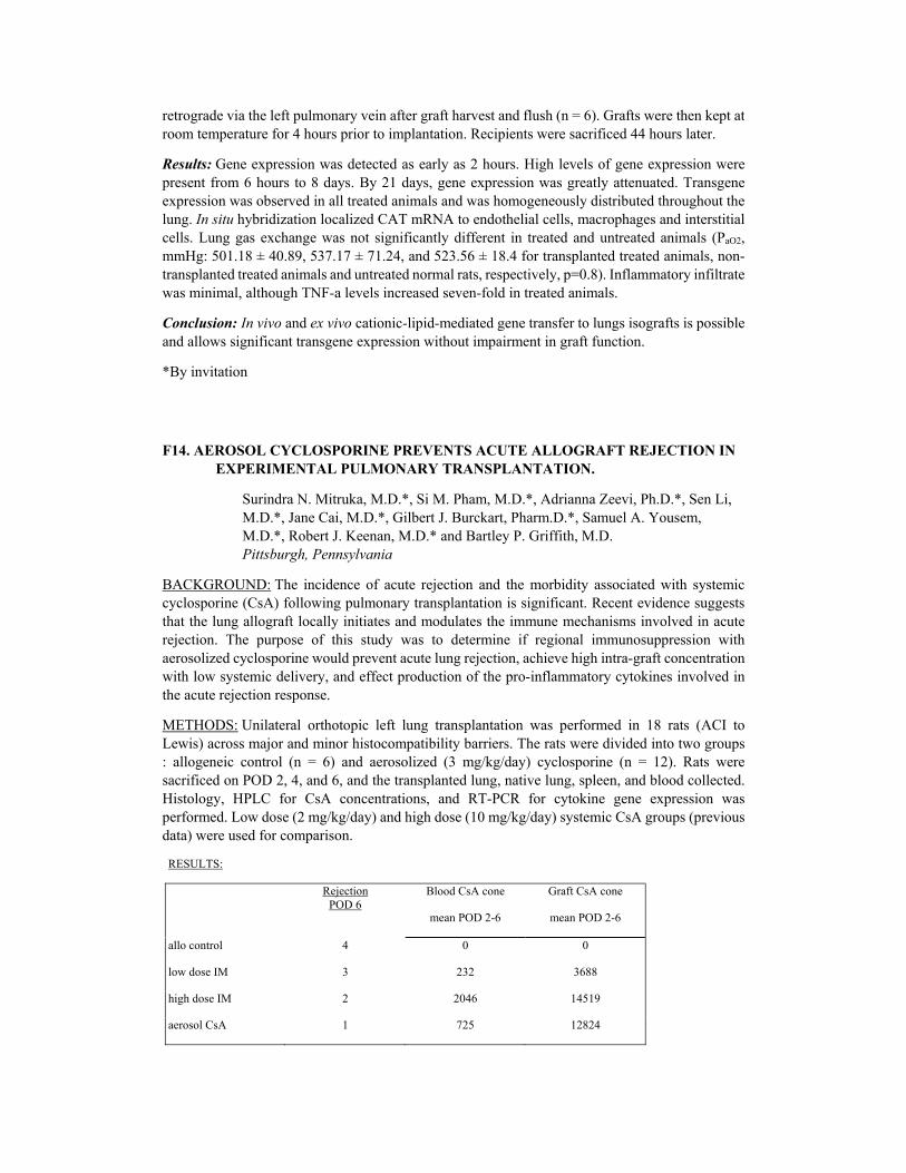
retrograde via the left pulmonary vein after graft harvest and flush (n = 6). Grafts were then kept atroom temperature for 4 hours prior to implantation. Recipients were sacrificed 44 hours later.
Results: Gene expression was detected as early as 2 hours. High levels of gene expression werepresent from 6 hours to 8 days. By 21 days, gene expression was greatly attenuated. Transgeneexpression was observed in all treated animals and was homogeneously distributed throughout thelung. In situ hybridization localized CAT mRNA to endothelial cells, macrophages and interstitial cells. Lung gas exchange was not significantly different in treated and untreated animals (PaO2, mmHg: 501.18 ± 40.89, 537.17 ± 71.24, and 523.56 ± 18.4 for transplanted treated animals, non-transplanted treated animals and untreated normal rats, respectively, p=0.8). Inflammatory infiltratewas minimal, although TNF-a levels increased seven-fold in treated animals.
Conclusion: In vivo and ex vivo cationic-lipid-mediated gene transfer to lungs isografts is possibleand allows significant transgene expression without impairment in graft function.
*By invitation
F14. AEROSOL CYCLOSPORINE PREVENTS ACUTE ALLOGRAFT REJECTION IN EXPERIMENTAL PULMONARY TRANSPLANTATION.
Surindra N. Mitruka, M.D.*, Si M. Pham, M.D.*, Adrianna Zeevi, Ph.D.*, Sen Li, M.D.*, Jane Cai, M.D.*, Gilbert J. Burckart, Pharm.D.*, Samuel A. Yousem, M.D.*, Robert J. Keenan, M.D.* and Bartley P. Griffith, M.D. Pittsburgh, Pennsylvania
BACKGROUND: The incidence of acute rejection and the morbidity associated with systemiccyclosporine (CsA) following pulmonary transplantation is significant. Recent evidence suggeststhat the lung allograft locally initiates and modulates the immune mechanisms involved in acuterejection. The purpose of this study was to determine if regional immunosuppression withaerosolized cyclosporine would prevent acute lung rejection, achieve high intra-graft concentration with low systemic delivery, and effect production of the pro-inflammatory cytokines involved in the acute rejection response.
METHODS: Unilateral orthotopic left lung transplantation was performed in 18 rats (ACI toLewis) across major and minor histocompatibility barriers. The rats were divided into two groups: allogeneic control (n = 6) and aerosolized (3 mg/kg/day) cyclosporine (n = 12). Rats weresacrificed on POD 2, 4, and 6, and the transplanted lung, native lung, spleen, and blood collected.Histology, HPLC for CsA concentrations, and RT-PCR for cytokine gene expression wasperformed. Low dose (2 mg/kg/day) and high dose (10 mg/kg/day) systemic CsA groups (previousdata) were used for comparison.
RESULTS:
Rejection POD 6
Blood CsA cone
mean POD 2-6
Graft CsA cone
mean POD 2-6
allo control 4 0 0
low dose IM 3 232 3688
high dose IM 2 2046 14519
aerosol CsA 1 725 12824

Aerosol CsA controlled rejection with a significantly less blood concentration (725 ng/ml vs. 2046ng/ml) and a similar tissue concentration (12, 824 ng/mg vs. 14, 519 ng/mg) compared to the highdose systemic group. The pro-inflammatory cytokines increased continuously in untreated animals from POD 2 to POD 6 at which time rejection was complete. Aerosol CsA treated animals initiallyexpressed IL-6 and IFN-g on POD 2 but none thereafter, and iNOS production was completelyattenuated; similar to results obtained in the high dose systemic CsA group.
CONCLUSION: Local delivery of CsA by aerosol inhalation effectively prevented acute rejectionof the rat lung allograft. Moderate dose aerosolized CsA achieved high graft concentrations withlow systemic delivery. The gene expression of pro-inflammatory cytokines involved in acuterejection was suppressed by aerosol CsA therapy.
*By invitation
F15. EXPRESSION OF ACIDIC FIBROBLAST GROWTH FACTOR CONTRIBUTES TO MALIGNANT TRANSFORMATION IN BARRETT'S ESOPHAGUS.
Robert Soslow, M.D.*, Liang Ying, M.D.* and Nasser K. Altorki, M.D.
New York, New York
The process of tumorigenesis involves loss of function of tumor suppressor genes or activation ofoncogenes many of which encode for various growth factors. Acidic fibroblast growth factor (aFGF) is a potent mitogen whose RNA transcripts were shown to be overexpressed in Barrett'sadenocarcinoma. In this study we investigated aFGF protein expression in 17 esophagectomyspecimens from patients with Barrett's adenocarcinoma. Immunostaining was performed on paraffin embedded tissue using a streptavidin-biotin technique with monoclonal antibody againstaFGF. In nine cases, the examined sections contained residual Barrett's epithelium (metaplasia 5,low grade dysplasia 3, high grade dysplasia 9). Epithelial cells were considered positive for aFGFif greater than 10% were immunostained. Results are shown below:
Positive aFGF Negative aFGF
Control (gastric funds) 2 15
Barrett metasplasia 1 4
Low grade dysplasia 2 1
High grade dysplasia 8 1
Carcinoma 17 0
Immunostaining was very intense (3+) in all carcinoma cases and 8/9 cases with high grade dysplasia. We conclude that increased expression of aFGF plays an important role in tumorigenesis in patients with Barrett's esophagus and further studies should be conducted to evaluate its use as a clinical blomarker.
*By invitation

F16. SUCCESSFUL IN VIVO AND EX VIVO TRANSFECTION OF PULMONARY ARTERY SEGMENT IN LUNG ISOGRAFTS.
Motoki Yano, M.D.*, Carlos H. Boasquevisque, M.D.*, Itaru Nagahiro, M.D.*, Masafiimi Hiratsuka, M.D.*, Bassem N. Mora, M.D.*, Joel D. Cooper, M.D. and G. Alec Patterson, M.D.
St. Louis, Missouri
Background: Gene transfer into donor lung grafts is feasible and may be useful in reducingreperfusion injury and rejection. However using conventional viral vectors whole organtransfection remains inefficient. Yet focal proximal pulmonary artery endothehal transfection mayprovide satisfactory downstream effects on the whole graft. The aim of this study was to achievetransfection of proximal pulmonary artery segments in left lung isografts.
Methods: Fisher rats (250-280 g) were divided into three groups. In group I (n = 4) and group II(n = 7) intact donor rats were subjected to occlusion of the proximal left pulmonary artery segmentfor twenty minutes after which flow was restored. In group I, 0.03 ml saline was injected into thepulmonary artery segment via a catheter in the right ventricle. In group II, the pulmonary arterysegments were injected with 2-4x10¹⁰ pfu/ml replication deficient adenovirus type V with LacZgene encoded β-galactosidase. In group III (n = 5) donor lungs were flushed with 20 ml LPDGsolution and extracted. The same construct as for group II was instilled ex vivo into the occludedleft pulmonary artery segment. After three hours storage (10°C), the grafts were implanted. In allgroups, seventy-two hours after reperfusion, heart-lung blocks were flushed with PBS and Bluo-gal and immersed in Bluo-gal for three hours.
Results: The survival rates were 50% (group I), 43% (group II) and 100% (group III).Macroscopically, in all survival animals of group II and III, multiple blue spots were observed onthe endothelial surface of the proximal left pulmonary artery indicating successful genetransfection. Microscopically, blue stained endothelial and smooth muscle ells were observed.
Conclusion: A high rate of focal gene transduction was observed in proximal pulmonary arterysegments following in vivo and ex vivo exposure. Direct gene transfer to pulmonary arterysegments is feasible and may avoid potential complications of systemic transfection strategies.
*By invitation
F17. CHANGES IN PULMONARY PHYSIOLOGY AFTER LUNG VOLUME REDUCTION SURGERY IN A RABBIT MODEL OF OBSTRUCTIVE DIFFUSE EMPHYSEMA.
Joseph Huh, M.D.*, Matthew Brenner, M.D.*, John C. Chen, M.D.*, Edward A. Stemmer, M.D., Benedict Yoong, B.S.*, David Mukai, B.S.* and Jeffrey C. Milliken, M.D.* Orange, California
Purpose: While surgical treatment of emphysema has recently gained popularity, the mechanismby which lung volume reduction surgery (LVRS) improves respiratory physiology is stillincompletely understood. Using an elastase induced purely obstructive emphysema model in NewZealand White rabbit, we studied the effects of LVRS on puhnonary compliance, airway flow, measured lung volume, and diffusion capacity.

Methods: Emphysema was induced in 14 New Zealand white rabbits by aerosolizing 15, 000 unitsof porcine elastase through an endotracheal tube under general anesthesia. Transpleural pressureswere measured at 60, 50, 40, 30, and 20 cc's inflation above functional residual capacity (FRC).Measurements were taken at baseline prior to induction of emphysema, preoperatively at 4 weeksfollowing induction of emphysema, and 1 week postoperatively following LVRS. FEV1, helium dilution lung volume, and single breath DLco were also measured concurrently. Stapled resectionof bilateral upper lobes was performed through a midline sternotomy with a standard multirowsurgical stapler (Ethicon). Histologic examination was obtained one week postoperatively.
Results: Comparison of compliance curves showed an increase in compliance following inductionof emphysema and a decrease in response to LVRS (graph). In like fashion, FEV1 showedimprovement in airway flow postoperatively, although this did not reach statistical significance,while FRC decreased following LVRS. DLco did not show a significant change (Table). Histologicexamination confirmed presence of severe diffuse emphysema in each animal at necropsy.
Conclusion: We have developed an animal model ofelastase induced diffuse emphysema applicable for LVRSstudies. Decreased compliance and increased airway flowfollowing volume reduction surgery parallels findings inhuman studies and suggests that similar mechanisms of increased elastic recoil and airway support contribute toimprovement. Furthermore, helium dilution volumes show
a decrease in lung volume postoperatively without significant decrease in diffusion capacity. Thismodel may be useful in assessing surgical techniques in LVRS, and may help identify optimallocation and quantity of lung tissue excision in the surgical treatment of emphysema.
Supported by NIH Grant #RR-011-92, DOE Grant #DE-FG03-91ER61, and DOD Grant #N00014-91-C-0134.
*By invitation
F18. THE RELATIONSHIP OF ISCHEMIA REPERFUSION INJURY AND THE EXPRESSION OF MAJOR HISTOCOMPATIBILITY COMPLEX ON HOST LYMPHOCYTES.
Karim A. Qayumi, M.D., Ph.D.*, David V. Godin, Ph.D.*, Maryam Nikbakht-Sangari, B.Sc.*, John C. English, M.D.*, Kathleen J. Horley, Ph.D.*, Seung P. Lim, M.D., Ph.D.*, Shahid Gul, B.Sc.* and Michael S. Koehle, B.Sc.H.*
Vancouver, British Columbia and Toronto, Ontario, Canada; Chung-Ku, Korea
Sponsored by: G. Frank O. Tyers, M.D., Vancouver, British Columbia, Canada
LUNG PARAMETERS
(t-test)
Baseline Preop p value
FEV1 (cc) 47.22 21.09 0.006
FRC (cc) 26.63 32.00 0.03
DLco (cc/min/mraHg)
0.62 0.56 0.37
Preop
Postop p value
FEV1 (cc) 21.09 31.33 0.28
FRC (cc) 32.00 22.52 0.05
DLco (cc/min/mmHg)
0.56 0.61 0.56

This study was designed to examine the effect of ex vivo preservation time on the release of specificinflammatory mediators, and the levels of plasma and tissue antioxidants related to the rejection ofthe transplanted organ (measured by the expression of MHC HLA-DR-β on host lymphocytes). Single lung transplantation was performed on three gropus of domestic swine. Group A (n = 7) andGroup B (n = 6) had ex vivo preservation times of 4 and 15 hours respectively at 4°C hypothermia.Group C (n = 6) underwent 2 hours of warm ischemia with the left pulmonary artery, vein, and bronchus cross-clamped without explantation. Methods of assessment included: the release ofinflammatory mediators-thromboxane B2 (TxB), interleukin-2 (IL-2), IL-4, IL-10, tumour necrosis factor α (TNFα) quantitated by radioimmunoassay and/or enzyme linked immunosorbent assay;the levels of plasma and tissue antioxidants determined by enzyme bioassay, the expression ofMHC HLA-DR-P on host lymphocytes by fluorescence intensity; and the mechanics of lungfunction by measurement of lung compliance, a/A ratio, and lung weight. The results demonstratedincreases (p<0.05) TxB, IL-2, IL-4, lung weight, O2 gradient, and HLA-DR-β expression on host lymphocytes directly proportional to ischemic time. IL-10, TNFα, lung compliance, a/A ratio, andboth plasma and tissue antioxidants were inversely proportional to ischemic time (p<0.05). Similarresults were observed in Group C which experienced ischemia reperfusion injury without tissue incompability. These results suggest that ischemia reperfusion injury alone without the complication of tissue incompatibility. These results suggest that ischemia reperfusion injury alonewithout the complication of tissue incompatibility is enough to initiate an acute post-transplantation response. Thus the severity of ischemia reperfusion injury, as measured by the release ofinflammatory mediators and the levels of antioxidants, could be directly related to the intensity ofrejection of the transplanted organ, as measured by the expression of HLA-DR-β on host lymphocytes.
*By invitation
7:00 a.m. FORUM SESSION III - CARDIAC SURGERY
North Sheraton Ballroom
Moderators: D. Glenn Pennington, M.D.
Tirone E. David, M.D.
F19. LONG-TERM GENE EXPRESSION AFTER VIRAL TRANSDUCTION OF CARDIAC ISOGRAFTS USING DNA VIRAL VECTORS.
Boulos Asfour, M.D.*, Paul D. Kessler, M.D.*, Ralph H. Hruban, M.D.*, Duke E. Cameron, M.D. and Barry J. Byrne, M.D., Ph.D.*
Baltimore, Maryland
Objectives: Viral transduction of cardiac allografts provides the opportunity to genetically modify graft vasculature and myocardium as well as to achieve graft-specific immunosuppression orpossibly tolerance. However, successful gene therapy in cardiac transplantation will require bothgene delivery in a clinically applicable manner and long-term gene expression. Adenovirus hasbeen used successfully to achieve short-term transduction in cardiovascular tissue. Adeno-associated virus (AAV) is a parvovirus which we have previously shown to be effective in long-term transduction of skeletal muscle, vascular smooth muscle, and cardiomyocytes in vitro and in vivo.
Methods: Female adult Sprague-Dawley rats (200-250 gms, n = 20) served as isograft donors andrecipients for vascularized heterotopic cardiac transplants. Following cannulation of the right

carotid artery, the graft was perfused with oxygenated cold (15°C) Krebs-Henselite solution and the donor organ harvested. A viral solution containing either, AAV-IacZ (5 x 10⁹ particles) or Ad-lacZ (1 x 10⁹ pfu), encoding the marker protein, bacterial β-galactosidase was then delivered to thecoronary vasculature. Perfusion was discontinued to allow for intracardiac recirculation of virusvia creation of an atrial septal defect or pulmonary bypass (PA-LA or PA-Ao shunt).
Results: Two weeks to two months following transplantation, grafts were removed for histologicalanalysis of β-galactosidase activity. Uniform staining of cardiac and vascular smooth muscle wasobserved at the highest dose with viral recirculation. The transgene was expressed for up to two months in hearts transduced by AAV-lacZ.
Conclusions: We have shown that AAV-lacZ and Ad-lacZ delivered via the coronary vasculatureof cardiac allografts are able to transduce vascular and cardiac tissue under conditions ofhypothermic perfusion and storage currently used in clinical cardiac transplantation. This approachwill be useful for genetic modification of cardiac allografts in the management of graft vasculo-pathy and rejection.
*By invitation
F20. HEARTS AND KIDNEYS FROM TRANSGENIC SWINE EXPRESSING HUMAN COMPLEMENT REGULATORY PROTEINS ARE PROTECTED FROM HYPERACUTE REJECTION IN PRIMATES.
Sepehre Naficy, M.D.*, Lisa E. Diamond, Ph.D.*, John S. Logan, Ph.D.* and David H. Adams, M.D.*
Boston, Massachusetts and Princeton, New Jersey
Sponsored by: Lawrence H. Cohn, M.D., Boston, Massachusetts
Unmodified vascularized porcine organs undergo destruction of tissue architecture and organfunction by hyperacute rejection within minutes to hours of transplantation into baboon recipients. Recipient complement activation and deposition onto donor endothelium is pivotal in thepathogenesis of hyperacute rejection. Decay accelerating factor (DAF), membrane cofactor protein(MCP), and CD59 are important human complement regulatory proteins (CRP). In order to clarify the role donor CRP play in pig to baboon hyperacute rejection, transgenic swine expressingdifferent human CRP combinations were used as donors. One heart expressing MCP, two heartsexpressing CD59/DAF, and two kidneys expressing CD59/DAF were transplanted into five baboonrecipients. Cardiac grafts were placed heterotopically in the neck and followed by periodicinspection, echocardiography, and biopsy. Renal grafts were placed heterotopically in the abdomenfollowing bilateral recipient nephrectomies. Recipient blood was tested serially for troughCyclosporine A levels, CBCs, creatinine, and serum xenoreactive antibody liters. Xenograftsamples were examined by light microscopy and immunostained for detection of antibodydeposition and complement activation.
Transgenic organs were protected against hyperacute rejection. The MCP heart was explanted withnormal function after 1.8 days following sudden death of the baboon. The CD59/DAF heartssurvived 3.6 and 5.4 days. One CD59/DAF kidney was explanted after 8.3 days with normalfunction (creatinine 2.1) from a terminally anemic recipient. The second transplanted kidney isfunctioning well (creatinine 1.3) after 11 days. Biopsies from functioning grafts were noted to havepreserved histologic architecture. Rejected hearts demonstrated regional hemorrhage andinterstitial edema, with fibrin plugs in thrombosed vessels. Immunostaining revealed specific andprominent IgM and C4 endothelial deposition and inhibition of terminal membrane attack complex

formation. Kidney samples showed normal histology with positive immunostaining for IgM andC4. Our results demonstrate transgenic swine organs expressing human CRP are protected fromhyperacute rejection. Further understanding of ongoing immune events resulting in graft failure isrequired before pig xenotransplantation can be applied clinically. *By invitation
F21. EFFICIENCY OF A HIGH-TITRE RETROVIRAL VECTOR IN GENE TRANSFER INTO SKELETAL MYOBLASTS.
Reida M. El Oakley, F.R.C.S., M.D.*, Mark A. Poznansky, M.R.C.P., Ph.D.*, Madeliene C. McMullen*, Gregor M. Adams, B.Sc.*, Nigel J. Brand, Ph.D.*, Paul J.R. Barton, Ph.D.* and Magdi H. Yacoub, F.R.C.S., Ph.D.
London, England
Background: Grafting genetically modified skeletal myoblasts for myocardial repair is dependenton an efficient gene transfer system that integrates the gene(s) of interest into the chromosome ofthe target cell and its progeny. The aim of this investigation is to evaluate the use of a new retroviral-based gene transfer system for this purpose.
Methods & Results: The retroviral vector MFG, carrying the β galactosidase gene (lacZ) withnuclear localisation signal, was used to transduce the skeletal myoblast cell line L6. The MFG-lacZ construct was packaged in a fourth generation, high-litre, split-genome packaging cell line (FLYA4). This cell line produced 105-106 infectious units per ml. L6 cells were cultured in tissueculture flasks and transduced with MFG-lacZ using filtered supernatant from the packaging cells.Transduced L6 cells were divided into 4 groups. Group I, cells were fixed as myoblasts 3 days aftertransduction. Group II, cells were allowed to differentiate into myotubes. Group III, cells were splitevery 3 days for 4 months. Group IV, cells were split as in group III, and allowed to differentiateinto myotubes. For each group, un-transduced L6 cells acted as "control". All samples were fixedwith 3.7% formaldehyde and stained for lacZ activity. The percentage of cells with successfultransgene expression are presented in the following table:
group I group II group III group IV Controls
% of positive cells 69.3% 73.6% 68.9% 71.4% 0%
Conclusion: Recombinant MFG retroviral agents packaged in a high-litre, split-genome packaging cells are efficient in gene-transfer into skeletal myoblasts and result in stable transgene expressioneven after repealed cell division and/or differentiation.
*By invitation
F22. EX-VIVO ADENOVIRAL-MEDIATED GENE TRANSFER TO THE TRANSPLANTED ADULT RAT HEART.
Alan P. Kypson, M.D.*, Karsten Peppel, Ph.D.*, Shahab A. Akhter, M.D.*, R. Eric Lilly, M.D.*, Donald D. Glower, M.D.*, Robert J. Lefkowitz, M.D.* and Walter J. Koch, Ph.D.*
Durham, North Carolina

Sponsored by: Robert W. Anderson, M.D., Durham, North Carolina
The ability to transfer functional genes to adult myocardium represents an area of study withpotentially significant therapeutic implications. We investigated the feasibility of adenoviral-mediated transfer of both marker genes LacZ and Luciferase, as well as the potentially functionalgene of the human P2 adrenergic receptor (P2-AR) in a heterotopic heart transplant model using adult male Long Evans rats. Donor hearts were arrested with warm cardioplegia and after removalflushed via the aortic root with one milliliter of solution containing 1012 total viral particles of recombinant adenovirus encoding one of the three transgenes. Hearts were transplanted into therecipient's abdomen and harvested at five days for hearts injected with the marker genes and atvarious time points for hearts injected with the β2-AR. lacZ-treatcd hearts were assessed by histochemical staining (X-gal). Luciferase-treated hearts were assayed for luciferase activity. β2-AR-treated hearts underwent radioligand binding assays and immunohistochemistry using anantibody specific for the human P2-AR. LacZ hearts (n = 6) revealed diffuse myocyte staining ofboth the right and left ventricles, as opposed to no staining within control hearts which receivedempty adenovirus. Luciferase hearts (n = 6) demonstrated a mean activity of 970, 000 ± 220, 000*arbitrary luciferase units as compared to controls which had a mean activity of 500 ± 200 arbitraryluciferase units (*p<0.05 vs. controls). Total P-AR densities (fmol/mg membrane protein) for heartsthat received the β2-AR transgene, at 3, 5, 7, 10, and 14 days after transfection were as follows; right ventricle - 488.5 ± 126.8, 519.4 ± 81.8*, 477.1 ± 51.8*, 183.0 ± 6.5*, 82.7 ± 19.1; left ventricle- 511.0 ± 167.6, 1206.4 ± 321.8*, 525.3 ± 188.7, 183.5 ± 18.6*, 75.9 ± 15.2 (n = 3 for each group;*p<0.05 as compared to control value of 75.6 ± 6.4). Immunohistochemical analysis with anti-β2-AR antibodies revealed diffuse staining of varying intensity within myocardial sarcolemmalmembranes. We conclude that global overexpression of two different adenoviral-mediated reporter genes and, a potentially functional gene, the human β2-AR, is possible during cardiactransplantation. Furthermore, β2-AR overexpression increases with time, peaking at five days,followed by a gradual decline returning to native levels at two weeks. Ultimately, gene transfer during cardiac transplantation may provide a unique opportunity for genetic manipulation of thedonor organ, potentially enhancing the function of the heart.
*By invitation
F23. ESTROGEN INHIBITS THE DEVELOPMENT OF TRANSPLANT ARTERIOSCLEROSIS BY PREVENTING INDUCIBLE MHC CLASS II ANTIGEN IN THE EARLY PHASE FOLLOWING THE TRANSPLANTATION.
Satoshi Saito, M.D.*, Noboru Motomura, M.D., Ph.D.*, Hong Lou, M.D.* and Marie L. Foegh, M.D., D.Sc.*
Washington, DC
Sponsored by: Edward A. Lefrak, M.D., Annandale, Virginia
Background: The development of transplant arteriosclerosis (TA) is a major limiting factor forlong time survival of cardiac transplants. We find chronic estradiol (E2) treatment inhibited TA.Very recently, we discovered that E2 inhibition of coronary TA in a chronic cardiac transplantmodel is associated with complete abolition of MHC class II expression. This model employscyclosporin A (10 mg/kg/day) immunosuppression. We hypothesize that E2 independent ofcyclosporin A inhibits inducible MHC class II antigen expression in professional and non-professional antigen presenting cells from the early phase following transplantation. The objective

of this study is to investigate in noncyclosporin requiring TA model that E2 treatment abolish MHC class II antigen expression in the allograft in the early phase following transplantation.
Methods: Orthotopic abdominal aorta allograft transplantation was performed using Brown-Norway rats as donors and Lewis rat as recipients . All recipients were treated with either 20µg/kg/day of estradiol 17 (J (n = 20) or placebo (n = 20) continuously s.c. from 2 days prior totransplantation until sacrifice using an osmotic minipump. The animals were sacrificed onpostoperative days 1, 3, 7, and 14 and the grafts were harvested following perfusion fixation andthen embedding in paraffin. Cross sections of the allografts were used for computerizedmorphometric analysis of medial area (M) and intimal thickening. Intimal thickening (I/I + M) wasquantitated as the ratio of intimal area (I) over total vascular area (intima + media). Followingimmunohisto-staining, the expression of MHC class II antigen and macrophage was gradedsemiquantitatively on a scale from 0 to +3.
Results: Intimal thickening was measureable at day 14 and I/I + M in the allograft from E2 treatedrecipients was significantly lower than placebo treated recipients. (9.2 + 2.2 % vs. 2.3 + 3.6%,p<0.01).
MHC class II antigen expression Intima Media Adventita
Day E2 Placebo E2 Placebo E2 Placebo
1 0 0 0 0 0 0
3 0 0 0 0.07 ± 0.05 0 0.50 ± 0.25
7 0 0.11 ± 0.07 0 0.55 ± 0.16 0.06 ± 0.02 2.11 ± 0.34*
14 0 0.83 ± 0.4 0 0.29 ± 0.10 0.32 ± 0.10 2.06 ± 0.38*
Macrophage expression Intima Media Adventita
Day E2 Placebo E2 Placebo E2 Placebo
1 0 0 0 0 0 0
3 0 0 0 0.22 ± 0.08 0 0.61 ± 0.11
7
00.43 ± 0.13 0 0.17 ± 0.08 0 0.60 ± 0.12
14 0.07 ± 0.04 040.43 ± 0.17* 0 0.29 ± 0.10 0.21 ± 0.10 0.89 ± 0.24*
Summary: 1. Chronic estrogen treatment inhibits development of transplant arteriosclerosis.
2. Estrogen abolishes inducible MHC class II antigen expression on SMC in the media in the earlyphase following the transplantation.
3. Estrogen suppresses inducible macrophag MHC class II antigen expression in the allograft vesselwall.
Conclusion: Estrogen may inhibit the development of transplant arteriosclerosis by abolition ofinducible MHC class II antigen in the early phase following the transplantation.
*By invitation
F24. AORTIC VALVE GRAFTS IN THE RAT MODEL: EVIDENCE FOR REJECTION.
Ahmad Moustapha, M.D.*, David B. Ross, M.D.*, Bindu Bittira, B.Sc.*, Dick Van Velzen, Ph.D.*, Vivian C. McAlister, M.B.*, Christopher L. Lannon, B.Sc.* and Timothy D. Lee, Ph.D.*
Halifax, Nova Scotia, Canada
Sponsored by: David A. Murphy, M.D., Halifax, Nova Scotia, Canada
Background: The role of immune mediated rejection in the failure of allograft heart valves in man is uncertain. Serial sampling of human allografts is not feasible and grafts are seldom removedshortly after implantation when any such damage may be occurring. The use of heart valvetransplants between syngeneic and allogeneic strains of rats permits investigations into the role ofimmune mediated rejection of these grafts.
Aims: To describe the pathological changes in rats, over time, following transplantation ofallogeneic versus syngeneic aortic valve grafts and to identify changes indicative of cell mediatedrejection.
Methods: Transplantations were performed placing the donor ascending aorta, valve and rim ofsubvalvular myocardium in the abdominal aorta of the recipient. Recipients were Lewis (Le) rats;syngeneic donors were Lewis and allogeneic donors were Brown Norway (BN). At sacrifice, thegrafts were removed, fixed and serially transacted at right angles. Samples of the aortic graft valvering and a sample of the distal graft aorta, 2 mm above the valve, were routinely processed in paraffin for microscopical assessment using 4 micron sections, H/E and van Giesson/Elastica stains.Histopathological assessment was performed by two pathologists blinded to the original protocoland study design.
Results:1) Early Post Transplant: 9 allogeneic (BN to Le) were transplanted with 3 sacrificed at2, 5 and 7 days with 3 Le transplanted as syngeneic controls. In both syn- and allogeneic cases a progressive, massive infiltration of neutrophils at the valve base and proximal aorta occurred increasing from day 2 to day 7. There was no difference seen between syngeneic and allogeneicanimals. 2)4 Weeks Post Transplant: 8 syngeneic (Le to Le) and 8 allogeneic (BN to Le) wereperformed. After initial blinded review, only two types of morphology were found. In one, virtuallynormal aortic grafts were seen, with minimal reactive changes to valves and minimal irregularityof the endothelium. In the other, severe chronic peri-aortic inflammation was associated withcontinuous cusp obliterating thrombosis, secondary valve degeneration and multi-focal, often completely full thickness, medial cell necrosis. All cases were attributed to either of the two types,reproducibility was complete. After unblinding, normal morphology was seen to be associated with the syngeneic transplants, chronic inflammation and destructive thrombosis was associated withallogeneic transplants. 3) 8 Weeks Post Transplant, Effects of Cryopreservation: 12 Le to Le and 12 BN to Le were transplanted with half of the donors cryopreserved using standard tissue banktechniques. Syngeneic transplants showed intact valves with no signs of attachment to aortic wall,degeneration or calcification. Only one (1/12) showed any thrombus in a sinus; the elastin layerswere preserved and there was no perivascular inflammation. The allogeneic grafts showed completeobliteration of the sinuses of Valsalva by organized thrombus with early calcification in some.Valve tissue remains were seen in the organised thrombus. Development of a neo-intima with medial cell necrosis was present. A moderate, mainly lymphocytic reactive infiltrate was presentin the perivascular tissues (11/12) and occasionally in the confines of the aorta (3/12). Thepathology did not differ between the cryopreserved and fresh grafts. Conclusions: The pathology of aortic valve transplantation in the rat model is dominated in the first week by a massiveinfiltration of neutrophils, probably induced by the necrotic muscle transplanted and is similar forboth allogeneic and syngeneic grafts. Thereafter, syngeneic grafts remain intact while allogeneicgrafts show progressive obliterative thrombosis of the valve leaflets and lymphocytic infiltration of

the aorta, unaffected by cryopreservation. These changes are characteristic of cell-mediated rejection. Investigations into therapies to modify this response appear justified.
*By invitation
F25. THE RELATIONSHIP BETWEEN CALCIUM AND MAGNESIUM IN PEDIATRIC MYOCARDIAL PROTECTION.
Bradley S. Allen, M.D.*, Michael Kronen, M.D.*, Kirk S. Boiling, M.D.*, Shaikh Ramon, M.S.*, Tingrong Wang, M.D.* and Harold Feinberg, Ph.D.*
Chicago, Illinois
Sponsored by: Renee S. Hartz, M.D., Chicago, Illinois
We have shown that adding magnesium to normocalcemic cardioplegic solutions offsets thedetrimental effects of calcium in neonatal hearts by competing with calcium entry. However, it isnot known whether magnesium offers any benefit when added to low calcium cardioplegicsolutions. Fifteen 5-18 day old neonatal piglets underwent sixty minutes of ventilator hypoxia(FiO2 8-10%) followed by reoxygenation using cardiopulmonary bypass (FiO2 100%) for 5 minutes, and 20 minutes of normothermic ischemia by cross clamping the aorta. This produces asevere injury that combines ischemia with hypoxia and reoxygenation. The hearts were thenprotected for seventy minutes with hypocalcemic (Ca2+0.2-0.4 mM/1) multidose bloodcardioplegia. In five (Group 1) no magnesium was added to the blood cardioplegia, in 5 (Group 2) magnesium was added to the cardioplegia to produce a concentration of 5 meq/1, and in the last 5(Group 3) magnesium was added at a concentration of 10 meq/1. Function was assessed usingpressure volume loops and expressed as percentage of control. Coronary vascular resistance (CVR)was measured during each cardioplegic infusion. Despite the use of a hypocalcemic cardioplegiasolution, in the absence of magnesium supplementation (Group 1) there was marked post bypassdepression of systolic (Ees 38 + 1%) and global myocardial function (40 ± 1%), and a marked risein diastolic stiffness (238 ± 3%). Conversely, even low dose (5 meq/l, Group 2) magnesiumsupplemented cardioplegia resulted in complete return of systolic (101% vs 38%)* and globalmyocardial function (102% vs 40%)*, preserved diastolic compliance (154% vs 238%)*, reducedmyocardial edema (79.7% vs 80.6%), maintained ATP levels (15.8 vs 12.2 ug/gm dry wt)* andpreserved CVR*, compared to a hypocalcemic cardioplegic solution without magnesium (Group 1). The use of a higher dose of magnesium (10 meq/1) did not result in any further improvement.In conclusion: 1) there is complete functional preservation, even in severely stressed neonatalhearts, when cardioplegia solutions are supplemented with magnesium, 2) this occurs despite evenwhen a hypocalcemic cardioplegic solution is used, and 3) doses as low as 5 meq/1 of magnesiumare effective. This study therefore strongly supports adding magnesium to all blood cardioplegiasolutions regardless of calcium concentration. *p<0.05
*By invitation

F26. EFFECT OF THE COX-MAZE PROCEDURE ON THE SECRETION OF ATRIAL NATRIURETIC PEPTIDE.
Ki-Bong Kim, M.D.*, Chang-Ha Lee, M.D.*, Young-Joo Cha, M.D.* and Cheol-Ho Kim, M.D.*
Seoul, Korea
Sponsored by: James L. Cox, M.D., St. Louis, Missouri
The Cox-Maze procedure (CMP) has been confirmed to be effective in curing atrial fibrillation(AF). Some authors reported severe fluid retention after CMP and suggested decreased secretionof atrial natriuretic peptide (ANP) as a possible mechanism. This study was designed 1) to followthe serial changes in ANP after CMP as compared to after coronary artery bypass grafting (CABG),and 2) to elucidate any differences between ANP levels in patients with transient recurrence of AF after CMP and those without recurrence. Blood samples were drawn from the right atrium (RA)and left atrium (LA) in patients undergoing CMP (n = 19) and from the RA in patients undergoingCABG (n = 6) before and 1, 2, and 3 days after surgery. The plasma samples were prepared byrefrigerated centrifugation and stored til radioimmunoassay. In the CMP group, ANP levels in theRA were 629 ± 366, 153 ± 112, 162 ± 112, and 183 ± 97, and in the LA were 276 ± 168, 152 ± 91,162 ± 111, and 145 ± 80 (pg/ml, mean ± SD) before and 1, 2, and 3 days after surgery, showing amarked decrease in ANP levels after CMP (p<0.01). In the CABG group, ANP levels in the RAwere 115 ±37, 124 ± 48, 154 ± 54, 156 ± 36 (pg/ml mean±SD) before and 1, 2, and 3 days aftersurgery, showing no change after surgery. There were no differences in ANP levels betweenpatients with transient recurrence of AF (n = 6) and those without recurrence (n = 13) after CMP.There was no significant correlation between ANP levels and LA or RA pressure after CMP, which suggests that the secretion of ANP from atria was impaired. In summary, we observed a significantdecrease in ANP levels after CMP and this might be one of the possible causes of fluid retentionafter CMP. The decreased ANP levels after CMP may result from the multiple atriotomy incisionsof the CMP rather than from the conversion of AF to sinus rhythm.
*By invitation
F27. EXTENDING THE CONCEPT OF AUTOGRAFT FOR COMPLETE REPAIR OF TRANSPOSITION OF THE GREAT ARTERIES WITH VSD AND LEFT VENTRICULAR OUTFLOW TRACT OBSTRUCTION. A REPORT OF 10 CASES OF A MODIFIED PROCEDURE.
Dominique R. Metras, M.D., Bernard Kreitmann, M.D.*, Alberto Riberi, M.D.*, John Yao, M.D.*, Elie El Khoury, M.D.*, François Wernert, M.D.* and Adrienne Pannetier, M.D.*
Marseille, France
BACKGROUND: Although in most cases of TGA with VSD and LVOTO, a REV (Lecompte)procedure is possible without interposition of a conduit between RV and PA, the anterior locationof the PAs after the Lecompte maneuver may be a potential cause for RV outflow obstruction thatremains reported in 5 to 10% of cases. We have used a tubular segment of aortic autograft to connectthe pulmonary artery, left in orthotopic posterior position (without Lecompte maneuver) to the RVin 10 consecutive cases of TGA VSD LVOTO. Late results up to 3, 4 years, show no obstructionin the RV reconstruction, low RV pressure, and no calcifications of the autograft.

METHODS: Ten consecutive patients aged 9 months to 11 years (mean 32 months) have beencorrected with a modified REV-Lecompte operation. Eight had a severe pulmonary stenosis, 2 hada pulmonary atresia, 4 had a restrictive VSD at the time of surgery, One had multiple VSDs. Sevenhad undergone one (5) or two (2) previous modified Blalock-Taussig shunts. All patients underwenta total correction with LV - Ao intra-ventricular connection (4 needed a VSD enlargement),connection between RV and PAs with a tubular segment of autograft aorta, without Lecomptemaneuver (anterior location of the bifurcation of Pas) on the right (5) or the left (4) of the aorta. Novalvular device was used for the RV outflow repair.
RESULTS: There was no early or late death. One patient with multiple VSDs needed an early (onemonth) reoperation for a residual muscular VSD. All patients are currently in NYHA class I,without medications, in sinus rhythm, at a mean follow-up of 2 years. There is no calcification onthe chest X-ray, and at the most recent echocardiogram, RV pressures were low (25-40, mean 33 mmHg) and no significant gradient (over 10 mmHg) was found between RV and PA. Left and rightventricular functions were satisfactory.
CONCLUSION: This modification of the REV operation using a segment of autograft allows anexcellent early and late result, with no danger of compression of anteriorly placed PAs, nosignificant RV outflow obstruction, normal aspect of the tubular autograft. In view of laboratoryand clinical evidence, normal growth of the autograft can be anticipated. It allows an electivecorrection of TGA VSD LVOTO without previous BT shunt (3 cases) and correction at a youngage (three patients less than one year).
*By invitation
F28. FIRST EXPERIENCE WITH A MODIFIED REPAIR TECHNIQUE FOR TRICUSPID INCOMPETENCE IN EBSTEIN'S MALFORMATION.
Roland Hetzer, M.D., Ph.D., Nicole Nagdymann, M.D.*, Peter Ewert, M.D.*, Vladimir Alexi-Meskhisvili, M.D, Ph.D.*, Yu-Guo Weng, M.D.* Felix Berger, M.D.* and Peter E. Lange, M.D, Ph.D.*
Berlin, Germany
Tricuspid incompetence (TI) in Ebstein's malformation is preferably treated by tricuspid repair(TR) rather than replacement (TVR) because of the characteristic complications of valve prosthesisand the higher incidence of heart block after TVR. Most repair techniques involve plication of the"atrialized" chamber which in some cases of small functional right ventricle may be difficult toachieve or, if attempted, may jeopardize the function of both ventricles.
In October 1988 a concept was instituted to restore tricuspid competence by a technique whichleaves the atrialized chamber unplicated, reduces tricuspid orifice circumference at the level of thetrue annulus and uses the individually most mobile leaflet for closing mechanism. Following thisconcept among 19 highly symptomatic patients, ages 2 to 52 years (mean 21 years) the following procedures were performed: creation of a double orifice tricuspid valve in 1, closure of posteriorpart of tricuspid orifice in 10, closure of anterior part in 5 and bilateral annulus plication in 3 cases.
There was no early death. Late death occurred in one patient with recurrent sepsis, after operationin active endocarditis. Intraoperative echocardiography revealed residual TI of grade 0-1 in 11, grade I-II in 8 patients. TI progressed in 2 patients and required repeat TR after 19 and 24 months. At a mean follow-up of 27 months (6 to 96 months) clinical status remains improved to NYHA I

in 3, II in 13 and III in 2 patients. There were 2 cases each of transient and permanent heart block.The atrialized chamber has not gained size in any case so far.
It is concluded that the proposed repair principle may offer an acceptable alternative to other repairtechniques for TI in Ebstein's malformation. *By invitation
9:30 a.m. SIMULTANEOUS SCIENTIFIC SESSION D - ADULT CARDIAC SURGERY
South Sheraton Ballroom
Moderators: Robert L. Hardesty, M.D.
Fred A. Crawford, Jr., M.D.
40. CORONARY ARTERY BYPASS WITHOUT ROUTINE PULMONARY ARTERY CATHETER USE - ASSESSMENT OF CRITERIA TO REDUCE COST AND PRESERVE QUALITY.
Robert D. Stewart, M.D.*, Christian T. Campos, . M.D.*, Triffin Psyhojos, M.D.*, Stephen J. Lahey, M.D.* and Sidney Levitsky, M.D.
Boston, Massachusetts
Discussant: Jack M. Matloff, M.D.
To reduce the cost and resource utilization of coronary artery bypass surgery (CABG) at ourinstitution, criteria to limit pulmonary artery (PA) catheter use were developed. Patients undergoingisolated, non-emergent, primary CABG with a left ventricular ejection fraction ‰¥ 40%, acreatinine < 2.0 mg/dl, without documented pulmonary hypertension or severe chronic obstructive pulmonary disease, and without unstable angina within 24 hours of surgery requiring the institutionof intravenous (IV) heparin or nitroglycerin (TNG) or an increase in the dose of IV TNG or theplacement of an intraaortic balloon pump were eligible for CABG with only central venous pressure(CVP) monitoring. The impact of these criteria from their implementation on April 22, 1996 untilJuly 31, 1996 in 77 patients (CVP group) were compared with results in 36 patients who met CVPcriteria but had a PA catheter placed due to surgeon or anesthesiologist preference (PA group).These 113 patients represented 65% of the 175 isolated, primary CABG operations performed atour institution during this three-month period.
The CVP and PA patients were well matched in demographics (age, gender, body mass index),severity of disease (ejection fraction, coronary vessel disease extent, priority of operation, and pre-op heparin or TNG use), and the prevalence of co-morbid conditions known to influence CABG outcome including hypertension, peripheral vascular disease, previous MI, and diabetes. Mean graftnumber, internal thoracic artery use, total cardiopulmonary bypass time, and cross clamp time wereidentical. Significantly more PA patients left the OR with inotropic support (14% vs. 1%; p=0.01),however, no significant increases in in-hospital mortality, mediastinitis, stroke, re-operation for bleeding, need for re-intubation, renal insufficiency, or need to institute post-op inotropic support were seen with CVP use. One patient had a CVP changed to a PA catheter.
CVP PA P % change vs. PA
12-hr volume infused (liters) 2.6 ±1.3 3.7 ± 1.1 0.001 -28.8
Post-op day #1 weight gain (kgs) 4.8 ± 2.3 5.7 ± 2.8 0.08 -15.5

Intubation time (hrs) 6.7 ± 3.7 8.3 ± 3.6 0.05 -18.4
ICU length of stay (days) 1 .4 ± 0.8 2.7 ± 6.3 0.06 -50.4
Overall length of stay (days) 5 .7 ± 3 .4 6.8 ± 6.7 0.21 -17.1
Hospital charges (thousand dollars) 23.4 ± 6.3 27.4 ± 18.2 0.09 -14.7
(Data are expressed as the mean ± S.D. P values are calculated by two-tailed Student's t-tests.)
A significant reduction in the volume infused in the first 12 hours following operation wasobserved in the CVP group. Definite trends toward reductions in all other resource outcomeindicators were seen in the CVP group as well. These clinically important trends approachedstatistical significance (Type II error). We conclude that these criteria for CVP catheter use, applicable to the majority of patients undergoing isolated primary CABG, resulted in clinically significant reductions in all measuresof CABG resource utilization with no increase in morbidity or in-hospital mortality.
*By invitation
41. A "PRIMELESS PUMP" FOR CARDIOPULMONARY BYPASS ENHANCES BLOOD CONSERVATION BY REDUCING THE NEED FOR PERIOPERATIVEBLOOD TRANSFUSIONS IN CORONARY BYPASS OPERATIONS.
John A. Rousou, M.D., Richard M. Engelman, M.D., Joseph E. Flack, III, M.D.*, David W. Deaton, M.D.*, Jane L. Garb, M.S.* and Susannah G. Owen, B.A.*
Springfield, Massachusetts
Discussant: Kenneth M. Taylor, M.D.
Severe hemodilution during cardiopulmonary bypass (CPB) often leads to significant drop inhematocrit (Hct) and coagulation factor (CF) levels requiring blood product transfusions (Tx). A method of removing pump prime prior to CPB, initiated and clinically used at our institution, wasnoted to limit hemodilution and reduce the need for perioperative Tx. A prospective evaluation oftwo consecutive series of patients, 52 with prime (control) and 95 without prime (primeless)undergoing coronary bypass operation (CABG) was undertaken to objectively study the method'seffectiveness in reducing Tx. Baseline characteristics between the control and the primeless groups such as body surface area (BSA), pre-CPB Hct, number of redos, use ofhemocon-centrator and antifibrinolytics, fluid balance in OR, operative and perfusion techniqueand transfusion criteria were the same in both series and strictly adhered to in all patients as per protocol. Patients within each group were consecutive and non-selected, regardless of their BSA,Hct or any other factors. Group differences in Hct during operation were analyzed using repeatedmeasures analysis of covariance. The proportion of patients requiring transfusions of red bloodcells (RBCs) and/or CF were compared using multiple logistic regression controlling for othercontributing factors. The drop in Hct during operation (over time) was significantly less for the"primeless" group (p<<0.0001) as shown in table below. Also shown are group differences in RBCsand CF given. There was no mortality in either group and no significant differences incomplications. Although not statistically significant in a smaller subgroup of this population, there was a trend for more pronounced effect of "primeless" pump on intraoperative Hct for patients withBSA < 2.
"Primeless" Control p value
% Hct drop from pre-CPB value 19.1 ± 0.8 27.8 ± 1.4

Post CPB Hct (Includes Tx) 26.5 ± 0.4 25.0 ± 0.6 <0.0001 for change in Hct over time
RBC Tx in OR (cc/pt) 29.9 ± 15. 6 142.5 ± 50.2 p=0.034
Tx RBCs in OR (% pts) 4.2 21.1 p<0.005
Clotting Factors Given Postop (% pts) 3 12 p=0.06
RBC: red blood cells; OR: operating room
Conclusions: The "primeless pump" leads to: 1) Significantly higher intraoperative Hct; 2)Significantly fewer patients requiring RBCs during CABG and (possibly) CF transfusion factorspostop; 3) The technique is simple and applicable to all cardiac operations leading to significant cost and risk reductions.
*By invitation
42. WHAT FACTORS TRULY AFFECT WAITING TIME TO TRANSPLANTATION? A MULTIVARIABLE ANALYSIS OF THE ISHLT/UNOS THORACIC REGISTRY.
Jonathan M. Chen, M.D.*, Keith D. Aaronson, M.D.*, Alan D. Weinberg, M.S.*, Berkeley M. Keck, R.N., M.P.H..*, Leah E. Bennett, Ph.D.*, Jeffrey D. Hosenpud, M.D.* and Robert E. Michler, M.D.
New York, New York, Richmond, Virginia and Milwaukee, Wisconsin
Discussant: Margaret D. Allen, M.D.
Because of the present donor organ crisis, the equity of organ distribution continues to bescrutinized. Although patients are currently listed and subsequently matched for cardiactransplantation (CT) according to clinical urgency, blood type, and weight, other variables mayhave an important impact on overall waiting time. We applied the Cox proportional hazards modelto all 7791 CT candidates listed with UNOS from 4/94 to 4/96 in an effort to assess the simultaneouseffect of multiple variables on the waiting times of CT candidates. The mean waiting time to CTwas 151.2 days (median time 175 days), the mean age was 45.6 years, and the mean time spentwaiting as a status I candidate was 20.5 days. At the termination of the study period, 4058 (52.1%) patients had undergone transplantation, 1954 (25.1%) were still waiting, 1216 (15.6%) had diedand 563 (7.2%) were removed from the list for other reasons. Variables that were not significantlyassociated with waiting time included: race, education level, arrhythmias, AICD and amiodaroneuse. Results of a multivariable analysis are depicted below:
Predictors of Shorter Waiting Times p-value Likelihood of CT
Proportion of Days as Status I p=0.0001 11.7
Weight at Listing p=0.0001 1.2/10 lb. †“ in weight
Blood Type AB* p=0.0001 3.9
Blood Type A p=0.0001 2.2
Blood Type B p=0.0001 1.7
No Need for a Prospective Cross-Match p=0.0001 2.0
Non-United States Citizenship p=0.0006 1.4

Age ‰¤ 2 years ‡ p=0.0001 0.3
Age > 2, and ‰¤ 65 years ‡ p=0.0362 0.9
* Blood types compared with Blood Type O; ‡ Age compared with patients > 65 years
Although preoperative predictors of patients at highest risk for death on the waiting list remainelusive, we describe herein factors that have an impact on the waiting time to transplantation. It isour hope that these data may help to identify certain patients at high risk for longer waiting timesfor whom other surgical alternatives to transplantation may be indicated.
*By invitation
§43. VIDEO-ASSISTED MINIMALLY INVASIVE MITRAL VALVE SURGERY.
W. Randolph Chitwood, Jr. M.D., Christopher L. Wixon, M.D.*, Joseph R. Elbeery, M.D.* and Jon F. Moran, M.D.*
Greenville, North Carolina
Discussant: Aubrey C. Galloway, Jr., M.D.
Video-assisted minimally invasive mitral valve surgery (VMIMS) may have advantages overconventional operative approaches. Since May of 1996, we have used a 2.5 inch right thoracicincision and video-assisted techniques in repairing (Rep N = 8) or replacing (Rpl N = 5) mitral valves. Ejection fractions ranged between .35 and .60 (.54 ± 2.2 SEM) with ages being 18 to 77years (55.5 ± 5.2 SEM). Other clinical characteristics were similar. Cardiac arrest was inducedeither by retrograde (N = 5) or antegrade (N = 6) blood cardioplegia using a new transthoraciccross-clamp to occlude the ascending aorta. In two patients cold ventricular fibrillation was used.Systemic perfusion was maintained at 28°C either by central or peripheral arterio/venouscannulation. Newly designed instruments enabled video-assisted suture placement and knot tying.Superb illumination and visualization were provided by a 10 mm port-access 30° thoracoscopic camera. Mean cardiopulmonary perfusion times were 197 ± 7.8 SEM minutes and arrest times averaged 138 ± 12.8 minutes. Postoperative transesophageal echocardio-graphic studies showed excellent valve function with minimal insufficiency, and all patients had little postoperative pain.There were no operative deaths. One patient developed lower extremity deep venous thrombosis asthe only major complication. Other patients were discharged between postoperative day three andfive (4.6 ± .4 days SEM). The mean hospital stay for the previous 111 conventional mitraloperations (N = 67 Rep, N = 44 Rpl) was 9.2 ± 0.9 days SEM. In the VMIMS patients atrialfibrillation (8% vs 28%), reoperation for bleeding (0% vs 5%), ICU length of stay (1.1 vs 2.0 days),and hospital charges (†“ > 30%) also were significantly less than the conventional cohort. Thus,despite long operative times, these early results are encouraging and suggest that video-assisted minimally invasive mitral valve operations are safe and may benefit patients by minimizingpostoperative pain, allowing earlier discharge, and decreasing hospital expenses. 10:50 a.m. INTERMISSION
§Authors have a relationship with Scanlan International, Inc.
*By invitation
11:10 a.m. SIMULTANEOUS SCIENTIFIC SESSION D -
ADULT CARDIAC SURGERY
South Sheraton Ballroom

Moderators: Robert L. Hardesty, M.D.
Fred A. Crawford, Jr., M.D.
44. MITRAL VALVE REPAIR WITH AND WITHOUT CHORDAL REPLACEMENT WITH EXPANDED POLYTETRAFLUOROETHYLENE SUTURES -A 10-YEAR EXPERIENCE.
Tirone E. David, M.D., Susan Armstrong, M.Sc.*, Zhao Sun, Ph.D.* and Ahmed Omran, M.D.*
Toronto, Ontario, Canada
Discussant: Lawrence H. Cohn, M.D.
Replacement of chordae tendineae with expanded polytetrafluoroethylene sutures (ePTFE)increases the probability of mitral valve (MV) repair in pts with mitral regurgitation (MR) due tomyxomatous disease of the MV. This study compares the results of MV repair with and withoutchordal replacement with ePTFE. From 1985 to 1995, 324 consecutive pts underwent MV repair:165 with ePTFE chordae and 159 without. There were no statistical differences between these twogroups as far as age (58 ± 14), functional class, left ventricular function and the incidence ofcoronary artery disease (16%). The differences were in the mechanism of mitral regurgitation andin the degree of myxomatous changes in the leaflets.
With ePTFE Without ePTFE
Prolapse of the anterior leaflet 49(30%) 9 (6%)
Prolapse of the posterior leaflet 29(17%) 139(87%)
Prolapse of both leaflets 87(53%) 11 (7%)
Advanced myxomatous changes 43 (26%) 14 (9%)
There were 2 operative deaths, both in the ePTFE group, neither one related to the MV repair.Operative morbidity was similar in both groups. Pts have been followed for a mean of 35 ± 30months. No pt was lost to follow-up. Every pt had an annual Doppler echocardiographic study. Eight pts developed severe recurrent MR and 2 other developed hemolysis and were reoperated on;6 of them were from the ePTFE group. All reoperations occurred in the first three years of follow-up. A logistic regression analysis identified that only the combination of prolapse of both leafletsand advanced myxomatous changes was predictive of reoperation in all pts. The latestechocardiographic study showed moderate MR in 9 pts (5 pts ePTFE group), and mild or none in284. The actuarial freedom from morbid events showed no statistical differences between the twogroups.
With ePTFE Without ePTFE
Freedom from death at 10 years 75% ± 7% 75% ± 6%
Freedom from stroke at 10 years 94% ± 2% 95% ± 2%
Freedom from reoperation at 10 years 94% ± 3% 97% ± 2%
This experience indicates that replacement of chordae tendineae with ePTFE sutures during MV repair for myxomatous disease of the MV provides as good long-term results as MV repair without ePTFE.
*By invitation

45. TEN-YEAR RESULTS OF CORONARY ARTERY BYPASS GRAFTING IN PATIENTS WITH ADVANCED LEFT VENTRICULAR DYSFUNCTION.
Gregory D. Trachiotis, M.D.*, William S. Weintraub, M.D.*, Thomas S. Johnston, M.D.*, Ellis L. Jones, M.D., Robert A. Guyton, M.D. and Joseph M. Graver, M.D.
Atlanta, Georgia
Discussant: Lynda L. Mickleborough, M.D.
The combination of severe coronary artery disease (CAD) and advanced left ventriculardysfunction carries a poor outlook with medical therapy. Coronary artery bypass grafting (CABG)in this group has often been regarded as high risk. We reviewed patients with CAD and sequentiallydecreased ejection fraction (EF) compared to those with an EF>50% with 10 year follow-up to determine if CABG can provide long-term symptomatic improvement and survival in patients withsevere left ventricular dysfunction (EF<25%). Between 1971-1994, 156 (1.3%) patients with an EF<25% [Group I], 588 (5%) patients with an EF=25-34% [Group II], 2438 (20.6%) patients withan EF=35-49% [Group III], and 8648 (73.1%) patients with an EF>50% [Group IV] underwentCABG. The EF was estimated from the contrast left ventriculogram (either uniplaner or biplaner).For all groups mean age was 60 ± 10 years. Groups l-III compared to Group IV had a higherpercentage of patients with men (<.0004); diabetes mellitus (p<.0001); class III-IV angina (p<.0001); heart failure (p<.0001); prior MI (p<.0001); 3 vessel disease (p<.0001); and left maindisease (p<.0001). Group I had the highest percentage of patients with men (88%); heart failure(34%); and left main disease (24%). The mean EF's were 19 ±4 in Group I, 29 ± 3 in Group II, 42 ± 4in Group III, and vs 64 ± 9 in Group IV. The results were as follows (<.05 significant byANOVA for Groups I, II, or III vs IV):
Groups (EF%) # Grafts Completely Revascularized
IMA Graft Q wave MI Death in Hospital
Length of Stay
Angina during Follow-up
I (<25) 3.3 ± 1.1 107 (69%) 54 (35%) 0 (0%) 6 (3.8%) 9.2 ±5. 8 31 (40%)
II (25-34) 3.5 ± 1.1 445 (76%) 244(41%) 12 (2%) 20 (3.4%) 10.5 ± 10.9 120(36%)
III (34-49) 3.4 ± 1.1
1952 (80%) 1149 (47%) 48 (2%) 72 (3%) 9.1 ±7.8 2277 (33%)
IV (>50) 3.3 ± 1 2 7400 (86%) 3616(42%) 231(27%) 134(16%) 8.4 ±6.4 2277 (33%)
P Value <.0001 <.0001 <.0001 <.04 <.0001 <.0001 <.06
Despite having a higher percentage of risk factors, poorer functional status, and more complexcoronary anatomy, patients with compromised left ventricular function have comparable in-hospital outcome to patients with a normal EF. While long-term there is much higher mortality in patientswith comprised LV function, over 60% of patients with EF <25% were alive at 5 years. In addition, patients after CABG with EF <25% have excellent long-term control of angina despite the loweruse of IMA grafts and less complete revascularization. These results suggest that in selectedpatients with ischemic cardiomyopathy, CABG may preserve remaining viable myocardium,provide relief of symptoms and offer survival >60% at >5 years.
*By invitation
46. MYOCARDIAL PERFUSION AND FUNCTION FOLLOWING ADENOVIRUS-MEDIATED TRANSFER OF THE VEGF121, cDNA TO ISCHEMIC PORCINE MYOCARDIUM.
Charles A. Mack, M.D.*, Shailen R. Patel, M.D.*, Eric A. Schwarz, M.D.*, Pat Zanzonico, Ph.D.*, Rebecca T. Hahn, M.D.*, Arzu Ilercil, M.D.*, Richard B. Devereux, M.D.*, Stanley J. Goldsmith, M.D.*, Timothy F. Christian, M.D.*, Timothy A. Sanborn, M.D.*, Imre Kovesdi, Ph.D.*, O. Wayne Isom, M.D., Ronald G. Crystal, M.D.* and Todd K. Rosengart, M.D.*
New York, New York and Rockville, Maryland
Discussant: Andrew S. Wechsler, M.D.
Background: A replication-deficient adenovirus vector expressing the cDNA for the angiogenic protein vascular endothelial growth factor121 (AdVEGF121) induces prolonged VEGF121 expression in vivo. We therefore hypothesized that direct myocardial injection ofAdVEGF121 would induce collateral vessel formation and enhance myocardial perfusion andfunction in ischemic myocardial territories.
Methods: Yorkshire swine (28-30 kg) underwent left thoracotomy and ameroid constrictorplacement on the left circumflex coronary artery. Three weeks later, AdVEGF121 or the control vector, AdNull (each 108 pfu in 100 l) was injected in the myocardium at 10 sites in thecircumflex distribution. Ischemia was assessed by echocardiography and 99mTc-sestamibi imaging during rest and rapid atrial pacing (‰¥ 200 beats/min) at the time of gene transfer and after 4 weeks, and collateral vessel formation was assessed by ex vivo an angiography. In a separate groupof animals, VEGF121 expression was quantified in the injected myocardium and serum by ELISAfollowing gene transfer.
Results: AdVEGF121-treated animals demonstrated significant VEGF121 expression (9 ng/mg protein) in the myocardium 3 days following vector administration. VEGF121 was undetectable in the serum of AdVEGF121 -treated animals. An improvement in ventricular perfusion and function in the circumflex territory was suggested by rest versus stress 99mTc-sestamibi scans and echocardiographic assessment of segmental wall thickening, respectively, in AdVEGF121 versus AdNull-treated animals. Angiography in the AdVEGF121-treated animals demonstrated a collateralnetwork with apparent reconstitution of the distal circumflex artery.
Conclusions: An adenovirus vector can be used to transfer the VEGF121 cDNA to the myocardium. This strategy may be useful in inducing therapeutic angiogenesis and improving perfusion andfunction in the ischemic myocardium.
12:10 p.m. ADJOURN
*By invitation
9:30 a.m. SIMULTANEOUS SCIENTIFIC SESSION E - GENERAL THORACIC SURGERY
North Sheraton Ballroom
Moderators: Mark K. Ferguson, M.D.
Andre C.H. Duranceau, M.D.

47. ANGIOGENESIS AS A PREDICTOR OF SURVIVAL FOLLOWING SURGICAL RESECTION FOR STAGE I NON-SMALL CELL LUNG CANCER.
Ignacio G. Duarte, M.D.*, Bradley L. Bufkin, M.D.*, Marion F. Pennington, M.D.*, Anthony A. Gal, M.D.*, Cynthia Cohen, M.D.*, Kamal A. Mansour, M.D. and Joseph I. Miller, M.D.
Atlanta, Georgia
Discussant: Valerie W. Rusch, M.D.
A subset of surgically resected Stage I non-small-cell lung cancer (NSCLC) patients will laterdevelop metastatic disease. A histologic marker of metastatic potential and diminished survival forStage I NSCLC may help identify this subset of patients. This study evaluates the degree ofangiogenic activity as a predictor of cancer-related mortality in patients having undergone surgicalresection for Stage I NSCLC. Demographic, surgical, and histopathologic data were reviewed for107 patients with Stage I NSCLC from 1985-1990. Visual quantitation of Factor VHI-related antigen (FVIII) and CDS 1 immunostained microvessels, 0.74 mm2 area (200x magnification), in 5µ sections from paraffin blocks defined tumor angiogenesis. Mean microvessel count was 20.7 ±11.2 for FVIII, and 29.6 ± 18.1 for CD31. Mean follow-up was 5.2 ±3.0 years (8 days - 11.06 years) and 95.3% complete. Lung cancer-related mortality was 23% at 5 years. Kaplan-Meier survival curves revealed FVIII count >20 (p=0.028) and the presence of blood vessel invasion (p=0.034) tobe significant predictors of disease-related mortality. Logistic regression analysis identified FVIIIquantitation > 20 as the single most significant independent correlate of lung cancer mortality(p=0.021, hazard ratio 2.64). CD31 quantitation did not predict survival in univariate andmultivariate analyses and did not correlate with FVIII quantitation (Spearman's rank correlation, r = 0.20). This analysis displays a significant association between tumor neovascularization andcancer-related mortality in patients with Stage I NSCLC. FVIII microvessel quantitation, as anindicator of tumor angiogenesis and metastatic potential, may help identify a subset of patients withStage I NSCLC who may benefit from adjuvant therapy following surgical resection.
*By invitation
48. THYMIC CARCINOMA: CURRENT STAGING DOES NOT PREDICT PROGNOSIS.
David B. Blumberg, M.D.*, Juan Rosai, M.D.*, Manjit S. Bains, M.D., Robert J. Downey, M.D., Robert J. Ginsberg, M.D., Nael Martini, M.D., Patricia M. McCormack, M.D, Valerie W. Rusch, M.D. and Michael E. Burt, M.D.
New York, New York
Discussant: Paul A. Kirschner, M.D.
Thymic carcinomas are currently staged by Masaoka classification, a staging system for thymomas.We retrospectively evaluated surgical patients with thymic carcinoma to determine factorspredicting survival. Methods: Our computerized tumor registry yielded 118 patients with thymoma. Review ofpathologic material revealed 43 cases of thymic carcinoma. Medical charts were reviewed. Follow-up was performed by physician charts and telephone. Analysis by Kaplan-Meier method and Cox proportional hazards.

Results: Between 1949 and 1993, 43 patients underwent surgery for thymic carcinoma. Overallsurvival was 65% at 5 years with a median survival of 6.7 years. Survival was not dependent onMasaoka stage (p=0.3). There were 3 stage I patients alive at 3.6, 4.1 and 8.2 years. Five year survivals were 58% for stage II (n = 15), 55% for stage III (n = 20) and 100% for stage IV (n = 5)patients. Five year survivals of patients with complete resection (n = 29) and patients with partialresection (n=14) were 68% and 62%, respectively, (p=0.2). Survival of completely resected patients(n = 29) was not dependent on age (p=0.1), sex (p=0.7), tumor size (p=0.7), or Masaoka stage(p=0.4). Six patients had tumors invading the innominate vessels, 4 which were low grade(well/moderately differentiated) and one indeterminate. Tumor invasion of the innominate vessels(n = 6) was associated with a worse survival (p=0.01) with only 40% alive at 2 years, compared to75% alive at 6 years with no invasion of the innominate vessels (n = 23). Of patients with no invasion of the innominate vessels, there were 19 low grade tumors and 4 high grade tumors (poorlydifferentiated). Survival of patients with low grade tumors was superior (p=0.03) with 95% aliveat 6 yrs. as compared to a 50% 4 yr. survival for high grade tumors. By Cox proportional hazardsmodel, survival was predicted only by grade (p=0.07) and innominate vessel invasion (p=0.05).
Conclusions: 1. Masaoka staging does not predict prognosis of patients with thymiccarcinoma. 2. Patients with low grade tumors without invasion of the innominate vesselshave early stage disease and long term survival may be achieved with surgical resection. 3.Despite complete resection, patients with high grade tumors and tumors invading the innominate vessels have a poor survival and these patients should be considered as havingadvanced disease. 4. These results have important implications for design of future adjuvant trials of patients with thymiccarcinoma. *By invitation
49. SAFETY AND EFFICACY OF BRONCHO VASCULAR RECONSTRUCTION AFTER INDUCTION CHEMOTHERAPY FOR LUNG CANCER.
Erino A. Rendina, M.D.*, Federico Venuta, M.D.*, Tiziano De Giacomo, M.D.*, Isac Flaishman, M.D.* and Costante Ricci, M.D.*
Rome, Italy
Sponsored by: Valerie W. Rusch, M.D., New York, New York
Discussant: Jean DesLauriers, M.D.
Desmoplastic reactions secondary to induction chemotherapy and/or residual tumor can make lungresection extremely difficult. In these patients, increased postoperative complications and mortality are reported, owing also to the high incidence of pneumonectomy. Between 1990 and July 1996we have operated on 68 patients who had received 3 cycles of cisplatin-based induction chemotherapy. In 27 of these we have performed a lobectomy (# 25) or bilobectomy (# 2) associated with reconstruction of the bronchus and/or the Pulmonary Artery (PA). In only 5 additional patientspneumonectomy had to be carried out. Before chemotherapy 14 patients were at stage HIA and 13at stage IIIB. At thoracotomy, 1 patient had no evidence of tumor, 6 were at stage I, 13 at stage II,6 at stage IIIA, and 1 at stage IIIB. Fourteen patients had epidermoid carcinoma and 11 hadadenocarcinoma. The type of reconstruction is tabulated below.
#
- Bronchial sleeve resection 16
- PA reconstruction by pericardial patch 2

- PA reconstruction by pericardial conduit 1
- Bronchial sleeve + PA pericardial patch 7
- Bronchial sleeve + PA sleeve 1
27
In 26 patients resection was radical with histologically negative margins. Bronchial anastomoseswere wrapped by the intercostal pedicle flap. No bronchial complications nor mortality occurred.One patient had empyema and 2 had wound infection. Mean chest tube stay was 6 days (3 to 15 days). Twelve patients had additional adjuvant therapy with no problems. After a postoperativefollow-up of 5 to 73 months (mean 25 months), 14 patients are alive disease-free, 1 is alive with disease and 12 died. No local recurrence occurred. One and four-year survival is 69% and 38%.Although technically demanding, lobectomy associated with bronchovascular reconstruction isfeasible with good immediate and long term results after induction chemotherapy.
*By invitation
50. PREOPERATIVE TUMOR VOLUME PREDICTS OUTCOME IN MALIGNANT PLEURAL MESOTHELIOMA.
Harvey I. Pass, M.D., Karen C. Kranda, R.N.*, Seth M. Steinberg, Ph.D.*, Barbara K. Temeck, M.D.* and Irwin R. Feuerstein, M.D.*
Bethesda, Maryland
Discussant: Larry R. Kaiser, M.D.
The staging systems for malignant pleural mesothelioma (MPM) rely upon postoperativepathologic findings for prognostic determination. Since MPM surgical cytoreduction remainscontroversial, it would be desirable to predict outcomes from quantitative preoperative data. We prospectively analyzed the impact of preoperative and postresection solid tumor volumes onprognostic variables in 47 of 48 consecutively resected MPM patients. Methods: From 7/93 to 6/96, 48 MPM patients had cytoreductive debulking to 5 mm or less residual tumor via extrapleuralpneumonectomy (EEP-25) or pleurectomy/ decortication (P/D-23). Three dimensional CT reconstructions of pre- and postresection solid tumor using the Voxell Q™ were prospectivelyperformed. All patients received the same postoperative adjuvant therapy and were stagedpostoperatively according to the new International Mesothelioma Interest Group (IMIG) staging.Patients were followed by chest and abdomen CT scans every 3 months until death. Prognosticfactors were examined by Cox proportional hazards model. Results: With a median potential follow-up of 23.1 months, median survival for all patients is 14.4 months (EPP-11 mos, P/D-22 mos, p2 = 0.066). Median survival for preop volume <100 cc was 22 months vs 11 months if >100cc, p2 = 0.027. Median survival for postop volume <9 cc was 25 months vs 9 months if >9 cc,p2= 0.0002. Thirty-two of 47 (68%) had positive Nl or N2 nodes. Tumor volumes associated withnegative node patients were significantly smaller (51 cc) than those with positive nodes (166cc,p2 = 0.0099). Progressively higher stage was associated with higher median preoperative volume:1-4 cc, II-94 cc, III-143 cc, IV-505 cc, p2 = 0.0070 for I vs II vs III vs IV. Patients with preoperativesizes >52 cc had shorter progression-free intervals (8 mos) than those ‰¤51 cc (11 mos), p2 = 0.021. By the Cox model, male sex, preoperative platelet count >314K, preoperative volume>100cc and postresection volume >9cc were associated with decreasedsurvival. Conclusions: Preresection tumor volume is representative of T status in MPM, and CTvolumetrics can predict overall and progression-free survival, as well as postoperative IMIG stage.

Large volumes are associated with nodal spread, and postresection residual tumor burden may predict outcome. Future trials should develop a uniform, simple method to quantify pretreatmentMPM volume, and to verify its prognostic and therapeutic implications.
10:50 a.m. INTERMISSION
*By invitation
11:10 a.m. SIMULTANEOUS SCIENTIFIC SESSION E - GENERAL THORACIC SURGERY
North Sheraton Ballroom
Moderators: Mark K. Ferguson, M.D.
Andre C.H. Duranceau, M.D.
51. EFFECT OF VOLUME REDUCTION ON LUNG TRANSPLANT TIMING AND SELECTION FOR COPD.
Joseph E. Bavaria, M.D.*, Alberto Pochettino, M.D.*, Robert Kotloff, M.D.*, Bruce R. Rosengard, M.D.*, Peter M. Wahl, B.A.*, Harold Palevsky, M.D.* and Larry R. Kaiser, M.D.
Philadelphia, Pennsylvania
Discussant: Joel D. Cooper, M.D.
Introduction: End-stage COPD has traditionally been treated with lung transplantation (LTX). Fortwo years, our LTX program has placed patients with appropriate criteria for LTX and VolumeReduction (LVR) into a prospective management algorithm. These patients are offered the LVR option as a means to "bridge" or extend the eventual time to LTX. These data examine the resultsof this pilot program.
Methods: From 7/7/94 to 10/25/96, 33 patients were evaluated for LTX who also had physiologicalcriteria for LVR (FEV1 ‰¤25%; RV >200%; significant V/Q heterogeneity). These patients weredivided into two groups: 26 patients (Group I) underwent LVR as a "bridge" and weresimultaneously listed for LTX. Seven patients (Group II), for various reasons, were offered LVRalone and not listed. All patients completed 6 weeks of pulmonary rehab, and then had baselinepulmonary function (PFTs) and Six Minute Walk (6MW) tests. LVR was performed via videothoracic in 81.8% or sternotomy in 18.2% of the patients. Patients were followed postop with repeat PFTs and 6MW at 3 month intervals.
Results: Nineteen of 26 pts (73.1%) in Group I had satisfactory clinical improvement after LVR.These 19 patients (Group I A) were subsequently delisted (Status 7). The remaining 7 patients inGroup I (26.9%) had unsatisfactory results (Group IB) and one died perioperatively. Three of 6survivors were subsequently transplanted with good outcomes. The other 3 patients are presentlyawaiting organs.
Pre-op
FEV1 (1)
Post-op
FEV1 (1)
Pre-op
RV(1)
Post-op
RV(1)
Pre-op
6MW (ft)
Post-op
6MW (ft)
Group IA 0.67 ± 0.18 0.86 ± 0.28 4.64 ± 0.83 3. 82 ± 1.38 1088 ± 340 1376 ± 209

Group IB 0.78 + 0.15 1.00 ± 0.40 4.54 ± 1.42 4.43 ± 0.57 909 ± 253 1208 ± 167
Group II 0.67 ± 0.27 0.85 ± 0.26 4.78 ± 1.27 4.29 ± 1.19 874 ± 365 1227 ± 225
Interestingly, 2 of the 33 patients had A-l antitrypsin deficiency both of which had poor LVR outcome.
Conclusions: LVR in these low FEV1 patients is safe. LVR has substantially impacted the practice, timing, and selection of patients for LTX. Our wait list presently has a reduced percentage of patients with a COPD diagnosis compared to 2 years ago. Seventy-three percent of otherwise suitable LTX candidates achieved good LVR results, especially reduction in RV and were deactivated from the list. The majority of patients entering our prospective management algorithm have either significantly delayed or completely avoided LTX after LVR. Our experience suggests that LVR may be limited as a "bridge" in alpha-1 antitrypsin patients.
*By invitation
52. PLEURAL TENTING DURING UPPER LOBECTOMY DECREASES CHEST TUBE TIME AND TOTAL HOSPITALIZATION DAYS.
Lary A. Robinson, M.D. and Dianne Preksto, PA-C*
Tampa, Florida
Discussant: Joseph I. Miller, Jr., M.D.
Purpose: A prolonged air leak following an upper lobectomy is one of the major determinants ofpostoperative morbidity and hospital stay. The use of stapling devices during pulmonary resectionshas greatly decreased air leaks, but the problem of sealing small persistent leaks to allow early chesttube removal is still present. Creation of a generous pleural tent following upper lobectomy wasemployed to investigate whether bringing the parietal pleura down to the lung to obliterate the usualpost-op apical space would help seal the air leak and shorten chest tube time.
Methods: From August, 1994 to September, 1996, the records of 43 consecutive patientsundergoing an upper lobectomy for a malignancy were reviewed. Twenty-three patients had creation of a pleural tent and 20 patients (undergoing surgery in the first year of the study period)did not. Mean patient age: tented 65.6 ± 1.7 years; non-tented 62.8 ± 3.0 years. Demographic andoperative profiles of both groups were not significantly different. Patients excluded from the studywere those undergoing concomitant chest wall resection (7), patients requiring post-operative mechanical ventilation (1), and those developing the alcohol withdrawal syndrome (2). All resections were performed by the same surgeon through a muscle-sparing thoracotomy and included a mediastinal lymphadenectomy. Chest tubes were removed when there was no air leakfor 48 hours and when the total chest tube drainage was less than 75 ml per 8 hours.
Results: The tented patients had significantly shorter chest tube times and total hospitalizationscompared to the non-tented patients, as shown below:
Indicator Tented (n=23) Non-tented (n=20) p value
Mean days air teak 1.8 ± 03 3.9 ± 1.2 0.083
Mean days chest tube duration 4.1 ± 0.2 6.6 ± 1.0 0.012
Mean total chest tube drainage (ml) 1636.4 ± 111.4 243 1.4 ± 339.4 0026

Mean hospital stay post-op (days) 6.6 ± 0.41 86 ± 1.0 0.050
There were no deaths. Morbidity was minimal: purulent bronchitis (17% tented, 5% non-tented, p=0.25); wound infections 0%; empyema 0%; reoperation for bleeding 0%; cardiovascularevents 0%; venous thrombosis 0%; and transfusion 0%. Time required to create the pleural tentaveraged 4 minutes.
Conclusions: 1. Creation of a pleural tent at the time of upper lobectomy significantly reduces thetime postoperatively that chest tubes remained in place, resulting in shorter hospital stays. 2. Therewas no morbidity or mortality associated with this simple, quick procedure. 3. Surgeons shouldconsider routine creation of a pleural tent at the time of upper lobectomy.
*By invitation
53. AGGRESSIVE SURGICAL MANAGEMENT IN LOCALIZED PULMONARY MYCOTIC AND NON-MYCOTIC INFECTIONS FOR NEUTROPENIC PATIENTS WITH ACUTE LEUKEMIA: REPORT OF 18 CASES.
Olivier Baron, M.D.*, Betty Guillaume, M.D.*, Philippe Despins, M.D.*, Patrick Germaud, M.D.*, Philippe Moreau, M.D.*, Anne-Yvonne De Lajartre, M.D.* and Jean Luc Michaud, M.D.*
Nantes, France
Sponsored by: Williard A. Fry, M.D., Evanston, Illinois
Discussant: Marvin Pomerantz, M.D.
Patients treated by chemotherapy or bone marrow transplants for hematologic malignancies are atrisk for a variety of infectious complications. During a 8-year period (1988-1996), 18 patients (10 women, 8 men; median age 47 years) were referred to our institution for the surgical managementof a suspected localized invasive pulmonary aspergillosis (IPA). Only four times the association ofaspergillus at the bronchoscopy and the air crescent sign at the chest CT scan was obtained. In theother cases, the diagnosis was based on clinical features, acute localized pulmonary mass at the CTscan, failure to respond to antibiotic therapy and retrieval of fungi by bronchoalveolar lavage. Fivepatients had haemoptysis. No patient was known to have active fungal or bacterial infection at thetime chemotherapy was performed. The diagnosis of IPA was suspected 28 ± 6 days after thebeginning of the chemotherapy. Seventeen patients had an antifungal medical treatment before surgery for 32 ± 6 days. The infection was localized in the upper lobe (n = 15), in the lower lobe (n= 5) and the middle lobe (n = 3) (p<0.001). Operative procedures included one pneumonectomy,four bilobectomies, seven lobectomies, six wedge resections and one lobectomy with wedgeresection (one patient had two procedures). Twice surgery was performed urgently because themass was located close to the main pulmonary artery. Sixteen patients were treated with antifungalagents after the operation. There were no perioperative deaths and no complications. The histologicexamination of the resected specimens confirmed the diagnosis of IPA in 12 cases where invasionof blood vessels by the fungus leaded to ball pulmonary infarction. This infarcted piece of tissue was often separated from the surrounding lung by phagocytes. In the six other cases, the diagnosiswas: one classical aspergilloma, one pneumonia, one pulmonary abcess and three pulmonaryabcesses colonized with aspergillus without typical invasion of blood vessels by aspergillus thatdefines IPA. With univariate analysis, in the non-invasive pulmonary aspercillus group (NIPA)there were less thoracic pain (1/6) than in the IPA group (8/12) (p<0.05), a tendency to find less aircrescent sign at the CT scan (1/6 in the NIPA group versus 6/12 in the IPA group) and aspergilluswas more rarely retrieved by bronchoalveolar lavage (1/6 in the NIPA groupversus 7/12 in the IPAgroup). Sixteen patients required subsequent hematological therapies. Sixty-six percent of the

patients are alive with a mean follow-up of 29.1 ± 27.8 months (range 2 to 103 months) withoutany statistical difference between the IPA and the NIPA group. Five patients died with a recurrenceof their malignancy at a mean of 17.2 ± 12.5 months (range 2 to 30 months) and one had a cerebralrecurrence of aspergillus infection during a bone marrow transplantation three months later. Thosegood results of operation may be attributed to the relative young age of patients, their good pulmonary function and the brief evolution of the disease before surgery that allows limitedoperation. Those results encourage an aggressive policy in the management of resistant to medicaltreatment localized infectious pulmonary mass to prevent life threatening haemoptysis and to allowpatients to proceed with further chemotherapy and bone marrow transplantation. 12:10 p.m. ADJOURN
*By invitation
9:30 a.m. SIMULTANEOUS SCIENTIFIC SESSION F - CONGENITAL HEART DISEASE
Washington Ballroom
Moderators: Frank L. Hanley, M.D.
John A. Waldhausen, M.D.
54. DILUTIONAL AND MODIFIED ULTRAFILTRATION REDUCES PULMONARY HYPERTENSION AFTER OPERATIONS FOR CONGENITAL HEART DISEASE: A PROSPECTIVE RANDOMIZED STUDY.
†Ko Bando, M.D.*, Palaniswamy Vijay, Ph.D.*, Mark W. Turrentine, M.D.*, Scott Purvines, B.S.*, Brian J. LaLone, C.C.P.*, Thomas G. Sharp, M.D.*, Yasuo Sekine, M.D.*, Lynn Means, M.D.*, Eri Sekine, B.S., MPH*, John W. Brown, M.D.
Indianapolis, Indiana
Discussant: Ross Ungerleider, M.D.
Background and Purpose: Pulmonary hypertension (PH) is an important cause of morbidity andmortality after congenital heart surgery (CHS). Studies have shown that a potent endothelium-derived vasoconstrictor, endothelin-1 (ET-1) may initiate the development of PH after CHS. This prospective, randomized clinical study tested the hypothesis that removal of plasma ET-1 using ultrafiltration techniques will reduce PH after CHS with cardio-pulmonary bypass (CPB).
Method: Twenty-four patients with pre-op PH (systolic pulmonary arterial pressure/systemicpressure ratio: Pp/Ps > 0.6) undergoing CHS with CPB were randomized into 2 groups: a control group (n = 12) who had conventional ultrafiltration and an experimental group (n = 12) who underwent dilutional ultrafiltration (DUF) during CPB and modified ultrafiltration (MUF) afterCPB (DUF/MUFgroup). DUF was designed to actively reduce liters of ET-1 during CPB. Veno-venous MUF was performed to further minimize ET-1 and remove excess fluid after CPB. Plasma ET-1, nitric oxide metabolites (NO) and cyclic GMP (c-GMP) levels were determined: immediatelybefore CPB, 10 minutes into CPB, immediately after CPB, and 0, 3, 6 and 12 hours after surgeryin both groups and immediately after MUF and in the ultrafiltrates for the DUF/MUF group. Bothgroups received prophylactic a-blockers (Chlorpromazine and/or Prazosin) after CPB based on thesame protocol. Perioperative changes in hemodynamics, ET-1, NO, and c-GMP levels as well as the incidence of pulmonary hypertensive crisis (PHC) and the duration of ventilatory support werecompared between the groups.

Results: DUF and MUF significantly removed plasma ET-1 (1.81 ± 0.86 pg/ml in the DUFultrafiltrate, 6.44 ± 1.82 pg/ml in the MUF ultrafiltrate). Post-op plasma ET-1 levels and Pp/Ps ratio were significantly lower in the DUF/MUF groups compared to controls. NO and c-GMP increased in both groups up to 12 hrs post-op, with no significant differences between the groups. Three of12 controls (25%), but none of the DUF/MUF patients had PHC after CPB (p=0.07). Patientstreated with DUF/MUF required significantly shorter durations of ventilatory support (68 ± 47 hrvs 178 ±139 hr for controls, p=0.048).
Conclusions: Higher levels of ET-1 may predispose patients to PH after CHS. DUF/MUF reduces plasma ET-1 and Pp/Ps after CPB and thus may represent an important adjunct for prevention ofPH early after operations for congenital heart disease in high risk patients.
†1991-92 AATS Graham Fellow
*By invitation
55. BENEFIT OF NEUROMONITORING FOR PEDIATRIC CARDIAC SURGERY.
Erie H. Austin, III, M.D., Harvey L. Edmonds, Ph.D.*, Vedad Seremet, M.D.*, Gregg Niznik, M.S.*, Aida Sehic, M.D.*, Steve M. Audenaert, M.D.* and Michael K. Sowell, M.D.*
Louisville, Kentucky
Discussant: Richard A. Jonas, M.D.
Purpose: The incidence of neurologic sequelae after PCS may reach 25% (Ferry PC, AmericanJournal Diseases of Childhood 1990; 144:369-73). Therefore, we prospectively examined thepotential benefit of interventions based on intraoperative neuromonitoring in decreasing bothneurologic events and length of stay as a cost proxy.
Methods: With IRB-approved informed parental consent, 232 PCS patients received intraoperativeneuromonitoring which consisted of 4-channel quantitative EEG/evoked potentials (EP), transcranial Doppler (TCD) ultrasonic measurement of middle cerebral artery blood flow velocity,and transcranial near-infrared spectroscopic determination of frontal lobe cerebral venous oxygensaturation (CVOS). Surgeon and anesthesiologist were notified if there were signs of seizureactivity, a near-loss of EEG/EP or TCD signal, or a >25% CVOS decline from the prebypassbaseline. Monitoring-based interventions consisted of 1) perfusion cannula or clamp repositioning,2) arterial blood pressure increase, 3) cooling or anesthetic-induced decrease in brain metabolism,4) resumption of cardiopulmonary bypass, 5) correction of perfusion system malfunction and/or 6)neuroprotection with dexamethasone and phenytoin.
Results: During the first year, 155 patients were monitored. Noteworthy changes in brain functionwere observed in 64/155 (41%) cases. The changes included two patients with a sudden total lossof the TCD signal and 24 cases with very low (<10cm/s) flow velocities despite normal systemic hemodynamics and oxygenation. Interventions were deemed appropriate in 39/64 (61%) cases.Repair complexity was unrelated to the likelihood of a monitored change or the decision tointervene. Neurologic sequelae, ranging from prolonged delerium or transient EEG-detected seizures to radiographically confirmed cerebral infarcts, occurred in 4/91 (4%) cases withoutnoteworthy change, 4/39 (10%) cases with intervention and 17/25 (68%) without intervention(P<.001). Survivors' median length of stay was 7 days in the no change group, 6 days withintervention and 9 days without intervention. Interim analysis of these results led to more

comprehensive monitoring and increased responsiveness during the second year. In the 77 casesmonitored thus far in year two, the notification rate increased to 62% and interventions were madein 92% of these cases. Only 3 patients had neurologic sequelae (i.e. prolonged confusion, transientvisual neglect, and choreiform movements following a 54 minute period of deep hypothermic circulatory arrest). Conclusions: Timely detection and correction of cerebral ischemia/hypoxia through multimodalityneuromonitoring appears to improve outcome and decrease the cost of PCS. Although additionalstudies are needed to confirm and expand these findings, the use of randomized designsincorporating an unmonitorcd control group may raise ethical questions.
*By invitation
56. CLINICAL TRIAL OF pH MANAGEMENT STRATEGY IN INFANTS: PERIOPERATIVE RESULTS.
Richard A. Jonas, M.D., Adre J. du Plessis, M.D.*, David Wypij, Ph.D.*, Christine Plumb, R.N.*, David Farrell, M.A., C.C.P.*, Pedro J. del Nido, M.D.*, John E. Mayer, M.D., and Jane W. Newburger, M.D.*
Boston, Massachusetts
Discussant: Julie A. Swain, M.D.
In a randomized, single-center trial, we compared perioperative outcome in infants undergoingreparative open heart surgery after use of the a stat vs pH stat strategy during deep hypothermic (<18°C) cardiopulmonary bypass. Admission criteria included (1) reparative open heart surgery; (2)age ‰¤9 months; (3) birth weight > 2.25 kg; and (4) absence of associated congenital or acquiredextracardiac disorder.
Among the 182 study infants, diagnoses included dTGA (n = 92), TOP (n = 50), TOF/PA (n = 6),VSD (n = 20), truncus arteriosus (n = 8), CAVC (n = 4, not Downs), and TAPVR (n = 2). In total,90 pts were assigned to a stat and 92, to pH stat, with randomization balanced within diagnosis, surgeon, and age group (< 1 mo, 1 -5 mo, and 5-9 mo). There were no differences between the a stat versus pH stat groups in the duration of circulatory arrest (22 ±16 vs. 21±17 min, mean ± S.D.)or total support time (129 + 49 vs. 124 ± 39 min). Early mortality (< 30 days) occurred in 4 infants(2%), all in the a stat group.
Perfusion strategy was not associated with differences in cardiac index measured in 123 patients at3 hour intervals in the first 24 hours postoperatively; however, within the TGA subgroup, there wasa tendency for those assigned to pH stat to have higher cardiac index at 12 (P=.14), 15 (p=.11), and18 hours (p=.12). Also in the TGA subgroup, patients assigned to the pH stat strategy had significantly shorter duration of mechanical ventilation (P=.01) and stay in the intensive care unit(P=.01); however, there were no significant differences in these variables among patients in theother diagnostic groups. Pts assigned to a stat tended to have a greater incidence of postoperative hypocalcemia (p=.056) and coagulopathy (p=.056).
Continuous EEG was monitored during the first 48 hours postoperatively in 108 infants; ictal (seizure) activity was present in 5/51 pts (9.8%) assigned to a stat and 1/57 pts (1.8%) assigned to pH stat (p=.098). Clinical postoperative seizures were observed in 4 infants in the a stat group (4.4%) and 2 infants (2.2%) in the pH stat group (one later diagnosed with DiGeorge syndrome) (P=NS). First EEG activity tended to return sooner among infants randomized to pH stat (p=.068).

CONCLUSION: Use of the pH stat strategy in infants undergoing deep hypothermic bypass with or without circulatory arrest was associated with a tendency toward fewer EEG seizures and shorter recovery time to first EEG activity, and, in patients with TGA, shorter duration of intubation and ICU stay.
*By invitation
57. THE DAMUS PROCEDURE IN NEONATES AND INFANTS WITH SINGLE VENTRICLE, SUBAORTIC STENOSIS, AND ARCH OBSTRUCTION: REVISITED.
Doff B. McElhinney, M.S.*, V. Mohan Reddy, M.D.*, Norman H. Silverman, M.D.* and Frank L. Hanley, M.D.
San Francisco, California
Discussant: Thomas L. Spray, M.D.
Background. The Damus procedure (DKS), originally proposed for biven-tricular repair of transposed great arteries, is now routinely used for palliation of functional univentricular heart. Inthe presence of significant arch obstruction, DKS is typically performed with periods of totalcirculatory arrest (CA), which may contribute to impaired neurological development. In addition,potential for semilunar valve insufficiency is a concern.
Methods. Since 1990, we have performed DKS in 16 infants (median age 12 d; 5 d to 7 mo) withfunctional single ventricle and subaortic stenosis, 10 of whom were neonates. Significant archobstruction was present in 11 pts. Diagnoses were {S, L, L} double-inlet left ventricle (n = 10), {S, D, D} tricuspid atresia (n = 2), and other forms of hypoplastic left ventricle (n = 4). All pts weredocumented to have (or potential for) subaortic obstruction by either a bulboventricular foramen(BVF) to aortic valve diameter ratio of < 1, a pressure gradient across the ventricular septal defector BVF, or a left ventricle outflow tract gradient at the subvalvar level. In 14 pts, DKS was performed as a primary palliation, at a median age of 9 days. Various techniques were used for theDKS anastomosis of the pulmonary trunk to the ascending aorta, with emphasis on avoiding anydistortion of the semilunar valves. In the most recent 5 pts with significant arch obstruction, archrepair was achieved by performing an end-to-side anastomosis of the descending aorta to theascending aorta, without CA to the upper body, by cannulating at the base of the innominate arteryor the arch with an 8 Fr arterial cannula. In the first 6 pts with arch obstruction, a median of 40minutes total CA was used. In pts without arch obstruction, whole body perfusion was maintained.In 13 pts a systemic to pulmonary artery shunt was placed, and 1 pt underwent concurrent bidirectional Glenn shunt (BGS).
Results. There were 3 early deaths (19%), all in patients with arch obstruction who underwentperiods of complete CA. There were no clinically evident neurologic events. At median follow-up of 24 months (2 to 76 months), there were no late deaths or known neurologic complications. Fivepts have undergone subsequent BGS and 2 have undergone Fontan completion. No pt has morethan trivial aortic or pulmonic valvar regurgitation at follow-up.
Conclusion. DKS is an effective first stage palliation for pts with univentricular heart and subaorticstenosis, with or without arch obstruction. Arch repair can be achieved without CA to the brain.With proper attention to technical details, semilunar valve insufficiency can be avoided.
10:50 a.m. INTERMISSION

*By invitation
11:10 a.m. SIMULTANEOUS SCIENTIFIC SESSION F - CONGENITAL HEART DISEASE
Washington Ballroom
Moderators: Frank L. Hanley, M.D.
John A. Waldhausen, M.D.
58. BLOOD CARDIOPLEGIA IS NOT DEMONSTRABLY ADVANTAGEOUS OVER CRYSTALLOID CARDIOPLEGIA IN PEDIATRIC CARDIAC SURGERY.
J. Nilas Young, M.D., Isaac O. Choy, M.D.*, Nolli K. Silva, M.D.* and Derek Y. Obayashi*
Oakland, California
Discussant: Bradley S. Allen, M.D.
The superiority of blood cardioplegia in pediatric cardiac surgery has not previously been demonstrated in a controlled clinical trial. We prospectively randomized 138 pediatric patients(median age 12 mos ± 3.9[se]) to receive either blood (4:1 dilution, KCl 15 mEq/L) or crystalloid (Plegisol®) cardioplegia during a variety of congenital heart operations (excluding atrial septaldefects). Cold (4°C), antegrade multidose cardioplegia was administered in addition to topicalcooling during surgery. Systemic hypothermia perfusion (30°C) was routinely utilized and totalcirculatory arrest was used in 40 patients (median circ. arrest time: 29.5 mini 5.1). Myocardialrecovery and outcome measures were assessed by the following clinical endpoints: (1) inotropic support in the first 8h post-op, INT (scale 1-10); (2) echocardiographic assessment of ventricularfunction in the first 24h post-op, VF (scale 1-10); (3) overall complication rate, COMP(%); (4)ICUlength of stay (days) and (5) 30-day survival, SV(%). Statistical significance of multivariate associations was evaluated using multiple logistic regression and analysis of variance to investigatewhich of the following clinical determinants were contributory: (1) cardioplegia, CP (blood, n = 62 vs. crystalloid, n = 76); (2) urgency of operation, URG; (3) aortic cross clamp time, X-time (mean 66.7 min ± 2.8) and (4) AGE of patient. Population data did not differ between the twocardioplegia groups (p>0.05).
Results:
Endpoints:
Determinants INT VF COMP ICU SV
CP: Blood (x±SE) 6.0 ± 0.7 9.4 ± 0.2 35.5% 8.9 ± 1.6 96.8%
Crystalloid (x ± SE) 5.2 ± 0.6 9.1 ± 0.3 33.0% 7.2 ± 1.1 92.1%
CP (p-value) 0.31 0.97 0.88 0.35 0.13
URG (p-value) 0.48 0.48 0.14 <0.001 0.14
X-time (p-value) 0.008 <0.001 <0.001 <0.001 0.002
AGE (p-value) 0.056 0.021 0.55 0.37 0.058
Significant variables: p<0.05

There were no statistically significant differences between blood and crystalloid cardioplegiawith all measured endpoints. Sub-group cohort analysis of cyanotic lesions (n = 55) alsoshowed no differences between blood and crystalloid cardioplegia. The most important clinicaldeterminant of studied endpoints was the aortic cross clamp time (ischemic interval). Ourresults suggest no clear clinical advantage of blood cardioplegia during hypothermic cardioplegic arrest in pediatric congenital heart surgery.
*By invitation
59. MODIFIED ULTRAFILTRATION IMPROVES LEFT VENTRICULAR SYSTOLIC FUNCTION IN INFANTS AFTER CARDIOPULMONARY BYPASS.
Michael J. Davies, F.R.C.S.*, Khan Nguyen, M.D.*, J. William Gaynor, M.D.* and Martin J. Elliott, M.D.*
London, England and Philadelphia, Pennsylvania
Sponsored by: Marc R. de Leval, M. D., F. R. C. S., London, England
Discussant: Patricia A. Penkoske, M.D.
Cardiopulmonary bypass (CPB) in children is often associated with increased capillarypermeability leading to increased total body water (TBW), tissue edema, and organ dysfunction.Modified ultrafiltration (MUF), performed after CPB, has been shown to reduce TBW and reversehemodilution as well as increase cardiac index and systolic blood pressure. The mechanism of theimprovement in hemodynamic parameters following MUF is unclear. This study was designed totest the hypothesis that the use of MUF after CPB improves left ventricular (LV) systolic function.Twenty-one infants undergoing CPB were instrumented with a LV micromanometer and ultrasonicdimension transducers to measure the LV anterior-posterior minor axis diameter. Patients wererandomized to MUF (n= 11, age 226 ±355 days, weight 6.7 ± 3.1 kg) or control (n = 10, age 300 ± 240 days, weight 7.0 ± 2.5 kg) (p=NS difference between groups). LV systolic function wasassessed using the slope of the preload recruitable stroke work (PRSW) index, a load-insensitive index of LV function. Myocardial cross-sectional area was measured by echocardiography. Datawere acquired immediately following separation from CPB, at steady state and during transientvena caval occlusion. In the MUF patients, data acquisition was repeated after 13 ± 5 minutes ofMUF. In the control patients, data acquisition was repeated after 12 + 5 minutes (p=NS). Inotropic drug support was the same at both study points. In the MUF group, the filtrate volume was 363 ±262 ml and the hematocrit increased from 26 ± 2.7% to 37 ± 9.5% after MUF (p=0.018). In the control group, the hematocrit did not change (p=NS). Heart rate, end-diastolic dimension, and end-diastolic pressure were unchanged in both groups (p=NS). ‘In the MUF group, mean ejectionpressure increased from 58 ± 25 to 71+23 mmHg after MUF (p=0.005), but did not change in thecontrol group (p=NS). Myocardial cross sectional area decreased from 3.72 ± 0.35 to 3.63 ± 0.36CM2 after MUF (p=0.01), suggesting a reduction in myocardial edema. Myocardial cross sectionalarea remained constant in the control group (p=NS). After MUF, the slope of the PRSW indexincreased from 52.3 ± 52 to 74.2 ± 66 (103 erg/cm)3 (p=0.02), but did not change in the control group (p=NS). One patient from each group died in the postoperative period. Patients in the MUFgroup received less inotropic drug support in the first 24 hours following surgery (156.62 + 92.31µg/kg/24hr) than patients in the control group (865.33 ± 1772.26 µg/kg/24hr, p=0.03). The use of MUF after CPB improves intrinsic LV systolic function, increases blood pressure, and decreasesinotropic drug utilization in the early postoperative period.
*By invitation

60. ONE-STAGE MIDLINE UNIFOCALIZATION AND COMPLETE REPAIR IN INFANCY VERSUS MULTIPLE STAGE UNIFOCALIZATION FOLLOWED BY REPAIR FOR COMPLEX HEART DISEASE WITH MAJOR AORTOPULMONARY COLLATERALS.
Christo I. Tchervenkov, M.D.*, Gary Salasidis, M.D.*, Renzo Cecere, M.D.*, Marie J. Beland, M.D.*, Luc Jutras, M.D.*, Marc Paquet, M.D.* and Anthony R.C. Dobell, M.D.
Montreal, Quebec, Canada
Discussant: Erie H. Austin, HI, M.D.
Patients with pulmonary atresia (PA), VSD and major aortopulmonary collaterals (MAPCA's)have traditionally required multiple unifocalization staging operations. In the few large series,complete repair was possible on only between 12-60% of all the patients. Recently, thefeasability of a single stage unifocalization and repair was demonstrated by Hanley. We would like to share our recent experience with both these approaches. Since 1989, 11 consecutivepatients not previously operated with complex heart disease and MAPCA's have undergonecorrective surgery. The first 6 pts underwent staged unifocalizations with 5 achieving complete repair (Group I). The last 5 pts since May 1995 have undergone one stage midlineunifocalization and complete repair (Group II). Four of these were infants (2 wks to 9 mos)and one was 13 years old. All pts in Group I had Tetralogy (TOP), PA whereas in Group II, three pts had TOP, PA, one had DORV, PA and one CAVC, TGA and severe PS.
#
MAPCA's
MEDIAN AGE AT 1st OR
MEDIAN AGE AT
COMPLETE REPAIR
#
OR's
POST-OP
RVp/LVp
Gr I 3.6(2-6) 6 mos 3 yrs 3 mos 3.2 .48
Gr II 3.0 (2-4) 6 mos 6 mos 1 .46
Complete repair was achieved in 83% of Gr I and 100% in Gr II. There was one intraoperativedeath (unrecognized severe mitral stenosis) and one late death in Gr I and all 5 pts in Gr II arealive and well with a mean follow-up of 9 mos (2-17 mos). We conclude that early intervention with both surgical approaches can lead to completebiventricular repair in almost all patients. Because the single stage midline unifocalization andrepair can achieve complete repair and excellent survival in infancy with one operation, it is currently our approach of choice.
12:10 p.m. ADJOURN
*By invitation

GEOGRAPHICAL ROSTER
NECROLOGY
Ralph D. Alley, M.D.................................................................. Loudonville, New York
Bruce F. Baisch, M.D......................................................................... La Jolla, California
Hector W. Benoit, M.D.............................................................. Kansas City, Missouri
Lee B. Brown, M.D............................................................................... Phoenix, Arizona
John G. Chesney, M.D.............................................................................. Miami, Florida
William B. Condon, M.D.................................................................... Denver, Colorado
Albert DeMatteis, M.D............................................................... St. Petersburg, Florida
H. Edward Garrett, Sr., M.D......................................................... Memphis, Tennessee
W.A. Hudson, M.D................................................................................. Jasper, Arizona
Karl E. Karlson, M.D............................................................. Providence, Rhode Island
Winfield O. Kelley, M.D.............................................................. Norwich, Connecticut
Joseph E. McManus, M.D................................................. Wellesley, Massachusetts
Gilles Lepage, M.D................................................................................ Quebec, Canada
Raleigh R. Ross, M.D................................................................................. Burnet, Texas
John M. Snyder, M.D............................................................ Bethlehem, Pennsylvania
Lamar Soutter, M.D................................................................ Concord, Massachusetts
Henry Swan, M.D........................................................................... Lakewood, Colorado
The American Association for Thoracic Surgery (Listed by Countries, States, Provinces and Cities)
Geographical - UNITED STATES 1996-1997
ALABAMA Capistrano Beach Birmingham Flynn, Pierce J
Blackstone, Eugene H Chico
Holman, William L Becker, Ronald M
Kahn, Donald R Coronado
Kessler, Charles R Silver, Arthur W

Kirklin, James K Covina
Kirklin, John W Wareham, Ellsworth E
Pacifico, Albert D Del Mar
Demopolis Fosburg, Richard G
McPhail, Jasper L El Macero
Montgomery Andrews, Neil C
Simmons, Earl M Escondido
ARIZONA Mannix, Edgar P, Jr
Green Valley Flintridge
McClenathan, James E Penido, John R F
Mesa Fresno Fisk, R Leighton Evans, Byron H
Paradise Valley Guernsey, James M
Nelson, Arthur R Indian Wells
Phoenix Carter, P Richard
Cornell, William P Salyer, John M
Scottsdale Inglewood Pluth, James R Lee, Myles E
Sun City Irvine Read, C Thomas Connolly, John E
Tucson Wakabayashi, Akio
Burbank, Benjamin La Canada
Copeland, Jack G, III Meyer, Bertrand W
Sanderson, Richard G La Jolla
Sethi, Gulshan K DeLaria, Giacomo A
ARKANSAS Hutchin, Peter
Little Rock Lafayette
Campbell, Gilbert S May, Ivan A
Read, Raymond C Loma Linda
CALIFORNIA Bailey, Leonard L
Anaheim Gundry, Steven R
Main, F Beachley Long Beach
Apple Valley Bloomer, William E
Dietl, Charles A Stemmer, Edward A
Berkeley Los Angeles Young, J Nilas Buckberg, Gerald D
Burlingame Chaux, Aurelio

Ullyot, Daniel J DeMeester, Tom R
Drinkwater, Davis C
Holmes, E Carmack
Daily, Pat O
Kay, Jerome H Dembitsky, Walter P
Khonsari, Siavosh Jamieson, Stuart W
Laks, Hillel Lamberti, John J
Lindesmith, George G Moreno-Cabral, Ricardo J
Longmire, William, Jr Trummer, Max J
Maloney, James V, Jr San Francisco
Mandal, Ashis K Ellis, Robert J
Matloff, Jack M Grimes, Orville F
Mulder, Donald G Hanley, Frank L
Starnes, Vaughn A Hill, J Donald
Trento, Alfredo Leeds, Sanford E
Waters, Paul F Roe, Benson B
Los Osos Thomas, Arthur N
Aronstam, Elmore M Turley, Kevin
Montebello San Jose Lui, Alfred H F Oakes, David D
Oakland San Marino Ecker, Roger R Tsuji, Harold K
Iverson, Leigh I G Santa Ana
Orange Pratt, Lawrence A
Gazzaniga, Alan B Santa Barbara
Oxnard Higginson, John F
Dart, Charles H, Jr Jahnke, Edward J
Palo Alto Love, Jack W
Cohn, Roy B Santa Cruz
Jamplis, Robert W Fishman, Noel H
Peters, Richard M Santa Monica
Wilson, John L Fonkalsrud, Eric W
Palos Verdes Estates Morton, Donald L
Stiles, Quentin R Nelson, Ronald J

Pasadena Robertson, John M
Hughes, Richard K Santa Rosa
Ingram, Ivan N Neville, William E
Newman, Melvin M Sausalito
Pebble Beach Zaroff, Lawrence I
Miller, George E, Jr Sonoma
Ramsay, Beatty H Richards, Victor
Portola Valley Spring Valley Fogarty, Thomas J Long, David M, Jr
Sacramento St Helena Benfield, John R Dugan, David J
Berkoff, Herbert A Stanford
Follette, David M Mark, James B D
Harlan, Bradley J Miller, D. Craig
Hurley, Edward J Oyer, Philip E
Smeloff, Edward A Reitz, Bruce A
San Bernardino Shumway, Norman E
Misbach, Gregory A Stinson, Edward B
San Diego Tiburon
Baronofsky, Ivan D Heydorn, William H
Chambers, John S
Torrance Stern, Harold
Carey, Joseph S DELAWARE
Cukingnan, Ramon A Newark
Moore, Thomas C Lemole, Gerald M
State, David Wilmington
Valley Village Pecora, David V
Davis, Lowell L DISTRICT OF COLUMBIA
Victorville Washington
Jurado, Roy A Aaron, Benjamin L
COLORADO Gomes, Mario N
Beulah Kate, Nevin M
Bartley, Thomas D. Keshishian, John M
Colorado Springs Lefemine, Armand A
Keith, William J Midgley, Frank M

Denver Simmons, Robert L
Brown, Robert K FLORIDA
Campbell, David N Atlantic Beach
Clarke, David R Stranahan, Allan
Eiseman, Ben Bal Harbour
Grover, Frederick L Grondin, Pierre
Grow, John B, Sr Belleair
Harken, Alden H Lasley, Charles H
Hopeman, Alan R Boca Raton
Paton, Bruce C Seley, Gabriel P
Pomerantz, Marvin Clearwater
Rainer, W Gerald Wheat, Myron W., Jr
Wright, George W Coconut Grove
Englewood Center, Sol
Kovarik, Joseph L Coral Gables
Littleton Cooke, Francis N
Pappas, George Delray Beach
Vail Shumacker, Harris B, Jr
Fuller, Josiah Gainesville
CONNECTICUT Alexander, James A
Bridgeport Jacksonville
Rose, Daniel M Edwards, Fred H
Essex Koster, J Kenneth, Jr
Jaretzki, Alfred, JII Stephenson, Sam, Jr
Hartford Jupiter Kemler, R Leonard Gerbasi, Francis S
New Haven Lakeland Elefteriades, John A Brown, Ivan W, Jr
Glenn, William W. L Marathon
Hammond, Graeme L Mangiardi, Joseph L
Kopf, Gary S Miami
New Milford Bolooki, Hooshang
Okinaka, Arthur J Daughtry, Dewitt C
Wilton Greenberg, Jack J
Pool, John L Jude, James R
Woodbridge Kaiser, Gerard A
Lindskog, Gustaf E MacGregor, David C

Palatianos, George M Chickamauga
Papper, Emanuel M Hall, David P
Ripstein, Charles B Macon
Thurer, Richard J Dalton, Martin L, Jr
Wilder, Robert J Sealy, Will C
Miami Beach Van De Water, Joseph M
Reis, Robert L Savannah
Spear, Harold C Yeh, Thomas J
Subramanian, S St Simons Island
Naples Taylor, Frederick H
Battersby, James S HAWAII
Linberg, Eugene J Honolulu
Smyth, Nicholas P D Ching, Nathaniel P
Orlando Gebauer, Paul W
Scott, Meredith L McNamara, J. Judson
Sherman, Paul H IDAHO
Ponte Vedra Beach Boise
Gilbert, Joseph, Jr Herr, Rodney H
Punta Gorda ILLINOIS Taber, Rodman E Burr Ridge
St Petersburg Blakeman, Bradford P
Daicoff, George R Chicago
Tallahassee Barker, Walter L
Kraeft, Nelson H Breyer, Robert H
Tamarac Campbell, Charles D
Mendelssohn, Edwin Ebert, Paul A
Tampa Faber, L. Penfield
Angell, William W Ferguson, Mark K
Robinson, Lary A Fullerton, David A
Seiler, Hawley H Goldin, Marshall D
Winter Haven Hanlon, C Rollins
Maurer, Elmer P R Hartz, Renee S
GEORGIA Head, Louis R
Atlanta Hunter, James A

Graver, Joseph M Karp, Robert B
Guyton, Robert A Kittle, C Frederick
Hatcher, Charles, Jr Mavroudis, Constantine
Hopkins, William A Michaelis, Lawrence
Jones, Ellis L Montoya, Alvaro
Kanter, Kirk R Najafi, Hassan
King, Richard Raffensperger, John
Lee, Arthur B, Jr Repiogle, Robert L
Mansour, Kamal A Shields, Thomas W
Miller, Joseph I, Jr Tatooles, C. J
Rivkin, Laurence M Thomas, Paul A, Jr
Symbas, Panagiotis Vanecko, Robert M
Williams, Willis H Warren, William H
Augusta Downers Grove Ellison, Robert G Leininger, Bernard J
Rubin, Joseph W Elk Grove Village
Sullivan, Henry J
Evanston KANSAS Fry, Willard A Cunningham
Glencoe Allbritten, Frank F, Jr
Rubenstein, L H Lawrence
Harvey Miller, Don R
Norman, John C Prairie Village
Maywood Holder, Thomas M
DeLeon, Serafm Y Shawnee Mission
Pifarre, Roque Adelman, Arthur
Oak Brook Mayer, John H, Jr
Amato, Joseph J Padula, Richard T
Hudson, Theodore R Wichita
Ilbawi, Michel N Tocker, Alfred M
Javid, Hushang KENTUCKY
Jensik, Robert J Lexington
Mason, G. Robert Crutcher, Richard R
Nigro, Salvatore L Mentzer, Robert M, Jr
Park Ridge Swain, Julie A

Baffes, Thomas G Todd, Edward P
Levett, James M Louisville
Weinberg, Milton, Jr Austin, Erie H, III
Peoria Gray, Laman A, Jr
DeBord, Robert A Mahaffey, Daniel E
Springfield Ransdell, Herbert, Jr
Wellons, Harry A, Jr LOUISIANA
Winnetka Alexandria
Mackler, S Allen Knoepp, Louis F
INDIANA Baton Rouge Bloomington Berry, B Eugene
O'Neill, Martin J, Jr Beskin, Charles A
Indianapolis Metairie Brown, John W Ochsner, Alton, Jr
King, Harold New Orleans
King, Robert D Blalock, John B
Mandelbaum, Isidore DeCamp, Paul T
Siderys, Harry Hewitt, Robert L
IOWA Lindsey, Edward S
Cedar Rapids McFadden, P Michael
Lawrence, Montague S Mills, Noel L
Council Bluffs Moulder, Peter V
Sellers, Robert D Ochsner, John L
Des Moines Pearce, Charles W
Dorner, Ralph A Schramel, Robert J
Phillips, Steven J Webb, Watts R
Zeff, Robert H MAINE
Iowa City Portland
Behrendt, Douglas M Bredenberg, Carl E
Ehrenhaft, Johann L Morton, Jeremy R
Richenbacher, Wayne E Rockport
Rossi, Nicholas P Swenson, Orvar
Stanford, William
Windham Rheinlander, Harold F
Hiebert, Clement Russell, Paul S

MARYLAND Scannell, J. Gordon
Baltimore Sellke, Frank W
Attar, Safiih Shemin, Richard J
Baker, R. Robinson Starkey, George W B
Baumgartner, William A Sugarbaker, David J
Blair, Emil Thurer, Robert L
Cameron, Duke Edward Vlahakes, Gus J
Dodrill, Forest D Weintraub, Ronald M
Gott, Vincent L Boylston
Haller, J Alex, Jr Okike, Okike N
Hankins, John R Brookline
McLaughlin, Joseph S Madoff, Irving M
Michelson, Elliott Burlington
Salomon, Neal W Shahian, David M
Turney, Stephen Z Cambridge
Watkins, Levi, Jr Berger, Robert L
Bethesda Malcolm, John A
Pass, Harvey I Chestnut Hill
Fort Detrick Laforet, Eugene G
Zajtchuk, Rostik Dover
Reisterstown Black, Harrison
Heitmiller, Richard F Falmouth
Worton McElvein, Richard B
Walkup, Harry E Framingham
MASSACHUSETTS Bernhard, William F
Boston Schuster, Samuel R
Akins, Cary W Medford
Austen, W. Gerald Desforges, Gerard
Barsamian, Ernest M Methuen
Bougas, James A Wilson, Norman J
Buckley, Mortimer J North Andover
Burke, John F Cook, William A
Cohn, Lawrence H Shrewsbury
Collins, John J, Jr Moran, John M
Daggett, Willard M Springfield
Daly, Benedict D T Engelman, Richard M

Ellis, F Henry , Jr Rousou, John A
Frank, Howard A Vineyard Haven
Gaensler, Edward A Malm, James R
Grille, Hermes C Wellesley
Hilgenberg, Alan D Cleveland, Richard J
Johnson, Robert G West Newton
Jonas, Richard A Neptune, Wilford B
Lazar, Harold L West Roxbury
Levitsky, Sidney Khuri, Shukri F
LoCicero, Joseph, III Westport Harbor
Mathisen, Douglas J Findlay, Charles W
Mayer, John E Williamstown
Moncure, Ashby C Wilkins, Earle W
Worcester Deschamps, Claude
Vander Salm, Thomas J McGoon, Dwight C
MICHIGAN McGregor, Christopher G A
Ann Arbor Olsen, Arthur M
Bartlett, Robert H Orszulak, Thomas A
Bolling, Steven F Pairolero, Peter C
Bove, Edward L Payne, W Spencer
Deeb, G. Michael Puga, Francisco J
Gago, Otto Schaff, Hartzell V
Greenfield, Lazar J Trastek, Victor F
Kirsh, Marvin M Shorewood
Morris, Joe D Riser, Joseph C
Neerken, A John St Paul
Orringer, Mark B Lillehei, C Walton
Prager, Richard L Miller, Fletcher A
Sloan, Herbert E Waubun
Bloomfield Township DeNiord, Richard N
Timmis, Hilary H MISSISSIPPI
Detroit Carthage
Arbulu, Agustin Logan, William D, Jr
Silverman, Norman A Jackson

Steiger, Zwi Johnston, J. Harvey , Jr
Stephenson, Larry W Netterville, Rush E
Wilson, Robert F Madison
Grand Rapids Hardy, James D
Harrison, Robert W Natchez
Rasmussen, Richard A Bloodwell, Robert D
Tomatis, Luis A MISSOURI
St Joseph Bridgeton Levine, Frederick H Codd, John E
West Bloomfield Chesterfield Arciniegas, Eduardo Bergmann, Martin
MINNESOTA Columbia Mendota Heights Bryant, Lester R
Dennis, Clarence Curtis, Jack J
Minneapolis Silver, Donald
Arom, Kit V Walls, Joseph T
Bolman, R. Morton, III Kansas City
Emery, Robert W Ashcraft, Keith W
Foker, John E Borkon, A Michael
Gannon, Paul G Killen, Duncan A
Garamella, Joseph J Piehler, Jeffrey M
Helseth, Hovald K Reed, William A
Kaye, Michael P Van Way, Charles W, III
Molina, J. Ernesto Mount Vernon
Nicoloff, Demetre M Campbell, Daniel C, Jr
Shumway, Sara J St Louis
Rochester Earner, Hendrick B
Allen, Mark S Baue, Arthur E
Bernatz, Philip E Connors, John P
Danielson, Gordon K Cooper, Joel D
Cox, James L Jersey City
Ferguson, Thomas B Demos, Nicholas J
Flye, M Wayne Millburn
Gay, William A, Jr Personnel, Victor
Johnson, Frank E Moorestown

Kaiser, George C Morse, Dryden P
Kouchoukos, Nicholas T Morristown
Lewis, J Eugene, Jr Parr, Grant V S
McBride, Lawrence R Neptune
Naunheim, Keith S Roberts, Arthur J
Pasque, Michael K New Brunswick
Patterson, G Alexander Lewis, Ralph J
Roper, Charles L MacKenzie, James W
Strevey, Tracy E, Jr Scholz, Peter M
Willman, Vallee L Newark
MONTANA Bregman, David
Missoula Donahoo, James
Duran, Carlos Gomez Gielchinsky, Isaac
Oury, James H Swan, Kenneth G
NEBRASKA Pittstown Omaha Garzon, Antonio A
Fleming, William H Short Hills
Schultz, Richard D Hochberg, Mark S
NEVADA Tenafly Las Vegas Gerst, Paul H
Little, Alex G NEW MEXICO
NEW HAMPSHIRE Albuquerque
Franconia Edwards, W Sterling
Taylor, Warren J Buena Vista
Jaffrey Thai, Alan P
Woods, Francis M Santa Fe
Lebanon Davila, Julio C
Nugent, William C Silver City
Sanders, John H, Jr Waddell, William R
NEW JERSEY NEW YORK Alpine Albany
Holswade, George R Ferraris, Victor A
Belleville Foster, Eric D
Gerard, Franklyn P Bay Shore
Browns Mills Ryan, Bernard J
Fernandez, Javier Bronx
McGrath, Lynn B Altai, Lari A

Camden Brodman, Richard F
Camishion, Rudolph C Fell, Stanley C
DelRossi, Anthony J Ford, Joseph M
East Orange Prater, Robert WM
Auerbach, Oscar Gold, Jeffrey P
Hackensack Hirose, Teruo
Hutchinson, John E, III Veith, Frank J
Brooklyn Cunningham, Joseph N, Jr
Levowitz, Bernard S Nealon, Thomas F, Jr
Sawyer, Philip N Quaegebeur, Jan M
Buffalo Redo, S Frank
Adler, Richard H Reemtsma, Keith
Andersen, Murray N Rose, Eric A
Bhayana, Joginder N Rusch, Valerie W
Hoover, Eddie L Skinner, David B
Lajos, Thomas Z Smith, Craig R
Salerno, Tomas A Spencer, Frank C
Cooperstown Spotnitz, Henry M
Blumenstock, David A Subramanian, Valavanur A
Fayerteville Tice, David A
Bugden, Walter F Tyras, Denis H
Effler, Donald B Wichern, Walter, Jr
Floral Park Wolff, William I
Crastnopol, Philip Patchogue
Larchmont Finnerty, James
Steichen, Felicien M Plattsburgh
Lido Beach Potter, Robert T
Mines, George L Rochester
New Rochelle Graver, William L
Rubin, Morris DeWeese, James A
New York Hicks, George L
Acinapura, Anthony J Schwartz, Seymour I
Adams, Peter X Stewart, Scott

Altorki, Nasser K Roslyn
Anagnostopoulos, C E Thomson, Norman B, Jr
Bains, Manjit S Wisoff, George
Beattie, Edward, Jr Saranac Lake
Bloomberg, Allan E Decker, Alfred M, Jr
Boyd, Arthur D Scarsdale
Burt, Michael E Robinson, George
Cahan, William G Scottsville
Clauss, Roy H Emerson, George L
Conklin, Edward F Slingerlands
Culliford, Alfred T Kausel, Harvey W
Ergin, M Arisan Stony Brook
Friedlander, Ralph Soroff, Harry S
Galloway, Aubrey C, Jr Syracuse
Ginsberg, Robert J Brandt, Berkeley, III
Green, George E Kohman, Leslie J
Griepp, Randall B Meyer, John A
Isom, O Wayne Parker, Frederick, Jr
King, Thomas C Valhalla
Kirschner, Paul A Moggio, Richard A
Krieger, Karl H Reed, George E
Lansman, Steven L NORTH CAROLINA
Litwak, Robert S Asheville
Martini, Nael Berts, Reeve H
McCord, Colin W Kroncke, George M
McCormack, Patricia M Scott, Stewart M
Michler, Robert E Takaro, Timothy
Chapel Hill Wilson, James M
Bowman, Frederick, Jr Wright, Creighton B
Egan, Thomas M Yee, Edward S
Keagy, Blair A Cleveland
Starek, Peter J Ankeney, Jay L
Wilcox, Benson R Cosgrove, Delos M
Charlotte Geha, Alexander S

Robicsek, Francis Groves, Laurence K
Selle, Jay G Kay, Earle B
Durham Kirby, Thomas J
Anderson, Robert W Loop, Floyd D
Jones, Robert H Lytle, Bruce W
Lowe, James E McCarthy, Patrick M
Oldham, H Newland, Jr Rice, Thomas W
Sabiston, David C, Jr., Snow, Norman J
Smith, Peter K Van Heeckeren, Daniel W
Ungerleider, Ross M Columbus
Wolfe, Walter G Davis, J Terrance
Young, W. Glenn, Jr Kakos, Gerard S
Greensboro Meckstroth, Charles
Van Trigt, Peter, III Myerowitz, P. David
Greenville Williams, Thomas E, Jr
Chitwood, W Randolph, Jr Dayton
High Point DeWall, Richard A
Mills, Stephen A Delaware
Oriental Clatworthy, H Williams, Jr
Deaton, W Ralph, Jr Grove City
Pinehurst Kilman, James W
Fischer, Walter W OKLAHOMA
Sugar Grove Jenks
Gentsch, Thomas O LeBeck, Martin B
Winston-Salem Lawton
Cordell, A. Robert Barnhorst, Donald A
Crosby, Ivan Keith Oklahoma City
Hammon, John W, Jr Elkins, Ronald C
Hudspeth, Allen S Felton, Warren L, II
Kon, Neal D Fisher, R Darryl
Meredith, Jesse H Greer, Allen E
Pennington, D. Glenn Munnell, Edward R
OHIO Williams, G Rainey
Chagrin Falls Zuhdi.MNazih
Cross, Frederick S OREGON
Cincinnati Days Creek

Albers, John E Miller, Arthur C
Callard, George M Portland
Flege, John B, Jr Cobanoglu, Adnan
Gonzalez, Luis L Krause, Albert H
Helmsworth, James A Lemmer, John H, Jr
Hiratzka, Loren F Okies, J Edward
Ivey, Tom D Poppe, J Karl
Starr, Albert
PENNSYLVANIA Myers, John L
Bristol Pontius, Robert G
Dunn, Jeffrey M Rams, James J
Bryn Mawr Siewers, Ralph D
Haupt, George J Rosemont
Mundth, Eldred D Templeton, John, III
Camp Hill Rydel Pennock, John L Frobese, Alfred S
Carlisle Wayne DeMuth, William, Jr Hargrove, W Clark, III
Darby Lemmon, William M
McKeown, John J, Jr Wynnewood
Hershey Wallace, Herbert W
Campbell, David B Yardley
Damiano, Ralph J, Jr Sommer, George N, Jr
Pae, Walter E, Jr RHODE ISLAND
Pierce, William S Providence
Waldhausen, John A Hopkins, Richard A
Johnstown Moulton, Anthony L
Kolff, Jacob Singh, Arun K
Lancaster SOUTH CAROLINA Bonchek, Lawrence I Charleston
Rosemond, George P Bradham, R Randolph
Witmer, Robert H Crawford, Fred A, Jr
Philadelphia Ferguson, T Bruce, Jr
Addonizio, V. Paul Kratz, John M
Bowles, L Thompson Parker, Edward F

Brockman, Stanley K Reed, Carolyn E
Diehl, James T Sade, Robert M
DiSesa, Verdi J Columbia
Edie, Richard N Almond, Carl H
Edmunds, L. Henry , Jr Hilton Head Island
Fineberg, Charles Humphrey, Edward W
Gardner, Timothy J Isle of Palms
Goldberg, Melvyn Mullen, Donald C
Kaiser, Larry R Landrum
MacVaugh, Horace Stayman, Joseph W
Mannion, John D Spartanburg
Nemir, Paul, Jr Utley, Joe R
Shochat, Stephen J TENNESSEE
Sink, James D Knoxville
Spray, Thomas L Blake, Hu Al
Whitman, Glenn J R Brott, Walter H
Pittsburgh Domm, Sheldon E
Bahnson, Henry T Memphis
Clark, Richard E Cole, Francis H
Griffith, Bartley P Eastridge, Charles E
Hardesty, Robert L Howard, Hector S, Jr
Kormos, Robert L Hughes, Felix A, Jr
Landreneau, Rodney J McBurney, Robert P
Magovern, George J Pate, James W
Robbins, S Gwin, Sr Frazier, O. Howard
Rosensweig, Jacob Hallman, Grady L
Skinner, Edward F Henly, Walter S
Watson, Donald C Jones, James W
Nashville Lawrie, Gerald M
Alford, WilIiam, Jr Mattox, Kenneth L
Bender, Harvey W, Jr Mountain, Clifton F
Gobbel, Walter G, Jr Ott, David A
Merrill, Walter H Overstreet, John W
Randolph, Judson G Putnam, Joe B, Jr

Rankin, J Scott Reul, George J, Jr
Sawyers, John L Roth, Jack A
Scott, H. William, Jr Safi, Hazim J
Stoney, William S Walker, William E
Thomas, Clarence, Jr Wukasch, Don C
Sparta Kemp Labrosse, Claude C Davis, Milton V
TEXAS Lubbock Amarillo Bricker, Donald L
Sutherland, R Duncan Feola, Mario
Austin Wallsh, Eugene
Hood, R Maurice San Antonio
Tyson, Kenneth R T Calhoon, John H
Dallas Cohen, David J
Adam, Maurice Dooley, Byron N
Estrera, Aaron S Heaney, John P
Holland, Robert H Treasure, Robert L
Lambert, Gary J Trinkle, J Kent
Mack, Michael J Shepherd
Mills, Lawrence J Morris, George C, Jr
Paulson, Donald L Temple
Platt, Melvin R Brindley, G. Valter, Jr
Razzuk, Maruf A Woodville
Ring, W Steves Harrison, Albert W
Seybold, William D UTAH
Urschel, Harold, Jr Salt Lake City
Dilley Doty, Donald B
Hood, Richard H, Jr Liddle, Harold V
El Paso McGough, Edwin C
Glass, Bertram A Mortensen, J D
Galveston Nelson, Russell M
Conti, Vincent R VERMONT
Derrick, John R Richford
Zwischenberger, Joseph B Grondin, Claude M
Houston West Dover
Baldwin, John C Humphreys, George H, II
Beall, Arthur C, Jr VIRGINIA

Burdette, Walter J Altavista
Cooley, Denton A Pierucci, Louis, Jr
Coselli, Joseph S Annandale
DeBakey, Michael E Akl, Bechara F
Burton, Nelson A Jones, Thomas W
Lefrak, Edward A Lupinetti, F. Mark
Arlington Manhas, Dev R
Klepser, Roy G Mansfield, Peter B
Aylett Merendino, K. Alvin
Gwathmey, Owen Miller, Donald W, Jr
Charlottesville Rittenhouse, Edward
Dammann, John F Sauvage, Lester R
Daniel, Thomas M Thomas, George I
Kron, Irving L Verrier, Edward D
Minor, George R Spokane
Muller, William, Jr Berg, Ralph Jr
Nolan, Stanton P WEST VIRGINIA
Spotnitz, William D Charleston
Tribble, Curtis G Walker, James H
Fredericksburg Huntington Armitage, John M Gonzalez-Lavin, Lorenzo
Lynchburg Morgantown Moore, Richmond L Graeber, Geoffrey M
McLean Gustafson, Robert A
Conrad, Peter W Hill, Ronald C
Mills, Mitchell Murray, Gordon F
Wallace, Robert B Warden, Herbert E
Reston Parkersburg Boyd, Thomas F Tamay, Thomas J
Richmond WISCONSIN Bosher, Lewis H, Jr Eau Claire
Brooks, James W McEnany, M Terry
Cole, Dean B Madison
Lower, Richard R Chopra, Paramjeet S
Wechsler, Andrew S Young, William P
WASHINGTON Marshfield

Bellingham Myers, William O
Varco, Richard L Ray, Jefferson F, III
Friday Harbor Sautter, Richard D
Lawrence, G Hugh Mequon
Issaquah Narodick, Benjamin
Jarvis, Fred J Milwaukee
Kirkland Johnson, W Dudley
Mills, Waldo O Litwin, S Bert
Mercer Island Olinger, Gordon N
Li, Wei-I Tector, Alfred J
Poulsbo West Bend Malette, William G Gardner, Robert J
Seattle WYOMING
Allen, Margaret D Teton Village
Anderson, Richard P Kaunitz, Victor H
Hill, Lucius D
Geographical - CANADA
1996-1997
CANADA Sudbury ALBERTA Field, Paul
Calgary Walker, George R
Bharadwaj, Baikunth Toronto
Miller, George E Baird, Ronald J
Edmonton Bigelow, Wilfred O
Callaghan, John C Christakis, George T
Gelfand, Elliot T Coles, John G
Koshal, Arvind David, Tirone E
Sterns, Laurence P Feindel, Christopher M
BRITISH COLUMBIA Fremes, Stephen E
Vancouver McKneally, Martin F
Ashmore, Phillip G Mickleborough, Lynda L
Jamieson, W R Eric Pearson, F Griffith
Tyers, G. Frank O Scully, Hugh E
Victoria Todd, Thomas R J

Stenstrom, John D Trimble, Alan S
MANITOBA Trusler, George A
Winnipeg Weisel, Richard D
Barwinsky, Jaroslaw Williams, William G
Cohen, Morley Westbrook
NOVA SCOTIA Lynn, R Beverley
Halifax QUEBEC
Murphy, David A Montreal
Mabou Blundell, Peter E
Thomas, Gordon W Chartrand, Claude C. C
ONTARIO Chiu, Chu-Jeng (Ray)
London Cossette, Robert
Guiraudon, Gerard M Dobell, Anthony R C
McKenzie, F Neil Duranceau, Andre C H
Novick, Richard J MacLean, Lloyd D
North York Morin, Jean E
Goldman, Bernard S Mulder, David S
Nottawa Pelletier, L. Conrad
Key, James A Scott, Henry J
Oakville Shennib, Hani
Allen, Peter Sainte-Foy
Ottawa DesLauriers, Jean
Keon, Wilbert J
Geographical - OTHER COUNTRIES
1996-1997
ARGENTINA de Leval, Marc R
Buenos Aires Lincoln, Christopher R
Favaloro, Rene G Ross, Donald N
AUSTRALIA Stark, Jaroslav F
QUEENSLAND Taylor, Kenneth M
Brisbane Yacoub, Magdi
O'Brien, Mark F Somerset
SOUTH AUSTRALIA Abbey-Smith, R

Stirling FINLAND
Sutherland, H D'Arcy Kauniainen
VICTORIA Mattila, Severi P
Melbourne FRANCE
Karl, Tom R Bordeaux
Nossal, Gustav J V Couraud, Louis
AUSTRIA Fontan, Francis M
Leonding Collonges Au Mt D'Or
Bruecke, Peter E Champsaur, Gerard L
Salzburg Le Plessis Robinson Unger, Felix H Binet, Jean-Paul
Vienna Dartevelle, Philippe G
Wolner, Ernst Marseille
BAHAMAS Metras, Dominique R
Abaco Montpellier
Heimbecker, Raymond Thevenet, Andre A
BELGIUM Paris Bertem Blondeau, Philip
Sergeant, Paul T Cabrol, Christian E A
Leuven Carpentier, Alain F
Lerut, Antoon E M R Loisance, Daniel
BRAZIL Menasche, Philippe
Rio de Janeiro Piwnica, Armand H
Meier, Milton A Planche, Claude
Sao Paulo Weldon, Clarence S
Jatene, Adib D Suresnes
ENGLAND Sachet, Jean E
Bath, Avon GERMANY
Belsey, Ronald Aachen
Cambridge Messmer, Bruno J
Kennedy, John H Berlin
Wallwork, John Hetzer, Roland
Herts Hannover
Lennox, Stuart C Haverich, Axel
Liverpool Munchen
Donnelly, Raymund J, M.B. Borst, Hans G
London Sebening, Fritz

Braimbridge, Mark V Neuss
Bircks, Wolfgang H
GUATEMALA P.R. OF CHINA Guatemala City Beijing
Herrera-Llerandi, Rodolfo Ying-Kai, Wu
INDIA PORTUGAL Bikaner Coimbra
VanAllen, Chester M Antunes, Manuel J
IRELAND Lisbon Dublin Macedo, Manuel E M
O'Malley, Eoin ROMANIA
ITALY Targu-Mures
Bergamo Deac, Radu C
Parenzan, Lucio RUSSIA
Milan Moscow
Peracchia, Alberto Bockeria, Leo A
Naples SAUDI ARABIA Cotrufo, Maurizio Riyadh
Rome Landymore, Roderick W
Bortolotti, Uberto SCOTLAND
Marcelletti, Carlo Edinburgh
JAPAN Logan, Andrew
Kamakura Glasgow
Suma, Hisayoshi Wheatley, David J
Kanazawa SPAIN Iwa, Takashi Barcelona
Kitakyushushi Aris, Alejandro
Miyamoto, Alfonso T Madrid
Minoo Rivera, Ramiro
Kawashima, Yasunaru Sandander
Sendai Revuelta, Jose Manuel
Mohri, Hitoshi SWEDEN
Shinjuku Sollentuna
Imai, Yasuharu Bjork, Viking
Tokyo SWITZERLAND Koyanagi, Hitoshi Arzier
Wada, Juro J Harm, Charles J

KOREA Genolier Seoul Castaneda, Aldo R
Cho, Bum-Koo Norwood, William I
MONACO Pully Monaco Naef, Andreas P
Dor, Vincent Zurich
THE NETHERLANDS Senning, Akc
Wassenaar Turina, Marko I
Brom, A Gerard VENEZUELA
NEW ZEALAND Caracas
Waiwera HBC Tricerri, Fernando E
Barratt-Boyes, Brian G
THE AMERICAN ASSOCIATION FOR THORACIC SURGERY Charter Members
June 17, 1917 E. Wyllis Andrews Arthur A. Law
John Auer William Lerche
Edward R. Baldwin Howard Lilienthal
Walter M. Boothby William H. Luckett
William Branower Morris Manges
Harlow Brooks Walton Martin
Lawrason Brown Rudolph Matas
Kenneth Bulkley E.S. McSweeney
Alexis Carrel Samuel J. Metzler
Norman B. Carson Willy Meyer (Founder)
J. Frank Corbett James Alexander Miller
Armistead C. Crump Robert T. Miller
Charles N. Dowd Fred J. Murphy
Kennon Dunham Leo S. Peterson
Edmond Melchior Eberts Eugene H. Pool
Max Einhorn Walter I. Rathbun
Herman Fischer Martin Rehling
Albert H. Garvin B. Merrill Ricketts
Nathan W. Green Samuel Robinson

John R. Hartwell Charles I. Scudder
George J. Heuer William H. Stewart
Chevalier Jackson Franz Torek
H.H. Janeway Martin W. Ware
James H. Kenyon Abraham O. Wilensky
Adrian V. S. Lambert Sidney Yankauer
BY-LAWS OF
THE AMERICAN ASSOCIATION
FOR THORACIC SURGERY
ARTICLE I. NAME The name of this Corporation is The American Association for Thoracic Surgery
(hereinafter the "Association"). ARTICLE II. PURPOSE
The purposes of the Association shall be:
To associate persons interested in, and carry on activities related to, the science and practiceof thoracic surgery, the cure of thoracic disease and the related sciences.
To encourage and stimulate investigation and study that will increase the knowledge ofintrathoracic physiology, pathology and therapy, and to correlate and disseminate suchknowledge. To hold scientific meetings featuring free discussion of problems and developments relating
to thoracic surgery, and to sponsor a journal for the publication of scientific papers presented atsuch meetings and other suitable articles.
To succeed to, and continue to carry on the activities formerly conducted by The AmericanAssociation for Thoracic Surgery, an unincorporated association.
ARTICLE III. MEMBERSHIP
Section 1. There shall be three classes of members: Honorary, Senior, and Active. Admissionto membership in the Association shall be by election. Membership shall be limited, the limits on the respective classes to be determined by these By-Laws. Only Active and Senior Members shallhave the privilege of voting or holding office, except as provided by these By-Laws. Honorary members shall have the privilege of voting but shall not be eligible to hold office.
Section 2. Honorary Membership shall be reserved for such distinguished persons as may bedeemed worthy of this honor by the Council with concurrence of the Association.
Section 3. The number of Senior Members shall be unlimited. Active Members automaticallyadvance to Senior Membership at the age of sixty-five years. In addition, a younger Active Membermay be eligible for Senior Membership if incapacitated by disability, but for no other reason.
Section 4. Active Membership shall be limited to six hundred. A candidate to be eligible mustbe a citizen of the United States of America or Canada, unless in unusual cases this citizenshiprequirement shall have been waived by the Council. The candidate shall have achieved distinction

in the thoracic field or shall have made a meritorious contribution to knowledge pertaining tothoracic disease or its surgical treatment.
Section 5. Election to Honorary, Senior or Active Membership shall be for life, subject to theprovisions of Section 8 following. All new members shall be elected directly to Honorary or Activestatus.
Section 6. Candidates for membership in this Association must be formally nominated andseconded, in an approved manner, by not less than three Active, Senior or Honorary Members. Such nomination must have been in the hands of the Membership Committee for not less than fourmonths, and the name of the candidate must have been distributed to all members of the Associationbefore final action may be taken on any new candidate for election to Active Membership. Providedthe foregoing requirements have been met and the candidates have been approved by theMembership Committee and by the Council, their names shall be presented to the Association at afuture regularly convened annual meeting for final action. A three-fourths vote of those present andvoting shall be required to elect. Any candidate for membership in the Association who has failedof election three times shall automatically cease to be a candidate and may not be renominated until after a lapse of three years.
Section 7. The report of the Membership Committee shall be rendered at the second executivesession of each annual meeting of the Association. Candidates shall be presented in groups in thefollowing order: Candidates for Honorary Membership; retirement of Active Members to SeniorMembership; Candidates for Active Membership; members dropped from the rolls of theAssociation.
Section 8. Membership may be voluntarily terminated at any time by members in good standing. The Council, acting as Board of Censors, may recommend the expulsion of a member onthe grounds of moral or professional delinquency, and submit his name, together with the groundsof complaint, to the Association as a whole at any of the regularly convened meetings, after givingsuch member ample opportunity to appear in his own behalf.
Section 9. The Council shall recommend that any Active Member whose dues are in arrearsfor two years, or who has been absent, without sufficient excuse, from three consecutive annualmeetings, shall have his membership terminated.
Section 10. Notwithstanding Section 9, any member of the Association over 65 years of age isexcused from the attendance requirement and upon his specific request may likewise be excused from the payment of dues.
ARTICLE IV. Board of Directors ("Council")
Section 1. The Board of Directors of the Association shall be called the Council and shall becomposed of the President, Vice-President, Secretary, Treasurer, five Councilors and the Editor of the Association shall be a member ex-officio without vote. All members of the Council must beActive or Senior Members of the Association, except that the Editor may be an Honorary Member.
Section 2. The Council shall be the governing body of the Association, and shall have fullpower to manage and act on all affairs of the Association, except as follows:
a. It may not alter the initiation fees or annual dues, or levy any general assessmentsagainst the membership, except that it may, in individual cases, waive annual dues orassessments.
b. It may not change the Articles of Incorporation or By-Laws.

c. It may neither elect new members nor alter the status of existing members, other than to
apply the provisions of Article III, Section 8.
Section 3. At the conclusion of the annual meeting, the retiring President shallautomatically become a Councilor for a one-year term of office. One of the other fourCouncilors shall be elected at each annual meeting of the Association to serve for a four-year term of office in the place of the elected Councilor whose term expires at such meeting, but noCouncilor may be re-elected to succeed himself. Any Councilor so elected shall take officeupon the conclusion of the annual meeting at which he is elected.
Section 4. Vacancies in the office of Councilor shall be temporarily filled by the Councilsubject to approval of the Association at the next annual meeting of the Association.
ARTICLE V. Officers Section 1. The officers of the Association shall be President, a Vice-President, a Secretary, and
a Treasurer. All officers must be Active or Senior Members of the Association. Said officers shallbe ex officio members of the Council of the Association.
Section 2. The Council may, for the purposes of Article IV, give status as officers of theAssociation to the individual members of an ad hoc Committee appointed by the Council.
Section 3. The President, Vice-President, Secretary and Treasurer shall be elected at the annualmeeting of the Association and shall take office upon conclusion of the meeting. The President andthe Vice-President shall be elected for a one-year term of office and neither may be re-elected to succeed himself in the same office, unless such officer is filling the unexpired term of an officer previously elected to such office. The Secretary and the Treasurer shall be elected for a one-year term of office and may be re-elected for not more than four additional terms.
Section 4. The President of the Association shall perform all duties customarily pertaining to the office of President. He shall preside at all meetings of the Association and at all meetings of theCouncil.
Section 5. The Vice-President of the Association shall perform all duties customarilypertaining to the office of the Vice-President, both as to the Association and the Council. In theevent of a vacancy occurring in the office of President, the Council shall advance the Vice-President to the Presidency and appoint an interim Vice-President.
Section 6. The Secretary of the Association shall perform all duties customarily pertaining tothe office of Secretary. He shall serve as Secretary of the Association and as Secretary of theCouncil. When deemed appropriate an Active or Senior Member may be elected to serve as anunderstudy to the Secretary in anticipation of the latter's retirement from office.
Section 7. The Treasurer of the Association shall perform all duties customarily pertaining tothe office of Treasurer. He shall serve as Treasurer of the Association.
Section 8. The Editor of the Association is not an officer of the Association. He shall beappointed by the Council at its annual meeting; provided, however, that such appointment shall notbecome effective until approved by the Association at the annual meeting of the Association. The Editor shiill be appointed for a five-year term and may not be appointed to more than two successiveterms; provided, however, that an Editor completing two years or less of the unexpired term of aprevious Editor may be appointed for two successive five-year terms. The Editor shall serve as theEditor of the Official Journal and shall be ex officio the Chairman of the Editorial Board and amember of the Council of the Association without vote.

Section 9. Vacancies occurring among the officers named in Section 1 or a vacancy in theposition of Editor shall be temporarily filled by the Council, subject to approval of the Associationat the next meeting of the Association.
ARTICLE VI. Committees Section 1. The Council is empowered to appoint a Membership Committee, a Program
Committee, a Necrology Committee and such other committees as may in its opinion be necessaryor desirable. All such committees shall render their reports at an executive session of theAssociation, except that no ad hoc committee need report unless so directed by the Council.
Section 2. The Membership Committee shall consist of seven Active or Senior Members. TheCouncil may appoint not more than one of its own members to serve on this Committee. The dutiesof the Membership Committee are to investigate all candidates for membership in the Associationand to report its findings as expeditiously as possible to the Council through the Secretary of theAssociation. This Committee is also charged with searching the literature of this and other countriesto the end that proper candidates may be presented to the Association for consideration.Appointment to this Committee shall be for a period of one year, and not more than five of themembers may be reappointed to succeed themselves. This Committee is also charged withmaintaining a record of membership attendance and participation in the scientific programs andreporting to the affected members and to the Council any deviations from the requirement of ArticleVIII, Section 4, of these By-Laws.
Section 3. The Program Committee shall consist of at least nine members: the President, theVice President, the Secretary and at least six members-at-large, three representing each of the areasof adult cardiac, pediatric cardiac and general thoracic surgery. Three of these members-at-large shall be appointed each year by the President for a three-year term. Additional committee membersshall be appointed for one-year terms. The Editor shall serve as an ex-officio member of the Committee without vote. The duties of this Committee shall be to arrange, in conformity withinstructions from the Council, the scientific program for the annual meeting.
Section 4. The Necrology Committee shall consist of one or more Active or Senior Members.Appointments to this Committee shall be for a one-year term of office. Any or all members of thisCommittee may be reappointed to succeed themselves. The Council may, if it so desires, appointone of its own members to serve as Chairman of this Committee. The duties of the Necrology Committee shall be to prepare suitable resolutions and memorials upon all deaths of members ofthe Association and to report such deaths at every annual meeting.
Section 5. The Nominating Committee shall consist of the five (5) immediate Past Presidents of the Association. The most senior Past President shall serve as Chairman. This Committee shallprepare a slate of nominees for Officers and Councilors upon instruction from the Council as to thevacancies which are to be filled by election and shall present its report at the Second ExecutiveSession of the Annual Meeting.
Section 6. The Association as a whole may authorize the Council to appoint Scientific orResearch Committees for the purpose of investigating thoracic problems and may further authorize the Council to support financially such committees to a limited degree. When Scientific or ResearchCommittees are authorized by the Association, the Council shall appoint the Chairmen of theseCommittees, with power to organize their committees in any way best calculated to accomplish thedesired object, subject only to the approval of the Council. Financial aid rendered to suchCommittees shall not exceed such annual or special appropriations as may be specifically voted forsuch purposes by the Association as a whole. Members are urged to cooperate with all Scientificor Research Committees of the Association.

Section 7. The Evarts A. Graham Memorial Traveling Fellowship Committee shall consist ofseven members: the President, Secretary, and Treasurer of the Association and four members-at-large, one member being appointed by the President each year to serve a term of four year. TheChairman shall be the member-at-large serving his fourth year. The duties of the Committee shallbe to recommend Fellowship candidates to the Graham Education and Research Foundation and tocarry out other business pertaining to the Fellowship and Fellows, past, present, and future.
Section 8. The Editorial Board shall be appointed by the Editor, subject only to the approval of the Council. The Editor shall be, ex officio, the chairman of this board and shall be privileged toappoint and indefinitely reappoint such members of the Association, regardless of class ofmembership, and such non-members of the Association as in his opinion may be best calculated tomeet the editorial requirements of the Association.
Section 9. The Ethics Committee shall consist of five members appointed by the Council. Nomember shall serve more than four years. The Ethics Committee shall advise the Councilconcerning alleged breaches of ethics. Complaints regarding alleged breaches of ethics shall bereceived in writing by the Ethics Committee and shall be investigated by it. In addition, the EthicsCommittee may investigate on its own initiative.
Section 10. The Thoracic Surgical Workforce Committee shall be a Joint Committee of thisAssociation and The Society of Thoracic Surgeons. The Committee shall consist of two membersof this Association, two members of The Society of Thoracic Surgeons, and a Chairman who shallbe a member of this Association and The Society of Thoracic Surgeons. The duties of thisCommittee, and the manner of appointment and term of its members and chairman, shall bedetermined jointly by the Council of this Association and the Council of The Society of ThoracicSurgeons.
Section 11. The Committee on Graduate Education in Thoracic Surgery shall consist of fivemembers appointed by the Council. One member shall be appointed each year for a five-year term. The committee shall review graduate medical education in thoracic surgery and makes itsrecommendations to the Council to assist in meeting the educational mission of the Association.
Section 12. The Committee on Publications shall consist of the Secretary, the Treasurer, and the Executive Secretary. The Committee shall oversee the business relationships between theAssociation and the publisher of its journal and maintain liaison among the publisher, the editor,and the Council.
ARTICLE VII. Finances Section 1. The fiscal year of the Association shall begin on the first day of January and end on
the last day of December each year.
Section 2. Members shall contribute to the financial maintenance of the Associationthrough initiation fees, annual dues, and special assessments. The amount of the annual duesand the initiation fees shall be determined by these By-Laws. If, at the end of any fiscal year,there is a deficit in the current funds of the Association, the Council may send out notices tothat effect and invite Active members to contribute the necessary amount so that no deficit iscarried over from one fiscal year to another. The Association may, in any regularly convenedmeeting, vote a special assessment for any purpose consistent with the purposes of the Association, and such special assessment shall become an obligatory charge against the classesof members affected thereby. Section 3. To meet the current expenses of the Association, there shall be available all revenue
derived by the Association subject to the provisions of Section 4, following.

Section 4. Funds derived from the payment of initiation fees shall not be available to currentexpenses and shall be placed in a special fund, to be invested and reinvested in legal securities, tobe held intact.
ARTICLE VIII. Meetings Section 1. The time, place, duration, and procedure of the annual meeting of the Association
shall be determined by the Council and the provisions of the By-Laws.
Section 2. Notice of any meeting of the Association shall be given to each member of the Association not less than five nor more than forty days prior to any annual meeting and not lessthan thirty nor more than forty days prior to any special meeting by written or printed noticedelivered personally or by mail, by or at the direction of the Council, the President or the Secretary.Such notice shall state the place, day and hour of the meeting and in the case of a special meetingshall also state the purpose or purposes for which the meeting is called.
Section 3. A special meeting of the Association may be called by the Council or on the writtenrequest of fifteen members delivered to the Council, the President or the Secretary. The specificpurposes of the meeting must be stated in the request.
Section 4. Attendance at annual meetings and participation in the scientific programs shall beoptional for all Honorary and Senior Members, but it shall be expected from all Active Members.
Section 5. Each annual meeting shall have at least two executive sessions
Section 6. When the Association convenes for its annual meeting, it shall immediately go intothe first executive session, but the business at this session shall be limited to:
1. Appointment of necessary committees. 2. Miscelleneous business of an urgent nature.
Section 7. The second executive session of the Association shall be held during theafternoon of the second day of the meeting. The business at this session shall include, but isnot limited to:
1. Reading or waiver of reading of the minutes of the preceeding meetings of the Association and the Council.
2. Report of the Treasurer of the last fiscal year.
3. Audit Report.
4. Report of the Necrology Committee.
5. Report of the Program Committee.
6. Action on amendments to the Articles of Incorporation and By-Laws, if any.
7. Action on recommendations emanating from the Council.
8. Unfinished Business.
9. New Business.
10. Report of the Membership Committee.
11. Election of new members.
12. Report of the Nominating Committee.

13. Election of officers.
Section 8. Except where otherwise required by law of these By-Laws, all questions at a meetingof the members shall be decided by a majority vote of the members present in person and voting.Voting by proxy is not permitted.
Section 9. Fifty voting members present in person shall constitute a quorum at a meeting ofmembers.
Section 10. While the scientific session of the annual meeting is held primarily for the benefitof the members of the Association, it may be open to non-members who are able to submitsatisfactory credentials, who register in a specified manner, and who pay such registration fee asmay be determined and published by the Council from year to year.
Section 11. There shall be an annual meeting of the Council held during the annual meeting ofthe Association. Additional meetings of the Council may be called on not less than seven days'prior written or telephonic notice by the President, the Secretary or any three members of theCouncil.
Section 12. Five members of the Council shall constitute a quorum for the conduct of businessat any meeting of the Council, but a smaller number may adjourn any such meeting.
Section 13. Whenever any notice is required to be given to any member of the Council, awaiver thereof in writing, signed by the member of the Council entitled to such notice, whetherbefore or after the time stated therein, shall be deemed equivalent thereto.
Section 14. Any action which may be or is required to be taken at a meeting of the Councilmay be taken without a meeting if a consent in writing, setting forth the action so taken, shall besigned by all of the members of the Council. Any such consent shall have the same force and effectas a unanimous vote at a duly called and constituted meeting.
ARTICLE IX. Indemnification and Directors and Officers Section 1. The Association shall indemnify any and all of its Councilors (hereinafter in this
Article referred to as "directors") or officers or former directors or officers, or any person who hasserved or shall serve at the Association's request or by its election as a director or officer of anothercorporation or association, against expenses actually and necessarily incurred by them inconnection with the defense or settlement of any action, suit or proceeding in which they, or any of them, are made parties, or a party, by reason of being or having been directors or officers or adirector or officer of the Association, or of such other corporation or association, provided,however, that the foregoing shall not apply to matters as to which any such director or officer orformer director or officer or person shall be adjudged in such action, suit or proceeding to be liablefor willful misconduct in the performance of duty or to such matters as shall be settled by agreement predicated on the existence of such liability.
Section 2. Upon specific authorization by the Council, the Association may purchase andmaintain insurance on behalf of any and all of its directors or officers or former directors orofficers, or any person who has served or shall serve at the Association's request or by itselection as a director or officer of another corporation or association, against any liability orsettlement based on asserted liability, incurred by them by reason of being or having been directors or officers as a director or officer of the Association or of such other corporation orassociation, whether or not the Association would have the power to indemnify them againstsuch liability or settlement under the provisions of Section 1.

ARTICLE X. Papers Section 1. All papers read before the Association shall become the property of the Association.
Authors shall leave original copies of their manuscripts with the Editor or reporter, at the time ofpresentation, for consideration for publication in the official Journal.
Section 2. When the number of papers makes it desirable, the Council may require authorsto present their papers in abstract, and may set a time limit on discussions.
ARTICLE XI. Initiation Fees, Dues and Assessments Section 1. Honorary Members of the Association are exempt from all initiation fees, dues, and
assessments.
Section 2. Annual dues for Active Members shall be $200.00 and shall include a year'ssubscription to THE JOURNAL OF THORACIC AND CARDIOVASCULAR SURGERY.
Section 3. Senior Members are exempt from dues.
Section 4. The initiation fee for those elected directly to Active Membership shall be $100.00.
Section 5. Active Members must subscribe to THE JOURNAL OF THORACIC ANDCARDIOVASCULAR SURGERY to retain their membership status.
Section 8. Subscription to THE JOURNAL OF THORACIC AND CARDIOVASCULARSURGERY is optional for Senior Members. Section 9. Bills for membership dues and for subscriptions to THE JOURNAL OF
THORACIC AND CARDIOVASCULAR SURGERY will be mailed to members by the Treasurerafter the Annual Meeting.
ARTICLE XII. Parliamentary Procedure Except where otherwise provided in these By-Laws or by law, all parliamentary proceedings
at the meetings of this Association and its Council and committees shall be governed by the then current Sturgis Standard Code of Parliamentary Procedure.
ARTICLE XIII. Amendments Section 1. These By-Laws may be amended by a two-thirds vote of the members present and
voting at an executive session of a properly convened annual or special meeting of the Associationprovided that the proposed amendment has been moved and seconded by not less than threemembers at a prior executive session of that meeting or a prior meeting of the Association.
Section 2. These By-Laws may be suspended in whole or in part for a period of not more thantwelve hours by a unanimous vote of those present and voting at any regularly convened meetingof the Association.
As amended, April, 1995

Meetings of the American Association for Thoracic Surgery
1918-Chicago................................................................... President, Samuel J. Meltzer
1919-Atlantic City................................................................... President, Willy Meyer
1920-New Orleans................................................................... President, Willy Meyer
1921-Boston....................................................................... President, Rudolph Matas
1922-Washington........................................................... President, Samuel Robinson
1923-Chicago.................................................................. President, Howard Lilienthal
1924-Rochester, Minn..................................................... President, Carl A. Hedblom
1925-Washington........................................................... President, Nathan W. Green
1926-Montreal......................................................... President, Edward W. Archibald
1927-New York.......................................................................... President, Franz Torek
1928-Washington........................................................... President, Evarts A. Graham
1929-St. Louis........................................................................ President, John L. Yates
1930-Philadelphia...................................................... President, Wyman Whittemore
1931-San Francisco...................................................... President, Ethan Flagg Butler
1932-Ann Arbor.............................................................. President, Frederick T. Lord
1933-Washington............................................................ President, George P. Muller
1934-Boston....................................................................... President, George J. Heuer
1935-New York................................................................... President, John Alexander
1936-Rochester, Minn.............................................................. President, Carl Eggers
1937-Saranac Lake.................................................................. President, Leo Eloesser
1938-Atlanta............................................................. President, Stuart W. Harrington
1939-Los Angeles................................................................. President, Harold Brunn
1940-Cleveland.......................................................... President, Adrian V. S. Lambert
1941-Toronto........................................................................ President, Fraser B. Gurd
1944-Chicago...................................................................... President, Frank S. Dolley
1946-Detroit......................................................................... President, Claude S. Beck
1947-St. Louis............................................................................ President, I. A. Bigger
1948-Quebec........................................................................ President, Alton Ochsner
1949-New Orleans...................................................... President, Edward D. Churchill
1950-Denver.................................................................... President, Edward J. O'Brien
1951-Atlantic City................................................................ President, Alfred Blalock
1952-Dallas............................................................................ President, Frank B. Berry
1953-San Francisco.......................................................... President, Robert M. Janes
1954-Montreal........................................................................ President, Emile Holman
1955-Atlantic City.......................................................... President, Edward S. Welles
1956-Miami Beach......................................................... President, Richard H. Meade
1957-Chicago..................................................................... President, Cameron Haight
1958-Boston............................................................................. President, Brian Blades
1959-Los Angeles..................................................... President, Michael E. De Bakey
1960-Miami Beach.......................................................... President, William E. Adams
1961-Philadelphia......................................................... President, John H. Gibbon, Jr.
1962-St. Louis............................... President, Richard H. Sweet (Deceased 1-11-62)
............................................................................................. President, O. Theron Clagett
1963-Houston...................................................................... President, Julian Johnson
1964-Montreal.................................................................... President, Robert E. Gross
1965-New Orleans................................................................. President, John C. Jones
1966-Vancouver, B. C...................................................... President, Herbert C. Maier
1967-New York............................................................. President, Frederick G. Kergin
1968-Pittsburgh.................................................................. President, Paul C. Samson
1969-San Francisco.......................................................... President, Edward M. Kent
1970-Washington, D. C............................................... President, Hiram T. Langston
1971-Atlanta................................................................. President, Thomas H. Burford
1974-Las Vegas......................................................... President, Lyman A. Brewer, III
1975-New York............................................................. President, Wilfred G. Bigelow
1976-Los Angeles.............................................................. President, David J. Dugan
1977-Toronto................................................................. President, Henry T. Bahnson
1978-New Orleans........................................................ President, J. Gordon Scannell
1979-Boston....................................................................... President, John W. Kirklin
1980-San Francisco............................................................... President, Herbert Sloan
1981-Washington, D.C................................................ President, Donald L. Paulson
1982-Phoenix, Arizona............................................. President, Thomas B. Ferguson

1983-Atlanta..................................................................... President, Frank C. Spencer
1984-New York............................................................ President, Dwight C. McGoon
1985-New Orleans......................................................... President, David C. Sabiston
1986-New York................................................................... President, James, R. Malm
1987-Chicago............................................................. President, Norman E. Shumway
1988-Los Angeles.................................................................. President, Paul A. Ebert
1989-Boston................................................................... President, W. Gerald Austen
1990-Toronto................................................................. President, F. Griffith Pearson
1991-Washington, D.C.................................................... President, Keith Reemtsma
1992-Los Angeles.................................................... President, John A. Waldhausen
1993-Chicago.................................................................... President, John L. Ochsner
1994-New York............................................................. President, Aldo R. Castaneda
1995-Boston................................................................... President, Robert B. Wallace
1996-San Diego.......................................................... President, Mortimer J. Buckley
GRAHAM EDUCATION AND RESEARCH FOUNDATION 13 Elm Street, Manchester, Massachusetts 01944, (508) 526-8330
President James L. Cox, M.D., St. Louis, Missouri Vice President Andrew S. Wechsler, M.D., Richmond, Virginia Secretary-Treasurer William T. Maloney, Manchester, Massachusetts Director John C. Baldwin, M.D., Houston, Texas
EVARTS A. GRAHAMMEMORIAL TRAVELING FELLOWSHIP The Evarts A. Graham Memorial Traveling Fellowship was established in 1951 by The AmericanAssociation for Thoracic Surgery. Administered through the Graham Education and ResearchFoundation, it provides grants to young surgeons from abroad who have completed their formaltraining in general, thoracic, and cardiovascular surgery. The award allows the recipient to study ayear to intensify his training in a program of special interest and to travel to several sites to broadenhis overall training and increase his contacts with thoracic surgeons internationally. Awards aremade to surgeons of unique promise who have been regarded as having potential for laterinternational thoracic surgical leadership. Since the inception of the Graham Fellowship, 43 youngsurgeons from 23 countries have completed their training at thoracic surgical centers.
1st 1951-52 L.L. Whytehead Winnepeg, Manitoba, CANADA
2nd 1953-54 W.B. Ferguson Newcastle-upon-tyne, ENGLAND
3rd 1954-55 Lance L. Bromley London, ENGLAND
4th 1955-56 Raymond L. Hurt Radlett Herts, ENGLAND
5th 1956-57 Mathias Paneth

London, ENGLAND 6th 1957-58 Peter L. Brunnen
Aberdeen, SCOTLAND 7th 1958-59 N.G. Meyne
Amsterdam, HOLLAND 8th 1960-61 Godrej S. Karai
Calcutta, INDIA 9th 1961-62 Fritz Helmer
Vienna AUSTRIA 10th 1962-63 Theodor M. Scheinin
Helsinki, FINLAND 11th 1963-64 Masahiro Saigusa
Tokyo, JAPAN 12th 1963-64 Adar J. Hallen
Uppsala, SWEDEN 13th 1964-65 Stuart C. Lennox
London, ENGLAND 14th 1964-65 Elias Carapistolis
Thessaloniki, GREECE 15th 1965-66 Gerhard Friehs
Graz, AUSTRIA 16th 1965-66 Ary Blesovsky
London, ENGLAND 17th 1966-67 C. Peter Clarke
Fitzroy, AUSTRALIA 18th 1966-67 G.B. Parulkar
Bombay, INDIA 19th 1967-68 Claus Jessen
Copenhagen, DENMARK 20th 1969-70 Peter Brueke
Linz-Puchenau, AUSTRIA 21st 1970-71 Michel S. Slim
New York, New York USA 22nd 1971-72 Severi Pellervo, Mattila
Kaunianen, FINLAND 23rd 1972-73 Yasuyuki Fujiwara
Tokyo, JAPAN 24th 1973-74 Marc Roger de Leval
London, ENGLAND 25th 1974-75 J. J. DeWet Lubbe
Cape Town, REPUBLIC OF SOUTH AFRICA 26th 1975-76 Mieczyslaw Trenkner
Gdansk, POLAND 27th 1976-77 Bum Koo Cho
Seoul, KOREA 28th 1977-78 Alan William Gale
Sydney, AUSTRALIA 29th 1978-79 Eduardo Otero Goto
Valencia, SPAIN 30th 1980-81 Richard K. Firmin
Leicester, ENGLAND 31st 1981-82 Claudio A. Salles
Belo Horizonte MG, BRAZIL 32nd 1982-83 Yasuhisa Shimazaki
Osaka, JAPAN 33rd 1983-84 Georg S. Kobinia

Klagenfurt, AUSTRIA 34th 1984-85 Aram Smolinsky
Tel Hashomer, ISREAL 35th 1985-86 Florentine J. Varga
Buenos Aires, ARGENTINA 36th 1986-87 Ari L. J. Harjula
Helsinki, FINLAND 37th 1987-88 Byung-Chul Chang
Seoul, KOREA 38th 1988-89 Wang Cheng
Beijing, PEOPLE'S REPUBLIC OF CHINA 39th 1989-90 Christopher John Knott-Craig
Cape Town, SOUTH AFRICA 40th 1991-92 Ko Bando
Okayama, JAPAN 41st 1992-93 Timothy E. Oaks
Hershey, PA, USA 42nd 1993-94 Alain E. Serraf
Le Plessis Robinson, FRANCE 43rd 1995-96 Cornelius McKown Dyke
Richmond, VA, USA 44th 1996-97 Monica Robotin-Johnson
Sydney, AUSTRALIA 45th 1997-98 Jun Wang
Beijing, PEOPLE'S REPUBLIC OF CHINA
THE THORACIC SURGERY FOUNDATION AWARDS
* Individual Research Investigator Grants
* Research Fellowship Awards
* Career Development Awards
* Alley-Sheridan Scholarships
Note: Recipients of the AATS Graham Education and Research Foundation are listed on page 305.
THE THORACIC SURGERY FOUNDATION RESEARCH FELLOWSHIP
Edward M. Boyle, Jr., MD
The University of Washington
Seth Force, MD
The University of Pennsylvania
THE THORACIC SURGERY FOUNDATION RESEARCH GRANT

Si M. Pham, MD
The University of Pittsburgh
Todd K. Rosengart, MD
The New York Hospital - Cornell Medical Center
David S. Schrump, MD
The University of Texas - MD Anderson Cancer
Nina S. Braunwald Career Development Award
Patricia A. Thistlethwatie, MD
The University of Pittsburgh Medical Center
PREVIOUS RESEARCH AWARD RECIPIENTS
THE THORACIC SURGERY FOUNDATION RESEARCH FELLOWSHIP
Julie R. Glasson, MD
Stanford University School of Medicine
1994-1996
Joseph H. Gorman, III, MD
Hospital of the University of Pennsylvania
1995-1996
Robert S. Poston, Jr., MD
Stanford University Medical Center
1996-1997
Andrew J. Sherman, MD
Northwestern University Medical School
1996-1997
THE THORACIC SURGERY FOUNDATION RESEARCH GRANT
Richard P. Embrey, MD
The Medical College of Virginia 1995-1996
Joren C. Madsen, MD
Massachusetts General Hospital
1995-1996
John D. Mannion, MD
Thomas Jefferson University
1995-1996

Si M. Pham, MD
University of Pittsburgh 1996-1997
NINA S. BRAUNWALD CAREER DEVELOPMENT AWARD
Margaret D. Allen, MD
University of Washington School of Medicine
1995-1997
Mary C. Mancini, MD
Louisiana State University Medical Center
1996-1998
NINA S. BRAUNWALD RESEARCH FELLOWSHIP
Elaine E. Tseng, MD
Johns Hopkins Hospital
1995-1997
Jennifer Dale Walker, MD
Medical University of South Carolina
1993-1995
PREVIOUS EDUCATION AWARD RECIPIENTS
HARVARD MPA SCHOLAR-IN-RESIDENCE
Paul N. Uhlig, MD
Wichita, Kansas
MAY. 1996 ALLEY-SHERIDAN SCHOLARS
E. Pendleton Alexander, MD
Washington, DC/VA Medical Center
Richard P. Embrey, MD
Medical College of Virginia Hospitals
Timothy J. Gardner, MD
Hospital of the University of Pennsylvania
Keith S. Naunheim, MD
St. Louis University School of Medicine
Anthony Louis Picone, MD
SUNY Health Science Center

Keith Eric Sommers, MD
University of Pittsburgh
Clifford H. Van Meter, MD
Ochsner Medical Institutions
THE AMERICAN ASSOCIATION FOR THORACIC SURGERY RESEARCH SCHOLARSHIP
The American Association for Thoracic Surgery Research Scholarship was established by theAssociation in 1985. Funded by the Association and individual contributions, the Research Scholarship provides opportunity for research, training and experience for North Americansurgeons committed to pursuing an academic career in cardiothoracic surgery. Administered by theGraham Education and Research Foundation, the program is undertaken within the first three yearsafter completion of an approved cardiothoracic residency and is about two years in duration.
EDWARD D. CHURCHILL RESEARCH SCHOLARSHIP "Pharmacology of the Pulmonary Lymphatics" 1986-1988 Mark K. Ferguson University of Chicago, Department of Surgery
ALFRED BLALOCK RESEARCH SCHOLARSHIP
"Efficacy and Toxicity of a New Blood Substitute: Polymerized, Ultra-Pure, Stroma-Free Bovine Hemoglobin" 1988-1990 Gus J. Vlahakes Massachusetts General Hospital and Harvard Medical School
JOHN H. GIBBON, JR., RESEARCH SCHOLARSHIP "Load-Independent Assessment of Cardiac Performance by Noninvasive Means" 1990-1992 Donald D. Glover Duke University Medical Center
ALTON OCHSNER RESEARCH SCHOLARSHIP
"Experimental Cardiac Graft Arteriosclerosis: Pathogenesis and Prevention of Lesion Development" 1992-1994 David H. Adams Brigham and Women's Hospital
ROBERT E. GROSS RESEARCH SCHOLARSHIP "Role of Intra-Cellular Messenger cAMP and cGMP in Organ Preservation" 1994-1996 Melmet C. Oz, Columbia-Presbyterian Medical Center "Development of a Model of Chronic Pulmonary Rejection in Miniature Swine" 1994-1996 Toralf Mauritz Sundt, III Washington University School of Medicine
JOHN ALEXANDER RESEARCH SCHOLARSHIP "Strategies to Prevent Hyperacute Rejection of the Pig Lung by Human Blood" 1996-1998 Richard Morris Pierson, III Vanderbilt University Medical Center

ANDREW G. MORROW RESEARCH SCHOLARSHIP "The Detection of Telomerase Activity in Patients with Non-Small Cell Lung Cancer" 1997-1999 Stephen C. Yang Johns Hopkins University School of Medicine