(12) United States Patent (10) Patent No.: US 8,946,180 B2
42
USOO894.6180B2 (12) United States Patent (10) Patent No.: US 8,946,180 B2 Drumm et al. (45) Date of Patent: Feb. 3, 2015 (54) MEANS AND METHODS FOR THE SPECIFIC (56) References Cited MODULATION OF TARGET GENES IN THE CNS AND THE EYE AND METHODS FOR THER DENTIFICATION (75) Inventors: Karina Drumm, Würzburg (DE); Stefan Hubert Schlör, Marktheidenfeld (DE): Frank Göhring, Würzburg (DE) (73) Assignee: Opko Pharmaceuticals, LLC, Miami, FL (US) Subject to any disclaimer, the term of this patent is extended or adjusted under 35 U.S.C. 154(b) by 0 days. (*) Notice: (21) Appl. No.: 13/493,690 (22) Filed: Jun. 11, 2012 (65) Prior Publication Data US 2012/O277288A1 Nov. 1, 2012 Related U.S. Application Data (63) Continuation of application No. 10/51 1,657, filed as application No. PCT/EP03/04.003 on Apr. 16, 2003, now Pat. No. 8,202,845. (60) Provisional application No. 60/431,173, filed on Dec. 5, 2002. (30) Foreign Application Priority Data Apr. 18, 2002 (EP) ..................................... O2O08761 (51) Int. Cl. A6 IK 4.8/00 (2006.01) C7H 2L/02 (2006.01) C7H 2L/04 (2006.01) (52) U.S. Cl. USPC ........ 514/44A: 536/23.1; 536/24.3: 536/24.5 (58) Field of Classification Search USPC ............................................ 514/44; 536/24.5 See application file for complete search history. blfer coat 200 gikgaW non silencing dsRNA 100 pg/kg BWGFP specific dsRNA U.S. PATENT DOCUMENTS 4,235,871 A 4,501,728 A 11/1980 Papahadjopoulos et al. 2f1985 Geho et al. (Continued) FOREIGN PATENT DOCUMENTS CA 23.59180 A1 8, 2000 EP 1144623 B1 8, 2002 (Continued) OTHER PUBLICATIONS http://waynesword palomar.edu/trfeb98.htm. pp. 1-17, downloaded on Jul. 22, 2013.* (Continued) Primary Examiner — Terra Cotta Gibbs (74) Attorney, Agent, or Firm — Pepper Hamilton LLP (57) ABSTRACT Provided are methods for the treatment of disorders of the central nervous system (CNS) and the eye. In particular, use of compositions comprising a compound capable of modu lating a target gene or gene product is described for the preparation of a pharmaceutical composition for the treat ment of disorders of the CNS and/or the eye, wherein the composition is designed to be administered outside the blood CNS and the blood-retina barriers. Furthermore, methods are provided for identifying and obtaining nucleic acid molecules encoding polypeptides involved in CNS disorders or of the eye, methods for diagnosing said disorders as well as trans genic animal deficient in the expression of target genes iden tified in accordance with the described method. In addition, methods of identifying and isolating drugs that are particu larly useful for the treatment of disorders related to the CNS and/or the eye are disclosed. 9 Claims, 1 Drawing Sheet fluorescence 200 pg/kg Bw GFP specific dsRNA bright-field
-
Upload
khangminh22 -
Category
Documents
-
view
0 -
download
0
Transcript of (12) United States Patent (10) Patent No.: US 8,946,180 B2

USOO894.6180B2
(12) United States Patent (10) Patent No.: US 8,946,180 B2 Drumm et al. (45) Date of Patent: Feb. 3, 2015
(54) MEANS AND METHODS FOR THE SPECIFIC (56) References Cited MODULATION OF TARGET GENES IN THE CNS AND THE EYE AND METHODS FOR THER DENTIFICATION
(75) Inventors: Karina Drumm, Würzburg (DE); Stefan Hubert Schlör, Marktheidenfeld (DE): Frank Göhring, Würzburg (DE)
(73) Assignee: Opko Pharmaceuticals, LLC, Miami, FL (US)
Subject to any disclaimer, the term of this patent is extended or adjusted under 35 U.S.C. 154(b) by 0 days.
(*) Notice:
(21) Appl. No.: 13/493,690
(22) Filed: Jun. 11, 2012
(65) Prior Publication Data
US 2012/O277288A1 Nov. 1, 2012
Related U.S. Application Data (63) Continuation of application No. 10/51 1,657, filed as
application No. PCT/EP03/04.003 on Apr. 16, 2003, now Pat. No. 8,202,845.
(60) Provisional application No. 60/431,173, filed on Dec. 5, 2002.
(30) Foreign Application Priority Data
Apr. 18, 2002 (EP) ..................................... O2O08761
(51) Int. Cl. A6 IK 4.8/00 (2006.01) C7H 2L/02 (2006.01) C7H 2L/04 (2006.01)
(52) U.S. Cl. USPC ........ 514/44A: 536/23.1; 536/24.3: 536/24.5
(58) Field of Classification Search USPC ............................................ 514/44; 536/24.5 See application file for complete search history.
blfer coat 200 gikgaW non silencing dsRNA
100 pg/kg BWGFP specific dsRNA
U.S. PATENT DOCUMENTS
4,235,871 A 4,501,728 A
11/1980 Papahadjopoulos et al. 2f1985 Geho et al.
(Continued)
FOREIGN PATENT DOCUMENTS
CA 23.59180 A1 8, 2000 EP 1144623 B1 8, 2002
(Continued)
OTHER PUBLICATIONS
http://waynesword palomar.edu/trfeb98.htm. pp. 1-17, downloaded on Jul. 22, 2013.*
(Continued)
Primary Examiner — Terra Cotta Gibbs (74) Attorney, Agent, or Firm — Pepper Hamilton LLP
(57) ABSTRACT
Provided are methods for the treatment of disorders of the central nervous system (CNS) and the eye. In particular, use of compositions comprising a compound capable of modu lating a target gene or gene product is described for the preparation of a pharmaceutical composition for the treat ment of disorders of the CNS and/or the eye, wherein the composition is designed to be administered outside the blood CNS and the blood-retina barriers. Furthermore, methods are provided for identifying and obtaining nucleic acid molecules encoding polypeptides involved in CNS disorders or of the eye, methods for diagnosing said disorders as well as trans genic animal deficient in the expression of target genes iden tified in accordance with the described method. In addition, methods of identifying and isolating drugs that are particu larly useful for the treatment of disorders related to the CNS and/or the eye are disclosed.
9 Claims, 1 Drawing Sheet
fluorescence
200 pg/kg Bw GFP specific dsRNA
bright-field

US 8,946.180 B2 Page 2
(56) References Cited FOREIGN PATENT DOCUMENTS
U.S. PATENT DOCUMENTS EP 1229134 A2 8, 2002 EP 1578.933 9, 2005
4,837,028 A 6, 1989 Allen WO WO93,24641 A2 12, 1993 4,920,016 A 4, 1990 Allen et al. WO WO94,08026 A1 4f1994 5,019,369 A 5, 1991 Presant et al. WO WO94f13788 A1 6, 1994 5,139,941 A 8/1992 Muzyczka et al. WO WO95/04142 A2 2, 1995 5,252.479 A 10, 1993 Srivastava WO WO98,48009 A2 10, 1998 5,550,289 A 8/1996 Eppstein et al. WO WO99,12572 A1 3, 1999 5,624,803 A 4/1997 Noonberg et al. WO WOOOO8141 2, 2000 5,639,736 A 6, 1997 Robinson WO WOOOf 44895 A1 8, 2000 5,639,872 A 6, 1997 Robinson WO WOOOf 44914 A 8, 2000 5,661,135 A 8, 1997 Robinson WO WOO1,36646 A 5, 2001 5,683,986 A 11, 1997 Carter WO WOO1/52904 A2 T 2001 5,712.257 A 1/1998 Carter WO WOO1,57206 A2 8, 2001 5,801,156 A 9, 1998 Robinson et al. WO WOO1,68836 9, 2001 5,902,598 A 5, 1999 Chen et al. WO WOO1/83729 A 11, 2001 6,015,894 A 1/2000 Bennett et al. WO WOO2,08242 A1 1, 2002 6,020,462 A 2/2000 Semenza WO WOO2,11666 A2 2, 2002 6,037,329 A 3, 2000 Baird et al. WO WOO2/44321 A2 6, 2002 6,121,000 A 9/2000 Wright et al. WO WOO2/O83184 A2 10, 2002 6,150,092 A 11/2000 Uchida et al. WO WOO2/O88320 A 11, 2002 6,177,401 B1 1/2001 Ulrich et al. WO WOO2/O96927 A2 12/2002 6,331,313 B1 12/2001 Wong et al. WO WOO2/O96957 A1 12/2002 6,355,271 B1 3, 2002 Bell et al. WO WOO3/O12105 A2 2, 2003 6,375,972 B1 4/2002 Guo et al. WO WOO3,066805 A2 8, 2003 6,433,145 B1 8, 2002 LaFleur et al. WO WOO3,O70910 A2 8, 2003 6,506,559 B1 1/2003 Fire et al. WO WOO3O87367 A2 10, 2003 6,852,510 B2 2/2005 Bremel et al. WO WOO3O87368 A2 10, 2003 7,056,704 B2 6, 2006 TuSchlet al. WO WOO3,099.298 A1 12/2003 7,090,864 B2 8/2006 Pardridge et al. WO WO2004/OO9769 A2 1, 2004 7,148,342 B2 * 12/2006 Tolentino et al. ............ 536, 24.5 WO WO2004/O13310 A2 2, 2004 7,345,027 B2 3/2008 Tolentino et al. WO WO2004/065546 8, 2004 7,674,895 B2 3/2010 Reich et al. WO WO2004/094606 A2 11/2004 7,750,143 B2 7, 2010 Tolentino et al. WO WO2006, 110813 A2 10, 2006 8,202,845 B2 * 6/2012 Drumm et al. .............. 514.f44 A WO WO2007/067981 A2 6, 2007
2001/OO21772 A1 9, 2001 Uhlmann et al. WO WO2007 146953 A2 12/2007 2002fOO54902 A1 5/2002 Pardridge WO WO2008/030996 A2 3, 2008 2002fO086356 A1 7/2002 TuSchlet al. 2002/0132788 A1 9, 2002 Lewis et al. OTHER PUBLICATIONS 2002/0162126 A1 10, 2002 Beach et al. . . . . .
2002/0165158 A1* 1 1/2002 King ............................... 514/12 Acheampong, et al., Distribution of Brimonidine into Anterior and 2002/0173478 A1 11, 2002 Gewirtz Posterior Tissues of Monkey, Rabbit, and Rat Eyes, Drug Metabol. & 2003. O138407 A1 7/2003 Lu et al. Disposition (Jan. 4, 2002), 30(4):421-429. 2003/O153519 A1 8/2003 Kay et al. Adamis, et al., Inhibition of Vascular Endothelial Growth Factor 2003/0216335 A1 11, 2003 Lockridge et al. Prevents Retinal Ischemia-Associated Iris Neovascularization in a 2004/001817.6 A1 1/2004 Tolentino et al. Nonhuman Primate, Arch. Ophthal. (Jan. 1996), 114:66-71. 2004, OO1871.6 A1 1/2004 Kitou et al. Addis-Lieser, et al., Opposing Regulatory Roles of Complement 2004/0096848 A1 5, 2004 Thrue et al. Factor 5 in the Development of Bleomycin-Induced Pulmonary 2004/01 15640 Al 6/2004 Myers et al. Fibrosis, J. Immunol. (2005), 175: 1894-1902. 2004/O180357 A1 9, 2004 Reich et al. Agami, RNAi and Related Mechanisms and Their Potential Use for 2004/022O129 A1 11/2004 Reich et al. Therapy, Chem. Biol. (Oct. 18, 2002), 6:829-834 2004/0248174 A1 12/2004 Reich et al. A al . . . . Kw .. .. grawal, et al., Antisense Therapeutics: Is It as Simple as Comple 2004/0259247 A1 12, 2004 TuSchlet al. mentary Base Recognition?, Mol. Med. Today (Feb. 2002), 6:72-81. 2005, OO19927 A1 1/2005 Hildinger et al. Alexion Pharmaceuticals. Alexion Ph icals Initi 2005/OO48529 A1 3, 2005 McSwi - cals, Alexion armaceutica S Initiates Treat ggen 2005/O159380 A1 7/2005 Guerciolini et al. ment in Pivotal Phase III Eculizummab Program in Paraxysmal Noc 2005/O187174 A1 8, 2005 Richards et al. turnal Emoglobinuria Patients, News and Information (a) www. 2005. O1973 15 A1 9, 2005 Taira et al. alexionpharm.com (2004). 2005/0246794 A1 11/2005 Khvorova et al. Altschul, et al., Basic Local Alignment Search Tool, J. Mol. Biol. 2006/0003915 A1 1/2006 Drumm et al. (May 15, 1990), 215:403-410. 2006/0094032 A1 5/2006 Fougerolles et al. Altschul, et al., Gapped BLAST and PSI-BLAST: a New Generation 2006, O182783 A1 8/2006 Hughes et al. of Protein Database Search Programs, Nucl. Acids. Res. (Jul 16, 2006/0217332 A1 9/2006 Vargeese et al. 1997), 25(17).3389-3402. 2006/0223770 A1 10/2006 Fougerolles et al. Ambati, et al., Transscleral Drug Delivery to the Retina and Choroid, 2006/0286.073 Al 12/2006 Tolentino et al. Progress in Retinal and Eye Res. (2002), 21:145-151. 2006/0292120 A1 12/2006 Tolentino et al. Ames, et al., Identification of a Selective Nonpeptide Antagonist of 2007/OOO3523 A1 1/2007 Tolentino et al. the Anaphylatoxin C3a Receptor That Demonstrates Antiinflamma 2007, 0037760 A1 2/2007 Tolentino et al. tory Activity in Animal Models, J. Immunol. (Mar. 9, 2001), 2007/0O37761 A1 2/2007 Tolentino et al. 166:6341-6348. 2007/0O37762 A1 2/2007 Tolentino et al. Anderson, Human Gene Therapy, Nature (Apr. 30, 1998),392:25-30. 2007/O149471 A1 6, 2007 Tolentino et al. Anderson, et al., A Role for Local Inflammation in the Formation of 2007/0178068 A1 8, 2007 Reich et al. Drusen in the Aging Eye, Am. J. Ophth. (May 9, 2002), 134(3):411 2008. O188437 A1 8, 2008 Tolentino et al. 431. 2009, O104259 A1 4/2009 Tolentino et al. Anderson, et al., Vitronectin Gene Expression in the Adult Human 2010, 0168207 A1 7, 2010 Tolentino et al. Retina, Invest. Ophth. & Vis. Sci. (Jul 14, 1999), 40(13):3305-3315.
US 8,946.180 B2 Page 3
(56) References Cited
OTHER PUBLICATIONS
Andra, et al., Generation and Characterization of Transgenic Mice Expressing Cobra Venom Factor, Mol. Immun. (Apr. 3, 2002)39:357 365. Banan, et al., The Ins and Outs of RNAi in Mammalian Cells, Curr: Pharma. Biotech. (2004), 5:441-450. Bao, et al., C5a Promotes Development of Experimental Lupus Nephritis which Can be Blocked with a Specific Receptor Antago nist, Eur: J. Immunol. (Jun. 5, 2005), 35(8):2496-2506. Bartz, et al., Production of High-Titer Human Immunodeficiency Virus Type 1 Pseudotyped with Vesiculuar Stomatitis Virus Glycoprotein, Enzymology (1997), 12:237-342. Bass, The Short Answer, Nature (May 21, 2001), 411:428-429. Bates, et al., VEGFb, an Inhibitory Splice Variant of Vascular Endothelial Growth Factor, Is Down-Regulated in Renal Cell Carci noma, Cancer Research (May 14, 2002), 62:4123-4131. Belletti, et al., Modulation of in vivo growth of thyroid tumor-derived cell lines by sense and antisense vascular endothelial growth factor gene. Oncogene (Mar. 26, 1999), 18:4860-4869. Bennett, et al., Humoral Response After Administration of El-de leted Adenoviruses: Immune Privilege of the Subretinal Space, Hum. Gene Ther. (Sep. 10, 1996), 7(14) 1763-1769 (Abstract). Berkhout. An Eye-Opener for RNAi Therapeutics, J. Formos. Med. Assoc. (2008), 107(10):749-750. Blinder, et al., Effect of Lesion Size, Visual Acuity, and Lesion Composition on Visual Acuity Change with and without Verteporfin Therapy for Choroidal Neovascularization Secondary to Age-Re lated Macular Degeneration: TAP and VIP Report No. 1, Amer: J. Ophthal. (Feb. 13, 2003), 136:407-418. Blom, et al., Complement Inhibitor C4b-binding Protein—Friend or Foe in the Innate Immune System?, Molecular Immunol. (Dec. 11, 2003), 40: 1333-1346. Bok, Evidence for an Inflammatory Process in Age-Related Macular Degeneration Gains New Support, PNAS (May 17, 2005), 102(20):7053-7054. Boocock, et al., Expression of Vascular Endothelial Growth Factor and its Receptors flt and KDR in Ovarian Carcinoma, J. Natl. Cancer Inst. (Apr. 5, 1995), 8(7):506-516 (Abstract). Bora, et al., Role of Complement and Complement Membrane Attack Complex in Laser-induced Choroidal Neovascularization, J. Immun. (Oct. 8, 2004), 174:491-497. Brantl, Antisense-RNA regulation and RNA Interference, Biochimica et Biophysica Acta (Feb. 4, 2002), 1575:15-25. Bressler et al. Verteporfin Therapy of Subfoveal Choroidal Neovasculariation in Patients with Age-Related Macular Degenera tion 2002, Arch. Ophthalmol. 120: 1443-1454. Brummelkamp, et al., A System for Stable Expression of Short Inter fering RNAs in Mammalian Cells, Science (Apr. 19, 2002), 296:550 553. Bullard, et al., Direct Comparison of Nick-Joining Activity of the Nucleic Acid Ligases from Bacteriophage T4, Biochem. J. (2006), 398:135-144. Bustin, Absolute Quantification of mRNA Using Real-time Reverse Transcription Polymerase Chain Reaction Assays, J. Mol. Endocrinol. (2000), 25(2):169-193. Cai, et al., A Direct Role for C1 Inhibitor in Regulation of Leukocyte Adhesion, J. Immunol. (Mar. 4, 2005), 174:6462-6466. Caplen, RNAi as a Gene Therapy Approach, Expert Opin. Biol. Ther. (2003), 3(4):575-586. Caplen"Gene Therapy Progress and Prospect. Downregulating Gene Expression: The Impact of RNA Interference' Gene Therapy, 2004, 11(16): 1241-1248. Chen, et al., Prevention of Hyperacute Rejection of Pig-to-Monkey Cardiac Xenografts by Chinese Cobra Venom Factor, Transplanta tion Proc. (2001), 33:3857-3858. Cho, Small Interfering RNA-induced TLR3 Activation Inhibits Blood and Lymphatic Vessel Growth, PNAS (Apr. 28, 2009), 106(17):7137-7142.
Chirila et al. “The Use of Synthetic Polymers for Delivery of Thera peutic Antisense Oligodeoxynucleotides' Biomaterials, 2002, 23:321-342. Coburn, et al., siRNAs: a New Wave of RNA-Based Therapeutics, J. Antimicrobial Chemotherapy (Mar. 13, 2003), 51:753-756. Conley, et al., Candidate Gene Analysis Suggests a Role for Fatty Acid Biosynthesis and Regulation of the Complement System in the Etiology of Age-Related Maculopathy, Human Mol. Genetics (May 23, 2005), 14(14): 1991-2002. Crooke “Progress in Antisense Technology: The End of the Begin ning” 1999, Methods of Enzymology, Academic Press, 313:3-45. Daiger, Was the Human Genome Project Worth the Effort?, Science (Apr. 15, 2005), 308:362-364. Davis, et al., The Age-Related Eye Disease Study Severity Scale for Age-Related Macular Degeneration, Arch. Ophthalmol. (Nov. 2005), 123:1484-1498. Devroe, et al., Retrovirus-delivered siRNA, BMC Biotechnology (Aug. 28, 2002), 1-5. Dornburg, Reticuloendotheliosis Viruses and Derived Vectors, Gene Therap. (Jul 1995), 2(5):301-310. Dorsett, et al., siRNAS: Applications in Functional Genomics and Potential as Therepeutics, Nature (Apr. 2004), 3(4):318-329. Downward, Science, medicine and the future, BMJ (May 22, 2004), 328:1245-1248. Dragun, et al., ICAM-1 Antisense Oligodeoxynucleotides Prevent Reperfusion Injury and Enhance Immediate Graft Function in Renal Transplantation, Kidney Intern. (Mar. 12, 1998). 54:590-602. Dyer, et al., The Role of Complement in Immunological Demyelina tion of the Mammalian Spinal Cord, Spinal Cord (May 17, 2005), 43(7):417-425. EBI Accession No. GSN-ADY90830—Retrieved from online data base Jun. 16, 2005, VEGF siRNASEQ ID No. 3868, XP002468091. EBI Accession No. GSN-ADY90830—Retrieved from online data base Jun. 16, 2005, VEGF siRNASEQ ID No. 3867, XP002468090. Edwards et al. Complement Factor H Polymorphism and Age-Re lated Macular Degeneration 2005, Science 308:421-424. Eglitis et al. Retroviral Vectors for Introduction of Genes into Mam malian Cells 1988, BioTechniques 6(7):608-614. Elbashir et al. Analysis of Gene Function in Somatic Mammalian Cells Using Small Interfering RNAs 2002, Methods 26:199-213. Elbashiretal. Functional Anatomy of siRNAs for Mediating Efficient RNAi in Drosophila melanogaster Embryo Lysate 2001, EMBO J. 20(23):6877-6888. Elbashir et al. RNA Interference is mediated by 21- and 22-nucleotide RNAs 2001, Genes and Dev. 15:188-200. Engstrometal. Complement C3 is a Risk Factor for the Development of Diabetes: a Population-Based Cohort Study 2005, Diabetes 54:57O-575. Erickson RNAi Revs Up 2002, Start-Up/RNAi Revs Up (AH2002900 168) pp. 1-12. Far et al. The Activity of siRNA in Mammalian Cells is Related to Structural Target Accessiblity: a Comparison with Antisense Oligonucleotides 2003, Nucl. Acids Res. 31(15):4417-4424. Finehout et al. Complement Protein Isoforms in CSF as Possible Biomarkers for Neurodegenerative Disease 2005, Dis Markers 21(2):93-101 (Abstract). Fire et al. Potent and Specific Genetic Interference by Double Stranded RNA in Caenorhabditis elegans 1998, Nature 391:806 811. Fisher et al. Transduction with Recombinant Adeno-Associated Virus for Gene Therapy is Limited by Leading-Strand Synthesis 1996, J. Virol. 70(1):520-532. Fose et al. RNAi and MicroRNAs: from Animal Models to Disease Therapy 2006, Birth Defects Research 78:150-171. Fujita et al. Complement Activation Accelerates Glomerular Injury in Diabetic Rats 1999, Nephron 81:208-214. Fung et al. Inhibition of Complement, Neutrophil , and Platelet Activation by an Anti-Factor D Monoclonal Antibody in Simulated Cardiopulmonary Bypass Circuits 2001, J. Thoracic and Cardiovas cular Surgery 122(1): 113-122. Gabizon et al. Liposome Formulations With Prolonged Circulation Time in Blood and Enhanced Uptake by Tumors 1988, PNAS USA 85:6949-6953.
US 8,946.180 B2 Page 4
(56) References Cited
OTHER PUBLICATIONS
Ganesh et al. Structure of Vaccinia Complement Protein in Complex with Heparin and Potential Implications for Complement Regulation 2004, PNAS 101(24):8924-8929. Garrett et al. “In vivo use of oligonucletides to inhibit choroidal neovascularisation in the eye” 2001, J. Gene Medicine 3:373-383. GENBANK Accession No. AF 214570, 1999 (see SATO-VEGF). GENBANK Accession No. AJ 245445, 1999 (Einspanier-Flt1). Gompels et al. “C1 Inhibitor Deficiency: Consensus Document” 2005, Clin. & Exper. Immunol. 139:379-394. Guo et al. “Role of C5A in Inflammatory Responses' 2005, Annu. Rev. Immunol. 23:821-852. Hageman et al. “A Common Haplotype in the Complement Regula tory Gene Factor H (HF1/CFH) Predisposes Individuals to Age Related Macular Degeneration” 2005, PNAS 102(20):7227-7232. Hageman et al. "An Integrated Hypothesis that Considers Drusen as Biomarkers of Immune-Mediated Processes at the RPE-Bruch's Membrane Interface in Aging and Age-Related Macular Degenera tion” 2001, Progress in Ret. & Eye Res. 2006):705-732. Hageman et al. “Molecular Composition of Drusen as Related to Substructural Phenotype” 1999, Molecular Vision 5:28-37. Haines et al. “Complement Factor H Variant Increases the Risk of Age-Related Macular Degeneration' 2005, Science 308:419–421. Halstead etal. “Complement Inhibition Abrogates Terminal Injury in Miller Fisher Syndrome” 2005, Ann. Neurol. 58:203-210. Hammond et al. “Post-transcriptional Gene Silencing by Double Stranded RNA' 2001, Nature Reviews/Genetics 2:110-119. Harborth et al. “Sequence, Chemical, and Structural Variation of Small Interfering RNAS and Short Hairpin RNAS and the Effect on Mammalian Gene Silencing 2003, Antisense and Nucl. Acid Drug Dew. 13:83-105. Harborth et al. “Self Assembly of NuMA Multiarm oligomers as Structural Units of a Nuclear Lattice' 1999, EMBO Journal 18(6):1689-1700. Hartet al. "Genotype-Phenotype Correlation of Mouse Pde6b Muta tions” 2005, IOVS 46(9):3443-3450. Hartet al. “Initiation of Complement Activation Following Oxidative Stress. In Vitro and In Vivo Observations' 2004, Mol. Immun. 41:165-171 Hasan et al. “VEGF Antagonists' 2001, Expt Opin. Biol. Ther. 1(4):703-718. He et al. “Complement Inhibitors Targeted to the Proximal tubule Prevent Injury in Experimental Nephrotic Syndrome and Demon strate a Key Role for C5b-9” 2005, J. Immunol 174:5750-5757. Hillebrandt et al. “Complement Factor 5 is a Quantitative Trait Gene that Modifies Liver Fibrogenesis in Mice and Humans' 2005, Nat. Genetics 37:835-843. Hodgetts et al. “Complement and Myoblast Transfer Therapy: Donor Myoblast Survival is Enhanced Following Depletion of Host Complement C3 using Cobra Venom Factor, but Not in the Absence of C5' 2001, Immunol & Cell Biol. 79:231-239. Hoegetal. “In Vitro and InVivo Efficacy of a HIF-1 Alpha-Antisense Oligonucleotide Containing Locked Nucleic Acids' ECJ Supple ments pp. S212-S213 (Abstract). Holash et al. “VEGF-Trap: A VEGF Blocker with Potent Antitumor Effects” 2002, PNAS USA99(17): 11393-11398. Holers et al. “The Alternative pathway of Complement in Disease: Opportunities for Therapeutic Targeting 2004, Mol. Immunol 41: 147-152. Houcket al. “The Vascular Endothelial Growth Factor Family: Iden tification of a Fourth Molecular Species and Characterization of Alternative Splicing of RNA' 1991, Mol. Edoc. 5(12): 1806-1814. Jakobsdottir et al. “Susceptibility Genes for Age-Related Maculopathy on Chromosome 10q26' 2005, Am. J. Hum. Genet. T7:389-407. Jenet al. “Suppression of Gene Expression by Targeted Disruption of Messenger RNA. Available Options and Current Strategies' 2000, StemCells 18:307-319.
Jha et al. “Vaccinia Complement Control Protein: Multi-Functional Protein and a Potential Wonder Drug” 2003, J. Biosci. 28(3):265 271. Johnson et al. “A Potential Role for Immune Complex Pathogenesis in Drusen Formation” 2000, Exp. Eye Res. 70:441-449. Johnson et al. “Complement Activation and Inflammatory Processes in Drusen Formation and Age Related Macular Degeneration” 2001, Exp. Eye Res. 73:887-896. Johnson et al. “The Alzheimer's AB-peptide is Deposited at Sites of Complement Activation in Pathologic Deposits Associated with Aging and Age-Related Macular Degeneration' 2002, PNAS 99(18): 11830-1 1835. Kang et al. "An Antisense Oligonucleotide that Inhibits the Expres sion of Hypoxia-Inducible Factor-1 Alpha Alters Hypoxia-Induced Changes in Proliferation and Viability of Human Cardiac Fibro blasts' Abstracts from Scientific Sessions 2001 II-57:274 (Abstract). Katz et al. “ICAM-1 Antisense Oligodeoxynucleotide Improves Islet Allograft Survival and Function” 2000, Cell Trans. 9:817-828. Kim et al. "Potent VEGF Blockade Causes Regression of Coopted Vessels in a Model of Neuroblastoma' 2002, PNAS USA 99(17): 11399-11404. Klein et al. “Complement Factor H Polymorphism in Age-Related Macular Degeneration” 2005, Science 308:385-389. Kleinman et al. “Sequence- and Target-Independent Angiogenesis Suppression by siRNA Via TLR3” 2008, Nature 452:591-598. Kock et al. “Structure and Function of Recombinant Cobra Venom Factor” 2004, J. Biol. Chem. 279(29):30836-30843. Konopatskaya et al. "VEGFb, an Endogenous C-terminal Splice Variant or VEGF, Inhibits Retinal Neovascularization in Mice' 2006, Molecular Vision 12(67-69):626-632. Kostelny et al. “Formation of a Bispecific Antibody by the Use of Leucine Zippers' 1992, J. Immunol 148(5):1547-1553. Krishnamachary et al. "Regulation of Colon Carcinoma Cell Inva sion by Hypoxia-Inducible Factor 1' 2003, Cancer Res. 63:1138 1143. Kuehn “Gene Discovery Provides Clues to Cause of Age-Related Macular Degeneration” 2005, JAMA 293(15): 1841-1845. Kurschat et al. "Optimizing Splinted Ligation of Highly Structured Small RNAs' 2005, Cold Spring Harbor Lab. Press, 11:1909-1914. Lawson et al. “Understanding the Glaucoma Gene' 2000, Develop mental Control of Gene Expression 69-74: 14a (abstract). Leconte et al. “Impairment of Rod Cgmp-Gated Channel O-Subunit Expression Leads to Photoreceptor and Bipolar Cell Degeneration” 2000, Invest. Ophth. Vis. Sci. 41(3):917-926. Lee et al. “Expression of Small Interfering RNAS Targeted Against HIV-1 rev Transcripts in Human Cells' 2002, Nat. Biotechnol. 19:500-505. Levy et al. “Post-Transcriptional Regulation of Vascular Endothelial Growth Factor by Hypoxia” 1996, J. Biol. Chem. 271(5):2746-2753. Lewis et al. “A Serum-Resistant Cytofectin for Cellular Delivery of Antisense Oligodeoxynucleotides and Plasmid DNA” 1996, Proc. Natl. Acad. Sci. USA 93:3176-3181. Linton et al. “Therapeutic Efficacy of a Novel Membrane-Targeted Complement Regulator in Antigen-Induced Arthritis in the Rat” 2000 Arthritis Rheum. 43(11):2590-2597 (Abstract). Liu et al. “Ribozyme Knockdown of the Y-Subunit of Rod coMP Phosphodiesterase Alters the ERG and Retinal Morphology in Wild Type Mice” 2005, Invest. Ophthal. Vis. Sci. 46(10):3836-3844. Lucas et al. “Secreted Immunomodulatory Viral Proteins as Novel Biotherapeutics' 2004, J. Immunol. 173:4765-4774. Manoharan “RNA Interference and Chemically Modified Small Interfering RNAs” 2004, Curr. Opn. Chem. Biol. 8:570-579. Marchand etal. “Blockade of in vivo VEGF-mediated Angiogenesis by Antisense Gene Therapy: Role of Flk-1 and Flt-1 Receptors' 2002, Am. J. Physiol. Heart Circ. Physiol. 282:H194-H204. Mastellos et al. “From Atoms to Systems: a Cross-Disciplinary Approach to Complement-Mediated Functions' 2004, Molecular Immunol 41: 153-164. Mastellos et al. “Novel Biological Networks Modulated Buy Complement” 2005, Clinical Immunol. 115:225-235. Merriam-Webster's Online Dictionary “Definition of Ligand'. http:// www.merriam-Webster.com/ dictionary/ligand.

US 8,946.180 B2 Page 5
(56) References Cited
OTHER PUBLICATIONS
Miller “Retrovirus Packaging Cells' 1990, Hum. Gene Therap. 1:5- 14 (Abstract). Miyagishi et al. “U6 Promoter-driven siRNAs with Four Uridine 3' Overhangs Efficiently Suppress Targeted Gene Expression in Mam malian Cells' 2002, Nat. Biotechnol. 19:497-500. Miyamoto et al. “Prevention of Leukostasis and Vascular Leakage in Streptozotocin-induced Diabetic Retinopathy via Intercellular Adhe sion Molecule-1 Inhibition' 1999, Proc. Nathl. Acad. Sci. USA 96:10836-10841. Miyamoto et al. “Vascular Endothelial Growth Factor (VEGF)-In duced Retinal Vascular Permeability is Mediated by Intercellular Adhesion Molecule-1 (ICAM-1)” 2000, Am. J. Pathology 156(5):1733-1739. Mollnes et al. “Complement in Inflammatory Tissue Damage and Disease” 2002, TRENDS in Immunol. 23(2):61-64. Moromizato et al. “CD18 and ICAM-1 Dependent Corneal Neovascularization and Inflammation after Limbal Injury’ 2000, Am. J. Pathology 157(4): 1277-1281. Mullins et al. “Drusen Associated with Aging and Age-Related Macular Degeneration Contain Proteins Common to Extracellular Deposits Associated with Atherosclerosis, Elastosis, Amyloidosis, and Dense Deposit Disease” 2000, FASEB J. 14:835-846. Nandakumaretal. “RNA Substrate Specificity and Structure-Guided Mutational Analysis of Bacteriophage T4 RNA Ligase 2' 2004, J. Biol. Chem. 279(30):31337-31347. Nielsen “Systemec Delivery: The Last Hurdle?” 2005, Gene Therapy 12:956-957. Nishiwaki et al. “Introduction of Short Interfering RNA to Silence Endogenous E-Selection in Vascular Endothelium Leads to Success ful Inhibition of Leukocyte Adhesion' 2003, Biochem. Biophys.Res. Comm. 310(4):1062-1066. Novina et al. “siRNA-Directed Inhibition of HIV-1 Infection' 2002, Nat. Medicine 8(7):681-686. Nuckeletal. "Alemtuzumab Induces Enhanced Apoptosis in Vitro in B-Cells from Patients with Chromic Lymphocytic Leukemia by Anti body-Dependent Cellular Cytotoxicity’ 2005, Eur, J. Pharmacology 514(2-3):217-224. Ohali et al. "Complement Profile in Childhood Immune Thrombocytopenic Purpura: a Prospective Pilot Study’ 2005, Ann. Hematol. 84(12):812-815. Opalinska et al. "Nucleic-Acid Therapeutics: Basic Principles and Recent Applications' Nature Reviews, 2002, 1:503-5 14. Ostergaard et al. “Complement Activation and Diabetic Vascular Complications' 2005, Clinica Chimica Acta. 09890: 1-10. Paddison et al. “Short Hairpin RNAs (shRNAs) induce Sequence Specific Silencing in Mammalian Cells' 2002, Genes & Dev. 16:948 958. Paroo et al. “Challenges for RNAi in vivo” 2004, TRENDS in Biotechnology 22(8):390-394. Patterson et al. “Cloning and Functional Analysis of the Promoter for KDR/flk-1, a Receptor for Vascular Endothelial Growth Factor” 1995, J. Biol. Chem. 270(39):2311 1-23118 (Abstract). Pauletal. “Effective Expression of Small Interfering RNA in Human Cells' 2002, Nat. Biotechnol. 20:505-508. Peng et al. “Role of C5 in the Development of Airway Inflammation, Airway Hyperresponsiveness, and Ongoing Airway Response' 2005, J. Clin. Invest. 115(6): 1590-1600. Pratt et al. "Nontransgenic Hyperexpression of a Complement Regu lator in Donor Kidney Modulates Transplant Ischemia/Reperfusion Damage, Acute Rejection, and Chronic Nephropathy' 2003, Am. J. Pathology 163(4): 1457-1465. Reich et al. “Small Interfering RNA (siRNA) targeting VEGF Effec tively Inhibits Ocular Neovascularization in a Mouse Model” 2003, Molecular Vision 9(31):210-216. Remington's Pharmaceutical Sciences. 17" ed. 1985, Mack Publish ing Co., Easton, PA.TOC (See Gennaro). Renneletal. “Recombinant Human VEGFb Protein is an Effective Anti-Cancer Agent in Mice' 2008, Eur, J. of Cancer 44(13): 1883 1894.
Rennel et al. “The Endogenous Anti-Angiogenic VEGF Isoform. VEGFb Inhibits Human Tumour Growth in Mice' 2008, Br. J. of Cancer 98(7): 1250-1257. Roberts et al. “Efficient expression of ribozyme and reduction of stromelysin mRNA in cultured cells and tissue from rabbit knee via Adeno-associated Virus (AAV) 1999, Gene Therapy and Mol. Biol. 4:45-58. Rosenfeld et al. “Maximum Tolerated Dose of a Humanized Anti Vascular Endothelial Growth Factor Antibody Fragment for Treating Neovascular Age-Related Macular Degeneration” 2005, Opthalmol ogy 1 12(6):1048-1053. Rother et al. “Inhibition of Terminal Complement: a Novel Thera peutic Approach for the Treatment of Systemic Lupus Erythematosus' 2004, Lupus 13:328-334. Rubinson et al. “A lentivirus-based system to functionally silence genes in primary mammalian cells, stem cells and transgenic mice by RNA interference” 2003, Nature Genetics published online 33(3):401–406 (Abstract). Russell et al. “Location, Substructure, and Composition of Basal Laminar Drusen Compared with Drusen Associated with Aging and Age-Related Macular Degeneration' 2000, Am. J. Ophthalmology 129(2):205-214. Sakurai et al. “Targeted Disruption of the CD18 or ICAM-1 Gene Inhibits Choroidal Neovascularization' 2003, Invest. Opthalmol. Vis. Sci., 44(6):2743-2749. Samarsky et al. “RNAi in drug development: Practical Consider ations' 2005, RNA Interference Tech., Cambridge (Appasanied.) pp. 384-395. Samulski et al. “A Recombinant Plasmid from Which an Infectious Adeno-Associated Virus Genome Can Be Excised in Vitro and its use to Study Viral Replication” 1987, J. Virol. 61 (10):3096-3101. Samulski et al. “Helper-Free Stocks of Recombinant Adeno-Associ ated Viruses: Normal Integration Does Not Require Viral Gene Expression” 1989, J. Virol. 63(9):3822-3828. Sato et al. “Human cDNA for Vascular Endothelial Growth Factor Isoform VEGF121' 1999, GenBank Accession No. AF214570. (see Genbank). Scanlon "Anti-Genes: siRNA, Ribozymes and Antisense” 2004, Curr. Pharm. Biotech. 5:415-420. Schroder et al. "A Single-Stranded Promoter for RNA Polymerase III: 2003, PNAS 100(3):934-939. Sewell et al. “Complement C3 and C5 Play Critical Roles in Trau matic Brain Cryoinjury: Blocking Effects on Neutrophil Extravasa tion by C5a Receptor Antagonist” 2004, J. Neuroimmunol. 155:55 63. Shen et al. “A Study of Cobra Venom Factor in Ex Vivo Pig Liver Perfusion Model” 2001, Transplantation Proc. 33:3860-3861. Shen et al. “Suppression of Ocular Neovascularization with siRNA Targeting VEGF Receptor 1' 2006, Gene Ther. 13:225-234. Shietal. "Inhibition of renal cell carcinoma antiogenesis and growth by antisense oligonucleotides targeting vascular endothelial growth factor' 2002, Br. J. Cancer 87:119-126. Shibuya et al. "Nucleotide Sequence and Expression of a Novel Human Receptor-TypeTyrosine Kinaase Gene (flt) Closely Related to the this Family” 1990. Oncogene 5(4):519-524 (Abstract). Shimetal. “Inhibition of Angiopoietin-1 Expression in Tumor Cells by an Antisense RNA Approach Inhibited Xenograft Tumor Growth in Immunodeficient Mice' 2001, Int. J. Canc. 94:6-15. Shu et al. “Sphingosine Kinase Mediates Vascular Endothelial Growth Factor-Induced Activation of Ras and Mitogen-Activated Protein Kinases' 2002, Mol. Cell Biol. 22(22): 7758-7768. Smith et al. “Membrane-Targeted Complement Inhibitors' 2001, Molecular Immunol 38:249-255. Sohn et al. “Chronic Low Level Complement Activation within the Eye is Controlled by Intraocular Complement Regulatory Proteins' 2000, Invest. Ophthal. & Vis. Sci. 41(11):3492-3502. Sohn et al. “Complement Regulatory Activity of Normal Human Intraocular Fluid is Mediated by MCP, DAF and CD59” 2000, Invest. Ophthal. & Vis. Sci. 41 (13):4195-4202. Sohn et al. “Tolerance is Dependent on Complement C3 Fragment iC3b Binding to Antigen-Presenting Cells' 2003, Nature Med. 9(2): 206-212.

US 8,946.180 B2 Page 6
(56) References Cited
OTHER PUBLICATIONS
Songsivilai et al. "Bispecific Antibody: a Tool for Diagnosis and Treatment of Disease” 1990, Clin. Exp. Immunol 79:315-321. Spaide et al. “Intravitreal Bevacizumab Treatment of Choroidal Neovascularization Secondary to Age-Related Macular Degenera tion” 2006, Retina 26(4):383-390. Speidlet al. “Complement Component C5a Predicts Future Cardio vascular Events in Patients with Advanced Atherosclerosis' 2005, Eur. Heart J. 26:2294-2299. Speirs et al. “Production of VEGF and Expression of the VEGF Receptors Flt-1 and KDR in Primary Cultures of Epithelial and Stromal Cells Derived from Breast Tumours' 1999, Br. J. of Cancer 80(5/6):898-903. Stein et al. “Oligodeoxynucleotides as Inhibitors of Gene Expres sion: A Review' 1988, Cancer Res. 48:2659-2668. Strachan et al. “A New Small Molecule C5a Receptor Antagonist Inhibits the Reverse-Passive Arthus Reaction and Endotoxic Shockin Rats' 2000, J. Immunol. 164:6560-6565. Sun et al. "Gene Transfer of Antisense Hypoxia Inducible Factor-1 O. Enhances the Therapeutic Efficacy of Cancer Immunotherapy” 2001, Gene Therapy 8:638-645. Sun et al. "Prolonged Cardiac Xenograft Survival in Guinea Pig-to Rat Model by a Highly Active Cobra Venom Factor” 2003, Toxicon 42:257-262. Szoka, Jr. etal. “Comparative Properties and Methods of Preparation of Lipid Vesicles (Liposomes)” 1980, Ann. Rev. Biophys. Bioeng. 9:467-508. Thurman et al. “A Novel Inhibitor of the Alternative Complement Pathway Prevents Antiphospholipid Antibody-Induced Pregnancy Loss in Mice' 2005, Molecular Immunol 42:87–97. Tischer et al. “The Human Gene for Vascular Endothelial Growth Factor. Multiple Protein Forms Are Encoded Through Alternative Exon Splicing” 1991, J. Biol. Chem. 266(18): 11947-11954 (Abstract). Tolentino et al. “Intravitreal Injection of Vascular Endothelial Growth Factor Small Interfering RNA Inhibits Growth and Leakage in a Nonhuman Primate, Laser-induced Model of Choroidal Neovascularization' 2004, Retina 24(1): 132-138. Tuschl “Expanding Small RNA Interference” 2002, Nat. Biotech. 20:446-448. Tuschl “The siRNA user guide” 2004, http://www.mpidpc.gwdg.de/ abteilungen/100/105/sirna.html. Van Brunt "Shoot the Messenger” 2002, Signals Magazine, http:// www.signalsmag.com/signalsmag/ 3DFSAEF6O49CC81C99256C1DOO55BAA Vanderkrol et al. “Modulation of Eukaryotic Gene Expression by Complementary RNA or DNA Sequences' 1988, BioTechniques 6(10):958-976. Vickers et al. “Efficient Reduction of Target RNAs by Small Inter fering RNA and RNase H-dependent Antisense Agents' 2003, J. Biol. Chem. 278(9):7108-7118. Vogel et al. “Recombinant Cobra Venom Factor” 2004, Molecular Immun. 41:191-199. Walport “Complement at the Interface Between Innate and Adaptive Immunity, Complement, First of Two Parts' 2001, N. Eng. J. Med. 344(14): 1058-1066. Walport “Complement at the Interface Between Innate and Adaptive Immunity, Complement: Second of Two Parts' 2001, N.Eng.J.Med. 344(15): 1140-1144. Ward et al. "Genomic Structure of the Human Angiopoietins Show Polymorphism in Angiopoietin-2' 2001, Cytogenetic and Cell Genetics 94: 147-154. Warren et al. “Successful ICAM-1 Gene Inactivation in Pluripotent StemCell using RNA Interference and in Situ Expressed Antisense? Ribozyme Transgenes' 2002, J. Am. Soc. Nephrology, p. 101A (Abstract). Xia et al. “siRNA-Mediated Gene Silencing in vitro and in vivo” 2002, Nat. Biotech. 20:1006-1010.
Xu et al. “Protective Effect of Membrane Cofactor Protein Against Complement-Dependent Injury’ 2005, Acta Pharmacologica Sinica 8:987-991. Zareparsietal. “Strong Association of theY402HVariant in Comple ment Factor H at 1d32 with Susceptibility to Age-Related Macular Degeneration' 2005, Am. J. Human Genet. 77:149-153. Zheng etal. “Protection of Renal Ischemia Injury Using Combination Gene Silencing of Complement 3 and Caspase3 Genes' 2006, Trans plantation 82(12):1781-1786. Lu et al. “Delivering siRNA in vivo for Functional Genomics and Novel Therapeutics' 2005, RNA Interference Technology: From Basic Science to Drug Development, Cambridge University Press, 303-317. Sioud “siRNA Delivery In Vivo” 2005, Methods in Molecular Biol ogy, vol.309: RNA Silencing: Methods and Protocols, Humana Press Inc. Simeoni et al. “Peptide-Based Strategy for siRNA Delivery into Mammalian Cells' 2005, Methods in Molecular Biology, vol. 309: RNA Silencing: Methods and Protocols, Humana Press Inc. Deonarain “Ligand-targeted Receptor-mediated Vectors for Gene Delivery' Expert Opinion on Therapeutic Patents, 1998, 8(1):53-69. Verma et al. "Gene Therapy—Promises, Problems and Prospects' Nature, 1997,389:239-242. Verma et al. “Gene Therapy Twenty-first Century Medicine” Annual Review of Biochemistry. 2005, 74:711-738. Goncalves “A Concise Peer into the Background. Initial Thoughts and Practices of Human Gene Therapy” BioEssays, 2005, 27:506 517. Gardliketal. “Vectors and Delivery Systems in Gene Therapy' Med. Sci. Monit. 2005, 11(4):RA110-121. Shoji et al. “Current Status of Delivery Systems to Improve Target Efficacy of Oligonucleotides' Current Pharmaceutical Design, 2004. 10:785-796. Mothe et al. "Analysis of Green Fluorescent Protein Expression in Transgenic Rats for Tracking Transplanted Neural Stem/Progenitor Cells' Journal of Histochemistry & Cytochemistry, 2005, 53(10): 1215-1226. Rummelt et al. “Triple Retinal Infection with Human Immunodeficiency Virus Type 1, Cytomegalovirus, and Herpes Simplex Virus Type 1. Light and Electron Microscopy, Immunohistochemistry, and in situ Hybridization' Ophthamology, 1994, 101(2):270-279, Abstract only. Caplen "An New Approach to the Inhibition of Gene Expression” Trends in Biotechnology, 2002, 2002):49-51. Gan et al. “Specific Interference of Gene Function by Double stranded RNA in Neuronal Cell Lines' Society for Neuroscience Abstracts, 2001, 27(2):2051. Dzitoyeva et al. “Intra-abdominal Injection of Double-stranded RNA into Anesthetized Adult Drosophila Triggers RNA Interference in the Central Nervous System” Molecular Psychiatry, 2001, 6:655-670. Nahoko Ogata et al. “Transfection of Basic Fibroblast Growth Factor (bFGF) Gene or bFGFAntisense Gene into Human Retinal Pigment Epithelial Cells' 1999, Graefe's Archive for Clinical and Experimen tal Ophthalmology, 237:678-684. Capeans et al. A c-myc Antisense Oligonucleotide Inhibits Human Retinal Pigment Epithelial Cell Proliferation' 1998, Experimental Eye Research, 66:581-589. Bonilha et al. “Ezrin Promotes Morphogenesis of Apical Microvilli and Basal Infoldings in Retinal Pigment Epithelium' 1999. The Journal of Cell Biology, 147(7): 1533-1547. Detricket al. "Inhibition of Human Cytomegalovirus Replication in a Human Retinal Epithelial Cell Model by Antisense Oligonucleotides' 2001, Investigative Ophthalmology & Visual Sci ence, 42(1):163-169. Chooi-May Laietal. “The Use of Adenovirus-Mediated Gene Trans fer to Develop a Rat Model for Photoreceptor Degeneration” 2000, Investigative Ophthalmology and Visual Science, 41(2):580-584. Carlson et al., Perineurium in the Drosophila embryo and its role in the blood-brain? nerve barrier, 1998, Int. J. Insect Morphology and Embryology 27(2):61-66.

US 8,946.180 B2 Page 7
(56) References Cited
OTHER PUBLICATIONS
Banks et al., Delivery across the blood-brain barrier of antisense directed against Amyloid beta: reversal of learning and memory deficits in mice overexpressing Amyloid precursor protein, 2001, J. Pharmacology and Experimental Therapeutics 297(3): 1113-1121. Pardridge et al., Vector-mediated delivery of a polymamide ("peptide') mucleic acid analogue through the blood-brain barrier in vivo, 1995, Proc. Nat. Acad. Sci., USA 92:5592-5596. Boado et al., Drug delivery of antisense molecules to the brain for treatment of Alzheimer's disease and cerebral AIDS, 1998, J. Pharm. Sci. 87(11): 1308-1315. Tyler et al., Peptide nucleic acids targeted to the neurotensin receptor and administered i.p. cross the blood-brain barrier and specifically reduce gene expression, 1999, Proc. Natl. Acad. Sci. USA96:7053 7058. Lee et al., Imaging gene expression in the brain in vivo in a transgenic mouse model of Huntington's disease with an antisense radiopharmaceutical and drug-targeting technology, 2002, J. Nuclear Medicine 43(7):948-956. Penichet et al. An antibody-Avidin fusion protein specific for the Transferrin Receptor serves as a delivery vehicle for the effective brain targeting: initial applications in anti-HIV antisense drug deliv ery to the brain, 1999, J. Immun. 163:4421-4426. Wu et al., Pharmacokinetics and blood-brain barrier transport of 3H-biotinylated phosphorothioate oligodeaoxynucleotide conju gated to a vector-mediated drug delivery system, 1996, J. Pharm. Exp. Ther. and Am. Soc. Pharm. 276(1):206-211. Shi et al., Antisense imaging of gene expression in the brain in vivo, 2000, Proc. Natl. Acad. Sci. USA 97(26): 14709-14714. Boado, Antisense drug delivery through the blood-brain barrier, 1995, Adv. Drug Delivery Reviews 15(1/3):73-107. Pineda et al., The genetic network of prototypic planarian eye regen eration is Pax6 independent, 2002, Development 129: 1423-1434. Drya et al., Mutations in the gene encoding the alpha Subunit of the rod coMP-gated channel in autosomal recessive retinitis pigmentosa, 1995, Proc. Natl. Acad. Sci. USA 92: 10177-10181. Hunt et al., Vitreous treatment of retinal pigment epithelial cells results in decreased expression of FGF-2, 1998, Investigative Oph thalmology & Visual Sci. 39(11):2111-2120.
Kocioket al., Vitreous treatment of cultured human RPE cells results in differential expression of 10 new genes, 2002, Investigative Oph thalmology & Visual Sci. 43(7):2474-2480. Campochiaro, Gene therapy for retinal and choroidal diseases, 2002, Expert Opinion on Biological Therapy, 2(5):537-544. Kociok et al., Upregulation of RAS-GTPase Activating Protein (GAP)-Binding Protein (G38BP) in proliferating RPE cells, 1999, J. Cellular Biochemistry 74: 194-201. Chan et al., Expression of chemokine receptors, CXCR4 and CXCR5, and chemokines, BLC and SDF-1, in the eyes of patients with primary intraocular lymphoma, 2003, Ophthalmology 1 10(2):421-426. Avgeropoulos et al., New treatment strategies formalignant gliomas, 1999, The Oncologist 4:209-224. Groothuis, The blood-brain and blood-tumor barriers: a review of strategies for increasing drug delivery, 2000, Neuro-Oncology 2:45 59. Pardridge, Brain drug targeting and gene technologies, 2001, Japa nese J. Pharmacology 87.97-103. Pardriege, Drug and gene targeting to the brain with molecular Trojan horses, 2002, Nature Reviews 1:131-139. Qian et al., Targeted drug delivery via the Transferrin Receptor medi ated endocytosis pathway, 2002, Pharmacological Reviews 54(4):561-587. Pardridge, Vector-Mediated drug delivery to the brain, 1999, Advanced Drug Delivery Reviews, 36(2-3):299-321. Pardridge, CNS drug design based onprinciples of blood-brain bar rier transport, 1998, J. Neurochemistry 70: 1781-1792. Pardridge, Drug delivery to the brain, 1997, J. Cerebral Blood Flow and Metabolism, 17(7):713-731. Asahara et al., Induction of Gene into the Rabbit Eye by Iontophoresis: Preliminary Report, 2001, Jpn. J. Ophthalmol. 45:31 39. Philipetal. Polarized expression of monocarboxylate transporters in human retinal pigment epithelium and ARPE-19 cells, 2003, Inves tigative Ophthalmology & Visual Sci. 44(4): 1716-1721. McCaffrey et al., RNA interference in adult mice, 2002, Nature 418:38-39. Cunnington et al., Naturally Occurring Double-Stranded RNA and Immune Responses, Immunol. (1975) 29:1001-1017.
* cited by examiner

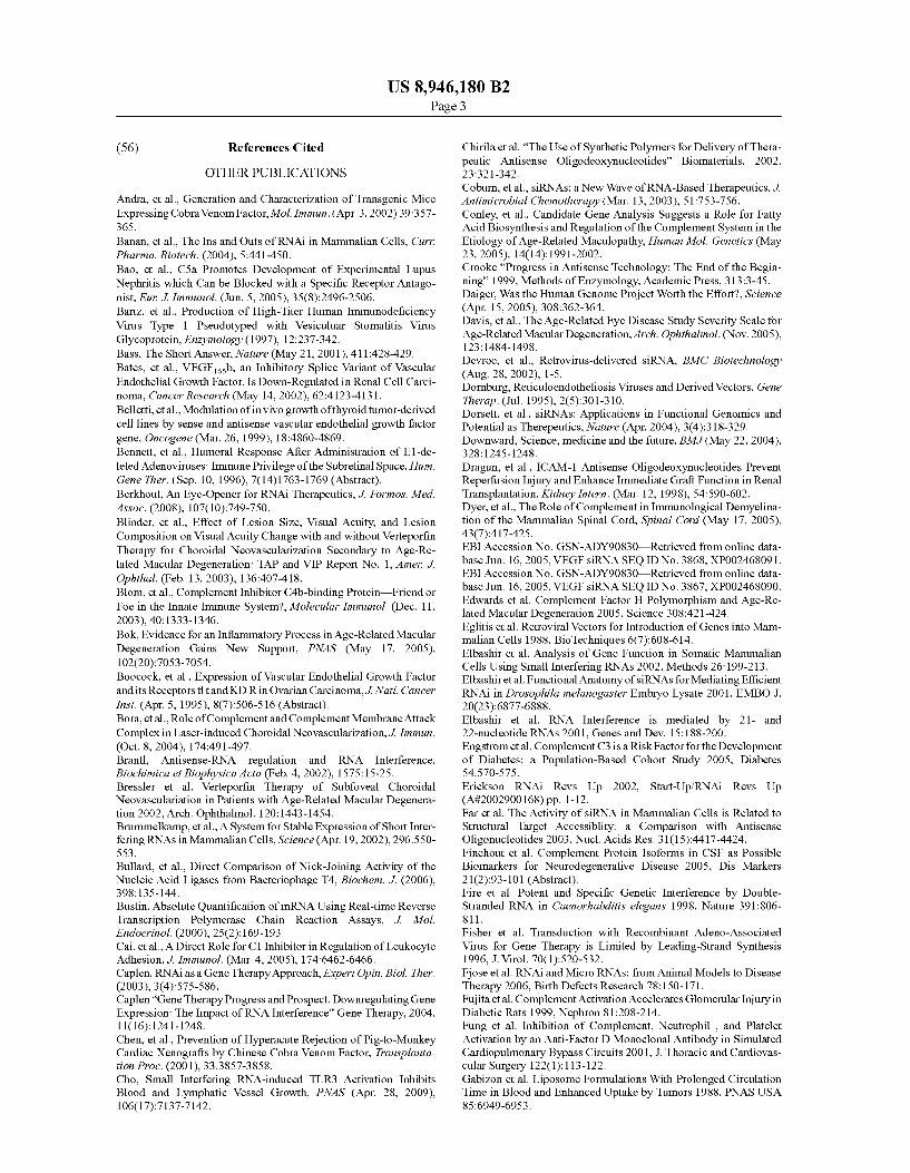
U.S. Patent Feb. 3, 2015 US 8,946,180 B2
fluorescence
buffer control 200 g/kg BW non- 100 pg/kg BW GFP- 200 pg/kg BW GFP silencing dsRNA specific dsRNA specific dsRNA
bright-field

US 8,946, 180 B2 1.
MEANS AND METHODS FOR THE SPECIFIC MODULATION OF TARGET GENES IN THE CNS AND THE EYE AND METHODS FOR
THER DENTIFICATION
CROSS REFERENCE TO RELATED APPLICATIONS
This application is a continuation of and claims priority to U.S. patent application Ser. No. 10/51 1,657, filed on Apr. 18. 2005, which issued as U.S. Pat. No. 8,202,845, which is a national stage application of PCT/EP03/04003, filed Apr. 16, 2003, which claims priority to and benefit of U.S. Provisional Application No. 60/431,173, filed Dec. 5, 2002 and EP02008761.5, filed Apr. 18, 2002, the contents of which are herein incorporated by reference in their entirety.
FIELD OF THE INVENTION
The present invention relates to methods for the treatment of disorders of the central nervous system (CNS) and the eye. In particular, the present invention relates to the use of com positions comprising a compound capable of modulating a target gene or gene product for the preparation of a pharma ceutical composition for the treatment of disorders of the CNS and/or the eye, wherein the composition is designed to be administered outside the blood-CNS and the blood-retina barriers. The instant invention further relates to methods of identifying and isolating nucleic acid molecules encoding polypeptides involved in CNS disorders or of the eye, meth ods for diagnosing said disorders as well as to transgenic animals, wherein the expression of target genes identified in accordance with the method of the invention has been modu lated. In addition, the present invention relates to methods of identifying and isolating drugs that are particularly useful for the treatment of disorders related to the CNS and/or the eye.
Several documents are cited throughout the text of this specification. Each of the documents cited herein (including any manufacturer's specifications, instructions, etc.) are hereby incorporated herein by reference; however, there is no admission that any document cited is indeed prior art as to the present invention.
BACKGROUND ART
A variety of approaches currently exist for delivering bio logically active agents to the CNS and/or the eye. These include, among possible others, oral administration, intrave nous-, intramuscular- and transcutaneous administration as well as intra-bulbous injection or application as eye-drops. If the drug is delivered into the systemic circulation, it is being carried to all internal organs and tissues and it has to pass through the blood-brain and/or blood retina barrier (in order to access the CNS and/or the inner parts of the eye). Obvi ously, all other organs are being exposed to the drug, which may lead to a high incidence of side effects, particularly when the drug exerts its effects on target genes or gene products, which are not specific for the disorder to be treated and/or the target cell or tissue.
Another strategy often employed in brain delivery is the use of invasive methods such as intraventricular infusion systems, intracerebral (polymeric) implants, transplantation of genetically engineered protein-secreting cells and cell implants. These methods are unfortunately only effective for drug delivery to the surface of the brain or to cells immedi ately adjacent to the depot or infusion site and can be used for example in the treatment of carcinomatous infiltration of the
10
15
25
30
35
40
45
50
55
60
65
2 meninges. However, these methods have many limitations because effective drug concentrations in brain parenchyma cannot be achieved.
Like the human central nervous system the human eye is an organ characterized by high complexity and the coordinated functioning of numerous specific structures and tissues. Both are protected by barriers (tear secretion, enzymes, transport mechanisms, blood-retina and blood-CNS barrier) against harmful environmental influences. Like the blood-brain bar rier, the blood-retina barrier also represents a physiological barrier for the uptake of medication by the inner part of the eye, and makes pharmacological therapy of ocular diseases very difficult indeed if at all possible—at the present state of technology.
Medication currently available on the market for the treat ment of disorders of the CNS including ophthalmological diseases is therefore almost exclusively available for treat ment of clinical symptoms often associated with side effects due to the high doses necessary. A causal therapy of the CNS, and particularly of the back sections of the eye, was not possible apart from the injections. Furthermore, the current state of information on the complex molecular metabolic interrelationship underlying the etiology of retinal diseases of multi-factorial origin is only limited. Consequently, medica ments available on the market are suitable to treat the symp toms of Such diseases only.
In view of the need of therapeutic means for the treatment of diseases related to CNS and/or the eye, the technical prob lem of the present invention is to provide means and methods for the identification and modulation of genes involved in disorders of the CNS and/or the eye. More specifically, the technical problem of present inven
tion is to provide non-invasive methods for the controlled modulation of target genes and gene products in the mamma lian CNS and/or eye while overcoming the blood-brain and/or blood retina barrier without injuring it. The solution to said technical problem is achieved by pro
viding the embodiments characterized in the claims, and described further below.
SUMMARY OF THE INVENTION
The present invention is directed to a method for the treat ment of a disorder of the central nervous system (CNS) and/or the eye comprising administering to a subject a composition comprising a compound capable of modulating a target gene or gene product in a therapeutically effective amount, wherein said composition is administered outside the blood brain and/or the blood-retina barriers. In particular, said com position can comprise one or more double-stranded oligori bonucleotides (dsRNA), which mediate an RNA interference of the corresponding mRNA of one or more target genes.
In another aspect, the present invention is directed to a method of identifying and isolating a nucleic acid molecule encoding a polypeptide involved in a disorder of the CNS and/or the eye comprising the steps of (a) culturing a cell, tissue or non-human animal under stress
conditions which lead to simulation of a pathological con dition related to a CNS or eye disorder;
(b) isolating nucleic acids and/or proteins from a sample of said cell, tissue or animal;
(c) comparing the expression or activity profile of at least one of said nucleic acids and/or proteins with that of a corre sponding non-treated cell, tissue or animal, and/or with that of a cell, tissue or animal, which has been treated under different stress conditions;

production of a polypeptide capable of inducing a responsive change in a phenotype comprising culturing said host cell under conditions allowing the expression of the polypeptide and recovering the produced polypeptide from the culture as well as to polypeptides obtainable by said method or encoded by the nucleic acid molecules mentioned above.
treating a disorder of the CNS and/or the eye comprising administering to the Subject said pharmaceutical composi tions in an effective dose.
US 8,946, 180 B2 3
(d) determining at least one nucleic acid and/or protein which is differentially expressed, whereby a change of expression or of the active amount of said at least one nucleic acid or activity of at least one of said proteins or an altered local ization of the protein is indicative for its role in a disorder 5 of the CNS or eye. The present invention also relates to nucleic acid molecules
obtainable by the method described above, particularly if the encoded polypeptide is involved in angiogenesis and/or neovascularization and/or retinal disorder as well as to Vec tors comprising Such nucleic acid molecules and host cells comprising said vector.
10
The present invention is also directed to a method for the 15
2O
Furthermore, the present invention relates to an antibody specifically recognizing Such a polypeptide and pharmaceu tical and/or diagnostic compositions comprising such an anti body or any one of the above described nucleic acid mol ecules, nucleic acid molecules which are complementary to Such a nucleic acid molecules, vectors, host cells, and/or polypeptides, and optionally a pharmaceutically acceptable carrier and Suitable means for detection, respectively.
25
In addition, the present invention is directed to methods for 30
Furthermore, the present invention relates to a method for detecting expression of a gene involved in a disorder of the CNS and/or eye comprising: (a) obtaining mRNA from a cell; (b) incubating the mRNA so obtained with a probe compris
35
ing a nucleic acid molecule described above or a fragment thereof under hybridizing conditions; and
(c) detecting the presence of mRNA hybridized to the probe: 40
(a) obtaining a cell sample from the Subject; (b) contacting the cell sample so obtained with an antibody
described above; and (c) detecting the presence of the antibody bound to the protein
encoded by said gene. The invention furthermore is directed to a method for diag
45
nosing in a Subject said disorder or a predisposition to Such disorder which comprises: (a) isolating DNA from patient suffering from the disorder; (b) digesting the isolated DNA of step (a) with at least one
restriction enzyme; (c) electrophoretically separating the resulting DNA frag
ments on a sizing gel; (d) incubating the resulting gel with a probe comprising a
nucleic acid molecule of the invention or a fragment thereof labelled with a detectable marker;
(e) detecting labelled bands on a gel which have hybridized to the probe as defined to create a band pattern specific to the DNA of patients of the disorder;
(f) preparing subject’s DNA by steps (a) to (e) to produce detectable labeled bands on a gel; and
(g) comparing the band pattern specific to the DNA of patients of the disorder of step (e) and the subject’s DNA of step (f) to determine whether the patterns are the same or different and to diagnose thereby the disorder or a predis position to the disorder, if the patterns are the same;
50
55
60
65
4 O
(a) analyzing a sample of nucleic acids of a Subject by means of a diagnostic chip, primer extension, single nucleotide polymorphisms or sequencing comprising a nucleic acid molecule as described above; and
(b) comparing the result with that of a sample obtained from a patient Suffering from the disorder,
wherein the identity of expression profile and/or nucleotide sequence is indicative for the disorder.
In further embodiment, the present invention relates to a method of determining whether a test substance has an effect on a nucleic acid molecule or polypeptide involved in a CNS or eye disorder comprising the steps: (a) contacting a cell which expresses the target gene or gene
product identified and isolated in accordance with the above described method with a compound to be screened; and
(b) determining if the compound modulates the expression or the activity of said target gene or gene product. In a further aspect, the present invention relates to a drug or
prodrug for the treatment of a disorder as defined above comprising: (a) syntheZising a test Substance or a collection of test Sub
Stances, (b) Subjecting said the test Substance or collection of test
Substances to the screening method of the invention; and (c) producing a compound identified as a modulator of a
target gene or gene product or a derivative thereof. In addition, the present invention is directed to a transgenic
non-human animal which displays an aberrant expression or activity of the target gene or gene product defined above and to its use for a process in drug discovery for the treatment of said disorder.
DETAILED DESCRIPTION OF THE INVENTION
The present invention relates to the use of a compound capable of modulating a target gene or gene product for the preparation of a pharmaceutical composition for the treat ment of a disorder of the central nervous system (CNS) and/or the eye, wherein said composition is designed to be applied outside the blood-CNS and/or blood-retina barriers.
In one aspect, the present invention is based on the Surpris ing finding that the blood-retina barrier could be overcome by the administration of compounds not considered to be capable of doing so in the therapy of ocular diseases by specific modulation of protein function in the tissues of the eye. Due to the functional similarity of the blood-retina bar rier to the blood-brain barrier, providing an improved method to overcome the blood-retina barrier with the aim to treat a given eye disease is expected to be suitable for the treatment of CNS disorders, too.
Hence, in accordance with the present invention the com positions comprising a compound capable of modulating a target gene or gene product in the CNS or the eye are prefer ably designed to be administered without any Substantial, i.e. Substantially effective amount of delivery-enhancing agents facilitating passage of compounds through the blood-brain barrier and/or without the necessity of applying invasive methods and devices; see, e.g., those compounds, methods and devices described in US2002 183683 and WOO3/000018. However, for some embodiments, which represent indepen dent aspects of the invention, such as the use of compounds mediating RNA interference, the use of such methods and compounds may be encompassed for the enhanced and con trolled delivery of a compound capable of modulating a target
US 8,946, 180 B2 5
gene or gene product into the mammalian CNS and/or eye while circumventing the blood-brain and blood-retina barri CS.
Those later embodiments are based, interalia, on the pro vision of novel methods that overcome the difficulty of the application of conventional experimental strategies for the identification of genes, which cause CNS disorders and/or eye diseases, and their validation as targets for diagnosis and for pharmacological intervention strategies. This applies especially for AMD, since the symptoms of this disorder appear only late, generally in the 7" decade of life. The current state of knowledge regarding the pathological meta bolic interrelationships is not sufficient for the medical treat ment of most CNS and eye diseases. Suitable animal or cell culture models are not available for such diseases, due to the complexity of the disease patterns and the lack of appropriate strategies for simple intervention and manipulation in the CNS and at the eye.
Hence, in one important aspect, the present invention relates to a cell, tissue and animal model based assay for the identification and isolation of target genes and gene products involved in disorders of the CNS and/or the eye and their use as targets for therapeutic intervention and/or diagnosis of Such disorders.
Examples for CNS disorders are, for example, Alzheimer's disease, Parkinson disease, depression, bipolar disorder, Schizophrenia, amnesia, migraine-headache, stroke, insom nia, alcohol abuse, anxiety, obsessive compulsive disorder, cerebral acquired human immunodeficiency syndrome, chronic pain and many others. The compositions of the invention may be administered
locally or systemically e.g., intravenously. Preparations for parenteral administration include sterile aqueous or non aqueous Solutions, Suspensions, and emulsions. Examples of non-aqueous solvents are propylene glycol, polyethylene gly col, vegetable oils such as olive oil, and injectable organic esters such as ethyl oleate. Aqueous carriers include water, alcoholic/aqueous solutions, emulsions or Suspensions, including saline and buffered media. Parenteral vehicles include Sodium chloride solution, Ringer's dextrose, dextrose and sodium chloride, lactated Ringers, or fixed oils. Intrave nous vehicles include fluid and nutrient replenishers, electro lyte replenishers (such as those based on Ringer's dextrose), and the like. Preservatives and other additives may also be present such as, for example, antimicrobials, anti-oxidants, chelating agents, and inert gases and the like. Furthermore, the pharmaceutical composition of the invention may com prise further agents such as interleukins or interferons depending on the intended use of the pharmaceutical compo sition.
In accordance with the present invention the pharmaceuti cal compositions are administered to a subject in an effective dose of between about 0.1 ug to about 10 mg units/day and/or units/kg body weight; see also infra. Furthermore, the appro priate dosage regimen can be determined according to Example 21.
In a preferred embodiment, the disorder to be treated is related to eye. Such disorders include chorioretinitis and her pes retinitis, which may be considered as acquired forms of retinal disease, the majority of retinal disease disorders are reduced to a genetic predisposition. These include for example primary retinal detachment (ablatio retinae), retinal blastoma, retinal astrocytoma (Bourneville-Pringle), angi omatosis retinae (Hippel-Lindau), Coat's disease (exudative retinitis), Eale's disease, central serous retinopathy, ocular albinism, retinitis pigmentosa, retinitis punctata albescens, Usher syndrome, Leber's congenital amaurosis, cone dystro
10
15
25
30
35
40
45
50
55
60
65
6 phy, Vitelliform macular degeneration (Best's disease), juve nile retinoschisis, North Carolina macular dystrophy, Sors by's fundus dystrophy, Doyne's honey comb retinal dystrophy (Malattia Leventinese), Stargardt’s disease, Wag ner vitreoretinal degeneration or Age-related macular degen eration (AMD) as well as single-gene retinopathies like Mor bus Best or Morbus Stargardt. Various genetic defects are known which lead or predispose to this wide range of eye disease phenotypes. Some of these clinical phenotypes are characterized by a
pathological de novo generation of blood vessels, which is called neoangiogenesis or neovascularization. Starting from the choriokapillaris, the growth of new blood vessels into the inner eye then leads to an increasing degeneration of photo receptor cells in the affected areas of the human retina. In the field of opthalmology, one can distinguish between two forms of neovascularization: subretinal (choroidal-CNV) neovas cularization and retinal neovascularization. Subretinal neovascularization, which is also called Subfoveal neovascu larization, is associated with degenerative disorders like Makular degeneration and characterized by loss of visual acuity and metamorphopsy. On the other hand, retinal neovascularization, vitreous body or Iris neovascularization is associated with ischemic processes (e.g. retinal vasculitis and diabetic retinopathy). Furthermore, neoangiogenesis is an important pathomechanism in different, non ophthalmo logical disease patterns such as tumor growth, arthritis and diabetic nephropathy. Therefore, in a preferred embodiment of the methods and uses of the present invention said disorder to be treated is related to angiogenesis and/or neovasculariza tion and particularly preferred to the retinal pigment epithe lium (RPE), neurosensory retina and/or choriodea. Most pre ferred, the disorder is wet age-related macular degeneration (AMD) or diabetic retinopathy. The following description deals with AMD as example for
a complex eye disease with a genetic component. Consider ing the wet form of AMD, it also serves as an example for a disease pattern, which is characterized by a distinct neovas cularization. The example shall illustrate the associated tech nical problems with reference to the study of molecular causes and the development of diagnostic and pharmacologi cal intervention strategies. AMD, which can be thought as a sub-type of retinal degen
eration, is the most common cause of visual morbidity in the developed world with a prevalence increasing from 9% in persons over 52 years to more than 25% in persons over the age of 75 (Paetkau et al. 1978, Leibowitz et al. 1980, Banks and Hutton 1981, Ghafour et al. 1983, Hyman 1987, Hyman et al. 1983, Grey et al. 1989, Yap and Weatherill 1989, Heiba et al. 1994). An early stage in the evolution of AMD pathology is
accompagnied by an increasing accumulation of yellowish lipofuscin-like particles within the retinal pigment epithelium (RPE: Feeney 1978). It is thought that these particles repre sent remnants of undigested phagocytosed photoreceptor outer segment membranes which, in the normal process, are excreted basally through Bruch's membrane into the chorio capillaris. Over time, accumulation of lipofuscin-like par ticles affect Bruch's membrane and lead to its progressive destruction (Hogan and Alvarado 1967, Sarks 1976, Feeney Burns and Ellersieck 1985, Pauleikhoff et al. 1990). The deposits in the RPE and Bruch's membrane consists largely of lipids although their exact composition may vary between individuals with Some deposits revealing more polar phos pholipids while others contain predominantly apolar neutral lipids.

US 8,946, 180 B2 7
These individual differences in drusen composition are thought to be the basis for the clinical heterogeneity in AMD (Green et al. 1985). While some patients present with an ingrowth of vessels from the choriocapillaris through Bruch's membrane (neovascularization) (Bressler et al. 1982), others show pigment epithelial detachment due to excudation under neath the RPE (Gass 1967, Green et al. 1985), and a third group of patients experiences a slow decrease of visual loss due to atrophic changes in the RPE and the overlying sensory neuroretina (Maguire and Vine 1986).
Although much less common the excudative/neovascular form of AMD accounts for more than 80% of blindness with a visual acuity of s20/200 (Bressler et al. 2002). In contrast to the above described “dry” form of AMD, the exudative “wet” AMD is associated with a choroidal neovascularization (CNV), leading to blindness and, thus, to a loss of life quality (followed by psychic disorders, increased risk of injury etc; Bressler et al. 2002). There is a high risk of developing (>40%) CNV in the second eye within 5 years of the devel opment of CNV-AMD in the first eye (Bressler et al. 2002). Neovascular AMD is characterized by choroidal neovascular lesions. These lesions develop when abnormal blood vessels from the choroid grow and proliferate through breaks in the Bruch membrane to beneath the retinal pigment epithelium (Bressleretal. 2002, Campochiaro et al. 1999). The abnormal leakage from these vessels can result in hemorrhage or detachment of the retinal pigment epithelium or the neuro sensory retina (which overlies the retinal pigment epithe lium). Accompanying scar formation can replace retinal tis Sue and result in permanent vision loss. AMD is a complex disease caused by exogenous as well as
endogenous factors (Meyers and Zachary 1988; Seddon et al. 1997). In addition to environmental factors, several personal risk factors such as hypermetropia, light skin and iris colour, elevated serum cholesterol levels, hypertension or cigarette Smoking have been suggested (Hyman et al. 1983, Klein et al. 1993, Sperduto and Hiller 1986, The Eye Disease Case-Con trol Study Group 1992, Bressler and Bressler 1995). A genetic component for AMD has been documented by several groups (Gass 1973, Piguet et al. 1993, Silvestri et al. 1994) and has lead to thehypothesis that the disease may be trig gered by environmental/individual factors in those persons who are genetically predisposed. The number of genes which, when mutated, can confer susceptibility to AMD is not known but may be numerous.
The late onset of symptoms generally in the 7th decade of life as well as the clinical and likely genetic heterogeneity make it difficult to apply conventional approaches for the identification of genes predisposing to AMD. Due to the complexity of the clinical phenotype, it may be assumed that the number of genes is large, which, when mutated contribute to AMD susceptibility.
With recent physical approaches for the treatment of AMD Such as laser photocoagulation, photo dynamic therapy (us ing verteprofin, trade name Visudyne R, Novartis), irradiation or Surgical therapies, success was only achieved with a mod erate percentage of the patients (Bressler et al. 2002, Yuzawa et al. 2001).
Hence, the methods, uses and compositions of the present invention described herein represent an important improve ment and alternative therapeutic intervention for the treat ment of this particular disease as well as of others. For those embodiments the pharmaceutical compositions are prefer ably designed to be effective in (and applied to) the posterior segment of the eye, preferably in a form designed to be applied outside the retinal region of the blood-retina barrier.
10
15
25
30
35
40
45
50
55
60
65
8 In one embodiment of the invention said compound is an
inhibitor/antagonist of said target gene or gene product and preferably inhibits the expression of a gene or the activity of a gene product involved in angiogenesis and/or neovascular ization; see Supra. The term “antagonist/inhibitor” in accordance with the
present invention includes chemical agents that modulate the action of a gene or the activity of a gene product either through altering its enzymatic activity or through modulation of expression, e.g., by affecting transcription or translation. In Some cases the antagonist/inhibitor may also be a substrate of a a gene product involved in the disorder or a ligand binding molecule. The term “inhibitor” includes both substances which
reduce the activity of the polypeptide and those which nullify it altogether. An “antagonist' that modulates the activity of the gene
product and causes for example a response in a cell based assay described below, refers to a compound that alters directly or indirectly the activity the gene product or the amount of active product. The effect of an antagonist may be observed as a blocking of agonist-induced activation of a target gene. Antagonists include competitive as well as non competitive antagonists. A competitive antagonist (or com petitive blocker) interacts with or near the site specific for agonist binding. A non-competitive antagonist or blocker inactivates the function of the gene product by interacting with a site other than the agonist interaction site. Preferably, the antagonist/inhibitor is Small chemical agent which directly interacts with the target gene product involved in the disorder, preferably with a gene product involved in angio genesis and/or neovascularization. Therefore, there will pref erably be a direct relationship between the molar amount of compound required to inhibit or stimulate the target gene activity and the molar amount of gene product present or lacking in the cell. The compounds can be derived from a polypeptide, an anti-polypeptide antibody, an RNA molecule encoding (part of) a polypeptide or its antisense sequence, a transcription regulator, a ligand binding molecule, a polypep tide Substrate or a known agonist/activator or antagonist/ inhibitor.
In a preferred embodiment of the present invention said antagonist is based on nucleic acids, for example a ribozyme, antisense or sense nucleic acid molecules to said gene or gene or dsRNA molecules which are capable of mediating RNA interference. Methods and computer programs for the prepa ration rational selection of for example antisense oligonucle otide sequences are described in the prior art; see for example Smith, Eur. J. Pharm. Sci. 11 (2000), 191-198; Toschi, Meth ods 22 (2000), 261-269; Sohail, Adv. Drug Deliv. Rev. 44 (2000), 23-34; Moulton, J. Comput. Biol. 7 (2000), 277-292. These procedures comprise how to find optimal hybridization sites, and secondly on how to select sequences that bind to for example mRNAs overexpressed in a CNS or eye disorder. These methods can include the more empirical testing of large numbers of mRNA complementary sequences to the more systematic techniques, i.e. RNase H mapping, use of combi natorial arrays and prediction of secondary structure of mRNA by computational methods. Structures that bind to structured RNA, i.e. aptastructures and tethered oligonucle otide probes, and foldback triplex-forming oligonucleotides can also be employed for the purpose of the present invention. Relating to selection of antisense sequences by aid of com putational analysis, valuable www addresses are given below.
In a particularly preferred embodiment of the present invention said antagonist/inhibitor Substantially consists of ribonucleotides which preferably contain a portion of double

US 8,946, 180 B2 9
stranded oligoribonucleotides (dsRNA). Secondary structure prediction and in vitro accessibility of mRNA as tools in the selection of target sites is described for example in Amarzgui oui, Nucleic Acids Res. 28 (2000), 4113-4124. Minimising the secondary structure of DNA targets by incorporation of a modified deoxynucleoside: implications for nucleic acid analysis by hybridisation is described in Nguyen, Nucleic Acids Res. 28 (2000), 3904-3909. dsRNA between 21 and 23 nucleotides in length is pre
ferred. The dsRNA molecule can also contain a terminal 3'-hydroxyl group and may representananalogue of naturally occurring RNA, differing from the nucleotide sequence of said gene or gene product by addition, deletion, Substitution or modification of one or more nucleotides. General pro cesses of introducing an RNA into a living cell to inhibit gene expression of a target gene in that cell comprising RNA with double-stranded structure, i.e. dsRNA or RNAi are known to the person skilled in the art and are described, for in WO99/ 32619, WO01/68836, WO01/77350, WO00/44895, WO02/ 055692 and WO02/055693, the disclosure content of which is hereby incorporated by reference.
The target mRNA of said dsRNA is preferably encoded by gene or a cDNA obtained in accordance with the method of the present invention described below. In one embodiment the target nucleotide sequence encodes an amino acid sequence of SEQID NO: 2 or 4 and/or comprises a nucleotide sequence of SEQID NO: 1 or 3.
In one embodiment of the invention the compound to be used in the compositions is a nucleic acid molecule or encoded by a nucleic acid molecule and is designed to be expressed in cells of the CNS and/or eye. For those embodi ments gene therapy intervention is envisaged; see also infra.
In a preferred embodiment of the methods and uses of the present invention the composition is in a form designed to be introduced into the cells or tissue of the CNS or eye by a Suitable carrier, characterized by the application occurring outside the blood-CNS and/or blood-retina barriers, for instance as eye drops. It can also be administered systemi cally, iontophoretically or by retrobulbar injection.
Iontophoresis has been defined as the active introduction of ionised molecules into tissues by means of an electric current. The technique has been used to enhance drug delivery into tissues underlying the donor electrode (e.g. skin) as well as to the general blood circulation, thus providing systemic deliv ery of a drug to the entire body. Iontophoresis devices require at least two electrodes, both being in electrical contact with Some portion of a biological membrane Surface of the body. One electrode commonly referred to as the “donor” or “active’ electrode, is the electrode from which the biologi cally active Substance, such as a drug or prodrug, is delivered into the body. Another electrode having an opposite polarity functions to complete the electric circuit between the body and the electrical power source. This electrode is commonly referred to as the “receptor' or “passive’ electrode. During iontophoresis, an electrical potential is applied over the elec trodes, in order to create an electrical current to pass through the drug solution and the adjacent tissue. Iontophoresis has been described for the treatment of blood-vessel related dis orders (e.g. restenosis), bladder, uterus, urethra and prostate disorders. U.S. Pat. Nos. 6,219,557: 5,588,961; 5,843.016: 5,486,160; 5,222,936; 5,232,441; 5,401.239 and 5,728,068 disclose different types of iontophoresis catheters for inser tion into hollow, tubular organs (bladder, urethra and pros tate) or into blood vessels. US 2002 183683 suggests the method for delivery of active substances into the CNS. Numerous active, often specifically expressed genes are
required to perform and control the processes in the cells of
5
10
15
25
30
35
40
45
50
55
60
65
10 the CNS and the retina and the metabolic exchanges across the blood-CNS and blood-retina barrier. Specific genetic activity is also necessary for maintaining the structure and functional integrity of numerous components of these com plex tissues. As a consequence, this unique and highly evolved system is especially Susceptible to various genetic defects, thus leading to a wide range of disease phenotypes. While studying monogenetic disorders is relatively easy, pro vided the patients are members of a family sufficiently large enough to allow positional cloning, the identification of genes that contribute to multigeneic disorders or confer or Suscep tibility to a disease is far more difficult.
Hence, in another aspect the present invention relates to a method of identifying and isolating a nucleic acid molecule encoding a polypeptide involved in a disorder of the CNS and/or the eye comprising: (a) culturing a cell, tissue or non-human animal under stress
conditions which lead to simulation of a pathological con dition related to a CNS or eye disorder;
(b) isolating nucleic acids and/or proteins from a sample of said cell, tissue or animal;
(c) comparing the expression or activity profile of at least one of said nucleic acids and/or proteins with that of a corre sponding non-treated cell, tissue or animal, and/or with that of a cell, tissue or animal, which has been treated under different stress conditions;
(d) determining at least one nucleic acid and/or protein which is differentially expressed, whereby a change of expression or of the active amount of said at least one nucleic acid or activity of at least one of said proteins or an altered local ization of the protein is indicative for its role in a disorder of the CNS or eye. First, a cell, tissue or non-human animal is cultured under
stress conditions which lead to simulation of a pathological condition related to a CNS or eye disorder. Preferably, said method is a cell culture based method. Preferred cells and tissue investigated (either in culture or comprised in a test animal) are those which belong to the CNS and/or eye, for example neuronal cells, glial cells, retinal cells, etc.
In a particular preferred embodiment of the method of the present invention said cell is an RPE cellor an established cell line derived from an RPE cell such as the cell line ARPE-19; see also infra. The isolation of RPE cells is described, e.g., in Example 1 below. ARPE-19 cell line is described in Dunn et al., Exp. Eye Res.62 (1996), 155-169. For example, ARPE 19 cell line is particularly suitable for mimicking the repair response observed in vivo during proliferative vitreoretinopa thy by vitreous treatment. The mentioned stress condition can be generated by an
aberrant Supply of the cell, tissue or animal culture conditions and comprise, for example, oxidative stress, hypoxic culture conditions, insufficient nutrition and/or supply with growth factors, change of pH-value and/or pathophysiological con centration of rod outer segments (ROS) and/or A2-E; see also Examples 3 to 10. Preferred stress conditions are those con ferred by pathophysiological concentration of rod outer seg ments (ROS) and/or A2-E. As an example the tissue of the interior segment of the eye can be supplied in an aberrant manner, which is a preferred embodiment of the method of the invention. Table 1 shows commonly marker genes, whose altered expression indicates apoptosis, hypoxic culture con ditions or oxidative stress and which can thereby be used to verify and/or quantify the applied stress and the cellular response, respectively. A further step of the method of the present invention
involves isolating the nucleic acids and/or proteins from a sample of said cell, tissue or animal and in a further step

US 8,946, 180 B2 11
comparing the expression or activity profile of at least one of said nucleic acids and/or proteins with that of a corresponding non-treated cell, tissue or animal, and/or with that of a cell, tissue or animal, which has been treated under different stress conditions. The isolation of nucleic acids and) proteins can be done by methods known to the person skilled in the art and described in the cited literature; see also Examples 11 and 12.
In the last step at least one nucleic acid and/or protein which is differentially expressed is determined, whereby a change of expression or of the active amount of said at least one nucleic acid or activity of at least one of said proteins or an altered localization of the protein is indicative for its role in a disorder of the CNS or eye.
In one embodiment of the screening method of the inven tion the expression of nucleic acids is analyzed with an expression array and/or realtime PCR. Chip and array tech nology are well known to the person skilled in the art; see also Examples 14 to 20. Advances in approaches to DNA-based diagnostics are reviewed, for example, by Whitcombe etal. in Curr. Opin. Biotechnol. 9 (1998), 602-608. Furthermore, DNA chips and microarray technology devices, systems, and applications are described by, e.g. Cuzin, Transfus. Clin. Biol. 8 (2001), 291-296 and Heller, Annu. Rev. Biomed. Eng. (2002), 129-153. Likewise, biomedical applications of pro tein chips is known and described in, e.g., Ng, J. Cell. Mol. Med. 6 (2002), 329-340.
In another embodiment the protein expression is analyzed with immunoblot or ELISA assay, or 2D gel electrophoresis or MALDI-TOF and particularly preferred antibodies are used which are specific for proteins involved in angiogenesis and/or neovascularization.
Detailed descriptions of conventional methods, such as those employed in the construction of vectors and plasmids, the insertion of genes encoding polypeptides into Such vec tors and plasmids, the introduction of plasmids into host cells, and the expression and determination thereof of genes and gene products can be obtained from numerous publication, including Sambrook et al., (1989) Molecular Cloning: A Laboratory Manual, 2nd ed., Cold Spring Harbor Laboratory Press. Candidate nucleic acids or encoded polypeptides iden tified in Such a manner can be validated by expressing them and observing the phenotype. A further embodiment of the screening method therefore comprises the overexpression or inhibition of expression of the identified candidate nucleic acid or encoded polypeptide in said cell, tissue or animal for their capability of inducing a responsive change in the phe notype of said cell, tissue or animal, wherein said phenotype is related to a disorder of the CNS or eye. The responsive change in the phenotype of said cells can be
observed by subjecting the cells, secreted factors thereof, or cell lysates thereof, to endothelial cell cultures; and/or ana lyzing different parameters like cell proliferation, electro physiological activity, DNA synthesis, out-growth of cells, cell migration, chemokinesis, chemotaxis, development of vessels, marker gene expression or activity, apoptosis and/or Vitality. Examples for Such assays are:
Proliferating cell nuclear antigen assay (PCNA) or TUNEL-assay are described in Montesano, R.: Regulation of angiogenesis in vitro. Eur J. Clin Invest, 22: 504-515, 1992. Montesano, R. et al.: Basic fibroblast growth factor induces angiogenesis in vitro. Proc Natl Acad Sci USA, 83: 7297 7301, 1986. Holmgren, L. et al.: Dormancy of mictrome tastases: Balanced proliferation and apoptosis in the presence of angiogenesis suppression. Nature Med, 1: 149-153, 1995.
Boyden chamber assay is described in Holmgren, L. et al.: Dormancy of mictrometastases: Balanced proliferation and apoptosis in the presence of angiogenesis Suppression.
10
15
25
30
35
40
45
50
55
60
65
12 Nature Med, 1: 149-153, 1995. Albini, A. et al.: A rapid in vitro assay for quantitating the invassive potential of tumor cells. Cancer Research, 47: 3239-3245, 1987. Hu, G. et al.: Angiogenen promotes invasiveness of cultured endothelial cells by stimulation of cell-associated proteolytic activities. Proc Natl Acad Sci USA, 6: 12096-12100, 1994. Alessandri, G. et al.: Mobilization of capillary endothelium in vitro induced by effectors of angiogenesisi in vivo. Cancer res, 43: 1790-1797, 1983.
Aortic ring angiogenesis assay is described in Zuh, W. H., et al.: Regulation of vascular growth and regression by matrix metalloproteinases in the rat aorta model of angiogenesis. Lab Invest, 80:545-555, 2000. Kruger, E. A. et al.: UCN01, a protein kinase C inhibitor, inhibits endothelial cell prolifera tion and angiogenic hypoxic response. Invasion Metastas, 18: 209-218, 2000. Kruger, E. A. et al.: Endostatin inhibits microVessel formation in the rat aortic ring angiogenesis assay. Biochem Biophys Res Commun, 268: 183-191, 2000. Bauer, K. S. et al.: Inhibition of angiogenesis by thalidomide requires metabolic activation, which is species dependent. Biochem Pharmacol, 55:1827-1834, 1998. Bauer, K.S. et al.: Carboxyamidotriazole inhibits angiogenesis by blocking the calcium-mediated nitric-oxide synthase-vascular endothelial growth factor pathway. J Pharmacol Exp Ther. 292: 31-37, 2000. Berger, A. C. et al.: Endothelial monocyte activating polypeptide III induces endothelial cell apoptosis and may inhibit tumor angiogenesis. Microvasc Res, 60: 70-80, 2000.
Saphenous vein angiogenesis assay is described in Kruger, E. A. et al.: Endostatin inhibits microVessel formation in the rat aortic ring angiogenesis assay. Biochem BiophyS Res Commun, 268: 183-191, 2000.
Cornes mircropocket assay is described in Gimbrone, E. A. et al: Tumor growth and neovascularization: an experimental model using the rabbit cornea. J Natl Cancer Inst, 52: 413 427, 1974. Kenyon, B. M. et al.: A model of angiogenesis in the mouse cornea. Invest OphthalmolV is Sci, 37: 1625-1632, 1996. Kenyon, B. M. et al.: Effects of thalidomide and related metabolites in a mouse corneal model of neovascularization. Exp Eye Res, 64:971-978, 1997. Proia, A. D. et al.: The effect of angiostatic steroids and beta-cyclodextrin tetradecasulfate on corneal neovascularization in the rat. Exp Eye Res, 57: 693-698, 1993. Chick embryo chorioallantoic membrane assay is
described in Knighton, D. et al.: Avascular and vascular phases of tumor growth factor in the chicken embryo. Br J Cancer, 35: 347-356, 1977. Auerbach, R. et al.: A simple procedure for the long-term cultivation of chicken embryos. Dev Biol, 41: 391-394, 1974. Ausprunk, D. H. et al.: Differ entiation of vascular endothelium in the chick chorioallan tois: A structural and autoradiographic study. Dev Biol, 38: 237-248, 1974. Nguyen, M. et al.: Quantitation of angiogen esis and antiangiogenesis in the chick embryo chorioallantoic membrane. Microvasc Res, 47: 31-40, 1994.
Furthermore a sample of said cells can be treated with an inhibitor specific for the candidate nucleic acid or encoded polypeptide to be validated and in a second step it is deter mined whether said cells, secreted factors thereof or cell lysates thereof have lost their capability of inducing the responsive change in the phenotype observed when no inhibi toris used. In a preferred embodiment the phenotype is angio genesis and/or neovascularization. As an inhibitor the mol ecules described above can be used. Preferably, siRNA technique is used for inhibiting the expression of the target gene. A collection of protocols for siRNA-mediated knock down of mammalian gene expression, which can be adapted to a method of the invention as mentioned herein is described

US 8,946, 180 B2 13
for example in Elbashir et al., Methods 26 (2002), 199-213 and Martinez et al., Cell 110 (2002), 563-574.
For the development of assays and drugs for treatment of disorders caused by the genes it is often necessary to identify the sequence of those nucleic acids and/or proteins, and optionally identifying the corresponding encoding gene or cDNA as well. Based on the specific functions of the cells of the CNS and/or eye specific, it is presumed that genes, the aberrant function of which cause a CNS or eye disease, are specifically expressed in the respective tissues and cells, thus representing preferred targets for drug interventions. There fore, the identified gene, cDNA or a fragment thereof is usually also cloned and nucleic acid molecules obtainable by the methods described hereinform also part of the invention, particularly if they encode polypeptides involved in angio genesis and/or neovascularization. Such a nucleic acid mol ecule can be DNA or cDNA and be derived from a mammal and in a preferred embodiment is from a mouse or a human.
Hence, in a first set of experiments several nucleic acid molecules could be identified which indeed were known to be involved in autosomal recessive retinitis pigmentosa (ARRP), which interalia is characterized by the degeneration of retinal photoreceptor cells. For example, nucleic acid mol ecules could be indentified corresponding to the gene encod ing the human cyclic nucleotide gated channel alpha 1 (CNGA1, accession No. NM000087; SEQID NO: 1 and 2). Mutations in this gene have been described to be involved in autosomal recessive retinitis pigmentosa; see Drya et al., Proc. Nat. Acad. Sci. USA 92 (1995), 10177-10181. In another experiment, nucleic acid molecules corresponding to the human gene encoding the beta-subunit of rod coiMP phosphodiesterase (accession No. NM 000283: SEQ ID NO:3 and 4) have been identified. Malfunction of this gene has also been associated with autosomal recessive retinitis pigmentosa, in particular with congenital stationary night blindness 3, CSNB3. These results confirm that the method of the present invention works.
In another embodiment the nucleic acid molecule specifi cally hybridizes to one of the nucleic acid molecules described above wherein the latter encodes a mutated version of the protein which has lost its capability of inducing a responsive change in a phenotype. In addition or alternatively, nucleic acid molecules are encompassed of at least 15 nucle otides in length and able to hybridize specifically to a nucleic acid molecule described above or with a complementary strand thereof. These nucleic acid molecules are particularly useful as probes; see infra.
The nucleic acid molecules described above can be con tained in a vector and preferably be operatively linked to regulatory elements permitting expression in prokaryotic or eukaryotic host cells. Expression of said nucleic acid mol ecule comprises transcription into a translatable mRNA. Regulatory elements ensuring expression in eukaryotic cells, preferably mammalian cells, are well known to those skilled in the art. They usually comprise regulatory sequences ensur ing initiation of transcription and optionally poly-A signals ensuring termination of transcription and stabilization of the transcript. Additional regulatory elements may include tran Scriptional as well as translational enhancers, and/or naturally associated or heterologous promoter regions.
Possible regulatory elements permitting expression in prokaryotic host cells comprise, e.g., the P. lac, trp or tac promoter in E. coli, and examples for regulatory elements permitting expression in eukaryotic host cells are the AOX1 or GAL1 promoter in yeast or the CMV-, SV40-, RSV-pro moter, CMV-enhancer, SV40-enhancer or a globin intron in mammalian and other animal cells.
5
10
15
25
30
35
40
45
50
55
60
65
14 Beside elements which are responsible for the initiation of
transcription Such regulatory elements may also comprise transcription termination signals, such as the SV40-poly-A site or the tk-poly-A site, downstream of the nucleic acid molecule. Furthermore, depending on the expression system used leader sequences capable of directing the polypeptide to a cellular compartment or secreting it into the medium may be added to the coding sequence of the polynucleotide of the invention and are well known in the art. The leader sequence(s) is (are) assembled in appropriate phase with translation, initiation and termination sequences, and prefer ably, a leader sequence capable of directing secretion of trans lated protein, or a portion thereof, into the periplasmic space or extracellular medium. Optionally, the heterologous sequence can encode a fusion protein including a C- or N-ter minal identification peptide imparting desired characteristics, e.g., stabilization or simplified purification of expressed recombinant product. In this context, Suitable expression vec tors are known in the art such as Okayama-Berg cDNA expression vector pcDV1 (Pharmacia), pCDM8, pRc/CMV, pcDNA1, pcDNA3 (Invitrogen), or pSPORT1 (GIBCO BRL).
Preferably, the expression control sequences will be eukaryotic promoter systems in vectors capable of transform ing or transfecting eukaryotic host cells, but control sequences for prokaryotic hosts may also be used. Once the vector has been incorporated into the appropriate host, the host is maintained under conditions suitable for high level expression of the nucleotide sequences, and, as desired, the collection and purification of the protein so produced.
Furthermore, the present invention relates to vectors, par ticularly plasmids, cosmids, viruses and bacteriophages used conventionally in genetic engineering that comprise a nucleic acid molecule of the invention. Preferably, said vector is an expression vector and/or a gene transfer or targeting vector. Expression vectors derived from viruses such as retroviruses, vaccinia adeno-associated virus, herpes viruses, or bovine papilloma virus, may be used for delivery of the polynucle otides or vector of the invention into targeted cell population. Methods which are well known to those skilled in the art can be used to construct recombinant viral vectors; see, for example, the techniques described in Sambrook, Molecular Cloning A Laboratory Manual, Cold Spring Harbor Labora tory (1989) N.Y.and Ausubel, Current Protocols in Molecular Biology, Green Publishing Associates and Wiley Inter science, N.Y. (1994). Alternatively, the polynucleotides and vectors of the invention can be reconstituted into liposomes for delivery to target cells. The vectors containing the nucleic acid molecules of the invention can be transferred into the host cell by well known methods, which vary depending on the type of cellular host. For example, calcium chloride trans fection is commonly utilized for prokaryotic cells, whereas calcium phosphate treatment or electroporation may be used for other cellular hosts; see Sambrook, supra.
Vectors that can be used for therapeutic and/or diagnostic purposes in accordance with the teaching of the present invention are known to the person skilled in the art; see, e.g., heritable and inducible genetic interference by double stranded RNA encoded by transgenes described in Tavemar akis et al., Nat. Genet. 24 (2000), 180-183. Further vectors and methods for gene transfer and generation of transgenic animals are described in the prior art; see, e.g., adeno-asso ciated virus related vectors described in Qing et al., Virol. 77 (2003), 2741-2746; human immunodeficiency virus type 2 (HIV-2) vector-mediated in vivo gene transfer into adult rab bit retina described in Cheng et al. Curr. Eye Res. 24 (2002), 196-201, long-term transgene expression in the RPE after

US 8,946, 180 B2 15
gene transfer with a high-capacity adenoviral vector described in Kreppel et al., Invest. Ophthalmol. Vis. Sci. 43 (2002), 1965-1970 and non-invasive observation of repeated adenoviral GFP gene delivery to the anterior segment of the monkey eye in vivo described in Borras et al., J. Gene Med. 3 (2001), 437-449. CNS gene transfer has also been described in Leone et al. Curr. Opin. Mol. Ther. 1 (1999), 487-492
Said vector in turn can be contained in a host cell. A bacterial, fungal, plant or animal cell can be used as a host but mammalian cells are preferred, especially RPE or neurosen sory retina cells.
If these host cells are cultured under conditions allowing the expression of the polypeptide and recovering the pro duced polypeptide from the culture this constitutes a method for the production of a polypeptide capable of inducing a responsive change in a phenotype. Polypeptides encoded by a nucleic acid molecule as defined above or obtainable by this method are therefore preferred embodiments of this invention as well as antibodies specifically recognizing Such a polypep tide. Antibodies or fragments thereof to the aforementioned polypeptides can be obtained by using methods which are described, e.g., in Harlow and Lane Antibodies, A Labora tory Manual, CSH Press, Cold Spring Harbor, 1988.
Furthermore, the polypeptides encoded by the identified and isolated nucleic acid molecules can be used to identify synthetic chemical peptide mimetics that bind to or can func tion as a ligand, Substrate, binding partner or the receptor of the polypeptide as effectively as does (e.g.) the natural ligand; see, e.g., Engleman, J. Clin. Invest 99 (1997), 2284-2292. For example, folding simulations and computer redesign of struc tural motifs of the polypeptide can be performed using appro priate computer programs (Olszewski, Proteins 25 (1996), 286-299; Hoffman, Comput. Appl. Biosci. 11 (1995), 675 679). Computer modeling of protein folding can be used for the conformational and energetic analysis of detailed peptide and protein models (Monge, J. Mol. Biol. 247 (1995), 995 1012: Renouf, Adv. Exp. Med. Biol. 376 (1995), 37–45). In particular, the appropriate programs can be used for the iden tification of interactive sites of the polypeptide and its ligand or other interacting proteins by computer assistant searches for complementary peptide sequences (Fassina, Immu nomethods 5 (1994), 114-120. Further appropriate computer systems for the design of protein and peptides are described in the prior art, for example in Berry, Biochem. Soc. Trans. 22 (1994), 1033-1036; Wodak, Ann. N.Y. Acad. Sci. 501 (1987), 1-13: Pabo, Biochemistry 25 (1986),5987-5991. Methods for the generation and use of peptidomimetic combinatorial libraries are described in the prior art, for example in Ostresh, Methods in Enzymology 267 (1996), 220-234 and Domer, Bioorg. Med. Chem. 4 (1996), 709-715. Furthermore, a three dimensional and/or crystallographic structure of the polypep tide can be used for the design of mimetic inhibitors of the biological activity of the protein of the invention (Rose, Bio chemistry 35 (1996), 12933-12944; Rutenber, Bioorg. Med. Chem. 4 (1996), 1545-1558). The structure-based design and synthesis of low-molecular-weight synthetic molecules that mimic the activity of a native biological polypeptide is further described in, e.g., Dowd, Nature Biotechnol. 16 (1998), 190 195; Kieber-Emmons, Current Opinion Biotechnol.8 (1997), 435-441; Moore, Proc. West Pharmacol. Soc. 40 (1997), 115 119; Mathews, Proc. West Pharmacol. Soc. 40 (1997), 121 125; Mukhija, European J. Biochem. 254 (1998), 433-438. The nucleic acid molecules identified and isolated by the
method of the present invention can also serve as a target for activators and inhibitors. Activators may comprise, for example, proteins that bind to the mRNA of the correspond ing gene, thereby stabilizing the native conformation of the
10
15
25
30
35
40
45
50
55
60
65
16 mRNA and facilitating transcription and/or translation, e.g., in like manner as Tat protein acts on HIV-RNA. Furthermore, methods are described in the literature for identifying nucleic acid molecules Such as an RNA fragment that mimics the structure of a defined or undefined target RNA molecule to which a compound binds inside of a cell resulting in retarda tion of cell growth or cell death; see, e.g., WO 98/18947 and references cited therein. These nucleic acid molecules can be used for identifying unknown compounds of pharmaceutical and/or agricultural interest, and for identifying unknown RNA targets for use in treating a disease. Alternatively, for example, the conformational structure of the RNA fragment which mimics the binding site can be employed in rational drug design to modify known ligands to make them bind more avidly to the target. One such methodology is nuclear mag netic resonance (NMR), which is useful to identify drug and RNA conformational structures. Still other methods are, for example, the drug design methods as described in WO 95/35367, U.S. Pat. No. 5,322,933, where the crystal struc ture of the RNA fragment can be deduced and computer programs are utilized to design novel binding compounds which can act as antibiotics.
Hence, the antagonist/inhibitor can be, for example, an antibody, an antisense nucleic acid molecule or a ligand bind ing molecule. Preferably, said antagonist/inhibitor interferes with change of conformation/function of the polypeptide, most preferably with a biological activity related to angio genesis and/or neovascularization. The antibodies, nucleic acid molecules, inhibitors and acti
vators used in the compositions of the present invention pref erably have a specificity at least substantially identical to the binding specificity of the natural ligand or binding partner of the protein, in particularif stimulation is desired. An antibody or inhibitor can have a binding affinity to the protein of at least 10 M', preferably higher than 107M and advantageously up to 10'M' in case suppression should be mediated. In a preferred embodiment, a suppressive antibody or inhibitor has an affinity of at least about 107 M, preferably at least about 10M and most preferably at least about 10' M; and an activator has an affinity of less than about 107M, prefer ably less than about 10 M and most preferably in order of 10 M.
In case of antisense nucleic acid molecules it is preferred that they have a binding affinity to those encoding the protein of at most 2-, 5- or 10-fold less than an exact complement of 20 consecutive nucleotides of the coding sequence.
Another embodiment of this invention is a pharmaceutical composition comprising a nucleic acid molecule described above, a vector, a host cell, a polypeptide and/or an antibody as defined above, and optionally a pharmaceutically accept able carrier; see Supra and infra. Those compositions can be used in a method for treating of a disorder of the CNS or the eye comprising administering to the Subject Such a pharma ceutical compositions in an effective dose.
Similarly the nucleic acid molecule, vector, host cell, polypeptide and/or antibody described above can be used in a diagnostic composition that optionally contains Suitable means for detection as well. Expression of a gene involved in a disorder of the CNS or the eye can be detected by obtaining mRNA from a cell; incubating the mRNA so obtained with a probe comprising a nucleic acid molecule as described above or a fragment thereof under hybridizing conditions; and detecting the presence of mRNA hybridized to the probe. On the protein level the method for detecting expression of a gene involves obtaining a cell sample from the Subject; contacting the cell sample so obtained with an antibody as defined above: and detecting the presence of antibody so bound. This way the

US 8,946, 180 B2 17
detection of the expression of a protein encoded by a mutated nucleic acid molecule which has lost its capability to induce a responsive change in phenotype is also possible. The invention also provides a method for diagnosing in a
subject a disorder or a predisposition to such disorder of the CNS or the eye which comprises: (a) isolating DNA from patient suffering from the disorder; (b) digesting the isolated DNA of step (a) with at least one
restriction enzyme; (c) electrophoretically separating the resulting DNA frag
ments on a sizing gel; (d) incubating the resulting gel with a probe comprising a
nucleic acid molecule described above or a fragment thereof labelled with a detectable marker;
(e) detecting labelled bands on a gel which have hybridized to the probe as defined to create a band pattern specific to the DNA of patients of the disorder;
(f) preparing subject’s DNA by steps (a) to (e) to produce detectable labelled bands on a gel; and
(g) comparing the band pattern specific to the DNA of patients of the disorder of step (e) and the subject’s DNA of step (f) to determine whether the patterns are the same or different and to diagnose thereby the disorder or a predis position to the disorder, if the patterns are the same. Another method provided by this invention for diagnosing
in a subject a disorder or a predisposition to such disorder of the CNS or the eye comprises: (a) analyzing a sample of nucleic acids of a Subject by means
of a diagnostic chip, primer extension, single nucleotide polymorphisms or sequencing comprising a nucleic acid molecule as defined above, and
(b) comparing the result with that of a sample obtained from a patient Suffering from the disorder, wherein the identity of expression profile and/or nucleotide
sequence is indicative for the disorder. In these embodiments, the nucleic acid molecules, (poly)
peptide, antibodies or compounds identified above are pref erably detectably labeled. A variety of techniques are avail able for labeling biomolecules, are well known to the person skilled in the art and are considered to be within the scope of the present invention. Such techniques are, e.g., described in Tijssen, “Practice and theory of enzyme immuno assays”. Burden, RH and von Knippenburg (Eds), Volume 15 (1985), “Basic methods in molecular biology': Davis LG, Dibmer M D: Battey Elsevier (1990), Mayer et al., (Eds) “Immu nochemical methods in cell and molecular biology Aca demic Press, London (1987), or in the series “Methods in Enzymology”. Academic Press, Inc. There are many different labels and methods of labeling known to those of ordinary skill in the art. Commonly used labels comprise, inter alia, fluorochromes (like fluorescein, rhodamine, Texas Red, etc.), enzymes (like horse radish peroxidase, B-galactosidase, alka line phosphatase), radioactive isotopes (like P or 'I) biotin, digoxygenin, colloidal metals, chemi- or biolumines cent compounds (like dioxetanes, luminol or acridiniums). Labeling procedures, like covalent coupling of enzymes or biotinyl groups, iodinations, phosphorylations, biotinyla tions, random priming, nick-translations, tailing (using ter minal transferases) are well known in the art. Detection meth ods comprise, but are not limited to, autoradiography, fluorescence microscopy, direct and indirect enzymatic reac tions, etc.
In addition, the above-described compounds etc. may be attached to a solid phase. Solid phases are known to those in the art and may comprise polystyrene beads, latex beads, magnetic beads, colloid metal particles, glass and/or silicon chips and Surfaces, nitrocellulose Strips, membranes, sheets,
5
10
15
25
30
35
40
45
50
55
60
65
18 animal red blood cells, or red blood cell ghosts, duracytes and the walls of wells of a reaction tray, plastic tubes or other test tubes. Suitable methods of immobilizing nucleic acids, (poly) peptides, proteins, antibodies, etc. on Solid phases include but are not limited to ionic, hydrophobic, covalent interactions and the like. The solid phase can retain one or more additional receptor(s) which has/have the ability to attract and immobi lize the region as defined above. This receptor can comprise a charged Substance that is oppositely charged with respect to the reagent itself or to a charged substance conjugated to the capture reagent or the receptor can be any specific binding partner which is immobilized upon (attached to) the solid phase and which is able to immobilize the reagent as defined above. Commonly used detection assays can comprise radioiso
topic or non-radioisotopic methods. These comprise, inter alia, RIA (Radioisotopic Assay) and IRMA (Immune Radio immunometric Assay), EIA (Enzym Immuno Assay), ELISA (Enzyme LinkedImmuno Assay), FIA (Fluorescent Immuno Assay), and CLIA (Chemioluminescent Immune Assay). Other detection methods that are used in the art are those that do not utilize tracer molecules. One prototype of these meth ods is the agglutination assay, based on the property of a given molecule to bridge at least two particles.
For diagnosis and quantification of (poly)peptides, poly nucleotides, etc. in clinical and/or Scientific specimens, a variety of immunological methods, as described above as well as molecular biological methods, like nucleic acid hybridization assays, PCR assays or DNA Enzyme Immu noassays (Mantero et al., Clinical Chemistry 37 (1991), 422 429) have been developed and are well known in the art. In this context, it should be noted that the nucleic acid molecules may also comprise PNAS, modified DNA analogs containing amide backbone linkages. Such PNAS are useful, interalia, as probes for DNA/RNA hybridization. The above-described compositions may be used for meth
ods for detecting expression of a target gene by detecting the presence of mRNA coding for a (poly)peptide which com prises, for example, obtaining mRNA from cells of a subject and contacting the mRNA so obtained with a probe?primer comprising a nucleic acid molecule capable of specifically hybridizing with the target gene under suitable hybridization conditions, and detecting the presence of mRNA hybridized to the probe/primer. Further diagnostic methods leading to the detection of nucleic acid molecules in a sample comprise, e.g., polymerase chain reaction (PCR), ligase chain reaction (LCR), Southern blotting in combination with nucleic acid hybridization, comparative genome hybridization (CGH) or representative difference analysis (RDA). These methods for assaying for the presence of nucleic acid molecules are known in the art and can be carried out without any undue experimentation.
Furthermore, the invention comprises methods of detect ing the presence of a target gene product, i.e. a protein in a sample, for example, a cell sample, which comprises obtain ing a cell Sample from a subject, contacting said sample with one of the aforementioned antibodies under conditions per mitting binding of the antibody to the protein, and detecting the presence of the antibody so bound, for example, using immuno assay techniques such as radioimmunoassay or enzymeimmunoassay. Furthermore, one skilled in the art may specifically detect and distinguish polypeptides which are functional target proteins from mutated forms which have lost or altered their activity by using an antibody which either specifically recognizes a (poly)peptide which has native activity but does not recognize an inactive form thereof or

US 8,946, 180 B2 19
which specifically recognizes an inactive form but not the corresponding polypeptide having native activity. The invention also encompasses a method for diagnosing
in a subject a predisposition to a CNS and/or eye disorder associated with the expression of a target gene allele; see supra. The detectable markers of the present invention may be labeled with commonly employed radioactive labels, such as, for example, 'PandS, although other labels such as biotin or mercury as well as those described above may be employed as well. Various methods well-known to the person skilled in the art may be used to label the detectable markers. For example, DNA sequences and RNA sequences may be labeled with P or S using the random primer method. Once a suitable detectable marker has been obtained, various methods well-known to the person skilled in the art may be employed for contacting the detectable marker with the sample of interest. For example, DNA-DNA, RNA-RNA and DNA-RNA hybridizations may be performed using standard procedures. Various methods for the detection of nucleic acids are well-known in the art, e.g., Southern and northern blotting, PCR, primer extension and the like. Suitable further DNA amplification techniques are known in the art and com prise, interalia, Ligase Chain reaction, Strand Displacement Amplification, Nucleic Acid Sequence based Amplification (NASBA), or Q-beta replicase.
Furthermore, the mRNA, cRNA, cDNA or genomic DNA obtained from the Subject may be sequenced to identify muta tions which may be characteristic fingerprints of target gene mutations in CNS and/or eye disorders such as described above associated with the expression of the target gene or mutated versions thereof. The present invention further com prises methods, wherein Such a fingerprint may be generated by RFLPs or AFLP of DNA or RNA obtained from the sub ject, optionally the DNA or RNA may be amplified prior to analysis, the methods of which are well known in the art. RNA fingerprints may be performed by, for example, digest ing an RNA sample obtained from the subject with a suitable RNA-Enzyme, for example RNaseT, RNaseT or the like or a ribozyme and, for example, electrophoretically separating and detecting the RNA fragments on PAGE as described above. Preferably, hybridization (and subsequent washing) is effected understringent conditions; see, e.g., Sambrooketal. loc. cit and Supra.
Furthermore, the present invention relates to a method as described above wherein said sample is or is derived from hair, blood, serum, sputum, feces or another body fluid. The sample to be analyzed may be treated Such as to extract, inter alia, nucleic acid molecules, (poly)peptides, or antibodies. The present invention also relates to kit compositions con
taining specific reagents such as those described herein-be fore. Kits containing oligonucleotides, DNA or RNA, anti bodies or protein may be prepared. Such kits are used to detect for example DNA which hybridizes to DNA of the target gene or to detect the presence of protein or peptide fragments in a sample. Such characterization is useful for a variety of pur poses including but not limited to forensic analyses, diagnos tic applications, and epidemiological studies in accordance with the above-described methods of the present invention. The recombinant target proteins, DNA molecules, RNA mol ecules and antibodies lend themselves to the formulation of kits suitable for the detection and typing of the target gene. Such a kit would typically comprise a compartmentalized carrier Suitable to hold in close confinement at least one container. The carrier would further comprise reagents such as recombinant protein orantibodies suitable for detecting the expression or activity of the target gene or gene product. The
5
10
15
25
30
35
40
45
50
55
60
65
20 carrier may also contain a means for detection Such as labeled antigen or enzyme Substrates or the like.
Another embodiment of this invention comprises the use of an effective dose of a nucleic acid molecule described above or a nucleic acid molecule which is complementary to such a nucleic acid molecule, or a vector as defined previously for the preparation of a composition for treating, preventing and/ or delaying a disorder of the CNS and/or the eye in a subject by Somatic gene therapy. As used herein, the term “effective dose” means the total
amount of the drug or pro-drug that is sufficient to show a meaningful patient benefit, i.e., treatment, healing, preven tion or amelioration of a condition related to the disorder of the CNS, for example neovascularization, or an increase in rate of treatment, healing, prevention or amelioration of Such conditions. In addition or alternatively, in particular with respect to pre-clinical testing of the drug the term “effective dose' includes the total amount of the drug or pro-drug that is Sufficient to elicit a physiological response in a non-human animal test. As mentioned above, the vectors of the present invention
may also be an expression, a gene transfer or gene targeting vector. Gene therapy, which is based on introducing thera peutic genes into cells by ex-Vivo or in-vivo techniques is one of the most important applications of gene transfer. Trans genic mice expressing a neutralizing antibody directed against nerve growth factor have been generated using the “neuroantibody” technique; Capsoni, Proc. Natl. Acad. Sci. USA 97 (2000), 6826-6831 and Biocca, Embo J. 9 (1990), 101-108. Suitable vectors, methods or gene-delivering sys tems for in-vitro or in-vivo gene therapy are described in the literature and are known to the person skilled in the art; see, e.g., Giordano, Nature Medicine 2 (1996), 534-539; Schaper, Circ. Res. 79 (1996), 911-919; Anderson, Science 256 (1992), 808–813, Isner, Lancet 348 (1996), 370-374; Muhl hauser, Circ. Res. 77 (1995), 1077-1086: Onodua. Blood 91 (1998), 30-36; Verzeletti, Hum. Gene Ther. 9 (1998), 2243 2251; Verma, Nature 389 (1997), 239-242; Anderson, Nature 392 (Supp. 1998), 25-30; Wang, Gene Therapy 4 (1997), 393-400; Wang, Nature Medicine 2 (1996), 714-716; WO 94/29469; WO 97/00957; U.S. Pat. No. 5,580,859; U.S. Pat. No. 5,589.466; U.S. Pat. No. 4.394,448 or Schaper, Current Opinion in Biotechnology 7 (1996), 635-640, and references cited therein. In particular, said vectors and/or gene delivery systems are also described in gene therapy approaches in neurological tissue/cells (see, inter alia Blamer, J. Virology 71 (1997) 6641-6649) or in the hypothalamus (see, interalia, Geddes, Front Neuroendocrinol. 20 (1999), 296-316 or Ged des, Nat. Med. 3 (1997), 1402-1404). Further suitable gene therapy constructs for use in neurological cells/tissues are known in the art, for example in Meier (1999), J. Neuro pathol. Exp. Neurol. 58, 1099-1110. The nucleic acid mol ecules and vectors of the invention may be designed for direct introduction or for introduction via liposomes, viral vectors (e.g. adenoviral, retroviral), electroporation, ballistic (e.g. gene gun) or other delivery systems into the cell. The intro duction and gene therapeutic approach should, preferably, lead to the expression of a functional copy of the target gene of the invention. On the other hand, if target gene expression should be reduced, the expression of the introduced vector preferably leads to the production of an inhibitor as described above, for example antisense RNA or RNAi molecules. In those embodiments, the nucleic acid molecules are preferably linked to cell and/or tissue specific promotors, particularly preferred to promotors directing the expression in the cells and tissue of the eye. Examples for suitable promotors include the angiopoietin 2 promotor (see in Hackett, J. Cell.

US 8,946, 180 B2 21
Physiol. 184 (2000), 275-284) and particularly preferred pro motors which are capable of targeting expression to the reti nal pigment epithelium such as the tyrosinase related pro tein-1 (Tyrpl) promoter; see Beermann, Cell Mol. Biol. 45 (1999), 961-968.
In a further aspect, the present invention also provides a method for the screening for compounds modulating the expression or the activity of a polypeptide involved in a dis order of the CNS or the eye. This method involves contacting a cell which expresses a polypeptide as described above iden tified by the methods illustrated previously with a compound to be screened and determining if the expression or the activ ity is altered. The amount of time necessary for cellular contact with the
compound is empirically determined, for example, by run ning a time course with a known modulator and measuring cellular changes as a function of time. The measurement means of the method of the present invention can be further defined by comparing a cell that has been exposed to a com pound to an identical cell that has not been similarly expose to the compound. Alternatively two cells, one containing a func tional target gene and a second cell identical to the first, but lacking a functional target gene could be both be contacted with the same compound and compared for differences between the two cells. This technique is also useful in estab lishing the background noise of these assays. One of average skill in the art will appreciate that these control mechanisms also allow easy selection of cellular changes that are respon sive to modulation of the functional target gene or gene prod uct.
The term “cell” refers to at least one cell, but includes a plurality of cells appropriate for the sensitivity of the detec tion method. Cells suitable for the present invention may be bacterial, yeast, or preferably eukaryotic. The methods of this invention employ certain types of cells, certain observations of changes in aspects of the biological state of a cell, and certain comparisons of these observed changes. Preferred cell lines to be used in the assays of the present invention, espe cially cells and cell lines derived from the CNS or eye of for example human, porcine, or murine origin, are described in the examples and in the prior art, for example human retinal pigment epithelial cells (see e.g. Dunaiefetal. Curr. Eye Res. 24 (2002), 392-396), immortalized human corneal epithelial cell line (see e.g. Athmanathan et al., BMC Ophthalmol. 30 (2002), 3), and also cells of the CNS, for example human neuronal cell lines (see e.g. Li et al., J. Neurosci. Res. 71 (2003), 559-566), CNS cell line immortalized with an N-ter minal fragment of SV40 large T (see e.g. Truckenmiller et al., Exp. Neurol. 175 (2002), 318-337), immortalized Z310 cho roidal epithelial cell line from murine choroid plexus (see Zheng and Zhao, Brain Res. 958 (2002), 371-380.
Suitable cell lines, in particular animal and human cell lines as well as technical information on the characteristics of cell lines, cytogenetic analysis, Suggestions for handling cell cultures, etc can be obtained from depository institutions, for example the AmericanType Culture Collection (ATCC), P.O. Box 1549, Manassas, Va. 20108, USA and DSMZ-Deutsche Sammlung von Mikroorganismen and Zellkulturen GmbH, Mascheroder Weg 1b, 3.8124 Braunschweig, GERMANY. Preferred are RPE cells or RPE derived cellines Such as ARPE-19, cells overexpressing or with inhibited expression of candidate genes involved in CNS or eye disorders or host cells as described previously.
In a preferred embodiment said polypeptide is expressed under the control of the GGTB-promoter, which is described in van Bokhoven et al., Genomics 38 (1996), 133-140. The
10
15
25
30
35
40
45
50
55
60
65
22 test Substance can be a single chemotherapeutic agent or a mixture of chemotherapeutic agents. The cell that is contacted with the test substance can be
derived from a single cell or a multi-cellular organism. Said multi-cellular organism can be selected from the group con sisting of a vertebrate animal, a mammal, a primate, an inver tebrate animal, an insect and a plant. The above-described cells can also be comprised in a tissue or organism, i.e. non human animal. General methods for the screening of com pounds that have a desired effect on a cell or organism as measured in a specific assay are described in the prior art; see for example U.S. Pat. No. 6,165,709 and references cited herein.
Cells, non-human animals and target gene expression and/ or knockout systems can be found in the prior art and adapted for the method of the present invention; see for example the documents cited herein. The cellular changes suitable for the method of the present
invention comprise directly measuring changes in the func tion or quantity of the target gene product, or by measuring downstream effects, for example by measuring secondary messanger concentrations or changes in transcription or by changes in protein levels of genes that are transcriptionally influenced by the target gene product, or by measuring phe notypic changes in the cell. Preferred measurement means include changes in the quantity of protein, changes in the functional activity, changes in the quantity of mRNA, changes in intracellular protein, changes in cell Surface pro tein, or secreted protein, or changes in Ca", cAMP or GTP concentration. Changes in the quantity or functional activity of target gene products are described herein. Changes in the levels of mRNA are detected by reverse transcription poly merase chain reaction (RT-PCR), by differential gene expres sion or by microarrays. Immunoaffinity, ligand affinity, or enzymatic measurement quantitates changes in levels of pro tein in host cells. Protein-specific affinity beads or specific antibodies are used to isolate for example S-methionine labelled or unlabelled protein. Labelled protein is analyzed by SDS-PAGE. Unlabelled protein is detected by Western blot ting, cell Surface detection by fluorescent cell sorting, cell image analysis, ELISA or RIA employing specific antibod ies. Where the protein is an enzyme, the induction of protein is monitored by cleavage of a fluorogenic or colorimetric substrate. Where the endogenous gene encodes a soluble intracellular
protein, changes in the endogenous gene may be measured by changes of the specific protein contained within the cell lysate. The soluble protein may be measured by the methods described herein. The assays may be simple "yes/no assays to determine
whether there is a change in expression or function, or may comprise any one of the above described methods, for example for the detection of angiogenic activity. The assay may be made quantitative by comparing the expression or function of a test sample with the levels of expression or function in a standard sample. Modulators identified in this process are useful as therapeutic agents. The above-described methods can, of course, be combined
with one or more steps of any of the above-described screen ing methods or other screening methods well known in the art. Methods for clinical compound discovery comprises for example ultrahigh-throughput screening (Sundberg, Curr. Opin. Biotechnol. 11 (2000), 47-53) for lead identification, and structure-based drug design (Verlinde and Hol, Structure 2 (1994), 577-587) and combinatorial chemistry (Salemmeet al., Structure 15 (1997), 319-324) for lead optimization.

US 8,946, 180 B2 23
Once a drug has been selected, the method can have the additional step of repeating the method used to perform ratio nal drug design using the modified drug and to assess whether said modified drug displays better affinity according to for example interaction/energy analysis.)
In a preferred embodiment of the method of the present invention, said cell, tissue or non-human animal is a trans genic cell, tissue or non-human animal which displays a Substantially reduced or enhanced level of target gene expres sion and/or gene product activity compared to a correspond ing wild-type animal. Usually, said transgenic non-human animal displaying a reduced level of target gene activity com prises at least one mutant allele of the target gene or a corre sponding trans-dominant allele of a different gene. Prefer ably, said transgenic non-human animal is a knock-out animal.
Preferably said substantially reduced or enhanced level of target gene expression and/or gene product activity results in an altered and a phenotypic response of the transgenic cell, tissue or non-human animal. An agonist/activator or antago nist/inhibitor will then be identified by observing whether a candidate compound is able at a certain concentration to revert the phenotypic response of said transgenic cell, tissue or non-human animal back to normal. In a particular preferred embodiment, said transgenic non-human animal displays a CNS and/or eye disorder as defined above. The assay methods of the present invention can be in con
ventional laboratory format or adapted for high throughput. The term “high throughput (HTS) refers to an assay design that allows easy analysis of multiple samples simultaneously, and capacity for robotic manipulation. Another desired fea ture of high throughput assays is an assay design that is optimized to reduce reagent usage, or minimize the number of manipulations in order to achieve the analysis desired. Examples of assay formats include 96-well, 384-well or more-well plates, levitating droplets, and “lab on a chip' microchannel chips used for liquid handling experiments. It is well known by those in the art that as miniaturization of plastic molds and liquid handling devices are advanced, or as improved assay devices are designed, that greater numbers of samples may be performed using the design of the present invention. The test substances which can be tested and identified
according to a method of the invention may be expression libraries, e.g., cDNA expression libraries, peptides, proteins, nucleic acids, antibodies, Small organic compounds, hor mones, peptidomimetics, PNAS, aptamers or the like (Milner, Nature Medicine 1 (1995), 879-880; Hupp, Cell 83 (1995), 237-245; Gibbs, Cell 79 (1994), 193-198 and references cited Supra). The test Substances to be tested also can be so called “fast seconds of known drugs. The invention also relates to further contacting the test cells with a second test Substance or mixture of test substances in the presence of the first test Substance.
In the method of the invention, said cells are preferably contained in a container, for example in a well in a microtiter plate, which may be a 24, 96, 384 or 1586 well plate. Alter natively, the cells can be introduced into a microfluidics device, such as those provided by Caliper (Newton, Mass. USA). In another preferred embodiment, step (b) of the method of the present invention comprises taking 2, 3, 4, 5, 7, 10 or more measurements, optionally at different positions within the container. In some embodiments of the method of the present invention, a compound known to activate or inhibit the target gene or gene product is added to the medium prior to step (b).
5
10
15
25
30
35
40
45
50
55
60
65
24 Preferably, in a first screen said test substance is comprised
in and Subjected as a collection of compounds. Said collection of compounds may have a diversity of about 10 to about 10. Methods for the generation and use of peptidomimetic com binatorial libraries are described in the prior art, for example in Ostresh, Methods in Enzymology 267 (1996), 220-234 and Dorner, Bioorg. Med. Chem. 4 (1996), 709–715. Drug dis covery by dynamic combinatorial libraries is described, for example, in Nat. Rev. Drug Discov. 1 (2002), 26-36 and Drug Discov. Today 7 (2002), 117-125.
Furthermore, the above-described methods can, of course, be combined with one or more steps of any of the above described screening methods or other screening methods well known in the art. Methods for clinical compound discovery comprises for example ultrahigh-throughput screening (Sun dberg, Curr. Opin. Biotechnol. 11 (2000), 47-53) for lead identification, and structure-based drug design (Verlinde and Hol, Structure 2 (1994), 577-587) and combinatorial chem istry (Salemme et al., Structure 15 (1997), 319-324) for lead optimization. Once a drug has been selected, the method can have the additional step of repeating the method used to perform rational drug design using the modified drug and to assess whether said modified drug displays better affinity according to for example interaction/energy analysis. The method of the present invention may be repeated one or more times such that the diversity of said collection of compounds is successively reduced. Preferably, the target polypeptide is involved in angiogenesis or neovascularization. As mentioned above, the present invention provides con
venient assays, preferably cell based and in Vivo assays for identifying and obtaining drugs capable of modulating the gene activity, thereby being useful as a therapeutic agent for the treatment of diseases related to CNS disorders including (e.g.) Schizophrenia, Parkinson's Disease, Alzheimer's Dis ease, and eye diseases such as those described above. In accordance with this, the present invention provides also a use for compounds which have been known in the art, properly also known to be able to modulate target gene activity but which hitherto have not been suggested for medical use because of the lack of knowledge of phenotypic responses of an organism evoked by target gene activity or the lack of it. One embodiment of this invention comprises a method for
the production of a drug or prodrug identified by Such a screening as a modulator or a derivative thereof, particularly if the substance has hitherto not been known as a drug for the treatment of a disorder of the CNS or the eye.
Substances are metabolized after their in vivo administra tion in order to be eliminated either by excretion or by metabolism to one or more active or inactive metabolites (Meyer, J. Pharmacokinet. Biopharm. 24 (1996), 449-459). Thus, rather than using the actual compound or drug identi fied and obtained in accordance with the methods of the present invention a corresponding formulation as a pro-drug can be used which is converted into its active form in the patient by his/her metabolism. Precautionary measures that may be taken for the application of pro-drugs and drugs are described in the literature; see, for review, Ozama, J. Toxicol. Sci. 21 (1996), 323-329.
Furthermore, the present invention relates to the use of a compound identified, isolated and/or produced by any of these methods for the preparation of a composition for the treatment of said CNS and eye disorders. As a method for treatment the identified Substance or the composition contain ing it can be administered to a Subject suffering from Such a disorder. Compounds identified, isolated and/or produced by

US 8,946, 180 B2 25
the method described above can also be used as lead com pounds in drug discovery and preparation of drugs or pro drugs.
This usually involves modifying the lead compound or a derivative thereofor an isolated compound as a to achieve (i) 5 modified site of action, spectrum of activity, organ specificity, and/or (ii) improved potency, and/or (iii) decreased toxicity (improved therapeutic index), and/or (iv) decreased side effects, and/or (V) modified onset of therapeutic action, dura tion of effect, and/or (vi) modified pharmakinetic parameters 10 (resorption, distribution, metabolism and excretion), and/or (vii) modified physico-chemical parameters (solubility, hygroscopicity, color, taste, odor, Stability, state), and/or (viii) improved general specificity, organ/tissue specificity, and/or (ix) optimized application form and route by (i) esterification 15 of carboxyl groups, or (ii) esterification of hydroxyl groups with carbon acids, or (iii) esterification of hydroxyl groups to, e.g. phosphates, pyrophosphates or Sulfates or hemi Succi nates, or (iv) formation of pharmaceutically acceptable salts, or (V) formation of pharmaceutically acceptable complexes, 20 or (vi) synthesis of pharmacologically active polymers, or (vii) introduction of hydrophilic moieties, or (viii) introduc tion/exchange of Substituents on aromates or side chains, change of Substituent pattern, or (ix) modification by intro duction of isosteric or bioisosteric moieties, or (X) synthesis 25 of homologous compounds, or (xi) introduction of branched side chains, or (xii) conversion of alkyl Substituents to cyclic analogues, or (xiii) derivatisation of hydroxyl group to ket ales, acetales, or (xiv) N-acetylation to amides, phenylcar bamates, or (XV) synthesis of Mannich bases, imines, or (Xvi) 30 transformation of ketones or aldehydes to Schiffs bases, oXimes, acetales, ketales, enolesters, oxazolidines, thiozo lidines or combinations thereof; and (b) formulating the prod uct of said modification with a pharmaceutically acceptable carrier. 35
The various steps recited above are generally known in the art. For example, computer programs for implementing these techniques are available; e.g., Rein, Computer-Assisted Mod eling of Receptor-Ligand Interactions (Alan Liss, New York, 1989). Methods for the preparation of chemical derivatives 40 and analogues are well known to those skilled in the art and are described in, for example, Beilstein, Handbook of Organic Chemistry, Springer edition New York Inc., 175 Fifth Avenue, New York, N.Y. 10010 U.S.A. and Organic Synthe sis, Wiley, New York, USA. Furthermore, peptidomimetics 45 and/or computer aided design of appropriate derivatives and analogues can be used, for example, according to the methods described above. Methods for the lead generation in drug discovery also include using proteins and detection methods Such as mass spectrometry (Cheng et al. J. Am. Chem. Soc. 50 117 (1995), 8859-8860) and some nuclear magnetic reso nance (NMR) methods (Fezo et al., Chem. Biol. 6 (1999), 755-769; Lin et al., J. Org. Chem. 62 (1997), 8930-8931). They may also include or rely on quantitative structure-action relationship (QSAR) analyses (Kubinyi, J. Med. Chem. 41 55 (1993), 2553-2564, Kubinyi, Pharm. Unserer Zeit 23 (1994), 281-290) combinatorial biochemistry, classical chemistry and others (see, for example, Holzgrabe and Bechtold, Pharm. Acta Hely. 74 (2000), 149-155). Furthermore, examples of carriers and methods of formulation may be 60 found in Remington's Pharmaceutical Sciences. Once a drug has been selected in accordance with any one
of the above-described methods of the present invention, the drug or a pro-drug thereof can be synthesized in a therapeu tically effective amount. As used herein, the term “therapeu- 65 tically effective amount’ means the total amount of the drug or pro-drug that is sufficient to show a meaningful patient
26 benefit, i.e., treatment, healing, prevention oramelioration of a condition related to disorders of the CNS and/or the eye, or an increase in rate of treatment, healing, prevention or ame lioration of Such conditions. In addition or alternatively, in particular with respect to pre-clinical testing of the drug the term “therapeutically effective amount includes the total amount of the drug or pro-drug that is Sufficient to elicit a physiological response in a non-human animal test.
Furthermore the nucleic acid molecules described above can in turn be used for the validation of test substances, lead compounds, drugs and prodrugs for the treatment of a disor der of the CNS or the eye or for the identification and isolation of downstream genes. The present invention also relates to a chip or array com
prising a solid Support and attached thereto one or more of the nucleic acid molecules or encoded (poly)peptides described above, which chip or assay is useful for performing any one of the above described methods. Chip-based or other means for the detection of expression and/or activity of a nucleic acid molecule described above or the respective polypeptides can be provided for in form of a kit, which constitutes a preferred embodiment of the invention. Similarly kit can be developed for the methods for identification, production and Screening of active molecules.
In a still further embodiment, the present invention relates to a transgenic non-human animal which displays an aberrant expression or activity of the target gene and/or gene product mentioned previously or identified and obtained by the meth ods described above, especially when said animal reproduces a disorder of the CNS and/or the eye. Preferably, said animal is a mammal. A method for the production of a transgenic non-human
animal, which is also encompassed by the present invention, for example transgenic mouse, comprises introduction of a polynucleotide or targeting vector encoding said polypeptide into a germ cell, an embryonic cell, stem cell or an egg or a cell derived therefrom. The non-human animal can be used in accordance with a screening method of the invention described herein. Production of transgenic embryos and screening of those can be performed, e.g., as described by A. L. Joyner Ed., Gene Targeting, A Practical Approach (1993), Oxford University Press. A general method for making trans genic non-human animals is described in the art, see for example WO94/24274. For making transgenic non-human organisms (which include homologously targeted non-hu man animals), embryonal stem cells (ES cells) are preferred. Murine ES cells, such as AB-1 line grown on mitotically inactive SNL76/7 cell feeder layers (McMahon and Bradley, Cell 62: 1073-1085 (1990)) essentially as described (Robert son, E. J. (1987) in Teratocarcinomas and Embryonic Stem Cells: A Practical Approach. E. J. Robertson, ed. (Oxford: IRL Press), p. 71-112) may be used for homologous gene targeting. Other suitable ES lines include, but are not limited to, the E14 line (Hooper et al., Nature 326: 292-295 (1987)), the D3 line (Doetschman et al., J. Embryol. Exp. Morph. 87: 27-45 (1985)), the CCE line (Robertson et al., Nature 323: 445-448 (1986)), the AK-7 line (Zhuang et al., Cell 77: 875 884 (1994)). The success of generating a mouse line from ES cells bearing a specific targeted mutation depends on the pluripotence of the ES cells (i.e., their ability, once injected into a host developing embryo. Such as a blastocyst or morula, to participate in embryogenesis and contribute to the germ cells of the resulting animal). The blastocysts containing the injected ES cells are allowed to develop in the uteri of pseudopregnant nonhuman females and are born as chimeric mice. The resultant transgenic mice are chimeric for cells having either the recombinase or reporter loci and are back

US 8,946, 180 B2 27
crossed and Screened for the presence of the correctly targeted transgene (s) by PCR or Southern blot analysis on tail biopsy DNA of offspring so as to identify transgenic mice heterozy gous for either the recombinase or reporter locus/loci.
Methods for producing transgenic flies, such as Drosophila melanogaster are also described in the art, see for example U.S. Pat. No. 4,670,388, Brand & Perrimon, Development (1993) 118: 401-415; and Phelps & Brand, Methods (April 1998) 14: 367-379. Transgenic worms such as C. elegans can be generated as described in Mello, et al., Embo J. 10 (1991), 3959-3970, Plasterk, Methods Cell Biol 48 (1995), 59-80.
Preferably, the transgenic non-human animal comprises at least one inactivated or suppressed wild type allele of the corresponding gene, involved in an CNS and/or eye disorder; see supra. This embodiment allows for example the study of the interaction of various mutant forms of these genes or gene products on the onset of the clinical symptoms and/or may be used to verify the involvement of said gene(s) in the disorder to be studied. All the applications that have been herein before discussed with regard to a transgenic animal also apply to animals carrying two, three or more transgenes. It might be also desirable to inactivate target gene expression or function at a certain stage of development and/or life of the transgenic animal. This can be achieved by using, for example, tissue specific (see Supra), developmental and/or cell regulated and/ or inducible promoters which drive the expression of, e.g., an antisense or ribozyme directed against the RNA transcript encoding the target gene mRNA, see also Supra. A Suitable inducible system is for example tetracycline-regulated gene expression as described, e.g., by Gossen and Bujard (Proc. Natl. Acad. Sci. 89 USA (1992),5547-5551) and Gossenet al. (Trends Biotech. 12 (1994), 58-62). Similar, the expression of a mutant target gene may be controlled by Such regulatory elements. Preferably, the presence of the transgenes in cells of the transgenic animals leads to various physiological, devel opmental and/or morphological changes, preferably to con ditions related to disorders of the CNS and/or eye such as those described above.
In another embodiment said transgenic non-human animal is used for a process in the discovery of drugs for the treatment of a disorder of the CNS and/or the eye. In particular, mam milian animals are preferred, especially mice and rats. Corre sponding animal systems that can be adapted in accordance with the present invention are known to person skilled in the art; see, e.g., molecular biological approaches to neurological disorders including knockout and transgenic mouse models described in Shibata et al., Neuropathology 22 (2002), 337 349. However, the widely used Zebra fish may also be used since this model system has also been shown to provide valuable predicitve results; see, e.g. Gerlai et al., Pharmacol. Biochem. Behay. 67 (2000), 773-782.
In a preferred embodiment of the invention the pharmaceu tical composition for use in the treatment of the above described CNS and/or eye disorders comprise one or more double-stranded oligoribonucleotides (dsRNA), see supra, which mediate an RNA interference of the corresponding mRNA of one or more nucleic acid molecules which have been shown to be involved in said disorders, and optionally a pharmaceutically acceptable carrier. The method for specific inhibition of genes by double-stranded oligoribonucleotides (dsRNA) is known from WOO1/75164. The disclosure of this application is hereby included in this present description.
This application describes that double-stranded oligoribo nucleotides (dsRNA) induce specific degradation of mRNA after delivery to the target cells. The specificity of this process is mediated by the complementarity of one of the two dsRNA strands to the mRNA of the target gene.
10
15
25
30
35
40
45
50
55
60
65
28 The process of gene-specific, post-transcriptional Switch
ing off of genes by dsRNA molecules is referred to as RNA interference (RNAi). This term was originally developed by Fire and co-workers to describe the blockage of gene expres sion observed by delivery of dsRNA molecules to the thread worm Caenorhabditis elegans (Fire et al., 1999). Subse quently, RNAi could also be demonstrated in plants, protozoa, insects (Kasschau and Carrington 1998) and recently also in mammalian cells (Caplen et al., 2001; Elbashir et al., 2001). The mechanism by which RNAi Sup presses gene expression is not yet fully understood. Studies of non-mammalian cells have shown that dsRNA molecules are transformed into small interfering RNA molecules (siRNA molecules) by endogenous ribonucleases (Bernstein et al., 2001: Grishok et al., 2001; Hamilton and Baulcombe, 1999; Knight and Bass, 2001; Zamore et al., 2000).
Desirably, the region of the double stranded RNA that is present in a double stranded conformation includes at least 5, 10, 20,30, 50, 75, 100 or 200. Preferably, the double stranded region includes between 15 and 30 nucleotides, most prefer ably 20 to 25 and particularly preferred 21 to 23 nucleotides, since for the specific inhibition of a target gene, it suffices that a double-stranded oligoribonucleotide exhibits a sequence of 21 to 23 nucleotides (base pairs) in length identical to the target gene; see, e.g., Elbashir et al., Methods 26 (2002), 199-213 and Martinez et al., Cell 110 (2002), 563-574. Gen eral means and methods for cell based assays for identifying nucleic acid sequences that modulate the function of a cell, by the use of post-transcriptional gene silencing including defi nitions, methods for the preparation of dsRNA, vectors, selectable markers, compositions, detection means, etc., and which can be adapted in accordance with the teaching of the present invention are described in European patent applica tion EP 1 229 134 A2, the disclosure content of which is incorporated herein by reference.
In contrast to the cited literature in which the use of siRNA and other RNA based molecules is described for cell culture only, experiments performed in accordance with the present invention surprisingly demonstrate that dsRNA molecules of a length of 21 to 23 nucleotides are capable of, after systemic application, for example by intravenous injection, to cross the blood-retina barrier, and specifically inactivate target genes in the tissues of the back of the eye. This overcoming the blood retina barrier is all the more remarkable, because no experi ment could demonstrate overcoming the blood-brain barrier by dsRNA so far. The methods and uses of the invention, explained below by means of examples, are thus suitable for the provision of animal models with which targets, the restricted function of which causing diseases of the eye, can be identified and validated. Those methods are moreover suitable for the specific intervention in CNS and eye diseases on a molecular level, without necessitating direct application to the site of for example affected cells or tissue. The speci ficity of selected inhibitors such as preferably RNAi for the inhibition of genes expressed specifically in target cells mini mizes the risk of unwanted side effects. The dosage regimen of the pharmaceutical compositions in
all of the above described methods and uses of the present invention will be determined by the attending physician and clinical factors. AS is well known in the medical arts, dosages for any one patient depends upon many factors, including the patient's size, body Surface area, age, the particular com pound to be administered, sex, time and route of administra tion, general health, and other drugs being administered con currently. A typical dose can be, for example, in the range of 0.001 ug to 10 mg (or of nucleic acid for expression or for inhibition of expression in this range); however, doses below

US 8,946, 180 B2 29
or above this exemplary range are envisioned, especially con sidering the aforementioned factors. Generally, the regimen as a regular administration of the pharmaceutical composi tion should be in the range of 0.01 ug to 10 mg units per day. If the regimen is a continuous infusion, it should also be in the range of 0.01 g to 10 mg units per kilogram of body weight per minute, respectively. Progress can be monitored by peri odic assessment. Dosages will vary but a preferred dosage for intravenous administration of nucleics acids is from approxi mately 10° to 10' copies of the nucleic acid molecule.
Therapeutic or diagnostic compositions of the invention are administered to an individual in an effective dose suffi cient to treat or diagnose disorders in which modulation of a target gene or gene product is indicated. The effective amount may vary according to a variety of factors such as the indi vidual’s condition, weight, sex and age. Other factors include the mode of administration. The pharmaceutical composi tions may be provided to the individual by a variety of routes Such as by intracoronary, intraperitoneal, Subcutaneous, intravenous, transdermal, intrasynovial, intramuscular or oral routes. In addition, co-administration or sequential adminis tration of other agents may be desirable. A therapeutically effective dose refers to that amount of
compounds described in accordance with the present inven tion needed to ameliorate the symptoms or condition. Thera peutic efficacy and toxicity of Such compounds can be deter mined by standard pharmaceutical procedures in cell cultures or experimental animals, e.g., ED50 (the dose therapeutically effective in 50% of the population) and LD50 (the dose lethal to 50% of the population). The dose ratio between therapeutic and toxic effects is the therapeutic index, and it can be expressed as the ratio, LD50/ED50.
These and other embodiments are disclosed and encom passed by the description and Examples of the present inven tion. Further literature concerning any one of the materials, methods, uses and compounds to be employed in accordance with the present invention may be retrieved from public libraries and databases, using for example electronic devices. For example the public database “Medline' may be utilized, which is hosted by the National Center for Biotechnology Information and/or the National Library of Medicine at the National Institutes of Health. Further databases and web addresses, such as those of the European Bioinformatics Institute (EBI), which is part of the European Molecular Biology Laboratory (EMBL) are known to the person skilled in the art and can also be obtained using internet search engines. An overview of patent information in biotechnology and a Survey of relevant sources of patent information useful for retrospective searching and for current awareness is given in Berks, TIBTECH 12 (1994), 352-364. The above disclosure generally describes the present
invention. A more complete understanding can be obtained by reference to the following specific examples and FIGURE which are provided herein for purposes of illustration only and are not intended to limit the scope of the invention. The contents of all cited references (including literature refer ences, issued patents, published patent applications as cited throughout this application and manufacturer's specifica tions, instructions, etc) are hereby expressly incorporated by reference; however, there is no admission that any document cited is indeed prior art as to the present invention. The practice of the present invention will employ, unless
otherwise indicated, conventional techniques of cell biology, cell culture, molecular biology, transgenic biology, microbi ology, recombinant DNA, and immunology, which are within the skill of the art. Such techniques are explained fully in the literature; see, for example, DNA Cloning, Volumes I and II
10
15
25
30
35
40
45
50
55
60
65
30 (D. N. Glover ed., 1985); Oligonucleotide Synthesis (M. J. Gaited., 1984); Mullis et al. U.S. Pat. No. 4,683, 195; Nucleic Acid Hybridization (B. D. Hames & S.J. Higgins eds. 1984); Transcription And Translation (B. D. Hames & S.J. Higgins eds. 1984); Culture Of Animal Cells (R. I. Freshney, Alan R. Liss, Inc., 1987); Immobilized Cells And Enzymes (IRL Press, 1986); B. Perbal, A Practical Guide To Molecular Cloning (1984); the treatise, Methods In Enzymology (Aca demic Press, Inc., N.Y.); Gene Transfer Vectors For Mamma lian Cells (J. H. Miller and M. P. Calos eds., 1987, Cold Spring Harbor Laboratory); Methods In Enzymology, Vols. 154 and 155 (Wu et al. eds.), Immunochemical Methods In Cell And Molecular Biology (Mayer and Walker, eds. Aca demic Press, London, 1987); Handbook Of Experimental Immunology, Volumes I-IV (D. M. Weir and C. C. Blackwell, eds., 1986).
Detailed descriptions of conventional methods, such as those employed in the construction of vectors and plasmids, the insertion of genes encoding polypeptides into Such vec tors and plasmids, the introduction of plasmids into host cells, and the expression and determination thereof of genes and gene products can be obtained from numerous publication, including Sambrook et al., (1989) Molecular Cloning: A Laboratory Manual, 2nd ed., Cold Spring Harbor Laboratory Press. The FIGURE shows: FIG. 1: eGFP-expression in retina and retinal pigment epi
thel (RPE) of systemically dsRNA-treated FVB.CG-TG (GFPU)5NAGY mice. The FIGURE shows eGFP-expression in eye paraffin sections of dsRNA-treated FVB.Cg-Tg (GFPU)5Nagy mice. Expression in retina and retinal pigment epithel (RPE) of systemically dsRNA-treated FVB.CG-TG (GFPU)5NAGY mice is highest in the buffer control, slightly decreased in mice treated with non-silencing dsRNA and clearly decreased in eGFP-specific dsRNA treated mice (buffer control D200 g/kg BW non-silencing dsRNA >100 ug/kg BW eGFP-specific dsRNA >200 g/kg BW eGFP specific dsRNA).
EXAMPLES
Example 1
Isolation of Primary Porcine Retinal Pigment Epithelial Cells (RPE Cells)
The following example describes exemplary the isolation of primary porcine RPE cells which is carried out under sterile conditions. The isolation of RPE-cells from pork, human and cattle is in principal the same.
Porcine eyes were obtained from a local slaughter house. After the eyes were liberated from rests of ocular muscle, they were washed once with sterile ice cold phosphate buffered saline (1XPBS: 1.15 g/l NaHPO, 0.20 g/l KHPOXHO, 8.00 g/l NaCl, 0.20 g/l KC1, 0.10 g/l MgCl, 0.10 g/l CaCl) containing penicillin (100 U/ml) and streptomycin (100 ug/ml). The eyes were sliced around the ora Serrata and the ocular lens and the vitreous were removed. From the eye cups the retinae were carefully detached with a hitch and removed after cutting the optic nerve. The retinae were used for the isolation of the rod outer segments as described in example 2. The remaining eye cup was covered with 1 ml 1xPBS to wash the cells. The solution was rejected and the cells were incu bated in 1 to 1.5 ml of a trypsin/EDTA solution (0.25%/ 0.02%) for 15 minutes at 37°C. The solution was rejected and the cups were again covered with 1 ml of the trypsin/EDTA solution for 1 hour at 37° C. The RPE cells were carefully

US 8,946, 180 B2 31
removed by pipetting up and down and cells from six eyes were diluted in 20 ml cell culture medium (DMEM F12 (Bio Whittaker) with 10% fetal calf serum (FCS), penicillin (100 U/ml) and streptomycin (100 ug/ml), 2 mM glutamine, 1.5 g/ml sodium bicarbonate). The cells were centrifuged 5 min utes with 120xg at room temperature and washed twice with the cell culture medium. Per T25 cell culture flask, cells according to three retinae were plated. To get rid of cell debris the cells were washed with 1xPBS on the next day. For approximately one week RPE-cells were cultivated in cell culture medium containing 10% FCS. After they have reached confluency the culture medium was substituted against medium containing 2% FCS. After a further one week-incubation the cells were splitted on cell culture dishes in the ratio 1:2 for further experimental use.
Example 2
Isolation of Rod Outer Segments (ROS) from Porcine Eyes
The ROS isolation was carried out understerile conditions. Thirty retinae isolated during the RPE-cell preparation (see example 1) were transferred to 15 ml of a ice cold homoge nisation buffer (containing 20% (w/v) sucrose, 20 mM tris acetat pH 7.2.2 mMMgCl, 10 mM glucose). The suspension was shaken gently for 1 minute, filtered 3 times through cheesecloth to remove tissue fragments and layered on a 24 ml 25-60% w/v continuous sucrose gradient containing 20 mM Tris acetate pH 7.2, 10 mM glucose. Centrifugation was carried out in a Beckman SW-27 rotor at 24,000 rpm for 1 hour at 4°C. The upper white-yellow band of the gradient was collected, diluted with the same volume of 10 mM Hepes buffer pH 7.4, 115 mMNaCl, 2.5 mMKC1, 1 mM DTT, 1 mM MgCl2, mixed cautious and centrifuged 10 minutes in a Sigma 4K15 centrifuge, 2989xg, 4°C. The supernatant was removed carefully and the ROS pellet was stored for further use at -20° C. From one retina were approximately 1x10⁷ ROS isolated.
Example 3
Incubation of Primary Porcine RPE-Cells with (Physiological Concentrations) of ROS
Since the incubation of RPE-cells from pork, human and cattle with ROS is in principal the same, the following example refers to the incubation of RPE cells isolated from porcine eyes (see example 2). ROS pellet stored at -20° C. was warmed-up slowly to
room temperature. According to the number of primary RPE cells, an amount of 10 to 100 ROS per cell was taken from the ROS-suspension. During incubation, the cell culture medium was changed every day and new ROS were added.
The phagocytosis of ROS by RPE cells was controlled in parallel by two approaches.
Approach one comprises the detection of ROS covalently labelled with the dual wavelength fluorescent dye SNAFL-2 (Molecular Probes, Leiden Netherlands). The acid form (pH 5) appears green-yellow whereas the alkaline form (pH 9) appears yellow-orange. SNAFL-2 (10 ug SNAFL-2 in 1 ul dimethylformimide) was added to isolated ROS in 100 ul sucrose buffer (20% sucrose, 2 mM MgCl, 10 mM glucose, 20 mM tris acetate pH 8.0) and the ROS were labelled for 1 hour at room temperature in the dark with gentle stirring. The labelled ROS were diluted with the same volume of the hepes buffer pH 8.0 spun down for 5 minutes at 2000 rpm in a
10
15
25
30
35
40
45
50
55
60
65
32 Heraeus Biofuge pico and washed two times with 100 ul the hepes buffer pH 8.0. The labelled ROS were resuspended in cell culture medium and added to the cells. After an incuba tion time of 4 to 8 hours cells were washed and cell culture medium pH 9.0 was added for fluorescence microscopy.
In approach two ROS treated cells were washed in 1xEBS (containing Ca" and Mg") and analyzed by fluorescence microscopy for autofluorescence-activity of internalized ROS.
Example 4
Post Transcriptional Gene Silencing (PTGS) of a Given Target Gene
The following example describes PTGS of a given target gene (growth factor) X in primary cells isolated from differ ent organisms (i.e. pork, human, cattle) or in cell lines (i.e. ARPE-19).
After optimizing the experimental conditions with respect to the dsRNA identity, its concentration and the appropriate transfection reagent, PTGS was performed using three (to five) dsRNAs homologous to different regions of the mRNA of the target gene synthesized either by a commercial pro vider (preferential Proligo) or from corresponding oligo nucleotides using a commercially available kit (preferential SilencerTM siRNA Construction Kit, Ambion). 1 ng up to 100 ug of each siRNA was introduced into the cells by commer cially available transfection reagents (preferential Gene Eraser/Stratagene, Transmessenger/Oiagen, Oligofectamin/ Invitrogen). Up to five days post transfection cells were harvested for
the analysis of mRNA expression profile by real time PCR and up to one week post transfection cells were harvested for the analysis of protein expression by western blot analysis or ELISA.
Example 5
Synthesis and Purification of A2-E
A2-E was synthesized from all-trans-retinal and ethanola mine as described from Parish et al. (1998) (Parish CA, Hashimoto M, Nakanishi K. Dillon J, Sparrow J.: Isolation and one-step preparation of A2E and iso-A2E, fluorophores from human retinal pigment epithelium. Proc Natl Acad Sci USA. 1998 Dec. 8:95(25): 14609-13) and purified chromato graphically on silica gel 60thin layer chromatography plates using the primary developing system from Eldred and Katz (1988) (Eldred GE, Katz M. L. Fluorophores of the human retinal pigment epithelium: separation and spectral charac terization. Exp Eye Res. 1988 July; 47(1):71-86). 50-150 mg retinal were dissolved in 1.5-5.5 ml ethanol. 5-15ul ethano lamine was added and stirred. While stirring 5-15 ul acetic acid were slowly added. The mixture was wrapped in alu minium foil, and stirred for two days at room temperature. The reaction mix was distributed 4 times into four Eppendorf tubes and concentrated to dryness in a speedvac overnight. The content of two Eppendorf reaction tubes was dissolved in a total of 200-600 ul “primary developing system (14.5 ml heptane, 8.8 ml hexane, 9.8 ml chloroform, 3 ml ether, 3 ml acetone, 14.8 ml iso-propanol, 27 ml ethanol. 2.5 ml metha nol, 0.4 ml acetic acid, 7.4 ml HO). Then 5-15 aliquots, containing 20-60 ul each, of this solution were applied to a silica chromatography plate that was developed with the “pri mary developing system for about 2 hours. A2-E was detected an the plates by their fluorescence upon illumination

US 8,946, 180 B2 33
with 366-nm light. The material containing A2-E was scraped off (A2-E: upper spot; iso-A2-E: lower spot) and eluted 2-3 times with chloroform/methanol/water by vortexing. The Supernatants were combined and dried in a speedvac for few hours. The dried material was taken up in 200-600 “primary developing system’’ and rechromatographed. The extraction and the drying were also repeated. The dried material was taken up as a A2-E stock solution in about 1 ml MeSO or ethanol and stored at -20°C. in the dark. Total A2-E diluted in MeSO or ethanol was quantified using a molar extinction coefficient of 36,900 at 439 nm (Parish CA, Hashimoto M, Nakanishi K. Dillon J, Sparrow J.: Isolation and one-step preparation of A2E and iso-A2E, fluorophores from human retinal pigment epithelium. Proc Natl Acad Sci USA. 1998 Dec. 8:95(25): 14609-13).
Example 6
Incubation of RPE Cells with A2-E and the Analysis of A2-E Accumulation
A2-E was diluted into the RPE cell culture medium to a concentration of 1 to 100 uM A2-E and 0.5% MeSO or ethanol. With the aim to find out sub-apoptocical A2E con centrations, control incubations were done with 0.5% MeSO or ethanol in the absence of A2-E as a negative control and with 1 to 200 uM staurosporine as a apoptosis control. Incu bation was carried out at 37°C. with 5% CO, for 24 to 144 h in the darkness (wrapped in aluminium foil) once each day. Cells can also placed under a light Source and exposed to light at 390 to 550 nm of various times up to 144 hours. Controls were also run with medium alone. To determine the mean autofluorescence perpopulation of RPE the intracellular fluo rescence was assessed using a fluorescence microscope (Ni kon; excitation 450-490 nm, emission >5.10 nm) and a Satire (Tecan; excitation 456 nm, emission 610 nm) at various times after feeding.
Example 7
Apoptosis Assay
The following example describes the analysis of the induc tion of apoptosis With the Cell Death Detection ELISA Plus (Roche) (Wylie A H, Kerr J. F. Currie A R.: Cell death: the significance of apoptosis. Int Rev. Cytol. 1980; 68:251-306. Review). The assay based on a quantitative Sandwich-en Zyme-immunoassay-principle using mouse monoclonal anti bodies directed against DNA and histones, respectively. This allows the specific determination of mono- and oligonucleo somes which are released into the cytoplasm of cells which die from apoptosis. The sample (cell-lysate, serum, culture Supernatant etc.) was placed into a streptavidin-coated micro plate. Subsequently, a mixture of anti-histone-biotin and anti DNA-POD was added and incubated for 2 hours. During the incubation period, the anti-histone antibody binds to the his tone-component of the nucleosomes and simultaneously fixes the immunocomplex to the streptavidin-coated microplate via its biotinylation. Additionally, the anti-DNA-POD anti body reacts with the DNA-component of the nucleosomes. After removal of unbound antibodies by a washing step, the amount of nucleosomes was quantified by the POD retained in the immunocomplex. POD was determined photometri cally with ABTS (2,2'-AZino-di3-ethyl-benz-thiazolin-sul fonat) as Substrate.
5
10
15
25
30
35
40
45
50
55
60
65
34 Example 8
Induction of Hypoxia
RPE cells were incubated in an incubator (Heracell, Ken dro) maintained at 37° C. and 95% air, 5% CO, by vol (nor moxic conditions) to 100% confluence (about 107 cells/100 mm plate). Cells were then Subjected to hypoxic conditions by placing them in an automatic CO/O incubator (B 5061 EC/O, Kendro) maintained at 37° C. and O levels ranging from 0 to 8%, 5% CO, 95 to 87% N (N to balance) for 1 h to 7 days. Uninduced cells remained in normoxic conditions. PO and PCO of the medium were measured in a blood gas analyzer (Corning model 178). Normoxic values were as follows: pH-7.2+/-0.1, PO 39.3+/-0.6 mmHg and PCO=131.5+/-0.9 mmHg. Hypoxic values were as follows: pH-7.2+/-0.1, PO-7 to 35+/- 1.1 mmHg and PCO=14.9+/- 1.2 mmHg (Liu et al., 1995: Palmer et al., 1998) (Liu Y. Cox S R, Morita T, Kourembanas S.: Hypoxia regulates vascular endothelial growth factor gene expression in endothelial cells. Identification of a 5' enhancer. Circ Res. 1995 September: 77(3):638-43; Palmer LA, Semenza G. L. Stoler MH, Johns R.A.: Hypoxia induces type II NOS gene expression in pulmonary artery endothelial cells via HIF-1. Am J. Physiol. 1998 February; 274(2 Pt 1): L212-9).
Example 9
Limitation of Essential Factors
The following example describes the cultivation of RPE cells in a modified Dulbecco's modified Eagle's medium (high glucose, Life Technologies, Inc.) for inducing stress in RPE cells by lacking essential factors. In normal RPE cell culture the cells grow in a modified Dulbecco's modified Eagle's medium. (high glucose, Life Technologies, Inc.) supplemented with 2% heat inactivated fetal calf serum (Roche), 100 units/ml penicillin, 100 ug/ml streptomycin, 1 x non-essential amino acids, 2 mM L-glutamine, ans 1 mM Sodium pyruvate (all Life Technologies, Inc.) in a humidified atmosphere containing 5% CO at 37°C. Normal cell culture media contains different anorganic salts and is added with Vitamins, amino acids, glucose, nucleotides and many other Substances. Most cells need for growing blood serum, Such as fetal calf serum. The serum Supplements proteins, hormons, growth factors, and intermediate products. For limiting these essential factors cells were cultured in a serum free medium. Additionally, a modified Dulbecco's modified Eagle's medium was used. The medium had no anorganic salts (e.g. KCl, NaCl), amino acids (e.g. L-glutamin, L-prolin), Vita mins (e.g. biotin, riboflavin, folic acid) or other substances (e.g. glucose, lipon acid, nucleotides).
Example 10
Induction of Metabolic Acidose
The following example describes the induction of a meta bolic acidose (pH~7.2+/-0.1) in cultured RPE cells by chang ing the pH value. Like in Vivo, mammalia cell cultures need a pH optimum which is ranging from pH 7.2-7.4. Growing cells will cause a decrease of the pH-value due to lactate produc tion. The pH value of the medium is measured in a blood gas analyzer (Corning model 178). Two ways were used to induce a metabolic acidose: Changing the CO pressure (from 5% to 0%) in combination with leaking HCO in the culture medium (Palmer et al., 1998) (Palmer L. A. Semenza G. L.

US 8,946, 180 B2 35
Stoler MH, Johns R.A.: Hypoxia induces type II NOS gene expression in pulmonary artery endothelial cells via HIF-1. Am J. Physiol. 1998 February; 274(2 Pt 1):L212-9) and the use of a modified Dulbecco's modified Eagle's medium con taining 20 mM Hepes buffer. A Hepes buffer medium can be 5 used for pH stress of cells because a medium buffered with Hepes is acidified in a humidified atmosphere containing 5% CO, at 37° C.
Example 11 10
RNA-Preparation from Cultured Primary RPE Cells
RNA prepared from primary RPE cells served as starting material for expression analyses for both real-time PCR and 15 DNA-microarrays. After stress exposition (like hypoxic cul ture conditions, nutrient and/or growth factor deficiencies or pH changes) RPE cells were washed once with PBS prior to cell lysis. Approximately 1x10 cells (e.g. one well of a six well cell culture plate) were lyzed with 800 ul of B-mercap- 20 toethanol containing RLT buffer (RNeasy Mini Kit from Qiagen Germany) by Softly shaking the plate. Lysates were immediately frozen at-80°C. After thawing for 15 minat37° C. lysates were processed for isolation of total RNA accord ing to the manufacturer's protocol (RNeasy Mini Kit, 25 Qiagen). The RNA on the matrix of one column (according to 1x10° cells) was eluted with a suitable volume (10-100 ul) RNase free water. RNA from identically cultivated cells was pooled to yield uniform RNA from one culture condition.
30
Example 12
DNase I Digestion of Contaminating Genomic DNA
For subsequent analysis of RNA care should be taken to 35 eliminate contaminating genomic DNA. Therefore digestion of genomic DNA via enzymatic restriction with DNA poly merase I (RNase free DNase I) was be performed. In brief a 100 ul reaction volume contained up to 50 ug RNA together with 20 ul of 25 mM MgSO (final conc. 5 mM), 3.4 ul 3M 40 NaAc pH4.7 (final conc. 100 mM) and 20 U DNase I (2 ul of 10 U/ul stock e.g. from Roche Diagnostics). Digestion was performed for 1 h at 37° C. Subsequent purification of RNA from DNAse and restricted DNA fragments was carried out again by utilizing the RNeasy Mini Kit (Qiagen) according to 45 the Supplier's recommendations.
Example 13
cDNA Synthesis for Expression Analysis Via 50 Real-Time PCR
Synthesis of cDNA from total RNA is a prerequisite for expression analyses both by real-time PCR and microarray technique. To allow comparison of gene expression between 55 mRNAs derived from different culture conditions the same amount of total RNA (here: e.g. 2 ug per different RNA) was reversely transcribed to cDNA by the following: 1 ul oligo dT-primer (500 ug/ml, Qiagen-Operon, #55000142), 2 ug RNA from porcine RPE cells, 1 ul 10 mM dNTP-mix (Invit- 60 rogen) and RNAse-free water (Qiagen) up to 12 Jul in total. The mixture was incubated at 65° C. for 5 min followed by 2 min on ice. After brief centrifugation following kit compo nents (Invitrogen, #18064-014) were added: 4 lul 5x First Strand buffer, 2 ul 0.1 M DTT and 1 ul RNasin (40 U?ul, 65 Promega, it N2511). After mixing by pipetting up and down the reaction was incubated for 2 min at 42°C. in a water bath.
36 Then 1 Jul reverse transcriptase (Superscript II, Invitrogen kit See above) was added and mixed by pipetting up and down. The reaction was incubated for 50 min at 42°C. in water bath and Subsequently stopped by Switching the reaction tube for 15 minto a 70° C. heat block. After short incubation on ice the newly synthesized cDNA was centrifuged and stored at -20° C. until use.
Example 14
Real-Time PCR
Concentrations of cDNA used as template in real-time PCR varied between 100 pg and 100 ng (referring to the original RNA). For each gene of interest and control genes specific TaqManTM probes (containing a fluorescent dye and a quencher molecule) can be designed. Alternatively, the dye SYBRCR-Green can be used, which intercalates in all double Stranded DNA molecules, allowing the in process measure ment of arising PCR products. Oligonucleotides for PCR amplification (in real-time PCR and microarray analysis) were designed to achieve PCR fragments of usually 150-600 base pairs (bp) in size. Along with the gene(s) of interest PCR probes were set up in duplicate or triplicate together with control or housekeeping genes like beta-actin (ACTB), glyc eraldehyde 3-phosphate dehydrogenase (GAPD) or hypox andhine phosphoribosyltransferase (HPRT1). A typical 25ul PCR reaction was composed like the following: Template cDNA (100 pg-100 ng), HotStart Taq-polymerase (e.g. Invit rogen) 0.5 U?ul, 1x polymerase reaction buffer, 0.2 mM of each dNTP (e.g. Invitrogen), 1.5 to 7 mM MgCl, (e.g. Invit rogen), oligonucleotides in the range between 50-300 nM (e.g. Qiagen-Operon, Germany) and SYBRR-Green (e.g. Bio Whittaker Molecular Applications, #50512, 10,000x conc.) 0.1-0.5x (diluted from stock). Typical PCR conditions for real-time PCR were: 5-15 min activation step at 95°C., 45 cycles with each 30 s denaturation at 94° C. annealing of primers (temperature depending on melting temperature of primers) for 30s and elongation of primers at 72°C. for up to 1 min. After each cycle the increase in fluorescence of the probe during PCR amplification was determined by the opti cal unit of the real-time PCR device (e.g. iCycler from BIO RAD). The data from real-time PCR generated from control Versus treated sample gave a profound information about changes in mRNA expression.
Example 15
PCR Amplification of Target Genes for Spotting on Microarray Slides
From all selected target genes PCR fragments of 150-600 bp were amplified from RPE-/retina- or liver-specific cDNA (as templates) according to the following protocol: PCR reac tions were typically set up in a total of 50 ul. They usually contained 1x polymerase reaction buffer (e.g. Invitrogen), 1.5-4 mM (final concentration) MgCl2 (e.g. Invitrogen), 0.2 mM of each nucleotide (10 mM dNTP stock, e.g. Invitrogen), up to 1 LM of gene specific forward and reverse primer (Qiagen Operon), 0.025-1 U Taq polymerase (e.g. Invitro gen), a suitable amount of template cDNA (1-10 depending on cDNA quality) and nuclease free water to a final volume of 50 ul. Typical conditions for PCR were: a single 5 min dena turation step at 95°C., 30 cycles with each 30s denaturation at 94°C., annealing of primers (usually between 45°-65°C.,

US 8,946, 180 B2 37
temperature depending on melting temperature of primers) for 30s and elongation of primers at 72° C. for up to 1 min.
Example 16
PCR Purification
PCR purification was performed by utilizing the QiaGuick Purification Kit (Qiagen) according to the manufacturers recommendations.
Example 17
Spotting of PCR-Products on Coated Glass-Slides
DNA from PCR products was arrayed with splitted pins (Telechem) on CMT GAPS coated slides (Corning) with a Genepak spotter (Genetix) using 50% DMSO as spotting buffer. Spotted slides were stored for up to 6 months.
Example 18
Labeling of RNAs for Hybridization
For each labeling reaction 5ug of RNA (at least 300 ng/ul, ODoso between 1.8 and 2.0) are recommended. Direct labeling of control RNA (from control/untreated cells) and test RNA (from stressed/treated cells) with Cy3 and Cy5 (supplied from Amersham Biosciences or PerkinElmer) was done with the Qiagen kit Label Star according to the Sup plier's protocol.
Example 19
Hybridization of RNAs with Microarray
Hybridization of mixed RNAs with the spotted DNA on the microarrays was performed at 42° C. overnight on a auto mated Lucidea Slidepro hybridization station (Amersham Biosciences) under well defined hybridization and washing conditions: Hybridization buffer was composed of 25% for mamide, 5xSSC and 0.1% SDS.
Example 20
Image Processing and Data Analysis of Arrays
After hybridization the signals on the microarrays were scanned with a laser scanner (Scan Array 4000, PerkinElmer) to yield the raw image data. To extract the signal information from above generated images into tab delimited text files software tools like ScanAlyze (Mike Eisen, Stanford Univer sity, CA., http://rana.lbl.gov/EisenSoftware.htm) were applied. Normalization of data was done by applying Microsoft's Excel and Access programs (part of Microsoft Office packages). Such prepared data were analyzed with Software programs like GeneSpring from Silicon Genetics, which enable e.g. complex cluster analysis to find differen tially expressed genes.
Example 21
Inhibition of the Expression of Green Fluorescent Protein (eGFP) in the Retinal Pigment Epithelium (RPE) and the Retina of Transgenic Mice by dsRNA
Molecules
This example describes specific post transcriptional gene silencing by dsRNA of the target gene eGFP in the mouse
10
15
25
30
35
40
45
50
55
60
65
38 animal model, during which the optimal dsRNA concentra tion for post transcriptional gene silencing on systemic appli cation is to be determined (experimental procedure 1, results see FIG. 1). The procedure involves the in vivo treatment of transgenic mice (FVB.Cg-Tg(GFPU)5Nagy, The Jackson Laboratory), which express the enhanced form of green fluo rescent protein (eGFP) in their body cells, by systemic appli cation of dsRNA oligoribonucleotide molecules against the target gene eGFP. Control animals are also treated systemi cally with non-silencing dsRNA molecules. For the purpose of post transcriptional gene silencing, the animals not under analgesic or anesthetic influence receive daily i.V. tail vein injections (1 day of treatment: day 0, final day of treatment: day 20) of 100 or 200 ug eCFP-specific dsRNA/kg body weight (BW) and the control group of 200 ug non-silencing dsRNA/kg BW. A control group of animals treated with buffer (daily i.v. injection of 0.1 ml buffer into the tail vein) is also kept. Each group of experimental animals consists of 8 animals, the maximum injection Volume/injection being 0.1 ml. On day 21, the animals are sacrificed by CO inhalation. The expression of green fluorescent protein in the eye of
the mice is examined immunohistologically (spontaneous eGFP fluorescence: fluorescence microscopic evaluation; eGFP-specific immunofluorescence staining: fluorescence microscopic evaluation). dsRNA Constructs and Plasmids:
For the design of the dsRNA molecules, sequences of the type AA(N19)TT (where N represents any nucleotide) were selected from the sequence of the target mRNA, in order to obtain 21 nucleotide (nt) long sense and antisense Strands with symmetrical 3'-overhangs of two nucleotides in length. In the 3'-Overhangs, 2'-deoxy-thymidine was used instead of uridine. In order to ensure that the dsRNA molecules are exclusively directed against the target gene, the chosen dsRNA sequences are tested against the mouse genome in a BLAST analysis. The 21-int RNA molecules are synthesized chemically and purified. For the duplex formation, 100 lug of the sense and antisense oligoribonucleotides each are mixed in 10 mM Tris/HC1, 20 mM. NaCl (pH 7.0) and heated to 95° C. and cooled to room temperature over a period of 18 hours. The dsRNA molecules are precipitated from ethanol and resuspended in sterile buffer (100 mM potassium acetate, 30 mM HEPES-KOH, 2 mM magnesium acetate, pH 7.4). The integrity and double strand character of the dsRNA are veri fied by gelelectrophoresis. Alternatively, the dsRNA mol ecules are obtained from commercial Suppliers. The sequences of the target genes and the corresponding dsRNA molecules are as follows:
GFP disRNA DNA target sequence:
(SEQ ID NO s' Coding region,
G CAA GCT GAC CCT GAA GTT CA 121-141 relative to the first
nucleotide of the start codon (Acc. No. U55761)
dsRNA (sense) (SEQ ID NO 6)
5' r (GCA AGC UGA CCC UGA AGU U)
dsRNA (antisense) (SEO ID NO 7)
5' r (AA CUU CAG GGU CAG CUU GC)
non-silencing dsRNA, control DNA target sequence:
(SEQ ID NO 8) s' AATTCTCCGAACGTGTCACGT

US 8,946, 180 B2 39
- Continued dsRNA (sense)
(SEO ID NO 9) s' r (UUCUCCGAACGUGUCACGU) d (TT)
40 a temperature of 52°C. The dried sections are deparaffinated in xylol, transferred to a decreasing series of alcohol followed by Tris/HCl pH 7.4. After blocking, the sections are incubated for 2 hours with primary anti-eGFP antiserum (polyclonal
dsRNA (antisense) 5 goat anti-eGFP antiserum, Santa Cruz No. sc-5384). Detec (SEQ ID NO 10) tion occurs by means of immunofluorescence staining by
5' r (ACGUGACACGUUCGGAGAA) d (TT) using a Cyt-conjugated rabbit anti-goat IgG (Dianova, No.
Analgesia and Anesthesia of the Mice: 305-225-045). The samples are embedded and then mounted - for microscopy with an Eclipse TE-2000-S microscope (Ni For systemic application, the animals are immobilized and - 0 ... 10 kon), equipped with a 20x and 40X/1.3 objective. The spon the dsRNAs are injected i.v. in the tail vein (maximal injection taneous, eGFP-specific fluorescence in deparaffinated, Volume: 0.1 ml), where analgesia or anesthesia are refrained untreated sections is analyzed using a fluorescence micro
from, since this would put more stress on the animals than the Scope iv. injection itself. For retrobulbar injection (maximal injec- Experimental Procedure: Systemic siRNA Application. t1On volume 0.005 ml) the animals a however subjected to 15 Determination of Optimal dsRNA Concentration for Post short-term isofluorane inhalation anaesthesia and provided Transcriptional Gene Silencing. with Metamizole Sodium for analgesic purposes. The animals are then kept in their accustomed animal cage Surroundings. After completion of in vivo diagnosis (the end of each animal Group Substance Number of animals experiment is stated respectively in example 1-5) the animals o
Control animals Buffer 8 are killed by CO inhalation, enucleated and the eyes are Negati egative control non-silencing dsRNA 8 studied histologically (immunohistology). 200 g dsKNA/kg Study of eGFP Expression in Retinal Pigment Epithelium and BW Retina: 200 g dsRNA/kg eGFP-specific dsRNA 8
MO BW After removal, the eyes are fixed in 4% formalin/PBS solu- as 100 g dsRNA/kg eGFP-specific dsRNA 8
tion for 24 hours. Using standard methods, the fixed samples BW are Subsequently dehydrated in a series of increasing alcohol and embedded in paraffin. With the aid of a microtome, stan- Animals per 32 dard 5 to 12 um serial slices are produced, stretched in a experiment heated water bath and transferred to a polylysin-coated cover slip. The sections are then dried in an incubator for 2 hours at For results see FIG. 1
Accession Alternative Genname No marker for CDS Description Symbols Citation
BAX NM 138761 apoptosis 53 . . . 631 BCL2-associated X protein unknown Martindale et al. (BAX), Cell Physiol. 2002 transcript variant alpha uly; 192(1): 1-15.
BBC3 NM O14417 apoptosis 1 ... 582 BCL2 binding component 3 JFY1, PUMA, Martindale et al. (BBC3) PUMAJFY1 Cell Physiol. 2002
uly; 192(1): 1-15. BCL2 NM 000633 apoptosis 32 . . . 751 B-cell CLL/lymphoma 2 unknown Nicotera. Toxicol
(BCL2), transcript variant Lett. 2002 Feb. 28; alpha 27(1-3): 189-95.
BIRC2 NM 001166 apoptosis 1160... 3016 baculoviral IAP repeat- API1, MIHB, Martindale et al. containing 2 (BIRC2) CIAP1, HIAP2 Cell Physiol. 2002
uly; 192(1): 1-15. BIRC3 NM 001165 apoptosis 725 ... 2539 baculoviral IAP repeat- API2, MIHC, Martindale et al.
containing 3 (BIRC3) CIAP2, HAIP1, Cell Physiol. 2002 HLAP1 uly; 192(1): 1-15.
BIRC4 NM 001167 apoptosis 34... 1527 baculoviral IAP repeat- ILPAPI3, Martindale et al. containing 4 (BIRC4) ILP1, MIHA, Cell Physiol. 2002
XIAP, Xiap, uly; 192(1): 1-15. hILP, ILP-1
CDKN1A NM 078467 apoptosis 236 . . . 730 cyclin-dependent kinase P21, CIP1, Almond & Cohen. inhibitor 1A (p21, SDI1, WAF1, Leukemia. 2002 April; Cip1)(CDKN1A), transcript CAP20, CDKN1, 6(4): 433-43. variant 2 MDA-6
ENDOG NM 004.435 apoptosis 167 ... 1060 endonuclease G (ENDOG) unknown Almond & Cohen. Leukemia. 2002 April; 6(4): 433-43.
HSPD1 NM 002156 apoptosis 25 ... 1746 heat shock 60 kDa protein 1 GROEL, HSP60, Martindale et al. (chaperonin) (HSPD1) SPG13 Cell Physiol. 2002
uly; 192(1): 1-15. HSPE1 NM 002157 apoptosis 42... 350 heat shock 10 kDa protein 1 CPN10, GROES, Ravagnan et al.
(chaperonin 10) (HSPE1) HSP10 Cell Physiol. 2002 August; 192(2): 131-7.
LRDD NM 145886 apoptosis 144. . . 2876 leucine-rich and death PIDD, MGC16925 Martindale et al. domain containing (LRDD), Cell Physiol. 2002 transcript variant 1 uly; 192(1): 1-15.
MCL1 NM021960 apoptosis 64... 1116 myeloid cell leukemia EAT Bae et al. J. sequence 1 (BCL2- Biol. Chem. 275: related) (MCL1) 25255-25261, 2000.

US 8,946, 180 B2 41 42
-continued
Accession Alternative Genname No marker for CDS Description Symbols Citation
P53AIP1 AB045830 apoptosis 211 . . . S85 for p53AIP1, complete cds unknown Martindale et al. Cell Physiol. 2002 uly; 192(1): 1-15.
PMAIP1 NM 021127 apoptosis 174 . . .338 phorbol-12-myristate-13- APR, NOXA Martindale et al. acetate-induced protein 1 Cell Physiol. 2002 (PMAIP1) uly; 192(1): 1-15.
TNFRSF10B NM 147187 apoptosis 286... 1521 tumor necrosis factor DR5, KILLER, Martindale et al. receptor Superfamily, TRICK2, TRICKB, Cell Physiol. 2002 member 10b ZTNFR9, TRAILR2, uly; 192(1): 1-15. (TNFRSF1OB), transcript TRICK2A, TRICK2B, variant 2 TRAIL-R2,
KILLERDRS TNFRSF6 NM 000043 apoptosis 347 ... 1354 tumor necrosis factor FAS, APT1, Hueber. Nature
receptor Superfamily, CD95, APO-1, Cell Biol. 2: member 6 (TNFRSF6), FASTM E23-E25, 2000. transcript variant 1.
TOP2B NM 001068 apoptosis 1 ... 4866 topoisomerase (DNA) II TOPIIB Solovyan et al. beta 180 kDa (TOP2B) Biol Chem. 2002 Jun.
4; 277(24): 21458-67. TP53 NM 000546 apoptosis 252... 1433 tumor protein p53 (Li- P53, p53, Almond & Cohen.
Fraumeni syndrome) TRP53 Leukemia. 2002 April; (TP53), 6(4): 433-43.
EPO NM 000799 hypoxic cult. 182 . . . 763 erythropoietin (EPO). EP Grimm et al. Nature cond Med. 8: 718–724, 2002.
FGF2 NM 002006 hypoxic cult. 3O2 . . . 934 fibroblast growth factor 2 BFGF, FGFB, Grimm et al. Nature cond (basic) (FGF2), HBGH-2 Med. 8: 718–724, 2002.
LDHA NM 005566 hypoxic cult. 98... 1096 lactate dehydrogenase A unknown Semenza et al. cond J Biol Chem. 1996 Dec.
20: 271 (51): 32529-37. NGB NM 021257 hypoxic cult. 376 . . . 831 neuroglobin (NGB). unknown Burmester et al.
cond Nature 407 (6803), 520-523 (2000)
VEGF NM 003376 hypoxic cult. 702. . . 1277 vascular endothelial growth VEGFA Grimm et al. Nature Med. cond factor (VEGF), 8: 718–724, 2002.
AVEN NM O2O371 oxidative 53 ... 1141 apoptosis, caspase PDCD12 Martindale et al. SSS activation inhibitor (AVEN). J Cell Physiol. 2002
July; 192(1): 1-15. BCL2A1 NM 004.049 oxidative 184 ... 711 BCL2-related protein A1 GRS, BFL1, Martindale et al.
SSS (BCL2A1). HBPA1, BCL2L5 J Cell Physiol. 2002 July; 192(1): 1-15.
CAT NM OO1752 oxidative 69... 1652 catalase (CAT), unknown Caietal. Prog SSS Retin Eye Res. 2000
March; 19(2): 205-21. DUSP1 NM 004417 oxidative 249... 1352 dual specificity HVH1, CL100, Wu et al. J. Biol Chem.
SSS phosphatase 1 (DUSP1). MKP-1, PTPN10 2002 Nov. 15: 277 (46): 44208-13.
GADD45 NM OO1924 oxidative 296 . . . 793 growth arrest and DNA- DDIT1, GADD45 Martindale et al. SSS damage-inducible, alpha Cell Physiol. 2002
(GADD45A). uly; 192(1): 1-15. HMOX1 NM 002133 oxidative 81 ... 947 neme oxygenase HO-1, bK286B10 Martindale et al.
SSS (decycling) 1 (HMOX1). Cell Physiol. 2002 uly; 192(1): 1-15.
IL6 NM 000600 oxidative 63 ... 701 interleukin 6 (interferon, HGF, HSF, Martindale et al. SSS beta 2) (IL6) BSF2, IL-6, Cell Physiol. 2002
FNB2 uly; 192(1): 1-15. MAP2K6 NM 002758 oxidative 341 ... 1345 mitogen-activated protein MEK6, MKK6, Seger & Krebs. FASEB
SSS kinase kinase 6 (MAP2K6), MAPKK6, PRKMK6, J.9: 726-735, 1995. transcript variant 1 SAPKK3
MAPK8 NM 139049 oxidative 18... 1301 mitogen-activated protein NK, JNK1, Almond & Cohen. SSS kinase 8 (MAPK8), PRKM8, SAPK1, Leukemia. 2002 April;
transcript NK1A2, 6(4): 433-43. variant 1 NK21B1.2
MAPK9 NM 002752 oxidative 50... 1324 mitogen-activated protein NK2, JNK2A, Martindale et al. SSS kinase 9 (MAPK9), NK2B, PRKM9, Cell Physiol. 2002
transcript NK-55, uly; 192(1): 1-15. variant 1 NK2BETA,
P54ASAPK, p54aSAPK, NK2ALPHA
NFKB2 NM OO25O2 oxidative 164... 2965 nuclear factor of kappa light LYT10, LYT-10 Martindale et al. StreSS polypeptide gene enhancer J Cell Physiol. 2002
in B-cells 2 (p49, p100) (NFKB2), July; 192(1): 1-15. PRDX2 NM OO5809 oxidative 90 ... 686 peroxiredoxin 2 (PRDX2). PRP, TSA, Fujii & Ikeda. Redox
StreSS NKEFB, TDPX1 Rep. 2002; 7(3): 123-30. Review.

US 8,946, 180 B2 43 44
-continued
Accession Alternative Genname No marker for CDS Description Symbols Citation
SFN NM OO6142 oxidative 166 . . . 912 stratifin (SFN), unknown Martindale et al. SSS Cell Physiol. 2002
uly; 192(1): 1-15. SHC1 NM OO3O29 oxidative 195 ... 1946 SHC (Src homology 2 SHC, SHCA Martindale et al.
SSS domain containing) Cell Physiol. 2002 transforming protein 1 uly; 192(1): 1-15 (SHC1),
SOD1 NM 000454 oxidative 1 . . . 465 Superoxide dismutase 1, ALS, ALS1, Martindale et al. SSS Soluble (amyotrophic lateral IPOA Cell Physiol. 2002
sclerosis 1 (adult)) (SOD1), uly; 192(1): 1-15. SOD2 NM 000636 oxidative S. . . 673 Superoxide dismutase 2, IPO-B, MNSOD Martindale et al.
SSS mitochondrial (SOD2), Cell Physiol. 2002 uly; 192(1): 1-15.
TNFAIP3 NM O06290 oxidative 67 . . . 2439 tumor necrosis factor, A20, TNFA1P2 Martindale et al. SSS alpha-induced protein 3 Cell Physiol. 2002
(TNFAIP3), uly; 192(1): 1-15. TRAF1 NM 005658. Oxidative 79... 1329 TNF receptor-associated EBI6, Martindale et al.
SSS factor 1 (TRAF1), MGC: 10353 Cell Physiol. 2002 uly; 192(1): 1-15
TRAF2 NM 021138 oxidative 58 ... 1563 TNF receptor-associated TRAP, TRAP3, Martindale et al. SSS factor 2 (TRAF2), transcript MGC: 45O12 Cell Physiol. 2002
variant 1, uly; 192(1): 1-15.
REFERENCES
Banks CN, Hutton W. K. Blindness in New South Wales: an estimate of the prevalence and some of the contributing causes. AustJ Ophthalmol 9:285-288 (1981).
Bohler C, Nielsen P. E. Orgel L. E. Template switching between PNA and RNA oligonucleotides. Nature. 376(6541):578-81 (1995).
Bernstein E. Caudy AA, Hammond S M, Hannon G. J. Role for a bidentate ribonuclease in the initiation step of RNA interference. Nature 4.09(6818):363-6 (2001).
Bressler NM. Early detection and treatment of neovascular age-related macular degeneration.) JAm Board Fam Pract. 15:142-152 (2002).
Bressler N. M. Bressler S. B. Preventative ophthalmology. Age-related macular degeneration. Ophthalmology 102: 1206-1211 (1995).
Bressler NM, Bressler S B, Fine S L. Age-related macular degeneration. Surv Ophthalmol 32:375-413 (1988).
Bressler SB, Bressler NM, Fine S L, Hillis A. Murphy R P. Olk RJ, Patz A. Natural course of choroidal neovascular membranes within the foveal avascular Zone in senile macular degeneration. Am J Ophthalmol 93:157-163 (1982).
Campochiaro PA, Soloway P. Ryan SJ, Miller J. W. The pathogenesis of choroidal neovascularization in patients with age-related macular degeneration. Mol. Vis. 5:34 (1999).
Caplen N.J., Parrish S., Imani F., Fire A., and Morgan R. A. Specific inhibition og gene expression by Small double stranded RNAs in invertebrate and vertebrate systems. Proc. Natl. Acad. Sci. 09:9742-9747 (2001).
Davis LG, Kuehl M, Kuehl M. W. Battey J. F. Basic methods in molecular biology. Sec. Ed. Elsevier (1990).
Elbashir S. M., Harborth J., Lendeckel W., Yalcin A., Weber K., and Tuschel T. Duplexes of 21-nucleotide RNAs medi ate RNA interference in cultured mammalian cells. Nature 411: 494-498 (2001).
Feeney L. Lipofuscin and melanin of human retinal pigment epithelium. Fluorescence, enzyme cytochemical, and ultrastructural studies. Invest Ophthalmol V is Sci 17:583 600 (1978).
25
30
35
40
45
50
55
60
65
Feeney-Burns L., Ellersieck MR. Age-related changes in the ultrastructure of Bruch's membrane. Am J Ophthalmol 100:686–697 (1985).
Finn PJ, Gibson NJ, Fallon R, Hamilton A, Brown T. Syn thesis and properties of DNA-PNA chimeric oligomers. Nucleic Acids Res. 24:3357-3363 (1996).
Fire A., Xu S., Montgomery M. K. Kostas S.A., Driver S.E., and Mello C. C. Potent and specific genetic interference by double-stranded RNA in Coenorhabditis elegans. Nature 391:806-811 (1998).
Fischer SG, Lerman L S. DNA fragments differing by single base-pair substitutions are separated in denaturing gradient gels: correspondence with melting theory. Proc Natl Acad Sci USA. 80(6): 1579-83 (1983).
Gass J D. Drusen and disciform macular detachment and degeneration. Arch Ophthalmol 90:206-217 (1973).
Gass J D. Pathogenesis of disciform detachment of the neu roepithelium. Am J Ophthalmol 63: Suppl: 1-139 (1967).
Ghafour I M, Allan D, Foulds W. S. Common causes of blindness and visual handicap in the west of Scotland. BrJ Ophthalmol 67: 209-213 (1983).
Gossen M. Bonin A L. Freundlieb S. Bujard H. Inducible gene expression systems for higher eukaryotic cells. Curr Opin Biotechnol. 5(5):516-20 (1994).
Gossen M. Bujard H. Tight control of gene expression in mammalian cells by tetracycline-responsive promoters. Proc Natl. AcadSci USA. 89(12):5547-5551 (1992).
Gotoh M. Hasebe M, Ohira T, Tosu M. Gene diagnosis with an affinity sensor, BIACORE principle and applica tions. Rinsho. Byori. 45(3):224-8 (1997). Japanese.
Green W R, McDonnell P.J. Yeo J. H. Pathologic features of senile macular degeneration. Ophthalmology 92:615-27 (1985).
Grey R H. Burns-Cox C J, Hughes A. Blind and partial sight registration in Avon. BrJOphthalmol 73:88-94 (1989).
Grishok A. Pasquinelli A. E. Conte D, Li N. Parrish S. Ha I, Baillie D L, Fire A, Ruvkun G, Mello C C. Genes and mechanisms related to RNA interference regulate expres sion of the small temporal RNAs that control C. elegans developmental timing. Cell 106(1):23-34 (2001).
Hamilton AJ and Baulcombe DC. A species of small anti sense RNA in posttranscriptional gene silencing in plants. Science 286: 950-952 (1999).

US 8,946, 180 B2 45
Heiba I M, Elston RC, Klein B E. Klein R. Sibling correla tions and segregation analysis of age-related maculopathy: the Beaver Dam Eye Study. Genet Epidemiol 11:51-67 (1994).
Hogan M. J. Alvarado J. Studies on the human macula. IV. Aging changes in Bruch's membrane. Arch Ophthalmol 77:410-420 (1967).
Hyman LG, Lilienfeld A M, Ferris F L. Fine S L. Senile macular degeneration: a case-control study. Am J Epide miol 118:213-227 (1983).
Hyman L. Epidemiology of eye disease in the elderly. Eye 1: 330-341 (1987).
Jalkanen M, Nguyen H. Rapraeger A. Kum N. Bernfield M. Heparan Sulfate proteoglycans from mouse mammary epi thelial cells: localization on the cell surface with a mono clonal antibody. J. Cell Biol. 101(3):976-984 (1985).
Jalkanen M. Rapraeger A, Saunders S. Bernfield M. Cell Surface proteoglycan of mouse mammary epithelial cells is shed by cleavage of its matrix-binding ectodomain from its membrane-associated domain. J. Cell Biol. 105:3087-96 (1987).
Jensen KK, Orum H, Nielsen PE, Norden B. Kinetics for hybridization of peptide nucleic acids (PNA) with DNA and RNA studied with the BIAcore technique. Biochem istry 36(16):5072-5077 (1997).
Joyner A. Gene Targeting, A Practical Approach, Second edition, Oxford University Press (1999).
Kasschau KD and Carrington J C. A counterdefensive strat egy of plant viruses: Suppression of posttranscriptional gene silencing. Cell 95:461-470 (1998).
Klein R. Klein B E, Franke T. The relationship of cardiovas cular disease and its risk factors to age-related maculopa thy. The Beaver Dam Eye Study. Ophthalmology 100:406 414 (1993).
Knight SW and Bass B L. A role for the RNase III enzyme DCR-1 in RNA interference and germline development in Caenorhabditis elegans. Science 2: 2269-2271 (2001).
Koch T, Hansen HF. Andersen P. Larsen T. Batz, H.G., Otteson K. Orum H. Improvements in automated PNA synthesis using Boc/Z monomers. J Pept Res. 49(1):80-8 (1997).
Köhler G, Milstein C. Continuous cultures of fused cells secreting antibody of predefined specificity. Nature 256 (5517):495-497 (1975).
Leibowitz HM, Krueger DE, Maunder L. R. Milton RC, Kini MM, Kahn HA, Nickerson RJ, Pool J, Colton TL, Ganley J. P. Loewenstein JI, Dawber T. R. The Framingham Eye Study monograph: An ophthalmological and epidemio logical study of cataract, glaucoma, diabetic retinopathy, macular degeneration, and visual acuity in a general popu lation of 2631 adults, 1973-1975. Surv Ophthalmol 24(Suppl): 335-610 (1980).
Le Mouellic H. Lallemand Y. Brulet P. Targeted replacement of the homeobox gene Hox-3.1 by the Escherichia coli lacZ in mouse chimeric embryos. Proc Natl AcadSci USA. 87(12):4712-4716 (1990).
Maguire P. Vine A K. Geographic atrophy of the retinal pig ment epithelium. Am J Ophthalmol 102:621-625 (1986).
Mantero G, Zonaro A, Albertini A, Bertolo P. Primi D. DNA enzyme immunoassay: general method for detecting prod ucts of polymerase chain reaction. Clin Chem. 37(3):422 429 (1991).
Mayer RJ, Walker J H, eds. Immunochemical methods in cell and molecular biology. Academic Press, London (1987).
Melani C, Rivoltini L, Parmiani G, Calabretta B, Colombo M P. Inhibition of proliferation by c-myb antisense oligode Oxynucleotides in colon adenocarcinoma cell lines that express c-myb. Cancer Res. 51:2897-2901 (1991)
10
15
25
30
35
40
45
50
55
60
65
46 Meyers SM, Zachary A A. Monozygotic twins with age
related macular degeneration. Arch Ophthalmol 106:651 653 (1988).
Paetkau ME, Boyd TA, Grace M. Bach-Mills J, Winship B. Senile disciform macular degeneration and Smoking. CanJ Ophthalmol 13:67-71 (1978).
Pauleikhoff D. Harper CA, Marshall J, Bird A. C. Aging changes in Bruch's membrane. A histochemical and mor phologic study. Ophthalmology 97:171-178 (1990).
Piguet B, Wells JA, Palmvang IB, Wormald R, Chisholm I H, Bird AC. Age-related Bruch's membrane change: a clini cal study of the relative role of heredity and environment. BrJ Ophthalmol 77:400-403 (1993).
Sambrook et al., Molecular Cloning, A Laboratory Manual, 2" edition (1989) Cold Spring Harbor Laboratory Press, Cold Spring Harbor, N.Y.
SarkSS H. Ageing and degeneration in the macular region: a clinico-pathological study. Br J Ophthalmol 60:324-341 (1976).
Schuler G D. Pieces of the puzzle: expressed sequence tags and the catalog of human genes. J Mol. Med. 75(10):694-8 (1997)
Seddon JM, Ajani UA, Mitchel B D. Familial aggregation of age-related maculopathy. Am J Ophthalmol 123: 199-206 (1997).
Smith and Johnson. Single-step purification of polypeptides expressed in Escherichia coli as fusions with glutathione S-transferase. Gene 67:31-40 (1988)
Silvestri G. Johnston PB, Hughes A. E.Is genetic predispo sition an important risk factor in age-related macular degeneration? Eye 8:564-568 (1994).
Sobol RE, Astarita RW, Chisari FV, Griffiths J C, Royston I. Use of immunoglobulin light chain analysis to detect bone marrow involvement in B-cell neoplasms. Clin Immunol Immunopathol. 24(1): 139-144 (1982).
Sobol RE, Dillman RO, Collins H, Griffiths J C, Green MR, Royston I. Applications and limitations of peripheral blood lymphocyte immunoglobulin light chain analysis in the evaluation of non-Hodgkin’s lymphoma. Cancer 56(8): 2005-2010 (1985).
Sperduto R D, Hiller R. Systemic hypertension and age related maculopathy in the Framingham Study. Arch Oph thalmol 104:216-219 (1986).
Steinecke, Ribozymes, Methods in Cell Biology 50, Gal braith et al. eds Academic Press, Inc.: 449-460 (1995).
The Eye Disease Case-Control Study Group. Risk factors for neovascular age-related macular. Arch Ophthalmol 110: 1701-1708 (1992).
Tijssen P.; Burden, RH and von Knippenburg (Eds). Practice and theory of enzyme immunoassays. Elsevier, Amster dam, Vol. 15 (1985).
Verma R. S.; Babu A. Human chromosomes; Manual of basic techniques. New York, Pergamon Press (1989).
Veselkov A G, Demidov V. V. Frank-Kamenetskii M D, Nielsen P E. PNA as a rare genome-cutter. Nature. 379 (6562):214 (1996).
Wahl RL, Parker C.W. Philpott GW. Improved radioimaging and tumor localization with monoclonal F(ab')2. J Nucl Med. 24(4):316-25 (1983).
Weiler J. Gausepohl H, Hauser N. Jensen ON, Hoheisel J. D. Hybridisation based DNA screening on peptide nucleic acid (PNA) oligomer arrays. Nucleic Acids Res. 25(14): 2792-9 (1997).
Yap M. Weatherill J. Causes of blindness and partial sight in the Bradford Metropolitan District from 1980 to 1985. Ophthalmic Physiol Opt 9:289-292 (1989).
US 8,946, 180 B2 47 48
Yuzawa M. Isomae T. Mori R, Shimada H. Utsunomiya I. Zamore PD, Tuschl T. Sharp PA, Bartel D. P. RNAi: double Surgical excision Versus laser photocoagulation for sub- stranded RNA directs the ATP-dependent cleavage of foveal choroidal neovascular membrane with age-related - macular degeneration: comparison of visual outcomes. Jpn. mRNA at 21 to 23 nucleotide intervals. Cell 101: 25-33 J. Ophthalmol. 45(2):192-8 (2001). (2000).
SEQUENCE LISTING
<16 Os NUMBER OF SEO ID NOS: 10
<21 Os SEQ ID NO 1 &211s LENGTH: 25 OO &212s. TYPE: DNA
<213> ORGANISM: homo sapiens 22 Os. FEATURE:
<221s NAME/KEY: CDS <222s. LOCATION: (25) ... (2097) <223> OTHER INFORMATION: Homo sapiens cyclic nucleotide gated channel
alpha 1.
<4 OOs SEQUENCE: 1
tat attactt aaacaac caa agat atgaaa cita t co atg aag aac aat att 51 Met Lys Lieu. Ser Met Lys Asn. Asn. Ile 1. 5
atc aat aca cag cag to t titt gta acc atg ccc aat gtg att gta cca 99 Ile Asn. Thir Glin Glin Ser Phe Wall. Thir Met Pro Asn. Wall le Wall Pro 1O 15 2O 25
gat att gala aag gala at a cda agg atg gala aat gga gca to agc ticc 147 Asp Ile Glu Lys Glu Ile Arg Arg Met Glu Asn Gly Ala Cys Ser Ser
3O 35 4 O
ttt t ct gag gat gat gac agt gcc tat aca tot gala gala t ca gag aat 195 Phe Ser Glu Asp Asp Asp Ser Ala Tyr Thr Ser Glu Glu Ser Glu Asn
45 SO 55
gaa aac cct cat goa agg ggit toc titt agt tat aag to a citc aga aag 243 Glu Asn Pro His Ala Arg Gly Ser Phe Ser Tyr Lys Ser Lieu. Arg Llys
6 O 65 70
gga gga cca to a cag agg gag cag tac Ctg cct ggit gcc att gcc att 291 Gly Gly Pro Ser Glin Arg Glu Glin Tyr Lieu Pro Gly Ala Ile Ala Ile
7s 8O 85
titt aat gtgaac aac agc agc aat aag gaC cag gaa cca gag gala aaa 339 Phe Asn Val Asn. Asn. Ser Ser Asn Lys Asp Glin Glu Pro Glu Glu Lys 90 95 1OO 105
aag aaa aag aaa aaa gala aag aag agc aag to a gat gat aaa aac gaa 387 Llys Llys Llys Llys Lys Glu Lys Llys Ser Llys Ser Asp Asp Lys Asn. Glu
11 O 115 12O
aat aaa aac gaC cca gag aag aaa aag aag aaa aag gac aaa gag aag 435 Asn Lys Asn Asp Pro Glu Lys Llys Llys Llys Llys Lys Asp Llys Glu Lys
125 13 O 135
aaa aag aaa gag gag aaa agc aaa gat aag aaa gaa cac cac aag aaa 483 Llys Llys Lys Glu Glu Lys Ser Lys Asp Llys Lys Glu. His His Llys Llys
14 O 145 150
gala gtt gtg gtt att gat CCC tog gga aac aca tat tac aac tdg Ctg 531 Glu Val Val Val Ile Asp Pro Ser Gly Asn Thr Tyr Tyr Asn Trp Lieu.
155 16 O 1.65
ttt togc atc aca tta cct gtt atg tac aac togg aca atg gtt att gcc sfe Phe Cys Ile Thr Lieu Pro Val Met Tyr Asn Trp Thr Met Val Ile Ala 17O 17s 18O 185
aga gca tdt titt gat gaa citt caa tot gat tac cta gaa tat togg ctic 627 Arg Ala Cys Phe Asp Glu Lieu. Glin Ser Asp Tyr Lieu. Glu Tyr Trp Lieu.
19 O 195 2 OO
att ttg gat tac gta to a gac at a gtc. tat tta at c gat atgttt gta 675 Ile Lieu. Asp Tyr Val Ser Asp Ile Val Tyr Lieu. Ile Asp Met Phe Val
2O5 210 215
cga aca agg aca ggit tac cta gaa caa gga ctg. Ctg gta aag gala gaa 723

Arg
citt Lell
gtt Wall 250
aac
Asn
titt Phe
tto Phe
aat Asn
gat Asp 330
gct Ala
a CC
Thr
gtg Wall
ggit Gly
titt Phe 410
gta Wall
tgg Trp
cott Pro
tta Lell
gag Glu 490
att
Ile
ggc Gly
Thir
a.a.a.
Lys 235
Ctg Luell
tat
gag Glu
agg Arg
gca Ala 315
aca
Thir
aga Arg
att
Ile
gtg Wall
aac
Asn 395
Cala
Glin
agc Ser
acc
Thir
gat Asp
a.a.a.
Lys 47s
ttg Luell
Arg 22 O
citc. Luell
to a Ser
CC a
Pro
ttic Phe
att Ile 3OO
Cys
tgg Trp
a.a.a.
ggt Gly
gtt Wall 38O
at a Ile
gca Ala
a.a.a.
aac
Asn
a.a.a.
Lys 460
aag Lys
gt C Wall
aag
citc.
Luell
Thir
at a Ile
Ctg Luell
gala Glu
ttic Phe 285
to c Ser
gtg Wall
gt C Wall
tac
gala Glu 365
gat Asp
ggt Gly
aga Arg
gat Asp
a.a.a.
Lys 445
Cta Luell
gta Wall
ttg Luell
a.a.a.
gct Ala 525
Gly
aat Asn
ata Ile
att Ile 27 O
Cag Glin
aac
Asn
tto Phe
tac
Tyr
gta Wall 350
a Ca
Thr
tto Phe
tot Ser
att Ile
atg Met 43 O
a.a.a.
aga Arg
cgc Arg
a.a.a.
Lys
999 Gly 510
gtg Wall
Tyr
a.a.a.
C Ca
Pro 255
aga Arg
aga Arg
citt Lell
tac
cott Pro 335
tac
c cc
Pro
Cta Lell
atg Met
gat Asp 415
gaa Glu
a Ca
Thir
gca Ala
att Ile
ttg Lell 495
gat Asp
gtg Wall
49
Lell
tat
Tyr 24 O
act Thir
tta Lell
a Ca
Thir
gtt Wall
tot Ser 32O
gat Asp
agc Ser
cott
Pro
att Ile
att Ile 4 OO
gct Ala
aag Lys
gtt Wall
gaa Glu
titt Phe 48O
Cala
Glin
atc.
Ile
gca Ala
Glu 225
a.a.a.
gat Asp
aac
Asn
gala Glu
atg Met 305
att Ile
att Ile
citt Luell
cc c
Pro
gga Gly 385
to c Ser
at C Ile
agg Arg
gat Asp
att Ile 465
gct Ala
cc c
Pro
gga Gly
gat Asp
Glin
to c Ser
ttg Luell
agg Arg
aca
Thir 29 O
tat
tot Ser
aat Asn
tac
Tyr
gtg Wall 37 O
gtg Wall
aac
Asn
aag Lys
gtt Wall
gag Glu 450
gcc Ala
gat Asp
Cala
Glin
cga Arg
gat Asp 53 O
Gly
aac
Asn
Ctg Luell
ttg Luell 27s
agg Arg
at C Ile
a.a.a.
gat Asp
tgg Trp 355
agg Arg
tta Luell
atg Met
Cala
Glin
att Ile 435
a.a.a.
Lys
at C Ile
tgt Cys
gt C Wall
gag Glu 515
gga Gly
Luell
ttg Luell
tat
Tyr 26 O
tta Luell
aca
Thir
gtc Wall
gct Ala
cott Pro 34 O
tot Ser
gat Asp
att Ile
aat ASn
tat
Tyr 42O
a.a.a.
gaa Glu
aac
ASn
gaa Glu
tac
Tyr 5 OO
atg Met
gtc Wall
US 8,946, 180 B2
- Continued
Lell
Cala
Glin 245
titt Phe
cgg Arg
aac
Asn
atc. Ile
att Ile 3.25
gaa Glu
a Ca
Thir
tot
Ser
titt Phe
gca Ala 4 OS
atg Met
tgg Trp
gtc Wall
gtt Wall
gct Ala 485
Ser
tac
act
Thir
Wall 23 O
titt Phe
aag Lys
tto Phe
tat
Tyr
att Ile 310
gga Gly
titt Phe
Ctg Lell
gag Glu
gct Ala 390
gcc Ala
Cat His
titt Phe
tta Lell
CaC
His 470
ggit Gly
cott
Pro
att
Ile
Cag Glin
Lys
a.a.a.
tta Luell
tot Ser
CC a
Pro 295
at C Ile
titt Phe
ggc Gly
act Thir
tat
Tyr 37s
acc
Thir
aga Arg
titt Phe
gac Asp
aag Lys 45.5
tta Luell
Ctg Luell
gga Gly
at C
Ile
titt
Phe 535
Glu
citt Luell
999 Gly
cgt Arg 28O
aac
Asn
CaC
His
gga Gly
cgt Arg
ttg Luell 360
gt C Wall
at C Ile
gca Ala
cga Arg
tac
Tyr 44 O
tat
gac Asp
ttg Luell
gat Asp
aag Lys 52O
gtg Wall
Glu
gat Asp
tgg Trp 265
atg Met
at C Ile
tgg Trp
aat Asn
ttg Luell 345
act Thir
titt
Phe
gtt Wall
gala Glu
aat Asn 425
Ctg Luell
Cta Luell
aca
Thir
gtg Wall
tat
Tyr 505
gala Glu
gta Wall
771
819
867
915
96.3
O11
1. Of
155
251
299
347
395
443
491
539
587
635
50

ttg Lell
999 Gly
tac
Tyr st O
act Thir
att Ile
agt Ser
gta Wall
t cc Ser 650
Lell
agc Ser
agc Ser
agc Ser 555
to a Ser
gag Glu
tta Luell
gat Asp
gac Asp 635
atg Met
a.a.a.
gala Glu
gat Asp 54 O
a.a.a.
gac Asp
tac
atg Met
cott Pro 62O
citc. Luell
cag Glin
cc.g Pro
agt Ser
51
ggc agc acc titc ggit Gly Ser Thr Phe Gly
545
gct ggc aat ca aga Ala Gly Asn Arg Arg
560
ctg. t t c tdt citc. tca
590
605
Lieu. Phe Cys Lieu. Ser sfs
cca gat gcc aaa act Pro Asp Ala Lys Thr
aaa gat ggt Cta Ctg Lys Asp Gly Lieu. Lieu.
aaa gat Ctt gala gag Lys Asp Lieu. Glu Glu
625
ct g caa acc agg titt Leu Gln Thr Arg Phe
64 O
cag aaa citg aaa caa Glin Llys Lieu Lys Glin
655
citt att gac aca gala Lieu. Ile Asp Thr Glu
670
ggg CCC atc gaC tct Gly Pro Ile Asp Ser 685
acagggacat gcct catgat
aagaggaaga
agcc cacacc
aactgataat
titt Cacatac
t catgc catt
Ctcagttggg
t ct ctgagag
gtgcaaagat
gct catttta
tccattgttg
t catatttgg gataacattt
<210s, SEQ ID NO 2 &211s LENGTH: 690 212. TYPE :
&213s ORGANISM: PRT
<4 OOs, SEQUENCE: 2
ccttittgatc
aaatttitt CC
atactatoat
ataaactgat
tgtaatatt c
agtgaagcgt.
tta
homo sapiens
Met Lys Lieu. Ser Met Lys Asn. Asn 1.
Wall
Arg
Ala
Ser 65
Glin
Thir
Met
Tyr SO
Phe
Met
Glu 35
Thir
Ser
Luell
Pro Asn. Wall Ile Wall
Asin Gly Ala Cys Ser 4 O
Ser Glu Glu Ser Glu 55
Tyr Lys Ser Lieu. Arg 70
Pro Gly Ala Ile Ala 85
gag at C agc Glu Ile Ser
acg gcc aat Thir Ala Asn
aaa gat gac
58O
atg ct a gaa Met Lieu. Glu
595
gat cita aac Asp Lieu. Asn 610
aag gtt act Llys Val Thr
gcc ca atc Ala Arg Ile
aga tta acc Arg Lieu. Thr
660
ttt to a agt Phe Ser Ser
675
US 8,946, 180 B2
- Continued
att Ile
att Ile 565
citc. Lell
gaa Glu
att Ile
cga Arg
ttg Lell 645
aag Lys
att Ile
citt Lell 550
a.a.a.
Lys
atg Met
a.a.a.
gca Ala
atg Met 630
gct Ala
gtt Wall
gag Glu
aac atti aaa.
Asn. Ile Llys
agt att ggc Ser Ile Gly
gala gct ct a Glu Ala Lieu.
585
ggg aag Caa Gly Lys Glin
6OO
aat gct ggc Asn Ala Gly
gag gig to a Glu Gly Ser
gag tat gag Glu Tyr Glu
gag aaa titt Glu Lys Phe
665
gga cct tig Gly Pro Trp
aca tag aaccgaaaag Ctggit Catta Thir 69 O.
ctatogactga
atgaggaaaa
taaaaaagct
taacttgtca
ttcataaaaa.
atttgaagta
Ile Ile Asn 1O
Pro Asp Ile 25
Ser Phe Ser
Asn. Glu Asn
Ile Phe Asn 90
catcaactaa aatttaaaag
tgtgctttgg to aagg tac
ttatatctgg gatttitt cac
gtgtctgitat tittctgattit
tgaataagta gcc ct cactt
actgagaatt accatgtaca
Thir
Glu
Glu
Pro 6 O
Pro
Wall
Glin
Asp 45
His
Ser
Asn
Glin Ser Phe 15
Glu Ile Arg 3O
Asp Asp Ser
Ala Arg Gly
Glin Arg Glu 8O
As Ser Ser 95
683
731
779
827
923
971.
2019
2117
2177
2237
2297
2357
24.17
2477
2500
52

Asn
Lys 145
Ser
Met
Glin
Ile
Glu 225
Asp
Asn
Glu
Met 3. OS
Ile
Ile
Lell
Pro
Gly 385
Ser
Ile
Arg
Asp
Ile 465
Ala
Pro
Ser
Lys 13 O
Asp
Gly
Ser
Wall 21 O
Glin
Ser
Luell
Arg
Thir 29 O
Ser
Asn
Tyr
Wall 37 O
Wall
Asn
Wall
Glu 450
Ala
Asp
Glin
Asp
Lys 115
Asn
Asn
Asp 195
Gly
Asn
Luell
Luell
Arg
Ile
Asp
Trp 355
Arg
Luell
Met
Glin
Ile 435
Ile
Wall
Glin 1OO
Ser
Thir
Trp 18O
Luell
Luell
Luell
Tyr 26 O
Luell
Thir
Wall
Ala
Pro 34 O
Ser
Asp
Ile
Asn
Tyr
Glu
Asn
Glu
Tyr SOO
Glu
Asp
Lys
Glu
Tyr 1.65
Thir
Lell
Ile
Lell
Glin 245
Phe
Arg
Asn
Ile
Ile 3.25
Glu
Thir
Ser
Phe
Ala 4 OS
Met
Trp
Wall
Wall
Ala 485
Ser
Pro
Asp
Asp
His 150
Met
Glu
Asp
Wall 23 O
Phe
Phe
Tyr
Ile 310
Gly
Phe
Lell
Glu
Ala 390
Ala
His
Phe
Lell
His 470
Gly
Pro
53
Glu
Lys 135
His
Asn
Wall
Tyr
Met 215
Lell
Ser
Pro 295
Ile
Phe
Gly
Thir
Tyr 375
Thir
Arg
Phe
Asp
Lys 45.5
Lell
Lell
Gly
Glu
Asn 12 O
Glu
Trp
Ile
Trp
Phe
Glu
Luell
Gly
Arg
Asn
His
Gly
Arg
Luell 360
Wall
Ile
Ala
Arg
Tyr 44 O
Asp
Luell
Asp
Lys 105
Glu
Luell
Ala 185
Luell
Wall
Glu
Asp
Trp 265
Met
Ile
Trp
Asn
Luell 345
Thir
Phe
Wall
Glu
Asn 425
Luell
Luell
Thir
Wall
Tyr 505
Lys
Asn
Glu
Phe 17O
Arg
Ile
Arg
Luell
Wall 250
Asn
Phe
Phe
Asn
Asp 330
Ala
Thir
Wall
Gly
Phe
Wall
Trp
Pro
Luell
Glu 490
Ile
Wall 155
Ala
Luell
Thir
Lys 235
Luell
Tyr
Glu
Arg
Ala 315
Thir
Arg
Ile
Wall
ASn 395
Glin
Ser
Thir
Asp
Lys
Luell
Cys
US 8,946, 180 B2
- Continued
Lys
Asn
Lys 14 O
Wall
Ile
Asp
Arg
Lell
Ser
Pro
Phe
Ile 3 OO
Gly
Wall 38O
Ile
Ala
Asn
Lys 460
Wall
Lys
Asp 125
Glu
Wall
Thir
Phe
Tyr
Thir
Ile
Lell
Glu
Phe 285
Ser
Wall
Wall
Glu 365
Asp
Gly
Arg
Asp
Lys 445
Lell
Wall
Lell
Lys 11 O
Pro
Glu
Ile
Luell
Asp 19 O
Wall
Gly
Asn
Ile
Ile 27 O
Glin
Asn
Phe
Wall 35. O
Thir
Phe
Ser
Ile
Met 43 O
Arg
Arg
Gly 51O
Glu
Glu
Asp
Pro 17s
Glu
Ser
Tyr
Pro 255
Arg
Arg
Luell
Pro 335
Pro
Luell
Met
Asp 415
Glu
Thir
Ala
Ile
Luell
495
Asp
Ser
Pro 160
Wall
Luell
Asp
Luell
Tyr 24 O
Thir
Luell
Thir
Wall
Ser
Asp
Ser
Pro
Ile
Ile 4 OO
Ala
Wall
Glu
Phe
Glin
Ile
54

US 8,946, 180 B2 55
- Continued
Gly Arg Glu Met Tyr Ile Ile Lys Glu Gly Lys Lieu Ala Val Val Ala 515 52O 525
Asp Asp Gly Val Thr Glin Phe Val Val Lieu Ser Asp Gly Ser Thr Phe 53 O 535 54 O
Gly Glu Ile Ser Ile Lieu. Asn. Ile Lys Gly Ser Lys Ala Gly Asn Arg 5.45 550 555 560
Arg Thr Ala Asn. Ile Llys Ser Ile Gly Tyr Ser Asp Lieu. Phe Cys Lieu 565 st O sts
Ser Lys Asp Asp Lieu Met Glu Ala Lieu. Thr Glu Tyr Pro Asp Ala Lys 58O 585 59 O
Thir Met Lieu. Glu Glu Lys Gly Lys Glin Ile Lieu Met Lys Asp Gly Lieu. 595 6OO 605
Lieu. Asp Lieu. Asn. Ile Ala Asn Ala Gly Ser Asp Pro Lys Asp Lieu. Glu 610 615 62O
Glu Lys Val Thr Arg Met Glu Gly Ser Val Asp Lieu. Lieu. Glin Thr Arg 625 630 635 64 O
Phe Ala Arg Ile Lieu Ala Glu Tyr Glu Ser Met Glin Gln Lys Lieu Lys 645 650 655
Glin Arg Lieu. Thir Lys Val Glu, Llys Phe Lieu Lys Pro Lieu. Ile Asp Thr 660 665 67 O
Glu Phe Ser Ser Ile Glu Gly Pro Trp Ser Glu Ser Gly Pro Ile Asp 675 68O 685
Ser Thr 69 O.
<210s, SEQ ID NO 3 &211s LENGTH: 3231 &212s. TYPE: DNA
<213> ORGANISM: homo sapiens 22 Os. FEATURE:
<221s NAME/KEY: CDS <222s. LOCATION: (22) ... (2586) <223> OTHER INFORMATION: Homo sapiens phosphodiesterase 6B, cqMP
specific, rod, beta (congenital stationary night blindness 3, autosomal dominant)
<4 OOs, SEQUENCE: 3
CtcCagggac aggcagccac C atg agc ct c agt gag gag cag gcc cqg agc 51 Met Ser Lieu. Ser Glu Glu Glin Ala Arg Ser
titt Ctg gaC cag aac C cc gat titt gcc cqC cag tac titt ggg aag aaa 99 Phe Lieu. Asp Glin Asn Pro Asp Phe Ala Arg Glin Tyr Phe Gly Lys Llys
15 2O 25
Ctg agc cct gag aat gtt ggc cc ggc tigc gag gac ggg tec cc.g. cc.g 147 Lieu. Ser Pro Glu Asn Val Gly Arg Gly Cys Glu Asp Gly Cys Pro Pro
3O 35 4 O
gac to gac agc ctic cq9 gac ct c tic cag gtg gag gag agc acg gCd 195 Asp Cys Asp Ser Lieu. Arg Asp Lieu. Cys Glin Val Glu Glu Ser Thr Ala
45 SO 55
Ctg Ctg gag ctg gtg cag gat atg cag gag agc atc aac atg gag cc 243 Lieu. Lieu. Glu Lieu Val Glin Asp Met Glin Glu Ser Ile Asn Met Glu Arg
60 65 70
gtg gtC tt C aag gtc. Ctg cgg cqc ct C to acc Ctc Ctg cag gCC gac 291 Val Val Phe Llys Val Lieu. Arg Arg Lieu. Cys Thr Lieu. Lieu. Glin Ala Asp 7s 8O 85 90
cgc tigc agc ct c titc atg tac cc cag cqc aac ggc gtg gCC gag ctg 339 Arg Cys Ser Lieu. Phe Met Tyr Arg Glin Arg Asn Gly Val Ala Glu Lieu
95 1OO 105
gcc acc agg ctt ttic agc gtg cag ccg gac agc gtc. Ctg gag gac tic 387


Wall
Glu
Thir 145
Ser
Ala
Wall
Phe
Lell 225
Trp
Phe
Wall
Thir 3. OS
Lell
His
Phe
Glin
Ser 385
Phe
Lell
Asp
Glin
Lell
465
Asp
Thir
Glu
Glin
Ile 13 O
Ser
Thir
Asn
Luell 21 O
Ser
Ser
His
Ser
Trp 29 O
Pro
His
Trp
Ile
Glu 37 O
Met
Met
Thir
Asp 450
Ile
Glu
Thir
Luell
Pro 115
Wall
Phe
Pro
Lys 195
Ala
Wall
Ser
Asp
Gly
Ala
Cys 355
Gly
Pro
Asn
Glu
Tyr 435
Met
Luell
Asp
Phe
Asp 515
Asp
Phe
Met
Ala
Ile 18O
Luell
Luell
Asn
Ala 26 O
Gly
Wall
Gly
Luell 34 O
Asn
Ala
Ile
Arg
Ser
Asp
Wall
Pro
Glu
Asp SOO
Luell
Ser
Pro
Wall
Asp 1.65
Met
Asn
Lel
His
Lys 245
Phe
Lel
Lel
Arg
Glu 3.25
Ala
Ile
Lell
Wall
Lys 4 OS
Lell
Lell
Thir
Lell
485
Ile
Wall
Wall
Lell
Asn 150
Glu
Asn
Gly
Asn
Asn 23 O
Wall
Lell
Met
Glu 310
Glu
Ser
Met
Asp
Asn 390
Asp
Thir
Met
Tyr
Arg 470
Gly
63
Lell
Asp 135
Wall
Lell
Gly
Pro
Phe 215
Phe
Thir
Asp
Gly 295
Ile
Ile
Gly
Asn
Asp 375
Gly
Glin
Asn
His 45.5
Ala
Glu
Glu
Glu 12 O
Ile
Glu
Thir
Phe
Ala
Glu
Glu
Wall
Met
Glu
Wall
Luell
Ala 360
Ser
Phe
Lys 44 O
Wall
Arg
Ile
Phe
Gly 52O
Asp
Gly
Asp
Asp
Asp 185
Phe
Thir
Thir
Glu
Arg 265
Thir
Ser
Phe
Wall
Pro 345
Ser
Gly
Glu
Lel
425
Lel
Lel
Lel
His 505
Ile
Wall
Wall
Tyr 17O
Wall
Thir
Luell
Arg
Luell 250
Ala
Glin
Ile 330
Ser
Ala
Trp
Glu
Phe
Gly
Glu
Gly
Lys 490
Phe
Glin
Luell
Wall
Ala 155
Wall
Ser
Tyr
Arg 235
Thir
Glu
Pro
Lys 315
Pro
Asp
Luell
Ile 395
Asp
Trp
ASn
Asp
Lys
Glu
Ser
Met
US 8,946, 180 B2
- Continued
Wall
Gly 14 O
Glu
Thir
Ala
Glu
Lell
Gly
Asp
Lell
Tyr 3 OO
Wall
Thir
Wall
Glu
Ile
Wall
Glu
Ser
Arg
Arg 460
Glu
Glu
Asp
Pro 125
His
Wall
Asp
Glin
Ile
Asn
Glu 285
Ser
Ile
Pro
Ala
Met 365
Gly
Glin
Wall
Lys 445
Asp
Pro
Lell
Lell
Tyr 525
Pro
Wall
Pro
Asn
Ile 19 O
Glu
Ile
Wall
Glu
Cys 27 O
Phe
Gly
Asp
Ser
Glu 35. O
Phe
Asn
Wall
Asp
Met 43 O
Asp
Glu
Ala
Pro
Glu
Glu
Asp
Ala
His
Met
Met
Asp
Luell
Arg 255
Glu
Phe
Pro
Ala 335
Ser
Wall
Ala
Glu 415
Asn
Ile
Ile
Asp
Gly 495
Luell
Ser
Glin
Phe 160
Luell
Ala
Wall
His
Luell 24 O
Glin
Arg
Asp
Arg
Ile
Asp
Gly
Phe
Luell
Thir 4 OO
Wall
Thir
Ala
Glin
Cys
Pro
Thir
Gly
64
US 8,946, 180 B2 65
- Continued
Val Val Arg Llys Phe Glin Ile Pro Glin Glu Val Lieu Val Arg Phe Lieu 53 O 535 54 O
Phe Ser Ile Ser Lys Gly Tyr Arg Arg Ile Thr Tyr His Asn Trp Arg 5.45 550 555 560
His Gly Phe Asin Val Ala Glin Thr Met Phe Thr Lieu Lleu Met Thr Gly 565 st O sts
Lys Lieu Lys Ser Tyr Tyr Thr Asp Leu Glu Ala Phe Ala Met Val Thr 58O 585 59 O
Ala Gly Lieu. Cys His Asp Ile Asp His Arg Gly Thr Asn. Asn Lieu. Tyr 595 6OO 605
Glin Met Lys Ser Glin Asn Pro Lieu Ala Lys Lieu. His Gly Ser Ser Ile 610 615 62O
Lieu. Glu Arg His His Lieu. Glu Phe Gly Llys Phe Lieu Lleu Ser Glu Glu 625 630 635 64 O
Thir Lieu. Asn. Ile Tyr Glin Asn Lieu. Asn Arg Arg Glin His Glu. His Val 645 650 655
Ile His Lieu Met Asp Ile Ala Ile Ile Ala Thr Asp Lieu Ala Lieu. Tyr 660 665 67 O
Phe Llys Lys Arg Ala Met Phe Glin Lys Ile Val Asp Glu Ser Lys Asn 675 68O 685
Tyr Glin Asp Llys Lys Ser Trp Val Glu Tyr Lieu Ser Lieu. Glu. Thir Thr 69 O. 695 7 OO
Arg Lys Glu Ile Val Met Ala Met Met Met Thr Ala Cys Asp Leu Ser 7 Os 71O 71s 72O
Ala Ile Thr Llys Pro Trp Glu Val Glin Ser Llys Val Ala Lieu. Lieu Val 72 73 O 73
Ala Ala Glu Phe Trp Glu Glin Gly Asp Lieu. Glu Arg Thr Val Lieu. Asp 740 74. 7 O
Glin Glin Pro Ile Pro Met Met Asp Arg Asn Lys Ala Ala Glu Lieu Pro 7ss 760 765
Lys Lieu. Glin Val Gly Phe Ile Asp Phe Val Cys Thr Phe Val Tyr Lys 770 775 78O
Glu Phe Ser Arg Phe His Glu Glu Ile Leu Pro Met Phe Asp Arg Lieu. 78s 79 O 79. 8OO
Glin Asn. Asn Arg Lys Glu Trp Lys Ala Lieu Ala Asp Glu Tyr Glu Ala 805 810 815
Llys Val Lys Ala Lieu. Glu Glu Lys Glu Glu Glu Glu Arg Val Ala Ala 82O 825 83 O
Llys Llys Val Gly Thr Glu Ile Cys Asn Gly Gly Pro Ala Pro Llys Ser 835 84 O 845
Ser Thr Cys Cys Ile Leu 850
<210s, SEQ ID NO 5 &211s LENGTH: 21 &212s. TYPE: DNA
<213> ORGANISM: Artificial Sequence 22 Os. FEATURE:
<223> OTHER INFORMATION: GFP target sequence
<4 OOs, SEQUENCE: 5
gcaa.gctgac cctgaagttc a
<210s, SEQ ID NO 6 &211s LENGTH: 19 212. TYPE : RNA

US 8,946, 180 B2 67 68
- Continued
<213> ORGANISM: Artificial Sequence 22 Os. FEATURE:
<223 is OTHER INFORMATION: GFP dsRNA sense strand
<4 OOs, SEQUENCE: 6
gcaa.gclugac cculgaagulu
SEO ID NO 7 LENGTH 19 TYPE : RNA
ORGANISM: Artificial Sequence FEATURE: OTHER INFORMATION: GFP dsRNA antisense Strand
<4 OO > SEQUENCE: 7
aaculucaggg lucagdulugc
SEQ ID NO 8 LENGTH: 21 TYPE: DNA
ORGANISM: Artificial Sequence FEATURE:
<4 OOs, SEQUENCE: 8
aattct coga acgtgtcacg t
SEO ID NO 9 LENGTH: 21 TYPE: DNA
ORGANISM: Artificial Sequence FEATURE:
<4 OOs, SEQUENCE: 9
uucuccgaac gugu.cacgut t
SEQ ID NO 10 LENGTH: 21 TYPE: DNA
ORGANISM: Artificial Sequence FEATURE:
< 4 OOs SEQUENCE: 1.O
acguga cacg lulu.cggaga at t
The invention claimed is: 1. A method for the treatment of wet age-related macular
degeneration or diabetic retinopathy comprising administer ing to a subject who has been diagnosed with wet age-related macular degeneration or diabetic retinopathy a composition comprising a RNA compound comprising a double stranded region of RNA in a therapeutically effective amount to reduce levels of a target mRNA,
wherein said composition is administered outside the blood-retina barrier and the composition crosses the blood-retina barrier and reduces levels of the target mRNA inside the blood-retina barrier and treats the wet age-related macular degeneration or diabetic retinopa thy of the subject who has been diagnosed with wet age-related macular degeneration or diabetic retinopa thy,
wherein said double stranded region is 21-23 nucleotides in length.
2. The method of claim 1, wherein the target mRNA is in the retina.
50
55
60
65
19
19
OTHER INFORMATION: non-silencing control target sequence
21
OTHER INFORMATION: non-silencing control dsRNA sense strand
21
OTHER INFORMATION: non-silencing control dsRNA antisense strand
21
3. The method of claim 1, wherein the target mRNA level is reduced in the inner segment of the eyeball.
4. The method of claim 1, wherein the composition is applied outside the retinal region of the eye of the blood retina barrier and crosses the blood-retina barrier to reduce the levels of the target mRNA in the retinal region of the eye.
5. The method of claim 1, wherein the double stranded region of RNA contains a terminal 3'-hydroxyl group.
6. The method of claim 1, wherein the nucleic acid mol ecule comprises an analogue of naturally occurring RNA.
7. The method or use of claim 6, wherein the nucleotide sequence of the nucleic acid molecule differs from the nucle otide sequence of said target mRNA by addition, deletion, Substitution or modification of one or more nucleotides.
8. The method of claim 1, wherein the composition is administered systemically or by iontophoresis.
9. The method of claim 1, wherein the composition is administered as a retrobulbar application or as eye drops.
k k k k k