Radang akut
42
RADANG AKUT Dr. NK SUNGOWATI, SpPA
Transcript of Radang akut
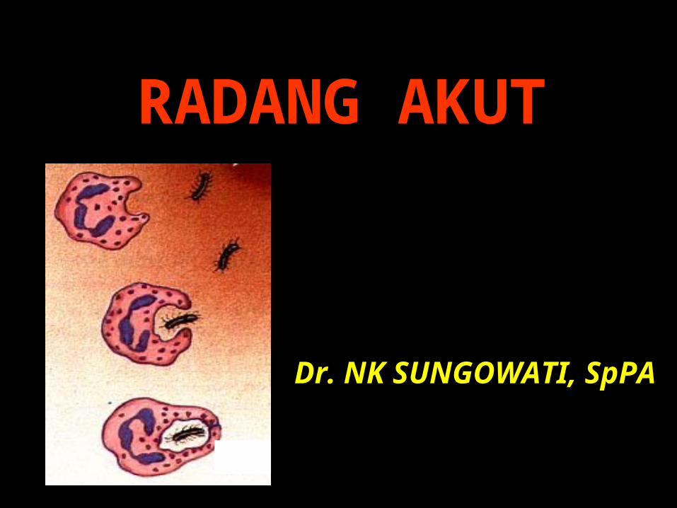
RADANG AKUT
Dr. NK SUNGOWATI, SpPA
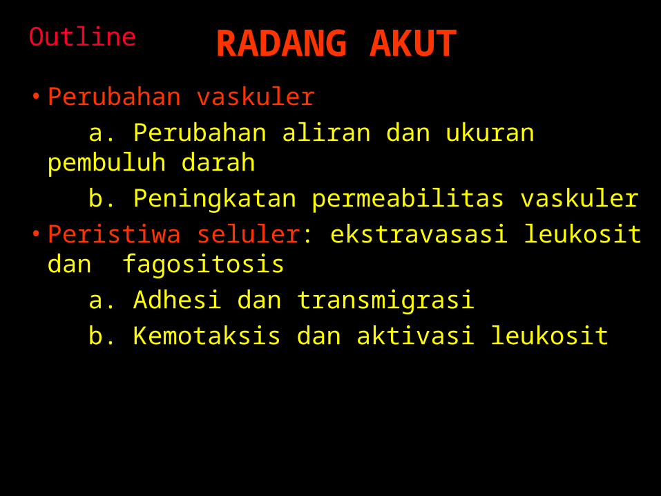
RADANG AKUT• Perubahan vaskuler a. Perubahan aliran dan ukuran pembuluh
darah b. Peningkatan permeabilitas vaskuler• Peristiwa seluler: ekstravasasi leukosit dan
fagositosis a. Adhesi dan transmigrasi b. Kemotaksis dan aktivasi leukosit
Outline

c. Fagositosis (pengenalan, perlekatan, pelahapan dan degradasi)d. Pengeluaran produk leukosite. Defek fungsi leukosit
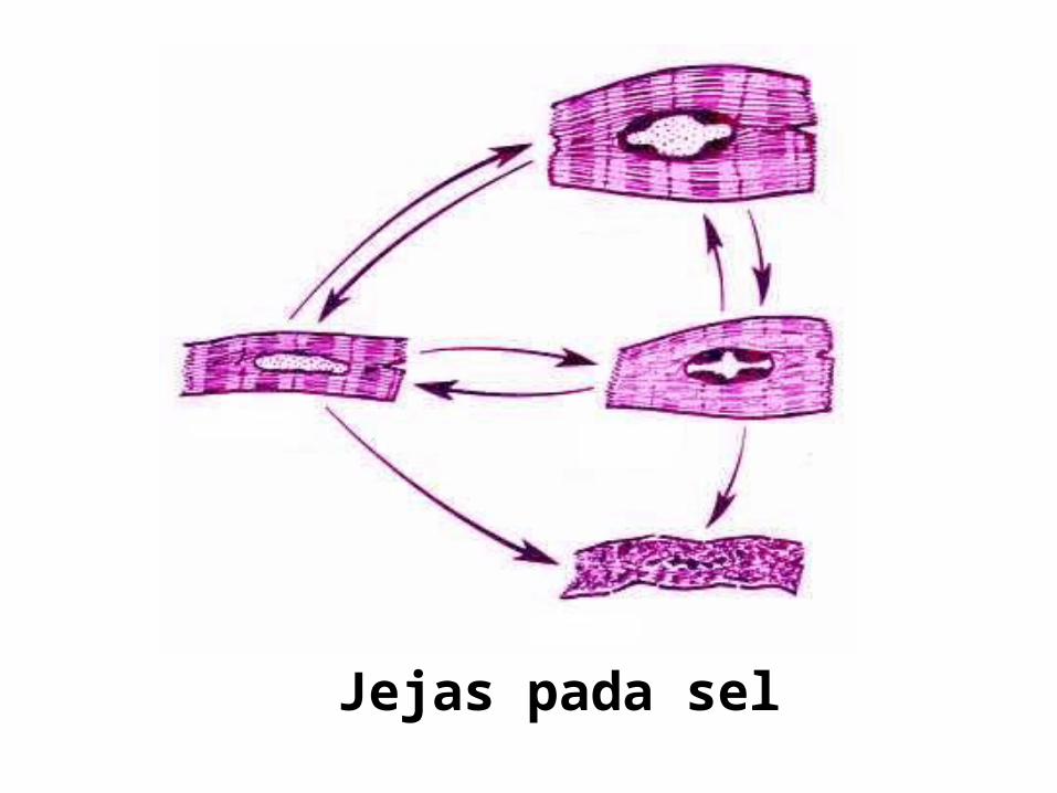
Jejas pada sel
RADANG AKUT
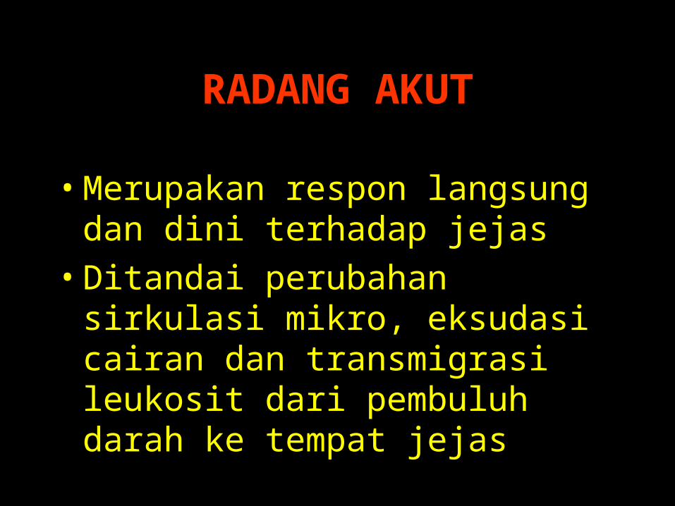
• Merupakan respon langsung dan dini terhadap jejas
• Ditandai perubahan sirkulasi mikro, eksudasi cairan dan transmigrasi leukosit dari pembuluh darah ke tempat jejas

CARDINAL SIGN
• Rubor• Calor• Tumor• Dolor• Functio
laesa
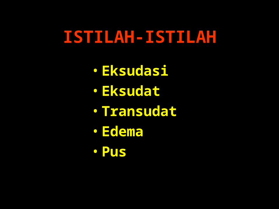
ISTILAH-ISTILAH
• Eksudasi• Eksudat• Transudat• Edema• Pus
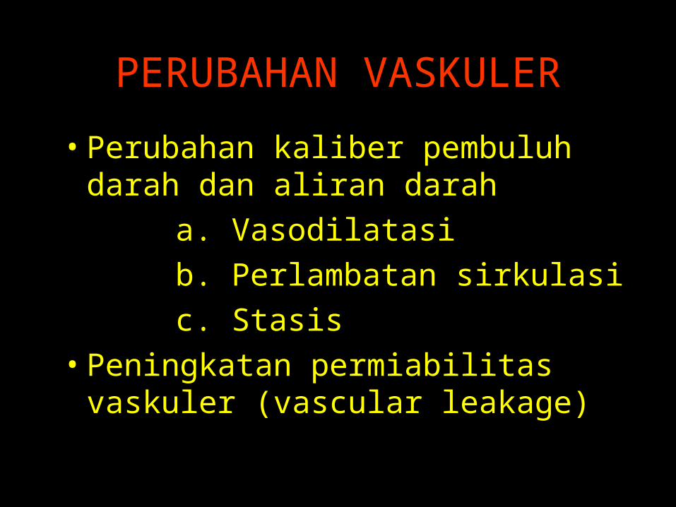
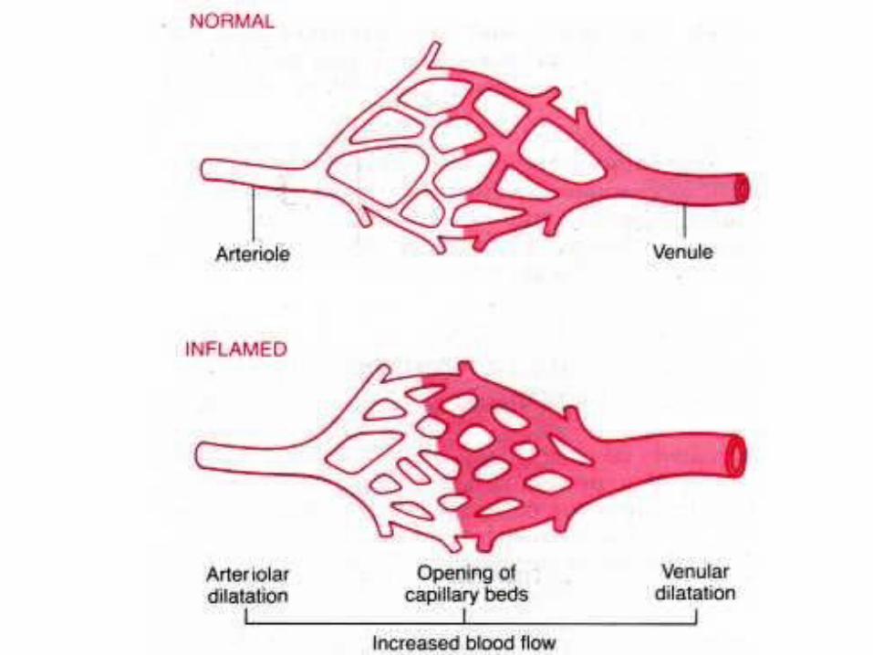
PERUBAHAN VASKULER
• Perubahan kaliber pembuluh darah dan aliran darah
a. Vasodilatasi b. Perlambatan sirkulasi c. Stasis• Peningkatan permiabilitas vaskuler
(vascular leakage)
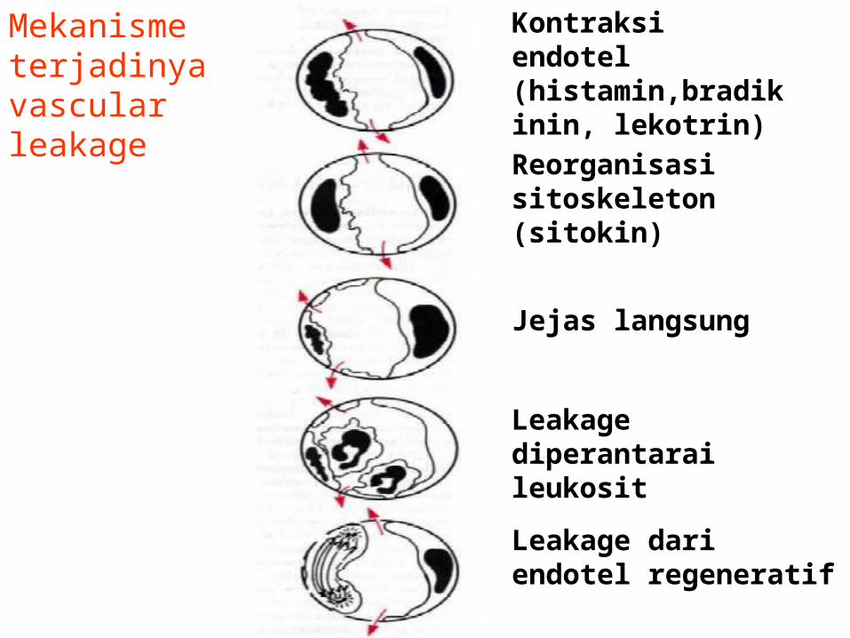
Mekanisme terjadinya vascular leakage
Kontraksi endotel (histamin,bradikinin, lekotrin)
Reorganisasi sitoskeleton (sitokin)
Jejas langsung
Leakage diperantarai leukosit
Leakage dari endotel regeneratif
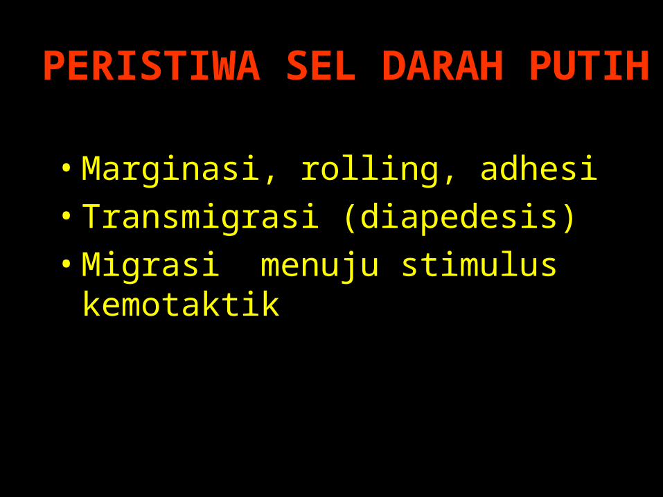
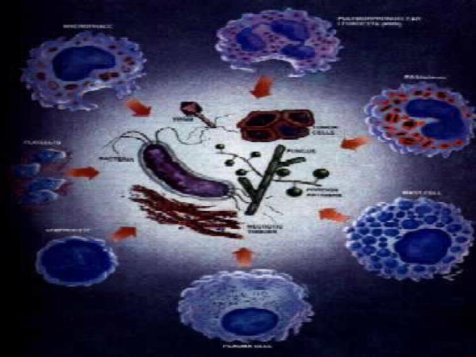
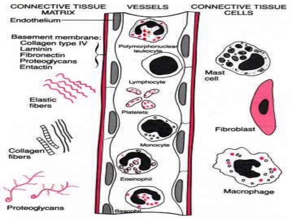
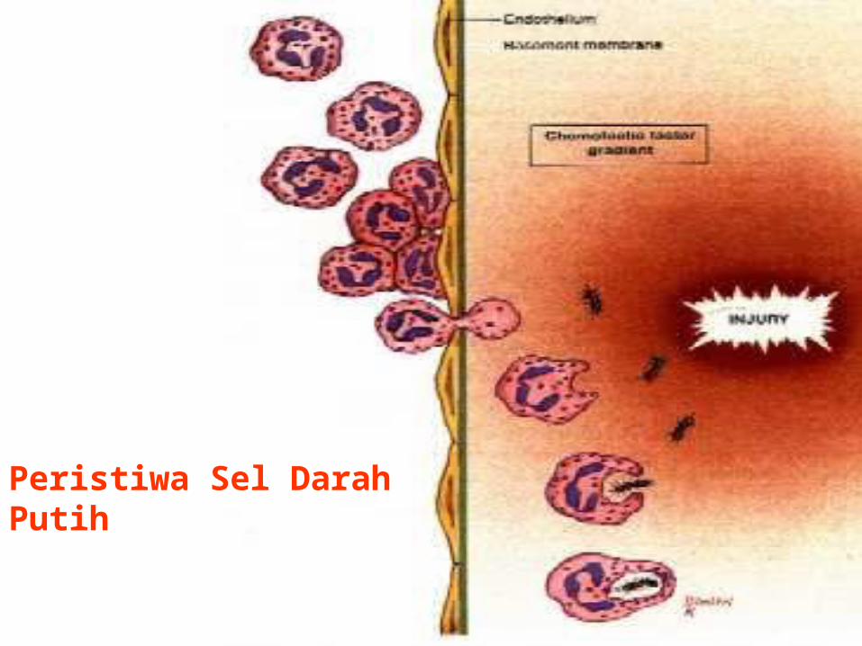
PERISTIWA SEL DARAH PUTIH
• Marginasi, rolling, adhesi• Transmigrasi (diapedesis)• Migrasi menuju stimulus
kemotaktik
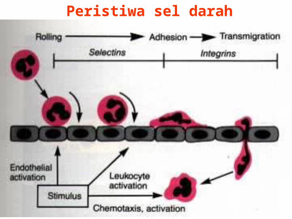
Peristiwa sel darah putih
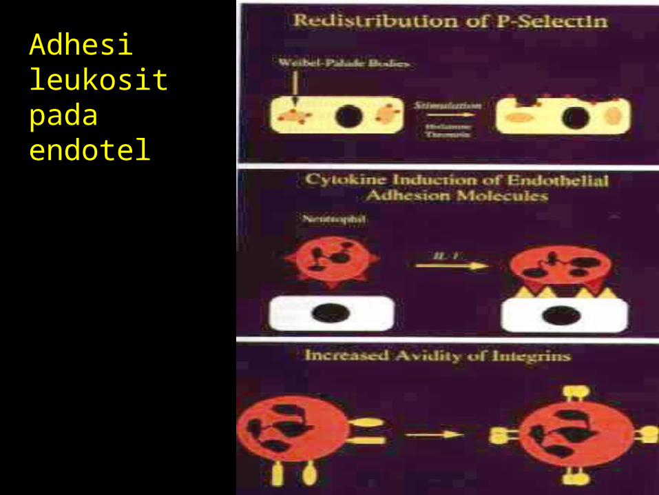
Adhesi leukosit pada endotel
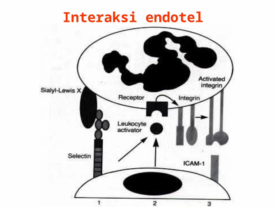
Interaksi endotel neutrofil
TRANSMIGRASI• Terjadi sepanjang interseluler junction• Tergantung umur lesi dan tipe stimulus• Neutrofil (6-24 jam)• Monosit (24-48 jam)• Pseudomonas (neutrofil ~ 2-4 hari)• Virus ~ limfosit• Hipersensitivitas ~ eosinofil
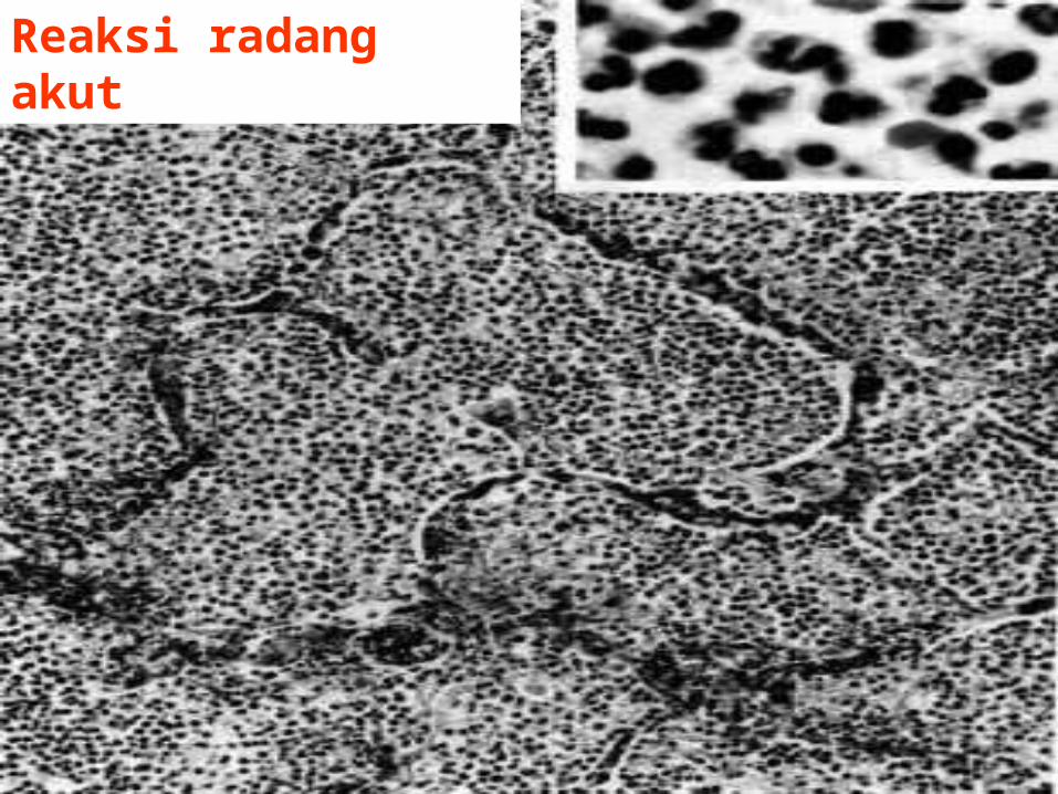
Reaksi radang akut
Peristiwa Sel Darah Putih
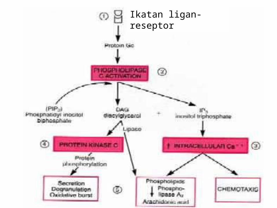
Ikatan ligan-reseptor
Ikatan ligan-reseptor
FAGOSITOSIS
• Pengenalan dan perlekatan• Pelahapan• Pembunuhan atau degradasi
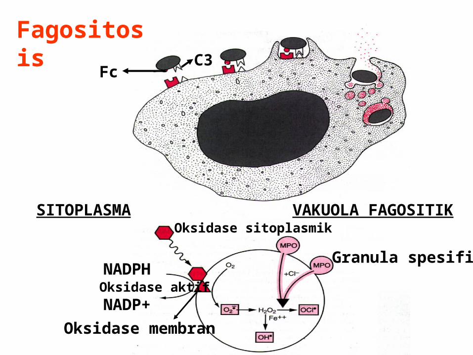
FagositosisFc
C3
NADPH
NADP+Oksidase aktif
SITOPLASMA VAKUOLA FAGOSITIKOksidase sitoplasmik
Oksidase membran
Granula spesifik
PENGELUARAN PRODUK LEUKOSIT
• Terdiri dari : E. lisosom, metabolit aktif O2, prostalglandin dan lekotrin.
DEFEK FUNGSI LEUKOSIT• Lebih rentan terhadap infeksi• Genetik : a. LAD tipe1,2 (defisiensi molekul adhesi) b. CGD (defisiensi NADPH oksidase) c. Chediak-Higashi S. (neutrofenia, defektif degranulasi, perlambatan pembunuhan bakteri)
• Didapat : a. Kemotaksis : febris, diabetes ,
sepsis, immunodefisiensi b. Adhesi : hemodialisis, DM c. Fagositosis, aktivitas mikrobisidal : leukemia, anemia, sepsis, diabetes, neonatus, malnutrisi
MANIFESTASI KLINIK(SISTEMIK)
• Febris : pirogen dan prostalglandin• Perubahan hitung sel darah putih perifer - Neutrofil leukositosis - Neutropenia, limfositosis• Perubahan protein plasma C-reactive protein, antitrypsin, fibrinogen, haptoglobin, ceruloplasmin
RINGKASAN
Reaksi radang akut :• Aliran darah meningkat (dilatasi
arteriol)• Permeabilitas meningkat
(interendotel junction melebar, jejas langsung endotel)
• Neutrofil (adhesi, transmigrasi, migrasi ke tempat jejas)
• Fagositosis• Produk leukosit
Dr.NK Sungowati
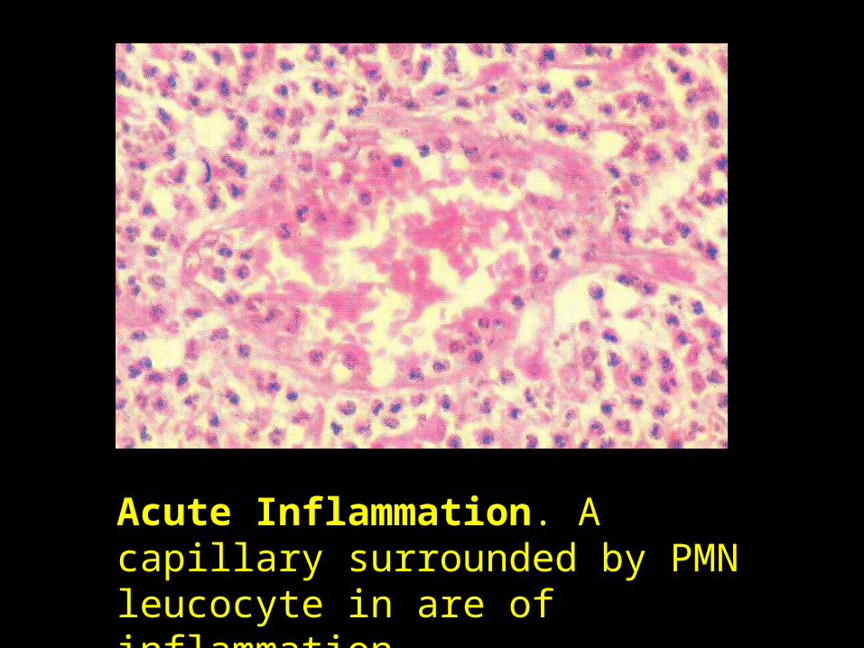
Acute Inflammation. A capillary surrounded by PMN leucocyte in are of inflammation.
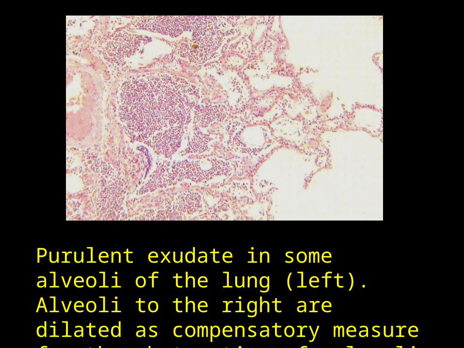
Purulent exudate in some alveoli of the lung (left). Alveoli to the right are dilated as compensatory measure for the obstruction of alveoli on the left.
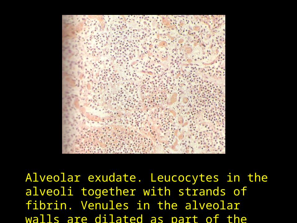
Alveolar exudate. Leucocytes in the alveoli together with strands of fibrin. Venules in the alveolar walls are dilated as part of the inflammatory response.
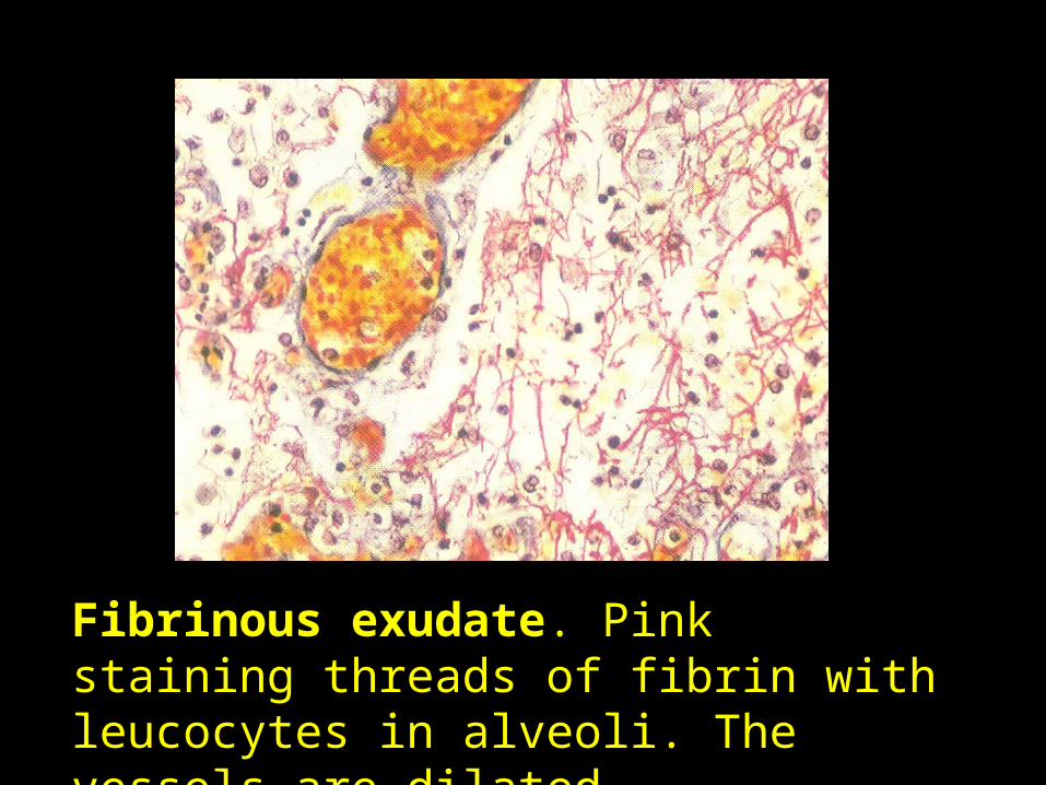
Fibrinous exudate. Pink staining threads of fibrin with leucocytes in alveoli. The vessels are dilated.
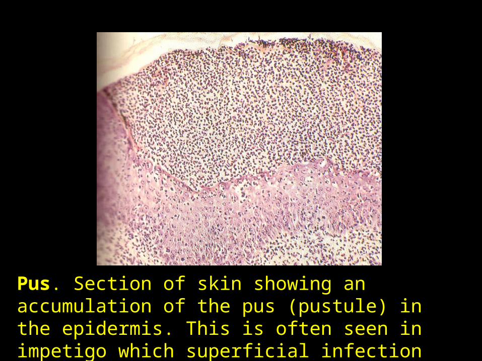
Pus. Section of skin showing an accumulation of the pus (pustule) in the epidermis. This is often seen in impetigo which superficial infection caused by coccal bacteria.
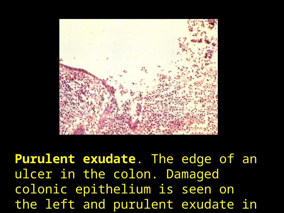
Purulent exudate. The edge of an ulcer in the colon. Damaged colonic epithelium is seen on the left and purulent exudate in the base of the ulcer on the right.
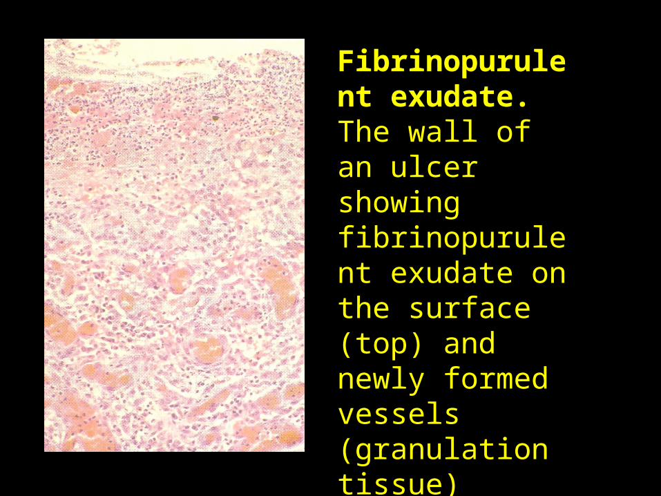
Fibrinopurulent exudate. The wall of an ulcer showing fibrinopurulent exudate on the surface (top) and newly formed vessels (granulation tissue) deeper down.
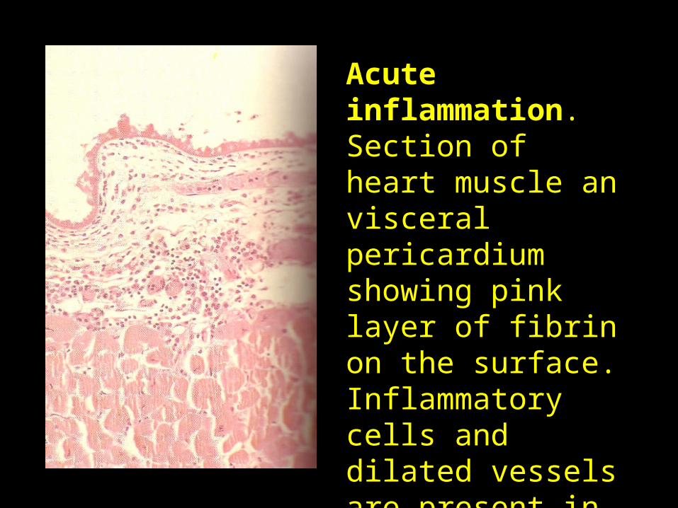
Acute inflammation. Section of heart muscle an visceral pericardium showing pink layer of fibrin on the surface. Inflammatory cells and dilated vessels are present in the underlying connective tissue.
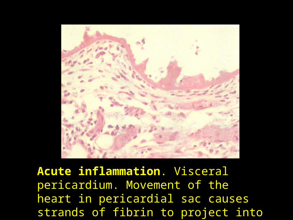
Acute inflammation. Visceral pericardium. Movement of the heart in pericardial sac causes strands of fibrin to project into the lumen of the pericardial sac.
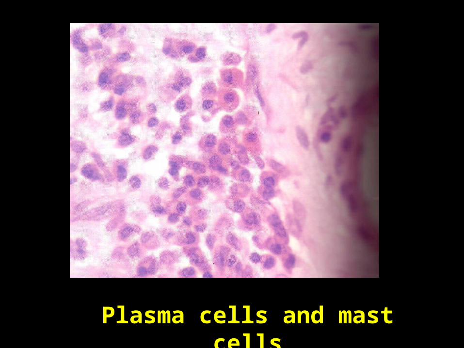
Plasma cells and mast cells
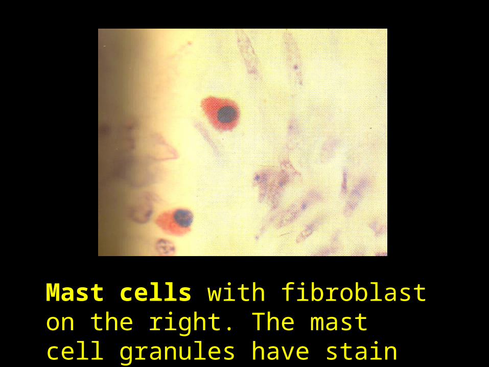
Mast cells with fibroblast on the right. The mast cell granules have stain bright red with solachrome cyanin.
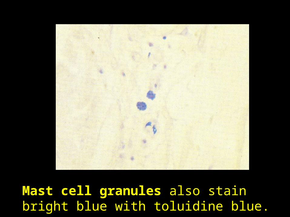
Mast cell granules also stain bright blue with toluidine blue.
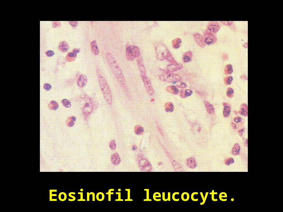
Eosinofil leucocyte.