peradangan kulit
18
Prophylactic Efficacy of Coriandrum sativum (Coriander) on Testis of Lead-Exposed Mice Veena Sharma & Leena Kansal & Arti Sharma Received: 5 September 2009 / Accepted: 14 October 2009 / Published online: 10 November 2009 # Humana Press Inc. 2009 Abstract Lead poisoning is a worldwide health problem, and its treatment is under investigation. The aim of this study was to access the efficacy of Coriandrum sativum (coriander) in reducing lead-induced changes in mice testis. Animal exposed to lead nitrate showed significant decrease in testicular SOD, CAT, GSH, total protein, and tissue lead level. This was accompanied by simultaneous increase in the activities of LPO, AST, ALT, ACP, ALP, and cholesterol level. Serum testosterone level and sperm density were suppressed in lead-treated group compared with the control. These influences of lead were prevented by concurrent daily administration of C. sativum extracts to some extent. Treating albino mice with lead-induced various histological changes in the testis and treatment with coriander led to an improvement in the histological testis picture. The results thus led us to conclude that administration of C. sativum significantly protects against lead-induced oxidative stress. Further work need to be done to isolate and purify the active principle involved in the antioxidant activity of this plant. Keywords Coriandrum sativum . Testis . Lead nitrate . Histology . Mice Introduction Coriandrum sativum (common name: coriander), belonging to family Umbelliferae, is an herb that is widely cultivated in India and is recognized for its carminative and cooling properties [1]. Both the leaves and seeds of the plant are used for medicinal purpose. Coriander contains many active principles, primarily isocoumarines [2], and the most important one is coriandrin. In the field of alternative medicine, coriander has immense value for the treatment of abdominal problems, especially stomach ulcers. The hypotensive [3] and hypoglycemic [4] properties of coriander have already been recognized. The ability of coriander to increase levels of antioxidant enzymes and to Biol Trace Elem Res (2010) 136:337–354 DOI 10.1007/s12011-009-8553-0 V. Sharma (*) : L. Kansal : A. Sharma Bioscience and Biotechnology Department, Banasthali University, District Tonk, Banasthali 304022 Rajasthan, India e-mail: [email protected]
-
Upload
ridho-dhe-holmes -
Category
Documents
-
view
232 -
download
0
description
jurnal internasional manfaat ketumbar
Transcript of peradangan kulit
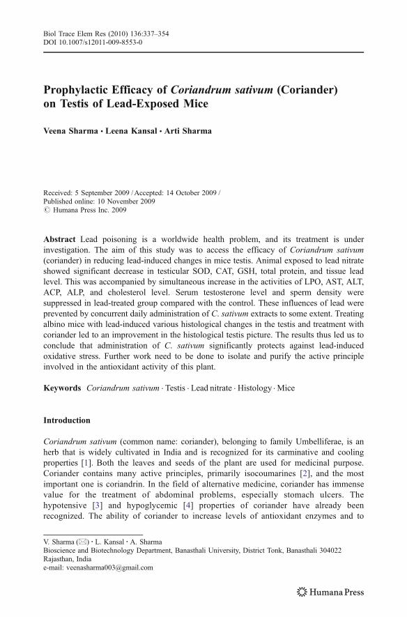
Prophylactic Efficacy of Coriandrum sativum (Coriander)on Testis of Lead-Exposed Mice
Veena Sharma & Leena Kansal & Arti Sharma
Received: 5 September 2009 /Accepted: 14 October 2009 /Published online: 10 November 2009# Humana Press Inc. 2009
Abstract Lead poisoning is a worldwide health problem, and its treatment is underinvestigation. The aim of this study was to access the efficacy of Coriandrum sativum(coriander) in reducing lead-induced changes in mice testis. Animal exposed to lead nitrateshowed significant decrease in testicular SOD, CAT, GSH, total protein, and tissue leadlevel. This was accompanied by simultaneous increase in the activities of LPO, AST, ALT,ACP, ALP, and cholesterol level. Serum testosterone level and sperm density weresuppressed in lead-treated group compared with the control. These influences of lead wereprevented by concurrent daily administration of C. sativum extracts to some extent. Treatingalbino mice with lead-induced various histological changes in the testis and treatment withcoriander led to an improvement in the histological testis picture. The results thus led us toconclude that administration of C. sativum significantly protects against lead-inducedoxidative stress. Further work need to be done to isolate and purify the active principleinvolved in the antioxidant activity of this plant.
Keywords Coriandrum sativum . Testis . Lead nitrate . Histology . Mice
Introduction
Coriandrum sativum (common name: coriander), belonging to family Umbelliferae, is anherb that is widely cultivated in India and is recognized for its carminative and coolingproperties [1]. Both the leaves and seeds of the plant are used for medicinal purpose.Coriander contains many active principles, primarily isocoumarines [2], and the mostimportant one is coriandrin. In the field of alternative medicine, coriander has immensevalue for the treatment of abdominal problems, especially stomach ulcers. Thehypotensive [3] and hypoglycemic [4] properties of coriander have already beenrecognized. The ability of coriander to increase levels of antioxidant enzymes and to
Biol Trace Elem Res (2010) 136:337–354DOI 10.1007/s12011-009-8553-0
V. Sharma (*) : L. Kansal :A. SharmaBioscience and Biotechnology Department, Banasthali University, District Tonk, Banasthali 304022Rajasthan, Indiae-mail: [email protected]
manipulate lipid metabolism have also been reported [5, 6]. In Ayurvedic literature, theregular use of the decotation of the seeds of coriander is considered to be effective inlowering blood lipid levels [1]. It has also been speculated that Chinese parsley mayenhance the excretion of heavy metals in the urine of patients with various infections andaugments the efficacy of antibiotics [7, 8].
Of all the heavy metals that contaminate the environment and pose hazards to publichealth, lead has been of major concern. Lead is an environmental pollutant and metabolicpoison with a variety of toxic effects, among which is its adverse influence on reproduction[9]. Numerous studies [9–13] over the year have investigated the detrimental effects of leadon reproductive function. Many studies have shown that reproductive toxicity is animportant feature of lead toxicity [14–16]. During lead exposure, it accumulates in testis ina dose-dependent manner [14, 15]. Lead toxicity induces a significant increase in apoptoticcell death in seminiferous tubules of young growing rats [14]. It is also associated withdisruption of spermatogenesis and histoarchitecture and lowered enzyme activities in testis[15]. A direct testicular toxicity may occur via the hypothalamic–pituitary–testicular axis[17, 18].
Recent studies have proposed that one possible mechanism of lead toxicity is thedisturbance of prooxidant and antioxidant balance by generation of reactive oxygen species(ROS) [19, 20]. This can evoke the oxidative damage of critical biomolecules such aslipids, proteins, and DNA. It has been also reported that lead exposure has a dose-responserelationship with changes in antioxidant enzyme levels and their activities [21]. Theseenzymes include superoxide dismutase (SOD), catalase (CAT), glutathione peroxidase(GPx), expressions of which were found to be changed in animals and in workers exposedto lead [22–24].
Present investigation attempts to reveal the efficacy of the extracts of C. sativum if any,in the regulation of lead toxicity.
Material and Methods
Chemicals
Lead nitrate was purchased from Central Drug House (India). All other chemicals used inthe study were of analytical reagent and obtained from Sisco Research Laboratories (India),Qualigens (India/ Germany), SD fine chemicals (India), HIMEDIA (India), and CentralDrug House (India).
Experimental Plant
The plant C. sativum (seeds) was collected from Krishi Vigyan Kendra, BanasthaliUniversity, Rajasthan, India, and was identified as an RCR 435 variety. Coriander seedscontain moisture (6.3%), protein (11–17%), volatile oil (0.3%), nonvolatile ether extract(22%), ether extract (19.6%), crude fiber (31.5%), carbohydrates (24%), ash (5.3%),calcium (0.08%), phosphorus (0.44%), sodium (0.02%), potassium (1.2%), vitamin B1
(0.26 mg/100 g), vitamin B2 (0.23 mg/100 mg), niacin (3.2 mg/100 g), vitamin C (ascorbicacid; 12 mg/ 100 g), vitamin A (175 IU/100 g), flavonoid glycosides (26%) [25], andessential oil linalool (60–80%), alpha-pinene (0.2–8.5%), gamma-terpinene (1–8%),geranylacetate (0.1–4.7%), camphor (1.4%), and geraniol (1.2–4.6%) [3]. Aqueous andalcoholic coriander extracts were prepared for the experimental purpose.
338 Sharma et al.
Preparation of Aqueous Extract of C. sativum
Dried coriander seeds were ground to a fine powder, of which 100 g were added to 500 mldistilled water. After 24 h, maceration was done at room temperature (37°C); the mixturewas then heated for 30 min in the water bath at 65°C. The extract was filtered, concentratedby heating over the water bath (65°C) and dried under vacuum [26] with the yield of 5.9%(w/w). The extract was stored at 4°C and used to treat animals as needed.
Preparation of Alcoholic Extraction of C. sativum
The dried and powered seeds (200 g) were extracted successively with ethanol (800 ml) in asoxhlet extractor for 48 h at 60°C. After extraction, the solvent was evaporated to dryness at50–55°C by using a rotary evaporator, and the extract left behind (yield was 9.8%) wasstored at 4°C. It was dissolved in distilled water whenever needed for experiments.
Animals and Treatment
Male Swiss albino mice (Mus musculus) weighing 15–30 g (2–2.5 months old) wereobtained from Haryana Agricultural University, Hisar (India), for experimental purpose.The animals were acclimatized for 15–25 days prior to experiment. The InstitutionalAnimal Ethical Committee has approved the animal studies. Colony-bred adult male albinomice were maintained under standard laboratory conditions at a temperature of 25°C±3°C,relative humidity of 50%±15%, and photoperiod of 12 h (12 h-light and 12 h-dark cycle).The mice were housed in polypropylene cages. Animals had free access to standard foodpellet diet (Hindustan Lever Limited; metal contents in parts per million dry weight: Cu10.0, Zn 45.0, Mn 55.0, Co 5.0, Fe 75.0) and drinking water ad libitum throughout thestudy. Essential cleanliness and, to the best extent, sterile conditions were also adoptedaccording to SPF facilities.
Experimental Design
Adult Swiss albino male mice were divided into six groups of 12 mice each and treated byoral gavage as follows:
Group I- Control (normal, untreated), received distilled water;Group II- Lead nitrate treated group, received freshly dissolved Pb(NO3)2 in 1 ml distilled
water at a dose of 40 mg/kg body weight per day for 40 days;Group III- After exposure to lead nitrate (40 mg/kg body weight per day for 7 days),
the animals received 1 ml of aqueous coriander extract at a dose of300 mg/kg body weight per day and lead nitrate simultaneously for33 days;
Group IV- After exposure to lead nitrate (40 mg/kg body weight per day for 7 days),the animals received 1 ml of aqueous coriander extract at a dose of600 mg/kg body weight per day and lead nitrate simultaneously for33 days;
Group V- After exposure to lead nitrate (40 mg/kg body weight per day for 7 days),the animals received 1 ml of ethanolic coriander extract at a dose of250 mg/kg body weight per day and lead nitrate simultaneously for33 days;
Prophylactic Efficacy of Coriandrum sativum (Coriander) 339
Group VI- After exposure to lead nitrate (40 mg/kg body weight per day for 7 days), theanimals received 1 ml of ethanolic coriander extract at a dose of 500 mg/kgbody weight per day and lead nitrate simultaneously for 33 days;
The concentration of lead nitrate used in the experiment was 1/56 of LD50 [27]. The dose forlead nitrate was decided on the basis of previously performed experiments in our ownlaboratory. The plant doses were selected on the basis of experiments conducted in our ownlaboratory and on the basis of earlier published reports [28]. After the administration of thelast dose, the animals were given a 1-day rest and killed under light chloroform anesthesia.Blood was collected for serum. The testis tissue was excised and divided into two parts; half aportion of the testis from each animal was processed for biochemical variables andhistological analysis and the other half portion of testis was stored at −20°C before wet aciddigestion with HNO3 for lead estimation.
Preparation of Testis Homogenate
Testes were sliced into pieces and homogenized with a blender in ice-cold 0.1 M sodiumphosphate buffer (pH 7.4) at 1–4°C to give 10% homogenate (w/v). The homogenates werecentrifuged at 10,000 rpm for 15–20 min at 4°C twice to get enzyme fraction. The resultingsupernatants were separated and used for various biochemical estimations as depicted below:
Biochemical Assays
Lipid peroxidation (LPO) was estimated by thiobarbituric acid reaction with malondialde-hyde (MDA), a product formed due to the peroxidation of membrane lipids as describedearlier by Nwanjo and Ojiako [29]. The testicular SOD activity was assayed according tothe method of Marklund and Marklund [30]. CAT activity was assayed following theprocedure of Aebi [31]. Glutathione (GSH) content was determined by the method ofEllman [32]. Activities of aspartate aminotransferase (AST) and alanine aminotransferase(ALT) were assayed by the method of Reitman and Frankel [33]. Activities of acidphosphatase (ACP) and alkaline phosphatase (ALP) were determined according to theprotocol described in the laboratory practical manual by Sadashivam [34]. The proteincontent was determined by using bovine serum albumin as a standard by the method ofLowry et al. [35]. The cholesterol level was determined by using cholesterol as a standardby the method of Zak's [36].
Determination of Testosterone Level
Serum level of testosterone was assayed following the standard protocol as suppliedthrough the respective ELISA kit.
Sperm Density
Sperm density was determined in testis according to the method given in modernexperimental Zoology book of Gupta and Chaturvedi [37]. To 100 mg of the tissue (testis),2 ml of normal saline was added. Then it was teased with a needle. In brief, 0.05 ml of thistissue was taken and diluted with NaHCO3 up to 11 marks on WBC pipette. Then, one dropwas put on the already focused slide, and sperms were counted in 16 squares.
340 Sharma et al.
Metal Estimation
Lead concentration in testis was measured after wet acid digestion. Lead was estimatedusing a hydride vapor generation system (model MHS-10, Perkin Elmer) fitted with anatomic absorption spectrophotometer (model A Analyst 100, Perkin Elmer).
Histological Analysis
Testes were removed, washed in saline, and fixed in 10% formalin at room temperature for72 h. After fixing the tissue, it was thoroughly washed under running water and dehydratedin ascending grades of ethyl alcohol, cleared, and then embedded in soft paraffin. Tissuesections of about 6µm were obtained, stained by hematoxylin and eosin, and examinedunder light microscope.
Statistical Analysis
Data are expressed as the mean±SEM. The data were analyzed using the Statistical Packagefor Social Science program (SPSS 11). Statistical analysis was done using analysis ofvariance followed by Tukey's test, and the level of significance was set at p<0.05.
Results
Lipid Peroxidation, Antioxidant Enzymes and GSH Level
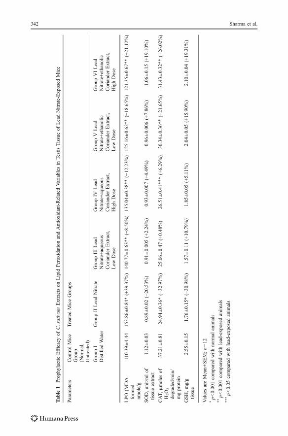
Table 1 illustrates the effect of lead nitrate alone, and effect of treatment with C. sativumextracts individually during lead nitrate exposure on lipid peroxidation, activity ofantioxidant enzymes (SOD and CAT), and level of nonantioxidant enzyme (GSH) incontrol and experimental groups of animals.
The level of lipid peroxidation was significantly higher (p<0.001) in lead-treatedanimals (group II) than that of normal untreated mice. Whereas, significant decrease (p<0.001) in testis SOD, CAT activity, and GSH content of mice were observed in lead nitrate-treated animals as compared with control group. After the treatment with aqueous corianderextract, a significant decrease (p<0.001 for both low- and high-dose groups) in the level ofLPO was observed in comparison to lead nitrate-treated group. Administration of ethanolicextract of plant to animals also improved LPO level as compared with lead group (p<0.001for both low- and high-dose groups).
In comparison to lead-exposed group, administration of aqueous and ethanolic corianderextract at low and high doses improved SOD and GSH content insignificantly (p>0.05).However, at a high dose of aqueous coriander extract, CAT activity increased significantly(p<0.05), but, in low-dose treated group, it increased insignificantly (p>0.05) incomparison to the lead-treated group. With ethanolic extract of coriander, CAT activitywas also recovered in animal groups V and VI when compared with group II (p<0.001 forboth low and high doses).
Biochemical Parameters
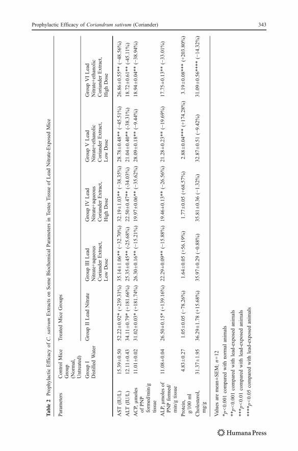
Table 2 represents the effect of lead nitrate alone and effect of treatment with C. sativumextracts individually during lead nitrate exposure on some biochemical parameters such as
Prophylactic Efficacy of Coriandrum sativum (Coriander) 341
Tab
le1
Proph
ylactic
Efficacyof
C.sativum
Extractson
Lipid
PeroxidationandAntioxidant-Related
Variables
inTestisTissueof
LeadNitrate-Exp
osed
Mice
Param
eters
Con
trol
Mice
Group
(Normal,
Untreated)
Treated
MiceGroup
s
Group
IDistilledWater
Group
IILeadNitrate
Group
IIILead
Nitrate+aqueous
Coriander
Extract,
Low
Dose
Group
IVLead
Nitrate+aqueous
Coriander
Extract,
HighDose
Group
VLead
Nitrate+ethanolic
Coriand
erExtract,
Low
Dose
Group
VILead
Nitrate+ethanolic
Coriander
Extract,
HighDose
LPO
(MDA
form
ednm
ole/g
110.39
±4.44
153.86
±0.84*(+39.37%
)14
0.77
±0.63
**(−8.50
%)
135.04
±0.38
**(−12
.23%
)12
5.16
±0.62**
(−18
.65%
)12
1.35
±0.67**
(−21
.12%
)
SOD,unit/mlof
tissueextract
1.12
±0.03
0.89
±0.02
(−20
.53%
)0.91
±0.00
5(+2.24%)
0.93
±0.00
7(+4.49
%)
0.96
±0.006(+7.86%)
1.06
±0.15
(+19.10%
)
CAT,
µmoles
ofH2O2
degraded/m
in/
mgprotein
37.21±0.81
24.94±0.36*(−32
.97%
)25
.06±0.47
(+0.48%)
26.51±0.41
***(+6.29
%)
30.34±0.36**
(+21.65%
)31
.43±0.32**
(+26.02%
)
GSH,mg/g
tissue
2.55
±0.15
1.76
±0.15*(−30
.98%
)1.57
±0.11
(+10.79%
)1.85
±0.05
(+5.11%)
2.04
±0.05
(+15
.90%
)2.10
±0.04
(+19.31%
)
ValuesareMean±SEM;n=12
*p<0.001comparedwith
norm
alanim
als
**p<0.001comparedwith
lead-exposed
anim
als
***p<0.05
comparedwith
lead-exp
osed
anim
als
342 Sharma et al.
Tab
le2
Proph
ylactic
Efficacyof
C.sativum
Extractson
Som
eBiochem
ical
Param
etersin
Testes
Tissueof
LeadNitrate-Exposed
Mice
Param
eters
Control
Mice
Group
(Normal,
Untreated)
Treated
MiceGroup
s
Group
IDistilledWater
Group
IILeadNitrate
Group
IIILead
Nitrate+aqueous
Coriander
Extract,
Low
Dose
Group
IVLead
Nitrate+aqueous
Coriander
Extract,
HighDose
Group
VLead
Nitrate+ethanolic
Coriander
Extract,
Low
Dose
Group
VILead
Nitrate+ethanolic
Coriander
Extract,
HighDose
AST(IU/L)
15.39±0.50
52.22±0.92*(+239.31%)
35.14±1.06**
(−32
.70%
)32
.19±1.03
**(−38
.35%
)28
.78±0.48
**(−45
.51%
)26
.86±0.55**
(−48
.56%
)
ALT
(IU/L)
12.11±0.43
34.11±0.79*(+18
1.66%)
25.35±0.45**
(-25
.68%
)22
.50±0.47
**(-34
.03%
)21
.04±0.40
**(-38
.31%
)18
.72±0.61**
(-45.11%
)
ACP,
µmoles
ofPNP
form
ed/m
in/g
tissue
11.01±0.02
31.02±0.03*(+181.74%)
26.30±0.16**
(−15
.21%
)19
.97±0.06
**(−35
.62%
)28
.09±0.18
**(−9.44
%)
18.94±0.04**
(−38
.94%
)
ALP,µmoles
ofPNPform
ed/
min/g
tissue
11.08±0.04
26.50±0.15*(+139.16%)
22.29±0.09**
(−15
.88%
)19
.46±0.13
**(−26
.56%
)21
.28±0.23
**(−19
.69%
)17
.75±0.13**
(−33
.01%
)
Protein,
g/10
0ml
4.83
±0.27
1.05
±0.05
(−78
.26%
)1.64
±0.05
(+56.19%
)1.77
±0.05
(+68.57%
)2.88
±0.04
***(+17
4.28%)
3.19
±0.08**
*(+203.80%)
Cho
lesterol,
mg/g
31.37±1.95
36.29±1.78
(+15.68%
)35
.97±0.29
(−0.88%)
35.81±0.36
(−1.32%)
32.87±0.51
(−9.42
%)
31.09±0.56**
**(−14
.32%
)
Valuesaremean±SEM;n=12
*p<0.001comparedwith
norm
alanim
als
**p<0.001comparedwith
lead-exp
osed
anim
als
***p
<0.01
comparedwith
lead-exp
osed
anim
als
****p<0.05
comparedwith
lead-exp
osed
anim
als
Prophylactic Efficacy of Coriandrum sativum (Coriander) 343
AST, ALT, ACP, and ALP and total protein and total cholesterol levels in testicular tissue ofcontrol and experimental groups of animals.
It is also clear from the results that treatment with lead nitrate showed a significantincrease in parameters which include AST, ALT, ACP, ALP, and total cholesterol levelas compared with the control group (p<0.001). Total protein concentration wassignificantly lower in lead group than in control.
Administration of aqueous extract of coriander showed significant decrease (p<0.001 for both low and high doses) in AST, ALT, ACP, and ALP when compared withlead nitrate-treated group. On the other hand, treatment with ethanolic coriander extractalso increased these values significantly as compared with group II (p<0.001).
Whereas, cholesterol level diminished insignificantly in testis tissue (p>0.05), afterthe administration of aqueous plant extract in both low- and high-dose groups.Supplementation of ethanolic coriander extract decreased cholesterol level significantlyin the high-dose group (p<0.05) but insignificantly in low-dose group (p>0.05), whencompared with lead-induced group (II).
On administration of aqueous coriander extract along with lead nitrate, total proteinincreased insignificantly (p>0.05 for both low- and high-dose groups) as compared withthe lead-treated group. Supplementation with ethanolic coriander extract significantlyincreased (p<0.01 for both low and high doses) total protein content as compared withgroup II.
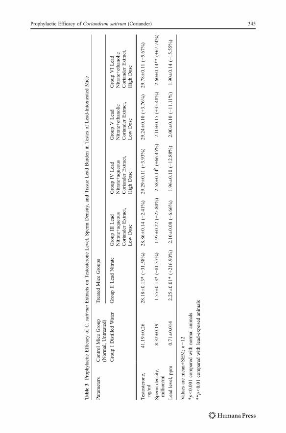
Table 3 represents the effect of lead nitrate alone and effect of treatment with C. sativumextracts individually during lead nitrate exposure on serum testosterone level, spermdensity, and lead nitrate concentration in testes of control and experimental group ofanimals.
Serum Testosterone
Testosterone level was significantly (p<0.001) lower in lead group than in control group.Whereas, administration of plant extracts individually in combination with lead nitrate togroups III and IV increased testosterone level, but results were insignificant (p>0.05).However, upon treatment with ethanolic coriander extract at low and high doses,insignificant increase (p>0.05) in testosterone level was observed, as compared withgroup II.
Sperm Density
The sperm density of mice treated with lead nitrate was significantly lower than that ofcontrol animals (p<0.001). Supplementation of aqueous coriander extract provokedincrease in sperm density, in both aqueous plant extract-treated groups, compared withlead group. Elevation in sperm density persisted on treatment with ethanolic corianderextract, compared with lead nitrate-treated group.
Lead Concentration in Testis Tissue
A significant increase (p<0.001) in lead content was observed in testis tissue of leadnitrate-treated group when compared with control group. Administration of aqueous andalcoholic extracts of coriander during the experiment resulted in Pb excretion from thetissues, in comparison to lead-treated group. But decrease in lead content was found to beinsignificant (p>0.05) in testis tissue after the treatment with both plant extracts.
344 Sharma et al.
Tab
le3
Proph
ylactic
Efficacyof
C.sativum
Extractson
Testosterone
Level,Sperm
Density,andTissueLeadBurdenin
Testes
ofLead-IntoxicatedMice
Param
eters
Control
MiceGroup
(Normal,Untreated)
Treated
MiceGroup
s
Group
IDistilledWater
Group
IILeadNitrate
Group
IIILead
Nitrate+aqueous
Coriander
Extract,
Low
Dose
Group
IVLead
Nitrate+aqueous
Coriander
Extract,
HighDose
Group
VLead
Nitrate+ethanolic
Coriander
Extract,
Low
Dose
Group
VILead
Nitrate+ethanolic
Coriander
Extract,
HighDose
Testosterone,
ng/m
l41
.19±0.26
28.18±0.13
*(−31
.58%
)28
.86±0.14
(+2.41%)
29.29±0.11
(+3.93%)
29.24±0.10
(+3.76%)
29.78±0.11
(+5.67
%)
Sperm
density,
million/ml
8.32
±0.19
1.55
±0.13
*(−81
.37%
)1.95
±0.22
(+25.80%
)2.58
±0.14
b(+66.45%
)2.10
±0.15
(+35.48%
)2.60
±0.14**
(+67.74%
)
Leadlevel,pp
m0.71
±0.014
2.25
±0.01
*(+216.90
%)
2.10
±0.08
(−6.66
%)
1.96
±0.10
(−12
.88%
)2.00
±0.10
(−11.11%
)1.90
±0.14
(−15
.55%
)
Valuesaremean±SEM;n=12
*p<0.001comparedwith
norm
alanim
als
**p<0.01
comparedwith
lead-exp
osed
anim
als
Prophylactic Efficacy of Coriandrum sativum (Coriander) 345
Histological Results
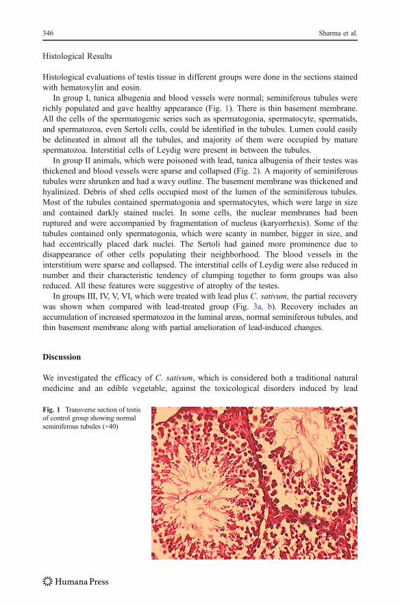
Histological evaluations of testis tissue in different groups were done in the sections stainedwith hematoxylin and eosin.
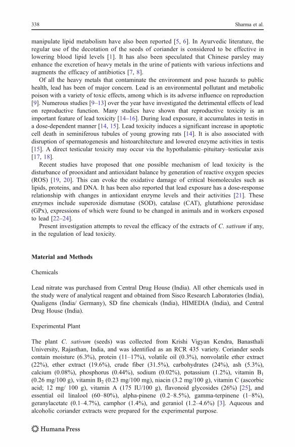
In group I, tunica albugenia and blood vessels were normal; seminiferous tubules wererichly populated and gave healthy appearance (Fig. 1). There is thin basement membrane.All the cells of the spermatogenic series such as spermatogonia, spermatocyte, spermatids,and spermatozoa, even Sertoli cells, could be identified in the tubules. Lumen could easilybe delineated in almost all the tubules, and majority of them were occupied by maturespermatozoa. Interstitial cells of Leydig were present in between the tubules.
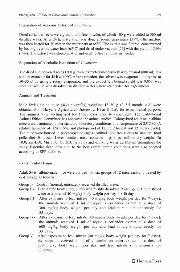
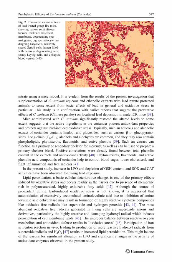
In group II animals, which were poisoned with lead, tunica albugenia of their testes wasthickened and blood vessels were sparse and collapsed (Fig. 2). A majority of seminiferoustubules were shrunken and had a wavy outline. The basement membrane was thickened andhyalinized. Debris of shed cells occupied most of the lumen of the seminiferous tubules.Most of the tubules contained spermatogonia and spermatocytes, which were large in sizeand contained darkly stained nuclei. In some cells, the nuclear membranes had beenruptured and were accompanied by fragmentation of nucleus (karyorrhexis). Some of thetubules contained only spermatogonia, which were scanty in number, bigger in size, andhad eccentrically placed dark nuclei. The Sertoli had gained more prominence due todisappearance of other cells populating their neighborhood. The blood vessels in theinterstitium were sparse and collapsed. The interstitial cells of Leydig were also reduced innumber and their characteristic tendency of clumping together to form groups was alsoreduced. All these features were suggestive of atrophy of the testes.
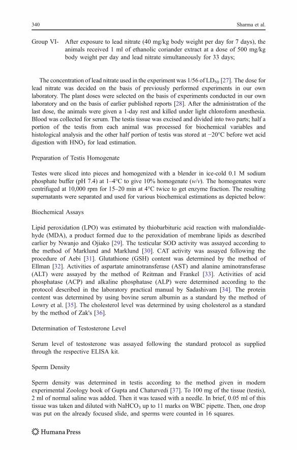
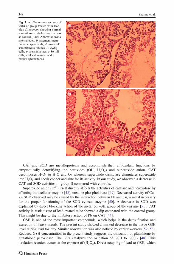
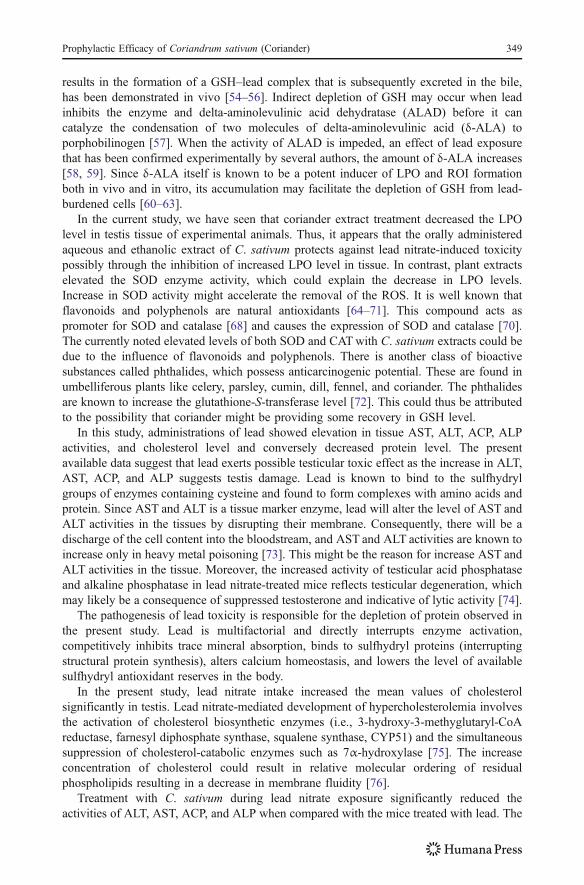
In groups III, IV, V, VI, which were treated with lead plus C. sativum, the partial recoverywas shown when compared with lead-treated group (Fig. 3a, b). Recovery includes anaccumulation of increased spermatozoa in the luminal areas, normal seminiferous tubules, andthin basement membrane along with partial amelioration of lead-induced changes.
Discussion
We investigated the efficacy of C. sativum, which is considered both a traditional naturalmedicine and an edible vegetable, against the toxicological disorders induced by lead
Fig. 1 Transverse section of testisof control group showing normalseminiferous tubules (×40)
346 Sharma et al.
nitrate using a mice model. It is evident from the results of the present investigation thatsupplementation of C. sativum aqueous and ethanolic extracts with lead nitrate protectedanimals to some extent from toxic effects of lead in general and oxidative stress inparticular. This study is in confirmation with earlier reports that suggest the preventiveeffects of C. sativum (Chinese parsley) on localized lead deposition in male ICR mice [38].
Mice administered with C. sativum significantly restored the altered levels to someextent suggests that the active ingredients in the coriander possess antioxidant propertiesand protects against lead-induced oxidative stress. Typically, such an aqueous and alcoholicextract of coriander contains linalool and glucosides, such as various β-D- glucopyrano-sides. Long-chain (C6-C10) alcohols and aldehydes are common, and they may also containphospholipids, phytosterols, flavonoids, and active phenols [39]. Such an extract canfunction as a primary or secondary chelator for mercury, as well as can be used to prepare aprimary chelator blend. Positive correlations were already found between total phenoliccontent in the extracts and antioxidant activity [40]. Phytonutrients, flavonoids, and activephenolic acid compounds of coriander help to control blood sugar, lower cholesterol, andfight inflammation and free radicals [41].
In the present study, increase in LPO and depletion of GSH content, and SOD and CATactivities have been observed following lead exposure.
Lipid peroxidation, a basic cellular deteriorative change, is one of the primary effectsinduced by oxidative stress and occurs readily in the tissues due to presence of membranerich in polyunsaturated, highly oxidizable fatty acids [42]. Although the source ofprooxidant during lead-induced oxidative stress is not known, it is suggested thatautooxidation of excessively accumulated aminolevulinic acid due to inhibition of aminolevulinic acid dehydratase may result in formation of highly reactive cytotoxic compoundslike oxidative free radicals like superoxide and hydrogen peroxide [43, 44]. The mostabundant oxidative free radicals generated in living cells are superoxide anions andderivatives, particularly the highly reactive and damaging hydroxyl radical which inducesperoxidation of cell membrane lipids [45]. The improper balance between reactive oxygenmetabolites and antioxidant defense results in “oxidative stress” [46]. Participation of ironin Fenton reaction in vivo, leading to production of more reactive hydroxyl radicals fromsuperoxide radicals and H2O2 [47] results in increased lipid peroxidation. This might be oneof the reasons for significant alteration in LPO and significant changes in the activity ofantioxidant enzymes observed in the present study.
Fig. 2 Transverse section of testisof lead-treated group IIA mice,showing narrow seminiferoustubules, thickened basementmembrane, degenerating sper-matogonia, big spermatocyte un-dergoing karyolysis, relativelyspared Sertoli cells, lumen filledwith debris of degenerating cells,scanty Leydig cells, and collapsedblood vessels (×40)
Prophylactic Efficacy of Coriandrum sativum (Coriander) 347
CAT and SOD are metalloproteins and accomplish their antioxidant functions byenzymatically detoxifying the peroxides (OH, H2O2) and superoxide anion. CATdecomposes H2O2 to H2O and O2 whereas superoxide dismutase dismutates superoxideinto H2O2 and needs copper and zinc for its activity. In our study, we observed a decrease inCAT and SOD activities in group II compared with controls.
Superoxide anion (O2−) itself directly affects the activities of catalase and peroxidase byaffecting intracellular enzyme [48], creatine phosphokinase [49]. Decreased activity of Cu–Zn SOD observed may be caused by the interaction between Pb and Cu, a metal necessaryfor the proper functioning of the SOD cytosol enzyme [50]. A decrease in SOD wasexplained by direct blocking action of the metal on –SH group of the enzyme [51]. CATactivity in testis tissue of lead-treated mice showed a dip compared with the control group.This might be due to the inhibitory action of Pb on CAT [44].
GSH is one of the most important compounds, which helps in the detoxification andexcretion of heavy metals. The present study showed a marked decrease in the tissue GSHlevel during lead toxicity. Similar observation was also noticed by earlier workers [52, 53].Reduced GSH concentration in the present study suggests the utilization of glutathione byglutathione peroxidase. The GPx catalyzes the oxidation of GSH to GSSG [48]. Thisoxidation reaction occurs at the expense of (H2O2). Direct coupling of lead to GSH, which
Fig. 3 a b Transverse sections oftestis of group treated with leadplus C. sativum, showing normalseminiferous tubules more or lessas control (×40). Abbreviations: aspermatozoa, b basement mem-brane, c spermatids, d lumen ofseminiferous tubules, l Leydigcells, p spermatocytes, s Sertolicells, v blood vessels, and zmature spermatozoa
348 Sharma et al.
results in the formation of a GSH–lead complex that is subsequently excreted in the bile,has been demonstrated in vivo [54–56]. Indirect depletion of GSH may occur when leadinhibits the enzyme and delta-aminolevulinic acid dehydratase (ALAD) before it cancatalyze the condensation of two molecules of delta-aminolevulinic acid (δ-ALA) toporphobilinogen [57]. When the activity of ALAD is impeded, an effect of lead exposurethat has been confirmed experimentally by several authors, the amount of δ-ALA increases[58, 59]. Since δ-ALA itself is known to be a potent inducer of LPO and ROI formationboth in vivo and in vitro, its accumulation may facilitate the depletion of GSH from lead-burdened cells [60–63].
In the current study, we have seen that coriander extract treatment decreased the LPOlevel in testis tissue of experimental animals. Thus, it appears that the orally administeredaqueous and ethanolic extract of C. sativum protects against lead nitrate-induced toxicitypossibly through the inhibition of increased LPO level in tissue. In contrast, plant extractselevated the SOD enzyme activity, which could explain the decrease in LPO levels.Increase in SOD activity might accelerate the removal of the ROS. It is well known thatflavonoids and polyphenols are natural antioxidants [64–71]. This compound acts aspromoter for SOD and catalase [68] and causes the expression of SOD and catalase [70].The currently noted elevated levels of both SOD and CATwith C. sativum extracts could bedue to the influence of flavonoids and polyphenols. There is another class of bioactivesubstances called phthalides, which possess anticarcinogenic potential. These are found inumbelliferous plants like celery, parsley, cumin, dill, fennel, and coriander. The phthalidesare known to increase the glutathione-S-transferase level [72]. This could thus be attributedto the possibility that coriander might be providing some recovery in GSH level.
In this study, administrations of lead showed elevation in tissue AST, ALT, ACP, ALPactivities, and cholesterol level and conversely decreased protein level. The presentavailable data suggest that lead exerts possible testicular toxic effect as the increase in ALT,AST, ACP, and ALP suggests testis damage. Lead is known to bind to the sulfhydrylgroups of enzymes containing cysteine and found to form complexes with amino acids andprotein. Since AST and ALT is a tissue marker enzyme, lead will alter the level of AST andALT activities in the tissues by disrupting their membrane. Consequently, there will be adischarge of the cell content into the bloodstream, and AST and ALT activities are known toincrease only in heavy metal poisoning [73]. This might be the reason for increase AST andALT activities in the tissue. Moreover, the increased activity of testicular acid phosphataseand alkaline phosphatase in lead nitrate-treated mice reflects testicular degeneration, whichmay likely be a consequence of suppressed testosterone and indicative of lytic activity [74].
The pathogenesis of lead toxicity is responsible for the depletion of protein observed inthe present study. Lead is multifactorial and directly interrupts enzyme activation,competitively inhibits trace mineral absorption, binds to sulfhydryl proteins (interruptingstructural protein synthesis), alters calcium homeostasis, and lowers the level of availablesulfhydryl antioxidant reserves in the body.
In the present study, lead nitrate intake increased the mean values of cholesterolsignificantly in testis. Lead nitrate-mediated development of hypercholesterolemia involvesthe activation of cholesterol biosynthetic enzymes (i.e., 3-hydroxy-3-methyglutaryl-CoAreductase, farnesyl diphosphate synthase, squalene synthase, CYP51) and the simultaneoussuppression of cholesterol-catabolic enzymes such as 7α-hydroxylase [75]. The increaseconcentration of cholesterol could result in relative molecular ordering of residualphospholipids resulting in a decrease in membrane fluidity [76].
Treatment with C. sativum during lead nitrate exposure significantly reduced theactivities of ALT, AST, ACP, and ALP when compared with the mice treated with lead. The
Prophylactic Efficacy of Coriandrum sativum (Coriander) 349
reduced concentrations of AST and ALT as a result of plant extract administration observedduring the present study might probably be due to the presence of flavonoids in the extract.It is well documented that flavonoids and glycosides are strong antioxidants [77]. Theincreased activity of plasma LCAT enhanced hepatic bile acid synthesis, and the increaseddegradation of cholesterol to fecal bile acids and neutral sterols appeared to account for itshypocholesterolemic effect [78]. The observed decrease in these enzymes shows thataqueous and alcoholic coriander extract preserve the structural integrity of the organs fromthe toxic effect of lead.
The present study showed deterioration in sperm density in the lead-exposed group. Thisobservation was in collaboration with the earlier observations of Chowdhury et al. [79].Furthermore, most of the testicular germ cells might have been destroyed either due tomembrane damage or macromolecular degradation incurred by ROS leading to a significantdecline in sperm count and ultimately, testicular weight loss. There was a decline intestosterone level of the lead-treated group, which is in accordance with Sokol et al. [80].
The concentration of lead in tissues from mice exposed to lead was higher than it was intissues from mice of control. C. sativum suppresses the deposition of lead by chelating themetal [38]. A sorbent prepared from coriander was found to have good efficiency inremoving organic and methyl mercury from aqueous solutions [81]. Phitic acid (PA), amajor phosphorus storage compound in most seeds and cereal grains, is known as a naturalchelating agent. PA has strong ability to chelate multivalent metal ions. The binding ofmetals with PA can result in the formation of very water-insoluble salts that are poorlyabsorbed from gastrointestinal tract and results in poor bioavailability [82]. It is possiblethat coriander may contain a similar type of chelating agents.
Lead exposure produced pronounced testicular histopathology evidenced by histologicalalternations in testis include degeneration of seminiferous tubules, thickening of basementmembrane, and condensation of the stroma. These findings correspond with theobservations made by some researchers that lead acts as a spermicidal agent in case ofhigh exposure [9, 11]. Seminiferous tubules decreased in size and had a wavy outline. TheSertoli cells were relatively spared because of their resistance to various obnoxious agents[83]. The number of blood vessels in the interstitial tissue was reduced, and most of themwere collapsed as was evident by their reduced diameter. The population of Leydig cellshad dispersed, and they were rarely seen in groups or clumps. The nucleoli in majority ofLeydig cells, rich in rRNA, had disappeared suggesting an atrophy of these cells. Thesefindings are agreed with that of some previous investigators [9, 84–87]. All these scientistsdescribed variety of toxic changes induced by lead in the testes depending upon dose andduration of the treatment.
Lead causes lipid peroxidation by generation of ROS. This peroxidation may causerupture of cell as well as nuclear membrane. This might be responsible for the observednecrosis and disarray in cellular organization in histological section Fig. 2. Evidencesuggests that lead induces free radical formation and thus the generated ROS react with thepolyunsaturated fatty acid-rich spermatozoa, specially the mid-spermatozoa and results inperoxidation which finally leads to destruction in spermatozoa causing reduced motility andviability [88]. Supplementation of coriander extract along with lead nitrate reveals that thedecrease in sperm count, motility, and viability due to toxic effects of lead is minimized tosome extent. As a possible mechanism, it could be stated that coriander extract have arecovery role on lead nitrate-mediated toxicity by inducing an antioxidant effect against theoxidative stress. The antioxidant properties of coriander have been established [40].
These findings of the present investigation support coriander seed to be an effectiveagent in lead intoxication. But the mechanism of its effect is not clear. Further investigation
350 Sharma et al.
is required in order to clarify antioxidant potential of this plant. It is thus concluded that theaqueous and ethanolic extracts of C. sativum may show prophylactic effect against leadtoxicity.
Acknowledgments The authors are thankful to the authorities of Banasthali University for providingsupport to the study.
Conflict of Interest The authors report no conflicts of interest. The authors alone are responsible for thecontent and writing of the paper.
References
1. Sairam TV (1998) Home remedies: a handbook of herbal cures for common ailments. Penguin BooksIndia, New Delhi, p 75
2. Taniguchi M, Yanai M, Xiao YQ, Kido T, Baba K (1996) Three isocumarines from Coriandrum sativum.Phytochemistry 42:843
3. Burdock GA, Carabin IG (2008) Safety assessment of coriander (Coriandrum sativum L.) essential oil asa food ingredient. Food Chem Toxicol 47:22–34
4. Eidi M, Eidi A, Saeidi A, Molanaei S, Sadeghipour A, Bahar M, Bahar K (2009) Effect of coriander seed(Coriandrum sativum L.) ethanol extract on insulin release from pancreatic beta cells in streptozotocin-induced diabetic rats. Phytother Res 23(3):404–406
5. Chithra V, Leelamma S (1999) Coriandrum sativum changes the levels of lipid peroxides and activity ofantioxidant enzymes in experimental animals. Indian J Biochem Biophys 36:59–61
6. Chithra VV, Leelamma S (2000) Coriandrum sativum—effect on lipid metabolism in 1, 2-dimethylhydrazine induced colon cancer. J Ethanopharmocol 17:457
7. Omura Y, Beckman SL (1995) Role of mercury in resistant infections and effective treatment ofChlamydia trachomatis and Herpes family viral infections (and potential treatment for cancer) byremoving localized mercury deposits with Chinese parsley and delivering effective antibiotics usingvarious drug uptake enhancement methods. Acupunct Electrother Res 20:195–229
8. Omura Y, Shimotsuura Y, Fukuoka A, Fukuoka H, Nomoto T (1996) Significant mercury deposits ininternal organs following the removal of dental amalgam, and development of pre cancer on the gingivaland the sides of the tongue and their represented organs as a result of inadvertent exposure to strongcuring light (used to solidify synthetic dental filling material) of effective treatment: a clinical casereport, along with organ representation areas for each tooth. Acupunct Electrother Res 21:133–160
9. Thomas JA, Brogan WC (1983) Some actions of lead on the sperm and on male reproductive system.Am J Ind Med 4:127–134
10. Hilderbrand DC, Der R, Griffen WT, Fahim MS (1973) Effects of lead acetate on reproduction. Am JObstet Gynecol 15:1558–1565
11. Lancranjan I, Popescu HI, Gavanescu O, Klepsch I, Serbanescu M (1975) Reproductive ability ofworkmen occupationally exposed to lead. Arch Environ Health 39:431–440
12. Ronis MJ, Badger TM, Shema SJ, Roberson PK, Shaikh F (1996) Reproductive toxicity and growtheffects in rats exposed to lead at different periods during development. Toxicol Appl Pharmacol136:361–371
13. Winder C (1989) Reproductive and chromosomal effects of occupational exposure to lead in the male.Reprod Toxicol 3:221–233
14. Adhikari N, Sinha N, Narayan R, Saxena DK (2001) Lead induced cell death in testes of young rats. JApplied Toxicol 21:275–277
15. Batra N, Nehru B, Bansal MP (2001) Influence of lead and zinc on rat male reproduction at biochemicaland histopathological levels. J Applied Toxicol 21:507–512
16. Boscolo P, Carmignani M, Sacchettoni-Logroscino G, Ranelletti FO, Artese L, Preziosi P (1988)Ultrastructure of testis in rats with blood hypertension induced by long-term lead exposure. Toxicol Lett41:129–137
17. Klein D, Wan YJ, Kamyab S, Okuda H, Sokol RZ (1994) Effects of toxic levels of lead on generegulation in the male axis: increase in messenger ribonucleic acids and intracellular stores ofgonadotrophs within the central nervous system. Biol Reprod 50:802–811
Prophylactic Efficacy of Coriandrum sativum (Coriander) 351
18. Sokol RZ, Okuda H, Nagler HM, Berman N (1994) Lead exposure in vivo alters the fertility potential ofsperm in vitro. Toxicol Appl Pharmacol 124:310–316
19. Gurer H, Ercal N (2000) Can antioxidants be beneficial in the treatment of lead poisoning? Free RadicBiol Med 29:927–945
20. Wang HP, Qian SY, Schafer FQ, Domann FE, Oberley LW, Buettner GR (2000) Phospholipidhydroperoxide glutathione peroxidase protects against singlet oxygen-induced cell damage ofphotodynamic therapy. Free Radic Biol Med 30:825–835
21. Adonaylo VN, Oteiza PI (1999) Lead intoxication: antioxidant defenses and oxidative damage in ratbrain. Toxicology 135:77–85
22. Mcgowan C, Donaldson WE (1986) Changes in organ nonprotein sulfhydryl and glutathioneconcentrations during acute and chronic administration of inorganic lead to chicks. Biol Trace ElemRes 10:37–46
23. Bechara EJ, Medeiros MH, Monteiro HP, Hermes-lima M, Pereira B, Demasi M (1993) A free 352radical hypotheses of lead poisoning and inborn porphyrias associated with 5-aminolevulinic acidoverloads. Quim Nova 16:385–392
24. Sugawara E, Nakamura K, Miyake T, Fukumura A, Seki Y (1991) Lipid peroxidation and concentrationof glutathione in erythrocytes from workers exposed to lead. Br J Ind Med 48:239–242
25. Budvari S (1996) The Merck Index: an encyclopedia of chemicals, drugs, and biologics, 12th edn. Merck& Co Inc., Whitehouse Station, NJ
26. Gray AM, Flatt PR (1999) Insulin-releasing and insulin-like activity of the traditional anti-diabetic plantCoriandrum sativum (coriander). Br J Nutr 81:203–209
27. Plastunov B, Zub S (2008) Lipid peroxidation processes and antioxidant defence under lead intoxicationand iodine-deficient in experiment. An UMCS Pharmacia 21:215–217
28. Sushruta K, Satyanarayana S, Srinivas N, Raja Sekhar J (2006) Evaluation of the blood-glucose reducingeffects of aqueous extracts of the selected Umbelliferous fruits used in culinary practies. Trop J PharmRes 5(2):613–617
29. Nwanjo HU, Ojiako OA (2005) Effect of vitamins E and C on exercise induced oxidative stress. Global JPure Appl Sci 12:199–202
30. Marklund S, Marklund G (1974) Involvement of superoxide anion radical in the autooxidation ofpyrogallol and convenient assay for SOD. Eur J Biochem 47:469–474
31. Aebi H (1983) Catalase. In: Bergmeyer H (ed) Methods in enzymatic analysis, Vol. 2. Academic, NewYork, pp 76–80
32. Ellman GC (1959) Tissue sulfhydryl groups. Arch Biochem Biophys 82:70–7733. Reitman S, Frankel AS (1957) A colorimetric method for the determination of serum glutamic
oxaloacetic and glutamic pyruvic transaminase. Am J Clin Path 28:53–5634. Sadashivam S, Manickam A (1996) Biochemical methods, 2nd edn. New Age International (P)
Publishers, New Delhi, India, pp. 121-12435. Lowry OH, Rosebrough NJ, Farr AL, Randall RJ (1951) Protein measurement with the folin phenol
reagent. J Biol Chem 193:265–27536. Zak B (1977) Cholesterol methodologies: a review. Clin Chem 23:1201–121437. Gupta P, Chaturvedi M (2000) Modern experimental Zoology book. Raj Publishing House, New Delhi,
pp. 15738. Aga M, Iwaki K, Ueda Y, Ushio S, Masaki N, Fukuda S, Kimoto T, Ikeda M, Kurimoto M (2001)
Preventive effect of Coriandrum sativum (Chinese parsley) on localized lead deposition in ICR mice. JEthnopharmacol 3(2–3):203–208
39. Henry DC, Neil RS, William JS (2003) Dietary supplement for promoting removal of heavy metals fromthe body. Available from: www.freepatentsonline.com/y2003/0194453.html
40. Wangensteen H, Samuelsen AB, Malterud KE (2004) Antioxidant activity in extracts from coriander.Food Chemistry 88:293–297
41. Drumweaver (2009) Coriander chelates heavy metals and toxins from your body. Available fromhubpages.com/hub/cilantro-chelates.
42. Cini M, Fariello RY, Bianchettei A, Morettei A (1994) Studies on lipid peroxidation in the rat brain.Neurochem Res 19:283
43. Monterio HP, Abdalla DSP, Alario A, Bechara EJ (1986) Generation of oxygen species during coupledautooxidation of oxyhemoglobin and δ-amino levulinic acid. Biochem 95:351
44. Gurer H, Ozgunes H, Oztezcan S, Ercal N (1999) Antioxidant role of alpha lipoic acid in lead toxicity.Free Radic Biol Med 27:75
45. Bhattacharya A, Chatterjee A, Ghosal S, Bhattacharya SK (1999) Antioxidant activity of active tannoidprinciples of Emblica officinalis (Amla). Ind J Exp Biol 37:676–680
46. Gibanananda R, Hussain SA (2002) Oxidants. Ind J Exp Biol 40:1213–1232
352 Sharma et al.
47. Halliwell B (1994) Free radicals, antioxidants and human disease: curiosity, cause and consequence?Lancet 344:721
48. Ghosh J, Myers E (1998) Inhibition of arachidonate 5-lipoxyenase triggers massive apoptosis in humanprostate cancer cells. Proc Natl Acad Sci, USA 95:13–182
49. Lee YJ, Galoforo SS, Berns CM (1998) Glucose deprivation induced cytotoxicity and alteration inmitogen activated protein kinase activation are mediated by oxidative stress in multidrug resistant humanbreast carcinoma cells. J Biol Chem 243:52–94
50. Mylorie AA, Collins H, Umbles C, Kyle J (1986) Erythrocyte SOD activity and other parameters ofcopper status in rats ingesting lead acetate. Toxicol Appl Pharmacol 82:512
51. Kasperczyk S, Brikner E, Kasperczyk A, Zalejska FJ (2004) Activity of SOD and catalase in peopleprotractedly exposed to lead compounds. Ann Agric Environ Med 11:291
52. Patra RC, Swarup D (2000) Effect of lead on erythrocyte antioxidant defence, lipid peroxide level andthiol groups in calves. Res Vet Sci 68:71
53. Bechara EJH (2004) Lead poisoning and oxidative stress. Free Radic Biol Med 36(Suppl 1):S2254. Fuhr BJ, Rabenstein DL (1973) Nuclear magnetic resonance studies of the solution chemistry of metal
complexes. IX. The binding of cadmium, zinc, lead, and mercury by glutathione. J Am Chem Sot95:6944–6954
55. Klaassen CD, Shoeman DW (1974) Biliary excretion of lead in rats, rabbits and dogs. Toxicol ApplPhatmacol 29:434–446
56. Christie NJ, Costa M (1984) In vitro assessment of the toxicity of metal compounds. IV. Disposition ofmetals in cells: interaction with membranes, glutathione, metallothionein, and DNA. Biol Trace ElemRes 6:139–158
57. Haeger-Aronsen B, Abdulla M, Fristedt BI (1971) Effect of lead on aminolevulinic acid dehydrataseactivity in red blood cells. Arch Environ Health 23:440–445
58. Ribarov SR, Bochev PG (1982) Lead-hemoglobin interaction as a possible source of reactive oxygenspecies—a chemiluminescent study. Arch Biochem Biophy 213:288–292
59. Gibbs PNB, Gore MG, Jordan PM (1991) Investigation of the effect of metal ions on the reactivity ofthiol groups in human 5-aminolevulinic dehydratase. Biochem J 225:573–580
60. Monteiro HP, Abdalla DSP, Faljoni-Alario A, Bechara EJH (1986) Generation of active oxygen speciesduring coupled autooxidation of oxyhemoglobin and delta-aminolevulinic acid. Biochem Biophys Acta881:100–106
61. Monteiro HP, Abdalla DSP, Augusta O, Bechara EJH (1989) Free radical generation during 6-aminolevulinic acid autooxidation: induction of hemoglobin and connections with porphyropathies. ArchBiochem Biophy 271:206–216
62. Hermes-Lima M, Valle GRV, Vercesi AE, Bechara EJH (1991) Damage to rat liver mitochondriapromoted by delta-aminolevulinic acid-generated reactive oxygen species: connections with acuteintermittent porphyria and lead poisoning. Biochem Biophys Acta 1056:57–63
63. Oteiza PI, Bechara EJH (1993) 5-Aminolevulinic acid induces lipid peroxidation in cardiolipin-richlipsomes. Arch Biochem Biophy 305:282–287
64. Fang YZ, Yang S, Wu G (2002) Free radicals, antioxidants and nutrition. Nutrition 18:872–87965. Badami S, Gupta MK, Suresh B (2003) Antioxidant activity of the ethanolic extract of Striga
orobanchioides. J Ethanopharmocol 85:227–23066. Frei B, Higdon JV (2003) Antioxidant activity of tea polyphenols in vivo. Evidence from animal studies.
J Nutr 133:3275–328467. Soto C, Recoba R, Barron H, Alvarez C, Favari L (2003) Silymarin increases antioxidant enzymes in
alloxan induced diabetes in rat pancreas. Comp Biochem Physiol C Toxicol Pharmacol 136:205–21268. Toyokuni S, Tanaka T, Kawaguchi W, Fang NR, Ozeki M, Akatsuka S (2003) Effects of the phenolic
contents of Mauritian endemic plant extracts on promoter activities of antioxidant enzymes. Free RadicRes 37:1215–1224
69. Jung SH, Lee YS, Lin SS, Lee S, Shin KH, Kim YS (2004) Antioxidant activities of isoflavones fromthe rhizome of Belamcanda chinesis on carbon tetra chloride induced hepatic injury in rats. Arch PharmaRes 27:184–188
70. Ranaivo HR, Rakotoarison O, Tesse A, Schott C, Randriantsoa A, Lobstein A (2004) Cedrelopsis greveiinduced hypotension and improved endothelial vasodilation through an increase of Cu/Zn SOD proteinexpression. Am J Physiol Heart Circ Physiol 286:775–781
71. Sudheesh S, Vijayalakshmi NR (2005) Flavonoids from Punica granatum—potential antiperoxidativeagents. Fitoterapia 76:181–186
72. Wildman REC (2000) Handbook of nutraceuticals and functional foods. CRC Press, London, NewYork, Washington D.C., p 16
73. Nduka N (1999) Clinical biochemistry for students of pathology. Longman Nigerian Plc, Ikeja, pp 1–236
Prophylactic Efficacy of Coriandrum sativum (Coriander) 353
74. Kaur R, Dhanuju CK, Kaur K (1999) Effect of dietary selenium on biochemical composition in rat testis.Ind J Exp Biol 37:509–511
75. Kojima M, Nemoto K, Murai U (2002) Altered gene expression of hepatic lanosterol 14x-demethylase(CYP51) in lead nitrate-treated rats. Arch Toxicol 76:398–403
76. Kumari SS, Verghese A, Muraleedharan D, Menon UP (1990) Protective action of aspirin inexperimental myocardial infarction induced by isoproterenol in rats and its effect on lipid peroxidation.Indian J Exp Biol 28:480–485
77. Kavithalakhsmi N, Narasimhan M, Shanmugasundaram KR, Shanmugasundaram ERB (2006)Antioxidant activity of a salt spice herbal mixture against free radical induction. J Ethnopharmacol105(1–2):76–83
78. Chithra V, Leelamma S (1997) Hypolipidemic effect of coriander seeds (Coriandrum sativum):mechanism of action. Plant Foods Hum 51:167–172
79. Chowdhury AR, Gautam AK (1995) Alteration of human sperm and other seminal constituents after leadexposure. Ind J Physiol Alld Sci 49:58–73
80. Sokol RZ, Madding CE, Swerdloff RS (1985) Lead toxicity and the hypothalamic–pituitary–testicularaxis. Biol Reprod 33:722–778
81. Karunasagar D, Krishna MV, Rao SV, Arunachalam J (2005) Removal and preconcentration of inorganicand methyl mercury from aqueous media using a sorbent prepared from the plant Coriandrum sativum. JHazard Mater 118(1–3):133–139
82. Zhou JR, Jr. Erdman JW (1995) Phitic acid in health and disease. Crit Rev Food Sci Nutr 35:495–50883. Leeson CR, Leeson TS, Paparo AA (1985) Textbook of histology, 5th edn. Saunders, Philadelphia, p 49884. Hilderbrand DC, Der R, Griffin WT, Fahim MS (1972) Effect of lead acetate on reproduction. Am J
Obstet Gynecol 115(8):1058–106585. Eyden BP, Maisin JR, Mattelin G (1978) Long-term effect of dietary lead acetate on survival, body
weight and seminal cytology in mice. Bull Environ Contam Toxicol 19:266–27286. Chowdhury AR, Rao RV, Gautam AK (1986) Histochemical changes in the testes of lead induced
experimental rats. Folia Histochem Cytobiol 24(3):233–23887. Saxena DK, Srivastara RS, Lal B, Chndra SV (1987) The effects of lead exposure on the testis of
growing rats. Exp Pathol 31:240–25288. Sarkar M, Ray Chaudhuri G, Chattopadhyay A, Biswas NM (2003) Effect of sodium arsenite on
spermatogenesis, plasma gonadotrophins and testosterone in rats. Indian Asian J Androl 5:27–31
354 Sharma et al.