Hyphochytriomycota fungi
4
International Research Journal of Biological Sciences ___________________________________ ISSN 2278-3202 Vol. 2(6), 31-34, June (2013) Int. Res. J. Biological Sci. International Science Congress Association 31 Isolation and Characterization of Some Indian Hyphochytriomycetes Dubey Manish Kumar and Upadhyay R.S. Laboratory of Mycopathology and Microbial Technology, Centre of Advanced Study in Botany, B anaras Hindu University, Varanasi- 221 005, Uttar Pradesh, INDIA Available online at: www.isca.in Received 19 th March 2013, revised 8 th April 2013, accepted 5 th May 2013 Abstract In the present study, three species of Hyphochytriomycetes were isolated, identified and described. They are Rhizidiomyces hirsutus Karling, Rhizidiomyces apophysatus Zopf and Rhizidiomyces bulbosus Karling. Among them, Rhizidiomyces bulbosus Karling species is being reported for the first time in India. Keywords: Hyphochytriomycetes, Rhizidiomyces and Rhizidiomyces bulbosus Karling. Introduction Hyphochytriomycetes (Hyphochytriales) is a small group of chytrid-like organisms that contain about 23 known species 1 . They are strikingly similar in morphology and development to many of the true chytrids, but are distinguishable from them by the presence of single anterior tinsel-type flagellum on their zoospores. Fuller 2 included them in the class Hyphochytriomycetes of phylum Hyphochytriomycota. According to Berbee and Taylor 3 the Hyphochytriomycota, Labyrinthulomycota and Oomycota belong to the Kingdom Stramenopila. Hyphochytriomycota, consisting of a single order Hyphochytriales, has been classified into three families, namely Anisolpidiaceae, Rhizidiomycetaceae and Hyphochytriaceae on the basis of their thallus structure 4,5,2,6 . The Hyphochytriomycota are a small group of little known fungi with almost no economic importance; but molecular and ultrastructural evidences place them together with the biflagellate heterokont organisms such as Oomycota and Chromistan algae 7 . In India the study of Hyphochytriomycetes started as early as 1935, when Chaudhuri and Kochhar 8 reported Rhizidiomyces apophysatus from the oogonia of Achlya klebsiana. Few years later, Mundkur 9 reported the same species on A. klebsiana. However, the real start of research on Hyphochytriomycetes in India should be credited to J.S. Karling, a mycologist of USA who visited India in 1963 and described 6 species of Hyphochytriomycetes 10 . The purpose of this study was to throw light on description and information pertaining to habitats, substrates, and geographical locations of the following Hyphochytriomycetes isolated from fresh water and soil sources. Three species of Rhizidiomyces were isolated and illustrated in this paper. The detail of their life history, with particular emphasis on their delepmental stages on soild media is described. Rhizidiomyces bulbosus Karling species is newly recorded in India. Material and Methods Isolation: Baiting technique 11,12 was used for the recovery of Hyphochytriomycetes. Samples of water and soil were collected at random and taken to the laboratory. Each sample was divided into triplicates, which were introduced in separate Petri dishes and flooded with 40 mL of sterile deionized water. Each triplicate was baited with chitin (purified shrimp exoskeleton) and keratin (purified snake skin). All triplicates were incubated at ambient room temperature for two weeks. The baits were periodically examined under a microscope for about two weeks and when the isolates became visible on the baits, they were transferred to 1/4YpSs (yeast extract peptone soluble starch) agar medium containing 300 ppm penicillin G and 300 ppm streptomycin sulfate. All isolates were obtained in to pure form by a series of regular subculture carried out on 1/4YpSs agar. Stock culture of all the isolates was maintained on 1/4YpSs agar slants. The cultures were stored at 10°C and subcultured after every three months onto fresh media. All specimens were deposited at the Laboratory of Mycopathology and Microbial Technology, Centre of Advanced Study in Botany, Banaras Hindu University, Varanasi, India. Observation and identification: Thallus morphological features and developmental pattern of the isolates were examined using a light microscope on 1/4YpSs agar, PYG broth and on baits. The isolates were examined by light microscopy to assess range and variation in thallus structural features, including sporangial shape and size, discharge apparatus, number of discharge pores/ tubes, type of z oospores discharge, flagellation of the zoospore, possession of apophysis and morphology of rhizoidal system. The type of habitat and substrata from which each culture was originally isolated were analyzed. Identification and characterization of isolates were made with the help of Sparrow's ` Aquatic Phycomycetes' 11 and Karling's `Chytridiomycetarum Iconographia' 13 and other relevant taxonomic literatures.
-
Upload
vinicius-manvailer-goncalves -
Category
Documents
-
view
252 -
download
9
Transcript of Hyphochytriomycota fungi
8/14/2019 Hyphochytriomycota fungi
http://slidepdf.com/reader/full/hyphochytriomycota-fungi 1/4
International Research Journal of Biological Sciences ___________________________________ ISSN 2278-3202
Vol. 2(6), 31-34, June (2013) Int. Res. J. Biological Sci.
International Science Congress Association 31
Isolation and Characterization of Some Indian Hyphochytriomycetes
Dubey Manish Kumar and Upadhyay R.S.Laboratory of Mycopathology and Microbial Technology, Centre of Advanced Study in Botany, Banaras Hindu University,
Varanasi- 221 005, Uttar Pradesh, INDIA
Available online at: www.isca.in Received 19th March 2013, revised 8th April 2013, accepted 5th May 2013
Abstract
In the present study, three species of Hyphochytriomycetes were isolated, identified and described. They are Rhizidiomyces
hirsutus Karling, Rhizidiomyces apophysatus Zopf and Rhizidiomyces bulbosus Karling. Among them, Rhizidiomyces
bulbosus Karling species is being reported for the first time in India.
Keywords: Hyphochytriomycetes, Rhizidiomyces and Rhizidiomyces bulbosus Karling.
Introduction
Hyphochytriomycetes (Hyphochytriales) is a small group ofchytrid-like organisms that contain about 23 known species1.They are strikingly similar in morphology and development tomany of the true chytrids, but are distinguishable from them bythe presence of single anterior tinsel-type flagellum on theirzoospores. Fuller2 included them in the classHyphochytriomycetes of phylum Hyphochytriomycota.According to Berbee and Taylor3 the Hyphochytriomycota,Labyrinthulomycota and Oomycota belong to the KingdomStramenopila. Hyphochytriomycota, consisting of a single orderHyphochytriales, has been classified into three families, namelyAnisolpidiaceae, Rhizidiomycetaceae and Hyphochytriaceae onthe basis of their thallus structure4,5,2,6. The
Hyphochytriomycota are a small group of little known fungiwith almost no economic importance; but molecular andultrastructural evidences place them together with thebiflagellate heterokont organisms such as Oomycota andChromistan algae7.
In India the study of Hyphochytriomycetes started as early as1935, when Chaudhuri and Kochhar8 reported Rhizidiomyces
apophysatus from the oogonia of Achlya klebsiana. Few yearslater, Mundkur9 reported the same species on A. klebsiana.
However, the real start of research on Hyphochytriomycetes inIndia should be credited to J.S. Karling, a mycologist of USAwho visited India in 1963 and described 6 species of
Hyphochytriomycetes10
.
The purpose of this study was to throw light on description andinformation pertaining to habitats, substrates, and geographicallocations of the following Hyphochytriomycetes isolated fromfresh water and soil sources. Three species of Rhizidiomyces
were isolated and illustrated in this paper. The detail of their lifehistory, with particular emphasis on their delepmental stages onsoild media is described. Rhizidiomyces bulbosus Karlingspecies is newly recorded in India.
Material and Methods
Isolation: Baiting technique11,12 was used for the recovery ofHyphochytriomycetes. Samples of water and soil were collectedat random and taken to the laboratory. Each sample was dividedinto triplicates, which were introduced in separate Petri dishesand flooded with 40 mL of sterile deionized water. Eachtriplicate was baited with chitin (purified shrimp exoskeleton)and keratin (purified snake skin). All triplicates were incubatedat ambient room temperature for two weeks. The baits wereperiodically examined under a microscope for about two weeksand when the isolates became visible on the baits, they weretransferred to 1/4YpSs (yeast extract peptone soluble starch)agar medium containing 300 ppm penicillin G and 300 ppmstreptomycin sulfate. All isolates were obtained in to pure form
by a series of regular subculture carried out on 1/4YpSs agar.Stock culture of all the isolates was maintained on 1/4YpSs agarslants. The cultures were stored at 10°C and subcultured afterevery three months onto fresh media. All specimens weredeposited at the Laboratory of Mycopathology and MicrobialTechnology, Centre of Advanced Study in Botany, BanarasHindu University, Varanasi, India.
Observation and identification: Thallus morphologicalfeatures and developmental pattern of the isolates wereexamined using a light microscope on 1/4YpSs agar, PYG brothand on baits. The isolates were examined by light microscopy toassess range and variation in thallus structural features,
including sporangial shape and size, discharge apparatus,number of discharge pores/ tubes, type of zoospores discharge,
flagellation of the zoospore, possession of apophysis andmorphology of rhizoidal system. The type of habitat andsubstrata from which each culture was originally isolated wereanalyzed. Identification and characterization of isolates weremade with the help of Sparrow's ̀Aquatic Phycomycetes'11 andKarling's `Chytridiomycetarum Iconographia'
13 and otherrelevant taxonomic literatures.
8/14/2019 Hyphochytriomycota fungi
http://slidepdf.com/reader/full/hyphochytriomycota-fungi 2/4
International Research Journal of Biological
Vol. 2(6), 31-34, June (2013)
International Science Congress Association
Results and Discussion
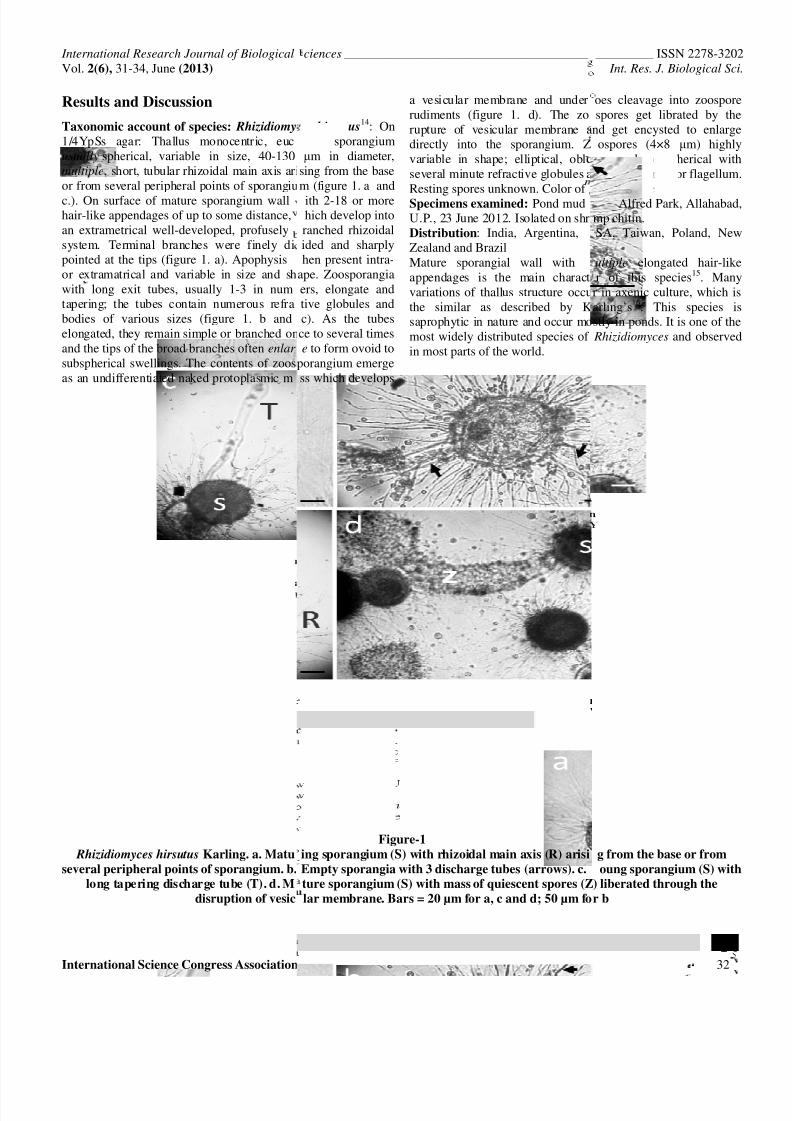
Taxonomic account of species: Rhizidiomy
1/4YpSs agar: Thallus monocentric, eucusually spherical, variable in size, 40-130multiple, short, tubular rhizoidal main axis arior from several peripheral points of sporangiu
c.). On surface of mature sporangium wallhair-like appendages of up to some distance,an extrametrical well-developed, profuselysystem. Terminal branches were finely dipointed at the tips (figure 1. a). Apophysisor extramatrical and variable in size and shwith long exit tubes, usually 1-3 in numtapering; the tubes contain numerous refrabodies of various sizes (figure 1. b andelongated, they remain simple or branched onand the tips of the broad branches often enlar
subspherical swellings. The contents of zoosas an undifferentiated naked protoplasmic m
Rhizidiomyces hirsutus Karling. a. Matu
several peripheral points of sporangium. b.
long tapering discharge tube (T). d. M
disruption of vesic
ciences ______________________________________
es hirsutus14: On
rpic; sporangiumµm in diameter,
sing from the basem (figure 1. a and
ith 2-18 or morehich develop intoranched rhizoidalided and sharplyhen present intra-
ape. Zoosporangiaers, elongate andtive globules andc). As the tubesce to several timese to form ovoid to
porangium emergess which develops
a vesicular membrane and underrudiments (figure 1. d). The zorupture of vesicular membrane adirectly into the sporangium. Zvariable in shape; elliptical, obloseveral minute refractive globules aResting spores unknown. Color ofSpecimens examined: Pond mudU.P., 23 June 2012. Isolated on shriDistribution: India, Argentina,Zealand and BrazilMature sporangial wall withappendages is the main charactvariations of thallus structure occuthe similar as described by Ksaprophytic in nature and occur momost widely distributed species ofin most parts of the world.
Figure-1
ing sporangium (S) with rhizoidal main axis (R) arisi
Empty sporangia with 3 discharge tubes (arrows). c.
ture sporangium (S) with mass of quiescent spores (Z
lar membrane. Bars = 20 µm for a, c and d; 50 µm fo
_________ ISSN 2278-3202 Int. Res. J. Biological Sci
32
oes cleavage into zoosporespores get librated by the
nd get encysted to enlargeospores (4×8 µm) highly
ng, oval, or spherical withnd a stout anterior flagellumolony white.rom Alfred Park, Allahabad,mp chitin.SA, Taiwan, Poland, New
ultiple elongated hair-liker of this species15. Many
r in axenic culture, which isarling’s16. This species isstly in ponds. It is one of the Rhizidiomyces and observed
g from the base or from
oung sporangium (S) with
) liberated through the
r b
8/14/2019 Hyphochytriomycota fungi
http://slidepdf.com/reader/full/hyphochytriomycota-fungi 3/4
International Research Journal of Biological
Vol. 2(6), 31-34, June (2013)
International Science Congress Association
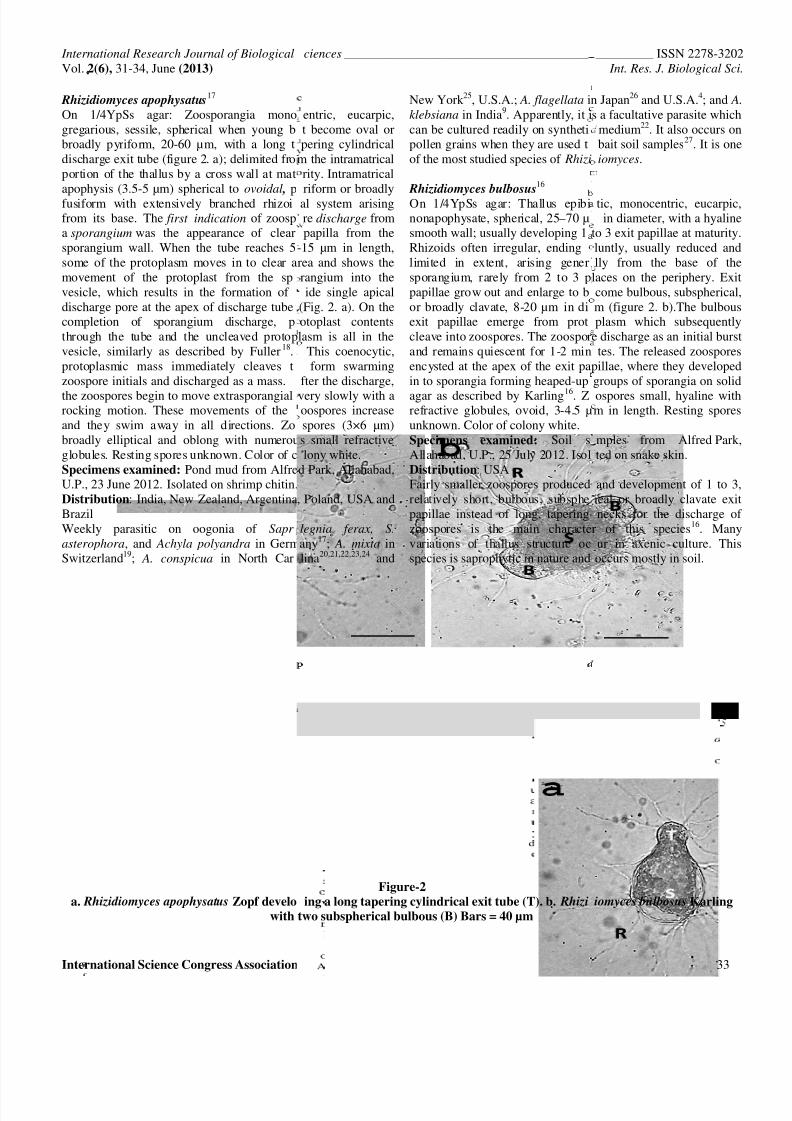
Rhizidiomyces apophysatus17
On 1/4YpSs agar: Zoosporangia monogregarious, sessile, spherical when young bbroadly pyriform, 20-60 µm, with a long tdischarge exit tube (figure 2. a); delimited froportion of the thallus by a cross wall at matapophysis (3.5-5 µm) spherical to ovoidal , pfusiform with extensively branched rhizoifrom its base. The first indication of zoospa sporangium was the appearance of clearsporangium wall. When the tube reaches 5some of the protoplasm moves in to clear amovement of the protoplast from the spvesicle, which results in the formation ofdischarge pore at the apex of discharge tubecompletion of sporangium discharge, pthrough the tube and the uncleaved protopvesicle, similarly as described by Fuller18.protoplasmic mass immediately cleaves tzoospore initials and discharged as a mass.
the zoospores begin to move extrasporangialrocking motion. These movements of theand they swim away in all directions. Zobroadly elliptical and oblong with numerouglobules. Resting spores unknown. Color of cSpecimens examined: Pond mud from AlfreU.P., 23 June 2012. Isolated on shrimp chitin.Distribution: India, New Zealand, ArgentinaBrazilWeekly parasitic on oogonia of Sapr
asterophora, and Achyla polyandra in GermSwitzerland19; A. conspicua in North Car
a. Rhizidiomyces apophysatus Zopf develo
with
ciences ______________________________________
entric, eucarpic,t become oval orpering cylindrical
m the intramatricalrity. Intramatricalriform or broadlyal system arisingre discharge frompapilla from the
-15 µm in length,rea and shows therangium into theide single apical
(Fig. 2. a). On theotoplast contentslasm is all in the
This coenocytic,form swarming
fter the discharge,
very slowly with aoospores increasespores (3×6 µm)s small refractivelony white.
d Park, Allahabad,
, Poland, USA and
legnia ferax, S.
any17; A. mixta inlina20,21,22,23,24 and
New York25, U.S.A.; A. flagellata iklebsiana in India9. Apparently, it ican be cultured readily on synthetipollen grains when they are used tof the most studied species of Rhizi
Rhizidiomyces bulbosus
16 On 1/4YpSs agar: Thallus epibinonapophysate, spherical, 25–70 µsmooth wall; usually developing 1Rhizoids often irregular, endinglimited in extent, arising genersporangium, rarely from 2 to 3 ppapillae grow out and enlarge to bor broadly clavate, 8-20 µm in diexit papillae emerge from protcleave into zoospores. The zoosporand remains quiescent for 1-2 minencysted at the apex of the exit pain to sporangia forming heaped-up
agar as described by Karling16. Zrefractive globules, ovoid, 3-4.5 µunknown. Color of colony white.Specimens examined: Soil sAllahabad, U.P., 25 July 2012. IsolDistribution: USAFairly smaller zoospores producedrelatively short, bulbous, subsphepapillae instead of long, taperingzoospores is the main charactevariations of thallus structure ocspecies is saprophytic in nature and
Figure-2
ing a long tapering cylindrical exit tube (T). b. Rhizi
two subspherical bulbous (B) Bars = 40 µm
_________ ISSN 2278-3202 Int. Res. J. Biological Sci
33
n Japan26 and U.S.A.4; and A.
s a facultative parasite whichmedium22. It also occurs onbait soil samples27. It is oneiomyces.
tic, monocentric, eucarpic,in diameter, with a hyaline
to 3 exit papillae at maturity.luntly, usually reduced andlly from the base of thelaces on the periphery. Exitcome bulbous, subspherical,m (figure 2. b).The bulbousplasm which subsequently
e discharge as an initial bursttes. The released zoospores
pillae, where they developedgroups of sporangia on solid
ospores small, hyaline withm in length. Resting spores
mples from Alfred Park,ted on snake skin.
and development of 1 to 3,ical or broadly clavate exitnecks for the discharge of
r of this species16. Manyur in axenic culture. Thisoccurs mostly in soil.
iomyces bulbosus Karling
8/14/2019 Hyphochytriomycota fungi
http://slidepdf.com/reader/full/hyphochytriomycota-fungi 4/4
International Research Journal of Biological Sciences ________________________________________________ ISSN 2278-3202Vol. 2(6), 31-34, June (2013) Int. Res. J. Biological Sci
International Science Congress Association 34
Hyphochytriomycetes has been mentioned for the first time toIndia by Chaudhuri and Kochhar8 and subsequently byMundkur9 and Karling10, but despite being cosmopolitan andubiquitous in its distribution, it never generated fascinationamong Indian scientists and researchers. Thus, the record ofHyphochytriomycetes of India is rather scanty as only sixspecies have so far reported from India.
The results of the present study indicate thatHyphochytriomycetes are of fairly common occurrence inIndian aquatic and terrestrial habitats. R. hirsutus Karling , R.
apophysatus Zopf and R. bulbosus Karling are isolated,described and illustrated for the first time on solid media inIndia. All of them are saprotrophic on broad range of substrate.The bulbous subspherical exit papillae with irregular rhizoids,possession of fairly extensive rhizoidal system oriented on its
base or sides and unique pattern of discharge tubes formationare key characteristic features of R. bulbosus Karling, R.
hirsutus Karling and R. apophysatus Zopf respectively.
Conclusion
The results of present study showed a taxonomic descriptionand ecological/distributional data of three species of Rhizidiomyces. This study also provides a taxonomic key withfigures for their identification using solely morphological anddevelopmental characters on solid media. R. bulbosus Karling ismentioned for the first time in our country, being also the firstrecord of it outside U.S.A. Thus, this investigation wouldcertainly lead to better understanding of role, diversity andecology of Hyphochytriomycetes of India.
Acknowledgements
The authors gratefully acknowledge Serena Rasconi, OsloUniversity, Norway along with Shu-Fen Chen, Tainan, Taiwanfor invaluable assistance in providing taxonomic literatures. Theauthors express their appreciation to Frank H. Gleason,University of Sydney, Australia and Ram Dayal, Varanasi fortheir helpful suggestions and encouragement.
References
1. Kirk P.M., Cannon P.F., David J.C. and Stalpers J.A., Ainsworth& Bisby`s Dictionary of the fungi, 9 thed- CABI Publishing,Wallingford, Oxon, UK (2001)
2. Fuller M.S., Phylum Hyphochytriomycota, In: Handbook ofProtoctista, Eds., L. Margulis, J.O. Corliss, M. Melkonian, and
D.J. Chapman. Jones and Bartlett, Boston, MA, 380–387 (1990) 3. Berbee M.L. and Taylor J.W., Fungal phylogeny , In: Molecular
Fungal Biology, Eds., R.P. Oliver and M. Schweizer, CambridgeUniversity Press, 21-77 (1999)
4. Karling J.S., The life history of Anisolpidium ectocarpii gen. nov.et sp. nov., and a synopsis and classification of other fungi withanteriorly uniflagellate zoospores, Amer. J. Bot., 30, 637–648(1943)
5. Karling J.S., Some zoosporic fungi of New Zealand. IX.Hyphochytriales or Anisochytridiales, Sydowia, 20, 137–143(1967)
6. Alexopoulos C.J., Mims C.W. and Blackwell M., IntroductoryMycology, 4th ed., John Wiley and Sons, Inc. (1996)
7. Fuller M.S., Hyphochytriomycota, In “The Mycota,” Vol. VII,“Systematics and Evolution” (D.J. McLaughlin, E.G. McLaughlin,and P.A. Lemke, eds.), Part A, Springer-Verlag, Berlin, 73–80(2001)
8. Chaudhuri H. and Kochhar P.L., Indian water moulds. I., Proc.
Indian Acad. Sci. Sect., B, 2, 137-154 (1935)
9. Mundkur B.B., Fungi of India, Suppl. I., Sci. Monogr. Counc.Agr. Res., India (1938)
10. Karling J.S., Indian anisochytrids, Sydowia, 17, 193-196 (1964)
11. Sparrow F.K.., Aquatic Phycomycetes, 2nd ed, The University ofMichigan Press, Ann Arbor, 743-767 (1960)
12. Fuller M.S. and Jaworski A., Zoosporic fungi in teaching andresearch, Southeastern Pub. Co., Athens, Georgia, 303 (1987)
13. Karling J.S., Chytridiomycetarum Iconorgraphia, Luberecht and
Cramer, Monticello, New York, 383-392 (1977) 14. Karling J.S., Rhizidiomyces hirsutus sp. nov., a hairy anisochytrid
from Brazil, Bull. Torrey Bot. Club., 72, 47-51 (1945)
15. Chen S.F., Anisolpidium saprobium and Rhizidiomyces hirsutus,new records of Hyphochytriomycetes (Hyphochytriales) inTaiwan, Fung. Sci., 22, 79–83 (2007)
16. Karling J.S., Zoosporic fungi of Oceania I, J. Mitchell Soc., 84,166–178 (1968)
17. Zopf W., Zur Kenntnis der Phycomyceten. I. Zur Morphologieund Biologie der Ancylisteen und Chytridiaceen, Nova Acta Acad
Leop.-Carol, 47, 143-236 (1884)
18. Fuller M.S., Growth and development of the water mold Rhizidiomyces in pure culture, Amer. Jour. Bot., 49, 64-71 (1962)
19. Maurizio A., Zur Kenntniss der schweizerischen Wasserpilzenebst Angaben uber eine neue Chytridine, Jahr. Nat. Gesell
Graubundens, 38, 9-38 (1895)
20. Coker W.C., The Saprolegniaceae with notes on other watermolds, Univ. North Carolina Press, Chapell Hill, North Carolina(1923)
21. Couch J.N., Observations on cilia of aquatic Phycomycetes,Science, 88, 476 (1938)
22. Couch J.N., Technic for collection, isolation, and culture ofchytrids, J. Elisha Mitchell Sci. Soc., 55, 208-214 (1939)
23. Couch J.N., The structure and action of the cilia in some aquaticPhycomycetes, Amer. Jour. Bot., 28, 704-713 (1941)
24. Whiffen A.J., Cellulose decomposition by the saprophyticchytrids, J. Elisha Mitchell Sci. Soc, 57, 321-330 (1941)
25. Sparrow F.K., Observations on the aquatic fungi of Cold SpringHarbor, Mycologia, 24, 268-303 (1932)
26. Tokunaga Y., Studies on the aquatic chytrids of Japan. III.Rhizidiaceae, Trans. Sapporo. Nat. Hist. Soc., 13, 388 (1934)
27. Gaertner A., Über das Vorkommen niederer Erdphycomyceten inAfrika, Schweden, und an einigen mitteleuropäischen Standorten,
Arch. f. Mikrobiol., 21, 4-56 (1954)