Anatomi Unggas Nervous
67
ANATOMI UNGGAS ANATOMI UNGGAS NERVOUS SYSTEM NERVOUS SYSTEM
Transcript of Anatomi Unggas Nervous
ANATOMI UNGGASANATOMI UNGGAS
NERVOUS SYSTEMNERVOUS SYSTEM
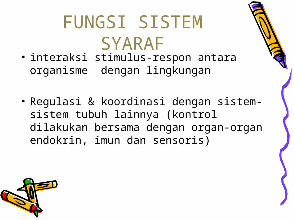
• interaksi stimulus-respon antara organisme dengan lingkungan
• Regulasi & koordinasi dengan sistem-sistem tubuh lainnya (kontrol dilakukan bersama dengan organ-organ endokrin, imun dan sensoris)
FUNGSI SISTEM SYARAF
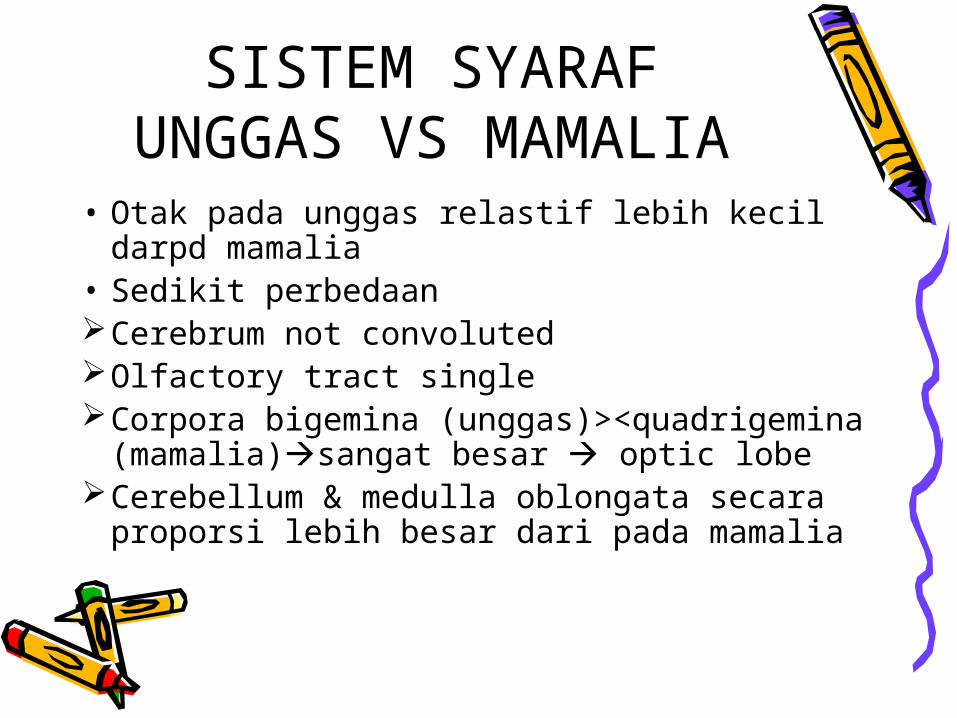
SISTEM SYARAF UNGGAS VS MAMALIA
• Otak pada unggas relastif lebih kecil darpd mamalia
• Sedikit perbedaanCerebrum not convolutedOlfactory tract singleCorpora bigemina (unggas)><quadrigemina
(mamalia)sangat besar optic lobeCerebellum & medulla oblongata secara
proporsi lebih besar dari pada mamalia
Medulla spinalis tidak mempunyai cauda equina
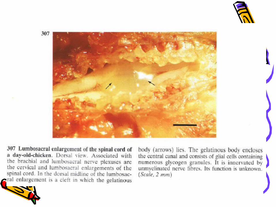
Medulla spinalis pada daerah pelvis membesar sacral organ/glycogen body
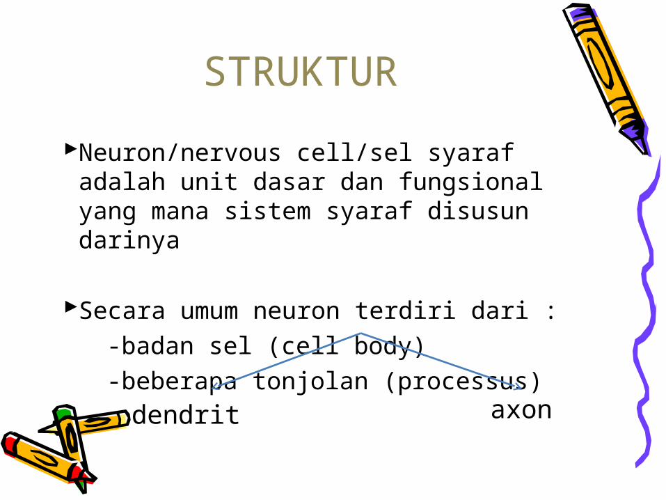
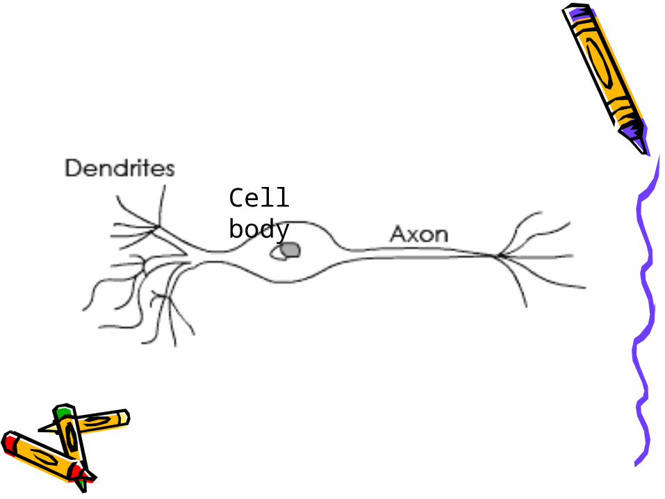
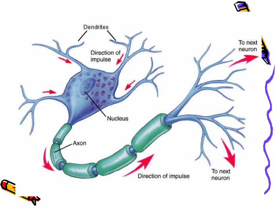
Neuron/nervous cell/sel syaraf adalah unit dasar dan fungsional yang mana sistem syaraf disusun darinya
Secara umum neuron terdiri dari : -badan sel (cell body) -beberapa tonjolan (processus)
STRUKTUR
dendrit axon

Cell body
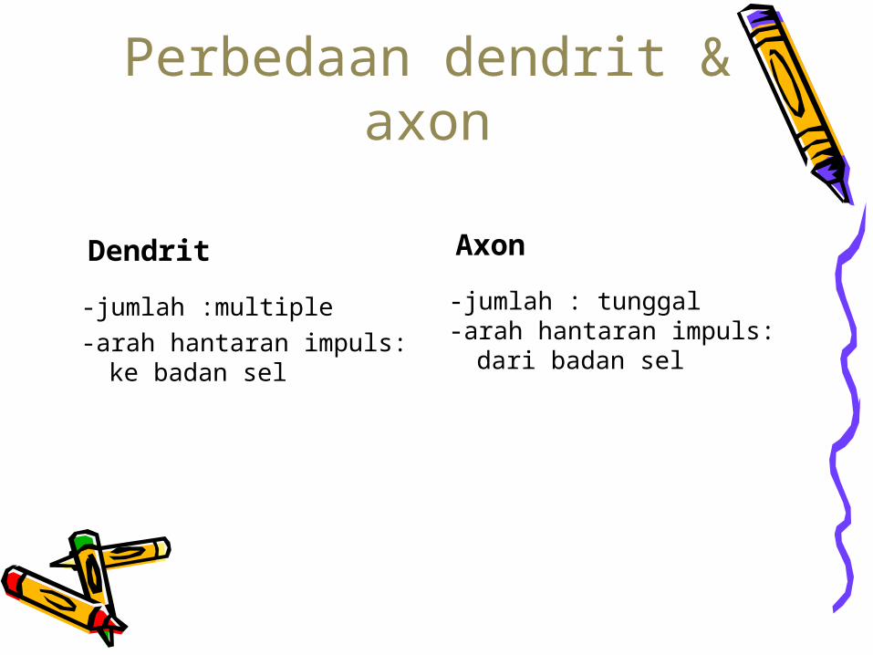
Perbedaan dendrit & axon
Dendrit Axon
-jumlah :multiple-arah hantaran impuls:
ke badan sel
-jumlah : tunggal-arah hantaran impuls:
dari badan sel
Cell body
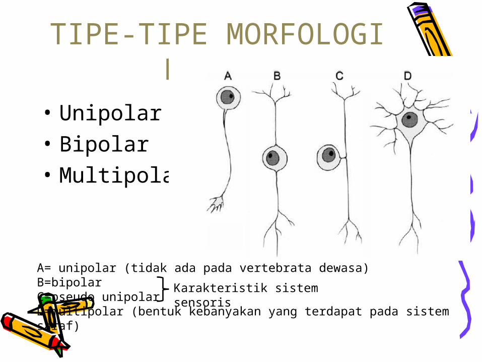
TIPE-TIPE MORFOLOGI NEURON
• Unipolar• Bipolar• Multipolar
A= unipolar (tidak ada pada vertebrata dewasa)B=bipolar C=pseudo unipolarD=multipolar (bentuk kebanyakan yang terdapat pada sistem saraf)
Karakteristik sistem sensoris
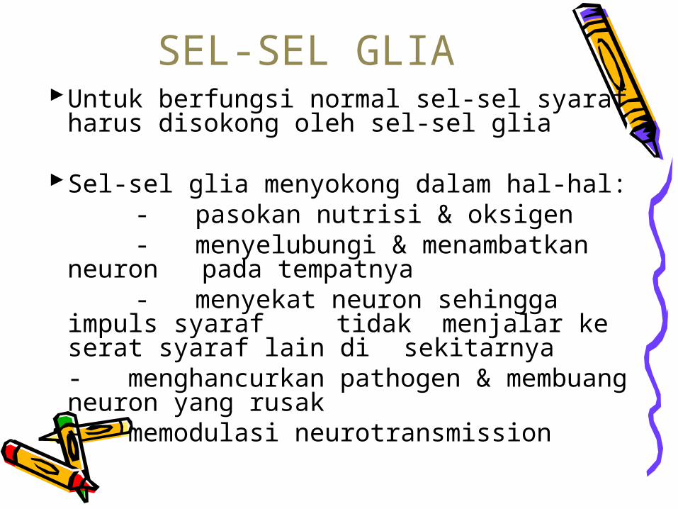
Untuk berfungsi normal sel-sel syaraf harus disokong oleh sel-sel glia
Sel-sel glia menyokong dalam hal-hal: - pasokan nutrisi & oksigen - menyelubungi & menambatkan neuron
pada tempatnya - menyekat neuron sehingga impuls syaraf
tidak menjalar ke serat syaraf lain di sekitarnya
- menghancurkan pathogen & membuang neuron yang rusak
- memodulasi neurotransmission
SEL-SEL GLIA
Jaringan penyokong sel-sel syaraf disebut neuroglia
Neuroglia terdiri dari berbagai jenis sel:-astrocyte-microglia-macroglia-sel ependimal melapisi ventrikel
otak & canalis centralis medula spinalis-oligodendrocyte membentuk lapisan-
lapisan disekeliling elemen penghantar.
Jenis-jenis sel glia
• Pada SST (sistem syaraf tepi) sel-sel penyokong adalah Sel Schwann
• Sel-sel syaraf dan glia membentuk jaringan syaraf (nervous tissue)
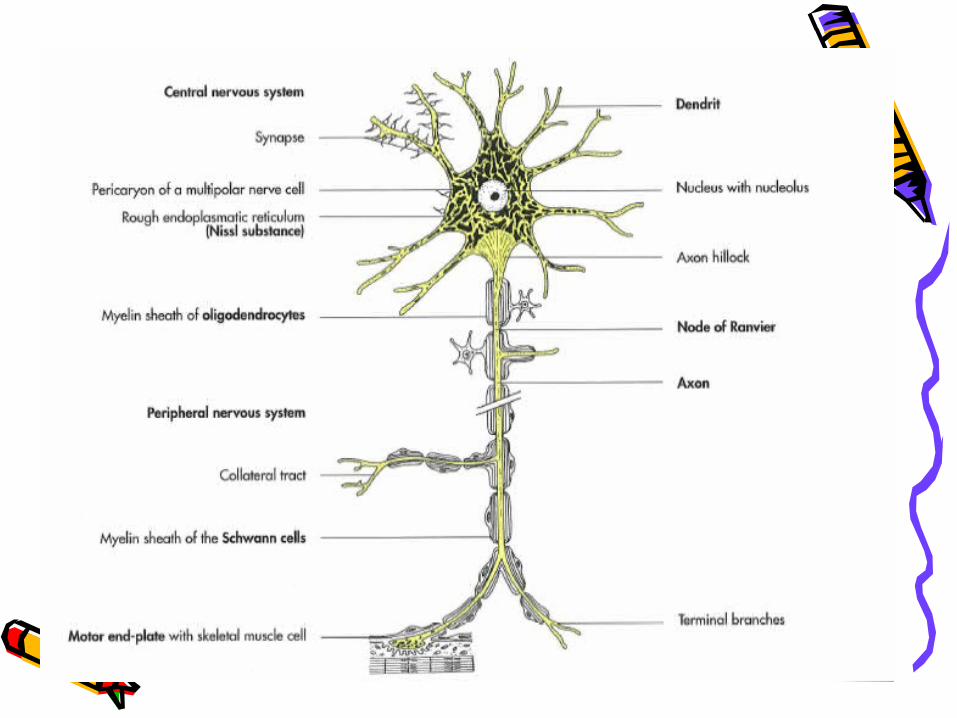
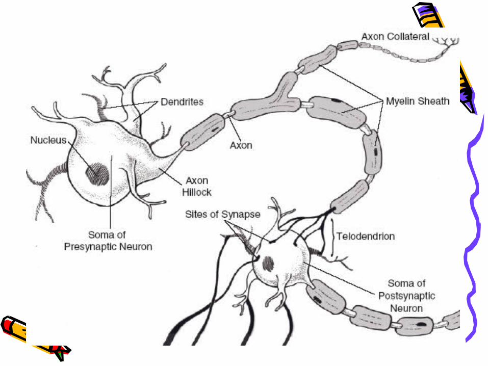
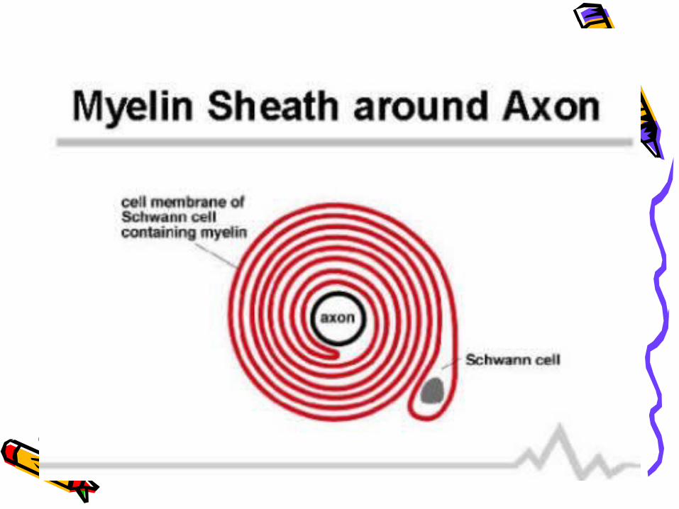
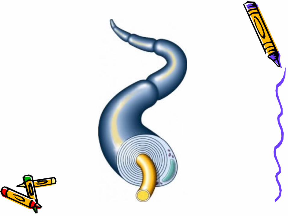
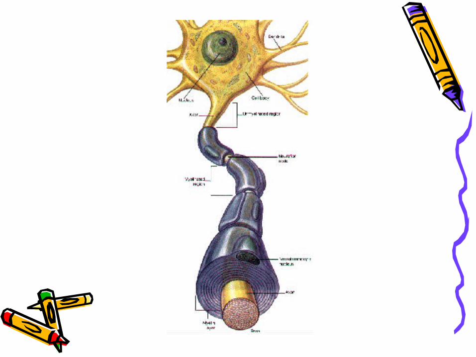
• Axon neuron bersama dengan struktur terkait disebut serabut saraf (nerve fibre)
• Nerve fibre ada yang tertutup dalam lapisan yang disebut sebagai selubung myelin.
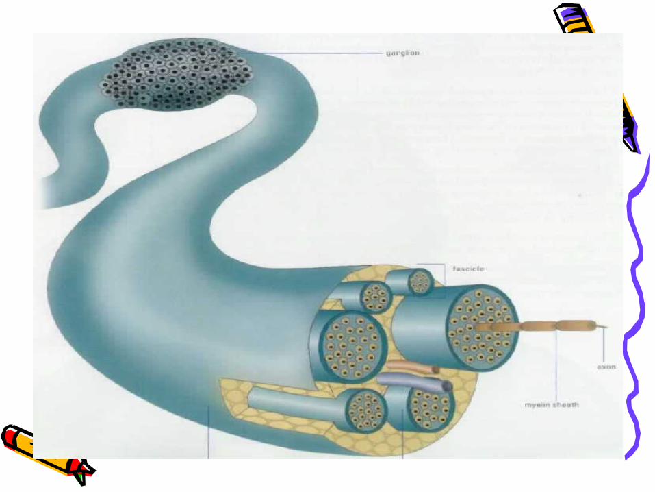
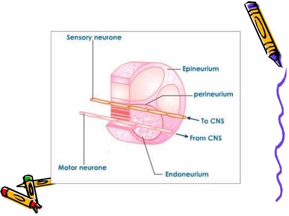
• Banyak serat saraf berkumpul bersama untuk membentuk saraf. Bundel serat ditutupi di dalam jaringan ikat disebut epineurium.
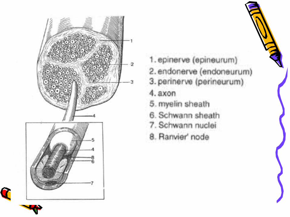
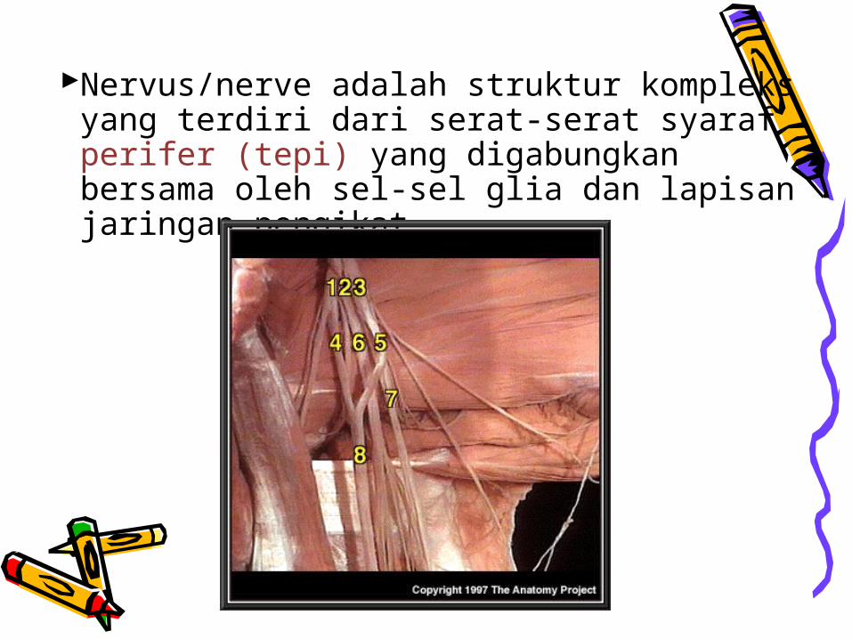
Nervus/nerve adalah struktur kompleks yang terdiri dari serat-serat syaraf perifer (tepi) yang digabungkan bersama oleh sel-sel glia dan lapisan jaringan pengikat.
Ganglion adalah kumpulan badan sel syaraf di sistem syaraf tepi
Nuclei adalah kumpulan badan sel syaraf di dalam sistem syaraf pusat, biasanya dengan fungsi yang sama
Serat-serat syaraf diselubungi myelin menimbulkan warna dari white matter pada SSP
Badan-badan sel syaraf & sel-sel glia menimbulkan warna grey matter pada SSP
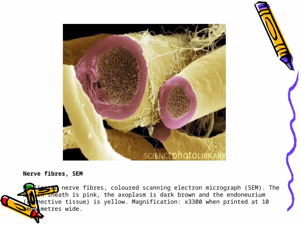
Nerve fibres, SEM
Myelinated nerve fibres, coloured scanning electron micrograph (SEM). The myelin sheath is pink, the axoplasm is dark brown and the endoneurium (connective tissue) is yellow. Magnification: x3300 when printed at 10 centimetres wide.
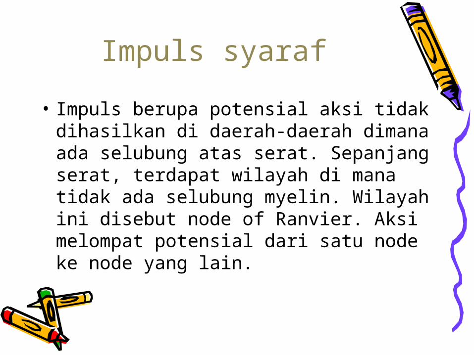
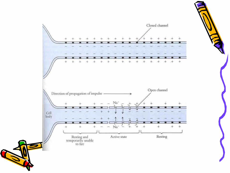
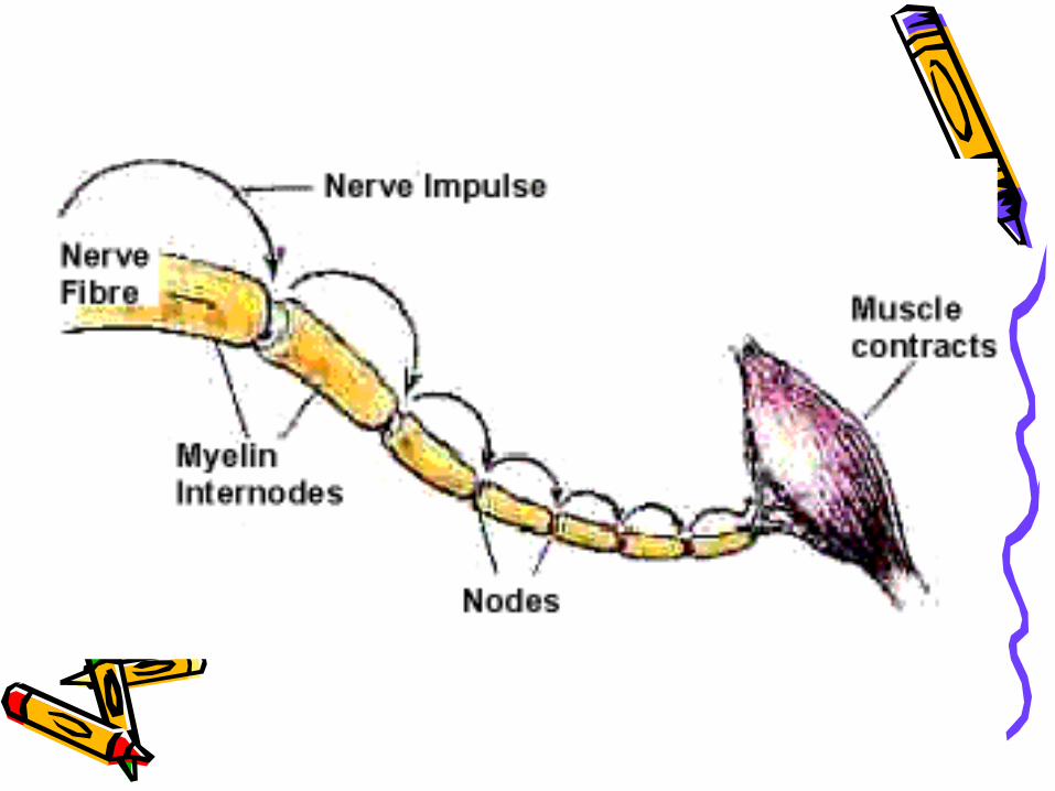
Impuls syaraf
• Impuls berupa potensial aksi tidak dihasilkan di daerah-daerah dimana ada selubung atas serat. Sepanjang serat, terdapat wilayah di mana tidak ada selubung myelin. Wilayah ini disebut node of Ranvier. Aksi melompat potensial dari satu node ke node yang lain.
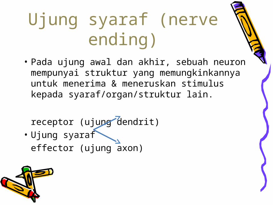
Ujung syaraf (nerve ending)
• Pada ujung awal dan akhir, sebuah neuron mempunyai struktur yang memungkinkannya untuk menerima & meneruskan stimulus kepada syaraf/organ/struktur lain.
receptor (ujung dendrit)• Ujung syaraf
effector (ujung axon)
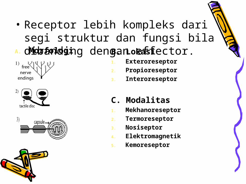
• Receptor lebih kompleks dari segi struktur dan fungsi bila dibanding dengan effector.A. Morfologi B. Lokasi
1. Exteroreseptor2. Propioreseptor3. Interoreseptor
C. Modalitas1. Mekhanoreseptor 2. Termoreseptor3. Nosiseptor4. Elektromagnetik5. Kemoreseptor
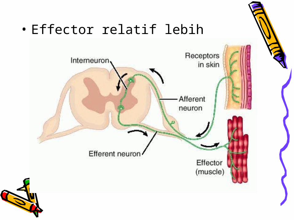
• Effector relatif lebih sederhana .
• In the nervous system, a synapse is a junction that permits aneuron to pass an electrical or chemical signal to another cell.
• Sinapsis adalah pertemuan yang menjadikan neuron dapat melewatkan sinyal listrik atau kimia ke sel lain
Synapse (sinapsis)
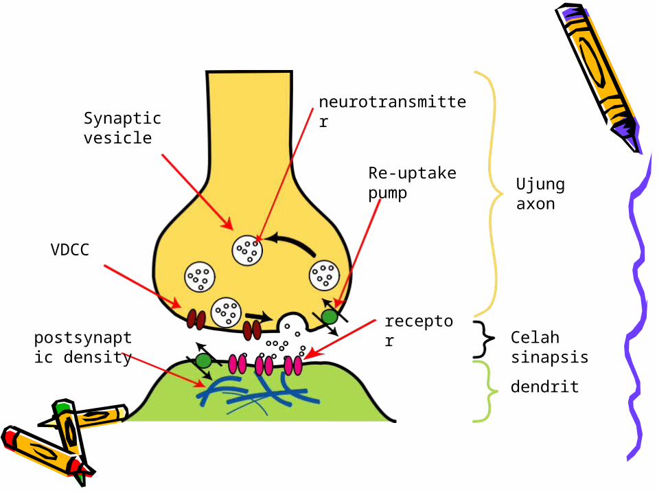
Ujung axon
dendrit
Celah sinapsis
receptor
Re-uptake pump
neurotransmitterSynaptic vesicle
VDCC
postsynaptic density
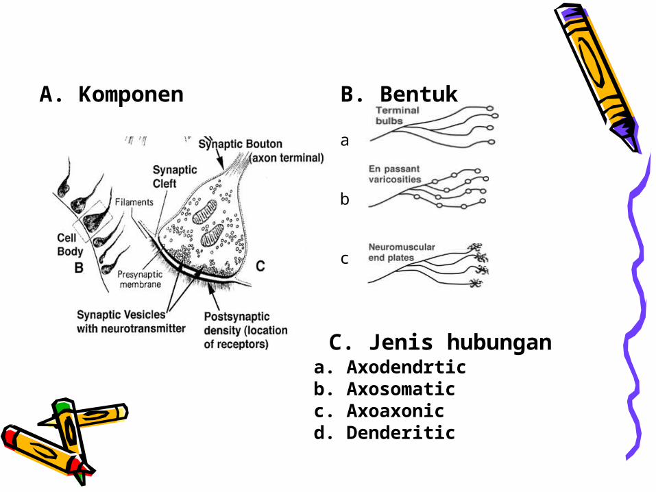
A. Komponen B. Bentuk
a
b
c
C. Jenis hubungan a. Axodendrticb. Axosomaticc. Axoaxonicd. Denderitic
Serat syaraf (nerve fibre) terdiri dari axon yang diselubungi lapisan neuroglia
Klasifikasi menurut jenis informasi :-motor nerve fibres-sensory nerve fibres-autonomic nerve fibres
Klasifikasi menurut arah impuls:-afferent impuls dari SST ke SSP-efferent impuls dari SSP ke SST
Klasifikasi serat syaraf
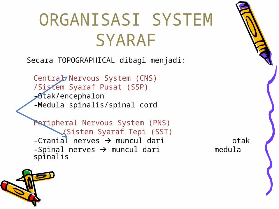
ORGANISASI SYSTEM SYARAF
Secara TOPOGRAPHICAL dibagi menjadi:
Central Nervous System (CNS) /Sistem Syaraf Pusat (SSP)
-Otak/encephalon-Medula spinalis/spinal cord
Peripheral Nervous System (PNS) /Sistem Syaraf Tepi (SST)
-Cranial nerves muncul dari otak
-Spinal nerves muncul dari medula spinalis
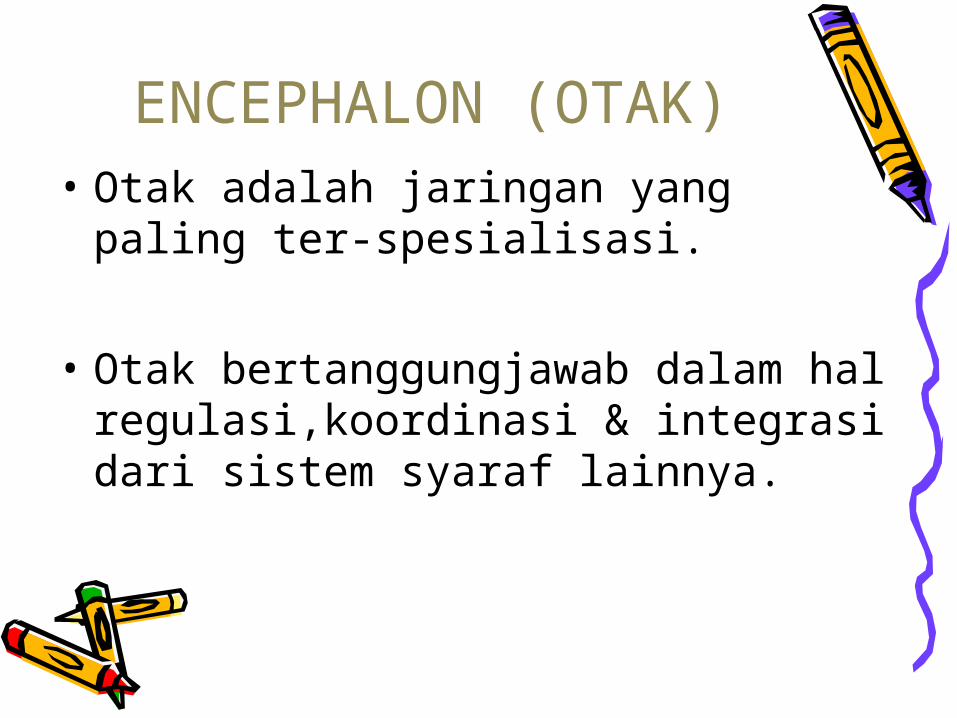
• Otak adalah jaringan yang paling ter-spesialisasi.
• Otak bertanggungjawab dalam hal regulasi,koordinasi & integrasi dari sistem syaraf lainnya.
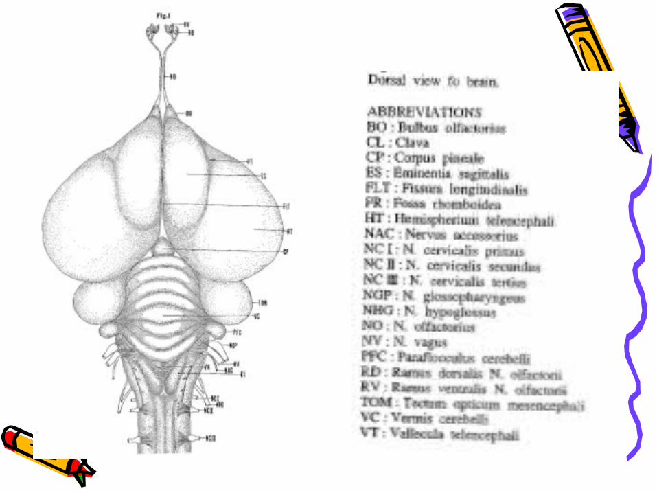
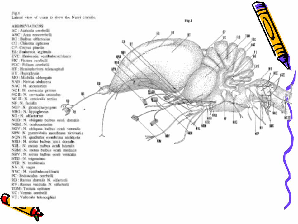
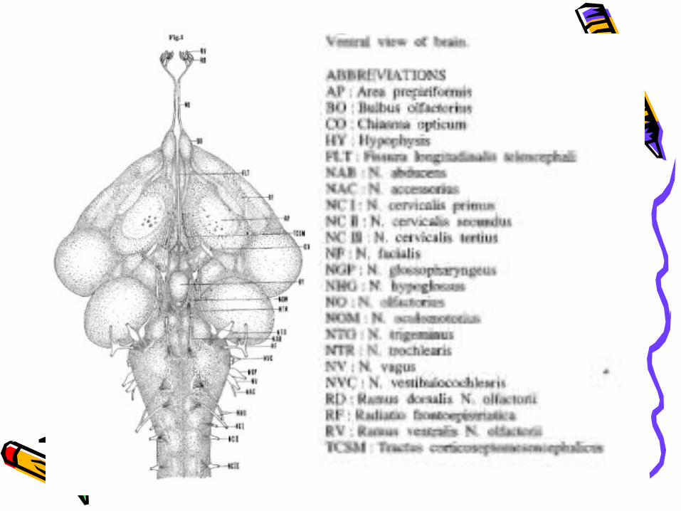
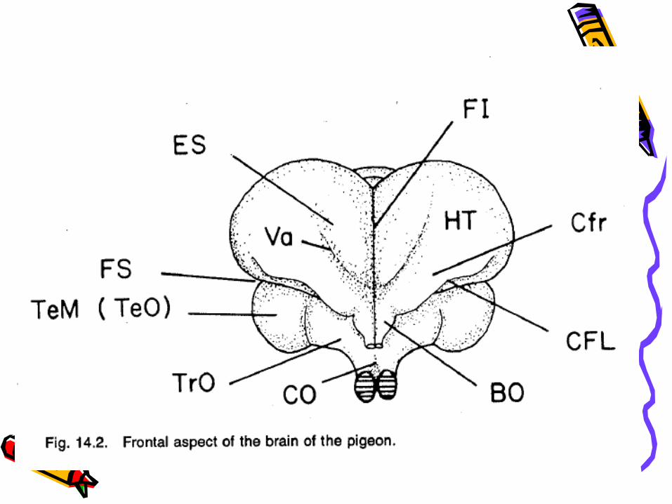
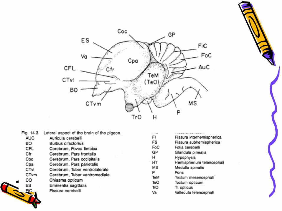
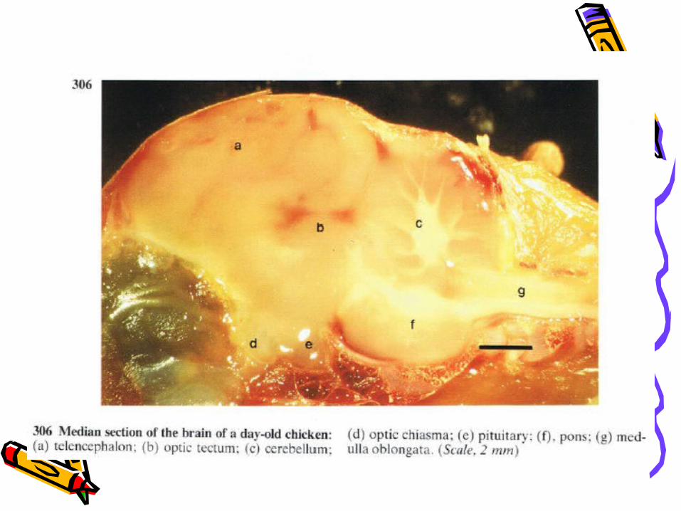
ENCEPHALON (OTAK)
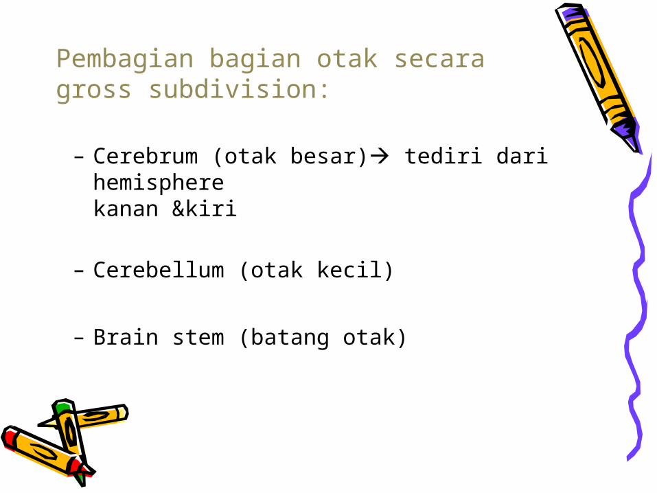
– Cerebrum (otak besar) tediri dari hemisphere kanan &kiri
– Cerebellum (otak kecil)
– Brain stem (batang otak)
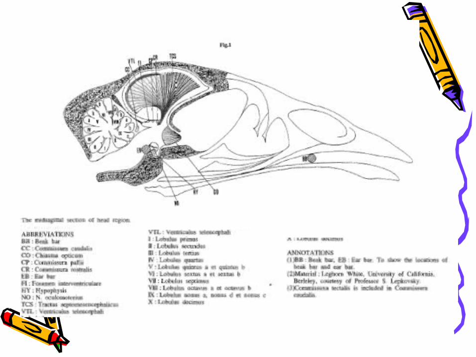
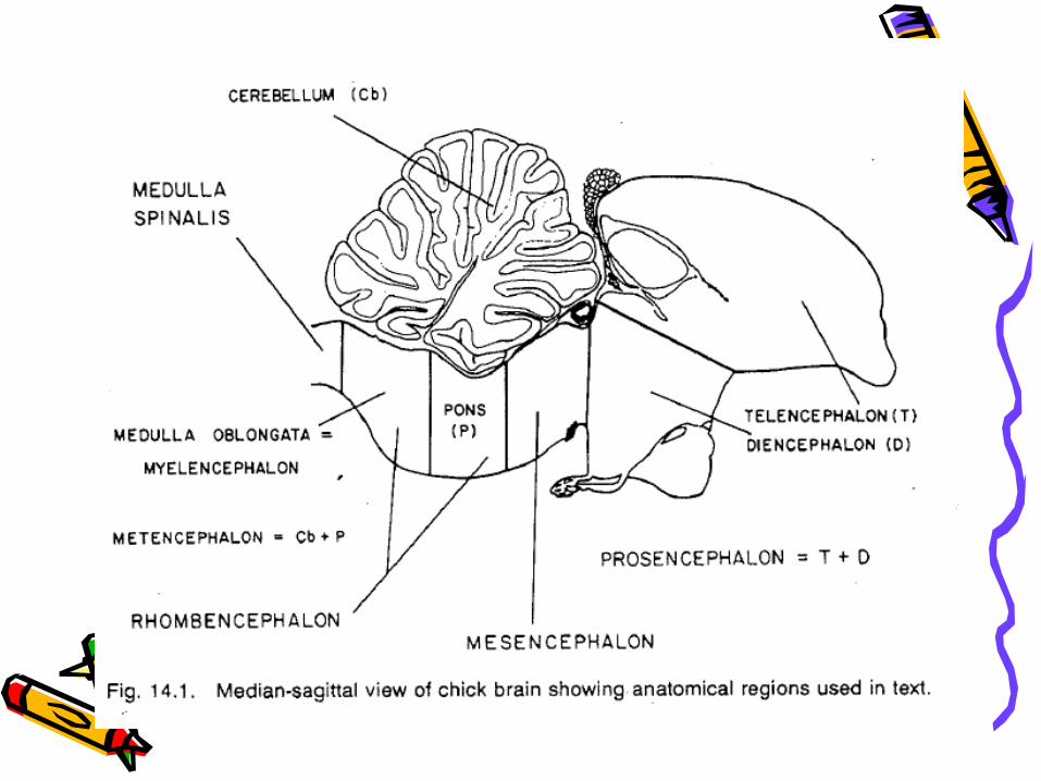
Pembagian bagian otak secara gross subdivision:
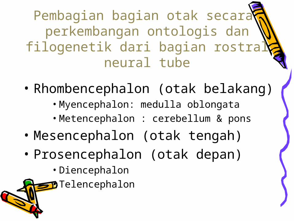
Pembagian bagian otak secara perkembangan ontologis dan
filogenetik dari bagian rostral neural tube
• Rhombencephalon (otak belakang)• Myencephalon: medulla oblongata• Metencephalon : cerebellum & pons
• Mesencephalon (otak tengah)• Prosencephalon (otak depan)
• Diencephalon• Telencephalon
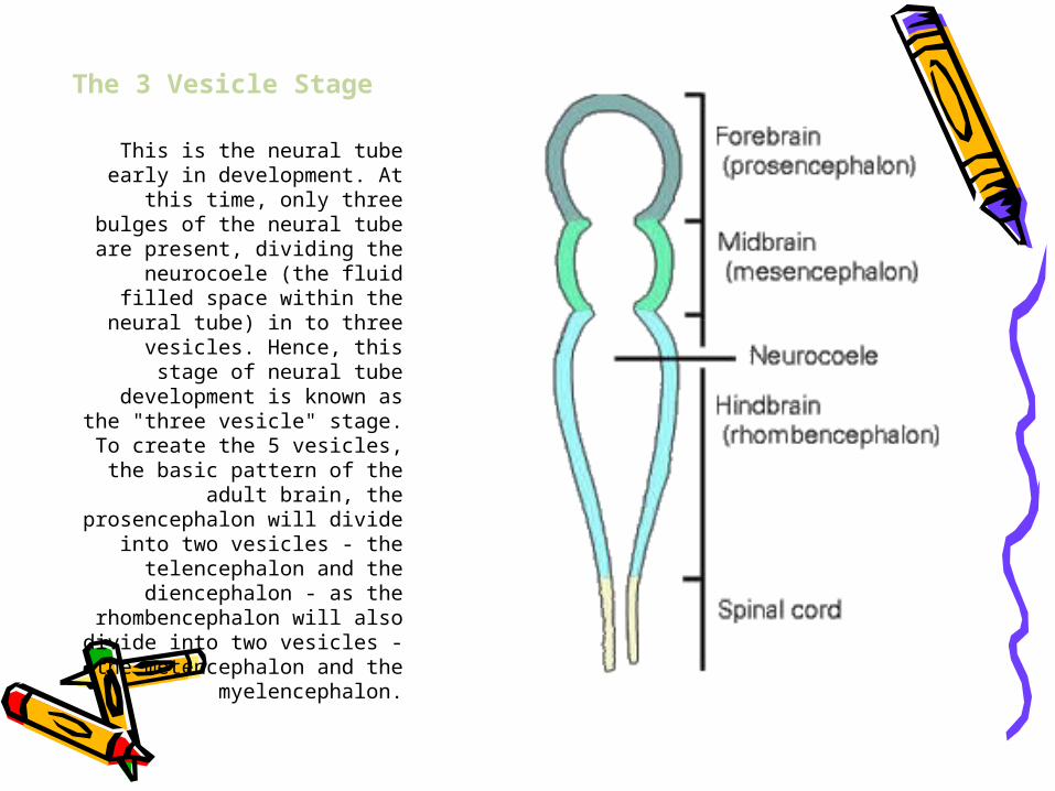
The 3 Vesicle Stage
This is the neural tube early in development. At this time,
only three bulges of the neural tube are present, dividing the
neurocoele (the fluid filled space within the neural tube)
in to three vesicles. Hence, this stage of neural tube
development is known as the "three vesicle" stage. To
create the 5 vesicles, the basic pattern of the adult brain, the
prosencephalon will divide into two vesicles - the
telencephalon and the diencephalon - as the
rhombencephalon will also divide into two vesicles - the
metencephalon and the myelencephalon.
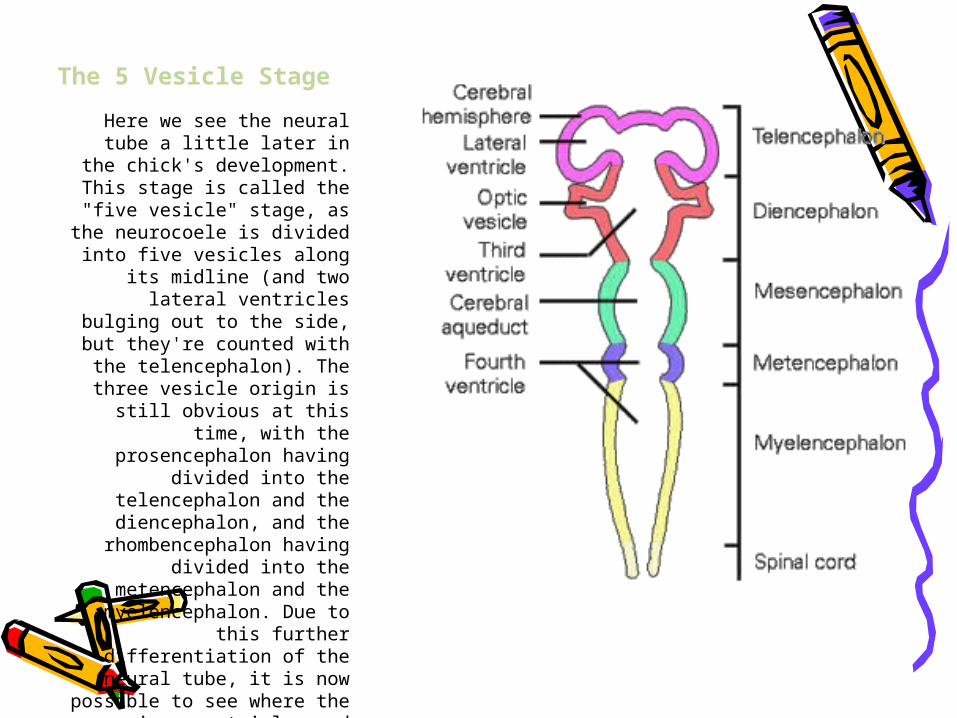
The 5 Vesicle Stage
Here we see the neural tube a little later in the chick's
development. This stage is called the "five vesicle" stage,
as the neurocoele is divided into five vesicles along its
midline (and two lateral ventricles bulging out to the
side, but they're counted with the telencephalon). The three
vesicle origin is still obvious at this time, with the
prosencephalon having divided into the
telencephalon and the diencephalon, and the
rhombencephalon having divided into the
metencephalon and the myelencephalon. Due to this further differentiation of the
neural tube, it is now possible to see where the various
ventricles and other remnants of the neurocoele will form
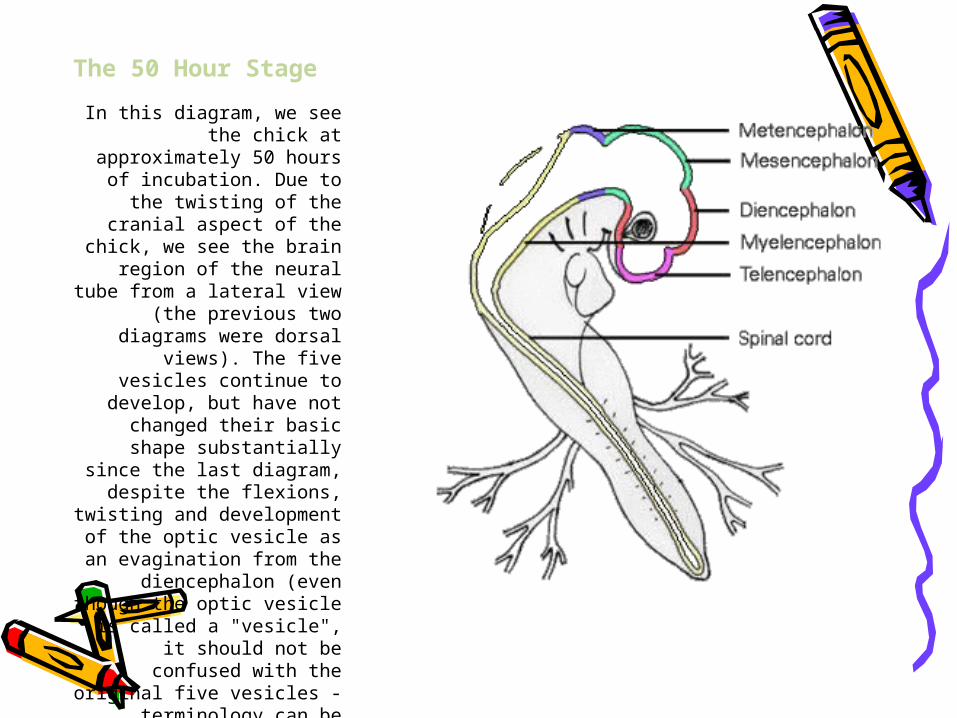
The 50 Hour Stage
In this diagram, we see the chick at approximately 50
hours of incubation. Due to the twisting of the cranial
aspect of the chick, we see the brain region of the neural tube from a lateral view (the previous two diagrams were
dorsal views). The five vesicles continue to develop,
but have not changed their basic shape substantially
since the last diagram, despite the flexions, twisting
and development of the optic vesicle as an
evagination from the diencephalon (even though the optic vesicle is called a
"vesicle", it should not be confused with the original five vesicles - terminology
can be confusing!)
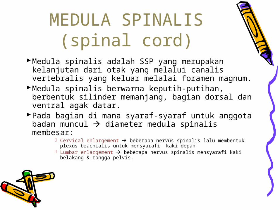
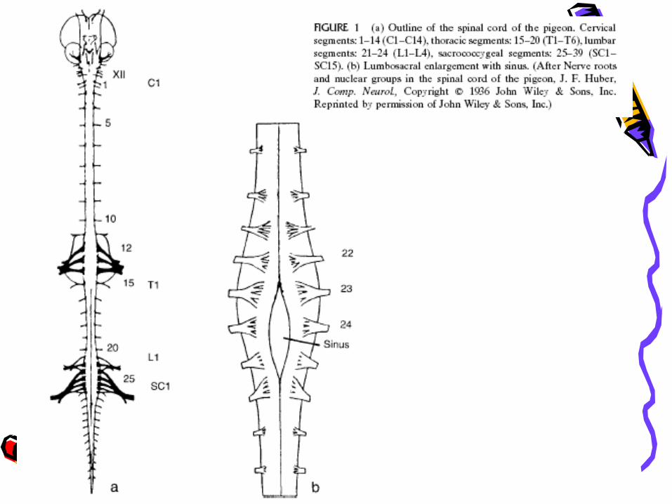
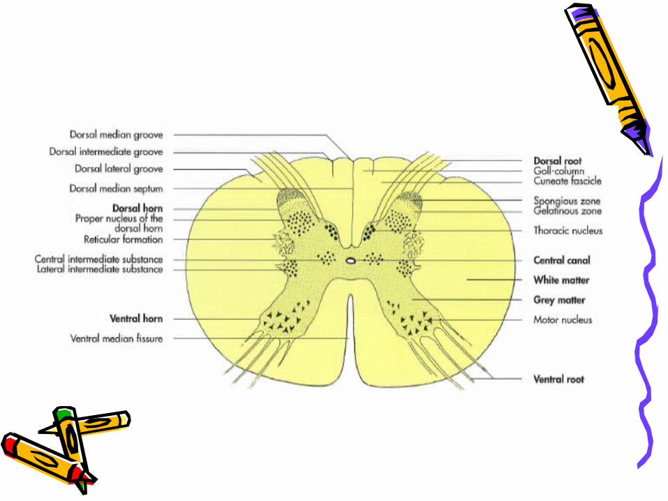
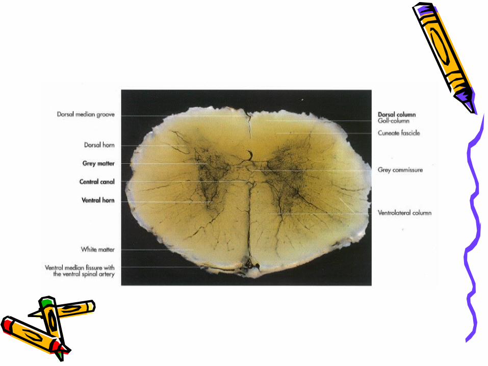
MEDULA SPINALIS (spinal cord)
Medula spinalis adalah SSP yang merupakan kelanjutan dari otak yang melalui canalis vertebralis yang keluar melalai foramen magnum.
Medula spinalis berwarna keputih-putihan, berbentuk silinder memanjang, bagian dorsal dan ventral agak datar.
Pada bagian di mana syaraf-syaraf untuk anggota badan muncul diameter medula spinalis membesar:
Cervical enlargement beberapa nervus spinalis lalu membentuk plexus brachialis untuk mensyarafi kaki depan
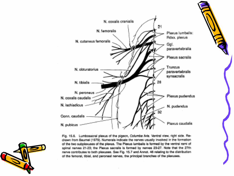
Lumbar enlargement beberapa nervus spinalis mensyarafi kaki belakang & rongga pelvis.
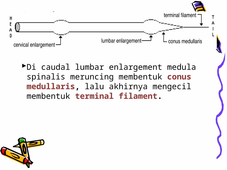
Di caudal lumbar enlargement medula spinalis meruncing membentuk conus medullaris, lalu akhirnya mengecil membentuk terminal filament.